KR101482803B1 - Antioxidant compound - Google Patents
Antioxidant compoundDownload PDFInfo
- Publication number
- KR101482803B1 KR101482803B1KR1020097007629AKR20097007629AKR101482803B1KR 101482803 B1KR101482803 B1KR 101482803B1KR 1020097007629 AKR1020097007629 AKR 1020097007629AKR 20097007629 AKR20097007629 AKR 20097007629AKR 101482803 B1KR101482803 B1KR 101482803B1
- Authority
- KR
- South Korea
- Prior art keywords
- delete delete
- complex
- vitamin
- tos
- cancer
- Prior art date
- Legal status (The legal status is an assumption and is not a legal conclusion. Google has not performed a legal analysis and makes no representation as to the accuracy of the status listed.)
- Active
Links
- 150000001875compoundsChemical class0.000titleclaimsabstractdescription131
- 230000003078antioxidant effectEffects0.000titleabstractdescription20
- 239000003963antioxidant agentSubstances0.000titleabstractdescription17
- 206010028980NeoplasmDiseases0.000claimsabstractdescription139
- 201000011510cancerDiseases0.000claimsabstractdescription110
- 230000006907apoptotic processEffects0.000claimsabstractdescription53
- 238000000034methodMethods0.000claimsabstractdescription45
- 230000008811mitochondrial respiratory chainEffects0.000claimsabstractdescription21
- 230000001590oxidative effectEffects0.000claimsabstractdescription20
- 239000003623enhancerSubstances0.000claimsabstractdescription7
- 210000004027cellAnatomy0.000claimsdescription235
- PRRZDZJYSJLDBS-UHFFFAOYSA-N3-bromo-2-oxopropanoic acidChemical compoundOC(=O)C(=O)CBrPRRZDZJYSJLDBS-UHFFFAOYSA-N0.000claimsdescription61
- 235000017471coenzyme Q10Nutrition0.000claimsdescription58
- NPCOQXAVBJJZBQ-UHFFFAOYSA-Nreduced coenzyme Q9Natural productsCOC1=C(O)C(C)=C(CC=C(C)CCC=C(C)CCC=C(C)CCC=C(C)CCC=C(C)CCC=C(C)CCC=C(C)CCC=C(C)CCC=C(C)C)C(O)=C1OCNPCOQXAVBJJZBQ-UHFFFAOYSA-N0.000claimsdescription58
- 229940035936ubiquinoneDrugs0.000claimsdescription58
- ACTIUHUUMQJHFO-UHFFFAOYSA-NCoenzym Q10Natural productsCOC1=C(OC)C(=O)C(CC=C(C)CCC=C(C)CCC=C(C)CCC=C(C)CCC=C(C)CCC=C(C)CCC=C(C)CCC=C(C)CCC=C(C)CCC=C(C)C)=C(C)C1=OACTIUHUUMQJHFO-UHFFFAOYSA-N0.000claimsdescription57
- ACTIUHUUMQJHFO-UPTCCGCDSA-Ncoenzyme Q10Chemical compoundCOC1=C(OC)C(=O)C(C\C=C(/C)CC\C=C(/C)CC\C=C(/C)CC\C=C(/C)CC\C=C(/C)CC\C=C(/C)CC\C=C(/C)CC\C=C(/C)CC\C=C(/C)CCC=C(C)C)=C(C)C1=OACTIUHUUMQJHFO-UPTCCGCDSA-N0.000claimsdescription53
- 230000003647oxidationEffects0.000claimsdescription48
- 238000007254oxidation reactionMethods0.000claimsdescription48
- 150000003712vitamin E derivativesChemical class0.000claimsdescription46
- 239000000203mixtureSubstances0.000claimsdescription35
- 230000002438mitochondrial effectEffects0.000claimsdescription33
- 230000034994deathEffects0.000claimsdescription27
- 230000001939inductive effectEffects0.000claimsdescription27
- 210000003470mitochondriaAnatomy0.000claimsdescription25
- 239000003642reactive oxygen metaboliteSubstances0.000claimsdescription25
- SEBPXHSZHLFWRL-UHFFFAOYSA-N3,4-dihydro-2,2,5,7,8-pentamethyl-2h-1-benzopyran-6-olChemical groupO1C(C)(C)CCC2=C1C(C)=C(C)C(O)=C2CSEBPXHSZHLFWRL-UHFFFAOYSA-N0.000claimsdescription23
- -13-hydroxy-5,6-dimethoxy-2 (1H) -pyridinoneChemical compound0.000claimsdescription23
- 230000003993interactionEffects0.000claimsdescription21
- 150000005829chemical entitiesChemical class0.000claimsdescription20
- 239000003112inhibitorSubstances0.000claimsdescription20
- 238000012360testing methodMethods0.000claimsdescription17
- 230000002209hydrophobic effectEffects0.000claimsdescription16
- 238000002560therapeutic procedureMethods0.000claimsdescription16
- 230000011664signalingEffects0.000claimsdescription15
- 238000004519manufacturing processMethods0.000claimsdescription14
- NGVDGCNFYWLIFO-UHFFFAOYSA-Npyridoxal 5'-phosphateChemical compoundCC1=NC=C(COP(O)(O)=O)C(C=O)=C1ONGVDGCNFYWLIFO-UHFFFAOYSA-N0.000claimsdescription12
- IELOKBJPULMYRW-NJQVLOCASA-ND-alpha-Tocopheryl Acid SuccinateChemical compoundOC(=O)CCC(=O)OC1=C(C)C(C)=C2O[C@@](CCC[C@H](C)CCC[C@H](C)CCCC(C)C)(C)CCC2=C1CIELOKBJPULMYRW-NJQVLOCASA-N0.000claimsdescription11
- 150000001408amidesChemical class0.000claimsdescription10
- 238000000338in vitroMethods0.000claimsdescription8
- 239000008194pharmaceutical compositionSubstances0.000claimsdescription8
- SQQWBSBBCSFQGC-JLHYYAGUSA-Nubiquinone-2Chemical compoundCOC1=C(OC)C(=O)C(C\C=C(/C)CCC=C(C)C)=C(C)C1=OSQQWBSBBCSFQGC-JLHYYAGUSA-N0.000claimsdescription8
- 239000000126substanceSubstances0.000claimsdescription7
- 101150045355akt1 geneProteins0.000claimsdescription6
- 230000004611cancer cell deathEffects0.000claimsdescription6
- 230000035806respiratory chainEffects0.000claimsdescription6
- 239000000758substrateSubstances0.000claimsdescription6
- 238000005094computer simulationMethods0.000claimsdescription5
- 239000003446ligandSubstances0.000claimsdescription5
- RROCMCBQTUYDSD-VIFPVBQESA-N2-[1-methylhexyl]-4,6-dinitrophenolChemical compoundCCCCC[C@H](C)C1=CC([N+]([O-])=O)=CC([N+]([O-])=O)=C1ORROCMCBQTUYDSD-VIFPVBQESA-N0.000claimsdescription4
- ROSTULATFVLODX-UHFFFAOYSA-N2-[2-(3,7-dimethyl-2,6-dioxopurin-1-yl)ethylideneamino]guanidineChemical compoundCN1C(=O)N(CC=NN=C(N)N)C(=O)C2=C1N=CN2CROSTULATFVLODX-UHFFFAOYSA-N0.000claimsdescription4
- NGQCKPKYAYBCDI-UHFFFAOYSA-N2-[2-carboxyethyl(hydroxy)phosphoryl]-3-(furan-2-yl)propanoic acidChemical compoundOC(=O)CCP(O)(=O)C(C(O)=O)CC1=CC=CO1NGQCKPKYAYBCDI-UHFFFAOYSA-N0.000claimsdescription4
- LZYMYBUOUUKLDI-UHFFFAOYSA-N3-(5-propoxy-2-sulfophenyl)propanoic acidChemical compoundCCCOC1=CC=C(S(O)(=O)=O)C(CCC(O)=O)=C1LZYMYBUOUUKLDI-UHFFFAOYSA-N0.000claimsdescription4
- UAWXXHFKVIMFBE-UHFFFAOYSA-N3-[hydroxy-[[methyl(methylsulfonyl)amino]methyl]phosphoryl]propanoic acidChemical compoundCS(=O)(=O)N(C)CP(O)(=O)CCC(O)=OUAWXXHFKVIMFBE-UHFFFAOYSA-N0.000claimsdescription4
- NHDVQAZYGIDXFR-UHFFFAOYSA-N4,6-dimethyl-3-methylsulfonyl-2-propylsulfonylpyridineChemical compoundCCCS(=O)(=O)C1=NC(C)=CC(C)=C1S(C)(=O)=ONHDVQAZYGIDXFR-UHFFFAOYSA-N0.000claimsdescription4
- DDNMJOPNGRPATA-UHFFFAOYSA-N8-sulfoquinoline-2,4-dicarboxylic acidChemical compoundC1=CC=C(S(O)(=O)=O)C2=NC(C(=O)O)=CC(C(O)=O)=C21DDNMJOPNGRPATA-UHFFFAOYSA-N0.000claimsdescription4
- BLGSSKGWJZLIHB-UHFFFAOYSA-NCOC1=CC=C(C(NNC(N)=N)P(O)=O)C=C1Chemical compoundCOC1=CC=C(C(NNC(N)=N)P(O)=O)C=C1BLGSSKGWJZLIHB-UHFFFAOYSA-N0.000claimsdescription4
- 208000003837Second Primary NeoplasmsDiseases0.000claimsdescription4
- GYSSRZJIHXQEHQ-UHFFFAOYSA-NcarboxinChemical compoundS1CCOC(C)=C1C(=O)NC1=CC=CC=C1GYSSRZJIHXQEHQ-UHFFFAOYSA-N0.000claimsdescription4
- 108010062381leucyl-threonyl-valyl-seryl-prolyl-tryptophyl-tyrosineProteins0.000claimsdescription4
- ZYCLWFHJQNMCSK-UHFFFAOYSA-Nn-(4-methoxy-6-methylpyrimidin-2-yl)benzenesulfonamideChemical compoundCOC1=CC(C)=NC(NS(=O)(=O)C=2C=CC=CC=2)=N1ZYCLWFHJQNMCSK-UHFFFAOYSA-N0.000claimsdescription4
- YGWISIYXVARJMB-UHFFFAOYSA-Nn-ethoxycarbonyl-n-(3-ethoxy-3-oxopropyl)-methylphosphonamidic acidChemical compoundCCOC(=O)CCN(P(C)(O)=O)C(=O)OCCYGWISIYXVARJMB-UHFFFAOYSA-N0.000claimsdescription4
- 235000007682pyridoxal 5'-phosphateNutrition0.000claimsdescription4
- 239000011589pyridoxal 5'-phosphateSubstances0.000claimsdescription4
- 229960001327pyridoxal phosphateDrugs0.000claimsdescription4
- TXBBUSUXYMIVOS-UHFFFAOYSA-NthenoyltrifluoroacetoneChemical compoundFC(F)(F)C(=O)CC(=O)C1=CC=CS1TXBBUSUXYMIVOS-UHFFFAOYSA-N0.000claimsdescription4
- 108060008682Tumor Necrosis FactorProteins0.000claimsdescription3
- 238000010998test methodMethods0.000claimsdescription2
- 101710113459RAC-alpha serine/threonine-protein kinaseProteins0.000claims4
- 102100033810RAC-alpha serine/threonine-protein kinaseHuman genes0.000claims4
- 102000000852Tumor Necrosis Factor-alphaHuman genes0.000claims2
- 238000012512characterization methodMethods0.000claims1
- 238000006073displacement reactionMethods0.000claims1
- 239000003937drug carrierSubstances0.000claims1
- 230000007717exclusionEffects0.000claims1
- 238000011282treatmentMethods0.000abstractdescription43
- GVJHHUAWPYXKBD-UHFFFAOYSA-N(±)-α-TocopherolChemical compoundOC1=C(C)C(C)=C2OC(CCCC(C)CCCC(C)CCCC(C)C)(C)CCC2=C1CGVJHHUAWPYXKBD-UHFFFAOYSA-N0.000abstractdescription38
- WIGCFUFOHFEKBI-UHFFFAOYSA-Ngamma-tocopherolNatural productsCC(C)CCCC(C)CCCC(C)CCCC1CCC2C(C)C(O)C(C)C(C)C2O1WIGCFUFOHFEKBI-UHFFFAOYSA-N0.000abstractdescription20
- QVGXLLKOCUKJST-UHFFFAOYSA-Natomic oxygenChemical compound[O]QVGXLLKOCUKJST-UHFFFAOYSA-N0.000abstractdescription19
- 239000001301oxygenSubstances0.000abstractdescription19
- 229910052760oxygenInorganic materials0.000abstractdescription19
- 235000006708antioxidantsNutrition0.000abstractdescription16
- 229930003427Vitamin ENatural products0.000abstractdescription15
- 229940046009vitamin EDrugs0.000abstractdescription15
- 235000019165vitamin ENutrition0.000abstractdescription15
- 239000011709vitamin ESubstances0.000abstractdescription15
- 230000002265preventionEffects0.000abstractdescription4
- 230000001093anti-cancerEffects0.000abstractdescription3
- 206010048627thoracic outlet syndromeDiseases0.000description167
- 230000000694effectsEffects0.000description59
- 208000026310Breast neoplasmDiseases0.000description42
- 206010006187Breast cancerDiseases0.000description37
- 239000003814drugSubstances0.000description35
- GVJHHUAWPYXKBD-IEOSBIPESA-Nα-tocopherolChemical compoundOC1=C(C)C(C)=C2O[C@@](CCC[C@H](C)CCC[C@H](C)CCCC(C)C)(C)CCC2=C1CGVJHHUAWPYXKBD-IEOSBIPESA-N0.000description32
- 229940079593drugDrugs0.000description29
- LFQSCWFLJHTTHZ-UHFFFAOYSA-NEthanolChemical compoundCCOLFQSCWFLJHTTHZ-UHFFFAOYSA-N0.000description28
- 230000009467reductionEffects0.000description28
- IAZDPXIOMUYVGZ-UHFFFAOYSA-NDimethylsulphoxideChemical compoundCS(C)=OIAZDPXIOMUYVGZ-UHFFFAOYSA-N0.000description26
- 108020004459Small interfering RNAProteins0.000description22
- 239000004055small Interfering RNASubstances0.000description20
- KDYFGRWQOYBRFD-UHFFFAOYSA-Lsuccinate(2-)Chemical compound[O-]C(=O)CCC([O-])=OKDYFGRWQOYBRFD-UHFFFAOYSA-L0.000description19
- 101000934888Homo sapiens Succinate dehydrogenase cytochrome b560 subunit, mitochondrialProteins0.000description18
- 108090000623proteins and genesProteins0.000description17
- 102100025393Succinate dehydrogenase cytochrome b560 subunit, mitochondrialHuman genes0.000description16
- 230000005764inhibitory processEffects0.000description16
- 241000700159RattusSpecies0.000description15
- 206010021143HypoxiaDiseases0.000description14
- 241000699670Mus sp.Species0.000description14
- 239000013078crystalSubstances0.000description14
- 150000002148estersChemical class0.000description14
- 238000002474experimental methodMethods0.000description14
- 230000014509gene expressionEffects0.000description14
- 102000004169proteins and genesHuman genes0.000description14
- 230000001737promoting effectEffects0.000description13
- 239000011732tocopherolSubstances0.000description13
- 235000004835α-tocopherolNutrition0.000description13
- 239000002076α-tocopherolSubstances0.000description13
- 229940087168alpha tocopherolDrugs0.000description12
- 235000018102proteinsNutrition0.000description12
- 229960000984tocofersolanDrugs0.000description12
- 239000012528membraneSubstances0.000description11
- 229930003799tocopherolNatural products0.000description11
- 238000004458analytical methodMethods0.000description10
- WGVKWNUPNGFDFJ-DQCZWYHMSA-Nbeta-TocopherolNatural productsOC1=CC(C)=C2O[C@@](CCC[C@H](C)CCC[C@H](C)CCCC(C)C)(C)CCC2=C1CWGVKWNUPNGFDFJ-DQCZWYHMSA-N0.000description10
- 230000001965increasing effectEffects0.000description10
- GVZFUVXPTPGOQT-UHFFFAOYSA-MmitoqChemical compoundCS([O-])(=O)=O.O=C1C(OC)=C(OC)C(=O)C(CCCCCCCCCC[P+](C=2C=CC=CC=2)(C=2C=CC=CC=2)C=2C=CC=CC=2)=C1CGVZFUVXPTPGOQT-UHFFFAOYSA-M0.000description10
- 238000003032molecular dockingMethods0.000description10
- 230000002829reductive effectEffects0.000description10
- 229960001295tocopherolDrugs0.000description10
- 241000699666Mus <mouse, genus>Species0.000description9
- 238000007792additionMethods0.000description9
- 230000007954hypoxiaEffects0.000description9
- 238000000302molecular modellingMethods0.000description9
- 239000002953phosphate buffered salineSubstances0.000description9
- 229940002612prodrugDrugs0.000description9
- 239000000651prodrugSubstances0.000description9
- 239000011731tocotrienolSubstances0.000description9
- CCBICDLNWJRFPO-UHFFFAOYSA-N2,6-dichloroindophenolChemical compoundC1=CC(O)=CC=C1N=C1C=C(Cl)C(=O)C(Cl)=C1CCBICDLNWJRFPO-UHFFFAOYSA-N0.000description8
- RXGJTUSBYWCRBK-UHFFFAOYSA-M5-methylphenazinium methyl sulfateChemical compoundCOS([O-])(=O)=O.C1=CC=C2[N+](C)=C(C=CC=C3)C3=NC2=C1RXGJTUSBYWCRBK-UHFFFAOYSA-M0.000description8
- RTZKZFJDLAIYFH-UHFFFAOYSA-NDiethyl etherChemical compoundCCOCCRTZKZFJDLAIYFH-UHFFFAOYSA-N0.000description8
- 241001465754MetazoaSpecies0.000description8
- 241000699660Mus musculusSpecies0.000description8
- 102100030086Receptor tyrosine-protein kinase erbB-2Human genes0.000description8
- 239000003795chemical substances by applicationSubstances0.000description8
- 230000035772mutationEffects0.000description8
- 230000003389potentiating effectEffects0.000description8
- 235000010384tocopherolNutrition0.000description8
- GJJVAFUKOBZPCB-UHFFFAOYSA-N2-methyl-2-(4,8,12-trimethyltrideca-3,7,11-trienyl)-3,4-dihydrochromen-6-olChemical compoundOC1=CC=C2OC(CCC=C(C)CCC=C(C)CCC=C(C)C)(C)CCC2=C1GJJVAFUKOBZPCB-UHFFFAOYSA-N0.000description7
- 102100034289Deoxynucleoside triphosphate triphosphohydrolase SAMHD1Human genes0.000description7
- 101000641031Homo sapiens Deoxynucleoside triphosphate triphosphohydrolase SAMHD1Proteins0.000description7
- 206010027406MesotheliomaDiseases0.000description7
- 230000004913activationEffects0.000description7
- 230000008859changeEffects0.000description7
- 238000006243chemical reactionMethods0.000description7
- 230000006882induction of apoptosisEffects0.000description7
- 238000002360preparation methodMethods0.000description7
- 238000006467substitution reactionMethods0.000description7
- 229930003802tocotrienolNatural products0.000description7
- 235000019148tocotrienolsNutrition0.000description7
- 125000001655ubiquinone groupChemical group0.000description7
- 239000006144Dulbecco’s modified Eagle's mediumSubstances0.000description6
- 108010040476FITC-annexin A5Proteins0.000description6
- 108010036781Fumarate HydrataseProteins0.000description6
- 101001012157Homo sapiens Receptor tyrosine-protein kinase erbB-2Proteins0.000description6
- 125000000539amino acid groupChemical group0.000description6
- 238000003556assayMethods0.000description6
- 239000006071creamSubstances0.000description6
- 238000009472formulationMethods0.000description6
- 229940022353herceptinDrugs0.000description6
- 239000001257hydrogenSubstances0.000description6
- 229910052739hydrogenInorganic materials0.000description6
- 210000004185liverAnatomy0.000description6
- 230000007959normoxiaEffects0.000description6
- 241000894007speciesSpecies0.000description6
- 230000008685targetingEffects0.000description6
- GZIFEOYASATJEH-VHFRWLAGSA-Nδ-tocopherolChemical compoundOC1=CC(C)=C2O[C@@](CCC[C@H](C)CCC[C@H](C)CCCC(C)C)(C)CCC2=C1GZIFEOYASATJEH-VHFRWLAGSA-N0.000description6
- 102000004190EnzymesHuman genes0.000description5
- 108090000790EnzymesProteins0.000description5
- 102100036160Fumarate hydratase, mitochondrialHuman genes0.000description5
- 108090000315Protein Kinase CProteins0.000description5
- 102000003923Protein Kinase CHuman genes0.000description5
- 238000002835absorbanceMethods0.000description5
- 238000009825accumulationMethods0.000description5
- 239000002246antineoplastic agentSubstances0.000description5
- 239000002285corn oilSubstances0.000description5
- 235000005687corn oilNutrition0.000description5
- 229940088598enzymeDrugs0.000description5
- 235000010382gamma-tocopherolNutrition0.000description5
- 238000001727in vivoMethods0.000description5
- 238000011534incubationMethods0.000description5
- 230000002401inhibitory effectEffects0.000description5
- 210000004072lungAnatomy0.000description5
- 238000011580nude mouse modelMethods0.000description5
- 238000011830transgenic mouse modelMethods0.000description5
- 235000007680β-tocopherolNutrition0.000description5
- 239000011590β-tocopherolSubstances0.000description5
- 239000002478γ-tocopherolSubstances0.000description5
- QUEDXNHFTDJVIY-DQCZWYHMSA-Nγ-tocopherolChemical compoundOC1=C(C)C(C)=C2O[C@@](CCC[C@H](C)CCC[C@H](C)CCCC(C)C)(C)CCC2=C1QUEDXNHFTDJVIY-DQCZWYHMSA-N0.000description5
- ZZUBHVMHNVYXRR-UHFFFAOYSA-N3-(4-hydroxyphenyl)-2h-chromen-7-olChemical compoundC1=CC(O)=CC=C1C1=CC2=CC=C(O)C=C2OC1ZZUBHVMHNVYXRR-UHFFFAOYSA-N0.000description4
- 102100021569Apoptosis regulator Bcl-2Human genes0.000description4
- VZCYOOQTPOCHFL-OWOJBTEDSA-NFumaric acidChemical compoundOC(=O)\C=C\C(O)=OVZCYOOQTPOCHFL-OWOJBTEDSA-N0.000description4
- 101000971171Homo sapiens Apoptosis regulator Bcl-2Proteins0.000description4
- 208000018142LeiomyosarcomaDiseases0.000description4
- 239000012124Opti-MEMSubstances0.000description4
- 241000589597Paracoccus denitrificansSpecies0.000description4
- 230000001640apoptogenic effectEffects0.000description4
- 229940066595beta tocopherolDrugs0.000description4
- 230000004071biological effectEffects0.000description4
- 230000001413cellular effectEffects0.000description4
- 238000002512chemotherapyMethods0.000description4
- 230000000875corresponding effectEffects0.000description4
- 230000007423decreaseEffects0.000description4
- 230000001419dependent effectEffects0.000description4
- 230000001146hypoxic effectEffects0.000description4
- 238000002347injectionMethods0.000description4
- 239000007924injectionSubstances0.000description4
- 239000002502liposomeSubstances0.000description4
- 239000000463materialSubstances0.000description4
- 238000005259measurementMethods0.000description4
- 230000001404mediated effectEffects0.000description4
- 230000004048modificationEffects0.000description4
- 238000012986modificationMethods0.000description4
- 231100000252nontoxicToxicity0.000description4
- 230000003000nontoxic effectEffects0.000description4
- 210000000056organAnatomy0.000description4
- 230000037361pathwayEffects0.000description4
- 208000033808peripheral neuropathyDiseases0.000description4
- 230000008569processEffects0.000description4
- 102000016914ras ProteinsHuman genes0.000description4
- 108010014186ras ProteinsProteins0.000description4
- 230000027756respiratory electron transport chainEffects0.000description4
- 150000003839saltsChemical class0.000description4
- 230000019491signal transductionEffects0.000description4
- 239000000243solutionSubstances0.000description4
- 238000005556structure-activity relationshipMethods0.000description4
- 150000003611tocopherol derivativesChemical class0.000description4
- 230000004614tumor growthEffects0.000description4
- 238000012285ultrasound imagingMethods0.000description4
- 239000003981vehicleSubstances0.000description4
- 238000001262western blotMethods0.000description4
- 239000011701zincSubstances0.000description4
- QTBSBXVTEAMEQO-UHFFFAOYSA-NAcetic acidChemical compoundCC(O)=OQTBSBXVTEAMEQO-UHFFFAOYSA-N0.000description3
- 241000699802Cricetulus griseusSpecies0.000description3
- GZIFEOYASATJEH-UHFFFAOYSA-ND-delta tocopherolNatural productsOC1=CC(C)=C2OC(CCCC(C)CCCC(C)CCCC(C)C)(C)CCC2=C1GZIFEOYASATJEH-UHFFFAOYSA-N0.000description3
- LYCAIKOWRPUZTN-UHFFFAOYSA-NEthylene glycolChemical compoundOCCOLYCAIKOWRPUZTN-UHFFFAOYSA-N0.000description3
- 101000951145Homo sapiens Succinate dehydrogenase [ubiquinone] cytochrome b small subunit, mitochondrialProteins0.000description3
- 101000874160Homo sapiens Succinate dehydrogenase [ubiquinone] iron-sulfur subunit, mitochondrialProteins0.000description3
- 241000124008MammaliaSpecies0.000description3
- OKKJLVBELUTLKV-UHFFFAOYSA-NMethanolChemical compoundOCOKKJLVBELUTLKV-UHFFFAOYSA-N0.000description3
- FBWADIKARMIWNM-UHFFFAOYSA-NN-3,5-dichloro-4-hydroxyphenyl-1,4-benzoquinone imineChemical compoundC1=C(Cl)C(O)=C(Cl)C=C1N=C1C=CC(=O)C=C1FBWADIKARMIWNM-UHFFFAOYSA-N0.000description3
- 229910019142PO4Inorganic materials0.000description3
- DNIAPMSPPWPWGF-UHFFFAOYSA-NPropylene glycolChemical compoundCC(O)CODNIAPMSPPWPWGF-UHFFFAOYSA-N0.000description3
- 102000052575Proto-OncogeneHuman genes0.000description3
- 108700020978Proto-OncogeneProteins0.000description3
- 102100035726Succinate dehydrogenase [ubiquinone] iron-sulfur subunit, mitochondrialHuman genes0.000description3
- ZMANZCXQSJIPKH-UHFFFAOYSA-NTriethylamineChemical compoundCCN(CC)CCZMANZCXQSJIPKH-UHFFFAOYSA-N0.000description3
- HCHKCACWOHOZIP-UHFFFAOYSA-NZincChemical compound[Zn]HCHKCACWOHOZIP-UHFFFAOYSA-N0.000description3
- 239000002253acidSubstances0.000description3
- 230000002378acidificating effectEffects0.000description3
- 230000009471actionEffects0.000description3
- 108010078068alpha-tocopherol transfer proteinProteins0.000description3
- 238000013459approachMethods0.000description3
- 125000003118aryl groupChemical group0.000description3
- 210000004369bloodAnatomy0.000description3
- 239000008280bloodSubstances0.000description3
- 230000037396body weightEffects0.000description3
- 125000003178carboxy groupChemical group[H]OC(*)=O0.000description3
- 238000004113cell cultureMethods0.000description3
- 230000030833cell deathEffects0.000description3
- 238000005119centrifugationMethods0.000description3
- KRKNYBCHXYNGOX-UHFFFAOYSA-Ncitric acidChemical compoundOC(=O)CC(O)(C(O)=O)CC(O)=OKRKNYBCHXYNGOX-UHFFFAOYSA-N0.000description3
- 238000002591computed tomographyMethods0.000description3
- 125000004122cyclic groupChemical group0.000description3
- 230000002950deficientEffects0.000description3
- 235000010389delta-tocopherolNutrition0.000description3
- 239000003085diluting agentSubstances0.000description3
- 239000000975dyeSubstances0.000description3
- 210000002950fibroblastAnatomy0.000description3
- 230000009368gene silencing by RNAEffects0.000description3
- 230000012010growthEffects0.000description3
- 102000045359human SDHCHuman genes0.000description3
- 125000002887hydroxy groupChemical group[H]O*0.000description3
- 239000007788liquidSubstances0.000description3
- 239000003550markerSubstances0.000description3
- 239000011159matrix materialSubstances0.000description3
- 230000007246mechanismEffects0.000description3
- 125000002496methyl groupChemical group[H]C([H])([H])*0.000description3
- 239000008188pelletSubstances0.000description3
- 235000021317phosphateNutrition0.000description3
- 230000035755proliferationEffects0.000description3
- 238000011321prophylaxisMethods0.000description3
- 102000027426receptor tyrosine kinasesHuman genes0.000description3
- 108091008598receptor tyrosine kinasesProteins0.000description3
- 102000005962receptorsHuman genes0.000description3
- 108020003175receptorsProteins0.000description3
- 108010078640respiratory complex IIProteins0.000description3
- 210000002966serumAnatomy0.000description3
- KDYFGRWQOYBRFD-UHFFFAOYSA-Nsuccinic acidChemical compoundOC(=O)CCC(O)=OKDYFGRWQOYBRFD-UHFFFAOYSA-N0.000description3
- 230000035322succinylationEffects0.000description3
- 238000010613succinylation reactionMethods0.000description3
- 239000006228supernatantSubstances0.000description3
- 230000004083survival effectEffects0.000description3
- 239000000725suspensionSubstances0.000description3
- 238000013268sustained releaseMethods0.000description3
- 239000012730sustained-release formSubstances0.000description3
- 230000001225therapeutic effectEffects0.000description3
- 210000001519tissueAnatomy0.000description3
- 235000019149tocopherolsNutrition0.000description3
- 230000000699topical effectEffects0.000description3
- 231100000331toxicToxicity0.000description3
- 230000002588toxic effectEffects0.000description3
- 230000001988toxicityEffects0.000description3
- 231100000419toxicityToxicity0.000description3
- VZCYOOQTPOCHFL-UHFFFAOYSA-Ntrans-butenedioic acidNatural productsOC(=O)C=CC(O)=OVZCYOOQTPOCHFL-UHFFFAOYSA-N0.000description3
- 238000001890transfectionMethods0.000description3
- 102000035160transmembrane proteinsHuman genes0.000description3
- 108091005703transmembrane proteinsProteins0.000description3
- QUEDXNHFTDJVIY-UHFFFAOYSA-Nγ-tocopherolChemical classOC1=C(C)C(C)=C2OC(CCCC(C)CCCC(C)CCCC(C)C)(C)CCC2=C1QUEDXNHFTDJVIY-UHFFFAOYSA-N0.000description3
- 239000002446δ-tocopherolSubstances0.000description3
- MTCFGRXMJLQNBG-REOHCLBHSA-N(2S)-2-Amino-3-hydroxypropansäureChemical compoundOC[C@H](N)C(O)=OMTCFGRXMJLQNBG-REOHCLBHSA-N0.000description2
- AZQWKYJCGOJGHM-UHFFFAOYSA-N1,4-benzoquinoneChemical compoundO=C1C=CC(=O)C=C1AZQWKYJCGOJGHM-UHFFFAOYSA-N0.000description2
- OTXNTMVVOOBZCV-UHFFFAOYSA-N2R-gamma-tocotrienolNatural productsOC1=C(C)C(C)=C2OC(CCC=C(C)CCC=C(C)CCC=C(C)C)(C)CCC2=C1OTXNTMVVOOBZCV-UHFFFAOYSA-N0.000description2
- 102100031663Alpha-tocopherol transfer proteinHuman genes0.000description2
- IJGRMHOSHXDMSA-UHFFFAOYSA-NAtomic nitrogenChemical compoundN#NIJGRMHOSHXDMSA-UHFFFAOYSA-N0.000description2
- OKTJSMMVPCPJKN-UHFFFAOYSA-NCarbonChemical compound[C]OKTJSMMVPCPJKN-UHFFFAOYSA-N0.000description2
- 201000009030CarcinomaDiseases0.000description2
- 244000068645Carya illinoensisSpecies0.000description2
- 235000009025Carya illinoensisNutrition0.000description2
- WQZGKKKJIJFFOK-GASJEMHNSA-NGlucoseNatural productsOC[C@H]1OC(O)[C@H](O)[C@@H](O)[C@@H]1OWQZGKKKJIJFFOK-GASJEMHNSA-N0.000description2
- VEXZGXHMUGYJMC-UHFFFAOYSA-NHydrochloric acidChemical compoundClVEXZGXHMUGYJMC-UHFFFAOYSA-N0.000description2
- 102000005298Iron-Sulfur ProteinsHuman genes0.000description2
- 108010081409Iron-Sulfur ProteinsProteins0.000description2
- 206010058467Lung neoplasm malignantDiseases0.000description2
- 239000004472LysineSubstances0.000description2
- 206010027476MetastasesDiseases0.000description2
- 241000713333Mouse mammary tumor virusSpecies0.000description2
- 102000006746NADH DehydrogenaseHuman genes0.000description2
- 108010086428NADH DehydrogenaseProteins0.000description2
- 206010028813NauseaDiseases0.000description2
- MWUXSHHQAYIFBG-UHFFFAOYSA-NNitric oxideChemical compoundO=[N]MWUXSHHQAYIFBG-UHFFFAOYSA-N0.000description2
- 240000007594Oryza sativaSpecies0.000description2
- 235000007164Oryza sativaNutrition0.000description2
- 206010033128Ovarian cancerDiseases0.000description2
- 108091005804PeptidasesProteins0.000description2
- 102000035195PeptidasesHuman genes0.000description2
- NBIIXXVUZAFLBC-UHFFFAOYSA-NPhosphoric acidChemical compoundOP(O)(O)=ONBIIXXVUZAFLBC-UHFFFAOYSA-N0.000description2
- 239000002202Polyethylene glycolSubstances0.000description2
- 206010060862Prostate cancerDiseases0.000description2
- 208000000236Prostatic NeoplasmsDiseases0.000description2
- JUJWROOIHBZHMG-UHFFFAOYSA-NPyridineChemical compoundC1=CC=NC=C1JUJWROOIHBZHMG-UHFFFAOYSA-N0.000description2
- 108091030071RNAIProteins0.000description2
- 101710100968Receptor tyrosine-protein kinase erbB-2Proteins0.000description2
- 208000006265Renal cell carcinomaDiseases0.000description2
- MTCFGRXMJLQNBG-UHFFFAOYSA-NSerineNatural productsOCC(N)C(O)=OMTCFGRXMJLQNBG-UHFFFAOYSA-N0.000description2
- 206010040914Skin reactionDiseases0.000description2
- FAPWRFPIFSIZLT-UHFFFAOYSA-MSodium chlorideChemical compound[Na+].[Cl-]FAPWRFPIFSIZLT-UHFFFAOYSA-M0.000description2
- 102100038014Succinate dehydrogenase [ubiquinone] cytochrome b small subunit, mitochondrialHuman genes0.000description2
- QAOWNCQODCNURD-UHFFFAOYSA-NSulfuric acidChemical compoundOS(O)(=O)=OQAOWNCQODCNURD-UHFFFAOYSA-N0.000description2
- 108700019146TransgenesProteins0.000description2
- 101710202136Voltage-dependent anion-selective channelProteins0.000description2
- 239000013543active substanceSubstances0.000description2
- 239000002671adjuvantSubstances0.000description2
- 229910052784alkaline earth metalInorganic materials0.000description2
- LCFWOFKPFDWYLR-CEFNRUSXSA-Nalpha-tocopheryloxyacetic acidChemical compoundOC(=O)COC1=C(C)C(C)=C2O[C@@](CCC[C@H](C)CCC[C@H](C)CCCC(C)C)(C)CCC2=C1CLCFWOFKPFDWYLR-CEFNRUSXSA-N0.000description2
- 150000001412aminesChemical class0.000description2
- 235000001014amino acidNutrition0.000description2
- 150000001413amino acidsChemical class0.000description2
- QGZKDVFQNNGYKY-UHFFFAOYSA-Oammonium groupChemical group[NH4+]QGZKDVFQNNGYKY-UHFFFAOYSA-O0.000description2
- JFCQEDHGNNZCLN-UHFFFAOYSA-Nanhydrous glutaric acidNatural productsOC(=O)CCCC(O)=OJFCQEDHGNNZCLN-UHFFFAOYSA-N0.000description2
- 230000001028anti-proliverative effectEffects0.000description2
- 229940041181antineoplastic drugDrugs0.000description2
- 238000003149assay kitMethods0.000description2
- 125000004429atomChemical group0.000description2
- 239000002585baseSubstances0.000description2
- 230000008901benefitEffects0.000description2
- WPYMKLBDIGXBTP-UHFFFAOYSA-Nbenzoic acidChemical compoundOC(=O)C1=CC=CC=C1WPYMKLBDIGXBTP-UHFFFAOYSA-N0.000description2
- 230000029918bioluminescenceEffects0.000description2
- 238000005415bioluminescenceMethods0.000description2
- 230000015572biosynthetic processEffects0.000description2
- 210000000481breastAnatomy0.000description2
- 238000004364calculation methodMethods0.000description2
- 239000002775capsuleSubstances0.000description2
- 229910052799carbonInorganic materials0.000description2
- 229910002091carbon monoxideInorganic materials0.000description2
- 150000007942carboxylatesChemical class0.000description2
- 150000001732carboxylic acid derivativesChemical class0.000description2
- 230000004663cell proliferationEffects0.000description2
- 239000003153chemical reaction reagentSubstances0.000description2
- 230000002301combined effectEffects0.000description2
- 231100000433cytotoxicToxicity0.000description2
- 229940127089cytotoxic agentDrugs0.000description2
- 239000002254cytotoxic agentSubstances0.000description2
- 230000001472cytotoxic effectEffects0.000description2
- 231100000135cytotoxicityToxicity0.000description2
- 230000003013cytotoxicityEffects0.000description2
- 230000003247decreasing effectEffects0.000description2
- 238000001514detection methodMethods0.000description2
- 238000011161developmentMethods0.000description2
- 230000018109developmental processEffects0.000description2
- 238000010586diagramMethods0.000description2
- 238000009826distributionMethods0.000description2
- 238000013104docking experimentMethods0.000description2
- 239000000839emulsionSubstances0.000description2
- 230000002255enzymatic effectEffects0.000description2
- 150000002170ethersChemical class0.000description2
- 238000011156evaluationMethods0.000description2
- 239000000499gelSubstances0.000description2
- SDUQYLNIPVEERB-QPPQHZFASA-NgemcitabineChemical compoundO=C1N=C(N)C=CN1[C@H]1C(F)(F)[C@H](O)[C@@H](CO)O1SDUQYLNIPVEERB-QPPQHZFASA-N0.000description2
- 229960005277gemcitabineDrugs0.000description2
- 239000008103glucoseSubstances0.000description2
- 125000001183hydrocarbyl groupChemical group0.000description2
- 230000007062hydrolysisEffects0.000description2
- 238000006460hydrolysis reactionMethods0.000description2
- 230000001976improved effectEffects0.000description2
- 230000002779inactivationEffects0.000description2
- 230000002452interceptive effectEffects0.000description2
- 238000001990intravenous administrationMethods0.000description2
- 230000000670limiting effectEffects0.000description2
- 201000005296lung carcinomaDiseases0.000description2
- 239000002609mediumSubstances0.000description2
- 201000001441melanomaDiseases0.000description2
- 230000004060metabolic processEffects0.000description2
- 230000011987methylationEffects0.000description2
- 238000007069methylation reactionMethods0.000description2
- 238000002156mixingMethods0.000description2
- 230000002107myocardial effectEffects0.000description2
- VMGAPWLDMVPYIA-HIDZBRGKSA-Nn'-amino-n-iminomethanimidamideChemical compoundN\N=C\N=NVMGAPWLDMVPYIA-HIDZBRGKSA-N0.000description2
- 230000008693nauseaEffects0.000description2
- 201000001119neuropathyDiseases0.000description2
- 230000007823neuropathyEffects0.000description2
- 229930027945nicotinamide-adenine dinucleotideNatural products0.000description2
- BOPGDPNILDQYTO-NNYOXOHSSA-Nnicotinamide-adenine dinucleotideChemical compoundC1=CCC(C(=O)N)=CN1[C@H]1[C@H](O)[C@H](O)[C@@H](COP(O)(=O)OP(O)(=O)OC[C@@H]2[C@H]([C@@H](O)[C@@H](O2)N2C3=NC=NC(N)=C3N=C2)O)O1BOPGDPNILDQYTO-NNYOXOHSSA-N0.000description2
- 239000000863peptide conjugateSubstances0.000description2
- NBIIXXVUZAFLBC-UHFFFAOYSA-KphosphateChemical compound[O-]P([O-])([O-])=ONBIIXXVUZAFLBC-UHFFFAOYSA-K0.000description2
- 239000010452phosphateSubstances0.000description2
- WLJVNTCWHIRURA-UHFFFAOYSA-Npimelic acidChemical compoundOC(=O)CCCCCC(O)=OWLJVNTCWHIRURA-UHFFFAOYSA-N0.000description2
- BASFCYQUMIYNBI-UHFFFAOYSA-NplatinumChemical compound[Pt]BASFCYQUMIYNBI-UHFFFAOYSA-N0.000description2
- 229920001223polyethylene glycolPolymers0.000description2
- 230000002035prolonged effectEffects0.000description2
- 235000019833proteaseNutrition0.000description2
- 125000006239protecting groupChemical group0.000description2
- BOLDJAUMGUJJKM-LSDHHAIUSA-Nrenifolin DNatural productsCC(=C)[C@@H]1Cc2c(O)c(O)ccc2[C@H]1CC(=O)c3ccc(O)cc3OBOLDJAUMGUJJKM-LSDHHAIUSA-N0.000description2
- 230000000241respiratory effectEffects0.000description2
- 230000029058respiratory gaseous exchangeEffects0.000description2
- 230000004044responseEffects0.000description2
- 108090000064retinoic acid receptorsProteins0.000description2
- 102000003702retinoic acid receptorsHuman genes0.000description2
- 235000009566riceNutrition0.000description2
- 229920006395saturated elastomerPolymers0.000description2
- 210000003491skinAnatomy0.000description2
- 230000035483skin reactionEffects0.000description2
- 231100000430skin reactionToxicity0.000description2
- 239000011734sodiumSubstances0.000description2
- 239000007787solidSubstances0.000description2
- 230000002269spontaneous effectEffects0.000description2
- 238000010186stainingMethods0.000description2
- 238000007619statistical methodMethods0.000description2
- UCSJYZPVAKXKNQ-HZYVHMACSA-NstreptomycinChemical compoundCN[C@H]1[C@H](O)[C@@H](O)[C@H](CO)O[C@H]1O[C@@H]1[C@](C=O)(O)[C@H](C)O[C@H]1O[C@@H]1[C@@H](NC(N)=N)[C@H](O)[C@@H](NC(N)=N)[C@H](O)[C@H]1OUCSJYZPVAKXKNQ-HZYVHMACSA-N0.000description2
- 238000007920subcutaneous administrationMethods0.000description2
- 125000002730succinyl groupChemical groupC(CCC(=O)*)(=O)*0.000description2
- 238000003786synthesis reactionMethods0.000description2
- 125000003831tetrazolyl groupChemical group0.000description2
- 210000000779thoracic wallAnatomy0.000description2
- 238000013518transcriptionMethods0.000description2
- 230000035897transcriptionEffects0.000description2
- 238000012546transferMethods0.000description2
- 230000009261transgenic effectEffects0.000description2
- 210000004881tumor cellAnatomy0.000description2
- GFTUVGXUYWIPMI-BHMOCAHYSA-N(2r,3s,4r,5r,6r)-2-(hydroxymethyl)-6-[(6-hydroxy-2,5,7,8-tetramethyl-3,4-dihydrochromen-2-yl)methyl]oxane-3,4,5-triolChemical compoundC1CC=2C(C)=C(O)C(C)=C(C)C=2OC1(C)C[C@H]1O[C@H](CO)[C@@H](O)[C@H](O)[C@H]1OGFTUVGXUYWIPMI-BHMOCAHYSA-N0.000description1
- VEEGZPWAAPPXRB-BJMVGYQFSA-N(3e)-3-(1h-imidazol-5-ylmethylidene)-1h-indol-2-oneChemical compoundO=C1NC2=CC=CC=C2\C1=C/C1=CN=CN1VEEGZPWAAPPXRB-BJMVGYQFSA-N0.000description1
- VRYALKFFQXWPIH-PBXRRBTRSA-N(3r,4s,5r)-3,4,5,6-tetrahydroxyhexanalChemical compoundOC[C@@H](O)[C@@H](O)[C@H](O)CC=OVRYALKFFQXWPIH-PBXRRBTRSA-N0.000description1
- ALSTYHKOOCGGFT-KTKRTIGZSA-N(9Z)-octadecen-1-olChemical compoundCCCCCCCC\C=C/CCCCCCCCOALSTYHKOOCGGFT-KTKRTIGZSA-N0.000description1
- 239000001707(E,7R,11R)-3,7,11,15-tetramethylhexadec-2-en-1-olSubstances0.000description1
- GJJVAFUKOBZPCB-ZGRPYONQSA-N(r)-3,4-dihydro-2-methyl-2-(4,8,12-trimethyl-3,7,11-tridecatrienyl)-2h-1-benzopyran-6-olChemical classOC1=CC=C2OC(CC/C=C(C)/CC/C=C(C)/CCC=C(C)C)(C)CCC2=C1GJJVAFUKOBZPCB-ZGRPYONQSA-N0.000description1
- 108091032973(ribonucleotides)n+mProteins0.000description1
- 102000040650(ribonucleotides)n+mHuman genes0.000description1
- RTBFRGCFXZNCOE-UHFFFAOYSA-N1-methylsulfonylpiperidin-4-oneChemical compoundCS(=O)(=O)N1CCC(=O)CC1RTBFRGCFXZNCOE-UHFFFAOYSA-N0.000description1
- UEJJHQNACJXSKW-UHFFFAOYSA-N2-(2,6-dioxopiperidin-3-yl)-1H-isoindole-1,3(2H)-dioneChemical compoundO=C1C2=CC=CC=C2C(=O)N1C1CCC(=O)NC1=OUEJJHQNACJXSKW-UHFFFAOYSA-N0.000description1
- JKMHFZQWWAIEOD-UHFFFAOYSA-N2-[4-(2-hydroxyethyl)piperazin-1-yl]ethanesulfonic acidChemical compoundOCC[NH+]1CCN(CCS([O-])(=O)=O)CC1JKMHFZQWWAIEOD-UHFFFAOYSA-N0.000description1
- 1250000009542-hydroxyethyl groupChemical group[H]C([*])([H])C([H])([H])O[H]0.000description1
- VJDVWBDSMDTODO-UHFFFAOYSA-N2-methoxyethyl 4-amino-4-oxobutanoateChemical compoundCOCCOC(=O)CCC(N)=OVJDVWBDSMDTODO-UHFFFAOYSA-N0.000description1
- ODADKLYLWWCHNB-UHFFFAOYSA-N2R-delta-tocotrienolNatural productsOC1=CC(C)=C2OC(CCC=C(C)CCC=C(C)CCC=C(C)C)(C)CCC2=C1ODADKLYLWWCHNB-UHFFFAOYSA-N0.000description1
- 10171017615932 kDa proteinProteins0.000description1
- WYFCZWSWFGJODV-MIANJLSGSA-N4-[[(1s)-2-[(e)-3-[3-chloro-2-fluoro-6-(tetrazol-1-yl)phenyl]prop-2-enoyl]-5-(4-methyl-2-oxopiperazin-1-yl)-3,4-dihydro-1h-isoquinoline-1-carbonyl]amino]benzoic acidChemical compoundO=C1CN(C)CCN1C1=CC=CC2=C1CCN(C(=O)\C=C\C=1C(=CC=C(Cl)C=1F)N1N=NN=C1)[C@@H]2C(=O)NC1=CC=C(C(O)=O)C=C1WYFCZWSWFGJODV-MIANJLSGSA-N0.000description1
- VCUVETGKTILCLC-UHFFFAOYSA-N5,5-dimethyl-1-pyrroline N-oxideChemical compoundCC1(C)CCC=[N+]1[O-]VCUVETGKTILCLC-UHFFFAOYSA-N0.000description1
- HBAQYPYDRFILMT-UHFFFAOYSA-N8-[3-(1-cyclopropylpyrazol-4-yl)-1H-pyrazolo[4,3-d]pyrimidin-5-yl]-3-methyl-3,8-diazabicyclo[3.2.1]octan-2-oneChemical classC1(CC1)N1N=CC(=C1)C1=NNC2=C1N=C(N=C2)N1C2C(N(CC1CC2)C)=OHBAQYPYDRFILMT-UHFFFAOYSA-N0.000description1
- SHGAZHPCJJPHSC-ZVCIMWCZSA-N9-cis-retinoic acidChemical compoundOC(=O)/C=C(\C)/C=C/C=C(/C)\C=C\C1=C(C)CCCC1(C)CSHGAZHPCJJPHSC-ZVCIMWCZSA-N0.000description1
- 230000002407ATP formationEffects0.000description1
- 206010069754Acquired gene mutationDiseases0.000description1
- 102000007469ActinsHuman genes0.000description1
- 108010085238ActinsProteins0.000description1
- 208000036762Acute promyelocytic leukaemiaDiseases0.000description1
- GFFGJBXGBJISGV-UHFFFAOYSA-NAdenineChemical classNC1=NC=NC2=C1N=CN2GFFGJBXGBJISGV-UHFFFAOYSA-N0.000description1
- 208000010507Adenocarcinoma of LungDiseases0.000description1
- 108090000672Annexin A5Proteins0.000description1
- 102000004121Annexin A5Human genes0.000description1
- 108020005544Antisense RNAProteins0.000description1
- 229940122815Aromatase inhibitorDrugs0.000description1
- 108010079882Bax protein (53-86)Proteins0.000description1
- 102100026596Bcl-2-like protein 1Human genes0.000description1
- 239000005711Benzoic acidSubstances0.000description1
- 241000283690Bos taurusSpecies0.000description1
- 102000053028CD36 AntigensHuman genes0.000description1
- 108010045374CD36 AntigensProteins0.000description1
- 101100356682Caenorhabditis elegans rho-1 geneProteins0.000description1
- 101100471685Caenorhabditis elegans ubq-2 geneProteins0.000description1
- 108010051152CarboxylesteraseProteins0.000description1
- 102000013392CarboxylesteraseHuman genes0.000description1
- 108010078791Carrier ProteinsProteins0.000description1
- 102100026550Caspase-9Human genes0.000description1
- 108090000566Caspase-9Proteins0.000description1
- 206010008479Chest PainDiseases0.000description1
- 206010063004Chest wall massDiseases0.000description1
- 108010077544ChromatinProteins0.000description1
- VYZAMTAEIAYCRO-UHFFFAOYSA-NChromiumChemical compound[Cr]VYZAMTAEIAYCRO-UHFFFAOYSA-N0.000description1
- 206010009944Colon cancerDiseases0.000description1
- 206010010144Completed suicideDiseases0.000description1
- 206010010356Congenital anomalyDiseases0.000description1
- 102000003910Cyclin DHuman genes0.000description1
- 108090000259Cyclin DProteins0.000description1
- 102100025287Cytochrome bHuman genes0.000description1
- 108010075028Cytochromes bProteins0.000description1
- ZAKOWWREFLAJOT-CEFNRUSXSA-ND-alpha-tocopherylacetateChemical compoundCC(=O)OC1=C(C)C(C)=C2O[C@@](CCC[C@H](C)CCC[C@H](C)CCCC(C)C)(C)CCC2=C1CZAKOWWREFLAJOT-CEFNRUSXSA-N0.000description1
- 238000001712DNA sequencingMethods0.000description1
- FEWJPZIEWOKRBE-JCYAYHJZSA-NDextrotartaric acidChemical compoundOC(=O)[C@H](O)[C@@H](O)C(O)=OFEWJPZIEWOKRBE-JCYAYHJZSA-N0.000description1
- 206010012735DiarrhoeaDiseases0.000description1
- LCGLNKUTAGEVQW-UHFFFAOYSA-NDimethyl etherChemical compoundCOCLCGLNKUTAGEVQW-UHFFFAOYSA-N0.000description1
- 206010013952DysphoniaDiseases0.000description1
- 206010058314DysplasiaDiseases0.000description1
- 102000011687Electron Transport Complex IIHuman genes0.000description1
- 108010076322Electron Transport Complex IIProteins0.000description1
- 102000015782Electron Transport Complex IIIHuman genes0.000description1
- 108010024882Electron Transport Complex IIIProteins0.000description1
- 241000196324EmbryophytaSpecies0.000description1
- 102000002045EndothelinHuman genes0.000description1
- 108050009340EndothelinProteins0.000description1
- 108090000371EsterasesProteins0.000description1
- 206010015548EuthanasiaDiseases0.000description1
- 101150063409Fh geneProteins0.000description1
- 108010010803GelatinProteins0.000description1
- 102000005548HexokinaseHuman genes0.000description1
- 108700040460HexokinasesProteins0.000description1
- 241000282412HomoSpecies0.000description1
- 101000685323Homo sapiens Succinate dehydrogenase [ubiquinone] flavoprotein subunit, mitochondrialProteins0.000description1
- UFHFLCQGNIYNRP-UHFFFAOYSA-NHydrogenChemical compound[H][H]UFHFLCQGNIYNRP-UHFFFAOYSA-N0.000description1
- HNDVDQJCIGZPNO-YFKPBYRVSA-NL-histidineChemical compoundOC(=O)[C@@H](N)CC1=CN=CN1HNDVDQJCIGZPNO-YFKPBYRVSA-N0.000description1
- 108010007622LDL LipoproteinsProteins0.000description1
- 206010024305Leukaemia monocyticDiseases0.000description1
- 108090001030LipoproteinsProteins0.000description1
- 102000004895LipoproteinsHuman genes0.000description1
- 108060001084LuciferaseProteins0.000description1
- 239000005089LuciferaseSubstances0.000description1
- KDXKERNSBIXSRK-UHFFFAOYSA-NLysineNatural productsNCCCCC(N)C(O)=OKDXKERNSBIXSRK-UHFFFAOYSA-N0.000description1
- 238000000134MTT assayMethods0.000description1
- 231100000002MTT assayToxicity0.000description1
- OFOBLEOULBTSOW-UHFFFAOYSA-LMalonateChemical compound[O-]C(=O)CC([O-])=OOFOBLEOULBTSOW-UHFFFAOYSA-L0.000description1
- OFOBLEOULBTSOW-UHFFFAOYSA-NMalonic acidChemical compoundOC(=O)CC(O)=OOFOBLEOULBTSOW-UHFFFAOYSA-N0.000description1
- 102000018697Membrane ProteinsHuman genes0.000description1
- 108010052285Membrane ProteinsProteins0.000description1
- 102000016943MuramidaseHuman genes0.000description1
- 108010014251MuramidaseProteins0.000description1
- 241001529936MurinaeSpecies0.000description1
- 108010062010N-Acetylmuramoyl-L-alanine AmidaseProteins0.000description1
- PAYRUJLWNCNPSJ-UHFFFAOYSA-NN-phenyl amineNatural productsNC1=CC=CC=C1PAYRUJLWNCNPSJ-UHFFFAOYSA-N0.000description1
- 229910002651NO3Inorganic materials0.000description1
- NHNBFGGVMKEFGY-UHFFFAOYSA-NNitrateChemical compound[O-][N+]([O-])=ONHNBFGGVMKEFGY-UHFFFAOYSA-N0.000description1
- 108091034117OligonucleotideProteins0.000description1
- 206010061535Ovarian neoplasmDiseases0.000description1
- MUBZPKHOEPUJKR-UHFFFAOYSA-NOxalic acidChemical compoundOC(=O)C(O)=OMUBZPKHOEPUJKR-UHFFFAOYSA-N0.000description1
- 102000004316OxidoreductasesHuman genes0.000description1
- 108090000854OxidoreductasesProteins0.000description1
- 208000002193PainDiseases0.000description1
- 229930182555PenicillinNatural products0.000description1
- JGSARLDLIJGVTE-MBNYWOFBSA-NPenicillin GChemical compoundN([C@H]1[C@H]2SC([C@@H](N2C1=O)C(O)=O)(C)C)C(=O)CC1=CC=CC=C1JGSARLDLIJGVTE-MBNYWOFBSA-N0.000description1
- BLUHKGOSFDHHGX-UHFFFAOYSA-NPhytolNatural productsCC(C)CCCC(C)CCCC(C)CCCC(C)C=COBLUHKGOSFDHHGX-UHFFFAOYSA-N0.000description1
- 208000033826Promyelocytic Acute LeukemiaDiseases0.000description1
- XBDQKXXYIPTUBI-UHFFFAOYSA-MPropionateChemical compoundCCC([O-])=OXBDQKXXYIPTUBI-UHFFFAOYSA-M0.000description1
- 108010001267Protein SubunitsProteins0.000description1
- 102000002067Protein SubunitsHuman genes0.000description1
- 208000003251PruritusDiseases0.000description1
- 101150111584RHOA geneProteins0.000description1
- 238000012228RNA interference-mediated gene silencingMethods0.000description1
- 239000006146Roswell Park Memorial Institute mediumSubstances0.000description1
- 208000000453Skin NeoplasmsDiseases0.000description1
- 102100023155Succinate dehydrogenase [ubiquinone] flavoprotein subunit, mitochondrialHuman genes0.000description1
- QAOWNCQODCNURD-UHFFFAOYSA-LSulfateChemical compound[O-]S([O-])(=O)=OQAOWNCQODCNURD-UHFFFAOYSA-L0.000description1
- FEWJPZIEWOKRBE-UHFFFAOYSA-NTartaric acidNatural products[H+].[H+].[O-]C(=O)C(O)C(O)C([O-])=OFEWJPZIEWOKRBE-UHFFFAOYSA-N0.000description1
- 229940123237TaxaneDrugs0.000description1
- HNZBNQYXWOLKBA-UHFFFAOYSA-NTetrahydrofarnesolNatural productsCC(C)CCCC(C)CCCC(C)=CCOHNZBNQYXWOLKBA-UHFFFAOYSA-N0.000description1
- 108090000190ThrombinProteins0.000description1
- 239000007983Tris bufferSubstances0.000description1
- GLEVLJDDWXEYCO-UHFFFAOYSA-NTroloxChemical compoundO1C(C)(C(O)=O)CCC2=C1C(C)=C(C)C(O)=C2CGLEVLJDDWXEYCO-UHFFFAOYSA-N0.000description1
- 102000005937TropomyosinHuman genes0.000description1
- 108010030743TropomyosinProteins0.000description1
- 108700025716Tumor Suppressor GenesProteins0.000description1
- 102000044209Tumor Suppressor GenesHuman genes0.000description1
- LEHOTFFKMJEONL-UHFFFAOYSA-NUric AcidChemical compoundN1C(=O)NC(=O)C2=C1NC(=O)N2LEHOTFFKMJEONL-UHFFFAOYSA-N0.000description1
- TVWHNULVHGKJHS-UHFFFAOYSA-NUric acidNatural productsN1C(=O)NC(=O)C2NC(=O)NC21TVWHNULVHGKJHS-UHFFFAOYSA-N0.000description1
- 206010047700VomitingDiseases0.000description1
- JUIUXBHZFNHITF-IEOSBIPESA-N[(2r)-2,5,7,8-tetramethyl-2-[(4r,8r)-4,8,12-trimethyltridecyl]-3,4-dihydrochromen-6-yl] dihydrogen phosphateChemical compoundOP(=O)(O)OC1=C(C)C(C)=C2O[C@@](CCC[C@H](C)CCC[C@H](C)CCCC(C)C)(C)CCC2=C1CJUIUXBHZFNHITF-IEOSBIPESA-N0.000description1
- ZAKOWWREFLAJOT-ADUHFSDSSA-N[2,5,7,8-tetramethyl-2-[(4R,8R)-4,8,12-trimethyltridecyl]-3,4-dihydrochromen-6-yl] acetateChemical groupCC(=O)OC1=C(C)C(C)=C2OC(CCC[C@H](C)CCC[C@H](C)CCCC(C)C)(C)CCC2=C1CZAKOWWREFLAJOT-ADUHFSDSSA-N0.000description1
- JLCPHMBAVCMARE-UHFFFAOYSA-N[3-[[3-[[3-[[3-[[3-[[3-[[3-[[3-[[3-[[3-[[3-[[5-(2-amino-6-oxo-1H-purin-9-yl)-3-[[3-[[3-[[3-[[3-[[3-[[5-(2-amino-6-oxo-1H-purin-9-yl)-3-[[5-(2-amino-6-oxo-1H-purin-9-yl)-3-hydroxyoxolan-2-yl]methoxy-hydroxyphosphoryl]oxyoxolan-2-yl]methoxy-hydroxyphosphoryl]oxy-5-(5-methyl-2,4-dioxopyrimidin-1-yl)oxolan-2-yl]methoxy-hydroxyphosphoryl]oxy-5-(6-aminopurin-9-yl)oxolan-2-yl]methoxy-hydroxyphosphoryl]oxy-5-(6-aminopurin-9-yl)oxolan-2-yl]methoxy-hydroxyphosphoryl]oxy-5-(6-aminopurin-9-yl)oxolan-2-yl]methoxy-hydroxyphosphoryl]oxy-5-(6-aminopurin-9-yl)oxolan-2-yl]methoxy-hydroxyphosphoryl]oxyoxolan-2-yl]methoxy-hydroxyphosphoryl]oxy-5-(5-methyl-2,4-dioxopyrimidin-1-yl)oxolan-2-yl]methoxy-hydroxyphosphoryl]oxy-5-(4-amino-2-oxopyrimidin-1-yl)oxolan-2-yl]methoxy-hydroxyphosphoryl]oxy-5-(5-methyl-2,4-dioxopyrimidin-1-yl)oxolan-2-yl]methoxy-hydroxyphosphoryl]oxy-5-(5-methyl-2,4-dioxopyrimidin-1-yl)oxolan-2-yl]methoxy-hydroxyphosphoryl]oxy-5-(6-aminopurin-9-yl)oxolan-2-yl]methoxy-hydroxyphosphoryl]oxy-5-(6-aminopurin-9-yl)oxolan-2-yl]methoxy-hydroxyphosphoryl]oxy-5-(4-amino-2-oxopyrimidin-1-yl)oxolan-2-yl]methoxy-hydroxyphosphoryl]oxy-5-(4-amino-2-oxopyrimidin-1-yl)oxolan-2-yl]methoxy-hydroxyphosphoryl]oxy-5-(4-amino-2-oxopyrimidin-1-yl)oxolan-2-yl]methoxy-hydroxyphosphoryl]oxy-5-(6-aminopurin-9-yl)oxolan-2-yl]methoxy-hydroxyphosphoryl]oxy-5-(4-amino-2-oxopyrimidin-1-yl)oxolan-2-yl]methyl [5-(6-aminopurin-9-yl)-2-(hydroxymethyl)oxolan-3-yl] hydrogen phosphatePolymersCc1cn(C2CC(OP(O)(=O)OCC3OC(CC3OP(O)(=O)OCC3OC(CC3O)n3cnc4c3nc(N)[nH]c4=O)n3cnc4c3nc(N)[nH]c4=O)C(COP(O)(=O)OC3CC(OC3COP(O)(=O)OC3CC(OC3COP(O)(=O)OC3CC(OC3COP(O)(=O)OC3CC(OC3COP(O)(=O)OC3CC(OC3COP(O)(=O)OC3CC(OC3COP(O)(=O)OC3CC(OC3COP(O)(=O)OC3CC(OC3COP(O)(=O)OC3CC(OC3COP(O)(=O)OC3CC(OC3COP(O)(=O)OC3CC(OC3COP(O)(=O)OC3CC(OC3COP(O)(=O)OC3CC(OC3COP(O)(=O)OC3CC(OC3COP(O)(=O)OC3CC(OC3COP(O)(=O)OC3CC(OC3COP(O)(=O)OC3CC(OC3CO)n3cnc4c(N)ncnc34)n3ccc(N)nc3=O)n3cnc4c(N)ncnc34)n3ccc(N)nc3=O)n3ccc(N)nc3=O)n3ccc(N)nc3=O)n3cnc4c(N)ncnc34)n3cnc4c(N)ncnc34)n3cc(C)c(=O)[nH]c3=O)n3cc(C)c(=O)[nH]c3=O)n3ccc(N)nc3=O)n3cc(C)c(=O)[nH]c3=O)n3cnc4c3nc(N)[nH]c4=O)n3cnc4c(N)ncnc34)n3cnc4c(N)ncnc34)n3cnc4c(N)ncnc34)n3cnc4c(N)ncnc34)O2)c(=O)[nH]c1=OJLCPHMBAVCMARE-UHFFFAOYSA-N0.000description1
- 230000037374absorbed through the skinEffects0.000description1
- 238000010521absorption reactionMethods0.000description1
- 235000011054acetic acidNutrition0.000description1
- 150000007513acidsChemical class0.000description1
- 239000012190activatorSubstances0.000description1
- 239000000654additiveSubstances0.000description1
- 208000009956adenocarcinomaDiseases0.000description1
- 230000001464adherent effectEffects0.000description1
- 230000002411adverseEffects0.000description1
- 239000000443aerosolSubstances0.000description1
- 239000000556agonistSubstances0.000description1
- 229940110282alimtaDrugs0.000description1
- 229960001445alitretinoinDrugs0.000description1
- 239000003513alkaliSubstances0.000description1
- 150000001342alkaline earth metalsChemical class0.000description1
- 229940100198alkylating agentDrugs0.000description1
- 239000002168alkylating agentSubstances0.000description1
- BOTWFXYSPFMFNR-OALUTQOASA-Nall-rac-phytolNatural productsCC(C)CCC[C@H](C)CCC[C@H](C)CCCC(C)=CCOBOTWFXYSPFMFNR-OALUTQOASA-N0.000description1
- SHGAZHPCJJPHSC-YCNIQYBTSA-Nall-trans-retinoic acidChemical compoundOC(=O)\C=C(/C)\C=C\C=C(/C)\C=C\C1=C(C)CCCC1(C)CSHGAZHPCJJPHSC-YCNIQYBTSA-N0.000description1
- PMMURAAUARKVCB-UHFFFAOYSA-Nalpha-D-ara-dHexpNatural productsOCC1OC(O)CC(O)C1OPMMURAAUARKVCB-UHFFFAOYSA-N0.000description1
- RZFHLOLGZPDCHJ-DLQZEEBKSA-Nalpha-TocotrienolNatural productsOc1c(C)c(C)c2O[C@@](CC/C=C(/CC/C=C(\CC/C=C(\C)/C)/C)\C)(C)CCc2c1CRZFHLOLGZPDCHJ-DLQZEEBKSA-N0.000description1
- 229910000147aluminium phosphateInorganic materials0.000description1
- 230000003698anagen phaseEffects0.000description1
- 229940035676analgesicsDrugs0.000description1
- 239000004037angiogenesis inhibitorSubstances0.000description1
- 229940121369angiogenesis inhibitorDrugs0.000description1
- 238000010171animal modelMethods0.000description1
- 239000010775animal oilSubstances0.000description1
- 239000000730antalgic agentSubstances0.000description1
- 239000003242anti bacterial agentSubstances0.000description1
- 230000002280anti-androgenic effectEffects0.000description1
- 230000006909anti-apoptosisEffects0.000description1
- 229940124650anti-cancer therapiesDrugs0.000description1
- 229940046836anti-estrogenDrugs0.000description1
- 230000001833anti-estrogenic effectEffects0.000description1
- 230000000340anti-metaboliteEffects0.000description1
- 230000000118anti-neoplastic effectEffects0.000description1
- 239000000051antiandrogenSubstances0.000description1
- 229940088710antibiotic agentDrugs0.000description1
- 238000011319anticancer therapyMethods0.000description1
- 229940125715antihistaminic agentDrugs0.000description1
- 239000000739antihistaminic agentSubstances0.000description1
- 229940100197antimetaboliteDrugs0.000description1
- 239000002256antimetaboliteSubstances0.000description1
- 229940034982antineoplastic agentDrugs0.000description1
- 230000006851antioxidant defenseEffects0.000description1
- 210000003433aortic smooth muscle cellAnatomy0.000description1
- 239000007864aqueous solutionSubstances0.000description1
- 239000003886aromatase inhibitorSubstances0.000description1
- 239000012298atmosphereSubstances0.000description1
- 230000008267autocrine signalingEffects0.000description1
- 229940120638avastinDrugs0.000description1
- VSRXQHXAPYXROS-UHFFFAOYSA-Nazanide;cyclobutane-1,1-dicarboxylic acid;platinum(2+)Chemical compound[NH2-].[NH2-].[Pt+2].OC(=O)C1(C(O)=O)CCC1VSRXQHXAPYXROS-UHFFFAOYSA-N0.000description1
- 235000010233benzoic acidNutrition0.000description1
- 125000001797benzyl groupChemical group[H]C1=C([H])C([H])=C(C([H])=C1[H])C([H])([H])*0.000description1
- XIWFQDBQMCDYJT-UHFFFAOYSA-Mbenzyl-dimethyl-tridecylazanium;chlorideChemical group[Cl-].CCCCCCCCCCCCC[N+](C)(C)CC1=CC=CC=C1XIWFQDBQMCDYJT-UHFFFAOYSA-M0.000description1
- 239000012148binding bufferSubstances0.000description1
- 238000004166bioassayMethods0.000description1
- 238000001574biopsyMethods0.000description1
- 230000000903blocking effectEffects0.000description1
- 238000010504bond cleavage reactionMethods0.000description1
- 210000001185bone marrowAnatomy0.000description1
- 238000009395breedingMethods0.000description1
- 230000001488breeding effectEffects0.000description1
- 239000000872bufferSubstances0.000description1
- 239000007975buffered salineSubstances0.000description1
- KDYFGRWQOYBRFD-NUQCWPJISA-Nbutanedioic acidChemical compoundO[14C](=O)CC[14C](O)=OKDYFGRWQOYBRFD-NUQCWPJISA-N0.000description1
- 229910052791calciumInorganic materials0.000description1
- 238000004422calculation algorithmMethods0.000description1
- 238000011088calibration curveMethods0.000description1
- 208000035269cancer or benign tumorDiseases0.000description1
- 229960004562carboplatinDrugs0.000description1
- 125000002843carboxylic acid groupChemical group0.000description1
- 230000000747cardiac effectEffects0.000description1
- 210000004413cardiac myocyteAnatomy0.000description1
- 239000000969carrierSubstances0.000description1
- 230000015556catabolic processEffects0.000description1
- 150000001768cationsChemical class0.000description1
- 238000000423cell based assayMethods0.000description1
- 239000006143cell culture mediumSubstances0.000description1
- 230000022131cell cycleEffects0.000description1
- 230000006037cell lysisEffects0.000description1
- 210000000170cell membraneAnatomy0.000description1
- 230000036755cellular responseEffects0.000description1
- 229940044683chemotherapy drugDrugs0.000description1
- 210000003483chromatinAnatomy0.000description1
- 229910052804chromiumInorganic materials0.000description1
- 239000011651chromiumSubstances0.000description1
- 230000002759chromosomal effectEffects0.000description1
- 210000000349chromosomeAnatomy0.000description1
- 229960004316cisplatinDrugs0.000description1
- DQLATGHUWYMOKM-UHFFFAOYSA-LcisplatinChemical compoundN[Pt](N)(Cl)ClDQLATGHUWYMOKM-UHFFFAOYSA-L0.000description1
- 235000015165citric acidNutrition0.000description1
- 238000011260co-administrationMethods0.000description1
- 238000000749co-immunoprecipitationMethods0.000description1
- 239000005515coenzymeSubstances0.000description1
- 230000001149cognitive effectEffects0.000description1
- 208000029742colonic neoplasmDiseases0.000description1
- 238000002648combination therapyMethods0.000description1
- 230000002860competitive effectEffects0.000description1
- 239000003184complementary RNASubstances0.000description1
- 239000002131composite materialSubstances0.000description1
- 238000007906compressionMethods0.000description1
- 230000006835compressionEffects0.000description1
- 238000012790confirmationMethods0.000description1
- 230000021615conjugationEffects0.000description1
- 239000013068control sampleSubstances0.000description1
- 230000002596correlated effectEffects0.000description1
- 230000008878couplingEffects0.000description1
- 238000010168coupling processMethods0.000description1
- 238000005859coupling reactionMethods0.000description1
- 125000000151cysteine groupChemical groupN[C@@H](CS)C(=O)*0.000description1
- 230000001086cytosolic effectEffects0.000description1
- 231100000599cytotoxic agentToxicity0.000description1
- 230000007402cytotoxic responseEffects0.000description1
- 229940099418d- alpha-tocopherol succinateDrugs0.000description1
- ZAKOWWREFLAJOT-UHFFFAOYSA-Nd-alpha-Tocopheryl acetateNatural productsCC(=O)OC1=C(C)C(C)=C2OC(CCCC(C)CCCC(C)CCCC(C)C)(C)CCC2=C1CZAKOWWREFLAJOT-UHFFFAOYSA-N0.000description1
- 230000006378damageEffects0.000description1
- 239000007857degradation productSubstances0.000description1
- 238000006731degradation reactionMethods0.000description1
- BTNBMQIHCRIGOU-UHFFFAOYSA-Ndelta-tocotrienolNatural productsCC(=CCCC(=CCCC(=CCCOC1(C)CCc2cc(O)cc(C)c2O1)C)C)CBTNBMQIHCRIGOU-UHFFFAOYSA-N0.000description1
- GJJVAFUKOBZPCB-HQLRYZJNSA-Ndesmethyl tocotrienolChemical compoundOC1=CC=C2O[C@@](CC/C=C(C)/CC/C=C(C)/CCC=C(C)C)(C)CCC2=C1GJJVAFUKOBZPCB-HQLRYZJNSA-N0.000description1
- 230000006866deteriorationEffects0.000description1
- 150000001982diacylglycerolsChemical class0.000description1
- 238000002405diagnostic procedureMethods0.000description1
- 150000001991dicarboxylic acidsChemical class0.000description1
- 239000012895dilutionSubstances0.000description1
- 238000010790dilutionMethods0.000description1
- 201000010099diseaseDiseases0.000description1
- 208000037265diseases, disorders, signs and symptomsDiseases0.000description1
- 239000003534dna topoisomerase inhibitorSubstances0.000description1
- 230000005059dormancyEffects0.000description1
- 239000002552dosage formSubstances0.000description1
- 231100000673dose–response relationshipToxicity0.000description1
- 230000002222downregulating effectEffects0.000description1
- 230000003828downregulationEffects0.000description1
- 239000008298dragéeSubstances0.000description1
- 239000000890drug combinationSubstances0.000description1
- 238000012377drug deliveryMethods0.000description1
- 238000002651drug therapyMethods0.000description1
- 230000008030eliminationEffects0.000description1
- 238000003379elimination reactionMethods0.000description1
- 238000004049embossingMethods0.000description1
- 230000001804emulsifying effectEffects0.000description1
- 210000002889endothelial cellAnatomy0.000description1
- ZUBDGKVDJUIMQQ-UBFCDGJISA-Nendothelin-1Chemical compoundC([C@@H](C(=O)N[C@@H](CC(C)C)C(=O)N[C@@H](CC(O)=O)C(=O)N[C@@H]([C@@H](C)CC)C(=O)N[C@@H]([C@@H](C)CC)C(=O)N[C@@H](CC=1C2=CC=CC=C2NC=1)C(O)=O)NC(=O)[C@H]1NC(=O)[C@H](CC=2C=CC=CC=2)NC(=O)[C@@H](CC=2C=CC(O)=CC=2)NC(=O)[C@H](C(C)C)NC(=O)[C@H]2CSSC[C@@H](C(N[C@H](CO)C(=O)N[C@@H](CO)C(=O)N[C@H](CC(C)C)C(=O)N[C@@H](CCSC)C(=O)N[C@H](CC(O)=O)C(=O)N[C@@H](CCCCN)C(=O)N[C@@H](CCC(O)=O)C(=O)N2)=O)NC(=O)[C@@H](CO)NC(=O)[C@H](N)CSSC1)C1=CNC=N1ZUBDGKVDJUIMQQ-UBFCDGJISA-N0.000description1
- 238000005516engineering processMethods0.000description1
- 230000007515enzymatic degradationEffects0.000description1
- 230000007071enzymatic hydrolysisEffects0.000description1
- 238000006047enzymatic hydrolysis reactionMethods0.000description1
- 238000001952enzyme assayMethods0.000description1
- 239000002532enzyme inhibitorSubstances0.000description1
- 230000032050esterificationEffects0.000description1
- 238000005886esterification reactionMethods0.000description1
- 239000000328estrogen antagonistSubstances0.000description1
- 230000001747exhibiting effectEffects0.000description1
- 238000010195expression analysisMethods0.000description1
- 208000024519eye neoplasmDiseases0.000description1
- 238000011049fillingMethods0.000description1
- 239000007850fluorescent dyeSubstances0.000description1
- 239000006260foamSubstances0.000description1
- 230000008014freezingEffects0.000description1
- 238000007710freezingMethods0.000description1
- 239000001530fumaric acidSubstances0.000description1
- 235000011087fumaric acidNutrition0.000description1
- 230000006870functionEffects0.000description1
- 125000000524functional groupChemical group0.000description1
- OTXNTMVVOOBZCV-YMCDKREISA-Ngamma-TocotrienolNatural productsOc1c(C)c(C)c2O[C@@](CC/C=C(\CC/C=C(\CC/C=C(\C)/C)/C)/C)(C)CCc2c1OTXNTMVVOOBZCV-YMCDKREISA-N0.000description1
- FOYKKGHVWRFIBD-UHFFFAOYSA-Ngamma-tocopherol acetateNatural productsCC(=O)OC1=C(C)C(C)=C2OC(CCCC(C)CCCC(C)CCCC(C)C)(C)CCC2=C1FOYKKGHVWRFIBD-UHFFFAOYSA-N0.000description1
- 210000001035gastrointestinal tractAnatomy0.000description1
- 239000008273gelatinSubstances0.000description1
- 229920000159gelatinPolymers0.000description1
- 235000019322gelatineNutrition0.000description1
- 235000011852gelatine dessertsNutrition0.000description1
- 238000003500gene arrayMethods0.000description1
- 238000003197gene knockdownMethods0.000description1
- 238000007429general methodMethods0.000description1
- 230000002068genetic effectEffects0.000description1
- 230000004153glucose metabolismEffects0.000description1
- 150000002334glycolsChemical class0.000description1
- 229910052737goldInorganic materials0.000description1
- 239000008187granular materialSubstances0.000description1
- 238000000227grindingMethods0.000description1
- 150000003278haemChemical class0.000description1
- 230000035876healingEffects0.000description1
- 125000005842heteroatomChemical group0.000description1
- 238000013537high throughput screeningMethods0.000description1
- HNDVDQJCIGZPNO-UHFFFAOYSA-NhistidineNatural productsOC(=O)C(N)CC1=CN=CN1HNDVDQJCIGZPNO-UHFFFAOYSA-N0.000description1
- 229940088597hormoneDrugs0.000description1
- 239000005556hormoneSubstances0.000description1
- 150000004677hydratesChemical class0.000description1
- 125000004435hydrogen atomChemical class[H]*0.000description1
- 125000001165hydrophobic groupChemical group0.000description1
- 229920001600hydrophobic polymerPolymers0.000description1
- 238000003312immunocaptureMethods0.000description1
- 239000002955immunomodulating agentSubstances0.000description1
- 230000006872improvementEffects0.000description1
- 238000007901in situ hybridizationMethods0.000description1
- 238000000099in vitro assayMethods0.000description1
- 238000010348incorporationMethods0.000description1
- 230000006698inductionEffects0.000description1
- 239000003701inert diluentSubstances0.000description1
- 238000001802infusionMethods0.000description1
- 230000000977initiatory effectEffects0.000description1
- 239000000543intermediateSubstances0.000description1
- 230000000968intestinal effectEffects0.000description1
- 210000000936intestineAnatomy0.000description1
- 230000003834intracellular effectEffects0.000description1
- 238000007917intracranial administrationMethods0.000description1
- 238000007918intramuscular administrationMethods0.000description1
- 238000007912intraperitoneal administrationMethods0.000description1
- 239000007928intraperitoneal injectionSubstances0.000description1
- 238000007919intrasynovial administrationMethods0.000description1
- 238000007913intrathecal administrationMethods0.000description1
- 239000007951isotonicity adjusterSubstances0.000description1
- 230000007803itchingEffects0.000description1
- 210000003292kidney cellAnatomy0.000description1
- 229940043355kinase inhibitorDrugs0.000description1
- 201000010260leiomyomaDiseases0.000description1
- 210000000265leukocyteAnatomy0.000description1
- 150000002632lipidsChemical class0.000description1
- 210000002311liver mitochondriaAnatomy0.000description1
- 239000006210lotionSubstances0.000description1
- 201000005249lung adenocarcinomaDiseases0.000description1
- 125000003588lysine groupChemical group[H]N([H])C([H])([H])C([H])([H])C([H])([H])C([H])([H])C([H])(N([H])[H])C(*)=O0.000description1
- 235000010335lysozymeNutrition0.000description1
- 239000004325lysozymeSubstances0.000description1
- 229960000274lysozymeDrugs0.000description1
- 229910052749magnesiumInorganic materials0.000description1
- 229940049920malateDrugs0.000description1
- 208000006178malignant mesotheliomaDiseases0.000description1
- 238000007726management methodMethods0.000description1
- 239000011572manganeseSubstances0.000description1
- 230000010534mechanism of actionEffects0.000description1
- 108020004999messenger RNAProteins0.000description1
- 230000002503metabolic effectEffects0.000description1
- 229910021645metal ionInorganic materials0.000description1
- 230000009401metastasisEffects0.000description1
- 125000001570methylene groupChemical group[H]C([H])([*:1])[*:2]0.000description1
- 230000005012migrationEffects0.000description1
- 238000013508migrationMethods0.000description1
- 239000002480mineral oilSubstances0.000description1
- 150000007522mineralic acidsChemical group0.000description1
- 210000001700mitochondrial membraneAnatomy0.000description1
- 230000006540mitochondrial respirationEffects0.000description1
- 239000003607modifierSubstances0.000description1
- 239000003068molecular probeSubstances0.000description1
- 201000006894monocytic leukemiaDiseases0.000description1
- 238000010172mouse modelMethods0.000description1
- 210000004877mucosaAnatomy0.000description1
- 239000006225natural substrateSubstances0.000description1
- 208000004296neuralgiaDiseases0.000description1
- 230000000955neuroendocrineEffects0.000description1
- 201000011519neuroendocrine tumorDiseases0.000description1
- 230000002232neuromuscularEffects0.000description1
- 208000021722neuropathic painDiseases0.000description1
- 230000007171neuropathologyEffects0.000description1
- 229910052757nitrogenInorganic materials0.000description1
- 201000008106ocular cancerDiseases0.000description1
- 239000003921oilSubstances0.000description1
- 235000019198oilsNutrition0.000description1
- 239000002674ointmentSubstances0.000description1
- 238000011275oncology therapyMethods0.000description1
- 150000007524organic acidsChemical class0.000description1
- 235000005985organic acidsNutrition0.000description1
- 229940000673orphan drugDrugs0.000description1
- 239000002859orphan drugSubstances0.000description1
- 239000007800oxidant agentSubstances0.000description1
- 230000010627oxidative phosphorylationEffects0.000description1
- 230000036542oxidative stressEffects0.000description1
- 229940124583pain medicationDrugs0.000description1
- 230000036961partial effectEffects0.000description1
- 239000006072pasteSubstances0.000description1
- QJVSMHJWAOSBMD-MYXYZBIASA-Lpemetrexed disodium heptahydrateChemical compoundO.O.O.O.O.O.O.[Na+].[Na+].C=1NC=2NC(N)=NC(=O)C=2C=1CCC1=CC=C(C(=O)N[C@@H](CCC([O-])=O)C([O-])=O)C=C1QJVSMHJWAOSBMD-MYXYZBIASA-L0.000description1
- 229940049954penicillinDrugs0.000description1
- CMFNMSMUKZHDEY-UHFFFAOYSA-MperoxynitriteChemical compound[O-]ON=OCMFNMSMUKZHDEY-UHFFFAOYSA-M0.000description1
- 239000003208petroleumSubstances0.000description1
- 239000000546pharmaceutical excipientSubstances0.000description1
- ISWSIDIOOBJBQZ-UHFFFAOYSA-Nphenol groupChemical groupC1(=CC=CC=C1)OISWSIDIOOBJBQZ-UHFFFAOYSA-N0.000description1
- 150000008106phosphatidylserinesChemical class0.000description1
- ACVYVLVWPXVTIT-UHFFFAOYSA-Nphosphinic acidChemical compoundO[PH2]=OACVYVLVWPXVTIT-UHFFFAOYSA-N0.000description1
- 150000003904phospholipidsChemical class0.000description1
- 150000003013phosphoric acid derivativesChemical group0.000description1
- 230000026731phosphorylationEffects0.000description1
- 238000006366phosphorylation reactionMethods0.000description1
- 230000000865phosphorylative effectEffects0.000description1
- 239000003757phosphotransferase inhibitorSubstances0.000description1
- 238000002428photodynamic therapyMethods0.000description1
- 230000000704physical effectEffects0.000description1
- 230000004962physiological conditionEffects0.000description1
- 239000002504physiological saline solutionSubstances0.000description1
- BOTWFXYSPFMFNR-PYDDKJGSSA-NphytolChemical compoundCC(C)CCC[C@@H](C)CCC[C@@H](C)CCC\C(C)=C\COBOTWFXYSPFMFNR-PYDDKJGSSA-N0.000description1
- 239000006187pillSubstances0.000description1
- 239000002985plastic filmSubstances0.000description1
- 229920006255plastic filmPolymers0.000description1
- 229910052697platinumInorganic materials0.000description1
- 108010012938polyethylene glycol-superoxide dismutaseProteins0.000description1
- 229920000642polymerPolymers0.000description1
- 230000023603positive regulation of transcription initiation, DNA-dependentEffects0.000description1
- 229910052700potassiumInorganic materials0.000description1
- 239000000843powderSubstances0.000description1
- 230000013823prenylationEffects0.000description1
- 239000003755preservative agentSubstances0.000description1
- 230000002335preservative effectEffects0.000description1
- 230000000069prophylactic effectEffects0.000description1
- XJMOSONTPMZWPB-UHFFFAOYSA-Mpropidium iodideChemical compound[I-].[I-].C12=CC(N)=CC=C2C2=CC=C(N)C=C2[N+](CCC[N+](C)(CC)CC)=C1C1=CC=CC=C1XJMOSONTPMZWPB-UHFFFAOYSA-M0.000description1
- 125000002572propoxy groupChemical group[*]OC([H])([H])C(C([H])([H])[H])([H])[H]0.000description1
- 230000001681protective effectEffects0.000description1
- 238000000159protein binding assayMethods0.000description1
- 150000003212purinesChemical class0.000description1
- 125000001725pyrenyl groupChemical group0.000description1
- UMJSCPRVCHMLSP-UHFFFAOYSA-NpyridineNatural productsCOC1=CC=CN=C1UMJSCPRVCHMLSP-UHFFFAOYSA-N0.000description1
- 150000003223pyridoxalsChemical class0.000description1
- 125000004076pyridyl groupChemical group0.000description1
- 150000003230pyrimidinesChemical class0.000description1
- 239000002516radical scavengerSubstances0.000description1
- 230000002285radioactive effectEffects0.000description1
- 238000001959radiotherapyMethods0.000description1
- 239000011541reaction mixtureSubstances0.000description1
- 230000009257reactivityEffects0.000description1
- 238000011160researchMethods0.000description1
- 230000004043responsivenessEffects0.000description1
- 230000000284resting effectEffects0.000description1
- 238000007894restriction fragment length polymorphism techniqueMethods0.000description1
- 229930002330retinoic acidNatural products0.000description1
- 102000027483retinoid hormone receptorsHuman genes0.000description1
- 108091008679retinoid hormone receptorsProteins0.000description1
- 230000002441reversible effectEffects0.000description1
- MYFATKRONKHHQL-UHFFFAOYSA-Nrhodamine 123Chemical compound[Cl-].COC(=O)C1=CC=CC=C1C1=C2C=CC(=[NH2+])C=C2OC2=CC(N)=CC=C21MYFATKRONKHHQL-UHFFFAOYSA-N0.000description1
- 239000000523sampleSubstances0.000description1
- 229920006298saranPolymers0.000description1
- 238000001963scanning near-field photolithographyMethods0.000description1
- 102000014452scavenger receptorsHuman genes0.000description1
- 108010078070scavenger receptorsProteins0.000description1
- 238000013515scriptMethods0.000description1
- 230000028327secretionEffects0.000description1
- 239000000932sedative agentSubstances0.000description1
- 230000001624sedative effectEffects0.000description1
- 230000035945sensitivityEffects0.000description1
- 230000001235sensitizing effectEffects0.000description1
- 231100000004severe toxicityToxicity0.000description1
- 239000002002slurrySubstances0.000description1
- 229910052708sodiumInorganic materials0.000description1
- 239000011780sodium chlorideSubstances0.000description1
- 239000001488sodium phosphateSubstances0.000description1
- 229910000162sodium phosphateInorganic materials0.000description1
- 230000037439somatic mutationEffects0.000description1
- 239000007921spraySubstances0.000description1
- 239000003381stabilizerSubstances0.000description1
- 238000010561standard procedureMethods0.000description1
- 239000011550stock solutionSubstances0.000description1
- 229960005322streptomycinDrugs0.000description1
- 210000001768subcellular fractionAnatomy0.000description1
- 239000007929subcutaneous injectionSubstances0.000description1
- 238000010254subcutaneous injectionMethods0.000description1
- 210000002831submitochondrial particleAnatomy0.000description1
- 238000002907substructure searchMethods0.000description1
- 150000003890succinate saltsChemical group0.000description1
- 239000001384succinic acidSubstances0.000description1
- 229940124530sulfonamideDrugs0.000description1
- 150000003456sulfonamidesChemical class0.000description1
- 125000000472sulfonyl groupChemical group*S(*)(=O)=O0.000description1
- 150000003467sulfuric acid derivativesChemical group0.000description1
- 239000012134supernatant fractionSubstances0.000description1
- 239000013589supplementSubstances0.000description1
- 230000001629suppressionEffects0.000description1
- 239000004094surface-active agentSubstances0.000description1
- 239000006188syrupSubstances0.000description1
- 235000020357syrupNutrition0.000description1
- 239000003826tabletSubstances0.000description1
- 239000011975tartaric acidSubstances0.000description1
- 235000002906tartaric acidNutrition0.000description1
- 150000003505terpenesChemical group0.000description1
- 235000007586terpenesNutrition0.000description1
- 229960003433thalidomideDrugs0.000description1
- 229940124597therapeutic agentDrugs0.000description1
- 229960004072thrombinDrugs0.000description1
- 229940068778tocotrienolsDrugs0.000description1
- 229940044693topoisomerase inhibitorDrugs0.000description1
- 230000037317transdermal deliveryEffects0.000description1
- 230000032258transportEffects0.000description1
- 238000011269treatment regimenMethods0.000description1
- 229960001727tretinoinDrugs0.000description1
- 230000004102tricarboxylic acid cycleEffects0.000description1
- RYFMWSXOAZQYPI-UHFFFAOYSA-Ktrisodium phosphateChemical compound[Na+].[Na+].[Na+].[O-]P([O-])([O-])=ORYFMWSXOAZQYPI-UHFFFAOYSA-K0.000description1
- 230000004565tumor cell growthEffects0.000description1
- 230000005748tumor developmentEffects0.000description1
- 102000003390tumor necrosis factorHuman genes0.000description1
- 229940040064ubiquinolDrugs0.000description1
- QNTNKSLOFHEFPK-UPTCCGCDSA-Nubiquinol-10Chemical compoundCOC1=C(O)C(C)=C(C\C=C(/C)CC\C=C(/C)CC\C=C(/C)CC\C=C(/C)CC\C=C(/C)CC\C=C(/C)CC\C=C(/C)CC\C=C(/C)CC\C=C(/C)CCC=C(C)C)C(O)=C1OCQNTNKSLOFHEFPK-UPTCCGCDSA-N0.000description1
- 150000003669ubiquinonesChemical class0.000description1
- 238000012762unpaired Student’s t-testMethods0.000description1
- 230000003827upregulationEffects0.000description1
- 229940116269uric acidDrugs0.000description1
- 235000015112vegetable and seed oilNutrition0.000description1
- 239000008158vegetable oilSubstances0.000description1
- GBABOYUKABKIAF-GHYRFKGUSA-NvinorelbineChemical compoundC1N(CC=2C3=CC=CC=C3NC=22)CC(CC)=C[C@H]1C[C@]2(C(=O)OC)C1=CC([C@]23[C@H]([C@]([C@H](OC(C)=O)[C@]4(CC)C=CCN([C@H]34)CC2)(O)C(=O)OC)N2C)=C2C=C1OCGBABOYUKABKIAF-GHYRFKGUSA-N0.000description1
- 229960002066vinorelbineDrugs0.000description1
- 238000003041virtual screeningMethods0.000description1
- 238000012800visualizationMethods0.000description1
- XLYOFNOQVPJJNP-UHFFFAOYSA-NwaterSubstancesOXLYOFNOQVPJJNP-UHFFFAOYSA-N0.000description1
- 229910052725zincInorganic materials0.000description1
- 125000001020α-tocopherol groupChemical group0.000description1
- 235000019150γ-tocotrienolNutrition0.000description1
- 239000011722γ-tocotrienolSubstances0.000description1
- OTXNTMVVOOBZCV-WAZJVIJMSA-Nγ-tocotrienolChemical compoundOC1=C(C)C(C)=C2O[C@@](CC/C=C(C)/CC/C=C(C)/CCC=C(C)C)(C)CCC2=C1OTXNTMVVOOBZCV-WAZJVIJMSA-N0.000description1
- 235000019144δ-tocotrienolNutrition0.000description1
- 239000011729δ-tocotrienolSubstances0.000description1
- ODADKLYLWWCHNB-LDYBVBFYSA-Nδ-tocotrienolChemical compoundOC1=CC(C)=C2O[C@@](CC/C=C(C)/CC/C=C(C)/CCC=C(C)C)(C)CCC2=C1ODADKLYLWWCHNB-LDYBVBFYSA-N0.000description1
Images


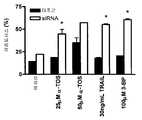


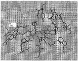


Classifications
- A—HUMAN NECESSITIES
- A61—MEDICAL OR VETERINARY SCIENCE; HYGIENE
- A61K—PREPARATIONS FOR MEDICAL, DENTAL OR TOILETRY PURPOSES
- A61K31/00—Medicinal preparations containing organic active ingredients
- A61K31/33—Heterocyclic compounds
- A61K31/335—Heterocyclic compounds having oxygen as the only ring hetero atom, e.g. fungichromin
- A61K31/35—Heterocyclic compounds having oxygen as the only ring hetero atom, e.g. fungichromin having six-membered rings with one oxygen as the only ring hetero atom
- A61K31/352—Heterocyclic compounds having oxygen as the only ring hetero atom, e.g. fungichromin having six-membered rings with one oxygen as the only ring hetero atom condensed with carbocyclic rings, e.g. methantheline
- A61K31/353—3,4-Dihydrobenzopyrans, e.g. chroman, catechin
- A61K31/355—Tocopherols, e.g. vitamin E
- A—HUMAN NECESSITIES
- A61—MEDICAL OR VETERINARY SCIENCE; HYGIENE
- A61K—PREPARATIONS FOR MEDICAL, DENTAL OR TOILETRY PURPOSES
- A61K31/00—Medicinal preparations containing organic active ingredients
- A61K31/66—Phosphorus compounds
- A61K31/662—Phosphorus acids or esters thereof having P—C bonds, e.g. foscarnet, trichlorfon
- A—HUMAN NECESSITIES
- A61—MEDICAL OR VETERINARY SCIENCE; HYGIENE
- A61P—SPECIFIC THERAPEUTIC ACTIVITY OF CHEMICAL COMPOUNDS OR MEDICINAL PREPARATIONS
- A61P35/00—Antineoplastic agents
- A—HUMAN NECESSITIES
- A61—MEDICAL OR VETERINARY SCIENCE; HYGIENE
- A61P—SPECIFIC THERAPEUTIC ACTIVITY OF CHEMICAL COMPOUNDS OR MEDICINAL PREPARATIONS
- A61P43/00—Drugs for specific purposes, not provided for in groups A61P1/00-A61P41/00
- G—PHYSICS
- G01—MEASURING; TESTING
- G01N—INVESTIGATING OR ANALYSING MATERIALS BY DETERMINING THEIR CHEMICAL OR PHYSICAL PROPERTIES
- G01N33/00—Investigating or analysing materials by specific methods not covered by groups G01N1/00 - G01N31/00
- G01N33/48—Biological material, e.g. blood, urine; Haemocytometers
- G01N33/50—Chemical analysis of biological material, e.g. blood, urine; Testing involving biospecific ligand binding methods; Immunological testing
- G01N33/5005—Chemical analysis of biological material, e.g. blood, urine; Testing involving biospecific ligand binding methods; Immunological testing involving human or animal cells
- G01N33/5008—Chemical analysis of biological material, e.g. blood, urine; Testing involving biospecific ligand binding methods; Immunological testing involving human or animal cells for testing or evaluating the effect of chemical or biological compounds, e.g. drugs, cosmetics
- G01N33/5076—Chemical analysis of biological material, e.g. blood, urine; Testing involving biospecific ligand binding methods; Immunological testing involving human or animal cells for testing or evaluating the effect of chemical or biological compounds, e.g. drugs, cosmetics involving cell organelles, e.g. Golgi complex, endoplasmic reticulum
- G01N33/5079—Mitochondria
- G—PHYSICS
- G01—MEASURING; TESTING
- G01N—INVESTIGATING OR ANALYSING MATERIALS BY DETERMINING THEIR CHEMICAL OR PHYSICAL PROPERTIES
- G01N33/00—Investigating or analysing materials by specific methods not covered by groups G01N1/00 - G01N31/00
- G01N33/48—Biological material, e.g. blood, urine; Haemocytometers
- G01N33/50—Chemical analysis of biological material, e.g. blood, urine; Testing involving biospecific ligand binding methods; Immunological testing
- G01N33/53—Immunoassay; Biospecific binding assay; Materials therefor
- G—PHYSICS
- G01—MEASURING; TESTING
- G01N—INVESTIGATING OR ANALYSING MATERIALS BY DETERMINING THEIR CHEMICAL OR PHYSICAL PROPERTIES
- G01N33/00—Investigating or analysing materials by specific methods not covered by groups G01N1/00 - G01N31/00
- G01N33/48—Biological material, e.g. blood, urine; Haemocytometers
- G01N33/50—Chemical analysis of biological material, e.g. blood, urine; Testing involving biospecific ligand binding methods; Immunological testing
- G01N33/82—Chemical analysis of biological material, e.g. blood, urine; Testing involving biospecific ligand binding methods; Immunological testing involving vitamins or their receptors
Landscapes
- Health & Medical Sciences (AREA)
- Life Sciences & Earth Sciences (AREA)
- Engineering & Computer Science (AREA)
- Chemical & Material Sciences (AREA)
- Immunology (AREA)
- Biomedical Technology (AREA)
- General Health & Medical Sciences (AREA)
- Medicinal Chemistry (AREA)
- Urology & Nephrology (AREA)
- Molecular Biology (AREA)
- Hematology (AREA)
- Pharmacology & Pharmacy (AREA)
- Animal Behavior & Ethology (AREA)
- Public Health (AREA)
- Veterinary Medicine (AREA)
- General Physics & Mathematics (AREA)
- Biotechnology (AREA)
- Cell Biology (AREA)
- Pathology (AREA)
- Microbiology (AREA)
- Biochemistry (AREA)
- Food Science & Technology (AREA)
- Physics & Mathematics (AREA)
- Analytical Chemistry (AREA)
- Epidemiology (AREA)
- Bioinformatics & Cheminformatics (AREA)
- Nuclear Medicine, Radiotherapy & Molecular Imaging (AREA)
- Chemical Kinetics & Catalysis (AREA)
- General Chemical & Material Sciences (AREA)
- Organic Chemistry (AREA)
- Toxicology (AREA)
- Tropical Medicine & Parasitology (AREA)
- Pharmaceuticals Containing Other Organic And Inorganic Compounds (AREA)
- Medicines That Contain Protein Lipid Enzymes And Other Medicines (AREA)
- Investigating Or Analysing Biological Materials (AREA)
- Acyclic And Carbocyclic Compounds In Medicinal Compositions (AREA)
- Medicinal Preparation (AREA)
- Pyrane Compounds (AREA)
Abstract
Translated fromKoreanDescription
Translated fromKorean본 발명은 특히 항암 화합물 및 암의 예방 또는 치료 방법에 관한 것이다. 특히, 본 발명은 산화 촉진제 항암 화합물, 예컨대 비타민 E 의 산화 촉진제 형태로서, 암세포의 미토콘드리아 호흡 사슬의 복합체 II 와 선별적으로 상호작용하여 활성 산소종을 생성하고 그 세포들의 아폽토시스(apoptosis)를 유도하는 화합물에 관한 것이다.The present invention particularly relates to a method for the prophylaxis or treatment of an anti-cancer compound and cancer. In particular, the present invention relates to an antioxidant composition comprising an antioxidant compound, for example, vitamin E as an oxidizing accelerator, which selectively interacts with mitochondrial respiratory chain complex II of cancer cells to produce active oxygen species and induce apoptosis of the cells ≪ / RTI >
이상적인 항암 약물은, 오직 암 세포 내부에서만 활성이며, 그 세포들의 핵심적인 구성요소를 표적으로 하거나 또는 핵심적인 프로세스를 방해하는 것이다. 세포 에너지의 대부분을 공급하며 아폽토시스의 중요 조절자인 미토콘드리아는, 항암 요법에 있어서 바람직한, 세포 선별성을 제공할 수 있는 유효한 표적으로서 대두되고 있다 (Don 및 Hogg, 2004, Armstrong, 2006).An ideal chemotherapeutic drug is only active within cancer cells, targeting the key components of the cells or interfering with key processes. Mitochondria, which supply most of the cell's energy and are important regulators of apoptosis, have emerged as effective targets for providing cell selectivity that is desirable for chemotherapy (Don and Hogg, 2004, Armstrong, 2006).
암 세포에서 미토콘드리아에 선별적으로 작용함으로서 활성을 나타내는 세포독성 약물인 "마이토칸(mitocan)" 은, 동물 실험에서 보고된 바와 같이 부작용이 거의 없는 항암제로서 작용할 수 있기 때문에 암 치료에 있어서 상당히 매력이 있는 것으로 증명되고 있다 (Ko 등, 2004). 마이토칸은 암 세포 미토콘드리아의 에너지 생산 시스템을 방해하여, 암 세포 내부에서의 활성 산소종 (ROS) 증가 및 미토 콘드리아 의존성 세포 사멸 시그널링 경로 활성화를 유도한다."Mitocan," a cytotoxic drug that selectively activates mitochondria in cancer cells, is quite attractive for cancer treatment because it can act as an anticancer drug with few side effects as reported in animal studies (Ko et al., 2004). Mytokan interferes with the energy production system of cancer cell mitochondria, leading to increased reactive oxygen species (ROS) within cancer cells and activation of the mitochondrial dependent cell death signaling pathway.
마이토칸에는 하기와 같은 미토콘드리아 관련 활성에 영향을 주는 약물이 포함된다: 헥소키나아제 저해제; 전자 전달/호흡 사슬 차단제; 전압 의존성 음이온-선별 채널 (VDAC) 또는 아데닌 뉴클레오티드 수송자 (ANT) 인, 미토콘드리아 막 투과성 전이 포어 표적화 구성원 단백질 서브유닛의 활성화제; Bcl-2 항-아폽토시스 패밀리 단백질 및 Bax/Bid 아폽토시스 촉진성 모방체의 저해제. 부작용이 거의 없는, 마이토칸의 두가지의 대표적인 예시는 3-프로모피루베이트 (3-BP) 및 α-토코페릴 숙시네이트 (α-TOS) 인데, 이들 두가지 모두 정상 세포에서보다 암 세포에서 훨씬 더 큰 효능을 갖고 아폽토시스를 유도한다 (Ko 등, 2004, Geschwind 등, 2002, Xu 등, 2005, Neuzil 등, 2001, 2004).Mytokans include drugs that affect mitochondrial-related activities such as: hexokinase inhibitors; Electron transfer / respiratory chain blocker; An activator of a mitochondrial transmembrane transfer target targeting member protein subunit that is a voltage dependent anion-selective channel (VDAC) or an adenine nucleotide transporter (ANT); Inhibitors of Bcl-2 anti-apoptosis family proteins and Bax / Bid apoptosis promoting mimetics. Two representative examples of mytokans with few side effects are 3-proparubrate (3-BP) and α-tocopheryl succinate (α-TOS), both of which are much more (Ko et al., 2004, Geschwind et al., 2002, Xu et al., 2005, Neuzil et al., 2001, 2004).
한 무리의 마이토칸에는 비타민 E 의 산화 촉진제 유사체가 포함된다 (Wang 등, 2006). α-TOS 가 전형인, 항암 약물로서 크게 전도유망한 산화 촉진성 비타민 E 유도체는, 누드 마우스에서 성장하는 인간 이종이식물과 같이 실험실에서 인위적으로 유도한 암을 통한 연구에서 유래했으며, 이들은 악성종약을 억제하는 것으로 나타났다 (Neuzil 등이 2004 년도에 리뷰). 그러한 연구에는, 직장 (Neuzil 등, 2001, Weber 등, 2002) 및 폐 암종 (Quin 등, 2005), 흑색종 (Malafa 등, 2002), 뿐만 아니라 중피종 (Tomasetti 등, 2004, Stapelberg 등, 2005) 이 포함된다. α-TOS 는 또한 유방암 휴면 상태를 촉진하며 (Malafa 등, 2000), 대장암의 간으로의 전이를 억제하는 것으로 나타났다 (Barnett 등, 2002).A group of mytocans contains the oxidation promoter analog of vitamin E (Wang et al., 2006). A highly promising antioxidant vitamin-E derivative, which is typical of α-TOS, is derived from a study of artificially induced cancers in humans, such as plants, in human dysplasia growing in nude mice, (Reviewed in 2004 by Neuzil et al.). Such studies have included mesenchymal (Tomasetti et al., 2004, Stapelberg et al., 2005) as well as rectal (Neuzil et al., 2001, Weber et al., 2002) and lung carcinoma (Quin et al., 2005), melanoma . α-TOS also promotes breast cancer dormancy (Malafa et al., 2000) and inhibits metastasis of colon cancer to the liver (Barnett et al., 2002).
비타민 E (α-토코페롤, α-TOH) 가 세포 내에서는 강력한 항산화제로서 작 용하지만, 에스테르화되고 산화환원-침묵성인 비타민 E 의 산화 촉진제 유사체인 α-TOS 는 특별한 특징을 갖는다. α-TOH 와는 대조적으로, α-TOS 는 강력한 세포 스트레스 유발자로서 작용하며, 일련의 상이한 암 세포주에서는 ROS 의 신속한 생산을 유발한다 (Neuzil 등, 2004, Weber 등, 2003, Wang 등, 2005, Swettenham 등, 2005, Stapelberg 등, 2005). α-TOS 는 또한 Bcl-2/Bcl-xL 에 결합하여 저해하는 능력을 갖고 있다 (Dong 등, 2006). 현재까지의 증거로는, α-TOS 의 암 세포 특이적인 특징 및 정상 세포에 대한 독성 효과의 결핍이, 정상 세포가 부여받은 훨씬 더 큰 항산화 방어능 (Allen 및 Balin, 2003, Safford 등, 1994, Church 등, 1993) 및/또는 숙시네이트 부분을 방출함으로써 α-TOS 를 불활성화시켜 산화환원-활성인 비-아폽토시스발생성 α-TOH 를 생산하는 고수준의 에스테라아제의 포함 (Fariss 등, 2001, Neuzil 등, 2004, Neuzil and Massa, 2005) 때문에 발생하는 것임을 시사한다.Although vitamin E (α-tocopherol, α-TOH) acts as a potent antioxidant in cells, α-TOS, which is esterified and is an oxidation promoter analogue of redox-silent vitamin E, has a special feature. In contrast to α-TOH, α-TOS acts as a potent cellular stressor and causes rapid production of ROS in a series of different cancer cell lines (Neuzil et al., 2004, Weber et al., 2003, Wang et al., 2005, Swettenham , 2005, Stapelberg et al., 2005). α-TOS also has the ability to bind and inhibit Bcl-2 / Bcl-xL (Dong et al., 2006). To date, the lack of toxic effects of α-TOS on cancer cell specificity and toxicity to normal cells has been attributed to a much greater antioxidant defense function given by normal cells (Allen and Balin, 2003, Safford et al., 1994, (Fariss et al., 2001, Neuzil et al., 1993) and / or the inactivation of? -TOS by releasing a succinate moiety to produce a redox-active, non-apoptogenic α-TOH , 2004, Neuzil and Massa, 2005).
자연발생적인 비타민 E 는, 크로마놀 고리의 메틸화 패턴이 상이하고 (α-, β-, γ-, δ-토코페롤), 피틸 측쇄의 이중결합이 상이한 (α-, β-, γ-, δ-토코트리에놀) 여덟가지 화합물의 혼합물로 이루어져 있다. 시험관내 및 생체내에서 친유성 항산화제로서의 상기 분자들의 역할은 널리 받아들여지고 있다. 추가로, VE 패밀리 구성원의 비-항산화 특징도 또한 연구된 바 있다 (Azzi 등, 2002).Naturally occurring vitamin E has a different chromatin ring pattern (α-, β-, γ-, and δ-tocopherol) and different double bonds in the pyryl side chain (α-, β-, γ-, Tocotrienol) consists of a mixture of eight compounds. The role of these molecules as lipophilic antioxidants in vitro and in vivo is widely accepted. In addition, non-antioxidant features of VE family members have also been studied (Azzi et al., 2002).
비타민 E 분자는 3 개의 상이한 도메인으로 구분될 수 있다.관능성 도메인 (I) 은 크로마놀 고리의 C6 위치에서의 치환 패턴으로 인한 것이다. 상기 위치는 분자가 산화환원-활성 또는 산화환원-침묵성으로 거동하는지 여부를 결정하는데, 이는, 유리된 히드록실기는 비타민 E 가 항산화제로서 작용하는데 있어서 핵심적이기 때문이다. 문서기록에 의해 충분히 입증된 4 가지 토코페놀 동질이성체에 대한 항산화 특성은 암 임상 시험에서의 그의 응용성을 제공했다. 이들 연구 중 그 어느 것도 암 예방에서의 유리된 토코페롤의 용도에 관한 긍정적인 성과를 보여주진 않았다 (Pham 및 Plakogiannis, 2005). 그러나, C6 에서의 특정한 화학적 변경은 에테르 (RO-), 에스테르 (RCOO-) 및 아미드 (RCONH-) 를 유도하여, 강력한 항신생물제임을 증명했다. 하기의 표 1 참조.Vitamin E molecules can be divided into three different domains.The functionaldomain (I) is due to the substitution pattern at the C6 position of the chromanol ring. This position determines whether the molecule behaves as redox-active or redox-silent, since the liberated hydroxyl group is essential for vitamin E to act as an antioxidant. The antioxidant properties of the four tocopherol isomers well documented by the documentation record provided its applicability in cancer clinical trials. Neither of these studies showed a positive outcome on the use of the liberated tocopherol in cancer prevention (Pham and Plakogiannis, 2005). However, certain chemical changes in C6 have led to ether (RO-), ester (RCOO-) and amide (RCONH-), proving to be a potent anti-neoplastic agent. See Table 1 below.
[표 1][Table 1]


a효과 없음;b세포 증식 저해;cα-TOS 보다 훨씬 더 세포독성;d54 보다는 덜 유효함;e에테르 유사체는 α-TOS 자체보다는 덜 유효함;fα-TOS 에 필적함;gEC50[㎍/ml];hα-TOS 보다 더욱 유효함.a No effect;b Inhibiting cell proliferation;c More cytotoxic than α-TOS; less valid thand54 ;e ether analog is also less effective than α-TOS itself; comparable tof ? -TOS;g EC50 [[mu] g / ml];h More effective than α-TOS.
[표 2][Table 2]

a효과 없음;b50 μM 에서 약한 저해;c10 μM 에서 82% 저해.a No effect;b Weak inhibition at 50 μM;c 82% inhibition at 10 μM.
[표 3][Table 3]

a0-40 μM 범위에서 세포독성;b매우 강력함;c완전 저해;dα-TOS 에 필적 함;eγ-토코트리에놀보다 2 배 더 강력함; f세포 증식 억제.a cytotoxicity in the 0-40 μM range;b Very powerful;c complete inhibition; comparable tod ? -TOS;e 2-fold stronger than γ-tocotrienol;f cell proliferation inhibition.
시그널링 도메인 (II) 으로 지칭하는 두번째 도메인은, 토코페롤의 항산화 특성과는 독립적인 소정의 활성을 나타낸다. 상기 특성은 방향족 고리의 메틸화 패턴에 기인한다. 예를 들어, 나머지 토코페롤들(예를 들어, β-토코페롤)은 비슷한 항산화 능력을 갖고 PKC 를 저해하지 않는 반면, α-토코페롤은 디아실글리세롤 (DAG) 수준을 감소시킴으로써 단백질 키나아제 C (PKC) 를 저해하는 것으로 보고되었다. 따라서, α-토코페롤의 PKC 저해 활성은 그의 항산화 역량과는 독립적이다 (Tasinato 등, 1995; Kunisaki 등, 1995). 그러나, 일부의 경우, 각종 토코페롤의 생물학적 활성은 시그널링 도메인의 구조적 차이에 영향을 받는데, 그 도메인은 특정 종에 대하여 그들의 항산화 활성에 대해 실로 중요한 영향력을 갖는다. 예를 들어, γ-토코페롤은, α-토코페롤보다 활성 산화질소종 (예를 들어, 페록시니트라이트) 에 대한 훨씬 더 나은 스캐빈저이다. 따라서, C5 에서 메틸기가 결핍된 γ-분자는 상기 부위에서 쉽게 니트로화된다 (Morton 등, 2002; Christen 등, 1997).The second domain, referred to as thesignalingdomain (II), exhibits certain activities that are independent of the antioxidant properties of tocopherol. This property is due to the methylation pattern of the aromatic ring. For example, the remaining tocopherols (eg, β-tocopherol) have similar antioxidant capacity and do not inhibit PKC, while α-tocopherol reduces protein kinase C (PKC) by decreasing diacylglycerol Lt; / RTI > Thus, the PKC inhibitory activity of alpha -tocopherol is independent of its antioxidant capacity (Tasinato et al., 1995; Kunisaki et al., 1995). However, in some cases, the biological activity of various tocopherols is affected by the structural differences in the signaling domains, which have indeed important effects on their antioxidant activity against certain species. For example, gamma -tocopherol is a much better scavenger for active nitric oxide species (e. G., Peroxynitrite) than alpha -tocopherol. Thus, the γ-molecule lacking the methyl group at C5 is easily nitrated at the site (Morton et al., 2002; Christen et al., 1997).
비타민 E 동질이성체의 친유성 측쇄는, 포화 이소프레닐 유닛을 가진 토코페롤과 불포화 이소프레닐 유닛을 가진 토코트리에놀을 구분짓게 한다.소수성 도메인 (III) 은 분자가 리포단백질 및 막에 각각 결합하게 되거나 또는 제 I 상 효소에 의해 분해되는지 여부를 결정한다 (Birringer 등, 2002; Neuzil 및 Massa, 2005).The lipophilic side chain of the vitamin E isomer distinguishes between tocopherol having a saturated isoprenyl unit and tocotrienol having an unsaturated isoprenyl unit.The hydrophobic domain (III) determines whether the molecule binds to lipoproteins and membranes respectively or is degraded by Phase I enzymes (Birringer et al., 2002; Neuzil and Massa, 2005).
변경된 히드록실기를 가진 많은 토코페롤 유도체들이 그의 아폽토시스 촉진 활성에 대해 시험된 바 있다 (표 1). 시험했던 대부분의 탁월한 유도체들은 크로마놀 고리의 C6 위치에 숙시닐에스테르를 가진 α-TOS 였다 (엔트리 1). 그의 낮은 pKa (< 6) 로 인해, α-TOS 는 생리학적 조건 하에서는 완전히 탈양자화되며, 이는 미토콘드리아 막을 불안정하게 하고, 복합체 II 에 영향을 미치는 계면활성제같은 분자를 유도한다. 토코페롤의 디카르복실 에스테르는 구조-활성 관계 (structure-activity relationship; SAR) 에 있어서 연구되었던 최상의 화합물들을 제시한다. 강력한 아폽토겐 (apoptogen) 에는 α-토코페롤 숙시네이트 (1), 옥살레이트 (10) 및 말로네이트 (11) 가 포함되는데, 뒤의 두가지는 B16-F1 흑색종 세포가 접종된 마우스에서 비선별적인 세포독성을 유도했다 (Kogure 등, 2005). 더욱더 큰 아폽토시스 촉진 활성은 α-토코페릴 말리에이트 (3) (Birringer 등, 2003) 및 α-토코페릴 푸마레이트와 같은 불포화 디카르복실산에서 관찰되었다. 디카르복실산의 사슬 길이 증가는, 전혀 활성이 없는 피멜산 (24) (Kogure 등, 2004)을 가진 글루타르산 (5), 메틸화 글루타르산 (6,7,8) (Birringer 등, 2003) 에 대해 나타난 바와 같이 활성 감소를 유도한다.Many tocopherol derivatives with altered hydroxyl groups have been tested for their apoptosis promoting activity (Table 1). Most of the excellent derivatives tested were α-TOS with succinyl ester at the C6 position of the chromanol ring (Entry 1). Due to its low pKa (<6), α-TOS is completely deprotonated under physiological conditions, which leads to molecules like surfactants that make mitochondrial membranes unstable and affect complex II. The dicarboxylic esters of tocopherol suggest the best compounds that have been studied in their structure-activity relationship (SAR). Strong apoptogenes include alpha-tocopherol succinate (1 ), oxalate (10 ) and malonate (11 ), the latter two of which are associated with unplanned cytotoxicity in mice inoculated with B16- (Kogure et al., 2005). The greater apoptosis promoting activity was observed in alpha-tocopheryl malate (3 ) (Birringer et al., 2003) and in unsaturated dicarboxylic acids such as alpha-tocopheryl fumarate. The increase in the chain length of the dicarboxylic acid is due to the addition of glutaric acid (5 ), methylated glutaric acid (6 ,7 ,8 ) (Birringer et al., 2003) with completely inactive pimelic acid (24 ) Lt; RTI ID = 0.0 > as < / RTI >
완전한 아폽토시스 유도 활성에는 전체 α-TOS 분자가 필요하다는 것이 확립된 바 있다 (Birringer 등, 2003). 유리된 카르복실기의 에스테르화는 아폽토시스 촉진 활성이 없는 비하전 유도체들을 유도한다 (9, 25). 지환족 카르복실산 에스테르, 예컨대 토코페릴 아세테이트 및 프로피오네이트 (19) 는 각각, 메틸 에테르 (18) 와 마찬가지로 불활성이다. α-TOS 의 경구 투여는 유효하지 않은데, 이는 상 기 화합물이 장의 에스테라아제에 의해 절단되기 때문이다 (Wu 등, 2004b; Cheeseman 등, 1995). 에스테르 결합 절단의 문제를 극복하기 위해, 화합물들 (20,21) 및 측쇄 단절 유도체 (42) 를 합성하고, 에테르 결합은 가수분해에 대해 내성이 있으므로, 에스테르 결합을 에테르 결합으로 치환했다 (Wu 등, 2004b; Nishikawa 등, 2003; Shun 등, 2004; Shiau 등, 2006). 메틸렌기에 의한 에테르 결합의 치환 (22) 은 아폽토시스 가속화에 효과적이라는 점에 유의한다 (Sanders 등, 2001).It has been established that total α-TOS molecules are required for complete apoptosis inducing activity (Birringer et al., 2003). Esterification of the liberated carboxyl groups leads to uncharged derivatives without apoptosis promoting activity (9 ,25 ). Alicyclic carboxylic acid esters such as tocopheryl acetate and propionate (19 ), respectively, are inert, like methyl ether (18 ). Oral administration of a-TOS is not effective because the compounds are cleaved by the intestinal esterase (Wu et al., 2004b; Cheeseman et al., 1995). In order to overcome the problem of ester bond cleavage, the compounds (20 ,21 ) and the side chain disconnection derivative (42 ) were synthesized and the ether bond was resistant to hydrolysis and the ester bond was replaced by an ether bond , 2004b; Nishikawa et al., 2003; Shun et al., 2004; Shiau et al., 2006). It is noted that substitution (22 ) of ether linkages by methylene groups is effective for accelerating apoptosis (Sanders et al., 2001).
에스테르 결합을 아미드 결합으로 치환한 경우, 아폽토시스 촉진 활성의 추가적 증강이 관찰되었다 (12,13,37,38) (Tomic-Vatic 등, 2005). 게다가, 불포화 아미드 (13,38) 는 포화 아미드보다 월등하다. 에스테르를 대신하여 아미드 결합을 도입함에 대한 원리는, 아닐린계 아미드가 그의 대응하는 페놀계 에스테르보다 가수분해에 대해 더욱더 덜 친화적이라는, 충분히 입증된 사실에 근거한다. 상기 토코페릴 에스테르 유도체의 안정성 증강은 이들 분자들을 생체내에서 보호하여, 이들이 온전하게 더 오래 머물도록 할 것이며, 그럼으로써 그의 생체이용능을 증가시킨다. 아미드에 의한 에스테르의 등전자성 치환은 또한 상기 연결이 효소적 가수분해에 덜 친화적이게끔 한다. 몇가지 비특이적인 에스테라아제가 장 점막 세포 및 혈액에 존재한다. 대조적으로, 펩티다아제에는 훨씬 더 좁은 특이성이 존재한다. 예를 들어, 아미드 연결에 아미노산이 있는 프로드러그는 장 및 혈액에서, 그의 대응하는 에스테르 유사체들보다는 더욱 안정하다 (Sugawara 등, 2000).Further enhancement of apoptosis promoting activity was observed when the ester linkage was replaced by an amide linkage (12 ,13 ,37 ,38 ) (Tomic-Vatic et al., 2005). In addition, the unsaturated amides (13 ,38 ) are superior to the saturated amides. The principle of introducing amide bonds in place of esters is based on the well-established fact that aniline amides are even less amenable to hydrolysis than their corresponding phenolic esters. Stability enhancement of the tocopheryl ester derivatives will protect these molecules in vivo, allowing them to stay longer and thus increase their bioavailability. Isomeric substitution of an ester with an amide also makes the link less amenable to enzymatic hydrolysis. Several non-specific esterases exist in the mucosa cells and blood. In contrast, peptidases have much narrower specificity. For example, prodrugs with amino acids in the amide linkage are more stable in their intestines and blood than their corresponding ester analogs (Sugawara et al., 2000).
마지막 군의 화합물들은 양으로 하전된 N-말단을 가진 일련의 라이신 α-토 코페릴 에스테르로 이루어졌다 (15-17). 친수성인 암모늄 관능기는 그의 카르복실레이트 카운터파트와 유사한 아폽토시스 촉진 효과를 발휘하여, 친유성 측쇄 및 친수성 헤드 그룹으로 이루어진 공통 모티프가 활성에 필요함을 시사했다. 그러나, 장쇄 지환족 알콜의 숙시닐 에스테르 (예를 들어, 피톨 및 올레올) 는 그 어떤 활성도 나타내지 않았다 (Birringer 등, 2003).The compounds of the last group are consisted of a series of co-Lysine α- Sat tocopheryl ester with the N- terminal charged in an amount(15 -17). The hydrophilic ammonium functional groups exert apoptosis promoting effects similar to their carboxylate counterparts, suggesting that a common motif consisting of lipophilic side chain and hydrophilic head groups is required for activity. However, succinyl esters of long-chain alicyclic alcohols (e.g., phytol and oleol) did not show any activity (Birringer et al., 2003).
일반적인 SAR 은 표 1 에 나타낸 데이터로부터 이끌어낼 수 있다:A typical SAR can be derived from the data shown in Table 1:
1. 충분한 산화 촉진 및 아폽토시스 촉진 활성을 수득하기 위해서는,관능성 도메인 I 의 변경이, 분열된 산 또는 하전된 암모늄기로 이루어진 친수성 헤드기를 필요로 했다.1. In order to obtain sufficient oxidation promoting and apoptosis promoting activity, the modification of thefunctional domain I required a hydrophilic head group consisting of a cleaved acid or a charged ammonium group.
2.관능성 도메인 불포화도 및 사슬 길이는 아폽토시스발생 활성을 결정했다. 배좌의 구속성 (Conformational restriction) 은 활성을 더 강하게 하는 것으로 나타났다.2. Thefunctional domain degree of unsaturation and chain length determined apoptotic activity. Conformational restriction was found to make the activity stronger.
3.관능성 도메인 화학 연결은 에스테르에 한정되는 것이 아니라, 기타 관능기들도 유도체들의 효소적 분해를 방지했다.3.Functional domains The chemical linkage is not limited to esters but other functional groups have also prevented enzymatic degradation of the derivatives.
크로마놀 고리의 치환 패턴은 종종 토코페롤의 항산화 특성에만 관련된 것은 아니다 (Azzi 등, 2002). 상이한 생화학적 관찰사항들이 시그널링 및 대사 프로세스에서의 α-토코페롤의 역할을 강조해준다. 이에 따라, α-토코페롤은, 다른 토코페롤 및 토코트리에놀에 비해 α-토코페롤에 대해 높은 친화성이 있는 32 kDa 단백질인 α-토코페롤 이동 단백질 (α-TTP) 에 의해 간에서 선별적으로 인지된다. α-TTP 에 대한 상대적인 친화성은 크로마놀 고리의 메틸화 소실과 함께 감소한다 (α-토코페롤 100 %, β-토코페롤 38%, γ-토코페롤 9% 및 δ-토코페롤 2%) (Hosomi 등, 1997). 최근 발견된 토코페롤 회합성 단백질 (TAP) 은 토코페롤 결합에 있어서 유사한 선호성을 보여준다 (Yamauchi 등, 2001). 내피 세포에서, 트롬빈-유도성 PKC 활성화 및 엔도텔린 분비는 β-토코페롤이 아닌 α-토코페롤에 의해 저해된다 (Martin-Nizard 등, 1998). 전사 수준에서, α-토코페롤은 α-트로포마이오신 발현의 상향조절 (Aratri 등, 1999) 및 LDL 스캐빈저 수용체 SR-A 및 CD36 의 하향조절을 초래하는 한편, β-토코페롤은 효과가 없다 (Ricciarelli 등, 2000; Devaraj 등, 2001). 추가로, 치환 패턴은 측쇄 분해 속도에 영향을 줄 수 있는데, 이는 세포 배양에서, γ- 및 δ-토코페롤이 α- 또는 β-토코페롤보다는 훨씬 빨리 분해되기 때문이다 (Birringer 등, 2001). 4 가지 토코페롤 이성질체의 숙시닐화는 화합물1,32,33 및35 를 제공한다. 이들 중에서, α-TOS (1) 가, 시험한 최고의 아폽토시스발생 활성을 보유하며, 뒤이어 β-TOS (32), γ-TOS (33) 및 δ-TOS (35) 로 올수록 덜 유효하다는 것은 그리 놀랍지 않다 (Birringer 등, 2001). 일반적으로, 토코페롤 패밀리 중에서 더 많이 메틸화된 구성원들이 가장 강력하나, 이러한 경향성은 토코트리에놀에 대해서는 역전된다 (하기 참조).The substitution pattern of the chromanol ring is often not only related to the antioxidant properties of tocopherol (Azzi et al., 2002). Different biochemical observations highlight the role of alpha-tocopherol in signaling and metabolic processes. Thus, alpha -tocopherol is selectively recognized in the liver by the alpha-tocopherol transfer protein (alpha-TTP), a 32 kDa protein with high affinity for alpha -tocopherol compared to other tocopherols and tocotrienols. The relative affinity for alpha -TTP decreases with methylation loss of the chromanol ring (100% alpha -tocopherol, 38% beta -tocopherol, 9% gamma -tocopherol and 2% delta-tocopherol) (Hosomi et al., 1997). Recently discovered tocopherol conformational protein (TAP) shows a similar preference for tocopherol binding (Yamauchi et al., 2001). In endothelial cells, thrombin-induced PKC activation and endothelin secretion are inhibited by? -Tocopherol, not? -Tocopherol (Martin-Nizard et al., 1998). At the transcription level, alpha -tocopherol results in upregulation of alpha-tropomyosin expression (Aratri et al., 1999) and downregulation of the LDL scavenger receptors SR-A and CD36, while beta -tocopherol is ineffective Ricciarelli et al., 2000; Devaraj et al., 2001). In addition, the substitution pattern can affect the rate of side chain degradation because in the cell culture, gamma- and delta-tocopherol degrade much faster than alpha or beta-tocopherol (Birringer et al., 2001). Succinylation of the four tocopherol isomers provides
더 짧은 측쇄를 가진, 수용성 비타민 E 유도체인 트롤록스 (Trolox) 의 숙시닐화는 아폽토시스 촉진 활성의 완전한 소실을 초래한다. 단절된 피톨 측쇄를 가진 각종 토코페롤 숙시네이트 (표 2,43,44,45) 의 SAR 실험은, 전립선 암 세포에서의 아폽토시스발생 활성의 최고 수준은 측쇄 길이가 2 개의 이소프레닐 유닛인 유도체 (43,44) 로 수득된다는 것을 밝혀냈다. 컴퓨터가 보조한 분자 모델링 및 공 동-면역침전 실험은, Bcl-xL 및 Bcl-2 에 대한 Bak BH3 펩티드의 결합이 토코페롤 유사체에 의해 저해된다는 것을 보여준다 (Shiau 등, 2006). 항-신생물 활성에 대한 중추적인 요건은 크로마놀 고리의 숙시닐화 및 1 개의 이소프레닐 유닛인 최소한의 사슬 길이였다 (42,46).도메인 III 측쇄에 연결된 에테르/에스테르가 있는 일련의 토코페릴 라이신 에스테르는 또한 사슬 길이와 IC50 사이에 음의 상관관계를 나타냈다 (47-50) (Arya 등, 1998).Succinylation of Trolox, a water soluble vitamin E derivative with shorter side chains, results in complete loss of apoptosis promoting activity. Various tocopheryl succinate with disconnected Capitol side chain (Table 2,43,44,45) of SAR experiments, the highest level of apoptosis-generated activity in prostate cancer cells is the side chain length of the two iso-prenyl units derivative(43,44 ). ≪ / RTI > Computer-assisted molecular modeling and co-immunoprecipitation experiments show that binding of Bak BH3 peptides to Bcl-xL and Bcl-2 is inhibited by tocopherol analogues (Shiau et al., 2006). The key requirement for anti-neoplastic activity was succinylation of the chromanol ring and minimal chain length, which is one isoprenyl unit (42,46 ). A series of tocopheryl lysine ester with ether / esters linked to a side chaindomain III are also shown a negative correlation between the chain length and theIC 50 (47 - 50) ( Arya et al., 1998).
토코트리에놀은 유효한 항암제이며, 그의 아폽토시스 촉진 특성은 단백질의 Ras 패밀리의 불활성화와 연관이 있을 수 있다. 토코트리에놀은관능성 도메인의 변경없이도 그의 아폽토시스 촉진 활성을 나타낸다.시그널링 도메인에서의 분류체계(hierarchy)는 또한 역전되어, δ-토코트리에놀 (59) 을 쥐과동물 B16-F10 흑색종 세포 모델에서 가장 강력한 약제가 되도록 하며, 그에 이어 γ- (56) 및 α-토코트리에놀 (53) 이 있다 (표 3; He 등, 1997). 흥미롭게도, 모든 방향족 메틸기가 결핍된 데스메틸 토코트리에놀 (60) 은, IC50 가 0.9 μM 인 훨씬 더 높은 활성을 보인다. 상기 화합물은 쌀겨에서 분리되었다 (Qureshi 등, 2000). 토코트리에놀 직접적인 저해 작용이 제안된 바 있는데, 이는 Ras 단백질의 멤브레인 고정 시스테인 잔기가 일반적인 구조 구성원인 파르네실 사슬에 의해 변경되기 때문이다. 이에 따라, Ras 파르네실화 및 RhoA 프레닐화는 A549 세포, 활성화ras 돌연변이를 가진 인간 폐 선암 세포주에서 토코트리에놀에 의해 저해된다 (Yano 등, 2005). 토코트리에놀의 짧은 생체내 반감기를 연장하기 위해, 관능성 도메인을 도입했다. 이러한 변경은 또한 분자의 항증식 활성을 증강시켰다 (54,57,58). 측쇄의 단절은 또한 화합물55 에 대해 발견된 바와 마찬가지로, 활성을 개선시켰다.Tokotrienol is an effective anticancer agent, and its apoptotic promoting properties may be associated with inactivation of the Ras family of proteins. Tokotrienol exhibits its apoptosis promoting activity without altering thefunctional domain .The hierarchy in thesignaling domain is also reversed to make? -Tocotrienol59 the most potent agent in the mouse and animal B16-F10 melanoma cell model followed by? - (56 ) and? -Tocotrienol53 ) (Table 3, He et al., 1997). Interestingly, desmethyltocotrienol (60 ), deficient in all aromatic methyl groups, shows much higher activity with an IC50 of 0.9 μM. The compound was isolated from rice bran (Qureshi et al., 2000). A direct inhibition of tocotrienol has been proposed because the membrane-immobilized cysteine residue of the Ras protein is altered by the common structural member paranesyl chain. Thus, Ras parunifacylation and RhoA prenylation are inhibited by tocotrienol in A549 cells, a human lung adenocarcinoma cell line with an activatedras mutation (Yano et al., 2005). To extend the short in vivo half-life of tocotrienol, a functional domain was introduced. This modification also enhanced the antiproliferative activity of the molecule (54 ,57 ,58 ). Disruption of the side chain also improved activity, as was found for compound55 .
관능성 도메인에 변경이 가해진 다수의 화합물들이 항증식 활성을 나타내며, 추가적인 특화된 특성들을 제공한다. 예를 들어, α-토코페릴 폴리에틸렌 글리콜 숙시네이트 (23) 는 약물 전달계에 대한 비히클로서 이용되어 왔다. 상기 화합물은 누드 마우스에 이식된 인간 폐 암종 세포에 대한 항암 활성을 보유하는 것으로 나타났다. 상기 화합물의 아폽토시스 유도 효능은, 세포에 대한 그의 흡수 증가로 인한 것이라기 보다는, 오히려 활성 산소종을 생성하는 능력 증대로 인한 것이다 (Youk 등, 2005). α-토코페릴 포스페이트 (30) 은 토코페롤-회합성 시그널링 도중에 발생하는 대사로 인한 것으로 여겨진다 (Negis 등, 2005).30 및 디-α-토코페릴 포스페이트 (31) 의 혼합물은 래트 대동맥 평활근 세포 및 인간 THP-1 단구성 백혈병 세포에서의 증식을 저해한다 (Munteanu 등, 2004). 상기 저자는 토코페릴 숙시네이트 및 토코페릴 말리에이트가 토코페릴 포스페이트에 대한 모방 및 치환에 의해 암 세포에 작용함으로써, 세포 시그널의 영구적인 활성화를 초래할 것이라고 제안했다.Many compounds with alteredfunctional domains exhibit antiproliferative activity and provide additional specialized properties. For example, alpha -tocopheryl polyethylene glycol succinate (23 ) has been used as a vehicle for drug delivery systems. The compound has been shown to have anticancer activity against human lung carcinoma cells transplanted into nude mice. The apoptosis-inducing effect of the compound is not due to its increased uptake to cells but rather to the increased ability to produce reactive oxygen species (Youk et al., 2005). It is believed that alpha -tocopheryl phosphate (30 ) is due to metabolism occurring during tocopherol-ash synthesis signaling (Negis et al., 2005).30 and di-alpha-tocopheryl phosphate (31 ) inhibits proliferation in rat aortic smooth muscle cells and human THP-I monocytic leukemia cells (Munteanu et al., 2004). The authors suggested that tocopheryl succinate and tocopheryl maliate act on cancer cells by mimicry and replacement for tocopheryl phosphate, resulting in permanent activation of the cell signal.
전부 트랜스인 레티노산 (28) 및 9-시스 레티노산 (29) 의 두가지 실험적인 α-토코페릴 에스테르는 각각, 급성 전골수구 백혈병 세포의 증식 감소에 이용된 바 있다 (Makishima 등, 1998). 레티노이드 수용체 응답성 리포터 구축물을 이용한 트랜스-활성화 실험은, 상기 화합물이 전부 레티노산 수용체 (RAR) 에 대한 아고니스트로서 작용한다는 것을 밝혔다. γ-토코페롤의 분해 생성물인 γ-카르복시에틸 히드록시크로만 (52) 은, 종종 요산 중에 분비되는 것으로 발견되는데, 싸이클린 D1 발현을 저해하여 PC-3 전립선 암 세포의 세포 증식을 감소시킬 수 있다 (Galli 등, 2004).Both experimental α-tocopheryl esters of trans-retinoic acid (28 ) and 9-cis retinoic acid (29 ) have been used to reduce the proliferation of acute promyelocytic leukemia cells, respectively (Makishima et al., 1998). Trans-activation experiments with retinoid receptor-responsive reporter constructs revealed that all of these compounds act as agonists for the retinoic acid receptor (RAR). γ-carboxyethylhydroxychroman (52 ), a degradation product of γ-tocopherol, is often found secreted in uric acid, which inhibits the expression of
발명의 개요Summary of the Invention
본 발명의 발명자들은, 특별히, 호흡 사슬의 복합체 II (숙시네이트-유비퀴논 옥시도리덕타아제) 를 표적화함으로써, 암 세포의 미토콘드리아 호흡 사슬을 따른 전자 이동의 선택적인 방해가 ROS 의 생성 및 상기 세포들의 사멸을 초래할 수 있다는 것을 발견했다. 특히, 본 발명의 발명자들은 비타민 E 의 산화 촉진제 형태가, 암 세포의 복합체 II 의 유비퀴논-결합 부위로부터 유비퀴논을 선택적으로 치환할 수 있고, 그로부터 나오는 전자들의 정상적인 이동을 차단한다는 면에서, 유효한 항암제가 될 수 있음을 발견했다.The inventors of the present invention have found that selective inhibition of electron transfer along the mitochondrial respiratory chain of cancer cells leads to the production of ROS and to the production of ROS by targeting the respiratory chain complex II (succinate-ubiquinone oxydoryldactase) Which can lead to death. In particular, the inventors of the present invention have found that the form of the oxidizing accelerator of vitamin E is capable of selectively substituting ubiquinone from the ubiquinone-binding site of the complex II of cancer cells and blocking the normal migration of electrons from the ubiquinone- It could be an anticancer drug.
발명의 상세한 설명DETAILED DESCRIPTION OF THE INVENTION
본 발명의 제 1 국면에 따르면, 암세포 사멸을 선별하여 유도하기 위한 산화 촉진제 화합물로서, 세포 내에서 암세포의 미토콘드리아 호흡 사슬의 복합체 II (숙시네이트-유비퀴논 옥시도리덕타아제) 와 상호작용하여 활성 산소종을 생성할 수 있는 산화 촉진제 화합물이 제공된다.According to a first aspect of the present invention, there is provided an oxidation promoter compound for selectively inducing cancer cell death, which comprises a compound capable of interacting with a complex II of a mitochondrial respiratory chain of cancer cells in a cell (succinate-ubiquinone oxidoreductase) An oxidation promoter compound capable of producing species is provided.
본 발명의 제 2 국면에 따르면, 암의 예방 또는 치료를 위한 산화 촉진제 화합물로서, 세포 내에서 암세포의 미토콘드리아 복합체 II 와 선별하여 상호작용하여 활성 산소종을 생성할 수 있는 산화 촉진제 화합물이 제공된다.According to a second aspect of the present invention, there is provided an oxidation promoter compound capable of selectively forming an active oxygen species with a mitochondrial complex II of cancer cells in a cell, as an oxidation promoter compound for preventing or treating cancer.
본 발명의 제 3 국면에 따르면, 대상체에서 암세포 사멸을 유도하는 방법으로서, 세포 내에서 암세포의 미토콘드리아 복합체 II 와 선별하여 상호작용하여 활성 산소종을 생성할 수 있는, 치료 유효량의 산화 촉진제 화합물을 대상체에게 투여하는 단계를 포함하는 방법이 제공된다.According to a third aspect of the present invention, there is provided a method for inducing cancer cell death in a subject, comprising the steps of: administering a therapeutically effective amount of an oxidizing promoter compound capable of selectively producing an active oxygen species with mitochondrial complex II of cancer cells in a cell, Comprising administering a therapeutically effective amount of a compound of formula
본 발명의 제 4 국면에 따르면, 대상체에서 암을 예방 또는 치료하는 방법으로서, 세포 내에서 암세포의 미토콘드리아 복합체 II 와 선별적으로 상호작용하여 활성 산소종을 생성할 수 있는, 치료 유효량의 산화 촉진제 화합물을 대상체에게 투여하는 단계를 포함하는 방법이 제공된다.According to a fourth aspect of the present invention, there is provided a method for preventing or treating cancer in a subject, which comprises administering to a subject a therapeutically effective amount of an oxidizing accelerator compound capable of selectively producing an active oxygen species with mitochondrial complex II of cancer cells Comprising administering to the subject a therapeutically effective amount of a compound of the invention.
본 발명의 제 5 국면에 따르면, 선별하여 암세포의 사멸을 유도하는 의약 제조에서의 산화 촉진제 화합물의 용도로서, 상기 화합물이 세포 내에서 암세포의 미토콘드리아 복합체 II 와 상호작용하여 활성 산소종을 생성할 수 있는 용도가 제공된다.According to a fifth aspect of the present invention, there is provided a use of an oxidation promoter compound in the manufacture of a medicament for selectively inducing the death of cancer cells, wherein the compound interacts with mitochondrial complex II of cancer cells in a cell to generate active oxygen species Uses are provided.
본 발명의 제 6 국면에 따르면, 암의 예방 또는 치료용 의약 제조에서의 산화 촉진제 화합물의 용도로서, 상기 화합물이 세포 내에서 선별하여 암세포의 미토콘드리아 복합체 II 에 결합하여 활성 산소종을 생성할 수 있는 용도가 제공된다.According to a sixth aspect of the present invention, there is provided a use of an oxidation promoter compound in the manufacture of a medicament for the prophylaxis or treatment of cancer, wherein the compound is selected in cells to bind to mitochondrial complex II of cancer cells to produce active oxygen species Uses are provided.
본 발명의 제 7 국면에 따르면, 암세포의 존재성을 검출하고, 선별하여 암세포의 사멸을 유도하기 위한 산화 촉진제 화합물로서, 세포 내에서 암세포의 미토콘드리아 복합체 II 와 상호작용하여 활성 산소종을 생성할 수 있고, 암세포 내에서의 그 화합물의 검출을 가능케 하는 검출가능한 부분을 가진 화합물이 제공된다.According to a seventh aspect of the present invention, there is provided an oxidation promoter compound for detecting the presence of cancer cells and selectively inducing the death of cancer cells, which is capable of interacting with mitochondrial complex II of cancer cells in cells to generate active oxygen species And which has a detectable moiety that allows detection of the compound in cancer cells.
본 발명의 제 8 국면에 따르면, 대상체에서 암세포의 존재성을 검출하고 선별하여 암세포의 사멸을 유도하는 방법으로서, 세포 내에서 암세포의 미토콘드리아 복합체 II 와 상호작용하여 활성 산소종을 생성할 수 있는 치료 유효량의 산화 촉진제 화합물을 대상체에게 투여하는 단계를 포함하며, 상기 화합물이 암세포 내에서의 그 화합물의 검출을 가능케 하는 검출가능한 부분을 가진 방법이 제공된다.According to an eighth aspect of the present invention, there is provided a method for inducing the death of cancer cells by detecting and selecting the presence of cancer cells in a subject, comprising the steps of: (a) administering a therapeutic agent capable of interacting with mitochondrial complex II of cancer cells in a cell to produce reactive oxygen species There is provided a method comprising administering to a subject an effective amount of an oxidative enhancer compound, wherein the compound has a detectable moiety that allows detection of the compound in a cancer cell.
임의의 적합한 타입의 산화 촉진제 화합물이 사용될 수 있으며, 그 화합물은 임의의 적합한 방식으로 미토콘드리아 복합체 II 와 상호작용할 수 있다. 바람직하게는, 상기 화합물은 복합체 II 의 유비퀴논-결합 부위에 결합하며, 자연 기질 유비퀴논, 유비세미퀴논 또는 유비퀴놀 (코엔자임 Qs) 또는 기타 퀴논 또는 차별적으로 복합체 II 와 상호작용하는 관련 화합물로 쉽게 대체될 수 있다. 상기 기질들은, 예를 들어 Briere 등, 2004, Tan 등, 1993, 및 Esposti 등, 1996 의 문헌에 구체적으로 나와 있다.Any suitable type of oxidation promoter compound may be used and the compound may interact with the mitochondrial complex II in any suitable manner. Preferably, the compound binds to the ubiquinone-binding site of complex < RTI ID = 0.0 > II, < / RTI > and is easily conjugated with a natural substrate ubiquinone, ubiquinonequinone or ubiquinol (coenzyme Qs) or other quinone, Can be replaced. Such substrates are described in, for example, Briere et al., 2004, Tan et al., 1993, and Esposti et al., 1996.
본 발명의 제 9 국면에 따르면, 선별하여 암세포의 사멸을 유도하기 위한 산화 촉진제 화합물로서, 세포 내에서 암세포의 미토콘드리아 호흡 사슬의 복합체 II 의 유비퀴논-결합 부위와 결합하여 활성 산소종을 생성할 수 있는 산화 촉진제 화합물이 제공된다.According to the ninth aspect of the present invention, as an oxidation promoter compound for selectively inducing the death of cancer cells, it is possible to combine with the ubiquinone-binding site of mitochondrial respiratory chain complex II of cancer cells in a cell to generate active oxygen species ≪ / RTI > is provided.
본 발명의 제 10 국면에 따르면, 암의 예방 또는 치료를 위한 산화 촉진제 화합물로서, 세포 내에서 암세포의 미토콘드리아 호흡 사슬의 복합체 II 의 유비퀴논-결합 부위에 결합하여 활성 산소종을 생성할 수 있는 산화 촉진제 화합물이 제공된다.According to a tenth aspect of the present invention, there is provided an oxidation promoter compound for prevention or treatment of cancer, which comprises a compound capable of binding to an ubiquinone-binding site of a complex II of a mitochondrial respiratory chain of cancer cells in a cell, A promoter compound is provided.
본 발명의 제 11 국면에 따르면, 대상체에서 암세포 사멸을 유도하는 방법으로서, 세포 내에서 암세포의 미토콘드리아 호흡 사슬의 복합체 II 의 유비퀴논-결합 부위와 결합하여 활성 산소종을 생성할 수 있는, 치료 유효량의 산화 촉진제 화합물을 대상체에게 투여하는 단계를 포함하는 방법이 제공된다.According to an eleventh aspect of the present invention, there is provided a method for inducing cancer cell death in a subject, which comprises administering to a subject a therapeutically effective amount capable of binding to a ubiquinone-binding site of a complex II of a mitochondrial respiratory chain of cancer cells in a cell, Comprising administering to the subject an oxidative enhancer compound of formula < RTI ID = 0.0 > (I) < / RTI >
본 발명의 제 12 국면에 따르면, 대상체에서 암을 예방 또는 치료하는 방법으로서, 세포 내에서 암세포의 미토콘드리아 호흡 사슬의 복합체 II 의 유비퀴논-결합 부위에 결합하여 활성 산소종을 생성할 수 있는, 치료 유효량의 산화 촉진제 화합물을 대상체에게 투여하는 단계를 포함하는 방법이 제공된다.According to a twelfth aspect of the present invention, there is provided a method for preventing or treating cancer in a subject, which comprises administering to a subject a therapeutically effective amount of a compound capable of binding to a ubiquinone-binding site of a complex II of a mitochondrial respiratory chain of cancer cells, There is provided a method comprising administering to a subject an effective amount of an oxidative enhancer compound.
본 발명의 제 13 국면에 따르면, 선별하여 암세포의 사멸을 유도하기 위한 의약 제조에서의 산화 촉진제 화합물의 용도로서, 세포 내에서 상기 화합물이 암세포의 미토콘드리아 호흡 사슬의 복합체 II 의 유비퀴논-결합 부위에 결합하여 활성 산소종을 생성할 수 있는 용도가 제공된다.According to a thirteenth aspect of the present invention, there is provided a use of an oxidation promoter compound in the manufacture of a medicament for selectively inducing the death of cancer cells, wherein the compound is administered to the ubiquinone-binding site of the complex II of the mitochondrial respiratory chain of cancer cells There is provided an application that can combine to produce active oxygen species.
본 발명의 제 14 국면에 따르면, 암의 예방 또는 치료용 의약 제조에서의 산화 촉진제 화합물의 용도로서, 상기 화합물이 세포 내에서 암세포의 미토콘드리아 호흡 사슬의 복합체 II 의 유비퀴논-결합 부위에 결합하여 활성 산소종을 생성할 수 있는 용도가 제공된다.According to a fourteenth aspect of the present invention, there is provided a use of an oxidation promoter compound in the manufacture of a medicament for the prophylaxis or treatment of cancer, wherein the compound binds to the ubiquinone-binding site of the mitochondrial respiratory chain of the cancer cell in the ubiquinone- An application is provided that can produce oxygen species.
본 발명의 제 15 국면에 따르면, 본 발명의 제 1, 제 2, 제 9 또는 제 10 국면에 따른 산화 촉진제 화합물 또는 그의 생리학적으로 허용되는 염, 및 생리학적으로 허용되는 담체를 함유하는 약제학적 또는 수의학적 조성물이 제공된다.According to a fifteenth aspect of the present invention, there is provided a pharmaceutical composition comprising an oxidation promoter compound according to the first, second, ninth or tenth aspect of the present invention or a physiologically acceptable salt thereof, and a physiologically acceptable carrier Or veterinary compositions are provided.
아폽토시스는 오로지 암세포의 미토콘드리아에서의 활성 산소종의 수준 증가의 결과로서 발생할 수 있거나, 또는 상기 화합물이 세포내 미토콘드리아 의존성 세포 사멸 시그널링 경로 활성화를 수단으로 하여 더욱더 아폽토시스 촉진성이 될 수 있다. 바람직하게는, 화합물은 복합체 II 에 대한 결합으로 활성 산소종을 생성하고, 미토콘드리아 의존성 세포 사멸 시그널링 경로 활성화를 수단으로 하여 더욱더 아폽토시스 촉진성이 된다.Apoptosis may only occur as a result of increased levels of reactive oxygen species in the mitochondria of cancer cells, or the compounds may become more apoptotic-promoting by means of activation of intracellular mitochondria-dependent cell death signaling pathways. Preferably, the compound is more apoptotic-promoting by producing reactive oxygen species with binding to complex II and by mitochondrial-dependent cell death signaling pathway activation.
바람직하게는, 상기 산화 촉진제 화합물은 비-암세포에서는 산화 촉진 활성이 결핍된 무해한 형태로 절단되거나, 처리되거나 또는 그렇지 않으면 대사된다.Preferably, the oxidation promoter compound is cleaved, treated or otherwise metabolized in a non-toxic form lacking the oxidation-promoting activity in non-cancer cells.
바람직하게는, 상기 화합물은 산화 촉진제 비타민 E 유사체이다. 본 발명자들은 산화 촉진제 비타민 E 유사체가 복합체 II 에 결합하여 유비퀴논으로의 전자 수송을 방해할 수 있다는 것을 발견했다. 본 발명의 발명자들은 또한, 산화 촉진제 비타민 E 유사체가 아폽토시스 촉진성이라는 것을 발견했다. 본 발명자들은 추가로, 산화 촉진제 비타민 E 유사체가 비-암세포에서 무해한 항산화제 형태로 처리될 수 있다는 것을 발견했다.Preferably, the compound is an oxidation promoter, vitamin E analog. The inventors have discovered that an oxidation promoter, vitamin E analog, can bind to complex II and interfere with electron transport to ubiquinone. The inventors of the present invention have also found that the oxidation promoter vitamin E analog is apoptosis-promoting. The present inventors further discovered that the oxidation promoter vitamin E analog can be treated in a non-toxic antioxidant form in non-cancer cells.
대조적으로, 테노일트리플루오로아세톤 (TTFA) 도, 복합체 II 에 결합하나 (Sun 등, 2005), 모든 세포에 대해 독성이다 (Zhang 등, 2001b). 3-BP 도 복합체 II 에 결합하지만, 이는 산화 촉진제 활성을 가진 것으로는 특징분석된 적이 없다.In contrast, teenoyltrifluoroacetone (TTFA) also binds to complex II (Sunet al., 2005) and is toxic to all cells (Zhang et al., 2001b). 3-BP also binds to complex II, but it has never been characterized as having an oxidizing promoter activity.
"산화 촉진제 비타민 E 유사체" 는 본원에서, 암세포의 미토콘드리아에 위치할 때에는 산화환원-침묵성이며 복합체 II 의 유비퀴논 결합 부위에 결합할 수 있고, 세포에 손상을 가할 수 있는, 대사의 산소 부산물 생성을 촉발하는 비타민 E 유사체로 정의된다. 산화 촉진제 비타민 E 유사체의 한가지 예시는 α-토코페릴 숙시네이트 (α-TOS) 이다."Oxidation accelerator vitamin E analogue" is used herein to refer to a compound that is capable of binding to the ubiquinone binding site of redox-silencing when placed in the mitochondria of cancer cells, ≪ / RTI > Oxidation Promoter One example of a vitamin E analog is? -Tocopheryl succinate (? -TOS).
다른 한편, "산화 촉진제 비타민 E 유사체" 는 암세포의 미토콘드리아에 위치하는 경우, 항산화 (산화환원) 활성을 가진 비타민 E 유사체, 예를 들어 α-토코페롤 (α-TOH) 이다. 이에 따라, 산화 촉진제 비타민 E 유사체 및 항산화 비타민 E 유사체의 생물학적 활성은 직접 대립된다.On the other hand, "oxidation promoter vitamin E analogue" is a vitamin E analog having antioxidant (redox) activity, for example, alpha -tocopherol (alpha-TOH) when it is located in the mitochondria of cancer cells. Accordingly, the biological activities of the oxidation promoter, vitamin E analog and antioxidant vitamin E analogue are directly opposed.
특히 바람직한 산화 촉진제 비타민 E 유사체의 예시는 상기 표 1-3, 하기의 표 4 및 5 에 제시되었으며, 본 명세서의 배경기술 부분에서 논의되었다.Examples of particularly preferred oxidation promoting vitamin E analogs are shown in Table 1-3, Tables 4 and 5 below and discussed in the background section of this specification.
대안적으로, 상기 화합물은 하기로 이루어지는 군으로부터 선택될 수 있다:Alternatively, the compound may be selected from the group consisting of:
[(N'-아미디노히드라지노)-(4-메톡시페닐)-메틸]포스핀산;[(N'-amidinohydrazino) - (4-methoxyphenyl) -methyl] phosphinic acid;
N-(4-메톡시-6-메틸-피리미딘-2-일)벤젠술폰아미드;N- (4-methoxy-6-methyl-pyrimidin-2-yl) benzenesulfonamide;
4,6-디메틸-3-메틸술포닐-2-프로필술포닐-피리딘;4, 6-dimethyl-3-methylsulfonyl-2-propylsulfonyl-pyridine;
(4-포르밀-5-히드록시-6-메틸-3-피리딜)메톡시포스폰산(피리독살 포스페이트);(4-formyl-5-hydroxy-6-methyl-3-pyridyl) methoxyphosphonic acid (pyridoxalphosphate);
3-(5-프로폭시-2-술포-페닐)프로판산;3- (5-propoxy-2-sulfo-phenyl) propanoic acid;
3-[히드록시-[(메틸-메틸술포닐-아미노)메틸]포스포릴]프로판산;3- [hydroxy- [(methyl-methylsulfonyl-amino) methyl] phosphoryl] propanoic acid;
(에톡시카르보닐-(2-에톡시카르보닐에틸)아미노)메틸포스핀산;(Ethoxycarbonyl- (2-ethoxycarbonylethyl) amino) methylphosphinic acid;
1-[2-(3,7-디메틸-2,6-디옥소-퓨린-1-일)에틸리덴아미노]구아니딘;1- [2- (3,7-dimethyl-2,6-dioxo-purin-1-yl) ethylideneamino] guanidine;
8-술포퀴놀린-2,4-디카르복실산;8-sulfoquinoline-2,4-dicarboxylic acid;
2-(2-카르복시에틸-히드록시-포스포릴)-3-(2-푸릴)프로판산;2- (2-carboxyethyl-hydroxy-phosphoryl) -3- (2-furyl) propanoic acid;
3-[2S,4S,5R)-5,6-디클로로-2,4-디메틸-1-옥소헥실]-4-히드록시-5,6-디메톡시-2(1H)-피리디논 (아트페닌 A5);3-hydroxy-5,6-dimethoxy-2 (1H) -pyridinone (prepared from 3- [2S, 4S, 5R) -5,6-dichloro- A5);
2,3-디메톡시-5-제라닐-6-메틸-1,4-벤조퀴논 (유비퀴논-2);2,3-dimethoxy-5-geranyl-6-methyl-1,4-benzoquinone (ubiquinone-2);
2-(1-메틸헥실)-4,6-디니트로페놀 (디니트로페놀-17);2- (1-methylhexyl) -4,6-dinitrophenol (dinitrophenol-17);
5,6-디히드로-2-메틸-1,4-옥사틴-3-카르복사닐라이드 (카르복신);5,6-dihydro-2-methyl-1,4-oxathine-3-carboxynilide (carboxine);
4,4,4-트리플루오로-1-(2-티에닐)-1,3-부탄디온 (2-테노일트리-플루오로아세톤); 및4,4,4-Trifluoro-1- (2-thienyl) -1,3-butanedione (2-te nooyltri-fluoroacetone); And
본원에 그 전문이 교차-참고문헌으로 포함되는 Gu LQ 등, 1990; He DY 등, 1994, Yang F 등, 1991, Yankovskaya 등, 1996, 및 Yabunaka 등, 2002 의 문헌에 기재된 유비퀴논 유도체.Gu LQ et al., 1990, which is incorporated herein by reference in its entirety as a cross-reference, He DY et al., 1994, Yang F et al., 1991, Yankovskaya et al., 1996, and Yabunaka et al.
상기 화합물은 대상체에서, 예를 들어 폐, 간, 신장, 뇌, 전립선, 유방, 난소, 림포이드(lymphoid), 피부, 눈, 대장, 위장관, 구강 편평세포 및 조혈기관계통에서 임의의 유형의 암세포의 사멸을 유도하기 위해 사용될 수 있다.The compounds may be used in any type of cancer cells in a subject, for example, in the lung, liver, kidney, brain, prostate, breast, ovary, lymphoid, skin, eye, colon, gastrointestinal tract, oral squamous cell, Lt; / RTI >
놀랍게도, 본 발명자에 의해 화합물 α-TOS 가 erbB2-low 또는 -high 암 세포를 효율적으로 사멸시키는 것으로 발견되었다. 화합물 α-TOS 는 또한, 본 발명자에 의해, 중피종을 치료하는 것으로 발견되었다.Surprisingly, it has been found by the present inventors that the compound alpha-TOS efficiently kills erbB2-low or -high cancer cells. The compound alpha-TOS was also found by the present inventor to treat mesothelioma.
놀랍게도, 본 발명자에 의해 화합물 α-TOS 가 정상산소증 및 저산소증의 두 암세포의 사멸을 모두 유도한다는 것이 발견되었다. 이에 따라, 상기 화합물은, 대상체에서 조기 및 말기 국면의 종양 모두의 사멸을 유도하기 위해 이용될 수 있다는 장점을 갖는다.Surprisingly, it has been discovered by the present inventors that the compound alpha-TOS induces both the death of both cancer cells of normal oxygenosis and hypoxia. Thus, the compound has the advantage that it can be used to induce the death of both early and end stage tumors in the subject.
치료 대상체는 인간, 포유류 또는 동물일 수 있다. 바람직하게는, 대상체는 인간 또는 기타 유형의 포유류이다.The therapeutic subject can be a human, a mammal, or an animal. Preferably, the subject is a human or other type of mammal.
화합물은 약제학적으로 또는 수의학적으로 허용되는 그의 유도체로서 조성물에 포함될 수 있다. 본원에 사용된 바와 같이, 화합물의 "유도체" 에는 염, 금속 이온, 예컨대 Mn2+및 Zn2+ 와의 배위 착물, 에스테르, 예컨대 생체내 가수분해가능한 에스테르, 유리된 산 또는 염기, 수화물 또는 프로드러그가 포함된다. 산성 기, 예컨대 포스페이트 또는 설페이트를 가진 화합물은 알칼리 또는 알칼리 토금속, 예컨대 Na, K, Mg 및 Ca 과 함께, 및 유기 아민, 예컨대 트리에틸아민 및 트리스(2-히드록시에틸)아민과 함께 염을 형성할 수 있다. 염은 또한, 염기성 기, 예컨대 아민, 무기산, 예컨대 염산, 인산 또는 황산, 또는 유기산, 예컨대 아세트산, 시트르산, 벤조산, 푸마르산 또는 타르타르산과 상기 화합물들 사이에서 형성될 수 있다. 산성 및 염기성 기를 모두 가진 화합물은 내염 (internal salt) 을 형성할 수 있다.The compound may be included in the composition as a pharmaceutically or veterinarily acceptable derivative thereof. As used herein, a "derivative" of a compound includes salts, complexes with metal ions such as Mn2+ and Zn2+ , esters such as in vivo hydrolysable esters, free acids or bases, hydrates or prodrugs . Compounds with acidic groups such as phosphates or sulphates form salts with alkali or alkaline earth metals such as Na, K, Mg and Ca, and with organic amines such as triethylamine and tris (2-hydroxyethyl) can do. Salts may also be formed between such compounds with basic groups such as amines, inorganic acids such as hydrochloric acid, phosphoric acid or sulfuric acid, or organic acids such as acetic acid, citric acid, benzoic acid, fumaric acid or tartaric acid. Compounds having both acidic and basic groups may form internal salts.
에스테르는, 화합물 내에 존재하는 히드록실 또는 카르복실산기와 적당한 카르복실산 또는 알콜 반응 파트너 사이에서, 당업자에게 널리 공지된 기술을 이용하여 형성될 수 있다.The esters can be formed using techniques well known to those skilled in the art between the hydroxyl or carboxylic acid groups present in the compounds and the appropriate carboxylic acid or alcohol reaction partner.
본 조성물은, 고려 중인 특정 상황에서의 필요에 따라 대상체에게 예방 유효량으로 또는 치료 유효량으로 투여될 수 있다. 조성물 중의 화합물의 실질적인 양 및 조성물의 투여 속도 및 시간 조절은, 치료할 암의 특성 및 중증도 또는 필요한 예방에 따라 좌우될 것이다. 치료 처방, 예컨대 투여량의 결정 등은 대상체의 케어를 담당하고 있는 의료 실무자 또는 수의사의 소관이다. 그러나, 일반적으로는, 인간 대상체에 대한 투여용 조성물에는, 체중 kg 당 약 0.01 내지 약 100 mg 의 화합물, 더욱 바람직하게는 체중 kg 당 약 0.1 내지 10 mg 의 화합물이 포함된다. α-토코페릴 숙시네이트 또는 기타 유사체는 인간 대상체에게 경피적으로 적용되며, 화합물의 혈청 수준은, 바람직하게는 그의 IC50 값 부근인, 약 40-50 μM 이다.The compositions may be administered to a subject in a prophylactically effective amount or in a therapeutically effective amount, as needed, in the particular circumstances under consideration. The actual amount of the compound in the composition and the rate and timing of administration of the composition will depend on the nature and severity of the cancer to be treated or the need for prevention. Treatment regimens, such as determination of dosage, are the responsibility of the medical practitioner or veterinarian in charge of care of the subject. In general, however, compositions for administration to human subjects include about 0.01 to about 100 mg of compound per kg of body weight, more preferably about 0.1 to 10 mg per kg of body weight. α- tocopheryl succinate, or other analogs are applied to human subjects transdermally, serum levels of the compound, preferably in the vicinity of about 40-50 μM its IC50 value.
조성물은 하기를 포함하는 임의의 적합한 방식으로 대상체에 투여될 수 있다: 비경구, 국소, 경구, 흡입 스프레이, 직장내, 비강내, 협측, 질내 또는 이식 용기를 통해. 본원에 사용된 용어 "비경구" 에는, 피하, 정맥내, 근육내, 관절내, 활액내, 흉골내, 경막내, 간내, 병변내 및 두개내 주사 또는 주입 기술이 포함된다.The composition may be administered to a subject in any suitable manner, including by way of parenteral, topical, oral, inhalation spray, rectal, intranasal, buccal, vaginal, or implanted containers. The term "parenteral" as used herein includes subcutaneous, intravenous, intramuscular, intraarticular, intra-synovial, intrasternal, intrathecal, intralesional, intralesional and intracranial injection or infusion techniques.
담체는 임의의 적합한 희석제, 아쥬반트, 부형제, 완충제, 안정화제, 등장화제, 보존제 또는 항산화제를 포함할 수 있다. 담체는 무독성이어야 하며, 화합물의 효능을 방해하지 말아야 한다는 점이 인정될 것이다. 조성물에 대한 담체 또는 임의의 기타 첨가제의 정확한 특성은 투여 경로 및 필요한 치료 타입에 좌우될 것이다. 참고문헌은, 예를 들어 Alfonso R. Gennaro. Remington: The Science and Practice of Pharmacy, 20th Edition. Baltimore, MD: Lippincott Williams & Wilkins, 2000, 및 Goodman and Gilman's The Pharmaceutical Basis of Therapeutics, Pergamon Press, New York, NY, 이들의 전체 내용은 본원에 참고문헌으로 포함된다. 약제학적 조성물은, 예를 들어 통상적인 혼합, 용해, 과립화, 당의정 제조, 가루제조, 에멀전화, 캡슐화, 포착 또는 동결건조 프로세스를 수단으로 하여 제조될 수 있다.The carrier may comprise any suitable diluent, adjuvant, excipient, buffer, stabilizer, isotonic agent, preservative or antioxidant. It will be appreciated that the carrier should be non-toxic and should not interfere with the efficacy of the compound. The exact nature of the carrier or any other additives for the composition will depend on the route of administration and the type of treatment required. References are described, for example, in Alfonso R. Gennaro. Remington: The Science and Practice of Pharmacy, 20th Edition. Baltimore, MD: Lippincott Williams & Wilkins, 2000, and Goodman and Gilman's The Pharmaceutical Basis of Therapeutics, Pergamon Press, New York, NY, the entire contents of which are incorporated herein by reference. Pharmaceutical compositions may be prepared, for example, by conventional mixing, dissolving, granulating, dragee-making, powdering, emulsifying, encapsulating, entrapping or lyophilizing processes.
조성물의 멸균 주사 형태가 수성 또는 유성 현탁액일 수 있다. 그러한 형태는 당업자에게 공지되어 있다. 치료가 필요한 부위에 대한 주사, 또는 정맥내, 피부 또는 피하 주사를 위해, 조성물은 적합한 pH, 등장성 및 안정성을 가진 비경구적으로 허용가능한 수용액의 형태일 수 있다.The sterile injectable form of the composition may be an aqueous or oily suspension. Such forms are known to those skilled in the art. For injection, or intravenous, subcutaneous, or subcutaneous injection, the composition may be in the form of a parenterally acceptable aqueous solution with suitable pH, isotonicity and stability.
조성물의 경구적으로 허용되는 용량 형태에는, 캡슐, 정제, 필, 분말, 리포좀, 과립, 스피어, 당의정, 액체, 겔, 시럽, 슬러리, 현탁액 등이 포함된다. 적합한 경구 형태는 당업자에게 공지되어 있다. 정제에는, 고체 담체, 예컨대 젤라틴 또는 아쥬반트 또는 불활성 희석제가 포함될 수 있다. 액상의 약제학적 조성물에는 일반적으로 액체 담체, 예컨대 물, 석유, 동물성 또는 식물성 오일, 미네랄 오일 또는 합성 오일이 포함된다. 생리학적 식염수 용액 또는 글리콜, 예컨대 에틸렌 글리콜, 프로필렌 글리콜 또는 폴리에틸렌 글리콜이 포함될 수 있다. 상기 조성물 및 제제는 일반적으로, 주어진 담체에 대한 그의 용해도에 따라, 0.1 중량% 이상 및 바람직하게는 약 25 중량% 이하의 화합물을 포함한다.Orally acceptable dosage forms of the composition include capsules, tablets, pills, powders, liposomes, granules, spear, dragees, liquids, gels, syrups, slurries, suspensions and the like. Suitable oral forms are known to those skilled in the art. Tablets may include solid carriers such as gelatin or adjuvants or inert diluents. Liquid pharmaceutical compositions generally include a liquid carrier such as water, petroleum, animal or vegetable oils, mineral oils or synthetic oils. Physiological saline solution or glycols such as ethylene glycol, propylene glycol or polyethylene glycol. The compositions and formulations generally comprise greater than or equal to 0.1% and preferably less than or equal to about 25% by weight of compounds, depending on their solubility for a given carrier.
조성물은, 특히 치료 표적이 눈, 피부 또는 하부 장관의 암을 포함하는, 국소 적용으로 쉽게 접근가능한 영역 또는 장기를 포함하는 경우, 국소적으로 투여될 수 있다. 조성물은 용액, 현탁액, 에멀전, 연고, 크림, 로션, 페이스트, 겔, 폼 또는 에어로졸의 형태로 적용될 수 있다. 적합한 국소 형태는 당업자에게 공지되어 있다.The composition may be administered topically, especially where the therapeutic target comprises an area or organ readily accessible by topical application, including cancer of the eye, skin or lower bowel. The compositions may be applied in the form of solutions, suspensions, emulsions, ointments, creams, lotions, pastes, gels, foams or aerosols. Suitable topical forms are known to those skilled in the art.
조성물에는, 특별한 장기, 조직 또는 암의 타입으로의 화합물의 전달 및/또는, 화합물이, 예를 들어 생물학적 효능의 손실없이 피부를 통해 흡수되거나 또는 소화관을 통해 소화될 수 있음을 보장하기 위한 전달 비히클이 포함될 수 있다. 전달 비히클은, 예를 들어 지질, 중합체, 리포좀, 에멀전, 항체 및/또는 단백질을 포함할 수 있다. 리포좀은, 중피종 치료를 위한 피부를 통한 화합물 전달에 특히 바람직하다.The composition may also include a delivery vehicle to ensure delivery of the compound to a particular organ, tissue or cancer type and / or to allow the compound to be absorbed through the skin or digested through the digestive tract, for example without loss of biological efficacy May be included. Delivery vehicles may include, for example, lipids, polymers, liposomes, emulsions, antibodies and / or proteins. Liposomes are particularly preferred for delivery of the compound through the skin for the treatment of mesothelioma.
조성물은 서방성 시스템, 예컨대 화합물을 포함하는 고형 소수성 중합체의 반투성 매트릭스를 이용하여 전달될 수 있다. 각종 서방성 물질이 이용가능하며, 당업자에게 널리 공지되어 있다. 서방성 캡슐은, 그의 화학적 특성에 따라, 화합물을 약 1 내지 20 주 동안 방출할 수 있다.The composition may be delivered using a sustained release system, such as a semi-permeable matrix of a solid hydrophobic polymer comprising the compound. Various sustained release materials are available and well known to those skilled in the art. Sustained-release capsules, depending on their chemical nature, may release the compound for about 1 to 20 weeks.
화합물은 프로드러그 형태일 수 있다. 프로드러그는, 조성물이 예컨대 경구적으로 취해졌을 경우 화합물의 활성이 제대로 발휘되지 못하도록 하는 보호기를 가질 수 있다. 프로드러그는 활성 화합물을 특정 장기 또는 세포 유형까지 전달할 수 있다. 적합한 프로드러그 형태 및 보호기는 당업자에게 공지되어 있다. 바람직하게는, α-TOS 의 부가물은, 수용체 티로신 키나아제인 erbB2 를 과발현하는 암 세포를 표적으로 하기 위해 헵타펩티드 LTVSPWY 에 연결된다.The compound may be in the form of a prodrug. A prodrug may have a protecting group that prevents the activity of the compound from being exerted properly when the composition is taken, for example, orally. Prodrugs can deliver the active compound to a particular organ or cell type. Suitable prodrug forms and protecting groups are well known to those skilled in the art. Preferably, the adduct of alpha -TOS is linked to the heptapeptide LTVSPWY to target cancer cells that overexpress erbB2, a receptor tyrosine kinase.
대상체는, 최적의 예방 또는 치료 효과를 달성하기 위해, 하나 이상의 기타 활성물과 함께 본 발명의 화합물을 함유하는 조성물을 투여받을 수 있다. 상기 활성물은, 예를 들어 알킬화제, 혈관생성 저해제, 항-안드로젠, 항-에스트로젠, 항-대사약, 아폽토시스제, 아로마타아제 저해제, 세포 싸이클 조절제, 세포 스트레스 유발제 (cell stressor), 세포독성물, 세포보호물, 호르몬제, 면역치료제, 키나아제 저해제, 모노클로날 항체, 백금 제제, 호흡 저해제, 레티노이드, 신호 전달 억제제, 탁산 (taxane) 및 토포이소머라아제 저해제일 수 있다. 특히 바람직한 약제에는, 해당작용 저해제, 예컨대 2-데옥시글루코오스 및 3-BP 가 포함된다. 기타 특히 바람직한 약제에는, TRAIL 및 Akt1 저해제 뿐만 아니라, 그의 전체 내용이 본원에 상호 참조문헌으로 포함되는 Ralph SJ 등, 2006, Neuzil J, Tomasetti 등, 2007, 및 Neuzil J, Dong 등, 2007 에서 리뷰한 기타 마이토칸이 포함된다.A subject may be administered a composition containing a compound of the invention in combination with one or more other active agents to achieve an optimal prophylactic or therapeutic effect. Such an active may be, for example, an alkylating agent, an angiogenesis inhibitor, an anti-androgen, an anti-estrogen, an anti-metabolite, an apoptosis agent, an aromatase inhibitor, a cell cycle modifier, a cell stressor, , Cell protectants, hormones, immunotherapeutic agents, kinase inhibitors, monoclonal antibodies, platinum agents, respiratory inhibitors, retinoids, signal transduction inhibitors, taxanes and topoisomerase inhibitors. Particularly preferred agents include the corresponding action inhibitors such as 2-deoxyglucose and 3-BP. Other particularly preferred agents include TRAIL and Aktl inhibitors as well as those reviewed by Ralph SJ et al., 2006, Neuzil J, Tomasetti et al., 2007, and Neuzil J, Dong et al., 2007, the entire contents of which are incorporated herein by cross- Other meitokan is included.
본 발명자들은, 예를 들어 암 세포가, α-TOS 및 3-BP 의 병용 (뿐만 아니라 기타 약물 병용도) 으로, 기타 단독으로 사용되는 약물에 비해, 사멸에 대해 더욱더 감수성이 된다는 것을 발견했다.The inventors have found, for example, that cancer cells are more susceptible to death than drugs used alone, such as a combination of alpha -TOS and 3-BP (as well as other drug combinations).
바람직하게는, 본 발명의 조성물은 비경구적으로 또는 국소적으로 투여된다. 특히 바람직한 산화 촉진제 비타민 E 유사체는 α-토코페릴 숙시네이트, α-토코페릴 말리에이트, α-토코페릴 말레일 아미드 및 2,5,7,8-테트라메틸-2R-(4R,8R,12-트리메틸트리데실)-크로만-6-일옥시아세트산 (α-토코페릴옥시아세트산) 이다. 에스테르인 α-토코페릴 숙시네이트, α-토코페릴 말리에이트 및 α-토코페릴 말레일 아미드에 바람직한 담체는 경피적용가능한 크림, 예컨대 리포좀-기재의 크림인 "Lopoderm" 이다. 비-가수분해성 에테르 유사체인 α-토코페릴옥시아세트산은 바람직하게는 경구로 전달된다.Preferably, the compositions of the present invention are administered parenterally or topically. Particularly preferred oxidation promoting vitamin E analogs are alpha -tocopheryl succinate, alpha -tocopheryl maliate, alpha -tocopheryl malaryl amide and 2,5,7,8-tetramethyl-2R- (4R, 8R, Trimethyltridecyl) -chroman-6-yloxyacetic acid (-tocopheryloxyacetic acid). A preferred carrier for the esters α-tocopheryl succinate, α-tocopheryl maliate and α-tocopheryl maloyl amide is "Lopoderm" which is a transdermally applicable cream, such as a liposome-based cream. The alpha -tocopheryl oxyacetic acid, which is a non-hydrolyzable ether analog, is preferably orally delivered.
임의의 적합한 타입의 검출가능한 부분이 이용될 수 있으며, 화합물은 임의의 적합한 수단으로 검출될 수 있다. 검출가능한 부분은 형광 염료 (예를 들어, 로다민 123) 또는 방사 표시 양이온일 수 있다. 상기 검출가능한 부분들은 추가적인 항암 요법, 예를 들어 광역학 요법 (photodynamic therapy) 의 동시적인 실시를 가능케 할 수 있다.Any suitable type of detectable moiety may be utilized, and the compound may be detected by any suitable means. The detectable moiety can be a fluorescent dye (e.g., rhodamine 123) or a spin-on cation. The detectable moieties may enable simultaneous administration of additional anti-cancer therapies, for example photodynamic therapy.
본 발명의 제 16 국면에 따르면, 하기 단계를 포함하는, 암세포의 사멸을 유도할 수 있는 화학물 실체 (chemical entity)를 동정하는 방법이 제공된다:According to a sixteenth aspect of the present invention, there is provided a method for identifying a chemical entity capable of inducing the death of cancer cells, comprising the steps of:
(i) 시험 화합물을 제 1 암세포에 도입하고, 암세포 사멸을 유도하지 않는 것으로 공지된 대조군 화합물을 제 2 암세포에 도입하는 단계; 및(i) introducing a test compound into a first cancer cell and introducing a known compound that is not inducing cancer cell death into a second cancer cell; And
(ii) 미토콘드리아 복합체 II 에 대한 화합물의 결합 및 제 1 및 제 2 암세포의 미토콘드리아 내에서의 활성 산소종의 생성을 검정하고, 시험 및 대조군 화합물에 의한 이들 암세포의 사멸을 검정하는 단계,(ii) assaying the binding of the compound to the mitochondrial complex II and the production of active oxygen species in the mitochondria of the first and second cancer cells, and testing the death of these cancer cells by the test and control compounds,
여기서, 대조군 화합물과 대비되는 시험 화합물에 의한 복합체 II 결합, 활성 산소의 생성 및 제 1 암세포의 사멸은 세포 사멸을 유도할 수 있는 화학물 실체가 동정되었음을 나타낸다.Here, the complex II binding, production of reactive oxygen species and death of the first cancer cell by the test compound as compared with the control compound indicates that the chemical substance capable of inducing apoptosis has been identified.
바람직하게는, 상기 화학물 실체는 복합체 II 의 유비퀴논 결합 부위에 결합하는 산화 촉진제 비타민 E 유사체이다.Preferably, the chemical entity is an oxidation promoter vitamin E analogue that binds to the ubiquinone binding site of complex II.
바람직하게는, 다수의 시험 화합물이 동시에 스크리닝될 수 있도록 고속 처리 스크리닝이 이용된다.Preferably, high throughput screening is used so that multiple test compounds can be screened simultaneously.
본 발명의 제 17 국면에 따르면, 본 발명의 제 16 국면에 따른 방법으로 동정되는 화학물 실체가 제공된다.According to a seventeenth aspect of the present invention, there is provided a chemical substance identified by a method according to the sixteenth aspect of the present invention.
본 발명의 제 18 국면에 따르면, 미토콘드리아 호흡 사슬 복합체 II 에 결합하는, 본 발명의 제 1, 제 2, 제 9 또는 제 10 국면에 따른 화합물을 함유하는, 분리되거나 또는 정제된 복합체 또는 그의 하나 이상의 서브유닛이 제공된다.According to an eighteenth aspect of the present invention there is provided a pharmaceutical composition comprising a compound according to the first, second, ninth or tenth aspect of the invention, which binds to the mitochondrial respiratory chain complex II, A sub-unit is provided.
바람직하게는, 상기 화합물은 α-TOS 와 같은 산화 촉진제 비타민 E 유사체이다. 바람직하게는, 상기 미토콘드리아 호흡 사슬 복합체 II 는 포유류, 더욱 바람직하게는 인간 기원의 것이다.Preferably, the compound is an antioxidant vitamin E analog such as? -TOS. Preferably, said mitochondrial respiratory chain complex II is of mammalian origin, more preferably of human origin.
본 발명의 제 19 국면에 따르면, 미토콘드리아 호흡 사슬 복합체 II 에 결합하는, 본 발명의 제 1, 제 2, 제 9 또는 제 10 국면에 따른 화합물을 함유하는 결정 또는 결정화가능한 조성물 또는 그의 하나 이상의 서브유닛이 제공된다.According to a nineteenth aspect of the present invention there is provided a process for the preparation of a crystalline or crystallizable composition comprising a compound according to the first, second, ninth or tenth aspect of the invention, which binds to mitochondrial respiratory chain complex II, / RTI >
바람직하게는, 상기 화합물은 α-TOS 와 같은 산화 촉진제 비타민 E 유사체이다. 바람직하게는, 상기 미토콘드리아 호흡 사슬 복합체 II 는 포유류, 더욱 바람직하게는 인간 기원의 것이다.Preferably, the compound is an antioxidant vitamin E analog such as? -TOS. Preferably, said mitochondrial respiratory chain complex II is of mammalian origin, more preferably of human origin.
본 발명의 제 20 국면에 따르면, 하기 단계를 포함하는, 미토콘드리아 호흡 사슬 복합체 II 의 유비퀴논 결합 부위에 결합할 수 있는 화학물 실체를 동정 또는 설계하는 방법이 제공된다:According to a twentieth aspect of the present invention there is provided a method of identifying or designing a chemical entity capable of binding to an ubiquinone binding site of a mitochondrial respiratory chain complex II comprising the steps of:
(i) 복합체 II 에 결합하고 유비퀴논에 전자 흐름을 방해하는 하나 이상의 비타민 E 유사체와 호흡 사슬 복합체 II 사이의 상호작용을 컴퓨터로 모델링하는 단계; 및(i) computer modeling the interaction between the complex II and one or more vitamin E analogs that interfere with electron flow to ubiquinone with the respiratory chain complex II; And
(ii) 상기 컴퓨터 모델링으로 생성된 데이터를 이용하여, 복합체 II 에 결합하여 유비퀴논에 전자 흐름을 방해할 수 있는 화학물 실체를 동정하거나 또는 설계하는 단계.(ii) identifying or designing a chemical entity capable of interfering with electron flow to ubiquinone by binding to complex II, using data generated by the computer modeling.
바람직하게는, 상기 하나 이상의 비타민 E 유사체는, α-TOS 처럼, 산화 촉진제 활성을 갖고, 활성 산소종의 생성을 촉진한다. 더욱 바람직하게는, 복합체 II 에 결합하고 유비퀴논으로의 전자 흐름을 방해하는 복수의 비타민 E 유도체가 단계 (i) 에 사용된다.Preferably, the one or more vitamin E analogs have oxidation promoter activity, such as? -TOS, and promote the production of active oxygen species. More preferably, a plurality of vitamin E derivatives which bind to complex II and interfere with electron flow to ubiquinone are used in step (i).
바람직하게는, 미토콘드리아 호흡 사슬 복합체 II 는 포유류, 더욱 바람직하게는 인간 기원이다.Preferably, the mitochondrial respiratory chain complex II is a mammal, more preferably a human origin.
본 발명의 제 21 국면에 따르면, 본 발명의 제 20 국면에 따라 동정 또는 설계된 화학물 실체가 제공된다.According to a twenty-first aspect of the present invention, there is provided a chemical entity identified or designed in accordance with the twentieth aspect of the present invention.
바람직하게는, 화학물 실체는 유비퀴논과 대략 동등하거나 또는 더 큰 복합체 II 결합 친화성을 가진 산화 촉진제 비타민 E 유사체, 또는 하기로부터 선택되는 화학물 실체이다:Preferably, the chemical entity is an oxidation promoter vitamin E analog having a complex II binding affinity that is approximately equal to or greater than ubiquinone, or a chemical entity selected from the following:
[(N'-아미디노히드라지노)-(4-메톡시페닐)-메틸]포스핀산;[(N'-amidinohydrazino) - (4-methoxyphenyl) -methyl] phosphinic acid;
N-(4-메톡시-6-메틸-피리미딘-2-일)벤젠술폰아미드;N- (4-methoxy-6-methyl-pyrimidin-2-yl) benzenesulfonamide;
4,6-디메틸-3-메틸술포닐-2-프로필술포닐-피리딘;4, 6-dimethyl-3-methylsulfonyl-2-propylsulfonyl-pyridine;
(4-포르밀-5-히드록시-6-메틸-3-피리딜)메톡시포스폰산(피리독살 포스페이트);(4-formyl-5-hydroxy-6-methyl-3-pyridyl) methoxyphosphonic acid (pyridoxalphosphate);
3-(5-프로폭시-2-술포-페닐)프로판산;3- (5-propoxy-2-sulfo-phenyl) propanoic acid;
3-[히드록시-[(메틸-메틸술포닐-아미노)메틸]포스포릴]프로판산;3- [hydroxy- [(methyl-methylsulfonyl-amino) methyl] phosphoryl] propanoic acid;
(에톡시카르보닐-(2-에톡시카르보닐에틸)아미노)메틸포스핀산;(Ethoxycarbonyl- (2-ethoxycarbonylethyl) amino) methylphosphinic acid;
1-[2-(3,7-디메틸-2,6-디옥소-퓨린-1-일)에틸리덴아미노]구아니딘;1- [2- (3,7-dimethyl-2,6-dioxo-purin-1-yl) ethylideneamino] guanidine;
8-술포퀴놀린-2,4-디카르복실산;8-sulfoquinoline-2,4-dicarboxylic acid;
2-(2-카르복시에틸-히드록시-포스포릴)-3-(2-푸릴)프로판산;2- (2-carboxyethyl-hydroxy-phosphoryl) -3- (2-furyl) propanoic acid;
3-[2S,4S,5R)-5,6-디클로로-2,4-디메틸-1-옥소헥실]-4-히드록시-5,6-디메톡시-2(1H)-피리디논 (아트페닌 A5);3-hydroxy-5,6-dimethoxy-2 (1H) -pyridinone (prepared from 3- [2S, 4S, 5R) -5,6-dichloro- A5);
2,3-디메톡시-5-제라닐-6-메틸-1,4-벤조퀴논 (유비퀴논-2);2,3-dimethoxy-5-geranyl-6-methyl-1,4-benzoquinone (ubiquinone-2);
2-(1-메틸헥실)-4,6-디니트로페놀 (디니트로페놀-17);2- (1-methylhexyl) -4,6-dinitrophenol (dinitrophenol-17);
5,6-디히드로-2-메틸-1,4-옥사틴-3-카르복사닐라이드 (카르복신);5,6-dihydro-2-methyl-1,4-oxathine-3-carboxynilide (carboxine);
4,4,4-트리플루오로-1-(2-티에닐)-1,3-부탄디온 (2-테노일트리-플루오로아세톤); 및4,4,4-Trifluoro-1- (2-thienyl) -1,3-butanedione (2-te nooyltri-fluoroacetone); And
Gu LQ 등, 1990; He DY 등, 1994, Yang F 등, 1991, Yankovskaya 등, 1996, 및 Yabunaka 등, 2002 의 문헌에 기재된 유비퀴논 유도체.Gu LQ et al., 1990; He DY et al., 1994, Yang F et al., 1991, Yankovskaya et al., 1996, and Yabunaka et al.
본 발명의 제 22 국면에 따르면, 하기 단계를 포함하는, 암세포의 사멸을 유도할 수 있는 화학물 실체의 동정 방법이 제공된다:According to a twenty-second aspect of the present invention, there is provided a method for identifying a chemical entity capable of inducing the death of cancer cells, comprising the steps of:
(i) 시험 화합물을 암세포의 미토콘드리아에 도입하는 단계; 및(i) introducing the test compound into mitochondria of cancer cells; And
(ii) 미토콘드리아의 복합체 II 의 유비퀴논-결합 부위의 자연 유비퀴논 기질의 배제를 검정하고, 암세포의 사멸을 검정하는 단계,(ii) assaying the exclusion of the natural ubiquinone substrate of the ubiquinone-binding site of the complex II of the mitochondria and assaying the death of cancer cells,
여기서, 시험 화합물에 의한 자연 유비퀴논 기질의 배제 및 암세포의 사멸은 세포 사멸을 유도할 수 있는 화학물 실체가 동정되었음을 나타낸다.Here, the exclusion of the natural ubiquinone substrate by the test compound and the death of cancer cells indicate that chemical entities capable of inducing apoptosis have been identified.
본 발명의 제 23 국면에 따르면, 본 발명의 제 22 국면에 따라 동정된 화학물 실체가 제공된다.According to a twenty-third aspect of the present invention, there is provided a chemical entity identified according to the twenty-second aspect of the present invention.
특별한 타입의 인간 신경 악성종양, 크롬친화세포종 및 부신경절종 뿐만 아니라 평활근종 및 신장 세포 암종의 연구는, 암 세포는 그 자신의 생존을 촉진한다는 한 가지 보호적인 장치에 대한 통찰을 제공했다. 이러한 프로세스는, 암의 저산소증 발생에 대한 일반적인 경로와는 판이한 "슈도-저산소증 (pseudohypoxia)" 으로 공지되게 되었다. 이는, 상기 암들이, 심지어는 정상 수준의 산소가 존재하는 경우에도, 마치 세포가 저산소증 상태인 것처럼 행동하도록 만드는 돌연변이를 나타내기 때문이다. 이에 따라, 슈도-저산소증 암에서는, 세포질 글루코오스 대사 및 미토콘드리아에서 산화인산화 반응 사이를 연결하는 트리카르복실산 (TCA) 싸이클의 일부인 숙시네이트 디히드로제나아제 (SDH) 또는 푸마레이트 히드라타아제 (FH, 또는 푸마라아제) 효소에서 체세포 돌연변이가 일어난다. SDHA 는 유비퀴논 옥시도리덕타아제 복합체 II 의 3 가지 다른 서브유닛인 SDHB, SDHC 및 SDHD 와 회합되는 단백질에 결합된 막으로서의 미토콘드리아 내막의 매트릭스측에 위치하는 효소 서브유닛을 코딩한다.SDHB,SDHC 또는SDHD 유전자에서의 선천성 또는 체세포 돌연변이는 크롬친화세포종 또는 부신경절종과 관련있는데, 반면FH 유전자에서의 그러한 돌연변이는 평활근종, 평활근육종 또는 신장 세포 암을 유발한다 (Gottlieb E 및 Tomlinson IP, 2005; King A 등, 2006; Astuti D 등, 2003; Bayley JP 등, 2006).Studies of leiomyosarcoma and kidney cell carcinoma as well as specific types of human neuroendocrine tumors, chromium-rich cell tumors and adenocarcinomas have provided insight into one protective device that cancer cells promote in their own survival. This process has become known as the "pseudohypoxia" which differs from the general pathway for the development of hypoxia in cancer. This is because the cancer, even in the presence of normal levels of oxygen, represents a mutation that makes the cell behave as if it were hypoxic. Thus, in pseudo-hypoxic cancers, succinate dihydrogenase (SDH) or fumarate hydratase (FH), which is part of the tricarboxylic acid (TCA) cycle linking cytoplasmic glucose metabolism and oxidative phosphorylation in mitochondria, Or fumarase) enzymes. SDHA encodes an enzyme subunit located on the matrix side of the mitochondrial inner membrane as a membrane bound to proteins associated with SDHB, SDHC and SDHD, three different subunits of the ubiquinone oxidoreductase complex II. Congenital or somatic mutations inSDHB,SDHC orSDHD genes are associated with chromosomal or neuroendocrine leukocytes, whereas such mutations in theFH gene cause leiomyosarcoma, leiomyosarcoma or renal cell carcinoma (Gottlieb E and Tomlinson IP, 2005 ; King A et al., 2006; Astuti D et al., 2003; Bayley JP et al., 2006).
SDH 또는 FH 를 코딩하는 유전자에서의 돌연변이 결과로서, 대사 중간산물 숙시네이트 및 푸마레이트는 상기 암 세포 내부에서 상승하게 되는데, 이는 SDH 또는 FH 의 효소 활성이 감소되기 때문이다. 감소된 SDH 효소 활성의 결과로서, 상기 암들은 α-TOS 와 같은 산화 촉진제 화합물을 이용한 치료에 다소간 감수성이 있게 될 수 있다. SDH 또는 FH 서브유닛을 코딩하는 유전자에서의 돌연변이를 포함하는 것으로 추정되는, 크롬친화세포종 또는 부신경절종, 평활근종, 평활근육종 또는 신장 세포 암과 같은 유형의 암은, 그의 SDH 또는 FH 효소 활성 수준에 대한 진단적 시험으로 분석될 수 있다. SDH 에 대한 효소 활성은, 600 nm 에서 측정하는 세포 또는 간 미토콘드리아 제제 존재 하의 CII 기질의 감소, DCPIP 의 측정을 기반으로 하는, King, TS, 1967 의 문헌에서 기재한 표준 절차를 기준으로 할 수 있다. 반응 혼합물은 0.5 mM NADH, 5 mM 숙시네이트, 10 mM KCN, 50 μM DCPIP 및 50 μM PMS 를 포함한다. 대안적으로, 예를 들어, Berridge MV 및 Tan AS, 1993 의 문헌에서 개요하고, Berridge MV 등, 2005 의 문헌에서 리뷰한 바와 같이, 복합체 II SDH 활성 분석을 위한 임의의 테트라졸륨 기재 염료 감소 검정.As a result of mutations in the gene encoding SDH or FH, the metabolic intermediates succinate and fumarate are elevated inside the cancer cells because of the reduced activity of SDH or FH enzymes. As a result of reduced SDH enzyme activity, the cancers may be more or less susceptible to treatment with oxidant enhancer compounds such as? -TOS. A type of cancer, such as a chromosome-positive cell tumor or a cutaneous neoplasm, leiomyoma, leiomyosarcoma, or renal cell carcinoma, which is presumed to contain a mutation in the gene encoding the SDH or FH subunit, Can be analyzed by diagnostic tests. Enzymatic activity for SDH can be based on standard procedures described in King, TS, 1967, based on measurement of DCPIP, reduction of CII substrate in the presence of cells or liver mitochondrial agents measured at 600 nm . The reaction mixture contained 0.5 mM NADH, 5 mM succinate, 10 mM KCN, 50 [mu] M DCPIP and 50 [mu] M PMS. Alternatively, any tetrazolium-based dye reduction assay for complex II SDH activity assays, as outlined, for example, in Berridge MV and Tan AS, 1993, and reviewed in Berridge MV et al.
대안적으로, SDH 돌연변이의 분자적 또는 면역세포화학적 분석이 상기와 같은 비정상을 검출하기 위해 이용될 수 있다. 방법에는, PCR 기반 분석, DNA 서열분석, RFLP 분석, SNP, 유전자 어레이 발현 분석 및 van Nederveen FH 등, 2007, Pollard PJ 등, 2005 및 Bayley JP 등, 2006 의 문헌에 기재된 예시에서와 같은 유사 기술이 포함된다.Alternatively, a molecular or immunocytochemical analysis of the SDH mutation can be used to detect such anomalies. Methods include PCR-based analysis, DNA sequencing, RFLP analysis, SNP, gene array expression analysis, and similar techniques as in van Nederveen FH et al., 2007, Pollard PJ et al., 2005 and Bayley JP et al. .
본 발명의 제 24 국면에 따르면, 하기 단계를 포함하는, 암이 있는 대상체가 본 발명의 제 1 또는 제 2 국면에 따른 산화 촉진제 화합물의 투여를 포함하는 요법에 응답할 가능성이 있는지 여부를 시험하는 방법이 제공된다:According to a twenty-fourth aspect of the present invention there is provided a method of testing whether a subject with cancer is likely to respond to a therapy comprising administering an oxidative enhancer compound according to the first or second aspect of the invention, A method is provided:
(i) 상기 대상체 유래의 암세포의 미토콘드리아에 산화 촉진제 화합물을 도입하는 단계; 및(i) introducing an oxidation promoter compound into the mitochondria of cancer cells derived from the subject; And
(ii) 미토콘드리아의 복합체 II 의 유비퀴논-결합 부위에 대한 산화 촉진제 화합물의 결합을 검정하고, 암세포의 사멸을 검정하는 단계,(ii) assaying the binding of the oxidation promoter compound to the ubiquinone-binding site of the complex II of mitochondria, and determining the death of cancer cells,
여기서, 산화 촉진제 화합물의 결합 및 산화 촉진제 화합물에 의한 암세포의 사멸은 대상체가 요법에 응답할 가능성이 있음을 나타낸다.Here, the binding of the oxidation promoter compound and the death of the cancer cell by the oxidation promoter compound indicate that the subject is likely to respond to the therapy.
도면의 간단한 설명Brief Description of Drawings
도 1. α-TOS, 3-BP 및TRAIL 은 유방암 세포에서의ATP 감소를 초래한다. MDA-MB-453, MCF-7 및 NeuTL 유방암 세포를 표시한 농도 및 나타낸 기간 만큼 α-TOS, 3-BP 또는 TRIAL 로 처리했다. 이어서, ATP 수준은 일반적인 재료 및 방법에서 기재한 바와 같이 ATP 바이오발광 검정 키트를 이용하여 평가했다. 제시한 데이터는 평균값±S.D. 를 나타낸다. (n=5). 기호 "*" 는 비처리 군과 비교한 세포의 ATP 수준에서의 유의차 (p<0.05) 를 나타낸다.Figure 1. α-TOS, 3-BPandTRAILresultinATPreductionin breast cancer cells . MDA-MB-453, MCF-7, and NeuTL breast cancer cells were treated with α-TOS, 3-BP or TRIAL for the indicated concentrations and indicated periods. ATP levels were then assessed using ATP bioluminescence assay kits as described in the General Materials and Methods. The data presented represent the mean ± SD. (n = 5). The symbol "* " represents a significant difference (p < 0.05) at the ATP level of cells compared to the untreated group.
도 2. α-TOS 및 3-BP 에 의한 유방암 세포주에서의 아폽토시스 유도.MCF-7 (A, B) 및 MDA-MB-453 세포(C, D) 를 정상산소증 또는 저산소증 하에 24 시간 동안 제시한 농도로 α-TOS 또는 3-BP 에 노출시키고, 아넥신 V-FITC 방법을 이용하여 아폽토시스에 대해 평가했다. 제시한 데이터는 평균값±S.D. 를 나타낸다. (n=3). 기호 "*" 는 비처리 군과 비교한 처리된 세포의 아폽토시스의 수준에서의 유의차 (p<0.05) 를 나타낸다.Figure 2. Induction of apoptosis in breast cancer cell lines by α-TOS and 3-BP. MCF-7(A, B) and MDA-MB-453 cells(C, D) were exposed to α-TOS or 3-BP at a given concentration for 24 hours under normoxia or hypoxia, and Annexin V-FITC method Were evaluated for apoptosis. The data presented represent the mean ± SD. (n = 3). The symbol "* " represents a significant difference (p < 0.05) in the level of apoptosis of the treated cells compared to the untreated group.
도 3.MDA-MB-453 세포에서의아폽토시스 유도에 대한 α-TOS 및 3-BP 의 병용 효과. MDA-MB-453 세포는 α-TOS 또는 3-BP 단독으로, 또는 α-TOS 과 3-BP 를 병용하여 표시한 농도로 24 시간 동안 정상산소증 또는 저산소증 조건 하에 처리했다. 이어서, 아넥신 V-FITC 방법을 이용하여 아폽토시스에 대해 평가했다. 제시한 데이터는 평균값±S.D. 를 나타낸다. (n=3). 기호 "*" 는 상응하는 단독 약물 처리와 비교한 α-TOS 과 3-BP 의 병용 처리한 세포에서의 아폽토시스 수준에서의 유의차 (p<0.05) 를 나타낸다.Figure 3.Combined effectofα-TOSand 3-BPon inductionofapoptosisinMDA-MB-453 cells. MDA-MB-453 cells were treated with α-TOS or 3-BP alone or in combination with α-TOS and 3-BP at the indicated concentrations for 24 hours under normoxia or hypoxia conditions. The apoptosis was then assessed using the Annexin V-FITC method. The data presented represent the mean ± SD. (n = 3). The symbol "*" represents a significant difference (p < 0.05) at the level of apoptosis in the co-treated cells of? -TOS and 3-BP compared to the corresponding single drug treatment.
도 4.Akt1 넉-다운은 α-TOS, 3-BP 및TRAIL 에 대해MDA-MB-453 세포를감작시킨다. 50% 컨플루언시에서의 MDA-MB-453 세포를 60 pM Akt1 siRNA 로 24 시간 동안 예비처리한 후, α-TOS, TRAIL 또는 3-BP 를 제시한 농도로 24 시간 동안 더 처리했다. 이어서, 아넥신 V-FITC 방법을 이용하여 아폽토시스에 대해 평가했다. 2 회의 독립적인 실험을 수행했다. 결과는 평균값±S.D. 로 나타낸다.Figure 4.Akt1knock-down theMDAfor theα-TOS, 3-BPandTRAIL-MB-453 cells totherebysensesmall. MDA-MB-453 cells at 50% confluency were pretreated with 60 pM Akt1 siRNA for 24 hours and then further treated with α-TOS, TRAIL or 3-BP at the indicated concentrations for 24 hours. The apoptosis was then assessed using the Annexin V-FITC method. Two independent experiments were performed. Results are shown as means ± SD.
도 5.MTT 를 환원시키는NeuTL 의 능력에 대한 3NPA,BP,TTFA 및 α-TOS 의 영향. (A) PBS 중 MTT 환원은, 제시한 농도로 사용한 3NPA 또는 3-BP 의 존재 하에 4 시간의 공동인큐베이션 기간 후 평가했다.(B) 세포를 60 분 동안 MitoQ 로 예비인큐베이션한 후, 제시한 농도로 3-BP 를 첨가하고, 2 시간의 인큐베이션 기간 후 PBS 중 MTT 를 환원시키는 그의 능력을 평가했다. 표시한 농도의(C)TTFA 또는(D)α-TOS 의 존재 하에, MitoQ 로 60 분 동안 예비인큐베이션한 세포는, 2 시간의 인큐베이션 기간 후, 20 mM 숙신산 (pH 7.4) 을 포함하는 RPMI 중의 MTT 를 환원시키는 그의 능력을 평가했다. 결과는, 대조군 (비처리) 에 대한 MTT 의 평균 환원% ±S.D 로 제시한다. 기호 "*" 는 유의차 (p<0.05) 를 나타낸다.Figure 5.Effect of3NPA,BP,TTFAand a-TOSonNeuTL'sabilitytoreduceMTT. (A) MTT reduction in PBS was assessed after a co-incubation period of 4 hours in the presence of 3NPA or 3-BP used at the indicated concentrations.(B) Cells were preincubated with MitoQ for 60 minutes, then 3-BP was added at the indicated concentrations and their ability to reduce MTT in PBS after a 2 hour incubation period was assessed. Cells pre-incubated with MitoQ for 60 minutes in the presence of the indicated concentrations of(C) TTFA or(D) a -TOS were incubated with MTT in RPMI containing 20 mM succinic acid (pH 7.4) Of his ability to reduce. The results are presented as the mean% reduction of MTT for the control (untreated) ± SD. The symbol "*" represents a significant difference (p <0.05).
도 6. α-TOS 에 의한, 분리된래트 간 미토콘드리아 (A, C) 또는파라코코스 데니트리피칸스 (B, D)에서의SDH/복합체II 활성의 저해. 세포는 일반적인 재료 및 방법에서 개요한 바와 같이 분획화하고, 래트 간 또는 파라코코스 데니트리피칸스 (P.denitrificans) 유래의 멤브레인 제제를, 미토콘드리아 SDH/복합체 II 활성을 촉진하고 표식자 염료 DCIP + PMS 를 포함하는 반응 부피 중에 인큐베이션했다. A 및 B 중의 시료는 숙시네이트를 포함했고, 표시한 바와 같이 100 또는 300 μM α-TOS 로 처리했다. 대조군 반응은 희석제 DMSO 단독으로 동등한 첨가를 수용했다. 600 nm 파장에서의 흡광도를 측정하고, 효소 활성에서의 변화로서 제시했다 (μmol/분/ml). 막대 그래프 A 및 B 에서의 결과는 평균값±S.D. (n = 3) 로 나타낸다. 기호 "*" 는 p<0.05 인, 대조군과의 유의차 값을 나타낸다. 패널 C 및 D 는 100 또는 300 μM α-TOS 의 부재 또는 존재 하에, 표시된 바와 같은 상이한 농도의 숙시네이트 하에서 측정된 반응 속도 (μmol/분) 에서의 변화를 보여준다.Figure 6of thelivercaused byα-TOS, a separateratmitochondria (A, C) and apara CocosDenny pecan tree bus (B, D)Inhibition ofSDH/ complexIIactivity. Cells were fractionated as outlined in the general materials and methods, and membrane preparations derived from rat liver orP.denitrificans were incubated with the marker dye DCIP + PMS to promote mitochondrial SDH / Lt; / RTI > and incubated in the reaction volume containing. Samples in A and B contained succinate and were treated with 100 or 300 [mu] M alpha-TOS as indicated. Control reactions received equivalent additions with diluent DMSO alone. Absorbance at 600 nm wavelength was measured and presented as a change in enzyme activity (μmol / min / ml). The results on the bar graphs A and B are shown as the mean value SD (n = 3). The symbol "* " represents a significant difference from the control group of p < 0.05. Panels C and D show the change in reaction rate (μmol / min) measured under different concentrations of succinate as indicated, in the absence or presence of 100 or 300 μM α-TOS.
도 7.α-TOS 에 의한 아폽토시스 유도는 CII 기능 장애 세포에서는 억제된다. 부모유래 (B1), CI-기능장애 (B10), CII-기능장애 (CybL-돌연변이; B9), 및 인간CybL (B9rec) 를 이용한 트랜스펙션으로의 복합체 II 재건 후의CybL-돌연변이 세포를, 제시한 농도 및 시간으로 α-TOS 에 노출시키고, 수합하여 ROS 축적(A), 숙시네이트를 기질로 한 MTT 환원를 기준으로 한, 전체 세포에서 평가된 SDH 활성(B), 및 아폽토시스(C) 에 대해 평가했다. MCF7 세포를 CybL 또는 비특이적인 (NS) siRNA 로 예비처리하고, 제시한 바와 같이 α-TOS 에 노출시키고, ROS 축적(D), SDH 활성(E), 및 아폽토시스 유도(F) 에 대해 평가했다. 패널G 는 B1, B10, B9 또는 B9rec 세포 뿐만 아니라 인간SDHC 프라이머를 이용한SDHC siRNA-처리 MCF 세포의 RT-PCT 분석 결과를 보여준다. 패널H 는 인간 모노클로날 항-SDHC IgG 를 이용한 B1, B10, B9 및 B9rec 세포의 웨스턴 브롯팅의 결과를 밝혀준다. 웨스턴 블롯팅은 또한 상이한SDHC siRNA 듀플렉스 및 NS siRNA 로 처리한 MCF7 세포에서의 SDHC 수준(I) 으로서, 액틴 밴드(J) 를 기준으로 하여 평가한 문헌에서도 제시된다. 결과는 평균값 ± S.D. (n = 3) 으로 나타내며, 영상물은 3 회의 독립적인 실험의 대표물이다. 기호 "*" 는 p<0.05 인, 대조군과의 유의차를 나타낸다.Figure 7.Induction of apoptosis by? -TOS is inhibited in CII dysfunctional cells.CybL -mutant cells afterreconstitution of complexes II with transfection with parental (B1), CI-dysfunction (B10), CII-dysfunction (CybL -mutation; B9), and humanCybL (B9rec ) Exposure to α-TOS at the indicated concentrations and times combined with SDT activity(B) and apoptosis(C) assessed in whole cells based on ROS accumulation(A) , MTT reduction with succinate as substrate . MCF7 cells were pretreated with CybL or nonspecific (NS) siRNA and exposed to a-TOS as indicated and evaluated for ROS accumulation(D) , SDH activity(E) , and apoptosis induction(F) . PanelG shows RT-PCT analysis ofSDHC siRNA-treated MCF cells using humanSDHC primers as well as B1, B10, B9 or B9rec cells. PanelH reveals the results of Western blotting of B1, B10, B9 and B9rec cells with human monoclonal anti-SDHC IgG. Western blotting is also presented in the literature assessed on the actin band(J) as SDHC level(I) in MCF7 cells treated with differentSDHC siRNA duplexes and NS siRNAs. The results are expressed as means ± SD (n = 3), and the images are representative of three independent experiments. The symbol "* " represents a significant difference from the control group, p < 0.05.
도 8. 분자 모델링 (A) 복합체 II 결정 구조에서의 QP 결합 부위에 결합된 α-TOS 구조와 비교한 UbQ 분자의 최상으로 등급분류된 독킹 포즈(docking pose). 영상물은, 결합된 약물이 막대 모양으로 제시되어 있는, 공간 채움 방식에서의 복합체 II 의 절단 횡단면/양각으로서 제시된다. 각종 구성요소들이 식별을 위해 표지되어 있다. (B) 막대 모양 포맷에서의 복합체 II 결정 구조로부터 추출된 QP 결합 부위에 대한 관련된 UbQ 구조와 비교한, QP 결합 부위에 대한 α-TOS 의 최상으로 등급분류된 독킹 포즈. 결합에 수반되는, 복합체 II 의 중요 아미노산 잔기는, 그의 서브유닛 사슬 및 각각에 대한 잔기 갯수에 따라 식별 및 코드분류되었다. α-TOS 구조는 더욱 진한 막대 모양으로 돋보이게 하고 표시했다.(C)알파-TOS 가 있는 복합체 II QP 결합 포켓의 상호작용에 수반되는 중요 아미노산 잔기 및 그의 측면 기의 공간 분포의 3 차원적 시각화를 제공하는, (B) 로부터 이끌어낸 입체 영상. 놀랍게도, 알파-TOS 는 UbQ 분자보다도 더 깊게 포켓에 침투하고, 수소 결합을 제공하여 SDHC (CybL) 서브유닛의 세린 42 에 대한 연결을 배위한다.(D) α-TOS 의 최상의 독킹된 배치와 결합에 중요한 QP 결합 부위 아미노산 잔기 사이의 주요 상호작용을 보여주는 리그플롯 (Ligplot) 다이어그램.(E) QD 결합 부위에 대한 α-TOS 의 최상으로 등급분류된 독킹 포즈; 포켓 내부를 감싸고 있는 UbQ 와 비교한 α-TOS 의 결합은, 포켓 전면을 가로질러 연장되는 복합체 II 의 부분에 의해 형성되는 브릿지 (반투명 포맷) 를 보임.(F) α-TOS 의 최상의 독킹된 배치와 결합에 중요한 QD 결합 부위 아미노산 잔기 사이의 주요 상호작용을 보여주는 리그플롯 다이어그램. 복합체 II 의 Qd 포켓 내에서의 α-TOS 의 결합 상호작용에 수반되는 중요한 아미노산 잔기의 공간 분포. 주: TOS 의 숙시닐기와 복합체 II SDHD 서브유닛의 라이신 잔기 D128, D135 사이의 수소 결합 (점선).(G)UbQ5 및 α-TOS 의 화학적 구조 및 복합체 II 의 QP및 QD부위에 결합하고 있는 구조 각각에 대해 계산한 상호작용 에너지. 주: 패널 A 및 E 는 Astex Viewer (Hartshorn, 2002) 를 이용하여 제작했고, 패널 D 및 F 는 Ligplot (Wallace 등, 1995) 를 이용하여 제작했다.Figure 8. Molecular modeling (A) The best ranked docking pose of the UbQ molecule compared to the alpha-TOS structure bound to the QP binding site in the complex II crystal structure. The video is presented as the cross-section / embossing of the complex II in a space-filling fashion, in which the combined drug is presented in the form of a rod. Various components are marked for identification. (B) The best graded singlet pose of the alpha-TOS to the QP binding site compared to the related UbQ structure for the QP binding site extracted from the complex II crystal structure in rod-shaped format. The important amino acid residues of complex II that accompany binding are identified and code-classified according to their subunit chains and the number of residues for each. The α-TOS structure was highlighted by a darker bar shape.(C) Complexes with alpha-TOS II QP stereoscopic images derived from (B), providing a three-dimensional visualization of the spatial distribution of key amino acid residues and their side groups involved in the interaction of the binding pocket. Surprisingly, alpha-TOS penetrates deeper into the pocket than the UbQ molecule and provides hydrogen bonding to coordinate the connection to the
도 9.표 4 및 5 (하기) 에서의 상이한 TOS 유사체의 생물학적 활성과 복합체 II 의 Qp 부위에 대한 그들의 계산된 결합 친화성 사이의 상관관계를 보여주는 그래프.IC50: 시험관내에서 암 세포의 50% 사멸을 유도하기 위해 필요한 특정 약물의 마이크로몰 농도. -log Kd: 특정 약물의 결합 친화성의 역 log.Figure 9.A graph showing the correlation between the biological activity of the different TOS analogs in Tables 4 and 5 (below) and their calculated binding affinity for the Qp region of complex II. IC50: micromolar concentration of the specific drug required to induce 50% kill of cancer cells in vitro. -log Kd: inverse log of binding affinity of a particular drug.
도 10.분자 모델링 - 복합체 II 활성 부위 및 기타 화학물 실체. 미토콘드리아의 복합체 II Qp 활성 부위에 잠재적으로 결합하는, 분자 모델링에 의해 동정한 10 개의 최상으로 등급분류된 화학물 실체의 상세사항.10.Molecular modeling - complex II active sites and other chemical entities . Details of the 10 best-classified chemical entities identified by molecular modeling that potentially bind to the complex Qp active site of mitochondria.
도 11. 펩티드 콘쥬게이트 LTVSPWY-α-TOS 는 유방암종을 억제한다.초음파 영상 (USI) 으로 검출한 유방암종이 있는 7 개월령을 초과하는 FVB/N202 c-neu 암컷 마우스를, 제시한 시간에, 옥수수유/에탄올 (100:5, v/v) 에 용해시킨 LTVSPWY-α-TOS 의 투여량 당 마우스마다 5 μmol 씩 또는 비히클을 복강내 주사하여 처리했다. 종양은 USI 를 이용하여 시각화하고 정량했으며, 처리 착수시 그들의 부피를 기준으로 한 상대값으로 나타냈다 (A). 패널 B 는 실험 15 일째의 대조군 마우스 (좌측) 및 처리한 동물 (마우스 한 마리 당 투여량에 대해 5 μmol LTVSPWY-α-TOS) (우측) 유래의 종양의 대표적인 영상물을 제시하며, 이는 종양 체적이 각각 402.1 및 34.8 mm3 임을 보여준다. 두 종양에 대한 실험 착수시 부피는 ~40 mm3 였다. 제시한 데이터는 평균값 ± SD (n=4-6) 이며, USI 영상물은 각 군으로부터의 종양의 대표물이다.Figure 11. Peptide conjugate LTVSPWY-α-TOS inhibits breast cancer. FVB / N202 c-neu female mice over 7 months of age with breast cancer detected by ultrasound imaging (USI) were immunized with LTVSPWY-α (100: 5, v / v) in corn oil / ethanol -TOS < / RTI > per dose, or by intraperitoneal injection of vehicle. Tumors were visualized and quantified using USI and expressed as relative values based on their volume at the start of treatment (A). Panel B presents representative images of tumors from the control mice on the 15th day of experiment (left) and treated animals (5 μmol LTVSPWY-α-TOS for one dose per mouse) (right side) 402.1 and 34.8 mm <3 >, respectively. The volume of the tumors was ~ 40 mm3 at the start of the experiment. The data presented are mean ± SD (n = 4-6), and USI images are representative of tumors from each group.
도 12. α-TOS 에 의한 마우스 모델에서의 유방암의 저해. (A) 누드 마우스에 MCF-7 세포를 접종하고, 종양이 자리잡은 후, 동물에는, 마우스마다 DMSO 에 용해시킨 α-TOS 를 10 μmole 또는 DMSO 만을 3 일에 한번씩 복강내 (i.p.) 주사로 처리했다. 종양 크기는 캘리퍼를 이용하여 측정했고, 요법 착수시 암종의 크기와 상관관계를 구했다. 각 군 당 4 마리의 동물을 사용했다. 제시한 결과는 평균값 ± S.E. 이다. 기호 "*" 는 유의차를 나타낸다 (p<0.05). (B) 촉진이 가능한 종양이 있는 암컷 FVB/N erbB2 마우스 (일반적인 재료 및 방법 참조) 에 옥수수유/4% 에탄올에 용해된 10 μmole 의 α-TOS (n=11) 또는 옥수수유/4% 에탄올 (대조군, n=9) 만을 i.p. 주사로 3 일에 한번씩 투여하거나 또는, 종양 부위에 매일 PBS 중의 0.2 μmole 3-BP (n=8) 를 투여했다. 종양 크기는 캘리퍼를 이용해 기록했다. 2 회의 독립적인 실험을 수행했다. 결과는 평균±S.E. 로 나타낸다. 기호 "*" 는, 3-BP 및 α-TOS 곡선 사이의 편차 분석에서의 유의차를 나타낸다 (p<0.05).Figure 12. Inhibition of breast cancer in mouse models by alpha-TOS. (A) MCF-7 cells were inoculated into nude mice. After tumors were placed, mice were treated with intraperitoneal (ip) injections of α-TOS dissolved in DMSO per mouse in a dose of 10 μmole or DMSO every 3 days did. Tumor size was measured using a caliper and correlated with the size of the carcinoma at the start of therapy. Four animals were used per group. The results presented are mean ± SE. The symbol "*" indicates a significant difference (p <0.05).(B) 10 μmoles of α-TOS (n = 11) or corn oil / 4% ethanol dissolved in corn oil / 4% ethanol in female FVB / N erbB2 mice (Control group, n = 9) was administered once every three days with ip injection, or the tumor site was administered with 0.2 μmole 3-BP (n = 8) in PBS daily. Tumor size was recorded using a caliper. Two independent experiments were performed. Results are expressed as means ± SE. The symbol "*" represents a significant difference in the deviation analysis between 3-BP and α-TOS curves (p <0.05).
본 발명을 더욱 잘 이해될 수 있도록 하기 위해서, 하기 실시예를 제시한다. 이들 실시예는 오직 설명을 목적으로 하며, 본 발명의 범위를 임의의 방식으로라도 제한하는 것으로 해석되어서는 안된다.In order that the invention may be better understood, the following examples are presented. These embodiments are for illustrative purposes only and are not to be construed as limiting the scope of the invention in any way.
일반적인 재료 및 방법Common materials and methods
세포 배양 및 처리.발현 수준에 있어서 erbB2-low 인 인간 유방암 세포주 MCF-7 및 erbB2-high 인 MDA-MB-453, 및 FVB/N202 래트c-neu 트랜스제닉 마우스 계열 (Guy 등, 1992) 로부터 유래된, 발현 수준에 있어서 erbB2-high 인 쥐과동물유방암 세포주 NeuTL 를 본 연구에 사용했다. 세포를 10% FCS, 100 U/ml 페니실린 및 100 ㎍/ml 스트렙토마이신이 있는 DMEM 중에서 37℃ 로 5% CO2 의 가습 분위기 중에서 배양했다. 미토콘드리아 복합체 I-결핍 (Breen 및 Scheffler 1979; DeFrancesco 등, 1976; Scheffler 등, 2004) 및 복합체 II-결핍 세포 (Albayrak 등, 2003; Oostveen 등, 1995) 를, 10% FCS, 10 g/ml 글루코오스 및 불필수 아미노 산이 있는 DMEM 중에서 성장시켰다.Cell culture and treatment. Expression levels were determined from the erbB2-low human breast cancer cell line MCF-7 and erbB2-high MDA-MB-453, and from the FVB / N202 ratc-neu transgenic mouse line (Guy et al., 1992) The erbB2-high mousemammary tumor cell line NeuTL was used in this study. The cells were cultured in DMEM with 10% FCS, 100 U / ml penicillin and 100 쨉 g / ml streptomycin at 37 째 C in a humidified atmosphere of 5% CO2 . Deficient cells (Albayrak et al., 2003; Oostveen et al., 1995) were cultured in DMEM supplemented with 10% FCS, 10 g / ml glucose and Lt; RTI ID = 0.0 > DMEM < / RTI >
약 70% 컨플루언시 (confluency) 에서, 세포를, 50 μM 이하의 α-TOS (Sigma), 100 μM 이하의 3-BP (Aldrich) 또는 30 ng/ml 인간 재조합 종양 괴사 인자-관련 아폽토시스-유도성 리간드 (TRAIL) (Weber 등, 2002) 로 정상산소증 또는 저산소증 조건 하에 처리했다. 저산소증에 대해서는, 질소-일소한 배지를 깐 세포를, 질소로 미리 집중적으로 쏟아부은 저산소증 챔버에 위치시켰다. 이어서, 세포가 있는 저산소증 챔버를 필요에 따라 인큐베이터에 위치시켰다. α-TOS 를 에탄올에 용해시키고, 세포 배양 배지 중의 최종 에탄올 농도가 0.1% (v/v) 이하가 되도록 하여 사용했다. 3-BP 의 스톡 용액은 사용 전에 PBS 중에 제조했다.At about 70% confluency, the cells were incubated with 50 μM α-TOS (Sigma), 100 μM 3-BP (Aldrich) or 30 ng / ml human recombinant tumor necrosis factor-related apoptosis- Was treated with inducible ligand (TRAIL) (Weber et al., 2002) under normoxia or hypoxia conditions. For hypoxia, cells with nitrogen-free medium were placed in a hypoxic chamber pre-concentrated with nitrogen. The hypoxic chamber with the cells was then placed in an incubator as needed. α-TOS was dissolved in ethanol and the final ethanol concentration in the cell culture medium was adjusted to be 0.1% (v / v) or less. Stock solutions of 3-BP were prepared in PBS before use.
ATP 측정.MDA-MB-453 및 NeuTL 세포를, 96-웰 조직 배양 마이크로타이터 플레이트에 ml 당 2x10⁵ 으로 시딩하고, MCF-7 세포는 ml 당 1.2x10⁵ 로 시딩하여, 사용 전 밤새 회복되도록 했다. 세포를 상기 기재된 바와 같이 정상산소증 또는 저산소증 하에서 α-TOS 또는 3-BP 를 이용하여 6 또는 24 시간 동안 인큐베이션했다. 이어서, ATP 수준은, ATP 바이오발광 검정 키트 HS11 (Roche Applied Science) 를 이용하여 제조사의 설명에 따라 측정했다. 간단하게, 대조군 또는 처리군 세포를 세포 용해 시약을 이용해 실온에서 5 분 동안 인큐베이션한 후 블랙 마이크로타이터 플레이트로 옮겼다. 루시퍼라아제 시약을 첨가하고, 시그널을 Flurostar Optima 플레이트 검독기 (BMG Labtech) 를 이용하여 바로 검출했다. 각각의 조건마다 3 벌씩의 웰을 검독했고, 아는 농도의 ATP 로 제작한 표준 보정 곡선을 이용하여 데이 터를 평가했다.ATP measurement. MDA-MB-453 and NeuTL cells were seeded at 2x10 <5 > per ml in 96-well tissue culture microtiter plates and MCF-7 cells were seeded at 1.2x10 <5 > Cells were incubated with α-TOS or 3-BP for 6 or 24 hours under normoxia or hypoxia as described above. The ATP levels were then measured according to the manufacturer's instructions using the ATP Bioluminescence Assay Kit HS11 (Roche Applied Science). Briefly, control or treated group cells were incubated with cell lysis reagents for 5 minutes at room temperature and then transferred to a black microtiter plate. The luciferase reagent was added and the signal was immediately detected using a Flurostar Optima Plate Sorter (BMG Labtech). Three wells were scanned for each condition and the data were evaluated using a standard calibration curve made with known concentrations of ATP.
ROS 축적의 평가. 세포의 ROS 는 프로브 디히드로에티디늄 (DHE) (Molecular Probes) 를 사용해 유세포분석기 (Weber 등, 2003) 로, 또는 전자 상자성 공명 (EPR) 분광법 (Weber 등, 2003) 을 이용한 5,5-디메틸-1-피롤린 N-옥시드 (DMPO; Sigma) 로의 트랩핑으로 검출했다. 일부 연구에서, 세포는 2 μM MitoQ 로 1 시간 동안 예비처리하거나 (James 등, 2005) 또는 SOD 와 함께 인큐베이션했다 (PEG-SOD, 750 units/ml; Sigma).Evaluation ofROSaccumulation . The ROS of the cells was detected by using 5D-dimethyl (5-methylpiperazin-1-yl) -ethanone using a probing dihydroethidinium (DHE) (Molecular Probes) -1-pyrroline N-oxide (DMPO; Sigma). In some studies, cells were pretreated with 2 μM MitoQ for 1 h (James et al., 2005) or incubated with SOD (PEG-SOD, 750 units / ml; Sigma).
아폽토시스 평가.아폽토시스는 아넥신 (annexin) V-FITC 방법으로 정량했는데, 이 방법은 아폽토시스 초기에 외부에 나타나는 포스파티딜 세린을 검출한다 (Boersma 등, 1996). 간단하게, 세포는 24-웰 플레이트에서 웰 당 105 로 플레이팅하고, 밤새 회복 후, α-TOS 또는 3-BP 를 단독으로 또는 병용으로 처리했다. 부유 및 부착된 세포를 수합하고, PBS 로 세척하고, 0.2 ml 결합 완충액 (10 mM Hepes, 140 mM NaCl, 5 mM CaCl2, pH 7.4) 으로 재현탁화하고, 200 ㎕ 의 50 ㎍/ml 프로피디움 요오다이드 (PI; Sigma) 를 보충한 2 ㎕ 아넥신 V-FITC (PharMingen) 와 함께 4℃ 에서 30 분 동안 인큐베이션하고, 아넥신 V-FITC 결합에 대해서는 채널 1 로, PI 염색에 대해서는 채널 2 로 하여 유세포분석기 (FACScalibur, Becton Dickinson) 로 분석했다. 세포 사멸은 증가된 아넥신 V 결합 및 PI 염색이 있는 세포의 백분율로 정량했다.Apoptosis assessment. Apoptosis was quantitated by the annexin V-FITC method, which detects phosphatidylserines that appear externally at the beginning of apoptosis (Boersma et al., 1996). Briefly, cells were plated at 105 per well in a 24-well plate, and after overnight restoration, either α-TOS or 3-BP was treated alone or in combination. The suspended and adherent cells were collected, washed with PBS, resuspended in 0.2 ml binding buffer (10 mM Hepes, 140 mM NaCl, 5 mM CaCl2 , pH 7.4) and resuspended in 200 μl of 50 μg / ml propidium Incubated for 30 min at 4 DEG C with 2 [mu] l Annexin V-FITC (PharMingen) supplemented with 0.1 [mu] M sodium phosphate buffered saline (PI; Sigma),
RNA 간섭 (RNAi).Akt1 에 특이적인 3'-dTdT 가 달린짧은 간섭(Short- interfering) RNA (siRNA) 올리고뉴클레오티드를 Qiagen 에서 합성했다. 서열은 다음과 같다: 표적 서열: CCT GCC CTT CTA CAA CCA GGA; siRNA 듀플렉스 서열: 센스 - r(UGC CCU UCU ACA ACC AGG A)dTdT, 안티센스 - r(UCC UGG UUG UAG AAG GGC A)dTdT. siRNA 를 이용한 배양된 MDA-MB-453 세포의 트랜스펙션은, 제조사의 설명에 따라 OligofectAmine (Invitrogen) 및 OptiMEM 를 이용하여 수행했다. 간단하게, 세포를 50% 컨플루언시에 이르게 하고, OligofectAmine 와 함께 예비인큐베이션하고 OptiMEM 로 덮은 60 pM siRNA 를 보충했다. 24 시간 후 세포를 PBS 로 세척하고, 완전 DMEM 로 덮고, 추가 24 시간 후 필요에 따라 처리했다.RNAinterference (RNAi). With Akt-specific 3'-dTdT Short interfering RNA (siRNA) oligonucleotides were synthesized in Qiagen. The sequence is as follows: Target sequence: CCT GCC CTT CTA CAA CCA GGA; siRNA duplex sequence: sense-r (UGC CCU UCU ACA ACC AGG A) dTdT, antisense-r (UCC UG UG UAG AAG GGC A) dTdT. Transfection of cultured MDA-MB-453 cells with siRNA was performed using OligofectAmine (Invitrogen) and OptiMEM according to the manufacturer's instructions. Briefly, the cells were brought to 50% confluence, supplemented with 60 pM siRNA preliminarily incubated with OligofectAmine and covered with OptiMEM. After 24 hours, the cells were washed with PBS, covered with complete DMEM, and treated after an additional 24 hours as needed.
MTT 검정.MTT 용액은 사용 직전에, 2.5 mg/ml 3-(4,5-디메틸-2,5-디페닐 2H-테트라졸륨 브로마이드 (MTT) (Sigma) 를 PBS 에만 또는 20 mM 숙신산, pH 7.4 를 포함하는 페놀 레드가 없는 RPMI 배지에 용해시킴으로써 제조했다. 에탄올 중의 3-니트로프로피온산 (3NPA), DMSO 중의 테노일트리플루오로아세톤 (TTFA), PBS 중의 3-BP, DMSO 중의 α-TOS, 및 에탄올 중의 MitoQ (Alleva 등, 2003, James 등, 2005) 의 스톡 용액들을 제조했다. 검정할 희석물마다 4-8 개의 반복 웰을 이용하여 96-웰 마이크로타이트 플레이트 내에서 급속 성장기 (exponential growth phase) 로 배양된 NeuTL 세포에서 약물을 시험했다. MTT 환원을 되돌리는 MitoQ 의 능력을 평가하기 위해, 세포는 2 또는 5 μM MitoQ 를 이용해 60 분 동안 예비인큐베이션시켰다. 배양물 중의 에탄올 또는 DMSO 의 최종 농도는 0.1% 이하였다. 처리군 또는 대조군 세포는 37℃ 및 5% CO2 에서 2 또는 4 시간 동안 MTT 가 청색 포르 마잔 결정으로 환원되도록 했다. 인큐베이션 후, 배양물의 상청액은 30 ㎕ 를 제외하고 제거했으며, 이후 포르마잔 결정은 150 ㎕ DMSO 의 첨가로 용해시켰고, Fluorostar Optima 플레이트 검독기를 이용하여 570 nm 에서 흡광도값을 측정했다.MTT assay. MTT solution was prepared by mixing 2.5 mg / ml 3- (4,5-dimethyl-2,5-diphenyl 2H-tetrazolium bromide (MTT) (Sigma) in PBS alone or 20 mM succinic acid, pH 7.4 (3NPA) in methanol, tehnoyltrifluoroacetone (TTFA) in DMSO, 3-BP in PBS, a-TOS in DMSO, and MitoQ in ethanol (Allevaet al., 2003, James et al., 2005) were prepared. The dilutions to be assayed were cultured in an exponential growth phase in 96-well microtitre plates using 4-8 repeat wells To assess the ability of MitoQ to reverse MTT reduction, the cells were preincubated for 60 minutes with 2 or 5 μM MitoQ. The final concentration of ethanol or DMSO in the culture was less than 0.1% . The treated or control cells were 3 The MTT was reduced to blue formazan crystals at 7 ° C and 5% CO2 for 2 or 4 hours. After incubation, the supernatant of the culture was removed except for 30 μl, after which formazan crystals were removed by addition of 150 μl DMSO And absorbance values were measured at 570 nm using a Fluorostar Optima plate reader.
분리한래트 간 유래의 서브-미토콘드리아 입자 (SMP) 제조. 래트 간 미토콘드리아를 Rice 및 Lindsay, 1997 의 변경된 방법에 따라 분별 원심분리로 제조했다. 2 ml 의 미토콘드리아 펠렛을 15 ml 의 20 mM KH2PO4 pH 7.4 에 재현탁시키고, 80 Watt 의 셋팅에서 3 x 15 s 로 초음파처리했다. 이어서, 초음파처리한 용액을 6,500xg 에서 7 분 동안 원심분리했다. 상청 분획을 얼음에서 유지하고, 남은 펠렛은 재현탁시키고 또다시 초음파처리했다. 초음파처리한 용액을 이전과 같이 원심분리하고, 두번째 원심분리에서 따라낸 상청액을 첫번째 원심분리에서의 것과 조합했다. 조합한 상청액을 108,000xg 으로 1 시간 동안 원심분리했다. SMP 의 펠렛을 5 ml 의 20 mM KH2PO4 pH 7.4 에 재현탁시키고, 사용할 때까지 -20℃ 에서 동결시켜 저장했다.Preparationof Sub-mitochondrial Particles (SMP)fromIsolatedRatLiver. Rat liver mitochondria were prepared by fractional centrifugation according to the modified method of Rice and Lindsay, 1997. The mitochondrial pellet was resuspended in 2 ml of 20 mM KH2 PO4 pH 7.4 for 15 ml, was sonicated at 80 Watt set to 3 x 15 s. The sonicated solution was then centrifuged at 6,500 xg for 7 minutes. The supernatant fraction was kept on ice, and the remaining pellet was resuspended and sonicated again. The sonicated solution was centrifuged as before, and the supernatant from the second centrifugation was combined with that in the first centrifugation. The combined supernatants were centrifuged at 108,000 xg for 1 hour. The pellets of the SMP was resuspended in 20 mM KH2 PO4 pH 7.4 of 5 ml, was stored by freezing at -20 ℃ until use.
파라코코스 데니트리피칸스 (Paracoccus denitrificans)유래의 멤브레인 베지클의 준비.파라코코스 데니트리피칸스 (P.denitrificans) CCM 982 (NCIB 8944) 를 탄소 공급원으로서의 50 mM 숙시네이트 및 말단 수용체로서의 10 mM 니트레이트를 함유하는 배지 중에서 30℃ 에서 혐기조건으로 성장시켰다. 멤브레인은 Burnell 등 (1975) 에 따라 라이소자임으로 처리함으로써, 수합한 세포로부터 제조했다. 최종 단백질 농도가 약 20 mg/ml 가 될 때까지 0.1 M Na 포스페이트, pH 7.3 에 희 석한 후, 멤브레인을 사용할 때까지 1 ml 획분으로 -20℃ 에서 저장했다. 단백질 농도는 비신크로닌산 방법 및 iEMS 플레이트 검독기 (Labsystem, Finland)를 이용하여 측정했다.Para Cocos Denny pecan tree's(Paracoccus denitrificans)preparation of membrane vesicles derived.P.denitrificans CCM 982 (NCIB 8944) was grown in an anaerobic culture at 30 占 폚 in a medium containing 50 mM succinate as a carbon source and 10 mM nitrate as a terminal receptor. Membranes were prepared from the harvested cells by treatment with lysozyme in accordance with Burnell et al. (1975). Diluted to 0.1 M Na phosphate, pH 7.3 until the final protein concentration was approximately 20 mg / ml and stored at -20 ° C with 1 ml fractions until the membrane was used. Protein concentrations were measured using a non-syncinogenic method and an iEMS plate inspecting instrument (Labsystem, Finland).
미토콘드리아의 복합체II 및 복합체 I 활성의 측정.미토콘드리아 제제에 의한 복합체 II 기질 2,6-디클로로페놀 인도페놀 (DCIP) 의 환원에 대한 시간 추이는, 1 ml 반응 부피를 포함하는 1 cm 큐벳 내에서 600 nm 에서의 흡광도 (ε600 = 21x103 M-1cm-1) 측정에 병행되었다. 반응 구성원에는, NADH, 0.5 mM; 숙시네이트, 5 mM; KCN, 10 mM; DCIP, 50 μM; PMS, 50 μM 이 포함되었다. 각각의 검정 지점에서, 0.5 mg 시료 단백질을 사용했고, α-TOS 는 표시한 바와 같이 100 또는 300 μM 로 첨가했다. DCIP 의 흡광도에서의 변화는 분광광도계 (UVIKON XL, Secomam) 를 이용하여 측정했고, 반복 시료를 검정했다 (n=3). 복합체 I (NADH 디히드로게나아제 활성) 측정시, PMS 는 생략했다. α-TOS 이 없는 대조군 반응에 대해서는, 희석제 DMSO 를 첨가하여 최종 농도가 0.1% (v/v) 미만이 되도록 했다.Measurementof complexIIand complex I activity ofmitochondria. The time course for reduction of the
분자모델링 - 복합체II 및 비타민 E 유도체. 돼지 심장 유래의 미토콘드리아 호흡 멤브레인 단백질 복합체 II 의 결정 구조는 Brookhaven Protein Databank (code 1ZOY) (Sun 등, 2005) 에서 입수했다. 복합체는 4 개의 단백질을 포함한다. 상기 복합체 중의 3 개의 서브유닛인 철-황 단백질 (사슬 B), 대형 (사슬 C) 및 소형 (사슬 D) 트랜스-멤브레인 단백질은 UbQ 에 대한 결합에 수반된다. NCBI 웹사이트에서의 BLAST 검색은 돼지 및 인간 복합체 II 사이의 서열 상동성이, 철-황 단백 질에 대해서는 97%, 대형 트랜스-멤브레인 단백질에 대해서는 90%, 소형 트랜스-멤브레인 단백질에 대해서는 94% 로 매우 높다는 것을 밝혀줬다.Molecularmodeling- ComplexIIand vitamin E derivatives . The crystal structure of the mitochondrial respiratory membrane protein complex II from pig hearts was obtained from the Brookhaven Protein Databank (code 1ZOY) (Sun et al., 2005). The complex contains four proteins. The three subunits of the complex, iron-sulfur protein (chain B), large (chain C) and small (chain D) trans-membrane proteins are involved in binding to UbQ. BLAST searches on the NCBI website showed sequence homology between pig and human complex II, 97% for iron-sulfur protein, 90% for large trans-membrane protein and 94% for small trans-membrane protein It was very high.
단백질 구조는 AutoDock Tools (Sanner, 1999) 을 이용한, 헤테로원자가 우선 제거된 독킹에 대해서 제작했다. 극성 수소를 그 구조에 추가하고, 콜맨 유나이티드 아톰 (Kollman United Atom) 전하를 단백질 원자들에 이용했다. UbQ5 는 InsightII (Accelrys, 2001) 를 이용하여 결합된 UbQ (1ZOY) 의 결정 구조 배위로부터 설정했다. α-TOS 는 또다시 InsightII 를 이용하여, α-TOS 의 고리 시스템에 대한 하위구조 검색으로, Cambridge Structural Database (Allen, 2002) 로부터 검색한 결정 구조 MOPHLB01 로부터 설정했다. 이어서, 두 리간드를 모두 AutoDock Tools 에 의한 독킹에 대해 준비했는데, 여기에는 무극성 수소의 병합, 가스테이거 (Gasteiger) 전하의 배정 및 회전가능한 결합의 지정이 포함된다.The protein structure was constructed using AutoDock Tools (Sanner, 1999), for the first time the heteroatoms were eliminated from the docking. Polar hydrogen was added to the structure and a charge of Kollman United Atom was used for protein atoms. UbQ5 was set from the crystal structure of the combined UbQ (1ZOY) using InsightII (Accelrys, 2001). α-TOS was again constructed from the crystal structure MOPHLB01 retrieved from the Cambridge Structural Database (Allen, 2002) as a sub-structure search for the ring system of α-TOS using InsightII. Both ligands were then prepared for docking by AutoDock Tools, which included the incorporation of apolar hydrogen, the assignment of Gasteiger charge and the assignment of rotatable bonds.
독킹은 Autodock 3.0.5. (Morris 등, 1998) 에서 충족된 바와 같이 라마르키안 유전학 알고리즘 (Lamarckian Genetic Algorithm) 을 이용하여 수행했고; 2 개의 독킹 격자를 제작했다. 두 가지 모두 0.375 Å 의 격자 간격 (grid pacing) 을 가진 126x126x126 포인트이며, 첫번째 것은 QP 부위에서의 Tyr173 (사슬 B) 를 중심으로 하며, 두번째 것은 QD 부위에서의 Trp134 (Chain D) 를 중심으로 한다. 하기의 것을 제외하고, 디폴트 파라미터를 사용했는데, 이는 관심대상의 리간드에 존재하는 회전가능한 결합 갯수가 상대적으로 많아서 증가했다 (UbQ5 = 16, α-TOS =17) :- ga_run = 250, ga_pop_size = 250, ga_num_evals = 10,000,000. 또한, 파 라미터 rmstol 은 2.5 로 증가해, 계산 분석 도중 취급가능한 클러스터를 더 많이 제공하도록 했다. 각각의 독킹 계산은 2 GHz G5 PowerPC Macintosh 를 이용하여 단지 49 시간 걸렸다. 결과 분석은 AutoDock 에서 제공하는 스크립트를 이용하여 수행했고, 독킹된 구조는 Astex Viewer (Hartshorn, 2002) 를 이용하여 시각화했다.Docking is Autodock 3.0.5. (Lamarckian Genetic Algorithm) as set forth in the literature (Morris et al., 1998); I made two docking grids. Both are 126x126x126 points with grid spacing of 0.375 Å. The first is centered at Tyr173 (chain B) at the QP site and the second is centered at Trp134 (Chain D) at the QD site do. (UbQ5 = 16, α-TOS = 17): - ga_run = 250, ga_pop_size = 250, and the default parameters were used, except for the following except that the number of rotatable couplings in the ligand of interest was relatively large , ga_num_evals = 10,000,000. The parameter rmstol also increased to 2.5, allowing more clusters to be handled during the calculation analysis. Each docking calculation took just 49 hours using a 2 GHz G5 PowerPC Macintosh. Results analysis was performed using scripts provided by AutoDock, and the docked structure was visualized using Astex Viewer (Hartshorn, 2002).
표 4 및 5 (하기) 에 제시한 기타 비타민 E 유사체의 오토독킹은 또한 하기 기재된 바와 같이 수행했다.Autoxicating of the other vitamin E analogs presented in Tables 4 and 5 (below) was also performed as described below.
분자모델링 - 복합체IIQp 부위 및 기타화학물 실체들. 65,000 가지의 시판되어 입수가능한 화합물들의 가상 라이브러리를 ZINC 데이터베이스 프로젝트 (http://blaster.docking.org/zinc/) 에서 다운로드받았다. 상기 화합물들은 계산된 물리적 특성을 근거로 하여 훨씬 더 큰 데이터베이스로부터 골라내어, 이들은 대부분, 약물 유도-유사 후보물질 (drug lead-like candidate) 을 닮았다 (Teague, SJ 등, 1999). 이어서, 이들은 최종 데이터베이스 제공을 위해 차이 분석 (diversity analysis) 을 거쳤다. DOCK 6.1 (http://dock.compbio.ucsf.edu/DOCK_6/index.htm) 은, 돼지 복합체 II (1ZOY) 의 결정 구조에 대한 상기 화합물들 각각을 독킹한 가상 스크리닝 실험에 이용했고, 상호작용 에너지를 근거로 상위 1,000 개의 화합물들을 동정했다. 이어서, 상기 최종 화합물들은 다시 한 번 DOCK 6.1 을 이용하여 더욱 엄격한 독킹 실험에 적용하고, 시각화 프로그램인 Chimera (http://www.rbvi.ucsf.edu/chimera/) 를 이용하여, 가장 그럴듯하게 돼지 복합체 II 와 상호작용하는 화합물로서 10 개의 화합물 을 동정했다.Molecularmodeling- ComplexIIQpsites and otherchemicalentities . Virtual libraries of 65,000 commercially available compounds were downloaded from the ZINC database project (http://blaster.docking.org/zinc/). The compounds are selected from a much larger database based on the calculated physical properties, and these mostly resemble drug-like-like candidates (Teague, SJ et al., 1999). They were then subjected to diversity analysis to provide a final database. DOCK 6.1 (http://dock.compbio.ucsf.edu/DOCK_6/index.htm) used each of the above compounds for the crystal structure of porcine complex II (1ZOY) in a virtual screening experiment, We have identified the top 1,000 compounds based on energy. The final compounds are then again applied to more rigorous docking experiments using DOCK 6.1 and visualized using the visualization program Chimera (http://www.rbvi.ucsf.edu/chimera/) Ten compounds were identified as compounds interacting with complex II.
마우스 종양 실험.누드 마우스에 MCF-7 세포 (2x10⁶ 세포/마우스) 를 피하로 접종했다. 종양이 발생한 후 (직경은 3-7 mm), 마우스에는 3 일에 한번씩 DMSO 중의 10 μmoles α-TOS 를 복강내 주사했다. 대조군 마우스에는 동등한 부피 (100 ㎕) 의 DMSO 만을 주사했다. 종양 크기는 디지털 캘리퍼로 측정하여 판단했다. H-2q FVB/N 백그라운드 상에서 MMTV 프로모터에 의해 구동되는 래트HER-2/neu 프로토옹코진 (Guy 등, 1992) 을 보유한 트랜스제닉 FVB/N202 래트 c-neu 마우스의 한 집단을 Griffith University Animal Facility 에서 확립하고, 엄격한 동종번식 조건 하에 유지했다. 트랜스유전자 (transgene) 의 존재성은 일상적으로 PCR 로 확인했다. 약 70% 의 암컷 마우스에서 10 개월의 평균 잠복 기간이 있는 자연발생적 유방암종이 발생했다. 평균 직경이 10 ± 5 mm 인, 점진적으로 성장하는 종양을 가진 암컷 트랜스제닉 FVB/N202 래트c-neu 마우스를 임의로 대조군 또는 처리군으로 지정했다. 종양 크기는 디지털 캘리퍼를 이용하여 측정했다. α-TOS 요법을 위해서는, 마우스는 옥수수유/4% 에탄올 단독 (대조군) 또는 옥수수유/4% 에탄올 중의 10 μmole α-TOS 를 이용한 처리를 3 일에 한 번 i.p. 투여로 수용했다. 3-BP 요법을 위해서는, 0.2 μmole 의 약물을 매일 종양내 주사했다. 사용한 3-BP 약물 투여량은, 상기 약물을 래트 (평균 체질량 ~ 300 g) 에게 적용한 과거의 보고 (Ko 등, 2004) 를 근거로 하여, 마우스에 대해서는 더 낮은 체질량 (~ 30 g) 을 고려한 것 이었다.Mouse tumor experiments. Nude mice were subcutaneously inoculated with MCF-7 cells (2x10⁶ cells / mouse). After tumor development (diameter 3-7 mm), mice were injected intraperitoneally with 10 [mu] Moles of [alpha] -TOS in DMSO once every 3 days. Control mice were injected with equal volumes (100 [mu] l) of DMSO alone. Tumor size was determined by measuring with a digital caliper. A group of transgenic FVB / N202 rat c-neu mice bearing the ratHER-2 /neu proto-oncogene (Guy et al., 1992) driven by the MMTV promoter in the background of H-2q FVB / N was transplanted to the Griffith University Animal Facility And maintained under strict homologous breeding conditions. The presence of the transgene was routinely confirmed by PCR. About 70% of female mice developed spontaneous breast cancer with an average latency period of 10 months. Female transgenic FVB / N202 ratc-neu mice with progressively growing tumors with an average diameter of 10 5 mm were randomly assigned to the control or treatment groups. Tumor size was measured using a digital caliper. For α-TOS therapy, the mice received treatment with 10 μmole α-TOS in corn oil / 4% ethanol alone (control) or corn oil / 4% ethanol by ip administration once every three days. For 3-BP therapy, 0.2 μmole of drug was injected daily into the tumor. The dose of 3-BP drug used was based on a previous report (Ko et al., 2004) in which the drug was applied to rats (mean body weight to 300 g), considering lower body mass (~ 30 g) for mice .
통계적 분석.아폽토시스, ATP 및 MTT 환원의 평균 백분율 ± S.D. 는, 군간 비교를 위해 언페어드 스튜던트 티 테스트 (unpaired Studentt test) 를 이용하여 비교했다. 상대적인 종양 평균 크기±SEM 에서의 차이는 공분산 분석 (ANCOVA) 을 이용해 경과일을 공변량으로 하여 검증했다. 통계적인 분석은 SPSS
실시예 1 - 3-BP, α-TOS 또는 TRAIL 은 유방암 세포 ATP 수준을 낮춘다.Example 1 - 3-BP, a-TOS or TRAIL lowers breast cancer cell ATP levels.
3-BP 는 선행기술에서, 사용된 약물의 특정 투여량에 따라서, 처리 수 시간 내에 세포의 ATP 수준 고갈에 대해 매우 활성인 약제로서 보고되었다 (Ko 등, 2004, Xu 등, 2005). 추가로, 세포 ATP 수준의 감소는 3-BP 약물-유도성 암 세포 사멸이 나타나기 전에 발생하는 것으로 나타났다 (Xu 등, 2005). 감소된 ATP 수준은 pBad 인산화의 소실과 상관관계가 있는데, 이는 이어서 아폽토시스 촉진 Bax 분자가 미토콘드리아로 이동해 가도록 해 아폽토시스 시그널링 경로를 활성화시킨다 (Xu 등, 2005).3-BP has been reported in the prior art as a highly active agent for ATP level depletion of cells within a few hours of treatment, depending on the specific dose of drug used (Ko et al., 2004, Xu et al., 2005). In addition, a decrease in cellular ATP levels occurred before 3-BP drug-induced cancer cell death (Xu et al., 2005). Reduced ATP levels correlate with the loss of pBad phosphorylation, which in turn activates the apoptosis signaling pathway by allowing apoptosis-promoting Bax molecules to migrate to the mitochondria (Xu et al., 2005).
따라서, 본 발명자들은 인간 유방암 MDA-MB-453 (erbB2-high) 및 MCF-7 (erbB2-low) 세포주에서의 세포 ATP 생산에 미치는 영향에 대해 3 가지 약물 α-TOS, 3-BP 및 TRAIL 의 비교를 수행했다. 추가로, 상기 약물들은 FVB/N 래트c-neu 트랜스제닉 마우스로부터 유도된 쥐과동물 유방암 세포주 NeuTL 에 대해 시험 했다.Thus, we have investigated the effects of three drugs, alpha-TOS, 3-BP and TRAIL, on cell ATP production in human breast cancer MDA-MB-453 (erbB2-high) and MCF-7 (erbB2- A comparison was made. In addition, the drugs were tested on murine NeuTL breast cancer cell lines derived from FVB / N ratsc-neu transgenic mice.
결과result
(도 1A) 는, 6 시간 내에 MDA-MB-453 세포가 현저히 감소된 ATP 수준을 보인다는 것을 밝혀냈다. 24 시간까지, 3 가지 세포주 모두 ATP 수준에 있어서 감소를 나타냈다 (도 1B-D). 3-BP 및 TRAIL 는, 상이한 시험 농도 범위에 걸쳐서 α-TOS 보다도 더욱 강력한 저해제임을 발견했다. α-TOS 는 오로지, 50 μM 의 더 높은 농도에 적용되었을 때 현저히 감소된 세포 ATP 수준을 나타냈다 (도 1). 세포들은, 저산소증 또는 정상산소증의 모든 조건 하에서 인큐베이션시, 세포 ATP 고갈에 있어서 3 가지 약물의 작용에 대해서 유사하게 응답성이었다.(FIG. IA) revealed that within 6 hours MDA-MB-453 cells showed significantly reduced ATP levels. By 24 hours, all three cell lines showed a decrease in ATP levels (Fig. 1B-D). 3-BP and TRAIL were found to be more potent inhibitors than α-TOS over different test concentration ranges. α-TOS exclusively exhibited significantly reduced cellular ATP levels when applied at higher concentrations of 50 μM (FIG. 1). Cells were similarly responsive to the action of the three drugs in cellular ATP depletion upon incubation under all conditions of hypoxia or normal oxygen.
실시예Example 2 - 고(高) 2 - High (high)erbB2erbB2-양성인 유방암 세포는- positive breast cancer cells
α-TOS 가 아닌 3-BP 에 의해 유도된 아폽토시스에 대해 내성이다.It is resistant to apoptosis induced by 3-BP rather than α-TOS.
높은 수준의 erbB2 발현을 나타내는 유방암 세포는 일부 암 치료로 유도되는 아폽토시스에 대해서, 연합된 더 큰 저항성을 갖는다 (Burstein, 2005). 이러한 발견사실과 일치되게, 본 발명자들은 최근, 고(高) erbB2-양성 MDA-MB-453 세포주가, 저(低) erbB2-양성 MCF-7 세포주에 대한 효과와 비교했을 때, 사멸 수용체 활성화제, TRAIL 에 의해 유도되는 아폽토시스에 대해 더욱더 많이 저항적이라는 점을 관찰했다. 그러나, MDA-MB-453 세포는 α-TOS-유도성 아폽토시스에 대해서는 응답성이라는 것이 증명되었다 (Wang 등, 2005). 본 연구에서, 본 발명자들은 두가지 미토콘드리아-영향 약물인 α-TOS 및 3-BP 를, 고 및 저 erbB2-발현 유방암 세포주에 서 및 상이한 O2 조건 하에서 아폽토시스 유도제로서의 그의 상대적인 효능에 대해 비교했다.Breast cancer cells exhibiting high levels of erbB2 expression have associated greater resistance to apoptosis induced by some cancer therapies (Burstein, 2005). Consistent with these findings, the present inventors have recently found that a high erbB2-positive MDA-MB-453 cell line, as compared to the effect on a low erbB2-positive MCF-7 cell line, , And more resistant to TRAIL-induced apoptosis. However, MDA-MB-453 cells have been shown to be responsive to a-TOS-induced apoptosis (Wang et al., 2005). In this study, the inventors have found that two kinds of mitochondrial - compared to the effects of the drug relative to its efficacy as apoptosis inducing agent for α-TOS and 3-BP, under high and low erbB2- expression in the breast cancer cell lines and in different O2 conditions.
결과 (도 2) 는, 정상산소증 및 저산소증 하에서 모두, MDA-MB-453 및 MCF-7 세포주는 모두 α-TOS 에 의해 유도된 아폽토시스에 대해 감수성이며, MCF-7 세포가 약간 더 영향을 받기 쉽다는 것이 밝혀냈다. MDA-MB-453 세포는, 100 μM 만큼 높은 농도에서조차도, 3-BP 에 의한 사멸에 대해 놀랄만큼 내성이 있다는 것을 발견했다. α-TOS 에 대해서는, 응답성 MCF-7 세포는, 정상산소증에서보다는 저산소증 하에서 3-BP 에 의한 사멸에 대해 더욱 감수성임을 발견했다 (도 2).The results (FIG. 2) show that both MDA-MB-453 and MCF-7 cell lines are susceptible to α-TOS induced apoptosis and that MCF-7 cells are slightly more susceptible under both normoxia and hypoxia . MDA-MB-453 cells were found to be surprisingly resistant to 3-BP death, even at concentrations as high as 100 μM. For α-TOS, responsive MCF-7 cells were found to be more susceptible to 3-BP death under hypoxia than in normoxia (FIG. 2).
실시예 3 - 3-BP 및 α-TOS 의 병용은Example 3 - Combined use of 3-BP and alpha-TOS
고(高)HigherbB2erbB2-양성인 세포에서- in positive cells아폽토시스를Apoptosis증강시킨다Reinforce..
본 연구에서, 본 발명자들은 α-TOS 및 3-BP 가 중복되는지 여부 또는 이들이 3-BP-저항성 MDA-MB-453 유방암 세포의 사멸을 강화할 수 있는지 여부를 결정했다. 본 실험은, 상기 약물들이 비슷한 메카니즘을 통해 또는 상이하게 독립적으로 작용하는지 여부에 대한 식견을 제공하는데 도움이 될 것이다. 3-BP 는 숙시네이트 디히드로게나아제의 활성 부위에 결합함으로써 복합체 II 의 저해제로서 작용하는 것으로 공지되어 있다 (Sanborn 등, 1971). 이에 따라, 이들 두가지 모두는 서로 중복될 수도 있을 것 같다.In this study, the inventors have determined whether α-TOS and 3-BP are redundant or whether they can potentiate the death of 3-BP-resistant MDA-MB-453 breast cancer cells. This experiment will be helpful in providing insight into whether the drugs work through similar mechanisms or differently independently. 3-BP is known to act as an inhibitor of complex II by binding to the active site of succinate dihydrogenase (Sanborn et al., 1971). Accordingly, both of these may seem to overlap with each other.
도 3 은 MDA-MB-453 세포에서의 아폽토시스 유도에 대한 α-TOS 및 3-BP 의 병용 효과의 결과를 보여준다. 실제로, 놀랍게도 MDA-MB-453 세포는, 각각의 약물 을 단독으로 사용할 때에 비해 α-TOS 및 3-BP 의 병용으로써, 사멸에 대해 더욱 감수성이 되어, 약물 병용 이용시 암 세포 아폽토시스 정도는 현저히 강화되었다 (p<0.05). 추가로, 병용의 강화 효과는 세포 배양 조건이 정상산소증인지 또는 저산소증인지 여부에 상관없이 발생했다. 이에 따라, 상기 결과로부터, 두 가지 상이한 약물이 서로 독립적으로 작용하고 있었고, α-TOS 처리가 3-BP 내성 MDA-MB-453 세포를 3-BP 의 세포독성 효과에 대해 감작되도록 했다는 특별한 발견사항을 제공하는 것으로 나타난다.Figure 3 shows the results of the combined effect of [alpha] -TOS and 3-BP on induction of apoptosis in MDA-MB-453 cells. Indeed, surprisingly, MDA-MB-453 cells are more susceptible to death by the combined use of? -TOS and 3-BP than when each drug is used alone, and the degree of cancer cell apoptosis is significantly enhanced (p < 0.05). In addition, the enhanced effect of the combination occurred regardless of whether the cell culture conditions were normal or hypoxic. Thus, from the above results, it was found that the two different drugs acted independently of each other and that the α-TOS treatment caused the 3-BP resistant MDA-MB-453 cells to be sensitized to the cytotoxic effect of 3-BP . ≪ / RTI >
실시예Example 4 - 4 -AktAktsiRNAsiRNA 는 3- Is 3-BPBP 에 대한 ForMDAMDA--MBMB-453 세포의 응답성을 되돌리며, α--453 cell response, and the α-TOSTOS 및 AndTRAILTRAIL 에 대한 감수성을 증가시킨다. To increase susceptibility to.
선행기술의 연구는 높은 수준의 erbB2 발현은 Bad (Zhao 등, 2004) 및 캐스파아제-9 (Cardone 등, 1998) 를 포함하는 다수의 상이한 세포 표적을 인산화시키는 Akt 의 활성화를 초래한다는 것을 보여줬다. 이런 방식으로, 암 세포는 길어진생존을 나타냈고, 약물 처리로 유도되는 아폽토시스에 대해 저항성이 되었다 (West 등, 2002, Osaki 등, 2004, Fresno 등, 2004, Grandage 등, 2005). 사실, 안티센스 RNA 접근법을 이용하여 Akt 의 세포 발현을 저해하는 것은 약물 요법에 대한 암 세포의 감수성을 되돌리기 위한 일반적인 방법으로서 제안되어 왔다 (Kim 등, 2004). 추가로, Akt 에 대항하는 siRNA 는 최근, 암 세포에서 그의 생존에 대해 Akt 발현을 하향조절하는 효과를 검증하기 위한 몇가지 연구에서 이용되었다 (Takeuchi 등, 2005, Yoeli-Lerner 등, 2005). 이에 따라, 본 발명자들은, 이것이 두 약물에 대해 erbB2-양성 3-BP- 및 TRAIL-내성 MDA-MB-453 세포가 응답성이 되도록하는지 여부 및 이것이 이들을 α-TOS-중재 아폽토시스에 대해 더욱 감수성이 되도록 하는지 여부를 결정하기 위해, Akt1 발현 감소 수단으로서 RNAi 기술을 이용했다.Prior art studies have shown that high levels of erbB2 expression lead to the activation of Akt phosphorylating many different cell targets including Bad (Zhao et al., 2004) and caspase-9 (Cardone et al., 1998). In this way, cancer cells exhibited prolonged survival and were resistant to apoptosis induced by drug treatment (Westet al., 2002, Osaki et al., 2004, Fresno et al., 2004, Grandage et al., 2005). Indeed, inhibition of Akt's cell expression using an antisense RNA approach has been proposed as a general method for reversing the sensitivity of cancer cells to drug therapy (Kim et al., 2004). In addition, siRNA against Akt has recently been used in several studies (Takeuchi et al., 2005, Yoeli-Lerneret al ., 2005) to verify the effect of down-regulating Akt expression on its survival in cancer cells. Accordingly, the present inventors have found that this allows the erbB2-positive 3-BP- and TRAIL-resistant MDA-MB-453 cells to become responsive to both drugs and that they are more susceptible to? -TOS-mediated apoptosis RNAi technology was used as a means of reducing Akt1 expression.
siRNA 를 이용한 MDA-MB-453 세포의 트랜스펙션은 OligofectAmine 및 OptiMEM 을 이용하여 수행했다. 간략하게, 세포는 ~ 50% 컨플루언시에 도달하도록 하고, OligofectAmine 으로 예비인큐베이션한 60 pmol/l siRNA 를 보충하고, OptiMEM 를 덮었다. 24 시간 후, 세포를 PBS 로 세척하고, 완전 DMEM 으로 덮어서, 24 시간 더 배양하고, 48 시간 후에는 트랜스유전자 발현을 웨스턴 블롯팅을 이용해 확인한 후, 추가 실험에 이용했다.Transfection of MDA-MB-453 cells with siRNA was performed using OligofectAmine and OptiMEM. Briefly, cells were allowed to reach ~ 50% confluence, supplemented with 60 pmol / l siRNA preincubated with OligofectAmine, and covered with OptiMEM. Twenty-four hours later, the cells were washed with PBS, covered with complete DMEM, and further cultured for 24 hours. After 48 hours, transgene expression was confirmed by Western blotting and used for further experiments.
결과 (도 4) 는, Akt1 siRNA 를 이용한 MDA-MB-453 세포의 예비처리가 시험한 세가지 약물 각각에 대해 이들을 감작시킴에 있어서 유효했으며, 3-BP 에 대한 세포독성 응답성에 있어서 특별히 눈에띄는 증가가 제공된다는 점을 밝혔다. 이러한 증거는, 높은 수준의 Akt 활성이 있는 유방암은 3-BP 처리에 대한 내성을 갖기 쉬우며, 이것은 분자적 조작에 의해 유효하게 조절될 수 있다는 점을 시사한다.The results (FIG. 4) show that pretreatment of MDA-MB-453 cells with Akt1 siRNA was effective in sensitizing them to each of the three drugs tested and was particularly prominent in cytotoxic response to 3-BP Increase is provided. This evidence suggests that breast cancer with high levels of Akt activity is likely to be resistant to 3-BP treatment, which can be effectively controlled by molecular manipulation.
3-BP 에 대한 세포 응답성에 대한 Akt siRNA 의 효과 및 병용된 α-TOS/3-BP 처리로 증가된 아폽토시스는, α-TOS 가 Akt 활성에도 영향을 줄 수 있으며, 이로써 Akt siRNA 와 마찬가지로 3-BP 에 대한 암 세포의 감수성에 있어서 유사한 증가를 제공할 수 있을 가능성을 높여준다.The effect of Akt siRNA on cell responsiveness to 3-BP and the increased apoptosis induced by the combined α-TOS / 3-BP treatment may also influence Akt activity, Thereby increasing the likelihood of providing a similar increase in cancer cell susceptibility to BP.
실시예 5 - α-TOS 는 복합체 II 상의 UbQ-결합 포켓을 표적으로 한다.Example 5 - a-TOS targets UbQ-binding pocket on complex II.
본 발명자들에 의한 선행기술의 연구 (Neuzil 등, 2004 에서 리뷰) 는 미토콘드리아가 암 세포에서의 α-TOS 의 작용 메카니즘에 있어서 중심이 되며, 미토콘 드리아 아폽토시스-시그널링 경로의 활성화를 초래한다는 것을 보여줬다. α-TOS 는 최근에 그의 Bcl-2/Bcl-xL 저해 활성 (Shiau 등, 2006) 을 발견할 때까지는, 오르펀 약물 (orphan drug) 상태였다. 그러나, α-TOS 를 암 세포에 첨가하고 1 시간도 안 되어 ROS 생산을 유도하고, 결과로서 아폽토시스 촉진 경로의 개시를 유도하는 (Weber 등, 2002, Stapelberg 등, 2005) 상기 약물의 특이적인 미토콘드리아 표적은 제대로 정의되어 있지 않다 (Zhang 등, 2001).Studies of the prior art by the present inventors (reviewed in Neuzil et al., 2004) have shown that mitochondria are central to the mechanism of action of a-TOS in cancer cells and lead to the activation of the mitochondrial apoptosis-signaling pathway . α-TOS was orphan drug until recently found its Bcl-2 / Bcl-xL inhibitory activity (Shiau et al., 2006). However, the addition of α-TOS to cancer cells induces ROS production in less than an hour and, as a result, induces initiation of apoptosis-promoting pathways (Weber et al., 2002, Stapelberg et al., 2005) Are not well defined (Zhang et al., 2001).
이 때문에, 본 발명자들은 α-TOS 가 복합체 II 에서 UbQ 결합을 방해하는지 여부를 연구했다. 전체 세포에서 복합체 II 활성을 특이적으로 측정하기 위해, 테트라졸륨 염, MTT (Maehara 등, 1988) 의 환원을 근간으로 하는 단시간 (1-2 시간) 검정을 이용했다. 이에 따라, 미토콘드리아 호흡이 전체 세포 내에서 복합체 II 를 통해 최적으로 진행되는 것을 보장하는 특별한 조건이 이용되었다. 이를 위해, 쥐과동물 NeuTL 유방암 세포의 산화적 호흡을 높은 수준의 숙시네이트 (20 mM) 상에서 유지함으로써, 복합체 II 의 SDH 활성을 촉진했다 (Maehara 등, 1988). 이어서, 세포는 MTT 를 효소적으로 환원시키는 그의 능력에 대해 1-2 시간 내에 검정했는데, 여기서, 이는, 많은 숙시네이트가 있는 조건 하에, 복합체 II/SDH-의존적 활성의 특이적인 표시자가 된다 (Maehara 등, 1988, Berridge 및 Tan, 1993).For this reason, the present inventors have studied whether? -TOS interferes with UbQ binding in complex II. To specifically measure complex II activity in whole cells, a short (1-2 hour) assay based on the reduction of tetrazolium salt, MTT (Maehara et al., 1988) was used. Thus, special conditions have been used to ensure that mitochondrial respiration proceeds optimally through complex II in whole cells. To this end, SDH activity in complex II was promoted by maintaining oxidative respiration in mouse NeuTL breast cancer cells at high levels of succinate (20 mM) (Maehara et al., 1988). Subsequently, the cells were assayed within 1-2 hours for their ability to enzymatically reduce MTT, which is a specific marker of complex II / SDH-dependent activity under conditions of high succinate (Maehara Et al., 1988, Berridge and Tan, 1993).
선행기술의 연구는 3-BP (Sanborn 등, 1971) 및 3NPA (Scallet 등, 2003) 가 모두 SDH 활성의 강력한 저해제라는 것을 보여준 바 있으며, 3NPA 는 최근 SDH 의 특이적인 자살 저해제로 나타나 (Huang 등, 2006), 세포에 첨가 후 4 시간 내에 비 교적 신속하게 MTT 환원을 저해할 수 있다 (Scallet 등, 2003). 초기의 실험은, 2 가지의 SHD 저해제인 3-BP 및 3NPA 를 사용해 수행하여, 이들 저해제와 경쟁을 하게 되는 숙시네이트를 첨가하지 않은 상태에서, 이들이 NeuTL 세포에서 MTT 환원을 직접적으로 저해하는지 여부를 결정했다. 결과 (도 5A) 는, 3-BP 및 3NPA 가 모두 복합체 II-매개 MTT 환원의 강력한 저해제이며, 따라서 그 검정법이 SDH 활성에 대한 약물의 유효성을 정량하려는 목적에 적합하다는 것을 확인해줬다.Previous studies have shown that 3-BP (Sanbornet al., 1971) and 3NPA (Scallet et al., 2003) are all potent inhibitors of SDH activity and 3NPA has recently been shown to be a specific suicide inhibitor of SDH (Huang et al. 2006) and can rapidly inhibit MTT reduction rapidly within 4 h after addition to cells (Scallet et al., 2003). Early experiments were performed using two SHD inhibitors, 3-BP and 3NPA, to determine whether they directly inhibited MTT reduction in NeuTL cells without addition of succinate competing with these inhibitors decided. The results (Figure 5A) confirm that 3-BP and 3NPA are both potent inhibitors of complex II-mediated MTT reduction, and that the assay is therefore suitable for the purpose of quantifying drug efficacy against SDH activity.
테노일트리플루오로아세톤 (TTFA) 은 복합체 II 의 전자 이동 흐름에 수반되는 UbQ-결합 부위를 표적으로 하는 특이적인 저해제로서 확립된 바 있다 (Berridge 및 Tan, 1993, Sun 등, 2005). 이들 부위는, UbQ 에 전자를 이동시키도록 준비하여 이들이 복합체 III 에 의존하도록 하기 위해, 복합체 II 의 트랜스멤브레인 영역 내에서 SDH 반응 중심 바로 아래에 위치해 있다. 흥미롭게도, TTFA 는 모든 세포에 대해 독성이다 (Zhang 등, 2001b). SDH 효소 저해제인 3-BP 및 3NPA 로는, TTFA 는 또한 몇시간 내에 유방암 세포에 의한 MTT 의 효소적 환원을 현저히 저해한다 (도 5C). 사실, 3-BP, α-TOS 또는 TTFA 를 이용한 처리는, 이들 세포에 의한 MTT 환원 수준에 있어서의 현저한 감소가 있는 처리 후 수 시간 내에 신속하게 투여량 의존적인 변화를 밝혀냈다 (도 5B-D). 그러나, UbQ 의 미토콘드리아 표적화 형태인 MitoQ 를 이용한 1 시간 동안의 예비인큐베이션은 (Kelso 등, 2001, James 등, 2005), MitoQ 가 TTFA (도 5C) 또는 α-TOS (도 5D) 에 의한, 그러나 3-BP (도 5B) 에 의한 것은 아닌, MTT 환원에서의 저해를 극복할 수 있다는 것을 밝혀냈다. 여기에서, 그 근거로는, 이들 약물 중 임의의 것이 복합체 II 의 UbQ 부위에 결합함으 로써 작용하고 있는 경우, UbQ-활성 유사체, MitoQ 의 수준이 증가되어 있는 로오딩된 세포는 복합체 II 에 의한 MTT 환원의 약물-매개성 저해 정도를 감소시킨다는 것이다. SDH 헤드 그룹 저해에 의한 것이며, 복합체 II 의 다운스트림 트랜스-멤브레인 UbQ 부위를 경유하는 것은 아닌 3-BP 작용과 일치하게, MTT 환원에 대한 3-BP 의 저해 효과는, 세포를 상이한 농도의 MitoQ 의 존재 또는 부재 하에 처리할 경우 현저하게 상이하지는 않았다 (도 5B). 그러나, TTFA (도 5C) 및 α-TOS (도 5D) 의 첨가에 의해 초래되는 저해는 세포를 MitoQ 로 예비인큐베이션하는 것에 의해 현저하게 회복된다. 특히, α-TOS 는 MitoQ 의 존재 하에서는 숙시네이트-구동 MTT 환원 저해시 훨씬 덜 유효하며, 이는 또한 최근에 미토콘드리아 산화환원 사슬의 복합체 II 에 대한 결합에 특이적인 것으로 나타난 바 있다 (James 등, 2005).Tenoyltrifluoroacetone (TTFA) has been established as a specific inhibitor targeting the UbQ-binding site involved in the electron transfer of complex II (Berridge and Tan, 1993, Sunet al ., 2005). These sites are located directly under the SDH reaction center within the transmembrane region of the complex II, so that they are ready to transfer electrons to UbQ so that they depend on the complex III. Interestingly, TTFA is toxic to all cells (Zhang et al., 2001b). With the SDH enzyme inhibitors 3-BP and 3NPA, TTFA also significantly inhibited the enzymatic reduction of MTT by breast cancer cells within a few hours (Figure 5C). In fact, treatment with 3-BP, a-TOS or TTFA revealed a rapid dose-dependent change within hours after treatment with a significant reduction in the level of MTT reduction by these cells (Figure 5B-D) . However, preliminary incubation for 1 hour with Mitochondrial Targeting Form of UbQ (Kelso et al., 2001, Jameset al., 2005) showed that MitoQ was inhibited by TTFA (Fig. 5C) or? -BP (FIG. 5B), but not the inhibition in MTT reduction. Here, it is believed that if any of these drugs are functioning by binding to the UbQ site of complex II, the rhodined cells with increased levels of the UbQ-active analogue, MitoQ, Lt; RTI ID = 0.0 > drug-mediated < / RTI > inhibition of reduction. The inhibitory effect of 3-BP on MTT reduction, consistent with the 3-BP action, which is due to SDH head group inhibition and not via the downstream trans-membrane UbQ site of complex II, (Fig. 5B). ≪ / RTI > However, the inhibition caused by the addition of TTFA (Fig. 5C) and a-TOS (Fig. 5D) is significantly restored by preincubating the cells with MitoQ. In particular, a-TOS is much less effective in inhibiting succinate-driven MTT reduction in the presence of MitoQ, which has also recently been shown to be specific for binding to complex II of the mitochondrial redox chain (Jameset al., 2005) .
실시예 6 - α-TOS 가 미토콘드리아 호흡 복합체 II 활성을Example 6 - [alpha] -TOS inhibits mitochondrial respiratory complex II activity
저해한다는 확인.Confirmation of inhibition.
미토콘드리아 호흡 복합체 II 활성 저해에 있어서의 α-TOS 의 역할에 대한 추가적인 근거는, 래트 간 유래의 미토콘드리아 제제를 포함하는 세포 이하의 분획 및 파라코코스 데니트리피칸스 (P. denitrificans) 유래의 멤브레인 분획을 분리함으로써 찾아냈다. 이들 연구를 위해서, SDH/복합체 II 활성의 검정 방법은 페나진 메토술페이트 (PMS) 및 말단 전자 수용체, 2,6-디클로로인도페놀 (DCIP) 을, 고(高) 숙시네이트 조건 하에서 구동되는 SDH 에 의해 감소되는 600 nm 에서의 흡광도 감소에 따른, 표식자 염료로서 사용하는 King (1967) 의 방법을 근간으로 한다. 결과 (도 6A, B) 는, 래트 간 미토콘드리아 및 파라코코스 멤브레인 제제를 모두 사용하면, 대조군 시료에 비해 DCIP 의 환원 속도가, α-TOS 로 처리한 후에는 신속하게 현저히 감소한다는 것을 밝혀냈다. 이에 따라, 그 결과는 α-TOS 가 복합체 II 의 UbQ 부위에 직접적으로 작용하며, PMS 및 DCIP 로의 전자 흐름을 방해한다는 것을 강력하게 뒷받침한다. SDH 활성에 대한 α-TOS 의 선별성은 NADH 디히드로게나아제 (복합체 I) 활성에 대한 비타민 E 유사체의 무(無)효능을 보여주는 결과에 의해 뒷받침된다 (데이터는 나타내지 않음).Additional rationale for the role of a-TOS in mitochondrial respiratory complex II activity inhibition may be found in the subcellular fraction containing mitochondria preparations from rat and the membrane fraction fromP. denitrificans I found it by separating. For these studies, a method for assaying SDH / complex II activity was performed using phenazine methosulfate (PMS) and a terminal electron acceptor, 2,6-dichloroindophenol (DCIP), SDH driven under high succinate conditions Based on the method of King (1967) for use as a marker dye, with a decrease in absorbance at 600 nm, which is reduced by < RTI ID = 0.0 > The results (Figs. 6A and B) show that the use of both rat mitochondria and para-cocosmal membrane preparations significantly reduced the rate of reduction of DCIP compared to the control sample, rapidly after treatment with a-TOS. Thus, the results strongly support that a-TOS acts directly on the UbQ site of complex II and disrupts electron flow to PMS and DCIP. The selectivity of a-TOS to SDH activity is supported by the results showing no effect of vitamin E analogs on NADH dehydrogenase (complex I) activity (data not shown).
실시예 7 - 복합체 I- 및 복합체 II-결핍 세포를 이용한,Example 7 - Complex I- and Complex II-
α-TOS 가 미토콘드리아 호흡 복합체 II 활성을 저해한다는 확인confirming that α-TOS inhibits mitochondrial respiratory complex II activity
복합체 I- 및 복합체 II-결핍 세포는 또한 상기 실시예 6 의 시험관내 결과를 확인하기 위해 이용했다. CI-기능장애 (B10 세포) (Seo, B.B. 등, 1998), CII-기능장애 (돌연변이 시토크롬 b 라지, CybL 이 있는 B9 세포), 및 부모인 중국 햄스터 폐 섬유아세포 (Chinese hamster lung fibroblasts) (B1 세포) (Oostveen, F.G. 등, 1995) 를 10% FCS, 항생제, 10 mg/ml 글루코오스 및 불필수 아미노산이 있는 DMEM 에서 성장시켰다. α-TOS 는 중국 햄스터 폐 섬유아세포 (B1 세포), CI 기능장애 세포 (B10 세포) 및 CII-기능장애 세포 (B9 세포, SDHC 서브유닛, CybL 을 코딩하는 유전자에 돌연변이 있음) 에서 아폽토시스를 개시했다. B9 세포는 α-TOS 에 대해서는 덜 응답성이었으며, 부모 (B1) 또는 B10 세포에 비해 ROS 축적 수준이 더 낮고 (도 7A), SDH 활성이 감소했다 (도 7B). 이러한 발견사실과 일치하게, B9 세포는 VE 유사체에 의해 유도되는 아폽토시스에 대해서는 비교적 내성이 있었다 (도 7C). CybL-돌연변이 (B9) 세포에서의 CII 의 재건은 SDH 활성을 정상화 했고 (도 7B), 또한 α-TOS-유도성 사멸에 대한 세포 감수성을 되돌려놓았다 (도 7C).Complex I- and complex II-deficient cells were also used to confirm the in vitro results of Example 6 above. (B9 cells with CI-dysfunction (B10 cells) (Seo, BB et al., 1998), CII-dysfunction (mutant cytochrome b, CybL), and Chinese hamster lung fibroblasts Cells) (Oostveen, FG et al., 1995) were grown in DMEM with 10% FCS, antibiotics, 10 mg / ml glucose and nonessential amino acids. α-TOS initiated apoptosis in Chinese hamster lung fibroblasts (B1 cells), CI dysfunctional cells (B10 cells) and CII-dysfunctional cells (B9 cells, SDHC subunits, mutations in the gene encoding CybL) . B9 cells were less responsive to alpha -TOS and had lower levels of ROS accumulation (Figure 7A) and decreased SDH activity (Figure 7B) compared to parent (B1) or B10 cells. Consistent with this finding, B9 cells were relatively resistant to apoptosis induced by VE analogs (Figure 7C). Reconstitution of CII in CybL-mutant (B9) cells normalized SDH activity (Fig. 7B) and also reversed the cell susceptibility to a-TOS-induced death (Fig. 7C).
CII-기능장애 (B9) 및 재건된 B9 세포로 수득한 데이터는CybL 에 대한 4 가지 상이한 약한-간섭 RNA (siRNA) 를 이용한 MCF7 세포의 처리로 독립적으로 입증되었다. 듀플렉스 1, 3 및 4 는 CII 활성을 실질적으로 억제했다 (도 7D).CybL 에서의 돌연변이는 ROS 축적 및 SDH 활성을 저해했으며, α-TOS 에 의해 매개되는 아폽토시스의 수준을 감소시켰다 (도 7D-F).Data obtained with CII-dysfunction (B9) and reconstituted B9 cells were independently demonstrated with the treatment of MCF7 cells with four different weakly-interfering RNAs (siRNAs) againstCybL .
인간 모노클로날 SDHC 항체, B1, B9 및 B10 를 이용하여, CII-재건 중국 햄스터 폐 섬유아세포 (B9rec) 를 SDHC 의 존재성에 대해 프루브했다. 웨스턴 블롯팅 분석 (도 7H, I) 은, B1, B9 및 B10 세포에서의 인간 SDHC 의 부재 및 SDHC siRNA 로 처리한 MCF-7 세포에서의 그의 낮은 수준을 밝혀냈다. 재건된 CII 가 있는 B9 세포는 SDHC 의 재출현을 밝혀냈다. 농도계 평가로 밝혀진 바와 같이, MCF7 세포의 siRNA 처리는 SDHC 단백질 수준을 50 - 80% 낮췄다 (도 7J). 이들 데이터는 CII-재건 세포에서의 인간SDHC mRNA 의 존재성 및 SDHC siRNA 로 처리한 MCF7 세포에서의 낮은 수준의 전사를 나타내는 RT-PCT 결과와 일치한다 (도 7G). siRNA 접근법은 SDHC 에 대해 특이적인데, 이는 SDHC-처리 MCF7 세포에서의 SDHB 서브유닛 단백질 의 수준에서 임의의 변화가 관찰되지 않았기 때문이다 (데이터는 나타내지 않음).Using human monoclonal SDHC antibodies, Bl, B9 and B10, CII-reconstructed Chinese hamster lung fibroblast (B9rec ) was probed for the presence of SDHC. Western blotting analysis (Figure 7H, I) revealed the absence of human SDHC in B1, B9 and B10 cells and its low level in MCF-7 cells treated with SDHC siRNA. B9 cells with reconstituted CII revealed reappearance of SDHC. As revealed by the densitometric evaluation, siRNA treatment of MCF7 cells lowered SDHC protein levels by 50-80% (FIG. 7J). These data are consistent with the presence of humanSDHC mRNA in CII-reconstructed cells and the RT-PCT results indicating low levels of transcription in MCF7 cells treated with SDHC siRNA (Fig. 7G). The siRNA approach is specific for SDHC because no change in the level of SDHB subunit protein in SDHC-treated MCF7 cells was observed (data not shown).
실시예Example 8 - 분자 8-molecule모델링은Modeling, 복합체, ComplexIIII 활성 부위에 대해 For the active site
UbQ 자체보다도 더욱 강력한 α-TOS 의 결합을 밝혀낸다.It uncovers a stronger α-TOS bond than UbQ itself.
α-TOS 가 UbQ 결합 부위를 통해 미토콘드리아 복합체 II 와 상호작용한다는 것을 나타내는 상기 결과를 이론적으로 설명하는 것을 돕기 위해, 본 발명자들은 AutoDock (Morris 등, 1998) 을 이용한 상기 시스템에서의 분자 모델링 연구를 수행했다. 돼지 심장 미토콘드리아 복합체 II 의 결정 구조는 최근에 보고된 바 있으며 (Sun 등, 2005), 그의 인간 미토콘드리아 복합체 II 와의 높은 서열 상동성 때문에, 본 발명자들은 상기 구조 (1ZOY) 를 그의 AutoDock 연구에 대한 기준으로 이용하기로 했다. 구조는, 명확하게 인접 UbQ 결합 부위 (QP) 를 보여주며, 저해제 TTFA 가 결합된 관련 구조 (1ZP0) 는 제안된 원거리 UbQ 결합 부위 (QD) 의 위치를 시사한다.To help theoretically explain the above results that α-TOS interacts with the mitochondrial complex II through the UbQ binding site, we performed molecular modeling studies in the system using AutoDock (Morris et al., 1998) did. Because of the high sequence homology with its human mitochondrial complex II, the crystal structure of the porcine cardiac mitochondrial complex II has been recently reported (Sun et al., 2005) and we have found that the structure (1ZOY) I decided to use it. The structure clearly shows the adjacent UbQ binding site (QP ), and the related structure (1ZPO) with the inhibitor TTFA attached suggests the position of the proposed remote UbQ binding site (QD ).
상기 시스템을 연구하기 위한 AutoDock 를 이용한 타당성을 시험하기 위해, 그들은 먼저 이것을, UbQ5 가 QP 및 제안된 QD 결합 부위에 독킹하도록 이용했다. UbQ5 는, 이것이 α-TOS (도 8) 와 유사한 크기이며, 유사한 갯수의 회전가능한 결합 (각각 16 및 17 개) 을 포함하기 때문에 선택했다. UbQ5 는, Sun 등 (2005) 이 공개한 결정 구조에서 나온 UbQ 의 부분에 대해 관찰된 것보다 약간 더 깊은 위치에 QP 와 독킹되었다. 흥미롭게도, 이러한 더 깊은 독킹 위치는, 대장균 (E. coli) 유래의 복합체 II 의 결정 구조에 UbQ2 를 독킹시키기 위해 독킹 프로그램 GOLD 를 사용한 최근의 연구에서 관찰되었다 (Horsefield 등, 2006). UbQ5 는 또한, 제안되었던 QD 부위에 독킹하는 것으로 발견되었으며, UbQ 고리는 그 부위의 안쪽에 위치한 결합 부위의 전면 및 소수성 꼬리에 안착하고 있었다. AutoDock 으로 계산한 상호작용 에너지 (도 8) 는 UbQ5 가, 제안된 QD 부위보다는 QP 부위와 더 많이 상호작용한다는 것을 시사한다. 이것은, 제안된 QD 부위에서는 UbQ 결합이 전혀 관찰되지 않는다는 사실과 일치하며, QP 부위가 UbQ5 와 가장 강한 상호작용을 제공한다는 것을 시사한다.To test the feasibility of using AutoDock to study the system, they first used this to allow UbQ5 to dock to the QP and proposed QD binding sites. UbQ5 was chosen because it is similar in size to alpha -TOS (Figure 8) and contains a similar number of rotatable bonds (16 and 17, respectively). UbQ5 was docked with QP slightly deeper than observed for the portion of UbQ from the crystal structure disclosed by Sun et al. (2005). Interestingly, this deeper docking site was observed in a recent study using the docking program GOLD to link UbQ2 to the crystal structure ofE. coli- derived complex II (Horsefield et al., 2006). UbQ5 was also found to be covalently attached to the proposed QD site, with the UbQ ring resting on the front of the binding site inside the site and the hydrophobic tail. The interaction energy calculated by AutoDock (Figure 8) suggests that UbQ5 interacts more with the QP site than the proposed QD site. This is consistent with the fact that no UbQ binding is observed at the proposed QD site, suggesting that the QP site provides the strongest interaction with UbQ5.
AutoDock 는 도 8A - D 에 제시된 바와 같이, QP 및 QD 부위 모두에서의 α-TOS 결합을 밝혀냈다. QP 부위에서는, α-TOS 의 고리 시스템이 UbQ 고리와 동일한 결합 포켓에 안착하지만, 다른 측면으로는 빗겨나간다. 놀랍게도, 숙시네이트 에스테르 부분은 결합 포켓을 향해 더 깊게 연장해 내려가는 것으로 발견되었으며, 프로스테틱 (prosthetic) 헵 그룹의 위치를 향해 더 아래로 연장된다. 카르복실기는 상기 포켓에 잘 맞고, Ser42(C) 와의 두자리성 수소 결합에 수반된다 (도 8B, C, D). Ser42(C) 는 또한 α-TOS 의 에스테르 산소와 상호작용한다. 나머지 α-TOS 분자의 소수성 특징에 걸맞게, 단백질과의 모든 나머지 상호작용은 소수성이다. 탄화수소 측쇄는 QP 부위의 바깥으로, UbQ5 의 이소프레노이드 측쇄가 독킹하는 것으로 보이는 동일 채널을 따라 둘러지고 연장된다. QD 부위에서, α-TOS 의 고리 시스템은, 결정 구조에서 보이는 포스포리피드의 헤드 그룹에 대해 관찰되는 것과 유사한 방식으로, 결합 부위의 바닥으로 뻗어있는 숙시네이트 에스테르 부분을 가진 결합 부위의 바닥을 향해서 안착한다. 숙시네이트 부분은 Lys135(D) 및 Lys128(D) 에 대한 수소 결합을 나타내며 (도 8E), 탄화수소 측쇄는 그 결합 부위 안쪽 주변을 두른다.AutoDock has found alpha-TOS binding in both the QP and QD regions, as shown in Figures 8A-D. At the QP site, the ring system of α-TOS rests in the same binding pocket as the UbQ ring, but it is deflected to the other side. Surprisingly, the succinate ester moiety was found to extend deeper toward the binding pocket and extends further down toward the position of the prosthetic hept group. The carboxyl groups fit the pocket and are followed by a tetradentate hydrogen bond with Ser42 (C) (Fig. 8B, C, D). Ser42 (C) also interacts with the ester oxygen of alpha-TOS. In keeping with the hydrophobic nature of the remaining α-TOS molecule, all remaining interactions with the protein are hydrophobic. The hydrocarbon side chain extends around and extends along the same channel, which appears to be the isoprenoid side chain of UbQ5, outside the QP site. At the QD site, the ring system of alpha -TOS can be cleaved at the bottom of the binding site with the succinate ester moiety extending to the bottom of the binding site in a manner similar to that observed for the head group of phospholipids visible in the crystal structure . The succinate portion represents a hydrogen bond to Lys135 (D) and Lys128 (D) (Fig. 8E), and the hydrocarbon side chain surrounds the inside of its binding site.
α-TOS 의 독킹된 배치에 대한 상호작용의 계산된 에너지 (도 8F) 는, 이것이 QP 또는 QD 부위에서 결합할 수 있음을 시사하나, QD 부위에서의 결합에 대해 우선성이 있다. QP 부위에서의 α-TOS 의 결합 에너지는 UbQ5 보다 약간 작지만, α-TOS 는 분명히 UbQ5 와 경쟁할 수 있다. QD 부위에서, α-TOS 는 UbQ5 보다 훨씬 더 나은 결합 에너지를 나타내며, 상기 결합 부위로부터 그것을 대체할 수 있다.The calculated energy of interaction for the observed placement of a-TOS (Figure 8F) suggests that it can bind at the QP or QD site, but it has precedence for binding at the QD site. The binding energy of α-TOS at the QP site is slightly smaller than that of UbQ5, but α-TOS can definitely compete with UbQ5. At the QD site, alpha -TOS exhibits much better binding energy than UbQ5 and can replace it from the binding site.
종합하면, 상기 발견사실들은, α-TOS 가 유비퀴논 (UbQ) 결합을 방해하며, 호흡 사슬에서 복합체 II 의 멤브레인 도메인에 의한 UbQ 의 환원을 차단한다는 것을 나타낸다. 그 결과, 전자들은 FAD 로 향하는 숙시네이트디히드로게나아제 친수성 헤드로 더이상 이동되지 않으며, UbQ 에 속하는 [4Fe-4S] 를 통해 계승되나, 그 대신 미토콘드리아로 새어 들어가서, 궁극적으로 암 세포의 아폽토시스를 유도하는 ROS 의 수준 증가를 제공한다.Taken together, these findings indicate that alpha -TOS interferes with ubiquinone (UbQ) binding and blocks the reduction of UbQ by the membrane domain of complex II in the respiratory chain. As a result, electrons are no longer transferred to the succinate dihydrogenase hydrophilic head towards the FAD and are inherited through [4Fe-4S], which belongs to UbQ, but instead migrate into mitochondria, ultimately inducing apoptosis of cancer cells To increase the level of ROS.
하기 표 4 및 5 에 나타내는 비타민 E 유사체의 오토독킹 (Autodocking) 은 상기에 기재한 것과 동일한 프로토콜을 이용하여 수행했으며, 예측된 상호작용 에너지는 각 표의 마지막 열에 제시된다.Autodocking of the vitamin E analogs shown in Tables 4 and 5 below was performed using the same protocol as described above and the predicted interaction energies are presented in the last column of each table.
[표 4][Table 4]
근접 코인자임 Q-결합 부위 (Qp) 에서의 비타민 E 유사체의 상호작용 에너지. 화합물들은 시그널링 도메인에 따라 분류되었다.Interaction energy of vitamin E analogs in the close-coin Q-binding site (Qp ). The compounds were classified according to the signaling domain.


a첫번째 값은 시스- 에 대한 것이며, 두번째 값은 트랜스-배치에 대한 것이다.The first value is for the cys, and the second value is for the trans-placement.
b두 개의 값은 키랄 탄소에서의 2 개의 거울상이성질체를 뜻한다.b The two values refer to the two enantiomers at the chiral carbon.
[표 5][Table 5]
변경된 소수성 도메인이 있는, 근접 코엔자임 Q-결합 부위 (QP) 에서의 비타민 E 유사체의 상호작용 에너지The interaction energy of the vitamin E analog at the proximal coenzyme Q-binding site (QP ) with the altered hydrophobic domain

AutoDock 3.0.5 및 앞서 기재한 것과 동일한 프로토콜을 이용해 돼지 복합체 II (근접한 유비퀴논 결합 부위) 의 결정 구조에 독킹하는 상기 비타민 E 유사체. 예측한 상호작용 에너지와 생성되는 배좌(conformation)의 갯수를 비교함으로써, 결합에 중요한 α-TOS 템플레이트에 대한 변화를 고려하여 하기와 같은 관찰사항이 나왔다:AutoDock 3.0.5 and the above vitamin E analogs that are linked to the crystal structure of the porcine complex II (adjacent ubiquinone binding site) using the same protocol as described above. By comparing the predicted interaction energies with the number of conformation generated, the following observations were made taking into account changes to the a-TOS template important for binding:
1. 말단 변화. α-TOS 템플레이트는 그의 말단 카르복실산으로 인해 음의 전하를 갖는데, 이것을 비슷한 상호작용 에너지를 유지하는 양의 전하로 교체하면서, 상기 전하의 소실은 상호작용 에너지를 감소시킨다. 상기 하전된 기가 독킹하는 영역은 극성 아미노산 잔기 (히스티딘 및 세린) 에 의해 둘러싸이나, 하전된 잔기는 없다. 최근접 전하는 부근의 헴 기의 카르복시기로부터 온다. 이것은 양으로 또는 음으로 하전된 기의 수용을 가능케 한다.1. Terminal change. The α-TOS template has a negative charge due to its terminal carboxylic acid, which, while replacing it with a positive charge that maintains a similar interaction energy, reduces the interaction energy. The charged gigaboxing region is surrounded by polar amino acid residues (histidine and serine), but there are no charged residues. The closest bond comes from the nearby carboxyl group of the heme. This allows the acceptance of positive or negative charged electrons.
2. 에스테르 연결. 숙시네이트 부분을 원래의 비타민 E 골격에 결합하는 에스테르 연결은, 상호작용 에너지에서 변경이 거의 없이 아미드 연결로 대체될 수 있다.2. Ester linkage. The ester linkages that link the succinate portion to the original vitamin E backbone can be replaced by amide linkages with little change in the interaction energy.
3. 숙시네이트 부분은 (이중 결합에서의 배열이 트랜스로 유지되는 한) 푸마레이트 부분으로 대체될 수 있으며, 여전히 양호한 상호작용 에너지를 유지한다.3. The succinate portion can be replaced by a fumarate moiety (as long as the arrangement in the double bond is maintained in the trans) and still maintains a good interaction energy.
4. α-TOS 의 방향족 고리 상의 메틸기는 근접한 유비퀴논 결합 부위 내에서 실체가 위치를 잡는데 있어서 중요하다. 이들의 제거는 상호작용의 에너지를 감소시킨다.4. The methyl group on the aromatic ring of α-TOS is important in locating the entity within the adjacent ubiquinone binding site. Their elimination reduces the energy of the interaction.
도 9 에서, 본 발명자들은 유비퀴논 부위에 대한 비타민 E 유사체의 결합 친화성과 암 세포의 아폽토시스 유발에서의 그의 생물학적 활성 사이의 상관성을 분 석했다. 그 결과는, 그 부위에 대한 결합 친화성과 암 세포를 사멸시키는 그의 능력 사이의 직접적인 상관관계를 나타낸다. 독킹 및 결과 목록 사이에서 관찰되는 차이점은 복합체 II 표적 부위에 이르는 화합물의 생물학적 접근능력을 반영한다. 이에 따라, IC50 은 상기 유사체가 상기 부위에 결합하는 능력 뿐 아니라, 세포 멤브레인을 가로질러 들어가 미토콘드리아 매트릭스에 대한 접근을 꾀하는 그의 능력까지 반영한다. IC50 측정을 위해, 본 발명의 발명자들은 세포 기반 검정을 이용했는데, 여기서 화합물들 중 일부가 복합체 II 와 상호작용하기 위해 실제로 그것을 미토콘드리아 안으로 가도록 하는지 여부는 평가할 수 없었다. 최선의 비교는, Autodock 결과와 시판되어 입수가능한 정제된 복합체 II 키트 (MS201 COMPLEX II IMMUNOCAPTURE KIT MitoScience LLC, 1850 Millrace Drive, Suite 3A, Eugene, OR 97403, USA,http://www.mitosciences.com) 를 이용한 효소 검정을 비교하는 것이다.In Figure 9, the present inventors analyzed the correlation between the binding affinity of the vitamin E analogue to the ubiquinone site and its biological activity in inducing apoptosis of cancer cells. The results indicate a direct correlation between the binding affinity for that site and its ability to kill cancer cells. The differences observed between the docking and result lists reflect the biological accessibility of the compound to the complex II target site. Accordingly, the IC50 reflects not only the ability of the analog to bind to the site, but also its ability to cross the cell membrane and gain access to the mitochondrial matrix. For IC50 measurements, the inventors of the present invention used cell-based assays, where it was not possible to assess whether some of the compounds actually interacted with complexes II and led them into the mitochondria. The best comparisons were made using the Autodock results and commercially available purified complex II kit (MS201 COMPLEX II IMMUNOCAPTURE KIT MitoScience LLC, 1850 Millrace Drive, Suite 3A, Eugene, OR 97403, USA,http://www.mitosciences.com ) To compare the enzyme assays using.
상기 키트는, 틀이 잡혀있는 DCPIP 및 PMS 검정 반응 (King TE, 1967) 을 이용하거나 또는 경쟁적인 방사활성 유비퀴논 결합 검정을 이용하여, 상기 유사체에 의한 활성 및 저해에 대해 스크리닝될 수 있는, 인간, 마우스, 래트 및 소 조직 유래의 정제된 복합체 II 를 분리하기 위해 이용될 수 있다.The kit can be used to screen for activity and inhibition by the analog using the framework DCPIP and PMS assays (King TE, 1967) or using competitive radioactive ubiquinone binding assays. , Mouse, rat, and bovine tissue derived purified complex II.
실시예 9 - 분자 모델링은Example 9 - Molecular modeling
복합체 II Qp 부위에 결합할 수 있는 화학물 실체를 밝혀낸다.Identify chemical entities that can bind to the complex II Qp site.
본 실시예에서는, 잠재적으로 유비퀴논 Qp 결합 부위에 큰 친화성을 갖고 결합할 수 있고 전자 이동을 방해할 수 있는 화학물 실체를 동정하기 위해 컴퓨터 모 델링 연구를 이용했다. 상기 실체는 잠재적으로 암세포를 사멸시킬 수 있다.In this example, a computer modeling study was used to identify chemical entities that could potentially bind with high affinity to the ubiquinone Qp binding site and interfere with electron transfer. The entity can potentially kill cancer cells.
65,000 개의 시판되어 입수가능한 화합물들의 가상 라이브러리를 ZINC Database 프로젝트로부터 다운로드받고, 바람직한 특성을 가진 하위세트를 돼지 복합체 II 의 결정 구조 (1ZOY) 에 대해 독킹시켰다. 상호작용 에너지를 근거로 상위 1,000 개의 화합물들을 더욱 엄격한 독킹 실험에 적용했고, 최종 10 개의 화합물들을 가장 그럴듯하게 돼지 복합체 II 와 상호작용하는 화합물로 동정했다. 상기 화합물들을 표1에 나타내었다.A virtual library of 65,000 commercially available compounds was downloaded from the ZINC Database project and a subset with the desired properties was docked against the crystal structure (1ZOY) of the porcine complex II. Based on the interaction energies, the top 1000 compounds were applied to more stringent docking experiments and the final 10 compounds were identified as compounds most likely to interact with the porcine complex II. These compounds are shown in Table 1.
화합물들의 목록 및 분자 모델링 및 생물학적 검정으로부터의 결합 데이터를 근거로 하여, 복합체 II 의 Q 부위로의 최상의 결합에 대한 바람직한/유리한 화학물 실체에는, 산소가 이온화될 수 있거나 또는 그렇지 않은, 산소 포함 측쇄가 있는 소수성 화합물들이 포함된다. 중심 소수성 기에는 토코페롤 및 그의 동족체, 피리독살 유도체, 유비퀴논 유도체, 퀴니놀린 유도체, 벤조술폰아미드를 포함하는 벤질기, 피리딜/피리딘, 퓨린 및 피리미딘, 파르테놀라이드, 레틴아미드, 세스퀴테르펜 락톤이 포함된다. 소수성 중심 화합물들의 측쇄에 대한 치환 변경에는, 설페이트, 포스페이트, 포스핀산, 술폰아미드, 술포닐기, 프로판산, 카르복실레이트 및, 메톡시, 디옥시 또는 프로폭시 치환이 포함된다.Based on the list of compounds and molecular modeling and binding data from biological assays, preferred / advantageous chemical entities for best binding to the Q moiety of complexes II include those with oxygen-containing side chains Lt; RTI ID = 0.0 > hydrophobic < / RTI > Examples of the central hydrophobic group include tocopherol and its analogues, pyridoxal derivatives, ubiquinone derivatives, quinolinoline derivatives, benzyl groups including benzosulfonamides, pyridyl / pyridine, purines and pyrimidines, Terpene lactones. Substitution modifications to the side chains of the hydrophobic core compounds include sulfate, phosphate, phosphinic acid, sulfonamide, sulfonyl, propanoic, carboxylate and methoxy, dioxy or propoxy substitution.
실시예 10 - 펩티드 콘쥬게이트 LTVSPWY-α-TOS 는 유방암종을 억제한다.Example 10 - Peptide conjugate LTVSPWY-a-TOS inhibits breast cancer species.
본 실시예는 수용체 티로신 키나아제 erbB2 를 과발현하는 암 세포에 대한 α-TOS 프로드러그의 표적화를 기재한다. α-TOS 의 부가는 헵타펩티드 LTVSPWY 에 연결되는데, 여기서 이 펩티드는 erbB2 를 과발현하는 암 세포를 표적화하는 것으 로 공지되어 있다. 수용체에 대한 결합에 이어, 프로드러그는 세포에 의해 흡수되어 산성 펩티다아제에 의해 가수분해되어 아폽토시스유발 α-TOS 를 제공하는 것으로 나타났다. 도 11 에 나타낸 바와 같이, 상기 부가는 높은 erbB2 발현의 결과로서 자발적인 유방 종양을 특징으로 하는 FVB/N MMTV 래트c-neu 트랜스제닉 마우스에서의 유방암종을 효율적으로 억제한다. 사실, 비처리 대조군에 비해, 처리된 종양의 크기에 있어서 예상치 목한 90% 감소가 있었다. 이에 따라, 상기 프로드러그/부가물은 암 치료에 있어서 특히 유용할 것이다.This example describes the targeting of [alpha] -TOS prodrugs to cancer cells overexpressing receptor tyrosine kinase erbB2. The addition of alpha -TOS is linked to the heptapeptide LTVSPWY, where it is known to target cancer cells that overexpress erbB2. Following binding to the receptor, the prodrugs were absorbed by the cells and hydrolyzed by acidic peptidases to provide apoptosis-inducing a-TOS. As shown in Fig. 11, the addition effectively suppresses breast cancer species in FVB / N MMTV ratc-neu transgenic mice characterized by spontaneous breast tumors as a result of high erbB2 expression. In fact, there was a 90% reduction in the size of the treated tumor in comparison to the untreated control. Thus, the prodrug / adduct will be particularly useful in the treatment of cancer.
실시예 11 - α-TOSExample 11 - Synthesis of α-TOS는TheerbB2 상태와 상관없이 종양 성장을 저해한다.It inhibits tumor growth regardless of erbB2 status.
생체내 연구를 위해, 본 발명자들은 우선 α-TOS 가 저(低) erbB2 발현성의 유방암 동물 모델에서의 종양 성장을 억제할 수 있는지 여부를 결정했는데, 여기에서는 인간 유방암의 3 분의 1 까지가 높은 수준의 상기 수용체 티로신 키나아제를 발현한다는 가설을 바탕으로 한다(Slamon 등, 1989). 본 연구를 위해서, 누드 마우스에 erbB2 저-발현성 MCF-7 세포를 이종기관이식했는데, 이어서 이것은 α-TOS 로 처리하기 전에 직경이 약 5 mm 인 종양으로서 자리잡을 수 있도록 했다 (도 12A). 그 결과는, 안락사가 필요한 시점까지 대조군 마우스에서의 종양이 초기 크기의 5 배까지 성장한 반면, α-TOS 는 처리 마우스 군에서 종양의 성장을 억제하여, 종양 크기에 있어서 전반적인 감소가 있었다는 것을 밝혀냈다.For in vivo studies, the inventors first determined whether alpha -TOS could inhibit tumor growth in a low erbB2-expressing breast cancer animal model, where up to one-third of human breast cancer is elevated ≪ / RTI > level of the receptor tyrosine kinase (Slamonet al., 1989). For this study, erbB2 low-expressing MCF-7 cells were transplanted into nude mice in a heterogeneous organs, which were then allowed to settle as tumors approximately 5 mm in diameter prior to treatment with alpha-TOS (Fig. 12A). The results showed that while α-TOS inhibited tumor growth in the treated mouse group, there was an overall reduction in tumor size, while tumors in control mice grew up to 5 times their initial size until euthanasia was needed.
이어서, 본 발명자들은 7 개월령 초과에서 직경이 ~10 mm 인 명확하게 식별가능한 유방암을 보이는, 자발적으로 발생한 트랜스제닉 마우스 모델인 FVB/N MMTV-래트c-neu(Guy 등, 1992) 를 이용하여 erbB2 고(高) 양성 유방암종의 치료 에 대한 응답성에 관심을 갖게 되었다. 2 가지의 약물, 3-BP 및 α-TOS 는 종양 성장의 억제자로서의 그의 상대적인 효능에 대해 비교했다. 그 결과 (도 12B) 는, 대조군 동물과 비교하여, 처리군 마우스에서의 종양이 성장 정지 또는 부분적인 퇴행을 보인다는 점을 밝혀냈다. α-TOS 는 상기 동물들에서의 유방 종양의 성장 퇴행 유도에 있어서, 3-BP 보다 통계적으로 더욱더 유효하여 (공분산 분석으로, p<0.05), 종양의 평균 크기에 있어서 처리 2 주에 걸쳐 약 30 ~ 40% 의 감소를 초래한다는 것을 발견했다.We then applied the spontaneously generated transgenic mouse model FVB / N MMTV-ratc-neu , which shows clearly identifiable breast cancer with a diameter of ~ 10 mm over 7 months of age (Guy et al., 1992), we were interested in the response to treatment of erbB2 high-grade breast cancer. Two drugs, 3-BP and alpha-TOS, were compared for their relative efficacy as inhibitors of tumor growth. The results (FIG. 12B) revealed that tumors in treated group mice showed growth arrest or partial regression, as compared to control animals. alpha -TOS was statistically more effective (by covariance analysis, p < 0.05) in inducing growth regression of breast tumors in these animals, with a mean tumor size of approximately 30 Resulting in a ~ 40% reduction.
이종기관이식 및 트랜스제닉 마우스에서의 유방 종양의 성장에 대한 α-TOS 및 3-BP 의 영향 (비교, 도 12) 은 인간 환자에서의 임상 시도에서 제안된 그의 적용을 뒷받침한다. 그러나, 즉각적인 연구는 erbB2 고-발현 세포의 3-BP 에 대한 내성을 밝혀냈다. 상기 수용체는 인간 유방암에서 일반적으로 상승된 수준으로 발현되므로 (Slamon 등, 1989), 이것은 erbB2 고(高) 양성 환자 치료에 대해서는 문제점을 제시한다. 상기 문제점은 Akt siRNA 를 동시 투여하거나, 또는 대안적으로는3-BP 를 α-TOS 와 병용하여 극복될 수 있다는 것이 제안되었다.The effect of alpha -TOS and 3-BP on the growth of breast tumors in xenograft and transgenic mice (compare, Figure 12) supports its proposed application in clinical trials in human patients. However, immediate research has revealed resistance to 3-BP of erbB2 hyper-expressing cells. Because the receptor is expressed at elevated levels in human breast cancer (Slamonet al., 1989), this presents a problem for the treatment of erbB2 high positive patients. The problem is that co-administration of Akt siRNA, or alternatively, It has been suggested that 3-BP can be overcome by using α-TOS in combination.
유방암 치료를 위한 α-TOS 및 유사 약제의 이용의 장점은 다음과 같다: α-TOS 는 HER2-저(低) (유방암 환자의 70%) 및 HER2-고(高) 유방 종양 (유방암 환자의 30%) 모두에 대해, 즉, HER-2 발현과는 상관없이 유효하다. 상기 단계에서, HER2-저 유방암 환자를 치료하기 위한 현재의 접근법은 그다지 유효하지 않으며, 상기 및 본 명세서 전반에 나타낸 바와 같이 α-TOS 는 선별성이 있고 암의 전임상 모델에서 매우 유효하다. HER2-고(高) 암 환자에 있어서, 현재의 화학치료 처방계 획을 보충하는 오직 한 가지의 치료가 존재하는데, 이는 HER2 에 대한 인간화된 항체, 헤르셉틴 (Herceptin) 의 적용이다. 또다시, α-TOS 는 몇가지 이유에서 헤르셉틴을 뛰어넘는 큰 유망성을 갖고 있다: 헤르셉틴은 오로지 매우 높은 수준의 HER2 발현 (형광 제자리 (in situ) 혼성화에서 3+) 이 있는 유방암에 대해서만 유효한 반면, α-TOS 는, 높거나 또는 낮은 수준의 HER2 발현을 보이는 것과 상관없이 유방 종양 세포를 사멸시키고, 실험실 유방 종양의 현저한 감소를 제공하고; 헤르셉틴은 심근독성이나, α-TOS 는 그렇지 않은데, 이는 심근 세포 (심장 근육 세포) 가 α-TOS 를 아폽토시스-침묵 비타민 E 로 가수분해시키는 경향을 갖기 때문이고; 헤르셉틴은 상당히 고가라서: 매 년 1 명의 환자 치료에 AU$50,000 이 소요되는 반면, α-TOS 는 상대적으로 고가가 아니며; 최종적으로, 헤르셉틴은 유방암 세포의 집중적인 아폽토시스를 초래하지 않고, 그보다는 오히려 종양 세포 성장을 저지하여, 헤르헵틴을 이용한 치료 종결 후에는 종양이 재발할 수 있다. 대조적으로, 유방암의 전임상 모델을 이용했던 α-TOS 를 이용한 실험은 종양의 현저한 축소를 밝혀냈는데, 결과적으로, 더 연장된 치료시 적당한 TOS 유사체 제형물을 이용해 최적화된 투여 치료계획을 이용한다면, 유방암종의 완전한 요법이 달성될 수 있을 것이라고 예견할 수 있다.Advantages of using α-TOS and similar agents for the treatment of breast cancer are as follows: α-TOS inhibits HER2-low (70% of breast cancer patients) and HER2-high breast tumors (30 of breast cancer patients %), I.e., regardless of HER-2 expression. In this step, the current approach to treating HER2-low breast cancer patients is not very effective, and as shown above and throughout this specification, a-TOS is selective and very effective in preclinical models of cancer. In HER2-high cancer patients, there is only one therapy that supplements the current chemotherapy prescription plan, which is the application of the humanized antibody, Herceptin, to HER2. Again, α-TOS has great promise for overcoming herceptin for several reasons: Herceptin is only effective for breast cancer with very high levels of HER2 expression (3+ in situ hybridization) , alpha -TOS kills breast tumor cells, regardless of whether they exhibit high or low levels of HER2 expression, and provides a significant reduction in laboratory breast tumors; Herceptin is myocardial toxicity, but not α-TOS, because myocardial cells (heart muscle cells) tend to hydrolyze α-TOS to apoptosis-silent vitamin E; Herceptin is fairly expensive: α-TOS is relatively inexpensive, while AU $ 50,000 is spent on one patient every year; Finally, Herceptin does not cause intensive apoptosis of breast cancer cells, but rather rather inhibits tumor cell growth, and the tumor may recur after termination of therapy with herpetine. In contrast, experiments with alpha-TOS that used pre-clinical models of breast cancer revealed a significant reduction in tumors, and as a result, when using an optimized dosing regimen using the appropriate TOS analog formulations for extended treatment, It can be foreseen that complete treatment of the species will be achieved.
실시예 12 - 중피종 치료에서의 α-TOS 의 용도Example 12 - Use of? -TOS in the Treatment of Mesothelioma
α-TOS 를 인간 암 환자에서의 중피종 치료에 이용했다. 일련의 상이한 화학요법 및 기타 약물을 사용한 과거의 실패한 시도들에도 불구하고, α-TOS 는 매우 유효하게 작용했고, 해당 환자는 현재 그의 삶의 질을 되찾아가고 있다. 혈액 혈청 수준을 모니터링하고, 혈청 중 α-TOS 의 수준은 연속된 적용기간 동안 오랜 기간에 걸쳐 50 μM 에 도달했다. 이는, 시험관내 분석을 기준으로 암 세포를 사멸시키기에 충분하다.α-TOS was used to treat mesothelioma in human cancer patients. Despite past failed attempts with a range of different chemotherapy and other drugs, α-TOS has been very effective and the patient is now regaining his quality of life. Blood serum levels were monitored and the level of α-TOS in serum reached 50 μM over a prolonged period of time over a continuous application period. This is sufficient to kill cancer cells based on in vitro assays.
임상 연구의 개요:Outline of clinical studies:
5 가지의 상이한 세포독성제를 이용하여 과거에 치료받고 실패한, "말기" 중피종이 있는 60 세의 여성 환자. 환자는 원래 높은 투여량의 진정제 의약, 신경병증 통증 의약, 산소 호흡기를 적용받았으며, 6 내지 8 주의 수명 연장이 있을 것으로 예측했었다. α-TOS 로 치료 후, 환자는 더이상 진통제 또는 산소를 필요로 하지 않았다. α-TOS 를 시작하기 전 환자는 촉진이 가능한 앞쪽 흉벽 덩어리를 가졌으나, 그것은 α-TOS 를 사용한 요법에서 극적으로 줄어들었다. α-TOS 요법은 관찰가능한 부작용이 없이 무독성이었고, 양호하게 받아들여졌다(well tolerated).A 60-year-old female patient with "late" mesothelioma who had been treated and failed in the past using 5 different cytotoxic agents. Patients were originally given high doses of sedative medication, neuropathic pain medication, oxygen respiration, and predicted a 6-8 week life extension. After treatment with alpha-TOS, the patient no longer needed analgesics or oxygen. Before starting α-TOS, the patient had an accelerated frontal chest wall mass, which was dramatically reduced in therapy with α-TOS. α-TOS therapy was non-toxic and well tolerated without observable side effects.
병력:case history:
환자는 2001 년 7 월에 흉통이 처음 발병하여, 2002 년 5 월 초반에 생검에 의해 중피종으로 진단받았다. 환자는 중증 통증 관리를 위해 팔리아티브 케어 (paliative care) 를 의뢰했으며, 2002 년 6 월에는 우측 척추옆 부위에 심부 X-선 방사요법 (DXRT) 을 받았다. 2002 년 7 월에, 시스플라틴, 젬시타빈 및 탈리도마이드로 동시에 화학요법을 시작했다.The patient first developed chest pain in July 2001 and was diagnosed with mesothelioma by biopsy in early May 2002. The patient referred to paliative care for severe pain management and received deep X-ray radiotherapy (DXRT) on the side of the right vertebrae in June 2002. In July 2002, chemotherapy was started simultaneously with cisplatin, gemcitabine and thalidomide.
심각한 말초 신경병증과 구역질 발생으로 인해, 2003 년 2 월에 치료를 중단했다. 2003 년 2 월에, 알림타(Alimta) 및 카르보플라틴(Carboplatin)을 이용한 치료를 시작했다. 골수 억제가 있는 심각한 독성과 구역질 및 구토로 인해 2003 년 9 월에 치료를 중단했다. 2003 년 9 월에, 소용량의 페녹소디올 경구 투여량 (Novogen SAS 약물) 으로 치료를 시작하고, 2005 년 3 월까지는 안정하게 유지되었다. 그러나, 페녹소디올과 관련된 심각한 신경병증 발생으로 인해 치료를 다시 중단했다.Due to severe peripheral neuropathy and nausea, treatment was discontinued in February 2003. In February 2003, treatment with Alimta and Carboplatin was initiated. Treatment was discontinued in September 2003 due to severe toxicity with bone marrow suppression, nausea and vomiting. In September 2003, treatment started with a small dose of phenoxodiol (Novogen SAS drug) and remained stable until March 2005. However, the treatment was stopped again due to the occurrence of severe neuropathy associated with phenoxodiol.
2005 년 3 월에 진전을 보임과 동시에 중피종이 다시 진행되었다. 환자는 2 회 분량의 젬시타빈으로 요법을 시작했으나, 수용불가한 수준의 독성이 다시 나타났다. 2005 년 8 월에는, 환자는 2 회 분량의 비노렐빈(Vinorelbine)으로 요법을 시작했으며, 이는 말초 신경병증의 심각한 악화를 나타냈다.In March 2005, progress was made and the mesophyll was resumed. The patient started the regimen with two doses of gemcitabine, but an unacceptable level of toxicity resumed. In August 2005, the patient started treatment with two doses of vinorelbine, which showed severe deterioration of peripheral neuropathy.
2005 년 12 월에, 환자는, 베이스 크림으로 제조되어 디메틸 술폭시드 (DMSO) 에 용해시킨 α-TOS 의 경피 전달을 수용하기 시작했다. 환자는 약 8 주 동안 치료를 위해 제형물을 계속하여 제공받았으나, DMSO 에 부차적인 주된 피부 반응 문제 (치유에 4 주 걸린 화상) 가 발생했다. 중피종은 임상적인 반응성을 보였으나, 피부 반응으로 인해 치료를 중단해야만 했다.In December 2005, the patient began to receive transdermal delivery of α-TOS, which was made in a base cream and dissolved in dimethylsulfoxide (DMSO). The patient continued to receive the formulation for treatment for about 8 weeks, but the main skin reaction problem secondary to DMSO (four weeks of healing burn) occurred. The mesothelioma showed clinical reactivity but had to stop treatment due to skin reactions.
2006 년 3 월에, 암은 척추측면 부위에서 신경근압박을 초래하는 빠른 진행을 보였다. 환자는 추가로 DXRT 를 받은 후, 코람신 (Coramsine (SAS drug Soblec WA)) 으로 추가 치료했다. 암은 6 개월 동안 안정한 응답성을 보이다가, 이후 다시 진행되었다.In March 2006, cancer showed a rapid progression resulting in neuromuscular compression at the lateral side of the vertebra. The patient received additional DXRT followed by additional treatment with Coramsine (SAS drug Soblec WA). Cancer showed stable response for 6 months, and then proceeded again.
2006 년 11 월에, 환자는 알림타-아바스틴 (Alimta-Avastin) 병용 요법을 권고받았으나, 요법 4 개월 후, 구역질, 통제불가능한 설사 및 신경병증의 증가를 포함하는 심각한 부작용이 있는 합병증이 있어서, 치료를 중단했다.In November 2006, the patient was recommended to take Alimta-Avastin combination therapy, but after 4 months of therapy, there were complications with severe side effects, including nausea, increased uncontrolled diarrhea and neuropathy, Stopped.
2007 년 4 월에, 리포더말 크림 (하기 참조) 에 혼합된 변형된 제형물을 이용하여, α-TOS 를 이용한 요법을 개시했다. 이 때, 중피종은 오른쪽 폐까지 포함하게 되도록 질환의 전개를 보였고, 양쪽 폐에 대한 CT 스캔으로 다발적인 전이가 보였다. 추가로, 갈비뼈와 우측 앞쪽 흉벽 사이에는 촉진가능한 손상이 자라나고 있음을 볼 수 있었다. 환자는 낮은 ECOG 와 함께 매우 열악한 상태에 있었으며 - α-TOS 치료를 개시하는 시점에서는, 환자가 빈사상태였기 때문에 CT 스캔을 수행하는 것이 불가능했다.In April 2007, therapy with alpha-TOS was initiated using a modified formulation mixed with lipoderma cream (see below). At this time, the mesothelioma showed development of the disease to include the right lung, and a CT scan of both lungs revealed multiple metastases. In addition, promotable damage was seen to be growing between the ribs and the right anterior chest wall. The patient was in a very poor condition with low ECOG and at the time of commencing α-TOS therapy, it was impossible to perform a CT scan because the patient was in a dysphonia state.
리포더말 제제를 적용하고 4 주 후, 환자는 CT 스캔에서, 임상적으로는 50% 감소한 훨씬 개선된 앞쪽 흉벽 부위로 회복을 보였으며, α-TOS 요법을 계속했고, 환자는 임상적 개선을 지속했다.Four weeks after application of the lipoderm formulation, the patient recovered to a much improved anterior chest wall site with a 50% reduction in clinical CT scan, continued α-TOS therapy, and the patient continued to undergo clinical improvement did.
α-TOS 제형물: 병원 약제부에서 혼합한, 500gms 의 Lipoderm 크림과 60gms 의 α-TOS. 약 30gms 의 혼합된 믹스를 수일마다 한번씩 투여하고, 상반신을 플라스틱 필름 (Saran 랩) 으로 감아 흡수를 증진시켰다.α-TOS Formulation: 500 gms of Lipoderm cream and 60 gms of α-TOS, mixed in a hospital pharmaceutical unit. A mixed mix of about 30 gms was administered once every several days and the upper body was wrapped with a plastic film (Saran wrap) to promote absorption.
부작용: 항히스타민과 함께 발생하는 가벼운 가려움증 외에는, α-TOS 요법으로 인한 명확한 부작용은 없었다. 혈액 마커, 신경병리학에 있어서의 기타 징후 또는 기타 불리한 작용에 대한 모든 실험실 시험에서 정상으로 유지되었다.Side effects : Except for mild itching with antihistamines, there were no obvious side effects due to α-TOS therapy. Blood markers, other signs of neuropathology, or other adverse effects.
앞에서의 구현예들은 오직 본 발명의 원리의 예증이며, 당업자에게 있어서 각종 변경 및 변화가 쉽게 떠오를 것이다. 본 발명은 여러가지 방식으로 기타 구현예에서 실시 및 수행될 수 있다. 또한, 본원에 채용된 용어들은 설명의 목적인 것이며, 제한으로 간주되어서는 안된다는 것이 이해될 것이다.The foregoing embodiments are merely illustrative of the principles of the invention, and various changes and modifications will readily occur to those skilled in the art. The invention can be practiced and carried out in other ways in various ways. It is also to be understood that the terminology employed herein is for the purpose of description and should not be regarded as limiting.
용어 "포함하다 (comprise)" 및 그 용어의 활용형, 예컨대 "포함한다 (comprises)" 또는 "포함하는 (comprising)" 은 본원에서, 언급한 정수 또는 언급한 정수들의 포괄성을 의미하고, 문맥상 또는 용어의 배제적 설명 사용이 필요한 경우가 아니라면, 임의의 기타 정수 또는 임의의 기타 정수들을 배제하지 않기 위해 이용된다.The word " comprise "and its conjugations, such as " comprises" or "comprising" in the present context mean inclusive of the mentioned integer or stated integers, Quot; or " an " or " an " does not exclude any other integer or any other integer, unless such use is necessary.
본 명세서에 참고문헌으로 인용된 출판물들은, 본 개시가 호주 또는 그밖의 지역에서 통상적인 일반 지식으로 구성되었다는 승인사실이 아니다.Publications cited as references in this specification are not an acknowledgment that this disclosure is made up of common general knowledge in Australia or elsewhere.
참고문헌 목록Bibliography
Albayrak T, Scherhammer V, Schoenfeld N, Braziulis E, Mund T, Bauer MK, Scheffler IE, Grimm S. (2003) 호흡 사슬의 구성요소인 종양 억제 유전자 cybL 은 아폽토시스 유도를 중재한다.Mol Biol Cell. Aug;14(8):3082-96.The tumor suppressor gene cybL, a member of the respiratory chain, mediates the induction of apoptosis. Albirerak T, Scherhammer V, Schoenfeld N, Braziulis E, Mund T, Bauer MK, Scheffler IE and Grimm S. (2003)Mol Biol Cell . Aug; 14 (8): 3082-96.
Allen FH (2002) 캠브릿지 구조 데이터베이스: 백만개 중 4 분의 1 의 결정구조 및 출현.ActaCrystallogr B58(Pt 3 Pt 1):380-388.Allen FH (2002) Cambridge Structural Database: A quarter of a million crystal structure and appearance.ActaCrystallogrB 58 (
Allen RG, Balin AK (2003) 정상 및 형질도입 세포의 항산화 응답성에 대한 산소의 유효성.ExpCellRes289:307-316.Allen RG, Balin AK (2003) Effectiveness of oxygen on antioxidant responsiveness of normal and transduced cells.ExpCellRes 289: 307-316.
Alleva R, Tomasetti M, Andera L, Gellert N, Borghi B, Weber C, Murphy MP, Neuzil J (2003) 미토콘드리아 항산화 보호 증가로 코엔자임 Q 는 화학물은 차단하나, 수용체-매개 아폽토시스는 차단하지 않는다.FEBS Lett 503:46-50.Alfeta R, Tomasetti M, Andera L, Gellert N, Borghi B, Weber C, Murphy MP, Neuzil J (2003) Increased mitochondrial antioxidant protection coenzyme Q blocks chemicals but does not block receptor-mediated apoptosis.FEBS Lett 503: 46-50.
Aratri E, Spycher SE, Breyer I 및 Azzi A (1999) 래트 혈관 평활근 세포에서의 α-토코페롤에 의한 α-트로포마이로신 발현의 조절.FEBSLett 447:91-94.Aratri E, Spycher SE, Breyer I and Azzi A (1999) Regulation of α-tropomyosin expression by α-tocopherol in rat vascular smooth muscle cells.FEBSLett 447: 91-94.
Armstrong JS (2006) 미토콘드리아: 암 요법을 위한 표적.Br JPharmacol 147:239-48.Armstrong JS (2006) Mitochondria: Targets for Cancer Therapy.BrJPharmacol 147: 239-48.
Arya P, Alibhai N, Qin H, Burton GW, Batist G, You SX 및 Alaoui-Jamali MA (1998) 비타민 E 의 유사체 설계 및 합성: 인간 유방 선암 세포에 대한 항증식 활성.Bioorg Med Chem Lett 8:2433-2438.(1998) Design and synthesis of analogs of vitamin E: antiproliferative activity on human breast adenocarcinoma cells.Bioorg Med Chem Lett 8: 2433-2438.
Astuti D, Hart-Holden N, Latif F, Lalloo F, Black GC, Lim C, Moran A, Grossman AB, Hodgson SV, Freemont A, Ramsden R, Eng C, Evans DG, Maher ER. (2003) 부신경절종 및 크롬친화세포종 감수성에 있어서의 미토콘드리아 복합체 II 서브유닛 SDHD, SDHB 및 SDHC 의 유전학적 분석.Clin Endocrinol (Oxf). Dec;59(6):728-33Astuti D, Hart-Holden N, Latif F, Lalloo F, Black GC, Lim C, Morana, Grossman AB, Hodgson SV, Freemonte, Ramsden R, Eng C, Evans DG, Maher ER. (2003) Genetic analysis of mitochondrial complex II subunit SDHD, SDHB and SDHC in adenomyosis and chromosome-positive cell-mediated susceptibility.Clin Endocrinol (Oxf). Dec; 59 (6): 728-33
Azzi A, Ricciarelli R 및 Zingg JM (2002) α-토코페롤 (비타민 E) 의 비-항산화성 분자 기능 (vitamin E).FEBSLett 519:8-10.Azzi A, Ricciarelli R and Zingg JM (2002) Non-antioxidant molecular function of vitamin-E (vitamin E).FEBSLett 519: 8-10.
Barnett KT, Fokum FD, Malafa MP (2002) 비타민 E 숙시네이트는 대장 암 간 전이를 저해한다.JSurgRes 106:292-298.Barnett KT, Fokum FD, Malafa MP (2002) Vitamin E succinate inhibits colorectal metastasis.JSurgRes 106: 292-298.
Bayley JP, van Minderhout I, Weiss MM, Jansen JC, Oomen PH, Menko FH, Pasini B, Ferrando B, Wong N, Alpert LC, Williams R, Blair E, Devilee P, Taschner PE. (2006) SDHB 및 SDHC 의 돌연변이 분석: 산발성 두경부 부신경절종 및 가족성 부신경절종 및/또는 크롬친화세포종에서의 신규한 배자계열 돌연변이.BMC Med Genet. Jan 11;7:1Bayley JP, van Minderhout I, Weiss MM, Jansen JC, Oomen PH, Menko FH, Pasini B, Ferrando B, Wong N, Alpert LC, Williams R, Blair E, Devilee P, Taschner PE. (2006) Mutational analysis of SDHB and SDHC: a new embryonic mutation in sporadic head and neck adenopathy and familial adenopathy and / or chromium-rich cell tumor.BMC Med Genet . Jan 11; 7: 1
Berridge MV, Herst PM, Tan AS. (2005) 세포 생물학에서의 도구로서의 테트 라졸륨 염료: 그의 세포성 환원에 대한 신규한 통찰.Biotechnol Annu Rev.11:127-52Berridge MV, Herst PM, Tan AS. (2005) Tetrazolium Dyes as a Tool in Cell Biology: A New Insight into Its Cellular Reduction.Biotechnol Annu Rev. 11: 127-52
Berridge MV 및 Tan AS (1993) 3-(4,5-디메틸티아졸-2-일)-2,5-디페닐테트라졸륨 브로마이드 (MTT) 의 세포성 환원의 특징분석: MTT 환원에서의 하위세포 위치지정, 기질 의존성 및 미토콘드리아 전자 수송의 관여.Arch Biochem Biophys 303:474-478.Berridge MV and Tan AS (1993) Characterization of cellular reduction of 3- (4,5-dimethylthiazol-2-yl) -2,5-diphenyltetrazolium bromide (MTT) Location, substrate dependence and involvement of mitochondrial electron transport.ArchBiochem Biophys 303: 474-478.
Birringer M, Drogan D 및 Brigelius-Flohe R (2001) 토코페롤은 측쇄 ω-산화 및 연쇄 β-산화에 의해 Hep2세포에서 대사된다.FreeRadicBiolMed 31:226-232.Birringer M, Drogan D, and Brigelius-Flohe R (2001) Tocopherols are metabolized in Hep2 cells by side chain? -Oxidation and chain? -Oxidation.FreeRadicBiolMed 31: 226-232.
Birringer M, EyTina JH, Salvatore BA 및 Neuzil J (2003) 아폽토시스 유도체로서의 비타민 E 유사체: 구조-기능 상관관계.Br JCancer 88:1948-1955.Birringer M, Eytina JH, Salvatore BA and Neuzil J (2003) Vitamin E analogs as apoptotic derivatives: structure-function relationship.BrJCancer 88: 1948-1955.
Birringer M, Pfluger P, Kluth D, Landes N 및 Brigelius-Flohe R (2002) HepG2 세포에서의 토코트리에놀 및 토코페롤의 대사에 있어서의 정체 및 차이점.J Nutr132:3113-3118.Birkener M, Pfluger P, Kluth D, Landes N and Brigelius-Flohe R (2002) Congenital differences in metabolism of tocotrienol and tocopherol in HepG2 cells.J Nutr 132: 3113-3118.
Boersma AW, Nooter K, Oostrum RG., Stoter G (1996) 시스플라틴이 있는 중국 햄스터 난소 세포 배양물에서의 플루오레신 이소티오시아네이트-표지된 아넥신 V 를 이용한 아폽토시스성 세포의 정량.Cytometry 24:123-130Boersma AW, Nooter K, Oostrum RG., Stoter G (1996) Quantification of apoptotic cells using fluorescein isothiocyanate-labeled Annexin V in Chinese hamster ovary cell cultures with cisplatin.Cytometry 24: 123-130
Breen GA, Scheffler IE. (1979) 호흡-결핍성 중국 햄스터 세포 돌연변이: 생화학적 특징분석.Somatic Cell Genet. Jul;5(4):441-51.Breen GA, Scheffler IE. (1979) Respiratory-deficient Chinese hamster cell mutation: Biochemical characterization.Somatic Cell Genet . Jul; 5 (4): 441-51.
Briere JJ, Schlemmer D, Chretien D, Rustin P. (2004) 퀴논 유사체는 미토 콘드리아의 기질 경쟁 산화를 조절한다.Biochem Biophys Res Commun. Apr 16;316(4):1138-42.Briere JJ, Schlemmer D, Chretien D, and Rustin P. (2004) Quinone analogues regulate matrix competing oxidation of mitochondria.Biochem Biophys Res Commun . Apr 16; 316 (4): 1138-42.
Burstein HJ (2005) HER2-양성 유방암의 특수한 성질.N Engl J Med353:1652-1654.Burstein HJ (2005) Specific properties of HER2-positive breast cancer.N Engl J Med 353: 1652-1654.
Burnell JN, John P, Whatley FR (1975) 파라코코스 데니트리피칸스 (Paracoccus denitrificans) 의 멤브레인 베지클에서의 활성 설페이트 수송의 가역성.Biochem J. 150: 527-36.Burnell JN, John P, Whatley FR (1975) Reversibility of active sulfate transport in the membrane vesicles of Paracoccus denitrificans.BiochemJ. 150: 527-36.
Cardone MH, Roy N, Stennicke HR, Salvesen GS, Franke TF, Stanbridge E, Frisch S, Reed JC (1998) 인산화에 의한 세포 사멸 프로테아제 캐스파아제-9 의 조절.Science282:1318-1321.Regulation of apoptosis-mediated protease caspase-9 by phosphorylation. ≪ RTI ID = 0.0 > (1998) < / RTI >Science 282: 1318-1321.
Cheeseman KH, Holley AE, Kelly FJ, Wasil M, Hughes L 및 Burton G (1995) RRR-α-토코페롤의 인간에 있어서의 바이오키네틱스: 비타민 E 의 유리된 페놀, 아세테이트 에스테르, 및 숙시네이트 에스테르 형태.FreeRadicBiolMed 19:591-598Biokinetic in human: Riboflavone, acetate ester, and succinate ester forms of vitamin E in RRR-alpha-tocopherol.FreeRadicBiolMed 19: 591-598
Choudhry ZM, Gavrikova EV, Kotlyar AB, Tushurashvili PR 및 Vinogradov AD. (1985) 숙시네이트의 피리독살 포스페이트-유도성 해리: 유비퀴논 리덕타아제.FEBS Lett. Mar 11;182(1):171-5.Choudhry ZM, Gavrikova EV, Kotlyar AB, Tushurashvili PR and Vinogradov AD. (1985) Pyridoxal phosphate-induced dissociation of succinate: ubiquinone reductase.FEBS Lett . Mar 11; 182 (1): 171-5.
Choudhry ZM, Kotlyar AB 및 Vinogradov AD. (1986) 숙시네이트 탈수소화 시스템에 대한 연구. 미토콘드리아의 숙시네이트-유비퀴논 리덕타아제와 피리독살 포스페이트의 상호작용.BiochimBiophysActa. Jun 10;850(1):131-8.Choudhry ZM, Kotlyar AB and Vinogradov AD. (1986) A Study on Succinate Dehydrogenation System. Interaction of succinate - ubiquinone reductase and pyridoxal phosphate in mitochondria.BiochimBiophysActa .
Christen S, Woodall AA, Shigenaga MK, Southwell-Keely PT, Duncan MW, Ames BN. (1997) 감마-토코페롤은 NO(X)와 같은 돌연변이유발 친전자체를 트랩핑하며, 알파-토코페롤을 보완한다: 생리학적 의의.Proc Natl Acad Sci U S A. Apr 1;94(7):3217-22.Christen S, Woodall AA, Shigenaga MK, Southwell-Keely PT, Duncan MW, Ames BN. (1997) Gamma-tocopherol traps mutagenic electrophiles, such as NO (X), and complements alpha-tocopherol: physiological significance.Proc Natl Acad Sci USA .
Church SL, Grant JW, Ridnour LA, Oberley LW, Swanson PE, Meltzer PS, Trent JM (1993) 망간 과산화물 디스뮤타아제 발현 증가는 인간 흑색종 세포의 악성 표현형을 억제한다.ProcNatlAcadSciUSA 90:3113-3117.Increased expression of manganese peroxide dismutase inhibits the malignant phenotype of human melanoma cells. Mantzer PS, Trent JM (1993).ProcNatlAcadSciUSA 90: 3113-3117.
DeFrancesco L, Scheffler IE, Bissell MJ. (1976) NADH-코엔자임 Q 리덕타아제에 결함이 있는, 호흡-결핍성 중국 햄스터 세포주.J Biol Chem. Aug 10;251(15):4588-95.DeFrancesco L, Scheffler IE, Bissell MJ. (1976) NADH-coenzyme Q deficient Chinese hamster cell line deficient in reductase.J Biol Chem . Aug 10: 251 (15): 4588-95.
Devaraj S, Hugou I 및 Jialal I (2001) α-토코페롤은 인간 단구체 유도성 마크로파지에서 CD36 발현을 감소시킨다.JLipidRes 42:521-527.Devaraj S, Hugou I and Jialal I (2001) α-tocopherol reduces CD36 expression in human monocyte-derived macrophages.JLipidRes 42: 521-527.
Don AS, Hogg PJ (2004) 암 약물 표적으로서의 미토콘드리아.Trends Mol Med 10:372-378.Don AS, Hogg PJ (2004) Mitochondria as cancer drug targets.TrendsMol Med 10: 372-378.
Dong LF, Wang XF, Zhao Y, Tomasetti M, Wu K, Neuzil J (2006) 항암제로서의 비타민 E 유사체: 아폽토시스 시그널링 경로의 조절 역할. Cancer Therapy 4:35-46.Vitamin E analogs as anticancer drugs: a regulatory role in the pathway of apoptosis signaling. J Korean Gastric Cancer Assoc 2006;Cancer Therapy 4: 35-46.
Esposti MD, Ngo A, Ghelli A, Benelli B, Carelli V, McLennan H, Linnane AW (1996) Q 유사체, 특히 히드록시데실 벤조퀴논 (이데베논) 과, 심장 미토콘드리아의 호흡 복합체의 상호작용.ArchBiochemBiophys. Jun 15;330(2):395-400.Esposti MD, Ngo A, Ghelli A, Benelli B, Carelli V, McLennan H, Linnane AW (1996) Interactions of Q analogs, especially hydroxydecylbenzoquinone (idebenone) with the respiratory complex of the cardiac mitochondria.ArchBiochemBiophys . Jun 15: 330 (2): 395-400.
Fariss MW, Nicholls-Grzemski FA, Tirmenstein MA, Zhang JG (2001) 비타민 E 숙시네이트의 증강된 항산화 및 세포보호 능력은 래트 간세포 및 미토콘드리아에서의 빠른 흡수 장점과 연관되어 있다.FreeRadicBiolMed 31:530-541.The enhanced antioxidant and cytoprotective potency of vitamin E succinate has been associated with its rapid absorption advantages in rat hepatocytes and mitochondria.FreeRadicBiolMed 31: 530-541.
Fresno Vara JA, Casado E, de Castro J, Cejas P, Belda-Iniesta C, Gonzalez-Baron M (2004) PI3K/Akt 시그널링 경로 및 암.Cancer Treat Rev 30:193-204.Fresno Vara JA, Casado E, de Castro J, Cejas P, Belda-Iniesta C, Gonzalez-Baron M (2004) PI3K / Akt signaling pathway and cancer.Cancer Treat Rev 30: 193-204.
Galli F, Stabile AM, Betti M, Conte C, Pistilli A, Rende M, Floridi A 및 Azzi A (2004) 전립선 암 세포 증식에 대한 α- 및 γ-토코페롤 및 그의 카르복시에틸 히드록시크로만 대사물이 유효성.ArchBiochemBiophys 423:97-102.Galli F, Stabile AM, Betti M, Conte C, Pistillia A, Rende M, Floridia A and Azzi A (2004) Effect of α- and γ-tocopherol and its carboxyethylhydroxychroman metabolites on prostate cancer cell proliferation .ArchBiochemBiophys 423: 97-102.
Geschwind JF, Ko YH, Torbenson MS, Magee C, Pedersen PL (2002) 간 암에 대한 신규한 요법: ATP 제공의 강력한 저해제의 직접적인 동맥내 주사.Cancer Res62:3909-3913.Geschwind JF, Ko YH, Torbenson MS, Magee C, Pedersen PL (2002) Novel therapies for liver cancer: direct intra-arterial injection of a potent inhibitor of ATP delivery.Cancer Res 62: 3909-3913.
Gottlieb E, Tomlinson IP. (2005) 미토콘드리아 종양 억제유전자: 유전학적 및 생화학적 업데이트.Nat Rev Cancer. Nov;5(11):857-66Gottlieb E, Tomlinson IP. (2005) Mitochondrial tumor suppressor genes: genetic and biochemical updates.Nat Rev Cancer . Nov; 5 (11): 857-66
Grandage VL, Gale RE, Linch DC, Khwaja A (2005) PI3-키나아제/Akt 는 원발성 급성 골수성 백혈병 세포에서 구성성분으로 활성이며, NF-κB, MAP 키나아제 및 p53 경로를 통해 생존 및 화학내성을 조절한다.Leukemia 19:586-594.PI3-kinase / Akt is active as a constituent in primary acute myeloid leukemia cells and regulates survival and chemoresistance through NF-κB, MAP kinase and p53 pathway .Leukemia 19: 586-594.
Gu LQ, Yu L, Yu CA. (1990) 유비퀴논 유도체의 전자 수송 활성에 대한 벤조퀴논 고리 치환의 유효성. Biochim Biophys Acta. Feb 22;1015(3):482-92.Gu LQ, Yu L, YuCa. (1990) Effectiveness of benzoquinone ring substitution on the electron transport activity of ubiquinone derivatives. Biochim Biophys Acta. Feb 22; 1015 (3): 482-92.
Guthrie N, Gapor A, Chambers AF 및 Carroll KK (1997) 단독이거나 또는 병 용하는 야자유 토코트리에놀 및 타목시펜에 의한 에스트로겐 수용체-음성 MDA-MB-435 및 -양성 MCF-7 인간 유방암 세포의 증식 저해.J Nutr 127: 544S-548S.Inhibition of the proliferation of estrogen receptor-negative MDA-MB-435 and-positive MCF-7 human breast cancer cells by palm oil tocotrienol and tamoxifen alone or in combination with Guthrie N, Gapor A, Chambers AF and Carroll KK (1997).J Nutr 127: 544S-548S.
Guy CT, Webster MA, Schaller M, Parsons TJ, Cardiff RD, Muller WJ (1992) 트랜스제닉 마우스의 유방 상피에서의 neu 프로토옹코진 (protooncogene) 의 발현은 전이성 질환을 유도한다.ProcNatlAcadSciUSA89:10578-10582.Guy CT, Webster MA, Schaller M, Parsons TJ, Cardiff RD, Muller WJ (1992) Expression of neu protooncogene in mammary epithelium of transgenic mice induces metastatic disease.ProcNatlAcadSciUSA 89: 10578-10582.
Hartshorn MJ (2002) AstexViewerTM: 구조기반 약물 설계에 대한 보조.J ComputerAidedMolDes 16: 871-881.Hartshorn MJ (2002) AstexViewerTM : Aid to structural-based drug design.J ComputerAidedMolDes 16: 871-881.
He L, Mo H, Hadisusilo S, Qureshi AA 및 Elson CE (1997) 이소프레노이드는 쥐과동물 B16 흑색종의 성장을 시험관내에서 및 생체내에서 억제한다.JNutr 127:668-674.He L, Mo H, Hadisusilo S, Qureshi AA and Elson CE (1997) Isoprenoids inhibit the growth of murine B16 melanoma in vitro and in vivo.JNutr 127: 668-674.
He DY, Yu L, Yu CA. (1994) 단백질 유비퀴논 상호작용. 5-알킬 유비퀴논 유도체의 합성 및 생물학적 특징.J Biol Chem. Nov 11;269(45):27885-8.He DY, Yu L, YuCa. (1994) Protein Ubiquinone Interaction. Synthesis and Biological Characteristics of 5 - alkyl ubiquinone Derivatives.J Biol Chem . Nov 11; 269 (45): 27885-8.
Hosomi A, Arita M, Sato Y, Kiyose C, Ueda T, Igarashi O, Arai H 및 Inoue K (1997) 비타민 E 유사체의 생물학적 활성의 결정자로서의 α-토코페롤 수송 단백질에 대한 친화성.FEBSLett 409:105-108.Hosomi A, Arita M, Sato Y, Kiyose C, Ueda T, Igarashi O, Arai H and Inoue K (1997) Affinity for α-tocopherol transport proteins as determinants of biological activity of vitamin E analogs.FEBSLett 409: 105-108.
Huang LS, Sun G, Cobessi D, Wang AC, Shen JT, Tung EY, Anderson VE, Berry EA (2006) 3-니트로프로피온산은, 복합체 II 의 산화시 효소의 활성 부위 중의 촉매 염기 아르기닌과 공유결합성 부가물을 형성하는 미토콘드리아 호흡의 자살 저해제 (suicide inhibitor) 이다.J Biol Chem 281:5965-59723-Nitropropionic acid is an enzyme that catalyzes the oxidation of complex II with the catalytic base arginine and the covalent additions in the active site of the enzyme II, It is a suicide inhibitor of mitochondrial respiration that forms water.J Biol Chem 281: 5965-5972
James AM, Cocheme HM, Smith RA, Murphy MP (2005) 미토콘드리아-표적화 및 비표적화 유비퀴논과 미토콘드리아 호흡 사슬과의 상호작용 및 활성 산소종. 요법 및 실험 도구로서의 외인성 유비퀴논의 용도에 대한 의의.JBiolChem 280:21295-212312.Interaction of mitochondria - targeted and untargeted ubiquinone with mitochondrial respiratory chain and reactive oxygen species. Kim AM, Cocheme HM, Smith RA, Murphy MP. Significance of the use of exogenous ubiquinone as a therapeutic and experimental tool.JBiolChem 280: 21295-212312.
Kelso GF, Porteous CM, Coulter CV, Hughes G, Porteous WK, Ledgerwood EC, Smith RA, Murphy MP (2001) 세포내 미토콘드리아에 대한 산화환원-활성 유비퀴논의 선별적인 표적화: 항산화 및 항-아폽토시스 특성.JBiolChem276:4588-4596.Selective targeting of redox - active ubiquinone to intracellular mitochondria: antioxidant and anti - apoptotic properties. In this study, we investigated the effect of redox - active ubiquinone on intracellular mitochondria.JBiolChem 276: 4588-4596.
Kim R, Tanabe K, Emi M, Uchida Y, Toge T (2004) 암에 수반되는 유전자의 분자 표적화에서의 안티센스 요법의 잠재적인 역할.Int JOncol 24:5-17Potential role of antisense therapy in molecular targeting of genes involved in cancer. Kim, R., Tanabe K, Emi M, Uchida Y, and Toge T (2004).IntJOncol 24: 5-17
King A, Selak MA, Gottlieb E. (2006) 숙시네이트 디히드로게나아제 및 푸마레이트 히드라타아제: 미토콘드리아 기능장애와 암의 연관성Oncogene. Aug 7;25(34):4675-82King A, Selak MA, Gottlieb E. (2006) Succinate dehydrogenase and fumarate Hydra other azepin: mitochondrial dysfunction associated with the cancerOncogene.
King TE (1967). 숙시네이트 디히드로게나아제의 제조 및 숙시네이트 옥시다아제의 재건.Methods Enzymol 10: 322-331.King TE (1967). Preparation of succinate dihydrogenase and reconstitution of succinate oxidase.Methods Enzymol 10: 322-331.
King, T.S. (1967) 숙시네이트-시토크롬c 리덕타아제 및 시토크롬b-c1 입자의 제조, 및 숙시네이트-시토크롬c 리덕타아제의 재건.MethodsinEnzymology, 10, 216-25King, TS (1967) Preparation of succinate-cytochromec reductase and cytochromebc1 particles, and reconstruction of succinate-cytochromec reductase.MethodsinEnzymology , 10, 216-25
Ko YH, Smith BL, Wang Y, Pomper MG, Rini DA, Torbenson MS, Hullihen J, Pedersen PL (2004) 진행암: ATP 를 고갈시키는 3-브로모피루베이트를 이용한, 모든 경우에 있어서의 소멸.Biochem Biophys Res Commun 324:269-275.Progressive cancer: extinction in all cases using 3-bromopyruvate to deplete ATP. PURPOSE: Prostate cancer: disappearance in all cases using 3-bromopyruvate depleting ATP.Biochem Biophys Res Commun 324: 269-275.
Kogure K, Hama S, Kisaki M, Takemasa H, Tokumura A, Suzuki I 및 Fukuzawa K (2004) α-토코페릴 에스테르의 아폽토시스발생 활성에 필요한 말단 디카르복실 부분의 구조적인 특징분석.Biochim Biophys Acta 1672: 93-99.Kogure K, Hama S, Kisaki M, Takemasa H, Tokumura A, Suzuki I and Fukuzawa K (2004) Structural characterization of terminal dicarboxylic moieties required for the apoptotic activity of α-tocopheryl esters.Biochim Biophys Acta 1672: 93-99.
Kogure K, Manabe S, Suzuki I, Tokumura A 및 Fukuzawa K (2005) 시험관내에서의 α-토코페릴 숙시네이트, 말로네이트 및 옥살레이드의 정상 및 암 세포에서의 세포독성 및, 마우스 흑색종에 대한 그의 생체내 항암 유효성.J Nutr Sci Vitaminol 51:392-397.Cytotoxicity of α-tocopheryl succinate, malonate and oxalate in normal and cancer cells in vitro and in vitro and in vivo for mouse melanoma in vitro Kogure K, Manabe S, Suzuki I, Tokumura A and Fukuzawa K (2005) In vivo chemotherapeutic efficacy.J Nutr Sci Vitaminol 51: 392-397.
Kunisaki M, Bursell SE, Clermont AC, Ishii H, Ballas LM, Jirousek MR, Umeda F, Nawata H 및 King GL (1995) 비타민 E 는 디아실글리세롤-단백질 키나아제 C 경로를 경유하여 당뇨-유도성 비정상 신장 혈류를 방해한다.Am J Physiol 269:E239-246.Kunisaki M, Bursell SE, Clermont AC, Ishii H, Ballas LM, Jirousek MR, Umeda F, Nawata H and King GL (1995) Vitamin E, via diacylglycerol-protein kinase C pathway, Lt; / RTI >Am J Physiol 269: E239-246.
Maehara Y, Kusumoto T, Kusumoto H, Anai H, Sugimachi K (1988) 나트륨 숙시네이트는 시험관내 화학물감수성 시험의 비색 반응을 강화한다: MTT 검정.Oncology 5:434-436.Maehara Y, Kusumoto T, Kusumoto H, Anai H, and Sugimachi K (1988) Sodium succinate enhances the colorimetric response of an in vitro chemical susceptibility test: MTT assay.Oncology 5: 434-436.
Makishima M, Umesono K, Shudo K, Naoe T, Kishi K 및 Honma Y (1998) 9-시스 레티노산 α-토코페롤 에스테르 (9-시스 트레티노인 토코페릴) 에 의한 급성 전골수구성 백혈병 세포에서의 분화 유도.Blood 91:4715-4726.Induction of differentiation in acute promyelocytic leukemia cells by 9-cis retinoic acid a-tocopherol ester (9-cis retinoin tocopheryl).Blood 91: 4715-4726.
Malafa MP, Fokum FD, Mowlavi A, Abusief M, King M (2002) 비타민 E 는 마 우스에서 흑색종 성장을 저해한다.Surgery 131:85-91.Malax MP, Fokum FD, Mowlavia A, Abusief M, and King M (2002) Vitamin E inhibits melanoma growth in mice.Surgery 131: 85-91.
Malafa MP, Neitzel LT. 비타민 E 숙시네이트는 유방암 종양 휴면 상태를 촉진한다.JSurgRes 93:163-170.Malafa MP, Neitzel LT. Vitamin E succinate promotes breast cancer tumor dormancy.JSurgRes 93: 163-170.
Martin-Nizard F, Boullier A, Fruchart JC 및 Duriez P (1998) β-토코페롤이 아닌 α-토코페롤이 내피 세포에서 트롬빈-유도성 PKC 활성화 및 엔토델린 분비를 저해한다.JCardiovascRisk 5:339-345.Martin-Nizard F, Boullier A, Fruchart JC and Duriez P (1998) α-tocopherol, which is not β-tocopherol, inhibits thrombin-induced PKC activation and entendinal secretion in endothelial cells.JCardiovascRisk 5: 339-345.
Morris G M, Goodsell DS, Halliday RS, Huey R, Hart W E, Belew RK, Olson AJ (1998) 라마르키안 (Lamarckian) 유전학 알고리즘 및 실험적인 결합 유리 에너지 함수를 이용한, 자동화된 독킹.J Comp Chem 19: 1639-1662.Morning GM, Goodsell DS, Halliday RS, Huey R, Hart WE, Belew RK, Olson AJ (1998) Automated docking using Lamarckian genetic algorithms and experimental combined glass energy functions.J Comp Chem 19: 1639-1662.
Morton LW, Ward NC, Croft KD, Puddey IB. (2002) 생체내에서의 감마-토코페롤의 질화에 대한 증거: 5-니트로-감마-토코페롤이 심장동맥 심장 질환이 있는 대상체의 혈청에서 상승한다.Biochem J. Jun 15;364(Pt 3):625-8.Morton LW, Ward NC, Croft KD, Puddey IB. (2002) Evidence for nitrification of gamma-tocopherol in vivo: 5-Nitro-gamma-tocopherol is elevated in the serum of subjects with coronary heart disease.Biochem J. Jun 15: 364 (Pt 3): 625-8.
Munteanu A, Zingg JM, Ogru E, Libinaki R, Gianello R, West S, Negis Y 및 Azzi (2004) α-토코페릴 포스페이트에 의한 세포 증식 및 유전자 발현의 조절: 죽상동맥경화증 및 염증에 대한 타당성.Biochem Biophys Res Commun 318:311-316.Modulation of cell proliferation and gene expression by α-tocopheryl phosphate: Validity for atherosclerosis and inflammation. Inhibition of α-tocopheryl phosphate by α-tocopheryl phosphate.Biochem Biophys Res Commun 318: 311-316.
Negis Y, Zingg JM, Ogru E, Gianello R, Libinaki R 및 Azzi A (2005) 세포성 토코페릴 포스페이트의 존재성, 그의 합성, 분해 및 세포에서의 역할에 대해: 가설.IUBMB Life 57:23-25.Negis Y, Zingg JM, Ogru E, Gianello R, Libinaki R and Azzi A (2005) Hypothesis on the presence of cellular tocopheryl phosphate, its synthesis, degradation and its role in cells.IUBMB Life 57: 23-25.
Nesaretnam K, Stephen R, Dils R 및 Darbre P (1998) 토코트리에놀은 에스트로겐 수용체 상태와 무관하게 인간 유방암 세포의 성장을 저해한다.Lipids 33:461-469.Nesaretnam K, Stephen R, Dils R and Darbre P (1998) Tocotrienols inhibit the growth of human breast cancer cells regardless of the estrogen receptor status.Lipids 33: 461-469.
Neuzil J, Dong LF, Ramanathapuram L, Hahn T, Chladova M, Wang XF, Zobalova R, Prochazka L, Gold M, Freeman R, Turanek J, Akporiaye ET, Dyason JC, Ralph SJ. (2007) 마이토칸의 신규한 군으로서의 비타민 E 유사체: 미토콘드리아를 표적으로 하여 작용하는 항암제.MolAspectsMed. Feb 23.Neuzil J, Dong LF, Ramanathapuram L, Hahn T, Chladova M, Wang XF, Zobalova R, Prochazka L, Gold M, Freeman R, Turanek J, Akporiaye ET, Dyason JC, Ralph SJ. (2007) Vitamin E analogue as a new group of mytokan: Anticancer agent acting on mitochondria as a target.MolAspectsMed . Feb 23.
Neuzil J, Massa H (2005) 간의 프로세싱은 비타민 E 숙시네이트의 이중적인 활성을 결정한다.Biochem Biophys Res Commun 327:1024-1027.Processing between Neuzil J and Massa H (2005) determines the dual activity of vitamin E succinate.Biochem Biophys Res Commun 327: 1024-1027.
Neuzil J, Tomasetti M, Mellick AS, Alleva R, Salvatore BA, Birringer M, Fariss MW (2004) 비타민 E 유사체: 선별적인 항암 유효성이 있는, 아폽토시스 유도제의 신규한 클래스.Curr Cancer Drug Targets 4:355-372.Vitamin E analogs: A novel class of apoptosis inducers with selective anticancer efficacy. Neisser J, Tomasetti M, Mellick AS, Alleva R, Salvatore BA, Birringer M, Fariss MW (2004)Curr Cancer Drug Targets 4: 355-372.
Neuzil J, Tomasetti M, Zhao Y, Dong LF, Birringer M, Wang XF, Low P, Wu K, Salvatore BA, Ralph SJ. (2007) 항암제로서의 신규한 군의 "마이토칸" 인 비타민 E 유사체: 산화환원-침묵성이라는 중요점.Mol Pharmacol. May;71(5):1185-99.Neuzil J, Tomasetti M, Zhao Y, Dong LF, Birringer M, Wang XF, Low P, Wu K, Salvatore BA, Ralph SJ. (2007) Vitamin E analogue, a new group of "mytokan" as an anticancer drug: redox - important point of silence.Mol Pharmacol. May; 71 (5): 1185-99.
Neuzil J, Weber T, Gellert N, Weber C (2001) α-토코페릴 숙시네이트에 의한 선별적인 암 세포 사멸.Br JCancer84:87-89.Neuzil J, Weber T, Gellert N, Weber C (2001) Selective cancer cell death by α-tocopheryl succinate.BrJCancer 84: 87-89.
Neuzil J, Weber T, Schroder A, Lu M, Ostermann G, Gellert N, Mayne GC, Olejnicka B, Negre-Salvayre A, Sticha M, Coffey RJ, Weber C (2001) α-토코페릴 숙시네이트에 의한 암 세포에서의 아폽토시스 유도: 분자적 경로 및 구조적 필요조건.FASEB J 15:403-415.Cancer cells by α-tocopheryl succinate (2001), which are expressed by Neucil J, Weber T, Schroder A, Lu M, Ostermann G, Gellert N, Mayne GC, Olejnicka B, Negre-Salvayre A, Sticha M, Coffey RJ, Induction of apoptosis: molecular pathways and structural requirements.FASEB J 15: 403-415.
Nishikawa K, Satoh H, Hirai A, Suzuzki K, Asano R, Kumadaki I, Hagiwara K 및 Yano T (2003) α-토코페릴옥시부티르산은 화학요법제로 치료한 유방암 세포에서의 괴사성 세포 사멸을 강화한다.CancerLett 201:51-56(2003) α-tocopheryl oxybutyric acid enhances necrotic cell death in breast cancer cells treated with chemotherapeutic agents. In this study, we investigated the effects of α-tocopheryl oxybutyric acid on necrotic cell death in breast cancer cells treated with chemotherapy.CancerLett 201: 51-56
Oostveen FG, Au HC, Meijer PJ, Scheffler IE. (1995) 미토콘드리아 전자 전달 사슬의 복합체 II 의 인테그랄 멤브레인 단백질 CII-3 에서 결함이 있는 중국 햄스터 돌연변이 세포. J Biol Chem. Nov 3;270(44):26104-8.Oostveen FG, Au HC, Meijer PJ, Scheffler IE. (1995) Mutant Chinese hamster mutant cells defective in integrin membrane protein CII-3 of complex II of the mitochondrial electron transport chain. J
Osaki M, Oshimura M, Ito H (2004) PI3K-Akt 경로: 인간 암에서의 그의 기능 및 변경.Apoptosis 9:667-676.Osaki M, Oshimura M, Ito H (2004) PI3K-Akt pathway: its function and alteration in human cancer.Apoptosis 9: 667-676.
Pham DQ 및 Plakogiannis R (2005) 심혈관계 질환 및 암 예방에서의 비타민 E 보충: 제 1 부.Ann Pharmacother 39:1870-8Pham DQ and Plakogiannis R (2005) Vitamin E Supplementation in Cardiovascular Disease and Cancer Prevention:
Pollard PJ, Briere JJ, Alam NA, Barwell J, Barclay E, Wortham NC, Hunt T, Mitchell M, Olpin S, Moat SJ, Hargreaves IP, Heales SJ, Chung YL, Griffiths JR, Dalgleish A, McGrath JA, Gleeson MJ, Hodgson SV, Poulsom R, Rustin P, Tomlinson IP. (2005) 배자계열 FH 및 SDH 돌연변이의 결과인 종양에서의, 싸이클 중간산물의 축적 및 HIF1alpha 의 과발현.Hum Mol Genet. Aug 1;14(15):2231-9.McGrath JA, Gleeson MJ, Olivin S, Moat SJ, Hargreaves IP, Heales SJ, Chung YL, Griffiths JR, Dalgleisha, Barth J, Barclay E, Wortham NC, Hunt T, , Hodgson SV, Poulsom R, Rustin P, Tomlinson IP. (2005) Accumulation of cyclic intermediates and overexpression of HIF1alpha in tumors resulting from embryonic FH and SDH mutations.Hum Mol Genet .
Quin J, Engle D, Litwiller A, Peralta E, Grasch A, Boley T, Hazelrigg S (2005) 비타민 E 숙시네이트는 마우스에서의 폐 암 종양을 감소시킨다.J Surg Res 127:139-143.Quin J, Engle D, Litwiller A, Peralta E, Grasch A, Boley T, and Hazelrigg S (2005) Vitamin E succinate reduces lung cancer tumors in mice.J Surg Res 127: 139-143.
Qureshi AA, Mo H, Packer L 및 Peterson DM (2000) 저콜레스테롤혈증성의, 항산화성 및 항종양 특성을 가진, 쌀겨 유래의 신규한 토코트리에놀의 분리 및 동 정.JAgricFoodChem 48:3130-3140.Qureshi AA, Mo H, Packer L and Peterson DM (2000) Isolation and identification of novel low-cholesterolemic, antioxidant and antitumor-specific tocotrienols from rice bran.JAgricFoodChem 48: 3130-3140.
Ralph SJ, Dong LF, Low P, Lawen A, 및 Neuzil J (2006) 마이토칸: 개선된 요법으로서의 미토콘드리아 표적 항암 약물 및 관련 특허들.RecentPat AnticancerDrugDiscov 1:305-326.Ritto SJ, Dong LF, Low P, Lawen A, and Neuzil J (2006) Mytokan: Mitochondrial targeting anticancer drugs and related patents as an improved regimen.RecentPatAnticancerDrugDiscov 1: 305-326.
Ricciarelli R, Zingg JM 및 Azzi A (2000) 비타민 E 는 배양된 동맥 평활근 세포에서 CD36 스캐빈져 수용체 발현을 저해함으로써 산화된 LDL 의 흡수를 줄여준다.Circulation 102:82-87.Ricciarelli R, Zingg JM, and Azzi A (2000) Vitamin E reduces the absorption of oxidized LDL by inhibiting CD36 scavenger receptor expression in cultured arterial smooth muscle cells.Circulation 102: 82-87.
Safford SE, Oberley TD, Urano M, St Clair DK (1994) 망간 과산화물 디스뮤타아제의 상승된 발현에 의한 섬유육종 전이의 억제.CancerRes54:4261-4265.Safford SE, Oberley TD, Urano M, St Clair DK (1994) Inhibition of fibrosarcoma metastasis by elevated expression of manganese peroxide dismutase.CancerRes54 : 4261-4265.
Sanborn BM, Felberg NT, Hollocher TC (1971) 브로모피루베이트에 의한 숙시네이트 디히드로게나아제의 불활성화.BiochimBiophysActa 227:219-231.Sanborn BM, Felberg NT, Hollocher TC (1971) Deactivation of succinate dihydrogenase by bromopyruvate.BiochimBiophysActa 227: 219-231.
Sanner MF (1999) 피톤: 소프트웨어 통합 및 개발을 위한 프로그래밍 언어,JMolGraphicMod 17: 57-61.Sanner MF (1999) Pitton: Programming language for software integration and development,JMolGraphicMode 17: 57-61.
Sanders G., 등 (2001) 항증식제로서의 치료 용도를 위해 세포 아폽토시스를 유도하는, 토코페롤, 토코트리에놀, 기타 크로만 및 측쇄 유도체들의 제조. 2001: PCT Int. Appl. WO 2001058889. p. 120.Sanders G., et al. (2001) Preparation of tocopherols, tocotrienols, other chromans and side chain derivatives which induce cellular apoptosis for therapeutic use as antiproliferative agents. 2001: PCT Int. Appl. WO 2001058889. P. 120.
Scallet AC, Haley RL, Scallet DM, Duhart HM, Binienda ZK (2003) 배양된 중국 햄스터 난소 세포에서의 숙시네이트 디히드로게나아제 (복합체 II) 의 3-니트로프로피온산 저해: L-카르니틴에 의한 길항 작용.Ann N YAcadSci 993:305-312.Inhibition of 3-Nitropropionic acid by succinate dihydrogenase (complex II) in cultured Chinese hamster ovary cells: antagonistic action by L-carnitine. Scallet AC, Haley RL, Scallet DM, Duhart HM, Binienda ZK.AnnNYAcadSci 993: 305-312.
Scheffler IE, Yadava N, Potluri P. (2004) 복합체 I-결핍 중국 햄스터 세 포주의 분자유전학.BiochimBiophysActa. Dec 6;1659(2-3):160-71. Review.Scheffler IE, Yadava N, Potluri P. (2004) Molecular genetics of complex I-deficient Chinese hamster epidemics.BiochimBiophysActa .
Seo, B.B., Kitajima-Ihara, T., Chan, E.K., Scheffler, I.E., Matsuno-Yagi, A., 및 Yagi, T. (1998)Proc.Natl.Acad.Sci.USA 95, 9167-9171Seo, BB, Kitajima-Ihara, T., Chan, EK, Scheffler, IE, Matsuno-Yagi, A., and Yagi, T. (1998)Proc.Natl.Acad.Sci.USA 95, 9167-9171
Shah SJ 및 Sylvester PW (2005) γ-토코트리에놀은 Akt 및 핵 인자 kB 활성을 감소시킴으로써 신생물성 유방 상피 세포 증식을 저해한다.ExpBiolMed 230: 235-241.Shah SJ and Sylvester PW (2005) γ-tocotrienol inhibits neoplastic epithelial cell proliferation by decreasing Akt and nuclear factor kB activity.ExpBiolMed 230: 235-241.
Shiau CW, Huang JW, Wang DS, Weng JR, Yang CC, Lin CH, Li C, Chen CS (2006) 알파-토코페릴 숙시네이트는 부분적으로는 Bcl-xL/Bcl-2 기능의 저해를 통해 전립선 암 세포에서 아폽토시스를 유도한다.JBiolChem 281:11819-11825.Alpha-tocopheryl succinate partially inhibits prostate cancer by inhibiting Bcl-xL / Bcl-2 function. In addition, the inhibition of Bcl-xL / Bcl- Induce apoptosis in the cell.JBiolChem 281: 11819-11825.
Shun MC, Yu W, Gapor A, Parsons R, Atkinson J, Sanders BG 및 Kline K (2004) 신규한 비타민 E 유사체 (α-TEA) 및 비타민 E 의 자연 발생 형태 (δ-토코트리에놀) 의 MDA-MB-435 인간 유방암 세포에서의 작용의 아폽토시스 촉진 메카니즘.NutrCancer48:95-105.(MDA-MB) of the naturally occurring forms of vitamin E (α-TEA) and vitamin E (δ-tocotrienol) 435 Mechanism of Apoptosis Promotion of Action in Human Breast Cancer Cells.NutrCancer 48: 95-105.
Slamon DJ, Godolphin W, Jones LA, Holt JA, Wong SG, Keith DE, Levin WJ, Stuart SG, Udove J, Ullrich A, 등 (1989) 인간 유방 및 난소 암에서의 HER-2/neu 프로토-옹코진 (proto-oncogene) 의 연구.Science 244:707-712.(1989) HER-2 / neu proto-oncogenes in human breast and ovarian carcinomas. In this study, we investigated the role of HER-2 / neu proto-oncogenes in human breast and ovarian cancer. Studies on proto-oncogene.Science 244: 707-712.
Stapelberg M, Gellert N, Swettenham E, Tomasetti M, Witting PK, Procopio A, Neuzil J (2005) α-토코페릴 숙시네이트는 FGF 자가분비 시그널링 루프의 파괴로 악성 중피종을 저해한다: 산화 스트레스의 메카니즘 및 역할. JBiol Chem 280:25369-25376.Α-tocopheryl succinate inhibits malignant mesothelioma by destroying the FGF autocrine signaling loop: Mechanism and role of oxidative stress. Stapelberg M, Gellert N, Swettenham E, Tomasetti M, Witting PK, Procopio A, Neuzil J .JBiolChem 280: 25369-25376.
Sugawara M, Huang W, Fei Y-J, Leibach FH, Ganapathy V 및 Ganapathy ME (2000) 펩티드 수송자 PEPT1 및 PEPT2 를 경유하는, 갱시클로비르 (ganciclovir) 프로드러그인 발간시클로비르(valganciclovir)의 수송.JPharmSci 89: 781-789.The transport of valganciclovir, a ganciclovir prodrug, via the peptide transporters PEPT1 and PEPT2. Ganapathy V and Ganapathy ME (2000) Sugawara M, Huang W, Fei YJ, Leibach FH.JPharmSci 89: 781-789.
Sun F, Huo X, Zhai Y, Wang A, Xu J, Su D, Bartlam M, Rao Z. 미토콘드리아 호흡 멤브레인 단백질 복합체 II 의 결정 구조.Cell 121:1043-1057.Sun F, Huo X, Zhai Y, Wang A, Xu J, Su D, Bartlam M, Rao Z. Crystalline structure of mitochondrial respiratory membrane protein complex II.Cell 121: 1043-1057.
Swettenham E, Witting PK, Salvatore BA, Neuzil J (2005) α-토코페릴 숙시네이트는 신경모세포종 세포에서 아폽토시스를 유도한다: 신경계의 악성종양의 잠재적인 요법.JNeurochem 94:1448-1456.Swettenham E, Witting PK, Salvatore BA, Neuzil J (2005) α-tocopheryl succinate induces apoptosis in neuroblastoma cells: potential therapy for malignant tumors of the nervous system.JNeurochem 94: 1448-1456.
Takeuchi H, Kim J, Fujimoto A, Umetani N, Mori T, Bilchik A, Turner R, Tran A, Kuo C, Hoon DS (2005) 대장 암에서의 아폽토시스 단백질 발현 수준의 X-연관 저해제는, Akt 시그널링을 경유하여 간세포 성장 인자/C-met 경로에 의해 조절된다.ClinCancerRes11:7621-7628.X-linked inhibitors of apoptosis protein expression levels in colorectal cancers have been reported to be associated with Akt signaling (Fig. Lt; RTI ID = 0.0 > hepatocyte growth factor / C-met pathway. ≪ / RTI >ClinCancerRes 11: 7621-7628.
Tan AK, Ramsay RR, Singer TP, Miyoshi H. (1993) 소 심장 미토콘드리아에서의 퀴논-결합 부위의 구조 비교.J Biol Chem. Sep 15;268(26):19328-33.Tan AK, Ramsay RR, Singer TP, Miyoshi H. (1993) Comparison of structure of quinone-binding sites in bovine heart mitochondria.J Biol Chem .
Tasinato A, Boscoboinik D, Bartoli GM, Maroni P 및 Azzi A (1995) 혈관 평활근 세포 증식의 d-α-토코페롤 저해는 생리학적 농도에서 발생하며, 단백질 키나아제 C 저해와 연관있고, 그의 항산화 특징과는 독립적이다.ProcNatlAcadSci USA 92:12190-12194.Tasinato A, Boscoboinik D, Bartoli GM, Maroni P and Azzi A (1995) d-α-tocopherol inhibition of vascular smooth muscle cell proliferation occurs at physiological concentrations and is associated with protein kinase C inhibition and independent of its antioxidant properties to be.ProcNatlAcadSciUSA 92: 12190-12194.
Teague, S. J.; Davis, A. M.; Leeson, P. D.; Oprea, T. (1999) 리드라이크 (Leadlike) 조합 라이브러리의 설계.AngewChemIntEdEngl, 38, 3743-3748Teague, SJ; Davis, AM; Leeson, PD; Oprea, T. (1999) Design of a Leadlike Combination Library.AngewChemIntEdEngl , 38, 3743-3748
Tomasetti M, Gellert N, Procopio A, Neuzil J (2004) 비타민 E 유사체는 전임상 모델에서 악성 중피종을 억제한다: 치명적인 신생물성 질환에 대응하는 미래 약물의 원형.Int J Cancer 109:641-642.Tomasetti M, Gellert N, Procopio A, Neuzil J (2004) Vitamin E analogs inhibit malignant mesothelioma in preclinical models: a prototype of the future drug in response to a fatal neoplastic disease.Int J Cancer 109: 641-642.
Tomic-Vatic A, EyTina JH, Chapmann JM, Mahdavian E, Neuzil J 및 Salvatore BA (2005) 비타민 E 아미드, 증강된 아폽토시스 촉진 활성을 갖는 신규한 클래스의 비타민 E 유사체.Int JCancer 117:118-193.Vitamin E amide, a new class of vitamin E analogs with enhanced apoptosis promoting activity. ≪ RTI ID = 0.0 > Tomic-Vatica,IntJCancer 117: 118-193.
van Nederveen FH, Korpershoek E, Lenders JW, de Krijger RR, Dinjens WN. (2007) 부신바깥 크롬친화세포종 (extraadrenal pheochromocytoma) 에서의 체세포성 SDHB 돌연변이.N Engl J Med. Jul 19;357(3):306-8.van Nederveen FH, Korpershoek E, Lenders JW, de Krijger RR, Dinjens WN. (2007) Somatic SDHB mutations in extraadrenal pheochromocytoma of the adrenal gland.N Engl J Med . Jul 19; 357 (3): 306-8.
Vraka PS, Drouza C, Rikkou MP, Odysseos AD 및 Keramidas AD (2006) α-, γ-토코페릴 및 γ-토코트리에닐 2-페닐셀레닐 숙시네이트의 합성 및 암 세포 성장 저해 특징 연구 .BioorgMedChem 14: 2684-2696.Vraka PS, Drouza C, Rikkou MP, Odysseos AD and Keramidas AD (2006) Synthesis of α-, γ-tocopheryl and γ-tocotrienenyl 2-phenyl-selenyl succinate and characterization of cancer cell growth inhibition.BioorgMedChem 14: 2684-2696.
Wallace, A.C., Laskowski, R.A. 및 Thornton, J.M. (1995). LIGPLOT: 단백질-리간드 상호작용의 도식적인 다이어그램을 생성하기 위한 프로그램. Protein Eng8, 127-134.Wallace, AC, Laskowski, RA and Thornton, JM (1995). LIGPLOT: A program for generating schematic diagrams of protein-ligand interactions.
Wang XF, Dong LF, Zhao Y, Tomasetti M, Wu K, Neuzil J (2006) 항암제로서의 비타민 E 유사체: α-토코페릴 숙시네이트를 이용한 연구로부터의 레슨.Mol Nutr Food Res 50:675-685.Wang XF, Dong LF, Zhao Y, Tomasetti M, Wu K, Neuzil J (2006) Vitamin E analogs as anticancer drugs: Lessons from the study using α-tocopheryl succinate.Mol Nutr Food Res 50: 675-685.
Wang XF, Witting PK, Salvatore BA, Neuzil J (2005) α-토코페릴 숙시네이트는 미토콘드리아 경로를 통한 시그널링으로 HER2/erbB2-과발현 유방암 세포에서 아폽토시스를 유도한다.Biochem Biophys Res Commun 326:282-289.Wang XF, Witting PK, Salvatore BA, Neuzil J (2005) α-tocopheryl succinate induces apoptosis in HER2 / erbB2-overexpressing breast cancer cells by signaling through the mitochondrial pathway.Biochem Biophys Res Commun 326: 282-289.
Weber T, Dalen H, Andera L, Negre-Salvayre A, Auge N, Sticha M, Lloret A, Terman A, Witting PK, Higuchi M, Plasilova M, Zivny J, Gellert N, Weber C, Neuzil J (2003) 미토콘드리아는, 항암 활성이 있는 약제인 α-토코페릴 숙시네이트에 의해 유도되는 아폽토시스에서 중심적인 역할을 한다. 수용체-매개 아폽토시스 촉진 시그널링과의 비교.Biochemistry 42:4277-4291.Weber T, Dalen H, Andera L, Negre-Salvayrea, Auge N, Sticha M, Lloreta, Terman A, Witting PK, Higuchi M, Plasilova M, Zivny J, Gellert N, Weber C, Neuzil J Plays a central role in apoptosis induced by α-tocopheryl succinate, an anticancer drug. Comparison with receptor-mediated apoptosis-promoted signaling.Biochemistry 42: 4277-4291.
Weber T, Lu M, Andera L, Lahm H, Gellert N, Fariss MW, Korinek V, Sattler W, Ucker DS, Terman A, Schroder A, Erl W, Brunk UT, Coffey RJ, Weber C, Neuzil J (2002) 비타민 E 숙시네이트는 높은 종양 선별성 및 생체내 종양 괴사 인자-관련 아폽토시스 유도성 리간드 (TRAIL, Apo2L) 와의 협력성이 있는 강력한 신규 항신생물제이다. Clin Cancer Res8:863-869.Weber T, Lu M, Andera L, Lahm H, Gellert N, Fariss MW, Corinek V, Sattler W, Ucker DS, Terman A, Schrodera, Erl W, Brunk UT, Coffey RJ, Weber C, Neuzil J (2002) Vitamin E succinate is a potent new anti-neoplastic agent that has high tumor selectivity and cooperativity with in vivo tumor necrosis factor-related apoptosis inducing ligand (TRAIL, Apo2L).Clin Cancer Res 8: 863-869.
West KA, Castillo SS, Dennis PA (2002) PI3K/Akt 경로의 활성화 및 화학요법 내성.Drug Resist Updat 5:234-248.West KA, Castillo SS, Dennis PA (2002) Activation of the PI3K / Akt pathway and resistance to chemotherapy.Drug Resist Updat 5: 234-248.
Wu Y, Zu K, Ni J, Yeh S, Kasi D, James NS, Chemler S 및 Ip C (2004) α-토코페릴옥시부티레이트의 세포적 및 분자적 유효성: 암 예방을 위한 비타민 E 의 설계을 위한 레슨.Anticancer Res 24:3795-3802.Cellular and Molecular Efficacy of α-Tocopheryl Oxybutyrate: Lessons for the Design of Vitamin E for the Prevention of Cancer (in Japanese) (in Japanese) .Anticancer Res 24: 3795-3802.
Xu RH, Pelicano H, Zhou Y, Carew JS, Feng L, Bhalla KN, Keating MJ, Huang P (2005) 암 세포에서의 해당과정의 저해: 미토콘드리아 호흡 결함 및 저산소증과 연관된 약물 내성을 극복하기 위한 신규한 전략.Cancer Res 65:613-621.Inhibition of the corresponding processes in cancer cells: a novel method for overcoming drug resistance associated with mitochondrial respiratory defects and hypoxia (Kang et al., 2005) strategy.Cancer Res . 65: 613-621.
Yabunaka H, Kenmochi A, Nakatogawa Y, Sakamoto K, Miyoshi H. (2002) 하 이브리드 유비퀴논: 미토콘드리아 복합체 I 의 신규한 저해제.Biochim Biophys Acta. Dec 2;1556(2-3):106-12.Yabunaka H, Kenmochia A, Nakatogawa Y, Sakamoto K, Miyoshi H. (2002) Hybriide ubiquinone: a novel inhibitor of mitochondrial complex I.Biochim Biophys Acta .
Yamauchi J, Iwamoto T, Kida S, Masushige S, Yamada K 및 Esashi T (2001) 토코페롤-회합 단백질은 리간드-의존성 전사 활성화제이다.BiochemBiophysRes Commun 285:295-299.Yamauchi J, Iwamoto T, Kida S, Masushige S, Yamada K and Esashi T (2001) Tocopherol-associative proteins are ligand-dependent transcription activators.BiochemBiophysResCommun 285: 295-299.
Yang F, Yu L, He DY, Yu CA. (1991) 소 심장 미토콘드리아 숙시네이트-시토크롬 c 리덕타아제에서의 단백질-유비퀴논 상호작용. 불소 치환 유비퀴논 유도체의 합성 및 생물학적 특징. J Biol Chem. Nov 5;266(31):20863-9.Yang F, Yu L, He DY, Yuca. (1991) Protein-ubiquinone interactions in bovine heart mitochondrial succinate-cytochrome c reductase. Synthesis and Biological Characteristics of Fluorinated Ubiquinone Derivatives.J Biol Chem .
Yankovskaya V, Sablin SO, Ramsay RR, Singer TP, Ackrell BA, Cecchini G, Miyoshi H. (1996) 포유류 복합체 II 및 대장균 (Escherichia coli) 푸마레이트 리덕타아제의 퀴논 결합 부위의 저해제 프로브.J Biol Chem. Aug 30;271(35):21020-4.Yankovskaya V, Sablin SO, Ramsay RR, Singer TP, Ackrell BA, Cecchini G, Miyoshi H. (1996) Mammalian complex II and Escherichia coli Inhibitor probe of quinone binding site of fumarate reductase.J Biol Chem .
Yano Y, Satoh H, Fukumoto K, Kumadaki I, Ichikawa T, Yamada K, HagiwaraK 및 Yano T (2005) α-토코트리에놀의 산화환원-침묵성 유도체인 6-O-카르복시-프로필-α-토코트리에놀에 의한 인간 폐 샘암종 세포에서의 세포독성 유도.Int J Cancer 115:839-846.Yano Y, Satoh H, Fukumoto K, Kumadaki I, Ichikawa T, Yamada K, Hagiwara K and Yano T (2005) Induction of cytotoxicity in human lung adenocarcinoma cells by 6-O-carboxy-propyl-α-tocotrienol, a redox-silent derivative of α-tocotrienol.Int J Cancer 115: 839-846.
Yoeli-Lerner M, Yiu GK, Rabinovitz I, Erhardt P, Jauliac S, Toker A (2005) Akt 는 전사 인자 NFAT 를 통해 유방암 세포 운동능 및 침입성을 차단한다.MolCell 20:539-550.Akt inhibits breast cancer cell motility and invasiveness through the transcription factor NFAT. It is known that Akt plays an important role in the pathogenesis of breast cancer.MolCell 20: 539-550.
Youk HJ, Lee E, Choi MK, Lee YJ, Chung JH, Kim SH, Lee CH 및 Lim SJ (2005) 폴리에틸렌 글리콜과의 콘쥬게이션에 의해 증강되는 α-토코페릴 숙시네이트의 항암 효능. JControlRelease 107:43-52.The anticancer efficacy of α - tocopheryl succinate enhanced by conjugation with polyethylene glycol. The anticancer activity of α - tocopheryl succinate enhanced by conjugation with polyethylene glycol.JControlRelease 107: 43-52.
Zhang JG, Nicholls-Grzemski FA, Tirmenstein MA, Fariss MW (2001a) 비타민 E 숙시네이트는 간세포를, 알킬화제에 의해 미토콘드리아 복합체 I 및 III 에서 생성되는 활성 산소종의 독성 효과에 대해 보호한다.ChemBiolInteract 138:267-284.Vitamin E succinate protects hepatocytes against toxic effects of active oxygen species produced in mitochondrial complexes I and III by alkylating agents.ChemBiolInteract 138: 267-284.
Zhang JG, Tirmenstein MA, Nicholls-Grzemski FA, Fariss MW (2001b) 미토콘드리아 전자 수송 저해제는 지질 과산화-의존성 및 -비의존성 세포 사멸을 초래한다: 항산화제의 보호적인 역할.ArchBiochem Biophys 393:87-96.Zhong JG, Tirmenstein MA, Nicholls-Grzemski FA, and Fariss MW (2001b) Mitochondrial electron transport inhibitors result in lipid peroxidation-dependent and -independent apoptosis: a protective role of antioxidants.ArchBiochem Biophys 393: 87-96.
Zhao S, Konopleva M, Cabreira-Hansen M, Xie Z, Hu W, Milella M, Estrov Z, Mills GB, Andreeff M (2004) 포스파티딜이노시톨 3-키나아제의 저해는 BAD 를 탈인산화하며, 골수성 백혈병에서 아폽토시스를 촉진한다.Leukemia18:267-27.Inhibition of phosphatidylinositol 3-kinase dephosphorylates BAD and leads to apoptosis in myeloid leukemia. Inhibition of phosphatidylinositol 3-kinase inhibits BAD and inhibits apoptosis in myelogenous leukemia. Zhao S, Konopleva M, Promote.Leukemia 18: 267-27.
Claims (57)
Translated fromKoreanApplications Claiming Priority (3)
| Application Number | Priority Date | Filing Date | Title |
|---|---|---|---|
| AU2006905109 | 2006-09-15 | ||
| AU2006905109AAU2006905109A0 (en) | 2006-09-15 | Pro-oxidant Anti-cancer Compounds | |
| PCT/AU2007/001371WO2008031171A1 (en) | 2006-09-15 | 2007-09-17 | Pro-oxidant anti-cancer compounds |
Publications (2)
| Publication Number | Publication Date |
|---|---|
| KR20090089845A KR20090089845A (en) | 2009-08-24 |
| KR101482803B1true KR101482803B1 (en) | 2015-01-15 |
Family
ID=39183283
Family Applications (1)
| Application Number | Title | Priority Date | Filing Date |
|---|---|---|---|
| KR1020097007629AActiveKR101482803B1 (en) | 2006-09-15 | 2007-09-17 | Antioxidant compound |
Country Status (8)
| Country | Link |
|---|---|
| US (1) | US8410056B2 (en) |
| EP (1) | EP2063884B1 (en) |
| JP (1) | JP5396274B2 (en) |
| KR (1) | KR101482803B1 (en) |
| AU (1) | AU2007295877B2 (en) |
| CA (1) | CA2663474C (en) |
| NZ (1) | NZ575920A (en) |
| WO (1) | WO2008031171A1 (en) |
Families Citing this family (8)
| Publication number | Priority date | Publication date | Assignee | Title |
|---|---|---|---|---|
| KR101642157B1 (en) | 2008-03-14 | 2016-07-22 | 스티븐 존 랄프 | Mitochondrially delivered anti-cancer compounds |
| WO2010120711A1 (en)* | 2009-04-13 | 2010-10-21 | The Ohio State University Research Foundation | Protein phosphatase 2a-activating agents |
| EA201101639A1 (en) | 2009-05-20 | 2012-06-29 | Юниверсите Де Женев | MITOCHONDRIAL ACTIVITY INHIBITORS OF CELLS, INITIATING CANCER, AND THEIR USE |
| CA2938919C (en)* | 2014-02-28 | 2020-12-29 | Hangzhou Dac Biotech Co., Ltd | Charged linkers and their uses for conjugation |
| US11007182B2 (en) | 2015-04-09 | 2021-05-18 | Health Research, Inc. | Use of atpenin to activate innate immunity |
| KR102644587B1 (en)* | 2015-12-24 | 2024-03-07 | (주)아모레퍼시픽 | Pseudoceramide compounds and preparation method thereof |
| EP3580328A4 (en)* | 2017-02-10 | 2020-12-30 | Health Research, Inc. | TARGETING THE MITOCHONDRIAL COMPLEX II TO REDUCE THE EFFECTS OF CHRONIC HYPOXIA |
| EP4043015A1 (en)* | 2018-09-04 | 2022-08-17 | H. Lee Moffitt Cancer Center And Research Institute, Inc. | Delta-tocotrienol for treating cancer |
Citations (2)
| Publication number | Priority date | Publication date | Assignee | Title |
|---|---|---|---|---|
| WO1999024417A1 (en)* | 1997-11-07 | 1999-05-20 | Mcgill University | Analogs of vitamin e |
| WO2000059488A2 (en)* | 1999-04-02 | 2000-10-12 | Washington State University Research Foundation | Enhanced tissue and subcellular delivery of vitamin e compounds |
Family Cites Families (7)
| Publication number | Priority date | Publication date | Assignee | Title |
|---|---|---|---|---|
| RU2191903C2 (en) | 1998-07-21 | 2002-10-27 | Карпенко Анатолий Григорьевич | Exhaust muffler of internal combustion engine |
| AU763197B2 (en) | 1998-09-15 | 2003-07-17 | Biophoretic Therapeutic Systems, Llc | Iontophoretic drug delivery electrodes |
| EP1512402B1 (en)* | 2002-06-10 | 2007-10-31 | The Kitasato Institute | Inhibitors against complex ii of electron transport system |
| US20040175415A1 (en)* | 2003-03-05 | 2004-09-09 | Chan Alvin C. | Formulations and methods of delivery of intact tocopheryl succinate to humans |
| US6716873B1 (en) | 2003-03-20 | 2004-04-06 | Yasoo Health Inc. | Tocopherol ester compounds |
| AU2005269383A1 (en) | 2004-07-28 | 2006-02-09 | Sd Pharmaceuticals, Inc. | Stable injectable composition of alpha tocopheryl succinate, analogues and salts thereof |
| AU2006211960A1 (en)* | 2005-02-08 | 2006-08-17 | Board Of Regents, The University Of Texas System | Compositions and methods involving MDA-7 for the treatment of cancer |
- 2007
- 2007-09-17KRKR1020097007629Apatent/KR101482803B1/enactiveActive
- 2007-09-17NZNZ575920Apatent/NZ575920A/enunknown
- 2007-09-17EPEP07800325.8Apatent/EP2063884B1/enactiveActive
- 2007-09-17AUAU2007295877Apatent/AU2007295877B2/enactiveActive
- 2007-09-17USUS12/441,487patent/US8410056B2/ennot_activeExpired - Fee Related
- 2007-09-17JPJP2009527656Apatent/JP5396274B2/ennot_activeExpired - Fee Related
- 2007-09-17WOPCT/AU2007/001371patent/WO2008031171A1/enactiveApplication Filing
- 2007-09-17CACA2663474Apatent/CA2663474C/enactiveActive
Patent Citations (2)
| Publication number | Priority date | Publication date | Assignee | Title |
|---|---|---|---|---|
| WO1999024417A1 (en)* | 1997-11-07 | 1999-05-20 | Mcgill University | Analogs of vitamin e |
| WO2000059488A2 (en)* | 1999-04-02 | 2000-10-12 | Washington State University Research Foundation | Enhanced tissue and subcellular delivery of vitamin e compounds |
Non-Patent Citations (4)
| Title |
|---|
| British Journal of Cancer, Vol. 89(10), pp. 1822~1826(2003.11.17.)* |
| British Journal of Cancer, Vol. 89(10), pp. 1822~1826(2003.11.17.)* |
| Journal of Biological Chemistry, Vol. 281(11), pp. 7309~7316(2006.03.17.)* |
| Journal of Biological Chemistry, Vol. 281(11), pp. 7309~7316(2006.03.17.)* |
Also Published As
| Publication number | Publication date |
|---|---|
| JP5396274B2 (en) | 2014-01-22 |
| US20110059898A1 (en) | 2011-03-10 |
| EP2063884A1 (en) | 2009-06-03 |
| WO2008031171A1 (en) | 2008-03-20 |
| EP2063884B1 (en) | 2014-08-13 |
| EP2063884A4 (en) | 2010-05-19 |
| KR20090089845A (en) | 2009-08-24 |
| CA2663474C (en) | 2017-06-27 |
| AU2007295877B2 (en) | 2013-04-18 |
| JP2010508242A (en) | 2010-03-18 |
| AU2007295877A1 (en) | 2008-03-20 |
| NZ575920A (en) | 2012-08-31 |
| US8410056B2 (en) | 2013-04-02 |
| CA2663474A1 (en) | 2008-03-20 |
Similar Documents
| Publication | Publication Date | Title |
|---|---|---|
| KR101482803B1 (en) | Antioxidant compound | |
| Fang et al. | The molecular and metabolic landscape of iron and ferroptosis in cardiovascular disease | |
| Dong et al. | α-Tocopheryl succinate induces apoptosis by targeting ubiquinone-binding sites in mitochondrial respiratory complex II | |
| KR101642157B1 (en) | Mitochondrially delivered anti-cancer compounds | |
| Boutin et al. | Melatonin controversies, an update | |
| Ding et al. | Bioevaluation of human serum albumin–hesperidin bioconjugate: insight into protein vector function and conformation | |
| Liu et al. | T63, a new 4-arylidene curcumin analogue, induces cell cycle arrest and apoptosis through activation of the reactive oxygen species–FOXO3a pathway in lung cancer cells | |
| Nadtochiy et al. | Nitroalkenes confer acute cardioprotection via adenine nucleotide translocase 1 | |
| US20100234402A1 (en) | Methods and compositions for treating spinal muscular atrophy | |
| Liu et al. | Acetylated FoxO1 mediates high-glucose induced autophagy in H9c2 cardiomyoblasts: Regulation by a polyphenol-(−)-epigallocatechin-3-gallate | |
| CA2638735A1 (en) | Methods to identify inhibitors of the unfolded protein response | |
| Zhang et al. | A GLUT1 inhibitor-based fluorogenic probe for Warburg effect-targeted drug screening and diagnostic imaging of hyperglycolytic cancers | |
| WO2006088849A2 (en) | Metabolic-based methods for modulating gene expression | |
| EP2522394B1 (en) | Substituted phosphonates and their use decreasing amyloid aggregates | |
| Lu et al. | Apoptosis activation in thyroid cancer cells by jatrorrhizine-platinum (II) complex via downregulation of PI3K/AKT/mammalian target of rapamycin (mTOR) pathway | |
| Sun et al. | Targeting GDP-dissociation inhibitor beta (GDI2) with a benzo [a] quinolizidine library to induce paraptosis for cancer therapy | |
| US20220218658A1 (en) | Use of delta-tocotrienol in th prevention of cancer and cancer recurrence | |
| EP4446306A1 (en) | Cannabidiol derivative, preparation method therefor and application thereof | |
| US20230035422A1 (en) | Methods for treating diseases by targeting oncogenic lipids | |
| Chinnaiyan et al. | Enhanced lipid metabolism serves as a metabolic vulnerability to a polyunsaturated fatty acid (PUFA)-rich diet in glioblastoma | |
| Dong et al. | α-Tocopheryl succinate induces apoptosis by targeting ubiquinone-binding | |
| AU2009225261B2 (en) | Mitochondrially delivered anti-cancer compounds | |
| Khalifa | PERIODIC FASTING AS A TOOL FOR DRUG REPURPOSING: ENHANCEMENT OF CHOLESTEROL BIOSYNTHESIS INHIBITORS ANTITUMOR EFFECTS VIA DIETARY RESTRICTION | |
| JP2018024644A (en) | ABCG2 pharmacological chaperone |
Legal Events
| Date | Code | Title | Description |
|---|---|---|---|
| PA0105 | International application | Patent event date:20090414 Patent event code:PA01051R01D Comment text:International Patent Application | |
| PG1501 | Laying open of application | ||
| A201 | Request for examination | ||
| PA0201 | Request for examination | Patent event code:PA02012R01D Patent event date:20120914 Comment text:Request for Examination of Application | |
| E902 | Notification of reason for refusal | ||
| PE0902 | Notice of grounds for rejection | Comment text:Notification of reason for refusal Patent event date:20131017 Patent event code:PE09021S01D | |
| E902 | Notification of reason for refusal | ||
| PE0902 | Notice of grounds for rejection | Comment text:Notification of reason for refusal Patent event date:20140423 Patent event code:PE09021S01D | |
| E701 | Decision to grant or registration of patent right | ||
| PE0701 | Decision of registration | Patent event code:PE07011S01D Comment text:Decision to Grant Registration Patent event date:20141023 | |
| GRNT | Written decision to grant | ||
| PR0701 | Registration of establishment | Comment text:Registration of Establishment Patent event date:20150108 Patent event code:PR07011E01D | |
| PR1002 | Payment of registration fee | Payment date:20150108 End annual number:3 Start annual number:1 | |
| PG1601 | Publication of registration | ||
| FPAY | Annual fee payment | Payment date:20171023 Year of fee payment:4 | |
| PR1001 | Payment of annual fee | Payment date:20171023 Start annual number:4 End annual number:4 | |
| FPAY | Annual fee payment | Payment date:20190103 Year of fee payment:5 | |
| PR1001 | Payment of annual fee | Payment date:20190103 Start annual number:5 End annual number:5 | |
| PR1001 | Payment of annual fee | Payment date:20210105 Start annual number:7 End annual number:7 | |
| PR1001 | Payment of annual fee | Payment date:20221227 Start annual number:9 End annual number:9 | |
| PR1001 | Payment of annual fee | Payment date:20231227 Start annual number:10 End annual number:10 |