KR101477543B1 - APPARATUS AND METHOD OF PHOTOGRAPHING USING X-ray - Google Patents
APPARATUS AND METHOD OF PHOTOGRAPHING USING X-rayDownload PDFInfo
- Publication number
- KR101477543B1 KR101477543B1KR1020110073245AKR20110073245AKR101477543B1KR 101477543 B1KR101477543 B1KR 101477543B1KR 1020110073245 AKR1020110073245 AKR 1020110073245AKR 20110073245 AKR20110073245 AKR 20110073245AKR 101477543 B1KR101477543 B1KR 101477543B1
- Authority
- KR
- South Korea
- Prior art keywords
- ray
- spectrum
- cross
- unit
- rays
- Prior art date
- Legal status (The legal status is an assumption and is not a legal conclusion. Google has not performed a legal analysis and makes no representation as to the accuracy of the status listed.)
- Active
Links
Images


Classifications
- A—HUMAN NECESSITIES
- A61—MEDICAL OR VETERINARY SCIENCE; HYGIENE
- A61B—DIAGNOSIS; SURGERY; IDENTIFICATION
- A61B6/00—Apparatus or devices for radiation diagnosis; Apparatus or devices for radiation diagnosis combined with radiation therapy equipment
- A61B6/02—Arrangements for diagnosis sequentially in different planes; Stereoscopic radiation diagnosis
- A61B6/03—Computed tomography [CT]
- G—PHYSICS
- G01—MEASURING; TESTING
- G01N—INVESTIGATING OR ANALYSING MATERIALS BY DETERMINING THEIR CHEMICAL OR PHYSICAL PROPERTIES
- G01N23/00—Investigating or analysing materials by the use of wave or particle radiation, e.g. X-rays or neutrons, not covered by groups G01N3/00 – G01N17/00, G01N21/00 or G01N22/00
- G01N23/02—Investigating or analysing materials by the use of wave or particle radiation, e.g. X-rays or neutrons, not covered by groups G01N3/00 – G01N17/00, G01N21/00 or G01N22/00 by transmitting the radiation through the material
- G01N23/04—Investigating or analysing materials by the use of wave or particle radiation, e.g. X-rays or neutrons, not covered by groups G01N3/00 – G01N17/00, G01N21/00 or G01N22/00 by transmitting the radiation through the material and forming images of the material
- A—HUMAN NECESSITIES
- A61—MEDICAL OR VETERINARY SCIENCE; HYGIENE
- A61B—DIAGNOSIS; SURGERY; IDENTIFICATION
- A61B6/00—Apparatus or devices for radiation diagnosis; Apparatus or devices for radiation diagnosis combined with radiation therapy equipment
- A61B6/02—Arrangements for diagnosis sequentially in different planes; Stereoscopic radiation diagnosis
- A61B6/025—Tomosynthesis
- A—HUMAN NECESSITIES
- A61—MEDICAL OR VETERINARY SCIENCE; HYGIENE
- A61B—DIAGNOSIS; SURGERY; IDENTIFICATION
- A61B6/00—Apparatus or devices for radiation diagnosis; Apparatus or devices for radiation diagnosis combined with radiation therapy equipment
- A61B6/02—Arrangements for diagnosis sequentially in different planes; Stereoscopic radiation diagnosis
- A61B6/027—Arrangements for diagnosis sequentially in different planes; Stereoscopic radiation diagnosis characterised by the use of a particular data acquisition trajectory, e.g. helical or spiral
- A—HUMAN NECESSITIES
- A61—MEDICAL OR VETERINARY SCIENCE; HYGIENE
- A61B—DIAGNOSIS; SURGERY; IDENTIFICATION
- A61B6/00—Apparatus or devices for radiation diagnosis; Apparatus or devices for radiation diagnosis combined with radiation therapy equipment
- A61B6/48—Diagnostic techniques
- A61B6/482—Diagnostic techniques involving multiple energy imaging
- G—PHYSICS
- G01—MEASURING; TESTING
- G01N—INVESTIGATING OR ANALYSING MATERIALS BY DETERMINING THEIR CHEMICAL OR PHYSICAL PROPERTIES
- G01N23/00—Investigating or analysing materials by the use of wave or particle radiation, e.g. X-rays or neutrons, not covered by groups G01N3/00 – G01N17/00, G01N21/00 or G01N22/00
- G01N23/02—Investigating or analysing materials by the use of wave or particle radiation, e.g. X-rays or neutrons, not covered by groups G01N3/00 – G01N17/00, G01N21/00 or G01N22/00 by transmitting the radiation through the material
- G01N23/04—Investigating or analysing materials by the use of wave or particle radiation, e.g. X-rays or neutrons, not covered by groups G01N3/00 – G01N17/00, G01N21/00 or G01N22/00 by transmitting the radiation through the material and forming images of the material
- G01N23/044—Investigating or analysing materials by the use of wave or particle radiation, e.g. X-rays or neutrons, not covered by groups G01N3/00 – G01N17/00, G01N21/00 or G01N22/00 by transmitting the radiation through the material and forming images of the material using laminography or tomosynthesis
- A—HUMAN NECESSITIES
- A61—MEDICAL OR VETERINARY SCIENCE; HYGIENE
- A61B—DIAGNOSIS; SURGERY; IDENTIFICATION
- A61B6/00—Apparatus or devices for radiation diagnosis; Apparatus or devices for radiation diagnosis combined with radiation therapy equipment
- A61B6/52—Devices using data or image processing specially adapted for radiation diagnosis
- A61B6/5205—Devices using data or image processing specially adapted for radiation diagnosis involving processing of raw data to produce diagnostic data
- G—PHYSICS
- G01—MEASURING; TESTING
- G01N—INVESTIGATING OR ANALYSING MATERIALS BY DETERMINING THEIR CHEMICAL OR PHYSICAL PROPERTIES
- G01N2223/00—Investigating materials by wave or particle radiation
- G01N2223/40—Imaging
- G01N2223/419—Imaging computed tomograph
- G—PHYSICS
- G01—MEASURING; TESTING
- G01N—INVESTIGATING OR ANALYSING MATERIALS BY DETERMINING THEIR CHEMICAL OR PHYSICAL PROPERTIES
- G01N2223/00—Investigating materials by wave or particle radiation
- G01N2223/60—Specific applications or type of materials
- G01N2223/612—Specific applications or type of materials biological material
- G01N2223/6126—Specific applications or type of materials biological material tissue
Landscapes
- Health & Medical Sciences (AREA)
- Life Sciences & Earth Sciences (AREA)
- Engineering & Computer Science (AREA)
- Medical Informatics (AREA)
- Pathology (AREA)
- General Health & Medical Sciences (AREA)
- Physics & Mathematics (AREA)
- Heart & Thoracic Surgery (AREA)
- Biophysics (AREA)
- Nuclear Medicine, Radiotherapy & Molecular Imaging (AREA)
- Radiology & Medical Imaging (AREA)
- Biomedical Technology (AREA)
- High Energy & Nuclear Physics (AREA)
- Molecular Biology (AREA)
- Surgery (AREA)
- Animal Behavior & Ethology (AREA)
- Optics & Photonics (AREA)
- Public Health (AREA)
- Veterinary Medicine (AREA)
- Chemical & Material Sciences (AREA)
- Analytical Chemistry (AREA)
- Biochemistry (AREA)
- General Physics & Mathematics (AREA)
- Immunology (AREA)
- Apparatus For Radiation Diagnosis (AREA)
- Analysing Materials By The Use Of Radiation (AREA)
Abstract
Translated fromKoreanDescription
Translated fromKorean엑스선 촬영 장치 및 방법에 관한 것으로, 구체적으로 진단의 대상이 되는 대상체를 이종의(heterogeneous) 엑스선 스펙트럼을 이용하여 촬영하는 촬영 장치 및 방법에 관한 것이다.The present invention relates to an X-ray photographing apparatus and method, and more particularly, to a photographing apparatus and method for photographing a target object to be diagnosed using a heterogeneous X-ray spectrum.
진단의 대상이 되는 대상체 내의 조직을 검사하기 위한 엑스선(X-ray) 촬영 기술로서 현재 엑스선 마모그래피(X-ray mammography)가 사용되며, 특히 FFDM(Full Field Digital Mamography)의 경우 크기가 매우 작은 미세석화(Microcalcification)까지 검출해낼 수 있다. 마모그래피의 경우 엑스선을 주사하는 방향의 깊이 정보가 없어 주사 방향으로 대상체 내의 조직이 겹쳐서 나타난다.X-ray mammography is currently used as an X-ray imaging technique for examining a tissue in a target object to be diagnosed. Particularly, in case of FFDM (Full Field Digital Mammography) Microcalcification can be detected. In the case of abrasion, there is no depth information in the direction of scanning the X-ray, and the tissues in the object overlap in the scanning direction.
CT(Computed Tomography)의 경우 마모그래피와 달리 대상체 주위를 180도 이상 회전하면서 엑스선을 주사하여 복수 개의 단층 영상을 획득하고 이를 재구성(reconstruction)하여 대상체를 나타내는 영상을 생성한다.In CT (Computed Tomography), unlike abrasion, multiple X-ray images are obtained by rotating X-ray around the object by 180 degrees or more and reconstructed to generate an image representing the object.
토모신세시스(Tomosynthesis)는 CT와 달리 제한된 각도 내에서 엑스선의 조사 각도를 변경하면서 복수 개의 단층 영상을 획득하고 이를 재구성(reconstruction)하여 대상체를 나타내는 영상을 생성한다. CT와 달리 제한된 각도 내에서 상대적으로 적은 개수의 단층 영상을 촬영하기 때문에 환자의 피폭량이 적다.Tomosynthesis, unlike CT, obtains multiple tomographic images while changing the angle of irradiation of the x-rays within a limited angle and reconstructs them to generate an image representing the object. Unlike CT, the patient dose is low because a relatively small number of tomographic images are taken at a limited angle.
대상체에 엑스선을 조사할 때 스펙트럼이 상이한 복수 개의 엑스선들을 서로 다른 위치에서 조사하는 엑스선 촬영 방법을 제공하는데 있다. 또한, 이와 같은 엑스선 촬영 방법을 컴퓨터에서 실행시키기 위한 프로그램을 기록한 컴퓨터로 읽을 수 있는 기록 매체를 제공하는 데 있다. 또한, 이와 같은 엑스선 촬영 방법을 구현하는 엑스선 촬영 장치를 제공하는데 있다.There is provided an X-ray imaging method for radiating a plurality of X-rays having different spectra at different positions when irradiating X-rays to a target object. The present invention also provides a computer-readable recording medium on which a program for causing a computer to execute such an X-ray imaging method is recorded. It is also an object of the present invention to provide an X-ray imaging apparatus that implements such an X-ray imaging method.
본 발명의 일 측면에 따른 엑스선 촬영 장치는 소정의 궤도 상에서 대상체에 제 1 엑스선(X-ray) 및 제 2 엑스선(X-ray)을 조사하는 엑스선 조사부, 상기 대상체를 통과한 상기 제 1 엑스선과 상기 제 2 엑스선을 검출하는 엑스선 검출부 및 상기 검출된 제 1 엑스선과 제 2 엑스선 각각에 대응되는 상기 대상체의 소정의 단면을 나타내는 단면 데이터들을 생성하는 영상 데이터 생성부를 포함하고, 상기 제 1 엑스선과 상기 제 2 엑스선이 조사되는 상기 소정의 궤도 상의 위치와 상기 엑스선들의 엑스선 스펙트럼이 서로 다르다.An X-ray imaging apparatus according to one aspect of the present invention includes an X-ray irradiating unit for irradiating a first X-ray and a second X-ray to a target object on a predetermined orbit, Ray detecting unit for detecting the second X-ray and an image data generating unit for generating cross-sectional data showing a predetermined cross-section of the object corresponding to each of the first X-ray and the second X-ray detected, The position on the predetermined orbit to which the second X-ray is irradiated differs from the X-ray spectrum of the X-rays.
본 발명의 다른 측면에 따른 엑스선 촬영 방법은 소정의 궤도 상에서 대상체에 제 1 엑스선(X-ray) 및 제 2 엑스선(X-ray)을 조사하는 단계, 상기 대상체를 통과한 상기 제 1 엑스선과 상기 제 2 엑스선을 검출하는 단계 및 상기 검출된 제 1 엑스선과 제 2 엑스선 각각에 대응되는 상기 대상체의 소정의 단면을 나타내는 단면 데이터들을 생성하는 단계를 포함하고, 상기 제 1 엑스선과 상기 제 2 엑스선이 조사되는 상기 소정의 궤도 상의 위치와 상기 엑스선들의 엑스선 스펙트럼이 서로 다르다.According to another aspect of the present invention, an X-ray imaging method includes irradiating a first X-ray and a second X-ray to a target object on a predetermined orbit, And generating cross-sectional data representing a predetermined cross-section of the object corresponding to the detected first X-ray and second X-ray, respectively, wherein the first X-ray and the second X-ray The position on the predetermined orbit to be irradiated and the X-ray spectrum of the X-rays are different from each other.
본 발명의 또 다른 측면에 따라 상기 엑스선 촬영 방법을 컴퓨터에서 실행시키기 위한 프로그램을 기록한 컴퓨터로 읽을 수 있는 기록매체가 제공된다.According to still another aspect of the present invention, there is provided a computer-readable recording medium having recorded thereon a program for causing a computer to execute the X-ray imaging method.
엑스선 촬영시 엑스선이 조사되는 위치를 달리하면서 이종의 엑스선 스펙트럼을 가지는 엑스선을 조사할 수 있다. 엑스선이 조사되는 각도의 범위를 넓힐 수 있고, 대상체 내의 조직 겹침 문제를 효과적으로 해결할 수 있으며, 대상체를 정확히 파악할 수 있다.When radiographing, it is possible to investigate x-rays having different x-ray spectrum while changing the position of x-ray irradiation. The range of the angle at which the X-ray is irradiated can be widened, the problem of overlapping of tissue within the object can be effectively solved, and the object can be accurately grasped.
도 1은 본 발명의 일 실시예에 따른 엑스선 진단 시스템의 개괄적인 모습을 도시한 도면이다.
도 2는 도 1에 도시된 엑스선 촬영 장치의 일 실시예의 외부 구조를 도시한 도면이다.
도 3은 도 2의 엑스선 조사부에 연결되어 있는 연결부의 회전에 따라 엑스선 조사부의 위치 변화와 엑스선 조사부 자체의 회전을 도시한 도면이다.
도 4는 엑스선 조사부가 여러 위치에서 엑스선을 조사하는 모습을 도시한 도면이다.
도 5는 도 1에 도시된 엑스선 촬영 장치의 주요 구성을 도시한 블록도이다.
도 6은 엑스선 조사부가 엑스선을 조사하는 다른 일 실시예를 도시한 도면이다.
도 7은 엑스선 조사부가 엑스선을 조사하는 또 다른 일 실시예를 도시한 도면이다.
도 8은 엑스선 조사부의 위치가 원호 궤도 내의 중심 위치일 때와 사선 위치일 때의 엑스선이 투과해야 하는 대상체 내의 길이를 도시한 도면이다.
도 9는 엑스선 촬영의 전반적인 과정을 개괄적으로 도시한 흐름도이다.
도 10은 도 9의 930 단계에 해당하는 엑스선 촬영 모드의 상세한 흐름도이다.FIG. 1 is a view showing an overview of an X-ray diagnosis system according to an embodiment of the present invention.
FIG. 2 is a diagram showing an external structure of an embodiment of the X-ray imaging apparatus shown in FIG. 1. FIG.
FIG. 3 is a view showing a change in the position of the X-ray irradiating unit and rotation of the X-ray irradiating unit itself according to the rotation of the connecting unit connected to the X-ray irradiating unit of FIG.
4 is a view showing a state where the X-ray irradiating unit irradiates the X-rays at various positions.
5 is a block diagram showing the main configuration of the X-ray imaging apparatus shown in FIG.
6 is a view showing another embodiment in which an X-ray irradiating unit irradiates X-rays.
7 is a view showing another embodiment in which an X-ray irradiating unit irradiates X-rays.
8 is a view showing the lengths of the X-ray irradiating portion in the object to be irradiated when the position of the X-ray irradiating portion is the center position in the circular arc or in the diagonal position.
9 is a flowchart schematically showing an overall process of X-ray imaging.
10 is a detailed flowchart of the X-ray imaging mode corresponding to
이하 첨부된 도면을 참조하면서 본 발명을 한정하지 아니하고 오로지 예시를 위한 실시예에 의해 본 발명을 상세히 설명하기로 한다. 본 발명의 하기 실시예는 본 발명을 구체화하기 위한 것일 뿐 본 발명의 권리 범위를 제한하거나 한정하는 것이 아님은 물론이다. 본 발명의 상세한 설명 및 실시예로부터 본 발명이 속하는 기술분야의 전문가가 용이하게 유추할 수 있는 것은 본 발명의 권리범위에 속하는 것으로 해석된다.DETAILED DESCRIPTION OF THE PREFERRED EMBODIMENTS Hereinafter, embodiments of the present invention will be described in detail with reference to the accompanying drawings. It should be understood that the following embodiments of the present invention are only for embodying the present invention and do not limit or limit the scope of the present invention. It will be understood by those of ordinary skill in the art that various changes in form and details may be made therein without departing from the spirit and scope of the invention as defined by the appended claims.
본 실시예들은 엑스선 촬영 장치 및 방법에 관한 것으로서 이하의 실시예들이 속하는 기술 분야에서 통상의 지식을 가진 자에게 널리 알려져 있는 사항들에 관해서는 자세한 설명을 생략한다.The embodiments are directed to an X-ray imaging apparatus and method, and a detailed description of known matters to those skilled in the art will be omitted.
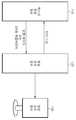
도 1은 본 발명의 일 실시예에 따른 엑스선 진단 시스템의 개괄적인 모습을 도시한 도면이다. 도 1에 도시된 엑스선 진단 시스템은 엑스선 촬영 장치(110), 영상 처리 장치(120) 및 영상 표시 장치(130)으로 구성된다.FIG. 1 is a view showing an overview of an X-ray diagnosis system according to an embodiment of the present invention. The X-ray diagnostic system shown in FIG. 1 includes an
엑스선 촬영 장치(110)는 엑스선 조사부와 엑스선 검출부를 구비한다. 엑스선 조사부는 진단의 대상이 되는 대상체를 향하여 엑스선을 조사한다. 대상체는 신체의 일부가 될 수 있으며, 유방(breast)이나 흉부(chest)가 될 수 있다. 엑스선 검출부는 대상체를 통과한 엑스선을 검출하고 이를 전기적 신호로 생성한다. 외부로부터 의사와 같은 의료 전문가의 지시가 있으면 엑스선 조사부는 대상체를 향하여 엑스선을 조사하고 엑스선 검출부는 조사한 엑스선을 검출하여 이로부터 전기적 신호를 생성한다. 엑스선을 짧은 시간 동안 대상체에 조사한 뒤 대상체를 통과한 엑스선만을 엑스선 검출부에서 검출하고 엑스선이 검출된 위치에 기초하여 대상체 내의 특정 단면 정보를 파악할 수 있게 된다. 엑스선 검출부에서 생성한 전기적 신호는 대상체 내의 특정 단면을 나타내는 단면 데이터로 바뀐다. 복수 번의 엑스선 조사에 의해 복수 개의 단면 데이터를 생성할 수 있고, 인접하는 단면 데이터들을 축적하여 3차원 볼륨 데이터를 생성할 수도 있다. 생성된 단면 데이터 및 3차원 볼륨 데이터는 영상 처리 장치(120)로 전달된다.The
영상 처리 장치(120)는 엑스선 촬영 장치(110)로부터 단면 데이터 또는 3차원 볼륨 데이터를 전달받는다. 영상 처리 장치(120)는 단면 데이터 또는 3차원 볼륨 데이터를 이용하여 영상 표시 장치(130)에 표시될 영상을 생성한다. 3차원 볼륨 데이터란 다수의 단면 데이터들을 축적하여 대상체를 3차원적으로 나타내는 데이터를 말한다. 영상 처리 장치는 단면 데이터로부터 대상체 내의 단면을 나타내는 단층 영상을 생성할 수 있다. 또한 3차원 볼륨 데이터를 횡단하는 여러 방향의 단면을 생각할 수 있는데 각각의 단면에 해당하는 데이터들에 의해 복수 개의 단층 영상을 생성할 수도 있으며, 복수 개의 단층 영상을 재구성(Recontruction)하여 대상체를 나타내는 3차원 영상을 생성할 수도 있다.The
단면 데이터로부터 단층 영상을 생성하려면 이미 알려진 연부 조직 교정(Soft Tissue Correction)과 같은 처리를 거친 후, 여과 역투사(Filtered Back Projection)나 최대우도 기댓값 최대화(Maximum likelihood-Expectation Maximization, ML-EM) 알고리즘 등을 이용하여 복원하면 된다. 하나 이상의 스펙트럼으로 생성된 단면 데이터에 대해서도 기존의 알려진 복원 알고리즘으로 단층 영상의 생성이 가능하다.In order to generate a tomographic image from the cross-sectional data, it is necessary to perform filtering similar to the known Soft Tissue Correction, and then perform Filtered Back Projection or Maximum Likelihood-Expectation Maximization (ML-EM) algorithm And so on. It is also possible to generate a tomographic image with a known restoration algorithm for cross-section data generated from more than one spectrum.
영상 표시 장치(130)는 영상 처리 장치(120)에서 생성된 영상을 전달받아 이를 영상 표시 장치(130)에 디스플레이한다.The
도 2는 도 1에 도시된 엑스선 촬영 장치(110)의 일 실시예의 외부 구조를 도시한 도면이다. 도 2의 엑스선 촬영 장치(200)는 지지대, 회전부(220), 몸체(230), 압착부(240), 엑스선 검출부(250), 이동 라인부(260), 엑스선 조사부(270)를 포함한다. 지지대(210)는 회전부(220)를 통하여 몸체(230)와 연결되어 있고, 엑스선 촬영 장치(200)의 각 구성들을 지지하는 역할을 한다. 회전부(220)는 지지대(210)와 몸체(230) 사이에 위치하여 몸체(230)를 시계 방향 또는 반시계 방향으로 회전시킨다. 몸체(230)에는 압착부(240), 엑스선 검출부(250), 이동 라인부(260), 엑스선 조사부(270)가 연결되어 있다. 대상체는 압착부(240)와 엑스선 검출부(250) 사이에 위치하게 되며 압착부(240)는 엑스선 검출부(250)와 간격을 적절히 조절하여 대상체가 움직이지 않도록 고정시킨다. 압착부(240) 뿐만 아니라 엑스선 검출부(250)도 이동 라인부(260)를 따라서 움직일 수도 있다. 엑스선 조사부(270)는 연결부(275)를 통하여 몸체(230)와 연결되어 있으며 엑스선을 대상체에 조사한다.FIG. 2 is a diagram showing an external structure of an embodiment of the
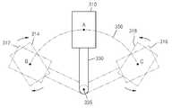
도 3은 엑스선 조사부(310)에 연결되어 있는 연결부(330)의 회전에 따라 엑스선 조사부(310)의 위치 변화와 엑스선 조사부(310) 자체의 회전을 도시한 도면이다. 연결부(330)는 엑스선 조사부(310)를 몸체와 연결하는 동시에 몸체와 독립적으로 시계 방향 또는 반시계 방향으로 회전이 가능하다. 도 3에서 엑스선 조사부(310)가 A 위치에 있을 때, 연결부(330)가 회전축(335)을 중심으로 반시계 방향으로 일정 각도만큼 회전하면 엑스선 조사부(310)는 B 위치에 오게 된다. 반대로 연결부(330)가 회전축(335)을 중심으로 시계 방향으로 일정 각도만큼 회전하면 엑스선 조사부(310)는 C 위치에 오게 된다. 엑스선 조사부(310)가 위치하는 A, B, C 점을 모두 연결하면 원호의 궤도(350)가 된다. 연결부(330)가 일정 각도 만큼 회전하게 되면 연결부(330)에 결합되어 있는 엑스선 조사부(310)의 위치도 변하게 되고, 연결부(330)가 회전한 각도 만큼 엑스선 조사부(310)는 원호의 궤도(350)를 그리게 된다. 엑스선 조사부(310)의 위치를 변경함에 따라 엑스선 조사부(310)의 중심이 그리는 궤적은 위에서 언급한 바와 같이 원호 형태가 될 수 있고, 경우에 따라서는 원형 또는 직선 형태가 될 수도 있다. 이하에서는 설명의 편의상 엑스선 조사부(310)가 일정한 범위 내에서 위치를 변경하는 경우로 설명하므로 원호 형태를 기준으로 설명한다.3 is a view showing a change in the position of the
엑스선 조사부(310)는 연결부(330)의 일부와 결합되어 있으며 엑스선 조사부(310)는 연결부(330)와 독립적으로 시계방향 또는 반시계 방향으로 회전할 수 있다. B 위치에 있는 엑스선 조사부(310)의 경우 312의 엑스선 조사부 위치에서 시계 방향으로 엑스선 조사부(310)만 독립적으로 회전시킴으로써 314의 엑스선 조사부 위치에 올 수 있다. C 위치에 있는 엑스선 조사부(310)의 경우 316의 엑스선 조사부 위치에서 반시계 방향으로 엑스선 조사부(310)만 독립적으로 회전시킴으로써 318의 엑스선 조사부 위치에 올 수 있다.The
엑스선 조사부(310)에 결합되어 있는 연결부(330)의 회전과 엑스선 조사부(310) 자체의 독립적인 회전에 의해 엑스선 조사부(310)의 위치를 변경할 수 있고, 이에 따라 대상체에 대해 적절한 방향으로 엑스선을 조사할 수 있다.The position of the
도 4는 엑스선 조사부(400)가 여러 위치에서 엑스선을 조사하는 모습을 도시한 도면이다. 도 3에서 A 위치의 엑스선 조사부와 B 위치의 314의 엑스선 조사부, 그리고 C 위치의 318의 엑스선 조사부에 해당하는 각각의 엑스선 조사부를 도시한 것이다. 대상체(410)는 압착부(420)와 엑스선 검출부(430) 사이에 위치하고 압착부(420)에 의해 압착되어 대상체(410)가 움직일 수 없게 된다. 그 후 엑스선 조사부(400)는 A, B, C 각각의 위치에서 엑스선을 조사한다.4 is a view showing a state where the X-ray irradiating unit 400 irradiates the X-rays at various positions. 3 shows an X-ray irradiating unit at
본 발명에서는 엑스선 조사부의 위치마다 다른 엑스선 스펙트럼을 가지는 엑스선을 조사할 수 있다. 엑스선 스펙트럼이란 엑스선의 파장에 대한 세기의 분포를 말한다. 엑스선은 음극에서 나온 가속전자가 양극에 부딪혀 갑자기 정지될 때 제동방사로서 방출하는 연속파장의 연속 스펙트럼(continuous X-ray)과 양극을 이루고 있는 원자 내에서 일정 궤도를 회전하던 전자가 큰 에너지를 받아 바깥 방향으로 튀어나가고 그 자리에 다른 전자가 들어오면서 방출되는 에너지에 의해 발생 되는 특성 스펙트럼(characteristic X-ray)이 있다. 따라서 가속전자가 부딪히는 타겟에 해당하는 양극을 구성하는 원소를 바꾸면 같은 전압으로도 스펙트럼이 다른 엑스선을 획득할 수 있고, 양극을 구성하는 원소를 바꾸지 않더라도 강한 전압을 걸어주면 가속전자가 더 빠르게 양극에 충돌하여 스펙트럼이 다른 엑스선을 획득할 수 있다. 다시 말해서 이종의(heterogeneous) 스펙트럼을 가지는 엑스선들을 발생시키려면 엑스선관 내의 음극과 양극에 가하는 전압과 양극을 이루고 있는 물질을 바꾸면 된다.In the present invention, an X-ray having a different X-ray spectrum can be irradiated for each position of the X-ray irradiating unit. X-ray spectrum refers to the distribution of the intensity with respect to the wavelength of an X-ray. X-rays are a continuous X-ray of a continuous wave emitted as a braking radiation when accelerating electrons from a cathode collide with an anode, and electrons rotating in a certain orbit around the anode have large energy There is a characteristic X-ray generated by the energy that protrudes outwardly and is emitted in the place of the other electrons. Therefore, if the element constituting the anode corresponding to the accelerating electron collides with an element constituting the anode, it is possible to acquire X-rays having different spectra with the same voltage. Even if the element constituting the anode is not changed, The spectrum can acquire different x-rays due to collision. In other words, to generate x-rays with a heterogeneous spectrum, the voltages applied to the cathodes and the anodes in the x-ray tube and the substances forming the anodes are changed.
엑스선 조사부 내에는 이종의 스펙트럼을 발생시키기 위한 파라미터 조절부가 존재할 수 있다. 이종의 엑스선 스펙트럼을 발생시키기 위한 파라미터가 엑스선관 내의 음극과 양극에 가하는 전압인 경우, 파라미터 조절부는 엑스선관에 가해지는 전압을 조절하는 전압 조절부와 같은 형태일 수 있다. 또한 이종의 엑스선 스펙트럼을 발생시키기 위한 파라미터가 가속전자가 부딪히는 양극을 구성하는 물질인 경우 타겟이 되는 양극을 바꾸어주는 타겟 변경부와 같은 형태가 될 수 있다. 엑스선 조사부 내에 각각 서로 다른 물질로 이루어져 있는 복수 개의 양극이 있는 경우 타겟 변경부는 가속전자가 부딪히는 위치에 복수 개의 양극 중 어느 하나의 양극을 위치시키는 역할을 함으로써 이종의 스펙트럼을 가지는 엑스선을 발생시킬 수 있다. 예를 들면, 복수 개의 양극은 각각의 구역으로 나뉜 회전체 모양이 될 수 있으며 이 경우 타겟 변경부는 회전체의 회전 각도를 조절하는 회전 조절부와 같은 형태가 될 수 있다.In the X-ray irradiator, there may be a parameter adjuster for generating a heterogeneous spectrum. When the parameter for generating the heterogeneous X-ray spectrum is a voltage applied to the cathode and the anode in the X-ray tube, the parameter adjusting unit may be the same as the voltage adjusting unit for adjusting the voltage applied to the X-ray tube. In addition, if the parameter for generating a different kind of X-ray spectrum is a material constituting the anode where the accelerating electrons hit, it can be shaped like a target changing unit which changes the target anode. When there are a plurality of anodes made of different materials in the X-ray irradiating unit, the target changing unit is capable of generating x-rays having different spectra by locating any one of the plurality of anodes at the position where the accelerating electrons hit each other . For example, the plurality of anodes may be in the shape of a rotating body divided into respective zones, and in this case, the target changing unit may be shaped like a rotation adjusting unit for adjusting the rotating angle of the rotating body.
본 발명에서는 엑스선 조사부의 위치에 따라 상이한 스펙트럼을 가지는 엑스선들을 조사할 수 있을 뿐만 아니라 엑스선 조사부의 위치가 일정 영역 이내에 있을 때는 동일한 스펙트럼을 가지는 엑스선도 사용할 수도 있다. 즉, 엑스선 조사부는 일정 영역 이내에서 여러 번 엑스선을 조사할 수 있는데 동일한 영역 이내라면 동일한 스펙트럼을 사용할 수도 있다. 도 4를 참조하여 설명하면, 엑스선 조사부(400)의 중심이 (1) 영역(451) 내에 있을 때에는 A 위치에서 엑스선 조사부(400)가 조사한 엑스선 스펙트럼과 동일한 스펙트럼의 엑스선을 조사할 수 있고, 엑스선 조사부(400)의 중심이 (2) 영역(452) 내에 있을 때에는 B 위치에서 엑스선 조사부(400)가 조사한 엑스선 스펙트럼과 동일한 스펙트럼의 엑스선을 조사할 수 있으며, 엑스선 조사부(400)의 중심이 (3) 영역(453) 내에 있을 때에는 C 위치에서 엑스선 조사부(400)가 조사한 엑스선 스펙트럼과 동일한 스펙트럼의 엑스선을 조사할 수 있다.In the present invention, X-rays having different spectra may be irradiated depending on the position of the X-ray irradiating unit, and X-rays having the same spectrum may be used when the position of the X-ray irradiating unit is within a certain region. That is, the X-ray irradiator can irradiate the X-rays many times within a certain area, and the same spectrum can be used if it is within the same area. 4, when the center of the X-ray irradiating unit 400 is within the (1)
도시된 바와 달리 엑스선 조사부가 위치하는 일정 영역은 더 작게 또는 더 크게 설정될 수 있다. 엑스선 조사부는 엑스선 조사부가 위치하는 일정 영역이 바뀔 때마다 다른 엑스선 스펙트럼을 가지는 엑스선을 조사한다. 또한 엑스선 조사부가 위치하는 일정 영역이 바뀔 때마다 서로 다른 스펙트럼을 가지는 엑스선들을 교대로 조사할 수도 있다. 예를 들어 엑스선 조사부가 위치하는 일정 영역을 순차적으로 (1)부터 (6)까지의 6개 영역으로 나눈 경우 (1), (3), (5) 영역에서는 제 1 엑스선을 조사하고 (2), (4), (6) 영역에서는 제 2 엑스선을 조사할 수 있다. 서로 다른 엑스선 스펙트럼을 가지는 엑스선이 3 개인 경우라면 (1), (4) 영역에서는 제 1 엑스선을 조사하고 (2), (5) 영역에서는 제 2 엑스선을 조사하며 (3), (6) 영역에서는 제 3 엑스선을 조사할 수도 있다. 엑스선 조사부가 위치하는 일정 영역을 더 세분화하는 경우 더 많은 종류의 엑스선을 교대로 조사할 수 있다.As shown, a certain area where the X-ray irradiator is located may be set smaller or larger. The X-ray probe examines X-rays with different X-ray spectra whenever a certain area of the X-ray detector is changed. It is also possible to alternately examine X-rays having different spectra each time a certain region where the X-ray irradiator is located is changed. For example, if a given region where the X-ray irradiator is located is divided into six regions sequentially from (1) to (6), (1), (3) , (4), and (6), the second X-ray can be inspected. (1), (4), (2), (5), (2) and (3) The third X-ray may be inspected. If you further subdivide the area where the x-ray probe is located, you can examine more types of x-rays alternately.
도 5는 도 1에 도시된 엑스선 촬영 장치(110)의 주요 구성을 도시한 블록도이다. 엑스선 촬영 장치는 입력부(510), 제어부(520), 엑스선 조사부(530), 엑스선 검출부(540), 영상 데이터 생성부(550), 스토리지(560), 출력부(570)를 포함한다. 입력부(510)는 의료 전문가와 같은 사용자로부터 엑스선 촬영에 대한 명령을 입력받는다. 엑스선 조사부(530)의 위치를 변경시키는 명령, 엑스선 조사 명령, 엑스선 스펙트럼을 달리하기 위한 파라미터 조정 명령, 엑스선 촬영 장치의 몸체 또는 엑스선 조사부(530)의 회전에 관한 명령, 압착부 또는 엑스선 검출부(540)의 이동에 관한 명령 등 엑스선 촬영 장치 제어하는 모든 명령을 입력받는다. 사용자로부터 받은 모든 명령에 관한 정보는 제어부(520)로 전달된다. 제어부(520)는 사용자의 명령에 따라 엑스선 촬영 장치 내의 구성들을 제어한다.FIG. 5 is a block diagram showing a main configuration of the
엑스선 조사부(530)는 엑스선 조사에 관한 모든 명령을 제어부(520)로부터 전달받는다. 엑스선 조사부(530)는 엑스선 조사부(530)가 위치할 수 있는 모든 위치를 연결한 소정의 궤도 내에서 사용자가 지정하는 위치까지 이동한 뒤 적절한 스펙트럼의 엑스선을 대상체를 향하여 조사한다. 이때 대상체에 피폭되는 엑스선 량을 고려하여 적절한 횟수와 적절한 선량(dose)으로 조사한다.The
엑스선 검출부(540)는 대상체를 통과한 엑스선들을 검출한다. 엑스선 조사부(530)에서 엑스선이 조사될 때마다 대상체를 통과 후 엑스선 검출부(540)에 도달하는 엑스선들을 검출한다. 엑스선 검출부(540)는 복수 개의 셀들의 집합으로 볼 수 있다. 각각의 셀에 감지되는 엑스선 신호를 전기적 신호로 변환한다. 엑스선 검출부(540)로서 Flat Panel Detector가 사용될 수 있다.The
영상 데이터 생성부(550)는 엑스선 검출부(540)에 의해 감지된 엑스선에 대응되는 전기적 신호를 수신한다. 수신된 전기적 신호로부터 대상체 내의 단면에 대한 정보를 담고 있는 디지털 데이터를 생성한다. 생성된 데이터는 대상체 내의 단면에 대한 데이터이므로 단면 데이터라 부른다. 한번의 엑스선 조사에 의해 대상체 내의 단면에 대한 정보를 담고 있는 하나의 단면 데이터가 생성된다. 엑스선 검출부(540)가 위치를 달리하여 엑스선을 여러 번 조사하는 경우 대상체 내의 서로 다른 단면에 대한 복수 개의 단면 데이터가 생성된다. 복수 개의 단면 데이터를 인접하는 단면 데이터끼리 축적하면 대상체를 3차원적으로 나타내는 3차원 볼륨 데이터를 생성할 수도 있다.The image
스토리지(560)는 영상 데이터 생성부(550)에 의해 생성된 단면 데이터를 저장한다. 또한 영상 데이터 생성부(550)에 의해 생성된 3차원 볼륨 데이터도 저장한다. 스토리지(560)는 사용자의 요청에 의해 저장된 단면 데이터 또는 3차원 볼륨 데이터를 출력부(570)로 전달한다.The
엑스선 조사부(530)가 엑스선을 조사하는 방식은 도 2에서 도시한 바와 같이 회전하는 연결부에 장착된 방식일 수도 있고 도 6 및 도 7에서와 같은 형태가 될 수도 있다.The manner in which the
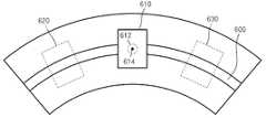
도 6은 엑스선 조사부가 엑스선을 조사하는 다른 일 실시예를 도시한 도면이다. 엑스선 조사부는 도 2와 같이 회전하는 연결부에 장착된 형태가 아니라, 미리 형성되어 있는 이동 라인(600)을 따라 좌우로 슬라이딩하는 방식이 될 수 있다. 즉 엑스선 조사부(610)는 좌측으로 이동하여 620의 엑스선 조사부 위치에 올 수 있고, 반대로 우측으로 이동하여 630의 엑스선 조사부 위치에 올 수도 있다. 엑스선 조사부(610)는 미리 형성되어 있는 이동 라인(600)을 따라 슬라이딩하는 연결부(612)에 장착되어 있으며, 엑스선 조사부(610)는 연결부(612)와 독립하여 회전할 수 있다. 엑스선 조사부는 그 위치에 따라 다른 스펙트럼의 엑스선을 조사한다. 또한 일정 영역 이내에서는 동일한 스펙트럼을 여러 번 조사할 수도 있다.6 is a view showing another embodiment in which an X-ray irradiating unit irradiates X-rays. The X-ray irradiating unit may be configured not to be mounted on a rotating connection part as shown in FIG. 2 but to slide left and right along a previously formed moving
도 7은 엑스선 조사부가 엑스선을 조사하는 또 다른 일 실시예를 도시한 도면이다. 엑스선 조사부는 어레이(array) 방식으로 엑스선을 조사할 수도 있다. 도 7(a)와 같이 엑스선 발생부(710) 내의 엑스선 소스(source)에 의해 엑스선이 방출되는 곳에 어레이 형태의 마스크(720)를 위치시키고 엑스선의 조사가 필요한 위치의 마스크(720)만 제거하여 마스크(720)가 제거된 곳으로만 엑스선이 방출되도록 하는 것이다. 도 7(b)는 엑스선이 방출되는 곳에 위치한 어레이 형태의 마스크를 도시한 것으로서 제일 왼쪽의 마스크(720-1)부터 제일 오른쪽의 마스크(720-7)로 구성되어 있다. 엑스선을 엑스선 발생부(710) 중심에서만 조사되도록 하려면 720-1, 720-2, 720-3, 720-5, 720-6, 720-7의 위치에서 마스크를 사용하여 엑스선이 차단되도록하고 오직 720-4의 위치에서만 엑스선이 방출될 수 있도록 720-4의 위치의 마스크를 제거하면 된다. 엑스선을 조사하는 횟수와 간격을 고려하여 마스크(720) 하나의 크기를 적절히 조절할 수 있다. 어레이 형태의 마스크(720)를 이용하는 방식의 경우 엑스선 조사부의 움직임을 최소화하여 엑스선 조사부의 위치 이동에 따른 진동과 소모 시간을 줄일 수 있다.7 is a view showing another embodiment in which an X-ray irradiating unit irradiates X-rays. The X-ray probe can also scan the X-rays in an array manner. As shown in FIG. 7 (a), the
엑스선 조사부(530)가 엑스선을 조사하는 위치를 달리하는 경우 엑스선이 조사되는 위치에 따라 엑스선의 투과력이 문제될 수 있다. 엑스선이 조사되는 대상체가 어느 방향으로든 두께가 동일하지 않기 때문이다. 즉, 엑스선이 투과해야 하는 길이가 동일하지 않다는 의미이다. 엑스선이 투과해야 하는 길이가 길수록 엑스선이 대상체 내에 많이 흡수되고 엑스선의 검출량은 급격히 떨어진다. 이를 아래에서 도면을 사용하여 설명한다.When the
도 8은 엑스선 조사부의 위치가 원호 궤도 내의 중심 위치일 때와 사선 위치일 때의 엑스선이 투과해야 하는 대상체 내의 길이를 도시한 도면이다. 엑스선 검출부(820) 위의 대상체(810)의 단면을 나타내고 있다. 엑스선 조사부(530)가 원호 궤도(800) 내의 중심 위치인 A 위치에 있는 경우, 엑스선을 조사하게 되면 엑스선이 대상체(810)를 투과해야 하는 길이는 엑스선 검출부(820)에 수직인 최단 거리로서 a 만큼이 된다. 반면 엑스선 조사부(530)가 원호 궤도(800) 내의 중심 위치인 A 위치에서 α°만큼 이동하여 원호 궤도(800) 내의 사선 위치인 B 위치에 있는 경우, 엑스선을 조사하게 되면 엑스선이 대상체(810)를 투과해야 하는 길이는 엑스선 검출부(820)와 사선 방향의 최장 거리인 b 만큼이 된다. 엑스선의 경우 진행경로의 길이가 길어질수록 기하급수적으로 흡수 또는 산란되는 물리적 특성이 있다. 따라서 동일한 스펙트럼의 엑스선을 A 위치와 B 위치에서 조사하는 경우 A 위치에서 조사한 엑스선은 대상체(810)를 투과하여 엑스선 검출부(820)에 의해 엑스선이 검출될 수 있어도 B 위치에서 조사한 엑스선의 대부분은 대상체(810)를 투과하지 못하게 될 수 있다..8 is a view showing the lengths of the X-ray irradiating portion in the object to be irradiated when the position of the X-ray irradiating portion is the center position in the circular arc or in the diagonal position. And shows a cross section of the
다시 말해서 엑스선 조사부(530)의 위치를 소정의 범위 내에서 이동시키는 경우 엑스선이 대상체를 통과해야 하는 길이가 길어질 수 있는데 엑스선의 경우 진행경로의 길이가 길어질수록 기하급수적으로 흡수 또는 산란되는 물리적 특성을 가지고 있어서 엑스선 검출기에 도달하는 엑스선 포톤(photon)의 개수가 급격히 줄어들고, 결과적으로 SNR(Signal to Noise Ratio)이 급격히 떨어지는 문제가 발생한다.In other words, when the position of the
마모그래피의 조직 겹침(tissue overlapping) 문제를 토모신세시스는 엑스선 조사부(530)를 제한된 범위 내에서 이동시켜서 엑스선이 조사되는 각도를 달리함으로써 복수 개의 단층 영상을 촬영하여 조직 겹침 문제를 해결한다. 이때 조직 겹침 문제를 해결하는 궁극적인 파라미터는 엑스선 조사부(530)가 움직이는 각도 범위 즉, 엑스선이 조사되는 위치의 각도 범위이다. 엑스선 조사부(530)가 움직이는 각도 범위를 크게 할수록 획득할 수 있는 깊이 방향의 정보량이 늘어나기 때문이다. 하지만 기존의 토모신세시스에서는 엑스선 조사부(530)가 움직이는 각도 범위가 엑스선의 물리적 특성에 의한 위와 같은 문제점 때문에 매우 제한적이었다. 하나의 엑스선이 대상체를 투과할 수 있는 길이가 제한되어 있으므로 그 제한된 길이를 넘어가는 범위부터는 동일한 엑스선을 조사하더라도 엑스선 검출부(820)에 의한 엑스선 검출이 어려워지기 때문이다.Tomo Synthesis solves tissue overlapping problem by moving
본 발명에서는 하나 이상의 엑스선 스펙트럼을 이용하여 엑스선이 통과해야 하는 경로의 길이가 짧은 경우 투과력은 떨어지지만 조직간 콘트라스트(contrast)가 좋은 저 에너지 스펙트럼의 엑스선을 사용하고, 엑스선이 통과해야 하는 경로의 길이가 긴 경우 투과력이 좋은 고 에너지 스펙트럼의 엑스선을 사용하여 엑스선 조사부(530)가 움직이는 각도 범위를 크게 할 수 있다. 엑스선이 투과할 수 있는 대상체의 길이가 문제되지 않는 경우, 예를 들어 스펙트럼이 다른 두 가지 이상의 엑스선들 모두가 투과할 수 있는 일정한 범위 이내의 대상체라면 두 가지 이상의 엑스선들을 교대로 반복적으로 조사할 수도 있다.In the present invention, when the length of a path through which an x-ray passes is short using one or more x-ray spectra, a low energy spectrum x-ray having a good contrast between tissues is used though the penetration power is low, The
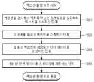
도 9는 엑스선 촬영의 전반적인 과정을 개괄적으로 도시한 흐름도이다. 이하, 생략된 내용이라 하더라도 촬영 장치에 관하여 이상에서 기술된 내용은 본 실시예에 따른 엑스선 촬영 방법에도 적용된다.9 is a flowchart schematically showing an overall process of X-ray imaging. Hereinafter, the contents described above with respect to the photographing apparatus are applied to the X-ray photographing method according to the present embodiment, even if omitted.
910 단계에서 엑스선 촬영을 위해 대상체를 위치시킨다. 엑스선이 대상체의 조사될 수 있도록 엑스선 검출부(540) 위의 적절한 곳에 대상체를 위치시키고, 엑스선 촬영 도중 대상체가 움직이는 경우 정확한 결과를 획득할 수 없으므로 움직임을 방지하기 위하여 대상체를 압착부 등을 이용하여 고정시킨다.In
920 단계에서 입력부(510)는 의료 전문가 등과 같은 사용자의 엑스선 촬영을 위한 명령을 입력받는다. 입력되는 명령은 엑스선이 조사되는 위치를 변경시키는 명령, 엑스선 조사 명령, 엑스선 스펙트럼을 달리하기 위한 파라미터 조정 명령, 엑스선 촬영 장치의 기타 구성의 조작을 위한 명령 등 엑스선 촬영에 관한 모든 명령이 될 수 있다.In
930 단계에서 엑스선 촬영 장치에 의한 엑스선 촬영 모드가 수행된다. 이와 관련하여서는 엑스선 촬영 모드의 상세한 흐름도를 가지고 이하 설명한다. 도 10은 도 9의 930 단계에 해당하는 엑스선 촬영 모드의 상세한 흐름도이다.In
1010 단계에서 엑스선 조사부(530)는 대상체를 향하여 엑스선을 조사한다. 이전에 엑스선을 조사한 경우라면 이전의 엑스선을 조사한 위치와 다른 위치에서 엑스선을 조사할 수 있다. 또한 이전의 엑스선 스펙트럼과 다른 엑스선 스펙트럼을 가지는 엑스선을 조사할 수 있다.In
1020 단계에서 엑스선 검출부(540)는 대상체를 통과한 엑스선을 검출한다. 대상체를 통과한 엑스선을 엑스선 검출부(540)에서 감지하고 이를 전기적 신호로 변환하는 것이다.In
1030 단계에서 영상 데이터 생성부(550)는 검출된 엑스선에 대응되는 단면 데이터를 생성한다. 대상체를 향하여 조사된 엑스선은 대상체를 통과하여 엑스선 검출부(540)에 도달하고 이때 엑스선 검출부(540)에 의해 검출되는 엑스선은 대상체 내의 단면에 관한 정보를 포함하는 전기적 신호로 변환되고 이를 디지털 데이터화하여 단면 데이터를 생성한다. 엑스선을 여러 번 조사하여 복수 개의 단면 데이터가 생성되면 이로부터 대상체를 3차원적으로 나타내는 3차원 볼륨 데이터를 생성할 수도 있다.In
1040 단계에서 스토리지(560)는 생성된 단면 데이터 또는 3차원 볼륨 데이터를 저장한다.In
940 단계에서 엑스선 촬영을 종료할지 판단한다. 예를 들어, 엑스선 조사 횟수가 진단에 필요한 만큼의 엑스선 영상을 만드는데 충분한 소정의 횟수보다 큰지를 판단한다. 소정의 횟수보다 엑스선 조사 횟수가 작은 경우, 즉 엑스선 촬영을 종료하지 않을 경우 다시 930 단계의 엑스선 촬영 모드가 진행된다. 엑스선 조사 횟수가 소정의 횟수보다 많은 경우 진단에 필요한 충분한 엑스선 영상을 획득할 수 있으므로 950 단계가 진행된다.It is determined in
950 단계에서 출력부(570)는 엑스선 촬영 결과를 영상 처리 장치로 출력한다. 엑스선 촬영 결과란 영상 데이터 생성부(550)에 의해 생성된 대상체의 단면을 나타내는 단면 데이터 또는 대상체를 3차원적으로 나타내는 3차원 볼륨 데이터를 말한다. 단면 데이터 또는 3차원 볼륨 데이터는 스토리지(560)에 저장되어 있을 수 있으며, 출력부(570)는 스토리지(560)로부터 이를 전달받아 영상 처리 장치로 출력한다.In
한편, 상술한 본 발명의 실시예에 따른 엑스선 촬영 방법은 컴퓨터에서 실행될 수 있는 프로그램으로 작성가능하고, 컴퓨터로 읽을 수 있는 기록매체를 이용하여 상기 프로그램을 동작시키는 범용 디지털 컴퓨터에서 구현될 수 있다. 상기 컴퓨터로 읽을 수 있는 기록매체는 마그네틱 저장매체(예를 들면, 롬, 플로피 디스크, 하드 디스크 등), 광학적 판독 매체(예를 들면, 시디롬, 디브이디 등)와 같은 저장매체를 포함한다.Meanwhile, the X-ray imaging method according to the embodiment of the present invention described above can be realized in a general-purpose digital computer that can be created as a program that can be executed in a computer and operates the program using a computer-readable recording medium. The computer-readable recording medium includes a storage medium such as a magnetic storage medium (e.g., ROM, floppy disk, hard disk, etc.), optical reading medium (e.g., CD ROM,
이제까지 본 발명에 대하여 그 실시예들을 중심으로 살펴보았다. 본 발명이 속하는 기술 분야에서 통상의 지식을 가진 자는 본 발명이 본 발명의 본질적인 특성에서 벗어나지 않는 범위에서 변형된 형태로 구현될 수 있음을 이해할 수 있을 것이다. 그러므로 개시된 실시예들은 한정적인 관점이 아니라 설명적인 관점에서 고려되어야 한다. 본 발명의 범위는 전술한 설명이 아니라 특허청구범위에 나타나 있으며, 그와 동등한 범위 내에 있는 모든 차이점은 본 발명에 포함된 것으로 해석되어야 할 것이다.The embodiments of the present invention have been described above. It will be understood by those skilled in the art that various changes in form and details may be made therein without departing from the spirit and scope of the invention as defined by the appended claims. Therefore, the disclosed embodiments should be considered in an illustrative rather than a restrictive sense. The scope of the present invention is defined by the appended claims rather than by the foregoing description, and all differences within the scope of equivalents thereof should be construed as being included in the present invention.
510 ... 입력부
520 ... 제어부
530 ... 엑스선 조사부
540 ... 엑스선 검출부
550 ... 영상 데이터 생성부
560 ... 스토리지
570 ... 출력부510 ... input
520 ... control unit
530 ... X-
540 ... X-
550 ... image data generating section
560 ... storage
570 ... output section
Claims (15)
Translated fromKorean상기 대상체를 통과한 상기 제 1 엑스선과 상기 제 2 엑스선을 검출하는 엑스선 검출부; 및
상기 검출된 제 1 엑스선과 제 2 엑스선 각각에 대응되는 상기 대상체의 소정의 단면을 나타내는 단면 데이터들을 생성하는 영상 데이터 생성부를 포함하고,
상기 대상체의 엑스선 영상을 촬영하는 동안 상기 제 1 엑스선과 상기 제 2 엑스선이 조사되는 상기 소정의 궤도 상의 위치에 따라 상기 제 1 엑스선과 상기 제 2 엑스선이 상기 대상체를 투과해야 하는 경로의 길이가 다르고, 상기 대상체를 투과해야 하는 경로의 길이가 길어질수록, 상기 엑스선들의 스펙트럼은 상대적으로 고(high) 에너지 스펙트럼인 엑스선 촬영 장치.An X-ray irradiator for generating a first X-ray and a second X-ray having different energy spectrums at different positions on a predetermined orbit and irradiating the object with the first X-ray and the second X-ray, respectively;
An X-ray detecting unit detecting the first X-ray and the second X-ray that have passed through the object; And
And an image data generation unit for generating cross-sectional data representing predetermined cross-sections of the object corresponding to the detected first X-ray and second X-ray, respectively,
A length of a path through which the first X-ray and the second X-ray pass through the object is different according to a position on the predetermined trajectory to which the first X-ray and the second X-ray are irradiated while photographing the X- And the spectrum of the x-rays is a relatively high energy spectrum as the length of the path through which the object is to be transmitted becomes longer.
상기 엑스선 조사부는 상기 제 1 엑스선 및 상기 제 2 엑스선의 스펙트럼과 다른 스펙트럼을 가지는 적어도 하나의 엑스선을 상기 제 1 엑스선 및 상기 제 2 엑스선이 조사되는 상기 소정의 궤도에서의 위치와 다른 위치에서 조사하는 엑스선 촬영 장치.The method according to claim 1,
The X-ray irradiating unit irradiates at least one X-ray having a spectrum different from the spectrum of the first X-ray and the second X-ray at a position different from a position in the predetermined orbit to which the first X-ray and the second X-ray are irradiated X-ray photographing device.
상기 대상체를 투과해야 하는 경로의 최대 길이가 상기 제 1 엑스선과 상기 제 2 엑스선이 모두 투과할 수 있는 범위 이내인 경우, 상기 제 1 엑스선과 상기 제 2 엑스선을 상기 소정의 궤도에서 위치를 달리하여 교대로 조사하는 엑스선 촬영 장치.The method according to claim 1,
When the maximum length of the path through which the object is to be transmitted is within a range that both the first X-ray and the second X-ray can penetrate, the first X-ray and the second X-ray are positioned at different positions in the predetermined orbit An X-ray photographing device alternately.
상기 영상 데이터 생성부는 상기 단면 데이터를 축적하여 대상체를 3차원적으로 나타내는 3차원 볼륨 데이터를 생성하는 엑스선 촬영 장치.The method according to claim 1,
Wherein the image data generation unit accumulates the cross-sectional data to generate three-dimensional volume data representing the object three-dimensionally.
상기 영상 데이터 생성부에서 생성된 상기 단면 데이터를 저장하는 스토리지를 더 포함하는 엑스선 촬영 장치.The method according to claim 1,
And a storage for storing the cross-sectional data generated by the image data generation unit.
상기 엑스선 촬영 장치는 토모신세시스(tomosynthesis)에 이용되는 엑스선 촬영 장치.The method according to claim 1,
The X-ray photographing apparatus is used for tomosynthesis.
상기 대상체를 통과한 상기 제 1 엑스선과 상기 제 2 엑스선을 검출하는 단계; 및
상기 검출된 제 1 엑스선과 제 2 엑스선 각각에 대응되는 상기 대상체의 소정의 단면을 나타내는 단면 데이터들을 생성하는 단계를 포함하고,
상기 대상체의 엑스선 영상을 촬영하는 동안 상기 제 1 엑스선과 상기 제 2 엑스선이 조사되는 상기 소정의 궤도 상의 위치에 따라 상기 제 1 엑스선과 상기 제 2 엑스선이 상기 대상체를 투과해야 하는 경로의 길이가 다르고, 상기 대상체를 투과해야 하는 경로의 길이가 길어질수록, 상기 엑스선들의 스펙트럼은 상대적으로 고(high) 에너지 스펙트럼인 엑스선 촬영 방법.Generating a first X-ray and a second X-ray having different energy spectrums at different positions on a predetermined trajectory and irradiating the object with the first X-ray and the second X-ray, respectively;
Detecting the first X-ray and the second X-ray that have passed through the object; And
And generating cross-sectional data representing a predetermined cross-section of the object corresponding to the detected first X-ray and second X-ray, respectively,
A length of a path through which the first X-ray and the second X-ray pass through the object is different according to a position on the predetermined trajectory to which the first X-ray and the second X-ray are irradiated while photographing the X- And the longer the path length of the path through which the object is to be transmitted, the higher the spectrum of the x-rays is the higher energy spectrum.
상기 엑스선을 조사하는 단계는 상기 제 1 엑스선 및 상기 제 2 엑스선의 스펙트럼과 다른 스펙트럼을 가지는 적어도 하나의 엑스선을 상기 제 1 엑스선 및 상기 제 2 엑스선이 조사되는 상기 소정의 궤도에서의 위치와 다른 위치에서 조사하는 엑스선 촬영 방법.9. The method of claim 8,
The step of irradiating the X-ray may include irradiating at least one X-ray having a spectrum different from the spectrum of the first X-ray and the second X-ray to a position different from a position in the predetermined orbit to which the first X- Ray photographing method.
상기 대상체를 투과해야 하는 경로의 최대 길이가 상기 제 1 엑스선과 상기 제 2 엑스선이 모두 투과할 수 있는 범위 이내인 경우, 상기 제 1 엑스선과 상기 제 2 엑스선을 상기 소정의 궤도에서 위치를 달리하여 교대로 조사하는 엑스선 촬영 방법.9. The method of claim 8,
When the maximum length of the path through which the object is to be transmitted is within a range that both the first X-ray and the second X-ray can penetrate, the first X-ray and the second X-ray are positioned at different positions in the predetermined orbit Alternate x-ray imaging methods.
상기 단면 데이터를 축적하여 대상체를 3차원적으로 나타내는 3차원 볼륨 데이터를 생성하는 단계를 더 포함하는 엑스선 촬영 방법.9. The method of claim 8,
Dimensional volume data representing the object three-dimensionally by accumulating the cross-sectional data.
상기 생성된 단면 데이터를 저장하는 단계를 더 포함하는 엑스선 촬영 방법.9. The method of claim 8,
And storing the generated cross-sectional data.
상기 엑스선 촬영 방법은 토모신세시스(tomosynthesis)에 이용되는 엑스선 촬영 방법.9. The method of claim 8,
The x-ray imaging method is used for tomosynthesis.
Priority Applications (2)
| Application Number | Priority Date | Filing Date | Title |
|---|---|---|---|
| KR1020110073245AKR101477543B1 (en) | 2011-07-22 | 2011-07-22 | APPARATUS AND METHOD OF PHOTOGRAPHING USING X-ray |
| US13/552,976US9146199B2 (en) | 2011-07-22 | 2012-07-19 | X-ray imaging apparatus and method |
Applications Claiming Priority (1)
| Application Number | Priority Date | Filing Date | Title |
|---|---|---|---|
| KR1020110073245AKR101477543B1 (en) | 2011-07-22 | 2011-07-22 | APPARATUS AND METHOD OF PHOTOGRAPHING USING X-ray |
Publications (2)
| Publication Number | Publication Date |
|---|---|
| KR20130011822A KR20130011822A (en) | 2013-01-30 |
| KR101477543B1true KR101477543B1 (en) | 2014-12-31 |
Family
ID=47555742
Family Applications (1)
| Application Number | Title | Priority Date | Filing Date |
|---|---|---|---|
| KR1020110073245AActiveKR101477543B1 (en) | 2011-07-22 | 2011-07-22 | APPARATUS AND METHOD OF PHOTOGRAPHING USING X-ray |
Country Status (2)
| Country | Link |
|---|---|
| US (1) | US9146199B2 (en) |
| KR (1) | KR101477543B1 (en) |
Families Citing this family (24)
| Publication number | Priority date | Publication date | Assignee | Title |
|---|---|---|---|---|
| WO2007095330A2 (en) | 2006-02-15 | 2007-08-23 | Hologic Inc | Breast biopsy and needle localization using tomosynthesis systems |
| ES2862525T3 (en) | 2009-10-08 | 2021-10-07 | Hologic Inc | Needle Breast Biopsy System and Method of Use |
| US20120133600A1 (en) | 2010-11-26 | 2012-05-31 | Hologic, Inc. | User interface for medical image review workstation |
| JP6057922B2 (en) | 2011-03-08 | 2017-01-11 | ホロジック, インコーポレイテッドHologic, Inc. | System and method for dual energy and / or contrast enhanced breast imaging for screening, diagnosis and biopsy |
| EP2782505B1 (en) | 2011-11-27 | 2020-04-22 | Hologic, Inc. | System and method for generating a 2d image using mammography and/or tomosynthesis image data |
| JP6240097B2 (en) | 2012-02-13 | 2017-11-29 | ホロジック インコーポレイティッド | How to navigate a tomosynthesis stack using composite image data |
| CN105451657A (en) | 2013-03-15 | 2016-03-30 | 霍罗吉克公司 | System and method for navigating tomosynthesis stack including automatic focusing |
| US10092358B2 (en) | 2013-03-15 | 2018-10-09 | Hologic, Inc. | Tomosynthesis-guided biopsy apparatus and method |
| EP3060132B1 (en)* | 2013-10-24 | 2019-12-04 | Hologic, Inc. | System and method for navigating x-ray guided breast biopsy |
| JP6506769B2 (en) | 2014-02-28 | 2019-04-24 | ホロジック, インコーポレイテッドHologic, Inc. | System and method for generating and displaying tomosynthesis image slabs |
| CN106526686B (en)* | 2016-12-07 | 2019-05-07 | 同方威视技术股份有限公司 | Spiral CT equipment and three-dimensional image reconstruction method |
| EP3600052A1 (en) | 2017-03-30 | 2020-02-05 | Hologic, Inc. | System and method for targeted object enhancement to generate synthetic breast tissue images |
| CN110621233B (en) | 2017-03-30 | 2023-12-12 | 豪洛捷公司 | Method for processing breast tissue image data |
| EP3600047A1 (en) | 2017-03-30 | 2020-02-05 | Hologic, Inc. | System and method for hierarchical multi-level feature image synthesis and representation |
| WO2018236565A1 (en) | 2017-06-20 | 2018-12-27 | Hologic, Inc. | METHOD AND SYSTEM FOR MEDICAL IMAGING WITH DYNAMIC SELF-LEARNING |
| US12121304B2 (en) | 2018-05-04 | 2024-10-22 | Hologic, Inc. | Introducer and localization wire visualization |
| EP3787520B1 (en) | 2018-05-04 | 2024-09-25 | Hologic, Inc. | Biopsy needle visualization |
| WO2020068851A1 (en) | 2018-09-24 | 2020-04-02 | Hologic, Inc. | Breast mapping and abnormality localization |
| US11020066B2 (en)* | 2018-12-10 | 2021-06-01 | KUB Technologies, Inc. | System and method for cabinet x-ray systems with stationary x-ray source array |
| US11883206B2 (en) | 2019-07-29 | 2024-01-30 | Hologic, Inc. | Personalized breast imaging system |
| EP4439580A3 (en) | 2019-09-27 | 2024-12-25 | Hologic, Inc. | Ai system for predicting reading time and reading complexity for reviewing 2d/3d breast images |
| US11481038B2 (en) | 2020-03-27 | 2022-10-25 | Hologic, Inc. | Gesture recognition in controlling medical hardware or software |
| US12254586B2 (en) | 2021-10-25 | 2025-03-18 | Hologic, Inc. | Auto-focus tool for multimodality image review |
| WO2023097279A1 (en) | 2021-11-29 | 2023-06-01 | Hologic, Inc. | Systems and methods for correlating objects of interest |
Citations (2)
| Publication number | Priority date | Publication date | Assignee | Title |
|---|---|---|---|---|
| KR101034258B1 (en)* | 2009-11-09 | 2011-05-12 | 한국전기연구원 | X―Detect Breast Cancer Diagnosis System and Method for Measuring Breast Density |
| KR20110055991A (en)* | 2009-11-20 | 2011-05-26 | 주식회사바텍 | X-ray imaging device with X-ray filter unit |
Family Cites Families (5)
| Publication number | Priority date | Publication date | Assignee | Title |
|---|---|---|---|---|
| US4323779A (en)* | 1977-06-03 | 1982-04-06 | Albert Richard David | Scanning radiographic method |
| DE102005022543A1 (en) | 2005-05-17 | 2006-11-23 | Siemens Ag | Mammography procedure and mammography device |
| JP2008079923A (en) | 2006-09-28 | 2008-04-10 | Konica Minolta Medical & Graphic Inc | Radiographic photographing equipment |
| JP4851296B2 (en) | 2006-10-26 | 2012-01-11 | 富士フイルム株式会社 | Radiation tomographic image acquisition apparatus and radiation tomographic image acquisition method |
| JP5346654B2 (en) | 2009-03-31 | 2013-11-20 | キヤノン株式会社 | Radiation imaging apparatus and control method thereof |
- 2011
- 2011-07-22KRKR1020110073245Apatent/KR101477543B1/enactiveActive
- 2012
- 2012-07-19USUS13/552,976patent/US9146199B2/enactiveActive
Patent Citations (2)
| Publication number | Priority date | Publication date | Assignee | Title |
|---|---|---|---|---|
| KR101034258B1 (en)* | 2009-11-09 | 2011-05-12 | 한국전기연구원 | X―Detect Breast Cancer Diagnosis System and Method for Measuring Breast Density |
| KR20110055991A (en)* | 2009-11-20 | 2011-05-26 | 주식회사바텍 | X-ray imaging device with X-ray filter unit |
Also Published As
| Publication number | Publication date |
|---|---|
| US20130022165A1 (en) | 2013-01-24 |
| KR20130011822A (en) | 2013-01-30 |
| US9146199B2 (en) | 2015-09-29 |
Similar Documents
| Publication | Publication Date | Title |
|---|---|---|
| KR101477543B1 (en) | APPARATUS AND METHOD OF PHOTOGRAPHING USING X-ray | |
| US8483361B2 (en) | Anode target for an x-ray tube and method for controlling the x-ray tube | |
| JP5042465B2 (en) | Radiation imaging apparatus and image processing method | |
| US8817947B2 (en) | Tomosynthesis imaging | |
| US7933378B2 (en) | Multi-tube X-ray detection | |
| US9420975B2 (en) | Imaging facility and radiation therapy device | |
| US7885378B2 (en) | Imaging system and related techniques | |
| JP4974131B2 (en) | Imaging method and system using a plurality of offset X-ray irradiation points | |
| US8605854B2 (en) | Mammography apparatus with X-ray sources arranged at different distances from the chest | |
| EP2326250B1 (en) | Calibration method for ring artifact correction in non-ideal isocentric 3d rotational x-ray scanner systems using a calibration phantom based rotation center finding algorithm | |
| US7869571B2 (en) | Methods and apparatus for x-ray imaging with focal spot deflection | |
| JP2019030637A (en) | Variable-distance imaging | |
| JP2010075338A (en) | Mammography and therapy apparatus equipped with x-ray therapy function | |
| US11207041B2 (en) | X-ray CT system and medical processing apparatus | |
| US20120224664A1 (en) | Tomosynthesis mammography system with enlarged field of view | |
| JP6488292B2 (en) | X-ray system such as a tomosynthesis system and method for acquiring an image of an object | |
| JPH0228818B2 (en) | ||
| JP6980668B2 (en) | Methods for CT Imaging Systems and CT Imaging Systems | |
| JP2013545545A (en) | Multi-modality image acquisition method and apparatus | |
| CN102551776A (en) | Device and method for generating X-ray, and computing program and data medium | |
| JP7467253B2 (en) | X-ray CT system and medical processing equipment | |
| JP2010540063A (en) | Computed tomography equipment | |
| JP5196782B2 (en) | X-ray CT apparatus and control method thereof | |
| JP7321846B2 (en) | X-ray CT system and medical processing equipment | |
| JP2022013679A (en) | Medical image processing method, medical image processing device and X-ray CT device |
Legal Events
| Date | Code | Title | Description |
|---|---|---|---|
| PA0109 | Patent application | St.27 status event code:A-0-1-A10-A12-nap-PA0109 | |
| R18-X000 | Changes to party contact information recorded | St.27 status event code:A-3-3-R10-R18-oth-X000 | |
| PG1501 | Laying open of application | St.27 status event code:A-1-1-Q10-Q12-nap-PG1501 | |
| A201 | Request for examination | ||
| PA0201 | Request for examination | St.27 status event code:A-1-2-D10-D11-exm-PA0201 | |
| A302 | Request for accelerated examination | ||
| PA0302 | Request for accelerated examination | St.27 status event code:A-1-2-D10-D17-exm-PA0302 St.27 status event code:A-1-2-D10-D16-exm-PA0302 | |
| E902 | Notification of reason for refusal | ||
| PE0902 | Notice of grounds for rejection | St.27 status event code:A-1-2-D10-D21-exm-PE0902 | |
| AMND | Amendment | ||
| P11-X000 | Amendment of application requested | St.27 status event code:A-2-2-P10-P11-nap-X000 | |
| P13-X000 | Application amended | St.27 status event code:A-2-2-P10-P13-nap-X000 | |
| E601 | Decision to refuse application | ||
| PE0601 | Decision on rejection of patent | St.27 status event code:N-2-6-B10-B15-exm-PE0601 | |
| T11-X000 | Administrative time limit extension requested | St.27 status event code:U-3-3-T10-T11-oth-X000 | |
| AMND | Amendment | ||
| E13-X000 | Pre-grant limitation requested | St.27 status event code:A-2-3-E10-E13-lim-X000 | |
| P11-X000 | Amendment of application requested | St.27 status event code:A-2-2-P10-P11-nap-X000 | |
| P13-X000 | Application amended | St.27 status event code:A-2-2-P10-P13-nap-X000 | |
| PX0901 | Re-examination | St.27 status event code:A-2-3-E10-E12-rex-PX0901 | |
| PX0701 | Decision of registration after re-examination | St.27 status event code:A-3-4-F10-F13-rex-PX0701 | |
| X701 | Decision to grant (after re-examination) | ||
| GRNT | Written decision to grant | ||
| PR0701 | Registration of establishment | St.27 status event code:A-2-4-F10-F11-exm-PR0701 | |
| PR1002 | Payment of registration fee | St.27 status event code:A-2-2-U10-U11-oth-PR1002 Fee payment year number:1 | |
| PG1601 | Publication of registration | St.27 status event code:A-4-4-Q10-Q13-nap-PG1601 | |
| FPAY | Annual fee payment | Payment date:20171129 Year of fee payment:4 | |
| PR1001 | Payment of annual fee | St.27 status event code:A-4-4-U10-U11-oth-PR1001 Fee payment year number:4 | |
| FPAY | Annual fee payment | Payment date:20181129 Year of fee payment:5 | |
| PR1001 | Payment of annual fee | St.27 status event code:A-4-4-U10-U11-oth-PR1001 Fee payment year number:5 | |
| PC1903 | Unpaid annual fee | St.27 status event code:A-4-4-U10-U13-oth-PC1903 Not in force date:20191224 Payment event data comment text:Termination Category : DEFAULT_OF_REGISTRATION_FEE | |
| K11-X000 | Ip right revival requested | St.27 status event code:A-6-4-K10-K11-oth-X000 | |
| PC1903 | Unpaid annual fee | St.27 status event code:N-4-6-H10-H13-oth-PC1903 Ip right cessation event data comment text:Termination Category : DEFAULT_OF_REGISTRATION_FEE Not in force date:20191224 | |
| PR0401 | Registration of restoration | St.27 status event code:A-6-4-K10-K13-oth-PR0401 | |
| R401 | Registration of restoration | ||
| PR1001 | Payment of annual fee | St.27 status event code:A-4-4-U10-U11-oth-PR1001 Fee payment year number:6 | |
| PR1001 | Payment of annual fee | St.27 status event code:A-4-4-U10-U11-oth-PR1001 Fee payment year number:7 | |
| PR1001 | Payment of annual fee | St.27 status event code:A-4-4-U10-U11-oth-PR1001 Fee payment year number:8 | |
| PR1001 | Payment of annual fee | St.27 status event code:A-4-4-U10-U11-oth-PR1001 Fee payment year number:9 | |
| L13-X000 | Limitation or reissue of ip right requested | St.27 status event code:A-2-3-L10-L13-lim-X000 | |
| PR1001 | Payment of annual fee | St.27 status event code:A-4-4-U10-U11-oth-PR1001 Fee payment year number:10 | |
| PN2301 | Change of applicant | St.27 status event code:A-5-5-R10-R11-asn-PN2301 | |
| PN2301 | Change of applicant | St.27 status event code:A-5-5-R10-R11-asn-PN2301 | |
| PN2301 | Change of applicant | St.27 status event code:A-5-5-R10-R14-asn-PN2301 | |
| P14-X000 | Amendment of ip right document requested | St.27 status event code:A-5-5-P10-P14-nap-X000 | |
| P16-X000 | Ip right document amended | St.27 status event code:A-5-5-P10-P16-nap-X000 | |
| Q16-X000 | A copy of ip right certificate issued | St.27 status event code:A-4-4-Q10-Q16-nap-X000 |