KR101391405B1 - needle unit - Google Patents
needle unitDownload PDFInfo
- Publication number
- KR101391405B1 KR101391405B1KR1020120019049AKR20120019049AKR101391405B1KR 101391405 B1KR101391405 B1KR 101391405B1KR 1020120019049 AKR1020120019049 AKR 1020120019049AKR 20120019049 AKR20120019049 AKR 20120019049AKR 101391405 B1KR101391405 B1KR 101391405B1
- Authority
- KR
- South Korea
- Prior art keywords
- outer cannula
- opening
- loop
- needle unit
- curve
- Prior art date
- Legal status (The legal status is an assumption and is not a legal conclusion. Google has not performed a legal analysis and makes no representation as to the accuracy of the status listed.)
- Expired - Fee Related
Links
Images


Classifications
- A—HUMAN NECESSITIES
- A61—MEDICAL OR VETERINARY SCIENCE; HYGIENE
- A61B—DIAGNOSIS; SURGERY; IDENTIFICATION
- A61B17/00—Surgical instruments, devices or methods
- A61B17/32—Surgical cutting instruments
- A61B17/3205—Excision instruments
- A61B17/32056—Surgical snare instruments
- A—HUMAN NECESSITIES
- A61—MEDICAL OR VETERINARY SCIENCE; HYGIENE
- A61B—DIAGNOSIS; SURGERY; IDENTIFICATION
- A61B17/00—Surgical instruments, devices or methods
- A61B17/04—Surgical instruments, devices or methods for suturing wounds; Holders or packages for needles or suture materials
- A61B17/0469—Suturing instruments for use in minimally invasive surgery, e.g. endoscopic surgery
- A—HUMAN NECESSITIES
- A61—MEDICAL OR VETERINARY SCIENCE; HYGIENE
- A61B—DIAGNOSIS; SURGERY; IDENTIFICATION
- A61B17/00—Surgical instruments, devices or methods
- A61B17/04—Surgical instruments, devices or methods for suturing wounds; Holders or packages for needles or suture materials
- A61B17/0482—Needle or suture guides
- A—HUMAN NECESSITIES
- A61—MEDICAL OR VETERINARY SCIENCE; HYGIENE
- A61B—DIAGNOSIS; SURGERY; IDENTIFICATION
- A61B17/00—Surgical instruments, devices or methods
- A61B17/04—Surgical instruments, devices or methods for suturing wounds; Holders or packages for needles or suture materials
- A61B17/06—Needles ; Sutures; Needle-suture combinations; Holders or packages for needles or suture materials
- A61B17/06066—Needles, e.g. needle tip configurations
- A—HUMAN NECESSITIES
- A61—MEDICAL OR VETERINARY SCIENCE; HYGIENE
- A61B—DIAGNOSIS; SURGERY; IDENTIFICATION
- A61B17/00—Surgical instruments, devices or methods
- A61B17/00234—Surgical instruments, devices or methods for minimally invasive surgery
- A61B2017/00292—Surgical instruments, devices or methods for minimally invasive surgery mounted on or guided by flexible, e.g. catheter-like, means
- A—HUMAN NECESSITIES
- A61—MEDICAL OR VETERINARY SCIENCE; HYGIENE
- A61B—DIAGNOSIS; SURGERY; IDENTIFICATION
- A61B17/00—Surgical instruments, devices or methods
- A61B17/00234—Surgical instruments, devices or methods for minimally invasive surgery
- A61B2017/00358—Snares for grasping
- A—HUMAN NECESSITIES
- A61—MEDICAL OR VETERINARY SCIENCE; HYGIENE
- A61B—DIAGNOSIS; SURGERY; IDENTIFICATION
- A61B17/00—Surgical instruments, devices or methods
- A61B17/04—Surgical instruments, devices or methods for suturing wounds; Holders or packages for needles or suture materials
- A61B17/06—Needles ; Sutures; Needle-suture combinations; Holders or packages for needles or suture materials
- A61B17/06066—Needles, e.g. needle tip configurations
- A61B2017/0608—J-shaped
- A—HUMAN NECESSITIES
- A61—MEDICAL OR VETERINARY SCIENCE; HYGIENE
- A61B—DIAGNOSIS; SURGERY; IDENTIFICATION
- A61B17/00—Surgical instruments, devices or methods
- A61B17/04—Surgical instruments, devices or methods for suturing wounds; Holders or packages for needles or suture materials
- A61B17/06—Needles ; Sutures; Needle-suture combinations; Holders or packages for needles or suture materials
- A61B17/06066—Needles, e.g. needle tip configurations
- A61B2017/06085—Needles, e.g. needle tip configurations having a blunt tip
Landscapes
- Health & Medical Sciences (AREA)
- Life Sciences & Earth Sciences (AREA)
- Surgery (AREA)
- Heart & Thoracic Surgery (AREA)
- Engineering & Computer Science (AREA)
- Biomedical Technology (AREA)
- Nuclear Medicine, Radiotherapy & Molecular Imaging (AREA)
- Medical Informatics (AREA)
- Molecular Biology (AREA)
- Animal Behavior & Ethology (AREA)
- General Health & Medical Sciences (AREA)
- Public Health (AREA)
- Veterinary Medicine (AREA)
- Surgical Instruments (AREA)
Abstract
Translated fromKoreanDescription
Translated fromKorean본 발명은 일반적으로 외과수술용 바늘에 관한 것으로, 보다 구체적으로 간 절제술에 적용되는 바늘 유닛에 관한 것이다.
BACKGROUND OF THE INVENTION 1. Field of the Invention The present invention relates generally to surgical needles, and more particularly to a needle unit adapted for hepatic resection.
성공적인 간 절제술은 숙련된 수술 기법과, 남은 간의 안전적 크기와, 불필요한 허혈 및 재관류(reperfusion) 부상 위험의 감소 또는 제거를 요구한다. 숙련된 외과의사는 간의 원표면에서 출혈이나 담즙 유출을 보호하고 남은 주요 혈관과 관들을 온전하게 유지하기 위해, 유조직(parenchyma) 절개 중 혈액 손실을 최소화하는 것을 목표로 한다. 이러한 모든 것들이 충족되면, 수술 과정은 별일 없이 진행될 수 있다. 그렇지 않으면, 번거로운 치료가 필요해질 수 있다.Successful hepatic resection requires skilled surgical techniques, safe size of the liver, and the reduction or elimination of unnecessary ischemia and reperfusion injury risk. A skilled surgeon aims to minimize blood loss during a parenchyma incision to preserve bleeding or bile drainage from the liver surface of the liver and to maintain the integrity of the remaining vessels and vessels. Once all these are met, the surgical procedure can proceed without any reason. Otherwise, troublesome treatment may be required.
간 절제술은 수술중 출혈과 수술후 간 장애의 높은 위험을 항상 안고 있다. 비록 수술 기법과 장비 및 수술후 치료가 정교해짐에 따라 사망률이 감소하고는 있으나, 유조직 절개 중 출혈은 극복해야할 중요한 장애물로 남아있다. 혈액 손실을 줄이기 위해, 유입(inflow)과 유출(backflow) 모두를 컨트롤할 필요하다. 간십이지장 인대(hepatoduodenal ligament)(예를 들어, Pringle's maneuver)나 폐문(hilar) 또는 개별 분절들(individual segmental branches) 규모에서 유입을 간헐적 또는 지속적으로 막기 위한 여러 알려진 방법들이 적용될 수 있다. 개별 유입의 부분적 차단은 남은 간에서의 허혈과 재관류 부상 위험을 줄일 수 있다. 간 장애를 방지하기 위한 목적으로, 특히 간경변성 간(cirrhotic liver)에 대해, 적당한 허혈 시간을 결정하는 것이 중요하다. 유출는 하대정맥(inferior vena cava, IVC) 차단 또는 개별 간정맥 차단을 통해 제어될 수 있다. 그러나, 하대정맥 차단은 혈류역학적 안정성을 저해할 수 있으며, 결과적으로 클램핑(clamping)하기 전에 액체를 과하게 오버로딩하는 것이 수술후 회복에 부담을 줄 수 있다.Hepatic resection has a high risk of intraoperative bleeding and postoperative liver failure. Although the mortality rate decreases as surgical techniques, equipment, and postoperative care become more sophisticated, bleeding during osseous incision remains an important obstacle to overcome. In order to reduce blood loss, it is necessary to control both the inflow and the backflow. Several known methods can be applied to prevent intermittent or persistent inflow at the hepatoduodenal ligament (eg Pringle's maneuver), hilar or individual segmental branches scale. Partial blockade of individual inflows may reduce the risk of ischemia and reperfusion injury in the remaining liver. For the purpose of preventing liver failure, it is important to determine the appropriate ischemia time, especially for the cirrhotic liver. Spillage can be controlled via inferior vena cava (IVC) blockage or individual hepatic vein occlusion. However, inferior vein occlusion may impair hemodynamic stability, and consequently overloading the fluid before clamping may put pressure on recovery after surgery.
따라서, 수술중 출혈을 줄이기 위해 개별 유입 및 유출 제어를 용이하게 하도록, 수술후 간 장애 등의 위험을 최소화하기 위해 간의 다른 부분에 불필요한 허혈 및 재관류가 발생하는 것을 방지하도록, 외과수술용 바늘을 제공하는 것이 필요하다.
Accordingly, there is a need to provide surgical needles to prevent unnecessary ischemia and reperfusion from occurring in other parts of the liver in order to facilitate individual inflow and outflow control to reduce bleeding during surgery, It is necessary.
따라서, 본 발명의 일 측면에 따르면, 외부 캐뉼라(outer cannula)와 외부 캐뉼라의 통로를 통과하는 내부 와이어(inner wire)를 포함하는 바늘 유닛을 제공한다. 외부 캐뉼라는 직선 또는 곡선이며, 외부 캐뉼라는 통로에 연결된 개구단(open end)과 천공단(piercing end)을 포함한다. 천공단은 무딘 팁(dull tip)을 가진다. 개구부는 천공단의 무딘 팁 주변에 배치되거나 캐뉼라의 끝단에 배치된다. 내부 와이어는 외부 캐뉼라의 개구부(opening)에 인접하여 그 단부에 스레딩부(threading part)를 포함하며, 내부 와이어는 봉합사(suture thread)가 스레딩부를 통과하도록 하며, 내부 와이어는 개구부를 통해 내측이나 외측으로 이동할 수 있다. 캐뉼라의 끝단에 개구부가 배치된 경우, 내부 와이어는 천공단으로서 플러그(plug)와 연결될 수 있다. 따라서, 바늘 유닛은 예를 들어 다양한 종류의 간 절제술시 간(liver)과 같이 부분 절제술시 장기(organ)의 원하는 각 혈관을 컨트롤하는데 유리할 수 있다.Accordingly, in accordance with one aspect of the present invention, there is provided a needle unit comprising an outer cannula and an inner wire passing through a passage of the outer cannula. The outer cannula is straight or curved, and the outer cannula includes an open end and a piercing end connected to the passageway. The cloth worktop has a dull tip. The opening is disposed around the blunt tip of the cloth fabric or at the end of the cannula. The inner wire includes a threading portion at an end adjacent to an opening of the outer cannula, the inner wire causing a suture thread to pass through the threading portion, the inner wire passing through the opening, . ≪ / RTI > When an opening is provided at the end of the cannula, the inner wire can be connected to a plug as a cloth end. Thus, the needle unit may be advantageous to control the desired angiogram of the organ during partial resection, such as, for example, various types of hepatic resection liver.
본 발명의 일 실시예에 따르면, 바늘 유닛은 외부 캐뉼라와 내부 와이어를 포함한다. 외부 캐뉼라는 내부로 연장된 통로와, 무딘 팁의 천공단을 구비하거나 구비하지 않은 개구단을 포함한다. 외부 캐뉼라는 직선 또는 곡선이다. 천공단은 연조직과 장기를 천공하며, 개구부는 천공단 주변에 배치되거나 캐뉼라의 끝단에 배치된다. 내부 와이어는 통로를 통과하며, 내부 와이어는 개구부의 단부나 그에 인접하여 스레딩부를 포함하며, 내부 와이어는 개구부를 통해 내측이나 외측으로 이동할 수 있으며, 스레딩부는 적어도 하나의 봉합사가 스레딩부를 통과하도록 한다. 캐뉼라의 끝단에 개구부가 배치된 경우, 내부 와이어는 천공단으로서 플러그와 연결될 수 있다.According to one embodiment of the invention, the needle unit comprises an outer cannula and an inner wire. The outer cannula includes an inwardly extending passageway and an open end with or without a beveled tip of blunt tip. The outer cannula is straight or curved. The pothole punctures the soft tissues and organs, and the openings are arranged around the pothole or at the end of the cannula. The inner wire passes through the passageway and the inner wire includes the threading portion at or near the end of the opening and the inner wire can move inward or outward through the opening and the threading portion allows at least one suture to pass through the threading portion. When the opening is arranged at the end of the cannula, the inner wire can be connected to the plug as a cloth end.
일례에서, 외부 캐뉼라는 전체 종방향 길이를 따라 연속 곡률을 가진 곡선 또는 직선일 수 있으며, 곡률은 0도보다 크거나 같으며 180도보다 작은 중심각을 외부 캐뉼라의 길이로 나눈 것이며, 길이는 사람 간장 길이의 101% 내지 250%이다. 다른 예에서, 외부 캐뉼라는 개구단에서 천공단까지 0도보다 크거나 같으며 90도보다 작은 탄젠트 현각도(tangent chord angle)를 가진 곡선 또는 직선일 수 있다.In one example, the outer cannula may be a curve or straight line with a continuous curvature along the entire longitudinal length, the curvature being greater than or equal to 0 degrees and a central angle less than 180 degrees divided by the length of the outer cannula, Is from 101% to 250% of the length. In another example, the outer cannula may be a curve or straight line with a tangent chord angle that is greater than or equal to zero degrees from the opening to the edge of the fabric and less than 90 degrees.
일례에서, 개구부는 무딘 팁에 인접하게 외부 캐뉼라의 측면에 배치될 수 있다. 다른 예에서, 개구부는 무딘 팁의 단부에 직접 배치될 수 있다. 또 다른 예에서, 스레딩부는 타원형 루프(loop)나 원형 루프나 다이아몬드형 루프나 U형상부나 훅(hook)형일 수 있다. 또는, 스레딩부는 외부 캐뉼라의 개구부에 인접하여 타원형 루프 또는 원형 루프 또는 다이아몬드형 루프의 끝단에 선형돌출부를 구비하거나 구비하지 않을 수 있다. 천공단은 바늘 유닛이 연조직과 장기를 천공하는 동안 무딘 팁이 되고 개구부를 커버하기 위해 선형돌출부의 끝단에 배치된 플러그(plug)일 수 있다.In one example, the opening can be disposed on the side of the outer cannula adjacent the blunt tip. In another example, the opening may be disposed directly at the end of the blunt tip. In another example, the threading portion may be an elliptical loop, a circular loop, a diamond-like loop, a U-shaped portion, or a hook. Alternatively, the threading portion may or may not have an elliptical loop or a circular loop adjacent to the opening of the outer cannula or a linear protrusion at the end of the diamond-like loop. The cloth fabric can be a plug disposed at the end of the linear protrusion to make the blunt tip and cover the opening during the perforation of the soft tissue and organs by the needle unit.
본 발명의 다른 실시예에 따르면, 간 절제술을 위한 수술용 바늘 유닛은 외부 캐뉼라와, 통로를 통과하는 내부 와이어와, 외부 캐뉼라의 개구단에 배치된 핸들(handle)을 포함할 수 있다. 핸들은 내부 와이어가 채널과 통로를 통과하도록, 외부 캐뉼라의 통로에 연결된 채널(channel)을 포함한다.According to another embodiment of the present invention, a surgical needle unit for hepatic resection may comprise an outer cannula, an inner wire passing through the passageway, and a handle disposed at an opening of the outer cannula. The handle includes a channel connected to the passageway of the outer cannula such that the inner wire passes through the channel and the passageway.
위에 설명된 바늘 유닛의 적용에 있어서, 외부 캐뉼라는 직선 또는 곡선이며, 내부 와이어는 적어도 하나의 봉합사가 스레딩부를 통해 통과하도록 하며, 내부 와이어는 개구부를 통해 내측이나 외측으로 이동할 수 있다. 따라서, 바늘 유닛은 예를 들어 간 절제술시 간과 같이 부분 절제술시 장기의 원하는 혈관을 묶는데 적용될 수 있다.In the application of the needle unit described above, the outer cannula is straight or curved, the inner wire causing at least one suture to pass through the threading portion, and the inner wire can move inward or outward through the opening. Therefore, the needle unit can be applied to bind desired blood vessels in the organ in partial resection such as, for example, liver resection time.
전술한 설명 및 후술할 상세한 설명은 모두 본 발명의 상세한 설명을 더 제공하려는 의도없이 단순한 예시에 불과하다는 것이 이해된다.
It is to be understood that both the foregoing description and the following detailed description are exemplary only and are not intended to provide further description of the invention.
도 1a 및 도 1b는 본 발명의 실시예들에 따른 바늘 유닛의 단면도.
도 1c 및 도 1d는 본 발명의 실시예들에 따른 바늘 유닛의 확대 사시도.
도 2는 본 발명의 다른 실시예에 따른 바늘 유닛의 단면도.
도 3은 본 발명의 실시예들에 따른 내부 와이어를 도시한 도면.
도 4는 국제간담췌학회(International Hepato-Pancreato-Billiary Association, IHPBA)에 의해 2000년에 발간된 브리즈번 간 해부 및 절제술 용어(Brisbane Terminology of Liver Anatomy and Resections)에 따른 간의 분절 분류(segmental classification of the liver)의 개략도를 도시한 도면.
도 5a 및 도 5b는 본 발명의 일 실시예에 따른 좌측 간엽(hepatic lobe)의 시상단면도.
도 6a는 본 발명의 다른 실시예에 따른 흉곽 아래 우측 간엽의 측면도.
도 6b는 본 발명의 다른 실시예에 따른 우측 간엽의 시상단면도.Figures 1a and 1b are cross-sectional views of a needle unit according to embodiments of the present invention.
1C and 1D are enlarged perspective views of a needle unit according to embodiments of the present invention;
2 is a sectional view of a needle unit according to another embodiment of the present invention;
Figure 3 illustrates an inner wire in accordance with embodiments of the present invention.
Figure 4 shows the segmental classification of the Brisbane Terminology of Liver Anatomy and Resections, published by the International Hepato-Pancreato-Billiary Association (IHPBA) liver. < / RTI >
5A and 5B are sagittal sectional views of a left hepatic lobe according to an embodiment of the present invention.
6A is a side view of the right lower lobe of the chest in accordance with another embodiment of the present invention.
6B is a cross-sectional side view of the right lobe according to another embodiment of the present invention.
따라서, 본 발명은 부분 절제술에서 장기의 원하는 혈관을 효과적이고 용이하게 묶는데 적용될 수 있는 바늘 유닛을 제공한다.Accordingly, the present invention provides a needle unit that can be applied to effectively and easily bundle desired blood vessels of a long term in a partial resection.
바늘 유닛의 구조Structure of the needle unit
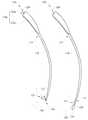
도 1a를 참고하면, 도 1a는 본 발명의 일 실시예에 따른 바늘 유닛의 단면도를 도시한다. 간략하게, 바늘 유닛(100)은 외부 캐뉼라(cannula)(직선 외부 캐뉼라(110a) 또는 곡선 외부 캐뉼라(110b))와 내부 와이어(120)를 포함할 수 있다. 외부 캐뉼라(110a 또는 110b)는 그 내부로 연장된 통로(111)와, 이러한 통로(111)에 연결된 개구단(open end, 113)과, 무딘 팁(dull tip)을 가진 천공단(piercing end, 115)을 포함한다. 일반적으로, 천공단(115)은 부드러운 연조직(soft tissue)이나 장기(예를 들어, 간)를 천공하기 위해 무딘 팁일 수 있으며, 개구부(117)는 천공단(115)의 무딘 팁 주변에 배치된다. 내부 와이어(120)는 통로(111)를 통과하며, 내부 와이어(120)는 개구부(117) 근처 내부 와이어(120)의 단부에 스레딩부(threading part, 121)(예를 들어, 루프(loop))를 포함하며, 내부 와이어(120)는 개구부(117)를 통해 내측이나 외측으로 이동 가능하며, 스레딩부(121)는 적어도 하나의 봉합사(suture thread, 130)가 스레딩부(121)를 통과하도록 한다.Referring to FIG. 1A, FIG. 1A illustrates a cross-sectional view of a needle unit according to an embodiment of the present invention. Briefly, the
일례로, 개구부(117)는 도 1a에 도시된 바와 같이 천공단(115)의 무딘 팁에 인접하여 외부 캐뉼라(110a 또는 110b)의 측면에 배치된다. 그러나, 다른 예에서는, 개구부(117)가 도 1b에 도시된 바와 같이 외부 캐뉼라(110a' 또는 110b')의 단부에 직접 배치되며, 이 경우 외부 캐뉼라(110a' 또는 110b')는 무딘 팁이 없다. 이 경우, 스레딩부(121)는 루프(loop)나 훅(hook)일 수 있다. 스레딩부(121)가 루프형인 경우, 스레딩부(121)는 외부 캐뉼라(110a' 또는 110b')의 개구부(117)에 인접하여 그 끝단에 선형돌출부(123)를 구비할 수 있다. 바늘 유닛(100')이 연조직과 장기를 천공하는 동안 도 1a의 무딘 팁이 되고 개구부(117)를 커버하기 위해 천공단은 선형돌출부(123)의 끝단에 배치된 플러그(plug, 150)일 수 있다. 플러그(150)는 연조직과 장기를 천공하기 위해 무딘 팁과, 바늘 유닛이 연조직과 장기를 천공하는 동안 개구부를 커버하기 위해 덮개 단부(capping end)를 구비할 수 있다. 이러한 플러그(150)는 외과수술용 장치에 적절하게 적용될 수 있는 금속이나 플라스틱이나 다른 재질 등으로 이루어질 수 있다.In one example, the
다른 실시예에서, 내부 와이어(120)는 금속 와이어나 플라스틱 와이어 또는 예를 들어 폴리테트라플루오로에틸렌(polytetrafluoroethylene, PTFE)이 코팅된 금속 와이어와 같이 폴리머 필름이 코팅된 금속 와이어일 수 있다.In another embodiment, the
다른 실시예에서, 외부 캐뉼라(110b)는 도 1a에 도시된 바와 같이 (개구부(117) 상측의) 천공단(115)에서 개구단(113)까지 탄젠트 현각도(tangent chord angle, α)에 의해 정의될 수 있다. 대안으로, 외부 캐뉼라(110b')는 도 1b에 도시된 바와 같이 개구부(117)에서 개구단(113)까지 탄젠트 현각도(α)에 의해 정의될 수도 있다. 탄젠트 현각도(α)는 개구부(117)에서 개구단(113)까지 0도보다 크거나 같으며 90도보다 작다.In another embodiment, the
다른 실시예에서, 외부 캐뉼라는 직선 외부 캐뉼라(straight outer cannula, 110a 또는 110a') 또는 곡선 외부 캐뉼라(curved outer cannula, 110b 또는 110b')일 수 있다. 일례로, 곡선 외부 캐뉼라(110b 또는 110b')는 전체 종방향 길이(l)에 대해 연속 곡률로 상호교환적으로 휘어질 수 있으며, 이러한 곡률은 아래 식(I)으로 정의된다:In other embodiments, the outer cannula may be a straight outer cannula (110a or 110a ') or a curved outer cannula (110b or 110b'). In one example, the curved
외부 캐뉼라의 곡률=
식(I)에서, 외부 캐뉼라의 곡률은 반지름(r)의 역수에 의해 바로 정의된다. 곡선 외부 캐뉼라(110b 또는 110b')의 길이(l)는 반지름(r)과 중심각(θ)을 곱함으로써 정의된다. 다시 말하면, 곡선 외부 캐뉼라(110b)의 곡률은 중심각(θ)을 길이(l)로 나눈 것이다. 일 실시예에서, 곡선 외부 캐뉼라(110b 또는 110b')의 중심각(θ)은 탄젠트 현각도(α)의 두 배, 또는 2α이며, 0 라디안(radian)보다 크거나 같으며 3.14 라디안보다 작다.In equation (I), the curvature of the outer cannula is directly defined by the reciprocal of the radiusr . The lengthl of the curved
직선 외부 캐뉼라(110a 또는 110a') 또는 곡선 외부 캐뉼라(110b 또는 110b')의 길이(l)는 사람 간장(human liver) 길이의 101% 내지 250% (예를 들어, 10cm 내지 30cm)일 수 있다. 직선 외부 캐뉼라(110a 또는 110a') 또는 곡선 외부 캐뉼라(110b 또는 110b')는 공지된 기술의 금속(예를 들어, 금속이나 금속합금이나 스테인리스강이나 그 유사 재질) 또는 플라스틱으로 제작될 수 있으며, 직선 외부 캐뉼라(110a 또는 110a') 또는 곡선 외부 캐뉼라(110b 또는 110b')는 14- 내지 24- 게이지(gauge)(즉, 2.108mm 내지 0.559mm) 폭(w)을 가진다. 비슷하게, 도 1b의 곡선 외부 캐뉼라(110b')도 전체 종방향 길이(l)에 대해 연속 곡률로 상호교환적으로 휘어질 수 있으며, 이러한 곡률도 앞서 상술한 식(I)으로 정의된다.The lengthl of the straight
도 1c 및 도 1d를 참고하면, 도 1c 및 도 1d는 본 발명의 일 실시예에 따른 바늘 유닛의 확대된 사시도를 도시한다. 바늘 유닛(100)은 외부 캐뉼라(110)와 내부 와이어(120)를 포함할 수 있으며, 바늘 유닛(100')은 외부 캐뉼라(110')와 플러그(150)가 구비된 내부 와이어(120)을 포함할 수 있다. 외부 캐뉼라(110)는 그 내부를 연장하는 통로(미도시)와, 통로에 연결된 개구단(113)과, 천공단(115)을 포함한다. 천공단(115)은 연조직 또는 장기(예를 들어, 간)를 천공하기 위해 무딘 팁을 가진다. 개구부(117)는 천공단(115) 주변에 배치되는데, 외부 캐뉼라의 측면에 천공단(115)에 인접하여 배치되거나(바늘 유닛(100)), 외부 캐뉼라(100')의 단부에 직접 배치된다(바늘 유닛(100')). 내부 와이어(120)의 단부에 스레딩부(121)는 통로로부터 개구부(117)를 통해 내측이나 외측으로 이동할 수 있으며, 스레딩부(121)는 스레딩부(121)가 개구부(117)를 통해 외측으로 연장되는 동안 적어도 하나의 봉합사(130)를 포획한다. 그러나, 도 1c 및 도 1d의 바늘 유닛은 단지 예를 든 것이며, 직선 외부 캐뉼라가 다른 실시예의 바늘 유닛에도 적용될 수 있음이 본 발명의 추가 상세 설명을 제공하는 것보다 쉽게 이해될 수 있다.Referring to Figures 1c and 1d, Figures 1c and 1d show an enlarged perspective view of a needle unit according to one embodiment of the present invention. The
다른 예들에서, 도 1c 및 도 1d에 도시된 바늘 유닛(100')의 스레딩부(121)는, 외부 캐뉼라(110', 미도시)의 개구부(117)에 인접한 타원형 루프나 원형 루프나 다이아몬드형 루프의 끝단에 선형돌출부를 구비하지 않을 수 있다. 상술한 예들에서, 바늘 유닛(100')의 이러한 루프(121)는 금속 재질로 만들어지며, 스레딩부(121)가 개구부 외측에서 연조직과 장기(미도시)를 천공하는 동안 천공단으로서 유지되도록 개구부(117)보다 다소 넓다.In other instances, the threading
일 실시예에서, 예를 들어 도 1c에 도시된 핸들(handle, 130b)이나 도 1d에 도시된 핸들(130c)과 같은 핸들은 외부 캐뉼라(110)나 외부 캐뉼라(110')의 개구단(113)에 선택적으로 배치될 수 있으며, 핸들(130b)은 외부 캐뉼라(110)나 외부 캐뉼라(110')의 개구단(113)에서 수직하게 경사지게 연장되며, 또는 핸들(130c)은 외부 캐뉼라(110)나 외부 캐뉼라(110')와 같이 동일한 방향으로 연장된다. 또한, 내부 와이어(120)가 개구부(133b 또는 133c)로부터 채널과 통로(111)를 통해 개구부(117)를 통과하도록, 도 1c에 도시된 핸들(130b)이나 도 1d에 도시된 핸들(130c)은 각각, 외부 캐뉼라(110 또는 110')의 통로(111)와 개구부(133b 또는 133c)를 연결하기 위한 채널(channel, 미도시)을 홀딩부(131b 또는 131c)에 가진다.A handle such as the
다른 실시예에서, 외부 캐뉼라는 복강경 수술용 기구의 중공 튜브 내에 배치될 수 있다. 도 2를 참고하면, 도 2는 본 발명의 다른 실시예에 따른 바늘 유닛의 단면도를 도시한다. 예를 들어 종래기술에서 일반적으로 사용되는 투관침(trocar) 기구와 같은 복강경 수술용 기구(140)는, 개구단(145)을 통해 내측 또는 외측으로 이동하기 위해 외부 캐뉼라(110 또는 110')를 수용하기 위한 중공 튜브(141)를 가진다. 이 경우, 종래기술에서 일반적으로 사용되는 복강경 카메라가 또 다른 투관침 기구에 별도로 배치될 수 있다.In another embodiment, the outer cannula may be disposed within a hollow tube of a laparoscopic surgical instrument. 2, there is shown a cross-sectional view of a needle unit according to another embodiment of the present invention. For example, a laparoscopic
일반적으로, 외부 캐뉼라(110 또는 110')의 길이는 실제 요구 사항에 따라 달라지며, 본 발명의 사상이 여기 설정된 길이에 한정하는 것은 아니다. 예를 들어, 통상의 개방 복부 수술(open abdomen surgery) 동안, 바늘 유닛(100 또는 100')이 수동으로 작동될 때, 외부 캐뉼라(110 또는 110')는 사람 간 두께의 101퍼센트 내지 250퍼센트에 상응하는 길이를 가질 수 있다. 또는, 복강경 수술 동안, 바늘 유닛(100 또는 100')이 투관침구(trocar port)를 통해 지나갈 때, 외부 캐뉼라(110)는 도 2에 도시된 중공 튜브(141)의 길이보다 더 길 수 있다(예를 들어, 대략 50cm).Generally, the length of the
도 3을 참조하면, 도 3은 본 발명의 실시예들에 따른 내부 와이어를 도시한다. 일 실시예에서, 내부 와이어(120)는 금속 와이어나 플라스틱 와이어 또는 예를 들어 폴리테트라플루오로에틸렌(PTFE)이 코팅된 금속 와이어와 같이 폴리머 필름이 코팅된 금속 와이어일 수 있다. 내부 와이어(120)의 스레딩부(121)는 예를 들어 (루프(121a, 121b)와 같은) 타원형 루프나 (루프(121c, 121d)와 같은) 원형 루프나 (루프(121e, 121f)와 같은) 다이아몬드형 루프와 같은 다양한 형상일 수 있다. 다른 실시예에서, 내부 와이어(120)의 스레딩부(121)는 U형상부(121g)일 수 있다. 또 다른 실시예에서, 내부 와이어(120)의 스레딩부(121)는 훅(hook, 121h)일 수 있다.Referring to FIG. 3, FIG. 3 illustrates an inner wire in accordance with embodiments of the present invention. In one embodiment, the
다시 도 3을 참조하면, 다른 실시예에서, 스레딩부(121)는 외부 캐뉼라(여기서는 외부 캐뉼라(110a 또는 110b)를 지칭함)의 개구부에 인접하는 끝단에 선형돌출부, 예를 들어 루프(121b)의 돌출부(123b)나 루프(121d)의 돌출부(123d)나 루프(121f)의 돌출부(123f)를 더 포함할 수 있다. 위에 예시된 돌출부들은 내부 와이어(120)가 개구부를 통해 내측이나 외측으로 용이하게 이동하도록 한다. 다른 예들에서, 루프(121b)의 돌출부(123b)나 루프(121d)의 돌출부(123d)나 루프(121f)의 돌출부(123f)는, 바늘 유닛(100 또는 100')이 연조직과 장기를 천공하는 동안 무딘 팁이 되며 개구부(117)를 커버하도록 그 표면에 각각 배치된 플러그(150)를 포함할 수 있다. 또는, 내부 와이어(120)는 외부 캐뉼라(110)(여기서는 외부 캐뉼라(110a 또는 110b)를 지칭함)의 개구부에 인접하는 단부에 배치된 훅(121i)과 플러그(150)를 포함할 수 있으며, 훅(121i)은 스레딩부(121i)로 적용되며, 플러그(150)는 무딘 팁으로 적용된다. 이 경우, 훅(121i)과 플러그(150)를 구비한 내부 와이어(120)는 외부 캐뉼라(110a)의 이용 또는 이용과 상관없이 수술시 그 자체로서 바늘로 적용될 수도 있다.Referring again to Figure 3, in another embodiment, the threading
간 절제술시 간의 원하는 혈관을 묶기 위해, 내부 와이어(120)는 적어도 하나의 봉합사(130)가 스레딩부(121)를 통해 통과하도록 한다. 일 실시예에서, 봉합사(130)는 종래에 일반적으로 사용되는 흡수성 또는 비흡수성 재질일 수 있다.The
바늘 유닛이 곡선일 때, 곡선 바늘 유닛은 직선 바늘 유닛보다 장기의 원하는 혈관을 더 효과적이고 용이하게 묶는데 적용될 수 있다. 도 4를 참조하면, 도 4는 국제간담췌학회(International Hepato-Pancreato-Billiary Association, IHPBA)에 의해 2000년에 발간된 브리즈번 간 해부 및 절제술 용어(Brisbane Terminology of Liver Anatomy and Resections)에 따른 간의 분절 분류(segmental classification of the liver)의 개략도를 도시한다. 본 발명의 바늘 유닛은 간의 모든 분절에서 Glissonian 유경(Glissonian pedicle)의 첫번째, 두번째, 세번째 및 네번째 오더 브랜치(order branch)에 적용될 수 있다.
When the needle unit is curved, the curved needle unit can be applied to more effectively and easily bundle the desired blood vessel of a long term than the straight needle unit. FIG. 4 is a block diagram of a liver segment according to the Brisbane Terminology of Liver Anatomy and Resections (BBS) published in 2000 by the International Hepato-Pancreato-Billiary Association (IHPBA) And a segmental classification of the liver. The needle unit of the present invention can be applied to first, second, third and fourth order branches of a Glissonian pedicle in all segments of the liver.
바늘 유닛의 적용Application of needle unit
간 절제술과 같은 수술 중, 바늘 유닛은 도 1a, 도 5a 내지 도 6b를 참조하여 다음과 같이 적용될 수 있다. 간 절제술 시, 수술중 초음파(미도시) 또는 복강경 카메라가 절개(resectability)를 평가하고 적절한 절개 라인을 결정하기 위해 적절히 사용될 수 있다. 바늘 유닛의 작동을 상세하게 설명하기 위해, 좌측 간엽(hepatic lobe)과 우측 간엽은 여기에 예시된 바와 같은 바늘 유닛(100)을 사용하여 절개된다. 그러나, 후술할 내용은 발명의 상세한 설명을 더 제공하려는 의도없이 단순한 예시에 불과하다는 것이 이해된다.During surgery such as hepatic resection, the needle unit can be applied as follows with reference to Figs. 1A, 5A to 6B. During liver resection, an intra-operative ultrasound (not shown) or a laparoscopic camera can be used to evaluate resectability and determine the appropriate incision line. To explain the operation of the needle unit in detail, the left hepatic lobe and the right hepatic lobe are incised using the
1. 간의 하부, 특히 IVC 상부에서 혈관 묶기1. Vein bundles at the bottom of the liver, especially at the top of the IVC
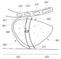
도 1a, 5a 및 도 5b를 참조하면, 도 5a 및 도 5b는 본 발명의 일 실시예에 따른 좌측 간엽의 시상단면도를 도시한다. 기본적으로 수술전 준비 후, 앞측 복벽(abdominal wall, 509)이 들어올려 진다. 바늘 유닛(100)의 외부 캐뉼라(110b)는 경로(511)를 따라 주요 공급 혈관(507)(예를 들어, 좌측 간 Glissonian 유경)의 일측에서 간(500)의 하면(501)(하대정맥(inferior venal cava, 503) 상측)부터 상면(505)까지 뚫으며, 내부 와이어(120)가 외부 캐뉼라(110b)의 개구부(117)를 통해 외측으로 이동하는 동안 봉합사(130)의 제1 단부는 바늘 유닛(100)의 내부 와이어(120)의 스레딩부(121)를 통과하여 묶는다.Referring now to Figs. 1A, 5A and 5B, Figs. 5A and 5B show a sagittal cross-sectional view of a left hepatic lobe according to an embodiment of the present invention. Basically, after preparation before surgery, the
다음으로, 외부 캐뉼라(110b)는 동일한 경로(511)를 따라 간(500)의 상면(505)과 하면(501)을 통해 회수되며, 봉합사(130)의 제1 단부를 따라 간의 하부로 이동시키고 봉합사(130)의 제2 단부를 간(500)의 상면(505)에 잔존시킨다. 다음으로, 바늘 유닛(100)은 경로(513)를 따라 주요 공급 혈관(507)의 타측에서 하면(501)부터 상면(505)까지 뚫으며, 봉합사(130)의 제1 단부를 간(500)의 상면(505)에 이동시키고 잔존시킨다. 경로(511)와 경로(513) 간에 거리는 상면에서 약 5cm이다. 그리고 마지막으로, 대상 혈관의 각각의 유입이나 유출을 컨트롤하도록, 봉합사(130)의 제1 단부와 제2 단부는 간(500)의 상면(505)에서 매듭(521)으로 단단히 묶인다.The
다른 실시예에서, 도 5b에 도시된 바와 같이 절개하고자 하는 좌측 간엽의 절개 라인의 내측면을 따라 간헐적으로 서로 맞물리는 봉합(521) 열(row)을 만들기 위해, 바늘 유닛(100)은 상술한 바와 같이 어느 한 방향으로 반복적으로 적용된다. 다음으로, 간의 유입과 유출을 차단하기 위해 Pringle's maneuver를 적용하거나 다른 시술을 적용할 필요 없이, 좌측 간엽은 클램핑 및 절개방법을 이용하여 가위나 전기 소작기(electrocautery) 또는 켈리 클램프(Kelly clamp)에 의해 직접 나눠질 수 있다. 상당한 크기를 가진 관형 구조는 보강을 위해 봉합된다. 상술한 간 절제술 후, 남겨진 간의 표면에 섬유소 밀봉제 또는 콜라겐 시트를 적용할 필요가 없다. 통상적으로, 필요한 경우 큰 구경의 배액관(drainage tube)이 종속 부분에 배치될 수 있다. In another embodiment, to make a row of
봉합사(130)의 두 단부를 서로 묶기 위해 간(500)의 상면(505) 또는 하면(501) 중 어느 하나에 봉합사(130)의 두 단부가 같은 측면으로 도출될 수 있다면, 봉합사(130)에 대한 어떠한 변형도 가능하다. 이러한 과정 후 종래의 방법으로 간 종양(liver tumor)의 적출(excision)이 수행될 수 있다. 간 종양의 적출이 완료된 후, 이러한 매듭(521)과 봉합사(130)는 영구적으로 남겨질 수도 있으며, 간(500)의 유입과 유출의 복원이 필요한 경우 매듭(521)과 봉합사(130)가 절단되어 제거될 수도 있다.If the two ends of the
간 표면에서 직선 바늘(예를 들어, 외부 캐뉼라(110a)와 내부 와이어(120)를 구비한 바늘 유닛(100))의 적용이 가능하도록 간의 상면(505)과 복벽(509) 사이에 작은 공간이 존재할 때, 상술한 시술은 간우엽절제술(right hepatic lobectomy)뿐만 아니라 다양한 절제술에도 적용될 수 있다.
A small space is formed between the
2. 간 표면 또는 하부에서, 특히 간과 복벽 사이의 좁은 공간 환경에서 (IVC에서 이격되어) 혈관 묶기2. In the narrow space between the liver surface and the lower surface, especially between the liver and the abdominal wall (apart from the IVC)
또한, 장소가 협소한 경우 종래의 직선 바늘은 어렵게 접근하는 곳을, 바늘 유닛은 좁은 흉곽 아래 또는 간과 복벽 사이의 좁은 공간에서 간의 혈관을 묶는 것을 용이하게 할 수 있다.In addition, when the place is narrow, the conventional straight needle can be difficult to approach, and the needle unit can easily bind the blood vessel in the narrow space below the narrow thorax or in the narrow space between the liver and abdominal wall.
도 1a, 6a 및 6b를 참조하면, 도 6a는 본 발명의 다른 실시예에 따른 흉곽(610)과 횡경막(602) 아래 우측 간엽의 측면도를 도시하고, 도 6b는 본 발명의 다른 실시예에 따른 우측 간엽의 시상단면도를 도시한다. 유사하게, 기본적으로 수술전 준비 후, 흉곽(610)이 약간 들려진다. 바늘 유닛(100)의 외부 캐뉼라(110b)는 외부 캐뉼라(110b)가 복막뒤의 복벽(623)을 마주보고 경로(611)를 따라 주요 공급 혈관(607)의 일측에서 간(600)의 상면(605)부터 하면(601)까지 (도 5a 및 5b의 하대정맥(503)에서 이격되게) 뚫는다.Referring to FIGS. 1A, 6A and 6B, FIG. 6A shows a side view of the right lateral lobe below the
다음으로, 내부 와이어(120)의 스레딩부(121)는 외부 캐뉼라(110b)의 개구부(117)를 통해 외측으로 이동하여 봉합사(130)의 제1 단부를 포획한다. 다음으로, 외부 캐뉼라(110b)는 동일한 경로(611)를 따라 간(600)의 하면(601)과 상면(605)을 통해 회수되며, 봉합사(130)의 제1 단부를 간(600)의 상면(605)에 잔존시킨다. 다음으로, 바늘 유닛(100)은 경로(613)를 따라 주요 공급 혈관(607)의 타측에서 상면(605)부터 하면(601)까지 뚫으며, 내부 와이어(120)의 스레딩부(121)는 외부 캐뉼라(110b)의 개구부(117)를 통해 외측으로 이동하여 봉합사(130)의 제2 단부를 포획한다. 경로(611)와 경로(613) 간에 거리는 상면에서 약 5cm이다. 그리고, 외부 캐뉼라(110b)는 동일한 경로(613)를 따라 간(600)의 하면(601)과 상면(605)을 통해 회수되며, 봉합사(130)의 제2 단부를 간(600)의 상면(605)에 잔존시킨다. 따라서, 봉합사(130)의 제1 단부와 제2 단부는 간(600)의 상면(605)에 단단히 묶인다.The threading
또는, 내부 와이어(120)가 외부 캐뉼라(110b)의 개구부(117)를 통해 외측으로 이동하는 동안, 봉합사(130)의 제1 단부는 바늘 유닛(100)의 내부 와이어(120)의 스레딩부(121)를 통해 지나가며, 내부 와이어(120)는 스레딩부(121)의 끝단이 개구부(117) 내에 위치할 때까지 내측으로 이동한다. 이러한 준비 후, 바늘 유닛(100)의 외부 캐뉼라(110b)는 경로(611)를 따라 주요 공급 혈관(607)의 일측에서 간(600)의 하면(601)부터 (하대정맥(503)에 이격되거나 하대정맥(503) 상측으로) 상면(605)까지 뚫으며, 봉합사(130)의 제1 단부를 간(600)의 상면(605)에 이동시키고 잔존시킨다. 다음으로, 외부 캐뉼라(110b)는 동일한 경로(611)를 따라 간(600)의 상면(605)과 하면(603)을 통해 회수된다.Alternatively, the first end of the
내부 와이어(120)의 스레딩부(121)는 외부 캐뉼라(110b)의 개구부(117)를 통해 외측으로 이동하여 봉합사(130)의 제2 단부를 포획하며, 내부 와이어(120)는 개구부(117) 내에서 스레딩부(121)의 끝단까지 내측으로 이동한다. 이러한 준비 후, 바늘 유닛(100)은 경로(613)를 따라 주요 공급 혈관(607)의 타측에서 하면(601)부터 상면(605)까지 뚫으며, 봉합사(130)의 제2 단부를 간(600)의 상면(605)에 이동시키고 잔존시킨다. 경로(611)와 경로(613) 간에 거리도 역시 상면에서 약 5cm이다. 그리고, 대상 혈관의 각각의 유입이나 유출을 컨트롤하도록, 봉합사(130)의 제1 단부와 제2 단부는 간(500)의 상면(505)에서 단단히 묶인다. The threading
다른 실시예에서, 도 5b에 유사하게 도시된 바와 같이 절개하고자 하는 간엽 또는 분절의 절개 라인의 내측면을 따라 간헐적으로 서로 맞물리는 봉합 열(row)을 만들기 위해, 바늘 유닛(100)은 상술한 바와 같이 어느 한 방향으로 반복적으로 적용된다. 다음으로, 간의 유입과 유출을 차단하기 위해 Pringle's maneuver를 적용하거나 다른 시술을 적용할 필요 없이, 간엽 또는 분절은 클램핑 및 절개방법을 이용하여 가위나 전기 소작기(electrocautery) 또는 켈리 클램프(Kelly clamp)에 의해 직접 나눠질 수 있다. 상당한 크기를 가진 관형 구조는 보강을 위해 봉합된다. 상술한 간 절제술 후, 남겨진 간의 표면에 섬유소 밀봉제 또는 콜라겐 시트를 적용할 필요가 없다. 통상적으로, 필요한 경우 큰 구경의 배액관(drainage tube)이 종속 부분에 배치될 수 있다.
In another embodiment, to create a stitching row that intermittently engages with each other along the inner side of the incision line of the mesenchy or segment to be incised, as shown in Fig. 5b, And is repeatedly applied in either direction. Next, we used scissors, electrocautery, or Kelly clamps, using clamping and incision techniques, without Pringle's maneuver or other procedures to block inflow and outflow of the liver, As shown in FIG. Tubular structures of considerable size are sealed for reinforcement. There is no need to apply a fibrin sealant or collagen sheet to the remaining liver surface after the above-mentioned liver resection. Typically, a large diameter drainage tube may be placed in the slave if necessary.
혈관을 묶기 위한 긴 직선 바늘(또는 직선 외부 캐뉼라(110a)와 내부 와이어(120)를 구비한 바늘 유닛(100 또는 100')과 비교하여, 곡선 외부 캐뉼라(110b)와 내부 와이어(120)를 구비한 바늘 유닛은 하대정맥(IVC) 상측에서 간 절제술시, 또는 하대정맥을 손상시키거나 지나치게 흉곽을 들어내거나 또는 개흉술(thoracotomy)을 할애할 필요 없이 간과 양측 간엽의 복벽(도 5a, 5b 및 6b의 복벽(509) 또는 도 5a, 5b 및 6b의 복막뒤의 복벽(623)) 사이의 협소한 공간에서, 좌측 간엽의 혈관을 묶는데 충분히 적용될 수 있으며, 종래의 직선 바늘은 하대정맥 상측의 혈관에 접근하거나 간 종양이 클 때 간과 복벽 사이 협소한 공간에 적용하는 것이 어렵다.(Or a
또한, 본 발명의 바늘 유닛은 긴 직선 바늘 또는 간 절제술시 간 혈관을 묶기 위한 직선 외부 캐뉼라(110a)와 내부 와이어(120)를 구비한 바늘 유닛(100 또는 100')과 조합하여 사용할 수 있다. 예를 들어, 하대정맥 상이나 간과 복벽 간의 좁은 공간에서 곡선 외부 캐뉼라(110b)와 내부 와이어(120)를 구비한 바늘 유닛을 이용하여 혈관을 묶을 수 있으며, 하대정맥에서 멀리 이격된 혈관은 긴 직선 바늘 또는 직선 외부 캐뉼라(110a)와 내부 와이어(120)를 구비한 바늘 유닛(100)을 이용하여 묶을 수 있다.In addition, the needle unit of the present invention can be used in combination with a
또한, 바늘 유닛은 본 발명에서 예시된 바와 같이 간의 혈관을 묶는데 적용된다; 그러나, 이러한 바늘 유닛은 첨부된 청구항의 사상과 범위 내에서 부분 절제술시 다른 장기의 원하는 혈관을 묶는데도 적용될 수 있다. 예를 들어, 이러한 바늘 유닛은 여성 비뇨기의 실금(urinary incontinence) 치료에 적용될 수 있다. 이런 실시예의 경우, 요도(urethra)를 안착하고 요도에 지지체를 제공하도록, 치골(pubic bone)의 양측을 따라 질(vagina)에서 결합조직(connective tissue)으로 지나가는 슬링 테이프(sling tape)를 배치하는데에, 바늘 유닛이 적용될 수 있다.Also, the needle unit is applied to bind blood vessels of the liver as illustrated in the present invention; However, such a needle unit can also be applied to bind desired blood vessels of different organs in a partial resection within the spirit and scope of the appended claims. For example, such a needle unit can be applied to urinary incontinence treatment of female urinary incontinence. In this embodiment, a sling tape is placed that passes from the vagina to the connective tissue along both sides of the pubic bone to seat the urethra and provide a support to the urethra The needle unit can be applied.
본 발명의 상술한 실시예들에 따르면, 본 발명의 상술한 바늘 유닛은 간 절제술(Chang's maneuver), 특히 직선 바늘로는 상당히 어렵거나 상황에 따라 불가능할 수 있는 간 절제술에서 분절(4, 7, 8)과 좌측 또는 중간 간정맥(hepatic veins)에서 원하는 혈관을 효과적이고 용이하게 묶기 위해 외부 캐뉼라와 그 내부를 통과하는 내부 와이어를 포함한다. 따라서, 종래의 직선 바늘의 한계가 효과적으로 극복될 수 있다.According to the above-described embodiments of the present invention, the above-mentioned needle unit of the present invention can be used in the liver's 4, 7, 8 < RTI ID = 0.0 > ) And an inner cannula through the outer cannula to effectively and easily bundle the desired vessel in the left or middle hepatic veins. Therefore, the limit of the conventional straight needle can be effectively overcome.
당업자에 의해 이해되는 바와 같이, 본 발명의 전술한 실시예들은 본 발명을 한정하기보다 본 발명의 예시이다. 이는 첨부된 청구항의 사상과 범위 내에 포함된 다양한 변형과 유사한 형태를 포함하기 위함이다. 따라서, 본 발명의 범위는 이러한 모든 변형 및 유사한 구조를 포괄하도록 광범위하게 해석되어야 한다.As will be understood by those skilled in the art, the foregoing embodiments of the invention are illustrative of the invention rather than limiting the invention. It is intended to cover various modifications and equivalent arrangements included within the spirit and scope of the appended claims. Accordingly, the scope of the present invention should be construed broadly to cover all such modifications and similar structures.
Claims (10)
Translated fromKorean-상기 곡선의 외부 캐뉼라는,
상기 통로와 연결된 개구단과,
개구부를 가진 천공단을 더 포함하며,
상기 천공단은 연조직과 장기를 천공하는 무딘 팁(dull tip)이며, 상기 개구부는 상기 곡선의 외부 캐뉼라의 내측 곡면을 따라 배치되며-; 및
상기 통로를 통과하며, 상기 개구부에 인접하여 단부에 스레딩부를 구비한 내부 와이어를 포함하며,
상기 내부 와이어는 상기 개구부를 통해 내측이나 외측으로 이동 가능하며, 상기 스레딩부는 상기 곡선의 외부 캐뉼라의 내측 곡면에서 적어도 하나의 봉합사가 상기 스레딩부를 통과하도록 하는 바늘 유닛.
A curved outer cannula with an internally extended passage,
- the outer cannula of said curve,
An opening end connected to the passage,
Further comprising a silencer having an opening,
The sill tip is a dull tip for perforating soft tissue and organs, the opening being disposed along an inner curved surface of the outer cannula of the curve; And
An internal wire passing through the passageway and having a threaded portion at an end adjacent the opening,
Wherein the inner wire is movable inward or outward through the opening and the threading portion allows at least one suture to pass through the threading portion at an inner curved surface of the outer cannula of the curve.
상기 곡선의 외부 캐뉼라는 전체 종방향 길이를 따라 곡률로 휘어지며, 상기 곡률은 0도보다 크거나 같으며 180도보다 작은 중심각을 상기 곡선의 외부 캐뉼라의 길이로 나눈 것이며, 상기 길이는 사람 간장 길이의 101% 내지 250%인 바늘 유닛.
The method according to claim 1,
Wherein the outer cannula of the curve is curved along the entire longitudinal length and wherein the curvature is greater than or equal to 0 degrees and a central angle less than 180 degrees divided by the length of the outer cannula of the curve, Lt; RTI ID = 0.0 > 101% < / RTI >
2.108mm 내지 0.559mm의 폭을 가진 상기 곡선의 외부 캐뉼라는 금속 또는 플라스틱으로 이루어지는 바늘 유닛.
The method according to claim 1,
The outer cannula of the curve having a width of 2.108 mm to 0.559 mm is made of metal or plastic.
상기 내부 와이어는 금속 와이어나 플라스틱 와이어 또는 폴리머 필름이 코팅된 금속 와이어인 바늘 유닛.
The method according to claim 1,
Wherein the inner wire is a metal wire, a plastic wire, or a metal wire coated with a polymer film.
상기 스레딩부는 타원형 루프(loop)나 원형 루프나 다이아몬드형 루프나 U형상부 또는 훅(hook)형인 바늘 유닛.
The method according to claim 1,
Wherein the threading portion is an elliptical loop, a circular loop, a diamond-like loop, a U-shaped portion, or a hook type.
상기 스레딩부는 상기 곡선의 외부 캐뉼라의 개구부에 인접하여 상기 타원형 루프 또는 상기 원형 루프 또는 상기 다이아몬드형 루프의 끝단에 선형돌출부를 포함하는 바늘 유닛.
6. The method of claim 5,
Wherein the threading portion comprises a linear protrusion at the end of the elliptical loop or the circular loop or the diamond-like loop adjacent the opening of the outer cannula of the curve.
상기 개구부는 상기 무딘 팁에 인접되게 상기 곡선의 외부 캐뉼라의 측면에 배치되는 바늘 유닛.
The method according to claim 6,
Wherein the opening is disposed on a side of the outer cannula of the curve adjacent the blunt tip.
상기 개구부는 상기 무딘 팁의 단부에 직접 배치되는 바늘 유닛.
The method according to claim 6,
Wherein the opening is disposed directly at an end of the blunt tip.
상기 천공단은, 상기 바늘 유닛이 상기 연조직과 장기를 천공하는 동안 상기 무딘 팁이 되고 상기 개구부를 커버하기 위해 상기 선형돌출부의 끝단에 배치된 플러그(plug)를 포함하는 바늘 유닛.
9. The method of claim 8,
Wherein said cloth end includes a plug disposed at an end of said linear protrusion for making said blunt tip and covering said opening while said needle unit punctures said soft tissue and said organs.
금속으로 이루어진 상기 타원형 루프 또는 상기 원형 루프 또는 상기 다이아몬드형 루프는, 상기 연조직과 장기를 천공하는 동안 상기 개구부 외측에서 상기 천공단이 되는 상기 스레딩부를 유지하도록 상기 개구부보다 넓은 바늘 유닛.9. The method of claim 8,
The elliptical loop made of metal or the circular loop or the diamond-like loop is wider than the opening so as to hold the threading portion which becomes the cloth end outside the opening during perforation of the soft tissue and the organs.
Priority Applications (1)
| Application Number | Priority Date | Filing Date | Title |
|---|---|---|---|
| KR1020120019049AKR101391405B1 (en) | 2012-02-24 | 2012-02-24 | needle unit |
Applications Claiming Priority (1)
| Application Number | Priority Date | Filing Date | Title |
|---|---|---|---|
| KR1020120019049AKR101391405B1 (en) | 2012-02-24 | 2012-02-24 | needle unit |
Publications (2)
| Publication Number | Publication Date |
|---|---|
| KR20130097415A KR20130097415A (en) | 2013-09-03 |
| KR101391405B1true KR101391405B1 (en) | 2014-05-02 |
Family
ID=49449799
Family Applications (1)
| Application Number | Title | Priority Date | Filing Date |
|---|---|---|---|
| KR1020120019049AExpired - Fee RelatedKR101391405B1 (en) | 2012-02-24 | 2012-02-24 | needle unit |
Country Status (1)
| Country | Link |
|---|---|
| KR (1) | KR101391405B1 (en) |
Cited By (1)
| Publication number | Priority date | Publication date | Assignee | Title |
|---|---|---|---|---|
| KR101486456B1 (en)* | 2014-11-26 | 2015-01-26 | (주)트리플씨메디칼 | Cannula body and lifting cannula having same, and method of manufacturing cannula body |
Families Citing this family (6)
| Publication number | Priority date | Publication date | Assignee | Title |
|---|---|---|---|---|
| KR101488004B1 (en)* | 2013-10-07 | 2015-01-29 | 포항공과대학교 산학협력단 | Curved type drill |
| KR101502074B1 (en)* | 2013-12-09 | 2015-03-11 | 주식회사 씨앤지 | A cannula for skin wrinkling plastic surgery and the fabrication method |
| KR101600567B1 (en)* | 2014-04-18 | 2016-03-07 | (주)리젠바이오참 | Medical round needle for imbedding |
| KR101825878B1 (en) | 2017-05-12 | 2018-02-06 | 장철호 | Needle apparatus for reposition surgery of orbital fat |
| CN114366254B (en)* | 2022-03-22 | 2022-06-10 | 中南大学 | A liver puncture device |
| US11925542B1 (en)* | 2023-08-14 | 2024-03-12 | Sangjoon Yoon | Mesh assembly for cosmetic surgery |
Citations (2)
| Publication number | Priority date | Publication date | Assignee | Title |
|---|---|---|---|---|
| US20040092953A1 (en) | 2002-08-20 | 2004-05-13 | Olympus Optical Co., Ltd. | Surgical treatment device |
| JP2009515657A (en) | 2005-11-15 | 2009-04-16 | ジョンズ ホプキンス ユニバーシティ | Active cannula for biosensing and surgical procedures |
- 2012
- 2012-02-24KRKR1020120019049Apatent/KR101391405B1/ennot_activeExpired - Fee Related
Patent Citations (2)
| Publication number | Priority date | Publication date | Assignee | Title |
|---|---|---|---|---|
| US20040092953A1 (en) | 2002-08-20 | 2004-05-13 | Olympus Optical Co., Ltd. | Surgical treatment device |
| JP2009515657A (en) | 2005-11-15 | 2009-04-16 | ジョンズ ホプキンス ユニバーシティ | Active cannula for biosensing and surgical procedures |
Cited By (1)
| Publication number | Priority date | Publication date | Assignee | Title |
|---|---|---|---|---|
| KR101486456B1 (en)* | 2014-11-26 | 2015-01-26 | (주)트리플씨메디칼 | Cannula body and lifting cannula having same, and method of manufacturing cannula body |
Also Published As
| Publication number | Publication date |
|---|---|
| KR20130097415A (en) | 2013-09-03 |
Similar Documents
| Publication | Publication Date | Title |
|---|---|---|
| KR101391405B1 (en) | needle unit | |
| JP5462301B2 (en) | Needle unit | |
| JP6763923B2 (en) | Sutures that do not require knotting and kits containing them | |
| Gonzalez-Rivas et al. | Technical aspects of uniportal video-assisted thoracoscopic double sleeve bronchovascular resections | |
| US10201353B2 (en) | Ligation clip | |
| US20220280193A1 (en) | Causing ischemia in tumors | |
| KR101668663B1 (en) | The kit which using a stitching fiber and method thereof | |
| CA2928615A1 (en) | Suturing device and method | |
| WO2021011502A1 (en) | Device allowing large bore transseptal access with subsequent atrial re-access and method thereof | |
| US9393020B2 (en) | Ligator and ligation method | |
| TWI477254B (en) | Needle unit | |
| WO2018062284A1 (en) | Suture needle for uterine hemostatic compression | |
| RU2691924C1 (en) | Method for gastrostomy using polypropylene mesh | |
| RU115641U1 (en) | LIGATURAL NEEDLE OF DESHAN MODIFIED FOR SEWING OF PARENCHIMATOUS BODIES | |
| CN219661787U (en) | Liver suspension device is assisted to peritoneoscope | |
| AU2020315591A1 (en) | Device allowing large bore transseptal access with subsequent atrial re-access and method thereof | |
| RU2459589C1 (en) | Method of sewing liver wounds | |
| CN103405252A (en) | Surgical suture needle for laparoscopic partial nephrectomy | |
| CN206548555U (en) | A kind of anchor tail suture | |
| RU2739860C1 (en) | Method for making a final sliding knot in an intracorporeal continuous surgical suture | |
| TR201812305A2 (en) | Suture material developed for end-to-end anastomosis | |
| RU195850U1 (en) | Device for emergency stop bleeding of parenchymal organs | |
| RU2460477C1 (en) | Disposable surgical needle | |
| Liehn | Basic Instruments | |
| CN118303935A (en) | Anastomosis clamp with suture line and anastomat |
Legal Events
| Date | Code | Title | Description |
|---|---|---|---|
| A201 | Request for examination | ||
| PA0109 | Patent application | St.27 status event code:A-0-1-A10-A12-nap-PA0109 | |
| PA0201 | Request for examination | St.27 status event code:A-1-2-D10-D11-exm-PA0201 | |
| P11-X000 | Amendment of application requested | St.27 status event code:A-2-2-P10-P11-nap-X000 | |
| P13-X000 | Application amended | St.27 status event code:A-2-2-P10-P13-nap-X000 | |
| D13-X000 | Search requested | St.27 status event code:A-1-2-D10-D13-srh-X000 | |
| D14-X000 | Search report completed | St.27 status event code:A-1-2-D10-D14-srh-X000 | |
| E902 | Notification of reason for refusal | ||
| PE0902 | Notice of grounds for rejection | St.27 status event code:A-1-2-D10-D21-exm-PE0902 | |
| T11-X000 | Administrative time limit extension requested | St.27 status event code:U-3-3-T10-T11-oth-X000 | |
| P11-X000 | Amendment of application requested | St.27 status event code:A-2-2-P10-P11-nap-X000 | |
| P13-X000 | Application amended | St.27 status event code:A-2-2-P10-P13-nap-X000 | |
| PG1501 | Laying open of application | St.27 status event code:A-1-1-Q10-Q12-nap-PG1501 | |
| E902 | Notification of reason for refusal | ||
| PE0902 | Notice of grounds for rejection | St.27 status event code:A-1-2-D10-D21-exm-PE0902 | |
| P11-X000 | Amendment of application requested | St.27 status event code:A-2-2-P10-P11-nap-X000 | |
| P13-X000 | Application amended | St.27 status event code:A-2-2-P10-P13-nap-X000 | |
| E701 | Decision to grant or registration of patent right | ||
| PE0701 | Decision of registration | St.27 status event code:A-1-2-D10-D22-exm-PE0701 | |
| GRNT | Written decision to grant | ||
| PR0701 | Registration of establishment | St.27 status event code:A-2-4-F10-F11-exm-PR0701 | |
| PR1002 | Payment of registration fee | St.27 status event code:A-2-2-U10-U11-oth-PR1002 Fee payment year number:1 | |
| PG1601 | Publication of registration | St.27 status event code:A-4-4-Q10-Q13-nap-PG1601 | |
| P22-X000 | Classification modified | St.27 status event code:A-4-4-P10-P22-nap-X000 | |
| LAPS | Lapse due to unpaid annual fee | ||
| PC1903 | Unpaid annual fee | St.27 status event code:A-4-4-U10-U13-oth-PC1903 Not in force date:20170426 Payment event data comment text:Termination Category : DEFAULT_OF_REGISTRATION_FEE | |
| P22-X000 | Classification modified | St.27 status event code:A-4-4-P10-P22-nap-X000 | |
| PC1903 | Unpaid annual fee | St.27 status event code:N-4-6-H10-H13-oth-PC1903 Ip right cessation event data comment text:Termination Category : DEFAULT_OF_REGISTRATION_FEE Not in force date:20170426 | |
| P22-X000 | Classification modified | St.27 status event code:A-4-4-P10-P22-nap-X000 |