KR101336830B1 - Proton Therapy Facility with Varying Cross-Section of the Proton Beams - Google Patents
Proton Therapy Facility with Varying Cross-Section of the Proton BeamsDownload PDFInfo
- Publication number
- KR101336830B1 KR101336830B1KR1020110142343AKR20110142343AKR101336830B1KR 101336830 B1KR101336830 B1KR 101336830B1KR 1020110142343 AKR1020110142343 AKR 1020110142343AKR 20110142343 AKR20110142343 AKR 20110142343AKR 101336830 B1KR101336830 B1KR 101336830B1
- Authority
- KR
- South Korea
- Prior art keywords
- proton
- proton beam
- synchrotron
- area
- tumor
- Prior art date
- Legal status (The legal status is an assumption and is not a legal conclusion. Google has not performed a legal analysis and makes no representation as to the accuracy of the status listed.)
- Expired - Fee Related
Links
Images


Classifications
- A—HUMAN NECESSITIES
- A61—MEDICAL OR VETERINARY SCIENCE; HYGIENE
- A61N—ELECTROTHERAPY; MAGNETOTHERAPY; RADIATION THERAPY; ULTRASOUND THERAPY
- A61N5/00—Radiation therapy
- A61N5/10—X-ray therapy; Gamma-ray therapy; Particle-irradiation therapy
- A61N5/1077—Beam delivery systems
- A61N5/1081—Rotating beam systems with a specific mechanical construction, e.g. gantries
- A—HUMAN NECESSITIES
- A61—MEDICAL OR VETERINARY SCIENCE; HYGIENE
- A61N—ELECTROTHERAPY; MAGNETOTHERAPY; RADIATION THERAPY; ULTRASOUND THERAPY
- A61N5/00—Radiation therapy
- A61N5/10—X-ray therapy; Gamma-ray therapy; Particle-irradiation therapy
- A61N5/1001—X-ray therapy; Gamma-ray therapy; Particle-irradiation therapy using radiation sources introduced into or applied onto the body; brachytherapy
- A—HUMAN NECESSITIES
- A61—MEDICAL OR VETERINARY SCIENCE; HYGIENE
- A61N—ELECTROTHERAPY; MAGNETOTHERAPY; RADIATION THERAPY; ULTRASOUND THERAPY
- A61N5/00—Radiation therapy
- A61N5/10—X-ray therapy; Gamma-ray therapy; Particle-irradiation therapy
- A61N5/1048—Monitoring, verifying, controlling systems and methods
- A61N2005/1074—Details of the control system, e.g. user interfaces
- A—HUMAN NECESSITIES
- A61—MEDICAL OR VETERINARY SCIENCE; HYGIENE
- A61N—ELECTROTHERAPY; MAGNETOTHERAPY; RADIATION THERAPY; ULTRASOUND THERAPY
- A61N5/00—Radiation therapy
- A61N5/10—X-ray therapy; Gamma-ray therapy; Particle-irradiation therapy
- A61N2005/1085—X-ray therapy; Gamma-ray therapy; Particle-irradiation therapy characterised by the type of particles applied to the patient
- A61N2005/1087—Ions; Protons
Landscapes
- Health & Medical Sciences (AREA)
- Engineering & Computer Science (AREA)
- Biomedical Technology (AREA)
- Pathology (AREA)
- Nuclear Medicine, Radiotherapy & Molecular Imaging (AREA)
- Radiology & Medical Imaging (AREA)
- Life Sciences & Earth Sciences (AREA)
- Animal Behavior & Ethology (AREA)
- General Health & Medical Sciences (AREA)
- Public Health (AREA)
- Veterinary Medicine (AREA)
- Radiation-Therapy Devices (AREA)
Abstract
Translated fromKoreanDescription
Translated fromKorean본 발명은 양성자 치료기에 관한 것으로서, 더욱 자세하게는 양성자 빔의 단면 크기를 다양하게 조절하여 빔 조사 횟수를 대폭 줄임으로써 짧은 조사횟수로 효과적으로 종양 치료가 가능한 양성자 치료기에 관한 것이다.The present invention relates to a proton therapy device, and more particularly, to a proton therapy device capable of effectively treating tumors with a short number of irradiation by significantly reducing the number of beam irradiation by variously controlling the cross-sectional size of the proton beam.
사회가 발전함에 따라 암의 발병률 또한 증가하는 추세에 있어 국가적인 대책이 시급히 요구되고 있다. 이에 따라, 암 등에 대한 치료 방법도 주요한 관심의 대상이 되고 있다.As society develops, the incidence of cancer also increases, and national measures are urgently needed. Accordingly, treatment methods for cancer and the like have also become a subject of interest.
최근에는 암세포(종양)에 양성자를 조사하여 치료하는 양성자 치료(Proton Therapy)가 각광을 받고 있다. 양성자 치료의 기본 원리는 양성자가 물질을 통과하면서 일정 깊이에 도달해 대부분의 에너지를 전달하는 브래그 피크(Bragg Peak) 특성을 이용하는 것이다. 기존의 X-선이나 감마선 등의 광자를 이용한 치료방법에 비해 원하는 부위에만 집중적으로 에너지를 전달할 수 있어 피부 깊숙이 존재하는 종양의 치료 시에 정상세포에의 영향을 최소화할 수 있을 뿐만 아니라 표적이 되는 종양을 통과 후 잔여 에너지가 없어 치료대상이 종양 후부에 중요 장기나 신경 등이 존재하는 경우에도 매우 효과적인 장점이 있다.In recent years, proton therapy, which treats cancer cells (tumors) by irradiating them with protons, is in the spotlight. The basic principle of proton therapy is to use the Bragg Peak characteristic, in which protons reach a certain depth and pass most energy through the material. It is possible to transmit energy intensively only to a desired site as compared with a conventional method using a photon such as X-ray or gamma ray, thereby minimizing the influence on normal cells in the treatment of deeply existing tumors, It is also very effective when there is no residual energy after the tumor passes and the subject has important organs or nerves in the posterior part of the tumor.
구체적으로 양성자 치료는 양성자 가속기를 이용하여 230~250 MeV로 가속된 양성자 빔을 종양에 조사한다. 양성자 가속기의 종류는 크게 싸이클로트론(Cyclotron) 또는 싱크로트론(Synchrotron)으로 구분된다.Specifically, proton therapy uses a proton accelerator to irradiate the tumor with a proton beam accelerated to 230-250 MeV. Proton accelerators are largely divided into cyclotron or synchrotron.
이 중에서 싱크로트론은 펄스 단위로 양성자 에너지 및 양성자 수를 정확하게 조절할 수 있기 때문에, 방출 에너지가 조절하기 위한 추가적인 장치들의 필요가 없는 장점이 있다.Among them, the synchrotron is able to precisely control the proton energy and the number of protons in pulse units, and thus there is no need for additional devices for controlling the emission energy.
그러나, 싱크로트론에서는 가속 및 인출을 반복하는 반복률에 제한이 있기 때문에 현실적인 치료에 한계가 있다. 또한, 조사되는 양성자 빔의 면적은 고정되어 있기 때문에 부피가 큰 종양을 치료하기 위해서는 많은 수의 펄스를 환부에 넣어주어야 하므로 반복률에 제한이 있는 싱크로트론으로는 적절한 치료가 이루어지지 못하는 문제가 있다.However, in synchrotron, there is a limit in the realistic treatment because there is a limit in the repeat rate of repeating the acceleration and withdrawal. In addition, since the area of the proton beam to be irradiated is fixed, a large number of pulses need to be put in the affected area to treat a large tumor, and thus there is a problem that proper treatment cannot be achieved with synchrotron having a limited repetition rate.
본 발명은 상기와 같은 문제를 해결하기 위한 것으로서, 양성자 빔의 단면을 다양하게 조절하여 빔 조사 횟수를 대폭 줄임으로써 짧은 시간에 효율적인 종양 치료가 가능한 양성자 치료기를 제공한다.The present invention is to solve the above problems, it provides a proton therapy device capable of efficient tumor treatment in a short time by significantly reducing the number of beam irradiation by varying the cross section of the proton beam.
본 발명에 일 특징에 따른 양성자 치료기는, 양성자 빔이 소정의 에너지를 갖도록 가속하는 싱크로트론; 및 상기 싱크로트론에서 입사된 양성자 빔의 면적을 조절하여 종양에 조사하는 갠트리;를 포함하되, 상기 갠트리에서 조사되는 양성자 빔의 단면 및 조사 횟수는, 상기 종양의 길이방향으로 복수 개로 구획된 영역의 횡단면에 따라 조절된다.Proton therapy device according to an aspect of the present invention, the synchrotron to accelerate the proton beam to have a predetermined energy; And a gantry for adjusting the area of the proton beam incident from the synchrotron to irradiate the tumor, wherein the cross-section and the number of irradiation of the proton beam irradiated from the gantry are cross-sectional views of a plurality of regions partitioned in the longitudinal direction of the tumor. Is adjusted accordingly.
본 발명의 또 다른 특징에 따르면, 상기 싱크로트론에 가속된 양성자 빔을 펄스 단위로 공급하는 선형가속기를 더 포함한다.According to another feature of the invention, the accelerometer further includes a linear accelerator for supplying the accelerated proton beam in pulse units.
본 발명의 또 다른 특징에 따르면, 상기 갠트리에서 조사되는 양성자 빔의 면적에 따라 상기 선형가속기에서 상기 싱크로트론으로 공급되는 양성자 빔의 펄스폭 및 펄스의 개수가 조절된다.According to another feature of the present invention, the pulse width and the number of pulses of the proton beam supplied from the linear accelerator to the synchrotron are adjusted according to the area of the proton beam irradiated from the gantry.
본 발명의 또 다른 특징에 따르면, 상기 갠트리는 복수 개의 사극전자석을 포함하며, 상기 사극전자석에 인가되는 전류값을 제어하여 양성자 빔의 면적을 조절하는 제어부를 더 포함한다.According to another feature of the present invention, the gantry includes a plurality of quadrupole electromagnets, and further includes a control unit for controlling the area of the proton beam by controlling the current value applied to the quadrupole electromagnet.
본 발명의 또 다른 특징에 따르면, 상기 제어부는 상기 조사되는 종양의 깊이에 따라 상기 양성자 빔의 에너지를 조절되도록 상기 싱크로트론을 제어한다.According to another feature of the invention, the control unit controls the synchrotron to adjust the energy of the proton beam in accordance with the depth of the tumor to be irradiated.
본 발명에 따르면, 종양의 크기에 따라 양성자 빔의 면적을 조절함으로써 치료에 필요한 양성자 빔의 조사 횟수를 줄일 수 있으며, 치료시간을 단축하고 환부만을 정확하게 치료할 수 있다.According to the present invention, by adjusting the area of the proton beam according to the size of the tumor, the number of irradiation of the proton beam required for treatment can be reduced, the treatment time can be shortened, and only the affected area can be treated accurately.
또한, 단면의 크기가 조절된 양성자 빔을 기존의 스캐닝 방식과 결합함으로써 적은 수의 펄스를 이용해 균일한 빔 조사가 가능하다.In addition, by combining the proton beam whose cross-sectional size is adjusted with the conventional scanning method, uniform beam irradiation is possible using a small number of pulses.
도 1은 본 발명의 실시예에 따른 양성자 치료기의 블럭도이고,
도 2는 본 발명의 실시예에 따른 갠트리의 개략도이고,
도 3은 사극전자석의 전류값을 조절하여 양성자 빔의 단면을 작게 조절한 상태를 보여주는 개념도이고,
도 4는 사극전자석의 전류값을 조절하여 양성자 빔의 단면을 크게 조절한 상태를 보여주는 개념도이고,
도 5는 종양을 길이방향으로 복수 개의 영역으로 정의하고 각 영역의 횡단면에 대응되는 양성자 빔을 조사하는 상태를 보여주는 개념도이다.
도 6은 양성자 빔의 단면을 조절한 후 일반적인 스캐닝 방식을 적용하여 여러 개의 빔을 통해 균일한 빔이 얻어지는 균일한 빔의 모습을 보여주는 그래프이다.1 is a block diagram of a proton therapy device according to an embodiment of the present invention,
2 is a schematic diagram of a gantry according to an embodiment of the present invention,
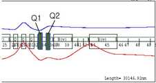
3 is a conceptual diagram showing a state in which the cross-section of the proton beam is adjusted small by adjusting the current value of the quadrupole electromagnet,
4 is a conceptual diagram illustrating a state in which a cross section of a proton beam is largely adjusted by adjusting a current value of a quadrupole electromagnet,
5 is a conceptual diagram illustrating a state in which a tumor is defined as a plurality of regions in the longitudinal direction and irradiated with a proton beam corresponding to a cross section of each region.
6 is a graph showing a uniform beam shape in which a uniform beam is obtained through a plurality of beams by applying a general scanning method after adjusting a cross section of a proton beam.
본 발명은 다양한 변경을 가할 수 있고 여러 가지 실시예를 가질 수 있는 바, 특정 실시예들을 도면에 예시하고 상세한 설명에 상세하게 설명하고자 한다.While the invention is susceptible to various modifications and alternative forms, specific embodiments thereof are shown by way of example in the drawings and will herein be described in detail.
그러나, 이는 본 발명을 특정한 실시 형태에 대해 한정하려는 것이 아니며, 본 발명의 사상 및 기술 범위에 포함되는 모든 변경, 균등물 내지 대체물을 포함하는 것으로 이해되어야 한다.It should be understood, however, that the invention is not intended to be limited to the particular embodiments, but includes all modifications, equivalents, and alternatives falling within the spirit and scope of the invention.
본 출원에서 사용한 용어는 단지 특정한 실시예를 설명하기 위해 사용된 것으로, 본 발명을 한정하려는 의도가 아니다. 단수의 표현은 문맥상 명백하게 다르게 뜻하지 않는 한, 복수의 표현을 포함한다.The terminology used in this application is used only to describe a specific embodiment and is not intended to limit the invention. Singular expressions include plural expressions unless the context clearly indicates otherwise.
또한 본 출원에서 첨부된 도면은 설명의 편의를 위하여 확대 또는 축소하여 도시된 것으로 이해되어야 한다.Also, the drawings in the present application should be understood as being enlarged or reduced for convenience of description.
이제 본 발명에 대하여 도면을 참고하여 상세하게 설명하고, 도면 부호에 관계없이 동일하거나 대응하는 구성 요소는 동일한 참조 번호를 부여하고 이에 대한 중복되는 설명은 생략하기로 한다.DETAILED DESCRIPTION OF THE PREFERRED EMBODIMENTS The present invention will now be described in detail with reference to the drawings, wherein like or corresponding elements are denoted by the same reference numerals, and redundant description thereof will be omitted.
본 발명의 실시예에 따른 양성자 치료기는, 선형가속기(100)와, 상기 선형가속기(100)에서 입사된 양성자 빔이 소정의 에너지를 갖도록 조절하는 싱크로트론(200)과, 상기 싱크로트론(200)에서 입사된 양성자 빔의 면적을 조절하여 종양에 조사하는 갠트리(300), 및 상기 양성자 빔의 에너지와 조사 면적을 조절하는 제어부(400)를 포함한다.Proton therapy device according to an embodiment of the present invention, the
싱크로트론(Synchrotron, 200)은 양성자 빔이 환부에 조사될 수 있는 에너지를 갖도록 양성자를 가속시키는 장치이다. 싱크로트론(200)은 사이클로트론(Cyclotron)에 비해 펄스 단위로 양성자 에너지 및 빔 전류를 상대적으로 정확하게 조절할 수 있기 때문에 에너지를 조절하기 위한 추가적인 장비가 필요 없는 장점이 있다. 본 실시예에서 적용되는 싱크로트론(200)의 구체적인 구성은 일반적인 싱크로트론의 구성이 모두 적용될 수 있는바 더 이상의 자세한 설명은 생략한다.Synchrotron (200) is a device that accelerates protons so that the proton beam has energy that can be irradiated to the affected area. Since the
이러한 싱크로트론(200)은 양성자 빔이 인체 내에 종양이 위치한 지점에서 브래그 피크(Bragg peak)를 갖도록 적절하게 양성자 빔의 에너지를 조절할 수 있다. 브래그 피크(Bragg peak)란 양성자가 물질 내에서 이동할 때에는 방사선을 거의 방출하지 않고 양성자가 정지한 경우 방사선을 방출하는 특성을 말한다.The
따라서, 양성자 빔이 인체에 조사되어 정지되는 위치를 고려하여 싱크로트론(200)에 의해 양성자 빔의 에너지를 조절하면, 인체 내에 특정 위치에 존재하는 종양에만 방사선이 방출되어 암세포를 치료할 수 있다.Therefore, when the energy of the proton beam is adjusted by the
싱크로트론(200)은 효율적인 가속을 위해 최초 입사되는 빔의 에너지가 높아야 하므로(약 3MeV), 싱크로트론(200)의 전단에 선형가속기 (Linear accelerator, 100)가 배치될 수 있다. 즉, 선형가속기(100)에서 양성자 빔을 1차 가속하여 펄스 단위로 싱크로트론(200)에 공급하면 싱크로트론(200)에서는 이를 인체의 특정 깊이에 주입될 에너지로 2차 가속하게 된다.Since the
갠트리(Gantry, 300)는 환자에 양성자 빔을 조사하는 원통형의 회전 치료장치로서, 싱크로트론(200)에서 입사된 양성자 빔을 환자에게 조사할 수 있도록 양성자 빔의 방향을 변화시키는 역할을 수행한다.The
구체적으로 갠트리(300)는 가속기에서 수평 방향으로 인출된 빔을 수직방향으로 변경하며, 회전을 통해 양성자 빔의 입사 각도를 조절한다. 도 2를 참조할 때, 갠트리(300)는 빔의 위치를 조절할 수 있는 이극전자석(320)과 복수 개의 사극 전자석(310)을 포함하며, 끝부분에는 양성자 빔을 치료목적에 맞게 조절하기 위해 노즐(330)이 배치된다.Specifically, the
본 발명에 따른 갠트리(300)는 양성자 빔의 조사면적을 조절할 수 있다. 구체적으로는, 제어부(400)가 갠트리(300)에 배치된 복수 개의 사극전자석에 인가되는 전류값을 조절함으로써 출사되는 양성자 빔의 면적을 조절할 수 있다.The
예를 들면, 도 3과 같이 제1사극전자석(Q1)의 자기장을 -6.8T/m으로 조절하고 제2사극전자석(Q2)의 자기장을 4.3T/m으로 조절하면 면적이 작은 빔을 얻을 수 있으며, 도 4와 같이 제1사극전자석(Q1)의 자기장을 -5.2T/m으로 조절하고 제2사극전자석(Q2)의 자기장을 3.0T/m으로 조절하면 면적이 큰 빔을 얻을 수 있다. 도 3과 도 4에서 파란색 선은 수평방향의 빔 크기이고 붉은 색 선은 수직 방향의 빔 크기이다. 그러나 이는 예시적인 것이며 사극전자석의 자기장이 조절되도록 인가되는 전류값을 적절히 변화시키면 다양한 면적의 양성자 빔을 얻을 수 있다.For example, as shown in FIG. 3, when the magnetic field of the first quadrupole electromagnet Q1 is adjusted to -6.8T / m and the magnetic field of the second quadrupole electromagnet Q2 is adjusted to 4.3T / m, a beam having a small area can be obtained. As shown in FIG. 4, when the magnetic field of the first quadrupole electromagnet Q1 is adjusted to -5.2T / m and the magnetic field of the second quadrupole electromagnet Q2 is adjusted to 3.0T / m, a beam having a large area can be obtained. 3 and 4, the blue line is the beam size in the horizontal direction and the red line is the beam size in the vertical direction. However, this is merely an example. Proper beams of various areas can be obtained by appropriately changing the current value applied to adjust the magnetic field of the quadrupole electromagnet.
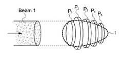
이하에서는 도 5a와 도 5b를 참조하여 종양의 횡단면에 따라 양성자 빔의 면적을 조사하는 구성에 대해 설명한다. 일반적으로 종양(t)은 길이방향에 따라 상이한 횡단면을 가지므로 작은 면적의 양성자 빔을 조사하는 경우에는 복수 회에 걸쳐 빔을 조사하여야 하며, 큰 면적의 양성자 빔을 조사하는 경우에는 종양 이외의 신체 조직에 양성자가 조사될 위험이 있다. 따라서 종양의 길이방향 횡단면에 따라 양성자 빔의 면적을 조절하여 조사하게 되면 짧은 조사횟수로도 효과적인 치료가 가능하다.Hereinafter, a configuration of examining the area of the proton beam along the cross section of the tumor will be described with reference to FIGS. 5A and 5B. In general, since the tumor (t) has a different cross section along the length direction, when irradiating a small area of the proton beam, the beam must be irradiated a plurality of times, and when irradiating a large area of the proton beam, a body other than the tumor is present. There is a risk of protons being investigated in the tissues. Therefore, by controlling the area of the proton beam along the longitudinal cross-section of the tumor, effective treatment is possible even with a short number of irradiations.
일반적으로 종양(t)은 자기공명영상 촬영장치(Magnetic Resonance Imaging: MRI), 컴퓨터 단층촬영장치(Computer Tomography: CT), 양전자 방출 단층촬영장치(Positron Emission Tomography: PET), 및 초음파(Ultrasonic Wave) 기기 등과 같은 영상진단기기로부터 3차원 영상을 얻을 수 있다.Tumors (t) are generally magnetic resonance imaging (MRI), computer tomography (CT), positron emission tomography (PET), and ultrasound (Ultrasonic Wave) Three-dimensional images can be obtained from an image diagnosis device such as a device.
이렇게 얻어진 종양의 3차원 영상은 길이방향으로 복수 개가 구획된 영역으로 정의할 수 있으며, 복수의 구획된 영역은 각각 상이한 횡단면을 가질 수 있다. 이때, 구획 영역의 개수는 종양의 길이, 크기 및 상태에 따라 상이하게 구획될 수 있다.The three-dimensional image of the tumor thus obtained may be defined as a plurality of divided regions in the longitudinal direction, and the plurality of divided regions may have different cross sections, respectively. At this time, the number of compartments may be partitioned differently according to the length, size and condition of the tumor.
예시적으로 종양이 5개의 영역으로 구획된 경우로 설명하면, 먼저 도 5(a)와 같이 환자의 표피에서 가장 가까운 제1영역(P1)에 양성자 빔을 조사하는 경우 제1영역(P1)의 지점에서 브래그 피크를 갖도록 양성자 빔의 에너지를 조절하고 양성자 빔(Beam 1)의 면적은 제1영역(P1)의 횡단면적에 대응되는 크기로 조절하여 조사할 수 있다.For example, when the tumor is divided into five regions, first, when the proton beam is irradiated to the first region P1 closest to the epidermis of the patient, as shown in FIG. The energy of the proton beam may be adjusted to have a Bragg peak at the point, and the area of the
이후, 도 5(b)와 같이 제2영역(P2)에 양성자 빔을 조사하는 경우 제2영역(P2)의 지점에서 브래그 피크를 갖도록 양성자 빔의 에너지를 조절하고 양성자 빔(Beam 2)의 면적은 제2영역(P2)의 횡단면적에 대응되는 크기로 조절하여 조사할 수 있다. 나머지 제3영역 내지 제5영역(P3 내지 P5)도 이와 동일한 방식으로 양성자 빔을 조사한다. 이때, 제어부(400)는 싱크로트론(200) 내에서 양성자의 가속을 제어하여 양성자 빔의 에너지를 조절하고, 갠트리(300)의 사극전자석에 인가되는 전류값을 조절하여 양성자 빔의 면적을 조절할 수 있다.Subsequently, when the proton beam is irradiated to the second region P2 as shown in FIG. 5 (b), the energy of the proton beam is adjusted to have a Bragg peak at the point of the second region P2, and the area of the
그러나, 양성자 빔을 종양에 조사하는 방법은 반드시 이에 한정되지 않고 다양한 방법이 적용될 수 있다. 예를 들면, 양성자 빔의 단면을 종양의 횡단면과 반드시 대응시키지 않고 싱크로트론의 반복률 및 종양의 단면에 따라 적절히 조절한 후, 스캐닝 방식(위치를 이동하면서 순차적으로 조사)을 이용하여 복수 회 조사하는 방식을 적용할 수도 있다.However, the method of irradiating the tumor with the proton beam is not necessarily limited thereto, and various methods may be applied. For example, the cross section of the proton beam is not necessarily corresponded to the cross section of the tumor, and is appropriately adjusted according to the repetition rate of the synchrotron and the cross section of the tumor, and then irradiated a plurality of times using a scanning method (sequential irradiation while moving the position). You can also apply
구체적으로는 길이방향으로 종양의 단면적에 따라 양성자 빔의 단면을 조절함으로써 각 단면적에 조사되는 양성자 빔의 펄스 개수는 동일하게 조절할 수도 있고, 또는 빔의 단면을 일정하게 한 경우 종양의 단면적에 따라 조사 횟수를 조절할 수 있다. 이 경우 빔의 단면이 크게 조절되었으므로 기존에 스캐닝 방식에 비해 빔의 조사 횟수는 현저하게 줄어들게 된다.Specifically, the number of pulses of the proton beam irradiated to each cross-sectional area may be equally adjusted by adjusting the cross-section of the proton beam in the longitudinal direction according to the cross-sectional area of the tumor. You can adjust the number of times. In this case, since the cross section of the beam is largely adjusted, the number of irradiation of the beam is significantly reduced compared to the conventional scanning method.
또한, 도 6을 참조할 때, 복수의 양성자 빔(B1)을 스캐닝 방식으로 조사하는 경우, 복수의 양성자 빔(B1)이 하나의 균일한 빔(B)으로 종양에 조사될 수 있음을 알 수 있다.In addition, referring to FIG. 6, when the plurality of proton beams B1 are irradiated by a scanning method, it may be understood that the plurality of proton beams B1 may be irradiated to the tumor with one uniform beam B. FIG. have.
이때, 종양의 크기, 깊이, 및 상태에 따라 각 횡단면적당 양성자 빔의 면적 및 조사 횟수는 달라질 수 있고, 종양 주변에 퍼진 암세포까지 치료하기 위하여 양성자 빔의 면적은 종양의 위치를 조금 벗어난 범위까지 조사될 수도 있다.In this case, the area and the number of irradiation of the proton beam per cross-sectional area may vary according to the size, depth, and condition of the tumor, and the area of the proton beam is irradiated to a range slightly outside the position of the tumor to treat cancer cells spread around the tumor. May be
이때, 양성자 빔의 면적이 종양의 횡단면적에 따라 조절되어도 양성자 빔의 단위면적당 양성자 수는 동일한 것이 바람직하다. 빔의 면적이 커짐에 따라 단위부피당 양성자 수가 줄어드는 경우에는 치료 효과가 떨어지기 때문이다.At this time, even if the area of the proton beam is adjusted according to the cross-sectional area of the tumor, the number of protons per unit area of the proton beam is preferably the same. This is because when the number of protons per unit volume decreases as the area of the beam increases, the therapeutic effect decreases.
따라서, 본 실시예에서는 단위면적당 양성자 빔의 개수를 유지하기 위해 면적이 큰 빔의 경우에는 싱크로트론(200)에서 가속되는 양성자 수를 증가시켜 빔의 면적이 커지는 경우에도 단위면적당 동일한 양성자 수가 조사되도록 조절한다. 이때, 전술한 스캐닝 방식을 이용하는 경우에는 양성자 빔의 단면적을 너무 크게 조절하지 않아도 되므로 단위면적당 동일한 양성자 수를 유지하기 유리하다. 도 6을 참조할 때, 균일한 빔(B)의 단위면적당 양성자 수는 상대적으로 균일하게 유지됨을 알 수 있다.Therefore, in this embodiment, in order to maintain the number of proton beams per unit area, the number of protons accelerated in the
구체적으로, 싱크로트론(200)에서 가속되는 양성자 개수는 선형가속기(100)에서 싱크로트론(200)으로 들어가는 양성자 수에 의해 결정되므로 단면이 큰 빔을 조사하기 위하여는 먼저 선형가속기(100)에서 싱크로트론(200)으로 입사되는 빔의 펄스폭을 늘리거나 펄스의 횟수를 증가시킨다. 펄스폭을 늘리거나 펄스의 횟수를 증가시키면 그에 비례하여 싱크로트론(200) 내의 양성자 수가 많아지므로 빔의 면적을 조절하는 경우에도 단위면적당 양성자 수는 동일하게 된다.Specifically, since the number of protons accelerated in the
따라서, 제어부(400)는 양성자 빔이 조사된 종양의 횡단면적 정보를 먼저 수신하여 양성자 빔의 조사 면적 및 조사 횟수를 결정한 뒤, 선형가속기(100)에서 싱크로트론(200)으로 입사되는 펄스의 폭 또는 펄스의 개수를 제어할 수 있다.
Therefore, the
이상에서 본 발명의 실시 예에 대하여 상세하게 설명하였지만 본 발명의 권리범위는 이에 한정되는 것은 아니고 다음의 청구범위에서 정의하고 있는 본 발명의 기본 개념을 이용한 당업자의 여러 변형 및 개량 형태 또한 본 발명의 권리범위에 속하는 것이다.While the present invention has been particularly shown and described with reference to exemplary embodiments thereof, it is to be understood that the invention is not limited to the disclosed exemplary embodiments, It belongs to the scope of right.
100: 선형가속기200: 싱크로트론
300: 갠트리310: 사극전자석
400: 제어부100: linear accelerator 200: synchrotron
300: gantry 310: quadrupole electromagnet
400:
Claims (6)
Translated fromKorean상기 싱크로트론에서 입사된 양성자 빔을 종양에 조사하는 갠트리; 및
상기 갠트리에 포함된 사극전자석에 인가되는 전류값을 제어하여 상기 양성자 빔의 면적을 조절하는 제어부를 포함하되,
상기 갠트리에서 조사되는 양성자 빔의 면적 및 조사 횟수는 상기 종양의 길이방향으로 복수 개로 구획된 영역의 횡단면에 따라 조절되는 양성자 치료기.A synchrotron that accelerates the proton beam to have a predetermined energy;
A gantry that irradiates a tumor with a proton beam incident from the synchrotron; And
It includes a control unit for controlling the area of the proton beam by controlling the current value applied to the quadrupole electromagnet included in the gantry,
The area and the number of irradiation of the proton beam irradiated from the gantry is adjusted according to the cross section of the plurality of partitioned areas in the longitudinal direction of the tumor.
상기 면적이 조절된 양성자 빔 간의 단위면적당 양성자 수는 동일한 양성자 치료기.The method of claim 1,
A proton therapy device having the same number of protons per unit area between the proton beams whose area is controlled.
Priority Applications (1)
| Application Number | Priority Date | Filing Date | Title |
|---|---|---|---|
| KR1020110142343AKR101336830B1 (en) | 2011-12-26 | 2011-12-26 | Proton Therapy Facility with Varying Cross-Section of the Proton Beams |
Applications Claiming Priority (1)
| Application Number | Priority Date | Filing Date | Title |
|---|---|---|---|
| KR1020110142343AKR101336830B1 (en) | 2011-12-26 | 2011-12-26 | Proton Therapy Facility with Varying Cross-Section of the Proton Beams |
Publications (2)
| Publication Number | Publication Date |
|---|---|
| KR20130074327A KR20130074327A (en) | 2013-07-04 |
| KR101336830B1true KR101336830B1 (en) | 2013-12-04 |
Family
ID=48988452
Family Applications (1)
| Application Number | Title | Priority Date | Filing Date |
|---|---|---|---|
| KR1020110142343AExpired - Fee RelatedKR101336830B1 (en) | 2011-12-26 | 2011-12-26 | Proton Therapy Facility with Varying Cross-Section of the Proton Beams |
Country Status (1)
| Country | Link |
|---|---|
| KR (1) | KR101336830B1 (en) |
Families Citing this family (3)
| Publication number | Priority date | Publication date | Assignee | Title |
|---|---|---|---|---|
| KR101682545B1 (en) | 2015-08-19 | 2016-12-05 | 한국원자력의학원 | System and Method for Active Scanning with Adjustable Beam Size |
| KR102020378B1 (en) | 2017-11-03 | 2019-09-10 | 주식회사 포스코 | Cooling system and control method thereof |
| CN107802968B (en)* | 2017-11-24 | 2024-05-10 | 北京新核核工程科技有限公司 | Deceleration filtering device and neutron radiation therapy system |
Citations (4)
| Publication number | Priority date | Publication date | Assignee | Title |
|---|---|---|---|---|
| JP2005050823A (en)* | 2003-05-13 | 2005-02-24 | Hitachi Ltd | Particle beam emitting apparatus and particle beam emitting method |
| JP2008173298A (en) | 2007-01-18 | 2008-07-31 | National Cancer Center-Japan | Charged particle beam irradiation device |
| KR20110019752A (en)* | 2008-05-22 | 2011-02-28 | 블라디미르 예고르비치 발라킨 | Method and apparatus for controlling beam path of charged particle cancer treatment |
| JP2011072537A (en) | 2009-09-30 | 2011-04-14 | Hitachi Ltd | Particle irradiation system and method for controlling the same |
- 2011
- 2011-12-26KRKR1020110142343Apatent/KR101336830B1/ennot_activeExpired - Fee Related
Patent Citations (4)
| Publication number | Priority date | Publication date | Assignee | Title |
|---|---|---|---|---|
| JP2005050823A (en)* | 2003-05-13 | 2005-02-24 | Hitachi Ltd | Particle beam emitting apparatus and particle beam emitting method |
| JP2008173298A (en) | 2007-01-18 | 2008-07-31 | National Cancer Center-Japan | Charged particle beam irradiation device |
| KR20110019752A (en)* | 2008-05-22 | 2011-02-28 | 블라디미르 예고르비치 발라킨 | Method and apparatus for controlling beam path of charged particle cancer treatment |
| JP2011072537A (en) | 2009-09-30 | 2011-04-14 | Hitachi Ltd | Particle irradiation system and method for controlling the same |
Also Published As
| Publication number | Publication date |
|---|---|
| KR20130074327A (en) | 2013-07-04 |
Similar Documents
| Publication | Publication Date | Title |
|---|---|---|
| JP7245352B2 (en) | Method of providing particle-based rotational radiation therapy | |
| Paganetti | Proton beam therapy | |
| Mohan et al. | Proton therapy–present and future | |
| US10307618B2 (en) | Radiation therapy systems and methods | |
| JP5330253B2 (en) | Particle beam irradiation equipment | |
| JP6256974B2 (en) | Charged particle beam system | |
| KR101953350B1 (en) | Beam modulation apparatus on build-up region of photon beam by transverse magnetic field and beam spoiler, radiotherapy apparatus using the depth dose modulation apparatus, and method for modulating dose transverse magnetic field and beam spoiler on build-up region of photon beam | |
| KR101803346B1 (en) | Tumor surface dose enhancing radiotherapy apparatus using magnetic field | |
| US8878464B2 (en) | Laser accelerator driven particle brachytherapy devices, systems, and methods | |
| US8613694B2 (en) | Method for biological modulation of radiation therapy | |
| CN103794261B (en) | Particle-beam exposure apparatus and particle-beam therapeutic apparatus | |
| US9750957B2 (en) | System for irradiating charged particles and method for irradiating charged particles | |
| JP5927122B2 (en) | Irradiation method and apparatus for performing the method | |
| CN103402581B (en) | Particle Beam Irradiation System | |
| US9539442B2 (en) | Proton irradiation using spot scanning | |
| Flanz et al. | Evolution of technology to optimize the delivery of proton therapy: the third generation | |
| JP2010253000A (en) | Radiation irradiation system | |
| KR101336830B1 (en) | Proton Therapy Facility with Varying Cross-Section of the Proton Beams | |
| KR101403662B1 (en) | Proton Therapy Facility | |
| US12064644B2 (en) | Pinhole collimator systems and methods | |
| Schardt | Hadrontherapy | |
| US20200246633A1 (en) | Deuteron therapy | |
| Hernández | Low-dose ion-based transmission radiography and tomography for optimization of carbon ion-beam therapy | |
| EP4321208A1 (en) | Composite field sequencing (cfs) for proton beam therapy | |
| Kubiak | Carbon Ion Radiotherapy-Advantages, Technical Aspects and Perspectives |
Legal Events
| Date | Code | Title | Description |
|---|---|---|---|
| A201 | Request for examination | ||
| PA0109 | Patent application | St.27 status event code:A-0-1-A10-A12-nap-PA0109 | |
| PA0201 | Request for examination | St.27 status event code:A-1-2-D10-D11-exm-PA0201 | |
| R18-X000 | Changes to party contact information recorded | St.27 status event code:A-3-3-R10-R18-oth-X000 | |
| D13-X000 | Search requested | St.27 status event code:A-1-2-D10-D13-srh-X000 | |
| D14-X000 | Search report completed | St.27 status event code:A-1-2-D10-D14-srh-X000 | |
| E902 | Notification of reason for refusal | ||
| PE0902 | Notice of grounds for rejection | St.27 status event code:A-1-2-D10-D21-exm-PE0902 | |
| PG1501 | Laying open of application | St.27 status event code:A-1-1-Q10-Q12-nap-PG1501 | |
| P11-X000 | Amendment of application requested | St.27 status event code:A-2-2-P10-P11-nap-X000 | |
| P13-X000 | Application amended | St.27 status event code:A-2-2-P10-P13-nap-X000 | |
| E701 | Decision to grant or registration of patent right | ||
| PE0701 | Decision of registration | St.27 status event code:A-1-2-D10-D22-exm-PE0701 | |
| GRNT | Written decision to grant | ||
| PR0701 | Registration of establishment | St.27 status event code:A-2-4-F10-F11-exm-PR0701 | |
| PR1002 | Payment of registration fee | St.27 status event code:A-2-2-U10-U11-oth-PR1002 Fee payment year number:1 | |
| PG1601 | Publication of registration | St.27 status event code:A-4-4-Q10-Q13-nap-PG1601 | |
| PN2301 | Change of applicant | St.27 status event code:A-5-5-R10-R13-asn-PN2301 St.27 status event code:A-5-5-R10-R11-asn-PN2301 | |
| FPAY | Annual fee payment | Payment date:20160928 Year of fee payment:4 | |
| PR1001 | Payment of annual fee | St.27 status event code:A-4-4-U10-U11-oth-PR1001 Fee payment year number:4 | |
| P22-X000 | Classification modified | St.27 status event code:A-4-4-P10-P22-nap-X000 | |
| PR1001 | Payment of annual fee | St.27 status event code:A-4-4-U10-U11-oth-PR1001 Fee payment year number:5 | |
| FPAY | Annual fee payment | Payment date:20181002 Year of fee payment:6 | |
| PR1001 | Payment of annual fee | St.27 status event code:A-4-4-U10-U11-oth-PR1001 Fee payment year number:6 | |
| PC1903 | Unpaid annual fee | St.27 status event code:A-4-4-U10-U13-oth-PC1903 Not in force date:20191129 Payment event data comment text:Termination Category : DEFAULT_OF_REGISTRATION_FEE | |
| PC1903 | Unpaid annual fee | St.27 status event code:N-4-6-H10-H13-oth-PC1903 Ip right cessation event data comment text:Termination Category : DEFAULT_OF_REGISTRATION_FEE Not in force date:20191129 | |
| PN2301 | Change of applicant | St.27 status event code:A-5-5-R10-R13-asn-PN2301 St.27 status event code:A-5-5-R10-R11-asn-PN2301 |