KR101309013B1 - Diagnosis Device For Face Form Using Facial Image and Cephalometric Image - Google Patents
Diagnosis Device For Face Form Using Facial Image and Cephalometric ImageDownload PDFInfo
- Publication number
- KR101309013B1 KR101309013B1KR1020110100656AKR20110100656AKR101309013B1KR 101309013 B1KR101309013 B1KR 101309013B1KR 1020110100656 AKR1020110100656 AKR 1020110100656AKR 20110100656 AKR20110100656 AKR 20110100656AKR 101309013 B1KR101309013 B1KR 101309013B1
- Authority
- KR
- South Korea
- Prior art keywords
- image
- facial
- shape
- feature points
- subject
- Prior art date
- Legal status (The legal status is an assumption and is not a legal conclusion. Google has not performed a legal analysis and makes no representation as to the accuracy of the status listed.)
- Active
Links
Images


Classifications
- A—HUMAN NECESSITIES
- A61—MEDICAL OR VETERINARY SCIENCE; HYGIENE
- A61B—DIAGNOSIS; SURGERY; IDENTIFICATION
- A61B5/00—Measuring for diagnostic purposes; Identification of persons
- A61B5/103—Measuring devices for testing the shape, pattern, colour, size or movement of the body or parts thereof, for diagnostic purposes
- A61B5/107—Measuring physical dimensions, e.g. size of the entire body or parts thereof
- A61B5/1077—Measuring of profiles
- A—HUMAN NECESSITIES
- A61—MEDICAL OR VETERINARY SCIENCE; HYGIENE
- A61B—DIAGNOSIS; SURGERY; IDENTIFICATION
- A61B5/00—Measuring for diagnostic purposes; Identification of persons
- A61B5/0059—Measuring for diagnostic purposes; Identification of persons using light, e.g. diagnosis by transillumination, diascopy, fluorescence
- A61B5/0077—Devices for viewing the surface of the body, e.g. camera, magnifying lens
- A—HUMAN NECESSITIES
- A61—MEDICAL OR VETERINARY SCIENCE; HYGIENE
- A61B—DIAGNOSIS; SURGERY; IDENTIFICATION
- A61B5/00—Measuring for diagnostic purposes; Identification of persons
- A61B5/103—Measuring devices for testing the shape, pattern, colour, size or movement of the body or parts thereof, for diagnostic purposes
- A61B5/107—Measuring physical dimensions, e.g. size of the entire body or parts thereof
- A61B5/1079—Measuring physical dimensions, e.g. size of the entire body or parts thereof using optical or photographic means
- A—HUMAN NECESSITIES
- A61—MEDICAL OR VETERINARY SCIENCE; HYGIENE
- A61B—DIAGNOSIS; SURGERY; IDENTIFICATION
- A61B6/00—Apparatus or devices for radiation diagnosis; Apparatus or devices for radiation diagnosis combined with radiation therapy equipment
- A—HUMAN NECESSITIES
- A61—MEDICAL OR VETERINARY SCIENCE; HYGIENE
- A61B—DIAGNOSIS; SURGERY; IDENTIFICATION
- A61B90/00—Instruments, implements or accessories specially adapted for surgery or diagnosis and not covered by any of the groups A61B1/00 - A61B50/00, e.g. for luxation treatment or for protecting wound edges
- A61B90/36—Image-producing devices or illumination devices not otherwise provided for
- A61B2090/364—Correlation of different images or relation of image positions in respect to the body
Landscapes
- Health & Medical Sciences (AREA)
- Life Sciences & Earth Sciences (AREA)
- Engineering & Computer Science (AREA)
- Medical Informatics (AREA)
- Surgery (AREA)
- Public Health (AREA)
- Biomedical Technology (AREA)
- Heart & Thoracic Surgery (AREA)
- Biophysics (AREA)
- Molecular Biology (AREA)
- Physics & Mathematics (AREA)
- Animal Behavior & Ethology (AREA)
- General Health & Medical Sciences (AREA)
- Pathology (AREA)
- Veterinary Medicine (AREA)
- Dentistry (AREA)
- Oral & Maxillofacial Surgery (AREA)
- High Energy & Nuclear Physics (AREA)
- Nuclear Medicine, Radiotherapy & Molecular Imaging (AREA)
- Optics & Photonics (AREA)
- Radiology & Medical Imaging (AREA)
- Apparatus For Radiation Diagnosis (AREA)
- Image Analysis (AREA)
Abstract
Translated fromKoreanDescription
Translated fromKorean본 발명은 한방의 형상의학을 바탕으로 피검자의 안면 형태를 분류하기 위한 형상진단기에 관한 것으로서, 보다 구체적으로는 형상의학을 기초로 환자를 진단/처방할 수 있도록 안면 영상과 두부의 엑스레이 영상을 이용하여 보다 정확하게 피검자의 안면 형태를 진단하고 형상을 분류하는 형상진단기에 관한 것이다.The present invention relates to a shape diagnosis apparatus for classifying a face shape of a subject based on a one-sided shape medicine, and more particularly, to a shape diagnosis apparatus using a facial image and an x-ray image of a head to diagnose / Thereby accurately diagnosing the shape of the subject's face and classifying the shape of the subject.
형상의학(形象醫學)이란, 지산 박인규 선생이 황제내경과 동의보감을 연구개발해 체계화시킨 의학으로서, 사상체질 의학과 더불어 현대 한국 한의학의 독창적인 의학이며, 개체생리병리 체질론을 형상의학적인 측면에서 완성한 한의학 이론으로 알려져 있다. 이는 개인의 특성을 중시하는 변증시치(辨證施治)가 한방 진단과 치료기술의 핵심사항인 점을 고려할 때, 임상적인 효율을 제고하기 위하여 환자의 형상, 특히 얼굴의 형상을 중심으로 형색맥증(형과 색과 맥과 증상)의 합일을 추구하는 진단기법이다.Shape medicine (form medicine) is a medicine that In-kyung Park In-Kyu Sensei researches and develops and systemizes Emperor Bongyung and Dongbokgomen. It is a unique medicine of contemporary Korean oriental medicine as well as Sasang Constitutional Medicine. . Considering the fact that the dialectic value that emphasizes individual characteristics is a key point in the diagnosis and treatment technology of oriental medicine, it is necessary to consider the shape of patient, especially the shape of face, (Type, color, vein and symptom).
상기 형상의학이란 한마디로 '생긴대로 병이 오고 생긴대로 치료한다'는 것으로서, 우리나라 고유의 환자 개별 맞춤형 의학이라 할 수 있다. 즉, 사람의 생김새, 즉 생긴 모습이 다르면 성격도 다르듯이 생긴 모습이 다르면 각각의 장부기능도 다르고, 사람에 따라 잘 찾아오는 병이 다르기 때문에 증상기 같다고 해도 치료와 건강관리법이 달라져야 한다는 이론이다.The above-mentioned shape medicine is, in a word, "treating the disease as it happens" as it is, and it can be said that it is the individual customized medicine of the patient's own country. In other words, it is the theory that people's appearance, that is, appearance is different, personality is also different, and the function of each book is different.
동의보감에도 '얼굴의 부위를 보고 어느 장기에 병이 있는가를 구분한다'라고 씌여 있는데, 이 말은 곧 남녀노소에 따른 특징과 얼굴모양, 피부색이나 체형에 따라 건강을 유지하는 방법이 다를 수 밖에 없다는 형상의학 이론과 상통한다고 볼 수 있다.In Dongbok-bo, there is a saying that "to distinguish which organs are in which organs by looking at the parts of the face", this means that the characteristics that depend on sex, age and sex, the shape of the face, It can be said that it is in common with medical theory.
사람의 생김새, 즉 형상(形象)을 분류하는 주요 방식은 다음과 같다. 안면의 정면과 측면의 면적(크기) 비교치로서 이루어지는 담체-방광체와, 안면 외곽의 형태관찰에 의한 정신기혈과와, 눈코입귀와 안면 크기(분포도) 비교에 따른 오장육부와, 눈과 코의 형태와 안면부의 돌출함몰여부로서 결정되는 육경형으로 구성된다.The main way of classifying human figure, that is, figure is as follows. The comparison of the size of the frontal and lateral sides of the face shows the relationship between the carrier and the bladder, the mental and gendering by observing the shape of the facial outline, The shape of the face and the height of the face are determined by whether or not the protrusion is depressed.
이하, 피검자(진단 대상자)의 안면 형상을 진단/결정하는 방법을 알아보면 다음과 같다.Hereinafter, a method of diagnosing / determining the facial shape of the subject (diagnosis subject) will be described.
형상의학에 의한 담체-방광체의 구분은 안면(얼굴)의 정면과 측면의 면적(크기) 비교치와 안면의 전체적인 형태를 기초로 한다. 다시 말해서, 얼굴 정면의 크기와 얼굴 측면의 크기 계산에서 세로는 동일값이므로 전면과 측면의 횡선 길이를 다음과 같이 비교한다. 보다 구체적으로 설명하면, 얼굴의 정면과 측면의 세로길이를 나타내는 이마 정중앙점과 하악 정중앙점 사이의 거리를 구한다. 그리고 얼굴 정면의 횡선길이를 나타내는 양 관골점 사이의 수평 거리와, 얼굴 측면의 횡선길이를 나타내는 귀 앞점에서 관골사이의 수평 거리를 구한다. 그리고, 얼굴 정면의 면적을 구하기 위하여 상기 얼굴 정면의 세로길이에 정면의 가로길이(횡선길이)를 곱하고, 얼굴 측면의 면적을 구하기 위하여 측면의 세로길이에 측면의 가로길이(횡선길이)를 곱한다.The division of the carrier-bladder by geometric medicine is based on the comparison of the frontal and lateral area (size) of the face (face) and the overall shape of the face. In other words, since the length is the same in the calculation of the face size and face size, the horizontal and vertical lengths of the front and side are compared as follows. More specifically, the distance between the midpoint of the forehead and the midpoint of the mandible, which indicates the vertical length of the front and side of the face, is obtained. The horizontal distance between the bifurcation points representing the horizontal length of the front of the face and the horizontal distance between the bifurcations from the front of the ear indicating the horizontal length of the face are obtained. In order to obtain the area of the front face of the face, the vertical length of the front face is multiplied by the horizontal length (horizontal length) of the front face, and the lateral length of the side is multiplied by the lateral length (lateral length) to obtain the area of the face.
이렇게 구한 수치를 기초로 방광체와 담체로 구분짓는데, 방광체는 정면의 면적이 측면의 면적보다 크면 방광체가 되고, 정면의 면적이 측면의 면적보다 작으면 담체가 된다.If the area of the front face is larger than the area of the side face, the bladder becomes the bladder, and if the area of the front face is smaller than the area of the side face, the bladder becomes the carrier.
다음으로, 안면 외곽의 형태에 따른 정신기혈(精神氣血)의 구분을 위하여, 두정부와 두유혈의 연결선의 각, 정중앙 아랫 턱의 각, 측면 턱의 각, 관골의 각을 중심으로 각도를 측정한다. 또한, 이마의 길이와 폭, 양 관골 점의 길이, 양 턱의 길이와 폭을 비교 그리고 이마 정중앙 점과 양 관골점 사이의 길이를 측정하며, 그 결과를 다음과 같이 구분한다.Next, for the classification of psychiatric blood according to the morphology of the facial outline, the angles around the angle of the connecting line of the two governments, the angle of the lower jaw in the middle, the angle of the side jaw, . The length and width of the forehead, the length of both bifurcation points, the length and width of both jaws, and the length between the forehead and both bifurcation points are measured and the results are as follows.
신과는 이마의 길이와 폭이 큰 경고 두정부와 두유혈의 연결선의 각이 크며 하악 턱의 상호간의 길이와 폭이 작은 경우이고, 혈과는 하악 턱의 상호간의 길이와 폭이 크고, 이마의 길이와 폭이 크고, 두정부와 두유혈의 연결선의 각이 큰 경우이다. 정과는 얼굴의 횡선이 종선보다 비율이 큰 경우이며, 기과는 얼굴 외곽 각도에 따라 결정된다.The length and width of the forehead are large. The length and width of the two jaws are large. The length and width of the jaws are small. The length and width of the jaws are large. The length and width are large, and the angles of the connections between the two governments and the two bloods are large. The regularity is a case where the horizontal line of the face is larger than the vertical line, and the turn is determined by the angle of the face.
그리고 눈코입귀과 안면 크기(분포도) 비교의 오장형상을 알기 위해, 이목구비의 길이와 크기 측정, 안면에서의 이목구비 분포 형태(모여있나 흩어져 있나), 안면전체 크기와 비교함으로써 진행하되, 입은 양 눈동자 중앙의 길이를 기준으로 비교하며, 귀는 세로길를 측정하고, 코는 입체적인 부피(길이 곱하기 높이)를 측정하며, 눈은 가로길이를 측정한다.In order to know the shape of the oculus in comparison with the eye-ear and facial size (distribution chart), proceed by proceeding by measuring the length and size of the eyeball, comparing the shape of the eyeball in the face (gathered or scattered) The ear is measured in the longitudinal direction, the nose is measured in the three-dimensional volume (length multiplied by height), and the eye is measured in the lateral length.
한편, 눈과 코의 형태와 안면부의 돌출함몰여부에 대한 육경형의 판단을 위해, 안면의 전면에서 눈동자, 와자가 지나는 수평선을기준으로 눈꼬리의 각도를 측정하며, 측면에서 인중라인과 준두부의 각도로 육경형을 판단한다.On the other hand, the angle of the eye tail was measured based on the horizontal line passing through the pupil and the whisker from the front of the face to determine the shape of the eyes and nose and the projection of the facial portion. Determine the height type at an angle.
상술한 바와 같이, 형상의학에 따라 분류를 하기 위하는 상술한 형상의학의 주 분류의 변수가 길이와 각도 그리고 면적 등과 같은 많은 데이터를 확보하여야 한다.As described above, it is necessary to obtain a large amount of data such as the length, angle, and area of the main classification variable of the above-mentioned shape medicine for classification according to the shape medicine.
한편, 종래에는 피험자를 눕힌 자세 또는 선 자세로 하여 체간 거리나 둘레를 수평자나 줄자 등을 이용하여 측정하였으나 이는 상기 피험자가 취하여 할 자세에 따라 측정값의 오차가 발생하는 문제점과, 상기 피험자가 측정자(의사 등)와의 신체적인 접촉에 의한 불쾌감 등이 발생하는 문제점 등이 있었다.Conventionally, the subject is measured in a lying position or a straight posture using a horizontal line or a tape measure or the like, but this causes a problem that an error occurs in the measurement value depending on the posture to be taken by the subject, And discomfort due to physical contact with a doctor (doctor, etc.).
이러한 문제점은 발전해 가는 자동화 의료 장비를 기반으로 보다 객관적인 진단의 표준화, 정량화가 요구되고 있는 실정이며, 이를 위해 3D(3차원) 카메라를 이용하여 안면 영상을 획득하고 이러한 안면 영상을 기초로 형상진단을 수행하는 형상진단기가 공개특허공보 제10-2009-0125359호에 개시되어 있으나, 안면의 형태는 영양상태나 운동량 기타의 환경에 따라 변화되므로 안면 영상만으로는 정확한 형상판단을 수행하기 어렵다고 볼 수 있다. 다시 말해서 피검자가 비만인 경우의 안면 영상으로부터 취득되는 데이터와 정상 체중인 경우의 안면 영상에서 취득되는 데이터가 다를 수 있으므로, 본 발명자는 형상판단시에 머리, 즉 두부의 골격성 구조를 함께 고려하여 정확한 형상판단을 수행할 수 있게 하는 형상진단기를 개발하게 되었다.This problem is required to standardize and quantify more objective diagnosis based on the developing automated medical equipment. For this purpose, a 3D (three-dimensional) camera is used to acquire a facial image, The shape of the face is varied depending on the nutrition state, the momentum, and other environments. Therefore, it can be considered that it is difficult to perform accurate shape determination only by the facial image. In other words, since the data obtained from the facial image when the subject is obese may differ from the data obtained from the facial image when the normal body weight is used, the inventor of the present invention contemplates that the skeletal structure of the head, And to develop shape diagnostics that can perform shape determination.
본 발명은 상술한 문제점을 해결하기 위해 창안된 것으로서, 얼굴의 외관 형태만을 기초로 형상진단을 수행하는 경우의 오류를 보완할 수 있도록 얼굴 외관 형상뿐만 아니라 두부(머리)의 골격성 구조로부터 얻어지는 데이터를 근거로 형상진단을 수행할 수 있는 형상진단기, 즉 안면 영상과 엑스레이 영상을 이용하는 형상진단기와 형상진단시스템 및 형상진단방법를 제공하는 데 그 목적이 있다.SUMMARY OF THE INVENTION The present invention has been made to solve the above-described problems, and it is an object of the present invention to provide an image processing apparatus and a data processing method capable of correcting errors in the case of performing shape diagnosis based only on the appearance form of a face, A shape diagnosis apparatus using a facial image and an x-ray image, a shape diagnosis system, and a shape diagnosis method.
상술한 목적의 해결을 위하여, 본 발명은: 피검자의 안면 영상(Facial Image)과 피검자의 두부에 대한 엑스선 영상(Cephalometric Image)을 저장하는 데이터 저장부; 상기 안면 영상에서 얻어지는 안면 데이터와 엑스선 영상에서 얻어지는 골격 데이터를 이용하여 피검자의 형상진단에 필요한 변수값을 산출하는 형상진단 제어부; 그리고 상기 안면 영상과 엑스선 영상을 표시하는 디스플레이부를 포함하여 구성되는 안면 영상과 엑스레이 영상을 이용한 형상진단기를 제공한다.In order to solve the above-mentioned object, the present invention provides a data storage apparatus comprising: a data storage unit for storing a facial image of a subject and a cephalometric image of a head of a subject; A shape diagnosis controller for calculating a variable value necessary for diagnosing a shape of a subject using facial data obtained from the facial image and skeleton data obtained from an x-ray image; And a display unit for displaying the facial image and the x-ray image, and a shape diagnosis unit using the facial image and the x-ray image.
상기 형상진단 제어부는; 상기 변수값의 산출을 위한 상기 안면 데이터로 상기 안면 영상의 특정 지점들에 해당되는 안면 특징점들을 이용하고, 상기 변수값의 산출을 위한 상기 골격 데이터로 상기 엑스선 영상의 특정 지점들에 해당되는 골격 특징점들을 이용하는 것을 특징으로 한다.The shape diagnosis control unit includes: Wherein the facial feature points corresponding to the specific points of the facial image are used as the facial data for calculating the variable values and the skeletal feature points corresponding to specific points of the x- Is used.
상기 변수값은, 상기 형상진단을 위해 상기 안면 특징점들로부터 얻어지는 길이와 각도와 면적 중 적어도 어느 하나를 포함하는 제1변수값과 상기 골격 특징점들로부터 얻어지는 길이와 각도와 면적 중 적어도 어느 하나를 포함하는 제2변수값을 포함하며; 상기 형상진단 제어부는 상기 안면 특정점들로부터 상기 제1변수값을 산출하고 상기 골격 특징점들로부터 상기 제2변수값을 산출하는 것을 특징으로 한다.Wherein the variable value includes at least one of a first variable value including at least one of a length, an angle and an area obtained from the facial feature points for the shape diagnosis, and a length, an angle and an area obtained from the skeletal feature points ≪ / RTI > And the shape diagnosis control unit calculates the first variable value from the facial specific points and calculates the second variable value from the skeletal feature points.
그리고, 상기 안면 영상은 얼굴 정면 영상과 얼굴 측면 영상을 포함하고, 상기 엑스선 영상은 두개골 정면 영상과 두개골 측면 영상을 포함하며; 상기 안면 특징점들은 상기 얼굴 정면 영상에서 얻어지는 제1정면 지표점들과 상기 얼굴 측면 영상에서 얻어지는 제1측면 지표점들을 포함하고, 상기 골격 특징점들은 상기 두개골 정면 영상에서 얻어지는 제2정면 지표점들과 상기 두개골 측면 영상에서 얻어지는 제2측면 지표점들을 포함하는 것을 특징으로 한다.The facial image includes a facial frontal image and a facial lateral image, and the x-ray image includes a skull frontal image and a skull lateral image; Wherein the facial feature points include first frontal surface points obtained from the face frontal image and first side surface points obtained from the facial lateral image, and the skeletal feature points include second frontal surface points obtained from the skull frontal image, And second side surface points obtained from the skull side image.
상기 형상진단 제어부는, 상기 변수값을 이용하여 피검자의 형상진단에 필요한 수치비교를 수행하는 것을 특징으로 한다.And the shape diagnosis control unit performs a numerical value comparison necessary for diagnosing the shape of the subject using the variable value.
다른 일 형태로서 본 발명은: 안면 영상을 촬영하는 카메라와 엑스선 영상을 촬영하는 엑스선 촬영기를 포함하는 영상 획득기; 상기 안면 영상과 엑스선 영상을 통해 피검자의 형상을 진단하는 형상진단기를 포함하여 구성되는 안면 영상과 엑스레이 영상을 이용한 형상진단시스템을 제공한다. 상기 형상진단기에 대한 구성은 전술한 바와 같다.According to another aspect of the present invention, there is provided an image processing apparatus comprising: an image acquiring unit including a camera for photographing a facial image and an X-ray photographing unit for photographing an X-ray image; And a shape diagnosis unit for diagnosing the shape of the subject through the facial image and the x-ray image, and a shape diagnosis system using the facial image and the x-ray image. The configuration of the shape diagnosis device is as described above.
또 다른 일 형태로서 본 발명은: 피검자의 안면 영상과 두부에 대한 엑스선 영상을 입력받아 저장하는 (a)단계; 그리고 상기 안면 영상에서 얻어지는 안면 데이터와 엑스선 영상에서 얻어지는 골격 데이터를 이용하여 피검자의 형상진단에 필요한 변수값을 산출하는 (b)단계를 포함하여 이루어지는 안면 영상과 엑스레이 영상을 이용한 형상진단방법을 제공한다.According to another aspect of the present invention, there is provided a method of analyzing a subject, comprising the steps of: (a) receiving and storing a facial image of a subject and an X-ray image of a head; (B) calculating a parameter value necessary for diagnosing the shape of the subject using face data obtained from the face image and skeleton data obtained from the x-ray image, and provides a method for diagnosing a shape using a facial image and an x-ray image .
상기 안면 데이터는 안면 특징점들에 대한 정보를 포함하고, 상기 골격 데이터는 골격 특징점들에 대한 정보를 포함하며; 상기 (b)단계는, 상기 안면 영상의 특정 지점들에 해당되는 상기 안면 특징점들로부터 상기 형상진단을 위한 제1변수값를 산출하고 상기 엑스선 영상의 특정 지점들에 해당되는 골격 특징점들로부터 상기 형상진단을 위한 제2변수값를 산출하는 (a1)단계와 상기 제1변수값와 제2변수값을 이용하여 상기 형상진단에 필요한 수치비교를 수행하는 (a2)단계를 포함하여 이루어진다.Wherein the facial data includes information about facial feature points, the skeletal data including information about skeletal feature points; Wherein the step (b) comprises: calculating a first parameter value for the shape diagnosis from the facial minutiae corresponding to specific points of the facial image, extracting a first parameter value from the facial minutiae corresponding to specific points of the x- (A1) of calculating a second variable value for the shape parameter, and (a2) performing a numerical comparison required for the shape diagnosis using the first variable value and the second variable value.
추가적인 일 형태로서 본 발명은: 컴퓨터를, 피검자의 안면 영상과 두부에 대한 엑스선 영상을 입력받아 저장하는 저장수단; 상기 안면 영상에서 얻어지는 안면 데이터와 엑스선 영상에서 얻어지는 골격 데이터를 이용하여 피검자의 형상진단에 필요한 변수값을 산출하는 제어수단; 그리고 상기 안면 영상과 엑스선 영상을 표시하는 디스플레이수단으로 기능시키기 위한 안면 영상과 엑스레이 영상을 이용한 형상진단 프로그램을 기록한 컴퓨터로 읽을 수 있는 기록매체를 제공한다.In a further aspect, the present invention provides a computer-readable storage medium storing a computer-readable recording medium storing a computer-readable recording medium for storing a facial image of a subject and an X- Control means for calculating parameter values necessary for diagnosing the shape of the subject using face data obtained from the face image and skeleton data obtained from the x-ray image; And a computer-readable recording medium on which a shape diagnosis program is recorded using a facial image and an x-ray image to function as a display means for displaying the facial image and the x-ray image.
본 발명에 따른 안면영상과 엑스선 영상을 이용한 형상진단기, 형상진단시스템 및 형상진단방법에 의하면 다음과 같은 효과가 있다.According to the present invention, the shape diagnosis apparatus, the shape diagnosis system, and the shape diagnosis method using the facial image and the x-ray image have the following effects.
본 발명에 따른 형상진단기, 형상진단시스템 그리고 형상진단방법에 의하면 피험자에 대한 형상진단을 형상진단에 기초하여 보다 정확하게 수행할 수 있으므로 진단의 오류를 최소화할 수 있고 처방의 정확성을 높일 수 있다.According to the shape diagnosis apparatus, the shape diagnosis system and the shape diagnosis method according to the present invention, since the shape diagnosis for the subject can be performed more accurately based on the shape diagnosis, the error of the diagnosis can be minimized and the accuracy of the prescription can be improved.
특히 본 발명에 따르면, 비만 등의 요인이나 컨디션 변화에 의해 얼굴이 붓는 등 안면 형상이 정상인 경우와 비교하여 변화된 경우에도 골격성 데이터를 기초로 골격에 따른 형상진단을 수행할 수 있으므로 형상진단의 오류를 보완할 수 있다.Particularly, according to the present invention, even if the face shape is changed in comparison with the case where the face shape is normal due to factors such as obesity or a change in condition, the shape diagnosis according to the skeleton can be performed based on skeletal data, Can be supplemented.
도 1은 본 발명에 따른 안면영상과 엑스선 영상을 이용한 형상진단기의 일 실시예를 나타낸 구성도이다.
도 2은 본 발명에 따른 안면영상과 엑스선 영상을 이용한 형상진단시스템의 일 실시예를 나타낸 구성도이다.
도 3은 도 2에 도시된 형상진단시스템을 위한 이미지 획득기의 일 실시예를 나타낸 사시도이다.
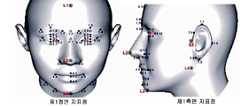
도 4는 안면 영상에서 얻어지는 안면 특징점들을 예시한 도면이다.
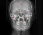
도 5는 두개골의 정면의 엑스선 영상에서 얻어지는 정면 지표점들을 예시한 사진이다.
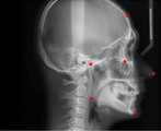
도 6은 두개골 측면 엑스선 영상에서 얻어지는 측면 지표점들을 예시한 사진이다.FIG. 1 is a block diagram showing an embodiment of a shape diagnosis apparatus using a facial image and an X-ray image according to the present invention.
2 is a block diagram showing an embodiment of a shape diagnosis system using a facial image and an x-ray image according to the present invention.
3 is a perspective view illustrating an embodiment of an image acquisition device for the shape diagnosis system shown in FIG.
4 is a diagram illustrating facial feature points obtained from a facial image.
FIG. 5 is a photograph illustrating frontal surface points obtained from an X-ray image of the front of the skull; FIG.
6 is a photograph illustrating side surface points obtained from a skull lateral x-ray image.
이하 본 발명의 목적이 구체적으로 실현될 수 있는 본 발명의 바람직한 실시예가 첨부된 도면을 참조하여 설명된다. 본 실시예를 설명함에 있어서, 동일 구성에 대해서는 동일 명칭 및 동일 부호가 사용되며 이에 따른 부가적인 설명은 하기에서 생략된다.BEST MODE FOR CARRYING OUT THE INVENTION Hereinafter, preferred embodiments of the present invention in which the object of the present invention can be specifically realized will be described with reference to the accompanying drawings. In describing the present embodiment, the same designations and the same reference numerals are used for the same components, and additional description thereof will be omitted in the following.
먼저, 도 1 및 도 2를 참조하여 본 발명에 따른 형상진단기 및 형상진단시스템의 일 실시예를 설명한다. 본 발명은 형상진단을 위해 안면 영상과 엑스선 영상을 이용하는 형상진단기(100)로서, 데이터 저장부(110)와 형상진단 제어부(120)와 디스플레이부(130)를 포함하여 구성된다.1 and 2, an embodiment of a shape diagnosis apparatus and a shape diagnosis system according to the present invention will be described. The present invention is a
여기서 상기 데이터 저장부(110)는 피검자, 즉 형상진단 검사 대상자의 안면 영상(Facial Image)와 피검자의 머리(두부)에 대한 엑스선 영상(Cephalometric Image)를 저장한다. 물론, 상기 데이터 저장부(110)는 후술하는 각종의 변수값과 이를 바탕으로 산출되는 각종의 수치 데이터를 저장할 수도 있으며, 피검자의 정보와 그에 대한 형상진단 결과를 저장할 수도 있고, 형상판단에 필요한 각종의 데이터 및 후술하는 각종 특징점들에 대한 좌표를 저장할 수 있다.The
그리고, 상기 형상진단 제어부(120)는 피검자의 안면 영상과 엑스선 영상을 이용하여 변수값를 산출한다. 보다 상세하게는 상기 안면 영상에서 얻어지는 안면 데이터와 상기 엑스선 영상에서 얻어지는 골격 데이터를 이용하여 상기 변수값을 산출한다. 상기 변수값의 예로는 형상진단을 위한 수치 데이터, 예를 들면 본 발명의 배경 기술에 기재된 각종 길이(얼굴 길이 등)나 각도 기타 면적 등을 들 수 있다.Then, the shape
보다 구체적으로 설명하면, 상기 형상진단 제어부(120)는 상기 변수값의 산출을 위한 안면 데이터로서 상기 안면 영상에서 얻어지는 안면 특징점들을 이용하고 상기 골격 데이터로는 상기 엑스선 영상에서 얻어지는 골격 특징점들을 이용한다.More specifically, the shape
다시 말해서 상기 형상진단 제어부(120)는 상기 안면 특징점들과 상기 골격 특징점들로부터 상기 변수값를 산출하며, 상기 안면 특징점들은 상기 안면 영상의 특정 지점들에 해당되는 점들이고 상기 골격 특징점들은 상기 엑스선 영상의 특정 지점들에 해당되는 점들로서, 상기 안면 특징점들과 골격 특징점들은 형상진단시에 고려되는 특정 위치의 점들을 말한다.In other words, the shape
상기 변수값는 상기 안면 특징점들로부터 얻어지는 제1변수값과 상기 골격 특징점들로부터 얻어지는 제2변수값를 포함하며, 상기 형상진단 제어부(120)는 상기 안면 특정점들로부터 상기 제1변수값를 산출하고 상기 골격 특징점들로부터 상기 제2변수값를 산출하는 것을 특징으로 한다.Wherein the parameter value includes a first parameter value obtained from the facial feature points and a second parameter value obtained from the skeleton feature points, wherein the shape diagnosis control unit (120) calculates the first parameter value from the facial feature points, And the second variable value is calculated from the minutiae points.
여기서, 상기 제1변수값은 상기 안면 특징점들로부터 얻어지는 길이와 각도와 면적 중 적어도 어느 하나를 포함하며, 상기 제2변수값은 상기 골격 특징점들로부터 얻어지는 길이와 각도와 면적 중 적어도 어느 하나를 포함하게 된다.Here, the first variable value includes at least one of a length, an angle and an area obtained from the facial feature points, and the second variable value includes at least one of a length, an angle and an area obtained from the skeletal feature points .
그리고 상기 형상진단을 위한 안면 영상으로서 얼굴 정면 영상과 얼굴 측면 영상이 이용되며, 상기 엑스선 영상으로는 두개골 정면 영상과 두개골 측면 영상이 이용된다.As the facial image for the shape diagnosis, a facial image and a facial image are used, and the x-ray image includes a skull frontal image and a skull lateral image.
이때, 상기 안면 특징점들은 상기 얼굴 정면 영상에서 얻어지는 제1정면 지표점들과 상기 얼굴 측면 영상에서 얻어지는 제1측면 지표점들을 포함하며, 상기 골격 특징점들은 상기 두개골 정면 영상에서 얻어지는 제1정면 지표점들과 상기 두개골 측면 영상에서 얻어지는 제2측면 지표점들을 포함한다.Here, the facial feature points include first facial feature points obtained from the face frontal image and first facial feature points obtained from the facial feature image, and the skeletal feature points include first frontal surface points obtained from the skull frontal image And second side surface points obtained from the skull side image.
더 나아가, 상기 형상진단 제어부(120)는 상기 변수값을 이용하여 피검자의 형상진단에 필요한 수치비교, 예를 들면 얼굴 정면의 면적과 측면의 면적을 비교하거나 각종의 길이비교를 수행할 수도 있다.Further, the shape
다음으로, 상기 디스플레이부(130)는 상기 안면 영상(도 4 참조)과 상기 엑스선 영상(도 5 및 도 6 참조)을 화면을 통해 표시하는 기능을 수행하며, 더 나아가 상기 형상진단 제어부(120)에 의해 산출된 각종의 변수값도 표시할 수 있으며, 또한 피검자에 대한 형상진단 결과를 표시할 수도 있다.The
도 2는 상기 형상진단기를 갖는 형상진단시스템의 일 실시예에 대한 구성을 나타낸 것으로서, 본 발명에 따른 형상진단시스템은 상술한 형상진단기(100)와 상기 형상진단기(100)에 상기 안면 영상과 엑스선 영상을 제공하기 위한 영상 획득기(10)을 포함하여 구성된다.FIG. 2 shows a configuration of an embodiment of a shape diagnosis system having the shape diagnosis unit. The shape diagnosis system according to the present invention includes the
여기서 상기 영상 획득기(10)는 안면 영상을 촬영하는 카메라(11)와 엑스선 영상을 촬영하는 엑스선 촬영기(12)를 포함하며, 상기 카메라(11)와 상기 엑스선 촬영기(12)가 각각 별도로 설치되거나 도 3에 도시된 바와 같이 하나의 몸체에 탑재될 수도 있다.The
상기 영상 획득기(10)에서 획득되는 안면 영상 및/또는 엑스선 영상은 상기 형상진단기(100)로 전송되며, 보다 상세하게는 유선 또는 무선을 통해 상기 형상진단기(100)로 전송된 후 상기 데이터 저장부(110)에 일시적 또는 영구적으로 저장된다.The facial image and / or the x-ray image obtained by the
본 실시예에 있어서, 상기 카메라(11)는 피검자의 안면 영상을 3D 영상(3차원 입체영상)으로 획득하기 위한 장치로서, 피검자의 안면 영상은 디지털 정보로 상기 형상진단기에 전송된다.In the present embodiment, the
다시 말해서, 상기 카메라(11)는 입체 영상의 획득이 가능한 3차원 카메라 기타 3차원 스캐너이며, 구체적으로는 공간분할 부호화 광학식 스캔방식, IEEE 1394 영상전송 인터페이스를 사용하는 480 X 640 픽셀의 3차원 카메라 등이 사용될 수 있으나 이에 한정되는 것은 아니다.In other words, the
그리고 상기 엑스선 촬영기(12)는 엑스레이(X-ray)를 주사하여 피검체의 두개골 영상을 획득하는 장치로서, 촬영방식과 장치 구성 그 자체는 일반적으로 공지된 것이므로 그에 대한 부가적인 설명은 생략된다.The
도 3을 참조하면, 상기 엑스선 촬영기(12)는 엑스선 출사기(12a)와 엑스선 검출기(12b)를 포함하여 구성되며, 본 실시예에서 상기 엑스선 출사기(12a)와 엑스선 검출기(12b)영상 획득기(10)는 지대주(13)에 의해 지지되는 회전 암에 상호 대향되게 탑재되며, 상기 카메라(11)도 상기 회전 암에 탑재된다.3, the
그리고 상기 지대주(13)에는 얼굴 위치의 정렬을 위한 정렬기(14)가 구비되며, 상기 정렬기(14)에 의해 피검자의 머리가 상기 영상 획득기(10) 상에 정위치 된 상태에서 안면 영상과 엑스선 영상이 한꺼번에 순차적으로 획득될 수 있다.The
한편, 본 발명에 따른 형상진단기 및 형상진단시스템에는 상기 형상진단을 위해 공개특허 제10-2009-0125359호에 개시된 설문 정보 입력기가 본 발명에 동일하게 적용될 수도 있다.In the meantime, the questionnaire information input device disclosed in Japanese Patent Laid-Open No. 10-2009-0125359 may be applied to the shape diagnosis device and the shape diagnosis system according to the present invention.
상기 데이터 저장부(110)는 상기 카메라(10)와 엑스선 촬영기(12) 및 설문 정보 입력기(20)로부터 입력되는 안면 영상과 엑스선 영상 등 각종의 데이터를 저장하며, 상기 형상진단 제어부(120)는 상기 안면 영상의 안면 데이터와 엑스선 영상의 골격 데이터를 분석하고 연산하며, 상술한 설문 정보도 분석할 수 있다.The
특히, 상기 형상진단 제어부(120)는 상기 카메라(11)에 의한 정보를 입력받아 이를 입체적으로 모델링시킨다. 다음으로, 입체화된 모델링에서 안면계측을 위한 복수의 안면 특징점들을 상기 입체 모델링의 전면 영상과 측면 영상에 자동으로 포인팅시킨다. 그리고 변수 데이터, 예를 들면 상기 특징점들의 좌표값을 획득한 후, 이를 형상진단을 위한 함수에 대입시켜 그 결과, 예를 들면 상술한 변수값을 출력한다. 상기 형상진단 제어부(120)에 의해 획득되는 변수값은, 복수의 안면 특징점들을 선택적으로 조합하여 계산되는 길이, 각도, 면적 등에 대한 값이다.In particular, the shape
물론, 상기 디스플레이부에 의해 화면(예를 들면 컴퓨터 모니터)에 출력된 안면 영상에 유저(User)가 마우스 등의 인터페이스를 이용하여 직접 상기 안면 특징점들을 표시(포인팅)할 수도 있다.Of course, the user may display (point) the facial feature points directly on the facial image output by the display unit on the screen (for example, a computer monitor) using an interface such as a mouse.
그리고 상기 형상진단 제어부(30)에서 출력되는 변수값은 디스플레이부(130)에 표시되거나 상기 데이터 저장부(110)에 축적되어 임상 정보로 누적될 수 있다.The variable values output from the shape diagnosis and control unit 30 may be displayed on the
보다 구체적으로 설명하면, 상기 형상진단 제어부(120)는 상기 카메라(11)로부터 입력되는 정보, 즉 안면 영상정보를 이용하여 피검자의 안면 이미지를 삼차원(3D) 입체 영상으로 재구성하며, 특정의 위치에 안면 특징점들이 포인팅되도록 한다.More specifically, the shape
이하 도 4를 참조하여 상기 안면 특징점들의 예로서 얼굴 정면 영상에서 얻어지는 제1정면 지표점들과 얼굴 측면 영상에서 얻어지는 제1측면 지표점들에 대해 살펴보기로 한다.Referring to FIG. 4, the first facial feature points obtained from the face facial image and the first facial feature points obtained from the facial feature image will be described as an example of the facial feature points.
상기 안면 특징점들은 형상진단에 필요한 안면의 특징을 추출하기 위한 '안면의 특이점'들로서 안면 영상의 특정 지점에 해당되는 점들을 의미한다. 실시예에서는 공지의 논문을 참조하여 마틴식 계측법을 기반으로 측정항목을 설정하였으며, 국제 표준 기관 ISO(International Organization for Standardization)는 인체 측정과 관련하여 머리 측정 항목을 6개로 규정하고 있다.The facial feature points are 'facial singularities' for extracting facial features necessary for shape diagnosis, and refer to points corresponding to a specific point of the facial image. In the embodiment, the measurement items are set based on the Martin type measurement method with reference to the known paper, and the International Organization for Standardization (ISO) specifies six head measurement items in relation to the human body measurement.
MPEG-4(Moving Picture Experts Group)에서 설정한 측정항목은 크게 FDP(Facial Definition Parameter)와 FAP(Facial Animation Parameter)로 나눌 수 있으며, FDP는 총 84개의 3차원 좌표값을 가진 특징점으로 구성되고 눈, 눈썹, 코, 입, 턱, 뺨, 혀, 치아, 귀, 머리, 머리를 표현하는 9개의 그룹으로 나누어 정의되고 있다. 여기서 상기 FAP는 총 68개의 특징점으로 구성되고, 얼굴 근육의 움직임에 있어서 표정 및 입의 움직임을 정의할 수 있다. 본 발명에서 사용하고 있는 안면 특징점들의 예로 MPEG-4(Moving Picture Experts Group)에서 사용하는 측정항목을 준용 및 개변하여 대한민국공개특허 제10-2009-0125359호에 개시된 바와 같이 정면에 36개, 측면에 26개의 안면 특징점들 설정하였다.The measurement items set by MPEG-4 (Moving Picture Experts Group) can be divided into FDP (Facial Definition Parameter) and FAP (Facial Animation Parameter). FDP is composed of feature points having a total of 84 three- , Eyebrows, nose, mouth, chin, cheek, tongue, tooth, ear, head, head. Here, the FAP is composed of a total of 68 feature points, and it is possible to define facial expressions and mouth movements in the movement of the facial muscles. As examples of facial feature points used in the present invention, measurement items used in the Moving Picture Experts Group (MPEG-4) are applied and modified, as disclosed in Korean Patent Laid-Open No. 10-2009-0125359, 26 facial feature points were set.
도 4에서 S1은 우동공점, S2는 좌동공점, S3는 좌이주점, L1은 발제점, L2는 비첨점, L3는 턱하점, L4는 좌하악각점, 1.1은 우눈썹상연점, 1.2는 좌눈썹상연점을 나타내며, 도 4의 좌측 사진은 얼굴 정면 영상에 안면 특징점들, 즉 제1정면 지표점들이 포인팅된 상태이고, 도 4의 우측 사진은 얼굴 측면 영상에 안면 특징점들, 즉 제1측면 지표점들이 포인팅된 상태이다.In Fig. 4, S1 is the ino-rial cavity, S2 is the left ear, S3 is the left ear, L1 is the presentation point, L2 is the uncut point, L3 is the tangential point, L4 is the left mandibular point, 1.1 is the right eyebrow point, 4, the facial feature points, i.e., the first facial feature points, are pointed to the facial frontal image, and the right-side photograph of FIG. 4 shows the facial feature points, that is, Points are pointing.
상기 제1정면 지표점들과 제1측면 지표점들은 3차원 이미지상의 좌표로 검출되며, 각각의 지표점에 대한 좌표값은 상기 데이터 저장부(110)에 저장된다. 그리고 상기 형상진단 제어부(120)는 상기 안면 데이터, 즉 좌표값을 이용하여 얼굴의 길이나 면적을 연산하고 그 값을 형상진단을 위한 함수의 변수값(제1변수값)으로 산출한다.The first front surface point points and the first side surface point points are detected as coordinates on the three-dimensional image, and the coordinate values for the respective surface points are stored in the
형상진단에 필요한 안면의 특이점, 즉 안면 특징점들의 위치는 형상진단의학에서 공지된 것이며, 본 발명은 안면 영상과 엑스선 영상에 각각 포인팅된 상기 안면 특징점과 골격 특징점들을 이용하여 정확한 결과를 출력하고 특히 엑스선 영상을 이용하여 형상진단의 정확성을 보완하는 데 그 특징이 있다.The position of the facial feature points required for the shape diagnosis, that is, the positions of the facial feature points, is well known in the shape diagnostics, and the present invention outputs accurate results using the facial feature points and skeletal feature points pointed to the facial image and the x- It is characterized by supplementing the accuracy of shape diagnosis using images.
다음으로 도 5 및 도 6을 참조하면, 상기 골격 특징점들은 두개골 정면 영상에 포인팅되는 정면 지표점들(이하 '제2정면 지표점들'이라 칭함)과 두개골 측면 영상에 포인팅되는 측면 지표점들(이하 '제2측면 지표점들'이라 칭함)를 포함한다.5 and 6, the skeletal feature points include front surface points (hereinafter, referred to as 'second front surface points') pointed to the skull frontal image and side surface points Hereinafter referred to as " second side surface points ").
상기 제2정면 지표점들의 예로는, 도 5에 도시된 바와 같이, 좌우 광대뼈에 포인팅되는 점과, 좌우 동공에 대응되는 위치는 포인팅되는 점과, 하악의 아래 경계에 포인팅되는 점과, 발제점(L1)에 대응되는 위치에 포인팅되는 점 등이 있다.5, points pointing to the left and right cheekbones, points corresponding to the left and right pupils are pointed, points pointing to the lower boundary of the mandible, And a point pointed at a position corresponding to the point L1.
그리고 상기 제2측면 지표점의 예로는, 도 6에 도시된 바와 같이, 일측 광대뼈에 포인팅되는 점과, 하악의 아래 경계에 포인팅되는 점과 발제점(L1)에 대응되는 위치에 포인팅되는 점과, 좌이주점(S3)에 대응되는 위치에 포인팅되는 점과, 코끝에 대앙되는 위치에 포인팅되는 점 등이 있다.6, a point pointing to one cheekbone, a point pointing to a lower boundary of the mandible, a point pointing to a point corresponding to the presentation point L1, , A point pointed to a position corresponding to the left main point S3, and a point pointed to a position corresponding to the nose tip.
상기 안면 영상에서의 발제점(L1) 위치를 그대로 엑스선 영상에 투영한 위치에 상기 안면 영상의 발제점(L1)에 대응되는 엑스선 영상의 지표점이 포인팅됨으로써 설정되거나, 상기 형상진단 제어부(120)가 상기 안면 영상의 턱하점(L3)를 기준으로 산출되는 상기 턱하점과 발제점의 거리와 발제점의 방향을 산출하고, 이를 바탕으로 엑스선 영상의 연조직 턱끝점(도시되지 않음)에서 동일거리와 동일방향에 상기 발제점(L1)에 대응되는 엑스선 영상의 지표점을 포인팅하는 등 다양한 방식이 적용될 수 있다.The
피검자의 엑스선 영상을 통해 획득되는 골격 데이터는 피검자가 비만인 경우나 저체중인 경우에도 변경되지 않으며, 성형수술을 위해 얼굴에 보형물이 삽입된 경우에도 안정적인 데이터로 활용될 수 있으며, 이에 따라 피검자의 안면 외관 형상과 함께 두개골의 골격 형상을 바탕으로 형상진단이 수행될 수 있다.The skeleton data obtained through the X-ray image of the subject is not changed even when the subject is obese or underweight, and can be used as stable data even when the implant is inserted on the face for plastic surgery. Accordingly, The shape diagnosis can be performed based on the skeletal shape of the skull together with the shape.
즉 골격 데이터, 즉 상기 제2정면 지표점들과 제2측면 지표점들의 좌표를 선택적으로 추출하여 그로부터 두개골 정면의 면적과 길이 등의 변수값(제2변수값)을 산출할 수 있으며, 그 값을 바탕으로 형상판단을 보완하게 된다.That is, the skeletal data, that is, the coordinates of the second frontal surface points and the second lateral surface points, can be selectively extracted, and variable values (second variable values) such as the area and length of the skull front face can be calculated therefrom. The shape determination is supplemented.
상기 제2정면 지표점들과 제2측면 지표점들은 상기 형상진단기에 설치되는 형상진단 프로그램에 의해 자동으로 포인팅될 수도 있고, 마우스 등의 인터페이스를 이용하여 유저에 의해 포인팅될 수도 있다.The second front surface point points and the second side surface point points may be automatically pointed by a shape diagnosing program installed in the shape diagnosing device or may be pointed by a user using an interface such as a mouse.
본 발명은 상술한 바와 같이 3차원 안면 이미지와 골격성 이미지를 이용하여 안면형태학적 특징을 검출할 수 있다. 종래의 경우, 안면 계측은 피검자를 눕힌 상태에서 안면 특이점간의 거리나 특정 부위의 각도 등을 수평자나 줄자 등을 이용하여 측정되었는데, 이러한 측정방식은 피검자의 자세에 따라 오차범위가 변동되고, 피검자가 측정자(의사 등)와의 신체적인 접촉에 의한 불쾌감 등이 있어 문제가 되어왔으나, 본 발명은 카메라(11)에 의한 안면 이미지와 함께 엑스선 영상을 사용할 수 있으므로 비만이나 체중미달 등으로 인한 안면 형상 변화에 대응할 수 있게 된다.The present invention can detect facial morphological features using a 3D facial image and a skeletal image as described above. In the conventional case, the face measurement is performed by measuring the distance between the facial singular points and the angle of a specific part in a state in which the subject is laid down by using a horizon or a tape measure. Such a measurement method varies the error range according to the posture of the subject, The present invention can use an X-ray image together with a facial image by the
본 발명은 또한 안면 영상과 엑스선 영상을 이용한 형상진단방법을 개시하며, 상기 형상진단방법은, 피검자의 안면 영상과 두부에 대한 엑스선 영상을 입력받아 저장하는 (a)단계와 상기 안면 영상에서 얻어지는 안면 데이터와 엑스선 영상에서 얻어지는 골격 데이터를 이용하여 피검자의 형상진단에 필요한 변수값를 산출하는 (b)단계를 포함하여 이루어진다.The present invention also discloses a shape diagnosis method using a facial image and an X-ray image, the method comprising: (a) receiving and storing a facial image of a subject and an X-ray image of a head; And (b) calculating a parameter value necessary for diagnosing the shape of the subject using skeletal data obtained from the data and the X-ray image.
그리고, 상기 (b)단계는 상기 안면 영상에서 얻어지는 안면 특징점들로부터 상기 형상진단을 위한 길이와 각도와 면적 중 적어도 어느 하나를 포함하는 제1변수값를 산출하고 상기 엑스선 영상에서 얻어지는 골격 특징점들로부터 상기 형상진단을 위한 길이와 각도와 면적 중 적어도 어느 하나를 포함하는 제2변수값를 산출하는 (b1)단계, 그리고 상기 제1변수값와 제2변수값을 이용하여 형상진단에 필요한 수치비교를 수행하는 (b2)단계를 포함하여 이루어진다.In the step (b), a first parameter value including at least one of a length, an angle and an area for the shape diagnosis is calculated from facial minutiae points obtained from the facial image, and the first parameter value is calculated from skeletal minutiae points obtained from the x- (B1) calculating a second parameter value including at least one of a length, an angle, and an area for the shape diagnosis, and performing a numerical comparison necessary for shape diagnosis using the first variable value and the second variable value b2).
또한 본 발명은 상기 수치비교의 결과를 기설정된 형상판단의 기준 데이터와 비교하여 담체-방광체, 정신기혈과, 오장형상, 및 육경형 등의 형상을 판단하고 그 결과를 출력할 수도 있다. 이때 골격 데이터부터 얻어지는 형산판단결과와 안면 데이터로부터 얻어지는 형상판단결과가 다른 경우에는 그 결과를 화면이나 프린터를 통해 출력할 수도 있다.Further, the present invention may compare the result of the numerical comparison with reference data of a predetermined shape judgment to judge the shapes of the carrier-bladder, the mental condyle, the oval shape, the height shape, and the like, and output the result. At this time, if the shape determination result obtained from the skeleton data is different from the shape determination result obtained from the face data, the result may be output through a screen or a printer.
한편, 본 발명은: 컴퓨터를 저장수단과 제어수단과 디스플레이수단으로 기능시키기 위한 안면 영상과 엑스레이 영상을 이용한 형상진단 프로그램을 기록한 컴퓨터로 읽을 수 있는 기록매체의 형태로 제공될 수도 있는데, 다시 말해서 컴퓨터를 피검자의 안면 영상과 두부에 대한 엑스선 영상을 입력받아 저장하는 저장수단과, 상기 안면 영상에서 얻어지는 안면 데이터와 엑스선 영상에서 얻어지는 골격 데이터를 이용하여 피검자의 형상진단에 필요한 변수값를 산출하는 제어수단과 상기 안면 영상과 엑스선 영상을 표시하는 디스플레이수단으로 기능시키는 것을 말한다.Meanwhile, the present invention may be provided in the form of a computer-readable recording medium in which a computer is used as a storage means, a control means and a display means, and a shape diagnosis program using a facial image and an x-ray image. In other words, A control means for calculating a parameter value necessary for diagnosing the shape of the subject using facial data obtained from the facial image and skeleton data obtained from the x-ray image; And functions as a display means for displaying the face image and the x-ray image.
상기 형상진단 프로그램을 기록한 컴퓨터로 읽을 수 있는 기록매체는 자기 디스크나 CD 등 공지의 각종 기록매체와 상기 형상진단 프로그램을 네트워크을 통해 제공하도록 상기 컴퓨터로 읽을 수 있는 프로그램을 기록한 시스템 서버 등이 있다.The computer-readable recording medium on which the shape diagnosing program is recorded includes a variety of known recording media such as a magnetic disk and a CD, and a system server in which the computer-readable program is recorded to provide the shape diagnosing program via a network.
상기와 같이 본 발명에 따른 바람직한 실시예를 살펴보았으며, 앞서 설명된 실시예 이외에도 본 발명이 그 취지나 범주에서 벗어남이 없이 다른 특정 형태로 구체화 될 수 있다는 사실은 해당 기술에 통상의 지식을 가진 이들에게는 자명한 것이다.As described above, according to the present invention, the present invention can be embodied in other specific forms without departing from the spirit and scope of the present invention. It is obvious to them.
그러므로, 상술된 실시예는 제한적인 것이 아니라 예시적인 것으로 여겨져야 하고, 이에 따라 본 발명은 상술한 설명에 한정되지 않고 첨부된 청구항의 범주 및 그 동등 범위 내에서 변경될 수도 있다.Therefore, the above-described embodiments are to be considered as illustrative rather than restrictive, and the present invention is not limited to the above description, but may be modified within the scope of the appended claims and equivalents thereof.
10: 영상 획득기11: 카메라
12: 엑스선 촬영기100: 형상진단기
110: 데이터 저장부120: 형상진단 제어부
130: 디스플레이부10: image capture device 11: camera
12: X-ray machine 100: Shape diagnosis machine
110: Data storage unit 120: Shape diagnosis control unit
130:
Claims (9)
Translated fromKorean한방의 형상진단의학에서 형상진단을 위해 사용되는 위치의 점으로서 상기 안면 영상에 지정되어서 상기 안면 영상에서 얻어지는 안면 특징점들과 상기 안면 특징점에 대응되도록 상기 엑스선 영상에 지정되어서 상기 엑스선 영상에서 얻어지는 골격 특징점들을 이용해서 피검자의 형상진단에 필요한 변수값을 산출하는 형상진단 제어부; 그리고
상기 안면 영상과 엑스선 영상을 표시하는 디스플레이부를 포함하여 구성되는 안면 영상과 엑스레이 영상을 이용한 형상진단기로서:
상기 변수값은, 상기 형상진단을 위해 상기 안면 특징점들로부터 얻어지는 길이와 각도와 면적 중 적어도 어느 하나를 포함하는 제1변수값과 상기 골격 특징점들로부터 얻어지는 길이와 각도와 면적 중 적어도 어느 하나를 포함하는 제2변수값을 포함하며; 상기 형상진단 제어부는 상기 안면 특징점들로부터 상기 제1변수값을 산출하고 상기 골격 특징점들로부터 상기 제2변수값을 산출하는 안면 영상과 엑스레이 영상을 이용한 형상진단기.A data storage unit for storing a facial image of the subject and a cephalometric image of the subject's head;
A facial feature point designated in the facial image as a point of a position used for shape diagnosis in the oriental shape diagnosing medicine, facial feature points obtained from the facial image, and skeletal feature points designated in the x-ray image corresponding to the facial feature point, A shape diagnosis control unit for calculating a variable value necessary for diagnosing the shape of the subject using the images; And
And a display unit for displaying the facial image and the x-ray image, the shape diagnostic apparatus using the facial image and the x-ray image,
Wherein the variable value includes at least one of a first variable value including at least one of a length, an angle and an area obtained from the facial feature points for the shape diagnosis, and a length, an angle and an area obtained from the skeletal feature points ≪ / RTI > Wherein the shape diagnosis control unit calculates the first parameter value from the facial feature points and calculates the second parameter value from the skeletal feature points.
상기 안면 영상은 얼굴 정면 영상과 얼굴 측면 영상을 포함하고, 상기 엑스선 영상은 두개골 정면 영상과 두개골 측면 영상을 포함하며; 상기 안면 특징점들은 상기 얼굴 정면 영상에서 얻어지는 제1정면 지표점들과 상기 얼굴 측면 영상에서 얻어지는 제1측면 지표점들을 포함하고, 상기 골격 특징점들은 상기 두개골 정면 영상에서 얻어지는 제2정면 지표점들과 상기 두개골 측면 영상에서 얻어지는 제2측면 지표점들을 포함하는 것을 특징으로 하는 안면 영상과 엑스레이 영상을 이용한 형상진단기.The method according to claim 1,
Wherein the facial image comprises a facial frontal image and a facial lateral image, the x-ray image comprising a skull frontal image and a skull lateral image; Wherein the facial feature points include first frontal surface points obtained from the face frontal image and first side surface points obtained from the facial lateral image, and the skeletal feature points include second frontal surface points obtained from the skull frontal image, And second side surface points obtained from the skull side image.
상기 디스플레이부는, 상기 피검자에 대한 형상진단결과를 출력하는 것을 특징으로 하는 안면 영상과 엑스레이 영상을 이용한 형상진단기.The method according to claim 1,
Wherein the display unit outputs a shape diagnosis result for the subject.
상기 안면 영상과 엑스선 영상을 통해 피검자의 형상을 진단하는 형상진단기를 포함하여 구성되는 안면 영상과 엑스레이 영상을 이용한 형상진단시스템으로서:
상기 형상진단기는, 피검자의 안면 영상과 피검자의 두부에 대한 엑스선 영상을 저장하는 데이터 저장부, 한방의 형상진단의학에서 형상진단을 위해 사용되는 위치의 점으로서 상기 안면 영상에 지정되어서 상기 안면 영상에서 얻어지는 안면 특징점들과 상기 안면 특징점에 대응되도록 상기 엑스선 영상에 지정되어서 상기 엑스선 영상에서 얻어지는 골격 특징점들을 이용해서 피검자의 형상진단에 필요한 변수값을 산출하는 형상진단 제어부, 그리고 상기 안면 영상과 엑스선 영상을 표시하는 디스플레이부를 포함하여 구성되고;
상기 변수값은, 상기 형상진단을 위해 상기 안면 특징점들로부터 얻어지는 길이와 각도와 면적 중 적어도 어느 하나를 포함하는 제1변수값과 상기 골격 특징점들로부터 얻어지는 길이와 각도와 면적 중 적어도 어느 하나를 포함하는 제2변수값을 포함하며; 상기 형상진단 제어부는 상기 안면 특징점들로부터 상기 제1변수값을 산출하고 상기 골격 특징점들로부터 상기 제2변수값을 산출하는 안면 영상과 엑스레이 영상을 이용한 형상진단시스템.An image acquirer including a camera for photographing a facial image and an x-ray photographing apparatus for photographing an x-ray image;
And a shape diagnosis unit for diagnosing the shape of the subject through the facial image and the x-ray image, the system comprising:
Wherein the shape diagnosis unit includes a data storage unit for storing a face image of a subject and an X-ray image of a head of the subject, a position storage unit for storing the X- A shape diagnosis control unit for calculating variable values necessary for diagnosing the shape of the subject using facial minutiae points obtained and skeletal minutiae points assigned to the x-ray image corresponding to the facial minutiae points and obtained from the x-ray image, Comprising: a display unit for displaying an image;
Wherein the variable value includes at least one of a first variable value including at least one of a length, an angle and an area obtained from the facial feature points for the shape diagnosis, and a length, an angle and an area obtained from the skeletal feature points ≪ / RTI > Wherein the shape diagnosis control unit calculates the first variable value from the facial feature points and calculates the second parameter value from the skeletal feature points.
한방의 형상진단의학에서 형상진단을 위해 사용되는 위치의 점으로서 상기 안면 영상에 지정되어서 상기 안면 영상에서 얻어지는 안면 특징점들과 상기 안면 특징점에 대응되도록 상기 엑스선 영상에 지정되어서 상기 엑스선 영상에서 얻어지는 골격 특징점들을 이용해서 피검자의 형상진단에 필요한 변수값을 산출하는 (b)단계를 포함하여 이루어지는 안면 영상과 엑스레이 영상을 이용한 형상진단방법으로서:
상기 변수값은, 상기 형상진단을 위해 상기 안면 특징점들로부터 얻어지는 길이와 각도와 면적 중 적어도 어느 하나를 포함하는 제1변수값과 상기 골격 특징점들로부터 얻어지는 길이와 각도와 면적 중 적어도 어느 하나를 포함하는 제2변수값을 포함하며; 상기 형상진단 제어부는 상기 안면 특징점들로부터 상기 제1변수값을 산출하고 상기 골격 특징점들로부터 상기 제2변수값을 산출하는 안면 영상과 엑스레이 영상을 이용한 형상진단방법.(A) receiving and storing a facial image of a subject and an X-ray image of a head; And
A facial feature point designated in the facial image as a point of a position used for shape diagnosis in the oriental shape diagnosing medicine, facial feature points obtained from the facial image, and skeletal feature points designated in the x-ray image corresponding to the facial feature point, (B) calculating a parameter value necessary for diagnosing the shape of the subject using the face image and the x-ray image, the method comprising:
Wherein the variable value includes at least one of a first variable value including at least one of a length, an angle and an area obtained from the facial feature points for the shape diagnosis, and a length, an angle and an area obtained from the skeletal feature points ≪ / RTI > Wherein the shape diagnosis control unit calculates the first variable value from the facial feature points and calculates the second parameter value from the skeletal feature points.
피검자의 안면 영상과 두부에 대한 엑스선 영상을 입력받아 저장하는 저장수단;
한방의 형상진단의학에서 형상진단을 위해 사용되는 위치의 점으로서 상기 안면 영상에 지정되어서 상기 안면 영상에서 얻어지는 안면 특징점들과 상기 안면 특징점에 대응되도록 상기 엑스선 영상에 지정되어서 상기 엑스선 영상에서 얻어지는 골격 특징점들을 이용해서 피검자의 형상진단에 필요한 변수값을 산출하는 제어수단; 그리고
상기 안면 영상과 엑스선 영상을 표시하는 디스플레이수단으로 기능시키기 위한 안면 영상과 엑스레이 영상을 이용한 형상진단 프로그램을 기록한 컴퓨터로 읽을 수 있는 기록매체로서:
상기 변수값은, 상기 형상진단을 위해 상기 안면 특징점들로부터 얻어지는 길이와 각도와 면적 중 적어도 어느 하나를 포함하는 제1변수값과 상기 골격 특징점들로부터 얻어지는 길이와 각도와 면적 중 적어도 어느 하나를 포함하는 제2변수값을 포함하며; 상기 형상진단 제어부는 상기 안면 특징점들로부터 상기 제1변수값을 산출하고 상기 골격 특징점들로부터 상기 제2변수값을 산출하는 안면 영상과 엑스레이 영상을 이용한 형상진단 프로그램을 기록한 컴퓨터로 읽을 수 있는 기록매체.Computer,
Storage means for receiving and storing a facial image of a subject and an X-ray image of a head;
A facial feature point designated in the facial image as a point of a position used for shape diagnosis in the oriental shape diagnosing medicine, facial feature points obtained from the facial image, and skeletal feature points designated in the x-ray image corresponding to the facial feature point, Control means for calculating a variable value necessary for diagnosing the shape of the examinee by using the parameters; And
A computer-readable recording medium recording a shape diagnosis program using a facial image and an x-ray image to function as a display means for displaying the facial image and x-ray image,
Wherein the variable value includes at least one of a first variable value including at least one of a length, an angle and an area obtained from the facial feature points for the shape diagnosis, and a length, an angle and an area obtained from the skeletal feature points ≪ / RTI > Wherein the shape diagnosis control unit is configured to calculate the first variable value from the facial minutiae points and to calculate the second parameter value from the skeletal minutiae points and a computer readable recording medium having recorded thereon a shape diagnosis program using an x- .
Priority Applications (1)
| Application Number | Priority Date | Filing Date | Title |
|---|---|---|---|
| KR1020110100656AKR101309013B1 (en) | 2011-10-04 | 2011-10-04 | Diagnosis Device For Face Form Using Facial Image and Cephalometric Image |
Applications Claiming Priority (1)
| Application Number | Priority Date | Filing Date | Title |
|---|---|---|---|
| KR1020110100656AKR101309013B1 (en) | 2011-10-04 | 2011-10-04 | Diagnosis Device For Face Form Using Facial Image and Cephalometric Image |
Publications (2)
| Publication Number | Publication Date |
|---|---|
| KR20130036526A KR20130036526A (en) | 2013-04-12 |
| KR101309013B1true KR101309013B1 (en) | 2013-09-17 |
Family
ID=48437810
Family Applications (1)
| Application Number | Title | Priority Date | Filing Date |
|---|---|---|---|
| KR1020110100656AActiveKR101309013B1 (en) | 2011-10-04 | 2011-10-04 | Diagnosis Device For Face Form Using Facial Image and Cephalometric Image |
Country Status (1)
| Country | Link |
|---|---|
| KR (1) | KR101309013B1 (en) |
Families Citing this family (4)
| Publication number | Priority date | Publication date | Assignee | Title |
|---|---|---|---|---|
| KR101918119B1 (en)* | 2017-03-10 | 2019-02-08 | 주식회사 모르페우스 | Method, system and non-transitory computer-readable recording medium for estimating anatomical layer of face |
| KR102099390B1 (en)* | 2018-08-21 | 2020-04-09 | 디디에이치 주식회사 | Dental image analyzing method for orthodontic daignosis and apparatus using the same |
| KR102182980B1 (en)* | 2018-09-20 | 2020-11-25 | 오스템임플란트 주식회사 | Method and Apparatus for x-ray imaging, computer-readable recording medium |
| KR20220100740A (en)* | 2021-01-07 | 2022-07-18 | 디디에이치 주식회사 | Medical image analyzing method and apparatus using the same |
Citations (4)
| Publication number | Priority date | Publication date | Assignee | Title |
|---|---|---|---|---|
| JPH09173352A (en)* | 1995-12-25 | 1997-07-08 | Toshiba Medical Eng Co Ltd | Medical navigation system |
| KR19980017533A (en)* | 1996-08-30 | 1998-06-05 | 김광호 | Scanning electron microscope with personal computer connected |
| KR20030062167A (en)* | 2002-01-16 | 2003-07-23 | 한승희 | search system with Metal detection and Face Recognition, Baggage search functions as one and so the operating method |
| KR20080085307A (en)* | 2007-03-19 | 2008-09-24 | (주)이우테크놀로지 | X-ray image acquisition method |
- 2011
- 2011-10-04KRKR1020110100656Apatent/KR101309013B1/enactiveActive
Patent Citations (4)
| Publication number | Priority date | Publication date | Assignee | Title |
|---|---|---|---|---|
| JPH09173352A (en)* | 1995-12-25 | 1997-07-08 | Toshiba Medical Eng Co Ltd | Medical navigation system |
| KR19980017533A (en)* | 1996-08-30 | 1998-06-05 | 김광호 | Scanning electron microscope with personal computer connected |
| KR20030062167A (en)* | 2002-01-16 | 2003-07-23 | 한승희 | search system with Metal detection and Face Recognition, Baggage search functions as one and so the operating method |
| KR20080085307A (en)* | 2007-03-19 | 2008-09-24 | (주)이우테크놀로지 | X-ray image acquisition method |
Also Published As
| Publication number | Publication date |
|---|---|
| KR20130036526A (en) | 2013-04-12 |
Similar Documents
| Publication | Publication Date | Title |
|---|---|---|
| US11089974B2 (en) | Monitoring the location of a probe during patient breathing | |
| CN105392423B (en) | Motion Tracking System for Real-Time Adaptive Motion Compensation in Biomedical Imaging | |
| Maal et al. | Registration of 3-dimensional facial photographs for clinical use | |
| Ma et al. | Validation of a three-dimensional facial scanning system based on structured light techniques | |
| JP2023014295A (en) | Method for cephalometric analysis | |
| KR101757642B1 (en) | Apparatus and method for 3d face modeling | |
| US20100312143A1 (en) | Human body measurement system and information provision method using the same | |
| AU2016354889B2 (en) | Method and device for estimating absolute size dimensions of a test object | |
| US11723614B2 (en) | Dynamic 3-D anatomical mapping and visualization | |
| KR100979506B1 (en) | Sasang Constitution Diagnostic Machine | |
| Nahm et al. | Accurate registration of cone-beam computed tomography scans to 3-dimensional facial photographs | |
| Schendel et al. | 3D orthognathic surgery simulation using image fusion | |
| KR101309013B1 (en) | Diagnosis Device For Face Form Using Facial Image and Cephalometric Image | |
| CN105899144A (en) | Image processing apparatus, image diagnostic system, image processing method, and storage medium | |
| CN106413568B (en) | Image processing apparatus, the control method of image processing apparatus and storage medium | |
| CN105899145B (en) | Image processing apparatus, image processing method and storage medium | |
| Kinel et al. | Normative 3D opto-electronic stereo-photogrammetric sagittal alignment parameters in a young healthy adult population | |
| US20250045924A1 (en) | System for obtaining useful data for analysis of body morphometry and associated method | |
| CN106725305A (en) | Pain scale evaluation method and system based on human body attitude angle | |
| RU2610911C1 (en) | System and method of virtual smile prototyping based on tactile computer device | |
| JP5687532B2 (en) | Information processing method, Frankfurt plane calculation method, and information processing apparatus | |
| CN101879057B (en) | Three-dimensional head spatial repositioning offset measuring system and operation method thereof | |
| Rahul et al. | To evaluate and correlate nasolabial angle, mentolabial sulcus angle and throat angle using cephalometric and photographic measurement | |
| JP2021083960A (en) | Medical image processor, medical image processing method, and medical image processing program | |
| Lin et al. | Reliability and reproducibility of landmarks on three-dimensional soft-tissue cephalometrics using different placement methods |
Legal Events
| Date | Code | Title | Description |
|---|---|---|---|
| A201 | Request for examination | ||
| PA0109 | Patent application | St.27 status event code:A-0-1-A10-A12-nap-PA0109 | |
| PA0201 | Request for examination | St.27 status event code:A-1-2-D10-D11-exm-PA0201 | |
| P11-X000 | Amendment of application requested | St.27 status event code:A-2-2-P10-P11-nap-X000 | |
| P13-X000 | Application amended | St.27 status event code:A-2-2-P10-P13-nap-X000 | |
| D13-X000 | Search requested | St.27 status event code:A-1-2-D10-D13-srh-X000 | |
| D14-X000 | Search report completed | St.27 status event code:A-1-2-D10-D14-srh-X000 | |
| E902 | Notification of reason for refusal | ||
| PE0902 | Notice of grounds for rejection | St.27 status event code:A-1-2-D10-D21-exm-PE0902 | |
| T11-X000 | Administrative time limit extension requested | St.27 status event code:U-3-3-T10-T11-oth-X000 | |
| P11-X000 | Amendment of application requested | St.27 status event code:A-2-2-P10-P11-nap-X000 | |
| P13-X000 | Application amended | St.27 status event code:A-2-2-P10-P13-nap-X000 | |
| T11-X000 | Administrative time limit extension requested | St.27 status event code:U-3-3-T10-T11-oth-X000 | |
| T11-X000 | Administrative time limit extension requested | St.27 status event code:U-3-3-T10-T11-oth-X000 | |
| P11-X000 | Amendment of application requested | St.27 status event code:A-2-2-P10-P11-nap-X000 | |
| P13-X000 | Application amended | St.27 status event code:A-2-2-P10-P13-nap-X000 | |
| T11-X000 | Administrative time limit extension requested | St.27 status event code:U-3-3-T10-T11-oth-X000 | |
| PG1501 | Laying open of application | St.27 status event code:A-1-1-Q10-Q12-nap-PG1501 | |
| E13-X000 | Pre-grant limitation requested | St.27 status event code:A-2-3-E10-E13-lim-X000 | |
| P11-X000 | Amendment of application requested | St.27 status event code:A-2-2-P10-P11-nap-X000 | |
| P13-X000 | Application amended | St.27 status event code:A-2-2-P10-P13-nap-X000 | |
| E701 | Decision to grant or registration of patent right | ||
| PE0701 | Decision of registration | St.27 status event code:A-1-2-D10-D22-exm-PE0701 | |
| GRNT | Written decision to grant | ||
| PR0701 | Registration of establishment | St.27 status event code:A-2-4-F10-F11-exm-PR0701 | |
| PR1002 | Payment of registration fee | St.27 status event code:A-2-2-U10-U11-oth-PR1002 Fee payment year number:1 | |
| PG1601 | Publication of registration | St.27 status event code:A-4-4-Q10-Q13-nap-PG1601 | |
| PN2301 | Change of applicant | St.27 status event code:A-5-5-R10-R13-asn-PN2301 St.27 status event code:A-5-5-R10-R11-asn-PN2301 | |
| R18-X000 | Changes to party contact information recorded | St.27 status event code:A-5-5-R10-R18-oth-X000 | |
| FPAY | Annual fee payment | Payment date:20160913 Year of fee payment:4 | |
| PR1001 | Payment of annual fee | St.27 status event code:A-4-4-U10-U11-oth-PR1001 Fee payment year number:4 | |
| P22-X000 | Classification modified | St.27 status event code:A-4-4-P10-P22-nap-X000 | |
| R18-X000 | Changes to party contact information recorded | St.27 status event code:A-5-5-R10-R18-oth-X000 | |
| FPAY | Annual fee payment | Payment date:20170908 Year of fee payment:5 | |
| PR1001 | Payment of annual fee | St.27 status event code:A-4-4-U10-U11-oth-PR1001 Fee payment year number:5 | |
| FPAY | Annual fee payment | Payment date:20180910 Year of fee payment:6 | |
| PR1001 | Payment of annual fee | St.27 status event code:A-4-4-U10-U11-oth-PR1001 Fee payment year number:6 | |
| R18-X000 | Changes to party contact information recorded | St.27 status event code:A-5-5-R10-R18-oth-X000 | |
| FPAY | Annual fee payment | Payment date:20190910 Year of fee payment:7 | |
| PR1001 | Payment of annual fee | St.27 status event code:A-4-4-U10-U11-oth-PR1001 Fee payment year number:7 | |
| PR1001 | Payment of annual fee | St.27 status event code:A-4-4-U10-U11-oth-PR1001 Fee payment year number:8 | |
| PR1001 | Payment of annual fee | St.27 status event code:A-4-4-U10-U11-oth-PR1001 Fee payment year number:9 | |
| PR1001 | Payment of annual fee | St.27 status event code:A-4-4-U10-U11-oth-PR1001 Fee payment year number:10 | |
| PR1001 | Payment of annual fee | St.27 status event code:A-4-4-U10-U11-oth-PR1001 Fee payment year number:11 | |
| R18-X000 | Changes to party contact information recorded | St.27 status event code:A-5-5-R10-R18-oth-X000 | |
| PR1001 | Payment of annual fee | St.27 status event code:A-4-4-U10-U11-oth-PR1001 Fee payment year number:12 | |
| PR1001 | Payment of annual fee | St.27 status event code:A-4-4-U10-U11-oth-PR1001 Fee payment year number:13 |