KR101192687B1 - An Hydroxy-apatite Implant having Porous Layer Continuously Combined with Substrate - Google Patents
An Hydroxy-apatite Implant having Porous Layer Continuously Combined with SubstrateDownload PDFInfo
- Publication number
- KR101192687B1 KR101192687B1KR1020100068919AKR20100068919AKR101192687B1KR 101192687 B1KR101192687 B1KR 101192687B1KR 1020100068919 AKR1020100068919 AKR 1020100068919AKR 20100068919 AKR20100068919 AKR 20100068919AKR 101192687 B1KR101192687 B1KR 101192687B1
- Authority
- KR
- South Korea
- Prior art keywords
- porogen
- sheet
- implant
- apatite
- present
- Prior art date
- Legal status (The legal status is an assumption and is not a legal conclusion. Google has not performed a legal analysis and makes no representation as to the accuracy of the status listed.)
- Active
Links
- 239000007943implantSubstances0.000titleclaimsabstractdescription32
- 229910052588hydroxylapatiteInorganic materials0.000titleclaimsdescription3
- XYJRXVWERLGGKC-UHFFFAOYSA-Dpentacalcium;hydroxide;triphosphateChemical compound[OH-].[Ca+2].[Ca+2].[Ca+2].[Ca+2].[Ca+2].[O-]P([O-])([O-])=O.[O-]P([O-])([O-])=O.[O-]P([O-])([O-])=OXYJRXVWERLGGKC-UHFFFAOYSA-D0.000titleclaimsdescription3
- 239000000758substrateSubstances0.000titleabstract3
- 239000003361porogenSubstances0.000claimsabstractdescription47
- 239000011148porous materialSubstances0.000claimsabstractdescription47
- 229910052586apatiteInorganic materials0.000claimsabstractdescription31
- VSIIXMUUUJUKCM-UHFFFAOYSA-Dpentacalcium;fluoride;triphosphateChemical compound[F-].[Ca+2].[Ca+2].[Ca+2].[Ca+2].[Ca+2].[O-]P([O-])([O-])=O.[O-]P([O-])([O-])=O.[O-]P([O-])([O-])=OVSIIXMUUUJUKCM-UHFFFAOYSA-D0.000claimsabstractdescription31
- XLYOFNOQVPJJNP-UHFFFAOYSA-MhydroxideChemical compound[OH-]XLYOFNOQVPJJNP-UHFFFAOYSA-M0.000claimsabstractdescription29
- 238000004519manufacturing processMethods0.000claimsabstractdescription18
- 239000000725suspensionSubstances0.000claimsabstractdescription15
- 238000000465mouldingMethods0.000claimsabstractdescription6
- 239000002861polymer materialSubstances0.000claimsabstractdescription5
- 239000012798spherical particleSubstances0.000claimsabstractdescription4
- 238000001035dryingMethods0.000claimsabstractdescription3
- 238000005245sinteringMethods0.000claimsabstractdescription3
- 238000000034methodMethods0.000claimsdescription22
- 239000000463materialSubstances0.000claimsdescription21
- 238000010438heat treatmentMethods0.000claimsdescription9
- 229910052602gypsumInorganic materials0.000claimsdescription5
- 239000010440gypsumSubstances0.000claimsdescription5
- 238000005192partitionMethods0.000claimsdescription5
- 239000002245particleSubstances0.000claimsdescription3
- 239000011159matrix materialSubstances0.000abstractdescription2
- 239000010410layerSubstances0.000description25
- 210000000988bone and boneAnatomy0.000description10
- 238000003556assayMethods0.000description8
- 210000004027cellAnatomy0.000description6
- 238000000576coating methodMethods0.000description6
- 239000011248coating agentSubstances0.000description5
- 239000002184metalSubstances0.000description5
- 229910052751metalInorganic materials0.000description5
- 230000008569processEffects0.000description5
- 239000002270dispersing agentSubstances0.000description4
- 239000011521glassSubstances0.000description4
- 230000004072osteoblast differentiationEffects0.000description4
- 239000010936titaniumSubstances0.000description4
- 229910052719titaniumInorganic materials0.000description4
- GWEVSGVZZGPLCZ-UHFFFAOYSA-NTitan oxideChemical compoundO=[Ti]=OGWEVSGVZZGPLCZ-UHFFFAOYSA-N0.000description3
- RTAQQCXQSZGOHL-UHFFFAOYSA-NTitaniumChemical compound[Ti]RTAQQCXQSZGOHL-UHFFFAOYSA-N0.000description3
- 239000000956alloySubstances0.000description3
- 229910045601alloyInorganic materials0.000description3
- 230000008901benefitEffects0.000description3
- 239000011247coating layerSubstances0.000description3
- 238000002360preparation methodMethods0.000description3
- 239000000243solutionSubstances0.000description3
- 239000002344surface layerSubstances0.000description3
- 230000021164cell adhesionEffects0.000description2
- 230000024245cell differentiationEffects0.000description2
- 238000010586diagramMethods0.000description2
- 210000000963osteoblastAnatomy0.000description2
- 230000035755proliferationEffects0.000description2
- 239000007787solidSubstances0.000description2
- 239000000126substanceSubstances0.000description2
- 210000001519tissueAnatomy0.000description2
- XLYOFNOQVPJJNP-UHFFFAOYSA-NwaterSubstancesOXLYOFNOQVPJJNP-UHFFFAOYSA-N0.000description2
- 229910014497Ca10(PO4)6(OH)2Inorganic materials0.000description1
- 229920005654SephadexPolymers0.000description1
- 239000012507Sephadex™Substances0.000description1
- 230000004071biological effectEffects0.000description1
- 239000012620biological materialSubstances0.000description1
- 230000015572biosynthetic processEffects0.000description1
- 210000002449bone cellAnatomy0.000description1
- 239000000919ceramicSubstances0.000description1
- 239000002131composite materialSubstances0.000description1
- 239000004053dental implantSubstances0.000description1
- 230000004069differentiationEffects0.000description1
- 238000007599dischargingMethods0.000description1
- 239000002612dispersion mediumSubstances0.000description1
- 230000000694effectsEffects0.000description1
- 238000002474experimental methodMethods0.000description1
- 230000002349favourable effectEffects0.000description1
- 238000011534incubationMethods0.000description1
- 238000002347injectionMethods0.000description1
- 239000007924injectionSubstances0.000description1
- 230000007246mechanismEffects0.000description1
- 239000007769metal materialSubstances0.000description1
- 150000002739metalsChemical class0.000description1
- 238000003801millingMethods0.000description1
- 239000011259mixed solutionSubstances0.000description1
- 238000002156mixingMethods0.000description1
- 238000010899nucleationMethods0.000description1
- 238000007745plasma electrolytic oxidation reactionMethods0.000description1
- 229920000642polymerPolymers0.000description1
- 239000000843powderSubstances0.000description1
- 239000002994raw materialSubstances0.000description1
- 230000009257reactivityEffects0.000description1
- 210000000130stem cellAnatomy0.000description1
- 239000004408titanium dioxideSubstances0.000description1
- -1titanium metalsChemical class0.000description1
- OGIDPMRJRNCKJF-UHFFFAOYSA-Ntitanium oxideInorganic materials[Ti]=OOGIDPMRJRNCKJF-UHFFFAOYSA-N0.000description1
- 238000002054transplantationMethods0.000description1
Images


Classifications
- A—HUMAN NECESSITIES
- A61—MEDICAL OR VETERINARY SCIENCE; HYGIENE
- A61L—METHODS OR APPARATUS FOR STERILISING MATERIALS OR OBJECTS IN GENERAL; DISINFECTION, STERILISATION OR DEODORISATION OF AIR; CHEMICAL ASPECTS OF BANDAGES, DRESSINGS, ABSORBENT PADS OR SURGICAL ARTICLES; MATERIALS FOR BANDAGES, DRESSINGS, ABSORBENT PADS OR SURGICAL ARTICLES
- A61L27/00—Materials for grafts or prostheses or for coating grafts or prostheses
- A61L27/02—Inorganic materials
- A61L27/12—Phosphorus-containing materials, e.g. apatite
- A—HUMAN NECESSITIES
- A61—MEDICAL OR VETERINARY SCIENCE; HYGIENE
- A61L—METHODS OR APPARATUS FOR STERILISING MATERIALS OR OBJECTS IN GENERAL; DISINFECTION, STERILISATION OR DEODORISATION OF AIR; CHEMICAL ASPECTS OF BANDAGES, DRESSINGS, ABSORBENT PADS OR SURGICAL ARTICLES; MATERIALS FOR BANDAGES, DRESSINGS, ABSORBENT PADS OR SURGICAL ARTICLES
- A61L27/00—Materials for grafts or prostheses or for coating grafts or prostheses
- A61L27/50—Materials characterised by their function or physical properties, e.g. injectable or lubricating compositions, shape-memory materials, surface modified materials
- A61L27/56—Porous materials, e.g. foams or sponges
- B—PERFORMING OPERATIONS; TRANSPORTING
- B29—WORKING OF PLASTICS; WORKING OF SUBSTANCES IN A PLASTIC STATE IN GENERAL
- B29C—SHAPING OR JOINING OF PLASTICS; SHAPING OF MATERIAL IN A PLASTIC STATE, NOT OTHERWISE PROVIDED FOR; AFTER-TREATMENT OF THE SHAPED PRODUCTS, e.g. REPAIRING
- B29C67/00—Shaping techniques not covered by groups B29C39/00 - B29C65/00, B29C70/00 or B29C73/00
- B29C67/02—Moulding by agglomerating
- B29C67/04—Sintering
- B—PERFORMING OPERATIONS; TRANSPORTING
- B29—WORKING OF PLASTICS; WORKING OF SUBSTANCES IN A PLASTIC STATE IN GENERAL
- B29C—SHAPING OR JOINING OF PLASTICS; SHAPING OF MATERIAL IN A PLASTIC STATE, NOT OTHERWISE PROVIDED FOR; AFTER-TREATMENT OF THE SHAPED PRODUCTS, e.g. REPAIRING
- B29C67/00—Shaping techniques not covered by groups B29C39/00 - B29C65/00, B29C70/00 or B29C73/00
- B29C67/20—Shaping techniques not covered by groups B29C39/00 - B29C65/00, B29C70/00 or B29C73/00 for porous or cellular articles, e.g. of foam plastics, coarse-pored
- A—HUMAN NECESSITIES
- A61—MEDICAL OR VETERINARY SCIENCE; HYGIENE
- A61L—METHODS OR APPARATUS FOR STERILISING MATERIALS OR OBJECTS IN GENERAL; DISINFECTION, STERILISATION OR DEODORISATION OF AIR; CHEMICAL ASPECTS OF BANDAGES, DRESSINGS, ABSORBENT PADS OR SURGICAL ARTICLES; MATERIALS FOR BANDAGES, DRESSINGS, ABSORBENT PADS OR SURGICAL ARTICLES
- A61L2430/00—Materials or treatment for tissue regeneration
- A61L2430/12—Materials or treatment for tissue regeneration for dental implants or prostheses
Landscapes
- Health & Medical Sciences (AREA)
- Chemical & Material Sciences (AREA)
- Life Sciences & Earth Sciences (AREA)
- Animal Behavior & Ethology (AREA)
- Medicinal Chemistry (AREA)
- Oral & Maxillofacial Surgery (AREA)
- Transplantation (AREA)
- Epidemiology (AREA)
- Mechanical Engineering (AREA)
- Dermatology (AREA)
- General Health & Medical Sciences (AREA)
- Public Health (AREA)
- Veterinary Medicine (AREA)
- Engineering & Computer Science (AREA)
- Dispersion Chemistry (AREA)
- Inorganic Chemistry (AREA)
- Prostheses (AREA)
- Materials For Medical Uses (AREA)
Abstract
Translated fromKoreanDescription
Translated fromKorean본 발명은 생체 활성을 갖는 수산화아파타이트 임플랜트의 제조 방법에 관한 것으로, 보다 상세하게는 모재와 모재 표면에 연속적으로 결합된 다공층을 갖는 수산화 아파타이트 임플랜트의 제조 방법에 관한 것이다.The present invention relates to a method for producing a hydroxyapatite implant having a biological activity, and more particularly to a method for producing a hydroxide apatite implant having a porous layer continuously bonded to the base material and the base material surface.
일반적으로 체내에 이식하는 임플랜트로는 다양한 소재가 널리 사용되어 왔다. 임플랜트가 인공 치아 등으로 사용되기 위해서는 인간의 생체조직에 대하여 안정적인 생체 친화성을 가져야 하며, 부작용 및 기타 생화학적인 반응성이 없어야 한다. 또, 반복되는 하중 및 순간적인 압력의 부과에도 변형 및 파괴되지 않도록 기계적 강도도 높아야 한다.In general, various materials have been widely used as implants in the body. In order for the implant to be used as an artificial tooth, it must have stable biocompatibility with human tissues, and must be free of side effects and other biochemical reactivity. In addition, the mechanical strength should be high so as not to be deformed and destroyed even under repeated loads and instantaneous pressures.
치과용 임플란트에 적합한 재료로서 다양한 금속과 그 합금이 개발 및 임상적으로 시도되었으며, 이들 중 티타늄 금속이나 그 합금이 주로 이용되어 왔다. 그러나, 이러한 금속 재료만으로는 생체 재료로서 생체 친화성(biocompatibility) 및 화학적?물리적 안정성 등을 만족시킬 수 없다는 문제가 있다.Various metals and alloys thereof have been developed and clinically tried as materials suitable for dental implants, and titanium metals and alloys thereof have been mainly used. However, there is a problem that such a metal material alone cannot satisfy biocompatibility, chemical and physical stability as a biomaterial.
따라서, 이러한 조건들을 만족시키기 위하여 티타늄 금속의 표면에 생체 친화성이 좋은 세라믹 등을 코팅한 복합 재료를 사용하려는 노력이 진행되어 왔다. 이러한 코팅 재료로서는 인체의 뼈와 비슷한 성분인 수산화아파타이트를 코팅하는 것이 가장 바람직하다. 그러나, 이 방법은 균일하게 코팅하는 것이 곤란하며, 금속과 코팅층 사이의 결합력이 약하기 때문에 장시간 사용하는 경우 코팅층이 모재로부터 박리된다는 문제점을 가지고 있다.Therefore, in order to satisfy these conditions, efforts have been made to use a composite material coated with a ceramic having good biocompatibility on the surface of titanium metal. As such a coating material, it is most preferable to coat apatite hydroxide which is a component similar to human bone. However, this method is difficult to coat uniformly, and has a problem in that the coating layer peels off from the base material when used for a long time because the bonding force between the metal and the coating layer is weak.
이러한 문제점을 해결하기 위해 산화티탄 등의 산화 피막을 형성하려는 시도도 있어 왔다.In order to solve this problem, there have been attempts to form an oxide film such as titanium oxide.
한편, 이와 같은 임플랜트는 표면에 조골세포 등의 이동이 자유로운 기공 구조를 도입하여 골조직이 기공 내로 성장하여 들어 갈 수 있는 구조를 부여하는 것이 바람직하다. 표면부의 기공은 되도록 서로 연결된 구조를 갖고 있어야 골전도에 유리하다. 또한 실제로 장골(long bone)의 대체를 목적으로 사용될 경우 실린더 형태인 인공골의 외피를 따라 골조직이 이동할 수 있어야 이식 후에도 견고한 결합을 기대할 수 있다.On the other hand, such an implant is preferably introduced to the surface by introducing a pore structure that is free to move osteoblasts and the like to give a structure in which bone tissue can grow into the pores. The pores of the surface portion should have a structure connected to each other as much as possible for bone conduction. In addition, when actually used for the purpose of replacing the long bone (long bone) bone tissue can be moved along the envelope of the artificial bone in the form of a cylinder can be expected to be a solid bond after transplantation.
따라서, 종래에는 코팅에 의한 피막 형성시 표면에 다공 구조를 형성하는 다양한 방법이 시도되어 왔다.Therefore, various methods have been conventionally attempted to form a porous structure on a surface when forming a coating film.
예를 들어, 등록특허 제402909호는 티타늄 금속 또는 합금의 표면에 산화 피막을 형성하는데, 피막의 표면을 전기화학적으로 표면처리하여 다공성 구조를 형성하는 방법을 제시하고 있다.For example, Patent No. 402909 forms an oxide film on the surface of a titanium metal or alloy, and proposes a method of forming a porous structure by electrochemically treating the surface of the film.
비슷하게, 특허공개 2010-3493호는 티타늄 모재를 양극으로 하여 플라즈마 전해 산화하여 다공성 이산화티탄 코팅을 형성하고 있다.Similarly, Korean Patent Publication No. 2010-3493 discloses a porous titanium dioxide coating by plasma electrolytic oxidation using a titanium base material as an anode.
그러나 이러한 류의 임플랜트는 코팅 층이 종종 모재로부터 박리된다는 문제를 여전히 안고 있으며, 또한 코팅 방법이 갖는 한계로 인해 코팅 소재에 제한이 가해질 수밖에 없다는 문제점을 갖는다.However, this type of implant still suffers from the problem that the coating layer is often peeled off from the base material, and also has a problem that the coating material has to be limited due to the limitations of the coating method.
따라서, 여전히 생체 친화성이 높고 인체 골 조직에 가장 유사한 수산화아파타이트로 제작된 임플랜트로서 주위 조직과의 안정적 결합에 특화된 요구는 해결되지 못하고 있다.Therefore, there is still a need for a specialized biocompatible and implant made of hydroxide apatite that is most similar to human bone tissue and specialized in stable bonding with surrounding tissue.
상기한 종래 기술의 문제점을 해결하기 위해, 본 발명은 생체 친화성이 높고 인체 골 조직에 가장 유사한 수산화아파타이트를 모재로 하고, 모재와 다공성 표면이 일체로 결합된 수산화아파타이트 임플랜트를 제조하는 방법을 제공하는 것을 목적으로 한다.In order to solve the above problems of the prior art, the present invention provides a method for producing a hydroxide apatite implant having a high biocompatibility and the most similar to the human bone tissue as a base material, the base material and the porous surface is integrally combined. It aims to do it.
상기 기술적 과제를 달성하기 위해 본 발명은 수산화아파타이트를 모재로 사용하되, 모재 표면에 기공 구조를 도입하여 임플랜트를 제조하는 방법을 제공한다.In order to achieve the above technical problem, the present invention uses apatite hydroxide as a base material, but provides a method for preparing an implant by introducing a pore structure on the surface of the base material.
본 발명에서 모재 부분은 이론밀도에 가깝게 충분히 치밀화된 상태이며 표면은 기공구조를 가지며, 이들 기공은 서로 연결된 채널을 형성하는 개방 기공인 것이 바람직하다. 본 발명에서 기공 구조의 기공 분율 및 기공층의 두께는 적절한 공정 변수에 의해 제어될 수 있다.In the present invention, the base material portion is sufficiently densified close to theoretical density and the surface has a pore structure, and these pores are preferably open pores that form channels connected to each other. In the present invention, the pore fraction of the pore structure and the thickness of the pore layer can be controlled by appropriate process variables.
보다 구체적으로, 본 발명은 고분자 소재의 구형 입자로 이루어지는 포로젠 시트를 성형하는 단계; 상기 시트를 몰드 내벽에 위치시키는 단계; 상기 몰드 내로 수산화아파타이트 현탁액을 주입하여 외부에 포로젠 시트가 부착된 성형체를 제조하는 단계; 및 성형된 성형체를 건조 및 소결하여 수산화아파타이트 임플랜트를 제조하는 단계를 포함하여, 수산화아파타이트 모재 표면에 포로젠 시트에 기인하는 표면 기공층이 형성된 것을 특징으로 하는 수산화아파타이트 임플랜트 제조 방법을 제공한다.More specifically, the present invention comprises the steps of molding a porogen sheet consisting of spherical particles of a polymer material; Positioning the sheet on a mold inner wall; Injecting an apatite hydroxide suspension into the mold to prepare a molded article having a porogen sheet attached thereto; And drying and sintering the molded body to produce an apatite hydroxide implant, thereby providing a method for producing an apatite hydroxide, wherein a surface pore layer due to porogen sheet is formed on a surface of the apatite hydroxide matrix.
본 발명에서 포로젠 시트 성형 단계에서, 시트의 두께는 시트 성형용 격벽 사이의 간격에 의해 조절될 수 있다.In the porogen sheet forming step in the present invention, the thickness of the sheet can be adjusted by the distance between the partition walls for forming the sheet.
또한, 본 발명에서 상기 몰드는 내벽의 최소한 일부가 석고로 구성되는 것이 바람직하다.In the present invention, it is preferable that at least a part of the inner wall is made of gypsum.
또한, 본 발명에서 상기 몰드는 실린더형일 수 있다.In addition, in the present invention, the mold may be cylindrical.
또한, 본 발명에서 상기 포로젠 시트 성형 단계는 200~300도의 온도에서 열처리 하여 상기 포로젠 입자가 연결되도록 수행될 수 있다.In addition, the porogen sheet forming step in the present invention may be carried out by heat treatment at a temperature of 200 ~ 300 degrees to connect the porogen particles.
본 발명에 의해 제조된 임플랜트는 표면의 다공층이 치밀층과 연속적으로 결합되어 있어, 박리의 문제점이 발생하지 않는다.In the implant manufactured according to the present invention, since the porous layer on the surface is continuously bonded to the dense layer, the problem of peeling does not occur.
또한, 본 발명의 임플랜트는 인체 골 조직에 가장 가까운 수산화아파타이트로 제조되어 생체 친화성이 매우 높다는 장점을 갖는다.In addition, the implant of the present invention has the advantage that the biocompatibility is very high because it is made of a hydroxide apatite closest to the human bone tissue.
또한, 본 발명의 임플랜트 제조 방법은 포로젠(Porogen)의 크기 및 사용량에 따라 기공의 크기와 기공율을 제어할 수 있어 개방 채널의 형성에 용이하다.In addition, the implant manufacturing method of the present invention can control the pore size and porosity according to the size and the amount of porogen (poreogen) it is easy to form an open channel.
따라서, 본 발명에 제조된 임플랜트는 결합 기능을 가진 다공체 층이 모재와 코팅이 아닌 연속층으로서 결합되어 있으며 표면에 조골세포 등의 이동이 자유로운 기공 구조를 제공할 수 있게 된다.Therefore, the implant prepared in the present invention is able to provide a pore structure in which a porous layer having a binding function is bonded as a continuous layer instead of a base material and a coating, and free to move osteoblasts on the surface.
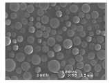
도 1은 본 발명의 바람직한 실시예에서 사용된 고분자 포로젠(Porogen)을 촬영한 사진이다.
도 2는 본 발명에서 사용되는 포로젠 시트를 제조하기 위한 장치의 모식도이다.
도 3은 본 발명의 일 실시예에 따라 열처리를 통하여 서로 연결된 포로젠 시트의 연결 구조를 촬영한 사진이다.
도 4는 본 발명의 성형체 제조 장치를 모식적으로 나타낸 도면이다.
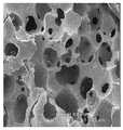
도 5는 본 발명의 일 실시예에 따라 제조된 연결 기공층을 갖는 시편의 표면 기공층 사진이다.
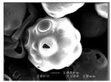
도 6은 본 발명과의 다른 실시예에 따라 제조된 비연결 기공층을 갖는 시편의 상면 및 측면 사진이다.
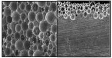
도 7은 본 발명의 다른 실시예에 따라 제조된 연결된 기공층을 갖는 시편의 사진이다.
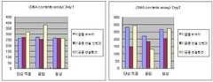
도 8은 본 발명의 다른 실시예에 따라 제조된 연결된 기공층 또는 고립된 기공층을 갖는 시편에서 hMSC세포의 부착능을 DNA contents assay로 측정한 결과이다.
도 9은 본 발명의 다른 실시예에 따라 제조된 연결된 기공층 또는 고립된 기공층을 갖는 시편에서 hMSC세포의 번식능을 DNA contents assay로 측정한 결과이다.
도 10은 본 발명의 다른 실시예에 따라 80 혹은 300 ㎛직경의 기공이 연결된 시편에서 hMSC세포의 골세포 분화 정도를 ALP및 Ca농도를 측정하여 평가한 결과이다.1 is a photograph of the polymer porogen (Porogen) used in the preferred embodiment of the present invention.
2 is a schematic diagram of an apparatus for producing a porogen sheet used in the present invention.
3 is a photograph of the connection structure of the porogen sheets connected to each other through heat treatment according to an embodiment of the present invention.
It is a figure which shows typically the molded object manufacturing apparatus of this invention.
5 is a photograph of the surface pore layer of a specimen having a connection pore layer prepared according to an embodiment of the present invention.
6 is a top and side view of a specimen having a non-connected pore layer prepared according to another embodiment of the present invention.
7 is a photograph of a specimen having a connected pore layer made in accordance with another embodiment of the present invention.
8 is a result of measuring the adhesion capacity of hMSC cells in a specimen having a connected or isolated pore layer prepared according to another embodiment of the present invention by DNA contents assay.
9 is a result of measuring the propagation ability of hMSC cells in a specimen having a connected or isolated pore layer prepared according to another embodiment of the present invention by DNA contents assay.
10 is a result of evaluating osteoblast differentiation of hMSC cells by measuring ALP and Ca concentration in specimens with pores of 80 or 300 μm diameter according to another embodiment of the present invention.
이하에서는 본 발명의 바람직한 실시예를 설명함으로써 본 발명을 상술한다.
Hereinafter, the present invention will be described in detail by explaining preferred embodiments of the present invention.
본 발명에서는 수산화아파타이트(Hydroxyapatite:(HAp, Ca10(PO4)6(OH)2))를 원료로 습식 공정을 적용하여 임플랜트를 제작한다.In the present invention, an implant is manufactured by applying a wet process using hydroxypatite ((HAp, Ca10 (PO4 )6 (OH)2 )) as a raw material.
습식 공정에 사용되는 수산화아파타이트 현탁액은 다음과 같은 방법에 의해 제조될 수 있다.Apatite hydroxide suspensions used in the wet process can be prepared by the following method.
먼저 물을 분산매로 수산화아파타이트와 다반씨(Darvan C)와 같은 분산제를 소정 함량 첨가한다. 현탁액의 제조에 사용되는 수산화아파타이트는 전체 용액의 체적을 기준으로 10~25 %의 범위가 바람직하다. 분산제의 함량은 전체 용액 체적 기준으로 1~5 % 포함되는 것이 바람직하다. 본 발명에서 분산제는 현탁액의 점도를 낮추면서 고체 분말의 분율을 높이기 위해 사용된다. 이와 같이 제조된 현탁액은 점도가 10 cP 이하를 갖게 된다. 본 발명에서 제조된 현탁액은 pH는 약 9~10의 범위에 있게 된다.First, a predetermined amount of a dispersant such as apatite hydroxide and Darvan C is added to the water as a dispersion medium. Apatite hydroxide used in the preparation of the suspension is preferably in the range of 10-25% based on the volume of the total solution. The content of the dispersant is preferably contained 1 to 5% based on the total solution volume. Dispersants in the present invention are used to increase the fraction of solid powder while lowering the viscosity of the suspension. The suspension thus prepared will have a viscosity of 10 cP or less. The suspension prepared in the present invention has a pH in the range of about 9-10.
이와 같이 혼합된 용액을 볼밀과 같은 통상의 분쇄 및 혼합 공정을 적용하여 현탁액을 제조하였다.The mixed solution was applied to a conventional milling and mixing process such as a ball mill to prepare a suspension.
다음으로, 기공 형성을 위한 고분자 소재로 구형의 형상을 갖는 포로젠(Porogen; Sephadex G-50 Sigma-Aldrich)으로 된 시트를 제조한다. 발명에서 표면 기공의 크기는 사용하는 포로젠의 입경에 따라 다양하게 조절할 수 있다. 본 발명에서 기공 형성을 위한 포로젠은 위 제품에 한정되지 않고 친수성을 가지며 현탁액 내에서 아파타이트와 잘 혼합되고 균일하게 분포될 수 있고, 후속 공정에서 열처리 또는 화학적 처리를 통해 제거될 수 있는 것이면 족하다.Next, a sheet made of porogen (Porogen; Sephadex G-50 Sigma-Aldrich) having a spherical shape is prepared as a polymer material for forming pores. In the present invention, the size of the surface pores can be variously adjusted according to the particle diameter of the porogen used. Porogens for pore formation in the present invention are not limited to the above products, but are hydrophilic and may be well mixed and uniformly distributed with the apatite in the suspension and removed by heat treatment or chemical treatment in subsequent processes.
도 2는 본 발명에서 사용될 포로젠 시트의 제조 방법을 개념적으로 도시한 도면이다.2 is a diagram conceptually illustrating a method of manufacturing a porogen sheet to be used in the present invention.
도시된 바와 같이, 슬라이드 글라스와 같은 구조물을 사용하여 격벽을 형성하고 그 사이에 포로젠을 투입한다. 포로젠을 투입한 후 포로젠이 서로 연결된 구조를 갖도록 오븐에서 열처리한다. 열처리 온도는 고분자 소재에 따라 다르나, 예컨대 200~300도의 온도에서 2시간 정도 처리하는 것이 바람직하다.As shown, a partition is formed using a structure such as slide glass, and porogen is introduced therebetween. After the porogen is added, the porogen is heat-treated in an oven so as to have a structure connected to each other. The heat treatment temperature varies depending on the polymer material, but for example, it is preferable to perform the treatment for about 2 hours at a temperature of 200 to 300 degrees.
도 3은 슬라이드 글라스 사이에 투입 후 열처리한 후 얻어진 포로젠을 촬영한 사진이다. 도시된 바와 같이, 포로젠의 구형 입자들이 인접하여 서로 연결된 구조를 가지고 있음을 알 수 있다.3 is a photograph of the porogen obtained after the heat treatment after the injection between the slide glass. As shown, it can be seen that the spherical particles of the porogen have a structure adjacent to each other.
전술한 방법으로 제조된 포로젠 시트를 사용하여 표면에 기공층이 형성된 아파타이트 임플랜트를 제조하는 방법은 다음과 같다.The method for producing an apatite implant having a pore layer formed on the surface using the porogen sheet manufactured by the above-described method is as follows.
임플랜트 성형체의 제조를 위해 도 4에 예시된 성형 장치를 사용할 수 있다. 도 4에 도시된 바와 같이, 예시된 성형 장치는 석고판 위에 고무와 같은 재질로 된 실린더 형의 몰드를 갖는 구조이다. 먼저, 몰드 내벽에 포로젠 시트를 삽입하고, 이어서 준비한 현탁액을 몰드 내부에 투입한다. 투입된 현탁액은 몰드 내부를 충진하고, 내벽의 포로젠 시트 내부로도 침투한다. 몰드 내부에 투입된 현탁액의 수분은 석고판으로 흡수되며, 그 결과 내부가 수산화 아파타이트 모재로 이루어지고 표면이 포로젠 시트로 덮히는 성형체가 얻어진다.The molding apparatus illustrated in FIG. 4 can be used for the production of implant shaped bodies. As shown in Fig. 4, the illustrated molding apparatus is a structure having a cylindrical mold of rubber-like material on a plasterboard. First, the porogen sheet is inserted into the mold inner wall, and the prepared suspension is then introduced into the mold. The charged suspension fills the inside of the mold and also penetrates into the porogen sheet on the inner wall. Moisture of the suspension introduced into the mold is absorbed by the gypsum board, and as a result, a molded body is obtained in which the inside is made of a hydroxide apatite base material and the surface is covered with a porogen sheet.
이어서, 이 성형체를 건조한 후에 1200 도와 같은 고온에서 2시간 소결하면 모재는 치밀한 상태로 가공되며, 포로젠은 제거된다. 포로젠이 제거된 표면에는 다공성 표면층이 얻어진다.Subsequently, after the molded product is dried and sintered at a high temperature such as 1200 degrees for 2 hours, the base material is processed in a dense state, and porogen is removed. On the surface from which porogen is removed, a porous surface layer is obtained.
본 발명의 방법이 적용되는 제조 장치는 전술한 것에 한하지 않는다. 예컨데, 포로젠 시트의 제조를 위해 격벽 구조를 구현하는 임의의 구조물이 슬라이드 글래스를 대신하여 사용될 수 있다. 또한, 본 발명의 성형 장치에서 석고를 대신하여 수분의 흡수를 위한 적절한 재질이 사용될 수 있고, 이를 대체하는 적절한 수분 배출 기구가 사용될 수 있다. 또한, 예시된 장치에서 석고 재질은 하부 플레이트가 아니라 몰드 측벽에 형성될 수도 있을 것이다.The manufacturing apparatus to which the method of the present invention is applied is not limited to the above. For example, any structure that implements a partition structure for the production of porogen sheets can be used in place of slide glass. In addition, in the molding apparatus of the present invention, a suitable material for absorbing moisture may be used in place of gypsum, and an appropriate water discharging mechanism may be used to replace it. Also, in the illustrated device the gypsum material may be formed on the mold sidewalls rather than on the bottom plate.
이상의 방법에 의해 제조된 임플랜트는 다음과 같은 장점을 갖는다. 먼저, 다공층의 두께는 슬라이드 글라스와 같은 격벽의 간격을 조절함으로써 적절히 조절될 수 있다. 다음으로, 다공층 내 기공의 크기는 사용하는 포로젠의 크기를 달리함으로써 조절할 수 있다. 또한, 포로젠 시트의 열처리 정도에 의해 포로젠의 연결상태를 제어할 수 있으며, 이에 의해 최종적으로 얻어지는 기공 채널의 연결 여부를 결정할 수 있다.Implants prepared by the above method have the following advantages. First, the thickness of the porous layer can be appropriately adjusted by adjusting the spacing of partition walls such as slide glass. Next, the size of the pores in the porous layer can be adjusted by varying the size of the porogen used. In addition, the connection state of the porogen can be controlled by the degree of heat treatment of the porogen sheet, thereby determining whether or not the pore channel is finally obtained.
이하 본 발명의 바람직한 실시예를 설명한다.
Hereinafter, preferred embodiments of the present invention will be described.
실시예 1Example 1
수산화아파타이트가 약 12 체적%가 되도록 현탁액을 제조하였다. 이 때, 분산제로 다반씨(Darvan C)는 용액 전체 체적을 기준으로 3 체적% 첨가하였다. 현탁액의 pH는 9.6~9.9 범위였고, 점도는 10 cP 이하였다.Suspensions were prepared such that the apatite hydroxide was about 12% by volume. At this time, Darvan C was added as a dispersant 3% by volume based on the total volume of the solution. The pH of the suspension ranged from 9.6 to 9.9 and the viscosity was 10 cP or less.
한편, 평균 직경이 100~300 미크론인 포로젠을 사용하여 230 도에서 2시간 열처리하여 포로젠 시트를 제조하였다.On the other hand, using a porogen having an average diameter of 100 ~ 300 microns was subjected to heat treatment at 230 degrees for 2 hours to prepare a porogen sheet.
제조된 포로젠 시트를 도 4의 장치를 이용하여 성형체를 제조한 후, 1200 도에서 2시간 소결하였다.The prepared porogen sheet was manufactured using the apparatus of FIG. 4, and then sintered at 1200 degrees for 2 hours.
도 5는 이와 같이 제조된 수산화아파타이트 임플랜트의 표면층을 촬영한 전자현미경 사진이다. 사진으로부터 표면층에 기공의 크기가 약 200 미크론이고 서로 연결된 기공 구조가 형성되었음을 알 수 있다.
5 is an electron microscope photograph of the surface layer of the apatite hydroxide implant prepared as described above. It can be seen from the photograph that the pore size is about 200 microns in the surface layer and the interconnected pore structures are formed.
실시예 2Example 2
실시예 1과 동일한 방법으로 임플랜트를 제조하되, 포로젠 시트 제조 과정에서 열처리 과정을 거치지 않았다. 도 6은 이 시편을 촬영한 전자현미경 사진이다. 기공의 크기는 같고 실시예1과 비슷하지만, 기공이 서로 연결되지 않은 상태임을 알 수 있다. 도 6에서 기공층은 2-3층 범위로 비교적 균일한 두께로 분포함을 알 수 있다.An implant was manufactured in the same manner as in Example 1, but the heat treatment was not performed during the preparation of the porogen sheet. 6 is an electron microscope photograph of this specimen. The pores are the same size and similar to Example 1, but it can be seen that the pores are not connected to each other. In Figure 6 it can be seen that the pore layer is distributed in a relatively uniform thickness in the 2-3 layer range.
실시예 3Example 3
실시예 1과 동일한 방법으로 임플랜트를 제조하되, 포로젠 시트의 제조시 평균 직경이 50~100 미크론인 포로젠을 사용하였다.An implant was prepared in the same manner as in Example 1, but in the preparation of the porogen sheet, porogen having an average diameter of 50 to 100 microns was used.
도 7은 본 실시예에 따라 제조된 임플랜트를 촬영한 사진이다.7 is a photograph of an implant manufactured according to the present embodiment.
도 7을 참조하면, 기공의 크기는 대략 50 ㎛로 원래 포로젠의 크기에 가까우며 기공들끼리 잘 연결된 상태에 있음을 확인할 수 있다.
Referring to Figure 7, the pore size is approximately 50 ㎛ close to the size of the original porogen and it can be seen that the pores are well connected.
실험예 4Experimental Example 4
실시예 1-3에 의해 제조된 수산화 아파타이트 임플랜트를 사용하여 hMSC 줄기세포에 대해 부착능, 번식능 및 골세포 분화능을 DNA contents assay 및 ALP와 Ca assay를 이용하여 테스트하였다. 실험은 각각 실시예1과 같이 연결 기공층이 형성된 임플랜트 시편, 실시예2와 같이 기공층 연결이 없는 임플랜트 시편, 그리고 대비를 위해 기공층이 없는 시편에 대해 행하였다.Using the hydroxide apatite implants prepared in Examples 1-3, the adhesion, propagation and osteoblast differentiation ability to hMSC stem cells were tested using DNA contents assay and ALP and Ca assay. Experiments were performed on implant specimens with connecting pore layers as in Example 1, implant specimens without pore layer connections as in Example 2, and specimens without pore layers for contrast.
각 시편에서 세포 담지 방법은 단순 직접법, 음압법, 원심력법을 적용하였고, 이 때 cell seeding 수는 2x10⁵로 하였다.For each specimen, the cell supporting method was simple direct method, negative pressure method, and centrifugal force method, and the cell seeding number was 2x10⁵ .
각 시편에 대해 각각 세포 부착율(Adhesion, DNA contents assay), 증식(Proliferaion, DNA contents assay), 골세포 분화(Differentiation, ALP assay/Ca+ assay)를 테스트 하여, 도 8 내지 도 10에 그 결과를 각각 나타내었다.Each specimen was tested for cell adhesion rate (Adhesion, DNA contents assay), proliferation (DNA contents assay), bone cell differentiation (Differentiation, ALP assay / Ca + assay), and the results are shown in FIGS. Respectively.
이 때, 세포 부착율 시험은 24 시간 인큐베이션 (ng/ μL) (n=2) 의 조건에서 행하였고, 증식 시험은 7일을 기준으로 (ng/ μL) (n=2)의 조건에서 행하였다. 골세포 분화 정도 측정은 14일을 기준으로 하였다.At this time, the cell adhesion rate test was performed under the conditions of 24 hours incubation (ng / μL) (n = 2), and the proliferation test was performed under the conditions of (ng / μL) (n = 2) based on 7 days. . Osteoblast differentiation was measured based on 14 days.
도 8에서 300 ㎛급의 기공층을 연결시킨 경우가 기공층이 없거나, 기공을 연결시키지 않은 경우에 비해 시편에 부착하는 비율이 배양접시에 비해 높아 효과적인 부착이 이루어 짐을 알 수 있었으며 단순직접법이나 음압법을 사용하는 경우 특히 부착특성이 우수한 것으로 나타났다.In FIG. 8, when the pore layer of 300 μm class is connected, the ratio of attaching to the specimen is higher than that of the culture dish compared to the case of no pore layer or no pore layer. In particular, the adhesion was found to be good when the method was used.
도 9에서는 부착 후 1일과 7일에 번식 정도를 DNA양으로부터 측정하였는데, 기공이 연결된 경우에 기공층이 연결 안 된 경우보다 상대적으로 DNA 함량이 높게 나타나 세포의 번식에 유리함을 보여주었다.
In Figure 9, the degree of reproduction was measured from the amount of DNA on the 1st and 7th day after attachment. When the pores are connected, the DNA content is higher than that of the case where the pore layer is not connected, which shows an advantage in cell propagation.
또한 도 10에서는 연결된 기공의 경우 기공의 크기에 따른 골세포 분화 정도를 배양 14일째 ALP와 Ca량을 측정하여 평가한 결과를 보여주며 300 ㎛ 직경의 기공인 경우가 80 ㎛ 직경의 기공에 비해 골세포 분화에 유리한 환경임을 보여준다.
In addition, in the case of connected pores, the degree of osteoblast differentiation according to the pore size was measured by measuring the amount of ALP and Ca on the 14th day of culture. It shows a favorable environment for cell differentiation.
Claims (5)
Translated fromKorean상기 시트를 몰드 내벽에 위치시키는 단계;
상기 몰드 내로 수산회인회석 현탁액을 주입하여 외부에 포로젠 시트가 부착된 성형체를 제조하는 단계; 및
성형된 성형체를 건조 및 소결하여 수산화아파타이트 임플랜트를 제조하는 단계를 포함하여,
수산화아파타이트 모재 표면에 포로젠 시트에 기인하는 표면 기공층이 형성된 것을 특징으로 하는 수산화아파타이트 임플랜트 제조 방법.Molding a porogen sheet consisting of spherical particles of a polymer material;
Positioning the sheet on a mold inner wall;
Preparing a molded article having a porogen sheet attached to the outside by injecting a hydroxyapatite suspension into the mold; And
Drying and sintering the molded body to produce an apatite hydroxide implant,
A method for producing an apatite hydroxide implant, characterized in that a surface pore layer derived from a porogen sheet is formed on the surface of the apatite hydroxide base material.
포로젠 시트 성형 단계는 격벽 사이의 간격에 의해 시트 두께를 조절하는 것을 특징으로 하는 수산화아파타이트 임플랜트 제조 방법.
The method of claim 1,
The porogen sheet forming step is characterized in that the sheet thickness is adjusted by the gap between the partition wall, the apatite hydroxide manufacturing method.
상기 몰드는 내벽의 최소한 일부가 석고로 구성되는 것을 특징으로 하는 수산화아파타이트 임플랜트 제조 방법.
The method of claim 1,
The mold is a method for producing an apatite hydroxide, characterized in that at least a portion of the inner wall is made of gypsum.
상기 몰드는 실린더형인 것을 특징으로 하는 수산화아파타이트 임플랜트 제조 방법.
The method of claim 1,
The mold is a method for producing an apatite hydroxide, characterized in that the cylindrical.
상기 포로젠 시트 성형 단계는 200~300도의 온도에서 열처리 하여 상기 포로젠 입자가 연결되도록 하는 것을 특징으로 하는 수산화아파타이트 임플랜트 제조 방법.
The method of claim 1,
The porogen sheet forming step is a heat treatment at a temperature of 200 ~ 300 degrees to make the porogen particles are connected, characterized in that the apatite hydroxide implant manufacturing method.
Priority Applications (1)
| Application Number | Priority Date | Filing Date | Title |
|---|---|---|---|
| KR1020100068919AKR101192687B1 (en) | 2010-07-16 | 2010-07-16 | An Hydroxy-apatite Implant having Porous Layer Continuously Combined with Substrate |
Applications Claiming Priority (1)
| Application Number | Priority Date | Filing Date | Title |
|---|---|---|---|
| KR1020100068919AKR101192687B1 (en) | 2010-07-16 | 2010-07-16 | An Hydroxy-apatite Implant having Porous Layer Continuously Combined with Substrate |
Publications (2)
| Publication Number | Publication Date |
|---|---|
| KR20120008198A KR20120008198A (en) | 2012-01-30 |
| KR101192687B1true KR101192687B1 (en) | 2012-10-19 |
Family
ID=45613254
Family Applications (1)
| Application Number | Title | Priority Date | Filing Date |
|---|---|---|---|
| KR1020100068919AActiveKR101192687B1 (en) | 2010-07-16 | 2010-07-16 | An Hydroxy-apatite Implant having Porous Layer Continuously Combined with Substrate |
Country Status (1)
| Country | Link |
|---|---|
| KR (1) | KR101192687B1 (en) |
Cited By (1)
| Publication number | Priority date | Publication date | Assignee | Title |
|---|---|---|---|---|
| KR20220033962A (en) | 2020-09-10 | 2022-03-17 | 건국대학교기술지주 주식회사 | Hydroxyapatite with Anti-viral or Anti-bacterial Activity and Preparation Method therefor |
Families Citing this family (1)
| Publication number | Priority date | Publication date | Assignee | Title |
|---|---|---|---|---|
| CN103818892B (en)* | 2014-01-06 | 2015-09-09 | 山东理工大学 | A kind of preparation method of submillimeter phosphogypsum ball |
Citations (1)
| Publication number | Priority date | Publication date | Assignee | Title |
|---|---|---|---|---|
| JP2005145755A (en) | 2003-11-14 | 2005-06-09 | National Institute Of Advanced Industrial & Technology | Method for producing high-strength porous apatite ceramics and product thereof |
- 2010
- 2010-07-16KRKR1020100068919Apatent/KR101192687B1/enactiveActive
Patent Citations (1)
| Publication number | Priority date | Publication date | Assignee | Title |
|---|---|---|---|---|
| JP2005145755A (en) | 2003-11-14 | 2005-06-09 | National Institute Of Advanced Industrial & Technology | Method for producing high-strength porous apatite ceramics and product thereof |
Cited By (1)
| Publication number | Priority date | Publication date | Assignee | Title |
|---|---|---|---|---|
| KR20220033962A (en) | 2020-09-10 | 2022-03-17 | 건국대학교기술지주 주식회사 | Hydroxyapatite with Anti-viral or Anti-bacterial Activity and Preparation Method therefor |
Also Published As
| Publication number | Publication date |
|---|---|
| KR20120008198A (en) | 2012-01-30 |
Similar Documents
| Publication | Publication Date | Title |
|---|---|---|
| Sous et al. | Cellular biocompatibility and resistance to compression of macroporous β-tricalcium phosphate ceramics | |
| Detsch et al. | In vitro-osteoclastic activity studies on surfaces of 3D printed calcium phosphate scaffolds | |
| US20250135426A1 (en) | Inorganic non-metallic nanoparticle-assembled hydrogel material and application thereof in additive manufacturing technology | |
| CN1183058C (en) | Artificial stabilized composition of calcium phosphate phases particularly adapted for supporting bone cell activity | |
| US7416564B2 (en) | Porous bioceramics for bone scaffold and method for manufacturing the same | |
| CN101518467A (en) | Medicinal porous titanium implant and method for preparing same | |
| Han et al. | Microstructure, mechanical properties and in vitro bioactivity of akermanite scaffolds fabricated by laser sintering | |
| CN108144113A (en) | A kind of porous bone repair material of bioactivity glass and preparation method thereof | |
| Liu et al. | Porous alumina ceramics prepared by slurry infiltration of expanded polystyrene beads | |
| CN112773939A (en) | Bone repair 3D printing material with low barium titanate content and preparation method and application thereof | |
| Bae et al. | Hydroxyapatite (HA) bone scaffolds with controlled macrochannel pores | |
| KR101192687B1 (en) | An Hydroxy-apatite Implant having Porous Layer Continuously Combined with Substrate | |
| Drevet et al. | Human osteoblast-like cells response to pulsed electrodeposited calcium phosphate coatings | |
| Kobatake et al. | Novel fabrication of porous titanium by a resin-impregnated titanium substitution technique for bone reconstruction | |
| US7687138B2 (en) | Porous calcium phosphate ceramic and method for producing same | |
| Liu et al. | Functionally graded triply periodic minimal surface scaffold of HA-Al2O3 via Vat photopolymerization 3D printing | |
| JP3718708B2 (en) | Calcium phosphate bioceramic sintered body and method for producing the same | |
| US20170100775A1 (en) | Method for manufacturing a porous metal material for biomedical applications and material obtained by said method | |
| SE1251044A1 (en) | Hard scaffold | |
| KR101091153B1 (en) | Preparation method of hydroxyapatite-chitosan composite coating layer using aerosol deposition and hydroxyapatite-chitosan composite coating layer with enhanced bioactivity | |
| KR100558157B1 (en) | Biotransplantable ceramic porous body and its manufacturing method | |
| CN113548890B (en) | Modified zirconia ceramic with high bioactivity and high mechanical strength and preparation method thereof | |
| KR101219871B1 (en) | Method for preparing porous strut of biomaterials and porous strut of biomaterials prepared thereby | |
| CN114366849A (en) | Bone repair material and preparation method and application thereof | |
| JP2002121088A (en) | Calcium phosphate porous sintered body and method for producing the same |
Legal Events
| Date | Code | Title | Description |
|---|---|---|---|
| A201 | Request for examination | ||
| PA0109 | Patent application | Patent event code:PA01091R01D Comment text:Patent Application Patent event date:20100716 | |
| PA0201 | Request for examination | ||
| PG1501 | Laying open of application | ||
| E701 | Decision to grant or registration of patent right | ||
| PE0701 | Decision of registration | Patent event code:PE07011S01D Comment text:Decision to Grant Registration Patent event date:20120712 | |
| GRNT | Written decision to grant | ||
| PR0701 | Registration of establishment | Comment text:Registration of Establishment Patent event date:20121012 Patent event code:PR07011E01D | |
| PR1002 | Payment of registration fee | Payment date:20121015 End annual number:3 Start annual number:1 | |
| PG1601 | Publication of registration | ||
| FPAY | Annual fee payment | Payment date:20160217 Year of fee payment:5 | |
| PR1001 | Payment of annual fee | Payment date:20160217 Start annual number:5 End annual number:5 | |
| FPAY | Annual fee payment | Payment date:20170925 Year of fee payment:6 | |
| PR1001 | Payment of annual fee | Payment date:20170925 Start annual number:6 End annual number:6 | |
| FPAY | Annual fee payment | Payment date:20181002 Year of fee payment:7 | |
| PR1001 | Payment of annual fee | Payment date:20181002 Start annual number:7 End annual number:7 | |
| FPAY | Annual fee payment | Payment date:20191001 Year of fee payment:8 | |
| PR1001 | Payment of annual fee | Payment date:20191001 Start annual number:8 End annual number:8 | |
| PR1001 | Payment of annual fee | Payment date:20201118 Start annual number:9 End annual number:9 | |
| PR1001 | Payment of annual fee | Payment date:20230921 Start annual number:12 End annual number:12 | |
| PR1001 | Payment of annual fee |