KR101169110B1 - Artificial spinal disc - Google Patents
Artificial spinal discDownload PDFInfo
- Publication number
- KR101169110B1 KR101169110B1KR1020077002424AKR20077002424AKR101169110B1KR 101169110 B1KR101169110 B1KR 101169110B1KR 1020077002424 AKR1020077002424 AKR 1020077002424AKR 20077002424 AKR20077002424 AKR 20077002424AKR 101169110 B1KR101169110 B1KR 101169110B1
- Authority
- KR
- South Korea
- Prior art keywords
- nucleus
- nucleus pulposus
- end plate
- support surface
- pulposus
- Prior art date
- Legal status (The legal status is an assumption and is not a legal conclusion. Google has not performed a legal analysis and makes no representation as to the accuracy of the status listed.)
- Expired - Lifetime
Links
Images


Classifications
- A—HUMAN NECESSITIES
- A61—MEDICAL OR VETERINARY SCIENCE; HYGIENE
- A61F—FILTERS IMPLANTABLE INTO BLOOD VESSELS; PROSTHESES; DEVICES PROVIDING PATENCY TO, OR PREVENTING COLLAPSING OF, TUBULAR STRUCTURES OF THE BODY, e.g. STENTS; ORTHOPAEDIC, NURSING OR CONTRACEPTIVE DEVICES; FOMENTATION; TREATMENT OR PROTECTION OF EYES OR EARS; BANDAGES, DRESSINGS OR ABSORBENT PADS; FIRST-AID KITS
- A61F2/00—Filters implantable into blood vessels; Prostheses, i.e. artificial substitutes or replacements for parts of the body; Appliances for connecting them with the body; Devices providing patency to, or preventing collapsing of, tubular structures of the body, e.g. stents
- A61F2/02—Prostheses implantable into the body
- A61F2/30—Joints
- A61F2/44—Joints for the spine, e.g. vertebrae, spinal discs
- A61F2/442—Intervertebral or spinal discs, e.g. resilient
- A61F2/4425—Intervertebral or spinal discs, e.g. resilient made of articulated components
- A—HUMAN NECESSITIES
- A61—MEDICAL OR VETERINARY SCIENCE; HYGIENE
- A61F—FILTERS IMPLANTABLE INTO BLOOD VESSELS; PROSTHESES; DEVICES PROVIDING PATENCY TO, OR PREVENTING COLLAPSING OF, TUBULAR STRUCTURES OF THE BODY, e.g. STENTS; ORTHOPAEDIC, NURSING OR CONTRACEPTIVE DEVICES; FOMENTATION; TREATMENT OR PROTECTION OF EYES OR EARS; BANDAGES, DRESSINGS OR ABSORBENT PADS; FIRST-AID KITS
- A61F2/00—Filters implantable into blood vessels; Prostheses, i.e. artificial substitutes or replacements for parts of the body; Appliances for connecting them with the body; Devices providing patency to, or preventing collapsing of, tubular structures of the body, e.g. stents
- A61F2/02—Prostheses implantable into the body
- A61F2/30—Joints
- A61F2/44—Joints for the spine, e.g. vertebrae, spinal discs
- A—HUMAN NECESSITIES
- A61—MEDICAL OR VETERINARY SCIENCE; HYGIENE
- A61F—FILTERS IMPLANTABLE INTO BLOOD VESSELS; PROSTHESES; DEVICES PROVIDING PATENCY TO, OR PREVENTING COLLAPSING OF, TUBULAR STRUCTURES OF THE BODY, e.g. STENTS; ORTHOPAEDIC, NURSING OR CONTRACEPTIVE DEVICES; FOMENTATION; TREATMENT OR PROTECTION OF EYES OR EARS; BANDAGES, DRESSINGS OR ABSORBENT PADS; FIRST-AID KITS
- A61F2/00—Filters implantable into blood vessels; Prostheses, i.e. artificial substitutes or replacements for parts of the body; Appliances for connecting them with the body; Devices providing patency to, or preventing collapsing of, tubular structures of the body, e.g. stents
- A61F2/02—Prostheses implantable into the body
- A61F2/30—Joints
- A61F2/46—Special tools for implanting artificial joints
- A61F2/4657—Measuring instruments used for implanting artificial joints
- A—HUMAN NECESSITIES
- A61—MEDICAL OR VETERINARY SCIENCE; HYGIENE
- A61F—FILTERS IMPLANTABLE INTO BLOOD VESSELS; PROSTHESES; DEVICES PROVIDING PATENCY TO, OR PREVENTING COLLAPSING OF, TUBULAR STRUCTURES OF THE BODY, e.g. STENTS; ORTHOPAEDIC, NURSING OR CONTRACEPTIVE DEVICES; FOMENTATION; TREATMENT OR PROTECTION OF EYES OR EARS; BANDAGES, DRESSINGS OR ABSORBENT PADS; FIRST-AID KITS
- A61F2/00—Filters implantable into blood vessels; Prostheses, i.e. artificial substitutes or replacements for parts of the body; Appliances for connecting them with the body; Devices providing patency to, or preventing collapsing of, tubular structures of the body, e.g. stents
- A61F2/02—Prostheses implantable into the body
- A61F2/30—Joints
- A61F2/46—Special tools for implanting artificial joints
- A61F2/4684—Trial or dummy prostheses
- A—HUMAN NECESSITIES
- A61—MEDICAL OR VETERINARY SCIENCE; HYGIENE
- A61F—FILTERS IMPLANTABLE INTO BLOOD VESSELS; PROSTHESES; DEVICES PROVIDING PATENCY TO, OR PREVENTING COLLAPSING OF, TUBULAR STRUCTURES OF THE BODY, e.g. STENTS; ORTHOPAEDIC, NURSING OR CONTRACEPTIVE DEVICES; FOMENTATION; TREATMENT OR PROTECTION OF EYES OR EARS; BANDAGES, DRESSINGS OR ABSORBENT PADS; FIRST-AID KITS
- A61F2/00—Filters implantable into blood vessels; Prostheses, i.e. artificial substitutes or replacements for parts of the body; Appliances for connecting them with the body; Devices providing patency to, or preventing collapsing of, tubular structures of the body, e.g. stents
- A61F2/02—Prostheses implantable into the body
- A61F2/30—Joints
- A61F2/46—Special tools for implanting artificial joints
- A61F2/4603—Special tools for implanting artificial joints for insertion or extraction of endoprosthetic joints or of accessories thereof
- A61F2/4611—Special tools for implanting artificial joints for insertion or extraction of endoprosthetic joints or of accessories thereof of spinal prostheses
- A—HUMAN NECESSITIES
- A61—MEDICAL OR VETERINARY SCIENCE; HYGIENE
- A61F—FILTERS IMPLANTABLE INTO BLOOD VESSELS; PROSTHESES; DEVICES PROVIDING PATENCY TO, OR PREVENTING COLLAPSING OF, TUBULAR STRUCTURES OF THE BODY, e.g. STENTS; ORTHOPAEDIC, NURSING OR CONTRACEPTIVE DEVICES; FOMENTATION; TREATMENT OR PROTECTION OF EYES OR EARS; BANDAGES, DRESSINGS OR ABSORBENT PADS; FIRST-AID KITS
- A61F2/00—Filters implantable into blood vessels; Prostheses, i.e. artificial substitutes or replacements for parts of the body; Appliances for connecting them with the body; Devices providing patency to, or preventing collapsing of, tubular structures of the body, e.g. stents
- A61F2/02—Prostheses implantable into the body
- A61F2/30—Joints
- A61F2002/30001—Additional features of subject-matter classified in A61F2/28, A61F2/30 and subgroups thereof
- A61F2002/30108—Shapes
- A61F2002/30199—Three-dimensional shapes
- A61F2002/302—Three-dimensional shapes toroidal, e.g. rings
- A—HUMAN NECESSITIES
- A61—MEDICAL OR VETERINARY SCIENCE; HYGIENE
- A61F—FILTERS IMPLANTABLE INTO BLOOD VESSELS; PROSTHESES; DEVICES PROVIDING PATENCY TO, OR PREVENTING COLLAPSING OF, TUBULAR STRUCTURES OF THE BODY, e.g. STENTS; ORTHOPAEDIC, NURSING OR CONTRACEPTIVE DEVICES; FOMENTATION; TREATMENT OR PROTECTION OF EYES OR EARS; BANDAGES, DRESSINGS OR ABSORBENT PADS; FIRST-AID KITS
- A61F2/00—Filters implantable into blood vessels; Prostheses, i.e. artificial substitutes or replacements for parts of the body; Appliances for connecting them with the body; Devices providing patency to, or preventing collapsing of, tubular structures of the body, e.g. stents
- A61F2/02—Prostheses implantable into the body
- A61F2/30—Joints
- A61F2002/30001—Additional features of subject-matter classified in A61F2/28, A61F2/30 and subgroups thereof
- A61F2002/30108—Shapes
- A61F2002/30199—Three-dimensional shapes
- A61F2002/30224—Three-dimensional shapes cylindrical
- A61F2002/30232—Half-cylinders
- A—HUMAN NECESSITIES
- A61—MEDICAL OR VETERINARY SCIENCE; HYGIENE
- A61F—FILTERS IMPLANTABLE INTO BLOOD VESSELS; PROSTHESES; DEVICES PROVIDING PATENCY TO, OR PREVENTING COLLAPSING OF, TUBULAR STRUCTURES OF THE BODY, e.g. STENTS; ORTHOPAEDIC, NURSING OR CONTRACEPTIVE DEVICES; FOMENTATION; TREATMENT OR PROTECTION OF EYES OR EARS; BANDAGES, DRESSINGS OR ABSORBENT PADS; FIRST-AID KITS
- A61F2/00—Filters implantable into blood vessels; Prostheses, i.e. artificial substitutes or replacements for parts of the body; Appliances for connecting them with the body; Devices providing patency to, or preventing collapsing of, tubular structures of the body, e.g. stents
- A61F2/02—Prostheses implantable into the body
- A61F2/30—Joints
- A61F2002/30001—Additional features of subject-matter classified in A61F2/28, A61F2/30 and subgroups thereof
- A61F2002/30108—Shapes
- A61F2002/30199—Three-dimensional shapes
- A61F2002/30242—Three-dimensional shapes spherical
- A—HUMAN NECESSITIES
- A61—MEDICAL OR VETERINARY SCIENCE; HYGIENE
- A61F—FILTERS IMPLANTABLE INTO BLOOD VESSELS; PROSTHESES; DEVICES PROVIDING PATENCY TO, OR PREVENTING COLLAPSING OF, TUBULAR STRUCTURES OF THE BODY, e.g. STENTS; ORTHOPAEDIC, NURSING OR CONTRACEPTIVE DEVICES; FOMENTATION; TREATMENT OR PROTECTION OF EYES OR EARS; BANDAGES, DRESSINGS OR ABSORBENT PADS; FIRST-AID KITS
- A61F2/00—Filters implantable into blood vessels; Prostheses, i.e. artificial substitutes or replacements for parts of the body; Appliances for connecting them with the body; Devices providing patency to, or preventing collapsing of, tubular structures of the body, e.g. stents
- A61F2/02—Prostheses implantable into the body
- A61F2/30—Joints
- A61F2002/30001—Additional features of subject-matter classified in A61F2/28, A61F2/30 and subgroups thereof
- A61F2002/30316—The prosthesis having different structural features at different locations within the same prosthesis; Connections between prosthetic parts; Special structural features of bone or joint prostheses not otherwise provided for
- A61F2002/30329—Connections or couplings between prosthetic parts, e.g. between modular parts; Connecting elements
- A61F2002/30331—Connections or couplings between prosthetic parts, e.g. between modular parts; Connecting elements made by longitudinally pushing a protrusion into a complementarily-shaped recess, e.g. held by friction fit
- A61F2002/30354—Cylindrically-shaped protrusion and recess, e.g. cylinder of circular basis
- A—HUMAN NECESSITIES
- A61—MEDICAL OR VETERINARY SCIENCE; HYGIENE
- A61F—FILTERS IMPLANTABLE INTO BLOOD VESSELS; PROSTHESES; DEVICES PROVIDING PATENCY TO, OR PREVENTING COLLAPSING OF, TUBULAR STRUCTURES OF THE BODY, e.g. STENTS; ORTHOPAEDIC, NURSING OR CONTRACEPTIVE DEVICES; FOMENTATION; TREATMENT OR PROTECTION OF EYES OR EARS; BANDAGES, DRESSINGS OR ABSORBENT PADS; FIRST-AID KITS
- A61F2/00—Filters implantable into blood vessels; Prostheses, i.e. artificial substitutes or replacements for parts of the body; Appliances for connecting them with the body; Devices providing patency to, or preventing collapsing of, tubular structures of the body, e.g. stents
- A61F2/02—Prostheses implantable into the body
- A61F2/30—Joints
- A61F2002/30001—Additional features of subject-matter classified in A61F2/28, A61F2/30 and subgroups thereof
- A61F2002/30316—The prosthesis having different structural features at different locations within the same prosthesis; Connections between prosthetic parts; Special structural features of bone or joint prostheses not otherwise provided for
- A61F2002/30329—Connections or couplings between prosthetic parts, e.g. between modular parts; Connecting elements
- A61F2002/30331—Connections or couplings between prosthetic parts, e.g. between modular parts; Connecting elements made by longitudinally pushing a protrusion into a complementarily-shaped recess, e.g. held by friction fit
- A61F2002/30362—Connections or couplings between prosthetic parts, e.g. between modular parts; Connecting elements made by longitudinally pushing a protrusion into a complementarily-shaped recess, e.g. held by friction fit with possibility of relative movement between the protrusion and the recess
- A61F2002/30364—Rotation about the common longitudinal axis
- A—HUMAN NECESSITIES
- A61—MEDICAL OR VETERINARY SCIENCE; HYGIENE
- A61F—FILTERS IMPLANTABLE INTO BLOOD VESSELS; PROSTHESES; DEVICES PROVIDING PATENCY TO, OR PREVENTING COLLAPSING OF, TUBULAR STRUCTURES OF THE BODY, e.g. STENTS; ORTHOPAEDIC, NURSING OR CONTRACEPTIVE DEVICES; FOMENTATION; TREATMENT OR PROTECTION OF EYES OR EARS; BANDAGES, DRESSINGS OR ABSORBENT PADS; FIRST-AID KITS
- A61F2/00—Filters implantable into blood vessels; Prostheses, i.e. artificial substitutes or replacements for parts of the body; Appliances for connecting them with the body; Devices providing patency to, or preventing collapsing of, tubular structures of the body, e.g. stents
- A61F2/02—Prostheses implantable into the body
- A61F2/30—Joints
- A61F2002/30001—Additional features of subject-matter classified in A61F2/28, A61F2/30 and subgroups thereof
- A61F2002/30316—The prosthesis having different structural features at different locations within the same prosthesis; Connections between prosthetic parts; Special structural features of bone or joint prostheses not otherwise provided for
- A61F2002/30329—Connections or couplings between prosthetic parts, e.g. between modular parts; Connecting elements
- A61F2002/30331—Connections or couplings between prosthetic parts, e.g. between modular parts; Connecting elements made by longitudinally pushing a protrusion into a complementarily-shaped recess, e.g. held by friction fit
- A61F2002/30362—Connections or couplings between prosthetic parts, e.g. between modular parts; Connecting elements made by longitudinally pushing a protrusion into a complementarily-shaped recess, e.g. held by friction fit with possibility of relative movement between the protrusion and the recess
- A61F2002/30369—Limited lateral translation of the protrusion within a larger recess
- A—HUMAN NECESSITIES
- A61—MEDICAL OR VETERINARY SCIENCE; HYGIENE
- A61F—FILTERS IMPLANTABLE INTO BLOOD VESSELS; PROSTHESES; DEVICES PROVIDING PATENCY TO, OR PREVENTING COLLAPSING OF, TUBULAR STRUCTURES OF THE BODY, e.g. STENTS; ORTHOPAEDIC, NURSING OR CONTRACEPTIVE DEVICES; FOMENTATION; TREATMENT OR PROTECTION OF EYES OR EARS; BANDAGES, DRESSINGS OR ABSORBENT PADS; FIRST-AID KITS
- A61F2/00—Filters implantable into blood vessels; Prostheses, i.e. artificial substitutes or replacements for parts of the body; Appliances for connecting them with the body; Devices providing patency to, or preventing collapsing of, tubular structures of the body, e.g. stents
- A61F2/02—Prostheses implantable into the body
- A61F2/30—Joints
- A61F2002/30001—Additional features of subject-matter classified in A61F2/28, A61F2/30 and subgroups thereof
- A61F2002/30316—The prosthesis having different structural features at different locations within the same prosthesis; Connections between prosthetic parts; Special structural features of bone or joint prostheses not otherwise provided for
- A61F2002/30329—Connections or couplings between prosthetic parts, e.g. between modular parts; Connecting elements
- A61F2002/30383—Connections or couplings between prosthetic parts, e.g. between modular parts; Connecting elements made by laterally inserting a protrusion, e.g. a rib into a complementarily-shaped groove
- A61F2002/3039—Connections or couplings between prosthetic parts, e.g. between modular parts; Connecting elements made by laterally inserting a protrusion, e.g. a rib into a complementarily-shaped groove with possibility of relative movement of the rib within the groove
- A61F2002/30397—Limited lateral translation of the rib within a larger groove
- A—HUMAN NECESSITIES
- A61—MEDICAL OR VETERINARY SCIENCE; HYGIENE
- A61F—FILTERS IMPLANTABLE INTO BLOOD VESSELS; PROSTHESES; DEVICES PROVIDING PATENCY TO, OR PREVENTING COLLAPSING OF, TUBULAR STRUCTURES OF THE BODY, e.g. STENTS; ORTHOPAEDIC, NURSING OR CONTRACEPTIVE DEVICES; FOMENTATION; TREATMENT OR PROTECTION OF EYES OR EARS; BANDAGES, DRESSINGS OR ABSORBENT PADS; FIRST-AID KITS
- A61F2/00—Filters implantable into blood vessels; Prostheses, i.e. artificial substitutes or replacements for parts of the body; Appliances for connecting them with the body; Devices providing patency to, or preventing collapsing of, tubular structures of the body, e.g. stents
- A61F2/02—Prostheses implantable into the body
- A61F2/30—Joints
- A61F2002/30001—Additional features of subject-matter classified in A61F2/28, A61F2/30 and subgroups thereof
- A61F2002/30316—The prosthesis having different structural features at different locations within the same prosthesis; Connections between prosthetic parts; Special structural features of bone or joint prostheses not otherwise provided for
- A61F2002/30329—Connections or couplings between prosthetic parts, e.g. between modular parts; Connecting elements
- A61F2002/30426—Bayonet coupling
- A—HUMAN NECESSITIES
- A61—MEDICAL OR VETERINARY SCIENCE; HYGIENE
- A61F—FILTERS IMPLANTABLE INTO BLOOD VESSELS; PROSTHESES; DEVICES PROVIDING PATENCY TO, OR PREVENTING COLLAPSING OF, TUBULAR STRUCTURES OF THE BODY, e.g. STENTS; ORTHOPAEDIC, NURSING OR CONTRACEPTIVE DEVICES; FOMENTATION; TREATMENT OR PROTECTION OF EYES OR EARS; BANDAGES, DRESSINGS OR ABSORBENT PADS; FIRST-AID KITS
- A61F2/00—Filters implantable into blood vessels; Prostheses, i.e. artificial substitutes or replacements for parts of the body; Appliances for connecting them with the body; Devices providing patency to, or preventing collapsing of, tubular structures of the body, e.g. stents
- A61F2/02—Prostheses implantable into the body
- A61F2/30—Joints
- A61F2002/30001—Additional features of subject-matter classified in A61F2/28, A61F2/30 and subgroups thereof
- A61F2002/30316—The prosthesis having different structural features at different locations within the same prosthesis; Connections between prosthetic parts; Special structural features of bone or joint prostheses not otherwise provided for
- A61F2002/30329—Connections or couplings between prosthetic parts, e.g. between modular parts; Connecting elements
- A61F2002/30476—Connections or couplings between prosthetic parts, e.g. between modular parts; Connecting elements locked by an additional locking mechanism
- A61F2002/305—Snap connection
- A—HUMAN NECESSITIES
- A61—MEDICAL OR VETERINARY SCIENCE; HYGIENE
- A61F—FILTERS IMPLANTABLE INTO BLOOD VESSELS; PROSTHESES; DEVICES PROVIDING PATENCY TO, OR PREVENTING COLLAPSING OF, TUBULAR STRUCTURES OF THE BODY, e.g. STENTS; ORTHOPAEDIC, NURSING OR CONTRACEPTIVE DEVICES; FOMENTATION; TREATMENT OR PROTECTION OF EYES OR EARS; BANDAGES, DRESSINGS OR ABSORBENT PADS; FIRST-AID KITS
- A61F2/00—Filters implantable into blood vessels; Prostheses, i.e. artificial substitutes or replacements for parts of the body; Appliances for connecting them with the body; Devices providing patency to, or preventing collapsing of, tubular structures of the body, e.g. stents
- A61F2/02—Prostheses implantable into the body
- A61F2/30—Joints
- A61F2002/30001—Additional features of subject-matter classified in A61F2/28, A61F2/30 and subgroups thereof
- A61F2002/30316—The prosthesis having different structural features at different locations within the same prosthesis; Connections between prosthetic parts; Special structural features of bone or joint prostheses not otherwise provided for
- A61F2002/30535—Special structural features of bone or joint prostheses not otherwise provided for
- A61F2002/30537—Special structural features of bone or joint prostheses not otherwise provided for adjustable
- A61F2002/30538—Special structural features of bone or joint prostheses not otherwise provided for adjustable for adjusting angular orientation
- A—HUMAN NECESSITIES
- A61—MEDICAL OR VETERINARY SCIENCE; HYGIENE
- A61F—FILTERS IMPLANTABLE INTO BLOOD VESSELS; PROSTHESES; DEVICES PROVIDING PATENCY TO, OR PREVENTING COLLAPSING OF, TUBULAR STRUCTURES OF THE BODY, e.g. STENTS; ORTHOPAEDIC, NURSING OR CONTRACEPTIVE DEVICES; FOMENTATION; TREATMENT OR PROTECTION OF EYES OR EARS; BANDAGES, DRESSINGS OR ABSORBENT PADS; FIRST-AID KITS
- A61F2/00—Filters implantable into blood vessels; Prostheses, i.e. artificial substitutes or replacements for parts of the body; Appliances for connecting them with the body; Devices providing patency to, or preventing collapsing of, tubular structures of the body, e.g. stents
- A61F2/02—Prostheses implantable into the body
- A61F2/30—Joints
- A61F2002/30001—Additional features of subject-matter classified in A61F2/28, A61F2/30 and subgroups thereof
- A61F2002/30316—The prosthesis having different structural features at different locations within the same prosthesis; Connections between prosthetic parts; Special structural features of bone or joint prostheses not otherwise provided for
- A61F2002/30535—Special structural features of bone or joint prostheses not otherwise provided for
- A61F2002/30563—Special structural features of bone or joint prostheses not otherwise provided for having elastic means or damping means, different from springs, e.g. including an elastomeric core or shock absorbers
- A—HUMAN NECESSITIES
- A61—MEDICAL OR VETERINARY SCIENCE; HYGIENE
- A61F—FILTERS IMPLANTABLE INTO BLOOD VESSELS; PROSTHESES; DEVICES PROVIDING PATENCY TO, OR PREVENTING COLLAPSING OF, TUBULAR STRUCTURES OF THE BODY, e.g. STENTS; ORTHOPAEDIC, NURSING OR CONTRACEPTIVE DEVICES; FOMENTATION; TREATMENT OR PROTECTION OF EYES OR EARS; BANDAGES, DRESSINGS OR ABSORBENT PADS; FIRST-AID KITS
- A61F2/00—Filters implantable into blood vessels; Prostheses, i.e. artificial substitutes or replacements for parts of the body; Appliances for connecting them with the body; Devices providing patency to, or preventing collapsing of, tubular structures of the body, e.g. stents
- A61F2/02—Prostheses implantable into the body
- A61F2/30—Joints
- A61F2002/30001—Additional features of subject-matter classified in A61F2/28, A61F2/30 and subgroups thereof
- A61F2002/30316—The prosthesis having different structural features at different locations within the same prosthesis; Connections between prosthetic parts; Special structural features of bone or joint prostheses not otherwise provided for
- A61F2002/30535—Special structural features of bone or joint prostheses not otherwise provided for
- A61F2002/30576—Special structural features of bone or joint prostheses not otherwise provided for with extending fixation tabs
- A61F2002/30578—Special structural features of bone or joint prostheses not otherwise provided for with extending fixation tabs having apertures, e.g. for receiving fixation screws
- A—HUMAN NECESSITIES
- A61—MEDICAL OR VETERINARY SCIENCE; HYGIENE
- A61F—FILTERS IMPLANTABLE INTO BLOOD VESSELS; PROSTHESES; DEVICES PROVIDING PATENCY TO, OR PREVENTING COLLAPSING OF, TUBULAR STRUCTURES OF THE BODY, e.g. STENTS; ORTHOPAEDIC, NURSING OR CONTRACEPTIVE DEVICES; FOMENTATION; TREATMENT OR PROTECTION OF EYES OR EARS; BANDAGES, DRESSINGS OR ABSORBENT PADS; FIRST-AID KITS
- A61F2/00—Filters implantable into blood vessels; Prostheses, i.e. artificial substitutes or replacements for parts of the body; Appliances for connecting them with the body; Devices providing patency to, or preventing collapsing of, tubular structures of the body, e.g. stents
- A61F2/02—Prostheses implantable into the body
- A61F2/30—Joints
- A61F2002/30001—Additional features of subject-matter classified in A61F2/28, A61F2/30 and subgroups thereof
- A61F2002/30316—The prosthesis having different structural features at different locations within the same prosthesis; Connections between prosthetic parts; Special structural features of bone or joint prostheses not otherwise provided for
- A61F2002/30535—Special structural features of bone or joint prostheses not otherwise provided for
- A61F2002/30604—Special structural features of bone or joint prostheses not otherwise provided for modular
- A61F2002/30616—Sets comprising a plurality of prosthetic parts of different sizes or orientations
- A—HUMAN NECESSITIES
- A61—MEDICAL OR VETERINARY SCIENCE; HYGIENE
- A61F—FILTERS IMPLANTABLE INTO BLOOD VESSELS; PROSTHESES; DEVICES PROVIDING PATENCY TO, OR PREVENTING COLLAPSING OF, TUBULAR STRUCTURES OF THE BODY, e.g. STENTS; ORTHOPAEDIC, NURSING OR CONTRACEPTIVE DEVICES; FOMENTATION; TREATMENT OR PROTECTION OF EYES OR EARS; BANDAGES, DRESSINGS OR ABSORBENT PADS; FIRST-AID KITS
- A61F2/00—Filters implantable into blood vessels; Prostheses, i.e. artificial substitutes or replacements for parts of the body; Appliances for connecting them with the body; Devices providing patency to, or preventing collapsing of, tubular structures of the body, e.g. stents
- A61F2/02—Prostheses implantable into the body
- A61F2/30—Joints
- A61F2002/30001—Additional features of subject-matter classified in A61F2/28, A61F2/30 and subgroups thereof
- A61F2002/30621—Features concerning the anatomical functioning or articulation of the prosthetic joint
- A61F2002/30649—Ball-and-socket joints
- A61F2002/3065—Details of the ball-shaped head
- A—HUMAN NECESSITIES
- A61—MEDICAL OR VETERINARY SCIENCE; HYGIENE
- A61F—FILTERS IMPLANTABLE INTO BLOOD VESSELS; PROSTHESES; DEVICES PROVIDING PATENCY TO, OR PREVENTING COLLAPSING OF, TUBULAR STRUCTURES OF THE BODY, e.g. STENTS; ORTHOPAEDIC, NURSING OR CONTRACEPTIVE DEVICES; FOMENTATION; TREATMENT OR PROTECTION OF EYES OR EARS; BANDAGES, DRESSINGS OR ABSORBENT PADS; FIRST-AID KITS
- A61F2/00—Filters implantable into blood vessels; Prostheses, i.e. artificial substitutes or replacements for parts of the body; Appliances for connecting them with the body; Devices providing patency to, or preventing collapsing of, tubular structures of the body, e.g. stents
- A61F2/02—Prostheses implantable into the body
- A61F2/30—Joints
- A61F2002/30001—Additional features of subject-matter classified in A61F2/28, A61F2/30 and subgroups thereof
- A61F2002/30621—Features concerning the anatomical functioning or articulation of the prosthetic joint
- A61F2002/30649—Ball-and-socket joints
- A61F2002/30654—Details of the concave socket
- A—HUMAN NECESSITIES
- A61—MEDICAL OR VETERINARY SCIENCE; HYGIENE
- A61F—FILTERS IMPLANTABLE INTO BLOOD VESSELS; PROSTHESES; DEVICES PROVIDING PATENCY TO, OR PREVENTING COLLAPSING OF, TUBULAR STRUCTURES OF THE BODY, e.g. STENTS; ORTHOPAEDIC, NURSING OR CONTRACEPTIVE DEVICES; FOMENTATION; TREATMENT OR PROTECTION OF EYES OR EARS; BANDAGES, DRESSINGS OR ABSORBENT PADS; FIRST-AID KITS
- A61F2/00—Filters implantable into blood vessels; Prostheses, i.e. artificial substitutes or replacements for parts of the body; Appliances for connecting them with the body; Devices providing patency to, or preventing collapsing of, tubular structures of the body, e.g. stents
- A61F2/02—Prostheses implantable into the body
- A61F2/30—Joints
- A61F2002/30001—Additional features of subject-matter classified in A61F2/28, A61F2/30 and subgroups thereof
- A61F2002/30621—Features concerning the anatomical functioning or articulation of the prosthetic joint
- A61F2002/30649—Ball-and-socket joints
- A61F2002/30662—Ball-and-socket joints with rotation-limiting means
- A—HUMAN NECESSITIES
- A61—MEDICAL OR VETERINARY SCIENCE; HYGIENE
- A61F—FILTERS IMPLANTABLE INTO BLOOD VESSELS; PROSTHESES; DEVICES PROVIDING PATENCY TO, OR PREVENTING COLLAPSING OF, TUBULAR STRUCTURES OF THE BODY, e.g. STENTS; ORTHOPAEDIC, NURSING OR CONTRACEPTIVE DEVICES; FOMENTATION; TREATMENT OR PROTECTION OF EYES OR EARS; BANDAGES, DRESSINGS OR ABSORBENT PADS; FIRST-AID KITS
- A61F2/00—Filters implantable into blood vessels; Prostheses, i.e. artificial substitutes or replacements for parts of the body; Appliances for connecting them with the body; Devices providing patency to, or preventing collapsing of, tubular structures of the body, e.g. stents
- A61F2/02—Prostheses implantable into the body
- A61F2/30—Joints
- A61F2002/30001—Additional features of subject-matter classified in A61F2/28, A61F2/30 and subgroups thereof
- A61F2002/30621—Features concerning the anatomical functioning or articulation of the prosthetic joint
- A61F2002/30649—Ball-and-socket joints
- A61F2002/30663—Ball-and-socket joints multiaxial, e.g. biaxial; multipolar, e.g. bipolar or having an intermediate shell articulating between the ball and the socket
- A—HUMAN NECESSITIES
- A61—MEDICAL OR VETERINARY SCIENCE; HYGIENE
- A61F—FILTERS IMPLANTABLE INTO BLOOD VESSELS; PROSTHESES; DEVICES PROVIDING PATENCY TO, OR PREVENTING COLLAPSING OF, TUBULAR STRUCTURES OF THE BODY, e.g. STENTS; ORTHOPAEDIC, NURSING OR CONTRACEPTIVE DEVICES; FOMENTATION; TREATMENT OR PROTECTION OF EYES OR EARS; BANDAGES, DRESSINGS OR ABSORBENT PADS; FIRST-AID KITS
- A61F2/00—Filters implantable into blood vessels; Prostheses, i.e. artificial substitutes or replacements for parts of the body; Appliances for connecting them with the body; Devices providing patency to, or preventing collapsing of, tubular structures of the body, e.g. stents
- A61F2/02—Prostheses implantable into the body
- A61F2/30—Joints
- A61F2002/30001—Additional features of subject-matter classified in A61F2/28, A61F2/30 and subgroups thereof
- A61F2002/30667—Features concerning an interaction with the environment or a particular use of the prosthesis
- A61F2002/30682—Means for preventing migration of particles released by the joint, e.g. wear debris or cement particles
- A61F2002/30685—Means for reducing or preventing the generation of wear particulates
- A—HUMAN NECESSITIES
- A61—MEDICAL OR VETERINARY SCIENCE; HYGIENE
- A61F—FILTERS IMPLANTABLE INTO BLOOD VESSELS; PROSTHESES; DEVICES PROVIDING PATENCY TO, OR PREVENTING COLLAPSING OF, TUBULAR STRUCTURES OF THE BODY, e.g. STENTS; ORTHOPAEDIC, NURSING OR CONTRACEPTIVE DEVICES; FOMENTATION; TREATMENT OR PROTECTION OF EYES OR EARS; BANDAGES, DRESSINGS OR ABSORBENT PADS; FIRST-AID KITS
- A61F2/00—Filters implantable into blood vessels; Prostheses, i.e. artificial substitutes or replacements for parts of the body; Appliances for connecting them with the body; Devices providing patency to, or preventing collapsing of, tubular structures of the body, e.g. stents
- A61F2/02—Prostheses implantable into the body
- A61F2/30—Joints
- A61F2002/30001—Additional features of subject-matter classified in A61F2/28, A61F2/30 and subgroups thereof
- A61F2002/30667—Features concerning an interaction with the environment or a particular use of the prosthesis
- A61F2002/3069—Revision endoprostheses
- A—HUMAN NECESSITIES
- A61—MEDICAL OR VETERINARY SCIENCE; HYGIENE
- A61F—FILTERS IMPLANTABLE INTO BLOOD VESSELS; PROSTHESES; DEVICES PROVIDING PATENCY TO, OR PREVENTING COLLAPSING OF, TUBULAR STRUCTURES OF THE BODY, e.g. STENTS; ORTHOPAEDIC, NURSING OR CONTRACEPTIVE DEVICES; FOMENTATION; TREATMENT OR PROTECTION OF EYES OR EARS; BANDAGES, DRESSINGS OR ABSORBENT PADS; FIRST-AID KITS
- A61F2/00—Filters implantable into blood vessels; Prostheses, i.e. artificial substitutes or replacements for parts of the body; Appliances for connecting them with the body; Devices providing patency to, or preventing collapsing of, tubular structures of the body, e.g. stents
- A61F2/02—Prostheses implantable into the body
- A61F2/30—Joints
- A61F2/30767—Special external or bone-contacting surface, e.g. coating for improving bone ingrowth
- A61F2/30771—Special external or bone-contacting surface, e.g. coating for improving bone ingrowth applied in original prostheses, e.g. holes or grooves
- A61F2002/30841—Sharp anchoring protrusions for impaction into the bone, e.g. sharp pins, spikes
- A—HUMAN NECESSITIES
- A61—MEDICAL OR VETERINARY SCIENCE; HYGIENE
- A61F—FILTERS IMPLANTABLE INTO BLOOD VESSELS; PROSTHESES; DEVICES PROVIDING PATENCY TO, OR PREVENTING COLLAPSING OF, TUBULAR STRUCTURES OF THE BODY, e.g. STENTS; ORTHOPAEDIC, NURSING OR CONTRACEPTIVE DEVICES; FOMENTATION; TREATMENT OR PROTECTION OF EYES OR EARS; BANDAGES, DRESSINGS OR ABSORBENT PADS; FIRST-AID KITS
- A61F2/00—Filters implantable into blood vessels; Prostheses, i.e. artificial substitutes or replacements for parts of the body; Appliances for connecting them with the body; Devices providing patency to, or preventing collapsing of, tubular structures of the body, e.g. stents
- A61F2/02—Prostheses implantable into the body
- A61F2/30—Joints
- A61F2/30767—Special external or bone-contacting surface, e.g. coating for improving bone ingrowth
- A61F2/30771—Special external or bone-contacting surface, e.g. coating for improving bone ingrowth applied in original prostheses, e.g. holes or grooves
- A61F2002/30878—Special external or bone-contacting surface, e.g. coating for improving bone ingrowth applied in original prostheses, e.g. holes or grooves with non-sharp protrusions, for instance contacting the bone for anchoring, e.g. keels, pegs, pins, posts, shanks, stems, struts
- A61F2002/30884—Fins or wings, e.g. longitudinal wings for preventing rotation within the bone cavity
- A—HUMAN NECESSITIES
- A61—MEDICAL OR VETERINARY SCIENCE; HYGIENE
- A61F—FILTERS IMPLANTABLE INTO BLOOD VESSELS; PROSTHESES; DEVICES PROVIDING PATENCY TO, OR PREVENTING COLLAPSING OF, TUBULAR STRUCTURES OF THE BODY, e.g. STENTS; ORTHOPAEDIC, NURSING OR CONTRACEPTIVE DEVICES; FOMENTATION; TREATMENT OR PROTECTION OF EYES OR EARS; BANDAGES, DRESSINGS OR ABSORBENT PADS; FIRST-AID KITS
- A61F2/00—Filters implantable into blood vessels; Prostheses, i.e. artificial substitutes or replacements for parts of the body; Appliances for connecting them with the body; Devices providing patency to, or preventing collapsing of, tubular structures of the body, e.g. stents
- A61F2/02—Prostheses implantable into the body
- A61F2/30—Joints
- A61F2/30767—Special external or bone-contacting surface, e.g. coating for improving bone ingrowth
- A61F2/30771—Special external or bone-contacting surface, e.g. coating for improving bone ingrowth applied in original prostheses, e.g. holes or grooves
- A61F2002/30878—Special external or bone-contacting surface, e.g. coating for improving bone ingrowth applied in original prostheses, e.g. holes or grooves with non-sharp protrusions, for instance contacting the bone for anchoring, e.g. keels, pegs, pins, posts, shanks, stems, struts
- A61F2002/30891—Plurality of protrusions
- A61F2002/30892—Plurality of protrusions parallel
- A—HUMAN NECESSITIES
- A61—MEDICAL OR VETERINARY SCIENCE; HYGIENE
- A61F—FILTERS IMPLANTABLE INTO BLOOD VESSELS; PROSTHESES; DEVICES PROVIDING PATENCY TO, OR PREVENTING COLLAPSING OF, TUBULAR STRUCTURES OF THE BODY, e.g. STENTS; ORTHOPAEDIC, NURSING OR CONTRACEPTIVE DEVICES; FOMENTATION; TREATMENT OR PROTECTION OF EYES OR EARS; BANDAGES, DRESSINGS OR ABSORBENT PADS; FIRST-AID KITS
- A61F2/00—Filters implantable into blood vessels; Prostheses, i.e. artificial substitutes or replacements for parts of the body; Appliances for connecting them with the body; Devices providing patency to, or preventing collapsing of, tubular structures of the body, e.g. stents
- A61F2/02—Prostheses implantable into the body
- A61F2/30—Joints
- A61F2/30767—Special external or bone-contacting surface, e.g. coating for improving bone ingrowth
- A61F2/30771—Special external or bone-contacting surface, e.g. coating for improving bone ingrowth applied in original prostheses, e.g. holes or grooves
- A61F2002/30904—Special external or bone-contacting surface, e.g. coating for improving bone ingrowth applied in original prostheses, e.g. holes or grooves serrated profile, i.e. saw-toothed
- A—HUMAN NECESSITIES
- A61—MEDICAL OR VETERINARY SCIENCE; HYGIENE
- A61F—FILTERS IMPLANTABLE INTO BLOOD VESSELS; PROSTHESES; DEVICES PROVIDING PATENCY TO, OR PREVENTING COLLAPSING OF, TUBULAR STRUCTURES OF THE BODY, e.g. STENTS; ORTHOPAEDIC, NURSING OR CONTRACEPTIVE DEVICES; FOMENTATION; TREATMENT OR PROTECTION OF EYES OR EARS; BANDAGES, DRESSINGS OR ABSORBENT PADS; FIRST-AID KITS
- A61F2/00—Filters implantable into blood vessels; Prostheses, i.e. artificial substitutes or replacements for parts of the body; Appliances for connecting them with the body; Devices providing patency to, or preventing collapsing of, tubular structures of the body, e.g. stents
- A61F2/02—Prostheses implantable into the body
- A61F2/30—Joints
- A61F2/44—Joints for the spine, e.g. vertebrae, spinal discs
- A61F2/442—Intervertebral or spinal discs, e.g. resilient
- A61F2/4425—Intervertebral or spinal discs, e.g. resilient made of articulated components
- A61F2002/443—Intervertebral or spinal discs, e.g. resilient made of articulated components having two transversal endplates and at least one intermediate component
- A—HUMAN NECESSITIES
- A61—MEDICAL OR VETERINARY SCIENCE; HYGIENE
- A61F—FILTERS IMPLANTABLE INTO BLOOD VESSELS; PROSTHESES; DEVICES PROVIDING PATENCY TO, OR PREVENTING COLLAPSING OF, TUBULAR STRUCTURES OF THE BODY, e.g. STENTS; ORTHOPAEDIC, NURSING OR CONTRACEPTIVE DEVICES; FOMENTATION; TREATMENT OR PROTECTION OF EYES OR EARS; BANDAGES, DRESSINGS OR ABSORBENT PADS; FIRST-AID KITS
- A61F2220/00—Fixations or connections for prostheses classified in groups A61F2/00 - A61F2/26 or A61F2/82 or A61F9/00 or A61F11/00 or subgroups thereof
- A61F2220/0025—Connections or couplings between prosthetic parts, e.g. between modular parts; Connecting elements
- A61F2220/0033—Connections or couplings between prosthetic parts, e.g. between modular parts; Connecting elements made by longitudinally pushing a protrusion into a complementary-shaped recess, e.g. held by friction fit
- A—HUMAN NECESSITIES
- A61—MEDICAL OR VETERINARY SCIENCE; HYGIENE
- A61F—FILTERS IMPLANTABLE INTO BLOOD VESSELS; PROSTHESES; DEVICES PROVIDING PATENCY TO, OR PREVENTING COLLAPSING OF, TUBULAR STRUCTURES OF THE BODY, e.g. STENTS; ORTHOPAEDIC, NURSING OR CONTRACEPTIVE DEVICES; FOMENTATION; TREATMENT OR PROTECTION OF EYES OR EARS; BANDAGES, DRESSINGS OR ABSORBENT PADS; FIRST-AID KITS
- A61F2230/00—Geometry of prostheses classified in groups A61F2/00 - A61F2/26 or A61F2/82 or A61F9/00 or A61F11/00 or subgroups thereof
- A61F2230/0063—Three-dimensional shapes
- A61F2230/0065—Three-dimensional shapes toroidal, e.g. ring-shaped, doughnut-shaped
- A—HUMAN NECESSITIES
- A61—MEDICAL OR VETERINARY SCIENCE; HYGIENE
- A61F—FILTERS IMPLANTABLE INTO BLOOD VESSELS; PROSTHESES; DEVICES PROVIDING PATENCY TO, OR PREVENTING COLLAPSING OF, TUBULAR STRUCTURES OF THE BODY, e.g. STENTS; ORTHOPAEDIC, NURSING OR CONTRACEPTIVE DEVICES; FOMENTATION; TREATMENT OR PROTECTION OF EYES OR EARS; BANDAGES, DRESSINGS OR ABSORBENT PADS; FIRST-AID KITS
- A61F2230/00—Geometry of prostheses classified in groups A61F2/00 - A61F2/26 or A61F2/82 or A61F9/00 or A61F11/00 or subgroups thereof
- A61F2230/0063—Three-dimensional shapes
- A61F2230/0069—Three-dimensional shapes cylindrical
- A—HUMAN NECESSITIES
- A61—MEDICAL OR VETERINARY SCIENCE; HYGIENE
- A61F—FILTERS IMPLANTABLE INTO BLOOD VESSELS; PROSTHESES; DEVICES PROVIDING PATENCY TO, OR PREVENTING COLLAPSING OF, TUBULAR STRUCTURES OF THE BODY, e.g. STENTS; ORTHOPAEDIC, NURSING OR CONTRACEPTIVE DEVICES; FOMENTATION; TREATMENT OR PROTECTION OF EYES OR EARS; BANDAGES, DRESSINGS OR ABSORBENT PADS; FIRST-AID KITS
- A61F2230/00—Geometry of prostheses classified in groups A61F2/00 - A61F2/26 or A61F2/82 or A61F9/00 or A61F11/00 or subgroups thereof
- A61F2230/0063—Three-dimensional shapes
- A61F2230/0071—Three-dimensional shapes spherical
- A—HUMAN NECESSITIES
- A61—MEDICAL OR VETERINARY SCIENCE; HYGIENE
- A61F—FILTERS IMPLANTABLE INTO BLOOD VESSELS; PROSTHESES; DEVICES PROVIDING PATENCY TO, OR PREVENTING COLLAPSING OF, TUBULAR STRUCTURES OF THE BODY, e.g. STENTS; ORTHOPAEDIC, NURSING OR CONTRACEPTIVE DEVICES; FOMENTATION; TREATMENT OR PROTECTION OF EYES OR EARS; BANDAGES, DRESSINGS OR ABSORBENT PADS; FIRST-AID KITS
- A61F2250/00—Special features of prostheses classified in groups A61F2/00 - A61F2/26 or A61F2/82 or A61F9/00 or A61F11/00 or subgroups thereof
- A61F2250/0004—Special features of prostheses classified in groups A61F2/00 - A61F2/26 or A61F2/82 or A61F9/00 or A61F11/00 or subgroups thereof adjustable
- A61F2250/0006—Special features of prostheses classified in groups A61F2/00 - A61F2/26 or A61F2/82 or A61F9/00 or A61F11/00 or subgroups thereof adjustable for adjusting angular orientation
- A—HUMAN NECESSITIES
- A61—MEDICAL OR VETERINARY SCIENCE; HYGIENE
- A61F—FILTERS IMPLANTABLE INTO BLOOD VESSELS; PROSTHESES; DEVICES PROVIDING PATENCY TO, OR PREVENTING COLLAPSING OF, TUBULAR STRUCTURES OF THE BODY, e.g. STENTS; ORTHOPAEDIC, NURSING OR CONTRACEPTIVE DEVICES; FOMENTATION; TREATMENT OR PROTECTION OF EYES OR EARS; BANDAGES, DRESSINGS OR ABSORBENT PADS; FIRST-AID KITS
- A61F2250/00—Special features of prostheses classified in groups A61F2/00 - A61F2/26 or A61F2/82 or A61F9/00 or A61F11/00 or subgroups thereof
- A61F2250/0058—Additional features; Implant or prostheses properties not otherwise provided for
- A—HUMAN NECESSITIES
- A61—MEDICAL OR VETERINARY SCIENCE; HYGIENE
- A61F—FILTERS IMPLANTABLE INTO BLOOD VESSELS; PROSTHESES; DEVICES PROVIDING PATENCY TO, OR PREVENTING COLLAPSING OF, TUBULAR STRUCTURES OF THE BODY, e.g. STENTS; ORTHOPAEDIC, NURSING OR CONTRACEPTIVE DEVICES; FOMENTATION; TREATMENT OR PROTECTION OF EYES OR EARS; BANDAGES, DRESSINGS OR ABSORBENT PADS; FIRST-AID KITS
- A61F2310/00—Prostheses classified in A61F2/28 or A61F2/30 - A61F2/44 being constructed from or coated with a particular material
- A61F2310/00005—The prosthesis being constructed from a particular material
- A61F2310/00011—Metals or alloys
- A61F2310/00017—Iron- or Fe-based alloys, e.g. stainless steel
- A—HUMAN NECESSITIES
- A61—MEDICAL OR VETERINARY SCIENCE; HYGIENE
- A61F—FILTERS IMPLANTABLE INTO BLOOD VESSELS; PROSTHESES; DEVICES PROVIDING PATENCY TO, OR PREVENTING COLLAPSING OF, TUBULAR STRUCTURES OF THE BODY, e.g. STENTS; ORTHOPAEDIC, NURSING OR CONTRACEPTIVE DEVICES; FOMENTATION; TREATMENT OR PROTECTION OF EYES OR EARS; BANDAGES, DRESSINGS OR ABSORBENT PADS; FIRST-AID KITS
- A61F2310/00—Prostheses classified in A61F2/28 or A61F2/30 - A61F2/44 being constructed from or coated with a particular material
- A61F2310/00005—The prosthesis being constructed from a particular material
- A61F2310/00011—Metals or alloys
- A61F2310/00023—Titanium or titanium-based alloys, e.g. Ti-Ni alloys
- A—HUMAN NECESSITIES
- A61—MEDICAL OR VETERINARY SCIENCE; HYGIENE
- A61F—FILTERS IMPLANTABLE INTO BLOOD VESSELS; PROSTHESES; DEVICES PROVIDING PATENCY TO, OR PREVENTING COLLAPSING OF, TUBULAR STRUCTURES OF THE BODY, e.g. STENTS; ORTHOPAEDIC, NURSING OR CONTRACEPTIVE DEVICES; FOMENTATION; TREATMENT OR PROTECTION OF EYES OR EARS; BANDAGES, DRESSINGS OR ABSORBENT PADS; FIRST-AID KITS
- A61F2310/00—Prostheses classified in A61F2/28 or A61F2/30 - A61F2/44 being constructed from or coated with a particular material
- A61F2310/00005—The prosthesis being constructed from a particular material
- A61F2310/00011—Metals or alloys
- A61F2310/00029—Cobalt-based alloys, e.g. Co-Cr alloys or Vitallium
- A—HUMAN NECESSITIES
- A61—MEDICAL OR VETERINARY SCIENCE; HYGIENE
- A61F—FILTERS IMPLANTABLE INTO BLOOD VESSELS; PROSTHESES; DEVICES PROVIDING PATENCY TO, OR PREVENTING COLLAPSING OF, TUBULAR STRUCTURES OF THE BODY, e.g. STENTS; ORTHOPAEDIC, NURSING OR CONTRACEPTIVE DEVICES; FOMENTATION; TREATMENT OR PROTECTION OF EYES OR EARS; BANDAGES, DRESSINGS OR ABSORBENT PADS; FIRST-AID KITS
- A61F2310/00—Prostheses classified in A61F2/28 or A61F2/30 - A61F2/44 being constructed from or coated with a particular material
- A61F2310/00005—The prosthesis being constructed from a particular material
- A61F2310/00179—Ceramics or ceramic-like structures
- A—HUMAN NECESSITIES
- A61—MEDICAL OR VETERINARY SCIENCE; HYGIENE
- A61F—FILTERS IMPLANTABLE INTO BLOOD VESSELS; PROSTHESES; DEVICES PROVIDING PATENCY TO, OR PREVENTING COLLAPSING OF, TUBULAR STRUCTURES OF THE BODY, e.g. STENTS; ORTHOPAEDIC, NURSING OR CONTRACEPTIVE DEVICES; FOMENTATION; TREATMENT OR PROTECTION OF EYES OR EARS; BANDAGES, DRESSINGS OR ABSORBENT PADS; FIRST-AID KITS
- A61F2310/00—Prostheses classified in A61F2/28 or A61F2/30 - A61F2/44 being constructed from or coated with a particular material
- A61F2310/00389—The prosthesis being coated or covered with a particular material
- A61F2310/00395—Coating or prosthesis-covering structure made of metals or of alloys
- A61F2310/00407—Coating made of titanium or of Ti-based alloys
- A—HUMAN NECESSITIES
- A61—MEDICAL OR VETERINARY SCIENCE; HYGIENE
- A61F—FILTERS IMPLANTABLE INTO BLOOD VESSELS; PROSTHESES; DEVICES PROVIDING PATENCY TO, OR PREVENTING COLLAPSING OF, TUBULAR STRUCTURES OF THE BODY, e.g. STENTS; ORTHOPAEDIC, NURSING OR CONTRACEPTIVE DEVICES; FOMENTATION; TREATMENT OR PROTECTION OF EYES OR EARS; BANDAGES, DRESSINGS OR ABSORBENT PADS; FIRST-AID KITS
- A61F2310/00—Prostheses classified in A61F2/28 or A61F2/30 - A61F2/44 being constructed from or coated with a particular material
- A61F2310/00389—The prosthesis being coated or covered with a particular material
- A61F2310/00592—Coating or prosthesis-covering structure made of ceramics or of ceramic-like compounds
- A61F2310/00796—Coating or prosthesis-covering structure made of a phosphorus-containing compound, e.g. hydroxy(l)apatite
Landscapes
- Health & Medical Sciences (AREA)
- Orthopedic Medicine & Surgery (AREA)
- Engineering & Computer Science (AREA)
- Biomedical Technology (AREA)
- Transplantation (AREA)
- Life Sciences & Earth Sciences (AREA)
- Cardiology (AREA)
- Veterinary Medicine (AREA)
- Heart & Thoracic Surgery (AREA)
- Vascular Medicine (AREA)
- Oral & Maxillofacial Surgery (AREA)
- Animal Behavior & Ethology (AREA)
- General Health & Medical Sciences (AREA)
- Public Health (AREA)
- Neurology (AREA)
- Physical Education & Sports Medicine (AREA)
- Biophysics (AREA)
- Nuclear Medicine, Radiotherapy & Molecular Imaging (AREA)
- Surgery (AREA)
- Prostheses (AREA)
Abstract
Translated fromKoreanDescription
Translated fromKorean본 발명은 인공 디스크 치환물(artificial disc replacement)을 이용하여 디스크 질환과 척주 변형(spinal deformity)을 치료하기 위한 방법과 장치에 관한 것이다.The present invention relates to methods and apparatus for treating disc disease and spinal deformity using artificial disc replacement.
척추 인공관절(spinal arthroplasty)은 정상적인 척주 운동의 복원 및/또는 유지 가망성을 제공하는 새로운 분야이다. 척추 인공관절의 목표는 정상적인 척주 생체 역학을 효과 있는 수준으로 유지함으로써 주변부 환절 질환(adjacent segment disease: ASD)을 줄이거나 없애는 데 있다. 이를 달성하기 위해, 인공 경부 보철(artificial cervical prosthesis)은 자연 그대로의 척주의 전체 운동 범위에 걸쳐 각도 조절을 인가하는 것뿐만 아니라 디스크의 축방향 높이를 유지하는 것을 포함하여 자연 그대로의 척주 생체역학을 가능한 한 꼭 맞게 복제해야 한다.Spinal arthroplasty is a new field that offers the possibility of restoring and / or maintaining normal spinal motion. The goal of spinal prosthesis is to reduce or eliminate peripheral segment disease (ASD) by maintaining normal spinal biomechanics at effective levels. To achieve this, artificial cervical prosthesis applies spinal biomechanics intact, including maintaining the axial height of the disc as well as applying angular control over the entire range of motion of the pristine spinal column. You should duplicate it as closely as possible.
척주는 신경 보호, 하중 지지 및 움직임에 있어서 필수적인 역할을 한다. 척주는 뼈대를 위한 강하지만 움직일 수 있는 중심축을 제공하며, 75개의 안전한 굴절부를 지닌 24개의 척추체로 구성되어 있다. 추간 디스크(intervertebral disc)는 척주 활동 환절의 기본적인 구성 부분으로서 쿠션과 유연성을 제공한다. 인접한 척추는 3개의 굴절부 즉, a) 압축 및 전단 하중을 전달하고 유연성을 제공 하는 척추체와 디스크, b) 디스크를 병진하는 전단 응력으로부터 보호하고 회전을 제한하는 2개의 추간 관절(facet joint)에 의해 서로 연결되어 있다. 이러한 "트리플 조인트 콤플렉스(triple joint complex)"는 굴곡, 확장, 측방향 굴절 및 척주의 회전을 허용한다.The spinal column plays an essential role in nerve protection, load bearing and movement. The spinal column provides a strong but movable central axis for the skeleton and consists of 24 vertebral bodies with 75 safe bends. Intervertebral discs provide cushioning and flexibility as a fundamental component of spinal column. Adjacent vertebrae are connected to three articulations: a) vertebral bodies and discs that deliver compressive and shear loads and provide flexibility, and b) two facet joints that protect the discs from translational shear stresses and limit rotation. Are connected to each other by This "triple joint complex" allows bending, expansion, lateral deflection and rotation of the spinal column.
추간 디스크는 수핵(nucleus pulposus)이라고 불리는 내측의 젤과 같은 매트릭스와, 섬유륜(annulus fibrosus)이라고 불리는 외측 주변의 섬유질 밴드로 이루어져 있다. 압축력이 있는 하중이 척주 상에 가해질 때, 수핵 내의 증가된 압력은 체환으로 전달되어 외측으로 부풀게 된다. 추간 디스크의 퇴행성 캐스케이드(degenerative cascade)는 초기의 수핵 건조(dessication)를 포함한다. 탄력이 감소하고 수핵으로부터 축축해짐에 따라, 증가된 하중은 수핵과 추간으로 전달된다. 체환 상의 증가된 응력은 체환의 콜라겐 섬유에서의 균열과 방사방향 파열로 이어질 수 있다. 퇴행이 심해짐에 따라, 이것은 디스크의 원주 방향의 부풀어 오름, 불룩하고 그리고 불룩하지 않은 디스크 탈출, 및 디스크의 완전한 건조로 이르게 될 수 있다. 이러한 퇴행성 캐스케이드는 체환 내의 아픈 섬유를 자극함으로써 축방향의 통증과, 척주 신경근 및/또는 척주 인대의 압박을 유발할 수 있다. 이것은 팔 혹은 다리 혹은 팔다리의 활동 근육 허약, 통증 및/또는 마비 징후를 나타낼 수 있다.The intervertebral disc consists of a matrix like an inner gel called the nucleus pulposus and a fibrous band around the outside called the annulus fibrosus. When a compressive load is applied on the spinal column, the increased pressure in the nucleus pulposus is transferred to the annulus and bulges outward. The degenerative cascade of the intervertebral disc involves early denuclearization. As the elasticity decreases and moistens from the nucleus pulposus, the increased load is transferred to the nucleus pulposus and the intervertebral discs. Increased stress on the ring can lead to cracking and radial rupture in the collagen fibers of the ring. As the degeneration increases, this can lead to bulging in the circumferential direction of the disc, bulging and bulging disc escape, and complete drying of the disc. Such degenerative cascades can cause axial pain and compression of spinal nerve roots and / or spinal ligaments by stimulating sick fibers in the body. This may indicate signs of active muscle weakness, pain and / or numbness of the arm or leg or limbs.
디스크의 구조 및 기능은 반복되는 응력, 외상, 감염, 종양, 변형, 환절 불안정 및 염증 상태를 포함하여 다양한 요인에 의해 변경될 수 있다. 추간 디스크의 퇴행은 척주와 관련한 임상 증후군의 가장 일반적인 원인이다. 추간 디스크의 퇴행은 늙어 감에 따라 생기는 보편적인 증상이다. 신경근 압박에 의해 야기되는 경추(cervical spine), 목 및 팔 통증은 성인의 51%에서 나타나는 것으로 평가되었다. 척추 관절증(spondylosis)과 노화는 나이가 들수록 보편화되는 동시에 더 심각한 관절증과 밀접한 관련이 있다. 다행히도, 대부분의 환자들은 수술하지 않고 호전될 수 있다. 관절증은 대략 10 내지 15%의 경우에 완고한 신경근과 척수 압박 및/또는 척주 통증과 관련이 있으며, 극히 일부만 수술을 필요로 한다.The structure and function of the disc can be altered by a variety of factors, including repeated stress, trauma, infection, tumors, deformation, joint instability and inflammatory conditions. Degeneration of the intervertebral disc is the most common cause of clinical syndrome associated with the spinal column. Degeneration of the intervertebral discs is a common symptom of ageing. Cervical spine, neck and arm pain caused by nerve root compression were assessed in 51% of adults. Spondylosis and aging are more common with age and are closely related to more severe arthrosis. Fortunately, most patients can improve without surgery. Arthrosis is associated with stubborn neuromuscular and spinal cord compression and / or spinal pain in approximately 10 to 15% of cases, and very few require surgery.
척주의 퇴행성 장애(척주 관절증) 치료를 위해 미국에서 적용하는 가장 보편적인 형태의 수술은 척주 융합(spinal fusion)법이다. 체내 융합(interbody fusion)에 있어서, 문제의 디스크를 제거하고, 디스크가 제거된 척추 사이에 쐐기꼴로 자른 환자의 엉덩이 뼈, 동종 이식편(allograft), 혹은 금속성 스페이서 중 하나가 배치된다. 이것은 기능적인 척주 유닛을 움직이지 못하게 만든다. 이러한 수술은 활동을 불가능하게 만드는 데는 성공적이었지만, 그것과 관련한 단점이 따른다. 움직이고 기능적인 척주 유닛을 고정되고 비기능적인 유닛으로 전환시킴으로써, 융합부는 융합 환절에 인접하는 레벨에서 변형 패턴의 증가를 초래한다. 척주의 환절이 융합될 때, 수술 레벨에서의 활동 배제가 존재한다. 따라서 수술 장소에서 디스크에 의해 정상적으로 흡수되곤 하던 스트레스는 이제 인접한 환절로 전달된다. 이것은 영향을 받은 레벨에 인접한 하나 또는 그 이상의 척주 유닛의 주변부 환절 질환(ASD) 발병의 원인이 된다. ASD는 이전에 융합된 활동 환절에 인접하게 발생하는 전조가 되는 퇴행성 변화의 임상 증후군으로서 정의될 수 있다. 소급 연구에 의하면 ASD는 10년에 26%의 생존율로 매년 2.9% 만큼 높은 비율로 경 추에서 발생할 수 있다고 판단하였다[Hilibrand AS, Carlson GD, Palumbo M, Jones PK, Bohlman HH: 전방 경추 관절증 위치에 인접한 환절에서의 신경근병증(radiculopathy)과 측수증(myelopathy). J Bone Joint Surg(Am) 81:519-528, 1999].The most common form of surgery in the United States for the treatment of spinal degenerative disorders (spinal arthrosis) is spinal fusion. In interbody fusion, the disc in question is removed and one of the patient's hip bone, an allograft, or a metallic spacer is placed between the spine from which the disc was removed. This renders the functional spinal column unit in motion. Such surgery has been successful in making the activity impossible, but there are disadvantages associated with it. By converting the moving and functional spinal column unit into a fixed and nonfunctional unit, the fusion results in an increase in the pattern of deformation at a level adjacent to the fusion node. When the spinal cord fusion fuses, there is exclusion of activity at the surgical level. Thus, the stress normally absorbed by the disc at the surgical site is now transferred to the adjacent joint. This causes the development of Peripheral Nodal Disease (ASD) of one or more spinal column units adjacent to the affected level. ASD can be defined as a clinical syndrome of prognostic degenerative changes that occur adjacent to previously fused activity nodes. Retrospective studies have shown that ASD can occur in the cervical spine at a rate as high as 2.9% per year with a 26% survival rate in 10 years [Hilibrand AS, Carlson GD, Palumbo M, Jones PK, Bohlman HH: Radiculopathy and myelopathy in adjacent lesions. J Bone Joint Surg (Am) 81: 519-528, 1999].
경추에 있어서, 다수의 북미 사람은 매년 경추 관절증 수술을 받고 있다. 이러한 수술 절차의 대부분은 척수 및/또는 신경근의 감압술과 함께 전방 탈출증을 포함한다. 경추 관절증 취급에서 수술에 대한 주요 징후는 신경근병증, 측수증 및/또는 목통증이다. 탈출증에 후속하여, 전방 체내 융합이 일반적으로 수행된다. 장골능(iliac crest) 혹은 사체의 뼈로부터 획득한 자기 이식 뼈는 디스크 제거에 의해 생성된 공간을 채우기 위해 가장 널리 사용된다. 융합 케이지 혹은 다른 형태의 스페이서 등의 금속 장치, 소뼈 등의 이종 이식편, 및 성장 인자의 사용 등과 같은 생물학적 방법을 포함하는 수많은 다른 해결책들이 제안되었다. 체내 융합을 위한 이식편은 경추의 기본 변형을 교정하기 위해 성형될 수 있다. 이식편에 윤곽을 갖게 함으로써, 척추 전만증(lordosis)을 직선 혹은 후만 척주로 복원할 수 있다.In the cervical spine, many North Americans undergo cervical arthrosis surgery each year. Most of these surgical procedures include anterior prolapse with decompression of the spinal cord and / or nerve roots. The main indications for surgery in the treatment of cervical arthrosis are neuromyopathy, nausea and / or neck pain. Following prolapse, anterior intrabody fusion is generally performed. Self-grafted bone obtained from the iliac crest or carcass bone is most widely used to fill the space created by disk removal. Numerous other solutions have been proposed, including biological devices such as metal devices such as fusion cages or other types of spacers, xenografts such as bovine bone, and the use of growth factors. Grafts for in vivo fusion can be shaped to correct basic deformation of the cervical spine. By delineating the graft, lordosis can be restored to a straight or posterior spinal column.
척주 융합의 더 최근의 변형례는 손상된 디스크를 활동 보존 장치로 치환하는 것으로, 이는 수핵 혹은 인공 디스크 치환물(TDR) 중 어느 하나를 포함한다. 인공 디스크의 개발을 위한 근본적인 이유는 주변부 환절 질환을 방지하는 데 있다. 인공 디스크 장치는 광범위하게 두 가지 종류, 즉 체환과 척추체 단부 플레이트를 본래대로 남겨 두고 단지 수핵만 치환하는 것과, 디스크의 치환과 보철 단부 플레이트의 추가를 포함하는 것으로 분류될 수 있다. 양자 방법은 추간 디스크 기능의 복원에 관한 것이다. 보철 수핵은 예컨대, 미국 특허 제5,047,055호 및 제5,192,326호에 개시되어 있다. 미국 특허 출원 번호 US2002/0183848호에도 또한 구속 재킷에 의해 에워싸인 히드로겔 코어를 구비하는 보철 척추 디스크 수핵이 개시되어 있다.A more recent variant of spinal fusion is the replacement of a damaged disc with an activity preservation device, which includes either nucleus or artificial disc substitutes (TDRs). The fundamental reason for the development of artificial discs is to prevent peripheral disease. Artificial disc devices can be broadly classified into two types: replacing only the nucleus, leaving the annulus and vertebral end plates intact, and replacing the disc and adding the prosthetic end plate. Both methods relate to the restoration of intervertebral disc function. Prosthetic nuclei are disclosed, for example, in US Pat. Nos. 5,047,055 and 5,192,326. U.S. Patent Application No. US2002 / 0183848 also discloses a prosthetic spinal disc nucleus with a hydrogel core surrounded by a restraint jacket.
TDR을 위해 설계된 척주의 경추 혹은 요추 환절에 사용하기 위한 상이한 타입의 보철 장치들이 존재한다. 예컨대, ProdiscTM및 ChariteTM는 폴리에틸렌 코어와 코발트 크롬 단부 플레이트의 복합체이다. ProdiscTM디스크는 미국 특허 제5,314,477호에 개시되어 있고, ChariteTM디스크는 미국 특허 제5,401,269호 및 제5,556,431호에 개시되어 있다. PrestigeTM디스크는 볼과 골 굴절과 함께 금속 위의 금속 디자인을 포함하는 다른 타입의 인공 디스크이다. 경추에서 대중성을 얻고 있는 인공 디스크의 또 다른 형태는 여러 미국 특허 출원 번호 제2004/0098131호, 제2004/00544411호, 및 제2002/0128715호에 개시된 Bryan(등록상표) 디스크이다. Bryan(등록상표) 디스크는 2개의 원형 금속 플레이트와 함께 굴절되는 저마찰, 내마모성, 탄성 수핵을 구비한다.There are different types of prosthetic devices for use in the cervical or lumbar spine of the spinal column designed for TDR. For example, ProdiscTM And Charite™ is a composite of a polyethylene core and cobalt chromium end plates. ProdiscTM Discs are disclosed in US Pat. No. 5,314,477 and Charite™ Discs are disclosed in US Pat. Nos. 5,401,269 and 5,556,431. Prestige™ discs are another type of artificial disc that includes a metallic design over metal with ball and bone deflections. Another type of artificial disc that has gained popularity in the cervical spine is the Bryan® disc disclosed in various US Patent Application Nos. 2004/0098131, 2004/00544411, and 2002/0128715. Bryan® discs have a low friction, wear resistant, elastic nucleus that is refracted with two circular metal plates.
최근에는, 세계적으로 임상 시험 중인 적어도 4개의 인공 목 디스크 치환 시스템이 존재한다. 이들은 PCM 목 디스크 등의 자연스러운 장치를 포함한다. 이들 자연스러운 장치들은 이들의 운동 범위를 제한하기 위한 기계적인 정지부를 구비하지 않는다. Bryan(등록상표) 목 디스크, ProdiscTM, 및 PrestigeTMLP 목 디스크 시스템은 운동 범위가 가변 정도가 되도록 제한한다. 이들 시스템은 움직임의 정상 범위를 벗어나는 것을 정지시키는 기계적인 정지부가 구비되어 있는 반 구속된 것으로 간주될 수 있다. 따라서 ChariteTM디스크만이 미국에서 사용하도록 승인되었다.Recently, there have been at least four artificial neck disc replacement systems in clinical trials worldwide. These include natural devices such as PCM neck discs. These natural devices do not have mechanical stops to limit their range of motion. The Bryan® neck disc, Prodisc™ , and Prestige™ LP neck disc systems limit the range of motion to varying degrees. These systems can be considered semi-constrained with mechanical stops that stop them from moving beyond their normal range of movement. Therefore, only Charite™ discs are approved for use in the United States.
인공 목 디스크는 신경근병증, 측수증 및/또는 축방향 척주 통증을 유발하는 퇴행성 디스크 질환의 치료를 위해 이식되어 왔다. 더 최근에는, 인공 디스크는 외상 치료를 위해 채택되었다. TDR의 목적은 자연 그대로의 디스크의 생체역학을 재생하는 데 있다. 단일 레벨 및 복수 레벨의 디스크 치환을 이용하는 초기의 임상 및 생체학 연구는 알맞은 임상학적 성과를 보고하였고 또 수술 레벨에서 운동 범위를 보존하였다. 그러나 운동 범위의 보전은 인공 디스크의 중요한 특징이지만 단지 척주 생체역학의 단일 측정이다. 수술 레벨에서의 각상(angulation)에 따른 디스크의 영향, 평균 디스크 공간 높이, 및 전체 척주 얼라인먼트(시상 및 관상 균형)도 또한 고려할 필요가 있다.Artificial neck discs have been implanted for the treatment of degenerative disc diseases that cause neuromyopathy, lateral disease and / or axial spinal pain. More recently, artificial discs have been used for the treatment of trauma. The purpose of the TDR is to reproduce the biomechanics of the disk as it is. Early clinical and biologic studies using single and multiple levels of disc replacement reported adequate clinical outcome and preserved range of motion at the surgical level. However, preservation of the range of motion is an important feature of artificial discs, but only a single measure of spinal biomechanics. The influence of discs on angulation at the surgical level, average disk space height, and overall spinal alignment (sagittal and coronal balance) also need to be considered.
인공 디스크의 도입은 많은 성공적인 수술 결과를 가져왔지만, 현재의 디스크와 관련하여 여전히 많은 문제점을 안고 있다. 예컨대, 현재의 인공 목 디스크 전부는 전체 디스크를 가로질러 고정된 높이를 지닌다. 현재 입수 가능한 인공 디스크는 환자가 수술 후에 일어서기를 재개하여 머리와 몸을 지탱한 한 후 척주의 주변부 환절에서의 초점 있는 후만증(focal kyphosis) 혹은 척추 후만증과 관련한 문제를 지닐 수 있다. 예컨대, Bryan 디스크를 이용하면, 단부 플레이트는 모든 회전축 둘레로 자유롭게 이동할 수 있게 되어 단부 플레이트가 머리와 목에 의해 임플란트 상에 미치는 힘으로부터 생긴 위치를 가정할 수 있도록 해준다. 이 때, 이러한 위치는 디스크 내부 수술의 위치 설정과 현저하게 상이할 수 있다. Bryan 목 디스크 치환 시스템과 관련한 여러 가지의 공개된 연구에 의하면, 보철의 단부 플레이트와 경추의 얼라인먼트는 수술 후의 후만증의 전개에 기여하는 경향이 있는 것으로 보고되었다[Pickett GE, Mitsis DK, Sekhon LH 등 저서의 제목 환절과 경추 얼라인먼트에서 목 디스크 보철의 영향.Neurosurg Focus 2004;17(E5):30-35; Johnson JP, Lauryssen C, Cambron HO 등 저서의 시상 얼라인먼트와 Bryan 목 디스크.Neurosurg Focus 2004;17(E14):1-4; Sekhon LHS 저서의 제목 척추 관절 측수증 취급에서의 경부 인공관절: 18개월 결과.Neurosurg Focus 2004;17(E8):55-61]. 이러한 보철의 척추 후만증 각상은 임플란트의 패시브(움직이는 수핵과 가변의 동시 회전축을 갖는 자연스러운 활동) 디자인의 탓으로 돌렸다. 현재의 TDR 시스템 중 어느 것도 이렇게 주요한 합병증을 다루지 못하였다.The introduction of artificial discs has resulted in many successful surgical outcomes, but there are still many problems with current discs. For example, all current artificial neck discs have a fixed height across the entire disc. Currently available artificial discs may have problems with focal kyphosis or scoliosis in the peripheral joints of the spinal column after the patient resumes standing up to support the head and body after surgery. For example, using Bryan discs, the end plate can move freely around all axes of rotation, allowing the end plate to assume the position resulting from the force exerted on the implant by the head and neck. At this time, this position may be significantly different from the positioning of the intradiscal surgery. Several published studies involving the Bryan neck disc replacement system have shown that the alignment of prosthetic end plates and cervical vertebrae tends to contribute to the development of postoperative sequelae [Pickett GE, Mitsis DK, Sekhon LH et al. Influence of neck disc prosthesis on the titled hip and cervical alignment.Neurosurg Focus 2004; 17 (E5): 30-35; Sagittal alignment and Bryan neck discs, including Johnson JP, Lauryssen C, Cambron HO.Neurosurg Focus 2004; 17 (E14): 1-4; Title of Sekhon LHS: Cervical Joints in the Treatment of Spinal Joint Scoliosis: 18-Month Results.Neurosurg Focus 2004; 17 (E8): 55-61]. This prosthetic vertebral vertebral image was attributed to the passive design of the implant, a natural activity with a moving nucleus and a variable axis of rotation. None of the current TDR systems has addressed this major complication.
척주 디스크 질병에 걸린 상당수의 환자들은 퇴행성 프로세스로 인해 척주의 시상 얼라인먼트 손실을 입게 된다. 추가적으로, 관상 불균형의 가변 정도가 또한 발생할 수 있다. 입수 가능한 인공 디스크 치환 시스템들 중 어느 것도 초점 있는/전체적인 후만증 혹은 관상 변형을 갖는 곧은 척주로의 정상적인 얼라인먼트를 복원하도록 설계되지 않았다. 곧은 후만증 혹은 각이진 환절 중 어느 하나로 삽입되는 기존의 인공 디스크 치환 시스템은 각상 인대, 및 근육 힘에 의해 결정된 각도 와 국부적인 생체역학을 취하기 쉽다. 이와 마찬가지로, 수술 전에 곧은 척주를 지닌 환자는 수술 후에 후만증으로 전개될 수 있고, 수술 전에 후만증에 걸린 환자는 수술 후 변형으로 더 악화될 수 있다. 척주의 후만증은 환절 불안정과 임상학적으로 심각한 퇴행성 질환의 전개와 관련이 있었다. 다양한 임상 연구에 의하면, 척주의 시상 혹은 관상 균형에서의 변화는 ASD의 초기화 및/또는 가속뿐만 아니라 임상학적으로 주요한 축방향 척주 통증을 초래할 수 있다고 기술하였다[Kawakami M, Tamaki T, Yoshida M 등의 "경부 측수증에 걸린 환자를 위한 전방 척주 융합 후 축방향 증후와 경부 얼라인먼트".JSpinalDisord 1999;12:50-60; Harrison DD, Harrison DE, Janik TJ 등의 "전만증을 식별하기 위한 방법으로서 사상 경추의 모델링 : 72 가지의 자각증상이 없는 주체, 52 가지의 심각한 목통증 주체, 70 가지의 만성 목통증 주체에서 타원과 원 모델링의 결과".Spine 2004;29:2485-2492; Katsuura A, Hukuda S, Saruhashi Y 등의 "전방 경부 융합 후 후만증 오정렬은 주변의 추간판 레벨에서 퇴행성 프로세스를 증대시키는 인자들 중 하나".EurSpine J 2001;10:320-324; Ferch RD, Shad A, Cadoux-Hudson TA, Teddy PJ의 "경부 후만증 변형의 전방 교정: 측수증, 목통증, 및 사상 얼라인먼트에 미치는 영향".J Neurosurg2004; 100:S13-S19; Katsuura A, Hukuda S, Imanaka T, Miyamoto K, Kanemoto M의 "퇴행성 질병에 사용된 전방 경부 플레이트는 경부 만곡증을 유지할 수 있다".JSpinalDisord 1996;9:470-476].Many patients with spinal disc disease suffer from spinal alignment loss due to the degenerative process. In addition, varying degrees of coronal imbalance can also occur. None of the available artificial disc replacement systems are designed to restore normal alignment to the straight spinal column with focal / global sclerosis or coronary deformity. Existing artificial disc replacement systems, which are inserted into either straight postpartum or angled joints, are likely to take local biomechanics and angles determined by ligament ligaments and muscle forces. Likewise, a patient with a spinal column straight before surgery may develop into post-operative symptom, and a patient with pre-operative post-operative symptom may worsen with post-operative deformation. Scoliosis was associated with lesion instability and the development of clinically serious degenerative diseases. Various clinical studies have shown that changes in the sagittal or coronal balance of the spinal column can lead to clinically major axial spinal pain as well as the initialization and / or acceleration of ASD [Kawakami M, Tamaki T, Yoshida M et al. "Axial symptoms and neck alignment after anterior spinal fusion for patients with cervical scoliosis".JSpinalDisord1999 ; 12: 50-60; Harrison DD, Harrison DE, Janik TJ et al. "Modeling of the sagittal cervical spine as a method of identifying prematurity: ellipsoids in 72 subjects with no subjective symptoms, 52 severe subjects with pain, and 70 chronic subjects with chronic pain. Results of circle modeling ".Spine2004 ; 29: 2485-2492; Katsuura A, Hukuda S, Saruhashi Y et al., "One of the factors that increase the degenerative process at the level of the intervertebral discs after anterior cervical fusion".EurSpineJ 2001 ; 10: 320-324; Ferch RD, Shad A, Cadoux-Hudson TA, Teddy PJ "Anterior Correction of Cervical Sclerosis Deformation: Effects on Scoliosis, Pain, and Sasang Alignment".J Neurosurg 2004; 100: S13-S19; Katsuura A, Hukuda S, Imanaka T, Miyamoto K, Kanemoto M "Anterior cervical plates used in degenerative diseases can maintain cervical curvature".JSpinalDisord1996 ; 9: 470-476.
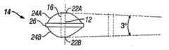
모든 회전축 둘레로의 자유 운동을 여전히 유지하면서 단부 플레이트 혹은 인공 디스크의 수핵을 간단히 변경함으로써 변형 교정을 제공하려는 시도는 머리와 몸체에 의해 인공 디스크 상에 작용된 힘이 소망하는 교정을 상쇄할 수 있기 때문에 유지할 수 없다. 유지할 수 있는 교정을 제공하기 위해 회전축 상에서의 몇몇 제한이 요구된다. 설계 도면으로부터 알 수 있는 바와 같이, 목표는 변형(관상 및 시상)을 교정할 수 있는 인공 디스크를 설계하는 데 있고, 정상 운동 범위를 벗어나는 것을 정지시키는 기계적인 정지부(반-구속)를 지니고, 양호하게는 가변의 동시 회전축(IAR)을 갖는다.Attempts to provide strain correction by simply changing the nucleus nucleus of the end plate or artificial disc while still maintaining free movement around all axes of rotation may offset the desired correction of the forces acting on the artificial disc by the head and body. Can't keep up. Some limitations on the axis of rotation are required to provide sustainable calibration. As can be seen from the design drawings, the goal is to design artificial discs capable of correcting deformations (coronal and sagittal), with mechanical stops (semi-constrained) to stop moving outside the normal range of motion, Preferably it has a variable simultaneous axis of rotation (IAR).
회전축 상에서의 제한은 두 종류로 분류될 수 있다. 첫 번째 종류는 교정을 지지하기 위해 영구 회전 혹은 축의 병진 운동을 사용하는 교정을 제공하는 것이다. 이것은 코어와 단부 플레이트 자체의 기하학적 모양을 사용하여 달성될 수 있고, 이를 기하학적 구속 부류로 일컫는다. 두 번째 종류는 모든 축을 중심으로 자유로운 운동 범위를 유지하지만 재료 지지부를 사용하여 교정을 제공하는 것이 있다. 이러한 타입의 구조는 교정 평면 내에서 정상적인 회전을 하도록 그 평면 내에 변형 가능한 재료의 부과에 의한 교정을 제공한다. 이는 디자인 재료 구속(Material Constraint) 부류로 일컬어진다.Restrictions on the axis of rotation can be classified into two types. The first kind is to provide calibration using permanent rotation or axis translational motion to support the calibration. This can be achieved using the geometry of the core and the end plate itself, which is referred to as the geometric restraint class. The second type maintains a free range of motion around all axes, but uses a material support to provide calibration. This type of structure provides calibration by imposing deformable material within that plane to make normal rotation within the calibration plane. This is referred to as the Material Constraint class.
퇴행성 디스크 질병은 우리 사회에서 사망률의 주원인이다. 그것은 고통 받는 자에게 심각한 경제적이고 감정적인 문제로 이르게 될 수 있다. 따라서 척주 변형(사상 혹은 관상 혹은 양자)의 징후를 완화하고 척주의 변형을 교정할 수 있는 인공 디스크의 필요성이 존재한다.Degenerative disc disease is the leading cause of mortality in our society. It can lead to serious economic and emotional problems for the suffering. Therefore, there is a need for an artificial disc capable of alleviating the manifestation of spinal column deformation (ideal or coronal or bilateral) and correcting the spinal column deformation.
척주의 얼라인먼트/변형 교정의 필요성에 역점을 두어 다루기 위해 디스크 치환과 함께 사용할 수 있는 수많은 상이한 방법이 존재한다. 대부분의 입수 가능한 디스크를 사용하면, 디스크 삽입의 각도는 보철의 배향을 현저하게 변경할 수 있다. 이것은 뼈 제거와 보철을 위한 단부 플레이트의 준비와 관련이 있다. 삽입 각도를 변경함으로써, 디스크는 디스크 공간에 대해 평행하거나 또는 소정의 각도를 이루면서 배치될 수 있다. 불행하게도, 단지 삽입 각도만을 변경함으로써, 누구도 척추의 기본 변형을 교정할 수 없게 된다. 삽입 각도를 단지 바꾸는 것만으로는 변형의 교정을 유지하기 위해 충분히 중심에서 벗어난 하중 지지부 혹은 구조체를 갖지 않는 장치를 보상하기에는 적절하지 못하다.There are a number of different methods that can be used with disc replacement to address the need for spinal alignment / strain correction. With most available discs, the angle of disc insertion can significantly change the orientation of the prosthesis. This involves the preparation of end plates for bone removal and prosthetics. By changing the insertion angle, the disc can be arranged parallel to the disc space or at an angle. Unfortunately, by changing only the insertion angle, no one can correct the basic deformation of the spine. Simply changing the insertion angle is not adequate to compensate for a device that does not have a load support or structure that is sufficiently off center to maintain correction of the deformation.
요추에서 척주 전만증을 교정하기 위한 방법은 쐐기형 단부 플레이트를 사용하는 Link-Carite 및 Prodisc 요추 디스크 치환 시스템에 의해 이루어진다. 쐐기형 단부 플레이트는 또한 Bryan 목 디스크 시스템을 지닌 적어도 하나의 케이스 내에서 사용되어 왔다. 그러나, 쐐기형 단부 플레이트는 현재 목 디스크 치환 시스템을 위해 일률적으로 이용할 수는 없다. 쐐기형 단부 플레이트(들)를 이용하는 방법은 단부 플레이트를 가로질러 상이한 두께를 형성하는 것을 포함한다. 볼과 소켓/골 혹은 수핵 및 단부 플레이트 사이의 굴절은 변경되지 않으며, 이는 보철이 어떻게 움직임을 제공하는가와 관련된 복잡한 기하학 형상이 바뀌지 않기 때문에 유리하다. 그러나, 이러한 방법은 과도하게 교정된 단부 플레이트나 충분히 교정되지 않은 단부 플레이트 중 하나에 에러가 생길 경우 대처하지 못한다는 단점이 있다. 단부 플레이트의 수정은 수술할 때 곤란할 수 있고 심지어 디스크 공간이 디스크 치환물을 수용하지 못하게 할 수 있다. 대부분의 시스템이 뼈 성장을 촉진하는 단부 플레이트 상에 코팅을 구비하기 때문에, 나중에 행하는 수정은 극히 어렵거나 심지어 불가능하게 될 수 있다. 단부 플레이트의 표면은 뼈와 접촉하는 외표면과 수핵 혹은 코어와 함께 굴절하는 내표면으로 2개의 표면이 존재하기 때문에, 내표면의 위치와 기하학 형상을 바꿈으로써 회전 중심을 바꿀 수 있다는 것을 알 수 있다. 이것은 "볼과 소켓" 굴절부로서의 기능을 하는 보철에 가장 잘 응용될 수 있다. "소켓" 혹은 골의 위치를 바꿈으로써, 이는 보철이 디스크의 레벨에서 얼라인먼트에 나쁜 영향을 주는 방법을 변경할 수 있다.The method for correcting spinal lordosis in the lumbar spine is by the Link-Carite and Prodisc Lumbar Disc Replacement System using wedge end plates. Wedge end plates have also been used in at least one case with a Bryan neck disk system. However, wedge end plates are currently not available uniformly for neck disc replacement systems. Methods using the wedge end plate (s) include forming different thicknesses across the end plates. The deflection between the ball and socket / gol or nucleus pulposus and end plate does not change, which is advantageous because the complex geometry associated with how the prosthesis provides movement does not change. However, this method has the disadvantage of failing to cope with an error in either an over corrected end plate or an under corrected end plate. Modifications of the end plates can be difficult when operating and may even prevent disk space from receiving disk replacements. Since most systems have a coating on the end plate that promotes bone growth, later modifications can be extremely difficult or even impossible. Since the surface of the end plate has two surfaces, the outer surface contacting the bone and the inner surface refracting with the nucleus or core, it can be seen that the center of rotation can be changed by changing the position and geometry of the inner surface. . This is best applied to prostheses that function as "ball and socket" bends. By changing the position of the "socket" or goal, this can change how the prosthesis adversely affects alignment at the level of the disc.
전만 교정을 얻기 위한 치환 방법은 수핵 혹은 내측 코어를 변경시키는 것이다. 이러한 방법의 가장 큰 장점은 수핵 혹은 코어가 더 용이하게 상호 교환 혹은 개조될 수 있다는 것이다. 내부 수술용 계기가 교정의 필요성과 그 양을 계측하기 위해 사용될 수 있고 적절한 수핵이 삽입될 수 있다. 수핵 교정을 설계함으로써, 외과 의사에 다른 방법으로는 불가능하였던 유연성, 삽입의 용이성, 차후의 개조 능력이 제공된다.The substitution method to obtain a full scale correction is to change the nucleus pulposus or inner core. The main advantage of this method is that the nucleus or core can be interchanged or modified more easily. Internal surgical instruments may be used to gauge the need and amount of correction and appropriate nuclei may be inserted. By designing the nucleus nucleus, the surgeon is provided with flexibility, ease of insertion, and subsequent remodeling capabilities that were otherwise impossible.
본 발명은 척주의 변형을 교정하기 위한 능력과 함께 자연 그대로의 추간판 디스크의 정상 운동 범위를 제공하는 신규의 인공 디스크를 포함한다. 제안된 디스크는 기능적인 척주 유닛의 반 구속 운동 범위를 허용해준다. 그것은 수술 전의 정상 척추의 운동학을 재현할 것이다. 그것은 최대 내구성, 생물학적 적합성, 및 장기간 안정을 위한 척주 뼈 구조로 그 자체를 통합하기 위한 수단을 유지할 것이다. 그것의 삽입은 현재의 절차에 비해 안전하고, 간단하고, 이상적으로 수술 시간을 현저하게 증가시킨다. 기존의 디스크 치환 시스템과는 대조적으로, 그것은 외과 의사로 하여금 척주의 자연적인 운동학을 유지하면서 변형을 교정할 수 있도록 해준다.The present invention includes a novel artificial disc that provides the normal range of motion of the intervertebral disc as it is, with the ability to correct deformation of the spinal column. The proposed disc allows for a semi-constrained range of motion of the functional spinal column unit. It will reproduce the kinematics of the normal spine before surgery. It will maintain the means for integrating itself into the spinal bone structure for maximum durability, biocompatibility, and long term stability. Its insertion is safer, simpler and ideally increases surgery time compared to current procedures. In contrast to conventional disc replacement systems, it allows surgeons to correct deformation while maintaining the natural kinematics of the spinal column.
이러한 시스템의 주요 장점은 수핵을 쉽게 개조할 수 있다는 데 있다. 예컨대, Bryan 디스크가 개조를 필요로 하는 대부분의 경우, 단부 플레이트를 포함하는 전체 디스크는 반드시 제거되어야 한다. 척주의 얼라인먼트가 시간에 따라 변할 경우, 특히 어린이와 청소년의 경우, 이러한 신규 디스크 치환 시스템은 필요에 따라 수핵의 개조를 허용할 것이다.The main advantage of these systems is that they can be easily retrofitted. For example, in most cases where Bryan disks require modification, the entire disk, including the end plate, must be removed. If the alignment of the spinal column changes over time, especially for children and adolescents, this new disk replacement system will allow modification of the nucleus pulposus as needed.
본 발명은 척주 얼라인먼트 변형의 교정을 제공하는 인공 디스크를 제공함으로써 종래 기술의 인공 디스크와 관련된 문제점을 다루고 있다.The present invention addresses the problems associated with artificial discs of the prior art by providing an artificial disc that provides correction of spinal column alignment deformation.
본 발명의 인공 디스크는 후만증, 전만증 및 만곡증 등의 척주 변형을 교정하는 것을 포함하는 퇴행성 디스크 질병의 치료에 유리하다.The artificial disc of the present invention is advantageous for the treatment of degenerative disc disease, which includes correcting spinal column deformations such as embarrassment, mania and curvature.
본 발명의 하나의 양태의 목적은 수술 레벨에서 움직임을 유지하고 주변부 환절 질환(adjacent segment disease)의 발병률을 줄이는, 향상된 인공 디스크 치환물을 제공하는 데 있다.It is an object of one aspect of the present invention to provide an improved artificial disc replacement that maintains movement at the surgical level and reduces the incidence of adjacent segment disease.
본 발명의 하나의 양태에 있어서, 인공 디스크는 비대칭 최대 수직축을 지닌 인공 수핵과 협동한다. 본 발명은 중심이 아닌 위치에 하중 지지의 최대 지점과 높이(수핵의 전방/후방 높이 차)를 지닌 비구형 수핵을 포함한다.In one aspect of the invention, the artificial disc cooperates with an artificial nucleus with an asymmetric maximum vertical axis. The present invention includes an aspheric nucleus pulposus having a maximum point and height of load bearing (a difference in the front / rear heights of the nucleus pulposus) in a non-central position.
일 실시예에 있어서, 수핵은 손상된 척주 환절에 전만 교정을 제공하도록 되어 있다. 이 경우, 최대 높이 축은 수핵의 전방 부분에 위치 설정된다.In one embodiment, the nucleus pulposus is intended to provide lordosis correction to the injured spinal column. In this case, the maximum height axis is positioned at the front part of the nucleus pulposus.
또 다른 실시예에 따르면, 수핵은 후만증 조절을 제공하도록 되어 있다. 이 경우, 최대 높이 축은 수핵의 후방 부분에 위치 설정된다.According to another embodiment, the nucleus pulposus is adapted to provide emesis. In this case, the maximum height axis is positioned at the rear part of the nucleus pulposus.
또 다른 실시예에 있어서, 비대칭 수핵은 척추 만곡의 치료를 위해 사용될 수 있다. 이를 달성하기 위해 최대 높이의 축은 디스크의 중앙으로 측방향(방시상 방향)의 축이 된다.In yet another embodiment, the asymmetric nucleus pulposus can be used for the treatment of spinal curvature. To achieve this, the axis of maximum height is laterally (axially) axis of the disc.
본 발명의 또 다른 실시예에 따르면, 인공 수핵 혹은 코어는 인공 디스크에 사용하기 위해 제공된다. 상기 수핵은 중앙 수직 축에서 혹은 중앙 수직 축이 아닌 다른 수직 축 중 어느 하나에서 가장 큰 수직 높이를 지닌 동시에 생물학적 적합성 재료의 본체를 포함한다.According to another embodiment of the invention, an artificial nucleus nucleus or core is provided for use in an artificial disc. The nucleus pulposus has the largest vertical height at either the central vertical axis or at any other vertical axis other than the central vertical axis and at the same time comprises a body of biocompatible material.
또 다른 실시예에 있어서, 상기 본체는 볼록한 상측면 및 하측면과 비중앙의 최대 높이 수직 축을 구비하는 구형 혹은 타원형(계란형)이다. 변형례에 따르면, 상기 수핵은 윗면이 베이스에 평행하지 않는 평면에서 절단되어 있는 절두된 실린더 형태이다. 또 다른 양호한 실시예에서, 디스크는 실질적으로 원형이다.In yet another embodiment, the body is spherical or elliptical (egg shaped) with a convex upper and lower side and a maximum height vertical axis of non-centrality. According to a variant, the nucleus pulposus is in the form of a truncated cylinder whose upper surface is cut in a plane that is not parallel to the base. In another preferred embodiment, the disk is substantially circular.
완전히 둥근 표면(반드시 구형일 필요는 없음)을 지닌 수핵 본체의 디자인은 머리와 목의 가변 힘에 노출될 때 교정을 신뢰성 있게 유지하는 것과 관련한 문제점이 있다고 밝혀졌다. 이러한 문제점을 다루기 위해, 평탄하거나 혹은 인접한 표면과 상이한 외형을 지닌 환절 혹은 섹션은 수핵 본체의 중앙 영역 내에 형성될 수 있다. 이러한 섹션은 수핵의 인접한 표면(들)과 동일하지 않는 임의의 외형을 언급하는 것을 의미하는 평탄화된 섹션이라 일컫는다. 이러한 평탄화된 표면은 평면일 수 있거나 또는 인접한 표면과 상이한 곡률 반경을 지닌 약간 볼록 혹은 오목한 형상 등의 다른 형상을 지닐 수 있다. 이렇게 평탄화된 표면은 또한 복합 곡선 혹은 다른 복잡한 형상이 될 수 있다. 전만 교정을 제공하는 예에 있어서, 평탄화된 환절은 전방 부분의 높이가 후방 부분의 높이보다 더 큰 하위의 척추체의 상위 단부 플레이트에 대해 각을 이룰 수 있다. 수핵 본체의 전체적인 형상은 여전히 비대칭형이지만, 평탄화된 환절은 변형의 신뢰성 있는 교정을 제공하기 위해 합체될 수 있다. 평탄한 환절은 수핵을 통해 작용하는 오정렬 모멘트에 저항함으로써 교정의 안정성을 제공한다. 평탄화된 환절이 적절한 크기가 아닐 경우, 전방 하중의 존재 하에서 교정이 사라지게 하거나 또는 후방 하중의 존재 하에서의 교정 동안(척추 전만 교정 동안) 과도한 척추 전만이 되게 하는 경향이 있을 수 있다. 평탄한 환절을 합체하는 것과 관련된 또 다른 장점은 약간 움직이는 동안 장치의 정지, 중립 위치를 중심의 영역 위로 표면 접촉을 제공하는 데 있다. 이는 장치에 마모를 줄이는 것을 보조한다.The design of the nucleus pulposus body with a completely round surface (not necessarily spherical) has been found to be a problem associated with maintaining the correction reliably when exposed to variable forces in the head and neck. To address this problem, a section or section having a contour different from the flat or adjacent surface may be formed in the central region of the nucleus pulposus body. This section is referred to as a flattened section, meaning referring to any contour that is not the same as the adjacent surface (s) of the nucleus pulposus. Such planarized surfaces may be planar or may have other shapes, such as slightly convex or concave shapes with different radii of curvature from adjacent surfaces. This flattened surface can also be a compound curve or other complex shape. In an example that provides for fullness correction, the flattened knee may be angled with respect to the upper end plate of the lower vertebral body, where the height of the anterior portion is greater than the height of the posterior portion. While the overall shape of the nucleus pulposus body is still asymmetric, the flattened lesions can be incorporated to provide reliable correction of deformation. The flat knee provides stability of correction by resisting misalignment moments acting through the nucleus pulposus. If the flattened node is not of an appropriate size, it may tend to cause the correction to disappear in the presence of anterior loads or to only cause excessive spinal vertebrae during correction in the presence of posterior loads (during spinal vertebrae only). Another advantage associated with incorporating flattened joints is to provide surface contact over the central region of the stationary, neutral position of the device while slightly moving. This helps to reduce wear on the device.
또 다른 실시예에서, 수핵 혹은 코어는 하측 단부 플레이트 내에 형성된 개구 혹은 골속으로 끼워지는 평탄한 하위 표면을 구비하는 반구형 형상일 수 있다. 골의 형상은 골이 하측 단부 플레이트에 대해 수핵의 회전 혹은 병진 운동의 외측 한계를 정하도록 이루어질 수 있다. 이러한 디자인은 인접한 척추의 과도한 신연(distraction) 없이 수핵 삽입의 용이성을 더 갖도록 해주는데 그 이유는 골이 일단부에서 개방되어 수핵이 삽입될 수 있게 해준 다음, 정지부가 골 내의 수핵을 유지하기 위해 골에 삽입될 수 있기 때문이다.In another embodiment, the nucleus pulposus or core may be hemispherical in shape with an opening formed in the lower end plate or a flat lower surface fitted into the bone. The shape of the bone may be such that the bone defines an outer limit of rotation or translation of the nucleus pulposus with respect to the lower end plate. This design allows for more ease of nucleus insertion without excessive distraction of the adjacent spine because the bone is open at one end to allow the nucleus to be inserted, and then the stop is placed on the bone to maintain the nucleus in the bone. Because it can be inserted.
또 다른 실시예에서, 타원 모양의 수핵 대신에, 구형 혹은 타원형 단부 섹션과 평탄화된 혹은 원통형 중심 섹션을 구비하는 가늘고 긴 혹은 "소시지 타입"의 형상을 사용할 수 있다. 이러한 형상의 수핵이 상측 단부 플레이트 상의 원통형 지지면과 합치될 때, 굴절 및 확장뿐만 아니라 측방향의 굴절 동안 면 접촉 및 선 접촉 양자가 제공된다. 이러한 타입의 가늘고 긴 수핵이 사용될 때, 정상 운동의 한계를 넘으면 정지부에 의해 정지되는 축방향 회전을 허용하는 하측 단부 플레이트 내의 대응하는 단부 플레이트 골이 제공된다. 이러한 골은 "나비넥타이", "강아지 뼈" 등의 형성을 지닐 수 있다. 상기 골은 한정된 전방/후방 및 중간/측방향 병진 운동을 허용하기 위해 수핵에 비해 약간 과도한 크기로 될 수 있다. 추가적으로, 단부 플레이트의 골 지지면은 수핵의 움직임의 외측 한계에서 상향으로 굴곡될 수 있다. 이러한 특징은 수핵이 회전할 때 수핵의 상향 상승을 강제하고, 또 인접한 척추체들이 이격되도록 강제하여 그들 사이의 조직에 하중을 가해 운동의 점진적인 정지를 초래하는 장치의 축방향 신연을 유발한다. 골 내부에서 코어의 병진 운동은 자연 그대로의 디스크의 가변 동시 회전축을 보존하려고 시도한다.In another embodiment, instead of an elliptic nucleus, one may use an elongated or “sausage type” shape having a spherical or elliptical end section and a flattened or cylindrical center section. When the nucleus of this shape coincides with the cylindrical support surface on the upper end plate, both face contact and line contact are provided during lateral deflection as well as deflection and expansion. When this type of elongated nucleus pulposus is used, a corresponding end plate trough in the lower end plate is provided that allows axial rotation to be stopped by the stop once it exceeds the limits of normal motion. Such bone may have the formation of a "butterfly", "dog bone", and the like. The bone may be slightly oversized relative to the nucleus pulposus to allow for limited forward / rear and intermediate / lateral translational movement. Additionally, the bone support surface of the end plate may be curved upward at the outer limit of the movement of the nucleus pulposus. This feature forces the upward elevation of the nucleus when the nucleus nucleus rotates, and forces adjacent vertebral bodies to be spaced apart, loading the tissue between them, causing axial distraction of the device resulting in a gradual stop of motion. The translational motion of the core inside the bone attempts to preserve the variable simultaneous rotational axis of the disk as it is.
추가적으로, 골은 인접한 단부 플레이트의 과도한 신연 없이 수핵의 삽입을 허용하기 위해 전방 단부에 개방될 수 있다. 잠금 기구는 수핵의 삽입 후 수핵이 골로부터 방출되는 것을 방지하기 위해 제공될 수 있다.Additionally, the bone may be open at the anterior end to allow insertion of the nucleus pulposus without excessive stretching of the adjacent end plate. A locking mechanism can be provided to prevent the nucleus pulposus from releasing after bone insertion.
본 발명의 또 다른 양태에 있어서, 신규 타입의 단부 플레이트가 제공된다. 광범위한 척추체 표면의 준비를 필요로 하는 다른 단부 플레이트와는 달리, 본 발명의 단부 플레이트들은 이들의 용이한 삽입을 허용해주는 실질적으로 평탄한 외표면 혹은 척추 접촉면을 구비한다. 양호한 실시예에서, 상기 표면은 플레이트를 정위치에 정착하기 위한 적어도 하나의 단일 방향 용골을 지닌 절반 정도가 둥근 플레이트이다. 단부 플레이트의 외표면은 인시츄식(in-situ)으로 단부 플레이트의 안전성을 향상시키기 위해 뼈의 내부 성장을 촉진하는 방법으로 처리될 수 있다. 하나의 실시예에서, 외측(척추 접촉) 표면과 내측(수핵 접촉) 표면은 서로 실질적으로 평행하다. 또 다른 실시예에서, 외표면과 내표면은 평행하지 않기 때문에 단부 플레이트가 실질적으로 쐐기 모양의 형상을 갖도록 해준다. 쐐기의 넓고 좁은 가장자리의 배향을 척주 교정의 다양한 타입 및 정도를 제공하기 위해 조절할 수 있다.In another aspect of the invention, a novel type of end plate is provided. Unlike other end plates, which require the preparation of a wide range of vertebral body surfaces, the end plates of the present invention have a substantially flat outer surface or spinal contact surface that allows for easy insertion thereof. In a preferred embodiment, the surface is a half rounded plate with at least one unidirectional keel for anchoring the plate in place. The outer surface of the end plate can be treated in-situ in a manner that promotes internal growth of the bone to improve the safety of the end plate. In one embodiment, the outer (vertebral contact) surface and the inner (nucleus contact) surface are substantially parallel to each other. In another embodiment, the outer and inner surfaces are not parallel, allowing the end plates to have a substantially wedge shaped shape. The orientation of the wide and narrow edges of the wedges can be adjusted to provide various types and degrees of spinal column correction.
본 발명의 또 다른 양태에서, 보철은 인공 수핵과 적어도 하나의 단부 플레이트를 포함한다. 이러한 실시예에서, 보철은 상측 척추 부재에 고정하기 위한 상위 단부 플레이트, 하측 척추 부재에 고정하기 위한 하위 단부 플레이트, 및 2개의 단부 플레이트 사이에 끼워지도록 채택된 수핵을 포함한다. 본 발명의 단부 플레이트는 뼈와 접촉하는 쪽의 거의 평탄한 표면과 수핵과 함께 굴절되도록 다른 쪽의 적절한 기하학적 리셉터클을 구비한다. 중앙 용골은 정위치에 수핵을 정착시키기 위해 단부 플레이트의 내표면의 중심에 형성될 수 있다. 단부 플레이트는 보철이 척추관을 향해 이동하는 것을 방지하기 위한 정지 부재를 포함한다. 상기 수핵은 기하학적 중심에 있지 않은 최대 수직 축을 또한 구비할 수 있다.In another aspect of the invention, the prosthesis comprises an artificial nucleus pulposus and at least one end plate. In this embodiment, the prosthesis includes an upper end plate for fastening to the upper vertebral member, a lower end plate for fastening to the lower vertebral member, and a nucleus pulposus adapted to fit between the two end plates. The end plate of the present invention has a substantially flat surface on the side in contact with the bone and a suitable geometric receptacle on the other side to be deflected with the nucleus pulposus. The central keel may be formed in the center of the inner surface of the end plate for anchoring the nucleus pulposus in place. The end plate includes a stop member for preventing the prosthesis from moving toward the spinal canal. The nucleus pulposus may also have a maximum vertical axis that is not at the geometric center.
또 다른 실시예에서, 수핵은 상측 리셉터클을 지닌 상측면과 하측 리셉터클을 지닌 하측면을 구비한다. 상위 단부 플레이트는 하방으로 돌출하는 돌출부 혹은 상측 리셉터클과 맞물리는 앵커를 구비하며, 하위 단부 플레이트는 상방으로 연장하는 돌출부 혹은 하측 리셉터클과 맞물리는 앵커를 구비한다. 상기 보철은 이웃하는 척추들 사이에 적절한 공간적 관계를 유지하며 또한 척주의 정상적인 운동 범위를 허용한다. 이러한 실시예는 또한 홈이 일단부로 개방되어 있는 리셉터클을 또한 포함할 수 있다. 단부 플레이트 상의 앵커는 수핵을 고정하기 위해 홈 내의 위치로 활주하는 중앙 용골을 포함할 수 있다.In another embodiment, the nucleus pulposus has an upper side with an upper receptacle and a lower side with a lower receptacle. The upper end plate has an anchor that engages with a downwardly projecting protrusion or upper receptacle, and the lower end plate has an anchor that engages with an upwardly extending projection or lower receptacle. The prosthesis maintains an appropriate spatial relationship between neighboring vertebrae and also allows a normal range of motion of the spinal column. This embodiment may also include a receptacle in which the groove is open at one end. The anchor on the end plate may include a central keel that slides into position in the groove to secure the nucleus pulposus.
본 발명의 또 다른 실시예는 유니버설 조인트와 같이 작동하고 3개의 해부학적 회전축과 협동하며, 이들 축 중 2개는 굴곡/확장 및 측방향 굴절 운동을 위해 제공되는 반면에 나머지 하나의 축은 축방향 회전을 위해 제공된다. 이들 회전축들은 중앙 포스트 둘레로 각각에 대해 회전할 수 있는 2개의 실린더 쌍을 사용함으로써 얻어진다.Another embodiment of the present invention operates as a universal joint and cooperates with three anatomical rotational axes, two of which are provided for flexion / expansion and lateral deflection movements while the other axis is axial rotation. Is provided for. These axes of rotation are obtained by using two cylinder pairs that can rotate about each around a central post.
본 발명의 또 다른 실시예에서, 플레이트 중 하나는 다른 하나의 플레이트와 맞물리는 중앙 포스트와, 탄성 재료로 형성되어 상기 중앙 포스트 둘레에 위치 설정된 환형 코어를 구비한다. 상기 코어는 비대칭형이면서 필요한 변형 교정을 제공하기 위해 양 플레이트와 맞물릴 수 있다. 코어는 변형 교정을 위해 플레이트들 사이에 소망하는 각도를 제공하도록 단부 플레이트와 맞물릴 수 있고, 중심 포스트는 하중이 예정된 한계를 초과할 때 다른 플레이트와 맞물린다. 그렇지 않으면, 상기 포스트는 상기 다른 플레이트와 맞물릴 수 있으며, 코어는 인가된 힘이 플레이트의 상대 각도를 변경시키는 경향이 있을 때 서로에 대해 소망하는 각도에서 플레이트를 유지하기 위해 다른 플레이트와 맞물린다. 별법으로서, 코어는 동일한 기능을 수행하기 위해 2개 혹은 그 이상의 별도의 스페이서로 치환될 수 있다.In another embodiment of the invention, one of the plates has a central post that engages the other plate and an annular core formed of an elastic material and positioned around the central post. The core is asymmetrical and can engage both plates to provide the necessary deformation correction. The core can engage the end plate to provide the desired angle between the plates for deformation correction, and the center post engages with the other plate when the load exceeds a predetermined limit. Otherwise, the post may be engaged with the other plate and the core is engaged with the other plates to maintain the plates at the desired angles with respect to each other when the applied force tends to change the relative angles of the plates. Alternatively, the core may be replaced with two or more separate spacers to perform the same function.
본 발명의 또 다른 양태에 있어서, 수핵은 희망하는 운동 범위를 얻기 위해 재료 변형을 이용할 수 있다. 상기 재료의 형상은 변형 교정을 위한 복원력을 제공하기 위해 사용될 수 있다. 이러한 결과를 얻기 위해, 상기 재료는 코어의 다양한 부분으로부터 제거되어 선택된 위치에서 코어의 탄성률을 변경시킬 수 있거나 혹은 가변 탄성률을 지닌 재료를 사용할 수 있다. 이러한 방식으로, 상이한 힘과 운동은 코어의 디자인을 통해 제공될 수 있다.In another aspect of the present invention, the nucleus pulposus may use material deformation to achieve the desired range of motion. The shape of the material can be used to provide restoring force for deformation correction. To achieve this result, the material can be removed from various parts of the core to change the modulus of elasticity of the core at selected locations or a material with variable modulus can be used. In this way, different forces and movements can be provided through the design of the core.
단부 플레이트들에는 소망하는 운동 범위를 벗어나는 것을 정지시키는 정지부로서 작용하면서 자동으로 유도된 점진적인 정지를 허용해주는 특징이 제공된다. 이러한 결과는 단부 플레이트들 중 하나에 혹은 그 위에 하나 이상의 캠 표면을 형성하고 캠 표면과 맞물리도록 다른 단부 플레이트 상에 협력 부재를 제공함으로써 달성될 수 있다. 상기 캠 표면은 그것의 내표면에 점진적으로 만곡하는 곡선을 구비한다. 단부 플레이트들 사이의 상대 운동 동안, 캠 표면은 단부 플레이트의 축방향 신연을 초래하고 격렬한 정지를 방지하는 것을 보조하도록 연질 조직을 제공하는 협력 부재에 의해 맞물린다. 회전 운동 대신에, 협력하는 캠 표면은 하나의 단부 플레이트가 다른 단부 플레이트에 대해 회전할 때 신연이 발생하도록 제공될 수 있다.The end plates are provided with a feature that allows for an automatically guided progressive stop while acting as a stop to stop moving outside the desired range of motion. This result can be achieved by forming one or more cam surfaces on or over one of the end plates and providing a cooperating member on the other end plate to engage the cam surface. The cam surface has a curve that curves gradually on its inner surface. During relative movement between the end plates, the cam surface is engaged by a cooperating member that provides soft tissue to assist in axial stretching of the end plates and to prevent violent stopping. Instead of a rotary motion, cooperating cam surfaces may be provided such that distraction occurs when one end plate rotates with respect to the other end plate.
이상, 후술하는 본 발명의 상세한 설명으로부터 더 용이한 이해를 도울 수 있도록 본 발명의 특징 및 기술적인 장점을 광범위하게 개설하였다. 본 발명의 추가의 특징 및 장점들이 이하에서 설명될 것이고 이들은 본 발명의 청구의 범위 주체를 구성한다. 개시된 개념과 특정 실시예는 본 발명의 동일한 목적을 수행하기 위한 다른 구조의 변형 혹은 설계의 기본으로서 용이하게 이용될 수 있다는 것이 당업자들에 의해 주지되어야 한다. 또한, 이러한 등가의 구성은 첨부 도면에 기술된 바와 같은 본 발명의 정신과 범주에서 벗어나지 않는 것으로 당업자들에게 구현되어야 한다. 본 발명의 추가의 목적 및 장점과 함께 그 구성 및 작동 방법 양자에서의 특징이 있는 것으로 사료되는 신규의 특징들은 첨부 도면과 함께 고려할 때 아래의 설명으로부터 더욱 용이하게 이해될 것이다. 그러나 각각의 도면은 단지 예시와 설명을 위해 제공된 것이며, 본 발명의 한정을 의도하지 않는다는 것으로 명백하게 이해될 것이다.The foregoing has outlined rather broadly the features and technical advantages of the present invention in order to facilitate an easier understanding thereof from the detailed description of the invention which follows. Further features and advantages of the invention will be described below, which constitute the subject matter of the claims of the invention. It should be noted by those skilled in the art that the conception and specific embodiment disclosed may be readily utilized as a basis for modification or design of other structures for carrying out the same purposes of the present invention. Moreover, such equivalent construction should be implemented to those skilled in the art without departing from the spirit and scope of the present invention as described in the accompanying drawings. Further features and advantages of the present invention, together with novel features that are believed to be characteristic in both its construction and method of operation, will be more readily understood from the following description when considered in conjunction with the accompanying drawings. However, it will be clearly understood that each figure is provided for the purpose of illustration and description only and is not intended to be limiting of the invention.
본 발명의 전술한 특징 및 다른 특징들을 첨부 도면을 참조한 아래의 상세한 설명을 읽음으로써 더욱 명백해질 것이다.The above and other features of the present invention will become more apparent upon reading the following detailed description with reference to the accompanying drawings.
도 1a는 최대 중심축이 수핵의 기하학적 중심선에 있는 구형 인공 디스크 수핵을 도시한 도면이다.1A shows a spherical artificial disc nucleus with its maximum central axis at the geometric centerline of the nucleus.
도 1b는 최대 수직 축 오프셋이 3°의 교정을 제공하는 도 1a의 수핵을 도시한 도면이다.FIG. 1B shows the nucleus nucleus of FIG. 1A in which the maximum vertical axis offset provides a correction of 3 °.
도 1c는 최대 수직 축 오프셋이 6°의 교정을 제공하는 도 1a의 수핵을 도시한 도면이다.1C shows the nucleus pulposus of FIG. 1A in which the maximum vertical axis offset provides a calibration of 6 °.
도 2a는 최대 중심축이 수핵의 기하학적 중심선에 있는 비대칭 인공 디스크 수핵을 도시한 도면이다.2A shows an asymmetric artificial disc nucleus with its maximum central axis at the geometric centerline of the nucleus.
도 2b는 최대 수직 축 오프셋이 3°의 교정을 제공하는 도 2a의 수핵을 도시한 도면이다.FIG. 2B shows the nucleus pulposus of FIG. 2A in which the maximum vertical axis offset provides a correction of 3 °.
도 2c는 최대 수직 축 오프셋이 6°의 교정을 제공하는 도 2a의 수핵을 도시한 도면이다.FIG. 2C shows the nucleus nucleus of FIG. 2A in which the maximum vertical axis offset provides a calibration of 6 °.
도 3은 도 1a에 도시된 인공 디스크 수핵의 실시예를 도시한 평면도이다.3 is a plan view showing an embodiment of the artificial disc nucleus nucleus shown in FIG. 1A.
도 4는 도 1a에 도시된 인공 디스크 수핵의 실시예를 도시한 사시도이다.4 is a perspective view showing an embodiment of the artificial disc nucleus nucleus shown in FIG. 1A.
도 5는 도 2a에 도시된 인공 디스크 수핵의 실시예를 도시한 사시도이다.FIG. 5 is a perspective view showing an embodiment of the artificial disc nucleus nucleus shown in FIG. 2A. FIG.
도 6은 단부 플레이트의 외표면을 도시한 사시도이다.6 is a perspective view showing the outer surface of the end plate;
도 7은 단부 플레이트의 내표면을 도시한 사시도이다.7 is a perspective view showing the inner surface of the end plate.
도 8은 단부 플레이트를 도시한 정면도이다.8 is a front view of the end plate.
도 9는 도 1a에 도시된 수핵을 지닌 척주 디스크 장치를 도시한 정면도이다.FIG. 9 is a front view of the spinal disc apparatus having the nucleus pulposus shown in FIG. 1A.
도 10은 도 8의 척주 디스크 장치를 도시한 측면도이다.FIG. 10 is a side view of the spinal disc apparatus of FIG. 8. FIG.
도 11은 도 2a에 도시된 수핵을 지닌 척주 디스크 장치를 도시한 정면도이다.FIG. 11 is a front view of the spinal disc apparatus having the nucleus pulposus shown in FIG. 2A.
도 12는 도 8의 척주 디스크 장치를 도시한 측면도이다.FIG. 12 is a side view of the spinal disc apparatus of FIG. 8. FIG.
도 13a 및 도 13b는 단부 플레이트가 전만 교정을 위해 채택될 수 있는 인공 척주 디스크 보철의 실시예를 도시한 도면이다.13A and 13B illustrate an embodiment of an artificial spinal disc prosthesis in which an end plate may be employed for full scale calibration.
도 14a, 도 14b, 및 도 14c는 단부 플레이트가 전만 교정을 위해 채택될 수 있는 다른 실시예를 도시한 도면이다.14A, 14B, and 14C illustrate another embodiment in which the end plate may be employed for full scale calibration.
도 15는 모든 방향의 움직임을 제공하는 또 다른 실시예를 도시한 측면도이다.15 is a side view illustrating another embodiment that provides movement in all directions.
도 16a 및 도 16b는 도 15의 실시예에 따른 수핵의 2개의 섹션을 도시한 도면이다.16A and 16B show two sections of the nucleus pulposus in accordance with the embodiment of FIG. 15.
도 17 및 도 18은 수핵이 중간 섹션과 함께 상측 및 하측 섹션으로 형성되어 있는 본 발명의 또 다른 실시예를 도시한 도면이다.17 and 18 show yet another embodiment of the present invention in which the nucleus pulposus is formed into an upper and a lower section together with an intermediate section.
도 19는 수핵이 절반으로 절단되고 평탄한 하측 내부면을 지닌 본 발명의 또 다른 실시예를 도시한 도면이다.19 shows another embodiment of the present invention with the nucleus pulposus cut in half and with a flat lower inner surface.
도 20은 도 19의 수핵을 개략적으로 도시한 도면이다.20 is a view schematically showing the nucleus nucleus of FIG. 19.
도 21은 도 19의 실시예의 변형례를 도시한 도면이다.21 is a diagram illustrating a modification of the embodiment of FIG. 19.
도 22는 도 21의 수핵의 배면을 도시한 도면이다.22 is a view showing the back of the nucleus pulposus in FIG. 21.
도 23은 도 21의 수핵을 개략적으로 도시한 도면이다.FIG. 23 is a view schematically showing the nucleus nucleus of FIG. 21.
도 24는 도 19의 실시예의 변형례를 도시한 도면이다.24 is a diagram illustrating a modification of the embodiment of FIG. 19.
도 25 내지 도 31은 수핵이 중심에 평탄한 섹션을 지니면서 가늘고 길게 되어 있는 본 발명의 또 다른 실시예를 도시한 도면이다.25 to 31 show yet another embodiment of the present invention in which the nucleus pulposus is elongated with a flat section in the center.
도 32 및 도 33은 유니버설 조인트를 사용하는 본 발명의 또 다른 실시예를 도시한 도면이다.32 and 33 illustrate another embodiment of the present invention using a universal joint.
도 34 내지 도 36은 탄성 링과 포스트가 단부 플레이트들 사이의 상대 운동을 제공하는 본 발명의 또 다른 실시예를 도시한 도면이다.34-36 illustrate another embodiment of the present invention in which the elastic ring and the post provide relative motion between the end plates.
도 37은 도 34의 실시예의 변형례를 도시한 도면이다.37 is a view showing a modification of the embodiment of FIG. 34.
도 38 및 도 39는 수핵이 중간/측방향 교정을 제공하도록 형성되어 있는 본 발명의 또 다른 실시예를 도시한 도면이다.38 and 39 illustrate yet another embodiment of the present invention in which the nucleus pulposus is formed to provide intermediate / lateral correction.
도 40 내지 도 43은 단부 플레이트에, 정상적인 운동 범위를 벗어난 위치에서 정지부가 마련되어 있는 본 발명의 또 다른 실시예를 도시한 도면이다.40 to 43 show another embodiment of the invention in which the end plate is provided with a stop at a position outside the normal range of motion.
척주의 적절하면서 튼튼한 얼라인먼트에서, 척주는 적절한 시상 및 관상 균형(신축성)을 향상시키는 자연스러운 곡선을 따르고 척추 사이에 균형 잡힌 하중 공유를 허용한다. 이러한 곡선은 경부, 흉부, 허리 및 척주의 천골 영역을 포함한다. 자연적으로, 곡선을 수용하기 위해, 기능적인 척주 유닛과 추간판내 공간의 높이 사이에서의 굴절 각도에 약간의 변화가 존재한다. 경부와 허리 영역은 자연적으로 전만(lordotic)이거나 또는 전방 방향으로 볼록하게 굴곡이 있다. 척주를 따른 상이한 환절에서, 통상적으로 척추 본체를 위한 추간판내 공간에 대한 상이한 높이가 존재한다. 추가적으로, 추간판내 공간과 척추 본체 높이는 사람에 따라 다를 수 있다.In proper and robust alignment of the spinal column, the spinal column follows a natural curve that improves proper sagittal and coronal balance (elasticity) and allows for balanced load sharing between the vertebrae. These curves include the sacral regions of the neck, thorax, waist and spinal column. Naturally, to accommodate the curve, there is a slight change in the angle of refraction between the functional spinal column unit and the height of the intervertebral disc space. The cervical and lumbar regions are naturally lordotic or convexly curved in the forward direction. At different joints along the spinal column, there are typically different heights for the intervertebral disc space for the vertebral body. In addition, the intervertebral disc space and the height of the vertebral body may vary from person to person.
각각의 추간판내 공간은 전방 영역과 후방 영역을 지닌다. 전방 영역에서 후방 영역까지 동일한 높이를 유지하는 경부, 흉부 및 허리 영역의 인공 디스크는 비정상적인 얼라인먼트를 향상시킬 수 있고, 그 결과 인접한 디스크의 전방 혹은 후방 부분에서 추가의 응력을 초래한다. 또한, 장치 전반에 걸쳐 불균일한 하중 분포를 초래할 수 있고, 과도한 상대 운동, 마찰 부스러기 및 쉬운 파손을 유발한다.Each intervertebral disc space has a front region and a back region. Artificial discs in the cervical, thorax and lumbar region that maintain the same height from the anterior region to the posterior region can improve abnormal alignment, resulting in additional stress in the anterior or posterior portion of the adjacent disc. It can also lead to non-uniform load distribution throughout the device and cause excessive relative motion, frictional debris and easy breakage.
본 명세서에 사용한 바와 같이, 용어 수핵(nucleus)과 코어(core)는 손상된 자연 그대로의 척주 디스크를 치환하는 인공의 추간판 장치를 언급하기 위해 호환적으로 사용된다. 인공 코어는 단독으로 제공될 수도 있고 상측 척추에 고정하도록 제공된 상위 단부 플레이트와 하측 척추에 고정되도록 제공된 하위 단부 플레이트 중 어느 하나 또는 이들 양자와 조합하여 제공될 수도 있다.As used herein, the terms nucleus and core are used interchangeably to refer to an artificial intervertebral disc device that replaces a damaged native spinal disc. The artificial core may be provided alone or in combination with either or both of the upper end plate provided for fixation to the upper vertebra and the lower end plate provided for fixation to the lower vertebrae.
용어 "상측(upper)"과 하측(lower)"은 치환될 디스크의 어느 한 쪽 또는 첨부 도면에 도시된 위치에 있는 부분의 표면 상의 척추골을 언급하기 위해 본 명세서에서 사용된다. "상위(superior)" 플레이트는 기능성 척주 유닛의 상측 척추골에 부착되고, "하위(inferior") 플레이트는 하측 척추골에 부착된다.The terms "upper" and "lower" are used herein to refer to the vertebrae on the surface of either side of the disc to be replaced or in the position shown in the accompanying figures. The plate is attached to the upper vertebrae of the functional spinal column unit, and the "inferior" plate is attached to the lower vertebrae.
용어 수직 및 수평은 해부학적 위치에서 선 자세로 있는 인간을 기준으로 하여 본 명세서에 사용한다. 용어 "전방(anterior)"은 전면으로 향하는 영역을 언급하고, 용어 "후방(posterior)"은 등을 향하는 영역을 언급한다. 용어 "시상(sagittal)"은 선 자세로 있는 인간의 중심의 중앙 축의 어느 한 측면 상의 영역을 언급한다.The terms vertical and horizontal are used herein with reference to the human being in a standing posture at an anatomical position. The term "anterior" refers to the area facing the front, and the term "posterior" refers to the area facing the back. The term "sagittal" refers to an area on either side of the central axis of the human center in a standing position.
용어 "비대칭"은 본 명세서에서는 중앙에 배치되어 있지 않은 최대 높이의 축 또는 그것의 최대 수직 축이 중앙에 배치되어 있지 않는 수핵 혹은 인공 디스크 치환술[total disc replacement(TDR)]을 언급하기 위해 사용된다. 다시 말해서, 최대 높이는 대칭의 중심선에 위치하거나 피벗되지 않기 때문에 TDR은 형상이나 크기에 있어서 대칭선의 다른 쪽에 있는 다른 영역과 정확하게 동일하지 않는 영역을 포함한다. 최대 하중 지지 위치는 중앙이 아닌 위치에 배치된다.The term "asymmetry" is used herein to refer to a shaft of maximum height that is not centered or to a total disc replacement (TDR) where its maximum vertical axis is not centered. . In other words, since the maximum height is not located or pivoted at the centerline of symmetry, the TDR includes an area that is not exactly the same as the other area on the other side of the line of symmetry in shape or size. The maximum load bearing position is located at a position other than the center.
본 발명의 하나의 실시예에 있어서, 인공 디스크는 기하학적으로 대칭이 아닌 수핵을 포함한다. 이 디스크는 디스크의 기하학적 중심에 배치되지 않은 최대 수직 축을 구비할 수 있다. 최대 수직 축은 디스크의 전방, 디스크의 후방을 향해 혹은 디스크의 일측면 상에 배치될 수 있다. 최대 수직 높이의 위치 설정 및 하중 지지 가능성은 교정을 필요로 하는 변형 형태에 따라 결정된다. 본 발명은 또한 비대칭 인공 디스크를 사용하여 디스크/척추체 질환, 척추 전만증, 척추 후만증 및 척추 만곡의 치료를 위한 방법을 제공한다.In one embodiment of the present invention, the artificial disc comprises a nucleus pulposus that is not geometrically symmetrical. The disk may have a maximum vertical axis that is not disposed in the geometric center of the disk. The maximum vertical axis may be disposed in front of the disk, towards the rear of the disk or on one side of the disk. The positioning of the maximum vertical height and the load bearing potential depend on the type of deformation that needs to be corrected. The invention also provides a method for the treatment of disc / vertebral body disease, vertebral lordosis, scoliosis, and spinal curvature using an asymmetric artificial disc.
본 발명의 하나 장점은 "수핵" 혹은 코어가 수술 중과 수술 후에 상호 교환 및 개조될 수 있다는 데 있다. 교정의 필요성과 그 양을 측정하기 위해 계기를 사용할 수 있고 그 다음 적절한 임플란트를 삽입할 수 있다. 수핵의 교정을 도입함으로써, 외과 의사는 기존의 시스템에 의해 달성하지 못하였던 유연성, 삽입의 용이성 및 교정성을 얻게 된다.One advantage of the present invention is that the "nucleus" or core can be interchanged and modified during and after surgery. The instrument can be used to measure the need for and amount of calibration and then insert the appropriate implant. By introducing the correction of the nucleus pulposus, the surgeon gains the flexibility, ease of insertion, and orthodontics that were not achieved by conventional systems.
본 발명의 교정 디스크는 다양한 정도의 변형 교정을 제공할 수 있다. 본 발명의 이러한 양태에 있어서, 외과 의사는 적합한 교정을 환자에 줄 수 있는 디스크를 선택할 수 있다. 따라서 척주 변형을 치료하는 방법이 제공된다. 이 방법은 인공 디스크의 이식을 위한 척주 환절을 준비하는 단계, 추간판 공간의 희망하는 각도를 결정하는 단계, 희망하는 치수를 지닌 인공 수핵을 선택하는 단계, 상위 단부 플레이트를 상측 척추에 고정하는 단계, 하위 단부 플레이트를 하측 척추에 고정하는 단계, 및 상위 단부 플레이트와 하위 단부 플레이트 사이에 선택된 핵을 삽입하는 단계를 포함한다. 별법으로서, 단부 플레이트-수핵-단부 플레이트의 조립된 유닛이 일제히 삽입될 수 있다. 이러한 예비 조립된 구조에서 수핵의 형상은 수술 중의 측정 공구에 의해 혹은 수술 전의 계산을 이용하여 결정될 수 있다. 수술전 계획법과 계기는 또한 삽입할 상기 장치의 크기와 방위를 결정하기 위해 사용할 수 있다.The calibration disk of the present invention can provide varying degrees of deformation calibration. In this aspect of the invention, the surgeon can select a disk that can give the patient a suitable correction. Thus, a method of treating spinal cord deformation is provided. The method comprises preparing a spinal column for implantation of an artificial disc, determining a desired angle of intervertebral disc space, selecting an artificial nucleus with the desired dimensions, securing the upper end plate to the upper spine, Securing the lower end plate to the lower spine, and inserting a selected nucleus between the upper end plate and the lower end plate. Alternatively, assembled units of the end plate-nucleus-end plate may be inserted in unison. The shape of the nucleus pulposus in this pre-assembled structure can be determined either by a measuring tool during surgery or using preoperative calculations. Preoperative planning and instrumentation can also be used to determine the size and orientation of the device to be inserted.
본 발명의 주요 장점은 인공 디스크를 더 용이하고 신속하게 삽입할 수 있고 수핵은 교정될 변형의 크기에 따라 교환 혹은 개조될 수 있다는 데 있다. 이는 척주의 얼라인먼트가 시간의 경과에 따라 변하는 어린이와 청소년에 특히 유리하다.The main advantage of the present invention is that the artificial disc can be inserted more easily and quickly and the nucleus pulposus can be exchanged or modified depending on the size of the deformation to be corrected. This is particularly beneficial for children and adolescents whose spinal alignment changes over time.
하나의 실시예에서, 경추의 척추 전만 교정을 위해 채택된 비대칭 수핵이 제공된다. 외과 의사는 움직임을 유지하는 동안 경추로 척추 전만을 복원할 수 있다. 수핵은 폴리우레탄, 폴리카보네이트-폴리우레탄 등의 저마찰 엘라스토머, 폴리에틸렌(특히 초고분자량의 폴리에틸렌) 등의 폴리머, 적절한 세라믹, 금속 혹은 티타늄 혹은 티타늄 합금, 크롬-코발트-몰리브덴(CoCrMo), 코발트 28 크롬 몰리브덴, 코발트 크롬, 스테인레스강 등의 금속 합금, 혹은 다른 적절한 재료로 구성될 수 있다. 그것은 일반적으로 수핵의 기하학적 중심 전방의 최대 높이의 축을 사용함으로써 그것과 관련한 척추 전만의 정도가 변화하는 원형의 기하학 구조를 지닌다. 수핵의 전방 높이는 필요로 하는 척추 전만 교정 정도에 따라 변한다. 수핵은 상이한 높이(예컨대, 4 mm, 6 mm, 8 mm) 뿐만 아니라 0°, 3°, 및 6°등의 다양한 척추 전만 각도로 이용 가능하다. 최종 수핵의 크기를 결정하기 이전에, 계기 세트 혹은 다른 수단을 척추 전만 교정의 필요성을 측정하기 위해 사용될 수 있다.In one embodiment, an asymmetric nucleus is provided that is adapted for the correction of the spine of the cervical spine. The surgeon can only restore the spinal column with the cervical spine while maintaining movement. The nucleus can be made of polyurethane, low friction elastomers such as polycarbonate-polyurethane, polymers such as polyethylene (especially ultra high molecular weight polyethylene), suitable ceramics, metals or titanium or titanium alloys, chromium-cobalt-molybdenum (CoCrMo), cobalt 28 chromium. Metal alloys such as molybdenum, cobalt chromium, stainless steel, or other suitable materials. It generally has a circular geometry in which the degree of spinal lordosis associated with it changes by using the axis of the maximum height in front of the nucleus' geometric center. The anterior height of the nucleus pulposus varies with the degree of vertebral lordosis needed. The nucleus pulposus is available at various heights of the spine, such as 0 °, 3 °, and 6 °, as well as at different heights (eg, 4 mm, 6 mm, 8 mm). Prior to determining the size of the final nucleus pulposus, an instrument set or other means can be used to measure the need for vertebral lordosis correction.
수핵은 상위 단부 플레이트와 하위 단부 플레이트 사이를 활주한다. 상기 수핵은 다양한 타입의 커넥터를 사용하여 정위치에 유지될 수 있다. 예컨대, 하나의 실시예에 있어서, 수핵의 볼록면은 수핵이 위치 설정된 단부 플레이트 사이로 활주하여 배치되도록 허용해주는 중심 홈부를 구비한다. 단부 플레이트의 오목면 상의 중앙 용골은 수핵의 홈부 내에 수용된다. 다른 타입의 연결은 수핵을 정위치에 유지하기 위해 사용될 수 있다는 것이 명백하다. 예컨대, 톱니 및 잠금 시스템 혹은 팝-인(pop-in) 시스템이 사용될 수 있다.The nucleus nucleus slides between the upper end plate and the lower end plate. The nucleus pulposus can be held in place using various types of connectors. For example, in one embodiment, the convex surface of the nucleus pulposus has a central groove portion that allows the nucleus pulpit to slide between the positioned end plates. The central keel on the concave surface of the end plate is received in the groove of the nucleus pulposus. It is clear that other types of connections may be used to keep the nucleus in place. For example, a tooth and lock system or a pop-in system can be used.
다수의 수핵의 실시예와 본 발명의 인공 디스크는 첨부 도면에 도시되어 있다. 본 발명의 하나의 양태에서, 척주 환절 얼라인먼트의 교정은 절두된 실린더의 형상을 지닌 인공 수핵에 의해 제공되거나 형상이 대개 구형이거나 타원형인 인공 수핵에 의해 제공되며, 여기서 중심축의 어느 한 쪽에 있는 원호 상의 2개의 절반부는 대칭이 아니다. 다시 말해서, 곡률은 기하학적으로 평행 혹은 대칭이 아니다.Many embodiments of the nucleus pulposus and the artificial disc of the present invention are shown in the accompanying drawings. In one aspect of the invention, correction of spinal columnar alignment is provided by an artificial nucleus with the shape of a truncated cylinder, or by an artificial nucleus that is usually spherical or elliptical in shape, wherein an arcuate on either side of the central axis is provided. The two halves are not symmetrical. In other words, the curvature is not geometrically parallel or symmetrical.
하나의 실시예에 있어서, 임플란트는 3개의 단편으로 구성된다. 단부 플레이트는 해부학적인 차이를 수용하도록 상이한 크기로 구성될 수 있다. 이들 플레이트는 티타늄 혹은 티타늄 합금, 크롬-코발트-몰리브덴(CoCrMo), 코발트 28 크롬 몰리브덴, 코발트 크롬, 스테인레스강 혹은 척주 보철 삽입물에 적합한 다른 재료로 구성될 수 있다.In one embodiment, the implant consists of three fragments. The end plates can be configured in different sizes to accommodate anatomical differences. These plates may be composed of titanium or titanium alloys, chromium-cobalt-molybdenum (CoCrMo), cobalt 28 chromium molybdenum, cobalt chromium, stainless steel or other materials suitable for spinal prosthetic inserts.
단부 플레이트는 2개의 별도의 표면을 지닐 수 있다. 척추체 단부 플레이트와 접촉하는 각 단부 플레이트의 평탄면은 뼈의 성장에 순응할 수 있고, 다공성 티타늄, 인산칼슘 등의 적절한 코팅과 합체하거나 또는 장기간의 안정성을 위한 뼈 성장을 향상시키는 다른 타입의 공지의 표면을 포함한다. 단부 플레이트는 또한 중간 고정을 제공하는 하나 이상의 방시상(parasagittal) 용골을 구비할 수 있다. 본 발명의 하나의 실시예에서, 평행 용골 쌍은 단부 플레이트들 중 하나의 외표면 상에 형성될 수 있으며, 단일이면서 중앙에 위치된 용골은 다른 단부 플레이트의 외표면 상에 형성될 수 있다. 단부 플레이트의 다른(내측) 표면은 수핵에 대한 최적의 굴절과 마모 특성을 허용하는 지지면을 형성하도록 수핵의 기하학적 형상과 일치하는 외형을 지닐 수 있다. 지지면의 중앙에서, 과도한 병진 운동과 운동 범위에 반하여 수핵의 압박을 제공하는 단일이면서 중앙 용골이 존재할 수 있다. 수핵은 이 수핵이 위치 설정된 단부 플레이트 사이의 위치로 활주하도록 허용하는 중간선 홈을 지닌 원형의 기하학적 형상을 지닐 수 있다. 단부 플레이트의 오목면 상의 중앙 용골은 수핵 홈에 끼울 수 있다. 최종 수핵 크기를 결정하기 이전에, 계기 세트는 척추 전만 교정에 맞춰지도록 삽입될 수 있지만, 또한 다른 타입의 예비 수술 계획 기술과 기계류의 검증을 위해 사용될 수 있다. 별법으로서 수술 중의 계기는 다른 타입의 예비 수술 계획 기술 및 기계류의 검증을 위해 사용될 수 있다.The end plate may have two separate surfaces. The flat surface of each end plate in contact with the vertebral end plates can conform to bone growth, incorporate with appropriate coatings of porous titanium, calcium phosphate, or the like, or other types of known methods that enhance bone growth for long term stability. It includes a surface. The end plate may also have one or more parasagittal keels that provide intermediate fixation. In one embodiment of the present invention, parallel keel pairs may be formed on the outer surface of one of the end plates, and a single, centrally located keel may be formed on the outer surface of the other end plate. The other (inner) surface of the end plate may have an appearance that matches the geometry of the nucleus pulposus to form a support surface that allows for optimal refractive and wear characteristics for the nucleus pulposus. At the center of the support surface, there may be a single and central keel that provides compression of the nucleus pulposus against excessive translational and range of motion. The nucleus pulposus may have a circular geometry with a midline groove that allows the nucleus pulsator to slide to a position between the positioned end plates. The central keel on the concave surface of the end plate can fit into the nucleus pulposus groove. Prior to determining the final nucleus size, the instrument set may be inserted to fit the spinal column correction, but may also be used for verification of other types of preliminary surgical planning techniques and machinery. Alternatively, the instrument during surgery may be used for verification of other types of preliminary surgical planning techniques and machinery.
도 1a 내지 도 1c에는 최대 중심축이 수핵(10)의 기하학적 중심(20)에 있고 대칭인 인공 디스크 수핵의 다양한 예가 도시되어 있다. 참조 부호A와P는 수핵(10, 14, 18)의 전방과 후방의 방위를 각각 표시한다. 수핵(10)의 형상은 일반적으로 구형이며, 수핵(10)의 상측에서는 평탄부(22A)로 하측에서는 또 다른 평탄한 표면(22B)으로 절두되어 있다. 상기 수핵은 또한 상측 및 하측 만곡면(24A, 24B) 각각과, 외주벽(26)을 구비한다.1A-1C show various examples of artificial disc nucleus symmetrical with the maximum central axis at the
전술한 바와 같이 평탄한 표면들은 수핵이 완전한 둥근 표면을 지닐 경우 그것은 머리와 목에 가변적인 작용력이 가해질 때 신뢰성 있게 결합을 유지할 수 없기 때문에 유리할 수 있다. 수핵의 중앙 영역으로 합체된 평탄한 표면은 이러한 문제를 해결하기 위해 사용될 수 있다. 평탄한 표면들은 인접한 표면과 상이한 외 형을 지니고 수핵 본체 내에 형성된다. 평탄한 섹션 혹은 평탄한 표면이란 용어는 상호 호환적으로 사용되고, 수핵의 인접한 표면과 동일하지 않은 임의의 외형을 언급한다. 이러한 평탄한 표면은 평면이거나 또는 약간 볼록하거나 오목할 수 있고 인접한 표면과 상이한 곡률 반경을 지닐 수 있다. 이러한 평탄한 표면은 또한 복합적인 곡선 또는 다른 복잡한 형상일 수 있다.As mentioned above, flat surfaces may be advantageous if the nucleus pulposus has a perfectly round surface because it cannot reliably retain the bond when variable forces are applied to the head and neck. Flat surfaces incorporated into the central region of the nucleus pulposus can be used to solve this problem. The flat surfaces have a different appearance than the adjacent surface and are formed in the nucleus body. The term flat section or flat surface is used interchangeably and refers to any contour that is not the same as the adjacent surface of the nucleus pulposus. Such flat surfaces may be planar or slightly convex or concave and may have a different radius of curvature from adjacent surfaces. Such flat surfaces may also be complex curves or other complex shapes.
이러한 평탄한 표면은 척추 전만 교정을 시도하려고 할 때 전방 단부의 높이가 후방 단부의 높이보다 더 높은 상태로 하위 척추체의 상위 단부 플레이트(혹은 역으로, 혹은 양자)에 대해 각을 이룰 수 있다. 코어의 전체 형상은 여전히 비대칭이지만, 평탄한 표면은 신뢰성 있는 변형 교정을 제공하도록 합체될 수 있다. 이러한 평탄한 환절은 수핵을 통해 작용하는 모멘트에 저항하는 안정성을 제공하는 데, 다시 말해서 평탄부가 적절한 크기가 아닐 경우, 전방 하중의 존재 하에서 교정이 사라지게 하거나 또는 후방 하중의 존재 하에서의 교정 동안(척추 전만 교정 동안) 과도한 척추 전만이 되게 하는 경향이 있을 수 있다. 평탄한 환절의 또 다른 장점은 약간 움직이는 동안 상기 장치의 중립 위치를 중심의 영역 위로 표면 접촉을 제공하는 데 있으며, 이는 장치에 마모를 줄이는 것을 보조할 수 있다.Such a flat surface may be angled with respect to the upper end plate (or vice versa, or both) of the lower vertebral body with the height of the anterior end higher than the height of the posterior end when attempting to perform vertebral lordosis correction. While the overall shape of the core is still asymmetrical, the flat surface can be incorporated to provide reliable deformation correction. This flattening provides stability against the moment acting through the nucleus, i.e. if the flat is not of an appropriate size, the calibration disappears in the presence of anterior load or during calibration in the presence of a posterior load (vertebral vertebrae only). May be prone to excessive spinal vertebrae). Another advantage of flattening is to provide surface contact over the central area of the device's neutral position while slightly moving, which may help reduce wear on the device.
도 1a에는 상측 및 하측 표면(22A, 22B)이 서로 평행하기 때문에 척추 전만 교정에 적합하지 않은 수핵(10)이 도시되어 있다. 이러한 수핵에 있어서, 가장 큰 높이의 축(20)은 디스크의 중심에 있다. 도 1b에는 3°교정을 제공하는 수핵(14)이 도시되어 있다. 이 수핵은 척추 전만 교정을 위해 제공된다. 도 1c에는 변형 교정 정도가 큰 또 다른 인공 디스크 수핵(18)이 도시되어 있다. 변형 교정이 도 1b 및 도 1c에 도시된 바와 같이 제공될 때, 수핵의 기하학적 중심은 축(20)으로부터 오프셋되어 있는 위치로 전위될 수 있다.In FIG. 1A, the
만약 전방/후방 방향이 역전되면 척추 후만 교정이 제공된다. 수핵이 90도 회전하면, 척추 만곡 교정을 제공한다. 도 1c에 도시된 바와 같이, 초대 수직축(20)은 6°의 교정을 제공하도록 위치 설정된다. 수핵은 다양한 정도의 교정을 제공하기 위해 조절될 수 있고, 어떤 경우에는 교정이 필요 없을 경우도 있다는 것이 명백하다. 별법으로서, 수핵(10)의 절반부들 중 단지 하나만 평탄부를 지닐 수 있고, 다른 절반은 만곡되어 있는 외표면을 지닐 수 있다.If the anterior / rear direction is reversed, correction is provided only after the spine. When the nucleus pulsatile rotates 90 degrees, it provides spinal curvature correction. As shown in FIG. 1C, the primary
도 2a 내지 도 2c에 있어서, 인공 수핵의 비대칭 타원형 실시예가 도시되어 있다. 상기 수핵은 이 수핵의 타원 형상으로 인해 "평탄화"되어 있는 상측 및 하측면(22A, 22B), 상측 및 하측 만곡면(24A, 24B), 및 외주의 중앙 부분(26)을 포함한다. 도 2b 및 도 2c에 도시된 실시예에서, 최대 높이 축(16)은 디스크의 기하학적 중심(12)과 비대칭이다. 도 2a에 도시된 수핵에서, 교정이 필요 없을 경우 최대 수직 높이는 중앙 수직 축(12)에 놓인다. 도 2b에 도시된 수핵에서, 최대 수직 축(16)은 3°의 교정 각도를 제공하도록 위치 설정되어 있다. 도 2c에 도시된 수핵에서, 최대 수직 축(16)은 6°의 교정 각도를 제공하도록 위치 설정되어 있다.2A-2C, an asymmetric elliptical embodiment of the artificial nucleus pulposus is shown. The nucleus pulposus includes upper and
도 3은 수핵의 일례를 도시한 평면도이다. 상기 수핵(40)은 홈 혹은 슬롯(44)을 포함하는 중앙의 볼록한 혹은 평탄한 영역(42)을 포함한다. 이 홈 혹은 슬롯(44)은 수핵이 단부 플레이트(도시 생략)의 중앙 용골 혹은 앵커 상으로 활주할 수 있도록 해준다. 수핵(40)은 실질적으로 원형인 것으로 도시되어 있지만, 타 원 혹은 타원면 등의 다른 형상을 취할 수 있다는 것이 명백하다. 또한, 다른 형태의 앵커 수납 수단을 사용하는 것도 명백할 것이다. 예컨대, 홈의 형상은 변할 수 있거나 스냅-인 또는 총검 혹은 강아지 뼈 모양의 리셉터클이 수핵을 정위치에 고정시키도록 제공될 수 있다. 해당 분야에서의 이러한 실시는 앵커를 제공하는 코어에 하나 이상의 부품 추가를 포함하는 추가의 록킹 방법을 제공할 수 있다.3 is a plan view showing an example of the nucleus pulposus. The
변형 교정을 위해, 상기 수핵은 도 4에 도시된 바와 같이 절두된 만곡체 형태를 취할 수 있다. 이러한 실시예에 있어서, 수핵(50)은 실질적으로 평탄화된 평면의 상부(54)에서 연장이 끝나는 상측면(52)을 구비한다. 슬롯(56) 혹은 홈 혹은 또 다른 적절한 형상의 개구는 단부 플레이트에 형성된 앵커를 수납하기 위해 상측면(52)에 형성될 수 있다. 하측면(58)은 통상적으로 상측면과 반대로 되어 있다. 그러나 도 4에 도시된 바와 같이 평탄면으로 절두되는 대신에 바닥면은 비대칭적인 구형 혹은 타원형일 수 있다.For deformation correction, the nucleus pulposus may take the form of a truncated curved body as shown in FIG. 4. In this embodiment, the
별법으로서, 수핵은 원형, 타원형 혹은 도 5에 도시된 바와 같이 비중앙의 최대 수직 축을 지닌 계란 모양일 수 있다. 또 다른 실시예에 있어서, 수핵은 실질적으로 원형 혹은 비대칭적인 구형일 수 있다.Alternatively, the nucleus pulposus may be round, oval or egg-shaped with a non-central maximum vertical axis as shown in FIG. 5. In another embodiment, the nucleus pulposus may be substantially circular or asymmetrical spherical.
도 5에는 상측면(62)이 비대칭 볼록면인 인공 수핵(60)이 도시되어 있다. 또한, 상측면 혹은 바닥면 중 어느 하나 혹은 양자의 표면은 비대칭일 수 있다.5 shows an
도시할 목적으로, 도면에 도시된 상기 수핵은 척추 전만 교정을 위해 채택된 것으로 도시되어 있다. 수핵은 정면(전방), 후면(후방), 혹은 측면(측방)에서 비대칭 최대 높이를 가질 수 있다는 것에 명백할 것이다. 본 발명의 비대칭 수핵은 시상 변형 및 관상 변형을 포함하는 다양한 유형의 척주 오정렬을 교정하기 위해 사용될 수 있다.For the purpose of illustration, the nucleus pulposus shown in the figure is shown to be adapted for spinal lordosis correction. It will be apparent that the nucleus pulposus may have an asymmetric maximum height at the front (front), rear (rear), or side (lateral). The asymmetric nucleus of the present invention can be used to correct various types of spinal misalignment, including sagittal and coronal deformation.
본 발명의 신규한 교정 수핵은 단독으로 제공될 수도 있고 상측 단부 플레이트, 하측 단부 플레이트, 혹은 상측 및 하측 단부 플레이트 양자와 조합하여 제공될 수도 있다.The novel orthodontic nucleus of the present invention may be provided alone or in combination with an upper end plate, a lower end plate, or both an upper and a lower end plate.
도 6 내지 도 8은 신규한 인공 디스크 유닛을 제공하기 위해 수핵과 협력하여 사용될 수 있는 예시적인 인공 단부 플레이트(70)를 예시한다. 본 발명에 따른 인공 단부 플레이트는 인공 디스크의 볼록한 표면을 수납하기 위한 오목한 지지면을 지닌 내표면을 포함한다. 외표면 혹은 뼈 접촉면은 실질적으로 평평하다.6-8 illustrate an exemplary
사전에 공지된 몇몇 단부 플레이트를 수용하기 위해서는, 적절한 형상으로 척추를 준비하여 인공 단부 플레이트를 수용하도록 상당량의 수술 시간을 소모할 필요가 있었다. 도 6에는 단부 플레이트가 척추의 표면 상에서 활주할 수 있게 해주는 평탄한 외표면(72)을 지닌 단부 플레이트(70)가 도시되어 있다. 하나 이상의 단일 방향 용골(76)은 중간 고정을 제공하기 위해 외표면(72) 상에 형성되어 있다. 상기 용골들은 중심에 혹은 방시상으로 배치될 수 있다. 상기 고정은 장기간의 안정성을 위한 뼈 성장을 증대시키도록 다공성 티타늄, 인산칼슘 등의 적절한 코팅(74)을 외표면(72) 상으로 합체함으로써 향상될 수 있다.In order to accommodate some of the previously known end plates, it was necessary to spend a considerable amount of surgical time to prepare the spine in a suitable shape to accommodate the artificial end plates. 6 shows an
정지 부재(78)는 단부 플레이트의 전방 가장자리(80)에 마련될 수 있다. 정지 부재는 보철이 후방으로 이동하는 것과 척수와 충돌할 가능성을 방지한다. 실질적으로 반원형 벽(82)은 단부 플레이트의 외표면을 내표면에 결합시킨다. 이 벽(82)의 두께는 상세하게 후술되어 있는 바와 같이 전방으로, 후방으로 혹은 방시상으로 두께가 증가함에 따라 변한다. 내표면(84)은 도 7에 더 상세히 도시되어 있다.The
단부 플레이트의 내표면(84)은 수핵과 함께 굴절된다. 도 7에 도시된 실시예에 있어서, 상기 내표면은 핵을 수용하는 오목한 영역(86)을 구비한다. 앵커(88)는 수핵을 위치 설정하고 그것이 이동하는 것을 방지하기 위해 오목한 영역(86)의 중심에 제공된다. 앵커(88)는 일반적으로 도시된 바와 같이 둥근 가장자리가 구비된 형상이 직사각형이므로 영구 마모와 수핵으로의 절단을 피할 수 있다. 도 8에는 2개의 방시상 용골(76)을 지닌 외표면(72)과, 오목한 영역(86)과 중앙 앵커(88)를 지닌 내표면(84)을 구비하는 단부 플레이트의 정면도가 도시되어 있다.The
도 9 내지 도 12는 TDR 임플란트로 조립된 전술한 수핵과 단부 플레이트가 도시되어 있다. 도 9 및 도 10에는 절두된 실린더 형상을 지닌 수핵(96)과 도 1a 내지 도 1c와 협력하여 전술한 바와 같은 수핵의 상위 측면 상의 평탄부(97)의 사용이 도시되어 있으며, 도 11 및 도 12에는 도 2a 내지 도 2c에 도시된 바와 같은 타원형을 지닌 수핵(96)과 동일한 구조가 도시되어 있다. 이러한 도면에서, 상위 단부 플레이트(92), 하위 단부 플레이트(94) 및 인공 디스크 수핵(96)을 포함하는 완전한 척주 디스크 보철(90)이 제공된다. 단부 플레이트와 수핵은 해부학상의 차이점에 적응하도록 상이한 크기로 제공될 수 있다. 단부 플레이트와 다양한 수핵은 키트로 외과 의사에게 공급될 수 있기 때문에 최종 크기가 결정되었을 때 적절한 크기의 부품들을 선택하여 사용할 수 있다. 단부 플레이트는 티타늄 혹은 티타늄 합금, 크롬-코발트-몰리브덴(CoCrMo), 코발트 28 크롬 몰리브덴, 코발트 크롬, 세라믹 혹은 척주 보철 임플란트에 적합한 다른 재료로 제조될 수 있다.9-12 show the above-described nucleus pulposus and end plates assembled with TDR implants. 9 and 10 illustrate the use of a
단부 플레이트는 2개의 별개의 표면을 구비한다. 외표면(98)은 척추 단부 플레이트와 접촉하는 표면이다. 외표면은 실질적으로 평탄하기 때문에 자연 그대로의 척추 단부 플레이트의 표면과 쉽게 접촉할 수 있도록 해준다. 평탄한 표면은 다공성일 수 있고 다공성 티타늄, 인산칼슘 등의 적절한 처리 혹은 코팅, 플라즈마 스프레이, 장기간 안전을 위한 뼈의 안쪽 성장 혹은 바깥쪽 성장을 증대시키는 표면의 구조 변화 등의 다른 형태의 공지된 처리와 협동한다. 적어도 하나의 방시상 용골(100)은 중간 고정을 제공하도록 각 단부 플레이트의 외표면 상에 형성되어 있다.The end plate has two separate surfaces. The
도 9 내지 도 12에 도시된 바와 같이, 3개의 방시상 용골(100)은 단부 플레이트의 외표면의 양면을 따라 서로에 대해 정렬되어 배치된다. 별법으로서, 도 9a에 도시된 바와 같이, 상측 단부 플레이트(92)와 하측 단부 플레이트(94)를 지닌 유사한 단부 플레이트 디자인은 한 쌍의 정렬된 방시상 용골(100A)이 상측 단부 플레이트의 외표면 상에 형성되고 중앙으로 배치 및 정렬된 용골(100B)의 열이 하측 단부 플레이트(94)의 외표면 상에 형성되어 있는 오프셋 용골 구조를 지닌다. 이러한 후자의 배치는 장점이 있는 것으로 간주되는 데, 그 이유는 상측 및 하측 용골이 서로 오프셋된 상태로 단부 플레이트는 더 큰 안정성을 가져야 하고 복합 임플란트를 사용하는 척추에 응력이 덜 걸리게 하기 때문이다.As shown in Figures 9-12, the three
도 9 내지 도 12를 다시 참조하면, 각 단부 플레이트의 내표면(102)은 오목한 영역(103) 혹은 수핵과 함께 굴절하는 지지면을 구비한다. 앵커 융기부(104)는 수핵을 위한 앵커를 제공하고 후방 병진 운동을 구속시키는 오목한 영역으로부터 외측으로 돌출한다. 상측 및 하측 단부 플레이트 양자는 단부 플레이트가 척주관 속으로 이동하는 것을 방지하기 위한 플랜지(106)를 구비한다. 상기 단부 플레이트는 이 단부 플레이트가 구멍(107)을 통해 삽입될 수 있는 금속 혹은 바이오 흡수 나사(도시 생략)를 통해 인접한 척추로의 연결을 허용해주는 구멍을(107)을 구비할 수 있다. 도 9 및 도 11에는 보철의 정면도, 도 10 및 도 12에는 그 측면도가 각각 도시되어 있다.Referring back to FIGS. 9-12, the
도 13a 및 도 13b와 도 14a 내지 도 14c에 도시된 본 발명의 또 다른 관점에서, 척주 변형은 단부 플레이트에 교정이 제공될 장소에 인공 척주 디스크 보철을 제공함으로써 다루어질 수 있다. 교정 단부 플레이트는 단독으로, 도 13a 및 도 13b에 도시되고 전술한 바와 같이 수핵의 윗면과 바닥 양자에 평탄화된 표면을 지닌 대칭 인공 수핵과 조합되거나, 또는 도 14 내지 도 14c에 도시되고 전술한 바와 같이 수핵의 윗면과 바닥 양자에 평탄화된 표면을 지닌 비대칭 수핵과 조합하여 제공될 수 있다.In another aspect of the invention shown in FIGS. 13A and 13B and FIGS. 14A-14C, spinal column deformation can be addressed by providing artificial spinal disc prostheses in places where corrections will be provided to the end plates. The calibration end plate alone is combined with a symmetric artificial nucleus with a flattened surface on both the top and bottom of the nucleus as shown in FIGS. 13A and 13B and as described above, or as shown and described above in FIGS. 14-14C. Likewise, it can be provided in combination with an asymmetric nucleus with a flattened surface on both the top and bottom of the nucleus.
교정 단부 플레이트는 도 13a 및 도 13b와 도 14a 내지 도 14c에 도시되어 있다. 교정의 정도는 단부 플레이트의 내표면(수핵-접촉면) 혹은 단부 플레이트의 외표면(척추-접촉면)을 개조함으로써 달성될 수 있다. 도 13a 및 도 13b에 도시된 바와 같이, 단부 플레이트(110)는 외표면(뼈-접촉면)(112), 내표면(114), 및 외표면과 내표면을 연결하는 외주벽(116)을 포함한다. 외주벽(116)의 높이는 요구되는 교정의 정도와 타입에 따라 변할 수 있다. 예컨대, 도 13b는 도 13a의 단부 플레이트보다 더 큰 교정 정도를 채택한 단부 플레이트가 도시되어 있다. 가변 높이의 위치 설정은 척추 전만, 척추 후만 혹은 척추 만곡 등의 상이한 조건을 치료하기 위해 조절될 수 있다. 내표면은 수핵을 수납하도록 채택될 수 있고 단부 플레이트의 높이는 필요한 교정 정도에 따라 조절될 수 있다.The calibration end plate is shown in FIGS. 13A and 13B and FIGS. 14A-14C. The degree of correction can be achieved by adapting the inner surface of the end plate (nucleus-contacting surface) or the outer surface of the end plate (vertebral-contacting surface). As shown in FIGS. 13A and 13B, the
별법으로서, 도 14a 내지 도 14c에 도시된 바와 같이, 외표면(120)과 내표면(122)은 실질적으로 평평할 수 있고, 그 높이는 외주벽(124)의 높이 변화의 결과로서 외표면과 내표면이 점진적으로 평행하지 않게 됨에 따라 조절될 수 있다. 도 14a 내지 도 14c에는 증가하는 교정 정도가 각각 도시되어 있다. 실질적으로 평평한 외측 혹은 척추 접촉면을 구비함으로써 얻게 되는 장점은 상기 장치의 삽입이 더 쉽게 되고, 종래의 인공 디스크 장치와 비교하여 척추 표면을 준비하기 위한 수술 시간을 덜 필요하게 된다는 것이다.Alternatively, as shown in FIGS. 14A-14C, the
도 15, 도 16a 및 도 16b는 모든 방향의 움직임, 굴곡/확장, 측방향 굴절 및 대칭축을 중심으로 한 회전을 제공하는 본 발명의 또 다른 실시예가 도시되어 있다. 이러한 구조에 따르면, 수핵(130)은 2개의 섹션(130A, 130B)에 형성된다. 포스트(132)는 하나의 섹션(130A)의 내표면 상에 형성되고, 다른 하나의 섹션(130B)의 내표면 상에 형성된 개구(134)에 끼워져 2개의 섹션(130A, 130B) 사이에서 상대 회전 운동을 제공한다. 포스트(132)와 개구(134)는 수핵(130)의 어느 한 섹션 상에 형성될 수 있다. 포스트와 개구는 임의의 적절한 크기일 수 있고, 수핵 섹션(130A, 130B)의 대향하는 표면에 직각일 수 있거나 또는 해부학적 교정 축을 갖는 축방향 회전축을 배향시켜 변형 교정을 제공하기 위해 수평에서 벗어난 각도로 기울어질 수 있다.15, 16A and 16B illustrate another embodiment of the present invention that provides movement in all directions, flexion / expansion, lateral deflection, and rotation about an axis of symmetry. According to this structure, the
이러한 구조에서, 수핵(130)과 단부 플레이트(136, 138) 사이의 접촉면은 도 15에 도시된 바와 같이 이들 사이의 양호한 각도에서 동일하게 대응하는 비대칭 외형을 갖도록 설계되어 있다. 전방/후방 및 중간/측방향으로 수핵과 단부 플레이트 사이에서는 상대 운동만이 존재하기 때문에, 수핵과 대응하는 단부 플레이트 사이의 더 큰 표면 접촉이 단부 플레이트의 회전을 수핵으로 전달하기 위해 가능하기 때문에 수핵(130)의 2개의 섹션(130A, 130B)은 단부 플레이트(136, 138)가 수핵(130)의 외표면 상에서 회전하게 만들기보다는 서로에 대해 회전할 것이다.In this structure, the contact surfaces between the
도 17 및 도 18에는 단편의 재료로 수핵(130)을 형성하는 대신에 수핵이 상측 및 하측 섹션(130A, 130B)으로 형성될 수 있고, 도 17에 도시된 바와 같이 평탄하거나 쐐기 모양 중 하나로 된 중간 섹션(140)이 상측 섹션과 하측 섹션에 고정되어 있는 본 발명의 또 다른 실시예가 도시되어 있다. 중간 섹션(140)은 전술한 바와 같이 쐐기 모양의 단부 플레이트를 제공하는 대신 도 18에 도시된 바와 같이 적절한 교정 정도를 갖는 수핵을 제공할 수 있다. 본 발명의 관련 실시예에 있어서, 수핵(130)은 실질적으로 절반으로 절단되어 있고 평탄한 내표면을 구비한다. 이것은 섹션(130B)이 제거되어 하위 단부 플레이트와 함께 굴절되는 중간 섹션(140)의 하위 표면을 남겨 놓은 도 17 및 도 18에 도시된 실시예에 적용될 수 있다. 중간 섹션(140)의 형상을 변경시킴으로써 변형 교정을 달성할 수 있다.In Figures 17 and 18, instead of forming the
도 19 및 도 20에는 수핵이 실질적으로 절반으로 절단되고 평평한 하측 내표면을 구비하는 본 발명의 또 다른 실시예가 도시되어 있다. 이러한 형상은 수핵의 타원형 형상으로 인해 단부 플레이트가 서로에 대해 기울어지도록 만들어 교정을 제공할 때 발생하는 타원형/대칭형을 지닌 수핵의 배제에 저항하기 위해 사용된다. 도시된 바와 같이, 수핵(130)의 하측면은 평탄하며, 수핵(130)과 단부 플레이트(138) 사이의 상대적인 회전 운동을 허용하기 위해 하측 단부 플레이트(138)의 대향하는 표면 상에 형성된 포스트(136)를 수용하도록 형성 및 위치 설정되어 있는 원형 개구를 구비하도록 형성되어 있다. 별법으로서, 수핵은 도시된 바와 같이 하측면 상에 있는 대신에 그것의 상측면 혹은 외측면 상에 평판한 표면과 개구(134)를 지닐 수 있다. 이 실시예에서, 수핵은 도 19에 도시된 바와 같이 비대칭인 것이 바람직하다.19 and 20 show another embodiment of the present invention in which the nucleus pulposus is substantially cut in half and has a flat lower inner surface. This shape is used to resist the exclusion of the oval / symmetric nucleus that occurs when the end plates are tilted relative to each other due to the oval shape of the nucleus pulposus to provide correction. As shown, the lower side of the
하측 단부 플레이트(138)의 상측면에 형성된 슬롯 혹은 골(142) 내에 수핵(130)이 위치 설정되어 있는 도 19 및 도 20의 구성의 변형례가 도 21 내지 도 24에 도시되어 있다. 도 21에 도시된 바와 같이, 상측 단부 플레이트(136)의 배면은 수핵과 상응하는 외형으로 되어 있다. 별법으로서, 도 24에 도시된 바와 같이, 단부 플레이트(136)의 배면은 평탄한 동시에 수핵(130)의 평탄화된 상측면(144)과 맞물린다.21 to 24 show modifications of the configuration of FIGS. 19 and 20 in which the
상기 골은 도 22의 화살표 A 및 B로 표시된 바와 같이 전방/후방 및 중간/측방향의 양자의 방향으로 소망하는 양의 병진 운동을 허용하기 위해 그 방향으로 수핵보다 더 클 수 있다. 골은 도 21에 화살표 A에 의해 도시된 바와 같이 수핵을 골속으로 활주시킴으로써 간단히 삽입할 수 있도록 후방 혹은 전방 단부 상에 개방될 수 있다. 이러한 방법으로, 수핵은 인접한 척추의 지나친 신연을 야기하지 않 고 삽입될 수 있다. 수핵은 임의의 적절한 크기와 형상을 지닌 정지부(144)를 제공함으로써 골 밖으로 이동하는 것을 방지할 수 있다. 도 23은 도 21의 골로 삽입되는 수핵(130)을 개략적으로 도시한 도면이다.The bone may be larger than the nucleus pulposus in that direction to allow the desired amount of translational motion in both the forward / rear and intermediate / lateral directions, as indicated by arrows A and B of FIG. 22. The bone can be opened on the rear or front end for simple insertion by sliding the nucleus pulposus into the bone as shown by arrow A in FIG. 21. In this way, the nucleus pulposus can be inserted without causing excessive distraction of the adjacent spine. The nucleus pulposus can be prevented from moving out of the bone by providing a
평탄한 섹션(150)이 평탄한 섹션의 양측면 상에서 곡선형 섹션(152, 154)을 지닌 어느 부분적인 실린더가 되도록 수핵(130)이 연신되어 있는 본 발명의 또 다른 실시예가 도 25 내지 도 31에 도시되어 있다. 예컨대, 수핵 본체는 한 쌍의 외측 단부를 갖도록 연신된다. 이러한 구조는, 도 29에 도시된 상측 단부 플레이트(136)의 내부에서 원통형 표면(156)과 상응할 때 더 양호한 마모 특성을 제공하는 것으로 간주되는데, 그 이유는 중간/측면 굴절 동안 면 접촉을 그리고 굴곡/확장 동안 선 접촉을 갖게 될 것이기 때문이다.Another embodiment of the present invention in which the
수핵(130)의 가늘고 긴 형상이 도 25 및 도 26에 도시되어 있고, 이 도면에서는 수핵이 전방에서 후방(A-P)으로 일정한 중간-측방향 반경을 지닌 둥근 단면적을 구비하며, 중간의 평탄한 섹션(150)은 수핵의 다른 실시예에서 평탄부를 위해 전술한 바와 같이 교정 각도를 제공하도록 배향되어 있다. 상측 단부 플레이트(136)의 내표면(156)은 전방/후방의 방향으로 수핵과 동일한 일정한 반경을 지닌 원통 형상을 구비한다.The elongated shape of the
중립 위치에서, 원통형 표면(156)은 수핵(130)의 평탄한 섹션(150)과 상응하며, 도 31에 도시된 바와 같이 변형 교정을 제공하는 각도로 자리하게 된다. 이러한 위치에서, 단부 플레이트(136)와 수핵(130) 사이에는 표면 접촉이 존재한다. 중간/측방향 굴절 동안, 단부 플레이트와 수핵 사이에도 또한 면 접촉이 존재한다. 굴곡/확장 동안, 측방향 굴절이 존재하거나 존재하지 않고 단부 플레이트와 수핵 사이의 선 접촉이 존재한다. 코어와 단부 플레이트의 이러한 구조는 항시 선 접촉 혹은 면 접촉을 가질 것이며, 이에 따라 몇몇 이전의 구조에서 점 접촉으로부터 마모 발생 가능성이 감소하게 된다.In the neutral position, the
수핵(130)의 긴 형상으로 인해 단부 플레이트(138)는 도 27 및 도 28에 도시된 바와 같이 "나비넥타이"의 형상으로 된 골(157)을 갖게 된다. 이러한 형상은 정지부가 정상적인 활동 한계를 넘게 되는 축방향 회전을 허용한다. 상기 형상은 한정된 전방/후방 및 중간/측방향 병진 운동을 허용하도록 적절한 양만큼 수핵(130)에 대해 지나치게 크다. 추가적으로, 골(157)의 바닥면은 도 30의 중간/측방향 쪽에서 상향으로 곡선으로 될 수 있기 때문에(도시 생략) 수핵(130)이 회전할 때 그것은 "캠 작용"을 하게 되고 그 결과 장치에 신연을 야기하여 척추체가 이격되도록 강제하고 척추체들 사이의 조직에 하중을 가하여 점차적인 운동 정지를 초래한다. 골(157) 내에서 수핵(130)의 병진 운동은 자연 그대로의 디스크의 변하기 쉬운 순간 회전축을 유지하는 경향이 있을 것이다.Due to the elongated shape of the
도 32 및 도 33에는 하나의 실린더(162)로부터 돌출하는 동시에 다른 실린더(160)의 개구(166)와 맞물리는 중앙 포스트(164) 둘레로 서로에 대해 회전하는 한 쌍의 실린더(160, 162)로 형성된 유니버설 조인트를 사용하는 본 발명의 또 다른 실시예가 도시되어 있다. 상기 실린더(160, 162)는 서로 수직하게 배향되어 있고, 인접한 단부 플레이트(136, 138)에서 원통형 표면(168, 170)과 각각 맞물린다. 이러한 디자인은 3개의 해부학적 회전축을 제공한다. 각각의 회전축은 독립적이기 때문에, 2개의 실린더로 형성되어 있는 수핵의 형상에 의해 제공되는 임의의 교정은 교정되지 않은 중립 위치로의 교정과 복귀를 보상하기 위한 회전 결과를 초래한다. 별법으로서, 실린더(160, 162)는 도 25 내지 도 27에 도시된 가늘고 긴 수핵(130)과 유사하게 혹은 평탄한 내면을 지닌 적절한 다른 형성으로 형성될 수 있다.32 and 33 show a pair of
도 34 내지 도 36에는 둥근 윗부분(176)을 지닌 포스트(174)와 탄성 링(172)이 단부 플레이트 사이의 상대 운동과 희망하는 교정 각도를 제공하는 또 다른 실시예가 도시되어 있다. 상기 링(172)은 도 35에 상세히 도시되어 있다. 이 링(172)은 희망하는 교정 양을 제공하기 위해 도시된 바와 같이 형성된 쐐기형일 수 있거나 교정을 원하지 않을 경우 평탄한 형상(도시 생략)일 수 있다. 돌출부(180)는 링(172)이 소망의 위치에 있을 때 그 링(172)이 하측 단부 플레이트에 대해 이동하는 것을 방지하기 위해 하측 단부 플레이트(138)의 상측면 상에 형성될 수 있다.34 to 36 show another embodiment in which the
상측 단부 플레이트(136)는 둥근 윗부분(176)의 형상과 상응하는 외형을 지닐 수 있는 캐비티(178)를 구비한다. 상기 링(172)은 단부 플레이트(136)가 "정상적인" 운동 범위 동안 혹은 규칙적인 활동을 통해 링(172) 상에 얹히게 되도록 형성될 것이다. 그러나 정상적인 운동 범위를 초과할 때, 링(172)은 압박될 것이고 상측 단부 플레이트(136)는 포스트(174)와 맞물려 인접하는 척추를 신연시켜 점진적인 정지 운동 혹은 "자동으로 유도된 점진적인 정지"를 제공한다. 별법으로서, 포스트(174)는 캐비티(178) 내에 얹힘으로써 굴절부의 주요 하중 운반 부분으로서의 역할을 하도록 설계될 수 있다. 이러한 디자인에서, 변형 교정력은 단지 링(172)을 압박함으로써 제공된다. 이러한 디자인은 엘라스토머 링과 크리프에서 재료 응력을 줄이는 장점을 갖는다.The
도 37에 도시된 바와 같이, 링(172)을 제공하는 대신 2개의 단부 플레이트 사이에 탄성 재료로 형성된 2개 혹은 그 이상의 정지부(184A, 184B)를 제공함으로써 동일한 결과를 달성할 수 있다. 상기 정지부(184A, 184B)는 하측 단부 플레이트(138)의 상측면 상에 장착될 수 있다. 정지부(184A) 중 하나는 소망하는 교정을 제공하기 위해 다른 정지부(184B)보다 더 큰 간격을 두고 상향으로 돌출할 수 있다.As shown in FIG. 37, the same result can be achieved by providing two or
도 38 및 도 39에는 수핵(186)이 탄성 재료로 형성되는 동시에 중간/측방향 회전을 제공하지만 굴절 및 확장 동안 그 재료의 변형을 필요하도록 형성되어 있는 본 발명의 또 다른 실시예가 도시되어 있다. 이것은 그 형상이 구형이거나 타원형이고 양단에서 수핵의 외주 둘레로 평탄화된 단부를 확장시키는 원통형인 "평탄화된" 인접한 단부(190A, 190B)가 되는 중심 부분(188)을 제공함으로써 달성된다. 상측 단부 플레이트는 수핵(186)과 그 형상이 유사한 윤곽을 지닌 캐비티(도시 생략)를 구비한다. 도 27 및 도 28에 도시된 것과 유사한 골(도시 생략)은 하측 단부 플레이트(138) 내에 형성될 수 있다.38 and 39 illustrate another embodiment of the present invention in which the
화살표 A-A 방향으로의 중간/측방향 운동에 있어서, 상측 및 하측 단부 플레이트는 수핵(186) 상에서 상측 단부 플레이트 회전 운동을 통해 서로에 대해 회전할 것이다. 그러나 화살표 B-B의 방향으로 굴곡/확장은 수핵(186)의 변형을 통해서만 발생할 것이다. 별법으로서, 수핵(186)은 하측 단부 플레이트(138) 상에서 90°회전할 수 있기 때문에 단부 플레이트(136)는 중간/측방향 운동 동안 수핵 상에서 회전할 것이고 중간/측방향 운동 동안 수핵은 변형될 것이다. 상기 단부(190A)는 소망하는 양의 교정을 제공하기 위해 단부(190B)보다 더 큰 직경을 지닌다. 도시된 바와 같이, 수핵은 그러한 형상을 갖기 때문에 수핵의 탄성은 그 길이를 따라 변할 것이다. 그러나 수핵은 동일한 결과를 얻기 위해 그 길이를 따라 탄성 정도가 변하게 되는 재료로 형성될 수 있다.In the middle / lateral movement in the direction of arrow A-A, the upper and lower end plates will rotate relative to each other via the upper end plate rotational movement on the
도 40 내지 도 43에는, 도 34 및 도 35와 관련하여 전술한 "해부학적으로 유도된 점진적인 정지"의 개념을 또한 사용하는 그러한 정상 범위의 운동에서 벗어난 정지부가 단부 플레이트(136, 138)에 마련되어 있는 본 발명의 또 다른 실시예가 도시되어 있다. 이러한 형태의 정지부는 단부 플레이트의 사용을 적용하는 임의의 설계에 추가될 수 있다. 이러한 본 발명의 개념은 인체가 움직이는 방식을 되풀이한 다음 그 운동을 가능한 한 근접하게 모방하도록 협동하는 표면을 설계하는가에 기초를 두고 있다. 도 40에 도시된 바와 같이, 단부 플레이트(136)는 하측 단부 플레이트(138)의 상측면에 형성된 포켓(202)과 맞물리는 단부 플레이트의 하측면 상의 포스트(200)를 구비한다. 양호하게는, 포스트와 포켓의 쌍은 수핵(130)의 양측면 상에 제공된다.In Figures 40-43,
도 40 내지 도 43에 도시된 바와 같이, 포켓(202)은 포스트(200)의 하측 단부(208)에 의해 맞물리는 만곡면(206)을 구비하는 동시에 포켓에 마련된 슬롯(204)을 구비한다. 단부 플레이트(136, 138)가 확장/굴절 동안 서로에 대해 전방/후방 방향으로 이동함에 따라, 포스트의 하측 단부(208)는 곡선형 표면(206)을 따라 얹 히게 된다. 포스트가 행정의 외측 한계에 도달함에 따라 하측 단부(208)는 상기 표면(208)의 점진적으로 만곡되는 섹션에 얹히기 시작할 것이며, 이는 도 43에 화살표 A로 표시된 바와 같이 인접하는 척추 사이에 신연을 야기하고 그들 사이의 조직에 하중을 가하여 움직임을 점차적으로 정지시키는 결과를 초래한다.As shown in FIGS. 40-43, the
이상, 본 발명과 본 발명의 장점들이 상세히 설명되었으며, 첨부된 청구의 범위에 의해 한정된 바와 같은 본 발명의 정신과 영역에서 벗어나지 않으면서 다양한 변경, 치환 및 변형이 본 명세서 내에서 행해질 수 있다는 것으로 이해되어야 한다. 더욱이, 본 출원의 영역은 프로세스, 머신, 제조, 재료의 조성, 수단, 방법 및 단계의 특정 실시예들을 한정하는 것을 의도하지 않는다. 해당 분야의 종사자들이라면 본 발명의 개시를 쉽게 이해할 수 있기 때문에, 본 명세서에 설명된 대응하는 실시예와 실질적으로 동일한 기능을 수행하는 실질적으로 동일한 결과를 달성하는 기존의 혹은 나중에 개발될 프로세스, 머신, 제조, 재료의 조성, 수단, 방법 및 단계들은 본 발명에 따라 이용될 수 있다. 따라서 첨부된 청구의 범위는 프로세스, 머신, 제조, 재료의 조성, 수단, 방법 및 단계들을 포함하는 것으로 의도된다.The foregoing has described the invention and its advantages in detail, and it is to be understood that various changes, substitutions and alterations can be made herein without departing from the spirit and scope of the invention as defined by the appended claims. do. Moreover, the scope of the present application is not intended to limit the particular embodiments of the process, machine, manufacture, composition of materials, means, methods and steps. As those skilled in the art can readily appreciate the disclosure of the present invention, existing or later developed processes, machines, which achieve substantially the same results performing substantially the same functions as the corresponding embodiments described herein, Preparation, composition of materials, means, methods and steps may be used in accordance with the present invention. It is therefore intended that the appended claims cover processes, machines, manufacture, compositions of materials, means, methods and steps.
Claims (31)
Translated fromKoreanApplications Claiming Priority (5)
| Application Number | Priority Date | Filing Date | Title |
|---|---|---|---|
| US58424004P | 2004-06-30 | 2004-06-30 | |
| US60/584,240 | 2004-06-30 | ||
| US65816105P | 2005-03-04 | 2005-03-04 | |
| US60/658,161 | 2005-03-04 | ||
| PCT/US2005/023134WO2006004848A1 (en) | 2004-06-30 | 2005-06-30 | Artificial spinal disc |
Publications (2)
| Publication Number | Publication Date |
|---|---|
| KR20070056049A KR20070056049A (en) | 2007-05-31 |
| KR101169110B1true KR101169110B1 (en) | 2012-07-26 |
Family
ID=35783203
Family Applications (1)
| Application Number | Title | Priority Date | Filing Date |
|---|---|---|---|
| KR1020077002424AExpired - LifetimeKR101169110B1 (en) | 2004-06-30 | 2005-06-30 | Artificial spinal disc |
Country Status (13)
| Country | Link |
|---|---|
| US (6) | US8100974B2 (en) |
| EP (3) | EP2327376B1 (en) |
| JP (1) | JP4917027B2 (en) |
| KR (1) | KR101169110B1 (en) |
| CN (1) | CN101022770B (en) |
| AU (1) | AU2005260554B2 (en) |
| BR (1) | BRPI0512736B1 (en) |
| CA (2) | CA2730842C (en) |
| DE (1) | DE602005036752T8 (en) |
| ES (1) | ES2398085T3 (en) |
| MX (1) | MXPA06014714A (en) |
| RU (1) | RU2401086C2 (en) |
| WO (1) | WO2006004848A1 (en) |
Cited By (2)
| Publication number | Priority date | Publication date | Assignee | Title |
|---|---|---|---|---|
| US10064739B2 (en) | 2004-06-30 | 2018-09-04 | Synergy Disc Replacement, Inc. | Systems and methods for vertebral disc replacement |
| KR20250046007A (en) | 2023-09-26 | 2025-04-02 | 재단법인 자생의료재단 | Pharmaceutical composition for preventing or treating degenerative disc disease comprising extract of Raphanus sativus seed as an active ingredient |
Families Citing this family (224)
| Publication number | Priority date | Publication date | Assignee | Title |
|---|---|---|---|---|
| US6936071B1 (en) | 1999-07-02 | 2005-08-30 | Spine Solutions, Inc. | Intervertebral implant |
| AR038680A1 (en) | 2002-02-19 | 2005-01-26 | Synthes Ag | INTERVERTEBRAL IMPLANT |
| US8696749B2 (en)* | 2002-04-25 | 2014-04-15 | Blackstone Medical, Inc. | Artificial intervertebral disc |
| AU2003226586A1 (en) | 2002-09-19 | 2004-04-08 | Malan De Villiers | Intervertebral prosthesis |
| CA2515247C (en) | 2003-02-06 | 2010-10-05 | Synthes (U.S.A.) | Intervertebral implant |
| AU2004212942A1 (en) | 2003-02-14 | 2004-09-02 | Depuy Spine, Inc. | In-situ formed intervertebral fusion device |
| US7819903B2 (en) | 2003-03-31 | 2010-10-26 | Depuy Spine, Inc. | Spinal fixation plate |
| US20040267367A1 (en) | 2003-06-30 | 2004-12-30 | Depuy Acromed, Inc | Intervertebral implant with conformable endplate |
| FR2858546B1 (en) | 2003-08-04 | 2006-04-28 | Spine Next Sa | INTERVERTEBRAL DISC PROSTHESIS |
| US7799082B2 (en) | 2003-08-05 | 2010-09-21 | Flexuspine, Inc. | Artificial functional spinal unit system and method for use |
| US7753958B2 (en) | 2003-08-05 | 2010-07-13 | Gordon Charles R | Expandable intervertebral implant |
| EP2113227B1 (en) | 2004-02-04 | 2015-07-29 | LDR Medical | Intervertebral disc prosthesis |
| US8636802B2 (en) | 2004-03-06 | 2014-01-28 | DePuy Synthes Products, LLC | Dynamized interspinal implant |
| MXPA06014714A (en)* | 2004-06-30 | 2007-06-22 | Synergy Disc Replacement Inc | Artificial spinal disc. |
| US9237958B2 (en)* | 2004-06-30 | 2016-01-19 | Synergy Disc Replacement Inc. | Joint prostheses |
| US8470004B2 (en) | 2004-08-09 | 2013-06-25 | Si-Bone Inc. | Apparatus, systems, and methods for stabilizing a spondylolisthesis |
| US8414648B2 (en) | 2004-08-09 | 2013-04-09 | Si-Bone Inc. | Apparatus, systems, and methods for achieving trans-iliac lumbar fusion |
| US20180228621A1 (en) | 2004-08-09 | 2018-08-16 | Mark A. Reiley | Apparatus, systems, and methods for the fixation or fusion of bone |
| US9949843B2 (en) | 2004-08-09 | 2018-04-24 | Si-Bone Inc. | Apparatus, systems, and methods for the fixation or fusion of bone |
| US8425570B2 (en) | 2004-08-09 | 2013-04-23 | Si-Bone Inc. | Apparatus, systems, and methods for achieving anterior lumbar interbody fusion |
| US8444693B2 (en) | 2004-08-09 | 2013-05-21 | Si-Bone Inc. | Apparatus, systems, and methods for achieving lumbar facet fusion |
| US8388667B2 (en) | 2004-08-09 | 2013-03-05 | Si-Bone, Inc. | Systems and methods for the fixation or fusion of bone using compressive implants |
| US20060036251A1 (en) | 2004-08-09 | 2006-02-16 | Reiley Mark A | Systems and methods for the fixation or fusion of bone |
| US9662158B2 (en)* | 2004-08-09 | 2017-05-30 | Si-Bone Inc. | Systems and methods for the fixation or fusion of bone at or near a sacroiliac joint |
| US20070156241A1 (en) | 2004-08-09 | 2007-07-05 | Reiley Mark A | Systems and methods for the fixation or fusion of bone |
| WO2006042486A1 (en)* | 2004-10-18 | 2006-04-27 | Buettner-Janz Karin | Intervertebral disk endoprosthesis having a motion-adapted edge for the lumbar and cervical spine |
| WO2006042485A1 (en)* | 2004-10-18 | 2006-04-27 | Buettner-Janz Karin | Intervertebral disk endoprosthesis for lumbar and cervical spine, which corresponds to the physiology of movement |
| WO2006058221A2 (en) | 2004-11-24 | 2006-06-01 | Abdou Samy M | Devices and methods for inter-vertebral orthopedic device placement |
| US8152850B2 (en)* | 2005-07-06 | 2012-04-10 | Spontech Spine Intelligence Group Ag | Intervertebral disc prosthesis |
| US8585764B2 (en) | 2005-07-06 | 2013-11-19 | Spontech Spine Intelligence Group Ag | Intervertebral disc prosthesis manufacturing method |
| US8366718B2 (en) | 2005-07-06 | 2013-02-05 | Copf Jr Franz | Preparation device for preparing an intervertebral disc compartment |
| FR2891135B1 (en) | 2005-09-23 | 2008-09-12 | Ldr Medical Sarl | INTERVERTEBRAL DISC PROSTHESIS |
| US7618459B2 (en)* | 2005-09-26 | 2009-11-17 | Infinity Orthopedics Ltd. | Universal spinal disc implant system |
| FR2893248A1 (en) | 2005-11-16 | 2007-05-18 | Vincent Pointillart | INTERVERTEBRAL DISC PROSTHESIS |
| US8182536B2 (en)* | 2006-02-01 | 2012-05-22 | Synthes Usa, Llc | Total disc replacement device |
| US8252058B2 (en)* | 2006-02-16 | 2012-08-28 | Amedica Corporation | Spinal implant with elliptical articulatory interface |
| US20070198093A1 (en)* | 2006-02-17 | 2007-08-23 | Amedica Corporation | Spinal implant with offset keels |
| EP1988855A2 (en) | 2006-02-27 | 2008-11-12 | Synthes GmbH | Intervertebral implant with fixation geometry |
| US8118869B2 (en)* | 2006-03-08 | 2012-02-21 | Flexuspine, Inc. | Dynamic interbody device |
| US8366776B2 (en)* | 2006-04-13 | 2013-02-05 | Warsaw Orthopedic, Inc. | Vertebral implants having predetermined angular correction and method of use |
| US8043379B2 (en)* | 2006-04-21 | 2011-10-25 | Depuy Spine, Inc. | Disc prosthesis having remote flexion/extension center of rotation |
| US8303660B1 (en)* | 2006-04-22 | 2012-11-06 | Samy Abdou | Inter-vertebral disc prosthesis with variable rotational stop and methods of use |
| DE102006030124A1 (en)* | 2006-06-28 | 2008-01-03 | Ulrich Gmbh & Co. Kg | Implant for insertion between two vertebral bodies of the spine |
| EP3628244A1 (en) | 2006-07-24 | 2020-04-01 | Centinel Spine Schweiz GmbH | Intervertebral implant with keel |
| US20080051901A1 (en)* | 2006-07-28 | 2008-02-28 | Spinalmotion, Inc. | Spinal Prosthesis with Multiple Pillar Anchors |
| US9278006B2 (en)* | 2006-10-26 | 2016-03-08 | European Foot Platform Sc | Ankle prosthesis with neutral position adjustment |
| WO2008070863A2 (en) | 2006-12-07 | 2008-06-12 | Interventional Spine, Inc. | Intervertebral implant |
| US7959677B2 (en) | 2007-01-19 | 2011-06-14 | Flexuspine, Inc. | Artificial functional spinal unit system and method for use |
| US8308801B2 (en)* | 2007-02-12 | 2012-11-13 | Brigham Young University | Spinal implant |
| US9237954B2 (en) | 2007-03-29 | 2016-01-19 | Life Spine, Inc. | Height adjustable spinal prostheses |
| US8900307B2 (en) | 2007-06-26 | 2014-12-02 | DePuy Synthes Products, LLC | Highly lordosed fusion cage |
| WO2009036380A1 (en)* | 2007-09-12 | 2009-03-19 | Life Spine, Inc. | Spinal interbody device |
| US8052754B2 (en) | 2007-09-28 | 2011-11-08 | Zimmer Gmbh | Intervertebral endoprosthesis |
| US8187330B2 (en) | 2007-10-22 | 2012-05-29 | Flexuspine, Inc. | Dampener system for a posterior stabilization system with a variable length elongated member |
| US8182514B2 (en) | 2007-10-22 | 2012-05-22 | Flexuspine, Inc. | Dampener system for a posterior stabilization system with a fixed length elongated member |
| US8523912B2 (en) | 2007-10-22 | 2013-09-03 | Flexuspine, Inc. | Posterior stabilization systems with shared, dual dampener systems |
| US8267965B2 (en) | 2007-10-22 | 2012-09-18 | Flexuspine, Inc. | Spinal stabilization systems with dynamic interbody devices |
| US8162994B2 (en) | 2007-10-22 | 2012-04-24 | Flexuspine, Inc. | Posterior stabilization system with isolated, dual dampener systems |
| US8157844B2 (en) | 2007-10-22 | 2012-04-17 | Flexuspine, Inc. | Dampener system for a posterior stabilization system with a variable length elongated member |
| JP2011502708A (en) | 2007-11-16 | 2011-01-27 | ジンテス ゲゼルシャフト ミット ベシュレンクテル ハフツング | Low profile intervertebral implant |
| FR2924923B1 (en)* | 2007-12-12 | 2010-02-05 | Medicrea International | PROSTHESIS OF VERTEBRAL DISC, IN PARTICULAR FOR CERVICAL VERTEBRATES |
| EP2237748B1 (en) | 2008-01-17 | 2012-09-05 | Synthes GmbH | An expandable intervertebral implant |
| FR2929105B1 (en)* | 2008-03-25 | 2010-04-02 | Medicrea International | PROSTHESIS OF VERTEBRAL DISC, IN PARTICULAR FOR CERVICAL VERTEBRATES |
| US8936641B2 (en) | 2008-04-05 | 2015-01-20 | DePuy Synthes Products, LLC | Expandable intervertebral implant |
| CN102046111A (en)* | 2008-06-05 | 2011-05-04 | 斯恩蒂斯有限公司 | Articulating disc implant |
| DE102008032691A1 (en)* | 2008-07-03 | 2010-01-07 | Aesculap Ag | Intervertebral disc prosthesis system |
| US8328872B2 (en) | 2008-09-02 | 2012-12-11 | Globus Medical, Inc. | Intervertebral fusion implant |
| US8709083B2 (en) | 2009-06-04 | 2014-04-29 | William E. Duffield | Intervertebral fusion implant |
| US8545566B2 (en)* | 2008-10-13 | 2013-10-01 | Globus Medical, Inc. | Articulating spacer |
| CN102256570B (en) | 2008-11-07 | 2015-09-02 | 斯恩蒂斯有限公司 | Spacer and connecting plate assembly between vertebral bodies |
| WO2010060423A2 (en)* | 2008-11-30 | 2010-06-03 | Privelop-Spine Ag | Intervertebral implant |
| US8349015B2 (en)* | 2009-02-11 | 2013-01-08 | Howmedica Osteonics Corp. | Intervertebral implant with integrated fixation |
| US9526620B2 (en) | 2009-03-30 | 2016-12-27 | DePuy Synthes Products, Inc. | Zero profile spinal fusion cage |
| US9066809B2 (en)* | 2009-05-15 | 2015-06-30 | Globus Medical Inc. | Method for inserting and positioning an artificial disc |
| EP2432429B1 (en) | 2009-05-19 | 2016-03-30 | Synthes GmbH | Dynamic trial implants |
| US9173748B2 (en)* | 2009-08-07 | 2015-11-03 | Ebi, Llc | Toroid-shaped spinal disc |
| EP2835113B1 (en) | 2009-08-10 | 2016-05-25 | Howmedica Osteonics Corp. | Intervertebral implant with integrated fixation |
| EP2496187B1 (en) | 2009-11-03 | 2016-12-21 | Howmedica Osteonics Corp. | Intervertebral implant with integrated fixation |
| US8764806B2 (en) | 2009-12-07 | 2014-07-01 | Samy Abdou | Devices and methods for minimally invasive spinal stabilization and instrumentation |
| US9393129B2 (en) | 2009-12-10 | 2016-07-19 | DePuy Synthes Products, Inc. | Bellows-like expandable interbody fusion cage |
| US9480511B2 (en) | 2009-12-17 | 2016-11-01 | Engage Medical Holdings, Llc | Blade fixation for ankle fusion and arthroplasty |
| US9155631B2 (en) | 2010-04-08 | 2015-10-13 | Globus Medical Inc. | Intervertbral implant |
| US8591585B2 (en) | 2010-04-12 | 2013-11-26 | Globus Medical, Inc. | Expandable vertebral implant |
| US9579211B2 (en)* | 2010-04-12 | 2017-02-28 | Globus Medical, Inc. | Expandable vertebral implant |
| US8282683B2 (en) | 2010-04-12 | 2012-10-09 | Globus Medical, Inc. | Expandable vertebral implant |
| US11426287B2 (en) | 2010-04-12 | 2022-08-30 | Globus Medical Inc. | Expandable vertebral implant |
| US8870880B2 (en) | 2010-04-12 | 2014-10-28 | Globus Medical, Inc. | Angling inserter tool for expandable vertebral implant |
| US9301850B2 (en) | 2010-04-12 | 2016-04-05 | Globus Medical, Inc. | Expandable vertebral implant |
| US9907560B2 (en) | 2010-06-24 | 2018-03-06 | DePuy Synthes Products, Inc. | Flexible vertebral body shavers |
| US8979860B2 (en) | 2010-06-24 | 2015-03-17 | DePuy Synthes Products. LLC | Enhanced cage insertion device |
| US8623091B2 (en) | 2010-06-29 | 2014-01-07 | DePuy Synthes Products, LLC | Distractible intervertebral implant |
| US9474625B2 (en)* | 2010-09-03 | 2016-10-25 | Globus Medical, Inc | Expandable fusion device and method of installation thereof |
| US9402732B2 (en) | 2010-10-11 | 2016-08-02 | DePuy Synthes Products, Inc. | Expandable interspinous process spacer implant |
| EP2651341B1 (en) | 2010-12-16 | 2017-01-04 | Engage Medical Holdings, LLC | Arthroplasty systems and methods |
| US9241809B2 (en) | 2010-12-21 | 2016-01-26 | DePuy Synthes Products, Inc. | Intervertebral implants, systems, and methods of use |
| WO2012088238A2 (en) | 2010-12-21 | 2012-06-28 | Synthes Usa, Llc | Intervertebral implants, systems, and methods of use |
| US9101485B2 (en)* | 2011-01-04 | 2015-08-11 | DePuy Synthes Products, Inc. | Intervertebral implant with multiple radii |
| US8998991B2 (en)* | 2011-02-23 | 2015-04-07 | Globus Medical, Inc. | Six degree spine stabilization devices and methods |
| JP5663674B2 (en)* | 2011-03-11 | 2015-02-04 | エフビーシー デバイス エーピーエス | Spine implant, pretreatment instrument and method of use |
| US8388687B2 (en) | 2011-03-25 | 2013-03-05 | Flexuspine, Inc. | Interbody device insertion systems and methods |
| US8425606B2 (en)* | 2011-06-21 | 2013-04-23 | John A. Cowan | Method and implant device for grafting adjacent vertebral bodies |
| US8685096B2 (en) | 2011-08-23 | 2014-04-01 | Amendia, Inc. | Lumbar fusion device |
| US9848994B2 (en) | 2011-09-16 | 2017-12-26 | Globus Medical, Inc. | Low profile plate |
| US9237957B2 (en) | 2011-09-16 | 2016-01-19 | Globus Medical, Inc. | Low profile plate |
| US9681959B2 (en) | 2011-09-16 | 2017-06-20 | Globus Medical, Inc. | Low profile plate |
| US9539109B2 (en) | 2011-09-16 | 2017-01-10 | Globus Medical, Inc. | Low profile plate |
| US10245155B2 (en) | 2011-09-16 | 2019-04-02 | Globus Medical, Inc. | Low profile plate |
| US10881526B2 (en) | 2011-09-16 | 2021-01-05 | Globus Medical, Inc. | Low profile plate |
| US9149365B2 (en) | 2013-03-05 | 2015-10-06 | Globus Medical, Inc. | Low profile plate |
| US8845728B1 (en) | 2011-09-23 | 2014-09-30 | Samy Abdou | Spinal fixation devices and methods of use |
| US9078772B2 (en)* | 2011-09-28 | 2015-07-14 | Depuy (Ireland) | Rotatable patella drill guide |
| US9254130B2 (en) | 2011-11-01 | 2016-02-09 | Hyun Bae | Blade anchor systems for bone fusion |
| US9615856B2 (en) | 2011-11-01 | 2017-04-11 | Imds Llc | Sacroiliac fusion cage |
| US9526627B2 (en) | 2011-11-17 | 2016-12-27 | Exactech, Inc. | Expandable interbody device system and method |
| US20130226240A1 (en) | 2012-02-22 | 2013-08-29 | Samy Abdou | Spinous process fixation devices and methods of use |
| US9044321B2 (en) | 2012-03-09 | 2015-06-02 | Si-Bone Inc. | Integrated implant |
| US8778026B2 (en)* | 2012-03-09 | 2014-07-15 | Si-Bone Inc. | Artificial SI joint |
| US10363140B2 (en) | 2012-03-09 | 2019-07-30 | Si-Bone Inc. | Systems, device, and methods for joint fusion |
| US9622877B2 (en) | 2012-03-09 | 2017-04-18 | Koc Universitesi | Disc prosthesis |
| US10238382B2 (en) | 2012-03-26 | 2019-03-26 | Engage Medical Holdings, Llc | Blade anchor for foot and ankle |
| EP3818947B1 (en) | 2012-05-04 | 2023-08-30 | SI-Bone, Inc. | Fenestrated implant |
| US20130325071A1 (en) | 2012-05-30 | 2013-12-05 | Marcin Niemiec | Aligning Vertebral Bodies |
| CN102727329B (en)* | 2012-06-28 | 2015-01-07 | 深圳清华大学研究院 | Artificial cervical intervertebral disc |
| US9326861B2 (en) | 2012-08-03 | 2016-05-03 | Globus Medical, Inc. | Stabilizing joints |
| US9198767B2 (en) | 2012-08-28 | 2015-12-01 | Samy Abdou | Devices and methods for spinal stabilization and instrumentation |
| US9320617B2 (en) | 2012-10-22 | 2016-04-26 | Cogent Spine, LLC | Devices and methods for spinal stabilization and instrumentation |
| US10105239B2 (en) | 2013-02-14 | 2018-10-23 | Globus Medical, Inc. | Devices and methods for correcting vertebral misalignment |
| US9585765B2 (en) | 2013-02-14 | 2017-03-07 | Globus Medical, Inc | Devices and methods for correcting vertebral misalignment |
| US9492288B2 (en) | 2013-02-20 | 2016-11-15 | Flexuspine, Inc. | Expandable fusion device for positioning between adjacent vertebral bodies |
| US10117754B2 (en) | 2013-02-25 | 2018-11-06 | Globus Medical, Inc. | Expandable intervertebral implant |
| US9717601B2 (en) | 2013-02-28 | 2017-08-01 | DePuy Synthes Products, Inc. | Expandable intervertebral implant, system, kit and method |
| US9522070B2 (en) | 2013-03-07 | 2016-12-20 | Interventional Spine, Inc. | Intervertebral implant |
| WO2014145902A1 (en) | 2013-03-15 | 2014-09-18 | Si-Bone Inc. | Implants for spinal fixation or fusion |
| US9572677B2 (en) | 2013-03-15 | 2017-02-21 | Globus Medical, Inc. | Expandable intervertebral implant |
| US9233009B2 (en) | 2013-03-15 | 2016-01-12 | Globus Medical, Inc. | Expandable intervertebral implant |
| US9034045B2 (en) | 2013-03-15 | 2015-05-19 | Globus Medical, Inc | Expandable intervertebral implant |
| US9149367B2 (en) | 2013-03-15 | 2015-10-06 | Globus Medical Inc | Expandable intervertebral implant |
| US9456906B2 (en) | 2013-03-15 | 2016-10-04 | Globus Medical, Inc. | Expandable intervertebral implant |
| US9539103B2 (en) | 2013-03-15 | 2017-01-10 | Globus Medical, Inc. | Expandable intervertebral implant |
| US9186258B2 (en) | 2013-03-15 | 2015-11-17 | Globus Medical, Inc. | Expandable intervertebral implant |
| US9474622B2 (en) | 2013-03-15 | 2016-10-25 | Globus Medical, Inc | Expandable intervertebral implant |
| DE102013005398B3 (en)* | 2013-03-28 | 2014-06-18 | Spontech Spine Intelligence Group Ag | Motion preserving spinal disk prosthesis implanted into intervertebral space between vertebral bodies of human spinal column, has foam core with pores, whose pore size from center of core is decreased continuously toward edges of core |
| FR3010628B1 (en) | 2013-09-18 | 2015-10-16 | Medicrea International | METHOD FOR REALIZING THE IDEAL CURVATURE OF A ROD OF A VERTEBRAL OSTEOSYNTHESIS EQUIPMENT FOR STRENGTHENING THE VERTEBRAL COLUMN OF A PATIENT |
| US11147688B2 (en) | 2013-10-15 | 2021-10-19 | Si-Bone Inc. | Implant placement |
| US9839448B2 (en) | 2013-10-15 | 2017-12-12 | Si-Bone Inc. | Implant placement |
| FR3012030B1 (en) | 2013-10-18 | 2015-12-25 | Medicrea International | METHOD FOR REALIZING THE IDEAL CURVATURE OF A ROD OF A VERTEBRAL OSTEOSYNTHESIS EQUIPMENT FOR STRENGTHENING THE VERTEBRAL COLUMN OF A PATIENT |
| KR101397192B1 (en)* | 2013-12-18 | 2014-05-19 | 윤홍원 | Intervertebral tentional artificial disc replacement |
| US9517144B2 (en) | 2014-04-24 | 2016-12-13 | Exactech, Inc. | Limited profile intervertebral implant with incorporated fastening mechanism |
| US10398565B2 (en) | 2014-04-24 | 2019-09-03 | Choice Spine, Llc | Limited profile intervertebral implant with incorporated fastening and locking mechanism |
| US9675465B2 (en) | 2014-05-15 | 2017-06-13 | Globus Medical, Inc. | Standalone interbody implants |
| US9545320B2 (en) | 2014-05-15 | 2017-01-17 | Globus Medical, Inc. | Standalone interbody implants |
| US9968461B2 (en) | 2014-05-15 | 2018-05-15 | Globus Medical, Inc. | Standalone interbody implants |
| US11160666B2 (en) | 2014-05-15 | 2021-11-02 | Globus Medical, Inc. | Laterally insertable intervertebral spinal implant |
| US9486327B2 (en) | 2014-05-15 | 2016-11-08 | Globus Medical, Inc. | Standalone interbody implants |
| US10945855B2 (en)* | 2014-08-12 | 2021-03-16 | Paul E. Kraemer | Spinal fusion apparatus |
| US10166033B2 (en) | 2014-09-18 | 2019-01-01 | Si-Bone Inc. | Implants for bone fixation or fusion |
| JP6542362B2 (en) | 2014-09-18 | 2019-07-10 | エスアイ−ボーン・インコーポレイテッドSi−Bone, Inc. | Matrix implant |
| GB201416867D0 (en)* | 2014-09-24 | 2014-11-05 | Fitzbionics Ltd | A prosthetic intervertebral disc joint assembly |
| US9867718B2 (en) | 2014-10-22 | 2018-01-16 | DePuy Synthes Products, Inc. | Intervertebral implants, systems, and methods of use |
| AU2016200195B2 (en) | 2015-01-14 | 2020-07-02 | Vb Spine Us Opco Llc | Spinal implant with fluid delivery capabilities |
| AU2016200179B2 (en) | 2015-01-14 | 2020-09-17 | Stryker European Operations Holdings Llc | Spinal implant with porous and solid surfaces |
| US11426290B2 (en) | 2015-03-06 | 2022-08-30 | DePuy Synthes Products, Inc. | Expandable intervertebral implant, system, kit and method |
| US20160270928A1 (en)* | 2015-03-18 | 2016-09-22 | Baui Biotech Co., Ltd. | Spinal spacer |
| US10376206B2 (en) | 2015-04-01 | 2019-08-13 | Si-Bone Inc. | Neuromonitoring systems and methods for bone fixation or fusion procedures |
| CA2930123A1 (en) | 2015-05-18 | 2016-11-18 | Stryker European Holdings I, Llc | Partially resorbable implants and methods |
| CN105030388A (en)* | 2015-08-19 | 2015-11-11 | 深圳清华大学研究院 | Lumbar intervertebral disc prosthesis, implanting instrument, device composed of waist prosthesis and implanting instrument and method for inserting lumbar vertebra implantation material |
| US10034768B2 (en) | 2015-09-02 | 2018-07-31 | Globus Medical, Inc. | Implantable systems, devices and related methods |
| US10857003B1 (en) | 2015-10-14 | 2020-12-08 | Samy Abdou | Devices and methods for vertebral stabilization |
| EP3474784A2 (en) | 2016-06-28 | 2019-05-01 | Eit Emerging Implant Technologies GmbH | Expandable and angularly adjustable intervertebral cages with articulating joint |
| US11510788B2 (en) | 2016-06-28 | 2022-11-29 | Eit Emerging Implant Technologies Gmbh | Expandable, angularly adjustable intervertebral cages |
| US10390955B2 (en) | 2016-09-22 | 2019-08-27 | Engage Medical Holdings, Llc | Bone implants |
| US10231849B2 (en) | 2016-10-13 | 2019-03-19 | Warsaw Orthopedic, Inc. | Surgical instrument system and method |
| US10973648B1 (en) | 2016-10-25 | 2021-04-13 | Samy Abdou | Devices and methods for vertebral bone realignment |
| US10744000B1 (en) | 2016-10-25 | 2020-08-18 | Samy Abdou | Devices and methods for vertebral bone realignment |
| WO2018109556A1 (en) | 2016-12-12 | 2018-06-21 | Medicrea International | Systems and methods for patient-specific spinal implants |
| US10888433B2 (en) | 2016-12-14 | 2021-01-12 | DePuy Synthes Products, Inc. | Intervertebral implant inserter and related methods |
| CN106726022B (en)* | 2017-02-22 | 2019-06-18 | 无锡宝莱福医疗器械有限公司 | An artificial intervertebral disc without interfacial friction and its forming method |
| CN106901876B (en)* | 2017-02-22 | 2019-03-01 | 无锡宝莱福医疗器械有限公司 | A kind of biomimetic type artificial intervertebral disk and its forming method |
| CN106618811B (en)* | 2017-02-22 | 2019-01-29 | 无锡宝莱福医疗器械有限公司 | A kind of function bionics type cervical artificial disc |
| US10456272B2 (en) | 2017-03-03 | 2019-10-29 | Engage Uni Llc | Unicompartmental knee arthroplasty |
| US11540928B2 (en) | 2017-03-03 | 2023-01-03 | Engage Uni Llc | Unicompartmental knee arthroplasty |
| US10376385B2 (en) | 2017-04-05 | 2019-08-13 | Globus Medical, Inc. | Decoupled spacer and plate and method of installing the same |
| US11452608B2 (en) | 2017-04-05 | 2022-09-27 | Globus Medical, Inc. | Decoupled spacer and plate and method of installing the same |
| EP3612122B1 (en) | 2017-04-21 | 2023-12-20 | Medicrea International | A system for developing one or more patient-specific spinal implants |
| US10398563B2 (en) | 2017-05-08 | 2019-09-03 | Medos International Sarl | Expandable cage |
| US11344424B2 (en) | 2017-06-14 | 2022-05-31 | Medos International Sarl | Expandable intervertebral implant and related methods |
| US10940016B2 (en) | 2017-07-05 | 2021-03-09 | Medos International Sarl | Expandable intervertebral fusion cage |
| US10881523B2 (en)* | 2017-07-05 | 2021-01-05 | Keith L Doty | Motion preserving spinal total disc replacement apparatus, method and related systems |
| EP3459502B1 (en) | 2017-09-20 | 2024-05-22 | Stryker European Operations Holdings LLC | Spinal implants |
| US11116519B2 (en) | 2017-09-26 | 2021-09-14 | Si-Bone Inc. | Systems and methods for decorticating the sacroiliac joint |
| US10918422B2 (en) | 2017-12-01 | 2021-02-16 | Medicrea International | Method and apparatus for inhibiting proximal junctional failure |
| US10426628B2 (en)* | 2017-12-14 | 2019-10-01 | Simplify Medical Pty Ltd | Intervertebral prosthesis |
| ES3011907T3 (en) | 2018-03-28 | 2025-04-08 | Si Bone Inc | Threaded implants for use across bone segments |
| CN108836579B (en)* | 2018-07-06 | 2023-07-28 | 北京爱康宜诚医疗器材有限公司 | Artificial Disc Prosthesis |
| US11678995B2 (en)* | 2018-07-20 | 2023-06-20 | Fellowship Of Orthopaedic Researchers, Inc. | Magnetic intervertebral disc replacement devices and methods thereof |
| CN108969162A (en)* | 2018-07-27 | 2018-12-11 | 深圳清华大学研究院 | A kind of intervertebral motion retaining device |
| US11179248B2 (en) | 2018-10-02 | 2021-11-23 | Samy Abdou | Devices and methods for spinal implantation |
| US11446156B2 (en) | 2018-10-25 | 2022-09-20 | Medos International Sarl | Expandable intervertebral implant, inserter instrument, and related methods |
| US11369419B2 (en) | 2019-02-14 | 2022-06-28 | Si-Bone Inc. | Implants for spinal fixation and or fusion |
| EP4613244A2 (en) | 2019-02-14 | 2025-09-10 | SI-Bone Inc. | Implants for spinal fixation and or fusion |
| US11944385B2 (en) | 2019-04-02 | 2024-04-02 | Medicrea International | Systems and methods for medical image analysis |
| US11925417B2 (en) | 2019-04-02 | 2024-03-12 | Medicrea International | Systems, methods, and devices for developing patient-specific spinal implants, treatments, operations, and/or procedures |
| US11877801B2 (en) | 2019-04-02 | 2024-01-23 | Medicrea International | Systems, methods, and devices for developing patient-specific spinal implants, treatments, operations, and/or procedures |
| US11452618B2 (en) | 2019-09-23 | 2022-09-27 | Dimicron, Inc | Spinal artificial disc removal tool |
| JP7646654B2 (en) | 2019-11-21 | 2025-03-17 | エスアイ-ボーン・インコーポレイテッド | Rod coupling assembly for bone stabilization construct - Patent application |
| AU2020392121B2 (en) | 2019-11-27 | 2025-05-22 | Si-Bone, Inc. | Bone stabilizing implants and methods of placement across SI joints |
| US11197765B2 (en) | 2019-12-04 | 2021-12-14 | Robert S. Bray, Jr. | Artificial disc replacement device |
| EP4072452A4 (en) | 2019-12-09 | 2023-12-20 | SI-Bone, Inc. | Sacro-iliac joint stabilizing implants and methods of implantation |
| US11769251B2 (en) | 2019-12-26 | 2023-09-26 | Medicrea International | Systems and methods for medical image analysis |
| US11839554B2 (en) | 2020-01-23 | 2023-12-12 | Robert S. Bray, Jr. | Method of implanting an artificial disc replacement device |
| US11426286B2 (en) | 2020-03-06 | 2022-08-30 | Eit Emerging Implant Technologies Gmbh | Expandable intervertebral implant |
| AU2021257918B1 (en)* | 2020-08-26 | 2021-11-25 | Signature Orthopaedics Europe Ltd | An intervertebral fusion cage implant and surgical procedure therefor |
| US20220110762A1 (en)* | 2020-10-12 | 2022-04-14 | Musgrave Solutions LLC | Spinal Prothesis |
| EP4259015A4 (en) | 2020-12-09 | 2024-09-11 | SI-Bone, Inc. | SACROILIAC JOINT STABILIZATION IMPLANTS AND METHODS OF IMPLANTATION |
| US11850160B2 (en) | 2021-03-26 | 2023-12-26 | Medos International Sarl | Expandable lordotic intervertebral fusion cage |
| US11752009B2 (en) | 2021-04-06 | 2023-09-12 | Medos International Sarl | Expandable intervertebral fusion cage |
| KR102573983B1 (en) | 2021-05-13 | 2023-09-04 | 주식회사 에이스메디코프 | Disc implant assembly |
| US12318144B2 (en) | 2021-06-23 | 2025-06-03 | Medicrea International SA | Systems and methods for planning a patient-specific spinal correction |
| TWI789143B (en)* | 2021-12-03 | 2023-01-01 | 財團法人工業技術研究院 | Aritificial intervertebral disk |
| US12090064B2 (en) | 2022-03-01 | 2024-09-17 | Medos International Sarl | Stabilization members for expandable intervertebral implants, and related systems and methods |
| KR102712116B1 (en)* | 2022-06-16 | 2024-10-02 | 주식회사 허브바이오텍 | Lumbar artificial disk |
| CN115281900B (en)* | 2022-08-17 | 2024-06-25 | 四川大学华西医院 | Automatic rotation center adjusting system for intervertebral disc prosthesis |
| WO2025038769A1 (en) | 2023-08-15 | 2025-02-20 | Si-Bone Inc. | Pelvic stabilization implants, methods of use and manufacture |
| KR20250104652A (en)* | 2023-12-29 | 2025-07-08 | 주식회사 허브바이오텍 | Anterior Artificial Disc |
Citations (2)
| Publication number | Priority date | Publication date | Assignee | Title |
|---|---|---|---|---|
| WO2002089701A2 (en)* | 2001-05-04 | 2002-11-14 | Ldr Medical | Intervertebral disc prosthesis and fitting tools |
| WO2004002291A2 (en)* | 2002-06-26 | 2004-01-08 | Nuvasive, Inc. | Total disc replacement system and related methods |
Family Cites Families (173)
| Publication number | Priority date | Publication date | Assignee | Title |
|---|---|---|---|---|
| SE391122B (en)* | 1971-01-25 | 1977-02-07 | Cutter Lab | PROTESTS IN THE FORM OF A SPINE BONIC DISC AND PROCEDURES FOR MANUFACTURE THEREOF |
| US3875595A (en)* | 1974-04-15 | 1975-04-08 | Edward C Froning | Intervertebral disc prosthesis and instruments for locating same |
| CA1146301A (en) | 1980-06-13 | 1983-05-17 | J. David Kuntz | Intervertebral disc prosthesis |
| US4309777A (en)* | 1980-11-13 | 1982-01-12 | Patil Arun A | Artificial intervertebral disc |
| GB8620937D0 (en)* | 1986-08-29 | 1986-10-08 | Shepperd J A N | Spinal implant |
| CA1283501C (en) | 1987-02-12 | 1991-04-30 | Thomas P. Hedman | Artificial spinal disc |
| US4714469A (en) | 1987-02-26 | 1987-12-22 | Pfizer Hospital Products Group, Inc. | Spinal implant |
| US4863477A (en)* | 1987-05-12 | 1989-09-05 | Monson Gary L | Synthetic intervertebral disc prosthesis |
| JPH01136655A (en)* | 1987-11-24 | 1989-05-29 | Asahi Optical Co Ltd | Movable type pyramid spacer |
| US4874389A (en) | 1987-12-07 | 1989-10-17 | Downey Ernest L | Replacement disc |
| DE3809793A1 (en)* | 1988-03-23 | 1989-10-05 | Link Waldemar Gmbh Co | SURGICAL INSTRUMENT SET |
| DE8807485U1 (en)* | 1988-06-06 | 1989-08-10 | Mecron Medizinische Produkte Gmbh, 1000 Berlin | Intervertebral disc endoprosthesis |
| US4911718A (en)* | 1988-06-10 | 1990-03-27 | University Of Medicine & Dentistry Of N.J. | Functional and biocompatible intervertebral disc spacer |
| US5609635A (en) | 1988-06-28 | 1997-03-11 | Michelson; Gary K. | Lordotic interbody spinal fusion implants |
| CA1333209C (en)* | 1988-06-28 | 1994-11-29 | Gary Karlin Michelson | Artificial spinal fusion implants |
| CA1318469C (en) | 1989-02-15 | 1993-06-01 | Acromed Corporation | Artificial disc |
| DE8912648U1 (en)* | 1989-10-23 | 1990-11-22 | Mecron Medizinische Produkte Gmbh, 1000 Berlin | Vertebral body implant |
| FR2659226B1 (en) | 1990-03-07 | 1992-05-29 | Jbs Sa | PROSTHESIS FOR INTERVERTEBRAL DISCS AND ITS IMPLEMENTATION INSTRUMENTS. |
| DE69115688T2 (en)* | 1990-04-17 | 1996-08-14 | Ecole Polytech | PHOTOVOLTAIC CELLS |
| US5047055A (en) | 1990-12-21 | 1991-09-10 | Pfizer Hospital Products Group, Inc. | Hydrogel intervertebral disc nucleus |
| US5192326A (en)* | 1990-12-21 | 1993-03-09 | Pfizer Hospital Products Group, Inc. | Hydrogel bead intervertebral disc nucleus |
| US5123926A (en)* | 1991-02-22 | 1992-06-23 | Madhavan Pisharodi | Artificial spinal prosthesis |
| US5320644A (en)* | 1991-08-30 | 1994-06-14 | Sulzer Brothers Limited | Intervertebral disk prosthesis |
| GB9125798D0 (en)* | 1991-12-04 | 1992-02-05 | Customflex Limited | Improvements in or relating to spinal vertebrae implants |
| US5258031A (en) | 1992-01-06 | 1993-11-02 | Danek Medical | Intervertebral disk arthroplasty |
| US5425773A (en) | 1992-01-06 | 1995-06-20 | Danek Medical, Inc. | Intervertebral disk arthroplasty device |
| DE4208116C2 (en) | 1992-03-13 | 1995-08-03 | Link Waldemar Gmbh Co | Intervertebral disc prosthesis |
| DE4208115A1 (en) | 1992-03-13 | 1993-09-16 | Link Waldemar Gmbh Co | DISC ENDOPROTHESIS |
| EP0566810B1 (en) | 1992-04-21 | 1996-08-14 | SULZER Medizinaltechnik AG | Artificial spinal disc |
| JPH06178787A (en)* | 1992-12-14 | 1994-06-28 | Shima Yumiko | Centrum spacer with joint, intervertebral cavity measuring device and centrum spacer pattern |
| US5445899A (en)* | 1992-12-16 | 1995-08-29 | Westinghouse Norden Systems Corp. | Color thin film electroluminescent display |
| US5676701A (en) | 1993-01-14 | 1997-10-14 | Smith & Nephew, Inc. | Low wear artificial spinal disc |
| EP0610837B1 (en)* | 1993-02-09 | 2001-09-05 | Acromed Corporation | Spine disc |
| CA2155422C (en)* | 1993-02-10 | 2005-07-12 | Stephen D. Kuslich | Spinal stabilization surgical method |
| RU2080841C1 (en)* | 1993-06-28 | 1997-06-10 | Частное предприятие "Алекс" | Intervertebral disk endoprosthesis |
| FR2707480B1 (en) | 1993-06-28 | 1995-10-20 | Bisserie Michel | Intervertebral disc prosthesis. |
| US5645596A (en)* | 1993-07-07 | 1997-07-08 | Asahi Kogaku Kogyo Kabushiki Kaisha | Ceramic vertebrae prosthesis |
| FR2709949B1 (en) | 1993-09-14 | 1995-10-13 | Commissariat Energie Atomique | Intervertebral disc prosthesis. |
| US5458642A (en) | 1994-01-18 | 1995-10-17 | Beer; John C. | Synthetic intervertebral disc |
| US5674296A (en) | 1994-11-14 | 1997-10-07 | Spinal Dynamics Corporation | Human spinal disc prosthesis |
| ES2259169T3 (en) | 1994-12-09 | 2006-09-16 | Sdgi Holdings, Inc. | REPLACEMENT OF ADJUSTABLE VERTEBRAL BODY. |
| FR2728159B1 (en) | 1994-12-16 | 1997-06-27 | Tornier Sa | ELASTIC DISC PROSTHESIS |
| US6051751A (en)* | 1995-01-20 | 2000-04-18 | Spire Corporation | Arthroplasty process for securely anchoring prostheses to bone, and arthroplasty products therefor |
| FR2730159B1 (en)* | 1995-02-06 | 1997-04-25 | Teule Jean Germain | PROSTHESIS FOR INTERVERTEBRAL DISC |
| US5782830A (en)* | 1995-10-16 | 1998-07-21 | Sdgi Holdings, Inc. | Implant insertion device |
| DE59510855D1 (en)* | 1995-11-08 | 2004-03-18 | Ct Pulse Orthopedics Ltd | Intervertebral prosthesis |
| US5683465A (en) | 1996-03-18 | 1997-11-04 | Shinn; Gary Lee | Artificial intervertebral disk prosthesis |
| US5755796A (en)* | 1996-06-06 | 1998-05-26 | Ibo; Ivo | Prosthesis of the cervical intervertebralis disk |
| US5895428A (en)* | 1996-11-01 | 1999-04-20 | Berry; Don | Load bearing spinal joint implant |
| US5898428A (en)* | 1996-11-19 | 1999-04-27 | Micron Display Technology Inc. | High impedance transmission line tap circuit |
| US5827328A (en) | 1996-11-22 | 1998-10-27 | Buttermann; Glenn R. | Intervertebral prosthetic device |
| US5893889A (en)* | 1997-06-20 | 1999-04-13 | Harrington; Michael | Artificial disc |
| US6146421A (en)* | 1997-08-04 | 2000-11-14 | Gordon, Maya, Roberts And Thomas, Number 1, Llc | Multiple axis intervertebral prosthesis |
| AU8768998A (en)* | 1997-08-04 | 1999-02-22 | Gordon, Maya, Roberts & Thomas Number 1 Llc | Multiple axis intervertebral prosthesis |
| US5824094A (en) | 1997-10-17 | 1998-10-20 | Acromed Corporation | Spinal disc |
| US5888226A (en)* | 1997-11-12 | 1999-03-30 | Rogozinski; Chaim | Intervertebral prosthetic disc |
| US5899941A (en)* | 1997-12-09 | 1999-05-04 | Chubu Bearing Kabushiki Kaisha | Artificial intervertebral disk |
| US6162252A (en) | 1997-12-12 | 2000-12-19 | Depuy Acromed, Inc. | Artificial spinal disc |
| US5989291A (en) | 1998-02-26 | 1999-11-23 | Third Millennium Engineering, Llc | Intervertebral spacer device |
| ES2163216T3 (en)* | 1998-03-13 | 2002-01-16 | Link Waldemar Gmbh Co | ENDOPROTESIS GAME FOR INTERVERTEBRAL DISCS. |
| US6019792A (en)* | 1998-04-23 | 2000-02-01 | Cauthen Research Group, Inc. | Articulating spinal implant |
| WO1999053871A1 (en)* | 1998-04-23 | 1999-10-28 | Cauthen Research Group, Inc. | Articulating spinal implant |
| US6136031A (en) | 1998-06-17 | 2000-10-24 | Surgical Dynamics, Inc. | Artificial intervertebral disc |
| US5928284A (en)* | 1998-07-09 | 1999-07-27 | Mehdizadeh; Hamid M. | Disc replacement prosthesis |
| WO2000004851A1 (en) | 1998-07-22 | 2000-02-03 | Spinal Dynamics Corporation | Threaded cylindrical multidiscoid single or multiple array disc prosthesis |
| US6063121A (en)* | 1998-07-29 | 2000-05-16 | Xavier; Ravi | Vertebral body prosthesis |
| US6749635B1 (en)* | 1998-09-04 | 2004-06-15 | Sdgi Holdings, Inc. | Peanut spectacle multi discoid thoraco-lumbar disc prosthesis |
| US6113637A (en) | 1998-10-22 | 2000-09-05 | Sofamor Danek Holdings, Inc. | Artificial intervertebral joint permitting translational and rotational motion |
| US6039763A (en)* | 1998-10-27 | 2000-03-21 | Disc Replacement Technologies, Inc. | Articulating spinal disc prosthesis |
| FR2787016B1 (en)* | 1998-12-11 | 2001-03-02 | Dimso Sa | INTERVERTEBRAL DISK PROSTHESIS |
| FR2787019B1 (en)* | 1998-12-11 | 2001-03-02 | Dimso Sa | INTERVERTEBRAL DISC PROSTHESIS WITH IMPROVED MECHANICAL BEHAVIOR |
| US6146422A (en) | 1999-01-25 | 2000-11-14 | Lawson; Kevin Jon | Prosthetic nucleus replacement for surgical reconstruction of intervertebral discs and treatment method |
| US6368350B1 (en)* | 1999-03-11 | 2002-04-09 | Sulzer Spine-Tech Inc. | Intervertebral disc prosthesis and method |
| US6602291B1 (en) | 1999-04-05 | 2003-08-05 | Raymedica, Inc. | Prosthetic spinal disc nucleus having a shape change characteristic |
| US6579321B1 (en)* | 1999-05-17 | 2003-06-17 | Vanderbilt University | Intervertebral disc replacement prosthesis |
| US6964686B2 (en) | 1999-05-17 | 2005-11-15 | Vanderbilt University | Intervertebral disc replacement prosthesis |
| ES2195470T3 (en)* | 1999-05-21 | 2003-12-01 | Link Waldemar Gmbh Co | INTERVERTEBRAL ENDOPROTESIS WITH A DENTED CONNECTION PLATE. |
| US6520996B1 (en)* | 1999-06-04 | 2003-02-18 | Depuy Acromed, Incorporated | Orthopedic implant |
| AU770261B2 (en)* | 1999-06-04 | 2004-02-19 | Warsaw Orthopedic, Inc. | Artificial disc implant |
| US6936071B1 (en)* | 1999-07-02 | 2005-08-30 | Spine Solutions, Inc. | Intervertebral implant |
| US7201776B2 (en)* | 1999-10-08 | 2007-04-10 | Ferree Bret A | Artificial intervertebral disc replacements with endplates |
| EP1792586B1 (en) | 1999-09-14 | 2012-12-26 | Spine Solutions Inc. | Insert instrument for an implant between vertebrae |
| US6592624B1 (en)* | 1999-11-24 | 2003-07-15 | Depuy Acromed, Inc. | Prosthetic implant element |
| US7115143B1 (en)* | 1999-12-08 | 2006-10-03 | Sdgi Holdings, Inc. | Orthopedic implant surface configuration |
| FR2805733B1 (en)* | 2000-03-03 | 2002-06-07 | Scient X | DISC PROSTHESIS FOR CERVICAL VERTEBRUS |
| FR2805985B1 (en)* | 2000-03-10 | 2003-02-07 | Eurosurgical | INTERVERTEBRAL DISK PROSTHESIS |
| US6478800B1 (en) | 2000-05-08 | 2002-11-12 | Depuy Acromed, Inc. | Medical installation tool |
| US6610093B1 (en)* | 2000-07-28 | 2003-08-26 | Perumala Corporation | Method and apparatus for stabilizing adjacent vertebrae |
| US7601174B2 (en) | 2000-08-08 | 2009-10-13 | Warsaw Orthopedic, Inc. | Wear-resistant endoprosthetic devices |
| JP2004516044A (en) | 2000-08-08 | 2004-06-03 | エスディージーアイ・ホールディングス・インコーポレーテッド | Method and apparatus for improving stereotactic body transplantation |
| CA2429246C (en) | 2000-08-08 | 2011-06-07 | Vincent Bryan | Implantable joint prosthesis |
| US20020035400A1 (en)* | 2000-08-08 | 2002-03-21 | Vincent Bryan | Implantable joint prosthesis |
| US6562045B2 (en)* | 2001-02-13 | 2003-05-13 | Sdgi Holdings, Inc. | Machining apparatus |
| US6669730B2 (en) | 2001-02-15 | 2003-12-30 | Spinecore, Inc. | Intervertebral spacer device utilizing a spirally slotted belleville washer having radially extending grooves |
| US6989032B2 (en)* | 2001-07-16 | 2006-01-24 | Spinecore, Inc. | Artificial intervertebral disc |
| EP1250898A1 (en)* | 2001-04-05 | 2002-10-23 | Waldemar Link (GmbH & Co.) | Intervertebral disc prosthesis system |
| US6607558B2 (en)* | 2001-07-03 | 2003-08-19 | Axiomed Spine Corporation | Artificial disc |
| DE10132588C2 (en)* | 2001-07-05 | 2003-05-22 | Fehling Instr Gmbh | Disc prosthesis |
| US6468310B1 (en) | 2001-07-16 | 2002-10-22 | Third Millennium Engineering, Llc | Intervertebral spacer device having a wave washer force restoring element |
| US6478801B1 (en) | 2001-07-16 | 2002-11-12 | Third Millennium Engineering, Llc | Insertion tool for use with tapered trial intervertebral distraction spacers |
| US6375682B1 (en)* | 2001-08-06 | 2002-04-23 | Lewis W. Fleischmann | Collapsible, rotatable and expandable spinal hydraulic prosthetic device |
| US6593624B2 (en)* | 2001-09-25 | 2003-07-15 | Matrix Semiconductor, Inc. | Thin film transistors with vertically offset drain regions |
| EP1287794B1 (en) | 2001-08-24 | 2008-06-18 | Zimmer GmbH | Artificial spinal disc |
| DE50114038D1 (en) | 2001-08-24 | 2008-07-31 | Zimmer Gmbh | Artificial disc |
| US6652533B2 (en) | 2001-09-20 | 2003-11-25 | Depuy Acromed, Inc. | Medical inserter tool with slaphammer |
| US6709439B2 (en)* | 2001-10-30 | 2004-03-23 | Depuy Spine, Inc. | Slaphammer tool |
| FR2831796B1 (en)* | 2001-11-06 | 2003-12-26 | Ldr Medical | BONE ANCHORING DEVICE FOR PROSTHESIS |
| US7025787B2 (en)* | 2001-11-26 | 2006-04-11 | Sdgi Holdings, Inc. | Implantable joint prosthesis and associated instrumentation |
| US6740118B2 (en)* | 2002-01-09 | 2004-05-25 | Sdgi Holdings, Inc. | Intervertebral prosthetic joint |
| US6761723B2 (en)* | 2002-01-14 | 2004-07-13 | Dynamic Spine, Inc. | Apparatus and method for performing spinal surgery |
| RU2303422C2 (en)* | 2002-03-12 | 2007-07-27 | Сервитек Инк. | Intervertebral prosthesis and system of intervertebral prostheses, in peculiar case, for cervical department of vertebral column |
| EP1344507A1 (en) | 2002-03-12 | 2003-09-17 | Waldemar Link (GmbH & Co.) | Intervertebral prosthesis for the cervical spine |
| US6726720B2 (en)* | 2002-03-27 | 2004-04-27 | Depuy Spine, Inc. | Modular disc prosthesis |
| US6706068B2 (en)* | 2002-04-23 | 2004-03-16 | Bret A. Ferree | Artificial disc replacements with natural kinematics |
| US7156848B2 (en)* | 2002-04-24 | 2007-01-02 | Ferree Bret A | Check reins for artificial disc replacements |
| US6960232B2 (en) | 2002-04-25 | 2005-11-01 | Blackstone Medical, Inc. | Artificial intervertebral disc |
| US7179294B2 (en)* | 2002-04-25 | 2007-02-20 | Warsaw Orthopedic, Inc. | Articular disc prosthesis and method for implanting the same |
| US7291171B2 (en) | 2002-05-10 | 2007-11-06 | Ferree Bret A | Artificial disc replacement (ADR) using elastic tether member |
| US7001433B2 (en)* | 2002-05-23 | 2006-02-21 | Pioneer Laboratories, Inc. | Artificial intervertebral disc device |
| US6770095B2 (en)* | 2002-06-18 | 2004-08-03 | Depuy Acroned, Inc. | Intervertebral disc |
| US6793678B2 (en)* | 2002-06-27 | 2004-09-21 | Depuy Acromed, Inc. | Prosthetic intervertebral motion disc having dampening |
| US7101400B2 (en) | 2002-08-19 | 2006-09-05 | Jeffery Thramann | Shaped memory artificial disc and methods of engrafting the same |
| US6899735B2 (en)* | 2002-10-02 | 2005-05-31 | Sdgi Holdings, Inc. | Modular intervertebral prosthesis system |
| US7156876B2 (en)* | 2002-10-09 | 2007-01-02 | Depuy Acromed, Inc. | Intervertebral motion disc having articulation and shock absorption |
| US7166131B2 (en)* | 2002-10-28 | 2007-01-23 | Synthes (Usa) | Intervertebral disk prosthesis or artificial vertebra |
| US6966929B2 (en) | 2002-10-29 | 2005-11-22 | St. Francis Medical Technologies, Inc. | Artificial vertebral disk replacement implant with a spacer |
| AU2003287370B2 (en) | 2002-10-31 | 2009-05-07 | Zimmer Spine, Inc. | Movable disc implant |
| FR2846550B1 (en) | 2002-11-05 | 2006-01-13 | Ldr Medical | INTERVERTEBRAL DISC PROSTHESIS |
| US7204852B2 (en)* | 2002-12-13 | 2007-04-17 | Spine Solutions, Inc. | Intervertebral implant, insertion tool and method of inserting same |
| AU2003297195A1 (en)* | 2002-12-17 | 2004-07-22 | Amedica Corporation | Total disc implant |
| AU2002347119B2 (en) | 2002-12-17 | 2007-01-25 | Synthes Gmbh | Intervertebral implant |
| ES2306800T3 (en) | 2002-12-17 | 2008-11-16 | Synthes Gmbh | INTERVERTEBRAL IMPLANT. |
| US7048764B2 (en)* | 2003-01-07 | 2006-05-23 | Ferree Bret A | Artificial disc replacements with articulating components |
| GB0301085D0 (en) | 2003-01-17 | 2003-02-19 | Krishna Manoj | Articulating spinal disc prosthesis |
| WO2004066884A1 (en)* | 2003-01-31 | 2004-08-12 | Spinalmotion, Inc. | Intervertebral prosthesis placement instrument |
| US7235101B2 (en)* | 2003-09-15 | 2007-06-26 | Warsaw Orthopedic, Inc. | Revisable prosthetic device |
| US20040158254A1 (en)* | 2003-02-12 | 2004-08-12 | Sdgi Holdings, Inc. | Instrument and method for milling a path into bone |
| US6908484B2 (en)* | 2003-03-06 | 2005-06-21 | Spinecore, Inc. | Cervical disc replacement |
| EP1605872B1 (en)* | 2003-03-24 | 2009-09-30 | Synthes GmbH | Vertebral disc or intervertebral disc prosthesis |
| US7108720B2 (en) | 2003-03-31 | 2006-09-19 | Depuy Products, Inc. | Reduced wear orthopaedic implant apparatus and method |
| US7060097B2 (en)* | 2003-03-31 | 2006-06-13 | Depuy Spine, Inc. | Method and apparatus for implant stability |
| EP1610740A4 (en) | 2003-04-04 | 2009-04-08 | Theken Disc Llc | Artificial disc prosthesis |
| US8012212B2 (en) | 2003-04-07 | 2011-09-06 | Nuvasive, Inc. | Cervical intervertebral disk prosthesis |
| BRPI0409091A (en) | 2003-04-07 | 2006-04-11 | Cervitech Inc | intervertebral joint prosthesis for the cervical spine |
| US7105024B2 (en) | 2003-05-06 | 2006-09-12 | Aesculap Ii, Inc. | Artificial intervertebral disc |
| US7048766B2 (en)* | 2003-06-06 | 2006-05-23 | Ferree Bret A | Methods and apparatus for total disc replacements with oblique keels |
| FR2856587B1 (en)* | 2003-06-26 | 2006-02-24 | Scient X | DISCRETE PROSTHESIS FOR CERVICAL VERTEBRATES WITH CONTROLLED DEBATMENT |
| US7695515B2 (en)* | 2003-07-15 | 2010-04-13 | Spinal Generations, Llc | Spinal disc prosthesis system |
| US7621956B2 (en) | 2003-07-31 | 2009-11-24 | Globus Medical, Inc. | Prosthetic spinal disc replacement |
| US7153325B2 (en) | 2003-08-01 | 2006-12-26 | Ultra-Kinetics, Inc. | Prosthetic intervertebral disc and methods for using the same |
| US7188254B2 (en) | 2003-08-20 | 2007-03-06 | Microsoft Corporation | Peer-to-peer authorization method |
| DE10339170B4 (en)* | 2003-08-22 | 2009-10-15 | Aesculap Ag | Intervertebral implant |
| US7794465B2 (en) | 2003-09-10 | 2010-09-14 | Warsaw Orthopedic, Inc. | Artificial spinal discs and associated implantation instruments and methods |
| AU2003272683A1 (en) | 2003-09-23 | 2005-05-11 | Vanderbilt University | Intervertebral disc replacement prosthesis |
| US7255714B2 (en)* | 2003-09-30 | 2007-08-14 | Michel H. Malek | Vertically adjustable intervertebral disc prosthesis |
| FR2861581B1 (en) | 2003-10-29 | 2006-01-06 | Laurent Salle | TOTAL PROSTHESIS OF INTERVERTEBRAL DISC |
| GB0325421D0 (en) | 2003-10-30 | 2003-12-03 | Gill Steven S | An intervertebral prosthesis |
| US7520899B2 (en)* | 2003-11-05 | 2009-04-21 | Kyphon Sarl | Laterally insertable artificial vertebral disk replacement implant with crossbar spacer |
| US7670377B2 (en)* | 2003-11-21 | 2010-03-02 | Kyphon Sarl | Laterally insertable artifical vertebral disk replacement implant with curved spacer |
| WO2005053580A1 (en) | 2003-11-28 | 2005-06-16 | Richard Mervyn Walker | An intervertebral prosthesis |
| US7217291B2 (en)* | 2003-12-08 | 2007-05-15 | St. Francis Medical Technologies, Inc. | System and method for replacing degenerated spinal disks |
| US7250060B2 (en)* | 2004-01-27 | 2007-07-31 | Sdgi Holdings, Inc. | Hybrid intervertebral disc system |
| US7850733B2 (en)* | 2004-02-10 | 2010-12-14 | Atlas Spine, Inc. | PLIF opposing wedge ramp |
| ATE387165T1 (en) | 2004-03-08 | 2008-03-15 | Impliant Ltd | SPINAL PROSTHESIS |
| US7637955B2 (en)* | 2004-03-23 | 2009-12-29 | Warsaw Orthopedic, Inc. | Constrained artificial spinal disc |
| MXPA06014714A (en)* | 2004-06-30 | 2007-06-22 | Synergy Disc Replacement Inc | Artificial spinal disc. |
| US8172904B2 (en)* | 2004-06-30 | 2012-05-08 | Synergy Disc Replacement, Inc. | Artificial spinal disc |
| US7763024B2 (en)* | 2004-09-23 | 2010-07-27 | Spine Solutions, Inc. | Adjustable cutting of cutout in vertebral bone |
| AU2006243713A1 (en) | 2005-05-02 | 2006-11-09 | Kinetic Spine Technologies Inc. | Artificial intervertebral disc |
| FR2893838B1 (en) | 2005-11-30 | 2008-08-08 | Ldr Medical Soc Par Actions Si | PROSTHESIS OF INTERVERTEBRAL DISC AND INSTRUMENTATION OF INSERTION OF THE PROSTHESIS BETWEEN VERTEBRATES |
| US20070198093A1 (en) | 2006-02-17 | 2007-08-23 | Amedica Corporation | Spinal implant with offset keels |
| EP3628244A1 (en) | 2006-07-24 | 2020-04-01 | Centinel Spine Schweiz GmbH | Intervertebral implant with keel |
| US7325260B1 (en) | 2007-01-22 | 2008-02-05 | Hoyt Bruce E | Bag and apparatus for retrofitting an extendable and retractable showerhead to existing fixed showerhead locations |
- 2005
- 2005-06-30MXMXPA06014714Apatent/MXPA06014714A/enactiveIP Right Grant
- 2005-06-30EPEP11158632Apatent/EP2327376B1/ennot_activeExpired - Lifetime
- 2005-06-30CACA2730842Apatent/CA2730842C/ennot_activeExpired - Lifetime
- 2005-06-30DEDE602005036752Tpatent/DE602005036752T8/enactiveActive
- 2005-06-30CACA2570192Apatent/CA2570192C/ennot_activeExpired - Lifetime
- 2005-06-30BRBRPI0512736-0Apatent/BRPI0512736B1/enactiveIP Right Grant
- 2005-06-30RURU2007103356/14Apatent/RU2401086C2/ennot_activeApplication Discontinuation
- 2005-06-30AUAU2005260554Apatent/AU2005260554B2/ennot_activeExpired
- 2005-06-30WOPCT/US2005/023134patent/WO2006004848A1/enactiveApplication Filing
- 2005-06-30KRKR1020077002424Apatent/KR101169110B1/ennot_activeExpired - Lifetime
- 2005-06-30JPJP2007519400Apatent/JP4917027B2/ennot_activeExpired - Lifetime
- 2005-06-30EPEP05773720.7Apatent/EP1773256B1/ennot_activeExpired - Lifetime
- 2005-06-30USUS10/590,139patent/US8100974B2/enactiveActive
- 2005-06-30EPEP13165703.3Apatent/EP2641571B1/ennot_activeExpired - Lifetime
- 2005-06-30ESES11158632Tpatent/ES2398085T3/ennot_activeExpired - Lifetime
- 2005-06-30CNCN2005800285062Apatent/CN101022770B/ennot_activeExpired - Lifetime
- 2008
- 2008-09-30USUS12/241,326patent/US8038716B2/enactiveActive
- 2008-09-30USUS12/241,344patent/US9125754B2/enactiveActive
- 2010
- 2010-11-15USUS12/946,581patent/US8231677B2/ennot_activeExpired - Lifetime
- 2011
- 2011-09-12USUS13/230,303patent/US20110320003A1/ennot_activeAbandoned
- 2013
- 2013-03-14USUS13/827,209patent/US20130204369A1/ennot_activeAbandoned
Patent Citations (2)
| Publication number | Priority date | Publication date | Assignee | Title |
|---|---|---|---|---|
| WO2002089701A2 (en)* | 2001-05-04 | 2002-11-14 | Ldr Medical | Intervertebral disc prosthesis and fitting tools |
| WO2004002291A2 (en)* | 2002-06-26 | 2004-01-08 | Nuvasive, Inc. | Total disc replacement system and related methods |
Cited By (3)
| Publication number | Priority date | Publication date | Assignee | Title |
|---|---|---|---|---|
| US10064739B2 (en) | 2004-06-30 | 2018-09-04 | Synergy Disc Replacement, Inc. | Systems and methods for vertebral disc replacement |
| US10786362B2 (en) | 2004-06-30 | 2020-09-29 | Synergy Disc Replacement, Inc. | Systems and methods for vertebral disc replacement |
| KR20250046007A (en) | 2023-09-26 | 2025-04-02 | 재단법인 자생의료재단 | Pharmaceutical composition for preventing or treating degenerative disc disease comprising extract of Raphanus sativus seed as an active ingredient |
Also Published As
Similar Documents
| Publication | Publication Date | Title |
|---|---|---|
| KR101169110B1 (en) | Artificial spinal disc | |
| US8172904B2 (en) | Artificial spinal disc | |
| US9237958B2 (en) | Joint prostheses | |
| CA2703237C (en) | Systems and methods for vertebral disc replacement | |
| CA2726642A1 (en) | Articulating disc implant | |
| US8882838B2 (en) | Articulating disc implant | |
| ZA200610670B (en) | Artificial spinal disc | |
| EP2462900A1 (en) | Articulating disc implant |
Legal Events
| Date | Code | Title | Description |
|---|---|---|---|
| PA0105 | International application | Patent event date:20070130 Patent event code:PA01051R01D Comment text:International Patent Application | |
| PG1501 | Laying open of application | ||
| A201 | Request for examination | ||
| PA0201 | Request for examination | Patent event code:PA02012R01D Patent event date:20100623 Comment text:Request for Examination of Application | |
| E902 | Notification of reason for refusal | ||
| PE0902 | Notice of grounds for rejection | Comment text:Notification of reason for refusal Patent event date:20111116 Patent event code:PE09021S01D | |
| E701 | Decision to grant or registration of patent right | ||
| PE0701 | Decision of registration | Patent event code:PE07011S01D Comment text:Decision to Grant Registration Patent event date:20120521 | |
| GRNT | Written decision to grant | ||
| PR0701 | Registration of establishment | Comment text:Registration of Establishment Patent event date:20120720 Patent event code:PR07011E01D | |
| PR1002 | Payment of registration fee | Payment date:20120720 End annual number:3 Start annual number:1 | |
| PG1601 | Publication of registration | ||
| FPAY | Annual fee payment | Payment date:20150603 Year of fee payment:4 | |
| PR1001 | Payment of annual fee | Payment date:20150603 Start annual number:4 End annual number:4 | |
| FPAY | Annual fee payment | Payment date:20160719 Year of fee payment:5 | |
| PR1001 | Payment of annual fee | Payment date:20160719 Start annual number:5 End annual number:5 | |
| FPAY | Annual fee payment | Payment date:20190711 Year of fee payment:8 | |
| PR1001 | Payment of annual fee | Payment date:20190711 Start annual number:8 End annual number:8 | |
| PR1001 | Payment of annual fee | Payment date:20210610 Start annual number:10 End annual number:10 | |
| PR1001 | Payment of annual fee | Payment date:20230703 Start annual number:12 End annual number:12 | |
| PR1001 | Payment of annual fee | Payment date:20240617 Start annual number:13 End annual number:13 |