KR101041727B1 - Storage medium recording optical measuring device, optical measuring method, and optical measuring program - Google Patents
Storage medium recording optical measuring device, optical measuring method, and optical measuring programDownload PDFInfo
- Publication number
- KR101041727B1 KR101041727B1KR20087030473AKR20087030473AKR101041727B1KR 101041727 B1KR101041727 B1KR 101041727B1KR 20087030473 AKR20087030473 AKR 20087030473AKR 20087030473 AKR20087030473 AKR 20087030473AKR 101041727 B1KR101041727 B1KR 101041727B1
- Authority
- KR
- South Korea
- Prior art keywords
- light
- predetermined distance
- optical measuring
- position separated
- layer
- Prior art date
- Legal status (The legal status is an assumption and is not a legal conclusion. Google has not performed a legal analysis and makes no representation as to the accuracy of the status listed.)
- Expired - Fee Related
Links
Images


Classifications
- G—PHYSICS
- G01—MEASURING; TESTING
- G01N—INVESTIGATING OR ANALYSING MATERIALS BY DETERMINING THEIR CHEMICAL OR PHYSICAL PROPERTIES
- G01N21/00—Investigating or analysing materials by the use of optical means, i.e. using sub-millimetre waves, infrared, visible or ultraviolet light
- G01N21/17—Systems in which incident light is modified in accordance with the properties of the material investigated
- G01N21/47—Scattering, i.e. diffuse reflection
- G01N21/4795—Scattering, i.e. diffuse reflection spatially resolved investigating of object in scattering medium
- A—HUMAN NECESSITIES
- A61—MEDICAL OR VETERINARY SCIENCE; HYGIENE
- A61B—DIAGNOSIS; SURGERY; IDENTIFICATION
- A61B5/00—Measuring for diagnostic purposes; Identification of persons
- A61B5/0059—Measuring for diagnostic purposes; Identification of persons using light, e.g. diagnosis by transillumination, diascopy, fluorescence
- A—HUMAN NECESSITIES
- A61—MEDICAL OR VETERINARY SCIENCE; HYGIENE
- A61B—DIAGNOSIS; SURGERY; IDENTIFICATION
- A61B5/00—Measuring for diagnostic purposes; Identification of persons
- A61B5/145—Measuring characteristics of blood in vivo, e.g. gas concentration or pH-value ; Measuring characteristics of body fluids or tissues, e.g. interstitial fluid or cerebral tissue
- A61B5/14532—Measuring characteristics of blood in vivo, e.g. gas concentration or pH-value ; Measuring characteristics of body fluids or tissues, e.g. interstitial fluid or cerebral tissue for measuring glucose, e.g. by tissue impedance measurement
- A—HUMAN NECESSITIES
- A61—MEDICAL OR VETERINARY SCIENCE; HYGIENE
- A61B—DIAGNOSIS; SURGERY; IDENTIFICATION
- A61B5/00—Measuring for diagnostic purposes; Identification of persons
- A61B5/145—Measuring characteristics of blood in vivo, e.g. gas concentration or pH-value ; Measuring characteristics of body fluids or tissues, e.g. interstitial fluid or cerebral tissue
- A61B5/1455—Measuring characteristics of blood in vivo, e.g. gas concentration or pH-value ; Measuring characteristics of body fluids or tissues, e.g. interstitial fluid or cerebral tissue using optical sensors, e.g. spectral photometrical oximeters
- A61B5/14551—Measuring characteristics of blood in vivo, e.g. gas concentration or pH-value ; Measuring characteristics of body fluids or tissues, e.g. interstitial fluid or cerebral tissue using optical sensors, e.g. spectral photometrical oximeters for measuring blood gases
- G—PHYSICS
- G01—MEASURING; TESTING
- G01N—INVESTIGATING OR ANALYSING MATERIALS BY DETERMINING THEIR CHEMICAL OR PHYSICAL PROPERTIES
- G01N21/00—Investigating or analysing materials by the use of optical means, i.e. using sub-millimetre waves, infrared, visible or ultraviolet light
- G01N21/17—Systems in which incident light is modified in accordance with the properties of the material investigated
- G01N21/25—Colour; Spectral properties, i.e. comparison of effect of material on the light at two or more different wavelengths or wavelength bands
- G01N21/31—Investigating relative effect of material at wavelengths characteristic of specific elements or molecules, e.g. atomic absorption spectrometry
- G—PHYSICS
- G01—MEASURING; TESTING
- G01N—INVESTIGATING OR ANALYSING MATERIALS BY DETERMINING THEIR CHEMICAL OR PHYSICAL PROPERTIES
- G01N21/00—Investigating or analysing materials by the use of optical means, i.e. using sub-millimetre waves, infrared, visible or ultraviolet light
- G01N21/17—Systems in which incident light is modified in accordance with the properties of the material investigated
- G01N21/47—Scattering, i.e. diffuse reflection
- G01N21/49—Scattering, i.e. diffuse reflection within a body or fluid
- G—PHYSICS
- G01—MEASURING; TESTING
- G01N—INVESTIGATING OR ANALYSING MATERIALS BY DETERMINING THEIR CHEMICAL OR PHYSICAL PROPERTIES
- G01N21/00—Investigating or analysing materials by the use of optical means, i.e. using sub-millimetre waves, infrared, visible or ultraviolet light
- G01N21/17—Systems in which incident light is modified in accordance with the properties of the material investigated
- G01N21/25—Colour; Spectral properties, i.e. comparison of effect of material on the light at two or more different wavelengths or wavelength bands
- G01N21/31—Investigating relative effect of material at wavelengths characteristic of specific elements or molecules, e.g. atomic absorption spectrometry
- G01N21/314—Investigating relative effect of material at wavelengths characteristic of specific elements or molecules, e.g. atomic absorption spectrometry with comparison of measurements at specific and non-specific wavelengths
- G01N2021/3144—Investigating relative effect of material at wavelengths characteristic of specific elements or molecules, e.g. atomic absorption spectrometry with comparison of measurements at specific and non-specific wavelengths for oxymetry
- G—PHYSICS
- G01—MEASURING; TESTING
- G01N—INVESTIGATING OR ANALYSING MATERIALS BY DETERMINING THEIR CHEMICAL OR PHYSICAL PROPERTIES
- G01N21/00—Investigating or analysing materials by the use of optical means, i.e. using sub-millimetre waves, infrared, visible or ultraviolet light
- G01N21/17—Systems in which incident light is modified in accordance with the properties of the material investigated
- G01N21/47—Scattering, i.e. diffuse reflection
- G01N21/4795—Scattering, i.e. diffuse reflection spatially resolved investigating of object in scattering medium
- G01N2021/4797—Scattering, i.e. diffuse reflection spatially resolved investigating of object in scattering medium time resolved, e.g. analysis of ballistic photons
Landscapes
- Physics & Mathematics (AREA)
- Health & Medical Sciences (AREA)
- Life Sciences & Earth Sciences (AREA)
- General Health & Medical Sciences (AREA)
- Pathology (AREA)
- Chemical & Material Sciences (AREA)
- Analytical Chemistry (AREA)
- Biochemistry (AREA)
- General Physics & Mathematics (AREA)
- Immunology (AREA)
- Spectroscopy & Molecular Physics (AREA)
- Optics & Photonics (AREA)
- Biophysics (AREA)
- Engineering & Computer Science (AREA)
- Biomedical Technology (AREA)
- Heart & Thoracic Surgery (AREA)
- Medical Informatics (AREA)
- Molecular Biology (AREA)
- Surgery (AREA)
- Animal Behavior & Ethology (AREA)
- Public Health (AREA)
- Veterinary Medicine (AREA)
- Emergency Medicine (AREA)
- Measurement Of The Respiration, Hearing Ability, Form, And Blood Characteristics Of Living Organisms (AREA)
- Investigating Or Analysing Materials By Optical Means (AREA)
Abstract
Translated fromKorean
Description
Translated fromKorean본 발명은, 광학적 측정 장치, 광학적 측정 방법, 및 광학적 측정 프로그램을 기록한 기억 매체에 관한 것으로, 특히 인체나 과일 등의 심층 조직의 빛의 흡수 정도에 대해 측정하는 광학적 측정 장치, 광학적 측정 방법, 및 광학적 측정 프로그램을 기록한 기억 매체에 관한 것이다.The present invention relates to an optical measuring device, an optical measuring method, and a storage medium on which an optical measuring program is recorded. In particular, the optical measuring device, the optical measuring method, and the optical measuring device for measuring the degree of light absorption of deep tissue such as a human body or a fruit, and A storage medium having recorded thereon an optical measurement program.
근적외광 분광법(NIRS:near-infrared spectroscopy)은 조직 대사를 평가하는 데에 있어, 가장 유용한 방법으로, 임상적으로도 응용되고 있다. 예컨대, 근적외광을 인체 등의 생체에 조사하고, 생체 내를 통과한 반사광을 해석 함으로써, 그 내부의 혈액량의 변화를 계측하는 기술이 알려져 있다. 이 계측 기술은, 헤모글로빈의 산소화, 탈산소화에 의한 흡광(light absorption) 특성의 차이를 이용하여 헤모글로빈의 존재 상태를 검출 함으로써, 혈액의 분포 상태를 검출하는 기술에 근거하고 있다.Near-infrared spectroscopy (NIRS) is the most useful method for evaluating tissue metabolism and is also applied clinically. For example, a technique for measuring a change in the amount of blood therein is known by irradiating near-infrared light to a living body such as a human body and analyzing the reflected light passing through the living body. This measurement technique is based on the technique of detecting the distribution state of blood by detecting the presence state of hemoglobin using the difference in the light absorption characteristic by oxygenation and deoxygenation of hemoglobin.
NIRS에서는, 연속 광법, 시간 분해법, 공간 분해법, 강도 변조법이 있지만, 어떠한 방법이라도, 근조직이나 뇌일 수 있는 심부 조직을 측정하는 경우에는, 지 방 등의 천층 조직이 정량성에 크게 영향을 준다. 이는, 생체 안은 일반적으로 복수의 조직으로부터 구성되어 있고, 각 조직은 근적외광에 대한 흡수 특성도 다르기 때문에, 반사광의 해석 결과는 복수의 조직 정보가 포함되어 이루어지기 때문이다.In NIRS, there are continuous light method, temporal decomposition method, spatial decomposition method, and intensity modulation method. However, when measuring deep tissue, which may be muscle tissue or brain, shallow tissue such as fat greatly influences quantitativeness. This is because the living body is generally composed of a plurality of tissues, and since each tissue also has different absorption characteristics for near infrared light, the analysis result of the reflected light includes a plurality of tissue information.
연속 광법 및 공간 분해법은 간단한 장치로 실현할 수 있어, 범용성, 휴대성, 실시간성 등의 점에서 어느 방법 보다 장점을 갖지만, 연속 광법의 NIRS에서는, 지방층의 영향 보정법이 제안되고 있지만(예컨대, 비특허문헌 1, 2 참조), 공간 분해법에서 천층 조직의 영향 보정법은 아직도 충분하지 않다.The continuous light method and the spatial resolution method can be realized by a simple device, and have advantages over the general methods, portability, and real-time. However, in the NIRS of the continuous light method, the effect of fat layer correction is proposed (for example, non- Patent Documents 1 and 2), and the effect correction method of the shallow structure in the spatial decomposition method is still not enough.
몇몇의 연구에서 공간 분해 파형으로부터의 흡수 계수 추정에 대해 언급하고 있지만(예컨대, 비특허문헌 3∼5 참조), 실제의 근조직 산소 농도계측에 간단하게 이용할 수 있는 구체적인 보정법은 개시되고 있지 않다. 또, 헤모글로빈 농도의 절대량의 오차 이외에, 산소 포화도를 산출하였을 때의 오차에 대해서도 명확하게 할 필요가 있다. 더욱이 다른 연구 결과도 여러가지 보고되고 있다(예컨대, 비특허문헌 6∼10 참조).Although some studies mention absorption coefficient estimation from spatial decomposition waveforms (see, for example, Non-Patent Literatures 3 to 5), there is no disclosure of a specific correction method that can be simply used for actual muscle tissue oxygen concentration measurement. In addition to the error in the absolute amount of the hemoglobin concentration, it is necessary to clarify the error when the oxygen saturation is calculated. Moreover, various other research results have also been reported (see, for example,
비특허문헌 1 : Yamamoto K, Niwayama M, Shiga T et al: Accurate NIRS measurement of muscle oxygenation by correcting the influence of a subcutaneous fat layer. Proc SPIE, 1998, 3194: 166-173.[Non-Patent Document 1] Yamamoto K, Niwayama M, Shiga T et al: Accurate NIRS measurement of muscle oxygenation by correcting the influence of a subcutaneous fat layer. Proc SPIE, 1998, 3194: 166-173.
비특허문헌 2 : Niwayama M, Lin L, Shao J et al: Quantitative measurement of muscle hemoglobin oxygenation using near-infrared spectroscopy with correction for the influence of a subcutaneous fat layer. Rev Sci Instrum, 2000, 71: 4571-4575.[Non-Patent Document 2] Niwayama M, Lin L, Shao J et al: Quantitative measurement of muscle hemoglobin oxygenation using near-infrared spectroscopy with correction for the influence of a subcutaneous fat layer. Rev Sci Instrum, 2000, 71: 4571-4575.
비특허문헌 3 : Kienle A, Patterson MS, Dognitz N et al: Noninvasive determination of the optical properties of two-layered turbid media. Appl Opt, 1998, 37: 779-791.[Non-Patent Document 3] Kienle A, Patterson MS, Dognitz N et al: Noninvasive determination of the optical properties of two-layered turbid media. Appl Opt, 1998, 37: 779-791.
비특허문헌 4 : Fabbri F, Sassaroli A, Henry ME et al: Optical measurements of absorption changes in two-layered diffusive media. Phys Med Biol, 2004, 49:1183-1201.[Non-Patent Document 4] Fabbri F, Sassaroli A, Henry ME et al: Optical measurements of absorption changes in two-layered diffusive media. Phys Med Biol, 2004, 49: 1183-1201.
비특허문헌 5 : Shimada M, Hoshi Y, Yamada Y: Simple algorithm for the measurement of absorption coefficients of a two-layered medium by spatially resolved and time-resolved reflectance. 2005, Appl Opt, 44:7554-63.[Non-Patent Document 5] Shimada M, Hoshi Y, Yamada Y: Simple algorithm for the measurement of absorption coefficients of a two-layered medium by spatially resolved and time-resolved reflectance. 2005, Appl Opt, 44: 7554-63.
비특허문헌 6 : van der Zee P, Delpy DT: Simulation of the point spread function for light in tissue by a Monte Carlo method. Adv Exp Med Biol, 1987, 215: 179-191.[Non-Patent Document 6] van der Zee P, Delpy DT: Simulation of the point spread function for light in tissue by a Monte Carlo method. Adv Exp Med Biol, 1987, 215: 179-191.
비특허문헌 7 : Wan S, Anderson RR, Parrish JA: Analytical modeling for the optical properties of skin with in vitro and in vivo applications. Photochem Photobiol, 1981, 34: 493-499.[Non-Patent Document 7] Wan S, Anderson RR, Parrish JA: Analytical modeling for the optical properties of skin with in vitro and in vivo applications. Photochem Photobiol, 1981, 34: 493-499.
비특허문헌 8 : Mitic G, Kozer J, Otto J et al: Time-gated transillumination of biological tissues and tissuelike phantoms. 1994, Appl Opt, 33: 6699-6710.[Non-Patent Document 8] Mitic G, Kozer J, Otto J et al: Time-gated transillumination of biological tissues and tissuelike phantoms. 1994, Appl Opt, 33: 6699-6710.
비특허문헌 9 : Zaccanti G, Taddeucci A, Barilli M et al: Optical properties of biological tissues. 1995, Proc. SPIE, 2389: 513-521.[Non-Patent Document 9] Zaccanti G, Taddeucci A, Barilli M et al: Optical properties of biological tissues. 1995, Proc. SPIE, 2389: 513-521.
비특허문헌 10 : Matcher SJ, Elwell CE, Cooper CE et al: Performance Comparison of Several Published Tissue Near-Infrared Spectroscopy Algorithms. Anal Biochem, 1995, 227: 54-68.[Non-Patent Document 10] Matcher SJ, Elwell CE, Cooper CE et al: Performance Comparison of Several Published Tissue Near-Infrared Spectroscopy Algorithms. Anal Biochem, 1995, 227: 54-68.
본 발명은 상기 사실을 고려하여 구성된 것으로, 천층 조직의 영향 등을 보정하여 인체나 과일 등의 심층 조직의 빛의 흡수 정도를 정확하게 측정할 수 있는 광학적 측정 장치, 광학적 측정 방법, 및 광학적 측정 프로그램을 얻는 것을 목적으로 한다.The present invention has been made in consideration of the above facts, and includes an optical measuring device, an optical measuring method, and an optical measuring program that can accurately measure the absorption of light from deep tissues such as human body or fruit by correcting the influence of the stratum tissue. The purpose is to get.
상기 목적을 달성하기 위해, 본 발명의 일 실시예는, 적어도 천층(superficial layer) 및 심층(deep layer)을 포함하는 복수의 층으로 형성된 측정 대상의 층상 형성체에 빛을 조사하는 발광 수단과, 상기 발광 수단으로부터 발광된 빛 중 상기 천층 및 심층을 통과한 빛을 수광하도록 상기 발광 수단으로부터 제1 소정 거리 만큼 떨어진 위치에서 수광 함과 동시에, 상기 발광 수단으로부터 발광된 빛 중 상기 천층 및 심층을 통과한 빛으로 상기 제1 소정 거리 만큼 떨어진 위치에서 수광한 빛과는 상기 심층의 통과 거리가 다른 빛을 수광하도록 상기 발광 수단으로부터 제2 소정 거리 만큼 떨어진 위치에서 수광하는 수광 수단과, 상기 제1 소정 거리 만큼 떨어진 위치에서 수광한 빛, 및 상기 제2 소정 거리 만큼 떨어진 위치에서 수광한 빛, 각각의 광강도(intensities of light)에 기초하여 공간적 기울기를 구하는 공간적 기울기 산출 수단과, 상기 심층에서의 빛의 흡수 정도를 연산하기 위한 연산 파라미터를 상기 천층의 두께마다 기억한 기억 수단과, 상기 천층의 두께를 입력하는 입력 수단, 및 입력된 상기 천층의 두께에 따라 상기 연산 파라미터를 상기 기억 수단으로부터 읽어내고, 상기 읽어낸 연산 파라미터 및 상기 공간적 기울기에 기초하여 상기 빛의 흡수 정도를 구하는 연산 수단을 구비한 광학적 측정 장치를 제공한다.In order to achieve the above object, an embodiment of the present invention, the light emitting means for irradiating light to the layered target of the measurement object formed of a plurality of layers including at least a superficial layer and a deep layer; The light is received at a position separated from the light emitting means by a first predetermined distance so as to receive the light passing through the top layer and the deep layer among the light emitted from the light emitting means, and passes through the top layer and the deep layer of the light emitted from the light emitting means. Light-receiving means for receiving light at a position separated from the light-emitting means by a second predetermined distance so as to receive light having a different pass distance from the deep layer from light received at a position separated by the first predetermined distance with one light; Light received at a position separated by a distance, and light received at a position separated by the second predetermined distance, each of the intensities o f) a spatial inclination calculating means for obtaining a spatial inclination based on light), storage means for storing calculation parameters for calculating the degree of absorption of light in the deep layer for each thickness of the ceiling layer, and an input for inputting the thickness of the ceiling layer Means, and calculating means for reading out said calculation parameter from said storage means in accordance with the thickness of said input layer and calculating the degree of absorption of said light based on said read calculation parameter and said spatial gradient. to provide.
발광 수단에 의해 측정 대상의 층상 형성체에 빛을 조사한다. 수광 수단은, 발광 수단으로부터 발광된 빛 중 층상 형성체의 천층 및 심층을 통과한 빛을 수광하도록 발광 수단으로부터 제1 소정 거리 만큼 떨어진 위치에서 수광 함과 동시에, 발광 수단으로부터 발광된 빛 중 층상 형성체의 천층 및 심층을 통과한 빛으로 제1 소정 거리 만큼 떨어진 위치에서 수광한 빛과는 심층의 통과 거리가 다른 빛을 수광하도록 발광 수단으로부터 제2 소정 거리 만큼 떨어진 위치에서 수광한다.Light is irradiated to the layered body to be measured by the light emitting means. The light receiving means receives light at a position separated by the first predetermined distance from the light emitting means so as to receive the light passing through the top layer and the deep layer of the layered body of the light emitted from the light emitting means, and simultaneously forms the layered light of the light emitted from the light emitting means. The light received at a position separated by the first predetermined distance by the light passing through the top and deep layers of the sieve is received at a position separated by the second predetermined distance from the light emitting means so as to receive light having a different pass distance from the deep layer.
본 발명의 다른 실시예에서는, 상기 수광 수단은, 상기 발광 수단으로부터 상기 제1 소정 거리 만큼 떨어진 제1 수광부와, 상기 발광 수단으로부터 상기 제2 소정 거리 만큼 떨어진 제2 수광부로 구성될 수 있다.In another embodiment of the present invention, the light receiving means may include a first light receiving portion separated from the light emitting means by the first predetermined distance, and a second light receiving portion separated from the light emitting means by the second predetermined distance.
공간적 기울기 산출 수단은, 제1 소정 거리 만큼 떨어진 위치에서 수광한 빛, 및 제2 소정 거리 만큼 떨어진 위치에서 수광한 빛, 각각의 광강도에 기초하여 공간적 기울기를 구한다.The spatial inclination calculating means calculates the spatial inclination based on the light received at the position separated by the first predetermined distance, and the light received at the position separated by the second predetermined distance, respectively.
기억 수단은, 측정 대상의 층상 형성체의 심층에서의 빛의 흡수 정도를 연산하기 위한 연산 파라미터를 층상 형성체의 천층의 두께마다 기억하고 있다. 또한, 연산 파라미터는, 연산식 그 자체라도 무방하고, 연산식을 특정하기 위한 파라미터(계수)라도 무방하다.The storage means memorize | stores the calculation parameter for calculating the absorbance degree of the light in the deep layer of the layer forming object to be measured for every thickness of the top layer of a layer forming body. The calculation parameter may be a calculation expression itself, or may be a parameter (coefficient) for specifying the calculation expression.
연산 수단은, 입력 수단에 의해 입력된 측정 대상의 층상 형성체의 천층의 두께에 따라 연산 파라미터를, 기억 수단으로부터 읽어내고, 상기 읽어낸 연산 파라미터 및 공간적 기울기 산출 수단으로 산출한 공간적 기울기에 기초하여 빛의 흡수 정도를 구한다.The calculation means reads the calculation parameter from the storage means according to the thickness of the top layer of the layered object to be measured input by the input means and based on the spatial slope calculated by the read calculation parameter and the spatial slope calculation means. Find the degree of light absorption.
이와 같이, 측정 대상의 층상 형성체의 천층의 두께에 따라 선택한 연산 파라미터를 이용하여 층상 형성체의 심층의 빛의 흡수 정도를 구하기 때문에, 층상 형성체의 천층의 영향 등을 보정하여 빛의 흡수 정도를 정확하게 측정할 수 있다.In this way, the degree of absorption of light in the deep layer of the layered body is determined by using a calculation parameter selected according to the thickness of the top layer of the layered object to be measured. Can be measured accurately.
본 발명의 다른 실시예에서는, 상기 층상 형성체는 생체의 일부이고, 상기 천층은 지방조직이며, 상기 심층은 근조직(muscular tissue)이도록 구성된다.In another embodiment of the present invention, the layered body is part of a living body, the top layer is adipose tissue, and the deeper layer is configured to be muscle tissue.
이 경우, 본 발명의 다른 실시예에서는, 상기 연산 수단은, 상기 빛의 흡수 정도에 기초하여, 산소화 헤모글로빈 농도, 탈산소화 헤모글로빈 농도, 및 산소 포화도 중 적어도 하나를 더 구할 수 있다. 이에 따라, 본 발명의 광학적 측정 장치를 리하빌리테이션(rehabilitation)이나 트레이닝(training)에서의 운동 부하 모니터에 적용할 수 있다. 본 발명의 다른 실시예에서는, 상기 수광 수단은, 상기 발광 수단으로부터 상기 제1 소정 거리 만큼 떨어진 제1 수광부과, 상기 발광 수단으로부터 상기 제2 소정 거리 만큼 떨어진 제2 수광부로 구성된다.In this case, in another embodiment of the present invention, the calculating means may further obtain at least one of oxygenated hemoglobin concentration, deoxygenated hemoglobin concentration, and oxygen saturation based on the degree of light absorption. Accordingly, the optical measuring device of the present invention can be applied to an exercise load monitor in rehabilitation or training. In another embodiment of the present invention, the light receiving means comprises a first light receiving portion separated from the light emitting means by the first predetermined distance and a second light receiving portion separated from the light emitting means by the second predetermined distance.
본 발명의 다른 실시예는, 적어도 천층 및 심층을 포함하는 복수의 층으로 형성된 측정 대상의 층상 형성체에 빛을 조사하고, 조사된 빛 중 상기 천층 및 심층을 통과한 빛을 수광하도록 빛의 조사 위치로부터 제1 소정 거리 만큼 떨어진 위치에서 수광 함과 동시에, 조사된 빛 중 상기 천층 및 심층을 통과한 빛으로 상기 제1 소정 거리 만큼 떨어진 위치에서 수광한 빛과는 상기 심층의 통과 거리가 다른 빛을 수광하도록 상기 조사 위치로부터 제2 소정 거리 만큼 떨어진 위치에서 수광하며, 상기 제1 소정 거리 만큼 떨어진 위치에서 수광한 빛, 및 상기 제2 소정 거리 만큼 떨어진 위치에서 수광한 빛, 각각의 광강도와, 상기 제1 소정 거리 및 상기 제2 소정 거리에 기초하여 공간적 기울기를 구하고 상기 천층의 두께를 입력하며, 상기 심층에서의 빛의 흡수 정도를 연산하기 위한 연산 파라미터를 상기 천층의 두께마다 기억한 기억 수단으로부터, 입력된 상기 천층의 두께에 따라 상기 연산 파라미터를 읽어내고, 상기 읽어낸 연산 파라미터 및 상기 공간적 기울기에 기초하여 상기 빛의 흡수 정도를 구하는 광학적 측정 방법을 제공한다.Another embodiment of the present invention, irradiating light to the layered body of the measurement object formed of a plurality of layers including at least a top layer and a deep layer, and to receive light passing through the top layer and the deep layer of the irradiated light Light received at a position separated by a first predetermined distance from a position, and light passing through the top layer and a deep layer of irradiated light is different from a light received at a position separated by the first predetermined distance; Light received at a position separated by a second predetermined distance from the irradiation position to receive light, and light received at a position separated by the first predetermined distance, and light received at a position separated by the second predetermined distance, Obtaining a spatial gradient based on the first predetermined distance and the second predetermined distance, inputting a thickness of the top layer, and absorbing light at the deep layer; From the storage means storing the calculation parameter for calculating the degree for each thickness of the top layer, the calculation parameter is read out according to the input thickness of the top layer, and the light is absorbed based on the read operation parameter and the spatial slope. An optical measurement method for obtaining the degree is provided.
이와 같이, 측정 대상의 층상 형성체의 천층의 두께에 따라 선택한 연산 파라미터를 이용하여 층상 형성체의 심층의 빛의 흡수 정도를 구하기 때문에, 층상 형성체의 천층의 영향 등을 보정하여 빛의 흡수 정도를 정확하게 측정할 수 있다.In this way, the degree of absorption of light in the deep layer of the layered body is determined by using a calculation parameter selected according to the thickness of the top layer of the layered object to be measured. Can be measured accurately.
본 발명의 다른 실시예는, 적어도 천층 및 심층을 포함하는 복수의 층으로 형성된 측정 대상의 층상 형성체에 빛을 조사시키는 단계와, 조사된 빛 중 상기 천층 및 심층을 통과한 빛을 수광하도록 빛의 조사 위치로부터 제1 소정 거리 만큼 떨어진 위치에서 수광 함과 동시에, 조사된 빛 중 상기 천층 및 심층을 통과한 빛으로 상기 제1 소정 거리 만큼 떨어진 위치에서 수광한 빛과는 상기 심층의 통과 거리가 다른 빛을 수광하도록 상기 조사 위치로부터 제2 소정 거리 만큼 떨어진 위치에서 수광시키는 단계와, 상기 제1 소정 거리 만큼 떨어진 위치에서 수광한 빛, 및 상기 제2 소정 거리 만큼 떨어진 위치에서 수광한 빛, 각각의 광강도와 상기 제1 소정 거리 및 상기 제2 소정 거리에 기초하여 공간적 기울기를 구하는 단계와, 상기 천층의 두께를 입력하는 단계와, 상기 심층에서의 빛의 흡수 정도를 연산하기 위한 연산 파라미터를 상기 천층의 두께마다 기억한 기억 수단으로부터, 입력된 상기 천층의 두께에 따라 상기 연산 파라미터를 읽어내고, 상기 읽어낸 연산 파라미터 및 상기 공간적 기울기에 기초하여 상기 빛의 흡수 정도를 구하는 단계를 포함한 처리를 컴퓨터에 실행시키는 광학적 측정 프로그램을 기록한 기억 매체를 제공한다.Another embodiment of the present invention, the step of irradiating light to the layered body of the measurement object formed of a plurality of layers including at least a top layer and a deep layer, and the light to receive light passing through the top layer and the deep layer of the irradiated light When the light is received at a position away from the irradiation position of the first predetermined distance, and the light received at a position separated by the first predetermined distance from the light passing through the top and deep layers of the irradiated light has a passing distance of the depth Receiving light at a position separated by a second predetermined distance from the irradiation position to receive another light, light received at a position separated by the first predetermined distance, and light received at a position separated by the second predetermined distance, respectively Obtaining a spatial gradient based on the light intensity of the light source and the first predetermined distance and the second predetermined distance, and inputting a thickness of the top layer; And calculating the calculation parameter according to the input thickness of the ceiling layer from the storage means storing the calculation parameter for calculating the degree of absorption of light in the deep layer for each thickness of the ceiling layer. A storage medium having recorded thereon an optical measurement program for causing a computer to perform a process including the step of obtaining the degree of absorption of light based on the spatial tilt.
이와 같이, 측정 대상의 층상 형성체의 천층의 두께에 따라 선택한 연산 파라미터를 이용하여 층상 형성체의 심층의 빛의 흡수 정도를 구하기 때문에, 층상 형성체의 천층의 영향 등을 보정하여 빛의 흡수 정도를 정확하게 측정할 수 있다.In this way, the degree of absorption of light in the deep layer of the layered body is determined by using a calculation parameter selected according to the thickness of the top layer of the layered object to be measured. Can be measured accurately.
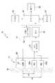
도 1은 광학적 측정 장치의 개략 구성도이다.1 is a schematic configuration diagram of an optical measuring device.
도 2는 제어부에서 실행되는 처리의 플로우차트이다.2 is a flowchart of processing executed in the control unit.
도 3은 송수광기간 거리와 공간적 기울기 S와의 관계를 도시하는 선도이다.3 is a diagram showing the relationship between the transmission and reception period distance and the spatial slope S. FIG.
도 4A는 근조직의 흡수 계수와 공간적 기울기 S와의 관계를 피부의 흡수 계수마다 도시하는 선도이다.4A is a diagram showing the relationship between the absorption coefficient of muscle tissue and the spatial slope S for each skin absorption coefficient.
도 4B는 근조직의 흡수 계수와 공간적 기울기 S와의 관계를 피부의 산란 계수마다 도시하는 선도이다.4B is a diagram showing the relationship between the absorption coefficient of muscle tissue and the spatial slope S for each scattering coefficient of the skin.
도 5A는 근조직의 흡수 계수와 공간적 기울기 S와의 관계를 지방의 흡수 계 수마다 도시하는 선도이다.5A is a diagram showing the relationship between the absorption coefficient of muscle tissue and the spatial slope S for each absorption coefficient of fat.
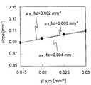
도 5B는 근조직의 흡수 계수와 공간적 기울기 S와의 관계를 지방의 산란 계수마다 도시하는 선도이다.5B is a diagram showing the relationship between the absorption coefficient of muscle tissue and the spatial slope S for each scattering coefficient of fat.
도 6은 근조직의 흡수 계수와 공간적 기울기 S와의 관계를 근조직의 산란 계수마다 도시하는 선도이다.Fig. 6 is a diagram showing the relationship between the absorption coefficient of muscle tissue and the spatial slope S for each scattering coefficient of muscle tissue.
도 7은 근조직의 흡수 계수와 공간적 기울기 S와의 관계를 지방두께 마다 도시하는 선도이다.7 is a diagram showing the relationship between the absorption coefficient of muscle tissue and the spatial slope S for each fat thickness.
도 8은 산소 포화도를 근조직의 산란 계수마다 도시하는 선도이다.8 is a graph showing oxygen saturation for each scattering coefficient of muscle tissue.
도 9는 산소 포화도를 지방두께 마다 도시하는 선도이다.9 is a diagram showing oxygen saturation for each fat thickness.
도 10은 다른 예의 광학적 측정 장치의 개략 구성도이다.10 is a schematic configuration diagram of another example of an optical measuring device.
<도면의 주요 부분에 대한 부호의 설명><Explanation of symbols for main parts of the drawings>
10 : 광학적 측정 장치10: optical measuring device
12 : 프로브12: probe
14 : 구동장치14: drive device
16 : 제어부(공간적 기울기 산출 수단, 연산 수단)16 control part (spatial slope calculation means, calculation means)
18 : 조작부(입력 수단)18: operation unit (input means)
20 : 메모리(기억 수단)20 memory (memory means)
22 : 출력부22: output unit
24 : LED(발광 수단)24: LED (light emitting means)
26A : PD( 제1 수광부)26A: PD (first light receiving unit)
26B : PD( 제2 수광부)26B: PD (second light receiver)
30 : 조직30: organization
32 : LED 드라이버32: LED driver
34 : I-V 컨버터34: I-V converter
36 : 앰프36: amplifier
이하, 본 발명의 실시 형태에 대해 도면을 참조하면서 상세하게 설명한다.EMBODIMENT OF THE INVENTION Hereinafter, embodiment of this invention is described in detail, referring drawings.
본 실시 형태에서는, 일례로서 인간의 팔의 근조직에서의 혈액량, 즉 헤모글로빈 농도나 산소 포화도를 측정하는 경우에 대해 설명한다.In this embodiment, the blood volume in the muscle tissue of a human arm, that is, the case where hemoglobin concentration and oxygen saturation are measured as an example is demonstrated.
도 1에는, 광학적 측정 장치(10)의 개략 구성을 도시한다. 도 1에 도시한 바와 같이, 광학적 측정 장치(10)는, 프로브(12), 구동장치(14), 제어부(16), 조작부(18), 메모리(20), 및 출력부(22)를 포함하여 구성되어 있다.1, the schematic structure of the optical measuring
프로브(12)는, LED(발광 다이오드, 24) 및 2개의 PD(포토 다이오드, 26A, 26B)가, 예컨대 가요성(可撓性, flexibility)을 갖는 평판 모양의 부재(예컨대 고무 성질의 부재 등, 28)에 설치되는 구성이다. 프로브(12)는, 예컨대 피측정자의 팔의 조직(30) 내에 빛을 쬐기 위해 피측정자의 팔에 접촉시킨다.The
LED(24)는, 본 실시 형태에서 일례로서 피크 파장이 제1 파장(λ1), 제2 파장(λ2)의 2 파장의 발광 다이오드이다. 제1 파장(λ1), 제2 파장(λ2)은, 물의 흡수가 적은 파장, 구체적으로는 900nm 이하의 파장으로, 한편 탈산소화 헤모글로 빈(Hb)와 산소화 헤모글로빈(HbO2)의 흡수 스펙트럼이 교차하는 위치의 파장인 약 805nm로부터 대략 등거리에 있는 파장으로 설정된다. 본 실시 형태에서는 일례로서 제1 파장(λ1)이 770nm, 제2 파장(λ2)이 830nm이다.As an example in this embodiment, the
LED(24)와 하나의 PD(26A)는, 제1 소정 거리(d1) 만큼 떨어져 배치되고 있고, LED(24)와 다른 하나의 PD(26B)는 제2 소정 거리(d2) 만큼 떨어져 배치되고 있다.The
제1 소정 거리(d1)는, LED(24)로부터 발광된 빛이, 인간의 팔의 심층 부분, 즉 피부 조직(표층) 및 지방조직(천층) 보다 더 아래의 근조직을 통과하여 하나의 PD(26A)에 도달하도록 하는 거리로 설정된다. 본 발명자는, 측정 대상인 심층 내의 빛의 통과 거리(여기에서는 평균 광로 길이)가 10mm 정도 이상이면, 그 층의 정보를 범용의 전자 회로에서도 충분한 S/N비로 검출할 수 있음을 실험적으로도 확인하였고, 천층의 두께가 0∼8mm 정도 일때에 심층의 빛의 통과 거리가 10mm 이상이 가능한 송수광기간 거리를 시뮬레이션으로 구했다. 그 결과, 본 실시 형태에서는, 제1 소정 거리(d1)를 일례로서 20mm로 설정하였다.The first predetermined distance d1 indicates that light emitted from the
또, 제2 소정 거리(d2)는, LED(24)로부터 발광된 빛이, 인간의 팔의 심층 부분을 통과하여 다른 하나의 PD(26B)에 도달하도록 하는 거리로, 제1 소정 거리와는 다른 거리로 설정된다. 송수광기간 거리가 길어지면 지수함수적으로 광강도가 감쇠하고, 범용의 전자 회로에서의 검출이 곤란하게 되기 때문에, 본 발명자는, 범용의 전자 회로에서 검출 가능한 광강도를 얻을 수 있는 송수광기간 거리를 이론과 실험으로부터 구했다. 그 결과, 본 실시 형태에서는, 제2 소정 거리(d2)를 일례로서 30mm로 설정하였다. 아울러, 제1 소정 거리(d1), 제2 소정 거리(d2)는 일례이며, 측정하고자 하는 근조직까지의 깊이 등에 따라 적절한 거리로 정할 수 있다.In addition, the second predetermined distance d2 is a distance for allowing the light emitted from the
구동장치(14)는, LED 드라이버(32), I-V 컨버터(34), 및 앰프(36)를 포함하여 구성될 수 있다.The
LED 드라이버(32)는, 제어부(16)로부터의 지시에 의해, LED(24)를 소정의 파장 및 소정의 광강도로 발광시킨다.The
I-V 컨버터(34)는, PD(26A, 26B)에서 수광한 빛을 광전 변환 함으로써 얻을 수 있는 전류를 전압으로 변환하여 앰프(36)에 출력한다.The
앰프(36)는, I-V 컨버터(34)에 의해 변환된 전압을 소정 레벨의 전압으로 증폭하고, 광강도를 나타내는 신호로서 제어부(16)에 출력한다.The
제어부(16)는, LED 드라이버(32)에 LED(24)를 발광시키도록 지시하고, 그 결과 얻을 수 있는 PD(26A, 26B)에서 수광한 빛의 광강도에 기초하여, 후술하는 연산에 의해 헤모글로빈 농도 등을 산출한다. 연산 결과는, 출력부(22)에 출력된다. 출력부(22)는, 예컨대 디스플레이나 프린터 등으로 구성되고, 연산 결과를 표시하거나 인쇄 함으로써 출력한다.The
메모리(20)에는, 후술하는 처리 루틴의 프로그램이나, 그 처리에서 이용하는 데이터로 미리 실행한 시뮬레이션 결과에 관한 데이터 등이 미리 기억되고 있다.In the
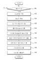
다음으로, 본 실시 형태의 작용으로서 제어부(16)에서 실행되는 측정 처리 에 대해, 도 2에 도시하는 플로우차트를 참조하여 설명한다. 또한 상기 처리는, 광학적 측정 장치(10)의 전원이 온 되면 실행된다.Next, the measurement process performed by the
측정할 때에는, 프로브(12)를 피측정자의 팔에 접촉시키고, 조작부(18)를 조작 함으로써 측정 개시를 지시한다.In the measurement, the
단계 100에서는, 조작부(18)의 조작에 의해 측정 개시가 지시되었는지를 판단하고, 측정 개시가 지시된 경우에는 단계 102로 이행한다.In
단계 102에서는, 피측정자의 지방두께를, 예컨대 조작부(18)의 조작에 의해 입력시킨다. 지방두께는, 예컨대 노기스(slide caliper) 등의 간단한 측정 부재를 이용하여 측정한 것이 입력되도록 하여도 무방하다. 또, 예컨대 도시하지 않는 지방두께 측정장치(예컨대, 초음파 진단 장치 등)를 광학적 측정 장치(10)에 접속하고, 상기 지방두께 측정장치에 의해 측정된 지방두께를 직접 입력되도록 하여도 무방하다. 입력된 지방두께는, 후술하는 근조직의 흡수 계수 μa_m(빛의 흡수 정도)을 구할 때에 필요하다.In
단계 104에서는, LED 드라이버(32)에 대해서 LED(24)의 발광을 지시하고, PD(26A), PD(26B)에서 수광한 빛의 광강도 IA, IB를 앰프(36)로부터 수신한다. 또는, 제1 파장(λ1), 제2 파장(λ2)으로 순차적으로 발광시키고, 각각의 광강도 IA, IB를 수신한다. 이하에서는, 제1 파장(λ1)으로 발광한 경우의 PD(26A), PD(26B)에서 수광한 빛의 광강도를 IA1, IB1이라 하고, 제2 파장(λ2)으로 발광한 경우의 PD(26A), PD(26B)에서 수광한 빛의 광강도를 IA2, IB2라 한다.In
단계 106에서는, 단계 104에서 측정한 광강도에 기초하여, 공간 분해법에서의 공간적 기울기 S를 구한다. 도 3에 도시한 바와 같이, LED와 PD와의 거리(송수광기간 거리)와, 수광한 빛의 광강도(log I)와는 도 3에 도시한 것과 같은 관계이고, 본 실시 형태에서의 공간적 기울기 S는, 도 3에 도시한 바와 같이 송수광기간 거리가 20mm인 경우의 광강도를 나타내는 A점과, 송수광기간 거리가 30mm의 광강도를 나타내는 B점을 연결한 선의 기울기로 표시된다. 본 실시 형태에서는, 공간적 기울기 S를 편의적으로 다음식과 같이 정의한다.In
수식 1Equation 1

여기서, ρ는 2개의 PD(26A), PD(26B) 사이의 거리이고, 본 실시 형태에서는 d1=20mm, d2=30mm로 하고 있기 때문에, ρ는 10mm가 된다.Here, p is a distance between two
공간적 기울기 S는, 파장 마다 구한다. 이하에서는, 제1 파장(λ1)으로 발광한 경우의 광강도 IA1, IB1에 기초하여 구한 공간적 기울기를 S1, 제2 파장(λ2)으로 발광한 경우의 광강도 IA2, IB2에 기초하여 구한 공간적 기울기를 S2로 한다.The spatial slope S is obtained for each wavelength. Hereinafter, based on the light intensities IA2 and IB2 when the spatial gradients obtained based on the light intensities IA1 and IB1 when emitting light at the first wavelength λ1 are emitted at the second wavelength λ2. The spatial slope obtained by
다음으로, 단계 108에서는, 단계 106에서 구한 공간적 기울기 S1, S2에 기초하여, 측정 대상인 팔의 근조직의 흡수 계수 μa_m을 구한다. 상기 근조직의 흡수 계수 μa_m은, 후술하는 몬테카를로 시뮬레이션 결과로부터 얻을 수 있는 S -μa_m 곡선을 이용하여 구한다.Next, in
여기서, 본 발명자가 수행한 몬테카를로 시뮬레이션의 결과에 대해 설명한다. 본 발명자는, 근조직의 산소 농도의 정량화를 위해서, 생체 조직의 모델로서, 피부, 지방, 근조직의 3층 모델에서의 몬테카를로 시뮬레이션을 실시하였다. 광전파의 알고리즘으로는, 모델 중에서 광자군을 무작위 동작시키고, 통과한 매질의 종류에 따라 광자군의 양을 감쇠시킨다는 일반적인 것을 이용하였다(하기 비특허문헌 6 참조).Here, the results of the Monte Carlo simulations performed by the inventors will be described. The present inventors conducted Monte Carlo simulation in a three-layer model of skin, fat, and muscle tissue as a model of living tissue, in order to quantify the oxygen concentration of muscle tissue. As the algorithm of the light propagation, a general method of randomly operating the photon group in the model and attenuating the amount of the photon group in accordance with the type of the medium passed through was used (see
각 층의 두께와 광학 정수(산란 계수, 흡수 계수)는, 이하의 표 1과 같이 설정하였다(하기 비특허문헌 7∼9 참조).The thickness and optical constant (scattering coefficient, absorption coefficient) of each layer were set as Table 1 below (refer the following nonpatent documents 7-9).
표 1TABLE 1

시뮬레이션에서는, 지방두께를 변화시켜 두께의 영향을 검증하는 것과 함께, 피부의 흡수 계수 및 산란 계수를 20% 증감시키고, 표층 및 천층 조직의 광학 정수의 영향을 해석하였다. 또, 공간 분해법에서는, 근조직의 산란 계수 μs_m을 적당하다고 판단되는 값으로 가정할 필요가 있지만, 상기 가정이 실제와 다른 경우의 오차를 검토하기 위해서,μs_m을 0.2mm-1 증감시켰을 경우의 시뮬레이션도 수행하였다.In the simulation, the effect of the thickness was changed by changing the fat thickness, the skin absorption coefficient and the scattering coefficient were increased and decreased by 20%, and the influence of the optical constants of the surface layer and the top layer tissue was analyzed. In addition, in the spatial decomposition method, it is necessary to assume a scattering coefficient μs_m of muscle tissue as a value judged to be appropriate, but in order to examine the error when the above assumption is different from the actual one, a simulation diagram when the μs_m is increased or decreased by 0.2 mm-1 is also used. Was performed.
도 4A는 근조직의 흡수 계수 μa_m과 공간적 기울기 S(slope)와의 관계를 피부의 흡수 계수 μa_skin이 0.01mm-1, 0.0125mm-1, 0.015mm-1의 경우에 대해 구한 결과를 나타내고, 도 4B는 근조직의 흡수 계수 μa_m과 공간적 기울기 S(slope)와의 관계를 피부의 산란 계수 μs_skin이 1.0mm-1, 1.2mm-1, 1.4mm-1의 경우에 대해 구한 결과를 나타낸다. 도 4A 및 4B에 도시한 바와 같이, 피부의 흡수 계수 μa_skin, 산란 계수 μs_skin에 의해서는 근조직의 흡수 계수 μa_m은 거의 영향을 받지 않는 것으로 판명되었다.Figure 4A shows the result obtained for the relationship between the absorption coefficient μa_m of the muscle tissue and the spatial slope S (slope) when a skin absorption coefficient μa_skin of0.01mm -1, 0.0125mm -1, 0.015mm -1 , Figure 4B The relationship between the absorption coefficient μa_m of the muscle tissue and the spatial slope S (slope) is obtained for the case where the skin scattering coefficient μs_skin is 1.0mm-1 , 1.2mm-1 , 1.4mm-1 . As shown in Figs. 4A and 4B, it was found that the absorption coefficient µa_m of the skin and the scattering coefficient µs_skin were hardly affected by the absorption coefficient µa_m of the muscle tissue.
또, 도 5A는 근조직의 흡수 계수 μa_m과 공간적 기울기 S(slope)와의 관계를 지방의 흡수 계수 μa_fat이 0.002mm-1, 0.003mm-1, 0.004mm-1의 경우에 대해 구한 결과를 나타내고, 도 5B는 근조직의 흡수 계수 μa_m과 공간적 기울기 S(slope)와의 관계를 지방의 산란 계수 μs_fat이 1.0mm-1, 1.2mm-1, 1.4mm-1의 경우에 대해 구한 결과를 나타낸다. 도 5A 및 5B에 도시한 바와 같이, 지방의 흡수 계수 μa_fat, 산란 계수 μs_fat에 의해서는 근조직의 흡수 계수 μa_m은 거의 영향을 받지 않는 것으로 판명되었다.In addition, Figure 5A is a muscle tissue of the relationship between the absorption coefficient μa_m the spatial slope S (slope) is the local absorption coefficient μa_fat show the results obtained for the case of0.002mm -1, 0.003mm -1, 0.004mm -1 , Fig. 5B shows the result of the relationship between the absorption coefficient μa_m of the muscle tissue and the spatial slope S (slope) for the case where the fat scattering coefficient μs_fat is 1.0mm-1 , 1.2mm-1 , 1.4mm-1 . As shown in Figs. 5A and 5B, the absorption coefficient μa_fat and the scattering coefficient μs_fat of fat were found to be hardly affected by the absorption coefficient μa_m of muscle tissue.
또, 도 6은 근조직의 흡수 계수 μa_m과 공간적 기울기 S와의 관계를 근조직의 산란 계수 μs_m이 1.0mm-1, 0.8mm-1, 0.6mm-1의 경우에 대해 구한 결과를 나타낸다. 도 6에 도시한 바와 같이, 근조직의 산란 계수 μs_m이 0.2mm-1 달라지면, 근조직의 흡수 계수 μa_m의 절대치는 20% 이상 달라지게 되는 것으로 판명되었다.6 shows the result obtained by calculating the relationship between the absorption coefficient μa_m of the muscle tissue and the spatial slope S in the case where the scattering coefficient μs_m of the muscle tissue is 1.0mm-1 , 0.8mm-1 , 0.6mm-1 . As shown in FIG. 6, it was found that when the scattering coefficient μs_m of the muscle tissue was changed by 0.2 mm−1 , the absolute value of the absorption coefficient μa_m of the muscle tissue was changed by 20% or more.
또, 도 7은 근조직의 흡수 계수 μa_m과 공간적 기울기 S와의 관계(S-μa_m 곡선)를 지방두께가 3, 5, 7, 9, 15mm의 경우에 대해 구한 결과를 나타낸다. 도 7에 도시한 바와 같이, 지방두께에 따라 S-μa_m 곡선의 형상이 크게 달라진다는 것으로 판명되었다. 이에 따라, 미리 피측정자의 지방두께를 측정해 두고, 지방두께에 따라 S-μa_m 곡선을 이용 함으로써, 근조직의 흡수 계수 μa_m을 정량화할 수 있다. S-μa_m 곡선은 공간적 기울기 S와 관련하여 하기에 나타낸 것과 같은 2차식으로 근사할 수 있다.7 shows the result obtained by calculating the relationship (S-μa_m curve) between the absorption coefficient μa_m of the muscle tissue and the spatial slope S for the case where the fat thickness is 3, 5, 7, 9, and 15 mm. As shown in Fig. 7, it was found that the shape of the S-μa_m curve largely changed depending on the fat thickness. Accordingly, the fat thickness of the subject is measured in advance, and the absorption coefficient μa_m of the muscle tissue can be quantified by using the S-μa_m curve according to the fat thickness. The S-μa_m curve can be approximated in quadratic form as shown below with respect to the spatial slope S.
수식 2

여기서, a, b, c는 정수이고, 이들 정수를 도 7에 도시한 몬테카를로 시뮬레이션의 결과로부터 지방두께 및 파장마다 구하고, 미리 메모리(20)에 기억해 둔다. 이에 따라, 지방두께, 공간적 기울기 S가 판명되면, 흡수 계수 μa_m을 구할 수 있다. 또한, a, b, c의 값은 지방두께나 근조직의 산란 계수 μs_m, 송수광기간 거리에 의해 달라지지만, 본 발명자가 수행한 시뮬레이션에서는, 일례로서 지방두께가 3mm, 근조직의 산란 계수 μs_m이 0.8mm-1의 경우에서는, a=4.95, b=-0.56, c=0.017이다. 또, 피부의 흡수 계수 μa_skin 및 산란 계수 μs_skin을 20% 증감시킨 결과, S-μa_m 곡선은 거의 동일하게 되어, 피부의 광학 정수는 공 간 분해법에서는 거의 영향을 주지 않는 것이 판명되었다.Here, a, b, and c are integers, and these constants are obtained for each fat thickness and wavelength from the Monte Carlo simulation results shown in Fig. 7, and stored in the
도 8은, 근조직의 산란 계수 μs_m이 0.6mm-1, 0.8mm-1, 1.0mm-1의 경우에서의 산소 포화도 StO2에 대해 실측한 결과를 도시한다. 산소 포화도는, 후술하는 것과 같이 산소화 헤모글로빈 농도를 토탈 헤모글로빈 농도(산소화 헤모글로빈 농도와 탈산소화 헤모글로빈 농도의 합)로 나눔으로써 얻을 수 있다. 도 8에서, 「Occlusion」의 기간은, 동정맥 폐색 기간으로, 위팔(上腕)을 단단히 조여 혈류를 멈추는 처리를 실시한 기간, 「rest」는 아무것도 하지 않는 기간이다.FIG. 8 shows the results obtained by measuring the oxygen saturation St O2 in the case where the scattering coefficient μs_m of muscle tissue is 0.6 mm−1 , 0.8 mm−1 , 1.0 mm−1 . The oxygen saturation can be obtained by dividing the oxygenated hemoglobin concentration by the total hemoglobin concentration (sum of oxygenated hemoglobin concentration and deoxygenated hemoglobin concentration) as described later. In FIG. 8, the period of "Occlusion" is an arteriovenous occlusion period, a period in which the upper arm is tightly tightened to stop blood flow, and "rest" is a period in which nothing is done.
상술한 바와 같이, 근조직의 산란 계수 μs_m이 0.2mm-1 달라지면, 근조직의 흡수 계수 μa_m의 절대치는 20% 이상 달라지는 것이 판명되었지만, 도 8에 도시한 바와 같이, 산소 포화도 StO2에 관하여서는, 근조직의 산란 계수 μs_m의 차이로 인한 오차는 수% 이내가 되는 것이 판명되었다. 이는, 산소 포화도 StO2가 산소화 헤모글로빈 농도와 토탈 헤모글로빈 농도와의 비로 표시되기 때문에, 2개의 흡수 계수의 비를 보는 것과 관련하고 있다. 근조직의 산란 계수 μs_m에 의해 S-μa_m곡선이 달라져도 닮음꼴이면, 곡선으로부터 구해진 2개의 흡수 계수의 비는 같게 된다. 근조직의 산란 계수 μs_m의 차이는, 주로 S-μa_m 곡선의 세로축 방향의 크기에 영향을 주고, 형상에는 크게 영향을 주지 않는 것이, 산소 포화도 StO2에 관한 오차가 매우 적어진 요인이라고 생각된다.As described above, when the scattering coefficient μs_m of the muscle tissue is 0.2mm-1 , the absolute value of the absorption coefficient μa_m of the muscle tissue is found to be changed by 20% or more. However, as shown in FIG. 8, the oxygen saturation St O2 is It was found that the error due to the difference in the scattering coefficient of the muscle tissue, μs μm, was within several%. This is related to seeing the ratio of two absorption coefficients because the oxygen saturation St O2 is expressed as the ratio of oxygenated hemoglobin concentration to total hemoglobin concentration. Even if the S-μa_m curve is changed by the scattering coefficient μs_m of the muscle tissue, the ratio of the two absorption coefficients obtained from the curve becomes the same. The difference in the scattering coefficient μs_m of the muscle tissue mainly affects the magnitude of the S-μa_m curve in the longitudinal axis direction, and it is considered that the error with respect to the oxygen saturation St O2 is very small. .
도 9는, 지방층 3mm 부위에서의 산소 포화도를 실측한 결과를 나타낸다. 도 9에 도시한 바와 같이, 실제와는 다른 지방두께(5∼9mm)에서의 S-μa_m 곡선을 사용하면, 산소 포화도 StO2의 값으로 최대 30% 정도의 오차가 발생되는 것이 판명되었다. 이는 지방두께가 달라지면 S-μa_m 곡선의 형상이 크게 변화하는 것에 기인한다.Fig. 9 shows the results of actual measurement of oxygen saturation at 3 mm of fat layer. As shown in Fig. 9, it was found that an error of up to 30% was generated in the value of oxygen saturation St O2 by using the S-μa_m curve at an actual fat thickness (5 to 9 mm). . This is due to the large change in the shape of the S-μa_m curve when the fat thickness is changed.
공간 분해법에서 헤모글로빈 농도의 절대량을 정확하게 인지하는 데에는, 심부 조직의 산란 계수를 미리 알지 못하면 곤란하다 할 수 있다. 이에 반해, 산소 포화도 StO2로 하면, 산란 계수를 가정하는 영향을 큰 폭으로 줄일 수 있다. 그러나, 산소 포화도 StO2이라도, 지방두께의 영향은 크고, 미리 두께를 파악해 두는 것이 정량화에서 중요하다.It is difficult to accurately recognize the absolute amount of hemoglobin concentration by spatial decomposition without knowing the scattering coefficient of deep tissue in advance. On the other hand, when the oxygen saturation is St O2 , the influence of assuming a scattering coefficient can be greatly reduced. However, even with oxygen saturation St O2 , the influence of fat thickness is large, and it is important to quantify the thickness in advance.
도 9의 지방두께가 3mm와 5mm의 결과로부터, 지방두께를 ±1% 정도의 정밀도로 측정해 두면, 산소 포화도 StO2는 2∼3% 이하의 오차로 할 수 있다고 추정되고, 노기스 등의 간단한 두께 측정법으로 지방두께를 측정하더라도 대응할 수 있다고 생각된다.From the results of 3 mm and 5 mm fat thickness in FIG. 9, if the fat thickness is measured with an accuracy of about ± 1%, the oxygen saturation St O2 is estimated to be an error of2 to 3% or less. Even if the fat thickness is measured by the simple thickness measurement method of, it is thought that it can cope.
상기와 같은 시뮬레이션 및 실측 결과로부터, 본 실시 형태에서는, 근조직의 흡수 계수 μa_m을 지방두께에 따라 구하고, 이로부터 헤모글로빈 농도나 산소 포화도를 구한다.From the above simulation and measurement results, in the present embodiment, the absorption coefficient μa_m of the muscle tissue is determined according to the fat thickness, and the hemoglobin concentration and the oxygen saturation are calculated therefrom.
우선, 단계 108에서는, 근조직의 흡수 계수 μa_m을 상기 (2)식에 의해 구한다. 즉, 단계 102에서 입력된 지방두께에 대응하는 정수 a, b, c의 값을 메모리(20)로부터 읽어내고, 이것과 단계 106에서 구한 공간적 기울기 S로부터, 상기 (2)식에 의한 근조직의 흡수 계수 μa_m을 구한다. 또한, 근조직의 흡수 계수 μ_am은 파장마다 구한다. 이하에서는, 제1 파장(λ1)의 경우 근조직의 흡수 계수를 μλ1_am, 제2 파장(λ2)의 경우 근조직의 흡수 계수를 μλ2_am으로 한다. 또한, 상술한 바와 같이, 근조직의 산란 계수 μs_m에 의해 S-μa_m 곡선의 형상은 달라지기 때문에, 예컨대 근조직의 산란 계수 μs_m이 0.8의 경우 S-μa_m 곡선을 이용하여 근조직의 흡수 계수 μa_m을 구한다.First, in
단계 110에서는, 단계 108에서 구한 근조직의 흡수 계수 μλ1_am, μλ2_am에 기초하여, 산소화 헤모글로빈 농도[HbO2]를 구한다. 상기 산소화 헤모글로빈 농도[HbO2]는 다음식으로 구할 수 있다.In
수식 3Equation 3

여기서,ελ1Hb는 제1 파장(λ1)에서의 탈산소화 헤모글로빈의 분자 흡광 계수, ελ2Hb는 제2 파장(λ2)에서의 탈산소화 헤모글로빈의 분자 흡광 계수, ελ1HbO2는 제1 파장(λ1)에서의 산소화 헤모글로빈의 분자 흡광 계수, ελ2HbO2는 제2 파장(λ2)에서의 산소화 헤모글로빈의 분자 흡광 계수이며, 어느 쪽이라도 알려진 기존의 값(예컨대 하기 비특허문헌 10에 기재된 값)을 이용한다.Here, ελ1 Hb is the molecular absorption coefficient of deoxygenated hemoglobin at the first wavelength λ1, ελ2 Hb is the molecular absorption coefficient of deoxygenated hemoglobin at the second wavelength λ2, and ελ1 HbO2 is the first wavelength molecular extinction coefficient of oxygenated hemoglobin in (λ1), ελ2 HbO2 is first a molecular extinction coefficient of the oxygenated hemoglobin at the second wavelength (λ2), either an existing value is known (for example, to non-value as described in Patent Document 10 ).
단계 112에서는, 단계 108에서 구한 근조직의 흡수 계수 μλ1_am, μλ2_am에 기초하여, 탈산소화 헤모글로빈 농도[Hb]를 구한다. 상기 탈산소화 헤모글로빈 농도[Hb]는 다음식으로 구할 수 있다.In
수식 4

그리고, 단계 114에서는, 토탈 헤모글로빈[total Hb]를 다음식에 의해 구한다.And in
[total Hb]=[HbO2]+[Hb] … (5)[Total Hb] = [HbO2 ] + [Hb]. (5)
다음으로, 단계 116에서는, 산소 포화도 StO2를 다음식에 의해 구한다.Next, in
[Hb]=[HbO2]/[total Hb] … (6)[Hb] = [HbO2 ] / [total Hb]... (6)
단계 118에서는, 구한 산소화 헤모글로빈 농도[HbO2], 탈산소화 헤모글로빈 농도[Hb], 산소 포화도 StO2를 출력부(22)에 출력시킨다.In
이와 같이, 본 실시 형태에서는, 지방두께에 따라 S-μa_m 곡선을 이용하여 근조직의 흡수 계수 μa_m을 구하고, 이에 기초하여 근조직의 산소화 헤모글로빈 농도나 탈산소화 헤모글로빈 농도, 산소 포화도를 구한다. 이 때문에, 지방두께의 영향이 보정된 정확한 산소화 헤모글로빈 농도나 탈산소화 헤모글로빈 농도, 산소 포화도를 얻을 수 있고, 이들의 정량성을 큰 폭으로 향상시킬 수 있다.As described above, in the present embodiment, the absorption coefficient μa_m of the muscle tissue is determined using the S-μa_m curve according to the fat thickness, and the oxygenated hemoglobin concentration, the deoxygenated hemoglobin concentration, and the oxygen saturation degree of the muscle tissue are calculated based on this. Therefore, accurate oxygenated hemoglobin concentration, deoxygenated hemoglobin concentration, and oxygen saturation can be obtained in which the influence of fat thickness is corrected, and these quantitative properties can be greatly improved.
아울러, 본 실시 형태에서는, 사람 팔의 근조직의 헤모글로빈 농도 등을 측정하는 경우에 대해 설명하였지만, 본 발명은 이에 한정하지 않고, 예컨대 과일의 과육 부분의 당분을 측정하는 장치에도 적용 가능하다. 이 경우, LED의 파장을 포도당(glucose)의 빛의 흡수 계수를 측정하는 데에 적합한 파장으로 설정하거나, LED와 PD 사이의 거리를 과일의 외피나 내피 두께에 적절한 거리로 설정하는 등, 적절히 필요한 설정을 할 필요가 있지만, 기본적으로는 상기와 같은 방법에 의해 과일의 과육 부분의 당분을 측정하는 것이 가능하다. 즉, 본 발명은, 내부 조직까지 빛이 도달하는 것이라면, 생체에 한정하지 않고 다른 물건에 대해서도 적용 가능하다.In addition, although this embodiment demonstrated the case where the hemoglobin density | concentration etc. of the muscle tissue of a human arm were measured, this invention is not limited to this, For example, it is applicable also to the apparatus which measures the sugar of the pulp part of a fruit. In this case, the wavelength of the LED is set to a wavelength suitable for measuring the absorption coefficient of glucose light, or the distance between the LED and the PD is set to a distance suitable for the thickness of the outer skin or the thickness of the fruit. Although it is necessary to set, it is basically possible to measure the sugar of the pulp portion of the fruit by the same method as described above. That is, the present invention can be applied not only to living bodies but also to other objects as long as light reaches the internal tissues.
또, 본 실시 형태에서는, PD를 두 개 설치한 구성의 경우에 대해서 설명하였지만, 이에 한정하지 않고, PD를 하나로 하거나, LED를 두 개로 하여도 무방하다. 또, LED 또는 PD를 이동 가능한 구성으로, 즉 LED와 PD의 거리를 조정 가능한 구성으로 하여도 무방하다. 이 경우, LED와 PD의 거리를 제1 소정 거리(d1)로 설정하여 LED로부터 발광된 빛을 수광하고, 그 후 LED와 PD의 거리를 제2 소정 거리(d2)로 설정하여 LED로부터 발광된 빛을 수광하도록 하면 된다.In the present embodiment, the configuration in which two PDs are provided has been described. However, the present invention is not limited thereto, and one PD or two LEDs may be used. The LED or PD may be configured to be movable, that is, the configuration where the distance between the LED and PD may be adjusted. In this case, the distance between the LED and the PD is set to the first predetermined distance d1 to receive light emitted from the LED, and then the distance between the LED and the PD is set to the second predetermined distance d2 to emit light from the LED. You just need to receive the light.
또, LED 등의 발광을, 펄스 형상 내지 간헐적으로 실시하고, 수광 수단의 PD 등의 소자의 출력을, 도 10에 도시한 바와 같이 시간 분해법을 갖는 록 인 앰프(Rock-in amplifier, 37)(또는 박스카 적분기(boxcar integrator), 위상 검파기 등)를 앰프(36)의 전단에 부가 함으로써, 감도 및 정밀도가 향상되도록 구성하여도 무방하다. 또한, LED를 두 개로 한 구성의 경우는, 교대로 발광 내지 발광 패턴을 반복하여 발광하게 된다. 또, 형광등 등에 의한 측정시의 외란광의 영향을 제거하기 위해서, LED 등의 발광을, 상용 주파수와는 다른 주파수에서의 정현파 교류 등으로 변조하도록 하여도 무방하다.In addition, a lock-in
본 발명에 의하면, 천층 조직의 영향 등을 보정하여 인체나 과일 등의 심층 조직의 빛의 흡수 정도를 정확하게 측정할 수 있다라는 효과가 있다.According to the present invention, there is an effect that the degree of light absorption of deep tissues such as the human body or fruit can be accurately measured by correcting the influence of the shallow tissue.
Claims (9)
Translated fromKoreanApplications Claiming Priority (2)
| Application Number | Priority Date | Filing Date | Title |
|---|---|---|---|
| JP2006152177 | 2006-05-31 | ||
| JPJP-P-2006-152177 | 2006-05-31 |
Publications (2)
| Publication Number | Publication Date |
|---|---|
| KR20090011030A KR20090011030A (en) | 2009-01-30 |
| KR101041727B1true KR101041727B1 (en) | 2011-06-14 |
Family
ID=38778709
Family Applications (1)
| Application Number | Title | Priority Date | Filing Date |
|---|---|---|---|
| KR20087030473AExpired - Fee RelatedKR101041727B1 (en) | 2006-05-31 | 2007-05-31 | Storage medium recording optical measuring device, optical measuring method, and optical measuring program |
Country Status (6)
| Country | Link |
|---|---|
| US (1) | US8369914B2 (en) |
| EP (1) | EP2034294B1 (en) |
| JP (1) | JP5062698B2 (en) |
| KR (1) | KR101041727B1 (en) |
| CN (1) | CN101454654B (en) |
| WO (1) | WO2007139192A1 (en) |
Cited By (1)
| Publication number | Priority date | Publication date | Assignee | Title |
|---|---|---|---|---|
| WO2018016709A3 (en)* | 2016-07-21 | 2018-08-02 | 주식회사 인핏앤컴퍼니 | Frequency domain-based multi-wavelength biometric signal analysis apparatus and method thereof |
Families Citing this family (27)
| Publication number | Priority date | Publication date | Assignee | Title |
|---|---|---|---|---|
| EP1653227B1 (en)* | 2003-07-11 | 2017-10-25 | SUN-A Corporation | Device and method of detecting liquid kind |
| JP4342855B2 (en)* | 2003-07-11 | 2009-10-14 | 三井金属鉱業株式会社 | Gas oil liquid type identification device and light oil liquid type identification method |
| KR101034798B1 (en)* | 2009-03-18 | 2011-05-17 | 한국과학기술연구원 | Brain condition measuring device |
| CA2764498C (en) | 2009-06-05 | 2016-09-20 | Nonin Medical, Inc. | Oximetry with remote display |
| JP2011067349A (en)* | 2009-09-25 | 2011-04-07 | Panasonic Electric Works Co Ltd | Body composition measuring apparatus |
| JP5510796B2 (en)* | 2010-01-12 | 2014-06-04 | 独立行政法人産業技術総合研究所 | Minimally invasive angiogenesis measuring device |
| JP5527658B2 (en)* | 2010-04-30 | 2014-06-18 | 浜松ホトニクス株式会社 | Scattering absorber measurement method and apparatus |
| CN102058393B (en)* | 2010-10-30 | 2012-10-31 | 华中科技大学 | Method for measuring skin physiological parameters and optical characteristic parameters based on reflection spectrum measurement |
| US9433352B2 (en) | 2011-02-23 | 2016-09-06 | National University Corporation Shizuoka University | Optical measuring device |
| JP5834704B2 (en)* | 2011-09-27 | 2015-12-24 | セイコーエプソン株式会社 | Concentration determination apparatus, light absorption coefficient calculation method, concentration determination method, program for calculating light absorption coefficient, and program for calculating concentration |
| JP2013103094A (en)* | 2011-11-16 | 2013-05-30 | Sony Corp | Measurement device, measurement method, program, and recording medium |
| WO2014188906A1 (en)* | 2013-05-24 | 2014-11-27 | 国立大学法人浜松医科大学 | Near infrared oxygen concentration sensor for palpation |
| WO2015115182A1 (en) | 2014-01-29 | 2015-08-06 | コーケンメディカル株式会社 | Non-invasive monitor for measuring regional saturation of oxygen |
| SG11201701015QA (en)* | 2014-08-29 | 2017-03-30 | Univ Tohoku | Optical concentration measuring method |
| JP2017023455A (en)* | 2015-07-23 | 2017-02-02 | 株式会社アドバンテスト | Near-infrared bioinstrumentation apparatus and probe thereof |
| CN108135505B (en)* | 2015-10-06 | 2021-01-26 | 柯惠有限合伙公司 | System and method for monitoring autoregulation with normalized regional oxygen saturation values |
| CN109348727B (en)* | 2016-01-26 | 2022-11-29 | 耐克创新有限合伙公司 | Near infrared spectroscopy techniques for sensing glycogen in muscle tissue |
| KR102556023B1 (en) | 2016-02-26 | 2023-07-17 | 삼성디스플레이 주식회사 | Photosensitive thin film device and apparatus for sensing biometric information including the same |
| KR102002589B1 (en)* | 2016-07-21 | 2019-07-22 | 주식회사 올리브헬스케어 | Frequency domian based multi-wavelength bio-signal analysing apparatus and method thereof |
| US10842381B2 (en) | 2017-10-10 | 2020-11-24 | Colgate-Palmolive Company | Spectroscopic system and method therefor |
| KR102005832B1 (en)* | 2018-02-21 | 2019-08-01 | 주식회사 올리브헬스케어 | Signal processing device for bio-signal analysing and bio-signal analysing apparatus using the same |
| CN112806972B (en)* | 2019-11-18 | 2023-04-07 | Oppo广东移动通信有限公司 | PPG measuring circuit and method, and wearable electronic device |
| CN111150401A (en)* | 2019-12-30 | 2020-05-15 | 浙江大学 | A method for measuring tissue thickness by detecting outgoing light intensity |
| CN118490219B (en)* | 2020-07-06 | 2025-06-17 | 注视科技株式会社 | Blood testing device |
| EP4218605A4 (en) | 2020-09-25 | 2025-01-01 | National University Corporation Shizuoka University | Measurement sensitivity calculation method, measurement sensitivity calculation device, measurement sensitivity calculation program, and optical measurement device |
| EP4122379A1 (en)* | 2021-07-23 | 2023-01-25 | Newmanbrain, S.L. | A method to obtain a near-infrared spectroscopy cerebral signal |
| CN119257595A (en)* | 2024-11-06 | 2025-01-07 | 重庆财经职业学院 | A non-invasive blood oxygen detection method |
Citations (2)
| Publication number | Priority date | Publication date | Assignee | Title |
|---|---|---|---|---|
| JPH08322821A (en)* | 1995-05-31 | 1996-12-10 | Shimadzu Corp | Optical measuring device for light absorber |
| WO1998023916A1 (en)* | 1996-11-26 | 1998-06-04 | Omron Corporation | Method and apparatus for measuring concentration of light absorbing material in living tissue and thickness of intercalary tissue |
Family Cites Families (9)
| Publication number | Priority date | Publication date | Assignee | Title |
|---|---|---|---|---|
| JPH05317295A (en)* | 1992-05-26 | 1993-12-03 | Omron Corp | Probe for measuring oxygen of living body tissue |
| US6957094B2 (en)* | 1994-12-02 | 2005-10-18 | Non-Invasive Technology, Inc. | Examination of scattering properties of biological tissue |
| US5524617A (en)* | 1995-03-14 | 1996-06-11 | Nellcor, Incorporated | Isolated layer pulse oximetry |
| US7043287B1 (en)* | 1998-05-18 | 2006-05-09 | Abbott Laboratories | Method for modulating light penetration depth in tissue and diagnostic applications using same |
| US6662031B1 (en) | 1998-05-18 | 2003-12-09 | Abbott Laboratoies | Method and device for the noninvasive determination of hemoglobin and hematocrit |
| US6280381B1 (en)* | 1999-07-22 | 2001-08-28 | Instrumentation Metrics, Inc. | Intelligent system for noninvasive blood analyte prediction |
| JP4846181B2 (en)* | 2000-08-04 | 2011-12-28 | フォトニフィー テクノロジーズ,インコーポレーテッド | System and method for providing information about chromophores in physiological media |
| US6597931B1 (en) | 2000-09-18 | 2003-07-22 | Photonify Technologies, Inc. | System and method for absolute oxygen saturation |
| CN1700882A (en)* | 2003-06-13 | 2005-11-23 | 松下电器产业株式会社 | Optical Fat Measurement Device |
- 2007
- 2007-05-31KRKR20087030473Apatent/KR101041727B1/ennot_activeExpired - Fee Related
- 2007-05-31WOPCT/JP2007/061108patent/WO2007139192A1/enactiveSearch and Examination
- 2007-05-31JPJP2008517985Apatent/JP5062698B2/enactiveActive
- 2007-05-31CNCN2007800198612Apatent/CN101454654B/ennot_activeExpired - Fee Related
- 2007-05-31USUS12/303,040patent/US8369914B2/enactiveActive
- 2007-05-31EPEP20070744496patent/EP2034294B1/enactiveActive
Patent Citations (2)
| Publication number | Priority date | Publication date | Assignee | Title |
|---|---|---|---|---|
| JPH08322821A (en)* | 1995-05-31 | 1996-12-10 | Shimadzu Corp | Optical measuring device for light absorber |
| WO1998023916A1 (en)* | 1996-11-26 | 1998-06-04 | Omron Corporation | Method and apparatus for measuring concentration of light absorbing material in living tissue and thickness of intercalary tissue |
Cited By (1)
| Publication number | Priority date | Publication date | Assignee | Title |
|---|---|---|---|---|
| WO2018016709A3 (en)* | 2016-07-21 | 2018-08-02 | 주식회사 인핏앤컴퍼니 | Frequency domain-based multi-wavelength biometric signal analysis apparatus and method thereof |
Also Published As
| Publication number | Publication date |
|---|---|
| JP5062698B2 (en) | 2012-10-31 |
| US8369914B2 (en) | 2013-02-05 |
| CN101454654A (en) | 2009-06-10 |
| EP2034294A1 (en) | 2009-03-11 |
| EP2034294A4 (en) | 2009-08-05 |
| CN101454654B (en) | 2011-04-06 |
| EP2034294B1 (en) | 2011-09-14 |
| KR20090011030A (en) | 2009-01-30 |
| JPWO2007139192A1 (en) | 2009-10-15 |
| WO2007139192A1 (en) | 2007-12-06 |
| US20090209836A1 (en) | 2009-08-20 |
Similar Documents
| Publication | Publication Date | Title |
|---|---|---|
| KR101041727B1 (en) | Storage medium recording optical measuring device, optical measuring method, and optical measuring program | |
| JP6983659B2 (en) | Systems and Methods for Time-Resolvable Diffusion Correlation Spectroscopy | |
| Fantini et al. | Frequency-domain multichannel optical detector for noninvasive tissue spectroscopy and oximetry | |
| Hull et al. | Quantitative broadband near-infrared spectroscopy of tissue-simulating phantoms containing erythrocytes | |
| US6985763B2 (en) | Method for measuring venous oxygen saturation | |
| KR101399907B1 (en) | Measuring tissue oxygenation | |
| US7952692B2 (en) | Method and apparatus for determination of analyte concentration | |
| US9433352B2 (en) | Optical measuring device | |
| Zhang et al. | Study of near infrared technology for intracranial hematoma detection | |
| JP2004230000A (en) | Blood absorption material concentration measurement device | |
| CN1223842C (en) | Method and system in diffused light for scatheless monitoring blood-oxygen metabolizability of biologic tissue | |
| JP2001025465A (en) | Apparatus and method for detecting substances | |
| CN103735274A (en) | Device and method for detecting absolute amount of blood oxygen and blood volume of local brain tissue | |
| CN102869978B (en) | Scattering absorber measuring method and device | |
| Hunter et al. | Haemoglobin oxygenation of a two-layer tissue-simulating phantom from time-resolved reflectance: effect of top layer thickness | |
| WO1998023916A1 (en) | Method and apparatus for measuring concentration of light absorbing material in living tissue and thickness of intercalary tissue | |
| CN1544919A (en) | Detection method of oxygen saturation in local brain tissue of neonates stimulated by oxygen inhalation | |
| Asare et al. | Multi-spectral optoelectronic device for skin microcirculation analysis | |
| Pande et al. | Non-invasive optical blood glucose measurement | |
| WO2018030314A1 (en) | Optical measurement device, optical measurement method, and optical measurement program | |
| Selb et al. | Sensitivity of Continuous-Wave NIRS and Diffuse Correlation Spectroscopy to Cerebral Hemodynamics during Hypercapnia | |
| Almajidy et al. | Dual Layered Models of Light Scattering in the Near Infrared B: Experimental Results with a Phantom | |
| Orfanakis | Development of optoacoustic system and methods for biomarker monitoring in the visible and infrared | |
| Shakya et al. | A Portable Device for Measuring Heart Rate in Comparison with the Pressure Applied for Light Penetration in Skin Surface | |
| Cysewska-Sobusiak et al. | Examples of the application of light-tissue interaction to biomedical engineering |
Legal Events
| Date | Code | Title | Description |
|---|---|---|---|
| A201 | Request for examination | ||
| E13-X000 | Pre-grant limitation requested | St.27 status event code:A-2-3-E10-E13-lim-X000 | |
| P11-X000 | Amendment of application requested | St.27 status event code:A-2-2-P10-P11-nap-X000 | |
| P13-X000 | Application amended | St.27 status event code:A-2-2-P10-P13-nap-X000 | |
| PA0105 | International application | St.27 status event code:A-0-1-A10-A15-nap-PA0105 | |
| PA0201 | Request for examination | St.27 status event code:A-1-2-D10-D11-exm-PA0201 | |
| PG1501 | Laying open of application | St.27 status event code:A-1-1-Q10-Q12-nap-PG1501 | |
| E902 | Notification of reason for refusal | ||
| PE0902 | Notice of grounds for rejection | St.27 status event code:A-1-2-D10-D21-exm-PE0902 | |
| P11-X000 | Amendment of application requested | St.27 status event code:A-2-2-P10-P11-nap-X000 | |
| P13-X000 | Application amended | St.27 status event code:A-2-2-P10-P13-nap-X000 | |
| E701 | Decision to grant or registration of patent right | ||
| PE0701 | Decision of registration | St.27 status event code:A-1-2-D10-D22-exm-PE0701 | |
| GRNT | Written decision to grant | ||
| PR0701 | Registration of establishment | St.27 status event code:A-2-4-F10-F11-exm-PR0701 | |
| PR1002 | Payment of registration fee | St.27 status event code:A-2-2-U10-U12-oth-PR1002 Fee payment year number:1 | |
| PG1601 | Publication of registration | St.27 status event code:A-4-4-Q10-Q13-nap-PG1601 | |
| P22-X000 | Classification modified | St.27 status event code:A-4-4-P10-P22-nap-X000 | |
| FPAY | Annual fee payment | Payment date:20140429 Year of fee payment:4 | |
| PR1001 | Payment of annual fee | St.27 status event code:A-4-4-U10-U11-oth-PR1001 Fee payment year number:4 | |
| LAPS | Lapse due to unpaid annual fee | ||
| PC1903 | Unpaid annual fee | St.27 status event code:A-4-4-U10-U13-oth-PC1903 Not in force date:20150609 Payment event data comment text:Termination Category : DEFAULT_OF_REGISTRATION_FEE | |
| PC1903 | Unpaid annual fee | St.27 status event code:N-4-6-H10-H13-oth-PC1903 Ip right cessation event data comment text:Termination Category : DEFAULT_OF_REGISTRATION_FEE Not in force date:20150609 | |
| P22-X000 | Classification modified | St.27 status event code:A-4-4-P10-P22-nap-X000 | |
| P22-X000 | Classification modified | St.27 status event code:A-4-4-P10-P22-nap-X000 |