KR100940765B1 - Corneal epithelial sheet, and preparation method thereof - Google Patents
Corneal epithelial sheet, and preparation method thereofDownload PDFInfo
- Publication number
- KR100940765B1 KR100940765B1KR1020047007655AKR20047007655AKR100940765B1KR 100940765 B1KR100940765 B1KR 100940765B1KR 1020047007655 AKR1020047007655 AKR 1020047007655AKR 20047007655 AKR20047007655 AKR 20047007655AKR 100940765 B1KR100940765 B1KR 100940765B1
- Authority
- KR
- South Korea
- Prior art keywords
- cells
- corneal
- sheet
- oral mucosal
- epithelial
- Prior art date
- Legal status (The legal status is an assumption and is not a legal conclusion. Google has not performed a legal analysis and makes no representation as to the accuracy of the status listed.)
- Expired - Lifetime
Links
- 238000002360preparation methodMethods0.000titledescription5
- 210000004027cellAnatomy0.000claimsabstractdescription142
- 210000002919epithelial cellAnatomy0.000claimsabstractdescription105
- 210000001691amnionAnatomy0.000claimsabstractdescription87
- 210000000981epitheliumAnatomy0.000claimsabstractdescription50
- 238000000034methodMethods0.000claimsdescription63
- 102000008186CollagenHuman genes0.000claimsdescription62
- 108010035532CollagenProteins0.000claimsdescription62
- 229920001436collagenPolymers0.000claimsdescription62
- 238000012258culturingMethods0.000claimsdescription15
- 238000004519manufacturing processMethods0.000claimsdescription13
- 239000011148porous materialSubstances0.000claimsdescription12
- 210000004379membraneAnatomy0.000claimsdescription10
- 239000012528membraneSubstances0.000claimsdescription10
- 230000002062proliferating effectEffects0.000claimsdescription9
- 239000001963growth mediumSubstances0.000claimsdescription7
- 238000000926separation methodMethods0.000claimsdescription7
- 210000003560epithelium cornealAnatomy0.000abstractdescription34
- 238000002054transplantationMethods0.000abstractdescription31
- 239000000463materialSubstances0.000abstractdescription16
- 210000004087corneaAnatomy0.000abstractdescription6
- 230000004069differentiationEffects0.000abstractdescription6
- 208000023715Ocular surface diseaseDiseases0.000abstractdescription5
- 210000004877mucosaAnatomy0.000abstract1
- 239000010410layerSubstances0.000description120
- 238000011282treatmentMethods0.000description27
- GNBHRKFJIUUOQI-UHFFFAOYSA-NfluoresceinChemical compoundO1C(=O)C2=CC=CC=C2C21C1=CC=C(O)C=C1OC1=CC(O)=CC=C21GNBHRKFJIUUOQI-UHFFFAOYSA-N0.000description25
- 238000010186stainingMethods0.000description24
- 210000001519tissueAnatomy0.000description17
- 230000004888barrier functionEffects0.000description15
- 239000002609mediumSubstances0.000description15
- 238000001356surgical procedureMethods0.000description13
- 239000000243solutionSubstances0.000description12
- 210000000130stem cellAnatomy0.000description11
- 102000011782KeratinsHuman genes0.000description10
- 108010076876KeratinsProteins0.000description10
- 230000007547defectEffects0.000description10
- 210000001508eyeAnatomy0.000description10
- 239000006144Dulbecco’s modified Eagle's mediumSubstances0.000description9
- 102100025759Keratin, type II cytoskeletal 3Human genes0.000description9
- 108010070918Keratin-3Proteins0.000description9
- 239000000126substanceSubstances0.000description8
- 239000000758substrateSubstances0.000description8
- 108091003079Bovine Serum AlbuminProteins0.000description7
- 241000283973Oryctolagus cuniculusSpecies0.000description7
- 239000012091fetal bovine serumSubstances0.000description7
- 230000006870functionEffects0.000description7
- 238000002513implantationMethods0.000description7
- 239000008363phosphate bufferSubstances0.000description7
- 230000035755proliferationEffects0.000description7
- PEDCQBHIVMGVHV-UHFFFAOYSA-NGlycerineChemical compoundOCC(O)COPEDCQBHIVMGVHV-UHFFFAOYSA-N0.000description6
- 102100040487Keratin, type I cytoskeletal 13Human genes0.000description6
- NWIBSHFKIJFRCO-WUDYKRTCSA-NMytomycinChemical compoundC1N2C(C(C(C)=C(N)C3=O)=O)=C3[C@@H](COC(N)=O)[C@@]2(OC)[C@@H]2[C@H]1N2NWIBSHFKIJFRCO-WUDYKRTCSA-N0.000description6
- 239000006285cell suspensionSubstances0.000description6
- 210000005081epithelial layerAnatomy0.000description6
- 208000014674injuryDiseases0.000description6
- 230000008733traumaEffects0.000description6
- 238000005406washingMethods0.000description6
- KCXVZYZYPLLWCC-UHFFFAOYSA-NEDTAChemical compoundOC(=O)CN(CC(O)=O)CCN(CC(O)=O)CC(O)=OKCXVZYZYPLLWCC-UHFFFAOYSA-N0.000description5
- 102000004190EnzymesHuman genes0.000description5
- 108090000790EnzymesProteins0.000description5
- 102100025758Keratin, type II cytoskeletal 4Human genes0.000description5
- 108010065070Keratin-13Proteins0.000description5
- 108010070921Keratin-4Proteins0.000description5
- 241000519995Stachys sylvaticaSpecies0.000description5
- 238000010586diagramMethods0.000description5
- 229940088598enzymeDrugs0.000description5
- 210000002826placentaAnatomy0.000description5
- 231100000241scarToxicity0.000description5
- 230000004083survival effectEffects0.000description5
- 230000001225therapeutic effectEffects0.000description5
- 102100023970Keratin, type I cytoskeletal 10Human genes0.000description4
- 102100023967Keratin, type I cytoskeletal 12Human genes0.000description4
- 102100022905Keratin, type II cytoskeletal 1Human genes0.000description4
- 108010070514Keratin-1Proteins0.000description4
- 108010065038Keratin-10Proteins0.000description4
- 108010065086Keratin-12Proteins0.000description4
- 210000005252bulbus oculiAnatomy0.000description4
- 201000010099diseaseDiseases0.000description4
- 208000037265diseases, disorders, signs and symptomsDiseases0.000description4
- NOESYZHRGYRDHS-UHFFFAOYSA-NinsulinChemical compoundN1C(=O)C(NC(=O)C(CCC(N)=O)NC(=O)C(CCC(O)=O)NC(=O)C(C(C)C)NC(=O)C(NC(=O)CN)C(C)CC)CSSCC(C(NC(CO)C(=O)NC(CC(C)C)C(=O)NC(CC=2C=CC(O)=CC=2)C(=O)NC(CCC(N)=O)C(=O)NC(CC(C)C)C(=O)NC(CCC(O)=O)C(=O)NC(CC(N)=O)C(=O)NC(CC=2C=CC(O)=CC=2)C(=O)NC(CSSCC(NC(=O)C(C(C)C)NC(=O)C(CC(C)C)NC(=O)C(CC=2C=CC(O)=CC=2)NC(=O)C(CC(C)C)NC(=O)C(C)NC(=O)C(CCC(O)=O)NC(=O)C(C(C)C)NC(=O)C(CC(C)C)NC(=O)C(CC=2NC=NC=2)NC(=O)C(CO)NC(=O)CNC2=O)C(=O)NCC(=O)NC(CCC(O)=O)C(=O)NC(CCCNC(N)=N)C(=O)NCC(=O)NC(CC=3C=CC=CC=3)C(=O)NC(CC=3C=CC=CC=3)C(=O)NC(CC=3C=CC(O)=CC=3)C(=O)NC(C(C)O)C(=O)N3C(CCC3)C(=O)NC(CCCCN)C(=O)NC(C)C(O)=O)C(=O)NC(CC(N)=O)C(O)=O)=O)NC(=O)C(C(C)CC)NC(=O)C(CO)NC(=O)C(C(C)O)NC(=O)C1CSSCC2NC(=O)C(CC(C)C)NC(=O)C(NC(=O)C(CCC(N)=O)NC(=O)C(CC(N)=O)NC(=O)C(NC(=O)C(N)CC=1C=CC=CC=1)C(C)C)CC1=CN=CN1NOESYZHRGYRDHS-UHFFFAOYSA-N0.000description4
- 239000007788liquidSubstances0.000description4
- UCSJYZPVAKXKNQ-HZYVHMACSA-NstreptomycinChemical compoundCN[C@H]1[C@H](O)[C@@H](O)[C@H](CO)O[C@H]1O[C@@H]1[C@](C=O)(O)[C@H](C)O[C@H]1O[C@@H]1[C@@H](NC(N)=N)[C@H](O)[C@@H](NC(N)=N)[C@H](O)[C@H]1OUCSJYZPVAKXKNQ-HZYVHMACSA-N0.000description4
- 208000028006Corneal injuryDiseases0.000description3
- 230000001154acute effectEffects0.000description3
- 239000003242anti bacterial agentSubstances0.000description3
- 230000015572biosynthetic processEffects0.000description3
- 230000001684chronic effectEffects0.000description3
- 208000021921corneal diseaseDiseases0.000description3
- 238000004043dyeingMethods0.000description3
- 230000000694effectsEffects0.000description3
- 230000003511endothelial effectEffects0.000description3
- 230000002255enzymatic effectEffects0.000description3
- 210000002219extraembryonic membraneAnatomy0.000description3
- 239000003102growth factorSubstances0.000description3
- 239000007943implantSubstances0.000description3
- 230000008595infiltrationEffects0.000description3
- 238000001764infiltrationMethods0.000description3
- 230000005732intercellular adhesionEffects0.000description3
- 230000003780keratinizationEffects0.000description3
- 229960004857mitomycinDrugs0.000description3
- 239000000203mixtureSubstances0.000description3
- 239000002504physiological saline solutionSubstances0.000description3
- 239000000049pigmentSubstances0.000description3
- 229920000515polycarbonatePolymers0.000description3
- 239000004417polycarbonateSubstances0.000description3
- 230000002980postoperative effectEffects0.000description3
- 238000005063solubilizationMethods0.000description3
- 230000007928solubilizationEffects0.000description3
- 230000002269spontaneous effectEffects0.000description3
- 239000011550stock solutionSubstances0.000description3
- 229960005322streptomycinDrugs0.000description3
- 239000002344surface layerSubstances0.000description3
- XLYOFNOQVPJJNP-UHFFFAOYSA-NwaterSubstancesOXLYOFNOQVPJJNP-UHFFFAOYSA-N0.000description3
- 208000002177CataractDiseases0.000description2
- 102000009016Cholera ToxinHuman genes0.000description2
- 108010049048Cholera ToxinProteins0.000description2
- 206010051559Corneal defectDiseases0.000description2
- 206010011013Corneal erosionDiseases0.000description2
- 102000004877InsulinHuman genes0.000description2
- 108090001061InsulinProteins0.000description2
- 241000283977OryctolagusSpecies0.000description2
- 229930182555PenicillinNatural products0.000description2
- JGSARLDLIJGVTE-MBNYWOFBSA-NPenicillin GChemical compoundN([C@H]1[C@H]2SC([C@@H](N2C1=O)C(O)=O)(C)C)C(=O)CC1=CC=CC=C1JGSARLDLIJGVTE-MBNYWOFBSA-N0.000description2
- 108091005804PeptidasesProteins0.000description2
- 102000035195PeptidasesHuman genes0.000description2
- 206010035664PneumoniaDiseases0.000description2
- 102000004142TrypsinHuman genes0.000description2
- 108090000631TrypsinProteins0.000description2
- 208000025865UlcerDiseases0.000description2
- 229940088710antibiotic agentDrugs0.000description2
- 210000000845cartilageAnatomy0.000description2
- 239000004568cementSubstances0.000description2
- 210000003298dental enamelAnatomy0.000description2
- 108010007093dispaseProteins0.000description2
- 210000002615epidermisAnatomy0.000description2
- 238000011156evaluationMethods0.000description2
- 230000004438eyesightEffects0.000description2
- JYGXADMDTFJGBT-VWUMJDOOSA-NhydrocortisoneChemical compoundO=C1CC[C@]2(C)[C@H]3[C@@H](O)C[C@](C)([C@@](CC4)(O)C(=O)CO)[C@@H]4[C@@H]3CCC2=C1JYGXADMDTFJGBT-VWUMJDOOSA-N0.000description2
- 238000007654immersionMethods0.000description2
- 238000012744immunostainingMethods0.000description2
- 229940125396insulinDrugs0.000description2
- 230000009545invasionEffects0.000description2
- 210000000214mouthAnatomy0.000description2
- 210000002200mouth mucosaAnatomy0.000description2
- 210000004400mucous membraneAnatomy0.000description2
- 230000003287optical effectEffects0.000description2
- 210000003254palateAnatomy0.000description2
- 229940049954penicillinDrugs0.000description2
- 238000004321preservationMethods0.000description2
- 229940024999proteolytic enzymes for treatment of wounds and ulcersDrugs0.000description2
- 239000002994raw materialSubstances0.000description2
- 210000003491skinAnatomy0.000description2
- 239000007858starting materialSubstances0.000description2
- 230000009885systemic effectEffects0.000description2
- 238000012360testing methodMethods0.000description2
- 230000007704transitionEffects0.000description2
- 239000012588trypsinSubstances0.000description2
- 231100000397ulcerToxicity0.000description2
- 230000004304visual acuityEffects0.000description2
- GSDSWSVVBLHKDQ-UHFFFAOYSA-N9-fluoro-3-methyl-10-(4-methylpiperazin-1-yl)-7-oxo-2,3-dihydro-7H-[1,4]oxazino[2,3,4-ij]quinoline-6-carboxylic acidChemical compoundFC1=CC(C(C(C(O)=O)=C2)=O)=C3N2C(C)COC3=C1N1CCN(C)CC1GSDSWSVVBLHKDQ-UHFFFAOYSA-N0.000description1
- 229930024421AdenineNatural products0.000description1
- GFFGJBXGBJISGV-UHFFFAOYSA-NAdenineChemical compoundNC1=NC=NC2=C1N=CN2GFFGJBXGBJISGV-UHFFFAOYSA-N0.000description1
- 241000283690Bos taurusSpecies0.000description1
- 102000012422Collagen Type IHuman genes0.000description1
- 108010022452Collagen Type IProteins0.000description1
- 102000001187Collagen Type IIIHuman genes0.000description1
- 108010069502Collagen Type IIIProteins0.000description1
- 102000004266Collagen Type IVHuman genes0.000description1
- 108010042086Collagen Type IVProteins0.000description1
- 208000016134Conjunctival diseaseDiseases0.000description1
- 208000006069Corneal OpacityDiseases0.000description1
- 102100021238Dynamin-2Human genes0.000description1
- 229930182566GentamicinNatural products0.000description1
- CEAZRRDELHUEMR-URQXQFDESA-NGentamicinChemical compoundO1[C@H](C(C)NC)CC[C@@H](N)[C@H]1O[C@H]1[C@H](O)[C@@H](O[C@@H]2[C@@H]([C@@H](NC)[C@@](C)(O)CO2)O)[C@H](N)C[C@@H]1NCEAZRRDELHUEMR-URQXQFDESA-N0.000description1
- 241000282412HomoSpecies0.000description1
- 101000817607Homo sapiens Dynamin-2Proteins0.000description1
- 108010001336Horseradish PeroxidaseProteins0.000description1
- 206010061218InflammationDiseases0.000description1
- 241001465754MetazoaSpecies0.000description1
- 239000004677NylonSubstances0.000description1
- 241001494479PecoraSpecies0.000description1
- 102000057297Pepsin AHuman genes0.000description1
- 108090000284Pepsin AProteins0.000description1
- 206010042033Stevens-Johnson syndromeDiseases0.000description1
- 231100000168Stevens-Johnson syndromeToxicity0.000description1
- 241000282887SuidaeSpecies0.000description1
- 102000004338TransferrinHuman genes0.000description1
- 108090000901TransferrinProteins0.000description1
- 239000002253acidSubstances0.000description1
- 229960000643adenineDrugs0.000description1
- 239000000853adhesiveSubstances0.000description1
- 230000001070adhesive effectEffects0.000description1
- 239000003513alkaliSubstances0.000description1
- 230000005875antibody responseEffects0.000description1
- 210000000270basal cellAnatomy0.000description1
- 210000002469basement membraneAnatomy0.000description1
- 230000003796beautyEffects0.000description1
- 230000008901benefitEffects0.000description1
- 230000003115biocidal effectEffects0.000description1
- 208000002352blisterDiseases0.000description1
- 239000012503blood componentSubstances0.000description1
- 230000010261cell growthEffects0.000description1
- 230000001413cellular effectEffects0.000description1
- 230000035606childbirthEffects0.000description1
- 238000003501co-cultureMethods0.000description1
- 210000002777columnar cellAnatomy0.000description1
- 210000000795conjunctivaAnatomy0.000description1
- 210000002808connective tissueAnatomy0.000description1
- 238000005138cryopreservationMethods0.000description1
- 238000012136culture methodMethods0.000description1
- 238000005520cutting processMethods0.000description1
- 210000004292cytoskeletonAnatomy0.000description1
- 230000006378damageEffects0.000description1
- 230000007423decreaseEffects0.000description1
- 230000007812deficiencyEffects0.000description1
- 238000009795derivationMethods0.000description1
- 230000009189divingEffects0.000description1
- 238000001035dryingMethods0.000description1
- 238000001493electron microscopyMethods0.000description1
- 230000008556epithelial cell proliferationEffects0.000description1
- 230000003203everyday effectEffects0.000description1
- 238000002474experimental methodMethods0.000description1
- 239000003889eye dropSubstances0.000description1
- 229940012356eye dropsDrugs0.000description1
- 230000002349favourable effectEffects0.000description1
- 238000007710freezingMethods0.000description1
- 230000008014freezingEffects0.000description1
- 239000007789gasSubstances0.000description1
- 229960002518gentamicinDrugs0.000description1
- ZDXPYRJPNDTMRX-UHFFFAOYSA-NglutamineNatural productsOC(=O)C(N)CCC(N)=OZDXPYRJPNDTMRX-UHFFFAOYSA-N0.000description1
- 230000012010growthEffects0.000description1
- 229960000890hydrocortisoneDrugs0.000description1
- 210000000987immune systemAnatomy0.000description1
- 229940125721immunosuppressive agentDrugs0.000description1
- 239000003018immunosuppressive agentSubstances0.000description1
- 230000001771impaired effectEffects0.000description1
- 230000006872improvementEffects0.000description1
- 239000012535impuritySubstances0.000description1
- 238000001727in vivoMethods0.000description1
- 230000002779inactivationEffects0.000description1
- 230000004054inflammatory processEffects0.000description1
- 210000005061intracellular organelleAnatomy0.000description1
- 239000000696magnetic materialSubstances0.000description1
- 238000007726management methodMethods0.000description1
- 239000003550markerSubstances0.000description1
- 238000012986modificationMethods0.000description1
- 230000004048modificationEffects0.000description1
- 229920001778nylonPolymers0.000description1
- 229920002113octoxynolPolymers0.000description1
- 229960001699ofloxacinDrugs0.000description1
- 239000002674ointmentSubstances0.000description1
- 238000000879optical micrographMethods0.000description1
- 238000000399optical microscopyMethods0.000description1
- 229940111202pepsinDrugs0.000description1
- 210000005059placental tissueAnatomy0.000description1
- 206010035653pneumoconiosisDiseases0.000description1
- 229920000728polyesterPolymers0.000description1
- 238000010837poor prognosisMethods0.000description1
- 230000035935pregnancyEffects0.000description1
- 230000008569processEffects0.000description1
- 238000004393prognosisMethods0.000description1
- 108090000623proteins and genesProteins0.000description1
- 238000000746purificationMethods0.000description1
- 230000005855radiationEffects0.000description1
- 238000011084recoveryMethods0.000description1
- 230000037390scarringEffects0.000description1
- 238000011476stem cell transplantationMethods0.000description1
- 150000003431steroidsChemical class0.000description1
- 238000013517stratificationMethods0.000description1
- 208000024891symptomDiseases0.000description1
- 238000007910systemic administrationMethods0.000description1
- 238000010257thawingMethods0.000description1
- 238000011200topical administrationMethods0.000description1
- 239000012581transferrinSubstances0.000description1
- 238000009966trimmingMethods0.000description1
- 238000004506ultrasonic cleaningMethods0.000description1
- 210000004291uterusAnatomy0.000description1
- 238000012795verificationMethods0.000description1
Images


Classifications
- A—HUMAN NECESSITIES
- A61—MEDICAL OR VETERINARY SCIENCE; HYGIENE
- A61L—METHODS OR APPARATUS FOR STERILISING MATERIALS OR OBJECTS IN GENERAL; DISINFECTION, STERILISATION OR DEODORISATION OF AIR; CHEMICAL ASPECTS OF BANDAGES, DRESSINGS, ABSORBENT PADS OR SURGICAL ARTICLES; MATERIALS FOR BANDAGES, DRESSINGS, ABSORBENT PADS OR SURGICAL ARTICLES
- A61L27/00—Materials for grafts or prostheses or for coating grafts or prostheses
- A61L27/36—Materials for grafts or prostheses or for coating grafts or prostheses containing ingredients of undetermined constitution or reaction products thereof, e.g. transplant tissue, natural bone, extracellular matrix
- A61L27/3604—Materials for grafts or prostheses or for coating grafts or prostheses containing ingredients of undetermined constitution or reaction products thereof, e.g. transplant tissue, natural bone, extracellular matrix characterised by the human or animal origin of the biological material, e.g. hair, fascia, fish scales, silk, shellac, pericardium, pleura, renal tissue, amniotic membrane, parenchymal tissue, fetal tissue, muscle tissue, fat tissue, enamel
- A—HUMAN NECESSITIES
- A61—MEDICAL OR VETERINARY SCIENCE; HYGIENE
- A61F—FILTERS IMPLANTABLE INTO BLOOD VESSELS; PROSTHESES; DEVICES PROVIDING PATENCY TO, OR PREVENTING COLLAPSING OF, TUBULAR STRUCTURES OF THE BODY, e.g. STENTS; ORTHOPAEDIC, NURSING OR CONTRACEPTIVE DEVICES; FOMENTATION; TREATMENT OR PROTECTION OF EYES OR EARS; BANDAGES, DRESSINGS OR ABSORBENT PADS; FIRST-AID KITS
- A61F2/00—Filters implantable into blood vessels; Prostheses, i.e. artificial substitutes or replacements for parts of the body; Appliances for connecting them with the body; Devices providing patency to, or preventing collapsing of, tubular structures of the body, e.g. stents
- A61F2/02—Prostheses implantable into the body
- A61F2/14—Eye parts, e.g. lenses or corneal implants; Artificial eyes
- A—HUMAN NECESSITIES
- A61—MEDICAL OR VETERINARY SCIENCE; HYGIENE
- A61L—METHODS OR APPARATUS FOR STERILISING MATERIALS OR OBJECTS IN GENERAL; DISINFECTION, STERILISATION OR DEODORISATION OF AIR; CHEMICAL ASPECTS OF BANDAGES, DRESSINGS, ABSORBENT PADS OR SURGICAL ARTICLES; MATERIALS FOR BANDAGES, DRESSINGS, ABSORBENT PADS OR SURGICAL ARTICLES
- A61L27/00—Materials for grafts or prostheses or for coating grafts or prostheses
- A61L27/36—Materials for grafts or prostheses or for coating grafts or prostheses containing ingredients of undetermined constitution or reaction products thereof, e.g. transplant tissue, natural bone, extracellular matrix
- A61L27/3641—Materials for grafts or prostheses or for coating grafts or prostheses containing ingredients of undetermined constitution or reaction products thereof, e.g. transplant tissue, natural bone, extracellular matrix characterised by the site of application in the body
- A61L27/3666—Epithelial tissues other than skin
- A—HUMAN NECESSITIES
- A61—MEDICAL OR VETERINARY SCIENCE; HYGIENE
- A61L—METHODS OR APPARATUS FOR STERILISING MATERIALS OR OBJECTS IN GENERAL; DISINFECTION, STERILISATION OR DEODORISATION OF AIR; CHEMICAL ASPECTS OF BANDAGES, DRESSINGS, ABSORBENT PADS OR SURGICAL ARTICLES; MATERIALS FOR BANDAGES, DRESSINGS, ABSORBENT PADS OR SURGICAL ARTICLES
- A61L27/00—Materials for grafts or prostheses or for coating grafts or prostheses
- A61L27/36—Materials for grafts or prostheses or for coating grafts or prostheses containing ingredients of undetermined constitution or reaction products thereof, e.g. transplant tissue, natural bone, extracellular matrix
- A61L27/3683—Materials for grafts or prostheses or for coating grafts or prostheses containing ingredients of undetermined constitution or reaction products thereof, e.g. transplant tissue, natural bone, extracellular matrix subjected to a specific treatment prior to implantation, e.g. decellularising, demineralising, grinding, cellular disruption/non-collagenous protein removal, anti-calcification, crosslinking, supercritical fluid extraction, enzyme treatment
- A—HUMAN NECESSITIES
- A61—MEDICAL OR VETERINARY SCIENCE; HYGIENE
- A61L—METHODS OR APPARATUS FOR STERILISING MATERIALS OR OBJECTS IN GENERAL; DISINFECTION, STERILISATION OR DEODORISATION OF AIR; CHEMICAL ASPECTS OF BANDAGES, DRESSINGS, ABSORBENT PADS OR SURGICAL ARTICLES; MATERIALS FOR BANDAGES, DRESSINGS, ABSORBENT PADS OR SURGICAL ARTICLES
- A61L27/00—Materials for grafts or prostheses or for coating grafts or prostheses
- A61L27/36—Materials for grafts or prostheses or for coating grafts or prostheses containing ingredients of undetermined constitution or reaction products thereof, e.g. transplant tissue, natural bone, extracellular matrix
- A61L27/38—Materials for grafts or prostheses or for coating grafts or prostheses containing ingredients of undetermined constitution or reaction products thereof, e.g. transplant tissue, natural bone, extracellular matrix containing added animal cells
- A61L27/3804—Materials for grafts or prostheses or for coating grafts or prostheses containing ingredients of undetermined constitution or reaction products thereof, e.g. transplant tissue, natural bone, extracellular matrix containing added animal cells characterised by specific cells or progenitors thereof, e.g. fibroblasts, connective tissue cells, kidney cells
- A61L27/3813—Epithelial cells, e.g. keratinocytes, urothelial cells
- A—HUMAN NECESSITIES
- A61—MEDICAL OR VETERINARY SCIENCE; HYGIENE
- A61L—METHODS OR APPARATUS FOR STERILISING MATERIALS OR OBJECTS IN GENERAL; DISINFECTION, STERILISATION OR DEODORISATION OF AIR; CHEMICAL ASPECTS OF BANDAGES, DRESSINGS, ABSORBENT PADS OR SURGICAL ARTICLES; MATERIALS FOR BANDAGES, DRESSINGS, ABSORBENT PADS OR SURGICAL ARTICLES
- A61L27/00—Materials for grafts or prostheses or for coating grafts or prostheses
- A61L27/36—Materials for grafts or prostheses or for coating grafts or prostheses containing ingredients of undetermined constitution or reaction products thereof, e.g. transplant tissue, natural bone, extracellular matrix
- A61L27/38—Materials for grafts or prostheses or for coating grafts or prostheses containing ingredients of undetermined constitution or reaction products thereof, e.g. transplant tissue, natural bone, extracellular matrix containing added animal cells
- A61L27/3839—Materials for grafts or prostheses or for coating grafts or prostheses containing ingredients of undetermined constitution or reaction products thereof, e.g. transplant tissue, natural bone, extracellular matrix containing added animal cells characterised by the site of application in the body
- A—HUMAN NECESSITIES
- A61—MEDICAL OR VETERINARY SCIENCE; HYGIENE
- A61L—METHODS OR APPARATUS FOR STERILISING MATERIALS OR OBJECTS IN GENERAL; DISINFECTION, STERILISATION OR DEODORISATION OF AIR; CHEMICAL ASPECTS OF BANDAGES, DRESSINGS, ABSORBENT PADS OR SURGICAL ARTICLES; MATERIALS FOR BANDAGES, DRESSINGS, ABSORBENT PADS OR SURGICAL ARTICLES
- A61L27/00—Materials for grafts or prostheses or for coating grafts or prostheses
- A61L27/50—Materials characterised by their function or physical properties, e.g. injectable or lubricating compositions, shape-memory materials, surface modified materials
- A61L27/54—Biologically active materials, e.g. therapeutic substances
- A—HUMAN NECESSITIES
- A61—MEDICAL OR VETERINARY SCIENCE; HYGIENE
- A61L—METHODS OR APPARATUS FOR STERILISING MATERIALS OR OBJECTS IN GENERAL; DISINFECTION, STERILISATION OR DEODORISATION OF AIR; CHEMICAL ASPECTS OF BANDAGES, DRESSINGS, ABSORBENT PADS OR SURGICAL ARTICLES; MATERIALS FOR BANDAGES, DRESSINGS, ABSORBENT PADS OR SURGICAL ARTICLES
- A61L2430/00—Materials or treatment for tissue regeneration
- A61L2430/16—Materials or treatment for tissue regeneration for reconstruction of eye parts, e.g. intraocular lens, cornea
Landscapes
- Health & Medical Sciences (AREA)
- Life Sciences & Earth Sciences (AREA)
- Chemical & Material Sciences (AREA)
- Engineering & Computer Science (AREA)
- Biomedical Technology (AREA)
- Public Health (AREA)
- Medicinal Chemistry (AREA)
- Transplantation (AREA)
- Veterinary Medicine (AREA)
- Oral & Maxillofacial Surgery (AREA)
- Animal Behavior & Ethology (AREA)
- General Health & Medical Sciences (AREA)
- Dermatology (AREA)
- Epidemiology (AREA)
- Botany (AREA)
- Chemical Kinetics & Catalysis (AREA)
- Zoology (AREA)
- Cell Biology (AREA)
- Vascular Medicine (AREA)
- Molecular Biology (AREA)
- Urology & Nephrology (AREA)
- Heart & Thoracic Surgery (AREA)
- Cardiology (AREA)
- Ophthalmology & Optometry (AREA)
- Materials For Medical Uses (AREA)
- Diaphragms For Electromechanical Transducers (AREA)
- Tents Or Canopies (AREA)
- Prostheses (AREA)
- Micro-Organisms Or Cultivation Processes Thereof (AREA)
- External Artificial Organs (AREA)
- Adornments (AREA)
- Medicines Containing Plant Substances (AREA)
Abstract
Translated fromKoreanDescription
Translated fromKorean본 발명은 각막 상피형 시트 및 각막 상피 이식용 시트, 및 이들의 제조 방법에 관한 것이다. 본 발명은 각막 상피의 이식이 필요한 질환(안구 표면 질환)의 치료에 이용할 수 있다. 특히, 양안성 각막 질환에 대하여 유효한 치료 수단을 제공한다.The present invention relates to a corneal epithelial sheet and a corneal epithelial graft sheet, and a method for producing the same. INDUSTRIAL APPLICABILITY The present invention can be used for the treatment of diseases (ocular surface diseases) requiring transplantation of corneal epithelium. In particular, it provides an effective therapeutic means for binocular corneal disease.
각막이 결막 상피로 덮여 혼탁이 발생하는 안구 표면 질환에 대한 외과적 치료로는, 현재는 각막 상피 이식술이 행해지고 있지만, 심한 염증을 수반한 난치성 각결막 질환(Stevens-Johnson 증후군이나 안류천포창, 각막 부식 등)에서는, 그 예후가 매우 불량하다. 그의 가장 큰 이유로서, 강한 항원성을 가지는 이질계(타가; allo) 각막 상피가 숙주의 면역계에 인식되어 거부되는 것을 들 수 있다. 또한, 거부 반응을 예방하기 위해, 수술 후에 다량의 면역 억제제를 전신 및 국소 투여하는 것에 따른 합병증도 그 예후 불량의 큰 요인이 되고 있다. 한편, 타가 각막 상피를 이용하는 경우에는, 도너(donor) 부족의 문제가 있다. 따라서, 타가 각막 상피가 아닌 자기 각막 상피조직을 이용해 이식을 행하는 것이 이상적인 것으로 생각된다. 편안성(片眼性) 질환(각막 부식 등)에서는, 과거에도, 건강한 안구의 각막 상피를 환자의 안구에 이식함으로써 양호한 성과를 거두었다는 보고가 있으나, 난치성 각막 질환의 대부분은 양안성으로서, 이러한 방법을 이용할 수 없는 것이 현실이다.Surgical treatment for ocular surface disease in which the cornea is covered with conjunctival epithelium and clouding is currently performed. Although corneal epithelial transplantation is currently performed, intractable corneal conjunctival disease with severe inflammation (Stevens-Johnson syndrome, ocular sulcus, corneal erosion) And the like, the prognosis is very poor. The biggest reason for this is that allo corneal epithelium having strong antigenicity is recognized and rejected by the host's immune system. In addition, in order to prevent rejection, complications of systemic and topical administration of a large amount of immunosuppressive agent after surgery are also a major cause of poor prognosis. On the other hand, when the other corneal epithelium is used, there is a problem of donor shortage. Therefore, it is considered ideal to perform transplantation using magnetic corneal epithelial tissue rather than other corneal epithelium. In comfort diseases (corneal erosion, etc.), in the past, there have been reports of good results by implanting healthy eye corneal epithelium into the patient's eye, but most of refractory corneal disease is binocular. The reality is that no method is available.
본 발명은 이상과 같은 배경 하에서, 안구 표면 질환에 적용 가능한 이식용 재료인 각막 상피형 시트 및 각막 상피 이식용 시트, 및 이들의 제조 방법을 제공하는 것을 과제로 한다. 특히, 자기 각막 줄기 세포의 취득이 곤란한 환자의 치료에 사용할 수 있는 이식 재료를 제공하는 것을 목적으로 한다.SUMMARY OF THE INVENTION An object of the present invention is to provide a corneal epithelial sheet, a corneal epithelial transplant sheet, and a manufacturing method thereof, which are implantable materials applicable to ocular surface diseases, under the above circumstances. In particular, it aims at providing the transplant material which can be used for the treatment of the patient which is difficult to acquire magnetic corneal stem cell.
본 발명자들은 이상의 과제를 감안하여 예의 검토를 실시하였다. 먼저, 양안성 질환의 환자에 있어서는 자기의 각막 세포를 취득할 수 없다는 점에서, 다른 건강한 부위의 자기 점막 조직을 이용하는 것이 이상적일 것으로 생각하여, 세포원으로서 구강 점막 상피 조직을 선택하였다. 이는, 구강 점막 상피에는 국소 점막 조직 유래의 줄기 세포의 존재가 시사된 바 있고, 또한 다른 상피 조직인 피부 표피에 비하여 분화 정도가 낮아 분열능이 높고, 비교적 입수가 용이하기 때문이다. 한편, 구강 점막 상피 세포를 배양하는 기질로서 양막을 채용하였다.The present inventors earnestly examined in view of the above subject. First, in the case of binocular disease patients, their corneal cells could not be obtained, so it would be ideal to use magnetic mucosal tissues of other healthy sites, and oral mucosal epithelial tissues were selected as cell sources. This is because the presence of stem cells derived from local mucosal tissues has been suggested in the oral mucosal epithelium, and since the degree of differentiation is lower than that of other epidermal tissues, the skin epidermis, the dividing ability is high, and it is relatively easy to obtain. On the other hand, amniotic membrane was employed as a substrate for culturing oral mucosal epithelial cells.
먼저, 집토끼의 구강 점막 상피를 수득하고, 효소적 처리에 의하여 상피 하조직을 제거하고, 줄기 세포를 포함하는 세포 부유액을 조제하였다. 그런 다음, 효소적 처리에 의하여 상피를 소파한 양막 상에 세포 부유액을 접종하고, 지지 세포(feeder cell)를 공배양하여, 상피 세포로의 분화를 유도하는 처리를 행하였다. 그 결과, 구강 점막 상피 세포가 중층화한 각막 상피형 세포층(각막 상피형 시트) 을 얻는 데 성공하였다. 얻어진 세포층의 형태를 관찰한 바, 양막 상에 5∼6층의 세포가 적층하여 있고, 또한 최상층에는 편평 형상의 세포가 존재하며, 나아가 고도의 투명성을 구비하며, 각막 상피에 매우 유사한 구조를 가지고 있었다. 한편, 각막 상피형 세포층을 배양하는 기질로서 양막을 이용하는 동시에, 집토끼의 눈에 자가 이식하여 그의 생존을 관찰하였다. 그 결과, 이식편이 안구 표면에 생존해 신장하여, 수술 후 투명성을 유지하는 것이 확인되었다. 이러한 점에서, 구강 점막 상피를 세포원으로서 이용하고, 더불어 기질로서 양막을 이용함으로써, 구강 점막 상피 세포가 중층화한 각막 상피형 세포층(각막 상피형 시트)을 형성할 수 있는 것으로 나타났고, 또한 이 각막 상피형 층은 각막 상피의 대체로서 적합하게 이용할 수 있는 것으로 나타났다. 본 발명은 이러한 지견에 기초하여 완성된 것으로, 하기의 구성으로 이루어진다.First, oral mucosal epithelium of rabbits was obtained, subepithelial tissue was removed by enzymatic treatment, and a cell suspension containing stem cells was prepared. Then, cell suspensions were inoculated onto the amnion that had erected epithelium by enzymatic treatment, co-cultured feeder cells to induce differentiation into epithelial cells. As a result, it succeeded in obtaining the corneal epithelial cell layer (corneal epithelial sheet) in which the oral mucosal epithelial cells were layered. Observation of the morphology of the obtained cell layer revealed that 5-6 layers of cells are stacked on the amnion, and squamous cells are present on the uppermost layer, furthermore, highly transparent and very similar to the corneal epithelium. there was. On the other hand, the amnion was used as a substrate for culturing the corneal epithelial cell layer, and at the same time, it was self-grafted into the rabbit's eyes to observe its survival. As a result, it was confirmed that the graft survived and elongated on the ocular surface to maintain transparency after surgery. In this respect, it has been shown that by using oral mucosal epithelium as a cell source and using amnion as a substrate, corneal epithelial cell layers (corneal epithelial sheets) in which oral mucosal epithelial cells are layered can be formed. The corneal epithelial layer has been shown to be suitably available as a replacement for corneal epithelium. This invention is completed based on this knowledge, and consists of the following structures.
[1] 구강 점막 상피 세포 유래의 세포가 중층화한 세포층으로 이루어지는 각막 상피형 시트.[1] A corneal epithelial sheet comprising a cell layer in which cells derived from oral mucosal epithelial cells are laminated.
[2] 상기 [1]에 있어서,[2] the above-mentioned [1],
최표층의 세포가 각화되어 있지 않은 것을 특징으로 하는 각막 상피형 시트.The corneal epithelial sheet, wherein the cells of the outermost layer are not keratinized.
[3] 상기 [1] 또는 [2]에 있어서,[3] The method of [1] or [2], wherein
최표층의 세포가 편평 형상인 것을 특징으로 하는 각막 상피형 시트.A corneal epithelial sheet, wherein the cells of the outermost layer have a flat shape.
[4] 상기 [1] 내지 [3] 중 어느 하나에 있어서,[4] The method according to any one of [1] to [3],
배리어 기능을 구비하는 것을 특징으로 하는 각막 상피형 시트.A corneal epithelial sheet comprising a barrier function.
[5] 콜라겐층 상에, 구강 점막 상피 세포 유래의 세포가 중층화한 세포층이 형성된 것을 특징으로 하는 각막 상피 이식용 시트.[5] A corneal epithelial graft sheet, wherein a cell layer in which cells derived from oral mucosal epithelial cells are multilayered is formed on the collagen layer.
[6] 상기 [5]에 있어서,[6] the above-mentioned [5],
상기 세포층의 최표층의 세포가 각화되어 있지 않은 것을 특징으로 하는 각막 상피 이식용 시트.The corneal epithelial graft sheet, wherein the cells of the outermost layer of the cell layer are not keratinized.
[7] 상기 [5] 또는 [6]에 있어서,[7] The method of [5] or [6], wherein
상기 세포층의 최표층의 세포가 편평 형상인 것을 특징으로 하는 각막 상피 이식용 시트.A corneal epithelial graft sheet, wherein the cells of the outermost layer of the cell layer have a flat shape.
[8] 상기 [5] 내지 [7] 중 어느 하나에 있어서,[8] The method according to any one of [5] to [7],
상기 콜라겐층이 양막(羊膜) 유래인 것을 특징으로 하는 각막 상피 이식용 시트.The corneal epithelial graft sheet, wherein the collagen layer is derived from amnion.
[9] 상기 [5] 내지 [7] 중 어느 하나에 있어서,[9] The method according to any one of [5] to [7],
상기 콜라겐층이 상피를 제거한 양막으로 이루어지는 것을 특징으로 하는 각막 상피 이식용 시트.Corneal epithelial graft sheet, characterized in that the collagen layer consists of amnion from which the epithelium was removed.
[10] [5] 내지 [9] 중 어느 하나에 있어서,[10] The method according to any one of [5] to [9],
배리어 기능을 가지는 것을 특징으로 하는 각막 상피 이식용 시트.A corneal epithelial graft sheet having a barrier function.
[11] a) 콜라겐층 상에서 구강 점막 상피 세포를 배양하는 단계; 및[11] a) culturing oral mucosal epithelial cells on the collagen layer; And
b) 구강 점막 상피 세포를 증식시켜 중층화한 후, 최표층을 공기에 접촉시키는 단계b) proliferating and stratifying oral mucosal epithelial cells, and then contacting the outermost layer with air
를 포함하는 각막 상피형 시트의 제조 방법.Method of producing a corneal epithelial sheet comprising a.
[12] 상기 [11]에 있어서,[12] The method of [11], wherein
상기 단계 a)가 지지 세포의 공존 하에 실시되는 것을 특징으로 하는 각막 상피형 시트의 제조 방법.The method of producing a corneal epithelial sheet, characterized in that the step a) is carried out in the presence of the support cells.
[13] 상기 [11]에 있어서,[13] The method of [11], wherein
상기 단계 a)가 지지 세포의 공존 하에, 상기 지지 세포와 상기 콜라겐층 사이에, 지지 세포가 통과할 수 없는 세공 크기(pore size)의 격리막이 존재하는 상태에서 실시되는 것을 특징으로 하는 각막 상피형 시트의 제조 방법.Step a) is carried out in the presence of a support cell, between the support cell and the collagen layer, in the presence of a pore size separation membrane (pore size) that the support cell can not pass, corneal epithelial type Method of manufacturing the sheet.
[14] 상기 [11] 내지 [13] 중 어느 하나에 있어서,[14] The method according to any one of [11] to [13],
상기 콜라겐층이 양막 유래인 것을 특징으로 하는 각막 상피형 시트의 제조 방법.A method for producing a corneal epithelial sheet, wherein the collagen layer is derived from amnion.
[15] 상기 [11] 내지 [13] 중 어느 하나에 있어서,[15] The method according to any one of [11] to [13],
상기 콜라겐층이 상피를 제거한 양막으로 이루어지는 것을 특징으로 하는 각막 상피형 시트의 제조 방법.The method of producing a corneal epithelial sheet, characterized in that the collagen layer consists of amnion from which the epithelium has been removed.
[16] 제1 용기 내에 지지 세포를 접종하여, 지지 세포의 층을 형성시키는 단계;[16] inoculating support cells into a first container to form a layer of support cells;
상기 지지 세포가 통과할 수 없는 세공 크기(pore size)의 격리막으로 이루어지는 바닥면을 가지는 제2 용기를, 상기 바닥면이 배양액 중에 잠기도록 상기 제1 용기 내에 배치하는 단계;Placing a second container having a bottom surface consisting of a pore size separator that the support cells cannot pass through in the first container such that the bottom surface is immersed in the culture medium;
상기 제2 용기의 바닥면 상에 콜라겐층을 형성시키는 단계;Forming a collagen layer on the bottom surface of the second container;
상기 콜라겐층 상에 구강 점막 상피 세포를 접종하는 단계;Inoculating oral mucosal epithelial cells on the collagen layer;
상기 구강 점막 상피 세포를 배양하여 중층화시키는 단계; 및Culturing and stratifying the oral mucosal epithelial cells; And
중층화한 구강 점막 상피 세포의 최표층을 공기에 접촉시키는 단계Contacting the outermost layer of the layered oral mucosal epithelial cells with air
를 포함하는 각막 상피형 시트의 제조 방법.Method of producing a corneal epithelial sheet comprising a.
[17] 상기 [16]에 있어서,[17] The method of [16],
상기 콜라겐층이 양막 유래인 것을 특징으로 하는 각막 상피형 시트의 제조 방법.A method for producing a corneal epithelial sheet, wherein the collagen layer is derived from amnion.
[18] 상기 [16]에 있어서,[18] The method of [16], wherein
상기 콜라겐층이 상피를 제거한 양막으로 이루어지는 것을 특징으로 하는 각막 상피형 시트의 제조 방법.The method of producing a corneal epithelial sheet, characterized in that the collagen layer consists of amnion from which the epithelium has been removed.
또한, 본 명세서에 있어서「각막 상피형 시트」란, 각막 상피와 유사한 특징을 가지며, 각막 상피의 대체로서 이식에 제공되는 세포층을 의미하는 용어로서 사용된다.In addition, in this specification, a "corneal epithelial sheet" has the characteristics similar to a corneal epithelium, and is used as a term which means the cell layer provided for transplantation as a replacement of a corneal epithelium.
마찬가지로, 「각막 상피 이식용 시트」란, 각막 상피와 유사한 특징을 가지는 세포층을 구비하며, 각막 상피의 재건을 위해 이식에 제공되는 조성물을 의미하는 용어로서 사용된다.Similarly, "corneal epithelial graft sheet" is used as a term meaning a composition provided with a cell layer having characteristics similar to corneal epithelium and provided for transplantation for reconstruction of corneal epithelium.
도 1은 주사형 전자 현미경에 의하여 관찰한 양막상을 도시한 도면이다. A는 정상적인 상태(상피 처리를 행하지 않은 상태)의 양막 표면의 상, B는 상피 소파 처리(0.02% EDTA 용액) 후의 양막 표면의 상이다. A에서는 다각형의 양막 상피가 관찰된다. 한편, B에서는 양막 상피가 관찰되지 않으며, 상피가 완전히 제거된 것이 확인된다.BRIEF DESCRIPTION OF THE DRAWINGS It is a figure which shows the amnion image observed with the scanning electron microscope. A is an image of the amnion surface in a normal state (the state not subjected to epithelial treatment), and B is an image of the amnion surface after epithelial sofa treatment (0.02% EDTA solution). In A, polygonal amniotic epithelium is observed. On the other hand, the amniotic epithelium was not observed in B, and it was confirmed that the epithelium was completely removed.
도 2는 양막 상에 구강 점막 상피 세포를 배양할 경우의 기구 등의 상태를 모식적으로 나타낸 단면도이다. 배양 접시(배양 접시)(1) 내에 배양 삽입기(culture insert)(2)가 정치되고, 배양 접시(1) 바닥면에는 3T3 세포층(5)이 형성된다. 또한, 배양 삽입기(2) 바닥면 상에 양막(3)이 정치되고, 그 위에 구강 점막 상피 세포(4)가 배양되는 상태를 나타낸다. 부호 (6)은 배지이다.2 is a cross-sectional view schematically showing a state of an apparatus or the like when culturing oral mucosal epithelial cells on amnion. A
도 3은 융합 상태의 구강 점막 상피층의 광학 현미경상을 도시한 도면이다.3 is a view showing an optical microscope image of the oral mucosal epithelial layer in a fused state.
도 4는 양막 상에 형성된 구강 점막 상피 세포층의 HE(헤마톡실린-에오신) 염색상을 도시한 도면이다. 각막 상피와 유사한 5∼6층의 세포로 이루어지는 층이 형성된 것이 관찰된다.4 is a diagram showing HE (hematoxylin-eosin) staining of oral mucosal epithelial cell layer formed on amnion. The formation of a layer consisting of 5 to 6 layers of cells similar to the corneal epithelium was observed.
도 5는 케라틴 3(K3) 및 케라틴 12(K12)에 대한 항체를 이용하여 양막 상의 구강 점막 상피 세포층(구강 점막 상피 시트)을 염색한 상을 도시한 도면이다. 케라틴 3에 대해서는 염색성이 확인되었고, 케라틴 12에 대해서는 염색성이 관찰되지 않는다. 또한, 염색된 부분은 백색 스폿으로 나타낸다.5 is a diagram showing the staining of the oral mucosal epithelial cell layer (oral mucosal epithelial sheet) on the amniotic membrane using antibodies to keratin 3 (K3) and keratin 12 (K12). Dyeing was confirmed for keratin 3 and no dyeing was observed for keratin 12. In addition, the stained portion is represented by a white spot.
도 6은 케라틴 4(K4) 및 케라틴 13(K13)에 대한 항체를 이용하여 양막 상의 구강 점막 상피 세포층(구강 점막 상피 시트)을 염색한 상을 도시한 도면이다. 케라틴 4 및 13에 대하여 염색성이 확인된다. 또한, 염색된 부분은 백색 스폿으로 나타나 있다.6 is a diagram showing the staining of the oral mucosal epithelial cell layer (oral mucosal epithelial sheet) on the amniotic membrane using antibodies to keratin 4 (K4) and keratin 13 (K13). Dyeing is confirmed for
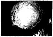
도 7은 케라틴 1 및 케라틴 10에 대한 항체를 이용하여 양막 상의 구강 점막 상피 세포층(구강 점막 상피 시트)을 염색한 상을 도시한 도면이다. 케라틴 1 및 케라틴 10 어느 것에 대해서도 염색성이 관찰되지 않는다.FIG. 7 is a diagram showing images of oral mucosal epithelial cell layers (oral mucosal epithelial sheets) on amniotic membrane using antibodies to keratin 1 and keratin 10. FIG. No dyeability was observed for either keratin 1 or keratin 10.
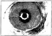
도 8은 각막 및 결막 상피를 제거한 집토끼의 안구 표면을 플루오레세인으로 염색한 상을 도시한 도면이다. 플루오레세인으로 염색된 부분은 백색 스폿으로 나타나 있다. 안구 표면 전체가 플루오레세인에 의해 염색된 것으로 관찰되었고, 상피가 완전히 제거된 것을 알 수 있다.8 is a view showing an image of fluorescein staining the eyeball surface of the rabbit removed the cornea and conjunctival epithelium. The portion stained with fluorescein is shown as a white spot. It was observed that the entire ocular surface was stained with fluorescein and the epithelium was completely removed.
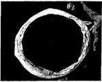
도 9는 각막 및 결막 상피를 제거한 집토끼의 4주 후의 안구 표면을 도시한 도면이다. 안구 표면이 주위보다 반흔 조직을 수반하는 결막 침입으로 인하여 투명성을 상실한 것이 관찰된다.9 is a view showing the ocular surface after 4 weeks of the rabbit removed the cornea and conjunctival epithelium. It was observed that the ocular surface lost transparency due to conjunctival involvement with scar tissue rather than the surroundings.
도 10은 각막 상피 이식용 시트를 이식한 후의 안구 표면의 상태를 도시한 도면이다. 안구 표면(이식부)이 투명성을 나타내는 것을 알 수 있다.Fig. 10 is a view showing the state of the eye surface after transplanting the corneal epithelial graft sheet. It can be seen that the eyeball surface (transplant) shows transparency.
도 11은 각막 상피 이식용 시트를 이식한 후 48시간 경과한 시점의 플루오레세인 염색상을 도시한 도면이다. 플루오레세인으로 염색된 부분은 백색 스폿으로 나타나 있다. 안구 표면(이식부)은 플루오레세인에 염색되지 않았으며, 이식편이 안구 표면에 생존하는 것을 알 수 있다. 또한, 이식편의 주위에서는 전반에 걸쳐 플루오레세인 염색성이 확인된다.FIG. 11 shows fluorescein staining at 48 hours after transplanting the corneal epithelial graft sheet. FIG. The portion stained with fluorescein is shown as a white spot. The ocular surface (graft) was not stained with fluorescein and the graft survived on the ocular surface. Fluorescein staining is also observed throughout the graft.
도 12는 각막 상피 이식용 시트를 이식한 후 10일 경과한 시점의 플루오레세인 염색상을 도시한 도면이다. 플루오레세인으로 염색된 부분은 백색 스폿으로 나타나 있다. 염색성을 나타내지 않는 영역이 확대된 것이 관찰되며, 이식편이 안구 표면에 잔존하고 있고, 또한 이식 후 48시간이 경과한 시점에 비하여 주위로 신장한 것으로 나타나 있다.12 shows fluorescein staining images 10 days after transplanting the corneal epithelial graft sheet. The portion stained with fluorescein is shown as a white spot. An enlarged area not showing staining was observed, and the graft remained on the ocular surface, and it was shown to have elongated around compared to the time point 48 hours after transplantation.
도 13은 화학적 외상 급성기의 천연성 상피 결손 환자의 수술 전(좌측 란) 및 수술 후 5개월(우측 란) 시점의 안구 표면의 상태를 도시한 도면이다.FIG. 13 shows the state of the ocular surface at the time of preoperative (left column) and postoperative 5 months (right column) of patients with spontaneous epithelial defect in acute chemical trauma.
도 14는 화학적 외상 만성기의 환자의 수술 전(좌측 란)(반흔성 각결막 상피증) 및 수술 후 2개월(우측 란) 시점의 안구 표면의 상태를 도시한 도면이다.FIG. 14 is a diagram showing the state of the ocular surface at the time of preoperative (left column) (scarious keratoconjunctival epithelium) and 2 months postoperatively (right column) of a patient in a chemical trauma chronic phase.
도 15는 안류천포창 환자의 수술 전(좌측 란) 및 수술 후 1개월(우측 란) 시점의 안구 표면의 상태를 도시한 도면이다.FIG. 15 is a view showing the state of the ocular surface at the time of surgery (left column) and one month after surgery (right column) of an ophthalmoplasty patient.
본 발명의 일면은 구강 점막 상피 세포 유래의 세포가 중층화한 세포층으로 이루어지는 각막 상피형 시트에 관한 것이다.One aspect of the present invention relates to a corneal epithelial sheet comprising a cell layer in which cells derived from oral mucosal epithelial cells are layered.
여기서, 구강 점막 상피 세포 유래의 세포가 중층화한 세포층이란, 구강 점막 상피부의 세포를 출발원료로 하여, 이것을 배양해 증식시켜, 적어도 일부가 분화 및 중층화하여 형성된 세포층을 일컫는다. 이러한 세포층을 형성하는 세포는, 구강내연점막 상피부의 세포, 입술부의 세포, 구개부의 세포, 볼부의 세포 등의 구강 점막 상피 세포로부터 유래된다. 이들 세포로부터 유래된 것은, 세포층을 형성하는 세포에 있어서 구강 점막 상피 세포 특이성 케라틴 4 또는 케라틴 13이 발현된 것을 지표로 하여 확인할 수 있다. 또는, 케라틴 3이 발현된 것을 지표로 하여 확인할 수도 있다. 이 케라틴 3은 각막 특이성 케라틴의 일종으로 알려져 있으나, 구강 점막 상피 세포에서도 발현되는 것으로 확인되어 있다. 또한, 이러한 각막 특이성 케라틴인 케라틴 3을 발현한다는 점에서, 구강 점막 상피 세포를 각막 상피 이식용 조성물을 제조하기 위한 재료로서 이용하는 것이 바람직하다고 할 수 있다.Here, the cell layer in which the cells derived from oral mucosal epithelial cells are layered refers to a cell layer formed by culturing and proliferating the cells of the oral mucosal epithelial cell as a starting material and at least partially differentiating and stratifying them. The cells forming the cell layer are derived from oral mucosal epithelial cells such as cells of the oral internal mucosal epithelium, cells of the lip, cells of the palate, and cells of the cheek. Derivation from these cells can be confirmed as an indicator that oral mucosal epithelial cell
한편, 구강 점막 상피 세포 특이성 유전자의 발현을 조사함으로써, 세포층을 형성하는 세포가 구강 점막 상피 세포 유래인 것인 지의 여부를 확인할 수도 있다.On the other hand, by examining the expression of oral mucosal epithelial cell specific genes, it is possible to confirm whether the cells forming the cell layer are derived from oral mucosal epithelial cells.
본 발명의 각막 상피형 시트는, 바람직하게는 이하의 성상 내지는 특성 중 일부를 구비하는 것이다. 특히 바람직하게는, 이하의 모든 성상 내지는 특성을 구비하는 것이다.The corneal epithelial sheet of the present invention is preferably provided with some of the following properties or characteristics. Especially preferably, it is equipped with all the following characteristics or characteristics.
(1) 최상층의 세포가 각화하지 않는다. 이는 각막 상피가 구비하는 특징 중 하나이다. 이러한 특징이 확인되는 경우, 본 발명의 각막 상피형 시트는 각막 상피와 유사하며, 각막 상피와 동일한 기능을 발휘할 것으로 기대된다. 또한, 각화란, 달리 각질화라고도 불리며, 세포내에 케라틴이 생성되고, 핵 등의 세포내 소기관이 소실되는 현상을 말한다. 세포가 각화되어 있는지 여부는, 예를 들면 세포의 편평화나 핵의 유무를 지표로 하여 확인할 수 있다.(1) The cells of the uppermost layer do not keratinize. This is one of the features of corneal epithelium. When these features are confirmed, the corneal epithelial sheet of the present invention is similar to the corneal epithelium and is expected to exert the same function as the corneal epithelium. In addition, keratinization, also called keratinization, refers to a phenomenon in which keratin is produced in a cell and intracellular organelles such as the nucleus are lost. Whether or not the cells are keratinated can be confirmed, for example, by flattening the cells and the presence or absence of the nucleus.
(2) 최상층의 세포가 편평 형상이다. 즉, 구강 점막 상피 세포층이 대략 원주 형상의 세포로 이루어지는 층 상에 편평 형상의 세포로 이루어지는 층이 형성되어 구성된다. 편평 형상의 세포가 최상층을 피복하는 경우, 세포간 기밀성이 높아져 이하의 배리어(barrier) 기능이 발휘될 것으로 생각된다. 각막 상피에 있어서도 최상층의 세포는 편평 형상이다. 이러한 특징이 확인되는 경우, 본 발명의 각막 상피형 시트는 각막 상피와 유사하며, 각막 상피와 동일한 기능을 발휘할 것으로 기대된다.(2) The cells of the uppermost layer are flat. That is, the oral mucosal epithelial cell layer is formed by forming a layer composed of squamous cells on a layer composed of substantially columnar cells. When the flat cells cover the uppermost layer, it is considered that the intercellular airtightness is increased and the following barrier functions are exerted. Also in the corneal epithelium, the cells of the uppermost layer are squamous. When these features are confirmed, the corneal epithelial sheet of the present invention is similar to the corneal epithelium and is expected to exert the same function as the corneal epithelium.
(3) 배리어 기능을 구비한다. 배리어 기능이란, 표층으로부터의 액체, 기체 등의 침윤이나, 표층을 통한 액체의 방산 등을 방지하는 기능을 말한다. 이러한 배리어 기능을 가짐으로써, 이식 후 그 표면에 수분(눈물)을 유지할 수 있고, 나아가 필요 이상의 수분이 방산하는 것을 방지할 수 있다. 각막은 배리어 기능을 구비함으로써 그 표면에 수분을 유지할 수 있고, 이에 따라 깜박거림에 견딜 수 있는 기능을 발휘한다. 따라서, 배리어 기능을 구비하는 것은, 각막용 이식 재료에 요구되는 중요한 하나의 특성이다. 이 특징이 확인되는 경우, 본 발명의 각막 상피형 시트는 각막 상피와 유사하여, 각막 상피와 동일한 기능을 발휘할 것으로 기대된다. 배리어 기능을 구비하는 지의 여부는, 예를 들면 서양 고추냉이 퍼옥시다아제(Horseradish peroxidase) 등의 표식 물질을 포함하는 용액의 침윤 정도에 의해 조사할 수 있다.(3) It has a barrier function. The barrier function refers to a function of preventing infiltration of liquids and gases from the surface layer, dissipation of liquid through the surface layer, and the like. By having such a barrier function, moisture (tear) can be maintained on the surface after transplantation, and further, the excess water can be prevented from dissipating. The cornea can retain moisture on its surface by providing a barrier function, thereby exerting a function to withstand flicker. Therefore, having a barrier function is one important characteristic required for the corneal graft material. When this feature is confirmed, the corneal epithelial sheet of the present invention is similar to the corneal epithelium and is expected to exert the same function as the corneal epithelium. Whether or not it has a barrier function can be investigated by the degree of infiltration of a solution containing a marker substance such as horseradish peroxidase.
이러한 각막 상피형 시트는, 각막이 손상, 결손된 환자에 대한 이식 재료(각막 상피의 대체)로서 이용될 수 있다. 이식 시에는, 수술용 봉합사를 이용해 이식편을 주위 조직에 고정하여 생존을 촉진하는 것이 바람직하다. 또한, 이식 후에는, 치료용 콘택트렌즈로 이식부 표면을 일시적으로 피복하여 보호하는 것이 바람직하다.Such corneal epithelial sheets can be used as a graft material (replacement of corneal epithelium) for patients with corneal damage or defects. At the time of implantation, surgical sutures are used to secure the graft to surrounding tissues to promote survival. In addition, after implantation, it is desirable to temporarily coat and protect the implant surface with a therapeutic contact lens.
본 발명의 다른 일면은, 하기의 구성으로 이루어지는 각막 상피 이식용 시트를 제공한다. 즉, 콜라겐층 상에 구강 점막 상피 세포 유래의 세포가 중층화한 세포층이 형성된 것을 특징으로 하는 각막 상피 이식용 시트이다. 바꾸어 말하면, 콜라겐층 상에, 상술한 각막 상피형 시트가 형성된 것을 특징으로 하는 각막 상피 이식용 시트이다. 여기서, 콜라겐층은 양막 유래인 것이 바람직하고, 양막으로부터 소파 처리 등에 의해 상피를 제거한 것이 보다 바람직하다. 콜라겐층이 이러한 상피를 제거한 양막을 원재료로 이용하였는지의 여부는, 콜라겐층에 양막 상피층의 세포가 포함되어 있는지의 여부를 조사함으로써 확인할 수 있다. 또한, 양막으로서 인간 양막을 이용하는 것이 특히 바람직하다.Another aspect of the present invention provides a corneal epithelial graft sheet having the following configuration. That is, the sheet for corneal epithelial transplantation characterized in that a cell layer in which cells derived from oral mucosal epithelial cells are layered on the collagen layer is formed. In other words, the above-described corneal epithelial sheet is formed on the collagen layer. Here, it is preferable that a collagen layer is derived from amnion, and it is more preferable that the epithelium was removed from amnion by sofa treatment or the like. Whether the collagen layer used the amnion from which such epithelium was removed as a raw material can be confirmed by investigating whether the collagen layer contains the cells of the amnion epithelium. In addition, it is particularly preferable to use human amniotic membrane as amniotic membrane.
이 각막 상피 이식용 시트는, 각막 손상, 결손 등의 환자에 대한 이식 재료(각막 상피의 대체)로서 이용할 수 있다. 이 경우, 콜라겐층이 안구측이 되도록 각막 상피 결손부에 이식된다. 이식 시에는, 수술용 봉합사를 이용해 이식편을 주위 조직에 고정하여 생존을 촉진하는 것이 바람직하다. 또한, 이식 후에는, 치료용 콘택트렌즈로 이식부 표면을 일시적으로 피복하여 보호하는 것이 바람직하다.This corneal epithelial graft sheet can be used as a graft material (replacement of corneal epithelium) for patients with corneal damage and defects. In this case, the collagen layer is implanted into the corneal epithelial defect so as to be the eye side. At the time of implantation, surgical sutures are used to secure the graft to surrounding tissues to promote survival. In addition, after implantation, it is desirable to temporarily coat and protect the implant surface with a therapeutic contact lens.
이상의 각막 상피형 시트 및 각막 상피 이식용 시트는 이하의 방법에 따라 제조할 수 있다.The above-described corneal epithelial sheet and corneal epithelial graft sheet can be produced by the following method.
본 발명의 또 다른 일면은, 각막 상피형 시트의 제조 방법에 관한 것으로, 하기의 단계를 포함한다.Another aspect of the present invention relates to a method for producing a corneal epithelial sheet, which includes the following steps.
a) 콜라겐층 상에서 구강 점막 상피 세포를 배양하는 단계; 및a) culturing oral mucosal epithelial cells on the collagen layer; And
b) 구강 점막 상피 세포를 증식시켜 중층화한 후, 최표층을 공기에 접촉시키는 단계.b) proliferating and stratifying oral mucosal epithelial cells and then contacting the outermost layer with air.
여기서, 콜라겐층의 원재료가 되는 콜라겐의 종류는 특별히 한정되지 않으며, I형 콜라겐, III형 콜라겐, IV형 콜라겐 등을 이용할 수 있다. 복수 종류의 콜라겐이 혼재하는 것을 이용하는 것도 가능하다. 이들 콜라겐은 돼지, 소, 양 등의 동물의 피부, 연골 등의 결합 조직으로부터, 산가용화법, 알칼리가용화법, 효소가용화법 등에 의해 추출, 정제할 수 있다. 또한, 항원성을 저하시킬 목적으로, 펩신이나 트립신 등의 분해 효소를 처리하여 텔로펩타이드(telopeptide)를 제거함 으로써 얻어지는, 이른바 아테로콜라겐(atherocollagen)을 이용하는 것이 바람직하다.Here, the kind of collagen serving as a raw material of the collagen layer is not particularly limited, and type I collagen, type III collagen, type IV collagen and the like can be used. It is also possible to use what mixes several types of collagen. These collagen can be extracted and purified from the skin of animals such as pigs, cows, sheep, and cartilages such as cartilage by acid solubilization, alkali solubilization, enzyme solubilization and the like. In addition, for the purpose of reducing antigenicity, it is preferable to use so-called atherocollagen obtained by removing telopeptide by treating a decomposing enzyme such as pepsin or trypsin.
콜라겐층으로서, 양막 유래, 특히 인간 양막 유래의 것을 이용하는 것이 특히 바람직하다. 여기서, 양막 유래란, 넓게 양막을 출발원료로 하여 얻어진 것을 의미한다. 인간 양막은 자궁과 태반의 최표층을 덮는 막으로서, 콜라겐이 풍부한 실질 조직 상에 기저막, 상피층이 형성되어 구성된다. 인간 양막은, 예를 들면 분만 시 산후에 얻어지는 인간 태아막, 태반 등으로부터 수득할 수 있다. 구체적으로는, 분만 직후에 얻어지는 인간 태아막, 태반 및 제대로 이루어지는 일체물을 처리하여, 정제함으로써 인간 양막을 조제할 수 있다. 처리 및 정제 방법은, 일본 특개 평5-5698호에 개시된 방법 등의 공지된 방법을 채용할 수 있다. 즉, 분만 시에 얻어지는 태아막으로부터 양막을 박리하고, 초음파 세정 등의 물리적 처리 및 효소 처리 등에 의해 잔존 조직을 제거하고, 적절하게 세정함으로써 인간 양막을 조제할 수 있다.As the collagen layer, it is particularly preferable to use amnion-derived ones, particularly those derived from human amnion. Herein, the amnion derived means that the amnion is obtained as a starting material widely. The human amniotic membrane is a membrane covering the outermost layers of the uterus and placenta, and is formed by the formation of a basement membrane and an epithelial layer on collagen-rich parenchyma. Human amniotic membrane can be obtained, for example, from human fetal membranes, placenta and the like obtained after childbirth at delivery. Specifically, the human amniotic membrane can be prepared by treating and purifying the human fetal membrane, placenta and a properly formed monolith obtained immediately after delivery. As a treatment and purification method, well-known methods, such as the method of Unexamined-Japanese-Patent No. 5-5698, can be employ | adopted. That is, the human amniotic membrane can be prepared by peeling the amniotic membrane from the fetal membrane obtained at the time of delivery, removing the remaining tissue by physical treatment such as ultrasonic cleaning, enzyme treatment, and the like, and washing appropriately.
이와 같이 조제한 인간 양막은, 사용 시까지 동결하여 보존할 수 있다. 인간 양막의 동결은, 예를 들면 -80℃, DMEM(Dulbecco's modified Eagle's medium)와 글리세롤을 체적비로 등량 혼합한 용액 중에서 행할 수 있다. 동결 보존하는 경우, 조작성이 향상되는 것은 물론이고, 항원성이 저하되는 것도 기대할 수 있다.The human amniotic membrane thus prepared can be frozen and stored until use. Freezing of the human amnion can be performed, for example, in a solution in which an equal amount of Dulbecco's modified Eagle's medium (DMEM) and glycerol are mixed in a volume ratio. In the case of cryopreservation, not only the operability but also the antigenicity can be reduced.
양막을 그대로 콜라겐층으로서 이용할 수도 있지만, 양막으로부터 소파 처리 등에 의하여 상피를 제거한 것을 이용하는 것이 바람직하다. 예를 들면, 상기와 같이 동결 저장한 인간 양막을 해동시킨 후, EDTA나 단백질 분해 효소로 처리하여 세포간 접착을 이완시키고, 셀 스크레이퍼(cell scraper) 등을 이용하여 상피를 소파 처리함으로써, 상피가 제거된 인간 양막을 조제할 수 있다.Although the amnion can be used as a collagen layer as it is, it is preferable to use what removed the epithelium from the amnion by couch treatment or the like. For example, after thawing the frozen human amnion, treatment with EDTA or proteolytic enzymes is used to relax intercellular adhesion, and the epithelium is cured by using a cell scraper or the like. Removed human amnion can be prepared.
콜라겐층 상에는 구강 점막 상피 세포가 배양된다. 구강 점막 상피에는 줄기 세포의 존재가 시사된 바 있고, 상피형 세포층을 형성하는 세포로 분화를 유도하기 용이한 것으로 여겨진다. 또한, 구강 점막 상피 세포를 이용하는 경우에는, 수득이 용이하고, 다량의 세포를 수득 가능하며, 나아가 양안성 환자를 처치하는 경우에도 자기 세포를 이용하여 이식 재료를 조제할 수 있다는 등의 이점을 가진다. 특히, 각막 상피 세포를 수득할 수 없는 환자에 대하여, 자기 세포 유래의 이식 재료를 적용할 수 있다는 이점은, 임상적으로 매우 중요한 거부 반응의 문제를 대폭 해소할 것으로 기대된다.Oral mucosal epithelial cells are cultured on the collagen layer. The presence of stem cells has been suggested in the oral mucosal epithelium, and is believed to be easy to induce differentiation into cells forming the epithelial cell layer. In addition, in the case of using oral mucosal epithelial cells, it is easy to obtain, a large amount of cells can be obtained, and furthermore, even when treating binocular patients, magnetic materials can be used to prepare a graft material. . In particular, the advantage of applying graft material derived from magnetic cells to patients who cannot obtain corneal epithelial cells is expected to greatly solve the problem of clinically important rejection.
구강 점막 상피 세포로는, 치근부에 존재하는 세포(구강내연점막 상피 세포), 입술부의 세포, 구개부의 세포, 볼부의 세포 등이 이용된다. 그 중에서도 구강내연점막 상피 세포는 증식능이 높고, 또한 항원성이 낮으므로, 이를 이용하는 것이 특히 바람직하다. 구강 점막 상피 세포는, 목적하는 세포가 존재하는 장소를 메스 등으로 절제하거나, 소파 처리하거나 함으로써 수득할 수 있다. 구강내연점막 상피 세포에서는, @발치에 부착되어 있는 구강 점막 상피를 에나멜 시멘트 이행부로부터 분리하여 수득할 수 있다. 또한, 결합 조직 등의 불순물을 제거하기 위하여, 디스파제나 트립신 등의 효소에 의한 처리나, 필터 처리를 실시하는 것이 바람직하다.As oral mucosal epithelial cells, cells (oral lining mucosal epithelial cells) present in the root portion, cells in the lip, cells in the palate, cells in the cheeks, and the like are used. Among them, oral endothelial mucosal epithelial cells have high proliferative capacity and low antigenicity, and therefore, it is particularly preferable to use them. Oral mucosal epithelial cells can be obtained by cutting off a place where a desired cell is present with a scalpel or by treating with a sofa. In oral endothelial mucosal epithelial cells, the oral mucosal epithelium adhering to the extract can be obtained by separating from the enamel cement transition. In order to remove impurities such as connective tissue, it is preferable to perform treatment with an enzyme such as dispase or trypsin or a filter treatment.
본 발명에 따라 제조되는 각막 상피형 시트가 이식될 환자 이외의 구강으로 부터 수득한 구강 점막 상피 세포를 이용하는 것도 가능하지만, 면역 거부 반응을 고려하면, 환자 자체의 구강으로부터 구강 점막 상피 세포를 수득하여, 배양하는 것이 바람직하다.It is also possible to use oral mucosal epithelial cells obtained from the oral cavity other than the patient to be implanted with the corneal epithelial sheet prepared according to the present invention, but considering the immune rejection reaction, oral mucosal epithelial cells are obtained from the oral cavity of the patient itself. It is preferable to culture.
이렇게 하여 수득된 구강 점막 상피 세포는 콜라겐층 상에서 배양된다. 예를 들면, 상기의 방법에 따라 구강 점막 상피 세포 부유액을 조제하고, 이것을 콜라겐층 상에 접종하고, 적당한 배양 조건 하에서 배양한다. 콜라겐층으로서 상피를 제거한 인간 양막을 이용하는 경우에는, 상피를 제거하고 표출된 면측(즉, 기저막측)에 구강 점막 상피 세포를 접종하는 것이 바람직하다. 이는, 이 면측에는 IV형 콜라겐이 풍부하게 포함되어 있어, 접종된 구강 점막 상피 세포의 증식 및 중층화가 양호하게 행해지는 것으로 생각되기 때문이다.The oral mucosal epithelial cells thus obtained are cultured on the collagen layer. For example, an oral mucosal epithelial cell suspension is prepared according to the above method, inoculated onto the collagen layer, and cultured under appropriate culture conditions. In the case of using the human amnion from which the epithelium has been removed as the collagen layer, it is preferable to inoculate oral mucosal epithelial cells to the surface side (that is, the basal membrane side) where the epithelium is removed. This is because IV-type collagen is abundantly contained on this surface side, and it is considered that proliferation and stratification of the inoculated oral mucosal epithelial cells are performed satisfactorily.
구강 점막 상피 세포는, 예를 들면 세포 밀도가 약 1×10³ 개/cm2 이상, 바람직하게는 약 1×10³ 개/cm2∼약 1×10⁷/cm2, 더욱 바람직하게는 약 1×10⁴ 개/cm2∼약 1×10⁶ 개/cm2가 되도록 콜라겐층 상에 접종할 수 있다.Oral mucosal epithelial cells to, for example, cell density of about 1 × 10³ gae / cm2 or higher, preferably from about 1 × 10³ gae / cm2 ~ about 1 × 10⁷ / cm2, more preferably from about It can be inoculated on the collagen layer so that it becomes 1 * 10 <4> / cm <2> -about 1 * 10 <6> / cm <2> .
구강 점막 상피 세포의 배양은 지지 세포의 존재 하에서 행하는 것이 바람직하다. 지지 세포란, 공급 세포(feeder cell)라고도 불리며, 배양액 중에 성장 인자 등을 공급한다. 지지 세포의 공존 하에 배양하는 경우, 구강 점막 상피 세포의 증식 효율이 향상된다. 지지 세포에는, 예를 들면 3T3 세포(스위스마우스 3T3 세포, 마우스 NIH 3T3 세포, 3T3J2 세포 등) 등을 이용할 수 있다. 그 중에서도, 증식 효율, 취급 용이성 등의 관점에서 마우스 NIH 3T3 세포를 지지 세포로서 이용하 는 것이 바람직하다.Culture of oral mucosal epithelial cells is preferably performed in the presence of support cells. Support cells, also called feeder cells, supply growth factors and the like to the culture solution. When cultured in the presence of support cells, the proliferation efficiency of oral mucosal epithelial cells is improved. As support cells, for example, 3T3 cells (Swiss mouse 3T3 cells, mouse NIH 3T3 cells, 3T3J2 cells, etc.) can be used. Especially, it is preferable to use mouse NIH 3T3 cells as support cells from a viewpoint of proliferation efficiency, handling ease, etc.
지지 세포를 미리 마이토마이신 C 등을 이용하여 불활성화하는 것이 바람직하다. 이는 지지 세포 자체가 증식하여 구강 점막 상피 세포의 증식을 저해하는 것을 방지하고, 구강 점막 상피 세포의 증식 효율을 상승시키기 위해서이다. 이러한 불활성화는 방사선 처리 등에 의해서도 행할 수 있다.It is preferable to inactivate the support cells with mitomycin C or the like beforehand. This is to prevent the supporting cells from proliferating to inhibit the proliferation of oral mucosal epithelial cells and to increase the proliferation efficiency of the oral mucosal epithelial cells. Such inactivation can also be performed by radiation treatment or the like.
구강 점막 상피 세포의 배양을 지지 세포의 공존 하에 행하는 경우에는, 지지 세포와 콜라겐층 사이에 지지 세포가 통과할 수 없는 세공 크기의 격리막을 존재시키는 것이 바람직하다. 이러한 격리막을 이용함으로써, 배양 시에 콜라겐층측(즉, 구강 점막 상피 세포측)에 지지 세포가 침입하는 것을 방지할 수 있다. 그 결과, 최종적으로 얻어지는 각막 상피형 시트 내에 지지 세포가 혼재할 우려가 없어진다. 이는 지지 세포에 의한 면역 거부 반응의 문제가 없는 각막 상피형 시트의 제조가 가능하다는 것을 의미하고, 임상적으로 매우 의의가 있다.When culturing oral mucosal epithelial cells is carried out in the presence of support cells, it is preferable to provide a pore-sized separation membrane between the support cells and the collagen layer, through which the support cells cannot pass. By using such a separation membrane, it is possible to prevent supporting cells from invading the collagen layer side (that is, the oral mucosal epithelial cell side) at the time of culture. As a result, there is no fear that support cells will be mixed in the finally obtained corneal epithelial sheet. This means that the preparation of the corneal epithelial sheet without problems of immune rejection by support cells is possible and is of great clinical significance.
격리막으로는, 지지 세포가 통과할 수 없는 세공 크기를 가지는 공지된 것을 적절하게 선택하여 사용할 수 있다. 예를 들면, 폴리카보네이트로 이루어진, 세공 크기가 약 0.4㎛∼3.0㎛인 막을 이용할 수 있다. 격리막의 재질은 특별히 한정되지 않으며, 폴리카보네이트 외에 폴리에스테르 등으로 이루어질 수도 있다. 이러한 격리막은 시판되고 있으며, 용이하게 입수할 수 있다.As the separation membrane, a known one having a pore size through which support cells cannot pass can be appropriately selected and used. For example, a membrane made of polycarbonate having a pore size of about 0.4 μm to 3.0 μm may be used. The material of the separator is not particularly limited, and may be made of polyester or the like in addition to polycarbonate. Such separators are commercially available and readily available.
격리막을 이용하는 경우의 배양 방법의 예로서 이하의 방법을 들 수 있다. 먼저, 샬레 등의 용기(제1 용기)에, 불활성화한 지지 세포를 접종하고 배양함으로써, 용기 표면에 지지 세포층을 형성시킨다. 그런 다음, 격리막으로 바닥면을 형 성한 제2 용기를 제1 용기 내에 배치한다. 이때, 제2 용기의 바닥면이 배양액 중에 잠기도록 제2 용기의 위치를 조정한다. 이어서, 제2 용기의 바닥면, 즉 격리막 상에 콜라겐층을 형성시킨다. 그리고, 콜라겐층 상에 구강 점막 상피 세포를 접종하여 배양한다.The following method is mentioned as an example of the culture method in the case of using a separation membrane. First, a supporting cell layer is formed on the surface of the container by inoculating and incubating the inactivated support cells in a container (first container) such as a chalet. Then, a second container having a bottom surface formed with a separator is placed in the first container. At this time, the position of the second container is adjusted so that the bottom surface of the second container is immersed in the culture solution. Next, a collagen layer is formed on the bottom surface of the second container, that is, the separator. Then, oral mucosal epithelial cells are inoculated and cultured on the collagen layer.
제2 용기의 바닥면에 미리 콜라겐층을 형성시킨(예를 들면, 제2 용기의 바닥면에 상피를 제거한) 양막을 적재한다. 이 상태로 일단 건조 처리를 행할 수도 있고, 제2 용기를 지지 세포가 접종된 제1 용기 내에 배치한 뒤, 콜라겐층 상에 구강 점막 상피 세포를 접종하여 배양할 수도 있다.The amniotic membrane in which the collagen layer was previously formed (for example, the epithelium was removed from the bottom surface of the second container) is loaded on the bottom surface of the second container. In this state, the drying treatment may be performed once, or the second container may be placed in the first container inoculated with the supporting cells, and then cultured by inoculating oral mucosal epithelial cells on the collagen layer.
지지 세포의 세포 밀도는, 예를 들면 약 1×10² 개/cm2 이상, 바람직하게는 약 1×10² 개/cm2∼약 1×10⁷/cm2, 더욱 바람직하게는 약 1×10² 개/cm2∼약 1×10⁵ 개/cm2로 할 수 있다. 구강 점막 상피 세포수에 대한 비로 말하면, 사용하는 지지 세포수를, 예를 들면 구강 점막 상피 세포수에 대하여 1/103배∼1×102배, 바람직하게는 1/102∼1배로 배양을 행할 수 있다. 지지 세포수가 적으면 구강 점막 상피 세포의 증식율이 저하되며, 지나치게 적은 경우에는 양호한 구강 점막 상피 세포의 중층화가 얻어지지 않는다. 한편, 지지 세포수가 지나치게 많은 경우에는, 오히려 구강 점막 상피 세포의 증식율을 저하시키므로 바람직하지 않다.Cell density of the support cell, for example, about 1 × 10² gae / cm2 or higher, preferably from about 1 × 10² gae / cm2 ~ is from about 1 × 10⁷ / cm2, more preferably about 1 × 10² pieces / cm2 to about 1 × 10⁵ pieces / cm2 . In terms of the ratio to the number of oral mucosal epithelial cells, the number of supporting cells to be used is, for example, cultured 1/103 to 1 × 10² times, preferably 1/102 to 1 times the number of oral mucosal epithelial cells. Can be done. When the number of supporting cells is small, the proliferation rate of oral mucosal epithelial cells decreases, and when too small, favorable oral mucosal epithelial cells are not obtained. On the other hand, when the number of supporting cells is too large, the proliferation rate of oral mucosal epithelial cells is lowered, which is not preferable.
구강 점막 상피 세포의 배양에 이용되는 배양액은, 상기 세포를 증식시키고, 중층화할 수 있는 것이면 특별히 한정되지 않는다. 예를 들면, 상피 세포의 성장 에 통상 이용되는 DMEM(Dulbecco's modified Eagle's medium)와 햄 F12 배지(Ham's F12 medium)를 소정의 비율로 혼합하고, FBS, 성장 인자, 항생 물질 등을 첨가한 배지를 사용할 수 있다. 구체적인 예로는, FBS(10%), 인슐린(5mg/㎖), 콜레라 톡신(0.1nM), 상피 세포 증식 인자(EGF)(10ng/㎖), 및 페니실린-스트렙토마이신(50IU/㎖)을 첨가한 DMEM과 햄 F12 배지의 혼합 배지(혼합 체적비 1:1)를 들 수 있다. 또한, 트리요오도티로닌(triiodothyrinine)(예를 들면, 2nM), 글루타민(예를 들면, 4mM), 트렌스페린(예를 들면, 5mg/㎖), 아데닌(예를 들면, 0.18mM), 및/또는 하이드로코르티손(예를 들면, 0.4mg/㎖)을 추가로 첨가한 DMEM/햄 F12 혼합 배지를 이용하는 것도 가능하다.The culture solution used for culturing the oral mucosal epithelial cells is not particularly limited as long as it can proliferate and multiply the cells. For example, DMEM (Dulbecco's modified Eagle's medium) and ham F12 medium (Ham's F12 medium), which are usually used for growth of epithelial cells, may be mixed at a predetermined ratio, and a medium containing FBS, growth factors, antibiotics, or the like may be used. Can be. Specific examples include the addition of FBS (10%), insulin (5 mg / ml), cholera toxin (0.1 nM), epithelial cell growth factor (EGF) (10 ng / ml), and penicillin-streptomycin (50 IU / ml). And a mixed medium (mixed volume ratio 1: 1) of DMEM and ham F12 medium. Triiodothyrinine (eg 2 nM), glutamine (eg 4 mM), transferrin (eg 5 mg / mL), adenine (eg 0.18 mM), and / Alternatively, it is also possible to use DMEM / Ham F12 mixed medium to which hydrocortisone (for example, 0.4 mg / ml) is additionally added.
단계 a)에 따라 콜라겐층 상에서 구강 점막 상피 세포를 증식시켜 중층화한다. 그리고, 중층화한 세포층의 표층을 공기에 접촉시키는 단계(단계(b))를 실시한다. 또한, 이 단계를 본 명세서에서는 에어 리프팅(Air-lifting)이라고도 부른다. 이 단계(b)는 세포층을 형성하는 세포의 분화 및 배리어 기능을 유도하기 위해 실시된다.According to step a), the oral mucosal epithelial cells are proliferated and laminated on the collagen layer. Then, the surface layer of the layered cell layer is brought into contact with air (step (b)). This step is also referred to herein as air-lifting. This step (b) is carried out to induce differentiation and barrier function of the cells forming the cell layer.
이 단계는 배양액의 일부를 스포이트, 피펫 등을 이용하여 일시적으로 제거함으로써 배양액 표면을 저하시키고, 이를 통해 구강 점막 상피 세포층의 최표층을 일시적으로 배양액 밖으로 노출시킴으로써 실시할 수 있다. 또는, 구강 점막 상피 세포층을 콜라겐층과 함께 들어올려, 최표층을 배양액 표면으로부터 일시적으로 노출시킴으로써 실시할 수도 있다. 나아가, 튜브 등을 이용하여 공기를 배양액 중에 공급하여, 구강 점막 상피 세포층의 최상층에 공기를 접촉시킬 수도 있다. 조작 용이성의 관점에서, 배양액 표면을 저하시켜 구강 점막 상피 세포의 최표층을 노출시키는 방법에 따라 실시하는 것이 바람직하다.This step can be performed by temporarily removing a portion of the culture solution using a dropper, a pipette, or the like to lower the surface of the culture medium, thereby temporarily exposing the outermost layer of the oral mucosal epithelial cell layer out of the culture medium. Alternatively, the oral mucosal epithelial cell layer may be lifted together with the collagen layer to temporarily expose the outermost layer from the surface of the culture medium. Furthermore, air may be supplied to the culture medium using a tube or the like to bring air into contact with the uppermost layer of the oral mucosal epithelial cell layer. It is preferable to carry out according to the method of exposing the outermost layer of oral mucosal epithelial cell by lowering the surface of a culture liquid from an operability viewpoint.
이 단계(b)를 실시하는 시간, 즉 중층화한 세포층의 최표층을 공기에 접촉시키는 시간은, 세포의 상태나 배양 조건 등에 따라 가변적이지만, 예를 들면 3일∼3주간 정도이며, 바람직하게는 5일∼2주간, 더욱 바람직하게는 약 1주일이다.The time for carrying out this step (b), that is, the time for contacting the outermost layer of the layered cell layer with air is variable depending on the state of the cells, the culture conditions, and the like, but is, for example, about 3 days to 3 weeks, preferably 5 days to 2 weeks, More preferably, it is about 1 week.
이상에서 설명한 본 발명의 방법에 따라, 콜라겐층 상에, 구강 점막 상피 세포가 중층화한 각막 상피형 세포층(각막 상피형 시트)이 형성된다. 이 각막 상피형 시트는, 구강 점막 상피 세포의 배양 기질로서 이용되는 콜라겐층과 함께, 각막 손상, 결손 등의 환자에 대한 이식 재료(각막 상피의 대체)로서 이용할 수 있다. 이 경우, 콜라겐층이 안구측이 되도록 각막 상피 결손부에 이식된다. 이식 시에는, 수술용 봉합사를 이용하여 이식편을 주위 조직에 고정하여 생존을 촉진하는 것이 바람직하다. 또한, 이식 후에는, 치료용 콘택트렌즈로 이식부 표면을 일시적으로 피복하여 보호하는 것이 바람직하다.According to the method of the present invention described above, a corneal epithelial cell layer (corneal epithelial sheet) in which oral mucosal epithelial cells are layered is formed on the collagen layer. The corneal epithelial sheet can be used as a transplant material (replacement of the corneal epithelium) for patients with corneal damage, defect, etc. together with the collagen layer used as a culture substrate for oral mucosal epithelial cells. In this case, the collagen layer is implanted into the corneal epithelial defect so as to be the eye side. At the time of transplantation, surgical sutures are used to secure the graft to surrounding tissues to promote survival. In addition, after implantation, it is desirable to temporarily coat and protect the implant surface with a therapeutic contact lens.
또한, 콜라겐층의 일부를 제거한 것 또는 전부를 제거한 것(즉, 각막 상피형 시트만)을 이식편으로서 이용하는 것도 가능하다. 이러한 콜라겐층의 제거는, EDTA 등에 의한 화학적 처리, 단백질 분해 효소 등에 의한 효소적 처리, 핀세트 등에 의한 소파 처리 등의 물리적 처리를 적절하게 조합하여 행할 수 있다.It is also possible to use part of the collagen layer or part of the collagen layer (ie, corneal epithelial sheet only) as a graft. The collagen layer can be removed by appropriately combining physical treatments such as chemical treatment with EDTA, enzymatic treatment with proteolytic enzymes, and sofa treatment with tweezers.
본 발명의 또 다른 일면에 있어서, 구강 점막 상피 세포가 중층화한 각막 상피형 세포층(각막 상피형 시트)이 제조되지만, 본 발명의 방법을 상기 세포층이 콜라겐층 상에 형성되어 이루어지는 각막 이식용 이식 재료(각막 상피 이식용 시트) 를 제공하는 방법으로 보는 것도 가능하다. 즉, 본 발명의 방법은 이러한 구조를 가지는 각막 상피 이식용 시트의 제조 방법으로서 이용할 수도 있다.In another aspect of the present invention, a corneal epithelial cell layer (corneal epithelial sheet) in which oral mucosal epithelial cells are layered is produced, but the method of the present invention is a transplant material for corneal transplantation, wherein the cell layer is formed on the collagen layer. It is also possible to see by the method of providing (the corneal epithelial transplant sheet). That is, the method of this invention can also be used as a manufacturing method of the corneal epithelial transplant sheet which has such a structure.
<실시예 1> 집토끼를 이용한 각막 상피형 시트의 평가Example 1 Evaluation of Corneal Epithelial Sheet Using Rabbit
[양막의 수득][Getting Amniotic Membrane]
양막은 전신적 합병증이 없는 제왕 절개술 예정의 임부에 대하여 사전에 산부인과의와 함께 충분한 정보에 입각하여 동의를 얻은 후, 수술실에서 제왕 절개 시에 수득하였다. 조작은 청결하게 주의하여, 수술 조작에 준하여 수세 후에 전용 가운을 착용하고 실시하였다. 분만 전에 청결한 양막 수득용 배트와 세정용 생리 식염수를 준비하였다. 분만 후에 태반 조직을 배트(vat)로 옮기고, 양막 조직을 태반으로부터 수동으로 박리하였다. 양막과 태반의 유착이 강한 부분은 가위로 절제하였다.Amniotic membrane was obtained at the time of caesarean section in the operating room after obtaining informed consent with a gynecologist prior to a caesarean scheduled pregnancy without systemic complications. The operation was carried out with caution and cleansing was performed after washing with water according to the surgical operation. Before delivery, a clean amnion bat and a physiological saline solution were prepared. After delivery the placental tissue was transferred to a vat and the amnion tissue was manually detached from the placenta. The strong adhesion of the amnion and placenta was excised with scissors.
[양막의 처리][Treatment of Amniotic Membrane]
양막 처리의 과정은 (1) 세정, (2) 트리밍, (3) 보존의 순으로 실시하였다. 모든 과정에서, 조작은 청결한 드래프트 내에서 실시하는 것이 바람직하고, 사용하는 용기나 기구도 모두 멸균된 것을 사용하고, 샬레 등은 멸균된 1회용(폐기 가능한) 타입을 사용하였다. 수득한 양막에 부착된 혈액 성분을 생리 식염수로 세정하하여 제거하고, 충분한 양의 생리 식염수(0.005% ofloxacin 첨가)로 추가 세정하였다. 그런 다음, 양막을 샬레 내의 인산 완충액(PBS)으로 옮기고, 가위를 이용하여 약 4×3cm 정도의 크기로 분할하였다. 분할 후 수개의 샬레에 보존액을 침지시키고, 그 중에서 상태가 좋은 양막을 선별하였다.The process of amnion treatment was performed in the order of (1) washing, (2) trimming, and (3) preservation. In all procedures, the operation is preferably carried out in a clean draft, and all of the containers and utensils used are sterilized, and the chalets and the like are sterilized disposable (disposable) types. The blood components adhered to the obtained amniotic membrane were removed by washing with physiological saline, and further washed with a sufficient amount of physiological saline (addition of 0.005% ofloxacin). Then, the amniotic membrane was transferred to phosphate buffer (PBS) in the chalet and divided into sizes of about 4 × 3 cm using scissors. After division, the stock solution was immersed in several chalets, and the amniotic membrane in good condition was selected therefrom.
[양막의 보존][Preservation of Amniotic Membrane]
2cc의 멸균 크라이오튜브(cryotube)에 1cc씩 보존액을 첨가하고, 거기에 수득, 세정하여 선별된 양막을 1매씩 넣고 라벨링한 후, -80℃의 냉장고에 저장하였다. 보존액으로는 50%의 멸균 완료된 글리세롤이 첨가된 DMEM(Dulbecco's Modofied Eagle Medium: GIBCOBRL사)를 사용하였다. 보존된 양막의 사용 기한은 3개월로 하고, 기한이 지나가면 소각 처분하였다.1 cc of each stock solution was added to 2 cc of sterile cryotubes, and obtained and washed therein. The selected amnion was labeled one by one, and then stored in a refrigerator at -80 ° C. As a stock solution, DMEM (Dulbecco's Modofied Eagle Medium: GIBCOBRL) with 50% of sterile glycerol was used. The shelf life of the preserved amniotic membrane was 3 months and incineration was terminated after the expiration date.
[양막 상피의 처리][Treatment of Amniotic Epithelium]
양막은 그 상피를 처리한 후, 배양에 사용하였다. 먼저, -80℃에 저장되어 있던 양막을 실온에서 해동시킨 후, 샬레 내의 멸균 완료된 인산 완충액(PBS)으로 충분히 세정하였다. 세정 후 0.02% EDTA 용액(Nacalai tesque사)에 2시간 동안 37℃ 하에 저장한 후, 셀 스크레이퍼(Nunc사, USA)를 이용하여 기계적으로 상피를 소파 처리하여, 배양 기질로서 사용하였다. 또한, 이 처리 방법에 따라 1층의 양막 상피가 완전히 소파 처리된 것을 광학 및 전자 현미경 조작(주사형 전자 현미경)으로 확인하였다(도 1). 또한, 도 1은 주사형 전자 현미경에 의해 관찰한 양막상이다. A는 정상적인 상태(상피 처리를 행하지 않은 상태)의 양막 표면의 상, B는 상피 소파 처리(0.02% EDTA 용액) 후의 양막 표면의 상이다.The amnion was used for culturing after treating the epithelium. First, the amnion stored at −80 ° C. was thawed at room temperature and then sufficiently washed with sterile phosphate buffer (PBS) in the chalet. After washing, the cells were stored in a 0.02% EDTA solution (Nacalai tesque Co., Ltd.) for 2 hours at 37 ° C., followed by mechanically treating the epithelium with a cell scraper (Nunc Co., USA) to use as a culture substrate. In addition, it was confirmed by optical and electron microscopy (scanning electron microscope) that the amniotic epithelium of one layer was completely cured in accordance with this treatment method (FIG. 1). 1 is the amniotic membrane observed with the scanning electron microscope. A is an image of the amnion surface in a normal state (the state not subjected to epithelial treatment), and B is an image of the amnion surface after epithelial sofa treatment (0.02% EDTA solution).
[구강 점막 상피 세포의 회수][Recovery of Oral Mucosal Epithelial Cells]
6주령의 일본 백색 집토끼를 발치 후, 그에 부착된 구강 점막 상피를 에나멜 시멘트 이행부로부터 신중하게 분리하였다. 또한, 일련의 조작은 멸균이 완료된 기구를 사용하여 가능한 한 멸균적으로 실시하였다.After extracting the Japanese white rabbit 6 weeks of age, the oral mucosa epithelium attached thereto was carefully separated from the enamel cement transition. In addition, a series of operations were performed as sterilized as possible using a sterilized device.
수득한 구강 점막 상피를 50IU/㎖의 페니실린스트렙토마이신, 젠타마이신을 함유하는 인산 완충액(PBS)에 30분간 실온 조건 하에서 2회 침지하였다. 그 후, 조직을 1.2U 디스파제(Nacalai tesque사)를 함유하는 인산 완충액(PBS)에 1시간 동안 37℃ 조건 하에 침지하고, 이어서 0.05% 트립신-EDTA 용액(GIBCOBRL사)으로 30분간 침지 처리하여 세포를 분리하였다. 효소 활성은 10%의 소 태아 혈청(FBS)을 함유하는 DMEM에 침지시킴으로써 정지시켰다. 그 후, 60㎛의 셀 필터(cell filter)를 통해 여분의 조직을 제거하고, 구강 점막 상피 세포(구강내연점막 상피 세포)를 단리하였다.The obtained oral mucosal epithelium was immersed twice in phosphate buffer (PBS) containing 50 IU / ml penicillin streptomycin and gentamicin under room temperature conditions for 30 minutes. The tissue was then immersed in phosphate buffer (PBS) containing 1.2 U dispase (Nacalai tesque) for 1 hour under 37 ° C., followed by immersion for 30 minutes in 0.05% trypsin-EDTA solution (GIBCOBRL). The cells were isolated. Enzyme activity was stopped by immersion in DMEM containing 10% fetal bovine serum (FBS). Thereafter, excess tissue was removed through a 60 μm cell filter, and oral mucosal epithelial cells (oral endothelial mucosal epithelial cells) were isolated.
[공배양 세포의 조제][Preparation of Co-Culture Cells]
공배양 세포(지지 세포)로서 NIH-3T3 세포(이하, 「3T3 세포」라고 약칭한다)를 사용하였다. 사전 배양하여 75F 플라스크(BD Falcon사)에 융합된 3T3 세포를 0.05% 마이토마이신 C 용액에 2시간 침지시켜, 3T3의 증식 활성을 억제하였다. 계속해서, 인산 완충액(PBS)으로 수회 세정하여 마이토마이신 C를 제거한 후, 0.05% 트립신-EDTA 용액으로 처리함으로써, 3T3 세포 현탁액을 조제하였다.NIH-3T3 cells (hereinafter, abbreviated as "3T3 cells") were used as coculture cells (support cells). 3T3 cells pre-cultured and fused to 75F flasks (BD Falcon) were immersed in 0.05% mitomycin C solution for 2 hours to inhibit the proliferative activity of 3T3. Subsequently, 3T3 cell suspensions were prepared by washing several times with phosphate buffer (PBS) to remove mitomycin C, followed by treatment with 0.05% trypsin-EDTA solution.
[양막 상 구강 점막 상피 시트의 형성][Formation of Oral Mucosal Epithelial Sheet on Amniotic Membrane]
상피를 소파한 인간 양막을 기질로 하여, 구강 점막 상피 세포를 상기와 같이 처리한 3T3 세포와 이하의 순서로 공배양하였다. 배양 기구로는 6웰의 배양 접시(Corning사, NY)와 배양 삽입기(배양 삽입 용기)(폴리카보네이트제, 평균 세공 크기 3.0㎛, Corning사, NY)를 이용하였다.Oral mucosal epithelial cells were cocultured with 3T3 cells treated as described above, using the human amnion of the epithelium as a substrate in the following order. As a culture apparatus, a 6 well culture dish (Corning, NY) and a culture inserter (culture inserting vessel) (polycarbonate, average pore size: 3.0 µm, Corning, NY) were used.
먼저, 배양 접시 상에 약 1×10⁴ 개/cm2의 세포 밀도가 되도록 3T3 세포 현탁액을 접종하고, 37℃ 하에 5% CO2의 조건 하에서 배양하였다. 또한, 배양 삽입기 상에 소파 처리한 상피측를 위로 하여 양막 기질을 정치하여 접착시키고, 실온에서 10분 건조시켰다. 그 후, 양막을 접착시킨 배양 삽입기 상에 약 1×10⁵ 개/cm2의 세포 밀도가 되도록 구강 점막 상피 세포를 포함하는 세포 현탁액을 접종하였다.First, the 3T3 cell suspension was inoculated on a culture dish to have a cell density of about 1 × 10⁴ cells / cm2 , and cultured under 37% of 5% CO2 . In addition, the amnion substrate was left standing and adhered to the culture inserter with the sofa-treated epithelial side up, and dried at room temperature for 10 minutes. Thereafter, the cell suspension containing the oral mucosal epithelial cells was inoculated on a culture inserter to which the amnion was adhered to a cell density of about 1 × 10⁵ cells / cm2 .
이상의 조작 후, 도 2에 도시한 바와 같이, 배양 삽입기를 배양 접시 내에 설치하고, 3T3 세포와 구강 점막 상피 세포를 동일 배지 중에서 배양하였다. 또한, 도 2는 배양 중의 상태를 나타낸 모식 단면도이다. 배양 접시(1) 내에 배양 삽입기(2)가 정치되고, 배양 접시(1) 바닥면에는 3T3 세포층(5)이 형성된다. 또한, 배양 삽입기(2)의 바닥면 상에 양막(3)이 정치되고, 그 위에 구강 점막 상피 세포(4)가 배양되는 상태를 나타낸다. 부호 6은 배지이다.After the above operation, as shown in FIG. 2, a culture inserter was installed in the culture dish, and 3T3 cells and oral mucosal epithelial cells were cultured in the same medium. 2 is a schematic cross section which shows the state in culture. The
배지에는 DMEM/햄 F12 혼합 배지(혼합 체적비 1:1)에, 10% FBS, 인슐린(5mg/㎖), 콜레라 톡신(0.1nM), 페니실린스트렙토마이신(50IU/㎖), 인간 재조합 상피 세포 증식 인자(EGF)(10ng/㎖)를 첨가한 것을 사용하였다.The medium contained DMEM / Ham F12 mixed medium (mixed volume ratio 1: 1), 10% FBS, insulin (5 mg / ml), cholera toxin (0.1 nM), penicillin streptomycin (50 IU / ml), human recombinant epithelial cell proliferation factor. The addition of (EGF) (10 ng / ml) was used.
상기 배지 내에서 2주간 배양하였다(잠수기간(Submerge)). 그 후, 점막 상피의 분화를 유도하기 위하여, 이른바 에어 리프팅법을 이용하여 일주일 배양하였다. 에어 리프팅법이란, 배지의 액면을 양막 상에 형성된 구강 점막 상피 세포층면까지로 하여, 상기 세포층의 표면을 공기 중에 노출시키는 방법이다. 잠수기간 중에는 격일로 배지를 교환하고, 에어 리프팅 후는 배지를 매일 교환하여 배양을 실시하였다.Cultures were incubated for 2 weeks in the medium (Submerge). Thereafter, in order to induce differentiation of mucosal epithelium, culture was performed for one week using the so-called air lifting method. The air lifting method is a method of exposing the surface of the cell layer to air by bringing the liquid level of the medium to the oral mucosal epithelial cell layer surface formed on the amniotic membrane. During the diving period, the medium was changed every other day, and after the air lifting, the medium was changed every day and cultured.
[양막 상의 구강 점막 상피 시트의 조직학적 특성의 검증][Verification of Histological Properties of Oral Mucosal Epithelial Sheet on Amniotic Membrane]
상기한 바와 같이 배양된 구강 점막 상피 세포층은 약 20일간의 배양에 의하여, 각막 상피와 유사한 5∼6층에 다분화, 중층화한 상피층(이하, 「각막 상피형 시트)라고도 한다)을 형성시켰다(도 3, 도 4). 이 상피층의 기저측(양막측)에는 비교적 원주 모양을 한 기저 세포 유사의 세포군이 존재하고 있었다. 또한, 최표층의 세포는 편평형을 나타내었지만 핵을 가지고 있으며, 피부와는 달리 그 표면은 각화되지 않은 것이 확인되었다. 이상으로부터, 광학 현미경 관찰에서는 양막 상에 각막 유사 상피층(각막 상피형 시트)이 형성된 것으로 나타났다.The oral mucosal epithelial cell layer cultured as described above formed a multi-differentiated and multilayered epithelial layer (hereinafter referred to as a `` corneal epithelial sheet) '' in 5 to 6 layers similar to the corneal epithelium by culture for about 20 days ( 3, 4). On the basal side (amniotic membrane side) of this epithelial layer, a relatively columnar basal cell-like cell group was present. In addition, it was confirmed that the cells of the outermost layer had a flat equilibrium but had a nucleus, and unlike the skin, the surface was not angled. From the above, optical microscopic observation showed that a corneal like epithelial layer (corneal epithelial sheet) was formed on the amnion.
이어서, 이 각막 상피형 시트의 조직학적 특성을 검토하기 위하여 면역염색을 실시하였다. 각막 상피형 시트를 제작 후, 적절한 크기로 절제하여 OCT 화합물로 동결 포매하였다. 그 후, 크라이오스태트를 이용해 얇게 잘라, 슬라이드 절편을 제작하였다. 면역 염색에 대해서는, 대표적인 세포 골격 단백질인 케라틴과 관련하여 검토를 행하였다. 즉, 각막 특이성 케라틴 3/12, 점막 특이성 케라틴 4/13, 표피 특이성 케라틴 1/10을 검토하였다. 방법은 이하와 같다. 슬라이드 절편을 인산 완충액(PBS)으로 세정한 후, 1%의 소 태아 혈청(FBS)으로 차단하여 비특이적인 항체 반응을 억제하였다. 그 후, 각 케라틴에 대한 항체(일차 항체)를 실온에서 1시간 반응시켰다. 반응 후 트리톤-X(triton-X) 함유의 PBS에서 15분간 3회의 조건으로 세정하고, 계속해서 형광 표식화 항체(2차 항체)를 실온에서 1시간 반응시켰다. 반응 후, 인산 완충액(PBS)을 이용해 15분간 3회의 조건으로 세정하 고, 봉입한 후 다중 초점 현미경을 이용해 조직을 관찰하였다.Subsequently, immunostaining was performed to examine the histological characteristics of the corneal epithelial sheet. Corneal epithelial sheets were prepared and then excised to appropriate sizes and frozen embedded in OCT compounds. Thereafter, the slices were thinly cut using a cryostat to prepare slide sections. Immunostaining was examined in connection with keratin, which is a representative cytoskeletal protein. That is, corneal specific keratin 3/12, mucosal
각막 상피형 시트에 대한 각종 케라틴의 항체 반응은 이하와 같다. 먼저, 각막 특이성 케라틴 3에 대한 염색성이 확인되었다(도 5의 좌측란). 생체내 구강 점막에서는 케라틴 3에 대하여 염색성이 확인되었기 때문에, 배양 조건 하에서도 그 성질이 유지된다고 생각되었다. 한편, 케라틴 12에 대해서는 염색성이 확인되지 않았다(도 5의 우측란). 점막 특이성 케라틴 4 및 13에 대해서는, 그 염색성이 동일하게 확인되었다(도 6). 그런데 표피 각화형 케라틴인 케라틴 1/10에 대해서는, 그 염색성이 확인되지 않았다(도 7). 이상으로부터, 각막 상피형 시트의 조직학적 성질로는, 세포 골격의 측면으로부터는 표피와 같이 각화되는 방향으로 분화되는 것이 아니라, 비각화형 점막 상피로서의 성질을 유지하면서, 각막 특이성 케라틴의 일부(케라틴 3)도 동시에 유지하는 것을 알았다.The antibody reaction of various keratin to the corneal epithelial sheet is as follows. First, staining for corneal specific keratin 3 was confirmed (left column of FIG. 5). In vivo oral mucosa, staining of keratin 3 was confirmed, it was thought that its properties are maintained even under culture conditions. On the other hand, no staining was confirmed for keratin 12 (right column of Fig. 5). For mucosal
[구강 점막 상피 시트의 자기 이식][Self Transplantation of Oral Mucosal Epithelial Sheet]
다음에, 각막 상피형 시트를 이용한 자기 이식 실험을 실시하였다. 전술한 방법에 따라, 6주령 일본 백색 집토끼로부터 구강 점막 상피 세포를 수득하여, 양막 상에 각막 상피형 시트가 형성된 이식용 시트(이하, 「각막 상피 이식용 시트」라고도 한다)를 제조하였다. 한편, 구강 점막 상피 세포의 수득을 행한 백색 집토끼에 대하여, 고리부로부터 4mm 외측으로부터 크레센트 나이프를 이용하여 각막 및 결막 상피를 100㎛ 두께로 전부 제거하였다. 이 조작에 의해 각막 상피줄기 세포를 포함하는 상피 세포가 제거되기 때문에, 인위적인 안구 표면 줄기 세포 피폐증이 재현된 것으로 생각할 수 있다. 이러한 조작 후, 각막 상피 세포를 포함하는 상피가 잔존하지 않는 것을 플루오레세인 염색으로 확인하였다(도 8). 또한, 플루오레세인 염색 시험은, 플루오레세인 시험지에 항생제의 점안 등의 수분을 포함하여 안구 표면에 직접 도포하고, 2~3회 깜박거림을 행하게 한 후, 안구 표면의 플루오레세인 염색성을 관찰함으로써 실시하였다. 각막 상피가 잔존하면, 타이트한 세포간 접착 구조에 의해 플루오레세인 색소가 침윤하지 않아, 플루오레세인에 의한 염색이 확인되지 않는다. 4주 후, 이 집토끼의 안구 표면은 주변의 잔존 결막 상피로 피복되어 투명성을 상실한 것으로 나타났다(도 9).Next, a magnetic transplant experiment using corneal epithelial sheet was performed. According to the method described above, oral mucosal epithelial cells were obtained from 6-week-old Japanese white rabbits to prepare a transplant sheet (hereinafter referred to as a "corneal epithelial transplant sheet") in which a corneal epithelial sheet was formed on the amnion. On the other hand, about the white rabbit which obtained the oral mucosal epithelial cell, all corneas and conjunctival epithelium were removed to 100 micrometer thickness using the crescent knife from the outer side of 4 mm from the collar part. Since this operation removes epithelial cells including corneal epithelial stem cells, it is considered that artificial ocular surface stem cell pneumoconiosis is reproduced. After this operation, it was confirmed by fluorescein staining that no epithelium including corneal epithelial cells remained (FIG. 8). In addition, the fluorescein staining test was applied directly to the ocular surface by containing water such as eye drops of an antibiotic on a fluorescein test paper, and flickering 2-3 times, and then observed the fluorescein staining property of the ocular surface. It carried out by doing. If the corneal epithelium remains, the fluorescein pigment does not infiltrate by the tight intercellular adhesion structure, and staining by fluorescein is not confirmed. After 4 weeks, the ocular surface of the rabbit was covered with the surrounding remaining conjunctival epithelium and appeared to lose transparency (FIG. 9).
다음에, 결막 상피로 피복되어 투명성을 상실한 동일 안구에 대하여, 안구 표면의 결막 조직을 제거한 후, 전술한 각막 상피 이식용 시트를 고리부로부터 약간 내측의 영역에 이식하였다. 이식 시에는 10-0 나일론사를 이용하여 주위의 조직에 봉합하였다. 이식 후, 이식편 상에 치료용 소프트 콘택트 렌즈를 봉합하였다. 수술 후, 항생제 및 스테로이드 안연고를 2회/일 도포하였다. 이식 후의 시점에서, 안구 표면은 이식 전의 각막 상피 이식용 시트와 동일한 투명성을 나타내고 있었다(도 10).Next, the conjunctival tissue on the ocular surface was removed from the same eye coated with the conjunctival epithelium and the above-described corneal epithelial graft sheet was implanted in a slightly inner region from the annulus. At the time of transplantation, 10-0 nylon yarn was used to seal the surrounding tissues. After implantation, the therapeutic soft contact lens was closed on the graft. After surgery, antibiotics and steroid ointments were applied twice / day. At the time of transplantation, the ocular surface showed the same transparency as the sheet for corneal epithelial transplantation before transplantation (FIG. 10).
이식 후 48시간 및 10일 경과한 시점에, 이식술을 실시한 안구 표면을 관찰하였다. 이식 후 48시간 경과한 시점에, 이식된 각막 상피 이식용 시트는 투명성을 유지하고 있으며, 또한 플루오레세인 염색을 통해 안구 표면에 결손 없이 잔존하는 것이 확인되었다(도 11). 또한, 이식편(각막 상피 이식용 시트)이 플루오레세인 염색성을 나타내지 않는 것으로부터, 본 발명의 각막 상피 이식용 시트(각막 상피형 시트)는 각막 상피와 동일한 배리어 기능을 구비하는 것이 확인되었다. 또 한, 플루오레세인 염색에서, 이식편의 주위는 전반에 걸쳐 플루오레세인의 염색성이 인지되었으므로, 이식부에 존재하는 조직은 주위의 잔존 결막 상피는 오염되지않은 것이 확인되었다.At 48 hours and 10 days after transplantation, the ocular surface underwent implantation was observed. At 48 hours after transplantation, the transplanted corneal epithelial graft sheets maintained transparency, and fluorescein staining revealed no defects on the ocular surface (FIG. 11). In addition, since the graft (corneal epithelial graft sheet) did not exhibit fluorescein staining, it was confirmed that the corneal epithelial graft sheet (corneal epithelial sheet) of the present invention had the same barrier function as the corneal epithelium. Further, in fluorescein staining, the periphery of the graft was recognized for fluorescein staining throughout, and it was confirmed that the tissue remaining in the graft was not contaminated with the remaining conjunctival epithelium.
또한, 각막 상피는 세포간에 타이트한 접착 구조로 결합하고 있기 때문에, 그 표면보다 플루오레세인의 색소가 침윤하지 않아, 플루오레세인 염색에 있어서 염색성을 나타내지 않는다. 한편, 세포간 접착이 느슨해지거나, 세포 자체가 박리하여 배리어 기능이 장해되었을 시에는, 플루오레세인 색소의 침윤이 발생해 조직이 염색된다. 따라서, 플루오레세인 염색에 의한 염색성을 조사함으로써, 이식한 각막 상피형 시트가 각막 상피와 동일한 배리어 기능을 구비하는 지의 여부를 확인할 수 있다.In addition, since the corneal epithelium binds with a tight adhesive structure between the cells, the pigment of fluorescein does not infiltrate from the surface thereof, and thus does not show stainability in fluorescein staining. On the other hand, when intercellular adhesion is loosened or the cells themselves detach and barrier function is impaired, infiltration of fluorescein pigment occurs and tissues are stained. Therefore, by examining the dyeability by fluorescein staining, it is possible to confirm whether or not the transplanted corneal epithelial sheet has the same barrier function as the corneal epithelium.
이식 후 10일이 경과한 시점에서, 상기와 같이 안구 표면을 관찰하였다. 이식된 각막 상피 이식용 시트는 안구 표면에 잔존하며, 또한 이식 후 48시간 경과한 시점의 상태와 비교하여 주위로 신장하여 있는 것이, 플루오레세인 염색을 통해 관찰되었다(도 12). 이식편은 플루오레세인 염색성을 나타내지 않고, 각막 상피에 필요한 배리어 기능을 유지하는 것이 확인되었다. 또한, 투명성도 유지되었다.At 10 days after transplantation, the ocular surface was observed as above. The transplanted corneal epithelial graft sheet remained on the ocular surface, and it was observed through fluorescein staining as compared with the state at the time of 48 hours after transplantation (FIG. 12). The graft did not show fluorescein staining and was confirmed to maintain the barrier function required for corneal epithelium. In addition, transparency was also maintained.
이상으로부터, 양막 상에서 배양하여 얻어지는 각막 상피형 시트가 안구 표면에 생존하여, 신장하고, 수술 후 장기간에 걸쳐 투명성을 유지하는 것이 확인되었다. 즉, 전술한 방법에 따라 제작되는 각막 상피형 시트는 각막 상피의 대체로서 양호하게 기능하며, 또한 양막과 그 위에 형성된 각막 상피형 시트로 이루어지는 시트상의 조성물(각막 상피 이식용 시트)은, 각막의 손상, 결손 등의 경우에 안 구 표면을 재건하기 위한 이식 재료로서 적합하게 이용할 수 있는 것이 확인되었다.From the above, it was confirmed that the corneal epithelial sheet obtained by culturing on the amnion survives on the ocular surface, elongates, and maintains transparency for a long time after surgery. That is, the corneal epithelial sheet produced according to the above-described method functions well as a substitute for the corneal epithelium, and the sheet-like composition (corneal epithelial transplant sheet) composed of the amnion and the corneal epithelial sheet formed thereon is a It has been confirmed that it can be suitably used as a graft material for reconstructing the ocular surface in the case of damage, defect or the like.
<실시예 2> 인간에서의 각막 상피형 시트의 평가Example 2 Evaluation of Corneal Epithelial Sheets in Humans
다음에, 각막 상피형 시트를 인간에게 적용한 경우의 효과를 확인하였다. (1) 화학적 외상 급성기의 천연성 상피 결손을 가지는 환자, (2) 화학적 외상 만성기의 반흔 조직 침입에 의한 혼탁을 가지는 환자, (3) 안류천포창 환자를 이식 대상자로 하여, 상기 실시예 1과 동일의 방법으로 각 환자로부터 각각 구강 점막 상피 세포를 수득하였다. 이어서, 상피를 소파 처리한 인간 양막을 기질로 하여 구강 점막 상피 세포를 3T3 세포와 공배양하고, 인간 양막 상에 구강 점막 상피 세포층이 형성된 각막 상피 이식용 시트를 얻었다. 또한, 상피를 소파 처리한 인간 양막의 조제 방법 및 3T3과의 공배양 조건은 실시예 1의 경우와 동일하다. 이렇게 하여 조제한 각각의 각막 상피 이식용 시트를, 그의 조제에 사용한 세포를 수득한 환자에게 각각 이식하고(자기이식), 그 효과를 평가하였다. 이식 방법은 실시예 1에 기재한 방법과 동일하게 실시하였다. 단, 안류천포창 환자에 대해서는 상방의 반흔 조직을 절제하여 유착을 해제시킨 후, 각막 상피형 이식 시트의 이식을 상방 1/3에 실시하고, 동시에 백내장 수술을 실시해 안내 렌즈를 삽입하였다.Next, the effect when the corneal epithelial sheet was applied to a human was confirmed. (1) Patients with spontaneous epithelial defects of chemical trauma acute phase, (2) Patients with turbidity due to scar tissue invasion of chemical trauma chronic phase, and (3) Patients with ocular blebs as transplantation targets. In each method, oral mucosal epithelial cells were obtained from each patient. Subsequently, oral mucosal epithelial cells were co-cultured with 3T3 cells using the human amnion treated with the epithelial sofa as a substrate, thereby obtaining a corneal epithelial graft sheet having an oral mucosal epithelial cell layer formed on the human amnion. In addition, the preparation method of the human amnion in which the epithelium was cured, and the coculture condition with 3T3 are the same as that of Example 1. Each corneal epithelial transplantation sheet thus prepared was transplanted (self-transplanted) to a patient who obtained the cells used for its preparation, and the effect thereof was evaluated. The transplantation method was performed similarly to the method described in Example 1. However, in patients with ocular ulcers, the upper scar tissue was excised to release the adhesions. Then, the corneal epithelial graft was implanted in the upper third, followed by cataract surgery, and the intraocular lens was inserted.
수술 전(좌측란) 및 수술 후 5개월(우측란) 시점의 화학적 외상 급성기의 천연성 상피 결손 환자의 안구 표면의 상태를 도 13에 나타낸다. 수술 후 5개월 시점에는 안구 표면이 훌륭히 재건되어 있는 것이 발견된다. 이식 수술을 행하지 않는 경우에는, 본 환자의 시력은 반흔 조직의 침입으로 인해 수동 수준 정도일 것이 예상되는 상태였다. 현재, 시력은 0.5까지 회복되었고, 상피의 상태도 안정되었다. 또한, 자가이식이므로, 거부 반응의 걱정도 없고, 수술 후 관리는 종래의 이식술에 비교하여 현저하게 용이하다.13 shows the state of the ocular surface of patients with spontaneous epithelial defects in the acute phase of chemical trauma at the preoperative (left column) and 5 months postoperative period (right column). Five months after surgery, the eye surface was found to be well reconstructed. If transplantation was not performed, the patient's visual acuity was expected to be on a manual level due to invasion of scar tissue. At present, vision was restored to 0.5, and the condition of the epithelium was stabilized. Moreover, since it is autograft, there is no worry of rejection reaction, and postoperative management is remarkably easy compared with conventional transplantation.
도 14에 수술 전(좌측란) 및 수술 후 2개월(우측란) 시점의 화학적 외상 만성기의 반흔성 각결막 상피증 환자의 안구 표면의 상태를 나타내었다. 이 환자는, 수술 전에는 안내 소견이 얻어지지 않을 정도의 중증이지만, 이식 수술에 의해서 유착을 해제할 수 있어, 수정체까지 투시 가능하게 되었다. 시력은 광각으로부터 지수 수준으로 개선하였다. 다음 수술 방침을 결정하기 위한 예비적인 수술로서 행하였지만, 안내를 관찰하고자 하는 당초의 목적을 달성한 것 외에, 미용적으로도 현저한 개선이 인지되었다.In FIG. 14, the state of the ocular surface of a patient with scarring keratoconjunctival epithelial disease in the chronic phase of chemical trauma at the time of preoperative (left column) and 2 months postoperatively (right column). Although this patient is severe enough to have no intraocular findings prior to surgery, adhesion can be released by transplantation, and the lens can be seen through the lens. Vision improved from wide angle to exponential level. Although performed as a preliminary operation for determining the next surgical policy, in addition to achieving the original purpose of observing the guidance, a marked improvement in beauty was recognized.
도 15에, 수술 전(좌측란) 및 수술 후 1개월(우측란) 시점의 안류천포창 환자의 안구 표면의 상태를 나타낸다. 안류천포창 환자에 대하여 통상의 백내장 수술을 실시하면, 이것을 계기로 결막이 침습되어 반흔이 형성되고, 그것에 의하여 현저한 각막 혼탁이 초래되는 것으로 알려져 있다. 이러한 증세의 예로는, 수술 전에 안류천포창에 의한 검구 유착이 상방으로 인지되었지만, 수술 후 1개월 경과한 시점에 안구 표면의 상태는 안정되어 있다. 시력에 관해서는, 수술 전 0.06에 지나지 않던 것이, 현재는 0.6까지 회복되었다.In FIG. 15, the state of the ocular surface of the ophthalmoplasty patient at the time of surgery (left column) and one month after surgery (right column) is shown. When cataract surgery is performed on a patient with ocular ulcer, it is known that the conjunctiva is invaded and a scar is formed, which causes significant corneal haze. An example of such a symptom is that the adhesion of the eyeball by the ophthalmoplasty was recognized upward before the operation, but the state of the eyeball surface was stabilized one month after the operation. As for visual acuity, what was only 0.06 before the operation, now recovered to 0.6.
이상의 결과로부터, 본 발명의 각막 상피형 시트(각막 상피 이식용 시트)의 인간에 대한 유효성이 실증되었다.From the above result, the effectiveness of the corneal epithelial sheet (corneal epithelial transplant sheet) of this invention to human was demonstrated.
본 발명은 상기 발명의 실시예 및 실시예의 설명에 전혀 한정되지 않는다. 특허 청구의 범위의 기재를 일탈하지 않고, 당업자가 용이하게 도달할 수 있는 범위에서 여러 가지 변형 양태도 본 발명에 포함된다.The present invention is not limited to the embodiments of the invention and the description of the embodiments. Various modifications are also included in the present invention without departing from the scope of the claims without departing from the scope of the claims.
이하, 다음 사항을 개시한다.Hereinafter, the following matters are disclosed.
[11] a) 콜라겐층 상에서 구강 점막 상피 세포를 배양하는 단계; 및[11] a) culturing oral mucosal epithelial cells on the collagen layer; And
b) 구강 점막 상피 세포를 증식시켜 중층화한 후, 최표층을 공기에 접촉시키는 단계b) proliferating and stratifying oral mucosal epithelial cells, and then contacting the outermost layer with air
를 포함하는 각막 상피형 시트의 제조 방법.Method of producing a corneal epithelial sheet comprising a.
[12] 상기 [11]에 있어서,[12] The method of [11], wherein
상기 단계 a)가 지지 세포의 공존 하에 실시되는 것을 특징으로 하는 각막 상피형 시트의 제조 방법.The method of producing a corneal epithelial sheet, characterized in that the step a) is carried out in the presence of the support cells.
[13] 상기 [11]에 있어서,[13] The method of [11], wherein
상기 단계 a)가 지지 세포의 공존 하에, 상기 지지 세포와 상기 콜라겐층 사이에, 지지 세포가 통과할 수 없는 세공 크기(pore size)의 격리막이 존재하는 상태에서 실시되는 것을 특징으로 하는 각막 상피형 시트의 제조 방법.Step a) is carried out in the presence of a support cell, between the support cell and the collagen layer, in the presence of a pore size separation membrane (pore size) that the support cell can not pass, corneal epithelial type Method of manufacturing the sheet.
[14] 상기 [11] 내지 [13] 중 어느 하나에 있어서,[14] The method according to any one of [11] to [13],
상기 콜라겐층이 양막 유래인 것을 특징으로 하는 각막 상피형 시트의 제조 방법.A method for producing a corneal epithelial sheet, wherein the collagen layer is derived from amnion.
[15] 상기 [11] 내지 [13] 중 어느 하나에 있어서,[15] The method according to any one of [11] to [13],
상기 콜라겐층이 상피를 제거한 양막으로 이루어지는 것을 특징으로 하는 각막 상피형 시트의 제조 방법.The method of producing a corneal epithelial sheet, characterized in that the collagen layer consists of amnion from which the epithelium has been removed.
[16] 제1 용기 내에 지지 세포를 접종하여, 지지 세포의 층을 형성시키는 단계;[16] inoculating support cells into a first container to form a layer of support cells;
상기 지지 세포가 통과할 수 없는 세공 크기(pore size)의 격리막으로 이루어지는 바닥면을 가지는 제2 용기를, 상기 바닥면이 배양액 중에 잠기도록 상기 제1 용기 내에 배치하는 단계;Placing a second container having a bottom surface consisting of a pore size separator that the support cells cannot pass through in the first container such that the bottom surface is immersed in the culture medium;
상기 제2 용기의 바닥면 상에 콜라겐층을 형성시키는 단계;Forming a collagen layer on the bottom surface of the second container;
상기 콜라겐층 상에 구강 점막 상피 세포를 접종하는 단계;Inoculating oral mucosal epithelial cells on the collagen layer;
상기 구강 점막 상피 세포를 배양하여 중층화시키는 단계; 및Culturing and stratifying the oral mucosal epithelial cells; And
중층화한 구강 점막 상피 세포의 최표층을 공기에 접촉시키는 단계Contacting the outermost layer of the layered oral mucosal epithelial cells with air
를 포함하는 각막 상피형 시트의 제조 방법.Method of producing a corneal epithelial sheet comprising a.
[17] 상기 [16]에 있어서,[17] The method of [16],
상기 콜라겐층이 양막 유래인 것을 특징으로 하는 각막 상피형 시트의 제조 방법.A method for producing a corneal epithelial sheet, wherein the collagen layer is derived from amnion.
[18] 상기 [16]에 있어서,[18] The method of [16], wherein
상기 콜라겐층이 상피를 제거한 양막으로 이루어지는 것을 특징으로 하는 각막 상피형 시트의 제조 방법.The method of producing a corneal epithelial sheet, characterized in that the collagen layer consists of amnion from which the epithelium has been removed.
지금까지의 안구 표면 재건술은, 자기(자가) 또는 타인(타가)으로부터 각막 상피 줄기 세포를 수득하여, 이것을 환자에게 이식하는 것이었다. 그러나, 자기의 건강한 안구로부터의 줄기 세포의 수득은 장래적으로 건강한 안구의 줄기 세포 피 폐증이 우려된다. 또한, 타인으로부터의 줄기 세포 이식에서는 항상 거부 반응의 위험이 수반될 뿐 아니라, 도너 부족의 문제가 있다. 본 발명에 따라 제공되는 각막 상피형 시트 및 각막 상피 이식용 시트에 의하면, 이식 재료용 세포원으로서 자기의 구강 점막 상피를 이용할 수 있기 때문에, 줄기 세포 피폐증이 초래될 우려가 없고, 거부 반응의 위험도 매우 적고, 나아가 도너에 대한 걱정 없이 이식술을 행하는 것이 가능하게 된다. 특히, 자기의 각막 상피 줄기 세포의 수득이 곤란하거나 또는 불가능한 환자, 즉 양안성 각막 질환 환자에 있어서도, 자기 세포를 이용하여 제작한 이식 재료를 이용한 이식술이 가능하다는 점에서 의의가 있다.The conventional ocular surface reconstruction has been to obtain corneal epithelial stem cells from self (self) or others (taga) and transplant them into patients. However, obtaining stem cells from one's healthy eye is a concern for future healthy eye stem cell pneumonia. In addition, stem cell transplantation from others always involves the risk of rejection, as well as the problem of donor deficiency. According to the corneal epithelial sheet and the corneal epithelial graft sheet provided according to the present invention, since the oral mucosal epithelium of its own can be used as a cell source for transplantation material, there is no fear that stem cell pneumonia is caused, The risk is very low, and it is possible to perform transplantation without worrying about donors. In particular, even in patients who are difficult or impossible to obtain their own corneal epithelial stem cells, that is, patients with binocular corneal disease, the transplantation using a graft material produced using magnetic cells is possible.
본 발명의 각막 상피형 시트(각막 상피 이식용 시트)는 이식 후의 생존이 양호하며, 또한 깜박거림에 견딜 수 있는 정도의 접착성을 갖는다. 또한, 기저 세포층에서 일정한 증식능을 유지하면서, 수직 방향으로 중층화한 분화된 상피형 세포층이 형성되고, 각막 상피에 매우 유사한 구조를 구비한다. 또한, 각막 상피가 그 기능을 발휘하는 데 필요한 배리어 기능도 구비하며, 투명성도 높다. 이와 같이 본 발명에 따라 제공되는 각막 상피형 시트 및 각막 상피 이식용 시트는 각막 상피를 재건하기 위한 이식 재료로서 매우 우수한 것이다.The corneal epithelial sheet of the present invention (corneal epithelial graft sheet) has good survival after transplantation and has adhesiveness enough to withstand flicker. In addition, while maintaining a constant proliferative capacity in the basal cell layer, the differentiated epithelial cell layer layered in the vertical direction is formed, and has a structure very similar to the corneal epithelium. It also has a barrier function necessary for the corneal epithelium to function, and also has high transparency. Thus, the corneal epithelial sheet and corneal epithelial transplant sheet provided according to the present invention are very excellent as a graft material for reconstructing the corneal epithelium.
Claims (24)
Translated fromKoreanApplications Claiming Priority (3)
| Application Number | Priority Date | Filing Date | Title |
|---|---|---|---|
| JP2001352675 | 2001-11-19 | ||
| JPJP-P-2001-00352675 | 2001-11-19 | ||
| PCT/JP2002/011857WO2003043542A1 (en) | 2001-11-19 | 2002-11-13 | Ectocornea-like sheet and method of constructing the same |
Publications (2)
| Publication Number | Publication Date |
|---|---|
| KR20040072635A KR20040072635A (en) | 2004-08-18 |
| KR100940765B1true KR100940765B1 (en) | 2010-02-10 |
Family
ID=19164815
Family Applications (1)
| Application Number | Title | Priority Date | Filing Date |
|---|---|---|---|
| KR1020047007655AExpired - LifetimeKR100940765B1 (en) | 2001-11-19 | 2002-11-13 | Corneal epithelial sheet, and preparation method thereof |
Country Status (11)
| Country | Link |
|---|---|
| US (3) | US20050003532A1 (en) |
| EP (1) | EP1454600B1 (en) |
| JP (1) | JP4392855B2 (en) |
| KR (1) | KR100940765B1 (en) |
| CN (1) | CN100360095C (en) |
| AT (1) | ATE522185T1 (en) |
| AU (1) | AU2002343774A1 (en) |
| CA (1) | CA2466903A1 (en) |
| DE (1) | DE2780078T9 (en) |
| ES (1) | ES2372814T3 (en) |
| WO (1) | WO2003043542A1 (en) |
Families Citing this family (25)
| Publication number | Priority date | Publication date | Assignee | Title |
|---|---|---|---|---|
| JP4576545B2 (en) | 2004-05-27 | 2010-11-10 | 独立行政法人理化学研究所 | Embryonic stem cell culture method using amnion-derived factor |
| EP1782846A1 (en)* | 2004-06-30 | 2007-05-09 | Arblast Co., Ltd. | Corneal epithelial sheet and process for producing the same |
| WO2006129673A1 (en)* | 2005-05-30 | 2006-12-07 | Arblast Co., Ltd. | Material for tissue reconstruction and utilization of the same |
| US8231908B2 (en) | 2005-07-25 | 2012-07-31 | Arblast Co., Ltd. | Sheet-like composition |
| US8932641B2 (en) | 2005-08-26 | 2015-01-13 | Amnos Co., Ltd. | Dried amnion and method for drying treatment of amnion |
| WO2007029676A1 (en)* | 2005-09-07 | 2007-03-15 | Arblast Co., Ltd. | Biological tissue sheet and method for preparation thereof |
| CN1916166B (en)* | 2006-09-08 | 2010-08-11 | 北京赛尔泰和生物医药科技有限公司 | Method for preparing epithelium of autologous cornea |
| JP5181172B2 (en)* | 2007-02-21 | 2013-04-10 | 国立大学法人富山大学 | Method for producing cultured stratified epithelial sheet |
| WO2009110647A1 (en)* | 2008-03-03 | 2009-09-11 | Modern Cell & Tissue Technologies Inc. | Tissue sheet of cornea or conjunctiva using nasal mucosa epithelium |
| KR100996846B1 (en) | 2008-03-03 | 2010-11-26 | 주식회사 엠씨티티 | Corneal or conjunctival tissue sheet using non-mucosal epithelial cells |
| CA2718706A1 (en) | 2008-03-19 | 2009-09-24 | Senju Pharmaceutical Co., Ltd. | Partial peptide of lacritin |
| EP2513297B1 (en)* | 2009-12-18 | 2016-09-21 | Shanghai iCell Biotechnology Co., Ltd. | Materials and methods for generating pluripotent stem cells |
| DE102010001271B8 (en)* | 2010-01-27 | 2012-12-06 | Urotiss Gmbh | A suturable tissue graft construct for reconstructing a human or animal organ |
| CN101757690B (en)* | 2010-02-05 | 2013-04-24 | 陕西瑞盛生物科技有限公司 | Method for preparing tissue engineering cornea |
| US10130736B1 (en) | 2010-05-14 | 2018-11-20 | Musculoskeletal Transplant Foundation | Tissue-derived tissuegenic implants, and methods of fabricating and using same |
| US9352003B1 (en) | 2010-05-14 | 2016-05-31 | Musculoskeletal Transplant Foundation | Tissue-derived tissuegenic implants, and methods of fabricating and using same |
| US8883210B1 (en) | 2010-05-14 | 2014-11-11 | Musculoskeletal Transplant Foundation | Tissue-derived tissuegenic implants, and methods of fabricating and using same |
| US10457913B2 (en) | 2015-01-13 | 2019-10-29 | Osaka University | Corneal epithelioid cells derived from surface ectodermal cells |
| CA2986702C (en) | 2015-05-21 | 2023-04-04 | David Wang | Modified demineralized cortical bone fibers |
| CN105238691A (en)* | 2015-11-13 | 2016-01-13 | 张志华 | Cell plantation model for artificial cornea reconstruction |
| KR20170002194U (en) | 2016-03-29 | 2017-06-21 | 다나제약 주식회사 | Dividing medical patch |
| SG11201809235QA (en)* | 2016-04-20 | 2018-11-29 | Kyoto Prefectural Public Univ Corp | Method for producing cultivated epithelial cell sheet |
| CN108567995A (en)* | 2017-03-10 | 2018-09-25 | 上海睿泰生物科技股份有限公司 | A kind of preparation method and applications of epithelial cell piece for disease of cornea treatment |
| CN107335096A (en)* | 2017-07-11 | 2017-11-10 | 上海白衣缘生物工程有限公司 | A kind of oral cavity sticking patch preparation method of compound oral mucosa epithelial cell |
| CN113143584B (en)* | 2021-03-09 | 2023-07-28 | 河南省立眼科医院 | Handheld equipment and method for collecting and cryopreserving animal cornea epithelial cells |
Citations (3)
| Publication number | Priority date | Publication date | Assignee | Title |
|---|---|---|---|---|
| US5032508A (en) | 1988-09-08 | 1991-07-16 | Marrow-Tech, Inc. | Three-dimensional cell and tissue culture system |
| US5885829A (en) | 1996-05-28 | 1999-03-23 | The Regents Of The University Of Michigan | Engineering oral tissues |
| US6143315A (en) | 1997-07-24 | 2000-11-07 | Wang; Ming X. | Biochemical contact lens for treating injured corneal tissue |
Family Cites Families (4)
| Publication number | Priority date | Publication date | Assignee | Title |
|---|---|---|---|---|
| US4888291A (en)* | 1987-06-19 | 1989-12-19 | President And Fellows Of Harvard College | Human epithelium originating from cell cultures |
| US5015584A (en)* | 1987-10-14 | 1991-05-14 | Board Of Regents, The University Of Texas System | Epidermal graft system |
| US5827641A (en)* | 1992-11-13 | 1998-10-27 | Parenteau; Nancy L. | In vitro cornea equivalent model |
| CA2406282C (en)* | 2000-04-27 | 2012-06-12 | Ray Jui-Fang Tsai | Method for expansion of epithelial stem cells |
- 2002
- 2002-11-13AUAU2002343774Apatent/AU2002343774A1/ennot_activeAbandoned
- 2002-11-13DEDE02780078Tpatent/DE2780078T9/enactiveActive
- 2002-11-13JPJP2003545226Apatent/JP4392855B2/ennot_activeExpired - Fee Related
- 2002-11-13CNCNB028230116Apatent/CN100360095C/ennot_activeExpired - Lifetime
- 2002-11-13ATAT02780078Tpatent/ATE522185T1/ennot_activeIP Right Cessation
- 2002-11-13WOPCT/JP2002/011857patent/WO2003043542A1/enactiveApplication Filing
- 2002-11-13ESES02780078Tpatent/ES2372814T3/ennot_activeExpired - Lifetime
- 2002-11-13EPEP02780078Apatent/EP1454600B1/ennot_activeExpired - Lifetime
- 2002-11-13CACA002466903Apatent/CA2466903A1/ennot_activeAbandoned
- 2002-11-13KRKR1020047007655Apatent/KR100940765B1/ennot_activeExpired - Lifetime
- 2004
- 2004-05-11USUS10/842,645patent/US20050003532A1/ennot_activeAbandoned
- 2005
- 2005-08-16USUS11/203,965patent/US20050287126A1/ennot_activeAbandoned
- 2014
- 2014-02-07USUS14/175,369patent/US20140220102A1/ennot_activeAbandoned
Patent Citations (3)
| Publication number | Priority date | Publication date | Assignee | Title |
|---|---|---|---|---|
| US5032508A (en) | 1988-09-08 | 1991-07-16 | Marrow-Tech, Inc. | Three-dimensional cell and tissue culture system |
| US5885829A (en) | 1996-05-28 | 1999-03-23 | The Regents Of The University Of Michigan | Engineering oral tissues |
| US6143315A (en) | 1997-07-24 | 2000-11-07 | Wang; Ming X. | Biochemical contact lens for treating injured corneal tissue |
Also Published As
| Publication number | Publication date |
|---|---|
| AU2002343774A1 (en) | 2003-06-10 |
| US20050003532A1 (en) | 2005-01-06 |
| CA2466903A1 (en) | 2003-05-30 |
| DE02780078T1 (en) | 2005-01-13 |
| EP1454600B1 (en) | 2011-08-31 |
| WO2003043542A1 (en) | 2003-05-30 |
| KR20040072635A (en) | 2004-08-18 |
| ES2372814T3 (en) | 2012-01-26 |
| JPWO2003043542A1 (en) | 2005-03-10 |
| ATE522185T1 (en) | 2011-09-15 |
| EP1454600A1 (en) | 2004-09-08 |
| CN100360095C (en) | 2008-01-09 |
| EP1454600A4 (en) | 2010-04-07 |
| CN1589126A (en) | 2005-03-02 |
| US20140220102A1 (en) | 2014-08-07 |
| US20050287126A1 (en) | 2005-12-29 |
| DE2780078T9 (en) | 2005-05-19 |
| JP4392855B2 (en) | 2010-01-06 |
Similar Documents
| Publication | Publication Date | Title |
|---|---|---|
| KR100940765B1 (en) | Corneal epithelial sheet, and preparation method thereof | |
| EP0918529B1 (en) | Grafts made from amniotic membrane; methods of separating, preserving, and using such grafts in surgeries | |
| KR20050105242A (en) | Amnion-origin medical material and method of preparing the same | |
| US20050214259A1 (en) | Corneal endothelium-like sheet and method of constructing the same | |
| KR20070015519A (en) | Biological tissue sheet, its manufacturing method, and transplantation method using the same | |
| US7611895B2 (en) | Method for growth of human conjunctival tissue equivalents for research, clinical ocular surface transplantation and tissue engineering | |
| KR20070093991A (en) | Sheet-like composition using amnion and its manufacturing method | |
| KR20070037451A (en) | Corneal epithelial sheet and preparation method thereof | |
| JPWO2005087285A1 (en) | Corneal epithelial sheet, method for producing the same, and transplantation method using the sheet | |
| JP2009225675A (en) | Feeder cell derived from same kind of skin for preparing epithelial cell sheet | |
| CN115181723A (en) | Application of a human-derived meniscal cell autocrine bio-collagen membrane in the treatment of knee joint injury | |
| KR100737294B1 (en) | Corneal Stem Cell Isolation and Corneal Reconstruction | |
| HK1064273A (en) | Ectocornea-like sheet and method of constructing the same |
Legal Events
| Date | Code | Title | Description |
|---|---|---|---|
| PA0105 | International application | Patent event date:20040519 Patent event code:PA01051R01D Comment text:International Patent Application | |
| PG1501 | Laying open of application | ||
| N231 | Notification of change of applicant | ||
| PN2301 | Change of applicant | Patent event date:20060413 Comment text:Notification of Change of Applicant Patent event code:PN23011R01D | |
| A201 | Request for examination | ||
| PA0201 | Request for examination | Patent event code:PA02012R01D Patent event date:20070528 Comment text:Request for Examination of Application | |
| E902 | Notification of reason for refusal | ||
| PE0902 | Notice of grounds for rejection | Comment text:Notification of reason for refusal Patent event date:20081127 Patent event code:PE09021S01D | |
| E902 | Notification of reason for refusal | ||
| PE0902 | Notice of grounds for rejection | Comment text:Notification of reason for refusal Patent event date:20090521 Patent event code:PE09021S01D | |
| E701 | Decision to grant or registration of patent right | ||
| PE0701 | Decision of registration | Patent event code:PE07011S01D Comment text:Decision to Grant Registration Patent event date:20091119 | |
| GRNT | Written decision to grant | ||
| PR0701 | Registration of establishment | Comment text:Registration of Establishment Patent event date:20100129 Patent event code:PR07011E01D | |
| PR1002 | Payment of registration fee | Payment date:20100129 End annual number:3 Start annual number:1 | |
| PG1601 | Publication of registration | ||
| FPAY | Annual fee payment | Payment date:20121105 Year of fee payment:4 | |
| PR1001 | Payment of annual fee | Payment date:20121105 Start annual number:4 End annual number:4 | |
| FPAY | Annual fee payment | Payment date:20140116 Year of fee payment:5 | |
| PR1001 | Payment of annual fee | Payment date:20140116 Start annual number:5 End annual number:5 | |
| FPAY | Annual fee payment | Payment date:20141023 Year of fee payment:6 | |
| PR1001 | Payment of annual fee | Payment date:20141023 Start annual number:6 End annual number:6 | |
| FPAY | Annual fee payment | Payment date:20151209 Year of fee payment:7 | |
| PR1001 | Payment of annual fee | Payment date:20151209 Start annual number:7 End annual number:7 | |
| FPAY | Annual fee payment | Payment date:20161213 Year of fee payment:8 | |
| PR1001 | Payment of annual fee | Payment date:20161213 Start annual number:8 End annual number:8 | |
| FPAY | Annual fee payment | Payment date:20171206 Year of fee payment:9 | |
| PR1001 | Payment of annual fee | Payment date:20171206 Start annual number:9 End annual number:9 | |
| FPAY | Annual fee payment | Payment date:20181128 Year of fee payment:10 | |
| PR1001 | Payment of annual fee | Payment date:20181128 Start annual number:10 End annual number:10 | |
| FPAY | Annual fee payment | Payment date:20191023 Year of fee payment:11 | |
| PR1001 | Payment of annual fee | Payment date:20191023 Start annual number:11 End annual number:11 | |
| PR1001 | Payment of annual fee | Payment date:20210818 Start annual number:13 End annual number:13 | |
| PC1801 | Expiration of term | Termination date:20230513 Termination category:Expiration of duration |