KR100893432B1 - Non-invasive measurement method of target analyte properties in tissue samples and apparatus therefor - Google Patents
Non-invasive measurement method of target analyte properties in tissue samples and apparatus thereforDownload PDFInfo
- Publication number
- KR100893432B1 KR100893432B1KR1020037009614AKR20037009614AKR100893432B1KR 100893432 B1KR100893432 B1KR 100893432B1KR 1020037009614 AKR1020037009614 AKR 1020037009614AKR 20037009614 AKR20037009614 AKR 20037009614AKR 100893432 B1KR100893432 B1KR 100893432B1
- Authority
- KR
- South Korea
- Prior art keywords
- tissue
- measurement
- glucose
- equation
- analyte
- Prior art date
- Legal status (The legal status is an assumption and is not a legal conclusion. Google has not performed a legal analysis and makes no representation as to the accuracy of the status listed.)
- Expired - Fee Related
Links
Images


Classifications
- A—HUMAN NECESSITIES
- A61—MEDICAL OR VETERINARY SCIENCE; HYGIENE
- A61B—DIAGNOSIS; SURGERY; IDENTIFICATION
- A61B5/00—Measuring for diagnostic purposes; Identification of persons
- A—HUMAN NECESSITIES
- A61—MEDICAL OR VETERINARY SCIENCE; HYGIENE
- A61B—DIAGNOSIS; SURGERY; IDENTIFICATION
- A61B5/00—Measuring for diagnostic purposes; Identification of persons
- A61B5/145—Measuring characteristics of blood in vivo, e.g. gas concentration or pH-value ; Measuring characteristics of body fluids or tissues, e.g. interstitial fluid or cerebral tissue
- A61B5/1455—Measuring characteristics of blood in vivo, e.g. gas concentration or pH-value ; Measuring characteristics of body fluids or tissues, e.g. interstitial fluid or cerebral tissue using optical sensors, e.g. spectral photometrical oximeters
- A—HUMAN NECESSITIES
- A61—MEDICAL OR VETERINARY SCIENCE; HYGIENE
- A61B—DIAGNOSIS; SURGERY; IDENTIFICATION
- A61B5/00—Measuring for diagnostic purposes; Identification of persons
- A61B5/145—Measuring characteristics of blood in vivo, e.g. gas concentration or pH-value ; Measuring characteristics of body fluids or tissues, e.g. interstitial fluid or cerebral tissue
- A61B5/14532—Measuring characteristics of blood in vivo, e.g. gas concentration or pH-value ; Measuring characteristics of body fluids or tissues, e.g. interstitial fluid or cerebral tissue for measuring glucose, e.g. by tissue impedance measurement
- A—HUMAN NECESSITIES
- A61—MEDICAL OR VETERINARY SCIENCE; HYGIENE
- A61B—DIAGNOSIS; SURGERY; IDENTIFICATION
- A61B5/00—Measuring for diagnostic purposes; Identification of persons
- A61B5/145—Measuring characteristics of blood in vivo, e.g. gas concentration or pH-value ; Measuring characteristics of body fluids or tissues, e.g. interstitial fluid or cerebral tissue
- A61B5/1495—Calibrating or testing of in-vivo probes
- G—PHYSICS
- G01—MEASURING; TESTING
- G01N—INVESTIGATING OR ANALYSING MATERIALS BY DETERMINING THEIR CHEMICAL OR PHYSICAL PROPERTIES
- G01N21/00—Investigating or analysing materials by the use of optical means, i.e. using sub-millimetre waves, infrared, visible or ultraviolet light
- G01N21/17—Systems in which incident light is modified in accordance with the properties of the material investigated
- G01N21/25—Colour; Spectral properties, i.e. comparison of effect of material on the light at two or more different wavelengths or wavelength bands
- G01N21/31—Investigating relative effect of material at wavelengths characteristic of specific elements or molecules, e.g. atomic absorption spectrometry
- G01N21/35—Investigating relative effect of material at wavelengths characteristic of specific elements or molecules, e.g. atomic absorption spectrometry using infrared light
- G01N21/359—Investigating relative effect of material at wavelengths characteristic of specific elements or molecules, e.g. atomic absorption spectrometry using infrared light using near infrared light
- A—HUMAN NECESSITIES
- A61—MEDICAL OR VETERINARY SCIENCE; HYGIENE
- A61B—DIAGNOSIS; SURGERY; IDENTIFICATION
- A61B2560/00—Constructional details of operational features of apparatus; Accessories for medical measuring apparatus
- A61B2560/02—Operational features
- A61B2560/0223—Operational features of calibration, e.g. protocols for calibrating sensors
- G—PHYSICS
- G01—MEASURING; TESTING
- G01N—INVESTIGATING OR ANALYSING MATERIALS BY DETERMINING THEIR CHEMICAL OR PHYSICAL PROPERTIES
- G01N21/00—Investigating or analysing materials by the use of optical means, i.e. using sub-millimetre waves, infrared, visible or ultraviolet light
- G01N21/17—Systems in which incident light is modified in accordance with the properties of the material investigated
- G01N21/25—Colour; Spectral properties, i.e. comparison of effect of material on the light at two or more different wavelengths or wavelength bands
- G01N21/31—Investigating relative effect of material at wavelengths characteristic of specific elements or molecules, e.g. atomic absorption spectrometry
- G01N21/35—Investigating relative effect of material at wavelengths characteristic of specific elements or molecules, e.g. atomic absorption spectrometry using infrared light
- G01N2021/3595—Investigating relative effect of material at wavelengths characteristic of specific elements or molecules, e.g. atomic absorption spectrometry using infrared light using FTIR
- G—PHYSICS
- G01—MEASURING; TESTING
- G01N—INVESTIGATING OR ANALYSING MATERIALS BY DETERMINING THEIR CHEMICAL OR PHYSICAL PROPERTIES
- G01N21/00—Investigating or analysing materials by the use of optical means, i.e. using sub-millimetre waves, infrared, visible or ultraviolet light
- G01N21/17—Systems in which incident light is modified in accordance with the properties of the material investigated
- G01N21/25—Colour; Spectral properties, i.e. comparison of effect of material on the light at two or more different wavelengths or wavelength bands
- G01N21/31—Investigating relative effect of material at wavelengths characteristic of specific elements or molecules, e.g. atomic absorption spectrometry
- G01N21/33—Investigating relative effect of material at wavelengths characteristic of specific elements or molecules, e.g. atomic absorption spectrometry using ultraviolet light
Landscapes
- Health & Medical Sciences (AREA)
- Life Sciences & Earth Sciences (AREA)
- Physics & Mathematics (AREA)
- Pathology (AREA)
- General Health & Medical Sciences (AREA)
- Surgery (AREA)
- Engineering & Computer Science (AREA)
- Biomedical Technology (AREA)
- Heart & Thoracic Surgery (AREA)
- Medical Informatics (AREA)
- Molecular Biology (AREA)
- Animal Behavior & Ethology (AREA)
- Biophysics (AREA)
- Public Health (AREA)
- Veterinary Medicine (AREA)
- Spectroscopy & Molecular Physics (AREA)
- Optics & Photonics (AREA)
- Emergency Medicine (AREA)
- Chemical & Material Sciences (AREA)
- Analytical Chemistry (AREA)
- Biochemistry (AREA)
- General Physics & Mathematics (AREA)
- Immunology (AREA)
- Measurement Of The Respiration, Hearing Ability, Form, And Blood Characteristics Of Living Organisms (AREA)
- Investigating Or Analysing Materials By Optical Means (AREA)
Abstract
Translated fromKoreanDescription
Translated fromKorean본 발명은 조직 분석 물질을 비침습적으로 측정하는 방법에 관한 것이다. 보다 상세하게는, 본 발명은 문제의 조직 성분의 광학 특성을 반영하는 스펙트럼 특징을 추출하여 조사된(irradiated) 조직 샘플의 생리학적 및 화학적 특성을 특징화하는 방법 및 장치에 관한 것이다. 또한, 이러한 스펙트럼의 특징을 근거로 하여, 조직에서의 생리학적 변화에 의해 영향을 받는, 비침습적 포도당 측정방법을 보상할 수 있다. 또는, 포도당은 포도당의 농도 변화에 대한 조직의 자연발생적 생리학적 반응을 근거로 하여 간접적으로 측정된다.The present invention relates to a method for non-invasive measurement of tissue analytes. More particularly, the present invention relates to methods and apparatus for characterizing the physiological and chemical properties of irradiated tissue samples by extracting spectral features that reflect the optical properties of the tissue component in question. In addition, based on the characteristics of these spectra, it is possible to compensate for non-invasive glucose measurement methods that are affected by physiological changes in tissue. Alternatively, glucose is measured indirectly based on the tissue's naturally occurring physiological response to changes in glucose concentration.
포도당의 비침습적 측정Non-invasive measurement of glucose
당뇨병은 전세계적으로 주요한 사망 원인 및 신체 장애 요인이며, 이로 인해 어림잡아 천육백만명의 미국인이 고생하고 있다. 당뇨병의 합병증은 심장 및 신장질병, 실명, 신경손상 및 고혈압을 포함하며, 미국 경제만 두고 보더라도 추산되는 총비용이 매년 900억 달러를 초과하고 있다[참조:Diabetes Statistics, Publication No. 98-3926, National Institutes of Health, Bethesda MD(Nov 1997)]. 장기간의 임상연구는, 혈당치를 적당히 조절함으로써 합병증의 발생을 현저히 감소시킬 수 있음을 입증한다[참조: The Diabetes Control and Complications Trial Research Group,The effect of intensive treatment of diabetes on the development and progression of long-term complications in insulin-dependent diabetes mellitus. N Eng J of Med, 329: 977-86 (1993)]. 당뇨병 관리의 중요한 요소는, 가정 환경내에서 당뇨병 환자가 혈당치를 자가 모니터링하는 데 있다. 현재의 모니터링 기술의 중요한 단점은, 분석 전에 피부를 통해 채혈하기가 불편하고 고통스럽기 때문에, 당뇨병 환자들이 정규적으로 사용하기 어렵다는 데 있다. 따라서, 당뇨병 환자들이 보다 엄밀하게 혈당을 제어할 가능성을 개선시키기 위해 혈당치를 자가 모니터링하는 새로운 방법이 요구되고 있다.Diabetes is a leading cause of death and disability worldwide, which has estimated an estimated 16 million Americans. Complications of diabetes include heart and kidney disease, blindness, nerve damage, and high blood pressure, and the total cost of the US economy alone exceeds $ 90 billion annually.Diabetes Statistics , Publication No. 98-3926, National Institutes of Health, Bethesda MD (Nov 1997). Long-term clinical studies demonstrate that moderate control of blood glucose levels can significantly reduce the incidence of complications.The Diabetes Control and Complications Trial Research Group,The effect of intensive treatment of diabetes on the development and progression of long- term complications in insulin-dependent diabetes mellitus . N Eng J of Med, 329: 977-86 (1993). An important element of diabetes management is the self-monitoring of blood sugar levels by diabetics in the home environment. An important drawback of current monitoring technology is that it is difficult to use regularly by diabetics because it is inconvenient and painful to draw blood through the skin prior to analysis. Therefore, there is a need for a new method of self-monitoring blood glucose levels to improve the likelihood that diabetics will have tighter control over blood sugar.
마이크로투석(microdialysis)과 같은 침습적 방법에서부터 분광학에 기초한 비침습적 기술에 이르기까지, 혈당치를 측정하기 위한 수많은 방법이 연구되어왔다. 각각의 방법은 이점과 단점을 갖고 있지만, 오직 일부만이 승인 당국으로부터 허가를 받았을 뿐이다. 지금까지, 혈당의 자가 모니터링에 대한 어떤 비침습적 기술도 승인을 받은 바 없었다.Numerous methods for measuring blood glucose levels have been studied, from invasive methods such as microdialysis to noninvasive techniques based on spectroscopy. Each method has advantages and disadvantages, only some of which have been approved by the party. To date, no non-invasive techniques for self monitoring of blood glucose have been approved.
한가지 방법, 근적외선 분광학은 신체의 한 곳에 근적외선 전자기 방사선(파장범위 750-2500nm의 광)을 조사하는 것을 포함한다. 당해 광은 검출기로 다시 반사되기 전에 조직 성분과의 상호 작용에 따라 부분적으로 흡수되고 산란된다. 검출광은 물, 지방, 단백질 및 포도당을 비롯한 체내 조직 성분과 입사광의 공지된 상호 작용을 근거로 하는 정량적 정보를 함유한다.One method, near infrared spectroscopy, involves irradiating near-infrared electromagnetic radiation (light having a wavelength range of 750-2500 nm) to one part of the body. The light is partially absorbed and scattered upon interaction with tissue components before being reflected back to the detector. The detection light contains quantitative information based on the known interaction of incident light with tissue components in the body, including water, fat, protein and glucose.
근적외선 분광학을 통한 포도당의 비침습적 측정에 대해 종래에 보고된 방법은, 표적 조직 용적에서 나타나는 바와 같이 혈당의 흡광 신호에 의해 유발되는 광 감쇠 크기를 검출하는 것을 근거로 하고 있다. 당해 조직 용적은 광이 반사되거나 투과되어 분광계 검출 시스템으로 전달되는 조사된 조직의 일부이다. 포도당의 흡광으로 인한 신호는 다양한 신호 처리 방법과 하나 이상의 수학적 모델을 통한 스펙트럼 측정으로부터 추출된다. 당해 모델은 예시적인 스펙트럼 측정 세트를 기초로 한 검량 과정 및 모세관(손끝) 또는 정맥혈의 분석을 근거로 하는 관련된 혈당 기준치(검량 세트)를 통하여 밝혀진다.The previously reported method for non-invasive measurement of glucose via near infrared spectroscopy is based on detecting the amount of light attenuation caused by the absorbance signal of blood glucose as shown in the target tissue volume. The tissue volume is the portion of irradiated tissue from which light is reflected or transmitted and delivered to the spectrometer detection system. Signals due to the absorption of glucose are extracted from spectral measurements through various signal processing methods and one or more mathematical models. The model is revealed through a calibration process based on an exemplary set of spectral measurements and an associated blood glucose reference (calibration set) based on analysis of capillary (fingertip) or venous blood.
근적외선 분광학은 특정 연구에서 혈당치를 비침습적으로 예상하는 편리하고도 유망한 방법을 나타내는 것으로 알려져 있다. 엠. 로빈슨, 알. 이튼, 디. 할란드, 지. 키프, 이. 토마스, 비. 스탈드, 피. 로빈슨은 문헌[참조:Noninvasive glucose monitoring in diabetic patients:A preliminary evaluation, Clin Chem, 38: 1618-22 (1992)]에서 600-1300nm 범위에서 손가락을 통하여 확산 투과량을 측정하기 위한 세 개의 상이한 계기 형태를 보고한 바 있다. 세 명의 개체의 혈당치를 교란시키기 위하여 식사 내성 시험을 이용하였고, 매일 각각의 개체에 대하여 특이적인 검량 모델을 작성하여 교차 검증(cross-validation)을 통하여 시험하였다. 절대 평균 예상 오차는 19.8 내지 37.8mg/dl 범위였다. 문헌[참조: H. Heise, R. Marbach, T. Koschinsky, F. Gries,Noninvaisve blood glucose sensors based on near-infrared spectroscopy, Artif Org, 18: 439-47 (1994); H. Heise, R. Marbach,Effect of data pretreatment on the noninvasive blood glucose measurement by diffuse reflectance near-IR spectroscopy, SPIE Proc, 2089: 114-5 (1994); R. Marbach, T. Koschinsky, F. Gries, H. Heise,Noninvasive glucose assay by near-infrared diffuse reflectance spectroscopy of the human inner lip,Appl Spectrosc, 47: 875-81 (1993) 및 R. Marbach, H. Heise,Optical diffuse reflectance accessory for measurements of skin tissue by near-infrared spectroscopy, Applied Optics 34(4): 610-21 (1995)]에는 1111-1835nm 범위에서 경구 점막의 확산반사율을 측정한 결과와 더불어 최적화된 확산반사율이 부대적으로 제시되어 있다. 생체내 실험은 포도당 내성 시험을 이용한 단일 당뇨병 환자 및 133명의 개체 집단에 대해 실시하였다. 보고된 예상치의 가장 유익한 표준 오차는 43mg/dl이며, 교차 검증을 통하여 측정된 2일간의 1인 경구 포도당 내성 시험으로부터 수득하였다.Near-infrared spectroscopy is known to represent a convenient and promising method for non-invasive prediction of blood glucose levels in certain studies. M. Robinson, Al. Eaton, D. Halland, G. Keep, Lee. Thomas, rain. Stad, blood. Robinson has described three different instrument types for measuring diffusion transmission through fingers in the 600-1300 nm range inNoninvasive glucose monitoring in diabetic patients :A preliminary evaluation , Clin Chem, 38: 1618-22 (1992). I have reported it. A meal tolerance test was used to disturb the blood glucose levels of the three individuals and tested through cross-validation by creating a specific calibration model for each individual each day. Absolute mean expected error ranged from 19.8 to 37.8 mg / dl. See H. Heise, R. Marbach, T. Koschinsky, F. Gries,Noninvaisve blood glucose sensors based on near-infrared spectroscopy , Artif Org, 18: 439-47 (1994); H. Heise, R. Marbach,Effect of data pretreatment on the noninvasive blood glucose measurement by diffuse reflectance near-IR spectroscopy , SPIE Proc, 2089: 114-5 (1994); R. Marbach, T. Koschinsky, F. Gries, H. Heise,Noninvasive glucose assay by near-infrared diffuse reflectance spectroscopy of the human inner lip, Appl Spectrosc, 47: 875-81 (1993) and R. Marbach, H. Heise,Optical diffuse reflectance accessory for measurements of skin tissue by near-infrared spectroscopy , Applied Optics 34 (4): 610-21 (1995)]. Diffuse reflectance is presented in collateral. In vivo experiments were conducted on a single diabetic patient and a population of 133 individuals using the glucose tolerance test. The most beneficial standard error of the reported estimate was 43 mg / dl and was obtained from a 2-day oral glucose tolerance test measured through cross validation.
문헌[참조: K. Jagemann, C. Fischbacker, K. Danzer, U. Muller, B. Mertes,Application of near-infrared spectroscopy for noninvasive determination of blood/tissue glucose using neutral network, Z Phys Chem, 191S: 179-190 (1995); C. Fischbacker, K. Jagemann, K. Danzer, U. Muller, L. Papenkrodt, J. Schuler,Enhancing calibration models for moninvasive near-infrared spectroscopic blood glucose determinations, Fresenius J Anal Chem 359: 78-82 (1997); K. Danzer, C. Fischbacker, K. Jagemann, K. Reichelt,Near-infrared diffuse reflection spectroscopy for noninvasive blood-glucose monitoring, LEOS Newsletter 12(2): 9-11 (1998); 및 U. Muller, B. Mertes, C. Fischbacker, K. Jagemann, K. Danzer,Noninvasive blood glucose monitoring by means of new infrared spectroscopic methods for improving the reliability of the calibration models, Int J Artif Organs, 20: 285-290 (1997)]에서는 섬유-광학 프로브(probe)를 이용하여 오른손의 중지에서 800-1350nm 범위에 걸쳐 확산 반사율의 스펙트럼을 기록하였다. 각각의 실험은 당뇨병 환자를 포함하고, 하루 동안에 걸쳐 탄수화물 공급을 통해 혈당치를 교란시켰다. 부분최소제곱회귀법(partial least square regression)및 래이디얼 기초 함수(radial basis function) 신경망을 이용한 결과를, 교차 검증을 통하여 매일 단일 개체에 대해 평가하였다. Danzer 등(상기와 동일한 문헌)은 31개 포도당 프로파일에 대한 교차 검증을 통하여 36mg/dl의 평균적인 제곱평균제곱근(root mean square) 예상오차를 보고한 바 있다.See K. Jagemann, C. Fischbacker, K. Danzer, U. Muller, B. Mertes,Application of near-infrared spectroscopy for noninvasive determination of blood / tissue glucose using neutral network , Z Phys Chem, 191S: 179- 190 (1995); C. Fischbacker, K. Jagemann, K. Danzer, U. Muller, L. Papenkrodt, J. Schuler,Enhancing calibration models for moninvasive near-infrared spectroscopic blood glucose determinations , Fresenius J Anal Chem 359: 78-82 (1997); K. Danzer, C. Fischbacker, K. Jagemann, K. Reichelt,Near-infrared diffuse reflection spectroscopy for noninvasive blood-glucose monitoring , LEOS Newsletter 12 (2): 9-11 (1998); And U. Muller, B. Mertes, C. Fischbacker, K. Jagemann, K. Danzer,Noninvasive blood glucose monitoring by means of new infrared spectroscopic methods for improving the reliability of the calibration models , Int J Artif Organs, 20: 285- 290 (1997) recorded a spectrum of diffuse reflectance over the 800-1350 nm range at the middle of the right hand using a fiber-optic probe. Each experiment involved diabetic patients and disturbed blood glucose levels through carbohydrate supply over the day. Results using partial least square regression and radial basis function neural networks were evaluated on a single individual daily via cross validation. Danzer et al. (Same document above) reported an average root mean square prediction error of 36 mg / dl through cross validation of 31 glucose profiles.
문헌[참조: J.Burmeister, M. Arnold, G. Small,Human noninvasive measurement of glucose using near infrared spectroscopy [abstract], Pittcon, New Orleans LA (1998)]은 1429-2000nm 범위에서 혀의 투과 측정을 통해 흡광 스펙트럼을 수집하였다. 5명의 당뇨병 환자에 대해 매일 5개의 샘플을 취하여 39일간에 걸쳐 연구를 실시하였다. 독립적인 시험 세트에 대해 매 5개의 샘플을 사용하였고, 모든 개체에 대한 예상치의 표준 오차는 54mg/dl보다 컸다.J.Burmeister, M. Arnold, G. Small,Human noninvasive measurement of glucose using near infrared spectroscopy [abstract], Pittcon, New Orleans LA (1998), through permeation measurements of the tongue in the 1429-2000 nm range. Absorbance spectra were collected. Five samples were taken daily for five diabetic patients and the study was conducted over 39 days. Every 5 samples were used for an independent test set and the standard error of the estimates for all subjects was greater than 54 mg / dl.
문헌[참조: T. Blank, T. Ruchti, S. Malin, S. Monfre,The use of near-infrared diffuse reflectance for the noninvasive prediction of blood glucose, IEEE Laser and Electro-Optics Society Newsletter, 13: 5 (October 1999)]에서 보고된 연구는 변형된 경구 포도당 내성 시험동안 단시간에 걸친 혈당의 비침습적 측정방법을 입증한다. 검량은 개인에 맞추어서 실시하며 비교적 단시간 동안 시험하였다.See T. Blank, T. Ruchti, S. Malin, S. Monfre,The use of near-infrared diffuse reflectance for the noninvasive prediction of blood glucose , IEEE Laser and Electro-Optics Society Newsletter, 13: 5 (October 1999) demonstrates a non-invasive measure of blood glucose over a short period of time during a modified oral glucose tolerance test. Calibration was carried out for individuals and tested for a relatively short time.
이들 모든 연구에서, 시판물로서의 이러한 방법의 승인에 영향을 줄 수 있는 제한이 적시되었다. 이들 제한은 감도, 샘플링 문제, 타임 래그(time lag), 검량 바이어스, 장기간 재현성 및 계기 잡음을 포함한다.In all of these studies, limitations were identified that could affect the approval of this method as a commercial product. These limits include sensitivity, sampling problems, time lag, calibration bias, long term reproducibility, and instrument noise.
그러나, 기본적으로, 혈당치의 정확한 비침습적 산정은, 이용가능한 근적외선 기술, 다른 성분과 비교한 포도당의 미량 농도 및 환자의 피부와 생조직의 동적 특성으로 인하여 현재로서는 제한을 받고 있다[참조: 예를 들면, O.Khalil,Spectroscopic and clinical aspects of noninvasive glucose measurements,Clin Chem, 45: 165-77 (1999)]. 본 명세서에 참고 문헌으로 포함되어 있는 문헌[참조: S. Malin, T. Ruchti,An Intelligent System for Noninvasive Blood Analyte prediction, 미국 특허 제6,280,381호(2001. 8. 28)]에서 보고된 바와 같이, 조직 샘플의 광학 특성을 현저하게 비선형적으로 변화시키는 화학적, 구조적 및 생리학적 변화가 발생한다[참조: R. Anderson, J. Parrish,The optics of human skin, Journal of Investigative Dermatology, 7:1, pp.13-19 (1981), W. Cheong, S.Prahl, A. Welch,A review of the optical properties of biological tissues,IEEE Journal of Quantum Electronics, 26: 12, pp. 2166-2185, (1990년 12월), D. Benaron, D. Ho,Imaging (NIRI) and quantitation (NIRS) in tissue using time-resolved spectrophotometry: the impact of statically and dynmically variable optical path lengths, SPIE, 1888, pp.10-21 (1993), J. Conway, K. Norris, C. Bodwell,A new approach for the estimation of body composition: infrared interactance, The American Journal of Clinical Nutrition, 40, pp. 1123-1140 (1984년 12월), S. Homma, T. Fukunaga, A. Kagaya,Influence of adipose tissue thickness in near infrared spectroscopic signals in the measurement of human muscle, Journal of Biomedical Optics, 1:4, pp.418-424 (October 1996), A. Profio,Light transport in tissue, Applied Optics, 28:12), pp.2216-2222 (1989년 6월), M. Van Gemert, S. Jacques, H. Sterenborg, W. Star,Skin optics, IEEE Transactions on Biomedical Engineering, 36:12, pp.1146-1154 (1989년 12월), 및 B. Wilson, S. Jacques,Optical reflectance and transmittance of tissue: principles and applications, IEEE Journal of Quantum Electronics, 26:12, pp.2186-2199].Basically, however, accurate non-invasive estimation of blood glucose levels is currently limited by the available near-infrared techniques, trace concentrations of glucose compared to other components, and the dynamic nature of the patient's skin and living tissues. For example, O. Khalil,Spectroscopic and clinical aspects of noninvasive glucose measurements ,Clin Chem , 45: 165-77 (1999). Tissue, as reported in S. Malin, T. Ruchti,An Intelligent System for Noninvasive Blood Analyte prediction , US Pat. No. 6,280,381 (August 28, 2001), incorporated herein by reference. Chemical, structural, and physiological changes occur that significantly alter the optical properties of the sample. R. Anderson, J. Parrish,The optics of human skin, Journal of Investigative Dermatology , 7: 1, pp. 13-19 (1981), W. Cheong, S. Prahl, A. Welch,A review of the optical properties of biological tissues, IEEE Journal of Quantum Electronics, 26: 12, pp. 2166-2185, (December 1990), D. Benaron, D. Ho,Imaging (NIRI) and quantitation (NIRS) in tissue using time-resolved spectrophotometry: the impact of statically and dynmically variable optical path lengths , SPIE, 1888 , pp. 10-21 (1993), J. Conway, K. Norris, C. Bodwell,A new approach for the estimation of body composition: infrared interactance , The American Journal of Clinical Nutrition, 40, pp. 1123-1140 (Dec. 1984), S. Homma, T. Fukunaga, A. Kagaya,Influence of adipose tissue thickness in near infrared spectroscopic signals in the measurement of human muscle , Journal of Biomedical Optics, 1: 4, pp. 418-424 (October 1996), A. Profio,Light transport in tissue , Applied Optics, 28:12), pp. 2216-2222 (June 1989), M. Van Gemert, S. Jacques, H. Sterenborg, W. Star,Skin optics , IEEE Transactions on Biomedical Engineering, 36:12, pp. 1146-1154 (Dec. 1989), and B. Wilson, S. Jacques,Optical reflectance and transmittance of tissue: principles and applications , IEEE Journal of Quantum Electronics, 26:12, pp. 2186-2199.
상기 측정은 샘플의 이질성, 피부의 다층 구조, 수화도와 관련된 신속한 변화, 조직 중의 혈액의 용적 분획의 변화, 호르몬 자극, 온도 변환 및 혈액 분석 물질의 양에 의해서도 더욱 복잡해진다. 이것은 피부의 산란 특성의 논의를 통하여 추가로 고려할 수 있다.The measurement is further complicated by the heterogeneity of the sample, the multilayered structure of the skin, the rapid change associated with the degree of hydration, the change in the volume fraction of blood in the tissue, the hormonal stimulation, the temperature conversion and the amount of blood analytes. This can be further considered through discussion of the scattering properties of the skin.
조직 산란 특성Tissue scattering properties
피부 구조Skin structure
피부의 구조와 조성은 개체에 따라 매우 상이할 뿐만 아니라 동일 개체내에서도 부위와 시간에 따라 상이하다. 피부는 각질층으로 공지된 표층, 층상화 세포성 상피 및 결합 조직의 하부 진피로 구성된다. 진피 아래는 피하지방층 또는 지방 조직이다. 각질층과 함께 두께 10 내지 150㎛의 상피는 감염과 수분의 손실을 막는 장벽을 제공하는 반면에, 진피는 두꺼운 내부층으로서 기계적 강도와 탄성을 제공한다[참조: F. Ebling,The Normal Skin,Textbook of Dermatology, 2nd ed.; A. Rook; D. Wilkinson, F. Ebling, Eds,; Blackwell Scientific, Oxford, pp 4-24 (1972)]. 인체에서, 진피의 두께는 눈꺼풀 위 0.5mm에서부터 등 위 4mm에 이르기까지 다양하며, 대부분의 인체에서는 평균적으로 약 1.2mm이다[참조: S. Wilson, V. Spence, Phys. Med. Biol., 33: 894-897 (1988)].The structure and composition of the skin is very different from person to person, as well as from site and time within the same individual. The skin consists of the superficial layer known as the stratum corneum, the stratified cellular epithelium and the lower dermis of connective tissue. Below the dermis is the subcutaneous fat layer or fatty tissue. The epidermis with a stratum corneum with a thickness of 10 to 150 μm provides a barrier against infection and loss of water, while the dermis provides a mechanical strength and elasticity as a thick inner layer. F. Ebling,The Normal Skin ,Textbook of Dermatology , 2nd ed .; A. Rook; D. Wilkinson, F. Ebling, Eds ,; Blackwell Scientific, Oxford, pp 4-24 (1972). In the human body, the thickness of the dermis varies from 0.5 mm above the eyelid to 4 mm above the back, and on average about 1.2 mm in most human bodies. S. Wilson, V. Spence, Phys. Med. Biol., 33: 894-897 (1988).
진피에서, 물이 용적의 약 70%를 차지한다. 그 다음으로 가장 풍부한 성분은 콜라겐인데, 진피의 건조 중량의 70 내지 75%를 구성하는 섬유상 단백질이다. 엘라스틴 섬유(단백질임)도 풍부하지만, 이들은 체적의 적은 부분만을 구성한다. 또한, 진피는 다양한 구조(예컨대, 한선, 모낭 및 혈관) 및 기타 세포성 성분을 갖는다 [참조: F. Ebling의 상기와 동일한 문헌]. 이와 반대로, 피하층(지방 조직)은 약 10용적%가 물이고 트리글리세리드(지방)가 풍부한 세포로 주로 구성된다. 포도당의 농도는 물 함량, 체액 구획의 상대적 크기, 모세관의 분포 및 혈류에 따라서 각각의 층에서 상이하다. 고농도의 지방으로 인하여, 피하 조직 내의 포도당의 평균 농도는 진피의 포도당의 평균 농도에 비해서 현저하게 낮다.In the dermis, water accounts for about 70% of the volume. The next most abundant component is collagen, which is a fibrous protein that makes up 70-75% of the dry weight of the dermis. Elastin fibers (which are proteins) are also rich, but they make up only a small part of the volume. In addition, the dermis has a variety of structures (eg, glandular, hair follicle and blood vessels) and other cellular components (same documents as described above by F. Ebling). In contrast, the subcutaneous layer (fat tissue) consists mainly of cells that are about 10% by volume water and rich in triglycerides (fats). The concentration of glucose differs in each layer depending on the water content, the relative size of the fluid compartment, the distribution of the capillaries and the blood flow. Due to the high concentration of fat, the average concentration of glucose in the subcutaneous tissue is significantly lower than the average concentration of glucose in the dermis.
피부의 광학 특성Optical properties of the skin
근적외선을 피부에 보내면, 일부는 반사되고 나머지는 피부로 침투한다. 반사광의 비율 또는 스펙큘라 반사율(specular reflectance)은 전형적으로 전체 스펙트럼 250-3000nm(입사각의 수직각)에 걸쳐 전달된 광의 4 내지 7%이다[참조: J.Parrish, R. Anderson, F. Urbach, D. Pitts,UV-A: Biologic Effects of Ultraviolet Radiation with Emphasis on Human Responses to Longwave Ultraviolet, New York, Plenum Press (1978)]. 피부로 침투하는 입사광의 93-96%는 피부의 많은 층내에서 흡광 및 산란됨으로 인하여 감소된다. 이들 2가지 과정은 분광계 기계의 센서의 배향과 조합되어, 광원이 조사된 조직의 용적 및 확산적 반사광의 수집을 통한 "샘플링된" 조직의 용적을 결정한다.When near-infrared rays are sent to the skin, some are reflected and others penetrate the skin. The ratio or specular reflectance of the reflected light is typically 4-7% of the light transmitted over the entire spectrum of 250-3000 nm (vertical angle of incidence). See J. Parrish, R. Anderson, F. Urbach, D. Pitts,UV-A: Biologic Effects of Ultraviolet Radiation with Emphasis on Human Responses to Longwave Ultraviolet , New York, Plenum Press (1978). 93-96% of incident light that penetrates the skin is reduced due to absorption and scattering in many layers of the skin. These two processes are combined with the orientation of the sensor of the spectrometer machine to determine the volume of tissue sampled through the collection of diffuse reflected light and the volume of tissue to which the light source is irradiated.
확산 반사율 또는 리미턴스(remittance)는 혼탁한 샘플로부터 회귀하는 입사광 방사선의 분획으로 정의된다. 또는, 확산 투과도는 혼탁한 샘플을 통하여 투과되는 입사광 방사선의 분획이다. 상술한 다양한 피부 성분에 의한 흡광은 각각의 층내에서 광 스펙트럼 소멸의 원인이 된다. 산란은 비임이 되돌아가서 피부의 확산 반사율에 기여하는 유일한 방법이다. 산란은 또한 피부의 일부를 통하여 확산적으로 투과되는 광에 의해 강하게 영향을 받는다.Diffuse reflectance or limit is defined as the fraction of incident light radiation that returns from a cloudy sample. Alternatively, the diffusion transmittance is the fraction of incident light radiation transmitted through the cloudy sample. Absorption by the various skin components described above causes the disappearance of the light spectrum in each layer. Scattering is the only way the beam returns and contributes to the diffuse reflectance of the skin. Scattering is also strongly influenced by light diffusely transmitted through a portion of the skin.
조직에서의 산란은, 세포외 매트릭스내에서 콜라겐 피브릴 또는 각각의 조직 구획 간의 수성-지질막 간극과 같은 현미경적 수준상에서 굴절률의 불연속성에 기인한 것이다[참조: B. Wilson, S. Jacques,Optical reflectance and transmittance of tissues: principles and applications, IEEE Journal of Quantum Electronics, 26: 12 (December 1990)]. 산란광의 공간 분포와 세기는 파장에 대한 입자의 크기 및 형태, 및 매질과 구성 입자 사이의 굴절률의 차이에 따라 다르다. 진피의 산란은 진피의 21용적%를 점유하는 2.8㎛ 직경 범위의 콜라겐 섬유 번들로부터의 산란에 의해 조절되며, 굴절률 부정합은 1.38/1.35이다[참조: S. Jacques,Origins of tissue optical properties in the UVA, Visible and NIR Regions, Optical Society of America, Topical Meeting, Orlando FL(1996. 3. 18-22)]. 조직으로부터의 확산 리미턴스의 스펙트럼 특징은 조직의 고유 흡광도 및 산란 특성, 이질성 산란 성분의 분포 및 광 검출점에 대한 조사점의 기하학적 관계에 의한 복잡한 상호 작용에 기인한 것이다.Scattering in tissue is due to discontinuities in refractive index on microscopic levels, such as collagen fibrils or aqueous-lipid gaps between individual tissue compartments in the extracellular matrix. B. Wilson, S. Jacques,Optical reflectance and transmittance of tissues: principles and applications , IEEE Journal of Quantum Electronics, 26: 12 (December 1990)]. The spatial distribution and intensity of the scattered light depend on the size and shape of the particles with respect to the wavelength and the difference in refractive index between the medium and the constituent particles. Scattering of the dermis is controlled by scattering from collagen fiber bundles in the 2.8 μm diameter range, occupying 21% by volume of the dermis, with a refractive index mismatch of 1.38 / 1.35. S. Jacques,Origins of tissue optical properties in the UVA , Visible and NIR Regions, Optical Society of America, Topical Meeting, Orlando FL (1996. 3. 18-22). The spectral characteristics of diffuse limitance from tissues are due to the complex interactions due to the intrinsic absorbance and scattering properties of the tissues, the distribution of heterogeneous scattering components, and the geometrical relationship of the irradiation point to the light detection point.
조직에서의 흡광은 3가지 기본 성분, 즉 물, 단백질 및 지방에 의한 것이다. 주 성분으로서, 물은 1100nm 이상의 근적외선을 흡수하며, 이는 현저한 흡광 밴드를 통하여 확인된다[참조: 예를 들어, 도 3]. 다양한 형태의 단백질 및, 특히, 콜라겐은 진피를 조사하는 광에 대한 강한 흡광제이다. 피하 조직으로 침투하는 근적외선은 주로 지방에 의해 흡광된다. 산란의 부재시, 특정 분석 물질로 인한 근적외선의 흡광도(A)는 각각의 파장에서 비어의 법칙(Beer's Law)에 의해 하기 수학식 1에 따라 산정될 수 있다:Absorption in tissue is due to three basic components: water, protein and fat. As a main component, water absorbs near infrared light of 1100 nm or more, which is identified through a pronounced absorption band (see, eg, FIG. 3). Various forms of proteins and, in particular, collagen, are strong absorbers for light that irradiates the dermis. Near infrared rays penetrating into the subcutaneous tissue are absorbed mainly by fat. In the absence of scattering, the absorbance (A) of near-infrared light due to a particular analyte can be estimated according to

위의 수학식 1에서,
ε는 분석 물질 특이적 흡광 계수이고,
c는 농도이며,
I는 경로길이이다.
특정 파장에서의 전체 흡광도는 비어의 법칙에 의해 주어진 각각의 특정 분석 물질의 개별 흡광도의 합이다. 포도당과 같은 특정 분석 물질의 농도는, ε가 각각의 분석 물질에 대해 특정하기 때문에 파장의 다중성에 대한 흡광도의 다변량 분석을 통하여 결정될 수 있다. 그러나, 포도당을 함유할 것으로 기대되는 조직 구획에서, 포도당의 농도는 물의 농도에 비하여 적어도 3배 미만의 크기이다. 따라서, 포도당의 근적외선 측정에 대해 보고된 연구에 의한 검출을 목표로 하는 신호(조직 내에서의 포도당으로 인한 흡광량)는, 기타 간섭 조직 성분보다 많아야 3배 미만이 될 것으로 예기된다. 따라서, 포도당의 근적외선 측정방법은 넓은 파장 범위에 걸쳐 높은 감도와 다변량 분석방법의 적용을 필요로 한다.In
ε is the analyte specific extinction coefficient,
c is the concentration,
I is the path length.
The total absorbance at a particular wavelength is the sum of the individual absorbances of each particular analyte given by Beer's Law. The concentration of a particular analyte, such as glucose, can be determined through multivariate analysis of absorbance for multiplicity of wavelengths since ε is specific for each analyte. However, in tissue compartments that are expected to contain glucose, the concentration of glucose is at least three times less than the concentration of water. Therefore, it is anticipated that the signal aimed at detection by the reported studies on the near infrared measurement of glucose (the absorbance due to glucose in tissues) will be at most three times less than other interfering tissue components. Therefore, the near infrared measurement method of glucose requires the application of high sensitivity and multivariate analysis method over a wide wavelength range.
그러나, 피부의 다양한 산란 특징(예컨대, 다층 및 이질성)은 조사된 샘플로부터 회귀하는 광이 조직 분석 물질, 특히 포도당에 대하여 극히 비선형 방식으로 변화되게 한다. 비어의 법칙과 같은 단순한 선형 모델은 진피에 대해서는 적용될 수 없는 것으로 보고된 바 있다[참조: R. Anderson, J. Parrish,The optics of human skin, Journal of Investigative Dermatology, 77:1, pp.13-19 (1981)]. 이러한 비선형 변이는 문제를 포함하고 있고 또 일부 보고는 필요한 감도를 제공하면서도 측정의 비선형성을 보상하기 위한 독특한 방법을 기재하고 있다[참조: S. Malin 등의 상기와 동일한 문헌; E. Thomas, R. Rowe,Methods and Apparatus for Tailoring Spectroscopic Calibration Models, 미국 특허 제6,157,041호(2000. 2. 5)].However, various scattering characteristics (eg, multilayer and heterogeneity) of the skin cause the light regressing from the irradiated sample to change in an extremely non-linear manner with respect to tissue analytes, especially glucose. Simple linear models, such as Beer's law, have been reported to be inapplicable to the dermis (R. Anderson, J. Parrish,The optics of human skin , Journal of Investigative Dermatology, 77: 1, pp. 13-). 19 (1981). Such nonlinear variations include problems and some reports describe unique methods for compensating for nonlinearity in measurements while providing the required sensitivity. See, eg, S. Malin et al .; E. Thomas, R. Rowe,Methods and Apparatus for Tailoring Spectroscopic Calibration Models , US Pat. No. 6,157,041 (February 5, 2000).
피부의 동적 특성Dynamic properties of the skin
피부의 광학 특성, 높은 계기 감도 및 고유한 비선형성의 보상에 대한 지식과 이용은 비침습적 혈액 분석 물질 측정에 근적외선 분광학을 적용하는 데 모두 필요한 것인 반면, 피부 조직의 광학 특성의 시간에 따른 변화를 초래하는 생물학적 및 화학적 메카니즘의 이해도 마찬가지로 중요하지만 대부분 무시되고 있다. 소정의 측정 부위에서, 피부 조직은 표적 분석 물질 및 기타 흡광종에서의 변화를 제외하고는 정적인 것으로 흔히 추정된다. 그러나, 조직의 생리학적 상태의 변화는 비교적 단시간에 걸쳐서 조직층과 구획의 광학 특성에 현저하게 영향을 준다. 이러한 변화는 물의 이동을 통하여 체액 구획 균등화에 의해 조절되며, 또 수화도 및 혈액 분석 물질의 양의 변화와 관련이 있다.Knowledge and use of the optical properties of the skin, high instrumental sensitivity, and compensation for inherent nonlinearity are all necessary for the application of near-infrared spectroscopy to the measurement of non-invasive blood analytes, while the time-dependent changes in the optical properties of skin tissue Understanding the resulting biological and chemical mechanisms is equally important but is largely ignored. At certain sites of measurement, skin tissue is often assumed to be static except for changes in target analytes and other absorbing species. However, changes in the physiological state of tissues significantly affect the optical properties of tissue layers and compartments over a relatively short time. These changes are regulated by the equalization of fluid compartments through the movement of water and are also associated with changes in the degree of hydration and the amount of blood analytes.
전체 체내 수분은 보통 사람의 체중의 60% 이상을 점유하며 2개의 주요 구획 사이에 분포되어 있다: 세포외 체액(전체 체내 수분의 1/3) 및 세포내 체액(전체 체내 수분의 2/3)[참조: A. Guyton, J. Hall,Textbook of Medical of Physiology, 9th ed., Philadelphia, W.B. Saunders Company (1996)]. 세포외 체액은, 다시, 세포간 체액(혈관외) 및 혈장(혈관내)으로 대별된다. 수 침투성 지질막은 구획을 분리하며, 물은 확산 과정을 통해 구획들 사이에 신속하게 전달되어, 막을 통과하는 물 및 기타 분석 물질의 농도를 균등하게 한다. 하나의 구획에서 다른 구획으로 가는 순수한 물 플럭스(flux)는 삼투 과정을 구성하며, 또 삼투를 방지하기 위해 필요한 압력의 양을 삼투압이라 칭한다. 정적 생리학적 조건하에서 체액 구획은 균형을 이룬다. 그러나, 물 흡수 또는 손실의 결과로서 순수한 체액이 증가 또는 손실되는 동안, 모든 구획은 물을 적당히 얻거나 손실하며 일정한 상대 용적을 유지한다.Total body fluids occupy more than 60% of a person's body weight and are distributed between two main compartments: extracellular fluid (1/3 of the total body water) and intracellular fluid (2/3 of the total body water). See A. Guyton, J. Hall,Textbook of Medical of Physiology , 9th ed., Philadelphia, WB Saunders Company (1996). Extracellular fluids are roughly divided into intercellular fluids (extravascular) and plasma (intravascular). The water permeable lipid membrane separates the compartments, and the water is quickly transferred between the compartments through a diffusion process to equalize the concentration of water and other analytes across the membrane. The pure water flux from one compartment to another constitutes an osmotic process, and the amount of pressure necessary to prevent osmosis is called osmotic pressure. The fluid compartments are balanced under static physiological conditions. However, while pure body fluid is increased or lost as a result of water absorption or loss, all compartments gain or lose water in moderation and maintain a constant relative volume.
물 및 포도당과 같이 조직에서 필요한 혈청에 함유된 물질을 분포시키기 위한 주요 메카니즘은 확산 과정을 거친다. 본 발명은 확산에 관한 픽의 법칙이 단시간의 혈관내/혈관외 혈관성 체액 구획 균형을 유발한다는 것에 인식을 같이 한다. 물 및 기타 분석 물질이 세포내 구획로부터 세포외 구획까지 이동하는 것은 물과 기타 성분의 분자수가 아주 많아서 급격히 생기며, 열적 이동과 대조적으로 모세관 벽을 통하여 전후로 확산된다. 평균적으로, 물 분자가 모세관 막을 통하여 확산되는 속도는, 플라즈마 자체가 모세관을 따라 선형적으로 유동할 때의 속도보다 약 80배 정도 더 크다. 픽의 법칙을 표시하는 데 있어서, 실제 확산 플럭스


혈당 농도의 단시간 증가(또는 감소)는 혈액 삼투압(물의 단위 중량당 분자의 개수)의 증가(또는 감소)를 초래한다. 체액은 그에 따라 신속하게 재분포되어 각각의 신체 구획의 물 농도 변화를 유발한다. 예컨대, 고혈당의 삼투효과는 혈관외 물이 혈관내 공간으로 이동하게 만든다. 역으로, 혈당 농도의 감소는 물이 혈관내 구획으로부터 혈관외 공간으로 이동하게 만든다.A short increase (or decrease) in blood glucose levels results in an increase (or decrease) in blood osmotic pressure (number of molecules per unit weight of water). Body fluids are thus rapidly redistributed causing changes in the water concentration of each body compartment. For example, the osmotic effect of hyperglycemia causes extravascular water to migrate into the intravascular space. Conversely, a decrease in blood glucose levels causes water to migrate from the intravascular compartment into the extravascular space.
세포막은 비교적 대부분의 용질에 대해서는 불투과성이지만 물에 대해서는 투과성이 높기 때문에, 세포막의 한쪽에 고농도의 용질이 있으면, 물은 막을 통과하여 고농도 용질 영역을 향하여 확산된다. 세포외 체액에서 용질 농도의 비교적 적은 변화에도 높은 삼투압이 세포막을 따라 발생할 수 있다. 그 결과, 세포외 체액에서 포도당과 같은 불투과성 용질의 농도의 비교적 적은 변화도 세포 용적 면에서는 대단한 변화를 유발할 수 있다.Since the cell membrane is impermeable to most of the solutes but highly permeable to water, if there is a high concentration of solute on one side of the cell membrane, water diffuses through the membrane and toward the high concentration solute region. High osmotic pressure can occur along the cell membrane even with relatively small changes in solute concentration in extracellular fluids. As a result, even relatively small changes in the concentration of impermeable solutes such as glucose in extracellular fluid can cause significant changes in cell volume.
장기간 체액 구획 균형은 체액 흡입, 운동, 식이요법, 약물치료 및 기타 생리학적 인자에 의해 영향을 받는다. 체액 구획 변동에 대한 보조적인 포도당 검량은 단시간에 가능하다. 장기간에 걸친 체액 변동에 대한 포도당의 검량은, 장기간 체액 구획 변동의 원인을 보상하기 위하여, 분석 신호 및 관련 혈당의 바이어스 보정을 필요로 한다. 픽의 법칙(수학식 2)은 포도당 농도 변화에 대한 물 농도의 플럭스에 관한 것임을 유의해야 한다. 따라서, 제1 원칙을 근거로 하는 상기 측정은 포도당의 상대적 이동만을 측정할 수 있다. 초기 물 농도는 엄밀히 말하자면 관련 포도당 농도의 함수가 아니기 때문에, 분광학적 물 신호 및 관련된 포도당 농도의 바이어스 보정이 필요하다. 따라서, 포도당의 상대적 이동을 예상하기는 더욱 용이하다. 절대 포도당 치를 산출하는 데 있어서는, 짝을 이루는 포도당/물 측정방법을 이용하여, 보조적인 체액 변동 신호에서 시간 의존적 바이어스를 조정할 것을 필요로 한다.Long-term fluid compartment balance is affected by fluid inhalation, exercise, diet, medication and other physiological factors. Assisted glucose calibration for fluid compartment changes is possible in a short time. Glucose calibration for long-term fluid fluctuations requires bias correction of the assay signal and associated blood glucose to compensate for the cause of long-term fluid compartment fluctuations. It should be noted that Pick's law (Equation 2) relates to the flux of water concentration against glucose concentration changes. Thus, the measurement based on the first principle can only measure the relative movement of glucose. Since the initial water concentration is not strictly a function of the associated glucose concentration, a bias correction of the spectroscopic water signal and the associated glucose concentration is needed. Thus, it is easier to predict the relative movement of glucose. Calculating the absolute glucose level requires using a paired glucose / water measurement method to adjust the time dependent bias in the supplemental fluid fluctuation signal.
문제Problem
다양한 조직 구획 사이에서의 물의 재분포는, 물 농도, 다른 분석 물질의 농도, 다양한 층의 굴절률, 조직층의 두께 및 산란 센터의 크기와 분포의 변화를 통하여 조직의 광학 특성을 변화시킨다. 따라서, 조직 샘플의 광학 특성은 고도로 비선형적이고 복잡한 방식으로 변형된다. 또한, 샘플링된 실제 조직 용적(및 광의 유효 또는 평균 경로길이)는 다양하다. 결과적으로, 스펙트럼 측정은 포도당의 근적외선 검출의 현재 모드와 양립하지 않는 복잡한 방식으로 변화된다. 예컨대, 혈당 농도 변화는 혈관내 삼투압의 증가 또는 감소를 보상하기 위한 물 구획 변동을 초래한다. 물 분포의 변화는 포도당의 흡광 변화와 상관이 있는 조직의 광학 특성의 신속한 변화를 초래할 것이다.Redistribution of water between the various tissue compartments changes the optical properties of the tissue through changes in the water concentration, the concentration of other analytes, the refractive indices of the various layers, the thickness of the tissue layer, and the size and distribution of the scattering centers. Thus, the optical properties of the tissue sample are modified in a highly nonlinear and complex manner. In addition, the actual tissue volume sampled (and the effective or average path length of the light) varies. As a result, the spectral measurements are changed in a complex manner that is incompatible with the current mode of near infrared detection of glucose. For example, changes in blood glucose levels result in water compartment fluctuations to compensate for the increase or decrease in intravascular osmotic pressure. Changes in the water distribution will result in rapid changes in the optical properties of the tissue that correlate with changes in the absorbance of glucose.
조직의 동적 변화를 일부 보상하기 위한 다양한 방법이 보고되어 있다. 예컨대, 보고된 몇 가지 비침습적 포도당 측정방법은, 단시간에 걸쳐 개체에 대해 특이적인 검량 모델을 밝혀냈다[참조: Robinson 등의 상기와 동일한 문헌; Burmeister 등의 상기와 동일한 문헌; Blank 등의 상기와 동일한 문헌; K. Hazen,Glucose determination in biological matrices using near-infrared spectroscopy, Doctoral Dissertation, University of Iowa (1995년 8월); 및 J. Burmeister,In vitro model for human noninvasive blood glucose measurements, Doctoral Dissertation, University of Iowa (1997년 12월)]. 이러한 방법은 환자간의 차이를 설정하고 있지 않으므로 수많은 개체에 대해 일반화할 수는 없다. 그러나, 이 검량 모델은 장시간에 걸쳐 시험되지 않았고, 체액구획의 동적인 물 변동과 관련된 변화를 보상하기 위한 수단을 보고하고 있지 않다.Various methods have been reported to compensate for some dynamic changes in the organization. For example, some of the non-invasive glucose measurement methods reported have revealed calibration models that are specific for individuals over short periods of time. See Robinson et al., Supra; The same documents as those of Burmeister et al .; The same document as above, such as Blank et al .; K. Hazen,Glucose determination in biological matrices using near-infrared spectroscopy , Doctoral Dissertation, University of Iowa (August 1995); And J. Burmeister,In vitro model for human noninvasive blood glucose measurements , Doctoral Dissertation, University of Iowa (Dec. 1997). These methods do not establish differences between patients and therefore cannot be generalized to many individuals. However, this calibration model has not been tested over long periods of time and does not report a means to compensate for changes associated with dynamic water fluctuations in body fluid compartments.
상기한 Malin 및 Ruchti는 측정시 환자에 가장 최적인 검량 모델을 측정할 수 있는 정보처리 기능성 패턴 인식 시스템(intelligent pattern recognition system)을 통하여 조직의 구조 및 상태와 관련된 변화를 보상하는 방법을 보고하고 있다. 이 검량 모델은 몇 개의 그룹으로 분리된 환자의 대표적인 집단의 스펙트럼 흡광으로 부터 밝혀진다. 상기 그룹 또는 부류는, 부류내 변화(variation within a class)이 부류간 변화(variation between classes)에 비해 적도록, 구조 및 상태 유사성을 근거로 하여 정의된다. 부류화는 현재 환자 상태 및 구조와 관련된 조직 흡광 스펙트럼의 추출된 특징을 통하여 실시한다. 그러나, 상기한 발명은 조직에서 생리학적 변화를 직접적으로 보상하기 위한 특징을 이용하고 있지 않다. 또한, 비침습적 포도당 측정을 위한 개체의 생리학적 상태(또는 개체의 측정 부위)를 나타내는 특징을 직접적으로 이용하는 것은 기재되어 있지 않다.Malin and Ruchti report a method for compensating for changes in tissue structure and condition through an intelligent pattern recognition system that can measure a calibration model that is most optimal for a patient at the time of measurement. . This calibration model is derived from the spectral absorption of a representative population of patients separated into several groups. The group or class is defined on the basis of structure and state similarity such that variation within a class is less than variation between classes. Classification is performed through the extracted features of the tissue absorbance spectrum related to the current patient condition and structure. However, the above invention does not utilize features to directly compensate for physiological changes in tissue. In addition, direct use of features indicative of the physiological state of the individual (or the site of measurement of the individual) for non-invasive glucose measurement is not described.
문헌[참조: E. Thomas, R. Rowe,Methods and Apparatus for Tailoring Spectroscopic Calibration Models, 미국 특허 제6,157,041호(2000년 12월 5일)]은 직접 측정 및 간접 측정 모두를 평균-센터링(mean-centering) 처리하는 과정을 통하여 개체내 변화를 감소시키는 방법을 개시하고 있다. 그러나, 이 방법은 조직의 동적 성질과 관련된 단시간 생리학적 및 화학적 변화와 관련된 주된 문제를 해결하고 있지 않다.E. Thomas, R. Rowe,Methods and Apparatus for Tailoring Spectroscopic Calibration Models , US Patent No. 6,157,041 (5 December 2000), mean-centering both direct and indirect measurements. A method for reducing changes in an individual through a process of treatment is disclosed. However, this method does not solve the major problems associated with short-term physiological and chemical changes related to the dynamic properties of tissues.
보고된 어떠한 방법도 체액구획 사이의 물 변동과 같은 조직의 생리학적 특성과 관련된 조직의 광학 특성에서의 변화를 반영하는 특징을 검출하는 방법과 장치를 제공하지 않는다. 둘째, 보고된 어떤 방법도 혈당의 근적외선 측정에 적합하지 않은 조건을 검출하기 위한 조직의 동적 성질을 반영하는 특징을 이용하고 있지 않다. 셋째, 생리학적 변화에 의해 유발된 바이어스에 대한 포도당 측정을 보상하기 위한 특징을 이용하는 방법은 존재하지 않았다. 마지막으로, 보고된 어떤 방법도 간접적으로 포도당을 측정하기 위한 조직의 광학적 특성과 관련된 스펙트럼 특징에 반영되는 것과 같은 체액의 구획 변동을 이용하고 있지 않다. 그 결과, 비침습적 포도당 측정은 다양한 조건에 대한 조직의 생리학적 반응에 관련된 동적 성질 및 조직 체액 구획 사이의 물의 재분포에 의해 제한을 받는다.None of the reported methods provide methods and apparatus for detecting features that reflect changes in tissue optical properties related to physiological properties of the tissue, such as water fluctuations between fluid compartments. Second, none of the reported methods uses features that reflect the dynamic nature of tissues to detect conditions that are not suitable for near-infrared measurement of blood glucose. Third, there was no way to use features to compensate for glucose measurements for bias caused by physiological changes. Finally, none of the reported methods utilizes compartmental fluctuations in body fluids as reflected in the spectral characteristics associated with the optical properties of the tissue to indirectly measure glucose. As a result, non-invasive glucose measurement is limited by the dynamic properties involved in tissue physiological responses to various conditions and the redistribution of water between tissue fluid compartments.
종래 기술에 의해 해결되지 않은 채 남은 문제의 측면에서, 개체의 생리 기능을 변화시키는 것으로 인하여 조직의 광학 특성 변화, 특히 조직 구획 사이의 물 변동과 관련된 변화를 검출하기 위한 방법과 장치가 요구되고 있다. 둘째, 근적외선 분광학을 통하여 포도당 측정에 적합하지 않은 조건을 결정하는 상기 특징의 이용은 유용한 진보일 것이다. 마지막으로, 조직의 광학 특성을 변화시키는 것을 보상하기 위한 특징을 이용하거나 또는 다르게는 포도당을 측정하기 위한 특징을 이용하는 방법을 결정하는 것은 중요한 진보일 것이다.In view of the problems left unsolved by the prior art, there is a need for a method and apparatus for detecting changes in the optical properties of tissues, particularly changes related to water fluctuations between tissue compartments, by changing the physiological function of the subject. . Second, the use of this feature to determine conditions not suitable for glucose measurement via near infrared spectroscopy would be a useful advance. Finally, it will be an important advance to determine how to use features to compensate for changing the optical properties of the tissue or otherwise to use features to measure glucose.
발명의 요지The gist of the invention
조직 구획 사이의 물 분포의 변화 및 기타 생리학적 조건은 피부의 측정된 흡광 스펙트럼에서 복잡한 변경을 초래하고, 또 광의 유효 경로길이의 변화를 반영한다. 이러한 동적 변화는 편중된 비침습적 포도당 측정을 유발하며, 기술의 위상을 제한했다. 본 발명은 근적외선 분광학을 통하여 비침습적 포도당 측정의 정확도와 정밀도를 향상시키기 위하여 주요 분광학적 특징에 나타낸 바와 같은 조직의 광학 특성을 이용하기 위한 방법과 장치를 제공한다. 특히, 생리학적 변동에 반응하며 생리학적 변동을 반영하는 주요 특징의 확인을 통하여 조직의 광학 특성의 변화를 검출하는 방법도 제공한다. 둘째, 비침습적 포도당 측정을 유도하지 않는 조건을 검출하기 위한 방법도 제공한다. 셋째, 조직에서 생리학적 변화에 의해 편중된 비침습적 포도당 측정을 보상하는 수단도 제공한다. 넷째, 포도당의 농도에 대한 조직의 자연발생적 생리학적 반응을 근거로 하여 간접적으로 포도당을 측정하기 위한 방법도 개시하고 있다. 마지막으로, 이러한 측정을 실시하는 데 필요한 장치를 상세하게 개시하고 있다.Changes in water distribution between tissue compartments and other physiological conditions result in complex changes in the measured absorption spectrum of the skin and reflect changes in the effective path length of light. This dynamic change resulted in a biased, non-invasive glucose measurement and limited the technology's phase. The present invention provides a method and apparatus for utilizing the optical properties of a tissue as indicated in key spectroscopic features to improve the accuracy and precision of non-invasive glucose measurement through near infrared spectroscopy. In particular, a method is provided for detecting changes in the optical properties of tissues by identifying key features that respond to and reflect physiological variations. Second, a method is provided for detecting conditions that do not induce non-invasive glucose measurements. Third, it also provides a means of compensating for non-invasive glucose measurements weighted by physiological changes in tissues. Fourth, a method for indirectly measuring glucose based on the naturally occurring physiological response of the tissue to the concentration of glucose is also disclosed. Finally, the apparatus required for carrying out such measurements is disclosed in detail.
발명의 상세한 설명Detailed description of the invention
근적외선 분광학을 통하여 비침습적으로 포도당을 측정함에 있어서 주요한 어려움은 포도당이 극소량(2-20mm)으로 존재한다는 사실에 있다. 이렇게 적은 포도당 신호에 대한 센서를 캘리브레이팅(calibrating)하는 것은, 조직 특징 및 수화도의 변화으로 인한 대규모 배경으로부터 그 신호를 추출하는 것을 필요로 한다. 이들 배경 변화는 샘플링된 조직의 광학 특성의 변화를 유발하여 추출된 신호에서 큰 불확실성을 유발하는 경로길이 변화에 기인한 혼동 효과를 초래하게 된다. 조직 특징의 장기간 변화(수일간)는, 작은 포도당 신호를 묻어버릴 만큼 이들 효과가 충분히 크기 때문에 특히 성가시다. 포도당 신호는 작지만, 혈당 변화는 큰 생리학적 반응을 유발한다. 스펙트럼으로 검출될 수 있는 주요 생리학적 반응은 혈당 변화로 인하여 생기는 체액 변동이며, 이로 인하여 물이 혈관 및 세포 구획 내외로 이동하게 된다. 이러한 물의 재배치는 피부의 산란 및 흡광 특성의 변화를 유발하게 되어, 피부의 관찰된 스펙트럼의 현저한 변화를 초래한다. 혈당 변화에 대한 반응으로서 체액 분포 변화로 인한 이러한 스펙트럼 변화는 포도당 측정을 위한 튼튼한 검량 모델을 설정하는데 극히 유효한 것으로 드러났다.A major difficulty in measuring glucose noninvasively through near infrared spectroscopy lies in the fact that glucose is present in very small amounts (2-20 mm). Calibrating the sensor for such a small glucose signal requires extracting that signal from a large background due to changes in tissue characteristics and degree of hydration. These background changes cause changes in the optical properties of the sampled tissue, resulting in confounding effects due to path length changes that cause large uncertainties in the extracted signal. Long-term changes in tissue characteristics (several days) are particularly cumbersome because these effects are large enough to bury small glucose signals. Glucose signals are small, but blood glucose changes cause large physiological responses. The main physiological response that can be detected in the spectrum is fluid fluctuations caused by changes in blood glucose, which causes water to move into and out of blood vessels and cell compartments. This rearrangement of water leads to changes in the scattering and absorption properties of the skin, resulting in significant changes in the observed spectrum of the skin. These spectral changes due to changes in body fluid distribution in response to changes in blood glucose have been found to be extremely effective in establishing robust calibration models for measuring glucose.
보다 상세하게는, 혈당 변화는 피부 조직에서 물의 분포와 함량의 변화를 초래한다. 이러한 변화는 굴절률 변화(및 그에 따라 산란 계수) 및 조직의 흡광 계수의 변화를 유발한다. 그 결과, 광이 조직을 침투하는 깊이가 변화된다. 확산 반사율 측정의 경우, 흡광 및 산란 특성의 변화는 검출기에 도달하는 조직에서의 특정 깊이로부터 광량에 영향을 준다. 예컨대, 진피에서의 물 함량의 변화는 피하 조직을 조사한 검출기에 도달한 광량을 알려줄 것이므로, 지방에 의해 흡수된 전체 광량을 변화시킬 것이다. 다시 말해서, 체액 분포의 변화는 검출된 지방 흡광 신호의 크기를 변화시킬 것이다. 본 명세서에 기재한 본 발명은 이러한 발견을 기초로 하여 이루어진 것이다.More specifically, changes in blood sugar result in changes in the distribution and content of water in skin tissue. This change causes a change in refractive index (and thus scattering coefficient) and a change in the extinction coefficient of the tissue. As a result, the depth at which light penetrates the tissue is changed. For diffuse reflectance measurements, changes in absorbance and scattering properties affect the amount of light from a particular depth in tissue reaching the detector. For example, a change in the water content in the dermis will tell the amount of light that has reached the detector that irradiated the subcutaneous tissue, thus changing the total amount of light absorbed by the fat. In other words, changes in the body fluid distribution will change the magnitude of the detected fat absorbance signal. The present invention described herein is made based on this finding.
상기 발견을 인식하고, 본 발명은 다양한 구획에서 물 분포와 같은 생리학적 변화와 관련된 조직의 광학 특징의 변화를 검출하는 방법, 근적외선 분광학을 통한비침습적 포도당 측정에 도움을 주지 않는 조건을 결정하는 방법, 및 조직 광학 특성에서의 검출된 변화를 근거로 하여 포도당 측정을 보정하는 방법, 또는 검출된 광학 특성을 반영하는 특징을 근거로 하여 간접적으로 포도당을 측정하는 방법을 제공한다.Recognizing the above findings, the present invention provides a method for detecting changes in optical characteristics of tissues associated with physiological changes, such as water distribution in various compartments, and methods for determining conditions that do not aid in non-invasive glucose measurement via near infrared spectroscopy. , And a method for calibrating glucose measurements based on detected changes in tissue optical properties, or a method for measuring glucose indirectly based on features reflecting the detected optical properties.
체액 구획의 변화를 검출하고 보정하며 포도당을 간접적으로 측정하는 장치는 다음을 포함하지만, 이들에 한정되는 것은 아니다:Devices for detecting and correcting changes in the fluid compartment and indirectly measuring glucose include, but are not limited to:
· 분광계 시스템;A spectrometer system;
· 환자 접촉 모듈; 및Patient contact module; And
· 분석기.Analyzer.
상기 분광계 시스템은 표적 조직으로부터 확산 투과되거나 또는 반사된 특정 영역내의 근적외선 광을 검출하며, 상기 분석기는 데이터 처리 과정 및 모델의 적용을 통하여 포도당을 측정한다.The spectrometer system detects near-infrared light in a particular area diffusely transmitted or reflected from the target tissue, and the analyzer measures glucose through data processing and application of the model.
상기 시스템의 기본은 환자 접촉 모듈이며, 이는 상기 장치를, 최소한의 방해로, 물리적으로 및 광학적으로 조직 측정 부위에 정확하게 결합시킨다. 또한, 스펙큘라 반사율과 피부 온도의 효과 및 피부 수화 변화를 감소시키기 위한 장치와 접촉하기 전에 분광학적 측정을 위한 샘플 부위를 제조하기 위한 광학 결합 매체와 같은 수단이 제공된다.The basis of the system is the patient contact module, which precisely couples the device to the tissue measurement site physically and optically with minimal interference. In addition, means are provided such as optical coupling media for preparing sample sites for spectroscopic measurements prior to contact with devices for reducing the effects of specular reflectance and skin temperature and skin hydration changes.
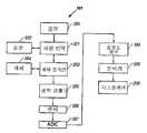
도 1은 본 발명에 따르는 근적외선 분광학을 통하여 포도당을 비침습적으로 측정하기 위한 시스템의 블록선도를 도시한 것이다.Figure 1 shows a block diagram of a system for non-invasive measurement of glucose via near infrared spectroscopy according to the present invention.
도 2는 본 발명에 따르는 도 1의 시스템으로부터의 분광계의 블록선도를 도시한 것이다.2 shows a block diagram of a spectrometer from the system of FIG. 1 in accordance with the present invention.
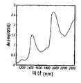
도 3은 인체의 전완으로부터 전형적인 흡광 스펙트럼을 측정한 결과를 도시한 것이다.Figure 3 shows the results of measuring the typical absorption spectrum from the forearm of the human body.
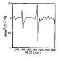
도 4는 파장에 대한 흡광 스펙트럼의 정규화 2차 도함수를 도시한 것이다.4 shows the normalized second derivative of the absorption spectrum over wavelength.
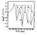
도 5는 본 발명에 따라 확인된 특징을 갖는 제1 상음에서 흡광 스펙트럼의 2차 도함수를 도시한 것이다.5 shows the second derivative of the absorbance spectrum at the first upper tone with the features identified in accordance with the invention.
도 6은 1910nm 물 밴드의 근처에서 도 5의 흡광 스펙트럼의 2차 도함수를 도시한 것이다.FIG. 6 shows the second derivative of the absorbance spectrum of FIG. 5 near the 1910 nm water band.
도 7은 1450nm 물 밴드의 근처에서 도 5의 흡광 스펙트럼의 2차 도함수를 도시한 것이다.FIG. 7 shows the second derivative of the absorption spectrum of FIG. 5 near the 1450 nm water band.
도 8은 본 발명에 따르는 예시적 특징을 갖는 제2 상음 영역에서 도 5의 흡광 스펙트럼의 2차 도함수를 도시한 것이다.FIG. 8 shows the second derivative of the absorbance spectrum of FIG. 5 in a second overtone region with exemplary features in accordance with the present invention. FIG.
도 9는 본 발명에 따라 표지된 주요 특징을 갖는 조합 밴드에서 도 5의 흡광 스펙트럼의 2차 도함수를 도시한 것이다.FIG. 9 shows the second derivative of the absorbance spectrum of FIG. 5 in combination bands with key features labeled according to the present invention.
도 10은 본 발명에 따르는 포도당 농도에 대한 정규화 지방 밴드의 플롯을 도시한 것이다.10 shows a plot of normalized fat bands for glucose concentration according to the present invention.
상기 시스템의 개요는 도 1에 도시하였으며, 일반적으로 2개 소자, 즉 포도당 측정을 실시하기 위한 방법을 구현하기 위한 분광계(101) 또는 계기 및 분석기(208)로 구성된다. 분광계는 개체 조직의 근적외선 스펙트럼을 측정한다. 분석기는 스펙트럼 측정을 처리하고, 특이점 검출과 포도당 측정에 관련된 특징을 추출하고, 또 포도당 측정을 실시하기 위해 처리된 스펙트럼 측정 및/또는 추출된 특징에 대한 모델을 제공한다. 시스템의 분광계 및 분석기 부재에 대한 상세한 설명은 다음과 같다.An overview of the system is shown in FIG. 1 and generally consists of a
분광계 시스템Spectrometer system
분광학적 측정 시스템(101)은 근적외선 방사선의 광원(200), 파장 선택 시스템(201), 환자 접촉면, 근적외선 방사선을 피부로 보내기 위한 수단(203) 및 피부로부터 반사되거나 투과된 방사선을 유도하기 위한 수단(205), 피부로부터 반사되거나 투과된 근적외선 방사선을 검출하기 위한 수단(206), 검출된 근적외선 방사선을 분석하기 위한 수단(208) 및 측정된 분석 물질, 특성 또는 성분을 표시하기 위한 수단(209)으로 구성된다.The
또 다른 배치로서는, 파장 선택(201)은 개체 접촉면(203) 및 광학 검출기(205) 사이에서 생길 수 있다.As another arrangement, the
광원(200)은 파장 범위 700-2500nm의 근적외선 에너지를 방사하며, 예컨대 LED 배열 또는 할로겐 램프로 구성될 수 있다. 관심의 대상이 되는 스펙트럼 범위의 외부로부터 파장의 영향을 최소화하기 위해 하나 이상의 밴드패스 필터(bandpass filter)가 (경우에 따라) 필요하지만, 여전히 근적외선 에너지의 광원에 의해 방사된다. 예컨대, 할로겐 램프는 약 1600nm에서 피이크 에너지를 갖기는 하지만 여전히 2500nm 이상의 전자기 방사선을 발생한다. 이것은 2500nm 이상의 파장이 조직 및 상응하는 성분을 가열함으로써 측정시에 유해한 효과를 갖고 있기 때문에, 포도당의 검출시에 유해한 효과를 갖는다.The
피부를 조사(illumination) 하기 전 및/또는 후에 파장 분리(201)하는 방법은 분산성 소자(예컨대, 평면 또는 오목면, 룰 격자 또는 홀로그래픽 격자), 인터페로미터(interferometer), 또는 추가의 분산 소자없이 LED 어레이 소자를 연속적으로 조사하는 것을 이용함으로써 실시한다. 환경에서 변화에 의해 유발된 이들 파장 분리 방법의 실시에서의 변화로 인하여, 조직 검사의 직전 또는 직후에, 참조 파장 표준(202), 예컨대 폴리스티렌 표준을 스캐닝(scanning)함으로써 이들 변화에 대해 보정할 필요가 있다. 인터페로미터(interferometer)를 기본으로 하는 시스템에서는 조직의 검사와 동시에 실시된다.The method of
감지 소자(206)는 표적 파장에 대하여 반응성을 나타내는 검출기이며, 어레이 또는 단일 소자를 구성할 수 있다. 선형 다이오드 어레이(또는 포토다이오드 어레이)의 경우, 관심의 대상이 되는 파장 영역을 커버하는 데 2개 이상의 상이한 검출기 재료가 필요한 경우, 재료 접합은 측정에 필요하지 않은 파장에서 수행한다. 예컨대, InGaAs 및 확장된 InGaAs 검출기의 경우, 상기 접합은 전형적으로 확장된 InGaAs의 고비용으로 인한 어레이의 비용을 감소시키기 위해 1750nm에서 수행한다. 그러나, 상기 파장 영역은 지방, 단백질 및 포도당과 관련된 흡광의 중간 지점에서 발생한다. 따라서, 약 1480nm ± 20nm에서 접합이 발생하는 것이 바람직하다. 또한, 어레이의 개별 소자를 감지하기 위해 사용된 전자 공학은 동일한 파장에서 생기는 접합을 갖는 것이 바람직하다.The
조직 샘플 계면은 개체(204) 계면 모듈(203)을 포함하며, 그에 의해 근적외선 방사선은 직접적으로 또는 광 파이프, 섬유 광학, 렌즈 시스템 또는 광 유도 거울 시스템을 통하여 (205) 조직으로 향하거나 그로부터 유도된다. 조사될 조직 표면 영역 및 회귀하는 근적외선 방사선이 검출되는 영역은 서로 상이하게 진행하여 소정 거리를 두도록 분리되고, 관심의 대상이 되는 특성의 측정에 최적인 조직 용적을 표적으로 하기 위해 선택된다. 조사 부위로부터 스펙큘라 반사된 방사선은 회귀하는 방사선의 검출을 크게 방해하지 않는 크기이다. 따라서, 소정량에 의해 조사 부위로부터 검출 부위를 옵셋팅(offsetting)할 때, 검출되는 광에 영향을 주는 다수의 조직의 부분집합인 조직의 용적을 샘플링할 수 있는 반면에, 스펙큘라 반사광으로부터의 간섭을 피할 수 있다. 보다 큰 규모의 테이블탑 또는 데스크탑 계기의 경우, 환자 접촉 모듈은 선택한 조사 메카니즘 및 관심의 대상이 되는 조직과 직면하도록 보조하는 팔꿈치 받침, 손목 받침 및 가이드를 더 포함한다. 보다 작은 소형 유닛의 경우, 환자 접촉 모듈은 관심의 대상이 되는 조직과 직면하도록 보조하는 가이드 또는 배치 메카니즘을 포함한다. 일반적으로, 상술한 바와 같이, 광학 커플링 체액은 피부의 표면으로부터 스펙큘라 반사를 최소화하기 위하여 조사 메카니즘과 관심의 대상이 되는 조직 사이에 배치된다. 상술한 환자 접촉 모듈의 일부는 미국 특허원 제09/563,782호 및 국제출원 제PCT/US01/29232호에 기재되어 있으며, 이들의 내용은 본 명세서에 참고문헌으로 포함되어 있다.The tissue sample interface comprises an
수집된 근적외선 방사선은 전압으로 변환되며, A/D(아날로그/디지탈)(207)변환기를 통하여 마이크로프로세서계 시스템(microprocessor-based system)(208)상에서 샘플링되며, 그 분석결과는 (209)로 표시된다.The collected near-infrared radiation is converted to voltage and sampled on a microprocessor-based
측정 프로브가 접촉하게 되는 개체의 샘플 부위, 표면 또는 지점은 분광계 시스템에 의해 조사된 특정 조직을 포함한다. 샘플 부위의 이상적인 품질은 표적 분석 물질에 대한 균질성, 면역성 및 접근성을 포함한다. 복부, 대퇴부, 손(손바닥 또는 손등), 귓볼 또는 손가락을 비롯한 다수의 측정 부위가 사용될 수 있지만, 바람직한 양태에서는, 전완 바닥이 사용된다. 또한, 상기 측정은 확산 반사 또는 확산 투과적 모드로 실시될 수 있지만, 바람직한 방법은 확산 반사이다. 조직의 스캐닝은, 맥동효과에 영향을 받지 않는 영역의 경우, 연속적으로 실시될 수 있고, 또는 상기 스캐닝은 펄스 사이에 간헐적으로 실시될 수 있다.The sample site, surface, or point of the subject that the measurement probe is to contact includes the particular tissue examined by the spectrometer system. Ideal quality of the sample site includes homogeneity, immunity and access to the target analyte. Many measurement sites can be used, including the abdomen, thigh, hand (palm or back of the hand), earlobe, or finger, but in a preferred embodiment, the forearm bottom is used. The measurement can also be carried out in diffuse reflection or diffuse transmission mode, but the preferred method is diffuse reflection. Scanning of the tissue may be performed continuously in the case of areas not affected by the pulsating effect, or the scanning may be performed intermittently between pulses.
스펙트럼 측정Spectral measurement
분광계 시스템은 포도당 농도 결정 또는 측정에 필요한 분석기(208)에 스펙트럼 측정(104) 또는 "스펙트럼"을 제공한다. 이 스펙트럼은 스펙트럼의 근적외선 부분(700-2500nm)에 걸친N 파장의 집합,


위의 수학식 3에서,
m은 피부의 반사 스펙트럼이며, 신체 조직의 성분과 입사광의 공지된 상호작용을 근거로 하는 정량적 정보를 함유하는 흡광 스펙트럼과 유사하다.
m 대 λ의 플롯은 도 3에 도시하였으며, 물, 지방 및 단백질에 주로 기인한 흡광 밴드로 구성된다. 그러나, 보다 상세하게는, 상기 측정은 특징의 추출 및 측정 요건에 최적인 근적외선 영역의 특정 파장의 집합으로 구성될 수 있다. 예컨대, 포도당의 측정은 파장 범위 1100-1935nm 또는 이의 소정의 부분집합의 범위에서 최적으로 실시된다.In
m is the reflection spectrum of the skin and is similar to the absorption spectrum containing quantitative information based on the known interaction of components of body tissue with incident light.
A plot ofm versus λ is shown in FIG. 3 and consists of an absorption band mainly due to water, fat and protein. In more detail, however, the measurement may consist of a set of specific wavelengths in the near infrared region that are optimal for feature extraction and measurement requirements. For example, the measurement of glucose is optimally carried out in the wavelength range 1100-1935 nm or a predetermined subset thereof.
또는, 스펙트럼 측정은 하기 수학식 4에 따라 결정될 수 있다:Alternatively, spectral measurements can be determined according to equation (4):

위의 수학식 4에서,
다른 양태에 있어서, 측정치m은 측정된 세기(I)로서 정의될 수 있다. 마지막으로,m은 계기로부터 수집된 단일 스펙트럼이거나 또는 소정 측정 기간 및 평균에 걸쳐 수집된 다수의(임의의) 수집 스펙트럼의 조합으로 구성될 수 있다. 최저 잡음 측정을 위해 사용되는 스펙트럼을 선택하는 방법은 유사성 또는 거리 측정(즉, 가장 유사한 것을 선택) 및 군집 연산을 포함한다.In Equation 4 above,
In another aspect, the measurementm can be defined as the measured intensityI. Finally,m can be composed of a single spectrum collected from the instrument or a combination of multiple (arbitrary) collection spectra collected over a predetermined measurement period and average. Methods of selecting the spectrum used for the lowest noise measurement include similarity or distance measurements (ie, selecting the most similar) and clustering operations.
전처리(preprocessing) 및 특징 추출(feature extraction)Preprocessing and feature extraction
특징 추출(106)은 해석하기 위한 샘플 측정의 품질 또는 특징을 개선시키는 수학적 변형이다[참조: R. Duda, P. Hart,Pattern Classification and Scene Analysis, John Wiley and Sons, New York (1973)]. 특징 추출의 일반적 목적은 조직 측정 부위의 화학적 농도, 구조적 특성 및 생리학적 상태를 정확하게 나타내거나 개선시키는 것이다. 본 발명에서는, 다음을 근거로 하여, 조직의 광학 특성을 나타내거나 반영하도록 특징의 집합을 밝혀내었다:
· 경로 길이의 변화에 관하여 다양한 방식으로 변화하는 분명한 흡광 밴드의 확인; 및Identification of apparent absorption bands that vary in various ways with respect to changes in path length; And
· 측정 부위의 산란 및 흡광 특성(또는 계수).Scattering and absorbance properties (or coefficients) at the measurement site.
이어서, 포도당의 측정에 적합하지 않은 조건을 확인하거나 또는 포도당의 실제 측정을 실시하기 위하여 상기 특징을 적용한다. 예컨대, 지방 밴드 흡광 크기의 분석 산정치는 진피에 대한 특정 정보를 추측하기 위해 사용될 수 있다. 지방은 진피에는 존재하지 않지만, 근적외선 방사선은 진피를 통하여 전달되어 지방 조직 하부로까지 침투한다. 이리하여, 생리학적 변화 및 진피의 광학적 특성의 상응하는 변화는 지방 밴드 흡광 크기에 영향을 미친다.This feature is then applied to identify conditions not suitable for the determination of glucose or to carry out the actual measurement of glucose. For example, an analysis estimate of fat band absorbance size can be used to infer specific information about the dermis. Fat is not present in the dermis, but near-infrared radiation passes through the dermis and penetrates into the lower fatty tissue. Thus, physiological changes and corresponding changes in the optical properties of the dermis affect the fat band absorbance size.
따라서, 진피에서의 물 농도가 증가함에 따라서, 지방 밴드의 크기는 자연적으로 감소되며 또 그 역도 성립한다.Thus, as the water concentration in the dermis increases, the size of the fat band naturally decreases and vice versa.
다수의 유형의 다음 특징을 측정하여 본 발명에 사용한다:A number of types of the following features are measured and used in the present invention:
· 특이점 검출(107);
· 조직의 광학 특성 변화에 대한 보상(102); 및Compensation for changes in optical properties of
· 포도당 측정(109).Glucose measurement (109).
스펙트럼 측정,m, 또는 필터링 작업에 의한 스펙트럼 측정 전처리(105)를 가정하면, 1차 또는 2차 도함수 계산[참조: A. Savitzky, M. Golay,Smoothing and Differentiation of Data by Simplified Least Squares Procedures, Anal. Chem., 36: 8, pp. 1627-1639 (1964)] 또는 산포 보정을 실시한다:Assuming
· "단순" 특징은 스펙트럼 측정치 또는 임계점(기울기가 0인 지점)에서 처리된 스펙트럼 측정치이다.A “simple” feature is a spectral measurement or a spectral measurement processed at a critical point (point of zero tilt).
· 추가적(유도된) 특징은 더하기, 빼기, 나누기 및 곱하기와 같은 수학적 변환을 통하여 기본 특징으로부터 결정된다.Additional (derived) features are determined from basic features through mathematical transformations such as addition, subtraction, division and multiplication.
· 추상적 특징은 전처리된 스펙트럼의 선형 및 비선형 변환을 통하여 유도된다.Abstract features are derived through linear and nonlinear transformations of the preprocessed spectrum.
단순 특징 및 유도된 특징은 일반적으로 지방 흡광 크기와 같은 물리적 해석을 갖지만, 추상적 특징의 집합은 물리적 시스템과 관련된 특정 해석을 반드시 갖는 것은 아니다. 예컨대, 주성분 분석 점수는 특징으로 이용되지만, 이들의 물리적 해석이 언제나 알려져 있는 것은 아니다. 주성분 분석의 이용은 조직 흡광 스펙트럼의 성질과 관련이 있다. 조직 흡광 스펙트럼에서 가장 중요한 변화는 포도당의 흡광에 의해 유발되는 것이 아니라 측정 부위의 상태, 구조 및 조성과 관련이 있다. 이러한 변화는 제1 주성분에 의해 모델링된다. 따라서, 제1 주성분은 조직 측정의 구조적 특성과 생리학적 상태와 관련된 변화를 나타내는 경향이 있으므로, 결국 조직의 광학 특성을 반영한다.Simple features and derived features generally have a physical interpretation, such as a fat absorbance size, but a set of abstract features does not necessarily have a specific interpretation associated with the physical system. For example, principal component analysis scores are used as features, but their physical interpretation is not always known. The use of principal component analysis relates to the nature of the tissue absorbance spectrum. The most important changes in the tissue absorption spectrum are not caused by the absorption of glucose, but are related to the condition, structure and composition of the site of measurement. This change is modeled by the first principal component. Thus, the first principal component tends to exhibit changes related to the structural properties and physiological state of the tissue measurements, which in turn reflects the optical properties of the tissue.
바람직한 양태에 있어서, 상기 특징은 도 4에 도시된 흡광 스펙트럼의 2차 도함수로부터 결정된다. 각각의 임계점은 그의 파장에 따라 확인한다. 각각의 임계점에서 2차 도함수 스펙트럼치는 측정 스펙트럼과 관련된 조직 샘플의 주요 특성을 나타내는 특징으로 이용된다. 도 5 내지 도 9에서, 많은 주요 특징이 예시적 측정으로서 확인되어 있다. 이들은 다음을 포함한다:In a preferred embodiment, the feature is determined from the second derivative of the absorbance spectrum shown in FIG. 4. Each critical point is identified according to its wavelength. The second derivative spectrum at each critical point is used as a feature that represents the main characteristic of the tissue sample associated with the measurement spectrum. In Figures 5-9, many key features have been identified as exemplary measurements. These include:
· 각각 1665, 1708, 1746, 1868, 1380, 1133, 2020 및 2232nm 근처의 정규화 점(n) 1-8;Normalization points (n ) 1-8 around 1665, 1708, 1746, 1868, 1380, 1133, 2020 and 2232 nm, respectively;
· 1727, 1765, 1214, 1165nm 근처의 지방 밴드점(f) 1-4;Fat band point (f ) 1-4 near 1727, 1765, 1214, 1165 nm;
· 1687, 1715, 1190, 2050, 2150, 2175, 2275, 2292 및 2355nm 근처의 단백질 밴드점(p) 1-9; 및Protein band points (p ) 1-9 near 1687, 1715, 1190, 2050, 2150, 2175, 2275, 2292 and 2355 nm; And
· 1789, 1896, 1410, 1465 및 1150nm 근처의 물 밴드점(w) 2-6.Water band points (w ) near 1789, 1896, 1410, 1465 and 1150 nm 2-6.
정규화 점(n1-n8)은 일반적으로 유도된 특징을 결정하기 위해 사용되며 또 "지방"(f1-f4), "단백질"(p1-p9) 및 "물"(w2-w6)로 표시된 점은 일반적으로 각기 지방, 단백질 또는 물로 인한 흡광 밴드 근처에 위치한다. 2차 도함수 스펙트럼의 밴드폭(낮은 분해능)으로 인하여, 1개 성분과 관련된 다수의 밴드는 다른 성분에 의한 흡광을 포함하며, 또 몇 개의 임계점은, 그 위치가 각각의 성분 근처에 있기 때문에 한 성분과 관련된다. 또한, 예시된 2차 도함수 스펙트럼에 도시된 특징에 대한 파장이 보고되며 이들은 감소된 산란 계수의 변환 및 피부의 다중층에 관련된 내부 필터 효과로 인하여 실질적으로 변화될 수 있다.Normalization points (n1-n8 ) are generally used to determine the derived characteristics, and the points marked "fat" (f1-f4 ), "protein" (p1-p9 ), and "water" (w2-w6 ) It is usually located near an absorption band due to fat, protein or water, respectively. Due to the bandwidth (low resolution) of the second derivative spectrum, many of the bands associated with one component include absorption by another component, and several critical points are due to one component being located near each component. Is associated with. In addition, the wavelengths for the features shown in the illustrated second derivative spectra are reported and can vary substantially due to the transformation of reduced scattering coefficients and internal filter effects associated with multiple layers of skin.
추가적 특징은 유도될 수 있고 플롯에 표시된다. 예컨대,d1=n1665-p1687, d2=n1665-f1727, d3=n1665-f1765, d4=n1665-w1789, d5=n1868-w1410, d6=n1380-w1465및 d7=n1380-w1150이며, 이때pλ, wλ, fλ및nλ는 파장λ에 근접한, 앞서 표기한 단백질, 물, 지방 또는 정규화 점을 나타낸다. 특이점 검출 및 측정에 이용되는, 부가적으로 유도된 특징은d2/d1을 포함한다.Additional features can be derived and displayed in the plots. For example,d1 = n1665 -p1687 , d2 = n1665 -f1727 , d3 = n1665 -f1765 , d4 = n1665 -w1789 , d5 = n1868 -w1410 , d6 = n1380 -w1465 andd7 = n1380- w1150 wherepλ , wλ , fλ andnλ represent the previously indicated protein, water, fat or normalization point, close to the wavelengthλ . Additional derived features, used for singularity detection and measurement, included2 / d1 .
이와 같은 상황에서 특정한 특징의 예가 제공되었지만, 당업자라면 흡광 스펙트럼, 1차 도함수 스펙트럼 또는 전처리된 흡광 스펙트럼으로부터 유도될 수 많은 유용한 특징이 수록되어 있지 않다는 것을 잘 알 수 있을 것이다. 또한, 주성분 분석은 일시적 조직 확인, 특이점 분석 및 분석 물질 측정에 유용한 추가적 추상적 특징을 제공한다. 특정 예로서, 적합한 전처리후의 전체 스펙트럼은 측정 모듈을 통과할 것이고 검량을 적용하여 혈당 농도를 산정하거나 예상한다.Although examples of specific features have been provided in such situations, those skilled in the art will recognize that many useful features are not listed which can be derived from an absorption spectrum, a first derivative spectrum or a pretreated absorbance spectrum. Principal component analysis also provides additional abstract features useful for transient tissue identification, singularity analysis and analyte determination. As a specific example, the full spectrum after suitable pretreatment will pass through the measurement module and apply calibration to estimate or predict blood glucose levels.
마지막으로, 포도당의 흡광과 관련된 특징은 전처리, 파장 선택 및 추상적 특징 선택을 통하여 추출된다. 바람직한 양태에 있어서, 전처리는 하나 이상의 필터링, 미분, 산포 보정 및 정규화 단계를 포함한다. 파장 선택은 스펙트럼을 1450-1700nm, 1700-1900nm, 2050-2200nm 및 2250-2400nm를 비롯한 포도당에 특이적으로 관련된 영역에 한정시킨다.Finally, the features associated with absorption of glucose are extracted through pretreatment, wavelength selection and abstract feature selection. In a preferred embodiment, the pretreatment comprises one or more filtering, derivative, scatter correction and normalization steps. Wavelength selection limits the spectrum to regions specifically related to glucose, including 1450-1700 nm, 1700-1900 nm, 2050-2200 nm and 2250-2400 nm.
조직 표준 패턴(template)(102)Organization Standard Template (102)
상기 정의한 전처리 단계에 이어 배경 삭감(background subtraction) 단계를, 하기 수학식 5를 따라, 스펙트럼 배경 산정치 또는 조직 표준 패턴(102) 및 x 사이의 차이를 측정하는 것에 의해 실시한다:Following the pretreatment step defined above, the background subtraction step is carried out by measuring the difference between the spectral background estimate or the tissue

위의 수학식 5에서,In Equation 5 above,
x는 전처리된 스펙트럼 또는 소정의 특징의 집합이고,x is a preprocessed spectrum or set of predetermined features,
xt는 측정 기간과 관련된 배경 또는 조직 표준 패턴의 산정치이며,xt is an estimate of the background or tissue standard pattern associated with the measurement period,
c및d는 조직 표준 패턴에 대한 기울기 및 절편 조정값이다.c andd are the slope and intercept adjustment values for the tissue standard pattern.
각각의 측정 기간 동안, 조직상의 측정 위치 및 측정 부위의 생리학적 안정성의 레벨에 의해 정의되는 조직 표준 패턴은 하나 이상의 스펙트럼 측정 및 데이터 선택 기준을 통하여, 예컨대 서로 밀접하게 유사한 스펙트럼 측정치만을 선택하여 이들을 평균하는 것에 의해 결정된다. 바람직한 양태에 있어서,xt는 측정 기간의 초기에 조직상에서 수집된 (스펙트럼) 측정으로부터 추출된 특징을 포함한다. 상기 과정을 "재검량"이라 칭하며, 조직 표준 패턴을 형성하기 위해 처리된 하나 이상의 스펙트럼 측정치 집합 뿐만 아니라, 관련된 포도당 기준치 집합을 포함한다. 이하에서 보다 상세하게 기재되는 바와 같이, 조직 표준 패턴을 생성하여 측정 바이어스 조정값(103)을 형성하기 위해 사용된 전략과 동일한 전략에 따라, 포도당 치를 조합한다. 측정 기간은 조직 샘플의 상태가 균일(소정의 결합내의 광학 특성)하고 조직측정 부위가 일정한 동안의 시간으로 정의된다. 그러나, 상기 조직 표준 패턴은 소정 환자로부터 얻은 임의의 특징 집합 또는 추후 스펙트럼 측정을 비교할 검량 세트일 수 있다. 후자의 양태에 있어서, 변수c및d는 측정 스펙트럼에 대한 특정 파장 범위에 걸친 조직 표준 패턴의 최소제곱피트[유클리드 놈(norm)(z)를 최소화하기 위해]를 통하여 결정된다.During each measurement period, the tissue standard pattern, defined by the measurement location on the tissue and the level of physiological stability of the measurement site, is averaged over one or more spectral measurements and data selection criteria, e.g., by selecting only closely similar spectral measurements. It is decided by doing. In a preferred embodiment,xt includes features extracted from (spectrum) measurements collected on tissue at the beginning of the measurement period. This process is called a “review amount” and includes one or more sets of spectral measurements that have been processed to form a tissue standard pattern, as well as an associated set of glucose criteria. As described in more detail below, glucose values are combined according to the same strategy used to generate tissue standard patterns to form
조직 상태의 검출Detection of tissue condition
앞서 논의한 바와 같이, 다양한 구획에서 물의 분포 변화는 광학 특성의 변화를 초래하며 이는 스펙트럼 특징 변화에 의해 나타난다. 따라서, 포도당의 분광학적 측정에 유해한 조건은 선택된 특징을 모니터링하여, 소정의 측정 기간 동안 이들의 변화가 검량 세트 또는 앞서 설정된 기타 제한의 변화를 초과하지 않도록 함으로써 검출할 수 있다. 예컨대, 정규화 지방 밴드의 크기인

측정(109)Measure (109)
포도당의 측정은 처리된 스펙트럼 측정 및/또는 추출된 특징에 검량 모델(108)을 적용하는 것에 의해 달성된다. 상기 모델은 전처리된 스펙트럼 측정치(x) 및 혈액 샘플 또는 세포 조직 사이의 체액을 분석하여 결정된 관련된 포도당 기준치(y)로 각각 구성된 예시된 짝을 이루는 데이터 점의 검량 세트로부터 결정된다. 또는, 기준 포도당 측정은 손가락 끝 또는 스펙트럼 측정 부위에서 채혈하여 결정할 수 있다. 마지막으로, 기준 포도당 측정은 분광학 측정 부위에서 또는 그 근처에서 또는 대표 부위, 예컨대 전완에서 측정한 세포 조직 사이의 포도당 농도로부터 결정할 수 있다.Measurement of glucose is accomplished by applying
상기 방법에 의하면, 혈액, 혈청, 혈장 또는 세포 조직 사이의 채취물은, 센서 샘플 부위 근처이거나 또는 샘플 부위를 반영하는 것으로 표시된/결정된 조직 부위로부터 취한다. 예컨대, 전완 위에서 검량하기 위해 비침습적 근적외선 측정방법을 이용하는 경우, 개인에 따라서는 동일한 전완 또는 바깥쪽 전완으로부터 모세혈관 혈액을 수집할 수 있다. 또는, 혈액을 사용하기 보다는, 일부 경우에서는 모세혈관 포도당 치보다 세포 조직 사이의 포도당 치를 이용하는 것이 유리할 수 있다.According to the method, a sample between blood, serum, plasma or cellular tissue is taken from a tissue site indicated / determined near or reflecting the sensor sample site. For example, when a non-invasive near infrared measurement method is used to calibrate the forearm, capillary blood may be collected from the same forearm or outer forearm, depending on the individual. Or, rather than using blood, in some cases it may be advantageous to use glucose levels between cellular tissues rather than capillary glucose levels.
검량 세트는 하나 이상의 개체를 기본으로 하며 또 예상되는 포도당 변화 범위 전반에 걸쳐 있고 추후 스펙트럼 측정시 직면할 수 있는 스펙트럼 변화 예를 포함하는 포도당 농도를 일반적으로 함유한다. 검량 모델(108)은 수학식, 파라미터의 집합 및 전처리된 스펙트럼 측정을 근거로 한 개체의 혈당치를 측정하기 위해 실시되는 상응하는 컴퓨터 부호를 포함한다. 바람직한 양태에 있어서, 전처리 및 특징 추출은 상기 모델과 함께 효과적으로 포도당의 순수한 분석 물질 신호를 추출하며, 이때 순수한 분석 물질 신호는 계면에 수직하는 표적 분석 물질과 관련된 스펙트럼 신호의 일부이다[참조: A. Lorber, K. Faber, B. Kowalski,Net Analyte Signal Calculation in Multivariate Calibration,Anal. Chem, 69, pp.1620-1626 (1997)]. 이어서, 순수한 분석 물질 신호를 환산(scaling)하고 바이어스 보정(103)하여, 소정의 포도당 측정 유닛(예컨대, mg/dL)을 대응시킨다.The calibration set generally contains glucose concentrations that are based on one or more individuals and that cover the expected range of glucose changes and include examples of spectral changes that may be encountered in later spectral measurements. The
본 발명의 다수의 양태를 2개의 카테고리로 기재한다. 제1 측정 카테고리에서, 추출된 특징은 보상적인 것이어서, 검출광의 유효 경로 및 샘플 조직 용적의 변화와 관련이 있지만, 그러한 변화가 포도당에 의한 흡광과는 무관한 광학 특성 변화에 대한 다른 모델을 보상하기 위해 적용된다. 이것은 구획 사이의 물 변동(또는 기타 일시적인 생리학적 조건)과 관련된 조직 광학 특성의 변화를 반영하는 흡광 특징을 이용하여 달성되어 포도당의 근적외선 흡광을 근거로 한 검량을 보상한다.Many aspects of the invention are described in two categories. In the first measurement category, the extracted features are compensatory to correlate with changes in the effective path of the detection light and the sample tissue volume, but such changes compensate for other models of optical property changes independent of absorption by glucose. Is applied to. This is accomplished using absorbance features that reflect changes in tissue optical properties related to water fluctuations (or other transient physiological conditions) between compartments to compensate for calibration based on near infrared absorption of glucose.
제2 측정 카테고리에서는, 신체의 생리학적 및 화학적 반응과 관련하여 추출된 특징이 개체의 혈당치를 간접적으로 측정하기 위해 주로 사용된다. 이 방법은 혈당 변화에 대한 자연발생적 반응을 근거로 하며, 결국 세포 조직간, 혈관 및 세포 구획에서 체액 분포를 변경시킨다. 이러한 체액 분포의 변경은 포도당의 신호보다 실질적으로 더 높으며 포도당을 비침습적으로 측정하기 위한 마커(marker)로서 사용될 수 있는 신호를 제공하는, 지방, 단백질 및 물과 같은 주요 성분의 분명한 흡광 변화를 초래한다. 그러나, 장기간 체액 구획 균형은 체액 흡입, 운동, 식이요법, 약물요법 및 기타 생리학적 인자에 의해 영향을 받는다.In the second measurement category, features extracted in connection with the physiological and chemical reactions of the body are mainly used to indirectly measure the blood glucose level of the individual. This method is based on a spontaneous response to blood glucose changes, which in turn alters the fluid distribution in intercellular tissues, blood vessels and cell compartments. This change in body fluid distribution results in a clear change in absorbance of key constituents such as fat, protein and water, which is substantially higher than the signal of glucose and provides a signal that can be used as a marker for non-invasive measurement of glucose. do. However, long-term fluid compartment balance is affected by fluid inhalation, exercise, diet, drug therapy and other physiological factors.
체액 구획 변동에 대한 포도당의 보조적 검량은 단기간에 걸쳐 가능하지만, 장기간에 걸친 체액 변동에 대한 포도당의 검량은, 장기간의 체액 구획 변동의 공급원을 보상하기 위하여 분석 신호 및 관련된 혈당의 바이어스 보정을 필요로 한다(수학식 2의 픽의 법칙은 포도당 농도 변화에 대한 물 농도의 변화에 관한 것임을 주목해야 한다). 이리하여, 상기 측정은 시간에 따라 시작점에 대한 포도당의 이동의 결정을 가능하게 하며, 초기 물 농도가 엄밀하게는 관련된 포도당 농도의 함수가 아니기 때문에, 분광학적 물 신호 및 시작점과 관련된 포도당 농도 둘 다의 바이어스 보정이 필요하다. 따라서, 본 발명의 양태에 있어서, 주 성분에 반영된 바와 같은 조직의 광학 특성의 변화를 측정하는 장치 및 방법, 및 이들 특성을 근거로 하는 포도당 농도를 결정하는 방법이 제공된다. Auxiliary calibration of glucose for bodily fluid fluctuations is possible over a short period of time, but calibration of glucose for long term fluid fluctuations requires bias correction of the analytical signal and associated blood glucose to compensate for the source of long-term fluid compartment fluctuations. (Note that the Pick's law in
스펙트럼 특징을 통한 포도당의 보상 측정은 상기 기재한 분류 시스템[참조: Malin 등의 상기와 동일한 문헌]에 의해 또는 하기 수학식 6을 통하여 선택된 특징을 포도당 측정 모델에 보상함으로써 실시한다:Compensation measurement of glucose via spectral features is carried out by compensating for the glucose measurement model by the above-described classification system (same reference as Malin et al.) Or by selecting the feature selected via Equation 6:

위의 수학식 6에서,


b는 조직 표준 패턴 및 검량 모델과 모두 관련된 포도당 측정을 위한 베이스라인 조정값이다.
상기 모델f(·)는 스펙트럼 측정치, 추출된 특징 및 포도당 기준치(혈액 측정 또는 세포 조직간 측정으로부터)를 포함하는 검량 세트를 통하여 결정된다.f(·)의 구조를 고안하기 위한 방법은 확인 시스템의 처리를 통한다[참조: L. Ljung,Systems Identification: Theory for the User, 2d, ed., Prentice Hall (1999)]. 상기 모델 파라미터는 다변량 회귀법 또는 가중 다변량 회귀법[참조: N. Draper, H. Smith,Applied Regression Analysis, 2d, ed., John Wiley and Sons, New York (1981)], 주성분 회귀법[참조: H. Martens, T. Naes,Multivariate Calibration, John Wiley and Sons, New York (1989)], 부분최소제곱회귀법[참조: P. Geladi, B. Kowalski,Partial least-squares regression: a tutorial, Analytica Chimica Acta, 185, pp.1-17 (1986)] 또는 인공 신경망[참조: S. Haykin,Neural Networks: A Comprehensive Foundation, Prentice Hall, Upper Saddle River NJ (1994)]을 포함한 공지 방법을 이용하여 산출한다.In Equation 6 above,
b is the baseline adjustment for glucose measurements associated with both tissue standard patterns and calibration models.
The modelf (·) is determined through a set of calibrations including spectral measurements, extracted features and glucose reference values (from blood measurements or intercellular measurements).The method for devising the structure off (·) is through the processing of a verification system (L. Ljung,Systems Identification: Theory for the User , 2d, ed., Prentice Hall (1999)). The model parameters include multivariate regression or weighted multivariate regression (N. Draper, H. Smith,Applied Regression Analysis , 2d, ed., John Wiley and Sons, New York (1981)), principal component regression (H. Martens). , T. Naes,Multivariate Calibration , John Wiley and Sons, New York (1989)], P. Geladi, B. Kowalski,Partial least-squares regression: a tutorial , Analytica Chimica Acta, 185, pp.1-17 (1986)] or artificial neural networks (S. Haykin,Neural Networks: A Comprehensive Foundation , Prentice Hall, Upper Saddle River NJ (1994)).
Xp 및 z가 독립적인 경우, 일반 수학식은 하기 수학식 7로 변형될 수 있다:When Xp and z are independent, the general equation can be transformed into the following equation:

위의 수학식 7에서,In Equation 7, above,


ms및mi는 g(z)를 보정 유닛으로 변환시키기 위해 사용된 기울기 및 절편이다.
상기한 경우에서는, 실험 디자인을 통하여f(·) 및g(·)를 별도로 결정할 수 있다. 첫째,f(·)는 포도당이 조작되는 동안 조직의 광학 특성이 안정하거나 또는 일정한 실험을 통하여 확인된다. 둘째, 조직의 광학 특성은 변경되며,g(·), ms및mi는 포도당의 측정 오차를 근거로 하여 결정되며, 이때g(·)에 대한 목표값은 하기 수학식 8에 의해 주어진다:
ms andmi are the slope and intercept used to convertg (z) to the correction unit.
In the above case,f (·) andg (·) can be determined separately through the experimental design. First,f (·) is confirmed by stable or constant experimentation of the optical properties of tissues during glucose manipulation. Second, the optical properties of the tissue are changed,g (·), ms andmi are determined based on the measurement error of glucose, where the target value forg (·) is given by:

위의 수학식 8에서,
y는 기준 포도당 농도이다.
제3 양태로서,f(·) 및g(·)는 측정범위에 걸쳐 선형인 것으로 결정된다.
수학식 8은 하기 수학식 9로 변형된다:In Equation 8 above,
y is the reference glucose concentration.
As a third aspect,f (·) andg (·) are determined to be linear over the measurement range.
Equation 8 is transformed into the following equation 9:

위의 수학식 9에서,
당해 양태에 있어서, F 및 G는 선형 검량 방법을 이용하여 상술한 바와 같이 별도로 결정된다. 포도당 측정을 위한 특징의 보상적 사용을 마지막으로 실시하는 것이 바람직한 방법이다.In Equation 9 above,
In this embodiment, F and G are separately determined as described above using the linear calibration method. It is a preferred method to carry out the compensatory use of the features for glucose measurement last.
측정의 제2 카테고리에서, 추출된 특징은 하기 수학식 10을 통하여 간접적으로 포도당을 측정하기 위해 사용된다:In a second category of measurement, the extracted feature is used to measure glucose indirectly through Equation 10:

위의 수학식 10에서,In Equation 10 above,

ms및mi는 g(z)를 보정 유닛으로 변환시키기 위해 사용된 기울기 및 절편이다.
g(·)를 측정하기 위한 방법은 스펙트럼 측정, 추출된 특징 및 기준 포도당 농도(혈액 측정 또는 세포 조직내 측정으로부터)의 예시적 집합(검량 세트)를 통하여 실시한다. 기준 포도당 농도에 대한 상호작용 조합된 관계를 근거로 하여 특징의 부분집합을 선택한다. 변수 선택에는 종래 기술의 지식과 시행착오가 이용되지만, 표준 방법도 또한 순차적 회귀[참조: Draper 등의 상기와 동일한 문헌] 랜덤 서치 수법, 유전자 앨고리즘[참조: D. Goldberg,Genetic Algorithm in Search, Optimization and Machine Learning, Addison Wesley Publishing Company (1989)] 또는 진화론적인 프로그래밍[참조: D. Fogel,An Introduction to Simulated Evolutionary Optimization, IEEE Trans. On Neural Networks, 5:1 (1994년 1월)]을 포함하는 방법이 변수 선택에 이용될 수 있다. 모델g(·)는 선형 또는 비선형 검량의 표준방법을 통하여 결정한다. 선형의 경우는 다음 수학식 11에 따른다:
ms andmi are the slope and intercept used to convert g (z) to the correction unit.
The method for measuring g (·) is carried out through an exemplary set (calibration set) of spectral measurements, extracted features and reference glucose concentrations (from blood measurements or intracellular measurements). A subset of features is selected based on the interaction combined relationship to the baseline glucose concentration. Prior art knowledge and trial and error are used for variable selection, but standard methods also use sequential regression (same as Draper et al.), Random search techniques, genetic algorithms [D. Goldberg,Genetic Algorithm in Search, Optimization and Machine Learning , Addison Wesley Publishing Company (1989)] or evolutionary programming [D. Fogel,An Introduction to Simulated Evolutionary Optimization , IEEE Trans. On Neural Networks, 5: 1 (January 1994)] can be used for variable selection. The modelg (·) is determined using the standard method of linear or nonlinear calibration. In the case of linearity, the following equation (11) is used:

위의 수학식 11에서,
본 발명의 바람직한 양태에 있어서, 특징(z)는 정규화 2차 도함수 지방 밴드(d2) 또는 정규화 2차 도함수 단백질 밴드(d1)를 포함하도록 선택된다. 모델의 파라미터(ms, mi 및 G)는 다변량 회귀법, 가중 다변량 회귀법 또는 국소적 가중 회귀법을 통하여 측정된다. 예컨대, 검량 세트는 포도당 농도가 70-350mg/dl 범위에 달하는 특정 개체상에서 수집하였다. 정규화 지방밴드(d2) 대 포도당 농도의 플롯은 도 10에 도시되어 있다. 특징 및 기준 포도당 농도 간의 상관 관계가 높으면, 포도당 측정이 상기 추출된 특징을 통하여 용이하게 실시된다는 것을 나타낸다. 단순 선형 회귀법을 실시하여 상기 수학식의 모델 파라미터를 결정한다.In a preferred embodiment of the invention, featurez is selected to include a normalized second derivative fat band (d2 ) or a normalized second derivative protein band (d1 ). The parameters of the model (ms , mi and G ) are measured through multivariate regression, weighted multivariate regression or local weighted regression. For example, calibration sets were collected on specific individuals with glucose concentrations ranging from 70-350 mg / dl. Plots of normalized fat band (d2 ) versus glucose concentration are shown in FIG. 10. High correlations between features and reference glucose concentrations indicate that glucose measurements are readily made through the extracted features. Simple linear regression is performed to determine the model parameters of the equation.
그러나, 본 발명은 정규화 지방 및 단백질 밴드에만 한정되는 것은 아니다. 물 흡광 피이크 및 정규화 물 흡광 피이크(d4=n1665-w1789, d5=n1868-w1410, d6=n1380-w1465및 d7=n1380-w1150)를 이용하여 유사한 방법을 밝혀내었다. 정규화를 위해 선택된 파장은 1665nm에 한정되지 않는다. 실제로, 각각의 조직 샘플의 광학 특성, 관류의 베이스라인 수준 및 포도당 농도 변화에 대한 생리학적 반응에 따라서 다양한 개체 및 개체의 카테고리에 대한 다수의 모델이 존재한다. 따라서, 다른 양태는 주요 유형의 모든 흡광 밴드에 관련된 특징의 조합을 사용하는 것으로 구성된다. 예컨대, 정규화 2차 도함수 지방 밴드 및 2개의 정규화 2차 도함수 물 밴드를 선택하였다. 포도당에 대한 이들 변수의 다중 회귀는, 이로 한정되지 않으나, 하기 수학식 12의 모델을 이용하여 실시한다.However, the present invention is not limited to normalized fat and protein bands. Similar methods were found using water absorption peaks and normalized water absorption peaks (d4 = n1665- w1789 , d5 = n1868- w1410 , d6 = n1380 -w1465 andd7 = n1380 -w1150 ). The wavelength selected for normalization is not limited to 1665 nm. Indeed, there are a number of models for various individuals and categories of individuals depending on the optical properties of each tissue sample, the baseline level of perfusion and the physiological response to glucose concentration changes. Thus, another aspect consists in using a combination of features related to all absorption bands of the main type. For example, a normalized second derivative fat band and two normalized second derivative water bands were selected. Multiple regression of these variables on glucose is performed using, but not limited to, the model of Equation 12 below.

위의 수학식 12에서,
상기 수학식 12는 전처리 및 특징 추출 후, 추후 취할 스펙트럼으로부터 포도당 값을 측정하기 위해 사용될 수 있다.In Equation 12 above,
Equation 12 may be used to measure glucose values from spectra to be taken after pretreatment and feature extraction.
또 다른 양태에 있어서, 주성분 분석으로부터 얻은 점수와 같은 피부 조직의 광학 특성의 변화를 나타내는 추상적 특징은 포도당을 비침습적 검량 및 측정하기 위한 독립적인 변수로서 이용될 수 있다. 당해 양태에 있어서, 스펙트럼 측정(m)은 전처리된 다음 파장 선택을 하여 전처리된 벡터(x)를 생성한다. 스펙트럼 분석은 하기 수학식 13에 따라 실시한다:In another embodiment, abstract features indicating changes in optical properties of skin tissue, such as scores obtained from principal component analysis, can be used as independent variables for noninvasive calibration and measurement of glucose. In this embodiment, the spectral measurement (m ) is pretreated followed by wavelength selection to produce a pretreated vector (x ). Spectrum analysis is performed according to the following equation (13):

위의 수학식 13에서,
N은 검량을 위해 선택된 파장수이며,

상술한 바와 같이, 검량 모델은 다변량회귀법, 가중다변량회귀법, 국소적 가중회귀법 또는 기타 표준방법을 통하여 결정될 수 있다. 주성분 회귀법은 스펙트럼 분석방법으로서 기재되어 있지만, 부분최소제곱회귀법도 또한 적용될 수 있다.In
N is the number of wavelengths selected for calibration
As described above, the calibration model can be determined through multivariate regression, weighted multivariate regression, local weighted regression, or other standard methods. Principal component regression is described as a spectral analysis method, but partial least squares regression can also be applied.
추상적 특징 추출이 관련되는 경우, 바람직한 방법은 넓은 스무딩 창(smoothing window) (예컨대, 31nm)를 갖는 1차 도함수를 통한 전처리 단계, 승법적 산포(multiplicative scatter) 보정을 통한 산포 보정단계 또는 표준 정규 변량 변환[참조: R. Barnes, M. Dhanoa, S. Lister, Applied Spectroscopy, 43: 772-777 (1989)] 및 1450-1850nm 범위 또는 그의 부분집합의 파장선택단계를 포함한다. 또한, 1180-1450nm와 같은 물 밴드로부터 얻은 정보도 또한 포함될 수 있다. 전처리된 데이터는 조직 표준 패턴으로 보정되고 부분최소제곱법을 적용하여 검량 모델을 밝혀낸다. 이어 동일한 전처리 단계(1차 도함수, 산포 보정, 파장선택 및 조직 표준 패턴 보정)를 스펙트럼 측정치에 적용하는 것을 통하여 포도당을 측정함으로써 처리된 스펙트럼 측정치(x)를 얻는다. 스펙트럼 측정과 관련된 포도당 측정치는 다음 수학식 14에 따라 결정된다:If abstract feature extraction is involved, the preferred method is a preprocessing step with a first derivative with a wide smoothing window (eg 31 nm), a scatter correction step with multiplicative scatter correction or a standard normal variance. Transformations (R. Barnes, M. Dhanoa, S. Lister, Applied Spectroscopy, 43: 772-777 (1989)) and wavelength selection steps in the 1450-1850 nm range or subset thereof. In addition, information obtained from water bands such as 1180-1450 nm may also be included. The preprocessed data is corrected to the tissue standard pattern and the partial least squares method is applied to reveal the calibration model. The processed spectral measurementsx are then obtained by measuring glucose through applying the same pretreatment steps (first derivative, dispersion correction, wavelength selection and tissue standard pattern correction) to the spectral measurements. Glucose measurements associated with spectral measurements are determined according to the following equation:

위의 수학식 14에서,
본 발명은 혈중 및 조직 중의 포도당을 측정하는 것에 관하여 기재하였지만, 본 발명의 원리는 다른 조직 성분 및 분석 물질의 검출에도 적용될 수 있다.Although the present invention has been described with regard to measuring glucose in blood and tissues, the principles of the present invention can also be applied to the detection of other tissue components and analytes.
본 발명은 바람직한 양태를 참고로 기재하였지만, 당해 분야의 통상의 지식을 가지고 있는 사람이라면, 본 발명의 취지와 범주를 벗어나지 않는 한, 본 명세서에 기재된 내용을 대체하여 적용할 수 있음을 숙지하고 있을 것이다. 따라서, 본 발명은 이하에 수록한 특허청구범위에 의해서만 제한을 받아야 한다.Although the present invention has been described with reference to preferred embodiments, it will be appreciated by those skilled in the art that the present invention may be substituted without departing from the spirit and scope of the present invention. will be. Therefore, the present invention should be limited only by the claims listed below.
Claims (61)
Translated fromKorean


Applications Claiming Priority (3)
| Application Number | Priority Date | Filing Date | Title |
|---|---|---|---|
| US26443101P | 2001-01-26 | 2001-01-26 | |
| US60/264,431 | 2001-01-26 | ||
| PCT/US2002/002288WO2002065090A2 (en) | 2001-01-26 | 2002-01-25 | Noninvasive measurement of glucose through the optical properties of tissue |
Publications (2)
| Publication Number | Publication Date |
|---|---|
| KR20040020878A KR20040020878A (en) | 2004-03-09 |
| KR100893432B1true KR100893432B1 (en) | 2009-04-17 |
Family
ID=23006047
Family Applications (1)
| Application Number | Title | Priority Date | Filing Date |
|---|---|---|---|
| KR1020037009614AExpired - Fee RelatedKR100893432B1 (en) | 2001-01-26 | 2002-01-25 | Non-invasive measurement method of target analyte properties in tissue samples and apparatus therefor |
Country Status (12)
| Country | Link |
|---|---|
| US (1) | US6990364B2 (en) |
| EP (1) | EP1364196A4 (en) |
| JP (3) | JP4071113B2 (en) |
| KR (1) | KR100893432B1 (en) |
| CN (1) | CN1325015C (en) |
| AU (1) | AU2002249985B2 (en) |
| CA (1) | CA2441078A1 (en) |
| IL (1) | IL156844A0 (en) |
| MX (1) | MXPA03006726A (en) |
| NZ (1) | NZ527164A (en) |
| TW (1) | TWI324686B (en) |
| WO (1) | WO2002065090A2 (en) |
Cited By (1)
| Publication number | Priority date | Publication date | Assignee | Title |
|---|---|---|---|---|
| KR20160024306A (en)* | 2014-08-25 | 2016-03-04 | 삼성전자주식회사 | Stress measurement device and Stress measurement method |
Families Citing this family (358)
| Publication number | Priority date | Publication date | Assignee | Title |
|---|---|---|---|---|
| US7383069B2 (en)* | 1997-08-14 | 2008-06-03 | Sensys Medical, Inc. | Method of sample control and calibration adjustment for use with a noninvasive analyzer |
| US7206623B2 (en)* | 2000-05-02 | 2007-04-17 | Sensys Medical, Inc. | Optical sampling interface system for in vivo measurement of tissue |
| US7098037B2 (en) | 1998-10-13 | 2006-08-29 | Inlight Solutions, Inc. | Accommodating subject and instrument variations in spectroscopic determinations |
| US7519406B2 (en)* | 2004-04-28 | 2009-04-14 | Sensys Medical, Inc. | Noninvasive analyzer sample probe interface method and apparatus |
| US20060211931A1 (en)* | 2000-05-02 | 2006-09-21 | Blank Thomas B | Noninvasive analyzer sample probe interface method and apparatus |
| US7606608B2 (en)* | 2000-05-02 | 2009-10-20 | Sensys Medical, Inc. | Optical sampling interface system for in-vivo measurement of tissue |
| US20070179367A1 (en)* | 2000-05-02 | 2007-08-02 | Ruchti Timothy L | Method and Apparatus for Noninvasively Estimating a Property of an Animal Body Analyte from Spectral Data |
| US7043288B2 (en) | 2002-04-04 | 2006-05-09 | Inlight Solutions, Inc. | Apparatus and method for spectroscopic analysis of tissue to detect diabetes in an individual |
| US6697658B2 (en) | 2001-07-02 | 2004-02-24 | Masimo Corporation | Low power pulse oximeter |
| US20030135547A1 (en)* | 2001-07-23 | 2003-07-17 | Kent J. Thomas | Extensible modular communication executive with active message queue and intelligent message pre-validation |
| US7355512B1 (en) | 2002-01-24 | 2008-04-08 | Masimo Corporation | Parallel alarm processor |
| US20070149868A1 (en)* | 2002-03-08 | 2007-06-28 | Blank Thomas B | Method and Apparatus for Photostimulation Enhanced Analyte Property Estimation |
| US20050187439A1 (en)* | 2003-03-07 | 2005-08-25 | Blank Thomas B. | Sampling interface system for in-vivo estimation of tissue analyte concentration |
| JP4189322B2 (en)* | 2002-03-08 | 2008-12-03 | センシス メディカル インク | Compact instrument for non-invasive measurement of glucose by near infrared spectroscopy |
| US8504128B2 (en) | 2002-03-08 | 2013-08-06 | Glt Acquisition Corp. | Method and apparatus for coupling a channeled sample probe to tissue |
| US8718738B2 (en)* | 2002-03-08 | 2014-05-06 | Glt Acquisition Corp. | Method and apparatus for coupling a sample probe with a sample site |
| US20050054908A1 (en)* | 2003-03-07 | 2005-03-10 | Blank Thomas B. | Photostimulation method and apparatus in combination with glucose determination |
| US7697966B2 (en)* | 2002-03-08 | 2010-04-13 | Sensys Medical, Inc. | Noninvasive targeting system method and apparatus |
| US6850788B2 (en) | 2002-03-25 | 2005-02-01 | Masimo Corporation | Physiological measurement communications adapter |
| US7027848B2 (en) | 2002-04-04 | 2006-04-11 | Inlight Solutions, Inc. | Apparatus and method for non-invasive spectroscopic measurement of analytes in tissue using a matched reference analyte |
| US7259906B1 (en) | 2002-09-03 | 2007-08-21 | Cheetah Omni, Llc | System and method for voice control of medical devices |
| CN100406872C (en)* | 2002-11-04 | 2008-07-30 | 天津市先石光学技术有限公司 | Composite spectrum measurement method and its spectrum detection instrument |
| US6920345B2 (en) | 2003-01-24 | 2005-07-19 | Masimo Corporation | Optical sensor including disposable and reusable elements |
| US20050159656A1 (en)* | 2003-03-07 | 2005-07-21 | Hockersmith Linda J. | Method and apparatus for presentation of noninvasive glucose concentration information |
| EP1464273B1 (en) | 2003-04-03 | 2006-11-29 | Matsushita Electric Industrial Co., Ltd. | Method and device for measuring concentration of specific component |
| US6968222B2 (en) | 2003-05-02 | 2005-11-22 | Oculir, Inc. | Methods and device for non-invasive analyte measurement |
| US7039448B2 (en)* | 2003-05-02 | 2006-05-02 | Diramed, Llc | Zero corrected optical blood analyte detector |
| US6975892B2 (en) | 2003-10-21 | 2005-12-13 | Oculir, Inc. | Methods for non-invasive analyte measurement from the conjunctiva |
| US6958039B2 (en) | 2003-05-02 | 2005-10-25 | Oculir, Inc. | Method and instruments for non-invasive analyte measurement |
| US7500950B2 (en) | 2003-07-25 | 2009-03-10 | Masimo Corporation | Multipurpose sensor port |
| US20070234300A1 (en)* | 2003-09-18 | 2007-10-04 | Leake David W | Method and Apparatus for Performing State-Table Driven Regression Testing |
| US20050073690A1 (en)* | 2003-10-03 | 2005-04-07 | Abbink Russell E. | Optical spectroscopy incorporating a vertical cavity surface emitting laser (VCSEL) |
| US7483729B2 (en) | 2003-11-05 | 2009-01-27 | Masimo Corporation | Pulse oximeter access apparatus and method |
| US7510849B2 (en)* | 2004-01-29 | 2009-03-31 | Glucolight Corporation | OCT based method for diagnosis and therapy |
| WO2005087097A1 (en) | 2004-03-08 | 2005-09-22 | Masimo Corporation | Physiological parameter system |
| US20080033275A1 (en)* | 2004-04-28 | 2008-02-07 | Blank Thomas B | Method and Apparatus for Sample Probe Movement Control |
| US8868147B2 (en) | 2004-04-28 | 2014-10-21 | Glt Acquisition Corp. | Method and apparatus for controlling positioning of a noninvasive analyzer sample probe |
| US8036727B2 (en)* | 2004-08-11 | 2011-10-11 | Glt Acquisition Corp. | Methods for noninvasively measuring analyte levels in a subject |
| US7254429B2 (en) | 2004-08-11 | 2007-08-07 | Glucolight Corporation | Method and apparatus for monitoring glucose levels in a biological tissue |
| DE112004002988B4 (en) | 2004-10-15 | 2013-06-06 | Nagasaki Prefectural Government | Instrument for non-invasive measurement of blood sugar level |
| US7647083B2 (en) | 2005-03-01 | 2010-01-12 | Masimo Laboratories, Inc. | Multiple wavelength sensor equalization |
| US20060217602A1 (en)* | 2005-03-04 | 2006-09-28 | Alan Abul-Haj | Method and apparatus for noninvasive targeting |
| WO2006110859A2 (en) | 2005-04-13 | 2006-10-19 | Glucolight Corporation | Method for data reduction and calibration of an oct-based blood glucose monitor |
| US8140139B2 (en)* | 2005-06-14 | 2012-03-20 | Dominion Assets, Llc | Method and apparatus for the non-invasive sensing of glucose in a human subject |
| US20060281982A1 (en)* | 2005-06-14 | 2006-12-14 | Diasense, Inc. | Method and apparatus for the non-invasive sensing of glucose in a human subject |
| US12014328B2 (en) | 2005-07-13 | 2024-06-18 | Vccb Holdings, Inc. | Medicine bottle cap with electronic embedded curved display |
| JP4744976B2 (en)* | 2005-08-09 | 2011-08-10 | 株式会社東芝 | Biological information measuring apparatus and method |
| US7962188B2 (en) | 2005-10-14 | 2011-06-14 | Masimo Corporation | Robust alarm system |
| US7519253B2 (en) | 2005-11-18 | 2009-04-14 | Omni Sciences, Inc. | Broadband or mid-infrared fiber light sources |
| JP2009521972A (en)* | 2005-12-28 | 2009-06-11 | コーニンクレッカ フィリップス エレクトロニクス エヌ ヴィ | Non-invasive system and method for measuring skin hydration of a subject |
| US8182443B1 (en) | 2006-01-17 | 2012-05-22 | Masimo Corporation | Drug administration controller |
| KR101047968B1 (en)* | 2006-02-20 | 2011-07-12 | (주)하모닉스 | Apparatus and method for analyzing blood components using energy absorption correction |
| US8219172B2 (en) | 2006-03-17 | 2012-07-10 | Glt Acquisition Corp. | System and method for creating a stable optical interface |
| DE102006024459A1 (en)* | 2006-05-24 | 2007-11-29 | Fraunhofer-Gesellschaft zur Förderung der angewandten Forschung e.V. | A sensor, processing device, method and computer program for providing information about a vital parameter of a living being |
| US10188348B2 (en) | 2006-06-05 | 2019-01-29 | Masimo Corporation | Parameter upgrade system |
| WO2007141730A1 (en)* | 2006-06-07 | 2007-12-13 | Koninklijke Philips Electronics N.V. | Dedicated spectral illumination spectroscopy |
| US8457707B2 (en) | 2006-09-20 | 2013-06-04 | Masimo Corporation | Congenital heart disease monitor |
| US8840549B2 (en) | 2006-09-22 | 2014-09-23 | Masimo Corporation | Modular patient monitor |
| JP4963921B2 (en)* | 2006-09-29 | 2012-06-27 | シスメックス株式会社 | Non-invasive living body measurement device |
| CN101523203B (en)* | 2006-09-29 | 2012-09-05 | 皇家飞利浦电子股份有限公司 | Determination of optical absorption coefficients |
| US7880626B2 (en) | 2006-10-12 | 2011-02-01 | Masimo Corporation | System and method for monitoring the life of a physiological sensor |
| EP2073692B1 (en) | 2006-10-12 | 2017-07-26 | Masimo Corporation | Perfusion index smoothing |
| US8255026B1 (en) | 2006-10-12 | 2012-08-28 | Masimo Corporation, Inc. | Patient monitor capable of monitoring the quality of attached probes and accessories |
| US8265723B1 (en) | 2006-10-12 | 2012-09-11 | Cercacor Laboratories, Inc. | Oximeter probe off indicator defining probe off space |
| US9861305B1 (en) | 2006-10-12 | 2018-01-09 | Masimo Corporation | Method and apparatus for calibration to reduce coupling between signals in a measurement system |
| JP5441707B2 (en) | 2006-12-09 | 2014-03-12 | マシモ コーポレイション | Plethysmograph variation processor |
| US8852094B2 (en) | 2006-12-22 | 2014-10-07 | Masimo Corporation | Physiological parameter system |
| US8652060B2 (en) | 2007-01-20 | 2014-02-18 | Masimo Corporation | Perfusion trend indicator |
| KR100883153B1 (en)* | 2007-04-20 | 2009-02-10 | 나가사끼-켄 | Non-invasive measuring device of blood sugar level |
| US8374665B2 (en) | 2007-04-21 | 2013-02-12 | Cercacor Laboratories, Inc. | Tissue profile wellness monitor |
| WO2009004541A1 (en)* | 2007-07-03 | 2009-01-08 | Koninklijke Philips Electronics N.V. | Spectroscopy measurements of the concentration of a substance in a scattering tissue |
| US8571617B2 (en)* | 2008-03-04 | 2013-10-29 | Glt Acquisition Corp. | Flowometry in optical coherence tomography for analyte level estimation |
| BRPI0909825B8 (en)* | 2008-03-25 | 2021-06-22 | Univ Missouri | method and systems for non-invasive blood glucose detection using spectral data from one or more non-glucose components |
| US20090275844A1 (en) | 2008-05-02 | 2009-11-05 | Masimo Corporation | Monitor configuration system |
| JP2011519684A (en) | 2008-05-05 | 2011-07-14 | マシモ コーポレイション | Pulse oximeter system with electrical disconnect circuit |
| CN101292875B (en)* | 2008-06-06 | 2010-07-14 | 天津市先石光学技术有限公司 | Method for measuring component concentration with reference wavelength |
| US20100004518A1 (en) | 2008-07-03 | 2010-01-07 | Masimo Laboratories, Inc. | Heat sink for noninvasive medical sensor |
| US8515509B2 (en) | 2008-08-04 | 2013-08-20 | Cercacor Laboratories, Inc. | Multi-stream emitter for noninvasive measurement of blood constituents |
| SE532941C2 (en) | 2008-09-15 | 2010-05-18 | Phasein Ab | Gas sampling line for breathing gases |
| US20100113899A1 (en)* | 2008-11-06 | 2010-05-06 | Mark Ries Robinson | Alignment System for Optically Sampling a Hand |
| US20100160747A1 (en)* | 2008-12-12 | 2010-06-24 | Mark Ries Robinson | Selection of preferred sampling location on hand via minimization of sampling error, and optical alignment for repeatably sampling tissue |
| US8771204B2 (en) | 2008-12-30 | 2014-07-08 | Masimo Corporation | Acoustic sensor assembly |
| US8588880B2 (en) | 2009-02-16 | 2013-11-19 | Masimo Corporation | Ear sensor |
| US9323894B2 (en) | 2011-08-19 | 2016-04-26 | Masimo Corporation | Health care sanitation monitoring system |
| US10007758B2 (en) | 2009-03-04 | 2018-06-26 | Masimo Corporation | Medical monitoring system |
| JP5749658B2 (en) | 2009-03-04 | 2015-07-15 | マシモ・コーポレイション | Medical monitoring system |
| US10032002B2 (en) | 2009-03-04 | 2018-07-24 | Masimo Corporation | Medical monitoring system |
| US8388353B2 (en) | 2009-03-11 | 2013-03-05 | Cercacor Laboratories, Inc. | Magnetic connector |
| WO2010105197A2 (en)* | 2009-03-12 | 2010-09-16 | The General Hospital Corporation | Non-contact optical system, computer-accessible medium and method for measuring at least one mechanical property of tissue using coherent speckle techniques(s) |
| EP2241246A1 (en)* | 2009-04-14 | 2010-10-20 | Koninklijke Philips Electronics N.V. | Using muscle tension sensing to locate an analyte measurement site on the skin |
| WO2010135373A1 (en) | 2009-05-19 | 2010-11-25 | Masimo Corporation | Disposable components for reusable physiological sensor |
| US8571619B2 (en) | 2009-05-20 | 2013-10-29 | Masimo Corporation | Hemoglobin display and patient treatment |
| US20110208015A1 (en) | 2009-07-20 | 2011-08-25 | Masimo Corporation | Wireless patient monitoring system |
| US8473020B2 (en) | 2009-07-29 | 2013-06-25 | Cercacor Laboratories, Inc. | Non-invasive physiological sensor cover |
| JP5427951B2 (en)* | 2009-08-10 | 2014-02-26 | ディアベテス トールス スウェーデン アーべー | Apparatus and method for generating status indication |
| US9579039B2 (en) | 2011-01-10 | 2017-02-28 | Masimo Corporation | Non-invasive intravascular volume index monitor |
| US20110137297A1 (en) | 2009-09-17 | 2011-06-09 | Kiani Massi Joe E | Pharmacological management system |
| US8571618B1 (en) | 2009-09-28 | 2013-10-29 | Cercacor Laboratories, Inc. | Adaptive calibration system for spectrophotometric measurements |
| JP5394501B2 (en) | 2009-10-02 | 2014-01-22 | シャープ株式会社 | Blood vessel condition monitoring method |
| US20110082711A1 (en) | 2009-10-06 | 2011-04-07 | Masimo Laboratories, Inc. | Personal digital assistant or organizer for monitoring glucose levels |
| US8690799B2 (en) | 2009-10-15 | 2014-04-08 | Masimo Corporation | Acoustic respiratory monitoring sensor having multiple sensing elements |
| US8430817B1 (en) | 2009-10-15 | 2013-04-30 | Masimo Corporation | System for determining confidence in respiratory rate measurements |
| US8790268B2 (en) | 2009-10-15 | 2014-07-29 | Masimo Corporation | Bidirectional physiological information display |
| US9848800B1 (en) | 2009-10-16 | 2017-12-26 | Masimo Corporation | Respiratory pause detector |
| JP5360718B2 (en)* | 2009-11-02 | 2013-12-04 | 独立行政法人国立高等専門学校機構 | Method for obtaining blood glucose concentration information |
| US9839381B1 (en) | 2009-11-24 | 2017-12-12 | Cercacor Laboratories, Inc. | Physiological measurement system with automatic wavelength adjustment |
| WO2011069122A1 (en) | 2009-12-04 | 2011-06-09 | Masimo Corporation | Calibration for multi-stage physiological monitors |
| US9153112B1 (en) | 2009-12-21 | 2015-10-06 | Masimo Corporation | Modular patient monitor |
| US11289199B2 (en) | 2010-01-19 | 2022-03-29 | Masimo Corporation | Wellness analysis system |
| WO2011109312A2 (en) | 2010-03-01 | 2011-09-09 | Masimo Corporation | Adaptive alarm system |
| US8584345B2 (en) | 2010-03-08 | 2013-11-19 | Masimo Corporation | Reprocessing of a physiological sensor |
| JP5386634B2 (en) | 2010-03-19 | 2014-01-15 | シャープ株式会社 | Measurement result processing apparatus, measurement system, measurement result processing method, control program, and recording medium |
| US9307928B1 (en) | 2010-03-30 | 2016-04-12 | Masimo Corporation | Plethysmographic respiration processor |
| US7884933B1 (en) | 2010-05-05 | 2011-02-08 | Revolutionary Business Concepts, Inc. | Apparatus and method for determining analyte concentrations |
| US8666468B1 (en) | 2010-05-06 | 2014-03-04 | Masimo Corporation | Patient monitor for determining microcirculation state |
| JP5982364B2 (en)* | 2010-06-22 | 2016-08-31 | センスペック・ゲゼルシャフト・ミット・ベシュレンクテル・ハフツングSenspec Gmbh | Apparatus and method for identifying and monitoring components or characteristics of a measurement medium, in particular physiological blood values |
| EP2621333B1 (en) | 2010-09-28 | 2015-07-29 | Masimo Corporation | Depth of consciousness monitor including oximeter |
| US12198790B1 (en) | 2010-10-07 | 2025-01-14 | Masimo Corporation | Physiological monitor sensor systems and methods |
| US9211095B1 (en) | 2010-10-13 | 2015-12-15 | Masimo Corporation | Physiological measurement logic engine |
| US20120226117A1 (en) | 2010-12-01 | 2012-09-06 | Lamego Marcelo M | Handheld processing device including medical applications for minimally and non invasive glucose measurements |
| WO2012087319A2 (en)* | 2010-12-22 | 2012-06-28 | Verifica-Bg Inc. | Method and apparatus for in vivo optical measurement of blood glucose concentration |
| US10332630B2 (en) | 2011-02-13 | 2019-06-25 | Masimo Corporation | Medical characterization system |
| US9066666B2 (en) | 2011-02-25 | 2015-06-30 | Cercacor Laboratories, Inc. | Patient monitor for monitoring microcirculation |
| US9986919B2 (en) | 2011-06-21 | 2018-06-05 | Masimo Corporation | Patient monitoring system |
| US9532722B2 (en) | 2011-06-21 | 2017-01-03 | Masimo Corporation | Patient monitoring system |
| US11439329B2 (en) | 2011-07-13 | 2022-09-13 | Masimo Corporation | Multiple measurement mode in a physiological sensor |
| US9782077B2 (en) | 2011-08-17 | 2017-10-10 | Masimo Corporation | Modulated physiological sensor |
| US9808188B1 (en) | 2011-10-13 | 2017-11-07 | Masimo Corporation | Robust fractional saturation determination |
| US9943269B2 (en) | 2011-10-13 | 2018-04-17 | Masimo Corporation | System for displaying medical monitoring data |
| WO2013056160A2 (en) | 2011-10-13 | 2013-04-18 | Masimo Corporation | Medical monitoring hub |
| US9778079B1 (en) | 2011-10-27 | 2017-10-03 | Masimo Corporation | Physiological monitor gauge panel |
| US9392945B2 (en) | 2012-01-04 | 2016-07-19 | Masimo Corporation | Automated CCHD screening and detection |
| US11172890B2 (en) | 2012-01-04 | 2021-11-16 | Masimo Corporation | Automated condition screening and detection |
| US12004881B2 (en) | 2012-01-04 | 2024-06-11 | Masimo Corporation | Automated condition screening and detection |
| US9267572B2 (en) | 2012-02-08 | 2016-02-23 | Masimo Corporation | Cable tether system |
| US10149616B2 (en) | 2012-02-09 | 2018-12-11 | Masimo Corporation | Wireless patient monitoring device |
| US9195385B2 (en) | 2012-03-25 | 2015-11-24 | Masimo Corporation | Physiological monitor touchscreen interface |
| JP6490577B2 (en) | 2012-04-17 | 2019-03-27 | マシモ・コーポレイション | How to operate a pulse oximeter device |
| US10542903B2 (en) | 2012-06-07 | 2020-01-28 | Masimo Corporation | Depth of consciousness monitor |
| US20150018646A1 (en) | 2013-07-12 | 2015-01-15 | Sandeep Gulati | Dynamic sample mapping noninvasive analyzer apparatus and method of use thereof |
| US9585604B2 (en) | 2012-07-16 | 2017-03-07 | Zyomed Corp. | Multiplexed pathlength resolved noninvasive analyzer apparatus with dynamic optical paths and method of use thereof |
| US9351672B2 (en) | 2012-07-16 | 2016-05-31 | Timothy Ruchti | Multiplexed pathlength resolved noninvasive analyzer apparatus with stacked filters and method of use thereof |
| US9351671B2 (en) | 2012-07-16 | 2016-05-31 | Timothy Ruchti | Multiplexed pathlength resolved noninvasive analyzer apparatus and method of use thereof |
| US9697928B2 (en) | 2012-08-01 | 2017-07-04 | Masimo Corporation | Automated assembly sensor cable |
| US10827961B1 (en) | 2012-08-29 | 2020-11-10 | Masimo Corporation | Physiological measurement calibration |
| US9877650B2 (en) | 2012-09-20 | 2018-01-30 | Masimo Corporation | Physiological monitor with mobile computing device connectivity |
| US9955937B2 (en) | 2012-09-20 | 2018-05-01 | Masimo Corporation | Acoustic patient sensor coupler |
| US9749232B2 (en) | 2012-09-20 | 2017-08-29 | Masimo Corporation | Intelligent medical network edge router |
| US9560996B2 (en) | 2012-10-30 | 2017-02-07 | Masimo Corporation | Universal medical system |
| US9787568B2 (en) | 2012-11-05 | 2017-10-10 | Cercacor Laboratories, Inc. | Physiological test credit method |
| US9993159B2 (en) | 2012-12-31 | 2018-06-12 | Omni Medsci, Inc. | Near-infrared super-continuum lasers for early detection of breast and other cancers |
| EP2938259A4 (en) | 2012-12-31 | 2016-08-17 | Omni Medsci Inc | Near-infrared lasers for non-invasive monitoring of glucose, ketones, hba1c, and other blood constituents |
| US9500634B2 (en) | 2012-12-31 | 2016-11-22 | Omni Medsci, Inc. | Short-wave infrared super-continuum lasers for natural gas leak detection, exploration, and other active remote sensing applications |
| US10660526B2 (en) | 2012-12-31 | 2020-05-26 | Omni Medsci, Inc. | Near-infrared time-of-flight imaging using laser diodes with Bragg reflectors |
| US12193790B2 (en) | 2012-12-31 | 2025-01-14 | Omni Medsci, Inc. | Wearable devices comprising semiconductor diode light sources with improved signal-to-noise ratio |
| WO2014143276A2 (en) | 2012-12-31 | 2014-09-18 | Omni Medsci, Inc. | Short-wave infrared super-continuum lasers for natural gas leak detection, exploration, and other active remote sensing applications |
| CA2895982A1 (en) | 2012-12-31 | 2014-07-03 | Omni Medsci, Inc. | Short-wave infrared super-continuum lasers for early detection of dental caries |
| US9750461B1 (en) | 2013-01-02 | 2017-09-05 | Masimo Corporation | Acoustic respiratory monitoring sensor with probe-off detection |
| US9724025B1 (en) | 2013-01-16 | 2017-08-08 | Masimo Corporation | Active-pulse blood analysis system |
| JP5448224B1 (en)* | 2013-03-12 | 2014-03-19 | 日本分光株式会社 | Chromatography system, signal processing apparatus, chromatography data processing terminal and program |
| US9965946B2 (en) | 2013-03-13 | 2018-05-08 | Masimo Corporation | Systems and methods for monitoring a patient health network |
| US10441181B1 (en) | 2013-03-13 | 2019-10-15 | Masimo Corporation | Acoustic pulse and respiration monitoring system |
| US9936917B2 (en) | 2013-03-14 | 2018-04-10 | Masimo Laboratories, Inc. | Patient monitor placement indicator |
| US10456038B2 (en) | 2013-03-15 | 2019-10-29 | Cercacor Laboratories, Inc. | Cloud-based physiological monitoring system |
| US12178572B1 (en) | 2013-06-11 | 2024-12-31 | Masimo Corporation | Blood glucose sensing system |
| US9891079B2 (en) | 2013-07-17 | 2018-02-13 | Masimo Corporation | Pulser with double-bearing position encoder for non-invasive physiological monitoring |
| US10555678B2 (en) | 2013-08-05 | 2020-02-11 | Masimo Corporation | Blood pressure monitor with valve-chamber assembly |
| US12367973B2 (en) | 2013-09-12 | 2025-07-22 | Willow Laboratories, Inc. | Medical device calibration |
| WO2015038683A2 (en) | 2013-09-12 | 2015-03-19 | Cercacor Laboratories, Inc. | Medical device management system |
| WO2015054161A2 (en) | 2013-10-07 | 2015-04-16 | Masimo Corporation | Regional oximetry sensor |
| US11147518B1 (en) | 2013-10-07 | 2021-10-19 | Masimo Corporation | Regional oximetry signal processor |
| US10828007B1 (en) | 2013-10-11 | 2020-11-10 | Masimo Corporation | Acoustic sensor with attachment portion |
| US10832818B2 (en) | 2013-10-11 | 2020-11-10 | Masimo Corporation | Alarm notification system |
| US10279247B2 (en) | 2013-12-13 | 2019-05-07 | Masimo Corporation | Avatar-incentive healthcare therapy |
| US11259745B2 (en) | 2014-01-28 | 2022-03-01 | Masimo Corporation | Autonomous drug delivery system |
| US10123729B2 (en) | 2014-06-13 | 2018-11-13 | Nanthealth, Inc. | Alarm fatigue management systems and methods |
| US10231670B2 (en) | 2014-06-19 | 2019-03-19 | Masimo Corporation | Proximity sensor in pulse oximeter |
| WO2016006113A1 (en)* | 2014-07-11 | 2016-01-14 | 株式会社ニコン | Image analysis device, imaging system, surgery assistance system, image analysis method, and image analysis program |
| KR102290280B1 (en)* | 2014-08-25 | 2021-08-17 | 삼성전자주식회사 | Non-invasive measuring device and method for bio-analyte and diagnosing device and method for diagnosing metabolic syndrome |
| US10111591B2 (en) | 2014-08-26 | 2018-10-30 | Nanthealth, Inc. | Real-time monitoring systems and methods in a healthcare environment |
| US10231657B2 (en) | 2014-09-04 | 2019-03-19 | Masimo Corporation | Total hemoglobin screening sensor |
| US10383520B2 (en) | 2014-09-18 | 2019-08-20 | Masimo Semiconductor, Inc. | Enhanced visible near-infrared photodiode and non-invasive physiological sensor |
| WO2016054079A1 (en)* | 2014-09-29 | 2016-04-07 | Zyomed Corp. | Systems and methods for blood glucose and other analyte detection and measurement using collision computing |
| JP2016075522A (en)* | 2014-10-03 | 2016-05-12 | セイコーエプソン株式会社 | Target component weighing device and electronic apparatus |
| US10154815B2 (en) | 2014-10-07 | 2018-12-18 | Masimo Corporation | Modular physiological sensors |
| US10206581B2 (en) | 2014-10-29 | 2019-02-19 | Zoll Medical Corporation | Transesophageal or transtracheal cardiac monitoring by optical spectroscopy |
| JP6632288B2 (en)* | 2014-12-12 | 2020-01-22 | キヤノン株式会社 | Information processing apparatus, information processing method, and program |
| KR102335739B1 (en) | 2014-12-19 | 2021-12-06 | 삼성전자주식회사 | Apparatus and method for measuring a blood glucose in a noninvasive manner |
| CN107405108B (en) | 2015-01-23 | 2020-10-23 | 迈心诺瑞典公司 | Nasal/oral intubation system and manufacture |
| BR112017016302B1 (en) | 2015-02-06 | 2022-12-06 | Masimo Corporation | PHYSIOLOGICAL SENSOR MANUFACTURING METHOD WITH EFFICIENT FLEXIBLE CIRCUIT |
| US10568553B2 (en) | 2015-02-06 | 2020-02-25 | Masimo Corporation | Soft boot pulse oximetry sensor |
| US10205291B2 (en) | 2015-02-06 | 2019-02-12 | Masimo Corporation | Pogo pin connector |
| KR102360075B1 (en)* | 2015-02-13 | 2022-02-08 | 삼성전자주식회사 | The method and apparatus for measuring bio signal |
| US10716499B1 (en) | 2015-03-24 | 2020-07-21 | Zoll Medical Corporation | Physiological monitoring by optical spectroscopy |
| TWI580398B (en)* | 2015-04-01 | 2017-05-01 | 長庚大學 | Skin detection probe and device |
| US10524738B2 (en) | 2015-05-04 | 2020-01-07 | Cercacor Laboratories, Inc. | Noninvasive sensor system with visual infographic display |
| WO2016191307A1 (en) | 2015-05-22 | 2016-12-01 | Cercacor Laboratories, Inc. | Non-invasive optical physiological differential pathlength sensor |
| US10991135B2 (en) | 2015-08-11 | 2021-04-27 | Masimo Corporation | Medical monitoring analysis and replay including indicia responsive to light attenuated by body tissue |
| KR102612874B1 (en) | 2015-08-31 | 2023-12-12 | 마시모 코오퍼레이션 | Wireless patient monitoring systems and methods |
| US11504066B1 (en) | 2015-09-04 | 2022-11-22 | Cercacor Laboratories, Inc. | Low-noise sensor system |
| CN106525729A (en)* | 2015-09-12 | 2017-03-22 | 南京理工大学 | Substance element content information detection method based on spectral analysis technology |
| WO2017049000A1 (en) | 2015-09-15 | 2017-03-23 | Massachusetts Institute Of Technology | Systems and methods for diagnosis of middle ear conditions and detection of analytes in the tympanic membrane |
| KR102443262B1 (en) | 2015-09-23 | 2022-09-13 | 삼성전자주식회사 | Method and apparatus for predicting analyte concentration |
| CN106680236A (en)* | 2015-11-06 | 2017-05-17 | 深圳市芭田生态工程股份有限公司 | Method for mapping spectral data and chemical detection data |
| KR102444674B1 (en) | 2015-11-20 | 2022-09-16 | 삼성전자주식회사 | Optical measuring device and electronic device having same |
| US11679579B2 (en) | 2015-12-17 | 2023-06-20 | Masimo Corporation | Varnish-coated release liner |
| US11129556B2 (en)* | 2015-12-31 | 2021-09-28 | Wear2B Ltd. | Device, system and method for non-invasive monitoring of physiological measurements |
| CN109348727B (en) | 2016-01-26 | 2022-11-29 | 耐克创新有限合伙公司 | Near infrared spectroscopy techniques for sensing glycogen in muscle tissue |
| US10471159B1 (en) | 2016-02-12 | 2019-11-12 | Masimo Corporation | Diagnosis, removal, or mechanical damaging of tumor using plasmonic nanobubbles |
| US10537285B2 (en) | 2016-03-04 | 2020-01-21 | Masimo Corporation | Nose sensor |
| US10993662B2 (en) | 2016-03-04 | 2021-05-04 | Masimo Corporation | Nose sensor |
| US11209358B2 (en) | 2016-03-14 | 2021-12-28 | Analog Devices, Inc. | Blocking specular reflections |
| US20170261427A1 (en)* | 2016-03-14 | 2017-09-14 | Analog Devices, Inc. | Optical measurements of chemical content |
| US9554738B1 (en) | 2016-03-30 | 2017-01-31 | Zyomed Corp. | Spectroscopic tomography systems and methods for noninvasive detection and measurement of analytes using collision computing |
| US11191484B2 (en) | 2016-04-29 | 2021-12-07 | Masimo Corporation | Optical sensor tape |
| KR102497849B1 (en)* | 2016-05-09 | 2023-02-07 | 삼성전자주식회사 | Method and apparatus for predicting analyte concentration |
| WO2018009612A1 (en) | 2016-07-06 | 2018-01-11 | Patient Doctor Technologies, Inc. | Secure and zero knowledge data sharing for cloud applications |
| US10617302B2 (en) | 2016-07-07 | 2020-04-14 | Masimo Corporation | Wearable pulse oximeter and respiration monitor |
| KR102655736B1 (en)* | 2016-07-19 | 2024-04-05 | 삼성전자주식회사 | Heterogeneous spectrum-based blood sugar estimation apparatus and method |
| KR102673720B1 (en) | 2016-07-29 | 2024-06-07 | 삼성전자주식회사 | Apparatus and method for estimating substance in blood |
| JP7197473B2 (en) | 2016-10-13 | 2022-12-27 | マシモ・コーポレイション | System and method for patient fall detection |
| GB2557199B (en) | 2016-11-30 | 2020-11-04 | Lidco Group Plc | Haemodynamic monitor with improved filtering |
| KR102655737B1 (en) | 2016-11-30 | 2024-04-05 | 삼성전자주식회사 | Apparatus and method for estimating substance in body |
| US11504058B1 (en) | 2016-12-02 | 2022-11-22 | Masimo Corporation | Multi-site noninvasive measurement of a physiological parameter |
| WO2018119239A1 (en) | 2016-12-22 | 2018-06-28 | Cercacor Laboratories, Inc | Methods and devices for detecting intensity of light with translucent detector |
| US10721785B2 (en) | 2017-01-18 | 2020-07-21 | Masimo Corporation | Patient-worn wireless physiological sensor with pairing functionality |
| US10327713B2 (en) | 2017-02-24 | 2019-06-25 | Masimo Corporation | Modular multi-parameter patient monitoring device |
| EP3585254B1 (en) | 2017-02-24 | 2024-03-20 | Masimo Corporation | Medical device cable and method of sharing data between connected medical devices |
| US11024064B2 (en) | 2017-02-24 | 2021-06-01 | Masimo Corporation | Augmented reality system for displaying patient data |
| WO2018156648A1 (en) | 2017-02-24 | 2018-08-30 | Masimo Corporation | Managing dynamic licenses for physiological parameters in a patient monitoring environment |
| US11086609B2 (en) | 2017-02-24 | 2021-08-10 | Masimo Corporation | Medical monitoring hub |
| US10388120B2 (en) | 2017-02-24 | 2019-08-20 | Masimo Corporation | Localized projection of audible noises in medical settings |
| EP3592231A1 (en) | 2017-03-10 | 2020-01-15 | Masimo Corporation | Pneumonia screener |
| CN110573064B (en) | 2017-03-22 | 2023-01-06 | 调节成像公司 | Systems and methods for assessing diabetic circulatory complications |
| EP3381368A1 (en) | 2017-03-27 | 2018-10-03 | Samsung Electronics Co., Ltd. | Method of enabling feature extraction for glucose monitoring using near-infrared (nir) spectroscopy |
| WO2018194992A1 (en) | 2017-04-18 | 2018-10-25 | Masimo Corporation | Nose sensor |
| US10918281B2 (en) | 2017-04-26 | 2021-02-16 | Masimo Corporation | Medical monitoring device having multiple configurations |
| WO2018201078A1 (en) | 2017-04-28 | 2018-11-01 | Masimo Corporation | Spot check measurement system |
| CN117373636A (en) | 2017-05-08 | 2024-01-09 | 梅西莫股份有限公司 | System for pairing a medical system with a network controller using an adapter |
| CN107192690B (en)* | 2017-05-19 | 2019-04-23 | 重庆大学 | Near-infrared spectroscopy non-invasive blood glucose detection method and its detection network model training method |
| WO2019014629A1 (en) | 2017-07-13 | 2019-01-17 | Cercacor Laboratories, Inc. | Medical monitoring device for harmonizing physiological measurements |
| KR102051811B1 (en)* | 2017-07-27 | 2019-12-04 | 고려대학교산학협력단 | Biosensor for measuring glucose comprising cytoplasmic filter |
| KR102375036B1 (en) | 2017-08-02 | 2022-03-15 | 삼성전자주식회사 | Spectrum measurement apparatus and method, calibration method of spectrum measurement apparatus |
| USD880477S1 (en) | 2017-08-15 | 2020-04-07 | Masimo Corporation | Connector |
| CN116805771A (en) | 2017-08-15 | 2023-09-26 | 梅西莫股份有限公司 | Waterproof connector for noninvasive patient monitor |
| US10687739B2 (en) | 2017-09-05 | 2020-06-23 | Cnoga Medical Ltd. | Method and apparatus for non-invasive glucose measurement |
| EP4039177B1 (en) | 2017-10-19 | 2025-02-26 | Masimo Corporation | Display arrangement for medical monitoring system |
| KR102539144B1 (en) | 2017-10-19 | 2023-06-01 | 삼성전자주식회사 | Apparatus and method for obtaining individualized unit spectrum, apparatus and method for estimating biological component |
| USD925597S1 (en) | 2017-10-31 | 2021-07-20 | Masimo Corporation | Display screen or portion thereof with graphical user interface |
| CN111372517B (en) | 2017-10-31 | 2023-02-17 | 梅西莫股份有限公司 | System for displaying oxygen status indication |
| KR102522203B1 (en) | 2017-12-01 | 2023-04-14 | 삼성전자주식회사 | Apparatus and method for measuring bio-information |
| US11766198B2 (en) | 2018-02-02 | 2023-09-26 | Cercacor Laboratories, Inc. | Limb-worn patient monitoring device |
| CN108303406B (en)* | 2018-02-07 | 2021-04-27 | 武汉轻工大学 | Camellia seed oil adulteration detection method based on Raman spectrum |
| KR102715921B1 (en) | 2018-02-23 | 2024-10-10 | 삼성전자주식회사 | Apparatus and method for estimation concentration of blood compound |
| WO2019204368A1 (en) | 2018-04-19 | 2019-10-24 | Masimo Corporation | Mobile patient alarm display |
| WO2019209915A1 (en) | 2018-04-24 | 2019-10-31 | Cercacor Laboratories, Inc. | Easy insert finger sensor for transmission based spectroscopy sensor |
| CN108464837B (en)* | 2018-05-04 | 2020-10-23 | 重庆大学 | An integrated instrument and method for blood viscosity optical diagnosis and treatment for family medicine |
| JP6839400B2 (en) | 2018-05-09 | 2021-03-10 | 国立研究開発法人量子科学技術研究開発機構 | Tissue identification device, organization identification system, organization identification method, organization identification program, and recording medium |
| US12097043B2 (en) | 2018-06-06 | 2024-09-24 | Masimo Corporation | Locating a locally stored medication |
| US10932729B2 (en) | 2018-06-06 | 2021-03-02 | Masimo Corporation | Opioid overdose monitoring |
| US10779098B2 (en) | 2018-07-10 | 2020-09-15 | Masimo Corporation | Patient monitor alarm speaker analyzer |
| KR102765474B1 (en)* | 2018-07-23 | 2025-02-07 | 삼성전자주식회사 | Apparatus and method for determining blood glucose concentration |
| US11872156B2 (en) | 2018-08-22 | 2024-01-16 | Masimo Corporation | Core body temperature measurement |
| KR102610591B1 (en) | 2018-09-11 | 2023-12-07 | 삼성전자주식회사 | Apparatus and method for health care |
| USD999246S1 (en) | 2018-10-11 | 2023-09-19 | Masimo Corporation | Display screen or portion thereof with a graphical user interface |
| KR102555104B1 (en) | 2018-10-11 | 2023-07-14 | 마시모 코오퍼레이션 | Patient Connector Assembly with Vertical Detents |
| USD917550S1 (en) | 2018-10-11 | 2021-04-27 | Masimo Corporation | Display screen or portion thereof with a graphical user interface |
| USD1041511S1 (en) | 2018-10-11 | 2024-09-10 | Masimo Corporation | Display screen or portion thereof with a graphical user interface |
| USD998631S1 (en) | 2018-10-11 | 2023-09-12 | Masimo Corporation | Display screen or portion thereof with a graphical user interface |
| USD916135S1 (en) | 2018-10-11 | 2021-04-13 | Masimo Corporation | Display screen or portion thereof with a graphical user interface |
| US11406286B2 (en) | 2018-10-11 | 2022-08-09 | Masimo Corporation | Patient monitoring device with improved user interface |
| USD917564S1 (en) | 2018-10-11 | 2021-04-27 | Masimo Corporation | Display screen or portion thereof with graphical user interface |
| USD998630S1 (en) | 2018-10-11 | 2023-09-12 | Masimo Corporation | Display screen or portion thereof with a graphical user interface |
| US11389093B2 (en) | 2018-10-11 | 2022-07-19 | Masimo Corporation | Low noise oximetry cable |
| US11272839B2 (en) | 2018-10-12 | 2022-03-15 | Ma Simo Corporation | System for transmission of sensor data using dual communication protocol |
| US11464410B2 (en) | 2018-10-12 | 2022-10-11 | Masimo Corporation | Medical systems and methods |
| USD897098S1 (en) | 2018-10-12 | 2020-09-29 | Masimo Corporation | Card holder set |
| KR102669993B1 (en)* | 2018-10-17 | 2024-05-27 | 삼성전자주식회사 | Apparatus and method for processing spectrum according to change of object temperature |
| KR102640331B1 (en) | 2018-10-19 | 2024-02-26 | 삼성전자주식회사 | Apparatus and method for estimating bio-information, and apparatus for supporting bio-information estimation |
| US12004869B2 (en) | 2018-11-05 | 2024-06-11 | Masimo Corporation | System to monitor and manage patient hydration via plethysmograph variablity index in response to the passive leg raising |
| US11986289B2 (en) | 2018-11-27 | 2024-05-21 | Willow Laboratories, Inc. | Assembly for medical monitoring device with multiple physiological sensors |
| KR20200072865A (en) | 2018-12-13 | 2020-06-23 | 삼성전자주식회사 | Apparatus and method for estimating blood glucose |
| US11684296B2 (en) | 2018-12-21 | 2023-06-27 | Cercacor Laboratories, Inc. | Noninvasive physiological sensor |
| TWI696451B (en)* | 2019-01-15 | 2020-06-21 | 邱裕中 | Glucose detection system with band-pass infrared light |
| US12066426B1 (en) | 2019-01-16 | 2024-08-20 | Masimo Corporation | Pulsed micro-chip laser for malaria detection |
| WO2020163640A1 (en) | 2019-02-07 | 2020-08-13 | Masimo Corporation | Combining multiple qeeg features to estimate drug-independent sedation level using machine learning |
| US12220207B2 (en) | 2019-02-26 | 2025-02-11 | Masimo Corporation | Non-contact core body temperature measurement systems and methods |
| CN110118767B (en)* | 2019-04-16 | 2020-07-14 | 华中科技大学 | Method and system for analyzing optical transition of material |
| CA3134842A1 (en) | 2019-04-17 | 2020-10-22 | Masimo Corporation | Patient monitoring systems, devices, and methods |
| USD917704S1 (en) | 2019-08-16 | 2021-04-27 | Masimo Corporation | Patient monitor |
| USD985498S1 (en) | 2019-08-16 | 2023-05-09 | Masimo Corporation | Connector |
| USD921202S1 (en) | 2019-08-16 | 2021-06-01 | Masimo Corporation | Holder for a blood pressure device |
| USD919094S1 (en) | 2019-08-16 | 2021-05-11 | Masimo Corporation | Blood pressure device |
| USD919100S1 (en) | 2019-08-16 | 2021-05-11 | Masimo Corporation | Holder for a patient monitor |
| US12207901B1 (en) | 2019-08-16 | 2025-01-28 | Masimo Corporation | Optical detection of transient vapor nanobubbles in a microfluidic device |
| CN114449942A (en)* | 2019-08-16 | 2022-05-06 | 格鲁可玛特有限公司 | Active miniaturized sensing systems and methods |
| KR20210022319A (en) | 2019-08-20 | 2021-03-03 | 삼성전자주식회사 | Apparatus and method for estimating bio-information |
| US11832940B2 (en) | 2019-08-27 | 2023-12-05 | Cercacor Laboratories, Inc. | Non-invasive medical monitoring device for blood analyte measurements |
| US12131661B2 (en) | 2019-10-03 | 2024-10-29 | Willow Laboratories, Inc. | Personalized health coaching system |
| JP2022552007A (en) | 2019-10-18 | 2022-12-14 | マシモ・コーポレイション | Display layouts and interactive objects for patient monitoring |
| USD927699S1 (en) | 2019-10-18 | 2021-08-10 | Masimo Corporation | Electrode pad |
| CN115176155A (en) | 2019-10-25 | 2022-10-11 | 塞卡科实验室有限公司 | Indicator compounds, devices including indicator compounds, and methods of making and using the same |
| US12272445B1 (en) | 2019-12-05 | 2025-04-08 | Masimo Corporation | Automated medical coding |
| WO2021146333A1 (en) | 2020-01-13 | 2021-07-22 | Masimo Corporation | Wearable device with physiological parameters monitoring |
| EP4096752A4 (en) | 2020-01-30 | 2024-06-12 | Willow Laboratories, Inc. | Redundant staggered glucose sensor disease management system |
| EP4104037A1 (en) | 2020-02-13 | 2022-12-21 | Masimo Corporation | System and method for monitoring clinical activities |
| US11879960B2 (en) | 2020-02-13 | 2024-01-23 | Masimo Corporation | System and method for monitoring clinical activities |
| CN115281667B (en)* | 2020-02-26 | 2024-12-03 | 先阳科技有限公司 | Noninvasive detection method, noninvasive detection device, noninvasive detection system and noninvasive detection wearable equipment for tissue components |
| US12048534B2 (en) | 2020-03-04 | 2024-07-30 | Willow Laboratories, Inc. | Systems and methods for securing a tissue site to a sensor |
| US12390114B2 (en) | 2020-03-20 | 2025-08-19 | Masimo Corporation | Wearable device for monitoring health status |
| PE20230272A1 (en)* | 2020-04-03 | 2023-02-08 | Decision Tree Llc | SYSTEMS AND METHODS FOR INTERPRETING HIGH ENERGY INTERACTIONS |
| USD933232S1 (en) | 2020-05-11 | 2021-10-12 | Masimo Corporation | Blood pressure monitor |
| US12127838B2 (en) | 2020-04-22 | 2024-10-29 | Willow Laboratories, Inc. | Self-contained minimal action invasive blood constituent system |
| USD979516S1 (en) | 2020-05-11 | 2023-02-28 | Masimo Corporation | Connector |
| US12029844B2 (en) | 2020-06-25 | 2024-07-09 | Willow Laboratories, Inc. | Combination spirometer-inhaler |
| USD974193S1 (en) | 2020-07-27 | 2023-01-03 | Masimo Corporation | Wearable temperature measurement device |
| USD980091S1 (en) | 2020-07-27 | 2023-03-07 | Masimo Corporation | Wearable temperature measurement device |
| US12082926B2 (en) | 2020-08-04 | 2024-09-10 | Masimo Corporation | Optical sensor with multiple detectors or multiple emitters |
| JP2023538378A (en) | 2020-08-19 | 2023-09-07 | マシモ・コーポレイション | Straps for wearable devices |
| USD946596S1 (en) | 2020-09-30 | 2022-03-22 | Masimo Corporation | Display screen or portion thereof with graphical user interface |
| WO2022072383A1 (en) | 2020-09-30 | 2022-04-07 | Cercacor Laboratories, Inc. | Insulin formulations and uses in infusion devices |
| USD946598S1 (en) | 2020-09-30 | 2022-03-22 | Masimo Corporation | Display screen or portion thereof with graphical user interface |
| USD946597S1 (en) | 2020-09-30 | 2022-03-22 | Masimo Corporation | Display screen or portion thereof with graphical user interface |
| USD1061585S1 (en) | 2020-10-16 | 2025-02-11 | Masimo Corporation | Display screen or portion thereof with graphical user interface |
| USD1072836S1 (en) | 2020-10-16 | 2025-04-29 | Masimo Corporation | Display screen or portion thereof with graphical user interface |
| USD1072837S1 (en) | 2020-10-27 | 2025-04-29 | Masimo Corporation | Display screen or portion thereof with graphical user interface |
| CA3196697A1 (en)* | 2020-10-28 | 2022-05-05 | Yaakov WENGARTEN | Biometric parameter measurement |
| US20240099586A1 (en)* | 2021-01-18 | 2024-03-28 | Hamamatsu Photonics K.K. | Analysis device and analysis method |
| KR20220124468A (en) | 2021-03-03 | 2022-09-14 | 삼성전자주식회사 | Apparatus and method for estimating target component |
| USD1085102S1 (en) | 2021-03-19 | 2025-07-22 | Masimo Corporation | Display screen or portion thereof with graphical user interface |
| USD997365S1 (en) | 2021-06-24 | 2023-08-29 | Masimo Corporation | Physiological nose sensor |
| EP4370022A1 (en) | 2021-07-13 | 2024-05-22 | Masimo Corporation | Wearable device with physiological parameters monitoring |
| USD1036293S1 (en) | 2021-08-17 | 2024-07-23 | Masimo Corporation | Straps for a wearable device |
| US12362596B2 (en) | 2021-08-19 | 2025-07-15 | Masimo Corporation | Wearable physiological monitoring devices |
| WO2023034879A1 (en) | 2021-08-31 | 2023-03-09 | Masimo Corporation | Privacy switch for mobile communications device |
| USD1000975S1 (en) | 2021-09-22 | 2023-10-10 | Masimo Corporation | Wearable temperature measurement device |
| USD1048571S1 (en) | 2021-10-07 | 2024-10-22 | Masimo Corporation | Bite block |
| JP2023092286A (en)* | 2021-12-21 | 2023-07-03 | アズビル株式会社 | Concentration measuring device, concentration measuring system and concentration measuring method |
| US20230222805A1 (en) | 2022-01-11 | 2023-07-13 | Masimo Corporation | Machine learning based monitoring system |
| KR102717386B1 (en)* | 2022-01-27 | 2024-10-15 | 스페클립스 주식회사 | Medical diagnosis apparatus and controlling method thereof |
| USD1063893S1 (en) | 2022-03-11 | 2025-02-25 | Masimo Corporation | Electronic device |
| USD1057159S1 (en) | 2022-03-29 | 2025-01-07 | Masimo Corporation | Electronic measurement device |
| USD1057160S1 (en) | 2022-03-29 | 2025-01-07 | Masimo Corporation | Electronic measurement device |
| CN114886421B (en)* | 2022-05-13 | 2024-10-18 | 四川启睿克科技有限公司 | Near infrared-based high-precision noninvasive blood glucose concentration detection system and method |
| USD1095288S1 (en) | 2022-07-20 | 2025-09-30 | Masimo Corporation | Set of straps for a wearable device |
| USD1092244S1 (en) | 2023-07-03 | 2025-09-09 | Masimo Corporation | Band for an electronic device |
| USD1083653S1 (en) | 2022-09-09 | 2025-07-15 | Masimo Corporation | Band |
| USD1095483S1 (en) | 2022-09-23 | 2025-09-30 | Masimo Corporation | Caregiver notification device |
| USD1048908S1 (en) | 2022-10-04 | 2024-10-29 | Masimo Corporation | Wearable sensor |
| USD1071195S1 (en) | 2022-10-06 | 2025-04-15 | Masimo Corporation | Mounting device for a medical transducer |
| USD1042596S1 (en) | 2022-12-12 | 2024-09-17 | Masimo Corporation | Monitoring camera |
| USD1078689S1 (en) | 2022-12-12 | 2025-06-10 | Masimo Corporation | Electronic device |
| USD1066244S1 (en) | 2023-05-11 | 2025-03-11 | Masimo Corporation | Charger |
| USD1068656S1 (en) | 2023-05-11 | 2025-04-01 | Masimo Corporation | Charger |
| USD1094735S1 (en) | 2023-05-25 | 2025-09-23 | Masimo Corporation | Wearable device for physiological monitoring |
| CN116959742B (en)* | 2023-08-16 | 2024-05-14 | 迈德医疗科技(深圳)有限公司 | Blood glucose data processing method and system based on spherical coordinate kernel principal component analysis |
| CN119770039B (en)* | 2025-03-11 | 2025-05-09 | 湖南安瑜健康科技有限公司 | Noninvasive blood glucose test method based on population big data model |
| CN119969973B (en)* | 2025-04-15 | 2025-06-20 | 山西禹创天璇生物科技有限公司 | Multi-physics field data fusion system, method, product, and medium for biological probe |
Citations (5)
| Publication number | Priority date | Publication date | Assignee | Title |
|---|---|---|---|---|
| EP0589191A1 (en) | 1992-09-04 | 1994-03-30 | Edward W. Stark | Non-invasive glucose measurement method and apparatus |
| EP0816839A1 (en)* | 1996-06-25 | 1998-01-07 | Ngk Insulators, Ltd. | Sensor for impinging particles in a fluid with measures for suppressing the bursting of bubbles |
| WO2000030530A1 (en) | 1998-11-23 | 2000-06-02 | Abbott Laboratories | Non-invasive sensor capable of determining optical parameters in a sample having multiple layers |
| WO2000042907A1 (en)* | 1999-01-22 | 2000-07-27 | Instrumentation Metrics, Inc. | System and method for noninvasive blood analyte measurements |
| WO2001001852A1 (en) | 1999-06-30 | 2001-01-11 | Rio Grande Medical Technologies, Inc. | Method and apparatus for non-invasive blood analyte measurement |
Family Cites Families (15)
| Publication number | Priority date | Publication date | Assignee | Title |
|---|---|---|---|---|
| CA2025330C (en)* | 1989-09-18 | 2002-01-22 | David W. Osten | Characterizing biological matter in a dynamic condition using near infrared spectroscopy |
| IL107396A (en)* | 1992-11-09 | 1997-02-18 | Boehringer Mannheim Gmbh | Method and apparatus for analytical determination of glucose in a biological matrix |
| US5487384A (en)* | 1993-02-25 | 1996-01-30 | Blue Marble Research, Inc. | Kinematic assay of plasma glucose concentration without blood sampling |
| CA2174719C (en)* | 1993-08-24 | 2005-07-26 | Mark R. Robinson | A robust accurate non-invasive analyte monitor |
| US5743262A (en)* | 1995-06-07 | 1998-04-28 | Masimo Corporation | Blood glucose monitoring system |
| US6212424B1 (en)* | 1998-10-29 | 2001-04-03 | Rio Grande Medical Technologies, Inc. | Apparatus and method for determination of the adequacy of dialysis by non-invasive near-infrared spectroscopy |
| US5879294A (en)* | 1996-06-28 | 1999-03-09 | Hutchinson Technology Inc. | Tissue chromophore measurement system |
| JP4212007B2 (en)* | 1996-11-26 | 2009-01-21 | パナソニック電工株式会社 | Blood component concentration analyzer |
| US6115673A (en)* | 1997-08-14 | 2000-09-05 | Instrumentation Metrics, Inc. | Method and apparatus for generating basis sets for use in spectroscopic analysis |
| EP1037553B1 (en) | 1997-11-12 | 2007-01-24 | Lightouch Medical, Inc. | Method for non-invasive measurement of an analyte |
| US6119026A (en)* | 1997-12-04 | 2000-09-12 | Hewlett-Packard Company | Radiation apparatus and method for analysis of analytes in sample |
| JPH11342123A (en)* | 1998-06-03 | 1999-12-14 | Omron Corp | Non-invasion organic component measuring device |
| US6157041A (en)* | 1998-10-13 | 2000-12-05 | Rio Grande Medical Technologies, Inc. | Methods and apparatus for tailoring spectroscopic calibration models |
| US6615061B1 (en)* | 1998-11-23 | 2003-09-02 | Abbott Laboratories | Optical sensor having a selectable sampling distance for determination of analytes |
| JP2000186998A (en)* | 1998-12-22 | 2000-07-04 | Matsushita Electric Works Ltd | Living body spectrum measuring device |
- 2002
- 2002-01-25EPEP02718874Apatent/EP1364196A4/ennot_activeWithdrawn
- 2002-01-25WOPCT/US2002/002288patent/WO2002065090A2/enactiveIP Right Grant
- 2002-01-25MXMXPA03006726Apatent/MXPA03006726A/enactiveIP Right Grant
- 2002-01-25CACA002441078Apatent/CA2441078A1/ennot_activeAbandoned
- 2002-01-25ILIL15684402Apatent/IL156844A0/ennot_activeIP Right Cessation
- 2002-01-25NZNZ527164Apatent/NZ527164A/enunknown
- 2002-01-25USUS10/297,736patent/US6990364B2/ennot_activeExpired - Lifetime
- 2002-01-25KRKR1020037009614Apatent/KR100893432B1/ennot_activeExpired - Fee Related
- 2002-01-25JPJP2002564560Apatent/JP4071113B2/ennot_activeExpired - Fee Related
- 2002-01-25AUAU2002249985Apatent/AU2002249985B2/ennot_activeCeased
- 2002-01-25CNCNB028041852Apatent/CN1325015C/ennot_activeExpired - Fee Related
- 2002-01-28TWTW091101414Apatent/TWI324686B/ennot_activeIP Right Cessation
- 2007
- 2007-06-16JPJP2007159608Apatent/JP2007296372A/enactivePending
- 2007-11-26JPJP2007303986Apatent/JP2008132335A/enactivePending
Patent Citations (5)
| Publication number | Priority date | Publication date | Assignee | Title |
|---|---|---|---|---|
| EP0589191A1 (en) | 1992-09-04 | 1994-03-30 | Edward W. Stark | Non-invasive glucose measurement method and apparatus |
| EP0816839A1 (en)* | 1996-06-25 | 1998-01-07 | Ngk Insulators, Ltd. | Sensor for impinging particles in a fluid with measures for suppressing the bursting of bubbles |
| WO2000030530A1 (en) | 1998-11-23 | 2000-06-02 | Abbott Laboratories | Non-invasive sensor capable of determining optical parameters in a sample having multiple layers |
| WO2000042907A1 (en)* | 1999-01-22 | 2000-07-27 | Instrumentation Metrics, Inc. | System and method for noninvasive blood analyte measurements |
| WO2001001852A1 (en) | 1999-06-30 | 2001-01-11 | Rio Grande Medical Technologies, Inc. | Method and apparatus for non-invasive blood analyte measurement |
Cited By (2)
| Publication number | Priority date | Publication date | Assignee | Title |
|---|---|---|---|---|
| KR20160024306A (en)* | 2014-08-25 | 2016-03-04 | 삼성전자주식회사 | Stress measurement device and Stress measurement method |
| KR102290279B1 (en)* | 2014-08-25 | 2021-08-17 | 삼성전자주식회사 | Stress measurement device and Stress measurement method |
Also Published As
| Publication number | Publication date |
|---|---|
| CN1620263A (en) | 2005-05-25 |
| JP2008132335A (en) | 2008-06-12 |
| MXPA03006726A (en) | 2004-10-15 |
| JP2005506517A (en) | 2005-03-03 |
| CA2441078A1 (en) | 2002-08-22 |
| AU2002249985B2 (en) | 2005-11-17 |
| CN1325015C (en) | 2007-07-11 |
| KR20040020878A (en) | 2004-03-09 |
| EP1364196A4 (en) | 2008-09-03 |
| TWI324686B (en) | 2010-05-11 |
| JP2007296372A (en) | 2007-11-15 |
| US6990364B2 (en) | 2006-01-24 |
| EP1364196A2 (en) | 2003-11-26 |
| US20040068163A1 (en) | 2004-04-08 |
| WO2002065090A3 (en) | 2003-04-10 |
| WO2002065090A2 (en) | 2002-08-22 |
| NZ527164A (en) | 2005-07-29 |
| JP4071113B2 (en) | 2008-04-02 |
| IL156844A0 (en) | 2004-02-08 |
Similar Documents
| Publication | Publication Date | Title |
|---|---|---|
| KR100893432B1 (en) | Non-invasive measurement method of target analyte properties in tissue samples and apparatus therefor | |
| US7039446B2 (en) | Indirect measurement of tissue analytes through tissue properties | |
| AU2002249985A1 (en) | Noninvasive measurement of glucose through the optical properties of tissue | |
| US7640140B2 (en) | Method of processing noninvasive spectra | |
| US7509153B2 (en) | Method and apparatus for control of skin perfusion for indirect glucose measurement | |
| US6788965B2 (en) | Intelligent system for detecting errors and determining failure modes in noninvasive measurement of blood and tissue analytes | |
| US7343185B2 (en) | Measurement of body compounds | |
| US5222496A (en) | Infrared glucose sensor | |
| US20030216627A1 (en) | Measurement site dependent data preprocessing method for robust calibration and prediction | |
| US6353226B1 (en) | Non-invasive sensor capable of determining optical parameters in a sample having multiple layers | |
| US8406839B2 (en) | Method and apparatus for determining blood analytes | |
| Uwadaira et al. | Factors affecting the accuracy of non-invasive blood glucose measurement by short-wavelength near infrared spectroscopy in the determination of the glycaemic index of foods | |
| KR100883153B1 (en) | Non-invasive measuring device of blood sugar level | |
| HK1071044B (en) | Indirect measurement of tissue analytes through tissue properties | |
| HK1071044A1 (en) | Indirect measurement of tissue analytes through tissue properties |
Legal Events
| Date | Code | Title | Description |
|---|---|---|---|
| AMND | Amendment | ||
| P11-X000 | Amendment of application requested | St.27 status event code:A-2-2-P10-P11-nap-X000 | |
| P13-X000 | Application amended | St.27 status event code:A-2-2-P10-P13-nap-X000 | |
| PA0105 | International application | St.27 status event code:A-0-1-A10-A15-nap-PA0105 | |
| P11-X000 | Amendment of application requested | St.27 status event code:A-2-2-P10-P11-nap-X000 | |
| P13-X000 | Application amended | St.27 status event code:A-2-2-P10-P13-nap-X000 | |
| PG1501 | Laying open of application | St.27 status event code:A-1-1-Q10-Q12-nap-PG1501 | |
| R17-X000 | Change to representative recorded | St.27 status event code:A-3-3-R10-R17-oth-X000 | |
| R17-X000 | Change to representative recorded | St.27 status event code:A-3-3-R10-R17-oth-X000 | |
| A201 | Request for examination | ||
| PA0201 | Request for examination | St.27 status event code:A-1-2-D10-D11-exm-PA0201 | |
| E902 | Notification of reason for refusal | ||
| PE0902 | Notice of grounds for rejection | St.27 status event code:A-1-2-D10-D21-exm-PE0902 | |
| T11-X000 | Administrative time limit extension requested | St.27 status event code:U-3-3-T10-T11-oth-X000 | |
| T11-X000 | Administrative time limit extension requested | St.27 status event code:U-3-3-T10-T11-oth-X000 | |
| AMND | Amendment | ||
| E13-X000 | Pre-grant limitation requested | St.27 status event code:A-2-3-E10-E13-lim-X000 | |
| P11-X000 | Amendment of application requested | St.27 status event code:A-2-2-P10-P11-nap-X000 | |
| P13-X000 | Application amended | St.27 status event code:A-2-2-P10-P13-nap-X000 | |
| P11-X000 | Amendment of application requested | St.27 status event code:A-2-2-P10-P11-nap-X000 | |
| P13-X000 | Application amended | St.27 status event code:A-2-2-P10-P13-nap-X000 | |
| E601 | Decision to refuse application | ||
| PE0601 | Decision on rejection of patent | St.27 status event code:N-2-6-B10-B15-exm-PE0601 | |
| T11-X000 | Administrative time limit extension requested | St.27 status event code:U-3-3-T10-T11-oth-X000 | |
| AMND | Amendment | ||
| E13-X000 | Pre-grant limitation requested | St.27 status event code:A-2-3-E10-E13-lim-X000 | |
| J201 | Request for trial against refusal decision | ||
| P11-X000 | Amendment of application requested | St.27 status event code:A-2-2-P10-P11-nap-X000 | |
| P13-X000 | Application amended | St.27 status event code:A-2-2-P10-P13-nap-X000 | |
| PJ0201 | Trial against decision of rejection | St.27 status event code:A-3-3-V10-V11-apl-PJ0201 | |
| PB0901 | Examination by re-examination before a trial | St.27 status event code:A-6-3-E10-E12-rex-PB0901 | |
| B701 | Decision to grant | ||
| PB0701 | Decision of registration after re-examination before a trial | St.27 status event code:A-3-4-F10-F13-rex-PB0701 | |
| GRNT | Written decision to grant | ||
| PR0701 | Registration of establishment | St.27 status event code:A-2-4-F10-F11-exm-PR0701 | |
| PR1002 | Payment of registration fee | St.27 status event code:A-2-2-U10-U12-oth-PR1002 Fee payment year number:1 | |
| PG1601 | Publication of registration | St.27 status event code:A-4-4-Q10-Q13-nap-PG1601 | |
| LAPS | Lapse due to unpaid annual fee | ||
| PC1903 | Unpaid annual fee | St.27 status event code:A-4-4-U10-U13-oth-PC1903 Not in force date:20120408 Payment event data comment text:Termination Category : DEFAULT_OF_REGISTRATION_FEE | |
| PC1903 | Unpaid annual fee | St.27 status event code:N-4-6-H10-H13-oth-PC1903 Ip right cessation event data comment text:Termination Category : DEFAULT_OF_REGISTRATION_FEE Not in force date:20120408 |