KR100878093B1 - Biological measurement system and method for detecting pathogens using the same - Google Patents
Biological measurement system and method for detecting pathogens using the sameDownload PDFInfo
- Publication number
- KR100878093B1 KR100878093B1KR1020037013064AKR20037013064AKR100878093B1KR 100878093 B1KR100878093 B1KR 100878093B1KR 1020037013064 AKR1020037013064 AKR 1020037013064AKR 20037013064 AKR20037013064 AKR 20037013064AKR 100878093 B1KR100878093 B1KR 100878093B1
- Authority
- KR
- South Korea
- Prior art keywords
- sample
- optical
- tube
- measurement system
- pathogen
- Prior art date
- Legal status (The legal status is an assumption and is not a legal conclusion. Google has not performed a legal analysis and makes no representation as to the accuracy of the status listed.)
- Expired - Fee Related
Links
- 0CNC1(C2)C(CC3)C(CC4)C3C(*O)CC(C3)C4C2C3NC1Chemical compoundCNC1(C2)C(CC3)C(CC4)C3C(*O)CC(C3)C4C2C3NC10.000description1
Images


Classifications
- G—PHYSICS
- G01—MEASURING; TESTING
- G01N—INVESTIGATING OR ANALYSING MATERIALS BY DETERMINING THEIR CHEMICAL OR PHYSICAL PROPERTIES
- G01N33/00—Investigating or analysing materials by specific methods not covered by groups G01N1/00 - G01N31/00
- G01N33/48—Biological material, e.g. blood, urine; Haemocytometers
- G01N33/50—Chemical analysis of biological material, e.g. blood, urine; Testing involving biospecific ligand binding methods; Immunological testing
- G01N33/53—Immunoassay; Biospecific binding assay; Materials therefor
- G01N33/531—Production of immunochemical test materials
- G01N33/532—Production of labelled immunochemicals
- G01N33/533—Production of labelled immunochemicals with fluorescent label
- G—PHYSICS
- G01—MEASURING; TESTING
- G01N—INVESTIGATING OR ANALYSING MATERIALS BY DETERMINING THEIR CHEMICAL OR PHYSICAL PROPERTIES
- G01N33/00—Investigating or analysing materials by specific methods not covered by groups G01N1/00 - G01N31/00
- G01N33/48—Biological material, e.g. blood, urine; Haemocytometers
- G01N33/50—Chemical analysis of biological material, e.g. blood, urine; Testing involving biospecific ligand binding methods; Immunological testing
- G01N33/58—Chemical analysis of biological material, e.g. blood, urine; Testing involving biospecific ligand binding methods; Immunological testing involving labelled substances
- G01N33/582—Chemical analysis of biological material, e.g. blood, urine; Testing involving biospecific ligand binding methods; Immunological testing involving labelled substances with fluorescent label
- A—HUMAN NECESSITIES
- A61—MEDICAL OR VETERINARY SCIENCE; HYGIENE
- A61B—DIAGNOSIS; SURGERY; IDENTIFICATION
- A61B5/00—Measuring for diagnostic purposes; Identification of persons
- A61B5/08—Measuring devices for evaluating the respiratory organs
- A61B5/097—Devices for facilitating collection of breath or for directing breath into or through measuring devices
- A—HUMAN NECESSITIES
- A61—MEDICAL OR VETERINARY SCIENCE; HYGIENE
- A61B—DIAGNOSIS; SURGERY; IDENTIFICATION
- A61B5/00—Measuring for diagnostic purposes; Identification of persons
- A61B5/41—Detecting, measuring or recording for evaluating the immune or lymphatic systems
- A61B5/411—Detecting or monitoring allergy or intolerance reactions to an allergenic agent or substance
- G—PHYSICS
- G01—MEASURING; TESTING
- G01N—INVESTIGATING OR ANALYSING MATERIALS BY DETERMINING THEIR CHEMICAL OR PHYSICAL PROPERTIES
- G01N21/00—Investigating or analysing materials by the use of optical means, i.e. using sub-millimetre waves, infrared, visible or ultraviolet light
- G01N21/62—Systems in which the material investigated is excited whereby it emits light or causes a change in wavelength of the incident light
- G01N21/63—Systems in which the material investigated is excited whereby it emits light or causes a change in wavelength of the incident light optically excited
- G01N21/64—Fluorescence; Phosphorescence
- G01N21/6428—Measuring fluorescence of fluorescent products of reactions or of fluorochrome labelled reactive substances, e.g. measuring quenching effects, using measuring "optrodes"
- G—PHYSICS
- G01—MEASURING; TESTING
- G01N—INVESTIGATING OR ANALYSING MATERIALS BY DETERMINING THEIR CHEMICAL OR PHYSICAL PROPERTIES
- G01N21/00—Investigating or analysing materials by the use of optical means, i.e. using sub-millimetre waves, infrared, visible or ultraviolet light
- G01N21/62—Systems in which the material investigated is excited whereby it emits light or causes a change in wavelength of the incident light
- G01N21/63—Systems in which the material investigated is excited whereby it emits light or causes a change in wavelength of the incident light optically excited
- G01N21/64—Fluorescence; Phosphorescence
- G01N21/645—Specially adapted constructive features of fluorimeters
- G01N21/648—Specially adapted constructive features of fluorimeters using evanescent coupling or surface plasmon coupling for the excitation of fluorescence
- G—PHYSICS
- G01—MEASURING; TESTING
- G01N—INVESTIGATING OR ANALYSING MATERIALS BY DETERMINING THEIR CHEMICAL OR PHYSICAL PROPERTIES
- G01N21/00—Investigating or analysing materials by the use of optical means, i.e. using sub-millimetre waves, infrared, visible or ultraviolet light
- G01N21/75—Systems in which material is subjected to a chemical reaction, the progress or the result of the reaction being investigated
- G01N21/77—Systems in which material is subjected to a chemical reaction, the progress or the result of the reaction being investigated by observing the effect on a chemical indicator
- G01N21/7703—Systems in which material is subjected to a chemical reaction, the progress or the result of the reaction being investigated by observing the effect on a chemical indicator using reagent-clad optical fibres or optical waveguides
- G—PHYSICS
- G01—MEASURING; TESTING
- G01N—INVESTIGATING OR ANALYSING MATERIALS BY DETERMINING THEIR CHEMICAL OR PHYSICAL PROPERTIES
- G01N33/00—Investigating or analysing materials by specific methods not covered by groups G01N1/00 - G01N31/00
- G01N33/48—Biological material, e.g. blood, urine; Haemocytometers
- G01N33/50—Chemical analysis of biological material, e.g. blood, urine; Testing involving biospecific ligand binding methods; Immunological testing
- G01N33/53—Immunoassay; Biospecific binding assay; Materials therefor
- G01N33/543—Immunoassay; Biospecific binding assay; Materials therefor with an insoluble carrier for immobilising immunochemicals
- G01N33/54366—Apparatus specially adapted for solid-phase testing
- G01N33/54373—Apparatus specially adapted for solid-phase testing involving physiochemical end-point determination, e.g. wave-guides, FETS, gratings
- G—PHYSICS
- G01—MEASURING; TESTING
- G01N—INVESTIGATING OR ANALYSING MATERIALS BY DETERMINING THEIR CHEMICAL OR PHYSICAL PROPERTIES
- G01N33/00—Investigating or analysing materials by specific methods not covered by groups G01N1/00 - G01N31/00
- G01N33/48—Biological material, e.g. blood, urine; Haemocytometers
- G01N33/50—Chemical analysis of biological material, e.g. blood, urine; Testing involving biospecific ligand binding methods; Immunological testing
- G01N33/53—Immunoassay; Biospecific binding assay; Materials therefor
- G01N33/569—Immunoassay; Biospecific binding assay; Materials therefor for microorganisms, e.g. protozoa, bacteria, viruses
- G01N33/56911—Bacteria
- G01N33/5695—Mycobacteria
- G—PHYSICS
- G01—MEASURING; TESTING
- G01N—INVESTIGATING OR ANALYSING MATERIALS BY DETERMINING THEIR CHEMICAL OR PHYSICAL PROPERTIES
- G01N1/00—Sampling; Preparing specimens for investigation
- G01N1/02—Devices for withdrawing samples
- G01N1/22—Devices for withdrawing samples in the gaseous state
- G01N2001/2244—Exhaled gas, e.g. alcohol detecting
- G—PHYSICS
- G01—MEASURING; TESTING
- G01N—INVESTIGATING OR ANALYSING MATERIALS BY DETERMINING THEIR CHEMICAL OR PHYSICAL PROPERTIES
- G01N1/00—Sampling; Preparing specimens for investigation
- G01N1/02—Devices for withdrawing samples
- G01N1/22—Devices for withdrawing samples in the gaseous state
- G01N1/24—Suction devices
- G01N2001/247—Syringes
- G—PHYSICS
- G01—MEASURING; TESTING
- G01N—INVESTIGATING OR ANALYSING MATERIALS BY DETERMINING THEIR CHEMICAL OR PHYSICAL PROPERTIES
- G01N2333/00—Assays involving biological materials from specific organisms or of a specific nature
- G01N2333/195—Assays involving biological materials from specific organisms or of a specific nature from bacteria
- G01N2333/35—Assays involving biological materials from specific organisms or of a specific nature from bacteria from Mycobacteriaceae (F)
- Y—GENERAL TAGGING OF NEW TECHNOLOGICAL DEVELOPMENTS; GENERAL TAGGING OF CROSS-SECTIONAL TECHNOLOGIES SPANNING OVER SEVERAL SECTIONS OF THE IPC; TECHNICAL SUBJECTS COVERED BY FORMER USPC CROSS-REFERENCE ART COLLECTIONS [XRACs] AND DIGESTS
- Y02—TECHNOLOGIES OR APPLICATIONS FOR MITIGATION OR ADAPTATION AGAINST CLIMATE CHANGE
- Y02A—TECHNOLOGIES FOR ADAPTATION TO CLIMATE CHANGE
- Y02A50/00—TECHNOLOGIES FOR ADAPTATION TO CLIMATE CHANGE in human health protection, e.g. against extreme weather
- Y02A50/30—Against vector-borne diseases, e.g. mosquito-borne, fly-borne, tick-borne or waterborne diseases whose impact is exacerbated by climate change
Landscapes
- Health & Medical Sciences (AREA)
- Life Sciences & Earth Sciences (AREA)
- Immunology (AREA)
- Engineering & Computer Science (AREA)
- Chemical & Material Sciences (AREA)
- Physics & Mathematics (AREA)
- Biomedical Technology (AREA)
- Molecular Biology (AREA)
- Pathology (AREA)
- General Health & Medical Sciences (AREA)
- Hematology (AREA)
- Urology & Nephrology (AREA)
- General Physics & Mathematics (AREA)
- Analytical Chemistry (AREA)
- Biochemistry (AREA)
- Medicinal Chemistry (AREA)
- Food Science & Technology (AREA)
- Microbiology (AREA)
- Cell Biology (AREA)
- Biotechnology (AREA)
- Public Health (AREA)
- Chemical Kinetics & Catalysis (AREA)
- Nuclear Medicine, Radiotherapy & Molecular Imaging (AREA)
- Tropical Medicine & Parasitology (AREA)
- Virology (AREA)
- Veterinary Medicine (AREA)
- Biophysics (AREA)
- Heart & Thoracic Surgery (AREA)
- Medical Informatics (AREA)
- Surgery (AREA)
- Animal Behavior & Ethology (AREA)
- Vascular Medicine (AREA)
- Optics & Photonics (AREA)
- Plasma & Fusion (AREA)
- Physiology (AREA)
- Pulmonology (AREA)
- Investigating, Analyzing Materials By Fluorescence Or Luminescence (AREA)
- Measuring Or Testing Involving Enzymes Or Micro-Organisms (AREA)
- Investigating Or Analysing Biological Materials (AREA)
Abstract
Translated fromKorean
Description
Translated fromKorean본 발명은 생물학적 측정 시스템(system)에 관한 것이고; 보다 자세하게는 본 발명은 호흡기 질병, 예컨대 결핵균(mycobacterium tuberculosis) 병원체에 의해 유도된 결핵을 조기에 검출하기 위한 생물학적 측정 시스템에 관한 것이나, 여기에 한정되는 것은 아니다.The present invention relates to a biological measurement system; More specifically, the present invention relates to, but is not limited to, a biological measurement system for early detection of tuberculosis induced by respiratory diseases such asmycobacterium tuberculosis pathogens.
다양한 형태의 병원체, 예컨대 세균, 바이러스, 곰팡이 및 진균을 검출하기 위한 기술분야에는 다수의 생물학적 측정 시스템이 알려져 있다.Numerous biological measurement systems are known in the art for detecting various forms of pathogens such as bacteria, viruses, fungi and fungi.
논문[참조: 'Detection of Antibody-Antigen Reactions at a glass-liquid interface as a Novel Optical Immunoassay Concept', Proceedings of 2nd Optical Fibere Conference (Stuttgart 1984) pp.75]에서, R. M. Sutherland 등은 항체종이 평면 또는 섬유-광학 도파관(waveguide)의 표면상에 공유 결합으로 고정되어 있는 광학 도파관 장치를 개시하고 있다. 항원을 포함하는 샘플 용액은, 항원이 항체종에 의해 고정되는 표면에 제공된다. 이 항원은 도파관 내에서 전체적으로 내부에서 수회 반사된 광 비임의 지수감쇠적 감쇠파(evanescent wave) 성분을 이용하여 검출된다. 상기 지수감쇠적 성분은 그 파장의 일부 분획만이, 항원이 고정된 표면에서 수성 상으로 침투하는 특징을 나타낸다; 따라서, 상기 지수감쇠적 성분은 계면에 결합되거나 계면에 매우 근접하여 결합된 고정 항원과 같은 물질과 상호작용하고 또 표면상에 작용할 수 있는 임의의 대량 용액과는 아주 최소로 상호작용할 수 있다.Paper: From the reference 'Detection of Antibody-Antigen Reactions at a glass-liquid interface as a Novel Optical Immunoassay Concept', Proceedings of 2 nd Optical Fibere Conference (Stuttgart 1984) pp.75], RM Sutherland et antibody paper plane or An optical waveguide device is disclosed that is covalently fixed on the surface of a fiber-optic waveguide. The sample solution containing the antigen is provided on the surface where the antigen is immobilized by the antibody species. This antigen is detected using an evanescent wave component of the light beam that has been reflected several times in its entirety within the waveguide. The exponentially degrading component is characterized by the penetration of only a fraction of its wavelength into the aqueous phase at the surface where the antigen is immobilized; Thus, the exponentially degradable component can interact with a substance such as a fixed antigen bound to or very close to the interface and with a minimal amount of any bulk solution that can act on the surface.
또한, 논문[참조: 'Sensitivity enhancement of evanescent wave immunoassay' (1993) Yoshida 등, Meas. Sci. Technol. 4pp. 1077-9]에는, 혈액 및 혈청 샘플에서 저농도의 병원체를 검출하는데 적합한 형광-면역 센서가 개시되어 있다. 이 면역 센서는 유동세포에서 샌드위치 에세이법(sandwich assay)을 비롯한 분석 시스템을 적용한다. 표준 샌드위치 에세이법의 경우, 다수의 세정 공정을 필요로 한다. 이들 세정 공정은 시스템을 복잡하게 만들어서 비교적 숙련된 작업자가 그 시험을 실시할 필요가 있다.See also, 'Sensitivity enhancement of evanescent wave immunoassay' (1993) Yoshida et al., Meas. Sci. Technol. 4pp. 1077-9 discloses fluorescent-immune sensors suitable for detecting low concentrations of pathogens in blood and serum samples. The immune sensor applies an analytical system including a sandwich assay in flow cells. Standard sandwich assays require multiple cleaning processes. These cleaning processes complicate the system and require relatively skilled operators to perform the test.
영국 특허 GB 2174802호 공보에는, 유동시험 유체 샘플 중의 특정 분석 분자종을 검출하고 모니터링(monitoring)하기 위한 광학 도파관 바이오센서(biosensor)가 개시되어 있다. 이 바이오센서는, 항체-피복된 표면에 대한 항원 결합과 관련된 형광이, 다수의 반사된 입사광에 대해 검출된 것과 동일한 방향에서 검출된 신호의 증가를 특징으로 하는, 복잡한 복수의 반사 광학 기하를 이용한다. 이러한 센서의 단점은, 상기 센서의 일부를 형성하는 광학 도파관에 의한 대량 산란이 도파관에서 검출되는 신호 수준에 영향을 줄 수 있다는 점이다. 또한 광학 도파관의 다층 구조가 바이오센서를 더욱 더 복잡하게 만들어 신호 세기 측정도 복잡하게 만든다. 또한, 바이오센서의 작업자는 비교적 숙련된 기술자이어야 한다.British Patent GB 2174802 discloses an optical waveguide biosensor for detecting and monitoring specific analyte molecular species in a flow test fluid sample. This biosensor uses a complex plurality of reflective optical geometries, characterized by an increase in the signal in which fluorescence associated with antigen binding to the antibody-coated surface is detected in the same direction as detected for multiple reflected incident light. . A disadvantage of this sensor is that mass scattering by the optical waveguide forming part of the sensor can affect the signal level detected in the waveguide. In addition, the multilayer structure of the optical waveguide further complicates the biosensor, making signal strength measurement complex. In addition, the operator of the biosensor should be a relatively skilled technician.
다른 영국 특허 GB2227089호 공보에는, 시험 유체 샘플의 특정 분석 분자종을 분석하기 위한 시스템이 개시되어 있다. 이 시스템은 평면 또는 섬유 광학 도포관의 표면상에 고정된 항체의 지수감쇠적 감쇠파 성분의 검출을 이용하는 것을 포함하는 검출방법을 적용한다. 공명 파장의 커플링은, 유전체와 매질 사이의 계면 또는 궁극적으로 배향 문제를 초래할 수 있는 감광된 피복물과 유전체 사이의 계면에 배치된 광학 격자에 의해 촉진된다. 또한, 상기 광은 도파관 내에서 수회 반사되며, 따라서 측정된 세기는 산란으로 인하여 손실되기 쉽다.Another British patent GB2227089 publication discloses a system for analyzing specific analytical molecular species of test fluid samples. This system applies a detection method comprising using detection of an exponentially decaying wave component of an antibody immobilized on a surface of a planar or fiber optical applicator. Coupling of the resonant wavelength is facilitated by an optical grating disposed at the interface between the dielectric and the medium or at the interface between the dielectric and the photosensitive coating which may ultimately lead to orientation problems. In addition, the light is reflected several times in the waveguide, so the measured intensity is likely to be lost due to scattering.
유럽 특허 출원 EP 0519623호는 제1파 및 제2파 진행면을 포함하는 지수감쇠적 감쇠파 시스템을 개시하고 있다. 상기 제 1면은 제1 분석 물질의 존재를 검출하는데 이용되고, 제2면은 제2 분석 물질 및/또는 기준물질의 존재를 나타내기 위해 이용된다. 당해 시스템은 복잡하고, 하나의 양태에서, 2개 파 진행면의 내부면 및 외부면을 이용한다.European patent application EP 0519623 discloses an exponentially damped wave system comprising first and second wave propagation planes. The first side is used to detect the presence of the first analyte and the second side is used to indicate the presence of the second analyte and / or reference. The system is complex and in one embodiment utilizes the inner and outer surfaces of the two wave traveling surfaces.
유럽 특허출원 EP 0239382호에는, 다수의 개구부를 갖고 그의 접촉지점에서 클래딩(cladding)을 이용하지 않는 다른 섬유 광학계 장치가 개시되어 있다. 상기 장치는 비임 스플리터(beam splitter)와 산란 손실되기 쉬운 렌즈 시스템을 이용하는 복잡한 디자인을 갖는다. 또한 상기 장치는 광학적으로 검출 가능한 분석결과를 인터로게이팅(interrogating)하기 위한 유동 셀(flow cell)을 포함한다. 상기 장치는 비교적 고가이고 또한 복잡하다.European patent application EP 0239382 discloses another fiber optics device which has a plurality of openings and does not use cladding at its point of contact. The device has a complex design using a beam splitter and a lens system susceptible to scattering. The apparatus also includes a flow cell for interrogating the optically detectable analytical results. The device is relatively expensive and complex.
PCT 국제 출원 PCT/US01/21634호는 특히 유체 자체에 사용하기 위한 지수감쇠적 감쇠파를 형광 분석하기 위한 장치 및 방법에 관한 것이다. 상기 장치는 CCD 카메라, 광 검출기, 광 어레이 또는 관련 센서와 같은 검출 센서를 동시에 이용하여 판독될 다수의 공간 해상 분석을 검출하기 위해 고안된 것이다. 공기 압력, 진공 또는 모세관 작용을 이용하여 샘플을 1회용 카트리지의 분석 면으로 이동시킨다. 또한, 상기 장치는 복수의 반사를 이용하며, 하나의 양태에서, 이들 반사는 초박막에서 발생하여, 측정 감도를 향상시킨다. 상기 장치는 샘플을 장치의 1회용 소자로 보내는 개인별 실시 시험에 의존하며, 따라서 병원체 샘플을 취급하는 안전한 방법은 아니다. 또한, 상기 개개인은 고도의 숙련자이어야 하며, 본 발명의 생물학적 측정 시스템의 주요 성능 요건이 아닌 다수의 조건에 대한 장치 시험을 필요로 한다.PCT International Application PCT / US01 / 21634 relates in particular to an apparatus and method for fluorescence analysis of exponentially attenuated waves for use in the fluid itself. The apparatus is designed to detect multiple spatial resolution analyzes to be read simultaneously using detection sensors such as CCD cameras, light detectors, light arrays or related sensors. The sample is moved to the analysis side of the disposable cartridge using air pressure, vacuum or capillary action. In addition, the apparatus utilizes a plurality of reflections, and in one embodiment, these reflections occur in the ultra-thin film, improving the measurement sensitivity. The device relies on individual trials to send the sample to the device's disposable device and, therefore, is not a safe way to handle pathogen samples. In addition, the individual must be highly skilled and require device testing for a number of conditions that are not the main performance requirements of the biological measurement system of the present invention.
미국 특허 5922550호는 면역 분석을 검출하기 위한 민감성 장치에 관한 것이다. 상기 장치의 기본은, 상기 민감성 장치가 병원체의 존재하에서 전달 광 또는 반사광으로부터 굴절된 패턴을 생성하는, 소정 패턴의 분석물 특정 리셉터를 이용하는 점에서 본 발명의 기본과는 약간 상이하다. 그에 의해 생성된 굴절 상은 육안으로 또는 광학 판독장치에 의해 관찰할 수 있다.US Patent 5922550 relates to sensitive devices for detecting immune assays. The basis of the device differs slightly from the basis of the present invention in that the sensitive device utilizes an analyte specific receptor of a predetermined pattern, which produces a pattern refracted from transmitted or reflected light in the presence of a pathogen. The refractive image produced thereby can be observed with the naked eye or by an optical reader.
미국 특허 4 673 657호에는 마이크로분석 로드(rod) 및 카드 시스템이 기재되어 있다. 상기 로드 및 카드 시스템은 소형의 단일 샘플 중에서 다수의 상이한 생물학적으로 중요한 물질의 존재를 동시에 검출하기 위한 것이다. 상기 장치는 신속하고 문헌에 알려져 있는 표준 면역분석 시스템을 이용한다. 상기 샘플은 카드 시스템 상에 존재해야 하고, 따라서 안전한 수집 수단이 제공되지 않는다. 또한, 다수의 병원체의 검출은 본 발명의 측정 시스템이 목적으로 하지 않는 조건에는 바람직하지 않다.US patent 4 673 657 describes a microanalysis rod and card system. The load and card system is for simultaneously detecting the presence of a number of different biologically important substances in a small single sample. The device uses a rapid and standard immunoassay system known in the literature. The sample must be present on the card system and therefore no safe collection means are provided. In addition, detection of a large number of pathogens is undesirable for conditions not intended by the measurement system of the present invention.
미국 특허 3 992 516호에는 직접적인 형광 항체 조성물 및 뉴모시스티스 카리니(pneumocystis carinii)를 검출하기 위한 방법이 기술되어 있으며; 샘플 수집 방법은 본 특허 문헌에는 제시되어 있지 않다.US
독일 특허출원 DE 3932784호는 호흡기 통로로부터 유체 및 타액을 비롯한 에어로졸을 분석하기 위한 시험에 관한 것이다. 내쉰 숨은 직접적으로 수집되어 질량 분광계로 보내지거나 또는 냉각 판상으로 수집하여 분석하기 전에 농축된다. 질량 분광계에 의한 분석은 가스/에어로졸/액체에 존재하는 분자종의 단편화에 이은 이들 질량의 측정에 의해 상기 분자종의 측정을 가능하게 하다.German patent application DE 3932784 relates to a test for analyzing aerosols, including fluids and saliva, from the respiratory passages. Exhaled breath is collected directly and sent to a mass spectrometer or collected on a cold plate and concentrated before analysis. Analysis by mass spectrometer allows the determination of molecular species by fragmentation of molecular species present in the gas / aerosol / liquid followed by measurement of these masses.
이러한 분석은 존재하는 모든 분자종을 측정하기 위해 공동 부가하는 복잡한 문제를 초래한다. 냉각판 상에서의 수집, 즉 매트릭스 분리가 상기 문헌에서 표준 기술이며 1960년대 이후 이용되어 왔다. 상기 시험은 화학적/생화학적 평가법을 이용하기 보다는 전체 샘플의 직접적인 측정을 기본으로 한다. 질량 분광계의 이용은 값비싸며 진공 장치의 혼입을 필요로 하므로; 이 시험법 또한 고가이고 휴대용이 아니다.This analysis introduces a complex problem of adding together to measure all molecular species present. Collection on a cold plate, ie matrix separation, is the standard technique in the literature and has been used since the 1960s. The test is based on direct measurement of the entire sample rather than using chemical / biochemical evaluation. The use of mass spectrometers is expensive and requires the incorporation of vacuum devices; This test is also expensive and not portable.
영국 특허 출원 GB 2311856호에는 0.1 ㎛ 내지 20 ㎛ 범위의 직경을 갖는 입자를 회수하기 위한 환경적 샘플러(sampler)가 기재되어 있다. 상기 샘플러에서는, 비이드 베드(bead bed) 내의 비이드 표면을 피복하기 위한 공급물을 사용하고, 이어서 공기 샘플로부터 입자를 포획한다. 상기 액체를 회수하고 그 액체 중에 용해된 성분에 대해 분석한다. 이러한 분석법은 폐 상부 영역으로부터 얻은 객담/점액에 함유된 병원체를 수집하는데에도 적합하지 않을 것이다.British patent application GB 2311856 describes an environmental sampler for recovering particles having a diameter in the range from 0.1 μm to 20 μm. In the sampler, a feed is used to coat the bead surface in the bead bed, and then the particles are captured from the air sample. The liquid is recovered and analyzed for the components dissolved in the liquid. This assay would also not be suitable for collecting pathogens contained in sputum / mucus obtained from the upper lung area.
국제 출원 PCT/AU95/00540호에는, 들숨 및 날숨에 의한 입자 포획을 위해 고안된 경비/경구 필터가 개시되어 있다. 상기 필터는 사용자의 입이나 콧구멍에 맞게 고안되고 미립자를 포획하기 위한 비선형 통로를 갖는 수집 시스템을 포함한다. 상기 필터에서, 포획용 주요 타겟 입자는 잠재적으로는 앨러지 종이나, 바이러스 또는 마이코박테리움(mycobacterium)을 포획할 가능성도 있다. 상기 입자는 샘플 수집 시스템을 통하여 세정 또는 블로잉(blowing)함으로써 회수한 다음, 배양, 핵산 분석 또는 유사 방법에 의해 분석한다. 이러한 방법은 샘플을 다른 시스템으로 전달하는 것을 의미하며, 이것은 안전한 샘플 취급 및 샘플의 시험속도에 관한 안전성 문제를 초래한다.International application PCT / AU95 / 00540 discloses a security / oral filter designed for particle capture by inhalation and exhalation. The filter includes a collection system designed to fit a user's mouth or nostrils and having a non-linear passageway for trapping particulates. In such filters, the main target particles for capture are potentially allergic species, but also the possibility of capturing viruses ormycobacterium . The particles are recovered by washing or blowing through a sample collection system and then analyzed by culture, nucleic acid analysis or similar methods. This method means delivering the sample to another system, which leads to safety issues regarding safe sample handling and test rate of the sample.
국제 PCT 특허출원 PCT/SE96/00474호는 인간의 위장관에 있는 병원성 헬리코박터 파일로리(helicobacter pylori)의 존재에 대해 내쉰 공기의 1개 이상의 성분을 인터로게이팅하는 장치에 관한 것이다. 상기 장치는 내쉰 공기를 샘플 수집하기 위한 다공성 막을 포함하는 기밀판 상으로 보내는 튜브형 소자를 포함한다. 샘플을 생성하기 전에, 환자는 동위원소 표지된, 바람직하게는 방사성 우레아 제제를 삼키며, 이것은 헬리코박터 파일로리가 존재하면 분해되는 것이다. 상기 장치의 바람직한 양태는 형성된 방사성 이산화탄소의 존재를 헬리코박터 파일로리의 전환 생성물로 나타낸다. 상기 판은 방사성 이산화탄소를 흡수한 다음 방사성을 분석하기 위해 장치로부터 제거된다. 바이러스 및 결핵균(mycobacterium tuberculosis)과 같은 세균의 검출시, 상기 장치는 동위원소 표지된 병원체를 제공하는 간단한 방법이 존재하지 않을 뿐만 아니라 간단한 분해 생성물을 형성하는 간단한 기술도 존재하지 않기 때문에 적합하지 않다.International PCT patent application PCT / SE96 / 00474 relates to an apparatus forinterrogating one or more components of exhaled air for the presence of pathogenic Helicobacter pylori in the human gastrointestinal tract. The device includes a tubular element that directs exhaled air onto an airtight plate containing a porous membrane for sample collection. Prior to generating the sample, the patient swallows an isotopically labeled, preferably radioactive urea preparation, which degrades if Helicobacter pylori is present. Preferred embodiments of the device indicate the presence of the radioactive carbon dioxide formed as a conversion product of Helicobacter pylori. The plate absorbs radioactive carbon dioxide and is then removed from the device to analyze the radioactivity. In the detection of bacteria such as viruses andmycobacterium tuberculosis , the device is not suitable because there is no simple method of providing isotopically labeled pathogens, and no simple technique exists to form simple degradation products.
미국 특허 4 350 507호에는, 대기로부터 먼지 입자를 수집하기 위한 입자 샘플링 장치가 기재되어 있다. 상기 장치는 가장 대형인 비-흡입성 입자를 제거하기 위한 그릴과 예비-필터 시스템 및 흡입성 입자를 수집하기 위한 주요 필터를 포함한다. 이는, 먼지 농도를 일부 공업 현황에서 허용가능한 한계로 제한시킨다. 상기 장치는 생화학적 에세이 분석을 실시할 수 있는 것으로는 기재되어 있지 않다.US Patent 4 350 507 describes a particle sampling device for collecting dust particles from the atmosphere. The apparatus includes a grill to remove the largest non-inhalable particles and a pre-filter system and a main filter to collect inhalable particles. This limits the dust concentration to acceptable limits in some industries. The device is not described as being capable of performing biochemical assay analysis.
미국 특허 US 5 372 126호에는, 폐 샘플을 안전하게 수집하기 위해 고안된 폐 샘플링 챔버가 기재되어 있다. 이 장치는 전적으로 환자를 포함하기 때문에 휴대할 수 없다. 이 시스템의 일차적인 목적은 환자의 폐로부터 비침습적으로 심층 샘플 분비물을 수집하는 것이다. 또한, 상기 챔버는 공기로 운반되는 병원체 및 기타 유해 입자를 포획하기 위한 교체가능한 배출 필터를 구비하고 있다; 이 배출 필터는 분석 및 폐기처분을 위해 제거될 수 있다.
미국 특허 US 3745991호는 의료 치료 및/또는 진단하는 동안 환경 오염을 감소시키기 위한 장치를 개시하고 있다. 이 장치의 목적은 환자의 얼굴을 감싸서 환자가 숨을 내쉬어 생기는 어떤 유체가 필터 시스템을 통과하도록 하여 나 중에 폐기/분석하기 위한 유해물질을 수집함으로써, 환자로부터 얻은 에어로졸 샘플을 안전하게 전달 및/또는 수집하기 위한 것이다. 상기 장치는 쉽게 휴대할 수 없다.US patent US 3745991 discloses a device for reducing environmental pollution during medical treatment and / or diagnosis. The purpose of the device is to securely deliver and / or deliver aerosol samples obtained from a patient by wrapping the patient's face to allow any fluid that the patient exhales through the filter system to collect hazardous materials for later disposal / analysis. It is to collect. The device is not easily portable.
해결해야 할 문제Problems to Solve
정부기관 및 인도적 구제 기관과 같은 조직에게는 폐기시설에서 병원체의 신속한 검출과 확인이 점점 더 중요하게 되고 있다. 이러한 병원체는 새롭게 생기는 바이러스 및 서혜 임파선종(bubonic plague), 결핵 및 콜레라와 같은 공지된 질병의 재출현을 포함한다. 이러한 병원체의 의약 내성 증가로 인하여, 분리 수단 및 표적 의약을 퍼진 병원체에 적용함으로써 병원체 출현을 조기 진단하는데 이용할 수 있는 측정 시스템이 절실히 요구되고 있다.For organizations such as government agencies and humanitarian relief organizations, the rapid detection and identification of pathogens at disposal facilities is becoming increasingly important. Such pathogens include the re-emergence of known viruses such as emerging viruses and bubonic plague, tuberculosis and cholera. Due to the increased drug resistance of the pathogen, there is an urgent need for a measurement system that can be used to diagnose pathogens early by applying separation means and targeted drugs to spread pathogens.
또한, 세계의 경제적으로 덜 진보된 지역에서 흔히 생기고 항공기와 같은 병원 매개체에 의해 다른 지역으로 퍼지는 질병의 격증으로 인하여, 비교적 값이 저렴하고, 숙련되지 않은 사람이 직접 사용할 수 있으며, 시험/치료 즉시 결과를 볼 수 있고 또 숙련되지 않은 사람에 의해 조작될 때 예상치 못한 병원체 유포 가능성이 적은 측정 시스템이 요구되고 있다.In addition, due to the proliferation of diseases that are common in the less economically advanced regions of the world and spread to other regions by hospital agents such as aircraft, they are relatively inexpensive, can be used directly by inexperienced persons, and are immediately tested / treated. There is a need for a measurement system that can see results and is less likely to cause unexpected pathogen spread when manipulated by an inexperienced person.
특히, 세균 감염의 검출은 세계적으로 아주 중요한 사항이다. 세균 감염의 실제 시험, 바람직하게는 즉시 감응하는 시험을 이용하는 것은 폐렴, 결핵, 말라리아 및 기타 병원체와 같은 주요 감염의 유포, 독성 및 주요 영향으로 인하여 특히 바람직한 것이다. 현재의 세균 시험은 주로 복잡한 실험실 에세이법을 기초로 하고 있기 때문에 고가이고 실제로 적용하기에 적합하지 않다. 더구나, 다수의 현재 이용되는 시험은 병원균의 존재를 포지티브하게 확인하는데 2 내지 4주간과 같은 상당한 시간이 걸린다. 또한, 최근에는, 확인에 걸리는 시간을 수시간/수일로 감소시키는 신속한 시험법이 개발되어 왔다. 이들 신속한 시험법은 주로 상부 폐 영역으로부터 얻은 객담/점액 샘플의 분석을 기초로 하고 있다; 그러나, 그러한 샘플의 수집과 취급은 그러한 시험을 실시하거나 및/또는 감독하는 사람에게는 유해할 수 있다. 따라서, 시간적으로 또 병원체 전달가능성 문제로 인하여 현재의 시험방법이 실제로는 적용하기가 어려워지고 있다.In particular, the detection of bacterial infections is very important throughout the world. The use of practical tests, preferably immediate response of bacterial infections, is particularly desirable due to the prevalence, toxicity and major effects of major infections such as pneumonia, tuberculosis, malaria and other pathogens. Current bacterial tests are expensive and are not suitable for practical application because they are based primarily on complex laboratory assays. Moreover, many currently used tests take considerable time, such as two to four weeks, to positively identify the presence of pathogens. In recent years, rapid test methods have been developed to reduce the time required for confirmation to several hours / days. These rapid assays are mainly based on the analysis of sputum / mucus samples from the upper lung area; However, the collection and handling of such samples may be detrimental to the person conducting and / or supervising such testing. Thus, due to time and pathogen delivery issues, current test methods are becoming increasingly difficult to apply.
또한, 결핵(TB)을 실제 시험하기 위한 기존의 기술에서, 적용되는 '표준' 피부 시험은 HIV 상태에 의해 상충되므로 제3 세계에서 현재 사용되고 있는 유일한 방법은 객담/점액 샘플의 도말표본 현미경 검사법이다. 이 시험법의 정확도는 숙련된 작업자 및 빈번한 재시험에 따라 결정된다.In addition, in existing techniques for the actual testing of tuberculosis (TB), the 'standard' skin test applied is conflicting with HIV status, so the only method currently used in the third world is smear microscopy of sputum / mucus samples. . The accuracy of this test method is determined by skilled personnel and frequent retests.
일반적으로, 상기와 같은 환경하에서는, 병원체를 분석하기 위한 샘플을 수집하고 취급할 때 흔히 생기는 병원체 전파 문제의 해결을 위해 폐 세균을 감지하는 현저히 안전한 방법이 요구된다.In general, under such circumstances, a significantly safe method of detecting pulmonary bacteria is required to solve the pathogen propagation problem that often occurs when collecting and handling samples for analyzing the pathogen.
또한, 프로 스포츠에서 스테로이드(호르몬) 남용, 암에 대한 마커와 관련한 호르몬 비정상과 같은 다른 유형의 의약조건의 신속하고 안전한 검출도 절실하게 요구되고 있다.There is also an urgent need for rapid and safe detection of other types of medical conditions such as steroid (hormone) abuse in the professional sports and hormonal abnormalities associated with markers for cancer.
상기 기재한 공지 시스템의 어떤 것도 이러한 문제를 적합하게 해결하고 있지 않다.None of the known systems described above suitably solves this problem.
발명의 요약Summary of the Invention
본 발명의 제1 양태에 따르면,According to the first aspect of the present invention,
샘플을 수집하기 위한 수집 수단(a);Collecting means (a) for collecting a sample;
샘플을 공간적으로 농축시키기 위한 농축 수단(b);Concentrating means (b) for spatially concentrating the sample;
농축된 샘플 중에 존재하는 성분을 광학적으로 표지시키기 위한 마킹 수단(marking means)(c); 및Marking means (c) for optically labeling the components present in the concentrated sample; And
표지된 성분을 광학적으로 인터로게이팅함으로써 샘플에 존재하는 성분의 농도를 측정하는 인터로게이팅 수단(interrogating means)(d)을 포함함을 특징으로 하는, 샘플에 포함된 성분의 농도를 측정하기 위한 생물학적 측정 시스템을 제공한다.Interrogating means (d) for measuring the concentration of a component present in the sample by optically interrogating the labeled component, for measuring the concentration of the component contained in the sample. Provide a biological measurement system.
바람직하게는, 상기 수집 수단은 에어로졸 형태의 샘플을 수집하기에 적합하다. 에어로졸 샘플 수집은, 상기 시스템을 폐 질환 및 공기로 운반되는 환경오염물을 시험하기 위해 이용되는 경우, 유리하다. 이는 통상의 (객담-단독 또는 점액-단독) "기침"-유형의 방법과 대조하여 수집할 수 있는 병원체를 최대화한다.Preferably, said collecting means is suitable for collecting a sample in aerosol form. Aerosol sample collection is advantageous when the system is used to test lung disease and environmental contaminants carried by air. This maximizes the pathogens that can be collected in contrast to conventional (sputum-only or mucus-only) "cough" -type methods.
바람직하게는, 상기 수집 수단은 또한 환자로부터 에어로졸 방출을 유도하기 위한 미스트(mist)를 방출하기 위한 분무 수단을 추가로 포함한다. 상기 분무 수단은 폐에서 시험을 실시할 때 수득한 샘플의 양을 증가시킬 수 있다는 점에서 유리하다.Preferably, the collecting means further comprises spraying means for releasing a mist for inducing aerosol release from the patient. The spray means are advantageous in that they can increase the amount of sample obtained when the test is carried out in the lungs.
보다 바람직하게는, 상기 사용중인 분무 수단은 6 ㎛ 내지 20㎛ 범위의 직경을 갖는 염수 소적을 포함하는 염수 미스트를 생성하기 위해 적합화된다. 6 ㎛ 미만의 소적 크기는 기분 좋은 정도여서 기침을 유발할 수 없는 반면에, 20㎛ 보다 큰 소적 크기는 흡입하기에 불쾌한 점에서 상기 범위가 유리하다.More preferably, the spraying means in use are adapted to produce saline mists comprising saline droplets having a diameter in the range of 6 μm to 20 μm. Droplet sizes below 6 μm are pleasant enough to induce cough, whereas droplet sizes larger than 20 μm are advantageous in that they are unpleasant to inhale.
보다 바람직하게는, 상기 염수 소적은 10 ㎛ 내지 15 ㎛ 범위의 직경을 갖고 또 0.1중량% 내지 2중량% 범위의 염수 농도를 갖는 염수 용액을 포함한다.More preferably, the saline droplets comprise saline solutions having a diameter in the range of 10 μm to 15 μm and having a saline concentration in the range of 0.1% to 2% by weight.
바람직하게는, 상기 농축 수단은, 샘플을 침적시켜 공간적으로 샘플을 농축시키는 표면을 스크래이핑(scraping)하는 특징부를 더 포함한다. 샘플의 공간농도는 시스템의 측정 감도를 향상시킬 수 있다.Preferably, the concentrating means further comprises a feature for scraping a surface on which the sample is deposited to spatially concentrate the sample. The spatial concentration of the sample can improve the measurement sensitivity of the system.
바람직하게는, 상기 특징부는, 농축된 샘플이 광학적 인터로게이팅되는 광학적 인터로게이션(interrogation) 영역상에, 공간적으로 농축된 샘플이 분포되도록 탄성적으로 변형가능하다.Preferably, the feature is elastically deformable such that the spatially concentrated sample is distributed on an optical interrogation region where the concentrated sample is optically interrogated.
유리하게는, 상기 마킹 수단은 형광 마커를 통하여 성분의 존재를 광학적으로 나타내기 위한 하나 이상의 선택적 결합 에세이법(selective binding assay) 및 경쟁적 교체 에세이법(competitive displacement assay)을 포함한다. 이러한 에세이법은 생화학적 도메인(domain)과 광학적 도메인 사이에 효과적인 계면을 제공할 수 있다.Advantageously, said marking means comprise one or more selective binding assays and competitive displacement assays for optically indicating the presence of a component via a fluorescent marker. Such assays can provide an effective interface between the biochemical and optical domains.
바람직하게는, 상기 형광 마커는 적어도 1개의 선택적 에세이법 및 경쟁적 에세이법에 이용하기 위한 항체에 결합되어 있다. 예컨대, 면역분석법에 이용되는 항체는 이들이 결합될 수 있는 분자 그룹 또는 미생물에 대하여 아주 선택성이 높게 될 수 있는 점에서 유리하다. 또한, 항체는 현재의 유전공학 기술을 이용하여 대량으로 제조하므로 비교적 값이 저렴해지고 있다.Preferably, the fluorescent marker is bound to an antibody for use in at least one selective assay and a competitive assay. For example, antibodies used in immunoassays are advantageous in that they can be highly selective to the molecular group or microorganism to which they can be bound. In addition, antibodies are relatively inexpensive because they are prepared in large quantities using current genetic engineering techniques.
바람직하게는, 상기 형광 마커는 중간 매개체에 의해 항체에 결합된 형광 발색단으로서 다수의 형광 발색단(fluorophore)이 각각 항체와 결합되어 있다. 형광 발색단은 제1 방사선 주파수에서 광학 방사선에 의해 여기될 수 있고 또 제2 방사선 주파수에서 형광 방사선을 방출할 수 있기 때문에(여기서, 제1 주파수와 제2 주파수는 서로 상이하다), 여기 방사선 및 방출 형광 방사선이 개별적으로 분리될 수 있다는 점에서, 형광 발색단을 사용하는 것이 유리하다.Preferably, the fluorescent marker is a fluorescent chromophore bound to the antibody by an intermediate medium, and a plurality of fluorophores are each associated with the antibody. Since fluorophores can be excited by optical radiation at a first radiation frequency and can emit fluorescent radiation at a second radiation frequency (where the first frequency and the second frequency are different from each other), the excitation radiation and emission It is advantageous to use fluorescent chromophores in that fluorescent radiation can be separated separately.
보다 바람직하게는, 상기 중간 매개체는 라텍스 구 형태로 제공된다.More preferably, the intermediate medium is provided in the form of a latex sphere.
상기 인터로게이팅 수단은 성분의 존재에 의해 유도되는 광학적 감응에서의 변화를 검출하기 위한 광학적 지수감쇠적 검출기를 포함하는 것이 유리하다. 지수감쇠적 감쇠파 인터로게이션은, 샘플이 인터로게이션에 특이적인 표적으로 되도록 분산되는 평면형 광학면이 움직이도록 하므로 특히 유리하다.The interrogating means advantageously comprises an optical exponential detector for detecting a change in the optical response induced by the presence of the component. Exponentially attenuated wave interrogation is particularly advantageous because it allows the planar optical plane to be dispersed so that the sample is targeted to be specific to the interrogation.
바람직하게는, 상기 지수감쇠적 검출기는,Preferably, the exponential decay detector,
(a) 농축된 샘플을 인터로게이팅하기 위한 인터로게이션 방사선의 공급원으로서 하나 이상의 다이오드 레이저 및 LED; 및(a) one or more diode lasers and LEDs as a source of interrogation radiation for interrogating a concentrated sample; And
(b) 샘플의 광학적 인터로게이션에 감응하여 농축된 샘플로부터 방출되는 형광 방사선을 검출하기 위한 광학 검출기로서 하나 이상의 아발란셰 광 다이오드(avalanche photodiode; APD), 광 다이오드 어레이(photodiode array) 및 광전 증배관(photomultiplier tube)을 포함하며, 이때 검출 신호를 생성하는 광학 검출기는 샘플 중의 성분의 존재로 인한 샘플의 형광 변화를 나타낸다.(b) one or more avalanche photodiodes (APDs), photodiode arrays, and photomultipliers as optical detectors for detecting fluorescent radiation emitted from the concentrated sample in response to optical interrogation of the sample; An optical detector that includes a photomultiplier tube, wherein the optical detector that generates the detection signal exhibits a change in fluorescence of the sample due to the presence of components in the sample.
이러한 광학 방사선의 공급원 및 검출기는 이들이 매우 저렴하고, 소형이며, 강성인 점에서 이점을 갖는다.These sources of optical radiation and detectors have the advantage that they are very inexpensive, compact and rigid.
바람직하게는, 상기 시스템은 인터로게이션 방사선의 공급원으로부터 방사된 방사선을 스트로빙(strobing)하기 위한 스트로빙 수단, 및 상기 스트로브(strobe)와 동기식으로 검출 신호를 복조(demodulating)하는 동기식(synchronous) 복조 수단을 더 포함하며, 그에 의해 상기 시스템은 광학 검출기에서 수용된 유사-상수(quasi-constant) 광학 방사선에 덜 민감하게 된다. 이러한 스트로빙은 시스템으로 침투하는 빗나간 주위 일루미네이션(illumination)의 효과에 의한 영향을 시스템이 덜 받게 할 수 있다. 또한, 이러한 스트로브는 측정시 검출 수단의 전자부품 내에서 옵셋 전압의 효과를 현저히 감소시킬 수 있다.Preferably, the system comprises strobing means for strobing radiation emitted from a source of interrogation radiation, and synchronous to demodulate the detection signal synchronously with the strobe. Demodulation means is further included, whereby the system is less sensitive to quasi-constant optical radiation received at the optical detector. Such strobing can cause the system to be less affected by the effects of deflected ambient illumination penetrating into the system. In addition, such a strobe can significantly reduce the effect of the offset voltage in the electronic component of the detection means in the measurement.
바람직하게는, 상기 시스템은, 샘플 중의 성분이 광학적으로 표지되거나 광학 표지로 치환될 때, 검출 신호의 변화에 대한 컴퓨팅 수단을 더 포함한다.Preferably, the system further comprises computing means for changing the detection signal when the component in the sample is optically labeled or replaced with an optical label.
보다 바람직하게는, 상기 컴퓨팅 수단은 샘플 중의 성분의 농도 정도를 산출하기 위해 형광 표지화 전후로 농축 샘플을 모니터링하도록 배치된다. 이러한 듀얼 측정은 시스템 내의 계통 오차의 효과, 예컨대 인터로게이팅 수단 내에 생기는 배경 형광을 제거하는 점에서 효과적이다.More preferably, the computing means is arranged to monitor the concentrated sample before and after fluorescence labeling to calculate the degree of concentration of components in the sample. This dual measurement is effective in eliminating the effects of systematic errors in the system, such as background fluorescence occurring in interrogating means.
유리하게는, 상기 컴퓨팅 수단은 하나 이상의 하기 수단을 더 포함한다:Advantageously, said computing means further comprises one or more of the following means:
샘플 중의 성분의 농도 측정을 표시하는 표시 수단(a), 및Display means (a) for displaying the concentration measurement of the components in the sample, and
성분의 농도 측정 기록을 저장하는 데이터 로깅(logging) 수단(b).(B) data logging means for storing a concentration measurement record of the component.
바람직하게는, 상기 수집 수단은 샘플을 봉입하도록 배치되므로, 시스템이 사용될 때 사용하는 사람이 샘플과 접촉되지 않게 한다. 이러한 구조는 위험한 병원체가 전파되는 것을 방지할 뿐만 아니라 시스템을 사용하기에 더욱 안전하게 만드는 이점이 있다.Preferably, the collecting means is arranged to enclose the sample so that the person using the system is not in contact with the sample. This structure not only prevents the spread of dangerous pathogens, but also makes the system safer to use.
보다 바람직하게는, 상기 수집 수단은 일회용 부품이도록 배치된다. 이러한 일회용은 잠재적으로 위험한 병원체가 전파되는 것을 예방하는 점에서 더욱 유리하다. 가장 바람직하게는, 상기 수집 수단은, 샘플이 수집 수단에 수집된 후, 떨어질 수 없게 하는 특징을 포함한다.More preferably, the collecting means is arranged to be a disposable part. Such disposables are more advantageous in that they prevent the transmission of potentially dangerous pathogens. Most preferably, the collecting means comprise a feature that prevents the sample from falling after it has been collected by the collecting means.
바람직하게는, 상기 수집 수단은 수집 수단 내에서 샘플을 침적시키기 위한 와동(vortex) 향상수단을 포함한다.Preferably, the collecting means comprises vortex enhancement means for depositing the sample in the collecting means.
상기 수집 수단은 바람직하게는 수집 수단으로부터 샘플 성분이 유도되는 것을 적어도 부분적으로 방지하기 위한 여과 수단을 포함한다.The collecting means preferably comprises filtration means for at least partially preventing the sample component from being derived from the collecting means.
바람직하게는, 상기 마킹 수단은 샘플 중에 존재하는 성분의 용해를 유발하는 용해 수단을 포함함으로써, 유용한 중요 광학 표지 부위의 개수를 증가시키는 것에 의해 시스템의 측정 감도를 향상시킨다.Preferably, the marking means comprises dissolution means that cause dissolution of the components present in the sample, thereby improving the measurement sensitivity of the system by increasing the number of useful important optical label sites.
제1 양태에 따르는 시스템은 다양한 용도에 사용될 수 있으며 생물학적 영역에 한정되는 것은 아니다. 특히, 상기 시스템은 하기 성분 중 하나 이상 형태의 성분을 확인하는 데 적합하며, 이것에 한정되는 것은 아니다:The system according to the first aspect can be used for various purposes and is not limited to the biological domain. In particular, the system is suitable for identifying components of one or more forms of the following components, including but not limited to:
(a) 항체;(a) an antibody;
(b) 핵산;(b) nucleic acid;
(c) 효소 및/또는 기타 단백질;(c) enzymes and / or other proteins;
(d) (a) 내지 (c)의 하나 이상의 유사체; 및(d) at least one analog of (a) to (c); And
(e) 미생물.(e) microorganisms.
미생물의 경우, 상기 시스템은 이하에 기재한 것 중 하나 이상을 검출하는데 특히 적합하다:In the case of microorganisms, the system is particularly suitable for detecting one or more of the following:
(a) 바이러스;(a) a virus;
(b) 포자;(b) spores;
(c) 곰팡이;(c) fungi;
(d) 화분; 및(d) pollen; And
(e) 미생물학적 알레르겐(allergen).(e) microbiological allergens.
또한, 상기 시스템은 바람직하게는 하기 성분 중 하나 이상 형태의 성분을 확인하는데 적합하다:In addition, the system is preferably suitable for identifying components of one or more forms of the following components:
(a) 독성 먼지;(a) toxic dust;
(b) 폭약;(b) explosives;
(c) 약물; 및(c) drugs; And
(d) 오염물질.(d) Contaminants.
본 발명의 제2 양태에 따르면,According to a second aspect of the present invention,
수집 수단에 하나 이상의 객담 샘플을 수집하는 단계(a);(A) collecting one or more sputum samples in the collecting means;
하나 이상의 객담 샘플을 농축 수단에서 공간적으로 농축시키는 단계(b);Spatially concentrating one or more sputum samples in a concentrating means;
하나 이상의 객담 샘플에 존재하는 하나 이상의 병원체를 광학적으로 표지시키는 단계(c);(C) optically labeling one or more pathogens present in the one or more sputum samples;
병원체를 광학적으로 인터로게이팅하여 광학적 감응을 얻는 단계(d); 및
하나 이상의 객담 샘플의 광학적 감응으로부터 하나 이상의 병원체가 하나 이상의 객담 샘플에 존재하는지의 여부를 결정하는 단계(e)를 포함하는, 본 발명의 제1 양태에 따르는 시스템을 사용하여 환자의 하나 이상의 객담 샘플 중에서 하나 이상의 병원체를 검출하는 방법을 제공한다.Optically interrogating the pathogen to obtain optical response (d); And
One or more sputum samples of the patient using the system according to the first aspect of the invention, comprising (e) determining whether one or more pathogens are present in the one or more sputum samples from the optical response of the one or more sputum samples Methods of detecting one or more pathogens are provided.
바람직하게는, 상기 단계(b) 및 (c)에서는, 형광으로 표지된 에세이법을 이용하여 광학적 감응을 제공한다. Preferably, in steps (b) and (c), optical response is provided using an assay labeled with fluorescence.
바람직하게는, 상기 단계(b), (c) 및 (d)에서는, 형광의 검출은 지수감쇠적 감쇠파 분광계를 이용하여 실시한다.Preferably, in steps (b), (c) and (d), the detection of fluorescence is carried out using an exponentially decaying wave spectrometer.
바람직하게는, 상기 방법을 실시할 때, 상기 하나 이상의 병원체는 다음 중 하나 이상을 포함한다:Preferably, when carrying out the method, the one or more pathogens comprise one or more of the following:
(1) 항체;(1) antibodies;
(2) 핵산;(2) nucleic acids;
(3) 효소 또는 기타 단백질;(3) enzymes or other proteins;
(4) (1) 내지 (3)의 유사체; 및(4) analogs of (1) to (3); And
(5) 미생물.(5) microorganisms.
상기 방법은 폐 및 폐와 관련된 감염과 결부된 세균의 검출에 특히 적합하다.The method is particularly suitable for the detection of bacteria associated with lungs and lung-associated infections.
또한, 상기 방법은 하나 이상의 하기 병원체를 검출하는데 바람직하다:In addition, the method is preferred for detecting one or more of the following pathogens:
(1) 바이러스;(1) virus;
(2) 단백질 및/또는 항체;(2) proteins and / or antibodies;
(3) (1) 또는 (2)에 포함되지 않는 기타 증상 입자;(3) other symptom particles not included in (1) or (2);
(4) 포자;(4) spores;
(5) 곰팡이;(5) fungi;
(6) 화분;(6) pollen;
(7) 알레르겐;(7) allergens;
(8) 독성 먼지;(8) toxic dust;
(9) 폭약;(9) explosives;
(10) 약물; 및(10) drugs; And
(11) 오염물질.(11) pollutants.
바람직하게는, 에어로졸 생성을 향상시키기 위하여, 에스테르, 수증기, 염수 증기, 거담제 및 멘톨 중 하나 이상을 흡입함으로써, 시험할 환자의 기관(trachea) 또는 상부 폐로부터 세균-함유 점액의 방출을 돕는다.Preferably, in order to enhance aerosol production, inhalation of one or more of esters, water vapor, saline vapors, expectorants and menthol aids in the release of bacterial-containing mucus from the trachea or upper lung of the patient to be tested.
유리하게는, 에어로졸 형태의 상기 하나 이상의 샘플을 얻기 위해 일부 네가티브 압력을 이용할 수 있다.Advantageously, some negative pressure may be used to obtain the one or more samples in aerosol form.
상기 방법은 다양한 범위의 샘플을 시험하기 위해 이용될 수 있다. 예컨대, 상기 하나 이상의 샘플은 바람직하게는 혈액 또는 기타 체액의 에어로졸 또는 액체 형태의 체액을 포함한다.The method can be used to test a wide range of samples. For example, the one or more samples preferably include body fluids in aerosol or liquid form of blood or other body fluids.
상기 방법에서, 상기 하나 이상의 샘플의 분석은 다음 중 하나 이상을 이용하여 실시한다:In this method, the analysis of the one or more samples is performed using one or more of the following:
(a) ELISA 색원체(chromogenic) 반응; 및(a) ELISA chromogenic reactions; And
(b) 하나 이상의 샘플 중의 항원을 검출하기 위한 표면 음향파(SAW) 바이오센서.(b) Surface Acoustic Wave (SAW) biosensor for detecting antigen in one or more samples.
본 발명의 제3 양태에 따르면,According to the third aspect of the present invention,
샘플을 수집하는 수집 수단(a); 및Collecting means (a) for collecting a sample; And
샘플을 공간적으로 농축시키는 농축 수단(b)을 포함함을 특징으로 하는, 샘플 수집 장치 또는 에어로졸 샘플을 수집하는 장치를 제공한다.Provided is a sample collection device or device for collecting an aerosol sample, characterized in that it comprises a concentration means (b) for spatially concentrating the sample.
바람직하게는, 상기 수집 수단은 환자로부터 에어로졸 방출을 유도하기 위한 미스트를 방출하기 위한 분무 수단을 추가로 포함한다.Preferably, the collecting means further comprises spraying means for releasing mist for inducing aerosol release from the patient.
바람직하게는, 사용되는 상기 분무 수단은 6㎛ 내지 20 ㎛ 범위의 직경을 갖는 염수 소적을 포함하는 염수 미스트를 생성하기에 적합하다.Preferably, the spraying means used are suitable for producing a saline mist comprising saline droplets having a diameter in the range of 6 μm to 20 μm.
보다 바람직하게는, 상기 염수 소적은 10㎛ 내지 15㎛ 범위의 직경을 가지며 0.1중량% 내지 2중량% 범위의 염수 농도를 갖는 염수 용액을 포함한다.More preferably, the saline droplets comprise saline solutions having a diameter in the range of 10 μm to 15 μm and having a saline concentration in the range of 0.1% to 2% by weight.
유리하게는, 상기 농축 수단은 샘플이 침적되어 샘플이 공간적으로 농축되는 표면을 스크래이핑하는 특징부를 추가로 포함한다.Advantageously, said concentrating means further comprises a feature for scraping the surface onto which the sample is deposited so that the sample is spatially concentrated.
바람직하게는, 상기 특징부는, 농축된 샘플이 광학적 인터로게이팅되는 광학적 인터로게이션 영역상에, 공간적으로 농축된 샘플이 분포되도록 탄성적으로 변형가능하다.Preferably, the feature is elastically deformable such that the spatially concentrated sample is distributed on the optical interrogation area where the concentrated sample is optically interrogated.
바람직하게는, 상기 장치는 샘플이 공간적으로 농축되는 인터로게이션 영역을 더 포함하며, 이 인터로게이션 영역은 광학적으로 인터로게이팅된다.Preferably, the apparatus further comprises an interrogation area in which the sample is spatially concentrated, the interrogation area being optically interrogated.
바람직하게는, 상기 인터로게이션 영역은 지수감쇠적 감쇠파 인터로게이팅 처리된다.Advantageously, said interrogation region is subjected to an exponentially damped wave interrogating process.
바람직하게는, 상기 수집 수단은 샘플을 봉입하도록 배치되므로, 장치를 사용할 때 사용하는 사람이 샘플과 접촉되지 않게 한다.Preferably, the collecting means is arranged to enclose the sample so that the person using the device is not in contact with the sample.
위험한 병원체가 잠재적으로 전파되는 것을 감소시키기 위하여, 상기 수집 수단은 샘플이 수집 수단에 수집된 후 실질적으로 떨어져나오지 않게 하는 특징부를 포함한다.In order to reduce the potential for the transmission of dangerous pathogens, the collecting means comprise features such that the sample does not substantially fall off after being collected by the collecting means.
바람직하게는, 상기 수집 수단은 수집 수단 내에서 샘플을 침적시키기 위한 와동 향상 수단을 포함한다. 상기 와동 향상수단은 샘플 수집 영역에서 1개 이상의 객담 및 밴드를 포함한다.Preferably, said collecting means comprises vortex enhancing means for depositing a sample in the collecting means. The vortex enhancing means comprises at least one sputum and band in the sample collection area.
잠재적으로 위험할 수 있는 병원체가 전파될 위험성을 감소시키기 위하여, 상기 수집 수단은 바람직하게는 수집 수단으로부터 샘플 성분의 전파를 적어도 부분적으로 억제하는 여과 수단을 포함한다.In order to reduce the risk of the transmission of potentially dangerous pathogens, said collecting means preferably comprises filtration means for at least partially inhibiting the propagation of sample components from the collecting means.
본 발명의 제4 양태에 따르면, 환자로부터 하나 이상의 객담 샘플을 에어로졸 형태로 수집하고 또 이들 하나 이상의 샘플을 분석하여 병원체가 그 속에 존재하는지 여부를 검출하는 면역 센서가 제공된다.According to a fourth aspect of the invention, an immune sensor is provided which collects one or more sputum samples from a patient in the form of aerosol and analyzes these one or more samples to detect whether a pathogen is present therein.
상기 면역 센서는 시험 샘플의 안전한 취급을 위해 고안된 것이므로 현저히 더 안전한 폐 시험 방법을 제공할 수 있다.The immune sensor is designed for the safe handling of test samples and thus can provide a significantly safer lung test method.
바람직하게는, 상기 면역 센서에서, 상기 하나 이상의 샘플은 센서에서 용액 상태로 존재하고 또 상기 하나 이상의 샘플 내에서 병원체의 검출은 형광으로 표지된 에세이법을 이용하여 실시한다. 형광으로 표지된 에세이법은 병원체의 존재를 검출하기 위한 감도 높고 신뢰성 있는 방법을 제공할 수 있다.Preferably, in the immune sensor, the one or more samples are in solution in the sensor and the detection of the pathogen in the one or more samples is carried out using fluorescence labeled assays. Fluorescently labeled assays can provide a sensitive and reliable method for detecting the presence of a pathogen.
보다 바람직하게는, 세균 병원체의 검출은 지수감쇠적 감쇠파 분광계 또는 형광계를 이용하여 실시한다. 지수감쇠적 감쇠파 분광계 또는 형광계를 이용하면 광학적 인터로게이션이 그 존재를 검출하기 위한 비교적 적은 병원체 샘플에 효과적으로 이용될 수 있다.More preferably, the detection of bacterial pathogens is carried out using an exponentially decaying wave spectrometer or a fluorometer. Using an exponentially decaying wave spectrometer or fluorometer, optical interrogation can be effectively used for relatively few pathogen samples to detect their presence.
바람직하게는, 상기 면역 센서는 다음 병원체 중 하나 이상을 검출하기에 적합하다:Preferably, the immune sensor is suitable for detecting one or more of the following pathogens:
(a) 항체;(a) an antibody;
(b) 핵산;(b) nucleic acid;
(c) 효소 및/또는 기타 단백질;(c) enzymes and / or other proteins;
(d) (a) 내지 (c)의 하나 이상의 유사체; 및(d) at least one analog of (a) to (c); And
(e) 미생물.(e) microorganisms.
보다 바람직하게는, 상기 면역 센서는 샘플 중의 세균을 검출하는데 적합하며, 상기 세균은 폐 및 폐관련 감염과 관련된다. 다르게는, 또는 부가적으로, 상기 면역 센서는 다음 중 하나 이상을 검출하는데 적합하다:More preferably, the immune sensor is suitable for detecting bacteria in the sample, which bacteria are associated with lung and lung related infections. Alternatively, or in addition, the immune sensor is suitable for detecting one or more of the following:
(a) 바이러스;(a) a virus;
(b) 단백질 및/또는 항체;(b) proteins and / or antibodies;
(c) (a) 및 (b)에 포함되지 않는 다른 징후 입자; 예컨대 암 형태의 지시자;(c) other symptom particles not included in (a) and (b); Indicators in the form of cancer, for example;
(d) 포자;(d) spores;
(e) 곰팡이;(e) fungi;
(f) 화분;(f) pollen;
(g) 알레르겐;(g) allergens;
(h) 독성 먼지;(h) toxic dust;
(i) 폭약;(i) explosives;
(j) 약물; 및(j) drugs; And
(k) 오염물질.(k) Pollutants.
본 발명의 제5 양태에 따르면,According to the fifth aspect of the present invention,
면역 센서에 하나 이상의 샘플을 수집하는 단계(a);(A) collecting one or more samples in an immune sensor;
하나 이상의 샘플에 존재하는 하나 이상의 병원체를 형광적으로 표지시키는 단계(b);(B) fluorescently labeling one or more pathogens present in the one or more samples;
광학적 인터로게이션을 이용하여 하나 이상의 샘플을 인터로게이팅하여 광학적 감응을 달성하는 단계(c); 및(C) interrogating one or more samples using optical interrogation to achieve optical response; And
하나 이상의 샘플의 광학적 감응으로부터 하나 이상의 샘플에 하나 이상의 병원체가 존재하는지의 여부를 측정하는 단계(d)를 포함하는, 본 발명에 따르는 제4 양태에 따르는 면역 센서를 이용하여 환자로부터 수득한 하나 이상의 객담 샘플 중에서 하나 이상의 병원체를 검출하는 방법을 제공한다.At least one obtained from a patient using an immune sensor according to the fourth aspect of the invention, comprising the step (d) of determining whether at least one pathogen is present in at least one sample from the optical response of the at least one sample Methods of detecting one or more pathogens in a sputum sample are provided.
바람직하게는, 상기 단계(b) 및 (c)에서, 형광적으로 표지된 에세이법을 이용하여 광학적 감응을 제공한다.Preferably, in steps (b) and (c), optical response is provided using fluorescently labeled assays.
보다 바람직하게는, 비교적 소량의 샘플을 효과적으로 인터로게이팅하기 위하여, 단계(b), (c) 및 (d)에서 형광검출은 지수감쇠적 감쇠파 분광계 또는 지수감쇠적 감쇠파 형광계를 이용하여 실시한다.More preferably, in order to effectively interrogate a relatively small amount of sample, the fluorescence detection in steps (b), (c) and (d) is carried out using an exponentially decaying wave spectrometer or an exponentially decaying wave fluorometer. Conduct.
바람직하게는, 상기 방법은 다음 중 하나 이상의 병원체의 발생을 검출하기 에 적합하다:Preferably, the method is suitable for detecting the occurrence of one or more of the following pathogens:
(1) 항체;(1) antibodies;
(2) 핵산;(2) nucleic acids;
(3) 효소 또는 기타 단백질;(3) enzymes or other proteins;
(4) (1) 내지 (3)의 유사체; 및(4) analogs of (1) to (3); And
(5) 미생물.(5) microorganisms.
보다 바람직하게는, 상기 방법은 폐 및 폐 관련 감염과 관련된 세균의 검출에 적합하다.More preferably, the method is suitable for the detection of bacteria associated with lung and lung related infections.
다르게는, 또는 부가적으로, 상기 방법은 다음 병원체 중 하나 이상을 검출하기에 적합하다:Alternatively, or in addition, the method is suitable for detecting one or more of the following pathogens:
(1) 바이러스;(1) virus;
(2) 단백질 및/또는 항체;(2) proteins and / or antibodies;
(3) (1) 또는 (2)에 포함되지 않는 다른 증상 입자; 예컨대 암 형태의 지시자;(3) other symptom particles not included in (1) or (2); Indicators in the form of cancer, for example;
(4) 포자;(4) spores;
(5) 곰팡이;(5) fungi;
(6) 화분;(6) pollen;
(7) 알레르겐;(7) allergens;
(8) 독성 먼지;(8) toxic dust;
(9) 폭약;(9) explosives;
(10) 약물; 및(10) drugs; And
(11) 오염물질.(11) pollutants.
보다 효과적인 샘플 생성을 유도하기 위하여, 상기 방법은 에스테르, 수증기, 염수 증기, 거담제 및 멘톨 중 하나 이상을 흡입함으로써 환자의 기관(trachea) 또는 상부 폐로부터 세균-함유 점액의 방출을 돕는다. 이 경우, 환자는 인간 또는 동물일 수 있다.To induce more effective sample generation, the method aids in the release of bacterial-containing mucus from the trachea or upper lung of the patient by inhaling one or more of esters, water vapor, saline vapors, expectorants and menthol. In this case, the patient can be human or animal.
증기의 흡입은 단순한 분무기(비제한적임)와 같은 별도의 용기, 샘플 수집 시스템 내에서부터 또는 도입관을 통하여 샘플 수집 시스템으로 흡입되어야 한다. 이러한 흡입은 밸브, 칸막이 밸브 또는 그 유사한 것을 포함하거나 포함하지 않는 파이프를 통하여 실시될 수 있다.Inhalation of the vapors should be aspirated into the sample collection system from within the sample collection system, or through a conduit, in a separate container such as a simple nebulizer (non-limiting). Such suction can be effected through a pipe, with or without a valve, a partition valve or the like.
숨을 내쉬는 것은 파이프 내의 '플러그'를 통하여 실시되며, 내어진 숨이 챔버에 들어간 다음 항체로 마킹된 형광 발색단을 활성화시킨다.Exhaling is performed through a 'plug' in the pipe, where the exhaled breath enters the chamber and then activates the fluorescent chromophore labeled with the antibody.
상기 샘플은 샘플 수집 시스템 내의 프리즘 위로 직접 수집된다.The sample is collected directly over the prism in the sample collection system.
환자가 숨을 내쉬는 것은 대형 유입 파이프를 통하여 실시한다. 에어로졸은 소직경의 파이프를 통하여 샘플 수집 시스템을 벗어나서 필터 또는 샘플 수집 백으로 들어간다. 이들 2개의 상이한 직경의 효과는 용기 중에서 '소용돌이' 효과를 발생하는 것이다.Exhaling of the patient is done through a large inlet pipe. The aerosol leaves the sample collection system through a small diameter pipe and enters the filter or sample collection bag. The effect of these two different diameters is to generate a 'swirl' effect in the container.
샘플 수집 용기로 숨을 내쉬는 것은, 파열되어 PBS 또는 물과 같은 수화제, 및/또는 목적하는 항체 및/또는 형광 마커를 샘플 수집 용기로 방출하는 플러그를 통하여 실시한다. 또한, 샘플 수집 용기로 숨을 내쉬는 것은 파이프 또는 벤투리관을 갖는 파이프를 통하여 실시한다. 샘플 수집기의 배출 파이프는 벤투리관을 또한 포함할 수 있다.Exhaling into the sample collection container is performed through a plug that ruptures and releases a hydrating agent, such as PBS or water, and / or a desired antibody and / or fluorescent marker into the sample collection container. In addition, exhaling into the sample collection vessel is performed through a pipe or pipe having a venturi tube. The discharge pipe of the sample collector may also comprise a venturi tube.
부분 네가티브 압력을 이용하여 하나 이상의 샘플을 에어로졸 형태로 수득하는 것을 도울 수 있다.Partial negative pressure can be used to help obtain one or more samples in aerosol form.
바람직하게는, 하나 이상의 샘플은 혈액 또는 뇨, 병원성 혈청, 정액, 타액, 눈물 또는 땀과 같은 기타 체액의 에어로졸을 포함한다. 이들 체액을 분석하면 시험될 수 있는 병원체의 범위를 현저히 증가시킨다. 타액에서의 시험은 스트렙토코커스(streptococcus) 및 스타필로코커스(staphylococcus)를 검출하기 위해 실시될 수 있다.Preferably, the one or more samples include aerosols of blood or urine, pathogenic serum, semen, saliva, tears, or other body fluids such as sweat. Analyzing these body fluids significantly increases the range of pathogens that can be tested. Tests in saliva can be conducted to detectstreptococcus andstaphylococcus .
상기 방법은 비-생물학적 기원의 하나 이상의 샘플을 분석하는데도 적합하다.The method is also suitable for analyzing one or more samples of non-biological origin.
상기 인터로게이션 기술은 혈액, 뇨, 병원성 혈청, 정액, 타액, 침 또는 땀과 같은 체액의 액체 샘플 및 음식 및 비-생물학적 샘플과 같은 기타 샘플을 사용하여 상술한 모든 병원체를 검출하기 위해 이용될 수 있다.The interrogation technique can be used to detect all pathogens described above using liquid samples of body fluids such as blood, urine, pathogenic serum, semen, saliva, saliva or sweat and other samples such as food and non-biological samples. Can be.
에어로졸 형태 또는 액체 형태의 샘플은 PBS, 물 또는 기타 적합한 용매를 사용하여 희석시킬 수 있다.Samples in aerosol form or liquid form can be diluted using PBS, water or other suitable solvent.
상기 방법은 하나 이상의 환경적 에어로졸 및 폐기물을 포함하는 비-생물학적 기원의 입자를 처리하는데도 적합하다.The method is also suitable for treating particles of non-biological origin, including one or more environmental aerosols and wastes.
보다 바람직하게는, 상기 하나 이상의 샘플을 분석하는 것은 다음 중 하나 이상을 이용하여 실시한다:More preferably, analyzing the one or more samples is performed using one or more of the following:
(a) ELISA 색원체 반응; 및(a) ELISA colorimetric reactions; And
(b) 상기 하나 이상의 샘플 중의 항원을 검출하기 위한 표면 음향파(SAW) 바이오센서.(b) Surface Acoustic Wave (SAW) biosensor for detecting antigen in the one or more samples.
바람직하게는, 상기 센서는 내어쉰 숨의 물 또는 객담 소적에 함유된 검출할 세균 또는 기타 병원체를 포함하는 에어로졸 형태의 내어쉰 숨을 분석하는 시험을 실시하기 위해 이용된다. 상술한 하나 이상의 샘플은 형광 분석 에세이법을 이용하여 시험할 샘플 튜브에 직접적으로 수집되는 것이 바람직하다.Preferably, the sensor is used to perform a test to analyze exhaled breath in the form of an aerosol containing bacteria or other pathogens to be detected contained in exhaled breath water or sputum droplets. The one or more samples described above are preferably collected directly into the sample tube to be tested using a fluorescence assay.
대량 형광 발색단과 표면 형광 발색단 사이를 구별할 수 있도록 PBS, 물 또는 기타 적합한 용매를 사용하는 수화반응이 필요할 수 있다.Hydration using PBS, water, or other suitable solvents may be necessary to distinguish between bulk fluorophores and surface fluorophores.
형광 분석 에세이 기술은 검출을 보조하기 위해 샘플 개수를 증가시키는 배양시간을 필요로 할 수 있다.Fluorescence assay assay techniques may require incubation times to increase the number of samples to aid detection.
바람직하게는, 샘플 수집 시스템은 일회 사용 후 안전한 처분 후 분쇄 또는 분열될 것이다.Preferably, the sample collection system will be ground or broken after safe disposal after one use.
형광 분석법은 단백질 및 DNA와 같은 생물학적 물질의 검출의 중요성을 나타내며, 이때 항체상의 형광 발색단이 검출 마커로서 사용된다. 이러한 형광 분석법을 이용한 검출은 다음을 이용하여 실시될 수 있다:Fluorescence assays show the importance of the detection of biological materials such as proteins and DNA, where fluorophores on antibodies are used as detection markers. Detection using this fluorescence assay can be performed using:
(a) 대량 형광 측정; 또는(a) mass fluorescence measurement; or
(b) 지수감쇠적 감쇠파 검출과 같은 인터로게이션 기술을 적용하여; 또는(b) applying interrogation techniques such as exponentially damped wave detection; or
(c) 분광계 아래의 공동 링; 또는(c) a cavity ring under the spectrometer; or
(d) 교체 에세이법을 이용하는 것을 통하여.(d) through the use of an alternate essay.
이러한 형광 분석법은 특이성, 단순성 및 감도 면에서 일부 강력한 이점을 제공한다. 지수감쇠적 감쇠파 검출은 공지된 것이지만, 비용이 적게 드는 지수감쇠적 감쇠파 형광 분석계는 본 발명에 기재된 바와 같은 병원체 검출에 사용할 만큼 충분히 시판되고 있지는 않다.Such fluorescence assays offer some powerful advantages in specificity, simplicity and sensitivity. Exponentially attenuated wave detection is known, but inexpensive exponentially attenuated wave fluorescence spectrometers are not commercially available enough for use in pathogen detection as described herein.
본 발명의 발명자는 내어쉰 숨으로부터 형광 분석을 이용하여 폐 세균성 감염원을 검출하는 것과 관련한 개발과 관련한 종래기술을 알지 못한다. 본 발명은, 에세이 시험을 실시하는데 필요한 숙련 정도를 감소시키기 때문에, 주의를 요하는 기존의 기술에 비해 개선을 나타내며; 또한 오염된 샘플을 취급함으로 인한 시험자에 대한 질병 전파 위험성이 감소된다. 본 발명자들은 신속하게 작용하고, 비용이 적게 들고, 일회용 샘플 홀더를 가져 휴대할 수 있는 면역 센서를 고안하기에 이르렀다; 이 면역 센서는 제3 세계에서 실제 환경에 적용하기 쉽다. 이것은 예컨대 학교나 기관에서 다수의 환자를 스크리닝하고 재시험하기 적합하게 고안된다.The inventors of the present invention are unaware of the prior art associated with the development of detecting pulmonary bacterial infectious agents using fluorescence analysis from exhaled breath. The present invention represents an improvement over existing techniques that require attention because it reduces the level of skill required to conduct assay tests; In addition, the risk of disease transmission to the investigator from handling contaminated samples is reduced. The inventors have come up with an immunosensor that works quickly, is inexpensive, and can be carried with a disposable sample holder; This immune sensor is easy to apply to the real environment in the third world. It is designed to be suitable for screening and retesting large numbers of patients, for example, in schools or institutions.
본 발명의 제6 양태는,According to a sixth aspect of the present invention,
(a) 가스성 샘플을 수용하기 위한 내부 표면에 의해 둘러싸인 샘플 수집 볼륨(volume);(a) a sample collection volume surrounded by an interior surface for receiving a gaseous sample;
(b) 액체 샘플을 수용하기 위한 내부 표면에 의해 둘러싸인 샘플 수집 볼륨(이때, 액체는 샘플 수집기에 분무되거나 적가된다); 및(b) a sample collection volume surrounded by an inner surface for receiving a liquid sample, wherein the liquid is sprayed or dropped into the sample collector; And
(c) 사용시, 내부 표면상에 침적된 샘플의 적어도 일부를 수집하고, 후속 인터로게이팅될 시험 위치에서 상기 부분을 농축시키기 위한 수집 수단을 포함하는, 샘플 수집 장치를 제공한다.(c) in use, collecting means for collecting at least a portion of the sample deposited on the inner surface and concentrating the portion at a test location to be subsequently interrogated.
본 발명은 상기 장치가 분석하려는 샘플을 효과적으로 또 편리하게 수집할 수 있다는 점에서 유리하다.The invention is advantageous in that the device can collect the sample to be analyzed effectively and conveniently.
바람직하게는, 상기 수집 볼륨은, 사용시, 가스성 샘플을 수송하는 유입 젯(jet)을 유발하여 하나 이상의 와동을 형성하게 하여 상기 내부 표면상으로 샘플이 침적되게 보조하는 와동 생성 수단을 구비하고 있다.Preferably, the collection volume is provided with vortex generating means which, when in use, cause an inlet jet to transport the gaseous sample to form one or more vortices to assist in depositing the sample onto the inner surface. .
미립자를 갖고 있는 유체 중의 와동 유량은 유량 내에서 운동 에너지의 전환을 유발하여 그 속에서 열적 분해된 다음 유량 내에서 수송된 미립자의 침적에 의해 감속된다.The vortex flow rate in the fluid containing the particles causes the conversion of kinetic energy in the flow rate, thermally decomposes therein, and then is decelerated by the deposition of the particles transported in the flow rate.
바람직하게는, 상기 수집 볼륨은 튜브형 소자로서 제공되며, 또 상기 수집 수단은 튜브형 소자의 내부 표면에 미끄러지듯 연동되도록 배치된 플런저(plunger)로서 제공된다. 이러한 배치는 튜브형 소자가 사용자의 입에 제공하거나 손으로 지지하는데 편리한 반면, 플런저 소자는 튜브형 소자의 단부를 밀봉할 수 있고 또 튜브형 소자에 밀고 들어갈 때 공간적으로 샘플을 농축시키는 점에서 이점을 갖는다.Preferably, the collection volume is provided as a tubular element, and the collection means is provided as a plunger arranged to slide in engagement with the inner surface of the tubular element. This arrangement is convenient for the tubular element to be provided or hand supported by the user's mouth, while the plunger element can seal the end of the tubular element and has the advantage of concentrating the sample spatially as it is pushed into the tubular element.
보다 바람직하게는, 플런저 소자는 사용중인 플런저 소자가 튜브형 소자 내에서 미끄러지듯 이동될 때 샘플을 고리형 물질로 수집하기 위해 튜브형 소자 위에 충분한 밀봉부를 형성한다.More preferably, the plunger element forms a sufficient seal over the tubular element to collect the sample into the annular material when the plunger element in use is slid in the tubular element.
바람직하게는, 상기 플런저 소자는 플런저 소자가 튜브형 소자에 대하여 회전될 때 고리형 물질을 수집하기 쉬운 돌출부를 포함하는 단부 영역을 포함한다. 상기 돌출부는 숟가락-유사 방식으로 작용하여 튜브형 소자로부터 샘플을 떠서 그것을 1개의 공간 위치에서 농축시키는 작용을 한다.Preferably, the plunger element comprises an end region comprising a protrusion that is easy to collect annular material when the plunger element is rotated relative to the tubular element. The protrusion acts in a spoon-like fashion to float the sample from the tubular element and concentrate it in one spatial location.
상기 플런저 소자는 그의 단부 영역, 광학적 인터로게이팅 수단 및 샘플 사이에 접하는 광학 계면 수단 및 샘플을 포함하고 있으므로, 상기 광학적 인터로게이팅 수단은 광학 계면 수단을 통하여 샘플을 인터로게이팅할 수 있다. 광학적 인터로게이팅 수단을 사용하면 샘플을 접촉하지 않고 인터로게이션을 실시할 수 있으므로, 전염병의 경우, 그 질병이 추가로 전파될 우려를 감소시키는 이점이 있다.The plunger element comprises an end region thereof, an optical interrogating means and an optical interface means and a sample in contact with the sample, so that the optical interrogating means can interrogate the sample through the optical interface means. The use of optical interrogating means allows the interrogation to be carried out without contacting the sample, so in the case of infectious diseases, there is an advantage of reducing the risk of further transmission of the disease.
바람직하게는, 상기 플런저 소자는, 사용시 광학적 인터로게이팅 수단을 수용하기 위한 중공 내부영역을 포함한다. 플런저 소자 내에 인터로게이팅 수단을 동심적으로 탑재하는 것과 튜브형 소자 내에 플런저 소자를 동심적으로 탑재하는 것은 인터로게이팅 수단이 샘플과 근접하게 되어, 예컨대 수 mm 내에 존재하게 되는 이점이 있다. 또한, 이러한 동심적 탑재는 상기 장치를 보다 더 소형화시킨다.Preferably, the plunger element comprises a hollow interior region for receiving optical interrogating means in use. Concentrically mounting the interrogating means in the plunger element and concentrically mounting the plunger element in the tubular element has the advantage that the interrogating means is in close proximity to the sample, for example within a few mm. This concentric mounting also makes the device even smaller.
바람직하게는, 상기 튜브형 소자 및 플런저 소자는 일회용으로 고안된 반면, 광학적 인터로게이팅 수단은 일회용이 아닌 것으로 고안된다. 이러한 일회용 성질은 튜브형 소자 및 플런저 소자가 전염성이 강한 병원체를 포함하는 샘플을 수집하기 위해 사용될 때 유리하다; 튜브형 소자 및 플런저 소자는 예컨대 바람직하지 않은 병원체의 전파를 방지하도록 소각에 의해 처리될 수 있다. 보다 바람직하게는, 튜브형 소자 및 플런저 소자는 샘플 수집 이후 상호 연동되도록 고안되어서 교차 오염의 우려와 결부된 상기 소자의 재사용을 방지한다.Preferably, the tubular element and plunger element are designed for single use, while the optical interrogating means is designed for non disposable. This disposable property is advantageous when the tubular element and the plunger element are used to collect samples containing highly infectious pathogens; Tubular elements and plunger elements can be treated by incineration, for example, to prevent the propagation of undesirable pathogens. More preferably, the tubular element and plunger element are designed to interoperate after sample collection to prevent reuse of the element associated with the concern of cross contamination.
광학 계면 수단은 바람직하게는 인터로게이션 방사선을 인터로게이팅 수단으로부터 샘플로 안내하고 또 샘플로부터 인터로게이팅 수단으로 되돌아가는 감응 방사선을 안내하기 위한 프리즘을 포함한다. 이방향성 광학 방사선 진행을 위한 단일 광학부품을 이용하면 장치의 비용과 크기를 현저히 감소시킬 수 있다. 보다 바람직하게는, 상기 프리즘은 도브(dove)형 프리즘이며; 이러한 프리즘은 예컨대 샘플의 지수감쇠적 감쇠파 광학적 인터로게이팅시, 특히 샘플이 형광 발색단 추적표시(tagging)될 때 사용될 수 있다.The optical interface means preferably comprise a prism for guiding the interrogation radiation from the interrogating means to the sample and for guiding the sensitive radiation returning from the sample to the interrogating means. The use of a single optical component for bidirectional optical radiation propagation can significantly reduce the cost and size of the device. More preferably, the prism is a dove type prism; Such prisms can be used, for example, in exponentially attenuating wave optical interrogating of a sample, especially when the sample is tagged with a fluorophore.
상기 인터로게이팅 수단은 인터로게이션 방사선을 제공하기 위한 스트로빙된 방사선의 공급원, 및 샘플로부터 감응 방사선을 검출하고 또 스트로브에 따른 감응 방사선을 복조하기 위한 광검출기 및 관련 복조기(demodulator)를 포함한다. 이러한 스트로브 배치는 예컨대 주위 환경으로부터 장치로 광이 누출된 결과로서 주위의 유사-상수 광학 방사선 영향을 구별하기 위해 적용된다.The interrogating means comprises a source of strobe radiation for providing interrogation radiation, and a photodetector and associated demodulator for detecting sensitive radiation from the sample and demodulating the sensitive radiation along the strobe. . This strobe arrangement is applied, for example, to distinguish between similar-constant optical radiation effects as a result of light leakage from the environment to the device.
바람직하게는, 제조의 용이성과 저렴화를 위해, 하나 이상의 튜브형 소자 및 플런저 소자는 플라스틱 재료로 제조한다. 보다 바람직하게는, 상기 플라스틱 재료는 하나 이상의 아크릴레이트, 폴리에틸렌, 폴리프로필렌, 실리콘 고무, 폴리비닐 클로라이드(PVC), 알킬렌, 폴리카보네이트, 및 폴리테트라플루오로에틸렌(PTFE) 플라스틱 재료를 포함한다. 가장 바람직하게는, 플라스틱 재료는 사출 성형된다.Preferably, at least one tubular element and plunger element are made of plastic material for ease of manufacture and cost reduction. More preferably, the plastic material comprises one or more acrylate, polyethylene, polypropylene, silicone rubber, polyvinyl chloride (PVC), alkylene, polycarbonate, and polytetrafluoroethylene (PTFE) plastic materials. Most preferably, the plastic material is injection molded.
본 발명의 제7 양태에 따르면,According to the seventh aspect of the present invention,
(a) 사용자로부터 장치의 수집 볼륨으로 점액 소적에 의한 공기를 내쉬는 단계;(a) exhaling air by mucus droplets from a user to a collection volume of the device;
(b) 내어쉰 공기로부터의 점액 소적을 수집 볼륨의 내부 표면상에 침적시키는 단계;(b) depositing mucus droplets from exhaled air onto the inner surface of the collection volume;
(c) 장치의 수집 수단을 사용하여 표면으로부터 소적을 수집하여 수집된 소적물을 제공하는 단계를 포함하는, 샘플 수집 계의 제1 양태에 따른 장치를 이용하여 사용자로부터 샘플을 수집하는 방법을 제공한다.(c) providing a collected droplet from a user using the device according to the first aspect of the sample collection system, comprising collecting the droplet from the surface using a collection means of the device to provide the collected droplet; do.
바람직하게는, 상기 방법은 단계(c) 이후에 수집된 소적물을 인터로게이팅하는 단계를 추가로 포함함으로써 하나 이상의 특징을 검측한다. 보다 바람직하게는, 상기 수집된 물질을 광학적으로 인터로게이팅한다.Preferably, the method further comprises interrogating the droplets collected after step (c) to detect one or more features. More preferably, the collected material is optically interrogated.
바람직하게는, 상기 방법에서, 수집된 물질은 광학적으로 인터로게이팅함에 따라 형광을 나타내도록 배치되며, 그 형광으로부터 하나 이상의 특징을 검측한다.Preferably, in the method, the collected material is arranged to fluoresce as optically interrogated and detects one or more features from the fluorescence.
바람직하게는, 상기 방법의 단계(b)에서, 내어쉰 공기를 와동으로 유동시키도록 배치하여 상기 소적의 내부 표면상에서의 침적을 증진시킨다.Preferably, in step (b) of the method, the exhaled air is arranged to vortex to promote deposition on the inner surface of the droplets.
상술한 본 발명의 특징은 최종 특허청구범위에서 정의한 바와 같이 본 발명의 범위 내에 포함되는 어떤 작용 가능한 조합도 포함하는 것으로 이해되어야 한다.It is to be understood that the above-described features of the present invention include any operable combination included within the scope of the present invention as defined in the final claims.
본 발명의 양태는 예시적인 것으로, 이하와 같은 도면을 참조하여 기재한다.Embodiments of the present invention are illustrative and will be described with reference to the following drawings.
도 1은 본 발명에 따르는 생물학적 측정 시스템의 개략도이다.1 is a schematic diagram of a biological measurement system according to the present invention.
도 2는 도 1의 측정 시스템의 샘플 수집 유닛(unit)의 작동을 도시한 개략도이다.FIG. 2 is a schematic diagram illustrating the operation of a sample collection unit of the measurement system of FIG. 1.
도 3은 도 1의 측정 시스템의 샘플 수집관을 도시한 것이다.3 illustrates a sample collection tube of the measurement system of FIG. 1.
도 4a 내지 도 4c는 도 3의 수집관과 함께 사용하기에 적합한 플런저를 도시한 것이다.4A-4C illustrate a plunger suitable for use with the collection tube of FIG. 3.
도 5는 도 1의 측정 시스템의 판독기 유닛의 전자 모듈을 도시한 것이다.5 shows an electronic module of the reader unit of the measuring system of FIG. 1.
도 6은 도 1의 측정 시스템에 대한 또 다른 샘플 수집관을 도시한 것이다.6 shows another sample collection tube for the measurement system of FIG. 1.
도 7은 도 1의 측정 시스템에 대한 또 다른 샘플 수집관의 개략도이다.7 is a schematic diagram of another sample collection tube for the measurement system of FIG. 1.
도 8a 및 도 8b는 도 5의 전자 모듈 내에 포함된 광학 부품을 도시한 것이다.8A and 8B illustrate optical components included in the electronic module of FIG. 5.
도 9는 도 2의 샘플 수집 유닛 내에 포함된 밀봉 캡의 개략도이다.9 is a schematic view of a sealing cap included in the sample collection unit of FIG. 2.
도 10은 도 1의 측정 시스템 내에 사용된 광학 구조의 개략도이다.10 is a schematic diagram of an optical structure used in the measurement system of FIG. 1.
도 11은 혈액과 같은 액체 샘플을 분석하기 위해 도 1의 측정 시스템의 변형시스템을 도시한 것이다.FIG. 11 illustrates a modification of the measurement system of FIG. 1 to analyze a liquid sample such as blood.
도 12는 도 1의 측정 시스템에 혼입하기 위한 소형 도브 프리즘의 개략도이다.12 is a schematic of a small dove prism for incorporation into the measurement system of FIG. 1.
도 13 및 도 14는 도 1의 시스템에 사용된 선택적 결합 에세이법의 설명도이다.13 and 14 illustrate explanatory selective assays used in the system of FIG.
도 15는 도 1의 시스템에 적용된 경쟁적 교체 에세이법의 설명도이다.15 is an explanatory view of a competitive replacement essay method applied to the system of FIG. 1.
본 발명의 실시 양태의 기재Description of Embodiments of the Invention
하기에서는, 생물학적 측정 시스템의 실시 양태를 먼저 개략적으로 기재한다. 나 중에, 이 실시예의 성분 부분 및 이들과 관련된 생화학을 보다 자세하게 기재한다.In the following, embodiments of a biological measurement system are first outlined. Later, the component parts of this example and the biochemistry associated with them are described in more detail.
여기서 기재한 시스템은 면역분석 기술을 이용하여 병원체 물질의 존재를 검출하기 위해 지수감쇠적 감쇠파 분광계 및 지수감쇠적 감쇠파 형광 분석법을 이용한다.The system described herein utilizes an exponentially decaying wave spectrometer and an exponentially decaying wave fluorescence assay to detect the presence of pathogen material using immunoassay techniques.
1. 시스템의 개요1. Overview of the system
도 1을 참조하여, 본 발명에 따른 생물학적 측정 시스템을 설명한다. 상기 시스템은 일반적으로 (10)으로 나타내며 (30)으로 표시된 샘플 수집 유닛 및 (50)으로 표시된 상응하는 상보적 판독기 유닛을 포함한다. 시험 결과를 표시하기 위하여, 판독기 유닛(50)은 판독 디스플레이(60)를 포함한다. 수집 유닛(30)은 사용자(40)가 방출한 물질을 수집하기에 적합하며, 이러한 물질은 다음 분석을 위한 시험 샘플을 제공한다.Referring to Figure 1, a biological measurement system according to the present invention will be described. The system generally comprises a sample collection unit, indicated at 10 and a corresponding complementary reader unit, indicated at 50. In order to display the test results, the
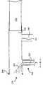
수집 유닛(30)은 판독기 유닛(50)을 기계적으로 연동되도록 고안된다. 또한 상기 수집 유닛(30)은 사용자(40)가 손으로 사용하기에 충분히 소형이다. 또한 상기 수집 유닛(30)은 다음을 포함하여 중공 샘플 튜브(70) 형태로 제공된다:The
(a) 사용자(40)의 입 영역에 연동되는 투입 오리피스(80);(a) an
(b) 가스 수집 영역, 예컨대 팽창가능한 백(100)에 결합시키기 위한 중간 오리피스(90); 및(b) an
(c) 샘플 튜브(70) 내에서 미끄러질 수 있고 회전적으로 이동할 수 있는 피스톤과 유사한 플런저(110)에 대한 액세스 오리피스.(c) an access orifice for a
사용자가 서로 교차 감염될 우려를 감소시키기 위하여, 상기 수집 수단(30)은 일회용품으로 고안되며; 즉, 상기 수집 유닛(30)은 시험하기 위한 샘플을 오직 한번 수집해서 그 샘플을 인터로게이션용 판독기 유닛(50)으로 안전하게 보내기 위해 사용된다. 상기 수집 유닛(30)은 바람직하게는 플라스틱 재료로부터 성형되므로 비교적 저렴하게 제조할 수 있고 또 소각되기 쉬워 그 안에 수집된 위험한 병원체 전파를 감소시킨다. 또한, 상기 수집 유닛(30)은 판독기 유닛(50)이 위험한 병원체를 포함할 수 있는 튜브(70) 내에 수집된 샘플과 직접적으로 접촉하지 못하게 고안되어 있다.In order to reduce the risk that users cross-infect each other, the collecting means 30 is designed as a disposable product; That is, the
2. 시스템 작동의 개요2. Overview of System Operation
생물학적 측정 시스템(10)의 작동은 도 1 및 도 2를 참조하여 개괄적으로 기재한다.Operation of the
활성 생물질의 침적을 포함한 제조후, 수집 유닛(30)은 배치하기 전에 저장을 위해 건조되고 밀봉 밀봉된 팩키지 내에서 밀봉된다. 이러한 팩키지는 수분이 상술한 활성 생물질을 변성시키지 못하게 하며 또한 수집 유닛(30)이 사용되기 전에 예기치 않게 병원체에 감염되는 것을 감소시킨다; 따라서, 이러한 팩키지는 대표적이지 않은 시험 결과를 갖지 않도록 시스템(10)을 돕는다.After manufacture, including the deposition of the active biomass, the
단계 1: 배치하기 직전, 사용자(40) 또는 시험을 감독하는 사람은 밀봉 팩키지로부터 수집 유닛(30)을 분리해낸다. 사용자(40)는 자신의 입을 샘플 튜브(70)의 투입 오리피스(80)에 댄다.Step 1 : Immediately prior to deployment, the
단계 2: 다음, 사용자(40)로부터 샘플의 생성을 보조하는데 필요한 경우, 염수 미스트를 샘플 튜브(70)로부터, 예컨대 결합된 소형 가압 가스통 분무기로부터 생성하거나, 또는 수집 유닛(30)에 원격 접속된 분무 장치 내에서 생성한다; 편리하게는, 상기 분무 장치는 발로 동작되는 펌프와 유사한 장치이다. 사용자(40)는 투입 오리피스(80)을 통하여 염수 미스트를 흡입하고, 이 미스트는 사용자(40)가 에어로졸 형태의 객담 및/또는 점액을 상기 오리피스(80)을 통하여 샘플 튜브(70)로 방출하기에 충분하도록 급격한 기침을 유발한다. 이 에어로졸은 튜브(70)를 통과하고 튜브(70)의 공기역학적 내부 특징에 의해 촉진되어 와동과 유사한 궤도로 감속되어 점액 및/또는 객담을 튜브(70)의 내부 벽상에 침적시킨다. 바람직하게는, 수집 볼륨, 예컨대 내부 오리피스(90)에 접속된 폴리에틸렌 또는 폴리비닐 클로라이드(PVC)와 같은 플라스틱 물질의 팽창가능한 백(100)은 사용자가 내쉰 공기를 수용하도록 포함되어져 있다; 각 기침은 2ℓ 용적의 공기를 함유할 수 있으므로, 상기 백(100)은 몇 개 기침을 수용하기에 편리한 크기를 가질 수 있다. 보다 바람직하게는, 수집 백(100)은 백(100)이 수십초의 기간에 걸쳐 공기를 뺄 수 있도록 충분히 큰 기공 크기를 갖는 미세 필터를 포함하는 가스 배출 오리피스(105)를 구비하고 있음으로써, 백(100)은 공기를 뺄 때 취급할 수 있는 크기로 될 뿐만 아니라 사용자(40)가 내쉰 병원체의 전파를 실질적으로 억제할 만큼 충분히 작게 할 수 있다. 또한, 중간 오리피스(90)는 투입 오리피스(80) 및 배출 오리피스(105)에 대하여 중간 정도의 유동 저항성을 가지고 또 바람직하게는 샘플 튜브(70)와 그와 결부된 백(100) 사이에 포함시켜서, 상술한 보르텍스 가스 궤도를 향상시키고 샘플 튜브(70)내의 점액 및/또는 객담의 효과적인 침적을 증진시킨다.Step 2 : Next, if necessary to assist in the generation of the sample from the
또는, 타액 샘플은 상기 샘플 수집 장치로의 분리작용을 통하여 시험 환자로부터 수집될 수 있다.Alternatively, saliva samples can be collected from the test patient through separation into the sample collection device.
단계 3: 객담 및/또는 점액의 충분한 많은 샘플이 수집 유닛(30) 내에 수집되면, 밀봉 캡(도 1에 도시되지 않음, 도 2에는 (900)으로 표시됨)을 투입 오리피스(80)위에 배치시킨다. 이어서, 플런저(110)를 작동시켜 객담 및/또는 점액을 인터로게이션 영역 내에서, 예컨대 플런저(110)의 광학 표면(120) 내에서 기계적으로 농축시킨다; 특히, 객담 및/또는 점액은 플런저(110)의 말단면에 제공된 광학적 인터로게이션면(120)위로 농축되며, 상기 광학면(120)은 이후에 자세하게 설명하는 지수감쇠적 인터로게이션-방사선 진행을 지지할 수 있다. 바람직하게는, 플런저는 샘플 튜브(70) 내에서 밀리고 회전되어서 표면(120)에서 시험 샘플을 기계적으로 농축시킨다. 보다 바람직하게는, 플런저(230)는 360°이상으로 회전되어 가능한 한 다량의 샘플이 플런저의 샘플 수집 돌출부에 수집되게 한다.Step 3 : Once a sufficient number of samples of sputum and / or mucus have been collected in the
배양기간은 샘플을 광학적으로 인터로게이팅하기 전에 측정 판독을 하기 위해 필요할 수 있다.Incubation period may be necessary for measurement readings prior to optically interrogating the sample.
단계 4: 플런저(110)가 샘플 튜브(70)에 충분히 밀어져서 광학 표면(120)상으로 샘플이 충분히 수집되면, 수집 유닛(30)은 판독기 유닛(50)에 제공되어 그 돌출부(130)는 플런저(110)와 짝을 이뤄 샘플의 광학특성을 측정하기 위한 광학 표면(120)의 광학적 인터로게이션을 가능하게 한다; 이러한 광학적 인터로게이션은 표면(120)에서 지수감쇠적 광 진행에 의해서 달성되는 것이 바람직하다. 광학적 인터로게이션의 결과는 디스플레이(60)위에 표시되어 사용자(40) 및/또는 관련 시험자에게 제공됨으로써, 사용자(40)는, 시스템(10)이 감응성인 하나 이상의 병원체, 예컨대 결핵균(mycobacterium tuberculosis) 으로 감염되었는지 여부를 검측한다.Step 4 : Once the
이로써 시스템(10)의 동작의 개요를 완성한다.This completes the outline of the operation of the
1개 이상의 밀봉 캡, 수집 튜브(70) 및 그의 플런저(110)는 그의 광학적 인터로게이션 전에 광학 표면(120)을 처리하기 위한 1개 이상의 액체 저장조에 포함될 수 있다. 이러한 천공가능한 저장조는 바람직하게는 완충액 용액 또는 이하와 같은 시약을 함유할 수 있지만, 이들에 한정되는 것은 아니다:One or more sealing caps,
(a) 수집된 병원체, 예컨대 마이코박테리움을 단편화시키는 용균제;(a) lysates that fragment the pathogens collected, such as mycobacterium;
(b) 치환된 형광 발색단을 광학 표면(120)으로부터 헹구고/헹구거나 병원체 선택성 항체에 결합된 형광 발색단 중의 광학 표면(120)을 씻어내기 위한 헹굼제;(b) a rinsing agent to rinse the substituted fluorophores from the
(c) 점액을 분산시키기 위한 희석제; 및(c) diluents to disperse mucus; And
(d) 표지된 항체와 같은 샘플에 대한 발색제.(d) Coloring agents for samples such as labeled antibodies.
시약은 저장되는 동안 보호하기 위하여 동결건조된 구와 같은 고형일 수 있다; 이러한 고형 시약은 하나 이상의 저장조 또는 샘플 수집 튜브(70) 내에 존재할 수 있다.Reagents may be solid, such as lyophilized spheres to protect during storage; Such solid reagents may be present in one or more reservoirs or
상기 하나 이상의 저장조는 바람직하게는 샘플 수집 후 광학적 인터로게이션 전에, 이들의 내용물을 전달하기 위해 사용자가 구멍을 낼 수 있도록 배치되어 있다. 상기 저장조의 기계적 구성은 도 9를 참조하여 나 중에 자세하게 설명한다.The one or more reservoirs are preferably arranged such that the user can puncture their contents for delivery after sample collection and prior to optical interrogation. The mechanical configuration of the reservoir will be described later in detail with reference to FIG. 9.
후술하는 바와 같이, 광학 표면(120)은 지수감쇠적 광 방사선 진행을 지지하기 위해 조직된 프리즘의 하나의 광학면이다. 이 프리즘은 도브 프리즘으로 제공되는 것이 바람직하지만, 다른 형태의 프리즘도 이용될 수 있다.As described below, the
3. 시스템의 부품부3. Parts of the system
시스템(10)의 개별적인 부품의 상세한 디자인을 설명한다.Detailed design of the individual components of
3.1 샘플 수집 유닛3.1 Sample Collection Unit
도 3을 참조하여, 실질적으로 원통형 중공 샘플 튜브(200)로서 공급되는 중공샘플 튜브(70)를 도시한다. 상기 튜브(200)는 사용자(40)로부터 방출된 샘플을 수용하기 위한 (210)으로 표시되는 제1 개방 단부를 포함하며; 이 개방 단부(210)는 도 1의 투입 오리피스(80)에 상응한다. 또한, 상기 튜브(200)는 중공 플런저(230)을 수용하기 위한 제2 단부(220)를 포함하며; 상기 플런저(230)는 도 1의 플런저(110)에 상응한다. 상기 튜브(200)는 또한 중간 오리피스(90)로 작용하는 실질적으로 원통형 측면 튜브(240)를 포함하며, 이 측면 튜브(240)는 샘플 튜브(200)의 관련된 종방향 중심축과 실질적으로 수직하는 관련된 종방향 중심축을 갖는다. 튜브(200, 240)가 결합되는 영역에서, 메쉬 또는 필터 거즈(250)가 포함된다. 상기 튜브(200)는 제1 단부(210) 근처에 주변 링(260)을 포함함으로써, 예컨대 사용자(40)가 튜브(200)를 사용자의 입으로 가도록 조작할 때 튜브(200)를 조작하는 사용자(40)를 손상시킬 수 있는 날카로운 모서리를 상기 단부(210)가 피할 수 있도록 한다.Referring to FIG. 3, a
상기 중공 플런저(230)는 또한 실질적으로 원통형이며 도시한 바와 같이 샘플 튜브(200)의 내부에 동심적으로 미끄러질 수 있고 회전이동할 수 있도록 제조되며, 상기 튜브(200) 및 플런저(230)은 상호 정확하게 꼭 맞는다. 바람직하게는, 상기 플런저(230)은, 사용시, 샘플 튜브(200)에 제공되는 플런저(230)의 단부에 탄력적으로 변형가능한 밀봉 링(도시되지 않음)을 구비하고 있다. 상기 밀봉 링은 바람직하게는 니트릴 고무 물질, 예컨대 독점적 비톤(Viton) 물질, 실리콘 또는 폴리테트라플루오로에틸렌(PTFE)으로부터 제조된다. 또한, 상기 밀봉 링은 튜브(200) 내에서 수집된 샘플을 오염시킬 가능성이 있어서, 시스템(10)의 작동을 손상시킬 수 있는 실리콘 그리스와 같은 윤활 물질을 피할 수 있다. 부가적으로, 윤활 물질로부터 방출된 증기는 사용자(40)가 들이마시면 유해할 수 있다.The
중공 튜브(200)는 제1 개방 단부(210) 근처의 튜브(200) 영역에서 일반적으로 (300)으로 표시되는 염수 분무화 어셈블리를 구비하고 있다. 상기 어셈블리(300)는 바람직하게는 사용자(40)에 의해 흡입되는 염수 미스트의 발산 젯트(310)를 생성하기 위한 어셈블리(300)에, 염수 용액을 가압하에서 강제시키기 위한 분무기, 예컨대 발-펌프 동작되는 장치와 결합된다; 상기 염수 미스트는 사용자(40)가 객담 및/또는 점액을 배출하도록 유도하는 급격한 기침을 증가시키는데 효과적이다. 바람직하게는, 상기 젯트(310)는 6 ㎛ 내지 20 ㎛ 범위의 직경을 갖는 염수 소적을 포함한다. 보다 바람직하게는, 염수 소적은 10 ㎛ 내지 15 ㎛ 범위의 직경을 갖는다. 바람직하게는 소적이 생성되는 염수 용액은 물에 대한 염화나트륨의 비율이 0.1중량% 내지 2중량% 범위의 농도이며, 보다 바람직하게는 염수 용액은 0.7중량% 내지 1.1중량% 범위의 농도를 지닌다. 상기 어셈블리(300)는 플런저(230)로 휩쓸고 향하는 염수 미스트의 양을 감소시키기 위한 제1 개방 단부(210)를 향하여 노즐 단부에서 각을 이루는 실질 중심 모세관(320)을 포함한다. 바람직하게는, 상기 어셈블리(300)는 중공 튜브(200)의 내부 구멍에 대하여 오목하기 때문에 플런저(230)는 도시된 바와 같이 어셈블리(300)가 중공 튜브(200)에 접속된 영역을 넘어서 제1 단부(210)를 향하여 진행될 수 있다. 상기 어셈블리(300)는 중공 튜브(200)의 일부로서 일체로 성형되며; 다르게는, 필요한 성형 도구를 단순화하기 위해, 상기 어셈블리(300)는 제조하는 동안 중공 튜브(200)의 돌출부 측면 포트(port)에 어셈블링된 스냅-피트(snap-fit) 보유된 삽입부일 수 있다. 필요한 경우, 상기 측면 포트는 제1 단부(210)를 향하도록 배치된 그의 중심축과 함께 성형될 수 있으므로, 상기 삽입물은 상기 단부(210)를 향하여 성형되는 모세관(320)을 필요로 하지 않는다.The
보다 효과적인 샘플 생성의 유도는 수증기, 에스테르, 거담제 및/또는 멘톨중의 하나 이상을 흡입하는 것에 의해 달성될 수 있다.Induction of more effective sample production can be achieved by inhaling one or more of water vapor, esters, expectorants and / or menthol.
작업시, 플런저(230)는 되돌아가므로, 도 4에서 (270)으로 표시된 그의 단부 표면은 실질적으로 튜브(200)의 제2 단부(220)에 존재한다. 이러한 수집 상태에서, 상기 튜브(200)는 제1 단부(210)에 노출된 바람직하게는 80% 이상의 대부분의 내부 표면을 갖는다. 또한, 수집 상태에서, 제1 단부(210)로부터 거즈(250)를 경유하고 또 측면 튜브(240)를 통과하는 가스 유동 경로가 백(100)(도 3에 도시되지 않음)에 제공되거나 또는 주위로 직접적으로 제공되며; 예컨대 덜 위험한 병원체에 대한 스크리닝 시험을 실시하는 경우 주위로 직접적으로 배출시키는 것이 바람직하다.In operation, the
상기 수집 상태에서, 사용자(40)는 제1 단부(210)를 자신의 입에 놓고 링(260)이 사용자의 입술을 연동시켜 밀봉시키게 한다. 사용자(40) 또는 시험자는 관련된 발 펌프를 눌러서 어셈블리(300)를 활성화시켜 사용자(40)가 흡입하는 염수 미스트의 제트(310)를 분출시킨다. 흡입된 염수 미스트는 사용자(40)에서 미세 미스트 형태의 공기, 점액 및/또는 객담 소적을 강제적으로 유발하도록 숨을 내쉬게 하여 사용자(40)의 폐로부터 튜브(200)로 보내는 자동적인 감응을 유발한다. 제2 단부(220) 주변의 튜브(200) 영역은 낮은 속도의 가스 영역을 형성하며, 사용자로부터 내뿜어진 공기는 와동 궤도로 유입되게 기울어지므로, 점액 및/또는 객담 소적을 튜브(200)의 내부 측벽상에 침적시킨다. 또한 사용자(40)가 내뿜은 공기는 비교적 고속으로 측면 튜브(240)를 통하여 배출된다.In the collection state, the
네가티브한 부분압을 사용하여 샘플을 수득하는 데 보조할 수 있다.Negative partial pressure may be used to aid in obtaining the sample.
분석할 샘플은 튜브(200)의 내부 표면상에, 특히 제2 단부(220)의 영역에 제공된다. 플런저(230)는 튜브(200)의 내부 표면으로부터 샘플을 수집한 다음 수집된 샘플을 광학적 인터로게이션 및 분석하기 위한 플런저(230)의 광학적 인터로게이션 표면상으로 침적시키기 위한 튜브(200)를 활성화한다; 이러한 활성화는 튜브(200)에 대하여 플런저(230)의 회전을 포함한다.The sample to be analyzed is provided on the inner surface of the
플런저(230)는 샘플을 수집하고 기계적으로 농축시키기 위해 적합하다. 플런저(230)의 제공은 도 4a 및 도 4b를 참조하여 설명한다.
도 4a 및 도 4b에서, 플런저(230)는 실질적으로 원통형이고 중심 중공 영역(400)을 포함한다. 플런저(230)는 제1 개방 단부에서 개방되고 또 플런저(230)가 튜브(200)에 충분히 삽입될 때 튜브(200)의 제2 단부(220)로 접하기 위한 원형 플랜지(410)를 포함함으로써, 플런저(230)가 튜브(200)로 밀고 들어갈 수 있는 정도를 제한한다. 플런저(230)는 평면이 실질적으로 플런저(230)의 중심 종방향 축과 수직하는 단부 표면(270)을 포함한다. 도시된 바와 같은 상기 표면(270)의 편심 영역에서는, 영역(400)으로 연장되는 광학 전달성 프리즘(420) 및 영역(400)으로부터 떨어져서 표면(270)으로부터 외부 방향으로 향하는 스푼 형태의 돌출부(430)를 포함한다. 상기 스푼 형태의 돌출부(430)는 튜브(200)의 내부 표면으로부터 점액 및/또는 객담을 빼내는 용도에 이용된다. 상기 스푼 형태의 돌출부(430)는 표면(270)에서 방사상으로 표면(270)의 외주부로 연장된다. 상기 돌출부(430)의 주변 엣지(440)는 튜브(200)의 내부 표면상으로 미끄러져 접촉하도록 배치된다. 프리즘 윈도우로 공지된 광학 개구부(450)는 단부 표면(270)에 포함되어 있으므로, 상기 프리즘(420)은 돌출부(430)에 수집된 샘플 객담 및/또는 점액과 광학적으로 연결된다.4A and 4B, the
돌출부(430)는 바람직하게는 도 3b에 도시한 바와 같이, 튜브(200)의 내부 표면으로부터 수집된 객담 및/또는 점액을 보유하도록 돌출부(430)의 성능을 향상시키기 위해, 주변 에지(440)로부터 떨어져 있는 그의 단부를 향하여 위에서 곡면을 이루고 있다.The
또한, 상기 돌출부(430)는 바람직하게는 유연한 플라스틱 재료로 제조되므로 플런저(230)가 튜브(200)로 향하여 충분히 진행할 때 광학 개구부(450)를 효과적으로 통과하는 샘플 물질을 밀어넣도록 굽혀지게 배치되며 또 상술한 밀봉 캡(도시되지 않음)을 향하여 탄성적으로 밀어진다; 상기 돌출부(430)에 적합한 플라스틱 재료는 하나 이상의 폴리비닐클로라이드(PVC), 나일론, 폴리테트라플루오로에틸렌(PTFE), 폴리에틸렌, 폴리프로필렌, 알킬렌 및 실리콘 고무를 포함한다. 필요한 경우, 밀봉 캡에는 돌출부(430)를 수용하도록 오목부가 제공될 수 있으므로, 오목부와 인접하는 밀봉 캡의 평탄한 비-오목 단부 표면은, 개구부(450)상에 실질적으로 균일하게 광학 개구부(450)를 향하도록 면하는 돌출부(430)의 측면 영역상에 샘플 물질의 형성을 추진하고 전파하도록 연장됨으로써, 향상된 검출감도를 갖는 시스템(10)을 제공한다. 필요한 경우, 돌출부(430)의 두께는 비교적 얇은 넥(neck)으로 감소될 수 있으며, 여기서 돌출부는 단부 표면(270)에 결합되어 힌지(hinge)를 형성하므로 돌출부(430)는 광학 개구부(450) 상으로 샘플 물질이 퍼지도록 하는 플랩으로 경사지게 된다.In addition, the
판독기 유닛(50)의 돌출부(130)의 디자인을 단순화하기 위해, 돌출부(130)는 플런저(230)의 중공 영역(400)으로 삽입되도록 조정되므로, 프리즘(420)은 편심적이지만 표면(270)의 주변 영역으로부터 떨어지게 탑재되는 것이 바람직하다. 프리즘(420) 및 그와 관련된 광학 개구부(450)가 그러한 방식으로 배치되면, 돌출부(430)는 도 4c에 도시된 바와 같이 일반적으로 "V"-형상 형태이다. 이러한 "V"-형상은 수집된 샘플의 실물을 수집하고 보유하는데 특히 효과적이다. 다르게는, 도 4b에 도시한 바와 같이, 돌출부(430)는 연속적으로 곡선을 이룰 수 있다; 바람직하게는 원격 에지에 있는 돌출부(430)는 플런저(230)의 중심 종방향 축을 향하여 더 가까운 광학 개구부(450)를 향하여 곡선을 이룬다.In order to simplify the design of the
플런저(230)는 중공 구성원으로 제조되므로, 플런저(230)는 판독기 유닛(50)의 돌출부(130)를 영역(400)에 수용할 수 있다. 상기 돌출부(130)는 전자 모듈을 포함하며, 바람직하게는 도 5에 도시한 바와 같이 고형의 원통 형태이며 일반적으로 (500)으로 표시된다. 반면, 사용시, 샘플 튜브(200) 및 그와 관련된 플런저(230)는 일회용 물품으로 고안되는 반면, 판독기 유닛(50)은 광전 증배관 및 다이오드 레이저와 같은 비교적 고가의 부품을 포함하고 있기 때문에 재사용될 수 있도록 배치되며, 이후에 더 자세하게 기재한다. 돌출부(130)는 디스플레이(60)를 포함하는 모듈상에서 상호 인접하는 제1 단부(520) 및 제2 단부를 포함하는 연장된 형태이다. 제 1단부(520)는 돌출부(130)가 플런저(230)의 중공 영역(400)에 삽입되어 스푼 형태의 돌출부(430)상에 수집된 샘플을 인터로게이팅할 때 개구부(450)와 정렬되도록 배치된 편심적으로 배치된 광학 경계 영역(510), 즉 프리즘 윈도우를 포함한다.Since the
샘플 튜브(200), 플런저(230) 및 돌출부(130)는 이들의 상호 동심원적 탑재성으로 인하여 저장시 소형화될 수 있는 이점이 있다. 또한, 플런저(230)와 조합된 튜브(200)는 사용자(40)가 내쉰 전체 샘플을 실질적으로 수집하고 기계적으로 농축시킬 수 있다. 또한 플런저(230)는 샘플과 접촉하지 않더라도 판독기 유닛(50)이 샘플을 인터로게이팅할 수 있기 때문에, 판독기 유닛(50)은 재사용될 수 있고; 따라서 작용 비용이 감소된다.The
관련된 측면 튜브를 갖는 샘플 튜브(200) 및 플런저(230)는 하나 이상의 아크릴레이트, 폴리에틸렌, 폴리프로필렌, 나일론, 실리콘 고무, 폴리비닐 클로라이드(PVC), 알킬렌 폴리카보네이트 및 폴리테트라플루오로에틸렌(PTFE) 플라스틱 재료와 같은 플라스틱 재료로 제조된다. 보다 바람직하게는, 튜브(200) 및 플런저(230)의 적어도 하나는 사출 성형된다. 다르게는, 하나 이상의 샘플 튜브(200)와 그와 관련된 측면 튜브 및 플런저(230)는 압출된 금속 쉬트로부터 제조되거나, 또는 다이-캐스트 금속 기술에 의해 제조한다.
가장 바람직하게는, 샘플 튜브(200), 플런저(230) 및 그와 관련된 포트(240) 및 필터(250)는 단일 부품으로 성형된다. 마찬가지로, 관련된 돌출부(430)를 갖는 플런저(230) 및 프리즘(420)은 폴리카보네이트 또는 아크릴 플라스틱 재료와 같은 광학적으로 투명한 플라스틱 재료로부터 단일 부품으로 성형되는 것이 바람직하다. 다르게는, 플런저(230)는 PVC와 같은 실질적으로 흑색 플라스틱 재료로부터 제조될 수 있고, 이어 프리즘(420)이 그 위에 어셈블링되며; PVC를 사용하면, 프리즘(420)을 빗겨나간 주위 일루미네이션으로부터 차폐시키고 또 측정되는 동안 플런저(230)에 삽입될 때 주위 일루미네이션으로부터 돌출부의 원격 단부를 차폐시키는데 이점이 있다.Most preferably,
단면에서, 플런저(230)는 소형 오리피스, 예컨대 0.1 mm 내지 2.5 mm 범위의 직경을 갖는 거의 원형 오리피스를 경우에 따라 포함할 수 있다. 상기 오리피스는 사용자(40)로부터 점액 및/또는 객담 샘플을 수집하기 위해 배치될 때, 염수 미스트와 같은 분무화된 분무 미스트를 샘플 튜브(200)에 주입하는데 이점을 갖는다. 수집할 샘플을 사용자(40)가 튜브(200)의 내부 표면상으로 내쉬기 전에 튜브(200)에 미스트를 주입하는 것은 사용자(40)로부터 상당량의 점액 소적을 얻는데 효과적이다. 다른 유형의 바이오 에세이법의 경우, 소량의 액체, 예컨대 염수 또는 완충액을 부가하면, 세균과 같은 시험될 미생물이 광학 개구부(450)로 확산되도록 보조하므로 바람직하다. 이러한 액체는 샘플 수집하기 전 또는 후에 예컨대 에어로졸 분무 또는 피펫으로 소적을 적당히 사용하여 시스템(10)에 부가될 수 있다.In cross section, the
튜브(200), 플런저(230) 및 돌출부(130)는 바람직한 크기 범위 내에 있도록 제조되는 것이 유리하다. 예컨대, 샘플 튜브(200)는 바람직하게는 20 mm 내지 30 mm 범위의 직경을 갖는다. 또한 측면 튜브(240)는 바람직하게는 1 mm 내지 10 mm 범위, 보다 바람직하게는 5 mm 내지 8 mm 범위의 직경을 갖는다. 또한, 샘플 튜브(200)는 40 mm 내지 150 mm, 보다 바람직하게는 50 mm 내지 80 mm 범위의 길이를 갖는다. 광학 개구부(450)는 바람직하게는 9 mm2 내지 64 mm2 범위의 면적을 갖는다. 이들 크기는 인체에 사용하기 위해 고안된 시스템(10)에 적합해야 하는 것도 중요하다. 기타 종류는 이들 크기를 적합하게 변형할 것이 요청되고 있다.The
샘플 튜브(200) 및 그와 관련된 플런저(230)는 바람직하게는 잠금 메카니즘을 구비하고 있어서, 점액 및/또는 객담 샘플을 튜브(200) 내에서 기계적으로 농축시켜서 광학 개구부(450) 및 그의 측정 위치로 이동된 플런저(230)상으로 전달하면, 플런저(230)는 샘플 튜브(200)에 대한 위치에 따라 기계적으로 잠궈진다. 이러한 메카니즘은 샘플 튜브(200) 및 플런저(230)가 재사용되는 것을 방지할 수 있다는 점에서 유리하다; 세계의 가난한 지역에서는 주사기와 같은 의료 부품을 재사용하는 유혹이 있었다. 보다 바람직하게는, 판독기 유닛(50)의 돌출부(130)의 삽입은 그러한 메카니즘이 재사용되는 것을 방지한다. 재사용의 유혹은 시스템(10)이 병원체의 존재에 대한 비-양성 지시를 나타낼 때 더 강력해진다. 이러한 잠금 메카니즘은, 샘플 튜브(200)를 밀봉 캡과 함께 플런저(230)에 잠그는 것이 유해한 병원체를 분리하기 위한 봉입된 영역을 형성하는 점에서 유리하다. 보다 바람직하게는, 상기 샘플 캡은 튜브(200)상에 스냅 연동되므로 샘플 캡과 튜브는 풀어질 수 없다.The
샘플 튜브(70, 200) 내에 생긴 와동을 증가시켜 튜브내의 객담 및/또는 점액의 침적을 향상시키기 위하여, 튜브(70, 200)는 부가적 특징부를 구비하여 공기 흐름을 변화시킬 수 있다. 도 6을 참조하면, 일반적으로 (600)으로 표시된 변형된 샘플 수집 튜브가 도시되어 있으며, 상기 튜브(600)는 분무기 어셈블리(300) 및 측면 튜브(240) 사이에 포함된 원형 객담 오리피스(610)를 포함한다. 상기 객담(610)은 가능한 한 단부(210) 가까이에 포함되는 것이 바람직하다. 또한, 객담 오리피스(610)는 튜브(600)의 원통형 부품(620)의 일체형 부품으로 성형되는 것이 바람직하다. 플런저(230)의 돌출부(430)는, 샘플 수집 후에 플런저(230)가 튜브(600)로 진행하면 돌출부(430)가 객담 오리피스(610)로부터 수축되어 샘플을 광학 개구부(450)상으로 밀어넣게 하도록 가요성으로 제조되는 것이 바람직하다.In order to increase the vortices generated in the
객담 오리피스(610)는 주변을 끌어당겨서 소용돌이 및 상응하는 복잡한 다중 와동 형성을 향상시키는 데 일조함으로써, 튜브(600)의 원통형 부품의 내부 표면상에서 점액 및/또는 객담 침적을 향상시킨다. 또한, 상기 오리피스(60)는 또한 사용시 사용자(40)로부터 멀리 떨어진 튜브(600)의 영역으로 염수가 침범하는 것을 방지하는 것을 보조한다. 바람직하게는, 객담 오리피스(610)는 5 mm 내지 15 mm 정도의 거리로 단부(210) 뒤에 오목하게 존재한다. 또한, 객담 오리피스(610)는 바람직하게는 실질적으로 3 mm 내지 20 mm 범위의 직경을 갖는 중심 홀을 포함한다. 상기 객담 오리피스(610)는, 사용자(40)의 입에 가장 가까운 샘플 튜브(200)의 벽상에 수집된 샘플이 광학 개구부(450) 상에서 농축될 수 있도록 접이식 재료(예컨대 가요성 플라스틱)로 제조되는 것이 바람직하다.
상기 샘플 튜브(200)는 상기 오리피스(210) 근처에 방향성 밴드부를 포함하도록 변형되어 일반적으로 (650)으로 표시된 샘플 수집 튜브를 형성할 수 있다. 상기 어셈블리(300)는 바람직하게는 도시된 바와 같이 튜브(650)의 외부 밴드부 상에 포함되므로 염수 미스트를 단부(210)로 향하여 주입한다. 이러한 방향성 밴드부는 공기에 대하여 비대칭적이고 공간적으로 다양한 드레그(drag)를 유발하여 소용돌이 형성을 증진시킨다.The
최상의 수집 성능을 얻기 위하여, 튜브(600, 650)의 특징을 조합한 것이 이용될 수 있다.In order to obtain the best collection performance, a combination of the features of the
또한 샘플 수집 유닛(30)의 다른 구체예도 가능하다.Other embodiments of the
예컨대, 도 7에는 일반적으로 (700)으로 표시되는 샘플 수집 챔버가 도시되어 있다. 상기 챔버(700)는 사용자(40)가 내쉰 숨을 수집 박스(710)로 전달하기 위한 약 25 mm 직경의 유입 파이프(720)를 포함한다. 보다 바람직하게는, 상기 유입 파이프(720)는 18 mm 내지 30 mm 범위의 직경을 갖는다. 상기 수집 박스(710)는 그 주변에, 상기 박스(710)의 내부쪽으로 면하는 노출면에서 지수감쇠적 방사선 진행을 촉진시키는 프리즘(750)을 포함하며, 이 프리즘에서는 내어쉰 숨으로부터 점액 및/또는 객담의 침적이 생긴다. 노출 표면 상에서 샘플 수집은 박스(710) 내에 생긴 와동에 의해 증가되며; 이 와동은 박스(710)로부터 배출 파이프(760)의 직경이 유입 파이프(720)의 직경보다 더 작을 때 특히 향상된다. 바람직하게는, 배출 파이프(760)의 직경은 약 5 mm이지만, 2 mm 내지 10 mm 범위의 직경이 특히 바람직하다. 상기 프리즘(750)은 그 위에 샘플을 수집하기 위하여 박스(710)의 벽 위의 어떤 위치에도 배치될 수 있다.For example, FIG. 7 shows a sample collection chamber, generally indicated at 700. The
박스(710)으로부터 내어쉰 숨은 유리하게는 배출 파이프(760)을 따라서 백(100) 및 그와 결합된 가스 배출 오리피스(105)로 전달된다. 다르게는, 박스(710)로부터 밖으로 내어쉰 숨은 필터를 먼저 통과하여 병원체를 제거한 다음 주위 또는 백(100)으로 배기된다. 유리하게는, 박스(710) 내에서 와동 형성을 보조하기 위해 벤투리를 유입 및/또는 배출 파이프에 포함시킬 수 있다. 배출 파이프(760) 및 유입 파이프(720)는 박스(710) 내에, 각각 서로에 대해 및 프리즘(750)에 대하여 어떤 위치에라도 배치될 수 있다.The breath exhaled from the
판독기 유닛(50)의 돌출부(130)에 대해 도 8a 및 도 8b를 참조하여 자세하게 기재한다. 프리즘(420)으로부터 최적 판독치를 얻기 위하여, 판독기 유닛(50)의 광학적 인터로게이션 부품은 어셈블리 내에 하우징되는 것이 바람직하다. 따라서 상기 돌출부(130)는 광전 증배관(PM 튜브) 및 다이오드 레이저를 포함한다. 유리하게는, 상기 PM 튜브(800)는 일본의 하마마쓰 포토닉스 가부시끼가이샤가 독점적으로 제공하는 장치로서, 이 장치의 제품번호는 R7400U-01이다. PM튜브(800)의 감광성 면(face)은 광학 계면 영역(510)에 바람직하게는 가능한 한 공간적으로 가깝게 배치된다. 또한, 돌출부(130)는 고형 상태의 다이오드 레이저(810)를 더 포함한다. 도 8a에 도시된 구조는 광학 방사선을 광학 개구부(450)에 결합시킬 때 광학 손실이 가장 적기 때문에 가장 바람직하다. 그러나, 특히 돌출부(130)가 비교적 작은 외부 직경을 갖는 경우, 다이오드 레이저(810)는 예컨대 광학 섬유 도파관의 평행한 번들을 포함하는 도광로(light guide)(820)를 통하여 결합되는 것이 유리하다. 도 8b에 도시된 바와 같은 표준적인 비교적 대형 돌출부(130)에서, 다이오드 레이저(810) 및 PM 튜브(800)는 상호 인접하게 탑재되므로, 상대적으로 단거리의 도광로를 사용할 필요가 있다. 그러나, 도 8b에 도시된 바와 같은 비교적 소형의 돌출부(130)에서는, 다이오드 레이저(810)가 도시된 바와 같이 PM 튜브(800) 뒤쪽에 배치되어 있고 광을 다이오드 레이저(810)로부터 계면 영역(510)으로 전달하기 위해 적용되는 광 파이프(830)의 길이가 상대적으로 더 길다. 필요한 경우, 상기 돌출부(130)는 다이캐스트, 가공된 또는 압출된 금속으로부터 제조될 수 있고 또 다이오드 레이저(810)는 냉각 목적을 위해 돌출부(130)의 주변 벽과 열적으로 결합될 수 있다. 유사한 열적 고려를 PM 튜브(800)에도 적용하지만, 이 장치는 동작시 비교적 무시할만한 전력을 소비한다. 돌출부(130)를 포함하는 판독기 유닛(50) 및 그와 결합된 부품의 디자인은 이후에 자세하게 기재한다.The
도 2를 다시 참조하면, 수집 튜브(70)에 적용된 상술한 밀봉 캡은 (900)으로 표시한다. 이 캡(900)은 도 9에 보다 자세하게 단면으로 도시되어 있으며, 상기 캡(900)은 주 본체 부품(905), 액체 물질(930, 940)을 각각 포함하는 제1 및 제2 액체 저장조(910, 920), 및 액체 물질(930, 940)를 캡(900)에 밀봉하기 위해 본체 부품(905)에 밀봉 결합된 밀봉 상단(950)을 포함한다. 상기 본체 부품(905) 및 밀봉 상단(950)은 바람직하게는 실리콘 고무와 같은 가요성이 높은 플라스틱 재료로부터 제조한다. 밀봉 상단(950)은 상응하는 저장조(920, 910)에 각각 정렬된 돔형 영역(960, 970)을 포함하는 비교적 얇은 가요성 막이다. 돔형 영역(960, 970)의 중심에 결합된 것이 강철 핀(980, 990)이다. 이들 강철 핀(980, 990)은 밀봉 상단(950)에 주형된 무디고 넓은 단부 및 저장조(920, 910)의 단부 영역과 대면한 예리한 단부를 갖는다. 밀봉 캡(900)은 또한 보유부(1000), 예컨대 수집 튜브(70, 200)에 결합되어 캡(900)이 튜브(70, 200)에 용이하게 삽입되지만 그로부터 다시 뺄 수는 없게 만드는 후방으로 향하는 미늘(barbs)을 갖는 미늘 삽입부(barbed insert)를 더 포함한다.Referring again to FIG. 2, the aforementioned sealing cap applied to the
동작시, 사용자(40) 또는 바람직하게는 시험자는 돔형 영역(960, 970)을 눌러서 핀(980, 990)이 각각의 저장조(920, 910)에 구멍을 뚫어 이들 각각의 내용물 액체 물질(940, 930)을 샘플 튜브(70, 200)로 방출하여 플런저(110, 230)의 광학 개구부(450)에서 수집된 점액 및/또는 객담을 화학적으로 처리한다.In operation, the
제조시, 본체 부품(905)은 그의 단부 면(1010)이 하부 방향으로 향하도록 배향시킨다. 저장조(910, 920)에는 각각의 액체 물질(930, 940)를 충전시킨 다음 핀(980, 990)을 포함하는 밀봉 상단(950)을 초음파적으로 용접시키거나, 또는 밀봉 결합시켜 도시된 바와 같이 본체 부품(905)에 오목하게 성형된다.In manufacture, the
상기 2개의 저장조(910, 920)가 도 9에 도시되어 있지만, 밀봉 캡(900)은 2개 이상의 저장조를 갖도록 제조될 수 있음을 이해해야 한다. 또한 액체 물질(930, 940)은 시스템(10)에 의해 검출될 병원체의 유형에 따라서 조성에서 변화될 수 있다. 예컨대, 1개의 액체 물질은 생물학적 세균 용균제일 수 있는 반면에, 나머지 액체 물질은 생물학적 형광 마커 물질일 수 있다. 이들 물질은 이후에 더욱 자세하게 기재한다.Although the two
3.2 판독기 유닛3.2 Reader Unit
도 1에 도시된 판독기 유닛(50)을 보다 자세하게 설명한다.The
도 10을 참조하면, 일반적으로 (1100)으로 표시된 광학 구조가 도시되어 있다. 이 구조(1100)는 시스템(10)에 적용되어 있고 그 부품은 판독기 유닛(50) 및 그와 결부된 돌출부(130) 및 플런저(110, 130) 사이에 분포되어 있다.Referring to FIG. 10, an optical structure, shown generally at 1100, is shown. This
특히, 돌출부(130) 중의 판독기 유닛(50)은 레이저(810), 도광로(820, 830)(필요한 경우), 광학 필터(1120), PM 튜브(800)(및 관련된 전력공급, 도시되지 않음) 및 소형 고상 다이오드 검출기(1130)를 포함한다. 판독기 유닛(50)의 주요 부품은 디스플레이(60), 계산을 실시할 마이크로콘트롤러(1150), 동기식 복조기(1140) 및 스트로브 회로(1160)를 포함한다.In particular, the
상기 플런저(110, 230)는 프리즘(420)을 포함하고; 특히 바람직한 프리즘(420)은 도시된 바와 같이 단면이 사다리꼴 형태인 도브형 프리즘이다. 이 프리즘의 주요 전면은 이후에 자세하게 기재된 생물학적 활성 층(1110)으로 피복된 광학 개구부(450)를 제공한다.The plunger (110, 230) comprises a prism (420); Particularly preferred
상기 구조내의 부품의 상호 접속은 도 10을 참조하여 기재한다. 마이크로콘트롤러(1150)는 디스플레이(60)와 결합된 데이터 출력을 포함한다. 가장 단순한 구조에서, 디스플레이(60)는 단순히 주어진 병원체가 점액 및/또는 객담 샘플 중에 소정 임계치 이상으로 존재하는지의 여부를 나타내기 위한 예/아니오 지시기를 포함한다. 보다 복잡한 구조에서, 디스플레이(60)는 인터로게이팅될 점액 및/또는 객담 샘플에 존재하는 병원체의 농도의 정량적 측정값을 제공한다; 상기 디스플레이는 1개 이상의 액정 디스플레이(LCD), 예컨대 알파-숫자 LCD 디스플레이, 발광 디스플레이(LED) 및 소형 플라즈마 디스플레이일 수 있다. 마이크로콘트롤러(1150)는 출력시 레이저(810)에 적용된 전력을 변조시키기 위한 스트로브 회로(1160)에 접속됨으로써, 광학 출력 비임(1200)을 일시적으로 변조시켜 도광로(820, 830)으로 보낸다. 또한, 상기 마이크로콘트롤러(1150)는 동기식 복조기(1140)로부터 복조된 출력 신호를 수용하기 위한 입력정보(S3)를 포함한다. PM 튜브(800)는 동기식 복조기(1140)의 신호 입력에 접속된 신호 입력 정보(S1)를 포함한다. 또한, 다이오드 검출기(1130)는 동기식 복조기(1140)의 스트로브 입력과 결합된 신호 출력정보(S2)를 포함한다. 상기 광학 필터(1120)는 도시된 바와 같이 PM 튜브(800) 및 프리즘(420)의 소형 주요 후방-평면(1170) 사이에 포함된다; 상기 필터(1120)는 활성층(1110)에서 지수감쇠적 감쇠파 상호작용으로부터 생기는 방사선 성분을 전달하고 또 프리즘(420) 내에서 일차 방사선의 산란으로부터 직접적으로 생긴 산란된 방사선을 반사 및/또는 흡수하는데 효과적이다.The interconnection of the components in the structure is described with reference to FIG. The
광학 구조의 작동은 도 1 및 도 10을 참조하여 기재한다. 사용자(40)는 시스템(10)을 활성화하여 스트로브 회로(1160)와 조합된 마이크로콘트롤러(1150)가 변조된 신호를 생성하여 다이오드 레이저(810)를 구동시킴으로써 상응하게 스트로브된 비임(1200)을 생성한다. 바람직하게는, 레이저(810)로부터 방사선 출력의 파장은 광학 개구부(450)상에 수집된 점액 및/또는 객담을 분석하기 위해 적용된 형광 발색단의 여기 파장과 일치하도록 선택한다. 이러한 형광 발색단은 상이한 파장, 예컨대 진한 적색, 녹색 또는 청색 방사선 파장에 선택적으로 감응하도록 선택한다. 레이저(810)로부터 적색 방사선에 상응하는 긴 파장의 방사선을 사용하는 것은 적색 레이저가 값이 아주 저렴하고 쉽게 구입할 수 있기 때문에 비용을 줄이므로 바람직하다. 반대로, 비교적 짧은 청색 및 녹색 방사선 파장에서 방사선을 출력할 수 있는 고상 레이저 다이오드는 비교적 고가이다. 그러나, 적색 스펙트럼 영역에서 민감한 PM 튜브가 사용되어야 하므로 청색/녹색 영역에서 가장 민감한 것에 비하여는 약간 덜 비싸다. 그러나, 광학 구조(1100)는 점액 및/또는 객담 샘플을 분석하기 위해 적용된 형광 발색단의 유형에 일치하는 상이한 파장에서 동작될 수 있어야 하므로, 상기 시스템(10)은 필요한 어떤 광학 주파수에서도 동작할 수 있도록 구성될 수 있다.Operation of the optical structure is described with reference to FIGS. 1 and 10. The
비임(1200)은 바람직하게는 100Hz 내지 100 kHz 범위의 주파수에서 스트로브된다. 보다 바람직하게는, 상기 비임(1200)은 100Hz 내지 1500Hz 범위의 주파수에서 스트로브된다. 가장 바람직하게는, 상기 비임(1200)은 거의 1030Hz의 주파수에서 스트로브되며, 이것은 PM 튜브(800)와 결합된 증폭기 회로(도시되지 않음) 및 동기식 복조기(1140)가 표준 부품을 이용하여 직접 디자인되게 하며, 이는 밴드폭 제약이 그러한 스트로브 주파수에서는 별 문제가 아니기 때문이다. 또한 1030 Hz는 50 Hz 상용 전원(mains supply)의 하모닉(harmonic)이 아니므로, 시스템(10)은 병원 및 진찰실에서 흔히 발견되는 상용작동(mains-operated) 형광 스트립 광과 같은 50 Hz 변동 광원에 의한 영향을 덜 받게 된다.
바람직하게는, 스트로브 유닛(1160)은 레이저(810)를 여기하도록 사용된 주입 전류를 변조시키기 위해 작용함으로써, 상기 전류는 레이저(810)의 전류 임계치 상하를 주기적으로 전환시킨다. 다르게는, 상기 레이저(810)는 일정한 출력 세기로 동작될 수 있으며, 또 별도의 변조기, 예컨대 액정 (LCD) 셀을 사용하여 레이저(810)로부터의 출력 비임을 일시적으로 변조시켜 방사선 비임(1200)을 생성한다.Preferably,
레이저(810)는 도광로(820, 830)를 통하여 진행하는 스트로브된 비임(1200)을 방출하여 프리즘(420)의 제1 경사면(1215)으로 진행하는 상응하는 배출 비임(1210)을 생성하고 또 그곳에서 굴절되어 광학 개구부(450)의 평면에 대하여 정상 각도 θ에 대응하는 상응하는 굴절비임(1220)을 생성한다. 바람직하게는 상기 각도 θ는 62° 내지 80° 범위이다. 보다 바람직하게는, 각도 θ는 거의 70°이다.The
굴절된 비임(1220)은 광학 개구부(450)로 진행하며 대부분 그곳에서 반사되어 반사 비임(1230)을 생성한 다음 프리즘(420)의 제2 경사면(1235)으로 진행하여 그곳에서부터 비임(1240)으로 굴절된 다음 검출기(1130)로 진행한다. 이 비임(1240)은 복조기(1140)에 대한 변조기준 신호로 이용되는 출력정보(S2)에서 스트로브 신호를 생성한다.The refracted
상기 비임(1220)이 광학 개구부(450)에 도달하면, 비임(1220)에 존재하는 방사선의 일부는 지수감쇠적 감쇠파(1245) 형태로 상기 개구부(450)의 평면에 결합된다. 이 지수감쇠적 감쇠파(1245)는 생물학적 활성층(1110)의 계면에 있는 경계 영역에서 프리즘(420) 자체로 진행한다. 상기 경계 영역은 주파수 의존적이며 또 시스템이 작동하는 주파수에서는 100-200 mm 두께 정도로도 효과적이다. 따라서, 비임(1220)을 결합시켜 지수감쇠적 감쇠파(1245)를 형성하는 것은 경계 영역에 존재하는 화학물질의 극히 효과적인 광학적 인터로게이션을 가능하게 한다. 경계 영역에 형광 발색단이 존재하면, 이들은 지수감쇠적 감쇠파에 의해 여기되어 형광 방사선을 생성할 것이다. 바람직하게는, 이 형광 방사선은 비임의 방사선 주파수와는 상이한 방사선 주파수이기 때문에, 필터(1120)를 사용하여 비임(1220)으로부터 산란된 방사선을 상술한 경계 영역에 있는 형광으로부터 나온 방사선으로부터 구별할 수 있다; 즉, 경계 영역에 존재하는 형광 발색단은 방사선 파장 전환을 제공하도록 동작될 수 있다.When the
상기 경계 영역에서 생성한 형광 방사선은 프리즘(420)을 통하여 경계 영역으로부터 진행하여 프리즘면(1170)으로부터 나와서 필터(1120)를 통하여 PM 튜브(800)까지 진행하여 출력정보(S1)에서 생성될 상응하는 센스 신호를 유발한다. 상기 센스 신호는 복조기(1140)의 신호 입력을 지나고 출력정보(S2)로부터 신호에 대응하게 동기식으로 복조되어 출력정보(S3)에서 복조된 신호를 제공하며, 이것은 다음 샘플링과 전환을 위해 마이크로콘트롤러(1150)를 통과하여 상응하는 데이터(D)를 얻는다. 상기 마이크로콘트롤러(1150)는 데이터(D)를 미리 프로그래밍된 임계치 (T)와 비교하여, 광학 개구부(450)에 수집되고 또 지수감쇠적 감쇠파 방사선 진행에 의해 인터로게이팅된 점액 및/또는 객담 샘플에 병원체가 존재하는지 여부를 결정하는 것에 의해 진행된다.The fluorescence radiation generated in the boundary region proceeds from the boundary region through the
바람직하게는, 형광도, 즉 데이터(D)의 크기는 데이터(D1)을 제공하기 전및 데이터(D2)를 제공한 후 결정되며, 광학 개구부(450)상에 객담 및/또는 점액 샘플을 수집하고 기계적으로 농축시킨다. 하기 수학식 1로 나타낸 차이는 마이크로콘트롤러(1140)에서 산출한다:Preferably, the fluorescence, ie, the size of data D, is determined before providing data D1 and after providing data D2, collecting sputum and / or mucus samples on
상기 값 차이(△D)를 임계치(T)와 비교하여 샘플에 병원체가 존재하는지 여부를 결정한다. 이러한 측정방법의 차이는 출력정보(S1)에서 제공된 센스 신호에 대한 체계적인 기여를 제거하는 데 효과적이다; 이러한 체계적 기여는 프리즘(420)내의 산란기, 특히 플라스틱 재료로 제조되는 경우 프리즘(420)내의 잔류 형광 및 필터(1120)에 의해 제공된 한정된 방사선 파장 구별에 의해 생길 수 있다. 프리즘(420)을 제조하기에 적합한 플라스틱 재료는 페르스펙스, 아크릴레이트, 폴리카보네이트 및 폴리메틸메타크릴레이트(PMMA)를 포함한다. 형광을 나타내는 중합체 재료를 사용하는 것은 필요한 경우 피할 수 있음에 주목한다.The value difference DELTA D is compared with the threshold T to determine whether a pathogen is present in the sample. This difference in measurement method is effective in eliminating the systematic contribution to the sense signal provided in the output information S1 ; This systematic contribution may be caused by scatterers in the
바람직하게는, 임계치(T)는 레이저(810)로부터 비임(1210)으로 전달되는 광학적 인터로게이션 방사선 전력에 비례하게 된다. 보다 바람직하게는, 상기 임계치(T)는, 특히 기계적 내성이 긴밀하게 제어되지 않을 때, 플런저(230)와 플런저(230)에 따라 다양할 수 있는 프리즘(420)에 대한 광학 결합의 효율을 설명하기 위해 검출기(1130)에서 수용되는 비임(1240)의 방사선 전력과 비례하게 된다.Preferably, the threshold T is proportional to the optical interrogation radiation power delivered from the
PM 튜브(800)의 사용은 앞서 기재하였지만, 다른 유형의 광학 검출기, 예컨대 APD(avalanche photodiode), 포토레지스터 또는 저-잡음 광 다이오드도 사용될 수 있음을 이해해야 한다. 신호 대 잡음을 고려하면, 레이저(810)는 바람직하게는 저-비용 고-휘도 발광 다이오드(LED)로 치환된다.While the use of the
필요한 경우, 필터(1120)는 몇 개의 굴절 격자층을 이용하는 것에 의해 파장 구별을 향상시키기 위한 몇 개의 광학 필터 부품을 포함할 수 있다. 또한, 마이크로컴퓨터(1150)는 냉각 상태로부터 활성화될 때 시스템(10)의 웜-업 특징을 고려하여 센스 신호에서의 체계적인 안정한 일시적 드리프트(drift)를 설명하기 위해 프로그래밍될 수 있다. 이러한 드리프트의 추정은 기계적으로 농축된 샘플을 도입하기 전에 수분간에 걸쳐 광학 개구부(450)를 인터로게이팅하는 것에 의해 실시될 수 있다.If desired, the
바람직하게는, 마이크로콘크롤러(1150)는 시험결과를 기록하기 위한 데이터 로거(logger) 및 판독기 유닛(50)으로부터 데이터베이스에 다운로딩하기 위한 상응하는 기준 코드를 포함한다. 이러한 구조에서, 판독기 유닛(50)은 바람직하게는 판독기 유닛(50)에 의해 실시된 각 시험이 확인 기준을 표시할 수 있도록 데이터 엔트리 키 패드(data entry key pad)를 포함한다. 상기 마이크로콘트롤러(1150)가 데이터 로깅 특징을 제공하도록 구조화되면, 상기 마이크로콘트롤러는 질병이 전염되는 동안 병원체 감염율 통계를 생성하도록 효과적으로 사용될 수 있다.Preferably,
시스템(10)은 내어쉰 숨보다 다른 공급원으로부터 얻은 액체 샘플을 인터로게이팅하는데 적합함도 이해되어야 한다. 도 11을 참조하면, 튜브(1310) 내에서 유동하거나 또는 정지하고 있는 액체, 예컨대 혈액샘플을 분석하기 위한 시스템(10)의 일부에 대한 다른 구조를 도시하고 있다. 프리즘(420)은 튜브(1310)의 측면 영역의 일체형 부품이거나 또는 튜브(1310)의 측면 영역에 부착되어 있을 수 있다. 비임(1210)은 비임(1220, 1230)으로서 프리즘(420)을 통과하하고 또 튜브(1310)내의 액체 샘플과 접촉하여 면하는 프리즘(420)의 주요 면에 부착된 형광 발색단을 여기시킨다. 액체 샘플, 예컨대 혈액의 조성에 반응한 형광 발색단의 형광은 PM 튜브(800)에 의해 수용되어 동기식 검출에 사용하기 위한 출력정보(S1)에서 스트로브 센스 신호를 생성한다. 도 11에 따라 변형된 시스템(10)은 혈액 또는 기타 체액, 예컨대 뇨가 병원체를 갖는지 연속적으로 모니터링할 수 있으므로, 병원이나 체액 가공 시설 등에서 광범위하게 적용될 수 있다.It should also be understood that
또한, 필요한 경우, 샘플 공기 스트림은 튜브(1310)를 통하여 연속적으로 통과되어 공기로 운반되는 병원체, 독성 오염물질 및 폭약 증기가 존재하는지 검출될 수 있다. 따라서, 생물학적 측정 시스템(10)은 결핵과 같은 호흡기 병원체를 검출하려는 목적 이외의 다른 용도에도 적용될 수 있다.In addition, if desired, the sample air stream can be detected for the presence of pathogens, toxic contaminants, and explosive vapors that are continuously passed through the
도브형 프리즘(420)은 도 12에 일반적으로 (1400)으로 표시된 바와 같은 대안 프리즘을 갖는 플런저(110, 230)에서 치환될 수 있다. 상기 프리즘(1400)은 바람직하게는 도시된 바와 같이 55°, 125°, 70° 및 110°의 내부각도를 갖는다. 경우에 따라, (1410)으로 표시된 면은 비임(1210)을 반사하여 비임(1220)을 형성할 때 면(1410)의 반사능을 향상시키기 위해 거울 코팅된 표면일 수 있다. 내부 반사 손실을 감소시키는 것에 의해, 프리즘(1400)은 시스템(10)에 대한 신호 대 잡음비 검출을 향상시킬 수 있다.Dove-shaped
또한, 프리즘 내에서 복수의 반사가 유도되도록 각도가 허용되는 도브형 또는 기타 프리즘이 채택될 수 있다. 예컨대, 적합한 대안적 프리즘은 문헌[참조: C. N. Banwell 및 E.M. McCash, "Fundamentals of Molecular Spectroscopy" (1994), McGraw-Hill, 제4판]에 기재되어 있으며, 이 문헌은 본 명세서에 참고 문헌으로 포함되어 있다.In addition, a dove or other prism may be employed that allows angles to induce a plurality of reflections within the prism. For example, suitable alternative prisms are described in C. N. Banwell and E.M. McCash, "Fundamentals of Molecular Spectroscopy" (1994), McGraw-Hill, 4th edition, which is incorporated herein by reference.
항체상의 형광 발색단이 검출시 마커로서 사용되는 형광계는 단백질 및 DNA와 같은 생물학적 물질을 검출하기 위해 아주 중요하다는 것이 알려져 있음은 인정되어야한다. 이러한 형광계를 이용한 검출은 이하의 방법에 의해 실시될 수 있다:It should be appreciated that the fluorometer, in which the fluorophore on the antibody is used as a marker in detection, is known to be of great importance for detecting biological materials such as proteins and DNA. Detection using such a fluorometer can be carried out by the following method:
(a) 대량 형광측정법; 또는(a) mass fluorometry; or
(b) 지수감쇠적 감쇠파 검출과 같은 인터로게이션 기술을 적용함으로써; 또는(b) applying interrogation techniques such as exponentially attenuated wave detection; or
(c) 동공 링 다운(cavity ring down) 분광계; 또는(c) a cavity ring down spectrometer; or
(d) 치환 에세이법을 이용함으로써.(d) by using a substitution assay method.
이러한 형광계는 특이성, 단순성 및 감도 면에서 특히 이점을 제공한다. 지수감쇠적 감쇠파 검출은 공지되어 있으나, 저가의 지수감쇠적 감쇠파 형광계는 본 발명에 따라 기재된 바와 같이 병원체 검출에 사용하도록 상업적으로 시판될 정도는 아니었다.Such fluorometers offer particular advantages in terms of specificity, simplicity and sensitivity. Exponential attenuated wave detection is known, but inexpensive attenuated wave fluorimeters have not been commercially available for use in pathogen detection as described in accordance with the present invention.
광학적 인터로게이션 영역에 샘플이 수집된 이후 샘플을 분석하는 방법은 형광계 이외의 수단, 예컨대 방사능 및 인광(phosphorescent) 마커 또는 화학적 일루미네이션 기술을 이용하여 실시할 수 있다.The method of analyzing the sample after the sample has been collected in the optical interrogation region may be carried out using a means other than a fluorometer, such as radioactive and phosphorescent markers or chemical illumination techniques.
검출은 면역분석 시스템 또는 지수감쇠적 감쇠파와 관련이 없는, 분광계 이외의 형태를 이용하여 실시될 수 있다. 예컨대, 적외선 분광계 방법은 적외선 스펙트럼의 특정 영역에서 '그룹 주파수'를 기본으로 하여 특정 분자량 단편의 존재를 확인할 수 있다; 이들은 투과 및 반사 기하를 이용하여 실시될 수 있는데, 후자는 지수감쇠적 IR 방사선의 흡수를 검출한다. 다른 가능성은 바람직하게는 표면 음향파(Surface Acoustic Wave: SAW) 검출법 및 신호 향상을 제공할 수 있어 감도를 증가시키는 표면 플라즈몬 공명(Surface Plasmon Resonance: SPR)을 이용할 수 있지만, 이들에 한정되는 것은 아니다.Detection can be carried out using a form other than a spectrometer that is not associated with an immunoassay system or exponentially attenuated wave. For example, an infrared spectrometer method can identify the presence of a specific molecular weight fragment based on the 'group frequency' in a particular region of the infrared spectrum; These can be done using transmission and reflection geometry, the latter detecting the absorption of exponentially irradiating IR radiation. Other possibilities are preferably, but not limited to, Surface Plasmon Resonance (SPR), which can provide Surface Acoustic Wave (SAW) detection and signal enhancement to increase sensitivity. .
4. 시스템의 생화학4. Biochemistry of the System
상기에서는, 측정 시스템(10)의 하드웨어에 관하여 기재하였다. 이하에서는, 시스템(10)의 화학적 특징에 대하여 자세하게 기재한다.In the above, the hardware of the
4.1 생화학적 개요4.1 Biochemical Overview
측정 시스템(10)은 다음 2가지 검출방법에 따라 동작할 수 있다:The
(a) 병원체의 존재로부터 기인한 형광 발색단 치환 방법(즉, 경쟁적 치환 에세이법); 또는(a) fluorophore substitution methods resulting from the presence of the pathogen (ie, competitive substitution assays); or
(b) 병원체의 존재에 의해 촉진된 형광 발색단 결합방법(즉, 선택적 결합 에세이법).(b) Fluorochromophore binding methods promoted by the presence of pathogens (ie, selective binding assays).
상기 경쟁적 치환 에세이법에서, 병원체가 샘플 수집 유닛(30)으로 도입됨에 따라서 출력정보(S1)에서 센스 신호가 감소된다. 역으로, 선택적 결합 에세이법에서는, 병원체가 수집 유닛(30)에 도입됨에 따라서 출력정보(S1)에서의 센스신호는 증가한다. 특정 유형의 병원체는 상술한 에세이법 중의 어느 하나 또는 다른 하나에 의해 잘 검출되기에 양쪽 에세이법은 서로 관련이 있다.In the competitive substitution assay, the sense signal is reduced in the output information S1 as the pathogen is introduced into the
선택적 결합 에세이법에서, 광학 개구부(450)는, 제조되는 동안, 검출된 병원체가 결합된 제1 항체로 피복된다. 동작 중에, 상기 병원체는 광학 개구부(450)상에서 기계적으로 농축되어 그곳에서 제1 항체에 대한 친화성에 따라 고정된다. 이어, 병원체에 대해 친화성을 갖는 제2 항체에 결합된 형광 발색단은 샘플 튜브(70, 200)에 방출되므로, 형광 발색단은 광학 개구부(450)에서 제1 항체에 고정된 병원체에 결합된다. 필요한 경우, 상기 제1 항체 및 제2 항체는 동일할 수 있지만, 반드시 필수적인 것은 아닌데, 그 이유는 병원체는 흔히 상이한 항체가 결합될 수 있는 몇 개의 표면 영역을 나타내기 때문이다. 제2 항체 및 이들의 결합된 형광 발색단은 밀봉캡(900)의 저장조(910, 920)의 하나에 유지된 액체 형태일 수 있다. 병원체가 광학 개구부(450)에서 병원체에 고정된 제2 항체에 결합된 형광 발색단과 함께 제1 항체에 직접적으로 고정되면, 지수감쇠적 감쇠파 방사선(1245)는 형광 발색단과 강하게 상호작용할 수 있으므로, PM 튜브(800)에 의해 검출되는 현저한 형광을 발생한다.In selective binding assays, the
경쟁적 결합 에세이법에서, 광학 개구부(450)는, 제조 중에, 인터로게이팅될 병원체를 결합하는 제1 항체에 의해 피복된다. 또한, 제조되는 동안, 제1 항체에 약하게 결합된 병원체의 유사체에 결합된 형광 발색단을 광학 개구부(450)에 도포한다. 기계적으로 농축된 샘플을 개구부(450)에 도포하면, 그속에 있는 병원체는 약하게 결합된 형광 발색단 및 결합된 유사체를 치환하여 고정된 제1 항체에 치환 결합된다. 약하게 결합된 형광 발색단 및 결합된 유사체는, 치환되면, 지수감쇠적 감쇠파 진행(1245)을 지지하는 경계 영역으로부터 떨어져 이동하여 PM 튜브(800)에 의해 검출된 형광을 감소시킨다. 바람직하게는, 밀봉 캡(900)의 저장조(910, 920) 중의 하나 이상은 결합된 유사체에 결합된 치환된 형광 발색단을 광학 개구부(450)로부터 제거하는 것을 보조하는 세정제를 포함하므로 최종 확정 판독치를 보다 신속하게 얻는다.In competitive binding assays, the
이들 시험에 사용될 수 있는 항체는 모노클로날 또는 폴리클로날 형태일 수 있음을 유의해야한다.It should be noted that the antibodies that can be used in these tests may be in monoclonal or polyclonal form.
4.2 항체 고정화4.2 Antibody Immobilization
유리 또는 플라스틱 표면, 예컨대 광학 개구부(450)에 항체를 고정화시키는 것은 이미 연구되어 있고 또 많은 실험법이 존재한다. 이들 실험법은 공지된 ELISA 시험의 성공으로부터 유도되며, 96개 웰 플레이트상에 고정된 항체는 이러한 시험의 주요 성분을 형성한다. 문헌[참조:"Immobilized Biomolecules in Analysis: a practical approach", T. Cass 및 F.S. Ligler 편찬(옥스포드 유니버시티 출판사)]에는, 다수의 고정화 실험법의 개요가 면밀하게 기재되어 있으며, 이것은 본 명세서에 참고 문헌으로 포함된다.Immobilization of antibodies on glass or plastic surfaces, such as
이러한 실험법은 각각 전형적으로 다음을 포함할 것이다:Each of these assays will typically include:
(a) 표면을 세정하고 경우에 따라 활성화시키는 준비 단계;(a) a preparation step of cleaning and optionally activating the surface;
(b) 항체 또는 항체 단편을 표면에 부착시키는 배양 단계; 및(b) culturing the antibody or antibody fragment to the surface; And
(c) 생분자가 표면에 비특이적으로 결합되는 것을 방지하는 블로킹 단계.(c) a blocking step that prevents biomolecules from nonspecifically binding to the surface.
헹굼 단계는 일반적으로 배양 단계 및 블로킹 단계 이후에 실시한다. 준비된 표면을 건조시키고 건조 분위기에 저장한다. 배양 시간은 포획할 분자(즉, 병원체) 및 표면 조성에 따라 현저한 정도로 상이하다.The rinse step is generally carried out after the culture step and the blocking step. The prepared surface is dried and stored in a dry atmosphere. Incubation times differ markedly depending on the molecule to be captured (ie, the pathogen) and the surface composition.
표면의 활성화는 표면을 인터로게이팅하거나, 또는 표면을 플라즈마 또는 항체가 결합될 수 있는 활성기를 포함하는 실란과 같은 화학제에 노출시키는 것에 의해 달성된다. 화학적 활성 기가 표면상에 존재하게 되면, 간단한 배양 단계로도 충분히 항체를 결합시키며, 이러한 항체를 이하에서는 수용체로 칭한다.Activation of the surface is accomplished by interrogating the surface or exposing the surface to a chemical such as a silane containing plasma or an activator to which the antibody can bind. Once a chemically active group is present on the surface, even a simple culturing step binds the antibody sufficiently, and this antibody is hereinafter referred to as a receptor.
4.3 경쟁적 결합 에세이법용 유사체4.3 Analogues for Competitive Binding Essay
상술한 경쟁적 치환 에세이법에 사용된 유사체는, 프리즘(420)의 광학 개구부(450)상에 고정된 항체와 같은 수용체에 결합되지만, 검출될 병원체에 비하여 결합 상수가 더 낮은 분자 또는 분자 기에 상응한다. 바람직하게는, 유사체-수용체 결합 상수는 병원체-수용체 결합상수의 10% 미만이며, 가능한 한 1% 미만이다.The analog used in the competitive substitution assay described above corresponds to a molecule or molecular group that binds to a receptor such as an antibody immobilized on the
이들 유사체는 서로 밀접하게 관련된 종으로부터 얻은 유사한 분자일 수 있으며, 예컨대 양의 황체 형성 호르몬은 인간의 장막 성선 자극 호르몬의 유사체를 제공할 수 있다. 다르게는, 상기 유사체는 병원체의 구조, 항체가 결합되는 항원 결정 부위(epitope) 또는 병원체의 변형된 형태를 모방하기 위해 합성된 분자일 수 있다.These analogs can be similar molecules from species closely related to one another, such as positive luteinizing hormones can provide analogs of human mastocrine gonadotropin. Alternatively, the analog may be a molecule synthesized to mimic the structure of the pathogen, the antigenic epitope to which the antibody is bound, or a modified form of the pathogen.
병원체의 유도체를 유사체로서 사용하는 것도 유리할 수 있다. 이러한 유도체는 부피가 큰 기 또는 이온성 기(결합 에너지를 감소시킬 수 있음)를 부가하는 것에 의해, 입체적 또는 전하 간섭을 부가하는 것에 의해, 또는 병원체의 결합 부위에서 구조 변화를 유발할 수 있는 부피가 큰 기에 결합시킴으로써 변형된 분자와 같은 인공 유도체일 수 있다. 분석 물질이 단백질인 경우, 이 단백질의 상응하는 아미노산 서열은 재조합 분자 생물학 기술, 예컨대 부위 특이적 돌연변이법에 의해 수용체에 대하여 가까운 곳에서 변형될 수 있다. 다르게는, 이들은 표적 분석 물질의 천연적인 대사물질일 수 있다.It may also be advantageous to use derivatives of the pathogen as analogs. Such derivatives are bulky to cause structural changes at the site of binding of pathogens, by adding bulky or ionic groups (which can reduce binding energy), by adding steric or charge interference, or It may be an artificial derivative such as a molecule modified by binding to a large group. If the analyte is a protein, the corresponding amino acid sequence of the protein can be modified close to the receptor by recombinant molecular biology techniques such as site specific mutagenesis. Alternatively, they may be natural metabolites of the target analyte.
이러한 유사체를 제조하는데 필요한 기술은 공지되어 있으며, 또 이러한 유사체의 디자인에 관한 다수의 종래기술이 존재한다. 유기 분자의 화학적 변성, 천연산출 분자의 생화학적 변성 및 구조적 모방체 또는 분자의 합성은 분석 물질 및 후보 유사체간의 경쟁적 ELISA 에세이법에 의해 또는 수용체와의 결합 상수를 측정하는 것에 의해 결정될 수 있다.The techniques required to prepare such analogs are known and there are many prior art regarding the design of such analogs. Chemical denaturation of organic molecules, biochemical denaturation of naturally occurring molecules, and the synthesis of structural mimetics or molecules can be determined by competitive ELISA assays between analytes and candidate analogs or by measuring binding constants with receptors.
결핵균과 같은 병원체에 대하여 반응성인 폴리클로날 및 모노클로날 항체는 몇몇 시판업자, 예컨대 Skybio Ltd(영국)로부터 용이하게 얻을 수 있다. 이러한 항체는 크기별 뱃치로 구입할 수 있으며 또 표준 공지 화학을 이용하여 표지되고 고정화된다.Polyclonal and monoclonal antibodies reactive against pathogens such as Mycobacterium tuberculosis are readily available from several commercial vendors such as Skybio Ltd (UK). Such antibodies can be purchased in batches by size and are labeled and immobilized using standard known chemistry.
4.4 형광 발색단4.4 Fluorescent Chromophores
시스템(10)에 사용하기에 적합한 형광 발색단의 선택은 시스템(10)의 기술적 성능, 예컨대 신호 대 잡음 비 및 그에 따른 질병의 초기 발병을 확인하는 능력을 고려할 때 중요성을 갖는다.The selection of fluorophores suitable for use in
다수의 형광 발색단이 시판되고 있다. 이러한 형광 발색단의 가장 중요한 특징은 다음과 같다:Many fluorophores are commercially available. The most important features of these fluorophores are as follows:
(a) 여기될 수 있는 인터로게이션 방사선 파장 범위를 제한하는 형광 발색단의 흡수 밴드; 및(a) an absorption band of fluorophores that limits the range of interrogation radiation wavelengths that can be excited; And
(b) 형광 발색단의 방출 밴드, 즉 여기되면 형광 발색단으로 부터 형광방사선이 방출되는 파장 범위.(b) The emission band of the fluorophore, ie the wavelength range in which the fluorescence radiation is emitted from the fluorophore when excited.
상기 흡수 밴드는 사용된 인터로게이팅 광원, 즉 시스템(10) 내의 레이저(810)의 스펙트럼과 가능한 한 많이 중첩되어야 하며; 또한 상기 방출 밴드는 흡수 밴드와 가능한 한 적게 중첩되어야 한다. 이들 특징은 시스템(10) 내에 사용될 수 있는 형광 발색단의 범위를 제한한다. 시스템(10)에 대한 최적의 형광 발색단의 선택에 영향을 줄 수 있는 다른 요소는 다음과 같다:The absorption band should overlap as much as possible with the spectrum of the interrogating light source used, ie the
(a) 형광 발색단이 상응하는 표적분자, 예컨대 항체 또는 유사체에 결합될 수 있는 용이성; 및(a) the ease with which the fluorophore can bind to the corresponding target molecule, such as an antibody or analog; And
(b) 형광 발색단의 흡수 및 방출 밴드 사이의 분리; 및(b) separation between absorption and emission bands of fluorophores; And
(c) 형광 발색단으로부터 방출된 형광 방사선의 휘도.(c) luminance of fluorescence radiation emitted from fluorophores.
Molecular Probes사는 Alexa 염료 시리즈라 불리는 상업용 형광염료를 제조 공급한다. 이 시리즈는 밝은 레이저 다이오드 또는 적색 LED와 같은 통상의 광원과 잘 작용하도록 특별히 고안된 많은 분자를 비롯한, 346 nm 내지 684 nm 범위의 최적 여기 파장을 갖는 다수의 밝은 형광 발색단을 포함한다. 플루오레신 이소티아시아네이트(FITC), BODIPY, 피코에리트린, 알로피코시아닌(APC), 로다민, 텍사스 레드 및 오레곤 그린과 같은 많은 다른 염료가 존재하고 또 널리 사용되고 있다. 이들 염료의 일부 및 이들의 관련 변수를 표 1에 수록하며; 하나 이상의 이들 염료가, 필요한 경우, 단독으로 또는 조합되어 시스템(10)에 이용될 수 있다.Molecular Probes manufactures and supplies commercial fluorescent dyes called the Alexa dye series. This series includes a number of bright fluorophores with optimal excitation wavelengths ranging from 346 nm to 684 nm, including many molecules specifically designed to work well with conventional light sources such as bright laser diodes or red LEDs. Many other dyes exist and are widely used, such as fluorescein isocyanate (FITC), BODIPY, phycoerythrin, allophycocyanin (APC), rhodamine, Texas red and Oregon green. Some of these dyes and their related variables are listed in Table 1; One or more of these dyes may be used in the

성능을 향상시키기 위하여, 형광재료를 포함하는 라텍스 구가 하나 이상의 항체 및 유사체에 결합되어 향상된 검출감도를 갖는 시스템(10)을 제공한다. 이러한 라텍스 구는 광학 개구부(450)에서 지수감쇠적 감쇠파 방사선(1245)에 의한 여기에 반응하여 향상된 정도의 형광 방사선을 나타낼 수 있다.To improve performance, latex spheres comprising fluorescent materials are coupled to one or more antibodies and analogs to provide a
라텍스 구는 다양한 표면 화학을 가지며 Dyna Biotech와 같은 회사로부터 시판되고 있다; 이러한 표면화학은 형광 발색단을 포함할 수 있고 또 자기 특성을 갖는 구의 제공도 포함한다. 샘플 튜브(70, 200)에서, 객담 및/또는 점액 샘플을 돌출부(430)상에서 기계적으로 농축시킬 때, 형광 발색단을 포함하는 라텍스 구의 자력은 시스템(10)으로부터 향상된 측정 감도를 달성하는 점에서 아주 유리하다. 상기 시스템에 사용된 라텍스 구는 바람직하게는 50 nm 내지 1 ㎛ 직경 범위이고; 보다 바람직하게는 100 nm 내지 200 nm 직경 범위이며, 즉 광학 개구부(450)에서 지수감쇠적 감쇠파 침투의 경계 깊이와 일치한다.Latex spheres have a variety of surface chemistries and are commercially available from companies such as Dyna Biotech; Such surface chemistry may include fluorescent chromophores and also include the provision of spheres with magnetic properties. In the
선택적 결합 에세이법에서, 자기 표지된 형광 라텍스 구는 광학 개구부(450)에서 샘플을 기계적으로 농축시키기 전에, 밀봉 캡(900)의 저장조(910, 920)의 하나로부터 샘플 튜브(70, 200)로 방출된다; 바람직하게는, 상기 광학 개구부(450) 및 돌출부(430)는 하나 이상의, 소형의 이동가능한 영구 자석을 구비하여 라텍스 구 수집을 보조한다. 광학 개구부(450)상에서 샘플을 수집하고 농축시킨 후, 자석을 제거함으로써 상기 개구부(450)에 화학적으로 결합되지 않는 구는 대량 액체로 발산되어, 광학 개구부(450)에서 검출할 결합된 종만을 남긴다.In selective binding assays, the magnetic labeled fluorescent latex spheres are released from one of the
4.5 용균 감도 향상4.5 Improve lysis sensitivity
병원체를 검출하기 위한 시스템(10)의 감도는 공지된 공정을 이용하여 향상될 수 있다. 용균은 병원균을 파쇄하여 그의 성분 단편으로 파쇄하는 과정이다. 항체 표지된 형광 발색단은 이들 단편과 결합할 수 있다. 또한, 상기 단편은 광학 개구부(450)에서 항체와 결합될 수 있다.The sensitivity of the
미생물, 예컨대 세균을 용균시키기 위한 다수의 방법이 있다. 이러한 방법은 하나 이상의 화학적, 기계적 및 열적 과정을 포함한다. 상기 방법은 공지된 해결책으로 알려져 있다. 샘플 튜브(70, 200) 내에 수집된 병원체의 용균은, 용균단편이 광학 개구부(450)에서 고정된 제1 항체 및 결합 형광 발색단에 결합된 제2 항체에 결합되기 쉬운 점에서 유리하다. 따라서, 용균은 적어도 광도 크기에 의해 시스템(10) 내에서 상술한 선택적 결합 에세이법으로 검출 효율을 향상시킬 수 있다. 마찬가지로, 용균은, 상기 경쟁적 치환 에세이법의 경우, 광학 개구부(450)에서 더 많은 경쟁적 치환 부위를 생성할 수 있다.There are a number of methods for lysing microorganisms such as bacteria. Such methods include one or more chemical, mechanical and thermal processes. This method is known as a known solution. Lysis of pathogens collected in the
용균의 화학적 방법은 모든 세포를 파쇄시키기 위하여 리소짐과 같은 효소의 사용을 포함하거나, 또는 SDS와 같은 세제의 사용을 포함한다. 기계적 방법은 물리적으로 세포막을 파쇄시키는 것이다; 기계적 방법의 예는 질소 캐비테이션 봄브(nitrogen cavitation bomb), 프렌치 프레스, 허그헤스(Hughes) 프레스, 초음파, 유리 비이드 또는 삼투압 용균 기술을 포함한다. 열적 용균은 세포 벽을 파쇄시키기 위해 극한 온도 처리를 적용한다; 이러한 온도 처리는 세포 배양물을 냉동과 해동을 반복하는 것도 포함한다.Chemical methods of lysis include the use of enzymes such as lysozyme to disrupt all cells, or the use of detergents such as SDS. The mechanical method is to physically break up the cell membrane; Examples of mechanical methods include nitrogen cavitation bombs, french presses, Hughes presses, ultrasonics, glass beads or osmotic lysis techniques. Thermal lysis applies extreme temperature treatment to break up cell walls; Such temperature treatment also includes repeated freezing and thawing of the cell culture.
마이코박테리아, 예컨대 결핵균은 특히 용균시키기 어렵다. 마이코박테리아에 특히 적합한 용균 완충액은 리조짐과 같은 부가적인 시약을 포함하여 마이코박테리아 세포벽을 파쇄한다.Mycobacteria, such as Mycobacterium tuberculosis, are particularly difficult to lyse. Particularly suitable lytic buffers for mycobacteria include additional reagents such as lysozyme to disrupt the mycobacterial cell wall.
용균하는 동안, 세포 내부 영역으로부터 방출된 효소는 시스템(10) 내에 있는 검출하고자하는 분자를 공격할 수 있다. 그러나, 용균 완충액은 표적 분자가 분해되거나 변성되지 않도록 프로테아제 억제제와 같은 부가적인 성분을 포함하도록 제형화될 수 있다. 표 2는 병원체 검출 성능을 향상시키기 위해 시스템(10) 내에서 사용하기 적합한 용균 실험법의 목록을 제공한다.During lysis, enzymes released from the intracellular region can attack the molecules to be detected within

용균은 바람직하게는 샘플을 기계적으로 농축하기 전 또는 기계적 농축을 실시한 후 수집 장치(30)에서 실시한다.Lysis is preferably carried out in the
시스템(10)의 감도는 표준 면역분석법에 사용되는 다수의 공지 증폭기술을 이용하는 것에 의해 향상될 수 있음을 유의해야 한다. 이러한 증폭 기술은 비오틴/옥시딘 또는 비오틴/스트렙트아비딘 샌드위치 기술 및 효소 결합된 에세이법을 포함하며, 이들에 한정되는 것은 아니다. 또한, 예컨대 ELISA 에세이에서와 같이 상술한 시스템(10)에서 형광 신호보다 용액에서 색 변화를 생성하는 색원체 물질이 형광 발색단을 대신하여 또는 형광 발색단에 부가적으로 사용될 수 있다. 이러한 색 변화는 색 민감성 전자 검출기를 이용하여 전자적으로 검출되거나 또는 육안으로 확인될 수 있다.It should be noted that the sensitivity of the
4.6 시스템 내에서 생화학적 상호작용의 기술4.6 Description of Biochemical Interactions in the System
시스템(10) 내의 동작, 특히 시스템에서 생기는 생화학적 반응을 완전하게 기재하기 위하여, 도 13 내지 도 15를 참조하여 설명한다.To fully describe the operation within the
4.6.1 선택적 결합 에세이법4.6.1 Selective Combination Essay
도 13을 참조하면, 광학 개구부(450)에서 샘플 튜브(200) 내에서 작동하는 결합 공정이 개시되어 있다.Referring to FIG. 13, a bonding process is disclosed that operates within a
단계 A에서는, (1500)으로 표시된 제1 항체가 광학 개구부 표면(450)에 결합되고, 제1 항체(1500)는 플런저(110, 230)를 제조하는 동안 침적된다. 지수감쇠적 감쇠파 방사선을 사용하여 상기 광학 표면을 광학적으로 인터로게이팅하여 첫 번째 형광도를 측정한다.In step A, a first antibody, denoted 1500, is bound to the
단계 B에서는, 점액 및/또는 객담 샘플을 상기 기재한 바와 같이 샘플 튜브(70, 200)에서 기계적으로 농축시키고 광학 개구부(450)에서 침적시키며, 그곳에서 목적하는 특정 병원체(1520)는 도시한 바와 같이 제1 항체(1500)에 결합된다.In step B, mucus and / or sputum samples are mechanically concentrated in
단계 C에서는, 도 9를 참조하여 상기 기재한 바와 같이 저장조를 파열시키는 것에 의해 하나 이상의 저장조(910, 920)으로부터 형광 발색단-표지된 제2 항체(1530, 1540)을 방출시킨다; 상기 형광 발색단-표지된 항체(1530, 1540)는 단계 B에서와 같이 광학 표면(450) 상에서 세정되고 단계 C에서 특정 병원체(1520)에 결합된다. 제1 항체 및 제2 항체(1500, 1530), 병원체(1520) 및 형광 발색단(1540)이 결합된 광학 개구부(450)를 제1 형광도를 결정하는데 이용된 것과 동일한 크기의 지수감쇠적 감쇠파 방사선을 이용하여 광학적으로 인터로게이팅하여 측정할 제2 형광도를 얻는다. 제1 형광도와 제2 형광도의 차이는 형광 발색단(1540)의 존재를 나타내는 것이며, 그로부터 병원체(1520)의 존재를 유추할 수 있다.In step C, fluorophore-labeled
도 13의 방법상의 변화는 도 14에 도시한 바와 같이 가능하다.The change in method of FIG. 13 is possible as shown in FIG.
도 14의 단계 1에서, 점액 및/또는 객담 형태의 병원체(1520)는 샘플 튜브(70, 200)의 내벽상에 침적된다.In
단계 2에서, 제2 항체(1530) 및 라텍스 구 형태의 이들의 결합된 형광 발색단(1540)은 밀봉 캡(900) 중의 하나 이상의 저장조(910, 920)로부터 방출된다. 제2 항체(1530)는 샘플 튜브(200)내의 병원체(1520)에 결합된다. 플런저(230)를 사용하여 제2 항체(1530) 및 결합된 비교적 대형 라텍스 구에 결합된 병원체(1520)를 기계적으로 농축시킨다. 이러한 단계순서는 도 13에서보다 수집할 액체량이 비교적 다량 존재함을 의미한다; 이러한 비교적 대량은, 비교적 적은 점액 및/또는 객담이 튜브(200) 내에 침적될 때 유리하다.In
단계 3에서는, 제2 항체(1530) 및 이들의 라텍스 구에 실린 형광 발색단(1540)에 결합된 병원체(1520)는 광학 개구부(450)에 존재하며, 여기서 개구부(450)에 고정된 제1 항체(1500)와 결합하게 된다. 병원체(1520), 항체(1500, 1530) 및 형광 발색단-담지 락텍스 구는 개구부(450)에 결합하게 되고 지수감쇠적 방사선을 이용하여 인터로게이팅할 때 형광은 샘플 중에 병원체(1520)가 존재함을 나타낸다.In
4.6.2 경쟁적 결합 에세이법4.6.2 Competitive Combination Essay
경쟁적 결합 에세이법은 도 15에 도시되어 있다.The competitive combined assay is shown in FIG. 15.
단계 1에서, 광학 개구부(450)는 그 제조하는 동안 제1 항체(1500)에 결합된다. 또한, 검출할 병원체(1520)의 유사체(1600)를 상기 개구부(450)에 부가하여 고정된 제1 항체(1500)에 약하게 결합시킨다. 유사체(1600)는 형광 발색단(1540)에 결합된 제3 항체(1610)에도 강하게 결합된다; 이 형광 발색단(1540)은 필요한 경우, 상술한 라텍스 구에 결합될 수 있다.In
작동시, 광학 개구부(450)는 지수감쇠적 방사선을 이용하여 인터로게이팅하여 제1 형광 측정치를 얻는다. 이어, 점액 및/또는 객담 샘플을 튜브(200)의 내면에서 수집한다. 샘플을 상기에 기재한 바와 같이 플런저(230)를 이용하여 기계적으로 농축시켜서 광학 개구부(450)상에 침적시킨다.In operation, the
단계 2에서, 기계적으로 농축된 샘플에 있는 병원체(1520)는 제1 항체(1500)에 대하여 더 큰 친화성을 가지므로 분리된 유사체(1600)를 경쟁적으로 치환하며 또 이들과 결합된 형광 발색단과 함께, 광학 개구부(450)에서 지수감쇠적 방사선이 진행되는 영역과 멀리 떨어진 영역으로 이동하여 간다. 제1 측정치를 얻을 때 사용한 크기와 동일한 지수감쇠적 감쇠파 방사선을 사용하여 광학 개구부(450)를 두 번째 인터로게이팅한다; 두 번째 형광 측정치를 얻는다. 제1 측정치와 제2 측정치 간의 차이는 치환된 형광 발색단의 개수를 나타내며, 이것으로부터 수집된 샘플에 병원체(1520)가 존재함을 유추한다.In
4.6.3 형광 또는 지수감쇠적 감쇠파를 포함하지 않는 에세이 검출법4.6.3 Essay Detection without Fluorescence or Exponential Attenuation Waves
측정 시스템(10)은 형광의 지수감쇠적 감쇠파 여기에 의존하지 않는 검출법 및 표지법을 이용하도록 조정될 수 있다. 이러한 계획의 예는 다음과 같이 요약할 수 있다:The
표준 방법:Standard way :
단계 1: 표면-결합된 IgG와 함께 샘플을 배양한다; 존재하는 임의의 분석 물질은 IgG에 의해 표면에 고정된다.Step 1: Incubate the sample with surface-bound IgG; Any analyte present is immobilized on the surface by IgG.
단계 2: 헹군다.Step 2: Rinse.
단계 3: 표지된 IgG와 함께 배양한다. 임의의 고정된 분석물이 존재하는 경우, 표지된 IgG는 표면에 고정된다.Step 3: Incubate with labeled IgG. If any immobilized analyte is present, the labeled IgG is immobilized on the surface.
단계 4: 헹구어서 결합되지 않은 IgG 및 라벨을 제거한다.Step 4: Rinse to remove unbound IgG and label.
단계 5: 필요한 경우 발색제를 부가한다.Step 5: Add color developer if necessary.
단계 6: 결과를 측정한다.Step 6: Measure the results.
상기 모든 계획에서, 2개의 상이한 항원결정기를 표적으로 한 모노클로날 항체가 사용된 경우, 2개의 배양 단계를 동시에 실시함으로써, 단계 2와 단계 4 사이에 헹굼 단계를 필요로 하지 않을 수 있다.In all of the above schemes, when monoclonal antibodies targeting two different epitopes are used, the rinsing step may not be required between
가능한 라벨은 하기 표 3에 수록한 물질을 포함하며, 이들에 한정되는 것은 아니다.Possible labels include, but are not limited to, the materials listed in Table 3 below.

5.0 측정 시스템의 용도5.0 Application of the measuring system
상기 생물학적 측정 시스템(10)은 샘플이 객담 및/또는 점액이 아닌 경우에서도 적용될 수 있다. 시스템(10)에 의해 분석할 수 있는 다른 샘플은 예컨대 이하에 기재한 것 중의 하나 이상을 포함한다:The
(a) 혈액;(a) blood;
(b) 뇨;(b) urine;
(c) 병원성 혈청;(c) pathogenic serum;
(d) 정액;(d) semen;
(e) 타액;(e) saliva;
(f) 눈물; 및(f) tears; And
(g) 땀.(g) sweat.
또한, 상기 측정 시스템(10)은 공기로 운반되는 미생물, 포자, 화분 또는 공기로운반되는 먼지(예컨대 유해한 화학물질이 사용되거나 및/또는 제조되는 화학처리 공장으로부터)와 같은 공기로 운반되는 입자를 인터로게이팅하기에도 적합하다.In addition, the
상기 기재한 측정 시스템(10)는 다음에 기재한 다수의 기타 세균 및 바이러스성 감염을 검출하는데 적용될 수 있으며, 이들에 한정되지 않는다:The
(a) 인플루엔자 폐렴 또는 바이러스성 폐렴과 같은 다른 형태의 폐렴;(a) other forms of pneumonia, such as influenza pneumonia or viral pneumonia;
(b) 결핵;(b) tuberculosis;
(c) 말라리아;(c) malaria;
(d) 디프테리아;(d) diphtheria;
(e) 홍반성 루프스;(e) lupus erythematosus;
(f) 백일해;(f) whooping cough;
(g) 기타 발효성 질병;(g) other fermentable diseases;
(h) 스트렙토코커스; 및(h) Streptococcus; And
(i) 스타필로코커스.(i) Staphylococcus.
상기 측정 시스템(10)은 바이러스성 입자, 알레르기 항원 또는 포자, 화분 또는 기타 생물학적 성질의 입자를 검출하거나, 또는 비-유기성이지만 항체, 핵산 또는 기타 적합한 인식 기에 의해 검출될 수 있는 입자를 검출하는데 이용될 수 있다. 이러한 비-유기성 입자는 공기로 운반되는 독성 화합물 입자, 통제되는 마취약, 폭약 또는 공기 중에 존재하는 기타 입자, 물 및 기타 액체를 포함할 수 있다.The
또한, 측정 시스템(10)은 특정 형태의 암의 지시약인 교감적(sympathetic) 입자를 검출하기에 적합할 수 있다.In addition, the
또한, 상기 측정 시스템(10)은 항체와 같이 질병을 유발하지 않는 입자를 검출하는데도 적합하다. 따라서, 상기 측정 시스템(10)은 HIV 및 AIDS의 조기 검출에 용이하게 이용될 수 있으므로, 이러한 질병과 직면해야 하는 남아프리카 등지의 국가에 극히 유용한 기술이다.The measuring
상술한 생물학적 측정 시스템(10)은 변형될 수 있음도 주목해야 한다. 예컨대, 항체를 사용하여 병원체에 대해 인터로게이팅될 입자를 인식하고 결합할 수 있지만, 다른 인식 기도 적용될 수 있다. 예컨대, 이하의 하나 이상의 물질이 사용될 수 있다:It should also be noted that the
(a) 효소와 같은 단백질;(a) proteins such as enzymes;
(b) 핵산 또는 핵산 유사체의 다른 서열의 앱타머(aptamer);(b) aptamers of other sequences of nucleic acids or nucleic acid analogs;
(c) 단백질의 유사체;(c) analogs of the protein;
(d) 가공 폴리펩티드; 및(d) processing polypeptides; And
(e) 전체 유기물.(e) Whole organic matter.
샘플 중의 입자 그 자체가 형광성이면, 이들은 인터로게이팅되어 직접적으로 방사선을 생성할 수 있다; 이러한 입자는 앞에서 기술한 형광-표지된 항체 처리를 필요로 하지 않는다.If the particles in the sample themselves are fluorescent, they can be interrogated to produce radiation directly; Such particles do not require the fluorescently-labeled antibody treatment described above.
Claims (21)
Translated fromKoreanApplications Claiming Priority (11)
| Application Number | Priority Date | Filing Date | Title |
|---|---|---|---|
| GBGB0108993.7AGB0108993D0 (en) | 2001-04-11 | 2001-04-11 | Aerosol immunosensor |
| GB0108993.7 | 2001-04-11 | ||
| GBGB0121039.2AGB0121039D0 (en) | 2001-04-11 | 2001-08-31 | Immunosensor |
| GB0121039.2 | 2001-08-31 | ||
| GB0122881AGB0122881D0 (en) | 2001-04-11 | 2001-09-24 | Aerosol collection and sampling system |
| GB0122881.6 | 2001-09-24 | ||
| GB0126001AGB2381580A (en) | 2001-10-30 | 2001-10-30 | Sample collection |
| GB0126001.7 | 2001-10-30 | ||
| GB0200265.7 | 2002-01-08 | ||
| GB0200265AGB0200265D0 (en) | 2001-04-11 | 2002-01-08 | Aerosol collection and sampling system |
| PCT/GB2002/001740WO2002084266A2 (en) | 2001-04-11 | 2002-04-11 | Biological measurement system |
Publications (2)
| Publication Number | Publication Date |
|---|---|
| KR20030086347A KR20030086347A (en) | 2003-11-07 |
| KR100878093B1true KR100878093B1 (en) | 2009-01-14 |
Family
ID=27516016
Family Applications (1)
| Application Number | Title | Priority Date | Filing Date |
|---|---|---|---|
| KR1020037013064AExpired - Fee RelatedKR100878093B1 (en) | 2001-04-11 | 2002-04-11 | Biological measurement system and method for detecting pathogens using the same |
Country Status (9)
| Country | Link |
|---|---|
| US (2) | US7384793B2 (en) |
| EP (1) | EP1377815B1 (en) |
| JP (1) | JP4179538B2 (en) |
| KR (1) | KR100878093B1 (en) |
| AU (1) | AU2002253310B2 (en) |
| BR (1) | BR0210210B1 (en) |
| CA (1) | CA2442359C (en) |
| MX (1) | MXPA03009111A (en) |
| WO (1) | WO2002084266A2 (en) |
Families Citing this family (85)
| Publication number | Priority date | Publication date | Assignee | Title |
|---|---|---|---|---|
| WO2002084266A2 (en)* | 2001-04-11 | 2002-10-24 | Rapid Biosensor Systems Limited | Biological measurement system |
| FI118061B (en)* | 2001-09-24 | 2007-06-15 | Beanor Oy | Procedure and bio donor for analysis |
| US20040118997A1 (en)* | 2001-12-12 | 2004-06-24 | Lehmann Kevin K. | Tapered fiber optic strain gauge using cavity ring-down spectroscopy |
| US7352468B2 (en)* | 2001-12-12 | 2008-04-01 | Trustees Of Princeton University | Cavity ring-down detection of surface plasmon resonance in an optical fiber resonator |
| US7318909B2 (en)* | 2001-12-12 | 2008-01-15 | Trustees Of Princeton University | Method and apparatus for enhanced evanescent field exposure in an optical fiber resonator for spectroscopic detection and measurement of trace species |
| FI115166B (en)* | 2001-12-31 | 2005-03-15 | Biofons Oy | Diagnostic procedures |
| AU2003276454A1 (en)* | 2002-11-05 | 2004-06-07 | Rapid Biosensor Systems Ltd | Sample collector and analyser |
| AU2003299850A1 (en)* | 2002-12-20 | 2004-07-22 | Amidex, Inc. | Breath aerosol collection system and method |
| US7364180B2 (en)* | 2004-02-13 | 2008-04-29 | James C. Hensley Revocable Living Trust A | Trailer hitch with separated functions |
| US8240187B2 (en)* | 2005-08-16 | 2012-08-14 | Oridion Medical (1987) Ltd. | Breath sampling device and method for using same |
| KR100727489B1 (en) | 2006-02-22 | 2007-06-13 | 국방과학연구소 | Biological material detector |
| US8140141B2 (en)* | 2006-05-17 | 2012-03-20 | University Of Utah Research Foundation | Devices and methods for fluorescent inspection and/or removal of material in a sample |
| US20100192669A1 (en)* | 2007-07-06 | 2010-08-05 | Koninklijke Philips Electronics N.V. | Photo acoustic sample detector with light guide |
| GB2451249A (en)* | 2007-07-24 | 2009-01-28 | Equine Healthcare Ltd | A breath condensate collector |
| CA2701352A1 (en)* | 2007-10-02 | 2009-04-09 | Ann-Charlotte Almstrand | Collection and measurement of exhaled particles |
| WO2010045049A1 (en)* | 2008-10-13 | 2010-04-22 | Purdue Research Foundation | Systems and methods for transfer of ions for analysis |
| EP2379128A4 (en)* | 2008-12-23 | 2014-02-05 | Us Gov Health & Human Serv | DEVICE FOR COLLECTING PULMONARY AEROSOLS |
| US8250903B2 (en)* | 2009-03-23 | 2012-08-28 | President And Fellows Of Harvard College | Biological particle collector and method for collecting biological particles |
| KR20120027182A (en) | 2009-04-30 | 2012-03-21 | 퍼듀 리서치 파운데이션 | Ion generation using wetted porous material |
| US8704167B2 (en)* | 2009-04-30 | 2014-04-22 | Purdue Research Foundation | Mass spectrometry analysis of microorganisms in samples |
| US9500572B2 (en) | 2009-04-30 | 2016-11-22 | Purdue Research Foundation | Sample dispenser including an internal standard and methods of use thereof |
| JP5317899B2 (en)* | 2009-09-11 | 2013-10-16 | 富士フイルム株式会社 | Immunoassay method using latex particles |
| JP5206997B2 (en) | 2010-01-28 | 2013-06-12 | 日本光電工業株式会社 | Gas analyzer |
| US9988691B2 (en) | 2010-07-06 | 2018-06-05 | Deton Corp. | System for airborne bacterial sample collection and analysis |
| CN102003211B (en)* | 2010-10-13 | 2013-04-10 | 常恒泰 | Mine alarm system based on fiber bragg grating sensing |
| US20120095369A1 (en) | 2010-10-15 | 2012-04-19 | Teixeira Scott M | System and Method for Sampling Device for Bodily Fluids |
| GB201105436D0 (en)* | 2011-03-31 | 2011-05-18 | Univ Bangor | Method |
| US20120259589A1 (en)* | 2011-04-09 | 2012-10-11 | Scott Alan Carpenter | Apparatuses and methods for analysis of samples through multiple thicknesses with beam-through assist |
| US9546979B2 (en) | 2011-05-18 | 2017-01-17 | Purdue Research Foundation | Analyzing a metabolite level in a tissue sample using DESI |
| US9157921B2 (en) | 2011-05-18 | 2015-10-13 | Purdue Research Foundation | Method for diagnosing abnormality in tissue samples by combination of mass spectral and optical imaging |
| US8895918B2 (en) | 2011-06-03 | 2014-11-25 | Purdue Research Foundation | Ion generation using modified wetted porous materials |
| EP2734841B1 (en)* | 2011-07-20 | 2017-07-05 | University Of Washington Through Its Center For Commercialization | Photonic blood typing |
| US9788730B2 (en)* | 2012-03-08 | 2017-10-17 | Dermasensor, Inc. | Optical process and apparatus for non-invasive detection of melanoma |
| US9155473B2 (en) | 2012-03-21 | 2015-10-13 | Korea Electrotechnology Research Institute | Reflection detection type measurement apparatus for skin autofluorescence |
| CN102634449B (en)* | 2012-03-30 | 2013-11-06 | 中国检验检疫科学研究院 | Viral aerosol collecting and enriching instrument |
| CN103536275B (en)* | 2012-07-09 | 2016-08-10 | 韩国电气研究院 | Reflectance-detection measuring device for skin autofluorescence |
| CN104956462B (en) | 2013-01-31 | 2017-10-10 | 普度研究基金会 | For analyzing the system and method for extracting sample |
| BR112015018484A2 (en) | 2013-01-31 | 2017-07-18 | Purdue Research Foundation | methods of analyzing crude oil |
| US9434977B2 (en) | 2013-02-27 | 2016-09-06 | Avent, Inc. | Rapid identification of organisms in bodily fluids |
| US11237082B2 (en) | 2013-02-27 | 2022-02-01 | Martin Atchison | Single outer tubular flow scoop for oil and gas pipelines with internal bidrectional flow |
| US9562839B2 (en) | 2013-02-27 | 2017-02-07 | Martin Atchison | Scoop assembly and method |
| WO2014165184A1 (en)* | 2013-03-12 | 2014-10-09 | Deton Corp. | System for breath sample collection and analysis |
| WO2014143981A1 (en) | 2013-03-15 | 2014-09-18 | Massachusetts Institute Of Technology | Deposition and imaging of particles on planar substrates |
| EP3014647B1 (en)* | 2013-06-25 | 2018-12-19 | Purdue Research Foundation | Mass spectrometry analysis of microorganisms in samples |
| DE102013215640A1 (en)* | 2013-08-08 | 2015-02-12 | Siemens Aktiengesellschaft | Arrangement for taking respiratory gas samples |
| EP3063317B1 (en)* | 2013-10-28 | 2020-06-03 | DOTS Technology Corp. | Allergen detection |
| EP3603531A1 (en) | 2014-06-27 | 2020-02-05 | Pulse Health LLC | Analysis cartridge and method for using same |
| CN106662519B (en) | 2014-08-20 | 2021-10-15 | 研究三角协会 | Apparatus, system and method for detecting particles |
| US9915600B2 (en) | 2016-02-19 | 2018-03-13 | Research Triangle Institute | Devices, systems and methods for detecting particles |
| US9786478B2 (en) | 2014-12-05 | 2017-10-10 | Purdue Research Foundation | Zero voltage mass spectrometry probes and systems |
| CN113725063B (en) | 2015-02-06 | 2024-09-20 | 普度研究基金会 | Probes, cartridges, systems, and methods for sample analysis |
| GB2537107A (en)* | 2015-03-30 | 2016-10-12 | Rapid Biosensor Systems Ltd | Measuring apparatus and method for biological samples |
| MX2017015494A (en)* | 2015-06-01 | 2018-08-15 | Univ Northwest | Method and device for collecting mycobacterium complex species from the oral cavity of a patient. |
| SE541088C2 (en)* | 2015-11-24 | 2019-04-02 | Munkplast Ab | Portable sampling device for collecting particles from exhaled breath |
| HK1254027A1 (en)* | 2015-09-14 | 2019-07-12 | Essenlix Corporation | Device and system for collecting and analyzing vapor condensate, particularly exhaled breath condensate, as well as method of using the same |
| KR101782859B1 (en)* | 2016-03-29 | 2017-09-28 | 원광대학교산학협력단 | Kit for detecting influenza virus using quantum dot-latex bead-influenza virus antibody complex and detecting method using the same |
| EP3232197A1 (en)* | 2016-04-15 | 2017-10-18 | LIONEX Diagnostics and Therapeutics GmbH | A novel substrate and device for detecting an analyte and method for rapid diagnosis |
| NL2017204B1 (en) | 2016-06-08 | 2017-12-18 | Kei International Ltd | Solid substrate comprising antigens immobilised thereto, biosensor comprising said solid substrate and method for detecting the presence of mycobacterial material in a sample |
| GB201703383D0 (en) | 2017-03-02 | 2017-04-19 | Gargle Tech Ltd | Testing for particulates |
| ES3029753T3 (en) | 2017-06-19 | 2025-06-25 | Airanswers Inc | (1 -> 3)-beta-d-glucan as a measure of active mold |
| US12247914B2 (en) | 2017-08-02 | 2025-03-11 | Vox Biomedical Llc | Virus sensing in exhaled breath by infrared spectroscopy |
| US11624703B2 (en)* | 2017-08-02 | 2023-04-11 | Vox Biomedical Llc | Virus sensing in exhaled breath by infrared spectroscopy |
| DE102018212411A1 (en)* | 2018-07-25 | 2020-01-30 | Robert Bosch Gmbh | Method for obtaining a breath sample from a test subject and device |
| NL2021443B1 (en) | 2018-08-08 | 2020-02-20 | Kei International Ltd | Synthetic antigens for tuberculosis detection |
| JP7454264B2 (en) | 2018-09-05 | 2024-03-22 | ヒーロー サイエンティフィック リミテッド | particle inspection |
| NL2021637B1 (en)* | 2018-09-14 | 2020-05-07 | Stichting Katholieke Univ | Breath sampler |
| CN109985678A (en)* | 2019-04-24 | 2019-07-09 | 浙江警察学院 | A liquid sample collection and storage device suitable for hand-held SPR detector |
| US11047787B2 (en) | 2019-04-29 | 2021-06-29 | Research Triangle Institute | And method for optical bench for detecting particles |
| EP4146067A4 (en)* | 2020-05-06 | 2024-09-04 | Jerry Aguren | A photonic method and apparatus for detecting compounds and pathogens in a respiratory sample |
| US12196672B2 (en) | 2020-05-07 | 2025-01-14 | Hand Held Products, Inc. | Apparatuses, systems, and methods for sample testing |
| US20230184682A1 (en)* | 2020-05-19 | 2023-06-15 | Enterprise Sensor Systems, Llc | Detection systems and methods of use thereof |
| CN116113370A (en)* | 2020-07-13 | 2023-05-12 | 迅息Pty有限公司 | Sample collection device, sample collection kit, sample collection system, diagnostic device, and related methods |
| KR102467827B1 (en)* | 2020-08-31 | 2022-11-16 | 연세대학교 산학협력단 | Method for prevent damage that occurs during the microbial aerosolization process |
| JP2023544025A (en)* | 2020-09-30 | 2023-10-19 | ザ クリーブランド クリニック ファウンデーション | Rapid testing system for viral and bacterial infections |
| US11846574B2 (en)* | 2020-10-29 | 2023-12-19 | Hand Held Products, Inc. | Apparatuses, systems, and methods for sample capture and extraction |
| DE102020129475B4 (en) | 2020-11-09 | 2024-08-14 | Leos Benes | Analyzer |
| WO2022149135A2 (en) | 2021-01-06 | 2022-07-14 | Hero Scientific Ltd. | Filtration sampling devices |
| US11845082B1 (en)* | 2021-03-25 | 2023-12-19 | Amazon Technologies, Inc. | Specimen tube |
| DE102021111494A1 (en)* | 2021-05-04 | 2022-11-10 | Atral-Secal Gmbh | Detection device and a detection method for pathogenic substances contained in the air |
| WO2022234044A1 (en) | 2021-05-05 | 2022-11-10 | Avelo Ag | Device, system and method for collecting and eluting aerosol particles from human breaths for analysis |
| US12053263B2 (en) | 2021-05-21 | 2024-08-06 | Dermasensor, Inc. | Dynamic calibration of light intensity in a system for non-invasive detection of skin cancer using elastic scattering spectroscopy |
| WO2023021985A1 (en)* | 2021-08-19 | 2023-02-23 | 株式会社島津製作所 | Virus detection method, detection system and detection kit |
| EP4166924A3 (en)* | 2021-09-24 | 2023-06-07 | Hand Held Products, Inc. | Apparatuses, systems, and methods for sample capture and extraction |
| US12376763B2 (en)* | 2022-06-29 | 2025-08-05 | Apple Inc. | Non-contact respiration sensing |
| WO2024089183A1 (en) | 2022-10-27 | 2024-05-02 | Avelo Ag | Device and system for collecting aerosol particles and preparing the sample for analysis |
Citations (2)
| Publication number | Priority date | Publication date | Assignee | Title |
|---|---|---|---|---|
| US4947861A (en) | 1989-05-01 | 1990-08-14 | Hamilton Lyle H | Noninvasive diagnosis of gastritis and duodenitis |
| US5573009A (en) | 1989-09-21 | 1996-11-12 | Epitope, Inc. | Oral sample collection method |
Family Cites Families (17)
| Publication number | Priority date | Publication date | Assignee | Title |
|---|---|---|---|---|
| US3871375A (en)* | 1973-08-30 | 1975-03-18 | Robert A Bennett | Unitary molded swabs |
| US4932081A (en)* | 1989-08-11 | 1990-06-12 | Becton, Dickinson And Company | Sputum cup |
| US5082629A (en)* | 1989-12-29 | 1992-01-21 | The Board Of The University Of Washington | Thin-film spectroscopic sensor |
| FR2686895B1 (en)* | 1992-01-30 | 1994-03-25 | Labarthe Jean Christophe | CULTURE BOX. |
| DE9211800U1 (en) | 1992-09-02 | 1992-11-12 | Moll, Klaus, 3380 Goslar | Garbage container for holding a garbage bag or sack lining the interior |
| US20030114379A1 (en)* | 1994-03-08 | 2003-06-19 | Human Genome Sciences, Inc. | Methods of treating or preventing cell, tissue, and organ damage using human myeloid progenitor inhibitory factor-1 (MPIF-1) |
| US5550049A (en) | 1994-11-10 | 1996-08-27 | Merck & Co., Inc | Process for identifying para cation channel modulators |
| FR2733594B1 (en)* | 1995-04-28 | 1997-06-06 | France Etat | DEVICE FOR THE SAMPLING OF GASEOUS SUBSTANCES, LIQUIDS, AEROSOLS OR EVEN OF POWDERY MATERIALS, FOR THEIR IN SITU ANALYSIS |
| US5866430A (en)* | 1996-06-13 | 1999-02-02 | Grow; Ann E. | Raman optrode processes and devices for detection of chemicals and microorganisms |
| DE29618623U1 (en)* | 1996-10-25 | 1997-02-06 | Jencons (Scientific) Ltd., Leighton Buzzard, Bedfordshire | Device for cultivating microorganisms |
| US6797513B2 (en)* | 1996-12-19 | 2004-09-28 | Max-Planck-Gesellschaft Zur Forderung Der Wissenschaften E.V. | Nucleic acid encoding CLK2 protein kinases |
| DE19718924A1 (en)* | 1997-04-28 | 1998-10-29 | Medium Sensor Gmbh | Apparatus to recover component fractions from exhaled air |
| WO2001021634A1 (en) | 1999-09-21 | 2001-03-29 | Lion Bioscience Ag | Benzimidazole derivatives and combinatorial libraries thereof |
| AU5287701A (en)* | 2000-01-06 | 2001-07-16 | Biosite Diagnostics Incorporated | Assays for detection of bacillus anthracis |
| EP1409985A4 (en)* | 2000-05-01 | 2009-01-07 | Respiratory Res Inc | Method and device for collecting and analyzing exhaled breath |
| WO2002084266A2 (en)* | 2001-04-11 | 2002-10-24 | Rapid Biosensor Systems Limited | Biological measurement system |
| EP1291413A1 (en)* | 2001-09-07 | 2003-03-12 | The Automation Partnership (Cambridge) Limited | Cell culture harvesting |
- 2002
- 2002-04-11WOPCT/GB2002/001740patent/WO2002084266A2/enactiveApplication Filing
- 2002-04-11JPJP2002581969Apatent/JP4179538B2/ennot_activeExpired - Fee Related
- 2002-04-11CACA2442359Apatent/CA2442359C/ennot_activeExpired - Fee Related
- 2002-04-11MXMXPA03009111Apatent/MXPA03009111A/enactiveIP Right Grant
- 2002-04-11EPEP02722430.2Apatent/EP1377815B1/ennot_activeExpired - Lifetime
- 2002-04-11AUAU2002253310Apatent/AU2002253310B2/ennot_activeCeased
- 2002-04-11BRBRPI0210210-2Apatent/BR0210210B1/ennot_activeIP Right Cessation
- 2002-04-11KRKR1020037013064Apatent/KR100878093B1/ennot_activeExpired - Fee Related
- 2002-04-11USUS10/474,877patent/US7384793B2/ennot_activeExpired - Fee Related
- 2007
- 2007-12-06USUS11/999,690patent/US8030088B2/ennot_activeExpired - Fee Related
Patent Citations (2)
| Publication number | Priority date | Publication date | Assignee | Title |
|---|---|---|---|---|
| US4947861A (en) | 1989-05-01 | 1990-08-14 | Hamilton Lyle H | Noninvasive diagnosis of gastritis and duodenitis |
| US5573009A (en) | 1989-09-21 | 1996-11-12 | Epitope, Inc. | Oral sample collection method |
Non-Patent Citations (1)
| Title |
|---|
| Meas. Sci. Technol. Vol.4:1077-1079 (1993) |
Also Published As
| Publication number | Publication date |
|---|---|
| EP1377815B1 (en) | 2015-12-30 |
| JP2004537038A (en) | 2004-12-09 |
| CA2442359A1 (en) | 2002-10-24 |
| US20100279271A1 (en) | 2010-11-04 |
| CA2442359C (en) | 2011-04-05 |
| BR0210210B1 (en) | 2015-02-24 |
| KR20030086347A (en) | 2003-11-07 |
| MXPA03009111A (en) | 2004-11-22 |
| JP4179538B2 (en) | 2008-11-12 |
| BR0210210A (en) | 2004-06-29 |
| WO2002084266A2 (en) | 2002-10-24 |
| EP1377815A2 (en) | 2004-01-07 |
| US7384793B2 (en) | 2008-06-10 |
| WO2002084266A3 (en) | 2003-06-12 |
| US20040161804A1 (en) | 2004-08-19 |
| AU2002253310B2 (en) | 2006-10-05 |
| US8030088B2 (en) | 2011-10-04 |
Similar Documents
| Publication | Publication Date | Title |
|---|---|---|
| KR100878093B1 (en) | Biological measurement system and method for detecting pathogens using the same | |
| AU2002253310A1 (en) | Biological measurement system | |
| JP6329164B2 (en) | Cartridge for air substance detection device and air substance detection device | |
| US8105849B2 (en) | Integration of fluids and reagents into self-contained cartridges containing sensor elements | |
| US8101431B2 (en) | Integration of fluids and reagents into self-contained cartridges containing sensor elements and reagent delivery systems | |
| TW517158B (en) | An automated point of care detection system including complete sample processing capabilities | |
| US20060257854A1 (en) | Membrane assay system including preloaded particles | |
| US20050191620A1 (en) | Particle on membrane assay system | |
| US20060257941A1 (en) | Integration of fluids and reagents into self-contained cartridges containing particle and membrane sensor elements | |
| US20060257991A1 (en) | Integration of fluids and reagents into self-contained cartridges containing particle-based sensor elements and membrane-based sensor elements | |
| US20060238757A1 (en) | Method and system for detecting, classifying and identifying particles | |
| JP5860175B2 (en) | Airborne substance detection device and cartridge used therefor | |
| CA2557549A1 (en) | System and method for integrating fluids and reagents in self-contained cartridges containing particle and membrane sensor elements | |
| KR20060133596A (en) | Detection of Biological and Chemical Samples | |
| ZA200307605B (en) | Biological measurement system. | |
| ES2566028T3 (en) | Biological measurement system and method of use | |
| US20220178922A1 (en) | Pathogen sampling and testing | |
| US20240269675A1 (en) | Mobile and autonomous device and method for detecting analytes in the air | |
| JP2002277396A (en) | Light emission detector | |
| GB2381580A (en) | Sample collection | |
| Ghosh et al. | Biological Terrorism: Sensors and Detection Systems |
Legal Events
| Date | Code | Title | Description |
|---|---|---|---|
| E13-X000 | Pre-grant limitation requested | St.27 status event code:A-2-3-E10-E13-lim-X000 | |
| PA0105 | International application | St.27 status event code:A-0-1-A10-A15-nap-PA0105 | |
| P11-X000 | Amendment of application requested | St.27 status event code:A-2-2-P10-P11-nap-X000 | |
| P13-X000 | Application amended | St.27 status event code:A-2-2-P10-P13-nap-X000 | |
| PG1501 | Laying open of application | St.27 status event code:A-1-1-Q10-Q12-nap-PG1501 | |
| R17-X000 | Change to representative recorded | St.27 status event code:A-3-3-R10-R17-oth-X000 | |
| R17-X000 | Change to representative recorded | St.27 status event code:A-3-3-R10-R17-oth-X000 | |
| A201 | Request for examination | ||
| P11-X000 | Amendment of application requested | St.27 status event code:A-2-2-P10-P11-nap-X000 | |
| P13-X000 | Application amended | St.27 status event code:A-2-2-P10-P13-nap-X000 | |
| PA0201 | Request for examination | St.27 status event code:A-1-2-D10-D11-exm-PA0201 | |
| E902 | Notification of reason for refusal | ||
| PE0902 | Notice of grounds for rejection | St.27 status event code:A-1-2-D10-D21-exm-PE0902 | |
| E13-X000 | Pre-grant limitation requested | St.27 status event code:A-2-3-E10-E13-lim-X000 | |
| P11-X000 | Amendment of application requested | St.27 status event code:A-2-2-P10-P11-nap-X000 | |
| P13-X000 | Application amended | St.27 status event code:A-2-2-P10-P13-nap-X000 | |
| R17-X000 | Change to representative recorded | St.27 status event code:A-3-3-R10-R17-oth-X000 | |
| E701 | Decision to grant or registration of patent right | ||
| PE0701 | Decision of registration | St.27 status event code:A-1-2-D10-D22-exm-PE0701 | |
| GRNT | Written decision to grant | ||
| PR0701 | Registration of establishment | St.27 status event code:A-2-4-F10-F11-exm-PR0701 | |
| PR1002 | Payment of registration fee | St.27 status event code:A-2-2-U10-U12-oth-PR1002 Fee payment year number:1 | |
| PG1601 | Publication of registration | St.27 status event code:A-4-4-Q10-Q13-nap-PG1601 | |
| PR1001 | Payment of annual fee | St.27 status event code:A-4-4-U10-U11-oth-PR1001 Fee payment year number:4 | |
| FPAY | Annual fee payment | Payment date:20130104 Year of fee payment:5 | |
| PR1001 | Payment of annual fee | St.27 status event code:A-4-4-U10-U11-oth-PR1001 Fee payment year number:5 | |
| FPAY | Annual fee payment | Payment date:20131220 Year of fee payment:6 | |
| PR1001 | Payment of annual fee | St.27 status event code:A-4-4-U10-U11-oth-PR1001 Fee payment year number:6 | |
| FPAY | Annual fee payment | Payment date:20141216 Year of fee payment:7 | |
| PR1001 | Payment of annual fee | St.27 status event code:A-4-4-U10-U11-oth-PR1001 Fee payment year number:7 | |
| FPAY | Annual fee payment | Payment date:20151230 Year of fee payment:8 | |
| PR1001 | Payment of annual fee | St.27 status event code:A-4-4-U10-U11-oth-PR1001 Fee payment year number:8 | |
| FPAY | Annual fee payment | Payment date:20170105 Year of fee payment:9 | |
| PR1001 | Payment of annual fee | St.27 status event code:A-4-4-U10-U11-oth-PR1001 Fee payment year number:9 | |
| FPAY | Annual fee payment | Payment date:20171221 Year of fee payment:10 | |
| PR1001 | Payment of annual fee | St.27 status event code:A-4-4-U10-U11-oth-PR1001 Fee payment year number:10 | |
| LAPS | Lapse due to unpaid annual fee | ||
| PC1903 | Unpaid annual fee | St.27 status event code:A-4-4-U10-U13-oth-PC1903 Not in force date:20190106 Payment event data comment text:Termination Category : DEFAULT_OF_REGISTRATION_FEE | |
| PC1903 | Unpaid annual fee | St.27 status event code:N-4-6-H10-H13-oth-PC1903 Ip right cessation event data comment text:Termination Category : DEFAULT_OF_REGISTRATION_FEE Not in force date:20190106 |