KR100832382B1 - Noise Reduction Method in EEG Signal Using Adaptive Filter in Electroencephalogram and Magnetic Resonance Imaging System - Google Patents
Noise Reduction Method in EEG Signal Using Adaptive Filter in Electroencephalogram and Magnetic Resonance Imaging SystemDownload PDFInfo
- Publication number
- KR100832382B1 KR100832382B1KR1020060089049AKR20060089049AKR100832382B1KR 100832382 B1KR100832382 B1KR 100832382B1KR 1020060089049 AKR1020060089049 AKR 1020060089049AKR 20060089049 AKR20060089049 AKR 20060089049AKR 100832382 B1KR100832382 B1KR 100832382B1
- Authority
- KR
- South Korea
- Prior art keywords
- signal
- noise
- electroencephalogram
- safety
- bcg
- Prior art date
- Legal status (The legal status is an assumption and is not a legal conclusion. Google has not performed a legal analysis and makes no representation as to the accuracy of the status listed.)
- Expired - Fee Related
Links
Images


Classifications
- A—HUMAN NECESSITIES
- A61—MEDICAL OR VETERINARY SCIENCE; HYGIENE
- A61B—DIAGNOSIS; SURGERY; IDENTIFICATION
- A61B5/00—Measuring for diagnostic purposes; Identification of persons
- A61B5/72—Signal processing specially adapted for physiological signals or for diagnostic purposes
- A61B5/7203—Signal processing specially adapted for physiological signals or for diagnostic purposes for noise prevention, reduction or removal
- A61B5/7207—Signal processing specially adapted for physiological signals or for diagnostic purposes for noise prevention, reduction or removal of noise induced by motion artifacts
- A61B5/721—Signal processing specially adapted for physiological signals or for diagnostic purposes for noise prevention, reduction or removal of noise induced by motion artifacts using a separate sensor to detect motion or using motion information derived from signals other than the physiological signal to be measured
- A—HUMAN NECESSITIES
- A61—MEDICAL OR VETERINARY SCIENCE; HYGIENE
- A61B—DIAGNOSIS; SURGERY; IDENTIFICATION
- A61B5/00—Measuring for diagnostic purposes; Identification of persons
- A61B5/24—Detecting, measuring or recording bioelectric or biomagnetic signals of the body or parts thereof
- A61B5/25—Bioelectric electrodes therefor
- A61B5/279—Bioelectric electrodes therefor specially adapted for particular uses
- A61B5/291—Bioelectric electrodes therefor specially adapted for particular uses for electroencephalography [EEG]
- A—HUMAN NECESSITIES
- A61—MEDICAL OR VETERINARY SCIENCE; HYGIENE
- A61B—DIAGNOSIS; SURGERY; IDENTIFICATION
- A61B5/00—Measuring for diagnostic purposes; Identification of persons
- A61B5/24—Detecting, measuring or recording bioelectric or biomagnetic signals of the body or parts thereof
- A61B5/25—Bioelectric electrodes therefor
- A61B5/279—Bioelectric electrodes therefor specially adapted for particular uses
- A61B5/297—Bioelectric electrodes therefor specially adapted for particular uses for electrooculography [EOG]: for electroretinography [ERG]
- A—HUMAN NECESSITIES
- A61—MEDICAL OR VETERINARY SCIENCE; HYGIENE
- A61B—DIAGNOSIS; SURGERY; IDENTIFICATION
- A61B5/00—Measuring for diagnostic purposes; Identification of persons
- A61B5/24—Detecting, measuring or recording bioelectric or biomagnetic signals of the body or parts thereof
- A61B5/316—Modalities, i.e. specific diagnostic methods
- A61B5/369—Electroencephalography [EEG]
- A—HUMAN NECESSITIES
- A61—MEDICAL OR VETERINARY SCIENCE; HYGIENE
- A61B—DIAGNOSIS; SURGERY; IDENTIFICATION
- A61B5/00—Measuring for diagnostic purposes; Identification of persons
- A61B5/24—Detecting, measuring or recording bioelectric or biomagnetic signals of the body or parts thereof
- A61B5/316—Modalities, i.e. specific diagnostic methods
- A61B5/398—Electrooculography [EOG], e.g. detecting nystagmus; Electroretinography [ERG]
- A—HUMAN NECESSITIES
- A61—MEDICAL OR VETERINARY SCIENCE; HYGIENE
- A61B—DIAGNOSIS; SURGERY; IDENTIFICATION
- A61B5/00—Measuring for diagnostic purposes; Identification of persons
- A61B5/72—Signal processing specially adapted for physiological signals or for diagnostic purposes
- A61B5/7203—Signal processing specially adapted for physiological signals or for diagnostic purposes for noise prevention, reduction or removal
- A—HUMAN NECESSITIES
- A61—MEDICAL OR VETERINARY SCIENCE; HYGIENE
- A61B—DIAGNOSIS; SURGERY; IDENTIFICATION
- A61B5/00—Measuring for diagnostic purposes; Identification of persons
- A61B5/72—Signal processing specially adapted for physiological signals or for diagnostic purposes
- A61B5/7235—Details of waveform analysis
- A61B5/725—Details of waveform analysis using specific filters therefor, e.g. Kalman or adaptive filters
- G—PHYSICS
- G01—MEASURING; TESTING
- G01R—MEASURING ELECTRIC VARIABLES; MEASURING MAGNETIC VARIABLES
- G01R33/00—Arrangements or instruments for measuring magnetic variables
- G01R33/0023—Electronic aspects, e.g. circuits for stimulation, evaluation, control; Treating the measured signals; calibration
- G01R33/0029—Treating the measured signals, e.g. removing offset or noise
Landscapes
- Health & Medical Sciences (AREA)
- Life Sciences & Earth Sciences (AREA)
- Engineering & Computer Science (AREA)
- Physics & Mathematics (AREA)
- Surgery (AREA)
- Public Health (AREA)
- Pathology (AREA)
- Veterinary Medicine (AREA)
- Biomedical Technology (AREA)
- Heart & Thoracic Surgery (AREA)
- Medical Informatics (AREA)
- Molecular Biology (AREA)
- Biophysics (AREA)
- Animal Behavior & Ethology (AREA)
- General Health & Medical Sciences (AREA)
- Signal Processing (AREA)
- Psychiatry (AREA)
- Artificial Intelligence (AREA)
- Computer Vision & Pattern Recognition (AREA)
- Physiology (AREA)
- Ophthalmology & Optometry (AREA)
- Psychology (AREA)
- Condensed Matter Physics & Semiconductors (AREA)
- General Physics & Mathematics (AREA)
- Measurement And Recording Of Electrical Phenomena And Electrical Characteristics Of The Living Body (AREA)
Abstract
Translated fromKoreanDescription
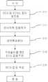
Translated fromKorean도 1는 본 발명에 따른 뇌전도 신호 처리 방법을 나타낸 도면,1 is a view showing an EEG signal processing method according to the present invention,
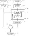
도 2은 뇌전도와 안전도 신호를 측정하는 실시 예를 나타낸 도면,2 is a view showing an embodiment for measuring the EEG and safety signal,
도 3는 칼만필터의 잡음 제거 과정을 나타낸 도면,3 is a diagram illustrating a noise removing process of a Kalman filter;
도 4는 잡음 제거 단계별 뇌전도 신호를 나타낸 도면,4 is a diagram illustrating an electroencephalogram signal according to noise removal;
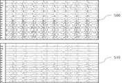
도 5은 적응필터를 적용하기 전과 후의 뇌전도 신호를 나타낸 도면이다.5 is a diagram illustrating an electroencephalogram signal before and after applying an adaptive filter.
* 도면의 주요 부분에 대한 부호의 설명 * Explanation of symbols on the main parts of the drawings
100 : 피험자200 : 뇌전도 측정 캡100: test subject 200: electroencephalography cap
201 : 뇌전도 전극202 : 안전도 전극201: electroencephalogram electrode 202: safety electrode
203 : 전극선204 : 턱끈203: electrode wire 204: chin strap
본 발명은 의료장비에 관한 기술로 종래의 뇌전도(electroencephalogram; EEG)와 자기공명영상(magnetic resonance imaging; MRI)을 동시에 획득할 때 뇌전도 내에 유발되는 잡음을 제거하기 위한 방법에 관한 것이다. 뇌전도와 자기공명영상을 동시에 검출할 때 다양한 원인에 의한 잡음이 뇌전도 신호에 유기된다. 특히 자기공명영상을 획득할 때 발생되는 경사자계 및 측정대상의 심장 박동파(ballistocardiogram; BCG)가 대표적인 잡음이다. 여기서 경사자계(gradient magnetic field)는 MRI 영상을 획득하기 위하여 코일에 인가하는 자계를 나타내며, BCG 잡음은 고자장 환경에서 심장박동의 미세한 움직임에 의해 유기된다. 상기 BCG 잡음은 시간의 경과에 따라 변화하는 비정상성(nonstationary)의 특성을 가지고 있어, 동일 피험자 내에서도 상당한 변화가 있다. 상기 BCG 잡음을 제거하기 위하여 알렌(Allen)이나 본머써(Bonmassar) 등에 의해 여러 가지 시도들이 있어 왔다. 상기 알렌의 방법은 먼저 BCG 잡음의 기본 원형(template)을 정한 뒤, 상기 기본 원형과 유사도가 높은 원형들을 추출하고 평균을 취하여, 피험자의 BCG 잡음 원형을 만든다. 상기 과정을 거쳐 만든 BCG 잡음 원형을 측정한 뇌전도 신호에서 차감하는 방법을 사용하여 BCG 잡음을 제거하였다. 상기 알렌 방법은 BCG 잡음의 비정상성을 고려하지 않고, 뇌전도내의 모든 BCG 잡음을 하나의 기본원형으로 제거하므로, BCG 잡음들 간의 미세한 차이를 제거하지 못하는 문제점이 있다. 특히, 고자장(high magnetic field) MRI 시스템일수록 미약한 심장박동에도 뇌전도 신호에 크게 영향을 주기 때문에 상기 BCG 잡음 제거의 문제점은 더욱 크게 나타난다. 상기 본머써 방법은 심장의 비정상성을 고려하기 위해, 심장의 박동에 의한 압력의 변화에 따라 전위가 유발되는 압전 센서(piezoelectric sensor)를 피험자 머리의 관자놀이에 부착하여 심장박동에 의해 유발되는 미세한 움직임을 측정하였다. 상기 센서에서 측정한 신호를 뇌전도 신호의 BCG 잡음에 대한 기준으로 보고, 적응필터를 사용하여 뇌전도 신호의 잡음을 제거하였다. 상기 본머써 방법은 압전센서가 압력의 차이를 측정하기 때문에, 상기 센서가 피험자에 부착되는 정도에 의해 그 신호의 특성이 쉽게 변할 수 있을 뿐 아니라, 상기 센서에서 감지하는 심장박동의 움직임은 뇌전도에 나타나는 BCG 잡음보다 시간지연이 나타나는 문제점을 갖는다. 또한, 상기 본머써 방법은 뇌전도 신호를 측정할 때 별도의 센서 장치를 추가하여 구성해야하는 단점이 있다. BACKGROUND OF THE INVENTION 1. Field of the Invention The present invention relates to a medical device and a method for removing noise caused in an electroencephalogram when a conventional electroencephalogram (EEG) and magnetic resonance imaging (MRI) are simultaneously acquired. When detecting EEG and MRI at the same time, noise from various sources is induced in the EEG signal. In particular, a gradient magnetic field generated when acquiring magnetic resonance images and a ballistocardiogram (BCG) of a measurement target are representative noises. Here, the gradient magnetic field represents a magnetic field applied to the coil in order to obtain an MRI image, and the BCG noise is induced by the minute movement of the heartbeat in a high magnetic field environment. The BCG noise has a characteristic of nonstationary that changes with time, and there is a significant change in the same subject. Various attempts have been made by Allen or Bonmassar to remove the BCG noise. Allen's method first establishes a basic template of BCG noise, and then extracts and averages the prototypes with high similarity to the basic prototype to form a BCG noise prototype of the subject. BCG noise was removed using a method of subtracting from the electroencephalogram signal measured by the BCG noise prototype. The Allen method removes all the BCG noises in the electroencephalogram into one basic circle without considering the abnormalities of the BCG noises, and thus, there is a problem in that it is impossible to remove the minute differences between the BCG noises. In particular, the higher magnetic field MRI system has a greater effect on the electroencephalogram signal even in a weak heartbeat, the greater the problem of the BCG noise removal. In order to consider the abnormality of the heart, the Bournemouth method attaches a piezoelectric sensor in which the potential is induced by the change of the pressure caused by the heartbeat to the temple of the subject's head, and thus the minute movement caused by the heartbeat. Was measured. The signal measured by the sensor was regarded as a reference for the BCG noise of the electroencephalogram signal, and the noise of the electroencephalogram signal was removed using an adaptive filter. Since the piezoelectric sensor measures the difference in pressure, the characteristics of the signal can be easily changed by the degree of attachment of the sensor to the subject, and the movement of the heartbeat detected by the sensor is influenced by the electroencephalogram. There is a problem in that time delay appears rather than BCG noise. In addition, the present method has a disadvantage in that an additional sensor device must be added and configured when measuring an electroencephalogram signal.
본 발명은 상술한 종래 기술들의 난점을 보완하기 위해 안출된 것으로, 본 발명의 목적은 압전센서와 같은 별도의 하드웨어의 추가 없이 안전도(electrooculogram; EOG) 신호를 기준으로 하는 적응필터 방법을 사용하여 뇌전도 신호 내에 유기되는 BCG 잡음을 효과적으로 제거하는 데 있다. The present invention has been made to solve the above-mentioned difficulties of the prior arts, and an object of the present invention is to use an adaptive filter method based on an electrooculogram (EOG) signal without the addition of additional hardware such as a piezoelectric sensor. Effective removal of BCG noise that is induced in the EEG signal.
상기의 목적을 위해 피험자의 얼굴 눈 밑 부위에서 측정하는 안전도 신호를 BCG 잡음의 기준 신호로 선정하였다. 안전도 신호는 뇌파에 의한 유발 전위가 거의 나 타나지 않으면서 BCG 잡음은 크게 나타나는 특징이 있다. 상기 안전도 신호의 측정은 뇌전도 측정 캡에 부착되어 있는 안전도 측정용 전극을 사용하여 측정할 수 있으며, 상기 뇌전도 측정용 캡에 안전도 측정용 전극이 없을 경우, 뇌전도 측정용 전극 중 1개 또는 2개를 안전도 신호 측정에 사용할 수 있다. 따라서 본 발명에서는, 별도의 센서 장치의 추가 없이 안전도를 이용한 BCG 잡음을 제거할 수 있다. For this purpose, the safety signal measured under the subject's face eye was selected as a reference signal for BCG noise. The safety signal is characterized by a large BCG noise with little or no EEG-induced potential. The measurement of the safety signal may be measured using a safety measuring electrode attached to an electroencephalogram measuring cap. When the safety cap has no electrode for measuring safety, one of the electrodes for measuring electroencephalogram or Two can be used for safety signal measurements. Therefore, in the present invention, it is possible to remove the BCG noise using the safety without the addition of a separate sensor device.
상기와 같은 목적을 달성하기 위한 본 발명의 뇌전도 신호의 잡음 제거방법은, 피험자로부터 뇌전도 신호와 안전도 신호를 검출하고, 상기 안전도 신호를 상기 뇌전도 신호에 포함된 BCG 잡음의 기준이 되는 신호로 선택하는 단계와; 상기 뇌전도 신호로부터 알렌 방법이나 또는 니아지 방법으로 경사자계에 의한 잡음을 제거하는 단계와; 상기 안전도 신호로부터 예측과정과 경신과정을 포함하는 적응필터 방법으로 상기 BCG 잡음을 추정하는 단계와; 상기 추정된 BCG 잡음을 상기 뇌전도 신호로부터 제거하는 단계를 포함한다.
상기 안전도 신호를 상기 뇌전도 신호에 포함된 BCG 잡음의 기준이 되는 신호로 선택하는 단계는, 안전도 측정용 전극을 뇌전도 측정용 캡에 부착하고, 상기 안전도 측정용 전극에서 검출된 안전도 신호를 상기 BCG 잡음의 기준이 되는 신호로 선택하는 단계를 포함한다.
이하, 첨부된 도면을 참조하여 본 발명의 바람직한 실시예를 상세히 설명하면 다음과 같다.
The noise canceling method of the electroencephalogram signal of the present invention for achieving the above object, detects the electroencephalogram signal and safety signal from a subject, and converts the safety signal as a reference signal of the BCG noise included in the electroencephalogram signal Selecting; Removing noise caused by a gradient magnetic field from the electroencephalogram signal by the Allen method or the Niaz method; Estimating the BCG noise from the safety signal by an adaptive filter method including a prediction process and a renewal process; Removing the estimated BCG noise from the electroencephalogram signal.
The selecting of the safety signal as a reference signal for BCG noise included in the electroencephalogram signal may include attaching a safety measuring electrode to an electroencephalogram measuring cap, and detecting the safety signal detected by the safety measuring electrode. Selecting a signal as a reference of the BCG noise.
Hereinafter, exemplary embodiments of the present invention will be described in detail with reference to the accompanying drawings.
삭제delete
전도 신호 처리 과정은 도 1에 나타낸 것처럼 여러 단계를 거치게 된다. 뇌전도와 안전도 전극에서 검출된 신호에서 BCG 잡음의 기준이 되는 신호를 선택하는 단계는, 먼저 MRI의 자장 안에서 측정된 뇌전도 신호에 인가된 BCG 잡음을 제거하기 위하여 뇌전도 신호와 BCG 잡음의 기준이 되는 신호를 측정(S11)해야 한다. 상기 기준이 되는 신호로 피험자의 얼굴 눈 밑 부위에서 측정하는 안전도 신호를 선택하였다. 안전도 신호에는 뇌파에 의한 유발 전위가 거의 나타나지 않으면서 BCG 잡음은 크게 나타나는 특징이 있기 때문에 기준이 되는 신호로 적합하다.
The conduction signal processing goes through several steps as shown in FIG. Selecting a signal that is a reference for BCG noise from the signal detected by the EEG and safety electrode, first to remove the BCG noise applied to the EEG signal measured in the magnetic field of the MRI The signal should be measured (S11). As a reference signal, the safety signal measured at the area under the eye of the subject was selected. The safety signal is suitable as a reference signal because the BCG noise is large while the induction potential caused by the EEG is almost insignificant.
경사자계 잡음을 제거하는 단계는 기존에 시도되었던 알렌과 니아지(Niazy) 방법을 사용(S12)한다. 알렌 방법은 MRI의 영상화 단위인 볼륨(volume)에 대하여, 각각의 볼륨별로 EEG에 나타나는 평균으로 경사자계의 기본 원형(template)을 만든 후 상기 원형을 본래의 EEG 신호에서 차감하여 경사자계 유발 잡음을 제거 하는 방법이다. 상기 니아지 방법은 상기 알렌 방법에 더하여 볼륨내의 각 슬라이스 획득시간 간에 미세한 흔들림으로 인하여 발생하는 오차까지 보완하여 경사자계를 제거하는 방법이다. 위의 방법을 통해 경자자계를 제거한 후 발생되는 고주파 신호와 원하지 않은 주파수 대역의 잡음을 제거하기 위해 해석에 필요한 주파수 대역으로 한정시키는 과정을 거친다. 상기 주파수 대역의 한정을 위하여 0.1Hz - 40Hz 정도의 대역통과 필터를 적용(S13)한다. The step of removing the gradient magnetic noise uses an allied Allen and Niiazy method (S12). The Allen method creates a basic template of the gradient magnetic field with the average appearing in the EEG for each volume, which is the imaging unit of the MRI, and subtracts the prototype from the original EEG signal to reduce gradient magnetic field induced noise. How to remove. In addition to the allen method, the nirage method eliminates the gradient magnetic field by compensating for an error caused by the slight shaking between each slice acquisition time in the volume. Through the above method, after removing the magnetic field, it is limited to the frequency band necessary for analysis in order to remove the high frequency signal and the noise of unwanted frequency band. In order to limit the frequency band, a bandpass filter of about 0.1 Hz to about 40 Hz is applied (S13).
안전도 신호에 적응필터를 사용하여 뇌전도 각 채널내의 BCG 잡음을 추정해 내는 단계에 있어서, BCG 잡음은 심장박동에 의한 전기생리학적 신호 및 심장박동의 물리적 진동이 원인이다. 즉, 심전도(electrocardiogram; ECG)와 물리적 진동이 어떠한 선형적인 결합에 의해 각 채널에 BCG 잡음으로 관찰되어지는 것이다. 그러므로 각 채널에 출현하는 BCG 잡음은 실제 뇌전도 신호와 독립적인 관계에 있다고 볼 수 있다. 따라서 실제 뇌전도에서 측정되는 신호(z(n))는 수학식 1과 같이 실제 뇌전도 신호 v(n)과 BCG 잡음 x(n)의 합으로 나타낼 수 있다. In estimating the BCG noise in each channel of the electroencephalogram using an adaptive filter on the safety signal, the BCG noise is caused by the electrophysiological signal caused by the heartbeat and the physical vibration of the heartbeat. That is, electrocardiogram (ECG) and physical vibrations are observed as BCG noise in each channel by some linear combination. Therefore, BCG noise appearing in each channel is independent of the actual EEG signal. Therefore, the signal z (n) measured at the actual electroencephalogram may be represented as the sum of the actual electroencephalogram signal v (n) and the BCG noise x (n) as shown in Equation 1.

안전도채널(202)은 뇌파의 영향은 거의 미치지 않으면서, BCG 잡음이 크게 영향 미치므로 BCG 잡음의 원형 r(n)이라고 가정할 수 있다. 상기 가정을 바탕으로, 각 채널에 유기되는 BCG 잡음은 안전도 신호를 디지털 유한 임펄스 응답(Finite Impulse Response; FIR) 필터로 추정할 수 있으므로 추정된 BCG 잡음인

wk(k=0, 1, 2,...,N-1)는 FIR 필터의 계수, N은 FIR 필터의 차수를 말하고, wk와 r(n)을 시간 n에서 정의하면 수학식 3과 같은 벡터로 표시된다.wk (k = 0, 1, 2, ..., N-1) is the coefficient of the FIR filter, N is the order of the FIR filter, and if wk and r (n) are defined at time n, It is represented by a vector such as


이 수학식들을 가지고 위의

수학식 4는 안전도 채널과 뇌전도 채널 속 BCG 잡음과의 선형적 관계를 이용함으로써 각 채널의 BCG 잡음을 추정(350)해 내는 것을 의미한다. BCG 잡음의 추정을 위해 w(n)의 추정치인


위의 수학식에서 n|n-1은 시간 n-1에 예측한, 시간 n에서의 추정치를 말하는 것이고, n-1|n-1은 시간 n-1에 경신한 시간 n-1에서의 추정치를 말한다. 또한 파라미터 a는 이전 상태 n-1과 다음 상태 n 사이의 상태 변화 파라미터이다.P(n)은 참값 w(n)과 그 추정치


여기서l은 뇌전도 측정 시에 발생하는 시스템 노이즈를 말한다.Wherel is the system noise that occurs during electroencephalogram measurements.
마지막으로 추정된 BCG 잡음을 각 채널 내에서 제거하는 단계이다. 위의 경신과정을 거쳐 최적의


다시 종합하면, 도 4에서 나타낸 것처럼 얻고자하는 뇌전도 신호만을 갖는 v(n)을 구하기 위해, 측정된 신호 z(n)속에(410) 섞여있는 BCG 잡음 x(n)을 적응필터를 통해 추정해내고(420), 이를 측정된 신호 z(n)에서 차감하는 과정을 통해 v(n)(430)를 얻는 것이다. 상기 BCG 잡음 x(n)을 추정함에 있어서, 안전도 신호 전체를 사용하는 것이 아니라, 현재부터 일정 구간 이전의 안전도 신호를 기준으로 하는 회귀 알고리즘의 칼만 적응 필터를 사용하기 때문에 실시간 처리가 가능해 진다. 상기 칼만 적응 필터의 성능을 최대화하기 위해 초기값들 및 파라미터들을 설정해야 하는데, 이 값들은 실험을 통하여 최적의 값을 찾을 수 있다. 도 5는 위의과정을 실제 데이터에 적용한 결과를 보여주는데, 칼만 적응 필터 적용 전에 BCG 잡음이 포함된 뇌전도 신호(500)와 이 신호를 칼만 적응 필터를 적용하여 뇌전도 신호 내에 유기된 BCG 잡음을 제거한 결과(510)를 도시한다. 도 5의 결과에서 확인할 수 있듯이 본 발명을 통해 잡음이 효과적으로 제거된 뇌전도 신호를 얻을 수 있다. In summary, in order to obtain v (n) having only the electroencephalogram signal to be obtained as shown in FIG. 4, the BCG noise x (n) mixed in the measured signal z (n) (410) is estimated through an adaptive filter. 420, and subtracts it from the measured signal z (n) to obtain v (n) 430. In estimating the BCG noise x (n), not the entire safety signal is used, but a Kalman adaptive filter of a regression algorithm based on the safety signal from a certain period before now is used, thereby enabling real-time processing. . Initial values and parameters must be set to maximize the Kalman adaptive filter's performance, and these values can be found through experimentation to find an optimal value. Figure 5 shows the result of applying the above process to the actual data, before applying the Kalman adaptive filter EEG signal 500 containing BCG noise and the result of removing the BCG noise induced in the EEG signal by applying the Kalman
상술한 바와 같이, 본 발명은 뇌전도 신호를 자기공명 영상과 동시에 측정할 때에 별도의 하드웨어 장치를 추가하지 않고 효과적으로 잡음을 줄일 수 있을 뿐만 아니라 신호 측정과 동시에 실시간으로 처리가 가능하므로 뇌기능 분석에 유용하며, 자기공명영상과 뇌전도 결합 시스템의 성능향상을 기대할 수 있다. As described above, the present invention is useful for brain function analysis because it can effectively reduce noise without adding a separate hardware device when measuring EEG signals simultaneously with magnetic resonance images, and can process them in real time simultaneously with signal measurement. And it can be expected to improve the performance of magnetic resonance imaging and electroencephalogram coupling system.
이상에서 설명한 내용을 통해 당업자라면 본 발명의 기술 사상을 이탈하지 아니하는 범위에서 다양한 변경 및 수정이 가능함을 알 수 있을 것이다. 따라서 본 발명 의 기술적 범위는 실시예에 기재된 내용으로 한정되는 것이 아니라 특허 청구의 범위에 의해 정해져야 한다. Those skilled in the art will appreciate that various changes and modifications can be made without departing from the spirit of the present invention. Therefore, the technical scope of the present invention should not be limited to the contents described in the embodiments, but should be defined by the claims.
Claims (3)
Translated fromKoreanPriority Applications (1)
| Application Number | Priority Date | Filing Date | Title |
|---|---|---|---|
| KR1020060089049AKR100832382B1 (en) | 2006-09-14 | 2006-09-14 | Noise Reduction Method in EEG Signal Using Adaptive Filter in Electroencephalogram and Magnetic Resonance Imaging System |
Applications Claiming Priority (1)
| Application Number | Priority Date | Filing Date | Title |
|---|---|---|---|
| KR1020060089049AKR100832382B1 (en) | 2006-09-14 | 2006-09-14 | Noise Reduction Method in EEG Signal Using Adaptive Filter in Electroencephalogram and Magnetic Resonance Imaging System |
Publications (2)
| Publication Number | Publication Date |
|---|---|
| KR20080024660A KR20080024660A (en) | 2008-03-19 |
| KR100832382B1true KR100832382B1 (en) | 2008-05-27 |
Family
ID=39412871
Family Applications (1)
| Application Number | Title | Priority Date | Filing Date |
|---|---|---|---|
| KR1020060089049AExpired - Fee RelatedKR100832382B1 (en) | 2006-09-14 | 2006-09-14 | Noise Reduction Method in EEG Signal Using Adaptive Filter in Electroencephalogram and Magnetic Resonance Imaging System |
Country Status (1)
| Country | Link |
|---|---|
| KR (1) | KR100832382B1 (en) |
Families Citing this family (3)
| Publication number | Priority date | Publication date | Assignee | Title |
|---|---|---|---|---|
| CN106361328B (en)* | 2016-10-21 | 2019-07-19 | 电子科技大学 | A method for extracting EEG signals in a magnetic resonance environment |
| CN114081503A (en)* | 2021-11-18 | 2022-02-25 | 江苏科技大学 | A method for removing electrooculography artifacts in EEG signals |
| KR102801066B1 (en)* | 2022-09-22 | 2025-04-24 | 연세대학교 원주산학협력단 | Apparatus and method for removing gradient magnetic field artifacts of EEG signal during simultaneous measurement of MR-DTI and EEG signal |
Citations (1)
| Publication number | Priority date | Publication date | Assignee | Title |
|---|---|---|---|---|
| US5513649A (en)* | 1994-03-22 | 1996-05-07 | Sam Technology, Inc. | Adaptive interference canceler for EEG movement and eye artifacts |
- 2006
- 2006-09-14KRKR1020060089049Apatent/KR100832382B1/ennot_activeExpired - Fee Related
Patent Citations (1)
| Publication number | Priority date | Publication date | Assignee | Title |
|---|---|---|---|---|
| US5513649A (en)* | 1994-03-22 | 1996-05-07 | Sam Technology, Inc. | Adaptive interference canceler for EEG movement and eye artifacts |
Also Published As
| Publication number | Publication date |
|---|---|
| KR20080024660A (en) | 2008-03-19 |
Similar Documents
| Publication | Publication Date | Title |
|---|---|---|
| Sai et al. | Automated classification and removal of EEG artifacts with SVM and wavelet-ICA | |
| Negishi et al. | Removal of time-varying gradient artifacts from EEG data acquired during continuous fMRI | |
| Grouiller et al. | A comparative study of different artefact removal algorithms for EEG signals acquired during functional MRI | |
| Mahajan et al. | Unsupervised eye blink artifact denoising of EEG data with modified multiscale sample entropy, kurtosis, and wavelet-ICA | |
| Garreffa et al. | Real-time MR artifacts filtering during continuous EEG/fMRI acquisition | |
| Moosmann et al. | Realignment parameter-informed artefact correction for simultaneous EEG–fMRI recordings | |
| EP1424637A1 (en) | Artifact removal from an electric signal | |
| WO2007041766A1 (en) | Adaptive real-time line noise suppression for electrical or magnetic physiological signals | |
| Kim et al. | Recursive approach of EEG-segment-based principal component analysis substantially reduces cryogenic pump artifacts in simultaneous EEG–fMRI data | |
| Luke et al. | Kalman filter based estimation of auditory steady state response parameters | |
| KR20080074413A (en) | Electroencephalogram test device and method with electrocardiogram noise removing means | |
| KR100832382B1 (en) | Noise Reduction Method in EEG Signal Using Adaptive Filter in Electroencephalogram and Magnetic Resonance Imaging System | |
| KR101831064B1 (en) | Apparatus for motion artifact removal using ppg signal and method thereof | |
| Prokopiou et al. | Modeling the hemodynamic response function using EEG-fMRI data during eyes-open resting-state conditions and motor task execution | |
| JP2012000280A (en) | Brain wave estimating device, brain wave estimation method and program | |
| US20080234596A1 (en) | Brain Wave Measuring Method, Apparatus And Computer Readable Recording Medium Implemented With Program For Executing The Method | |
| EP3405098B1 (en) | Signal processing method and apparatus | |
| Turnip | Automatic artifacts removal of EEG signals using robust principal component analysis | |
| KR101978905B1 (en) | Early seizure detection method | |
| Philiastides et al. | Causal influences in the human brain during face discrimination: a short-window directed transfer function approach | |
| KR101701299B1 (en) | Device and method for denoising noise of electroencephalogram signal using series independent component analysis | |
| Ferreira et al. | Optimized moving-average filtering for gradient artefact correction during simultaneous EEG-fMRI | |
| Mahamune et al. | Ocular artifacts removal from eeg signals using discrete wavelet transform and quadratic regression method | |
| Ferreira et al. | Gradient artefact correction in the EEG signal recorded within the fMRI scanner | |
| KR100689987B1 (en) | Apparatus and method for removing noise included in an EEG signal in an EEG and MRI system |
Legal Events
| Date | Code | Title | Description |
|---|---|---|---|
| A201 | Request for examination | ||
| PA0109 | Patent application | St.27 status event code:A-0-1-A10-A12-nap-PA0109 | |
| PA0201 | Request for examination | St.27 status event code:A-1-2-D10-D11-exm-PA0201 | |
| R18-X000 | Changes to party contact information recorded | St.27 status event code:A-3-3-R10-R18-oth-X000 | |
| PN2301 | Change of applicant | St.27 status event code:A-3-3-R10-R13-asn-PN2301 St.27 status event code:A-3-3-R10-R11-asn-PN2301 | |
| E902 | Notification of reason for refusal | ||
| PE0902 | Notice of grounds for rejection | St.27 status event code:A-1-2-D10-D21-exm-PE0902 | |
| E13-X000 | Pre-grant limitation requested | St.27 status event code:A-2-3-E10-E13-lim-X000 | |
| P11-X000 | Amendment of application requested | St.27 status event code:A-2-2-P10-P11-nap-X000 | |
| P13-X000 | Application amended | St.27 status event code:A-2-2-P10-P13-nap-X000 | |
| R18-X000 | Changes to party contact information recorded | St.27 status event code:A-3-3-R10-R18-oth-X000 | |
| PG1501 | Laying open of application | St.27 status event code:A-1-1-Q10-Q12-nap-PG1501 | |
| E701 | Decision to grant or registration of patent right | ||
| PE0701 | Decision of registration | St.27 status event code:A-1-2-D10-D22-exm-PE0701 | |
| GRNT | Written decision to grant | ||
| PR0701 | Registration of establishment | St.27 status event code:A-2-4-F10-F11-exm-PR0701 | |
| PR1002 | Payment of registration fee | St.27 status event code:A-2-2-U10-U11-oth-PR1002 Fee payment year number:1 | |
| PG1601 | Publication of registration | St.27 status event code:A-4-4-Q10-Q13-nap-PG1601 | |
| PR1001 | Payment of annual fee | St.27 status event code:A-4-4-U10-U11-oth-PR1001 Fee payment year number:4 | |
| FPAY | Annual fee payment | Payment date:20120619 Year of fee payment:5 | |
| PR1001 | Payment of annual fee | St.27 status event code:A-4-4-U10-U11-oth-PR1001 Fee payment year number:5 | |
| FPAY | Annual fee payment | Payment date:20130619 Year of fee payment:6 | |
| PR1001 | Payment of annual fee | St.27 status event code:A-4-4-U10-U11-oth-PR1001 Fee payment year number:6 | |
| LAPS | Lapse due to unpaid annual fee | ||
| PC1903 | Unpaid annual fee | St.27 status event code:A-4-4-U10-U13-oth-PC1903 Not in force date:20140521 Payment event data comment text:Termination Category : DEFAULT_OF_REGISTRATION_FEE | |
| R18-X000 | Changes to party contact information recorded | St.27 status event code:A-5-5-R10-R18-oth-X000 | |
| PC1903 | Unpaid annual fee | St.27 status event code:N-4-6-H10-H13-oth-PC1903 Ip right cessation event data comment text:Termination Category : DEFAULT_OF_REGISTRATION_FEE Not in force date:20140521 | |
| P22-X000 | Classification modified | St.27 status event code:A-4-4-P10-P22-nap-X000 | |
| R18-X000 | Changes to party contact information recorded | St.27 status event code:A-5-5-R10-R18-oth-X000 | |
| P22-X000 | Classification modified | St.27 status event code:A-4-4-P10-P22-nap-X000 | |
| P22-X000 | Classification modified | St.27 status event code:A-4-4-P10-P22-nap-X000 | |
| R18-X000 | Changes to party contact information recorded | St.27 status event code:A-5-5-R10-R18-oth-X000 |