KR100741160B1 - High performance assay of protein-protein interactions on protein nanoarrays - Google Patents
High performance assay of protein-protein interactions on protein nanoarraysDownload PDFInfo
- Publication number
- KR100741160B1 KR100741160B1KR1020060098980AKR20060098980AKR100741160B1KR 100741160 B1KR100741160 B1KR 100741160B1KR 1020060098980 AKR1020060098980 AKR 1020060098980AKR 20060098980 AKR20060098980 AKR 20060098980AKR 100741160 B1KR100741160 B1KR 100741160B1
- Authority
- KR
- South Korea
- Prior art keywords
- protein
- cantilever
- nanoarrays
- interactions
- proteins
- Prior art date
- Legal status (The legal status is an assumption and is not a legal conclusion. Google has not performed a legal analysis and makes no representation as to the accuracy of the status listed.)
- Expired - Fee Related
Links
Images


Classifications
- G—PHYSICS
- G01—MEASURING; TESTING
- G01N—INVESTIGATING OR ANALYSING MATERIALS BY DETERMINING THEIR CHEMICAL OR PHYSICAL PROPERTIES
- G01N33/00—Investigating or analysing materials by specific methods not covered by groups G01N1/00 - G01N31/00
- G01N33/48—Biological material, e.g. blood, urine; Haemocytometers
- G01N33/50—Chemical analysis of biological material, e.g. blood, urine; Testing involving biospecific ligand binding methods; Immunological testing
- G01N33/68—Chemical analysis of biological material, e.g. blood, urine; Testing involving biospecific ligand binding methods; Immunological testing involving proteins, peptides or amino acids
- G01N33/6803—General methods of protein analysis not limited to specific proteins or families of proteins
- G01N33/6845—Methods of identifying protein-protein interactions in protein mixtures
- G—PHYSICS
- G01—MEASURING; TESTING
- G01N—INVESTIGATING OR ANALYSING MATERIALS BY DETERMINING THEIR CHEMICAL OR PHYSICAL PROPERTIES
- G01N33/00—Investigating or analysing materials by specific methods not covered by groups G01N1/00 - G01N31/00
- G01N33/48—Biological material, e.g. blood, urine; Haemocytometers
- G01N33/50—Chemical analysis of biological material, e.g. blood, urine; Testing involving biospecific ligand binding methods; Immunological testing
- G01N33/53—Immunoassay; Biospecific binding assay; Materials therefor
- G01N33/543—Immunoassay; Biospecific binding assay; Materials therefor with an insoluble carrier for immobilising immunochemicals
- G01N33/544—Immunoassay; Biospecific binding assay; Materials therefor with an insoluble carrier for immobilising immunochemicals the carrier being organic
- G01N33/549—Immunoassay; Biospecific binding assay; Materials therefor with an insoluble carrier for immobilising immunochemicals the carrier being organic with antigen or antibody entrapped within the carrier
- B—PERFORMING OPERATIONS; TRANSPORTING
- B82—NANOTECHNOLOGY
- B82Y—SPECIFIC USES OR APPLICATIONS OF NANOSTRUCTURES; MEASUREMENT OR ANALYSIS OF NANOSTRUCTURES; MANUFACTURE OR TREATMENT OF NANOSTRUCTURES
- B82Y15/00—Nanotechnology for interacting, sensing or actuating, e.g. quantum dots as markers in protein assays or molecular motors
Landscapes
- Health & Medical Sciences (AREA)
- Life Sciences & Earth Sciences (AREA)
- Engineering & Computer Science (AREA)
- Immunology (AREA)
- Molecular Biology (AREA)
- Hematology (AREA)
- Chemical & Material Sciences (AREA)
- Urology & Nephrology (AREA)
- Biomedical Technology (AREA)
- Physics & Mathematics (AREA)
- Medicinal Chemistry (AREA)
- Food Science & Technology (AREA)
- Biotechnology (AREA)
- Pathology (AREA)
- Microbiology (AREA)
- General Physics & Mathematics (AREA)
- General Health & Medical Sciences (AREA)
- Cell Biology (AREA)
- Biochemistry (AREA)
- Analytical Chemistry (AREA)
- Bioinformatics & Cheminformatics (AREA)
- Bioinformatics & Computational Biology (AREA)
- Biophysics (AREA)
- Proteomics, Peptides & Aminoacids (AREA)
- Apparatus Associated With Microorganisms And Enzymes (AREA)
Abstract
Translated fromKoreanDescription
Translated fromKorean도 1은 단백질 나노어레이 상에서 분자적 상호작용력의 직접 측정 및 골드 코팅된 실리콘 기지 상에서 나노어레이 제조에 대한 실험 과정을 나타낸 도식도. (a) ProLinker™ SAM으로 금 코팅된 실리콘 기질 상에서 단백질 나노어레이의 형성을 나타냄. 금 코팅된 실리콘 웨이퍼(50nm)는 ProLinker™ SAM의 고정화 및 단백질 나노어레이에 대한 기질로 사용됨. 인테그린, BSA, 및 항-안지오제닌 Ab를 포함하는 단백질 용액들이 100 ㎍/ml의 농도로 제조. 단백질 나노어레이 패터닝은 0.01 nN의 컨택트 포스와 6 초의 컨택트 타임을 가지는 골드 코팅된 캔틸레버(NSC14 Cr-Au)를 사용하여 수행. (b) 단백질 나노어레이들에서 상호작용력의 직접 측정을 나타냄. 단백질의 단단한 고정을 위하여 캔틸레버 표면을 ProLinker™ SAM (A 및 B)으로 모디화이. ProLinker™ 코팅된 캔틸레버는 비트로넥틴 및 안지오제닌 용액(C 및 D)에 침지하였다. 물리적으로 흡착된 과량의 단백질을 제거하기 위하여, 캔틸레버를 PBS (E 및 F)로 세척하고 N2 가스 스트림(G 및 H) 하에서 건조하였다. 인테그린 및 비트로넥틴 또는 BSA와 비트로넥틴 사이의 분자적 상호작용력 측정은 비트로넥틴으로 모디화이된 캔틸레버를 사용하여 각각 수행하였다(I). 안지오제닌과 항- 안지오제닌 Ab 사이의 분자적 상호작용력 측정은 안지오제닌으로 모디화이된 캔틸레버를 사용하여 수행하였다(J). 상호작용력 측정은 넌-컨택트 모드로 골드 코팅된 캔틸레버(NSC36 Cr-Au)를 사용하여 수행하였다.1 is a schematic showing the experimental procedure for the direct measurement of molecular interaction forces on protein nanoarrays and nanoarray fabrication on gold coated silicon substrates. (a) Represents the formation of protein nanoarrays on a silicon substrate coated with ProLinker ™ SAM. Gold-coated silicon wafer (50 nm) was used as a substrate for the immobilization of ProLinker ™ SAM and protein nanoarrays. Protein solutions comprising integrin, BSA, and anti- angiogenin Ab were prepared at a concentration of 100 μg / ml. Protein nanoarray patterning was performed using a gold coated cantilever (NSC14 Cr-Au) with a contact force of 0.01 nN and a contact time of 6 seconds. (b) direct measurement of interaction force in protein nanoarrays. Modulate cantilever surfaces with ProLinker ™ SAM (A and B) for tight fixation of proteins. ProLinker ™ coated cantilevers were immersed in Vitronectin and Angiogenin Solution (C and D). To remove excess protein that was physically adsorbed, the cantilever was washed with PBS (E and F) and dried under N2 gas streams (G and H). Molecular interaction force measurements between integrin and Vitronectin or BSA and Vitronectin were performed using cantilevers modulated with Vitronectin (I), respectively. Molecular interaction force measurement between angiogenin and anti- angiogenin Ab was performed using a cantilever modulated with angiogenin (J). Interaction force measurements were performed using gold coated cantilevers (NSC36 Cr-Au) in non-contact mode.
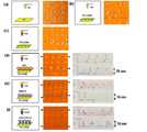
도 2는 여러 실험 조건에서 단백질 나노어레이의 AFM 이미지 및 분자적 상호작용의 도식 도면. (a) Au 기질 vs. Au-코팅된 캔틸레버. (b) ProLinker™ SAM vs. Au 코팅된 캔틸레버. (c) ProLinker™ SAM vs. 비트로넥틴-고정화된 캔틸레버. (d) BSA 나노어레이 vs. 비트로넥틴-고정화된 캔틸레버. AFM 이미지는 나노어레이된 BSA 스팟들을 보인다. 라인 프로화일은 12에서 25 nm의 BSA 나노어레이의 높이를 보인다. (e) 인테그린 나노어레이 vs. 비트로넥틴 고정화된 캔틸레버. AFM 이미지는 상기 나노어레이된 인테그린 스팟들을 나타낸다. 라인 프로화일은 12에서 16 nm의 인테그린 나노어레이의 높이를 보인다. (f) 항-안지오제닌 Ab 나노어레이 vs. 안지오제닌-고정화된 캔틸레버. AFM 이미지는 상기 나노어레이된 항체 스팟들을 보인다. 라인 프로화일은 20nm의 항체 나노어레이의 높이를 보인다.2 is a schematic representation of AFM images and molecular interactions of protein nanoarrays at various experimental conditions. (a) Au substrate vs. Au-coated cantilever. (b) ProLinker ™ SAM vs. Au coated cantilever. (c) ProLinker ™ SAM vs. Vitronectin-immobilized cantilever. (d) BSA nanoarray vs. Vitronectin-immobilized cantilever. AFM images show nanoarrayed BSA spots. The line profile shows a height of BSA nanoarray from 12 to 25 nm. (e) Integrin nanoarray vs. Vitronectin immobilized cantilever. An AFM image shows the nanoarrayed integrin spots. The line profile shows the height of the integrin nanoarray from 12 to 16 nm. (f) anti-angiogenin Ab nanoarray vs. Angiogenin-Immobilized Cantilever. AFM images show the nanoarrayed antibody spots. The line profile shows the height of the antibody nanoarrays of 20 nm.
도 3은 캔틸레버 말단에서 컨택트되는 분자들의 수와 캔틸레버의 구조적 차원 사이의 관계를 나타낸 그림. (a) AFM 캔틸레버의 특성(NSC36/Cr-Au). (b) 상호작용력의 측정을 위한 단백질 나노어레이 상의 선택된 접근 지점들. (c) 캔틸레버 말단에서 컨택트되는 분자들의 수. 캔틸레버의 구조적 특성으로 인하여 캔틸레버 상의 타겟 단백질의 제한된 수가 단백질 나노어레이 상의 프로브 분자들과 접촉할 수 있다. (d) 측정된 상호작용력 곡선.3 shows the relationship between the number of molecules contacted at the cantilever end and the structural dimensions of the cantilever. (a) Characteristics of AFM cantilever (NSC36 / Cr-Au). (b) Selected access points on protein nanoarrays for measurement of interaction force. (c) Number of molecules contacted at the cantilever end. Due to the structural nature of the cantilever, a limited number of target proteins on the cantilever may contact the probe molecules on the protein nanoarrays. (d) Interaction force curve measured.
도 4는 다양한 실험 조건에 대한 히스토그램과 상호작용력 곡선. (a) 다른 여섯 개의 실험 조건에 대한 전형적인 상호작용력 곡선. (b) 다른 여섯 개의 실험 조건에 대한 상호작용의 히스토그램.4 histogram and interaction force curves for various experimental conditions. (a) Typical interaction force curves for six different experimental conditions. (b) Histogram of interactions for six different experimental conditions.
본 발명은 단백질 나노어레이 상에서 단백질-단백질 상호작용의 고성능 분석법에 관한 것이다.The present invention relates to high performance assays of protein-protein interactions on protein nanoarrays.
일반적으로,고체 표면에 고정화된 생체분자의 어레이들은 과학기술의 여러 분야에서 소형화된 바이오분석에 대한 새로운 도구로 사용될 수 있다(Charboneau, L., 외,Brief.Funct.Genomics Proteomics 2002, 1, 305-315;Cutler, P., Protein arrays: The current state-of-the-art,Proteomics 2003, 3, 3). 특히 단백질 또는 DNA 고정화된 마이크로어레이들은 고성능 스크리닝 및 매우 패킷된 진단 칩에 대한 새로운 분석법의 개발과 같은 생물학적 용도에 유용한 플랫폼으로 간주된다(Knezevic, V., 외,Proteomics 2001, 1, 1271-1278;Liotta, L., 외,Nat. Rev.Genet. 2000, 1, 48-56).Generally, Arrays of biomolecules immobilized on solid surfaces can be used as new tools for miniaturized bioanalysis in many areas of science and technology (Charboneau, L., et al. ,Brief.Funct.GenomicsProteomics 2002, 1, 305-315; Cutler, P., Protein arrays: The current state-of-the-art,Proteomics 2003, 3, 3). In particular, protein or DNA immobilized microarrays are considered to be useful platforms for biological applications such as high performance screening and development of new assays for highly packetized diagnostic chips (Knezevic, V., et al.,Proteomics 2001, 1, 1271-1278; Liotta, L., et al. ,Nat. Rev.Genet. 2000, 1, 48-56).
생체분자 상호작용에서 통상의 검출 방법들은 동위원소, 형광 염료 및 발색 염료와 같은 다른 시약들과 생체분자의 추가적인 태깅이 필요하다. 특히, 단백질 마이크로어레이의 어레이들은 형광과 같은 광학성을 사용하여 수행되고, 단백질-단백질 상호작용을 검출하는 형광 물질로 표지된 분자를 필요로 한다(Lesaicherre, M. L., 외,Bioorg. Med.Chem.Lett. 2002, 12, 2085-2088). 그러나 형광물질들을 사용한 이들 추가적인 태킹은 단백질의 구조형태에 영향을 주어 단백질의 기능적 변화를 야기할 수 있다. 따라서 표지된 프로브없이 단백질의 분자적 상호작용들을 검출하는 기술을 개발할 필요가 있다. 그러나 형광기술을 넘어서는 단백질-단백질 상호작용을 분석할 수 있는 검출 기술은 현재까지 제한적이다.Conventional detection methods in biomolecule interactions require additional tagging of the biomolecule with other reagents such as isotopes, fluorescent dyes, and color dyes. In particular, arrays of protein microarrays are performed using optical properties such as fluorescence and require molecules labeled with fluorescent substances that detect protein-protein interactions (Lesaicherre, ML, etal. ,Bioorg. Med.Chem.Lett. 2002, 12, 2085-2088). However, these additional tagging with fluorescents can affect the protein's conformation and cause functional changes in the protein. Therefore, there is a need to develop a technique for detecting molecular interactions of proteins without labeled probes. However, detection techniques that can analyze protein-protein interactions beyond fluorescence are limited to date.
최근에 마이크로-컨택트 프린팅(MCP) 및 답-펜 나노리소그래피 (DPN)을 포함하는 새로운 기술들 중 일부가 초소형화된 생체분자 어레이의 패터닝 기술로 개발되었다. Method 원자력 현미경(AFM)을 사용하는 답-펜 나노리소그래피(Dip-pen nanolithography;DPN)는 패터닝 기술뿐 아니라 추가적인 태깅없는 검출 방법으로 관심이 있다. 상기 DPN 방법은 나노미터 스캐일에서 고밀도 어레이의 제조의 경로를 제공한다(Hong,S., 외,Science, 1999, 286, 523-525;Pinder, R. D., 외,Science, 1999, 283, 661-663).Recently some of the new technologies, including micro-contact printing (MCP) and dap-pen nanolithography (DPN), have been developed with the patterning of microminiaturized biomolecule arrays. Method Dip-pen nanolithography (DPN) using atomic force microscopy (AFM) is of interest as a patterning technique as well as an additional tag-free detection method. The DPN method provides a route for the fabrication of high density arrays in nanometer scales (Hong, S., et al.,Science , 1999, 286, 523-525; Finder, RD, et al.,Science, 1999, 283, 661-663). ).
DPN 방법에 의해 패턴된 단백질 나노어레이에서, 표면에 고정화된 프로브 단백질과 용액 상태 내의 타겟 단백질 사이의 단백질-단백질 상호작용은 배양과 건조 과정을 거친 후에 각 단백질 스팟의 높이 변화를 측정하여 검출할 수 있다(K. B. Lee, 외,J. Am.Chem.Soc. 2003, 125, 5588-5589). 이 방법에서, 배양 과정을 통한 타겟 단백질의 상호작용 후에 각 스팟의 높이 정보를 얻기 위하여 여러 번의 단백질 나노어레이의 이미지를 스캐닝하여야 한다. AFM에 의한 주어진 표면 면적의 토포로지컬 이미지들을 스캐닝하는데 비교적 오랜 시간이 소요된다. 이들 이유로 인하여 DPN 방법 기반의 단백질-단백질 상호작용 분석은 비교적 긴 검출 시간과 복잡한 과정을 요구한다. 따라서 매우 빠르고 간단한 검출 원리를 가진 바이오분석 방법의 필요성이 대두되었다.In protein nanoarrays patterned by the DPN method, protein-protein interactions between probe proteins immobilized on the surface and target proteins in solution can be detected by measuring the change in height of each protein spot after incubation and drying. (KB Lee, etal. ,J. Am.Chem.Soc . 2003, 125, 5588-5589). In this method, images of several protein nanoarrays need to be scanned to obtain height information of each spot after interaction of the target protein through the culturing process. It takes a relatively long time to scan topological images of a given surface area by AFM. For these reasons, protein-protein interaction analysis based on DPN methods requires a relatively long detection time and a complex process. Thus, there is a need for a bioanalytical method having a very fast and simple detection principle.
본 발명은 상기의 필요성에 의하여 안출된 것으로서 본 발명의 목적은 단백질 나노어레이 상의 단백질-단백질 상호작용의 고성능 분석에 대한 나노스캐일에서 새로운 향상된 검출 방법을 제공하는 것이다.SUMMARY OF THE INVENTION The present invention has been made by the above necessity, and an object of the present invention is to provide a new and improved detection method in nanoscale for high performance analysis of protein-protein interaction on protein nanoarrays.
상기의 목적을 달성하기 위하여 본 발명은 a)골드 코팅된 캔틸레버 표면에 크라운-에테르 모이어티를 가지는 캘릭스[4]아렌(calix[4]arene)] 유도체 용액를 처리하는 단계;In order to achieve the above object, the present invention comprises the steps of: a) treating a solution of Callix [4] arene derivative having a crown-ether moiety on a surface of a gold coated cantilever;
b)상기 캔틸레버 표면 상에 크라운-에테르 모이어티를 가지는 캘릭스[4]아렌(calix[4]arene)] 유도체 자가 조립 단층구조(SAM) 형성 후 캔틸레버를 단백질 단층 형성을 위하여 대상 단백질 용액에 침지하는 단계;b) After forming the Calix [4] arene derivative self-assembled monolayer structure (SAM) having a crown-ether moiety on the surface of the cantilever, the cantilever is immersed in the target protein solution for protein monolayer formation. Doing;
c)버퍼로 세척하여 물리적으로 흡착된 과량의 단백질을 제거하는 단계;c) washing with a buffer to remove excess protein physically adsorbed;
d) 단백질 모디화이된 캔틸레버를 건조하여 상기 단백질로 모디화인된 캔틸레버 팁을 제조하는 단계;d) drying the protein modulated cantilever to produce a cantilever tip modulated with the protein;
e) 상기 단백질로 모디화인된 캔틸레버 팁을 크라운-에테르 모이어티를 가지는 캘릭스[4]아렌(calix[4]arene)] 유도체 코팅된 웨이퍼 상의 타겟 단백질 나노어레이 또는 상기 고정화된 단백질들의 항체 나노어레이 스팟에 접근하는 단계;및e) a target protein nanoarray on a Calix [4] arene] derivative-coated wafer having a crown-ether moiety or a cantilever tip modulated with the protein or an antibody nanoarray of the immobilized proteins. Accessing the spot; and
f) 캔틸레버 팁과 웨이퍼 모두가 분리될 때 양 단백질 사이의 분리력을 측정하는 것을 포함하는 단백질 나노어레이 상에서 단백질-단백질 상호작용의 고성능 분석법을 제공하는 것이다.f) to provide a high performance analysis of protein-protein interactions on protein nanoarrays, including measuring the separation force between both proteins when both the cantilever tip and the wafer are separated.
본 발명에 있어서, 상기 대상 단백질은 타겟 단백질과 상호작용할 수 있는 특징을 가지는 모든 단백질이 가능하며, 상기 대상 단백질은 보바인 시럼 알부민, 비트로넥틴 또는 안지오제닌인 것이 더욱 바람직하다.In the present invention, the target protein may be any protein having a feature capable of interacting with the target protein, and the target protein is more preferably bovine serum albumin, vitronectin or angiogenin.
또한 본 발명에서 상기 타겟 단백질은 대상 단백질들과 상호작용할 수 있는 항체를 포함한 모든 단백질이 가능하며, 상기 타겟 단백질은 안테그린 또는 항-안지오제닌 항체인이 더욱 바람직하다.In the present invention, the target protein may be any protein including an antibody capable of interacting with the target proteins, and the target protein is more preferably an antegrin or an angiogenin antibody.
또한 본 발명은 a) 골드-코팅된 캔틸레버 표면에 크라운-에테르 모이어티를 가지는 캘릭스[4]아렌(calix[4]arene)] 유도체 용액를 처리하는 단계;The present invention also provides a process for treating a solution of Calix [4] arene derivative having a crown-ether moiety on a surface of a gold-coated cantilever;
b)상기 캔틸레버 표면 상에 크라운-에테르 모이어티를 가지는 캘릭스[4]아렌(calix[4]arene)] 유도체 자가 조립 단층구조(SAM) 형성 후 캔틸레버를 단백질 단층 형성을 위하여 대상 단백질 용액에 침지하는 단계;b) After forming the Calix [4] arene derivative self-assembled monolayer structure (SAM) having a crown-ether moiety on the surface of the cantilever, the cantilever is immersed in the target protein solution for protein monolayer formation. Doing;
c)버퍼로 세척하여 물리적으로 흡착된 과량의 단백질을 제거하는 단계; 및c) washing with a buffer to remove excess protein physically adsorbed; And
d) 단백질 모디화이된 캔틸레버를 건조하여 상기 단백질로 모디화인된 캔틸레버 팁을 제공한다.d) drying the protein modulated cantilever to provide a cantilever tip modulated with the protein.
이하 본 발명을 설명한다.Hereinafter, the present invention will be described.
본 발명에서는 나노어레이된 단백질과 AFM 캔틸레버 표면 상에 고정화된 타겟 단백질 사이의 상호작용을 측정하여 단백질 나노어레이 상에 나노스캐일화된 단백질 분자 상호작용을 검출하는 새로운 기술을 제공한다. 본 발명자들은 AFM을 사 용하는 DPN 방법에 의하여 ProLinker™-표면화된 Si 웨이퍼 상에 인테그린 avb3의 단백질 나노어레이를 제조하였다.인테그린 avb3은 비트로넥틴에 대한 주된 수용체 작용을 하는 막 단백질이다.비트로넥틴은 ProLinker™ -코팅된 캔틸레버 팁 상에 고정화하였다. BSA 및 항-안지오제닌(ANG) mAb의 단백질 나노어레이들을 여러 분자적 상호작용을 측정하는 레퍼런스 물질로 고안하였다. 인테그린 avb3 및 비트로넥틴 사이의 분자적 상호작용은 AFM 이미지 상의 나노어레이된 단백질 스팟의 높이 변화를 측정하는 대신에 ProLinker™ 표면 상의 양 단백질 사이의 분리력(unbinding force)을 측정하여 검출하였다. 또 비트로넥틴 및 BSA 사이 또는 항-ANG mAb 및 ANG 사이의 분자적 상호작용력을 동시에 측정하고 평가하였다.The present invention provides a novel technique for detecting nanoscaled protein molecular interactions on protein nanoarrays by measuring the interaction between nanoarrayed proteins and target proteins immobilized on the AFM cantilever surface. We prepared protein nanoarrays of integrin av b3 on a ProLinker ™ -surfaced Si wafer by DPN method using AFM. Integrin av b3 is a membrane protein that acts as the primary receptor for Vitronectin. Vitronectin was immobilized on a ProLinker ™ -coated cantilever tip. Protein nanoarrays of BSA and anti-angiogenin (ANG) mAbs were designed as reference materials to measure several molecular interactions. Molecular interactions between integrin av b3 and vitronectin were detected by measuring the unbinding force between both proteins on the ProLinker ™ surface instead of measuring the height change of the nanoarrayed protein spot on the AFM image. In addition, the molecular interaction force between Vitronectin and BSA or between anti-ANG mAb and ANG was simultaneously measured and evaluated.
단백질 나노어레이들은 초소형(ultraminiaturized) 바이오분석에 대한 유용한 프랫폼으로 작용할 수 있다. 많은 경우에서 단백질 샘플들은 매우 제한적이고 일시적으로 유일무이하다. 따라서 단백질 나노어레이 기술을 사용하여 고성능 분석을 위한 단백질 칩을 제조하는 것은 많은 잇점을 얻을 수 있다. 본 발명에서는 캔틸레버 표면에 고정화된 타겟 단백질과 Au-코팅된 Si 웨잎 상의 나노어레이된 캡쳐 단백질 사이의 상호작용력을 측정하였다. Anti-angiogenin(ANG) mAb, integrin avb3 및 BSA를 포함하는 여러 단백질 나노어레이들을 딥-펜 나노리소그래피(dip-pen nanolithography;DPN)에 의하여 ProLinker™ -코팅된 금 표면상에 제조하였다. ProLinker™가 코팅된 원자력 현미경(AFM) 팁은 인테그린 avb3 및 ANG의 특 정 리간드인 비트로넥틴에 의하여 모디화이되었다. 단백질-고정화된 AFM 팁을 사용하여, 나노어레이된 단백질들(인테그린 avb3 및 BSA)과 팁에 고정화된 단백질 사이의 상호작용력을 양 단백질에 대한 테더링(tethering) 및 언바인딩 방법을 통하여 조사하였다. 인테그린 avb3 및 비트로넥틴 사이의 측정된 분리력은 대조군으로 사용된 BSA-비트로넥틴의 것보다 더 높았다. 그리고 또 항-ANG mAb-ANG 상호작용에 대한 특정 단백질 상호작용은 BSA-비트로넥틴 및 BSA-ANG와 같은 음성 대조군 단백질들의 것과 비교하여 더 높았다.Protein nanoarrays can serve as a useful platform for ultraminiaturized bioanalysis. In many cases protein samples are very limited and temporarily unique. Thus, the production of protein chips for high performance analysis using protein nanoarray technology has many advantages. In the present invention, the interaction force between the target protein immobilized on the surface of the cantilever and the nanoarrayed capture protein on the Au-coated Si weave was measured. Several protein nanoarrays, including anti-angiogenin (ANG) mAb, integrin av b3 and BSA, were prepared on ProLinker ™ -coated gold surfaces by dip-pen nanolithography (DPN). Atomic microscope (AFM) tips coated with ProLinker ™ were modulated by integrin av b3 and Vitronectin, a specific ligand of ANG. Using protein-immobilized AFM tips, the interaction between nanoarrayed proteins (integrin av b3 and BSA) and the protein immobilized on the tip is determined by tethering and unbinding methods for both proteins. Investigate. The measured separation force between integrin av b3 and Vitronectin was higher than that of BSA-Vitronectin used as a control. And also the specific protein interactions for anti-ANG mAb-ANG interactions were higher compared to that of negative control proteins such as BSA-Vitronectin and BSA-ANG.
본 발명에서 사용된 크라운-에테르 모이어티를 가지는 캘릭스[4]아렌(calix[4]arene)] 유도체중의 하나인 ProLinker™은 화학식 1a의 구조가 바람직하다. 화학식 1a에서 m=0-2, n=0-2이 더욱 바람직하다. 본 발명에 있어서 캘릭스 크라운 유도체는 화학식 1a의 구조가 바람직하다. 화학식 1a에서 m=0-2, n=0-2이 더욱 바람직하다. 또 화학식 1a의 R1은 -CH2SH, -CHO, -CH2Cl, -CH2CN, -CH2CHO, -CH2NH2, -CH2COOH, -NH2, -NO2 R2은 -H, methyl, ethyl, propyl, isopropyl, isobutyl를 갖는 것이 바람직하고, 상기 캘릭스 크라운 유도체는 화학식 1b 또는 1c 구조를 갖는 것이 더욱 바람직하다.ProLinker ™, one of the Calix [4] arene derivatives having the crown-ether moiety used in the present invention, preferably has the structure of Formula 1a. In Formula 1a, m = 0-2 and n = 0-2 are more preferable. In the present invention, the Calix crown derivative preferably has the structure of Formula 1a. In Formula 1a, m = 0-2 and n = 0-2 are more preferable. R1 in Formula 1a is -CH2 SH, -CHO, -CH2 Cl, -CH2 CN, -CH2 CHO, -CH2 NH2 , -CH2 COOH, -NH2 , -NO2 R2 is- It is preferable to have H, methyl, ethyl, propyl, isopropyl, isobutyl, and more preferably the Calix crown derivative has a structure of Formula 1b or 1c.


실시예Example
본 발명의 단백질 나노어레이 제조를 위하여 ANG, 보바인 시럼 알부민(BSA) 및 인테그린 avb3을 케미콘(CA, Temecula, USA)으로부터 구입하여 단백질 나노어레이의 소스 물질로 사용하였다. 안지오제닌 및 인테그린 샘플 용액을 제조하여 PBS 버퍼 용액에서 500 mg/ml 농도로 사용하였다. 단백질 용액은 1 M MgCl2 및 0.1 mM CaCl2을 포함한다. 메캅토-운데카노익산(MUDA), PBS 용액, RGD (Arg-Gly-Asp) 펩타이드, RGE (Arg-Gly-Glu) 및 다른 시약들은 시그마 케미컬(St. Louis, MO)에서 구 입하였다. Milli-Q 급(>18.2 mΩ/cm) 워터를 샘플 및 버퍼 용액 제조에 사용하였다. 크라운-에테르 모이어티를 가지는 캘릭스[4]아렌(calix[4]arene)] 유도체중의 하나인 ProLinker™은 프로테오젠(Seoul, Korea)으로부터 구입하여 단백질의 고정화에 대한 링커 시스템으로 사용하였다.In order to prepare the protein nanoarray of the present invention, ANG, bovine syrum albumin (BSA) and integrin av b3 were purchased from Chemicon (CA, Temecula, USA) and used as source materials for protein nanoarrays. Angiogenin and integrin sample solutions were prepared and used at a concentration of 500 mg / ml in PBS buffer solution. Protein solution is 1 M MgCl2 and 0.1 mM CaCl2 It includes. Mecapto-Undecanoic acid (MUDA), PBS solution, RGD (Arg-Gly-Asp) peptide, RGE (Arg-Gly-Glu) and other reagents were purchased from Sigma Chemical (St. Louis, MO). Milli-Q grade (> 18.2 mPa / cm) water was used for sample and buffer solution preparation. One of the Calix [4] arene derivatives with crown-ether moieties, ProLinker ™ was purchased from Proteogen (Seoul, Korea) and used as a linker system for the immobilization of proteins.
실시예 1:Example 1:
ProLinker™-코팅된 Si 웨이퍼 상의 단백질 나노어레이의 제조Preparation of Protein Nanoarrays on ProLinker ™ -Coated Si Wafers
금-코팅된 AFM 캔틸레버(PSIA Co., NSC 14/Cr-Au, Korea)를 금-코팅된 실리콘 웨이퍼의 표면에 단백질 분자를 운반하는 전달체로 사용하였다. 캔틸레버 표면의 친수성을 증가하기 위하여, 금-코팅된 캔틸레버 팁을 에탄올 안에 1 mM 메캅토-운데카노익산(MUDA) 속에서 30분간 침지한 후 상온에서 N2 가스 스트림으로 건조하였다. MUDA로 골드-코팅된 실리콘 캔틸레버를 변형한 후, 상기 캔틸레버 100 mg/ml 농도의 단백질 용액에 담가서 캔틸레버 표면에 단백질을 흡착하도록 1시간 배양하였다. 캔틸레버 표면 상에 단백질 흡착을 통하여 단백질 분자들은 골드-코팅된 실리콘 웨이퍼의 표면으로 전달되어 단백질 나노어레이를 제조하였다.Gold-coated AFM cantilevers (PSIA Co.,
50 nm Au 증착층을 가지는 실리콘 웨이퍼를 단백질 나노어레이에 대한 기질로 사용하였다. 단백질의 안정한 고정화에 대한 링커 층을 제조하기 위하여, 골드-코팅된 실리콘 웨이퍼를 클로로포름 내의 1 mM ProLinker™ 용액에 1시간 동안 침지하였다. 프로링커™자기 조립 단층구조(self-assembled monolayer;SAM) 제조 후, 아세톤과 에탄올로 세척한 후 상온에서 dried under the N2 가스 스트림하에서 건조 하였다. 컨택드 모드로 캔틸레버 팁 상에 흡착된 단백질들의 나노어레이 패턴닝을 수행한 후 80 % 습도 조건의 상온에서 3시간 동안 단백질칩을 배양하였다. ProLinker™과 단백질 사이의 안정한 상호작용을 위하여 배양 후에, 단백질 나노어레이의 토포로직 이미지들과 높이 프로화일을 넌-컨택트 모드로 얻었다. 모든 단All 백질 나노어레이의 제조 과정은 컨택트 및 넌-컨택트 모드로 AFM XE-100 (PSIA, Sungnam, Korea)로 수행되었다.A silicon wafer with a 50 nm Au deposited layer was used as substrate for protein nanoarrays. To prepare a linker layer for stable immobilization of the protein, gold-coated silicon wafers were immersed in 1 mM ProLinker ™ solution in chloroform for 1 hour. Prolinker ™ self-assembled monolayer (SAM) was prepared, washed with acetone and ethanol, and dried under a stream of dried under the N2 gas at room temperature. After performing nanoarray patterning of the proteins adsorbed on the cantilever tip in the contact mode, the protein chips were incubated at room temperature under 80% humidity for 3 hours. After incubation for stable interaction between ProLinker ™ and protein, topographical images and height profiles of protein nanoarrays were obtained in non-contact mode. The manufacturing process of all protein white nanoarrays was performed with AFM XE-100 (PSIA, Sungnam, Korea) in both contact and non-contact modes.
실시예 2-3: 상호작용 측정Example 2-3: Interaction Measurement
골드-코팅된 캔틸레버 표면은 클로로포름 내에 1 mM ProLinkerTM 용액으로 1시간 동안 변형하였다. 캔틸레버 표면 상에 프로링커 SAM 형성 후, 단백질 단층을 제조하기 위하여 캔틸레버를 비트로넥틴(실시예 2) 또는 ANG 용액(실시예 3)에 침지하였다. PBS 버퍼로 강한 세척 후 물리적으로 흡착된 과량의 단백질을 화학적으로 흡착된 단층을 제외하곤 제거하였다. N2 스트림 하에서 건조 후, 단백질-변형된 캔틸레버를 넌-컨택트 모드로 상호작용을 측정하기 위하여 적용하였다.비트로넥틴 또는 ANG로 고정화된 캔틸레버 팁은 0.0001 ㎛/분의 접근 속도 및 0.015N/m 스프링 링 상수를 가지고 ProLinker™-코팅된 Si 웨이퍼 상의 인테그린 나노어레이 스팟(실시예 2)또는 항-ANG mAb 웨이퍼 나노어레이 스팟(실시예 3)에 접근하였다. 그리고 후에 캔틸레버 팁과 상기 Si 웨이퍼 모두가 멀리 분리되었을 때, 양 단백질 들 사이의 분리력(unbinding force)을 수치값으로 측정하였다.Gold-coated cantilever surfaces were modified for 1 hour with 1 mM ProLinker™ solution in chloroform. After prolinker SAM formation on the cantilever surface, the cantilever was immersed in Vitronectin (Example 2) or ANG solution (Example 3) to prepare a protein monolayer. Excess protein adsorbed physically was removed except for the chemically adsorbed monolayer after strong washing with PBS buffer. After drying under an N2 stream, the protein-modified cantilever was applied to measure the interaction in non-contact mode. The cantilever tip immobilized with Vitronectin or ANG had an access speed of 0.0001 μm / min and a 0.015 N / m spring Integrin nanoarray spots (Example 2) or anti-ANG mAb wafer nanoarray spots (Example 3) on ProLinker ™ -coated Si wafers with ring constants were approached. Then, when both the cantilever tip and the Si wafer were separated far away, the unbinding force between both proteins was measured numerically.
상기 실시예의 결과는 다음과 같다. 두 다른 단백질들 사이의 상호작용력의 직접적인 측정을 통하여 단백질 나노어레이 상의 단백질 분자 상호작용의 검출 및 측정을 수행하였다. 고체 기질상에서 나노 스케일로 단백질 분자 결합의 상호작용을 측정하기 위해서 고정화 물질로 인테그린 avb3, BSA, 및 항-ANG mAb을 포함하는 단백질 나노어레이들을 DPN 방법에 의한 ProLinker™-코팅된 Si 웨이퍼 상에 고안하였고(도 1a) 캔틸레버 팁들을 인테그린 avb3의 특정한 리간드인 비트로넥틴과 항-ANG mAb의 항체인 ANG을 포함하는 여러 단백질들로 변형(modify)하였다(도 1b). Si 웨이퍼 상에 단백질 단층의 강한 고정화를 위하여, 상기 골드-코팅된 Si 웨이퍼 상에 자가 조립 단층구조를 형성하기 위하여 ProLinker™을 사용하였다. Au 기질 vs. Au-코팅된 캔틸레버, ProLinker™ SAM vs. Au 코팅된-캔틸레버, ProLinker™ SAM vs. 비트로넥틴-고정화된 캔틸레버를 포함하는 세 가지 다른 실험 조건의 도시적인 그림 및 AFM 이미지를 도 2에서 보여준다. 부가적으로, ProLinker™ 표면에 스팟된 BSA, 인테그린 avb3, 및 항-ANG mAb 나노어레이의 라인 프로화일 및 토포로지컬 AFM 이미지는 단일 단백질 분자의 스팟 높이를 가지는 잘 조절된 규칙적인 배열을 보였다(도 2d, 2e, 및 2f). 항-ANG mAb 나노스팟들은 규칙적인 결정 구조로 배열되었고 20 nm 내의 모든 항체 나노스팟들의 유사한 높이 값들을 나타냈다(도 2f). 항체는 이론적으로 Y-형태를 가지는 매우 특이적인 차원을 가지는 것으로 알려졌다(높이 = 14.5 nm, 폭= 8.5 nm, 두께 = 4.0 nm)[Silverton, E.W., 외 Proc Natl Acad Sci U S A., 1977, 74, 5140-5144; Wadu-Mesthrige, K, 외 , Biophys J. 2001, 80, 1891-1899]. 게다가 고체 표면에 고정화된 항체 차원의 실험 값이 6.5 = 0.9 nm로 보고되었다[K. B. Lee, 외, Science, 2002, 295, 1702-1705]. 따라서 본 발명에서 얻은 항-ANG mAb 나노어레이들의 높이 값은 단일 분자 층을 보여준다.The result of the above example is as follows. Detection and measurement of protein molecular interactions on protein nanoarrays was performed through direct measurement of the interaction force between two different proteins. To measure the interaction of protein molecule binding on a nanoscale on a solid substrate, protein nanoarrays containing integrin avb3, BSA, and anti-ANG mAb as immobilization materials were designed on a ProLinker ™ -coated Si wafer by the DPN method. The cantilever tips were modified with several proteins, including Vitronectin, a specific ligand of integrin avb3, and ANG, an antibody of anti-ANG mAb (FIG. 1B). For strong immobilization of protein monolayers on Si wafers, ProLinker ™ was used to form self-assembled monolayers on the gold-coated Si wafers. Au substrate vs. Au-coated cantilever, ProLinker ™ SAM vs. Au coated cantilever, ProLinker ™ SAM vs. A graphical representation of the three different experimental conditions including the Vitronectin-immobilized cantilever and AFM images are shown in FIG. 2. Additionally, line profile and topological AFM images of BSA, integrin avb3, and anti-ANG mAb nanoarrays spotted on the ProLinker ™ surface showed a well-regulated regular array with spot heights of single protein molecules (FIG. 2D). , 2e, and 2f). Anti-ANG mAb nanospots were arranged in a regular crystal structure and showed similar height values of all antibody nanospots within 20 nm (FIG. 2F). Antibodies are known to have theoretically very specific dimensions with Y-forms (height = 14.5 nm, width = 8.5 nm, thickness = 4.0 nm) [Silverton, EW, et al. Proc Natl Acad Sci US A., 1977, 74 , 5140-5144; Wadu-Mesthrige, K, et al., Biophys J. 2001, 80, 1891-1899]. In addition, experimental values of the antibody dimension immobilized on the solid surface were reported to be 6.5 = 0.9 nm [K. B. Lee, et al., Science, 2002, 295, 1702-1705]. Thus, the height values of the anti-ANG mAb nanoarrays obtained in the present invention show a single molecular layer.
두 단백질들 사이의 상호작용력의 직접 검출을 조사하기 위하여, AFM 캔틸레버 팁들을 ProLinker™-코팅된 웨이퍼 상에 나노어레이된 다른 캡쳐 단백질들에 해당하는 상호작용하는 단백질로 변형하였다. 많은 수의 스팟 이미지들이 서포트 상의 캡쳐 단백질과 캔틸레버 팁 상의 상호작용하는 단백질 사이의 분리력을 측정하기 위한 캡쳐 단백질 스팟들을 보여준다. 단백질-변형된 캔틸레버 접근을 통한 단백질 나노어레이 상에서 힘 측정 과정의 이미지와 도식적인 그림들이 도 3에 기재된다. 캔틸레버 끝에서 접촉되는 분자들의 수 및 캔틸레버의 구조 차원 사이의 관계는 캔틸레버 타입들에 의존적이다. AFM 캔티레버 NSC36/Cr-Au는 특히 양 단백질 사이의 분리력을 측정하는데 유용하다. ProLinker™ 층으로 모디화이된 캔틸레버 표면은 비트로넥틴 및 안지오제닌을 포함하는 상호작용하는 단백질 단층의 형성에 대한 적당한 베이스로 작용할 수 있다. 이 표면의 특징들은 직접 힘 측정을 사용하여 단백질 나노어레이 상의 분자 상호작용의 검출력에 대한 두 가지 이론적 기초를 제공한다. 먼저, 단백질-변형된 캔틸레버가 상호작용하는 단백질 용액에 단순하게 침지하여 제조되었을 때, ProLinker™ 층은 단순한 세척 과정을 통하여 안정한 단백질 단층의 형성을 보장한다. 캡쳐 (나노어레이) 및 상호작용하는 단백질(캔틸레버 상) 사이의 접촉에 대한 기질에 대한 단백질 나노어레이에 캔틸레버를 접근할 때, 캔틸레버 표면 상의 상호작용하는 단백질의 이 단층 구조는 나노어레이 상의 캡쳐 단백질에 접촉하는 단백질 분자들의 수를 제한할 수 있다(도 3c). 둘째, 본 발명자들이 나노어레이 상의 캡쳐 단백질로부터 상호작용하는 단백질을 떨어트려서 상호작용력을 측정하는 경우에 고정화된 단백질에 대한 ProLinker™ 층의 강한 홀딩이 서포트 또는 캔틸레버 팁 상의 한 방향으로 단백질 분자들을 튀어나가게 하는 것을 보호한다.To investigate the direct detection of the interaction force between the two proteins, AFM cantilever tips were modified with interacting proteins corresponding to other capture proteins nanoarrayed on a ProLinker ™ -coated wafer. A large number of spot images show capture protein spots for measuring the separation force between the capture protein on the support and the interacting protein on the cantilever tip. Images and schematic illustrations of the force measurement process on protein nanoarrays through protein-modified cantilever approaches are described in FIG. 3. The relationship between the number of molecules contacted at the cantilever end and the structural dimension of the cantilever depends on the cantilever types. AFM cantilever NSC36 / Cr-Au is particularly useful for measuring the separation force between both proteins. The cantilever surface modulated with the ProLinker ™ layer can serve as a suitable base for the formation of interacting protein monolayers including Vitronectin and Angiogenin. These surface features provide two theoretical foundations for the detection of molecular interactions on protein nanoarrays using direct force measurements. First, when a protein-modified cantilever is prepared by simply immersing in an interacting protein solution, the ProLinker ™ layer ensures the formation of a stable protein monolayer through a simple washing process. When approaching the cantilever to the protein nanoarray for substrates for contact between the capture (nanoarray) and the interacting protein (on the cantilever), this monolayer structure of the interacting protein on the cantilever surface is bound to the capture protein on the nanoarray. The number of protein molecules in contact can be limited (FIG. 3C). Second, when we measure the interaction force by dropping the interacting protein from the capture protein on the nanoarray, the strong holding of the ProLinker ™ layer for the immobilized protein may bounce the protein molecules in one direction on the support or cantilever tip. Protect it from getting out
힘 커브들(힘-대-거리 곡선)은 전형적으로 캔틸레버의 고정된 말단이 수직적인 방향으로 이동하고 샘플 표면으로부터 멀어질 때 AFM 캔틸레버의 자유 말단의 치우침을 보인다. 측정된 힘 곡선들은 여러 실험 조건에 의하여 그래프적으로 형성되었다. 일반적으로, AFM 힘의 주된 측정은 결합이 파괴되고 캔틸레버가 서포트로부터 자유로와 지는 지점에 정확한 위치 관계이다. 서포트 및 팁 모드로부터 결합이나 부착을 파괴하는데 필요한 상호작용력(탈착력)을 측정하는데 사용될 수 있다. 도 4(b)에서 알 수 있는 바와 같이, 빈 기질(bare Au 표면) vs. 캔틸레버(bare Au 표면) 및 ProLinker™ 표면 vs. 캔틸레버(bare Au 표면) 의 상호작용력은 각각 258 및 415 pN으로 측정되었다. 이 두 실험 조건들은 그들은 각 표면상에서 상호작용이 없거나 매우 적기에 음성 대조군으로 사용되었다. 인테그린과 비트로넥틴 사이의 분리력은 1087 pN로 계산되고 이 값은 항-ANG mAb과 ANG을 사용한 항체-항원 상호작용의 값(1028 pN)과 유사하였다. 그러나 음성 대조군으로 사용된 BSA -비트로넥틴 상호작용은 인테그린-비트로넥틴의 값(1087 pN)보다 매우 적은 상호작용 값(643 pN)을 나타내었다. 상호작용력의 수치 값은 ProLinker™ 표면 vs. 비트로넥틴 변형된 캔틸레버의 경우에 가장 높았다(1359 pN). 이 결과는 것은 ProLinker™ 층이 단백질 표면의 아민 잔기를 강하게 잡고 있다는 것을 나타내고, ProLinker™ 층과 관련되 우리의 상기 두 가정을 증명하는 것이다. 이 결과들은 단백질 나노어레이 상 의 인테그린 및 비트로넥틴 또는 항-ANG mAb 및 ANG 사이의 분자적 상호작용이 강하고 특이적인 결합 사건이고 따라서 AFM을 사용한 단백질 나노어레이 상에서 직접적인 힘 측정을 통하여 BSA-비트로넥틴과 같은 다른 비 특이적인 상호작용으로부터 특이적인 단백질 상호작용들을 구별할 수 있다. 표 1은 분리 이벤트 동안에 상호 작용력 및 높이(z축 거리)의 측정된 수치 값을 보여준다. 높이 값은 분리 이벤트 동안에 힘 제로 지점(기준선과 같은)과 분리 지점 사이에서 거리 차이를 의미한다. 표 1에서 나타난 바와 같이, 상호작용력과 높이의 측정 값들은 아주 작은 오차 범위를 가진다. 이들 결과들은 프로브 및 타겟 단백질 사이의 분자적 상호작용의 측정이 성공적으로 이루어졌다는 것을 의미한다. 한편 분리 이벤트 동안에 z-축 높이는 ProLinker™ 표면 vs. 비트로넥틴의 경우가 가장 높았다(44.1 nm). 빈 기질(Au 표면) vs. 캔틸레버 (Au 표면)과 ProLinker™ 표면 vs. 캔틸레버 (Au 표면)의 경우의 높이 값은 각각 13.6과 17.3 nm였다. z-축 높이 값은 분리 이벤트 동안에 상호작용력이 증가함에 따라서 증가하였다. 인테그린 vs. 비트로넥틴의 경우에, z-축 높이 값은 예외적으로 약간 높았다(40.5 nm). 특히, 인테그린 vs. 비트로넥틴에서 z-축 높이 값은 비록 그들은 상호작용력이 비슷한 값을 가지지만 항체 vs. 항원(안지오제닌)의 경우보다 더 높다. 이들 결과들은 이들 두 프로브 및 타겟 단백질 사이의 구조적 차이에 의하여 설명될 수 있다. 인테그린 vs. 비트로넥틴의 경우에는, 이들 두 단백질들이 약간 풀린 구조를 가지지만, 항체 및 BSA는 더 조밀하고 구형 구조를 가진다. 따라서 비록 그들은 상호작용력이 비슷한 값을 가지지만 높이 값에는 차이가 있다. 반면에, 상호작용력(pN)과 높이(z-축 거리, nm)의 측정 값들은 정량적 비교 및 용이한 이해를 위한 분리 에너지(kcal/mol)와 수소 결합의 수에 용이하게 전용할 수 있다.Force curves (force-to-distance curve) typically show bias of the free end of the AFM cantilever when the fixed end of the cantilever moves in the vertical direction and moves away from the sample surface. The measured force curves were graphically formed by various experimental conditions. In general, the main measurement of AFM force is the exact positional relationship at which point the bond is broken and the cantilever is freed from the support. It can be used to measure the interaction force (desorption force) required to break the bond or attachment from the support and tip modes. As can be seen in Figure 4 (b), empty substrate (bare Au surface) vs. Cantilever (bare Au surface) and ProLinker ™ surface vs. surface The interaction force of the cantilever (bare Au surface) was measured at 258 and 415 pN, respectively. These two experimental conditions were used as negative controls because they had little or no interaction on each surface. The separation force between integrin and vitronectin was calculated to be 1087 pN, which was similar to the value of antibody-antigen interaction using anti-ANG mAb and ANG (1028 pN). However, the BSA-Vitronectin interaction used as a negative control showed a much smaller interaction value (643 pN) than the value of integrin-Vitronectin (1087 pN). The numerical value of the interaction force is based on the ProLinker ™ surface vs. Highest for Vitronectin modified cantilever (1359 pN). This result indicates that the ProLinker ™ layer strongly holds the amine residues on the protein surface, demonstrating our two assumptions related to the ProLinker ™ layer. These results indicate that the molecular interactions between integrin and vitronectin on protein nanoarrays or anti-ANG mAb and ANG are strong and specific binding events, and thus the BSA-Vitronectin and the direct force measurements on protein nanoarrays using AFM. Specific protein interactions can be distinguished from other nonspecific interactions, such as. Table 1 shows measured numerical values of interaction force and height (z-axis distance) during the separation event. The height value refers to the distance difference between the force zero point (such as the baseline) and the separation point during the separation event. As shown in Table 1, the measured values of interaction force and height have a very small margin of error. These results indicate that the measurement of the molecular interaction between the probe and the target protein was successful. The z-axis height, on the other hand, during the detachment event increased the ProLinker ™ surface vs. surface height. Vitronectin was the highest (44.1 nm). Empty substrate (Au surface) vs. Cantilever (Au surface) and ProLinker ™ surface vs. surface The height values for the cantilever (Au surface) were 13.6 and 17.3 nm, respectively. The z-axis height value increased as the interaction force increased during the separation event. Integrin vs. In the case of Vitronectin, the z-axis height values were exceptionally slightly higher (40.5 nm). In particular, integrin vs. The z-axis height values in Vitronectin are similar to antibody vs. Higher than for the antigen (angiogenin). These results can be explained by the structural differences between these two probes and the target protein. Integrin vs. In the case of Vitronectin, these two proteins have a slightly loosened structure, but the antibody and BSA have a more compact and spherical structure. Thus, although they have similar interaction forces, they differ in height values. On the other hand, the measured values of interaction force (pN) and height (z-axis distance, nm) can be readily dedicated to the number of hydrogen bonds and separation energy (kcal / mol) for quantitative comparison and easy understanding.

상기 표에서 PRO, INT, VITRO, 및 ANG는 각각 ProLinker™, 인테그린, 비트로넥틴 및 안지오제닌을 의미하고, △ 높이 값은 분리 이벤트 동안에 힘 제로 지점(기준선과 같은)과 분리 지점 사이에서 z-축 상의 높이 차이를 의미한다. Ed는 단백질 나노어레이 상에서 타겟 분자들의 분리 에너지를 나타낸다.In the table above, PRO, INT, VITRO, and ANG mean ProLinker ™, integrin, vitronectin, and angiogenin, respectively, and Δ height values are z- between the force zero point (such as baseline) and the separation point during the separation event. The difference in height on the axis. Ed represents the separation energy of the target molecules on the protein nanoarray.
먼저, 분자적 상호작용의 단위(N·m)은 단순한 변환을 통하여 직접 에너지 단위 (J 또는 cal)로 전용될 수 있다. 에너지 단위 정의에 기초하여, 1 pn·nm은 23.9x10⁻²³ cal와 같다. 각 실험 조건에 대한 변환된 분리 에너지를 표 1에 나타내었다. 일반적으로, 수소 결합은 1에서 3 kcal/mol의 결합 에너지를 가지는 것으로 알려졌다. 이 사실에 기초하여 아보가드로 수로 나눈 후에, 본 발명자들은 그것을 수소 결합의 수로 전용할 수 있었고 분자적 상호작용 정도의 상대적 차이를 비교하려하였다.First, the unit of molecular interaction (N · m) can be converted directly into energy units (J or cal) through simple transformations. Based on the energy unit definition, 1 pn · nm equals 23.9 × 10⁻²³ cal. The converted separation energies for each experimental condition are shown in Table 1. In general, hydrogen bonding is known to have a binding energy of 1 to 3 kcal / mol. Based on this fact, after dividing by the number of avogades, we were able to dedicate it to the number of hydrogen bonds and tried to compare the relative differences in the degree of molecular interaction.
DPN에 의한 단백질 나노어레이 및 직접 힘 측정을 사용한 단순 검출 방법에 기초하여, 단일 분자 수준에서 단백질들의 분자적 상호작용을 분석하는데 적용할 수 있다. 또한 이 기술은 제한된 생물학적 샘플에 대한 나노-스팟 당 매우 적은 양의 단백질 용액(피코리터 수준)을 필요로 한다. 또 이 기술은 단백질 상호작용의 이미지를 얻기 위한 세척 및 재스캐닝에 대한 추가적인 복합체 과정을 필요로 하지 않고 본 발명의 기술은 스캐닝 모드에서 전형적인 DPN 방법보다 더 단순하고 빠르다. 이러한 점에서 본 발명은 매우 병렬(parallel) 형태로 단백질들의 다중 상호작용들을 검출하는데 사용될 수 있다. 따라서 단백질 나노어레이에 대한 직접 힘 측정에 기초한 본 발명의 원리는 DPN에 의한 단백질 나노어레이를 새로운 약물의 개발을 위한 고성능 스캐닝(HTS)에 적용할 수 있다.Based on protein nanoarrays by DPN and simple detection methods using direct force measurements, it can be applied to analyze the molecular interactions of proteins at the single molecule level. The technique also requires a very small amount of protein solution (picoliter level) per nano-spot for limited biological samples. The technique also does not require additional complex processes for washing and rescanning to obtain images of protein interactions and the technique of the present invention is simpler and faster than the typical DPN method in scanning mode. In this regard, the present invention can be used to detect multiple interactions of proteins in a very parallel form. Thus, the principles of the present invention based on direct force measurements on protein nanoarrays can apply protein nanoarrays by DPN to high performance scanning (HTS) for the development of new drugs.
본 발명에서 본 발명자들은 ProLinkerTM 코팅된 Au 표면상에 인테그린, BSA 및 항-안지오제닌 Ab를 사용하여 단백질 나노어레이를 고안하였다. 또 본 발명자들은 단백질 나노어레이들 상에 여러 단백질들(인테그린, BSA 및 Ab)과 그들의 카운 터 파트 (리간드 또는 항원) 사이의 상호작용력을 측정하였다. 이들 결과들은 단백질 나노어레이 상에서 단백질-단백질 상호작용은 힘 측정에 의하여 직접적으로 검출될 수 있다는 것을 나타낸다. 특히 단백질 나노어레이에서 직접 힘 측정은 단백질-단백질 상호작용에 대한 연구 및 고성능 스캐닝(HTS)에 대한 새로운 검출 방법을 제공한다. 상호작용력을 측정하는 본 발명의 방법은 단백질 나노어레이 상의 고상 단백질-단백질 상호작용의 고성능 분석에 대한 나노스캐일에서 새로운 향상된 검출 방법이고, 이것은 단백질 칩을 더욱 축소한 형태이며 무 표지로 단일 분자의 단백질 상호작용을 측정할 수 있는 원천 기술이며, 더 나아가 나노 스캐일로 단백질의 High-throughput multiple analysis가 가능한 기반 기술이다.In the present invention we designed protein nanoarrays using integrin, BSA and anti- angiogenin Ab on a ProLinker™ coated Au surface. We also measured the interaction between various proteins (integrin, BSA and Ab) and their counterparts (ligand or antigen) on protein nanoarrays. These results indicate that protein-protein interactions on protein nanoarrays can be detected directly by force measurements. In particular, direct force measurements in protein nanoarrays provide a study of protein-protein interactions and new detection methods for high performance scanning (HTS). The method of the present invention, which measures the interaction force, is a new and improved detection method in nanoscale for high performance analysis of solid state protein-protein interactions on protein nanoarrays, which is a miniaturized protein chip and a single molecule protein with no label It is a source technology that can measure interactions, and it is also a foundation technology that enables high-throughput multiple analysis of proteins with nanoscales.
Claims (6)
Translated fromKoreanPriority Applications (1)
| Application Number | Priority Date | Filing Date | Title |
|---|---|---|---|
| KR1020060098980AKR100741160B1 (en) | 2006-10-11 | 2006-10-11 | High performance assay of protein-protein interactions on protein nanoarrays |
Applications Claiming Priority (1)
| Application Number | Priority Date | Filing Date | Title |
|---|---|---|---|
| KR1020060098980AKR100741160B1 (en) | 2006-10-11 | 2006-10-11 | High performance assay of protein-protein interactions on protein nanoarrays |
Publications (1)
| Publication Number | Publication Date |
|---|---|
| KR100741160B1true KR100741160B1 (en) | 2007-07-20 |
Family
ID=38499130
Family Applications (1)
| Application Number | Title | Priority Date | Filing Date |
|---|---|---|---|
| KR1020060098980AExpired - Fee RelatedKR100741160B1 (en) | 2006-10-11 | 2006-10-11 | High performance assay of protein-protein interactions on protein nanoarrays |
Country Status (1)
| Country | Link |
|---|---|
| KR (1) | KR100741160B1 (en) |
Cited By (6)
| Publication number | Priority date | Publication date | Assignee | Title |
|---|---|---|---|---|
| WO2013165065A1 (en)* | 2012-05-03 | 2013-11-07 | 한국과학기술원 | Device for analyzing protein-protein interactions at single molecular level in cell environment |
| KR101337046B1 (en) | 2012-08-02 | 2013-12-06 | 중앙대학교 산학협력단 | Biomarker composition for diagnosis of xerophthalmia comprising angiogenin, and method for diagnosis of xerophthalmia using the same |
| CN103550156A (en)* | 2013-11-15 | 2014-02-05 | 南开大学 | Preparation method of supermolecule globular micelle based on antidepressant medicament chlorpromazine |
| US9377462B2 (en) | 2011-04-20 | 2016-06-28 | Korea Advanced Institute Of Science And Technology | Method for analyzing protein-protein interaction on single-molecule level in cell environment, and method for measuring density of protein activated in cytosol |
| CN108192108A (en)* | 2018-01-09 | 2018-06-22 | 南通大学 | A kind of preparation method of the hydridization supramolecular materials near infrared light stimulating responsive based on column aromatic hydrocarbons host-guest interaction |
| CN113614072A (en)* | 2019-03-15 | 2021-11-05 | 创新药物筛选有限公司 | Calixago compounds and uses thereof |
Citations (3)
| Publication number | Priority date | Publication date | Assignee | Title |
|---|---|---|---|---|
| KR20020031734A (en)* | 2000-10-23 | 2002-05-03 | 김태선 | Novel aminocalixarene derivatives, method of preparation thereof, (self-assembled)monolayer prepared by using them and fixing method of oligo-dna by using the same and dna chip prepared by the method |
| KR20030033486A (en)* | 2001-10-23 | 2003-05-01 | 삼성전자주식회사 | Methods for detecting binding of biomolecules using shear stress measurements |
| US7105301B2 (en) | 2002-09-24 | 2006-09-12 | Intel Corporation | Detecting molecular binding by monitoring feedback controlled cantilever deflections |
- 2006
- 2006-10-11KRKR1020060098980Apatent/KR100741160B1/ennot_activeExpired - Fee Related
Patent Citations (3)
| Publication number | Priority date | Publication date | Assignee | Title |
|---|---|---|---|---|
| KR20020031734A (en)* | 2000-10-23 | 2002-05-03 | 김태선 | Novel aminocalixarene derivatives, method of preparation thereof, (self-assembled)monolayer prepared by using them and fixing method of oligo-dna by using the same and dna chip prepared by the method |
| KR20030033486A (en)* | 2001-10-23 | 2003-05-01 | 삼성전자주식회사 | Methods for detecting binding of biomolecules using shear stress measurements |
| US7105301B2 (en) | 2002-09-24 | 2006-09-12 | Intel Corporation | Detecting molecular binding by monitoring feedback controlled cantilever deflections |
Cited By (11)
| Publication number | Priority date | Publication date | Assignee | Title |
|---|---|---|---|---|
| US9377462B2 (en) | 2011-04-20 | 2016-06-28 | Korea Advanced Institute Of Science And Technology | Method for analyzing protein-protein interaction on single-molecule level in cell environment, and method for measuring density of protein activated in cytosol |
| US9423400B2 (en) | 2011-04-20 | 2016-08-23 | Korea Advanced Institute Of Science And Technology | Method and apparatus for analyzing protein-protein interaction on single-molecule level within the cellular environment |
| US9733255B2 (en) | 2011-04-20 | 2017-08-15 | Korea Advanced Institute Of Science And Technology | Method and apparatus for analyzing protein-protein interaction on single-molecule level within the cellular environment |
| US9964544B2 (en) | 2011-04-20 | 2018-05-08 | Korea Advanced Institute Of Science And Technology | Method and apparatus for analyzing protein-protein interaction on single-molecule level within the cellular environment |
| US10401367B2 (en) | 2011-04-20 | 2019-09-03 | Korea Advanced Institute Of Science And Technology | Method and apparatus for analyzing protein-protein interaction on single molecule level within the cellular environment |
| WO2013165065A1 (en)* | 2012-05-03 | 2013-11-07 | 한국과학기술원 | Device for analyzing protein-protein interactions at single molecular level in cell environment |
| KR101337046B1 (en) | 2012-08-02 | 2013-12-06 | 중앙대학교 산학협력단 | Biomarker composition for diagnosis of xerophthalmia comprising angiogenin, and method for diagnosis of xerophthalmia using the same |
| CN103550156A (en)* | 2013-11-15 | 2014-02-05 | 南开大学 | Preparation method of supermolecule globular micelle based on antidepressant medicament chlorpromazine |
| CN108192108A (en)* | 2018-01-09 | 2018-06-22 | 南通大学 | A kind of preparation method of the hydridization supramolecular materials near infrared light stimulating responsive based on column aromatic hydrocarbons host-guest interaction |
| CN108192108B (en)* | 2018-01-09 | 2020-08-11 | 南通大学 | Preparation method of hybrid supramolecular material with near-infrared light stimulus responsiveness based on pillararene host-guest action |
| CN113614072A (en)* | 2019-03-15 | 2021-11-05 | 创新药物筛选有限公司 | Calixago compounds and uses thereof |
Similar Documents
| Publication | Publication Date | Title |
|---|---|---|
| Lynch et al. | Functional protein nanoarrays for biomarker profiling | |
| Lee et al. | Protein nanoarray on Prolinker™ surface constructed by atomic force microscopy dip‐pen nanolithography for analysis of protein interaction | |
| Lee et al. | ProteoChip: A highly sensitive protein microarray prepared by a novel method of protein immobilization for application of protein‐protein interaction studies | |
| US6977155B2 (en) | Arrays of biological membranes and methods and use thereof | |
| US5763768A (en) | Analytical method using modified scanning probes | |
| EP1388010B1 (en) | Arrays comprising biological membrane microspots | |
| KR100741160B1 (en) | High performance assay of protein-protein interactions on protein nanoarrays | |
| JP2003505665A (en) | Method and apparatus for measuring interaction between molecules by atomic force microscope, sample holding member for atomic force microscope, and cantilever | |
| US20080242559A1 (en) | Protein and peptide arrays | |
| EP1833987B1 (en) | A single-step platform for on-chip integration of bio-molecules | |
| Huff et al. | Label-free protein and pathogen detection using the atomic force microscope | |
| US20030186311A1 (en) | Parallel analysis of molecular interactions | |
| US6146899A (en) | Height referencing biochemical cassette | |
| JP3748893B2 (en) | Method for screening active compounds | |
| CA2100683A1 (en) | Scanning probe microscopy immunoassay | |
| US7775088B2 (en) | Atomic force microscope tip arrays and methods of manufacturing same | |
| WO2008070241A2 (en) | A method to measure serum biomarkers for the diagnosis of liver fibrosis | |
| USH2223H1 (en) | Patterned, micrometer-sized antibody features | |
| CN109030815B (en) | Protein chip for detecting liquid phase protein interaction and preparation method and application thereof | |
| Kang et al. | Analysis of Antigen-antibody Interactions Using Combination of Protein Nanoarray and Atomic Force Measurement | |
| US8932991B2 (en) | Planar support having an ultraflat surface and a device for detecting antigens comprising said planar support | |
| TW202216984A (en) | Bioactive composition test piece structure and detection method thereof capable of simplifying biomedical detection under the surface imaging device and avoiding the test piece structure from occupying the surface imaging device | |
| Lee et al. | Measurement of interaction force between nanoarrayed integrin αvβ3 and immobilized vitronectin on the cantilever tip | |
| Cretich et al. | Peptide microarrays on coated silicon slides for highly sensitive antibody detection | |
| WO2007140497A1 (en) | Virus-nanoarray |
Legal Events
| Date | Code | Title | Description |
|---|---|---|---|
| A201 | Request for examination | ||
| PA0109 | Patent application | St.27 status event code:A-0-1-A10-A12-nap-PA0109 | |
| PA0201 | Request for examination | St.27 status event code:A-1-2-D10-D11-exm-PA0201 | |
| R17-X000 | Change to representative recorded | St.27 status event code:A-3-3-R10-R17-oth-X000 | |
| D13-X000 | Search requested | St.27 status event code:A-1-2-D10-D13-srh-X000 | |
| D14-X000 | Search report completed | St.27 status event code:A-1-2-D10-D14-srh-X000 | |
| E701 | Decision to grant or registration of patent right | ||
| PE0701 | Decision of registration | St.27 status event code:A-1-2-D10-D22-exm-PE0701 | |
| GRNT | Written decision to grant | ||
| PR0701 | Registration of establishment | St.27 status event code:A-2-4-F10-F11-exm-PR0701 | |
| PR1002 | Payment of registration fee | St.27 status event code:A-2-2-U10-U11-oth-PR1002 Fee payment year number:1 | |
| PG1601 | Publication of registration | St.27 status event code:A-4-4-Q10-Q13-nap-PG1601 | |
| PR1001 | Payment of annual fee | St.27 status event code:A-4-4-U10-U11-oth-PR1001 Fee payment year number:4 | |
| PR1001 | Payment of annual fee | St.27 status event code:A-4-4-U10-U11-oth-PR1001 Fee payment year number:5 | |
| FPAY | Annual fee payment | Payment date:20120712 Year of fee payment:6 | |
| PR1001 | Payment of annual fee | St.27 status event code:A-4-4-U10-U11-oth-PR1001 Fee payment year number:6 | |
| FPAY | Annual fee payment | Payment date:20130701 Year of fee payment:7 | |
| PR1001 | Payment of annual fee | St.27 status event code:A-4-4-U10-U11-oth-PR1001 Fee payment year number:7 | |
| LAPS | Lapse due to unpaid annual fee | ||
| PC1903 | Unpaid annual fee | St.27 status event code:A-4-4-U10-U13-oth-PC1903 Not in force date:20140713 Payment event data comment text:Termination Category : DEFAULT_OF_REGISTRATION_FEE | |
| R18-X000 | Changes to party contact information recorded | St.27 status event code:A-5-5-R10-R18-oth-X000 | |
| PC1903 | Unpaid annual fee | St.27 status event code:N-4-6-H10-H13-oth-PC1903 Ip right cessation event data comment text:Termination Category : DEFAULT_OF_REGISTRATION_FEE Not in force date:20140713 | |
| R18-X000 | Changes to party contact information recorded | St.27 status event code:A-5-5-R10-R18-oth-X000 | |
| P22-X000 | Classification modified | St.27 status event code:A-4-4-P10-P22-nap-X000 | |
| R18-X000 | Changes to party contact information recorded | St.27 status event code:A-5-5-R10-R18-oth-X000 | |
| PN2301 | Change of applicant | St.27 status event code:A-5-5-R10-R13-asn-PN2301 St.27 status event code:A-5-5-R10-R11-asn-PN2301 | |
| R18-X000 | Changes to party contact information recorded | St.27 status event code:A-5-5-R10-R18-oth-X000 | |
| R18-X000 | Changes to party contact information recorded | St.27 status event code:A-5-5-R10-R18-oth-X000 |