JP7700799B2 - Ablation Zone Assessment and Construction - Google Patents
Ablation Zone Assessment and ConstructionDownload PDFInfo
- Publication number
- JP7700799B2 JP7700799B2JP2022558158AJP2022558158AJP7700799B2JP 7700799 B2JP7700799 B2JP 7700799B2JP 2022558158 AJP2022558158 AJP 2022558158AJP 2022558158 AJP2022558158 AJP 2022558158AJP 7700799 B2JP7700799 B2JP 7700799B2
- Authority
- JP
- Japan
- Prior art keywords
- ablation
- procedure
- zone
- predicted
- data
- Prior art date
- Legal status (The legal status is an assumption and is not a legal conclusion. Google has not performed a legal analysis and makes no representation as to the accuracy of the status listed.)
- Active
Links
Images


Classifications
- A—HUMAN NECESSITIES
- A61—MEDICAL OR VETERINARY SCIENCE; HYGIENE
- A61B—DIAGNOSIS; SURGERY; IDENTIFICATION
- A61B34/00—Computer-aided surgery; Manipulators or robots specially adapted for use in surgery
- A61B34/10—Computer-aided planning, simulation or modelling of surgical operations
- G—PHYSICS
- G16—INFORMATION AND COMMUNICATION TECHNOLOGY [ICT] SPECIALLY ADAPTED FOR SPECIFIC APPLICATION FIELDS
- G16H—HEALTHCARE INFORMATICS, i.e. INFORMATION AND COMMUNICATION TECHNOLOGY [ICT] SPECIALLY ADAPTED FOR THE HANDLING OR PROCESSING OF MEDICAL OR HEALTHCARE DATA
- G16H20/00—ICT specially adapted for therapies or health-improving plans, e.g. for handling prescriptions, for steering therapy or for monitoring patient compliance
- G16H20/40—ICT specially adapted for therapies or health-improving plans, e.g. for handling prescriptions, for steering therapy or for monitoring patient compliance relating to mechanical, radiation or invasive therapies, e.g. surgery, laser therapy, dialysis or acupuncture
- G—PHYSICS
- G16—INFORMATION AND COMMUNICATION TECHNOLOGY [ICT] SPECIALLY ADAPTED FOR SPECIFIC APPLICATION FIELDS
- G16H—HEALTHCARE INFORMATICS, i.e. INFORMATION AND COMMUNICATION TECHNOLOGY [ICT] SPECIALLY ADAPTED FOR THE HANDLING OR PROCESSING OF MEDICAL OR HEALTHCARE DATA
- G16H30/00—ICT specially adapted for the handling or processing of medical images
- G16H30/20—ICT specially adapted for the handling or processing of medical images for handling medical images, e.g. DICOM, HL7 or PACS
- G—PHYSICS
- G16—INFORMATION AND COMMUNICATION TECHNOLOGY [ICT] SPECIALLY ADAPTED FOR SPECIFIC APPLICATION FIELDS
- G16H—HEALTHCARE INFORMATICS, i.e. INFORMATION AND COMMUNICATION TECHNOLOGY [ICT] SPECIALLY ADAPTED FOR THE HANDLING OR PROCESSING OF MEDICAL OR HEALTHCARE DATA
- G16H30/00—ICT specially adapted for the handling or processing of medical images
- G16H30/40—ICT specially adapted for the handling or processing of medical images for processing medical images, e.g. editing
- A—HUMAN NECESSITIES
- A61—MEDICAL OR VETERINARY SCIENCE; HYGIENE
- A61B—DIAGNOSIS; SURGERY; IDENTIFICATION
- A61B34/00—Computer-aided surgery; Manipulators or robots specially adapted for use in surgery
- A61B34/10—Computer-aided planning, simulation or modelling of surgical operations
- A61B2034/107—Visualisation of planned trajectories or target regions
- A—HUMAN NECESSITIES
- A61—MEDICAL OR VETERINARY SCIENCE; HYGIENE
- A61B—DIAGNOSIS; SURGERY; IDENTIFICATION
- A61B34/00—Computer-aided surgery; Manipulators or robots specially adapted for use in surgery
- A61B34/25—User interfaces for surgical systems
- A61B2034/258—User interfaces for surgical systems providing specific settings for specific users
Landscapes
- Health & Medical Sciences (AREA)
- Engineering & Computer Science (AREA)
- Public Health (AREA)
- Nuclear Medicine, Radiotherapy & Molecular Imaging (AREA)
- General Health & Medical Sciences (AREA)
- Medical Informatics (AREA)
- Epidemiology (AREA)
- Primary Health Care (AREA)
- Surgery (AREA)
- Radiology & Medical Imaging (AREA)
- Life Sciences & Earth Sciences (AREA)
- Animal Behavior & Ethology (AREA)
- Biomedical Technology (AREA)
- Heart & Thoracic Surgery (AREA)
- Molecular Biology (AREA)
- Robotics (AREA)
- Veterinary Medicine (AREA)
- Urology & Nephrology (AREA)
- Apparatus For Radiation Diagnosis (AREA)
- Surgical Instruments (AREA)
- Magnetic Resonance Imaging Apparatus (AREA)
- Ultra Sonic Daignosis Equipment (AREA)
Description
Translated fromJapanese本発明は、アブレーション装置の分野に関し、特に、処置中のアブレーションデータを捕捉するアブレーション装置に関する。The present invention relates to the field of ablation devices, and more particularly to an ablation device that captures ablation data during a procedure.
経皮的熱アブレーションは、過去10年間に採用が大幅に増加した被検体の介入がん処置法である。熱アブレーションは、高周波(RF)、マイクロ波(MW)、高密度焦点式超音波(HIFU)、焦点レーザアブレーション(FLA)、不可逆電気穿孔法(irreversible electroporation、IRE)、低温アブレーションなどを含む、様々なアブレーションモダリティを使用して送達(deliver)されることができる。Percutaneous thermal ablation is a subject interventional cancer treatment method that has seen a significant increase in adoption over the past decade. Thermal ablation can be delivered using a variety of ablation modalities, including radiofrequency (RF), microwave (MW), high intensity focused ultrasound (HIFU), focal laser ablation (FLA), irreversible electroporation (IRE), cryoablation, and others.
臨床現場では、これらのアブレーションプロシージャは、画像誘導の助けを借りて、ターゲット領域の内側又は近傍にアブレーションシステムの1又は複数のアブレーションアプリケータを配置することからなる。典型的には、医師は、製造業者から提供された情報に基づいて、リアルタイムの超音波画像又は介入放射線画像(CT又はMR画像など)を検査しながら、これらのニードル様のアプリケータを配置し、臨床試験及び個人的経験をもたらす。特に、これらの画像は、臨床医がアブレーションアプリケータを正しい/所望の領域に配置するのを助けるために使用される。In clinical practice, these ablation procedures consist of placing one or more ablation applicators of an ablation system in or near the target area with the help of image guidance. Typically, a physician places these needle-like applicators while examining real-time ultrasound or interventional radiology images (such as CT or MR images) based on information provided by the manufacturer, clinical trials and personal experience. In particular, these images are used to help the clinician place the ablation applicator in the correct/desired area.
プロシージャ中にターゲットカバレッジに関するフィードバックを提供する、より進歩したアブレーション治療計画システム(ablation therapy planning systems、ATPS)の使用が研究されている。これらのシステムは、個々のアブレーションの周りの予想されるアブレーションゾーン(「個々のアブレーションゾーン」)に基づいて、アブレーションゾーン全体を再構成することができる。そのような個々のアブレーションゾーンの形状及び寸法に関する情報は、典型的には、アブレーションデバイスの製造業者によって提供される。全体的なアブレーションゾーンに関する情報は、アブレーションプロシージャ中に臨床医が患者の所望の領域又はゾーンを完全にアブレーションするのを助けるように、臨床医を支援することができる。The use of more advanced ablation therapy planning systems (ATPS) that provide feedback on target coverage during the procedure is being investigated. These systems can reconstruct the overall ablation zone based on expected ablation zones around individual ablations ("individual ablation zones"). Information regarding the shape and dimensions of such individual ablation zones is typically provided by the manufacturer of the ablation device. Information regarding the overall ablation zone can assist the clinician during the ablation procedure to help him or her completely ablate the desired area or zone of the patient.
アブレーション処置が行われた後、一般的には、被検体のアブレーションされたゾーンを正確に確立する、すなわち「真の」アブレーションゾーンを決定するために、被検体がMRI検査を受ける。MRI検査の結果は、例えば、より早期のアブレーション中に逃した任意の領域が、後続のアブレーション処置中にアブレーションされるように、被検体の将来のアブレーションをガイドするために使用されることができる。After an ablation procedure is performed, the subject typically undergoes an MRI examination to precisely establish the ablated zone of the subject, i.e., to determine the "true" ablation zone. The results of the MRI examination can be used to guide future ablations of the subject, for example, so that any areas missed during earlier ablations are ablated during subsequent ablation procedures.
米国出願公開第2014/064449(A1)号公報は、腫瘍アブレーションマージンを決定する装置に関する。本方法は、腫瘍アブレーションが実行されるとき、腫瘍アブレーション前の血管造影画像及び腫瘍アブレーション後の血管造影画像を取得することと、腫瘍アブレーション前の血管造影画像及び腫瘍アブレーション後の血管造影画像をレジストレーション(register、位置合わせ)することと、腫瘍アブレーション前の血管造影画像における腫瘍領域とレジストレーション後の腫瘍アブレーション後の血管造影画像におけるアブレーション領域との相対位置に応じて、腫瘍アブレーションマージンを決定することとを含む。一実施形態を使用して、腫瘍アブレーション中に腫瘍アブレーションマージンが直接決定され、こうして、腫瘍アブレーションの効率及び成功率が改善される。US2014/064449A1 relates to an apparatus for determining tumor ablation margins. The method includes obtaining pre- and post-tumor ablation angiographic images when tumor ablation is performed, registering the pre- and post-tumor ablation angiographic images, and determining tumor ablation margins as a function of the relative positions of the tumor region in the pre- and post-tumor ablation angiographic images and the ablation region in the registered post-tumor ablation angiographic images. Using one embodiment, the tumor ablation margins are determined directly during tumor ablation, thus improving the efficiency and success rate of tumor ablation.
別の文献である米国特許出願公開第2009/287066(A1)号公報は、癌性腫瘍の処置などの低侵襲性介入のためのワークフローに関する。ワークフローは、多機能撮像装置に患者を配置することと、コンピュータトモグラフィ又は血管造影撮像機能を使用して患者の解剖学的構造の介入前画像を取得することと、多機能撮像装置に患者が配置されている間に、蛍光透視撮像機能を使用して低侵襲介入を実行することと、患者が多機能撮像装置に配置されている間に、コンピュータトモグラフィ又は血管造影撮像機能を使用して患者の解剖学的構造の介入後撮像を実行することとを含む。追加の介入が必要であると介入後のイメージングが決定した場合、追加の介入が、患者が撮像装置に配置されている間に行われる。他のソースからの介入前画像及びデータセットが、介入中に組み合わされるか、又は使用されることができる。処置計画ステップは、介入前メージング及び介入の後に含められることができる。Another document, U.S. Patent Application Publication No. 2009/287066(A1), relates to a workflow for minimally invasive interventions, such as the treatment of cancerous tumors. The workflow includes positioning a patient in a multifunctional imaging device, acquiring pre-intervention images of the patient's anatomy using computed tomography or angiography imaging capabilities, performing the minimally invasive intervention using fluoroscopy imaging capabilities while the patient is in the multifunctional imaging device, and performing post-intervention imaging of the patient's anatomy using computed tomography or angiography imaging capabilities while the patient is in the multifunctional imaging device. If the post-intervention imaging determines that additional intervention is required, the additional intervention is performed while the patient is in the imaging device. Pre-intervention images and data sets from other sources can be combined or used during the intervention. Treatment planning steps can be included after the pre-intervention imaging and the intervention.
本発明は、請求項により規定される。The invention is defined by the claims.
本発明の一態様による例によれば、アブレーション処置中にアブレーションシステムによって生成される処置中データから得られる予測されたアブレーションゾーンの精度の評価を可能にするためのコンピュータ実現方法が提供される。According to an example embodiment of the present invention, a computer-implemented method is provided for enabling assessment of the accuracy of a predicted ablation zone obtained from in-procedure data generated by an ablation system during an ablation procedure.
コンピュータ実現方法は、アブレーション処置中にアブレーションシステムによって生成される処置中アブレーションデータを取得するステップであって、処置中アブレーションデータは、被検体に実施されたアブレーション処置に関する情報を提供する、ステップと、処置中アブレーションデータを処理して、アブレーション処置によって生成されるアブレーションゾーンを予測する予測アブレーションゾーンを構築するステップと、アブレーション処置が被検体に実施された後に生成される処置後アブレーションデータを取得するステップであって、処置後アブレーションデータは、被検体の達成されたアブレーションゾーンに関する情報を提供する、ステップと、処置後アブレーションデータを処理して、達成されたアブレーションゾーンを構築するステップと、予測アブレーションゾーンと達成されたアブレーションゾーンとを互いにレジストレーション(register、位置合わせする)して、予測アブレーションゾーンの精度の評価を実現するステップと、を有する。The computer-implemented method includes the steps of acquiring in-procedure ablation data generated by an ablation system during an ablation procedure, the in-procedure ablation data providing information about an ablation procedure performed on a subject, processing the in-procedure ablation data to construct a predicted ablation zone that predicts an ablation zone that will be created by the ablation procedure, acquiring post-procedure ablation data generated after the ablation procedure is performed on the subject, the post-procedure ablation data providing information about an achieved ablation zone on the subject, processing the post-procedure ablation data to construct an achieved ablation zone, and registering the predicted ablation zone and the achieved ablation zone with each other to provide an assessment of the accuracy of the predicted ablation zone.
本発明は、個々のアブレーションゾーン(例えば、アブレーションデバイスの製造業者によって提供される)の寸法及び形状に関する情報が不正確であるという認識に依存する。これは、そのような寸法及び/又は形状が、典型的には、実際の使用と一致しない実験条件(例えば、エクスビボ、動物又は異なる組織)下で確立されるためである。The present invention relies on the recognition that information regarding the dimensions and shapes of individual ablation zones (e.g., provided by the manufacturer of the ablation device) is imprecise because such dimensions and/or shapes are typically established under experimental conditions (e.g., ex vivo, in animals, or in different tissues) that do not correspond to actual use.
従って、本発明者らは、(アブレーション計画が実行されるときに)予測されたアブレーションゾーンの精度が評価されることを可能にする必要性及び/又は要望を認識した。本発明者らは、例えば、達成された(すなわち「真である」)アブレーションゾーンと予測されたアブレーションゾーンとの間の比較が実行されることができるように、処置中アブレーションデータから導出された(予測された)アブレーションゾーンに対して処置後アブレーションデータから導出された(達成された)アブレーションゾーンをレジストレーションすることによって、そのような評価が行われることを可能にすることを提案する。The inventors have therefore recognised a need and/or desire to enable the accuracy of a predicted ablation zone (when an ablation plan is performed) to be assessed. The inventors propose to enable such an assessment to be made, for example, by registering the (achieved) ablation zone derived from post-treatment ablation data to the (predicted) ablation zone derived from in-treatment ablation data, such that a comparison between the achieved (i.e. "true") ablation zone and the predicted ablation zone can be performed.
従って、本発明は、予測されたアブレーションゾーンの精度、例えば予測誤差を決定又は計算することができる新しい機構を提供する。The present invention therefore provides a new mechanism by which the accuracy of the predicted ablation zone, e.g. the prediction error, can be determined or calculated.
本発明の少なくとも1つの実施形態は、予測されたアブレーションゾーンと達成されたアブレーションゾーンとの間の誤差を決定することを含む。言い換えれば、実施形態は、予測されたアブレーションゾーンの予測誤差を能動的に決定することを含むことができる。この予測誤差は、例えば、品質保証、その後の処置選択肢の誘導などの目的のために、理解を助けるために、例えば、提示され、表示され、又は他の方法で(臨床医又は操作者に)利用可能にされることができる。At least one embodiment of the present invention includes determining an error between a predicted ablation zone and an achieved ablation zone. In other words, an embodiment can include actively determining a prediction error of a predicted ablation zone. This prediction error can be, for example, presented, displayed, or otherwise made available (to a clinician or operator) to aid in understanding, for purposes such as quality assurance, guiding subsequent treatment options, etc.
処置中アブレーションデータは、アブレーション処置中の1又は複数のアブレーションアプリケータの位置、向き、及び/又はタイプに関するアプリケータ情報を含むことができる。そのような実施形態では、予測アブレーションゾーンを構築するステップは、予測アブレーションゾーンを構築するためにアプリケータ情報を処理することを含むことができる。特に、アプリケータ情報は、各個々のアプリケータの周りの予測される個々のアブレーションゾーンを構築するように処理されることができ、個々のアブレーションゾーンが一緒に、全体の予測アブレーションゾーンを形成することができる。The in-treatment ablation data may include applicator information regarding the position, orientation, and/or type of one or more ablation applicators during the ablation procedure. In such an embodiment, constructing the predicted ablation zones may include processing the applicator information to construct the predicted ablation zones. In particular, the applicator information may be processed to construct predicted individual ablation zones around each individual applicator, and the individual ablation zones together may form an overall predicted ablation zone.
いくつかの実施形態では、処置中アブレーションデータを処理するステップは、アブレーション適用設定を使用して、予測アブレーションゾーンを構築することを含むことができる。アプリケータ設定は、各アブレーションアプリケータによって実行される個々のアブレーションの周りの予想される個々のアブレーションゾーンを規定することができる。特に、アブレーション適用設定は、アプリケータの位置、向き及び/又はタイプ(例えば、アプリケータ情報によって提供される)と個々のアブレーションゾーンとの間の対応付け(マッピング)が行われることを可能にすることができる。In some embodiments, processing the in-treatment ablation data can include constructing predicted ablation zones using ablation application settings. The applicator settings can define expected individual ablation zones around individual ablations performed by each ablation applicator. In particular, the ablation application settings can enable a mapping to be made between applicator position, orientation, and/or type (e.g., as provided by applicator information) and individual ablation zones.
方法は、予測アブレーションゾーンと達成されたアブレーションゾーンとの間の誤差を決定するステップを更に有することができる。言い換えれば、予測アブレーションゾーンと達成されたアブレーションゾーンとの間の差は、誤差計算を使用することによって定量化されることができる。決定された誤差のフォーマッティングは、好ましくは数値であり、例えば、二乗平均平方根の誤差である。アブレーションゾーン間の誤差を決定する好適な例は、当業者には明らかであろう。The method may further comprise the step of determining an error between the predicted ablation zone and the achieved ablation zone. In other words, the difference between the predicted ablation zone and the achieved ablation zone may be quantified by using an error calculation. The formatting of the determined error is preferably numerical, for example, root mean square error. Suitable examples of determining the error between ablation zones will be apparent to those skilled in the art.
アブレーションゾーンの正確なデータフォーマットは、実施の詳細に応じて異なり得る。例えば、アブレーションゾーンは、メッシュ、輪郭セット、バイナリマスク、1又は複数の分析記述などとして形成されることができる(又はそれを有することができる)。アブレーションゾーンは、DICOM又はDICOM RTなどの通信規格に埋め込まれることができる。The exact data format of the ablation zone may vary depending on implementation details. For example, the ablation zone may be formed as (or may have) a mesh, a contour set, a binary mask, one or more analytical descriptions, etc. The ablation zone may be embedded in a communication standard such as DICOM or DICOM RT.
特に、アブレーションゾーンのデータフォーマットは、アブレーションゾーンの3D構築が行われることを可能にすべきであり、例えば、アブレーションゾーンの3D構築が行われることを可能にする、3D位置、形状、向き、及び/又は1又は複数の他の特性を規定する。アブレーションゾーンを規定する他の方法は、当業者には明らかであろう。In particular, the data format of the ablation zone should enable a 3D construction of the ablation zone to be performed, e.g., defining a 3D position, shape, orientation, and/or one or more other characteristics that enable a 3D construction of the ablation zone to be performed. Other methods of defining the ablation zone will be apparent to those skilled in the art.
方法は、決定された誤差に基づいてアブレーションシステムの1又は複数の設定を調整するステップを更に有することができる。特に、調整された設定は、処置中アブレーションデータに基づく予測アブレーションゾーンの生成を制御する1又は複数の設定を含むことができる。The method may further include adjusting one or more settings of the ablation system based on the determined error. In particular, the adjusted settings may include one or more settings that control generation of a predicted ablation zone based on in-procedure ablation data.
言い換えれば、予測アブレーションゾーンを生成する後続のステップは、アブレーションシステムの1又は複数の調整された設定を使用して処置中アブレーションデータを処理して、予測アブレーションゾーンを生成することを含むことができる。In other words, the subsequent step of generating a predicted ablation zone may include processing the in-treatment ablation data using one or more adjusted settings of the ablation system to generate the predicted ablation zone.
設定に対する調整は、自動化されるか、又はユーザ入力に応答することができる。例えば、誤差は、ユーザ/臨床医/オペレータに表示することができ、ユーザは、誤差情報を使用して、処置中アブレーションデータが予測アブレーションゾーンを構築するためにどのように処理されるかを調整することができる。例えば、(アブレーションアプリケータ及び/又は異なるアブレーションアプリケータの配置の異なる例について)個々のアブレーションゾーンを組み合わせることによって予測アブレーションゾーンが構築される場合、決定された誤差に基づいて、各個々のアブレーションゾーンのデフォルトサイズ及び/又は寸法が変更されることができる。Adjustments to the settings can be automated or responsive to user input. For example, the error can be displayed to the user/clinician/operator, and the user can use the error information to adjust how in-procedure ablation data is processed to construct the predicted ablation zone. For example, if the predicted ablation zone is constructed by combining individual ablation zones (for different instances of ablation applicators and/or different ablation applicator placements), the default size and/or dimensions of each individual ablation zone can be altered based on the determined error.
このプロセスは、機械学習方法、多次元最適化アプローチなどを使用することによって自動化されることができる。例として、多次元最適化アプローチを使用して1又は複数の設定を自動的に修正することで、予測アブレーションゾーンと達成されたアブレーションゾーンとの間の数値誤差を低減することが可能である。This process can be automated by using machine learning methods, multidimensional optimization approaches, etc. As an example, one or more settings can be automatically modified using a multidimensional optimization approach to reduce the numerical error between the predicted and achieved ablation zones.
いくつかの実施形態では、処置中アブレーションデータは、アブレーション処置中の1又は複数のアブレーションアプリケータの少なくとも位置及びタイプを識別するアプリケータ情報を含み、予測アブレーションゾーンを構築するステップは、アプリケータ情報と、1又は複数のアブレーションアプリケータ設定とを処理して、予測アブレーションゾーンを構築することを含み、1又は複数のアプリケータ設定は、アプリケータ情報に基づいて、各アブレーションアプリケータによって実行される個々のアブレーションの周りの予測アブレーションゾーンを規定し、アブレーションシステムの1又は複数の設定を調整するステップは、1又は複数のアプリケータ設定のうちの少なくとも1つを調整することを含む。In some embodiments, the in-procedure ablation data includes applicator information identifying at least a location and type of one or more ablation applicators during the ablation procedure, constructing the predicted ablation zone includes processing the applicator information and one or more ablation applicator settings to construct the predicted ablation zone, the one or more applicator settings defining a predicted ablation zone around each ablation performed by each ablation applicator based on the applicator information, and adjusting one or more settings of the ablation system includes adjusting at least one of the one or more applicator settings.
アブレーションアプリケータ情報は、アブレーション処置中の1又は複数のアブレーションアプリケータの空間的な位置及び/又は向きを識別することができる。これにより、方法は、例えば、アプリケータ設定を使用して、アブレーションの各個別の適用のサイズ、寸法、及び/又は形状を予測することができる。アプリケータ設定は、アプリケータの位置及びタイプと、予想される個々のアブレーションゾーンとの間の対応付けを行うことができる。The ablation applicator information can identify the spatial location and/or orientation of one or more ablation applicators during an ablation procedure. This allows the method to predict, for example, the size, dimensions, and/or shape of each individual application of ablation using the applicator settings. The applicator settings can provide a correspondence between applicator location and type and expected individual ablation zones.
本方法は、アブレーションシステムの調整された設定を1又は複数の他のアブレーションシステムと共有するステップを更に有することができる。The method may further include sharing the adjusted settings of the ablation system with one or more other ablation systems.
1又は複数の実施形態において、予測アブレーションゾーン及び達成されたアブレーションゾーンをレジストレーションするステップは、処置中アブレーションデータ及び処置後アブレーションデータを互いに対しレジストレーションし、それによって、予測アブレーションゾーン及び達成されたアブレーションゾーンを互いに対しレジストレーションすることを含む。In one or more embodiments, the step of registering the predicted ablation zone and the achieved ablation zone includes registering the in-treatment ablation data and the post-treatment ablation data to each other, thereby registering the predicted ablation zone and the achieved ablation zone to each other.
処置中アブレーションデータ及び処置後アブレーションデータを互いにレジストレーションすることにより、アブレーションゾーンを互いに間接的にレジストレーションすることが可能である。By registering the in-treatment ablation data and the post-treatment ablation data to each other, it is possible to indirectly register the ablation zones to each other.
処置中アブレーションデータ及び処置後アブレーションデータを互いに対しレジストレーションするステップは、任意に、基準アブレーションデータを取得するステップと、処置中アブレーションデータを基準アブレーションデータに対しレジストレーションするステップと、処置後アブレーションデータを基準アブレーションデータに対しレジストレーションし、それによって処置中アブレーションデータを処置後アブレーションデータに間接的にレジストレーションするステップと、を有する。The step of registering the in-treatment ablation data and the post-treatment ablation data to each other optionally includes the steps of obtaining reference ablation data, registering the in-treatment ablation data to the reference ablation data, and registering the post-treatment ablation data to the reference ablation data, thereby indirectly registering the in-treatment ablation data to the post-treatment ablation data.
従って、処置中アブレーションデータ及び処置後アブレーションデータは、処置中アブレーションデータ及び処置後アブレーションデータの互いのレジストレーションを容易にするために、いくつかの基準アブレーションデータに関してレジストレーションされることができる。これは、2つの潜在的な異種のデータを互いにレジストレーションする方法を提供する。The in-treatment and post-treatment ablation data can therefore be registered with respect to some reference ablation data to facilitate registration of the in-treatment and post-treatment ablation data with respect to one another. This provides a way to register two potentially disparate types of data with one another.
処置中アブレーションデータを基準アブレーションデータとレジストレーションすることにより、処置中アブレーションデータをアブレーション処置自体の間にレジストレーションすることができ、例えば、レジストレーションが行われた後に潜在的に複雑なレジストレーション又は一致を行う必要性を回避することができる。By registering the in-procedure ablation data with the reference ablation data, the in-procedure ablation data can be registered during the ablation procedure itself, for example, avoiding the need to perform potentially complex registration or matching after registration has been performed.
特定の例では、アブレーション処置中に、アブレーション処置を実行する際に臨床医を支援するために生成された画像を使用して、基準アブレーションデータに対して処置中アブレーションデータをレジストレーションすることができる。In certain examples, during an ablation procedure, the images generated can be used to register the in-procedure ablation data to the baseline ablation data to assist the clinician in performing the ablation procedure.
基準アブレーションデータは、例えば、アブレーション処置が被検体に実行される前に生成された処置前アブレーションデータを含むことができる。The baseline ablation data may include, for example, pre-treatment ablation data that is generated before an ablation procedure is performed on the subject.
いくつかの実施形態では、予測アブレーションゾーン及び達成されたアブレーションゾーンをレジストレーションするステップは、予測アブレーションゾーン及び達成されたアブレーションゾーンを直接処理して、それら2つのアブレーションゾーンを互いにレジストレーションすることを含む。In some embodiments, the step of registering the predicted ablation zone and the achieved ablation zone includes directly processing the predicted ablation zone and the achieved ablation zone to register the two ablation zones to each other.
言い換えれば、構築されるアブレーションゾーンは、対応するアブレーションデータをレジストレーションすることによって間接的にレジストレーションされるのではなく、2つのアブレーションゾーンを互いにレジストレーションするように直接処理されることができる。In other words, the constructed ablation zones can be directly processed to register two ablation zones to each other, rather than being indirectly registered by registering corresponding ablation data.
いくつかの実施形態では、予測アブレーションゾーン及び達成されたアブレーションゾーンをレジストレーションするステップは、予測アブレーションゾーンを基準アブレーションゾーンに対しレジストレーションし、達成されたアブレーションゾーンを基準アブレーションゾーンに対しレジストレーションし、それによって2つのアブレーションゾーンを互いにレジストレーションすることを含むことができる。基準アブレーションゾーンは、例えば、実行されたアブレーション処置が被検体に行われるときに行われるプロシージャのための、デフォルトの又は代表的なアブレーションゾーンであってもよい。In some embodiments, the step of registering the predicted ablation zone and the achieved ablation zone can include registering the predicted ablation zone to a reference ablation zone and registering the achieved ablation zone to the reference ablation zone, thereby registering the two ablation zones to each other. The reference ablation zone can be, for example, a default or representative ablation zone for the procedure being performed when the performed ablation procedure is performed on the subject.
処置後アブレーションデータは、アブレーション処置後に捕捉された被検体の1又は複数の医用画像を含むことができ、1又は複数の医用画像は、達成されたアブレーションゾーンに関する視覚情報を提供する。The post-procedure ablation data may include one or more medical images of the subject captured after the ablation procedure, where the one or more medical images provide visual information regarding the ablation zone that has been achieved.
それにより、処置後アブレーションデータは、達成されたアブレーションゾーンの画像を提供する被検体の任意の適切な医用画像を含むことができる。医用画像の特に好ましい例は、MR(I)画像である。MR画像は、MRシーケンスによって生成された軟組織コントラストに基づいて、達成された/真のアブレーションゾーンを輪郭描出する能力を提供する。The post-treatment ablation data may thereby include any suitable medical image of the subject that provides an image of the achieved ablation zone. A particularly preferred example of a medical image is an MR(I) image. The MR image provides the ability to outline the achieved/true ablation zone based on the soft tissue contrast generated by the MR sequence.
処置中アブレーションデータは、アブレーション処置中に捕捉された1又は複数の医用画像を含むことができる。In-procedure ablation data may include one or more medical images captured during the ablation procedure.
いくつかの医用画像の好適な例は、任意の適切な超音波又は介入放射線科画像(CT又はMR画像など)を含む。Some suitable examples of medical images include any suitable ultrasound or interventional radiology images (such as CT or MR images).
少なくとも1つの実施形態において、処置後アブレーションデータは、アブレーション処置の後に捕捉された被検体の1又は複数の医用画像を含み、1又は複数の医用画像は、達成されたアブレーションゾーンに関する視覚情報を提供し、予測アブレーションゾーンと達成されたアブレーションゾーンとを互いに対してレジストレーションするステップは、アブレーション処置の最中に捕捉された医用画像のうちの少なくとも1つを、アブレーション処置の後に捕捉された医用画像のうちの少なくとも1つに対しレジストレーションし、それによって、予測アブレーションゾーンと達成されたアブレーションゾーンとを互いに対してレジストレーションすることを含む。In at least one embodiment, the post-treatment ablation data includes one or more medical images of the subject captured after the ablation procedure, the one or more medical images providing visual information regarding the achieved ablation zone, and the step of registering the predicted ablation zone and the achieved ablation zone to one another includes registering at least one of the medical images captured during the ablation procedure to at least one of the medical images captured after the ablation procedure, thereby registering the predicted ablation zone and the achieved ablation zone to one another.
任意には、アブレーション処置の最中に捕捉された医用画像の少なくとも1つを、アブレーション処置の後に捕捉された医用画像の少なくとも1つに対してレジストレーションするステップが、基準医用画像を取得するステップと、アブレーション処置の最中に捕捉された医用画像の少なくとも1つを、基準医用画像に対してレジストレーションするステップと、アブレーション処置の後に捕捉された医用画像の少なくとも1つを、基準医用画像に対してレジストレーションすることにより、アブレーション治療中に捕捉された医用画像の少なくとも1つを、アブレーション処置後に取得された医用画像の少なくとも1つに対して間接的にレジストレーションするステップと、を有する。Optionally, the step of registering at least one of the medical images captured during the ablation procedure to at least one of the medical images captured after the ablation procedure includes the steps of acquiring a reference medical image, registering at least one of the medical images captured during the ablation procedure to the reference medical image, and indirectly registering at least one of the medical images captured during the ablation treatment to at least one of the medical images acquired after the ablation procedure by registering at least one of the medical images captured after the ablation procedure to the reference medical image.
方法は、予測アブレーションゾーンと達成されたアブレーションゾーンとを表示するステップを更に含むことができ、かかる表示は、予測アブレーションゾーンと達成されたアブレーションゾーンとの間のレジストレーションに基づく。The method may further include displaying the predicted ablation zone and the achieved ablation zone, such display being based on registration between the predicted ablation zone and the achieved ablation zone.
特に、予測アブレーションゾーン及び達成されたアブレーションゾーンは、単一の基準フレーム内に表示されることができ、それにより、アブレーションゾーンの品質保証を可能にする。2つアブレーションゾーンの間のレジストレーションに基づいてアブレーションゾーンを表示する他の方法が、当業者には明らかであろう。In particular, the predicted ablation zone and the achieved ablation zone can be displayed in a single reference frame, thereby enabling quality assurance of the ablation zone. Other methods of displaying the ablation zone based on the registration between the two ablation zones will be apparent to those skilled in the art.
本発明の別の態様によれば、処理システムを有するコンピューティング装置上で実行される場合に、処理システムに、本明細書に記載のいずれかの方法のステップの全てを実行させるコンピュータプログラムコード手段を有するコンピュータプログラム製品が提供される。According to another aspect of the present invention, there is provided a computer program product having computer program code means which, when executed on a computing device having a processing system, causes the processing system to perform all of the steps of any of the methods described herein.
本発明の別の態様によれば、アブレーション処置中にアブレーションシステムによって生成された処置中データから得られた予測アブレーションゾーンの精度の評価を可能にするアブレーションシステムが提供される。According to another aspect of the present invention, an ablation system is provided that enables assessment of the accuracy of a predicted ablation zone obtained from in-procedure data generated by the ablation system during an ablation procedure.
アブレーションシステムは、アブレーション治療計画システム(ATPS)とアブレーション治療フォローアップシステム(ablation therapy follow-up system、ATFS)とを有する。The ablation system includes an ablation treatment planning system (ATPS) and an ablation therapy follow-up system (ATFS).
アブレーション治療計画システムは、アブレーション処置中にアブレーションシステムによって生成された処置中アブレーションデータを取得するように構成され、処置中アブレーションデータは、被検体に対して実行されたアブレーション処置に関する情報を提供する。The ablation treatment planning system is configured to acquire in-treatment ablation data generated by the ablation system during an ablation procedure, the in-treatment ablation data providing information regarding the ablation procedure performed on the subject.
アブレーション治療フォローアップシステムは、アブレーション処置が被検体に行われた後に生成された処置後アブレーションデータを取得するステップであって、処置後アブレーションデータは、被検体の達成されたアブレーションゾーンに関する情報を提供する、ステップと、処置後アブレーションデータを処理して、達成されたアブレーションゾーンを構築するステップと、アブレーション処置によって生成されたアブレーションゾーンを予測する予測アブレーションゾーンと、達成されたアブレーションゾーンとを互いにレジストレーションし、それによって予測アブレーションゾーンの精度の評価を可能にするステップと、を実行するように構成される。The ablation treatment follow-up system is configured to perform the steps of acquiring post-treatment ablation data generated after an ablation procedure has been performed on the subject, the post-treatment ablation data providing information regarding an achieved ablation zone in the subject, processing the post-treatment ablation data to construct an achieved ablation zone, and registering a predicted ablation zone, which predicts the ablation zone generated by the ablation procedure, and the achieved ablation zone with each other, thereby enabling an assessment of the accuracy of the predicted ablation zone.
ATPS又はATFSのいずれかは、処置中アブレーションデータを処理して予測アブレーションゾーンを構築するように構成される。Either the ATPS or ATFS is configured to process the ablation data during the procedure to construct a predicted ablation zone.
本発明のこれら及び他の態様は、以下に記載される実施形態から明らかになり、これを参照して説明される。These and other aspects of the invention will become apparent from and be elucidated with reference to the embodiments described hereinafter.
本発明をより良く理解し、本発明をどのように実施することができるかをより明確に示すために、単なる例として、添付の図面を参照する。For a better understanding of the invention and to show more clearly how it may be carried into effect, reference is made, by way of example only, to the accompanying drawings in which:
本発明は、図面を参照して説明される。The present invention will be described with reference to the drawings.
詳細な説明及び特定の例は、装置、システム及び方法の例示的な実施形態を示しているが、例示のみを目的としたものであり、本発明の範囲を限定することを意図したものではないことを理解されたい。本発明の装置、システム及び方法のこれら及び他の特徴、態様、及び利点は、以下の説明、添付の特許請求の範囲、及び添付の図面からより良く理解されるのであろう。図面は単に概略的なものであり、一定の縮尺で描かれていないことを理解されたい。また、同じ参照番号が、同じ又は類似の部分を示すために、図面全体にわたって使用されることを理解されたい。It should be understood that the detailed description and specific examples, while illustrating exemplary embodiments of the devices, systems, and methods, are for purposes of illustration only and are not intended to limit the scope of the invention. These and other features, aspects, and advantages of the devices, systems, and methods of the present invention will become better understood from the following description, the appended claims, and the accompanying drawings. It should be understood that the drawings are merely schematic and are not drawn to scale. It should also be understood that the same reference numerals are used throughout the drawings to denote the same or similar parts.
本発明は、アブレーション処置中に得られるデータから生成される予測されたアブレーションゾーンの品質を評価及び改善するための概念を提供する。特に、本発明は、アブレーション処置中に生成される予測アブレーションゾーンを、処置が行われた後に生成される実際のアブレーションゾーンと比較することを提案する。The present invention provides a concept for assessing and improving the quality of a predicted ablation zone generated from data obtained during an ablation procedure. In particular, the present invention proposes to compare the predicted ablation zone generated during the ablation procedure with the actual ablation zone generated after the procedure has been performed.
本考案の実施形態は、予測アブレーションゾーンが、例えばアブレーションゾーンを予測するために使用されるデータが生成される方法に起因して、不正確であり得るという認識に基づく。これは、アブレーション処置中の不正確な又は不完全なアブレーションにつながる可能性があり、これは、患者処置の質の低下につながるであろう。Embodiments of the present invention are based on the recognition that the predicted ablation zone may be inaccurate, for example due to the way in which the data used to predict the ablation zone is generated. This can lead to inaccurate or incomplete ablation during the ablation procedure, which will lead to a reduced quality of patient treatment.
本発明は、任意の臨床環境において、例えば、腫瘍アブレーションが行われる病院において使用されることができる。The present invention can be used in any clinical environment, for example in a hospital where tumor ablation is performed.
本発明の文脈において、「アブレーションゾーン」は、アブレーションされた領域の(3D)モデルを提供するデータであり、当業者によって十分に理解されるであろう。特に、アブレーションゾーンを形成するデータは、被検体のアブレーションされた領域の(3D)視覚表現が(例えばユーザに)視覚的に提示され又は表示されることを可能にすることができる。In the context of the present invention, an "ablation zone" is data that provides a (3D) model of the ablated area, as will be appreciated by those skilled in the art. In particular, the data forming the ablation zone can enable a (3D) visual representation of the ablated area of the subject to be visually presented or displayed (e.g., to a user).
アブレーションゾーンのデータフォーマットは、アブレーションゾーンの(3D)構築が行われることを可能にすべきであり、例えば、アブレーションゾーンの(3D)構築が行われることを可能にする(3D)位置、形状、向き、及び/又は1又は複数の他の特性を規定する。アブレーションゾーンの正確なデータフォーマットは、アブレーションゾーンが互いに対して(空間的に)レジストレーションされることができる(及び好ましくは、アブレーションゾーン間の誤差を決定するように処理されることができる)ことを条件として、本発明の根底にある発明概念を達成するために必須ではない。The data format of the ablation zones should enable a (3D) construction of the ablation zones to be performed, e.g., specifying the (3D) position, shape, orientation, and/or one or more other characteristics that enable a (3D) construction of the ablation zones to be performed. The exact data format of the ablation zones is not essential to achieve the inventive concept underlying the present invention, provided that the ablation zones can be (spatially) registered with respect to each other (and preferably processed to determine the error between the ablation zones).
アブレーションゾーンのためのデータフォーマットの好適な例には、メッシュ、輪郭セット、バイナリマスク、1又は複数の分析的な記述などが含まれる。アブレーションゾーンは、DICOM又はDICOM RTなどの通信規格に埋め込まれる。Suitable examples of data formats for the ablation zone include a mesh, a contour set, a binary mask, one or more analytical descriptions, etc. The ablation zone may be embedded in a communication standard such as DICOM or DICOM RT.
図1は、本発明の実施形態を実施することができるアブレーションシステム100を示す。Figure 1 shows an
アブレーションシステム100は、アブレーション治療計画システム、ATPS110、及びアブレーション治療フォローアップシステム、ATFS120を有する。図示のアブレーションシステム100はまた、1又は複数のアブレーションアプリケータ150を有するが、これらはアブレーションシステムの必須の部分ではない。The
アブレーションアプリケータ150は、それらが被検体に対して適切に位置付けられ、アクティベートされると、被検体の領域に個々のアブレーションを送達するように構成される。典型的には、アブレーションアプリケータの位置付けは、臨床医によって手動で行われる。The
個々のアブレーションのサイズ(例えば、寸法)及び形状は、アプリケータのタイプ、活性化の長さ、アプリケータの位置などの多くのファクタに依存することができる。それぞれ異なるアブレーションアプリケータ150が、それぞれ異なるモダリティに従ってアブレーションを送達することもできる。例えば、熱アブレーションは、高周波(RF)、マイクロ波(MW)、高強度集束超音波(HIFU)、焦点レーザアブレーション(FLA)、不可逆電気穿孔(IRE)、低温アブレーションなどを含む、様々なアブレーションモダリティを使用して送達されることができる。The size (e.g., dimensions) and shape of individual ablations can depend on many factors, such as the type of applicator, the length of activation, and the location of the applicator.
当然ながら、アブレーションアプリケータ150は、アブレーションコントローラによって制御されることができ、アブレーションコントローラは、アブレーション治療計画システムに統合されることができ、又は別個であってもよく、アブレーションがアブレーションアプリケータによって実行されるときにアプリケーションアプリケータを制御することができる。アブレーションアプリケータは、ユーザ入力(例えば、ユーザトリガ)に応答して、又は自動的に作動されることができる。Of course, the
典型的には、被検体のアブレーションは、アブレーション計画に従うことによって行われる。アブレーション計画は、所望の/計画されたアブレーションゾーンのアブレーションを達成するために、被検体に対するアブレーションアプリケータ150の推奨位置を指示することができる。所望の/計画されたアブレーションゾーンのサイズ/形状は、被検体の所望の領域(例えば、被検体内の腫瘍のロケーション)をアブレーションするように選択されることができる。Typically, ablation of a subject is performed by following an ablation plan. The ablation plan may indicate a recommended position of the
ATPS110は、アブレーション処置中に臨床医をガイドすることができる。特に、ATPSは、臨床医が被検体内にアプリケータを位置決めするのを支援するリアルタイム情報(例えば、リアルタイム超音波又は介入放射線画像(CT/MR))を提供するように構成されることができる。The
ATPS110は、例えば、1又は複数のアブレーションアプリケータ150がアブレーションを適用するよう作動するときを制御するアブレーションコントローラとして機能するように構成されてもよい。アブレーションアプリケータのアクティベーションは、自動(例えば、アプリケータが推奨位置に達したとき)であってもよく、ユーザ入力に応答(臨床医によって活性化)してもよい。The
いくつかの更なる例では、ATPSは、被検体の所望の領域をアブレーションする(例えば、腫瘍又は癌領域をアブレーションする)ために、(1又は複数の)アブレーションアプリケータの適切なロケーションを識別することができる、アブレーション計画を取得又は生成するように構成されることができる。ATPSは、臨床医に、(例えば、画像上のマーカーを使用して)これらの所望のロケーションを又は所望のロケーションからのアプリケータの距離を、示すことができる。In some further examples, the ATPS can be configured to obtain or generate an ablation plan that can identify appropriate locations for an ablation applicator(s) to ablate desired regions of a subject (e.g., to ablate a tumor or cancerous region). The ATPS can indicate to the clinician (e.g., using markers on the image) these desired locations or the distance of the applicator from the desired locations.
ATPS110は、処置中アブレーションデータ、例えばアブレーションプロシージャ中に得られるデータを記録するように構成される。The
具体的な例では、処置中データは、アブレーションを実行するときの(1又は複数の)アブレーションアプリケータの位置、ロケーション、及び/又は向きに関する情報を含むことができる。処置中データは更に、実行されるアブレーションのタイプ(例えば、アブレーションを実行するときのアブレーションアプリケータのタイプ又はモード)に関する情報を提供してもよい。In particular examples, the in-procedure data can include information regarding the position, location, and/or orientation of the ablation applicator(s) when performing the ablation. The in-procedure data can also provide information regarding the type of ablation being performed (e.g., the type or mode of ablation applicator when performing the ablation).
他の例では、処置中データは、アブレーション処置中にアブレーションアプリケータによって辿られる経路に関する情報を提供することができる。In other examples, the in-treatment data can provide information regarding the path taken by the ablation applicator during the ablation procedure.
いくつかの他の例では、処置中データは、アブレーション下の領域の処置中医用画像、例えば、CT、超音波、又はMR画像を含んでもよい。In some other examples, the intra-treatment data may include intra-treatment medical images, e.g., CT, ultrasound, or MR images, of the area under ablation.
ATPS110は、処置中アブレーションデータを処理して、アブレーション処置によって生成された(全体的な)アブレーションゾーンを予測する予測アブレーションゾーンを構築するように構成されることができる。このプロセスは、処置中又は処置後に行うことができる。処置中に実行される場合、予測アブレーションゾーンは、アブレーション処置中に臨床医が例えば、これまでアブレーションされた予測領域を識別することを支援するために、定期的に更新(及びユーザに表示)されることができる。The
このプロセスの様々な実施形態は、当業者には明らかであり、処置中アブレーションデータの実施の詳細に依存することができる。Various implementations of this process will be apparent to those skilled in the art and may depend on the details of the implementation of the in-procedure ablation data.
一実施形態では、処置中アブレーションデータが、アブレーションを実行するときのアブレーションアプリケータの位置(ポジション)、ロケーション、及び/又は向きに関する情報を提供する場合、ATPSは、アブレーションによって生成された各個々のアブレーションゾーンのサイズ、形状、及び位置を予測するように構成されることができる。アブレーションアプリケータについての他の情報、例えば、アプリケータのタイプ及び/又はアプリケータのモードも使用されることができる。アブレーションアプリケータは、アブレーションを実行するとき、個々のアブレーションゾーンをアブレーションすることが理解されるであろう。In one embodiment, if the in-procedure ablation data provides information regarding the position, location, and/or orientation of the ablation applicator when performing the ablation, the ATPS can be configured to predict the size, shape, and location of each individual ablation zone created by the ablation. Other information about the ablation applicator, such as the type of applicator and/or the mode of the applicator, can also be used. It will be appreciated that the ablation applicator ablates individual ablation zones when performing the ablation.
個々のアブレーションゾーンを生成するために、ATPS110は、例えば、アブレーションアプリケータがアクティベートされた(すなわちアブレーションを実行した)ときのアブレーションアプリケータのタイプ、モード、ロケーション、及び/又は向きに基づいて、個々のアブレーションの大きさ及び/又は形状を規定するアブレーション設定を使用することができる。設定は、例えば、(楕円アブレーションに関する)主軸及び副軸のサイズ、並びにアプリケータに対する特定のアブレーションゾーンのオフセットを規定することができる。To generate the individual ablation zones, the
次いで、ATPS110は、個々のアブレーションゾーンを組み合わせることによって、予測された(全体的である)アブレーションゾーン(以下、「予測アブレーションゾーン」)を構築することができる。言い換えれば、ATPSは、被検体のどの領域がアブレーションされるかを予測することができる。The
従って、ATPS110は、処置中アブレーションデータを使用して予測アブレーションゾーンを構築することが可能であり得る。処置中アブレーションデータを使用して予測アブレーションゾーンを構築する他の方法は、当業者には明らかであろう。The
単なる別の例として、ATPSは、(例えば、機械学習アルゴリズム又は画像セグメンテーションプロセスを使用して)アブレーション処置プロシージャ中に捕捉された(医用)画像から予測アブレーションゾーンを生成するように構成されることができる。As just another example, the ATPS can be configured to generate a predicted ablation zone from (medical) images captured during an ablation treatment procedure (e.g., using machine learning algorithms or image segmentation processes).
更に別の例として、ATPSは、アブレーションプロシージャ中にアブレーションアプリケータ(単数又は複数)によって辿られた経路から、例えば辿られた経路を、履歴経路及び履歴経路について生成された対応するアブレーションゾーンと比較することによって、予測アブレーションゾーンを生成するよう構成されることができる。As yet another example, the ATPS can be configured to generate a predicted ablation zone from a path followed by the ablation applicator(s) during an ablation procedure, e.g., by comparing the path followed to a historical path and a corresponding ablation zone generated for the historical path.
更に別の例として、処置中アブレーションデータが、アブレーションを実行するときのアブレーションアプリケータの位置、ロケーション、及び/又は向きに関する情報を提供する場合、ATPSは、個々のアブレーションゾーンを予測し及び組み合わせることによってではなく、(1又は複数の)アブレーションアプリケータの識別された位置、ロケーション、及び/又は向きと予測アブレーションゾーンとの間を(例えばルックアップテーブルを参照することによって)直接的に対応付けるよう構成されることができる。As yet another example, if the in-procedure ablation data provides information regarding the position, location, and/or orientation of the ablation applicator when performing the ablation, the ATPS can be configured to directly map (e.g., by referencing a look-up table) between the identified position, location, and/or orientation of the ablation applicator(s) and the predicted ablation zones, rather than by predicting and combining individual ablation zones.
前述から、予測アブレーションゾーンは、処置中アブレーションデータから導出される、すなわち、処置中に導出されたデータであることが明らかであろう。From the foregoing, it will be apparent that the predicted ablation zone is derived from in-treatment ablation data, i.e., data derived during treatment.
いくつかの例では、ATPSは、例えば、ATPSによって生成されたライブ処置中アブレーションデータに基づいて、アブレーション処置中に予測アブレーションゾーンを動的に生成し(ユーザインタフェースにおいて)表示するように適応されることができる。これは、アブレーション処置の全体にわたって反復的に実行され、それによってアブレーション処置の全体にわたって予測アブレーションゾーンを更新することができる。これは、ユーザ/臨床医が、アブレーション全体の予測される進行を追跡することを可能にする。In some examples, the ATPS can be adapted to dynamically generate and display (in the user interface) a predicted ablation zone during the ablation procedure, for example, based on live in-procedure ablation data generated by the ATPS. This can be performed iteratively throughout the ablation procedure, thereby updating the predicted ablation zone throughout the ablation procedure. This allows the user/clinician to track the predicted progress throughout the ablation.
他の例では、予測アブレーションゾーンは、処置が完了すると生成されることができる。代替として、ユーザは、処置を制御するときに、単に医用画像(及び任意に、アプリケータの推奨ロケーション)を使用することができる。In another example, a predicted ablation zone can be generated once the procedure is complete. Alternatively, the user can simply use the medical image (and optionally the recommended location of the applicator) when controlling the procedure.
ATFS120は、達成されたアブレーションゾーンを生成し、アブレーションゾーンの「真の」又は測定されたサイズ/形状を識別するように構成される。ATFSは、アブレーション処置が完了した後(例えば、アブレーション処置の3~21日後)に作動され、更なるアブレーションが必要とされる場所を決定するために(例えば、腫瘍を除去するために)使用可能である。The
達成されたアブレーションゾーンは、処置後アブレーションデータ、すなわちアブレーション処置が完了した後に得られたデータから決定される。処置後アブレーションデータの好適な例としては、医用画像データ、例えばMR(磁気共鳴)画像、すなわち「処置後医用画像」が挙げられる。The achieved ablation zone is determined from post-procedure ablation data, i.e. data obtained after the ablation procedure is completed. Suitable examples of post-procedure ablation data include medical imaging data, such as MR (magnetic resonance) images, i.e. "post-procedure medical images."
ATFSは、MRシーケンスによって生成された軟組織コントラストに基づいて、「真の」又は達成されたアブレーションゾーンを輪郭描出する能力を提供する。ATFSを使用して真のアブレーションゾーンを決定する方法は、当業者には明らかであろう。典型的には、そのような方法は、アブレーションされたゾーンのロケーションを識別するために、機械学習又はセグメント化アプローチを使用して1又は複数の医用(例えば、MR)画像をセグメント化することを含む。ATFS provides the ability to delineate the "true" or achieved ablation zone based on the soft tissue contrast generated by the MR sequence. Methods for determining the true ablation zone using ATFS will be apparent to those skilled in the art. Typically, such methods involve segmenting one or more medical (e.g., MR) images using machine learning or segmentation approaches to identify the location of the ablated zone.
完全性のために、1又は複数の医用画像(処置後アブレーションデータを形成するもの)からの達成されたアブレーションゾーンの生成は、2D(例えば、輪郭描出)又は3D(例えば、ブラシ)の手動輪郭描出ツール、2D(例えば、アクティブ輪郭)又は3D(例えば、画像適応ブラシ)の半自動輪郭描出ツール、及び/又は古典的なバイナリ形態からディープラーニングアプローチ(例えば、畳み込みニューラルネットワーク)に及ぶ技術を使用する自動輪郭描出ツールのうちの1又は複数を使用して送達されることができることに留意されたい。For completeness, it is noted that generation of the achieved ablation zone from one or more medical images (forming the post-treatment ablation data) can be delivered using one or more of: manual contouring tools in 2D (e.g., contouring) or 3D (e.g., brushes); semi-automatic contouring tools in 2D (e.g., active contours) or 3D (e.g., image-adaptive brushes); and/or automatic contouring tools using techniques ranging from classical binary forms to deep learning approaches (e.g., convolutional neural networks).
上記から、達成されたアブレーションゾーンは、処置後アブレーションデータから導出され、すなわち、処置後に導出されたデータであることが明らかであろう。From the above, it will be apparent that the achieved ablation zone is derived from post-treatment ablation data, i.e., data derived after treatment.
いくつかの例では、ATFSは、処置中アブレーションデータから予測アブレーションゾーンを生成するステップを実行することができる。これは、前述のアプローチを使用して実行されることができる。In some examples, the ATFS can perform the step of generating a predicted ablation zone from the in-treatment ablation data. This can be performed using the approaches described above.
本発明は、真の/達成されたアブレーションゾーンが予測アブレーションゾーンと異なり得ることを認識する。これは、アブレーション処置中の不正確な仮定につながり得る。例えば、予測アブレーションゾーンは、実際に所望の領域全体がアブレーションされていない場合に(ただし、これは、処置後にのみ認識されることができる)、所望の領域全体がアブレーションされたことを示すことができる。The present invention recognizes that the true/achieved ablation zone may differ from the predicted ablation zone. This may lead to inaccurate assumptions during the ablation procedure. For example, the predicted ablation zone may indicate that the entire desired region has been ablated when in fact the entire desired region has not been ablated (although this may only be recognized post-procedure).
しかしながら、本発明者らはまた、アブレーション処置中に、例えばアブレーション処置中に生成された医用画像を使用して、「真の」アブレーションゾーンを正確に生成することが非常に困難であることを認識した。これは、処置中に、浮腫又は出血のような二次効果が、しばしば、真の/達成されたアブレーションゾーンの処置中の視覚化を乱すからである。However, the inventors have also recognized that it is very difficult to accurately generate the "true" ablation zone during an ablation procedure, for example using medical images generated during the ablation procedure. This is because during the procedure, secondary effects such as edema or bleeding often disrupt the in-procedure visualization of the true/achieved ablation zone.
これらの理由から、本発明者らは、予測アブレーションゾーンの精度を評価し、好ましくは改善することの重要性を認識した。For these reasons, the inventors recognized the importance of evaluating and preferably improving the accuracy of the predicted ablation zone.
図2は、本発明の一実施形態によるコンピュータ実現方法200を示すフローチャートである。方法200は、図1を参照して前述したアブレーションシステム100によって実行することができる。FIG. 2 is a flow chart illustrating a computer-implemented
方法200は、アブレーション処置中にアブレーションシステムによって生成された処置中アブレーションデータを取得するステップ201を含む。The
処置中アブレーションデータは、アブレーション処置中に1又は複数のアブレーションアプリケータの少なくとも位置及びタイプを識別するアプリケータ情報など、被検体に行われたアブレーション処置に関する情報を提供する。処置中アブレーションデータは、任意に、例えばアブレーションを受けている領域のアブレーション処置中に得られる、被検体の1又は複数の医用画像を提供する。The in-procedure ablation data provides information about the ablation procedure performed on the subject, such as applicator information identifying at least the location and type of one or more ablation applicators during the ablation procedure. The in-procedure ablation data optionally provides one or more medical images of the subject, e.g., obtained during the ablation procedure of the area undergoing ablation.
次いで、方法200は、アブレーション処置によって生成されたアブレーションゾーンを予測する予測アブレーションゾーンを構築するために、処置中アブレーションデータを処理するステップ202に移動することができる。The
ステップ202は、アブレーション処置によって生成されるアブレーションゾーンを予測する予測アブレーションゾーンを構築するために、処置中アブレーションデータを処理することを含む。処置中アブレーションデータを処理する方法は、図1を参照して以前に記載されているが、予測アブレーションゾーンを導出する他の方法は、当業者には明らかであろう。Step 202 involves processing the in-treatment ablation data to construct a predicted ablation zone that predicts the ablation zone that will be produced by the ablation procedure. A method for processing the in-treatment ablation data has been previously described with reference to FIG. 1, although other methods for deriving the predicted ablation zone will be apparent to those skilled in the art.
いくつかの実施形態では、ステップ202は、例えば、処置中アブレーションデータに対してセグメンテーションプロセスを実行することによって、処置中アブレーションデータに存在する領域を輪郭描出又は識別することを含むことができる。例として、処置中アブレーションデータは、アブレーション処置中に捕捉される1又は複数の医用画像を含むことができ、これは、被検体の領域(例えば、器官、骨など)を識別するために輪郭描出されることができる。In some embodiments,
ステップ201及び202は、図1を参照して説明したATPSなどのATPSによって実行されることができる。
次に、方法200は、アブレーション処置が被検体に実行された後に生成される処置後アブレーションデータを取得するステップ203に移る。処置後アブレーションデータは、任意の適切な超音波又は介入放射線画像(例えば、CT、超音波又はMR画像)などの1又は複数の医用画像の形態など、被検体の達成されたアブレーションゾーンに関する情報を提供する。Next,
次いで、方法は、処置後アブレーションデータを処理して、達成されたアブレーションゾーンを構築するステップ204に移る。処置後アブレーションデータから、達成されたアブレーションゾーンを構築する方法は、当業者には明らかであり、例えば、処置後アブレーションデータの1又は複数の(医用)画像をセグメント化又は輪郭描出することによる。図1を参照して、処置後アブレーションデータから達成されたアブレーションゾーンを構築するいくつかの方法を説明した。The method then proceeds to step 204, where the post-treatment ablation data is processed to construct the achieved ablation zone. How to construct the achieved ablation zone from the post-treatment ablation data will be clear to the skilled person, for example by segmenting or contouring one or more (medical) images of the post-treatment ablation data. Several methods of constructing the achieved ablation zone from the post-treatment ablation data have been described with reference to FIG. 1.
特定の実施形態では、ステップ204は、処置後アブレーションデータに存在する領域の輪郭を識別し又は輪郭描出することを含む。例として、処置後アブレーションデータは、アブレーション処置後に捕捉された1又は複数の医用画像を含むことができ、医用画像は、被検体の領域(例えば、器官、骨など)を輪郭描出し又は識別するように輪郭描出されることができる。In certain embodiments,
次に、方法は、予測アブレーションゾーンと達成されたアブレーションゾーンとを互いにレジストレーションするステップ205に進み、それによって予測アブレーションゾーンの精度の評価を可能にする。The method then proceeds to step 205 of registering the predicted and achieved ablation zones with each other, thereby enabling assessment of the accuracy of the predicted ablation zone.
予測アブレーションゾーン及び達成されたアブレーションゾーンを互いにレジストレーションする様々な方法が、本開示において企図される。Various methods of registering the predicted and achieved ablation zones with each other are contemplated in this disclosure.
いくつかの実施形態では、予測アブレーションゾーンと達成されたアブレーションゾーンとのレジストレーションは、処置中アブレーションデータと処置後アブレーションデータとを互いにレジストレーションする(それによって、そこから導出されるアブレーションゾーンも互いにレジストレーションする)ことによって実行されることができる。レジストレーションは、処置中又は処置後のアブレーションデータをアブレーションゾーンに変換するときに保存することができる。In some embodiments, registration of the predicted ablation zone with the achieved ablation zone can be performed by registering the in-treatment and post-treatment ablation data with each other (and thereby the ablation zones derived therefrom). The registration can be preserved when converting the in-treatment or post-treatment ablation data to an ablation zone.
これらの実施形態では、予測アブレーションゾーンと達成されたアブレーションゾーンとのレジストレーションは、アブレーションゾーンを生成する必要がある前に効果的に行うことができる。In these embodiments, registration of the predicted ablation zone with the achieved ablation zone can be effectively performed before the ablation zone needs to be generated.
他の実施形態では、アブレーションゾーンは、アブレーションゾーン自体を処理することによってレジストレーションされる。In other embodiments, the ablation zone is registered by processing the ablation zone itself.
本開示の文脈において、用語「レジストレーションする(register、位置合わせする)」は、例えば、異なるデータセット又はデータインスタンスの異なる要素間の相対的なロケーション、向き、及び/又は距離が識別されることができるように、「空間的にレジストレーションする」ことを意味するために使用される。データを互いにレジストレーションする多数の方法が本明細書に記載されているが、他の実施形態も当業者には明らかであろう。In the context of this disclosure, the term "register" is used to mean "spatially register," e.g., so that the relative location, orientation, and/or distance between different elements of different data sets or data instances can be identified. Numerous methods of registering data to one another are described herein, and other embodiments will be apparent to those skilled in the art.
単なる例として、処置中アブレーションデータが、アブレーション処置中のアブレーションアプリケータのロケーションに関する情報を提供し、処置後アブレーションデータが、アブレーションされた領域の1又は複数の医用画像を含む場合、処置中アブレーションデータ及び処置後アブレーションデータをレジストレーションすることは、アブレーションされた領域の医用画像に対する、アブレーション処置中のアブレーションアプリケータの相対的なロケーションを決定することを含むことができる。By way of example only, where the in-procedure ablation data provides information regarding the location of an ablation applicator during the ablation procedure and the post-procedure ablation data includes one or more medical images of the ablated region, registering the in-procedure ablation data and the post-procedure ablation data may include determining a relative location of the ablation applicator during the ablation procedure with respect to the medical images of the ablated region.
処置中アブレーションデータと処置後アブレーションデータとの間のレジストレーションのためのプロセスは、手動で又は自動で、実行されることができる。The process for registration between in-treatment and post-treatment ablation data can be performed manually or automatically.
手動アプローチが使用される場合、処置中アブレーションデータ(例えば、処置中医用画像)及び/又は予測アブレーションゾーンの視覚表現、並びに処置後データ(例えば、処置後医用画像)及び/又は達成されたアブレーションゾーンの視覚表現は、ユーザインタフェースを介してユーザ/臨床医に提示されることができる。ユーザ/臨床医は、処置中又は処置後(導出された)データを互いに対して手動でアラインし又はレジストレーションするために、ユーザインタフェースを介して視覚表現を操作することができ、それによって、予測アブレーションゾーン及び達成されたアブレーションゾーンを互いに対しレジストレーションすることができる。When a manual approach is used, the in-procedure ablation data (e.g., in-procedure medical images) and/or visual representation of the predicted ablation zone, and the post-procedure data (e.g., post-procedure medical images) and/or visual representation of the achieved ablation zone can be presented to a user/clinician via a user interface. The user/clinician can manipulate the visual representations via the user interface to manually align or register the in-procedure or post-procedure (derived) data to one another, thereby registering the predicted ablation zone and the achieved ablation zone to one another.
例えば、手動レジストレーションツールは、予測アブレーションゾーン及び達成されたアブレーションゾーンの視覚表現をアラインするためのパン及び回転ツールを提供することを含み、それによりユーザが2つのアブレーションゾーンを互いにレジストレーションすることを可能にする。For example, the manual registration tool may include providing pan and rotate tools to align visual representations of the predicted and achieved ablation zones, thereby allowing the user to register the two ablation zones with each other.
レジストレーションが行われた後、一方のアブレーションゾーンの移動は、別のアブレーションゾーンにおける対応する移動を引き起こすことができる(例えば、ユーザが画像を操作し、実際のアブレーションゾーンと予測アブレーションゾーンとの間の差をより明確に理解することを可能にするため)。After registration is performed, movement of one ablation zone can cause a corresponding movement in another ablation zone (e.g., to allow the user to manipulate the image and more clearly understand the differences between the actual and predicted ablation zones).
別の例では、処置中及び処置後データ内の領域の輪郭描出が実行された場合、手動レジストレーションツールは、パンツール及び回転ツールを提供して、臨床医が、処置中及び処置後アブレーションデータにより導出される輪郭描出された領域を互いにアラインすることを可能にし、それによって、ユーザが処置中及び処置後アブレーションデータを互いにレジストレーションする(例えば、処置中及び処置後の輪郭描出において提供された異なる器官及び/又は骨をアラインする)ことを可能にすることができる。In another example, if contouring of regions in the in-procedure and post-procedure data has been performed, the manual registration tool may provide pan and rotate tools to allow the clinician to align the contoured regions derived by the in-procedure and post-procedure ablation data to one another, thereby allowing the user to register the in-procedure and post-procedure ablation data to one another (e.g., align different organs and/or bones provided in the in-procedure and post-procedure contouring).
更に別の例では、処置中及び処置後の医用画像が利用可能である場合、ツールセット(例えば、パン、ズーム、及び/又は回転ツールを提供すること)を提供して、ユーザが、1又は複数の処置中及び処置後の医用画像を互いに対して手動でアラインすることを可能にし、医用画像が互いにレジストレーションされることを可能にすることができる。In yet another example, if in-procedure and post-procedure medical images are available, a toolset (e.g., providing pan, zoom, and/or rotation tools) may be provided to allow a user to manually align one or more in-procedure and post-procedure medical images relative to one another, allowing the medical images to be registered to one another.
手動アプローチを使用するのではなく、任意の適切な自動化アプローチを使用して、アブレーション中データ及びアブレーション後データを互いに対してレジストレーションすることができる。Rather than using a manual approach, the intra-ablation and post-ablation data can be registered to each other using any suitable automated approach.
単なる例として、処置中及び処置後の医用画像が利用可能である場合、処置中及び処置後の医用画像間の画像ベースのレジストレーションは、例えば、相互相関又は相互情報の最大化を使用して実行されることができる。By way of example only, if intra- and post-treatment medical images are available, image-based registration between the intra- and post-treatment medical images can be performed using, for example, cross-correlation or mutual information maximization.
同様に、処置中及び処置後データ内の領域の輪郭描出が実行された場合、例えば、輪郭描出された領域間のRMS誤差を最小化するために反復最近接点アルゴリズムを使用して、処置中及び処置後データ内に存在する輪郭描出された領域間の輪郭ベースのレジストレーションが行われる。Similarly, if contouring of regions in the in-treatment and post-treatment data has been performed, then contour-based registration between the contoured regions present in the in-treatment and post-treatment data is performed, for example, using an iterative closest point algorithm to minimize the RMS error between the contoured regions.
別の例では、予測アブレーションゾーンと達成されたアブレーションゾーンとの間の輪郭ベースのレジストレーションを、例えば、反復最近接点アルゴリズムを使用して実行して、両方の間のRMS誤差を最小化することができる。In another example, a contour-based registration between the predicted and achieved ablation zones can be performed, for example, using an iterative closest point algorithm, to minimize the RMS error between the two.
当業者は、処置中アブレーションデータ及び処置後アブレーションデータを互いにレジストレーションすることが、例えば、画像を互いにレジストレーションするために実行される任意の回転、スキュー、ミラーリング、サイズ変更を考慮するために、処置中アブレーションデータ及び処置後アブレーションデータのうちの1又は複数を修正することを含むことができることを理解するであろう。Those skilled in the art will appreciate that registering the in-treatment ablation data and the post-treatment ablation data to one another can include, for example, correcting one or more of the in-treatment ablation data and the post-treatment ablation data to account for any rotation, skewing, mirroring, or resizing performed to register the images to one another.
そのような例では、処置中データ及び処置後データの2つの様相が互いに対しレジストレーションされる(例えば、医用画像が互いにレジストレーションされる)場合、処置中データ及び/又は処置後データの他の様相及び/又は導出されたアブレーションゾーンは、処置中データと処置後データとの間のレジストレーションに基づいて自動的に修正されることができる。In such an example, when two aspects of the in-treatment data and the post-treatment data are registered to each other (e.g., medical images are registered to each other), other aspects of the in-treatment data and/or the post-treatment data and/or the derived ablation zone can be automatically modified based on the registration between the in-treatment data and the post-treatment data.
例えば、処置中及び処置後データの両方が1又は複数の医用画像を有する場合、処置中及び処置後データを互いにレジストレーションすることにより、処置後医用画像が、処置中医用画像上にマッピングされるように修正(例えば、回転又はサイズ変更)されることができる(又はその逆)。予測及び/又は達成されたアブレーションゾーンは、対応する処置中又は処置後データに、実施された修正を合致又はミラーリングして、予測アブレーションゾーンとアブレーションゾーンとを互いにレジストレーションするように、適切に修正されることができる。For example, if both the in-procedure and post-procedure data include one or more medical images, the in-procedure and post-procedure data can be registered with each other such that the post-procedure medical image is modified (e.g., rotated or resized) to map onto the in-procedure medical image (or vice versa). The predicted and/or achieved ablation zone can be appropriately modified to match or mirror the modifications performed on the corresponding in-procedure or post-procedure data to register the predicted ablation zone and the ablation zone with each other.
上記実施形態では、処置中の(導出された)データと処置後の(導出された)データとが直接に互いにレジストレーションされる。しかしながら、他の例では、予測アブレーションゾーン及び達成されたアブレーションゾーンは、基準アブレーションデータを介して間接的にレジストレーションされることができる。In the above embodiment, the in-treatment (derived) data and the post-treatment (derived) data are directly registered to each other. However, in other examples, the predicted and achieved ablation zones can be indirectly registered via the reference ablation data.
基準アブレーションデータは、例えば、アブレーション処置が被検体に実行される前に生成された処置前アブレーションデータを含むことができる。The baseline ablation data may include, for example, pre-treatment ablation data that is generated before an ablation procedure is performed on the subject.
単なる例として、処置前アブレーションデータは、アブレーションを受ける領域の1又は複数の医用画像、すなわち「処置前医用画像」を含んでもよい。処置中アブレーションデータは、(例えば、任意の適切な画像レジストレーションアプローチを使用して、処置中医用画像を処置前医用画像とレジストレーションすることによって)処置前医用画像に対して予測アブレーションゾーンをレジストレーションするために使用されることができる。同様に、処置後アブレーションデータが、同様の方法で処置前医用画像に対してレジストレーションされてもよい。このようにして、処置中アブレーションデータ及び処置後アブレーションデータは、互いに対して間接的にレジストレーションされることもできる。By way of example only, the pre-treatment ablation data may include one or more medical images of the area to be ablated, i.e., "pre-treatment medical images." The in-treatment ablation data can be used to register the predicted ablation zone to the pre-treatment medical images (e.g., by registering the in-treatment medical images with the pre-treatment medical images using any suitable image registration approach). Similarly, the post-treatment ablation data may be registered to the pre-treatment medical images in a similar manner. In this manner, the in-treatment ablation data and the post-treatment ablation data may also be indirectly registered to each other.
基準アブレーションデータの使用は、処置中アブレーションデータ(例えば、医用画像)が使用されることにより予測アブレーションゾーンを達成されたアブレーションゾーンにレジストレーションすることを可能にし、これは、処置中アブレーションデータが処置後アブレーションデータ及び/又は達成されたアブレーションゾーンと同時に利用可能であることを必要はとしない。特に、基準アブレーションデータ(例えば、術前画像)に対して予測アブレーションゾーンをレジストレーションすることによって、処置中アブレーションデータは廃棄され又は削除されることができる。The use of reference ablation data allows in-procedure ablation data (e.g., medical images) to be used to register the predicted ablation zone to the achieved ablation zone, without requiring that the in-procedure ablation data be available at the same time as the post-procedure ablation data and/or the achieved ablation zone. In particular, by registering the predicted ablation zone to the reference ablation data (e.g., pre-operative images), the in-procedure ablation data can be discarded or deleted.
従って、方法200に対する修正では、ステップ205は、(例えば、処置中アブレーションデータを使用して)予測アブレーションゾーンを基準アブレーションデータに対しレジストレーションする第1のサブステップと、(例えば、処置後アブレーションデータを使用して)達成されたアブレーションゾーンを基準アブレーションデータに対しレジストレーションする第2のサブステップと、に分割されることができる。第1のサブステップは、ステップ203及び204が実行される前に実行されることができ、その結果、ステップ203及び204が実行される前に(不図示のステップにおいて)処置中データが削除することができる。Thus, in a modification to
上述のアプローチは画像レジストレーション技術を使用するが、データを互いにレジストレーションする他の方法は、当業者には明らかであろう。The approach described above uses image registration techniques, but other methods of registering data to each other will be apparent to those skilled in the art.
上記から、予測アブレーションゾーン及び達成されたアブレーションゾーンを(空間的に)互いにレジストレーションするための、直接的及び間接的の両方の多数の方法が想定されることが明らかであろう。From the above, it will be apparent that numerous methods, both direct and indirect, are envisaged for (spatially) registering the predicted ablation zone and the achieved ablation zone with each other.
予測アブレーションゾーン及び達成されたアブレーションゾーンを互いにレジストレーションすることにより、予測アブレーションゾーンの精度評価を行うことができる。これは、例えば、予測が正しいことを保証するために、品質保証の目的のために使用することができ、又は、予測アブレーションゾーンを生成するために使用される情報を補正及び/又は較正するために使用されることができる。By registering the predicted and achieved ablation zones with each other, an accuracy assessment of the predicted ablation zone can be performed. This can be used for quality assurance purposes, for example, to ensure that the prediction is correct, or can be used to correct and/or calibrate the information used to generate the predicted ablation zone.
オプションのステップ206が実行されてもよい。ステップ206は、取得された処置中アブレーションゾーンと処置後アブレーションゾーンとの間のレジストレーションデータに基づいて、予測アブレーションゾーン及び達成されたアブレーションゾーンを(例えば、単一の基準フレーム内に)表示することを含む。これは、例えば、ユーザが予測アブレーションゾーンの精度を評価することを可能にするために、品質保証が行われることを可能にする。
いくつかの好ましい実施形態では、ステップ206は、(利用可能な場合には)処置中医用画像及び/又は(利用可能な場合には)処置後医用画像に対して予測アブレーションゾーン及び/又は達成されたアブレーションゾーンを表示することを含む。予測アブレーションゾーン及び/又は達成されたアブレーションゾーンは、これらの画像のうちの1又は複数をオーバーレイすることができる。正確な表示は、ユーザ入力に応答するように構成されることができ、例えば、ユーザは、どの医用画像が使用されるかを選択することができる。In some preferred embodiments,
このプロセスは、予測アブレーションゾーン及び達成されたアブレーションゾーンが、処置中医用画像及び/又は処置後医用画像に対してレジストレーションされることを必要とすることができる。This process may require that the predicted and achieved ablation zones be registered to intra- and/or post-treatment medical images.
方法200は更に、予測アブレーションゾーンと達成されたアブレーションゾーンとの間の誤差を算出するステップ207を更に有することができる。具体的には、ステップ207は、予測アブレーションゾーンと達成されたアブレーションゾーンとの間の数値誤差を計算することを含むことができる。
誤差を決定するための正確な機構は、インプリメンテーションの詳細に応じて異なり得る。いくつかの例では、予測アブレーションゾーンと達成されたアブレーションゾーンとの間で、平均二乗根誤差又は平均絶対誤差が決定されることができる。The exact mechanism for determining the error may vary depending on the details of the implementation. In some examples, the mean square root error or the mean absolute error may be determined between the predicted ablation zone and the achieved ablation zone.
方法200は、例えば、ディスプレイシステムに、決定された誤差を表示するステップ208を含むことができる。ステップ208は任意であるが、臨床医が予測アブレーションゾーンの精度を理解し、評価するのに役立つ。The
いくつかの実施形態では、方法200は、アブレーションシステムの1又は複数の設定を調整するステップ209を含む。好ましくは、このステップは、ステップ207で決定された誤差を使用して実行される。特に、1又は複数の設定は、予測アブレーションゾーンの生成に使用される設定でありうる。In some embodiments, the
単なる例として、アブレーションシステムが(予測される)個々のアブレーションゾーンを組み合わせて予測アブレーションゾーンを生成し、また個々のアブレーションのサイズ及び/又は形状を規定するアブレーション設定を使用する場合、1又は複数の設定は、これらのアブレーション設定のうちの1又は複数を有することができる。従って、いくつかの例では、(修正される)1又は複数の設定は、(楕円アブレーションの場合の)主軸及び/又は副軸のサイズ、並びに(任意には)アプリケータに対する特定のアブレーションゾーンのオフセットを規定することができる。By way of example only, if the ablation system uses ablation settings that combine (predicted) individual ablation zones to generate a predicted ablation zone and that define the size and/or shape of the individual ablations, the one or more settings can include one or more of these ablation settings. Thus, in some examples, the one or more settings (to be modified) can define the size of the major and/or minor axes (in the case of elliptical ablations) and (optionally) the offset of a particular ablation zone relative to the applicator.
予測アブレーションゾーンの生成を制御するための他の適切な設定は、当業者に明らかであり、処置中アブレーションデータから予測アブレーションゾーンを生成するために使用される機構に依存することができる。Other suitable settings for controlling the generation of the predicted ablation zone will be apparent to one of skill in the art and may depend on the mechanism used to generate the predicted ablation zone from in-treatment ablation data.
ステップ209は、自動的に、及び/又は手動入力に応答して実行されることができる。Step 209 can be performed automatically and/or in response to manual input.
設定の手動調整のために、ユーザ/臨床医は、アブレーションシステムの1又は複数の設定(例えば、主軸及び/又は副軸の長さ及び/又は個々のアブレーションゾーンのオフセット)を調整することが可能であり得る。この調整は、ユーザがユーザインタフェースと対話する、例えばユーザ入力を提供することによって実行されることができる。本方法は、新しい設定を使用して予測アブレーションゾーンを更新することによって、例えば、アブレーションシステムの1又は複数の設定を使用して処置中アブレーションデータを処理することによって予測アブレーションゾーンを再び生成することによって、ユーザ調整に(例えば、ユーザ入力を介して)応答することができる。For manual adjustment of settings, a user/clinician may be able to adjust one or more settings of the ablation system (e.g., major and/or minor axis lengths and/or offsets of individual ablation zones). This adjustment can be performed by a user interacting with a user interface, e.g., providing user input. The method can respond to the user adjustment (e.g., via user input) by updating the predicted ablation zone using the new settings, e.g., by regenerating the predicted ablation zone by processing the in-treatment ablation data using the one or more settings of the ablation system.
好ましくは、手動調整が行われるとき、ステップ206が実行される。これは、ユーザ/臨床医が、アブレーションシステムの設定に対するそれらの調整の効果を観察し、それらの調整が(達成されたアブレーションゾーンと比較して)予測アブレーションゾーンの精度を改善するか、又は悪影響を及ぼすかどうかを決定することを可能にする。Preferably, when manual adjustments are made, step 206 is performed. This allows the user/clinician to observe the effect of those adjustments on the ablation system settings and determine whether those adjustments improve or adversely affect the accuracy of the predicted ablation zone (compared to the achieved ablation zone).
ユーザ/臨床医が手動調整を実行するのを更に支援するために、予測アブレーションゾーンと達成されたアブレーションゾーンとの間の誤差が計算され、表示されることができる(すなわち、ステップ207及び208が実行されることができる)。これは、設定をどのように変更することが、予測アブレーションゾーンの精度を改善するか、又は悪影響を及ぼすかの定量的尺度を提供する。To further assist the user/clinician in performing manual adjustments, the error between the predicted and achieved ablation zones can be calculated and displayed (i.e., steps 207 and 208 can be performed). This provides a quantitative measure of how changing the settings improves or adversely affects the accuracy of the predicted ablation zone.
代替として、アブレーションシステムの1又は複数の設定は、自動的に調整されることができる。これは、多次元最適化アプローチのような数学的最適化アプローチを使用して実行されることができる。特に、ステップ207は、予測アブレーションゾーンと達成されたアブレーションゾーンとの間の誤差を計算又は生成するために実行されることができ、かかる誤差は、次いで、(例えば、誤差を最小化又は低減する)最適化アプローチを実行するために使用される。Alternatively, one or more settings of the ablation system can be automatically adjusted. This can be performed using a mathematical optimization approach, such as a multidimensional optimization approach. In particular,
特定の例では、1又は複数の設定を修正し、予測アブレーションゾーンを再び生成し、予測アブレーションゾーンと達成されたアブレーションゾーンとの間の誤差を再び決定するシーケンスが実行されることができる。このシーケンスは、数学的最適化アプローチを使用して反復的に繰り返されて、1又は複数の設定を修正することができる。In certain examples, a sequence can be performed in which one or more settings are modified, a predicted ablation zone is generated again, and the error between the predicted ablation zone and the achieved ablation zone is determined again. This sequence can be repeated iteratively using a mathematical optimization approach to modify one or more settings.
手動アプローチと自動アプローチが組み合わせされることもできる。例えば、アブレーションシステムの1又は複数の設定を調整するための自動アプローチが実行されることができ、その後、予測アブレーションゾーン及び達成されたアブレーションゾーンが表示され(例えば、ユーザ/臨床医による評価のために)、次いで、ユーザ/臨床医による更なる修正を受けることができる。Manual and automated approaches can also be combined. For example, an automated approach can be implemented to adjust one or more settings of the ablation system, after which the predicted and achieved ablation zones can be displayed (e.g., for evaluation by the user/clinician) and then subject to further modification by the user/clinician.
手動及び自動最適化の両方において、方法は、この最適化のまれなケースのいくつかを適切に処理するように適応されることができる。例えば、大きなターゲットの中心に配置された個々のアブレーションは、予想されるアブレーションゾーンの外側境界に影響を及ぼさない場合がある。これは、自動最適化の失敗につながるものではなく、手動及び自動最適化の両方において、ユーザに明確に伝達されるべきである。別の例では、アブレーション処置は、必ずしも、1つの処置に組み合わせることができる全てのタイプのアブレーションを含むとは限らない。このような未使用アブレーションのアブレーションデータは変更されるべきではない。In both manual and automatic optimization, the method can be adapted to properly handle some of the corner cases of this optimization. For example, an individual ablation placed in the center of a large target may not affect the outer boundary of the expected ablation zone. This does not lead to a failure of the automatic optimization and should be clearly communicated to the user in both manual and automatic optimization. In another example, an ablation procedure does not necessarily include all types of ablations that can be combined in one procedure. The ablation data of such unused ablations should not be changed.
アブレーションシステムの1又は複数の設定を変更するプロセスは、アブレーションシステムの任意の素子によって、例えば、ATPS又はATFSによって実行されることができる。好ましい例では、修正された設定は、ATPSに提供され(又は少なくとも利用可能にされ)、その後の症例の最中に改善された治療ガイダンスを提供する。The process of modifying one or more settings of the ablation system can be performed by any element of the ablation system, for example, by the ATPS or the ATFS. In a preferred example, the modified settings are provided (or at least made available) to the ATPS to provide improved treatment guidance during subsequent cases.
図3は、本発明の一実施形態によるアブレーションシステム300を示すブロック図である。アブレーションシステム300は、アブレーション治療計画システムATPS310、及びアブレーション治療フォローアップシステムATFS320を有する。Figure 3 is a block diagram illustrating an
ATPSは、アブレーション処置中にアブレーションシステムによって生成される処置中アブレーションデータを取得するように適応され、処置中アブレーションデータは、被検体に対して行われたアブレーション処置に関する情報を提供する。The ATPS is adapted to acquire in-procedure ablation data generated by the ablation system during an ablation procedure, the in-procedure ablation data providing information regarding the ablation procedure performed on the subject.
ATFSは、アブレーション処置が被検体に行われた後に生成される処置後アブレーションデータを取得するステップであって、処置後アブレーションデータは、被検体の達成されたアブレーションゾーンに関する情報を提供する、ステップと、処置後アブレーションデータを処理して、達成されたアブレーションゾーンを構築するステップと、アブレーション処置によって生成されたアブレーションゾーンを予測する予測アブレーションゾーンと、達成されたアブレーションゾーンとを互いに記録し、それによって予測アブレーションゾーンの精度の評価を可能にするステップと、を実行するように構成され、The ATFS is configured to perform the steps of acquiring post-treatment ablation data generated after an ablation procedure has been performed on the subject, the post-treatment ablation data providing information regarding the subject's achieved ablation zone, processing the post-treatment ablation data to construct an achieved ablation zone, and registering a predicted ablation zone, which predicts the ablation zone generated by the ablation procedure, and the achieved ablation zone against each other, thereby enabling an assessment of the accuracy of the predicted ablation zone,
ATPS又はATFSのいずれかは、処置中アブレーションデータを処理して予測アブレーションゾーンを構築するように構成される。Either the ATPS or ATFS is configured to process the ablation data during the procedure to construct a predicted ablation zone.
当業者は、ATPS及び/又はATFSを、本明細書に記載される任意の方法を実施するように適応させることが可能である。A person skilled in the art can adapt the ATPS and/or ATFS to perform any of the methods described herein.
例えば、ATFSは、好ましくは、予測アブレーションゾーン及び達成されたアブレーションゾーンを表示するように構成されたユーザインタフェースを有し、これらのアブレーションゾーンは、互いに対してレジストレーションされる。For example, the ATFS preferably has a user interface configured to display the predicted ablation zone and the achieved ablation zone, which are registered with respect to each other.
いくつかの例において、ATFSが、予測アブレーションゾーンを生成するために使用される設定を修正するように適応される場合、この情報は、(ATPSの設定を修正するために、例えば、アブレーション処置中の予測アブレーションゾーンのディスプレイを改善するために、使用されることができる)ATPSに渡されることができる。そのような実施形態では、ATPSは、例えば、ATPSによって生成された生の処置中アブレーションデータに基づいて、アブレーション処置中に予測アブレーションゾーンを動的に生成し(ユーザインタフェースに)表示するように適応されることができる。In some examples, if the ATFS is adapted to modify the settings used to generate the predicted ablation zone, this information can be passed to the ATPS (which can be used to modify the settings of the ATPS, e.g., to improve the display of the predicted ablation zone during the ablation procedure). In such an embodiment, the ATPS can be adapted to dynamically generate and display (in a user interface) the predicted ablation zone during the ablation procedure, e.g., based on raw in-procedure ablation data generated by the ATPS.
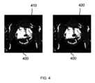
図4は、予測アブレーションゾーンと実際のアブレーションゾーンとの間のレジストレーションを示すために使用される。明確にするために、これらの例示的なゾーンは、2つのゾーン間の「現実の」相関を実証するために、例示的なMRIゾーンの上にスーパーインポーズされる。Figure 4 is used to show the registration between predicted and actual ablation zones. For clarity, these example zones are superimposed over the example MRI zones to demonstrate the "real" correlation between the two zones.
図4は、(アブレーション処置が行われた後に得られた)例示的なMRI画像400を示す。周囲の充血を伴う境界明瞭なアブレーション欠損がある。Figure 4 shows an example MRI image 400 (obtained after an ablation procedure has been performed). There is a well-defined ablation defect with surrounding hyperemia.
第1の例では、実際のアブレーションゾーン410のみが(点線形式で)図示/輪郭描出される。このアブレーションゾーンは、セグメンテーションプロセスを使用して生成され、MRI画像とレジストレーション/アラインされる。In the first example, only the
第2の例では、予測アブレーションゾーン420も示されている(実際のアブレーションゾーン(並びにMRI画像)に対してレジストレーションされている)。2つのアブレーションゾーンが互いに対して(空間的に)レジストレーションされるので、予測アブレーションゾーンの精度は、容易に識別され、手動で、又は自動的に補正されることができる。In the second example, the predicted
従って、図4は、本発明の使用及び目的、すなわち、予測アブレーションゾーンの精度が修正可能になるように、予測アブレーションゾーンと実際のアブレーションゾーンとを互いにレジストレーションすることを示す。FIG. 4 thus illustrates the use and purpose of the present invention, namely, to register the predicted ablation zone and the actual ablation zone with each other so that the accuracy of the predicted ablation zone can be corrected.
一般的に言えば、実施形態は、予測アブレーションゾーンと達成されたアブレーションとを互いにレジストレーションすることによって、予測アブレーションゾーンの精度を評価し、任意には改善する概念に向けられている。Generally speaking, embodiments are directed to the concept of evaluating and optionally improving the accuracy of a predicted ablation zone by registering the predicted ablation zone and the achieved ablation with respect to each other.
しかしながら、いくつかの実施形態では、計画されたアブレーションゾーンを使用することも可能である。計画されたアブレーションゾーンは、アブレーションのための所望の領域、例えば、腫瘍の領域を規定することがで、アブレーション計画を生成するために使用されることができる。However, in some embodiments, it is also possible to use planned ablation zones. Planned ablation zones can define desired regions for ablation, e.g., regions of tumors, and can be used to generate an ablation plan.
実施形態は、計画されたアブレーションゾーンを予測された/達成されたアブレーションゾーンに対しレジストレーションして、計画されたアブレーションゾーンに対する全アブレーション処置の精度を決定することを更に含むことができる。この情報は、アブレーション処置の計画を改善するために、及び/又はアブレーションアプリケータの配置における誤差を識別するために使用されることができる。Embodiments may further include registering the planned ablation zone to the predicted/achieved ablation zone to determine the accuracy of the entire ablation procedure relative to the planned ablation zone. This information may be used to improve the planning of the ablation procedure and/or to identify errors in the placement of the ablation applicator.
例として、予測アブレーションゾーンと達成されたアブレーションゾーンとの間にわずかな誤差しかないが、計画されたアブレーションゾーンと達成されたアブレーションゾーンとの間に大きな誤差がある場合、これは、アブレーション処置中のアブレーションアプリケータの配置に誤差があったことを明確に示す。As an example, if there is only a small error between the predicted ablation zone and the achieved ablation zone, but a large error between the planned ablation zone and the achieved ablation zone, this is a clear indication that there was an error in the placement of the ablation applicator during the ablation procedure.
この情報は、例えば、アブレーション計画に従って到達されなかった達成されたアブレーションゾーンの領域を識別することによって、アブレーション計画を改善するために使用されることができる。This information can be used to improve the ablation plan, for example, by identifying areas of the achieved ablation zone that were not reached according to the ablation plan.
当業者は、本明細書に記載の如何なる方法も実行するための処理システムを開発することが容易にできる。従って、フローチャートの各ステップは、処理システムにより実行される異なる動作を表し、前記処理システムの個々のモジュールにより行われることができる。A person skilled in the art can readily develop a processing system to perform any of the methods described herein. Thus, each step of the flow chart represents a different operation performed by a processing system and may be performed by individual modules of the processing system.
従って、実施形態は、処理システムを利用することができる。前記処理システムは、必要とされる様々な機能を行うために、ソフトウェア及び/又はハードウェアを用いて、様々な方法で実施される。プロセッサは、前記必要とされる機能を行うために、ソフトウェア(例えば、マイクロコード)を用いてプログラムされる1又は複数のマイクロプロセッサを用いる処理システムの一例である。しかしながら、処理システムは、プロセッサを使用しても又はしなくても実施されることができ、幾つかの機能を行うための専用ハードウェアと、他の機能を行うためのプロセッサ(1又は複数のプログラムされるマイクロプロセッサ及び関連する回路)との組み合わせとして実施されてもよい。Thus, embodiments may utilize a processing system. The processing system may be implemented in a variety of ways using software and/or hardware to perform the various functions required. A processor is one example of a processing system that uses one or more microprocessors that are programmed with software (e.g., microcode) to perform the functions required. However, a processing system may be implemented with or without a processor, and may be implemented as a combination of dedicated hardware to perform some functions and a processor (one or more programmed microprocessors and associated circuitry) to perform other functions.
本開示の種々の実施形態に用いられる処理システムのコンポーネントの例は、これらに限定されないが、従来のマイクロプロセッサ、特定用途向け集積回路(ASIC)及びFPGA(field-programmable gate array)を含む。Examples of processing system components that may be used in various embodiments of the present disclosure include, but are not limited to, conventional microprocessors, application specific integrated circuits (ASICs), and field-programmable gate arrays (FPGAs).
様々な実施において、プロセッサ又は処理システムは、例えば揮発性及び不揮発性のコンピュータメモリの1又は複数の記憶媒体、例えばRAM、PROM、EPROM及びEEPROMに関連付けられてもよい。前記記憶媒体は、1又は複数のプロセッサ及び/又は処理システム上で実行されるとき、前記必要とされる機能を行う1又は複数のプログラムで符号化されてもよい。様々な記憶媒体は、プロセッサ或いは処理システム内に固定されていてもよいし、又は運搬可能であっても良く、それにより、記憶媒体に記憶された1又は複数のプログラムがプロセッサ又は処理システムにロードされることができる。In various implementations, the processor or processing system may be associated with one or more storage media, e.g., volatile and non-volatile computer memory, e.g., RAM, PROM, EPROM, and EEPROM. The storage media may be encoded with one or more programs that, when executed on the one or more processors and/or processing systems, perform the required functions. The various storage media may be fixed within the processor or processing system or may be portable, such that one or more programs stored on the storage media can be loaded into the processor or processing system.
開示される方法は、好ましくは、コンピュータで実現される方法であることが理解される。そのようなものとして、そのプログラムが処理システム、例えば、コンピュータ上で実行されるとき、記載された如何なる方法も実施するためのコード手段を有するコンピュータプログラムの概念も提案される。従って、一実施形態によるコンピュータプログラムのコードの異なる部分、ライン又はブロックは、本明細書に記載される任意の方法を行うために、処理システム又はコンピュータにより実行されることができる。幾つかの代替の実現形態において、ブロック図又はフローチャートに述べられる機能が、図面に述べられる順序とは異なる順序で行われてもよい。例えば、連続して示される2つのブロックは、実際には実質的に同時に実行されてもよく、又は、ブロックは、含まれる機能に応じて、逆の順序で実行されてもよい。It is understood that the disclosed methods are preferably computer-implemented methods. As such, the concept of a computer program having code means for performing any of the described methods when the program is executed on a processing system, e.g., a computer, is also proposed. Thus, different parts, lines or blocks of code of a computer program according to an embodiment can be executed by a processing system or a computer to perform any of the methods described herein. In some alternative implementations, the functions depicted in the block diagrams or flow charts may be performed in an order different from that depicted in the figures. For example, two blocks shown in succession may in fact be executed substantially simultaneously, or the blocks may be executed in the reverse order, depending on the functionality involved.
開示された実施形態に対する変形例は、図面、開示、及び添付の特許請求の範囲の検討から、特許請求された発明を実施する際に当業者によって理解され、実施されることができる。請求項において、単語「有する、含む(comprising)」は、他の構成要素又はステップを排除するものではなく、不定冠詞「a」又は「an」は、複数性を排除するものではない。単一のプロセッサ又は他のユニットが、請求項に列挙されるいくつかの項目の機能を果たすことができる。特定の手段が相互に異なる従属請求項に記載されているという単なる事実は、これらの手段の組み合わせが有利に使用されることができないことを示すものではない。コンピュータプログラムが上述される場合、コンピュータプログラムは、適切な媒体、例えば他のハードウェアと一緒にあるいはその一部として供給される光記憶媒体若しくはソリッドステート媒体に記憶又は配布されることができるが、他の形態、例えばインターネット又は他の有線若しくは無線電気通信システムを介して配布されてもよい。「~するように適応される(adapted to)」という用語が請求項又は明細書に用いられる場合、「~するように適応される(adapted to)」という用語は、「~するように構成される(configured to)」と言う用語と同様であることを意味する。請求項におけるいかなる参照符号も、その範囲を限定するものとして解釈されるべきではない。Variations to the disclosed embodiments can be understood and implemented by those skilled in the art in practicing the claimed invention, from a study of the drawings, the disclosure, and the appended claims. In the claims, the word "comprising" does not exclude other elements or steps, and the indefinite article "a" or "an" does not exclude a plurality. A single processor or other unit may fulfill the functions of several items recited in the claims. The mere fact that certain means are recited in mutually different dependent claims does not indicate that a combination of these means cannot be used to advantage. Where a computer program is described above, the computer program can be stored or distributed on a suitable medium, for example an optical storage medium or a solid-state medium supplied together with or as part of other hardware, but may also be distributed in other forms, for example via the Internet or other wired or wireless telecommunications systems. When the term "adapted to" is used in the claims or the description, the term "adapted to" is meant to be equivalent to the term "configured to". Any reference signs in the claims should not be construed as limiting the scope.
Claims (14)
Translated fromJapaneseアブレーション処置中にアブレーションシステムによって生成される処置中アブレーションデータを取得するステップであって、前記処置中アブレーションデータは、被検体に実行されるアブレーション処置に関する情報を提供し、前記処置中アブレーションデータは、前記アブレーション処置中の1又は複数のアブレーションアプリケータの位置及び/又は向き及び/又はタイプを識別するアプリケータ情報を含む、ステップと、
前記アプリケータ情報を有する前記処置中アブレーションデータを処理して、前記アブレーション処置によって生成されるアブレーションゾーンを予測する予測アブレーションゾーンを構築するステップと、
前記アブレーション処置が被検体に行われた後に生成される処置後アブレーションデータを取得するステップであって、前記処置後アブレーションデータは、前記被検体の達成されたアブレーションゾーンに関する情報を提供する、ステップと、
前記処置後アブレーションデータを処理して、達成されたアブレーションゾーンを構築するステップと、
前記予測アブレーションゾーンと前記達成されたアブレーションゾーンとを互いに対してレジストレーションするステップと、
前記予測アブレーションゾーンと前記達成されたアブレーションゾーンとの間の誤差を決定するステップであって、それにより前記予測アブレーションゾーンの精度の評価を提供する、ステップと、
前記予測アブレーションゾーンの精度の前記評価を使用して、前記予測アブレーションゾーンを生成するために使用された前記アプリケータ情報を較正するステップと、
を有し、
前記予測アブレーションゾーンは、複数の個々のアブレーションゾーンを組み合わせることによって構築され、
前記アプリケータ情報は、各アブレーションゾーンのデフォルトサイズ及び/又は寸法をさらに有し、
前記予測アブレーションゾーンの精度の前記評価を使用して、前記アプリケータ情報を較正するステップは、前記決定された誤差に基づいて、各個々のアブレーションゾーンの前記デフォルトサイズ及び/又は寸法を変更するステップ、を有する、
コンピュータ実現方法。 1. A computer-implemented method for providing an assessment of accuracy of a predicted ablation zone obtained from in-procedure data generated by an ablation system during an ablation procedure,the computer-implemented method comprising:
acquiring in-procedure ablation data generated by an ablation system during an ablation procedure, said in-procedure ablation data providing information regarding an ablation procedure being performed on a subject, said in-procedure ablation data including applicator information identifying a position and/or orientation and/or type of one or more ablation applicators during said ablation procedure;
processing the in-procedure ablation data with the applicator information to construct a predicted ablation zone that predicts an ablation zone that will be created by the ablation procedure;
acquiring post-treatment ablation data generated after the ablation procedure is performed on the subject, the post-treatment ablation data providing information regarding an achieved ablation zone in the subject;
processing the post-treatment ablation data to construct an achieved ablation zone;
registering the predicted ablation zone and the achieved ablation zone to each other;
determining an error between the predicted ablation zone and the achieved ablation zone, thereby providing an assessment of accuracy of the predicted ablation zone;
using the assessment of accuracy of the predicted ablation zone to calibrate the applicator information used to generate the predicted ablation zone;
having
the predicted ablation zone is constructed by combining a plurality of individual ablation zones;
the applicator information further comprising a default size and/or dimensions for each ablation zone;
calibrating the applicator information using the assessment of accuracy of the predicted ablation zones comprises modifying the default size and/or dimensions of each individual ablation zone based on the determined error.
A computer-implemented method.
前記予測アブレーションゾーンを構築する前記ステップは、前記アプリケータ情報及び1又は複数のアブレーションアプリケータ設定を処理して前記予測アブレーションゾーンを構築するステップを有し、前記1又は複数のアブレーションアプリケータ設定が、前記アプリケータ情報に基づいて、各アブレーションアプリケータによって実行される個々のアブレーションの周りの予測アブレーションゾーンを規定し、前記アブレーションシステムの1又は複数の設定を調整する前記ステップが、前記1又は複数のアブレーションアプリケータ設定の少なくとも1を調整することを含む、請求項3に記載のコンピュータ実現方法。 the in-procedure ablation data comprises applicator information identifying at least a location and type of one or more ablation applicators during the ablation procedure;
4. The computer-implemented method of claim 3, wherein constructing the predicted ablation zone comprises processing the applicator information and one or more ablation applicator settings to construct the predicted ablation zone, the one or more ablation applicator settings defining a predicted ablation zone around each ablation performed by each ablation applicator based on the applicator information, and wherein adjusting one or more settings of the ablation system comprises adjusting at least one of theone or more ablation applicator settings.
前記アブレーション処置が被検体に行われる前に生成された処置前アブレーションデータを取得するステップと、
前記処置中アブレーションデータを前記処置前アブレーションデータに対してレジストレーションするステップと、
前記処置後アブレーションデータを前記処置前アブレーションデータに対してレジストレーションするステップであって、それにより前記処置中アブレーションデータを前記処置後アブレーションデータに間接的にレジストレーションする、ステップと、
を有する、請求項7に記載のコンピュータ実現方法。 the step of registering the in-treatment ablation data and the post-treatment ablation data to each other comprises:
acquiring pre-treatment ablation data generated before the ablation procedure is performed on a subject ;
registering the in-treatment ablation datatothe pre-treatment ablation data;
registering the post-treatment ablation data tothe pre-treatment ablation data, thereby indirectly registering the during-treatment ablation data to the post-treatment ablation data;
8. The computer-implemented method of claim7 , comprising:
前記アブレーション処置前に取り込まれた前記非検体の医用画像を取得するステップと、
前記アブレーション処置中に取り込まれた前記医用画像の少なくとも1つを前記アブレーション処置前に取り込まれた前記医用画像に対してレジストレーションするステップと、
前記アブレーション処置後に取り込まれた前記医用画像の少なくとも1つを前記アブレーション処置前に取り込まれた前記医用画像に対してレジストレーションするステップであって、それによって前記アブレーション処置中に取り込まれた前記医用画像の少なくとも1つを、前記アブレーション処置後に取り込まれた前記医用画像の少なくとも1つに対して間接的にレジストレーションする、ステップと、
を有する、請求項12に記載のコンピュータ実現方法。 The step of registering at least one of the medical images captured during the ablation procedure to at least one of the medical images captured after the ablation procedure comprises:
obtaining a medical imageof the non-specimen captured prior to the ablation procedure ;
registering at least one of the medical images captured during the ablation procedure to the medical imagescaptured before the ablation procedure ;
registering at least one of the medical images captured after the ablation procedure to the medical imagescaptured before the ablation procedure , thereby indirectly registering at least one of the medical images captured during the ablation procedure to at least one of the medical images captured after the ablation procedure;
13. The computer-implemented method of claim12 , comprising:
アブレーション治療計画システムであって、アブレーション処置中に前記アブレーションシステムによって生成される処置中アブレーションデータを取得するよう構成され、前記処置中アブレーションデータが、被検体に実行されるアブレーション処置に関する情報を提供し、前記処置中アブレーションデータが、前記アブレーション処置中の1又は複数のアブレーションアプリケータの位置及び/又は向き及び/又はタイプを識別するアプリケータ情報を含む、アブレーション治療計画システムと、
アブレーション治療フォローアップシステムであって、
前記アブレーション処置が被検体に行われた後に生成される処置後アブレーションデータを取得するステップであって、前記処置後アブレーションデータが、前記被検体の達成されたアブレーションゾーンに関する情報を提供する、ステップと、
前記処置後アブレーションデータを処理して達成されたアブレーションゾーンを構築するステップ、
前記アブレーション処置によって生成されるアブレーションゾーンを予測する予測アブレーションゾーンと前記達成されたアブレーションゾーンとを互いに対してレジストレーションするステップと、
前記予測アブレーションゾーンと前記達成されたアブレーションゾーンとの間の誤差を決定するステップであって、それによって前記予測アブレーションゾーンの精度の評価を提供する、ステップと、
を実行するよう構成される、アブレーション治療フォローアップシステムと、
を有し、前記アブレーション治療計画システム又は前記アブレーション治療フォローアップシステムのいずれかが、前記アプリケータ情報を含む前記処置中データを処理して前記予測アブレーションゾーンを構築するステップと、前記予測アブレーションゾーンの精度の前記評価を使用して、前記予測アブレーションゾーンを生成するために使用された前記アプリケータ情報を較正するステップと、
を実行するよう構成され、
前記予測アブレーションゾーンは、複数の個々のアブレーションゾーンを組み合わせることによって構築され、
前記アプリケータ情報は、各アブレーションゾーンのデフォルトサイズ及び/又は寸法をさらに有し、
前記予測アブレーションゾーンの精度の前記評価を使用して、前記アプリケータ情報を較正するステップは、前記決定された誤差に基づいて、各個々のアブレーションゾーンの前記デフォルトサイズ及び/又は寸法を変更するステップ、を有する、
アブレーションシステム。 1. An ablation system for providing an assessment of accuracy of a predicted ablation zone obtained from in-procedure data generated by the ablation system during an ablation procedure, the ablation system comprising:
an ablation treatment planning system configured to acquire in-procedure ablation data generated by the ablation system during an ablation procedure, the in-procedure ablation data providing information regarding an ablation procedure to be performed on a subject, the in-procedure ablation data including applicator information identifying a position and/or an orientation and/or a type of one or more ablation applicators during the ablation procedure;
1. An ablation treatment follow-up system, comprising:
acquiring post-treatment ablation data generated after the ablation procedure is performed on the subject, the post-treatment ablation data providing information regarding an achieved ablation zone in the subject;
processing the post-treatment ablation data to construct an achieved ablation zone;
registering a predicted ablation zone that will be produced by the ablation procedure and the achieved ablation zone with respect to each other;
determining an error between the predicted ablation zone and the achieved ablation zone, thereby providing an assessment of accuracy of the predicted ablation zone;
an ablation treatment follow-up system configured to perform
wherein either the ablation treatment planning system or the ablation treatment follow-up system processes the in-treatment data including the applicator information to construct the predicted ablation zone; and calibrating the applicator information used to generate the predicted ablation zone using the assessment of accuracy of the predicted ablation zone.
configured to execute
the predicted ablation zone is constructed by combining a plurality of individual ablation zones;
the applicator information further comprising a default size and/or dimensions for each ablation zone;
calibrating the applicator information using the assessment of accuracy of the predicted ablation zones comprises modifying the default size and/or dimensions of each individual ablation zone based on the determined error.
Ablation system.
Applications Claiming Priority (3)
| Application Number | Priority Date | Filing Date | Title |
|---|---|---|---|
| EP20167655.8AEP3889966A1 (en) | 2020-04-02 | 2020-04-02 | Ablation zone assessment and configuration |
| EP20167655.8 | 2020-04-02 | ||
| PCT/EP2021/057862WO2021198047A1 (en) | 2020-04-02 | 2021-03-26 | Ablation zone assessment and configuration |
Publications (3)
| Publication Number | Publication Date |
|---|---|
| JP2023519309A JP2023519309A (en) | 2023-05-10 |
| JPWO2021198047A5 JPWO2021198047A5 (en) | 2024-02-06 |
| JP7700799B2true JP7700799B2 (en) | 2025-07-01 |
Family
ID=70165789
Family Applications (1)
| Application Number | Title | Priority Date | Filing Date |
|---|---|---|---|
| JP2022558158AActiveJP7700799B2 (en) | 2020-04-02 | 2021-03-26 | Ablation Zone Assessment and Construction |
Country Status (5)
| Country | Link |
|---|---|
| US (1) | US20230172665A1 (en) |
| EP (2) | EP3889966A1 (en) |
| JP (1) | JP7700799B2 (en) |
| CN (1) | CN115380332A (en) |
| WO (1) | WO2021198047A1 (en) |
Citations (4)
| Publication number | Priority date | Publication date | Assignee | Title |
|---|---|---|---|---|
| JP2005199072A (en) | 2004-01-14 | 2005-07-28 | Biosense Webster Inc | Prediction and evaluation of cardiac tissue ablation |
| JP2011131061A (en) | 2009-12-23 | 2011-07-07 | Biosense Webster (Israel) Ltd | Estimation and mapping of ablation volume |
| JP2012510332A (en) | 2008-12-03 | 2012-05-10 | コーニンクレッカ フィリップス エレクトロニクス エヌ ヴィ | A feedback system that integrates interventional planning and navigation |
| JP2014507199A (en) | 2010-12-27 | 2014-03-27 | エンドーセンス エスアー | Prediction of atrial wall electrical reconnection based on contact force measured during RF ablation |
Family Cites Families (6)
| Publication number | Priority date | Publication date | Assignee | Title |
|---|---|---|---|---|
| US8267927B2 (en)* | 2007-01-24 | 2012-09-18 | Koninklijke Philips Electronics N.V. | Advanced ablation planning |
| US20090287066A1 (en)* | 2008-05-19 | 2009-11-19 | Oliver Meissner | Method for minimally invasive medical intervention |
| US20120277763A1 (en)* | 2009-12-30 | 2012-11-01 | Koninklijke Philips Electronics N.V. | Dynamic ablation device |
| CN103717167B (en)* | 2011-07-28 | 2017-06-27 | 皇家飞利浦有限公司 | Ablation planning system |
| CN103678837A (en)* | 2012-08-31 | 2014-03-26 | 西门子公司 | Method and device for determining processing remains of target area |
| US10402535B2 (en)* | 2014-02-26 | 2019-09-03 | Siemens Healthcare Gmbh | System and method for personalized computation of tissue ablation extent based on medical images |
- 2020
- 2020-04-02EPEP20167655.8Apatent/EP3889966A1/ennot_activeWithdrawn
- 2021
- 2021-03-26CNCN202180026916.2Apatent/CN115380332A/enactivePending
- 2021-03-26EPEP21713438.6Apatent/EP4128257A1/enactivePending
- 2021-03-26USUS17/916,150patent/US20230172665A1/enactivePending
- 2021-03-26JPJP2022558158Apatent/JP7700799B2/enactiveActive
- 2021-03-26WOPCT/EP2021/057862patent/WO2021198047A1/ennot_activeCeased
Patent Citations (4)
| Publication number | Priority date | Publication date | Assignee | Title |
|---|---|---|---|---|
| JP2005199072A (en) | 2004-01-14 | 2005-07-28 | Biosense Webster Inc | Prediction and evaluation of cardiac tissue ablation |
| JP2012510332A (en) | 2008-12-03 | 2012-05-10 | コーニンクレッカ フィリップス エレクトロニクス エヌ ヴィ | A feedback system that integrates interventional planning and navigation |
| JP2011131061A (en) | 2009-12-23 | 2011-07-07 | Biosense Webster (Israel) Ltd | Estimation and mapping of ablation volume |
| JP2014507199A (en) | 2010-12-27 | 2014-03-27 | エンドーセンス エスアー | Prediction of atrial wall electrical reconnection based on contact force measured during RF ablation |
Also Published As
| Publication number | Publication date |
|---|---|
| WO2021198047A1 (en) | 2021-10-07 |
| EP3889966A1 (en) | 2021-10-06 |
| US20230172665A1 (en) | 2023-06-08 |
| EP4128257A1 (en) | 2023-02-08 |
| JP2023519309A (en) | 2023-05-10 |
| CN115380332A (en) | 2022-11-22 |
Similar Documents
| Publication | Publication Date | Title |
|---|---|---|
| CN111248998B (en) | System and method for ultrasound image guided ablation antenna placement | |
| JP5685546B2 (en) | A feedback system that integrates interventional planning and navigation | |
| EP2640292B1 (en) | System for probabilistic ablation planning | |
| JP6208535B2 (en) | Radiotherapy apparatus and system and method | |
| CN102781356A (en) | Dynamic ablation device | |
| EP2844342A2 (en) | Videographic display of real-time medical treatment | |
| JP2009545394A (en) | Biologically guided adaptive treatment planning | |
| US11938344B2 (en) | Beam path based patient positioning and monitoring | |
| JP7420746B2 (en) | Assisting in positioning thermal ablation devices | |
| JP6676780B2 (en) | Provide image guided therapy | |
| US9014454B2 (en) | Method and apparatus pertaining to images used for radiation-treatment planning | |
| CN108778421B (en) | System for assisting in performing an interventional procedure | |
| JP7700799B2 (en) | Ablation Zone Assessment and Construction | |
| WO2017188345A1 (en) | Image processing device and image processing method | |
| WO2020064659A1 (en) | Ablation therapy planning system | |
| JP2021505330A (en) | Automatic excision antenna segmentation from CT images | |
| US20230285095A1 (en) | System and method for independently positioning an ablation tool and imaging device | |
| AU2018219996B2 (en) | Videographic display of real-time medical treatment | |
| CN118787452B (en) | A surgical needle guiding device based on image projection | |
| CN120053065A (en) | Puncture planning system and intraoperative puncture planning and ablation information display method | |
| AU2013257386B2 (en) | Videographic display of real-time medical treatment | |
| CN120204637A (en) | Positioning method, device, computer equipment and storage medium | |
| CN119970220A (en) | A tumor ablation decision-making method and system driven by CT big data |
Legal Events
| Date | Code | Title | Description |
|---|---|---|---|
| A521 | Request for written amendment filed | Free format text:JAPANESE INTERMEDIATE CODE: A523 Effective date:20240129 | |
| A621 | Written request for application examination | Free format text:JAPANESE INTERMEDIATE CODE: A621 Effective date:20240129 | |
| A131 | Notification of reasons for refusal | Free format text:JAPANESE INTERMEDIATE CODE: A131 Effective date:20240910 | |
| A977 | Report on retrieval | Free format text:JAPANESE INTERMEDIATE CODE: A971007 Effective date:20240911 | |
| A601 | Written request for extension of time | Free format text:JAPANESE INTERMEDIATE CODE: A601 Effective date:20241122 | |
| A521 | Request for written amendment filed | Free format text:JAPANESE INTERMEDIATE CODE: A523 Effective date:20250306 | |
| TRDD | Decision of grant or rejection written | ||
| A01 | Written decision to grant a patent or to grant a registration (utility model) | Free format text:JAPANESE INTERMEDIATE CODE: A01 Effective date:20250520 | |
| A61 | First payment of annual fees (during grant procedure) | Free format text:JAPANESE INTERMEDIATE CODE: A61 Effective date:20250602 | |
| R150 | Certificate of patent or registration of utility model | Ref document number:7700799 Country of ref document:JP Free format text:JAPANESE INTERMEDIATE CODE: R150 |