JP7677909B2 - Intelligent artificial joint - Google Patents
Intelligent artificial jointDownload PDFInfo
- Publication number
- JP7677909B2 JP7677909B2JP2021572375AJP2021572375AJP7677909B2JP 7677909 B2JP7677909 B2JP 7677909B2JP 2021572375 AJP2021572375 AJP 2021572375AJP 2021572375 AJP2021572375 AJP 2021572375AJP 7677909 B2JP7677909 B2JP 7677909B2
- Authority
- JP
- Japan
- Prior art keywords
- data
- patient
- monitoring
- session
- prosthesis
- Prior art date
- Legal status (The legal status is an assumption and is not a legal conclusion. Google has not performed a legal analysis and makes no representation as to the accuracy of the status listed.)
- Active
Links
Images


Classifications
- A—HUMAN NECESSITIES
- A61—MEDICAL OR VETERINARY SCIENCE; HYGIENE
- A61F—FILTERS IMPLANTABLE INTO BLOOD VESSELS; PROSTHESES; DEVICES PROVIDING PATENCY TO, OR PREVENTING COLLAPSING OF, TUBULAR STRUCTURES OF THE BODY, e.g. STENTS; ORTHOPAEDIC, NURSING OR CONTRACEPTIVE DEVICES; FOMENTATION; TREATMENT OR PROTECTION OF EYES OR EARS; BANDAGES, DRESSINGS OR ABSORBENT PADS; FIRST-AID KITS
- A61F2/00—Filters implantable into blood vessels; Prostheses, i.e. artificial substitutes or replacements for parts of the body; Appliances for connecting them with the body; Devices providing patency to, or preventing collapsing of, tubular structures of the body, e.g. stents
- A61F2/02—Prostheses implantable into the body
- A61F2/28—Bones
- A—HUMAN NECESSITIES
- A61—MEDICAL OR VETERINARY SCIENCE; HYGIENE
- A61F—FILTERS IMPLANTABLE INTO BLOOD VESSELS; PROSTHESES; DEVICES PROVIDING PATENCY TO, OR PREVENTING COLLAPSING OF, TUBULAR STRUCTURES OF THE BODY, e.g. STENTS; ORTHOPAEDIC, NURSING OR CONTRACEPTIVE DEVICES; FOMENTATION; TREATMENT OR PROTECTION OF EYES OR EARS; BANDAGES, DRESSINGS OR ABSORBENT PADS; FIRST-AID KITS
- A61F2/00—Filters implantable into blood vessels; Prostheses, i.e. artificial substitutes or replacements for parts of the body; Appliances for connecting them with the body; Devices providing patency to, or preventing collapsing of, tubular structures of the body, e.g. stents
- A61F2/02—Prostheses implantable into the body
- A61F2/30—Joints
- A61F2/46—Special tools for implanting artificial joints
- A61F2/468—Testing instruments for artificial joints
- A—HUMAN NECESSITIES
- A61—MEDICAL OR VETERINARY SCIENCE; HYGIENE
- A61B—DIAGNOSIS; SURGERY; IDENTIFICATION
- A61B5/00—Measuring for diagnostic purposes; Identification of persons
- A61B5/0002—Remote monitoring of patients using telemetry, e.g. transmission of vital signals via a communication network
- A61B5/0004—Remote monitoring of patients using telemetry, e.g. transmission of vital signals via a communication network characterised by the type of physiological signal transmitted
- A—HUMAN NECESSITIES
- A61—MEDICAL OR VETERINARY SCIENCE; HYGIENE
- A61B—DIAGNOSIS; SURGERY; IDENTIFICATION
- A61B5/00—Measuring for diagnostic purposes; Identification of persons
- A61B5/0002—Remote monitoring of patients using telemetry, e.g. transmission of vital signals via a communication network
- A61B5/0031—Implanted circuitry
- A—HUMAN NECESSITIES
- A61—MEDICAL OR VETERINARY SCIENCE; HYGIENE
- A61B—DIAGNOSIS; SURGERY; IDENTIFICATION
- A61B5/00—Measuring for diagnostic purposes; Identification of persons
- A61B5/07—Endoradiosondes
- A61B5/076—Permanent implantation
- A—HUMAN NECESSITIES
- A61—MEDICAL OR VETERINARY SCIENCE; HYGIENE
- A61B—DIAGNOSIS; SURGERY; IDENTIFICATION
- A61B5/00—Measuring for diagnostic purposes; Identification of persons
- A61B5/103—Measuring devices for testing the shape, pattern, colour, size or movement of the body or parts thereof, for diagnostic purposes
- A61B5/11—Measuring movement of the entire body or parts thereof, e.g. head or hand tremor or mobility of a limb
- A61B5/1113—Local tracking of patients, e.g. in a hospital or private home
- A61B5/1114—Tracking parts of the body
- A—HUMAN NECESSITIES
- A61—MEDICAL OR VETERINARY SCIENCE; HYGIENE
- A61B—DIAGNOSIS; SURGERY; IDENTIFICATION
- A61B5/00—Measuring for diagnostic purposes; Identification of persons
- A61B5/45—For evaluating or diagnosing the musculoskeletal system or teeth
- A61B5/4504—Bones
- A—HUMAN NECESSITIES
- A61—MEDICAL OR VETERINARY SCIENCE; HYGIENE
- A61B—DIAGNOSIS; SURGERY; IDENTIFICATION
- A61B5/00—Measuring for diagnostic purposes; Identification of persons
- A61B5/48—Other medical applications
- A61B5/4851—Prosthesis assessment or monitoring
- A—HUMAN NECESSITIES
- A61—MEDICAL OR VETERINARY SCIENCE; HYGIENE
- A61B—DIAGNOSIS; SURGERY; IDENTIFICATION
- A61B5/00—Measuring for diagnostic purposes; Identification of persons
- A61B5/68—Arrangements of detecting, measuring or recording means, e.g. sensors, in relation to patient
- A61B5/6846—Arrangements of detecting, measuring or recording means, e.g. sensors, in relation to patient specially adapted to be brought in contact with an internal body part, i.e. invasive
- A61B5/6847—Arrangements of detecting, measuring or recording means, e.g. sensors, in relation to patient specially adapted to be brought in contact with an internal body part, i.e. invasive mounted on an invasive device
- A61B5/686—Permanently implanted devices, e.g. pacemakers, other stimulators, biochips
- A—HUMAN NECESSITIES
- A61—MEDICAL OR VETERINARY SCIENCE; HYGIENE
- A61B—DIAGNOSIS; SURGERY; IDENTIFICATION
- A61B5/00—Measuring for diagnostic purposes; Identification of persons
- A61B5/68—Arrangements of detecting, measuring or recording means, e.g. sensors, in relation to patient
- A61B5/6846—Arrangements of detecting, measuring or recording means, e.g. sensors, in relation to patient specially adapted to be brought in contact with an internal body part, i.e. invasive
- A61B5/6867—Arrangements of detecting, measuring or recording means, e.g. sensors, in relation to patient specially adapted to be brought in contact with an internal body part, i.e. invasive specially adapted to be attached or implanted in a specific body part
- A61B5/6878—Bone
- A—HUMAN NECESSITIES
- A61—MEDICAL OR VETERINARY SCIENCE; HYGIENE
- A61B—DIAGNOSIS; SURGERY; IDENTIFICATION
- A61B5/00—Measuring for diagnostic purposes; Identification of persons
- A61B5/74—Details of notification to user or communication with user or patient; User input means
- A61B5/7465—Arrangements for interactive communication between patient and care services, e.g. by using a telephone network
- A—HUMAN NECESSITIES
- A61—MEDICAL OR VETERINARY SCIENCE; HYGIENE
- A61F—FILTERS IMPLANTABLE INTO BLOOD VESSELS; PROSTHESES; DEVICES PROVIDING PATENCY TO, OR PREVENTING COLLAPSING OF, TUBULAR STRUCTURES OF THE BODY, e.g. STENTS; ORTHOPAEDIC, NURSING OR CONTRACEPTIVE DEVICES; FOMENTATION; TREATMENT OR PROTECTION OF EYES OR EARS; BANDAGES, DRESSINGS OR ABSORBENT PADS; FIRST-AID KITS
- A61F2/00—Filters implantable into blood vessels; Prostheses, i.e. artificial substitutes or replacements for parts of the body; Appliances for connecting them with the body; Devices providing patency to, or preventing collapsing of, tubular structures of the body, e.g. stents
- A61F2/02—Prostheses implantable into the body
- A61F2/30—Joints
- A61F2/30721—Accessories
- A—HUMAN NECESSITIES
- A61—MEDICAL OR VETERINARY SCIENCE; HYGIENE
- A61F—FILTERS IMPLANTABLE INTO BLOOD VESSELS; PROSTHESES; DEVICES PROVIDING PATENCY TO, OR PREVENTING COLLAPSING OF, TUBULAR STRUCTURES OF THE BODY, e.g. STENTS; ORTHOPAEDIC, NURSING OR CONTRACEPTIVE DEVICES; FOMENTATION; TREATMENT OR PROTECTION OF EYES OR EARS; BANDAGES, DRESSINGS OR ABSORBENT PADS; FIRST-AID KITS
- A61F2/00—Filters implantable into blood vessels; Prostheses, i.e. artificial substitutes or replacements for parts of the body; Appliances for connecting them with the body; Devices providing patency to, or preventing collapsing of, tubular structures of the body, e.g. stents
- A61F2/02—Prostheses implantable into the body
- A61F2/30—Joints
- A61F2/32—Joints for the hip
- A61F2/36—Femoral heads ; Femoral endoprostheses
- A—HUMAN NECESSITIES
- A61—MEDICAL OR VETERINARY SCIENCE; HYGIENE
- A61F—FILTERS IMPLANTABLE INTO BLOOD VESSELS; PROSTHESES; DEVICES PROVIDING PATENCY TO, OR PREVENTING COLLAPSING OF, TUBULAR STRUCTURES OF THE BODY, e.g. STENTS; ORTHOPAEDIC, NURSING OR CONTRACEPTIVE DEVICES; FOMENTATION; TREATMENT OR PROTECTION OF EYES OR EARS; BANDAGES, DRESSINGS OR ABSORBENT PADS; FIRST-AID KITS
- A61F2/00—Filters implantable into blood vessels; Prostheses, i.e. artificial substitutes or replacements for parts of the body; Appliances for connecting them with the body; Devices providing patency to, or preventing collapsing of, tubular structures of the body, e.g. stents
- A61F2/02—Prostheses implantable into the body
- A61F2/30—Joints
- A61F2/38—Joints for elbows or knees
- A61F2/389—Tibial components
- A—HUMAN NECESSITIES
- A61—MEDICAL OR VETERINARY SCIENCE; HYGIENE
- A61F—FILTERS IMPLANTABLE INTO BLOOD VESSELS; PROSTHESES; DEVICES PROVIDING PATENCY TO, OR PREVENTING COLLAPSING OF, TUBULAR STRUCTURES OF THE BODY, e.g. STENTS; ORTHOPAEDIC, NURSING OR CONTRACEPTIVE DEVICES; FOMENTATION; TREATMENT OR PROTECTION OF EYES OR EARS; BANDAGES, DRESSINGS OR ABSORBENT PADS; FIRST-AID KITS
- A61F2/00—Filters implantable into blood vessels; Prostheses, i.e. artificial substitutes or replacements for parts of the body; Appliances for connecting them with the body; Devices providing patency to, or preventing collapsing of, tubular structures of the body, e.g. stents
- A61F2/02—Prostheses implantable into the body
- A61F2/48—Operating or control means, e.g. from outside the body, control of sphincters
- A61F2/482—Electrical means
- G—PHYSICS
- G06—COMPUTING OR CALCULATING; COUNTING
- G06Q—INFORMATION AND COMMUNICATION TECHNOLOGY [ICT] SPECIALLY ADAPTED FOR ADMINISTRATIVE, COMMERCIAL, FINANCIAL, MANAGERIAL OR SUPERVISORY PURPOSES; SYSTEMS OR METHODS SPECIALLY ADAPTED FOR ADMINISTRATIVE, COMMERCIAL, FINANCIAL, MANAGERIAL OR SUPERVISORY PURPOSES, NOT OTHERWISE PROVIDED FOR
- G06Q40/00—Finance; Insurance; Tax strategies; Processing of corporate or income taxes
- G06Q40/08—Insurance
- G—PHYSICS
- G16—INFORMATION AND COMMUNICATION TECHNOLOGY [ICT] SPECIALLY ADAPTED FOR SPECIFIC APPLICATION FIELDS
- G16H—HEALTHCARE INFORMATICS, i.e. INFORMATION AND COMMUNICATION TECHNOLOGY [ICT] SPECIALLY ADAPTED FOR THE HANDLING OR PROCESSING OF MEDICAL OR HEALTHCARE DATA
- G16H20/00—ICT specially adapted for therapies or health-improving plans, e.g. for handling prescriptions, for steering therapy or for monitoring patient compliance
- G16H20/10—ICT specially adapted for therapies or health-improving plans, e.g. for handling prescriptions, for steering therapy or for monitoring patient compliance relating to drugs or medications, e.g. for ensuring correct administration to patients
- G—PHYSICS
- G16—INFORMATION AND COMMUNICATION TECHNOLOGY [ICT] SPECIALLY ADAPTED FOR SPECIFIC APPLICATION FIELDS
- G16H—HEALTHCARE INFORMATICS, i.e. INFORMATION AND COMMUNICATION TECHNOLOGY [ICT] SPECIALLY ADAPTED FOR THE HANDLING OR PROCESSING OF MEDICAL OR HEALTHCARE DATA
- G16H20/00—ICT specially adapted for therapies or health-improving plans, e.g. for handling prescriptions, for steering therapy or for monitoring patient compliance
- G16H20/30—ICT specially adapted for therapies or health-improving plans, e.g. for handling prescriptions, for steering therapy or for monitoring patient compliance relating to physical therapies or activities, e.g. physiotherapy, acupressure or exercising
- G—PHYSICS
- G16—INFORMATION AND COMMUNICATION TECHNOLOGY [ICT] SPECIALLY ADAPTED FOR SPECIFIC APPLICATION FIELDS
- G16H—HEALTHCARE INFORMATICS, i.e. INFORMATION AND COMMUNICATION TECHNOLOGY [ICT] SPECIALLY ADAPTED FOR THE HANDLING OR PROCESSING OF MEDICAL OR HEALTHCARE DATA
- G16H20/00—ICT specially adapted for therapies or health-improving plans, e.g. for handling prescriptions, for steering therapy or for monitoring patient compliance
- G16H20/40—ICT specially adapted for therapies or health-improving plans, e.g. for handling prescriptions, for steering therapy or for monitoring patient compliance relating to mechanical, radiation or invasive therapies, e.g. surgery, laser therapy, dialysis or acupuncture
- G—PHYSICS
- G16—INFORMATION AND COMMUNICATION TECHNOLOGY [ICT] SPECIALLY ADAPTED FOR SPECIFIC APPLICATION FIELDS
- G16H—HEALTHCARE INFORMATICS, i.e. INFORMATION AND COMMUNICATION TECHNOLOGY [ICT] SPECIALLY ADAPTED FOR THE HANDLING OR PROCESSING OF MEDICAL OR HEALTHCARE DATA
- G16H40/00—ICT specially adapted for the management or administration of healthcare resources or facilities; ICT specially adapted for the management or operation of medical equipment or devices
- G16H40/60—ICT specially adapted for the management or administration of healthcare resources or facilities; ICT specially adapted for the management or operation of medical equipment or devices for the operation of medical equipment or devices
- G16H40/67—ICT specially adapted for the management or administration of healthcare resources or facilities; ICT specially adapted for the management or operation of medical equipment or devices for the operation of medical equipment or devices for remote operation
- H—ELECTRICITY
- H04—ELECTRIC COMMUNICATION TECHNIQUE
- H04W—WIRELESS COMMUNICATION NETWORKS
- H04W12/00—Security arrangements; Authentication; Protecting privacy or anonymity
- H04W12/03—Protecting confidentiality, e.g. by encryption
- H04W12/037—Protecting confidentiality, e.g. by encryption of the control plane, e.g. signalling traffic
- H—ELECTRICITY
- H04—ELECTRIC COMMUNICATION TECHNIQUE
- H04W—WIRELESS COMMUNICATION NETWORKS
- H04W84/00—Network topologies
- H04W84/18—Self-organising networks, e.g. ad-hoc networks or sensor networks
- H—ELECTRICITY
- H04—ELECTRIC COMMUNICATION TECHNIQUE
- H04W—WIRELESS COMMUNICATION NETWORKS
- H04W88/00—Devices specially adapted for wireless communication networks, e.g. terminals, base stations or access point devices
- H04W88/08—Access point devices
- A—HUMAN NECESSITIES
- A61—MEDICAL OR VETERINARY SCIENCE; HYGIENE
- A61B—DIAGNOSIS; SURGERY; IDENTIFICATION
- A61B2562/00—Details of sensors; Constructional details of sensor housings or probes; Accessories for sensors
- A61B2562/02—Details of sensors specially adapted for in-vivo measurements
- A61B2562/0219—Inertial sensors, e.g. accelerometers, gyroscopes, tilt switches
- A—HUMAN NECESSITIES
- A61—MEDICAL OR VETERINARY SCIENCE; HYGIENE
- A61B—DIAGNOSIS; SURGERY; IDENTIFICATION
- A61B5/00—Measuring for diagnostic purposes; Identification of persons
- A61B5/103—Measuring devices for testing the shape, pattern, colour, size or movement of the body or parts thereof, for diagnostic purposes
- A61B5/11—Measuring movement of the entire body or parts thereof, e.g. head or hand tremor or mobility of a limb
- A61B5/112—Gait analysis
- A—HUMAN NECESSITIES
- A61—MEDICAL OR VETERINARY SCIENCE; HYGIENE
- A61F—FILTERS IMPLANTABLE INTO BLOOD VESSELS; PROSTHESES; DEVICES PROVIDING PATENCY TO, OR PREVENTING COLLAPSING OF, TUBULAR STRUCTURES OF THE BODY, e.g. STENTS; ORTHOPAEDIC, NURSING OR CONTRACEPTIVE DEVICES; FOMENTATION; TREATMENT OR PROTECTION OF EYES OR EARS; BANDAGES, DRESSINGS OR ABSORBENT PADS; FIRST-AID KITS
- A61F2/00—Filters implantable into blood vessels; Prostheses, i.e. artificial substitutes or replacements for parts of the body; Appliances for connecting them with the body; Devices providing patency to, or preventing collapsing of, tubular structures of the body, e.g. stents
- A61F2/02—Prostheses implantable into the body
- A61F2/12—Mammary prostheses
- A—HUMAN NECESSITIES
- A61—MEDICAL OR VETERINARY SCIENCE; HYGIENE
- A61F—FILTERS IMPLANTABLE INTO BLOOD VESSELS; PROSTHESES; DEVICES PROVIDING PATENCY TO, OR PREVENTING COLLAPSING OF, TUBULAR STRUCTURES OF THE BODY, e.g. STENTS; ORTHOPAEDIC, NURSING OR CONTRACEPTIVE DEVICES; FOMENTATION; TREATMENT OR PROTECTION OF EYES OR EARS; BANDAGES, DRESSINGS OR ABSORBENT PADS; FIRST-AID KITS
- A61F2/00—Filters implantable into blood vessels; Prostheses, i.e. artificial substitutes or replacements for parts of the body; Appliances for connecting them with the body; Devices providing patency to, or preventing collapsing of, tubular structures of the body, e.g. stents
- A61F2/02—Prostheses implantable into the body
- A61F2/24—Heart valves ; Vascular valves, e.g. venous valves; Heart implants, e.g. passive devices for improving the function of the native valve or the heart muscle; Transmyocardial revascularisation [TMR] devices; Valves implantable in the body
- A—HUMAN NECESSITIES
- A61—MEDICAL OR VETERINARY SCIENCE; HYGIENE
- A61F—FILTERS IMPLANTABLE INTO BLOOD VESSELS; PROSTHESES; DEVICES PROVIDING PATENCY TO, OR PREVENTING COLLAPSING OF, TUBULAR STRUCTURES OF THE BODY, e.g. STENTS; ORTHOPAEDIC, NURSING OR CONTRACEPTIVE DEVICES; FOMENTATION; TREATMENT OR PROTECTION OF EYES OR EARS; BANDAGES, DRESSINGS OR ABSORBENT PADS; FIRST-AID KITS
- A61F2/00—Filters implantable into blood vessels; Prostheses, i.e. artificial substitutes or replacements for parts of the body; Appliances for connecting them with the body; Devices providing patency to, or preventing collapsing of, tubular structures of the body, e.g. stents
- A61F2/02—Prostheses implantable into the body
- A61F2/30—Joints
- A61F2/3094—Designing or manufacturing processes
- A61F2/30942—Designing or manufacturing processes for designing or making customized prostheses, e.g. using templates, CT or NMR scans, finite-element analysis or CAD-CAM techniques
- A—HUMAN NECESSITIES
- A61—MEDICAL OR VETERINARY SCIENCE; HYGIENE
- A61F—FILTERS IMPLANTABLE INTO BLOOD VESSELS; PROSTHESES; DEVICES PROVIDING PATENCY TO, OR PREVENTING COLLAPSING OF, TUBULAR STRUCTURES OF THE BODY, e.g. STENTS; ORTHOPAEDIC, NURSING OR CONTRACEPTIVE DEVICES; FOMENTATION; TREATMENT OR PROTECTION OF EYES OR EARS; BANDAGES, DRESSINGS OR ABSORBENT PADS; FIRST-AID KITS
- A61F2/00—Filters implantable into blood vessels; Prostheses, i.e. artificial substitutes or replacements for parts of the body; Appliances for connecting them with the body; Devices providing patency to, or preventing collapsing of, tubular structures of the body, e.g. stents
- A61F2/02—Prostheses implantable into the body
- A61F2/30—Joints
- A61F2/32—Joints for the hip
- A—HUMAN NECESSITIES
- A61—MEDICAL OR VETERINARY SCIENCE; HYGIENE
- A61F—FILTERS IMPLANTABLE INTO BLOOD VESSELS; PROSTHESES; DEVICES PROVIDING PATENCY TO, OR PREVENTING COLLAPSING OF, TUBULAR STRUCTURES OF THE BODY, e.g. STENTS; ORTHOPAEDIC, NURSING OR CONTRACEPTIVE DEVICES; FOMENTATION; TREATMENT OR PROTECTION OF EYES OR EARS; BANDAGES, DRESSINGS OR ABSORBENT PADS; FIRST-AID KITS
- A61F2/00—Filters implantable into blood vessels; Prostheses, i.e. artificial substitutes or replacements for parts of the body; Appliances for connecting them with the body; Devices providing patency to, or preventing collapsing of, tubular structures of the body, e.g. stents
- A61F2/02—Prostheses implantable into the body
- A61F2/30—Joints
- A61F2/40—Joints for shoulders
- A—HUMAN NECESSITIES
- A61—MEDICAL OR VETERINARY SCIENCE; HYGIENE
- A61F—FILTERS IMPLANTABLE INTO BLOOD VESSELS; PROSTHESES; DEVICES PROVIDING PATENCY TO, OR PREVENTING COLLAPSING OF, TUBULAR STRUCTURES OF THE BODY, e.g. STENTS; ORTHOPAEDIC, NURSING OR CONTRACEPTIVE DEVICES; FOMENTATION; TREATMENT OR PROTECTION OF EYES OR EARS; BANDAGES, DRESSINGS OR ABSORBENT PADS; FIRST-AID KITS
- A61F2/00—Filters implantable into blood vessels; Prostheses, i.e. artificial substitutes or replacements for parts of the body; Appliances for connecting them with the body; Devices providing patency to, or preventing collapsing of, tubular structures of the body, e.g. stents
- A61F2/02—Prostheses implantable into the body
- A61F2/28—Bones
- A61F2002/2892—Tibia
- A—HUMAN NECESSITIES
- A61—MEDICAL OR VETERINARY SCIENCE; HYGIENE
- A61F—FILTERS IMPLANTABLE INTO BLOOD VESSELS; PROSTHESES; DEVICES PROVIDING PATENCY TO, OR PREVENTING COLLAPSING OF, TUBULAR STRUCTURES OF THE BODY, e.g. STENTS; ORTHOPAEDIC, NURSING OR CONTRACEPTIVE DEVICES; FOMENTATION; TREATMENT OR PROTECTION OF EYES OR EARS; BANDAGES, DRESSINGS OR ABSORBENT PADS; FIRST-AID KITS
- A61F2/00—Filters implantable into blood vessels; Prostheses, i.e. artificial substitutes or replacements for parts of the body; Appliances for connecting them with the body; Devices providing patency to, or preventing collapsing of, tubular structures of the body, e.g. stents
- A61F2/02—Prostheses implantable into the body
- A61F2/30—Joints
- A61F2002/30001—Additional features of subject-matter classified in A61F2/28, A61F2/30 and subgroups thereof
- A61F2002/30316—The prosthesis having different structural features at different locations within the same prosthesis; Connections between prosthetic parts; Special structural features of bone or joint prostheses not otherwise provided for
- A61F2002/30317—The prosthesis having different structural features at different locations within the same prosthesis
- A61F2002/30324—The prosthesis having different structural features at different locations within the same prosthesis differing in thickness
- A—HUMAN NECESSITIES
- A61—MEDICAL OR VETERINARY SCIENCE; HYGIENE
- A61F—FILTERS IMPLANTABLE INTO BLOOD VESSELS; PROSTHESES; DEVICES PROVIDING PATENCY TO, OR PREVENTING COLLAPSING OF, TUBULAR STRUCTURES OF THE BODY, e.g. STENTS; ORTHOPAEDIC, NURSING OR CONTRACEPTIVE DEVICES; FOMENTATION; TREATMENT OR PROTECTION OF EYES OR EARS; BANDAGES, DRESSINGS OR ABSORBENT PADS; FIRST-AID KITS
- A61F2/00—Filters implantable into blood vessels; Prostheses, i.e. artificial substitutes or replacements for parts of the body; Appliances for connecting them with the body; Devices providing patency to, or preventing collapsing of, tubular structures of the body, e.g. stents
- A61F2/02—Prostheses implantable into the body
- A61F2/30—Joints
- A61F2002/30001—Additional features of subject-matter classified in A61F2/28, A61F2/30 and subgroups thereof
- A61F2002/30316—The prosthesis having different structural features at different locations within the same prosthesis; Connections between prosthetic parts; Special structural features of bone or joint prostheses not otherwise provided for
- A61F2002/30329—Connections or couplings between prosthetic parts, e.g. between modular parts; Connecting elements
- A61F2002/30331—Connections or couplings between prosthetic parts, e.g. between modular parts; Connecting elements made by longitudinally pushing a protrusion into a complementarily-shaped recess, e.g. held by friction fit
- A—HUMAN NECESSITIES
- A61—MEDICAL OR VETERINARY SCIENCE; HYGIENE
- A61F—FILTERS IMPLANTABLE INTO BLOOD VESSELS; PROSTHESES; DEVICES PROVIDING PATENCY TO, OR PREVENTING COLLAPSING OF, TUBULAR STRUCTURES OF THE BODY, e.g. STENTS; ORTHOPAEDIC, NURSING OR CONTRACEPTIVE DEVICES; FOMENTATION; TREATMENT OR PROTECTION OF EYES OR EARS; BANDAGES, DRESSINGS OR ABSORBENT PADS; FIRST-AID KITS
- A61F2/00—Filters implantable into blood vessels; Prostheses, i.e. artificial substitutes or replacements for parts of the body; Appliances for connecting them with the body; Devices providing patency to, or preventing collapsing of, tubular structures of the body, e.g. stents
- A61F2/02—Prostheses implantable into the body
- A61F2/30—Joints
- A61F2002/30001—Additional features of subject-matter classified in A61F2/28, A61F2/30 and subgroups thereof
- A61F2002/30316—The prosthesis having different structural features at different locations within the same prosthesis; Connections between prosthetic parts; Special structural features of bone or joint prostheses not otherwise provided for
- A61F2002/30329—Connections or couplings between prosthetic parts, e.g. between modular parts; Connecting elements
- A61F2002/30476—Connections or couplings between prosthetic parts, e.g. between modular parts; Connecting elements locked by an additional locking mechanism
- A61F2002/30507—Connections or couplings between prosthetic parts, e.g. between modular parts; Connecting elements locked by an additional locking mechanism using a threaded locking member, e.g. a locking screw or a set screw
- A—HUMAN NECESSITIES
- A61—MEDICAL OR VETERINARY SCIENCE; HYGIENE
- A61F—FILTERS IMPLANTABLE INTO BLOOD VESSELS; PROSTHESES; DEVICES PROVIDING PATENCY TO, OR PREVENTING COLLAPSING OF, TUBULAR STRUCTURES OF THE BODY, e.g. STENTS; ORTHOPAEDIC, NURSING OR CONTRACEPTIVE DEVICES; FOMENTATION; TREATMENT OR PROTECTION OF EYES OR EARS; BANDAGES, DRESSINGS OR ABSORBENT PADS; FIRST-AID KITS
- A61F2/00—Filters implantable into blood vessels; Prostheses, i.e. artificial substitutes or replacements for parts of the body; Appliances for connecting them with the body; Devices providing patency to, or preventing collapsing of, tubular structures of the body, e.g. stents
- A61F2/02—Prostheses implantable into the body
- A61F2/30—Joints
- A61F2002/30001—Additional features of subject-matter classified in A61F2/28, A61F2/30 and subgroups thereof
- A61F2002/30667—Features concerning an interaction with the environment or a particular use of the prosthesis
- A61F2002/30668—Means for transferring electromagnetic energy to implants
- A61F2002/3067—Means for transferring electromagnetic energy to implants for data transfer
- A—HUMAN NECESSITIES
- A61—MEDICAL OR VETERINARY SCIENCE; HYGIENE
- A61F—FILTERS IMPLANTABLE INTO BLOOD VESSELS; PROSTHESES; DEVICES PROVIDING PATENCY TO, OR PREVENTING COLLAPSING OF, TUBULAR STRUCTURES OF THE BODY, e.g. STENTS; ORTHOPAEDIC, NURSING OR CONTRACEPTIVE DEVICES; FOMENTATION; TREATMENT OR PROTECTION OF EYES OR EARS; BANDAGES, DRESSINGS OR ABSORBENT PADS; FIRST-AID KITS
- A61F2/00—Filters implantable into blood vessels; Prostheses, i.e. artificial substitutes or replacements for parts of the body; Appliances for connecting them with the body; Devices providing patency to, or preventing collapsing of, tubular structures of the body, e.g. stents
- A61F2/02—Prostheses implantable into the body
- A61F2/30—Joints
- A61F2/30767—Special external or bone-contacting surface, e.g. coating for improving bone ingrowth
- A61F2/30771—Special external or bone-contacting surface, e.g. coating for improving bone ingrowth applied in original prostheses, e.g. holes or grooves
- A61F2002/30878—Special external or bone-contacting surface, e.g. coating for improving bone ingrowth applied in original prostheses, e.g. holes or grooves with non-sharp protrusions, for instance contacting the bone for anchoring, e.g. keels, pegs, pins, posts, shanks, stems, struts
- A61F2002/30884—Fins or wings, e.g. longitudinal wings for preventing rotation within the bone cavity
- A—HUMAN NECESSITIES
- A61—MEDICAL OR VETERINARY SCIENCE; HYGIENE
- A61F—FILTERS IMPLANTABLE INTO BLOOD VESSELS; PROSTHESES; DEVICES PROVIDING PATENCY TO, OR PREVENTING COLLAPSING OF, TUBULAR STRUCTURES OF THE BODY, e.g. STENTS; ORTHOPAEDIC, NURSING OR CONTRACEPTIVE DEVICES; FOMENTATION; TREATMENT OR PROTECTION OF EYES OR EARS; BANDAGES, DRESSINGS OR ABSORBENT PADS; FIRST-AID KITS
- A61F2/00—Filters implantable into blood vessels; Prostheses, i.e. artificial substitutes or replacements for parts of the body; Appliances for connecting them with the body; Devices providing patency to, or preventing collapsing of, tubular structures of the body, e.g. stents
- A61F2/02—Prostheses implantable into the body
- A61F2/30—Joints
- A61F2/3094—Designing or manufacturing processes
- A61F2/30942—Designing or manufacturing processes for designing or making customized prostheses, e.g. using templates, CT or NMR scans, finite-element analysis or CAD-CAM techniques
- A61F2002/30957—Designing or manufacturing processes for designing or making customized prostheses, e.g. using templates, CT or NMR scans, finite-element analysis or CAD-CAM techniques using a positive or a negative model, e.g. moulds
- A—HUMAN NECESSITIES
- A61—MEDICAL OR VETERINARY SCIENCE; HYGIENE
- A61F—FILTERS IMPLANTABLE INTO BLOOD VESSELS; PROSTHESES; DEVICES PROVIDING PATENCY TO, OR PREVENTING COLLAPSING OF, TUBULAR STRUCTURES OF THE BODY, e.g. STENTS; ORTHOPAEDIC, NURSING OR CONTRACEPTIVE DEVICES; FOMENTATION; TREATMENT OR PROTECTION OF EYES OR EARS; BANDAGES, DRESSINGS OR ABSORBENT PADS; FIRST-AID KITS
- A61F2/00—Filters implantable into blood vessels; Prostheses, i.e. artificial substitutes or replacements for parts of the body; Appliances for connecting them with the body; Devices providing patency to, or preventing collapsing of, tubular structures of the body, e.g. stents
- A61F2/02—Prostheses implantable into the body
- A61F2/30—Joints
- A61F2/3094—Designing or manufacturing processes
- A61F2002/30985—Designing or manufacturing processes using three dimensional printing [3DP]
- A—HUMAN NECESSITIES
- A61—MEDICAL OR VETERINARY SCIENCE; HYGIENE
- A61F—FILTERS IMPLANTABLE INTO BLOOD VESSELS; PROSTHESES; DEVICES PROVIDING PATENCY TO, OR PREVENTING COLLAPSING OF, TUBULAR STRUCTURES OF THE BODY, e.g. STENTS; ORTHOPAEDIC, NURSING OR CONTRACEPTIVE DEVICES; FOMENTATION; TREATMENT OR PROTECTION OF EYES OR EARS; BANDAGES, DRESSINGS OR ABSORBENT PADS; FIRST-AID KITS
- A61F2250/00—Special features of prostheses classified in groups A61F2/00 - A61F2/26 or A61F2/82 or A61F9/00 or A61F11/00 or subgroups thereof
- A61F2250/0001—Means for transferring electromagnetic energy to implants
- A61F2250/0002—Means for transferring electromagnetic energy to implants for data transfer
- A—HUMAN NECESSITIES
- A61—MEDICAL OR VETERINARY SCIENCE; HYGIENE
- A61F—FILTERS IMPLANTABLE INTO BLOOD VESSELS; PROSTHESES; DEVICES PROVIDING PATENCY TO, OR PREVENTING COLLAPSING OF, TUBULAR STRUCTURES OF THE BODY, e.g. STENTS; ORTHOPAEDIC, NURSING OR CONTRACEPTIVE DEVICES; FOMENTATION; TREATMENT OR PROTECTION OF EYES OR EARS; BANDAGES, DRESSINGS OR ABSORBENT PADS; FIRST-AID KITS
- A61F2250/00—Special features of prostheses classified in groups A61F2/00 - A61F2/26 or A61F2/82 or A61F9/00 or A61F11/00 or subgroups thereof
- A61F2250/0058—Additional features; Implant or prostheses properties not otherwise provided for
- A61F2250/0096—Markers and sensors for detecting a position or changes of a position of an implant, e.g. RF sensors, ultrasound markers
- H—ELECTRICITY
- H04—ELECTRIC COMMUNICATION TECHNIQUE
- H04L—TRANSMISSION OF DIGITAL INFORMATION, e.g. TELEGRAPHIC COMMUNICATION
- H04L67/00—Network arrangements or protocols for supporting network services or applications
- H04L67/01—Protocols
- H04L67/12—Protocols specially adapted for proprietary or special-purpose networking environments, e.g. medical networks, sensor networks, networks in vehicles or remote metering networks
Landscapes
- Health & Medical Sciences (AREA)
- Life Sciences & Earth Sciences (AREA)
- Engineering & Computer Science (AREA)
- General Health & Medical Sciences (AREA)
- Public Health (AREA)
- Biomedical Technology (AREA)
- Heart & Thoracic Surgery (AREA)
- Veterinary Medicine (AREA)
- Animal Behavior & Ethology (AREA)
- Medical Informatics (AREA)
- Physics & Mathematics (AREA)
- Orthopedic Medicine & Surgery (AREA)
- Biophysics (AREA)
- Surgery (AREA)
- Oral & Maxillofacial Surgery (AREA)
- Pathology (AREA)
- Molecular Biology (AREA)
- Transplantation (AREA)
- Cardiology (AREA)
- Vascular Medicine (AREA)
- Computer Networks & Wireless Communication (AREA)
- Primary Health Care (AREA)
- Epidemiology (AREA)
- Business, Economics & Management (AREA)
- Physical Education & Sports Medicine (AREA)
- Signal Processing (AREA)
- Dentistry (AREA)
- Physiology (AREA)
- General Business, Economics & Management (AREA)
- Finance (AREA)
- Accounting & Taxation (AREA)
- Computer Security & Cryptography (AREA)
- Rheumatology (AREA)
- Medicinal Chemistry (AREA)
- Marketing (AREA)
- Bioinformatics & Cheminformatics (AREA)
- Theoretical Computer Science (AREA)
- General Physics & Mathematics (AREA)
- Urology & Nephrology (AREA)
- Nuclear Medicine, Radiotherapy & Molecular Imaging (AREA)
Description
Translated fromJapanese本発明は、一般に、センサを備えた医療デバイス、かかるデバイスを含むシステム、かかるデバイス及びシステム並びにこれらから生じたデータを用いる方法、及びセンサ付きの植え込まれた医療デバイスと関連した問題に取り組む装置及び方法に関する。The present invention relates generally to medical devices with sensors, systems including such devices, methods for using such devices and systems and data generated therefrom, and apparatus and methods for addressing problems associated with implanted medical devices with sensors.
〔関連出願の参照〕
本願は、2019年6月6日に出願された米国特許仮出願第62/858,277号の35U.S.C.§119(e)に基づく権益主張出願であり、この米国特許仮出願を参照により引用し、全ての目的についてその記載内容全体を本明細書の一部とする。REFERENCE TO RELATED APPLICATIONS
This application is a claim of the benefit of U.S. Provisional Patent Application No. 62/858,277, filed June 6, 2019, under 35 U.S.C. §119(e), which is incorporated by reference and incorporated herein in its entirety for all purposes.
医療デバイス及びインプラントの使用は、最新医療において慣例になっている。典型的には、医療デバイス及びインプラントは、解剖学的又は生物学的構造を置き換え、支え、又は強化するよう製造されている。医療デバイスが患者の体表面上に置かれると、このデバイスは、患者及び付き添いのヘルスケア専門家又は医療従事者によって容易に視認可能である。しかしながら、医療デバイスが患者の体内に植え込まれるよう設計されており、すなわち、植え込み型医療デバイス又は医用インプラントである場合、かかる医療デバイスは、典型的には、容易には視認できない。The use of medical devices and implants has become commonplace in modern medicine. Typically, medical devices and implants are manufactured to replace, support, or enhance anatomical or biological structures. When a medical device is placed on a patient's body surface, the device is readily visible by the patient and the attending healthcare professional or medical personnel. However, when a medical device is designed to be implanted within a patient's body, i.e., an implantable medical device or medical implant, such a medical device is typically not readily visible.
医用インプラントの例としては、整形外科インプラント、例えば、人工股関節、人工肩関節、及び人工膝関節、脊椎インプラント(脊椎ケージ及び人工椎間板)及び脊椎ハードウェア(スクリュー、プレート、ピン、ロッド)、子宮内避妊器具、骨折及び軟組織損傷を修復するために用いられる整形外科ハードウェア(ギプス包帯、ブレース、張筋包帯、プレート、スクリュー、ワイヤ、ダイナミックヒップスクリュー、ピン及びプレート)、人工内耳、美容外科インプラント(乳房インプラント、フィラー)、及び歯科インプラントが挙げられる。Examples of medical implants include orthopedic implants, such as hip, shoulder, and knee replacements, spinal implants (spinal cages and artificial discs) and spinal hardware (screws, plates, pins, rods), intrauterine contraceptive devices, orthopedic hardware used to repair fractures and soft tissue injuries (casts, braces, tensor bandages, plates, screws, wires, dynamic hip screws, pins and plates), cochlear implants, cosmetic surgery implants (breast implants, fillers), and dental implants.
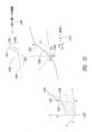
膝関節を特定の例として用いると、人工膝関節形成術(TKA)のための現行の人工システムは、典型的には、最大5つまでのコンポーネント、すなわち、大腿骨コンポーネント、脛骨コンポーネント、脛骨インサート、脛骨ステム延長部及び膝蓋骨コンポーネントから成り、これら5つのコンポーネントをひとまとめに全膝関節インプラント(TKI)と称する場合がある。これらコンポーネントは、生まれつきの膝関節の機能に置き換わってこれを提供するよう機能的ユニットとして一緒に働くよう設計されている。大腿骨コンポーネントは、膝関節の大腿骨頭に取り付けられて上関節面を形成する。脛骨インサート(スペーサとも呼ばれる)は、ポリマーで構成される場合が多く、この脛骨インサートは、金属製大腿骨頭と一緒になって下関節面を形成する。脛骨コンポーネントは、脛骨の髄腔中に入る脛骨ステムとベースプレートから成り、ベースプレートは、脛骨インサートに接触/保持する脛骨プレートか、脛骨トレイか、或いは脛骨ベースプレートかのいずれかと呼ばれる場合がある。オプションとして、また特に、近位脛骨の質及び/又は骨量が損なわれた場合、脛骨ステム延長部は、脛骨コンポーネントの傾動に抵抗して安定性を増すためのキール(keel)として役立つ。市販されているTKA製品の例としては、Persona(商標)膝関節システム(I113369)及び関連テーパ付き脛骨ステム延長部(K133737)が挙げられ、これらは共に、ジンマー・バイオメット・インコーポレイテッド(Zimmer Biomet Inc.)(米国インディアナ州ワルシャワ所在)によって製造されている。これら4つのコンポーネントを患者の体内に植え込む外科手術は、人工膝関節置換術(TKR)とも呼ばれる。同様な人工デバイスが他の関節、例えば、人工股関節形成術(THA)及び人工肩関節形成術(TSA)について利用でき、この場合、一方の関節面は、金属製であり、対向した表面は、ポリマー性である。これらデバイス及び手技(TKA、THA及びTSA)をひとまとめに人工関節形成術(TJA)という場合が多い。Using the knee joint as a specific example, current prosthetic systems for total knee arthroplasty (TKA) typically consist of up to five components: a femoral component, a tibial component, a tibial insert, a tibial stem extension, and a patellar component, which may collectively be referred to as a total knee implant (TKI). These components are designed to work together as a functional unit to replace and provide the function of the natural knee joint. The femoral component attaches to the femoral head of the knee joint to form the superior articular surface. The tibial insert (also called a spacer), often composed of a polymer, forms the inferior articular surface together with the metallic femoral head. The tibial component consists of a tibial stem that fits into the medullary cavity of the tibia and a baseplate, which may be referred to as either a tibial plate, tibial tray, or tibial baseplate, which contacts/retains the tibial insert. Optionally, and especially when the quality and/or bone mass of the proximal tibia is compromised, the tibial stem extension serves as a keel to resist tilting of the tibial component and increase stability. Examples of commercially available TKA products include the Persona™ Knee System (I113369) and associated Tapered Tibial Stem Extension (K133737), both manufactured by Zimmer Biomet Inc. (Warsaw, Indiana, USA). The surgical procedure to implant these four components in the patient's body is also called total knee replacement (TKR). Similar prosthetic devices are available for other joints, such as total hip arthroplasty (THA) and total shoulder arthroplasty (TSA), where one articular surface is metallic and the opposing surface is polymeric. These devices and procedures (TKA, THA, and TSA) are often collectively referred to as total joint arthroplasty (TJA).
TKAに関し、脛骨コンポーネント及び大腿骨コンポーネントは、典型的には、それぞれ脛骨及び大腿骨内に挿入されてこれらの中に定位置で結合される。幾つかの場合、これらコンポーネントは、非結合状態の膝関節の場合のように定位置に結合されない。これらコンポーネントが定位置に結合されるかどうかとは無関係に、周囲の骨内にいったん配置されてこの中に一体化されると(このプロセスは、オッセオインテグレーションと呼ばれる)、これらコンポーネントは、取り外すのが容易ではない。したがって、植え込み中におけるこれらコンポーネントの適正な配置は、手技につき功を奏する結果にとって極めて重要であり、外科医は、これらコンポーネントの植え込み及び固定に細心の注意を払う。For TKA, the tibial and femoral components are typically inserted into and bonded in place within the tibia and femur, respectively. In some cases, the components are not bonded in place as in a non-union knee. Regardless of whether the components are bonded in place, once positioned and integrated into the surrounding bone (a process called osseointegration), they are not easily removed. Therefore, proper placement of the components during implantation is crucial to a successful outcome of the procedure, and surgeons pay close attention to implanting and fixing the components.
現行の商用TKAシステムは、臨床的用途について長い歴史を持っており、植え込み期間は、通常は10年を超え、幾つかのレポートによれば、25年で87%の存続状態が実証されている。臨床医は、現在においては、2~3週間、6~8週間、3か月、6か月、12か月、それ以降は毎年、一連の理学的検査を用いて事後のインプラントの進行状態をモニタしている。Current commercial TKA systems have a long history of clinical use, with implant durations typically exceeding 10 years, with some reports demonstrating 87% survival at 25 years. Clinicians currently monitor the progress of subsequent implants with a series of physical examinations at 2-3 weeks, 6-8 weeks, 3 months, 6 months, 12 months, and annually thereafter.
TKIを植え込み、そして患者が人工膝関節で歩き始めた後、問題が起こる場合があり、かかる問題は、突き止めるのが困難な場合がある。臨床検査は、プロテーゼの故障を検出するこれらの能力において制限されている場合が多く、したがって追加のモニタリング、例えばCTスキャン法、MRIスキャン法、又はそれどころか核スキャン法が必要とされる場合が多い。インプラントの寿命全体にわたってケア要件が連続して行われるものと仮定すると、患者は、毎年かかりつけの医師のもとに訪れて自分の健康状態を調べ、他の関節をモニタし、そしてTKAインプラントの機能を評価してもらうことが奨励される。ケアの現行の基準は、ケアの90日エピソードの間、患者のTKA機能を評価する能力を医師及びヘルスケアシステムに与えているが、測定は、主観的であり、しかも大きな可動性に関する問題に至る前触れである場合がある機能性における僅かな変化を描出するための時間分解能を欠いている場合が多い。TKA患者の長期間(1年超)フォローアップもまた、患者が毎年一貫してかかりつけ医師に会うことがないという問題を提起している。むしろ、TKA患者は、痛み又は他の症状があった場合にのみ別途相談を持ち掛ける場合が多い。After the TKI is implanted and the patient begins to walk with the artificial knee, problems may occur that may be difficult to pinpoint. Clinical tests are often limited in their ability to detect prosthesis failure, and therefore additional monitoring, such as CT, MRI, or even nuclear scans, is often required. Assuming continuous care requirements throughout the life of the implant, patients are encouraged to visit their primary care physician annually to check their health, monitor other joints, and evaluate the function of the TKA implant. Current standards of care provide physicians and health care systems with the ability to assess a patient's TKA function during a 90-day episode of care, but measurements are subjective and often lack the time resolution to depict subtle changes in functionality that may be a precursor to major mobility problems. Long-term (>1 year) follow-up of TKA patients also poses the problem that patients do not consistently see their primary care physician year after year. Rather, TKA patients often only seek separate consultation if they experience pain or other symptoms.
現時点において、臨床訪問医及び熟練したヘルスケア提供者の腕前や目視観察なしでTKAの置き間違い、不安定性又はアライメント不良を高信頼度で検出するための仕組みは存在しない。その場合でも、準臨床的問題又は状態を早期に突き止めることは、困難であるか不可能であるかのいずれかであり、というのは、かかる準臨床的問題又は状態は、極めて微妙なので理学的検査で検出できない場合が多く、或いは放射線学的検査で実証可能である場合が多い。さらに、検出が可能であったとしても、修正措置は、特定の量の運動及び/又は不適切なアライメントの程度を正確に測定することができず又は定量化することができないということによって妨げられ、目標とする上首尾のインターベンション又は介入を見込みがないものにする。外部モニタリング装置は、不安定性を検出するのに必要な忠実度を提供することがなく、というのは、これら装置は、皮膚、筋、及び脂肪によって(これらの各々は、不安定性についての機械的痕跡を覆い隠す)TKAから離されており、そして異常、例えば屈曲、組織‐骨音響ノイズ、表面上における一貫性のないセンサ配置、及びTKAに対する外部センサの一貫性のない配置場所をもたらす。Currently, there is no mechanism for reliably detecting misplacement, instability, or malalignment of the TKA without the skill and visual observation of a visiting clinician and a skilled healthcare provider. Even then, it is difficult or impossible to identify subclinical problems or conditions early on, as they are often so subtle that they cannot be detected by physical examination or are demonstrable by radiological examination. Furthermore, even if detection is possible, corrective measures are hindered by the inability to accurately measure or quantify the degree of specific amounts of motion and/or improper alignment, making targeted and successful intervention unlikely. External monitoring devices do not provide the fidelity required to detect instability, as they are separated from the TKA by skin, muscle, and fat (each of which masks the mechanical signature of instability) and introduce abnormalities such as bending, tissue-bone acoustic noise, inconsistent sensor placement on the surface, and inconsistent placement of the external sensor relative to the TKA.
TKAインプラント以外のインプラントもまた、植え込み中と手術後の両方において種々の合併症と関連している場合がある。一般に、医用インプラントを正確に配置することは、外科医にとって難題といえ、種々の合併症がどのような医用インプラントの挿入中にも生じる場合がある(開放外科的処置であれ低侵襲手技であれいずれにせよ)。例えば、外科医は、周りの組織及び構造内のインプラントの正確な解剖学的アライメント及び配置を確認したいと考える場合がある。しかしながら、これは、手技自体を実施している間に行うことが困難であるといえ、それにより術中の正確な調整が困難になる。Implants other than TKA implants may also be associated with various complications both during implantation and after surgery. Accurate placement of medical implants in general can be a challenge for surgeons, and various complications can arise during insertion of any medical implant (whether an open surgical procedure or a minimally invasive technique). For example, a surgeon may want to ensure precise anatomical alignment and placement of the implant within the surrounding tissues and structures. However, this can be difficult to do while performing the procedure itself, making precise intraoperative adjustments difficult.
加うるに、患者は、手技後に多数回の合併症にかかる場合がある。かかる合併症としては、神経学的症状、疼痛、機能障害(閉塞、弛みなど)及び/又はインプラントの摩耗、インプラントの動き又は破断、炎症及び/又は感染が挙げられる。これら問題のうちの幾つかに医薬品及び/又はさらなる手術で取り組むことができるが、これらは、予測して予防するのが困難であり、多くの場合、合併症及び副作用の早期確認が望ましいけれども困難であり又は不可能である。In addition, patients may suffer from a number of complications following the procedure, including neurological symptoms, pain, functional impairment (blockage, loosening, etc.) and/or implant wear, implant movement or rupture, inflammation and/or infection. While some of these problems can be addressed with medicines and/or further surgery, these are difficult to predict and prevent, and early identification of complications and side effects, while desirable, is often difficult or impossible.
本発明は、特に早期の段階においてこれらの問題を確認し、その問題の原因を究明すると共に/或いは定量化すること、及びこれらの問題を改善するための方法及びデバイス又は装置を提供することを目的としている。The present invention aims to identify these problems, particularly at an early stage, determine and/or quantify the causes of the problems, and provide methods and devices or apparatus for remedying these problems.
技術の背景の項で説明した主題の全ては、必ずしも先行技術であるとは限らず、単に技術の背景の項における説明の結果として先行技術であるとみなされるべきではない。これらの趣旨にしたがって、背景技術の項で説明し又はかかる主題と関連した先行技術における問題についてのいかなる認識も、先行技術であるという明示がなければ、先行技術として取り扱われるべきではない。これとは異なり、背景技術の項における任意の主題についての説明は、特定の問題に対する本発明者のアプローチの一部として取り扱われるべきであり、本質的にかつ元来、かかるアプローチもまた、発明に関する場合がある。Not all of the subject matter discussed in the Background of the Art section is necessarily prior art, and should not be considered to be prior art merely as a result of its discussion in the Background of the Art section. In accordance with these principles, any awareness of a problem in the prior art discussed in the Background of the Art section or related to such subject matter should not be treated as prior art unless expressly stated to be prior art. Instead, the discussion of any subject matter in the Background of the Art section should be treated as part of the inventor's approach to a particular problem, and such approach may also be inventive in nature and intrinsically.
概要を説明すると、本発明は、インテリジェントインプラント、インテリジェントインプラントを含むシステム、インプラント/システムを用いてインプラントと関連した問題の検出、原因究明、定量化及び/又は特徴づけのうちの少なくとも1つを行う方法、及び突き止められた問題に取り組むための方法及びデバイス又は装置に関する。以下に詳細に説明するように、本発明は、センサに結合された医療デバイス、及びかかる医療デバイスを含むシステムを提供し、かかる医療デバイスは、データを生じさせると共に当該データに基づいて分析結果を生じさせることができ、かかるデータは、植え込まれた医療デバイスと関連した問題を突き止めると共に/或いはこれら問題に取り組むために使用されるのが良い。一実施形態では、医療デバイスは、プロテーゼ(TJA)であり、データは、プロテーゼの動きを反映した運動学的データである。突き止めることができる問題としては、TJA器具の不正確な配置、この器具の不正確なアライメント、この器具の予期しない劣化又は摩耗、この器具(及び関連の関節)の不安定性、及びこの器具の望ましくない動きが挙げられる。また、センサに結合された医療デバイス、及び植え込まれた医療デバイスで突き止められた問題に取り組むための装置及び方法もまた提供される。In summary, the present invention relates to an intelligent implant, a system including an intelligent implant, a method of using an implant/system to detect, identify, quantify and/or characterize problems associated with the implant, and a method and device or apparatus for addressing the identified problems. As described in detail below, the present invention provides a medical device coupled to a sensor and a system including such a medical device that can generate data and generate analysis results based on the data, which can be used to identify and/or address problems associated with the implanted medical device. In one embodiment, the medical device is a prosthesis (TJA) and the data is kinematic data reflecting the motion of the prosthesis. Problems that can be identified include incorrect placement of the TJA instrument, incorrect alignment of the instrument, unexpected deterioration or wear of the instrument, instability of the instrument (and associated joint), and undesired motion of the instrument. Also provided are a medical device coupled to a sensor and an apparatus and method for addressing problems identified with the implanted medical device.
センサに結合された医療デバイスをインテリジェントインプラントという場合があり、この場合、インテリジェントインプラントは、インプラントの機能の発揮具合及び/又はインプラントのすぐ近くの周囲環境及び/又はインプラントの活動度/動き並びに患者の活動度及び動きを検出すると共に/或いは測定することができるセンサを有する。インプラントを別の言い方としてプロテーゼという場合があり、この場合、インテリジェントインプラントとインテリジェントプロテーゼは、同じ意味を有する。一実施形態では、センサを医療デバイス、例えばプロテーゼ/インプラントに結合することは、センサを医療デバイス内に全体を配置することであり、その結果、センサは、全体が医療デバイスの外面によって包囲され、したがって、医療デバイスが植え込まれる患者のどのような組織にも物理的に接触するセンサの部分は存在しないようになる。本発明の実施形態では、医療デバイス又はインプラント若しくはプロテーゼといった場合、これらは、本明細書において開示する医療デバイス又はインプラント/プロテーゼ内に全体が配置されたセンサを有するインテリジェント医療デバイス又はインプラント/プロテーゼのことを意味しているものと理解されるのが良い。本発明の実施形態では、本明細書において、センサを有する医療デバイス又はインプラント若しくはプロテーゼといった場合、これは、センサが医療デバイス又はインプラント/プロテーゼ内に全体が配置されたインテリジェント医療デバイス又はインプラント/プロテーゼのことを意味しているものと理解されるべきである。本発明の実施形態では、本明細書において、センサを有する医療デバイス又はインプラント若しくはプロテーゼといった場合、これは、センサが医療デバイス又はインプラント/プロテーゼ内に全体が配置された1個の加速度計又は2個以上の加速度計(例えば、2個、3個、4個、5個、6個、7個などの加速度計)であるインテリジェント医療デバイス又はインプラント/プロテーゼを意味していると理解されるべきである。本発明の実施形態では、本明細書において、センサを有する医療デバイス又はインプラント/プロテーゼといった場合、これは、医療デバイス、インプラント/プロテーゼの脛骨延長部内に全体が配置された1個以上の加速度計(例えば、2個、3個、4個、5個、6個、7個などの加速度計)であるインテリジェント医療デバイス又はインプラント/プロテーゼを意味していると理解されるべきであり、したがって、医療デバイス又はインプラント/プロテーゼは、例えば、TKAのコンポーネントである。A medical device coupled to a sensor may be referred to as an intelligent implant, where the intelligent implant has a sensor capable of detecting and/or measuring the functioning of the implant and/or the immediate surrounding environment and/or the activity/movement of the implant and the patient. Another term for an implant may be a prosthesis, where intelligent implant and intelligent prosthesis have the same meaning. In one embodiment, coupling a sensor to a medical device, e.g., a prosthesis/implant, is to place the sensor entirely within the medical device, such that the sensor is entirely surrounded by the exterior surface of the medical device, and thus no part of the sensor is in physical contact with any tissue of the patient in which the medical device is implanted. In embodiments of the present invention, references to a medical device or implant or prosthesis may be understood to refer to an intelligent medical device or implant/prosthesis having a sensor entirely within the medical device or implant/prosthesis disclosed herein. In embodiments of the present invention, when a medical device or implant or prosthesis having a sensor is referred to herein, this should be understood to mean an intelligent medical device or implant/prosthesis in which the sensor is located entirely within the medical device or implant/prosthesis. In embodiments of the present invention, when a medical device or implant or prosthesis having a sensor is referred to herein, this should be understood to mean an intelligent medical device or implant/prosthesis in which the sensor is an accelerometer or more than one accelerometer (e.g., 2, 3, 4, 5, 6, 7, etc. accelerometers) located entirely within the medical device or implant/prosthesis. In embodiments of the present invention, when a medical device or implant/prosthesis having a sensor is referred to herein, this should be understood to mean an intelligent medical device or implant/prosthesis in which the sensor is one or more accelerometers (e.g., 2, 3, 4, 5, 6, 7, etc. accelerometers) located entirely within the tibial extension of the medical device, implant/prosthesis, and thus the medical device or implant/prosthesis is, for example, a component of a TKA.
システムは、インテリジェントインプラント及び検出及び/又は測定から得られたデータを記憶する1つ以上のメモリ、そのデータを伝送するアンテナ、センサによって生じたデータを受け取ってこのデータ及び/又は分析されたデータをクラウドベースの記憶場所に伝送することができる基地局、データを記憶して分析することができ、そして分析したデータを記憶すると共に/或いはさらに分析することができるクラウドベースの記憶場所、及びクラウドベースの記憶場所から出力を受け取る受信局を含み、この受信局には、例えばヘルスケア専門家又は保険会社又はインプラントの製造業者によってアクセスすることができ、出力は、インプラントの状態及び/又はインプラントの機能の発揮具合及び/又はインプラントを受け入れた患者の状態を割り出すことができ、そしてまた、元データの分析によって生じた何らかの懸念に取り組むための推奨策を提供することができる。The system includes an intelligent implant and one or more memories for storing data obtained from the detections and/or measurements, an antenna for transmitting the data, a base station capable of receiving data generated by the sensors and transmitting the data and/or analyzed data to a cloud-based storage location, a cloud-based storage location capable of storing and analyzing the data and storing and/or further analyzing the analyzed data, and a receiving station for receiving an output from the cloud-based storage location, which may be accessed, for example, by a healthcare professional or an insurance company or the implant manufacturer, and the output may determine the condition of the implant and/or how well the implant is functioning and/or the condition of a patient receiving the implant, and may also provide recommendations to address any concerns raised by the analysis of the raw data.
例えば、人工関節形成術(例えば、TKA、THA及びTSA)ハードウェアの不安定性により、骨の侵食及びインプラントコンポーネントの疲労の加速が生じる場合がある。未治療のまま又は是正しないままにしておくと、骨の侵食及び疲労の加速により、典型的には、疼痛や炎症が生じる。疼痛や炎症により人工関節形成術(TJA)患者が医療ケアを探し求めざるを得なくなった時点ではすでに、骨侵食及びTJA疲労の程度によるがヘルスケア専門家にはたった1つの選択肢、すなわち、結果的に「成功する」見込みが低い高侵襲性のかつ費用の高くつく手術という選択肢しか残されてない場合がある。本発明は、TJAハードウェアの不安定性を骨侵食及びインプラントの疲労による損傷が起こる前に早期に検出することができるようにする装置、システム及び方法を提供する。この不安定性を検出し、定量化し、そして特徴づけることができ、その結果をヘルスケア提供者に伝えて早期治療及び/又は問題についてのより効果的な取り扱いを可能にし、すなわち、ヘルスケア提供者は、侵襲性がはるかに低く、安価であり、しかも成功の見込みの高い矯正治療を利用することができる。本発明はまた、不安定性の問題に取り組むためのデバイス又は装置及び/又は方法を提供する。For example, instability of total joint arthroplasty (e.g., TKA, THA, and TSA) hardware can result in bone erosion and accelerated fatigue of implant components. If left untreated or uncorrected, bone erosion and accelerated fatigue typically result in pain and inflammation. By the time pain and inflammation force a total joint arthroplasty (TJA) patient to seek medical care, depending on the extent of bone erosion and TJA fatigue, healthcare professionals may be left with only one option: highly invasive and expensive surgery that is unlikely to result in a "successful" outcome. The present invention provides devices, systems, and methods that allow early detection of TJA hardware instability before damage from bone erosion and implant fatigue occurs. This instability can be detected, quantified, and characterized, and the results communicated to healthcare providers to allow for early treatment and/or more effective management of the problem, i.e., healthcare providers can utilize corrective treatments that are much less invasive, less expensive, and more likely to be successful. The present invention also provides a device or apparatus and/or method for addressing the problem of instability.
本発明は、TJA(人工関節形成術)に関し、この用語は、手術及び関連の植え込まれたハードウェア、例えば本発明のTJAプロテーゼを含む。本発明の方法、デバイス及びシステムの特徴が特定のインテリジェントTJAプロテーゼを参照することにより本明細書において説明されるが、本発明は、TKA(人工膝関節形成術)プロテーゼ、例えばTKAシステムとも呼ばれる場合があるTKI(人工膝関節インプラント)、TSA(人工肩関節形成術)プロテーゼ、例えばTSIシステムとも呼ばれる場合があるTSI(人工肩関節インプラント)、及びTHA(人工股関節形成術)プロテーゼ、例えばTHAシステムとも呼ばれる場合があるTHI(人工股関節インプラント)を含む任意の1つ以上のTJAプロテーゼに当てはまるものと理解されるべきである。一実施形態では、TJAプロテーゼは、本明細書に開示されているような少なくとも1つのセンサを有するインテリジェントTJAプロテーゼとも呼ばれるインテリジェントTJAである。The present invention relates to TJA (prosthetic joint arthroplasty), a term that includes procedures and associated implanted hardware, such as the TJA prosthesis of the present invention. Although features of the methods, devices and systems of the present invention are described herein with reference to a particular intelligent TJA prosthesis, it should be understood that the present invention applies to any one or more TJA prostheses, including TKA (prosthetic knee arthroplasty) prostheses, such as TKI (prosthetic knee implant), which may also be referred to as a TKA system, TSA (prosthetic shoulder arthroplasty) prostheses, such as TSI (prosthetic shoulder implant), which may also be referred to as a TSI system, and THA (prosthetic hip arthroplasty) prostheses, such as THI (prosthetic hip implant), which may also be referred to as a THA system. In one embodiment, the TJA prosthesis is an intelligent TJA, also referred to as an intelligent TJA prosthesis, having at least one sensor as disclosed herein.
この発明の概要の項は、詳細な説明において以下に詳細にさらに説明するある特定の技術的思想を単純化された形態で導入するよう設けられている。別段の明示してあるものを除き、この発明の概要の項は、クレーム請求されている主題の重要な又は必須の特徴を特定することを意図しておらず、しかもクレーム請求されている主題の範囲を制限することを意図していない。This Summary is intended to introduce in a simplified form certain concepts that are further described below in the Detailed Description. Unless expressly stated otherwise, this Summary is not intended to identify key or essential features of the claimed subject matter, nor is it intended to limit the scope of the claimed subject matter.
以下は、本発明の幾つかの例示の番号が付けられた実施態様である。
〔実施態様項1〕
植え込み型人工膝関節用の脛骨インサートであって、上記インプラントの内側(ないそく)側の厚さが上記インプラントの外側(がいそく)側の厚さと比較して1mm、2mm、3mm、4mm、5mm、6mm、7mm、8mm、9mm、又は10mm厚い脛骨インサートから成る、脛骨インサート。
〔実施態様項2〕
植え込み型人工膝関節用の脛骨インサートであって、上記インプラントの外側(がいそく)側の厚さが上記インプラントの内側(ないそく)側の厚さと比較して1mm、2mm、3mm、4mm、5mm、6mm、7mm、8mm、9mm、又は10mm厚い脛骨インサートから成る、脛骨インサート。
〔実施態様項3〕
植え込み型人工膝関節用の脛骨インサートであって、上記インプラントの前方側の厚さが上記インプラントの後方側の厚さと比較して1mm、2mm、3mm、4mm、5mm、6mm、7mm、8mm、9mm、又は10mm厚い脛骨インサートから成る、脛骨インサート。
〔実施態様項4〕
植え込み型人工膝関節用の脛骨インサートであって、上記インプラントの後方側の厚さが上記インプラントの前方側の厚さと比較して1mm、2mm、3mm、4mm、5mm、6mm、7mm、8mm、9mm、又は10mm厚い脛骨インサートから成る、脛骨インサート。
〔実施態様項5〕
植え込み型人工膝関節用の脛骨インサート又は関節スペーサであって、上記インプラントの内側(ないそく)側、外側(がいそく)側、前方側及び/又は後方側のうちの1つの厚さが上記インプラントの対応の側の厚さと比較して1mm、2mm、3mm、4mm、5mm、6mm、7mm、8mm、9mm、又は10mm厚い脛骨インサートを含む、脛骨インサート又は関節スペーサ。
〔実施態様項6〕
上記脛骨インサートは、ポリエチレン又はポリエーテルエーテルケトン(PEEK)で構成されている、実施態様項1~5のうちいずれか一に記載の脛骨インサート。
〔実施態様項7〕
上記脛骨インサートは、患者に合わせてカスタマイズされている、実施態様項1~6のうちいずれか一に記載の脛骨インサート。
〔実施態様項8〕
上記脛骨インサートは、3Dプリンティング又は成形によって製造されている、実施態様項1~7のうちいずれか一に記載の脛骨インサート。
〔実施態様項9〕
植え込み型医療デバイスであって、植え込み型人工デバイスに固定的に取り付けられるよう構成された回路と、電力コンポーネントと、上記回路を上記電力コンポーネントから切り離すよう構成された装置と、を有する、植え込み型医療デバイス。
〔実施態様項10〕
植え込み型医療デバイスであって、植え込み型人工デバイスに固定的に取り付けられるよう構成された回路と、バッテリと、上記回路と上記バッテリとの間に結合されたヒューズと、を有する、植え込み型医療デバイス。
〔実施態様項11〕
方法であって、回路とバッテリとの間に配置されたヒューズを電気的に開くステップを含み、少なくとも、上記ヒューズ及び上記回路は、植え込み型人工デバイスに設けられている、方法。
〔実施態様項12〕
植え込み型医療デバイスであって、センサ信号を発生するよう構成された少なくとも1つのセンサと、上記少なくとも1つのセンサに遠隔医療コードに関連づけられた周波数で上記センサ信号を発生させるよう構成された制御回路と、を有する、植え込み型医療デバイス。
〔実施態様項13〕
植え込み型医療デバイスであって、センサ信号を発生するよう構成された少なくとも1つのセンサと、医師が遠隔医療保険コード下で上記少なくとも1つのセンサに支払いの資格を有することができるようにする周波数で上記センサ信号を発生させるよう構成された制御回路と、を有する、植え込み型医療デバイス。
〔実施態様項14〕
植え込み型医療デバイスであって、センサ信号を発生するよう構成された少なくとも1つのセンサと、医師が遠隔医療保険コード下で上記少なくとも1つのセンサに全額支払いの資格を有することができるようにする周波数で上記センサ信号を発生させるよう構成された制御回路と、を有する、植え込み型医療デバイス。
〔実施態様項15〕
方法であって、植え込み型医療デバイスに関連づけられたセンサ信号を、医師が遠隔医療保険コード下で支払いの資格を有することができるようにする周波数で発生させるステップを含む、方法。
〔実施態様項16〕
方法であって、植え込み型医療デバイスに関連づけられたセンサ信号を、医師が遠隔医療保険コード下で全額支払いの資格を有することができるようにする周波数で発生させるステップを含む、方法。
〔実施態様項17〕
植え込み型プロテーゼであって、
ハウジングと、
上記ハウジング内に配置された植え込み型回路と、を有し、上記植え込み型回路は、
動きを表す少なくとも1つの第1の信号を発生させ、
上記少なくとも1つの第1の信号が少なくとも1つの第1の基準を満たしているかどうかを判定し、そして
上記少なくとも1つの第1の信号が上記少なくとも1つの第1の基準を満たしているという判定に応答して上記少なくとも1つの第1の信号を遠隔の場所に送るよう構成されている、植え込み型プロテーゼ。
〔実施態様項18〕
基地局であって、
ハウジングと、
上記ハウジング内に配置された基地局回路と、を有し、上記基地局回路は、
植え込み型プロテーゼから動きを表す少なくとも1つの第1の信号を受け取り、
上記少なくとも1つの第1の信号を目的地に送り、
ソースから少なくとも1つの第2の信号を受け取り、そして
上記少なくとも1つの第2の信号を上記植え込み型プロテーゼに送るよう構成されている、基地局。
〔実施態様項19〕
方法であって、電源と植え込み型回路との間で植え込み型プロテーゼに設けられたヒューズを、過電流しきい値を超えて上記ヒューズを通る電流に応答して開くステップを含む、方法。
〔実施態様項20〕
方法であって、電源と植え込み型回路との間で植え込み型プロテーゼに設けられたヒューズを、少なくともしきい時間にわたって過電流しきい値を超えて上記ヒューズを通る電流に応答して開くステップを含む、方法。
〔実施態様項21〕
方法であって、電源と植え込み型回路との間で植え込み型プロテーゼに設けられたヒューズを、過電圧しきい値を超えて上記ヒューズ前後にかかる電圧に応答して開くステップを含む、方法。
〔実施態様項22〕
方法であって、電源と植え込み型回路との間で植え込み型プロテーゼに設けられたヒューズを、少なくともしきい時間にわたって過電圧しきい値を超えて上記ヒューズ前後にかかる電圧に応答して開くステップを含む、方法。
〔実施態様項23〕
方法であって、電源と植え込み型回路との間で植え込み型プロテーゼに設けられたヒューズを、過剰温度しきい値を超える温度に応答して開くステップを含む、方法。
〔実施態様項24〕
方法であって、電源と植え込み型回路との間で植え込み型プロテーゼに設けられたヒューズを、少なくとも時間の長さのしきい値にわたって過剰温度しきい値を超える温度に応答して開くステップを含む、方法。
〔実施態様項25〕
方法であって、プロテーゼが植え込まれた患者の動きに応答してセンサ信号を発生させるステップと、上記センサ信号を遠隔場所に送信するステップと、を含む、方法。
〔実施態様項26〕
方法であって、プロテーゼが植え込まれた患者の動きに応答してセンサ信号を発生させるステップと、上記センサ信号をサンプリングするステップと、上記サンプルを遠隔場所に送信するステップと、を含む、方法。
〔実施態様項27〕
方法であって、プロテーゼが植え込まれた患者の動きに応答してセンサ信号を発生させるステップと、上記センサ信号が有資格事象を表しているかどうかを判定するステップと、上記センサ信号が有資格事象を表しているかどうかの判定に応答して上記センサ信号を遠隔場所に送信するステップと、を含む、方法。
〔実施態様項28〕
方法であって、
プロテーゼが植え込まれた患者の動きに応答してセンサ信号を発生させるステップと、ポーリング信号を遠隔場所から受け取るステップと、上記センサ信号を上記ポーリング信号に応答して上記遠隔場所に送信するステップと、を含む、方法。
〔実施態様項29〕
方法であって、プロテーゼが植え込まれた患者の動きに応答してセンサ信号を発生させるステップと、上記センサ信号又は上記センサ信号を表すデータを含むメッセージを作成するステップと、上記メッセージを遠隔場所に送信するステップと、を含む、方法。
〔実施態様項30〕
方法であって、プロテーゼが植え込まれた患者の動きに応答してセンサ信号を発生させるステップと、上記センサ信号又は上記センサ信号を表すデータを含むデータパケットを生じさせるステップと、上記データパケットを遠隔場所に送信するステップと、を含む、方法。
〔実施態様項31〕
方法であって、プロテーゼが植え込まれた患者の動きに応答してセンサ信号を発生させるステップと、上記センサ信号又は上記センサ信号を表すデータの少なくとも一部分を暗号化するステップと、上記暗号化されたセンサ信号を遠隔場所に送信するステップと、を含む、方法。
〔実施態様項32〕
方法であって、プロテーゼが植え込まれた患者の動きに応答してセンサ信号を発生させるステップと、上記センサ信号又は上記センサ信号を表すデータの少なくとも一部分を符号化するステップと、上記符号化されたセンサ信号を遠隔場所に送信するステップと、を含む、方法。
〔実施態様項33〕
方法であって、プロテーゼが植え込まれた患者の動きに応答してセンサ信号を発生させるステップと、上記センサ信号を遠隔場所に送信するステップと、上記プロテーゼと関連した植え込み型回路を上記センサ信号の送信後に低電力モードに設定するステップと、を含む、方法。
〔実施態様項34〕
方法であって、プロテーゼが植え込まれた患者の動きに応答して第1のセンサ信号を発生させるステップと、上記第1のセンサ信号を遠隔場所に送信するステップと、上記プロテーゼと関連した植え込み型回路の少なくとも1つの部品を上記センサ信号の送信後に低電力モードに設定するステップと、上記植え込み型回路の設定対象である低電力モード時間の経過後に上記患者の動きに応答して第2のセンサ信号を発生させるステップと、を含む、方法。
〔実施態様項35〕
方法であって、センサ信号を患者に取り付けられ又は患者の体内に植え込まれたプロテーゼから受け取るステップと、上記受け取ったセンサ信号を目的地に送信するステップと、を含む、方法。
〔実施態様項36〕
方法であって、患者に取り付けられ又は患者の体内に植え込まれたプロテーゼに問い合わせを送るステップと、上記問い合わせを送った後、センサ信号をプロテーゼから受け取るステップと、上記受け取ったセンサ信号を目的地に送信するステップと、を含む、方法。
〔実施態様項37〕
方法であって、センサ信号及び少なくとも1つの識別子を患者に取り付けられ又は患者の体内に植え込まれたプロテーゼから受け取るステップと、上記識別子が正しいかどうかを判定するステップと、上記受け取ったセンサ信号を上記識別子が正しいかどうかの判定に応答して目的地に送信するステップと、を含む、方法。
〔実施態様項38〕
方法であって、患者に取り付けられ又は患者の体内に植え込まれたプロテーゼからのセンサ信号を含むメッセージを受け取るステップと、上記メッセージの少なくとも一部分を解読するステップと、上記解読したメッセージを目的地に送信するステップと、を含む、方法。
〔実施態様項39〕
方法であって、患者に取り付けられ又は患者の体内に植え込まれたプロテーゼからのセンサ信号を含むメッセージを受け取るステップと、上記メッセージの少なくとも一部分を復号するステップと、上記復号したメッセージを目的地に送信するステップと、を含む、方法。
〔実施態様項40〕
方法であって、患者に取り付けられ又は患者の体内に植え込まれたプロテーゼからのセンサ信号を含むメッセージを受け取るステップと、上記メッセージの少なくとも一部分を符号化するステップと、上記符号化したメッセージを目的地に送信するステップと、を含む、方法。
〔実施態様項41〕
方法であって、患者に取り付けられ又は患者の体内に植え込まれたプロテーゼからのセンサ信号を含むメッセージを受け取るステップと、上記メッセージの少なくとも一部分を暗号化するステップと、上記暗号化したメッセージを目的地に送信するステップと、を含む、方法。
〔実施態様項42〕
方法であって、患者に取り付けられ又は患者の体内に植え込まれたプロテーゼからのセンサ信号を含むデータパケットを受け取るステップと、上記データパケットの少なくとも一部分を解読するステップと、上記解読したデータパケットを目的地に送信するステップと、を含む、方法。
〔実施態様項43〕
方法であって、患者に取り付けられ又は患者の体内に植え込まれたプロテーゼからのセンサ信号を含むデータパケットを受け取るステップと、上記データパケットの少なくとも一部分を復号するステップと、上記復号したデータパケットを目的地に送信するステップと、を含む、方法。
〔実施態様項44〕
方法であって、患者に取り付けられ又は患者の体内に植え込まれたプロテーゼからのセンサ信号を含むデータパケットを受け取るステップと、上記データパケットの少なくとも一部分を符号化するステップと、上記符号化したデータパケットを目的地に送信するステップと、を含む、方法。
〔実施態様項45〕
方法であって、患者に取り付けられ又は患者の体内に植え込まれたプロテーゼからのセンサ信号を含むデータパケットを受け取るステップと、上記データパケットの少なくとも一部分を暗号化するステップと、上記暗号化したデータパケットを目的地に送信するステップと、を含む、方法。
〔実施態様項46〕
方法であって、患者に取り付けられ又は患者の体内に植え込まれたプロテーゼからのセンサ信号を受け取るステップと、上記センサ信号の少なくとも一部分を解読するステップと、上記解読したセンサ信号を目的地に送信するステップと、を含む、方法。
〔実施態様項47〕
方法であって、患者に取り付けられ又は患者の体内に植え込まれたプロテーゼからのセンサ信号を受け取るステップと、上記センサ信号の少なくとも一部分を復号するステップと、上記復号したセンサ信号を目的地に送信するステップと、を含む、方法。
〔実施態様項48〕
方法であって、患者に取り付けられ又は患者の体内に植え込まれたプロテーゼからのセンサ信号を受け取るステップと、上記センサ信号の少なくとも一部分を符号化するステップと、上記符号化したセンサ信号を目的地に送信するステップと、を含む、方法。
〔実施態様項49〕
方法であって、患者に取り付けられ又は患者の体内に植え込まれたプロテーゼからのセンサ信号を受け取るステップと、上記センサ信号の少なくとも一部分を暗号化するステップと、上記暗号化したセンサ信号を目的地に送信するステップと、を含む、方法。
〔実施態様項50〕
植え込み型プロテーゼ用の植え込み型回路。
〔実施態様項51〕
植え込み型回路を有する植え込み型プロテーゼ。
〔実施態様項52〕
ヒューズを有する植え込み型プロテーゼ。
〔実施態様項53〕
植え込み型プロテーゼと通信可能な基地局。
〔実施態様項54〕
1つ以上のコンピュータシステムのコンポーネントとして具体化されたモニタリング-セッション-データ収集、分析、及び状態報告システムであって、各コンピュータシステムは、1つ以上のプロセッサ、1つ以上のメモリ、1つ以上のネットワーク接続手段、及び1つ以上の大容量記憶装置へのアクセスを含み、上記1つ以上のモニタリング-セッション-データ収集、データ分析、及び状態報告システムは、
患者に取り付けられた又は患者の体内に植え込まれたプロテーゼ内又は該プロテーゼの近くに設けられたセンサによって生じた加速度データを含むモニタリング-セッション-データを外部モニタリング-セッション-データソースから受け取り、上記受け取ったモニタリング-セッション-データを上記1つ以上のメモリ及び上記1つ以上の大容量記憶装置に記憶させるモニタリング-セッション-データ受け取りコンポーネントを含み、
モニタリング-セッション-データ処理コンポーネントを含み、上記モニタリング-セッション-データ処理コンポーネントは、
上記モニタリング-セッション-データを処理のために準備し、
運動モード及び追加のメトリック値を表すコンポーネント軌跡を上記モニタリング-セッション-データから求め、
モニタリング-セッション-データ分析コンポーネントを含み、上記モニタリング-セッション-データ分析コンポーネントは、
上記運動モード及び上記追加のメトリック値からプロテーゼの状態及び患者の状態を割り出し、
上記割り出したプロテーゼ状態及び患者状態を上記ネットワーク接続手段経由で標的コンピュータシステムに配信し、そして
上記割り出したプロテーゼ状態及び患者状態により指示されたときに、1つ以上の警告及び事象を上記ネットワーク接続手段経由で標的コンピュータシステムに配信する、モニタリング-セッション-データ収集、分析、及び状態報告システム。
〔実施態様項55〕
上記モニタリング-セッション-データは、患者識別子、器具識別子、時刻スタンプ、装置構成データ、及び順序づけられた1組のデータを含む、実施態様項54記載のモニタリング-セッション-データ収集、分析、及び状態報告システム。
〔実施態様項56〕
上記順序づけられた1組のデータは、
各々が内部デバイス座標系の3つの座標軸に関して直線加速度に関連づけられた数値を含むデータ‐ベクトルの時間系列、及び
各々が第1の内部デバイス座標系の3つの座標軸に関して直線加速度に関連づけられた数値を含むと共に角速度に関連づけられた数値、第1の内部デバイス座標系又は第2の内部デバイス座標系に関して角速度に関連づけられた数値を含むデータ‐ベクトルの時間系列のうちの一方を含む、実施態様項55記載のモニタリング-セッション-データ収集、分析、及び状態報告システム。
〔実施態様項57〕
上記モニタリング-セッション-データ処理コンポーネントは、
各々が第1の内部デバイス座標系の3つの座標軸の方向における直線加速度に関連づけられた3つの数値を含むと共に上記第1又は第2の内部デバイス座標系の各軸回りの角速度に関連づけられた3つの数値を含むデータ‐ベクトルの時間系列を受け取り、
上記データ‐ベクトル系列の再スケール変更が必要とされた場合、上記データ‐ベクトルの上記数値をスケール変更し、
上記データ‐ベクトル系列の標準化が必要とされた場合、上記データ‐ベクトルの上記数値を標準化し、
上記直線加速度に関連づけられた上記数値及び上記角速度に関連づけられた上記数値のうちの1つ以上の変換が上記直線加速度に関連づけられた上記数値及び上記角速度に関連づけられた上記数値を共通内部座標系に関連づけることが必要とされた場合、上記直線加速度に関連づけられた上記数値及び上記角速度に関連づけられた上記数値のうちの1つ以上を変換して、上記共通内部座標系に関連づけ、そして、
データ‐ベクトルの上記時間系列が固定された間隔時間系列に関して同期される必要がある場合、上記データ‐ベクトルを固定された間隔時間系列に関して同期させることによって処理のために上記モニタリング-セッション-データを準備する、実施態様項54記載のモニタリング-セッション-データ収集、分析、及び状態報告システム。
〔実施態様項58〕
上記モニタリング-セッション-データ処理コンポーネントは、
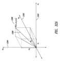
各々が内部デバイス座標系の3つの座標軸の上記方向における直線加速度に関連づけられた3つの数値を含むと共に上記内部デバイス座標系の各軸回りの角速度に関連づけられた3つの数値を含むデータ‐ベクトルを有する上記準備したモニタリング-セッション-データを自然座標系に対して方向づけ、
上記方向づけたデータ‐ベクトルを帯域通過フィルタ処理して、正常運動周波数を含む多数の周波数の各々について1組のデータ‐ベクトルを得、
非正常運動周波数の各々についての上記データ‐ベクトルから、上記自然座標系の上記座標軸方向の各々における空間振幅を求め、
患者についての基本軌跡及び上記正常運動周波数についての上記データ‐ベクトルから、上記自然座標系の上記座標軸方向の各々における空間振幅を求め、そして
上記患者についての上記基本軌跡及び上記正常運動周波数についての上記データ‐ベクトルから、現在における正常運動特性を求めることによって、
上記モニタリング-セッション-データからの運動モード及び追加のメトリック値を表すコンポーネント軌跡を割り出す、実施態様項54記載のモニタリング-セッション-データ収集、分析、及び状態報告システム。
〔実施態様項59〕
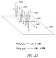
周波数についての上記データ‐ベクトルから、上記自然座標系の上記座標軸方向の各々における空間振幅を求める上記ステップは、上記データ‐ベクトルから空間軌跡を生じさせるステップと、上記空間周波数を上記座標軸の各々上に投射するステップと、上記座標軸の各々上への上記空間周波数の投射長さを求めるステップと、をさらに含む、実施態様項58記載のモニタリング-セッション-データ収集、分析、及び状態報告システム。
〔実施態様項60〕
上記モニタリング-セッション-データ分析コンポーネントは、プロテーゼ状態及び患者状態を上記運動モード及び上記追加のメトリック値から、
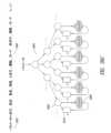
上記運動モード及び上記追加のメトリック値を診断・示唆(diagnosis-and-suggestion)レポートを生じさせる決定木に提出し、
上記運動モードについて生じた振幅、正常運動周波数軌跡及び基本軌跡から生じたメトリクス、及び上記プロテーゼ状態及び上記患者状態を特徴づける出力レポート及び出力データ値のうちの一方又は両方を生じさせる追加のメトリック値と一緒に、上記診断・示唆レポートをパッケージ化することによって、割り出す、実施態様項54記載のモニタリング-セッション-データ収集、分析、及び状態報告システム。
〔実施態様項61〕
上記モニタリング-セッション-データ分析コンポーネントによって標的コンピュータシステムに配信される上記1つ以上の警告及び事象は、
上記患者が即時援助又はインターベンションを必要とする旨を医師又は医療施設に通知する警告を含み、
上記患者によって必要とされる追加のケア及び/又は機器を指示する事象を含み、上記追加のケア及び/又は機器は、上記追加のケア及び/又は機器を上記患者に自動的に提供し又は上記患者に上記追加のケア及び/又は機器を通知して上記患者に上記追加のケア及び/又は機器の獲得に関する情報を提供するよう種々の外部コンピュータシステムによって取り扱い可能である、実施態様項54記載のモニタリング-セッション-データ収集、分析、及び状態報告システム。
〔実施態様項62〕
1つ以上のコンピュータシステムのコンポーネントとして具体化されたモニタリング-セッション-データ収集、分析、及び状態報告システムによって実施される方法であって、各コンピュータシステムは、1つ以上のプロセッサ、1つ以上のメモリ、1つ以上のネットワーク接続手段、及び1つ以上の大容量記憶装置へのアクセスを含み、上記方法は、
患者に取り付けられた又は患者の体内に植え込まれたプロテーゼ内に又は該プロテーゼに近接して設けられたセンサによって生じる加速度データを含むモニタリング-セッション-データを外部モニタリング-セッション-データソースから受け取るステップと、
上記受け取ったモニタリング-セッション-データを上記1つ以上のメモリ及び上記1つ以上の大容量記憶装置のうちの1つ以上に記憶させるステップと、
プロテーゼの状態及び患者の状態を運動モード及び追加のメトリック値から割り出すステップと、
上記割り出したプロテーゼ状態及び患者状態を上記ネットワーク接続手段経由で標的コンピュータシステムに配信するステップと、
上記割り出したプロテーゼ状態及び患者状態によって指示されると、1つ以上の警告及び事象を上記ネットワーク接続手段経由で標的コンピュータシステムに配信するステップと、を含む、方法。
〔実施態様項63〕
プロテーゼ状態及び患者状態を上記運動モード及び上記追加のメトリック値から割り出す上記ステップは、
上記モニタリング-セッション-データを処理のために準備するステップと、
運動モード及び追加のメトリック値を表すコンポーネント軌跡を上記モニタリング-セッション-データから求めるステップと、
上記運動モード及び上記追加のメトリック値を、診断・示唆レポートを生じさせる決定木に提出するステップと、
上記運動モードについて生じた振幅、正常運動周波数軌跡及び基本軌跡から生じたメトリクス、及び上記プロテーゼ状態及び上記患者状態を特徴づける出力レポート及び出力データ値のうちの一方又は両方を生じさせる追加のメトリック値と一緒に、上記診断・示唆レポートをパッケージ化するステップと、を含む、実施態様項62記載の方法。
〔実施態様項64〕
上記モニタリング-セッション-データを処理のために準備する上記ステップは、
各々が第1の内部デバイス座標系の3つの座標軸の方向における直線加速度に関連づけられた3つの数値を含むと共に上記第1又は第2の内部デバイス座標系の各軸回りの角速度に関連づけられた3つの数値を含むデータ‐ベクトルの時間系列を受け取るステップと、
上記データ‐ベクトル系列の再スケール変更が必要とされた場合、上記データ‐ベクトルの上記数値をスケール変更するステップと、
上記データ‐ベクトル系列の標準化が必要とされた場合、上記データ‐ベクトルの上記数値を標準化するステップと、
上記直線加速度に関連づけられた上記数値及び上記角速度に関連づけられた上記数値のうちの1つ以上の変換が上記直線加速度に関連づけられた上記数値及び上記角速度に関連づけられた上記数値を共通内部座標系に関連づけることが必要とされた場合、上記直線加速度に関連づけられた上記数値及び上記角速度に関連づけられた上記数値のうちの1つ以上を変換して、上記共通内部座標系に関連づけるステップと、
データ‐ベクトルの上記時間系列が固定された間隔時間系列に関して同期される必要がある場合、上記データ‐ベクトルを固定された間隔時間系列に関して同期させるステップと、をさらに含む、実施態様項62記載の方法。
〔実施態様項65〕
運動モード及び追加のメトリック値を表すコンポーネント軌跡を上記モニタリング-セッション-データから求める上記ステップは、
各々が内部デバイス座標系の3つの座標軸の上記方向における直線加速度に関連づけられた3つの数値を含むと共に上記内部デバイス座標系の各軸回りの角速度に関連づけられた3つの数値を含むデータ‐ベクトルを有する上記準備したモニタリング-セッション-データを自然座標系に対して方向づけるステップと、
上記方向づけたデータ‐ベクトルを帯域通過フィルタ処理して、正常運動周波数を含む多数の周波数の各々について1組のデータ‐ベクトルを得るステップと、
非正常運動周波数の各々についての上記データ‐ベクトルから、上記自然座標系の上記座標軸方向の各々における空間振幅を求めるステップと、
患者についての基本軌跡及び上記正常運動周波数についての上記データ‐ベクトルから、上記自然座標系の上記座標軸方向の各々における空間振幅を求めるステップと、
上記患者についての上記基本軌跡及び上記正常運動周波数についての上記データ‐ベクトルから、現在における正常運動特性を求めるステップと、をさらに含む、実施態様項62記載の方法。
〔実施態様項66〕
周波数についての上記データ‐ベクトルから、上記自然座標系の上記座標軸方向の各々における空間振幅を求める上記ステップは、
上記データ‐ベクトルから空間軌跡を生じさせるステップと、
上記空間周波数を上記座標軸の各々上に投射するステップと、
上記座標軸の各々上への上記空間周波数の投射長さを求めるステップと、をさらに含む、実施態様項62記載の方法。
〔実施態様項67〕
プロテーゼ状態及び患者状態を上記運動モード及び上記追加のメトリック値から割り出す上記ステップは、
上記運動モード及び上記追加のメトリック値を診断・示唆レポートを生じさせる決定木に提出するステップと、
上記運動モードについて生じた振幅、正常運動周波数軌跡及び基本軌跡から生じたメトリクス、及び上記プロテーゼ状態及び上記患者状態を特徴づける出力レポート及び出力データ値のうちの一方又は両方を生じさせる追加のメトリック値と一緒に、上記診断・示唆レポートをパッケージ化するステップと、をさらに含む、実施態様項62記載の方法。
〔実施態様項68〕
上記モニタリング-セッション-データ分析コンポーネントによって標的コンピュータシステムに配信される上記1つ以上の警告及び事象は、
上記患者が即時援助又はインターベンションを必要とする旨を医師又は医療施設に通知する警告を含み、
上記患者によって必要とされる追加のケア及び/又は機器を指示する事象を含み、上記追加のケア及び/又は機器は、上記追加のケア及び/又は機器を上記患者に自動的に提供し又は上記患者に上記追加のケア及び/又は機器を通知して上記患者に上記追加のケア及び/又は機器の獲得に関する情報を提供するよう種々の外部コンピュータシステムによって取り扱い可能である、実施態様項62記載の方法。
〔実施態様項69〕
コンピュータ命令で符号化された物理的データ記憶装置であって、上記コンピュータ命令は、モニタリング-セッション-データ収集、分析、及び状態報告システムの1つ以上のコンピュータシステム内に設けられた1つ以上のプロセッサによって実行されると、1つ以上のプロセッサ、1つ以上のメモリ、1つ以上のネットワーク接続手段、及び1つ以上の大容量記憶装置へのアクセスを含む各コンピュータシステムは、
患者に取り付けられた又は患者の体内に植え込まれたプロテーゼ内に又は該プロテーゼに近接して設けられたセンサによって生じる加速度データを含むモニタリング-セッション-データを外部モニタリング-セッション-データソースから受け取るよう上記モニタリング-セッション-データ収集、分析、及び状態報告システムを制御する、物理的データ記憶装置。
〔実施態様項70〕
植え込まれた人工関節を備えた患者の体内の関節弛みを割り出す方法であって、a)植え込まれた人工関節の動きを分析するステップと、b)上記運動を先の/標準化された基準と比較するステップと、を含む、方法。
〔実施態様項71〕
プロテーゼが植え込まれている患者の体内の上記植え込まれたプロテーゼの弛みを割り出す方法であって、
a)1つ以上の第1のモニタリングセッション中、植え込まれたプロテーゼの動きを分析することによって動きの標準化された基準を得るステップと、
b)上記1つ以上の第1のモニタリングセッションの次に起こる1つ以上の第2のモニタリングセッション中、植え込まれたプロテーゼの動きを分析することによって動きの現在の内容を得るステップと、
c)運動の上記現在の内容を運動の上記標準化基準と比較し、それにより上記植え込まれたプロテーゼを備えた患者の体内の上記植え込まれたプロテーゼの弛みを突き止めるステップと、を含む、方法。
〔実施態様項72〕
患者の体内のインプラントと関連した臨床的又は準臨床的状態を割り出す方法であって、上記方法は、
a.上記インプラントに直接結合されたセンサを用いて第1のモニタリングセッション中、上記インプラントの第1の運動をモニタして上記第1の運動に関する第1のモニタリング-セッション-データを提供するステップと、
b.上記センサを用いて第2のモニタリングセッション中、上記インプラントの第2の運動をモニタして上記第2の運動に関する第2のモニタリング-セッション-データを提供するステップと、
c.上記第1のモニタリング-セッション-データ又はその処理結果を上記第2のモニタリング-セッション-データ又はその処理結果と比較して、上記インプラントと関連した臨床的又は準臨床的状態を表す比較結果を提供するステップと、を含む、方法。
〔実施態様項73〕
上記臨床的又は準臨床的状態は、オプションとしてプロテーゼ周囲の光輝部(lucency)又はプロテーゼ周囲の骨溶解に起因した上記インプラントの弛みである、実施態様項72記載の方法。
〔実施態様項74〕
上記臨床的又は準臨床的状態は、上記インプラントのアライメント不良(プロテーゼコンポーネントの次善の位置決め)又は再調整(プロテーゼコンポーネントのアライメントの変化)である、実施態様項72記載の方法。
〔実施態様項75〕
上記臨床的又は準臨床的状態は、上記インプラントの変形(摩耗)である、実施態様項72記載の方法。
〔実施態様項76〕
上記患者は、上記臨床的又は準臨床的状態について無症状であり、上記第1及び上記第2のデータ又は該データの処理結果の上記比較結果により、上記状態は、上記第1のモニタリングセッションと上記第2のモニタリングセッションとの間で起こっているらしいことが分かる、実施態様項72記載の方法。
〔実施態様項77〕
上記患者は、上記インプラントの弛みについて無症状であり、上記第1及び上記第2のデータ又は該データの処理結果の上記比較結果により、上記インプラントは、上記第1のモニタリングセッションと上記第2のモニタリングセッションとの間で弛んでいるらしいことが分かる、実施態様項72記載の方法。
〔実施態様項78〕
上記患者は、上記インプラントの再調整について無症状であり、上記第1及び上記第2のデータ又は該データの処理結果の上記比較結果により、上記インプラントは、上記第1のモニタリングセッションと上記第2のモニタリングセッションとの間でアライメントを変えているらしいことが分かる、実施態様項72記載の方法。
〔実施態様項79〕
上記患者は、上記インプラントの変形について無症状であり、上記第1及び上記第2のデータ又は該データの処理結果の上記比較結果により、上記インプラントは、上記第1のモニタリングセッションと上記第2のモニタリングセッションとの間で変形しているらしいことが分かる、実施態様項72記載の方法。
〔実施態様項80〕
患者の体内のインプラントと関連した臨床的又は準臨床的状態を治療する方法であって、
a.患者の体内のインプラントを識別するステップを含み、上記インプラントは、臨床的又は準臨床的状態を呈し、
b.矯正外部ブレーシングを患者に取り付けて適正なアライメント及び/又は上記インプラントに対する向上した安定性を回復させるステップを含む、方法。
〔実施態様項81〕
上記矯正外部ブレーシングは、上記患者及び上記準臨床的状態に合わせて特別にカスタマイズされている、実施態様項80記載の方法。
〔実施態様項82〕
患者の体内のインプラントと関連した臨床的又は準臨床的状態を治療する方法であって、
a.患者の体内のインプラントを識別するステップを含み、上記インプラントは、臨床的又は準臨床的状態を呈し、
b.上記インプラントを固定システムに接触させて上記準臨床的状態の進行を遅らせるステップを含む、方法。
〔実施態様項83〕
上記固定システムは、Kワイヤ、ピン、スクリュー、プレート及び髄内デバイスから選択されたハードウェアを含む、実施態様項82記載の方法。
〔実施態様項84〕
上記インプラントを保持するスクリューが骨を貫通して配置され、上記スクリューの末端が上記インプラントの表面を押して上記インプラントの動きを妨げ、スクリューは、1本、2本、3本、4本、5本、6本、7本、8本、9本、10本、11本、12本、13本、14本、15本、16本、17本、18本、19本及び20本のスクリューから選択される、実施態様項82記載の方法。
〔実施態様項85〕
上記固定システムは、骨セメントを含む、実施態様項82記載の方法。
〔実施態様項86〕
患者の体内のインプラントと関連した臨床的又は準臨床的状態を治療する方法であって、
a.患者の体内のインプラントを識別するステップを含み、上記インプラントは、臨床的又は準臨床的状態を呈し、
b.タンプに接触させるステップを含み、上記接触は、上記患者の体内の上記インプラントの存在場所を変え、そしてオプションとして、
c.セメントを存在場所の変わった上記インプラントの周りに塗布する、方法。
〔実施態様項87〕
上記準臨床的状態は、上記インプラントの再調整である、実施態様項86記載の方法。
〔実施態様項88〕
患者の体内のインプラントと関連した臨床的又は準臨床的状態を治療する方法であって、
a.患者の体内のインプラントを識別するステップを含み、上記インプラントは、臨床的又は準臨床的状態を呈し、
b.インサートを上記インプラントのコンポーネントに隣接して植え込むステップを含み、上記インサートは、上記インプラントの上記コンポーネントに作用する力を加減する、方法。
〔実施態様項89〕
上記インサートは、脛骨インサートである、実施態様項88記載の方法。
〔実施態様項90〕
上記インサートは、(i)最小厚さを備えた外側側及び(ii)上記外側側の上記最小厚さとは同一ではない最小厚さを備えた内側側を有する脛骨インサートである、実施態様項88記載の方法。
〔実施態様項91〕
患者の体内のインプラントと関連した臨床的又は準臨床的状態を治療する方法であって、
a.患者の体内のインプラントを識別するステップを含み、上記インプラントは、臨床的又は準臨床的状態を呈し、
b.オッセオインテグレーション前薬剤を上記インプラントの周りの場所に送り出すステップを含む、方法。
〔実施態様項92〕
上記オッセオインテグレーション前薬剤は、自己骨グラフト、異種骨グラフト、合成骨グラフト、骨ペースト、骨成長因子、及び成長因子から選択される、実施態様項91記載の方法。
〔実施態様項93〕
患者の体内のインプラントと関連した臨床的又は準臨床的状態を治療する方法であって、
a.患者の体内のインプラントを識別するステップを含み、上記インプラントは、臨床的又は準臨床的状態を呈し、
b.抗菌薬を上記インプラントの周りの場所に送り出すステップを含む、方法。
〔実施態様項94〕
上記抗菌薬は、持続放出形態で調合されている、実施態様項93記載の方法。
〔実施態様項95〕
上記インプラントは、インテリジェントインプラントである、実施態様項72~94のうちいずれか一に記載の方法。
〔実施態様項96〕
上記インプラントは、膝関節インプラント、股関節インプラント、及び肩関節インプラントから選択される、実施態様項72~94のうちいずれか一に記載の方法。
〔実施態様項97〕
上記モニタリング-セッション-データの上記処理結果は、運動モードから成る、実施態様項72~94のうちいずれか一に記載の方法。
〔実施態様項98〕
上記モニタリング-セッション-データの上記処理結果は、運動モードから成り、上記インプラントの状態は、上記運動モードから割り出される、実施態様項72~94のうちいずれか一に記載の方法。
〔実施態様項99〕
上記モニタリング-セッション-データの上記処理結果は、運動モードから成り、上記患者の状態は、上記運動モードから割り出される、実施態様項72~94のうちいずれか一に記載の方法。
〔実施態様項100〕
上記インプラントは、上記第1のモニタリングセッションに先立つ少なくとも10週間前に記患者の体内に配置されている、実施態様項72~94のうちいずれか一に記載の方法。
〔実施態様項101〕
上記インプラントは、少なくとも2週間の期間にわたってアライメントを変化させている、実施態様項72~94のうちいずれか一に記載の方法。
〔実施態様項102〕
上記インプラントは、少なくとも2週間の期間にわたって弛くなっている、実施態様項72~94のうちいずれか一に記載の方法。
〔実施態様項103〕
上記インプラントは、少なくとも2週間の期間にわたって変形している、実施態様項72~94のうちいずれか一に記載の方法。
〔実施態様項104〕
上記インプラントは、上記センサが上記臨床的又は準臨床的状態に関する遠隔医療コードに関連づけられた周波数でセンサ信号を発生させるよう構成された制御回路を有する、実施態様項72~94のうちいずれか一に記載の方法。
〔実施態様項105〕
上記インプラントは、医師が遠隔医療保険コード下で支払いの資格を有することができるようにする周波数で上記センサがセンサ信号を発生させるよう構成された制御回路を有し、上記センサ信号は、上記周波数で生じる、実施態様項72~94のうちいずれか一に記載の方法。
〔実施態様項106〕
上記インプラントは、医師が遠隔医療保険コード下で全額支払いの資格を有することができるようにする周波数で上記センサがセンサ信号を発生させるよう構成された制御回路を有し、上記センサ信号は、上記周波数で生じる、実施態様項72~94のうちいずれか一に記載の方法。
〔実施態様項107〕
(i)医師が遠隔医療保険コード下で利用できる全額支払いの資格を有し、又は(ii)医師が遠隔医療保険コード下で利用できる支払いの資格を有することができるようにする周波数で、上記インプラントに関連づけられたセンサ信号を発生させるステップをさらに含む、実施態様項72~94のうちいずれか一に記載の方法。
〔実施態様項108〕
方法であって、
a.患者の関節に隣接した骨内に植え込まれたインテリジェントプロテーゼを用意するステップを含み、加速度計が上記インテリジェントプロテーゼ内に納められ、上記加速度計は、上記骨内に位置決めされ、
b.上記植え込まれたインテリジェントプロテーゼを外部環境に対して動かすステップを含み、上記患者は、上記植え込まれたインテリジェントプロテーゼが第1のモニタリングセッション中に動かされる場所に配置され、
c.上記第1のモニタリングセッション中、第1の測定を上記加速度計で行うステップを含み、上記第1の測定は、上記植え込まれたインテリジェントプロテーゼの状態を上記第1の測定の時点で識別する第1のモニタリング-セッション-データ又はその処理結果を提供する、方法。
〔実施態様項109〕
上記加速度計は、複数の加速度計である、実施態様項108記載の方法。
〔実施態様項110〕
上記加速度計は、1軸加速度計、2軸加速度計、及び3軸加速度計から選択される、実施態様項108記載の方法。
〔実施態様項111〕
上記加速度計は、ブロードバンドモードで動作する、実施態様項108記載の方法。
〔実施態様項112〕
上記骨は脛骨である、実施態様項108記載の方法。
〔実施態様項113〕
上記加速度計は、上記インテリジェントプロテーゼの脛骨延長部内に配置される、実施態様項108記載の方法。
〔実施態様項114〕
上記植え込まれたインテリジェントプロテーゼは、衝撃力が上記第1のモニタリングセッション中、上記患者又は上記インテリジェントプロテーゼに加えられることなく、上記外部環境に対して動かされる、実施態様項108記載の方法。
〔実施態様項115〕
上記外部環境は、上記患者の住居から成る、実施態様項108記載の方法。
〔実施態様項116〕
上記外部環境は、手術室から成り、上記インテリジェントプロテーゼは、上記患者の体内に植え込まれている、実施態様項108記載の方法。
〔実施態様項117〕
上記植え込まれたインテリジェントプロテーゼの上記状態は、上記骨内の上記植え込まれたインテリジェントプロテーゼの弛みの特性評価である、実施態様項108記載の方法。
〔実施態様項118〕
上記植え込まれたインテリジェントプロテーゼの上記状態は、上記骨内の上記植え込まれたインテリジェントプロテーゼのアライメントの特性評価である、実施態様項108記載の方法。
〔実施態様項119〕
上記植え込まれたインテリジェントプロテーゼの上記状態は、上記植え込まれたインテリジェントプロテーゼの摩耗の特性評価である、実施態様項108記載の方法。
〔実施態様項120〕
上記植え込まれたインテリジェントプロテーゼの上記状態は、上記植え込まれたインテリジェントプロテーゼに隣接して位置する骨内の領域の細菌感染の特性評価である、実施態様項108記載の方法。
〔実施態様項121〕
上記植え込まれたインテリジェントプロテーゼの上記状態は、準臨床的状態を指示している、実施態様項108記載の方法。
〔実施態様項122〕
上記ステップb)は、待機期間後に繰り返され、上記ステップb)の上記繰り返しは、上記植え込まれたインテリジェントプロテーゼを上記患者の居る外部環境に対して動かすステップを含み、上記植え込まれたインテリジェントプロテーゼは、第2のモニタリングセッション中に動かされ、第2の測定が上記第2のモニタリングセッション中に上記加速度計で行われ、上記第2の測定は、上記第2の測定時点における上記植え込まれたインテリジェントプロテーゼの状態を識別する第2のモニタリング-セッション-データ又は第2のモニタリング-セッション-データ処理結果を提供する、実施態様項108記載の方法。
〔実施態様項123〕
上記ステップb)は、複数回にわたって繰り返され、上記複数の回数は、同一又は同一ではない待機期間だけ互いに分けられ、上記ステップb)の上記繰り返しは、上記植え込まれたインテリジェントプロテーゼを上記患者の居る外部環境に対して動かすステップを含み、上記植え込まれたインテリジェントプロテーゼは、複数のモニタリングセッション中動かされ、測定は、上記複数のモニタリングセッションの各々の間に上記加速度計で行われ、上記測定は、各々が上記測定時点における上記植え込まれたインテリジェントプロテーゼの状態を識別する複数のモニタリング-セッション-データ又は複数のモニタリング-セッション-データの処理結果を提供する、実施態様項108記載の方法。
〔実施態様項124〕
上記ステップb)は、複数回にわたって繰り返され、上記複数の回数は、同一又は同一ではない待機期間だけ互いに分けられ、上記ステップb)の上記繰り返しは、上記植え込まれたインテリジェントプロテーゼを上記患者の居る外部環境に対して動かすステップを含み、上記植え込まれたインテリジェントプロテーゼは、複数のモニタリングセッション中動かされ、測定は、上記複数のモニタリングセッションの各々の間に上記加速度計で行われ、上記測定は、各々が上記測定時点における上記植え込まれたインテリジェントプロテーゼの状態を識別する複数のモニタリング-セッション-データ又は複数のモニタリング-セッション-データの処理結果を提供し、上記複数のモニタリング-セッション-データは、オプションとして、2~20回のモニタリングセッションから選択され、上記複数のモニタリング-セッション-データは、一緒に考慮されると、上記複数のモニタリングセッションが起こった時間の間、上記植え込まれたインテリジェントプロテーゼの状態の変化を指示する、実施態様項108記載の方法。
〔実施態様項125〕
上記状態の上記変化は、上記植え込まれたインテリジェントプロテーゼの周りの組織の治癒状態を表している、実施態様項124記載の方法。
〔実施態様項126〕
上記状態の上記変化は、上記植え込まれたインテリジェントプロテーゼの周りの組織の感染状態を表している、実施態様項124記載の方法。
〔実施態様項127〕
上記状態の上記変化は、上記骨内の上記植え込まれたインテリジェントプロテーゼの弛みを表している、実施態様項124記載の方法。
〔実施態様項128〕
上記状態の上記変化は、上記植え込まれたインテリジェントプロテーゼの摩耗を表している、実施態様項124記載の方法。
〔実施態様項129〕
上記状態の上記変化は、上記植え込まれたインテリジェントプロテーゼのアライメント不良を表している、実施態様項124記載の方法。
〔実施態様項130〕
上記状態の上記変化は、上記植え込まれたインテリジェントプロテーゼのアライメントの変化を表している、実施態様項124記載の方法。 Below are some exemplary numbered embodiments of the present invention.
[Embodiment 1]
A tibial insert for an implantable artificial knee joint, comprising a tibial insert in which the thickness of an inner side of the implant is 1 mm, 2 mm, 3 mm, 4 mm, 5 mm, 6 mm, 7 mm, 8 mm, 9 mm, or 10 mm thicker than the thickness of an outer side of the implant.
[Embodiment 2]
A tibial insert for an implantable artificial knee joint, comprising a tibial insert having a thickness on an outer side of the implant that is 1 mm, 2 mm, 3 mm, 4 mm, 5 mm, 6 mm, 7 mm, 8 mm, 9 mm, or 10 mm thicker than the thickness on an inner side of the implant.
[Embodiment 3]
A tibial insert for an implantable knee prosthesis, comprising a tibial insert having a thickness on an anterior side of the implant that is 1 mm, 2 mm, 3 mm, 4 mm, 5 mm, 6 mm, 7 mm, 8 mm, 9 mm, or 10 mm thicker than a thickness on a posterior side of the implant.
[Embodiment 4]
A tibial insert for an implantable knee prosthesis, comprising a tibial insert having a posterior thickness that is 1 mm, 2 mm, 3 mm, 4 mm, 5 mm, 6 mm, 7 mm, 8 mm, 9 mm, or 10 mm thicker than an anterior thickness of the implant.
[Embodiment 5]
1. A tibial insert or joint spacer for an implantable knee prosthesis, comprising a tibial insert having a thickness on one of the medial, lateral, anterior and/or posterior sides of the implant that is 1 mm, 2 mm, 3 mm, 4 mm, 5 mm, 6 mm, 7 mm, 8 mm, 9 mm, or 10 mm thicker than the thickness of the corresponding side of the implant.
[Embodiment 6]
The tibial insert according to any one of the first to fifth embodiments, wherein the tibial insert is made of polyethylene or polyetheretherketone (PEEK).
[Embodiment 7]
The tibial insert according to any one of
[Embodiment 8]
The tibial insert according to any one of
[Embodiment Item 9]
1. An implantable medical device comprising: a circuit configured to be fixedly attached to an implantable prosthetic device; a power component; and an apparatus configured to decouple the circuit from the power component.
[Embodiment 10]
1. An implantable medical device comprising: a circuit configured to be fixedly attached to an implantable prosthetic device; a battery; and a fuse coupled between the circuit and the battery.
[Embodiment 11]
11. A method, comprising the step of electrically opening a fuse disposed between a circuit and a battery, wherein at least the fuse and the circuit are disposed in an implantable prosthetic device.
[Embodiment 12]
1. An implantable medical device comprising: at least one sensor configured to generate a sensor signal; and control circuitry configured to cause the at least one sensor to generate the sensor signal at a frequency associated with a telemedicine code.
[Embodiment 13]
1. An implantable medical device comprising: at least one sensor configured to generate a sensor signal; and control circuitry configured to generate the sensor signal at a frequency that enables a physician to be eligible for payment for the at least one sensor under telehealth insurance codes.
[Embodiment 14]
1. An implantable medical device comprising: at least one sensor configured to generate a sensor signal; and control circuitry configured to generate the sensor signal at a frequency that enables a physician to be eligible for full payment for the at least one sensor under telehealth insurance codes.
[Embodiment 15]
A method comprising generating a sensor signal associated with an implantable medical device at a frequency that enables a physician to qualify for payment under telemedicine insurance codes.
[Embodiment 16]
A method, comprising generating a sensor signal associated with an implantable medical device at a frequency that enables a physician to be eligible for full payment under telemedicine insurance codes.
[Embodiment 17]
1. An implantable prosthesis comprising:
Housing and
an implantable circuit disposed within the housing, the implantable circuit comprising:
generating at least one first signal representative of the movement;
determining whether the at least one first signal meets at least one first criterion; and transmitting the at least one first signal to a remote location in response to determining that the at least one first signal meets the at least one first criterion.
[Embodiment 18]
A base station,
Housing and
and a base station circuit disposed within the housing, the base station circuit comprising:
receiving at least one first signal representative of movement from the implantable prosthesis;
sending said at least one first signal to a destination;
A base station configured to receive at least one second signal from a source; and transmit said at least one second signal to said implantable prosthesis.
[Embodiment 19]
11. A method comprising the step of opening a fuse disposed in an implantable prosthesis between a power source and implantable circuitry in response to a current passing through said fuse exceeding an overcurrent threshold.
[Embodiment 20]
11. A method comprising the step of opening a fuse disposed in an implantable prosthesis between a power source and implantable circuitry in response to a current passing through said fuse exceeding an overcurrent threshold for at least a threshold time.
[Embodiment 21]
11. A method comprising the step of opening a fuse located in an implantable prosthesis between a power source and implantable circuitry in response to a voltage across said fuse exceeding an overvoltage threshold.
[Embodiment 22]
1. A method comprising the step of opening a fuse in an implantable prosthesis between a power source and implantable circuitry in response to a voltage across said fuse exceeding an overvoltage threshold for at least a threshold time.
[Embodiment 23]
A method comprising the step of opening a fuse located in an implantable prosthesis between a power source and implantable circuitry in response to a temperature exceeding an overtemperature threshold.
[Embodiment Item 24]
A method comprising the step of opening a fuse located in an implantable prosthesis between a power source and implantable circuitry in response to a temperature exceeding an overtemperature threshold for at least a threshold length of time.
[Embodiment 25]
A method, comprising the steps of generating a sensor signal in response to movement of a patient in which a prosthesis is implanted, and transmitting the sensor signal to a remote location.
[Embodiment 26]
11. A method, comprising: generating a sensor signal in response to movement of a patient in which a prosthesis is implanted; sampling the sensor signal; and transmitting the sample to a remote location.
[Embodiment 27]
A method comprising the steps of generating a sensor signal in response to movement of a patient in which a prosthesis is implanted, determining whether the sensor signal represents a qualifying event, and transmitting the sensor signal to a remote location in response to determining whether the sensor signal represents a qualifying event.
[Embodiment 28]
1. A method comprising:
1. A method comprising: generating a sensor signal in response to movement of a patient in which a prosthesis is implanted; receiving a polling signal from a remote location; and transmitting the sensor signal to the remote location in response to the polling signal.
[Embodiment 29]
1. A method, comprising: generating a sensor signal in response to movement of a patient in which a prosthesis is implanted; creating a message including the sensor signal or data representative of the sensor signal; and transmitting the message to a remote location.
[Embodiment 30]
1. A method, comprising: generating a sensor signal in response to movement of a patient in which a prosthesis is implanted; generating a data packet including the sensor signal or data representative of the sensor signal; and transmitting the data packet to a remote location.
[Embodiment Item 31]
1. A method, comprising: generating a sensor signal in response to movement of a patient in which a prosthesis is implanted; encrypting at least a portion of the sensor signal or data representative of the sensor signal; and transmitting the encrypted sensor signal to a remote location.
[Embodiment Item 32]
1. A method, comprising: generating a sensor signal in response to movement of a patient in which a prosthesis is implanted; encoding at least a portion of the sensor signal or data representative of the sensor signal; and transmitting the encoded sensor signal to a remote location.
[Embodiment Item 33]
1. A method, comprising the steps of: generating a sensor signal in response to movement of a patient in which a prosthesis is implanted; transmitting the sensor signal to a remote location; and setting implantable circuitry associated with the prosthesis to a low power mode after transmission of the sensor signal.
[Embodiment Item 34]
1. A method, comprising the steps of: generating a first sensor signal in response to movement of a patient in whom a prosthesis is implanted; transmitting the first sensor signal to a remote location; placing at least one component of implantable circuitry associated with the prosthesis into a low power mode after transmission of the sensor signal; and generating a second sensor signal in response to movement of the patient after a low power mode time period for which the implantable circuitry is configured.
[Embodiment 35]
A method, comprising the steps of receiving a sensor signal from a prosthesis attached to or implanted within a patient, and transmitting the received sensor signal to a destination.
[Embodiment 36]
A method comprising the steps of: sending a query to a prosthesis attached to or implanted within a patient; receiving a sensor signal from the prosthesis after sending the query; and transmitting the received sensor signal to a destination.
[Embodiment Item 37]
1. A method comprising the steps of receiving a sensor signal and at least one identifier from a prosthesis attached to or implanted within a patient, determining whether the identifier is correct, and transmitting the received sensor signal to a destination in response to determining whether the identifier is correct.
[Embodiment 38]
1. A method, comprising: receiving a message including a sensor signal from a prosthesis attached to or implanted within a patient; decrypting at least a portion of the message; and transmitting the decrypted message to a destination.
[Embodiment 39]
1. A method, comprising: receiving a message including a sensor signal from a prosthesis attached to or implanted within a patient; decoding at least a portion of the message; and transmitting the decoded message to a destination.
[Embodiment 40]
1. A method, comprising the steps of: receiving a message including a sensor signal from a prosthesis attached to or implanted within a patient; encoding at least a portion of the message; and transmitting the encoded message to a destination.
[Embodiment 41]
1. A method, comprising: receiving a message including a sensor signal from a prosthesis attached to or implanted within a patient; encrypting at least a portion of the message; and transmitting the encrypted message to a destination.
[Embodiment Item 42]
1. A method, comprising: receiving a data packet including a sensor signal from a prosthesis attached to or implanted within a patient; decrypting at least a portion of the data packet; and transmitting the decrypted data packet to a destination.
[Embodiment Item 43]
1. A method, comprising: receiving a data packet including a sensor signal from a prosthesis attached to or implanted within a patient; decoding at least a portion of the data packet; and transmitting the decoded data packet to a destination.
[Embodiment Item 44]
1. A method, comprising: receiving a data packet including a sensor signal from a prosthesis attached to or implanted within a patient; encoding at least a portion of the data packet; and transmitting the encoded data packet to a destination.
[Embodiment Item 45]
1. A method, comprising: receiving a data packet including a sensor signal from a prosthesis attached to or implanted within a patient; encrypting at least a portion of the data packet; and transmitting the encrypted data packet to a destination.
[Embodiment 46]
1. A method, comprising: receiving a sensor signal from a prosthesis attached to or implanted within a patient; deciphering at least a portion of the sensor signal; and transmitting the deciphered sensor signal to a destination.
[Embodiment Item 47]
1. A method, comprising: receiving a sensor signal from a prosthesis attached to or implanted within a patient; decoding at least a portion of the sensor signal; and transmitting the decoded sensor signal to a destination.
[Embodiment 48]
1. A method, comprising the steps of receiving a sensor signal from a prosthesis attached to or implanted within a patient, encoding at least a portion of the sensor signal, and transmitting the encoded sensor signal to a destination.
[Embodiment 49]
1. A method, comprising the steps of receiving a sensor signal from a prosthesis attached to or implanted within a patient, encrypting at least a portion of the sensor signal, and transmitting the encrypted sensor signal to a destination.
[Embodiment 50]
Implantable circuits for implantable prostheses.
[Embodiment 51]
An implantable prosthesis having an implantable circuit.
[Embodiment 52]
An implantable prosthesis having a fuse.
[Embodiment 53]
A base station capable of communicating with an implantable prosthesis.
[Embodiment 54]
A monitoring-session-data collection, analysis, and status reporting system embodied as a component of one or more computer systems, each computer system including one or more processors, one or more memories, one or more network connections, and access to one or more mass storage devices, said one or more monitoring-session-data collection, data analysis, and status reporting systems comprising:
a monitoring-session-data receiving component configured to receive monitoring-session-data from an external monitoring-session-data source, the monitoring-session-data including acceleration data generated by a sensor located in or near a prosthesis attached to or implanted in the patient, and to store the received monitoring-session-data in the one or more memories and the one or more mass storage devices;
a monitoring-session-data processing component, the monitoring-session-data processing component comprising:
preparing said monitoring session data for processing;
determining component trajectories representative of motion modes and additional metric values from the monitoring session data;
a monitoring-session-data analysis component, the monitoring-session-data analysis component comprising:
determining a prosthesis status and a patient status from said motion mode and said additional metric value;
a monitoring-session-data collection, analysis, and status reporting system that delivers the determined prosthesis status and patient status to a target computer system via the network connection means; and, when indicated by the determined prosthesis status and patient status, delivers one or more alerts and events to the target computer system via the network connection means.
[Embodiment Item 55]
55. The monitoring session data collection, analysis and status reporting system of embodiment 54, wherein said monitoring session data includes a patient identifier, an instrument identifier, a time stamp, device configuration data, and an ordered set of data.
[Embodiment 56]
The ordered set of data is
The monitoring session data collection, analysis and status reporting system of embodiment 55 includes one of: a time series of data vectors each including numerical values related to linear acceleration with respect to three coordinate axes of an internal device coordinate system; and a time series of data vectors each including numerical values related to linear acceleration with respect to three coordinate axes of a first internal device coordinate system and numerical values related to angular velocity, with respect to the first internal device coordinate system or the second internal device coordinate system.
[Embodiment 57]
The monitoring-session-data processing component includes:
receiving a time sequence of data-vectors each including three values associated with linear acceleration in the directions of three coordinate axes of a first internal device coordinate system and three values associated with angular velocity about each axis of said first or second internal device coordinate system;
rescaling the numerical values of the data-vectors if rescaling of the data-vector sequence is required;
standardizing the numerical values of the data-vectors if standardization of the data-vector series is required;
If a transformation of one or more of the numerical values related to the linear acceleration and the numerical values related to the angular velocity is required to relate the numerical values related to the linear acceleration and the numerical values related to the angular velocity to a common internal coordinate system, transforming one or more of the numerical values related to the linear acceleration and the numerical values related to the angular velocity to relate them to the common internal coordinate system; and
The monitoring-session-data collection, analysis, and status reporting system of embodiment 54, wherein if the time sequence of data-vectors needs to be synchronized with respect to a fixed interval time sequence, the monitoring-session-data is prepared for processing by synchronizing the data-vectors with respect to a fixed interval time sequence.
[Embodiment 58]
The monitoring-session-data processing component includes:
orienting said prepared monitoring-session-data with respect to a natural coordinate system, said data-vectors each including three values associated with linear acceleration in said directions of three coordinate axes of the internal device coordinate system and including three values associated with angular velocity about each axis of said internal device coordinate system;
bandpass filtering the oriented data-vectors to obtain a set of data-vectors for each of a number of frequencies including normal movement frequencies;
determining a spatial amplitude in each of said coordinate axes directions of said natural coordinate system from said data vector for each of said abnormal motion frequencies;
determining a spatial amplitude in each of the coordinate axis directions of the natural coordinate system from the fundamental trajectory and the data vector for the normal movement frequency of the patient; and determining a current normal movement characteristic from the fundamental trajectory and the data vector for the normal movement frequency of the patient.
55. The monitoring session data collection, analysis and status reporting system of embodiment 54, further comprising determining component trajectories representative of motion modes and additional metric values from said monitoring session data.
[Embodiment 59]
The monitoring-session-data collection, analysis, and status reporting system of embodiment 58, wherein the step of determining spatial amplitude in each of the coordinate axis directions of the natural coordinate system from the data-vector for frequency further comprises the steps of generating a spatial locus from the data-vector, projecting the spatial frequency onto each of the coordinate axes, and determining the projection length of the spatial frequency onto each of the coordinate axes.
[Embodiment Item 60]
The monitoring-session-data analysis component derives prosthesis status and patient status from the motion mode and the additional metric values,
submitting said motion mode and said additional metric value to a decision tree which produces a diagnosis-and-suggestion report;
The monitoring session data collection, analysis and status reporting system of embodiment 54 determines by packaging the diagnostic and suggestive report together with the amplitudes generated for the motion modes, metrics generated from normal motion frequency trajectories and fundamental trajectories, and additional metric values that result in one or both of an output report and output data values characterizing the prosthesis status and the patient status.
[Embodiment Item 61]
The one or more alerts and events delivered to the target computer system by the monitoring-session-data analysis component include:
an alert to notify a physician or medical facility that the patient requires immediate assistance or intervention;
The monitoring-session-data collection, analysis, and status reporting system of embodiment 54, further comprising an event indicating additional care and/or equipment required by the patient, the additional care and/or equipment being handleable by various external computer systems to automatically provide the additional care and/or equipment to the patient or to notify the patient of the additional care and/or equipment and provide the patient with information regarding the acquisition of the additional care and/or equipment.
[Embodiment Item 62]
A method implemented by a monitoring session data collection, analysis, and status reporting system embodied as a component of one or more computer systems, each computer system including one or more processors, one or more memories, one or more network connections, and access to one or more mass storage devices, the method comprising:
receiving monitoring-session-data from an external monitoring-session-data source, the monitoring-session-data including acceleration data generated by a sensor located in or adjacent to a prosthesis attached to or implanted within the patient;
storing said received monitoring-session-data in one or more of said one or more memories and said one or more mass storage devices;
determining a state of the prosthesis and a state of the patient from the mode of motion and the additional metric value;
delivering said determined prosthesis status and patient status to a target computer system via said network connection means;
and delivering one or more alerts and events via said network connection means to a target computer system as dictated by said determined prosthesis status and patient status.
[Embodiment Item 63]
said step of determining a prosthesis status and a patient status from said motion mode and said additional metric value comprising:
preparing said monitoring session data for processing;
determining component trajectories representative of motion modes and additional metric values from said monitoring session data;
submitting said motion mode and said additional metric values to a decision tree which generates a diagnostic and suggestive report;
The method of embodiment 62, further comprising packaging the diagnostic and suggestive report together with the amplitudes generated for the motion modes, metrics generated from normal motion frequency trajectories and fundamental trajectories, and additional metric values resulting in one or both of an output report and output data values characterizing the prosthesis status and the patient status.
[Embodiment Item 64]
said step of preparing said monitoring session data for processing further comprising:
receiving a time sequence of data-vectors each including three values associated with linear acceleration in the directions of three coordinate axes of a first internal device coordinate system and three values associated with angular velocity about each axis of said first or second internal device coordinate system;
rescaling said numerical values of said data-vectors if rescaling of said data-vector sequence is required;
standardizing the numerical values of the data-vectors if standardization of the data-vector sequence is required;
if transformation of one or more of the numerical values related to the linear acceleration and the numerical values related to the angular velocity is required to relate the numerical values related to the linear acceleration and the numerical values related to the angular velocity to a common internal coordinate system, transforming one or more of the numerical values related to the linear acceleration and the numerical values related to the angular velocity to relate them to the common internal coordinate system;
63. The method of embodiment 62, further comprising the step of: if said time sequence of data-vectors needs to be synchronized with respect to a fixed interval time sequence, synchronizing said data-vectors with respect to a fixed interval time sequence.
[Embodiment Item 65]
said step of determining component trajectories representative of motion modes and additional metric values from said monitoring session data comprising:
orienting said prepared monitoring-session-data with respect to a natural coordinate system, said data-vectors each including three values associated with linear acceleration in said directions of three coordinate axes of the internal device coordinate system and including three values associated with angular velocity about each axis of said internal device coordinate system;
bandpass filtering the oriented data-vectors to obtain a set of data-vectors for each of a number of frequencies including normal movement frequencies;
determining a spatial amplitude in each of said coordinate axes directions of said natural coordinate system from said data vector for each abnormal motion frequency;
determining spatial amplitudes in each of said coordinate axes directions of said natural coordinate system from said data-vector for said fundamental trajectory and said normal movement frequency for a patient;
63. The method of embodiment 62, further comprising the step of determining current normal movement characteristics from the data vector for the fundamental trajectory and the normal movement frequency for the patient.
[Embodiment Item 66]
The step of determining spatial amplitude in each of the coordinate axis directions of the natural coordinate system from the data vector for frequency comprises:
generating spatial trajectories from said data-vectors;
projecting said spatial frequencies onto each of said coordinate axes;
63. The method of embodiment 62, further comprising the step of determining a projection length of said spatial frequency onto each of said coordinate axes.
[Embodiment Item 67]
said step of determining a prosthesis status and a patient status from said motion mode and said additional metric value comprising:
submitting said motion mode and said additional metric value to a decision tree which generates a diagnostic and suggestive report;
The method of embodiment 62, further comprising packaging the diagnostic and suggestive report together with the amplitudes generated for the motion modes, metrics generated from normal motion frequency trajectories and fundamental trajectories, and additional metric values resulting in one or both of an output report and output data values characterizing the prosthesis status and the patient status.
[Embodiment Item 68]
The one or more alerts and events delivered to a target computer system by the monitoring-session-data analysis component include:
an alert to notify a physician or medical facility that the patient requires immediate assistance or intervention;
The method of embodiment 62, further comprising an event indicating additional care and/or equipment required by the patient, the additional care and/or equipment being capable of being handled by various external computer systems to automatically provide the additional care and/or equipment to the patient or to notify the patient of the additional care and/or equipment and provide the patient with information regarding the acquisition of the additional care and/or equipment.
[Embodiment Item 69]
a physical data storage device encoded with computer instructions that, when executed by one or more processors located within one or more computer systems of a monitoring-session-data collection, analysis, and status reporting system, cause each computer system, including one or more processors, one or more memories, one or more network connections, and access to one or more mass storage devices, to:
A physical data storage device that controls the monitoring-session-data collection, analysis, and status reporting system to receive monitoring-session-data from an external monitoring-session-data source, including acceleration data generated by a sensor located within or adjacent to a prosthesis attached to or implanted within the patient.
[Embodiment 70]
A method for determining joint loosening in a patient with an implanted prosthetic joint, the method comprising the steps of: a) analyzing the motion of the implanted prosthetic joint; and b) comparing said motion to a previous/standardized baseline.
[Embodiment Item 71]
1. A method for determining loosening of an implanted prosthesis in a patient having the prosthesis implanted therein, comprising:
a) obtaining a standardized measure of motion by analyzing the motion of an implanted prosthesis during one or more first monitoring sessions;
b) obtaining a current content of motion by analyzing the motion of the implanted prosthesis during one or more second monitoring sessions subsequent to the one or more first monitoring sessions;
and c) comparing said current content of motion with said standardized criteria of motion, thereby determining loosening of said implanted prosthesis in a patient with said implanted prosthesis.
[Embodiment Item 72]
1. A method for determining a clinical or subclinical condition associated with an implant in a patient, the method comprising:
a. monitoring a first movement of the implant during a first monitoring session with a sensor directly coupled to the implant to provide first monitoring-session data relating to the first movement;
b. monitoring a second movement of the implant during a second monitoring session with the sensor to provide second monitoring-session data relating to the second movement;
and c) comparing said first monitoring session data or a result of processing thereof with said second monitoring session data or a result of processing thereof to provide a comparison result indicative of a clinical or sub-clinical condition associated with said implant.
[Embodiment Item 73]
73. The method of embodiment 72, wherein said clinical or subclinical condition is optionally periprosthetic lucency or loosening of said implant due to periprosthetic osteolysis.
[Embodiment Item 74]
73. The method of embodiment 72, wherein said clinical or subclinical condition is malalignment (suboptimal positioning of a prosthetic component) or realignment (change in alignment of a prosthetic component) of said implant.
[Embodiment Item 75]
73. The method of embodiment 72, wherein said clinical or subclinical condition is deformation (wear) of said implant.
[Embodiment Item 76]
The method of embodiment 72, wherein the patient is asymptomatic for the clinical or subclinical condition, and the comparison of the first and second data or the processing results of said data indicates that the condition is likely to have occurred between the first monitoring session and the second monitoring session.
[Embodiment Item 77]
The method of embodiment 72, wherein the patient is asymptomatic about loosening of the implant and the comparison of the first and second data or the processing results of the data indicates that the implant is likely to have loosened between the first monitoring session and the second monitoring session.
[Embodiment Item 78]
The method of embodiment 72, wherein the patient is asymptomatic regarding readjustment of the implant and the comparison of the first and second data or the processing results of the data indicates that the implant is likely to have changed alignment between the first monitoring session and the second monitoring session.
[Embodiment Item 79]
The method of embodiment 72, wherein the patient is asymptomatic about deformation of the implant and the comparison of the first and second data or the processing results of the data indicates that the implant is likely to have deformed between the first monitoring session and the second monitoring session.
[Embodiment 80]
1. A method of treating a clinical or subclinical condition associated with an implant in a patient's body, comprising:
a. identifying an implant in a patient's body, said implant exhibiting a clinical or subclinical condition;
b) fitting a corrective external bracing to the patient to restore proper alignment and/or improved stability to said implant.
[Embodiment 81]
The method of
[Embodiment Item 82]
1. A method of treating a clinical or subclinical condition associated with an implant in a patient's body, comprising:
a. identifying an implant in a patient's body, said implant exhibiting a clinical or subclinical condition;
b) contacting said implant with a fixation system to slow the progression of said subclinical condition.
[Embodiment 83]
The method of embodiment 82, wherein the fixation system includes hardware selected from K-wires, pins, screws, plates and intramedullary devices.
[Embodiment Item 84]
83. The method of embodiment 82, wherein a screw holding the implant is placed through the bone, the end of the screw pressing against the surface of the implant to prevent movement of the implant, the screw being selected from 1, 2, 3, 4, 5, 6, 7, 8, 9, 10, 11, 12, 13, 14, 15, 16, 17, 18, 19 and 20 screws.
[Embodiment 85]
83. The method of embodiment 82, wherein the fixation system comprises bone cement.
[Embodiment 86]
1. A method of treating a clinical or subclinical condition associated with an implant in a patient's body, comprising:
a. identifying an implant in a patient's body, said implant exhibiting a clinical or subclinical condition;
b. contacting the implant with a tamp, said contacting changing the location of the implant within the patient's body, and optionally
c) applying cement around the displaced implant.
[Embodiment 87]
87. The method of embodiment 86, wherein said subclinical condition is a reconditioning of said implant.
[Embodiment Item 88]
1. A method of treating a clinical or subclinical condition associated with an implant in a patient's body, comprising:
a. identifying an implant in a patient's body, said implant exhibiting a clinical or subclinical condition;
b) implanting an insert adjacent to a component of said implant, said insert modifying a force acting on said component of said implant.
[Embodiment 89]
The method of embodiment 88, wherein the insert is a tibial insert.
[Embodiment Item 90]
The method of embodiment 88, wherein the insert is a tibial insert having (i) a lateral side with a minimum thickness and (ii) a medial side with a minimum thickness that is not the same as the minimum thickness of the lateral side.
[Embodiment Item 91]
1. A method of treating a clinical or subclinical condition associated with an implant in a patient's body, comprising:
a. identifying an implant in a patient's body, said implant exhibiting a clinical or subclinical condition;
b) A method comprising the step of delivering a pre-osseointegration agent to a location around said implant.
[Embodiment Item 92]
92. The method of embodiment 91, wherein said pre-osseointegration agent is selected from autologous bone graft, allogeneic bone graft, synthetic bone graft, bone paste, bone growth factor, and growth factor.
[Embodiment Item 93]
1. A method of treating a clinical or subclinical condition associated with an implant in a patient's body, comprising:
a. identifying an implant in a patient's body, said implant exhibiting a clinical or subclinical condition;
b) delivering an antimicrobial agent to a location around said implant.
[Embodiment Item 94]
94. The method of embodiment 93, wherein said antimicrobial agent is formulated in sustained release form.
[Embodiment Item 95]
The method of any one of claims 72 to 94, wherein the implant is an intelligent implant.
[Embodiment Item 96]
The method of any one of paragraphs 72-94, wherein the implant is selected from a knee implant, a hip implant, and a shoulder implant.
[Embodiment Item 97]
The method of any one of claims 72 to 94, wherein the processing results of the monitoring session data comprise an exercise mode.
[Embodiment Item 98]
The method of any one of paragraphs 72 to 94, wherein the processing result of the monitoring session data comprises a motion mode, and the status of the implant is determined from the motion mode.
[Embodiment Item 99]
The method of any one of claims 72 to 94, wherein the processing result of the monitoring session data comprises an exercise mode, and the patient's condition is determined from the exercise mode.
[Embodiment Item 100]
95. The method of any one of embodiments 72-94, wherein the implant has been placed in the patient's body at least 10 weeks prior to the first monitoring session.
[Embodiment Item 101]
The method of any one of paragraphs 72-94, wherein said implant changes alignment over a period of at least two weeks.
[Embodiment Item 102]
The method of any one of paragraphs 72-94, wherein the implant has been loosened for a period of at least two weeks.
[Embodiment Item 103]
The method of any one of paragraphs 72-94, wherein the implant is deformed for a period of at least two weeks.
[Embodiment Item 104]
The method of any one of claims 72-94, wherein the implant has control circuitry configured to cause the sensor to generate a sensor signal at a frequency associated with a telemedicine code related to the clinical or subclinical condition.
[Embodiment Item 105]
The method of any one of claims 72 to 94, wherein the implant has control circuitry configured to cause the sensor to generate a sensor signal at a frequency that enables a physician to be eligible for payment under telehealth insurance codes, the sensor signal occurring at the frequency.
[Embodiment Item 106]
The method of any one of claims 72 to 94, wherein the implant has control circuitry configured to cause the sensor to generate a sensor signal at a frequency that enables a physician to be eligible for full payment under telehealth insurance codes, and the sensor signal occurs at the frequency.
[Embodiment Item 107]
The method of any one of embodiments 72-94, further comprising the step of generating a sensor signal associated with said implant at a frequency that enables (i) the physician to be eligible for full payment available under the telehealth insurance code, or (ii) the physician to be eligible for payment available under the telehealth insurance code.
[Embodiment Item 108]
1. A method comprising:
a. providing an intelligent prosthesis implanted in a bone adjacent a joint of a patient, an accelerometer housed within said intelligent prosthesis, said accelerometer being positioned within said bone;
b. moving the implanted intelligent prosthesis relative to an external environment, the patient being positioned at a location where the implanted intelligent prosthesis will be moved during a first monitoring session;
c) taking a first measurement with said accelerometer during said first monitoring session, said first measurement providing first monitoring-session-data or processed results thereof that identify a status of said implanted intelligent prosthesis at the time of said first measurement.
[Embodiment Item 109]
The method of embodiment 108, wherein said accelerometer is a plurality of accelerometers.
[Embodiment Item 110]
The method of embodiment 108, wherein said accelerometer is selected from a single-axis accelerometer, a dual-axis accelerometer, and a three-axis accelerometer.
[Embodiment Item 111]
19. The method of embodiment 108, wherein the accelerometer operates in a broadband mode.
[Embodiment Item 112]
The method of embodiment 108, wherein said bone is the tibia.
[Embodiment Item 113]
The method of embodiment 108, wherein the accelerometer is positioned within a tibial extension of the intelligent prosthesis.
[Embodiment Item 114]
19. The method of embodiment 108, wherein the implanted intelligent prosthesis is moved relative to the external environment without any impact forces being applied to the patient or the intelligent prosthesis during the first monitoring session.
[Embodiment Item 115]
The method of embodiment 108, wherein said external environment comprises said patient's residence.
[Embodiment Item 116]
19. The method of embodiment 108, wherein said external environment comprises an operating room and said intelligent prosthesis is implanted within said patient's body.
[Embodiment Item 117]
19. The method of embodiment 108, wherein said condition of said implanted intelligent prosthesis is a characterization of loosening of said implanted intelligent prosthesis within said bone.
[Embodiment Item 118]
19. The method of embodiment 108, wherein said condition of said implanted intelligent prosthesis is a characterization of an alignment of said implanted intelligent prosthesis within said bone.
[Embodiment Item 119]
19. The method of embodiment 108, wherein said condition of said implanted intelligent prosthesis is a wear characterization of said implanted intelligent prosthesis.
[Embodiment Item 120]
19. The method of embodiment 108, wherein said condition of said implanted intelligent prosthesis is a characterization of a bacterial infection in an area within the bone located adjacent to said implanted intelligent prosthesis.
[Embodiment Item 121]
19. The method of embodiment 108, wherein said condition of said implanted intelligent prosthesis is indicative of a subclinical condition.
[Embodiment Item 122]
The method of embodiment 108, wherein step b) is repeated after a waiting period, the repetition of step b) including the step of moving the implanted intelligent prosthesis relative to an external environment in which the patient is present, the implanted intelligent prosthesis being moved during a second monitoring session, a second measurement being taken with the accelerometer during the second monitoring session, the second measurement providing second monitoring-session-data or second monitoring-session-data processing results that identify a state of the implanted intelligent prosthesis at the time of the second measurement.
[Embodiment Item 123]
109. The method of claim 108, wherein step b) is repeated a plurality of times, the plurality of times being separated from one another by identical or non-identical waiting periods, the repetition of step b) including the step of moving the implanted intelligent prosthesis relative to an external environment in which the patient is present, the implanted intelligent prosthesis being moved during a plurality of monitoring sessions, measurements being taken with the accelerometer during each of the plurality of monitoring sessions, the measurements providing a plurality of monitoring-session-data or a processed result of a plurality of monitoring-session-data, each of which identifies a state of the implanted intelligent prosthesis at the time of the measurement.
[Embodiment Item 124]
109. The method of embodiment 108, wherein step b) is repeated a plurality of times, the plurality of times being separated from one another by identical or non-identical waiting periods, the repetitions of step b) including the step of moving the implanted intelligent prosthesis relative to an external environment in which the patient is present, the implanted intelligent prosthesis being moved during a plurality of monitoring sessions, measurements being taken with the accelerometer during each of the plurality of monitoring sessions, the measurements providing a plurality of monitoring-session-data or a processed result of a plurality of monitoring-session-data each identifying a state of the implanted intelligent prosthesis at the time of the measurement, the plurality of monitoring-session-data optionally being selected from 2-20 monitoring sessions, and the plurality of monitoring-session-data, when considered together, is indicative of a change in state of the implanted intelligent prosthesis during the time the plurality of monitoring sessions occurred.
[Embodiment Item 125]
The method of embodiment 124, wherein said change in condition is indicative of a healing state of tissue surrounding said implanted intelligent prosthesis.
[Embodiment Item 126]
The method of embodiment 124, wherein said change in condition is indicative of an infectious condition of tissue surrounding said implanted intelligent prosthesis.
[Embodiment Item 127]
The method of embodiment 124, wherein said change in said condition is indicative of loosening of said implanted intelligent prosthesis within said bone.
[Embodiment Item 128]
The method of embodiment 124, wherein said change in said condition is indicative of wear of said implanted intelligent prosthesis.
[Embodiment Item 129]
The method of embodiment 124, wherein said change in said condition is indicative of a misalignment of said implanted intelligent prosthesis.
[Embodiment Item 130]
The method of embodiment 124, wherein said change in said condition represents a change in alignment of said implanted intelligent prosthesis.
1つ以上の実施形態の細部が以下の説明に記載されている。例示の一実施形態と関連して図示され又は説明される特徴を他の実施形態の特徴と組み合わせることができる。かくして、本明細書において説明する種々の実施形態のうちの任意のものを組み合わせると、別の実施形態を提供することができる。かかる実施形態の諸観点を必要ならば改造して本明細書において記載している種々の特許、種々の出願、及び種々の特許出願公開の概念を採用してさらに別の実施形態を提供することができる。他の特徴、他の目的及び他の利点は、明細書本文、図面、及び特許請求の範囲から明らかになろう。Details of one or more embodiments are set forth in the following description. Features illustrated or described in connection with one exemplary embodiment may be combined with features of other embodiments. Thus, any of the various embodiments described herein may be combined to provide further embodiments. Aspects of such embodiments may be modified, if necessary, to employ concepts of the various patents, applications, and published patent applications described herein to provide further embodiments. Other features, objects, and advantages will become apparent from the specification, drawings, and claims.
本発明の例示の特徴、その性質及び種々の利点は、種々の実施形態についての添付の図面及び以下の詳細な説明から明らかになろう。非限定的かつ非網羅的な実施形態は、添付の図面を参照して説明されており、図中、同一のラベル又は参照符号は、別段の指定がなければ、種々の図を通じて同一の部分を示している。図面中の要素の寸法及び相対位置は、必ずしも縮尺通りには描かれていない。例えば、種々の要素の形状は、図面を見やすくするために選択され、拡大されると共に配置されている。描かれている要素の特定の形状は、図面について認識を容易にするために選択されている。以下において、添付の図面を参照して1つ以上の実施形態について説明する。Illustrative features of the present invention, its nature and various advantages will become apparent from the accompanying drawings and the following detailed description of various embodiments. Non-limiting and non-exhaustive embodiments are described with reference to the accompanying drawings, in which the same labels or reference characters indicate the same parts throughout the various views unless otherwise specified. The dimensions and relative positions of elements in the drawings are not necessarily drawn to scale. For example, the shapes of the various elements have been selected, enlarged and positioned to facilitate viewing of the drawings. The particular shapes of the depicted elements have been selected to facilitate recognition of the drawings. One or more embodiments are described below with reference to the accompanying drawings.
本発明は、本発明の好ましい実施形態の以下の詳細な説明及び本明細書に記載された「インテリジェントプロテーゼ」の実施例を参照すると容易に理解できる。以下の説明は、添付の図面と一緒になって、ある特定の細部を記載しており、その目的は、種々の開示した実施形態の完全な理解を提供することにある。しかしながら、当業者であれば認識されるように、開示された実施形態は、これら特定の細部のうちの1つ以上を備えない状態で又は他の方法、他のコンポーネント、他の装置、他の材料などを用いて種々の組み合わせで実施できる。他の場合、本発明の環境と関連した周知の構造又はコンポーネント(通信システム及びネットワークを含むが、これらには限定されない)は、実施形態を不必要にぼかす説明を回避するために図示されておらず又は記載されていない。加うるに、種々の実施形態は、様々な方法、様々なシステム、様々な媒体、又は様々な器具であって良い。したがって、種々の実施形態は、全体がハードウェアの実施形態であっても良く、全体がソフトウェアの実施形態であっても良く、全体がファームウェアの実施形態であっても良く、或いはソフトウェア、ファームウェア、及びハードウェアという諸観点を組み合わせ又は部分的に組み合わせた実施形態であって良い。The present invention can be readily understood with reference to the following detailed description of preferred embodiments of the present invention and examples of "intelligent prostheses" described herein. The following description, together with the accompanying drawings, sets forth certain specific details for the purpose of providing a thorough understanding of the various disclosed embodiments. However, those skilled in the art will recognize that the disclosed embodiments can be practiced without one or more of these specific details or in various combinations using other methods, components, devices, materials, etc. In other cases, well-known structures or components associated with the environment of the present invention, including but not limited to communication systems and networks, have not been shown or described to avoid unnecessarily obscuring the embodiments. In addition, the various embodiments may be various methods, systems, media, or devices. Thus, the various embodiments may be entirely hardware embodiments, entirely software embodiments, entirely firmware embodiments, or embodiments that combine or partially combine aspects of software, firmware, and hardware.
本発明を詳細に説明する前に、本明細書で用いられるべきある特定の用語の定義を提供することが本発明の理解に役立つであるといえる。追加の定義が本開示全体にわたって記載されている。原文明細書における“include”(訳文では「~を含む」としている場合が多い)及び“comprise”(訳文では「~を有する」としている場合が多い)並びにこれらの派生語は、限定なしの包含を意味している。用語「又は」は包含的であり、「及び/又は」を意味している。「~と関連した」及び「それと関連した」という語句並びにこれらの派生語は、「~を含む」、「~内に含まれる」、「~と相互結合し」、「~を収納する」、「~内に収納される」、「~に連結し又は~と連結し」、「~に結合し又は~と結合し」、「~と通信可能であり」、「~と協働し」、「交互配置し」、「並置し」、「~の近くに位置し」、「~に結束され又は~と結束され」、「~を有する」、「~の特性を有する」などを意味する場合がある。「コントローラ」は、少なくとも1つの動作を制御する任意の装置、システム、又はこれらの一部を意味し、かかる装置は、ハードウェア(例えば、電子回路)、ファームウェア、ソフトウェア、又はこれらのうちの少なくとも2つの幾つかの組み合わせの状態で具体化できる。任意特定のコントローラと関連した機能性は、構内であれ遠隔であれいずれにせよ、集中管理されても良く或いは分散管理されても良い。ある特定の用語及び語句の他の定義は、本明細書に提供されている場合がある。当業者であれば理解されるように、多くの場合(最も多くの場合でないにせよ)、かかる定義は、かかる定義された用語及び語句の従来の使用並びに将来の使用に当てはまる。Before describing the invention in detail, it may be helpful to provide definitions of certain terms to be used herein. Additional definitions are provided throughout this disclosure. The terms "include" and "comprise" and their derivatives in the original specification mean inclusion without limitation. The term "or" is inclusive and means "and/or". The phrases "associated with" and "associated with" and their derivatives may mean "including," "contained within," "intercoupled with," "contains," "contained within," "connected to or connected with," "connected to or connected with," "communicable with," "cooperate with," "interleaved," "juxtaposed," "located near," "bound to or bound with," "having," "having the properties of," and the like. "Controller" means any device, system, or part thereof that controls at least one operation, and such device may be embodied in hardware (e.g., electronic circuitry), firmware, software, or some combination of at least two of these. The functionality associated with any particular controller may be centralized or distributed, whether locally or remotely. Other definitions of certain terms and phrases may be provided herein. As will be appreciated by those skilled in the art, in many cases (if not most), such definitions will apply to previous and future uses of such defined terms and phrases.
本開示で用いられる「インテリジェントプロテーゼ」又は「インテリジェント医療デバイス」は、望ましくは患者の生まれつきの身体部分に取って代わり又はこれを機能的に補完する植え込み可能な又は植え込まれる医療デバイスである。本明細書で用いられる「インテリジェントプロテーゼ」という用語は、「インテリジェントインプラント」、「スマートインプラント」、「スマート医療デバイス」、「関節インプラント」、「植え込まれた医療デバイス」、又は別の同様な用語によって互換的に使用される。インテリジェントプロテーゼが運動学的測定を行う場合、これを「運動学的医療デバイス」、又は「運動学的植え込み型デバイス」という場合がある。本発明の実施形態を説明する際、運動学的植え込み型デバイスという場合があるが、理解されるべきこととして、これは、本発明の器具、方法、システムなどで採用できるインテリジェント医療デバイスのうちの例示に過ぎない。インテリジェントプロテーゼが運動学的測定を行おうと行うまいと、或いは他の又は追加の測定を行おうと行うまいと、プロテーゼは、植え込み型レポートプロセッサ(IRP)を有し又はこれに関連することになる。一実施形態では、インテリジェントプロテーゼは、本明細書において説明するような機能を実行するよう構成された植え込み型レポートプロセッサを備えた、植え込まれた又は植え込み可能な医療デバイスである。インテリジェントプロテーゼは、インテリジェントプロテーゼの植え込み後状態を特徴づけるために以下の例示の作用のうちの1つ以上を実行することができ、これら例示の作用は、インテリジェントプロテーゼ又はインテリジェントプロテーゼの一部分に関する1つ以上の一意的な識別コードを認識することによってインテリジェントプロテーゼ又はインテリジェントプロテーゼの一部分を識別すること、インテリジェントプロテーゼ又はインテリジェントプロテーゼの一部分に関する動作データ、運動学的データ、又は他のデータを集めるためにひとまとめにモニタリングパラメータという場合があるパラメータを検出し、検知すると共に/或いは測定すること(かかるデータは、オプションとして、時間の関数として収集される場合がある)、収集したデータをインテリジェントプロテーゼ又はインテリジェントプロテーゼの一部分内に記憶させること、及び収集したデータ及び/又は記憶させたデータをワイヤセス手段によってインテリジェントプロテーゼ又はインテリジェントプロテーゼの一部分から外部コンピューティングデバイスに伝送することである。外部コンピューティングデバイスは、例えばパーソナルコンピュータ、基地局、コンピュータネットワーク、クラウドベース記憶システム、又はかかる記憶システムへのアクセスを備えた別のコンピューティングデバイスのような少なくとも1つのデータ記憶場所を有する場合があり、又は違ったやり方でかかる少なくとも1つのデータ記憶場所へのアクセスを有する場合がある。インテリジェントプロテーゼの実施形態の非限定的かつ非網羅的なリストは、人工関節形成術、例えば人工膝関節形成術(TKA)、TKA脛骨プレート、TKA大腿骨コンポーネント、TKA膝蓋骨コンポーネント、脛骨延長部、人工股関節形成術(THA)、THA用の大腿骨コンポーネント、THA用の寛骨臼コンポーネント、肩関節形成術、乳房インプラント、腕又は脚骨折修復用の骨髄内ロッド、側弯ロッド、ダイナミックヒップスクリュー、脊椎椎体間スペーサ、脊椎人工椎間板、弁輪形成術リング、心臓弁、血管内ステント、血管グラフト及び血管ステントグラフトを含む。As used in this disclosure, an "intelligent prosthesis" or "intelligent medical device" is an implantable or implanted medical device that preferably replaces or functionally complements a patient's natural body part. As used herein, the term "intelligent prosthesis" is used interchangeably with "intelligent implant," "smart implant," "smart medical device," "articular implant," "implanted medical device," or another similar term. If the intelligent prosthesis performs kinematic measurements, it may be referred to as a "kinematic medical device" or a "kinematic implantable device." In describing embodiments of the present invention, it may be referred to as a kinematic implantable device, but it should be understood that this is merely an example of an intelligent medical device that may be employed in the instruments, methods, systems, and the like of the present invention. Whether or not the intelligent prosthesis performs kinematic measurements, or other or additional measurements, the prosthesis will have or be associated with an implantable report processor (IRP). In one embodiment, the intelligent prosthesis is an implanted or implantable medical device with an implantable report processor configured to perform the functions as described herein. The intelligent prosthesis may perform one or more of the following example actions to characterize the post-implantation status of the intelligent prosthesis, including identifying the intelligent prosthesis or a portion of the intelligent prosthesis by recognizing one or more unique identification codes associated with the intelligent prosthesis or a portion of the intelligent prosthesis, detecting, sensing and/or measuring parameters, which may collectively be referred to as monitoring parameters, to gather operational, kinematic or other data associated with the intelligent prosthesis or a portion of the intelligent prosthesis (which may optionally be collected as a function of time), storing the collected data within the intelligent prosthesis or a portion of the intelligent prosthesis, and transmitting the collected and/or stored data wirelessly from the intelligent prosthesis or a portion of the intelligent prosthesis to an external computing device, which may have at least one data storage location, such as a personal computer, a base station, a computer network, a cloud-based storage system, or another computing device with access to such a storage system, or may otherwise have access to such at least one data storage location. A non-limiting and non-exhaustive list of embodiments of the intelligent prosthesis includes artificial joint arthroplasty, such as total knee arthroplasty (TKA), TKA tibial plates, TKA femoral components, TKA patellar components, tibial extensions, total hip arthroplasty (THA), femoral components for THA, acetabular components for THA, shoulder arthroplasty, breast implants, intramedullary rods for arm or leg fracture repair, scoliosis rods, dynamic hip screws, spinal interbody spacers, spinal disc prostheses, annuloplasty rings, heart valves, endovascular stents, vascular grafts, and vascular stent grafts.
本明細書で用いられる「運動学的データ」は、個々に又はまとめて、特定の運動学的植え込み型デバイスと関連しかつ特定の運動学的植え込み型デバイスの外部との通信に利用できる幾つかの又は全てのデータを含む。例えば、運動学的データは、運動学的植え込み型デバイスの1つ以上のセンサからの生のデータを含むことができ、この場合、1つ以上のセンサは、例えば運動、力、張力、速度、又は他の機械的力と関連したデータを生じさせるジャイロスコープ、加速度計、歩数計、ひずみ計などを含む。運動学的データはまた、特定の運動学的植え込み型デバイスと関連した1つ以上のセンサからの処理済みデータ、状態データ、動作データ、制御データ、故障データ、時間データ、スケジュール設定データ、事象(イベント)データ、ログデータなどを含むことができる。幾つかの場合、高分解能運動学的データは、運動学的植え込み型デバイスのセンサのうちの1つ、多くのセンサ、又は全てのセンサからの運動学的データを含み、かかる運動学的データは、多くのセンサから高い品質で、高い分解能で、頻繁に等々で集められる。As used herein, "kinematic data" includes some or all of the data, individually or collectively, associated with a particular kinematic implantable device and available for communication with the outside world of the particular kinematic implantable device. For example, kinematic data can include raw data from one or more sensors of the kinematic implantable device, where the one or more sensors include, for example, gyroscopes, accelerometers, pedometers, strain gauges, etc., that produce data associated with motion, force, tension, velocity, or other mechanical forces. Kinematic data can also include processed data, status data, operational data, control data, fault data, time data, scheduling data, event data, log data, etc., from one or more sensors associated with a particular kinematic implantable device. In some cases, high-resolution kinematic data includes kinematic data from one, many, or all of the sensors of the kinematic implantable device, where such kinematic data is collected from many sensors with high quality, high resolution, frequent, etc.
一実施形態では、運動学的という用語は、運動中における体節(身体セグメント)及び関節の・、角度、速度、及び加速度の測定を意味している。体節は、体の運動を説明する目的で剛体であるとみなされる。体節としては、足、脛(脚)、太もも、骨盤、胸郭、手、前腕、上腕、及び頭が挙げられる。隣り合う体節相互間の関節としては、足関節(距腿関節と距骨下関節とを合わせた関節)、膝関節、股関節、手首関節、肘関節、及び肩関節が挙げられる。位置は、距離の面で例えばメートル単位で測定された空間中の体節又は関節の配置場所を説明する。変位と呼ばれる関連測定値は、開始位置に対する位置を表す。2つの次元において、位置は、デカルト座標系で与えられ、すなわち、水平位置ので、次に垂直位置で表される。一実施形態では、運動学的インプラント又はインテリジェント運動学的インプラントは、運動学的データを得、オプションとして、運動学的データだけを得るに過ぎない。In one embodiment, the term kinematic refers to the measurement of angles, velocities, and accelerations of body segments and joints during movement. Body segments are considered to be rigid for the purpose of describing the body's motion. Body segments include the feet, shins (legs), thighs, pelvis, rib cage, hands, forearms, upper arms, and head. Joints between adjacent body segments include the ankle (combined talocrural and subtalar joints), knee, hip, wrist, elbow, and shoulder. Position describes where a body segment or joint is located in space, measured in terms of distance, e.g., in meters. A related measurement called displacement describes the position relative to a starting position. In two dimensions, position is given in Cartesian coordinate system, i.e., horizontal position and then vertical position. In one embodiment, a kinematic implant or intelligent kinematic implant obtains kinematic data, and optionally only kinematic data.
「センサ」は、体組織の1つ以上の互いに異なる観点(解剖学的観点、生理学的観点、代謝的観点、及び/又は機能的観点)及び/又は整形外科器具又はインプラントの観点の検出、測定及び/又はモニタのうちの1つ以上を行うために利用できる装置を意味している。本発明に用いるのに適したセンサの代表的な例としては、例えば、流体圧力センサ、流体体積センサ、接触センサ、位置センサ、パルス圧力センサ、血液量センサ、血液流量センサ、化学センサ(例えば、血液及び/又は他の体液用)、代謝センサ(例えば、血液及び/又は他の体液用)、加速度計、機械的応力センサ、及び温度センサが挙げられる。ある特定の実施形態では、センサは、ワイヤレスセンサであっても良く、また他の実施形態では、ワイヤレスマイクロプロセッサに接続されたセンサであっても良い。別の実施形態では、センサのうちの1つ以上(全てを含む)は、センサを特定する一意的センサ識別番号(“USI”)を有するのが良い。ある特定の実施形態では、センサは、体組織の1つ以上の互いに異なる観点(解剖学的観点、生理学的観点、代謝的観点、及び/又は機能的観点)及び/又は整形外科器具他はインプラントの1つ以上の観点を定量的に測定するために利用できる装置である。ある特定の実施形態では、センサは、体組織の1つ以上の互いに異なる観点(例えば、機能的観点)及び/又は整形外科器具又はインプラントの1つ以上の観点(例えば、患者内でのアライメント)を定量的に測定するために利用できる加速度計である。By "sensor" is meant a device that can be used to detect, measure, and/or monitor one or more different aspects of body tissue (anatomical, physiological, metabolic, and/or functional) and/or an orthopedic device or implant. Representative examples of sensors suitable for use in the present invention include, for example, fluid pressure sensors, fluid volume sensors, contact sensors, position sensors, pulse pressure sensors, blood volume sensors, blood flow sensors, chemical sensors (e.g., for blood and/or other body fluids), metabolic sensors (e.g., for blood and/or other body fluids), accelerometers, mechanical stress sensors, and temperature sensors. In certain embodiments, the sensor may be a wireless sensor, and in other embodiments, a sensor connected to a wireless microprocessor. In another embodiment, one or more (including all) of the sensors may have a unique sensor identification number ("USI") that identifies the sensor. In certain embodiments, the sensor is a device that can be used to quantitatively measure one or more different aspects of a body tissue (e.g., anatomical, physiological, metabolic, and/or functional aspects) and/or one or more aspects of an orthopedic instrument or other implant. In certain embodiments, the sensor is an accelerometer that can be used to quantitatively measure one or more different aspects of a body tissue (e.g., functional aspects) and/or one or more aspects of an orthopedic instrument or implant (e.g., alignment within a patient).
多種多様なセンサ(マイクロ(微小)電気機械システムすなわち“MEMS”、又はナノ電気機械システムすなわち“NEMS”、及びBioMEMS又はBioNEMS 、これについては、一般に、https://en.wikipedia.org/wiki/MEMSを参照されたい)を本発明に利用できる。代表的な特許文献としては、米国特許第7,383,071号明細書、同第7,450,332号明細書、同第7,463,997号明細書、同第7,924,267号明細書、同第8,634,928号明細書、並びに米国特許出願公開第2010/0285082、及び同第2013/0215979が挙げられる。代用的な刊行物(非特許文献)としては、アルバート・フォッシュ(Albert Foch),「イントロダクション・トゥ・バイオメムス(Introduction to BioMEMS)」,シーアールシー・プレス(CRC Press),2013年、マーク・ジェイ・マドウ(Marc J. Madou),「フロム・メムス・トゥ・バイオメムス・アンド・バイオメムス:マニュファクチャリング・テクニクス・アンド・アプリケーションズ(From MEMS to Bio-MEMS and Bio-NEMS: Manufacturing Techniques and Applications)」,シーアールシー・プレス(CRC Press),2011年、シモーナ・バディレスク(Simona Badilescu),「バイオメムス:サイエンス・アンド・エンジニアリング・パースペクティブス(Bio-MEMS: Science and Engineering Perspectives)」,シーアールシー・プレス(CRC Press),2011年、スティーブン・エス・サリターマン(Steven S. Saliterman),「ファンダメンタルズ・オブ・バイオメムス・アンド・メディカル・マイクロデバイシーズ(Fundamentals of BioMEMS and Medical Microdevices)」,エスピーアイイー‐ジ・インターナショナル・ソサエティ・オブ・オプティカル・エンジニアリング(SPIE-The International Society of Optical Engineering),2006年、ワンジュン・ワン(Wanjun Wang)及びスティーブン・エー・ソパー(Steven A. Soper)編,「バイオメムス:テクノロジーズ・アンド・アプリケーションズ(Bio-MEMS: Technologies and Applications)」,シーアールシー・プレス(CRC Press),2012年、フォルカー・ケンプ(Volker Kempe),「イナーシャル・メムス:プリンシパルズ・アンド・プラクティス(Inertial MEMS: Principles and Practice)」,ケンブリッジ・ユニバーシティ・プレス(Cambridge University Press),2011年、ポーラ・ディー・エル等(Polla, D. L. et al.),「マイクロデバイシーズ・イン・メディシン(Microdevices in Medicine)」,アニュアル・レビュー・オブ・バイオメディカル・エンジニアリング(Ann. Rev. Biomed. Eng.),2000年,02号,p.551~576、ユン・ケー・エス等(Yun, K. S. et al.),「ア・サーフェス‐テンション・ドリブン・マイクロポンプ・フォア・ロー‐ボルテージ・アンド・ロー‐パワー・オペレーションズ(A Surface-Tension Driven Micropump for Low-voltage and Low-Power Operations)」,ジャーナル・オブ・マイクロエレクトロメカニカル・システムズ(J. Microelectromechanical Sys.),2002年10月,11:5,p.454~461、イエ・アール等(Yeh, R. et al.),「シングル・マスク,ラージ・フォース,アンド・ラージ・デゥプレイスメント・エレクトロスタティック・リニア・インチワーム・モーターズ(Single Mask, Large Force, and Large Displacement Electrostatic Linear Inchworm Motors)」,ジャーナル・オブ・マイクロエレクトロメカニカル・システムズ(J. Microelectromechanical Sys.),2002年8月,11:4,p.330~336、ロー・エヌ・シー等(Loh, N. C. et al.),「サブ‐10cm3・インターフェロメトリック・アクセロメータ・ウィズ・ナノ‐ジー・レゾリューション(Sub-10 cm3 Interferometric Accelerometer with Nano-g Resolution)」,ジャーナル・オブ・マイクロエレクトロメカニカル・システムズ(J. Microelectromechanical Sys.),2002年6月,11:3,p.182~187が挙げられ、上記非特許文献の全てを参照により引用し、これらの記載内容全体を本明細書の一部とする。A wide variety of sensors (including Micro (Micro) Electromechanical Systems or "MEMS", or Nanoelectromechanical Systems or "NEMS", and BioMEMS or BioNEMS, see generally https://en.wikipedia.org/wiki/MEMS) can be used in the present invention. Representative patent documents include U.S. Pat. Nos. 7,383,071, 7,450,332, 7,463,997, 7,924,267, 8,634,928, and U.S. Patent Application Publication Nos. 2010/0285082 and 2013/0215979. Alternative publications (non-patent literature) include Albert Foch, "Introduction to BioMEMS", CRC Press, 2013; Marc J. Madou, "From MEMS to Bio-MEMS and Bio-NEMS: Manufacturing Techniques and Applications", CRC Press, 2011; Simona Badilescu, "Bio-MEMS: Science and Engineering Perspectives", CRC Press, 2011; Steven S. Saliterman, "Mechanical Design and Engineering of Membrane Structures", CRC Press, 2012; Saliterman, "Fundamentals of BioMEMS and Medical Microdevices", SPIE-The International Society of Optical Engineering, 2006; Wanjun Wang and Steven A. Soper, eds., "Bio-MEMS: Technologies and Applications", CRC Press, 2012; Volker Kempe, "Inertial MEMS: Principles and Practice", Cambridge University Press, 2011; Polla, D. L. et al. al.), "Microdevices in Medicine", Annual Review of Biomedical Engineering (Ann. Rev. Biomed. Eng.), 2000, No. 02, pp. 551-576; Yun, K. S. et al., "A Surface-Tension Driven Micropump for Low-voltage and Low-Power Operations", Journal of Microelectromechanical Sys., October 2002, 11:5, p. 454-461, Yeh, R. et al., "Single Mask, Large Force, and Large Displacement Electrostatic Linear Inchworm Motors", J. Microelectromechanical Sys., August 2002, 11:4, pp. 330-336, Loh, N. C. et al., "Sub-10 cm3 Interferometric Accelerometer with Nano-g Resolution", J. Microelectromechanical Sys., June 2002, 11:3, pp. 330-336. 182-187, all of which are hereby incorporated by reference and their entire contents are incorporated herein.
本明細書において提供される本発明の実施形態の種々の観点を一層理解するため、次の項、すなわち、A.インテリジェント医療デバイス及びインプラント、B.インテリジェントインプラントを含むシステム、C.関節インプラント及び関節インプラントを含むシステム、D.分析、情報の普及、注文、及び供給のためのコンピュータシステム:患者モニタリング中に記録されたIMUデータの処理、E.人工関節を安定化する方法及び装置、F.人工関節の位置を調整するための方法及び装置、G.関節インサート及びその使用、及びH.臨床的解決策及び成果が以下に提供されている。To better understand various aspects of the embodiments of the invention provided herein, the following sections are provided below: A. Intelligent medical devices and implants, B. Systems including intelligent implants, C. Joint implants and systems including joint implants, D. Computer system for analysis, information dissemination, ordering, and delivery: processing of IMU data recorded during patient monitoring, E. Methods and apparatus for stabilizing an artificial joint, F. Methods and apparatus for adjusting the position of an artificial joint, G. Joint inserts and uses thereof, and H. Clinical solutions and outcomes.
A.インテリジェント医療デバイス及びインプラント
一観点では、本発明は、患者の体内に植え込み可能な医療デバイス(インプラント)を含む医療デバイスを提供し、かかる医療デバイスは、医療デバイスの状態及び/又は活動をモニタして記録するために利用可能であり、状態及び/又は活動としては、術後活動及び当の患者の経過、並びにこれらの特徴が挙げられる。一実施形態では、本発明は、医用インプラントの利益、例えばプロテーゼによって提供され、患者の生まれつきの機能に取って代わり又はこれを補完する利益を達成する一方で器具及び/又は器具が植え込まれてこれを受け入れた患者の機能及び/又は状態への洞察をもたらすモニタリング及びレポートの利益をも達成するインテリジェントインプラントを提供する。一実施形態では、医療デバイスは、生きているホスト(患者とも呼ばれる)の体内に植え込まれて、例えば患者の体の生物学的構造又は臓器の機能を改善し又はこれに取って代わる生体内植え込み型プロテーゼである植え込み型デバイスである。 A.Intelligent Medical Devices and Implants
In one aspect, the present invention provides a medical device, including a medical device (implant) implantable in a patient's body, which can be used to monitor and record the status and/or activity of the medical device, including post-operative activity and the progress of the patient in question, and characteristics thereof. In one embodiment, the present invention provides an intelligent implant that achieves the benefits of a medical implant, such as the benefits provided by a prosthesis, of replacing or complementing the patient's natural functions, while also achieving the benefits of monitoring and reporting that provide insight into the function and/or status of the device and/or the patient in whom the device is implanted and receives it. In one embodiment, the medical device is an implantable device, which is an in vivo implantable prosthesis that is implanted in the body of a living host (also called a patient) to improve or replace the function of, for example, a biological structure or organ in the patient's body.
本発明の一実施形態では、医用インプラントは、ステントグラフトであり、インテリジェントインプラントは、例えば、国際公開第2014/100795号パンフレット及び米国特許第9,949,692号明細書、並びに国際公開第2016/044651号パンフレット及び米国特許出願公開第2016/0310077号明細書に開示されているようにセンサに結合されたステントグラフトである。In one embodiment of the invention, the medical implant is a stent graft and the intelligent implant is a stent graft coupled to a sensor, for example as disclosed in WO 2014/100795 and U.S. Pat. No. 9,949,692, and WO 2016/044651 and U.S. Pat. Appl. Pub. No. 2016/0310077.
本発明の一実施形態では、医用インプラントは、ステントであり、インテリジェントインプラントは、センサ、例えば、国際公開第2014/144070号パンフレット及び米国特許出願公開第2016/0038087号明細書、並びに国際公開第2016/044651号パンフレット及び米国特許出願公開第2016/0310077号明細書に開示されているステントモニタリング組立体に結合されたステントである。In one embodiment of the invention, the medical implant is a stent and the intelligent implant is a stent coupled to a sensor, such as a stent monitoring assembly as disclosed in WO 2014/144070 and U.S. Patent Publication No. 2016/0038087, and WO 2016/044651 and U.S. Patent Publication No. 2016/0310077.
本発明の一実施形態では、医用インプラントは、大腿骨幹、大腿骨頭、及び寛骨臼インプラントのうちの1つ以上を含む股関節置換プロテーゼであり、インテリジェントインプラントは、股関節置換プロテーゼ又はそのコンポーネント、例えば、国際公開第2014/144107号パンフレット及び米国特許出願公開第2016/0029952号明細書、並びに国際公開第2016/044651号パンフレット及び米国特許出願公開第2016/0310077号明細書に開示されている股関節置換プロテーゼに結合されたセンサである。In one embodiment of the present invention, the medical implant is a hip replacement prosthesis including one or more of a femoral shaft, a femoral head, and an acetabular implant, and the intelligent implant is a sensor coupled to the hip replacement prosthesis or a component thereof, such as the hip replacement prostheses disclosed in WO 2014/144107 and U.S. Patent Publication No. 2016/0029952, and WO 2016/044651 and U.S. Patent Publication No. 2016/0310077.
本発明の一実施形態では、医用インプラントは、医用チューブであり、インテリジェントインプラントは、センサに結合された医用チューブである。医用チューブは、医療手技で使用できる全体として円筒形の本体を意味している(例えば、チューブが一般に滅菌状態であり、非発熱性であり、かつ/或いは人体への使用に適すると共に/或いは人体中への植え込みに適している)。例えば、チューブは、1)障害物をバイパスし(例えば、冠動脈バイパスグラフト、すなわち“CABG”及び末梢血管バイパスグラフトの場合)又は障害物を切り開くために(例えば、バルーン拡張カテーテル、動脈形成バルーン)、2)圧力を逃がすために(例えば、シャント、ドレナージチューブ及びドレナージカテーテル、尿路カテーテル)、3)解剖学的構造を回復させ又は支えるために(例えば、気管内チューブ、気管切開チューブ、及び栄養チューブ)、4)接近のために(例えば、CVCカテーテル、腹膜カテーテル及び血液透析カテーテル)利用できる。チューブの代表的な例としては、カテーテル、耳管又はオイスタヒイ管、ドレナージチューブ、気管切開チューブ(例えば、ダーラム発酵管)、気管支内挿入用チューブ、気管内チューブ、食道管、栄養管(例えば、経鼻胃管又はNGチューブ)、胃管、直腸管、結腸管、及び多様なグラフト(例えば、バイパスグラフト)が挙げられる。例えば、医用チューブ及びこれに取り付けたセンサの開示については、国際公開第2015/200718号パンフレット及び米国特許出願公開第2017/0196478号明細書、並びに国際公開第2016/044651号パンフレット及び米国特許出願公開第2016/0310077号明細書を参照されたい。一実施形態では、医用チューブは、カテーテル、耳管又はオイスタヒイ管、ドレナージチューブ、気管切開チューブ、気管支内挿入用チューブ、気管内チューブ、食道管、栄養管、胃管、直腸管、及び結腸管から選択される。In one embodiment of the present invention, the medical implant is a medical tube, and the intelligent implant is a medical tube coupled to a sensor. A medical tube refers to a generally cylindrical body that can be used in a medical procedure (e.g., the tube is generally sterile, non-pyrogenic, and/or suitable for use in and/or implantation into the human body). For example, the tube can be used to 1) bypass an obstruction (e.g., in the case of coronary artery bypass grafts, or "CABG", and peripheral vascular bypass grafts) or to open an obstruction (e.g., balloon dilatation catheters, arterioplasty balloons), 2) to relieve pressure (e.g., shunts, drainage tubes and catheters, urinary catheters), 3) to restore or support an anatomical structure (e.g., endotracheal tubes, tracheotomy tubes, and feeding tubes), and 4) for access (e.g., CVC catheters, peritoneal catheters, and hemodialysis catheters). Representative examples of tubes include catheters, Eustachian or oestahye tubes, drainage tubes, tracheotomy tubes (e.g., Durham bronchial tubes), endotracheal tubes, esophageal tubes, feeding tubes (e.g., nasogastric or NG tubes), gastric tubes, rectal tubes, colonic tubes, and various grafts (e.g., bypass grafts). See, for example, WO 2015/200718 and U.S. Patent Publication No. 2017/0196478, as well as WO 2016/044651 and U.S. Patent Publication No. 2016/0310077 for disclosures of medical tubes and sensors attached thereto. In one embodiment, the medical tube is selected from a catheter, an Eustachian or Eustachian tube, a drainage tube, a tracheotomy tube, an endotracheal tube, an esophageal tube, a feeding tube, a gastric tube, a rectal tube, and a colonic tube.
本発明の一実施形態では、医用インプラントは、審美(美容)用インプラントであり、インテリジェントインプラントは、センサに結合された審美又は美容用インプラントである。美容用インプラントは、体内に植え込まれた又は体内に植え込み可能な人工又は合成プロテーゼを意味している。インプラントは、代表的には、体内の構造を強化し又は取って代わるよう利用され、かかるインプラントは、多種多様な美容用途で利用されており、かかる美容用途としては、例えば、顔用インプラント(例えば、唇、顎、鼻、ほうれい線、及び頬骨インプラント)、陰茎用インプラント、及び体の輪郭用インプラント(例えば、乳房、胸筋、ふくらはぎ、臀部、腹部及び二頭筋/三頭筋)が挙げられる。例えば、美容インプラント及びこれに取り付けられたセンサの開示については、国際公開第2015/200704号パンフレット及び米国特許出願公開第2017/0181825号明細書、並びに国際公開第2016/044651号パンフレット及び米国特許出願公開第2016/0310077号明細書を参照されたい。一実施形態では、美容インプラントは、乳房インプラントである。In one embodiment of the present invention, the medical implant is a cosmetic implant and the intelligent implant is a cosmetic or cosmetic implant coupled to a sensor. A cosmetic implant refers to an artificial or synthetic prosthesis that is implanted or can be implanted in the body. Implants are typically used to enhance or replace structures within the body, and such implants are used in a wide variety of cosmetic applications, including, for example, facial implants (e.g., lip, chin, nose, nasolabial fold, and cheekbone implants), penile implants, and body contour implants (e.g., breast, pectoral, calf, buttocks, abdominal, and biceps/triceps). See, for example, WO 2015/200704 and U.S. Patent Publication No. 2017/0181825, as well as WO 2016/044651 and U.S. Patent Publication No. 2016/0310077, for disclosures of cosmetic implants and sensors attached thereto. In one embodiment, the cosmetic implant is a breast implant.
本発明の一実施形態では、医用インプラントは、脊椎インプラントであり、インテリジェントインプラントは、センサに結合された脊椎インプラントである。脊椎器具及びインプラントの例としては、ペディクルスクリュー、脊椎ロッド、脊椎ワイヤ、脊椎プレート、脊椎ケージ、人工椎間板、骨セメント並びにこれらの組み合わせ(例えば、1つ以上のペディクルスクリューと脊椎ロッド、1つ以上のペディクルスクリューと脊椎プレート)が挙げられる。加うるに、脊椎器具及びインプラントを1つ以上のセンサと一緒に配置する医用運搬器具もまた、本発明にかかるインテリジェント医療デバイスであるといえる。脊椎インプラントのための医用運搬器具の例としては、経皮的後弯矯正術バルーン、カテーテル(サーマルカテーテル及び骨トンネルカテーテル)、骨セメント注入器具、顕微鏡による椎間板切除術ツール及び他の手術ツールが挙げられる。例えば、脊椎インプラント及びこれに取り付けたセンサ、並びに脊椎器具を配置するためのセンサ付き運搬器具の開示については、国際公開第2015/200720号パンフレット及び米国特許出願公開第2017/0196508号明細書、並びに国際公開第2016/044651号パンフレット及び米国特許出願公開第2016/0310077号明細書を参照されたい。なお、これら脊椎インプラント、センサ、運搬装置のいずれも、本発明のインテリジェント医療デバイス又はインテリジェントインプラントであるのが良い。In one embodiment of the present invention, the medical implant is a spinal implant and the intelligent implant is a spinal implant coupled to a sensor. Examples of spinal instruments and implants include pedicle screws, spinal rods, spinal wires, spinal plates, spinal cages, artificial discs, bone cement, and combinations thereof (e.g., one or more pedicle screws and spinal rods, one or more pedicle screws and spinal plates). In addition, medical delivery instruments for placing spinal instruments and implants together with one or more sensors are also considered intelligent medical devices according to the present invention. Examples of medical delivery instruments for spinal implants include percutaneous kyphoplasty balloons, catheters (thermal catheters and bone tunnel catheters), bone cement injection instruments, microdiscectomy tools, and other surgical tools. For example, see WO 2015/200720 and U.S. Patent Publication No. 2017/0196508, as well as WO 2016/044651 and U.S. Patent Publication No. 2016/0310077, for disclosures of spinal implants and attached sensors, as well as sensor-equipped delivery instruments for placing spinal instruments. Any of these spinal implants, sensors, and delivery instruments may be intelligent medical devices or intelligent implants of the present invention.
本発明の一実施形態では、医療デバイスは、植え込み可能であっても良く又はそうでなくても良い整形外科ハードウェアの一部品であり、インテリジェント医療デバイスは、植え込み可能な整形外科ハードウェアであっても良く又はそうでなくても良い整形外科ハードウェアの一部品に結合されたセンサである。整形外科器具及びインプラントの例としては、外部ハードウェア(例えば、ギプス包帯、ブレース、外部固定器具、張筋包帯、三角巾、及び支持器)及び内部ハードウェア(例えば、Kワイヤ、ピン、スクリュー、プレート、及び骨髄内デバイス(例えば、ロッド及びネイル))が挙げられる。加うるに、整形外科器具及びインプラントを1つ以上のセンサと一緒に配置するための医用運搬器具はまた、本発明のインテリジェント医療デバイスであっても良い。整形外科ハードウェアのための医用運搬器具の例としては、ドリル、ドリルガイド、マレット、ガイドワイヤ、カテーテル、骨トンネリングカテーテル、超微細手術ツール、及び一般的手術ツールが挙げられる。例えば、整形外科ハードウェア及びこれに取り付けられたセンサ、並びに整形外科ハードウェアを配置するためのセンサ付き運搬器具の開示については、国際公開第2015/200722号パンフレット及び米国特許出願公開第2017/0196499号明細書、並びに国際公開第2016/044651号パンフレット及び米国特許出願公開第2016/0310077号明細書を参照されたい。なお、センサ付き整形外科ハードウェア及びセンサ付き運搬器具の全ては、本発明のインテリジェント医療デバイス及びインテリジェントインプラントであるのが良い。In one embodiment of the present invention, the medical device is a piece of orthopedic hardware, which may or may not be implantable, and the intelligent medical device is a sensor coupled to the piece of orthopedic hardware, which may or may not be implantable. Examples of orthopedic instruments and implants include external hardware (e.g., casts, braces, external fixators, tensor bandages, slings, and braces) and internal hardware (e.g., K-wires, pins, screws, plates, and intramedullary devices (e.g., rods and nails)). In addition, medical delivery instruments for placing orthopedic instruments and implants with one or more sensors may also be intelligent medical devices of the present invention. Examples of medical delivery instruments for orthopedic hardware include drills, drill guides, mallets, guidewires, catheters, bone tunneling catheters, microsurgical tools, and general surgical tools. For example, see WO 2015/200722 and U.S. Patent Publication No. 2017/0196499, as well as WO 2016/044651 and U.S. Patent Publication No. 2016/0310077 for disclosures of orthopedic hardware and sensors attached thereto, as well as sensored delivery instruments for placing the orthopedic hardware. All of the sensored orthopedic hardware and sensored delivery instruments may be intelligent medical devices and intelligent implants of the present invention.
本発明の一実施形態では、医療デバイスは、医療手技で用いられる医用ポリマーである。種々のポリマーを医用ポリマーとして使用することができ、この場合、取り付け状態のセンサは、ポリマーの健全性及び有効性をモニタすることができる(単独で利用されるにせよ別の医療デバイス又はインプラントとして使用させるにせよ、或いは別の医療デバイス又はインプラン付きであるにせよいずれにせよ)。本発明の医用ポリマーは、医学的用途に適した多種多様な形状及び寸法の状態に形成できる。ポリマー形態の代表的な例として、中実形態、例えば、フィルム、シート、成形された異形物、注型された異形物、又は切断された異形物が挙げられる。他の中実形態としては、チューブ(例えば、シャント、ドレナージチューブ、及びカテーテル)の状態に造ることができる押し出し形態及びメッシュの状態に編組でき又は縫合糸を作るために用いられる繊維が挙げられる。ポリマーの液体形態としては、ゲル、分散液、コロイド懸濁液などが挙げられる。本発明に使用される特に好ましいポリマーは、滅菌及び/又は非発熱性形態で提供でき、しかも人体内に使用するのに適した医用ポリマーである。ポリマーの代表的な例としては、ポリエステル、ポリウレタン、シリコーン、エポキシ樹脂、メラミンホルムアルデヒド樹脂、アセタール、ポリエチレンテレフタレート、ポリスルフォン、ポリスチレン、ポリ塩化ビニル、ポリアミド、ポリオレフィン、ポリカーボネート、ポリエチレン、ポリアミド、ポリイミド、ポリプロピレン、ポリテトラフルオロエチレン、エチレンプロピレンジエンゴム、スチレン(例えば、スチレンブタジエンゴム)、ニトリル(例えば、ニトリルゴム)、ハイパロン、ポリスルフィド、ブチルゴム、シリコーンゴム、セルロース、キトサン、フィブリノゲン、コラーゲン、ヒアルロン酸、PEEK、PTFE、PLA、PLGA、PCL、及びPMMAが挙げられる。例えば、ポリマー及びこれに取り付けたセンサの開示については、国際公開第2015/200723号パンフレット及び米国特許出願公開第2017/0189553号明細書、並びに国際公開第2016/044651号パンフレット及び米国特許出願公開第2016/0310077号明細書を参照されたい。なお、ポリマー及びこれに取り付けたセンサの全ては、本発明のインテリジェント医療デバイス及びインテリジェントインプラントであるのが良い。In one embodiment of the present invention, the medical device is a medical polymer used in a medical procedure. A variety of polymers can be used as medical polymers, where attached sensors can monitor the health and effectiveness of the polymer (whether used alone or with another medical device or implant). The medical polymers of the present invention can be formed into a wide variety of shapes and sizes suitable for medical applications. Representative examples of polymer forms include solid forms, such as films, sheets, molded profiles, cast profiles, or cut profiles. Other solid forms include extruded forms that can be made into tubes (e.g., shunts, drainage tubes, and catheters) and fibers that can be braided into meshes or used to make sutures. Liquid forms of polymers include gels, dispersions, colloidal suspensions, and the like. Particularly preferred polymers for use in the present invention are medical polymers that can be provided in a sterile and/or non-pyrogenic form and are suitable for use within the human body. Representative examples of polymers include polyester, polyurethane, silicone, epoxy resin, melamine formaldehyde resin, acetal, polyethylene terephthalate, polysulfone, polystyrene, polyvinyl chloride, polyamide, polyolefin, polycarbonate, polyethylene, polyamide, polyimide, polypropylene, polytetrafluoroethylene, ethylene propylene diene rubber, styrene (e.g., styrene butadiene rubber), nitrile (e.g., nitrile rubber), hypalon, polysulfide, butyl rubber, silicone rubber, cellulose, chitosan, fibrinogen, collagen, hyaluronic acid, PEEK, PTFE, PLA, PLGA, PCL, and PMMA. For example, see WO 2015/200723 and U.S. Patent Publication No. 2017/0189553, as well as WO 2016/044651 and U.S. Patent Publication No. 2016/0310077 for disclosures of polymers and sensors attached thereto, all of which may be intelligent medical devices and intelligent implants of the present invention.
本発明の一実施形態では、医療デバイスは、心臓弁であり、インテリジェント医療デバイスは、センサに結合された心臓弁である。「心臓弁」は、血管疾患のある患者の心臓の中に植え込むことができる器具である。3つの主要な形式の心臓弁、すなわち、機械式弁、生物学的弁、及び組織工学弁が存在する(ただし、本発明の目的に関し、組織工学弁は、他の生物学的弁と一緒に考慮される)。機械式弁は、代表的には、2つのカテゴリ、すなわち、1)胸骨切開術又は「開心」手技を利用した外科的処置用の心臓弁(例えば、「ケージ入りボール型」、「傾斜円板型」、二尖及び三尖設計)、及び2)生物学的源(ウシ又はブタの心膜)から作成される弁膜尖を含むことができる場合が多い経皮的に植え込まれる心臓弁(例えば、ステントフレーム付き(自己拡張型ステント又はバルーン拡張型ステント)又は非ステントフレーム設計)に入る。組織を利用した又は「生物学的」弁は、代表的には、ブタかウシの源から作られ、代表的には、動物の弁(例えば、ブタの弁)又は心膜嚢の組織(例えば、ウシの心膜弁又はブタの心膜弁)のいずれかから調製される。組織工学弁は、「足場」上に人工的に作られた弁である(例えば、組織足場上における適当な細胞の成長によって)。組織工学弁は、まだ商業的に採用されていない。例えば、それぞれ本発明の医療デバイス及びインテリジェント医療デバイスであるのが良い心臓弁及び心臓弁に取り付けられたセンサの開示については、国際公開第2015/200707号パンフレット及び米国特許出願公開第2017/0196509号明細書、並びに国際公開第2016/044651号パンフレット及び米国特許出願公開第2016/0310077号明細書を参照されたい。様々な実施形態では、心臓弁は、機械式心臓弁、例えばケージ入りボール設計、傾斜円板機械式弁、二尖又は三尖機械式弁、自己拡張型経皮心臓弁、経皮心臓弁、又はバルーン拡張型経皮人工心臓弁である。センサを備えた医療デバイスは、バルーン拡張型経皮心臓弁用のバルーン運搬器具であるのが良い。In one embodiment of the present invention, the medical device is a heart valve and the intelligent medical device is a heart valve coupled to a sensor. A "heart valve" is an instrument that can be implanted into the heart of a patient with vascular disease. There are three main types of heart valves: mechanical, biological, and tissue engineered (although for purposes of the present invention, tissue engineered valves are considered along with other biological valves). Mechanical valves typically fall into two categories: 1) heart valves for surgical procedures utilizing a sternotomy or "open-heart" procedure (e.g., "caged ball", "tilted disc", bicuspid and tricuspid designs), and 2) percutaneously implanted heart valves (e.g., stent-framed (self-expanding or balloon-expandable stents) or non-stent-framed designs), which often may include valve cusps created from a biological source (bovine or porcine pericardium). Tissue-based or "biological" valves are typically made from porcine or bovine sources, typically prepared from either animal valves (e.g., porcine valves) or pericardial sac tissue (e.g., bovine or porcine pericardial valves). Tissue engineered valves are valves that are artificially created on a "scaffold" (e.g., by growing appropriate cells on a tissue scaffold). Tissue engineered valves have not yet been commercially adopted. See, for example, WO 2015/200707 and U.S. Patent Publication No. 2017/0196509, as well as WO 2016/044651 and U.S. Patent Publication No. 2016/0310077, for disclosures of heart valves and sensors attached to heart valves that may be medical devices and intelligent medical devices of the present invention, respectively. In various embodiments, the heart valve is a mechanical heart valve, such as a caged ball design, a tilting disc mechanical valve, a bicuspid or tricuspid mechanical valve, a self-expanding percutaneous heart valve, a percutaneous heart valve, or a balloon-expandable percutaneous prosthetic heart valve. The medical device with the sensor can be a balloon delivery device for a balloon-expandable percutaneous heart valve.
一実施形態では、医用インプラントは、患者の関節、例えば、膝関節、肩関節、又は股関節に取って代わり、かかる医用インプラントにより、患者は、健常な関節によって提供されるのと同一の又はほぼ同一の可動性を有することができる。医用インプラントが関節に取って代わる場合、一実施形態では、このインプラントに結合されたセンサは、変位又は動きをモニタすることができる。一般に、センサが関節内でかつこの関節周りで検出することができる3つの形式の三次元運動、すなわち、コア歩行(又は肩関節又は肘関節形成術の場合における体肢可動性)、巨視的不安定性、及び微視的不安定性が存在する。これらの運動についてTKAインプラントと関連した状態で説明するが、かかる運動は、人工股関節形成術、人工肩関節形成術、人工肘関節形成術、及び人工足首関節形成術にも当てはまる。例えば、患者の関節に取って代わることができ、本発明に用いられる医用インプラント及びそのインテリジェントバージョンの開示については、国際公開第2014/144107号パンフレット、同第2014/209916号パンフレット、同第2016/044651号パンフレット、及び同第2017/165717号パンフレットを参照されたい。In one embodiment, a medical implant replaces a patient's joint, such as a knee, shoulder, or hip, allowing the patient to have the same or nearly the same mobility as provided by a healthy joint. When a medical implant replaces a joint, in one embodiment, a sensor coupled to the implant can monitor displacement or movement. In general, there are three types of three-dimensional motion that sensors can detect in and around the joint: core gait (or limb mobility in the case of shoulder or elbow arthroplasty), macroinstability, and microinstability. Although these motions are described in the context of a TKA implant, they also apply to hip arthroplasty, shoulder arthroplasty, elbow arthroplasty, and ankle arthroplasty. For example, see WO 2014/144107, WO 2014/209916, WO 2016/044651, and WO 2017/165717 for disclosures of medical implants and intelligent versions thereof that can replace a patient's joint and that can be used in the present invention.
一実施形態では、医用インプラントは、膝関節インプラントであり、特に、人工膝関節形成術用の人工膝関節インプラントである。本発明の人工膝関節インプラントに取り付けられるセンサは、膝関節インプラントの動きをモニタしてこれを特徴づけることができ、この場合、この運動は、例えば以下に説明するようなコア歩行、巨視的不安定性、及び微視的不安定性の形態を取る場合がある。In one embodiment, the medical implant is a knee implant, and in particular a prosthetic knee implant for knee arthroplasty. A sensor attached to the prosthetic knee implant of the present invention can monitor and characterize the motion of the knee implant, which may take the form of, for example, core gait, macroinstability, and microinstability, as described below.
図1は、図2に示された例示の実施形態に記載されている例示のインテリジェントインプラントを使用するために利用できるレポートプロセッサ10の例示の実施形態の斜視図である。図1に示された実施形態では、植え込み型レポートプロセッサ10は、電力コンポーネント(バッテリ)14を包囲した外側ケーシング12、エレクトロニクス組立体16、及びアンテナ20を備えている。ケーシングの一コンポーネントは、アンテナを覆ってこれを保護するために用いられるレードーム18であり、このアンテナにより、植え込み型レポートプロセッサは、情報を受け取ったり送ったりすることができる。外側ケーシング12は、図2に示されているように、レポートプロセッサ10を脛骨プレート32に物理的に取り付けるために利用できる止めねじ係合穴22を有するのが良い。1 is a perspective view of an exemplary embodiment of a
図2は、本発明の例示の一実施形態を具体化するために利用できる脛骨コンポーネント30の斜視図である。例えば、図2に示された脛骨コンポーネント30は、TKAのための植え込み型医療デバイス、例えば脛骨延長部などを有するのが良い。図2に示された例示の実施形態を参照すると、脛骨コンポーネント30は、脛骨34の上面に物理的に取り付けられた脛骨プレート32を含む。例えば、脛骨プレート32は、外科的処置、例えばTKAの実施中に植え込むことができる人工膝関節(プロテーゼ)のベースプレート区分であるのが良い。外科的手技に先立って又は外科的手技中、図1の植え込み型レポートプロセッサ10は、脛骨プレート32に物理的に取り付けられると共に脛骨34中に植え込まれるのが良い。図2に示された例示の実施形態に関し、脛骨コンポーネント30は、脛骨プレート32及びレポートプロセッサ10を含み、これらは、脛骨延長部36を形成するよう外科的に植え込まれる。2 is a perspective view of a
コア歩行は、基本的移動と関連した運動として説明される。コア歩行は、主として矢状面内で起こり、その周波数範囲は、0.5Hz~5Hzである。最も一般的には、これは、つま先が離れることで始まり、脚が股関節の運動と組み合わせて膝関節のところで曲がった状態で前方にスイングし、脚がかかとの当たることで終わった状態で伸び、そして足を回してつま先が離れる位置に戻る基本歩行運動として考えることができる。Core gait is described as a movement associated with basic locomotion. It occurs primarily in the sagittal plane and its frequency ranges from 0.5 Hz to 5 Hz. Most commonly, it can be thought of as a basic gait movement that begins with a toe-off, the leg swinging forward with bending at the knee joint combined with hip movement, the leg extending with heel strike ending, and the foot rotating back to a toe-off position.
巨視的不安定性は、コア歩行の運動の部分的な集まりでありかつ歩行プロセス中に関節に負荷をかけたときに筋骨不安定性と関係している。一例として、単純にいえば、これは、椅子から立ち上がり又は階段を上り下りするときに制御されない内側‐外側及び/又は前方‐後方運動として考えることができ、その周波数範囲は、2Hzから20Hzまでの範囲にあり、その運動範囲は、5mmから10cmである。Macroscopic instability is a subset of core gait movements and is associated with musculoskeletal instability when joints are loaded during the walking process. As an example, it can be thought of simply as the uncontrolled medial-lateral and/or anterior-posterior movements when rising from a chair or climbing stairs, with a frequency range of 2 Hz to 20 Hz and a range of motion of 5 mm to 10 cm.
微視的不安定性は、大腿骨コンポーネントと脛骨プレートとその脛骨インサートとの間におけるアライメント不良に起因したTKA関節内における運動と関連した運動の別の部分的な集まりである。この運動は、前方‐後方平面及び/又は内側‐外側平面内で起こる場合があり、その周波数範囲は、5Hzから50Hzまでの範囲にあり、その運動範囲は、これらの方向の任意の組み合わせ又は1つの組み合わせにおいて0.1mmから2cmまでの範囲にある。この運動は、初期手技の時点におけるインプラントの不適切なサイズ設定、体重減少及び/又は別の外傷と関連した筋骨構造の変化、及び/又はポリマーパック幾何学的形状及び大腿骨コンポーネントとの関連の嵌まり具合の変化を生じさせる関節の摩耗に起因している場合がある。加うるに、この運動は、骨粗しょう症と関連した骨量減少及び/又は貧弱な骨構造又は骨密度に影響を及ぼす他の代謝障害に起因して脛骨コンポーネントそれ自体が弛むことによって引き起こされる場合がある。また、上記の微視的不安定性は、上述の運動のモダリティの組み合わせに起因する場合のあることは言うまでもない。巨視的不安定性と微視的不安定性の両方は、疼痛及び患者にとってのクオリティオブライフというメトリクスの低下と関連している場合がある。Microinstability is another sub-set of motions associated with motions in the TKA joint due to malalignment between the femoral component and the tibial plate and its tibial insert. This motion may occur in the anterior-posterior and/or medial-lateral planes, with frequencies ranging from 5 Hz to 50 Hz, and with motions ranging from 0.1 mm to 2 cm in any or a combination of these directions. This motion may be due to improper sizing of the implant at the time of the initial procedure, changes in the musculoskeletal structure associated with weight loss and/or other trauma, and/or wear of the joint causing changes in the polymer pack geometry and fit in relation to the femoral component. In addition, this motion may be caused by loosening of the tibial component itself due to bone loss associated with osteoporosis and/or other metabolic disorders affecting poor bone structure or bone density. It goes without saying that the above microinstability may also be due to a combination of the above mentioned motion modalities. Both macroscopic and microscopic instability may be associated with pain and reduced quality of life metrics for patients.
骨の中に植え込まれると共にTKAに統合されたセンサモダリティ又は実際には骨の中にのみ植え込まれて必ずしもインプラントには結合されていないセンサは、外部装置の信号忠実度及びコンプライアンス上の制約を解決する。しかしながら、侵襲性/高価なインターベンションを必要としない骨の侵食、TKAハードウェアの劣化、及び疼痛の開始前に、「早期警告システム」を実行可能にするのに十分な忠実度をもって不安定性を表すセンサデータシグネチャを識別するという要望が依然として満たされていない。本発明は、この要望に取り組む。Sensor modalities implanted in bone and integrated into TKA, or sensors that are actually implanted only in bone and not necessarily coupled to an implant, address the signal fidelity and compliance limitations of external devices. However, there remains an unmet need to identify sensor data signatures indicative of instability with sufficient fidelity to enable an "early warning system" prior to the onset of bone erosion, TKA hardware degradation, and pain without the need for invasive/expensive intervention. The present invention addresses this need.
様々な実施形態では、本発明は、TKAハードウェアに結合されると共に/或いは骨に結合された植え込み状態のセンサを含む方法及び装置を提供し、この場合、センサは、不安定性シグネチャの識別と一致した脛骨(又はTKIの脛骨コンポーネント)の運動を検出するのに十分な感度及び特異性を有する。「不安定性シグネチャ」は、約20Hz以上、約25Hz以上、又は約30Hz以上、かつ約90Hz以下、約100Hz以下、又は約110Hz以下の特徴的な周波数応答を有するものとして定義され、この不安定性シグネチャは、TKAハードウェアが脛骨としっかりと係合していないことを指示する。通常の人間の移動中における通常の運動学的運動は、代表的には、約20Hz以下であり、摩耗、摩滅、及びオッセオインテグレーションの欠如(ひとまとめに劣化という)に関連した装置の動きは、代表的には、100Hz以上の動きと関連している。装置不安定性は、代表的には、約20Hz~約100Hzの運動をもたらす。本発明は、本発明のインテリジェントインプラントに結合されたセンサを提供し、このセンサは、これが通常の運動学的運動(代表的には約20Hz以下)、インプラントの不安定性(代表的には約20~100Hz)、及びインプラントの劣化又はオッセオインテグレーションの欠如(代表的には約100Hz以上)を検出すると共にこれらを区別することができるのに十分なダイナミックレンジを有する。センサは、通常の運動学的運動によってはいっぱいにはならないようにするに足るだけ高いが「不安定性シグネチャ」を表す僅かな運動/衝撃を検出するに足るほどの感度を有する十分なダイナミックレンジを有するのが良い。加うるに、センサは、エリアシングなしで、i)通常の運動学的運動、ii)不安定性シグネチャ、及びiii)劣化シグネチャを識別するのに十分な周波数応答及びサンプルレートを有するのが良い。In various embodiments, the present invention provides methods and devices that include an implanted sensor coupled to the TKA hardware and/or to the bone, where the sensor has sufficient sensitivity and specificity to detect motion of the tibia (or the tibial component of the TKI) consistent with identifying an instability signature. An "instability signature" is defined as having a characteristic frequency response of about 20 Hz or more, about 25 Hz or more, or about 30 Hz or more, and about 90 Hz or less, about 100 Hz or less, or about 110 Hz or less, that indicates that the TKA hardware is not securely engaged with the tibia. Normal kinematic motion during normal human locomotion is typically about 20 Hz or less, while device motion associated with wear, abrasion, and lack of osseointegration (collectively referred to as degradation) is typically associated with motion of 100 Hz or more. Device instability typically results in motion of about 20 Hz to about 100 Hz. The present invention provides a sensor coupled to the intelligent implant of the present invention, the sensor having sufficient dynamic range to enable it to detect and differentiate between normal kinematic motion (typically below about 20 Hz), implant instability (typically between about 20-100 Hz), and implant degradation or lack of osseointegration (typically above about 100 Hz). The sensor should have sufficient dynamic range high enough to not be overwhelmed by normal kinematic motion, but sensitive enough to detect subtle motion/impulses that represent "instability signatures". In addition, the sensor should have sufficient frequency response and sample rate to distinguish i) normal kinematic motion, ii) instability signatures, and iii) degradation signatures without aliasing.
様々な実施形態では、本発明は、実行運動センサ(例えば、加速度を検出する加速度計及びジャイロスコープから選択されたセンサのうちの1つ以上)に結合された医療デバイスを提供すると共に不安定性の程度、すなわち、1mmの動きに対する2mmの動き又は5゜の動きに対する10゜の動きを定量化することができるアルゴリズムを提供し、この場合、不安定性の程度は、時間及びスペクトルに関する定義に適合した定義済みの過渡的シグネチャから定められる。この情報から、不安定性の程度は、経時的に評価可能であり、「インターベンションに関するしきい値」は、i)臨床的データ、ii)解剖学的しきい値、iii)TKAデバイスの設計上の制約及び分析、並びに他の要因に基づいて決定できる。In various embodiments, the present invention provides a medical device coupled to an effective motion sensor (e.g., one or more sensors selected from an accelerometer and a gyroscope for detecting acceleration) and an algorithm capable of quantifying the degree of instability, i.e., 1 mm vs. 2 mm or 5° vs. 10°, where the degree of instability is determined from a predefined transient signature that meets a time and spectrum definition. From this information, the degree of instability can be assessed over time and a "threshold for intervention" can be determined based on i) clinical data, ii) anatomical thresholds, iii) TKA device design constraints and analysis, and other factors.
一観点では、本発明は、医療デバイス、例えばプロテーゼと共に植え込まれるようになったレポートプロセッサを提供し、この場合、レポートプロセッサは、代表的には約10Hzから120Hzまでの範囲における運動学的データを得ることによって植え込み後におけるデバイスの状態をモニタする。このレポートプロセッサを植え込み型レポートプロセッサ、又はIRPともいう。本明細書に説明するように、デバイスの状態は、デバイスの健全性、デバイスの動き、デバイスに及ぼされる力及び植え込まれたデバイスに関する他の情報を含むのが良い。本発明はまた、IRPを容易に備えることができる構造体を備えた医療デバイスを提供する。IRPを備えた植え込み型医療デバイスをインテリジェントインプラントとも言い、これは、このインプラントがそれ自体の状態又は条件をモニタし、それによりデータを得ていることを認識しての上であり、この場合、そのデータは、インプラント内に記憶され、次に必要に応じてこのデータは、例えば医師によって再検討可能に別個のデバイスに伝送される。In one aspect, the present invention provides a report processor adapted to be implanted with a medical device, e.g., a prosthesis, where the report processor monitors the state of the device after implantation by obtaining kinematic data, typically in the range of about 10 Hz to 120 Hz. The report processor is also referred to as an implantable report processor, or IRP. As described herein, the state of the device may include the health of the device, the motion of the device, the forces exerted on the device, and other information about the implanted device. The present invention also provides a medical device with a structure that can easily be equipped with an IRP. An implantable medical device with an IRP is also referred to as an intelligent implant, knowing that the implant is monitoring its own state or condition and thereby obtaining data that is stored within the implant and then transmitted as needed to a separate device for review, e.g., by a physician.
例えば、適当な内部電子コンポーネントを備えた本発明のインテリジェント植え込み型デバイスは、人工膝関節形成術(TKA)により植え込まれた手術対象の患者の合成関節(プロテーゼ)の動きをモニタすると共に測定し、測定データ及びプロテーゼコンポーネントの一意的な識別情報を記憶し、そしてこのデータを必要に応じて外部受益者(例えば、医師、臨床医、医療アシスタントなど)に伝送するために利用されるのが良い。IRPは、1つ以上のセンサ、例えばジャイロスコープ、加速度計、並びに温度及び圧力センサを含み、これらセンサは、IRP外側ケーシング内のどこかの場所に配置可能であり、例えば、これらセンサは全て、プリント回路基板上に配置可能である。一実施形態では、例えば、インテリジェントインプラントが人工関節である場合、IRPは、運動学的測定を行い、別の実施形態では、IRPは、運動学的測定のみを実施する。かくして、インテリジェント関節インプラントは、植え込まれたプロテーゼの受ける動きを求めるために運動学的測定のためのセンサを有するのが良い。For example, an intelligent implantable device of the present invention with suitable internal electronic components may be utilized to monitor and measure the motion of a synthetic joint (prosthesis) of a patient implanted in a total knee arthroplasty (TKA), store the measurement data and unique identification information of the prosthesis components, and transmit the data to an external beneficiary (e.g., a physician, clinician, medical assistant, etc.) as needed. The IRP includes one or more sensors, such as a gyroscope, an accelerometer, and temperature and pressure sensors, which may be located anywhere within the IRP outer casing, e.g., all of the sensors may be located on a printed circuit board. In one embodiment, e.g., if the intelligent implant is an artificial joint, the IRP performs kinematic measurements, and in another embodiment, the IRP performs only kinematic measurements. Thus, an intelligent joint implant may have sensors for kinematic measurements to determine the motion undergone by the implanted prosthesis.
本発明のインテリジェント医療デバイスは、関節全又は部分置換術用のコンポーネントを有するのが良く、この関節全又は部分置換術は、例えば、IRPが脛骨コンポーネント、大腿骨コンポーネント及び/又は脛骨延長部のコンポーネントであっても良く又はこれに取り付けられても良い人工膝関節形成術(TKA)の実施中に起こり、又は、例えば、IRPが股関節置換術のための大腿骨コンポーネント、又は寛骨臼コンポーネントのコンポーネントであっても良く又はこれに取り付けられても良い股関節置換術の実施中に起こり、或いは、例えば、IRPが肩関節置換術のための上腕骨コンポーネントのコンポーネントであっても良く又はこれに取り付けられても良い肩関節置換術の実施中に起こる。インテリジェントインプラントを提供するようIRPと組み合わせ可能な医療デバイスの他の例としては、乳房インプラント、腰部椎体幹ケージ、人工脊椎円板、ダイナミックヒップスクリュー、及び脚骨髄内ロッドが挙げられる。The intelligent medical device of the present invention may have components for a total or partial joint replacement, such as occurs during a total knee arthroplasty (TKA) where the IRP may be or be attached to a tibial component, a femoral component, and/or a tibial extension component, or during a hip replacement where the IRP may be or be attached to a femoral component or an acetabular component for a hip replacement, or during a shoulder replacement where the IRP may be or be attached to a humeral component for a shoulder replacement. Other examples of medical devices that can be combined with the IRP to provide an intelligent implant include breast implants, lumbar vertebral trunk cages, artificial spinal discs, dynamic hip screws, and leg intramedullary rods.
IRP及び医療デバイスは各々、生きている対象物、例えば哺乳類、例えばヒト、ウマ、イヌなどの体内に植え込まれるようになっている。したがって、一実施形態では、IRPは、無菌であり、例えば、滅菌放射線で処理され又は酸化エチレンで処理されている。別の実施形態では、IRP及び医療デバイスを有するインテリジェントインプラントは2つの実施例として、この場合もまた、オプションとして、滅菌放射線又は酸化エチレンで処理されることによって無菌である。生体内環境から保護されるようにするために、一実施形態では、IRPは、気密封止され、その結果、体液がIRP中に入ることができないようになっている。医療デバイスが植え込まれた対象物を、本明細書においては、別の表現として患者という場合がある。一実施形態では、対象物/患者は、ヒトである。The IRP and the medical device are each intended to be implanted in a living subject, such as a mammal, such as a human, horse, dog, etc. Thus, in one embodiment, the IRP is sterile, for example, treated with sterilizing radiation or with ethylene oxide. In another embodiment, the intelligent implant with the IRP and the medical device is sterile, again, optionally, by treatment with sterilizing radiation or ethylene oxide, as two examples. To be protected from the in vivo environment, in one embodiment, the IRP is hermetically sealed, such that bodily fluids cannot enter the IRP. The subject in which the medical device is implanted may alternatively be referred to herein as a patient. In one embodiment, the subject/patient is a human.
植え込み型デバイスは、かかるデバイスを配置するために堅牢であると共に、体内及び/又は人工インプラント内の空間が制限されているために小型であり又は空間効率が良いことが必要である。内部電子コンポーネント及び内部又は外部送信アンテナを備えた植え込み型デバイスの商業的成功に対する難題は、デバイス及び/又は送信アンテナが不適切に大型であってはならず、これらの電力消費量はこれらが適度に長い期間にわたって(すなわち限定された持続時間にわたってではなく)動作することができなければならず、しかもこれらがこれらの局所生物学的環境によって悪影響を受けないようにされるべきであるということにある。本発明のIRPは、内部又は外部の空間効率が良くかつ/或いは電力効率の良い適当なアンテナを有するのが良い。Implantable devices need to be robust for placement of such devices, as well as small or space-efficient due to limited space within the body and/or artificial implants. The challenge to the commercial success of implantable devices with internal electronic components and internal or external transmitting antennas is that the devices and/or transmitting antennas must not be unduly large, their power consumption must allow them to operate for reasonably long periods (i.e., not for a limited duration), and they should not be adversely affected by their local biological environment. The IRP of the present invention may have suitable internal or external space-efficient and/or power-efficient antennas.
インテリジェントインプラントは、オプションとして、インプラントの状態に関するデータを測定し、記録し、そして送るエレクトロニクスをIRP内で動作させるのに必要とされる電源を有する。医療インプラントの中には既に電源を備えたものがある。臓器の機能を向上させることができ、しかも電源を備えた生体内植え込み型プロテーゼの一例は、植え込み型心房除細動器であり、この植え込み型心房除細動器は、心臓が「心房除細動」と通称されている異常な調律に入った時点を検出し、そして1つ以上の電気パルスを発生させて心臓を通常の洞調律に回復させる。代表的には、この電源は、バッテリの形態をしている。Intelligent implants optionally have a power source required to operate the electronics within the IRP that measure, record, and transmit data regarding the implant's status. Some medical implants already have a power source. One example of an in vivo implantable prosthesis that can improve organ function and also has a power source is an implantable atrial defibrillator, which detects when the heart goes into an abnormal rhythm commonly referred to as "atrial defibrillation" and generates one or more electrical pulses to restore the heart to normal sinus rhythm. Typically, the power source is in the form of a battery.
バッテリの電荷のもちは比較的短い期間の場合があるので、プロテーゼは、代表的には、バッテリを交換し又はバッテリを充電するためにプロテーゼを取り外すことが実用的な体の領域に配置される。例えば、心房除細動器は、代表的には、患者の胸部の皮膚の真下に植え込まれる。バッテリの交換のため、外科医は、切開創を作り、古い除細動器を取り外し、新品のバッテリの入った新たな除細動器を植え込み、そして切開創を閉じる。或いは、患者又は医師、例えば、心臓病専門医は、除細動器を対象物から取り外すことなく、植え込まれた除細動器上に誘導結合(場合によっては磁気結合と呼ばれる)によりバッテリを充電する装置を配置することによって、バッテリを再準電する。Because the battery may only hold a charge for a relatively short period of time, the prosthesis is typically placed in an area of the body where it is practical to remove the prosthesis to replace or recharge the battery. For example, an atrial defibrillator is typically implanted just under the skin in the patient's chest. To replace the battery, a surgeon makes an incision, removes the old defibrillator, implants a new one with a fresh battery, and closes the incision. Alternatively, the patient or a physician, e.g., a cardiologist, may recharge the battery by placing a device over the implanted defibrillator that charges the battery by inductive coupling (sometimes called magnetic coupling) without removing the defibrillator from the subject.
残念ながら、プロテーゼを取り外してバッテリを交換することは、少なくともそのためには比較的高価なことがありかつ副作用、例えば感染やびらんを生じさせる場合がある侵襲的な手技が必要であるので望ましくない場合が多い。植え込まれたバッテリを電磁誘導充電することは、非侵襲的であるが、バッテリを電磁誘導充電することができるようプロテーゼを配置することが非実用的であり又は不可能である。加うるに、電力を伝えるのに必要なコイルのサイズがデバイスに対して大きく、これは、体内で利用できる制限された空間に関して問題を提起する場合がある。充電のための時間は、過剰である場合があり、コイルアライメントが失われると、過剰な熱が生じる場合があり、それにより周囲組織が潜在的に損傷を受ける場合があり、しかも誘導式バッテリ形態は、インプラントをMRI使用に関して不適合にする場合がある。加うるに、充電と適合性のあるバッテリの化学的性質(すなわち、二次電池)は、一般に、非充電式化学的性質(すなわち、一次電池)を用いて構成されたほぼ同じサイズのバッテリと比較してエネルギー貯蔵容量が著しく少ない。Unfortunately, removing the prosthesis and replacing the battery is often undesirable, at least because it requires an invasive procedure that can be relatively expensive and can cause side effects, such as infection and erosion. While inductive charging of an implanted battery is non-invasive, it is impractical or impossible to position the prosthesis so that the battery can be inductively charged. In addition, the size of the coil required to transmit power is large relative to the device, which can pose problems with the limited space available in the body. The time for charging can be excessive, loss of coil alignment can generate excessive heat that can potentially damage surrounding tissue, and inductive battery configurations can make the implant incompatible for MRI use. In addition, battery chemistries that are compatible with charging (i.e., secondary batteries) generally have significantly less energy storage capacity than batteries of approximately the same size constructed with non-rechargeable chemistries (i.e., primary batteries).
この後者の問題を解決することができる変形例は、バッテリを電磁誘導充電するのに実用的な場所でバッテリを植え込まれたプロテーゼから見て遠隔の場所に植え込むことである。バッテリを植え込まれたプロテーゼから見て遠隔の場所に植え込んだ場合の利点は、バッテリを大型にすることができることであり、かくしてこれがプロテーゼ内に配置された場合よりも長く持続することである。しかしながら、バッテリを植え込まれたプロテーゼから見て遠隔に植え込むことにより、幾つかの欠点が生じる場合がある。例えば、バッテリが誘導的充電のために適切に配置された場合であっても、充電機器は、家庭使用にとっては高価すぎ又は複雑すぎる場合があり、患者はデバイスを充電することを忘れる場合があり、定期的に医師を訪れてバッテリを充電することは、患者にとって不便であり費用が高くつく場合がある。さらに、バッテリを遠隔の(バッテリから見て)植え込まれたプロテーゼに結合するために用いられたワイヤを植え込むことは困難な場合があり、或いは、インプラントセンサに充電式バッテリからワイヤレスで電力供給する場合、センサは、測定能力が制限される場合がある。さらに、バッテリは、一般的には、誘導結合係数を高めるために皮膚の真下に植え込まれているので、バッテリは、患者にとって目に見えて恥ずかしい思いをさせる場合があり、しかもバッテリは、患者を身体的に不快にする場合がある。A variation that can solve this latter problem is to implant the battery remotely from the implanted prosthesis where it is practical to inductively charge it. The advantage of implanting the battery remotely from the implanted prosthesis is that the battery can be larger and thus last longer than if it were located within the prosthesis. However, implanting the battery remotely from the implanted prosthesis may result in several disadvantages. For example, even if the battery is properly located for inductive charging, the charging equipment may be too expensive or complicated for home use, the patient may forget to charge the device, and periodic visits to the doctor to charge the battery may be inconvenient and costly for the patient. Furthermore, the wires used to couple the battery to the remote (from the battery) implanted prosthesis may be difficult to implant, or if the implanted sensor is powered wirelessly from a rechargeable battery, the sensor may have limited measurement capabilities. Furthermore, because the battery is typically implanted just beneath the skin to increase the inductive coupling coefficient, the battery may be visibly embarrassing to the patient, and the battery may cause physical discomfort to the patient.
かくして、植え込み型レポートプロセス(IRP)は、電源(例えば、バッテリ)並びに植え込まれた電源の電力出力を管理するための仕組みを有する場合があり、したがって、電源は、患者の体内における電源の存在場所とは無関係に、十分な期間にわたって電力を提供するようになっている。IRPは、インテリジェントインプラント内に存在する電源のみを収納することができる。Thus, the implantable reporting process (IRP) may have a mechanism for managing the power source (e.g., a battery) as well as the power output of the implanted power source so that the power source provides power for a sufficient period of time regardless of the location of the power source within the patient's body. The IRP may only accommodate power sources present within the intelligent implant.
植え込み型レポートプロセッサに用いられるのに適したバッテリの一例は、生きている患者の骨の内側に嵌まるよう寸法決めされた容器を含み、このバッテリは、植え込み型レポートプロセッサが収納されるプロテーゼに適している期間にわたり植え込み型レポートプロセッサ内の電子回路に電力供給するのに十分な寿命、例えば数年間の寿命を有する。バッテリは、骨の中に直接配置可能に構成されるのが良く又は骨の中に配置された植え込み型レポートプロセッサの一部分内に配置可能に構成されるのが良い。或いは、バッテリは、バッテリを充電するのに非実用的でありしかもバッテリと関連したプロテーゼ又は他のデバイスの交換前にバッテリを交換することが非実用的な骨以外の生体の領域内に配置可能に構成されるのが良い。One example of a battery suitable for use in an implantable report processor includes a container sized to fit inside a bone of a living patient, the battery having a sufficient life span to power electronic circuitry in the implantable report processor for a period of time appropriate for the prosthesis in which it is housed, e.g., several years. The battery may be configured for placement directly in the bone or within a portion of the implantable report processor disposed within the bone. Alternatively, the battery may be configured for placement in an area of the body other than the bone where it is impractical to charge the battery and to replace the battery prior to replacement of the prosthesis or other device associated with the battery.
IRPは、代表的には、複数のコンポーネントを収納した外側ケーシングを有する。例示の適当なIRPコンポーネントは、信号ポータル、エレクトロニクス組立体、及び電源を含む。一実施形態では、IRPは、信号ポータル、エレクトロニクス組立体、及び電源の各々を有する。信号ポータルは、ワイヤレス信号を受信したり送信したりするよう機能し、かかる信号ポータルは、例えば、ワイヤレス信号を送信するためのアンテナを有するのが良い。エレクトロニクス組立体は、回路組立体を含み、この回路組立体は、例えば、プリント回路基板と、1つ以上の集積回路(IC)又はチップ上に形成されるのが良い電気部品とを含むのが良く、チップは、例えば無線送信機チップ、リアルタイムクロックチップ、1つ以上のセンサコンポーネント、例えば、慣性計測ユニット(IMU)チップ、温度センサ、圧力センサ、歩数計、メモリチップなどである。加うるに、エレクトロニクス組立体は、回路組立体と信号ポータル(例えば、アンテナ)との通信インターフェースとなるヘッダ組立体を含むのが良い。電源は、IRPを作動させるのに必要なエネルギーを提供し、この電源は、例えば、バッテリであるのが良い。IRPは、1つ以上のセンサ、例えばジャイロスコープ、加速度計、歩数計、温度センサ、及び圧力センサをさらに含み、これらセンサは、IRP外側ケーシング内のどこかに配置可能であり、例えば、これらセンサは全て、プリント回路基板上に配置される。より正確に言えば、本発明の実施形態は、植え込み型レポートプロセッサ(IRP)用の空間効率の良いプリント回路組立体(PCA)に関する。植え込み型レポートプロセッサは、種々の形態に構造化された複数の送信アンテナをさらに有するのが良い。したがって、本発明の実施形態は、植え込み型レポートプロセッサ、例えばIRP向けに構成された複数の改良型の空間効率が良くかつ電力効率の良いアンテナに関する。The IRP typically has an outer casing that houses several components. Exemplary suitable IRP components include a signal portal, an electronics assembly, and a power source. In one embodiment, the IRP includes each of a signal portal, an electronics assembly, and a power source. The signal portal functions to receive and transmit wireless signals, and may include, for example, an antenna for transmitting wireless signals. The electronics assembly includes a circuit assembly, which may include, for example, a printed circuit board and electrical components that may be formed on one or more integrated circuits (ICs) or chips, such as a wireless transmitter chip, a real-time clock chip, one or more sensor components, such as an inertial measurement unit (IMU) chip, a temperature sensor, a pressure sensor, a pedometer, a memory chip, and the like. In addition, the electronics assembly may include a header assembly that provides a communication interface between the circuit assembly and the signal portal (e.g., an antenna). The power source provides the energy required to operate the IRP, and may be, for example, a battery. The IRP further includes one or more sensors, such as a gyroscope, an accelerometer, a pedometer, a temperature sensor, and a pressure sensor, which may be located anywhere within the IRP outer casing, for example, all of the sensors are located on a printed circuit board. More precisely, an embodiment of the present invention relates to a space-efficient printed circuit assembly (PCA) for an implantable report processor (IRP). The implantable report processor may further include multiple transmit antennas structured in various configurations. Thus, an embodiment of the present invention relates to multiple improved space-efficient and power-efficient antennas configured for an implantable report processor, such as an IRP.
植え込み型レポートプロセッサの一例は、生きている患者の骨内に嵌まり込むよう設計された少なくとも一部分を有するプロテーゼ内に嵌まり込み又はその一部を形成するよう寸法決めされた外側ケーシング又はハウジングを有する。電子回路がハウジング内に配置され、この電子回路は、プロテーゼに関連した情報を患者の体の外側に位置する目的地に提供するよう構成されている。バッテリも又はウジング内に設けられ、このバッテリは、電子回路に結合される。One example of an implantable report processor has an outer casing or housing sized to fit within or form a part of a prosthesis having at least a portion designed to fit within a bone of a living patient. Electronic circuitry is disposed within the housing and configured to provide information related to the prosthesis to a destination located outside the patient's body. A battery is also provided within the housing and is coupled to the electronic circuitry.
プロテーゼの一例は、生きている患者の骨内に形成された空所内に嵌まり込むよう設計されるのが良い植え込み型レポートプロセッサを受け入れる受け具を有する。例えば、植え込み型レポートプロセッサは、人工膝関節の脛骨コンポーネント又は脛骨延長部内に設けられ又はその一部を成すのが良く、脛骨コンポーネント又は脛骨延長部は、生きている患者の脛骨内に形成された空所内に嵌まり込むよう設計されている。One example of a prosthesis has a receptacle that receives an implantable report processor that may be designed to fit within a cavity formed in a living patient's bone. For example, the implantable report processor may be provided within or be part of a tibial component or tibial extension of a knee prosthesis, the tibial component or tibial extension being designed to fit within a cavity formed in a living patient's tibia.
植え込み型レポートプロセッサの電子回路の電力プロフィールは、バッテリがバッテリと関連したプロテーゼ(又は他のデバイス)の形式に適した所望の予想寿命を有するよう構成されているのが良い。例えば、かかる所望の予想寿命は、1年から15年以上までの範囲にあるのが良く、例えば10年である。かかる回路の一実施形態は、バッテリに結合されるよう構成された供給ノード、少なくとも1つの周辺回路、供給ノードに結合されると共に少なくとも1つの周辺回路を供給ノードに結合するよう構成された処理回路、及び供給ノードに結合されると共に処理回路を設定された時刻に又は複数の設定時刻に起動させるよう構成されたタイミング回路を有する。The power profile of the implantable report processor electronic circuitry may be configured so that the battery has a desired expected life appropriate for the type of prosthesis (or other device) with which the battery is associated. For example, such a desired expected life may range from one year to 15 years or more, e.g., 10 years. One embodiment of such a circuitry includes a supply node configured to be coupled to the battery, at least one peripheral circuit, a processing circuit coupled to the supply node and configured to couple the at least one peripheral circuit to the supply node, and a timing circuit coupled to the supply node and configured to activate the processing circuit at a set time or at multiple set times.
基地局が、植え込み型レポートプロセッサがプロテーゼの一部として患者の体内に植え込まれる前及び植え込まれた後に、植え込み型レポートプロセッサの通信を容易にし、しかもレポートプロセッサと別のコンピューティングシステム、例えば「クラウド」上のデータベース又は遠隔サーバとのインターフェースとして働くよう設けられるのが良い。基地局は、互いに異なる形態を取ることができる。例えば、基地局は、プロテーゼが植え込まれる前に外科医又は他の専門家によって使用可能に構成されているのが良い。基地局はまた、患者の住居内で使用可能に構成されているのが良い。例えば、基地局は、プロセッサがプロテーゼに関して取得又は生成した情報が得られるよう、植え込み型レポートプロセッサを定期的にかつ自動的に(例えば、患者が寝ている間に)ポーリングすると共に、この情報をワイヤレスインターネット接続手段により保存又は分析のために他のコンピューティングシステムに提供するよう構成されているのが良い。そして、基地局は、医師がプロテーゼの動作及び機能、並びにプロテーゼに関連する患者の健康をチェックしている間、医師の診療所内で使用可能に構成されているのが良い。さらに、基地局が一部となっているネットワークは、基地局と対話するよう構成された音声指令装置(例えば、Amazon Echo(登録商標)、Amazon Dot(登録商標)、Google Home(登録商標))を含むのが良い。A base station may be provided to facilitate communication of the implantable report processor before and after it is implanted in the patient's body as part of the prosthesis, and to interface the report processor with another computing system, such as a database on the "cloud" or a remote server. The base stations may take different forms. For example, the base station may be configured for use by a surgeon or other professional before the prosthesis is implanted. The base station may also be configured for use within the patient's residence. For example, the base station may be configured to periodically and automatically (e.g., while the patient is sleeping) poll the implantable report processor for information acquired or generated by the processor regarding the prosthesis, and to provide this information by wireless Internet connection means to another computing system for storage or analysis. And the base station may be configured for use within a doctor's office while the doctor is checking the operation and function of the prosthesis and the patient's health in relation to the prosthesis. Additionally, the network of which the base station is a part may include a voice command device (e.g., Amazon Echo®, Amazon Dot®, Google Home®) configured to interact with the base station.
例えば、オプションとして本明細書に説明するように補完的に本発明のインテリジェントインプラントとして使用できるセンサ付き医療デバイスの開示については、米国特許出願公開第2016/0310077号明細書(発明の名称:Devices, Systems and Methods for Using and Monitoring Medical Devices)を参照されたい。なお、この米国特許出願公開を参照により引用し、その開示内容全体を本明細書の一部とする。また、例えば、オプションとして本明細書に説明するように補完された状態で本発明のインテリジェントインプラントとして使用できるセンサ付き医療デバイスの開示については、国際公開第2017/165717号パンフレット(発明の名称:Implantable Reporting Processor for an Intelligent Implant)を参照されたい。なお、この国際公開を参照により引用し、その開示内容全体を本明細書の一部とする。For example, see U.S. Patent Application Publication No. 2016/0310077 (title: Devices, Systems and Methods for Using and Monitoring Medical Devices) for a disclosure of a sensor-equipped medical device that can be used as an intelligent implant of the present invention in a supplemental state as described herein as an option. The disclosure of this U.S. patent application is incorporated herein by reference in its entirety. For example, see WO 2017/165717 (title: Implantable Reporting Processor for an Intelligent Implant) for a disclosure of a sensor-equipped medical device that can be used as an intelligent implant of the present invention in a supplemental state as described herein as an option. The disclosure of this international application is incorporated herein by reference in its entirety.
B.インテリジェントインプラントを含むシステム
インテリジェントインプラントは、1)インプラントの機能発揮状態及び/又はインプラントのすぐ周りの環境及び/又は患者の活動度を検出すると共に/或いは測定するセンサ、2)検出及び/又は測定結果からのデータを記憶するメモリ、3)そのデータを伝送するアンテナ、4)センサによって生じたデータを受け取ってこのデータ及び/又は分析したデータをクラウドベースの場所に伝送することができる基地局、5)データを記憶すると共に分析することができ、分析したデータを記憶すると共に/或いはさらに分析することができるクラウドベースの場所、6)クラウドベースの場所からの出力を受け取る受け取り端末のうちの1つ以上を含む本発明のシステムの一コンポーネントであるのが良く、受け取り端末には例えばヘルスケア専門家若しくは保険会社又はインプラントの製造業者によって接近可能であり、出力は、インプラントの状態及び/又はインプラントの機能発揮状態及び/又はインプラントを受け入れた患者の状態を割り出すことができ、しかも原データの分析によって得られた懸念があったとしてもこれに取り組むための推奨例を提供することができる。 B.Systems Including Intelligent Implants
An intelligent implant may be a component of a system of the present invention that includes one or more of: 1) sensors that detect and/or measure the functionality of the implant and/or the environment immediately surrounding the implant and/or the patient's activity; 2) a memory that stores data from the detections and/or measurements; 3) an antenna that transmits the data; 4) a base station that can receive the data generated by the sensors and transmit the data and/or analyzed data to a cloud-based location; 5) a cloud-based location where the data can be stored and analyzed and where the analyzed data can be stored and/or further analyzed; and 6) a receiving terminal that receives output from the cloud-based location, the receiving terminal being accessible, for example, by a healthcare professional or insurance company or the implant manufacturer, and the output can determine the status of the implant and/or the functionality of the implant and/or the status of a patient receiving the implant and can provide recommendations to address any concerns identified by the analysis of the raw data.
本発明のシステムは、以下の段落で提供されているように、インテリジェントインプラントとして運動学的植え込み型デバイスを用いて説明される場合がある。しかしながら、これらシステムは、本明細書において記載したインテリジェント医療デバイスを含む任意のインテリジェント医療デバイスに採用可能である。例えば、オプションとして本明細書において説明するように補完された本発明に係るシステムの開示については、米国特許出願公開第2016/0310077号明細書(発明の名称:Devices, Systems and Methods for Using and Monitoring Medical Devices)を参照されたい。なお、この米国特許出願公開を参照により引用し、その開示内容全体を本明細書の一部とする。また、例えば、オプションとして本明細書において説明するように補完された本発明に係るシステムの開示については、国際公開第2017/165717号パンフレット(発明の名称:Implantable Reporting Processor for an Intelligent Implant)を参照されたい。なお、この国際公開を参照により引用し、その開示内容全体を本明細書の一部とする。The systems of the present invention may be described using kinematic implantable devices as intelligent implants, as provided in the following paragraphs. However, the systems may be employed in any intelligent medical device, including those described herein. For example, see U.S. Patent Application Publication No. 2016/0310077 (titled "Devices, Systems and Methods for Using and Monitoring Medical Devices") for a disclosure of the systems of the present invention, optionally supplemented as described herein, and incorporated herein in their entirety. For example, see WO 2017/165717 (titled "Implantable Reporting Processor for an Intelligent Implant") for a disclosure of the systems of the present invention, optionally supplemented as described herein, and incorporated herein in their entirety.
C.関節インプラント及び関節インプラントを含むシステム
図3は、運動学的植え込み型デバイスの環境1000の状況を示す図である。当該環境では、運動学的植え込み型デバイス1002が医療従事者(図3には示さず)によって患者(図3には示さず)の体内に植え込まれる。運動学的植え込み型デバイス1002は、患者の特定の動き又は患者の体の一部分、例えば患者の膝関節のうちの一方の特定の動きと関連した運動学的データと一緒にデバイス1002の動作データを含むデータを収集するよう構成されている。運動学的植え込み型デバイス1002は、患者の種々のモニタリング段階中、1つ以上の基地局又は1つ以上のスマートデバイスと通信する。 C.Joint Implants and Systems Including Joint Implants
3 illustrates the context of a kinematic
例えば、医学的手技に関連して、運動学的植え込み型デバイス1002は、患者の体内に植え込まれる。医学的手技と同時期に、運動学的植え込み型デバイス1002は、手術室基地局(図3には示さず)と通信する。次いで、医学的手技からの十分な回復後、患者は、自宅に戻り、この場合、運動学的植え込み型デバイス1002は、ホーム基地局1004と通信するよう構成されている。普段は、運動学的植え込み型デバイス1002は、医師の診療所の基地局(図3には示さず)と通信するよう構成されている。運動学的植え込み型デバイス1002は、短距離ネットワークプロトコル、例えば医療インプラント通信サービス(MICS)、医療デバイス無線通信サービス(MedRadio)、又は運動学的植え込み型デバイス1002と併用するのに適した他の何らかのワイヤレス通信プロトコルを介して各基地局と通信する。For example, in connection with a medical procedure, the kinematic
運動学的植え込み型デバイス1002は、患者(図3には示さず)の体内に植え込まれる。運動学的植え込み型デバイス1002は、スタンドアロン型医療デバイスであっても良く、或いは、大型の医療デバイス中の一コンポーネント、例えば人工関節(例えば、置換用膝関節、置換用股関節、脊椎デバイスなど)、乳房インプラント、大腿骨ロッド、又は望ましくは現場運動学的データ、動作データ、又は他の有用なデータを収集してこれを提供することができる他の何らかの植え込み型医療デバイスであった良い。The kinematic
運動学的植え込み型デバイス1002は、運動学的植え込み型デバイス1002と関連した身体部分の使用と関連した情報及び運動学的データを収集するための1つ以上のセンサを有する。例えば、運動学的植え込み型デバイス1002は、慣性計測ユニットを有するのが良く、かかる慣性計測ユニットは、ジャイロスコープ、加速度計、歩数計、又は関連身体部分の内側/外側、前方/後方、及び上方/下方軸線に関する加速度データ、関連身体部分の矢状面、前頭面、及び横断面に関する角速度、並びに力、応力、張力、圧力、デュレス(duress)、移行量、振動、屈曲、剛性、又は他の何らかの測定可能なデータを収集するための他の運動学的センサを含む。The kinematic
運動学的植え込み型デバイス1002は、患者のモニタリングプロセス中、種々の互いに異なる時点でかつ種々の互いに異なるレートでデータを収集する。幾つかの実施形態では、運動学的植え込み型デバイス1002は、患者のモニタリングコース全体にわたって複数の互いに異なる段階で動作することができ、すなわち、運動学的植え込み型デバイス1002が患者の体内に植え込まれた直後には多くのデータが収集されるが、患者が治癒しているとき及びその後において収集されるデータは少なくなるようになっている。The kinematic
非限定的な一実施例では、運動学的植え込み型デバイス1002のモニタリングプロセスは、3つの互いに異なる段階を含むのが良い。第1の段階は、4か月続く場合があり、この場合、運動学的データは、1日に1回、1分間にわたって、1週間毎日収集される。第1の段階後、運動学的植え込み型デバイス1002は、第2の段階に移行し、この段階は、8か月間続き、1日に1回、1分間、1週間に2日運動学的データを収集する。そして、第2の段階後、運動学的植え込み型デバイス1002は、第3の段階に移行し、第3の段階では、9年間続き、そして1週間に1日、1分間、次の9年間にわたって運動学的データを収集する。当然のことながら、各段階と関連した期間は、これよりも長くても良く、短くても良く、又は違ったやり方で制御可能であっても良く、例えば、気管は、医療保険遠隔医療コードによって指定された期間と適合性があるよう選択されるのが良く、その結果、遠隔医療コードに基づいて請求書を送る医師は、医療保険業者によって許容される最大返済額を集めることができるようになっている。収集したデータの種類及び量もまた、制御可能であるのが良い。この受動的モニタリングプロセスの追加の利点は、第1のモニタリング段階後、患者がデータの収集されている時期を知らないということにある。かくして、収集されたデータは、潜在的なかたよりから保護されることになる。In one non-limiting example, the monitoring process of the kinematic
種類の互いに異なる段階と一緒に、運動学的植え込み型デバイス1002は、互いに異なる種類の動きを検出するよう種々のモードで動作することができる。このように、所定種類の運動が検出されると、運動学的植え込み型デバイス1002は、運動学的データ及び収集された他のデータの量及び種類を増加させ、減少させ、又は違ったやり方で制御することができる。Along with different stages of type, the kinematic
一実施例では、運動学的植え込み型デバイス1002は、患者が動いているかどうかを判定するための歩数計を使用することができる。運動学的植え込み型デバイス1002により定められた回数のステップが所定の時間内でしきい値を超えたことが測定された場合、運動学的植え込み型デバイス1002は、患者が歩行していると判定することができる。この判定に応答して、収集したデータの量及び種類を開始させ、停止させ、増大させ、減少させ、又は違ったやり方で適切に制御することができる。運動学的植え込み型デバイス1002は、ある特定の条件、例えば、患者が歩くのを止めた時点、当該収集セッションについてデータの選択された最大量が収集された時点、運動学的植え込み型デバイス1002がタイムアウトした時点、或いは他の条件に基づいてデータ収集をさらに制御することができる。データが特定のセッションで収集された後、運動学的植え込み型デバイス1002は、翌日まで、患者が歩行する次の時点まで、先に収集されたデータが データ収集を停止することができ、オフロードされた後(例えば、収集したデータをホーム基地局1004に伝送することによって)、又は1つ以上の他の条件に従ってデータの収集を停止することができる。In one embodiment, the kinematic
運動学的植え込み型デバイス1002により収集されたデータの量及び種類は、患者ごとに異なる場合があり、収集されたデータの量及び種類は、患者一人だけについて変化する場合もある。例えば、運動学的植え込み型デバイス1002により収集された特定の患者のデータを研究している医療従事者は、運動学的植え込み型デバイス1002が将来のデータをどのように収集するかを調整することができ又は違ったやり方で制御することができる。The amount and type of data collected by the kinematic
運動学的植え込み型デバイス1002により収集されたデータの量及び種類は、互いに異なる身体部分、互いに異なる種類の動き、互いに異なる患者のデモグラフィクス、又は他の相違点について異なる場合がある。代替的に又は追加的に、収集されたデータの量及び種類は、他の要因、例えば患者がどれほど注意しているか又はどのように感じているか、モニタリングプロセスがどれほど長く続くかが計画されているか、バッテリ電力がどれほど多く残っているかかつどれほど多く保存されるべきか、モニタ対象の動きの種類、モニタ対象の身体部分などに基づいて経時的に変化する場合がある。幾つかの場合、収集したデータは、患者により提供される個人的に記述される情報、例えば主観的疼痛データ、クオリティオブライフのメトリックデータ、併存症、患者が運動学的植え込み型デバイス1002と関連している知覚又は期待などで補われる。The amount and type of data collected by the kinematic
幾つかの実施形態では、運動学的植え込み型デバイス1002は、特定の身体部分の動き又は他の観点をモニタするために患者の体内に植え込まれる。患者の体内への運動学的植え込み型1002の植え込みは、手術室内でなされるのが良い。本明細書で用いられる「手術室」は、運動学的植え込み型デバイス1002が患者の体内に植え込まれる任意の診療所、部屋、建物、又は施設を含む。例えば、手術室は、病院内の典型的な手術室、外科クリニック又は医師の診療所内の手術室、又は運動学的植え込み型デバイス1002が患者の体内に植え込まれる任意他の手術室であって良い。In some embodiments, the kinematic
手術室基地局(図3には示さず)は、患者の体内に植え込まれている運動学的植え込み型デバイス1002と関連して運動学的植え込み型デバイス1002を構成すると共に初期化するために利用される。通信関係が例えば手術室基地局により送信されるポーリング信号及び運動学的植え込み型デバイス1002により送信される応答信号に基づいて、運動学的植え込み型デバイス1002と手術室基地局との間に確立される。The operating room base station (not shown in FIG. 3) is utilized to configure and initialize the kinematic
運動学的植え込み型デバイス1002の植え込み前に起こる場合の多い通信関係の確立時、手術室基地局(図3には示さず)は、初期設定(コンフィグレーション)情報を運動学的植え込み型デバイス1002に伝送する。この初期設定情報としては、時刻スタンプ、日付スタンプ、運動学的植え込み型デバイス1002の種類及び配置の識別コード、運動学的植え込み型デバイスと関連した他のインプラントに関する情報、外科医に関する情報、患者の識別コード、手術室に関する情報などが挙げられるが、これらには限定されない。Upon establishing a communication relationship, which often occurs prior to implantation of the kinematic
幾つかの実施形態では、初期設定情報は、一方向に通され、他の実施形態では、初期設定は、二方向に通される。初期設定情報は、運動学的植え込み型デバイス1002による運動学的データの収集と関連した少なくとも1つのパラメータを定めることができる。例えば、設定情報は、1つ以上の動作モードの各々について、運動学的植え込み型デバイス1002に設けられている1つ以上のセンサに関する設定値(例えば、加速度計レンジ、加速度計出力データレート、ジャイロスコープレンジ、ジャイロスコープ出力データレートなど)を測定することができる。設定情報は、他の制御情報、例えば運動学的植え込み型デバイス1002の初期動作モード、動作モードの変化をトリガする特定の動き、無線設定、データ収集情報(例えば、運動学的植え込み型デバイス1002がデータを収集するためどれほど多く起動するか、運動学的植え込み型デバイス1002がどれほど長くデータを収集するか、どれほど多くのデータを収集するか)、ホーム基地局1004、スマートデバイス1005、及び接続状態にある携帯情報端末1007の識別情報、及び運動学的植え込み型デバイス1002の植え込み又は動作と関連した他の制御情報をさらに含むのが良い。スマートスピーカーとも呼ばれる接続状態にある携帯情報端末1007の例としては、Amazon Echo(登録商標)、Amazon Dot(登録商標)、Google Home(登録商標)、Philips(登録商標)、patient monitor 、コムキャスト(Comcast)社のhealth-tracking speaker、及びApple HomePod(登録商標)が挙げられる。In some embodiments, the initialization information is passed unidirectionally, and in other embodiments, the initialization is passed bidirectionally. The initialization information may define at least one parameter associated with collection of kinematic data by the kinematic
幾つかの実施形態では、設定情報は、手術室基地局(図3には示さず)又は関連コンピューティングデバイス上にあらかじめ記憶されているのが良い。他の実施形態では、外科医、外科技術師、又は他の医療従事者が制御情報及び他のパラメータを運動学的植え込み型デバイス1002への送信のために手術室基地局に入力するのが良い。少なくともいつのかかる実施形態では、手術室基地局は、手術室設定コンピューティングデバイス(図3には示さず)と通信するのが良い。手術室設定コンピューティングデバイスは、医療従事者が運動学的植え込み型デバイス1002に関する設定情報を入力することができるようにするグラフィカルユーザインターフェースを備えたアプリケーションを有する。種々の実施形態では、手術室設定コンピューティングデバイス上で実行されるアプリケーションは、既定の設定情報のうちの幾つかを有するのが良く、かかる情報は、医療従事者によって調節可能である場合があり又はそうではない場合がある。In some embodiments, the configuration information may be pre-stored on the operating room base station (not shown in FIG. 3) or an associated computing device. In other embodiments, a surgeon, surgical technician, or other medical personnel may input control information and other parameters into the operating room base station for transmission to the kinematic
手術室設定コンピューティングデバイス(図3には示さず)は、設定情報をワイヤード又はワイヤレスネットワーク接続手段(例えば、USB接続手段、ブルートゥース(登録商標)(Bluetooth(登録商標))接続手段、Bluetooth(登録商標)ローエネルギー(BTLE)接続手段又はWi‐Fi接続手段)経由で手術室基地局(図3には示さず)に伝送し、このワイヤード又はワイヤレスネットワーク接続手段は、この設定情報を運動学的植え込み型デバイス1002に伝送する。The operating room configuration computing device (not shown in FIG. 3) transmits the configuration information to the operating room base station (not shown in FIG. 3) via a wired or wireless network connection (e.g., a USB connection, a Bluetooth® connection, a Bluetooth® Low Energy (BTLE) connection, or a Wi-Fi connection), which transmits the configuration information to the kinematic
手術室設定コンピューティングデバイス(図3には示さず)はまた、運動学的植え込み型デバイス1002又は手術室基地局(図3には示さず)に関する情報を外科医、外科技術者、又は他の医療従事者に表示することができる。例えば、手術室設定コンピューティングデバイスは、運動学的植え込み型デバイス1002が設定情報を記憶又はこれにアクセスすることができない場合、運動学的植え込み型デバイス1002が応答しない場合、運動学的植え込み型デバイス1002が初期自己検査中にセンサ又は無線のうちの一方で問題を識別した場合、手術室基地局(図3には示さず)が応答せず又は誤動作を起こしている場合又は他の理由で、エラー情報を表示するのが良い。The operating room setting computing device (not shown in FIG. 3) may also display information about the kinematic
手術室基地局(図3には示さず)及び手術室設定コンピューティングデバイス(図3には示さず)は、別々のデバイスとして図示されているが、実施形態は、そのように限定されることはなく、むしろ、手術室設定コンピューティングデバイス及び手術室基地局の機能を単一のコンピューティングデバイス又は図示のように別々のデバイスに含ませることができる。このように、医療従事者は、一実施形態では、設定情報を直接手術室基地局に入力することができるようになっている。Although the operating room base station (not shown in FIG. 3) and the operating room configuration computing device (not shown in FIG. 3) are illustrated as separate devices, embodiments are not so limited; rather, the functionality of the operating room configuration computing device and the operating room base station may be included in a single computing device or in separate devices as illustrated. Thus, in one embodiment, medical personnel may be able to input configuration information directly into the operating room base station.
運動学的植え込み型デバイス1002が患者の体内にいったん植え込まれて患者が自宅に戻ると、ホーム基地局1004、スマートデバイス1005(例えば、患者のスマートフォン)、接続状態にある携帯情報端末1007、又はホーム基地局、スマートデバイス、接続状態にある携帯情報端末のうちの2つ以上は、運動学的植え込み型デバイス1002と通信することができる。運動学的植え込み型デバイス1002は、定められたレート及び時刻、可変レート及び時刻、又は制御されたレート及び時刻で運動学的データを収集することができる。データ収集は、運動学的植え込み型デバイス1002が手術室で初期化された場合、医療従事者によって指図された場合、又は後の何らかの時点で開始することができる。運動学的植え込み型デバイス1002により収集された少なくとも幾つかのデータをホーム基地局1004に直接伝送することができ、スマートデバイス1005に直接伝送することができ、接続状態にある携帯情報端末1007に直接伝送することができ、スマートデバイス及び接続状態にある携帯情報端末のうちの一方又は両方経由で基地局に伝送することができ、基地局及び接続状態にある携帯情報端末のうちの一方又は両方経由でスマートデバイスに伝送することができ、又はスマートデバイス及び基地局のうちの一方又は両方経由で接続状態にある携帯情報端末に伝送することができる。この場合、「一方又は両方」という表現は、アイテム単独経由で、又は両方のアイテム経由で連続して又は並列にということを意味している。例えば、運動学的植え込み型デバイス1002により収集されたデータをスマートデバイス1005単独経由で、接続状態にある携帯情報端末1007単独経由で、スマートデバイス及び接続状態にある携帯情報端末経由で連続的に、接続状態にある携帯情報端末及びスマートデバイス経由で連続的に、スマートデバイスと接続状態にある携帯情報端末の両方経由で直接的に、場合によっては同時にホーム基地局1004に伝送することができる。同様に、運動学的植え込み型デバイス1002により収集されたデータをホーム基地局1004単独経由で、接続状態にある携帯情報端末1007単独経由で、ホーム基地局及び接続状態にある携帯情報端末経由で連続的に、接続状態にある携帯情報端末及びホーム基地局経由で連続的に、ホーム基地局と接続状態にある携帯情報端末の両方経由で直接的に、場合によっては同時にスマートデバイス1005に伝送することができる。さらに、一実施例では、運動学的植え込み型デバイス1002により収集されたデータをスマートデバイス1005単独経由で、ホーム基地局1004単独経由で、スマートデバイス及びホーム基地局経由で連続的に、ホーム基地局及びスマートデバイス経由で連続的に、スマートデバイスとホーム基地局の両方経由で直接的に、場合によっては同時に接続状態にある携帯情報端末1007に伝送することができる。Once the kinematic
種々の実施形態では、ホーム基地局1004、スマートデバイス1005、及び接続状態にある携帯情報端末1007のうちの1つ以上は、運動学的植え込み型デバイス1002がホーム基地局、スマートデバイス、及び接続状態にある携帯情報端末のうちの1つ以上の通信範囲内にあるかどうかを判定するために周期的時点、所定の時点、又は他の時点で運動学的植え込み型デバイス1002をピングする。運動学的植え込み型デバイス1002からの応答に基づいて、ホーム基地局1004、スマートデバイス1005、及び接続状態にある携帯情報端末1007のうちの1つ以上は、運動学的植え込み型デバイス1002が通信範囲内にあると判定し、運動学的植え込み型デバイス1002が収集したデータをホーム基地局1004、スマートデバイス1005、及び接続状態にある携帯情報端末1007のうちの1つ以上に送信するように、要求を出し、指令を出し、又は違ったやり方で指図を出すことができる。In various embodiments, one or more of the
ホーム基地局1004、スマートデバイス1005、及び接続状態にある携帯情報端末1007のうちの1つ以上の各々は、幾つかの場合、それぞれのオプションとしてのユーザインターフェースを備えるのが良い。ユーザインターフェースは、1つ以上の種類のマルチメディア情報(例えば、ビデオ、音声、触覚など)を一方向又は二方向に通すマルチメディアインターフェースとして形成されるのが良い。ホーム基地局1004、スマートデバイス1005、及び接続状態にある携帯情報端末1007のうちの1つ以上のそれぞれのユーザインターフェースを介して、患者(図3には示さず)又は患者の関係者(図3には示さず)は、他のデータをエンターして運動学的植え込み型デバイス1002により収集された運動学的データを補完することができる。例えばユーザは、個人的記述情報(例えば、年齢の変化、体重の変化)、医学的状態の変化、併存症、疼痛レベル、クオリティオブライフ、植え込まれたプロテーゼ1002がどのように「感じる」かの指標、又は他の主観的メトリックデータ、医療従事者向けのパーソナルメッセージなどをエンターすることができる。これらの実施形態では、個人的記述情報をキーボード、マウス、タッチスクリーン、マイクロフォン、ワイヤード若しくはワイヤレスコンピュータインターフェース、又は他の何らかの入力手段によりエンターすることができる。個人的記述情報を収集する場合、個人的記述情報は、この情報を運動学的植え込み型デバイス1002、患者、関連する医療従事者、関連する医療施設などの一意的識別子に関連づける1つ以上の識別子を含むか、又は違ったやり方でこれと関連づけることができる。Each of the
これらの場合の幾つかにおいて、ホーム基地局1004、スマートデバイス1005、及び接続状態にある携帯情報端末1007のうちの1つ以上の各々のオプションとしてのユーザインターフェースはまた、運動学的植え込み型デバイス1002と関連した情報を例えば医療従事者からユーザに送るよう構成されているのが良い。これらの場合、ユーザに送られる情報は、ビデオスクリーン、音声出力装置、触覚変換機、ワイヤード又はワイヤレスコンピュータインターフェース、又は他の何らかの同様の手段により送られるのが良い。In some of these cases, the optional user interface of each of one or more of the
様々な実施形態では、ホーム基地局1004、スマートデバイス1005、及び接続状態にある携帯情報端末1007のうちの1つ以上がユーザインターフェースを備え、これは、患者ポータルデバイスに通信可能に結合するよう構成された内部ユーザインターフェースを備えるのが良い。患者ポータルデバイスは、スマートフォン、タブレット、身に着けるデバイス、体重若しくは他の健康測定装置(例えば、温度計、体重計など)、又はワイヤード又はワイヤレス通信可能な他の何らかのコンピューティングデバイスであるのが良い。これらの場合、ユーザは、個人的記述情報をエンターすることができ、ユーザはまた、植え込み型デバイス1002と関連した情報を受け取ることができる。In various embodiments, one or more of the
ホーム基地局1004は、収集したデータ(すなわち、運動学的データ及び幾つかの場合には個人記述情報)をクラウド1008に伝送するために患者のホームネットワーク1006を利用する。ローカルエリアネットワークであるのが良いホームネットワーク1006は、患者の自宅からワイドエリアネットワーク、例えばインターネット(internet)へのアクセスを提供する。幾つかの実施形態では、ホーム基地局1004は、Wi‐Fi接続手段を利用してホームネットワーク1006への接続を行ってインターネットにアクセスすることができる。他の実施形態では、ホーム基地局1004は、例えばUSB接続手段により患者の自宅のコンピュータ(図3には示さず)に接続されるのが良く、USB接続手段はそれ自体、ホームネットワーク1006に接続されている。The
スマートデバイス1005は、例えばBluetooth(登録商標)準拠信号により運動学的植え込み型デバイス1002と直接通信することができ、また、患者のホームネットワーク1006を利用して収集したデータ(すなわち、運動学的データ及び幾つかの場合には個人記述情報)をクラウド1008に伝送することができ、又は例えばセルラーネットワーク経由でクラウドと直接通信することができる。変形例として、スマートデバイス1005は、例えばBluetooth(登録商標)準拠信号により基地局1004と接続状態にある携帯情報端末1007のうちの一方又は両方と直接通信するよう構成されており、このスマートデバイスは、運動学的植え込み型デバイス1002と直接通信するようには構成されていない。The
さらに、接続状態にある携帯情報端末1007は、例えばBluetooth(登録商標)準拠信号により運動学的植え込み型デバイス1002と直接通信することができ、また、患者のホームネットワーク1006を利用して収集したデータ(すなわち、運動学的データ及び幾つかの場合には個人記述情報)をクラウド1008に伝送することができ、或いは、例えばモデム/インターネット接続手段又はセルラーネットワーク経由でクラウドと直接通信することができる。変形例として、接続状態にある携帯情報端末1007は、例えばBluetooth(登録商標)準拠信号により基地局1004とスマートデバイス1005のうちの一方又は両方と直接通信するよう構成されており、このスマートデバイスは、運動学的植え込み型デバイス1002と直接通信するようには構成されていない。Furthermore, the connected
クラウド1008への収集データの伝送と一緒に、ホーム基地局1004、スマートデバイス1005、及び接続状態にある携帯情報端末1007のうちの1つ以上もまた、クラウト1008から直接又はホームネットワーク1006経由でデータ、指令、又は他の情報を得ることができる。ホーム基地局1004、スマートデバイス1005、及び接続状態にある携帯情報端末1007のうちの1つ以上は、受け取ったデータ、指令、又は他の情報のうちの何割か又は全てを運動学的植え込み型デバイス1002に提供することができる。かかる情報の例としては、アップデートされた設定情報、運動学的植え込み型デバイス1002が正しく機能しているかどうかを判定するための診断リクエスト、データ収集リクエスト、及び他の情報が挙げられるが、これらには限定されない。Along with transmitting collected data to the
クラウド1008は、運動学的植え込み型デバイス1002から収集したデータ及び場合によっては患者(図3には示さず)から収集した個人記述情報を他の運動学的植え込み型デバイス(図示せず)から収集したデータ及び場合によっては他の患者から収集した個人記述情報を統合するために1つ以上のサーバーコンピュータ又はデータベースを含むのが良い。このように、クラウド1008は、別々の患者の体内に植え込まれた複数の運動学的植え込み型デバイスの各々からの収集データに関する種々の互いに異なるメトリックを作ることができる。この情報は、運動学的植え込み型デバイスが正常に機能しているかどうかを判定する際に役に立つといえる。収集した情報はまた、他の目的、例えばどの特定のデバイスが正常に機能していないかの判定、運動学的植え込み型デバイスと関連した手順又は状態が患者を助けているかどうか(例えば、置換された膝関節が正しく動作して患者の疼痛を和らげているかどうか)の判定、及び他の医学的情報の判定のために役立つといえる。The
モニタリングプロセス全体を通じての種々の時点において、患者は、経過観察の予約のために医療従事者を訪問することが要求される場合がある。この医療従事者は、運動学的植え込み型デバイス1002を患者の体内に植え込んだ外科医、又はモニタリングプロセス、理学療法、及び患者の回復を監督している別の医療従事者であるのが良い。種々の異なる理由で、医療従事者は、制御された環境内において運動学的植え込み型デバイス1002からリアルタイムデータを収集したいと思う場合がある。幾つかの場合、医療従事者を訪問するためのリクエストは、ホーム基地局1004、スマートデバイス1005、及び接続状態にある携帯情報端末1007のうちの1つ以上の各々のオプションとしての二方向ユーザインターフェースにより送られるのが良い。At various times throughout the monitoring process, the patient may be required to visit a medical professional for a follow-up appointment. This medical professional may be the surgeon who implanted the kinematic
医療従事者は、運動学的植え込み型デバイス1002と通信する医師の診療所の基地局(図3には示さず)を利用して、追加のデータを医師診療所の基地局と運動学的植え込み型デバイス1002との間で伝える。代替的に又は追加的に、医療従事者は、医師診療所基地局(図3には示さず)を利用して指令を運動学的植え込み型デバイス1002に伝える。幾つかの実施形態では、医師診療所基地局は、運動学的植え込み型デバイス1002に命令を出して高分解能モードにエンターし、それにより短時間にわたって集められるデータのレート又は種類を一時的に増大させる。高分解能モードは、運動学的植え込み型デバイス1002に指図を出して医療従事者もまた患者をモニタしている活動中に、異なる(例えば、多くの)量のデータを収集する。The medical practitioner utilizes a doctor's office base station (not shown in FIG. 3) in communication with the kinematic
幾つかの実施形態では、医師診療所基地局(図3には示さず)により、医療従事者は、事象又は疼痛マーカーを入力することができ、かかるマーカーを運動学的植え込み型デバイス1002によって収集された高分解能データと同期させることができる。例えば、運動学的植え込み型デバイス1002が交換用膝関節内の一コンポーネントであると仮定する。医療従事者は、運動学的植え込み型デバイス1002が高分解能モードにある間に、患者をトレッドミル上で歩かせる場合がある。患者が歩いているとき、患者は、自分の膝の痛みについて不平を言う場合がある。医療従事者は、医師診療所基地局の疼痛マーカーボタンをクリックして患者の不快感を指示する。医師診療所基地局は、マーカー及びマーカーが入力された時点を記録する。このマーカーのタイミングを収集した高分解能データのタイミングと同期させると、医療従事者は、そのデータを分析して疼痛の原因を検査したり究明したりすることができる。In some embodiments, the physician's office base station (not shown in FIG. 3) allows a medical practitioner to input event or pain markers and synchronize such markers with the high-resolution data collected by the kinematic
他の実施形態では、医師診療所基地局(図3には示さず)は、アップデートされた設定情報を運動学的植え込み型デバイス1002に提供することができる。運動学的植え込み型デバイス1002は、このアップデートされた設定情報を記憶することができ、このアップデートされた設定情報を用いると、運動学的データの収集と関連したパラメータを調整することができる。例えば、患者が健康を回復しつつある場合、医療従事者は、運動学的植え込み型デバイス1002がデータを収集する頻度の減少を指図するのが良い。これとは逆に、患者が予期しないほどの強い疼痛を受けている場合、医療従事者は、運動学的植え込み型デバイス1002に指図して定められた期間にわたって(例えば、数日間)追加のデータを収集するのが良い。医療従事者は、追加のデータを用いて特定の問題を診断してこれを取り扱うのが良い。幾つかの場合、追加のデータは、患者が医療従事者のもとを離れてもはや医師診療所基地局のレンジからいなくなった後であっても、患者(図3には示さず)により提供される個人記述情報を含むのが良い。これらの場合、個人記述情報をホーム基地局1004、スマートデバイス1005、及び接続状態にある携帯情報端末1007のうちの1つ以上を経由して収集して伝送することができる。運動学的植え込み型デバイス及び/又は基地局内のファームウェアは、かかる強化されたモニタリング持続時間を制限する予防対策を提供してバッテリがインプラントのライフサイクルにわたってもつのに十分な電力を保持するようにする。In other embodiments, the physician's office base station (not shown in FIG. 3) can provide updated configuration information to the kinematic
種々の実施形態では、医師診療所基地局(図3には示さず)は、医師診療所設定コンピューティングデバイス(図3には示さず)と通信するのが良い。医師診療所設定コンピューティングデバイスは、医療従事者が指令及びデータを入力することができるようにするグラフィカルユーザインターフェースを備えたアプリケーションを含む。指令、データ、及び他の情報のうちの何割か又は全てを後ほど医師診療所基地局経由で運動学的植え込み型デバイス1002に伝送することができる。例えば、幾つかの実施形態では、医療従事者は、グラフィカルユーザインターフェースを用いて運動学的植え込み型デバイス1002に命令を出してその高分解能モードにエンターすることができる。他の実施形態では、医療従事者は、グラフィカルユーザインターフェースを用いて運動学的植え込み型デバイス1002のための設定情報を入力し又は改変することができる。医師診療所設定コンピューティングデバイスは、情報(例えば、指令、データ、又は他の情報)をワイヤード又はワイヤレスネットワーク接続手段(例えば、USB接続手段、Bluetooth(登録商標)接続手段、又はWi‐Fi接続手段)経由で医師診療所基地局に伝送し、医師診療所基地局は、情報の何割か又は全てを運動学的植え込み型デバイス1002に伝送する。In various embodiments, the physician's office base station (not shown in FIG. 3) may communicate with a physician's office configuration computing device (not shown in FIG. 3). The physician's office configuration computing device includes an application with a graphical user interface that allows a medical practitioner to input commands and data. Some or all of the commands, data, and other information may be later transmitted to the kinematic
医師診療所設定コンピューティングデバイス(図3には示さず)はまた、運動学的植え込み型デバイス1002に関する他の情報、患者に関する他の情報(例えば、個人記述情報)、又は医師診療所基地局に関する他の情報を医療従事者に表示することができる。例えば、医師診療所設定コンピューティングデバイスは、運動学的植え込み型デバイス1002により収集されて医師診療所基地局(図3には示さず)に伝送された高分解能データを表示することができる。医師診療所設定コンピューティングデバイスはまた、運動学的植え込み型デバイス1002が設定情報を記憶することができず又はこれにアクセスすることができない場合、運動学的植え込み型デバイス1002が無応答の場合、運動学的植え込み型デバイス1002がセンサ又は無線のうちの一方に関する問題を突き止めた場合、医師診療所基地局が無反応であり又は誤動作を起こしている場合、又は他の理由でエラー情報を表示することができる。The physician's office setting computing device (not shown in FIG. 3) may also display other information to the medical practitioner about the kinematic
幾つかの実施形態では、医師診療所設定コンピューティングデバイス(図3には示さず)は、クラウド1008へのアクセスを有するのが良い。少なくとも1つの実施形態では、医療従事者は、医師診療所設定コンピューティングデバイスを利用してクラウド1008に記憶されているデータにアクセスすることができ、かかるデータは、あらかじめ運動学的植え込み型デバイス1002によって収集され、そしてホーム基地局1004及びスマートデバイス1005のうちの一方又は両方経由でクラウド1008に伝送されたものである。同様に、医師診療所設定コンピューティングデバイスは、運動学的植え込み型デバイス1002から得られた高分解能データを医師診療所基地局経由でクラウド1008に伝送することができる。幾つかの実施形態では、医師診療所基地局は、インターネットアクセスを有することができ、かつ医師診療所設定コンピューティングデバイスを使用しないで高分解能データをクラウド1008に直接伝送することができるようになっている。In some embodiments, the physician office-based computing device (not shown in FIG. 3) may have access to the
種々の実施形態では、医療従事者は、患者が医療従事者の診療所内にいなくても運動学的植え込み型デバイス1002の設定情報をアップデートすることができる。これらの場合、医療従事者は、医師診療所設定コンピューティングデバイス(図3には示さず)を利用してアップデートされた設定情報をクラウド1008経由で運動学的植え込み型デバイス1002に伝送することができる。ホーム基地局1004、スマートデバイス1005、及び接続状態にある携帯情報端末1007のうちの1つ以上は、クラウド1008からアップデートされた設定情報を得たり、アップデートされた設定情報をクラウドに上げたりすることができる。これにより、医療従事者は、患者に医療従事者の診療所に来させる必要なく運動学的植え込み型デバイス1002の動作を遠隔から調整することができる。また、これにより、医療従事者は、例えば患者によって提供され、そしてホーム基地局1004、スマートデバイス1005、及び接続状態にある携帯情報端末1007のうちの1つ以上を介して医師診療所基地局(図3には示さず)に送られた個人記述情報に応答して、メッセージを患者(図3には示さず)に送ることができる。例えば、人工膝関節の患者が「歩くと脚が痛い」と接続状態にある携帯情報端末1007に話しかけた場合、医療従事者は、鎮痛薬の処方箋を出し、そしてその接続状態にある携帯情報端末が「医師が患者の希望の薬局にVicodin(登録商標)の処方箋を電話連絡し、処方箋が午後4時には受け取り可能である」旨「話す」ことによって患者に通知することができるようにする。In various embodiments, a medical practitioner can update the configuration information of the kinematic
医師診療所基地局(図3には示さず)及び医師診療所設定コンピューティングデバイス(図3には示さず)が別々のデバイスとして説明されているが、実施形態はこれには限定されず、むしろ、医師診療所設定コンピューティングデバイス及び医師診療所基地局の機能が単一のコンピューティングデバイスに含まれても良く、別々の装置に(図示のように)含まれても良い。このように、医療従事者は、一実施形態において、設定情報又はマーカーを医師診療所基地局に直接入力して医師診療所基地局のディスプレイから高分解能データ(及び同期されたマーカー情報)を見ることができるようになっているのが良い。Although the physician office base station (not shown in FIG. 3) and the physician office setup computing device (not shown in FIG. 3) are described as separate devices, embodiments are not limited thereto, and rather the functionality of the physician office setup computing device and the physician office base station may be included in a single computing device or may be included in separate devices (as shown). In this manner, in one embodiment, a medical practitioner may be able to input setup information or markers directly into the physician office base station and view high resolution data (and synchronized marker information) on a display of the physician office base station.
依然として図3を参照すると、変形実施形態が想定される。例えば、ホーム基地局1004、スマートデバイス1005、及び接続状態にある携帯情報端末1007のうちの1つ又は2つを運動学的植え込み型デバイス環境1000から省いても良い。さらに、ホーム基地局1004、スマートデバイス1005、及び接続状態にある携帯情報端末1007の各々は、基地局、スマートデバイス、及び接続状態にある携帯情報端末のうちの別の1つ又は2つを経由して植え込み型デバイス1002及びクラウド1008のうちの一方又は両方と通信するよう構成されているのが良い。さらに、スマートデバイス1005は、植え込み型プロテーゼ1002に対するインターフェースとして一時的に契約されても良く、かかるスマートデバイスは、スマートフォン以外の任意適当なデバイス、例えば、スマートウォッチ、スマートパッチ、及び植え込み型デバイス1002に対するインターフェースとして作用することができる任意のIoTデバイス、例えばコーヒーポットであっても良い。加うるに、基地局1004、スマートデバイス1005、及び接続状態にある携帯情報端末1007のうちの1つ以上は、一人以上の患者の体内に植え込まれる多数のプロテーゼのための通信ハブとしての役目を果たすことができる。さらに、基地局1004、スマートデバイス1005、及び接続状態にある携帯情報端末1007のうちの1つ以上は、患者の入力又は植え込み型プロテーゼの入力(例えば、疼痛レベル、不安定性レベル)に応答して処方箋又は医療用品(例えば、膝装具)を自動的に注文し、又は再注文することができ、これは、医療専門家及び保険会社がかかる注文又は再注文を事前に認可していることを条件とし、変形例として、基地局、スマートデバイス、及び接続状態にある携帯情報端末のうちの1つ以上は、注文又は再注文を出すために医療専門家又は保険会社からの認可を要求するよう構成されているのが良い。さらに、基地局1004、スマートデバイス1005、及び接続状態にある携帯情報端末1007のうちの1つ以上は、携帯情報端末、例えば、Alexa (登録商標)又はSiri(登録商標)を搭載しているのが良い。加うるに、図4~図27と関連して以下に説明する1つ以上の変形実施形態は、運動学的植え込み型デバイス環境1000に利用可能であるのが良い。Still referring to FIG. 3, alternative embodiments are envisioned. For example, one or two of the
図4は、アラート運動学的インプラント、例えば人工膝関節形成術(TKA)の一部として植え込み可能な人工膝関節内に設けられ又は違ったやり方でこれと共に使用できるよう構成された植え込み型回路1010の略図である。Figure 4 is a schematic diagram of an
回路1010は、バッテリ又は他の適当な植え込み型電源1012によって電力供給され、この回路は、ヒューズ1014、スイッチ1016,1018、クロック発生器及び電源管理ユニット1020、慣性計測ユニット(IMU)1022、メモリ回路1024、無線周波(RF)トランシーバ1026、RFフィルタ1028、RF準拠アンテナ1030、及び制御回路1032を含む。これらコンポーネントのうちの幾つか又は全ての例が本願の別のところに又は米国特許出願第16/084,544号明細書に記載されており、この米国特許出願を参照により引用し、その記載内容を本明細書の一部とする。The
バッテリ1012は、任意適当なバッテリであって良く、例えば、運動学的インプラントの予想寿命(例えば、5~25年以上)にわたって回路1000に電力供給するためにエネルギーを貯蔵するよう構成されたリチウム炭素モノフルオリド(LiCFx)バッテリ、又は他の蓄電池である。
ヒューズ1014は、バッテリ1012又はバッテリから流れる電流が患者に外傷を与えると共に回路1000のバッテリ及び1つ以上のコンポーネントを損傷させるのを阻止するよう構成された任意適当なヒューズ(例えば、永久ヒューズ)又は回路遮断器(例えば、再設定可能な回路遮断器)であって良い。例えば、ヒューズ1014は、バッテリ1012が患者にやけどを負わし、回路1000を損傷させ、バッテリを損傷させ、又は運動学的インプラントの構造的コンポーネントを損傷させるに足るほどの熱を発生させるのを阻止するよう構成されているのが良い。The
スイッチ1016は、制御回路1032からの制御信号に応答して、バッテリ1012をIMU1022に結合したり結合解除したりするよう構成されている。例えば、制御回路1032は、バッテリ1012の寿命を延ばすために、電力を節約するためのスリープモード又は他の低電力モード中にスイッチ1016を開離させ、したがってこのスイッチが電力をIMU1022から切り離し、したがって、開離状態を有する制御信号を発生させるよう構成されているのが良い。同様に、制御回路1032はまた、スイッチ1016を閉成させ、したがってこのスイッチがスリープモードから「目覚め」又は別の低電力モードを出たときに電力をIMU1022に結合する閉成状態を有する制御信号を発生させるよう構成されているのが良い。かかる低電力モードは、植え込み型回路1010のIMU1022のためだけであっても良く、又はIMU及び1つ以上の他のコンポーネントのためであっても良い。The
スイッチ1018は、制御回路1032からの制御信号に応答して、バッテリ1012をメモリ回路1024に結合したり結合解除したりするよう構成されている。例えば、制御回路1032は、バッテリ1012の寿命を延ばすために、電力を節約するためのスリープモード又は他の低電力モード中にスイッチ1018を開離させ、したがってこのスイッチが電力をメモリ1024から切り離し、したがって、開離状態を有する制御信号を発生させるよう構成されているのが良い。同様に、制御回路1032はまた、スイッチ1018を閉成させ、したがってこのスイッチがスリープモードから「目覚め」又は別の低電力モードを出たときに電力をメモリ1024に結合する閉成状態を有する制御信号を発生させるよう構成されているのが良い。かかる低電力モードは、植え込み型回路1010のメモリ回路1024のためだけであっても良く、又はメモリ回路及び1つ以上の他のコンポーネントのためであっても良い。The
クロック及び電力管理回路1020は、植え込み型回路1010の他のコンポーネントのうちの1つ以上のためのクロック信号を発生させるよう構成されているのが良く、かつ制御回路1032により植え込み型回路の1つ以上のコンポーネントがスリープ若しくは他の低電力モードにエンターし又はスリープ若しくは他の低電力モードを出るようにしたことに応答して定期的指令又は他の信号(例えば、割り込み要求)を発生させるよう構成されているのが良い。クロック及び電力管理回路1020はまた、バッテリ1012からの電圧を調整すると共に一定電力電圧を植え込み型回路1010の他のコンポーネントのうちの幾つか又は全てに提供するよう構成されているのが良い。The clock and
IMU1022は、座標軸x軸、y軸、z軸を備えた座標系を有し、このIMUは、IMUがx軸、y軸、及びz軸の各々に沿って経験する加速度、及びIMUがx軸、y 軸、及びz軸の各々回りに経験する角速度を測定し、又は違ったやり方で定量化するよう構成されているのが良い。IMU1022のかかる構成は、IMU1022が6つの一意的な量、accx(t),accy(t),accz(t),Ωx(t),Ωy(t),Ωz(t)を測定するので、少なくとも6軸形態である。変形例として、IMU1022は、9軸形態に構成されても良く、この場合、IMUは、accx(t),accy(t),accz(t),Ωx(t),及びΩy(t)の累積誤差を補償し、又は違ったやり方で修正するために重力を用いるのが良い。しかしながら、IMUがほんの短いバースト(例えば、0.10~100秒(s))にわたって加速度及び角速度を測定する実施形態では、多くの用途に関し、累積誤差は、代表的には、それぞれの誤差許容範囲を超えずに無視できる。IMU1022は、x、y、及びz加速度計又はジャイロスコープの各々についてそれぞれのアナログ‐デジタル変換器(ACD)を含むのが良い。変形例として、IMU1022は、x、y、及びz加速度計及びジャイロスコープの各々について、それぞれのサンプルホールド回路及び加速度計及びジャイロスコープによって共有される1つという僅かな数のADCを含んでも良い。加速度計及びジャイロスコープごとに1つ以下のADCを設けることにより、IMU1022のサイズ及び回路密度のうちの一方又は両方を減少させることができ、しかもIMUの消費電力を減少させることができる。しかしながら、IMU1022が各加速度計及びジャイロスコープについてそれぞれのサンプルホールド回路を含んでいるので、加速度計及びジャイロスコープによって生じるアナログ信号のサンプルを同一又は異なるサンプル時刻で、同一又は異なるサンプルレートで、しかも同一又は異なる出力データレート(ODR)で取ることができる。 The
メモリ回路1024は、任意適当な不揮発性メモリ回路、例えばEEPROM又はFLASH(登録商標)メモリであっても良く、このメモリ回路は、制御回路1032によって書き込まれたデータを記憶すると共に制御回路からの読み取り指令に応答してデータを提供するよう構成されているのが良い。The
RFトランシーバ1026は、制御回路1032(及びオプションとしてヒューズ1014)が運動学的植え込み型デバイスに試用できるよう構成された基地局(図4には示さず)と通信することができるよう構成された従来型トランシーバであって良い。例えば、RFトランシーバ1026は、任意適当な形式のトランシーバ(例えば、Bluetooth(登録商標)、Bluetooth(登録商標)ローエネルギー(BTLE)、及びWi‐Fi(登録商標))であって良く、このRFトランシーバは、任意適当なプロトコル(例えば、MICS、ISM、Bluetooth(登録商標)、Bluetooth(登録商標)ローエネルギー(BTLE)、及びWi‐Fi(登録商標))に準拠して動作可能に構成されているのが良く、しかも1MHzから5.4GHzまでの範囲内にあり、又は任意他の適当な範囲内にある周波数帯で動作可能に構成されているのが良い。The
フィルタ1028は、任意適当な帯域通過フィルタであって良く、例えば、表面弾性波(SAW)又はバルク弾性波(BAW)フィルタである。
アンテナ1030は、RFトランシーバ1026がアンテナによって送信可能な信号を発生させる周波数帯、及び基地局(図4には示さず)がアンテナにより受信可能な信号を発生させる周波数帯に適した任意のアンテナであって良い。The
任意適当な植え込み型レポートプロセッサ(IRP)、例えばマイクロコントローラ又はマイクロプロセッサであって良い制御回路1032は、植え込み型回路101の他のコンポーネントのうちの1つ以上のコンフィグレーション及び動作を制御するよう構成されている。例えば、制御回路1032は、IMU1022を制御して植え込み型回路1010と関連した植え込み型プロテーゼの動きの測定を行い、かかる測定値の質を定量化し(例えば、測定が「良」か「不良」か)、それによりIMUによって生じた測定データをメモリ1024に記憶させて、記憶したデータをペイロードとして含むメッセージを生じさせてこのメッセージをパケット化し、それによりメッセージパケットを基地局(図4には示さず)への送信のためにRFトランシーバ1026に提供するよう構成されている。制御回路1032はまた、基地局(図4には示さず)からアンテナ1030、フィルタ1028、及びRFトランシーバ1026経由で受け取った指令を実行するよう構成されているのが良い。例えば、制御回路1032は、基地局からコンフィグレーションデータを受け取り、そしてこのコンフィグレーションデータを基地局がコンフィグレーションデータを差し向ける植え込み型回路1010のコンポーネントに提供するよう構成されているのが良い。基地局がコンフィグレーションデータを制御回路1032に差し向けた場合、制御回路は、コンフィグレーションデータに応答してそれ自体コンフィグレーションするよう構成される。The
依然として図4を参照し、回路1010が収納されている植え込み型プロテーゼ又は回路1010が違ったやり方で関連している植え込み型プロテーゼが患者(図4には示さず)の体内に植え込まれる実施形態に従って回路1010の動作について説明する。Still referring to FIG. 4, the operation of the
通常は電気的に閉成されているヒューズ1014は、植え込み型回路1010が植え込まれている患者を傷つける事象、又はこの事象が安全期間を超えて続く場合に植え込み型回路のバッテリ1012を損傷させる場合のある事象に応答して電気的に開離するよう構成されている。ヒューズ1014が電気的に開離する場合のある事象としては、過電流状態、過電圧状態、過剰温度状態、過電流時間状態、過電圧時間状態、及び過剰温度時間状態が挙げられる。過電流状態は、過電流しきい値を超えてヒューズ1014を通る電流に応答して起こる。同様に、過電圧状態は、過電圧しきい値を超えてヒューズ1014の両端にかかる電圧に応答して起こり、過剰温度状態は、温度しきい値を超えるヒューズの温度に応答して起こる。過電流時間状態は、電流時間しきい値を超えて測定時間窓(例えば、10秒)にわたってヒューズ1014を通る電流の積分値に応答して起こり、この場合、この窓は、この窓が常時現時点から窓の長さ(時間の単位)に戻って延びるよう時間が前方に「スライド」することができる。変形例として、過電流時間状態は、ヒューズ1014を通る電流がしきい時間を超える時間にわたって過電流しきい値を超える場合に起こる。同様に、過電圧時間状態は、測定時間窓にわたってヒューズ1014にかかる電圧の積分値に応答して起こり、過剰温度時間状態は、測定時間窓にわたってヒューズの温度の積分値に応答して起こる。変形例として、過電圧時間状態は、ヒューズ1014にかかる電圧がしきい時間を超える時間の間、過電圧しきい値を超えた場合に起こり、過剰温度時間状態は、ヒューズ1014、バッテリ1012又は植え込み型回路1010と関連した温度がしきい時間を超える時間の間過剰温度しきい値を超えた場合におこる。しかしながら、ヒューズ1014が開離した場合であっても、すなわち植え込み型回路1010から電力が切り離された場合でも、植え込み型回路が関連している運動学的プロテーゼ(図4には示さず)の機械的及び構造的コンポーネントは、依然として完全に動作状態にある。例えば、運動学的プロテーゼが人工膝関節である場合、人工膝関節は、依然として、患者の膝関節として十分に機能することができるが、失われた機能は、プロテーゼの運動学的動きを検出してこれを測定し、それにより測定した運動学的動きを表すデータを生じさせてこれを記憶し、そして記憶したデータを運動学的プロテーゼの外部に位置する基地局又は他の目的地に提供する機能である。図27と関連してヒューズの動作について以下にさらに説明する。The
制御回路1032は、IMU1022に植え込み型回路1010が関連している運動学的プロテーゼの動きに応答して、時間の窓(例えば、10秒、20秒、1分)にわたる動きを測定させるよう構成されており、それにより測定した動きが定量化された動きであるかどうかを判定し、測定した定量化された動きを表すデータを記憶し、そしてRFトランシーバ1026に記憶させたデータをプロテーゼの外部に位置する基地局又は他のソースに伝送させるようにする。The
例えば、IMU1022は、それぞれの期間(日数)内に検出された動きに応答してその1つ以上の加速度計又は1つ以上のジャイロスコープから出力された検出信号のサンプリングを開始するよう構成されているのが良く、制御回路1032は、検出した動きが定量化された動きであるかどうかを判定するようサンプルを分析するのが良い。さらに、実施例では、IMU1022は、例えばその1つ以上の加速度計の1つ以上の動きによって動きを任意の従来方法で検出することができる。IMU1022が制御回路1032に検出した動きを伝えたことに応答して、制御回路は、IMUからのサンプルをコンピュータシミュレーションにより生じた記憶された加速度計及びジャイロスコープサンプルに相関させることができ、又は、当該患者若しくは別の患者が正常に歩行している間、制御回路は、運動が続いている時間(この時間は、サンプルの個数にサンプルレートの逆行列を掛けた値に等しい)を測定することができる。加速度計及びジャイロスコープの出力信号のサンプルがそれぞれの記憶されたサンプルと相関し、動きが続く時間がしきい時間よりも長い場合、制御回路1032は、この動きを定量化された動きとして有効にラベル表示する。For example, the
動きが定量化された動きであるとの判定に応答して、制御回路1032は、サンプルを他のデータと一緒にメモリ回路1024内に記憶させ、そしてスイッチ1016を開離させることによって次の期間(例えば、翌日又は翌週)までIMU1022をディスエーブルにしてバッテリ1012の寿命を延ばすのが良い。クロック及び電力管理回路1020は、定期的タイミング信号、例えば割り込みを生じさせて各期間を開始させるよう構成されているのが良い。例えば、制御回路1032は、クロック及び電力管理回路1020からのかかるタイミング信号に応答してスイッチ1016を閉成することができる。さらに、他のデータとしては、例えば、加速度計及びジャイロスコープサンプルの各組に関するそれぞれのサンプルレート、IMU1022がサンプルの対応した組を成すサンプルを収集した時刻を指示するそれぞれの時刻スタンプ、サンプルの各組に関するそれぞれのサンプル時間、植え込み型プロテーゼの識別子(例えば、シリアル番号)、及び患者識別子(例えば、番号又は氏名)が挙げられる。他のデータのボリュームをサンプルレート、時刻スタンプ及びサンプル時間がサンプルの各組について同一である(すなわち、同一時刻に同一レートで取られた全ての加速度計及びジャイロスコープからの信号のサンプル)場合には著しく減少させることができ、というのは、サンプルの全ての組についてヘッダがたった1つのサンプルレート、たった1つの時刻スタンプ、及びたった1組のサンプル時間を含むからである。さらに、制御回路1032は、データをメモリ1024に記憶させる前にデータのうちの何割か又は全てを従来方法で暗号化するのが良い。例えば、制御回路1032は、任意所与の時点で同一のデータが別の時刻に暗号化された場合とは異なる暗号化形態を有するようデータの何割か又は全てを動的に暗号化するのが良い。In response to determining that the motion is a quantified motion, the
図9~図24及び本願の別のところと関連して以下にさらに説明するように、IMU1022の1つ以上の加速度計及び1つ以上のジャイロスコープが発生させる信号の記憶されたデータサンプルは、植え込み型プロテーゼの状態の手がかりを提供することができる。例えば、データサンプルを分析して(例えば、リモートサーバ、例えばクラウドサーバにより)外科医がプロテーゼを正確に植え込んだかどうかを判定し、植え込まれたプロテーゼが現時点において示す不安定性及び劣化のレベルを求め、経時的な不安定性及び劣化プロフィールを求め、そして不安定性及び劣化プロフィールを統計学的に有意な患者グループからの推計学的シミュレーション又はデータで生じたベンチマーク不安定性及び劣化プロフィールと比較することができる。As further described below in connection with FIGS. 9-24 and elsewhere herein, the stored data samples of signals generated by one or more accelerometers and one or more gyroscopes of the
さらに、IMU1022のサンプルレート、出力データレート(ODR)、及びサンプリング頻度は、任意適当な値に設定されるのが良い。例えば、サンプルレートは、任意適当な値であって良く、例えば3200Hzに固定されるのが良く、サンプルレート以下であって良くかつサンプルを定期的に「落下させる」ことによって生じるODRは、任意適当な値であって良く、例えば800Hzであって良く、限定的事象についてのサンプリング頻度(サンプリング期間相互間の時間的間隔の逆行列)は、任意適当な値であって良く、例えば1日当たり2回、1日当たり1回、2日に1回、1週間に1回、1か月に1回又はこれよりも多い若しくは少ない頻度である。そして、サンプルレート又はODRをサンプリング中の事象の種類に応じて変化させることができる。例えば、患者が患者の歩行又は不安定性又は摩耗についてインプラントを分析することなく、患者が歩行していることを検出するため、サンプルレート又はODRは、200Hz、25Hz以下であって良い。したがって、かかる低分解能モードを用いると、限定的事象(患者が少なくとも10歩連続して歩いている)に対する前触れ(患者が人工膝関節で何歩も歩いている)を検出することができ、というのは、限定的事象に関する「サーチ」が限定的事象を検出する前に多数回の誤った検出が含まれることがあるためである。低サンプルレート又はODRを用いることによって、IMU1022は、サーチを実施しながら電力を節約し、そして検出された限定的事象をサンプリングするためにのみサンプルレート又はODRを(例えば、800Hz、1600Hz、又は3200Hzに)増大させ、その結果、加速度計及びジャイロスコープ信号は、例えばプロテーゼの不安定性及び摩耗についてサンプルの分析に十分なサンプリング分解能を有するようになる。Furthermore, the sample rate, output data rate (ODR), and sampling frequency of the
依然として図4を参照すると、基地局(図4には示さず)によって又は植え込まれたプロテーゼの外部に位置する別のデバイスによってポーリングされることに応答して、制御回路1032は、ペイロード及びヘッダを有する従来メッセージを生じさせる。ペイロードは、IMU1022の加速度計及びジャイロスコープが生じさせた信号の記憶状態のサンプルを含み、ヘッダは、ペイロード中のサンプルパーテーション(すなわち、どのビット位置にx軸加速度計のサンプルが配置されているか、どのビット位置にx軸ジャイロスコープのサンプルが配置されているかなど)、加速度計及びジャイロスコープサンプルの各組に関するそれぞれのサンプルレート、IMU1022がサンプルを収集した時刻を指示する時刻スタンプ、植え込み型プロテーゼの識別子(例えば、シリアル番号)、及び患者識別子(例えば、番号又は氏名)を含む。Still referring to FIG. 4, in response to being polled by a base station (not shown in FIG. 4) or by another device external to the implanted prosthesis, the
制御回路1032は、従来型データパケット化プロトコルに準拠したメッセージを含むデータパケットを生じさせる。各パケットは、例えばパケットのシーケンス番号を含むパケットヘッダをさらに含むのが良く、その結果、受け取りデバイスは、パケットの順序がばらばらの状態で送られ又は受け取られる場合であっても、パケットを正しく順序付けることができる。The
制御回路1032は、例えば従来型暗号化アルゴリズムに従ってデータパケットの各々の幾つかの部分又は全ての部分を暗号化し、エラーは暗号化されたデータパケットを符号化する。例えば、制御回路1032は、少なくとも、プロテーゼ及び患者の識別子を暗号化してデータパケットを医療保険の相互運用性と説明責任に関する法律(Health Insurance Portability and Accountability Act:HIPAA)に準拠させる。The
制御回路1032は、暗号化され、そしてエラー符号化されたデータパケットをRFトランシーバ1026に提供し、このRFトランシーバは、フィルタ1028及びアンテナ1030経由でデータパケットを植え込み型プロテーゼの外部に位置する目的地、例えば基地局1004(図3)に送る。RFトランシーバ1026は、任意適当なデータパケット送信プロトコルに準拠してデータパケットを送ることができる。The
依然として図4を参照すると、植え込み型回路1010の変形実施形態が想定される。例えば、RFトランシーバは、制御回路1032に代えて又はこれと相補して暗号化又はエラー符号化を実施することができる。さらに、スイッチ1016,1018の一方又は両方を植え込み型回路1010から省くことができる。さらに、植え込み型回路1010は、本明細書において説明するコンポーネント以外のコンポーネントを含むことができ、本明細書において説明するコンポーネントのうちの1つ以上を省くことができる。加うるに、図3及び図5~図27と慣例して説明する1つ以上の実施形態は、植え込み型回路1010に利用できるといえる。Still referring to FIG. 4, alternative embodiments of the
図5は、一実施形態に従って図4の植え込み型回路1010と通信可能に構成された基地局、例えば図3のホーム基地局1004内に設けられ又はこれと共に使用可能に構成された基地局回路1040の略図である。Figure 5 is a schematic diagram of a
基地局回路1040は、電源1042によって電力が供給され、この基地局回路は、第1及び第2のアンテナ1044,1046、第1及び第2のRFフィルタ1048,1050、第1及び第2のRFトランシーバ1052,1054、メモリ回路1056、及び基地局制御回路1058を含む。これらコンポーネントのうちの幾つか又は全ての例が本願の別の場所に又は米国特許出願第16/084,544号明細書に記載されており、この米国特許出願を全ての法的権限の管轄区域において参照により引用し、その記載内容を本明細書の一部とする。The
電源1042は、任意適当な電源であってよく、例えば、バッテリ又は電力をコンセントから受け取る電源であり、電源が後者の形式のものである場合、電源は、停電又は基地局回路1040の「プラグが抜かれている」間のためのバッテリバックアップをさらに含むのが良い。The
アンテナ1044は、RFトランシーバ1052が図4の植え込み型回路1010と通信する周波数帯に適した任意のアンテナであって良い。The
同様に、アンテナ1046は、RFトランシーバ1054が、図3のホームネットワーク1006のコンポーネント、例えばWi‐Fi(登録商標)ルータ、アクセスポイント又はリピータと通信する周波数帯に適した任意のアンテナであって良い。Similarly,
フィルタ1048,1050の各々は、任意適当な帯域通過フィルタであって良く、例えば、表面弾性波(SAW)フィルタ又はバルク弾性波(BAW)フィルタである。Each of
RFトランシーバ1052は、インプラント回路が植え込み型プロテーゼ、例えば図3の運動学的植え込み型デバイス1002内に設けられている間、又はそれとは違った仕方でこれと関連している間、制御回路1058が図4の植え込み型回路1010と通信することができるようにするよう構成された従来型トランシーバであるのが良い。例えば、RFトランシーバ1052は、任意適当な形式のトランシーバ(例えば、Bluetooth(登録商標)、Bluetooth(登録商標)ローエネルギー(BTLE)、及びWi‐Fi(登録商標))であって良く、任意適当なプロトコル(例えば、MICS、ISM、Bluetooth(登録商標)、Bluetooth(登録商標)ローエネルギー(BTLE)、及びWi‐Fi(登録商標))に準拠して動作可能に構成されても良く、また、1MHzから5.4GHzまでの範囲にあり、又は他の任意適当な範囲内にある周波数帯で動作可能に構成されても良い。The
同様に、RFトランシーバ1054は、制御回路1058が、例えば、図3のホームネットワーク1006のWi‐Fi(登録商標)のルータ、アクセスポイント、又はリピータと通信し又は図3のホーム基地局1004、スマートデバイス1005、及び接続状態にある携帯情報端末1007のうちの1つ以上と通信することができるよう構成された任意の従来型トランシーバであって良い。例えば、RFトランシーバ1054は、任意適当な形式のトランシーバ(例えば、Bluetooth(登録商標)、Bluetooth(登録商標)ローエネルギー(BTLE)、及びWi‐Fi(登録商標))であって良く、任意適当なプロトコル(例えば、MICS、ISM、Bluetooth(登録商標)、Bluetooth(登録商標)ローエネルギー(BTLE)、及びWi‐Fi(登録商標))に準拠して動作可能に構成されても良く、また、1MHzから5.4GHzまでの範囲にあり、又は他の任意適当な範囲内にある周波数帯で動作可能に構成されても良い。Similarly, the
メモリ回路1056は、任意適当な不揮発性メモリ回路、例えばEEPROM又はFLASH(登録商標)メモリであっても良く、このメモリ回路は、制御回路1058によって書き込まれたデータを記憶すると共に制御回路からの読み取り指令に応答してデータを提供するよう構成されているのが良い。例えば、制御回路1058は、メモリ1056に、図4の植え込み型回路1010から受け取ったデータパケットを記憶させることができ、また、クラウドサーバからRFトランシーバ1054経由で受け取ったデータパケットを記憶させることができ、この場合、データパケットは、例えば、図4の植え込み型回路1010のための指令、命令、又はコンフィグレーションデータを含む。変形例として、メモリ1056は、揮発性メモリであっても良い。The
任意適当なプロセッサであって良く、例えばマイクロコントローラ又はマイクロプロセッサである基地局制御回路1058は、それ自体のコンフィグレーション及び動作並びに基地局回路1040の他のコンポーネントのうちの1つ以上のコンフィグレーション及び動作を制御するよう構成されている。例えば、基地局制御回路1058は、図4の植え込み型回路1010からRFトランシーバ1052経由でデータパケットを受け取り、受け取ったデータパケットを図3のホームネットワーク1006への伝送に適したデータパケットに変換し、そして変換したデータパケットをRFトランシーバ1054経由でホームネットワークに伝送するよう構成されているのが良い。そして、基地局制御回路1058もまた、ホームネットワーク1006からRFトランシーバ1054経由でデータパケットを受け取り、受け取ったデータパケットを植え込み型回路1010への伝送に適したデータパケットに変換し、そして変換したデータパケットをRFトランシーバ1052経由で植え込み型回路に伝送するよう構成されているのが良い。The base
依然として図5を参照して、基地局回路が基地局回路の通信相手である植え込み型プロテーゼ(図5には示さず)が患者(図5には示さず)の体内に植え込まれる実施形態にかかる基地局回路1040の動作について説明する。Still referring to FIG. 5, the operation of the
制御回路1058は、植え込み型プロテーゼ(図5には示さず)の植え込み型回路1010(図4)を一定間隔で、例えば、1日に1回、1日おきに1回、1週間に1回、1か月に1回の間隔でポーリングする。制御回路1058がポーリングに対する応答を受け取らない場合、制御回路は、制御回路が応答を受け取るまで又は植え込み型プロテーゼが基地局の通信圏外にあると判定するまで、植え込み型回路1010をより頻繁に(例えば、5分おきに、30分おきに、1時間おきに)ポーリングするのが良い。The
植え込み型回路1010(図4)は、植え込み型回路が最後のデータパケットの伝送以来生じさせたIMUサンプルのデータパケットの全てを伝送することによってポーリングに応答する。The implantable circuit 1010 (FIG. 4) responds to the poll by transmitting all of the data packets of IMU samples that the implantable circuit has generated since transmitting the last data packet.
アンテナRFトランシーバ1052は、植え込み型回路1010(図4)からアンテナ1044及びフィルタ1048経由でデータパケットを受け取り、そして受け取ったデータパケットを基地局制御回路1058に提供し、この基地局制御回路は、データパケットを復号して解読し、データパケットからメッセージを解析し、そして解析したメッセージをメモリ回路1056に記憶させる。解析したメッセージを記憶させる前に、基地局制御回路1058は、HIPAAの順守のために解析したメッセージの各々の一部又は全てを暗号化するのが良い。The
次に、基地局制御回路1058は、記憶させたメッセージを再フォーマットし、又は記憶させたメッセージのヘッダ及びペイロードに応答して新たなメッセージを作成する。例えば、基地局制御回路1058は、各々が受け取ったメッセージからのそれぞれのペイロード及びヘッダを含むが、各々が追加のヘッダ情報、例えば基地局1004(図4)の識別子、植え込み型回路1010(図4)からの原メッセージの受け取り時刻、及び新たなメッセージの作成時刻を含む新たなメッセージを作成することができる。The base
新たなメッセージを作成する前に、基地局制御回路1058は、メモリ1056に記憶させた解析済みのメッセージを解読するのが良い。Before creating a new message, the base
基地局制御回路1058は、次に、新たなメッセージを含むデータパケットを作成し、データパケットの各々の一部又は全てを暗号化し、そしてデータパケットをエラー符号化し、そして暗号化されると共に符号化されたデータパケットをRFトランシーバ1054に提供し、RFトランシーバ1054は、暗号化されると共に符号化されたデータパケットをフィルタ1050及びアンテナ1046経由でホームネットワーク1006に伝送する。基地局制御回路1058は、暗号化されると共に符号化されたデータパケットを一時的にメモリ1056に記憶させるのが良く(例えば、バッファ内に)、その後において、データパケットをRFトランシーバ1054に提供する。The base
変形実施形態では、基地局制御回路1058は、植え込み型回路1010(図4)から受け取ったデータパケットをホームネットワーク1006(図3)に「通過させる」。すなわち、基地局制御回路1058は、植え込み型回路1010からの1つ以上のデータパケットをRFトランシーバ1052経由で受け取り、1つ以上のデータパケットをメモリ1056に一時的に記憶させ、そしてRFトランシーバ1054に1つ以上のデータパケットをホームネットワーク1006に伝送させる。In an alternative embodiment, the base
さらに別の変形例では、制御回路1058は、最初に1つ以上のデータパケットを解析しないで又は各データパケットのうちの全てではなくて何割かを解析することにより、植え込み型回路1010(図4)から受け取った1つ以上のデータパケットを改変する。In yet another variation, the
ホームネットワーク1006(図3)は、基地局1004から受け取った1つ以上のデータパケットを目的地、例えばクラウド1008(図3)上のサーバに「通過させる」ても良く、或いは、1つ以上のデータパケットを目的地に送る前に適当な通信プロトコルに従って1つ以上のデータパケットを改変しても良い。The home network 1006 (FIG. 3) may "pass through" one or more data packets received from the
図26と関連して基地局回路1040の動作についてさらに説明する。The operation of the
依然として図5を参照すると、基地局回路1040の変形実施形態が想定される。例えば、図3及び図4並びに図6~図27と関連して説明する実施形態は、基地局回路1040に利用できる。Still referring to FIG. 5, alternative embodiments of the
図6は、一実施形態にかかる図4のIMU1022の斜視図である。例えば、IMU1022は、ボッシュ・ビーエムアイ(Bosch BMI)160小型低電力IMUであるのが良い。Figure 6 is a perspective view of the
図4と関連して上述したように、IMU1022は、3つの測定軸線1060,1062,1064を有し、これら3つの測定軸線は、説明の目的上、恣意的にx,y,zとラベル表示されている。すなわち、デカルト座標系において、ラベル“x”,“y”,及び“z”は、任意の順番で又は配置で軸線1060,1062,1064に恣意的に適応可能である。マーク1066は、IMU1022パッケージに対する軸線1060,1062,1064の配置場所及び向きを指示する基準である。As discussed above in connection with FIG. 4, the
IMU1022は、3つの加速度計(図6には示さず)を含み、これら加速度計の各々は、各軸線1060(x),1062(y),1064(z)に沿う加速度a(t)をそれぞれ検出すると共に測定し、ここで、ax(t)は、x軸沿いの加速度であり、ay(t)は、y軸沿いの加速度であり、az(t)は、z軸沿いの加速度である。各加速度計は、対応の軸線に沿って検出された加速度の瞬時振幅を表す、それぞれのアナログ検出又は出力信号を生じさせる。例えば、所与の時点における加速度計出力信号の振幅は、同時点において加速度計の検出軸線に沿う加速度の大きさに比例する。 The
IMU1022は、3つのジャイロスコープ(図6には示さず)をさらに含み、これらジャイロスコープの各々は、各軸線1060(x),1062(y),1064(z)回りの角速度Ω(t)をそれぞれ検出すると共に測定し、ここで、Ωx(t)は、x軸回りの角速度であり、Ωy(t)は、y軸回りの角速度であり、Ωz(t)は、z軸回りの角速度である。各ジャイロスコープは、対応の軸線回りで検出された角速度の瞬時振幅を表す、それぞれのアナログ検出又は出力信号を生じさせる。例えば、所与の時点におけるジャイロスコープ出力信号の振幅は、同時点においてジャイロスコープの検出軸線回りの角速度の大きさに比例する。 The
IMU1022は、各軸線1060,1062,1064について少なくとも2つのアナログ‐デジタル変換器(ADC)(図6には示さず)を含み、一方のADCは、対応関係にある加速度計の出力信号を対応のデジタル加速度信号に変換し、他方のADCは、対応のジャイロスコープの出力信号を対応のデジタル角速度信号に変換する。例えば、ADCの各々は、8ビット、16ビット、又は24ビットADCであるのが良い。The
回路設計者は、各ADC(図6には示さず)を他のADCのパラメータ値と同一であり又はこれとは異なるそれぞれのパラメータ値を有するよう構成するのが良い。設定値を有するかかるパラメータの例としては、サンプルレート、ADC入力ノードのところでのダイナミックレンジ、及び出力データレート(ODR)が挙げられる。これらパラメータのうちの1つ以上は、一定値に設定されるのが良く、これらパラメータのうちの他の1つ以上は、動的に設定可能であるのが良い(例えば、実行時間中)。例えば、各ADCのそれぞれのサンプルレートは、動的に設定可能であるのが良く、その結果、1つのサンプリング期間中、サンプルレートは、1つの値を有し、別のサンプリング期間中、サンプルレートは、別の値を有するようになる。A circuit designer may configure each ADC (not shown in FIG. 6) to have respective parameter values that are the same or different from the parameter values of the other ADCs. Examples of such parameters that have set values include the sample rate, the dynamic range at the ADC input node, and the output data rate (ODR). One or more of these parameters may be set to a constant value, while one or more of the other parameters may be dynamically configurable (e.g., during run time). For example, the respective sample rate of each ADC may be dynamically configurable, such that during one sampling period, the sample rate has one value and during another sampling period, the sample rate has another value.
各デジタル加速度信号及び各デジタル角速度信号に関し、IMU1022は、この信号と関連したパラメータ値を提供するよう構成されているのが良い。例えば、IMU1022は、各デジタル加速度信号及び各デジタル角速度信号に関し、サンプルレート、ダイナミックレンジ、及び第1のサンプル又は最後のサンプルを取った時刻を指示する時刻スタンプを提供するのが良い。IMU1022は、これらパラメータ値をメッセージヘッダ(メッセージペイロードからの対応のサンプル)の形態で又は任意他の適当な形態で提供するよう構成されているのが良い。For each digital acceleration signal and each digital angular velocity signal, the
依然として図6を参照すると、IMU1022の変形実施形態が想定される。例えば、IMU1022は、正方形又は長方形以外の形状を有することができる。さらに、図3~図5及び図7~図27と関連して説明する実施形態は、IMU1022に利用できる。Still referring to FIG. 6, alternative embodiments of the
図7は、一実施形態に従って、左膝関節を置換するよう植え込まれた人工膝関節1072を有する立った状態の男性患者1070、IMU1022(図6)の軸線1060,1062,1064(恣意的にx,y,zとラベル表示されている)の正面図である。Figure 7 is a front view of the
図8は、一実施形態に従って、直立姿勢の図7の患者1070及びIMU1022(図6)の軸線1060,1062,1064(恣意的にx,y,zとラベル表示されている)の側面図である。人工膝関節1072は、患者の右脚越しに示されている。Figure 8 is a side view of the
図7及び図8を参照すると、一実施形態では、患者1070が理想的には1つのIMU軸線(図7及び図8のx軸1060)が垂直であり、その間、患者1070は、真っ直ぐに立っており、1つのIMU軸線(図7及び図8のy軸1062)は、人工膝関節が回転し曲がる際にその中心となる軸線であり又は平行であり、残りのIMU軸線(図7及び図8のz軸1064)は、他の2つの軸線に垂直であり、この場合3つ全ての軸線は、座標系の原点のところで交差している。Referring to Figures 7 and 8, in one embodiment, the
人工膝関節1072を植え込む外科医がIMU軸線1060,1062,1064を理想的な軸の方向に合わせるのを助ける多くの技術が存在する。第1に、脛骨延長部(本明細書において他の場所に記載されている)におけるIMU1022(図6)の配向状態は、諸コンポーネントの物理的設計によって脛骨延長部を組み立てるプロセス中、延長部から延長部への比較的厳密な公差の範囲内に固定されている。第2に、脛骨延長部と脛骨ベースプレート(本明細書の別の場所に記載されている)の両方は、人工膝関節を植え込むための手技中に脛骨延長部を脛骨ベースプレートコンポーネントと整列させるために外科医が用いるアライメントマーカを含み、その結果、延長部‐プレートのアライメントは、インプラントごとの比較的厳密な公差内にある。第3に、患者ごとの脛骨ヘッドの画一性及び外科医が脛骨ベースプレートを受け入れるために脛骨ヘッドをどのように改変するかという画一性は、脛骨ベースプレートコンポーネントの配向状態を患者ごとの比較的厳密な公差内に固定する。There are many techniques that help the surgeon implanting the
これらの軸線‐アライメント技術にも関わらず、IMU軸線1060,1062,1064は、上述した理想的な軸線のアライメントに対してアライメント不良となる場合がある。例えば、かかるアライメント不良は、並進成分と回転成分のうちの一方又はこれら両方を有する場合があり、ただし、回転成分は、代表的には、並進成分よりも影響力大である。さらに、実施例では、回転アライメント不良は、1゜のほぼ何分の一からほぼ90゜までの範囲にわたる場合がある。Despite these axis-alignment techniques, the IMU axes 1060, 1062, 1064 may be misaligned with respect to the ideal axis alignment described above. For example, such misalignment may have one or both of a translational and/or rotational components, although the rotational components are typically more influential than the translational components. Furthermore, in embodiments, the rotational misalignment may range from approximately a fraction of a degree to approximately 90 degrees.
かかる軸線アライメント不良を補償し又は修正する技術について本願の別のところに記載する。Techniques for compensating for or correcting such axis misalignment are described elsewhere in this application.
依然として図7及び図8を参照すると、上述した軸線配向状態及び軸線‐配向技術の変形実施形態が想定される。例えば、上述の軸線配向は、別の種類の植え込まれたプロテーゼ、例えば人工肩関節や人工股関節に関して変更可能である。さらに、図3~図6及び図9~図27と関連して説明する実施形態は、上述の軸線配向及び軸線‐配向技術に利用できる。Still referring to Figures 7 and 8, alternative embodiments of the axial orientation conditions and axial-orientation techniques described above are contemplated. For example, the axial orientations described above can be modified for other types of implanted prostheses, such as shoulder and hip prostheses. Additionally, the embodiments described in connection with Figures 3-6 and 9-27 can be utilized with the axial orientations and axial-orientation techniques described above.
図9は、一実施形態に従って、患者1070(図7及び図8)が約10秒の期間にわたって通常の歩行で前に歩いている間、IMU1022(図4)の加速度計がx軸1060、y軸1062、及びz軸1064(図6)に沿う加速度に応答してそれぞれ生じさせるアナログ加速度信号ax(t),ay(t),az(t)(単位はm/s2)のデジタル化と時間の関係を表すプロット1080である。上述の実施例では、x軸、y軸、及びz軸は、図7及び図8と関連して説明する理想的なアライメントを有し、人工膝関節1072(図7及び図8)は、不安定性又は摩耗により引き起こされる劣化がほとんどなく又は全くなく、IMU1022は、アナログ加速度信号ax(t),ay(t),az(t)の各々を同じサンプル時刻にサンプリングし、サンプルレートは3200Hzであり、出力データレート(ODR)は800Hzである。ODRは、IMU1022によって出力されたサンプルのレートであり、3200Hzで取られたサンプルをダウンサンプリングすることによって生じる。すなわち、3200Hz/800Hz=4なので、IMU1022は、3200Hzで取られた4番目ごとのサンプルのみを出力することによって800HzODRを生じさせる。 FIG. 9 is a plot 1080 representing the digitization of analog acceleration signals a x (t), a y (t), and a z (t) (in m/s2) versus time produced by the accelerometers of the IMU 1022 (FIG. 4) in response to acceleration along thex -
図10は、一実施形態に従って、患者1070(図7及び図8)が約10秒の期間にわたって通常の歩行で前に歩いている間、IMU1022(図4)のジャイロスコープがx軸1060、y軸1062、及びz軸1064(図6)回りの角速度に応答してそれぞれ生じさせるアナログ角速度信号Ωx(t),Ωy(t),Ωz(t)(単位m/s2)のデジタル化と時間の関係を表すプロット1080である。上述の実施例では、x軸、y軸、及びz軸は、図7及び図8と関連して説明した理想的なアライメントを有し、人工膝関節1072(図7及び図8)は、不安定性又は摩耗により引き起こされる劣化がほとんどなく又は全くなく、IMU1022は、アナログ角速度信号Ωx(t),Ωy(t),Ωz(t)、及びアナログ加速度信号ax(t),ay(t),az(t)の各々を同じサンプル時刻で、かつ3200Hzの同一のサンプルレート及び800Hzの同一のODRでサンプリングする。すなわち、プロット1082は、時間の面で、図9のプロット1080と整合している。 FIG. 10 is a plot 1080 showing the digitization of analog angular rate signals Ω x (t), Ω y (t), and Ω z (t) (in m/s2) versus time produced by the gyroscope of the IMU 1022 (FIG. 4) in response to angular rate about thex -
図11は、一実施形態に従って、拡大(すなわち、高い分解能)時間スケール及び歩行に関した事象がマーキングされた図9のプロット1080の中間部分1084を示している。例えば、患者1070(図7及び図8)のかかとがその患者の歩いている表面に当たる時刻及び患者がつま先を表面から持ち上げる時刻がマーキングされている。さらに、中間部分1084は、患者1070が自分の通常の歩行速度まで加速している期間を表すプロット1080の開始部分を排除し、かつ患者が停止まで減速している期間を表すプロット1080の終了部分を排除し、したがって、患者がほぼ一定の速度で歩行している期間を表している。11 shows an intermediate portion 1084 of the plot 1080 of FIG. 9 with an expanded (i.e., higher resolution) time scale and gait-related events marked according to one embodiment. For example, the times when the heel of the patient 1070 (FIGS. 7 and 8) hits the surface on which the patient is walking and when the patient lifts his or her toes off the surface are marked. Additionally, the intermediate portion 1084 excludes the beginning of the plot 1080, which represents a period when the
図12は、一実施形態に従って、図11のグラフ図(プロット)1084と同一の拡大(すなわち、高い分解能)時間スケールを含む図10のプロット1082の中間部分1086を示している。例えば、患者1070(図7及び図8)のかかとがその患者の歩いている表面に当たる時刻、患者がつま先を表面から持ち上げる時刻、及び人工膝関節がy軸回りに(又はy軸にほぼ平行な軸線回りに)曲がるときの膝関節のピーク角速度Ωy(t)の時刻がマーキングされている。さらに、中間部分1086は、患者1070が自分の通常の歩行速度まで加速している期間を表すプロット1082の開始部分を排除し、かつ患者が停止まで減速している期間を表すプロット1082の終了部分を排除し、したがって、患者がほぼ一定の速度で歩行している期間を表している。 Figure 12 shows an intermediate portion 1086 of the plot 1082 of Figure 10 including the same expanded (i.e., higher resolution) time scale as the plot 1084 of Figure 11 according to one embodiment. For example, the times are marked when the heel of the patient 1070 (Figures 7 and 8) hits the surface on which the patient is walking, when the patient lifts his or her toes off the surface, and the peak angular velocity Ωy(t) of the knee joint as the prosthetic knee bends about they -axis (or about an axis approximately parallel to the y-axis). Additionally, the intermediate portion 1086 excludes the beginning of the plot 1082, which represents a period when the
図4及び図9~図12を参照すると、植え込み型制御回路1032は、IMU1022の加速度計及びジャイロスコープにより生じた加速度信号及び角速度信号を例えばプロット1080,1082,1084,1086に示されたベンチマーク通常歩行信号と比較することによって患者1070が歩行しているかどうかを判定するよう構成されているのが良い。例えば、植え込み型制御回路1032は、IMU1022の加速度計、ジャイロスコープ、及びADCによって生じたデジタル化加速度信号ax(t),ay(t),az(t)並びにデジタル化角速度信号Ωx(t),Ωy(t),Ωz(t)をそれぞれのベンチマーク通常歩行信号と相関させるよう構成されているのが良く、そしてこの相関により、例えば0.60から0.95までのおおよその範囲(1.0は、相関が生じさせることができる最大値である)内にある値を有するのが良い相関しきい値よりも大きい相関値が生じている場合に患者1070が歩行しているかどうかを判定することができる。変形例として、処理電力及び時間を節約するため、植え込み型制御回路1032は、IMU1022の加速度計及びジャイロスコープにより生じた加速度信号及び角速度信号の範囲をベンチマーク通常歩行信号の領域、例えばかかと当たり範囲に相関させるよう構成されているのが良い。そして、患者1070が歩行しているかどうかの判定は、植え込み型制御回路1032が、IMU1022からの加速度信号及び角速度信号が、植え込み型制御回路1032の構成上の記憶対象である限定的信号であるかどうかの判定を行うよう構成されているのが良いという1つ以上の判定のうちの1つである。それぞれのベンチマーク通常歩行信号を例えば医師の診療所内で患者1070自身によって生じさせることができる(医師は、植え込み型制御回路1032を制御してベンチマーク通常歩行信号をメモリ開度1024に記憶させることができる)。或いは、それぞれのベンチマーク通常歩行信号を患者1070の通常の歩行のシミュレーションによって、又は同一又は類似の人工膝関節の一グループを成す他の患者の通常の歩行の統計学的分析に応答して生じさせることができる。それぞれのベンチマーク通常歩行信号を患者1070自身の実際の歩行以外の歩行に応答して生じさせる場合、相関中、植え込み型制御回路1032は、患者1070の歩幅を考慮に入れるよう時間又は振幅の次元においてベンチマーク通常歩行信号を拡大縮小することができる。例えば、患者1070の背が高ければ高いほど、その患者の歩幅はそれだけ一層長くなり、これとは逆に、患者1070の背が低ければ低いほど、その患者の歩幅はそれだけ一層短くなる。 4 and 9-12, the
依然として図9~図12を参照すると、上述のベンチマーク信号発生技術及び信号比較技術の変形実施形態が想定される。例えば、図3~図8及び図13~図27と関連して説明する実施形態は、図9~図12と関連して説明した信号及び技術に利用できる。Still referring to Figures 9-12, alternative embodiments of the benchmark signal generation and signal comparison techniques described above are contemplated. For example, the embodiments described in conjunction with Figures 3-8 and 13-27 can be used with the signals and techniques described in conjunction with Figures 9-12.
図13は、一実施形態に従って、患者1070が通常の歩行で前に歩きながら図9~図12と関連して上述したかかと当たりのうちの一つの間、IMU1022(図4)の加速度計がx軸1060、y軸1062、及びz軸1064(図6)に沿う加速度に応答してそれぞれ生じさせるアナログ加速度信号ax(t),ay(t),az(t)のデジタル化と時間の関係を表すプロット1090である。上述の実施例では、x軸、y軸、及びz軸は、図7及び図8と関連して説明する理想的なアライメントを有し、人工膝関節1072(図7及び図8)は、不安定性又は摩耗により引き起こされる劣化がほとんどなく又は全くなく、IMU1022は、アナログ加速度信号ax(t),ay(t),az(t)の各々を同じサンプル時刻にサンプリングし、有効サンプルレートは800Hzである。例えば、人工膝関節に関し、体重がかかと当たり中に人工関節に移されるので、かかと当たり領域は、人工膝関節の不安定性及び摩耗について分析すべき歩行信号の良好な領域であるといえる。 13 is a plot 1090 illustrating the digitization of analog acceleration signals a x (t), a y (t), and a z (t) versus time produced by the accelerometers of the IMU 1022 ( FIG. 4 ) in response to acceleration along the
図14は、一実施形態に従って、図13のアナログ加速度信号ax(t),ay(t),az(t)のデジタル化バージョンによって表されたx加速度、y加速度、及びz加速度のそれぞれのスペクトル分布X(f),Y(f),Z(f)と周波数との関係を表すプロット1092である。例えば、人工膝関節から遠くに位置するサーバ(例えば、クラウドサーバ)は、アナログ加速度信号ax(t),ay(t),az(t)のデジタル化バージョンの各々の離散フーリエ変換(DFT)又は(高速フーリエ変換(FFT))を行うことによってスペクトル分布X(f),Y(f),Z(f)を生じさせることができる。恣意的な単位を有するものとして上述したが、スペクトル分布X(f),Y(f),Z(f)は、任意適当な単位、例えばエネルギー(ジュール、ジュール二乗平均平方根)の単位を有するよう数学的に操作されるのが良い。 14 is a plot 1092 of the spectral distributionsX (f),Y (f), andZ (f) of the x, y, and z accelerations, respectively, represented by digitized versions of the analog acceleration signals ax(t), ay(t), and az(t) of FIG. 13 versus frequency, according to one embodiment. For example, a server (e.g., a cloud server) located remotely from the knee prosthesis can generate the spectral distributionsX (f),Y (f), andZ (f) by performing a discrete Fourier transform (DFT) or (fast Fourier transform (FFT)) of each of the digitized versions of the analog acceleration signals ax(t), ay(t), and az(t). Although described above as having arbitrary units, the spectral distributions X(f), Y(f), and Z(f) may be mathematically manipulated to have any suitable units, such as units of energy (Joules, Joules root mean square).
図15は、一実施形態に従って、アナログ加速度信号ax(t),ay(t),az(t)のデジタル化バージョンによって表されたx加速度、y加速度、及びz加速度の累積スペクトル分布XYZ(f)(例えば、ジュール二乗平均平方根、対数目盛りの単位で、又はそれとは異なる恣意的な単位で表される)と周波数の関係を表すプロット1094である。例えば、人工膝関節から遠くに位置するサーバ(例えば、クラウドサーバ)はコンテンツX(f),Y(f),Z(f)(図14)の各々を時間に関して積分し、そしてそれぞれの積分結果を合計することによって累積スペクトル分布を生じさせることができる。 15 is a
図13~図15を参照すると、スペクトル分布X(f),Y(f),Z(f)及び累積スペクトル分布XYZ(f)を人工膝関節1072が不安定性又は摩耗により引き起こされる劣化を示しているかどうかを判定するためのベンチマークとして使用することができる。例えば、累積スペクトル分布XYZ(f)の分析結果の示すところによれば、不安定性又は劣化がないことを示す人工膝関節1072に関し、RMS運動のほぼ90%が10Hz未満の周波数の状態にあり、RMS運動のほぼ98%が20Hz未満の周波数状態にある。したがって、累積スペクトル分布XYZ(f)が20Hzを超える相当大きなRMS運動を生じさせているとすれば、これは、人工膝関節1072が不安定性又は劣化を示しているらしいということがわかる。Referring to FIG. 13-FIG. 15, the spectral distributions X(f), Y(f), Z(f) and the cumulative spectral distribution XYZ(f) can be used as benchmarks to determine whether the
ベンチマークスペクトル分布X(f),Y(f),Z(f)及びベンチマーク累積スペクトル分布XYZ(f)を生じさせる応答対象としてのそれぞれのベンチマークアナログ加速度信号ax(t),ay(t),az(t)を例えば、医師の診療所内で患者1070自身によって生じさせることができる(医師は、植え込み型制御回路1032を制御してベンチマーク通常歩行・非不安定性かつ非劣化信号をメモリ開度1024に記憶させることができる)。或いは、それぞれのベンチマーク通常歩行・非不安定性かつ非劣化信号を患者1070の通常の歩行のシミュレーションによって、又は同一又は類似の人工膝関節の一グループを成す他の患者の通常の歩行の統計学的分析に応答して生じさせることができる。 The respective benchmark analog acceleration signals a(t ), a(t),a (t) from which the benchmark spectral distributions X(f), Y(f),Z (f) and the benchmark cumulative spectral distributions XYZ(f) are generated may be generated, for example, by the
依然として図13~図15を参照すると、上述のベンチマーク信号、スペクトル分布、並びに累積スペクトル分布発生技術及び分析技術の変形実施形態が想定される。例えば、IMU1022が上述のベンチマーク信号を発生させるために実行するサンプルレート及びODRはそれぞれ、3200Hz以外、800Hz以外であるのが良い。さらに、図3~図12及び図16~図27と関連して説明する実施形態は、図13~図15と関連して説明した信号、スペクトル分布、累積スペクトル分布、及び技術に利用できる。Still referring to Figures 13-15, alternative embodiments of the benchmark signals, spectral distributions, and cumulative spectral distribution generation and analysis techniques described above are contemplated. For example, the sample rate and ODR performed by the
図16は、一実施形態に従って、IMU1022(図4)の加速度計がそれぞれ、患者1070(図7及び図8)が通常の歩行で前に歩いている間、図9~図12と関連して上述したかかと当たり1回の際、x軸1060、y軸1062、及びz軸1064(図6)に沿う加速度に応答して生じさせるアナログ加速度信号ax(t),ay(t),az(t)(単位はm/s2)のデジタル化バージョンと時間の関係を表すプロット1096である。上述の実施例では、x軸、y軸、及びz軸は、図7及び図8と関連して説明した理想的なアライメントを有し、人工膝関節1072(図7及び図8)は、不安定性を示しているが、摩耗により引き起こされる劣化がほとんどなく又は全くなく、IMU1022は、同一サンプル時刻でアナログ加速度信号ax(t),ay(t),az(t)の各々をサンプリングし、サンプルレート(「生のサンプルレート」と呼ばれる場合がある)は、3200Hzであり、ODR(有効サンプルレート)は、800Hzである。この場合、「不安定性」は、人工膝関節1072(図7及び図8)の曲げは、患者1070が歩行している間、滑らかではないことを意味している。すなわち、人工膝関節1072は、人工膝関節の大腿骨コンポーネントが意図しない仕方で又はそれとは異なる望ましくない仕方でx軸1060、y軸1062、及びz軸1064のうちの1つ以上に沿って又はこの回りに振動する場合に不安定性を示す。 FIG. 16 is a plot 1096 that represents digitized versions of analog acceleration signals a x (t), a y (t), and a z (t) (in m/s2) versus time that the accelerometers of the IMU 1022 (FIG. 4) produce in response to acceleration along thex -
図17は、一実施形態に従って、図16のアナログ加速度信号ax(t),ay(t),az(t)のデジタル化バージョンによって示されたx、y、及びz加速度のそれぞれのスペクトル分布X(f),Y(f),Z(f)(恣意的な単位、例えばジュール、対数目盛り)と周波数の関係を表すプロット1098である。例えば、人工膝関節から遠くに位置するサーバ(例えば、クラウドサーバ)は、アナログ加速度信号ax(t),ay(t),az(t)のデジタル化バージョンの各々の離散フーリエ変換(DFT)又は(高速フーリエ変換(FFT))を行うことによってスペクトル分布X(f),Y(f),Z(f)を生じさせることができる。 Figure 17 is a plot 1098 of the spectral distributionsX (f),Y (f),Z (f) (in arbitrary units, e.g., Joules, logarithmic scale) of x, y, and z accelerations, respectively, shown by digitized versions of the analog acceleration signals ax(t), ay(t), az(t) of Figure 16 versus frequency, according to one embodiment. For example, a server (e.g., a cloud server) located remotely from the prosthetic knee joint can generate the spectral distributionsX (f),Y (f),Z (f) by performing a Discrete Fourier Transform (DFT) or (Fast Fourier Transform (FFT)) of each of the digitized versions of the analog acceleration signals ax(t), ay(t), az(t).
図18は、一実施形態によれば、図16のアナログ加速度信号ax(t),ay(t),az(t)のデジタル化バージョンによって表されたx、y、及びz加速度の累積スペクトル分布XYZ(f)(恣意的な単位、例えばジュール二乗平均平方根、対数目盛り)と周波数の関係を表すプロット1100である。例えば、人工膝関節1072(図7及び図8)から遠くに位置するサーバ(例えば、クラウドサーバ)はコンテンツX(f),Y(f),Z(f)(図14)の各々を時間に関して積分し、そしてそれぞれの積分結果を合計することによって累積スペクトル分布を生じさせることができる。 Figure 18 is a
図16~図18を参照すると、累積スペクトル分布XYZ(f)の分析結果の示すところによれば、不安定性を示すが、劣化を示さないことを示す人工膝関節1072に関し、RMS運動のほぼ90%が28Hz未満(不安定性を示さない人工膝関節1072の場合の10Hz(図13~図15)と比較して)の周波数の状態にあり、RMS運動のほぼ98%が44Hz未満(不安定性を示さない人工膝関節1072の場合の20Hz(図13~図15)と比較して)の周波数状態にある。人工膝関節1072のRMS運動の90%及び98%における周波数範囲が、不安定性も劣化も示さない人工膝関節のRMS運動の対応するベンチマーク周波数範囲よりも著しく広く、不安定性及び劣化のうちの少なくとも一方を示す人工膝関節1072を表す場合がある(初期の実験結果の示す傾向によれば、図18にプロットしたスペクトル分布XYZ(f)により生じるRMS周波数運動周波数範囲は、人工膝関節の不安定性を表していて、劣化を表しているわけではない)。Referring to Figures 16-18, the analysis of the cumulative spectral distribution XYZ(f) shows that for an artificial knee joint 1072 exhibiting instability but not deterioration, approximately 90% of the RMS motion is at a frequency below 28 Hz (compared to 10 Hz for an artificial knee joint 1072 not exhibiting instability (Figures 13-15)), and approximately 98% of the RMS motion is at a frequency below 44 Hz (compared to 20 Hz for an artificial knee joint 1072 not exhibiting instability (Figures 13-15)). The frequency ranges at 90% and 98% of the RMS motion of the artificial knee joint 1072 may be significantly wider than the corresponding benchmark frequency ranges of RMS motion of an artificial knee joint that does not exhibit instability or deterioration, and may represent an artificial knee joint 1072 that exhibits at least one of instability and deterioration (initial experimental results indicate a trend in which the RMS frequency motion frequency ranges produced by the spectral distribution XYZ(f) plotted in Figure 18 represent instability of the artificial knee joint, but not deterioration).
人工膝関節1072(図7及び図8)が示す不安定性の大きさ、形式及び他の特性を決定するため、例えば、以下のパラメータのうちの1つ以上を分析するのが良い(例えば、人工膝関節から遠隔のサーバ上で、例えばクラウドサーバ上で自動的に)。
(1) 図16のアナログ加速度信号ax(t),ay(t),az(t)のデジタル化バージョンのうちの1つ以上のピークの大きさ、個数、及び相対位相
(2) 1つ以上の周波数の各々におけるスペクトル分布X(f),Y(f),Z(f)のうちの1つ以上の各々の大きさ、及び
(3) 1つ以上の周波数の各々のところでの累積スペクトル分布XYZ(f)のそれぞれの大きさ。 To determine the amount, type, and other characteristics of instability exhibited by the prosthetic knee 1072 (FIGS. 7 and 8), for example, one or more of the following parameters may be analyzed (e.g., automatically on a server remote from the prosthetic knee, e.g., on a cloud server):
(1) the magnitude, number, and relative phase of one or more peaks in a digitized version of the analog acceleration signalsax (t),ay (t),az (t) of FIG. 16; (2) the respective magnitude of one or more of the spectral distributions X(f), Y(f), Z(f) at each of one or more frequencies; and (3) the respective magnitude of the cumulative spectral distribution XYZ(f) at each of one or more frequencies.
本願の別のところに記載しているように、不安定性を特徴付けると共に不安定性を修正するための1つ以上の手順を示唆するために1つ以上の決定論アルゴリズム又は1つ以上の機械学習アルゴリズム(例えば、ニューラルネットワーク)を使用するのが良い。例えば、アルゴリズムは、アナログ加速度信号ax(t),ay(t),az(t)(図16)のデジタル化バージョン、スペクトル分布X(f),Y(f),Z(f)、及び累積スペクトル分布XYZ(f)のうちの1つ以上を処理して不安定性の最大(ピークピーク)振幅(例えば、2ミリメートル(mm)未満の並進又は回転、2~3ミリメートル(mm)の並進又は回転、及び3mm超の並進又は回転)、不安定性の推定原因(例えば、不安定性に関する大腿骨コンポーネントとスペーサ(「パック」)との間の大きすぎる「スロップ(slop)」)、及び不安定性を修正する可能性の高い手技(例えば、パックをサイズ変更して置き換える、患者1070(図7及び図8)を理学療法に行かせて人工膝関節と関連した筋肉、靭帯、及び腱を引き締める)を求めるのが良い。 As described elsewhere in this application, one or more deterministic algorithms or one or more machine learning algorithms (e.g., neural networks) may be used to characterize the instability and suggest one or more steps to correct the instability. For example, an algorithm may process one or more of digitized versions of the analog acceleration signals ax (t), ay (t), az (t) ( FIG. 16 ), the spectral distributions X(f), Y(f), Z(f), and the cumulative spectral distribution XYZ(f) to determine the maximum (peak-to-peak) amplitude of instability (e.g., less than 2 millimeters (mm) translation or rotation, 2-3 millimeters (mm) translation or rotation, and greater than 3 mm translation or rotation), the probable cause of the instability (e.g., too much “slop” between the femoral component and the spacer (“puck”) related to instability), and procedures likely to correct the instability (e.g., resizing and replacing the puck, sending the patient 1070 ( FIGS. 7 and 8 ) to physical therapy to tighten the muscles, ligaments, and tendons associated with the knee prosthesis).
依然として図16~図18を参照すると、人工膝関節1072(図7及び図8)の不安定性の修正を検出し、定量化し、そして提案するための上述の分析及びアルゴリズムの変形実施形態が想定される。例えば、上述の分析及びアルゴリズムを用いても良く、又は人工膝関節以外の植え込み型プロテーゼに使用するよう改変しても良い。さらに、図3~図15及び図19~図27と関連して説明する実施形態は、図16~図18と関連して説明した分析及びアルゴリズムに利用できる。Still referring to FIGS. 16-18, alternative embodiments of the above-described analyses and algorithms for detecting, quantifying, and suggesting correction of instability in the knee prosthesis 1072 (FIGS. 7 and 8) are contemplated. For example, the above-described analyses and algorithms may be used or modified for use in implantable prostheses other than knee prostheses. Additionally, the embodiments described in connection with FIGS. 3-15 and 19-27 may be utilized with the analyses and algorithms described in connection with FIGS. 16-18.
図19は、一実施形態に従って、IMU1022(図4)の加速度計がそれぞれ、患者1070(図7及び図8)が通常の歩行で前に歩いている間、図9~図12と関連して上述したかかと当たり1回の際、x軸1060、y軸1062、及びz軸1064(図6)に沿う加速度に応答して生じさせるアナログ加速度信号ax(t),ay(t),az(t)(単位はm/s2)のデジタル化バージョンと時間の関係を表すプロット1102である。上述の実施例では、x軸、y軸、及びz軸は、図7及び図8と関連して説明した理想的なアライメントを有し、人工膝関節1072(図7及び図8)は、不安定性及び早期開始摩耗誘発劣化を示し、IMU1022は、同一サンプル時刻でアナログ加速度信号ax(t),ay(t),az(t)の各々をサンプリングし、サンプルレートは3200Hzであり、ODRは800Hzである。この場合、「早発性劣化」は、人工膝関節1072(図7及び図8)が膝関節の屈曲の繰り返しによって引き起こされる摩耗の徴候(例えば、人工膝関節の大腿骨コンポーネントとプラスチックスペーサの乱暴な係合(研削))を示し始めたばかりであることを意味している。すなわち、人工膝関節1072は、患者1070(図7及び図8)が例えば歩行中に人工膝関節を屈曲させている間に大腿骨コンポーネントがプラスチックスペーサに乱暴に係合し、例えばこれを研削した場合に摩耗を呈する。 FIG. 19 is a plot 1102 that represents digitized versions of analog acceleration signals a x (t), a y (t), and a z (t) (in m/s2) versus time that the accelerometers of the IMU 1022 (FIG. 4) produce in response to acceleration along thex -
図20は、一実施形態に従って、図19のアナログ加速度信号ax(t),ay(t),az(t)のデジタル化バージョンによって示されたx、y、及びz加速度のそれぞれのスペクトル分布X(f),Y(f),Z(f)(恣意的な単位、例えばジュール、対数目盛り)と周波数の関係を表すプロット1104である。例えば、人工膝関節から遠くに位置するサーバ(例えば、クラウドサーバ)は、アナログ加速度信号ax(t),ay(t),az(t)のデジタル化バージョンの各々の離散フーリエ変換(DFT)又は(高速フーリエ変換(FFT))を行うことによってスペクトル分布X(f),Y(f),Z(f)を生じさせることができる。 Figure 20 is a plot 1104 of the spectral distributionsX (f),Y (f),Z (f) (in arbitrary units, e.g., Joules, logarithmic scale) of x, y, and z accelerations, respectively, shown by digitized versions of the analog acceleration signals ax(t), ay(t), az(t) of Figure 19 versus frequency, according to one embodiment. For example, a server (e.g., a cloud server) located remotely from the prosthetic knee joint can generate the spectral distributionsX (f),Y (f),Z (f) by performing a Discrete Fourier Transform (DFT) or (Fast Fourier Transform (FFT)) of each of the digitized versions of the analog acceleration signals ax(t), ay(t), az(t).
図21は、一実施形態によれば、図19のアナログ加速度信号ax(t),ay(t),az(t)のデジタル化バージョンによって表されたx、y、及びz加速度の累積スペクトル分布XYZ(f)(恣意的な単位、例えばジュール二乗平均平方根、対数目盛り)と周波数の関係を表すプロット1106である。例えば、人工膝関節1072(図7及び図8)から遠くに位置するサーバ(例えば、クラウドサーバ)はスペクトル分布X(f),Y(f),Z(f)(図14)の各々を時間に関して積分し、そしてそれぞれの積分結果を合計することによって累積スペクトル密度を生じさせることができる。 Figure 21 is a
図19~図21を参照すると、累積スペクトル分布XYZ(f)の分析結果の示すところによれば、不安定性及び早発性劣化を示す人工膝関節1072に関し、RMS運動のほぼ90%は、34Hz未満(不安定性も劣化も示さない人工膝関節の場合は10Hz(図13~図15)、及び不安定性を示すが劣化を示さない人工膝関節の場合は28Hz(図16~図18)と比較して)の周波数の状態にあり、RMS運動のほぼ98%は、175Hz未満(不安定性も劣化も示さない人工膝関節1072の場合の20Hz(図13~図15)、及び不安定性を示すが劣化を示さない人工膝関節の場合は44Hz(図16~図18)と比較して)の周波数状態にある。人工膝関節1072のRMS運動の90%及び98%における周波数範囲が、不安定性も劣化も示さない人工膝関節のRMS運動の対応するベンチマーク周波数範囲よりも著しく広く、不安定性を示すが劣化を示さない人工膝関節のRMS運動に対応する周波数範囲は、不安定性と早発性劣化の両方を示す人工膝関節1072を表している場合がある。Referring to Figures 19-21, the analysis of the cumulative spectral distribution XYZ(f) shows that for an artificial knee joint 1072 exhibiting instability and early deterioration, approximately 90% of the RMS motion is in a frequency state below 34 Hz (compared to 10 Hz for an artificial knee joint exhibiting neither instability nor deterioration (Figures 13-15) and 28 Hz for an artificial knee joint exhibiting instability but not deterioration (Figures 16-18)), and approximately 98% of the RMS motion is in a frequency state below 175 Hz (compared to 20 Hz for an artificial knee joint 1072 exhibiting neither instability nor deterioration (Figures 13-15) and 44 Hz for an artificial knee joint exhibiting instability but not deterioration (Figures 16-18)). The frequency ranges at 90% and 98% of the RMS motion of the artificial knee joint 1072 are significantly wider than the corresponding benchmark frequency ranges of the RMS motion of an artificial knee joint that exhibits neither instability nor deterioration, and the frequency ranges corresponding to the RMS motion of an artificial knee joint that exhibits instability but not deterioration may represent an artificial knee joint 1072 that exhibits both instability and early deterioration.
人工膝関節1072(図7及び図8)が示す不安定性及び劣化の大きさ、形式及び他の特性を決定するため、例えば、以下のパラメータのうちの1つ以上を分析するのが良い(例えば、人工膝関節から遠隔のサーバ上で、例えばクラウドサーバ上で自動的に)。
(1) 図19のアナログ加速度信号ax(t),ay(t),az(t)のデジタル化バージョンのうちの1つ以上のピークの大きさ、個数、及び相対位相
(2) 図20の1つ以上の周波数の各々におけるスペクトル分布X(f),Y(f),Z(f)のうちの1つ以上の各々の大きさ、及び
(3) 図21の1つ以上の周波数の各々のところでの累積スペクトル分布XYZ(f)のそれぞれの大きさ。 To determine the magnitude, type, and other characteristics of instability and deterioration exhibited by the prosthetic knee 1072 (FIGS. 7 and 8), for example, one or more of the following parameters may be analyzed (e.g., automatically on a server remote from the prosthetic knee, e.g., on a cloud server):
(1) the magnitude, number, and relative phase of one or more peaks in a digitized version of the analog acceleration signalsax (t),ay (t),az (t) of FIG. 19; (2) the respective magnitude of one or more of the spectral distributions X(f), Y(f), Z(f) at each of one or more frequencies of FIG. 20; and (3) the respective magnitude of the cumulative spectral distribution XYZ(f) at each of one or more frequencies of FIG. 21.
本願の別のところに記載しているように、不安定性及び劣化のうちの一方又は両方を特徴付けると共に不安定性及び劣化のうちの一方又は両方を修正するための1つ以上の手順を示唆するために1つ以上の決定論アルゴリズム又は1つ以上の機械学習アルゴリズム(例えば、ニューラルネットワーク)を使用するのが良い。例えば、アルゴリズムは、アナログ加速度信号ax(t),ay(t),az(t)(図16)のデジタル化バージョン、スペクトル分布X(f),Y(f),Z(f)、及び累積スペクトル分布XYZ(f)のうちの1つ以上を処理して不安定性及び劣化のうちの一方又は両方の最大(ピークピーク)振幅(例えば、2ミリメートル(mm)未満の並進又は回転、2~3ミリメートル(mm)の並進又は回転、及び3mm超の並進又は回転)、不安定性及び劣化のうちの一方又は両方の推定原因(例えば、不安定性に関する大腿骨コンポーネントとスペーサ(「パック」)との間の大きすぎる「スロップ(slop)」、劣化に関するパック又は大腿骨コンポーネントの摩耗)、並びに不安定性及び劣化のうちの一方又は両方を修正する可能性の高い手技(例えば、パックをサイズ変更して置き換える、患者1070(図7及び図8)を理学療法に行かせて人工膝関節と関連した筋肉、靭帯、及び腱を引き締める)を求めるのが良い。 As described elsewhere herein, one or more deterministic algorithms or one or more machine learning algorithms (e.g., neural networks) may be used to characterize the instability and/or degradation and to suggest one or more procedures for correcting the instability and/or degradation. For example, an algorithm may process one or more of digitized versions of the analog acceleration signals ax (t), ay (t), az (t) ( FIG. 16 ), the spectral distributions X(f), Y(f), Z(f), and the cumulative spectral distribution XYZ(f) to determine the maximum (peak-to-peak) amplitude of the instability and/or deterioration (e.g., less than 2 millimeters (mm) translation or rotation, 2-3 millimeters (mm) translation or rotation, and greater than 3 mm translation or rotation), the probable cause of the instability and/or deterioration (e.g., too much “slop” between the femoral component and the spacer (“puck”) for instability, wear of the puck or femoral component for deterioration), and procedures likely to correct the instability and/or deterioration (e.g., resizing and replacing the puck, sending the patient 1070 ( FIGS. 7 and 8 ) to physical therapy to tighten the muscles, ligaments, and tendons associated with the knee prosthesis).
依然として図19~図21を参照すると、人工膝関節1072(図7及び図8)の不安定性の修正を検出し、定量化し、そして提案するための上述の分析及びアルゴリズムの変形実施形態が想定される。例えば、上述の分析及びアルゴリズムを用いても良く、又は人工膝関節以外の植え込み型プロテーゼに使用するよう改変しても良い。さらに、図3~図18及び図22~図27と関連して説明する実施形態は、図19~図21と関連して説明した分析及びアルゴリズムに利用できる。Still referring to FIGS. 19-21, alternative embodiments of the above-described analyses and algorithms for detecting, quantifying, and suggesting correction of instability in the knee prosthesis 1072 (FIGS. 7 and 8) are envisioned. For example, the above-described analyses and algorithms may be used or modified for use in implantable prostheses other than knee prostheses. Additionally, the embodiments described in connection with FIGS. 3-18 and 22-27 may be utilized with the analyses and algorithms described in connection with FIGS. 19-21.
図22は、一実施形態に従って、IMU1022(図4)の加速度計がそれぞれ、患者1070(図7及び図8)が通常の歩行で前に歩いている間、図9~図12と関連して上述したかかと当たり1回の際、x軸1060、y軸1062、及びz軸1064(図6)に沿う加速度に応答して生じさせるアナログ加速度信号ax(t),ay(t),az(t)(単位はm/s2)のデジタル化バージョンと時間の関係を表すプロット1108である。上述の実施例では、x軸、y軸、及びz軸は、図7及び図8と関連して説明した理想的なアライメントを有し、人工膝関節1072(図7及び図8)は、不安定性及び進行した摩耗により誘発される劣化の状態を示している、IMU1022は、同一サンプル時刻でアナログ加速度信号ax(t),ay(t),az(t)の各々をサンプリングし、サンプルレートは3200Hzであり、ODRは800Hzである。この場合、「進行した劣化」は、人工膝関節1072(図7及び図8)が膝関節の屈曲の繰り返しによって引き起こされる摩耗の徴候(例えば、人工膝関節の大腿骨コンポーネントとプラスチックスペーサの乱暴な係合(研削))を明らかに示していることを意味している。すなわち、人工膝関節1072は、患者1070(図7及び図8)が例えば歩行中に人工膝関節を屈曲させている間に大腿骨コンポーネントがプラスチックスペーサに乱暴に係合し、例えばこれを研削した場合に摩耗を呈する。 FIG. 22 is a plot 1108 that represents digitized versions of analog acceleration signals a x (t), a y (t), and a z (t) (in m/s2) versus time that the accelerometers of the IMU 1022 (FIG. 4) produce in response to acceleration along thex -
図23は、一実施形態に従って、図22のアナログ加速度信号ax(t),ay(t),az(t)のデジタル化バージョンによって示されたx、y、及びz加速度のそれぞれのスペクトル分布X(f),Y(f),Z(f)(恣意的な単位、例えばジュール、対数目盛り)と周波数の関係を表すプロット1110である。例えば、人工膝関節から遠くに位置するサーバ(例えば、クラウドサーバ)は、アナログ加速度信号ax(t),ay(t),az(t)のデジタル化バージョンの各々の離散フーリエ変換(DFT)又は(高速フーリエ変換(FFT))を行うことによってスペクトル分布X(f),Y(f),Z(f)を生じさせることができる。 Figure 23 is a plot 1110 of the spectral distributionsX (f),Y (f),Z (f) (in arbitrary units, e.g., Joules, logarithmic scale) of x, y, and z accelerations, respectively, as illustrated by digitized versions of the analog acceleration signals ax(t), ay(t), az(t) of Figure 22 versus frequency, according to one embodiment. For example, a server (e.g., a cloud server) located remotely from the prosthetic knee joint can generate the spectral distributionsX (f),Y (f),Z (f) by performing a Discrete Fourier Transform (DFT) or (Fast Fourier Transform (FFT)) of each of the digitized versions of the analog acceleration signals ax(t), ay(t), az(t).
図24は、一実施形態によれば、図22のアナログ加速度信号ax(t),ay(t),az(t)のデジタル化バージョンによって表されたx、y、及びz加速度の累積スペクトル分布XYZ(f)(恣意的な単位、例えばジュール二乗平均平方根、対数目盛り)と周波数の関係を表すプロット1112である。例えば、人工膝関節1072(図7及び図8)から遠くに位置するサーバ(例えば、クラウドサーバ)はコンテンツX(f),Y(f),Z(f)(図14)の各々を時間に関して積分し、そしてそれぞれの積分結果を合計することによって累積スペクトル分布を生じさせることができる。 Figure 24 is a
図22~図24を参照すると、累積スペクトル分布XYZ(f)の分析結果の示すところによれば、不安定性及び早発性劣化を示す人工膝関節1072に関し、RMS運動のほぼ90%は、306Hz未満(不安定性も劣化も示さない人工膝関節の場合は10Hz(図13~図15)、不安定性を示すが劣化を示さない人工膝関節の場合は28Hz(図16~図18)、及び不安定性と早発性劣化を示す人工膝関節の場合は34Hzと比較して)の周波数の状態にあり、RMS運動のほぼ98%は、394Hz未満(不安定性も劣化も示さない人工膝関節1072の場合の20Hz(図13~図15)、不安定性を示すが劣化を示さない人工膝関節の場合は44Hz(図16~図18)、及び不安定性と早発性劣化を示す人工膝関節の場合は175Hzと比較して)の周波数状態にある。人工膝関節1072のRMS運動の90%及び98%における周波数範囲が、不安定性も劣化も示さない人工膝関節のRMS運動の対応するベンチマーク周波数範囲よりも著しく広く、不安定性を示すが劣化を示さない及び不安定性と早発性劣化の両方を示す人工膝関節のRMS運動に対応する周波数範囲は、不安定性と進行した劣化の両方を示す人工膝関節1072を表している場合がある。Referring to Figures 22-24, the analysis of the cumulative spectral distribution XYZ(f) shows that for an artificial knee joint 1072 exhibiting instability and early deterioration, approximately 90% of the RMS motion is in a frequency state below 306 Hz (compared to 10 Hz for an artificial knee joint exhibiting neither instability nor deterioration (Figures 13-15), 28 Hz for an artificial knee joint exhibiting instability but not deterioration (Figures 16-18), and 34 Hz for an artificial knee joint exhibiting instability and early deterioration), and approximately 98% of the RMS motion is in a frequency state below 394 Hz (compared to 20 Hz for an artificial knee joint 1072 exhibiting neither instability nor deterioration (Figures 13-15), 44 Hz for an artificial knee joint exhibiting instability but not deterioration (Figures 16-18), and 175 Hz for an artificial knee joint exhibiting instability and early deterioration). The frequency ranges at 90% and 98% of the RMS motion of the artificial knee joint 1072 are significantly wider than the corresponding benchmark frequency ranges of the RMS motion of an artificial knee joint that exhibits neither instability nor deterioration, and the frequency ranges corresponding to the RMS motion of an artificial knee joint that exhibits instability but no deterioration and that exhibits both instability and early deterioration may represent an artificial knee joint 1072 that exhibits both instability and advanced deterioration.
人工膝関節1072(図7及び図8)が示す不安定性及び劣化の大きさ、形式及び他の特性を決定するため、例えば、以下のパラメータのうちの1つ以上を分析するのが良い(例えば、人工膝関節から遠隔のサーバ上で、例えばクラウドサーバ上で自動的に)。
(1) 図22のアナログ加速度信号ax(t),ay(t),az(t)のデジタル化バージョンのうちの1つ以上のピークの大きさ、個数、及び相対位相
(2) 図23の1つ以上の周波数の各々におけるスペクトル分布X(f),Y(f),Z(f)のうちの1つ以上の各々の大きさ、及び
(3) 図24の1つ以上の周波数の各々のところでの累積スペクトル分布XYZ(f)のそれぞれの大きさ。 To determine the magnitude, type, and other characteristics of instability and deterioration exhibited by the prosthetic knee 1072 (FIGS. 7 and 8), for example, one or more of the following parameters may be analyzed (e.g., automatically on a server remote from the prosthetic knee, e.g., on a cloud server):
(1) the magnitude, number, and relative phase of one or more peaks in a digitized version of the analog acceleration signalsax (t),ay (t),az (t) of FIG. 22; (2) the respective magnitude of one or more of the spectral distributions X(f), Y(f), Z(f) at each of one or more frequencies of FIG. 23; and (3) the respective magnitude of the cumulative spectral distribution XYZ(f) at each of one or more frequencies of FIG. 24.
本願の別のところに記載しているように、不安定性及び劣化のうちの一方又は両方を特徴付けると共に不安定性及び劣化のうちの一方又は両方を修正するための1つ以上の手順を示唆するために1つ以上の決定論アルゴリズム又は1つ以上の機械学習アルゴリズム(例えば、ニューラルネットワーク)を使用するのが良い。例えば、アルゴリズムは、アナログ加速度信号ax(t),ay(t),az(t)(図22)のデジタル化バージョン、スペクトル分布X(f),Y(f),Z(f)、及び累積スペクトル分布XYZ(f)のうちの1つ以上を処理して不安定性及び劣化のうちの一方又は両方の最大(ピークピーク)振幅(例えば、2ミリメートル(mm)未満の並進又は回転、2~3ミリメートル(mm)の並進又は回転、及び3mm超の並進又は回転)、不安定性及び劣化のうちの一方又は両方の推定原因(例えば、不安定性に関する大腿骨コンポーネントとスペーサ(「パック」)との間の大きすぎる「スロップ(slop)」、劣化に関するパック及び大腿骨コンポーネントの摩耗)、並びに不安定性及び劣化のうちの一方又は両方を修正する可能性の高い手技(例えば、パックをサイズ変更して置き換える、患者1070(図7及び図8)を理学療法に行かせて人工膝関節と関連した筋肉、靭帯、及び腱を引き締める)を求めるのが良い。 As described elsewhere herein, one or more deterministic algorithms or one or more machine learning algorithms (e.g., neural networks) may be used to characterize the instability and/or degradation and to suggest one or more procedures for correcting the instability and/or degradation. For example, an algorithm may process one or more of digitized versions of the analog acceleration signals ax (t), ay (t), az (t) ( FIG. 22 ), the spectral distributions X(f), Y(f), Z(f), and the cumulative spectral distribution XYZ(f) to determine the maximum (peak-to-peak) amplitude of the instability and/or deterioration (e.g., less than 2 millimeters (mm) translation or rotation, 2-3 millimeters (mm) translation or rotation, and greater than 3 mm translation or rotation), the probable cause of the instability and/or deterioration (e.g., too much “slop” between the femoral component and the spacer (“puck”) for instability, wear of the puck and femoral component for deterioration), and procedures likely to correct the instability and/or deterioration (e.g., resizing and replacing the puck, sending the patient 1070 ( FIGS. 7 and 8 ) to physical therapy to tighten the muscles, ligaments, and tendons associated with the knee prosthesis).
依然として図22~図24を参照すると、人工膝関節1072(図7及び図8)の不安定性の修正を検出し、定量化し、そして提案するための上述の分析及びアルゴリズムの変形実施形態が想定される。例えば、上述の分析及びアルゴリズムを用いても良く、又は人工膝関節以外の植え込み型プロテーゼに使用するよう改変しても良い。さらに、アルゴリズムは、IMU1022(図4)のジャイロスコープがx軸1060、y軸1062、及びz軸1064(図6)回りの角速度に対応してそれぞれ生じさせる角速度Ωx(t),Ωy(t),Ωz(t)(単位は゜/s)のうちの1つ以上のデジタル化バージョンに対応した結果を生じさせることができる。さらに、アルゴリズムは、かかと当たり以外の又はこれに加えた患者1070(図7及び図8)の歩行の1つ以上の部分(例えば、つま先離れ)に対応した結果を生じさせることができる。加うるに、図3~図21及び図25~図27と関連して説明する実施形態は、図22~図24と関連して説明した分析及びアルゴリズムに利用できる。 Still referring to FIGS. 22-24, alternative embodiments of the above-described analysis and algorithms for detecting, quantifying, and suggesting correction of instability in a prosthetic knee 1072 (FIGS. 7 and 8) are envisioned. For example, the above-described analysis and algorithms may be used or modified for use in implantable prostheses other than prosthetic knees. Additionally, the algorithms may produce results corresponding to digitized versions of one or more of the angular velocities Ωx (t), Ω y (t), and Ωz (t) (in °/s) produced by the gyroscopes of the IMU 1022 (FIG. 4) corresponding to angular velocities about the
図25は、一実施形態に係る図4の植え込み型回路1010の動作の流れ図1120である。Figure 25 is a flow diagram 1120 of the operation of the
図4及び図25を参照すると、ステップ1122において、植え込み型回路1010は、植え込まれたプロテーゼ、例えば図7及び図8の人工膝関節1072の動きを検出する。例えば、制御回路1032は、IMU1022の加速度計及びジャイロスコープのうちの1つ以上の各々からそれぞれのデジタル化出力信号をモニタし、そして動き検出しきい値を超えるデジタル化出力信号のうちの1つ以上の各々の大きさに応答して植え込まれたプロテーゼの動きを検出する。Referring to FIGS. 4 and 25, in step 1122, the
次に、ステップ1124において、ステップ1122における植え込まれたプロテーゼの動きの検出に応答して、制御回路1032により、IMU1022は、IMUの加速度計及びジャイロスコープのうちの一方又は両方から出力されたアナログ信号をサンプリングする(以下、IMU1022は、IMUの加速度計及びジャイロスコープの両方から出力されたアナログ信号をサンプリングするものと仮定する)。IMU1022は、アナログ信号を同一のサンプルレートで又はそれぞれのサンプルレートでサンプリングする。例えば、IMU1022は、1600Hz(生のサンプルレート)でx、y、及びz加速度計及びジャイロスコープの全てから出力されたアナログ信号をサンプリングし、生のサンプルレートをスケールダウンして加速度計及びジャイロスコープ信号の各々に関し、800Hzの有効サンプルレート(出力データレート(ODR)とも呼ばれる)を達成する。さらに、制御回路1032により、IMU1022は、有限時間の間、例えば、10秒の時間窓の間、加速度計及びジャイロスコープから出力されたアナログ信号をサンプリングする。Next, in step 1124, in response to detecting movement of the implanted prosthesis in step 1122, the
次に、ステップ1126において、制御回路1032は、IMU1022がステップ1124において収集したサンプルが限定的事象、例えば患者1070(図7及び図8)が植え込まれた人工膝関節1072(図7及び図8)で歩行しているかどうかを判定する。例えば、制御回路1032は、加速度計及びジャイロスコープのうちの1つ以上の各々からのサンプルを限定的事象の対応のベンチマークサンプル(例えば、図のメモリ回路1024に記憶されたサンプル)と相関させ、この相関結果をしきい値と比較し、そして相関結果がしきい値に等しく又はこれを超えている場合、サンプルが限定的事象のものであると判定し、又は相関結果がしきい値未満である場合、サンプルが限定的事象のものではないと判定する。別法として、制御回路1032は、患者がしきい時間長さにわたって歩行しているのを指示する場合のあるピークピーク振幅及び持続時間をサンプルが有していた場合であっても、サンプルが限定付きの事象であると判定することによって複雑さの少なくかつエネルギー消費量の少ない決定を行うことができる。患者が歩行している間にサンプルが実際に収集されたかどうかという判定は、遠隔目的地(例えば、クラウドサーバ)によって実施できる。例えば、制御回路1032がIMU1022に比較的高いサンプルレート(例えば、3200Hz)及びODR(例えば、800Hz)で1日当たり3回の検出された患者の動きに応答して加速度計及びジャイロスコープのうちの一方又は両方によって出力されたアナログ信号をサンプリングさせるよう構成され、そして統計的に、検出された動きのうちの少なくとも1つが、患者が少なくともしきい時間長さにわたって歩行していることである場合、この技術は、適当なプロテーゼ情報を提供する一方で、IMUが消費するエネルギーよりも少ないエネルギーをバッテリ1012から消費し、少なめの事象をサンプリングするが、サンプリングされた事象に対応した動きが患者の歩行であると判定することができる。Next, in step 1126, the
制御回路1032によりIMU1022がステップ1124で収集したサンプルが限定的事象のものではないと判定された場合、制御回路は、ステップ1122に戻る。If the
しかしながら、制御回路1032によりIMU1022がステップ1124で収集したサンプルが限定的事象のものであることが判定された場合、制御回路は、ステップ1128に進み、このステップの間、制御回路は、サンプルの各組について、サンプル自体及びそれぞれのサンプル情報をメモリ回路1024に記憶させる。1組のサンプルは、加速度計及びジャイロスコープのそれぞれからのサンプルを含み、サンプル情報は、例えば、当該組のサンプルの収集元であるアナログ信号を発生させた加速度計又はジャイロスコープが何であるかということ、生のサンプルレート及びODR、サンプル組の開始時刻(当該組の第1のサンプルの収集時刻)、サンプル組の終了時刻(当該組の最後のサンプルの収集時刻)、サンプル窓の長さ、並びにサンプルを収集したADCの動的振幅入力範囲及び振幅出力範囲を含む。動的振幅入力範囲は、最大ピーク、すなわちADCが入力信号を「カットオフ」しないで受け取ることができるピークピーク信号振幅である。そして、振幅出力範囲は、最大ピーク、又はピークピーク、すなわちサンプルが及ぶ最大範囲であり、この振幅出力範囲は、各デジタルサンプルによって表されるアナログ振幅の指標である。サンプル情報(例えば、生のサンプルレート、ODR、サンプル窓)が、各加速度計及びジャイロスコープからのそれぞれのサンプルについて同一である場合、制御回路1032は、同一の時間窓の間に収集された加速度計及びジャイロスコープサンプルの全てを、共通サンプル情報を有するサンプルの単一の組にグルーピングするのが良い。However, if the
次に、ステップ1130では、制御回路1032は、各記憶された組を成すサンプル及び対応のサンプル情報について、ヘッダ及びペイロードを含むそれぞれのメッセージを生じさせる。ヘッダは、サンプル情報を含み、ペイロードは、組を形成するサンプルを含む。ヘッダは、追加の情報、例えば植え込み型プロテーゼの一意的な識別子(例えば、シリアル番号)、患者1070(図7及び図8)の一意的な識別子、及びペイロードの長さをさらに含むのが良い。Next, in step 1130, the
次に、オプションであるステップ1132では、制御回路1032は、例えばHIPAAによって指定できるように各メッセージの一部又は全てを暗号化する。このステップ又はステップ1130の一部として、制御回路1032は、メッセージヘッダに、メッセージの公認受益者がこのメッセージの暗号化部分を解読することができるようにする公開暗号鍵を含むのが良い。別法として、制御回路1032は、サンプルメッセージを暗号化しなくても良く、或いは、サンプルメッセージを遠隔目的地に送るまで暗号化を何ら実施しなくても良い。Next, in optional step 1132,
次に、ステップ1134では、制御回路1032は、暗号化又は暗号化されていない各メッセージをメモリ1024に記憶させる。Next, in step 1134, the
次に、ステップ1136では、制御回路1032は、植え込み型回路1010がメッセージを基地局1004(図3)に送った最後の時刻以降に生成された全てのメッセージについて基地局1004(図3)が植え込み型回路1010をポーリングしたかどうかを判定する。Next, in step 1136, the
制御回路1032により基地局1004(図3)が植え込み型回路1010をポーリングしなかったと判定された場合、制御回路は、メッセージに関してそれ以上何ら措置をとらず、制御回路は、基地局が植え込み型回路をポーリングするのを事実上、待機する。If the
しかしながら、制御回路1032により基地局1004(図3)が植え込み型回路1010をポーリングしたと判定された場合、制御回路は、ステップ1138に進む。However, if the
ステップ1138では、制御回路1032は、ステップ1134と関連して上述したようにメモリ1024に記憶されているメッセージをひとまとめに含む1つ以上のデータパケットを生じさせる。制御回路1032は、任意適当な通信プロトコルに準拠して1つ以上のデータパケットを生じさせ、データパケットの各々は、ヘッダ及びペイロードを含む。ヘッダは、例えば植え込まれたプロテーゼに対して一意的な識別子(例えば、シリアル番号)、患者1070(図7及び図8)に一意的な識別子、及び制御回路1032が基地局1004(図4)に送るデータパケットのシーケンス内の相対位置(データパケットヘッダ中の情報は、メッセージヘッダ中の情報のうちの何割か又は全てに対して冗長であっても良い)を示すシーケンス番号などの情報を含む。そして、ペイロードは、メモリ回路1024に記憶されているメッセージのうちの1つ以上(全て又は一部)を含む。例えば、記憶されているメッセージが単一のデータパケットとしては長すぎる場合、制御回路1032は、このメッセージを2つ以上のデータパケットに分割するのが良い(それゆえ、シーケンス番号によりメッセージの目的地がメッセージを再構築することができる)。これとは対照的に、記憶されているメッセージがデータパケットのペイロードを埋めるのに足るほど長くない場合、データパケットは、このメッセージに加えて1つ以上の他のメッセージ全体又はその一部を含むのが良い。さらに、メッセージヘッダを含むのではなく、データパケットペイロードは、メッセージペイロード(サンプル)だけを含んでも良く、メッセージヘッダのコンテンツは、データパケットヘッダとマージされても良く、又は違ったやり方でこれに含ませても良い。In step 1138, the
次に、オプションであるステップ1140では、制御回路1032は、各データパケットの一部又は全て、例えば、HIPAAによって指定できるように少なくともプロテーゼ及び患者識別子を暗号化する。このステップ又はステップ1140の一部として、制御回路1032は、メッセージの公認受益者がデータパケットヘッダの暗号化部分を解読することができるようにする公開暗号鍵をデータパケットヘッダ内に含むのが良い。メッセージの何割か又は全てがステップ1132で暗号化された場合、制御回路1032は、データパケットを作る前にメッセージを解読するのが良い。変形例では、制御回路1032は、メッセージを暗号化形態で維持しても良く、その結果、メッセージの暗号化部分の少なくとも一部分は、二重暗号化される(メッセージレベルの暗号化及びデータパケットレベルの暗号化)。もう1つの変形例では、制御回路1032は、プロテーゼ識別子を暗号化しなくても良く、その結果、基地局1004又はスマートデバイス1005(図3)は、基地局又はスマートデバイスがデータパケットを無視すべきか或いはデータパケットを受け取って処理すべきかどうかを判定するためにプロテーゼ識別子を使用することができる。Next, in optional step 1140, the
次に、ステップ1142では、制御回路1032は、任意適当なエラー符号化技術に準拠して暗号化されているにせよ暗号化されていないにせよいずれにせよ、1つ以上のデータパケットをエラー符号化する(1つ以上のデータパケットと適合性のある通信プロトコルがエラー符号化技術を指定することができる)。1つ以上のデータパケットをエラー符号化することにより、目的地は、制御回路1032から目的地までのデータパケットの伝搬中にもたらされたエラーを含むデータパケットを復元することができる。Next, in step 1142, the
次に、ステップ1144では、制御回路1032は、エラー符号化された1つ以上のデータパケットをRFトランシーバ1025、フィルタ1028、及びアンテナ1030経由で基地局1004(図3)に送る。変形例として、制御回路1032は、エラー符号化された1つ以上のデータパケットをスマートデバイス1005経由で基地局1004に送り、スマートデバイス1005に直接送り、或いは基地局経由でスマートデバイスに送る。Next, in step 1144, the
次に、ステップ1146では、制御回路1032は、別の限定的事象のサンプルを収集すべき時であるかどうかを判定する。Next, in step 1146, the
制御回路1032により別の限定的事象のサンプルをまだ収集するときではないことが判定された場合、制御回路により、植え込み型回路1010は、ステップ1148においてスリープモード又は他の電力モードにエンターしてバッテリ1012の電力を節約してその寿命を延ばす。例えば、制御回路1032は、スイッチ1016,1018を開路してそれぞれIMU1022及びメモリ回路1024への電力を遮断するのが良い。さらに、クロック及び電力管理回路1020は、制御回路1032に植え込み型回路1010をプログラムされた絶対時刻に又はプログラムされた時間の長さ(例えば、1日、2日、1週間、1か月)が経過した後に「起動」するよう知らせるタイマーを含む。加うるに、限定的事象相互間の時間を、例えばプロテーゼが患者1070(図7及び図8)の体内に植え込まれてから植え込み後にどれくらい時間が経ったかに関連づけるのが良く、又は健康保険ビリングコード、例えば遠隔医療コード又はCPTコードに関連づけるのが良い。前者に関し、例えば、植え込み後最初の3か月(0~3か月)の間、制御回路1032は、毎日少なくとも1つの限定的事象(例えば、少なくとも10歩の歩行)を測定するよう構成され、その結果、患者のかかりつけ医がインプラントの機能発揮状態をモニタすることができるようにする。次に、植え込み後3~6か月の間、制御回路1032は、1日置きに、例えば少なくとも24時間の待機時間後、又は3日毎に、例えば少なくとも48時間の待機時間後に、少なくとも1回の限定的事象を測定するよう構成されているのが良い。6~12か月の間、制御回路1032は、1週間に少なくとも1回の限定的事象を測定し、しかる後、1か月に1回又は2回の限定的事象を測定するよう構成されているのが良い。後者(健康保険ビリングコード)に関し、遠隔医療コード又はCPTコードは、医師が遠隔から、例えばインターネットにより、Eメールにより、又は電話により患者情報を報告して保険会社に課金するための保険コードである。かかる情報の一例は、IMU1022(図4)によって検出されてサンプリングされた1つ以上の限定的事象のサンプルについて実施される分析結果である。保険計画では、一般的に、医学的問題(例えば、人工膝関節)の遠隔医療コード又はCPTコードに基づいて医師が受け取ることができる最大支払い額(例えば、毎年3000ドル)と、医師が最大支払い額の対象となるために患者情報を確認する頻度が指定されている。その結果、植え込み型回路1010の制御回路1032又は他の部分(例えば、クロック及び電力管理回路1020)は、患者のかかりつけ医が1つ以上の遠隔医療コード、CPTコード、又は他の賠償規約に基づいて保険会社から受け取ることができる支払いに当該かかりつけ医師が適任となる頻度でプロテーゼに関する限定的事象を検出及び測定するよう構成されているのが良い。例えば、保険計画により医師がIMU1022によって生じたサンプルを、植え込み後0~6か月の間については毎日、植え込み後6~12か月の間については毎週、しかる後毎月分析することによって生じた結果を報告することが必要とされている場合、0~6か月の間については毎日少なくとも1回の限定的事象、植え込み後6~12か月の間については毎週少なくとも1回の限定的事象、及びしかる後毎月少なくとも1回の限定的事象を検出し、サンプリングし、そしてこれらのサンプルを記憶するために植え込み型回路1010の制御回路1032又は他の部分を構成するのが良い。変形例として、毎月少なくとも16日間毎日、少なくとも1回の限定的事象を検出し、サンプリングし、そしてこれらのサンプルを記憶するために植え込み型回路1010の制御回路1032又は他の部分を構成しても良い。If the
しかしながら、制御回路1032によりステップ1146で別の限定的事象のサンプルを収集する時であることが判定された場合、制御回路は、ステップ1122に戻る。However, if the
依然として図25を参照すると、植え込み型回路1010の動作の変形実施形態が想定される。例えば、流れ図1120のステップのうちの1つ以上を省いても良く、1つ以上のステップを追加しても良い。加うるに、図3~図24並びに図26及び図27と関連して説明する実施形態は、植え込み型回路1010の動作に利用できる。Still referring to FIG. 25, alternative embodiments of the operation of the
図26は、一実施形態にかかる図5の基地局回路1040の動作の流れ図1160である。Figure 26 is a flow diagram 1160 of the operation of the
図5及び図26を参照すると、ステップ1162において、基地局回路1040は、植え込み型回路1010(図4)がデータパケットを基地局1004(図3)に送った最後の時刻から植え込み型回路が生じさせた運動学的動きのメッセージ(もしあれば)を含むデータパケットについて植え込み型回路1010(図4)をポーリングする。Referring to FIG. 5 and FIG. 26, in
次に、ステップ1164では、基地局回路1040は、この基地局回路が植え込まれたプロテーゼ(例えば、図7及び図8の人工膝関節1072)の植え込み型回路1010からポーリングに対する有効回答を受け取ったかどうかを判定する。例えば、基地局回路1040は、この基地局回路が有効回答中のインプラント識別子を基地局のメモリ回路1056に記憶されているインプラント識別子のバージョンと比較してこのインプラントが基地局1004に登録されているかどうかを判定することによって植え込まれたプロテーゼから有効回答を受け取ったかどうかを判定する。回答中のインプラント識別子が暗号化されている場合、基地局回路1040は、この回答を解読し、その後、インプラント識別子が基地局1004に登録されていて有効であるかどうかを判定する。Next, in
基地局回路1040によりこの基地局回路が植え込み型回路1010(図4)から有効ポーリング回答をまだ受け取っていないと判定された場合、ステップ1166において、制御回路1058は、現在のポーリング期間中における不成功のポーリング試行の回数が第1のしきい値、すなわちThreshold_1 を超えているかどうかを判定する。If the
ステップ1166において、制御回路1058により不成功のポーリング試行の回数がThreshold_1 を超えていないと判定された場合、制御回路は、ステップ1162に戻り、再び、植え込まれたプロテーゼの植え込み型回路1010をポーリングし、制御回路は、プログラムされた遅延時間だけ待機し、その後、植え込み型回路を再ポーリングするのが良い。例えば、Threshold_1 は、1から100までの近似範囲にある値を有するのが良い。If, in
しかしながら、ステップ1166において、制御回路1058により不成功のポーリング試行の回数がThreshold_1 を超えたと判定された場合、制御回路は、ステップ1168に進む。However, if in
ステップ1168において、制御回路1058は、RFトランシーバ1054、フィルタ1050、及びアンテナ1046経由でエラーメッセージを目的地、例えばクラウド又は他のサーバに送り、この場合、エラーメッセージは、植え込まれたプロテーゼが基地局ポーリングに対応していないことを指示する。本願の別の場所に記載されているように、目的地は、適当な措置を取ることができ、例えば、Eメール又はテキストにより患者1070(図7及び図8)に知らせて基地局1004(図3)の電源が「オン」になっていて患者のホームネットワーク1006(図3)に適切にリンクされていることをチェックするのが良い。In
再びステップ1164を参照すると、制御回路1058によりこの制御回路が図4の植え込み型回路1010のそのポーリングに対する有効回答を受け取ったことが判定された場合、制御回路は、ステップ1170に進む。Referring again to step 1164, if the
ステップ1170において、制御回路1058は、植え込み型プロテーゼの植え込み型回路1010(図4)からアンテナ1044、フィルタ1048、及びRFトランシーバ1052経由で、IMU1022(図4)により収集されたサンプル及び関連情報を含むデータパケットを受け取る。制御回路1058はまた、データパケットを復号して解読し(必要な場合)、そしてIMUサンプル及び関連情報(例えば、一意的プロテーゼ識別子、一意的患者識別子)を解析する。In
ステップ1172において、制御回路1058によりステップ1170で受け取ったデータパケットから解析された患者識別子、プロテーゼ識別子、及びデータが正しいかどうかを判定する(制御回路1058により、プロテーゼ識別子がステップ1164で正しいと既に判定されている場合、制御回路は、プロテーゼ識別子が正しいかどうかを再び判断するのを控えるのが良い)。例えば、制御回路1058は、制御回路1032(図4)によって用いられたエラー符号化アルゴリズムに対応する適当なエラー復号アルゴリズム(例えば、巡回冗長検査(CRC)、リードソロモン)を用いてデータパケットをエラー復号し、そしてデータパケットが複合結果に対応した復旧不能なエラーを含むかどうかを判定する。そして、制御回路1058によりデータパケットが復旧不能なエラーを含んでいないと判定された場合、制御回路1058は、受け取った患者識別子及びプロテーゼ識別子をメモリ回路1056に記憶され又は遠隔場所からダウンロードされたそれぞれの識別子と比較する。制御回路1058によりデータパケットが復旧不能なエラーを含むと判定され又は受け取った患者識別子及びプロテーゼ識別子のうちの少なくとも一方が不正確であると判定された場合、制御回路は、ステップ1174に進み、もしそうでなければ、制御回路1058は、有効なデータパケットの受け取りを植え込み型回路1010に伝え(例えば、適当なハンドシェイクプロトコルに準拠して)、そしてステップ1176に進む。In
ステップ1174において、基地局制御回路1058は、この基地局制御回路が現在のポーリングサイクル中にエラーのあるデータパケット(例えば、復旧不能なエラー、不正確な患者識別子又は不正確なプレテーゼ識別子を含むデータパケット)を受け取った回数が第2のしきい値Threshold_2 を超えているかどうかを判定する。制御回路1058によりエラーのあるデータパケットが現在のポーリングサイクル中に受け取られた回数がThreshold_2 を超えていないことが判明すると、制御回路は、ステップ1162に戻り、そしてプロテーゼの植え込み型回路1010(図4)を再ポーリングして制御回路1058が基地局1004(図3)のところでの受け取り時にエラーがあると判定したデータパケットを再び送る。しかしながら、制御回路1058によりエラーのあるデータパケットが現在のポーリングサイクル中に受け取られた回数がThreshold_2 を超えていると判定された場合、制御回路は、ステップ1168に進み、そして上述したようにエラーメッセージを送る。In
ステップ1172において、基地局制御回路1058により患者識別子及びプロテーゼ識別子が正確であると判定された場合、ステップ1176において、制御回路は、植え込み状態のプロテーゼの植え込み型回路1010(図4)からの解析されたメッセージを含むと共に任意適当な通信プロトコルに合致している基地局データパケットを生じさせる。すなわち、制御回路1058は、メッセージを1つ以上の基地局データパケットに効果的に再パッケージ化する。各基地局データパケットのそれぞれのヘッダは、プロテーゼから受け取ったメッセージヘッダ及びプロテーゼデータパケット中の情報の何割か又は全てに加えて追加の情報、例えばパケットソース(例えば、ホームネットワーク1006(図3))及びパケット目的地(例えば、クラウドサーバ)のインターネット又は他のアドレスのような基地局データパケットルーチング情報を含むのが良い。そして、各データパケットのそれぞれのペイロードは、IMU1022(図4)によって収集された加速度計又はジャイロスコープサンプルを含む。さらに、メッセージが単一のデータパケットとしては長すぎる場合、制御回路1058は、このメッセージを2つ以上のデータパケットに分割するのが良い(それゆえ、シーケンス番号によりメッセージの目的地がメッセージを再構築することができる)。これとは対照的に、メッセージがデータパケットのペイロードを埋めるのに足るほど長くない場合、データパケットは、このメッセージに加えて1つ以上の他のメッセージ全体又はその一部を含むのが良い。さらに、メッセージヘッダを含むのではなく、データパケットペイロードは、メッセージペイロード(サンプル)だけを含んでも良く、メッセージヘッダのコンテンツをデータパケットヘッダとマージされても良く、又は違ったやり方でこれに含ませても良い。If the base
次に、ステップ1178において、基地局制御回路1058は、例えば、HIPAA、及び制御回路が基地局データパケットを送る手段としての通信プロトコルのうちの一方又は両方により指定できるように、各基地局データパケットの一部又は全てを暗号化する。このステップ又はステップ1176の一部として、制御回路1058は、データパケットの公認受益者がデータパケットの暗号化部分を解読することができるようにする公開暗号鍵をデータパケットヘッダ内に含むのが良い。メッセージ又はプロテーゼデータパケットの何割か又は全てが暗号化された場合、制御回路1058は、基地局データパケットを作る前にメッセージを解読するのが良い。変形例では、制御回路1058は、メッセージ及びプロテーゼデータパケットを暗号化形態で維持しても良く、その結果、基地局データパケットの暗号化部分の少なくとも一部分は、二重又は三重暗号化される(2つ以上のメッセージレベルの暗号化、プロテーゼデータパケットレベルの暗号化、及び基地局データパケットレベルの暗号化)。Next, in
次に、ステップ1180では、制御回路1058は、任意適当なエラー符号化技術に準拠して、1つ以上の暗号化基地局データパケットをエラー符号化する(1つ以上の基地局データパケットと適合性のある通信プロトコルがエラー符号化技術を指定することができる)。1つ以上の基地局データパケットをエラー符号化することにより、目的地は、基地局制御回路1058(図5)から目的地までのデータパケットの伝搬中にもたらされたエラーを含むデータパケットを復元することができる。Next, in step 1180, the
次に、ステップ1182では、基地局制御回路1058は、エラー符号化された1つ以上の基地局データパケットをRFトランシーバ1054、フィルタ1050、アンテナ1046、ホームネットワーク1006(図3)、及びインターネット又は他の通信ネットワーク経由で目的地(例えば、クラウドサーバ)に送る。Next, in
次に、基地局制御回路1058は、ステップ1162に戻り、プログラムされた時間(例えば、1日、2~6日、1週間、1か月)待機し、次にプログラムされた時間の経過後、植え込み状態のプロテーゼを再びポーリングする。The base
依然として図26を参照すると、基地局回路1040の動作の変形実施形態が想定される。例えば、スマートデバイス1005(図3)は、流れ図1160と関連して上述した仕方と同様な仕方で動作するのが良い。さらに、スマート基地局回路1040は、流れ図1160中のステップのうちの1つ以上を実施するのが良く、スマートデバイス1005は、流れ図1160中の1つ以上の残りのステップを実施するのが良い。さらに、上述したように、基地局回路1040は、スマートデバイス1005経由で植え込み型回路1010(図4)と通信することができ、或いは、スマートデバイスは、基地局回路経由で植え込み型回路と通信することができる。加うるに、流れ図1160のステップのうちの1つ以上を省くことができ、1つ以上の追加のステップを加えることができる。さらに、図3~図25及び図27と関連して説明する実施形態は、基地局回路1040の動作に利用できる。26, alternative embodiments of the operation of the
図27は、一実施形態にかかる図4のヒューズ1014及び制御回路1032の動作の流れ図1190である。Figure 27 is a flow diagram 1190 of the operation of the
図4及び図27を参照すると、ステップ1192において、ヒューズ1014を電気的に閉成し、制御回路1032は、このヒューズを通るバッテリ1012からの電流が第1の過電流しきい値を超えているかどうかを判定する。例えば、制御回路1032又は植え込み型回路1010の別の部分は、ヒューズ1014を通る電流を過電流しきい値を表す基準と比較することによってこの決定を行う。4 and 27, in
制御回路1032によりヒューズ1014を通る電流が過電流しきい値を超えていると判定された場合、制御回路は、ステップ1194に進み、もしそうでなければ、制御回路は、ステップ1196に進む。If the
ステップ1194では、制御回路1032は、ヒューズ1014を電気的に開離し、カウント値を増分し、そして遅延を実行し、その後、ヒューズを再び閉成するかどうかを判定する。ヒューズ1014を開離したり再閉成したりできるようにするため、バッテリ1012と制御回路1032との接続は、ヒューズをバイパスし、その結果、ヒューズの開離が制御回路への電力を遮断しないようにし、或いは、制御回路は、ヒューズ1014が開離状態にある間であっても制御回路に電力を供給する別の電力源(例えば、バッテリ)を有し又はこれに結合される。In
ステップ1196では、制御回路1032は、ヒューズ1014を通るバッテリ1012からの電流が第1のしきい時間長さにわたって第2の過電流しきい値を超えるかどうかを判定し、この場合、第2の過電流しきい値は、第1の過電流しきい値よりも小さい。例えば、制御回路1032又は植え込み型回路1010の別の部分は、ヒューズ1014を通る電流を第2の過電流しきい値を表す基準と比較することにより、かつ電流が第2の過電流しきい値よりも大きい時間長さを求めることによって、この決定を行う。In step 1196, the
制御回路1032によりヒューズ1014を通る電流がこの第1のしきい時間長さにわたって第2の過電流しきい値を超えていると判定された場合、制御回路は、ステップ1194に進み、ヒューズを上述したように少なくとも一時的に開離し、もしそうでなければ、制御回路は、ステップ1198に進む。If the
ステップ1198では、制御回路1032は、閉成されたヒューズ1014にかかる電圧が第1の過電圧しきい値を超えているかどうかを判定する。例えば、制御回路1032又は植え込み型回路1010の別の部分は、ヒューズ1014にかかる電圧を過電圧しきい値を表す基準と比較することによってこの判定を行う。In step 1198, the
制御回路1032によりヒューズ1014にかかる電圧が過電圧しきい値を超えていると判定された場合、制御回路は、ステップ1194に進み、もしそうでなければ、制御回路は、ステップ1200に進む。If the
ステップ1194では、制御回路1032は、ヒューズ1014を上述したように少なくとも一時的に電気的に開離する。In
ステップ1200では、制御回路1032は、ヒューズ1014にかかる電圧が第2のしきい時間長さにわたって第2の過電圧しきい値を超えていると判定された場合、例えば、制御回路1032又は植え込み型回路1010の別の部分は、ヒューズ1014にかかる電圧を第2の過電圧しきい値を表す基準と比較することにより、かつ電圧が第2の過電圧しきい値よりも大きい時間長さを求めることによってこの判定を行う。In step 1200, if the
制御回路1032によりヒューズ1014にかかる電圧が第2のしきい時間長さにわたって第2の過電圧しきい値を超えていると判定された場合、制御回路は、ステップ1194に進んでヒューズを上述したように少なくとも一時的に開離し、もしそうでなければ、制御回路は、ステップ1202に進む。If the
ステップ1202では、制御回路1032は、閉成されたヒューズ1014の温度(又はプロテーゼの別の部分の温度)が第1の過剰温度しきい値を超えているかどうかを判定する。例えば、制御回路1032又は植え込み型回路1010の別の部分は、この温度を過剰温度しきい値を表す基準と比較することによってこの判定を行う。In step 1202, the
制御回路1032によりこの温度が過剰温度しきい値を超えていると判定された場合、制御回路は、ステップ1194に進み、もしそうでなければ、制御回路は、ステップ1204に進む。If the
ステップ1194では、制御回路1032は、ヒューズ1014を上述したように少なくとも一時的に電気的に開離する。In
ステップ1204では、制御回路1032は、ヒューズ1014の温度(又はプロテーゼの別の部分の温度)が第3のしきい時間長さにわたって第2の過剰温度しきい値を超えていると判定された場合、第2の過剰温度しきい値は、第1の過剰温度しきい値よりも低い。例えば、制御回路1032又は植え込み型回路1010の別の部分は、この温度を第2の過剰温度しきい値を表す基準信号と比較することにより、かつ温度が第2の過剰温度しきい値よりも高い時間長さを求めることによってこの判定を行う。In step 1204, if the
制御回路1032によりこの温度が第3のしきい時間長さにわたって第2の過剰温度しきい値を超えていると判定された場合、制御回路は、ステップ1194に進んでヒューズを上述したように少なくとも一時的に開離し、もしそうでなければ、制御回路は、ステップ1206に進む。If the
ステップ1206では、制御回路1032は、ヒューズ1014を電気的に閉成した状態に維持し、そしてステップ1192に戻る。In step 1206, the
しかしながら、制御回路1032がステップ1192~1204のうちのどのステップからもステップ1194に進んだ場合、制御回路は、ステップ1208に進む。However, if the
ステップ1208では、制御回路1032は、カウントがカウントしきい値を超えているかどうかを判定する(カウントは、バッテリ1012が植え込み型回路1010に電力を供給してから制御回路がヒューズ1014を開離した回数を表す)。制御回路1032によりこのカウントがカウントしきい値を超えていると判定された場合、制御回路は、ステップ1210に進み、もしそうでなければ、制御回路は、ステップ1212に進む。In
ステップ1210では、制御回路1032は、ヒューズ1014を永続的に開離する。そして、プロテーゼが、ヒューズ1014が開離状態にある場合であっても植え込み型回路1010に電力を供給するためのバッテリ1012以外の電力源を有する場合、制御回路1032は、エラーメッセージ及びエラーメッセージを含む1つ以上のデータパケットを生じさせ、1つ以上のデータパケットをメモリ回路1024に記憶させ、そして基地局からの次のポーリング要求に応答して、1つ以上のデータパケットをRFトランシーバ1026、フィルタ1028、及びアンテナ1030経由で基地局1004(図3)に伝送する。In
ステップ1212では、制御回路1032は、遅延が経過したかどうかを判定する。遅延が経過していない場合、制御回路1032は、遅延が経過するまで効果的に待機する。しかしながら、遅延が経過している場合、制御回路1032は、ステップ1214に進む。In
ステップ1214では、制御回路1032は、ヒューズ1014を閉成してステップ1192に戻る。ステップ1212,1214により、制御回路1032は、制御回路1032がステップ1194でヒューズを開離させた事象が一時的であり、その結果ヒューズが永続的に開離される必要がないという可能性があるのでヒューズ1014をリセットすることができる。In
依然として図4及び図27を参照すると、ヒューズ1014及び植え込み型回路1010の関連動作の変形実施形態が想定される。例えば、ヒューズ1014は、1回限り開離可能なヒューズであるのが良く、このヒューズは、制御回路1032によって制御可能ではなく、ヒューズがいったん開離すると開離したままである。さらに、ヒューズ1014は、ステップ1192~1204と関連して説明した状態のうちの全てには至らない数又はたった1つに応答して開離するのが良い。例えば、ヒューズ1014は、ステップ1192で過電流しきい値を超えるヒューズを通る電流に応答してのみ開離するのが良い。さらに、流れ図1190のステップのうちの1つ以上を省いても良く、1つ以上の追加のステップを加えても良い。加うるに、図3~図26と関連して説明した実施形態は、ヒューズ1014及び植え込み型回路1010の関連動作に利用できる。4 and 27, alternative embodiments of the
以下は、本発明の例示の実施態様である。
〔実施態様項1〕
植え込み型医療デバイスであって、
a.植え込み型人工デバイスに固定的に取り付けられるよう構成された回路と、
b.電力コンポーネントと、
c.上記回路を上記電力コンポーネントから切り離すよう構成された装置と、を有する、植え込み型医療デバイス。
〔実施態様項2〕
上記回路は、植え込み型レポートプロセッサを含む、実施態様項1記載の植え込み型医療デバイス。
〔実施態様項3〕
上記電力コンポーネントは、バッテリを含む、実施態様項1記載の植え込み型医療デバイス。
〔実施態様項4〕
上記デバイスは、ヒューズを含む、実施態様項1記載の植え込み型医療デバイス。
〔実施態様項5〕
上記デバイスは、再設定可能なヒューズを含む、実施態様項1記載の植え込み型医療デバイス。
〔実施態様項6〕
上記デバイスは、スイッチを含む、実施態様項1記載の植え込み型医療デバイス。
〔実施態様項7〕
上記デバイスは、1回限り開離可能なヒューズを含む、実施態様項1記載の植え込み型医療デバイス。
〔実施態様項8〕
上記デバイスは、しきい電流を超える上記デバイスを通る電流に応答して上記回路を上記電力コンポーネントから切り離すよう構成されている、実施態様項1記載の植え込み型医療デバイス。
〔実施態様項9〕
上記デバイスは、しきい電圧を超える上記デバイスにかかる電圧に応答して上記回路を上記電力コンポーネントから切り離すよう構成されている、実施態様項1記載の植え込み型医療デバイス。
〔実施態様項10〕
上記デバイスは、しきい温度を超える温度に応答して上記回路を上記電力コンポーネントから切り離すよう構成されている、実施態様項1記載の植え込み型医療デバイス。
〔実施態様項11〕
上記デバイスは、しきい温度を超える上記回路の温度に応答して上記回路を上記電力コンポーネントから切り離すよう構成されている、実施態様項1記載の植え込み型医療デバイス。
〔実施態様項12〕
上記デバイスは、しきい温度を超える上記電力コンポーネントの温度に応答して上記回路を上記電力コンポーネントから切り離すよう構成されている、実施態様項1記載の植え込み型医療デバイス。
〔実施態様項13〕
上記デバイスは、しきい温度を超える上記デバイスの温度に応答して上記回路を上記電力コンポーネントから切り離すよう構成されている、実施態様項1記載の植え込み型医療デバイス。
〔実施態様項14〕
上記デバイスは、少なくともしきい時間にわたってしきい電流を超える上記デバイスを通る電流に応答して上記回路を上記電力コンポーネントから切り離すよう構成されている、実施態様項1記載の植え込み型医療デバイス。
〔実施態様項15〕
上記デバイスは、少なくともしきい時間にわたってしきい電圧を超える上記デバイスにかかる電圧に応答して上記回路を上記電力コンポーネントから切り離すよう構成されている、実施態様項1記載の植え込み型医療デバイス。
〔実施態様項16〕
上記デバイスは、少なくともしきい時間にわたってしきい温度を超える温度に応答して上記回路を上記電力コンポーネントから切り離すよう構成されている、実施態様項1記載の植え込み型医療デバイス。
〔実施態様項17〕
上記デバイスは、少なくともしきい時間にわたってしきい温度を超える上記回路の温度に応答して上記回路を上記電力コンポーネントから切り離すよう構成されている、実施態様項1記載の植え込み型医療デバイス。
〔実施態様項18〕
上記デバイスは、少なくともしきい時間にわたってしきい温度を超える上記電力コンポーネントの温度に応答して上記回路を上記電力コンポーネントから切り離すよう構成されている、実施態様項1記載の植え込み型医療デバイス。
〔実施態様項19〕
上記デバイスは、しきい温度を超える上記デバイスの温度に応答して上記回路を上記電力コンポーネントから切り離すよう構成されている、実施態様項1記載の植え込み型医療デバイス。
〔実施態様項20〕 上記デバイスは、上記回路を上記電力コンポーネントから切り離している間に機能するよう構成された少なくとも1つの機械的コンポーネントをさらに含む、実施態様項1記載の植え込み型医療デバイス。
〔実施態様項21〕 植え込み型医療デバイスであって、
a.植え込み型人工デバイスに固定的に取り付けられるよう構成された回路と、
b.バッテリと、
c.上記回路と上記バッテリとの間に結合されたヒューズと、を有する、植え込み型医療デバイス。
〔実施態様項22〕
方法であって、回路とバッテリとの間に配置されたヒューズを電気的に開くステップを含み、少なくとも、上記ヒューズ及び上記回路は、植え込み型人工デバイスに設けられている、方法。
〔実施態様項23〕
上記ヒューズが電気的に開離している間、上記植え込まれた状態の人工デバイスの少なくとも1つの機械的コンポーネントを動作させるステップをさらに含む、実施態様項22記載の方法。
〔実施態様項24〕
上記バッテリは、上記植え込まれた状態の人工デバイスに設けられている、実施態様項22記載の方法。
〔実施態様項25〕
植え込み型医療デバイスであって、
a.センサ信号を発生するよう構成された少なくとも1つのセンサと、
b.上記少なくとも1つのセンサに遠隔医療コードに関連づけられた周波数で上記センサ信号を発生させるよう構成された制御回路と、を有する、植え込み型医療デバイス。
〔実施態様項26〕
植え込み型医療デバイスであって、
a.センサ信号を発生するよう構成された少なくとも1つのセンサと、
b.医師が遠隔医療保険コード下で上記少なくとも1つのセンサに支払いの資格を有することができるようにする周波数で上記センサ信号を発生させるよう構成された制御回路と、を有する、植え込み型医療デバイス。
〔実施態様項27〕
植え込み型医療デバイスであって、
a.センサ信号を発生するよう構成された少なくとも1つのセンサと、
b.医師が遠隔医療保険コード下で上記少なくとも1つのセンサに全額支払いの資格を有することができるようにする周波数で上記センサ信号を発生させるよう構成された制御回路と、を有する、植え込み型医療デバイス。
〔実施態様項28〕
方法であって、植え込み型医療デバイスに関連づけられたセンサ信号を、医師が遠隔医療保険コード下で支払いの資格を有することができるようにする周波数で発生させるステップを含む、方法。
〔実施態様項29〕
方法であって、植え込み型医療デバイスに関連づけられたセンサ信号を、医師が遠隔医療保険コード下で全額支払いの資格を有することができるようにする周波数で発生させるステップを含む、方法。
〔実施態様項30〕
植え込み型プロテーゼであって、
a.ハウジングと、
b.上記ハウジング内に配置された植え込み型回路と、を有し、上記植え込み型回路は、
i.動きを表す少なくとも1つの第1の信号を発生させ、
ii.上記信号が少なくとも1つの第1の基準を満たしているかどうかを判定し、そして
iii.上記信号が上記少なくとも1つの第1の基準を満たしているという判定に応答して上記信号を遠隔の場所に送るよう構成されている、植え込み型プロテーゼ。
〔実施態様項31〕
上記ハウジングは、脛骨延長部を有する、実施態様項30記載の植え込み型プロテーゼ。
〔実施態様項32〕
上記動きは、患者の動きを含む、実施態様項30記載の植え込み型プロテーゼ。
〔実施態様項33〕
上記動きは、患者の歩行を含む、実施態様項30記載の植え込み型プロテーゼ。
〔実施態様項34〕
上記少なくとも1つの第1の基準は、上記信号が少なくともしきい持続時間にわたって上記動きを表していることを含む、実施態様項30記載の植え込み型プロテーゼ。
〔実施態様項35〕
上記少なくとも1つの第1の基準は、上記信号が少なくとも事象のしきい回数にわたって上記動きを表していることを含む、実施態様項30記載の植え込み型プロテーゼ。
〔実施態様項36〕
a.上記動きは、患者の歩行を含み、
b.上記少なくとも1つの第1の基準は、上記信号が患者によってなされた少なくともしきい歩行回数にわたって上記動きを表していることを含む、実施態様項30記載の植え込み型プロテーゼ。
〔実施態様項37〕
上記植え込み型回路は、さらに、
a.上記信号が上記少なくとも1つの第1の基準を満たしているかどうかを判定する前に上記動きが少なくとも1つの第2の基準を満たしているかどうかを判定し、
b.上記動きが上記第2の基準を満たしているという判定に応答して上記信号が上記少なくとも1つの第1の基準を満たしているかどうかを判定するよう構成されている、実施態様項30記載の植え込み型プロテーゼ。
〔実施態様項38〕
上記少なくとも1つの第2の基準は、上記動きが患者の歩行であることを含む、実施態様項37記載の植え込み型プロテーゼ。
〔実施態様項39〕
上記植え込み型回路はさらに、
a.上記信号に応答して、上記信号が上記少なくとも1つの第1の基準を満たしているかどうかの判定前に上記動きが少なくとも1つの第2の基準を満たしているかどうかを判定し、
b.上記動きが上記第2の基準を満たしているという判定に応答して上記信号が上記少なくとも1つの第1の基準を満たしているかどうかを判定するよう構成されている、実施態様項30記載の植え込み型プロテーゼ。
〔実施態様項40〕
上記植え込み型回路はさらに、
a.上記信号に応答して、上記動きが少なくとも1つの第2の基準を満たしているかどうかを判定し、
b.上記動きが上記少なくとも1つの第2の基準を満たしていないという判定に応答して上記信号の発生を停止するよう構成されている、実施態様項30記載の植え込み型プロテーゼ。
〔実施態様項41〕
上記植え込み型回路はさらに、
a.上記信号に応答して、上記動きが少なくとも1つの第2の基準を満たしているかどうかを判定し、
b.上記動きが上記少なくとも1つの第2の基準を満たしていないという判定に応答して上記信号が上記少なくとも1つの第1の基準を満たしているかどうかを判定する前に上記信号を生じさせるのをやめるよう構成されている、実施態様項30記載の植え込み型プロテーゼ。
〔実施態様項42〕
上記植え込み型回路はさらに、
a.上記信号が上記少なくとも1つの第1の基準を満たしているという判定に応答して上記信号を記憶させ、
b.上記記憶させた信号を上記遠隔場所に送るよう構成されている、実施態様項30記載の植え込み型プロテーゼ。
〔実施態様項43〕
上記植え込み型回路はさらに、上記信号を上記遠隔場所に送る前に上記信号を暗号化するよう構成されている、実施態様項30記載の植え込み型プロテーゼ。
〔実施態様項44〕
上記植え込み型回路はさらに、上記信号を上記遠隔場所に送る前に上記信号を符号化するよう構成されている、実施態様項30記載の植え込み型プロテーゼ。
〔実施態様項45〕
上記植え込み型回路はさらに、
a.上記信号を含むメッセージを作成するよう構成され、
b.上記信号を送ることは、上記メッセージを送ることを含む、実施態様項30記載の植え込み型プロテーゼ。
〔実施態様項46〕
上記植え込み型回路はさらに、
a.上記信号を含むデータパケットを生じさせるよう構成され、
b.上記メッセージを送ることは、上記データパケットを上記遠隔場所に送ることを含む、実施態様項30記載の植え込み型プロテーゼ。
〔実施態様項47〕
基地局であって、
a.ハウジングと、
b.上記ハウジング内に配置された基地局回路と、を有し、上記基地局回路は、
i.植え込み型プロテーゼから動きを表す少なくとも1つの第1の信号を受け取り、
ii.上記少なくとも1つの第1の信号を目的地に送り、
iii.ソースから少なくとも1つの第2の信号を受け取り、そして
iv.上記少なくとも1つの第2の信号を上記植え込み型プロテーゼに送るよう構成されている、基地局。
〔実施態様項48〕
上記基地局回路は、上記植え込み型プロテーゼを上記第1の信号についてポーリングするよう構成されている、実施態様項47記載の基地局。
〔実施態様項49〕
上記基地局回路は、上記少なくとも1つの第1の信号を上記目的地に送る前に上記少なくとも1つの第1の信号を解読するよう構成されている、実施態様項47記載の基地局。
〔実施態様項50〕
上記基地局回路は、上記少なくとも1つの第1の信号を上記目的地に送る前に上記少なくとも1つの第1の信号を暗号化するよう構成されている、実施態様項47記載の基地局。
〔実施態様項51〕
上記基地局回路は、上記少なくとも1つの第1の信号を上記目的地に送る前に上記少なくとも1つの第1の信号を復号するよう構成されている、実施態様項47記載の基地局。
〔実施態様項52〕
上記基地局回路は、上記少なくとも1つの第1の信号を上記目的地に送る前に上記少なくとも1つの第1の信号を符号化するよう構成されている、実施態様項47記載の基地局。
〔実施態様項53〕
方法であって、電源と植え込み型回路との間で植え込み型プロテーゼに設けられたヒューズを、過電流しきい値を超えて上記ヒューズを通る電流に応答して開くステップを含む、方法。
〔実施態様項54〕
方法であって、電源と植え込み型回路との間で植え込み型プロテーゼに設けられたヒューズを、少なくともしきい時間にわたって過電流しきい値を超えて上記ヒューズを通る電流に応答して開くステップを含む、方法。
〔実施態様項55〕
方法であって、電源と植え込み型回路との間で植え込み型プロテーゼに設けられたヒューズを、過電圧しきい値を超えて上記ヒューズ前後にかかる電圧に応答して開くステップを含む、方法。
〔実施態様項56〕
方法であって、電源と植え込み型回路との間で植え込み型プロテーゼに設けられたヒューズを、少なくともしきい時間にわたって過電圧しきい値を超えて上記ヒューズ前後にかかる電圧に応答して開くステップを含む、方法。
〔実施態様項57〕
方法であって、電源と植え込み型回路との間で植え込み型プロテーゼに設けられたヒューズを、過剰温度しきい値を超える温度に応答して開くステップを含む、方法。
〔実施態様項58〕
方法であって、電源と植え込み型回路との間で植え込み型プロテーゼに設けられたヒューズを、少なくとも時間の長さのしきい値にわたって過剰温度しきい値を超える温度に応答して開くステップを含む、方法。
〔実施態様項59〕
方法であって、
a.プロテーゼが植え込まれた患者の動きに応答してセンサ信号を発生させるステップと、
b.上記センサ信号を遠隔場所に送信するステップと、を含む、方法。
〔実施態様項60〕
方法であって、
a.プロテーゼが植え込まれた患者の動きに応答してセンサ信号を発生させるステップと、
b.上記センサ信号をサンプリングするステップと、
c.上記サンプルを遠隔場所に送信するステップと、を含む、方法。
〔実施態様項61〕
方法であって、
a.プロテーゼが植え込まれた患者の動きに応答してセンサ信号を発生させるステップと、
b.上記センサ信号が有資格事象を表しているかどうかを判定するステップと、
c.上記センサ信号が有資格事象を表しているかどうかの判定に応答して上記センサ信号を遠隔場所に送信するステップと、を含む、方法。
〔実施態様項62〕
方法であって、
a.プロテーゼが植え込まれた患者の動きに応答してセンサ信号を発生させるステップと、
b.ポーリング信号を遠隔場所から受け取るステップと、
c.上記センサ信号を上記ポーリング信号に応答して上記遠隔場所に送信するステップと、を含む、方法。
〔実施態様項63〕
方法であって、
a.プロテーゼが植え込まれた患者の動きに応答してセンサ信号を発生させるステップと、
b.上記センサ信号又は上記センサ信号を表すデータを含むメッセージを作成するステップと、
c.上記メッセージを遠隔場所に送信するステップと、を含む、方法。
〔実施態様項64〕
方法であって、
a.プロテーゼが植え込まれた患者の動きに応答してセンサ信号を発生させるステップと、
b.上記センサ信号又は上記センサ信号を表すデータを含むデータパケットを生じさせるステップと、
c.上記データパケットを遠隔場所に送信するステップと、を含む、方法。
〔実施態様項65〕
方法であって、
a.プロテーゼが植え込まれた患者の動きに応答してセンサ信号を発生させるステップと、
b.上記センサ信号又は上記センサ信号を表すデータの少なくとも一部分を暗号化するステップと、
c.上記暗号化されたセンサ信号を遠隔場所に送信するステップと、を含む、方法。
〔実施態様項66〕
方法であって、
a.プロテーゼが植え込まれた患者の動きに応答してセンサ信号を発生させるステップと、
b.上記センサ信号又は上記センサ信号を表すデータの少なくとも一部分を符号化するステップと、
c.上記符号化されたセンサ信号を遠隔場所に送信するステップと、を含む、方法。
〔実施態様項67〕
方法であって、
a.プロテーゼが植え込まれた患者の動きに応答してセンサ信号を発生させるステップと、
b.上記センサ信号を遠隔場所に送信するステップと、
c.上記プロテーゼと関連した植え込み型回路を上記センサ信号の送信後に低電力モードに設定するステップと、を含む、方法。
〔実施態様項68〕
方法であって、
a.プロテーゼが植え込まれた患者の動きに応答して第1のセンサ信号を発生させるステップと、
b.上記第1のセンサ信号を遠隔場所に送信するステップと、
c.上記プロテーゼと関連した植え込み型回路の少なくとも1つの部品を上記センサ信号の送信後に低電力モードに設定するステップと、
d.上記植え込み型回路の設定対象である低電力モード時間の経過後に上記患者の動きに応答して第2のセンサ信号を発生させるステップと、を含む、方法。
〔実施態様項69〕
方法であって、
a.センサ信号を患者に取り付けられ又は患者の体内に植え込まれたプロテーゼから受け取るステップと、
b.上記受け取ったセンサ信号を目的地に送信するステップと、を含む、方法。
〔実施態様項70〕
方法であって、
a.患者に取り付けられ又は患者の体内に植え込まれたプロテーゼに問い合わせを送るステップと、
b.上記問い合わせを送った後、センサ信号をプロテーゼから受け取るステップと、
c.上記受け取ったセンサ信号を目的地に送信するステップと、を含む、方法。
〔実施態様項71〕
方法であって、
a.センサ信号及び少なくとも1つの識別子を患者に取り付けられ又は患者の体内に植え込まれたプロテーゼから受け取るステップと、
b.上記識別子が正しいかどうかを判定するステップと、
c.上記受け取ったセンサ信号を上記識別子が正しいかどうかの判定に応答して目的地に送信するステップと、を含む、方法。
〔実施態様項72〕
方法であって、
a.患者に取り付けられ又は患者の体内に植え込まれたプロテーゼからのセンサ信号を含むメッセージを受け取るステップと、
b.上記メッセージの少なくとも一部分を解読するステップと、
c.上記解読したメッセージを目的地に送信するステップと、を含む、方法。
〔実施態様項73〕
方法であって、
a.患者に取り付けられ又は患者の体内に植え込まれたプロテーゼからのセンサ信号を含むメッセージを受け取るステップと、
b.上記メッセージの少なくとも一部分を復号するステップと、
c.上記復号したメッセージを目的地に送信するステップと、を含む、方法。
〔実施態様項74〕
方法であって、
a.患者に取り付けられ又は患者の体内に植え込まれたプロテーゼからのセンサ信号を含むメッセージを受け取るステップと、
b.上記メッセージの少なくとも一部分を符号化するステップと、
c.上記符号化したメッセージを目的地に送信するステップと、を含む、方法。
〔実施態様項75〕
方法であって、
a.患者に取り付けられ又は患者の体内に植え込まれたプロテーゼからのセンサ信号を含むメッセージを受け取るステップと、
c.上記メッセージの少なくとも一部分を暗号化するステップと、
d.上記暗号化したメッセージを目的地に送信するステップと、を含む、方法。
〔実施態様項76〕
方法であって、
a.患者に取り付けられ又は患者の体内に植え込まれたプロテーゼからのセンサ信号を含むデータパケットを受け取るステップと、
b.上記データパケットの少なくとも一部分を解読するステップと、
c.上記解読したデータパケットを目的地に送信するステップと、を含む、方法。
〔実施態様項77〕
方法であって、
a.患者に取り付けられ又は患者の体内に植え込まれたプロテーゼからのセンサ信号を含むデータパケットを受け取るステップと、
b.上記データパケットの少なくとも一部分を復号するステップと、
c.上記復号したデータパケットを目的地に送信するステップと、を含む、方法。
〔実施態様項78〕
方法であって、
a.患者に取り付けられ又は患者の体内に植え込まれたプロテーゼからのセンサ信号を含むデータパケットを受け取るステップと、
b.上記データパケットの少なくとも一部分を符号化するステップと、
c.上記符号化したデータパケットを目的地に送信するステップと、を含む、方法。
〔実施態様項79〕
方法であって、
a.患者に取り付けられ又は患者の体内に植え込まれたプロテーゼからのセンサ信号を含むデータパケットを受け取るステップと、
b.上記データパケットの少なくとも一部分を暗号化するステップと、
c.上記暗号化したデータパケットを目的地に送信するステップと、を含む、方法。
〔実施態様項80〕
方法であって、
a.患者に取り付けられ又は患者の体内に植え込まれたプロテーゼからのセンサ信号を受け取るステップと、
b.上記センサ信号の少なくとも一部分を解読するステップと、
c.上記解読したセンサ信号を目的地に送信するステップと、を含む、方法。
〔実施態様項81〕
方法であって、
a.患者に取り付けられ又は患者の体内に植え込まれたプロテーゼからのセンサ信号を受け取るステップと、
b.上記センサ信号の少なくとも一部分を復号するステップと、
c.上記復号したセンサ信号を目的地に送信するステップと、を含む、方法。
〔実施態様項82〕
方法であって、
a.患者に取り付けられ又は患者の体内に植え込まれたプロテーゼからのセンサ信号を受け取るステップと、
b.上記センサ信号の少なくとも一部分を符号化するステップと、
c.上記符号化したセンサ信号を目的地に送信するステップと、を含む、方法。
〔実施態様項83〕
方法であって、
a.患者に取り付けられ又は患者の体内に植え込まれたプロテーゼからのセンサ信号を受け取るステップと、
b.上記センサ信号の少なくとも一部分を暗号化するステップと、
c.上記暗号化したセンサ信号を目的地に送信するステップと、を含む、方法。
〔実施態様項84〕
植え込み型プロテーゼ用の植え込み型回路。
〔実施態様項85〕
植え込み型回路を有する植え込まれた又は植え込み可能なプロテーゼ。
〔実施態様項86〕
ヒューズを有する植え込まれた又は植え込み可能なプロテーゼ。
〔実施態様項87〕
植え込まれた又は植え込み可能なプロテーゼと通信可能な基地局。 The following are exemplary embodiments of the present invention.
[Embodiment 1]
1. An implantable medical device comprising:
a. a circuit configured to be fixedly attached to an implantable prosthetic device;
b. a power component;
and c) an apparatus configured to disconnect said circuit from said power component.
[Embodiment 2]
2. The implantable medical device of
[Embodiment 3]
2. The implantable medical device of
[Embodiment 4]
2. The implantable medical device of
[Embodiment 5]
2. The implantable medical device of
[Embodiment 6]
2. The implantable medical device of
[Embodiment 7]
2. The implantable medical device of
[Embodiment 8]
2. The implantable medical device of
[Embodiment Item 9]
2. The implantable medical device of
[Embodiment 10]
2. The implantable medical device of
[Embodiment 11]
2. The implantable medical device of
[Embodiment 12]
2. The implantable medical device of
[Embodiment 13]
2. The implantable medical device of
[Embodiment 14]
2. The implantable medical device of
[Embodiment 15]
2. The implantable medical device of
[Embodiment 16]
2. The implantable medical device of
[Embodiment 17]
2. The implantable medical device of
[Embodiment 18]
2. The implantable medical device of
[Embodiment 19]
2. The implantable medical device of
[Embodiment 20] The implantable medical device of
a. a circuit configured to be fixedly attached to an implantable prosthetic device;
b. a battery;
c) a fuse coupled between said circuitry and said battery.
[Embodiment 22]
11. A method, comprising the step of electrically opening a fuse disposed between a circuit and a battery, wherein at least the fuse and the circuit are disposed in an implantable prosthetic device.
[Embodiment 23]
23. The method of
[Embodiment 24]
23. The method of
[Embodiment 25]
1. An implantable medical device comprising:
a. at least one sensor configured to generate a sensor signal;
and b) control circuitry configured to cause said at least one sensor to generate said sensor signal at a frequency associated with a telehealth code.
[Embodiment 26]
1. An implantable medical device comprising:
a. at least one sensor configured to generate a sensor signal;
and b. a control circuit configured to generate said sensor signal at a frequency that enables a physician to qualify for payment for said at least one sensor under telehealth insurance codes.
[Embodiment 27]
1. An implantable medical device comprising:
a. at least one sensor configured to generate a sensor signal;
and b. a control circuit configured to generate said sensor signal at a frequency that enables a physician to be eligible for full payment for said at least one sensor under telehealth insurance codes.
[Embodiment 28]
A method comprising generating a sensor signal associated with an implantable medical device at a frequency that enables a physician to qualify for payment under telemedicine insurance codes.
[Embodiment 29]
A method, comprising generating a sensor signal associated with an implantable medical device at a frequency that enables a physician to be eligible for full payment under telemedicine insurance codes.
[Embodiment 30]
1. An implantable prosthesis comprising:
a. a housing;
b. an implantable circuit disposed within the housing, the implantable circuit comprising:
i. generating at least one first signal representative of a movement;
ii. determining whether the signal meets at least one first criterion, and iii. in response to determining that the signal meets the at least one first criterion, transmitting the signal to a remote location.
[Embodiment Item 31]
31. The implantable prosthesis of
[Embodiment Item 32]
31. The implantable prosthesis of
[Embodiment Item 33]
31. The implantable prosthesis of
[Embodiment Item 34]
31. The implantable prosthesis of
[Embodiment 35]
31. The implantable prosthesis of
[Embodiment 36]
a. the movement includes the patient walking;
31. The implantable prosthesis of
[Embodiment Item 37]
The implantable circuit further comprises:
a. determining whether said motion satisfies at least one second criterion before determining whether said signal satisfies said at least one first criterion;
31. The implantable prosthesis of
[Embodiment 38]
38. The implantable prosthesis of embodiment 37, wherein the at least one second criterion includes the movement being the patient's ambulation.
[Embodiment 39]
The implantable circuit further comprises:
a. in response to said signal, determining whether said movement satisfies at least one second criterion prior to determining whether said signal satisfies said at least one first criterion;
31. The implantable prosthesis of
[Embodiment 40]
The implantable circuit further comprises:
a. in response to said signal, determining whether said movement satisfies at least one second criterion;
31. The implantable prosthesis of
[Embodiment 41]
The implantable circuit further comprises:
a. in response to said signal, determining whether said movement satisfies at least one second criterion;
b. The implantable prosthesis of
[Embodiment Item 42]
The implantable circuit further comprises:
a. storing said signal in response to determining that said signal satisfies said at least one first criterion;
b. The implantable prosthesis of
[Embodiment Item 43]
31. The implantable prosthesis of
[Embodiment Item 44]
31. The implantable prosthesis of
[Embodiment Item 45]
The implantable circuit further comprises:
configured to create a message including said signal;
31. The implantable prosthesis of
[Embodiment 46]
The implantable circuit further comprises:
configured to generate a data packet including said signal;
The implantable prosthesis of
[Embodiment Item 47]
A base station,
a. a housing;
b. base station circuitry disposed within said housing, said base station circuitry comprising:
i. receiving at least one first signal representative of movement from the implantable prosthesis;
ii. sending said at least one first signal to a destination;
a base station configured to: iii. receive at least one second signal from a source; and iv. transmit said at least one second signal to said implantable prosthesis.
[Embodiment 48]
48. The base station of embodiment 47, wherein said base station circuitry is configured to poll said implantable prosthesis for said first signal.
[Embodiment 49]
48. The base station of embodiment 47, wherein said base station circuitry is configured to decrypt said at least one first signal prior to sending said at least one first signal to said destination.
[Embodiment 50]
48. The base station of embodiment 47, wherein said base station circuitry is configured to encrypt said at least one first signal before sending said at least one first signal to said destination.
[Embodiment 51]
48. The base station of embodiment 47, wherein said base station circuitry is configured to decode said at least one first signal prior to sending said at least one first signal to said destination.
[Embodiment 52]
48. The base station of embodiment 47, wherein said base station circuitry is configured to encode said at least one first signal prior to sending said at least one first signal to said destination.
[Embodiment 53]
11. A method comprising the step of opening a fuse disposed in an implantable prosthesis between a power source and implantable circuitry in response to a current through said fuse exceeding an overcurrent threshold.
[Embodiment 54]
11. A method comprising the step of opening a fuse in an implantable prosthesis between a power source and implantable circuitry in response to a current through said fuse exceeding an overcurrent threshold for at least a threshold time.
[Embodiment Item 55]
11. A method comprising the step of opening a fuse located in an implantable prosthesis between a power source and implantable circuitry in response to a voltage across said fuse exceeding an overvoltage threshold.
[Embodiment 56]
1. A method comprising the step of opening a fuse in an implantable prosthesis between a power source and implantable circuitry in response to a voltage across said fuse exceeding an overvoltage threshold for at least a threshold time.
[Embodiment 57]
A method comprising the step of opening a fuse located in an implantable prosthesis between a power source and implantable circuitry in response to a temperature exceeding an overtemperature threshold.
[Embodiment 58]
A method comprising the step of opening a fuse located in an implantable prosthesis between a power source and implantable circuitry in response to a temperature exceeding an overtemperature threshold for at least a threshold length of time.
[Embodiment 59]
1. A method comprising:
a. generating a sensor signal in response to movement of a patient having an implanted prosthesis;
b. transmitting said sensor signal to a remote location.
[Embodiment Item 60]
1. A method comprising:
a. generating a sensor signal in response to movement of a patient having an implanted prosthesis;
b. sampling said sensor signal;
c. transmitting said sample to a remote location.
[Embodiment Item 61]
1. A method comprising:
a. generating a sensor signal in response to movement of a patient having an implanted prosthesis;
b. determining whether the sensor signal represents a qualifying event;
c) transmitting said sensor signal to a remote location in response to determining whether said sensor signal represents a qualifying event.
[Embodiment Item 62]
1. A method comprising:
a. generating a sensor signal in response to movement of a patient having an implanted prosthesis;
b. receiving a polling signal from a remote location;
c) transmitting said sensor signal to said remote location in response to said polling signal.
[Embodiment Item 63]
1. A method comprising:
a. generating a sensor signal in response to movement of a patient having an implanted prosthesis;
b. creating a message including said sensor signal or data representative of said sensor signal;
c. transmitting said message to a remote location.
[Embodiment Item 64]
1. A method comprising:
a. generating a sensor signal in response to movement of a patient having an implanted prosthesis;
b. generating a data packet including said sensor signal or data representative of said sensor signal;
c. transmitting said data packet to a remote location.
[Embodiment Item 65]
1. A method comprising:
a. generating a sensor signal in response to movement of a patient having an implanted prosthesis;
b. encrypting at least a portion of said sensor signal or data representative of said sensor signal;
c. transmitting the encrypted sensor signal to a remote location.
[Embodiment Item 66]
1. A method comprising:
a. generating a sensor signal in response to movement of a patient having an implanted prosthesis;
b. encoding at least a portion of said sensor signal or data representative of said sensor signal;
c. transmitting said encoded sensor signal to a remote location.
[Embodiment Item 67]
1. A method comprising:
a. generating a sensor signal in response to movement of a patient having an implanted prosthesis;
b. transmitting said sensor signal to a remote location;
c) placing implantable circuitry associated with said prosthesis into a low power mode after transmitting said sensor signal.
[Embodiment Item 68]
1. A method comprising:
a. generating a first sensor signal in response to movement of a patient having an implanted prosthesis;
b. transmitting the first sensor signal to a remote location;
c. placing at least one component of an implantable circuit associated with said prosthesis into a low power mode after transmission of said sensor signal;
and d) generating a second sensor signal in response to movement of the patient after a period of time during which the implantable circuit is in a low power mode.
[Embodiment Item 69]
1. A method comprising:
a. receiving a sensor signal from a prosthesis attached to or implanted within a patient;
b. transmitting said received sensor signal to a destination.
[Embodiment 70]
1. A method comprising:
a. sending a query to a prosthesis attached to or implanted within a patient;
b. receiving a sensor signal from the prosthesis after sending said query;
c. transmitting said received sensor signal to a destination.
[Embodiment Item 71]
1. A method comprising:
a. receiving a sensor signal and at least one identifier from a prosthesis attached to or implanted within a patient;
b. determining whether the identifier is correct;
c) transmitting said received sensor signal to a destination in response to determining whether said identifier is correct.
[Embodiment Item 72]
1. A method comprising:
a. receiving a message including a sensor signal from a prosthesis attached to or implanted within a patient;
b. decrypting at least a portion of said message;
c) sending the decrypted message to a destination.
[Embodiment Item 73]
1. A method comprising:
a. receiving a message including a sensor signal from a prosthesis attached to or implanted within a patient;
b. Decrypting at least a portion of the message;
c. sending the decrypted message to a destination.
[Embodiment Item 74]
1. A method comprising:
a. receiving a message including a sensor signal from a prosthesis attached to or implanted within a patient;
b. encoding at least a portion of said message;
c) transmitting the encoded message to a destination.
[Embodiment Item 75]
1. A method comprising:
a. receiving a message including a sensor signal from a prosthesis attached to or implanted within a patient;
c. encrypting at least a portion of said message;
d. sending the encrypted message to a destination.
[Embodiment Item 76]
1. A method comprising:
a. receiving a data packet including a sensor signal from a prosthesis attached to or implanted within a patient;
b. decrypting at least a portion of said data packet;
c) transmitting said decrypted data packet to a destination.
[Embodiment Item 77]
1. A method comprising:
a. receiving a data packet including a sensor signal from a prosthesis attached to or implanted within a patient;
b. decoding at least a portion of said data packet;
c. transmitting said decoded data packet to a destination.
[Embodiment Item 78]
1. A method comprising:
a. receiving a data packet including a sensor signal from a prosthesis attached to or implanted within a patient;
b. encoding at least a portion of said data packet;
c) transmitting said encoded data packet to a destination.
[Embodiment Item 79]
1. A method comprising:
a. receiving a data packet including a sensor signal from a prosthesis attached to or implanted within a patient;
b. encrypting at least a portion of said data packet;
c) transmitting the encrypted data packet to a destination.
[Embodiment 80]
1. A method comprising:
a. receiving a sensor signal from a prosthesis attached to or implanted within a patient;
b. decoding at least a portion of said sensor signal;
c. transmitting said decoded sensor signal to a destination.
[Embodiment 81]
1. A method comprising:
a. receiving a sensor signal from a prosthesis attached to or implanted within a patient;
b. decoding at least a portion of the sensor signal;
c. transmitting the decoded sensor signal to a destination.
[Embodiment 82]
1. A method comprising:
a. receiving a sensor signal from a prosthesis attached to or implanted within a patient;
b. encoding at least a portion of said sensor signal;
c. transmitting said encoded sensor signal to a destination.
[Embodiment 83]
1. A method comprising:
a. receiving a sensor signal from a prosthesis attached to or implanted within a patient;
b. encrypting at least a portion of said sensor signal;
c. transmitting the encrypted sensor signal to a destination.
[Embodiment Item 84]
Implantable circuits for implantable prostheses.
[Embodiment 85]
An implanted or implantable prosthesis having an implantable circuit.
[Embodiment 86]
An implanted or implantable prosthesis having a fuse.
[Embodiment 87]
A base station capable of communicating with an implanted or implantable prosthesis.
D.分析、情報の普及、注文、及び供給のためのコンピュータシステム:患者モニタリング中に記録されたIMUデータの処理
本明細書の先の項で説明したように、患者は、自宅で、仕事環境内で、医師の診療所で、患者が頻繁に居住する別の環境において、インプラント中に組み込まれたセンサと基地局又は別の通信デバイスと組み合わせにより間欠的にモニタされる。センサデータは、インプラントから基地局にアップデートされ、患者モニタリングセッションにおけるデータの蓄積中に基地局内に一時的に記憶され、その後、基地局から1つ以上のスタンドアロン型サーバ、データセンタ、又はクラウドコンピューティング施設内で実行中のデータ処理アプリケーションに伝送される。また、本明細書の先の項で説明したように、データは、多くの互いに異なる形式のデータ伝送プロトコルを用いて、種々の互いに異なる形式の通信メディア、関連する通信デバイス及びサブシステム、並びにオペレーティングシステムの通信サービス及び機能によって伝送される。データは、所定のフォーマット及びデジタルコーディング規約に従って符号化されて暗号化される。本明細書の現在の項では、モニタリングデータは、基地局から一連の順序付けられたメッセージで基地局からデータ処理アプリケーションに伝送されると仮定する。データが以下により詳細に説明する符号化IMUデータ‐ベクトルの時間シーケンス、患者識別子、デバイス識別子、IMUの設定パラメータ、及び符号化されたIMUデータ‐ベクトルを解釈し、患者及びセンサ装備インプラントを識別し、患者からのモニタリングデータの受け取り及び処理を認可し、出力結果及び出力レポートを作成し、そして出力結果及び出力レポートを種々の所定の受益者、例えば医師、保険会社、及び他のかかる受益者に配布するためにデータ処理アプリケーションによって必要とされる他の情報を含むと仮定する。別の具体化例では、モニタリングデータは、種々のファイル転送プロトコル及び施設、によって1つ以上のファイルとして伝送されてもよく、ただし当然のことながら、ファイル伝送プロトコルはメッセージプロトコルに勝って実行される。ある特定の具体化例では、患者モニタリング-セッション-データは、データ処理アプリケーションを実行させるコンピュータ又はコンピューティング施設に物理的に搬送される種々の形式の光又は電磁データ記憶デバイス上に受け取られても良い。 D.Computer system for analysis, information dissemination, ordering and supply: processing of IMU data recorded during patient monitoring
As described in the previous sections of this specification, the patient is intermittently monitored at home, in a work environment, in a doctor's office, or in another environment where the patient frequently resides, by a sensor incorporated in an implant in combination with a base station or another communication device. The sensor data is updated from the implant to the base station, temporarily stored in the base station during the accumulation of data in the patient monitoring session, and then transmitted from the base station to a data processing application running in one or more standalone servers, data centers, or cloud computing facilities. Also, as described in the previous sections of this specification, the data is transmitted by a variety of different types of communication media, associated communication devices and subsystems, and operating system communication services and functions, using many different types of data transmission protocols. The data is coded and encrypted according to a predefined format and digital coding conventions. In the current section of this specification, it is assumed that the monitoring data is transmitted from the base station to the data processing application in a series of ordered messages from the base station. Assume that the data includes a time sequence of encoded IMU data-vectors, as described in more detail below, a patient identifier, a device identifier, configuration parameters of the IMU, and other information required by the data processing application to interpret the encoded IMU data-vectors, identify the patient and the sensor-equipped implant, authorize receipt and processing of monitoring data from the patient, generate output results and output reports, and distribute the output results and output reports to various predetermined beneficiaries, such as physicians, insurance companies, and other such beneficiaries. In another embodiment, the monitoring data may be transmitted as one or more files by various file transfer protocols and facilities, although it should be understood that file transfer protocols are implemented over message protocols. In one particular embodiment, the patient monitoring-session-data may be received on various types of optical or electromagnetic data storage devices that are physically transported to a computer or computing facility running the data processing application.
データ処理アプリケーションのデータ移入及びモニタリングデータ処理コンポーネントの主要なタスクは、モニタリングセッション中、インプラント内に組み込まれたセンサによって出力された生のセンサデータ出力を、デジタル方式で符号化された人間が読めるレポート及び/又は次の自動化処理タスクのために医師、保険会社、及び/又は追加の自動化システムに送られるのが良いデジタル方式で符号化された出力結果に変換することにある。加うるに、データ処理アプリケーションのモニタリングデータ処理コンポーネントは、モニタリングセッションの出力結果に基づいて、種々の互いに異なる形式の事象及び警告を出すことができ、かかる事象及び警告は、データ処理アプリケーションの他のコンポーネント又は1 つ以上のコンピュータ又は分散型コンピュータシステム内で同時に実行中の他のアプリケーションによってハンドル可能である。The primary task of the data input and monitoring data processing component of the data processing application is to convert the raw sensor data output by the sensors embedded in the implant during a monitoring session into a digitally coded human readable report and/or digitally coded output that may be sent to a physician, insurance company, and/or additional automated systems for subsequent automated processing tasks. In addition, the monitoring data processing component of the data processing application may generate a variety of different types of events and alerts based on the output of the monitoring session, which may be handled by other components of the data processing application or other applications running simultaneously in one or more computers or distributed computer systems.
互いに異なる具体化例において、出力結果を生じさせるために生のセンサデータを分析するために企てることができる極めて多くの互いに異なるアプローチが存在する。次に、一アプローチは、図28~図37Hを参照して以下に説明する。変形具体化例では、互いに異なる及び/又は追加の形式のセンサデータは、データ処理アプリケーションによって多数の互いに異なるセンサから受け取られたモニタリングデータ内に含まれると共に分析に組み込まれるのが良い。例えば、インプラントは、温度センサ、種々の形式の化学センサ、音響センサ、及び他形式のセンサを含むのが良く、かかるセンサからのデータ出力は、種々の互いに異なる形式のインプラントで起こる多くの形式の問題及び異常を診断する上で有用な場合がある。現在の説明は、膝関節の近くに配置されるインプラントによって得られるIMUデータに焦点を当てている。以下の説明の最初の部分は、多くのメトリクスを生じさせるためのIMU出力データの処理についての説明に費やされ、その後、多くのメトリクスは、人工膝関節の動作条件及び特性を推測すると共に患者の歩行の特性を推測するために試用できる。In different embodiments, there are numerous different approaches that can be undertaken to analyze the raw sensor data to produce output results. One approach is now described below with reference to FIGS. 28-37H. In alternative embodiments, different and/or additional types of sensor data can be included in the monitoring data received from multiple different sensors and incorporated into the analysis by the data processing application. For example, the implant can include temperature sensors, various types of chemical sensors, acoustic sensors, and other types of sensors, and data output from such sensors can be useful in diagnosing many types of problems and abnormalities that occur in various different types of implants. The current description focuses on IMU data obtained by an implant placed near the knee joint. The first part of the following description is devoted to the processing of IMU output data to produce a number of metrics that can then be used to infer the operating conditions and characteristics of the prosthetic knee joint and to infer the characteristics of the patient's gait.
図28は、三次元デカルト座標空間及びベクトルによる空間中の一点の表示を示している。三次元座標空間は、通常のx座標軸2201、y座標軸2202、z座標軸2203によって規定される。この空間内の一点p2204の存在場所は、ベクトルr2205によって表示でき、ベクトル成分rx,ry,rzは、座標軸上へのベクトルの投影長さ2206~2208に対応している。異なる点q2209は、異なる位置ベクトル2210と関連している。時間のベクトル値関数f(t)は、各時点についての位置ベクトルを関数の時間領域内に戻すことができる。一形式のベクトル値関数は、互いに異なる時点について、空間曲線、すなわち軌跡、例えば空間内を移動している物体を記述する位置ベクトルを戻すことができる。別の形式の離散ベクトル値関数は、IMUの出力を経時的に表す関数であるのが良い。 FIG. 28 shows a three-dimensional Cartesian coordinate space and the representation of a point in the space by a vector. The three-dimensional coordinate space is defined by the
図29A及び図29Bは、IMUによって出力されたデータを示している。IMUは、固定内部座標系2222~2224を備えたブラックボックスデバイス2220とみなすことができ、この固定内部座標系は、六次元ベクトル2226~2228の時間順序のシーケンスを出力し、この場合、省略記号2229は、シーケンスの続きを示している。省略記号は、図28~図37H全体を通じて、シーケンス又はシリーズのうちの追加の要素を示すために用いられている。各六次元ベクトル、例えばベクトル2226は、3つの座標軸の方向におけるIMUの直線加速度の3つの数字による表示2230、及び3つのIMU座標軸の各々回りの回転速度又は角速度の3つの数字による表示2232を含む。IMUによって出力されたベクトルは、シーケンス番号(連続番号)と関連しており、例えばベクトル2226はシーケンス番号“1”2234と関連している。一般に、加速度及び角速度は、一定時間間隔2236~2237でサンプリングされ、その結果、各ベクトルについての相対サンプリング時間は、ベクトルと関連したシーケンス番号の線形関数である。サンプリングレート並びに数値の意味は、IMUパラメータによって指定されており、かかるIMUパラメータとしては、固定パラメータ及び設定又は動作パラメータが挙げられる。上述したように、データ処理アプリケーションによって受け取られたデータは、これらパラメータに関し、数値を特定の1組の単位で表された加速度及び角速度に復号すると共にサンプリング間隔を定めるのに十分な情報を含む。以下の説明において、ベクトルデータを必要とされる場合には処理し、その結果、角速度及び加速度データが同一の内部座標系を参照しているものと仮定する。また、サンプリングレートは、データについて一様であると仮定する。サンプリングレートが一様でない場合、以下における分析のサンプリングレートに依存する部分は、一様なサンプリングレートをもつデータ‐ベクトルIMU出力のサブシーケンスに対して区分的に実施される必要があるといえる。29A and 29B show the data output by the IMU. The IMU can be considered as a
図29Bは、IMUのデータ‐ベクトル出力から導き出すことができる情報の一形式を示している。上述したように、IMUによって出力されたベクトルを時間のベクトル値関数2240とみなすことができる。この関数を軌跡再構成プロセス2242によって変換して対応のベクトル値関数2244を生じさせることができ、このベクトル値関数は、この関数の時間領域内の各点について正のベクトルを戻し、かくして、IMUの内部座標系の原点2248の空間曲線軌跡2246を記述することができ、この空間曲線軌跡は、IMU内部座標系の原点の初期位置に対する空間と時間に関するIMUの原点の動きを表している。IMU出力ベクトル値関数もまた、方位再構成プロセス2252によって変換することができ、それにより対応のベクトル値関数2254を生じさせることができ、このベクトル値関数は、内部座標系2250の初期方位に対するIMU内部座標系の任意の時点における方位を記述する方位ベクトルを出力する。IMUの現実世界での初期位置及び方位が既知の場合、空間曲線を現実世界の座標系に対して方向づけることができ、ベクトル値関数2254によって生じる相対方位を現実世界座標系によって定められた方位に変換することができる。当然のことながら、多種多様な現実世界座標系が存在する。以下に説明するように、現行の分析法では、x軸が地面に平行でありかつモニタリングセッション中患者が歩行する方向に平行な方向を有し、z軸が地面に垂直でありかつ患者の両側対称軸に平行であり、y軸がx軸とz軸の両方と直角であるような座標系が考察されている。この座標系を以下の説明において「自然座標系」と称する。他の多くの座標系を変形具体化例において用いることができ、かかる座標系としては、患者の体の特定の剛体部分に固定された座標系や、患者に対して固定され又は地球表面に対する位置及び方位に対して固定された円柱又は球面座標系が挙げられる。FIG. 29B illustrates one type of information that can be derived from the data-vector output of the IMU. As discussed above, the vectors output by the IMU can be viewed as vector-valued
上述の軌跡再構成及び方位再構成プロセスについては、これ以上説明しない。これらのプロセスは、周知であり、加速度の積分による速度の生成、及び直線速度と角速度の積分による直線距離と角距離の生成などニュートン力学に基づいている。しかしながら、追加の複雑精巧な数学的処理が軌跡及び方位再構成において採用される。計測データのあらゆる解釈の場合と同様、多くのエラーソースがあり、エラーは、伝搬すると共に蓄積して、コンピュータ計算された軌跡及び方位と、IMUによる位置及び方位モニタ中に実際に生じた軌跡及び方位との間にばらつきが生じる。可能であれば、追加のデータ及び情報がIMUデータの処理中に計測エラーを検出してこれを考慮に入れるために用いられる。以下に説明するIMUデータ処理に対するアプローチにおいて、IMU出力ベクトルの数値は、自然座標系に対する加速度及び角速度を表す数値に変換される。この変換を行う一アプローチについて以下に説明する。The above trajectory and orientation reconstruction processes will not be described further. These processes are well known and are based on Newtonian mechanics, such as integrating acceleration to produce velocity, and integrating linear and angular velocities to produce linear and angular distances. However, additional sophisticated mathematical processing is employed in trajectory and orientation reconstruction. As with any interpretation of measurement data, there are many sources of error that propagate and accumulate to produce variations between the computed trajectory and orientation and those that actually occurred during position and orientation monitoring by the IMU. Where possible, additional data and information is used to detect and take into account measurement errors during processing of the IMU data. In the approach to IMU data processing described below, the numerical values of the IMU output vectors are converted to numerical values representing acceleration and angular velocity relative to a natural coordinate system. One approach to making this conversion is described below.
図30A~図30Gは、運動を表す複合空間曲線並びに成分運動への複合空間曲線の分解結果を示している。図30Aは、三次元デカルト空間の体積内の調和空間軌跡の短い区分を示している。調和軌跡2260は、x軸2261、z軸2262及び原点2264と一致したxz平面内に含まれている。この調和軌跡は、ベクトル値関数2266によって表され、このベクトル値関数は、時間のベクトル値関数2268である。この種の調和軌跡は、患者による歩行中における膝関節の近くに位置するインプラント内のIMUの軌跡に高いレベルで類似しているのが良い。図30Bは、追加の運動成分を図30Aに示された運動、すなわち軌跡に導入している。新たな運動成分2270は、中心が内部IMU座標系の原点に位置するy方向における線形調和運動であり、この新たな運動成分は、ベクトル値関数2272によって表される。図30Aに示された元の軌跡2276と新たな運動成分2278の両方を含む合成ベクトル値関数2274は、空間体積2282内の曲線2280として示されている。新たな軌跡は、x軸に対して周期的なままであるが、原調和軌跡の周期的な周波数と振幅よりも高い周波数と小さい振幅のy方向における周期的な偏差を特徴付ける、かなり複雑な形状を有する。図30Cは、空間体積2298内に示された複合空間曲線2296を表す合成ベクトル値関数2294を生じさせるための原軌跡2292のx方向への新たな線形調和運動の追加結果を示している。この場合、複合空間曲線2296は、平面ないに位置するが、図30Aに示された原調和運動の周期的な周波数と振幅よりも高い周波数と小さい振幅のx方向における周期的な偏差を含む。図30Dは、図30A~図30Cで用いられたのと同じ慣例の作図法を用いて、空間曲線2300を示しており、この空間曲線は、図30Aに示された原調和軌跡2304、図30Bと関連して上述したy方向運動成分、及び図30Cを参照して上述したx方向運動成分を含む複合ベクトル値関数2302を表している。空間曲線2300は、3つの運動成分だけを組み合わせた比較的簡単なベクトル値関数を表しているにもかかわらず、極めて複雑である。30A-30G show a composite space curve representing the motion as well as the resulting decomposition of the composite space curve into component motions. FIG. 30A shows a short section of a harmonic space trajectory in a volume of three-dimensional Cartesian space. The
歩行中における人工膝関節インプラント内のIMUの軌跡は、多くの互いに異なる周波数で振動する多くの互いに異なる成分運動を特徴とする極めて複雑な空間曲線の場合がある。1つの運動成分は、下腿部が歩行中に大腿部に対して回るときに膝関節周りにおけるインプラントの回転の場合がある。別の成分運動は、歩行方向における患者の運動である。これら2つの運動の組み合わせにより、自然座標系のxz平面内に周期的軌跡を生じさせることができる。しかしながら、多くの他の成分運動が存在する場合があり、かかる成分運動としては、膝関節の外側運動、歩行中における患者の両側軸の横揺れに起因した成分運動、及び患者の体の膝関節及び他のコンポーネント内の摩擦力並びに患者の体のコンポーネントの複雑な幾何学的形状に関連づけられた高周波数運動が挙げられ、さらに、振動に起因した高周波運動又はがたつきのある嵌まり合い又は他の原因に起因した患者の身体に対するインプラントの横揺れが挙げられる。その結果、IMUの空間軌跡は、極めて複雑なので空間領域分析技術によってこれを成分運動に分解することができない。The trajectory of the IMU in a prosthetic knee implant during gait can be a very complex spatial curve characterized by many different component motions that oscillate at many different frequencies. One component motion can be the rotation of the implant around the knee joint as the lower leg rotates relative to the upper leg during gait. Another component motion is the patient's motion in the direction of walking. The combination of these two motions can result in a periodic trajectory in the xz plane of the natural coordinate system. However, there can be many other component motions, including lateral motion of the knee joint, component motions due to lateral sway of the patient's bilateral axes during gait, and high frequency motions associated with frictional forces within the knee joint and other components of the patient's body and the complex geometry of the components of the patient's body, as well as high frequency motions due to vibration or sway of the implant relative to the patient's body due to a loose fit or other causes. As a result, the spatial trajectory of the IMU is too complex to be decomposed into component motions by space domain analysis techniques.
図30Dに示されているように、図30Aに示された基本軌跡よりも高い周波数及び小さい振幅を持つ成分運動は、基本軌跡を担う成分運動に加えられると、基本軌跡からの比較的細かくかつ複雑な偏差を生じさせる。これとは対照的に、基本軌跡を生じさせる運動と同じ周波数を持つ追加の成分運動は、基本軌跡中に幾何学的変更を生じさせる傾向がある。図30Eは、2つの振幅の小さい成分運動2310,2312を、基本軌跡を生じさせる成分運動2314に追加して体積2320内に示された空間曲線2318によって表された合成ベクトル値関数2316を生じさせた結果を示している。この新たな軌跡は、明確に周期的であり、しかも基本軌跡(図30Aの2260)と同一の周波数を有するが、今や、基本軌跡の平面形態よりはむしろわずかに螺旋形態を有する。歩行から生じる通常の軌跡は、主要な回転及び並進歩行運動に加えて、多数の異なる成分運動を含む場合があるが、その周波数は歩行周波数と同じであるため、幾分複雑な形態を有する場合があるが、高周波数の成分運動から生じる非常に細かい複雑さを持たないといえる。As shown in FIG. 30D, component movements with higher frequencies and smaller amplitudes than the fundamental trajectory shown in FIG. 30A, when added to the component movements that carry the fundamental trajectory, produce relatively fine and complex deviations from the fundamental trajectory. In contrast, additional component movements with the same frequency as the movements that produce the fundamental trajectory tend to produce geometrical changes in the fundamental trajectory. FIG. 30E shows the result of adding two small
図30Fは、図30Eを参照して上述した複合関数2316から基本軌跡関数2320を差し引くことによって得られたベクトル値関数に対応した軌跡を示している。ベクトル値関数2316と基底ベクトル値関数2320との差を表すベクトル値関数によって生じた軌跡2330は、楕円形である。これは、驚くべきことではなく、というのは、基本軌跡を引き算で除去することによって、もはや、患者が歩行しているときに空間内の経路に沿う患者の動きに対応した並進運動成分が存在しないからである。楕円形軌跡は、複合ベクトル値関数と基本軌跡との差を表すベクトル値関数内に残存する特定の調和成分運動に応じて、互いに異なる方位及び偏心度を有する場合がある。座標軸の方向における唯一の直線調和運動成分が残っている場合、楕円形軌跡は、直線調和運動を表す線分に潰れる。図30Gに示されているように、楕円形軌跡2340を自然座標軸の各々上に投影することができ、それによりx方向、y方向、及びz方向における成分運動の合計の振幅2342~2344を生じさせることができる。かくして、患者の歩行軌跡は、歩行軌跡の周波数と同一の周波数の追加の運動成分を表す楕円形軌跡のx振幅、y振幅、及びz振幅、並びに非歩行振動の周波数の追加の運動成分を表す楕円形軌跡のx振幅、y振幅、及びz振幅を基本軌跡に加えることによって得られる複合運動として説明できる。以下に説明するように、フーリエ解析は、複合多周波数成分運動の軌跡を互いに異なる周波数の成分運動に分解するために試用できる位置技術である。単一の周波数ではないある範囲の周波数又は周波数帯を考察する場合、上述の楕円軌跡は、幾分歪んだ状態になる場合があるが、かかる楕円形軌道を依然として図30Gを参照して上述したように分析することができ、それによりこの周波数帯についてx振幅、y振幅、及びz振幅を得ることができる。特定の周波数帯について得られた特定の楕円形軌跡は、単一の回転運動成分又は多数の直線調和運動成分を表すことができ、したがって、複合空間曲線を個々の身体の部分及びインプラント部分の個々の運動に対応した正確に組を成す運動成分に分解することはできないが、複合空間曲線を再結合された場合に元の測定軌跡に極めて類似した軌跡と関連した運動を生じさせる多数の互いに異なる周波数帯の各々について1組のx振幅、y振幅、及びz振幅に分解することが可能である。多数の互いに異なる周波数帯の各々に関するx振幅、y振幅、及びz振幅は、基本歩行プロフィール又は軌跡上に重ね合わされた特定の問題、病理学的原因、及び他の原因の結果として生じる多くの異なる形式の軌跡について極めて詳細かつ信頼性のある数値フィンガプリントとして役立ち得る。30F shows a trajectory corresponding to a vector-valued function obtained by subtracting the
ウェーブレットを含む他形式の技術をフーリエ技術に代えて又はこれに追加して使用することができ、かかる技術は、ある特定の場合、フーリエ技術よりも顕著な利点を奏する場合がある。一例として、多くの互いに異なる高周波数運動成分は、周期的である場合があるが、これらの振幅は、低い周波数で周期的に増減する場合がある。例えば、インプラントスクリューが弛んでいると、その結果として、比較的高周波数の振動が生じる場合があるが、かかる振動は、各かかと当たり又は膝の回転に続く比較的短い期間だけである。かくして、ウェーブレットを含む追加の解析方法は、高周波数運動成分を低周波数歩行関連事象に相関させる際に有用な場合がある。これらの技術は、例えば、高周波数運動成分がかかと当たり、最大膝回転、及び他の歩行関連事象と強い相関があるという見通しを提供するために使用される場合がある。これら形式の相関は、高周波数運動成分を基本的な生理学に基づく線形ハーモニックスに分解する際に有用な場合がある。Other types of techniques, including wavelets, can be used in place of or in addition to Fourier techniques and may provide significant advantages over Fourier techniques in certain cases. As an example, many different high frequency motion components may be periodic, but their amplitude may periodically increase and decrease at low frequencies. For example, loose implant screws may result in relatively high frequency vibrations, but only for a relatively short period of time following each heel strike or knee rotation. Thus, additional analysis methods, including wavelets, may be useful in correlating high frequency motion components with low frequency gait-related events. These techniques may be used, for example, to provide insight into which high frequency motion components are strongly correlated with heel strikes, maximum knee rotation, and other gait-related events. These types of correlations may be useful in decomposing high frequency motion components into linear harmonics based on underlying physiology.
周期的な空間曲線を、図30A~図30Gを参照して上述した成分調和運動に分解することにより、周期的空間曲線の成分調和運動に関する一種の数値フィンガプリントが得られる。しかしながら、非周期的運動、例えばインプラントの偶発的な滑り又は非周期的な筋収縮が生じる場合がある。図31は、種々の形式の非周期的な運動を取り扱う一方法を示している。図31の三次元2352にプロットされた空間曲線2350を考察する。この曲線は、全体として連続しているが、短い直線区分2354を含み、この短い直線区分は、IMUの入ったインプラントの突然の滑りを表している場合がある。この種の非周期的運動を空間曲線中の1対の不連続部2356,2357で認識することができる。IMUが加速度及び回転速度を離散的にサンプリングするので、IMUデータから得られた空間曲線は、連続ではなく全体としてとびとびであり、ただし、連続曲線を種々の形式の内挿によって得ることができる。不連続部2356の近くに位置する空間曲線2350の僅かな部分2358が極めて高い分解能で図31の最上部に示されている。とびとびの曲線の個々の点は、ドット、例えばドット2360,2361で表されている。分解能は、この曲線の部分2362,2363がほぼ直線に見えるのに十分高い。これら2つの直線部分の交差により、好天が不連続部のところに位置した交角2364が生じる。軌跡中のある点は、この点の前後の2つの点における最良適合の線分に関する交角がしきい値2365よりも大きい場合、不連続部として扱われることが可能である。不連続点作用素を軌跡に沿って数学的に動かすと、非周期的運動、例えばシフト又はスリップ2368を規定する対を成す不連続部2366,2367を見分けることができる。不連続部によってひとまとめにされたかかる非周期的運動について、成分方向の各々における平均速度を非周期的運動の距離と一緒にコンピュータ計算(2370)し、その目的は、スリップ又はシフトの深刻度を特徴づけることにある。By decomposing a periodic space curve into its component harmonic motions as described above with reference to Figs. 30A-30G, a kind of numerical fingerprint for the component harmonic motions of the periodic space curve is obtained. However, non-periodic motions may occur, such as accidental slippage of the implant or non-periodic muscle contractions. Fig. 31 shows one way of dealing with various types of non-periodic motions. Consider a
図32A~図32Fは、当初の座標系を軸が実験観察結果を表す点の分布と並列する座標系まで回転させるために用いられる主性部分分析法を示している。主成分分析法は、データ分析に用いられる場合が多い。各観察結果は、メトリックデータ値のベクトルである。図32Aは、特定の時点でなされた観察結果とP次元空間中のP次元ベクトルとの等価性を示している。図32Aに示された実施例では、3つのメトリクスS1,S2,S3しか存在せず、かくして、P=3である。各メトリクスは、寸法であるとみなされ、したがって、3つのデカルト軸2382,2383,2384は各々、メトリクスのうちの1つに割り当てられる。各観察結果は、3つのメトリックデータ値2386のタプルであり、かかるタプルは、ベクトルの成分として用いられる場合、P次元メトリック空間中のベクトル2388を表している。 Figures 32A-F show the principal component analysis method used to rotate the original coordinate system to one whose axes are parallel to the distribution of points representing the experimental observations. Principal component analysis is often used in data analysis. Each observation is a vector of metric data values. Figure 32A shows the equivalence of an observation made at a particular time to a P-dimensional vector in a P-dimensional space. In the example shown in Figure 32A, there are only three metricsS1 ,S2 , S3, thus P=3 . Each metric is considered to be a dimension, and thus each of the three
図32Bは、各々が行列として特定の時点で得られた又は計算された各データソースに関して1組のメトリックデータ値から成る観察結果を表している。特定の時点、例えば時点ti2394に関するメトリックデータ値の各行(ロウ)、例えば行2392は、「観察結果」とも呼ばれるP次元ベクトル2396であるとみなされる場合がある。N個の観察結果のシーケンスをN×P行列


図32Cは、行列

図32D及び図32Eは、固有ベクトル及び固有値を示している。3×3行列A2422及び列ベクトルu2424が図32Dの上部に示されている。uが行列Aの固有ベクトルである場合、方程式2426は、固有ベクトルuと定数又はスケーラであるその対応の固有値λとの関係を表している。この方程式は、行列形態で行列方程式2428として展開される。方程式2426の1組の単純な行列代数操作2430,2432を用いると、行列の逆行列A-λI(Iは、単位(恒等)行列である)に列ベクトル0を乗算する(2434)ことによって固有ベクトルuを生じさせることができ、或いは、この行列の行列式が0である(2436)ことによって表現されているように、この行列の逆行列A-λIは存在しないことが分かる。後者の命題だけが理に適っており、このことは、式2436の展開2442により得られた図32Eに示されている多項式2444の解を求めることによって、行列Aの固有値を見出すことができるということを示している。多項式2444は、uの次元である3次のものなので、一般に3つの固有値が存在し、ただし、方程式2444の根のうちの1つ以上が縮退の場合がある。行列方程式2446は、行列Aと、各列が行列Aの固有ベクトルのうちの1つである行列Uと、対角線に沿う元が行列Aの固有値を行列Uの対応する固有ベクトルの順に並べた対角行列である行列Λとの関係を表している。右から見た方程式2446の各辺に行列Uの逆行列U-1を乗算すると、方程式2448が得られる。行列Aが式2450に示されているように、行列Xとその転置行列XTの積である場合、行列の固有値は、正の実数2451であり、行列の固有ベクトルは、これらの対応の固有値が等しくない場合に直交ベクトル2452であり、行列Uの逆行列U-1は、行列Uの転置行列UT2453に等しい。かくして、行列Aが行列Xとその転置行列XTの積である場合、行列Aは、左から行列Uによって乗算されると共に、右から行列Uの転置行列UTによって乗算された行列Λに等しい。一例としての3×3行列が図32D及び図32Eに用いられているが、固有ベクトル及び固有値の上述の特性は、恣意的な次元の行列に当てはまる。 FIG. 32D and FIG. 32E show the eigenvectors and eigenvalues. A 3×3
次に図32Fを参照して説明する主成分分析(“PCA”)方法は、図32Cを参照して上述した行列X2414にまとめられたスケール変更されると共に標準化された観察結果についての基底ベクトルの変化を表している。図32Fの三次元プロット2462に示されているように、例えば図32Aに示された三次元メトリック空間の場合において、行列Xの行又は行列XTの列に対応した観察結果又は観察結果データ点の分布は、三次元メトリック空間内の楕円形体積2464内に納まるのが良い。図32Fのプロット2462に示されているように、楕円形体積は、メトリクスS1 2466,S2 2467,S3 2468に対応した軸とは一致していない長軸及び短軸を有する。1組の座標変化と等価な基底ベクトル変化は、「主成分」(“PC”)と呼ばれるものに対応した1組の新たな座標軸を見出すことができるよう所望される場合がある。新たな座標軸は、三次元空間内の観測結果の分布を表す楕円形体積の長軸及び短軸と整列している。さらに、主成分PC1 2470は、楕円形体積の長軸と整列し、主成分PC2は、楕円形体積の2つの短軸2471のうちの長い方と整列し、主成分PC3 2472は、楕円形体積の2つの短軸のうちの短い方と整列している。新たな座標軸の主成分に対応した基底ベクトルは、行列Q2476内の列として含まれている。これら主成分は、ばらつきの減少順における楕円形体積内の最も大きなばらつきの方向に一致し、主成分に対応した基底ベクトルは、互いに直交している。一般に、観察結果の分布内のばらつきの大部分は、主に、主成分の初期部分集合の観点で説明でき又はこの関数として表現できる。例えば、図32Fに示された分布では、楕円形体積が第3の主成分2472に垂直な平面上に投影され、観察結果の分布中のばらつきの大部分は、長軸が第1の主成分2470に対応し、短軸が第2の主成分2471に対応した結果として得られる二次元楕円形内に見える。本質的に、主成分を各々が原メトリクスの線形結合として原メトリクスから導き出される新たな1組のメトリクスとみなすことができる。新たな組を成すメトリクスに対応したデータ値は、因子スコア行列F内に含まれ、この因子スコア行列は、右から行列Xに行列Qを乗算することによって行列X内に記憶された原メトリックデータ値から生じるよう定められ、行列Qは、行列FTF=QTXTXQが直交行列2480でありかつ行列Qが直交行列2482であるという制約の下で列ベクトル2478として主成分を含む。ラグランジュ乗数の技術を用いることによって、XTX=QΛQT2484(Λは、ラグランジュ乗数の直交行列である)が式2486を導くということがわかる。かくして、行列Qを決定するということと等しい主成分を決定するということは、行列XTXの固有ベクトル及び固有値を定めるという問題に帰着する。行列Qの検討中、行列X内の元のスケール変更されると共に標準化されたメトリックデータ値を行列Fに示された主成分と呼ばれる新たな1組のメトリクスについてのデータ値にするという座標変換は、式2478で示されているように、行列Xに右から行列Qを乗算することによって実施される。 The Principal Component Analysis ("PCA") method, described next with reference to Figure 32F, represents the changes in basis vectors for the scaled and standardized observations organized in
図33は、生の又はフィルタリング済みのIMU出力データに基づいて自然座標系を定めるための主成分分析の使用法を示している。自然座標系2480~2482が基本軌跡2484及び地面2486と一致した状態で図33に示されている。一般に、患者は、歩いている間は直立しており、患者の脚は、主として垂直平面2488内で動く。したがって、直線加速度のうちの大部分は、この平面に平行又はほぼ平行である。患者がx方向において経路に沿くので、主成分分析は、IMU出力データの直線加速度成分に適応されると、自然座標系のx軸を主軸と定める。自然座標系のz軸は、二次主軸であると定められ、自然座標系のy軸は、三次主軸であり、というのは、y成分を含むべき直線加速度の数が少ないからである。同様に、角速度のうちの大部分は、歩行中、y軸に垂直なxz平面内に存在すべきであり、主成分分析は、IMU出力データの角速度成分に適用されると、自然座標系のy軸を主軸と定める。IMU出力データをフィルタ処理して歩行サイクル周波数データ成分のみを保持することにより、主成分分析による主軸決定に対して高い信頼性を得ることができる。次に、行列の乗算によって、IMU出力データ‐ベクトルに適用された場合に、データ‐ベクトル内の数値を自然座標系に対応した数値に変換する回転行列を定めるのが良い。変形具体化例では、IMU出力データを処理することによって得られる空間軌跡を三次元空間内で回転させて基本軌跡平面を垂直平面に整列させ、次に、回転行列をこの整列を行うのに必要な回転から導き出すのが良い。他形式の具体化例の採用が可能である。Figure 33 illustrates the use of principal component analysis to define a natural coordinate system based on raw or filtered IMU output data. The natural coordinate systems 2480-2482 are shown in Figure 33 aligned with the
図34A~図34Dは、順及び逆フーリエ変換を示している。図34Aに示されているように、実変数の連続関数2490を順フーリエ変換2492によって周波数変数の関数2494に変換することができる。逆フーリエ変換2496は、周波数変数関数を変換して原関数2498に戻す。周波数変数関数は、一般に、実数成分と虚数成分の両方2499を含む複素数値を生じさせる。周波数変数関数により得られる値を各々別法として、大きさ又はモジュラスと位相角の積2500によって表すことができる。多くの場合、実変数の関数のフーリエ変換により得られる複素数値の絶対値の二乗がフーリエ変換を可視化するためにプロットされており、可視化を「パワースペクトル」2502と称する。n個の別々の実数変数値の和2504とみなされるフーリエ変換中の複素指数項は、n個のハーモニックス2506と等価であり、このことは、フーリエ変換をハーモニックスの無限数の合計の極限とみなすことができるということを示している。34A-34D show forward and inverse Fourier transforms. As shown in FIG. 34A, a
図34Aの下部に実数値2508の例示の関数のための式及び関数の対応のプロット2509が示されている。この関数のフーリエ変換のコンピュータ計算が式2510で示されており、プロット2511がフーリエ変換の絶対値のプロット、すなわち周波数変数の関数uを示している。図34Bの一番上のところに記載された式2512は、2つの実変数の関数に関するフーリエ変換及び逆フーリエ変換を示している。例示としての2変数関数2516及びその対応のフーリエ変換2518が図34Bの中ほどに示されている。多くの場合、図34Bのプロット2520に示されているように、この領域の離散値2522~2526に関する関数によって生じたy値のサンプルを表す関数は、離散関数である。単一の実変数の離散関数に関する順及び逆フーリエ変換が図34Bの式2530で示されている。2つの実変数の離散関数に関する順及び逆フーリエ変換が式2532に示されている。At the bottom of FIG. 34A is shown an equation for an example function of
フーリエ変換は、数学において及び多くの互いに異なる目的のための定量科学の全ての部門に広く用いられている。図34C及び図34Dは、周期関数から周波数成分をフィルタリングするためにフーリエ変換をどのように用いることができるかを示している。プロット2540は、余弦関数2542のグラフ図である。プロット2544は、周波数が関数2542の周波数の3倍である調和関数2546のグラフ図である。プロット2548は、関数2542と関数2546を加えることによって得られた複合関数2550のグラフ図である。図30A~図30Gを参照して上述した互いに異なる形式の調和運動を表す空間曲線を追加する実施形態の場合とちょうど同様、複合関数2550のグラフ図は、幾分複雑な形をしている。多くの互いに異なる形式の調和関数が互いに加えられると、形が極めて複雑になる場合がある。かかる複雑な関数を分解するため、フーリエ変換が採用される。図34Dのプロット2560は、図34Cのプロット2548によって表された関数2550のフーリエ変換の絶対値のグラフ図である。フーリエ変換プロットは、4つの点又は垂直線分2562~2565を含む。垂直線分2562,2565が周波数-3,3で生じ、垂直線分2563,2564が周波数-1,1で生じる。かくして、プロットは、関数2550の2つの調和成分、すなわち、複合関数の基本ハーモニックの周波数を持つ調和成分及び基本周波数よりの3倍の周波数を持つ調和成分が存在することを示している。後者の成分を復旧するため、フーリエ変換の基本周波数値をプロット2566に示すと共に式2568によって表されているように取り除いて周波数変数の新たな関数を生じさせることができる。逆フーリエ変換を周波数変数のこの新たな関数に適用すると、プロット2572によってグラフ図で表されている結果としての実変数関数2570は、周波数が基本周波数の3倍に等しい関数2550の調和成分である。かくして、多くの互いに異なる調和成分を含む複合関数の所望の調和成分を選択するため、複合関数は、周波数領域にフーリエ変換されるのが良く、所望の調和成分の周波数以外の周波数に関する値の全ては、0に設定され、改変された周波数領域関数は、もとの複合関数の所望の調和成分を生じさせるよう逆フーリエ変換されるのが良い。このプロセスは、「帯域通過フィルタリング」と呼ばれる。Fourier transforms are widely used in mathematics and in all branches of quantitative science for many different purposes. Figures 34C and 34D show how the Fourier transform can be used to filter frequency components from a periodic function.
図35は、IMUのデータ‐ベクトル出力へのフーリエ変換の使用法を示している。IMUデータ出力を表すベクトル値関数2580を6つの関数2582に分解することができ、これら6つの関数は、単一の浮動小数点値を戻す。6つの関数の各々は、IMUによって出力された六次元ベクトルの成分のうちの1つの数値を戻す。これら離散関数は、周波数領域2584にフーリエ変換されるのが良く、周波数領域関数は、特定の周波数2586についてフィルタ処理されるのが良く、この場合、逆フーリエ変換が適用されて元の関数2588の所望の周波数の調和成分を戻す。次に、IMUデータに関するフィルタリング済みベクトル値関数2590を得るには、フィルタリング済み浮動小数点値関数の全てを加算するのが良い。次に、上述の軌跡再構成プロセスをフィルタリング済みベクトル値関数2592に対して実施すると、指定された周波数での出力データの成分ハーモニックに関する軌跡2594を生じさせることができる。一般に、上述したように、軌跡は、楕円2596であり、この楕円から、投影により指定された周波数における調和成分の和についてのx、y、及びz振幅2598を求めることができる。角速度‐角度データと関連した追加の複雑性が生じる場合があるが、一般に、帯域通過フィルタリングを用いてIMUによって出力されたデータ‐ベクトルによって表される全体的軌跡の成分運動軌跡を分離することが可能である。35 illustrates the use of a Fourier transform on the data-vector output of the IMU. A vector-valued
図36A及び図36Bは、モニタリングセッション中に得られると共に基地局から受け取られた生のデータを処理して分析した結果としてデータ処理アプリケーションによって出力されたデータを示している。出力データは、患者及びセンサデータ2600、例えば患者ID2602、日付2604、時刻2606、センサ設定パラメータ2608、及びコンピューティング施設内で達成された圧縮生データの参照番号2609を含む。具体化例に応じて、多量の追加の患者及びセンサデータが含まれる場合がある。データ処理アプリケーションによって出力されたデータの次のブロック2610は、患者の歩行の特徴を表す種々のパラメータ及びメトリクスを含む。これらは、記録されたモニタリングセッション中の移動距離2611、観察された歩行周期の数2612、平均歩幅距離26313、歩行中における患者の平均直線速度2614、歩行周波数空間軌跡を分析することによって得ることができる追加の結果2615、及び患者に関する基本歩行の参照番号2616を含むのが良い。基本歩行プロフィールは、患者について先に記録された歩行軌跡であっても良く、或いは、患者の身体的特徴に基づいて、1組の標準基本歩行プロフィールから選択されても良い。次に、患者の歩行は、楕円軌跡からコンピュータ計算されたx、y、z振幅によって表され(2620)、この楕円軌跡を得るには、図30E~図30Gを参照して上述したように、モニタリングセッション中に観察され、基本歩行プロフィールと一緒に帯域通過フィルタリングによって得られた歩行周波数軌跡から基本歩行プロフィール又は軌跡を減算する。次に、IMUデータの帯域通過フィルタリング及び軌跡再構成によって検出された種々の非歩行周波数運動モードの全てを帯域通過フィルタリング済みIMUデータから得られた楕円軌跡からコンピュータ計算されたx、y、z振幅によって表す(2624~2626)。このデータは、検出された非歩行周波数運動モードの数2628の表示に続いて、運動モードの表示を含み、運動モードの各々は、運動モードデータブロック2624内の周波数2630などの運動モードの周波数と、運動モード2624に対する振幅2632などのx、y、z振幅と、を含む。各運動モードの表示はまた、運動モードが現在考察しているモニタリングセッションから出力されたデータ中に最初に検出されたかどうかを指示するフィールド、例えば、運動モード2624に関するフィールド2634をさらに含むのが良い。次に、出力データは、再構成された歩行周期中の検出された不連続部の数の表示2636、及びこれら不連続部の各々の表示2638,2639を含む。この表示、例えば、表示2638は、2つの不連続部によってひとまとまりにされたスリップ又はシフト中に患者が移動した時刻及び距離の表示2640,2642並びにx、y、z方向における非周期的運動の速度の表示2644を含むのが良い。次に、図36Bに示すように、モニタリングセッションに関するデータ分析により得られた現在の結果を先のモニタリングセッションからの結果並びに現在の時点までのモニタリングセッションの全てに関する結果の移動平均と比較する。この結果、図36Aに示された多くのメトリクスに関する複数の対をなすΔ値が得られ、各対は、各メトリクスに関する現在の結果と、先のモニタリングセッション中に得られたメトリクスに関する結果との差(メトリクスについてのΔt-1値と呼ばれる)、及びメトリクスに関する現在の結果と、先のモニタリングセッションの全ての又は直近の部分集合にわたるメトリクスに関する移動平均との差(Δ平均という)が含まれる。Δ値の複数の対は、歩行特徴データ2650、歩行周波数の追加の運動モード2652、非歩行周波数運動モード2654、不連続部の数2654、及び観察された最大不連続速度2656について得られる。上述したように、これらの結果は、互いに異なる運動成分相互間の相関、例えば、歩行周期事象、例えばかかと当たり、下肢部の最大伸長、膝の回転方向の変化、及びその他の事象を含む高周波運動モード相互間の相関を指示するメトリクスをさらに含むのが良い。 36A and 36B show data output by the data processing application as a result of processing and analyzing the raw data obtained during the monitoring session and received from the base station. The output data includes patient and sensor data 2600, such as
多くの場合、モニタリングセッション中にデータを多数のセンサから収集するのが良い。例えば、患者の体内の多数のインプラント、例えば人工膝関節の上下に位置する骨内のインプラント中に多数のIMUセンサが設けられているのが良い。他の場合、種々の形式の多数のセンサが設けられるのが良い。これらの場合、図36A及び図36Bを参照して上述した1組の出力結果が得られるのが良く、かかる出力結果は、互いに異なるセンサの各々に関する出力結果を含む。加うるに、多数のセンサからの出力結果相互間の時間相関が追加の出力結果として含まれるのが良い。一例として、下腿部IMUセンサは、大腿部IMUセンサが検出した異なる周波数の運動成分に時間的に常時続く高周波の運動成分を検出することができる。これは、膝を通って下腿部に伝わる大腿部中の不安定性を表しているかもしれず、又はこれとは異なり、膝関節内の運動と相関された2つの事象を表している場合がある。In many cases, data may be collected from multiple sensors during a monitoring session. For example, multiple IMU sensors may be provided in multiple implants within the patient's body, such as in bone implants located above and below an artificial knee joint. In other cases, multiple sensors of various types may be provided. In these cases, a set of output results may be obtained as described above with reference to Figures 36A and 36B, including output results for each of the different sensors. In addition, a time correlation between the output results from the multiple sensors may be included as an additional output result. As an example, the lower leg IMU sensor may detect a high frequency motion component that is always followed in time by the different frequency motion component detected by the thigh IMU sensor. This may represent instability in the thigh that is transmitted through the knee to the lower leg, or alternatively, it may represent two events that are correlated with motion in the knee joint.
図36Cは、データ処理アプリケーションによって得られた結果の最終部分を示している。モニタリング-セッション-データの分析から得られた種々の結果2660の全てを、1組のパラメータ2662であるとみなすことができる。これらパラメータは、決定木2664に入力されるのが良く、決定木2664は、データが患者の状態におけるインプラントの状態に関して何を指示しているように見えるかどうかを判定するためにパラメータを分析する。決定木は、図36Cに示されたノードより多くのレベルのノードを含むことができ、ノードの各々は、データ結果によって示されるインプラント状態及び患者状態のどのサブカテゴリに関する決定かを表している。決定木の終端ノード、例えば終端ノード2666では、パラメータの一部をニューラルネットワーク2668又は他の何らかの形式の機械学習又はパターン認識エンティティに入力すると、モニタリングセッション中に検出された何らかの問題又は異常、及びこれらの問題又は異常を治療、追加機器、又は他のインターベンションによってどのように取り組めばよいかどうかに関するより詳細な推定及び示唆を導き出すことができる。この分析結果は、追加のレポート部分、例えば高レベル分析結果及び推定を含む追加のレポート部分2670として出力される。例えば、種々のΔt-1及びΔ平均データと一緒に、モニタリングセッション中に観察される特定の調和運動モードに基づいて、追加のレポートは、特定のインプラントスクリューが弛み、その結果として、下腿部が歩行中に回転振動を呈するという推定を含むのが良く、そして、この問題を追加の外部プロテーゼにより、手術により、又は他のインターベンションによって取り組むことができるということを示唆するのが良い。高レベル分析及び推定を含むこの追加のレポート部分を図36A及び図36Bを参照して上述した出力データと一緒に、受け取ったモニタリング-セッション-データに関するデータ処理アプリケーションによって生じた出力レポート及び出力データとしてパッケージ化する。 Fig. 36C shows the final part of the results obtained by the data processing application. All the
図37A~図37Hは、患者モニタリング-セッション-データを処理するデータ処理アプリケーションの現在説明している実行方法を示す制御流れ図を提供している。図37Aは、データ処理アプリケーションのデータイングレス(data-ingress)コンポーネントに関する制御流れ図を示している。この成分は、外部コンピュータシステムから通信を受け取るクラウドコンピューティング施設内の1つ以上のフロントエンドサーバ上でランするのが良い。ステップ2671では、電源投入時、データイングレスコンポーネントは、通信サポート、データベース接続、及び内部サーバ接続を初期化し、そして他形式の初期化を実施して、外部コンピュータからのデータメッセージの受け取りをいつでもできるようにする。ステップ2672では、データイングレスコンポーネントは、次の事象が発生するまで待機する。次の事象がステップ2673で決定されたように新たなデータ伝送事象である場合、ステップ2674において新データハンドラを呼び出して新データ伝送を取り扱う。次の事象がステップ2675で決定されたようにデータ伝送完了事象である場合、ステップ2676において完全データハンドラを呼び出してデータ伝送の受け取りを完了させる。他の多くの互いに異なる形式の事象は、例えば、データ伝送中の追加のメッセージのうちの最後のものを除く全ての追加メッセージに関するハンドラ、及びステップ2677で決定されるように、管理ハンドラ2678を呼び出すことによって取り扱われる管理アップデート事象に関するハンドラによって取り扱い可能である。デフォルトハンドラ2679は、何らかの稀な又は予期しない事象を取り扱う。ステップ2680で決定されるように取り扱いのために待ち行列に入れられる多くの事象が存在する場合、制御は、ステップ2673に戻って次の事象を処理する。もしそうでない場合、制御は、ステップ2672に戻り、ここで、データイングレスコンポーネントは、次の事象が起こるまで待機する。37A-37H provide control flow diagrams illustrating the presently described implementation of a data processing application that processes patient monitoring-session-data. FIG. 37A illustrates a control flow diagram for the data-ingress component of the data processing application. This component may run on one or more front-end servers in a cloud computing facility that receives communications from external computer systems. In
図37Bは、図37Aのステップ2674で呼び出されるハンドラの「新データ」のための制御流れ図を提供している。ステップ2682において、新データハンドラは、新データメッセージを受け取ってこのメッセージを解析し、それによりユーザID、デバイスID、及び他のかかる情報を受け取る。ステップ2683において、新データハンドラは、データ記憶装置にアクセスしてデータメッセージを確認すると共に認証する。例えば、ユーザID及びデバイスIDは、データベース表又はファイル内に記憶された患者及び患者のインプラントに対応すべきである。認証はまた、パスワード、暗号化キー、並びに敵同データメッセージだけが処理されるようにするための他形式のセキュリティデータ及び対応のセキュリティ手段を必要とするのが良い。ステップ2684で決定されるように、新データメッセージが認証に失敗すると、新データハンドラは、種々のエラー取り扱い手順2685を実行する。エラー取り扱い手順がステップ2686で決定されるように、データメッセージの認証を行った場合、或いは、メッセージが当初認証された場合、制御は、ステップ2687に進む。もしそうでない場合、ステップ2688において、新データハンドラは、メッセージ拒否応答を送信側に送る。ステップ2687において、新データハンドラは、データ伝送のための新たなトランザクションIDを作成し、トランザクションIDによって索引付けされた新データバッファを割り当て、トランザクションID/ユーザID/デバイスIDの3つを時刻スタンプと一緒に待ち状態データのルックアップテーブルに記憶させ、そしてデータメッセージに含まれるデータ伝送の初期部分をデータ伝送のために割り当てられたデータバッファに記憶させる。ステップ2690では、新データハンドラは、承認メッセージをこのメッセージを受け取った基地局に送る。FIG. 37B provides a control flow diagram for the handler "New Data" called in
図37Cは、図37Aのステップ2676で呼び出された完了データハンドラのための制御流れ図を提供している。ステップ2692では、完了データハンドラは、データ伝送中の最終メッセージを受け取り、そしてこのメッセージから識別情報を引き出す。ステップ2693では、完了データハンドラは、ユーザID及びデバイスIDがあるかどうかについてルックアップテーブルをチェックし、そしてユーザID及びデバイスIDと関連したトランザクションIDを検索して取り出す。トランザクションIDがステップ2694で決定されるように見受けられない場合、完了データハンドラは、ステップ2695においてエラー取り扱いに着手する。トランザクションIDがステップ2696において決定されるように見受けられた場合、制御は、トランザクションIDが当初から見受けられた場合にそうであるようにステップ2698に進む。もしそうでない場合、ステップ2697において、メッセージ拒否メッセージを基地局に送り戻す。完了データハンドラは、ステップ2698においてデータメッセージに含まれているデータをデータバッファ中に記憶させてデータ伝送を完了させ、ステップ2699においてデータを処理するためのバックエンドデータ処理サーバを決定し、ステップ2700においてデータ伝送完了事象をログファイルにログ記録し、ステップ2701において伝送されたデータをアーカイブし、ステップ2702においてデータ処理リクエストを選択されたバックエンドサーバに送り、そして最後に、ステップ2703において時刻スタンプをデータ伝送のためのルックアップテーブル項目に追加して承認を基地局に戻す。Figure 37C provides a control flow diagram for the Completed Data Handler called in
図37Dは、図37A~図37Cを参照して上述したデータイングレスサーバ又はフロントエンドサーバによってバックエンドサーバに送られたデータ処理リクエストを処理するようバックエンドサーバによって実行されるデータ処理ルーチンのための制御流れ図を提供している。フロントエンドサーバと同様、バックエンドサーバは、図37Aを参照して上述した事象ループと類似した、基礎となる事象ループ上に実装されていると見なすことができる。データ処理ルーチンを基礎となる事象ループから呼び出して新たに受け取ったデータ処理リクエストを取り扱う。ステップ2710において、データ処理ルーチンは、データ処理リクエストをフロントエンドサーバから受け取り、そしてこのリクエストのコンテンツを解析する。ステップ2711において、データ処理ルーチンは、データ処理リクエストに含まれている患者ID及びデバイスIDを用いてデータストア中の患者及びデバイス情報にアクセスすると共に、トランザクションIDがデータ処理リクエストに含まれていない場合にはデータ伝送のためにトランザクションIDを得、その目的は、フロントエンドサーバによって受け取られた患者モニタリング-セッション-データを含むデータバッファにアクセスすることにある。ステップ2712において、データ処理ルーチンは、ルーチン「処理」を呼び出してトランザクションIDによって索引付けされた患者モニタリング-セッション-データを処理する。処理ルーチンがステップ2713において決定されるように処理成功の指示を返すと、データ処理ルーチンはステップ2714において、出力レポート及び出力データを出力レポート及び出力データのための受益者のリストに配布し、そしてさらに、レポート出力データをデータストアに記憶させる。上述したように、先の患者モニタリングセッションについて生じた分析出力データを分析出力データの生成中に採用し、次に患者モニタリングセッションを行う。最終的には、記憶させた出力レポート及び出力データを幾分かのしきい時間期間後にアーカイブする。ステップ2715において、データ処理ルーチンは、処理完了エントリをログファイル中にエンターし、データ伝送と関連したルックアップテーブル項目を削除し、フロントエンドサーバがデータを記憶させたトランザクションIDインデックスデータバッファの割り当てを解除し、そして患者及びインプラントのためのメトリクスの移動平均をアップデートする。Figure 37D provides a control flow diagram for a data processing routine executed by a back-end server to process a data processing request sent to the back-end server by the data ingress server or the front-end server described above with reference to Figures 37A-37C. Like the front-end server, the back-end server can be considered to be implemented on an underlying event loop, similar to the event loop described above with reference to Figure 37A. The data processing routine is called from the underlying event loop to handle the newly received data processing request. In
最後に、ステップ2716において、データ処理ルーチンは、レポートの内容や、患者モニタリング-セッション-データのデータ処理及び分析による出力データに基づいて、様々な事象や警告を発生させることができる。種々の互いに異なる形式の事象及び警告を発生させることができる。一例として、レポートは、応急手当を必要とする深刻な問題が生じたことを指示している場合があり、この場合、コンピュータシステム内で実行中のデータ処理アプリケーション又は他のアプリケーションの他のコンポーネントによって取り扱うための注意を喚起することができる。これらコンポーネントは、メッセージを基地局に伝送するのが良く、基地局は、医療従事者と連絡をとる必要のある患者に注意を喚起し、又は1人以上の医療従事者又は緊急サービスに直接注意を喚起することができる出力装置を含むのが良い。別の例として、レポートにより追加の人工器具又は他形式の追加の器具若しくはサービスの必要性があるらしいことがわかると、コンピュータシステム内で実行中のデータ処理アプリケーション又は他のアプリケーションの他のコンポーネントによって取り扱うことができる事象を引き起こすことができ、それにより、追加の器具を患者及び患者に適用された追加のサービスによって、又はこれらに代わって購入することができるよう手配することができる。かくして、データ処理からのレポート及びデータ出力処理は、現在の患者及びデバイス状態を医療従事者に知らせる基礎であるだけでなく、患者を支援するために患者及びデバイスの状態に関連づけられた多くの追加の形式のサービスの提供のための基礎にもなるといえる。Finally, in
図37E~図37Hは、図37Dのステップ2712で呼び出された処理ルーチンに関する制御流れ図を提供している。処理ルーチンは、図28~図35を参照して上述した種々の互いに異なる形式の分析ツールを採用している。図示及び説明を簡潔にするために、単一センサに関するデータだけがデータ伝送に含まれているものと仮定する。多数のセンサに関するデータがデータ伝送に含まれている場合、データ処理ルーチンは、多数のセンサの各々に関するデータの処理を制御するための高レベルのルーピング制御構造を含み、データ処理ルーチンは、成分運動を歩行周期事象と相関させると共に上述したように互いに異なるセンサによって検出された成分運動を互いに相関させるための論理回路をさらに含むのが良い。ステップ2728では、処理ルーチンは、ルーチン「データを資格ありとする」を呼び出して生のIMUデータ及び/又はそのデータからコンピュータ計算された初期軌跡から種々の互いに異なるメトリクスを生じさせて伝送されたデータが上述の分析法の適応のために十分な長さの期間に一致しているかどうか(その間、患者は歩行している)を判定する。これら初期メトリクスは、一般に、歩行周期を認識して周波数を求め、そしてデータが患者の歩行周期に対応している場合、患者の歩行に関してさらに細かい細分性の情報を提供することができる。ステップ2721で決定されたように、ステップ2720におけるデータ資格ありルーチンによって出力されたリターン値から決定されるように、データが分析に関して十分であるように思われる場合、制御は、ステップ2723に進み、ここでデータ分析が始まる。もしそうでなければ、処理ルーチンは、ステップ2722において故障指示をリターンする。データが資格ありとされると、ステップ2723において、伝送データに含まれているデバイス情報及びセンサ設定情報がIMUによって出力されたデータ‐ベクトルシーケンスをスケール変更し、同期させ、そして標準化し、そしてサンプリング間隔を決定するよう用いられる。ある特定の具体化例では、生のデータは、フィルタ処理されて伝送エラーから結果として生じる誤ったデータ値を検出するのが良い。ステップ2724において、図34Dを参照して上述した一連のn個の帯域通過フィルタは、データストアから生じ又は検索して取り出される。帯域通過フィルタの各々は、特定の比較的狭い周波数範囲に対応したデータを復旧するために用いられる。ステップ2725では、IMUデータは、フィルタ処理されて歩行経路に沿う患者の体肢回転並びに動きを含む歩行周波数データを復旧させ、主成分分析が歩行周波数データに適用されて図33を参照して上述したように自然座標系を定める。ステップ2727では、自然座標系と整列しているIMUによって生じる数値中の数値変換のための回転行列を生じさせる。ステップ2728~2730のforループでは、ステップ2724で生じ又は検索して取り出された種々の帯域通過フィルタを首尾よく生のIMUデータに適用し、その目的は、帯域通過フィルタによって選択された周波数範囲の各々についてIMUデータを復旧させることにある。当然のことながら、これらフィルタのうちの1つは、歩行周期周波数に対応している可能性があり、この場合、その周波数に関するフィルタリング及びデータ復旧は、ステップ2725において既に実施されている。次に、図37Fを参照すると、空間軌跡をステップ2736~2739のforループ中の周波数範囲の各々について再構成する。加うるに、初期全データ軌跡再構成Tもまた実施する。ステップ2740~2742では、観察された患者歩行と基本歩行プロフィールとの差を表す軌跡を図30E及び図30Fと関連して上述したように生じさせる。ステップ2743では、歩行特性出力データを歩行周波数データからコンピュータ計算された軌跡から得、そしてステップ2741において、出力レポート中の歩行特性データ2610として含まれている基本歩行プロフィールに関して整列させると共にスケール変更する。ステップ2744において、観察された患者歩行と基本歩行プロフィールとの差を表す軌跡に関するx、y、z振幅をコンピュータ計算して出力レポート中のΔ歩行運動モード2620として含まれている基本歩行プロフィールからの観察された患者歩行の逸脱を表す調和運動モードを生じさせる。ステップ2745~2748のforループでは、調和非歩行周波数運動モードをステップ2736~2739のforループにおいてコンピュータ計算された非歩行周波数軌跡からコンピュータ計算する。図37Gを参照すると、しきい値よりも大きな全体的振幅を有する非歩行周波数モードだけをレポートのために選択する。これらは、非歩行周波数運動モード(図36Aの2624~2626)に報告されている。ステップ2751,2752では、図31を参照して上述した歩行軌跡中の不連続部を突き止め、そしてしきい値よりも大きな全体的速度又は変位を有する不連続部をレポートのために選択する(図36Aの2638,2639)。追加の出力値、例えば、報告された非歩行周波数運動モードの数及び報告された不連続部の数をステップ2753でコンピュータ計算する。ステップ2754では、直近の出力レポートを患者に関するデータストア並びに上述した種々のコンピュータ計算メトリクスに関する移動平均から検索により取り出す。次に、ステップ2755~2757のforループでは、各メトリクスに関する上述のΔt-1/Δ平均対を図36Bに示された出力レポートの部分についてコンピュータ計算する。図37Hを参照すると、患者モニタリング-セッション-データの分析により得られたコンピュータ計算メトリクスの全てをステップ2761において決定木に提供される1組のパラメータとしてまとめ、それにより図36Cを参照して上述した診断/示唆レポートを得る。次に、ステップ2762において、コンピュータ計算メトリクス、診断/示唆レポート、及びデバイス及び患者情報を含む他の情報を患者モニタリング-セッション-データの受け取りに応答してデータ処理システムによって生じる出力レポート及び出力データとしてまとめてパッケージ化する。 Figures 37E-H provide a control flow diagram for the processing routine called in
上述した方法論を用いて、一観点では、本発明は、不安定性シグネチャを運動学的運動及び劣化異常性(不完全オッセオインテグレーションを含む)から判別するアルゴリズム特徴を提供する。これらアルゴリズムは、約10Hz~約120Hzの規定スペクトル仕様及び約0.05~0.5秒の時間的特異性にわたって得られたデータを利用することになる。加うるに、センサによって生じたデータは、何らかの方向特異性を提供する。例えば、内側‐外側不安定性は、y軸加速度計及びz軸ジャイロスコープによって得られたデータから観察でき、これら2つのデータセットは、オプションとして互いに相関させ又は乗算して特異性(センサフュージョンと呼ばれる)を増大させることができる。別の例として、前方‐後方不安定性は、z軸加速度計及びy軸ジャイロスコープによって得られたデータから観察でき、この場合もまた、これら2つのデータセットをオプションとして互いに相関させ又は乗算して特異性を増大させることができる。Using the methodology described above, in one aspect, the present invention provides algorithmic features that distinguish instability signatures from kinematic motion and degradation abnormalities (including incomplete osseointegration). These algorithms will utilize data acquired over a prescribed spectral specification of about 10 Hz to about 120 Hz and a temporal specificity of about 0.05 to 0.5 seconds. In addition, the data generated by the sensors provides some directional specificity. For example, medial-lateral instability can be observed from data acquired by a y-axis accelerometer and a z-axis gyroscope, and these two data sets can be optionally correlated or multiplied together to increase specificity (referred to as sensor fusion). As another example, anterior-posterior instability can be observed from data acquired by a z-axis accelerometer and a y-axis gyroscope, and again, these two data sets can be optionally correlated or multiplied together to increase specificity.
一実施形態では、本発明の方法は、アルゴリズムの信頼度/性能を資格ありとし又は増大させると共に外部ノイズを拒絶するのを助ける互いに異なるセンサ入力の組み合わせ又は相関に言及するセンサフュージョンを利用する。「不安定性シグネチャ」とオーバーラップし又は干渉する異常を導入する可能性のあるノイズソースは、車に乗るときである。車の振動を拒絶するためのセンサフュージョンの例としては、通常の運動学的運動の特異点で繰り返し起こるような「不安定性シグネチャ」を相関させると共に資格ありとすること、車ノイズ/振動は、ランダムであり又は通常の運動学的運動がない場合に起こり、不安定性は、通常の運動学的運動の変曲点と相関関係を有することになり―例えば外側(内側‐外側)不安定性は、不安定性シグネチャについてはy軸加速度計によって検出され、かかと当たり(x軸加速度計によって検出される可能性が最も高い)、つま先離れ(y軸ジャイロスコープによって検出される可能性が最も高い)、又はピーク脛骨角速度中の中程度の歩幅と時間的に
相関することになる。センサフュージョンは、臨床的又は制御された実験環境ではなく、自律的にデータを収集する「自由行動」の人間によって実施可能になり又は場合によっては必要とされる。 In one embodiment, the method of the present invention utilizes sensor fusion, which refers to the combination or correlation of different sensor inputs that qualify or increase the reliability/performance of the algorithm and help reject external noise. A possible noise source that introduces anomalies that overlap or interfere with the "instability signature" is when riding in a car. An example of sensor fusion to reject car vibrations would be to correlate and qualify the "instability signature" as it occurs repeatedly at singular points of normal kinematic motion, the car noise/vibration is random or occurs in the absence of normal kinematic motion, and the instability would be correlated with inflection points of normal kinematic motion - for example, lateral (medial-lateral) instability would be detected by the y-axis accelerometer for the instability signature and would be correlated in time with heel strike (most likely detected by the x-axis accelerometer), toe off (most likely detected by the y-axis gyroscope), or medium stride length during peak tibial angular velocity. Sensor fusion can be performed, or in some cases must be performed, by "free-roaming" humans collecting data autonomously, rather than in clinical or controlled experimental settings.
何らかの方向特異性を提供するためにセンサによって生じるデータを用いる別の例は、下方‐上方不安定性を識別することであり、この不安定性は、x軸加速度計によって得られたデータから明らかになろう。さらに別の例は、インプラントの回転不安定性を検出することであり、これは、x軸ジャイロスコープによって生じるデータから明らかになろう。Another example of using the data generated by the sensor to provide some directional specificity is to identify inferior-superior instability, which would be evident from data obtained by an x-axis accelerometer. Yet another example is to detect rotational instability of the implant, which would be evident from data generated by an x-axis gyroscope.
図38Aは、データの作成及び処理、通信経路、レポート作成、及び収益作成のための代表的なクラウドベースのシステム及び方法を示している。図38Bは、データの作成及び処理、通信経路、レポート作成、及び収益作成のための代表的なローカルベースのシステム及び方法を示している。Figure 38A illustrates an exemplary cloud-based system and method for data creation and processing, communication pathways, report generation, and revenue generation. Figure 38B illustrates an exemplary local-based system and method for data creation and processing, communication pathways, report generation, and revenue generation.
図38A及び図38Bは、自動及び自動サービスを患者に提供する際における、インテリジェントプロテーゼ、基地局、分析手法、記憶された情報、及び種々の外部実体の役割を図示すると共にこれらの概要を示している。図38Aは、クラウドに常駐のデータ記憶装置及び分析手法を含むサービスプロビジョニング環境を示し、図38Bは、クラウド常駐データ記憶装置及び分析手法のない別のサービスプロビジョニング環境を示している。図38Aでは、図37Dを参照して上述したようにクラウド常駐データ記憶装置及び分析手法2764によって生じると共に配信されるレポート、事象、及び警告は、モニタリング-セッション-データ、基地局2768からの他のデータ及び追加のソース2770から収集された追加のデータの受け取り、及び次の処理に応答してクラウド常駐データ記憶装置及び分析手法2766から出力される。また、上述したように、モニタリング-セッション-データは、インテリジェントプロテーゼ2772及び/又は追加の患者携帯デバイス2774によって基地局2768に提供される。追加のデータ2770は、クラウド常駐であっても良く又は別法としてヘルスケア記録の施設内ソースを含むオンラインソースから要求されても良い。レポート、事象、及び警告2764は、ブロック2776によって表された種々の互いに異なる実体及び個人によって消費される。これとは対照的に、図38Bに示されたサービスプロビジョニング環境では、レポート、事象、及び警告は、ブロック2778によって表された種々の互いに異なる実体及び個人のうちの1 つ以上によって基地局2780、インテリジェントプロテーゼ2782、追加の患者携帯デバイス2784、及び種々の追加の形式のデータ2786と協働して生じる。要するに、図38Aは、クラウド常駐記憶装置及び分析手法がブロック2776によって表された種々の互いに異なる実体及び個人による消費可能に情報を収集すると共にレポート、事象、及び警告を生じさせる際に集中的な役割を果たし、図38Bは、レポート、事象、及び警告をブロック2778によって表された種々の互いに異なる実体及び個人によってより配信された方式で生じさせる別のサービスプロビジョニング環境を示している。38A and 38B illustrate and summarize the roles of the intelligent prosthesis, the base station, the analysis techniques, the stored information, and various external entities in providing automated and automated services to the patient. FIG. 38A illustrates a service provisioning environment including cloud-resident data storage and analysis techniques, and FIG. 38B illustrates another service provisioning environment without cloud-resident data storage and analysis techniques. In FIG. 38A, reports, events, and alerts generated and delivered by the cloud-resident data storage and analysis techniques 2764 as described above with reference to FIG. 37D are output from the cloud-resident data storage and analysis techniques 2766 in response to receiving and subsequently processing the monitoring-session-data, other data from the base station 2768 and additional data collected from additional sources 2770. Also, as described above, the monitoring-session-data is provided to the base station 2768 by the intelligent prosthesis 2772 and/or additional patient-portable devices 2774. The additional data 2770 may be cloud-resident or alternatively requested from online sources, including in-house sources of healthcare records. The reports, events, and alerts 2764 are consumed by various different entities and individuals represented by block 2776. In contrast, in the service provisioning environment shown in FIG. 38B, the reports, events, and alerts are generated by one or more of the various different entities and individuals represented by block 2778 in cooperation with the base station 2780, the intelligent prosthesis 2782, the additional patient portable devices 2784, and various additional forms of data 2786. In summary, FIG. 38A shows an alternative service provisioning environment in which cloud-resident storage and analysis techniques play a central role in collecting information and generating reports, events, and alerts for consumption by the various different entities and individuals represented by block 2776, while FIG. 38B shows an alternative service provisioning environment in which the reports, events, and alerts are generated in a more distributed manner by the various different entities and individuals represented by block 2778.
図38Aに示された第1のサービスプロビジョニング環境か図38Bに示された第2のサービスプロビジョニング環境かのいずれかにおいて、図38Aに示されたモニタリング-セッション-データ及び他の情報の分析から得られるレポート、事象、及び/又は警告は、保険会社2788、医療従事者2790、クリニック及び病院を含む医療施設2792、及びある特定の場合には患者2794によって消費される。互いに異なる形式のレポート、事象、及び/又は警告は、互いに異なる消費実態及び個人について、かかる実体及び個人の情報の必要性並びに信頼性における制約、規則における制約、及び他の制約に応じて生じる。種々の形式のレポートの各々を異なる時点で種々の報告期間にわたって作成することができる。一例として、モニタリング-セッション-データを分析すると共に集めると、プロテーゼの設置に続く数週間から数か月にわたる一定の時間間隔で医療従事者及び/又は臨床スタッフに提供される経過報告書を作成することができ、それにより医療従事者は、非常に重要な初期プロテーゼ使用期間の間、患者の経過を注意深くモニタすることができる。ある特定の実施形態では、レポートは、患者からの記録された映像又は音声並びに多くのソースのものの任意のものから収集することができる主観的データをさらに含むのが良い。その後においては、経過報告書をそれほど頻繁に提供することはない。In either the first service provisioning environment shown in FIG. 38A or the second service provisioning environment shown in FIG. 38B, reports, events, and/or alerts resulting from the analysis of the monitoring session data and other information shown in FIG. 38A are consumed by insurance companies 2788, medical personnel 2790, medical facilities 2792, including clinics and hospitals, and in certain cases patients 2794. Different types of reports, events, and/or alerts arise for different consumption entities and individuals, depending on the entities' and individuals' information needs and reliability constraints, regulatory constraints, and other constraints. Each of the different types of reports can be generated at different times and over different reporting periods. As an example, the monitoring session data can be analyzed and collected to generate progress reports that are provided to medical personnel and/or clinical staff at regular intervals over the weeks to months following placement of the prosthesis, so that medical personnel can closely monitor the patient's progress during the critical initial prosthesis use period. In certain embodiments, the report may further include recorded video or audio from the patient, as well as subjective data, which may be collected from any of a number of sources. Thereafter, progress reports are provided less frequently.
別の例として、1人以上の医療従事者への経過報告の配布に関する累積レポートは、遠隔医療コード2796の自動生成又は医療従事者への返済のための他の手段を可能にするよう1つ以上の保険会社に提出されるのが良い。種々の実施形態では、サービスプロビジョニング環境は、レポートの医師及び/又は臨床スタッフによる検討を記録して弁済のために記録の証拠を残すのが良い。好ましい実施形態では、医師及び/又は臨床スタッフの少なくとも10分、15分、20分、30分、45分又は60分が1か月間にわたって記録され、そして弁済に適当な形態で準備されて提出されるのが良い。As another example, cumulative reports regarding the distribution of progress reports to one or more healthcare providers may be submitted to one or more insurance companies to enable automatic generation of telemedicine codes 2796 or other means for reimbursement to the healthcare providers. In various embodiments, the service provisioning environment may record the physician and/or clinical staff review of the reports to provide evidence of the record for reimbursement purposes. In a preferred embodiment, at least 10, 15, 20, 30, 45, or 60 minutes of physician and/or clinical staff activity may be recorded over a one month period and prepared and submitted in a form appropriate for reimbursement.
種々の実態及び個人は、互いに協働して二次レポート又はリクエストを生じさせるのが良く、かかるレポート又はリクエストは、種々の第三者としての供給業者及びサービスプロバイダ2798に自動的に提出される。例えば、責任のある医療従事者及び/又は臨床スタッフは、集められたモニタリングセッションレポートの検討から、患者が追加の器具、医薬品、又は他のサービス及び製品を必要としていることを確認するのが良く、そしてこれらの要望の指示を患者に代わって追加の器具、医療品、又はサービス及び製品を自動的に入手するためのシステムに入力するのが良い。ある特定の実施例では、医師及び/又は臨床スタッフは、追加の器具、例えば膝関節装具、杖、歩行器、血圧モニタリング製品及び/又は医薬品を必要とする場合がある。Various entities and individuals may collaborate with one another to generate secondary reports or requests that are automatically submitted to various third party suppliers and service providers 2798. For example, a responsible medical practitioner and/or clinical staff may determine from a review of aggregated monitoring session reports that a patient requires additional equipment, medication, or other services and products, and may enter these indications of need into a system for automatically obtaining the additional equipment, medical supplies, or services and products on behalf of the patient. In certain embodiments, the physician and/or clinical staff may require additional equipment, such as a knee brace, a cane, a walker, a blood pressure monitoring product, and/or medication.
別の実施形態では、自動入手にあたっては、これらの二次リクエスト及び/又はレポートによって通知される道具、医薬品、及びサービスプロバイダと患者との対話が必要な場合がある。レポートは、Eメール、音声記録、並びに外科医ダッシュボード及び自動情報サービスを含む種々の形式の電子インターフェースを介して提供されるテキスト及び図形による情報を含む種々の互いに異なる手段によって配布されるのが良い。In another embodiment, automated procurement may require patient interaction with the tools, medications, and service providers notified by these secondary requests and/or reports. Reports may be distributed by a variety of different means including email, audio recordings, and textual and graphical information provided through various forms of electronic interfaces including surgeon dashboards and automated information services.
レポートに加えて、システムは、図37Dを参照して上述したように、種々の互いに異なる形式の警告及び事象を生じさせることができる。たとえば、クラウド常駐の自動分析モジュールは、応急手当を必要とするある特定の形式の異常又は問題を検出することができ、そして患者携帯デバイス2774、基地局2768を介して、又は電話によって警告を生じさせることができ、或いは患者のタブレット又はラップトップへの電子警報を生じさせることができ、そして医療従事者、医療施設及びさらに非常時医療サービスプロバイダによって消費可能に同様な警告を生じさせることができる。これとは対照的に、クラウド常駐の自動分析モジュールによって生じた事象は、保険会社、医療従事者、及び医療施設によって用いられる外部自動システムへの正確なレポート及び通知のために使用できる。一例として、クラウド常駐の自動分析モジュールは、ある特定の具体化例では、医療従事者のダッシュボードに伝送されるモニタリングセッションに対応した事象を生じさせることができ、その結果、医療従事者は、患者がシステムによって首尾よく自動的にモニタされているという事実を知るようになる。多くの具体化例では、医療従事者、医療施設、及び保険会社には、かかる医療従事者、医療施設、及び保険会社が受け取ることを望む種々の互いに異なる形式の警告及び事象を設定し、そして警告及び事象の伝送及び受け取った警告及び事象の通知のための種々の方法を設定するためのツールが提供される。In addition to reports, the system can generate a variety of different types of alerts and events, as described above with reference to FIG. 37D. For example, the cloud-resident automated analysis module can detect a certain type of anomaly or problem that requires immediate attention and generate an alert via the patient portable device 2774, base station 2768, or by phone, or can generate an electronic alert to the patient's tablet or laptop, and can generate similar alerts consumable by medical personnel, medical facilities, and even emergency medical service providers. In contrast, the events generated by the cloud-resident automated analysis module can be used for accurate reporting and notification to external automated systems used by insurance companies, medical personnel, and medical facilities. As an example, the cloud-resident automated analysis module can generate an event corresponding to the monitoring session that is transmitted to a medical personnel's dashboard in one particular embodiment, so that the medical personnel becomes aware of the fact that the patient is being successfully and automatically monitored by the system. In many embodiments, healthcare professionals, facilities, and insurance companies are provided with tools to configure the various different types of alerts and events that they wish to receive, and to configure various methods for transmission of alerts and events and notification of received alerts and events.
図38A及び図38Bに示されたサービスプロビジョニング環境をおそらくは最も一般的には、患者と患者の体内・体外に設けられたインテリジェントプロテーゼ、種々の互いに異なる個人及び機関、及び種々の互いに異なる電子デバイス及びシステムとの間の他の通信をサポートする極めて能力の高い通信システムとして認識できる。全ての通信システムの場合と同様、図38A及び図38Bに示されたサービスプロビジョニング環境は、多くの互いに異なる目的のために使用でき、これら目的の全ては、患者のための質の高いタイムリーなかつ客観的データによりなされるケアに著しく寄与することができる。通信及び対話並びにプロテーゼ及び患者のモニタリングを自動化することによって、質の高い医療サービスは、これらサービスが高度な自動通信システムを持っていない個人及びシステムによって提供できるレベルよりもはるかに時間効率が良くかつ費用効果の良い仕方で提供される。The service provisioning environment shown in Figures 38A and 38B can perhaps be seen most generally as a highly capable communication system supporting other communications between the patient and intelligent prostheses located inside and outside the patient's body, a variety of different individuals and institutions, and a variety of different electronic devices and systems. As with all communication systems, the service provisioning environment shown in Figures 38A and 38B can be used for many different purposes, all of which can contribute significantly to care provided by quality, timely, and objective data for the patient. By automating communication and interaction, as well as monitoring of the prosthesis and the patient, quality medical services can be provided in a much more time- and cost-effective manner than those services can be provided by individuals and systems that do not have advanced automated communication systems.
以下は、本発明の例示の実施態様である。
〔実施態様項1〕
1つ以上のコンピュータシステムのコンポーネントとして具体化されたモニタリング-セッション-データ収集、分析、及び状態報告システムであって、各コンピュータシステムは、1つ以上のプロセッサ、1つ以上のメモリ、1つ以上のネットワーク接続手段、及び1つ以上の大容量記憶装置へのアクセスを含み、上記1つ以上のモニタリング-セッション-データ収集、データ分析、及び状態報告システムは、
患者に取り付けられた又は患者の体内に植え込まれたプロテーゼ内又は該プロテーゼの近くに設けられたセンサによって生じた加速度データを含むモニタリング-セッション-データを外部モニタリング-セッション-データソースから受け取り、上記受け取ったモニタリング-セッション-データを上記1つ以上のメモリ及び上記1つ以上の大容量記憶装置に記憶させるモニタリング-セッション-データ受け取りコンポーネントを含み、
モニタリング-セッション-データ処理コンポーネントを含み、上記モニタリング-セッション-データ処理コンポーネントは、
上記モニタリング-セッション-データを処理のために準備し、
運動モード及び追加のメトリック値を表すコンポーネント軌跡を上記モニタリング-セッション-データから求め、
モニタリング-セッション-データ分析コンポーネントを含み、上記モニタリング-セッション-データ分析コンポーネントは、
上記運動モード及び上記追加のメトリック値からプロテーゼの状態及び患者の状態を割り出し、
上記割り出したプロテーゼ状態及び患者状態を上記ネットワーク接続手段経由で標的コンピュータシステムに配信し、そして
上記割り出したプロテーゼ状態及び患者状態により指示されたときに、1つ以上の警告及び事象を上記ネットワーク接続手段経由で標的コンピュータシステムに配信する、モニタリング-セッション-データ収集、分析、及び状態報告システム。
〔実施態様項2〕
上記モニタリング-セッション-データは、患者識別子、器具識別子、時刻スタンプ、装置構成データ、及び順序づけられた1組のデータを含む、実施態様項1記載のモニタリング-セッション-データ収集、分析、及び状態報告システム。
〔実施態様項3〕
上記順序づけられた1組のデータは、
各々が内部デバイス座標系の3つの座標軸に関して直線加速度に関連づけられた数値を含むデータ‐ベクトルの時間系列、及び
各々が第1の内部デバイス座標系の3つの座標軸に関して直線加速度に関連づけられた数値を含むと共に角速度に関連づけられた数値、第1の内部デバイス座標系又は第2の内部デバイス座標系に関して角速度に関連づけられた数値を含むデータ‐ベクトルの時間系列のうちの一方を含む、実施態様項2記載のモニタリング-セッション-データ収集、分析、及び状態報告システム。
〔実施態様項4〕
上記モニタリング-セッション-データ処理コンポーネントは、
各々が第1の内部デバイス座標系の3つの座標軸の方向における直線加速度に関連づけられた3つの数値を含むと共に上記第1又は第2の内部デバイス座標系の各軸回りの角速度に関連づけられた3つの数値を含むデータ‐ベクトルの時間系列を受け取り、
上記データ‐ベクトル系列の再スケール変更が必要とされた場合、上記データ‐ベクトルの上記数値をスケール変更し、
上記データ‐ベクトル系列の標準化が必要とされた場合、上記データ‐ベクトルの上記数値を標準化し、
上記直線加速度に関連づけられた上記数値及び上記角速度に関連づけられた上記数値のうちの1つ以上の変換が上記直線加速度に関連づけられた上記数値及び上記角速度に関連づけられた上記数値を共通内部座標系に関連づけることが必要とされた場合、上記直線加速度に関連づけられた上記数値及び上記角速度に関連づけられた上記数値のうちの1つ以上を変換して、上記共通内部座標系に関連づけ、そして、
データ‐ベクトルの上記時間系列が固定された間隔時間系列に関して同期される必要がある場合、上記データ‐ベクトルを固定された間隔時間系列に関して同期させることによって処理のために上記モニタリング-セッション-データを準備する、実施態様項1記載のモニタリング-セッション-データ収集、分析、及び状態報告システム。
〔実施態様項5〕
上記モニタリング-セッション-データ処理コンポーネントは、
各々が内部デバイス座標系の3つの座標軸の上記方向における直線加速度に関連づけられた3つの数値を含むと共に上記内部デバイス座標系の各軸回りの角速度に関連づけられた3つの数値を含むデータ‐ベクトルを有する上記準備したモニタリング-セッション-データを自然座標系に対して方向づけ、
上記方向づけたデータ‐ベクトルを帯域通過フィルタ処理して、正常運動周波数を含む多数の周波数の各々について1組のデータ‐ベクトルを得、
非正常運動周波数の各々についての上記データ‐ベクトルから、上記自然座標系の上記座標軸方向の各々における空間振幅を求め、
患者についての基本軌跡及び上記正常運動周波数についての上記データ‐ベクトルから、上記自然座標系の上記座標軸方向の各々における空間振幅を求め、そして
上記患者についての上記基本軌跡及び上記正常運動周波数についての上記データ‐ベクトルから、現在における正常運動特性を求めることによって、
上記モニタリング-セッション-データからの運動モード及び追加のメトリック値を表すコンポーネント軌跡を割り出す、実施態様項1記載のモニタリング-セッション-データ収集、分析、及び状態報告システム。
〔実施態様項6〕
周波数についての上記データ‐ベクトルから、上記自然座標系の上記座標軸方向の各々における空間振幅を求める上記ステップは、
上記データ‐ベクトルから空間軌跡を生じさせるステップと、
上記空間周波数を上記座標軸の各々上に投射するステップと、
上記座標軸の各々上への上記空間周波数の投射長さを求めるステップと、をさらに含む、実施態様項5記載のモニタリング-セッション-データ収集、分析、及び状態報告システム。
〔実施態様項7〕
上記モニタリング-セッション-データ分析コンポーネントは、プロテーゼ状態及び患者状態を上記運動モード及び上記追加のメトリック値から、
上記運動モード及び上記追加のメトリック値を診断・示唆(diagnosis-and-suggestion)レポートを生じさせる決定木に提出し、
上記運動モードについて生じた振幅、正常運動周波数軌跡及び基本軌跡から生じたメトリクス、及び上記プロテーゼ状態及び上記患者状態を特徴づける出力レポート及び出力データ値のうちの一方又は両方を生じさせる追加のメトリック値と一緒に、上記診断・示唆レポートをパッケージ化することによって、割り出す、実施態様項1記載のモニタリング-セッション-データ収集、分析、及び状態報告システム。
〔実施態様項8〕
上記モニタリング-セッション-データ分析コンポーネントによって標的コンピュータシステムに配信される上記1つ以上の警告及び事象は、
上記患者が即時援助又はインターベンションを必要とする旨を医師又は医療施設に通知する警告を含み、
上記患者によって必要とされる追加のケア及び/又は機器を指示する事象を含み、上記追加のケア及び/又は機器は、上記追加のケア及び/又は機器を上記患者に自動的に提供し又は上記患者に上記追加のケア及び/又は機器を通知して上記患者に上記追加のケア及び/又は機器の獲得に関する情報を提供するよう種々の外部コンピュータシステムによって取り扱い可能である、実施態様項1記載のモニタリング-セッション-データ収集、分析、及び状態報告システム。
〔実施態様項9〕
1つ以上のコンピュータシステムのコンポーネントとして具体化されたモニタリング-セッション-データ収集、分析、及び状態報告システムによって実施される方法であって、各コンピュータシステムは、1つ以上のプロセッサ、1つ以上のメモリ、1つ以上のネットワーク接続手段、及び1つ以上の大容量記憶装置へのアクセスを含み、上記方法は、
患者に取り付けられた又は患者の体内に植え込まれたプロテーゼ内に又は該プロテーゼに近接して設けられたセンサによって生じる加速度データを含むモニタリング-セッション-データを外部モニタリング-セッション-データソースから受け取るステップと、
上記受け取ったモニタリング-セッション-データを上記1つ以上のメモリ及び上記1つ以上の大容量記憶装置のうちの1つ以上に記憶させるステップと、
プロテーゼの状態及び患者の状態を運動モード及び追加のメトリック値から割り出すステップと、
上記割り出したプロテーゼ状態及び患者状態を上記ネットワーク接続手段経由で標的コンピュータシステムに配信するステップと、
上記割り出したプロテーゼ状態及び患者状態によって指示されると、1つ以上の警告及び事象を上記ネットワーク接続手段経由で標的コンピュータシステムに配信するステップと、を含む、方法。
〔実施態様項10〕
プロテーゼ状態及び患者状態を上記運動モード及び上記追加のメトリック値から割り出す上記ステップは、
上記モニタリング-セッション-データを処理のために準備するステップと、
運動モード及び追加のメトリック値を表すコンポーネント軌跡を上記モニタリング-セッション-データから求めるステップと、
上記運動モード及び上記追加のメトリック値を、診断・示唆レポートを生じさせる決定木に提出するステップと、
上記運動モードについて生じた振幅、正常運動周波数軌跡及び基本軌跡から生じたメトリクス、及び上記プロテーゼ状態及び上記患者状態を特徴づける出力レポート及び出力データ値のうちの一方又は両方を生じさせる追加のメトリック値と一緒に、上記診断・示唆レポートをパッケージ化するステップと、を含む、実施態様項9記載の方法。
〔実施態様項11〕
上記モニタリング-セッション-データを処理のために準備する上記ステップは、
各々が第1の内部デバイス座標系の3つの座標軸の方向における直線加速度に関連づけられた3つの数値を含むと共に上記第1又は第2の内部デバイス座標系の各軸回りの角速度に関連づけられた3つの数値を含むデータ‐ベクトルの時間系列を受け取るステップと、
上記データ‐ベクトル系列の再スケール変更が必要とされた場合、上記データ‐ベクトルの上記数値をスケール変更するステップと、
上記データ‐ベクトル系列の標準化が必要とされた場合、上記データ‐ベクトルの上記数値を標準化するステップと、
上記直線加速度に関連づけられた上記数値及び上記角速度に関連づけられた上記数値のうちの1つ以上の変換が上記直線加速度に関連づけられた上記数値及び上記角速度に関連づけられた上記数値を共通内部座標系に関連づけることが必要とされた場合、上記直線加速度に関連づけられた上記数値及び上記角速度に関連づけられた上記数値のうちの1つ以上を変換して、上記共通内部座標系に関連づけるステップと、
データ‐ベクトルの上記時間系列が固定された間隔時間系列に関して同期される必要がある場合、上記データ‐ベクトルを固定された間隔時間系列に関して同期させるステップと、をさらに含む、実施態様項9記載の方法。
〔実施態様項12〕
運動モード及び追加のメトリック値を表すコンポーネント軌跡を上記モニタリング-セッション-データから求める上記ステップは、
各々が内部デバイス座標系の3つの座標軸の上記方向における直線加速度に関連づけられた3つの数値を含むと共に上記内部デバイス座標系の各軸回りの角速度に関連づけられた3つの数値を含むデータ‐ベクトルを有する上記準備したモニタリング-セッション-データを自然座標系に対して方向づけるステップと、
上記方向づけたデータ‐ベクトルを帯域通過フィルタ処理して、正常運動周波数を含む多数の周波数の各々について1組のデータ‐ベクトルを得るステップと、
非正常運動周波数の各々についての上記データ‐ベクトルから、上記自然座標系の上記座標軸方向の各々における空間振幅を求めるステップと、
患者についての基本軌跡及び上記正常運動周波数についての上記データ‐ベクトルから、上記自然座標系の上記座標軸方向の各々における空間振幅を求めるステップと、
上記患者についての上記基本軌跡及び上記正常運動周波数についての上記データ‐ベクトルから、現在における正常運動特性を求めるステップと、をさらに含む、実施態様項9記載の方法。
〔実施態様項13〕
周波数についての上記データ‐ベクトルから、上記自然座標系の上記座標軸方向の各々における空間振幅を求める上記ステップは、
上記データ‐ベクトルから空間軌跡を生じさせるステップと、
上記空間周波数を上記座標軸の各々上に投射するステップと、
上記座標軸の各々上への上記空間周波数の投射長さを求めるステップと、をさらに含む、実施態様項9記載の方法。
〔実施態様項14〕
プロテーゼ状態及び患者状態を上記運動モード及び上記追加のメトリック値から割り出す上記ステップは、
上記運動モード及び上記追加のメトリック値を診断・示唆レポートを生じさせる決定木に提出するステップと、
上記運動モードについて生じた振幅、正常運動周波数軌跡及び基本軌跡から生じたメトリクス、及び上記プロテーゼ状態及び上記患者状態を特徴づける出力レポート及び出力データ値のうちの一方又は両方を生じさせる追加のメトリック値と一緒に、上記診断・示唆レポートをパッケージ化するステップと、をさらに含む、実施態様項9記載の方法。
〔実施態様項15〕
上記モニタリング-セッション-データ分析コンポーネントによって標的コンピュータシステムに配信される上記1つ以上の警告及び事象は、
上記患者が即時援助又はインターベンションを必要とする旨を医師又は医療施設に通知する警告を含み、
上記患者によって必要とされる追加のケア及び/又は機器を指示する事象を含み、上記追加のケア及び/又は機器は、上記追加のケア及び/又は機器を上記患者に自動的に提供し又は上記患者に上記追加のケア及び/又は機器を通知して上記患者に上記追加のケア及び/又は機器の獲得に関する情報を提供するよう種々の外部コンピュータシステムによって取り扱い可能である、実施態様項9記載の方法。
〔実施態様項16〕
コンピュータ命令で符号化された物理的データ記憶装置であって、上記コンピュータ命令は、モニタリング-セッション-データ収集、分析、及び状態報告システムの1つ以上のコンピュータシステム内に設けられた1つ以上のプロセッサによって実行されると、1つ以上のプロセッサ、1つ以上のメモリ、1つ以上のネットワーク接続手段、及び1つ以上の大容量記憶装置へのアクセスを含む各コンピュータシステムは、
患者に取り付けられた又は患者の体内に植え込まれたプロテーゼ内に又は該プロテーゼに近接して設けられたセンサによって生じる加速度データを含むモニタリング-セッション-データを外部モニタリング-セッション-データソースから受け取るよう上記モニタリング-セッション-データ収集、分析、及び状態報告システムを制御する、物理的データ記憶装置。 The following are exemplary embodiments of the present invention.
[Embodiment 1]
A monitoring-session-data collection, analysis, and status reporting system embodied as a component of one or more computer systems, each computer system including one or more processors, one or more memories, one or more network connections, and access to one or more mass storage devices, said one or more monitoring-session-data collection, data analysis, and status reporting systems comprising:
a monitoring-session-data receiving component configured to receive monitoring-session-data from an external monitoring-session-data source, the monitoring-session-data including acceleration data generated by a sensor located in or near a prosthesis attached to or implanted in the patient, and to store the received monitoring-session-data in the one or more memories and the one or more mass storage devices;
a monitoring-session-data processing component, the monitoring-session-data processing component comprising:
preparing said monitoring session data for processing;
determining component trajectories representative of motion modes and additional metric values from the monitoring session data;
a monitoring-session-data analysis component, the monitoring-session-data analysis component comprising:
determining a prosthesis status and a patient status from said motion mode and said additional metric value;
a monitoring-session-data collection, analysis, and status reporting system that delivers the determined prosthesis status and patient status to a target computer system via the network connection means; and, when indicated by the determined prosthesis status and patient status, delivers one or more alerts and events to the target computer system via the network connection means.
[Embodiment 2]
2. The monitoring session data collection, analysis and status reporting system of
[Embodiment 3]
The ordered set of data is
The monitoring session data collection, analysis and status reporting system of
[Embodiment 4]
The monitoring-session-data processing component includes:
receiving a time sequence of data-vectors each including three values associated with linear acceleration in the direction of three coordinate axes of a first internal device coordinate system and three values associated with angular velocity about each axis of said first or second internal device coordinate system;
rescaling the numerical values of the data-vectors if rescaling of the data-vector sequence is required;
standardizing the numerical values of the data-vectors if standardization of the data-vector series is required;
If a transformation of one or more of the numerical values related to the linear acceleration and the numerical values related to the angular velocity is required to relate the numerical values related to the linear acceleration and the numerical values related to the angular velocity to a common internal coordinate system, transforming one or more of the numerical values related to the linear acceleration and the numerical values related to the angular velocity to relate them to the common internal coordinate system; and
The monitoring-session-data collection, analysis and status reporting system of
[Embodiment 5]
The monitoring-session-data processing component includes:
orienting said prepared monitoring-session-data with respect to a natural coordinate system, said data-vectors each including three values associated with linear acceleration in said directions of three coordinate axes of the internal device coordinate system and including three values associated with angular velocity about each axis of said internal device coordinate system;
bandpass filtering the oriented data-vectors to obtain a set of data-vectors for each of a number of frequencies including normal movement frequencies;
determining a spatial amplitude in each of said coordinate axes directions of said natural coordinate system from said data vector for each of said abnormal motion frequencies;
determining a spatial amplitude in each of the coordinate axis directions of the natural coordinate system from the fundamental trajectory and the data vector for the normal movement frequency of the patient; and determining a current normal movement characteristic from the fundamental trajectory and the data vector for the normal movement frequency of the patient.
2. The monitoring session data collection, analysis and status reporting system of
[Embodiment 6]
The step of determining spatial amplitude in each of the coordinate axis directions of the natural coordinate system from the data vector for frequency comprises:
generating spatial trajectories from said data-vectors;
projecting said spatial frequencies onto each of said coordinate axes;
6. The monitoring-session-data collection, analysis and status reporting system of
[Embodiment 7]
The monitoring-session-data analysis component derives prosthesis status and patient status from the motion mode and the additional metric values,
submitting said motion mode and said additional metric value to a decision tree which produces a diagnosis-and-suggestion report;
The monitoring-session-data collection, analysis, and status reporting system of
[Embodiment 8]
The one or more alerts and events delivered to a target computer system by the monitoring-session-data analysis component include:
an alert to notify a physician or medical facility that the patient requires immediate assistance or intervention;
The monitoring-session-data collection, analysis, and status reporting system of
[Embodiment Item 9]
A method implemented by a monitoring session data collection, analysis, and status reporting system embodied as a component of one or more computer systems, each computer system including one or more processors, one or more memories, one or more network connections, and access to one or more mass storage devices, the method comprising:
receiving monitoring-session-data from an external monitoring-session-data source, the monitoring-session-data including acceleration data generated by a sensor located in or adjacent to a prosthesis attached to or implanted within the patient;
storing said received monitoring-session-data in one or more of said one or more memories and said one or more mass storage devices;
determining a state of the prosthesis and a state of the patient from the mode of motion and the additional metric value;
delivering said determined prosthesis status and patient status to a target computer system via said network connection means;
and delivering one or more alerts and events via said network connection means to a target computer system as dictated by said determined prosthesis status and patient status.
[Embodiment 10]
said step of determining a prosthesis status and a patient status from said motion mode and said additional metric value comprising:
preparing said monitoring session data for processing;
determining component trajectories representative of motion modes and additional metric values from said monitoring session data;
submitting said motion mode and said additional metric values to a decision tree which generates a diagnostic and suggestive report;
and packaging the diagnostic and suggestive report together with the amplitudes generated for the motion modes, metrics generated from normal motion frequency trajectories and fundamental trajectories, and additional metric values resulting in one or both of an output report and output data values characterizing the prosthesis status and the patient status.
[Embodiment 11]
said step of preparing said monitoring session data for processing further comprising:
receiving a time sequence of data-vectors each including three values associated with linear acceleration in the directions of three coordinate axes of a first internal device coordinate system and three values associated with angular velocity about each axis of said first or second internal device coordinate system;
rescaling said numerical values of said data-vectors if rescaling of said data-vector sequence is required;
standardizing the numerical values of the data-vectors if standardization of the data-vector sequence is required;
if transformation of one or more of the numerical values related to the linear acceleration and the numerical values related to the angular velocity is required to relate the numerical values related to the linear acceleration and the numerical values related to the angular velocity to a common internal coordinate system, transforming one or more of the numerical values related to the linear acceleration and the numerical values related to the angular velocity to relate them to the common internal coordinate system;
10. The method of embodiment 9, further comprising the step of: if said time sequence of data-vectors needs to be synchronized with respect to a fixed interval time sequence, synchronizing said data-vectors with respect to a fixed interval time sequence.
[Embodiment 12]
said step of determining component trajectories representative of motion modes and additional metric values from said monitoring session data comprising:
orienting said prepared monitoring-session-data with respect to a natural coordinate system, said data-vectors each including three values associated with linear acceleration in said directions of three coordinate axes of the internal device coordinate system and including three values associated with angular velocity about each axis of said internal device coordinate system;
bandpass filtering the oriented data-vectors to obtain a set of data-vectors for each of a number of frequencies including normal movement frequencies;
determining a spatial amplitude in each of said coordinate axes directions of said natural coordinate system from said data-vector for each abnormal motion frequency;
determining spatial amplitudes in each of said coordinate axes directions of said natural coordinate system from said data-vector for said fundamental trajectory and said normal movement frequency for a patient;
10. The method of embodiment 9, further comprising the step of determining current normal movement characteristics from the data vector for the fundamental trajectory and the normal movement frequency for the patient.
[Embodiment 13]
The step of determining spatial amplitude in each of the coordinate axis directions of the natural coordinate system from the data vector for frequency comprises:
generating spatial trajectories from said data-vectors;
projecting said spatial frequencies onto each of said coordinate axes;
10. The method of embodiment 9, further comprising the step of determining a projection length of said spatial frequency onto each of said coordinate axes.
[Embodiment 14]
said step of determining a prosthesis status and a patient status from said motion mode and said additional metric value comprising:
submitting said motion mode and said additional metric value to a decision tree which generates a diagnostic and suggestive report;
The method of embodiment 9, further comprising packaging the diagnostic and suggestive report together with the generated amplitudes for the motion modes, metrics generated from normal motion frequency trajectories and fundamental trajectories, and additional metric values resulting in one or both of an output report and output data values characterizing the prosthesis status and the patient status.
[Embodiment 15]
The one or more alerts and events delivered to a target computer system by the monitoring-session-data analysis component include:
an alert to notify a physician or medical facility that the patient requires immediate assistance or intervention;
The method of embodiment 9, further comprising an event indicating additional care and/or equipment required by the patient, the additional care and/or equipment being handleable by various external computer systems to automatically provide the additional care and/or equipment to the patient or to notify the patient of the additional care and/or equipment and provide the patient with information regarding the acquisition of the additional care and/or equipment.
[Embodiment 16]
a physical data storage device encoded with computer instructions that, when executed by one or more processors located within one or more computer systems of a monitoring-session-data collection, analysis, and status reporting system, cause each computer system, including one or more processors, one or more memories, one or more network connections, and access to one or more mass storage devices, to:
A physical data storage device that controls the monitoring-session-data collection, analysis, and status reporting system to receive monitoring-session-data from an external monitoring-session-data source, including acceleration data generated by a sensor located within or adjacent to a prosthesis attached to or implanted within the patient.
本発明の上記の例示の実施形態の各々に関し、本発明はまた、コンピュータシステムにおいて、モニタリング-セッション-データ処理コンポーネントがモニタリング-セッション-データから運動モードを表すが、必ずしも追加のメトリック値を表すのではない成分軌跡を求める例示の実施形態を提供している。換言すると、追加のメトリック値を表す成分軌跡を求めることがオプションとして実施される。同様に、本発明の諸実施形態におけるモニタリング-セッション-データ収集によって実施される方法では、本発明はまた、プロテーゼ状態及び/又は患者状態を判断することが運動モードから達成されるが、必ずしも追加のメトリック値からも達成されるわけではない例示の実施形態を提供している。換言すると、プロテーゼ状態及び/又は患者状態を判断することは、オプションとして追加のメトリック値からなされる。また、本発明の諸実施形態においてモニタリング-セッション-データ収集によって実施される方法では、本発明はまた、この方法がモニタリング-セッション-データから運動モード及びオプションとして追加のメトリック値を表す成分軌跡を求め、それにより例示の実施形態の方法において記載されているようにプロテーゼ状態及び/又は患者状態の判断の元となる運動モード及び/又は追加のメトリック値を提供するステップをさらに含むのが良い例示の実施形態を提供する。With respect to each of the above exemplary embodiments of the present invention, the present invention also provides an exemplary embodiment in which, in a computer system, a monitoring-session-data processing component determines from the monitoring-session-data a component trajectory representative of the motion mode, but not necessarily an additional metric value. In other words, determining the component trajectory representative of the additional metric value is optionally performed. Similarly, in the method implemented by the monitoring-session-data collection in the embodiments of the present invention, the present invention also provides an exemplary embodiment in which determining the prosthesis status and/or the patient status is achieved from the motion mode, but not necessarily from the additional metric value. In other words, determining the prosthesis status and/or the patient status is optionally from the additional metric value. Also, in the method implemented by the monitoring-session-data collection in the embodiments of the present invention, the present invention also provides an exemplary embodiment in which the method may further include a step of determining from the monitoring-session-data a component trajectory representative of the motion mode and optionally an additional metric value, thereby providing a motion mode and/or an additional metric value from which the prosthesis status and/or the patient status can be determined as described in the method of the exemplary embodiment.
E.人工関節を安定化する方法及び装置
人工関節形成術(TJA)用の人工デバイスは、人体内の多数の関節の置換、例えば人工膝関節形成術(TKA)、人工股関節形成術(THA)、人工肩関節形成術(TSA)、足首関節形成術、及び肘関節形成術に利用できる。設計は、患者の解剖学的存在場所及び特定の要望によって様々であるが、疾患のある関節の代表的には両方の関節面が外科的に除去され(ただし、幾つかの場合に おいては、一方の関節面だけが完全に又は部分的に除去される)、一方の関節面は、隣接の長骨に直接固着された研磨金属プロテーゼによって置換され、反対側の関節面は、他の隣接の長骨髄腔中に固着された金属プロテーゼによって支持されたポリマー「スペーサ」によって置換される。互いに異なる金属合金、ポリマー配合物、及びさらにはセラミック又は他の材料を種々の組み合わせにおいて用いることができるが、全てのTJAデバイスは、同一の基本設計原理に従う。本実施形態において説明するインテリジェントインプラント技術は、TJAのコンポーネントのうちの任意のものに含まれるのが良く、かかるコンポーネントとしては、関節の片側又は両側に取り付けられる金属プロテーゼ及び金属プロテーゼ相互間に配置されるポリマースペーサが挙げられる。インテリジェントインプラント技術を組み込むのに特に好ましい場所としては、人工膝関節形成術(TKA)の脛骨ステム及び脛骨ステム延長部、人工股関節形成術(THA)の大腿骨ステム、人工肩関節形成術(TSA)の上腕骨ステム、人工肘関節形成術の上腕骨コンポーネント、及び人工足首関節形成術の脛骨コンポーネントが挙げられる。E. Methods and Apparatus for Stabilizing Artificial Joints Prosthetic devices for total knee arthroplasty (TKA), total hip arthroplasty (THA), total shoulder arthroplasty (TSA), ankle arthroplasty, and elbow arthroplasty are available for the replacement of many joints in the human body, such as total knee arthroplasty (TKA), total hip arthroplasty (THA), total shoulder arthroplasty (TSA), ankle arthroplasty, and elbow arthroplasty. Designs vary depending on the anatomical location and specific needs of the patient, but typically both articular surfaces of a diseased joint are surgically removed (although in some cases only one articular surface is completely or partially removed), and one articular surface is replaced with a polished metal prosthesis fixed directly to the adjacent long bone, and the contralateral articular surface is replaced with a polymer "spacer" supported by a metal prosthesis fixed into the medullary cavity of the other adjacent long bone. Although different metal alloys, polymeric compositions, and even ceramic or other materials can be used in various combinations, all TJA devices follow the same basic design principles. The intelligent implant technology described in this embodiment may be included in any of the components of a TJA, including metal prostheses attached to one or both sides of the joint and polymer spacers placed between the metal prostheses. Particularly preferred locations for incorporating the intelligent implant technology include the tibial stem and tibial stem extension of a total knee arthroplasty (TKA), the femoral stem of a total hip arthroplasty (THA), the humeral stem of a total shoulder arthroplasty (TSA), the humeral component of a total elbow arthroplasty, and the tibial component of a total ankle arthroplasty.
特別な図示の仕方によって、人工膝関節形成術(TKA)のためのシステムを説明する代表的な術語体系が図39及び図40A~図40Dと関連して本明細書に提供されている。図39では、TKA用のシステム(3010)は、最高5つのコンポーネント、すなわち、大腿骨コンポーネント(3012)、脛骨インサート(3014)、脛骨コンポーネント(3016)、脛骨延長部(図39には示されていないが、図40に示されている)及び関節の前に位置決めされる膝蓋骨コンポーネント(図面を分かりやすくするためにこれまた図39から省かれており、さらに図40からも省かれている)から成るのが良い。これらコンポーネントは、機能ユニットとして協働するよう設計されている。脛骨インサート(3014)は、場合によっては、スペーサ又は関節動作面と呼ばれる。脛骨コンポーネント(3016)は、脛骨プレート、脛骨トレイ、又は脛骨ベースプレートと呼ばれる場合があるベースプレート(3018)、及び脛骨ステム(3020)と呼ばれる脛骨の髄腔中に入り込むセグメントを含む。ベースプレート(3018)の上面は、脛骨インサート(3014)に接触してこれを支持する。脛骨コンポーネントは、脛骨ステム(3020)にその遠位端のところで結合する脛骨ステム延長部(図39には示さず)をさらに含むのが良い。図40に示されているように、脛骨ステム(3020)は、中空であるのが良くかつ「雌型」開口部(3022)で終端するのが良く、この場合、この開口部/中空の腔は、脛骨ステム延長部(3023)を脛骨ステム(3020)中にはめ込むのを助けるために脛骨ステム延長部の「雄型」部分(3025)を受け入れるよう位置決めされるのが良い。脛骨ステム(3020)と脛骨ステム延長部(3023)との間の結合を固定する多くの方法が存在し、かかる結合としては、螺合(スクリューによる取り付け)、特定の相補結合取り付け具及び止めねじが挙げられる。脛骨ステム延長部の雄型結合部分(3025)は、脛骨ステムの雌型部分内に納められているが、脛骨ステム延長部の外側部分(3024)は、脛骨髄腔中に手術により配置されると、TKAのステム部分を効果的に伸長させてTKAプロテーゼを良好に安定化するのに役立つ。By way of specific illustration, a representative nomenclature describing a system for total knee arthroplasty (TKA) is provided herein in connection with Figures 39 and 40A-40D. In Figure 39, a system (3010) for TKA may consist of up to five components: a femoral component (3012), a tibial insert (3014), a tibial component (3016), a tibial extension (not shown in Figure 39, but shown in Figure 40), and a patella component (also omitted from Figure 39 and also omitted from Figure 40 for clarity of the drawing) positioned in front of the joint. These components are designed to cooperate as a functional unit. The tibial insert (3014) is sometimes called a spacer or an articulating surface. The tibial component (3016) includes a base plate (3018), which may be referred to as a tibial plate, tibial tray, or tibial base plate, and a segment that fits into the medullary cavity of the tibia, referred to as the tibial stem (3020). The top surface of the base plate (3018) contacts and supports the tibial insert (3014). The tibial component may further include a tibial stem extension (not shown in FIG. 39) that couples to the tibial stem (3020) at its distal end. As shown in FIG. 40, the tibial stem (3020) may be hollow and terminate in a "female" opening (3022), where the opening/hollow cavity may be positioned to receive a "male" portion (3025) of the tibial stem extension to aid in fitting the tibial stem extension (3023) into the tibial stem (3020). There are many ways to secure the connection between the tibial stem (3020) and the tibial stem extension (3023), including threaded (screwed) connections, specific complementary attachments, and set screws. The male connection portion (3025) of the tibial stem extension is housed within the female portion of the tibial stem, while the outer portion (3024) of the tibial stem extension, once surgically placed in the tibial canal, effectively lengthens the stem portion of the TKA to better stabilize the TKA prosthesis.
図40A及び図40Bは、各々がベースプレート(3018)及び脛骨ベースプレートステム(3020)を含む脛骨コンポーネント(3016)の2つの互いに異なる斜視図である。図40Cは、脛骨ステム延長部(3023)の斜視図である。上述したように、脛骨ステム延長部は、脛骨ベースステム中に入り込むセグメント(3025)と、TKAの全体的脛骨ステム部分を伸長させるよう突き出た部分(3024)とから成っている。図40Dは、ステム付きの脛骨プレートが、ベースプレートステム(3020)及び脛骨延長部の遠位部分(3024)からの区分で構成された状態の組立状態にある形態(3026)を示している。上述したように、インテリジェントインプラントは、大腿骨コンポーネント、脛骨コンポーネント及び/又はスペーサ内に配置できるが、好ましい配置場所としては、脛骨ステム及び脛骨ステム延長部(図1及び図2に示されている)が挙げられる。40A and 40B are two different perspective views of a tibial component (3016), each including a base plate (3018) and a tibial base plate stem (3020). FIG. 40C is a perspective view of a tibial stem extension (3023). As mentioned above, the tibial stem extension is made up of a segment (3025) that fits into the tibial base stem and a portion (3024) that protrudes to extend the entire tibial stem portion of the TKA. FIG. 40D shows an assembled configuration (3026) in which a stemmed tibial plate is made up of a section from the base plate stem (3020) and the distal portion of the tibial extension (3024). As mentioned above, the intelligent implant can be placed in the femoral component, the tibial component and/or the spacer, but preferred placement locations include the tibial stem and tibial stem extension (shown in FIGS. 1 and 2).
幾つかの場合、TJA(例えば、TKA、THA、及びTSA)は、患者の体内に安定的に又は解剖学的に正確に植え込まれない場合がある。このことは、例えば、植え込まれた骨及び/又はポリマー関節面に対するある程度のアライメント不良及び/又は動き、例えばある程度のウィグリング又はウォブリングを示すことがある。この不安定性又はアライメント不良は、当然のことながら望ましくなく、これにより、疼痛、歩行/運動上の問題、身体的制約及び患者の機能障害が生じる場合がある。TJAハードウェアのアライメント不良又は不安定性はまた、骨侵食及びインプラントコンポーネントの疲労の加速を招く場合がある。未治療のまま又は矯正しないままにしておくと、骨侵食及び疲労の加速は、一般的に疼痛と炎症を招くことになる。疼痛や炎症によりTJA患者が医療ケアを探し求めざるを得なくなった時点ではすでに、骨侵食やTJA疲労の程度にもよるがヘルスケア専門家には唯一の選択肢、すなわち、結果的に「成功する」見込みが低い高侵襲かつ費用の高くつく手術という選択肢しか残されていない場合がある。In some cases, TJAs (e.g., TKAs, THAs, and TSAs) may not be stably or anatomically correctly implanted in a patient. This may, for example, exhibit some degree of misalignment and/or movement relative to the implanted bone and/or polymer articular surfaces, such as some degree of wiggling or wobbling. This instability or misalignment is, of course, undesirable and may result in pain, gait/mobility problems, physical limitations, and impaired patient function. Malalignment or instability of the TJA hardware may also lead to bone erosion and accelerated fatigue of the implant components. If left untreated or uncorrected, bone erosion and accelerated fatigue generally lead to pain and inflammation. By the time pain and inflammation have forced a TJA patient to seek medical care, depending on the extent of bone erosion and TJA fatigue, health care professionals may be left with only one option: highly invasive and expensive surgery that is unlikely to result in a "successful" outcome.
現在、準臨床的な問題がどのようなものであるかを早期に気づくことは、困難であるか不可能であり、というのは、かかる問題は、極めて軽微なので理学的検査では検出できず又は放射線学的検査で実証できない場合が多いからである。検出が可能な場合であっても、矯正措置は、特定の量の動き及び/又は不適切なアライメントの程度を正確に測定し又は定量化することができず、標的とされた上首尾のインターベンションが可能性の低いものになるという事実によって妨げられることになる。本発明は、骨侵食及び疲労によるインプラント損傷が相当大きなレベルまで進展する前に、TJAハードウェアのアライメント不良及び/又は不安定性を早期に検出することができようにするインテリジェントインプラント、デバイス、システム、及び方法を提供する。アライメント不良又は不安定性がいったん検出されて特徴づけられると、その結果をヘルスケア提供者に伝えて問題の早期治療及び/又はより効果的な治療の実現を可能にするのが良く、すなわち、ヘルスケア提供者は、侵襲性がはるかに低く、費用がそれほどかからず、しかも成功する見込みの高い矯正治療を利用するのが良い。本発明の実施形態は、(1)術後における「現実世界」の身体的条件下においてデータを収集するインテリジェントインプラントを介して患者の日々の活動及び機能をモニタすることにより不適切なアライメント及び不安定性の存在を識別し、(2)かかるモニタリングにより識別されたアライメント不良又は不安定性の特定の程度及び量を定量化し、(3)インテリジェントインプラントを用いて経時的に起こる何らかのマイナスの変化又は進展(又は効果的な治療が行われた場合におけるプラスの変化)を見出すことによって、したがって、(4)突き止められた問題に取り組むための特定の、先手としての、矯正的で、低侵襲の手段の設計及び具体化を可能にすることによって上述の問題のうちの多くを解決する。本発明はまた、不安定性及び/又はアライメント不良の問題に取り組むと共に/或いは治療するデバイス及び/又は方法を提供する。相当な骨損失が起こる前に異常を早期に矯正することにより、患者の症状(疼痛、歩行機能の低下)を改善するだけでなく、TJA手技の有効寿命を延ばすと共に高侵襲の外科的矯正手技を繰り返す必要性を減少させる。Currently, it is difficult or impossible to realize what a subclinical problem is early on, as such problems are often so minor that they cannot be detected by physical examination or demonstrated by radiological examination. Even when detection is possible, corrective measures are hindered by the fact that the degree of specific amounts of movement and/or improper alignment cannot be accurately measured or quantified, making targeted and successful intervention unlikely. The present invention provides intelligent implants, devices, systems, and methods that allow for early detection of TJA hardware malalignment and/or instability before implant damage due to bone erosion and fatigue progresses to significant levels. Once the malalignment or instability is detected and characterized, the results can be communicated to a healthcare provider to allow for earlier and/or more effective treatment of the problem, i.e., the healthcare provider can utilize corrective treatments that are much less invasive, less expensive, and more likely to be successful. The present invention solves many of the problems mentioned above by (1) identifying the presence of improper alignment and instability by monitoring the patient's daily activities and functions via an intelligent implant that collects data under "real world" physical conditions after surgery, (2) quantifying the specific degree and amount of malalignment or instability identified by such monitoring, (3) using the intelligent implant to find any negative changes or progressions (or positive changes in the case of effective treatment) that occur over time, and therefore (4) enabling the design and implementation of specific, proactive, corrective, minimally invasive measures to address the identified problems. The present invention also provides devices and/or methods that address and/or treat the problem of instability and/or malalignment. Early correction of the abnormality before significant bone loss occurs not only improves the patient's symptoms (pain, impaired walking function), but also extends the useful life of the TJA procedure and reduces the need for repeat highly invasive surgical correction procedures.
一観点では、本発明は、アライメント不良の程度、植え込まれたTJAの弛みの解剖学的存在場所及び/又はこの弛みの絶対的程度を文書記録するインテリジェントインプラントからのデータを得ることができる。患者のインテリジェントTJAインプラントから直接収集されたこの正確で部位指定データ及び対応の分析済みデータを医療従事者が用いることにより、インターベンションが必要かどうか、どの形式のインターベンションが望ましいか、及びインターベンションがいつ望ましいかを判定することができる。僅かな欠点があっても、市販の関節ブレースを用いる矯正的な外部ブレーシングを用いると、適正なアライメントを回復させると共に安定性を向上させることができる。インテリジェントTJAを介するアライメント不良の正確な程度及び/又は不安定性の大きさを求めることにより、患者の不具合に合わせて特別にカスタマイズされた矯正ブレースの設計が可能である。下肢TJA(TKA、THA及び足首関節形成術)に関し、カスタマイズされた矯正器具を同一目的に利用することができる。より深刻な又はより進行したアライメント不良又は不安定性の場合、低侵襲な矯正手段を採用するのが良い。ヘルスケア提供者は、TJAコンポーネントが定位置にセメント結合されたか又はセメントを使用しないで骨の中に緊密にはめ込まれたかどうかに応じて、不安定性に取り組む適切なインターベンションを推奨することができる。例えば、TJAがセメント不使用のインテリジェントプロテーゼ(例えば、セメント不使用のTKA又はセメント不使用のTHA)でありかつTJAのアライメント不良及び/又は不安定性が患者に疼痛を引き起こしているとき、インテリジェントデバイスから得られたデータを用いると、アライメント不良又は不安定性をモニタし、そしてアライメント不良又は不安定性の解剖学的存在場所及び量/程度を突き止めることができる。次に骨セメントの局所化かつ正確な量を特定の領域に低侵襲で注入/塗布すると、異常性を矯正することができる。患者をインテリジェントTJAによって、術前にモニタすると、矯正の成功を確かめることができ、術後に治療に対する患者の機能的反応をフォローすることができる。セメント結合されたTJA患者の場合、モニタリングデータを用いると、骨セメントとプロテーゼとのインターフェースが壊れているかどうかを判定することができる。セメント不使用のTJAの場合と同様、骨セメントを以下に詳細に説明する技術及び装置を用いて正確な場所に所要量送り出すことができ、それにより壊れた状態のセメント‐骨インターフェースを修正することができる。In one aspect, the present invention can obtain data from an intelligent implant that documents the degree of malalignment, the anatomical location of loosening of the implanted TJA, and/or the absolute degree of loosening. This precise, site-specific data collected directly from the patient's intelligent TJA implant and the corresponding analyzed data can be used by medical personnel to determine whether intervention is necessary, what type of intervention is recommended, and when intervention is recommended. Corrective external bracing using commercially available joint braces can be used to restore proper alignment and improve stability, even with minor drawbacks. By determining the exact degree of malalignment and/or the amount of instability via the intelligent TJA, a corrective brace can be designed specifically for the patient's problem. For lower extremity TJAs (TKA, THA, and ankle arthroplasty), customized orthotics can be utilized for the same purpose. For more severe or more advanced malalignments or instabilities, less invasive corrective measures may be employed. A healthcare provider can recommend an appropriate intervention to address the instability depending on whether the TJA components are cemented in place or tightly fitted into the bone without cement. For example, when the TJA is an uncemented intelligent prosthesis (e.g., uncemented TKA or uncemented THA) and the malalignment and/or instability of the TJA is causing pain to the patient, data obtained from the intelligent device can be used to monitor the malalignment or instability and locate the anatomical location and amount/degree of the malalignment or instability. A localized and precise amount of bone cement can then be injected/applied minimally invasively to the specific area to correct the abnormality. The patient can be monitored preoperatively with the intelligent TJA to ensure the success of the correction and postoperatively to follow the patient's functional response to the treatment. For cemented TJA patients, the monitoring data can be used to determine whether the interface between the bone cement and the prosthesis is broken. As with cementless TJA, bone cement can be delivered to a precise location and in the required quantity using the techniques and devices described in detail below, thereby repairing the disrupted cement-bone interface.
不安定性が不十分なオッセオインテグレーションに起因しているという判定がなされた場合、本発明は、TJA不安定性に取り組むためにオッセオインテグレーションを促進する方法を提供する。かくして、本発明は、インテリジェントTJAを用いて、TJA不安定性が起こっていることを検出し、オプションとして、不安定性の存在場所を突き止め、これを測定し又は違ったやり方で特徴づけ、そして低侵襲インターベンションを実施してTJA周りの所要の部位のところでオッセオインテグレーションプロセスを向上させるステップを含む方法を提供する。かかるインターベンションとしては、自己骨グラフト配置/注入、異種骨グラフト配置/注入、合成骨グラフト配置/注入、骨ペースト、骨成長因子(例えば、骨形成タンパク、すなわちBMP)の注入、他の成長因子の使用並びにかかる治療薬をTJA設置部位に配置する方法が挙げられる。If a determination is made that the instability is due to insufficient osseointegration, the present invention provides a method for promoting osseointegration to address the TJA instability. Thus, the present invention provides a method that includes using an intelligent TJA to detect that TJA instability is occurring, optionally locating, measuring or otherwise characterizing the instability, and performing minimally invasive interventions to enhance the osseointegration process at desired sites around the TJA. Such interventions include autologous bone graft placement/injection, xenogeneic bone graft placement/injection, synthetic bone graft placement/injection, bone paste, injection of bone growth factors (e.g., bone morphogenetic proteins, or BMPs), the use of other growth factors, as well as methods of placing such therapeutic agents at the TJA placement site.
一観点では、インテリジェントインプラントは、TJA周りの1つ以上の分離された領域のみが弛んでいるらしいことを示すデータを生じさせる。したがって、本発明は、この/これらの特定の領域を識別し、そして自己、異種又は合成骨グラフト材料(成長因子、例えばBMPの有無を問わない)、及び/又は骨セメント又は他の固定手段をこの/これらの領域にのみ差し向ける方法を提供する。実際には、これは、不安定性の源に取り組むスポット溶接方式を提供する。In one aspect, the intelligent implant generates data indicating that only one or more isolated regions around the TJA are likely to be loose. Thus, the present invention provides a method to identify this/these specific regions and direct autologous, xenogeneic or synthetic bone graft material (with or without growth factors, e.g., BMP), and/or bone cement or other fixation means only to this/these regions. In effect, this provides a spot welding method that addresses the source of instability.
TJAコンポーネントが定位置にセメント結合されていない場合(例えば、非セメント結合されるTKA又はTHA)、本発明は、自己、異種又は合成骨グラフト材料(成長因子、例えばBMPの有無を問わない)及び/又はセメントを注入/配置して安定し絵を向上させるために使用できるツールを提供する。例えば、フェネストレーテッドスクリュー又は他の中空穴あけ器具を骨の中に差し込み、これが非セメント結合されたコンポーネントとその近くに位置する骨の内面との間の空間で止まるようにする。このようにして提供されたアクセスを用いると、自己、異種、又は合成骨グラフト材料(成長因子、例えばBMPの有無を問わない)、及び/又はセメントを非セメント結合状態のTJAの周りの空間内に注入/配置することができる。セメント及び/又は異種又は合成骨グラフト材料(成長因子、例えばBMPの有無を問わない)をこの空間内に堆積させることができ、その目的は、この空間を埋めて脛骨コンポーネントを永続的な位置、すなわち、周囲の骨に対して動かない位置に固定することにある。In cases where the TJA component is not cemented in place (e.g., uncemented TKA or THA), the present invention provides tools that can be used to inject/place autologous, xenogeneic or synthetic bone graft material (with or without growth factors, e.g., BMP) and/or cement to improve stabilization. For example, a fenestrated screw or other hollow drilling instrument is inserted into the bone so that it rests in the space between the uncemented component and the inner surface of the bone adjacent to it. With the access thus provided, autologous, xenogeneic or synthetic bone graft material (with or without growth factors, e.g., BMP) and/or cement can be injected/placed in the space around the uncemented TJA. Cement and/or xenogeneic or synthetic bone graft material (with or without growth factors, e.g., BMP) can be deposited in this space with the purpose of filling the space and fixing the tibial component in a permanent position, i.e., a position that does not move relative to the surrounding bone.
一観点では、本発明は、TJA不安定性が存在することを検出し、オプションとして、その不安定性を特徴付け、次にインターベンションしてTJAを安定化させるステップを含む方法を提供する。一観点では、このインターベンション方法は、TJAの周りの骨皮質に穴をあけ、そして自己、異質又は合成骨グラフト材料(成長因子、例えばBMPの有無を問わない)、及び/又はセメントをTJAとその周りの骨との間の空間内に注入/配置するステップを含む方法を提供する。In one aspect, the present invention provides a method that includes detecting the presence of TJA instability, optionally characterizing the instability, and then intervening to stabilize the TJA. In one aspect, the intervention method includes drilling a hole in the cortical bone around the TJA and injecting/placing autologous, allogeneic, or synthetic bone graft material (with or without growth factors, e.g., BMP), and/or cement into the space between the TJA and the surrounding bone.
オプションとして、TJAとその周りの骨との間の空間は、望ましくは注入されるセメントの量を受け入れるのに足るほど大きくなくても良い。この場合、本発明は、バルーンをTJAとその周りの脛骨との間の空間中に挿入し、そしてこのバルーンをインフレートさせてTJAとその周りの骨との間に追加の空間を開き、次に自己、異種又は合成骨グラフト材料(成長因子、例えばBMPの有無を問わない)、及び/又はセメントをこのあいた空間中に注入/配置するステップを含む方法を提供する。Optionally, the space between the TJA and the surrounding bone may not be large enough to accommodate the amount of cement injected, if desired. In this case, the present invention provides a method that includes inserting a balloon into the space between the TJA and the surrounding tibia and inflating the balloon to open additional space between the TJA and the surrounding bone, and then injecting/placing autologous, xenogeneic or synthetic bone graft material (with or without growth factors, e.g., BMP), and/or cement into the open space.
別の観点では、本発明は、インテリジェントインプラントを用いてTJA人工コンポーネントの不適切なアライメントがあることを検出し、オプションとしてその軸及びアライメント不良の程度を特徴付け、次に、インターベンションして人工コンポーネントを正確に整列させるステップを含む方法を提供する。一観点では、インターベンション方法は、TJAの周りの骨皮質を貫通して穴をあけ、そして自己、異種又は合成骨グラフト材料(成長因子、例えばBMPの有無を問わない)、及び/又はセメントを指定された量でTJAとその周りの脛骨との間の指定された空間内に指定された量、注入/配置してTJAのステムを正確な軸線中に「押し」込んでアライメント不良を修正するステップを含む方法である。本発明はまた、バルーンをTJAとその周りの骨との間の空間中に挿入し、このバルーンをインフレートさせて人工関節ステムを正確な軸中に「押し」込んでアライメント不良を修正し、次にセメントを開かれた空間中に注入するステップを含む方法を提供する。本発明はまた、TJAとその周りの骨との間の空間中に挿入されるスクリュー、ワイヤ、ロッド又は他の物理的デバイスを用いて人工ステムを正確な軸中に正確な量/距離だけ「押し」込んでアライメント不良を修正し、次に自己、異種又は合成骨グラフト材料(成長因子、例えばBMPの有無を問わない)、及び/又はセメントを開かれた空間中に注入/配置してTJAを永続的に再整列させる手法をさらに含む。アライメント不良又は不安定性が検出されこれらに対処されようとしているときにオッセオインテグレーションが既に生じている場合、上述のデバイス及び方法を用いると、デバイスの人工ステム部分を押圧してこれを正確なアライメントの状態にし、次いで、この人工ステム部分を骨セメント、自己、異種又は合成骨グラフト材料(成長因子、例えばBMPの有無を問わない)、又は他の固定技術の適用により永続的に正確に埋め込むのが良い。In another aspect, the invention provides a method that includes using an intelligent implant to detect the presence of improper alignment of the TJA prosthetic components, optionally characterizing the axis and the degree of malalignment, and then intervening to accurately align the prosthetic components. In one aspect, the intervention method includes drilling a hole through the cortical bone around the TJA and injecting/placing a specified amount of autologous, xenogeneic, or synthetic bone graft material (with or without growth factors, e.g., BMP), and/or cement into a specified space between the TJA and the surrounding tibia to "push" the stem of the TJA into the correct axis to correct the malalignment. The invention also provides a method that includes inserting a balloon into the space between the TJA and the surrounding bone, inflating the balloon to "push" the prosthetic stem into the correct axis to correct the malalignment, and then injecting cement into the opened space. The present invention also includes a technique of "pushing" the artificial stem in the correct axis and a correct amount/distance using a screw, wire, rod or other physical device inserted into the space between the TJA and the surrounding bone to correct the malalignment, and then injecting/placing autologous, xenogeneic or synthetic bone graft material (with or without growth factors, e.g. BMP), and/or cement into the opened space to permanently realign the TJA. If osseointegration has already occurred when the malalignment or instability is detected and attempted to be addressed, the above-described device and method may be used to push the artificial stem portion of the device into the correct alignment, and then the artificial stem portion may be permanently and precisely embedded by application of bone cement, autologous, xenogeneic or synthetic bone graft material (with or without growth factors, e.g. BMP), or other fixation techniques.
本発明に従ってTKIを安定化し又は再整列させるのに適当なツール及びバルーンは、椎間円板治療、例えば経皮的後弯矯正術で用いるのに適したツール及び/又はバルーンと同一又は類似しているのが良い。Tools and balloons suitable for stabilizing or realigning a TKI in accordance with the present invention may be the same as or similar to tools and/or balloons suitable for use in intervertebral disc treatment, e.g., percutaneous kyphoplasty.
オプションとして、一実施形態では、インテリジェントインプラントをアライメント不良又は不安定性に取り組むために用いられる手技中かつインテリジェントインプラントから得られた情報に基づいてインターロゲートし、外科医は、インプラントを安定化するために用いられているセメント、異種又は合成骨グラフト材料(成長因子、例えばBMPの有無を問わない)の量及び位置決めを最適化するのが良い。Optionally, in one embodiment, the intelligent implant is interrogated during the procedure used to address the malalignment or instability and based on information obtained from the intelligent implant, the surgeon can optimize the amount and positioning of cement, xenogeneic or synthetic bone graft material (with or without growth factors, e.g., BMP) used to stabilize the implant.
不安定性が人工関節感染症(PJI)の存在に起因しているという判定がなされた場合、本発明は、TJA不安定性に取り組みためにPJIを治療する方法を提供する。人工関節感染症は、TJA患者のほぼ1%に発症し、多くの場合、皮膚細菌(多くの場合、表皮ブドウ球菌又は黄色ブドウ球菌)によって引き起こされる深刻な合併症である。感染が無痛性であることに起因してかつ特異性のある徴候及び症状がないことに起因して診断及び治療が困難な場合があり、したがって、この感染は、臨床的に発現するときまでにかなり進行している場合が多い。多くの場合、その結果、TJA手技が失敗し、しかも感染したインプラントを除去する必要が生じる。本発明は、インテリジェントTJAを用いてPJIの存在を早期検出する方法を提供し、かくして医療従事者がPJIの存在を確認し、その存在場所及び程度をつい止め、そして全身性及び/又は局所性の治療インターベンションを実施してその感染症を治療すると共に/或いはなくすことができる。かかるインターベンションは、抗生物質による髄腔の局所的な灌流を用いた全身性抗生調合薬の投与、感染部位での持効性抗生調合薬の局所投与、及び上述したデバイス及び技術を用いてかかる治療薬をTJA感染部位に適用する方法を含むのが良い。If a determination is made that the instability is due to the presence of a prosthetic joint infection (PJI), the present invention provides a method of treating the PJI to address the TJA instability. Prosthetic joint infection is a serious complication that affects approximately 1% of TJA patients and is often caused by skin bacteria (often Staphylococcus epidermidis or Staphylococcus aureus). Diagnosis and treatment can be difficult due to the indolent nature of the infection and the lack of specific signs and symptoms, and therefore the infection is often well advanced by the time it is clinically manifested. This often results in a failed TJA procedure and the need to remove the infected implant. The present invention provides a method of early detection of the presence of PJI using an intelligent TJA, thus allowing medical personnel to confirm the presence of PJI, determine its location and extent, and perform systemic and/or local therapeutic interventions to treat and/or eliminate the infection. Such interventions may include administration of systemic antibiotic preparations using local irrigation of the intrathecal cavity with antibiotics, local administration of long acting antibiotic preparations at the site of infection, and methods of applying such treatments to the TJA infection site using the devices and techniques described above.
F.人工関節の位置を調整するための方法及び装置
幾つかの場合、TJAを患者の体内に安定的に植え込むことができるが、TJAの位置決めは、次善の策である。これは、患者が大幅に減量した場合又は別の関節置換してもらった場合に「上首尾の」植え込み後であっても起こる場合がある。さらに、特定の患者についての「完全な」解剖学的アライメントは、患者の固有の解剖学的構造が通常の位置から数度外れている場合があるのでその患者にとって「理想的」ではない場合がある。TJAプロテーゼ、例えばTKIのこの次善の配置により、疼痛、歩行のゆがみ、ある特定の活動(例えば、階段を上ること、椅子から立ち上がること)を実施する場合の困難さ、弱さ、不安定性及びさらにTJAプロテーゼの全ての部分又は一部分の摩擦の加速を含む問題が生じる場合がある。例示として、TKA手術に不満足感を示す患者のうちの20%は、不正確な配置が要因であると考えられる。本発明のインテリジェントTJAプロテーゼから収集されたデータを用いると、TJAプロテーゼの配置、アライメント及び機能発揮に関する問題を検出することができ、かかるデータを医療従事者が利用すると、是正措置を講じて症状を軽減すると共に長期間にわたる後遺症を防止することができる。 F.Methods and Devices for Adjusting the Position of an Artificial Joint
In some cases, the TJA may be stably implanted in the patient, but the positioning of the TJA is suboptimal. This may occur even after a "successful" implantation, if the patient has lost significant weight or has had another joint replacement. Furthermore, the "perfect" anatomical alignment for a particular patient may not be "ideal" for that patient, as the patient's unique anatomy may be several degrees out of place. This suboptimal positioning of the TJA prosthesis, e.g., TKI, may result in problems including pain, distortion of gait, difficulty in performing certain activities (e.g., climbing stairs, rising from a chair), weakness, instability, and even accelerated friction of all or part of the TJA prosthesis. By way of example, inaccurate positioning is believed to be a contributing factor in 20% of patients who are dissatisfied with TKA surgery. Data collected from the intelligent TJA prosthesis of the present invention can be used to detect problems with the placement, alignment and function of the TJA prosthesis, and such data can be used by medical professionals to take corrective measures to alleviate symptoms and prevent long-term complications.
一観点では、本発明は、実質的なオッセオインテグレーションが起こる前に、特に最適ではない位置決めがインテリジェントTJAによって早期に検出された場合、患者の体内のTJAの位置決めを微調整することができる一連のタンプ(tamp)を提供する。本発明はまた、植え込まれたTJAの位置を改善された位置に合わせて調節する方法を提供し、かかる方法は、タンプを用いてTJAの位置を調節するステップを含む。In one aspect, the present invention provides a series of tamps that can fine-tune the positioning of the TJA in the patient before substantial osseointegration occurs, particularly if suboptimal positioning is detected early by the intelligent TJA. The present invention also provides a method of adjusting the position of an implanted TJA to an improved position, including adjusting the position of the TJA using tamps.
一観点では、本発明は、実質的なオッセオインテグレーションが起こる前に、特に最適ではない位置決めが早期に検出された場合、患者の体内のTJAの位置決めを調節するために使用できる金属ピン及び/又はKワイヤを提供する。本発明はまた、金属ピン又はKワイヤを用いて植え込まれたTJAの位置を改善された位置に合わせて調節する方法を提供し、この方法は、患者にとってより有益な位置に合わせてTJAの位置を調節するステップを含む。In one aspect, the present invention provides metal pins and/or K-wires that can be used to adjust the positioning of a TJA in a patient before substantial osseointegration occurs, particularly if suboptimal positioning is detected early. The present invention also provides a method of adjusting the position of an implanted TJA with a metal pin or K-wire to an improved position, the method including adjusting the position of the TJA to a position more beneficial to the patient.
一観点では、本発明は、実質的なオッセオインテグレーションが起こる前に、特に最適ではない位置決めがインテリジェントTJAによって早期に検出された場合、患者の体内のTJAの位置決めを調節するために使用できる金属ピン及び/又はKワイヤを提供する。本発明はまた、金属ピン又はKワイヤを用いて植え込まれたTJAの位置を改善された位置に合わせて調節する方法を提供し、この方法は、TJAの位置を調節するステップを含む。In one aspect, the present invention provides metal pins and/or K-wires that can be used to adjust the positioning of a TJA in a patient before substantial osseointegration occurs, particularly if a suboptimal positioning is detected early by an intelligent TJA. The present invention also provides a method of adjusting the position of an implanted TJA with metal pins or K-wires to an improved position, the method including the step of adjusting the position of the TJA.
一観点では、本発明は、「クリスマスツリースタンド」方式を提供し、この場合、2本、3本、又はそれ以上(例えば、4本、5本、6本、7本、8本、9本、10本、11本、12本、13本、14本、15本、16本、17本、18本、19本、20本)のスクリューを骨の中に互いに異なる位置で(例えば、3本のスクリューについては、0゜、120゜、240゜のところで)挿入してTJA人工ステム(又は、存在していればステム延長部)のシャフトに圧力を加えてTJAを所望の解剖学的場所の中に調節/押し込む。この方式を用いると、例えば臨床上の徴候が生じる前、さらに代表的には相当なオッセオインテグレーションが起こる前に、特に最適ではない位置決めが早期に検出された場合、患者の体内のTJAの位置決めを調節することができる。本発明はまた、改善された位置に合わせて植え込み状態のTJAの位置を調節する方法を提供し、この方法は、この「クリスマスツリースタンド」方式を用いてTJAの位置を調節するステップを含む。この方式では、穴をドリルで骨の外面とインプラントに隣接して位置する内面との間にあける。次に、スクリューの端がインプラントに当たるまでスクリューをこの穴の中にねじ込む。スクリューをさらに挿入すると、このスクリューがインプラントを押し、それによりインプラントが僅かに傾くようにする。このように、骨内におけるインプラントの位置を所望の場所に合わせて調節する。追加のねじを用いると、インプラントを動かすと共に/或いはインプラントを所望の場所に保持することができる。In one aspect, the present invention provides a "Christmas tree stand" approach, in which two, three or more (e.g., four, five, six, seven, eight, nine, ten, eleven, twelve, thirteen, fourteen, fifteen, sixteen, seventeen, eighteen, nineteen, twenty) screws are inserted into the bone at different positions (e.g., for three screws, at 0°, 120°, 240°) to apply pressure to the shaft of the TJA prosthesis (or stem extension, if present) to adjust/push the TJA into the desired anatomical location. Using this approach, the positioning of the TJA in the patient can be adjusted, particularly if a suboptimal positioning is detected early, e.g., before clinical symptoms occur and typically before significant osseointegration has occurred. The present invention also provides a method of adjusting the position of the implanted TJA to an improved position, which includes adjusting the position of the TJA using this "Christmas tree stand" approach. In this method, a hole is drilled between the outer surface of the bone and the inner surface adjacent to the implant. A screw is then screwed into the hole until the end of the screw rests against the implant. As the screw is inserted further, it pushes against the implant, causing it to tilt slightly. In this way, the position of the implant within the bone is adjusted to the desired location. Additional screws can be used to move the implant and/or hold it in the desired location.
G.関節インサート及びその使用
上述したように、TJAは、ある程度のアライメント不良及び/又はステムコンポネント、ポリマーインサート又は他の関節コンポーネントに対する動きに起因して患者の体内に安定的に又は解剖学的に正確には植え込まれない場合がある。ポリマーインサート(又は他の関節面)との接触状態にあるTJAコンポーネントの異常な動きにより、ポリマー表面の異常な摩擦、関節ポリマーの疲労の加速、微視的ポリマー破片の発生及び関節空間中への自由な入り込み(それにより、疼痛及び炎症が生じる場合がある)及びTJAインプラントそれ自体の早期破損が生じる場合がある。不安定性、TJA関節面(すなわち、ポリマーインサート及び反対側の関節面)に対する異常運動/動き及び/又はアライメント不良は、臨床的に疼痛、炎症、歩行/動きの問題、関節の不安定性、関節の亜脱臼、身体的制約及び患者の機能障害を臨床的に表す。疼痛及び炎症によりTJA患者が医療を求めるのを促されるときまで、高価でかつ侵襲性の置換術が必要とされる場合がある。 G.Joint Inserts and Their Use
As mentioned above, the TJA may not be stably or anatomically correctly implanted in the patient due to some degree of malalignment and/or motion relative to the stem component, polymer insert, or other joint components. Abnormal motion of the TJA component in contact with the polymer insert (or other joint surface) may result in abnormal friction of the polymer surface, accelerated fatigue of the joint polymer, generation of microscopic polymer debris and free entry into the joint space (which may result in pain and inflammation), and premature failure of the TJA implant itself. Instability, abnormal motion/motion and/or malalignment relative to the TJA joint surface (i.e., the polymer insert and the opposing joint surface) clinically manifests as pain, inflammation, gait/movement problems, joint instability, joint subluxation, physical limitations, and impaired patient function. By the time pain and inflammation prompt the TJA patient to seek medical care, expensive and invasive replacement surgery may be required.
現在、準臨床的なTJA関節面問題の早期突き止めは、困難であるか不可能であり、というのは、かかる問題は、極めて軽微なので理学的検査では検出できず又は放射線学的検査で実証できない場合が多いからである。検出が可能な場合であっても、矯正措置は、特定の量の動き、亜脱臼、不適切なアライメントの程度、又は不規則で加速した摩耗を正確に測定し又は定量化することができず、標的とされた上首尾のインターベンションが可能性の低いものになるという事実によって妨げられることになる。本発明は、ポリマー侵食によりTJAプロテーゼに永続的な問題が発生する前に、TJA関節面に関するアライメント不良及び/又は不適切な動きを早期に検出することができようにするインテリジェントインプラント、デバイス、システム、及び方法を提供する。アライメント不良、不適切な動き、及び/又は不安定性がインテリジェントTJAインプラントによっていったん検出されて特徴づけられると、その結果をヘルスケア提供者に伝えて問題の早期治療及び/又はより効果的な治療の実現を可能にするのが良く、すなわち、ヘルスケア提供者は、侵襲性がはるかに低く、費用がそれほどかからず、しかも成功する見込みの高い矯正治療を利用するのが良い。本発明の実施形態は、上述の問題のうちの多くに幾つかの仕方で取り組むが、非侵襲的インターベンション、例えば外部ブレーシング及び矯正器具(下肢プロテーゼに関して)、並びに低侵襲外科的インターベンション、例えば、破損しているポリマーインサートの取り出し及びこれを突き止められた問題を是正するために設計された特別にカスタマイズされたポリマーインサートに置き換えることを含む。TJA破損の発生に先立って、異常を早期に矯正することにより、患者の症状(疼痛、炎症、関節運動障害、歩行障害)が改善されるだけでなく、TJA手技の有効寿命が延びると共に侵襲度の高い矯正手技の繰り返しの必要性が減少する。Currently, early identification of subclinical TJA articular surface problems is difficult or impossible because such problems are often so minor that they cannot be detected by physical examination or demonstrated by radiological examination. Even when detection is possible, corrective measures are hindered by the fact that the specific amount of motion, subluxation, degree of improper alignment, or irregular and accelerated wear cannot be accurately measured or quantified, making targeted and successful intervention unlikely. The present invention provides intelligent implants, devices, systems, and methods that allow for early detection of malalignment and/or improper motion with respect to the TJA articular surface before polymer erosion causes permanent problems with the TJA prosthesis. Once malalignment, improper motion, and/or instability are detected and characterized by the intelligent TJA implant, the results can be communicated to a health care provider to allow for earlier and/or more effective treatment of the problem, i.e., the health care provider can utilize corrective treatments that are much less invasive, less expensive, and more likely to be successful. Embodiments of the present invention address many of the above problems in several ways, including non-invasive interventions, such as external bracing and orthotics (for lower limb prostheses), and minimally invasive surgical interventions, such as removal of a failed polymer insert and replacing it with a specifically customized polymer insert designed to correct the identified problem. Early correction of the abnormality, prior to the occurrence of TJA failure, not only improves the patient's symptoms (pain, inflammation, impaired joint motion, impaired ambulation), but also extends the useful life of the TJA procedure and reduces the need for repeat, highly invasive corrective procedures.
ポリマーインサートが多くのTJA用途に用いられている。諸実施形態では、本発明は、股関節インプラント、膝関節インプラント、肩関節インプラント、肘関節インプラント及び足首関節インプラントのための非対称ポリマーインサートを提供する。ポリマーインサートは、スペーサであっても良く、関節スペーサであっても良く、関節内スペーサであっても良い。一実施形態では、スペーサは、静的スペーサである。一実施形態では、スペーサは、関節スペーサである。Polymer inserts are used in many TJA applications. In embodiments, the present invention provides asymmetric polymer inserts for hip, knee, shoulder, elbow, and ankle implants. The polymer inserts may be spacers, articular spacers, or intra-articular spacers. In one embodiment, the spacer is a static spacer. In one embodiment, the spacer is an articular spacer.
図41は、TKA手技で用いられる脛骨インサート(3014)の特定の実施例を示している。図41では、脛骨インサート(3014)は、インサート(3014)の最も長い幅(解剖学的に内側‐外側)を横切って延びる軸線(3040)を有するよう示されており、この脛骨インサートは、点3040aのところでインサートの内側縁から点3040bのところでインサートの外側縁まで延びており、点3040cのところでインサートの中心線を横切り、点3040cは、点3040a,点3040b相互間の中間に位置している。加うるに、脛骨インサート(3014)は、インサート(3014)の深さ(解剖学的に前方‐後方)を横切って延びる軸線(3042)を有する。総合すると、これら2本の軸線(3040)、軸線(3042)は、インサート(3014)をその上面(3052)又は大腿骨コンポーネント(3012)に接触する表面から見ると、脛骨インサートを4つの象限に分割している。内側‐外側軸線(3040)は、インサート(3014)を前方側(3044)と後方側(3046)に分割している。前方‐後方軸線(3042)は、インサート(3014)を受益者の第2の膝に最も近く(すなわち正中線)に位置する内側(ないそく)側(3048)及び膝の外側に位置する(受益者の第2の膝から遠ざかる方向に、より明確にいえば、図41に示されているインサート(3014)は、TKA受益者の左膝内に配置されたインサートである)に位置する外側(がいそく)側(3050)に分割している。また、図41に示されているように、インサート(3014)の前方側(3044)は、膝蓋骨コンポーネント(図示せず)を受け入れる凹部(3054)を有するのが良く、後方側(3046)は、脛骨の解剖学的構造を模倣した「切り欠き」を備える場合が多い。インサート(3014)は、インサート(3014)の周りに延びる側壁(3056)をさらに有するのが良い。41 illustrates a specific embodiment of a tibial insert (3014) for use in a TKA procedure. In FIG. 41, the tibial insert (3014) is shown having an axis (3040) that extends across the greatest width (anatomically medial-lateral) of the insert (3014), from the medial edge of the insert at
図42Aは、軸線3040に沿って見たときに図41の脛骨インサート(3014)の断面(解剖学的に上‐下)を示している。図42Aの断面図では、インサート(3014)は、頂(上)関節面(3052)及び底(下)表面3058)を有するよう示されており、下面(3058)は、代表的には、脛骨プレート(3016)によって定位置に保持され、上面(3052)は、大腿骨頭と接触状態にある。また、内側縁(3080)が図42Aに示されており、この内側部値は、図41の点3040aであり、外側縁(3084)は、これまた図41に示されているインサート(3014)の点3040bであり、内側縁(3080)は、インプラントの底(下)面(3058)から頂(上)面(3052)まで延びる高さ(3082)(これは厚さ(3082)とも呼ばれる)を有し、同様に、外側縁(3084)は、インプラントの底(下)面(3058)から頂(上)面(3052)まで延びる高さ(3086)を有する。加うるに、インサート(3014)は、インサートの中点のところ、すなわち図41に示されている点3040cのところに高さ(3088)を有する。加うるに、インサート(3014)は、インサート(3014)の内側側に最も短い高さである高さ(3090)を有し、インサート(3014)の外側側に最も短い高さである高さ(3092)を有する。FIG. 42A shows a cross-section (anatomically superior-inferior) of the tibial insert (3014) of FIG. 41 as viewed along
一観点では、本発明は、インサートの外側側の最も短い高さ(3092)に等しくはないインサートの内側側に最も短い高さ(3090)を有する脛骨インサートを提供する。一観点では、本発明は、インサートの外側側の最も短い高さ(3092)よりも小さいインサートの内側側の最も短い高さ(3090)を有する脛骨インサートを提供する。一観点では、本発明は、インサートの外側側の最も短い高さ(3092)よりも大きいインサートの内側側の最も短い高さ(3090)を有する脛骨インサートを提供する。最も小さい厚さという用語は、最も短い高さに代えて用いられる場合がある。In one aspect, the present invention provides a tibial insert having a shortest height (3090) on a medial side of the insert that is not equal to the shortest height (3092) on the lateral side of the insert. In one aspect, the present invention provides a tibial insert having a shortest height (3090) on a medial side of the insert that is less than the shortest height (3092) on the lateral side of the insert. In one aspect, the present invention provides a tibial insert having a shortest height (3090) on a medial side of the insert that is greater than the shortest height (3092) on the lateral side of the insert. The term shortest thickness may be used in place of shortest height.
例えば、諸実施形態では、本発明は、植え込み型TKAプロテーゼ用の脛骨インサートを提供し、脛骨インサートは、外側側と比較してインプラントの内側側が約1mm、2mm、3mm、4mm、5mm、6mm、7mm、8mm、9mm、又は10mm厚い(高い)。かかる実施形態は、図42Bに示されており、インサート(3014)の内側側の厚さ(3090)は、インサート(3014)の外側側の厚さ(3092)よりも大きいように示されている。この形態は、例えば、プロテーゼが正中線に向かうある程度の望ましくない動き又は亜脱臼状態を呈する患者の感じている内側膝関節不安定の際に望ましい場合がある。インテリジェントインプラントを用いて内側不安定性をもたらす解剖学的配置場所、動きの方向及び/又は量/程度を突き止めた後、カスタマイズされた脛骨インサートを異常な動き及び不安定性を矯正するために大きい内側最小厚さ及び小さい外側最小厚さを備えた状態で作られるのが良い。かくして、関節全体を置換するのではなく、膝を手術により開き、役に立たない脛骨インサートを取り外して、上側(患者にとって)のカスタマイズされた脛骨インサートで置き換える。植え込まれたセンサによる早期検出により、高い侵襲性のTKA矯正手術を必要とする相当な骨損失又はインプラント損傷前に矯正が可能である。For example, in embodiments, the present invention provides a tibial insert for an implantable TKA prosthesis, the tibial insert being approximately 1 mm, 2 mm, 3 mm, 4 mm, 5 mm, 6 mm, 7 mm, 8 mm, 9 mm, or 10 mm thicker (higher) on the medial side of the implant compared to the lateral side. Such an embodiment is shown in FIG. 42B, where the thickness (3090) of the medial side of the insert (3014) is shown to be greater than the thickness (3092) of the lateral side of the insert (3014). This configuration may be desirable, for example, in the case of medial knee instability felt by a patient, where the prosthesis exhibits some degree of undesirable motion or subluxation toward the midline. After using an intelligent implant to determine the anatomical location, direction of motion, and/or amount/degree of motion that results in medial instability, a customized tibial insert can be made with a large medial minimum thickness and a small lateral minimum thickness to correct the abnormal motion and instability. Thus, rather than replacing the entire joint, the knee is surgically opened, the failing tibial insert is removed, and replaced with a superior (to the patient) customized tibial insert. Early detection with implanted sensors allows correction before substantial bone loss or implant failure occurs that would require highly invasive TKA revision surgery.
別の実施例として、諸実施形態では、本発明は、植え込み型TKAプロテーゼ用の脛骨インサートを提供し、脛骨インサートは、内側側と比較してインプラントの外側側が約1mm、2mm、3mm、4mm、5mm、6mm、7mm、8mm、9mm、又は10mm厚い(高い)。この設計は上述したように、しかしながら、異常な外側動き及び不安定性を感じ取っている患者に用いられる。As another example, in embodiments, the present invention provides a tibial insert for an implantable TKA prosthesis, the tibial insert being approximately 1 mm, 2 mm, 3 mm, 4 mm, 5 mm, 6 mm, 7 mm, 8 mm, 9 mm, or 10 mm thicker (taller) on the lateral side of the implant compared to the medial side. This design is used as described above, however, in patients experiencing abnormal lateral motion and instability.
図43は、脛骨インサート(3014)の追加の斜視図である。図43では、脛骨インサート(3014)は、インサート(3014)の内側側及び外側側のそれぞれの最も薄い部分を横切って延びる2本の軸線(3100)及び軸線(3102)を有するよう示されており、各軸線は、前方‐後方の方向に延びている。軸線3100はさらに、インサート(3014)の前方縁のところに位置する点3100a、インサート(3014)の後方縁のところに位置する点3100b、及び点3100aと点3100bとの中間にある点3100cのところに位置する中央点(中点)を有するものとして特徴付けられる。43 is an additional perspective view of the tibial insert (3014). In FIG. 43, the tibial insert (3014) is shown having two axes (3100) and (3102) that extend across the thinnest portions of the medial and lateral sides of the insert (3014), each extending in an anterior-posterior direction. The
図44Aは、線3100に沿って見た図43のインサート(3014)の断面図である。かくして、図44Aでは、インサート(3014)は、高さ(厚さとも呼ばれている)(3110)を有する図43の位置3100aにある前方縁(3112)及び高さ(3114)を有する図43の位置3100bにある後方縁(3116)を有し、この場合、高さは、インサート(3014)の頂面(3052)から底面(3058)まで延びる縁の距離3110又は3114として測定される。加うるに、インサート(3014)は、軸線(3040)と軸線(3100)の交点、すなわち、図43の位置3100cのところにある中央点のところに高さ(3118)を有する。この中心点から両側に離れたところに、インサート(3014)は、インサートの前方側に平均高さ(3120)及び後方側に平均高さ(3122)を有する。平均高さ(3120)は、高さ(3100)と高さ(3118)の合計の50%であり、他方、平均高さ(3122)は、高さ(3104)と高さ(3118)の合計の50%である。44A is a cross-sectional view of the insert (3014) of FIG. 43 taken along
一観点では、本発明は、インサートの後方側の平均高さ(3122)に等しくはないインサートの前方側に平均高さ(3120)を有する脛骨インサートを提供する。一観点では、本発明は、インサートの後方側の平均高さ(3122)よりも小さいインサートの前方側に平均高さ(3120)を有する脛骨インサートを提供する。一観点では、本発明は、インサートの後方側の平均高さ(3122)よりも大きいインサートの前方側に平均高さ(3120)を有する脛骨インサートを提供する。最小厚さという用語は、高さに代えて用いられる場合がある。In one aspect, the present invention provides a tibial insert having an average height (3120) on the anterior side of the insert that is not equal to the average height (3122) on the posterior side of the insert. In one aspect, the present invention provides a tibial insert having an average height (3120) on the anterior side of the insert that is less than the average height (3122) on the posterior side of the insert. In one aspect, the present invention provides a tibial insert having an average height (3120) on the anterior side of the insert that is greater than the average height (3122) on the posterior side of the insert. The term minimum thickness may be used in place of height.
例えば、諸実施形態では、本発明は、植え込み型人工膝関節用の脛骨インサートを提供し、脛骨インサートの内側側は、その前方部分に平均厚さ(3120)を有し、この平均厚さは、内側側の後方部分の平均厚さ(3122)よりも約1mm、2mm、3mm、4mm、5mm、6mm、7mm、8mm、9mm、又は10mm厚い。かかる実施形態は、図44Bに示されており、この場合、インサート(3014)の前方側の厚さ(3120)は、インサート(3014)の後方側の厚さ(3122)よりも大きいように示されている。この設計方式は、上述したように使用されるが、特に異常な前方の動き及び不安定性を呈している患者に使用される。For example, in various embodiments, the present invention provides a tibial insert for an implantable knee prosthesis, the medial side of the tibial insert having an average thickness (3120) at its anterior portion that is approximately 1 mm, 2 mm, 3 mm, 4 mm, 5 mm, 6 mm, 7 mm, 8 mm, 9 mm, or 10 mm thicker than the average thickness (3122) at the posterior portion of the medial side. Such an embodiment is shown in FIG. 44B, where the thickness (3120) of the anterior side of the insert (3014) is shown to be greater than the thickness (3122) of the posterior side of the insert (3014). This design approach may be used as described above, but particularly in patients exhibiting abnormal anterior motion and instability.
別の実施例として、諸実施形態では、本発明は、植え込み型人工膝関節用の脛骨インサートを提供し、脛骨インサートの内側側は、その前方部分に平均厚さ(3120)を有し、この平均厚さは、内側側の後方部分の平均厚さ(3122)よりも約1mm、2mm、3mm、4mm、5mm、6mm、7mm、8mm、9mm、又は10mm薄い。この設計方式は、上述したように使用されるが、特に異常な前方の動き及び不安定性を呈している患者に使用される。As another example, in embodiments, the present invention provides a tibial insert for an implantable knee prosthesis, the medial side of the tibial insert having an average thickness (3120) at its anterior portion that is approximately 1 mm, 2 mm, 3 mm, 4 mm, 5 mm, 6 mm, 7 mm, 8 mm, 9 mm, or 10 mm less than the average thickness (3122) at the posterior portion of the medial side. This design approach is used as described above, but particularly in patients exhibiting abnormal anterior motion and instability.
別の実施例として、諸実施形態では、本発明は、植え込み型人工膝関節用の脛骨インサートを提供し、脛骨インサートの内側側は、その前方部分に平均厚さ(3120)を有し、この平均厚さは、インプラントの外側側の後方部分の平均厚さ(3122)よりも約1mm、2mm、3mm、4mm、5mm、6mm、7mm、8mm、9mm、又は10mm厚い。As another example, in embodiments, the present invention provides a tibial insert for an implantable knee prosthesis, where the medial side of the tibial insert has an average thickness (3120) at its anterior portion that is approximately 1 mm, 2 mm, 3 mm, 4 mm, 5 mm, 6 mm, 7 mm, 8 mm, 9 mm, or 10 mm thicker than the average thickness (3122) at the posterior portion of the lateral side of the implant.
さらに別の実施例として、諸実施形態では、本発明は、植え込み型人工膝関節用の脛骨インサートを提供し、脛骨インサートの内側側は、その前方部分に平均厚さ(3120)を有し、この平均厚さは、インプラントの外側側の後方部分の平均厚さ(3122)よりも約1mm、2mm、3mm、4mm、5mm、6mm、7mm、8mm、9mm、又は10mm薄い。As yet another example, in various embodiments, the present invention provides a tibial insert for an implantable knee prosthesis, the medial side of the tibial insert having an average thickness (3120) at its anterior portion that is approximately 1 mm, 2 mm, 3 mm, 4 mm, 5 mm, 6 mm, 7 mm, 8 mm, 9 mm, or 10 mm less than the average thickness (3122) at the posterior portion of the lateral side of the implant.
別の実施形態では、本発明は、植え込み型人工膝関節用の脛骨インサート及び脛骨インサートを含むTKAシステムを提供し、脛骨インサートは、インプラントの内側側、外側側、前方側及び/又は後方側が約1mm、2mm、3mm、4mm、5mm、6mm、7mm、8mm、9mm、又は10mm厚い。かくして、本発明は、脛骨インサートの4つの象限、すなわち、インプラントの内側側の前方部分及び後方部分、並びにインプラントの外側側の前方部部分及び後方部分の各々が独自の高さを有するのが良いということを開示している。TKA関節の異常な動き、不安定性及び亜脱臼状態は、任意の方向で起こる場合があり、上述の実施形態を組み合わせると、望ましくない動きを矯正することができる脛骨インサートを設計することができる。In another embodiment, the present invention provides a tibial insert for an implantable knee prosthesis and a TKA system including the tibial insert, the tibial insert being about 1 mm, 2 mm, 3 mm, 4 mm, 5 mm, 6 mm, 7 mm, 8 mm, 9 mm, or 10 mm thick on the medial, lateral, anterior, and/or posterior sides of the implant. Thus, the present invention discloses that each of the four quadrants of the tibial insert, i.e., the anterior and posterior parts on the medial side of the implant, and the anterior and posterior parts on the lateral side of the implant, may have their own height. Abnormal motion, instability, and subluxation conditions of the TKA joint may occur in any direction, and by combining the above-mentioned embodiments, a tibial insert can be designed that can correct the undesired motion.
一実施形態では、本発明は、患者の特定の要望にカスタマイズされた脛骨インサートを提要する。これら要望は、本明細書において提供されている植え込まれたセンサ及び関連分析法を用いて確認できる。また、本発明の脛骨インサートを図39に示された形状以外の形状に形作ることができる。例えば、図45に示されているように、本発明の脛骨インサート3140は、大腿骨コンポーネント中に延びるのが良い角部(ホーン)3142を有するのが良く、この場合、脛骨インサート3140は、膝蓋骨インプラントに合う空間3054、大腿骨コンポーネントに関節結合された頂面3052、及び側部3056を有するのが良い。図46に示されているように変形例として又は追加例として、脛骨インサート3150は、脛骨コンポーネント3016中に延びるのが良いスパイク3152を有しても良く、図46に示されているインサート3150は、頂面3052、膝蓋骨インプラントのための空間3054、側面3056、及び穴3142をさらに有する。In one embodiment, the present invention provides a tibial insert customized to the specific needs of a patient. These needs can be identified using the implanted sensors and associated analytics provided herein. Also, the tibial insert of the present invention can be shaped other than the shape shown in FIG. 39. For example, as shown in FIG. 45, the
上記において提供された説明及び実施例は、上述したようにTKA実施形態に特有であるが、他の多くの人工関節形成術(TJA)はまた、ポリマーインサートを備えている。正確な解剖学的構造は、股関節形成術(THA)、肩関節形成術(TSA)、肘関節形成術及び足首関節形成術に関しては互いに異なるが、同一の原理が当てはまり、すなわち、(1)インテリジェントインプラントは、異常な動きの存在場所、方向及び範囲、不安定性又は亜脱臼状態を識別するために利用され、(2)スペーサ、関節スペーサ又は関節内スペーサとも呼ばれる場合のあるカスタマイズされたポリマーインサートが異常な動きの観察された方向、不安定性又は亜脱臼状態を最小限に抑え、これに抵抗すると共に/或いはこれをなくすよう設計されると共に構成されているのが良く、(3)既存の効果のないポリマーインサートは、次に手術により除去され、(4)カスタマイズされたポリマーインサートは、望ましくない動き、不安定性及び/又は亜脱臼状態を軽減し、これに抵抗し又はこれをなくすようその定位置に植え込まれる。先に十分説明したように、これらの実施形態は、TJA又はその周りの骨組織に対する修復不能な損傷の発生を阻止してTJAの有効寿命を延ばすと共に侵襲的で費用の高くつく矯正手術の必要性を減少させることができる。While the description and examples provided above are specific to the TKA embodiment as discussed above, many other total joint arthroplasties (TJA) also include polymer inserts. The exact anatomical structures differ for hip arthroplasties (THA), shoulder arthroplasties (TSA), elbow arthroplasties, and ankle arthroplasties, but the same principles apply: (1) an intelligent implant is utilized to identify the location, direction, and extent of abnormal motion, instability, or subluxation; (2) a customized polymer insert, which may also be referred to as a spacer, articular spacer, or intra-articular spacer, can be designed and configured to minimize, resist, and/or eliminate the observed direction of abnormal motion, instability, or subluxation; (3) the existing, ineffective polymer insert is then surgically removed; and (4) the customized polymer insert is implanted in its place to reduce, resist, or eliminate the unwanted motion, instability, and/or subluxation. As fully explained above, these embodiments can prevent irreparable damage to the TJA or surrounding bone tissue, prolonging the useful life of the TJA and reducing the need for invasive and costly corrective surgery.
本発明に係るTJAポリマーインサートは、任意適当な技術で形成できる。例示の技術としては、アディティブマニュファクチャリングとも呼ばれている3Dプリンティング、成形、又は機械加工、例えばコンピュータ数値制御(CNC)加工が挙げられる。The TJA polymer inserts of the present invention can be formed by any suitable technique. Exemplary techniques include 3D printing, also known as additive manufacturing, molding, or machining, such as computer numerical control (CNC) machining.
本発明のTJAポリマーインサートは、任意適当な材料で構成できる。例示の適当な材料としては、ポリエチレン、例えば高分子量ポリエチレン及び超高分子量ポリエチレン、又はポリエーテルエーテルケトン(PEEK)が挙げられる。The TJA polymer insert of the present invention can be constructed of any suitable material. Exemplary suitable materials include polyethylene, such as high molecular weight polyethylene and ultra-high molecular weight polyethylene, or polyetheretherketone (PEEK).
以下は、本発明の幾つかの例示の番号が付けられた実施態様である。
〔実施態様項1〕
植え込み型人工膝関節用の脛骨インサートであって、上記インプラントの内側(ないそく)側の厚さが上記インプラントの外側(がいそく)側の厚さと比較して1mm、2mm、3mm、4mm、5mm、6mm、7mm、8mm、9mm、又は10mm厚い脛骨インサートから成る、脛骨インサート。
〔実施態様項2〕
植え込み型人工膝関節用の脛骨インサートであって、上記インプラントの外側(がいそく)側の厚さが上記インプラントの内側(ないそく)側の厚さと比較して1mm、2mm、3mm、4mm、5mm、6mm、7mm、8mm、9mm、又は10mm厚い脛骨インサートから成る、脛骨インサート。
〔実施態様項3〕
植え込み型人工膝関節用の脛骨インサートであって、上記インプラントの前方側の厚さが上記インプラントの後方側の厚さと比較して1mm、2mm、3mm、4mm、5mm、6mm、7mm、8mm、9mm、又は10mm厚い脛骨インサートから成る、脛骨インサート。
〔実施態様項4〕
植え込み型人工膝関節用の脛骨インサートであって、上記インプラントの後方側の厚さが上記インプラントの前方側の厚さと比較して1mm、2mm、3mm、4mm、5mm、6mm、7mm、8mm、9mm、又は10mm厚い脛骨インサートから成る、脛骨インサート。
〔実施態様項5〕
植え込み型人工膝関節用の脛骨インサート又は関節スペーサであって、上記インプラントの内側(ないそく)側、外側(がいそく)側、前方側及び/又は後方側のうちの1つの厚さが上記インプラントの対応の側の厚さと比較して1mm、2mm、3mm、4mm、5mm、6mm、7mm、8mm、9mm、又は10mm厚い脛骨インサートを含む、脛骨インサート又は関節スペーサ。
〔実施態様項6〕
上記脛骨インサートは、ポリエチレン又はポリエーテルエーテルケトン(PEEK)で構成されている、実施態様項1~5のうちいずれか一に記載の脛骨インサート。
〔実施態様項7〕
上記脛骨インサートは、患者に合わせてカスタマイズされている、実施態様項1~6のうちいずれか一に記載の脛骨インサート。
〔実施態様項8〕
上記脛骨インサートは、3Dプリンティング又は成形によって製造されている、実施態様項1~7のうちいずれか一に記載の脛骨インサート。 Below are some exemplary numbered embodiments of the present invention.
[Embodiment 1]
A tibial insert for an implantable artificial knee joint, comprising a tibial insert in which the thickness of an inner side of the implant is 1 mm, 2 mm, 3 mm, 4 mm, 5 mm, 6 mm, 7 mm, 8 mm, 9 mm, or 10 mm thicker than the thickness of an outer side of the implant.
[Embodiment 2]
A tibial insert for an implantable artificial knee joint, comprising a tibial insert having a thickness on an outer side of the implant that is 1 mm, 2 mm, 3 mm, 4 mm, 5 mm, 6 mm, 7 mm, 8 mm, 9 mm, or 10 mm thicker than the thickness on an inner side of the implant.
[Embodiment 3]
A tibial insert for an implantable knee prosthesis, comprising a tibial insert having a thickness on an anterior side of the implant that is 1 mm, 2 mm, 3 mm, 4 mm, 5 mm, 6 mm, 7 mm, 8 mm, 9 mm, or 10 mm thicker than a thickness on a posterior side of the implant.
[Embodiment 4]
A tibial insert for an implantable knee prosthesis, comprising a tibial insert having a posterior thickness that is 1 mm, 2 mm, 3 mm, 4 mm, 5 mm, 6 mm, 7 mm, 8 mm, 9 mm, or 10 mm thicker than an anterior thickness of the implant.
[Embodiment 5]
1. A tibial insert or joint spacer for an implantable knee prosthesis, comprising a tibial insert having a thickness on one of the medial, lateral, anterior and/or posterior sides of the implant that is 1 mm, 2 mm, 3 mm, 4 mm, 5 mm, 6 mm, 7 mm, 8 mm, 9 mm, or 10 mm thicker than the thickness of the corresponding side of the implant.
[Embodiment 6]
The tibial insert according to any one of the first to fifth embodiments, wherein the tibial insert is made of polyethylene or polyetheretherketone (PEEK).
[Embodiment 7]
The tibial insert according to any one of
[Embodiment 8]
The tibial insert according to any one of
脛骨インサートがインプラントの内側側、外側側、前方側及び/又は後方側に厚さを有するものとして説明されているが、この説明は、インサートが患者の体内のインプラントに隣接して位置決めされたとき、インプラントの内側側、外側側、前方側及び/又は後方側がそのインプラントに隣接して位置するインサートのそれぞれの内側側、外側側、前方側及び/又は後方側に一致する場所に関して行われている。Although the tibial insert has been described as having a thickness on the medial, lateral, anterior and/or posterior side of the implant, this description is made with respect to where the medial, lateral, anterior and/or posterior side of the implant corresponds to the respective medial, lateral, anterior and/or posterior side of the insert positioned adjacent to the implant when the insert is positioned adjacent to the implant in a patient.
以下は、本発明の幾つかの例示の番号が付けられた実施態様である。
〔実施態様項9〕
植え込み型人工膝関節用の脛骨インサートであって、上記インプラントの内側(ないそく)側の厚さが上記インプラントの外側(がいそく)側の厚さと比較して、1~10mm厚い、例えば1mm、2mm、3mm、4mm、5mm、6mm、7mm、8mm、9mm、又は10mm厚い脛骨インサートから成る、脛骨インサート。
〔実施態様項10〕
植え込み型人工膝関節用の脛骨インサートであって、上記インプラントの外側(がいそく)側の厚さが上記インプラントの内側(ないそく)側の厚さと比較して、1~10mm厚い、例えば1mm、2mm、3mm、4mm、5mm、6mm、7mm、8mm、9mm、又は10mm厚い脛骨インサートから成る、脛骨インサート。
〔実施態様項11〕
植え込み型人工膝関節用の脛骨インサートであって、上記インプラントの前方側の厚さが上記インプラントの後方側の厚さと比較して、1~10mm厚い、例えば1mm、2mm、3mm、4mm、5mm、6mm、7mm、8mm、9mm、又は10mm厚い脛骨インサートから成る、脛骨インサート。
〔実施態様項12〕
植え込み型人工膝関節用の脛骨インサートであって、上記インプラントの後方側の厚さが上記インプラントの前方側の厚さと比較して、1~10mm厚い、例えば1mm、2mm、3mm、4mm、5mm、6mm、7mm、8mm、9mm、又は10mm厚い脛骨インサートから成る、脛骨インサート。
〔実施態様項13〕
植え込み型人工膝関節用の脛骨インサート又は関節スペーサであって、上記インプラントの内側(ないそく)側、外側(がいそく)側、前方側及び/又は後方側のうちの1つの厚さが上記インプラントの対応の側の厚さと比較して、1~10mm厚い、例えば1mm、2mm、3mm、4mm、5mm、6mm、7mm、8mm、9mm、又は10mm厚い脛骨インサートを含み、内側側と外側側が互いに対応した側であり、前方側と後方側が互いに対応した側である、脛骨インサート又は関節スペーサ。
〔実施態様項14〕
上記脛骨インサートは、ポリエチレン又はポリエーテルエーテルケトン(PEEK)で構成されている、実施態様項9~13のうちいずれか一に記載の脛骨インサート。
〔実施態様項15〕
上記脛骨インサートは、患者に合わせてカスタマイズされている、実施態様項9~14のうちいずれか一に記載の脛骨インサート。
〔実施態様項16〕
上記脛骨インサートは、3Dプリンティング又は成形によって製造されている、実施態様項9~15のうちいずれか一に記載の脛骨インサート。 Below are some exemplary numbered embodiments of the present invention.
[Embodiment Item 9]
A tibial insert for an implantable artificial knee joint, the thickness of the inner side of the implant being 1 to 10 mm thicker than the thickness of the outer side of the implant, for example, 1 mm, 2 mm, 3 mm, 4 mm, 5 mm, 6 mm, 7 mm, 8 mm, 9 mm, or 10 mm thick.
[Embodiment 10]
A tibial insert for an implantable artificial knee joint, the thickness of the outer side of the implant being 1 to 10 mm thicker than the thickness of the inner side of the implant, for example, 1 mm, 2 mm, 3 mm, 4 mm, 5 mm, 6 mm, 7 mm, 8 mm, 9 mm, or 10 mm thicker.
[Embodiment 11]
A tibial insert for an implantable artificial knee joint, the thickness of the anterior side of the implant being 1 to 10 mm thicker than the thickness of the posterior side of the implant, for example, 1 mm, 2 mm, 3 mm, 4 mm, 5 mm, 6 mm, 7 mm, 8 mm, 9 mm, or 10 mm thicker.
[Embodiment 12]
A tibial insert for an implantable artificial knee joint, the posterior side of the implant being 1 to 10 mm thicker than the anterior side of the implant, for example 1 mm, 2 mm, 3 mm, 4 mm, 5 mm, 6 mm, 7 mm, 8 mm, 9 mm, or 10 mm thicker.
[Embodiment 13]
1. A tibial insert or joint spacer for an implantable knee prosthesis, comprising a tibial insert having one of a medial side, a lateral side, an anterior side and/or a posterior side that is 1 to 10 mm thicker than the corresponding side of the implant, e.g., 1 mm, 2 mm, 3 mm, 4 mm, 5 mm, 6 mm, 7 mm, 8 mm, 9 mm or 10 mm thicker, wherein the medial side and the lateral side are corresponding sides, and the anterior side and the posterior side are corresponding sides.
[Embodiment 14]
The tibial insert according to any one of embodiments 9 to 13, wherein the tibial insert is made of polyethylene or polyetheretherketone (PEEK).
[Embodiment 15]
The tibial insert according to any one of embodiments 9 to 14, wherein the tibial insert is customized for a patient.
[Embodiment 16]
The tibial insert according to any one of embodiments 9 to 15, wherein the tibial insert is manufactured by 3D printing or molding.
H.臨床的解決策及び成果
加速度計及びジャイロスコープセンサの信号処理技術により、コアとなる下肢歩行又は上肢動きと関連した運動の絶対長さ、巨視的不安定性、及び微視的不安定性を計算することができる。幾つかの場合、これらの異常運動の差及び/又は存在を解明するための能力は、(1)共通の因子、例えば年齢、性別、身長、体重、肥満度指数(BMI)、骨密度(これらには限定されない)について階層化された他の個人集団及び/又は(2)関節インプラント植え込み後の既知の日付における自身の運動に対する、個人の運動学的運動を見ることによって高められる。 H.Clinical Solutions and Outcomes
Signal processing techniques of accelerometer and gyroscope sensors allow calculation of absolute length of motion, macro-instability, and micro-instability associated with core lower limb gait or upper limb movements. In some cases, the ability to resolve differences and/or presence of these abnormal motions is enhanced by looking at an individual's kinematic motion against (1) a population of other individuals stratified for common factors such as, but not limited to, age, sex, height, weight, body mass index (BMI), bone density, and/or (2) their own motion at a known date after joint implantation.
巨視的不安定性に関し、理解されるように、5mmから10cmまでの範囲の絶対的異常運動を臨床データと相関させるのが良く、さらに部分的範囲を用いて準臨床的及び臨床的に有意な異常運動を識別することができる。準臨床的異常運動を更なる変化があるかどうかについて注視するのが良いが、臨床的に有意な異常運動及び不安定性は、患者の症状をなくすためのインターベンションを必要とする。インターベンションは、患者が関節に追加の安定性を与えるための外部支持手段、例えばブレース、カスタムシューズ、及び/又は矯正器具を装備しているという形態をとるのが良い。薬理学的療法を用いて患者の疼痛及び炎症の症状に取り組むのが良い。医師はまた、周りの筋骨格構造をさらに強化して問題をなくすためにこれらの器具に代えて又はこれらの器具と共同して理学療法を処方するのが良い。With regard to macroscopic instability, as will be appreciated, absolute abnormal motion ranging from 5 mm to 10 cm can be correlated with clinical data, and further subranges can be used to identify subclinical and clinically significant abnormal motion. Subclinical abnormal motion can be watched for further changes, while clinically significant abnormal motion and instability require intervention to eliminate the patient's symptoms. Intervention can take the form of the patient being outfitted with external support measures, such as braces, custom shoes, and/or orthotics, to provide additional stability to the joint. Pharmacological therapy can be used to address the patient's symptoms of pain and inflammation. The physician can also prescribe physical therapy in lieu of or in conjunction with these devices to further strengthen the surrounding musculoskeletal structures to eliminate the problem.
微視的不安定性に関し、理解されるように、0.1mmから2cmまでの範囲の絶対的異常運動を臨床データと相関させるのが良く、さらに部分的範囲を用いて準臨床的及び臨床的に有意な異常運動を識別することができる。準臨床的異常運動を更なる変化があるかどうかについて注視するのが良いが、臨床的に有意な異常運動及び不安定性は、患者の症状をなくすためのインターベンションを必要とする。上述したように、インターベンションは、患者が関節の適正な運動及び対向した関節面との接触を再び確立するよう設計されたカスタムポリマーインサートを受け入れるという形態を取るのが良い。関節を完全な矯正が必要になるほどまで劣化させるのとは対照的に、この解決策では、セメント結合された(又はセメント結合されていない)金属、人工コンポーネントを除去してこれを交換する必要なく、侵襲性の低い手技による早期インターベンションの機会を提供する。微視的不安定性をなくすことはまた、隣接の骨内に埋め込まれたTJA金属コンポーネントを安定化するための技術の形態をとることができる。かかる手技の一例は、患者をセデーション下(意識下鎮静法又は無意識下鎮静法)に置くこと、及びイメージングモダリティ、例えば二平面X線検査法、超音波法、又は当業者に知られている他の方法を用いて、皮膚、筋肉、皮質骨、及び海綿骨を通って配置された3本以上のKワイヤを用いて、ついにはこれらKワイヤがTJAステム及び/又はTJAコンポーネントの遠位面に接触するようにし、TJAコンポーネントを骨内の適正な平面内に再位置決めすることができるようにすることを含む。また、電極をKワイヤに取り付けて神経構造がKワイヤ挿入プロセス中にマイナスの影響を受けないようにするのが良い。Kワイヤをいったん操作してTJAコンポーネントを再位置決めすると、フェネストレーテッドスクリューをKワイヤ上でこれに沿って前進させ、フェネストレーテッドスクリューを引っ掛かりとするためにかつTJAコンポーネント(代表的には、インテリジェントインプラントを含むステム)と接触関係をなして骨内に嵌め込むのが良い。幾つかの場合、フェネストレーテッドスクリューの挿入に先立って、フェネストレーテッドスクリューの近位面を外部導管に取り付けるのが良く、この外部導管は、流動性物質、例えば骨セメント、生物学的薬剤、及び成長因子(例えばBMP)、骨移植材料(自己又は異種)、合成骨グラフト材料、又は骨内へのTJAコンポーネント及び関連ステムの更なる安定化を容易にするための他の材料を送り出すことができる。また、理解されるように、ポリマーインサートと他のTJAコンポーネントの両方に対する調節が微視的不安定性をなくすのに必要な場合がある。With regard to microinstability, as will be appreciated, absolute abnormal motion ranging from 0.1 mm to 2 cm can be correlated with clinical data, and further subranges can be used to identify subclinical and clinically significant abnormal motion. Subclinical abnormal motion can be monitored for further changes, while clinically significant abnormal motion and instability require intervention to eliminate the patient's symptoms. As mentioned above, intervention can take the form of the patient receiving a custom polymer insert designed to re-establish proper joint motion and contact with the opposing articular surfaces. As opposed to allowing the joint to deteriorate to the point where a complete correction is required, this solution offers the opportunity for early intervention with a less invasive procedure without the need to remove and replace a cemented (or uncemented) metal, prosthetic component. Eliminating microinstability can also take the form of techniques to stabilize the TJA metal components embedded within the adjacent bone. An example of such a procedure involves placing the patient under sedation (conscious or unconscious sedation) and using an imaging modality, such as biplanar x-ray, ultrasound, or other methods known to those skilled in the art, to use three or more K-wires placed through the skin, muscle, cortical bone, and cancellous bone until they contact the distal surface of the TJA stem and/or TJA component, allowing the TJA component to be repositioned in the proper plane within the bone. Electrodes may also be attached to the K-wires to ensure that neural structures are not negatively affected during the K-wire insertion process. Once the K-wires have been manipulated to reposition the TJA component, a fenestrated screw may be advanced over the K-wires to engage the fenestrated screw into the bone for hooking and in contact with the TJA component (typically a stem including an intelligent implant). In some cases, prior to insertion of the fenestrated screw, the proximal surface of the fenestrated screw may be attached to an external conduit that may deliver flowable substances such as bone cement, biological agents, and growth factors (e.g., BMP), bone grafting materials (autologous or xenogeneic), synthetic bone graft materials, or other materials to facilitate further stabilization of the TJA component and associated stem within the bone. It will also be appreciated that adjustments to both the polymer insert and other TJA components may be necessary to eliminate microinstability.
一実施形態では、本発明は、人工関節が植え込まれた患者の状態、臨床状態又は準臨床状態のいずれか一方を突き止めるための方法であって、a)植え込まれた人工関節の動きを分析するステップ、及びb)この動きと先の/標準化された基準と比較するステップを含むことを特徴とする方法を提供する。本発明の方法はまた、植え込まれた人工関節が本明細書において説明しているように植え込まれたインテリジェントプロテーゼのことを指しているようにし、プロテーゼは、関節(例えば、膝関節)に隣接して骨(例えば、脛骨)内に植え込まれる。植え込まれた人工関節の動きを分析するには、人工関節が動いているモニタリングセッション中、植え込まれた人工関節に結合されているセンサを用いて測定を行うのが良く、この場合、測定値は、動き、例えば運動モード及びオプションとして追加のメトリック値に関する情報を提供するよう処理を受けるのが良いモニタリング-セッション-データを提供する。オプションとして、センサは、加速度計、すなわち1つ以上の加速度計であるのが良い。オプションとして、植え込まれた人工関節の動きは、患者の環境、例えば、患者の住環境、例えば患者が座るとき、立ち上がるとき、又は患者がフロアを歩き回るときに関連している。これら動きは、1つ以上のモニタリングセッション中に得られたデータに基づいて分析でき、それによりインプラントの状態の当初の説明を提供することができる。次に、その後の動きを1つ以上のモニタリングセッション中に得られたデータに基づいて分析し、それによりインプラントの状態のその後の説明を提供することができ、その場合、その後の説明を当初の説明(先の/標準化された基準ともいう)と比較してインプラントの状態に変化があったかどうかを確認すると共にその変化がどういうものであったかについての情報を提供する。例えば、本発明は、人工関節が植え込まれた患者の関節の弛みを突き止める方法であって、a)植え込まれた人工関節の動きを分析するステップ、及びb)この動きを先の/標準化された基準と比較するステップを含むことを特徴とする方法を提供する。例えば、本発明は、患者の体内に植え込まれたインテリジェントプロテーゼの弛みを突き止める方法であって、a)植え込まれたプロテーゼの動きを分析するステップ、及びb)この動きを先の/標準化された基準と比較するステップを含むことを特徴とする方法を提供する。In one embodiment, the present invention provides a method for determining the condition, either clinical or sub-clinical, of a patient in whom a prosthesis is implanted, comprising the steps of: a) analyzing the motion of the implanted prosthesis; and b) comparing this motion with a previous/standardized standard. The method of the present invention also refers to an implanted intelligent prosthesis as described herein, the prosthesis being implanted in a bone (e.g., the tibia) adjacent to a joint (e.g., the knee joint). To analyze the motion of the implanted prosthesis, measurements can be taken with a sensor coupled to the implanted prosthesis during a monitoring session in which the prosthesis is moving, where the measurements provide monitoring-session-data that can be processed to provide information about the motion, e.g., the mode of motion, and optionally additional metric values. Optionally, the sensor can be an accelerometer, i.e., one or more accelerometers. Optionally, the motion of the implanted prosthesis is related to the patient's environment, e.g., the patient's living environment, e.g., when the patient sits down, stands up, or walks around the floor. These movements can be analyzed based on data obtained during one or more monitoring sessions to provide an initial description of the implant's condition. Subsequent movements can then be analyzed based on data obtained during one or more monitoring sessions to provide a subsequent description of the implant's condition, where the subsequent description is compared to the initial description (also referred to as a previous/standardized standard) to ascertain whether there has been a change in the implant's condition and to provide information about what the change was. For example, the present invention provides a method for determining loosening of a joint in a patient having an implanted artificial joint, the method comprising the steps of a) analyzing the movement of the implanted artificial joint, and b) comparing the movement to a previous/standardized standard. For example, the present invention provides a method for determining loosening of an intelligent prosthesis implanted in a patient's body, the method comprising the steps of a) analyzing the movement of the implanted prosthesis, and b) comparing the movement to a previous/standardized standard.
以下は、本発明の例示の実施態様である。
〔実施態様項1〕
患者の体内のインプラントと関連した臨床的又は準臨床的状態を割り出す方法であって、上記方法は、
a)上記インプラントに直接結合されたセンサを用いて第1のモニタリングセッション中、上記インプラントの第1の運動をモニタして上記第1の運動に関する第1のモニタリング-セッション-データを提供するステップと、
b)上記センサを用いて第2のモニタリングセッション中、上記インプラントの第2の運動をモニタして上記第2の運動に関する第2のモニタリング-セッション-データを提供するステップと、
c)上記第1のモニタリング-セッション-データ又はその処理結果を上記第2のモニタリング-セッション-データ又はその処理結果と比較して、上記インプラントと関連した臨床的又は準臨床的状態を表す比較結果を提供するステップと、を含む、方法。
〔実施態様項2〕
上記臨床的又は準臨床的状態は、インプラントの弛みである、実施態様項1記載の方法。上記弛みは、上記周りの骨又はセメント内におけるプロテーゼの動きであるのが良く、例えば、上記インプラントは、例えばプロテーゼ周囲の光輝部又はプロテーゼ周囲の骨溶解に起因して上記ホスト骨から分離された状態になる。
〔実施態様項3〕
上記臨床的又は準臨床的状態は、アライメント不良であり、上記アライメント不良は、プロテーゼコンポーネントの次善の位置決めを指す場合のあるアライメント不良又はプロテーゼコンポーネントのアライメントの際の経時的な変化を意味する場合があるインプラントの再アライメントである、実施態様項1記載の方法。
〔実施態様項4〕
上記臨床的又は準臨床的状態は、インプラントの変形であり、上記変形は、上記インプラントの摩滅である場合がある、実施態様項1記載の方法。
〔実施態様項5〕
上記患者は、上記臨床的又は準臨床的状態について無症状であり、上記第1及び上記第2のデータ又は該データの処理結果の上記比較結果により、上記状態は、上記第1のモニタリングセッションと上記第2のモニタリングセッションとの間で起こっているらしいことが分かる、実施態様項1記載の方法。
〔実施態様項6〕
上記患者は、上記インプラントの弛みについて無症状であり、上記第1及び上記第2のデータ又は該データの処理結果の上記比較結果により、上記インプラントは、上記第1のモニタリングセッションと上記第2のモニタリングセッションとの間で弛んでいるらしいことが分かる、実施態様項72記載の方法。
〔実施態様項7〕
上記患者は、上記インプラントの再調整について無症状であり、上記第1及び上記第2のデータ又は該データの処理結果の上記比較結果により、上記インプラントは、上記第1のモニタリングセッションと上記第2のモニタリングセッションとの間でアライメントを変えているらしいことが分かる、実施態様項72記載の方法。
〔実施態様項8〕
上記患者は、上記インプラントの変形について無症状であり、上記第1及び上記第2のデータ又は該データの処理結果の上記比較結果により、上記インプラントは、上記第1のモニタリングセッションと上記第2のモニタリングセッションとの間で変形しているらしいことが分かる、実施態様項72記載の方法。
〔実施態様項9〕
患者の体内のインプラントと関連した臨床的又は準臨床的状態を治療する方法であって、
a)患者の体内のインプラントを識別するステップを含み、上記インプラントは、臨床的又は準臨床的状態を呈し、
b)矯正外部ブレーシングを患者に取り付けて適正なアライメント及び/又は上記インプラントに対する向上した安定性を回復させるステップを含む、方法。
〔実施態様項10〕
上記矯正外部ブレーシングは、上記患者及び上記準臨床的状態に合わせて特別にカスタマイズされている、実施態様項9記載の方法。
〔実施態様項11〕
患者の体内のインプラントと関連した臨床的又は準臨床的状態を治療する方法であって、
a)患者の体内のインプラントを識別するステップを含み、上記インプラントは、臨床的又は準臨床的状態を呈し、
b)上記インプラントを固定システムに接触させて上記準臨床的状態の進行を遅らせるステップを含む、方法。
〔実施態様項12〕
上記固定システムは、Kワイヤ、ピン、スクリュー、プレート及び髄内デバイスから選択されたハードウェアを含む、実施態様項11記載の方法。
〔実施態様項13〕
上記インプラントを保持するスクリューが骨を貫通して配置され、上記スクリューの末端が上記インプラントの表面を押して上記インプラントの動きを妨げ、スクリューは、1本、2本、3本、4本、5本、6本、7本、8本、9本、10本、11本、12本、13本、14本、15本、16本、17本、18本、19本及び20本のスクリューから選択される、実施態様項11記載の方法。
〔実施態様項14〕
上記固定システムは、骨セメントを含む、実施態様項11記載の方法。
〔実施態様項15〕
患者の体内のインプラントと関連した臨床的又は準臨床的状態を治療する方法であって、
a)患者の体内のインプラントを識別するステップを含み、上記インプラントは、臨床的又は準臨床的状態を呈し、
b)タンプに接触させるステップを含み、上記接触は、上記患者の体内の上記インプラントの存在場所を変える、方法。
〔実施態様項16〕
上記準臨床的状態は、上記インプラントの再調整である、実施態様項15記載の方法。
〔実施態様項17〕
患者の体内のインプラントと関連した臨床的又は準臨床的状態を治療する方法であって、
a)患者の体内のインプラントを識別するステップを含み、上記インプラントは、臨床的又は準臨床的状態を呈し、
b)インサートを上記インプラントのコンポーネントに隣接して植え込むステップを含み、上記インサートは、上記インプラントの上記コンポーネントに作用する力を加減する、方法。
〔実施態様項18〕
上記インサートは、脛骨インサートである、実施態様項17記載の方法。
〔実施態様項19〕
上記インサートは、(i)最小厚さを備えた外側側及び(ii)上記外側側の上記最小厚さとは同一ではない最小厚さを備えた内側側を有する脛骨インサートである、実施態様項17記載の方法。
〔実施態様項20〕
患者の体内のインプラントと関連した臨床的又は準臨床的状態を治療する方法であって、
a)患者の体内のインプラントを識別するステップを含み、上記インプラントは、臨床的又は準臨床的状態を呈し、
b)オッセオインテグレーション前薬剤を上記インプラントの周りの場所に送り出すステップを含む、方法。
〔実施態様項21〕
上記オッセオインテグレーション前薬剤は、自己骨グラフト、異種骨グラフト、合成骨グラフト、骨ペースト、骨成長因子、及び成長因子から選択される、実施態様項20記載の方法。
〔実施態様項22〕
患者の体内のインプラントと関連した臨床的又は準臨床的状態を治療する方法であって、
a)患者の体内のインプラントを識別するステップを含み、上記インプラントは、臨床的又は準臨床的状態を呈し、
b)抗菌薬を上記インプラントの周りの場所に送り出すステップを含む、方法。
〔実施態様項23〕
上記抗菌薬は、持続放出形態で調合されている、実施態様項22記載の方法。
〔実施態様項24〕
上記インプラントは、インテリジェントインプラントである、実施態様項1~23のうちいずれか一に記載の方法。
〔実施態様項25〕
上記インプラントは、膝関節インプラント、股関節インプラント、及び肩関節インプラントから選択される、実施態様項1~23のうちいずれか一に記載の方法。
〔実施態様項26〕
上記モニタリング-セッション-データの上記処理結果は、運動モードから成る、実施態様項1~23のうちいずれか一に記載の方法。
〔実施態様項27〕
上記モニタリング-セッション-データの上記処理結果は、運動モードから成り、上記インプラントの状態は、上記運動モードから割り出される、実施態様項1~23のうちいずれか一に記載の方法。
〔実施態様項28〕
上記モニタリング-セッション-データの上記処理結果は、運動モードから成り、上記患者の状態は、上記運動モードから割り出される、実施態様項1~23のうちいずれか一に記載の方法。
〔実施態様項29〕
上記インプラントは、上記第1のモニタリングセッションに先立つ少なくとも10週間前に記患者の体内に配置されている、実施態様項1~23のうちいずれか一に記載の方法。
〔実施態様項30〕
上記インプラントは、少なくとも2週間の期間にわたってアライメントを変化させている、実施態様項1~23のうちいずれか一に記載の方法。
〔実施態様項31〕
上記インプラントは、少なくとも2週間の期間にわたって弛くなっている、実施態様項1~23のうちいずれか一に記載の方法。
〔実施態様項32〕
上記インプラントは、少なくとも2週間の期間にわたって変形している、実施態様項1~23のうちいずれか一に記載の方法。
〔実施態様項33〕
上記インプラントは、上記センサが上記臨床的又は準臨床的状態に関する遠隔医療コードに関連づけられた周波数でセンサ信号を発生させるよう構成された制御回路を有する、実施態様項1~23のうちいずれか一に記載の方法。
〔実施態様項34〕
上記インプラントは、医師が遠隔医療保険コード下で支払いの資格を有することができるようにする周波数で上記センサがセンサ信号を発生させるよう構成された制御回路を有し、上記センサ信号は、上記周波数で生じる、実施態様項1~23のうちいずれか一に記載の方法。
〔実施態様項35〕
上記インプラントは、医師が遠隔医療保険コード下で全額支払いの資格を有することができるようにする周波数で上記センサがセンサ信号を発生させるよう構成された制御回路を有し、上記センサ信号は、上記周波数で生じる、実施態様項1~23のうちいずれか一に記載の方法。
〔実施態様項36〕
(i)医師が遠隔医療保険コード下で利用できる全額支払いの資格を有し、又は(ii)医師が遠隔医療保険コード下で利用できる支払いの資格を有することができるようにする周波数で、上記インプラントに関連づけられたセンサ信号を発生させるステップをさらに含む、実施態様項1~23のうちいずれか一に記載の方法。
〔実施態様項37〕
方法であって、
a)患者の関節に隣接した骨内に植え込まれたインテリジェントプロテーゼを用意するステップを含み、加速度計が上記インテリジェントプロテーゼ内に納められ、上記加速度計は、上記骨内に位置決めされ、
b)上記植え込まれたインテリジェントプロテーゼを外部環境に対して動かすステップを含み、上記患者は、上記植え込まれたインテリジェントプロテーゼが第1のモニタリングセッション中に動かされる場所に配置され、
c)上記第1のモニタリングセッション中、第1の測定を上記加速度計で行うステップを含み、上記第1の測定は、上記植え込まれたインテリジェントプロテーゼの状態を上記第1の測定の時点で識別する第1のモニタリング-セッション-データ又はその処理結果を提供する、方法。
〔実施態様項38〕
上記加速度計は、複数の加速度計である、実施態様項37記載の方法。
〔実施態様項39〕
上記加速度計は、1軸加速度計、2軸加速度計、及び3軸加速度計から選択される、実施態様項37記載の方法。
〔実施態様項40〕
上記加速度計は、ブロードバンドモードで動作する、実施態様項37記載の方法。
〔実施態様項41〕
上記骨は脛骨である、実施態様項37記載の方法。
〔実施態様項42〕
上記加速度計は、上記インテリジェントプロテーゼの脛骨延長部内に配置される、実施態様項37記載の方法。
〔実施態様項43〕
上記植え込まれたインテリジェントプロテーゼは、衝撃力が上記第1のモニタリングセッション中、上記患者又は上記インテリジェントプロテーゼに加えられることなく、上記外部環境に対して動かされる、実施態様項37記載の方法。
〔実施態様項44〕
上記外部環境は、上記患者の住居から成る、実施態様項37記載の方法。
〔実施態様項45〕
上記外部環境は、手術室から成り、上記インテリジェントプロテーゼは、上記患者の体内に植え込まれ、上記第1のモニタリングセッションは、オプションとして、上記インテリジェントプロテーゼが上記患者の体内に植え込まれている間に起こると共に/或いは上記第1のモニタリングセッションは、オプションとして、上記インテリジェントプロテーゼが上記患者の体内に植え込まれた後に起こる、実施態様項37記載の方法。
〔実施態様項46〕
上記植え込まれたインテリジェントプロテーゼの上記状態は、上記骨内の上記植え込まれたインテリジェントプロテーゼの弛みの特性評価である、実施態様項37記載の方法。
〔実施態様項47〕
上記植え込まれたインテリジェントプロテーゼの上記状態は、上記骨内の上記植え込まれたインテリジェントプロテーゼのアライメントの特性評価である、実施態様項37記載の方法。
〔実施態様項48〕
上記植え込まれたインテリジェントプロテーゼの上記状態は、上記植え込まれたインテリジェントプロテーゼの摩耗の特性評価である、実施態様項37記載の方法。
〔実施態様項49〕
上記植え込まれたインテリジェントプロテーゼの上記状態は、上記植え込まれたインテリジェントプロテーゼに隣接して位置する骨内の領域の細菌感染の特性評価である、実施態様項37記載の方法。
〔実施態様項50〕
上記植え込まれたインテリジェントプロテーゼの上記状態は、準臨床的状態を指示している、実施態様項37記載の方法。
〔実施態様項51〕
上記ステップb)は、待機期間後に繰り返され、上記ステップb)の上記繰り返しは、上記植え込まれたインテリジェントプロテーゼを上記患者の居る外部環境に対して動かすステップを含み、上記植え込まれたインテリジェントプロテーゼは、第2のモニタリングセッション中に動かされ、第2の測定が上記第2のモニタリングセッション中に上記加速度計で行われ、上記第2の測定は、上記第2の測定時点における上記植え込まれたインテリジェントプロテーゼの状態を識別する第2のモニタリング-セッション-データ又は第2のモニタリング-セッション-データ処理結果を提供する、実施態様項37記載の方法。
〔実施態様項52〕
上記ステップb)は、複数回にわたって繰り返され、上記複数の回数は、同一又は同一ではない待機期間だけ互いに分けられ、上記ステップb)の上記繰り返しは、上記植え込まれたインテリジェントプロテーゼを上記患者の居る外部環境に対して動かすステップを含み、上記植え込まれたインテリジェントプロテーゼは、複数のモニタリングセッション中動かされ、測定は、上記複数のモニタリングセッションの各々の間に上記加速度計で行われ、上記測定は、各々が上記測定時点における上記植え込まれたインテリジェントプロテーゼの状態を識別する複数のモニタリング-セッション-データ又は複数のモニタリング-セッション-データの処理結果を提供する、実施態様項37記載の方法。
〔実施態様項53〕
上記ステップb)は、複数回にわたって繰り返され、上記複数の回数は、同一又は同一ではない待機期間だけ互いに分けられ、上記ステップb)の上記繰り返しは、上記植え込まれたインテリジェントプロテーゼを上記患者の居る外部環境に対して動かすステップを含み、上記植え込まれたインテリジェントプロテーゼは、複数のモニタリングセッション中動かされ、測定は、上記複数のモニタリングセッションの各々の間に上記加速度計で行われ、上記測定は、各々が上記測定時点における上記植え込まれたインテリジェントプロテーゼの状態を識別する複数のモニタリング-セッション-データ又は複数のモニタリング-セッション-データの処理結果を提供し、上記複数のモニタリング-セッション-データは、オプションとして、2~20回のモニタリングセッションから選択され、上記複数のモニタリング-セッション-データは、一緒に考慮されると、上記複数のモニタリングセッションが起こった時間の間、上記植え込まれたインテリジェントプロテーゼの状態の変化を指示する、実施態様項37記載の方法。
〔実施態様項54〕
上記状態変化は、上記植え込まれたインテリジェントプロテーゼの周りの組織の治癒状態を表している、実施態様項53記載の方法。
〔実施態様項55〕
上記状態の上記変化は、上記植え込まれたインテリジェントプロテーゼの周りの組織の感染状態を表している、実施態様項53記載の方法。
〔実施態様項56〕
上記状態変化は、上記骨内の上記植え込まれたインテリジェントプロテーゼの弛みを表している、実施態様項53記載の方法。
〔実施態様項57〕
上記状態記変化は、上記植え込まれたインテリジェントプロテーゼの摩耗を表している、実施態様項53記載の方法。
〔実施態様項58〕
上記状態記変化は、上記植え込まれたインテリジェントプロテーゼの変形を表している、実施態様項124記載の方法。
〔実施態様項59〕
上記状態変化は、上記植え込まれたインテリジェントプロテーゼのアライメント不良を表している、実施態様項53記載の方法。
〔実施態様項60〕
上記状態変化は、上記植え込まれたインテリジェントプロテーゼのアライメントの変化を表している、実施態様項53記載の方法。
〔実施態様項61〕
上記状態変化は、上記植え込まれたインテリジェントプロテーゼに隣接して位置する上記骨の骨侵食を表している、実施態様項53記載の方法。
〔実施態様項62〕
上記状態変化は、準臨床的状態を表している、実施態様項53記載の方法。
〔実施態様項63〕
上記状態変化は、臨床的状態を表している、実施態様項53記載の方法。
〔実施態様項64〕
上記ステップb)は、2~4週間にわたって2~14回繰り返される、実施態様項53記載の方法。
〔実施態様項65〕
上記ステップb)は、2週間にわたって2~30回、例えば、2回又は3回又は4回又は5回又は6回又は7回又は8回又は9回又は10回又は11回又は12回又は13回又は14回繰り返される、実施態様項53記載の方法。
〔実施態様項66〕
上記ステップb)は、毎日繰り返される、実施態様項53記載の方法。
〔実施態様項67〕
上記状態は、準臨床的状態であり、上記患者は、上記インプラントの上記状態について無症候性である、実施態様項1~66のうちいずれか一に記載の方法。 The following are exemplary embodiments of the present invention.
[Embodiment 1]
1. A method for determining a clinical or subclinical condition associated with an implant in a patient, the method comprising:
a) monitoring a first movement of the implant during a first monitoring session using a sensor directly coupled to the implant to provide first monitoring-session data relating to the first movement;
b) monitoring a second movement of the implant during a second monitoring session using the sensor to provide second monitoring-session data relating to the second movement;
and c) comparing said first monitoring session data or a result of processing thereof with said second monitoring session data or a result of processing thereof to provide a comparison result indicative of a clinical or sub-clinical condition associated with said implant.
[Embodiment 2]
The method of
[Embodiment 3]
The method of
[Embodiment 4]
2. The method of
[Embodiment 5]
The method of
[Embodiment 6]
The method of embodiment 72, wherein the patient is asymptomatic about loosening of the implant and the comparison of the first and second data or the processing results of the data indicates that the implant is likely to have loosened between the first monitoring session and the second monitoring session.
[Embodiment 7]
The method of embodiment 72, wherein the patient is asymptomatic regarding readjustment of the implant and the comparison of the first and second data or the processing results of the data indicates that the implant is likely to have changed alignment between the first monitoring session and the second monitoring session.
[Embodiment 8]
The method of embodiment 72, wherein the patient is asymptomatic about deformation of the implant and the comparison of the first and second data or the processing results of the data indicates that the implant is likely to have deformed between the first monitoring session and the second monitoring session.
[Embodiment Item 9]
1. A method of treating a clinical or subclinical condition associated with an implant in a patient's body, comprising:
a) identifying an implant in a patient's body, said implant exhibiting a clinical or subclinical condition;
b) fitting the patient with a corrective external bracing to restore proper alignment and/or improved stability to said implant.
[Embodiment 10]
10. The method of embodiment 9, wherein said orthodontic external bracing is specifically customized for said patient and said subclinical condition.
[Embodiment 11]
1. A method of treating a clinical or subclinical condition associated with an implant in a patient's body, comprising:
a) identifying an implant in a patient's body, said implant exhibiting a clinical or subclinical condition;
b) contacting said implant with a fixation system to slow progression of said subclinical condition.
[Embodiment 12]
12. The method of
[Embodiment 13]
12. The method of
[Embodiment 14]
12. The method of
[Embodiment 15]
1. A method of treating a clinical or subclinical condition associated with an implant in a patient's body, comprising:
a) identifying an implant in a patient's body, said implant exhibiting a clinical or subclinical condition;
b) contacting with a tamp, said contacting changing the location of said implant within said patient.
[Embodiment 16]
16. The method of embodiment 15, wherein said subclinical condition is a reconditioning of said implant.
[Embodiment 17]
1. A method of treating a clinical or subclinical condition associated with an implant in a patient's body, comprising:
a) identifying an implant in a patient's body, said implant exhibiting a clinical or subclinical condition;
b) implanting an insert adjacent to a component of the implant, the insert modulating a force acting on the component of the implant.
[Embodiment 18]
18. The method of embodiment 17, wherein said insert is a tibial insert.
[Embodiment 19]
18. The method of embodiment 17, wherein the insert is a tibial insert having (i) a lateral side with a minimum thickness and (ii) a medial side with a minimum thickness that is not the same as the minimum thickness of the lateral side.
[Embodiment 20]
1. A method of treating a clinical or subclinical condition associated with an implant in a patient's body, comprising:
a) identifying an implant in a patient's body, said implant exhibiting a clinical or subclinical condition;
b) delivering a pre-osseointegration agent to a location around said implant.
[Embodiment 21]
21. The method of
[Embodiment 22]
1. A method of treating a clinical or subclinical condition associated with an implant in a patient's body, comprising:
a) identifying an implant in a patient's body, said implant exhibiting a clinical or subclinical condition;
b) delivering an antimicrobial agent to a location around said implant.
[Embodiment 23]
23. The method of
[Embodiment Item 24]
24. The method of any one of
[Embodiment 25]
24. The method of any one of claims 1-23, wherein said implant is selected from a knee implant, a hip implant, and a shoulder implant.
[Embodiment 26]
24. The method of any one of
[Embodiment 27]
24. The method of any one of
[Embodiment 28]
24. The method of any one of
[Embodiment 29]
24. The method of any one of embodiments 1-23, wherein said implant has been placed in said patient's body at least 10 weeks prior to said first monitoring session.
[Embodiment 30]
24. The method of any one of embodiments 1-23, wherein said implant changes alignment over a period of at least two weeks.
[Embodiment Item 31]
24. The method of any one of embodiments 1-23, wherein the implant has been loosened for a period of at least two weeks.
[Embodiment Item 32]
24. The method of any one of embodiments 1-23, wherein the implant is deformed for a period of at least two weeks.
[Embodiment Item 33]
24. The method of any one of claims 1-23, wherein the implant has control circuitry configured to cause the sensor to generate a sensor signal at a frequency associated with a telemedicine code related to the clinical or subclinical condition.
[Embodiment Item 34]
24. The method of any one of
[Embodiment 35]
24. The method of any one of
[Embodiment 36]
The method of any one of claims 1-23, further comprising the step of generating a sensor signal associated with said implant at a frequency that enables (i) the physician to be eligible for full payment available under the telehealth insurance code, or (ii) the physician to be eligible for payment available under the telehealth insurance code.
[Embodiment Item 37]
1. A method comprising:
a) providing an intelligent prosthesis implanted in a bone adjacent a joint of a patient, an accelerometer being housed within the intelligent prosthesis, the accelerometer being positioned within the bone;
b) moving the implanted intelligent prosthesis relative to an external environment, the patient being positioned at a location where the implanted intelligent prosthesis will be moved during a first monitoring session;
c) taking a first measurement with the accelerometer during the first monitoring session, the first measurement providing first monitoring-session-data or processed results thereof that identify a status of the implanted intelligent prosthesis at the time of the first measurement.
[Embodiment 38]
38. The method of embodiment 37, wherein said accelerometer is a plurality of accelerometers.
[Embodiment 39]
38. The method of embodiment 37, wherein said accelerometer is selected from a single axis accelerometer, a dual axis accelerometer, and a three axis accelerometer.
[Embodiment 40]
38. The method of embodiment 37, wherein the accelerometer operates in a broadband mode.
[Embodiment 41]
38. The method of embodiment 37, wherein said bone is the tibia.
[Embodiment Item 42]
38. The method of embodiment 37, wherein the accelerometer is positioned within a tibial extension of the intelligent prosthesis.
[Embodiment Item 43]
38. The method of embodiment 37, wherein the implanted intelligent prosthesis is moved relative to the external environment during the first monitoring session without any impact forces being applied to the patient or the intelligent prosthesis.
[Embodiment Item 44]
38. The method of embodiment 37, wherein said external environment comprises said patient's residence.
[Embodiment Item 45]
38. The method of embodiment 37, wherein the external environment comprises an operating room, the intelligent prosthesis is implanted within the patient's body, and the first monitoring session optionally occurs while the intelligent prosthesis is implanted within the patient's body and/or the first monitoring session optionally occurs after the intelligent prosthesis is implanted within the patient's body.
[Embodiment 46]
38. The method of embodiment 37, wherein said condition of said implanted intelligent prosthesis is a characterization of loosening of said implanted intelligent prosthesis within said bone.
[Embodiment Item 47]
38. The method of embodiment 37, wherein said condition of said implanted intelligent prosthesis is a characterization of an alignment of said implanted intelligent prosthesis within said bone.
[Embodiment 48]
38. The method of embodiment 37, wherein said condition of said implanted intelligent prosthesis is a characterization of wear of said implanted intelligent prosthesis.
[Embodiment 49]
38. The method of embodiment 37, wherein said condition of said implanted intelligent prosthesis is a characterization of a bacterial infection in an area within the bone adjacent to said implanted intelligent prosthesis.
[Embodiment 50]
38. The method of embodiment 37, wherein said condition of said implanted intelligent prosthesis is indicative of a subclinical condition.
[Embodiment 51]
38. The method of embodiment 37, wherein step b) is repeated after a waiting period, the repetition of step b) including the step of moving the implanted intelligent prosthesis relative to an external environment in which the patient is present, the implanted intelligent prosthesis being moved during a second monitoring session, a second measurement being taken with the accelerometer during the second monitoring session, the second measurement providing second monitoring-session-data or second monitoring-session-data processing results that identify a state of the implanted intelligent prosthesis at the time of the second measurement.
[Embodiment 52]
38. The method of embodiment 37, wherein step b) is repeated a plurality of times, the plurality of times being separated from one another by identical or non-identical waiting periods, the repetitions of step b) including the step of moving the implanted intelligent prosthesis relative to an external environment in which the patient is present, the implanted intelligent prosthesis being moved during a plurality of monitoring sessions, measurements being taken with the accelerometer during each of the plurality of monitoring sessions, the measurements providing a plurality of monitoring-session-data or a processed result of a plurality of monitoring-session-data each identifying a state of the implanted intelligent prosthesis at the time of the measurement.
[Embodiment 53]
38. The method of embodiment 37, wherein step b) is repeated a plurality of times, the plurality of times being separated from one another by identical or non-identical waiting periods, the repetitions of step b) including the step of moving the implanted intelligent prosthesis relative to an external environment of the patient, the implanted intelligent prosthesis being moved during a plurality of monitoring sessions, measurements being taken with the accelerometer during each of the plurality of monitoring sessions, the measurements providing a plurality of monitoring-session-data or a result of processing a plurality of monitoring-session-data each identifying a state of the implanted intelligent prosthesis at the time of the measurement, the plurality of monitoring-session-data optionally being selected from 2-20 monitoring sessions, and the plurality of monitoring-session-data, when considered together, is indicative of a change in state of the implanted intelligent prosthesis during a time during which the plurality of monitoring sessions occurred.
[Embodiment 54]
54. The method of
[Embodiment Item 55]
54. The method of
[Embodiment 56]
54. The method of
[Embodiment 57]
54. The method of
[Embodiment 58]
The method of embodiment 124, wherein said change in state represents a deformation of said implanted intelligent prosthesis.
[Embodiment 59]
54. The method of
[Embodiment Item 60]
54. The method of
[Embodiment Item 61]
54. The method of
[Embodiment Item 62]
54. The method of
[Embodiment Item 63]
54. The method of
[Embodiment Item 64]
54. The method of
[Embodiment Item 65]
54. The method according to
[Embodiment Item 66]
54. The method of
[Embodiment Item 67]
67. The method of any one of the preceding embodiments, wherein said condition is a subclinical condition and said patient is asymptomatic for said condition of said implant.
かくして、一実施形態では、本発明は、患者の関節に隣接して位置する骨内に植え込まれたインテリジェントプロテーゼを用意するステップ(ステップa)を含む方法を提供し、この場合、加速度計がインテリジェントプロテーゼ内に納められ、この加速度計は骨内に位置決めされる。次にこの植え込まれたインテリジェントプロテーゼを患者が位置している外部環境に対して動かし(ステップb)、この場合、患者の住居又はプロテーゼが植え込まれた手術室は、2つの例示の外部環境、すなわち、患者に対する外部の環境である。動きは、例えば、少なくとも1インチ、又は少なくとも2インチ、又は3インチ、又は4インチ、又は5インチ、又は6インチ、又は7インチ、又は8インチ、又は9インチ、又は10インチ、又は11インチ、又は12インチであるのが良い(なお、1インチは、2.54cmである)。植え込まれたインテリジェントプロテーゼを第1のモニタリングセッション中に動かす、第1の測定を第1のモニタリングセッション中、加速度計によって行い(ステップc)、この場合、第1の測定は、第1のモニタリング-セッション-データ又はその結果を提供する。このデータ又はその結果は、第1の測定時点において植え込まれたインテリジェントプロテーゼの状態又は条件を提供し又は特定する。例えば、この状態は、プロテーゼが患者の骨内に納められたときにプロテーゼに生じるある特定の弛み(又は弛みのなさ)の場合がある。別の例として、状態は、患者の骨内のプロテーゼのある特定のアライメントの場合がある。この情報は、患者の体内のプロテーゼに関するベースライン状態を提供することができ、この場合、次のモニタリングセッションを待機期間後に実施して次のモニタリング-セッション-データ又はその結果を得ることができ、かかるモニタリング-セッション-データ又はその結果は、次のモニタリングセッションが実施される時点で植え込まれた状態のプロテーゼの状態又は条件を提供し又は特定することができる。待機期間は、例えば、23時間であるのが良く、その結果、モニタリングセッションは、毎日実施されるようになる。しかしながら、待機期間は、23時間超又は未満であって良い。例えば、待機期間は、23時間よりも短くても良く、例えば1~10時間又は11~22時間規模であってよく、或いは、23時間よりも長くても良く、例えば2~14日又は1~4週間又は1~6か月であって良い。一般的に、条件が依然として準臨床的条件である間にこの条件を識別するため、待機期間は、比較的短いのが良く、例えば、モニタリングセッションを毎日実施するのが良い。Thus, in one embodiment, the present invention provides a method including the steps of: providing an intelligent prosthesis (step a) implanted in a bone adjacent to a joint of a patient, where an accelerometer is housed in the intelligent prosthesis and positioned in the bone; then moving the implanted intelligent prosthesis (step b) relative to an external environment in which the patient is located, where the patient's residence or an operating room in which the prosthesis is implanted are two exemplary external environments, i.e., environments external to the patient. The movement may be, for example, at least 1 inch, or at least 2 inches, or 3 inches, or 4 inches, or 5 inches, or 6 inches, or 7 inches, or 8 inches, or 9 inches, or 10 inches, or 11 inches, or 12 inches (wherein 1 inch is 2.54 cm); moving the implanted intelligent prosthesis during a first monitoring session; and taking a first measurement by the accelerometer during the first monitoring session (step c), where the first measurement provides first monitoring-session-data or results. This data or its results provide or identify the state or condition of the implanted intelligent prosthesis at the first measurement time. For example, the state may be a certain looseness (or lack of looseness) that the prosthesis experiences when seated in the patient's bone. As another example, the state may be a certain alignment of the prosthesis in the patient's bone. This information may provide a baseline state for the prosthesis in the patient's body, in which case a next monitoring session may be performed after a waiting period to obtain next monitoring-session-data or its results, which may provide or identify the state or condition of the implanted prosthesis at the time the next monitoring session is performed. The waiting period may be, for example, 23 hours, such that a monitoring session is performed every day. However, the waiting period may be longer or shorter than 23 hours. For example, the waiting period may be shorter than 23 hours, such as on the order of 1-10 hours or 11-22 hours, or may be longer than 23 hours, such as 2-14 days or 1-4 weeks or 1-6 months. In general, to identify the condition while it is still subclinical, the waiting period may be relatively short, such as with daily monitoring sessions.
オプションとして、本方法では、本方法及び/又は本方法で用いられるインプラントを次の特徴、すなわち、加速度計が複数の加速度計であること、加速度計が1軸加速度計、2軸加速度計、及び3軸加速度計のなかから選択されること、加速度計がブロードバンドモードで動作すること、骨が脛骨でありかつセンサが脛骨インテリジェントプロテーゼの脛骨延長部内に配置されること、植え込み状態のインテリジェントプロテーゼが衝撃力を第1のモニタリングセッション中に患者かインテリジェントプロテーゼかのいずれかに加えていない場合に外部環境に対して動かされ、換言すると、インテリジェントインプラントの動きを生じさせるものが患者の外部には存在しないこと、外部環境には患者の住居を含むこと、外部環境にはインテリジェントプロテーゼを患者の体内に植え込む手術室を含むこと、植え込み状態のインテリジェントプロテーゼの状態が骨内の植え込み状態のインテリジェントプロテーゼの弛みの特徴付けであること、植え込み状態のインテリジェントプロテーゼの状態が骨内における植え込み状態のインテリジェントプロテーゼのアライメントの特徴付けであること、植え込み状態のインテリジェントプロテーゼの状態が植え込み状態のインテリジェントプロテーゼの摩耗の特徴付けであること、植え込み状態のインテリジェントプロテーゼの状態が植え込み状態のインテリジェントプロテーゼに隣接して位置する骨内の領域の細菌感染の特徴付けであること、植え込み状態のインテリジェントプロテーゼの状態が準臨床的状態を指示していることのうちの1つ以上によってさらに説明できる。Optionally, the method further comprises the step of determining whether the method and/or the implant used in the method have the following characteristics: the accelerometer is a plurality of accelerometers; the accelerometer is selected from a one-axis accelerometer, a two-axis accelerometer, and a three-axis accelerometer; the accelerometer operates in a broadband mode; the bone is a tibia and the sensor is disposed within the tibial extension of the tibial intelligent prosthesis; the implanted intelligent prosthesis is moved relative to the external environment when no impact force is applied to either the patient or the intelligent prosthesis during the first monitoring session, in other words, there is nothing outside the patient that would cause movement of the intelligent implant; the external environment includes the patient's residence; the external environment includes the intelligent prosthesis. The condition of the implanted intelligent prosthesis may be further described by one or more of: a surgical procedure in which the implanted intelligent prosthesis is implanted in the patient's body; the condition of the implanted intelligent prosthesis is a characterization of loosening of the implanted intelligent prosthesis in the bone; the condition of the implanted intelligent prosthesis is a characterization of alignment of the implanted intelligent prosthesis in the bone; the condition of the implanted intelligent prosthesis is a characterization of wear of the implanted intelligent prosthesis; the condition of the implanted intelligent prosthesis is a characterization of bacterial infection of an area in the bone adjacent to the implanted intelligent prosthesis; and the condition of the implanted intelligent prosthesis is indicative of a subclinical condition.
上述したように、ステップb)は、待機期間後に繰り返されるのが良く、この場合、ステップb)の繰り返しは、植え込み状態のインテリジェントプロテーゼを患者の居る外部環境に対して動かすステップを含み、植え込み状態のインテリジェントプロテーゼは、第2のモニタリングセッション中に動かされ、第2の測定が第2のモニタリングセッション中に加速度計により行われ、第2の測定は、第2の測定時点における植え込み状態のインテリジェントプロテーゼの状態を特定する第2のモニタリング-セッション-データ又はその結果を提供する。As described above, step b) may be repeated after a waiting period, in which case the repetition of step b) includes moving the implanted intelligent prosthesis relative to the patient's external environment, the implanted intelligent prosthesis being moved during a second monitoring session, and a second measurement being taken by the accelerometer during the second monitoring session, the second measurement providing second monitoring-session-data or results thereof identifying a state of the implanted intelligent prosthesis at the time of the second measurement.
ステップb)は、複数回にわたって繰り返され、複数の回数は、同一又は同一ではない待機期間だけ互いに分けられ、ステップb)の繰り返しは、植え込まれたインテリジェントプロテーゼを患者の居る外部環境に対して動かすステップを含み、植え込み状態のインテリジェントプロテーゼは、複数のモニタリングセッション中動かされ、測定は、複数のモニタリングセッションの各々の間に加速度計で行われ、測定は、各々が測定時点における植え込み状態のインテリジェントプロテーゼの状態を特定する複数のモニタリング-セッション-データ又はこれらデータの結果を提供する。Step b) is repeated a number of times, the number of times being separated from one another by identical or non-identical waiting periods, and the repetitions of step b) include moving the implanted intelligent prosthesis relative to an external environment in which the patient is present, the implanted intelligent prosthesis being moved during a number of monitoring sessions, and measurements being taken with the accelerometer during each of the number of monitoring sessions, the measurements providing a number of monitoring-session-data or results of these data, each of which identifies a state of the implanted intelligent prosthesis at the time of the measurement.
ステップb)は、複数回にわたって繰り返され、複数の回数は、同一又は同一ではない待機期間だけ互いに分けられ、ステップb)の繰り返しは、植え込み状態のインテリジェントプロテーゼを患者の居る外部環境に対して動かすステップを含み、植え込み状態のインテリジェントプロテーゼは、複数のモニタリングセッション中動かされ、測定は、複数のモニタリングセッションの各々の間に加速度計で行われ、測定は、各々が測定時点における植え込み状態のインテリジェントプロテーゼの状態を識別する複数のモニタリング-セッション-データ又は複数のモニタリング-セッション-データの処理結果を提供し、複数回数は、オプションとして、2~20回のモニタリングセッションから選択され、複数のモニタリング-セッション-データは、一緒に考慮されると、複数のモニタリングセッションが起こった時間の間、植え込み状態のインテリジェントプロテーゼの状態の変化を指示する。Step b) is repeated a number of times, the number of times being separated from one another by identical or non-identical waiting periods, the number of times including moving the implanted intelligent prosthesis relative to an external environment in which the patient is present, the implanted intelligent prosthesis being moved during a number of monitoring sessions, measurements being taken with the accelerometer during each of the number of monitoring sessions, the measurements providing a number of monitoring-session-data or a result of processing the number of monitoring-session-data each identifying a state of the implanted intelligent prosthesis at the time of the measurement, the number of times being optionally selected from 2 to 20 monitoring sessions, and the number of monitoring-session-data, when considered together, indicating a change in state of the implanted intelligent prosthesis during the time the number of monitoring sessions occurred.
上述の方法、例えば実施態様項1~67の方法は、インテリジェント植え込みプロテーゼに関する問題、例えば、臨床的又は準臨床的状態、例えばインプラントの弛み、インプラントのアライメントの変化及び/又はインプラントの変形を識別することができる。The methods described above, such as those of
上述の方法、例えば、実施態様項1~67の方法の各々の一実施形態では、インプラントは、植え込み型医療デバイスであり、このデバイスは、センサ信号を発生させるよう構成された少なくとも1つのセンサと、少なくとも1つのセンサに遠隔医療コードに関連した周波数でセンサ信号を発生させるよう構成された制御回路と、を有する。遠隔医療コードは、患者の体内の植え込み状態の医療デバイスと関連した臨床的又は準臨床的状態を指示することができる。In one embodiment of each of the methods described above, e.g., methods of embodiment paragraphs 1-67, the implant is an implantable medical device having at least one sensor configured to generate a sensor signal and control circuitry configured to cause the at least one sensor to generate a sensor signal at a frequency associated with the telemedicine code. The telemedicine code can indicate a clinical or subclinical condition associated with the implanted medical device within the patient.
上述の方法、例えば、実施態様項1~67の方法の各々の一実施形態では、インプラントは、植え込み型医療デバイスであり、このデバイスは、センサ信号を発生させるよう構成された少なくとも1つのセンサと、少なくとも1つのセンサに医師が遠隔医療保険コード下で支払いの資格を有することができるようにする周波数でセンサ信号を発生させるよう構成された制御回路と、を有する。In one embodiment of each of the methods described above, e.g., methods of embodiment paragraphs 1-67, the implant is an implantable medical device having at least one sensor configured to generate a sensor signal and control circuitry configured to cause the at least one sensor to generate a sensor signal at a frequency that enables a physician to qualify for payment under telehealth insurance codes.
上述の方法、例えば、実施態様項1~67の方法の各々の一実施形態では、インプラントは、植え込み型医療デバイスであり、このデバイスは、センサ信号を発生させるよう構成された少なくとも1つのセンサと、少なくとも1つのセンサに医師が遠隔医療保険コード下で全額支払いの資格を有することができるようにする周波数でセンサ信号を発生させるよう構成された制御回路と、を有する。In one embodiment of each of the methods described above, e.g., methods of embodiment paragraphs 1-67, the implant is an implantable medical device having at least one sensor configured to generate a sensor signal and control circuitry configured to cause the at least one sensor to generate a sensor signal at a frequency that enables a physician to be eligible for full payment under telehealth insurance codes.
上記方法、例えば実施態様項1~67の方法は、インテリジェント植え込みプロテーゼに関する問題、例えば、臨床的又は準臨床的状態、例えばインプラントの弛み、インプラントのアライメントの変化及び/又はインプラントの変形を識別することができる。問題の識別に加えて、各方法は、本明細書において説明したような遠隔医療コードの発生を含むのが良い。例えば、上記方法の各々の一実施形態では、当該方法は、医師が遠隔医療保険コード下で利用できる支払いの資格があることができるようにする周波数で植え込み状態の医療デバイスに関連付けられたセンサ信号を発生させるステップをさらに含む。別の例として、上記方法の各々の一実施形態では、当該方法は、医師が遠隔医療保険コード下で利用できる全額支払いの資格があることができるようにする周波数で植え込み状態の医療デバイスに関連付けられたセンサ信号を発生させるステップをさらに含む。The above methods, such as those of embodiment 1-67, can identify problems with the intelligent implanted prosthesis, such as clinical or subclinical conditions, such as implant loosening, changes in implant alignment, and/or implant deformation. In addition to identifying the problem, each method can include generating a telemedicine code as described herein. For example, in one embodiment of each of the above methods, the method further includes generating a sensor signal associated with the implanted medical device at a frequency that enables the physician to be eligible for payment available under the telemedicine insurance code. As another example, in one embodiment of each of the above methods, the method further includes generating a sensor signal associated with the implanted medical device at a frequency that enables the physician to be eligible for full payment available under the telemedicine insurance code.
例えば、本発明は、患者の体内のインプラントと関連した臨床的又は準臨床的状態を識別する方法を提供し、この方法は、インプラントに直接結合されたセンサを用いて、第1のモニタリングセッション中、インプラントの第1の運動をモニタして第1の運動に関する第1のデータ説明を提供するステップと、センサを用いて第2のモニタリングセッション中、インプラントの第2の運動をモニタして第2の運動に関する第2のデータ説明を提供するステップと、第1のデータ説明と第2のデータ説明を互いに比較してインプラントと関連した臨床的又は準臨床的状態を識別するステップと、医師が遠隔医療保険コード下で利用できる全額支払いの資格があることができるようにする周波数でインプラントに関連付けられたセンサ信号を発生させるステップをさらに含む。For example, the present invention provides a method for identifying a clinical or subclinical condition associated with an implant in a patient's body, the method further comprising the steps of: monitoring a first movement of the implant during a first monitoring session with a sensor directly coupled to the implant to provide a first data description relating to the first movement; monitoring a second movement of the implant during a second monitoring session with the sensor to provide a second data description relating to the second movement; comparing the first data description and the second data description to each other to identify a clinical or subclinical condition associated with the implant; and generating a sensor signal associated with the implant at a frequency that enables a physician to be eligible for full payment available under telehealth insurance codes.
上記方法、例えば実施態様項1~67の方法は、インテリジェント植え込みプロテーゼに関する問題、例えば、臨床的又は準臨床的状態、例えばインプラントの弛み、インプラントのアライメントの変化及び/又はインプラントの変形を識別することができる。上記方法は、センサを有するインテリジェントインプラントを利用し、センサは、1つ以上のセンサを意味し、同様に少なくとも1つのセンサを意味する。インテリジェントインプラントは、本明細書において開示した追加の特徴を備えるのが良い。The above methods, such as those of embodiment 1-67, can identify problems with an intelligent implanted prosthesis, such as clinical or sub-clinical conditions, such as implant loosening, changes in implant alignment, and/or implant deformation. The above methods utilize an intelligent implant having a sensor, where sensor means one or more sensors, and similarly means at least one sensor. The intelligent implant may include additional features as disclosed herein.
例えば、インテリジェントインプラントは、センサ又はインプラントの一構成要素である別のセンサに医師が遠隔医療保険コード下で利用できる支払いの資格があることができるようにする周波数でセンサ信号を発生させるよう構成された制御回路をさらに有するのが良い。For example, the intelligent implant may further include control circuitry configured to generate a sensor signal at a frequency that enables the sensor, or another sensor that is a component of the implant, to be eligible for payment by a physician under telehealth insurance codes.
別の例として、インテリジェントインプラントは、ハウジングと、ハウジング内に配置された植え込み状態の回路と、を有するものとして説明でき、回路は、本明細書において説明したように、i)動きを表す少なくとも1つの第1の信号を発生させ、ii)少なくとも信号が少なくとも1つの第1の基準を満たすかどうかを判定し、iii)信号が少なくとも1つの第1の基準を満たしているかどうかの判定に応答して信号を遠隔場所に送るよう構成されている。インテリジェントインプラント又はその一コンポーネント、例えば、インテリジェントインプラントのハウジング内に設けられた植え込み状態の回路は、次の特徴、すなわち、ハウジングが脛骨延長部を収容していること、動きが患者の動きを含むこと、動きが患者の歩行を含むこと、少なくとも1つの第1の基準が少なくともしきい期間中、信号が動きを表しているということを含むこと、少なくとも1つの第1の基準が少なくとも事象のしきい数にわたって信号が動きを表しているということを含むこと、運動が患者の歩行を含みかつ少なくとも1つの第1の基準が、信号が患者の少なくともしきい歩数にわたって動きを表しているということを含むこと、植え込み状態の回路がさらに、信号が少なくとも1つの第1の基準を満たしているかどうかを判定する前に動きが少なくとも1つの第2の基準を満たしているかどうかを判定し、そして動きが第2の基準を満たしているという判定に応答して信号が少なくとも1つの第1の基準を満たしているかどうかを判定するよう構成されており、特に少なくとも1つの第2の基準が動きが患者の歩行であるということを含むこと、植え込み状態の回路がさらに、この信号に応答して、信号が少なくとも1つの第1の基準を満たしているかどうかの判定前に動きが少なくとも1つの第2の基準を満たしているかどうかを判定し、そして動きが第2の基準を満たしているという判定に応答して信号が少なくとも1つの第1の基準を満たしているかどうかを判定するよう構成されていること、植え込み状態の回路がさらに、この信号に応答して、動きが少なくとも1つの第2の基準を満たしているかどうかを判定し、そして動きが少なくとも1つの第2の基準を満たしていないという判定に応答して信号の発生を停止するよう構成されていること、植え込み状態の回路がさらに、この信号に応答して、動きが少なくとも1つの第2の基準を満たしているかどうかを判定し、そして動きが少なくとも1つの第2の基準を満たしていないという判定に応答して信号が少なくとも1つの第1の基準を満たしているかどうかを判定する前に信号の発生を停止するよう構成されていること、植え込み状態の回路がさらに、信号が少なくとも1つの第1の基準を満たしているという判定に応答して信号を記憶させ、そして記憶させた信号を遠隔場所に送るよう構成されていること、植え込み状態の回路がさらに、この信号を遠隔場所に送る前に信号を暗号化するよう構成されていること、植え込み状態の回路がさらに、信号を遠隔場所に送る前に信号を符号化するよう構成されていること、植え込み状態の回路がさらに、信号を含むメッセージを作成するよう構成されていること、信号を送るステップがメッセージを送るステップを含むこと、植え込み状態の回路がさらに、信号を含むデータパケットを作成するよう構成されていること、メッセージを送るステップがデータパケットを遠隔場所に送るステップを含むことのうちの1つ以上によってさらに説明できる。As another example, an intelligent implant can be described as having a housing and an implanted circuit disposed within the housing, the circuit configured to: i) generate at least one first signal representative of motion, ii) determine whether at least the signal satisfies at least one first criterion, and iii) transmit a signal to a remote location in response to determining whether the signal satisfies the at least one first criterion, as described herein. The implanted circuit disposed within the intelligent implant or a component thereof, e.g., a housing of the intelligent implant, can include a housing housing a tibial extension, the motion includes patient motion, the motion includes patient ambulation, the at least one first criterion includes the signal being representative of motion for at least a threshold time period, the at least one first criterion includes the signal being representative of motion for at least a threshold number of events, the motion includes patient ambulation and the at least one first criterion includes the signal being representative of motion for at least a threshold number of steps of the patient, the implanted circuit further includes a housing housing a tibial extension, the motion includes patient motion ... the implanted circuitry is further configured to determine whether the motion satisfies at least one second criterion prior to determining whether the signal satisfies the at least one first criterion, and to determine whether the signal satisfies the at least one first criterion in response to determining that the motion satisfies the second criterion, in particular the at least one second criterion including the motion being the patient walking; the implanted circuitry is further configured to determine whether the motion satisfies at least one second criterion in response to the signal prior to determining whether the signal satisfies the at least one first criterion, and to determine whether the signal satisfies the at least one first criterion in response to determining that the motion satisfies the second criterion. the implanted circuitry is further configured to determine, in response to the signal, whether the motion meets at least one second criterion, and to cease generating the signal in response to a determination that the motion does not meet the at least one second criterion; the implanted circuitry is further configured to determine, in response to the signal, whether the motion meets the at least one second criterion, and to cease generating the signal in response to a determination that the motion does not meet the at least one second criterion, before determining whether the signal meets the at least one first criterion; the implanted circuitry is further configured to determine, in response to the signal, whether the motion meets the at least one second criterion, and to cease generating the signal in response to a determination that the motion does not meet the at least one second criterion The method can be further described by one or more of: storing the signal in response to determining that the signal has been transmitted to the remote location; the implanted circuitry is further configured to encrypt the signal before transmitting the signal to the remote location; the implanted circuitry is further configured to encode the signal before transmitting the signal to the remote location; the implanted circuitry is further configured to create a message including the signal; transmitting the signal includes transmitting the message; the implanted circuitry is further configured to create a data packet including the signal; transmitting the message includes transmitting the data packet to the remote location.
追加の実施形態では、本発明は、インテリジェントプロテーゼが植え込まれた患者の動きに応答してセンサ信号を発生させるステップを含む方法を提供する。以下は、本発明のかかる方法の例示である。In an additional embodiment, the present invention provides a method including generating a sensor signal in response to movement of a patient in which an intelligent prosthesis is implanted. The following is an example of such a method of the present invention:
方法であって、インテリジェントプロテーゼが植え込まれた患者の動きに応答してセンサ信号を発生させるステップと、センサ信号を遠隔場所に送信するステップと、を含み、センサ信号は、植え込み状態のインテリジェントプロテーゼと関連した臨床的又は準臨床的状態を識別し、特に、患者は、この状態に対して無症候であることを特徴とする方法。A method, comprising the steps of generating a sensor signal in response to movement of a patient in whom an intelligent prosthesis is implanted, and transmitting the sensor signal to a remote location, the sensor signal identifying a clinical or subclinical condition associated with the implanted intelligent prosthesis, and, in particular, the patient being asymptomatic for the condition.
方法であって、インテリジェントプロテーゼが植え込まれた患者の動きに応答してセンサ信号を発生させるステップと、センサ信号をサンプリングするステップと、サンプルを遠隔場所に送信するステップと、を含み、センサ信号は、植え込み状態のインテリジェントプロテーゼと関連した臨床的又は準臨床的状態を識別し、特に、患者は、この状態に対して無症候であることを特徴とする方法。A method, comprising the steps of generating a sensor signal in response to movement of a patient in whom an intelligent prosthesis is implanted, sampling the sensor signal, and transmitting the sample to a remote location, wherein the sensor signal identifies a clinical or subclinical condition associated with the implanted intelligent prosthesis, and in particular, the patient is asymptomatic to the condition.
方法であって、インテリジェントプロテーゼが植え込まれた患者の動きに応答してセンサ信号を発生させるステップと、センサ信号が有資格事象を表すかどうかを判定するステップと、センサ信号が有資格事象を表しているという判定に応答して信号を遠隔場所に送信するステップと、を含み、センサ信号は、植え込み状態のインテリジェントプロテーゼと関連した臨床的又は準臨床的状態を識別し、特に、患者は、この状態に対して無症候であることを特徴とする方法。A method, comprising the steps of generating a sensor signal in response to movement of a patient in whom an intelligent prosthesis is implanted, determining whether the sensor signal represents a qualifying event, and transmitting a signal to a remote location in response to a determination that the sensor signal represents a qualifying event, wherein the sensor signal identifies a clinical or subclinical condition associated with the implanted intelligent prosthesis, and, in particular, the patient is asymptomatic to the condition.
方法であって、インテリジェントプロテーゼが植え込まれた患者の動きに応答してセンサ信号を発生させるステップと、遠隔場所からポーリング信号を受信するステップと、ポーリング信号に応答してセンサ信号を遠隔場所に送信するステップと、を含み、センサ信号は、植え込み状態のインテリジェントプロテーゼと関連した臨床的又は準臨床的状態を識別し、特に、患者は、この状態に対して無症候であることを特徴とする方法。A method, comprising the steps of generating a sensor signal in response to movement of a patient in whom an intelligent prosthesis is implanted, receiving a polling signal from a remote location, and transmitting the sensor signal to the remote location in response to the polling signal, wherein the sensor signal identifies a clinical or subclinical condition associated with the implanted intelligent prosthesis, and in particular, the patient is asymptomatic to the condition.
方法であって、インテリジェントプロテーゼが植え込まれた患者の動きに応答してセンサ信号を発生させるステップと、センサ信号又はセンサ信号を表すデータを含むメッセージを作成するステップと、メッセージを遠隔場所に送信するステップと、を含み、センサ信号は、植え込み状態のインテリジェントプロテーゼと関連した臨床的又は準臨床的状態を識別し、特に、患者は、この状態に対して無症候であることを特徴とする方法。A method, comprising the steps of generating a sensor signal in response to movement of a patient in whom an intelligent prosthesis is implanted, creating a message including the sensor signal or data representative of the sensor signal, and transmitting the message to a remote location, wherein the sensor signal identifies a clinical or subclinical condition associated with the implanted intelligent prosthesis, and in particular, the patient is asymptomatic to the condition.
方法であって、インテリジェントプロテーゼが植え込まれた患者の動きに応答してセンサ信号を発生させるステップと、センサ信号又はセンサ信号を表すデータを含むデータパケットを生じさせるステップと、データパケットを遠隔場所に送信するステップと、を含み、センサ信号は、植え込み状態のインテリジェントプロテーゼと関連した臨床的又は準臨床的状態を識別し、特に、患者は、この状態に対して無症候であることをしたから方法。A method, comprising the steps of generating a sensor signal in response to movement of a patient in whom an intelligent prosthesis is implanted, generating a data packet including the sensor signal or data representative of the sensor signal, and transmitting the data packet to a remote location, wherein the sensor signal identifies a clinical or subclinical condition associated with the implanted intelligent prosthesis, and in particular, the patient is asymptomatic to the condition.
方法であって、インテリジェントプロテーゼが植え込まれた患者の動きに応答してセンサ信号を発生させるステップと、センサ信号又はセンサ信号を表すデータの少なくとも一部分を暗号化するステップと、暗号化されたセンサ信号を遠隔場所に送信するステップと、を含み、センサ信号は、植え込み状態のインテリジェントプロテーゼと関連した臨床的又は準臨床的状態を識別し、特に、患者は、この状態に対して無症候であることを特徴とする方法。A method, comprising the steps of generating a sensor signal in response to movement of a patient in whom an intelligent prosthesis is implanted, encrypting at least a portion of the sensor signal or data representative of the sensor signal, and transmitting the encrypted sensor signal to a remote location, wherein the sensor signal identifies a clinical or subclinical condition associated with the implanted intelligent prosthesis, and in particular, the patient is asymptomatic to the condition.
方法であって、インテリジェントプロテーゼが植え込まれた患者の動きに応答してセンサ信号を発生させるステップと、センサ信号又はセンサ信号を表すデータの少なくとも一部分を符号化するステップと、符号化されたセンサ信号を遠隔場所に送信するステップと、を含み、センサ信号は、植え込み状態のインテリジェントプロテーゼと関連した臨床的又は準臨床的状態を識別し、特に、患者は、この状態に対して無症候であることを特徴とする方法。A method, comprising the steps of generating a sensor signal in response to movement of a patient in whom an intelligent prosthesis is implanted, encoding at least a portion of the sensor signal or data representative of the sensor signal, and transmitting the encoded sensor signal to a remote location, wherein the sensor signal identifies a clinical or subclinical condition associated with the implanted intelligent prosthesis, and in particular, the patient is asymptomatic to the condition.
方法であって、インテリジェントプロテーゼが植え込まれた患者の動きに応答してセンサ信号を発生させるステップと、センサ信号を遠隔場所に送信するステップと、プロテーゼと関連した植え込み型回路をセンサ信号の送信後に低電力モードに設定するステップと、を含み、センサ信号は、植え込み状態のインテリジェントプロテーゼと関連した臨床的又は準臨床的状態を識別し、特に、患者は、この状態に対して無症候であることを特徴とする方法。A method, comprising the steps of generating a sensor signal in response to movement of a patient in whom an intelligent prosthesis is implanted, transmitting the sensor signal to a remote location, and configuring implantable circuitry associated with the prosthesis to a low power mode after transmitting the sensor signal, wherein the sensor signal identifies a clinical or subclinical condition associated with the implanted intelligent prosthesis, and in particular, the patient is asymptomatic to the condition.
追加の実施形態では、本発明は、センサ信号を発生させるステップ及び/又は植え込まれたインテリジェントプロテーゼからセンサ信号を受信するステップを含む方法を提供する。以下は、本発明のかかる方法の例示であり、センサ信号は、データパケット内に存在するのが良い。In an additional embodiment, the present invention provides a method including generating a sensor signal and/or receiving a sensor signal from an implanted intelligent prosthesis. The following is an example of such a method of the present invention, where the sensor signal may be present in a data packet:
方法であって、インテリジェントプロテーゼが植え込まれた患者の動きに応答して第1のセンサ信号を発生させるステップと、第1のセンサ信号を遠隔場所に送信するステップと、プロテーゼと関連した植え込み型回路の少なくとも1つの部品をセンサ信号の送信後に低電力モードに設定するステップと、植え込み型回路の設定対象である低電力モード時間の経過後に患者の動きに応答して第2のセンサ信号を発生させるステップと、を含み、センサ信号は、植え込み状態のインテリジェントプロテーゼと関連した臨床的又は準臨床的状態を識別し、特に、患者は、この状態に対して無症候であることを特徴とする方法。A method, comprising the steps of: generating a first sensor signal in response to movement of a patient in whom an intelligent prosthesis is implanted; transmitting the first sensor signal to a remote location; placing at least one component of an implantable circuit associated with the prosthesis in a low power mode after transmitting the sensor signal; and generating a second sensor signal in response to movement of the patient after a period of time during which the implantable circuit is in a low power mode, the sensor signal identifying a clinical or sub-clinical condition associated with the implanted intelligent prosthesis, and, in particular, the patient being asymptomatic to the condition.
方法であって、センサ信号を患者の体内に植え込まれたインテリジェントプロテーゼから受け取るステップと、受け取ったセンサ信号を目的地に送信するステップと、を含み、センサ信号は、植え込み状態のインテリジェントプロテーゼと関連した臨床的又は準臨床的状態を識別し、特に、患者は、この状態に対して無症候であることを特徴とする方法。A method, comprising the steps of receiving a sensor signal from an intelligent prosthesis implanted in a patient's body and transmitting the received sensor signal to a destination, the sensor signal identifying a clinical or subclinical condition associated with the implanted intelligent prosthesis, and in particular, the patient being asymptomatic for the condition.
方法であって、患者の体内に植え込まれたインテリジェントプロテーゼに問い合わせを送るステップと、問い合わせを送った後、センサ信号をインテリジェントプロテーゼから受け取るステップと、受け取ったセンサ信号を目的地に送信するステップと、を含み、センサ信号は、植え込み状態のインテリジェントプロテーゼと関連した臨床的又は準臨床的状態を識別し、特に、患者は、この状態に対して無症候であることを特徴とする方法。A method comprising the steps of: sending a query to an intelligent prosthesis implanted in a patient's body; receiving a sensor signal from the intelligent prosthesis after sending the query; and transmitting the received sensor signal to a destination, the sensor signal identifying a clinical or subclinical condition associated with the implanted intelligent prosthesis, and in particular, the patient being asymptomatic for the condition.
方法であって、センサ信号及び少なくとも1つの識別子を患者の体内に植え込まれたインテリジェントプロテーゼから受け取るステップと、識別子が正しいかどうかを判定するステップと、受け取ったセンサ信号を識別子が正しいかどうかの判定に応答して目的地に送信するステップと、を含み、センサ信号は、植え込み状態のインテリジェントプロテーゼと関連した臨床的又は準臨床的状態を識別し、特に、患者は、この状態に対して無症候であることを特徴とする方法。A method comprising the steps of receiving a sensor signal and at least one identifier from an intelligent prosthesis implanted in a patient's body, determining whether the identifier is correct, and transmitting the received sensor signal to a destination in response to determining whether the identifier is correct, wherein the sensor signal identifies a clinical or subclinical condition associated with the implanted intelligent prosthesis, and in particular, the patient is asymptomatic for the condition.
方法であって、患者の体内に植え込まれたインテリジェントプロテーゼからのセンサ信号を含むメッセージを受け取るステップと、メッセージの少なくとも一部分を解読するステップと、解読したメッセージを目的地に送信するステップと、を含み、センサ信号は、植え込み状態のインテリジェントプロテーゼと関連した臨床的又は準臨床的状態を識別し、特に、患者は、この状態に対して無症候であることを特徴とする方法。A method, comprising the steps of receiving a message including a sensor signal from an intelligent prosthesis implanted in a patient's body, decrypting at least a portion of the message, and transmitting the decrypted message to a destination, wherein the sensor signal identifies a clinical or subclinical condition associated with the implanted intelligent prosthesis, and in particular, the patient is asymptomatic for the condition.
方法であって、患者の体内に植え込まれたインテリジェントプロテーゼからのセンサ信号を含むメッセージを受け取るステップと、メッセージの少なくとも一部分を復号するステップと、復号したメッセージを目的地に送信するステップと、を含み、センサ信号は、植え込み状態のインテリジェントプロテーゼと関連した臨床的又は準臨床的状態を識別し、特に、患者は、この状態に対して無症候であることを特徴とする方法。A method, comprising the steps of receiving a message including a sensor signal from an intelligent prosthesis implanted in a patient's body, decoding at least a portion of the message, and transmitting the decoded message to a destination, wherein the sensor signal identifies a clinical or subclinical condition associated with the implanted intelligent prosthesis, and in particular, the patient is asymptomatic for the condition.
方法であって、患者の体内に植え込まれたインテリジェントプロテーゼからのセンサ信号を含むメッセージを受け取るステップと、メッセージの少なくとも一部分を符号化するステップと、符号化したメッセージを目的地に送信するステップと、を含み、センサ信号は、植え込み状態のインテリジェントプロテーゼと関連した臨床的又は準臨床的状態を識別し、特に、患者は、この状態に対して無症候であることを特徴とする方法。A method, comprising the steps of receiving a message including a sensor signal from an intelligent prosthesis implanted in a patient's body, encoding at least a portion of the message, and transmitting the encoded message to a destination, wherein the sensor signal identifies a clinical or subclinical condition associated with the implanted intelligent prosthesis, and in particular, the patient is asymptomatic for the condition.
方法であって、患者の体内に植え込まれたインテリジェントプロテーゼからのセンサ信号を含むメッセージを受け取るステップと、メッセージの少なくとも一部分を暗号化するステップと、暗号化したメッセージを目的地に送信するステップと、を含み、センサ信号は、植え込み状態のインテリジェントプロテーゼと関連した臨床的又は準臨床的状態を識別し、特に、患者は、この状態に対して無症候であることを特徴とする方法。A method, comprising the steps of receiving a message including a sensor signal from an intelligent prosthesis implanted in a patient's body, encrypting at least a portion of the message, and transmitting the encrypted message to a destination, wherein the sensor signal identifies a clinical or subclinical condition associated with the implanted intelligent prosthesis, and in particular, the patient is asymptomatic for the condition.
方法であって、患者の体内に植え込まれたインテリジェントプロテーゼからのセンサ信号を含むデータパケットを受け取るステップと、データパケットの少なくとも一部分を解読するステップと、解読したデータパケットを目的地に送信するステップと、を含み、センサ信号は、植え込み状態のインテリジェントプロテーゼと関連した臨床的又は準臨床的状態を識別し、特に、患者は、この状態に対して無症候であることを特徴とする方法。A method, comprising the steps of receiving a data packet including a sensor signal from an intelligent prosthesis implanted in a patient's body, decrypting at least a portion of the data packet, and transmitting the decrypted data packet to a destination, wherein the sensor signal identifies a clinical or subclinical condition associated with the implanted intelligent prosthesis, and in particular, the patient is asymptomatic for the condition.
方法であって、患者の体内に植え込まれたインテリジェントプロテーゼからのセンサ信号を含むデータパケットを受け取るステップと、データパケットの少なくとも一部分を復号するステップと、復号したデータパケットを目的地に送信するステップと、を含み、センサ信号は、植え込み状態のインテリジェントプロテーゼと関連した臨床的又は準臨床的状態を識別し、特に、患者は、この状態に対して無症候であることを特徴とする方法。A method, comprising the steps of receiving a data packet including a sensor signal from an intelligent prosthesis implanted in a patient's body, decoding at least a portion of the data packet, and transmitting the decoded data packet to a destination, wherein the sensor signal identifies a clinical or subclinical condition associated with the implanted intelligent prosthesis, and in particular, the patient is asymptomatic for the condition.
方法であって、患者の体内に植え込まれたインテリジェントプロテーゼからのセンサ信号を含むデータパケットを受け取るステップと、データパケットの少なくとも一部分を符号化するステップと、符号化したデータパケットを目的地に送信するステップと、を含み、センサ信号は、植え込み状態のインテリジェントプロテーゼと関連した臨床的又は準臨床的状態を識別し、特に、患者は、この状態に対して無症候であることを特徴とする方法。A method, comprising the steps of receiving a data packet including a sensor signal from an intelligent prosthesis implanted in a patient's body, encoding at least a portion of the data packet, and transmitting the encoded data packet to a destination, wherein the sensor signal identifies a clinical or subclinical condition associated with the implanted intelligent prosthesis, and in particular, the patient is asymptomatic for the condition.
方法であって、患者の体内に植え込まれたインテリジェントプロテーゼからのセンサ信号を含むデータパケットを受け取るステップと、データパケットの少なくとも一部分を暗号化するステップと、暗号化したデータパケットを目的地に送信するステップと、を含み、センサ信号は、植え込み状態のインテリジェントプロテーゼと関連した臨床的又は準臨床的状態を識別し、特に、患者は、この状態に対して無症候であることを特徴とする方法。A method, comprising the steps of receiving a data packet including a sensor signal from an intelligent prosthesis implanted in a patient's body, encrypting at least a portion of the data packet, and transmitting the encrypted data packet to a destination, wherein the sensor signal identifies a clinical or subclinical condition associated with the implanted intelligent prosthesis, and in particular, the patient is asymptomatic for the condition.
方法であって、患者の体内に植え込まれたインテリジェントプロテーゼからのセンサ信号を受け取るステップと、センサ信号の少なくとも一部分を解読するステップと、解読したセンサ信号を目的地に送信するステップと、を含み、センサ信号は、植え込み状態のインテリジェントプロテーゼと関連した臨床的又は準臨床的状態を識別し、特に、患者は、この状態に対して無症候であることを特徴とする方法。A method, comprising the steps of receiving a sensor signal from an intelligent prosthesis implanted in a patient's body, decoding at least a portion of the sensor signal, and transmitting the decoded sensor signal to a destination, wherein the sensor signal identifies a clinical or subclinical condition associated with the implanted intelligent prosthesis, and in particular, the patient is asymptomatic for the condition.
方法であって、患者の体内に植え込まれたインテリジェントプロテーゼからのセンサ信号を受け取るステップと、センサ信号の少なくとも一部分を復号するステップと、復号したセンサ信号を目的地に送信するステップと、を含み、センサ信号は、植え込み状態のインテリジェントプロテーゼと関連した臨床的又は準臨床的状態を識別し、特に、患者は、この状態に対して無症候であることを特徴とする方法。A method, comprising the steps of receiving a sensor signal from an intelligent prosthesis implanted in a patient's body, decoding at least a portion of the sensor signal, and transmitting the decoded sensor signal to a destination, wherein the sensor signal identifies a clinical or subclinical condition associated with the implanted intelligent prosthesis, and in particular, the patient is asymptomatic for the condition.
方法であって、患者の体内に植え込まれたインテリジェントプロテーゼからのセンサ信号を受け取るステップと、センサ信号の少なくとも一部分を符号化するステップと、符号化したセンサ信号を目的地に送信するステップと、を含み、センサ信号は、植え込み状態のインテリジェントプロテーゼと関連した臨床的又は準臨床的状態を識別し、特に、患者は、この状態に対して無症候であることを特徴とする方法。A method comprising the steps of receiving a sensor signal from an intelligent prosthesis implanted in a patient's body, encoding at least a portion of the sensor signal, and transmitting the encoded sensor signal to a destination, wherein the sensor signal identifies a clinical or subclinical condition associated with the implanted intelligent prosthesis, and in particular, the patient is asymptomatic for the condition.
方法であって、患者の体内に植え込まれたインテリジェントプロテーゼからのセンサ信号を受け取るステップと、センサ信号の少なくとも一部分を暗号化するステップと、暗号化したセンサ信号を目的地に送信するステップと、を含み、センサ信号は、植え込み状態のインテリジェントプロテーゼと関連した臨床的又は準臨床的状態を識別し、特に、患者は、この状態に対して無症候であることを特徴とする方法。A method, comprising the steps of receiving a sensor signal from an intelligent prosthesis implanted in a patient's body, encrypting at least a portion of the sensor signal, and transmitting the encrypted sensor signal to a destination, wherein the sensor signal identifies a clinical or subclinical condition associated with the implanted intelligent prosthesis, and in particular, the patient is asymptomatic for the condition.
加うるに、本発明は、患者の体内のインプラントと関連した臨床的又は準臨床的状態、例えばインプラントの弛み又はインプラとのアライメント不良を識別する方法を提供する。この方法は、インプラントに直接結合されているセンサを用いて第1のモニタリングセッション中、インプラントの第1の運動をモニタするステップを含む。第1の運動は、例えば、インプラントが植え込まれた患者の居る環境に対するインプラントの動きであるのが良い。モニタステップは、第1の運動に関して第1のモニタリング-セッション-データ又はその結果を提供する。モニタステップは、本明細書に記載されているように、1つ以上のコンピュータシステムのコンポーネントとして具体化されたモニタリング-セッション-データ収集、分析、及び状態報告システムによって実施され、各コンピュータシステムは、1つ以上のプロセッサ、1つ以上のメモリ、1つ以上のネットワーク接続手段、及び1つ以上の大容量記憶装置を含む。また、本明細書において記載されているように、モニタステップは、オプションとして、患者に取り付けられ又は患者の体内に植え込まれたプロテーゼ内に設けられている又はこれの近くに設けられた1つ以上のセンサによって生じた加速度データを含むモニタリング-セッション-データを外部モニタリング-セッション-データソースから受け取るステップと、受け取ったモニタリング-セッション-データを1つ以上のメモリ及び1つ以上の大容量記憶装置のうちの1つ以上に記憶させるステップと、運動モードを表し、オプションとして、追加のメトリック値を表す成分軌跡をモニタリング-セッション-データから求めるステップと、プロテーゼ状態及び患者状態のうちの少なくとも一方を運動モードから、オプションとして追加のメトリック値から突き止めるステップと、突き止めたプロテーゼ状態及び/又は患者状態をネットワーク接続手段経由で標的コンピュータシステムに配信するステップと、突き止めたプロテーゼ状態及び/又は患者状態により指示されたときに、1つ以上の警告及び事象をネットワーク接続手段経由で標的コンピュータシステムに配信するステップと、を含むのが良い。Additionally, the present invention provides a method for identifying a clinical or sub-clinical condition associated with an implant in a patient's body, such as implant loosening or implant malalignment. The method includes monitoring a first motion of the implant during a first monitoring session using a sensor directly coupled to the implant. The first motion may be, for example, a motion of the implant relative to an environment in which the patient is implanted. The monitoring step provides first monitoring-session-data or results thereof relating to the first motion. The monitoring step is performed by a monitoring-session-data collection, analysis, and status reporting system embodied as a component of one or more computer systems, as described herein, each computer system including one or more processors, one or more memories, one or more network connections, and one or more mass storage devices. Also, as described herein, the monitoring step may include receiving monitoring-session-data from an external monitoring-session-data source, optionally including acceleration data generated by one or more sensors in or near a prosthesis attached to or implanted in the patient; storing the received monitoring-session-data in one or more of one or more memories and one or more mass storage devices; determining from the monitoring-session-data component trajectories representative of a motion mode and, optionally, additional metric values; determining at least one of a prosthesis state and a patient state from the motion mode and, optionally, from the additional metric values; delivering the determined prosthesis state and/or patient state to a target computer system via a network connection means; and delivering one or more alerts and events to the target computer system via the network connection means when indicated by the determined prosthesis state and/or patient state.
以下の実施例は、本発明を限定する仕方ではなく例示の仕方で提供されている。
実施例
実施例1
脛骨インサートの交換のための手術方法
ポリマーインサートを交換する決断がいったんなされると、患者に対して手術の準備をする。これは、健康診断だけでなく、異常運動の方向、量、及びパターン、並びに不安定性を突き止めるための運動学的データの評価を含む。次に、患者のインテリジェントインプラントから得られたデータを用いて、ポリエチレンインサートの固有の特性(上述した)を求め、患者に観察された異常運動又は不安定性の影響を受けにくいようにし又はこれらなくす。これは、インテリジェントインプラント関節運動解析によって求められた冠状アライメントを調節するよう寸法及び制約を大きくすると共に/或いはオフセット設計のポリマーインサートを含む。 The following examples are provided by way of illustration and not by way of limitation of the present invention.
Examples Example 1
Surgical Method for Tibial Insert Exchange Once the decision to exchange the polymer insert is made, the patient is prepared for surgery. This includes a physical examination as well as an evaluation of the kinematic data to identify the direction, amount, and pattern of abnormal motion, as well as instability. Data obtained from the patient's intelligent implant is then used to determine the unique properties of the polyethylene insert (described above) to make it less susceptible to or eliminate the abnormal motion or instability observed in the patient. This includes oversizing and constraining the polymer insert and/or offset design to accommodate the coronal alignment determined by the intelligent implant articulation analysis.
例えば、TKA患者では、新たな創部を先の創部に作り、切除を伸展機構のレベルまで続ける。形成術を実施し、近位脛骨を露出させる。次に、操作を行って脛骨を前方位置に配置し、それにより脛骨トレイの交換を可能にする。既存の役に立たない脛骨インサートを除去し、カスタマイズされた脛骨インサートをその定位置に植え込む。次に、トライアル整復を実施し、最良適合を見極める。正しい脛骨インサートの選択は、臨床上の感触及び新たな脛骨インサートを入れたTKAの安定性に基づいて外科医によって行われるだけでなく、インテリジェントTKAによって術中に行われる運動学的分析から得られるデータによっても知らされる。数種類の互いに異なる脛骨インサートをどのインサートが異常な動き及び/又は不安定性を最もよくなくすかを判断する前に試験することができる。次に、新たな好ましい脛骨インサートを脛骨トレイ中に配置し、標準縫合術を実施し、術後リハビリテーションを開始させる。For example, in a TKA patient, a new incision is made over the previous one and the resection is continued to the level of the extensor mechanism. An arthroplasty is performed to expose the proximal tibia. The tibia is then manipulated to place it in an anterior position, thereby allowing for the exchange of the tibial tray. The existing, non-functional tibial insert is removed and the customized tibial insert is implanted in its place. A trial reduction is then performed to determine the best fit. Selection of the correct tibial insert is not only made by the surgeon based on clinical feel and the stability of the TKA with the new tibial insert, but is also informed by data obtained from the kinematic analysis performed intraoperatively by the intelligent TKA. Several different tibial inserts can be tested before determining which insert best eliminates abnormal motion and/or instability. The new preferred tibial insert is then placed into the tibial tray, standard suturing is performed, and postoperative rehabilitation begins.
実施例2
充填剤を用いたアライメント不良状態の植え込まれた人工関節のアライメントを矯正する手術方法
TJAアライメント矯正についての伝統的な方法では、故障するまでプロテーゼを保持するか、部分的な若しくは完全な修正を行うかのいずれかであった。これらの修正の結果として、コストが掛かるだけではない大規模な侵襲手技が必要であるだけでなく、骨量減少、性能の低下、感染症などの合併症が増加し、一次関節置換術と比較して貧弱な結果が生じる場合がある。 Example 2
Surgical Methods for Correcting Malaligned Implanted Prostheses Using Fillers Traditional methods for correcting TJA alignment have been to either retain the prosthesis until failure or to perform partial or complete revisions. These revisions result in extensive invasive procedures that are not only costly but also increase complications such as bone loss, poor performance, and infections, and may result in poorer outcomes compared to primary joint replacements.
TJAインプラントの術中アライメント矯正を術中骨切り術によりプロテーゼ保持で達成できる。インテリジェントTJAから得られた関節運動学的特性を術前に検討し、必要とされるプロテーゼ調節の方向及び程度についての決定を下す。調節は、アライメント不良状態のTJAコンポーネント(代表的には、プロテーゼのステム)の内側側及び外側側周りに小規模な骨切り術を行うことによって始まる。次に、一連のタンプを骨切り術により挿入してこれがTJAコンポーネントと接触関係を成すようにする。タンプを注意深く前進させ、その目的は、プロテーゼアライメントを望ましい「新たな」場所に合わせて調節することにあり、術中運動学的処置を実施して配置場所を確認すると共に適切な矯正を保証する。次に、骨に小さな窓を作り、次に、液体骨セメント、骨移植材料(自己又は異種)、合成骨グラフト材料、又は他の充填材料をインプラントと骨との間の空間中に注入してプロテーゼをその新たな位置に固定する。次に、標準縫合術及び術後リハビリテーションを開始させる。Intraoperative alignment correction of the TJA implant can be achieved with prosthesis retention by intraoperative osteotomy. The joint kinematics obtained from the intelligent TJA are reviewed preoperatively and a decision is made as to the direction and degree of prosthesis adjustment required. The adjustment begins by making small osteotomies around the medial and lateral sides of the malaligned TJA component (typically the stem of the prosthesis). A series of tamps are then inserted through the osteotomy so that they come into contact with the TJA component. The tamps are carefully advanced in order to adjust the prosthesis alignment to the desired "new" location, and an intraoperative kinematic procedure is performed to confirm the placement location and ensure proper correction. A small window is then made in the bone, and liquid bone cement, bone graft material (autologous or xenogeneic), synthetic bone graft material, or other filler material is then injected into the space between the implant and the bone to fix the prosthesis in its new position. Standard suturing and postoperative rehabilitation are then initiated.
本発明のデバイス、方法、システムなどについては本明細書において広義にかつ包括的に説明した。属発明の範囲に収まる本発明に含まれる狭い種及び亜属グループの各々もまた、本発明の要部をなす。切除された材料が本明細書に具体的に記載されているかどうかとは無関係に、任意の主題を属から除去する条件又は否定的制限を持たせた状態の本発明のデバイス、方法、システムなどの包括的な説明を含む。The devices, methods, systems, etc. of the present invention have been described broadly and generically herein. Each of the narrower species and subgeneric groupings included within the invention that fall within the scope of the generic invention also forms part of the invention. This generic description of the devices, methods, systems, etc. of the present invention includes any proviso or negative limitation that removes any subject matter from the genus, regardless of whether the excised material is specifically described herein.
以下は、本発明の幾つかの例示の実施態様であり、便宜上、符号が付けられている。
〔実施態様項1〕
植え込み型人工膝関節用の脛骨インサートであって、上記インプラントの内側(ないそく)側の厚さが上記インプラントの外側(がいそく)側の厚さと比較して1mm、2mm、3mm、4mm、5mm、6mm、7mm、8mm、9mm、又は10mm厚い脛骨インサートから成る、脛骨インサート。
〔実施態様項2〕
植え込み型人工膝関節用の脛骨インサートであって、上記インプラントの外側(がいそく)側の厚さが上記インプラントの内側(ないそく)側の厚さと比較して1mm、2mm、3mm、4mm、5mm、6mm、7mm、8mm、9mm、又は10mm厚い脛骨インサートから成る、脛骨インサート。
〔実施態様項3〕
植え込み型人工膝関節用の脛骨インサートであって、上記インプラントの前方側の厚さが上記インプラントの後方側の厚さと比較して1mm、2mm、3mm、4mm、5mm、6mm、7mm、8mm、9mm、又は10mm厚い脛骨インサートから成る、脛骨インサート。
〔実施態様項4〕
植え込み型人工膝関節用の脛骨インサートであって、上記インプラントの後方側の厚さが上記インプラントの前方側の厚さと比較して1mm、2mm、3mm、4mm、5mm、6mm、7mm、8mm、9mm、又は10mm厚い脛骨インサートから成る、脛骨インサート。
〔実施態様項5〕
植え込み型人工膝関節用の脛骨インサート又は関節スペーサであって、上記インプラントの内側(ないそく)側、外側(がいそく)側、前方側及び/又は後方側のうちの1つの厚さが上記インプラントの対応の側の厚さと比較して1mm、2mm、3mm、4mm、5mm、6mm、7mm、8mm、9mm、又は10mm厚い脛骨インサートを含む、脛骨インサート又は関節スペーサ。
〔実施態様項6〕
上記脛骨インサートは、ポリエチレン又はポリエーテルエーテルケトン(PEEK)で構成されている、実施態様項1~5のうちいずれか一に記載の脛骨インサート。
〔実施態様項7〕
上記脛骨インサートは、患者に合わせてカスタマイズされている、実施態様項1~6のうちいずれか一に記載の脛骨インサート。
〔実施態様項8〕
上記脛骨インサートは、3Dプリンティング又は成形によって製造されている、実施態様項1~7のうちいずれか一に記載の脛骨インサート。
植え込み型人工膝関節用の脛骨インサートに関する実施態様項1~8において、インサートは、内側側、外側側、前方側及び後方側を有する。これら実施形態は、脛骨インサートの一方の側の存在場所の厚さがこの脛骨インサートの反対側の同等な存在場所の厚さよりも厚く、例えば、脛骨インサートの内側側の中央が脛骨インサートの外側側の中央と比較して厚く又は脛骨インサートの前方側の中央が脛骨インサートの後方側の中央と比較して厚くなるように脛骨インサートの厚さに非対称性をもたらす。この非対称性は、例えば、植え込まれた人工膝関節の位置決めのアライメント不良を補償することができ、その結果、力のバランスを良好に取れるようになっている。
〔実施態様項9〕
植え込み型医療デバイスであって、
植え込み型人工デバイスに固定的に取り付けられるよう構成された回路と、
電力コンポーネントと、
上記回路を上記電力コンポーネントから切り離すよう構成された装置と、を有する、植え込み型医療デバイス。
〔実施態様項10〕
植え込み型医療デバイスであって、
植え込み型人工デバイスに固定的に取り付けられるよう構成された回路と、
バッテリと、
上記回路と上記バッテリとの間に結合されたヒューズと、を有する、植え込み型医療デバイス。
〔実施態様項11〕
方法であって、回路とバッテリとの間に配置されたヒューズを電気的に開くステップを含み、少なくとも、上記ヒューズ及び上記回路は、植え込み型人工デバイスに設けられている、方法。
〔実施態様項12〕
植え込み型医療デバイスであって、
センサ信号を発生するよう構成された少なくとも1つのセンサと、
上記少なくとも1つのセンサに遠隔医療コードに関連づけられた周波数で上記センサ信号を発生させるよう構成された制御回路と、を有する、植え込み型医療デバイス。
〔実施態様項13〕
植え込み型医療デバイスであって、
センサ信号を発生するよう構成された少なくとも1つのセンサと、
医師が遠隔医療保険コード下で上記少なくとも1つのセンサに支払いの資格を有することができるようにする周波数で上記センサ信号を発生させるよう構成された制御回路と、を有する、植え込み型医療デバイス。
〔実施態様項14〕
植え込み型医療デバイスであって、
センサ信号を発生するよう構成された少なくとも1つのセンサと、
医師が遠隔医療保険コード下で上記少なくとも1つのセンサに全額支払いの資格を有することができるようにする周波数で上記センサ信号を発生させるよう構成された制御回路と、を有する、植え込み型医療デバイス。
〔実施態様項15〕
方法であって、植え込み型医療デバイスに関連づけられたセンサ信号を、医師が遠隔医療保険コード下で支払いの資格を有することができるようにする周波数で発生させるステップを含む、方法。
〔実施態様項16〕
方法であって、植え込み型医療デバイスに関連づけられたセンサ信号を、医師が遠隔医療保険コード下で全額支払いの資格を有することができるようにする周波数で発生させるステップを含む、方法。
〔実施態様項17〕
植え込み型プロテーゼであって、
ハウジングと、
上記ハウジング内に配置された植え込み型回路と、を有し、上記植え込み型回路は、
動きを表す少なくとも1つの第1の信号を発生させ、
上記信号が少なくとも1つの第1の基準を満たしているかどうかを判定し、そして
上記信号が上記少なくとも1つの第1の基準を満たしているという判定に応答して上記信号を遠隔の場所に送るよう構成されている、植え込み型プロテーゼ。
〔実施態様項18〕
基地局であって、
ハウジングと、
上記ハウジング内に配置された基地局回路と、を有し、上記基地局回路は、
植え込み型プロテーゼから動きを表す少なくとも1つの第1の信号を受け取り、
上記少なくとも1つの第1の信号を目的地に送り、
ソースから少なくとも1つの第2の信号を受け取り、そして
上記少なくとも1つの第2の信号を上記植え込み型プロテーゼに送るよう構成されている、基地局。
〔実施態様項19〕
方法であって、電源と植え込み型回路との間で植え込み型プロテーゼに設けられたヒューズを、過電流しきい値を超えて上記ヒューズを通る電流に応答して開くステップを含む、方法。
〔実施態様項20〕
方法であって、電源と植え込み型回路との間で植え込み型プロテーゼに設けられたヒューズを、少なくともしきい時間にわたって過電流しきい値を超えて上記ヒューズを通る電流に応答して開くステップを含む、方法。
〔実施態様項21〕
方法であって、電源と植え込み型回路との間で植え込み型プロテーゼに設けられたヒューズを、過電圧しきい値を超えて上記ヒューズ前後にかかる電圧に応答して開くステップを含む、方法。
〔実施態様項22〕
方法であって、電源と植え込み型回路との間で植え込み型プロテーゼに設けられたヒューズを、少なくともしきい時間にわたって過電圧しきい値を超えて上記ヒューズ前後にかかる電圧に応答して開くステップを含む、方法。
〔実施態様項23〕
方法であって、電源と植え込み型回路との間で植え込み型プロテーゼに設けられたヒューズを、過剰温度しきい値を超える温度に応答して開くステップを含む、方法。
〔実施態様項24〕
方法であって、電源と植え込み型回路との間で植え込み型プロテーゼに設けられたヒューズを、少なくとも時間の長さのしきい値にわたって過剰温度しきい値を超える温度に応答して開くステップを含む、方法。
〔実施態様項25〕
方法であって、
プロテーゼが植え込まれた患者の動きに応答してセンサ信号を発生させるステップと、
上記センサ信号を遠隔場所に送信するステップと、を含む、方法。
〔実施態様項26〕
方法であって、
プロテーゼが植え込まれた患者の動きに応答してセンサ信号を発生させるステップと、
上記センサ信号をサンプリングするステップと、
上記サンプルを遠隔場所に送信するステップと、を含む、方法。
〔実施態様項27〕
方法であって、
プロテーゼが植え込まれた患者の動きに応答してセンサ信号を発生させるステップと、
上記センサ信号が有資格事象を表しているかどうかを判定するステップと、
上記センサ信号が有資格事象を表しているかどうかの判定に応答して上記センサ信号を遠隔場所に送信するステップと、を含む、方法。
〔実施態様項28〕
方法であって、
プロテーゼが植え込まれた患者の動きに応答してセンサ信号を発生させるステップと、
ポーリング信号を遠隔場所から受け取るステップと、
上記センサ信号を上記ポーリング信号に応答して上記遠隔場所に送信するステップと、を含む、方法。
〔実施態様項29〕
方法であって、
プロテーゼが植え込まれた患者の動きに応答してセンサ信号を発生させるステップと、
上記センサ信号又は上記センサ信号を表すデータを含むメッセージを作成するステップと、
上記メッセージを遠隔場所に送信するステップと、を含む、方法。
〔実施態様項30〕
方法であって、
プロテーゼが植え込まれた患者の動きに応答してセンサ信号を発生させるステップと、
上記センサ信号又は上記センサ信号を表すデータを含むデータパケットを生じさせるステップと、
上記データパケットを遠隔場所に送信するステップと、を含む、方法。
〔実施態様項31〕
方法であって、
プロテーゼが植え込まれた患者の動きに応答してセンサ信号を発生させるステップと、
上記センサ信号又は上記センサ信号を表すデータの少なくとも一部分を暗号化するステップと、
上記暗号化されたセンサ信号を遠隔場所に送信するステップと、を含む、方法。
〔実施態様項32〕
方法であって、
プロテーゼが植え込まれた患者の動きに応答してセンサ信号を発生させるステップと、
上記センサ信号又は上記センサ信号を表すデータの少なくとも一部分を符号化するステップと、
上記符号化されたセンサ信号を遠隔場所に送信するステップと、を含む、方法。
〔実施態様項33〕
方法であって、
プロテーゼが植え込まれた患者の動きに応答してセンサ信号を発生させるステップと、
上記センサ信号を遠隔場所に送信するステップと、
上記プロテーゼと関連した植え込み型回路を上記センサ信号の送信後に低電力モードに設定するステップと、を含む、方法。
〔実施態様項34〕
方法であって、
プロテーゼが植え込まれた患者の動きに応答して第1のセンサ信号を発生させるステップと、
上記第1のセンサ信号を遠隔場所に送信するステップと、
上記プロテーゼと関連した植え込み型回路の少なくとも1つの部品を上記センサ信号の送信後に低電力モードに設定するステップと、
上記植え込み型回路の設定対象である低電力モード時間の経過後に上記患者の動きに応答して第2のセンサ信号を発生させるステップと、を含む、方法。
〔実施態様項35〕
方法であって、
センサ信号を患者の体内に植え込まれたプロテーゼから受け取るステップと、
上記受け取ったセンサ信号を目的地に送信するステップと、を含む、方法。
〔実施態様項36〕
方法であって、
患者の体内に植え込まれたプロテーゼに問い合わせを送るステップと、
上記問い合わせを送った後、センサ信号をプロテーゼから受け取るステップと、
上記受け取ったセンサ信号を目的地に送信するステップと、を含む、方法。
〔実施態様項37〕
方法であって、
センサ信号及び少なくとも1つの識別子を患者の体内に植え込まれたプロテーゼから受け取るステップと、
上記識別子が正しいかどうかを判定するステップと、
上記受け取ったセンサ信号を上記識別子が正しいかどうかの判定に応答して目的地に送信するステップと、を含む、方法。
〔実施態様項38〕
方法であって、
患者の体内に植え込まれたプロテーゼからのセンサ信号を含むメッセージを受け取るステップと、
上記メッセージの少なくとも一部分を解読するステップと、
上記解読したメッセージを目的地に送信するステップと、を含む、方法。
〔実施態様項39〕
方法であって、
患者の体内に植え込まれたプロテーゼからのセンサ信号を含むメッセージを受け取るステップと、
上記メッセージの少なくとも一部分を復号するステップと、
上記復号したメッセージを目的地に送信するステップと、を含む、方法。
〔実施態様項40〕
方法であって、
患者の体内に植え込まれたプロテーゼからのセンサ信号を含むメッセージを受け取るステップと、
上記メッセージの少なくとも一部分を符号化するステップと、
上記符号化したメッセージを目的地に送信するステップと、を含む、方法。
〔実施態様項41〕
方法であって、
患者の体内に植え込まれたプロテーゼからのセンサ信号を含むメッセージを受け取るステップと、
上記メッセージの少なくとも一部分を暗号化するステップと、
上記暗号化したメッセージを目的地に送信するステップと、を含む、方法。
〔実施態様項42〕
方法であって、
患者の体内に植え込まれたプロテーゼからのセンサ信号を含むデータパケットを受け取るステップと、
上記データパケットの少なくとも一部分を解読するステップと、
上記解読したデータパケットを目的地に送信するステップと、を含む、方法。
〔実施態様項43〕
方法であって、
患者の体内に植え込まれたプロテーゼからのセンサ信号を含むデータパケットを受け取るステップと、
上記データパケットの少なくとも一部分を復号するステップと、
上記復号したデータパケットを目的地に送信するステップと、を含む、方法。
〔実施態様項44〕
方法であって、
患者の体内に植え込まれたプロテーゼからのセンサ信号を含むデータパケットを受け取るステップと、
上記データパケットの少なくとも一部分を符号化するステップと、
上記符号化したデータパケットを目的地に送信するステップと、を含む、方法。
〔実施態様項45〕
方法であって、
患者の体内に植え込まれたプロテーゼからのセンサ信号を含むデータパケットを受け取るステップと、
上記データパケットの少なくとも一部分を暗号化するステップと、
上記暗号化したデータパケットを目的地に送信するステップと、を含む、方法。
〔実施態様項46〕
方法であって、
患者の体内に植え込まれたプロテーゼからのセンサ信号を受け取るステップと、
上記センサ信号の少なくとも一部分を解読するステップと、
上記解読したセンサ信号を目的地に送信するステップと、を含む、方法。
〔実施態様項47〕
方法であって、
患者の体内に植え込まれたプロテーゼからのセンサ信号を受け取るステップと、
上記センサ信号の少なくとも一部分を復号するステップと、
上記復号したセンサ信号を目的地に送信するステップと、を含む、方法。
〔実施態様項48〕
方法であって、
患者の体内に植え込まれたプロテーゼからのセンサ信号を受け取るステップと、
上記センサ信号の少なくとも一部分を符号化するステップと、
上記符号化したセンサ信号を目的地に送信するステップと、を含む、方法。
〔実施態様項49〕
方法であって、
患者の体内に植え込まれたプロテーゼからのセンサ信号を受け取るステップと、
上記センサ信号の少なくとも一部分を暗号化するステップと、
上記暗号化したセンサ信号を目的地に送信するステップと、を含む、方法。
〔実施態様項50〕
植え込み型プロテーゼ用の植え込み型回路。
〔実施態様項51〕
植え込み型回路を有する植え込み型プロテーゼ。
〔実施態様項52〕
ヒューズを有する植え込み型プロテーゼ。
〔実施態様項53〕
植え込み型プロテーゼと通信可能な基地局。
〔実施態様項54〕
1つ以上のコンピュータシステムのコンポーネントとして具体化されたモニタリング-セッション-データ収集、分析、及び状態報告システムであって、各コンピュータシステムは、1つ以上のプロセッサ、1つ以上のメモリ、1つ以上のネットワーク接続手段、及び1つ以上の大容量記憶装置へのアクセスを含み、上記1つ以上のモニタリング-セッション-データ収集、データ分析、及び状態報告システムは、
患者に取り付けられた又は患者の体内に植え込まれたプロテーゼ内又は該プロテーゼの近くに設けられたセンサによって生じた加速度データを含むモニタリング-セッション-データを外部モニタリング-セッション-データソースから受け取り、上記受け取ったモニタリング-セッション-データを上記1つ以上のメモリ及び上記1つ以上の大容量記憶装置に記憶させるモニタリング-セッション-データ受け取りコンポーネントを含み、
モニタリング-セッション-データ処理コンポーネントを含み、上記モニタリング-セッション-データ処理コンポーネントは、
上記モニタリング-セッション-データを処理のために準備し、
運動モード及び追加のメトリック値を表すコンポーネント軌跡を上記モニタリング-セッション-データから求め、
モニタリング-セッション-データ分析コンポーネントを含み、上記モニタリング-セッション-データ分析コンポーネントは、
上記運動モード及び上記追加のメトリック値からプロテーゼの状態及び患者の状態を割り出し、
上記割り出したプロテーゼ状態及び患者状態を上記ネットワーク接続手段経由で標的コンピュータシステムに配信し、そして
上記割り出したプロテーゼ状態及び患者状態により指示されたときに、1つ以上の警告及び事象を上記ネットワーク接続手段経由で標的コンピュータシステムに配信する、モニタリング-セッション-データ収集、分析、及び状態報告システム。
〔実施態様項55〕
上記モニタリング-セッション-データは、
患者識別子、
器具識別子、
時刻スタンプ、
装置構成データ、及び
順序づけられた1組のデータを含む、実施態様項54記載のモニタリング-セッション-データ収集、分析、及び状態報告システム。
〔実施態様項56〕
上記順序づけられた1組のデータは、
各々が内部デバイス座標系の3つの座標軸に関して直線加速度に関連づけられた数値を含むデータ‐ベクトルの時間系列、及び
各々が第1の内部デバイス座標系の3つの座標軸に関して直線加速度に関連づけられた数値を含むと共に角速度に関連づけられた数値、第1の内部デバイス座標系又は第2の内部デバイス座標系に関して角速度に関連づけられた数値を含むデータ‐ベクトルの時間系列のうちの一方を含む、実施態様項55記載のモニタリング-セッション-データ収集、分析、及び状態報告システム。
〔実施態様項57〕
上記モニタリング-セッション-データ処理コンポーネントは、
各々が第1の内部デバイス座標系の3つの座標軸の方向における直線加速度に関連づけられた3つの数値を含むと共に上記第1又は第2の内部デバイス座標系の各軸回りの角速度に関連づけられた3つの数値を含むデータ‐ベクトルの時間系列を受け取り、
上記データ‐ベクトル系列の再スケール変更が必要とされた場合、上記データ‐ベクトルの上記数値をスケール変更し、
上記データ‐ベクトル系列の標準化が必要とされた場合、上記データ‐ベクトルの上記数値を標準化し、
上記直線加速度に関連づけられた上記数値及び上記角速度に関連づけられた上記数値のうちの1つ以上の変換が上記直線加速度に関連づけられた上記数値及び上記角速度に関連づけられた上記数値を共通内部座標系に関連づけることが必要とされた場合、上記直線加速度に関連づけられた上記数値及び上記角速度に関連づけられた上記数値のうちの1つ以上を変換して、上記共通内部座標系に関連づけ、そして、
データ‐ベクトルの上記時間系列が固定された間隔時間系列に関して同期される必要がある場合、上記データ‐ベクトルを固定された間隔時間系列に関して同期させることによって処理のために上記モニタリング-セッション-データを準備する、実施態様項54記載のモニタリング-セッション-データ収集、分析、及び状態報告システム。
〔実施態様項58〕
上記モニタリング-セッション-データ処理コンポーネントは、
各々が内部デバイス座標系の3つの座標軸の上記方向における直線加速度に関連づけられた3つの数値を含むと共に上記内部デバイス座標系の各軸回りの角速度に関連づけられた3つの数値を含むデータ‐ベクトルを有する上記準備したモニタリング-セッション-データを自然座標系に対して方向づけ、
上記方向づけたデータ‐ベクトルを帯域通過フィルタ処理して、正常運動周波数を含む多数の周波数の各々について1組のデータ‐ベクトルを得、
非正常運動周波数の各々についての上記データ‐ベクトルから、上記自然座標系の上記座標軸方向の各々における空間振幅を求め、
患者についての基本軌跡及び上記正常運動周波数についての上記データ‐ベクトルから、上記自然座標系の上記座標軸方向の各々における空間振幅を求め、そして
上記患者についての上記基本軌跡及び上記正常運動周波数についての上記データ‐ベクトルから、現在における正常運動特性を求めることによって、
上記モニタリング-セッション-データからの運動モード及び追加のメトリック値を表すコンポーネント軌跡を割り出す、実施態様項54記載のモニタリング-セッション-データ収集、分析、及び状態報告システム。
〔実施態様項59〕
周波数についての上記データ‐ベクトルから、上記自然座標系の上記座標軸方向の各々における空間振幅を求める上記ステップは、
上記データ‐ベクトルから空間軌跡を生じさせるステップと、
上記空間周波数を上記座標軸の各々上に投射するステップと、
上記座標軸の各々上への上記空間周波数の投射長さを求めるステップと、をさらに含む、実施態様項58記載のモニタリング-セッション-データ収集、分析、及び状態報告システム。
〔実施態様項60〕
上記モニタリング-セッション-データ分析コンポーネントは、プロテーゼ状態及び患者状態を上記運動モード及び上記追加のメトリック値から、
上記運動モード及び上記追加のメトリック値を診断・示唆(diagnosis-and-suggestion)レポートを生じさせる決定木に提出し、
上記運動モードについて生じた振幅、正常運動周波数軌跡及び基本軌跡から生じたメトリクス、及び上記プロテーゼ状態及び上記患者状態を特徴づける出力レポート及び出力データ値のうちの一方又は両方を生じさせる追加のメトリック値と一緒に、上記診断・示唆レポートをパッケージ化することによって、割り出す、実施態様項54記載のモニタリング-セッション-データ収集、分析、及び状態報告システム。
〔実施態様項61〕
上記モニタリング-セッション-データ分析コンポーネントによって標的コンピュータシステムに配信される上記1つ以上の警告及び事象は、
上記患者が即時援助又はインターベンションを必要とする旨を医師又は医療施設に通知する警告を含み、
上記患者によって必要とされる追加のケア及び/又は機器を指示する事象を含み、上記追加のケア及び/又は機器は、上記追加のケア及び/又は機器を上記患者に自動的に提供し又は上記患者に上記追加のケア及び/又は機器を通知して上記患者に上記追加のケア及び/又は機器の獲得に関する情報を提供するよう種々の外部コンピュータシステムによって取り扱い可能である、実施態様項54記載のモニタリング-セッション-データ収集、分析、及び状態報告システム。
〔実施態様項62〕
1つ以上のコンピュータシステムのコンポーネントとして具体化されたモニタリング-セッション-データ収集、分析、及び状態報告システムによって実施される方法であって、各コンピュータシステムは、1つ以上のプロセッサ、1つ以上のメモリ、1つ以上のネットワーク接続手段、及び1つ以上の大容量記憶装置へのアクセスを含み、上記方法は、
患者に取り付けられた又は患者の体内に植え込まれたプロテーゼ内に又は該プロテーゼに近接して設けられたセンサによって生じる加速度データを含むモニタリング-セッション-データを外部モニタリング-セッション-データソースから受け取るステップと、
上記受け取ったモニタリング-セッション-データを上記1つ以上のメモリ及び上記1つ以上の大容量記憶装置のうちの1つ以上に記憶させるステップと、
プロテーゼの状態及び患者の状態を運動モード及び追加のメトリック値から割り出すステップと、
上記割り出したプロテーゼ状態及び患者状態を上記ネットワーク接続手段経由で標的コンピュータシステムに配信するステップと、
上記割り出したプロテーゼ状態及び患者状態によって指示されると、1つ以上の警告及び事象を上記ネットワーク接続手段経由で標的コンピュータシステムに配信するステップと、を含む、方法。
〔実施態様項63〕
プロテーゼ状態及び患者状態を上記運動モード及び上記追加のメトリック値から割り出す上記ステップは、
上記モニタリング-セッション-データを処理のために準備するステップと、
運動モード及び追加のメトリック値を表すコンポーネント軌跡を上記モニタリング-セッション-データから求めるステップと、
上記運動モード及び上記追加のメトリック値を、診断・示唆レポートを生じさせる決定木に提出するステップと、
上記運動モードについて生じた振幅、正常運動周波数軌跡及び基本軌跡から生じたメトリクス、及び上記プロテーゼ状態及び上記患者状態を特徴づける出力レポート及び出力データ値のうちの一方又は両方を生じさせる追加のメトリック値と一緒に、上記診断・示唆レポートをパッケージ化するステップと、を含む、実施態様項62記載の方法。
〔実施態様項64〕
上記モニタリング-セッション-データを処理のために準備する上記ステップは、
各々が第1の内部デバイス座標系の3つの座標軸の方向における直線加速度に関連づけられた3つの数値を含むと共に上記第1又は第2の内部デバイス座標系の各軸回りの角速度に関連づけられた3つの数値を含むデータ‐ベクトルの時間系列を受け取るステップと、
上記データ‐ベクトル系列の再スケール変更が必要とされた場合、上記データ‐ベクトルの上記数値をスケール変更するステップと、
上記データ‐ベクトル系列の標準化が必要とされた場合、上記データ‐ベクトルの上記数値を標準化するステップと、
上記直線加速度に関連づけられた上記数値及び上記角速度に関連づけられた上記数値のうちの1つ以上の変換が上記直線加速度に関連づけられた上記数値及び上記角速度に関連づけられた上記数値を共通内部座標系に関連づけることが必要とされた場合、上記直線加速度に関連づけられた上記数値及び上記角速度に関連づけられた上記数値のうちの1つ以上を変換して、上記共通内部座標系に関連づけるステップと、
データ‐ベクトルの上記時間系列が固定された間隔時間系列に関して同期される必要がある場合、上記データ‐ベクトルを固定された間隔時間系列に関して同期させるステップと、をさらに含む、実施態様項62記載の方法。
〔実施態様項65〕
運動モード及び追加のメトリック値を表すコンポーネント軌跡を上記モニタリング-セッション-データから求める上記ステップは、
各々が内部デバイス座標系の3つの座標軸の上記方向における直線加速度に関連づけられた3つの数値を含むと共に上記内部デバイス座標系の各軸回りの角速度に関連づけられた3つの数値を含むデータ‐ベクトルを有する上記準備したモニタリング-セッション-データを自然座標系に対して方向づけるステップと、
上記方向づけたデータ‐ベクトルを帯域通過フィルタ処理して、正常運動周波数を含む多数の周波数の各々について1組のデータ‐ベクトルを得るステップと、
非正常運動周波数の各々についての上記データ‐ベクトルから、上記自然座標系の上記座標軸方向の各々における空間振幅を求めるステップと、
患者についての基本軌跡及び上記正常運動周波数についての上記データ‐ベクトルから、上記自然座標系の上記座標軸方向の各々における空間振幅を求めるステップと、
上記患者についての上記基本軌跡及び上記正常運動周波数についての上記データ‐ベクトルから、現在における正常運動特性を求めるステップと、をさらに含む、実施態様項62記載の方法。
〔実施態様項66〕
周波数についての上記データ‐ベクトルから、上記自然座標系の上記座標軸方向の各々における空間振幅を求める上記ステップは、
上記データ‐ベクトルから空間軌跡を生じさせるステップと、
上記空間周波数を上記座標軸の各々上に投射するステップと、
上記座標軸の各々上への上記空間周波数の投射長さを求めるステップと、をさらに含む、実施態様項62記載の方法。
〔実施態様項67〕
プロテーゼ状態及び患者状態を上記運動モード及び上記追加のメトリック値から割り出す上記ステップは、
上記運動モード及び上記追加のメトリック値を診断・示唆レポートを生じさせる決定木に提出するステップと、
上記運動モードについて生じた振幅、正常運動周波数軌跡及び基本軌跡から生じたメトリクス、及び上記プロテーゼ状態及び上記患者状態を特徴づける出力レポート及び出力データ値のうちの一方又は両方を生じさせる追加のメトリック値と一緒に、上記診断・示唆レポートをパッケージ化するステップと、をさらに含む、実施態様項62記載の方法。
〔実施態様項68〕
上記モニタリング-セッション-データ分析コンポーネントによって標的コンピュータシステムに配信される上記1つ以上の警告及び事象は、
上記患者が即時援助又はインターベンションを必要とする旨を医師又は医療施設に通知する警告を含み、
上記患者によって必要とされる追加のケア及び/又は機器を指示する事象を含み、上記追加のケア及び/又は機器は、上記追加のケア及び/又は機器を上記患者に自動的に提供し又は上記患者に上記追加のケア及び/又は機器を通知して上記患者に上記追加のケア及び/又は機器の獲得に関する情報を提供するよう種々の外部コンピュータシステムによって取り扱い可能である、実施態様項62記載の方法。
〔実施態様項69〕
コンピュータ命令で符号化された物理的データ記憶装置であって、上記コンピュータ命令は、モニタリング-セッション-データ収集、分析、及び状態報告システムの1つ以上のコンピュータシステム内に設けられた1つ以上のプロセッサによって実行されると、1つ以上のプロセッサ、1つ以上のメモリ、1つ以上のネットワーク接続手段、及び1つ以上の大容量記憶装置へのアクセスを含む各コンピュータシステムは、
患者に取り付けられた又は患者の体内に植え込まれたプロテーゼ内に又は該プロテーゼに近接して設けられたセンサによって生じる加速度データを含むモニタリング-セッション-データを外部モニタリング-セッション-データソースから受け取るよう上記モニタリング-セッション-データ収集、分析、及び状態報告システムを制御する、物理的データ記憶装置。
〔実施態様項70〕
植え込まれた人工関節を備えた患者の体内の関節弛みを割り出す方法であって、a)植え込まれた人工関節の動きを分析するステップと、b)上記運動を先の/標準化された基準と比較するステップと、を含む、方法。 Below are some illustrative embodiments of the present invention, numbered for convenience.
[Embodiment 1]
A tibial insert for an implantable artificial knee joint, comprising a tibial insert in which the thickness of an inner side of the implant is 1 mm, 2 mm, 3 mm, 4 mm, 5 mm, 6 mm, 7 mm, 8 mm, 9 mm, or 10 mm thicker than the thickness of an outer side of the implant.
[Embodiment 2]
A tibial insert for an implantable artificial knee joint, comprising a tibial insert having a thickness on an outer side of the implant that is 1 mm, 2 mm, 3 mm, 4 mm, 5 mm, 6 mm, 7 mm, 8 mm, 9 mm, or 10 mm thicker than the thickness on an inner side of the implant.
[Embodiment 3]
A tibial insert for an implantable knee prosthesis, comprising a tibial insert having a thickness on an anterior side of the implant that is 1 mm, 2 mm, 3 mm, 4 mm, 5 mm, 6 mm, 7 mm, 8 mm, 9 mm, or 10 mm thicker than a thickness on a posterior side of the implant.
[Embodiment 4]
A tibial insert for an implantable knee prosthesis, comprising a tibial insert having a posterior thickness that is 1 mm, 2 mm, 3 mm, 4 mm, 5 mm, 6 mm, 7 mm, 8 mm, 9 mm, or 10 mm thicker than an anterior thickness of the implant.
[Embodiment 5]
1. A tibial insert or joint spacer for an implantable knee prosthesis, comprising a tibial insert having a thickness on one of the medial, lateral, anterior and/or posterior sides of the implant that is 1 mm, 2 mm, 3 mm, 4 mm, 5 mm, 6 mm, 7 mm, 8 mm, 9 mm, or 10 mm thicker than the thickness of the corresponding side of the implant.
[Embodiment 6]
The tibial insert according to any one of the first to fifth embodiments, wherein the tibial insert is made of polyethylene or polyetheretherketone (PEEK).
[Embodiment 7]
The tibial insert according to any one of
[Embodiment 8]
The tibial insert according to any one of
In the
[Embodiment Item 9]
1. An implantable medical device comprising:
a circuit configured to be fixedly attached to an implantable prosthetic device;
[0023] A power component;
and an apparatus configured to disconnect the circuit from the power component.
[Embodiment 10]
1. An implantable medical device comprising:
a circuit configured to be fixedly attached to an implantable prosthetic device;
A battery;
a fuse coupled between the circuit and the battery.
[Embodiment 11]
11. A method, comprising the step of electrically opening a fuse disposed between a circuit and a battery, wherein at least the fuse and the circuit are disposed in an implantable prosthetic device.
[Embodiment 12]
1. An implantable medical device comprising:
at least one sensor configured to generate a sensor signal;
and control circuitry configured to cause the at least one sensor to generate the sensor signal at a frequency associated with a telemedicine code.
[Embodiment 13]
1. An implantable medical device comprising:
at least one sensor configured to generate a sensor signal;
and control circuitry configured to generate said sensor signal at a frequency that enables a physician to qualify for payment for said at least one sensor under telehealth insurance codes.
[Embodiment 14]
1. An implantable medical device comprising:
at least one sensor configured to generate a sensor signal;
and control circuitry configured to generate said sensor signal at a frequency that enables a physician to be eligible for full payment for said at least one sensor under telehealth insurance codes.
[Embodiment 15]
A method comprising generating a sensor signal associated with an implantable medical device at a frequency that enables a physician to qualify for payment under telemedicine insurance codes.
[Embodiment 16]
A method, comprising generating a sensor signal associated with an implantable medical device at a frequency that enables a physician to be eligible for full payment under telemedicine insurance codes.
[Embodiment 17]
1. An implantable prosthesis comprising:
Housing and
an implantable circuit disposed within the housing, the implantable circuit comprising:
generating at least one first signal representative of the movement;
The implantable prosthesis is configured to determine whether the signal meets at least one first criterion; and in response to determining that the signal meets the at least one first criterion, transmit the signal to a remote location.
[Embodiment 18]
A base station,
Housing and
and a base station circuit disposed within the housing, the base station circuit comprising:
receiving at least one first signal representative of movement from the implantable prosthesis;
sending said at least one first signal to a destination;
A base station configured to receive at least one second signal from a source; and transmit said at least one second signal to said implantable prosthesis.
[Embodiment 19]
11. A method comprising the step of opening a fuse disposed in an implantable prosthesis between a power source and implantable circuitry in response to a current passing through said fuse exceeding an overcurrent threshold.
[Embodiment 20]
11. A method comprising the step of opening a fuse disposed in an implantable prosthesis between a power source and implantable circuitry in response to a current passing through said fuse exceeding an overcurrent threshold for at least a threshold time.
[Embodiment 21]
11. A method comprising the step of opening a fuse located in an implantable prosthesis between a power source and implantable circuitry in response to a voltage across said fuse exceeding an overvoltage threshold.
[Embodiment 22]
1. A method comprising the step of opening a fuse in an implantable prosthesis between a power source and implantable circuitry in response to a voltage across said fuse exceeding an overvoltage threshold for at least a threshold time.
[Embodiment 23]
A method comprising the step of opening a fuse located in an implantable prosthesis between a power source and implantable circuitry in response to a temperature exceeding an overtemperature threshold.
[Embodiment 24]
A method comprising the step of opening a fuse located in an implantable prosthesis between a power source and implantable circuitry in response to a temperature exceeding an overtemperature threshold for at least a threshold length of time.
[Embodiment 25]
1. A method comprising:
generating a sensor signal in response to movement of a patient in which the prosthesis is implanted;
transmitting the sensor signal to a remote location.
[Embodiment 26]
1. A method comprising:
generating a sensor signal in response to movement of a patient in which the prosthesis is implanted;
sampling the sensor signal;
transmitting the sample to a remote location.
[Embodiment 27]
1. A method comprising:
generating a sensor signal in response to movement of a patient in which the prosthesis is implanted;
determining whether the sensor signal represents a qualifying event;
transmitting the sensor signal to a remote location in response to determining whether the sensor signal represents a qualifying event.
[Embodiment 28]
1. A method comprising:
generating a sensor signal in response to movement of a patient in which the prosthesis is implanted;
receiving a polling signal from a remote location;
transmitting said sensor signal to said remote location in response to said polling signal.
[Embodiment 29]
1. A method comprising:
generating a sensor signal in response to movement of a patient in which the prosthesis is implanted;
creating a message including said sensor signal or data representative of said sensor signal;
transmitting said message to a remote location.
[Embodiment 30]
1. A method comprising:
generating a sensor signal in response to movement of a patient in which the prosthesis is implanted;
generating a data packet including said sensor signal or data representative of said sensor signal;
transmitting the data packet to a remote location.
[Embodiment Item 31]
1. A method comprising:
generating a sensor signal in response to movement of a patient in which the prosthesis is implanted;
encrypting at least a portion of the sensor signal or data representative of the sensor signal;
transmitting the encrypted sensor signal to a remote location.
[Embodiment Item 32]
1. A method comprising:
generating a sensor signal in response to movement of a patient in which the prosthesis is implanted;
encoding at least a portion of the sensor signal or data representative of the sensor signal;
and transmitting the encoded sensor signal to a remote location.
[Embodiment Item 33]
1. A method comprising:
generating a sensor signal in response to movement of a patient in which the prosthesis is implanted;
transmitting said sensor signal to a remote location;
and placing implantable circuitry associated with the prosthesis into a low power mode after transmitting the sensor signal.
[Embodiment Item 34]
1. A method comprising:
generating a first sensor signal in response to movement of a patient having the prosthesis implanted therein;
transmitting the first sensor signal to a remote location;
placing at least one component of an implantable circuit associated with the prosthesis into a low power mode after transmitting the sensor signal;
generating a second sensor signal in response to movement of the patient after a period of time during which the implantable circuit is in a low power mode.
[Embodiment 35]
1. A method comprising:
receiving a sensor signal from a prosthesis implanted within a patient;
and transmitting the received sensor signal to a destination.
[Embodiment 36]
1. A method comprising:
sending a query to a prosthesis implanted in the patient's body;
receiving a sensor signal from the prosthesis after sending said query;
and transmitting the received sensor signal to a destination.
[Embodiment Item 37]
1. A method comprising:
receiving a sensor signal and at least one identifier from a prosthesis implanted within a patient;
determining whether the identifier is correct;
and transmitting the received sensor signal to a destination in response to determining whether the identifier is correct.
[Embodiment 38]
1. A method comprising:
receiving a message including a sensor signal from a prosthesis implanted in a patient's body;
decrypting at least a portion of the message;
and transmitting the decrypted message to a destination.
[Embodiment 39]
1. A method comprising:
receiving a message including a sensor signal from a prosthesis implanted in a patient's body;
decrypting at least a portion of the message;
and transmitting the decrypted message to a destination.
[Embodiment 40]
1. A method comprising:
receiving a message including a sensor signal from a prosthesis implanted in a patient's body;
encoding at least a portion of said message;
and transmitting the encoded message to a destination.
[Embodiment 41]
1. A method comprising:
receiving a message including a sensor signal from a prosthesis implanted in a patient's body;
encrypting at least a portion of said message;
sending the encrypted message to a destination.
[Embodiment Item 42]
1. A method comprising:
receiving a data packet including a sensor signal from a prosthesis implanted in a patient's body;
decrypting at least a portion of the data packet;
and transmitting the decrypted data packet to a destination.
[Embodiment Item 43]
1. A method comprising:
receiving a data packet including a sensor signal from a prosthesis implanted in a patient's body;
decoding at least a portion of the data packet;
and transmitting the decoded data packet to a destination.
[Embodiment Item 44]
1. A method comprising:
receiving a data packet including a sensor signal from a prosthesis implanted in a patient's body;
encoding at least a portion of the data packet;
transmitting the encoded data packet to a destination.
[Embodiment Item 45]
1. A method comprising:
receiving a data packet including a sensor signal from a prosthesis implanted in a patient's body;
encrypting at least a portion of said data packet;
and transmitting the encrypted data packet to a destination.
[Embodiment 46]
1. A method comprising:
receiving a sensor signal from a prosthesis implanted within a patient;
decrypting at least a portion of the sensor signal;
and transmitting the decoded sensor signal to a destination.
[Embodiment Item 47]
1. A method comprising:
receiving a sensor signal from a prosthesis implanted within a patient;
decoding at least a portion of the sensor signal;
and transmitting the decoded sensor signal to a destination.
[Embodiment 48]
1. A method comprising:
receiving a sensor signal from a prosthesis implanted within a patient;
encoding at least a portion of the sensor signal;
and transmitting the encoded sensor signal to a destination.
[Embodiment 49]
1. A method comprising:
receiving a sensor signal from a prosthesis implanted within a patient;
encrypting at least a portion of the sensor signal;
transmitting the encrypted sensor signal to a destination.
[Embodiment 50]
Implantable circuits for implantable prostheses.
[Embodiment 51]
An implantable prosthesis having an implantable circuit.
[Embodiment 52]
An implantable prosthesis having a fuse.
[Embodiment 53]
A base station capable of communicating with an implantable prosthesis.
[Embodiment 54]
A monitoring-session-data collection, analysis, and status reporting system embodied as a component of one or more computer systems, each computer system including one or more processors, one or more memories, one or more network connections, and access to one or more mass storage devices, said one or more monitoring-session-data collection, data analysis, and status reporting systems comprising:
a monitoring-session-data receiving component configured to receive monitoring-session-data from an external monitoring-session-data source, the monitoring-session-data including acceleration data generated by a sensor located in or near a prosthesis attached to or implanted in the patient, and to store the received monitoring-session-data in the one or more memories and the one or more mass storage devices;
a monitoring-session-data processing component, the monitoring-session-data processing component comprising:
preparing said monitoring session data for processing;
determining component trajectories representative of motion modes and additional metric values from the monitoring session data;
a monitoring-session-data analysis component, the monitoring-session-data analysis component comprising:
determining a prosthesis status and a patient status from said motion mode and said additional metric value;
a monitoring-session-data collection, analysis, and status reporting system that delivers the determined prosthesis status and patient status to a target computer system via the network connection means; and, when indicated by the determined prosthesis status and patient status, delivers one or more alerts and events to the target computer system via the network connection means.
[Embodiment Item 55]
The above monitoring session data is
Patient identifiers,
Instrument identifier,
Time stamp,
55. The monitoring-session-data collection, analysis and status reporting system of embodiment 54, further comprising: device configuration data; and an ordered set of data.
[Embodiment 56]
The ordered set of data is
The monitoring session data collection, analysis and status reporting system of embodiment 55 includes one of: a time series of data vectors each including numerical values related to linear acceleration with respect to three coordinate axes of an internal device coordinate system; and a time series of data vectors each including numerical values related to linear acceleration with respect to three coordinate axes of a first internal device coordinate system and numerical values related to angular velocity, with respect to the first internal device coordinate system or the second internal device coordinate system.
[Embodiment 57]
The monitoring-session-data processing component includes:
receiving a time sequence of data-vectors each including three values associated with linear acceleration in the directions of three coordinate axes of a first internal device coordinate system and three values associated with angular velocity about each axis of said first or second internal device coordinate system;
rescaling the numerical values of the data-vectors if rescaling of the data-vector sequence is required;
standardizing the numerical values of the data-vectors if standardization of the data-vector series is required;
If a transformation of one or more of the numerical values related to the linear acceleration and the numerical values related to the angular velocity is required to relate the numerical values related to the linear acceleration and the numerical values related to the angular velocity to a common internal coordinate system, transforming one or more of the numerical values related to the linear acceleration and the numerical values related to the angular velocity to relate them to the common internal coordinate system; and
The monitoring-session-data collection, analysis, and status reporting system of embodiment 54, wherein if the time sequence of data-vectors needs to be synchronized with respect to a fixed interval time sequence, the monitoring-session-data is prepared for processing by synchronizing the data-vectors with respect to a fixed interval time sequence.
[Embodiment 58]
The monitoring-session-data processing component includes:
orienting said prepared monitoring-session-data with respect to a natural coordinate system, said data-vectors each including three values associated with linear acceleration in said directions of three coordinate axes of the internal device coordinate system and including three values associated with angular velocity about each axis of said internal device coordinate system;
bandpass filtering the oriented data-vectors to obtain a set of data-vectors for each of a number of frequencies including normal movement frequencies;
determining a spatial amplitude in each of said coordinate axes directions of said natural coordinate system from said data vector for each of said abnormal motion frequencies;
determining a spatial amplitude in each of the coordinate axis directions of the natural coordinate system from the fundamental trajectory and the data vector for the normal movement frequency of the patient; and determining a current normal movement characteristic from the fundamental trajectory and the data vector for the normal movement frequency of the patient.
55. The monitoring session data collection, analysis and status reporting system of embodiment 54, further comprising determining component trajectories representative of motion modes and additional metric values from said monitoring session data.
[Embodiment 59]
The step of determining spatial amplitude in each of the coordinate axis directions of the natural coordinate system from the data vector for frequency comprises:
generating spatial trajectories from said data-vectors;
projecting said spatial frequencies onto each of said coordinate axes;
59. The monitoring-session-data collection, analysis, and status reporting system of embodiment 58, further comprising the step of: determining a projection length of said spatial frequency onto each of said coordinate axes.
[Embodiment Item 60]
The monitoring-session-data analysis component derives prosthesis status and patient status from the motion mode and the additional metric values,
submitting said motion mode and said additional metric value to a decision tree which produces a diagnosis-and-suggestion report;
The monitoring session data collection, analysis and status reporting system of embodiment 54 determines by packaging the diagnostic and suggestive report together with the amplitudes generated for the motion modes, metrics generated from normal motion frequency trajectories and fundamental trajectories, and additional metric values that result in one or both of an output report and output data values characterizing the prosthesis status and the patient status.
[Embodiment Item 61]
The one or more alerts and events delivered to a target computer system by the monitoring-session-data analysis component include:
an alert to notify a physician or medical facility that the patient requires immediate assistance or intervention;
The monitoring-session-data collection, analysis, and status reporting system of embodiment 54, further comprising an event indicating additional care and/or equipment required by the patient, the additional care and/or equipment being handleable by various external computer systems to automatically provide the additional care and/or equipment to the patient or to notify the patient of the additional care and/or equipment and provide the patient with information regarding the acquisition of the additional care and/or equipment.
[Embodiment Item 62]
A method implemented by a monitoring session data collection, analysis, and status reporting system embodied as a component of one or more computer systems, each computer system including one or more processors, one or more memories, one or more network connections, and access to one or more mass storage devices, the method comprising:
receiving monitoring-session-data from an external monitoring-session-data source, the monitoring-session-data including acceleration data generated by a sensor located in or adjacent to a prosthesis attached to or implanted within the patient;
storing said received monitoring-session-data in one or more of said one or more memories and said one or more mass storage devices;
determining a state of the prosthesis and a state of the patient from the mode of motion and the additional metric value;
delivering said determined prosthesis status and patient status to a target computer system via said network connection means;
and delivering one or more alerts and events via said network connection means to a target computer system as dictated by said determined prosthesis status and patient status.
[Embodiment Item 63]
said step of determining a prosthesis status and a patient status from said motion mode and said additional metric value comprising:
preparing said monitoring session data for processing;
determining component trajectories representative of motion modes and additional metric values from said monitoring session data;
submitting said motion mode and said additional metric values to a decision tree which generates a diagnostic and suggestive report;
The method of embodiment 62, further comprising packaging the diagnostic and suggestive report together with the amplitudes generated for the motion modes, metrics generated from normal motion frequency trajectories and fundamental trajectories, and additional metric values resulting in one or both of an output report and output data values characterizing the prosthesis status and the patient status.
[Embodiment Item 64]
said step of preparing said monitoring session data for processing further comprising:
receiving a time sequence of data-vectors each including three values associated with linear acceleration in the directions of three coordinate axes of a first internal device coordinate system and three values associated with angular velocity about each axis of said first or second internal device coordinate system;
rescaling said numerical values of said data-vectors if rescaling of said data-vector sequence is required;
standardizing the numerical values of the data-vectors if standardization of the data-vector sequence is required;
if transformation of one or more of the numerical values related to the linear acceleration and the numerical values related to the angular velocity is required to relate the numerical values related to the linear acceleration and the numerical values related to the angular velocity to a common internal coordinate system, transforming one or more of the numerical values related to the linear acceleration and the numerical values related to the angular velocity to relate them to the common internal coordinate system;
63. The method of embodiment 62, further comprising the step of: if said time sequence of data-vectors needs to be synchronized with respect to a fixed interval time sequence, synchronizing said data-vectors with respect to a fixed interval time sequence.
[Embodiment Item 65]
said step of determining component trajectories representative of motion modes and additional metric values from said monitoring session data comprising:
orienting said prepared monitoring-session-data with respect to a natural coordinate system, said data-vectors each including three values associated with linear acceleration in said directions of three coordinate axes of the internal device coordinate system and including three values associated with angular velocity about each axis of said internal device coordinate system;
band-pass filtering the oriented data-vectors to obtain a set of data-vectors for each of a number of frequencies including normal movement frequencies;
determining a spatial amplitude in each of said coordinate axes directions of said natural coordinate system from said data vector for each abnormal motion frequency;
determining spatial amplitudes in each of said coordinate axes directions of said natural coordinate system from said data-vector for said fundamental trajectory and said normal movement frequency for a patient;
63. The method of embodiment 62, further comprising the step of determining current normal movement characteristics from the data vector for the fundamental trajectory and the normal movement frequency for the patient.
[Embodiment Item 66]
The step of determining spatial amplitude in each of the coordinate axis directions of the natural coordinate system from the data vector for frequency comprises:
generating spatial trajectories from said data-vectors;
projecting said spatial frequencies onto each of said coordinate axes;
63. The method of embodiment 62, further comprising the step of determining a projection length of said spatial frequency onto each of said coordinate axes.
[Embodiment Item 67]
said step of determining a prosthesis status and a patient status from said motion mode and said additional metric value comprising:
submitting said motion mode and said additional metric value to a decision tree which generates a diagnostic and suggestive report;
The method of embodiment 62, further comprising packaging the diagnostic and suggestive report together with the amplitudes generated for the motion modes, metrics generated from normal motion frequency trajectories and fundamental trajectories, and additional metric values resulting in one or both of an output report and output data values characterizing the prosthesis status and the patient status.
[Embodiment Item 68]
The one or more alerts and events delivered to a target computer system by the monitoring-session-data analysis component include:
an alert to notify a physician or medical facility that the patient requires immediate assistance or intervention;
The method of embodiment 62, further comprising an event indicating additional care and/or equipment required by the patient, the additional care and/or equipment being capable of being handled by various external computer systems to automatically provide the additional care and/or equipment to the patient or to notify the patient of the additional care and/or equipment and provide the patient with information regarding the acquisition of the additional care and/or equipment.
[Embodiment Item 69]
a physical data storage device encoded with computer instructions that, when executed by one or more processors located within one or more computer systems of a monitoring-session-data collection, analysis, and status reporting system, cause each computer system, including one or more processors, one or more memories, one or more network connections, and access to one or more mass storage devices, to:
A physical data storage device that controls the monitoring-session-data collection, analysis, and status reporting system to receive monitoring-session-data from an external monitoring-session-data source, including acceleration data generated by a sensor located within or adjacent to a prosthesis attached to or implanted within the patient.
[Embodiment 70]
A method for determining joint loosening in a patient with an implanted prosthetic joint, the method comprising the steps of: a) analyzing the motion of the implanted prosthetic joint; and b) comparing said motion to a previous/standardized baseline.
また、理解されるべきこととして、原文明細書及び添付の原文特許請求の範囲で用いられている単数形“a”,“an”,及び“the”は、別段の明示の指定がなければ、複数形を含み、表現“X and/or Y”は、「X」若しくは「Y」、又は「X」と「Y」の両方を意味し、名詞に続く語尾“s”は、その名詞の複数形と単数形の両方を意味する。加うるに、本発明の特徴又は観点がマーカッシュ形式で記載されている場合、当業者の認識によれば、本発明は、マーカッシュ形式のクレームの個々の構成要素及び構成要素の任意のサブグループを含むと共に、また、これらの観点で記載されており、本出願人は、マーカッシュ形式のクレームの任意の個々の構成要素又は構成要素の任意のサブグループを具体的に示すよう本願又は特許請求の範囲を補正する権利を持っていることが意図されている。It is also to be understood that the singular forms "a", "an" and "the" used in the specification and the appended claims include the plural unless expressly specified otherwise, the phrase "X and/or Y" means "X" or "Y" or both "X" and "Y", and the suffix "s" following a noun means both the plural and the singular of that noun. In addition, when features or aspects of the invention are described in Markush form, it is intended that the invention includes and is described in terms of each element and any subgroup of elements of the Markush claims as recognized by those skilled in the art, and that the applicant has the right to amend the application or claims to specifically recite any individual element or any subgroup of elements of the Markush claims.
理解されるべきこととして、本明細書で用いられている用語は、特定の実施形態だけを説明する目的であり、本発明を限定するものではない。さらに、特段の規定がなければ、本明細書で用いられている用語は、関連分野において知られているその伝統的な意味が与えられるべきものであることが理解されるべきである。It is to be understood that the terminology used herein is for the purpose of describing particular embodiments only and is not intended to be limiting of the present invention. Moreover, unless otherwise specified, it is to be understood that the terms used herein are to be given their traditional meaning as known in the relevant art.
本明細書全体を通じて「一実施形態」又は「実施形態」及びその変形例といった場合、当該表現は、諸実施形態と関連して説明する特定の特徴、構造、又は特性が少なくとも1つの実施形態に含まれていることを意味している。かくして、本明細書全体を通じて種々の場所に記載されている語句「一実施形態では」又は「実施形態では」がみられる場合、これらは、必ずしも全てが同一実施形態を指しているわけではない。さらに、特定の特徴、構造、又は特性を1つ以上の実施形態において任意適当な仕方で組み合わせることができる。The references throughout this specification to "one embodiment" or "an embodiment" and variations thereof mean that a particular feature, structure, or characteristic described in connection with the embodiments is included in at least one embodiment. Thus, the appearances of the phrases "in one embodiment" or "in an embodiment" in various places throughout this specification do not necessarily all refer to the same embodiment. Furthermore, the particular features, structures, or characteristics may be combined in any suitable manner in one or more embodiments.
原文明細書及び原文特許請求の範囲用いられている単数形“a”,“an”,及び“the”は、別段の明示の指定がなければ、複数形、すなわち1つ以上を含む。例えば、用語“a sensor”は、1個以上のセンサを指し、“a medical device comprising a sensor”は、少なくとも1個のセンサを含む医療デバイスのことであり、この場合、センサを有する医療デバイスは、例えば1個のセンサ、2個のセンサ、3個のセンサ、4個のセンサ、5個のセンサ、6個のセンサ、7個のセンサ、8個のセンサ、9個のセンサ、10個のセンサ、及び11個以上のセンサを有する場合がある。複数のセンサは、2個以上のセンサを意味している。また、注目されるべきこととして、接続語「及び」及び「又は」は、別段テキストが明示的に、場合によっては包含性又は排他性を示していない場合、「及び/又は」を含むものとして一般的に広義に採用されている。かくして、択一的表現(例えば、「又は」)の使用は、いずれか一方、両方、又はこれらの任意の組み合わせを意味するものと理解されるべきである。加うるに、本明細書において「及び/又は」と記載されている場合、「及び」及び「又は」の構成は、関連のアイテム又は思想の全てを含む実施形態、及び全てよりも少ない関連のアイテム又は思想を含む1つ以上の他の変形実施形態を含むことが意図されている。The singular forms "a", "an", and "the" used in the original specification and claims include the plural, i.e., one or more, unless expressly specified otherwise. For example, the term "a sensor" refers to one or more sensors, and "a medical device comprising a sensor" refers to a medical device including at least one sensor, where a medical device having a sensor may have, for example, one sensor, two sensors, three sensors, four sensors, five sensors, six sensors, seven sensors, eight sensors, nine sensors, ten sensors, and eleven or more sensors. A plurality of sensors means two or more sensors. It should also be noted that the conjunctions "and" and "or" are generally taken broadly to include "and/or" unless the text expressly indicates otherwise, inclusiveness or exclusivity as the case may be. Thus, the use of alternative expressions (e.g., "or") should be understood to mean either one, both, or any combination thereof. In addition, when "and/or" is used herein, the "and" and "or" configuration is intended to include embodiments that include all of the associated items or ideas, as well as one or more other alternative embodiments that include fewer than all of the associated items or ideas.
文脈上別段の必要がなければ、原文明細書及びこれに続く原文特許請求の範囲全体を通じて、用語“comprise”(訳文では「~を有する」としている場合が多い)及び同義語並びにその異形語、例えば“have”( 訳文では「~を有する」又は「~を備える」としている場合が多い)及び“include”( 訳文では「~を含む」としている場合が多い)、並びに“comprises”及び“comprising”は、非限定的な包括的意味、例えば“including, but not limited to”( 「~を含むが、~には限定されない」)と解されるべきである。表現“consisting essentially of”(「本質的に~から成る」)は、特許請求の範囲を指定された材料又はステップに又はクレーム請求された発明の基本かつ新規な特徴にそれほど影響を及ぼさない材料又はステップに限定する。Unless the context requires otherwise, throughout the specification and the claims that follow, the terms "comprise" and synonyms and variants thereof, such as "have" and "include," as well as "comprises" and "comprising," are to be construed in an open-ended inclusive sense, such as "including, but not limited to." The expression "consisting essentially of" limits the scope of the claim to the specified materials or steps or to materials or steps that do not significantly affect the basic and novel characteristics of the claimed invention.
本明細書に用いられている見出し語は、読者によるその検討に供するよう利用されているに過ぎず、本開示内容、本発明又は特許請求の範囲に記載された発明を任意の仕方で限定するものと解されてはならない。かくして、本明細書に記載されている見出し語及び「要約書」は、便宜上であるに過ぎず、実施形態の範囲又は意味を解釈するものではない。The headings used herein are merely utilized to facilitate the reader's consideration and should not be construed as limiting the disclosure, the invention, or the claimed invention in any manner. Thus, the headings and "abstract" provided herein are merely for convenience and do not interpret the scope or meaning of the embodiments.
ある範囲の値が本明細書において記載されている場合、理解されるように、別段の明示の指定がなければ、下限の単位の1/10までかつ上記範囲の上限と下限との間にある各値及び記載された範囲内の任意他の記載され又は間に位置する値は、本開示内容、本発明又は特許請求の範囲の記載に含まれる。狭い範囲に別個独立に含まれる場合のある狭い範囲の上限と下限もまた、その記載された範囲中の任意の特別に排除された制限を受けることを条件として、本開示内容に含まれる。記載された範囲が限界のうちの一方又は両方を含む場合、これら含まれた限界のうちのいずれか一方又は両方を排除する範囲もまた、本開示内容に含まれる。When a range of values is described herein, it is understood that, unless expressly stated otherwise, each value between the upper and lower limits of the range, to the tenth of the unit of the lower limit, and any other stated or intervening values in the described range, are included in the disclosure, invention or claims. The upper and lower limits of narrower ranges that may be independently included in a narrower range are also included in the disclosure, subject to any specifically excluded limit in the described range. When a described range includes one or both of the limits, ranges excluding either or both of those included limits are also included in the disclosure.
例えば、本明細書において記載されている任意の濃度範囲、百分率範囲、比範囲、又は整数範囲は、別段の指定がなければ、記載された範囲内の任意の整数の値及び該当する場合にはその分数(例えば、整数の1/10及び1/100)を含むものと理解されるべきである。また、物理的特徴、例えばポリマー副次的単位、寸法又は厚さに関する本明細書において記載されている任意の数の範囲は、別段の指定がなければ、この記載された範囲内の任意の整数を含むものと理解されるべきである。本明細書で用いられる「約」は、別段の指定がなければ、指示された範囲、値、又は構造の±20%を意味している。For example, any concentration range, percentage range, ratio range, or integer range described herein should be understood to include any integer value within the described range and fractions thereof, where applicable (e.g., 1/10 and 1/100 of an integer), unless otherwise specified. Also, any numerical range described herein for a physical characteristic, such as a polymer subunit, dimension, or thickness, should be understood to include any integer within the described range, unless otherwise specified. As used herein, "about" means ±20% of the indicated range, value, or structure, unless otherwise specified.
本明細書で言及されると共に/或いは出願データシートに一覧表示されている米国特許、米国特許出願公開、米国特許出願、外国特許、外国特許出願、及び非特許文献の全てを参照により引用し、これらの記載内容全体を本明細書の一部とする。かかる特許文献及び非特許文献を、例えば本開示内容と関連して用いられる可能性のある文献中に記載された材料及び方法論を記載すると共に開示する目的で参照により引用し、これらの記載内容を本明細書の一部とする。上述し、また本テキスト全体を通じて記載されている出願公開は、本願の出願日に先立ってこれらの開示がなされている場合にのみ与えられている。本明細書のいかなる内容も、本発明者が先の出願によって任意の引用文献として挙げた出願公開に対して先行する権利を持たないことを承認するものと理解されるべきではない。All U.S. patents, U.S. patent application publications, U.S. patent applications, foreign patents, foreign patent applications, and non-patent literature mentioned herein and/or listed in the Application Data Sheet are hereby incorporated by reference and incorporated herein in their entirety. Such patent and non-patent literature is hereby incorporated by reference and incorporated herein in its entirety for the purpose of describing and disclosing, for example, the materials and methodologies described therein that may be used in connection with the present disclosure. The application publications discussed above and throughout this text are provided only to the extent that their disclosure occurred prior to the filing date of this application. Nothing herein should be construed as an admission that the inventors have no antedate right to any application publication cited by prior filing.
本明細書に引用文献として挙げられ又は言及された全ての特許文献、刊行物、科学論文、ウェブサイト、及び他の文献及び論文は、本発明と関連した当業者の通常の知識を表しており、かかる引用文献として挙げた各文献及び論文をこれがあたかも参照により引用されてその記載内容全体を個々に又は全体が記載されているかのような程度まで参照により引用し、その記載内容を本明細書の一部とする。出願人は、任意のかかる特許文献、刊行物、科学論文、ウェブサイト、電子的入手可能な情報、及び他の研究される論文又は文献からの任意及び全ての論文及び情報を本明細書に物理的に組み込む権利を有する。All patent documents, publications, scientific articles, websites, and other documents and articles cited or referred to herein represent the common general knowledge of one of ordinary skill in the art relevant to the present invention, and each such cited document and article is hereby incorporated by reference to the same extent as if it were individually or entirely set forth in its entirety. Applicant reserves the right to physically incorporate into this specification any and all articles and information from any such patent documents, publications, scientific articles, websites, electronically available information, and other researched articles or documents.
一般に、以下の特許請求の範囲において、用いられる用語は、請求項の内容を本明細書及び特許請求の範囲に開示された特定の実施形態に限定していると解されてはならず、かかる請求項の内容に対して全ての均等範囲と一緒に全ての考えられる実施形態を含むものと考えられるべきである。したがって、特許請求の範囲に記載された発明の範囲は、開示内容によっては限定されない。In general, in the following claims, the terms used should not be construed to limit the claim content to the specific embodiments disclosed in the specification and claims, but should be construed to include all possible embodiments of such claim content together with all equivalents thereto. Accordingly, the scope of the invention(s) described in the claims is not limited by the disclosure.
さらに、本願の書面部分は、全ての請求項を含む。さらに、元の全ての請求項並びに任意及び全ての優先権を主張する文書からの特許請求の範囲を含む全ての請求項を参照により引用し、その記載内容全体を本明細書の書面部分の一部とし、出願人は、任意及び全てのかかる請求項を本願の書面部分又は任意他の部分中に物理的に組み込む権利を有する。かくして、例えば、いかなる場合であっても、特許が申し立てによって請求項の正確な用語が特許の書面部分にこれらの言葉で記載されていないという主張に基づいて請求項について書面部分を提供していないものとして解されることはない。Furthermore, the written portion of this application includes all claims. Moreover, all claims, including all original claims and claims from any and all priority documents, are incorporated by reference in their entirety into the written portion of this specification, and applicant has the right to physically incorporate any and all such claims into the written portion or any other portion of this application. Thus, for example, in no event will a patent be construed as not providing a written portion for a claim based on a claim alleging that the precise terms of the claim are not set forth in those words in the written portion of the patent.
請求項の内容は、法律に従って解される。しかしながら、しかも任意の請求項又はその任意の部分を解釈する上での申し立てにより又は知覚された容易さ又は困難さがあるにも関わらず、いかなる場合であっても、本願特許をもたらす本願又は複数の出願の手続き中に請求項又はその任意の部分の任意の調節又は補正が、先行技術の要部をなしていない任意及び全てのその均等範囲まで喪失したものと解される場合がない。The contents of the claims shall be interpreted according to law. However, and notwithstanding any alleged or perceived ease or difficulty in interpreting any claim or any part thereof, in no event shall any adjustment or amendment of a claim or any part thereof during the prosecution of this application or applications resulting in a patent be interpreted as losing any and all equivalents thereof that are not part of the prior art.
他の非限定的な実施形態が以下の請求項の範囲にある。特許は、本明細書において具体的にかつ/或いは明示的に開示された特定の実施例若しくは非限定的な実施形態又は方法に限定されるものと解されてはならない。いかなる場合であっても、特許は、特許商標局の任意の審査官又は任意他の役人又は使用人によりなされた任意の声明によって限定されるものと解されてはならず、ただし、かかる声明が具体的でありかつ資格又は留保なしに、出願人による応答書面に明示的に採用されていないことを条件とする。Other non-limiting embodiments are within the scope of the following claims. The patent should not be construed as limited to the particular examples or non-limiting embodiments or methods specifically and/or expressly disclosed herein. In no event should the patent be construed as limited by any statement made by any examiner or any other officer or employee of the Patent and Trademark Office, unless such statement is specific and without qualification or reservation and is expressly adopted in the reply brief by the applicant.
上述したように、以下の特許請求の範囲において、用いられている用語は、請求項の内容を明細書及び特許請求の範囲に開示された具体的な実施形態に限定するものと解されてはならず、かかる請求項の内容に対して全ての均等範囲と一緒に全ての考えられる実施形態を含むものと考えられるべきである。例えば、1つ以上の構成要素又はステップが省かれた状態で記載された実施形態は、本願によって計画されると共に本願の権利範囲の及ぶ追加の実施形態である場合がある。さらに、実施例では、かかる追加の実施形態は、1つ以上のステップが省かれた状態の流れ図1120(図25)、流れ図1160(図26)、及び流れ図1190(図27)である場合がある。同様に、例えば、1つ以上の構成要素又はステップが追加された状態で記載された実施形態は、本願によって計画されて本願の権利範囲の及ぶ追加の実施形態である場合がある。さらに、実施例では、かかる追加の実施形態は、1つ以上のステップが追加された状態の流れ図1120(図25)、流れ図1160(図26)、及び流れ図1190(図27)である場合がある。そして1つ以上の構成要素又はステップが省かれた状態及び1つ以上の構成要素又はステップが追加された状態で記載された実施形態は、本願によって計画されて本願の権利範囲の及ぶ追加の実施形態である場合がある。さらに、実施例では、かかる追加の実施形態は、1つ以上のステップが省かれた状態及び1つ以上のステップが追加された状態の流れ図1120(図25)、流れ図1160(図26)、及び流れ図1190(図27)である場合がある。したがって、特許請求の範囲に記載された本発明の範囲は、開示によっては限定されない。As stated above, the terms used in the following claims should not be construed to limit the subject matter of the claims to the specific embodiments disclosed in the specification and claims, but should be construed to include all possible embodiments with all equivalents to the subject matter of such claims. For example, an embodiment described with one or more components or steps omitted may be an additional embodiment contemplated by and covered by the present application. Further, in examples, such additional embodiments may be flow chart 1120 (FIG. 25), flow chart 1160 (FIG. 26), and flow chart 1190 (FIG. 27) with one or more steps omitted. Similarly, for example, an embodiment described with one or more components or steps added may be an additional embodiment contemplated by and covered by the present application. Further, in examples, such additional embodiments may be flow chart 1120 (FIG. 25), flow chart 1160 (FIG. 26), and flow chart 1190 (FIG. 27) with one or more steps added. And the embodiments described with one or more components or steps omitted and with one or more components or steps added may be additional embodiments contemplated by and within the scope of the present application. Further, in examples, such additional embodiments may be flow diagrams 1120 (FIG. 25), 1160 (FIG. 26), and 1190 (FIG. 27) with one or more steps omitted and with one or more steps added. Accordingly, the scope of the invention as set forth in the claims is not limited by the disclosure.
Claims (8)
Translated fromJapanese患者に取り付けられた又は患者の体内に植え込まれたプロテーゼ内又は該プロテーゼの近くに設けられたセンサによって生じた加速度データを含む順序づけられた1組のデータを含むモニタリング-セッション-データを外部モニタリング-セッション-データソースから受け取り、前記受け取ったモニタリング-セッション-データを前記1つ以上のメモリ及び前記1つ以上の大容量記憶装置に記憶させるモニタリング-セッション-データ受け取りコンポーネントを含み、
モニタリング-セッション-データ処理コンポーネントを含み、前記モニタリング-セッション-データ処理コンポーネントは、
前記モニタリング-セッション-データを処理のために準備し、
運動モード及び追加のメトリック値を前記モニタリング-セッション-データから求め、
モニタリング-セッション-データ分析コンポーネントを含み、前記モニタリング-セッション-データ分析コンポーネントは、
前記運動モード及び前記追加のメトリック値からプロテーゼの状態及び患者の状態を割り出し、
前記割り出したプロテーゼ状態及び患者状態を前記ネットワーク接続手段経由で標的コンピュータシステムに配信し、そして
前記割り出したプロテーゼ状態及び患者状態により指示されたときに、1つ以上の警告及び事象を前記ネットワーク接続手段経由で標的コンピュータシステムに配信し、
前記順序づけられた1組のデータは、
各々が内部デバイス座標系の3つの座標軸に関して直線加速度に関連づけられた数値を含むデータ-ベクトルの時間系列と、
各々が内部デバイス座標系の3つの座標軸に関して直線加速度に関連づけられた数値を含むと共に角速度に関連づけられた数値、及び、前記内部デバイス座標系に関して角速度に関連づけられた数値を含むデータ‐ベクトルの時間系列と、を含む、モニタリング-セッション-データ収集、分析、及び状態報告システム。 A monitoring-session-data collection, analysis, and status reporting system embodied as a component of one or more computer systems, each computer system including one or more processors, one or more memories, one or more network connections, and access to one or more mass storage devices, said one or more monitoring-session-data collection, data analysis, and status reporting systems comprising:
a monitoring-session-data receiving component configured to receive monitoring-session-data from an external monitoring-session-data source, the monitoring-session-data including an ordered set of data including acceleration data generated by a sensor located in or near a prosthesis attached to or implanted within a patient, and to store the received monitoring-session-data in the one or more memories and the one or more mass storage devices;
a monitoring-session-data processing component, the monitoring-session-data processing component comprising:
preparing said monitoring session data for processing;
determining an exercise mode and additional metric values from the monitoring session data;
a monitoring-session-data analysis component, the monitoring-session-data analysis component comprising:
determining a prosthesis status and a patient status from said motion mode and said additional metric value;
delivering the determined prosthesis status and patient status to a target computer system via said network connection means; and delivering one or more alerts and events to a target computer system via said network connection means when indicated by the determined prosthesis status and patient status.
The ordered set of data includes:
a time sequence of data-vectors each containing numerical values related to linear acceleration with respect to three coordinate axes of an internal device coordinate system;
A monitoring session - data collection, analysis and status reporting system comprising: a time series of data-vectors each including numerical values related to linear acceleration with respect to three coordinate axes of an internal device coordinate system and associated with angular velocity, and a time series of data-vectors including numerical values related to angular velocity with respect to the internal device coordinate system.
前記データ‐ベクトルの時間系列の再スケール変更が必要とされた場合、前記データ‐ベクトルの前記数値をスケール変更し、
前記データ-ベクトルの時間系列の標準化が必要とされた場合、前記データ-ベクトルの前記数値を標準化し、
前記直線加速度に関連づけられた前記数値及び前記角速度に関連づけられた前記数値のうちの1つ以上の変換が前記直線加速度に関連づけられた前記数値及び前記角速度に関連づけられた前記数値を共通内部座標系に関連づけることが必要とされた場合、前記直線加速度に関連づけられた前記数値及び前記角速度に関連づけられた前記数値のうちの1つ以上を変換して、前記共通内部座標系に関連づけ、そして、
データ-ベクトルの前記時間系列が固定された間隔時間系列に関して同期される必要がある場合、前記データ-ベクトルを固定された間隔時間系列に関して同期させることによって処理のために前記モニタリング-セッション-データを準備する、請求項1記載のモニタリング-セッション-データ収集、分析、及び状態報告システム。 the monitoring-session-data includes three numerical values associated with linear acceleration in the directions of three coordinate axes of an internal device coordinate system and three numerical values associated with angular velocity about each axis of the internal device coordinate system, and the monitoring-session-data processing component:
rescaling the numerical values of the data-vector if a rescaling of the time series of the data-vector is required;
normalizing the numerical values of the data vectors if normalization of the time sequence of the data vectors is required;
if a transformation of one or more of the numerical values related to the linear acceleration and the numerical values related to the angular velocity is required to relate the numerical values related to the linear acceleration and the numerical values related to the angular velocity to a common internal coordinate system, transforming one or more of the numerical values related to the linear acceleration and the numerical values related to the angular velocity to relate them to the common internal coordinate system; and
2. The monitoring session data collection, analysis and status reporting system of claim 1, further comprising: preparing the monitoring session data for processing by synchronizing the data vectors with respect to a fixed interval time sequence when the time sequence of data vectors needs to be synchronized with respect to a fixed interval time sequence.
前記準備したモニタリング-セッション-データを自然座標系に対して方向づけるステップと、
前記方向づけたデータ-ベクトルを帯域通過フィルタ処理して、正常運動周波数を含む多数の周波数の各々について1組のデータ-ベクトルを得るステップと、
非正常運動周波数の各々についての前記データ-ベクトルから、前記自然座標系の前記座標軸方向の各々における空間振幅を求めるステップと、
患者についての基本軌跡及び前記正常運動周波数についての前記データ-ベクトルから、前記自然座標系の前記座標軸方向の各々における空間振幅を求めるステップと、そして
前記患者についての前記基本軌跡及び前記正常運動周波数についての前記データ-ベクトルから、現在における正常運動特性を求めるステップとによって、
前記モニタリング-セッション-データからの運動モード及び追加のメトリック値を割り出す、請求項1記載のモニタリング-セッション-データ収集、分析、及び状態報告システム。 the monitoring-session-data includes three numerical values associated with linear acceleration in the directions of three coordinate axes of an internal device coordinate system and three numerical values associated with angular velocity about each axis of the internal device coordinate system, the monitoring-session-data processing component:
orienting the prepared monitoring-session-data to a natural coordinate system;
bandpass filtering the oriented data-vectors to obtain a set of data-vectors for each of a number of frequencies including normal movement frequencies;
determining a spatial amplitude in each of said coordinate axes directions of said natural coordinate system from said data vector for each abnormal motion frequency;
determining a spatial amplitude in each of the coordinate axis directions of the natural coordinate system from the data vector for the fundamental trajectory and the normal movement frequency of the patient; and determining a current normal movement characteristic from the data vector for the fundamental trajectory and the normal movement frequency of the patient,
2. The monitoring session data collection, analysis and status reporting system of claim 1, further comprising: determining an exercise mode and additional metric values from said monitoring session data.
前記データ-ベクトルから空間軌跡を生じさせるステップと、
空間周波数を前記座標軸の各々上に投射するステップと、
前記座標軸の各々上への前記空間周波数の投射長さを求めるステップと、をさらに含む、請求項4記載のモニタリング-セッション-データ収集、分析、及び状態報告システム。 The step of determining a spatial amplitude in each of the coordinate axis directions of the natural coordinate system from the data vector for frequency includes:
generating spatial trajectories from said data-vectors;
projecting aspatial frequency onto each of said coordinate axes;
5. The monitoring session data collection, analysis and status reporting system of claim 4, further comprising the step of: determining a projection length of said spatial frequency onto each of said coordinate axes.
前記運動モード及び前記追加のメトリック値を診断・示唆(diagnosis-and-suggestion)レポートを生じさせる決定木に提出し、
前記運動モードについて生じた振幅、正常運動周波数軌跡及び基本軌跡から生じたメトリクス、及び前記プロテーゼ状態及び前記患者状態を特徴づける出力レポート及び出力データ値のうちの一方又は両方を生じさせる追加のメトリック値と一緒に、前記診断・示唆レポートをパッケージ化することによって、割り出す、請求項1記載のモニタリング-セッション-データ収集、分析、及び状態報告システム。 The monitoring-session-data analysis component derives prosthesis status and patient status from the motion mode and the additional metric values,
submitting the motion mode and the additional metric value to a decision tree that produces a diagnosis-and-suggestion report;
2. The monitoring-session-data collection, analysis and status reporting system of claim 1, wherein the diagnostic and suggestive report is determined by packaging the diagnostic and suggestive report with metrics generated from the amplitudes generated for the motion modes, normal motion frequency trajectories and fundamental trajectories, and additional metric values that result in one or both of an output report and output data values that characterize the prosthesis status and the patient status.
上記患者が即時援助又はインターベンションを必要とする旨を医師又は医療施設に通知する警告を含み、
前記患者によって必要とされる追加のケア及び/又は機器を指示する事象を含み、前記追加のケア及び/又は機器は、前記追加のケア及び/又は機器を前記患者に自動的に提供し又は前記患者に前記追加のケア及び/又は機器を通知して前記患者に前記追加のケア及び/又は機器の獲得に関する情報を提供するよう種々の外部コンピュータシステムによって取り扱い可能である、請求項1記載のモニタリング-セッション-データ収集、分析、及び状態報告システム。 The one or more alerts and events delivered to a target computer system by the monitoring-session-data analysis component include:
an alert to notify a physician or medical facility that the patient requires immediate assistance or intervention;
10. The monitoring-session-data collection, analysis and status reporting system of claim 1, further comprising an event indicating additional care and/or equipment required by the patient, the additional care and/or equipment being handleable by various external computer systems to automatically provide the additional care and/or equipment to the patient or to notify the patient of the additional care and/or equipment and provide the patient with information regarding the acquisition of the additional care and/or equipment.
患者に取り付けられた又は患者の体内に植え込まれたプロテーゼ内に又は該プロテーゼに近接して設けられたセンサによって生じる加速度データを含む順序づけられた1組のデータを含むモニタリング-セッション-データを外部モニタリング-セッション-データソースから受け取り、
前記モニタリング-セッション-データを処理のために準備し、
運動モード及び追加のメトリック値を前記モニタリング-セッション-データから求め、
前記運動モード及び前記追加のメトリック値からプロテーゼの状態及び患者の状態を割り出し、
前記割り出したプロテーゼ状態及び患者状態を前記ネットワーク接続手段経由で標的コンピュータシステムに配信し、
前記割り出したプロテーゼ状態及び患者状態により指示されたときに、1つ以上の警告及び事象を前記ネットワーク接続手段経由で標的コンピュータシステムに配信するよう前記モニタリング-セッション-データ収集、分析、及び状態報告システムを制御し、
前記順序づけられた1組のデータは、
各々が内部デバイス座標系の3つの座標軸に関して直線加速度に関連づけられた数値を含むデータ-ベクトルの時間系列と、
各々が内部デバイス座標系の3つの座標軸に関して直線加速度に関連づけられた数値を含むと共に角速度に関連づけられた数値、及び、前記内部デバイス座標系に関して角速度に関連づけられた数値を含むデータ‐ベクトルの時間系列と、を含む、物理的データ記憶装置。 a physical data storage device encoded with computer instructions that, when executed by one or more processors located within one or more computer systems of a monitoring-session-data collection, analysis, and status reporting system, cause each computer system, including one or more processors, one or more memories, one or more network connections, and access to one or more mass storage devices, to:
receiving monitoring-session-data from an external monitoring-session-data source, the monitoring-session-data including an ordered set of data including acceleration data generated by a sensor located in or adjacent to a prosthesis attached to or implanted within the patient;
preparing said monitoring session data for processing;
determining an exercise mode and additional metric values from the monitoring session data;
determining a prosthesis status and a patient status from said motion mode and said additional metric value;
delivering said determined prosthesis status and patient status to a target computer system via said network connection means;
controlling said monitoring-session-data collection, analysis, and status reporting system to deliver one or more alerts and events to a target computer system via said network connection means when dictated by said determined prosthesis status and patient status;
The ordered set of data includes:
a time sequence of data-vectors each containing numerical values related to linear acceleration with respect to three coordinate axes of an internal device coordinate system;
A physical data storage device comprising: a time sequence of data-vectors each including numerical values associated with linear acceleration with respect to three coordinate axes of an internal device coordinate system and associated with angular velocity with respect to the internal device coordinate system; and a time sequence of data-vectors each including numerical values associated with angular velocity with respect to the internal device coordinate system.
Priority Applications (1)
| Application Number | Priority Date | Filing Date | Title |
|---|---|---|---|
| JP2025041584AJP2025100552A (en) | 2019-06-06 | 2025-03-14 | Intelligent artificial joint |
Applications Claiming Priority (3)
| Application Number | Priority Date | Filing Date | Title |
|---|---|---|---|
| US201962858277P | 2019-06-06 | 2019-06-06 | |
| US62/858,277 | 2019-06-06 | ||
| PCT/US2020/036516WO2020247890A1 (en) | 2019-06-06 | 2020-06-06 | Intelligent joint prosthesis |
Related Child Applications (1)
| Application Number | Title | Priority Date | Filing Date |
|---|---|---|---|
| JP2025041584ADivisionJP2025100552A (en) | 2019-06-06 | 2025-03-14 | Intelligent artificial joint |
Publications (3)
| Publication Number | Publication Date |
|---|---|
| JP2022535448A JP2022535448A (en) | 2022-08-08 |
| JPWO2020247890A5 JPWO2020247890A5 (en) | 2023-06-14 |
| JP7677909B2true JP7677909B2 (en) | 2025-05-15 |
Family
ID=73652438
Family Applications (2)
| Application Number | Title | Priority Date | Filing Date |
|---|---|---|---|
| JP2021572375AActiveJP7677909B2 (en) | 2019-06-06 | 2020-06-06 | Intelligent artificial joint |
| JP2025041584APendingJP2025100552A (en) | 2019-06-06 | 2025-03-14 | Intelligent artificial joint |
Family Applications After (1)
| Application Number | Title | Priority Date | Filing Date |
|---|---|---|---|
| JP2025041584APendingJP2025100552A (en) | 2019-06-06 | 2025-03-14 | Intelligent artificial joint |
Country Status (10)
| Country | Link |
|---|---|
| US (4) | US12232985B2 (en) |
| EP (2) | EP4424281A3 (en) |
| JP (2) | JP7677909B2 (en) |
| CN (4) | CN120078557A (en) |
| AU (1) | AU2020287174A1 (en) |
| CA (6) | CA3176801A1 (en) |
| IL (2) | IL288635B2 (en) |
| MX (1) | MX2021014760A (en) |
| SG (1) | SG11202113172TA (en) |
| WO (1) | WO2020247890A1 (en) |
Families Citing this family (17)
| Publication number | Priority date | Publication date | Assignee | Title |
|---|---|---|---|---|
| US12354279B2 (en) | 2021-06-02 | 2025-07-08 | Zimmer Us, Inc. | Movement tracking |
| WO2022266087A1 (en)* | 2021-06-14 | 2022-12-22 | Canary Medical Inc. | Intelligent implants and associated components and assembly tools |
| US20230022710A1 (en) | 2021-07-01 | 2023-01-26 | Canary Medical Switzerland Ag | Systems and methods for processing and analyzing kinematic data from intelligent kinematic devices |
| AU2022311928A1 (en)* | 2021-07-16 | 2024-02-01 | Zimmer Us, Inc. | Dynamic sensing and intervention system |
| JP2024532309A (en)* | 2021-08-23 | 2024-09-05 | オルソセンサー・インコーポレイテッド | Devices, systems and methods for determining tibial coronal alignment - Patents.com |
| US20230109882A1 (en)* | 2021-10-12 | 2023-04-13 | Statera Medical Inc. | Medical implant device |
| EP4197464A1 (en) | 2021-12-17 | 2023-06-21 | icotec AG | Medical bone implant and method for monitoring the condition of an implant |
| AU2023249366A1 (en)* | 2022-04-07 | 2024-11-21 | Canary Medical Inc. | Universal payload for integration with each of a plurality of different orthopedic implants having a payload receptacle |
| CN114879605B (en)* | 2022-05-27 | 2022-11-11 | 广州万乘达信息科技有限公司 | Four-axis nanometer control system |
| CN115350401B (en)* | 2022-09-09 | 2024-05-28 | 深圳市宗匠科技有限公司 | Gear switching method, cosmetic instrument and computer readable storage medium |
| US20240138767A1 (en)* | 2022-10-06 | 2024-05-02 | Canary Medical Switzerland Ag | Spinal implant sensor assembly |
| US11983329B1 (en)* | 2022-12-05 | 2024-05-14 | Meta Platforms, Inc. | Detecting head gestures using inertial measurement unit signals |
| WO2025037258A1 (en)* | 2023-08-17 | 2025-02-20 | Statera Medical Inc. | Smart and expandable articular joint implant device, system and method |
| CN117224289B (en)* | 2023-11-14 | 2024-02-20 | 北京爱康宜诚医疗器材有限公司 | Asymmetric knee joint prosthesis and preparation method thereof |
| KR102786522B1 (en)* | 2023-12-08 | 2025-03-26 | 정문섭 | ankle cartilage implant |
| KR102786525B1 (en)* | 2023-12-08 | 2025-03-26 | 정문섭 | shoulder cartilage implant |
| KR102786536B1 (en)* | 2023-12-14 | 2025-03-26 | 정문섭 | knee cartilage implants |
Citations (5)
| Publication number | Priority date | Publication date | Assignee | Title |
|---|---|---|---|---|
| US20060271112A1 (en) | 2004-11-15 | 2006-11-30 | Martinson James B | Instrumented orthopedic and other medical implants |
| US20160166201A1 (en) | 2012-02-27 | 2016-06-16 | Orthosensor Inc. | Muscular-Skeletal Joint Stability Detection and Method Therefor |
| JP2016525389A (en) | 2013-06-23 | 2016-08-25 | ウィリアム エル ハンター | Apparatus, system, and method for monitoring a knee replacement device |
| JP2017023436A (en) | 2015-07-23 | 2017-02-02 | 国立大学法人北海道大学 | Walking analysis method and walking analysis system |
| JP2017510307A (en) | 2013-12-09 | 2017-04-13 | ラシュワン マフホウズ,モハメド | Bone reconstruction and orthopedic implants |
Family Cites Families (405)
| Publication number | Priority date | Publication date | Assignee | Title |
|---|---|---|---|---|
| US4161782A (en) | 1977-12-23 | 1979-07-17 | Otis Engineering Corporation | Microprocessor computerized pressure/temperature/time down-hole recorder |
| US4532930A (en)* | 1983-04-11 | 1985-08-06 | Commonwealth Of Australia, Dept. Of Science & Technology | Cochlear implant system for an auditory prosthesis |
| US4894728A (en) | 1985-02-27 | 1990-01-16 | Goodman Robert M | Data acquisition and recording system |
| DE3714218A1 (en) | 1987-04-29 | 1988-12-01 | Huberti Helmut Dr Med | THERAPEUTIC PROTECTIVE DEVICE AGAINST OVERLOAD OF THE HUMAN MOTORIZED APPARATUS, SOCIAL FOOT SCALE |
| US6120502A (en) | 1988-06-13 | 2000-09-19 | Michelson; Gary Karlin | Apparatus and method for the delivery of electrical current for interbody spinal arthrodesis |
| FR2644649B1 (en) | 1989-03-17 | 1991-05-31 | Portenseigne Radiotechnique | ADJUSTABLE BANDWIDTH FILTER |
| DE9104680U1 (en)* | 1991-04-17 | 1991-06-20 | Eska Medical Lübeck Medizintechnik GmbH & Co, 2400 Lübeck | Tibial plateau endoprosthesis |
| US5312216A (en) | 1991-06-28 | 1994-05-17 | Hogg John M | Artificial joint prosthesis |
| US5245109A (en) | 1991-10-11 | 1993-09-14 | Amoco Corporation | Hydrocarbon conversion |
| US5383874A (en) | 1991-11-08 | 1995-01-24 | Ep Technologies, Inc. | Systems for identifying catheters and monitoring their use |
| US5358202A (en) | 1992-07-21 | 1994-10-25 | Consolidated Rail Corporation | Cab signal track code analyzer system |
| US5413604A (en) | 1992-12-24 | 1995-05-09 | Osteonics Corp. | Prosthetic knee implant for an anterior cruciate ligament deficient total knee replacement |
| GB9305279D0 (en) | 1993-03-15 | 1993-05-05 | Univ London | Total knee replacement prothesis |
| DE4322619C1 (en) | 1993-07-07 | 1994-09-22 | S & G Implants Gmbh | Tibial component of a knee-joint endoprosthesis |
| US5886026A (en) | 1993-07-19 | 1999-03-23 | Angiotech Pharmaceuticals Inc. | Anti-angiogenic compositions and methods of use |
| GB9415180D0 (en) | 1994-07-28 | 1994-09-21 | Walker Peter S | Stabilised mobile bearing knee |
| JPH08126223A (en) | 1994-10-26 | 1996-05-17 | Mitsubishi Electric Corp | Alternator control device |
| US7603894B2 (en) | 2000-09-08 | 2009-10-20 | Automotive Technologies International, Inc. | Self-powered tire monitoring system |
| US5626581A (en) | 1995-11-27 | 1997-05-06 | Volunteers For Medical Engineering | Implantable bone lengthening apparatus |
| US5833603A (en) | 1996-03-13 | 1998-11-10 | Lipomatrix, Inc. | Implantable biosensing transponder |
| CN100371914C (en) | 1996-07-22 | 2008-02-27 | Cyva研究公司 | Tool for safety and exchanging personal information |
| US9603711B2 (en)* | 2001-05-25 | 2017-03-28 | Conformis, Inc. | Patient-adapted and improved articular implants, designs and related guide tools |
| US8771365B2 (en)* | 2009-02-25 | 2014-07-08 | Conformis, Inc. | Patient-adapted and improved orthopedic implants, designs, and related tools |
| USH1765H (en) | 1997-01-30 | 1998-12-01 | O'phelan; Michael J. | Implantable battery and device incorporating an internal fuse |
| JP3185741B2 (en) | 1998-02-24 | 2001-07-11 | 日本電気株式会社 | Radio selective call receiver |
| US6063091A (en) | 1998-10-13 | 2000-05-16 | Stryker Technologies Corporation | Methods and tools for tibial intermedullary revision surgery and associated tibial components |
| US6447448B1 (en) | 1998-12-31 | 2002-09-10 | Ball Semiconductor, Inc. | Miniature implanted orthopedic sensors |
| US20050171594A1 (en) | 1998-12-31 | 2005-08-04 | Angiotech International Ag | Stent grafts with bioactive coatings |
| US20020065546A1 (en) | 1998-12-31 | 2002-05-30 | Machan Lindsay S. | Stent grafts with bioactive coatings |
| US6358202B1 (en) | 1999-01-25 | 2002-03-19 | Sun Microsystems, Inc. | Network for implanted computer devices |
| US6206835B1 (en) | 1999-03-24 | 2001-03-27 | The B. F. Goodrich Company | Remotely interrogated diagnostic implant device with electrically passive sensor |
| US20010050087A1 (en) | 1999-03-24 | 2001-12-13 | Pmd Holdings Corp. | Ultrasonic detection of restenosis in stents |
| CA2368470C (en) | 1999-03-25 | 2011-05-17 | Metabolix, Inc. | Medical devices and applications of polyhydroxyalkanoate polymers |
| DE19924676A1 (en) | 1999-05-29 | 2000-11-30 | Dietmar Wolter | Hip prosthesis |
| EP1223862A4 (en) | 1999-09-30 | 2004-10-20 | Vine Brook Res Corp | Implantable mechanical force sensor |
| US6245109B1 (en) | 1999-11-18 | 2001-06-12 | Intellijoint Systems, Ltd. | Artificial joint system and method utilizing same for monitoring wear and displacement of artificial joint members |
| US7478108B2 (en) | 1999-12-06 | 2009-01-13 | Micro Strain, Inc. | Data collection using sensing units and separate control units with all power derived from the control units |
| US6611783B2 (en) | 2000-01-07 | 2003-08-26 | Nocwatch, Inc. | Attitude indicator and activity monitoring device |
| WO2001056467A1 (en) | 2000-02-04 | 2001-08-09 | Medtronic, Inc. | Information remote monitor (irm) medical device |
| US6834308B1 (en) | 2000-02-17 | 2004-12-21 | Audible Magic Corporation | Method and apparatus for identifying media content presented on a media playing device |
| JP4411622B2 (en) | 2000-03-29 | 2010-02-10 | マサチューセッツ・インスティテュート・オブ・テクノロジー | Lower limb artificial joint system and control method thereof |
| US6656135B2 (en) | 2000-05-01 | 2003-12-02 | Southwest Research Institute | Passive and wireless displacement measuring device |
| JP3478785B2 (en) | 2000-07-21 | 2003-12-15 | 松下電器産業株式会社 | Thermal fuse and battery pack |
| US6539253B2 (en) | 2000-08-26 | 2003-03-25 | Medtronic, Inc. | Implantable medical device incorporating integrated circuit notch filters |
| EP1195139A1 (en) | 2000-10-05 | 2002-04-10 | Ecole Polytechnique Féderale de Lausanne (EPFL) | Body movement monitoring system and method |
| US8372139B2 (en) | 2001-02-14 | 2013-02-12 | Advanced Bio Prosthetic Surfaces, Ltd. | In vivo sensor and method of making same |
| US6719800B2 (en) | 2001-01-29 | 2004-04-13 | Zimmer Technology, Inc. | Constrained prosthetic knee with rotating bearing |
| US7044911B2 (en) | 2001-06-29 | 2006-05-16 | Philometron, Inc. | Gateway platform for biological monitoring and delivery of therapeutic compounds |
| US6610096B2 (en) | 2001-08-22 | 2003-08-26 | Macdonald Stuart G. | Prosthetic implants having enhanced utility |
| US20030069644A1 (en) | 2001-10-05 | 2003-04-10 | Nebojsa Kovacevic | Dual-tray teletibial implant |
| US6993393B2 (en) | 2001-12-19 | 2006-01-31 | Cardiac Pacemakers, Inc. | Telemetry duty cycle management system for an implantable medical device |
| US6846985B2 (en) | 2002-01-22 | 2005-01-25 | Nanoset, Llc | Magnetically shielded assembly |
| ATE427699T1 (en) | 2002-02-07 | 2009-04-15 | Ecole Polytech | BODY MOVEMENT MONITORING DEVICE |
| EP1487385A2 (en) | 2002-03-19 | 2004-12-22 | The Board of Trustees for the University of Illinois | System and method for prosthetic fitting and balancing in joints |
| NL1020288C1 (en) | 2002-04-02 | 2003-05-07 | Eric Berreklouw | An assembly comprising a stabilizer and an instrument to be positioned in or around a passage surrounded by body tissue. |
| DE10216216A1 (en) | 2002-04-05 | 2003-10-16 | Biotronik Mess & Therapieg | System for monitoring and adjusting control of an electro-medical implant uses an external control/evaluatory device for intra-cardiac heart therapy |
| US20150039093A1 (en) | 2002-04-12 | 2015-02-05 | DJO Surgical | Chattanooga Group | Differential porosity prosthetic system |
| US20030204267A1 (en) | 2002-04-30 | 2003-10-30 | Hazebrouck Stephen A. | Modular limb preservation system |
| EP2075014B9 (en) | 2002-05-24 | 2012-02-01 | Angiotech International Ag | Compositions and methods for coating medical implants |
| WO2004005872A2 (en) | 2002-07-10 | 2004-01-15 | Orthodata Technologies Llc | Strain sensing system |
| US20070167809A1 (en)* | 2002-07-22 | 2007-07-19 | Ep Medsystems, Inc. | Method and System For Estimating Cardiac Ejection Volume And Placing Pacemaker Electrodes Using Speckle Tracking |
| US6821299B2 (en) | 2002-07-24 | 2004-11-23 | Zimmer Technology, Inc. | Implantable prosthesis for measuring six force components |
| AU2002330660A1 (en) | 2002-08-15 | 2004-03-03 | La Fondation De Soutien De L'hopital Orthopedique De La Suisse Romande | Knee prosthesis |
| US20080139954A1 (en) | 2002-09-20 | 2008-06-12 | Mary Carol Day | System for at least two types of patient alerting associated with cardiac events |
| US7256695B2 (en) | 2002-09-23 | 2007-08-14 | Microstrain, Inc. | Remotely powered and remotely interrogated wireless digital sensor telemetry system |
| US7029178B2 (en) | 2002-10-04 | 2006-04-18 | Ght Ventures, Llc | Zip-lock closure |
| US7309361B2 (en) | 2002-10-23 | 2007-12-18 | Wasielewski Ray C | Biologic modular tibial and femoral component augments for use with total knee arthroplasty |
| CN2580920Y (en) | 2002-12-03 | 2003-10-22 | 陈扬 | Femur for artificial coxa replacement |
| US7009511B2 (en) | 2002-12-17 | 2006-03-07 | Cardiac Pacemakers, Inc. | Repeater device for communications with an implantable medical device |
| ES2465090T3 (en) | 2002-12-20 | 2014-06-05 | Smith & Nephew, Inc. | High performance knee prostheses |
| US7127300B2 (en) | 2002-12-23 | 2006-10-24 | Cardiac Pacemakers, Inc. | Method and apparatus for enabling data communication between an implantable medical device and a patient management system |
| US20040138757A1 (en) | 2003-01-09 | 2004-07-15 | Nadzadi Mark E. | Eccentric neck for femoral hip prosthesis |
| US7445605B2 (en)* | 2003-01-31 | 2008-11-04 | The Board Of Trustees Of The Leland Stanford Junior University | Detection of apex motion for monitoring cardiac dysfunction |
| US20070161884A1 (en) | 2003-04-02 | 2007-07-12 | Sicel Technologies, Inc. | Methods, systems, and computer program products for providing dynamic data of positional localization of target implants |
| US7364590B2 (en) | 2003-04-08 | 2008-04-29 | Thomas Siebel | Anatomical knee prosthesis |
| WO2004091419A2 (en) | 2003-04-08 | 2004-10-28 | Wasielewski Ray C | Use of micro-and miniature position sensing devices for use in tka and tha |
| US20040204635A1 (en) | 2003-04-10 | 2004-10-14 | Scharf Tom D. | Devices and methods for the annotation of physiological data with associated observational data |
| US7383071B1 (en) | 2003-04-25 | 2008-06-03 | United States Of America As Represented By The Secretary Of The Navy | Microsensor system and method for measuring data |
| US7351262B2 (en) | 2003-06-05 | 2008-04-01 | Warsaw Orthopedic, Inc. | Bone implants and methods of making same |
| US7252685B2 (en) | 2003-06-05 | 2007-08-07 | Sdgi Holdings, Inc. | Fusion implant and method of making same |
| US8473065B2 (en) | 2007-05-17 | 2013-06-25 | Jeffrey A. Matos | Implantable medical device which may be controlled from central station |
| US8998919B2 (en) | 2003-06-25 | 2015-04-07 | DePuy Synthes Products, LLC | Assembly tool for modular implants, kit and associated method |
| WO2005039440A2 (en) | 2003-07-11 | 2005-05-06 | Depuy Products, Inc. | In vivo joint space measurement device and method |
| US7190273B2 (en) | 2003-07-11 | 2007-03-13 | Depuy Products, Inc. | Joint endoprosthesis with ambient condition sensing |
| EP1648349B1 (en) | 2003-07-11 | 2010-12-08 | DePuy Products, Inc. | In vivo joint implant cycle counter |
| US7613497B2 (en) | 2003-07-29 | 2009-11-03 | Biosense Webster, Inc. | Energy transfer amplification for intrabody devices |
| US8346482B2 (en) | 2003-08-22 | 2013-01-01 | Fernandez Dennis S | Integrated biosensor and simulation system for diagnosis and therapy |
| DE10342823A1 (en) | 2003-09-17 | 2005-04-21 | Peter Brehm Gmbh | Implanted prosthesis seat check has two axis acceleration sensor with wireless data transmission to telemedicine centre to determine movement relative to bone |
| US7485099B2 (en) | 2003-09-18 | 2009-02-03 | Theodore Benderev | Systems and methods for determining pressure and spacing relating to anatomical structures |
| DE10344464A1 (en) | 2003-09-25 | 2005-04-14 | Nexans | Device for detecting signals from sensors arranged in a motor vehicle |
| US20050165317A1 (en) | 2003-11-04 | 2005-07-28 | Turner Nicholas M. | Medical devices |
| AU2004293075A1 (en) | 2003-11-20 | 2005-06-09 | Angiotech International Ag | Soft tissue implants and anti-scarring agents |
| EP1720452A1 (en) | 2004-03-05 | 2006-11-15 | Depuy International Limited | Orthopaedic monitoring system, methods and apparatus |
| US7324850B2 (en) | 2004-04-29 | 2008-01-29 | Cardiac Pacemakers, Inc. | Method and apparatus for communication between a handheld programmer and an implantable medical device |
| US7333013B2 (en) | 2004-05-07 | 2008-02-19 | Berger J Lee | Medical implant device with RFID tag and method of identification of device |
| JP4874970B2 (en) | 2004-06-07 | 2012-02-15 | ジンテス ゲゼルシャフト ミット ベシュレンクテル ハフツング | Orthopedic implant with sensor |
| US20070060955A1 (en)* | 2004-06-10 | 2007-03-15 | Ndi Medical, Llc | Implantable pulse generator systems and methods for providing functional and/or therapeutic stimulation of muscles and/or nerves and/or central nervous system tissue |
| ITTO20040436A1 (en) | 2004-06-28 | 2004-09-28 | St Microelectronics Srl | FREE FALL DETECTION DEVICE FOR THE PROTECTION OF PORTABLE APPLIANCES. |
| US7247136B2 (en) | 2004-06-29 | 2007-07-24 | Hitachi Global Storage Technologies Netherlands, B.V. | Hard disk drive medical monitor with alert signaling system |
| US8176922B2 (en) | 2004-06-29 | 2012-05-15 | Depuy Products, Inc. | System and method for bidirectional communication with an implantable medical device using an implant component as an antenna |
| US20080027679A1 (en) | 2004-07-21 | 2008-01-31 | Dror Shklarski | Wearable Device, System and Method for Measuring Physiological and/or Environmental Parameters |
| US7658753B2 (en) | 2004-08-03 | 2010-02-09 | K Spine, Inc. | Device and method for correcting a spinal deformity |
| US20060030771A1 (en) | 2004-08-03 | 2006-02-09 | Lewis Levine | System and method for sensor integration |
| US8366782B2 (en) | 2004-08-05 | 2013-02-05 | Depuy Products, Inc. | Modular orthopaedic implant system with multi-use stems |
| US7097662B2 (en) | 2004-08-25 | 2006-08-29 | Ut-Battelle, Llc | In-vivo orthopedic implant diagnostic device for sensing load, wear, and infection |
| JP5009158B2 (en) | 2004-09-21 | 2012-08-22 | シャロン ヴェンチャーズ インコーポレイテッド | Tissue expansion device |
| US7559951B2 (en) | 2004-09-30 | 2009-07-14 | Depuy Products, Inc. | Adjustable, remote-controllable orthopaedic prosthesis and associated method |
| US7918800B1 (en) | 2004-10-08 | 2011-04-05 | Endovascular Technologies, Inc. | Aneurysm sensing devices and delivery systems |
| EP1814471B1 (en) | 2004-10-25 | 2010-03-03 | Synvasive Technology, Inc. | Dynamic knee balancer with pressure sensing |
| US9456915B2 (en) | 2004-11-19 | 2016-10-04 | Fulfilium, Inc. | Methods, devices, and systems for obesity treatment |
| US8070807B2 (en) | 2004-11-19 | 2011-12-06 | Fulfillium, Inc. | Wireless breach detection |
| US7813808B1 (en) | 2004-11-24 | 2010-10-12 | Remon Medical Technologies Ltd | Implanted sensor system with optimized operational and sensing parameters |
| EP1677178A1 (en) | 2004-12-29 | 2006-07-05 | STMicroelectronics S.r.l. | Pointing device for a computer system with automatic detection of lifting, and relative control method |
| US20060142670A1 (en) | 2004-12-29 | 2006-06-29 | Disilvestro Mark R | System and method for determining patient follow-up subsequent to an orthopaedic procedure |
| US7277021B2 (en) | 2005-01-11 | 2007-10-02 | Wisconsin Alumni Research Foundation | Device and method for alerting a runner when a new pair of running shoes is needed |
| US7849751B2 (en) | 2005-02-15 | 2010-12-14 | Clemson University Research Foundation | Contact sensors and methods for making same |
| CA2598391C (en) | 2005-02-18 | 2012-05-22 | Ray C. Wasielewski | Smart joint implant sensors |
| EP1871219A4 (en) | 2005-02-22 | 2011-06-01 | Health Smart Ltd | Methods and systems for physiological and psycho-physiological monitoring and uses thereof |
| JP4852596B2 (en) | 2005-03-14 | 2012-01-11 | インボーン テクノロジーズ, インコーポレイテッド | Ankle replacement prosthesis |
| US20100204551A1 (en) | 2008-10-22 | 2010-08-12 | Martin William Roche | Detection, Prevention and Treatment of Infections in Implantable Devices |
| AU2006230176B2 (en) | 2005-03-29 | 2012-04-05 | Martin Roche | Body parameter detecting sensor and method for detecting body parameters |
| US11457813B2 (en) | 2005-03-29 | 2022-10-04 | Martin W. Roche | Method for detecting body parameters |
| US20100100011A1 (en) | 2008-10-22 | 2010-04-22 | Martin Roche | System and Method for Orthopedic Alignment and Measurement |
| CN101287408A (en) | 2005-03-29 | 2008-10-15 | 马丁·罗切 | Body parameter detection sensor and method for detecting body parameter |
| US8099168B2 (en) | 2008-10-22 | 2012-01-17 | Martin William Roche | Post-operative pain inhibitor for joint replacement and method thereof |
| US20110213221A1 (en) | 2005-03-29 | 2011-09-01 | Roche Martin W | Method for Detecting Body Parameters |
| EP1865882A4 (en) | 2005-04-05 | 2013-05-08 | Elixir Medical Corp | Degradable implantable medical devices |
| JP4713917B2 (en) | 2005-04-12 | 2011-06-29 | 株式会社東芝 | Ultrasonic diagnostic apparatus, image display apparatus, and image display method |
| US9204830B2 (en) | 2005-04-15 | 2015-12-08 | Surgisense Corporation | Surgical instruments with sensors for detecting tissue properties, and system using such instruments |
| US7768408B2 (en) | 2005-05-17 | 2010-08-03 | Abbott Diabetes Care Inc. | Method and system for providing data management in data monitoring system |
| US20060271199A1 (en) | 2005-05-20 | 2006-11-30 | Johnson Lanny L | Navigational markers in implants |
| US20060282168A1 (en) | 2005-06-13 | 2006-12-14 | Sherman Jason T | Orthopaedic prosthesis having a superparamagnetic material and associated method |
| US20070005141A1 (en) | 2005-06-30 | 2007-01-04 | Jason Sherman | Apparatus, system, and method for transcutaneously transferring energy |
| US7780613B2 (en) | 2005-06-30 | 2010-08-24 | Depuy Products, Inc. | Apparatus, system, and method for transcutaneously transferring energy |
| DE102005034167B4 (en) | 2005-07-21 | 2012-01-26 | Siemens Ag | Device and method for determining a position of an implant in a body |
| US7347102B2 (en) | 2005-08-10 | 2008-03-25 | Postech Foundation | Contact-type electric capacitive displacement sensor |
| AU2006282828B2 (en) | 2005-08-23 | 2013-01-31 | Smith & Nephew, Inc | Telemetric orthopaedic implant |
| US7452372B2 (en)* | 2005-09-22 | 2008-11-18 | Boston Scientific Scimed, Inc. | Bifurcated stent |
| DE602005014641D1 (en) | 2005-10-03 | 2009-07-09 | St Microelectronics Srl | Pedometer device and step detection method by algorithm for self-adaptive calculation of acceleration limits |
| US20070088442A1 (en) | 2005-10-14 | 2007-04-19 | Microchips, Inc. | Passive wear-indicating sensor for implantable prosthetic device |
| DE102005051496A1 (en) | 2005-10-26 | 2007-05-10 | Otto Bock Healthcare Ip Gmbh & Co. Kg | Method for performing a functional analysis on an artificial limb |
| US20070089518A1 (en) | 2005-10-26 | 2007-04-26 | Ericson Milton N | Method and apparatus for orthopedic implant assessment |
| US8494805B2 (en) | 2005-11-28 | 2013-07-23 | Orthosensor | Method and system for assessing orthopedic alignment using tracking sensors |
| US8000926B2 (en) | 2005-11-28 | 2011-08-16 | Orthosensor | Method and system for positional measurement using ultrasonic sensing |
| US7725288B2 (en) | 2005-11-28 | 2010-05-25 | Navisense | Method and system for object control |
| US8864686B2 (en) | 2005-12-01 | 2014-10-21 | Orthosensor Inc. | Virtual mapping of an anatomical pivot point and alignment therewith |
| US7788607B2 (en) | 2005-12-01 | 2010-08-31 | Navisense | Method and system for mapping virtual coordinates |
| CN100411596C (en) | 2005-12-16 | 2008-08-20 | 清华大学 | Two-way digital wireless pressure monitoring system for biological implant joints |
| DE502005005426D1 (en) | 2005-12-23 | 2008-10-30 | Roche Diagnostics Gmbh | Container for medical supplies, with desiccant stock |
| FR2896684B1 (en) | 2006-02-01 | 2008-09-26 | Tornier Soc Par Actions Simplifiee | TIBIAL IMPLANT WITH OFFSET SHAFT |
| US20070238992A1 (en) | 2006-02-01 | 2007-10-11 | Sdgi Holdings, Inc. | Implantable sensor |
| US7328131B2 (en) | 2006-02-01 | 2008-02-05 | Medtronic, Inc. | Implantable pedometer |
| US8016859B2 (en) | 2006-02-17 | 2011-09-13 | Medtronic, Inc. | Dynamic treatment system and method of use |
| US8139029B2 (en) | 2006-03-08 | 2012-03-20 | Navisense | Method and device for three-dimensional sensing |
| US8334841B2 (en) | 2006-03-13 | 2012-12-18 | Navisense | Virtual user interface method and system thereof |
| JP2009529954A (en) | 2006-03-14 | 2009-08-27 | マコ サージカル コーポレーション | Prosthetic device and system and method for implanting a prosthetic device |
| US8141437B2 (en) | 2006-03-29 | 2012-03-27 | Ortho Sensing Technologies, Llc | Force monitoring system |
| US20070233267A1 (en) | 2006-03-29 | 2007-10-04 | Farid Amirouche | Application of neural networks to prosthesis fitting and balancing in joints |
| US8075627B2 (en) | 2006-04-07 | 2011-12-13 | Depuy Products, Inc. | System and method for transmitting orthopaedic implant data |
| GB0607027D0 (en) | 2006-04-07 | 2006-05-17 | Depuy Int Ltd | Patella tracking |
| US20070265662A1 (en) | 2006-04-27 | 2007-11-15 | Ufford Keith A | Implantable electromagnetic interference tolerant, wired sensors and methods for implementing same |
| US20090012372A1 (en) | 2006-06-12 | 2009-01-08 | Novalert, Inc. | External sensing for implant rupture |
| US20080020012A1 (en) | 2006-06-22 | 2008-01-24 | Ju Young M | Collagen scaffolds, medical implants with same and methods of use |
| EP2046242A4 (en) | 2006-07-07 | 2010-08-25 | Endotronix Inc | Methods and systems for monitoring an endoprosthetic implant |
| US8626472B2 (en)* | 2006-07-21 | 2014-01-07 | James C. Solinsky | System and method for measuring balance and track motion in mammals |
| US8556888B2 (en) | 2006-08-04 | 2013-10-15 | INTIO, Inc. | Methods and apparatuses for performing and monitoring thermal ablation |
| US7978091B2 (en) | 2006-08-24 | 2011-07-12 | Navisense | Method and device for a touchless interface |
| US8421642B1 (en) | 2006-08-24 | 2013-04-16 | Navisense | System and method for sensorized user interface |
| WO2008032316A2 (en) | 2006-09-12 | 2008-03-20 | Mor Research Applications Ltd. | Method of treatment of pressure sores |
| US20080076972A1 (en) | 2006-09-21 | 2008-03-27 | Apple Inc. | Integrated sensors for tracking performance metrics |
| GB0618612D0 (en) | 2006-09-21 | 2006-11-01 | Smith & Nephew | Medical device |
| US7769422B2 (en) | 2006-09-29 | 2010-08-03 | Depuy Products, Inc. | Apparatus and method for monitoring the position of an orthopaedic prosthesis |
| JP2010505471A (en) | 2006-10-02 | 2010-02-25 | エムキネティクス, インコーポレイテッド | Method and apparatus for magnetic induction therapy |
| US7889070B2 (en) | 2006-10-17 | 2011-02-15 | At&T Intellectual Property I, L.P. | Methods, systems, devices and computer program products for transmitting medical information from mobile personal medical devices |
| US20080108912A1 (en) | 2006-11-07 | 2008-05-08 | General Electric Company | System and method for measurement of clinical parameters of the knee for use during knee replacement surgery |
| US8562616B2 (en) | 2007-10-10 | 2013-10-22 | Biomet Manufacturing, Llc | Knee joint prosthesis system and method for implantation |
| US9445720B2 (en) | 2007-02-23 | 2016-09-20 | Smith & Nephew, Inc. | Processing sensed accelerometer data for determination of bone healing |
| US8814868B2 (en) | 2007-02-28 | 2014-08-26 | Smith & Nephew, Inc. | Instrumented orthopaedic implant for identifying a landmark |
| US7873673B2 (en) | 2007-03-02 | 2011-01-18 | Samsung Electronics Co., Ltd. | Method and system for data aggregation in a sensor network |
| US8060841B2 (en) | 2007-03-19 | 2011-11-15 | Navisense | Method and device for touchless media searching |
| US8204599B2 (en) | 2007-05-02 | 2012-06-19 | Cardiac Pacemakers, Inc. | System for anchoring an implantable sensor in a vessel |
| US8665091B2 (en) | 2007-05-08 | 2014-03-04 | Abbott Diabetes Care Inc. | Method and device for determining elapsed sensor life |
| WO2008152549A2 (en) | 2007-06-13 | 2008-12-18 | Laboratory Of Movement Analysis And Measurement | Device for functional assessment of a shoulder |
| JP5213097B2 (en) | 2007-06-15 | 2013-06-19 | 株式会社日立製作所 | Sensor node and sensor network system |
| US8080064B2 (en) | 2007-06-29 | 2011-12-20 | Depuy Products, Inc. | Tibial tray assembly having a wireless communication device |
| KR20100053526A (en) | 2007-06-29 | 2010-05-20 | 에프. 호프만-라 로슈 아게 | Device and methods for optimizing communications between a medical device and a remote electronic device |
| US20090005708A1 (en) | 2007-06-29 | 2009-01-01 | Johanson Norman A | Orthopaedic Implant Load Sensor And Method Of Interpreting The Same |
| EP2018825A1 (en) | 2007-07-26 | 2009-01-28 | Ecole Polytechnique Fédérale de Lausanne (EPFL) | Methods and device to quantify human physical activity pattern |
| DE102007038801A1 (en) | 2007-08-17 | 2009-02-19 | Biotronik Crm Patent Ag | Implantable pressure measuring device and arrangement for internal pressure measurement in a blood vessel |
| US20090088756A1 (en) | 2007-10-01 | 2009-04-02 | General Electric Company | System and method for securing surgical implant |
| US8790351B2 (en) | 2007-10-10 | 2014-07-29 | Orthosoft Inc. | Hip replacement in computer-assisted surgery |
| US8611319B2 (en) | 2007-10-24 | 2013-12-17 | Hmicro, Inc. | Methods and apparatus to retrofit wired healthcare and fitness systems for wireless operation |
| US7983763B2 (en) | 2007-11-20 | 2011-07-19 | Greatbatch Ltd. | Implanted lead sleeve having RFID tag |
| JP2011505964A (en) | 2007-12-12 | 2011-03-03 | カーディアック ペースメイカーズ, インコーポレイテッド | Implantable medical device with Hall sensor |
| US8915866B2 (en)* | 2008-01-18 | 2014-12-23 | Warsaw Orthopedic, Inc. | Implantable sensor and associated methods |
| US8337389B2 (en) | 2008-01-28 | 2012-12-25 | Ethicon Endo-Surgery, Inc. | Methods and devices for diagnosing performance of a gastric restriction system |
| US20090192534A1 (en) | 2008-01-29 | 2009-07-30 | Ethicon Endo-Surgery, Inc. | Sensor trigger |
| CN101981821B (en) | 2008-02-01 | 2015-06-03 | 史密夫和内修有限公司 | System and method for communicating with an implant |
| US8317869B2 (en) | 2008-02-06 | 2012-11-27 | Exactech, Inc. | Femoral component of knee prosthesis, the femoral component having anterior/posterior claw(s) for digging into bone and/or a raised rib with a bulbous terminus |
| US9092071B2 (en) | 2008-02-13 | 2015-07-28 | Logitech Europe S.A. | Control device with an accelerometer system |
| WO2009108705A1 (en) | 2008-02-25 | 2009-09-03 | Autonomic Technologies, Inc. | Devices, methods, and systems for harvesting energy in the body |
| US9189083B2 (en) | 2008-03-18 | 2015-11-17 | Orthosensor Inc. | Method and system for media presentation during operative workflow |
| US8377073B2 (en) | 2008-04-21 | 2013-02-19 | Ray Wasielewski | Method of designing orthopedic implants using in vivo data |
| EP2281205B1 (en) | 2008-05-13 | 2011-07-20 | Koninklijke Philips Electronics N.V. | Fall detection system |
| EP2127596A1 (en) | 2008-05-29 | 2009-12-02 | Nederlandse Organisatie voor toegepast- natuurwetenschappelijk onderzoek TNO | Implantable sensor, sensor assembly comprising such a sensor, implant comprising such an implantable sensor or sensor assembly and method of monitoring a condition of an in-vivo implant or of tissue surrounding said implant |
| US8029566B2 (en) | 2008-06-02 | 2011-10-04 | Zimmer, Inc. | Implant sensors |
| US8283793B2 (en) | 2008-08-21 | 2012-10-09 | Autonomic Technologies, Inc. | Device for energy harvesting within a vessel |
| US20100057046A1 (en) | 2008-09-03 | 2010-03-04 | Keimar, Inc | Systems for characterizing physiologic parameters and methods for use therewith |
| US10290203B2 (en) | 2008-09-15 | 2019-05-14 | Lasso Technologies, LLC | Interface for communicating sensor data to a satellite network |
| US8078440B2 (en) | 2008-09-19 | 2011-12-13 | Smith & Nephew, Inc. | Operatively tuning implants for increased performance |
| US9662502B2 (en) | 2008-10-14 | 2017-05-30 | Great Lakes Neurotechnologies Inc. | Method and system for tuning of movement disorder therapy devices |
| US8784490B2 (en)* | 2008-11-18 | 2014-07-22 | Ray C. Wasielewski | Method of designing orthopedic implants using in vivo data |
| TWI503101B (en) | 2008-12-15 | 2015-10-11 | Proteus Digital Health Inc | Body-associated receiver and method |
| US9659423B2 (en) | 2008-12-15 | 2017-05-23 | Proteus Digital Health, Inc. | Personal authentication apparatus system and method |
| US8742912B2 (en) | 2008-12-31 | 2014-06-03 | Stmicroelectronics, Inc. | Self-powered sensor system for monitoring tire pressure |
| CN101773387B (en) | 2009-01-08 | 2011-12-14 | 香港中文大学 | Cuffless arterial blood pressure measurement and automatic calibration device based on somatosensory network |
| US20100191100A1 (en) | 2009-01-23 | 2010-07-29 | Warsaw Orthopedic, Inc. | Methods and systems for diagnosing, treating, or tracking spinal disorders |
| US20100250276A1 (en) | 2009-03-26 | 2010-09-30 | Jay Pierce | System and method for an orthopedic dynamic data repository and registry for clinical |
| JP5286473B2 (en) | 2009-03-31 | 2013-09-11 | ホーヤ レンズ マニュファクチャリング フィリピン インク | Progressive power spectacle lens design method |
| US8551023B2 (en) | 2009-03-31 | 2013-10-08 | Depuy (Ireland) | Device and method for determining force of a knee joint |
| US9095436B2 (en) | 2009-04-14 | 2015-08-04 | The Invention Science Fund I, Llc | Adjustable orthopedic implant and method for treating an orthopedic condition in a subject |
| JP5131243B2 (en) | 2009-05-11 | 2013-01-30 | 富士通株式会社 | Transmission apparatus and transmission apparatus pause method |
| US9095275B2 (en) | 2009-06-03 | 2015-08-04 | Andrew C. Clark | Contact sensors and methods for making same |
| US8634928B1 (en) | 2009-06-16 | 2014-01-21 | The Board Of Trustees Of The Leland Stanford Junior University | Wireless power transmission for implantable medical devices |
| US8826733B2 (en) | 2009-06-30 | 2014-09-09 | Orthosensor Inc | Sensored prosthetic component and method |
| US8661893B2 (en) | 2010-06-29 | 2014-03-04 | Orthosensor Inc. | Prosthetic component having a compliant surface |
| US8516884B2 (en) | 2010-06-29 | 2013-08-27 | Orthosensor Inc. | Shielded prosthetic component |
| US8714009B2 (en) | 2010-06-29 | 2014-05-06 | Orthosensor Inc. | Shielded capacitor sensor system for medical applications and method |
| US8701484B2 (en) | 2010-06-29 | 2014-04-22 | Orthosensor Inc. | Small form factor medical sensor structure and method therefor |
| US20100331679A1 (en) | 2009-06-30 | 2010-12-30 | Orthosensor | Pulsed echo sensing device and method for an orthopedic joint |
| US20110160616A1 (en) | 2009-06-30 | 2011-06-30 | Orthosensor | System and method for orthopedic load and location sensing |
| US8679186B2 (en) | 2010-06-29 | 2014-03-25 | Ortho Sensor Inc. | Hermetically sealed prosthetic component and method therefor |
| US20100331738A1 (en) | 2009-06-30 | 2010-12-30 | Orthosensor | Integrated sensor and interconnect for measuring a parameter of the muscular-skeletal system |
| US8421479B2 (en) | 2009-06-30 | 2013-04-16 | Navisense | Pulsed echo propagation device and method for measuring a parameter |
| US9839390B2 (en) | 2009-06-30 | 2017-12-12 | Orthosensor Inc. | Prosthetic component having a compliant surface |
| US9462964B2 (en) | 2011-09-23 | 2016-10-11 | Orthosensor Inc | Small form factor muscular-skeletal parameter measurement system |
| US8539830B2 (en) | 2010-06-29 | 2013-09-24 | Orthosensor Inc. | High precision sensing for parameter measurement of bone density |
| US8720270B2 (en) | 2010-06-29 | 2014-05-13 | Ortho Sensor Inc. | Prosthetic component for monitoring joint health |
| US8707782B2 (en) | 2009-06-30 | 2014-04-29 | Orthosensor Inc | Prosthetic component for monitoring synovial fluid and method |
| US20130079668A1 (en) | 2011-09-23 | 2013-03-28 | Orthosensor | Self-contained muscular-skeletal parameter measurement system having a first and second support structure |
| US9259179B2 (en) | 2012-02-27 | 2016-02-16 | Orthosensor Inc. | Prosthetic knee joint measurement system including energy harvesting and method therefor |
| US8427176B2 (en) | 2009-06-30 | 2013-04-23 | Orthosensor Inc | Pulsed waveguide sensing device and method for measuring a parameter |
| US8746062B2 (en) | 2010-06-29 | 2014-06-10 | Orthosensor Inc. | Medical measurement system and method |
| CA2805030C (en) | 2009-07-10 | 2020-05-05 | Milux Holding S.A. | Implantable lubrication device and method of treating a human or mammal patient by means of the device |
| US20110015693A1 (en) | 2009-07-20 | 2011-01-20 | Pacesetter, Inc. | Enhanced Patient Programming Security for Remote Programming via Paired Communication / IMD Access via Custom Hardware |
| US8876739B2 (en) | 2009-07-24 | 2014-11-04 | Oregon Health & Science University | System for clinical assessment of movement disorders |
| US9326705B2 (en) | 2009-09-01 | 2016-05-03 | Adidas Ag | Method and system for monitoring physiological and athletic performance characteristics of a subject |
| US20110077719A1 (en) | 2009-09-30 | 2011-03-31 | Broadcom Corporation | Electromagnetic power bio-medical unit |
| US8417340B2 (en) | 2009-10-13 | 2013-04-09 | Empire Technology Development Llc | Implant with antenna array |
| US8827986B2 (en) | 2009-10-19 | 2014-09-09 | Pharmaco-Kinesis Corporation | Remotely activated piezoelectric pump for delivery of biological agents to the intervertebral disc and spine |
| DE102009052887B4 (en) | 2009-11-13 | 2016-09-15 | Otto Bock Healthcare Products Gmbh | Method for controlling an orthotic or prosthetic joint of a lower extremity |
| US9113844B2 (en) | 2009-12-01 | 2015-08-25 | David S. Hollstien | Non-invasive implant rupture detection system |
| US8976763B2 (en) | 2009-12-21 | 2015-03-10 | Telecommunications Research Laboratories | Method and system for allocation guaranteed time slots for efficient transmission of time-critical data in IEEE 802.15.4 wireless personal area networks |
| US9011448B2 (en) | 2009-12-31 | 2015-04-21 | Orthosensor Inc. | Orthopedic navigation system with sensorized devices |
| US9149210B2 (en) | 2010-01-08 | 2015-10-06 | Medtronic, Inc. | Automated calibration of posture state classification for a medical device |
| US20110184740A1 (en) | 2010-01-26 | 2011-07-28 | Google Inc. | Integration of Embedded and Network Speech Recognizers |
| EP2360871B1 (en) | 2010-02-15 | 2016-04-06 | Accenture Global Services Limited | Machine to machine architecture |
| US8313531B2 (en) | 2010-03-08 | 2012-11-20 | Hip Innovation Technology Llc | Interlocking reverse hip prosthesis and method |
| US20110224499A1 (en) | 2010-03-10 | 2011-09-15 | Sotera Wireless, Inc. | Body-worn vital sign monitor |
| US9332943B2 (en) | 2011-09-23 | 2016-05-10 | Orthosensor Inc | Flexible surface parameter measurement system for the muscular-skeletal system |
| US20130023795A1 (en) | 2010-03-26 | 2013-01-24 | Orthosensor Inc. | Distractor having an internal load measurment system for the muscular-skeletal system and method therefor |
| US20130079884A1 (en) | 2011-09-23 | 2013-03-28 | Orthosensor | Insert measuring system having a polymer articular surface |
| US20130079670A1 (en) | 2011-09-23 | 2013-03-28 | Orthosensor | Self-contained muscular-skeletal parameter measurement system having un-loaded or lightly loaded cavity |
| US9161717B2 (en) | 2011-09-23 | 2015-10-20 | Orthosensor Inc. | Orthopedic insert measuring system having a sealed cavity |
| US20130317367A1 (en) | 2010-05-04 | 2013-11-28 | Michael Simms Shuler | Method and system for providing versatile nirs sensors |
| US20110288436A1 (en) | 2010-05-20 | 2011-11-24 | Tronics MedTech | Materials and methods for insulating electronic components and services |
| US8755798B2 (en) | 2010-05-27 | 2014-06-17 | Lg Electronics Inc. | Method and apparatus for transceiving data in a wireless access system |
| EP2575934A4 (en) | 2010-05-28 | 2016-04-20 | Shuntcheck Inc | Real time csf flow measurement system&method |
| CN101849865A (en) | 2010-06-08 | 2010-10-06 | 上海科生假肢有限公司 | Artificial limb state intelligent monitoring and displaying device |
| US8979758B2 (en) | 2010-06-29 | 2015-03-17 | Orthosensor Inc | Sensing module for orthopedic load sensing insert device |
| US8939030B2 (en) | 2010-06-29 | 2015-01-27 | Orthosensor Inc | Edge-detect receiver for orthopedic parameter sensing |
| EP2593009B1 (en) | 2010-07-14 | 2020-08-26 | Ecole Polytechnique Federale De Lausanne (Epfl) | System and method for 3d gait assessment |
| US9017255B2 (en) | 2010-07-27 | 2015-04-28 | Carefusion 303, Inc. | System and method for saving battery power in a patient monitoring system |
| US10216893B2 (en) | 2010-09-30 | 2019-02-26 | Fitbit, Inc. | Multimode sensor devices |
| US8996892B1 (en) | 2010-10-20 | 2015-03-31 | Marvell International Ltd. | Power smart peripheral devices with external power supplies |
| US20120232918A1 (en) | 2010-11-05 | 2012-09-13 | Mack Jonathan F | Electronic data capture, documentation, and clinical decision support system |
| US20120123498A1 (en) | 2010-11-15 | 2012-05-17 | Rainbow Medical Ltd. | Sleep apnea treatment system |
| WO2012092197A2 (en) | 2010-12-27 | 2012-07-05 | Medtronic, Inc. | Application limitations for a medical communication module and host device |
| WO2012095784A1 (en) | 2011-01-13 | 2012-07-19 | Koninklijke Philips Electronics N.V. | Visualization of catheter in three-dimensional ultrasound |
| FR2972344B1 (en) | 2011-03-07 | 2014-01-31 | Lape Medical | DEVICE FOR MONITORING A MEDICAL PROSTHESIS AND THE HUMAN BODY |
| CN202036215U (en) | 2011-03-14 | 2011-11-16 | 浙江大学 | Noninvasive artery pulse wave detection apparatus based on MEMS sensor technology |
| WO2012138148A2 (en) | 2011-04-06 | 2012-10-11 | 엘지전자 주식회사 | Method for transmitting and receiving parameter update information, and apparatus for same |
| JP2012254121A (en) | 2011-06-07 | 2012-12-27 | Olympus Corp | Wireless communication system |
| WO2012170837A2 (en) | 2011-06-08 | 2012-12-13 | Nader Najafi | Implantable wireless sensor systems |
| US8617250B2 (en) | 2011-06-17 | 2013-12-31 | Biomet Manufacturing, Llc | Revision knee tibial locking mechanism |
| US20120330367A1 (en) | 2011-06-20 | 2012-12-27 | Orthosensor, Inc. | Orthopedic Check and Balance System |
| US9393433B2 (en) | 2011-07-20 | 2016-07-19 | Boston Scientific Neuromodulation Corporation | Battery management for an implantable medical device |
| US20130027186A1 (en) | 2011-07-26 | 2013-01-31 | Can Cinbis | Ultralow-power implantable hub-based wireless implantable sensor communication |
| US11133096B2 (en) | 2011-08-08 | 2021-09-28 | Smith & Nephew, Inc. | Method for non-invasive motion tracking to augment patient administered physical rehabilitation |
| US8491569B1 (en) | 2011-08-11 | 2013-07-23 | Robert K. Anderson and Eunice Anderson Family Trust | Intermittent catheter |
| US9635522B2 (en) | 2011-09-09 | 2017-04-25 | Lg Electronics Inc. | Method for determining validity of terminal group identifier in machine-to-machine communication |
| US9210532B2 (en) | 2011-09-12 | 2015-12-08 | Intel Corporation | Changing the machine-to-machine (M2M) group of an M2M device |
| US8911448B2 (en) | 2011-09-23 | 2014-12-16 | Orthosensor, Inc | Device and method for enabling an orthopedic tool for parameter measurement |
| CA2849895A1 (en) | 2011-09-23 | 2013-03-28 | Martin Roche | System and method for vertebral load and location sensing |
| US9839374B2 (en) | 2011-09-23 | 2017-12-12 | Orthosensor Inc. | System and method for vertebral load and location sensing |
| CA2852174A1 (en) | 2011-09-23 | 2013-03-28 | Marc Stein | Device and method for enabling an orthopedic tool for parameter measurement |
| US8690888B2 (en)* | 2011-09-23 | 2014-04-08 | Orthosensor Inc. | Modular active spine tool for measuring vertebral load and position of load |
| CN107049233A (en) | 2011-09-23 | 2017-08-18 | 奥索传感器公司 | Prosthetic component for monitoring articulation health |
| US20130110008A1 (en) | 2011-10-28 | 2013-05-02 | Medtronic, Inc. | Communication between external devices and implantable medical devices |
| GB2497565A (en) | 2011-12-14 | 2013-06-19 | Isis Innovation | Orthopaedic bearing with sensor |
| US20130215979A1 (en) | 2012-01-04 | 2013-08-22 | The Board Of Trustees Of The Leland Stanford Junior University | Method and Apparatus for Efficient Communication with Implantable Devices |
| US11026600B2 (en) | 2012-01-09 | 2021-06-08 | Invensense, Inc. | Activity classification in a multi-axis activity monitor device |
| US9216086B2 (en) | 2012-02-01 | 2015-12-22 | Zimmer, Inc. | Adjustable provisional component of a medical device |
| FR2988996B1 (en) | 2012-04-06 | 2015-01-23 | Uromems | METHOD AND DEVICE FOR CONTROLLING AN IMPLANTABLE DEVICE |
| US9271675B2 (en) | 2012-02-27 | 2016-03-01 | Orthosensor Inc. | Muscular-skeletal joint stability detection and method therefor |
| US10004449B2 (en) | 2012-02-27 | 2018-06-26 | Orthosensor Inc. | Measurement device for the muscular-skeletal system having alignment features |
| US9844335B2 (en) | 2012-02-27 | 2017-12-19 | Orthosensor Inc | Measurement device for the muscular-skeletal system having load distribution plates |
| HK1203805A1 (en)* | 2012-03-02 | 2015-11-06 | Conformis, Inc. | Patient-adapted posterior stabilized knee implants, designs and related methods and tools |
| US10070973B2 (en) | 2012-03-31 | 2018-09-11 | Depuy Ireland Unlimited Company | Orthopaedic sensor module and system for determining joint forces of a patient's knee joint |
| CA2873224A1 (en)* | 2012-04-06 | 2013-10-10 | Conformis, Inc. | Advanced methods, techniques, devices, and systems for cruciate retaining knee implants |
| CN104411269A (en) | 2012-04-06 | 2015-03-11 | 伊兰扎公司 | Systems and methods for power management of implantable ophthalmic devices |
| CN102688097B (en) | 2012-05-14 | 2014-11-26 | 清华大学 | Attitude acquisition method and system for acetabulum and femoral head in artificial hip joint replacement |
| US9549742B2 (en) | 2012-05-18 | 2017-01-24 | OrthAlign, Inc. | Devices and methods for knee arthroplasty |
| US9113915B2 (en) | 2012-05-30 | 2015-08-25 | Depuy (Ireland) | Method of surgically preparing a patient's tibia |
| US9060224B1 (en) | 2012-06-01 | 2015-06-16 | Rawles Llc | Voice controlled assistant with coaxial speaker and microphone arrangement |
| US9144489B2 (en) | 2012-06-13 | 2015-09-29 | Elwha Llc | Breast implant with covering, analyte sensors and internal power source |
| US8790400B2 (en) | 2012-06-13 | 2014-07-29 | Elwha Llc | Breast implant with covering and analyte sensors responsive to external power source |
| EP2861145B1 (en) | 2012-06-14 | 2019-07-24 | Autonomix Medical, Inc. | Devices and systems for diagnosis and treatment of overactive bladder |
| US10321873B2 (en) | 2013-09-17 | 2019-06-18 | Medibotics Llc | Smart clothing for ambulatory human motion capture |
| US9256222B2 (en) | 2012-07-18 | 2016-02-09 | International Business Machines Corporation | Sensor virtualization through cloud storage and retrieval mechanisms |
| US9424840B1 (en) | 2012-08-31 | 2016-08-23 | Amazon Technologies, Inc. | Speech recognition platforms |
| CN102885626B (en) | 2012-09-20 | 2015-01-14 | 清华大学 | Method and system for acquiring postures of acetabular bone and femoral head in novel hip arthroplasty |
| US20140094715A1 (en) | 2012-09-28 | 2014-04-03 | Orthosensor Inc. | Distractor for measuring load and position of load applied by the muscular-skeletal system and method therefor |
| GB2506650A (en) | 2012-10-05 | 2014-04-09 | Wli Trading Ltd | Filter apparatus with lever operated cam mechanism |
| AT513434B1 (en) | 2012-10-05 | 2015-02-15 | Tech Universität Graz | Diagnostic device and method for assessing joints |
| US9351782B2 (en) | 2012-11-09 | 2016-05-31 | Orthosensor Inc. | Medical device motion and orientation tracking system |
| US9237885B2 (en) | 2012-11-09 | 2016-01-19 | Orthosensor Inc. | Muscular-skeletal tracking system and method |
| US12419564B2 (en) | 2012-11-15 | 2025-09-23 | Flint Hills Scientific, L.L.C. | Sensor systems implantable at bones of the face or the skull base |
| WO2014089392A1 (en) | 2012-12-07 | 2014-06-12 | Medtronic, Inc. | Minimally invasive implantable neurostimulation system |
| US8977555B2 (en) | 2012-12-20 | 2015-03-10 | Amazon Technologies, Inc. | Identification of utterance subjects |
| US9949692B2 (en) | 2012-12-21 | 2018-04-24 | Canary Medical Inc. | Stent graft monitoring assembly and method of use thereof |
| US20140209481A1 (en) | 2013-01-25 | 2014-07-31 | Google Inc. | Standby Biasing Of Electrochemical Sensor To Reduce Sensor Stabilization Time During Measurement |
| US9414776B2 (en) | 2013-03-06 | 2016-08-16 | Navigated Technologies, LLC | Patient permission-based mobile health-linked information collection and exchange systems and methods |
| US10596009B2 (en) | 2013-03-15 | 2020-03-24 | Smith & Nephew, Inc. | Knee augment |
| US9111156B2 (en) | 2013-03-15 | 2015-08-18 | Mojix, Inc. | Systems and methods for compressive sensing ranging evaluation |
| US9603649B2 (en) | 2013-03-15 | 2017-03-28 | Depuy Ireland Unlimited Company | Instruments for use in disassembling implants |
| US20140275849A1 (en) | 2013-03-15 | 2014-09-18 | Peerbridge Health, Inc. | System and method for monitoring and diagnosing a patient condition based on wireless sensor monitoring data |
| US20140275861A1 (en) | 2013-03-15 | 2014-09-18 | Cardiomems, Inc. | Ambulatory sensing system and associated methods |
| CN110384575B (en) | 2013-03-15 | 2023-11-17 | 卡纳里医疗公司 | Bracket monitoring assembly and use method thereof |
| JP2016516489A (en) | 2013-03-15 | 2016-06-09 | ウィリアム エル ハンター | Apparatus, system and method for monitoring hip replacements |
| US9408557B2 (en) | 2013-03-18 | 2016-08-09 | Orthosensor Inc. | System and method to change a contact point of the muscular-skeletal system |
| JP6237982B2 (en) | 2013-04-23 | 2017-11-29 | セイコーエプソン株式会社 | Physical quantity sensor, electronic device and moving object |
| US9011164B2 (en) | 2013-04-30 | 2015-04-21 | Medimop Medical Projects Ltd. | Clip contact for easy installation of printed circuit board PCB |
| WO2014176695A1 (en) | 2013-04-30 | 2014-11-06 | Lensvector Inc. | Reprogrammable tuneable liquid crystal lens intraocular implant and methods therefor |
| KR101586729B1 (en) | 2013-05-10 | 2016-02-02 | 주식회사 모두시스 | System and apparatus for measureing vital signal, and method for monitoring of vital signal using the same |
| CN103393482B (en) | 2013-08-14 | 2016-04-06 | 北京瑞健高科生物科技有限公司 | A kind of mammary prostheses supporting device based on tissue matrix material and preparation method thereof |
| US20150057775A1 (en) | 2013-08-20 | 2015-02-26 | Naxin Dong | System and method for capturing and using move data |
| WO2015038979A1 (en) | 2013-09-13 | 2015-03-19 | Orthosensor, Inc. | Kinetic assessment and alignment of the muscular-skeletal system and method therefor |
| US10149261B2 (en) | 2013-11-04 | 2018-12-04 | Qualcomm Incorporated | Methods and systems for mobile device clock management |
| MX2016007236A (en) | 2013-12-04 | 2016-08-04 | Obalon Therapeutics Inc | Systems and methods for locating and/or characterizing intragastric devices. |
| WO2015092747A2 (en) | 2013-12-18 | 2015-06-25 | Tylerton International Inc. | Medical device with separate control |
| US10478318B2 (en)* | 2013-12-29 | 2019-11-19 | Kambiz Behzadi | Prosthesis installation systems and methods |
| CN103735303B (en) | 2014-01-07 | 2015-09-30 | 清华大学 | The inner Real time visible aided positioning system of hip joint in replacement of total hip |
| US9734389B2 (en) | 2014-01-09 | 2017-08-15 | Under Armour, Inc. | System with multi-axis athletic performance tracking |
| US20150238691A1 (en) | 2014-02-25 | 2015-08-27 | Elwha Llc | Control systems for release of medication responsive to joint activity |
| FR3019303B1 (en) | 2014-04-01 | 2019-06-14 | Socomec | DEVICE FOR MEASURING AT LEAST ONE PHYSICAL SIZE OF AN ELECTRICAL INSTALLATION |
| WO2015188867A1 (en) | 2014-06-12 | 2015-12-17 | Gaia Ag | Analysis and evaluation of the quality of body movements |
| WO2015196131A1 (en) | 2014-06-19 | 2015-12-23 | Polaris Surgical, LLC | Systems and methods for measuring performance parameters related to artificial orthopedic joints |
| EP3160331A4 (en) | 2014-06-25 | 2018-09-12 | Canary Medical Inc. | Devices, systems and methods for using and monitoring orthopedic hardware |
| US20170196509A1 (en) | 2014-06-25 | 2017-07-13 | Canary Medical Inc. | Devices, systems and methods for using and monitoring heart valves |
| CA2992263A1 (en) | 2014-06-25 | 2015-12-30 | Canary Medical Inc. | Devices, systems and methods for using and monitoring tubes in body passageways |
| US20170189553A1 (en) | 2014-06-25 | 2017-07-06 | Canary Medical Inc. | Polymers, systems, and methods for using and monitoring polymers for use in medical polymers, implants, and procedures |
| EP4449979A3 (en) | 2014-06-25 | 2025-05-21 | Canary Medical Switzerland AG | Devices, systems and methods for using and monitoring implants |
| CA3223705A1 (en) | 2014-06-25 | 2015-12-30 | Canary Medical Switzerland Ag | Devices, systems and methods for using and monitoring spinal implants |
| US9368105B1 (en) | 2014-06-26 | 2016-06-14 | Amazon Technologies, Inc. | Preventing false wake word detections with a voice-controlled device |
| US10371944B2 (en) | 2014-07-22 | 2019-08-06 | Sony Interactive Entertainment Inc. | Virtual reality headset with see-through mode |
| US9427589B2 (en) | 2014-08-22 | 2016-08-30 | Cardiac Pacemakers, Inc. | Leadless cardiac pacemaker having a sensor with a lower power mode |
| US10213324B2 (en)* | 2014-08-28 | 2019-02-26 | Rehabilitation Institute Of Chicago | Minimum jerk swing control for assistive device |
| US11071279B2 (en) | 2014-09-05 | 2021-07-27 | Intervet Inc. | Method and system for tracking health in animal populations |
| WO2016040837A1 (en) | 2014-09-11 | 2016-03-17 | Interdigital Patent Holdings, Inc. | Method and apparatus for spatial sharing in wireless local area network (wlan) systems |
| CA2998709A1 (en) | 2014-09-17 | 2016-03-24 | Canary Medical Inc. | Devices, systems and methods for using and monitoring medical devices |
| WO2016044831A1 (en) | 2014-09-21 | 2016-03-24 | Athlete Architect Llc | Methods and apparatus for power expenditure and technique determination during bipedal motion |
| KR102588501B1 (en) | 2014-10-23 | 2023-10-11 | 누베이시브 스페셜라이즈드 오소페딕스, 인크. | Remotely adjustable interactive bone reshaping implant |
| US10285637B1 (en) | 2014-11-10 | 2019-05-14 | University Of Louisville Research Foundation, Inc. | Apparatus for sensing strain on a spinal fusion rod |
| US10219699B2 (en) | 2014-11-10 | 2019-03-05 | Intellirod Spine Inc. | Implantable sensors and methods of use |
| US20180064335A1 (en) | 2014-11-18 | 2018-03-08 | Elwha Llc | Retinal imager device and system with edge processing |
| CA2973192C (en) | 2015-01-09 | 2023-04-04 | Axonics Modulation Technologies, Inc. | Improved antenna and methods of use for an implantable nerve stimulator |
| CA2975488C (en) | 2015-02-06 | 2021-09-07 | Nalu Medical, Inc. | Medical apparatus including an implantable system and an external system |
| CA2974017A1 (en) | 2015-02-10 | 2016-08-18 | Dexcom, Inc. | Systems and methods for distributing continuous glucose data |
| US20160258779A1 (en) | 2015-03-05 | 2016-09-08 | Xsens Holding B.V. | Inertial Motion Capture Calibration |
| CN107613866B (en) | 2015-03-23 | 2021-12-17 | 合意骨科有限公司 | System and method for monitoring orthopedic implants and rehabilitation |
| US9820858B2 (en) | 2015-03-23 | 2017-11-21 | Modal Manufacturing, LLC | Knee implants and instruments |
| US9364659B1 (en) | 2015-04-27 | 2016-06-14 | Dantam K. Rao | Smart lead for deep brain stimulation |
| EP3289367A1 (en) | 2015-04-28 | 2018-03-07 | Ecole Polytechnique Fédérale de Lausanne (EPFL) | High precision trajectory and speed sensor and measuring method |
| EP3090685A1 (en) | 2015-05-08 | 2016-11-09 | The Swatch Group Research and Development Ltd. | Pedometer and method for analyzing motion data |
| EP3090684A1 (en) | 2015-05-08 | 2016-11-09 | The Swatch Group Research and Development Ltd. | Pedometer and method for analyzing motion data |
| US20160374566A1 (en) | 2015-06-23 | 2016-12-29 | Microsoft Technology Licensing, Llc | Sample-count-based sensor data calculations |
| US10667745B2 (en) | 2015-08-12 | 2020-06-02 | Clemson University Research Foundation | Radiographic discernable sensors and orthopedic applications for same |
| US10149635B2 (en) | 2015-08-14 | 2018-12-11 | Massachusetts Institute Of Technology | Ingestible devices and methods for physiological status monitoring |
| US10478557B2 (en) | 2015-08-21 | 2019-11-19 | Medtronic Minimed, Inc. | Personalized parameter modeling methods and related devices and systems |
| US20170119316A1 (en) | 2015-10-30 | 2017-05-04 | Orthosensor Inc | Orthopedic measurement and tracking system |
| US10595941B2 (en) | 2015-10-30 | 2020-03-24 | Orthosensor Inc. | Spine measurement system and method therefor |
| US20200155327A1 (en) | 2015-12-21 | 2020-05-21 | Ctl Medical Corporation | Space bridging plate and modular components |
| EP3190421B1 (en) | 2016-01-07 | 2019-05-22 | Analog Devices, Inc. | Two- or three-axis angular accelerometer |
| US10828175B2 (en) | 2016-03-03 | 2020-11-10 | Orthosensor Inc. | Orthopedic leg alignment system and method |
| KR102854603B1 (en) | 2016-03-23 | 2025-09-04 | 카나리 메디칼 아이엔씨. | Implantable reporting processor for an alert implant |
| US11191479B2 (en) | 2016-03-23 | 2021-12-07 | Canary Medical Inc. | Implantable reporting processor for an alert implant |
| JP7241405B2 (en) | 2016-11-29 | 2023-03-17 | ファウンドリー イノベーション アンド リサーチ 1,リミテッド | Wireless resonant circuit and variable inductance vascular implant for monitoring vascular and fluid status in patients, and systems and methods utilizing same |
| US11266512B2 (en) | 2016-12-22 | 2022-03-08 | Orthosensor Inc. | Surgical apparatus to support installation of a prosthetic component and method therefore |
| US10772641B2 (en) | 2016-12-22 | 2020-09-15 | Orthosensor Inc. | Surgical apparatus having a frame and moving support structure and method therefore |
| JP2020507382A (en)* | 2017-02-01 | 2020-03-12 | コンセンサス オーソペディックス インコーポレイテッド | System and method for monitoring joint physiotherapy and rehabilitation |
| US10893955B2 (en) | 2017-09-14 | 2021-01-19 | Orthosensor Inc. | Non-symmetrical insert sensing system and method therefor |
| US10893953B2 (en) | 2018-01-29 | 2021-01-19 | Seth Neubardt | Side pocket spinal fusion cage |
| US20190247197A1 (en) | 2018-02-09 | 2019-08-15 | Ctl Medical Corporation | Dual position cage systems and methods |
| JP6952209B1 (en) | 2021-02-21 | 2021-10-20 | キム ジェホンKim, Jae Hong | Breast augmentation implant bag and breast augmentation implant bag abnormality detector |
- 2020
- 2020-06-06USUS17/424,058patent/US12232985B2/enactiveActive
- 2020-06-06CACA3176801Apatent/CA3176801A1/enactivePending
- 2020-06-06AUAU2020287174Apatent/AU2020287174A1/enactivePending
- 2020-06-06CACA3176861Apatent/CA3176861A1/enactivePending
- 2020-06-06JPJP2021572375Apatent/JP7677909B2/enactiveActive
- 2020-06-06CACA3142442Apatent/CA3142442A1/enactivePending
- 2020-06-06CNCN202510239612.4Apatent/CN120078557A/enactivePending
- 2020-06-06EPEP24175236.9Apatent/EP4424281A3/enactivePending
- 2020-06-06CNCN202510239623.2Apatent/CN120114229A/enactivePending
- 2020-06-06CACA3176873Apatent/CA3176873A1/enactivePending
- 2020-06-06CNCN202080055460.8Apatent/CN114269239B/enactiveActive
- 2020-06-06CNCN202510239573.8Apatent/CN120284539A/enactivePending
- 2020-06-06EPEP20818178.4Apatent/EP3979909A4/ennot_activeWithdrawn
- 2020-06-06ILIL288635Apatent/IL288635B2/enunknown
- 2020-06-06CACA3176781Apatent/CA3176781A1/enactivePending
- 2020-06-06WOPCT/US2020/036516patent/WO2020247890A1/ennot_activeCeased
- 2020-06-06SGSG11202113172TApatent/SG11202113172TA/enunknown
- 2020-06-06MXMX2021014760Apatent/MX2021014760A/enunknown
- 2020-06-06ILIL318974Apatent/IL318974A/enunknown
- 2020-06-06CACA3176863Apatent/CA3176863A1/enactivePending
- 2021
- 2021-08-11USUS17/399,499patent/US12232984B2/enactiveActive
- 2021-08-12USUS17/401,098patent/US12239552B2/enactiveActive
- 2021-08-12USUS17/401,110patent/US12138181B2/enactiveActive
- 2025
- 2025-03-14JPJP2025041584Apatent/JP2025100552A/enactivePending
Patent Citations (5)
| Publication number | Priority date | Publication date | Assignee | Title |
|---|---|---|---|---|
| US20060271112A1 (en) | 2004-11-15 | 2006-11-30 | Martinson James B | Instrumented orthopedic and other medical implants |
| US20160166201A1 (en) | 2012-02-27 | 2016-06-16 | Orthosensor Inc. | Muscular-Skeletal Joint Stability Detection and Method Therefor |
| JP2016525389A (en) | 2013-06-23 | 2016-08-25 | ウィリアム エル ハンター | Apparatus, system, and method for monitoring a knee replacement device |
| JP2017510307A (en) | 2013-12-09 | 2017-04-13 | ラシュワン マフホウズ,モハメド | Bone reconstruction and orthopedic implants |
| JP2017023436A (en) | 2015-07-23 | 2017-02-02 | 国立大学法人北海道大学 | Walking analysis method and walking analysis system |
Also Published As
| Publication number | Publication date |
|---|---|
| CA3176781A1 (en) | 2020-12-10 |
| AU2020287174A1 (en) | 2022-02-03 |
| IL288635B1 (en) | 2025-03-01 |
| CA3176863A1 (en) | 2020-12-10 |
| US12232985B2 (en) | 2025-02-25 |
| SG11202113172TA (en) | 2021-12-30 |
| CA3176801A1 (en) | 2020-12-10 |
| CA3176861A1 (en) | 2020-12-10 |
| IL318974A (en) | 2025-04-01 |
| US20210369199A1 (en) | 2021-12-02 |
| EP3979909A4 (en) | 2023-11-08 |
| WO2020247890A1 (en) | 2020-12-10 |
| CN114269239B (en) | 2025-03-21 |
| CA3142442A1 (en) | 2020-12-10 |
| US12138181B2 (en) | 2024-11-12 |
| US20220079468A1 (en) | 2022-03-17 |
| EP4424281A3 (en) | 2024-11-06 |
| CN120078557A (en) | 2025-06-03 |
| EP3979909A1 (en) | 2022-04-13 |
| IL288635A (en) | 2022-02-01 |
| CN120114229A (en) | 2025-06-10 |
| CN120284539A (en) | 2025-07-11 |
| CA3176873A1 (en) | 2020-12-10 |
| EP4424281A2 (en) | 2024-09-04 |
| US20210369198A1 (en) | 2021-12-02 |
| JP2025100552A (en) | 2025-07-03 |
| MX2021014760A (en) | 2022-01-18 |
| US20210369471A1 (en) | 2021-12-02 |
| JP2022535448A (en) | 2022-08-08 |
| CN114269239A (en) | 2022-04-01 |
| IL288635B2 (en) | 2025-07-01 |
| US12232984B2 (en) | 2025-02-25 |
| US12239552B2 (en) | 2025-03-04 |
Similar Documents
| Publication | Publication Date | Title |
|---|---|---|
| JP7677909B2 (en) | Intelligent artificial joint | |
| US20230293104A1 (en) | Intelligent knee joint prosthesis | |
| US20250152095A1 (en) | Devices, systems and methods for monitoring knee replacements | |
| US12159714B2 (en) | Intelligent joint prosthesis | |
| HK40072768A (en) | Intelligent joint prosthesis | |
| HK1223811B (en) | Devices, systems and methods for monitoring knee replacements |
Legal Events
| Date | Code | Title | Description |
|---|---|---|---|
| A521 | Request for written amendment filed | Free format text:JAPANESE INTERMEDIATE CODE: A523 Effective date:20230606 | |
| A621 | Written request for application examination | Free format text:JAPANESE INTERMEDIATE CODE: A621 Effective date:20230606 | |
| A977 | Report on retrieval | Free format text:JAPANESE INTERMEDIATE CODE: A971007 Effective date:20240313 | |
| A131 | Notification of reasons for refusal | Free format text:JAPANESE INTERMEDIATE CODE: A131 Effective date:20240319 | |
| A521 | Request for written amendment filed | Free format text:JAPANESE INTERMEDIATE CODE: A523 Effective date:20240619 | |
| A131 | Notification of reasons for refusal | Free format text:JAPANESE INTERMEDIATE CODE: A131 Effective date:20240917 | |
| A601 | Written request for extension of time | Free format text:JAPANESE INTERMEDIATE CODE: A601 Effective date:20241204 | |
| A521 | Request for written amendment filed | Free format text:JAPANESE INTERMEDIATE CODE: A523 Effective date:20250314 | |
| TRDD | Decision of grant or rejection written | ||
| A01 | Written decision to grant a patent or to grant a registration (utility model) | Free format text:JAPANESE INTERMEDIATE CODE: A01 Effective date:20250401 | |
| A61 | First payment of annual fees (during grant procedure) | Free format text:JAPANESE INTERMEDIATE CODE: A61 Effective date:20250501 | |
| R150 | Certificate of patent or registration of utility model | Ref document number:7677909 Country of ref document:JP Free format text:JAPANESE INTERMEDIATE CODE: R150 |