JP7642965B2 - Methods and compositions for preventing or treating eye diseases - Google Patents
Methods and compositions for preventing or treating eye diseasesDownload PDFInfo
- Publication number
- JP7642965B2 JP7642965B2JP2020096157AJP2020096157AJP7642965B2JP 7642965 B2JP7642965 B2JP 7642965B2JP 2020096157 AJP2020096157 AJP 2020096157AJP 2020096157 AJP2020096157 AJP 2020096157AJP 7642965 B2JP7642965 B2JP 7642965B2
- Authority
- JP
- Japan
- Prior art keywords
- aromatic
- cells
- pharmaceutical composition
- peptide
- arg
- Prior art date
- Legal status (The legal status is an assumption and is not a legal conclusion. Google has not performed a legal analysis and makes no representation as to the accuracy of the status listed.)
- Active
Links
Images


Classifications
- A—HUMAN NECESSITIES
- A61—MEDICAL OR VETERINARY SCIENCE; HYGIENE
- A61K—PREPARATIONS FOR MEDICAL, DENTAL OR TOILETRY PURPOSES
- A61K38/00—Medicinal preparations containing peptides
- A61K38/04—Peptides having up to 20 amino acids in a fully defined sequence; Derivatives thereof
- A61K38/06—Tripeptides
- A—HUMAN NECESSITIES
- A61—MEDICAL OR VETERINARY SCIENCE; HYGIENE
- A61K—PREPARATIONS FOR MEDICAL, DENTAL OR TOILETRY PURPOSES
- A61K38/00—Medicinal preparations containing peptides
- A61K38/04—Peptides having up to 20 amino acids in a fully defined sequence; Derivatives thereof
- A61K38/07—Tetrapeptides
- A—HUMAN NECESSITIES
- A61—MEDICAL OR VETERINARY SCIENCE; HYGIENE
- A61K—PREPARATIONS FOR MEDICAL, DENTAL OR TOILETRY PURPOSES
- A61K45/00—Medicinal preparations containing active ingredients not provided for in groups A61K31/00 - A61K41/00
- A61K45/06—Mixtures of active ingredients without chemical characterisation, e.g. antiphlogistics and cardiaca
- A—HUMAN NECESSITIES
- A61—MEDICAL OR VETERINARY SCIENCE; HYGIENE
- A61P—SPECIFIC THERAPEUTIC ACTIVITY OF CHEMICAL COMPOUNDS OR MEDICINAL PREPARATIONS
- A61P27/00—Drugs for disorders of the senses
- A61P27/02—Ophthalmic agents
- A—HUMAN NECESSITIES
- A61—MEDICAL OR VETERINARY SCIENCE; HYGIENE
- A61P—SPECIFIC THERAPEUTIC ACTIVITY OF CHEMICAL COMPOUNDS OR MEDICINAL PREPARATIONS
- A61P27/00—Drugs for disorders of the senses
- A61P27/02—Ophthalmic agents
- A61P27/06—Antiglaucoma agents or miotics
- A—HUMAN NECESSITIES
- A61—MEDICAL OR VETERINARY SCIENCE; HYGIENE
- A61P—SPECIFIC THERAPEUTIC ACTIVITY OF CHEMICAL COMPOUNDS OR MEDICINAL PREPARATIONS
- A61P27/00—Drugs for disorders of the senses
- A61P27/02—Ophthalmic agents
- A61P27/12—Ophthalmic agents for cataracts
- C—CHEMISTRY; METALLURGY
- C07—ORGANIC CHEMISTRY
- C07K—PEPTIDES
- C07K5/00—Peptides containing up to four amino acids in a fully defined sequence; Derivatives thereof
- C07K5/04—Peptides containing up to four amino acids in a fully defined sequence; Derivatives thereof containing only normal peptide links
- C07K5/08—Tripeptides
- C07K5/0815—Tripeptides with the first amino acid being basic
- C07K5/0817—Tripeptides with the first amino acid being basic the first amino acid being Arg
- C—CHEMISTRY; METALLURGY
- C07—ORGANIC CHEMISTRY
- C07K—PEPTIDES
- C07K5/00—Peptides containing up to four amino acids in a fully defined sequence; Derivatives thereof
- C07K5/04—Peptides containing up to four amino acids in a fully defined sequence; Derivatives thereof containing only normal peptide links
- C07K5/10—Tetrapeptides
- C07K5/1002—Tetrapeptides with the first amino acid being neutral
- C07K5/1016—Tetrapeptides with the first amino acid being neutral and aromatic or cycloaliphatic
Landscapes
- Health & Medical Sciences (AREA)
- Life Sciences & Earth Sciences (AREA)
- Chemical & Material Sciences (AREA)
- Medicinal Chemistry (AREA)
- General Health & Medical Sciences (AREA)
- Public Health (AREA)
- Veterinary Medicine (AREA)
- Pharmacology & Pharmacy (AREA)
- Animal Behavior & Ethology (AREA)
- Bioinformatics & Cheminformatics (AREA)
- Engineering & Computer Science (AREA)
- Organic Chemistry (AREA)
- Proteomics, Peptides & Aminoacids (AREA)
- Epidemiology (AREA)
- Ophthalmology & Optometry (AREA)
- Gastroenterology & Hepatology (AREA)
- Immunology (AREA)
- General Chemical & Material Sciences (AREA)
- Nuclear Medicine, Radiotherapy & Molecular Imaging (AREA)
- Chemical Kinetics & Catalysis (AREA)
- Biochemistry (AREA)
- Biophysics (AREA)
- Genetics & Genomics (AREA)
- Molecular Biology (AREA)
- Medicines That Contain Protein Lipid Enzymes And Other Medicines (AREA)
- Peptides Or Proteins (AREA)
- Acyclic And Carbocyclic Compounds In Medicinal Compositions (AREA)
- Pharmaceuticals Containing Other Organic And Inorganic Compounds (AREA)
Description
Translated fromJapanese(関連出願の相互参照)
本願は、2009年8月24日に出願された米国仮出願第61/236,440号、2009年8月28日に出願された米国仮出願第61/237,745号、および2010年5月26日に出願された米国仮特許第61/348,470号に対する優先権を主張する。これらの出願全体の内容を参照により援用する。
本技術は、一般的に、眼疾患または病状を予防または治療する組成物および方法に関する。特に本技術は、哺乳動物被検体において、眼疾患または病状(例えば糖尿病性網膜症、白内障、網膜色素変性症、緑内障、脈絡膜血管新生、および酸素誘導性網膜症)を予防または治療するために、有効量の芳香族カチオン性ペプチドを投与することに関する。(CROSS REFERENCE TO RELATED APPLICATIONS)
This application claims priority to U.S. Provisional Application No. 61/236,440, filed Aug. 24, 2009, U.S. Provisional Application No. 61/237,745, filed Aug. 28, 2009, and U.S. Provisional Patent No. 61/348,470, filed May 26, 2010, the entire contents of which are incorporated by reference.
The present technology relates generally to compositions and methods for preventing or treating ocular diseases or conditions, and in particular to administering an effective amount of an aromatic-cationic peptide to prevent or treat an ocular disease or condition, such as diabetic retinopathy, cataracts, retinitis pigmentosa, glaucoma, choroidal neovascularization, and oxygen-induced retinopathy, in a mammalian subject.
以下の説明は、読み手の理解を助けるために提供される。提供される情報または引用される文献はいずれも、本発明の先行技術と認めるものではない。The following explanation is provided to aid the reader's understanding. None of the information provided or documents cited are admitted to be prior art to the present invention.
視神経および網膜の疾患および変性症状は、世界中で失明の主要原因である。網膜の著しく大きな変性症状が加齢黄斑変性症(ARMD)である。ARMDは米国の50歳以上の人における失明の最も一般的な原因であり、その罹患率は年齢とともに増加する。ARMDは湿性(新生血管性)または乾性(非新生血管性)に分類され、疾患の乾性形態がより一般的である。黄斑変性症は、通常は年齢に伴って中心網膜が歪み、薄くなったときに起こるが、眼内炎および血管新生(湿性ARMDのみ)および/または眼内感染症を特徴とすることもある。酸化的組織損傷、局所炎症、および成長因子(VEGFおよびFGFなど)の生成の原因となるフリーラジカル、および炎症性メディエータのその後の生成は、ARMDの湿性形態と同様に、不適切な血管新生を引き起こす。Optic nerve and retinal diseases and degenerative conditions are the leading causes of blindness worldwide. A significantly larger degenerative condition of the retina is age-related macular degeneration (ARMD). ARMD is the most common cause of blindness in people over 50 years of age in the United States, and its prevalence increases with age. ARMD is classified as wet (neovascular) or dry (non-neovascular), with the dry form of the disease being more common. Macular degeneration occurs when the central retina becomes distorted and thinned, usually with age, but may also be characterized by endophthalmitis and neovascularization (wet ARMD only) and/or intraocular infection. The subsequent production of free radicals, which cause oxidative tissue damage, local inflammation, and production of growth factors (such as VEGF and FGF), and inflammatory mediators, leads to inappropriate angiogenesis, similar to the wet form of ARMD.
網膜症は、I型糖尿病での失明の主要原因であり、II型糖尿病でもよく見られる。網膜症の程度は糖尿病の期間に依存し、一般的には糖尿病の発病から10年以上後に起こり始める。糖尿病性網膜症は非増殖性と分類されることがあり、その場合の網膜症は毛細血管透過性、浮腫と滲出液、または増殖性が高まることを特徴とし、その場合の網膜症は網膜から硝子体へ延びる血管新生、瘢瘍化、線維組織の沈着、および網膜剥離の可能性を特徴とする。糖尿病性網膜症は、高血糖による糖化タンパク質の形成に起因すると考えられている。他の幾つかのあまり一般的ではない網膜症としては、脈絡膜新生血管膜(CNVM)、嚢胞様黄斑浮腫(CME)、網膜上膜(ERM)、および黄斑円孔が挙げられる。Retinopathy is the leading cause of blindness in type I diabetes and is also common in type II diabetes. The extent of retinopathy depends on the duration of diabetes, and typically begins to occur 10 years or more after the onset of diabetes. Diabetic retinopathy may be classified as non-proliferative, in which case the retinopathy is characterized by increased capillary permeability, edema and exudate, or proliferative, in which case the retinopathy is characterized by neovascularization extending from the retina to the vitreous, scarring, deposition of fibrous tissue, and possible retinal detachment. Diabetic retinopathy is thought to result from the formation of glycated proteins due to hyperglycemia. Some other less common retinopathies include choroidal neovascular membrane (CNVM), cystoid macular edema (CME), epiretinal membrane (ERM), and macular hole.
緑内障は、視神経に対する損傷によって失明を引き起こす一群の眼疾患から構成されている。房水排出が不十分なことによる眼内圧(IOP)の上昇が緑内障の主要原因である。緑内障は眼の老化とともに進行することがよくあり、あるいは眼球の損傷、炎症、腫瘍の結果として、あるいは白内障や糖尿病の進行症例として起こる可能性がある。ステロイドを用いた治療によって生じるIOPの上昇が原因の場合もある。緑内障での有効性が証明されている薬物治療では、硝子体液産生を減らすか、房水排出を促進することでIOPを下げる。当該薬剤は血管拡張剤であることが多く、それ自体は交感神経系に作用し、アドレナリン拮抗薬を含む。Glaucoma comprises a group of eye diseases that cause blindness through damage to the optic nerve. Elevated intraocular pressure (IOP) due to inadequate drainage of aqueous humor is the primary cause of glaucoma. Glaucoma often develops with aging of the eye or may occur as a result of ocular injury, inflammation, tumors, or advanced cases of cataracts or diabetes. Elevated IOP caused by treatment with steroids may also be the cause. Medications proven to be effective in glaucoma lower IOP by reducing vitreous humor production or by promoting drainage of aqueous humor. These drugs are often vasodilators, which themselves act on the sympathetic nervous system and include adrenergic antagonists.
本技術は、概して、治療または予防を必要とする被検体に、治療上有効量の芳香族カチオン性ペプチドの投与することで、哺乳動物の眼疾患または症状の治療または予防に関する。The present technology generally relates to treating or preventing a mammalian ocular disease or condition by administering a therapeutically effective amount of an aromatic-cationic peptide to a subject in need of such treatment or prevention.
一態様において、本開示では、治療または予防を必要とする哺乳動物被検体の眼疾患を治療または予防する方法、治療上有効量のペプチドD-Arg-2’6’-Dmt-Lys-Phe-NH2またはPhe-D-Arg-Phe-Lys-NH2を被検体に投与す
ることを含む方法を提供する。一実施形態では、眼疾患は、糖尿病性網膜症、白内障、網膜色素変性症、緑内障、黄斑変性症、脈絡膜血管新生、網膜変性症、および酸素誘導性網膜症からなる群から選択される。 In one aspect, the disclosure provides a method of treating or preventing an ocular disease in a mammalian subject in need thereof, the method comprising administering to the subject a therapeutically effective amount of the peptide D-Arg-2'6'-Dmt-Lys-Phe-NH2 or Phe-D-Arg-Phe-Lys-NH2 . In one embodiment, the ocular disease is selected from the group consisting of diabetic retinopathy, cataracts, retinitis pigmentosa, glaucoma, macular degeneration, choroidal neovascularization, retinal degeneration, and oxygen induced retinopathy.
一態様において、開示では、治療上有効量の芳香族カチオン性ペプチドを哺乳動物被検体に投与することを含む、哺乳動物被検体の眼疾患を治療または予防する方法を提供する。一部の実施形態では、芳香族カチオン性ペプチドは以下を有するペプチドである。
少なくとも1の正味正電荷;
最小4個のアミノ酸;
最大約20個のアミノ酸;
正味正電荷の最小数(pm)とアミノ酸残基の総数(r)の関係において、3pmがr+1以下の最大数である;芳香族基の最小数(a)と正味正電荷の総数(pt)の関係において、2aがpt+1以下である最大数であり、aが1でる場合を除き、ptが1であってもよい。特定の実施形態では、哺乳動物被検体がヒトである。 In one aspect, the disclosure provides a method of treating or preventing an ocular disease in a mammalian subject comprising administering to the mammalian subject a therapeutically effective amount of an aromatic-cationic peptide. In some embodiments, the aromatic-cationic peptide is a peptide having the following:
at least one net positive charge;
A minimum of 4 amino acids;
up to about 20 amino acids;
In the relationship between the minimum number of net positive charges (pm ) and the total number of amino acid residues (r), 3pm is the maximum number that is less than or equal to r+1; in the relationship between the minimum number of aromatic groups (a) and the total number of net positive charges (pt ), 2a is the maximum number that is less than or equal to pt +1, where pt may be 1, except when a is 1. In certain embodiments, the mammalian subject is a human.
一実施形態では、2pmがr+1以下の最大数であり、ptと等しくてもよい。前記芳香族カチオン性ペプチドは、最小2または最小3の正電荷を有する水溶性ペプチドであってもよい。 In one embodiment, 2pm is a maximum number less than or equal to r+1 and may be equal to p T. The aromatic-cationic peptide may be a water-soluble peptide having a minimum of two or a minimum of three positive charges.
一実施形態では、前記ペプチドは1つ以上の天然に存在しないアミノ酸、例えば1つ以上のD-アミノ酸を含む。一部の実施形態では、C末端にあるアミノ酸のC末端カルボキシル基がアミド化されている。特定の実施形態では、前記ペプチドは最小4個のアミノ酸を有する。前記ペプチドは最大約6個、最大約9個、または最大約12個のアミノ酸を有してもよい。In one embodiment, the peptide includes one or more non-naturally occurring amino acids, e.g., one or more D-amino acids. In some embodiments, the C-terminal carboxyl group of the amino acid at the C-terminus is amidated. In certain embodiments, the peptide has a minimum of four amino acids. The peptide may have a maximum of about six, a maximum of about nine, or a maximum of about twelve amino acids.
一実施形態では、前記ペプチドは化学式Phe-D-Arg-Phe-Lys-NH2(SS-20)または2’,6’-Dmp-D-Arg-Phe-Lys-NH2を有してもよい。特定の実施形態では、前記芳香族カチオン性ペプチドは化学式D-Arg-2
’,6’-Dmt-Lys-Phe-NH2(区別しないでSS-31、MTP-131、またはBendavia(商標)と呼ばれる)を有する。 In one embodiment, the peptide may have the formula Phe-D-Arg-Phe-Lys-NH2 (SS-20) or 2',6'-Dmp-D-Arg-Phe-Lys-NH2. In a particular embodiment, the aromatic-cationic peptide has the formula D-Arg-2
',6'-Dmt-Lys-Phe-NH2 (indistinguishably referred to as SS-31, MTP-131, or Bendavia™).
一実施形態では、前記ペプチドは、以下の化学式Iで定義される:
式中、R1とR2は、それぞれ独立して、以下から選択される
(i)水素;
(ii)直鎖状または分岐状の炭素数1~6のアルキル;
(iii)


(i)水素;
(ii)直鎖状または分岐状の、炭素数1~6のアルキル;
(iii)炭素数1~6のアルコキシ;
(iv)アミノ;
(v)炭素数1~4のアルキルアミノ;
(vi)炭素数1~4のジアルキルアミノ;
(vii)ニトロ;
(viii)ヒドロキシル;
(ix)ハロゲン、ここで「ハロゲン」はクロロ、フロロ、ブロモ、およびヨードを含み、nは1~5の整数である。 In one embodiment, the peptide is defined by Formula I:
wherein R1 and R2 are each independently selected from: (i) hydrogen;
(ii) linear or branched alkyl having 1 to 6 carbon atoms;
(iii)
(ii) a linear or branched alkyl group having 1 to 6 carbon atoms;
(iii) alkoxy having 1 to 6 carbon atoms;
(iv) amino;
(v) alkylamino having 1 to 4 carbon atoms;
(vi) dialkylamino having 1 to 4 carbon atoms;
(vii) nitro;
(viii) hydroxyl;
(ix) halogen, where “halogen” includes chloro, fluoro, bromo, and iodo, and n is an integer from 1 to 5.
特定の実施形態では、R1、R2、R3、R4、R5、R6、R7、R8、R9、R10、R11、およびR12はすべて水素で、nは4である。別の実施形態では、R1、R2、R3、R4、R5、R6、R7、R8、R9、およびR11はすべて水素、R8およびR12はメチル、R10はヒドロキシル、ならびにnは4である。 In certain embodiments,R1 ,R2 ,R3 ,R4 ,R5 ,R6 ,R7 ,R8 ,R9 ,R10 ,R11 , andR12 are all hydrogen and n is 4. In another embodiment,R1 , R2,R3 ,R4 ,R5 ,R6 ,R7 ,R8,R9 , andR11 are all hydrogen,R8 andR12 are methyl,R10 is hydroxyl, and n is 4.
一実施形態では、前記ペプチドは以下の化学式IIで定義される:
ここで、R1とR2はそれぞれ独立して以下から選択される
(i)水素;
(ii)直鎖状または分岐状の、炭素数1~6のアルキル;
(iii)


(i)水素;
(ii)直鎖状または分岐状の、炭素数1~6のアルキル;
(iii)炭素数1~6のアルコキシ;
(iv)アミノ;
(v)炭素数1~4のアルキルアミノ;
(vi)炭素数1~4のジアルキルアミノ;
(vii)ニトロ;
(viii)ヒドロキシル;
(ix)ハロゲン、ここで「ハロゲン」はクロロ、フロロ、ブロモ、およびヨードを含み、R5、R6、R7、R8、およびR9はそれぞれ独立して以下から選択される
(i)水素;
(ii)直鎖状または分岐状の、炭素数1~6のアルキル;
(iii)炭素数1~6のアルコキシ;
(iv)アミノ;
(v)炭素数1~4のアルキルアミノ;
(vi)炭素数1~4のジアルキルアミノ;
(vii)ニトロ;
(viii)ヒドロキシル;
(ix)ハロゲン、ここで「ハロゲン」はクロロ、フロロ、ブロモ、およびヨードを含み、nは1~5の整数である。 In one embodiment, the peptide is defined as follows:
wherein R1 and R2 are each independently selected from: (i) hydrogen;
(ii) a linear or branched alkyl group having 1 to 6 carbon atoms;
(iii)
(ii) a linear or branched alkyl group having 1 to 6 carbon atoms;
(iii) alkoxy having 1 to 6 carbon atoms;
(iv) amino;
(v) alkylamino having 1 to 4 carbon atoms;
(vi) dialkylamino having 1 to 4 carbon atoms;
(vii) nitro;
(viii) hydroxyl;
(ix) halogen, where "halogen" includes chloro, fluoro, bromo, and iodo, and R5 , R6 , R7 , R8 , and R9 are each independently selected from: (i) hydrogen;
(ii) a linear or branched alkyl group having 1 to 6 carbon atoms;
(iii) alkoxy having 1 to 6 carbon atoms;
(iv) amino;
(v) alkylamino having 1 to 4 carbon atoms;
(vi) dialkylamino having 1 to 4 carbon atoms;
(vii) nitro;
(viii) hydroxyl;
(ix) halogen, where “halogen” includes chloro, fluoro, bromo, and iodo, and n is an integer from 1 to 5.
前記芳香族カチオン性ペプチドはさまざまな方法で投与することができる。一部の実施形態では、前記ペプチドは眼内投与、経口投与、局所投与、鼻腔内投与、静脈内投与、皮下投与、または経皮投与(例えばイオンフォレシスにより)することができる。The aromatic-cationic peptides can be administered in a variety of ways. In some embodiments, the peptides can be administered intraocularly, orally, topically, intranasally, intravenously, subcutaneously, or transdermally (e.g., by iontophoresis).
一態様において、本開示では、局所投与、イオンフォレシス投与、または眼内投与用に製剤化された治療上有効量のペプチドD-Arg-2’6’-Dmt-Lys-Phe-NH2またはPhe-D-Arg-Phe-Lys-NH2を含む医薬組成物を提供する。 In one aspect, the disclosure provides a pharmaceutical composition comprising a therapeutically effective amount of the peptide D-Arg-2'6'-Dmt-Lys-Phe-NH2 or Phe-D-Arg-Phe-Lys-NH2 formulated for topical, iontophoretic or intraocular administration.
一態様において、本開示では、治療上有効量のペプチドD-Arg-2’6’-Dmt-Lys-Phe-NH2またはPhe-D-Arg-Phe-Lys-NH2を含む眼製
剤を提供する。一実施形態では、前記製剤は隔膜、房水、および水晶体内で可溶である。一実施形態では、前記製剤は防腐剤をさらに含む。一実施形態では、前記防腐剤は1%未満の濃度で存在する。 In one aspect, the disclosure provides an ophthalmic formulation comprising a therapeutically effective amount of the peptide D-Arg-2'6'-Dmt-Lys-Phe-NH2 or Phe-D-Arg-Phe-Lys-NH2 . In one embodiment, the formulation is soluble in the septum, aqueous humor, and lens. In one embodiment, the formulation further comprises a preservative. In one embodiment, the preservative is present at a concentration of less than 1%.
一実施形態では、前記製剤は、酸化防止剤、金属錯体、抗炎症薬、抗生物質、および抗ヒスタミン剤からなる群から選択される活性薬剤をさらに含む。一実施形態では、前記酸化防止剤はビタミンA、ビタミンC、ビタミンE、リコピン、セレン、α-リポ酸、コエンザイムQ、グルタチオン、またはカロテノイドである。一実施形態では、前記製剤は、アセクリジン、アセタゾールアミド、アネコルタブ、アプラクロニジン、アトロピン、アザペンタセン、アゼラスチン、バシトラシン、ベフノロール、ベタメタゾン、ベタキソロール、ビマトプロスト、ブリモニジン、ブリンゾラミド、カルバコール、カルテオロール、セレコキシブ、クロラムフェニコール、クロルテトラサイクリン、シプロフロキサン、クロモグリケート、クロモリン、シクロペントレート、シクロスポリン、ダピプラゾール、デメカリウム、デキサメタゾン、ジクロフェナック、ジクロルフェナミド、ジピベフリン、ドルゾラミド、エコチオフェート、エメダスチン、エピナスチン、エピネフリン、エリスロマイシン、エトキスゾラミド、オイカトロピン、フルドロコルチゾン、フルオロメトロン、フルルビプロフェン、ホミビルゼン、フラマイセチン、ガンシクロビル、ガチフロキサシン、ゲンタマイシン、ホマトロピン、ヒドロコルチゾン、イドクスウリジン、インドメタシン、イソフルロフェート、ケトロラック、ケトチフェン、ラタノプロスト、レボベタキソロール、レボブノロール、レボカバスチン、レボフロキサシン、ロドキサミド、ロテプレドノール、メドリゾン、メタゾラミド、メチプラノロール、モキシフロキサシン、ナファゾリン、ナタマイシン、ネドクロミル、ネオマイシン、ノルフロキサシン、オフロキサシン、オロパタジン、オキシメタゾリン、ペミロラスト、ペガプタニブ、フェニレフリン、フィゾスチグミン、ピロカルピン、ピンドロール、ピレノキシン、ポリミキシンB、プレドニゾロン、プロパラカイン、ラニビズマブ、リメキソロン、スコポラミン、セゾラミド、スクアラミン、スルファセタミド、スプロフェン、テトラカイン、テトラサイクリン、テトラヒドロゾリン、テトリゾリン、チモロール、トブラマイシン、トラボプロスト、トリアムシノロン、トリフルオロメタゾラミド、トリフルリジン、トリメトプリム、トロピカミド、ウノプロストン、ビダラビン、キシロメタゾリン、それらの薬学的に許容される塩、およびそれらの組み合わせ、からなる群から選択される活性薬剤をさらに含む。In one embodiment, the formulation further comprises an active agent selected from the group consisting of antioxidants, metal complexes, anti-inflammatory drugs, antibiotics, and antihistamines. In one embodiment, the antioxidant is vitamin A, vitamin C, vitamin E, lycopene, selenium, alpha-lipoic acid, coenzyme Q, glutathione, or a carotenoid. In one embodiment, the formulation further comprises aceclidine, acetazolamide, anecortave, apraclonidine, atropine, azapentacene, azelastine, bacitracin, befunolol, betamethasone, betaxolol, bimatoprost, brimonidine, brinzolamide, carbachol, carteolol, celecoxib, chloramphenicol, chlortetracycline, ciprofloxacin, cromoglycate, cromolyn, cyclopentolate, cyclosporine, dapiprazole, demecarium, dexamethasone ... Samethasone, diclofenac, dichlorphenamide, dipivefrin, dorzolamide, echothiophate, emedastine, epinastine, epinephrine, erythromycin, ethoxzolamide, eucatropine, fludrocortisone, fluorometholone, flurbiprofen, fomivirzen, framycetin, ganciclovir, gatifloxacin, gentamicin, homatropine, hydrocortisone, idoxuridine, indomethacin, isoflurophate, ketorolac, ketotifen, latanovir, Prost, levobetaxolol, levobunolol, levocabastine, levofloxacin, lodoxamide, loteprednol, medrysone, methazolamide, metipranolol, moxifloxacin, naphazoline, natamycin, nedocromil, neomycin, norfloxacin, ofloxacin, olopatadine, oxymetazoline, pemirolast, pegaptanib, phenylephrine, physostigmine, pilocarpine, pindolol, pirenoxine, polymyxin B, prednisolone, proparacaine , ranibizumab, rimexolone, scopolamine, sezolamide, squalamine, sulfacetamide, suprofen, tetracaine, tetracycline, tetrahydrozoline, tetrizoline, timolol, tobramycin, travoprost, triamcinolone, trifluoromethazolamide, trifluridine, trimethoprim, tropicamide, unoprostone, vidarabine, xylometazoline, pharmaceutically acceptable salts thereof, and combinations thereof.
当然のことながら、本発明を十分に理解してもらうために、本発明の特定の形態、様式、実施形態、変化、および特徴があらゆるレベルで詳細に後述されている。Naturally, certain aspects, modes, embodiments, variations, and features of the invention are described below in detail at all levels in order to provide a thorough understanding of the invention.
本発明の実施では、分子生物学、タンパク質生化学、細胞生物学、免疫学、微生物学および組み換え型DNAにおける多くの従来技術が使用される。これらの技術は公知であり、それぞれ次の参考文献で説明されている:Current Protocols in Molecular Biology, Vols. I-III, Ausubel, Ed.(1997);Sambrook et al, Molecular Cloning:A Laboratory Manual, Second Ed.(Cold Spring Harbor Laboratory Press, Cold Spring Harbor, NY, 1989);DNA Cloning:A Practical Approach, Vols. I and II, Glover, Ed.(1985);Oligonucleotide Synthesis, Gait, Ed.(1984);Nucleic Acid Hybridization, Hames & Higgins, Eds.(1985);Transcription and Translation, Hames & Higgins, Eds.(1984);Animal Cell Culture, Freshney, Ed.(1986);Immobilized Cells and Enzymes(IRL Press, 1986);Perbal, A Practical Guide to Molecular Cloning;the series, Meth. EnzymoL,(Academic Press, Inc., 1984);Gene Transfer Vectors for Mammalian Cells, Miller & Calos, Eds.(Cold Spring Harbor Laboratory, NY, 1987);およびMeth. EnzymoL, Vols. 154 and 155, Wu & Grossman, and Wu, Eds。The practice of the present invention employs many conventional techniques in molecular biology, protein biochemistry, cell biology, immunology, microbiology, and recombinant DNA. These techniques are well known and are described in the following references: Current Protocols in Molecular Biology, Vols. I-III, Ausubel, Ed. (1997); Sambrook et al, Molecular Cloning: A Laboratory Manual, Second Ed. (Cold Spring Harbor Laboratory Press, Cold Spring Harbor, NY, 1989); DNA Cloning: A Practical Approach, Vols. I and II, Glover, Ed. (1985); Oligonucleotide Synthesis, Gait, Ed. (1984); Nucleic Acid Hybridization, Hames & Higgins, Eds. (1985); Transcription and Translation, Hames & Higgins, Eds. (1984); Animal Cell Culture, Freshney, Ed. (1986); Immobilized Cells and Enzymes (IRL Press, 1986); Perbal, A Practical Guide to Molecular Cloning; the series, Meth. EnzymoL, (Academic Press, Inc., 1984); Gene Transfer Vectors for Mammalian Cells, Miller & Calos, Eds. (Cold Spring Harbor Laboratory, NY, 1987); and Meth. EnzymoL, Vols. 154 and 155, Wu & Grossman, and Wu, Eds.
本願明細書で使用される用語の定義を下記に示す。別段に定義されなければ、本願明細書で使用されるすべての技術および科学用語は一般的に、本発明が属する技術分野における当事者によって共通して理解されるものと同じ意味を有する。The following are definitions of terms used in this specification. Unless otherwise defined, all technical and scientific terms used in this specification generally have the same meaning as commonly understood by those skilled in the art to which this invention belongs.
本願明細書および追加請求項で使用される場合、別段に明確に指示されないかぎり、単数形は複数の指示対象を含む。例えば、「細胞」への言及は2つ以上の細胞の組み合わせなどを含む。As used herein and in the appended claims, the singular forms include plural referents unless expressly indicated otherwise. For example, a reference to a "cell" includes a combination of two or more cells, etc.
本願明細書で使用される場合、「約」は当事者には明らかであり、使用される文脈に応じてある程度変わり得る。当事者にとって明確ではない用語が使用される場合、使用される文脈から判断して、「約」は列挙される値の最大プラスマイナス10%を意味するものであってもよい。As used herein, "about" will be clear to those of skill in the art and may vary to some extent depending on the context in which it is used. If a term is used that is not clear to those of skill in the art, "about" may mean up to plus or minus 10% of the recited value, depending on the context in which it is used.
本願明細書で使用される場合、被検体に対する化学物質、医薬品、またはペプチドの「投与」には、被検体にその目的とする機能を果たすための化合物を導入または供給する経路を含む。投与は、経口、眼内、鼻腔内、非経口(静脈、筋肉内、腹腔内、または皮下)、または局所を含む適切な経路によって行うことができる。投与には、自己投与および他者による投与を含む。As used herein, "administration" of a chemical, pharmaceutical, or peptide to a subject includes any route by which the compound is introduced or delivered to the subject to perform its intended function. Administration may be by any suitable route, including oral, intraocular, intranasal, parenteral (intravenous, intramuscular, intraperitoneal, or subcutaneous), or topical. Administration includes self-administration and administration by another.
本願明細書で使用される場合、用語「アミノ酸」には天然アミノ酸や合成アミノ酸のほかに、天然アミノ酸に似た方法で機能するアミノ酸類似物やアミノ酸模倣体も含む。天然アミノ酸は、遺伝情報によってコードされるもののほかに、ヒドロキシプロリン、γ-カルボキシグルタミン酸、およびo-ホスホセリンなどの後で修飾されるこれらアミノ酸でもある。アミノ酸類似物は、ホモセリン、ノルロイシン、メチオニンスルホキシド、メチオニンメチルスルホニウムなどの天然アミノ酸と同じ基本化学構造、すなわち水素、カルボキシル基、アミノ基、およびR基に結合するα-炭素を有する化合物を意味する。当該類似物は修飾R基(ノルロイシンなど)または修飾ペプチド主鎖を有するが、天然アミノ酸と同じ基本化学構造を維持する。アミノ酸模倣体は、アミノ酸の全体化学構造と違う構造を有するが、天然アミノ酸に似た方法で機能する化合物を意味する。本明細書中では、アミノ酸は一般に知られている3文字の記号、またはIUPAC-IUB生化学命名法委員会によって推奨される1文字の記号で言及できる。As used herein, the term "amino acid" includes naturally occurring and synthetic amino acids, as well as amino acid analogs or amino acid mimetics that function in a manner similar to the naturally occurring amino acids. Naturally occurring amino acids are those encoded by the genetic code, as well as those that are later modified, such as hydroxyproline, γ-carboxyglutamate, and o-phosphoserine. Amino acid analogs refer to compounds that have the same basic chemical structure as naturally occurring amino acids, such as homoserine, norleucine, methionine sulfoxide, and methionine methylsulfonium, i.e., an α-carbon bonded to a hydrogen, a carboxyl group, an amino group, and an R group. Such analogs have modified R groups (such as norleucine) or modified peptide backbones, but maintain the same basic chemical structure as naturally occurring amino acids. Amino acid mimetics refer to compounds that have a structure that differs from the overall chemical structure of an amino acid, but that functions in a manner similar to a naturally occurring amino acid. As used herein, amino acids may be referred to by their commonly known three letter symbols or by the one-letter symbols recommended by the IUPAC-IUB Biochemical Nomenclature Commission.
本願明細書で使用される場合、用語「有効量」は、眼疾患に伴う症状の予防または減少をもたらす量などの、期待する治療および/または予防の効果を達成するために十分な量を意味する。被検体に投与される組成物の量は、疾患の種類や重症度、および全体的な健康、年齢、性別、体重、および薬物耐性などの個体の特性によって決まる。これは、疾患の段階、重症度、および種類によっても決まる。当事者は、これらや他の要因に応じて適切な用量を決定することができる。前記組成物は、1つ以上の追加治療化合物との併用で投与することもできる。本明細書中で述べられる方法では、芳香族カチオン性ペプチドが眼疾患の1つ以上の兆候または症状を有する被検体に投与される。例えば、「治療上有効量」の芳香族カチオン性ペプチドは、最低限で眼疾患の生理学的効果が改善される濃度を意味する。As used herein, the term "effective amount" refers to an amount sufficient to achieve a desired therapeutic and/or prophylactic effect, such as an amount that results in the prevention or reduction of symptoms associated with an ocular disease. The amount of the composition administered to the subject will depend on the type and severity of the disease, and individual characteristics such as general health, age, sex, weight, and drug tolerance. This will also depend on the stage, severity, and type of disease. The artisan can determine the appropriate dose depending on these and other factors. The composition can also be administered in combination with one or more additional therapeutic compounds. In the methods described herein, an aromatic-cationic peptide is administered to a subject having one or more signs or symptoms of an ocular disease. For example, a "therapeutically effective amount" of an aromatic-cationic peptide refers to a concentration that, at a minimum, improves the physiological effects of an ocular disease.
「単離」または「精製」されたポリペプチドまたはペプチドには、化学物質が抽出される細胞または組織の発生源からの細胞物質または他の汚染ポリペプチドが実質的にない、あるいは化学合成されるときの化学的前駆体または他の化学薬品が実質的にない。例えば、単離された芳香族カチオン性ペプチドには、化学物質の診断上または治療上の使用を妨げる物質がないことになる。当該妨害物質としては、酵素、ホルモン、および他のタンパク性および非タンパク性溶質が挙げられる。An "isolated" or "purified" polypeptide or peptide is substantially free of cellular material or other contaminating polypeptides from the cell or tissue source from which the chemical is extracted, or is substantially free of chemical precursors or other chemicals when chemically synthesized. For example, an isolated aromatic-cationic peptide will be free of substances that interfere with the diagnostic or therapeutic use of the chemical. Such interfering substances include enzymes, hormones, and other proteinaceous and non-proteinaceous solutes.
本願明細書で使用される場合、用語「ポリペプチド」、「ペプチド」、および「タンパク質」は、本明細書中では区別しないで、ペプチド結合または修飾ペプチド結合、すなわちペプチドイソスターでお互いに結合した2つ以上のアミノ酸を含む重合体を意味する。ポリペプチドは、一般的にペプチド、グリコペプチド、またはオリゴマーと呼ばれる短鎖と、一般的にタンパク質と呼ばれる長鎖の両方を意味する。ポリペプチドは、20種類の遺伝子コードアミノ酸以外のアミノ酸を含むものであってもよい。ポリペプチドは、翻訳後プロセッシングなどの自然過程で、または当技術分野で公知の化学的修飾技術で修飾されたアミノ酸配列を含む。As used herein, the terms "polypeptide," "peptide," and "protein" are used interchangeably herein to refer to a polymer containing two or more amino acids joined to each other by peptide bonds or modified peptide bonds, i.e., peptide isosteres. Polypeptide refers to both short chains, commonly referred to as peptides, glycopeptides, or oligomers, and longer chains, commonly referred to as proteins. Polypeptides may contain amino acids other than the 20 genetically encoded amino acids. Polypeptides include amino acid sequences modified by natural processes, such as post-translational processing, or by chemical modification techniques known in the art.
本願明細書で使用される場合、用語「同時」治療的使用は、同じ経路によって、かつ同時または実質的に同時に、少なくとも2つの有効成分を投与することを意味する。As used herein, the term "concurrent" therapeutic use means administration of at least two active ingredients by the same route and at the same time or substantially the same time.
本願明細書で使用される場合、用語「個別」治療的使用は、異なる経路によって、同時または実質的に同時に、少なくとも2つの有効成分を投与することを意味する。As used herein, the term "separate" therapeutic use means administration of at least two active ingredients by different routes at the same time or substantially the same time.
本願明細書で使用される場合、用語「順次」治療的使用は、異なる時期に、同一または異なる投与経路で、少なくとも2つの有効成分を投与することを意味する。特に順次使用は、他の投与前、または開始の前の1つの有効成分をすべて投与することを意味する。それ故に、他の有効成分を投与する前に数分、数時間、または数日にわたって有効成分の1つを投与することができる。この場合には同時治療はない。As used herein, the term "sequential" therapeutic use means administration of at least two active ingredients at different times, by the same or different routes of administration. In particular, sequential use means administration of one active ingredient all prior to, or before the start of, the other. Thus, one of the active ingredients can be administered minutes, hours, or days before the administration of the other active ingredient. There is no simultaneous treatment in this case.
本願明細書で使用される場合、用語「治療」または「処置」または「緩和」は、治療的処置と予防方法または予防対策の両方を意味し、目的は標的とされる病状または疾患を防止または抑制する(減少させる)ことである。本明細書中で述べられる方法に従って治療量の芳香族カチオン性ペプチドの投与を受けた後、被検体が観察可能および/または測定可能な眼疾患の兆候または症状の1つ以上の減少または消失を示す場合、被検体は眼疾患に関してうまく「治療」されている。また、当然のことながら、述べられるような病状の治療または予防のさまざまな様式は「実質的」を意味することを目的としており、それには治療または予防の全体だけでなく全体以下を含み、一部の生物学的または医学的に関連性のある結果が達成される。As used herein, the terms "treatment" or "treat" or "alleviate" refer to both therapeutic treatment and prophylactic or preventative measures, the purpose of which is to prevent or inhibit (reduce) the targeted condition or disease. A subject is successfully "treated" for an ocular disease if, after administration of a therapeutic amount of an aromatic-cationic peptide according to the methods described herein, the subject exhibits a reduction or elimination of one or more observable and/or measurable signs or symptoms of the ocular disease. It should also be understood that the various modes of treatment or prevention of such conditions as described are intended to mean "substantial," including not only the entirety of the treatment or prevention, but less than the entirety, that some biologically or medically relevant result is achieved.
本願明細書で使用される場合、疾患または病状の「予防」または「予防する」は、統計検体において、未治療対照検体と比較して治療検体での疾患または病状の発生を減少させる、または未治療検体と比較して疾患または病状の1つ以上の症状の発病を遅らせる、または重症度を低下させる化合物を意味する。
芳香族カチオン性ペプチド As used herein, "prevention" or "preventing" a disease or condition refers to a compound that, in a statistical sample, reduces the occurrence of the disease or condition in treated subjects compared to untreated control subjects, or delays the onset of or reduces the severity of one or more symptoms of the disease or condition compared to untreated subjects.
Aromatic Cationic Peptides
本技術は、特定の芳香族カチオン性ペプチドの投与による眼疾患の治療または予防に関する。理論によって限定されることを望まないが、前記芳香族カチオン性ペプチドは、眼内酸化的損傷の重症度または発生を減らすことで眼疾患を治療または予防できる。前記芳香族カチオン性ペプチドは水溶性、かつ高極性である。これらの特性にも関わらず、前記ペプチドは細胞膜を簡単に浸透できる。前記芳香族カチオン性ペプチドは一般的に、ペプチド結合によって共有結合された最小3個のアミノ酸または最小4個のアミノ酸を含む。前記芳香族カチオン性ペプチドに存在するアミノ酸の最大数は、ペプチド結合によって共有結合された約20個のアミノ酸である。好適には、アミノ酸の最大数は約12個、より好適には約9個、最も好適には約6個である。The present technology relates to the treatment or prevention of ocular diseases by administration of certain aromatic-cationic peptides. Without wishing to be limited by theory, the aromatic-cationic peptides can treat or prevent ocular diseases by reducing the severity or occurrence of intraocular oxidative damage. The aromatic-cationic peptides are water-soluble and highly polar. Despite these properties, the peptides can easily penetrate cell membranes. The aromatic-cationic peptides generally contain a minimum of three amino acids or a minimum of four amino acids covalently linked by peptide bonds. The maximum number of amino acids present in the aromatic-cationic peptide is about 20 amino acids covalently linked by peptide bonds. Preferably, the maximum number of amino acids is about 12, more preferably about 9, and most preferably about 6.
前記芳香族カチオン性ペプチドの前記アミノ酸は任意のアミノ酸にすることができる。本願明細書で使用される場合、「アミノ酸」は、少なくとも1つのアミノ基および少なくとも1つのカルボキシル基を含む有機分子を意味するために使用される。一般的に、少なくとも1つのアミノ基はカルボキシル基に対してα位に位置する。前記アミノ酸は天然のものであってもよい。例えば、天然アミノ酸としては、哺乳動物のタンパク質中で通常見られる20種類の最も一般的な左旋性(L)アミノ酸、すなわち、アラニン(Ala)、アルギニン(Arg)、アスパラギン(Asn)、アスパラギン酸(Asp)、システイン(Cys)、グルタミン(Gln)、グルタミン酸(Glu)、グリシン(Gly)、ヒスチジン(His)、イソロイシン(Ile)、ロイシン(Leu)、リジン(Lys)、メチオニン(Met)、フェニルアラニン(Phe)、プロリン(Pro)、セリン(Ser)、スレオニン(Thr)、トリプトファン(Trp)、チロシン(Tyr)、およびバリン(Val)が挙げられる。他の天然アミノ酸としては、例えば、タンパク質合成と関係がない代謝過程で合成されるアミノ酸が挙げられる。例えば、アミノ酸オルニチンおよびシトルリンは尿素生成中の哺乳動物の代謝で合成される。天然アミノ酸の別の例としてはヒドロキシプロリン(Hyp)が挙げられる。The amino acid of the aromatic-cationic peptide can be any amino acid. As used herein, "amino acid" is used to mean an organic molecule that contains at least one amino group and at least one carboxyl group. Generally, at least one amino group is located alpha to the carboxyl group. The amino acid may be naturally occurring. For example, natural amino acids include the 20 most common levorotatory (L) amino acids commonly found in mammalian proteins, namely alanine (Ala), arginine (Arg), asparagine (Asn), aspartic acid (Asp), cysteine (Cys), glutamine (Gln), glutamic acid (Glu), glycine (Gly), histidine (His), isoleucine (Ile), leucine (Leu), lysine (Lys), methionine (Met), phenylalanine (Phe), proline (Pro), serine (Ser), threonine (Thr), tryptophan (Trp), tyrosine (Tyr), and valine (Val). Other natural amino acids include, for example, amino acids synthesized in metabolic processes unrelated to protein synthesis. For example, the amino acids ornithine and citrulline are synthesized in mammalian metabolism during ureogenesis. Another example of a natural amino acid is hydroxyproline (Hyp).
前記ペプチドは、任意に1つ以上の非天然アミノ酸を含む。好適には、前記ペプチドは天然に発生するアミノ酸を有しない。前記非天然アミノ酸は左旋性(L-)、右旋性(D-)、またはそれらの混合物であってもよい。非天然アミノ酸は、生物の通常の代謝過程では一般的に合成されない、そしてタンパク質中では天然に発生しないそれらのアミノ酸である。さらに、前記非天然アミノ酸は一般的なプロテアーゼによっても認識されることはない。前記非天然アミノ酸は、ペプチドの任意の位置に存在できる。例えば、前記非天然アミノ酸はN末端、C末端、またはN末端とC末端の間の位置に存在できる。The peptide optionally includes one or more unnatural amino acids. Preferably, the peptide has no naturally occurring amino acids. The unnatural amino acids may be levorotatory (L-), dextrorotatory (D-), or mixtures thereof. Unnatural amino acids are those amino acids that are not typically synthesized by the normal metabolic processes of an organism and do not naturally occur in proteins. Furthermore, the unnatural amino acids are not recognized by common proteases. The unnatural amino acids can be present at any position in the peptide. For example, the unnatural amino acids can be present at the N-terminus, the C-terminus, or at a position between the N-terminus and the C-terminus.
例えば、前記非天然アミノ酸は、天然アミノ酸では見られないアルキル基、アリール基、またはアルキルアリール基を含むものであってもよい。非天然アルキルアミノ酸のいくつかの例としては、α-アミノ酪酸、β-アミノ酪酸、γ-アミノ酪酸、δ-アミノ吉草酸、およびε-アミノカプロン酸が挙げられる。非天然アリールアミノ酸のいくつかの例としては、オルト-、メタ-、およびパラ-アミノ安息香酸が挙げられる。非天然アルキルアリールアミノ酸のいくつかの例としては、オルト-、メタ-、およびパラ-アミノフェニル酢酸、およびγ-フェニル-β-アミノ酪酸が挙げられる。非天然アミノ酸としては、天然アミノ酸の誘導体が挙げられる。前記天然アミノ酸の誘導体としては、例えば、天然アミノ酸に1つ以上の化学基を付加したものが挙げることが可能である。For example, the unnatural amino acid may include an alkyl group, an aryl group, or an alkylaryl group not found in a natural amino acid. Some examples of unnatural alkyl amino acids include α-aminobutyric acid, β-aminobutyric acid, γ-aminobutyric acid, δ-aminovaleric acid, and ε-aminocaproic acid. Some examples of unnatural aryl amino acids include ortho-, meta-, and para-aminobenzoic acid. Some examples of unnatural alkylaryl amino acids include ortho-, meta-, and para-aminophenylacetic acid, and γ-phenyl-β-aminobutyric acid. Unnatural amino acids include derivatives of natural amino acids. Derivatives of the natural amino acids can include, for example, the addition of one or more chemical groups to a natural amino acid.
例えば、1つ以上の化学基を、フェニルアラニンまたはチロシン残基の芳香環の2位、3位、4位、5位、または6位の位置、またはトリプトファン残基のベンゾ環の4位、5位、6位、または7位の1つ以上に付加できる。前記の基は、芳香環に付加できる任意の化学基であってもよい。前記の基のいくつかの例としては、メチル、エチル、n-プロピル、イソプロピル、ブチル、イソブチル、またはt-ブチルなどの分岐または非分岐の炭素数1~4のアルキル、炭素数1~4のアルキルオキシ(すなわち、アルコキシ)、アミノ、炭素数1~4のアルキルアミノ、および炭素数1~4のジアルキルアミノ(例えば、メチルアミノ、ジメチルアミノ)、ニトロ、ヒドロキシル、ハロ(すなわち、フルオロ、クロロ、ブロモ、またはヨード)が挙げられる。天然アミノ酸の非天然誘導体のいくつかの具体例として、ノルバリン(Nva)およびノルロイシン(Nle)が挙げられる。For example, one or more chemical groups can be added to one or more of the 2-, 3-, 4-, 5-, or 6-positions of the aromatic ring of a phenylalanine or tyrosine residue, or the 4-, 5-, 6-, or 7-positions of the benzo ring of a tryptophan residue. The groups can be any chemical group that can be added to an aromatic ring. Some examples of the groups include branched or unbranched C1-4 alkyl, such as methyl, ethyl, n-propyl, isopropyl, butyl, isobutyl, or t-butyl, C1-4 alkyloxy (i.e., alkoxy), amino, C1-4 alkylamino, and C1-4 dialkylamino (e.g., methylamino, dimethylamino), nitro, hydroxyl, halo (i.e., fluoro, chloro, bromo, or iodo). Some specific examples of unnatural derivatives of natural amino acids include norvaline (Nva) and norleucine (Nle).
ペプチド中のアミノ酸の修飾の別の例としては、ペプチドのアスパラギン酸またはグルタミン酸残基のカルボキシル基の誘導体化がある。誘導体化の一例は、アンモニアによる、またはメチルアミン、エチルアミン、ジメチルアミン、またはジエチルアミンなどの第1級または第2級アミンによるアミド化である。誘導体化の別の例としては、例えば、メチルまたはエチルアルコールによるエステル化が挙げられる。別の前記修飾としては、リジン、アルギニン、またはヒスチジン残基のアミノ基の誘導体化が挙げられる。例えば、前記アミノ基をアクリル化できる。いくつかの適切なアクリル基としては、例えば、アセチルまたはプロピオニル基などの上述の炭素数1~4のアルキル基のいずれかを含むベンゾイル基またはアルカノイル基が挙げられる。Another example of a modification of an amino acid in a peptide is derivatization of a carboxyl group of an aspartic acid or glutamic acid residue of the peptide. One example of derivatization is amidation with ammonia or with a primary or secondary amine such as methylamine, ethylamine, dimethylamine, or diethylamine. Another example of derivatization is esterification with, for example, methyl or ethyl alcohol. Another such modification is derivatization of an amino group of a lysine, arginine, or histidine residue. For example, the amino group can be acrylated. Some suitable acrylate groups include, for example, a benzoyl group or an alkanoyl group, including any of the alkyl groups having 1 to 4 carbon atoms mentioned above, such as an acetyl or propionyl group.
前記非天然アミノ酸は一般的なプロテアーゼに好適には耐性を示し、より好適には無反応である。プロテアーゼに耐性がある、または無反応である非天然アミノ酸の例としては、上述の天然L-アミノ酸のいずれかの右旋性(D-)形態のほか、L-および/またはD-非天然アミノ酸も挙げられる。D-アミノ酸は通常、タンパク質中では生じないが、細胞の通常のリボソームタンパク質合成装置以外によって合成される特定のペプチド抗生物質中で見つかる。本願明細書で使用される場合、D-アミノ酸は非天然アミノ酸と考えられる。The unnatural amino acids are preferably resistant, and more preferably insensitive, to common proteases. Examples of unnatural amino acids that are resistant or insensitive to proteases include the dextrorotatory (D-) forms of any of the natural L-amino acids listed above, as well as L- and/or D-unnatural amino acids. D-amino acids do not normally occur in proteins, but are found in certain peptide antibiotics that are synthesized outside the normal ribosomal protein synthesis machinery of the cell. As used herein, D-amino acids are considered unnatural amino acids.
プロテアーゼ感受性を最低限に抑えるため、ペプチドは、アミノ酸が天然に発生するか発生しないかに関係なく、5個未満、好適には4個未満、さらに好適には3個未満、最も好適には2個未満が隣接する、一般的なプロテアーゼによって認識されるL-アミノ酸を有する必要がある。好適には、前記ペプチドはD-アミノ酸のみを含み、L-アミノ酸は含まない。ペプチドがアミノ酸のプロテアーゼ感受性配列を含む場合、好適にはアミノ酸の少なくとも1つは非天然D-アミノ酸であり、それによって、プロテアーゼ耐性を授与する。プロテアーゼ感受性配列の例としては、エンドペプチターゼおよびトリプシンなどの一般的なプロテアーゼによって簡単に開裂される2つの以上が隣接する基本アミノ酸が挙げられる。基本アミノ酸の例としては、アルギニン、リジン、およびヒスチジンが挙げられる。To minimize protease susceptibility, peptides should have fewer than five, preferably fewer than four, more preferably fewer than three, and most preferably fewer than two adjacent L-amino acids that are recognized by common proteases, regardless of whether the amino acids are naturally occurring or not. Preferably, the peptide contains only D-amino acids and no L-amino acids. If the peptide contains protease-sensitive sequences of amino acids, preferably at least one of the amino acids is a non-naturally occurring D-amino acid, thereby conferring protease resistance. Examples of protease-sensitive sequences include two or more adjacent basic amino acids that are easily cleaved by common proteases such as endopeptidases and trypsin. Examples of basic amino acids include arginine, lysine, and histidine.
芳香族カチオン性ペプチドは、ペプチド中のアミノ酸残基の総数と比較して、生理学的pHで最小数の正味正電荷を有する必要がある。前記生理学的pHでの正味正電荷の最小数は以下(pm)と呼ばれる。前記ペプチド中のアミノ酸残基の総数は以下(r)と呼ばれる。以下で述べられる正味正電荷の最小数はすべて生理学的pHでの値である。本願明細書で使用される場合の用語「生理学的pH」は哺乳動物の体の組織および臓器の細胞中の通常pHを意味する。例えば、ヒトの生理学的pHは通常約7.4であるが、哺乳動物の通常の生理学的pHは約7.0~約7.8の間のいずれかのpHである場合がある。 The aromatic-cationic peptides must have a minimum number of net positive charges at physiological pH compared to the total number of amino acid residues in the peptide. The minimum number of net positive charges at physiological pH is hereinafter referred to as (pm ). The total number of amino acid residues in the peptide is hereinafter referred to as (r). All minimum numbers of net positive charges stated below are at physiological pH. The term "physiological pH" as used herein means the normal pH in the cells of the tissues and organs of the mammalian body. For example, the physiological pH of humans is normally about 7.4, while the normal physiological pH of mammals may be any pH between about 7.0 and about 7.8.
本願明細書で使用される場合の「正味電荷」は、ペプチド中に存在するアミノ酸によって帯びている正電荷の数と負電荷の数の差し引きの数を意味する。本明細書では、当然のことながら正味電荷は生理学的pHで測定される。生理学的pHで正の電荷を帯びた天然アミノ酸としては、L-リジン、L-アルギニン、およびL-ヒスチジンが挙げられる。生理学的pHで負の電荷を帯びた天然アミノ酸としては、L-アスパラギン酸およびL-グルタミン酸が挙げられる。As used herein, "net charge" refers to the number of positive charges minus the number of negative charges carried by the amino acids present in a peptide. As used herein, it is understood that net charge is measured at physiological pH. Naturally occurring amino acids that are positively charged at physiological pH include L-lysine, L-arginine, and L-histidine. Naturally occurring amino acids that are negatively charged at physiological pH include L-aspartic acid and L-glutamic acid.
一般的に、ペプチドは正に電荷を帯びたN末端アミノ基および負の電荷を帯びたC末端カルボキシル基を有する。前記電荷は、生理学的pHでお互いに打ち消し合う。正味電荷の計算の例として、ペプチドTyr-Arg-Phe-Lys-Glu-His-Trp-D-Argは1の負の電荷を帯びたアミノ酸(すなわち、GLu)および4の正の電荷を帯びたアミノ酸(すなわち、2のArg残基、1のLys、および1のHis)を有する。そのため、上記ペプチドは3の正味正電荷を有する。Generally, peptides have a positively charged N-terminal amino group and a negatively charged C-terminal carboxyl group. The charges cancel each other out at physiological pH. As an example of net charge calculation, the peptide Tyr-Arg-Phe-Lys-Glu-His-Trp-D-Arg has one negatively charged amino acid (i.e., GLu) and four positively charged amino acids (i.e., two Arg residues, one Lys, and one His). Thus, the peptide has a net positive charge of three.
一実施形態では、前記芳香族カチオン性ペプチドは、3pmがr+1以下の最大数である生理学的pHでの正味正電荷の最小数(pm)とアミノ酸残基の総数(r)の関係を有する。本実施形態では、正味正電荷の最小数(pm)とアミノ酸残基の総数(r)の関係は以下のとおりである:
別の実施形態では、前記芳香族カチオン性ペプチドは、2pmがr+1以下の最大数である正味正電荷の最小数(pm)とアミノ酸残基の総数(r)の関係を有する。この実施形態では、正味正電荷の最小数(pm)とアミノ酸の総数(r)の関係は、以下の通りである:
一実施形態では、正味正電荷の最小数(pm)とアミノ酸残基の総数(r)は等しい。別の実施形態では、ペプチドは3個または4個のアミノ酸残基および最小1の正味正電荷を有し、好適には最小2の正味正電荷を有し、さらに好適には最小3の正味正電荷を有する。 In one embodiment, the minimum number of net positive charges (pm ) and the total number of amino acid residues (r) are equal, hi another embodiment, the peptide has 3 or 4 amino acid residues and a minimum of 1 net positive charge, preferably a minimum of 2 net positive charges, and more preferably a minimum of 3 net positive charges.
また、前記芳香族カチオン性ペプチドは、正味正電荷の総数(pt)と比較して最小数の芳香族基を有することも重要である。前記最小数の芳香族基は以下(a)と呼ばれる。芳香族基を有する天然アミノ酸としては、アミノ酸ヒスチジン、トリプトファン、チロシン、およびフェニルアラニンが挙げられる。例えば、ヘキサペプチドLys-Gln-Tyr-D-Arg-Phe-Trpは2の正味正電荷(リジンとアルギニン残基による寄与)と3個の芳香族基(チロシン、フェニルアラニン、およびトリプトファン残基による寄与)を有する。 It is also important that the aromatic-cationic peptides have a minimum number of aromatic groups compared to the total number of net positive charges (pt ). The minimum number of aromatic groups is referred to below as (a). Natural amino acids that have aromatic groups include the amino acids histidine, tryptophan, tyrosine, and phenylalanine. For example, the hexapeptide Lys-Gln-Tyr-D-Arg-Phe-Trp has two net positive charges (contributed by lysine and arginine residues) and three aromatic groups (contributed by tyrosine, phenylalanine, and tryptophan residues).
また、前記芳香族カチオン性ペプチドは、ptが1で、aもおそらく1である場合を除き、3aがpt+1以下の最大数である芳香族基の最小数(a)と生理学的pHでの正味正電荷の総数(pt)の関係を有する必要がある。本実施形態では、芳香族基の最小数(a)と正味正電荷の総数(pt)の関係は以下のとおりである:
別の実施形態では、前記芳香族カチオン性ペプチドは、2aがpt+1以下の最大数である芳香族基の最小数(a)と生理学的pHでの正味正電荷の総数(pt)の関係を有する。本実施形態では、芳香族アミノ酸残基の最小数(a)と正味正電荷の総数(pt)の関係は以下のとおりである:
別の実施形態では、芳香族基の最小数(a)と正味正電荷の総数(pt)は等しい。 In another embodiment, the minimum number of aromatic groups (a) and the total number of net positive charges (pt ) are equal.
カルボキシル基、特にC末端アミノ酸の末端カルボキシル基は、好適には、例えばC末端アミドを形成するアンモニアでアミド化される。あるいは、C末端アミノ酸の末端カルボキシル基は、第1級または第2級アミンによってアミド化してもよい。前記第1級または第2級アミンは、例えば、アルキルであってもよく、特に分岐または非分岐の炭素数1~4のアルキルまたはアリールアミンであってもよい。それ故に、ペプチドのC末端の前記アミノ酸はアミド基、N-メチルアミド基、N-エチルアミド基、N,N-ジメチルアミド基、N,N-ジエチルアミド基、N-メチル-N-エチルアミド基、またはN-フェニル-N-エチルアミド基に変換してもよい。また、前記芳香族カチオン性ペプチドのC末端では生じないアスパラギン、グルタミン、アスパラギン酸、およびグルタミン酸残基の遊離型のカルボキシレート基も、ペプチド内のどこで生じてもアミド化してもよい。これらの内部位置のアミド化は、上述のようにアンモニアまたは、第1級または第2級アミンのいずれかであってもよい。Carboxyl groups, particularly the terminal carboxyl group of the C-terminal amino acid, are suitably amidated, for example with ammonia to form the C-terminal amide. Alternatively, the terminal carboxyl group of the C-terminal amino acid may be amidated with a primary or secondary amine. The primary or secondary amine may be, for example, an alkyl, particularly a branched or unbranched alkyl or aryl amine having 1 to 4 carbon atoms. Thus, the amino acid at the C-terminus of the peptide may be converted to an amide, N-methylamide, N-ethylamide, N,N-dimethylamide, N,N-diethylamide, N-methyl-N-ethylamide, or N-phenyl-N-ethylamide group. Also, the free carboxylate groups of asparagine, glutamine, aspartic acid, and glutamic acid residues that do not occur at the C-terminus of the aromatic-cationic peptide may be amidated wherever they occur within the peptide. Amidation of these internal positions may be with either ammonia or a primary or secondary amine as described above.
一実施形態では、前記芳香族カチオン性ペプチドは、2の正味正電荷および少なくとも1つの芳香族アミノ酸を有するトリペプチドである。特定の実施形態では、前記芳香族カチオン性ペプチドは、2の正味正電荷および2つの芳香族アミノ酸を有するトリペプチドである。In one embodiment, the aromatic-cationic peptide is a tripeptide having two net positive charges and at least one aromatic amino acid. In a particular embodiment, the aromatic-cationic peptide is a tripeptide having two net positive charges and two aromatic amino acids.
芳香族カチオン性ペプチドとしては以下のペプチド例が挙げられるが、これに限定されるものではない:
Lys-D-Arg-Tyr-NH2
Phe-D-Arg-His
D-Tyr-Trp-Lys-NH2
Trp-D-Lys-Tyr-Arg-NH2
Tyr-His-D-Gly-Met
Phe-Arg-D-His-Asp
Tyr-D-Arg-Phe-Lys-Glu-NH2
Met-Tyr-D-Lys-Phe-Arg
D-His-Glu-Lys-Tyr-D-Phe-Arg
Lys-D-Gln-Tyr-Arg-D-Phe-Trp-NH2
Phe-D-Arg-Lys-Trp-Tyr-D-Arg-His
Gly-D-Phe-Lys-Tyr-His-D-Arg-Tyr-NH2
Val-D-Lys-His-Tyr-D-Phe-Ser-Tyr-Arg-NH2
Trp-Lys-Phe-D-Asp-Arg-Tyr-D-His-Lys
Lys-Trp-D-Tyr-Arg-Asn-Phe-Tyr-D-His-NH2
Thr-Gly-Tyr-Arg-D-His-Phe-Trp-D-His-Lys
Asp-D-Trp-Lys-Tyr-D-His-Phe-Arg-D-Gly-Lys-NH2
D-His-Lys-Tyr-D-Phe-Glu-D-Asp-D-His-D-Lys-Arg-Trp-NH2
Ala-D-Phe-D-Arg-Tyr-Lys-D-Trp-His-D-Tyr-Gly-Phe
Tyr-D-His-Phe-D-Arg-Asp-Lys-D-Arg-His-Trp-D-His-Phe
Phe-Phe-D-Tyr-Arg-Glu-Asp-D-Lys-Arg-D-Arg-His-Phe-NH2
Phe-Try-Lys-D-Arg-Trp-His-D-Lys-D-Lys-Glu-Arg-D-Tyr-Thr
Tyr-Asp-D-Lys-Tyr-Phe-D-Lys-D-Arg-Phe-Pro-D-Tyr-His-Lys
Glu-Arg-D-Lys-Tyr-D-Val-Phe-D-His-Trp-Arg-D-Gly-Tyr-Arg-D-Met-NH2
Arg-D-Leu-D-Tyr-Phe-Lys-Glu-D-Lys-Arg-D-Trp-Lys-D-Phe-Tyr-D-Arg-Gly
D-Glu-Asp-Lys-D-Arg-D-His-Phe-Phe-D-Val-Tyr-Arg-Tyr-D-Tyr-Arg-His-Phe-NH2
Asp-Arg-D-Phe-Cys-Phe-D-Arg-D-Lys-Tyr-Arg-D-Tyr-Trp-D-His-Tyr-D-Phe-Lys-Phe
His-Tyr-D-Arg-Trp-Lys-Phe-D-Asp-Ala-Arg-Cys-D-Tyr-His-Phe-D-Lys-Tyr-His-Ser-NH2
Gly-Ala-Lys-Phe-D-Lys-Glu-Arg-Tyr-His-D-Arg-D-Arg-Asp-Tyr-Trp-D-His-Trp-His-D-Lys-Asp
Thr-Tyr-Arg-D-Lys-Trp-Tyr-Glu-Asp-D-Lys-D-Arg-His-Phe-D-Tyr-Gly-Val-Ile-D-His-Arg-Tyr-Lys-NH2 Examples of aromatic-cationic peptides include, but are not limited to, the following peptide examples:
Lys-D-Arg-Tyr-NH2
Phe-D-Arg-His
D-Tyr-Trp-Lys-NH2
Trp-D-Lys-Tyr-Arg-NH2
Tyr-His-D-Gly-Met
Phe-Arg-D-His-Asp
Tyr-D-Arg-Phe-Lys-Glu-NH2
Met-Tyr-D-Lys-Phe-Arg
D-His-Glu-Lys-Tyr-D-Phe-Arg
Lys-D-Gln-Tyr-Arg-D-Phe-Trp-NH2
Phe-D-Arg-Lys-Trp-Tyr-D-Arg-His
Gly-D-Phe-Lys-Tyr-His-D-Arg-Tyr-NH2
Val-D-Lys-His-Tyr-D-Phe-Ser-Tyr-Arg-NH2
Trp-Lys-Phe-D-Asp-Arg-Tyr-D-His-Lys
Lys-Trp-D-Tyr-Arg-Asn-Phe-Tyr-D-His-NH2
Thr-Gly-Tyr-Arg-D-His-Phe-Trp-D-His-Lys
Asp-D-Trp-Lys-Tyr-D-His-Phe-Arg-D-Gly-Lys-NH2
D-His-Lys-Tyr-D-Phe-Glu-D-Asp-D-His-D-Lys-Arg-Trp-NH2
Ala-D-Phe-D-Arg-Tyr-Lys-D-Trp-His-D-Tyr-Gly-Phe
Tyr-D-His-Phe-D-Arg-Asp-Lys-D-Arg-His-Trp-D-His-Phe
Phe-Phe-D-Tyr-Arg-Glu-Asp-D-Lys-Arg-D-Arg-His-Phe-NH2
Phe-Try-Lys-D-Arg-Trp-His-D-Lys-D-Lys-Glu-Arg-D-Tyr-Thr
Tyr-Asp-D-Lys-Tyr-Phe-D-Lys-D-Arg-Phe-Pro-D-Tyr-His-Lys
Glu-Arg-D-Lys-Tyr-D-Val-Phe-D-His-Trp-Arg-D-Gly-Tyr-Arg-D-Met-NH2
Arg-D-Leu-D-Tyr-Phe-Lys-Glu-D-Lys-Arg-D-Trp-Lys-D-Phe-Tyr-D-Arg-Gly
D-Glu-Asp-Lys-D-Arg-D-His-Phe-Phe-D-Val-Tyr-Arg-Tyr-D-Tyr-Arg-His-Phe-NH2
Asp-Arg-D-Phe-Cys-Phe-D-Arg-D-Lys-Tyr-Arg-D-Tyr-Trp-D-His-Tyr-D-Phe-Lys-Phe
His-Tyr-D-Arg-Trp-Lys-Phe-D-Asp-Ala-Arg-Cys-D-Tyr-His-Phe-D-Lys-Tyr-His-Ser-NH2
Gly-Ala-Lys-Phe-D-Lys-Glu-Arg-Tyr-His-D-Arg-D-Arg-Asp-Tyr-Trp-D-His-Trp-His-D-Lys-Asp
Thr-Tyr-Arg-D-Lys-Trp-Tyr-Glu-Asp-D-Lys-D-Arg-His-Phe-D-Tyr-Gly-Val-Ile-D-His-Arg-Tyr-Lys-NH2
一実施形態では、μオピオイド受容体作動薬活性を有するペプチドは化学式Tyr-D-Arg-Phe-Lys-NH2(本願明細書では「SS-01」と呼ぶ)を有する。SS-01は、アミノ酸チロシン、アルギニン、およびリジンが寄与した3の正味正電荷を有し、そしてアミノ酸フェニルアラニンおよびチロシンが寄与した2つの芳香族基を有する。SS-01のチロシンは、化学式2’,6’-Dmt-D-Arg-Phe-Lys-NH2(本願明細書では「SS-02」と呼ぶ)を有する化合物を生成する2’6’-ジメチルチロシンなどのチロシンの修飾誘導体であってもよい。SS-02は640の分子量を有し、生理学的pHで正味正電荷を帯びている。SS-02は、エネルギーに依存しない方法で複数の哺乳動物細胞型の細胞膜を簡単に浸透する(Zhao et al.,J. Pharmacol Exp Ther. 304:425-432,2003)。 In one embodiment, a peptide having μ-opioid receptor agonist activity has the formula Tyr-D-Arg-Phe-Lys-NH2 (referred to herein as “SS-01”). SS-01 has a net positive charge of three contributed by the amino acids tyrosine, arginine, and lysine, and has two aromatic groups contributed by the amino acids phenylalanine and tyrosine. The tyrosine of SS-01 may be a modified derivative of tyrosine, such as 2′6′-dimethyltyrosine, which produces a compound having the
μオピオイド受容体作動薬活性を持たないペプチドは、一般的にN末端(すなわち、アミノ酸位置1)にチロシン残基またはチロシンの誘導体を有さない。前記N末端のアミノ酸は、チロシン以外の天然または非天然アミノ酸であってもよい。一実施形態では、N末端のアミノ酸はフェニルアラニンまたはその誘導体である。フェニルアラニンの典型的な誘導体としては、2’-メチルフェニルアラニン(Mmp)、2’6’-ジメチルフェニルアラニン(2’,6’-Dmp)、N,2’,6’-トリメチルフェニルアラニン(Tmp)、および2’-ヒドロキシ-6’-メチルフェニルアラニン(Hmp)が挙げられる。Peptides that do not have μ-opioid receptor agonist activity generally do not have a tyrosine residue or a derivative of tyrosine at the N-terminus (i.e., amino acid position 1). The N-terminal amino acid may be a natural or unnatural amino acid other than tyrosine. In one embodiment, the N-terminal amino acid is phenylalanine or a derivative thereof. Exemplary derivatives of phenylalanine include 2'-methylphenylalanine (Mmp), 2'6'-dimethylphenylalanine (2',6'-Dmp), N,2',6'-trimethylphenylalanine (Tmp), and 2'-hydroxy-6'-methylphenylalanine (Hmp).
μオピオイド受容体作動薬活性を持たないペプチドの例では、化学式Phe-D-Arg-Phe-Lys-NH2(本願明細書では「SS-20」と呼ぶ)を有する。あるいは、N末端フェニルアラニンは、2’,6’-ジメチルフェニルアラニン(2’6’-Dmp)などのフェニルアラニンの誘導体であってもよい。アミノ酸位置1に2’,6’-ジメチルフェニルアラニンを含むSS-01は、化学式2’,6’-Dmp-D-Arg-Phe-Lys-NH2を有する。一実施形態では、SS-01のアミノ酸配列は、DmtがN末端にないように再配列される。μオピオイド受容体作動薬活性を持たない前記芳香族カチオン性ペプチドの例は、化学式D-Arg-2’6’-Dmt-Lys-Phe-NH2(SS-31)を有する。 An example of a peptide that does not have μ-opioid receptor agonist activity has the formula Phe-D-Arg-Phe-Lys-NH2 (referred to herein as "SS-20"). Alternatively, the N-terminal phenylalanine may be a derivative of phenylalanine, such as 2',6'-dimethylphenylalanine (2'6'-Dmp). SS-01, which contains 2',6'-dimethylphenylalanine at
SS-01、SS-20、SS-31、およびそれらの誘導体は、機能的類似物をさらに含むことができる。前記類似物がSS-01、SS-20、またはSS-31と同じ機能を有する場合、ペプチドはSS-01、SS-20、またはSS-31の機能的類似物と考えられる。例えば、前記類似物は、SS-01、SS-20、またはSS-31の置換異性体になることがあり、1つ以上のアミノ酸が別のアミノ酸で置換される。SS-01, SS-20, SS-31, and their derivatives can further include functional analogs. A peptide is considered a functional analog of SS-01, SS-20, or SS-31 if the analog has the same function as SS-01, SS-20, or SS-31. For example, the analog can be a substitution isomer of SS-01, SS-20, or SS-31, in which one or more amino acids are replaced with another amino acid.
SS-01、SS-20、またはSS-31の適切な置換異性体としては、保存アミノ酸置換が挙げられる。アミノ酸は、物理化学的性質によって以下のように分類できる。
(a)非極性アミノ酸:Ala(A) Ser(S) Thr(T) Pro(P) Gly(G) Cys (C);
(b)酸性アミノ酸:Asn(N) Asp(D) Glu(E) Gln(Q);
(c)塩基性アミノ酸:His(H) Arg(R) Lys(K);
(d)疎水性アミノ酸:Met(M) Leu(L) Ile(I) Val(V);および
(e)芳香族アミノ酸:Phe(F) Tyr(Y) Trp(W) His (H)。 Suitable substitution isomers of SS-01, SS-20, or SS-31 include conservative amino acid substitutions. Amino acids can be classified according to their physicochemical properties as follows:
(a) non-polar amino acids: Ala (A) Ser (S) Thr (T) Pro (P) Gly (G) Cys (C);
(b) acidic amino acids: Asn (N) Asp (D) Glu (E) Gln (Q);
(c) basic amino acids: His (H) Arg (R) Lys (K);
(d) hydrophobic amino acids: Met (M) Leu (L) Ile (I) Val (V); and (e) aromatic amino acids: Phe (F) Tyr (Y) Trp (W) His (H).
同じ分類内の別のアミノ酸によるペプチド中のアミノ酸の置換は保存的置換と呼ばれ、元のペプチドの物理的化学的性質を維持できる。その一方で、異なる群内の別のアミノ酸によるペプチド中のアミノ酸の置換は、元のペプチドの性質を変える可能性が一般的に高い。Replacing an amino acid in a peptide with another amino acid in the same class is called a conservative substitution and maintains the physical and chemical properties of the original peptide. On the other hand, replacing an amino acid in a peptide with another amino acid in a different group is generally more likely to change the properties of the original peptide.
一部の実施形態では、芳香族カチオン性ペプチドは表5に示すような化学式を有する。


Dap=ジアミノプロピオン酸
Dmt=ジメチルチロシン
Mmt=2’-メチルチロシン
Tmt=N,2’,6’-トリメチルチロシン
Hmt=2’-ヒドロキシ,6’-メチルチロシン
dnsDap=β-ダンシル-L-α、β-ジアミノプロピオン酸
atnDap=β-アントラニロイル-L-α、β-ジアミノプロピオン酸
Bio=ビオチン In some embodiments, the aromatic-cationic peptide has a formula as shown in Table 5.
μオピオイド受容体を活性化させない他の芳香族カチオン性ペプチドの例としては、表6に示す芳香族カチオン性ペプチドが挙げられるが、これに限定されるものではない。
表5および6に示したペプチドのアミノ酸はL-またはD-構成であってもよい。The amino acids of the peptides shown in Tables 5 and 6 may be in the L- or D-configuration.
ペプチドは、当該技術分野で公知の方法のいずれかで合成できる。タンパク質を化学合成するための適切な方法としては、例えば、Solid Phase Peptide Synthesis、Second Edition、Pierce Chemical Company (1984)、およびMethods Enzymol. 289、Academic Press, Inc、New York (1997)でStuartとYoungによって説明されたものが挙げられる。
芳香族カチオン性ペプチドの予防的および治療的使用 Peptides can be synthesized by any method known in the art. Suitable methods for chemically synthesizing proteins include, for example, those described by Stuart and Young in Solid Phase Peptide Synthesis, Second Edition, Pierce Chemical Company (1984), and Methods Enzymol. 289, Academic Press, Inc, New York (1997).
Prophylactic and Therapeutic Uses of Aromatic-Cationic Peptides - Patent application
本願明細書で述べられる芳香族カチオン性ペプチドは、疾患の予防または治療に有用である。特に、本開示では、眼疾患および病状のリスクがある(あるいは感染しやすい)被検体を治療するための予防および治療両方の方法を提供する。それ故に、本方法は、それを必要としている被検体に有効量の芳香族カチオン性ペプチドを投与することによって、被検体の眼疾患の予防および/または治療を提供する。例えば、被検体には、眼疾患または病状に寄与する1つ以上の要因を改善する目的で芳香族カチオン性ペプチド組成物を投与できる。The aromatic-cationic peptides described herein are useful for preventing or treating diseases. In particular, the present disclosure provides both prophylactic and therapeutic methods for treating subjects at risk for (or susceptible to) ocular diseases and conditions. Thus, the methods provide for the prevention and/or treatment of ocular diseases in a subject in need thereof by administering to the subject an effective amount of an aromatic-cationic peptide. For example, the subject can be administered an aromatic-cationic peptide composition for the purpose of ameliorating one or more factors that contribute to an ocular disease or condition.
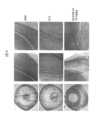
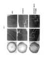
本技術の一態様としては、治療目的のために被検体の眼疾患を軽減する方法を含む。治療用途では、組成物または薬剤は、当該疾患の疑いがある、または既に罹患している被検体に、その合併症および当該疾病の進行における中期病理学的表現型を含む当該疾患の症状を治療する、または少なくとも部分的に進行を阻むのに十分な量で投与される。そのため本開示では、眼疾患に悩む個体を治療する方法を提供する。一部の実施形態では、前記技術は、芳香族カチオン性ペプチドを投与することによって、哺乳動物の糖尿病性網膜症、白内障、網膜色素変性症、緑内障、脈絡膜血管新生、網膜変性症、および酸素誘導性網膜症などの特定の眼疾患を治療または予防する方法を提供する。One aspect of the technology includes a method of alleviating ocular disease in a subject for therapeutic purposes. In therapeutic applications, the composition or agent is administered to a subject suspected of or already suffering from the disease in an amount sufficient to treat or at least partially arrest the symptoms of the disease, including complications and intermediate pathological phenotypes in the progression of the disease. The disclosure thus provides a method of treating an individual suffering from an ocular disease. In some embodiments, the technology provides a method of treating or preventing certain ocular diseases, such as diabetic retinopathy, cataracts, retinitis pigmentosa, glaucoma, choroidal neovascularization, retinal degeneration, and oxygen-induced retinopathy, in a mammal by administering an aromatic-cationic peptide.
一実施形態では、糖尿病性網膜症を治療または予防するために、被検体に芳香族カチオン性ペプチドが投与される。糖尿病性網膜症は、毛細血管瘤および点状出血を特徴とする。その後、微小血管の閉塞によって、網膜に綿状白斑を形成する。さらに、血管透過性が増大するために、網膜浮腫および/または硬性白斑が糖尿病性網膜症を患う個体に形成される場合がある。その後、血管新生が現れ、硝子体中での結合組織成長の摩擦によって網膜剥離が引き起こされる。また、回りまわって失明につながる可能性がある虹彩ルベオーシスおよび血管新生緑内障も起きることもある。糖尿病性網膜症の症状としては、読み取り能力の低下、目のかすみ、片目の突然の失明、光の周りに輪か見える、暗い点が見える、および/または点滅光が見えることが挙げられるが、これに限定されるものではない。In one embodiment, an aromatic-cationic peptide is administered to a subject to treat or prevent diabetic retinopathy. Diabetic retinopathy is characterized by microaneurysms and petechiae. Microvascular occlusion then leads to the formation of cotton wool spots in the retina. In addition, retinal edema and/or hard white spots may form in individuals with diabetic retinopathy due to increased vascular permeability. Neovascularization then appears, and friction of connective tissue growth in the vitreous causes retinal detachment. Rubeosis iridis and neovascular glaucoma may also occur, which may ultimately lead to blindness. Symptoms of diabetic retinopathy include, but are not limited to, impaired reading ability, blurred vision, sudden blindness in one eye, seeing halos around lights, seeing dark spots, and/or seeing flashing lights.
一実施形態では、白内障を治療または予防するために、被検体に芳香族カチオン性ペプチドが投与される。白内障は、自然のままの水晶体の透明度を低下させることを特徴とする先天性または後天性疾患である。白内障を患う個体は、水晶体表面の曇り、水晶体内部の曇り、および/または水晶体の腫れを含むが、これに限定されるものではない1つ以上の症状を示すことがある。先天性白内障関連疾患の典型的な例としては、偽白内障、膜内白内障、冠状白内障、層状白内障、点状白内障、および糸状白内障である。後天性白内障関連疾患の典型的な例としては、老年性白内障、後発白内障、褐変白内障、併発白内障、糖尿病性白内障、および外傷性白内障である。また、後天性白内障は感電、放射線、超音波、薬品、全身性疾患、および栄養障害によっても誘導される。後天性白内障にはさらに術後白内障が含まれる。In one embodiment, an aromatic-cationic peptide is administered to a subject to treat or prevent cataracts. Cataracts are congenital or acquired disorders characterized by a decrease in the transparency of the natural lens. Individuals suffering from cataracts may exhibit one or more symptoms, including, but not limited to, clouding of the lens surface, clouding of the interior of the lens, and/or swelling of the lens. Typical examples of congenital cataract-related disorders are pseudocataract, intramembranous cataract, coronal cataract, lamellar cataract, punctate cataract, and filiform cataract. Typical examples of acquired cataract-related disorders are senile cataract, secondary cataract, browning cataract, combined cataract, diabetic cataract, and traumatic cataract. Acquired cataracts are also induced by electrical shock, radiation, ultrasound, drugs, systemic diseases, and nutritional disorders. Acquired cataracts further include postoperative cataract.
一実施形態では、網膜色素変性症を治療または予防するために、被検体に芳香族カチオン性ペプチドが投与される。網膜色素変性症は、桿体細胞および/または錐状体細胞の損傷を特徴とする疾患である。網膜に暗い線があることが、網膜色素変性症を患う個体において典型的である。また、網膜色素変性症個体は、頭痛、四肢の無感覚または刺痛、閃光、および/または視覚障害を含むが、これに限定されるものではないさまざまな症状も示す。例えば、次の参考文献を参照:Heckenlively et al., Clinical findings and common symptoms in retinitis pigmentosa. Am J Ophthalmol. 105(5): 504-511 (1988)。In one embodiment, an aromatic-cationic peptide is administered to a subject to treat or prevent retinitis pigmentosa. Retinitis pigmentosa is a disease characterized by damage to rod and/or cone cells. Dark lines in the retina are typical in individuals with retinitis pigmentosa. Individuals with retinitis pigmentosa also exhibit a variety of symptoms, including, but not limited to, headaches, numbness or tingling in the extremities, flashes of light, and/or visual disturbances. See, e.g., the following reference: Heckenlively et al., Clinical findings and common symptoms in retinitis pigmentosa. Am J Ophthalmol. 105(5): 504-511 (1988).
一実施形態では、緑内障を治療または予防するために、被検体に芳香族カチオン性ペプチドが投与される。緑内障は、眼内圧の上昇を特徴とする遺伝的疾患であり、視力低下につながる。緑内障は、外傷、手術、および他の構造的奇形などの個体に既に存在するさまざまな眼科病状から生じることがある。緑内障はどんな年齢でも生じる可能性があるが、高齢個体でよく発症し、失明につながる。緑内障患者は、典型的には眼内圧が21mmHg以上である。しかし、視野および視神経乳頭に緑内障性変形が見られる正常眼圧緑内障は、前記眼内圧上昇、すなわち21mmHg以上にならなくても、起こる可能性がある。緑内障の症状としては、目のかすみ、眼の激痛、頭痛、光の周りに後光が見える、吐き気、および/または嘔吐が挙げられるが、これに限定されるものではない。In one embodiment, an aromatic-cationic peptide is administered to a subject to treat or prevent glaucoma. Glaucoma is a genetic disease characterized by elevated intraocular pressure, leading to loss of vision. Glaucoma can result from a variety of ophthalmic conditions that are pre-existing in an individual, such as trauma, surgery, and other structural anomalies. Glaucoma can occur at any age, but is more common in older individuals, leading to blindness. Patients with glaucoma typically have an intraocular pressure of 21 mmHg or higher. However, normal tension glaucoma, which is characterized by glaucomatous deformation of the visual field and optic disc, can occur without the intraocular pressure being elevated, i.e., 21 mmHg or higher. Symptoms of glaucoma include, but are not limited to, blurred vision, severe eye pain, headache, seeing halos around lights, nausea, and/or vomiting.
一実施形態では、黄斑変性症を治療または予防するために、被検体に芳香族カチオン性ペプチドが投与される。黄斑変性症は、典型的には加齢に伴う病気である。黄斑変性症の一般的な分類としては、湿性、乾性、および加齢に伴うものではない黄斑変性症が挙げられる。全症例の約80~90パーセントを占める乾性黄斑変性症は、萎縮性、非滲出性、またはドルセノイド黄斑変性症としても公知である。乾性黄斑変性症では、ドルーゼンは典型的には網膜色素上皮組織の下に蓄積する。ドルーゼンが黄斑内の光受容体の機能を妨げると、その後、失明が生じる。乾性黄斑変性症の症状としては、乱視、中心視覚の歪み、明暗の歪み、および/または色覚の変化が挙げられるが、これに限定されるものではない。乾性黄斑変性症によって、視力が徐々に失われることがある。In one embodiment, an aromatic-cationic peptide is administered to a subject to treat or prevent macular degeneration. Macular degeneration is typically an age-related condition. General classifications of macular degeneration include wet, dry, and non-age-related macular degeneration. Dry macular degeneration, which accounts for approximately 80-90 percent of all cases, is also known as atrophic, nonexudative, or drusenoid macular degeneration. In dry macular degeneration, drusen typically accumulate under the retinal pigment epithelium tissue. When drusen interfere with the function of the photoreceptors in the macula, blindness subsequently occurs. Symptoms of dry macular degeneration include, but are not limited to, astigmatism, distortion of central vision, light and dark distortion, and/or changes in color vision. Dry macular degeneration can cause a gradual loss of vision.
湿性黄斑変性症は、血管新生、網膜下血管新生、滲出、または円板状変性症としても公知である。湿性黄斑変性症では、黄斑の下で血管が異常に成長する。血管から黄斑に血液が漏れ出し、光受容体に損傷を与える。湿性黄斑変性症は急速に進行し、中心視覚に大きな損傷を引き起こす可能性がある。湿性および乾性黄斑変性症は同一の症状を有する。しかし、加齢に伴うものではない黄斑変性症は希であり、遺伝、糖尿病、栄養不良、怪我、感染症、または他の要因と関係がある可能性がある。加齢を伴うものではない黄斑変性症の症状としては、乱視、中心視覚の歪み、明暗の歪み、および/または色覚の変化も挙げられるが、これに限定されるものではない。Wet macular degeneration is also known as neovascularization, subretinal neovascularization, exudation, or disciform degeneration. In wet macular degeneration, blood vessels grow abnormally under the macula. The blood vessels leak blood into the macula, damaging the photoreceptors. Wet macular degeneration progresses quickly and can cause significant damage to central vision. Wet and dry macular degeneration have identical symptoms. However, non-age-related macular degeneration is rare and may be related to genetics, diabetes, poor nutrition, injury, infection, or other factors. Symptoms of non-age-related macular degeneration also include, but are not limited to, astigmatism, distortion of central vision, light and dark distortion, and/or changes in color vision.
一実施形態では、脈絡膜血管新生を治療または予防するために、被検体に芳香族カチオン性ペプチドが投与される。脈絡膜血管新生(CNV)は、眼の脈絡膜層に新しい血管が成長することを特徴とする疾患である。新たに形成される血管はブルッフ膜を通って脈絡膜で成長し、網膜下腔へ浸入する。CNVは視力障害や完全な失明に至る可能性がある。CNVの症状としては、罹患した眼にちらつき、点滅光、または灰色の点が見える、目のかすみ、乱視、および/または失明が挙げられるが、これに限定されるものではない。In one embodiment, an aromatic-cationic peptide is administered to a subject to treat or prevent choroidal neovascularization. Choroidal neovascularization (CNV) is a disease characterized by the growth of new blood vessels in the choroidal layer of the eye. The newly formed blood vessels grow in the choroid through Bruch's membrane and invade the subretinal space. CNV can lead to impaired vision or complete blindness. Symptoms of CNV include, but are not limited to, flickering, flashing lights, or gray dots in the affected eye, blurred vision, astigmatism, and/or blindness.
一実施形態では、網膜変性症を治療または予防するために、被検体に芳香族カチオン性ペプチドが投与される。網膜変性症は、網膜の破壊に関連する遺伝的疾患である。網膜組織は、動脈や静脈の閉塞、糖尿病性網膜症、未熟児網膜症、および/または水晶体後部線維増殖症などのさまざまな理由で変性することがある。網膜変性症としては、一般的には網膜分離症、格子様変性が挙げられ、進行性黄斑変性症と関連がある。網膜変性症の症状としては、視力低下、失明、夜盲症、視野狭窄、周辺視野の喪失、網膜剥離、および/または光過敏症が挙げられるが、これに限定されるものではない。In one embodiment, an aromatic-cationic peptide is administered to a subject to treat or prevent retinal degeneration. Retinal degeneration is a genetic disease associated with the destruction of the retina. Retinal tissue can degenerate for a variety of reasons, including arterial or venous blockage, diabetic retinopathy, retinopathy of prematurity, and/or retrolental fibroplasia. Retinal degeneration typically includes retinoschisis, lattice-like degeneration, and is associated with progressive macular degeneration. Symptoms of retinal degeneration include, but are not limited to, decreased vision, blindness, night blindness, constricted visual field, loss of peripheral vision, retinal detachment, and/or photosensitivity.
一実施形態では、酸素誘導性網膜症を治療または予防するために、被検体に芳香族カチオン性ペプチドが投与される。酸素誘導性網膜症(OIR)は、微小血管変性を特徴とする疾患である。OIRは、未熟児網膜症を研究するために確立されたモデルである。OIRは、異常血管新生に至る血管細胞の損傷と関連がある。微小血管変性は、OIRと関連した身体的変化に寄与する虚血につながる。酸化ストレスも、内皮細胞が過酸化傷害になりやすいOIRの血管閉塞で重要な役割を果たす。しかし、周皮細胞、平滑筋細胞、および血管周囲星状細胞は一般的に過酸化傷害に耐性を示す。例えば、次の参考文献を参照:Beauchamp et al., Role of thromboxane in retinal microvascular degeneration in oxygen-induced retinopathy, J Appl Physiol. 90: 2279-2288 (2001)。未熟児網膜症を含むOIRは一般的に無症候性である。しかし、異常な眼の動き、内斜視、重度の近視、および/または白色瞳孔は、OIRまたは未熟児網膜症の兆候である可能性がある。In one embodiment, an aromatic-cationic peptide is administered to a subject to treat or prevent oxygen-induced retinopathy. Oxygen-induced retinopathy (OIR) is a disease characterized by microvascular degeneration. OIR is an established model for studying retinopathy of prematurity. OIR is associated with vascular cell damage leading to abnormal angiogenesis. Microvascular degeneration leads to ischemia, which contributes to the physical changes associated with OIR. Oxidative stress also plays an important role in vascular occlusion in OIR, predisposing endothelial cells to peroxidative injury. However, pericytes, smooth muscle cells, and perivascular astrocytes are generally resistant to peroxidative injury. See, e.g., the following references: Beauchamp et al., J. Neurosci. 1999, 143: 1311-1323, 1999. , Role of thromboxane in retinal microvascular degeneration in oxygen-induced retinopathy, J Appl Physiol. 90: 2279-2288 (2001). OIR, including retinopathy of prematurity, is generally asymptomatic. However, abnormal eye movements, esotropia, severe myopia, and/or leukocoria may be signs of OIR or retinopathy of prematurity.
一態様において、本発明では、眼疾患の1つ以上の兆候またはマーカーを調整する芳香族カチオン性ペプチドを被検体に投与することによって、被検体の眼疾患を予防する方法を提供する。眼疾患のリスクがある被検体は、例えば、本明細書中で述べられるような診断分析または予後分析のいずれかまたは組み合わせによって特定できる。予防的用途では、芳香族カチオン性ペプチドの医薬品または薬剤が、疾患または病状が疑われる、またはリスクがある被検体に、疾患の生化学的、組織学的、および/または行動的症状、その合併症、および当該疾病の進行における中期病理学的表現型を含む、疾患のリスクを排除または減らす、重症度を低下させる、または発生を遅らせるために十分な量で投与される。予防的芳香族カチオン薬の投与は、疾患または傷害が予防されるように、あるいはその進行が遅らされるように、異常の症状特性の兆候の前に行うことができる。異常の種類に応じて、例えば、ミトコンドリア機能を強化または向上させる、または酸化的損傷を減らすように作用する芳香族カチオン性ペプチドを被検体治療のために使用できる。適した化合物は、本明細書中で述べられるスクリーニング試験に基づいて決定できる。In one aspect, the present invention provides a method for preventing ocular disease in a subject by administering to the subject an aromatic-cationic peptide that modulates one or more symptoms or markers of ocular disease. Subjects at risk for ocular disease can be identified, for example, by any or a combination of diagnostic or prognostic assays as described herein. In prophylactic applications, aromatic-cationic peptide pharmaceuticals or agents are administered to subjects suspected of or at risk for a disease or condition in an amount sufficient to eliminate or reduce the risk, reduce the severity, or delay the onset of disease, including biochemical, histological, and/or behavioral symptoms of the disease, its complications, and intermediate pathological phenotypes in the progression of the disease. Administration of prophylactic aromatic-cationic agents can occur prior to the manifestation of symptoms characteristic of the abnormality, such that disease or injury is prevented or its progression is delayed. Depending on the type of abnormality, aromatic-cationic peptides that act, for example, to enhance or improve mitochondrial function or reduce oxidative damage can be used to treat the subject. Suitable compounds can be determined based on screening tests as described herein.
芳香族カチオン性ペプチドを主体とした治療法の生物学的効果の測定。さまざまな実施形態で、特定の芳香族カチオン性ペプチドを主体とした治療法の効果、およびその投与が治療として行われるかどうかを測定するため、適切なインビトロ内またはインビボ試験が行われる。さまざまな実施形態では、特定の芳香族カチオン性ペプチドを主体とした治療法が細胞型に目的の効果を与えるかを測定するために、被検体の障害に関係する型の典型的な細胞を用いてインビトロ試験を行うことができる。治療用の化合物は、ヒト被験者で試験する前に、ラット、マウス、ニワトリ、ウシ、サル、ウサギなどの適切な動物モデル系で試験できる。同様に、体内試験の場合、ヒト被験者に投与する前に、当該技術分野で公知の動物モデル系のいずれかを使用できる。一実施形態では、眼疾患と関連する症状を示す被検体への芳香族カチオン性ペプチドの投与により、それらの症状の1つ以上で改善をもたらす。
投与の様式および有効薬量 Determining the Biological Efficacy of Aromatic-Cationic Peptide-Based Therapies. In various embodiments, appropriate in vitro or in vivo testing is performed to determine the efficacy of a particular aromatic-cationic peptide-based therapy and whether its administration is therapeutic. In various embodiments, in vitro testing can be performed using cells representative of the type relevant to the subject's disorder to determine whether a particular aromatic-cationic peptide-based therapy has the desired effect on the cell type. Therapeutic compounds can be tested in suitable animal model systems, such as rats, mice, chickens, cows, monkeys, rabbits, etc., prior to testing in human subjects. Similarly, for in vivo testing, any of the animal model systems known in the art can be used prior to administration to human subjects. In one embodiment, administration of an aromatic-cationic peptide to a subject exhibiting symptoms associated with an ocular disorder results in an improvement in one or more of those symptoms.
Mode of Administration and Effective Dose
ペプチドに対して、細胞、臓器、または組織を接触させるための当事者に公知の方法を、適用してもよい。適切な様式としては、インビトロ、エクスビボ、またはインビボの方法が挙げられる。体内法として、典型的には上述のような芳香族カチオン性ペプチドの動物への、好適にはヒトへの投与が挙げられる。治療のために体内で使用される場合、芳香族カチオン性ペプチドは有効量(すなわち、目標とする治療効果を有する量)で被検体に投与される。投与量と投薬計画は、被検体の眼疾患の程度、その治療指数などの使用される特定の芳香族カチオン性ペプチドの特性、被検体、および被検体の病歴によって決まる。Any method known to one of skill in the art for contacting a cell, organ, or tissue with a peptide may be applied. Suitable modalities include in vitro, ex vivo, or in vivo methods. Internal methods typically include administration of an aromatic-cationic peptide, as described above, to an animal, preferably a human. When used internally for therapy, the aromatic-cationic peptide is administered to the subject in an effective amount (i.e., an amount that has a desired therapeutic effect). The dosage and dosing regimen will depend on the extent of the subject's ocular disease, the characteristics of the particular aromatic-cationic peptide used, such as its therapeutic index, the subject, and the subject's medical history.
有効量を、医師や臨床医によく知られている方法によって、前臨床試験および臨床試験の際に、定めることが可能である。好適には医薬品で、本発明の方法に有用であるペプチドの有効量を、医薬品を投与するための多くの公知の方法で必要とする哺乳動物に投与できる。一部の実施形態では、前記ペプチドを全身投与、局所投与、または眼内投与できる。Effective amounts can be determined during preclinical and clinical trials by methods well known to physicians and clinicians. An effective amount of a peptide, preferably a pharmaceutical, useful in the methods of the invention can be administered to a mammal in need thereof by any of a number of known methods for administering pharmaceuticals. In some embodiments, the peptide can be administered systemically, topically, or intraocularly.
本明細書中で述べられる芳香族カチオン性ペプチドは、本明細書中で述べられる障害の治療または予防のために単独または併用で被検体に投与するための医薬品に組み込むことができる。前記化合物として典型的には、活性薬剤および薬学的に許容可能な担体が挙げられる。本願明細書で使用される場合、用語「薬学的に許容可能な担体」としては、医薬品投与に適合した生理食塩水、溶剤、分散媒質、コーティング剤、抗菌剤、抗真菌薬、等張剤、吸収遅延剤などが挙げられる。補助的な活性化合物も、組成物に組み込むことができる。The aromatic-cationic peptides described herein can be incorporated into pharmaceutical preparations for administration to a subject, either alone or in combination, for the treatment or prevention of the disorders described herein. The compounds typically include an active agent and a pharma-ceutically acceptable carrier. As used herein, the term "pharma-ceutically acceptable carrier" includes saline, solvents, dispersion media, coatings, antibacterial agents, antifungal agents, isotonic agents, absorption delaying agents, and the like, compatible with pharmaceutical administration. Supplementary active compounds can also be incorporated into the compositions.
医薬品は典型的には、その意図とした投与経路に適合するように製剤化される。投与経路の例としては、非経口(例えば、静脈内、皮内、副腔内、または皮下)、経口、吸入、経皮(局所)、眼内、イオンフォレシス、および経粘膜投与が挙げられる。非経口、皮内、または皮下投与に使用される溶液または懸濁液は以下の成分を含むことができる:注射用水、生理食塩水溶液、固定油、ポリエチレングリコール、グリセリン、プロピレングリコール、または他の合成溶剤などの無菌希釈剤;ベンジルアルコールまたはメタルパラベンなどの抗菌剤;アスコルビン酸または亜硫酸水素ナトリウムなどの酸化防止剤;エチレンジアミン四酢酸などのキレート剤;酢酸塩、クエン酸塩、またはリン酸塩などの緩衝液;および塩化ナトリウムまたはデキストロースなどの張性の調整用薬剤。pHは、塩酸または水酸化ナトリウムなどの、酸または塩基で調整できる。非経口調製品を、ガラス製またはプラスチック製のアンプル、使い捨て注射器、または複数回投与用バイアルに封入できる。患者または治療医師の都合上、投薬製剤は、治療単位に必要なすべての機器(例えば、薬品のバイアル、希釈液のバイアル、注射器、および針)を含むキットで提供してもよい。Pharmaceuticals are typically formulated to be compatible with their intended route of administration. Examples of routes of administration include parenteral (e.g., intravenous, intradermal, intraperitoneal, or subcutaneous), oral, inhalation, transdermal (topical), ocular, iontophoretic, and transmucosal administration. Solutions or suspensions used for parenteral, intradermal, or subcutaneous administration can contain the following components: a sterile diluent such as water for injection, saline solution, fixed oils, polyethylene glycol, glycerin, propylene glycol, or other synthetic solvents; an antibacterial agent such as benzyl alcohol or methamphetamine; an antioxidant such as ascorbic acid or sodium bisulfite; a chelating agent such as ethylenediaminetetraacetic acid; a buffer such as acetate, citrate, or phosphate; and an agent for adjusting tonicity such as sodium chloride or dextrose. The pH can be adjusted with acids or bases, such as hydrochloric acid or sodium hydroxide. Parenteral preparations can be enclosed in glass or plastic ampoules, disposable syringes, or multiple dose vials. For the convenience of the patient or treating physician, the dosage formulation may be provided in a kit that includes all the equipment needed for a unit of treatment (e.g., drug vial, diluent vial, syringe, and needle).
注射用に適した医薬品には、無菌注射剤または分散剤を準備なしで調製できるように、無菌水溶液(水溶性の場合)または分散剤および無菌粉末を含めてもよい。静脈内投与の場合、適切な担体としては、生理食塩水、静菌性水、Cremophor EL(商標)(BASF,Parsippany,NJ.)またはリン酸緩衝生理食塩水(PBS)が挙げられる。すべての場合で、非経口投与用組成物は無菌であり、容易に注射可能な流動性が存在するような程度の液体である必要がある。製造および保存の条件の下で安定していて、細菌や菌類などの微生物の汚染作用に対して保護する必要がある。Pharmaceuticals suitable for injection may include sterile aqueous solutions (where water soluble) or dispersions and sterile powders so that sterile injectable solutions or dispersions can be prepared without prep. For intravenous administration, suitable carriers include physiological saline, bacteriostatic water, Cremophor EL™ (BASF, Parsippany, NJ.) or phosphate buffered saline (PBS). In all cases, compositions for parenteral administration must be sterile and fluid to the extent that easy syringability exists. They must be stable under the conditions of manufacture and storage and preserved against the contaminating action of microorganisms such as bacteria and fungi.
前記芳香族カチオン性ペプチド組成物は担体を含むことができ、その担体は、例えば水、エタノール、ポリオール(例えば、グリセロール、プロピレングリコール、および液体ポリエチレングリコールなど)、およびそれらの適切な混合物を含む溶剤または分散剤にしてもよい。例えば、レシチンなどのコーティングを用いる、分散液の場合には目的とする粒径を維持する、および界面活性剤を用いることで、適切な流動性を維持できる。微生物の作用の防止は、例えばパラベン、クロロブタノール、フェノール、アスコルビン酸、チオメルサールなどのさまざまな抗菌剤および抗真菌剤によって達成できる。酸化を防止するために、グルタミンおよび他の酸化防止剤を誘導できる。多くの場合、例えば糖類や、マンニトール、ソルビトールなどのポリアルコール類や、または塩化ナトリウムの等張剤を組成物に含むことが望ましい場合がある。注射組成物の持続的吸収は、例えばモノステアリン酸アルミニウムまたはゼラチンなどの吸収を遅らせる化学物質を組成物に含むことによって行うことができる。The aromatic-cationic peptide composition can include a carrier, which can be a solvent or dispersion medium, for example, including water, ethanol, polyol (e.g., glycerol, propylene glycol, and liquid polyethylene glycol, and the like), and suitable mixtures thereof. Proper fluidity can be maintained, for example, by the use of a coating such as lecithin, by maintaining the desired particle size in the case of dispersions, and by the use of surfactants. Prevention of the action of microorganisms can be achieved by various antibacterial and antifungal agents, for example, parabens, chlorobutanol, phenol, ascorbic acid, thiomersal, and the like. Glutamine and other antioxidants can be introduced to prevent oxidation. In many cases, it is desirable to include isotonic agents, for example, sugars, polyalcohols, such as mannitol, sorbitol, or sodium chloride, in the composition. Prolonged absorption of injectable compositions can be achieved by including in the composition a chemical that delays absorption, for example, aluminum monostearate or gelatin.
無菌注射剤は、上記で列挙された成分の1つまたは組み合わせとともに、適切な溶剤に活性化合物を必要量で組み込み、必要な場合には、その後に濾過滅菌することで調製できる。一般的に、分散剤は、無菌賦形剤に活性化化合物を組み込むことで調製され、これには、塩基性分散媒や上記で列挙されたその他の必要な成分を含む。無菌注射剤調製用の無菌粉末の場合、調製の典型的な方法としては真空乾燥や凍結乾燥が挙げられ、これらによって、それについての事前に無菌濾過した溶液から、活性成分の粉末に加えて、追加の必要な成分を獲得することができる。Sterile injectable solutions can be prepared by incorporating the active compound in the required amount in an appropriate solvent with one or a combination of ingredients enumerated above, followed by filtered sterilization, if necessary. Generally, dispersions are prepared by incorporating the active compound in a sterile vehicle, which contains a basic dispersion medium and other required ingredients enumerated above. In the case of sterile powders for the preparation of sterile injectable solutions, typical methods of preparation include vacuum drying and freeze-drying, which allow the powder of the active ingredient, plus any additional required ingredients, to be obtained from a previously sterile-filtered solution thereof.
眼科用の場合、治療化合物は溶液、懸濁液、および眼での使用に適した軟膏に製剤化される。点眼製剤の場合、一般的には次の参考文献を参照:Mitra (ed.), Ophthalmic Drug Delivery Systems, Marcel Dekker, Inc., New York, N.Y. (1993)、または次の参考文献も参照:Havener, W. H., Ocular Pharmacology, C.V. Mosby Co., St. Louis (1983)。点眼医薬品は、溶液、懸濁液、軟膏、クリーム、または固体挿入物の剤形で眼への局所投与に適するものであってもよい。単回投与の場合、0.1ng~5000μg、1ng~500μg、または10ng~100μgの芳香族カチオン性ペプチドがヒトの眼に点眼することができる。For ophthalmic use, the therapeutic compounds are formulated into solutions, suspensions, and ointments suitable for use in the eye. For eye drop formulations, see generally Mitra (ed.), Ophthalmic Drug Delivery Systems, Marcel Dekker, Inc., New York, N.Y. (1993), or see also Havener, W.H., Ocular Pharmacology, C.V. Mosby Co., St. Louis (1983). Eye drop pharmaceuticals may be in the form of solutions, suspensions, ointments, creams, or solid inserts suitable for topical administration to the eye. For a single dose, 0.1 ng to 5000 μg, 1 ng to 500 μg, or 10 ng to 100 μg of the aromatic-cationic peptide can be instilled into the human eye.
点眼調製品は、使用上有害ではない抗菌剤などの無毒性物質を含むものであってもよく、例えば、チメロサール、塩化ベンザルコニウム、メチルおよびプロピルパラベン、臭化ベンジルドデシニウム、ベンジルアルコール、またはフェニルエタノール;塩化ナトリウム、ホウ酸ナトリウム、酢酸ナトリウム、クエン酸ナトリウム、またはグルコン酸塩などの緩衝成分:ソルビタンラウリン酸モノエステル、トリエタノールアミン、モノパルミチン酸ポリオキシエレンソルビタン、エチレンジアミン4酢酸などの他の従来成分などである。Eye drop preparations may contain non-toxic substances such as antibacterial agents that are not harmful in use, for example, thimerosal, benzalkonium chloride, methyl and propyl parabens, benzyldodecinium bromide, benzyl alcohol, or phenylethanol; buffering ingredients such as sodium chloride, sodium borate, sodium acetate, sodium citrate, or gluconate; and other conventional ingredients such as sorbitan monolaurate, triethanolamine, polyoxyethylene sorbitan monopalmitate, ethylenediaminetetraacetic acid, etc.
点眼溶液または懸濁液は、眼内の芳香族カチオン性ペプチド許容濃度を維持する必要があるたびに投与される。哺乳動物の眼への投与は、日に約1回または2回であってもよい。The eye drop solution or suspension is administered as often as necessary to maintain an acceptable concentration of aromatic-cationic peptide in the eye. Administration to the mammalian eye may be about once or twice daily.
経口組成物としては一般的に、不活性希釈剤または可食性担体が挙げられる。経口治療的投与の目的ために、活性化合物を賦形剤に組み込むことができ、そして錠剤、トローチ、またはゼラチンカプセルなどのカプセルの剤形で使用できる。薬学的に適合する結合剤、および/または補助物質を、組成物の一部として含有することができる。錠剤、丸薬、カプセル、トローチなどは、以下の成分または同様の性質の化合物のいずれかを含有できる:微結晶性セルロース、ガムトラガカント、またはゼラチンなどの結合剤;澱粉またはラクトースなどの賦形剤、アルギン酸、プリモジェル、またはコーンスターチなどの分解剤;ステアリン酸マグネシウムまたはステロートなどの滑剤;コロイド状二酸化ケイ素;ショ糖またはサッカリンなどの甘味剤;またはペパーミント、サリチル酸メチル、またはオレンジ香料などの香料添加剤。Oral compositions generally include an inert diluent or an edible carrier. For the purpose of oral therapeutic administration, the active compound can be incorporated into excipients and used in the form of tablets, troches, or capsules, such as gelatin capsules. Pharmaceutically compatible binders, and/or adjuvant substances can be included as part of the composition. The tablets, pills, capsules, troches, and the like can contain any of the following ingredients or compounds of a similar nature: binders such as microcrystalline cellulose, gum tragacanth, or gelatin; excipients such as starch or lactose, disintegrants such as alginic acid, primogel, or corn starch; lubricants such as magnesium stearate or sterol; colloidal silicon dioxide; sweetening agents such as sucrose or saccharin; or flavoring agents such as peppermint, methyl salicylate, or orange flavoring.
吸入による投与の場合、例えば二酸化炭素などのガスなどの適切な推進剤を含む加圧容器またはディスペンサー、または噴霧器からのエアゾールスプレーの剤形で化合物を送達できる。前記方法には、米国特許第6,468,798号で述べられた方法を含む。For administration by inhalation, the compounds can be delivered in the form of an aerosol spray from a pressured container or dispenser which contains a suitable propellant, e.g., a gas such as carbon dioxide, or a nebulizer. Such methods include those described in U.S. Pat. No. 6,468,798.
本明細書中で述べられるような治療化合物の全身投与は、経粘膜的または経皮的方法によって行うこともできる。経粘膜的または経皮的投与の場合、浸透される障壁に適合している浸透剤が製剤中に使用される。前記浸透剤は一般的に当該技術分野で公知であり、例えば、経粘膜的投与の場合には、洗浄剤、胆汁塩、およびフシジン酸誘導体を含む。経粘膜的投与は、鼻腔用スプレーの使用を通じて達成できる。経皮的投与の場合、活性化合物は、一般的に当該技術分野で公知なように軟膏、ゲル、またはクリームに製剤化される。一実施形態では、経皮的投与がイオンフォレシスによって行ってもよい。Systemic administration of therapeutic compounds as described herein may also be accomplished by transmucosal or transdermal methods. For transmucosal or transdermal administration, penetrants appropriate to the barrier to be permeated are used in the formulation. Such penetrants are generally known in the art and include, for example, for transmucosal administration, detergents, bile salts, and fusidic acid derivatives. Transmucosal administration may be accomplished through the use of nasal sprays. For transdermal administration, the active compounds are formulated into ointments, gels, or creams as generally known in the art. In one embodiment, transdermal administration may be accomplished by iontophoresis.
治療用タンパク質またはペプチドは、担体系で製剤化できる。前記担体はコロイド系であってもよい。前記コロイド系は、リポソーム、リン脂質二重層賦形剤にすることができる。一実施形態では、前記治療用ペプチドは、ペプチドの完全性を維持しながらリポソームでカプセル化される。当事者は十分理解しているように、リポソームを調製する方法にはさまざまなものがある(次の参考文献を参照:Lichtenberg et al., Methods Biochem. Anal, 33:337-462 (1988); Anselem et al, Liposome Technology, CRC Press (1993))。リポソーム製剤はクリアランスを遅らせ、細胞取り込みを増加させることができる(以下のReddy, Ann.による文献を参照、Pharmacother., 34 (7-8):915-923 (2000))。活性剤は、可溶性、不溶性、透過性、非透過性、生分解性、または胃保持型高分子またはリポソームを含むが、これに限定されるものではない薬学的に許容可能な成分から調製される粒子内に投入することもできる。前記粒子としては、ナノ粒子、生分解性ナノ粒子、微小粒子、生分解性微小粒子、ナノ球体、生分解性ナノ球体、微小球体、生分解性微小球体、カプセル、エマルジョン、リポソーム、ミセル、およびウイルスベクター系が挙げられるが、これに限定されるものではない。The therapeutic protein or peptide can be formulated in a carrier system. The carrier can be a colloidal system. The colloidal system can be a liposome, a phospholipid bilayer vehicle. In one embodiment, the therapeutic peptide is encapsulated in a liposome while maintaining the integrity of the peptide. As those skilled in the art will appreciate, there are a variety of methods for preparing liposomes (see the following references: Lichtenberg et al., Methods Biochem. Anal, 33:337-462 (1988); Anselem et al, Liposome Technology, CRC Press (1993)). Liposomal formulations can slow clearance and increase cellular uptake (see Reddy, Ann., Pharmacother., 34 (7-8):915-923 (2000)). The active agent can also be loaded into particles prepared from pharma-ceutically acceptable components, including, but not limited to, soluble, insoluble, permeable, non-permeable, biodegradable, or gastroretentive polymers or liposomes. The particles include, but are not limited to, nanoparticles, biodegradable nanoparticles, microparticles, biodegradable microparticles, nanospheres, biodegradable nanospheres, microspheres, biodegradable microspheres, capsules, emulsions, liposomes, micelles, and viral vector systems.
前記担体は、例えば生分解性、生体適合性高分子マトリックスなどの高分子にすることもできる。一実施形態では、前記治療用ペプチドは、タンパク質の完全性を維持しながら高分子マトリックスでカプセル化できる。前記高分子は、ポリペプチド、タンパク質、または多糖類などの天然高分子、またはポリα-ヒドロキシ酸などの合成高分子である。例としては、例えばコラーゲン、フィブロネクチン、エラスチン、酢酸セルロース、ニトロセルロース、多糖類、フィブリン、ゼラチン、およびそれらの組み合わせなどから作られる担体が挙げられる。一実施形態では、前記高分子がポリ乳酸(PLA)または乳酸/グリコール酸共重合体(PGLA)である。前記高分子マトリックスは、微小球体およびナノ球体を含むさまざまな形態または大きさに調製および単離することができる。高分子製剤は治療効果の持続期間が長くなる可能性がある(次の参考文献を参照:Reddy, Ann. Pharmacother., 34 (7-8):915-923 (2000))。臨床試験では、ヒト成長ホルモン(hGH)用高分子製剤が使用されてきた(次の参考文献を参照:Kozarich and Rich, Chemical Biology, 2:548-552 (1998))。The carrier can also be a polymer, e.g., a biodegradable, biocompatible polymer matrix. In one embodiment, the therapeutic peptide can be encapsulated in a polymer matrix while maintaining protein integrity. The polymer can be a natural polymer, e.g., a polypeptide, protein, or polysaccharide, or a synthetic polymer, e.g., a poly-α-hydroxy acid. Examples include carriers made from, e.g., collagen, fibronectin, elastin, cellulose acetate, nitrocellulose, polysaccharides, fibrin, gelatin, and combinations thereof. In one embodiment, the polymer is polylactic acid (PLA) or polylactic/glycolic acid (PGLA). The polymer matrix can be prepared and isolated in a variety of forms or sizes, including microspheres and nanospheres. Polymer formulations may provide a longer duration of therapeutic effect (see reference: Reddy, Ann. Pharmacother., 34 (7-8):915-923 (2000)). Polymeric formulations of human growth hormone (hGH) have been used in clinical trials (see reference: Kozarich and Rich, Chemical Biology, 2:548-552 (1998)).
高分子微小球体徐放性製剤の例は、PCT公報第WO99/15154号(Tracy等)、米国特許第5,674,534号および第5,716,644号(両方とも、Zale等)、およびPCT公報第WO96/40073(Zale等)、およびPCT公報第WO00/38651号(Shah等)で述べられている。米国特許第5,674,534号および第5,716,644号およびPCT公報第WO96/40073号では、塩との凝集に対して安定化させたエリスロポエチンの粒子を含む高分子マトリックスを説明している。Examples of polymeric microsphere sustained release formulations are described in PCT Publication No. WO 99/15154 (Tracy et al.), U.S. Patent Nos. 5,674,534 and 5,716,644 (both by Zale et al.), and PCT Publication No. WO 96/40073 (Zale et al.), and PCT Publication No. WO 00/38651 (Shah et al.). U.S. Patent Nos. 5,674,534 and 5,716,644 and PCT Publication No. WO 96/40073 describe polymeric matrices containing particles of erythropoietin stabilized against aggregation with salt.
一部の実施形態では、前記治療化合物は、インプラントおよびマイクロカプセル化送達システムを含み、放出制御製剤など、体内から排出に対して治療化合物を保護する担体で調製される。エチレン酢酸ビニル、ポリ無水物、ポリグリコール酸、コラーゲン、ポリオルトエステル、およびポリ乳酸などの生分解性、生体適合性高分子を使用できる。当該製剤は、公知の技術を用いて調製できる。前記物質は、Alza CorporationおよびNova Pharmaceuticals, Inc.などからも市販で入手できる。薬学的に許容可能な担体として、リポソーム懸濁液(細胞特異的抗原に対するモノクローナル抗体を持つ特定の細胞に標的とされたリポソームを含む)を使用できる。これらは、例えば、米国特許第4,522,811号で述べられているように当事者に公知の方法に従って調製できる。In some embodiments, the therapeutic compound is prepared in a carrier that protects the therapeutic compound against elimination from the body, such as a controlled release formulation, including implants and microencapsulated delivery systems. Biodegradable, biocompatible polymers such as ethylene vinyl acetate, polyanhydrides, polyglycolic acid, collagen, polyorthoesters, and polylactic acid can be used. Such formulations can be prepared using known techniques. Such materials are also commercially available from sources such as Alza Corporation and Nova Pharmaceuticals, Inc. As pharmaceutical acceptable carriers, liposomal suspensions (including liposomes targeted to specific cells with monoclonal antibodies against cell-specific antigens) can be used. These can be prepared according to methods known to those skilled in the art, for example, as described in U.S. Pat. No. 4,522,811.
前記治療化合物は、細胞内送達を高めるように製剤化することもできる。例えば、リポソーム送達システムが公知であり、例えば次の参考文献を参照:Chonn and Cullis, “Recent Advances in Liposome Drug Delivery Systems,” Current Opinion in Biotechnology 6:698-708 (1995); Weiner, “Liposomes for Protein Delivery: Selecting Manufacture and Development Processes,” Immunomethods 4 (3) 201-9 (1994); and Gregoriadis, “Engineering Liposomes for Drug Delivery: Progress and Problems,” Trends Biotechnol. 13 (12):527-37 (1995).Mizguchi等著『Cancer Lett』,100:63-69(1996)では、インビボおよびインビトロの両方で細胞にタンパク質を送達するために融合性リポソームを使用することを説明している。The therapeutic compounds can also be formulated to enhance intracellular delivery. For example, liposome delivery systems are known, see, e.g., the following references: Chonn and Cullis, "Recent Advances in Liposome Drug Delivery Systems," Current Opinion in Biotechnology 6:698-708 (1995); Weiner, "Liposomes for Protein Delivery: Selecting Manufacture and Development Processes," Immunomethods 4 (3) 201-9 (1994); and Gregoriadis, "Engineering Liposomes for Drug Delivery: Progress and Problems," Trends Biotechnol. 13 (12):527-37 (1995). Mizguchi et al., Cancer Lett. 100:63-69 (1996), describe the use of fusogenic liposomes to deliver proteins to cells both in vivo and in vitro.
治療薬の用量、毒性、および治療効果は、細胞培養または実験動物での標準的製薬手順で、例えば、LD50(個体群の50%で死に至る用量)およびED50(個体群の50%で治療効果がある用量)を決定することで決定できる。毒性と治療効果の用量比が治療指数であり、LD50/ED50の比で表すことができる。高い治療指数を示す化合物が望ましい。中毒性副作用を示す化合物を使用されることがある一方、非感染細胞に対する損傷の可能性を最小限に抑えるために、当該化合物の標的を罹患組織の部位にするように、送達システムの設計には注意する必要がある。これによって、副作用を減らすことになる。The dose, toxicity, and therapeutic efficacy of therapeutic drugs can be determined by standard pharmaceutical procedures in cell cultures or experimental animals, for example, by determining the LD50 (the dose lethal to 50% of the population) and ED50 (the dose therapeutic in 50% of the population). The dose ratio between toxicity and therapeutic efficacy is the therapeutic index, which can be expressed as the ratio LD50/ED50. Compounds that exhibit high therapeutic indices are desirable. While compounds that exhibit toxic side effects may be used, care must be taken to design delivery systems that target the compound to the site of the affected tissue to minimize the potential for damage to uninfected cells. This will reduce side effects.
細胞培養試験および動物試験から得られるデータは、ヒト用の幅広い用量の製剤化で使用できる。当該化合物の用量は、好適には、毒性をあまり示さない、または毒性のないED50を含む幅広い血中濃度内に入る。用量は、採用される剤形および利用される投与経路に応じてこの範囲内で変えてもよい。方法で使用されるあらゆる化合物に対して、細胞培養試験から最初に治療有効量を推定できる。用量は、細胞培養で決定されるように、IC50(すなわち、症状の最大半量の抑制を達成する試験化合物の濃度)を含む循環血漿濃度範囲を達成する動物モデルで公式化できる。当該情報は、ヒトに有用な用量をより正確に決定するために使用できる。血漿中の濃度を、例えば、高性能液体クロマトグラフィーで測定してもよい。The data obtained from cell culture and animal studies can be used to formulate a range of dosages for use in humans. The dose of the compound preferably falls within a range of circulating concentrations that includes the ED50 with little or no toxicity. The dose may vary within this range depending on the dosage form employed and the route of administration utilized. For any compound used in the method, the therapeutically effective amount can be estimated initially from cell culture studies. A dose can be formulated in animal models that achieves a circulating plasma concentration range that includes the IC50 (i.e., the concentration of the test compound that achieves a half-maximal inhibition of symptoms) as determined in cell culture. Such information can be used to more accurately determine useful doses in humans. Concentrations in plasma may be measured, for example, by high performance liquid chromatography.
典型的には、治療または予防の効果を達成するのに十分な、有効量の前記芳香族カチオン性ペプチドは、約0.000001mg/体重1kg/日~約10,000mg/体重1kg/日の範囲に及ぶ。好適には、用量範囲は約0.0001mg/体重1kg/日~約100mg/体重1kg/日である。例えば、用量は毎日、2日毎、または3日毎に1mg/体重1kgまたは10mg/体重1kg、または毎週、2週間毎、または3週間毎に1~10mg/体重1kgの範囲であってもよい。一実施形態では、ペプチドの単回投与は0.1~10,000μg/体重1kgの範囲に及ぶ。一実施形態では、担体中の芳香族カチオン性ペプチドの濃度は0.2~2000μg/(送達される製剤1mL)の範囲に及ぶ。例示的治療投薬計画では、日に1回または週に1回の投与を必要とする。被検体のブドウ糖またはインシュリンの血中濃度を測定して、それに応じて用量または投与を調整することで、間隔が示されたものより不規則になることもある。治療用途では、疾患の進行が抑制されるか停止されまで、好適には、被検体が疾患の症状の部分的または全体的な改善を示すまで、相対的に短い間隔で相対的に高い用量が必要になることがよくある。その後、患者に予防投薬計画を施すことができる。Typically, an effective amount of the aromatic-cationic peptide sufficient to achieve a therapeutic or prophylactic effect ranges from about 0.000001 mg/kg body weight/day to about 10,000 mg/kg body weight/day. Suitably, the dose range is from about 0.0001 mg/kg body weight/day to about 100 mg/kg body weight/day. For example, doses may range from 1 mg/kg body weight or 10 mg/kg body weight daily, every 2 days, or every 3 days, or from 1 to 10 mg/kg body weight weekly, every 2 weeks, or every 3 weeks. In one embodiment, a single dose of the peptide ranges from 0.1 to 10,000 μg/kg body weight. In one embodiment, the concentration of the aromatic-cationic peptide in the carrier ranges from 0.2 to 2000 μg/mL of the formulation delivered. Exemplary treatment dosing regimens call for administration once a day or once a week. Intervals may be more irregular than those indicated, with the subject's blood glucose or insulin levels being measured and the dose or administration adjusted accordingly. In therapeutic applications, relatively high doses at relatively short intervals are often required until disease progression is inhibited or halted, preferably until the subject shows partial or total improvement in disease symptoms. The patient can then be placed on a prophylactic regimen.
一部の実施形態では、芳香族カチオン性ペプチドの治療的有効量を、10-11~10-6モル、例えば約10-7モルの標的組織でのペプチドの濃度として定義することが可能である。この濃度が0.001~100mg/kgの全身用量または体表面積で同等の用量で送達されるものであってもよい。標的組織で治療濃度を維持するように、用量のスケジュールが最適化されることになり、最も好適には、毎日または毎週1回投与であるが、連続投与(例えば、静脈注入または経皮的塗布)を含む。 In some embodiments, a therapeutically effective amount of an aromatic-cationic peptide can be defined as a concentration of peptide in a target tissue of 10−11 to 10−6 molar, for example about 10−7 molar. This concentration may be delivered at a systemic dose of 0.001 to 100 mg/kg or equivalent dose per body surface area. Dose schedules will be optimized to maintain therapeutic concentrations in the target tissue, most preferably once daily or weekly, but including continuous administration (e.g., intravenous infusion or transdermal application).
一部の実施形態では、「低」、「中」、または「高」の用量レベルで、芳香族カチオン性ペプチドの用量が規定される。一実施形態では、低用量は約0.0001~約0.5mg/kg/h、好適には、約0.01~約0.1mg/kg/hである。一実施形態では、中用量は約0.1~約1.0mg/kg/h、好適には、約0.1~約0.5mg/kg/hである。一実施形態では、高用量は約0.5~約10mg/kg/h、好適には、約0.5~約2mg/kg/hである。In some embodiments, the dose of aromatic-cationic peptide is defined as "low", "medium" or "high" dose levels. In one embodiment, the low dose is about 0.0001 to about 0.5 mg/kg/h, preferably about 0.01 to about 0.1 mg/kg/h. In one embodiment, the medium dose is about 0.1 to about 1.0 mg/kg/h, preferably about 0.1 to about 0.5 mg/kg/h. In one embodiment, the high dose is about 0.5 to about 10 mg/kg/h, preferably about 0.5 to about 2 mg/kg/h.
当事者は十分理解することであるが、疾患または障害の重症度、被検体の全体的な健康および/または年齢、および他の疾患の存在を含むが、これに限定されるものではない、特定の要因が効果的に治療するために必要な用量やタイミングに影響を及ぼすことがある。さらに、本明細書中に記載される治療組成物を治療有効量用いた被検体の治療としては、単回治療または一連の治療が挙げられる。As those skilled in the art will appreciate, certain factors may affect the dosage and timing required to effectively treat, including, but not limited to, the severity of the disease or disorder, the overall health and/or age of the subject, and the presence of other diseases. Moreover, treatment of a subject with a therapeutically effective amount of the therapeutic compositions described herein may include a single treatment or a series of treatments.
当事者は十分理解することであるが、疾患または障害の重症度、被検体の全体的な健康および/または年齢、および他の疾患の存在を含むが、これに限定されるものではない、特定の要因が効果的に治療するために必要な用量やタイミングに影響を及ぼすことがある。さらに、本明細書中に記載される治療組成物を治療有効量用いた被検体の治療としては、単回治療または一連の治療が挙げられる。As those skilled in the art will appreciate, certain factors may affect the dosage and timing required to effectively treat, including, but not limited to, the severity of the disease or disorder, the overall health and/or age of the subject, and the presence of other diseases. Moreover, treatment of a subject with a therapeutically effective amount of the therapeutic compositions described herein may include a single treatment or a series of treatments.
本方法に従って治療される哺乳動物は、例えば、羊、豚、牛、および馬などの家畜;犬や猫などの愛玩動物;ラット、マウス、およびウサギなどの実験動物を含む任意の哺乳動物であってもよい。好適な実施形態では、哺乳動物がヒトである。
芳香族カチオン性ペプチドと他の治療薬による併用治療 The mammal treated according to the present method may be any mammal, including, for example, farm animals such as sheep, pigs, cows, and horses, pet animals such as dogs and cats, and laboratory animals such as rats, mice, and rabbits. In a preferred embodiment, the mammal is a human.
Combination treatment with aromatic-cationic peptides and other therapeutic agents
場合によっては、別の治療薬と組み合わせて、本明細書中で述べられる芳香族カチオン性ペプチド(または薬学的に許容される塩、エステル、アミド、プロドラッグ、または溶媒和物)の少なくとも1つを施すことが適切であろう。単なる例として、本明細書中の芳香族カチオン性ペプチドの1つの投与を受けたときに患者が経験した副作用の1つが炎症である場合、初期治療薬と組み合わせて抗炎症薬を投与することが適切であろう。あるいは、単なる例として、本明細書中に記載の化合物の1つの治療有効性は補助物質の投与によって高めることが可能である(すなわち、補助物質それだけで最低限の治療効果を有するが、別の治療薬と組み合わせることで、患者に対する全体的な治療効果が高められる)。あるいは、単なる例として、患者が経験した効果は、眼疾患の予防または治療でも治療効果がある別の治療薬(治療計画も含む)とともに本明細書中に記載の化合物の1つを投与することで高められることが可能である。単なる一例として、芳香族カチオン性ペプチドの1つの投与を伴う黄斑変性症の治療において、患者に黄斑変性症の他の治療薬または治療を提供することでも、高い治療効果が得られよう。いかなる場合にも、治療を受けている眼疾患、障害、または病状に関わらず、患者が経験する全体的な効果は、単純に2つの治療薬の効果の加算になることがあり、あるいは患者は相乗効果を経験することが可能である。In some cases, it may be appropriate to administer at least one of the aromatic-cationic peptides (or a pharma- ceutically acceptable salt, ester, amide, prodrug, or solvate) described herein in combination with another therapeutic agent. By way of example only, if inflammation is one of the side effects experienced by a patient upon administration of one of the aromatic-cationic peptides herein, it may be appropriate to administer an anti-inflammatory agent in combination with the initial therapeutic agent. Alternatively, by way of example only, the therapeutic efficacy of one of the compounds described herein may be enhanced by administration of an adjunct (i.e., the adjunct alone has a minimal therapeutic effect, but in combination with another therapeutic agent, the overall therapeutic effect on the patient is enhanced). Alternatively, by way of example only, the effect experienced by the patient may be enhanced by administration of one of the compounds described herein with another therapeutic agent (including a treatment regimen) that also has a therapeutic effect in the prevention or treatment of an ocular disease. By way of example only, in the treatment of macular degeneration with administration of one of the aromatic-cationic peptides, a high therapeutic effect may also be obtained by providing the patient with another therapeutic agent or treatment for macular degeneration. In any case, regardless of the ocular disease, disorder, or condition being treated, the overall effect experienced by the patient may simply be the additive effect of the two therapeutic agents, or the patient may experience a synergistic effect.
考えられる組み合わせ治療の具体的な限定されない例としては、一酸化窒素(NO)誘導剤、スタチン、負電荷リン脂質、酸化防止剤、無機物、抗炎症剤、抗血管新生剤、マトリックスメタロプロテイナーゼ阻害剤、およびカロテノイドとともに少なくとも1つの芳香族カチオン性ペプチドを使用することが挙げられる。いくつかの例として、適切な組み合わせ薬剤が複数の分類に入れてもよい(単なる例として、ルテインは酸化防止剤およびカロテノイドである)。さらに、芳香族カチオン性ペプチドは、単なる例としてシクロスポリンAを含む患者に効果をもたらす追加薬剤とともに投与されることもある。Specific non-limiting examples of possible combination therapies include the use of at least one aromatic-cationic peptide with nitric oxide (NO) inducers, statins, negatively charged phospholipids, antioxidants, minerals, anti-inflammatory agents, anti-angiogenic agents, matrix metalloproteinase inhibitors, and carotenoids. In some instances, suitable combination agents may fall into multiple categories (by way of example only, lutein is an antioxidant and a carotenoid). Additionally, aromatic-cationic peptides may be administered with additional agents that provide benefit to the patient, including, by way of example only, cyclosporine A.
さらに、芳香族カチオン性ペプチドは、単なる例として、体外レオフェレシス(膜差次的濾過としても公知)の使用、移植可能小型望遠鏡の使用、ドルーゼンのレーザー光凝固術、および微小刺激治療を含む、患者に相乗効果をもたらす手順との組み合わせで使用されることもある。Additionally, aromatic-cationic peptides may be used in combination with procedures that provide synergistic benefits to patients, including, by way of example only, the use of extracorporeal rheopheresis (also known as membrane differential filtration), the use of implantable telescopes, laser photocoagulation of drusen, and microstimulation therapy.
酸化防止剤を使用することが、黄斑変性症および栄養失調症の患者に効果があることが証明されている。例えば次の参考文献を参照:Arch. Ophthalmol., 119: 1417-36(2001);Sparrow et al., J. Biol Chem., 278:18207-13(2003)。少なくとも1つの芳香族カチオン性ペプチドとの組み合わせで使用できる適切な酸化防止剤の例としては、ビタミンC、ビタミンE、β-カロテンおよび他のカロテノイド、コエンザイムQ、4-ヒドロキシ-2、2、6、6-テトラメチルピペリジン-N-オキシル(テンポールとしても公知)、ルテイン、ブチルヒドロキシトルエン、レスベラトロール、トロロクス類似物(PNU-83836-E)、およびコケモモ抽出物が挙げられる。The use of antioxidants has been shown to be beneficial for patients with macular degeneration and malnutrition. See, for example, the following references: Arch. Ophthalmol., 119: 1417-36 (2001); Sparrow et al., J. Biol Chem., 278: 18207-13 (2003). Examples of suitable antioxidants that can be used in combination with at least one aromatic-cationic peptide include vitamin C, vitamin E, beta-carotene and other carotenoids, coenzyme Q, 4-hydroxy-2,2,6,6-tetramethylpiperidine-N-oxyl (also known as tempol), lutein, butylated hydroxytoluene, resveratrol, trolox analogs (PNU-83836-E), and bilberry extract.
特定の無機物を使用することも、黄斑変性症および栄養失調症の患者に効果があることが証明されている。例えば次の参考文献を参照:Arch. Ophthalmol., 119: 1417-36(2001)。少なくとも1つの芳香族カチオン性ペプチドとの組み合わせで使用できる適切な無機物の例としては、酸化第二銅などの銅含有無機物;酸化亜鉛などの亜鉛含有無機物;セレン含有化合物が挙げられる。The use of certain minerals has also proven beneficial in patients with macular degeneration and malnutrition. See, for example, Arch. Ophthalmol., 119: 1417-36 (2001). Examples of suitable minerals that can be used in combination with at least one aromatic-cationic peptide include copper-containing minerals, such as cupric oxide; zinc-containing minerals, such as zinc oxide; and selenium-containing compounds.
特定の負電荷リン脂質を使用することも、黄斑変性症および栄養失調症の患者に効果があることが証明されている。例えば次の参考文献を参照:Shaban & Richter, Biol. Chem., 383:537-45 (2002); Shaban, et al., 9 Exp. Eye Res., 75:99-108 (2002)。少なくとも1つの芳香族カチオン性ペプチドとの組み合わせで使用できる適切な負電荷リン脂質の例としては、カルジオリピンおよびフォスファチジルグリセロールが挙げられる。正電荷および/または中性リン脂質も、芳香族カチオン性ペプチドとの組み合わせで使用された場合に黄斑変性症および栄養失調症の患者に効果をもたらすことが可能である。The use of certain negatively charged phospholipids has also proven beneficial for patients with macular degeneration and malnutrition. See, for example, the following references: Shaban & Richter, Biol. Chem., 383:537-45 (2002); Shaban, et al., 9 Exp. Eye Res., 75:99-108 (2002). Examples of suitable negatively charged phospholipids that can be used in combination with at least one aromatic-cationic peptide include cardiolipin and phosphatidylglycerol. Positively charged and/or neutral phospholipids can also provide benefits for patients with macular degeneration and malnutrition when used in combination with aromatic-cationic peptides.
特定カロテノイドの使用は、光受容体細胞で必要な光防御の維持に関連付けられてきた。カロテノイドは、食物、藻類、細菌、ならびに鳥および貝などの特定の動物で見つけることができるテルペノイド群の天然の黄色から赤色の色素である。カロテノイドは、600以上の天然カロテノイドが特定されている大きな分子の集合である。カロテノイドとしては、炭化水素(カロテン)およびその酸素化物、アルコール誘導体(キサントフィル)が挙げられる。これらには、アクチニオエリトロール、アスタキサンチン、カンタキサンチン、カプサンチン、カプソルビン、β-8’-アポ-カロテナール(アポ-カロテナール)、β-12’-アポ-カロテナール、α-カロテン、β-カロテン「カロテン」(α-およびβ-カロテンの混合物)、γ-カロテン、β-クリプトキサンチン、ルテイン、リコピン、ビオレリトリン、ゼアキサンチン、およびそれらのヒドロキシル-またはカルボキシル-含有部分のエステルが挙げられる。カロテノイドの多くはシス-およびトランス-異性体として天然に発生する一方、合成化合物はラセミ化合物であることがよくある。The use of certain carotenoids has been linked to the maintenance of necessary photoprotection in photoreceptor cells. Carotenoids are natural yellow to red pigments of the terpenoid group that can be found in food, algae, bacteria, and certain animals such as birds and shellfish. Carotenoids are a collection of large molecules with over 600 naturally occurring carotenoids identified. Carotenoids include hydrocarbons (carotenes) and their oxygenated and alcohol derivatives (xanthophylls). These include actinioerythrol, astaxanthin, canthaxanthin, capsanthin, capsorubin, β-8'-apo-carotenal (apo-carotenal), β-12'-apo-carotenal, α-carotene, β-carotene "carotene" (a mixture of α- and β-carotene), γ-carotene, β-cryptoxanthin, lutein, lycopene, violeritrin, zeaxanthin, and esters of their hydroxyl- or carboxyl-containing moieties. Many of the carotenoids occur naturally as cis- and trans-isomers, while synthetic compounds are often racemic.
ヒトでは、網膜が主にゼアキサンチンとルテインの2つのカロテノイドを選択的に蓄積する。これらの2つのカロテノイドは、強力な酸化防止剤であり、青色光を吸収するため、網膜の保護に役立つと考えられる。ウズラでの研究からは、カロテノイドの不足した食事で育てられた群では網膜のゼアキサンチン濃度が低く、非常に多くのアポトーシスを起こした光受容細胞からも明らかなように、重度の光損傷を患った一方で、ゼアキサンチン濃度の高い群では最小限の損傷であったことが確認されている。少なくとも1つの芳香族カチオン性ペプチドとの組み合わせ使用に適切なカロテノイドの例としては、ルテインおよびゼアキサンチンのほか、前述のカロテノイド類も挙げられる。In humans, the retina selectively accumulates two carotenoids, primarily zeaxanthin and lutein. These two carotenoids are powerful antioxidants and absorb blue light, which may help protect the retina. Studies in quail have confirmed that animals fed a carotenoid-deficient diet had low retinal zeaxanthin concentrations and suffered severe light damage, as evidenced by a significant number of apoptotic photoreceptor cells, while animals with high zeaxanthin concentrations suffered minimal damage. Examples of carotenoids suitable for use in combination with at least one aromatic-cationic peptide include lutein and zeaxanthin, as well as the carotenoids mentioned above.
適切な一酸化窒素誘導剤としては、内因性NOを活性化させる、あるいは体内で内因性内皮由来血管弛緩因子(EDRF)の濃度を上げる、あるいは一酸化窒素合成酵素のための物質である化合物を含む。当該化合物としては以下のものが挙げられ、例えば、L-アルギニン、L-ホモアルギニン、およびN-ヒドロキシ-L-アルギニンであり、それらのニトロソ化およびニトロシル化類似物(例えば、ニトロソ化L-アルギニン、ニトロシル化L-アルギニン、ニトロソ化N-ヒドロキシ-L-アルギニン、ニトロシル化N-ヒドロキシ-L-アルギニン、ニトロソ化L-ホモアルギニン、およびニトロシル化L-ホモアルギニン)を含み、およびL-アルギニンおよび/またはその薬学的に許容される塩の前駆体で、例えば、シトルリン、オルニチン、グルタミン、リジン、これらのアミノ酸の少なくとも1つを含むポリペプチド、酵素アルギナーゼの阻害剤(例えば、N-ヒドロキシ-L-アルギニンおよび2(S)-アミノ-6-ボロノヘキサン酸)および一酸化窒素合成酵素、サイトカイン、アデノシン、プラジキニン、カルレティキュリン、ビサコジル、およびフェノールフタレインのための物質を含む。EDRFは内皮細胞によって分泌される血管弛緩因子であり、一酸化窒素またはそれに密接に関連する誘導体として特定されている(Palmer et al, Nature, 327:524-526 (1987); Ignarro et al, Proc. Natl. Acad. Sci. USA, 84:9265-9269(1987))。Suitable nitric oxide inducers include compounds that activate endogenous NO or increase the concentration of endogenous endothelium-derived relaxing factor (EDRF) in the body or are substrates for nitric oxide synthase. Such compounds include, for example, L-arginine, L-homoarginine, and N-hydroxy-L-arginine, including their nitrosated and nitrosylated analogs (e.g., nitrosated L-arginine, nitrosylated L-arginine, nitrosated N-hydroxy-L-arginine, nitrosylated N-hydroxy-L-arginine, nitrosated L-homoarginine, and nitrosylated L-homoarginine), and precursors of L-arginine and/or its pharma-ceutically acceptable salts, including, for example, citrulline, ornithine, glutamine, lysine, polypeptides comprising at least one of these amino acids, inhibitors of the enzyme arginase (e.g., N-hydroxy-L-arginine and 2(S)-amino-6-boronohexanoic acid) and substances for nitric oxide synthase, cytokines, adenosine, bradykinin, calreticulin, bisacodyl, and phenolphthalein. EDRF is a vasorelaxant factor secreted by endothelial cells and has been identified as nitric oxide or a closely related derivative (Palmer et al, Nature, 327:524-526 (1987); Ignarro et al, Proc. Natl. Acad. Sci. USA, 84:9265-9269 (1987)).
スタチンは脂質低下薬および/または適切な一酸化窒素誘導剤として働く。さらに、スタチンの使用と黄斑変性症の発症または進行との間の関係が実証されている(G. McGwin, et al., British Journal of Ophthalmology, 87:1121-25(2003))。したがって、スタチンは、芳香族カチオン性ペプチドと組み合わせて投与した場合に、眼疾患(黄斑変性症、栄養失調症、および網膜ジストロフィーなど)を患う患者に効果をもたらす。適切なスタチンとしては、単なる例として、ロスバスタチン、ピタバスタチン、シンバスタチン、プラバスタチン、セリバスタチン、メバスタチン、ベロスタチン、フルバスタチン、コンパクチン、ロバスタチン、ダルバスタチン、フルイドスタチン、アトルバスタチン、アトルバスタチンカルシウム(アトルバスタチンのヘミカルシウム塩である)、およびジヒドロコンパクチンが挙げられる。Statins act as lipid-lowering drugs and/or suitable nitric oxide inducers. Furthermore, a relationship between the use of statins and the onset or progression of macular degeneration has been demonstrated (G. McGwin, et al., British Journal of Ophthalmology, 87:1121-25 (2003)). Thus, statins, when administered in combination with aromatic-cationic peptides, provide benefits to patients suffering from ocular diseases (such as macular degeneration, malnutrition, and retinal dystrophies). Suitable statins include, by way of example only, rosuvastatin, pitavastatin, simvastatin, pravastatin, cerivastatin, mevastatin, velostatin, fluvastatin, compactin, lovastatin, dalvastatin, fluidostatin, atorvastatin, atorvastatin calcium (which is the hemicalcium salt of atorvastatin), and dihydrocompactin.
一緒に芳香族カチオン性ペプチドを使用できる適切な抗炎症薬としては、単なる例として、アスピリンおよび他のサリチル酸塩、クロモリン、ネドクロミル、テオフィリン、ジレウトン、ザフィルルカスト、モンテルカスト、プランルカスト、インドメタシン、およびリポオキシゲナーゼ阻害剤;非ステロイド性抗炎症薬(NSAID)(イブプロフェンおよびナプロキシンなど);プレドニゾン、デキサメタゾン、シクロオキシゲナーゼ阻害剤(すなわち、ナプロキセン(商標)またはセレブレックス(商標)などのCOX-1および/またはCOX-2阻害剤);スタチン(単なる例として、ロスバスタチン、ピタバスタチン、シンバスタチン、プラバスタチン、セリバスタチン、メバスタチン、ベロスタチン、フルバスタチン、コンパクチン、ロバスタチン、ダルバスタチン、フルイドスタチン、アトルバスタチン、アトルバスタチンカルシウム(アトルバスタチンのヘミカルシウム塩である)、およびジヒドロコンパクチン);および解離性ステロイド類が挙げられる。Suitable anti-inflammatory drugs with which the aromatic-cationic peptides may be used include, by way of example only, aspirin and other salicylates, cromolyn, nedocromil, theophylline, zileuton, zafirlukast, montelukast, pranlukast, indomethacin, and lipooxygenase inhibitors; nonsteroidal anti-inflammatory drugs (NSAIDs) (such as ibuprofen and naproxen); prednisone, dexamethasone, cyclooxygenase inhibitors (i.e., naproxen (trademark) or cerebroside); COX-1 and/or COX-2 inhibitors such as Revlex™); statins (by way of example only, rosuvastatin, pitavastatin, simvastatin, pravastatin, cerivastatin, mevastatin, velostatin, fluvastatin, compactin, lovastatin, dalvastatin, fluidostatin, atorvastatin, atorvastatin calcium (which is the hemicalcium salt of atorvastatin), and dihydrocompactin); and dissociative steroids.
黄斑変性症または網膜変性症に関連する眼疾患または症状を治療するために、適切なマトリックスメタロプロテイナーゼ(MMP)阻害剤も、芳香族カチオン性ペプチドと組み合わせて投与してもよい。MMPは、細胞外マトリックスの大部分の成分を加水分解することが知られている。これらのプロテイナーゼは、正常組織改造、胚形成、創傷治癒、および血管形成などの生体内作用で中心的役割を担う。しかし、黄斑変性症を含む多くの病状でMMPの過剰発現が観察されている。多くのMMPは特定されており、その大部分はマルチドメイン亜鉛エンドペプチダーゼである。多くのメタロプロテイナーゼ阻害剤が公知である(例えば、Whittacker M.等による『MMP阻害剤の検討』(Chemical Reviews 99(9):2735-2776(1999))を参照。)。MMP阻害剤の典型的な例としては、メタロプロテイナーゼの組織阻害剤(TIMP)(例えば、TIMP-1、TIMP-2、TIMP-3、またはTIMP-4)、α-2-マクログロブリン、テトラサイクリン類(例えば、テトラサイクリン、ミノサイクリン、およびドキシサイクリン)、ヒドロキサメート類(例えば、バチマスタット、マリミスタット、およびトロケード)、キレート剤(例えば、EDTA、システイン、アセチルシステイン、D-ペニシラミン、および金塩)、合成MMPフラグメント、スクシニルメルカプトプリン、ホスホナミデート、およびヒドロキサム酸が挙げられる。芳香族カチオン性ペプチドと組み合わせて使用してもよいMMP阻害剤の例としては、単なる例として、前述の阻害剤のいずれかが含まれる。To treat ocular diseases or conditions associated with macular or retinal degeneration, a suitable matrix metalloproteinase (MMP) inhibitor may also be administered in combination with the aromatic-cationic peptide. MMPs are known to hydrolyze most components of the extracellular matrix. These proteinases play a central role in biological processes such as normal tissue remodeling, embryogenesis, wound healing, and angiogenesis. However, overexpression of MMPs has been observed in many disease states, including macular degeneration. Many MMPs have been identified, most of which are multidomain zinc endopeptidases. Many metalloproteinase inhibitors are known (see, for example, Whittacker M. et al., A Review of MMP Inhibitors, Chemical Reviews 99(9):2735-2776 (1999)). Exemplary examples of MMP inhibitors include tissue inhibitors of metalloproteinases (TIMPs) (e.g., TIMP-1, TIMP-2, TIMP-3, or TIMP-4), alpha-2-macroglobulin, tetracyclines (e.g., tetracycline, minocycline, and doxycycline), hydroxamates (e.g., batimastat, marimistat, and trocade), chelating agents (e.g., EDTA, cysteine, acetylcysteine, D-penicillamine, and gold salts), synthetic MMP fragments, succinylmercaptopurine, phosphonamidates, and hydroxamic acids. Examples of MMP inhibitors that may be used in combination with the aromatic-cationic peptides include, by way of example only, any of the aforementioned inhibitors.
抗血管新生薬または抗VEGF薬を使用することも、黄斑変性症および栄養失調症の患者に効果があることが証明されている。少なくとも1つの芳香族カチオン性ペプチドと組み合わせて使用してもよい適切な抗血管新生薬または抗VEGF薬の例としては、Rhufab V2(ルセンティス(商標))、トリプトファニル-tRNA合成酵素(TrpRS)、Eye001(抗VEGFペグ化アプタマー)、スクアラミン、Retaane(商標)15mg(デポー懸濁液用酢酸アネコルタブ;Alcon,Inc.)、コンブレタスタチンA4プロドラッグ(CA4P)、マクジェン(商標)Mifeprex(商標)(ミフェプリストン-ru486)、テノン嚢下トリアムシノロンアセトニド、硝子体内結晶性トリアムシノロンアセトニド、プリノマスタット(AG3340-合成マトリックスメタロプロテイナーゼ阻害剤、Pfizer)、フルオシノロンアセトニド(フルオシノロン眼内移植物、Bausch&Lomb/Control Delivery Systemsを含む)、VEGFR阻害剤(Sugen)、およびVEGFトラップ(Regeneron/Aventis)が挙げられる。The use of anti-angiogenic or anti-VEGF drugs has also proven effective in patients with macular degeneration and malnutrition. Examples of suitable anti-angiogenic or anti-VEGF agents that may be used in combination with at least one aromatic-cationic peptide include Rhufab V2 (Lucentis™), tryptophanyl-tRNA synthetase (TrpRS), Eye001 (anti-VEGF pegylated aptamer), squalamine,
視覚障害を緩和するために用いられている他の薬物療法を、少なくとも1つの芳香族カチオン性ペプチドと組み合わせて使用することができる。当該療法としては、非熱的レーザーの使用とともに用いられるビスダイン(商標)、PKC 412、Endovion(NeuroSearch A/S)、単なる例としてグリア由来神経栄養因子および毛様体神経栄養因子を含む神経栄養因子、ジアタゼム、ドルゾラミド、光合成物、9-シス-レイチナール、フォスフォリンアイオダイドまたはエコチオフェートまたは炭酸脱水酵素阻害剤を含む点眼薬(Echo療法を含む)、AE-941(AEterna Laboratories, Inc)、Sirna-027(Sirna Therapeutics, Inc.)、ペガプタニブ(NeXstar Pharmaceuticals/Gilead Sciences)、ニューロトロフィン類(単なる例として、NT-4/5、Genentechを含む)、Cand5(Acuity Pharmaceuticals)、ラニビズマブ(Genentech)、INS-37217(Inspire Pharmaceuticals)、インテグリン拮抗薬(Jerini AGおよびAbbott Laboratoriesの製品を含む)、EG-3306(Ark Therapeutics Ltd.)、BDM-E(BioDiem Ltd.)、サリドマイド(例示すると、例えば、EntreMed, Inc.製)、カルジオトロフィン-1(Genentech)、2-メトキシエストラジオール(Allergan/Oculex)、DL-8234(Toray Industries)、NTC-200(Neurotech)、テトラチオモリブデート(ミシガン大学)、LYN-002(Lynkeus Biotech)、小型藻類化合物(Aquasearch/Albany, Mera Pharmaceuticals)、D-9120(Celltech Group pic)、ATX-S10(Hamamatsu Photonics)、TGF-beta 2(Genzyme/Celtrix)、チロシンキナーゼ阻害剤(Allergan, SUGEN, Pfizer)、NX-278-L(NeXstar Pharmaceuticals/Gilead Sciences)、Opt-24(OPTIS France SA)、網膜細胞神経節神経保護物質(Cogent Neurosciences)、N-ニトロピラゾール誘導体(Texas A&M University System)、KP-102(Krenitsky Pharmaceuticals)、およびシクロスポリンAが挙げられるが、これに限定されるものではない。Other drug therapies used to alleviate visual impairment may be used in combination with at least one aromatic-cationic peptide, including Visudyne™ with the use of a non-thermal laser, PKC 412, Endovion (NeuroSearch A/S), neurotrophic factors including, by way of example only, glial-derived neurotrophic factor and ciliary neurotrophic factor, diatazem, dorzolamide, photosynthetic products, 9-cis-retinal, phosphoryl iodide or echothiophate or eye drops containing carbonic anhydrase inhibitors (including Echo Therapy), AE-941 (AEterna Laboratories, Inc), Sirna-027 (Sirna Therapeutics, Inc.), pegaptanib (NeXstar Pharmaceuticals/Gilead Sciences, Inc.), EGFR-1 ( ... Sciences), neurotrophins (including, by way of example only, NT-4/5, Genentech), Cand5 (Acuty Pharmaceuticals), ranibizumab (Genentech), INS-37217 (Inspire Pharmaceuticals), integrin antagonists (including products from Jerini AG and Abbott Laboratories), EG-3306 (Ark Therapeutics Ltd.), BDM-E (BioDiem Ltd.), thalidomide (by way of example only, e.g., EntreMed, Inc.), cardiotrophin-1 (Genentech), 2-methoxyestradiol (Allergan/Oculex), DL-8234 (Toray Industries), NTC-200 (Neurotech), tetrathiomolybdate (University of Michigan), LYN-002 (Lynkeus Biotech), microalgae compounds (Aquasearch/Albany, Mera Pharmaceuticals), D-9120 (Celltech Group pic), ATX-S10 (Hamamatsu Photonics), TGF-beta 2 (Genzyme/Celtrix), tyrosine kinase inhibitors (Allergan, SUGEN, Pfizer), NX-278-L (NeXstar Pharmaceuticals/Gilead Sciences), Opt-24 (OPTIS France SA), retinal ganglion neuroprotectant (Cogent Neurosciences), N-nitropyrazole derivatives (Texas A&M University System), KP-102 (Krenitsky Pharmaceuticals), and cyclosporine A, but are not limited to these.
いかなる場合にも、複数の治療薬をどんな順番でも、あるいは同時にでも投与できる。同時の場合、複数の治療薬を1種類のまとめた剤形または数種類の剤形で提供できる(単なる例として、1種類の溶液または2種類の別の溶液として)。治療薬のうちの1つを複数の用量で投与すること、あるいは両方を複数の用量で投与することができる。同時ではない場合、複数回の投与の間隔は0週以上から約4週間以下、約6週間以下、約2ヶ月以下、約4ヶ月以下、約6ヶ月以下、または約1年以下までさまざまである。さらに、組み合わせ方法、組成物および製剤は、2種類の薬剤のみの使用に限定されるものではない。単なる例として、芳香族カチオン性ペプチドが、少なくとも1つの酸化防止剤と少なくとも1つの負電荷リン脂質とともに投与される;あるいは、芳香族カチオン性ペプチドが、少なくとも1つの酸化防止剤と少なくとも1つの一酸化窒素生成の誘導剤とともに投与される;あるいは、芳香族カチオン性ペプチドが、少なくとも1つの一酸化窒素の誘導剤と少なくとも1つの負電荷リン脂質とともに投与されるなど。In any case, the therapeutic agents can be administered in any order or simultaneously. If simultaneously, the therapeutic agents can be provided in one combined dosage form or in several dosage forms (by way of example only, as one solution or two separate solutions). One of the therapeutic agents can be administered in multiple doses, or both can be administered in multiple doses. If not simultaneously, the intervals between multiple administrations can vary from 0 weeks or more to about 4 weeks or less, about 6 weeks or less, about 2 months or less, about 4 months or less, about 6 months or less, or about 1 year or less. Furthermore, the combination methods, compositions, and formulations are not limited to the use of only two agents. By way of example only, an aromatic-cationic peptide is administered with at least one antioxidant and at least one negatively charged phospholipid; or an aromatic-cationic peptide is administered with at least one antioxidant and at least one inducer of nitric oxide production; or an aromatic-cationic peptide is administered with at least one inducer of nitric oxide and at least one negatively charged phospholipid, etc.
さらに、芳香族カチオン性ペプチドは、患者に付加的効果または相乗効果をもたらすかもしれない処置と組み合わせ使用されることもある。視覚障害を緩和することが公知である、提案されている、または考えられている処置としては、「限定網膜移動術」、光線力学的治療(単なる例として、受容体標的化PDT、Bristol-Myers Squibb, Co.;PDTとともに注射用ポリフィマーナトリウム;ベルテポルフィン、QLT Inc.;PDTとともにロスタポルフィン、Miravent Medical Technologies;PDTとともにタラポルフィンナトリウム、Nippon Petroleum;モテキサフィンルテチウム、Pharmacyclics, Inc.を含む)、アンチセンスオリゴヌクレオチド類(単なる例として、Novagali Pharma SAによって試験された製品、およびISIS-13650, Isis Pharmaceuticalsを含む)、レーザー光凝固術、ドルーゼンレーザー療法、黄斑円孔手術、黄斑移動術、移植可能小型望遠鏡、ファイル運動血管造影法(マイクロレーザー療法および栄養血管凝固治療としても公知)、陽子ビーム療法、微小刺激療法、網膜剥離および 硝子体手術、強膜バックル、黄斑下手術、経瞳孔温熱療法、光化学系I療法、RNA干渉(RNAi)の使用、体外レオフェレシス(膜差次的濾過およびレオテラピーとしても公知)、マイクロチップ埋め込み、幹細胞療法、遺伝子置換療法、リボザイム遺伝子療法(低酸素症応答配列の遺伝子療法、Oxford Biomedica;Lentipak、Genetix;PDEF遺伝子療法、GenVecを含む)、光受容体/網膜細胞移植術(網膜色素上皮細胞Diacrin, Inc.;網膜細胞移植物、Cell Genesys, Inc.を含む)、および鍼治療が挙げられるが、これに限定されるものではない。Additionally, aromatic-cationic peptides may be used in combination with treatments that may provide additive or synergistic benefits to the patient. Treatments known, proposed, or believed to alleviate visual impairment include "limited retinal translocation surgery," photodynamic therapy (including, by way of example only, receptor-targeted PDT, Bristol-Myers Squibb, Co.; injectable porfimer sodium with PDT; verteporfin, QLT Inc.; rostaporfin with PDT, Miravent Medical Technologies; talaporfin sodium with PDT, Nippon Petroleum; motexafin lutetium, Pharmacyclics, Inc.), antisense oligonucleotides (by way of example only, products tested by Novagali Pharma SA, and ISIS-13650, Isis Laser photocoagulation, drusen laser therapy, macular hole surgery, macular translocation, implantable miniature telescopes, file motion angiography (also known as microlaser therapy and vegetative vascular coagulation therapy), proton beam therapy, microstimulation therapy, retinal detachment and vitreous surgery, scleral buckle, submacular surgery, transpupillary thermotherapy, photosystem I therapy, use of RNA interference (RNAi), extracorporeal rheopheresis (also known as membrane differential filtration and rheotherapy), microchip implantation, stem cell therapy, gene replacement therapy, ribozyme gene therapy (including hypoxia responsive element gene therapy, Oxford Biomedica; Lentipak, Genetix; PDEF gene therapy, GenVec), photoreceptor/retinal cell transplantation (retinal pigment epithelial cell Diacrin, Inc.; retinal cell transplant, Cell Genesys, Inc.), and acupuncture.
さらに、個体に効果をもたらすために使用してもよい組み合わせとしては、個体が特定の眼疾患に関連していることが公知である突然変異遺伝子の保持者であるかを確認する遺伝子検査を使用することを含む。単なる例として、ヒトABCA4遺伝子の欠損は、シュタルガルト病、錐体杆体ジストロフィー、加齢黄斑変性症、および網膜色素変性症を含む5つの特徴のある網膜表現型と関連があると考えられる。例えば、次の参考文献を参照:Allikmets et al., Science, 277:1805-07 (1997); Lewis et al., Am. J. Hum. Genet., 64:422-34 (1999); Stone et al., Nature Genetics, 20:328-29 (1998); Allikmets, Am. JHum. Gen., 67:793-799 (2000); Klevering, et al., Ophthalmology, 11 1:546-553 (2004)。さらに、シュタルガルト病の常染色体優性は、ELOV4遺伝子の突然変異遺伝子によって引き起こされる。次の参考文献を参照:Karan, et al., Proc. Natl. Acad. Sci.(2005)。これらの突然変異遺伝子のいずれかを保有する患者には、本明細書中に記載の方法で治療および/または予防の効果が見られることが期待される。Additionally, combinations that may be used to benefit an individual include using genetic testing to determine whether the individual is a carrier of a mutant gene known to be associated with a particular eye disease. By way of example only, defects in the human ABCA4 gene are believed to be associated with five distinct retinal phenotypes, including Stargardt's disease, cone-rod dystrophy, age-related macular degeneration, and retinitis pigmentosa. See, e.g., the following references: Allikmets et al., Science, 277:1805-07 (1997); Lewis et al., Am. J. Hum. Genet., 64:422-34 (1999); Stone et al., Nature Genetics, 20:328-29 (1998); Allikmets, Am. JHum. Gen., 67:793-799 (2000); Klevering, et al., Ophthalmology, 11 1:546-553 (2004). Additionally, autosomal dominant Stargardt disease is caused by a mutant allele of the ELOV4 gene. See the following reference: Karan, et al., Proc. Natl. Acad. Sci. (2005). Patients carrying any of these mutant alleles are expected to benefit from the treatment and/or prevention methods described herein.
(実施例)
本発明は以下の実施例でさらに説明されるが、これによって制限するものと解釈してはならない。(Example)
This invention is further illustrated by the following examples which should not be construed as limiting.
ヒト網膜上皮細胞の損傷を誘導した高血糖の予防Prevention of hyperglycemia induced damage in human retinal epithelial cells
ヒト網膜上皮細胞(HREC)の損傷を誘導した高血糖の予防における本発明の芳香族カチオン性ペプチドの効果を培養HRECで調査した。The effect of the aromatic-cationic peptides of the present invention in preventing hyperglycemia induced damage in human retinal epithelial cells (HREC) was investigated in cultured HREC.
本発明の研究でのHREC培養の方法は公知である。一般的には、次の参考文献を参照:Li B, Tang SB, Zhang G, Chen JH, Li BJ. Culture and characterization of human retinal capillary endothelial cell. Chin Ophthal Res 2005; 23: 20-2; Premanand C, Rema M, Sameer MZ, Sujatha M, Balasubramanyam M. Effect of curcumin on proliferation of human retinal endothelial cells under in vitro conditions. Invest Ophthalmol Vis Sci 2006; 47:2179-84。The methods of culturing HRECs in the present study are known. See generally the following references: Li B, Tang SB, Zhang G, Chen JH, Li BJ. Culture and characterization of human retinal capillary endothelial cells. Chin Ophthalm Res 2005; 23: 20-2; Premanand C, Rema M, Sameer MZ, Sujatha M, Balasubramanyam M. Effect of curcumin on proliferation of human retinal endothelial cells under in vitro conditions. Invest Ophthalmol Vis Sci 2006; 47:2179-84.
簡単に言うと、HREC細胞を以下の3つの群に分けた:正常対照群;30mMのグルコースが投与された群;30mMのグルコース+SS-31が投与された群。さまざまな濃度のSS-31(10nM、100nM、1μM、10μM)とともに高濃度グルコースで併用処理されたHRECの生存率を、アネキシンV+PI試験およびフローサイトメトリーを用いて測定した。一般的には、次の参考文献を参照:Koopman, G., Reutelingsperger, C. P., Kuijten, G. A. M., Keehnen, R. M. J., Pals, S. T., and van Oers, M. H. J. 1994. Annexin V for flow cytometric detection of phosphatidylserine expression on B cells undergoing apoptosis. Blood 84: 1415; Homburg, C. H., de Haas, M., von dem Borne, A. E., Verhoeven, A. J., Reutelingsperger, C. P., and Roos, D. 1995. Human neutrophils lose their surface Fc gamma RIII and acquire Annexin V binding sites during apoptosis in vitro. Blood 85: 532; Vermes, L, Haanen, C, Steffens-Nakken, H., and Reutelingsperger, C. 1995. A novel assay for apoptosis -flow cytometric detection of phosphatidylserine expression on early apoptotic cells using fluorescein labelled Annexin V. J. Immunol. Meth. 184: 39; Fadok, V. A.,Voelker, D. R., Campbell, P. A., Cohen, J. J., Bratton, D. L., and Henson, P. M. 1992. Exposure of phosphatidylserine on the surface of apoptotic lymphocytes triggers specific recognition and removal by macrophages. J. Immunol. 148: 2207。Briefly, HREC cells were divided into three groups: normal control group; 30 mM glucose group; 30 mM glucose + SS-31 group. The viability of HRECs co-treated with high glucose with various concentrations of SS-31 (10 nM, 100 nM, 1 μM, 10 μM) was measured using Annexin V + PI test and flow cytometry. See generally the following references: Koopman, G., Reutelingsperger, C. P., Kuijten, G. A. M., Keehnen, R. M. J., Pals, S. T., and van Oers, M. H. J. 1994. Annexin V for flow cytometric detection of phosphotidylserine expression on B cells undergoing apoptosis. Blood 84: 1415; Homburg, C. H. , de Haas, M. , von dem Borne, A. E. , Verhoeven, A. J. , Reuteringsperger, C. P. , and Roos, D. 1995. Human neutrophils lose their surface Fc gamma RIII and acquire Annexin V binding sites during apoptosis in vitro. Blood 85: 532; Vermes, L., Haanen, C., Steffens-Nakken, H. , and Reuteringsperger, C. 1995. A novel assay for apoptosis -flow cytometric detection of phosphotidylserine expression on early apoptotic cells using fluorescein labeled Annexin V. J. Immunol. Meth. 184: 39; Fadok, V. A. , Voelker, D. R. , Campbell, P. A. , Cohen, J. J. , Bratton, D. L. , and Henson, P. M. 1992. Exposure of phosphotidylserine on the surface of apoptotic lymphocytes triggers specific recognition and removal by macrophages. J. Immunol. 148: 2207.
SS-31とともに高濃度グルコースで併用処理したHRECの生存率は、24時間と48時間で検査した。結果は図1に示したとおりであり、SS-31の投与によって、アポトーシスを起こした細胞および壊死細胞は減るとともに、HRECの生存率は著しく増加した。また、SS-31の治療によってROSの生成も減少させた(図2)。The viability of HRECs treated with SS-31 and high glucose was examined at 24 and 48 hours. The results are shown in Figure 1. Administration of SS-31 significantly increased the viability of HRECs while reducing apoptotic and necrotic cells. SS-31 treatment also reduced ROS generation (Figure 2).
高濃度グルコースで処理されたHRECのミトコンドリア膜電位消失に対する保護剤としてのSS-31の評価を調査した。高濃度グルコースが誘導した細胞死に対するSS-31の保護効果においてミトコンドリア媒介経路が重要だったかを確認するために、フローサイトメトリーでΔΨmを測定した。SS-31を用いずに高濃度グルコースでHRECを24時間または48時間処理した後、JC-1蛍光プローブによって、高濃度グルコース群で緑色蛍光に対する赤色蛍光の比率の大幅な減少が観察されることで示されるように、ミトコンドリア膜電位の急速な消失が検出された。対照的に、100nMのSS-31併用処理群のΔΨmは事実上変化がないままで、通常濃度グルコース対照群と同程度であった(図3)。これらのデータからは、SS-31が、高濃度グルコース環境に曝されることで生じるミトコンドリア膜電位消失を予防したことを示唆している。We investigated the evaluation of SS-31 as a protective agent against the loss of mitochondrial membrane potential in HRECs treated with high glucose. To determine whether the mitochondrial-mediated pathway was important in the protective effect of SS-31 against high glucose-induced cell death, ΔΨm was measured by flow cytometry. After 24 or 48 h of treatment of HRECs with high glucose without SS-31, a rapid loss of mitochondrial membrane potential was detected by the JC-1 fluorescent probe, as indicated by a significant decrease in the ratio of red to green fluorescence observed in the high glucose group. In contrast, the ΔΨm of the 100 nM SS-31 co-treatment group remained virtually unchanged and was comparable to that of the normal glucose control group (Figure 3). These data suggest that SS-31 prevented the loss of mitochondrial membrane potential caused by exposure to a high glucose environment.
グルコース(30mmol/L)はHRECのミトコンドリアからのシトクロムcの放出を誘導した。固定HRECをシトクロムc抗体およびミトコンドリア特異抗体(HSP60)で免疫標識化した。共焦点顕微鏡分析から、正常培養およびグルコースとともにSS-31による併用処理のHRECでは、シトクロムc染色とミトコンドリア染色が重なり合っていて、シトクロムcおよびミトコンドリアが共局在化していることを示すことが分かった(図4)。30mmol/Lのグルコースを用いた24時間または48時間の処理後、一部のシトクロムcがHRECの細胞質中で観察され、グルコースはHREC細胞のミトコンドリアから細胞質へのシトクロムcの放出を誘導するが、SS-31はそのようなミトコンドリアと細胞質間の転移を減らすことができることを示している。Glucose (30 mmol/L) induced the release of cytochrome c from mitochondria in HREC. Fixed HREC were immunolabeled with cytochrome c antibody and mitochondrial-specific antibody (HSP60). Confocal microscopy analysis showed that cytochrome c staining and mitochondrial staining were overlapped in HRECs cultured normally and co-treated with SS-31 along with glucose, indicating colocalization of cytochrome c and mitochondria (Figure 4). After 24 or 48 h of treatment with 30 mmol/L glucose, some cytochrome c was observed in the cytoplasm of HREC, indicating that glucose induces the release of cytochrome c from mitochondria to the cytoplasm in HREC cells, but that SS-31 can reduce such transfer between mitochondria and cytoplasm.
ミトコンドリアからのシトクロムc放出を予防することで、カスパーゼ-3の活性を下げることになった。図5に示すとおり、SS-31は、高濃度グルコース処理HREC中のカスパーゼ-3のタンパク質発現を減少させた。開裂カスパーゼ-3タンパク質発現レベルは、ウエスタンブロット法で測定された(図5A)。HRECを30mMのグルコースに24時間および48時間曝した場合、カスパーゼ-3発現のレベルは劇的増加した。同時に、SS-31併用処理群では、カスパーゼ-3タンパク質レベルの著しい減少を示した(*p<0.05)。図5Bは、SS-31を用いて24時間および48時間併用処理した高濃度グルコース中のHRECのカスパーゼ-3発現レベルの定量分析を示す。Preventing cytochrome c release from mitochondria resulted in a decrease in caspase-3 activity. As shown in Figure 5, SS-31 reduced the protein expression of caspase-3 in high glucose-treated HREC. The cleaved caspase-3 protein expression level was measured by Western blotting (Figure 5A). When HREC were exposed to 30 mM glucose for 24 and 48 hours, the level of caspase-3 expression was dramatically increased. At the same time, the SS-31 co-treated group showed a significant decrease in caspase-3 protein level (*p<0.05). Figure 5B shows the quantitative analysis of caspase-3 expression level in high glucose HREC co-treated with SS-31 for 24 and 48 hours.
SS-31は、高濃度グルコースで処理したHREC中のTrx2の発現を増加させた。図5Cは、24時間および48時間、SS-31で併用処理され、30mMのグルコースに曝されたHREC中のTrx2のmRNAレベルを示す。Trx2のmRNA発現レベルは、定量リアルタイムPCRで測定した。Trx2の相対的なmRNAレベルは、18S mRNAレベルで標準化された(* p<0.05対正常濃度グルコース培地群および30mM高濃度グルコース処理群)。各時間に対して、3つの個別のサンプルを使用した。図5Dは、ウエスタンブロット法で測定されたTrx2タンパク質の発現レベルを示す。SS-31で併用処理された高濃度グルコース群のTrxのタンパク質発現は、正常濃度グルコース群と比較して著しく増加した(*p<0.05)。図5Eは、SS-31による併用処理を行わない場合と行った場合で、高濃度グルコースに曝した24時間および48時間後のHREC中のTrx2のタンパク質レベルの定量分析を示す。SS-31 increased Trx2 expression in HRECs treated with high glucose. Figure 5C shows Trx2 mRNA levels in HRECs co-treated with SS-31 and exposed to 30 mM glucose for 24 and 48 hours. Trx2 mRNA expression levels were measured by quantitative real-time PCR. Relative Trx2 mRNA levels were normalized to 18S mRNA levels (*p<0.05 vs. normal glucose medium and 30 mM high glucose treatment groups). Three individual samples were used for each time point. Figure 5D shows Trx2 protein expression levels measured by Western blotting. Trx protein expression in the high glucose co-treated with SS-31 group was significantly increased compared to the normal glucose group (*p<0.05). Figure 5E shows quantitative analysis of Trx2 protein levels in
これらの結果が示しているのは、SS-31は高濃度グルコース環境中のHREC細胞の生存率を高めるということである。同様に、SS-31および他の芳香族カチオン性ペプチドは糖尿病性網膜症を予防する方法として有用であろう。These results indicate that SS-31 enhances the survival of HREC cells in a high glucose environment. Similarly, SS-31 and other aromatic-cationic peptides may be useful as a method to prevent diabetic retinopathy.
高脂肪食を与えられたラットでの糖尿病性網膜症の予防Prevention of diabetic retinopathy in rats fed a high-fat diet
糖尿病性網膜症の進行を予防することにおける本発明の芳香族カチオン性ペプチドの効果を、スプラーグドーリーラットモデルで調査した。本実施例では上記実験の結果を述べる。The effect of the aromatic-cationic peptides of the present invention in preventing the progression of diabetic retinopathy was investigated in a Sprague-Dawley rat model. The results of the above experiment are described in this Example.
糖尿病のラットモデルは、SDラットで6週間のHFDと低用量のSTZ(30 mg/kg)注射または単回高用量(65 mg/kg)のSTZの組み合わせによって構築した。一般的には次の参考文献を参照:K. Srinivasan, B. Viswanad, Lydia Asrat, C.L. Kaul and P. Ramarao, Combination of high-fat diet-fed and low-dose streptozotocin-treated rat: A model for type 2 diabetes and pharmacological screening, Pharmacological Research, 52(4): 313-320, 2005。正常食(NRC)が与えられた同じバッチのラットを対照として使用した。表7~10は治療スケジュールおよび実験プロトコルを示す。


このような実験プロトコルに従って、SDラットモデルでの糖尿病に関連した病状の治療における芳香族カチオン性ペプチドの効果を証明した。SS-20およびSS-31の投与によって、糖尿病ラットの水晶体での白内障形成の予防または回復に向かわせることになった(図6および7、表11および12)。
-: 透明;
+: 少し混濁;
++: 混濁;
+++: 中程度の混濁;
++++: 重度の混濁;
-: transparent;
+: slightly turbid;
++: turbidity;
+++: moderate turbidity;
++++: severe opacity;
SDラットモデルにおける水晶体上皮に対する前記芳香族カチオン性ペプチドの効果を調査した。SS-31を投与することにより、STZラットモデル(図8)とHFD/STZラットモデル(図9)の両方で上皮細胞の変化を減少させた。The effects of the aromatic-cationic peptide on the lens epithelium in an SD rat model were investigated. Administration of SS-31 reduced the changes in epithelial cells in both the STZ rat model (Figure 8) and the HFD/STZ rat model (Figure 9).
SDラットモデルにおける内側血液網膜関門機能に対する前記芳香族カチオン性ペプチドの効果を調査した。SS-20およびSS-31を投与することにより、SS-20またはSS-31が投与されていないHFDを与えられたラットと比べて内側血液網膜関門機能を向上させることになった(図10)。The effects of the aromatic-cationic peptides on the inner blood-retinal barrier function in an SD rat model were investigated. Administration of SS-20 and SS-31 improved the inner blood-retinal barrier function compared to rats fed a HFD that did not receive SS-20 or SS-31 (Figure 10).
SDラットモデルにおける網膜微小血管に対する前記芳香族カチオン性ペプチドの効果を調査した(図11および図12)。SS-31を投与することによって、STZまたはHFD/STZラットで見られた網膜微小血管の変化を減少させた。The effects of the aromatic-cationic peptides on retinal microvessels in an SD rat model were investigated (Figures 11 and 12). Administration of SS-31 reduced the retinal microvascular changes seen in STZ or HFD/STZ rats.
SDラットモデルにおける網膜微小血管中の密着結合タンパク質クローディン5の分布に対する前記芳香族カチオン性ペプチドの効果を調査した。共焦点顕微鏡下で密着結合タンパク質クローディン5の分布を検出した(図13)。クローディン5は、正常ラット(A)では網膜血管に沿ってスムーズに、直線的に、および均一に分布されたが、STZラット(B)では直線形状が崩れていた。SS-20(10 mg/kg)またはSS-31(10 mg/kg)で治療されたSTZラットにおける網膜血管でのクローディン5の分布は正常ラットのものと同様であった(それぞれ、パネルCおよびD)。The effect of the aromatic-cationic peptides on the distribution of the tight
要約すれば、これらの研究結果は集合的に、芳香族カチオン性ペプチドが眼における糖尿病の悪影響、例えば白内障や微小血管系を予防または補正することを立証している。そのため、本発明の前記芳香族カチオン性ペプチドの投与は、ヒト被験者での糖尿病に関連した眼疾患を予防または治療する方法において有用である。In summary, these findings collectively demonstrate that aromatic-cationic peptides prevent or correct the adverse effects of diabetes in the eye, such as cataracts and the microvasculature. Thus, administration of the aromatic-cationic peptides of the present invention is useful in methods for preventing or treating diabetes-associated eye disease in human subjects.
SS-31は、緑内障の小柱網細胞における酸化ストレスを予防する。SS-31 prevents oxidative stress in glaucoma trabecular meshwork cells.
緑内障の小柱網細胞におけるペプチドの効果を研究することで、緑内障の予防または治療における本発明の前記芳香族カチオン性ペプチドの効果を調査した。緑内障は、世界中で不可逆的失明の2番目に多い原因である。原発性開放隅角緑内障(POAG)は緑内障の主な亜型である。POAGでは、小柱網の目に見える異常はない。しかし、正常な機能を行う小柱網内の細胞の能力は弱まっていると考える。The effect of said aromatic-cationic peptides of the present invention in preventing or treating glaucoma was investigated by studying the effect of the peptides on glaucoma trabecular meshwork cells. Glaucoma is the second most common cause of irreversible blindness worldwide. Primary open angle glaucoma (POAG) is the main subtype of glaucoma. In POAG, there is no visible abnormality of the trabecular meshwork. However, it is believed that the ability of the cells in the trabecular meshwork to perform their normal functions is compromised.
本実施例では、本発明の前記芳香族カチオン性ペプチドの効果を、POAG患者(GTM)からの小柱網細胞と非疾患個体(HTM)からの小柱網細胞の間で比較した。本発明の研究で有用な方法は説明されてきている。一般的には、次の参考文献を参照:He Y, Ge J, Tombran-Tink J., Mitochondrial defects and dysfunction in calcium regulation in glaucomatous trabecular meshwork cells. Invest Ophthalmol Vis Sci. 2008, 49(11):4912-22; He Y, Leung KW, Zhang YH, Duan S, Zhong XF, Jiang RZ, Peng Z, Tombran-Tink J, Ge J. Mitochondrial complex I defect induces ROS release and degeneration in trabecular meshwork cells of POAG patients: protection by antioxidants. Invest Ophthalmol Vis Sci. 2008, 49(4): 1447-58。GTM細胞は、HTM細胞と比較してミトコンドリア膜電位の著しい障害を示す(図18)。In this example, the effect of the aromatic-cationic peptides of the invention was compared between trabecular meshwork cells from POAG patients (GTM) and non-diseased individuals (HTM). Methods useful in the study of the invention have been described. See generally the following references: He Y, Ge J, Tombran-Tink J., Mitochondrial defects and dysfunction in calcium regulation in glaucomatous trabecular meshwork cells. Invest Ophthalmol Vis Sci. 2008, 49(11):4912-22; He Y, Leung KW, Zhang YH, Duan S, Zhong XF, Jiang RZ, Peng Z, Tombran-Tink J, Ge J. Mitochondrial complex I defect induces ROS release and degeneration in trabecular meshwork cells of POAG patients: protection by antioxidants. Invest Ophthalmol Vis Sci. 2008, 49(4): 1447-58. GTM cells show a significant impairment of mitochondrial membrane potential compared to HTM cells (Figure 18).
細胞を以下の3つの群に分けた:「群A」細胞は、SS-31の投与前に過酸化水素にさらした。「群B」細胞は、過酸化水素の投与前にSS-31にさらした。「群C」細胞は、SS-31と過酸化水素を同時に投与した。The cells were divided into three groups: "Group A" cells were exposed to hydrogen peroxide before administration of SS-31; "Group B" cells were exposed to SS-31 before administration of hydrogen peroxide; and "Group C" cells were administered SS-31 and hydrogen peroxide simultaneously.
SS-31にHTMまたはGTM細胞の細胞毒性効果があるかを評価するために、細胞にさまざまな濃度のSS-31を投与し、LDH試験を用いて細胞毒性を測定した。LDH細胞毒性試験は、細胞の細胞毒性を試験する比色法である。本試験では、損傷を受けた細胞から放出される安定した細胞質性の乳酸脱水素酵素(LDH)を定量的に測定する。この放出されるLDHを、ジアフォラーゼによるテトラゾリウム塩(ヨードニトロテトラゾリウム(INT))の赤色ホルマザンへの変換を生じる共役酵素反応によって測定される。本発明の研究で有用な細胞からLDHを検出する方法は公知である。一般的には次の参考文献を参照:Haslam, G. et al. (2005) Anal. Biochem. 336: 187; Tarnawski, A. (2005) Biochem. Biophys. Res. Comm. 333: 207; Round, J. L et al (2005) J. Exp. Med. 201: 419; Bose, C etal (2005) Am. J. Physiol. Gastr. L. 289: G926; Chen, A. and Xu, J. (2005) Am. J. Physiol. Gastr. L. 288: G447。LDH活性は、定義した時間にわたるNADH酸化またはINT減少として測定される。結果は図14に示されており、SS-31がHTMおよびGTM細胞の生存率に影響を与えないことを示している。To assess whether SS-31 has a cytotoxic effect on HTM or GTM cells, cells were treated with various concentrations of SS-31 and cytotoxicity was measured using the LDH assay. The LDH cytotoxicity assay is a colorimetric method for testing cellular cytotoxicity. The assay quantitatively measures stable cytosolic lactate dehydrogenase (LDH) released from damaged cells. This LDH release is measured by a coupled enzymatic reaction resulting in the conversion of a tetrazolium salt (iodonitrotetrazolium (INT)) to a red formazan by diaphorase. Methods for detecting LDH from cells that are useful in the study of the present invention are known. See generally the following references: Haslam, G. et al. (2005) Anal. Biochem. 336: 187; Tarnowski, A. (2005) Biochem. Biophys. Res. Comm. 333: 207; Round, J. L et al (2005) J. Exp. Med. 201: 419; Bose, C et al (2005) Am. J. Physiol. Gastr. L. 289: G926; Chen, A. and Xu, J. (2005) Am. J. Physiol. Gastr. L. 288: G447. LDH activity is measured as NADH oxidation or INT reduction over a defined time period. The results are shown in FIG. 14 and show that SS-31 does not affect the viability of HTM and GTM cells.
本発明の研究で有用なTMTRを用いたミトコンドリア膜電を測定する方法は次の参考文献で述べられている:Andrea Rasola and Massimo Geuna, A flow cytometry assay simultaneously detects independent apoptotic parameters, Cytometry 45:151-157, 2001; Mitoprobe JC-1 Kit for Flow Cytometry, Molecular Probes, Invitrogen, USA。図16はGTM細胞での結果を示す。まとめると、これらの結果は、SS-31がSS-31の投与前に過酸化水素にさらされた細胞のミトコンドリア膜電位を改善することを立証している。Methods for measuring mitochondrial membrane potential using TMTR useful in the study of the present invention are described in the following references: Andrea Rasola and Massimo Geuna, A flow cytometry assay simultaneously detects independent apoptotic parameters, Cytometry 45:151-157, 2001; Mitoprobe JC-1 Kit for Flow Cytometry, Molecular Probes, Invitrogen, USA. Figure 16 shows the results in GTM cells. Taken together, these results demonstrate that SS-31 improves mitochondrial membrane potential in cells exposed to hydrogen peroxide prior to administration of SS-31.
群A:細胞がSS-31の投与前に過酸化水素にさらされた場合のHTMおよびGTM細胞のミトコンドリア膜電位(ΔΨm)を調査した。まず、テトラメチルローダミンメチルエステル(TMRM、500nM x 30分)で標識化された細胞の共焦点顕微鏡法を用いて、ミトコンドリア膜電位を測定した(図15)。また、ミトコンドリア選択プローブテトラメチルローダミンメチルエステル(TMRM、500nMx30分)で細胞を標識化することで、フローサイトメトリーを用いたミトコンドリア膜電位の測定も行った(図16、17)。Group A: Mitochondrial membrane potential (ΔΨm) was investigated in HTM and GTM cells when cells were exposed to hydrogen peroxide prior to administration of SS-31. First, mitochondrial membrane potential was measured using confocal microscopy of cells labeled with tetramethylrhodamine methyl ester (TMRM, 500 nM x 30 min) (Figure 15). Mitochondrial membrane potential was also measured using flow cytometry by labeling cells with the mitochondria-selective probe tetramethylrhodamine methyl ester (TMRM, 500 nM x 30 min) (Figures 16, 17).
群B:細胞が過酸化水素の投与前にSS-31にさらされた場合のGTM細胞の形態を調査した。図18は、さまざまな濃度のSS-31を投与した細胞の逆位相コントラスト顕微鏡法の結果を示す。この結果は、SS-31が濃度依存および時間依存の方法で過酸化水素介在形態変化から細胞を保護することを示す。すなわち、SS-31ペプチドに曝された細胞では、過酸化水素介在の細胞消失が減少した。また、細胞が過酸化水素の投与前にSS-31にさらされた場合のHTMおよびGTM細胞のミトコンドリア膜電位(ΔΨm)も調査した。テトラメチルローダミンメチルエステル(TMRM、500nMx30分)で標識化された細胞の共焦点顕微鏡法を用いて、ミトコンドリア膜電位を測定した(図19~21)。これらの結果は、SS-31が過酸化水素に曝された細胞のミトコンドリア膜電位を用量依存的に改善することを示している。そのため、SS-31はGTM細胞での酸化ストレスに対する保護効果をもたらす。Group B: The morphology of GTM cells was investigated when the cells were exposed to SS-31 before hydrogen peroxide administration. Figure 18 shows the results of inverse phase contrast microscopy of cells treated with various concentrations of SS-31. The results indicate that SS-31 protects cells from hydrogen peroxide-mediated morphological changes in a concentration- and time-dependent manner. That is, hydrogen peroxide-mediated cell loss was reduced in cells exposed to SS-31 peptide. We also investigated the mitochondrial membrane potential (ΔΨm) of HTM and GTM cells when the cells were exposed to SS-31 before hydrogen peroxide administration. The mitochondrial membrane potential was measured using confocal microscopy of cells labeled with tetramethylrhodamine methyl ester (TMRM, 500 nM x 30 min) (Figures 19-21). These results indicate that SS-31 improves the mitochondrial membrane potential of cells exposed to hydrogen peroxide in a dose-dependent manner. Therefore, SS-31 provides a protective effect against oxidative stress in GTM cells.
GTMおよびHTM細胞の急性酸化損傷を緩和する上でのSS-31の効果を調査した。図36は、FACS分析を用いたGTMおよびHTM細胞のTMRMの蛍光強度を示す。H2O2、SS-31 10-6M、SS-31 10-7M、SS-31 10-8M中のGTM対照と比較した蛍光強度の割合はそれぞれ35.2±2.12%、56.2±4.04%、50.3±4.46%、47.5±2.82%であり、n=4;HTM群はそれぞれ37.4±0.725%、57.7±1.80%、50.6±3.06%、49.4±2.27%、n=4;**はGTM H2O2群と比較してP<0.01を意味し;*はGTM H2O2群と比較してP<0.05を意味し;▲▲▲はHTM H2O2群と比較してP<0.001を意味する。 The effect of SS-31 in alleviating acute oxidative damage in GTM and HTM cells was investigated. Figure 36 shows the fluorescence intensity of TMRM in GTM and HTM cells using FACS analysis. The percentage of fluorescence intensity compared to the GTM control in H2 O2 , SS-31 10-6 M, SS-31 10-7 M, and SS-31 10-8 M was 35.2±2.12%, 56.2±4.04%, 50.3±4.46%, and 47.5±2.82%, respectively, n=4; in the HTM group, it was 37.4±0.725%, 57.7±1.80%, 50.6±3.06%, and 49.4±2.27%, respectively, n=4; ** means P<0.01 compared to the GTM H2 O2 group; * means P<0.05 compared to the GTM H2 O2 group; ▲▲▲ means P<0.001 compared to the HTM H2 O2 group.
図37は、FACS分析を用いて、対照群およびSS-31治療群でのGTMおよびHTM細胞のROSの蛍光強度を示す。H2O2、SS-31 10-6M、SS-31 10-7M、SS-31 10-8M中のGTM対照と比較した細胞内ROS産生の割合はそれぞれ146.0±2.27%、84.5±8.75%、102.0±5.69%、133.0±5.17%であり(n=3);HTM群はそれぞれ153.0±3.46%、79±2.39%、91.8±3.49%、129.0±8.24%であり(n=4)、H2O2群は対照と比較してP<0.001 GTMおよびHTM;***はGTM H2O2群と比較してP<0.001を意味し;▲▲▲はHTM H2O2群と比較してP<0.001を意味し;▲▲はHTM H2O2群と比較してP<0.01を意味する。図38は、SS-31がH2O2で誘導される細胞アポトーシスの量を減少させたことを示す。 FIG. 37 shows the fluorescence intensity of ROS in GTM and HTM cells in control and SS-31-treated groups using FACS analysis. The percentage of intracellular ROS production in H2 O2 , SS-31 10-6 M, SS-31 10-7 M, and SS-31 10-8 M compared with the GTM control was 146.0±2.27%, 84.5±8.75%, 102.0±5.69%, and 133.0±5.17%, respectively (n=3); and in the HTM group it was 153.0±3.46%, 79±2.39%, 91.8±3.49%, and 129.0±8.24%, respectively (n=4). P<0.001 for the H2 O2 group compared with the control GTM and HTM; *** means P<0.001 compared with the GTM H2 O2 group; ▲▲▲ means HTM H2 O ▲▲ means P<0.01 compared to HTM H2 O2 group. Figure 38 shows thatSS -31 reduced the amount of cell apoptosis induced by H2 O2 .
GTMおよびHTM細胞の持続性酸化損傷に関するSS-31の効果を調査した。SS-31の保護効果を調査するために、細胞を10-6、10-7、10-8MのSS-31で1時間事前治療し、その後、200μMのH2O2で1時間培養した。図39および表13は、GTMおよびHTM細胞の持続性酸化損傷からのROS産生に関するSS-31の効果を示す。図40および表14は、各治療群におけるGTMおよびHTM細胞でのMMPの変化を示す。

まとめると、これらの結果は、SS-31が10-4Mの濃度でGTMとHTMの両方の細胞に対して細胞毒性を持たず、過酸化水素によって誘導される持続性および急性酸化ストレスをSS-31(>10-9M)で予防できることを証明している。そのため、本発明の前記芳香族カチオン性ペプチドは、ヒト被験者での緑内障を予防または治療する方法において有用である。 Taken together, these results demonstrate that SS-31 is not cytotoxic to both GTM and HTM cells at a concentration of 10−4 M, and that SS-31 (>10−9 M) can prevent persistent and acute oxidative stress induced by hydrogen peroxide, making the aromatic-cationic peptides of the present invention useful in methods for preventing or treating glaucoma in human subjects.
SS-31は、初代網膜色素上皮細胞における酸化ストレスを予防する。SS-31 prevents oxidative stress in primary retinal pigment epithelial cells.
初代網膜色素上皮細胞(RPE)の細胞での酸化的損傷を防止または削減する上での本発明の芳香族カチオン性ペプチドの効果を試験するために、これらの細胞を培養した。初代網膜色素上皮細胞の試験に有用な方法は述べられてきた。次の参考文献を参照:Dunn l., ARPE-19, A Human Retinal Pigment Epithelial Cell Line with Differentiated Properties, Experimental Eye Research, 1996, 62(2): 155-170。まず、SS-31がこれらの細胞に悪影響を及ぼさないことが分かった。さまざまな濃度のSS-31単独で初代培養ヒトRPE細胞を培養し、細胞の生存率をMTT試験で測定した(図22)。Primary retinal pigment epithelial (RPE) cells were cultured to test the effect of the aromatic-cationic peptides of the present invention in preventing or reducing oxidative damage in these cells. Methods useful for testing primary retinal pigment epithelial cells have been described. See the following reference: Dunn l., ARPE-19, A Human Retinal Pigment Epithelial Cell Line with Differentiated Properties, Experimental Eye Research, 1996, 62(2): 155-170. First, it was found that SS-31 had no adverse effect on these cells. Primary human RPE cells were cultured with various concentrations of SS-31 alone and cell viability was measured by MTT assay (Figure 22).
次に、tBHPおよびさまざまな濃度のSS-31の存在下で初代RPE細胞の生存率を試験した。細胞は、96ウェルプレートにウェルあたり10,000個の細胞を入れ、24時間培養し、その後、24時間飢餓処理を行った。その後、細胞を高濃度のtBHPにさらすか(図23A)、またはさまざまな濃度のSS-31で4時間プレインキュベートし、その後、6時間tBHPで刺激した(図23B)。これらの結果は、SS-31がtBHPの投与に応えて細胞生存率を高めたことを示す。FACS分析を用いて、3つの群のRPE細胞での細胞内ROS産生も調査した。図31Aは対照RPE細胞でのROS産生を示し;図31Bは500μMのtBHPにより3時間処理されたRPE細胞でのROS産生を示し;図31Cは500μMのtBHPにより3時間、および1μMのSS-31により処理されたRPE細胞でのROS産生を示す。図32は、FACS分析でJC-1によって標識化されたMMPを示す。3つの異なる濃度のSS-31群を分析した。3時間500μMのtBHPで処理した群で緑色に対する赤色の比率は1.08、4時間10nMのSS-31+3時間500μMのtBHPで処理した群で緑色に対する赤色の比率は1.25;4時間100nMのSS-31+3時間500μMのtBHPで処理した群で緑色に対する赤色の比率は1.4;4時間1μMのSS-31+3時間500μMのtBHPで処理した群で緑色に対する赤色の比率は2.28である。図33は、tBHPによって誘導されたMMP減少に対する1μMのSS-31の効果を示す。図33A:対照群、R/Gは3.63±0.24;図33B:3時間500μMのtBHPによる処理群、R/Gは1.08±0.11;図33C:4時間1μMのSS-31+3時間500μMのtBHPによる処理群、R/Gは2.38±0.18。図33Dは、さまざまな群の蛍光比を比較するグラフである。*P<0.01、C対B。Next, we tested the viability of primary RPE cells in the presence of tBHP and various concentrations of SS-31. Cells were plated in 96-well plates at 10,000 cells per well and cultured for 24 h, followed by starvation for 24 h. Cells were then exposed to high concentrations of tBHP (Figure 23A) or preincubated with various concentrations of SS-31 for 4 h and then stimulated with tBHP for 6 h (Figure 23B). These results indicate that SS-31 enhanced cell viability in response to tBHP administration. Intracellular ROS production was also investigated in the three groups of RPE cells using FACS analysis. Figure 31A shows ROS production in control RPE cells; Figure 31B shows ROS production in RPE cells treated with 500 μM tBHP for 3 hours; Figure 31C shows ROS production in RPE cells treated with 500 μM tBHP for 3 hours and 1 μM SS-31. Figure 32 shows MMPs labeled by JC-1 by FACS analysis. Three different concentrations of SS-31 groups were analyzed. The green to red ratio was 1.08 for 500 μM tBHP for 3 hr, 1.25 for 10 nM SS-31 for 4 hr + 500 μM tBHP for 3 hr, 1.4 for 100 nM SS-31 for 4 hr + 500 μM tBHP for 3 hr, and 2.28 for 1 μM SS-31 for 4 hr + 500 μM tBHP for 3 hr. Figure 33 shows the effect of 1 μM SS-31 on tBHP-induced MMP reduction. FIG. 33A: Control group, R/G is 3.63±0.24; FIG. 33B: Treatment with 500 μM tBHP for 3 hours, R/G is 1.08±0.11; FIG. 33C: Treatment with 1 μM SS-31 for 4 hours + 500 μM tBHP for 3 hours, R/G is 2.38±0.18. FIG. 33D is a graph comparing the fluorescence ratios of the various groups. *P<0.01, C vs. B.
図34は、24時間250μMのtBHPによって誘導された細胞アポトーシスに対するSS-31の効果を示す。図34A:対照群;(Q2+Q4)%=1.27±0.3%;図34B:24時間250μMのtBHPによる処理群;(Q2+Q4)%=15.7±0.6%;図34C:4時間1μMのSS-31+24時間250μMのtBHPによる処理群;(Q2+Q4)%=8.4±0.8%。図34Dは、さまざまな群の蛍光比を比較するグラフである。*P<0.05、C対B。図35は、RPE細胞の3つの群でのtBHPによって誘導されるMDA濃度を示すグラフである。(*P<0.05)。Figure 34 shows the effect of SS-31 on cell apoptosis induced by 250 μM tBHP for 24 hours. Figure 34A: Control group; (Q2+Q4)%=1.27±0.3%; Figure 34B: Treatment with 250 μM tBHP for 24 hours; (Q2+Q4)%=15.7±0.6%; Figure 34C: Treatment with 1 μM SS-31 for 4 hours + 250 μM tBHP for 24 hours; (Q2+Q4)%=8.4±0.8%. Figure 34D is a graph comparing the fluorescence ratios of the various groups. *P<0.05, C vs. B. Figure 35 is a graph showing the MDA concentration induced by tBHP in the three groups of RPE cells. (*P<0.05).
まとめると、これらの結果は、SS-31が初代網膜色素上皮細胞における酸化ストレスを予防することを証明している。そのため、本発明の前記芳香族カチオン性ペプチドは、ヒト被験者での網膜細胞に対する損傷を予防または治療する方法において有用である。Collectively, these results demonstrate that SS-31 prevents oxidative stress in primary retinal pigment epithelial cells. As such, the aromatic-cationic peptides of the present invention are useful in methods for preventing or treating damage to retinal cells in human subjects.
CNVマウスモデルでの本発明の前記芳香族カチオン性ペプチドによる脈絡膜血管新生の予防および治療Prevention and treatment of choroidal neovascularization with the aromatic cationic peptide of the present invention in a CNV mouse model
一方では脈絡膜血管新生(CNV)の予防を、そして他方ではCNVの治療をさらに証明するために、CNVのマウスモデルで本発明の前記芳香族カチオン性ペプチドを試験した(図24)。CNVは、レーザーによる火傷で眼に誘導した。本研究で有用な方法は次の参考文献を参照:Reich, Mol Vis 2003; 9:210-216。To further demonstrate the prevention of choroidal neovascularization (CNV) on the one hand and the treatment of CNV on the other hand, the aromatic-cationic peptides of the invention were tested in a mouse model of CNV (Figure 24). CNV was induced in the eye by laser burn. For methods useful in this study see the following reference: Reich, Mol Vis 2003; 9:210-216.
簡単に言うと、週齢5~6の雄C57BL/6マウスを飽水クロラールで麻酔し、瞳孔をトロピカミドで拡張させた。コンタクトレンズとして使用されるカバースリップを用いて、右眼の視神経乳頭の周りの円形の眼底に4つのレーザースポット(532nm、260mw、0.01秒、50μm;Novus Spectra, Lumenis, USA)を当てた。レーザー光凝固術の前日に、1 mg/kg、9 mg/kg SS-31、または賦形剤の毎日の腹腔内注射を開始した。Briefly, male C57BL/6 mice aged 5-6 weeks were anesthetized with chloral hydrate and pupils were dilated with tropicamide. Four laser spots (532 nm, 260 mw, 0.01 sec, 50 μm; Novus Spectra, Lumenis, USA) were applied to the circular fundus around the optic disc of the right eye with a coverslip used as a contact lens. Daily intraperitoneal injections of 1 mg/kg, 9 mg/kg SS-31, or vehicle were started the day before laser photocoagulation.
1週間後、マウスに深い麻酔をかけ、PBS緩衝フルオレセインデキストラン1mL(50 mg/mL)で左心室から灌流した。眼を摘出し、4%パラホルムアルデヒドで2時間固定した。眼を眼球赤道で切開し、前半分と網膜を取り除いた。強膜と脈絡膜を含む後眼部を4~5回半径方向に切って4分の1に切断し、スライドに載せた。すべてのフラットマウント標本を蛍光顕微鏡(AxioCam MRC;Carl Zeiss)で調査した。各CNV病変の面積の測定には、Image-Pro Plusソフトウェア(Media Cybernetics, Silver Spring, MD)を使用した。One week later, mice were deeply anesthetized and perfused with 1 mL of PBS-buffered fluorescein dextran (50 mg/mL) via the left ventricle. Eyes were enucleated and fixed in 4% paraformaldehyde for 2 h. Eyes were dissected at the equator, and the anterior half and retina were removed. The posterior segment, including the sclera and choroid, was cut into quarters by 4-5 radial cuts and mounted on slides. All flat-mount preparations were examined under a fluorescent microscope (AxioCam MRC; Carl Zeiss). The area of each CNV lesion was measured using Image-Pro Plus software (Media Cybernetics, Silver Spring, MD).
各群に48箇所の血管新生があった。IMAGE-PROPLUS6.0ソフトウェアを用いて血管新生の面積を計算した。CNVモデル、1 mg/kg SS-31、および9 mg/kg SS-31の群での血管新生の面積はそれぞれ0.0130±0.0034、0.0068±0.0025、0.0067±0であった。これらの結果は、2種類の濃度のSS-31は脈絡膜血管新生の面積を大幅に減少させたことを示している(P<0.05)(図24)。There were 48 neovascular sites in each group. The area of neovascularization was calculated using IMAGE-PROPLUS 6.0 software. The areas of neovascularization in the CNV model, 1 mg/kg SS-31, and 9 mg/kg SS-31 groups were 0.0130 ± 0.0034, 0.0068 ± 0.0025, and 0.0067 ± 0, respectively. These results indicate that SS-31 at two concentrations significantly reduced the area of choroidal neovascularization (P < 0.05) (Figure 24).
OIRマウスモデルでの本発明の芳香族カチオン性ペプチドによる酸素誘導性網膜症(OIR)の予防および治療Prevention and treatment of oxygen-induced retinopathy (OIR) with the aromatic cationic peptides of the present invention in an OIR mouse model
酸素誘導性網膜症(OIR)の予防をさらに証明するために、OIRのマウスモデルで本発明の前記芳香族カチオン性ペプチドを試験した(図25)。このモデルでは、部分的に網膜血管系が発達した生後7日の仔マウスを高酸素状態(酸素75%)に5日間曝し、これによって網膜血管の成長を停止させ、著しい血管閉塞を引き起こす。生後12日目、仔マウスを室内の空気に戻し、生後17日目までに、血色の良い代償性網膜血管新生が生じた。病理学的血管新生のこのモデルは、増殖性糖尿病網膜症(DR)の代替として幅広く使用されている。To further demonstrate prevention of oxygen-induced retinopathy (OIR), the aromatic-cationic peptides of the present invention were tested in a mouse model of OIR (FIG. 25). In this model, 7-day-old mouse pups with partially developed retinal vasculature were exposed to hyperoxia (75% oxygen) for 5 days, which arrests retinal vascular growth and causes significant vascular occlusion. On
OIRの予防に関する本発明の前記芳香族カチオン性ペプチドの効果を調査するため、仔マウスにOIRを誘導して、マウスには約6週間、芳香族カチオン性ペプチド(例えば、SS-20またはSS-30)を同時に投与した。結果は図26に示しており、SS-31による治療によって補償性網膜血管新生を予防したことが分かる。そのため、本発明の前記芳香族カチオン性ペプチドは、哺乳動物被験者での増殖性糖尿病網膜症を予防する方法において有用である。To investigate the effect of the aromatic cationic peptides of the present invention in preventing OIR, OIR was induced in mouse pups and the mice were co-administered with an aromatic cationic peptide (e.g., SS-20 or SS-30) for approximately 6 weeks. The results are shown in FIG. 26 and show that treatment with SS-31 prevented compensatory retinal neovascularization. Thus, the aromatic cationic peptides of the present invention are useful in methods for preventing proliferative diabetic retinopathy in mammalian subjects.
網膜色素変性症のモデルで酸化防止剤が光受容体の細胞死を減少させる。Antioxidants reduce photoreceptor cell death in a model of retinitis pigmentosa.
錐状体細胞特異的系統661Wはマウス網膜腫瘍から得られた。661W細胞の本研究で有用な方法はこれまでに説明されている。一般的には、次の参考文献を参照:Gearoid Tuohy, Sophia Millington-Ward, Paul F. Kenna, Peter Humphries and G. Jane Farrar, Sensitivity of Photoreceptor-Derived Cell Line (661W) to Baculoviral p35, Z-VAD.FMK, and Fas-Associated Death Domain, Investigative Ophthalmology and Visual Science. 2002;43:3583-3589。錐状体細胞での酸化的損傷を防止または削減する上での本発明の前記芳香族カチオン性ペプチドの効果を試験するために、これらの細胞を培養した(図27)。まず、tBHPが661W細胞の生存率に影響を及ぼすことが分かった(図27A)。さまざまな用量のtBHPを細胞に3時間投与した。次に、さまざまな用量のSS-1がtBHP誘導661Wの細胞死を減少させることが分かった(図27B)。The cone cell specific line 661W was derived from a mouse retinal tumor. Methods useful in this study of 661W cells have been described previously. See generally the following references: Gearoid Tuohy, Sophia Millington-Ward, Paul F. Kenna, Peter Humphries and G. Jane Farrar, Sensitivity of Photoreceptor-Derived Cell Line (661W) to Baculoviral p35, Z-VAD. FMK, and Fas-Associated Death Domain, Investigative Ophthalmology and Visual Science. 2002; 43: 3583-3589. To test the effect of the aromatic-cationic peptides of the present invention in preventing or reducing oxidative damage in pyramidal cells, these cells were cultured (Figure 27). First, tBHP was found to affect the viability of 661W cells (Figure 27A). Various doses of tBHP were administered to the cells for 3 hours. Second, various doses of SS-1 were found to reduce tBHP-induced 661W cell death (Figure 27B).
tBHP、100nmol/LのSS-31によって誘導されるミトコンドリアの生存能力の低下を防ぐSS-31の潜在能力を、661W細胞の培養物に投与した。結果は図30に示し、JC-1試験で示されるとおり、SS-1を投与しなかった細胞と比較してSS-31がミトコンドリアの生存率を大幅に高めたことが分かる。The potential of SS-31 to prevent the decline in mitochondrial viability induced by tBHP, 100 nmol/L SS-31, was administered to cultures of 661W cells. The results are shown in Figure 30 and show that SS-31 significantly increased mitochondrial viability compared to cells that were not administered SS-1, as shown in the JC-1 assay.
網膜変性症のマウスモデルでのSS-31の効果。Effects of SS-31 in a mouse model of retinal degeneration.
網膜変性症の予防をさらに証明するために、網膜変性症のマウスモデルで本発明の前記芳香族カチオン性ペプチドを試験した。CNVは、レーザーによる火傷で眼に誘導される(実施例5を参照)。網膜変性症のマウスモデルは、光受容体の細胞死の原因を理解することを期待して長年調査されている。他のすべての網膜細胞型を保存したまま網膜中の光受容体の変性を表す以下の自然発生のマウス突然変異が見つかっている:網膜変性(以前はrd、rodless網膜rと同じ、現在はPde6b rdl);プルキンエ細胞変性(pcd);神経(nr);網膜緩慢変性(rds、現在Prph Rd2);網膜変性3(rd3);運動ニューロン変性(mnd):網膜変性4(rd4);網膜変性5(rd5);白斑(vit、現在Mitfmi-vit);網膜変性6(rd6);網膜変性7(rd7);神経セロイドリポフスチン症(nclf);網膜変性8(rd8);網膜変性9(rd9);網膜変性10(rd10);および錐状体光受容体機能喪失(cpf11)。To further demonstrate prevention of retinal degeneration, the aromatic-cationic peptides of the present invention were tested in a mouse model of retinal degeneration. CNV is induced in the eye by laser burn (see Example 5). Mouse models of retinal degeneration have been investigated for many years in hopes of understanding the causes of photoreceptor cell death. The following naturally occurring mouse mutations have been found that display degeneration of photoreceptors in the retina while preserving all other retinal cell types: retinal degeneration (formerly rd, same as rodless retina r, now Pde6b rdl); Purkinje cell degeneration (pcd); neuronal (nr); retinal slow degeneration (rds, now Prph Rd2); retinal degeneration 3 (rd3); motor neuron degeneration (mnd); retinal degeneration 4 (rd4); retinal degeneration 5 (rd5); vitiligo (vit, now Mitfmi-vit); retinal degeneration 6 (rd6); retinal degeneration 7 (rd7); neuronal ceroid lipofuscinosis (nclf); retinal degeneration 8 (rd8); retinal degeneration 9 (rd9); retinal degeneration 10 (rd10); and loss of cone photoreceptor function (cpf11).
図28は、対照マウスとSS-31治療マウスにおける網膜変性症のマウスモデルでの網膜外核層(ONL)の厚みを示す一連の顕微鏡写真である。結果は、SS-31治療マウスが、未治療マウスと比較してONLの数多くの細胞の列を維持したことを示す。また、錐状体内節および外節を選択的に染色するピーナッツ凝集素(PNA)によって染色された平面状に装着した網膜からは、錐状体細胞密度がSS-31治療マウスの場合よりも高いことも分かる(図29)。これらの結果は、SS-31による治療が、網膜変性症のマウスモデルで網膜外核層に対する代償性損傷を予防したことを示している。そのため、本発明の前記芳香族カチオン性ペプチドは、哺乳動物被験体での網膜変性症を予防する方法において有用である。Figure 28 is a series of photomicrographs showing the thickness of the outer nuclear layer (ONL) of the retina in a mouse model of retinal degeneration in control and SS-31-treated mice. The results show that SS-31-treated mice maintained numerous rows of cells in the ONL compared to untreated mice. Planar mounted retinas stained with peanut agglutinin (PNA), which selectively stains cone inner and outer segments, also show that cone cell density is higher in SS-31-treated mice (Figure 29). These results indicate that treatment with SS-31 prevented compensatory damage to the outer nuclear layer of the retina in a mouse model of retinal degeneration. Thus, the aromatic-cationic peptides of the present invention are useful in methods of preventing retinal degeneration in a mammalian subject.
(対応特許)
本発明は、本願で述べられる特定の実施形態に関して限定されるものではなく、本発明の個別の態様のただ1つの説明となることを目的としている。当事者には明白なように、その精神及び範囲から逸脱することなく本発明の多くの変更及び変形を行うことができる。本明細書中に列挙される内容に加えて、本発明の範囲内の機能的に同等の方法及び機器は、前述の説明から当事者には明白である。前記変更及び変形は追加請求項の範囲に入れることを目的とする。本発明は、前記請求項が権利を与えられる同等物の全範囲とともに、追加請求項のみに関して限定されるものである。当然のことながら、本発明が特定の方法、試薬、化合物、組成物、または生物系に限定されるものではなく、もちろん変わる可能性がある。また当然のことながら、本願明細書で使用される用語は、特定の実施形態を説明するためだけのものであり、限定するためのものではない。(Corresponding patent)
The present invention is not limited in terms of the specific embodiments described herein, which are intended to be only illustrative of individual aspects of the invention. As will be apparent to those skilled in the art, many modifications and variations of the present invention can be made without departing from its spirit and scope. In addition to what is recited herein, functionally equivalent methods and apparatuses within the scope of the present invention will be apparent to those skilled in the art from the foregoing description. Such modifications and variations are intended to fall within the scope of the appended claims. The present invention is limited only by the appended claims, along with the full scope of equivalents to which the claims are entitled. It is to be understood that the present invention is not limited to the specific methods, reagents, compounds, compositions, or biological systems, which may, of course, vary. It is also to be understood that the terminology used herein is for the purpose of describing particular embodiments only, and is not intended to be limiting.
さらに、開示の特徴または側面がマーカッシュグループの観点から記述される場合、それによって、当事者はマーカッシュグループのメンバーの個々のメンバーまたはサブグルーブの観点からも開示が記述されることを認識する。Furthermore, when features or aspects of the disclosure are described from the perspective of the Markush Group, the parties thereby acknowledge that the disclosure is also described from the perspective of any individual member or subgroup of members of the Markush Group.
当事者には明らかなように、ありとあらゆる目的のため、特に書面による明細の提供に関して、本願明細書で開示されるすべての範囲は、そのありとあらゆる考え得る部分的な範囲及び部分的な範囲の組み合わせを包含する。少なくとも同等半分、3分の1、4分の1、5分の1、10分の1などに分けられている同じ範囲を十分に説明及び可能にしているので、いずれの記載される範囲も簡単に認識できる。限定されない例として、本願明細書にて開示される各範囲は下部3分の1、中央3分の1、上部3分の1などに簡単に分けることができる。また、当事者には明らかなように、「最大」、「少なくとも」、「より大きい」、「より小さい」などのすべての言葉は列挙される数字を含み、その後に上述のように部分的な範囲に分けることができる範囲である。最後に、当事者には明らかなように、範囲には個々の数字を含む。したがって、例えば1~3個の細胞を有する群は、1個、2個、または3個の細胞を有する群のことを言う。同様に、1~5個の細胞を有する群は、1個、2個、3個、4個、または5個の細胞を有する群のことなどを言う。As will be apparent to those of skill in the art, for any and all purposes, particularly with respect to the provision of a written specification, all ranges disclosed herein include any and all possible subranges and combinations of subranges thereof. Any described range can be easily recognized as fully describing and allowing for the same range to be divided into at least equal halves, thirds, quarters, fifths, tenths, etc. As a non-limiting example, each range disclosed herein can be easily divided into a lower third, middle third, upper third, etc. As will also be apparent to those of skill in the art, all words such as "up to," "at least," "greater than," "less than," etc. include the recited numbers, which are then ranges that can be divided into subranges as described above. Finally, as will be apparent to those of skill in the art, ranges include individual numbers. Thus, for example, a group having 1-3 cells refers to a group having 1, 2, or 3 cells. Similarly, a group having 1-5 cells refers to a group having 1, 2, 3, 4, or 5 cells, etc.
本願明細書で紹介または引用されるすべての特許、特許出願、仮出願、および特許公報は、すべての図および表を含み、本明細書の明示的指導と矛盾しない範囲で、その全体の内容が引用により援用される。All patents, patent applications, provisional applications, and patent publications introduced or cited in this specification are incorporated by reference in their entirety, including all figures and tables, to the extent not inconsistent with the express guidance of this specification.
他の実施形態は以下の請求項で示される。
本発明のまた別の態様は、以下のとおりであってもよい。
〔1〕治療または予防を必要とする哺乳動物被検体の眼疾患を治療または予防するための方法であって、
化学式D-Arg-2’6’-Dmt-Lys-Phe-NH2またはPhe-D-Arg-Phe-Lys-NH2で調製される治療上有効量のペプチドを被検体に投与する
ことを含む、方法。
〔2〕前記〔1〕の方法であって、
前記眼疾患が、糖尿病性網膜症、白内障、網膜色素変性症、緑内障、黄斑変性症、脈絡膜血管新生、網膜変性症、および酸素誘導性網膜症からなる群から選択される、方法。
〔3〕前記〔1〕の方法であって、
前記ペプチドがD-Arg-2’6’-Dmt-Lys-Phe-NH2である、方法。
〔4〕前記〔1〕の方法であって、
前記ペプチドがPhe-D-Arg-Phe-Lys-NH2である、方法。
〔5〕前記〔1〕の方法であって、
前記被検体がヒトである、方法。
〔6〕前記〔1〕の方法であって、
前記ペプチドが眼内に、イオンフォレシス、経口投与、局所投与、全身投与、静脈内投与、皮下投与、または筋肉内投与される、方法。
〔7〕前記〔1〕の方法であって、
さらに、2番目の活性薬剤を個別、順次、または同時に投与することを含む、方法。
〔8〕前記〔7〕の方法であって、
前記2番目の活性薬剤が、酸化防止剤、金属錯体、抗炎症薬、抗生物質、および抗ヒスタミン剤からなる群から選択される、方法。
〔9〕前記〔8〕の方法であって、
前記酸化防止剤がビタミンA、ビタミンC、ビタミンE、リコピン、セレン、α-リボ酸、コエンザイムQ、グルタチオン、またはカロテノイドである、方法。
〔10〕前記〔7〕の方法であって、
前記2番目の活性薬剤が、アセクリジン、アセタゾールアミド、アネコルタブ、アプラクロニジン、アトロピン、アザペンタセン、アゼラスチン、バシトラシン、ベフノロール、ベタメタゾン、ベタキソロール、ビマトプロスト、ブリモニジン、ブリンゾラミド、カルバコール、カルテオロール、セレコキシブ、クロラムフェニコール、クロルテトラサイクリン、シプロフロキサン、クロモグリケート、クロモリン、シクロペントレート、シクロスポリン、ダピプラゾール、デメカリウム、デキサメタゾン、ジクロフェナック、ジクロルフェナミド、ジピベフリン、ドルゾラミド、エコチオフェート、エメダスチン、エピナスチン、エピネフリン、エリスロマイシン、エトキスゾラミド、オイカトロピン、フルドロコルチゾン、フルオロメトロン、フルルビプロフェン、ホミビルゼン、フラマイセチン、ガンシクロビル、ガチフロキサシン、ゲンタマイシン、ホマトロピン、ヒドロコルチゾン、イドクスウリジン、インドメタシン、イソフルロフェート、ケトロラック、ケトチフェン、ラタノプロスト、レボベタキソロール、レボブノロール、レボカバスチン、レボフロキサシン、ロドキサミド、ロテプレドノール、メドリゾン、メタゾラミド、メチプラノロール、モキシフロキサシン、ナファゾリン、ナタマイシン、ネドクロミル、ネオマイシン、ノルフロキサシン、オフロキサシン、オロパタジン、オキシメタゾリン、ペミロラスト、ペガプタニブ、フェニレフリン、フィゾスチグミン、ピロカルピン、ピンドロール、ピレノキシン、ポリミキシンB、プレドニゾロン、プロパラカイン、ラニビズマブ、リメキソロン、スコポラミン、セゾラミド、スクアラミン、スルファセタミド、スプロフェン、テトラカイン、テトラサイクリン、テトラヒドロゾリン、テトリゾリン、チモロール、トブラマイシン、トラボプロスト、トリアムシノロン、トリフルオロメタゾラミド、トリフルリジン、トリメトプリム、トロピカミド、ウノプロストン、ビダラビン、キシロメタゾリン、それらの薬学的に許容される塩、およびそれらの組み合わせからなる群から選択される、方法。 Other embodiments are set forth in the following claims.
Yet another aspect of the present invention may be as follows.
[1] A method for treating or preventing an ocular disease in a mammalian subject in need of such treatment or prevention, comprising:
A method comprising administering to a subject a therapeutically effective amount of a peptide prepared by the formula: D-Arg-2'6'-Dmt-Lys-Phe-NH2 or Phe-D-Arg-Phe-Lys-NH2 .
[2] The method according to [1],
The method, wherein the ocular disease is selected from the group consisting of diabetic retinopathy, cataracts, retinitis pigmentosa, glaucoma, macular degeneration, choroidal neovascularization, retinal degeneration, and oxygen-induced retinopathy.
[3] The method according to [1],
The peptide is D-Arg-2'6'-Dmt-Lys-Phe-NH2 .
[4] The method according to [1],
The method of claim2 , wherein the peptide is Phe-D-Arg-Phe-Lys-NH2.
[5] The method according to [1],
The method, wherein the subject is a human.
[6] The method according to [1],
The method wherein the peptide is administered intraocularly, iontophoretically, orally, topically, systemically, intravenously, subcutaneously, or intramuscularly.
[7] The method according to [1],
The method further comprising administering separately, sequentially or simultaneously a second active agent.
[8] The method according to [7],
The method, wherein the second active agent is selected from the group consisting of antioxidants, metal complexes, anti-inflammatory agents, antibiotics, and antihistamines.
[9] The method according to [8],
The method, wherein the antioxidant is vitamin A, vitamin C, vitamin E, lycopene, selenium, α-lipoic acid, coenzyme Q, glutathione, or a carotenoid.
[10] The method according to [7],
The second active agent may be aceclidine, acetazolamide, anecortave, apraclonidine, atropine, azapentacene, azelastine, bacitracin, befunolol, betamethasone, betaxolol, bimatoprost, brimonidine, brinzolamide, carbachol, carteolol, celecoxib, chloramphenicol, chlortetracycline, ciprofloxacin, cromoglycate, cromolyn, cyclopentolate, cyclosporine, dapiprazole, demecarium, demeclovir ... xamethasone, diclofenac, dichlorphenamide, dipivefrin, dorzolamide, echothiophate, emedastine, epinastine, epinephrine, erythromycin, ethoxzolamide, eucatropine, fludrocortisone, fluorometholone, flurbiprofen, fomivirzen, framycetin, ganciclovir, gatifloxacin, gentamicin, homatropine, hydrocortisone, idoxuridine, indomethacin, isoflurophate, ketorolac, ketotifen, Latanoprost, levobetaxolol, levobunolol, levocabastine, levofloxacin, lodoxamide, loteprednol, medrysone, methazolamide, metipranolol, moxifloxacin, naphazoline, natamycin, nedocromil, neomycin, norfloxacin, ofloxacin, olopatadine, oxymetazoline, pemirolast, pegaptanib, phenylephrine, physostigmine, pilocarpine, pindolol, pirenoxine, polymyxin B, prednisolone, promyelitis, wherein the active ingredient is selected from the group consisting of paracaine, ranibizumab, rimexolone, scopolamine, sezolamide, squalamine, sulfacetamide, suprofen, tetracaine, tetracycline, tetrahydrozoline, tetrizoline, timolol, tobramycin, travoprost, triamcinolone, trifluoromethazolamide, trifluridine, trimethoprim, tropicamide, unoprostone, vidarabine, xylometazoline, pharmaceutically acceptable salts thereof, and combinations thereof.
Claims (7)
Translated fromJapanese式D-Arg-2’6’-Dmt-Lys-Phe-NH2で表されるペプチドを含む、医薬組成物。1. A pharmaceutical composition for treatinga retinal vascular occlusionin a mammalian subject in need thereof, comprising:
A pharmaceutical composition comprising a peptide represented by the formula D-Arg-2'6'-Dmt-Lys-Phe-NH2 .
前記被検体がヒトである、医薬組成物。 2. The pharmaceutical composition of claim1 ,
The pharmaceutical composition, wherein the subject is a human.
前記医薬組成物が、局所投与、皮下投与、眼内投与、イオントフォレシスで投与、経口投与、全身投与、静脈内投与または筋肉内投与されるように製剤化される、医薬組成物。 The pharmaceutical composition according to any one of claims 1 to2 ,
The pharmaceutical composition is formulated for topical, subcutaneous, ocular, iontophoretic, oral, systemic, intravenous or intramuscular administration.
さらに、2番目の活性薬剤を含む、医薬組成物。 The pharmaceutical composition according to any one of claims 1 to3 ,
The pharmaceutical composition further comprises a second active agent.
前記2番目の活性薬剤が、酸化防止剤、金属錯体、抗炎症薬、抗生物質、および抗ヒスタミン剤からなる群から選択される、医薬組成物。 5. The pharmaceutical composition according to claim4 ,
A pharmaceutical composition wherein the second active agent is selected from the group consisting of antioxidants, metal complexes, anti-inflammatory agents, antibiotics, and antihistamines.
前記酸化防止剤がビタミンA、ビタミンC、ビタミンE、リコピン、セレン、α-リポ酸、コエンザイムQ、グルタチオン、またはカロテノイドである、医薬組成物。 6. The pharmaceutical composition according to claim5 ,
The pharmaceutical composition, wherein the antioxidant is vitamin A, vitamin C, vitamin E, lycopene, selenium, α-lipoic acid, coenzyme Q, glutathione, or a carotenoid.
前記2番目の活性薬剤が、アセクリジン、アセタゾールアミド、アネコルタブ、アプラクロニジン、アトロピン、アザペンタセン、アゼラスチン、バシトラシン、ベフノロール、ベタメタゾン、ベタキソロール、ビマトプロスト、ブリモニジン、ブリンゾラミド、カルバコール、カルテオロール、セレコキシブ、クロラムフェニコール、クロルテトラサイクリン、シプロフロキサシン、クロモグリケート、クロモリン、シクロペントレート、シクロスポリン、ダピプラゾール、デメカリウム、デキサメタゾン、ジクロフェナック、ジクロルフェナミド、ジピベフリン、ドルゾラミド、エコチオフェート、エメダスチン、エピナスチン、エピネフリン、エリスロマイシン、エトキスゾラミド、オイカトロピン、フルドロコルチゾン、フルオロメトロン、フルルビプロフェン、ホミビルゼン、フラマイセチン、ガンシクロビル、ガチフロキサシン、ゲンタマイシン、ホマトロピン、ヒドロコルチゾン、イドクスウリジン、インドメタシン、イソフルロフェート、ケトロラック、ケトチフェン、ラタノプロスト、レボベタキソロール、レボブノロール、レボカバスチン、レボフロキサシン、ロドキサミド、ロテプレドノール、メドリゾン、メタゾラミド、メチプラノロール、モキシフロキサシン、ナファゾリン、ナタマイシン、ネドクロミル、ネオマイシン、ノルフロキサシン、オフロキサシン、オロパタジン、オキシメタゾリン、ペミロラスト、ペガプタニブ、フェニレフリン、フィゾスチグミン、ピロカルピン、ピンドロール、ピレノキシン、ポリミキシンB、プレドニゾロン、プロパラカイン、ラニビズマブ、リメキソロン、スコポラミン、セゾラミド、スクアラミン、スルファセタミド、スプロフェン、テトラカイン、テトラサイクリン、テトラヒドロゾリン、テトリゾリン、チモロール、トブラマイシン、トラボプロスト、トリアムシノロン、トリフルオロメタゾラミド、トリフルリジン、トリメトプリム、トロピカミド、ウノプロストン、ビダラビン、キシロメタゾリン、それらの薬学的に許容される塩、およびそれらの組み合わせからなる群から選択される、医薬組成物。 5. The pharmaceutical composition according to claim4 ,
The second active agent may be aceclidine, acetazolamide, anecortave, apraclonidine, atropine, azapentacene, azelastine, bacitracin, befunolol, betamethasone, betaxolol, bimatoprost, brimonidine, brinzolamide, carbachol, carteolol, celecoxib, chloramphenicol, chlortetracycline, ciprofloxacin, cromoglycate, cromolyn, cyclopentolate, cyclosporine, dapiprazole, demecarium, demetrium, cefotaxime ... xamethasone, diclofenac, dichlorphenamide, dipivefrin, dorzolamide, echothiophate, emedastine, epinastine, epinephrine, erythromycin, ethoxzolamide, eucatropine, fludrocortisone, fluorometholone, flurbiprofen, fomivirzen, framycetin, ganciclovir, gatifloxacin, gentamicin, homatropine, hydrocortisone, idoxuridine, indomethacin, isoflurophate, ketorolac, ketotifen, la Tanoprost, levobetaxolol, levobunolol, levocabastine, levofloxacin, lodoxamide, loteprednol, medrysone, methazolamide, metipranolol, moxifloxacin, naphazoline, natamycin, nedocromil, neomycin, norfloxacin, ofloxacin, olopatadine, oxymetazoline, pemirolast, pegaptanib, phenylephrine, physostigmine, pilocarpine, pindolol, pirenoxine, polymyxin B, prednisolone, propranolol, 1. A pharmaceutical composition comprising an active ingredient selected from the group consisting of caine, ranibizumab, rimexolone, scopolamine, sezolamide, squalamine, sulfacetamide, suprofen, tetracaine, tetracycline, tetrahydrozoline, tetrizoline, timolol, tobramycin, travoprost, triamcinolone, trifluoromethazolamide, trifluridine, trimethoprim, tropicamide, unoprostone, vidarabine, xylometazoline, pharmaceutically acceptable salts thereof, and combinations thereof.
Applications Claiming Priority (7)
| Application Number | Priority Date | Filing Date | Title |
|---|---|---|---|
| US23644009P | 2009-08-24 | 2009-08-24 | |
| US61/236,440 | 2009-08-24 | ||
| US23774509P | 2009-08-28 | 2009-08-28 | |
| US61/237,745 | 2009-08-28 | ||
| US34847010P | 2010-05-26 | 2010-05-26 | |
| US61/348,470 | 2010-05-26 | ||
| JP2018143180AJP2018162329A (en) | 2009-08-24 | 2018-07-31 | Methods and compositions for preventing or treating eye diseases |
Related Parent Applications (1)
| Application Number | Title | Priority Date | Filing Date |
|---|---|---|---|
| JP2018143180ADivisionJP2018162329A (en) | 2009-08-24 | 2018-07-31 | Methods and compositions for preventing or treating eye diseases |
Publications (2)
| Publication Number | Publication Date |
|---|---|
| JP2020128431A JP2020128431A (en) | 2020-08-27 |
| JP7642965B2true JP7642965B2 (en) | 2025-03-11 |
Family
ID=43628336
Family Applications (7)
| Application Number | Title | Priority Date | Filing Date |
|---|---|---|---|
| JP2012526880AActiveJP6120399B2 (en) | 2009-08-24 | 2010-08-23 | Methods and compositions for preventing or treating eye diseases |
| JP2015197004AActiveJP6332757B2 (en) | 2009-08-24 | 2015-10-02 | Methods and compositions for preventing or treating eye diseases |
| JP2017105602AActiveJP6381737B2 (en) | 2009-08-24 | 2017-05-29 | Methods and compositions for preventing or treating eye diseases |
| JP2018143180APendingJP2018162329A (en) | 2009-08-24 | 2018-07-31 | Methods and compositions for preventing or treating eye diseases |
| JP2020096157AActiveJP7642965B2 (en) | 2009-08-24 | 2020-06-02 | Methods and compositions for preventing or treating eye diseases |
| JP2021170354APendingJP2022001609A (en) | 2009-08-24 | 2021-10-18 | Methods and compositions for preventing or treating ophthalmic conditions |
| JP2023179375APendingJP2023179733A (en) | 2009-08-24 | 2023-10-18 | Methods and compositions for preventing or treating eye diseases |
Family Applications Before (4)
| Application Number | Title | Priority Date | Filing Date |
|---|---|---|---|
| JP2012526880AActiveJP6120399B2 (en) | 2009-08-24 | 2010-08-23 | Methods and compositions for preventing or treating eye diseases |
| JP2015197004AActiveJP6332757B2 (en) | 2009-08-24 | 2015-10-02 | Methods and compositions for preventing or treating eye diseases |
| JP2017105602AActiveJP6381737B2 (en) | 2009-08-24 | 2017-05-29 | Methods and compositions for preventing or treating eye diseases |
| JP2018143180APendingJP2018162329A (en) | 2009-08-24 | 2018-07-31 | Methods and compositions for preventing or treating eye diseases |
Family Applications After (2)
| Application Number | Title | Priority Date | Filing Date |
|---|---|---|---|
| JP2021170354APendingJP2022001609A (en) | 2009-08-24 | 2021-10-18 | Methods and compositions for preventing or treating ophthalmic conditions |
| JP2023179375APendingJP2023179733A (en) | 2009-08-24 | 2023-10-18 | Methods and compositions for preventing or treating eye diseases |
Country Status (17)
| Country | Link |
|---|---|
| US (8) | US8470784B2 (en) |
| EP (18) | EP2826487A1 (en) |
| JP (7) | JP6120399B2 (en) |
| CN (3) | CN105056206A (en) |
| CA (1) | CA2772094C (en) |
| DK (2) | DK3708178T3 (en) |
| ES (3) | ES3036317T3 (en) |
| FI (1) | FI3708178T3 (en) |
| HK (4) | HK1204958A1 (en) |
| HR (1) | HRP20231277T1 (en) |
| HU (1) | HUE064800T2 (en) |
| LT (1) | LT3708178T (en) |
| PL (2) | PL2470191T3 (en) |
| PT (2) | PT3708178T (en) |
| SI (2) | SI2470191T1 (en) |
| SM (1) | SMT202300433T1 (en) |
| WO (1) | WO2011025734A1 (en) |
Families Citing this family (61)
| Publication number | Priority date | Publication date | Assignee | Title |
|---|---|---|---|---|
| US20120081315A1 (en) | 2010-10-01 | 2012-04-05 | Imerj LLC | Keyboard spanning multiple screens |
| CN102292053A (en) | 2008-09-29 | 2011-12-21 | 卡迪尔克阀门技术公司 | Heart valve |
| WO2010040009A1 (en) | 2008-10-01 | 2010-04-08 | Cardiaq Valve Technologies, Inc. | Delivery system for vascular implant |
| CA2961053C (en) | 2009-04-15 | 2019-04-30 | Edwards Lifesciences Cardiaq Llc | Vascular implant and delivery system |
| EP2826487A1 (en)* | 2009-08-24 | 2015-01-21 | Stealth Peptides International, Inc. | Methods and compositions for preventing or treating ophthalmic conditions |
| EP2808029A3 (en)* | 2010-03-15 | 2015-04-01 | Stealth Peptides International, Inc. | Combination therapies using cyclosporine and aromatic cationic peptides |
| US20110269807A1 (en)* | 2010-04-30 | 2011-11-03 | Allergan, Inc. | Novel treatment for age related macular degeneration and ocular ischemic disease associated with complement activation by targeting 5-lipoxygenase |
| US8579964B2 (en) | 2010-05-05 | 2013-11-12 | Neovasc Inc. | Transcatheter mitral valve prosthesis |
| SG187770A1 (en) | 2010-08-12 | 2013-03-28 | Univ Nanyang Tech | A liposomal formulation for ocular drug delivery |
| WO2012129427A2 (en)* | 2011-03-24 | 2012-09-27 | Cornell University | Aromatic-cationic peptides and uses of same |
| US9308087B2 (en) | 2011-04-28 | 2016-04-12 | Neovasc Tiara Inc. | Sequentially deployed transcatheter mitral valve prosthesis |
| US9554897B2 (en) | 2011-04-28 | 2017-01-31 | Neovasc Tiara Inc. | Methods and apparatus for engaging a valve prosthesis with tissue |
| CN102327600A (en)* | 2011-09-14 | 2012-01-25 | 中国人民解放军军事医学科学院放射与辐射医学研究所 | RX31 peptide for preventing and curing radiation damage |
| US9182935B2 (en) | 2011-09-27 | 2015-11-10 | Z124 | Secondary single screen mode activation through menu option |
| JP2015507611A (en)* | 2011-12-09 | 2015-03-12 | ステルス ペプチドズ インターナショナル インコーポレイテッド | Aromatic cationic peptides and uses thereof |
| WO2013126597A1 (en)* | 2012-02-22 | 2013-08-29 | Stealth Peptides International, Inc. | Methods and compositions for preventing or treating ophthalmic conditions |
| CA2869080C (en)* | 2012-03-30 | 2020-09-01 | Stealth Peptides International, Inc. | Methods and compositions for the prevention and treatment neuropathy |
| US9345573B2 (en) | 2012-05-30 | 2016-05-24 | Neovasc Tiara Inc. | Methods and apparatus for loading a prosthesis onto a delivery system |
| JP2015524439A (en)* | 2012-08-02 | 2015-08-24 | ステルス ペプチドズ インターナショナル インコーポレイテッド | Methods for treating atherosclerosis |
| CA2888899C (en)* | 2012-10-24 | 2023-02-21 | Japan Innovative Therapeutics, Inc. | Use of tryptase inhibitors in the treatment retinopathy of prematurity |
| US9265458B2 (en) | 2012-12-04 | 2016-02-23 | Sync-Think, Inc. | Application of smooth pursuit cognitive testing paradigms to clinical drug development |
| CA2916880C (en)* | 2013-03-01 | 2021-02-09 | Stealth Biotherapeutics Corp | Methods and compositions for the prevention or treatment of barth syndrome |
| WO2014134562A1 (en)* | 2013-03-01 | 2014-09-04 | Stealth Peptides International, Inc. | Methods for the treatment of mitochondrial disease |
| US9380976B2 (en) | 2013-03-11 | 2016-07-05 | Sync-Think, Inc. | Optical neuroinformatics |
| US10583002B2 (en) | 2013-03-11 | 2020-03-10 | Neovasc Tiara Inc. | Prosthetic valve with anti-pivoting mechanism |
| US9681951B2 (en) | 2013-03-14 | 2017-06-20 | Edwards Lifesciences Cardiaq Llc | Prosthesis with outer skirt and anchors |
| US9572665B2 (en) | 2013-04-04 | 2017-02-21 | Neovasc Tiara Inc. | Methods and apparatus for delivering a prosthetic valve to a beating heart |
| US10047395B2 (en) | 2013-06-26 | 2018-08-14 | Stealth Biotherapeutics Corp | Methods and compositions for detecting and diagnosing diseases and conditions |
| US20160193278A1 (en)* | 2013-08-01 | 2016-07-07 | Stealth Biotherapeutics Corp | Methods and compositions for preventing or treating leber's hereditary optic neuropathy |
| WO2015023680A1 (en)* | 2013-08-12 | 2015-02-19 | Stealth Peptides International, Inc. | Combination therapy for the treatment of ischemia-reperfusion injury |
| AU2014339890A1 (en)* | 2013-10-25 | 2016-06-02 | Children's Medical Center Corporation | Methods of treating or preventing vascular diseases of the retina |
| JP2016540762A (en)* | 2013-12-02 | 2016-12-28 | ステルス バイオセラピューティックス コープ | Compositions and methods for the treatment of vitiligo |
| CA2936146A1 (en)* | 2014-01-06 | 2015-07-09 | Stealth Biotherapeutics Corp | Methods and compositions for preventing or treating dominant optic atrophy |
| US9956195B2 (en) | 2014-01-07 | 2018-05-01 | Nanyang Technological University | Stable liposomal formulations for ocular drug delivery |
| CA2945149A1 (en) | 2014-04-10 | 2015-10-15 | Lankenau Institute For Medical Research | Methods and compositions for the treatment of ocular diseases and disorders |
| US20170182117A1 (en)* | 2014-05-28 | 2017-06-29 | Stealth Biotherapeutics Corp | Therapeutic compositions including therapeuticsmall molecules and uses thereof |
| PL3233174T3 (en)* | 2014-12-19 | 2021-08-30 | Kemin Industries, Inc. | Intraocular delivery of bioactive molecules using iontophoresis |
| CN108697772B (en) | 2015-12-30 | 2022-06-24 | 马歇尔大学科研协会 | Compositions and methods for treating retinopathy |
| WO2017156403A1 (en)* | 2016-03-11 | 2017-09-14 | Stealth Biotherapeutics | Crystalline salt forms |
| AU2017249218B2 (en) | 2016-04-11 | 2020-08-27 | Arcuate Therapeutics, Inc. | Chiral peptides |
| KR102578476B1 (en)* | 2016-04-11 | 2023-09-13 | 유니버시티 오브 캔버라 | Ophthalmic compositions comprising levodopa, an antioxidant and an aqueous carrier |
| EP3606938A1 (en) | 2017-04-05 | 2020-02-12 | Stealth BioTherapeutics Corp | Crystalline salt forms of boc-d-arg-dmt-lys-(boc)-phe-nh2 |
| US20180318319A1 (en) | 2017-05-04 | 2018-11-08 | Ocular Science, Inc. | Compositions and Methods for Treating Eyes and Methods of Preparation |
| JP7006990B2 (en)* | 2017-11-17 | 2022-02-10 | セリックス バイオ プライヴェート リミテッド | Compositions and Methods for the Treatment of Eye Disorders |
| US10676506B2 (en) | 2018-01-26 | 2020-06-09 | Stealth Biotherapeutics Corp. | Crystalline bis- and tris-hydrochloride salt of elamipretide |
| PL424452A1 (en)* | 2018-01-31 | 2019-08-12 | Forty-Four Pharmaceuticals Spółka Z Ograniczoną Odpowiedzialnością | Inhibitors of inactive endopeptidase (NEP) and human soluble endopeptidase (hSEP) for prevention and treatment of eye diseases |
| WO2019213358A1 (en)* | 2018-05-03 | 2019-11-07 | Alimera Sciences, Inc. | Methods of treating retinal diseases |
| WO2020018455A1 (en)* | 2018-07-16 | 2020-01-23 | Stealth Biotherapeutics Corp | Compositions and methods for the treatment of traumatic optic neuropathy |
| CN113164043B (en)* | 2018-09-21 | 2024-10-22 | 奥夫博医疗创新有限公司 | Compositions and methods for glaucoma |
| CA3126328A1 (en)* | 2019-01-12 | 2020-07-16 | Cellix Bio Private Limited | Combination of selective alpha-adrenergic receptor agonist or an anticholinergic agent and lipoic acid and uses thereof |
| WO2020214990A1 (en)* | 2019-04-17 | 2020-10-22 | Ascidian Therapeutics, Inc. | Abca4 cell line and methods of use |
| JP2020174859A (en)* | 2019-04-17 | 2020-10-29 | 学校法人日本医科大学 | Method of acquiring retinal degeneration (rd) model mouse having low risk of generating retinal detachment |
| US11071724B2 (en) | 2019-05-17 | 2021-07-27 | Ocular Science, Inc. | Compositions and methods for treating presbyopia |
| JP7705106B2 (en)* | 2019-05-28 | 2025-07-09 | ナノ-ネオ リミテッド | Compositions and methods for treating retinopathy |
| JP2023518853A (en)* | 2020-03-25 | 2023-05-08 | アウフバウ・メディカル・イノベイションズ・リミテッド | Processes and drugs for glaucoma |
| TW202241448A (en)* | 2020-12-24 | 2022-11-01 | 日商參天製藥股份有限公司 | Pharmaceutical composition for transdermal administration containing epinastine or salt thereof and containing sulfur-based antioxidant |
| WO2023069255A1 (en)* | 2021-10-20 | 2023-04-27 | Stealth Biotherapeutics Inc. | Methods and compositions comprising peptidomimitics for treating, preventing, inhibiting, ameliorating or delaying the onset of ophthalmic conditions |
| EP4419123A1 (en)* | 2021-10-20 | 2024-08-28 | Stealth BioTherapeutics Inc. | Methods and compositions comprising peptidomimitics for treating, preventing, inhibiting, ameliorating or delaying the onset of ophthalmic conditions |
| US20230226105A1 (en)* | 2021-11-10 | 2023-07-20 | Reverspah Llc | Methods and compositions for treating cancer |
| CN114099678A (en)* | 2021-11-23 | 2022-03-01 | 浙江大学 | Lens damage inhibitor based on Keap1-Nrf2 signaling pathway |
| KR102662922B1 (en)* | 2022-05-31 | 2024-05-03 | 주식회사 세네릭스 | Novel D-amino acid derivatives and compositions comprising the same |
Citations (2)
| Publication number | Priority date | Publication date | Assignee | Title |
|---|---|---|---|---|
| JP2007518818A (en) | 2004-01-23 | 2007-07-12 | コーネル リサーチ ファウンデイション インコーポレイテッド | Methods for reducing oxidative damage |
| JP2008247898A (en) | 2007-03-08 | 2008-10-16 | Santen Pharmaceut Co Ltd | Agent for preventing or treating oxidative-stress-associated eye disease, containing triterpenoid as active ingredient |
Family Cites Families (58)
| Publication number | Priority date | Publication date | Assignee | Title |
|---|---|---|---|---|
| US4522811A (en) | 1982-07-08 | 1985-06-11 | Syntex (U.S.A.) Inc. | Serial injection of muramyldipeptides and liposomes enhances the anti-infective activity of muramyldipeptides |
| EP0350221B1 (en)* | 1988-06-30 | 1996-04-17 | Astra Aktiebolag | Dermorphin analogs, their methods of preparation, pharmaceutical compositions, and methods of therapeutic treatment using the same |
| US5602100A (en)* | 1988-06-30 | 1997-02-11 | Astra Ab | Dermorphin analogs having pharmacological activity |
| US5674980A (en)* | 1989-12-21 | 1997-10-07 | Biogen Inc | Fusion protein comprising tat-derived transport moiety |
| US5858784A (en) | 1991-12-17 | 1999-01-12 | The Regents Of The University Of California | Expression of cloned genes in the lung by aerosol- and liposome-based delivery |
| US5674534A (en) | 1992-06-11 | 1997-10-07 | Alkermes, Inc. | Composition for sustained release of non-aggregated erythropoietin |
| US5716644A (en) | 1992-06-11 | 1998-02-10 | Alkermes, Inc. | Composition for sustained release of non-aggregated erythropoietin |
| ATE255450T1 (en)* | 1995-06-09 | 2003-12-15 | Hisamitsu Pharmaceutical Co | MATRIX FOR IONTOPHORESIS |
| US5849761A (en)* | 1995-09-12 | 1998-12-15 | Regents Of The University Of California | Peripherally active anti-hyperalgesic opiates |
| US5885958A (en)* | 1997-03-25 | 1999-03-23 | Administrators Of The Tulane Educational Fund | Mu-opiate receptor peptides |
| US5989463A (en) | 1997-09-24 | 1999-11-23 | Alkermes Controlled Therapeutics, Inc. | Methods for fabricating polymer-based controlled release devices |
| AU1818299A (en)* | 1997-12-10 | 1999-06-28 | Washington University | Anti-pathogen system and methods of use thereof |
| AU3965699A (en)* | 1998-04-24 | 1999-11-16 | Mitokor | Compounds and methods for treating mitochondria-associated diseases |
| US6245740B1 (en) | 1998-12-23 | 2001-06-12 | Amgen Inc. | Polyol:oil suspensions for the sustained release of proteins |
| SE9900961D0 (en)* | 1999-03-16 | 1999-03-16 | Astra Ab | Novel compounds |
| ATE432993T1 (en)* | 1999-10-04 | 2009-06-15 | Univ New Jersey Med | TAR RNA BINDING PEPTIDES |
| US6759520B1 (en)* | 1999-10-28 | 2004-07-06 | The New England Medical Center Hospitals, Inc. | Chimeric analgesic peptides |
| US20050192215A1 (en)* | 2000-01-21 | 2005-09-01 | Malabika Ghosh | Methods and materials relating to novel polypeptides and polynucleotides |
| JP2001270832A (en)* | 2000-03-27 | 2001-10-02 | Ito En Ltd | Retinal disorder preventive agent |
| AU2001277908A1 (en)* | 2000-07-18 | 2002-01-30 | Cornell Research Foundation Inc. | Medicinal uses of mu-opioid receptor agonists |
| US6900178B2 (en)* | 2000-09-12 | 2005-05-31 | University Of Kentucky Research Foundation | Protection against ischemia and reperfusion injury |
| US20070015146A1 (en)* | 2001-05-22 | 2007-01-18 | Gene Logic, Inc. | Molecular nephrotoxicology modeling |
| US7259180B2 (en)* | 2002-08-14 | 2007-08-21 | University Of Georgia Research Foundation, Inc. | Methods and compositions for treatment of macular and retinal disease |
| US7354574B2 (en)* | 2002-11-07 | 2008-04-08 | Advanced Ocular Systems Limited | Treatment of ocular disease |
| US20060172972A1 (en) | 2002-12-20 | 2006-08-03 | Chakshu Research Inc | Formulation and method for administration of ophthalmologically active agents |
| EP2865385B1 (en)* | 2003-02-04 | 2016-11-16 | Cornell Research Foundation, Inc. | Uses of aromatic-cationic peptide |
| JP2004323486A (en)* | 2003-04-30 | 2004-11-18 | National Institute Of Advanced Industrial & Technology | Eye drops for prevention or treatment of retinal and / or uveal diseases |
| DK2604621T3 (en)* | 2003-05-01 | 2016-02-01 | Cornell Res Foundation Inc | Process and carrier complexes for delivering molecules to cells. |
| WO2005032481A2 (en)* | 2003-09-30 | 2005-04-14 | Scios Inc. | Quinazoline derivatives as medicaments |
| US20050245497A1 (en) | 2004-04-08 | 2005-11-03 | Penfold Philip L | Treatment of ophthalmic conditions |
| US20050272101A1 (en)* | 2004-06-07 | 2005-12-08 | Prasad Devarajan | Method for the early detection of renal injury |
| JP4760111B2 (en) | 2005-04-26 | 2011-08-31 | 株式会社セガ | Data structure generation program for video object representation, data structure generation method for video object representation, video software development device, video processing program, video processing method, video processing device, data structure for video object representation, and recording medium |
| US8088773B2 (en) | 2005-05-12 | 2012-01-03 | The Texas A&M University System | Therapeutic compositions and methods |
| JP2007043629A (en)* | 2005-06-30 | 2007-02-15 | Sony Corp | Graphic user interface device, operation input processing method, and two-way communication device |
| CA2615360A1 (en) | 2005-07-15 | 2007-01-25 | Chakshu Research Inc. | Formulation and method for administration of ophthalmologically active agents |
| EP3260127B1 (en)* | 2005-09-16 | 2019-05-01 | Cornell Research Foundation, Inc. | Aromatic-cationic peptide for use in a method of treating cerebral ischemia |
| CA2623563A1 (en) | 2005-10-05 | 2007-04-19 | Aqumen Biopharmaceuticals K.K. | Method of inhibiting angiogenesis by using ephrin b2 |
| US20070259377A1 (en)* | 2005-10-11 | 2007-11-08 | Mickey Urdea | Diabetes-associated markers and methods of use thereof |
| TW200744627A (en) | 2006-02-21 | 2007-12-16 | Pfizer Prod Inc | Methods for the treatment of macular degeneration and related eye conditions |
| JP2009533456A (en)* | 2006-04-14 | 2009-09-17 | ノバルティス アクチエンゲゼルシャフト | Use of IL-1 antibodies for the treatment of eye diseases |
| US20080027082A1 (en)* | 2006-06-19 | 2008-01-31 | Berthold Hocher | Use of adenosine a1 antagonists in radiocontrast media induced nephropathy |
| DE102006042329B4 (en)* | 2006-09-01 | 2008-08-21 | Fraunhofer-Gesellschaft zur Förderung der angewandten Forschung e.V. | A method for selective plasma chemical dry etching of phosphosilicate glass formed on surfaces of silicon wafers |
| WO2008073441A2 (en) | 2006-12-11 | 2008-06-19 | University Of Utah Research Foundation | Compositions and methods for treating pathologic angiogenesis and vascular permeability |
| JP2008195655A (en)* | 2007-02-14 | 2008-08-28 | Kitasato Institute | Treatment for diabetic cataract |
| CN101679328B (en)* | 2007-05-25 | 2011-12-28 | 参天制药株式会社 | Prophylactic or therapeutic agent for age-related macular degeneration |
| CA2713150C (en)* | 2008-02-07 | 2016-01-05 | Cornell University | Methods for preventing or treating insulin resistance |
| JP5677096B2 (en)* | 2008-02-26 | 2015-02-25 | コーネル ユニヴァーシティー | Methods for preventing and treating acute kidney injury |
| US20100331265A1 (en) | 2009-03-20 | 2010-12-30 | Tompkins Ronald G | Methods for the prevention and treatment of burn injuries and secondary complications |
| FR2945540B1 (en) | 2009-05-15 | 2012-06-08 | Rhodia Operations | PROCESS FOR PREPARING POLYAMIDE FOAM AND FOAM WHICH CAN BE OBTAINED BY THIS PROCESS |
| US20110039766A1 (en) | 2009-08-12 | 2011-02-17 | Szeto Hazel H | Methods for preventing or treating metabolic syndrome |
| EP2826487A1 (en)* | 2009-08-24 | 2015-01-21 | Stealth Peptides International, Inc. | Methods and compositions for preventing or treating ophthalmic conditions |
| CN104725472A (en)* | 2010-01-25 | 2015-06-24 | 康奈尔大学 | Aromatic-cationic peptides and uses of same |
| WO2011106717A1 (en)* | 2010-02-26 | 2011-09-01 | University Of Florida Research Foundation, Inc. | Mitochondrial-targeted antioxidants protect against mechanical ventilation-induced diaphragm dysfunction and skeletal muscle atrophy |
| EP2942354A1 (en) | 2010-05-03 | 2015-11-11 | Stealth Peptides International, Inc. | Aromatic-Cationic Peptides and Uses of Same |
| CN103764159A (en) | 2011-06-14 | 2014-04-30 | 康肽德生物医药技术有限公司 | Aromatic-cationic peptides and uses thereof |
| CA2916492A1 (en)* | 2013-06-27 | 2014-12-31 | Stealth Biotherapeutics Corp | Peptide therapeutics and methods for using same |
| US20160199437A1 (en)* | 2015-01-08 | 2016-07-14 | D. Travis Wilson | Therapeutic compositions including iron chelators and uses thereof |
| US20160228491A1 (en)* | 2015-02-09 | 2016-08-11 | Stealth Biotherapeutics Corp | Therapeutic compositions including phenazine-3-one and phenothiazine-3-one derivatives and uses thereof |
- 2010
- 2010-08-23EPEP14181858.3Apatent/EP2826487A1/ennot_activeWithdrawn
- 2010-08-23HRHRP20231277TTpatent/HRP20231277T1/enunknown
- 2010-08-23EPEP17205537.8Apatent/EP3338788A1/ennot_activeWithdrawn
- 2010-08-23FIFIEP20156769.0Tpatent/FI3708178T3/enactive
- 2010-08-23PLPL10812524Tpatent/PL2470191T3/enunknown
- 2010-08-23EPEP15172384.8Apatent/EP2990049A1/ennot_activeWithdrawn
- 2010-08-23EPEP16175746.3Apatent/EP3124036A1/ennot_activeWithdrawn
- 2010-08-23DKDK20156769.0Tpatent/DK3708178T3/enactive
- 2010-08-23EPEP14164726.3Apatent/EP2813235A1/ennot_activeWithdrawn
- 2010-08-23EPEP16177723.0Apatent/EP3175862A1/ennot_activeWithdrawn
- 2010-08-23EPEP25175673.0Apatent/EP4596049A3/enactivePending
- 2010-08-23HUHUE20156769Apatent/HUE064800T2/enunknown
- 2010-08-23PTPT201567690Tpatent/PT3708178T/enunknown
- 2010-08-23JPJP2012526880Apatent/JP6120399B2/enactiveActive
- 2010-08-23ESES23194389Tpatent/ES3036317T3/enactiveActive
- 2010-08-23SISI201030612Tpatent/SI2470191T1/enunknown
- 2010-08-23PTPT108125246Tpatent/PT2470191E/enunknown
- 2010-08-23CNCN201510388677.1Apatent/CN105056206A/enactivePending
- 2010-08-23SISI201032136Tpatent/SI3708178T1/enunknown
- 2010-08-23CACA2772094Apatent/CA2772094C/enactiveActive
- 2010-08-23USUS12/861,593patent/US8470784B2/enactiveActive
- 2010-08-23EPEP23194389.5Apatent/EP4302829B1/enactiveActive
- 2010-08-23EPEP15177582.2Apatent/EP2962695A1/ennot_activeWithdrawn
- 2010-08-23EPEP18205074.0Apatent/EP3494983A1/ennot_activeWithdrawn
- 2010-08-23CNCN2010800481393Apatent/CN102625709A/enactivePending
- 2010-08-23EPEP17183644.8Apatent/EP3292868A1/ennot_activeWithdrawn
- 2010-08-23PLPL20156769.0Tpatent/PL3708178T3/enunknown
- 2010-08-23CNCN201610861402.XApatent/CN107412722A/enactivePending
- 2010-08-23LTLTEP20156769.0Tpatent/LT3708178T/enunknown
- 2010-08-23DKDK10812524.6Tpatent/DK2470191T3/enactive
- 2010-08-23ESES20156769Tpatent/ES2964555T3/enactiveActive
- 2010-08-23WOPCT/US2010/046338patent/WO2011025734A1/enactiveApplication Filing
- 2010-08-23EPEP15174180.8Apatent/EP2957291A1/ennot_activeWithdrawn
- 2010-08-23EPEP18214554.0Apatent/EP3524256A1/ennot_activeWithdrawn
- 2010-08-23EPEP20156769.0Apatent/EP3708178B1/enactiveActive
- 2010-08-23EPEP16186448.3Apatent/EP3150215A1/ennot_activeWithdrawn
- 2010-08-23EPEP10812524.6Apatent/EP2470191B1/enactiveActive
- 2010-08-23EPEP18194243.4Apatent/EP3485896A1/ennot_activeWithdrawn
- 2010-08-23SMSM20230433Tpatent/SMT202300433T1/enunknown
- 2010-08-23ESES10812524.6Tpatent/ES2487633T3/enactiveActive
- 2010-08-23EPEP17194835.9Apatent/EP3318264A1/ennot_activeWithdrawn
- 2013
- 2013-05-17USUS13/897,070patent/US9023807B2/enactiveActive
- 2015
- 2015-03-27USUS14/671,538patent/US9549963B2/enactiveActive
- 2015-06-16HKHK15105695.0Apatent/HK1204958A1/enunknown
- 2015-07-20HKHK16105839.6Apatent/HK1217900A1/enunknown
- 2015-07-20HKHK15106883.0Apatent/HK1206258A1/enunknown
- 2015-10-02JPJP2015197004Apatent/JP6332757B2/enactiveActive
- 2017
- 2017-01-09USUS15/401,527patent/US10188692B2/enactiveActive
- 2017-05-29JPJP2017105602Apatent/JP6381737B2/enactiveActive
- 2018
- 2018-06-01HKHK18107246.7Apatent/HK1247821A1/enunknown
- 2018-07-31JPJP2018143180Apatent/JP2018162329A/enactivePending
- 2019
- 2019-01-25USUS16/258,521patent/US20200061147A1/ennot_activeAbandoned
- 2020
- 2020-06-02JPJP2020096157Apatent/JP7642965B2/enactiveActive
- 2020-07-02USUS16/919,777patent/US11612633B2/enactiveActive
- 2021
- 2021-10-18JPJP2021170354Apatent/JP2022001609A/enactivePending
- 2023
- 2023-02-09USUS18/107,640patent/US20230346874A1/enactivePending
- 2023-08-11USUS18/448,650patent/US11944662B2/enactiveActive
- 2023-10-18JPJP2023179375Apatent/JP2023179733A/enactivePending
Patent Citations (2)
| Publication number | Priority date | Publication date | Assignee | Title |
|---|---|---|---|---|
| JP2007518818A (en) | 2004-01-23 | 2007-07-12 | コーネル リサーチ ファウンデイション インコーポレイテッド | Methods for reducing oxidative damage |
| JP2008247898A (en) | 2007-03-08 | 2008-10-16 | Santen Pharmaceut Co Ltd | Agent for preventing or treating oxidative-stress-associated eye disease, containing triterpenoid as active ingredient |
Also Published As
Similar Documents
| Publication | Publication Date | Title |
|---|---|---|
| JP7642965B2 (en) | Methods and compositions for preventing or treating eye diseases | |
| HK40007730A (en) | Methods and copositions for preventing or treating opthalmic conditions | |
| HK40037744A (en) | Peptide for use in preventing or treating macular degeneration | |
| HK1219674A1 (en) | Methods and compositions for preventing or treating opthalmic conditions |
Legal Events
| Date | Code | Title | Description |
|---|---|---|---|
| A521 | Request for written amendment filed | Free format text:JAPANESE INTERMEDIATE CODE: A523 Effective date:20200701 | |
| A621 | Written request for application examination | Free format text:JAPANESE INTERMEDIATE CODE: A621 Effective date:20200701 | |
| A131 | Notification of reasons for refusal | Free format text:JAPANESE INTERMEDIATE CODE: A131 Effective date:20210804 | |
| A601 | Written request for extension of time | Free format text:JAPANESE INTERMEDIATE CODE: A601 Effective date:20211102 | |
| A601 | Written request for extension of time | Free format text:JAPANESE INTERMEDIATE CODE: A601 Effective date:20211227 | |
| A02 | Decision of refusal | Free format text:JAPANESE INTERMEDIATE CODE: A02 Effective date:20220615 | |
| A711 | Notification of change in applicant | Free format text:JAPANESE INTERMEDIATE CODE: A711 Effective date:20220809 | |
| C60 | Trial request (containing other claim documents, opposition documents) | Free format text:JAPANESE INTERMEDIATE CODE: C60 Effective date:20221014 | |
| C116 | Written invitation by the chief administrative judge to file amendments | Free format text:JAPANESE INTERMEDIATE CODE: C116 Effective date:20221026 | |
| C22 | Notice of designation (change) of administrative judge | Free format text:JAPANESE INTERMEDIATE CODE: C22 Effective date:20221026 | |
| A601 | Written request for extension of time | Free format text:JAPANESE INTERMEDIATE CODE: A601 Effective date:20240828 | |
| A521 | Request for written amendment filed | Free format text:JAPANESE INTERMEDIATE CODE: A523 Effective date:20241122 | |
| A61 | First payment of annual fees (during grant procedure) | Free format text:JAPANESE INTERMEDIATE CODE: A61 Effective date:20250204 | |
| R150 | Certificate of patent or registration of utility model | Ref document number:7642965 Country of ref document:JP Free format text:JAPANESE INTERMEDIATE CODE: R150 |