JP7627722B2 - Dissectable simulated tissue - Google Patents
Dissectable simulated tissueDownload PDFInfo
- Publication number
- JP7627722B2 JP7627722B2JP2023112956AJP2023112956AJP7627722B2JP 7627722 B2JP7627722 B2JP 7627722B2JP 2023112956 AJP2023112956 AJP 2023112956AJP 2023112956 AJP2023112956 AJP 2023112956AJP 7627722 B2JP7627722 B2JP 7627722B2
- Authority
- JP
- Japan
- Prior art keywords
- layer
- simulated
- silicone
- dissectable
- gel
- Prior art date
- Legal status (The legal status is an assumption and is not a legal conclusion. Google has not performed a legal analysis and makes no representation as to the accuracy of the status listed.)
- Active
Links
- 229920001296polysiloxanePolymers0.000claimsdescription220
- 210000001519tissueAnatomy0.000claimsdescription139
- 239000000835fiberSubstances0.000claimsdescription39
- 210000004204blood vesselAnatomy0.000claimsdescription22
- 210000000713mesenteryAnatomy0.000claimsdescription21
- 210000003195fasciaAnatomy0.000claimsdescription13
- 210000000936intestineAnatomy0.000claimsdescription12
- 239000004033plasticSubstances0.000claimsdescription9
- 229920003023plasticPolymers0.000claimsdescription9
- 210000005036nerveAnatomy0.000claimsdescription8
- 210000001198duodenumAnatomy0.000claimsdescription7
- -1polyethylene terephthalatePolymers0.000claimsdescription6
- 210000001815ascending colonAnatomy0.000claimsdescription5
- 210000000626ureterAnatomy0.000claimsdescription4
- 229920000742CottonPolymers0.000claimsdescription3
- 210000001731descending colonAnatomy0.000claimsdescription3
- 229920000728polyesterPolymers0.000claimsdescription3
- QTBSBXVTEAMEQO-UHFFFAOYSA-MAcetateChemical compoundCC([O-])=OQTBSBXVTEAMEQO-UHFFFAOYSA-M0.000claimsdescription2
- 229920001410MicrofiberPolymers0.000claimsdescription2
- 239000004677NylonSubstances0.000claimsdescription2
- 239000004952PolyamideSubstances0.000claimsdescription2
- 229920002334SpandexPolymers0.000claimsdescription2
- 230000002745absorbentEffects0.000claimsdescription2
- 239000002250absorbentSubstances0.000claimsdescription2
- NIXOWILDQLNWCW-UHFFFAOYSA-Nacrylic acid groupChemical groupC(C=C)(=O)ONIXOWILDQLNWCW-UHFFFAOYSA-N0.000claimsdescription2
- 239000003658microfiberSubstances0.000claimsdescription2
- 229920001778nylonPolymers0.000claimsdescription2
- 229920003207poly(ethylene-2,6-naphthalate)Polymers0.000claimsdescription2
- 229920002647polyamidePolymers0.000claimsdescription2
- 239000011112polyethylene naphthalateSubstances0.000claimsdescription2
- 229920000139polyethylene terephthalatePolymers0.000claimsdescription2
- 239000005020polyethylene terephthalateSubstances0.000claimsdescription2
- 229920000642polymerPolymers0.000claimsdescription2
- 229920000098polyolefinPolymers0.000claimsdescription2
- 239000004759spandexSubstances0.000claimsdescription2
- 229920002994synthetic fiberPolymers0.000claimsdescription2
- 239000012209synthetic fiberSubstances0.000claimsdescription2
- 210000005000reproductive tractAnatomy0.000claims1
- 239000000499gelSubstances0.000description132
- 210000000056organAnatomy0.000description49
- 210000005166vasculatureAnatomy0.000description36
- 238000000034methodMethods0.000description33
- 239000000203mixtureSubstances0.000description29
- 238000002224dissectionMethods0.000description19
- 210000001072colonAnatomy0.000description18
- 239000006260foamSubstances0.000description17
- LFQSCWFLJHTTHZ-UHFFFAOYSA-NEthanolChemical compoundCCOLFQSCWFLJHTTHZ-UHFFFAOYSA-N0.000description16
- 210000003484anatomyAnatomy0.000description16
- 239000000463materialSubstances0.000description16
- KFZMGEQAYNKOFK-UHFFFAOYSA-NIsopropanolChemical compoundCC(C)OKFZMGEQAYNKOFK-UHFFFAOYSA-N0.000description15
- 239000000654additiveSubstances0.000description15
- 206010028980NeoplasmDiseases0.000description13
- 210000002747omentumAnatomy0.000description13
- 238000001356surgical procedureMethods0.000description13
- 239000003795chemical substances by applicationSubstances0.000description12
- 238000007453hemicolectomyMethods0.000description12
- 238000004519manufacturing processMethods0.000description10
- BASFCYQUMIYNBI-UHFFFAOYSA-NplatinumChemical compound[Pt]BASFCYQUMIYNBI-UHFFFAOYSA-N0.000description10
- 238000010586diagramMethods0.000description9
- 230000008807pathological lesionEffects0.000description9
- 210000000709aortaAnatomy0.000description8
- 239000000049pigmentSubstances0.000description8
- 210000003815abdominal wallAnatomy0.000description7
- VYQNWZOUAUKGHI-UHFFFAOYSA-NmonobenzoneChemical compoundC1=CC(O)=CC=C1OCC1=CC=CC=C1VYQNWZOUAUKGHI-UHFFFAOYSA-N0.000description7
- 238000010276constructionMethods0.000description6
- 210000004197pelvisAnatomy0.000description6
- 210000002307prostateAnatomy0.000description6
- 210000000664rectumAnatomy0.000description6
- 210000003491skinAnatomy0.000description6
- 238000013334tissue modelMethods0.000description6
- 230000003187abdominal effectEffects0.000description5
- 230000000996additive effectEffects0.000description5
- 239000000853adhesiveSubstances0.000description5
- 230000001070adhesive effectEffects0.000description5
- 229910052697platinumInorganic materials0.000description5
- 229920002379silicone rubberPolymers0.000description5
- 238000004088simulationMethods0.000description5
- 229920002633Kraton (polymer)Polymers0.000description4
- 238000012321colectomyMethods0.000description4
- 238000005520cutting processMethods0.000description4
- 239000012530fluidSubstances0.000description4
- 210000000232gallbladderAnatomy0.000description4
- 210000002429large intestineAnatomy0.000description4
- 239000002985plastic filmSubstances0.000description4
- 230000008569processEffects0.000description4
- 239000013464silicone adhesiveSubstances0.000description4
- 229920002545silicone oilPolymers0.000description4
- 210000003708urethraAnatomy0.000description4
- 239000000945fillerSubstances0.000description3
- 230000003278mimic effectEffects0.000description3
- 238000012986modificationMethods0.000description3
- 230000004048modificationEffects0.000description3
- 229920002631room-temperature vulcanizate siliconePolymers0.000description3
- 210000001625seminal vesicleAnatomy0.000description3
- 239000004945silicone rubberSubstances0.000description3
- 239000000126substanceSubstances0.000description3
- 229920002725thermoplastic elastomerPolymers0.000description3
- 210000003384transverse colonAnatomy0.000description3
- 210000001177vas deferenAnatomy0.000description3
- 201000010653vesiculitisDiseases0.000description3
- JOYRKODLDBILNP-UHFFFAOYSA-NEthyl urethaneChemical compoundCCOC(N)=OJOYRKODLDBILNP-UHFFFAOYSA-N0.000description2
- 206010016322Feeling abnormalDiseases0.000description2
- 210000000683abdominal cavityAnatomy0.000description2
- 210000001367arteryAnatomy0.000description2
- 230000008901benefitEffects0.000description2
- 230000015572biosynthetic processEffects0.000description2
- 230000015556catabolic processEffects0.000description2
- 210000004534cecumAnatomy0.000description2
- 230000008859changeEffects0.000description2
- 239000003086colorantSubstances0.000description2
- 210000002808connective tissueAnatomy0.000description2
- 238000006731degradation reactionMethods0.000description2
- 210000002615epidermisAnatomy0.000description2
- 239000004744fabricSubstances0.000description2
- 239000002657fibrous materialSubstances0.000description2
- 238000009472formulationMethods0.000description2
- 239000002223garnetSubstances0.000description2
- 210000003405ileumAnatomy0.000description2
- 210000003734kidneyAnatomy0.000description2
- 210000004185liverAnatomy0.000description2
- 210000004379membraneAnatomy0.000description2
- 239000012528membraneSubstances0.000description2
- 210000000562peritoneum layerAnatomy0.000description2
- 230000004044responseEffects0.000description2
- 210000000574retroperitoneal spaceAnatomy0.000description2
- 210000001599sigmoid colonAnatomy0.000description2
- 210000003462veinAnatomy0.000description2
- 229920002323Silicone foamPolymers0.000description1
- 239000004830Super GlueSubstances0.000description1
- 230000003872anastomosisEffects0.000description1
- 210000000845cartilageAnatomy0.000description1
- 238000005266castingMethods0.000description1
- 210000001520combAnatomy0.000description1
- 230000008878couplingEffects0.000description1
- 238000010168coupling processMethods0.000description1
- 238000005859coupling reactionMethods0.000description1
- 230000001627detrimental effectEffects0.000description1
- 229920005839ecoflex®Polymers0.000description1
- 238000005538encapsulationMethods0.000description1
- FGBJXOREULPLGL-UHFFFAOYSA-Nethyl cyanoacrylateChemical compoundCCOC(=O)C(=C)C#NFGBJXOREULPLGL-UHFFFAOYSA-N0.000description1
- 238000001704evaporationMethods0.000description1
- 230000008020evaporationEffects0.000description1
- CEVCTNCUIVEQOY-STXHBLNNSA-NfumagillolChemical compoundC([C@@H](O)[C@H](C1[C@]2(C)[C@H](O2)CC=C(C)C)OC)C[C@@]21CO2CEVCTNCUIVEQOY-STXHBLNNSA-N0.000description1
- 210000004392genitaliaAnatomy0.000description1
- 230000002710gonadal effectEffects0.000description1
- 230000005484gravityEffects0.000description1
- 229910052602gypsumInorganic materials0.000description1
- 239000010440gypsumSubstances0.000description1
- 238000002513implantationMethods0.000description1
- 239000004615ingredientSubstances0.000description1
- 208000014674injuryDiseases0.000description1
- 230000000968intestinal effectEffects0.000description1
- 238000002357laparoscopic surgeryMethods0.000description1
- 239000002932lusterSubstances0.000description1
- 210000001165lymph nodeAnatomy0.000description1
- 238000005259measurementMethods0.000description1
- 210000001758mesenteric veinAnatomy0.000description1
- 230000001483mobilizing effectEffects0.000description1
- 239000003921oilSubstances0.000description1
- 210000000496pancreasAnatomy0.000description1
- 230000002093peripheral effectEffects0.000description1
- 210000004303peritoneumAnatomy0.000description1
- 238000002360preparation methodMethods0.000description1
- 210000003689pubic boneAnatomy0.000description1
- 238000002271resectionMethods0.000description1
- 238000000926separation methodMethods0.000description1
- 229920005573silicon-containing polymerPolymers0.000description1
- 239000013514silicone foamSubstances0.000description1
- 229920002050silicone resinPolymers0.000description1
- 210000000813small intestineAnatomy0.000description1
- 239000007787solidSubstances0.000description1
- 210000000952spleenAnatomy0.000description1
- 210000002784stomachAnatomy0.000description1
- 229920005992thermoplastic resinPolymers0.000description1
- 210000003813thumbAnatomy0.000description1
- 230000008733traumaEffects0.000description1
Images


Classifications
- G—PHYSICS
- G09—EDUCATION; CRYPTOGRAPHY; DISPLAY; ADVERTISING; SEALS
- G09B—EDUCATIONAL OR DEMONSTRATION APPLIANCES; APPLIANCES FOR TEACHING, OR COMMUNICATING WITH, THE BLIND, DEAF OR MUTE; MODELS; PLANETARIA; GLOBES; MAPS; DIAGRAMS
- G09B23/00—Models for scientific, medical, or mathematical purposes, e.g. full-sized devices for demonstration purposes
- G09B23/28—Models for scientific, medical, or mathematical purposes, e.g. full-sized devices for demonstration purposes for medicine
- G09B23/285—Models for scientific, medical, or mathematical purposes, e.g. full-sized devices for demonstration purposes for medicine for injections, endoscopy, bronchoscopy, sigmoidscopy, insertion of contraceptive devices or enemas
- G—PHYSICS
- G09—EDUCATION; CRYPTOGRAPHY; DISPLAY; ADVERTISING; SEALS
- G09B—EDUCATIONAL OR DEMONSTRATION APPLIANCES; APPLIANCES FOR TEACHING, OR COMMUNICATING WITH, THE BLIND, DEAF OR MUTE; MODELS; PLANETARIA; GLOBES; MAPS; DIAGRAMS
- G09B23/00—Models for scientific, medical, or mathematical purposes, e.g. full-sized devices for demonstration purposes
- G09B23/28—Models for scientific, medical, or mathematical purposes, e.g. full-sized devices for demonstration purposes for medicine
- G09B23/30—Anatomical models
- G09B23/34—Anatomical models with removable parts
Landscapes
- Engineering & Computer Science (AREA)
- Physics & Mathematics (AREA)
- General Physics & Mathematics (AREA)
- Health & Medical Sciences (AREA)
- Mathematical Analysis (AREA)
- Pure & Applied Mathematics (AREA)
- Medical Informatics (AREA)
- Algebra (AREA)
- Computational Mathematics (AREA)
- General Health & Medical Sciences (AREA)
- Chemical & Material Sciences (AREA)
- Mathematical Optimization (AREA)
- Mathematical Physics (AREA)
- Medicinal Chemistry (AREA)
- Business, Economics & Management (AREA)
- Educational Administration (AREA)
- Educational Technology (AREA)
- Theoretical Computer Science (AREA)
- Pulmonology (AREA)
- Radiology & Medical Imaging (AREA)
- Instructional Devices (AREA)
- Medicines Containing Antibodies Or Antigens For Use As Internal Diagnostic Agents (AREA)
- Pharmaceuticals Containing Other Organic And Inorganic Compounds (AREA)
Description
Translated fromJapanese本発明は、外科的訓練用ツール、特に外科的処置を教示するとともに練習するための模擬組織構造およびモデルに関する。The present invention relates to surgical training tools, and in particular to simulated tissue structures and models for teaching and practicing surgical procedures.
〔関連出願の説明〕
2015年11月23日に出願された米国特許仮出願第62/258,710号(発明の名称:Simulated dissectible tissue)に係る優先権および権益の主張出願であり、この米国特許仮出願を参照により引用し、その記載内容全体を本明細書の一部とする。本願はまた、2015年11月20日に出願された米国特許仮出願第62/257,847号(発明の名称:Simulated dissectible tissue)に係る優先権および権益の主張出願であり、この米国特許仮出願を参照により引用し、その記載内容全体を本明細書の一部とする。Description of Related Applications
This application claims priority to and the benefit of U.S. Provisional Patent Application No. 62/258,710, filed Nov. 23, 2015, entitled "Simulated dissectible tissue," which is incorporated herein by reference in its entirety. This application also claims priority to and the benefit of U.S. Provisional Patent Application No. 62/257,847, filed Nov. 20, 2015, entitled "Simulated dissectible tissue," which is incorporated herein by reference in its entirety.
腹腔鏡下結腸切除術は、種々の場所での腸の切除を伴う。腸の存在場所に応じて、結腸切除術は、右半結腸切除術、左半結腸切除術、S状結腸造瘻術、全結腸摘除術と呼ばれる。右半結腸切除術は、横行結腸の一部分を含む上行結腸全体の除去であり、この右半結腸切除術は、結腸切除手技の中で最も一般的である。右半結腸切除手技の重要なステップは、適当な血管および付着部を横切して(横方向に切開して)結腸の可動化を可能にするために重要な解剖学的ランドマーク(標識点)および血管系を識別することができるということにある。この手技の外科医の最初のステップは、回結腸血管を識別してこれを横切することである。右側が上方に向けられて患者がトレンデレンブルグ体位にある状態で回結腸血管を引き下げる。この体位は、網(omentum)および小腸をどけるのを助ける。回結腸血管は、典型的には、十二指腸に隣接して位置し、これら回結腸血管は、2つの腹膜層で構成された腸間膜内に包封されている。このステップの実施中、外科医は、回結腸血管の存在場所を突き止める際に十二指腸を構造的ランドマークとして用いる。回結腸血管の横切時、腸間膜層の内側-外側切開か外側-内側切開かのいずれかが実施される場合がある。この切開は、腸間膜層内に包み込まれている小さな血管系または脈管系およびリンパ節を切断して密封することができる腹腔鏡的ツールまたはエネルギー適合性器具を用いて鈍的切開により実施される。内側‐外側切開に関し、盲腸および回腸に取り付けられている腸間膜根に向かって十二指腸およびジェロタ筋膜の前方への運動が行われる。外科医が外側‐内側に動いた場合、切開は、回盲部接合部のところで行われて内側に動き、この場合もまた、十二指腸およびジェロタ筋膜の前方の状態のままであるようになる。盲腸および回腸がいったん可動化されると、外科医は、結腸の右結腸曲に到達するためにトルトの白線(White Line of Toldt)を上げる。トルトの白線は、外側または側方付着部を介して腹部側壁に連結されている無血管平面である。外科医は、典型的には、エネルギーと適合性のある腹腔鏡的はさみまたは他の腹腔鏡的器具を用いて付着部およびトルトの白線を解体する。トルトの白線の解体時、右結腸曲に沿う付着部を除去し、その目的は、腸の体外可動化および横切を可能にすることにある。腸の横切時、外科医は、体外吻合を行い、それにより残りの腸を再びつなぐ。Laparoscopic colectomy involves the resection of the intestine at various locations. Depending on the location of the intestine, the colectomy is called a right hemicolectomy, left hemicolectomy, sigmoidostomy, or total colectomy. A right hemicolectomy is the removal of the entire ascending colon, including a portion of the transverse colon, and is the most common of the colectomy procedures. The key step in the right hemicolectomy is to be able to identify important anatomical landmarks and vasculature to transect the appropriate vessels and attachments to allow mobilization of the colon. The surgeon's first step in this procedure is to identify and transect the ileocolic vessels. With the patient in the Trendelenburg position with the right side facing upwards, the ileocolic vessels are retracted. This position helps to remove the omentum and small bowel. The ileocolic vessels are typically located adjacent to the duodenum, and are encapsulated within the mesentery, which is made up of two peritoneal layers. During this step, the surgeon uses the duodenum as a structural landmark in locating the ileocolic vessels. When transecting the ileocolic vessels, either a medial-lateral or a lateral-medial incision of the mesenteric layer may be performed. This incision is performed by blunt dissection using laparoscopic tools or energy compatible instruments that can cut and seal small vasculature or vasculature and lymph nodes that are encapsulated within the mesenteric layer. For a medial-lateral incision, an anterior movement of the duodenum and Gerota's fascia is made towards the mesenteric root that is attached to the cecum and ileum. If the surgeon moves lateral-medial, the incision is made at the ileocecal junction and moved medially, again leaving the duodenum and Gerota's fascia anterior. Once the cecum and ileum have been mobilized, the surgeon elevates the White Line of Toldt to reach the right flexure of the colon. The White Line of Toldt is an avascular plane that is connected to the abdominal sidewall via lateral or external attachments. The surgeon typically uses energy-compatible laparoscopic scissors or other laparoscopic instruments to disrupt the attachments and the White Line of Toldt. During disruption of the White Line of Toldt, the attachments along the right flexure are removed in order to allow for external mobilization and transection of the bowel. Upon transection of the bowel, the surgeon performs an external anastomosis, thereby reconnecting the remaining bowel.
右半結腸切除術については幾つかの処置ステップが存在するので、外科医は、この外科的処置を学習したり練習したりする仕方を確保することが重要である。モデルは、解剖学的に正確である必要があり、そしてこのモデルは、右半結腸切除術手技で必要な重要なランドマークおよび/または血管系を含む。モデルは、処置ステップのどのようなバリエーションとも適合性があるべきである。一例として、内側‐外側切開か外側‐内側切開かのいずれかがモデルに対して実施されることが可能になるべきである。さらに、モデルは、外科医が処置中に観察する手応えをシミュレートする必要がある。一例として、腸間膜層の切開が行われる場合、腸間膜層を進んで大きな血管に至る際の違いが明らかであるべきである。血管は、把持され、切断され、そしてクリップ留めされることが可能であるべきである。幾つかの処置ステップが存在しているが、この処置の大部分では、種々の切開技術により腸を可動化し、したがって、正確な切開モデルを開発することがシミュレーションにとって極めて重要である。モデル内の臓器は、臓器が体内に位置するように動かされて操作可能であるようシミュレートされるべきである。加うるに、モデル上の臓器は、モデルの位置決めをトレンデレンブルグまたは逆トレンデレンブルグ体位決定の際に行っているときにこれらモデル上の臓器を正確な方向に動かすことができるようモデルに取り付けられるべきである。これらの問題を解決する解剖学的モデルが要望されている。Since there are several procedural steps for a right hemicolectomy, it is important to ensure that the surgeon has a way to learn and practice this surgical procedure. The model needs to be anatomically accurate and includes the important landmarks and/or vasculature required for the right hemicolectomy procedure. The model should be compatible with any variations of the procedural steps. As an example, it should allow either a medial-lateral or a lateral-medial incision to be performed on the model. Additionally, the model needs to simulate the feedback the surgeon observes during the procedure. As an example, when an incision is made in the mesenteric layer, the difference in navigating the mesenteric layer to the larger blood vessels should be evident. The blood vessels should be able to be grasped, cut, and clipped. Although there are several procedural steps, the majority of the procedure involves mobilizing the intestine through various incision techniques, and therefore developing an accurate incision model is crucial to the simulation. The organs in the model should be simulated such that they can be moved and manipulated as they would be in the body. In addition, the organs on the model should be attached to the model in a way that allows the organs on the model to be moved in the correct direction when the model is positioned in Trendelenburg or reverse Trendelenburg positioning. There is a need for an anatomical model that solves these problems.
さらに、外科レジデントならびに開業外科医は、患者としての人間に対して手術を行う資格を得る前に多岐にわたる訓練を受ける。訓練では、手術の種々の観点が教示され、かかる観点としては、特定の技能を開発し、特定の外科的処置を練習し、またはある特定の外科的器械を用いて練習する訓練が含まれる場合がある。外科医のための訓練を容易にする統合型模擬モデルが要望されている。具体的に言えば、切開されているヒト(人間)の組織の応答に酷似した模擬組織が要望されている。平面相互間の切開または血管系を周りの解剖学的構造からスケルトン化するための切開を行う能力は、外科的処置で見受けられる技能である。特に、腹腔鏡下手技が行われる場合、切開を行うための器械の操作は、習得可能な技能であり、かかる技能は、外傷を最小限にする無外傷性手技を可能にする。本発明は、かかる模擬組織に関する。Furthermore, surgical residents as well as practicing surgeons undergo extensive training before being qualified to operate on human patients. During the training, various aspects of surgery are taught, which may include training to develop specific skills, practice specific surgical procedures, or practice with certain surgical instruments. There is a need for an integrated simulated model that facilitates training for surgeons. Specifically, there is a need for simulated tissue that closely resembles the response of human tissue being dissected. The ability to make dissections between planes or to skeletonize the vasculature from the surrounding anatomical structures are skills found in surgical procedures. Particularly when laparoscopic procedures are performed, the manipulation of instruments to make the dissections is a skill that can be learned, which allows for an atraumatic procedure that minimizes trauma. The present invention relates to such simulated tissue.
本発明の一観点によれば、外科的訓練用の切開可能な模擬組織が提供される。切開可能模擬組織は、シリコーンで作られるとともに内面および外面を備えた第1の層を有し、内面と外面との間には厚さが定められる。切開可能模擬組織は、シリコーンで作られていて内面および外面を備えた第2の層を有し、内面と外面との間には厚さが定められる。切開可能模擬組織は、第1の層と第2の層との間に配置されたシリコーンゲルを含む第3の層を有する。シリコーンゲルは、第1の層および第2の層によって密封される。第1および第2の層は、切開可能であり、第3の層は、第1の層と第2の層を互いに弾性的に付着させ、その結果、第1および第2の層は、鈍的(blunt)器械により分離可能である。According to one aspect of the present invention, a dissectable simulated tissue for surgical training is provided. The dissectable simulated tissue has a first layer made of silicone and having an inner surface and an outer surface, with a thickness defined between the inner surface and the outer surface. The dissectable simulated tissue has a second layer made of silicone and having an inner surface and an outer surface, with a thickness defined between the inner surface and the outer surface. The dissectable simulated tissue has a third layer including a silicone gel disposed between the first layer and the second layer. The silicone gel is sealed by the first layer and the second layer. The first and second layers are dissectable, and the third layer elastically attaches the first layer and the second layer to one another such that the first and second layers are separable by a blunt instrument.
本発明の別の観点によれば、外科的訓練用の切開可能な模擬組織が提供される。切開可能模擬組織は、シリコーンで作られていて内部キャビティを形成するよう構成された外側シェルを有する。充填物が内部キャビティ内に配置されて密封される。包封状態の充填物は、シリコーンゲルを含み、外側シェルは、外科的スケルトン化を真似るよう充填物の存在場所で分離可能である。In accordance with another aspect of the present invention, a dissectable simulated tissue for surgical training is provided. The dissectable simulated tissue has an outer shell made of silicone and configured to define an interior cavity. A filler is disposed within the interior cavity and sealed. The encapsulated filler includes silicone gel, and the outer shell is separable at the location of the filler to mimic surgical skeletonization.
本発明の別の観点によれば、外科的訓練用の切開可能な模擬組織を製造する方法が提供される。この方法は、シリコーンの第1の層を用意するステップと、第1の層を硬化させるステップと、中央キャビティを備えたモールドを用意するステップと、第1のシリコーン層をモールド上に配置して第1の層が中央キャビティを覆うようにするステップと、シリコーンゲルを調製するステップと、未硬化のシリコーンゲルを第1の層上に塗布するステップと、シリコーンの第2の層を用意するステップと、第2の層をシリコーンゲルおよび第1の層上に配置するステップと、シリコーンゲルを硬化させるステップと、第2の層を硬化させるステップとを含む。According to another aspect of the present invention, there is provided a method for producing a dissectable simulated tissue for surgical training. The method includes the steps of providing a first layer of silicone, curing the first layer, providing a mold with a central cavity, placing the first silicone layer on the mold so that the first layer covers the central cavity, preparing a silicone gel, applying uncured silicone gel onto the first layer, providing a second layer of silicone, placing the second layer on the silicone gel and the first layer, curing the silicone gel, and curing the second layer.
本発明の別の観点によれば、内側層を包封した1つまたは2つ以上の外側層を有する切開可能な模擬組織を製造する方法が提供される。この方法は、外側層のための材料を選択するステップを含む。外側層のための材料を選択するステップは、シリコーンおよびシリコーンとデッドナ(deadener)の混合物のうちの一方を選択するステップを含む。この方法は、内側層のための材料を選択するステップを含む。内側層のための材料を選択するステップは、シリコーンゲルおよびシリコーンゲルとデッドナの混合物のうちの一方を選択するステップを含む。According to another aspect of the present invention, there is provided a method for producing a dissectable simulated tissue having one or more outer layers encapsulating an inner layer. The method includes the step of selecting a material for the outer layer. The step of selecting a material for the outer layer includes the step of selecting one of a silicone and a mixture of silicone and deadener. The method includes the step of selecting a material for the inner layer. The step of selecting a material for the inner layer includes the step of selecting one of a silicone gel and a mixture of silicone gel and deadener.
本発明の別の観点によれば、外科的訓練のための切開可能模擬組織構造体が提供される。切開可能模擬組織構造体は、第1のルーメンを画定する外面と内面を備えたシリコーンの第1の筒体を含む。切開可能模擬組織構造体は、第2のルーメンを画定する外面と内面を備えたシリコーンの第2の筒体と、第3のルーメンを画定する外面と内面を備えたシリコーンの第3の筒体とを更に含む。切開可能模擬組織構造体は更に、第3の筒体と第2の筒体との間に配置されたポリフィルの第4の筒体を含む。内部および外部を画定するフレームが設けられている。第1の筒体、第2の筒体、第3の筒体および第4の筒体は、フレームの内部の中に浮かされ、第1の筒体は、第2の筒体内に配置され、第2の筒体は、第3の筒体内に配置されている。According to another aspect of the present invention, a dissectable simulated tissue structure for surgical training is provided. The dissectable simulated tissue structure includes a first cylinder of silicone having an outer surface and an inner surface that define a first lumen. The dissectable simulated tissue structure further includes a second cylinder of silicone having an outer surface and an inner surface that define a second lumen, and a third cylinder of silicone having an outer surface and an inner surface that define a third lumen. The dissectable simulated tissue structure further includes a fourth cylinder of polyfill disposed between the third cylinder and the second cylinder. A frame is provided that defines an interior and an exterior. The first cylinder, the second cylinder, the third cylinder, and the fourth cylinder are suspended within the interior of the frame, the first cylinder being disposed within the second cylinder, and the second cylinder being disposed within the third cylinder.
本発明の別の観点によれば、外科的訓練のための切開可能模擬組織構造体が提供される。切開可能模擬組織構造体は、頂部層厚さを定める上面と下面を備えたシリコーンの頂部層と、底部層厚さを定める上面と下面を備えたシリコーンの底部層と、頂部層と底部層との間に配置された中間層を有する。シリコーンの少なくとも1本の模擬血管が中間層内に配置されている。切開可能模擬組織構造体は更に、上面および下面を備えたシリコーンの第2の層を有し、上面と下面との間には厚さが定められている。切開可能模擬組織構造体は更に、第2の層と底部層との間に配置されたポリフィルの第3の層を有する。In accordance with another aspect of the present invention, a dissectable simulated tissue structure for surgical training is provided. The dissectable simulated tissue structure has a top layer of silicone with upper and lower surfaces defining a top layer thickness, a bottom layer of silicone with upper and lower surfaces defining a bottom layer thickness, and an intermediate layer disposed between the top and bottom layers. At least one simulated blood vessel of silicone is disposed within the intermediate layer. The dissectable simulated tissue structure further has a second layer of silicone with an upper surface and a lower surface, defining a thickness between the top and lower surfaces. The dissectable simulated tissue structure further has a third layer of polyfill disposed between the second layer and the bottom layer.
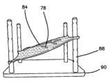
本発明の別の観点によれば、切開可能模擬組織構造体が提供される。切開可能模擬組織構造体は、シリコーンで作られた摸擬臓器を有する。1つまたは2つ以上の締結具が模擬臓器に連結されている。切開可能模擬組織構造体は、剛性材料で作られたトレーを有する。トレーは、ベースおよび支持プラットホームを有する。支持プラットホームは、ベースから離隔された状態でこのベースの上方に位置する。トレーは、1つまたは2つ以上の締結具配置場所を有する。模擬臓器は、1つまたは2つ以上の締結具と、1つまたは2つ以上の締結具配置場所の連結によりトレーに連結されている。1つまたは2つ以上の締結具は、1つまたは2つ以上の締結具配置場所に取り外し可能に連結可能である。In accordance with another aspect of the present invention, a dissectable simulated tissue structure is provided. The dissectable simulated tissue structure has a simulated organ made of silicone. One or more fasteners are coupled to the simulated organ. The dissectable simulated tissue structure has a tray made of a rigid material. The tray has a base and a support platform. The support platform is spaced apart from and above the base. The tray has one or more fastener placement locations. The simulated organ is coupled to the tray by coupling one or more fasteners to the one or more fastener placement locations. The one or more fasteners are removably coupleable to the one or more fastener placement locations.
1つまたは2つ以上の模擬臓器および組織の臓器トレーモデルは、カリフォルニア州所在のアプライド・メディカル・リソーシーズ・コーポレイション(Applied Medical Resources Corporation )により製造されているSIMSEI腹腔鏡下訓練システムのような模擬腹腔鏡下訓練装置の内部に配置されたときに腹腔鏡下手技および技術を訓練したり練習したりするのに理想的である。腹腔鏡下訓練装置10が図1に示されている。腹腔鏡下訓練装置10は、プラボング等(Pravong et al.)により2011年9月29日に出願され、アプライド・メディカル・リソーシーズ・コーポレイションに譲渡され、そして米国特許出願公開第2012/0082970号明細書として公開された同時係属中の米国特許出願第13/248,449号明細書(発明の名称:Portable laparoscopic trainer)に記載されており、この米国特許出願公開を参照により引用し、その記載内容全体を本明細書の一部とする。腹腔鏡下訓練装置10は、トップカバー12を有し、トップカバー12は、ベース14に、トップカバー12をベース14から隔てる1対のレッグ16によって連結されている。腹腔鏡下訓練装置10は、患者の胴、例えば腹部領域を真似るよう構成されている。トップカバー12は、患者の前方面を表し、トップカバー12とベース14との間に構成された空間は、臓器が位置する患者の内部または体腔またはキャビティ18を表している。腹腔鏡下訓練装置10は、患者を真似た状態で種々の外科的処置およびこれらの関連の器械を教示し、練習するとともに実演する上で有用なツールである。外科用器械がトップカバー12にあらかじめ設けられている孔20を通ってキャビティ18中に挿入される。これらあらかじめ設けられた孔20は、トロカールをシミュレートしたシールを有するのが良くまたは患者の皮膚および腹壁部分をシミュレートした模擬組織を有するのが良い。種々のツールおよび種々の方法を用いてトップカバー12を穿通することができ、それによりトップカバー12とベース14との間に配置されたモデル臓器、例えば本発明の右結腸モデルに対する実寸大の手技を実施することができる。訓練装置10のキャビティ18内に配置されると、臓器モデルは、一般に、ユーザの視界から覆い隠され、ユーザは、この場合、ビデオモニタ22上に表示されるビデオフィードにより手術部位を間接的に観察することによって術式を腹腔鏡下で練習することができる。An organ tray model of one or more simulated organs and tissues is ideal for training and practicing laparoscopic procedures and techniques when placed within a simulated laparoscopic trainer, such as the SIMSE I Laparoscopic Training System manufactured by Applied Medical Resources Corporation of California. A
トップカバー12にヒンジ留めされたビデオディスプレイモニタ22は、図1に開放配向状態で示されている。ビデオモニタ22は、イメージをモニタ22に送るための種々の視覚システムに連結可能である。例えば、あらかじめ形成された孔20のうちの1つまたはキャビティ内に設けられたウェブカムを通って挿入され、そして模擬手技を観察するために用いられる腹腔鏡をビデオモニタ22および/またはモバイルコンピュータ計算装置に連結してイメージをユーザに提供するのが良い。別の変形例では、トップカバー12は、ビデオディスプレイ22を備えず、ラップトップ型コンピュータ、モバイルディジタル装置またはタブレットを支持し、そしてこれをワイヤでまたはワイヤレスで訓練装置10に接続する手段を有する。The video display monitor 22 hinged to the
組み立てられると、トップカバー12は、レッグ16が実質的に周囲のところに配置されるとともにトップカバー12とベース14との間に相互に連結された状態で、ベース14の真上に配置される。トップカバー12およびベース14は、実質的に同一の形状およびサイズのものであり、実質的に同一の周辺輪郭を有している。訓練装置10は、側壁を備えていないが、レッグ16は、端部開放型訓練装置10からの視界から内部キャビティを部分的に隠している。腹腔鏡下訓練装置10は、ベース14に対して角度をなしたトップカバー12を有している。レッグ16は、ベース14に対するトップカバー12の角度を調節することができるよう構成されている。図1は、ベース14に対して約30~45°の角度まで調節される訓練装置10を示している。訓練装置10の傾斜により、有利には、トレンデレンブルグまたは逆トレンデレンブルグ位置での患者がシミュレートされる。トレンデレンブルグ位置では、患者の体は、足が心臓よりも高い位置にある状態でまたはこの反対の関係をなした状態でこの体が仰向けで平らになった状態になるよう傾けられる。トレンデレンブルグ位置により、重力が腸管を骨盤から引き離しているときに骨盤内臓器への良好な接近が可能になり、それにより骨盤内手術野上への腸管の侵入を阻止して外科医が臓器を容易に操作することができる腹腔内に広い作業空間を得ることができる。トップカバー12の選択された角度位置は、レッグ16に設けられている蝶ねじを示すことによってロックされる。ベース14に対する訓練装置10のトップカバー12の角度または水平面、例えばテーブルトップに対するトップカバー12の角度は、本発明の結腸モデルが訓練装置10のキャビティ18内に挿入された状態で右半結腸切除術の訓練および練習に関して特に有利である。When assembled, the
次に図2を参照すると、本発明の右結腸モデル26が示されており、この右結腸モデルは、腹腔鏡下環境中での手技のうちでとりわけ右半結腸切除術手技、例えば図1を参照して上述した腹腔鏡下訓練装置10の訓練および練習に特に適している。模擬臓器は、代表的には、シリコーンまたは熱可塑性エラストマー(TPE)で作られ、トレー28に配置される。トレー28は、トレー28内に納められたモデル臓器を収容するよう構成されている。トレー28は、ベースおよび代表的にはベースの周囲に沿ってぐるりと形成された少なくとも1つの側壁を有する。追加の側壁が解剖学的構造に特有な場所を定めるようこの周囲の内側に形成され、かかる追加の側壁は、模擬構造および組織を収容する構成されている。これら追加の側壁は、モデル26がキャビティ18内に納められた状態で医師が訓練装置10のトップカバー12を通って挿入された器械を用いて模擬臓器を操作している間、医師により加えられる力に応答して側方支持体となる。図2は、トレー28の頂部に沿って配置されたシリコーンで作られているモデル肝臓30および他の臓器の上に位置し、それぞれの血管系34を含む模擬網層32を示している。2, a
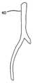
図3を参照すると、網層32は、下に位置する模擬臓器を露出させるよう引き戻された状態で示されており、かかる模擬臓器は、虫垂42およびS状結腸に取り付け可能な大腸36(図4に単独で示されている)の少なくとも一部分、小腸38の少なくとも一部分、胆嚢集成体を含む肝臓30、胃、十二指腸、腎臓、尿管、大動脈40(図5に単独で示されている)、動脈および静脈を表す血管44、腹膜、ジェロタ筋膜および腸間膜層46(図6に単独で示されている)を含む結合組織層を含む。臓器は、種々の腹腔鏡下器械を用いた外科的訓練のために人体内に存在する正確な解剖学的体位設定および存在場所を表すよう組み立てられている。右腸モデル26とも呼ばれる場合のある右結腸モデル26は、右半結腸切除術の外科的訓練のために重要なランドマークおよび特徴を強調するための改造を施した状態でシリコーン模擬臓器を用いて組み立てられている。With reference to FIG. 3, the
ベーストレー28が設けられている。ベーストレー28は、黄色または赤色のフォームで作られ、このベーストレーは、訓練装置10のキャビティ18内に挿入可能であるような寸法形状になっている。変形例として、ベーストレー28は、ベーストレー28中に直接嵌まり込む黄色または赤色のフォームで作られているライナを有しても良く、ベーストレー28は、このライナと一緒に腹腔鏡下訓練装置10内に挿入可能である。追加のフォーム部分をフォームベースの左側側部に追加するのが良く、その目的は、右側部側壁をシミュレートすることにある。模擬外科的処置の際の種々の体位置のシミュレーションを可能にするため、別のモデルベースが設けられる。例えば、右結腸モデルベース28またはライナは、モデル26の一端部のところに傾斜角を有するよう真空成形プラスチックで作られるのが良い。この角度は、外科的処置中における患者の逆トレンデレンブルグ体位設定をシミュレートすることができる。さらに、モデル26は、骨盤形状を真似るよう成形された湾曲形状を有するよう真空成形プラスチックベース上に作られるのが良く、骨盤形状は、腹部側壁の湾曲形状を形成するよう近位側に延びている。A
シリコーンで作られたシートが模擬臓器の取り付けおよび組み立てを助けるようモデルのベース28の頂部にくっつけられる。シリコーンで作られた臓器模擬およびこれらの色の一覧が以下の表1に見える。大腸36、大動脈40および腸間膜46は、実質的に、図2および図3に示されているサイズのままであっても良く、あるいは、これらは、腹腔鏡下訓練装置10のベースに良好に適合するために短くされても良く若しくは縮められても良い。これら解剖学的構造は、これらの正確な相対的解剖学的位置設定を厳密に表す仕方でフォームベーストレー28の頂部にくっつけられている。
表1:臓器及びこれらの色
Table 1: Organs and their colors
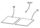
腸間膜層46は、動脈および静脈44を包封し、この腸間膜層は、腹腔鏡下切開用器を用いて把持されて切開されるよう構成されている。組織層相互間の切開は、シリコーンだけでシミュレートすることができない特性を備えている。したがって、この問題を解決するため、実際の解剖学的構造、例えば腸間膜46をシミュレートするに適した切開可能な模擬組織の幾つかの変形例を開発した。腸間膜46をシミュレートするのに適した切開可能模擬組織は、互いに頂部に積み重ねられた3つの層で構成されている。3つの層は、頂部層48、底部層50および中間層54を含む。頂部層48および底部層50は、腹膜層を表すのが良く、ゲルから成る中間層54は、切開可能なシリコーンで作られている血管44を包囲した結合組織を表すのが良い。The
次に図7A~図7Eを参照して、腸間膜層46として例示的に使用できる本発明の切開可能模擬組織の構成について今説明する。注目されるように、本発明の切開可能模擬組織47は、腸間膜層46としての使用には限定されず、かかる切開可能模擬組織は、任意の模擬組織構造の少なくとも一部を形成することができる。切開可能模擬組織47の構成は、2枚の別々の薄いシリコーンシートを作る初期ステップを含み、1枚のシートは頂部層48のためであり、1枚のシートは、底部層50のためであり、これら2枚のシリコーン層は、図7Aに示されているように完全に硬化されている。シートを完全に硬化させると、薄いシリコーンゲル層58a,58bをスパチュラまたはこれに類似したツールを用いてそれぞれ図7Bに示されているようにシリコーンシート48,50の模様が付いていない側部上に広げる。シリコーン血管を含む模擬血管系44を未硬化ゲル層58a,58bのいずれか一方上に置く。図7Cは、頂部層48のシート上の未硬化ゲル層58a上に配置されている模擬血管系44を示している。次に、シリコーンシート48,50は、ゲル層58a,58bとともに完全に硬化して模擬血管系44を頂部層48にくっつける。ゲル内張り層48,50を硬化させると、新たなシリコーンゲルから成る第3のまたは中間の層54を調製して層48,50の一方上に流す。一変形例では、新たなシリコーンゲルをシリコーンシート48上に流してシリコーン血管44が配置されるようにする。ゲルを塗り広げて血管系44の模擬血管を完全に覆うようにする。次に、第2の層50を第1の層48の頂部上で中間層54を覆って配置し、その間、シリコーンは、依然として未硬化のゲルであり、空気ポケットがサンドイッチ状の構造を作るようエッジに押し出される。このプロセスの結果として、3層切開可能模擬組織47を用いると、これら層相互間に配置されている包封血管系44の腹腔鏡下切開およびスケルトン化に特に適するとともに適合性のある例えば図6に示されている腸間膜組立体46をシミュレートすることができる。多数の層を設けることにより、切開可能な模擬組織構造にとって正確でかつ真に迫った感触および機能が得られる。さらに、切開可能模擬組織47は、有利には、種々の組織平面を作り、医師は、かかる組織平面を介して切開技能を磨くことができる。モデル26は、層を切開する能力を与えるだけでなく、モデル26により、医師は、組織平面または層44,48,50,54,58a,58bを正しく識別することができ、これは、個々の各手技にとって学習すべき重要な技能である。一変形例では、切開可能模擬組織47は、血管系層44なしで構成され、かかる切開可能模擬組織もまた、切開を実施するのに使用できる。7A-7E, a dissectable simulated tissue of the present invention that can be used as an
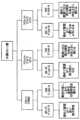
切開可能模擬組織47を作製するプロセス中に、結果的に切開可能模擬組織47の種々の所望の特性および繰り返しが得られるようにする幾つかの添加剤が導入されている。切開可能な腸間膜層46の外側の第1および第2の層48,50および内側のまたは中間の層54のための種々の成分の一覧が表2に示されており、かかる一覧は、図8Aおよび図8Bの流れ図にまとめられた状態で記載されている。図8Aおよび図8Bの所望の特性に基づいて最適な切開可能シートを決定する流れ図が図9Aおよび図9Bに示されている。図9Aおよび図9Bでは、腸間膜層は、本発明の切開可能模擬組織の例示の用途として用いられ、図9Aおよび図9Bの流れ図は、模擬腸間膜層だけを作るための用途には限定されず、任意の模擬組織構造における使用を含む。また、図9Aおよび図9Bに示されている血管は、模擬血管には限定されず、埋め込み状態の任意の模擬解剖学的構造または組織を含むことができ、かかる解剖学的構造または組織としては、腫瘍、病理学的病変部、臓器、管、軟骨などが挙げられるが、これらには限定されない。シリコーン外側層48,50は、従来の2つの部分が1:1の比の室温加硫(RTV)シリコーンで、若しくは1:1の比のRTVシリコーンとデッドナ添加剤で、または2:1の比のRTVシリコーンとデッドナ添加剤で作られている。RTVは、白金硬化室温加硫シリコーン(PCRTVS)を含むが、これには限定されない。シリコーンデッドナは、シリコーン油を含む(これには限定されない)シリコーン流体化学系統群に属し、このシリコーンデッドナは、白金硬化シリコーン添加剤である。デッドナの一例は、ペンシルベニア州マカンジー所在のスムース‐オン・インコーポレイテッド(Smooth-On, Inc.)により製造されているSLACKERと呼ばれる。添加剤としてのデッドニング剤(deadening agent)は、シリコーンを皮膚または人間組織の感触に類似するように軟らかくし、しかも本物らしくする。シリコーンデッドナは、軟化することができるとともに結果として生じる「感触」ならびに硬化シリコーンのリバウンド特性を変更することができるシリコーン添加剤である。この添加剤は、シリコーン油を含むシリコーン流体の化学系統群に含まれる。シリコーン流体およびシリコーン油は、流体の粘度および化学構造に依存するある範囲の使用を有する。シリコーンデッドナは、白金硬化室温加硫シリコーンと混合可能な種類のシリコーン油である。During the process of making the dissectable simulated tissue 47, several additives are introduced that result in various desired properties and iterations of the dissectable simulated tissue 47. A list of various ingredients for the outer first and
臓器を成形するために用いられる従来型シリコーンのデュロメータは、00~10ショアから10Aショアまでの範囲にある。かくして、デッドナの添加の結果として、互いに異なるデュロメータのシリコーンに添加されたときに互いに異なる特性が得られる。デュロメータの観点からは軟質のシリコーンへのデッドナの添加の結果として、完全硬化時に、ゲル状の組成物が得られる。しかしながら、デュロメータ的に高いシリコーンへのデッドニング剤(deadening agent)の添加の結果として、軟らかい感触のシリコーンの所望の特徴が得られ、かかる軟らかい感触のシリコーンは、完全硬化時に、その変形破断点に容易に達する。かくして、シリコーンとデッドニング剤の組み合わせは、腸間膜46を構成する外側層48,50、例えば腹膜層の手触り上の特徴を提供することができる。The durometer of conventional silicones used to mold organs ranges from 00-10 Shore to 10A Shore. Thus, the addition of deadening agents results in different properties when added to silicones of different durometers. The addition of deadening agents to silicones that are soft in terms of durometer results in a gel-like composition upon full cure. However, the addition of deadening agents to silicones with high durometer results in the desired characteristic of a soft-feeling silicone, which easily reaches its deformation break point upon full cure. Thus, the combination of silicone and deadening agent can provide the tactile characteristics of the
中間層の変形例は、(1)デッドナまたはデッドニング剤を含むゲル、(2)アルコールを含むゲル、(3)アルコールを含み熱が加えられたゲル、または(4)デッドナまたはデッドニング剤およびアルコールを含み熱が加えられたゲルを含む。イソプロピルアルコールが用いられる。包封ゲル層54への各添加剤の添加は、層を切開するために用いられて層の切開を容易にする圧力および力の大きさを減少させる。ゲルは、腸間膜組立体46中に中間可能層54として使用できる白金硬化シリコーンゴムゲルである。中間層54の切開を容易にする別の変形例では、アルコールを添加してゲルを薄くし、かくしてゲルへの侵入を容易にする。ゲル層54のそれ以上の劣化は、中間層54を切開することができる容易さを一層高めることができる。アルコールとゲルの混合物を約70℃まで加熱して硬化時間を早めるとともにアルコールの蒸発の結果として生じる多孔質中間層54を作る。ゲル、アルコールおよび熱で構成された多孔質中間層54は、ゲルに固有の粘着性を減少させ、腸間膜層48,50,54中に侵入してこれを切開しやすくする。別の変形例では、デッドナをシリコーンゲルに添加し、その結果、低い弾力特性を有することになるが、完全硬化時に粘着性が高められた調合物が得られる。硬化ゲル混合物の粘着性を含む問題を軽減するため、アルコールを同じ比でシリコーンゲルとデッドナの混合物に添加する。その結果得られた特性は、完全硬化時に、ゲル混合物それ自体およびゲルとデッドナと比較して粘着性が減少するが、この特性はまた、腹腔鏡下切開用器を用いた場合に所望の切開可能な手応えを示す。この場合もまた、熱をこの混合物に加えると、切開可能層の別の変形例について上記において一覧表示した特徴を有する多孔質中間層54が作られる。ゲルおよび種々の添加剤で構成される中間の切開可能層54の変形例は、有利には、組織を通って移動して腸間膜層46内で2つの外側層48,50相互間に納められている自由な血管を切開するという手応えを提供する。さらに、本明細書において与えられている調合されたゲル変形例は、キャビティが包囲されて腹腔鏡により照明される腹腔鏡下手技において特に有利である光沢または艶を提供する本物のような濡れた見掛けを層54に与える。
表2:層及び材料
Table 2: Layers and Materials
右結腸モデル26中に存在する血管系44は、テキサス州ヒューストン所在のクレートン・ポリマーズ(Kraton Polymers)製のシリコーンまたはKRATONポリマー管で作られている。血管は、上述した模擬腸間膜層46内に納められている。血管系44は、解剖学的に配置されてゲル中間層54によって腹膜層48,50にくっつけられている。さらに、血管系44が納められた切開可能な組織を右半結腸切除術用モデルに関して説明したが、この製造方法を同様な手段により任意の組織模擬モデルに利用することができまたは模擬組織プラットホーム用のスタンドアロン型モデルとして利用することができる。The
右結腸トレーの別のコンポーネントは、網(omentum)32である。網32は、大腸36上にくっつけられ、この網は、モデル26の頂部上に掛かっている。網32の幾つかの変形例が開発されている。第1の変形例は、モデル26の頂部上に容易に掛かることができるテキスチャ付きシリコーン流延網32である。しかしながら、網32の重さおよび感触をシミュレートするため、この網もまた、軟質シリコーンフォームを用いて流延により成形されるのが良い。フォームで作られた網32は、黄色に着色されており、これは腹腔内に広い空間を占めるように見えるが、モデル26の頂部上に掛かることができる。血管系34は、網32が体内で見えるようにその外観をシミュレートするために網32の両方の変形例上に存在している。Another component of the right colon tray is the
図3に戻ってこれを参照すると、上行結腸60を腹壁62に連結しているモデル上の付着部64の存在は、右半結腸切除手技の訓練にとって重要な特徴である。腹壁62に取り組むため、数個の同じ模型が作製される。腹壁62の第1の模型は、高さが約2インチ(5.08cm)である長くて薄いフォーム片をこれまたフォームで作られるのが良いベース28の側部に取り付けることによって作られる。腹部側壁62もまた、上述の成形されたベースおよび側壁の曲率を通ってベース28中に組み込まれるのが良い。最後に、腹部側部62は、フォームベース28の長さに合わせて延びてフォームベース28にくっつけられる湾曲した剛性で硬質の流延材料で作られるのが良い。外側付着部64を上述の腹壁62のうちの任意のものに取り付けることができる。外側付着部64は、上述の腹壁62のちの任意のものの頂部にくっつく2枚のテキスチャ付きシリコーンシートを用いることによって作られる。腹壁62にくっつけられた2枚のシート相互間にはトルトの白線が存在する。トルトの白線については幾つかのモデルが存在する。第1の模型は、トルトの白線をロープファイバ(rope fiber)でシミュレートしている。白い綿ロープのストランドがトルトの白線の血管系平面の外観を真似るようモデル26内に用いられている。トルトの白線もまた、解剖学的ランドマークを表すためにシリコーンで白いストライプまたは縞を作ることによってシミュレートできる。トルトの白線は、シリコーンシートの2つの層相互間にくっつけられ、次に、これらの層は、エッジに沿って互いにくっつけられ、次に上行結腸60にくっつけられる。その結果得られた構造は、腸36を含む側壁62に連結する外側付着部である。3, the presence of
本発明の別の観点では、切開可能な模擬組織47は、少なくとも2つの互いに異なる層で構成されている。第1の層は、シリコーン層で構成され、第2の層は、シリコーンゲルで構成されている。切開可能な模擬組織47を用いると、解剖学的酷似を備えた合成組織および臓器モデルを作ることができ、しかもかかる切開可能模擬組織47を切開および他の外科的処置の訓練のために用いられる模擬訓練モデルとして使用することができる。本発明の切開可能模擬組織47は、少なくとも1つの外側シリコーン層および1つまたは2つ以上の外側シリコーン層によって包封されたゲル層で構成されている組立体であり、その結果、外科医によって観察される切開に酷似する構造が得られる。切開可能模擬組織の1つまたは2つ以上のシリコーン層は、全重量の33%を占めるシリコーンデッドナと混合された2部品RTV10Aデュロメータシリコーンで作られ、それにより、用いられる全シリコーンとデッドナの比は2:1になる。デッドナは、これが添加される硬化シリコーンの特性を軟化するシリコーン油である。その結果、デッドナが添加される10Aデュロメータシリコーンは、硬化して10A未満のデュロメータシリコーンになる。デッドナの添加量は、これが添加されるデュロメータの特性の変化に比例する。シリコーン顔料がシリコーンとデッドナの混合物に添加され、それによりシリコーンが流延されてこれによって表される解剖学的構造に対応した顔料との粘性混合物がつくられる。シリコーン混合物をオプションとしてテキスチャを有するフォームのシート上に流延しまたは石膏材料で作られたテキスチャを含むシート上に流延する。フォームが用いられる場合には室温で約45分間かけてまたはフォームが用いられない場合にはオーブン内において約70℃の温度で約25分間かけて流延シリコーン混合物を硬化させる。シートサイズは、切開されている平面または表面のサイズに応じて漸変する長さおよび幅を有するのが良い。In another aspect of the present invention, the dissectable simulated tissue 47 is comprised of at least two distinct layers. The first layer is comprised of a silicone layer and the second layer is comprised of a silicone gel. The dissectable simulated tissue 47 can be used to create synthetic tissue and organ models with anatomical similarity and can be used as a simulated training model for training in dissection and other surgical procedures. The dissectable simulated tissue 47 of the present invention is an assembly comprised of at least one outer silicone layer and a gel layer encapsulated by one or more outer silicone layers, resulting in a structure that closely resembles an incision observed by a surgeon. The one or more silicone layers of the dissectable simulated tissue are made of a two-part RTV10A durometer silicone mixed with a silicone deadener that accounts for 33% of the total weight, resulting in a 2:1 ratio of total silicone to deadener used. Deadener is a silicone oil that softens the properties of the cured silicone to which it is added. As a result, a 10A durometer silicone to which deadener is added will cure to a durometer silicone less than 10A. The amount of deadener added is proportional to the change in durometer properties to which it is added. A silicone pigment is added to the silicone and deadener mixture, which causes the silicone to be cast to create a viscous mixture with the pigment corresponding to the anatomical structure it represents. The silicone mixture is cast onto a sheet of foam, optionally with texture, or onto a textured sheet made of a gypsum material. The cast silicone mixture is cured at room temperature for about 45 minutes if foam is used, or in an oven at a temperature of about 70°C for about 25 minutes if foam is not used. The sheet size may have a length and width that varies depending on the size of the plane or surface being incised.
シリコーンシートは、いったん硬化すると、長方形キャビティを有するモールド上に配置され、このキャビティは、シリコーンサイズよりも小さい。シリコーンシートは、このシートの中央領域がキャビティ内に配置されるとともにシートの外周部がモールドの表面上に平らに置かれるようモールド上に配置される。このセットアップ構成を提供することにより、ゲルの漏れを最小限に抑えた状態でゲルの包封プロセスが容易になる。中央キャビティ内において、シリコーン血管系および病理学的病変部、例えば腫瘍がシリコーン系接着剤を用いてキャビティ内に位置するシートの部分上にくっつけられる。血管系および病理学的病変部の配置は、切開が代表的に実施される解剖学的組織に類似している。シリコーン系接着剤が硬化し、血管系および病理学的病変部が無傷である場合、中間ゲル層が作られる。本発明は、血管系の埋め込みには限定されず、他の解剖学的ランドマークおよび構造をも含み、かかるランドマークおよび構造としては、血管系、腫瘍、病理学的病変部、臓器および組織構造が挙げられるが、これらには限定されず、これらの構成材料としては、任意のポリマー材料、シリコーン、KRATONなどが挙げられるが、これらには限定されない。Once the silicone sheet has cured, it is placed on a mold with a rectangular cavity, the cavity being smaller than the silicone size. The silicone sheet is placed on the mold such that the central region of the sheet is placed within the cavity and the outer periphery of the sheet is laid flat on the surface of the mold. Providing this set-up configuration facilitates the gel encapsulation process with minimal gel leakage. Within the central cavity, the silicone vasculature and pathological lesion, e.g., tumor, are attached with a silicone adhesive onto the portion of the sheet located within the cavity. The placement of the vasculature and pathological lesion resembles the anatomical tissue in which dissections are typically performed. When the silicone adhesive cures and the vasculature and pathological lesion are intact, an intermediate gel layer is created. The present invention is not limited to implantation in the vasculature, but also includes other anatomical landmarks and structures, including but not limited to the vasculature, tumors, pathological lesions, organs and tissue structures, and materials of construction, including but not limited to any polymeric material, silicone, KRATON, etc.
一変形例では、切開可能模擬組織中に存在する包封ゲルは、シリコーンゲル、デッドナ、およびイソプロピルアルコールで構成されている。ゲルを作るため、2部分から成るシリコーンゲルを等しい重量部および容量部で混合カップに入れる。デッドナを全シリコーン添加容量と等しい量で添加する。イソプロピルアルコールをデッドナの容量と等しい量で添加する。混合物を混合してついには、均質の溶液が得られるようにする。シリコーン顔料を必要に応じて添加して切開されるべき人間の組織に酷似する顔料を作るのが良い。溶液をいったん十分に混合すると、この溶液をモールドのキャビティ内に配置されている外側シリコーンシートの頂部上に流延してゲル層を作る。ゲルは、封入されてキャビティの頂部を通ることはなく、と言うのは、もしそうなると、ゲルの漏れが生じ、これは全体的組織モデルにとって有害だからである。シリコーンゲルは、シリコーンエラストマーである。しかし極めて軟質であるのは、白金硬化シリコーンゴムである。シリコーンゲルのデュロメータは、ショア00硬度スケールを下回り、それにより、ゲル状の軟質性、粘着性および低い耐引き裂き性が生じる。切開可能な組織に用いられるゲルの一例は、スムース‐オン社によって製造されて000‐35の硬度を有するECOFLEXゲルである。In one variation, the encapsulating gel present in the dissectable tissue simulant is composed of silicone gel, deadener, and isopropyl alcohol. To make the gel, the two-part silicone gel is placed in a mixing cup in equal parts by weight and volume. Deadener is added in an amount equal to the total silicone addition volume. Isopropyl alcohol is added in an amount equal to the volume of deadener. The mixture is mixed until a homogenous solution is obtained. Silicone pigment can be added as needed to create a pigment that closely resembles the human tissue to be dissected. Once the solution is thoroughly mixed, it is cast onto the top of the outer silicone sheet that is placed in the cavity of the mold to create a gel layer. The gel is encapsulated and does not pass over the top of the cavity, as this would cause gel leakage, which would be detrimental to the overall tissue model. The silicone gel is a silicone elastomer. However, it is a platinum-cured silicone rubber that is extremely soft. The durometer of silicone gels is below the Shore 00 hardness scale, which makes the gel soft, sticky, and has low tear resistance. One example of a gel used for dissectable tissue is ECOFLEX Gel, manufactured by Smooth-On, Inc., and has a hardness of 000-35.
この製造時点で、切開可能な模擬組織モデルを完了させる2つの別々の方法が存在する。例えば、切開可能模擬組織を図1~図6を参照して説明したように外科的処置の訓練に的を絞った臓器トレー内のコンポーネントとして消費することができる。かかる場合、切開可能模擬組織がトレー内のコンポーネントとして消費される場合、第1のシリコーンシートと同一のサイズおよびシリコーン、デッドナ、および顔料の蘇生を有する第2のシリコーンシートを用いてゲル層を包封する。第2のシリコーンシートをゲルを含む第1のシート上に載せる。層を押して空気ポケットがシートの側部に押し出されて大気中に放出されるようにする。シリコーン接着剤を用いて2枚のシリコーンシート相互間でキャビティの周囲を内張りし、それにより2つのシリコーン層相互間にシールを作ってゲルの漏れを阻止する。ゲルを2枚のシリコーンシート相互間で室温で硬化させる。いったん硬化すると、切開可能模擬組織を注型モールドから取り出すのが良い。シリコーンシートの周囲を用いて切開可能模擬組織を臓器トレー内で種々のシリコーン臓器に付着させるのが良い。切開可能模擬組織47は、特に図2~図6に示されているような右半結腸切除手技について訓練する模擬臓器訓練トレーのために作られるのが良い。At this point of manufacture, there are two separate ways to complete the dissectable simulated tissue model. For example, the dissectable simulated tissue can be consumed as a component in an organ tray targeted to training surgical procedures as described with reference to Figs. 1-6. In such a case, if the dissectable simulated tissue is consumed as a component in a tray, a second silicone sheet having the same size and silicone, deadener, and pigment reanimation as the first silicone sheet is used to encapsulate the gel layer. The second silicone sheet is placed on the first sheet containing the gel. The layer is pressed so that air pockets are pushed to the side of the sheet and released into the atmosphere. A silicone adhesive is used to line the perimeter of the cavity between the two silicone sheets, thereby creating a seal between the two silicone layers to prevent leakage of the gel. The gel is allowed to cure at room temperature between the two silicone sheets. Once cured, the dissectable simulated tissue can be removed from the casting mold. The perimeter of the silicone sheet can be used to attach the dissectable simulated tissue to various silicone organs in the organ tray. The dissectable simulated tissue 47 may be specifically constructed for a simulated organ training tray for training in a right hemicolectomy procedure, such as that shown in Figures 2-6.
別の例では、切開可能模擬組織46を小さなプラットホーム上で利用して切開の技術についてのみ訓練を行うことができる。この場合、ゲル層をキャビティ内にいったん流し込むと、ゲル層を約60℃においてオーブン内で約35分間かけて硬化させる。ゲルを硬化させると、10Aデュロメータシリコーン混合物を切開可能模擬組織の外側シリコーンシートと同一の顔料で調製する。第2のシリコーンシート層を形成するため、シリコーン混合物をゲルおよび外側シリコーンシート層に流延し、次に、オーブン内において約60℃で約30分間かけて硬化させる。その結果得られた切開可能模擬組織モデルは、切開の技量を練習するために用いることができるスタンドアロン型モデルである。切開可能模擬組織のこの模型は、一方の側部を持つモデルであり、この場合、外側層の一方だけが人の組織と同様な特性を持つ軟質のシリコーンである。10Aデュロメータシリコーンで構成された外側層は、例えば2012年9月25日に出願された米国特許出願第14/037,005号明細書(発明の名称:Surgical training model for laparoscopic procedures)に記載されている形式の縫合プラットホーム内に配置された場合にモデルのためのぴんと張った支持体に役立ち、この米国特許出願を参照により引用し、その記載内容全体を本明細書の一部とする。In another example, the dissectable
外側シリコーン層の作製中に添加される添加剤としてのデッドニング剤により、硬化後のシリコーンが軟質になるとともに皮膚またはヒトの組織の感触について本物のようになる。デッドナの添加の結果として、互いに異なるデュロメータを持つシリコーンに添加されると、互いに異なる特性が得られる。デュロメータ的に軟質のシリコーンへのデッドニング剤の添加の結果として、完全硬化時にゲルのような組成物が得られる。しかしながら、デュロメータ的に高いシリコーンへのデッドニング剤の添加の結果として、軟らかい感触のシリコーンの所望の特徴が得られ、かかる軟らかい感触のシリコーンは、完全硬化時に、その変形破断点に容易に達する。かくして、シリコーンとデッドニング剤の組み合わせは、腸間膜を構成するヒトの組織、例えば腹膜層の手触り上の特徴を提供することができる。The deadening agent as an additive added during the preparation of the outer silicone layer makes the silicone soft and realistic to the touch of skin or human tissue after curing. The addition of deadening agents results in different properties when added to silicones with different durometers. The addition of deadening agents to durometer soft silicones results in a gel-like composition upon full curing. However, the addition of deadening agents to durometer high silicones results in the desired characteristics of a soft-feeling silicone, which easily reaches its deformation break point upon full curing. Thus, the combination of silicone and deadening agent can provide the tactile characteristics of human tissues that make up the mesentery, such as the peritoneal layer.
中間ゲル層は、デッドナを含むとともにアルコールが追加されかつ熱の加えられたゲルを有する。包封ゲル層への各添加剤の添加により、切開のために用いられる圧力および力の大きさが減少し、それにより切開が容易になる。ゲルは、切開可能模擬組織中の中間切開可能層として使用できる白金硬化シリコーンゴムゲルである。アルコールを添加してゲルを薄くし、それにより中間層を切開しやすくするとともにこれに侵入しやすくする。さらに、ゲル層の劣化は、中間層の切開可能特性を更に高めることができる。アルコールとゲルの混合物を加熱して硬化時間を早めるとともにアルコールの蒸発により多孔質中間層を作る。ゲルおよびアルコールで構成された多孔質中間層は、ゲルに固有の粘着性を減少させるとともに包封ゲル層に侵入してこれを切開しやすくする。別の変形例では、デッドナをシリコーンゲルに添加し、その結果、完全硬化時に弾力特性が低いが、粘着性が増大した配合物が得られる。硬化ゲル混合物の粘着性を含む問題を軽減するため、アルコールを同じ比でシリコーンゲルとデッドナの混合物に添加する。その結果得られた特性は、完全硬化時に、ゲル混合物それ自体およびゲルとデッドナと比較して粘着性が減少するが、この特性はまた、腹腔鏡下切開用器を用いた場合に所望の切開可能な手応えを示す。この場合もまた、熱をこの混合物に加えると、切開可能層の別の変形例について上記において一覧表示した特徴を有する多孔質中間層が作られる。ゲルおよび種々の添加剤で構成された中間切開可能層の構成により、組織を通って移動して腸間膜層または他の組織構造若しくは臓器内の自由な血管を切開するという手応えを提供する。さらに、ゲルの光沢または艶の使用により、本物の組織の場合と同様な本物のような濡れた見掛けが与えられ、かかる使用は、腹腔鏡下技能の訓練の際にビデオモニタで見て特に有用である。The intermediate gel layer has a gel containing deadener, alcohol and heat added. The addition of each additive to the encapsulating gel layer reduces the amount of pressure and force used to make the incision, thereby facilitating the incision. The gel is a platinum cured silicone rubber gel that can be used as an intermediate dissectable layer in a dissectable simulated tissue. Alcohol is added to thin the gel, thereby making the intermediate layer easier to incise and penetrate. Additionally, degradation of the gel layer can further enhance the dissectable properties of the intermediate layer. The alcohol and gel mixture is heated to hasten the cure time and evaporate the alcohol to create a porous intermediate layer. The porous intermediate layer composed of gel and alcohol reduces the inherent tackiness of the gel and makes it easier to penetrate and dissect the encapsulating gel layer. In another variation, deadener is added to the silicone gel, resulting in a formulation with reduced elastic properties but increased tackiness when fully cured. To alleviate issues with tackiness of the cured gel mixture, alcohol is added to the silicone gel and deadener mixture in equal ratios. The resulting properties, when fully cured, are less tacky compared to the gel mixture itself and the gel plus deadner, but also provide the desired dissectable feel when used with laparoscopic dissectors. Again, application of heat to the mixture creates a porous intermediate layer having the characteristics listed above for the other dissectable layer variations. The construction of the intermediate dissectable layer, comprised of gel and various additives, provides the feel of moving through tissue to dissect free blood vessels within the mesenteric layer or other tissue structures or organs. Additionally, the use of a gel sheen or glaze provides a realistic wet appearance similar to that of real tissue, which is particularly useful when viewed on a video monitor during training of laparoscopic skills.
外側シリコーン層を作製する際の変形例は、シリコーンのデュロメータの変更を含む。模擬臓器モデルを作るために有用であるRTV白金硬化シリコーンは、00‐10デュロメータおよび10Aデュロメータを有し、シリコーン外側層は、いずれか一方のシリコーンを用いて作製できる。加うるに、デッドナをシリコーンに添加すると、シリコーンの硬化形態を軟化させることができる。シリコーンの軟らかさおよび弾性の変化は、添加されるデッドナの量に正比例する。図8Aは、切開可能模擬組織を形成する外側シリコーン層の流れ図を示している。Variations in making the outer silicone layer include changing the durometer of the silicone. RTV platinum cured silicones, useful for making simulated organ models, have a 00-10 durometer and a 10A durometer, and the silicone outer layer can be made with either silicone. Additionally, deadeners can be added to the silicone to soften the cured form of the silicone. The change in softness and elasticity of the silicone is directly proportional to the amount of deadener added. Figure 8A shows a flow diagram of the outer silicone layer forming the dissectable simulated tissue.
中間ゲル層は、デッドナ、アルコールを含む添加剤が施されるとともに硬化するために熱が加えられたベースシリコーンゲルから成る。添加剤の各々を別々になくすことにより、各ステップのところでの変形例が与えられ、その結果、各形態について特性が得られる。図8Bは、特定の比で添加剤を含むとともにこれら添加剤が示す特性を備えた各ゲル層組成物の変形例を示している。全ての実施形態についてこの本明細書全体にわたり、外側層に関する比は、容量に基づくか重量に基づくかのいずれかであるのが良く、その理由は、シリコーンおよびデッドナの密度がほぼ等しいからある。ゲル層とデッドナとアルコールの比は、容量に基づくものである。The middle gel layer is comprised of a base silicone gel to which additives including deadener and alcohol are applied and heat is applied to cure. Each additive is eliminated separately to provide variations at each step and result in properties for each configuration. Figure 8B shows variations of each gel layer composition with additives in specific ratios and properties exhibited by those additives. Throughout this specification for all embodiments, ratios for the outer layers may be either by volume or by weight since the densities of the silicone and deadener are approximately equal. The ratios of the gel layer, deadener, and alcohol are by volume.
切開可能な模擬腸間膜層の組み立てにおける変形例は、血管系をくっつけるシリコーンゲルの使用を含む場合がある。この組立体の構成は、デッドナを含むシリコーンの2枚の別々のシートを作り、そして図7Aに示されているようにこれらを完全に硬化させる初期ステップを含む。シートを完全に硬化させると、スパチュラまたは類似のツールを用いてシリコーンゲルの薄い層58a,58bを図7Bに示されているようにシリコーンシート48,50の各々の模様なしの側部上に広げる。シリコーン血管で作られた血管系44を図7Cに示されているようにシートのうちの1枚上の未硬化ゲル層58a上に載せる。ゲル層58a,58bを含むシリコーンシート48,50を完全に硬化させる。ゲル内張り層を硬化させると、新たなシリコーンゲル54を調製し、シリコーン血管44を配置させたシリコーンシート48上に流延する。ゲル54を塗り広げてこれが図7Dに示されているように血管44を完全に覆う。次に、第2のシリコーンシート50を未硬化ゲル上に載せ、空気ポケットをエッジまで押し出す。このプロセスの結果として、多層腸間膜は、図7Eに示されているような腹腔鏡下切開と適合性を持つことができる。A variation in the construction of the dissectable simulated mesenteric layer may include the use of silicone gel to hold the vasculature together. The construction of this assembly involves the initial steps of making two separate sheets of silicone with deadeners and allowing them to fully cure as shown in FIG. 7A. Once the sheets are fully cured, a spatula or similar tool is used to spread a thin layer of
本発明の切開可能模擬組織は、切開される代表的な組織の低い耐引き裂き性、弾性、靱性、色、およびテキスチャという機械的特性を有する。腹腔鏡下ツール、例えばマリーランド(Maryland)切開容器または腹腔鏡下はさみをこの組織内で用いて組織をそれぞれ切開しまたは切断するのが良い。ゲルの切開可能組織使用により、材料に対して独特の光沢が付与され、それによりこの材料は、本物のように濡れた外観を有することができる。この切開可能組織を構成するために用いられるゲルは、シリコーンを主成分としているので、ゲルを既に製造された種々の他のシリコーンモデルまたは他の臓器、例えばシリコーン血管に結合することができる。さらに、ゲルの粘性により、他の熱可塑性エラストマー、例えばKRATONポリマー(クレートンポリマー)で作られた血管をシリコーンゲルで外側シリコーン層に付着させることができる。The dissectable simulated tissue of the present invention has the mechanical properties of low tear resistance, elasticity, toughness, color, and texture of the representative tissue to be dissected. A laparoscopic tool, such as a Maryland dissecting container or laparoscopic scissors, may be used within the tissue to dissect or cut the tissue, respectively. The use of gel in the dissectable tissue imparts a unique sheen to the material, which allows the material to have a realistic wet appearance. Because the gel used to construct the dissectable tissue is silicone-based, the gel can be bonded to a variety of other silicone models or other organs already manufactured, such as silicone blood vessels. Furthermore, the viscosity of the gel allows blood vessels made of other thermoplastic elastomers, such as KRATON polymers, to be attached to the outer silicone layer with silicone gel.
本発明の切開可能模擬組織は、切開可能であり、しかもヒトの組織に酷似した幾つかの有利な特性を有する。模擬組織は、ヒトの組織の機械的特性、例えば弾性、靱性、色、およびテキスチャを真似ている。また、模擬組織の耐引き裂き性または引き裂き強度は低く、有利には、組織分離の伝搬を可能にする。模擬組織の耐引き裂き性が低いことにより、腹腔鏡下マリーランド(Maryland)切開容器または腹腔鏡下はさみを最小限の力で用いて鈍切開が容易になる。模擬組織はまた、切開を必要とする代表的な解剖学的構造の解剖学的ランドマークまたは解剖学的構造を含むことができる。これら解剖学的ランドマークまたは構造としては、臓器、腸間膜層相互間に埋め込まれた血管系、または切除される必要のある病理学的病変部、例えば腫瘍を包囲した腹膜シートが挙げられるが、これには限定されない。解剖学的ランドマークまたは構造は、非外傷性腹腔鏡下把持器またはマリーランド切開用器を用いて把持できまたは腹腔鏡下はさみを用いて切断できる。加うるに、本発明の切開可能模擬組織は、切開の完了時に解剖学的構造の操作および手技を可能にする。これら構造の運動は、切開の完了時にヒトの組織の解剖学的構造の運動に酷似している。加うるに、切開可能模擬組織は、一貫して製造可能である。模擬組織は、ヒトの臓器または膜の形状を取るよう成形可能である。切開可能模擬組織はまた、種々のシリコーンおよび熱可塑性樹脂と結合可能である。本発明は、シリコーン層の任意のものおよび全ては、下に位置する埋め込み状態の病理学的病変部、腫瘍、血管系などが層のうちの1つまたは2つ以上越しに僅かに視認できるよう半透明または透明であるのが良い。
実施例 The dissectable simulated tissue of the present invention is dissectable and has several advantageous properties that closely resemble human tissue. The simulated tissue mimics the mechanical properties of human tissue, such as elasticity, toughness, color, and texture. The simulated tissue also has low tear resistance or tear strength, which advantageously allows for the propagation of tissue separation. The low tear resistance of the simulated tissue facilitates blunt dissection using laparoscopic Maryland dissectors or laparoscopic scissors with minimal force. The simulated tissue can also include anatomical landmarks or structures representative of anatomical structures that require dissection. These anatomical landmarks or structures include, but are not limited to, organs, vasculature embedded between mesenteric layers, or peritoneal sheets surrounding pathological lesions that need to be excised, such as tumors. The anatomical landmarks or structures can be grasped using atraumatic laparoscopic graspers or Maryland dissectors or cut using laparoscopic scissors. In addition, the dissectable simulated tissue of the present invention allows for manipulation and manipulation of anatomical structures upon completion of the dissection. The movement of these structures closely resembles the movement of anatomical structures of human tissue upon completion of the dissection. In addition, the dissectable simulated tissue can be consistently manufactured. The simulated tissue can be molded to assume the shape of a human organ or membrane. The dissectable simulated tissue can also be bonded with a variety of silicone and thermoplastic resins. In the present invention, any and all of the silicone layers can be translucent or transparent to allow for a slight visibility of the underlying implanted pathological lesion, tumor, vasculature, etc. through one or more of the layers.
Working Example
図10A~図10Hを参照して、切開可能な模擬組織の組成物を有する付勢を有する模擬組織モデルを製造する実施例を今説明する。2つの部分、すなわち部分Aおよび部分Bを含む10Aデュロメータシリコーンを用意する。10Aデュロメータシリコーンの約5グラムの部分Aを10Aデュロメータシリコーンの約5グラムの部分Bと混合する。約5グラムのシリコーンデッドナを添加する。黄色のシリコーン顔料を添加する。シリコーンとデッドナと顔料を十分に混合する。混合物をテキスチャ付きモールド上に流延し、そして硬化させて図10Aに示されているように第1の層シート66を形成する。深さ約0.125インチ(3.175mm)の中央キャビティ70を備えた長方形のモールド68を図10Bに示されているように用意する。中央キャビティ70の深さを所望の切開可能な厚さに応じて変更することができる。血管系テンプレート72を図10Bに示されているよう長方形のモールドの中央キャビティ内に配置する。テンプレート72は、シリコーン血管が正確な解剖学的構造のために配置されるべき線74を示している。テンプレート72は、ある特定の病理学的病変部が配置されるべき解剖学的存在的場所76の表示を更に含む。血管系テンプレート72を開示するが、本発明は、これには限定されず、任意の解剖学的特徴のテンプレート72をある特定の構造、病理学的病変部、臓器、腫瘍および他の組織ならびに解剖学的ランドマークが配置されるべき場所に差し向けた状態で用いることができる。次に、第1の層シート66をこれが図10Cに示されているようにモールド68の外縁部と整列するようモールド68上に配置する。第1の層シート66は、テンプレート72が第1の層シート66越しに視認できるように透明である。次に、図10Dに示されているように、シリコーン系接着剤を用いて模擬血管系78および模擬腫瘍80を第1のシリコーンシート層66にくっつけるとともに下に配置されているテンプレート72上に示されている場所に配置する。模擬血管系78をテンプレート72の線74上に配置する。テンプレート72上の線74または他の形状を更に色付けしてこれと対応して色付けされた血管および/または臓器を正確な解剖学的存在場所に配置するのが良い。約3.3グラムの部分Aシリコーンゲルを約3.3グラムの部分Bシリコーンゲルと混合する。約6.67ミリメートルのシリコーンデッドナおよび5.27グラムのイソプロピルアルコールをシリコーンゲルに添加して一緒に混ぜる。体積または容量測定により、これらは、量的に約3.3ミリリットルの部分Aシリコーンゲル、約3.3ミリリットルの部分Bシリコーンゲル、6.67ミリリットルのシリコーンデッドナおよび6.67ミリリットルのイソプロピルアルコールを混ぜたものになる。黄色および白色のシリコーン含量を添加して混合する。混合物を中央キャビティ内に流し込んで模擬血管系78を包囲するが、キャビティを越えて漏れることがないようにし、それにより図10Eに示されているような中間ゲル層82を形成する。コンポーネントの全てをオーブン内において約60℃で約35分間かけて硬化させる。中間ゲル層82もまた、硬化時には透明であり、したがって、模擬血管78および模擬腫瘍80が中間ゲル層82越しに視認できるようになる。10Aデュロメータシリコーンの約35グラムの部分Aと35グラムの部分Bを混合する。シリコーンを硬化済みのゲルおよびモデルの側面上に流延して第2の外側層84を形成する。次に、モデルをオーブン内において約60℃で約25分間かけて硬化させる。第2の外側層84もまた、透明であり、ゲル層82と外側層84の組み合わせ中の埋め込み状態のランドマーク、例えば模擬腫瘍80および模擬血管78が層66,82,84越しに視認できるようにする。シリコーンシートサンドイッチの過剰の周囲を切り落としてモデルの寸法が縦横約4インチ×5インチ(10.16cm×12.7cm)になるようにし、中間ゲル層82が2つの包囲している外側層66,84で包封されるようにする。モデルの周囲を2つの外側層66,84相互間に中間ゲル層82がない状態で互いにくっつけられた2つの外側層66,84で作ってモデルからのゲル層82の漏れを阻止するようにする。外側層66,84は、ゲル中間層82を密封するとともにこれを包囲するのに役立つ。図10Gおよび図10Hを参照すると、長方形のモデルのコーナー部に4つの穴86をあけ、モデルを模擬組織プラットホーム90の直立したペグ88上に配置してトランポリンのように浮かせ、切開を練習することができるようにする。図10A~図10Hの模擬組織モデルは、胆嚢の付近の血管系のモデルであり、部分胆嚢モデルと見なすことができ、これは、模擬胆嚢を含んでも良くまたは含まなくても良い。使用にあたり、モデルをプラットホーム90上に浮かせまたは大きな臓器モデルまたは臓器トレーの一部を形成し、そして医師がモデルに対して外科的処置を訓練するための外科的模擬装置および/または訓練装置内に配置する。モデルを模擬装置および/または訓練装置の外側で用いても良い。さらに図11を参照すると、医師は、第2の外側層84に切り込んで中間ゲル層82に入る。医師は、第2の外側層84を第1の外側層66から離して広げ、それにより埋め込み状態の構造、模擬血管78および模擬腫瘍80に接近する。このようにする際、医師は、中間ゲル層82を分離しまたは切開する必要があろう。中間ゲル層82は、軟らかくて光沢があり、しかも弾性である。第2の外側層84を持ち上げると、中間層82の軟質弾性ゲルは、有利には、第2の外側層84を第1の外側層66から遠ざけたときにこのゲルが延びているときに線維膜に似ている。中間ゲル層82が延びているとき、この中間ゲル層は、有利には、ゲルのストランド92が、外科医がゲルのこれらストランドを切り開くよう練習する2つの層相互間に相互結合状態のままで深いポケット内に開く。軟質シリコーンゲルの鈍または鋭(sharp )切開が続いて空間をあけ、それにより中間ゲル層82を通りかつ第1の外側層66と第2の外側層84との間の切開平面を作る。前方の層もまた、切り込んでこれを分割するのが良く、それにより例えば中間ゲル層82内に埋め込まれている模擬血管系78のような構造の視認性を更に得るのが良く、それによりスケルトン化を真似る。10A-10H, an embodiment of fabricating a simulated tissue model having a bias with a dissectable simulated tissue composition will now be described. Prepare 10A durometer silicone including two parts, part A and part B. Mix approximately 5 grams of part A of 10A durometer silicone with approximately 5 grams of part B of 10A durometer silicone. Add approximately 5 grams of silicone deadener. Add yellow silicone pigment. Mix silicone, deadener and pigment thoroughly. Cast the mixture onto a textured mold and allow to cure to form a
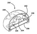
次に図12~図15を参照して本発明の別の形態としての臓器モデル200について説明する。臓器トレーモデル200は、図2~図6を参照して説明した特徴の全てを含み、これら特徴は、右結腸モデル26と関連しており、この臓器トレーモデルは、以下の模擬解剖学的構造、すなわち、模擬脾臓、模擬下降結腸、模擬S状結腸、模擬腸、模擬横行結腸、模擬直腸、模擬左腹側壁、模擬左トルトの白線、模擬大動脈、模擬左腎臓、模擬膵臓、模擬神経、腸間膜と腹膜後隙との間の模擬トルトの筋膜/腔、模擬左尿管、模擬生殖腺血管、模擬前立腺、模擬精嚢、模擬尿道、模擬膀胱、模擬腸間膜、模擬ダノンビリエー筋膜、模擬骨盤、左内腸間膜静脈および血管、およびプラスチックベースのうちの1つまたは2つ以上を含む左結腸モデルの特徴を更に有する。模擬解剖学的構造202は、オプションとしてのプラットホーム204上に支持され、このプラットホームは、ベース206内に配置された状態で外側模擬表皮層208で覆われている。プラットホーム204は、締結具、例えばリベット226を受け入れる複数の穴209を有し、これらリベット226は、模擬解剖学的構造202と関連しておりかつ模擬解剖学的構造202をプラットホーム204に解除可能に連結するよう穴209内にスナップ動作で入り込むよう構成されている。表皮層208の追加により開放手技をシミュレートすることができる。切開創211が図14Aに示されているように表皮層208に入れられており、次にこの切開創をレトラクトして図14Bおよび図15に示されているように模擬解剖学的構造202を露出させる。モデル200によりユーザは、開放または腹腔鏡下手術法を用いて直腸腸間膜全摘出手術(TME)を練習することができる。模擬腹腔鏡下手術の場合、表皮層208なしのモデル200が訓練装置10のキャビティ18内に配置される。開放手技を練習するため、訓練装置10のレッグ16を取り外し、トップカバー12をベース14上に直接載せてモデル200がトップカバー12とベース14との間に位置した状態でキャビティ18のサイズおよび高さを減少させる。シリコーン壁インサートをトップカバー12に設けられた大きなアパーチュア20内に配置する。開放手技を練習するための別の形態では、訓練装置10のレッグ16は、長さが短くなっており、それによりトップカバー12とベース14との間のキャビティ18の高さを減少させるよう改造されている。開放手技を練習するための更に別の形態では、訓練装置10のトップカバー12を取り外し、シリコーンシートをベース14内に配置されたモデル200上に配置する。表皮層をシミュレートするシリコーンシート208もまた、例えば図12に示されているベース14およびベース14の側壁上に配置する。Next, an
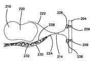
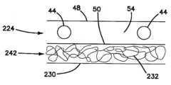
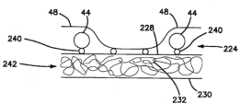
次に図16および図17を参照すると、模擬上行結腸216、模擬横行結腸218、および模擬下降結腸220を含む模擬腸210の一部分が模擬トルトの右白線212および模擬トルトの左白線214と一緒に示されている。右の模擬トルトの白線212と左の模擬トルトの白線214の両方が図16に示されているが、モデル200は、右または左のトルトの白線のうちの両方または一方だけを含む場合がある。図17は、左トルトの白線214に焦点を絞った断面図である。右トルトの白線212は、モデル200の右側について実質的に同一である。模擬腹膜層222は、腸210の上に位置した状態で腸210の底部からの模擬腸間膜層224に接合して模擬左トルトの白線214を形成し、次にそれぞれ、プラットホーム204の側壁に設けられたアパーチュア209を通って挿入された締結具、例えば226により側壁に連結されている。模擬腸間膜層224の少なくとも一部は、2015年7月16日に出願された米国特許仮出願第62/193,143号明細書に詳細に説明されているサンドイッチ層を作るためにポリフィル繊維材料の第3の層232によって互いに隔てられたシリコーンの第1の層228とシリコーンの第2の層230を有している。16 and 17, a portion of the
ポリフィルの層は、複数の1つまたは2つ以上の整列していない状態でランダムに配置された不織繊維から成り、かかる繊維は、この繊維の長さに沿う1つまたは2つ以上の場所で1つまたは2つ以上の隣接のシリコーン層に連結されていても良くそうでなくても良い。繊維は、以下に詳細に説明する製造プロセス中、第1の層および第2の層のうちの1つまたは2つ以上の中へ埋め込まれることによって第1の層および第2の層のうちの1つまたは2つ以上に連結されている。各繊維は、ストランド、フィラメント、ヤーン、マイクロファイバなどの形態をしているのが良く、各繊維は、長さを有するとともに、少なくとも第1の端部および第2の端部を備えている。繊維を連結するために接着剤が用いられても良くそうでなくても良い。第3の層の繊維は、第1の層と第2の層との間の隙間内にランダムに配置されたやり方で位置している。例えば、ファイバの1本のストランドは、1つの場所で第1の層に連結されるのが良く、次に繊維の長さに沿う別の場所で再び第1の層または第2の層に連結されても良く、その自由端部は、第1または第2の層中に埋め込まれても良くそうでなくても良い。繊維の数本のストランドが第1の層または第2の層に連結されなくても良く、これら数本のストランドは、第1の層と第2の層との間に自由に配置される。繊維の数本のストランドは、これらストランドが他のストランドに対して動くことができるよう他のストランドとルーズな仕方で絡み合わされるとともに撚りあわされている。繊維は、繊維の長さに沿う1つまたは2つ以上の場所で向かいのまたは第2の層に連結されるよう隙間をまたぐのが良い。第3の層を構成するのに複数の繊維ストランドに代えて単一の繊維ストランドを用いることが可能である。単一の繊維ストランドは、同じ隙間を埋めるのに短いストランドを用いるのと比較して、層相互間の隙間を埋めるとともに作るよう長さが長い。ポリフィルという用語が明細書全体を通じて用いられているが、組成は、ポリエステルには限定されない。繊維は、任意適当な材料、例えばポリエステル、ポリアミド、アクリル樹脂、アセテート、ポリオレフィン、綿、ファイバーフィル(繊維充填材)、詰め物、ポリエチレンテレフタレート、ポリエチレンナフタレート、ナイロン、ポリフィル、ファイバーフィル、ポリマー、プラスチック、スパンデックスまたは他の適当な繊維、天然繊維、非吸収性繊維、合成繊維または繊維状材料から選択されるが、かかる繊維は、依然としてポリフィルと呼ばれる。かかる材料は、織られていても良く、織られていなくても良く、あるいは部分的に織られていても良い。ファイバーフィル/ポリフィルは、代表的には、ガーネッティングによって作られ、かかるガーネッティングでは、ガーネット機が繊維を受け取ってこれらをくしけずってバットの形態にする。ガーネット機は、次に、繊維を折り曲げて裁断し、それにより短くしかも互いにかたまりとされるストランドを作る。繊維は、互いにもつれ、絡み合い、そして束になる。繊維は、有利には、特に腹腔鏡を用いた画像捕捉装置によりビデオモニタ上で見ると、濡れた状態の生きている組織を真似ている光沢のある繊維から光が多くの方向に反射されると、つやのある組織の見た目を提供する。The polyfill layer is comprised of a plurality of one or more unaligned randomly arranged nonwoven fibers that may or may not be connected to one or more adjacent silicone layers at one or more locations along the length of the fibers. The fibers are connected to one or more of the first and second layers by embedding the fibers in one or more of the first and second layers during a manufacturing process described in detail below. Each fiber may be in the form of a strand, filament, yarn, microfiber, or the like, each fiber having a length and at least a first end and a second end. An adhesive may or may not be used to connect the fibers. The fibers of the third layer are located in a randomly arranged manner within the gap between the first and second layers. For example, a single strand of fiber may be connected to the first layer at one location and then connected again to the first or second layer at another location along the length of the fiber, with its free end either embedded or not embedded in the first or second layer. Several strands of fiber may not be connected to the first or second layer, but are freely disposed between the first and second layers. Several strands of fiber are loosely intertwined and twisted with other strands so that they can move relative to the other strands. Fibers may span gaps to be connected to the opposite or second layer at one or more locations along the length of the fiber. A single fiber strand can be used to construct the third layer instead of multiple fiber strands. A single fiber strand has a long length to fill and create gaps between layers compared to using a shorter strand to fill the same gap. Although the term polyfill is used throughout the specification, the composition is not limited to polyester. The fibers may be selected from any suitable material, such as polyester, polyamide, acrylic, acetate, polyolefin, cotton, fiberfill, wadding, polyethylene terephthalate, polyethylene naphthalate, nylon, polyfill, fiberfill, polymer, plastic, spandex or other suitable fibers, natural fibers, non-absorbent fibers, synthetic fibers or fibrous materials, but such fibers are still referred to as polyfill. Such materials may be woven, non-woven, or partially woven. Fiberfill/polyfill is typically made by garnetting, in which a garnet machine receives the fibers and combs them into a batt. The garnet machine then bends and cuts the fibers, creating short, yet clumped strands. The fibers entangle, intertwine, and bundle together. The fibers advantageously provide the appearance of shiny tissue, especially when viewed on a video monitor with a laparoscopic image capture device, as light is reflected in many directions from the shiny fibers, mimicking living tissue in a wet state.
図18A~図18Cを参照すると、模擬トルトの白線212,214は、扁平なシリコーンの2つの薄い層234a,234bを図18Aに示されているように互いの頂部上に配置することによって作られる。底部層234bは、トルトの筋膜をシミュレートするよう第1のシリコーン層228と第2のシリコーン層230との間に配置されたポリフィルの第3の層232を有するポリフィルサンドイッチ層である。幅の狭いストリップ236が2つの並置された切れ目を図18Aに示されているように入れることによって両方の層234a,234bから除去される。トルトの白線をシミュレートする白色シリコーンの層238が幅の狭いストリップ236の除去によって作られた隙間を横切って被着されて図18Bに示されているように硬化するようになる。この構成の結果として、第1のシリコーン層234aおよび第2のシリコーン層234bの各々をそれぞれ2つ有する4つの層が図18Cに示されているようにシリコーンの白色の層238によって互いにくっつけられる。シミュレートされたトルトの白線238の一方の側に位置するシリコーンの第1の層234aおよび第2の層234bは、それぞれ、模擬腹膜層222および模擬腸間膜224を形成し、そしてこれらの層234a,234bは、接着剤により結腸に連結され、模擬トルトの白線234の他方の側に位置するシリコーンの第1の層234aおよび第2の層234bは、プラットホーム204に設けられた穴209中に差し込まれた締結具、例えばプラスチックリベット226によりプラットホーム204の側壁に取り付けられている。層222はまた、シリコーン、締結具、リベット、またはシアノアクリレート系接着剤を用いて右結腸の近傍で他方の側において側壁に取り付けられるのが良い。18A-18C, the
一形態では、模擬腸間膜224は、3つの層、すなわち、シリコーンの頂部の層48、シリコーンの底部層50およびゲルの中間層54のゲルサンドイッチから成り、ゲル中間層54は、本明細書に説明するとともに図19に示されているように頂部層と底部層との間にシリコーンなどで作られた摸擬血管44を封入している。図20に示されている別の形態では、模擬腸間膜層224は、シリコーンの第1の層228およびシリコーンの第2の層230ならびに第1の層と第2の層との間に位置するポリフィル繊維の第3の層232を有する。シリコーンの第1の層228は、接着剤240によりシリコーンの底部層50にくっつけられ、このシリコーンの底部層50は、シリコーンの頂部層48と一緒になって、ゲルの中間層54をサンドイッチしている。ゲル中間層54は、シリコーンで作られた摸擬血管44を封入するのが良い。図21に示されている更に別の形態では、シリコーンの頂部層48とシリコーンの底部層50は、ゲルの中間層54をサンドイッチし、このゲルの中間層54は、シリコーンで作られた摸擬血管44を封入するのが良い。底部層50は、シリコーンの第2の層230と一緒になって、これらの間にポリフィル繊維の第3の層232をサンドイッチしている。図22に示されたさらに別の形態では、シリコーンの第1の層228は、シリコーンの第2の層230と一緒になって、これらの間にポリフィルの第3の層232をサンドイッチしている。シリコーンの頂部層48が設けられ、模擬血管44が頂部層48と第1の層228との間に配置されている。模擬血管44は、接着剤240によって第1の層228にくっつけられているが、頂部層48にはくっつけられていない。また、頂部層48は、例えば図22に示されているように模擬血管44相互間の選択された領域に塗布された接着剤240により第1の層228にくっつけられている。この形態では、ゲルの中間層が設けられていない。別の形態では、シリコーンの第1の層228および第2の層230は、これらの間に設けられたポリフィルの第3の層232を備えている。シリコーンの頂部層48は、頂部層48と第1の層228との間にポリフィルの中間層54を備え、シリコーンの模擬血管44が頂部層48と第1の層228との間に配置されている。また、底部層50がポリフィルの中間層54とシリコーンの第1の層228との間に設けられるのが良い。図19~図22に示されているこれらの形態は、腸間膜と腹膜後隙との間のトルトの筋膜/腔をシミュレートする形態である。この腔は、2つのシリコーン層によって画定され、ポリフィルの第3の層232がこれら2つのシリコーン層の間に位置している。図20~図22の形態は、トルトの筋膜242と腸間膜224の組み合わせの形態を示している。筋膜層内において、模擬尿管、模擬生殖線血管、模擬十二指腸、および模擬神経束が配置されるのが良い。In one form, the
次に図23を参照すると、好ましくは赤色に染色されたシリコーンで作られている摸擬大動脈40が示されている。黄色に着色されたシリコーンで作られている模擬神経束244が設けられ、この模擬神経束は、図示のように模擬大動脈40に取り付けられている。模擬神経束244は、複数の開口部246を有する。一形態では、模擬神経束244は、ポリフィル領域内のトルトの筋膜242の腔内で大動脈上に嵌まっている。一形態では、模擬神経束244の色は、黄色ではない。23, there is shown a
次に図24を参照すると、モデル200は、下骨盤部分を有する。右または左結腸の可動化を練習した後、ユーザは、骨盤領域への切開を練習して模擬直腸間膜262を取り出すことができる。図24は、モデル200の骨盤領域のほぼ正中線断面を示している。モデル200は、恥骨をシミュレートするよう構成された前方に配置されているプラスチックシート248を有する。プラスチックシート248は、ユーザが練習を行わなければならない作業空間を減少させる。模擬直腸250の上方の解剖学的構造は、模擬前立腺系252を含む。模擬前立腺系252は、模擬前立腺254、模擬精嚢256、模擬膀胱258、模擬尿道、および模擬精管を含む。模擬尿道および模擬精管は、中実または中空の管の状態に形成されたシリコーンで作られている。模擬精嚢256は、ウレタンフォームもしくは他のフォームまたは模擬精管上に被覆成形された材料で作られている。模擬前立腺254は、ウレタンフォームもしくは他のフォームまたは模擬尿道上に被覆された材料で作られている。模擬前立腺系252は、プラスチックシート248に設けられた穴209に通して挿入された締結具、例えばリベット226によりプラスチックシート248に連結されている。プラスチックベース260がモデル200の後側に設けられている。プラスチックベース260は、外科的処置を練習するための限局された空間を作っている。模擬直腸250は、模擬直腸間膜層262によって包囲されたシリコーンの筒形の形状を有する。シリコーンの別の層または管264が設けられ、この層または管264は、ポリフィル繊維の内側層266を含む。外科医は、ポリフィル繊維層266の切断を練習して模擬直腸250の一部分を可動化する。模擬直腸250は、プラスチックベース260に設けられた穴209中に挿入された締結具、例えばリベット226によりベース260に連結されている。リベット226は、ユーザにポリフィル層266に入ってこれを切開して模擬直腸250および模擬直腸間膜262を除去するための空間を与えるよう第2の層または管264に取り付けられている。24, the
2015年3月26日に出願された国際出願PCT/US2015/022774号明細書(発明の名称:Simulated dissectible tissue)を参照により引用し、その記載内容全体を本明細書の一部とする。2016年7月12日に出願された国際出願PCT/US/2016/041852号明細書(発明の名称:Simulated dissectible tissue)を参照により引用し、その記載内容全体を本明細書の一部とする。International application PCT/US2015/022774 (title: Simulated dissectible tissue), filed March 26, 2015, is incorporated herein by reference in its entirety. International application PCT/US/2016/041852 (title: Simulated dissectible tissue), filed July 12, 2016, is incorporated herein by reference in its entirety.
本明細書において開示した切開可能な模擬組織の実施形態に対して種々の改造を行うことができることは言うまでもない。したがって、上述の説明は、本発明を限定するものと解されてはならず、単に好ましい実施形態の例示として考えるべきである。当業者であれば、本発明の範囲および精神に含まれる他の改造例を想到するであろう。It will be appreciated that various modifications may be made to the embodiments of the dissectable simulated tissue disclosed herein. Therefore, the above description should not be construed as limiting the invention, but merely as illustrative of preferred embodiments. Those skilled in the art will recognize other modifications that are within the scope and spirit of the invention.
Claims (15)
Translated fromJapanese上面および下面を備えたシリコーンの頂部層であって、前記上面と前記下面との間に厚さを定める頂部層と、
上面および下面を備えたシリコーンの底部層であって、前記上面と前記下面との間に厚さを定める底部層と、
前記頂部層と前記底部層との間に配置された中間層と、
上面および下面を備えたシリコーンの第1の層であって、前記上面と前記下面との間に厚さを定める第1の層と、
多層構造体を形成するように前記第1の層と前記底部層との間に配置されたポリフィルの第3の層と、
上面および下面を備えたシリコーンの第2の層と、を備え、
前記第2の層は、シリコーンの白いストリップの全長に沿って接合部を形成するように前記多層構造体に結合されている、
ことを特徴とする切開可能模擬組織構造体。 1. A dissectable simulated tissue structure for surgical training, comprising:
a top layer of silicone having an upper surface and a lower surface, the top layer defining a thickness between said upper surface and said lower surface;
a bottom layer of silicone having an upper surface and a lower surface, the bottom layer defining a thickness between said upper surface and said lower surface;
a middle layer disposed between the top layer and the bottom layer;
a first layer of silicone having an upper surface and a lower surface, the first layer defining a thickness between the upper surface and the lower surface;
a third layer of polyfill disposed between the first layer and the bottom layer to form a multi-layer structure;
a second layer of silicone having an upper surface and a lower surface;
the second layeris bonded to the multi-layer structure to form a bond along the entire length of the white strip of silicone;
A dissectable simulated tissue structure.
請求項1に記載の切開可能模擬組織構造体。 the contact area on one side of the outer side of the joint, the second layer, and the multi-layer structure are connected to a portion of a simulated intestine;
The dissectable simulated tissue structure of claim 1 .
請求項2に記載の切開可能模擬組織構造体。 The portion of the simulated intestine comprises a simulated ascending colon and a simulated descending colon.
The dissectable simulated tissue structure of claim 2.
請求項2または3に記載の切開可能模擬組織構造体。 the contact area on one side of the joint, the second layer, and the multi-layer structure are connected to a frame;
The dissectable simulated tissue structure according to claim2 or 3 .
請求項4に記載の切開可能模擬組織構造体。 the frame includes one or more fastener locations;
The dissectable simulated tissue structure of claim 4.
請求項5に記載の切開可能模擬組織構造体。 the second layer and the multi-layer structure are connected to the frame via a connection of one or more fasteners to the one or more fastener locations;
The dissectable simulated tissue structure of claim 5.
請求項6に記載の切開可能模擬組織構造体。 the one or more fasteners are rivets and the one or more fastener locations are holes formed in the frame to receive the rivets.
The dissectable simulated tissue structure of claim 6.
請求項1ないし7のいずれか1項に記載の切開可能模擬組織構造体。 a third layer of polyfill coupled to at least one of the first layer and the bottom layer;
The dissectable simulated tissue structure according to any one of claims 1 to 7.
請求項1ないし8のいずれか1項に記載の切開可能模擬組織構造体。 the intermediate layer is configured to surround at least one simulated tube, the at least one simulated tube being made of silicone;
The dissectable simulated tissue structure according to any one of claims 1 to 8.
請求項9に記載の切開可能模擬組織構造体。 The at least one simulated tract comprises one or more of a simulated ureter, a simulated reproductive tract blood vessel, a simulated duodenum, and a simulated nerve bundle.
The dissectable simulated tissue structure of claim 9.
請求項1ないし10のいずれか1項に記載の切開可能模擬組織構造体。 The intermediate layer is made of a gel or polyfill layer.
The dissectable simulated tissue structure according to any one of claims 1 to 10.
請求項1ないし11のいずれか1項に記載の切開可能模擬組織構造体。 the third layer of polyfill being in the form of a strand, filament, yarn, or microfiber having a length and first and second ends;
A dissectable simulated tissue structure according to any one of claims 1 to 11.
他の前記ファイバの幾つかが前記第1の層または底部層に連結されること無く前記第1の層と第2の層の間に自由に配置され、
これ以外の前記ファイバが、前記他のファイバのストランドが違いに動けるように、緩い態様で他のファイバのストランドと絡まり且つ絡み合ってる、
請求項12に記載の切開可能模擬組織構造体。 some of the strands of fiber are embedded in the first and bottom layers at one or more locations along the length of the fiber, and their first and second free ends may or may not be embedded in the first or bottom layers;
some of the other fibers are freely disposed between the first and second layers without being connected to the first or bottom layers;
the other fibers are entangled and intertwined with the other fiber strands in a loose manner such that the other fiber strands can move freely;
The dissectable simulated tissue structure of claim 12.
請求項12または13に記載の切開可能模擬組織構造体。 the strands of fiber are selected from the group consisting of polyester, polyamide, acrylic, acetate, polyolefin, cotton, fiberfill, wadding, polyethylene terephthalate, polyethylene naphthalate, nylon, polyfill, polymers, plastics, spandex, natural fibers, non-absorbent fibers, and synthetic fibers;
The dissectable simulated tissue structure according to claim 12 or 13.
請求項1ないし14のいずれか1項に記載の切開可能模擬組織構造体。 the white stripe of silicone, the second layer, and the multi-layer structure respectively represent a combination of a white tort, a simulated peritoneal layer, a simulated mesentery, and a simulated tort fascia;
A dissectable simulated tissue structure according to any one of claims 1 to 14.
Priority Applications (1)
| Application Number | Priority Date | Filing Date | Title |
|---|---|---|---|
| JP2025011561AJP2025067920A (en) | 2015-11-20 | 2025-01-27 | Dissectable simulated tissue |
Applications Claiming Priority (6)
| Application Number | Priority Date | Filing Date | Title |
|---|---|---|---|
| US201562257847P | 2015-11-20 | 2015-11-20 | |
| US62/257,847 | 2015-11-20 | ||
| US201562258710P | 2015-11-23 | 2015-11-23 | |
| US62/258,710 | 2015-11-23 | ||
| JP2018525577AJP6886975B2 (en) | 2015-11-20 | 2016-11-18 | Simulated incisable tissue |
| JP2021082973AJP7312783B2 (en) | 2015-11-20 | 2021-05-17 | Incisable simulated tissue |
Related Parent Applications (1)
| Application Number | Title | Priority Date | Filing Date |
|---|---|---|---|
| JP2021082973ADivisionJP7312783B2 (en) | 2015-11-20 | 2021-05-17 | Incisable simulated tissue |
Related Child Applications (1)
| Application Number | Title | Priority Date | Filing Date |
|---|---|---|---|
| JP2025011561ADivisionJP2025067920A (en) | 2015-11-20 | 2025-01-27 | Dissectable simulated tissue |
Publications (2)
| Publication Number | Publication Date |
|---|---|
| JP2023138507A JP2023138507A (en) | 2023-10-02 |
| JP7627722B2true JP7627722B2 (en) | 2025-02-06 |
Family
ID=57539628
Family Applications (4)
| Application Number | Title | Priority Date | Filing Date |
|---|---|---|---|
| JP2018525577AActiveJP6886975B2 (en) | 2015-11-20 | 2016-11-18 | Simulated incisable tissue |
| JP2021082973AActiveJP7312783B2 (en) | 2015-11-20 | 2021-05-17 | Incisable simulated tissue |
| JP2023112956AActiveJP7627722B2 (en) | 2015-11-20 | 2023-07-10 | Dissectable simulated tissue |
| JP2025011561APendingJP2025067920A (en) | 2015-11-20 | 2025-01-27 | Dissectable simulated tissue |
Family Applications Before (2)
| Application Number | Title | Priority Date | Filing Date |
|---|---|---|---|
| JP2018525577AActiveJP6886975B2 (en) | 2015-11-20 | 2016-11-18 | Simulated incisable tissue |
| JP2021082973AActiveJP7312783B2 (en) | 2015-11-20 | 2021-05-17 | Incisable simulated tissue |
Family Applications After (1)
| Application Number | Title | Priority Date | Filing Date |
|---|---|---|---|
| JP2025011561APendingJP2025067920A (en) | 2015-11-20 | 2025-01-27 | Dissectable simulated tissue |
Country Status (8)
| Country | Link |
|---|---|
| US (3) | US10706743B2 (en) |
| EP (2) | EP4235632A3 (en) |
| JP (4) | JP6886975B2 (en) |
| KR (2) | KR20250016466A (en) |
| AU (3) | AU2016358076A1 (en) |
| CA (1) | CA3005880A1 (en) |
| ES (1) | ES2955662T3 (en) |
| WO (1) | WO2017087746A1 (en) |
Families Citing this family (18)
| Publication number | Priority date | Publication date | Assignee | Title |
|---|---|---|---|---|
| US11776428B1 (en)* | 2015-10-21 | 2023-10-03 | University Of Rochester | Systems, models, and methods for simulating surgery on anatomical organs |
| US11568762B2 (en) | 2017-10-20 | 2023-01-31 | American Association of Gynecological Laparoscopists, Inc. | Laparoscopic training system |
| US11189195B2 (en)* | 2017-10-20 | 2021-11-30 | American Association of Gynecological Laparoscopists, Inc. | Hysteroscopy training and evaluation |
| JP7066137B2 (en)* | 2018-06-28 | 2022-05-13 | 豊田合成株式会社 | Surgical training equipment |
| AU2018368439B2 (en) | 2017-11-14 | 2024-08-29 | Applied Medical Resources Corporation | Hysterectomy model |
| EP3727172A1 (en)* | 2017-12-19 | 2020-10-28 | Applied Medical Resources Corporation | Total mesorectal excision surgical simulator |
| EP3803837A1 (en)* | 2018-06-01 | 2021-04-14 | Applied Medical Resources Corporation | Renal hilum surgical simulation system |
| WO2019244436A1 (en)* | 2018-06-19 | 2019-12-26 | イービーエム株式会社 | Artificial organ model for training for surgical technique, method for manufacturing artificial organ model, and method for training for surgical technique using artificial organ model |
| CN109523867B (en)* | 2018-11-21 | 2021-03-09 | 杨天烈 | Bracelet-type cow rectum positioning examination teaching device and teaching method |
| WO2020185385A1 (en)* | 2019-03-11 | 2020-09-17 | Kci Licensing, Inc. | Incision model to demonstrate closure effectiveness |
| KR102165852B1 (en) | 2019-05-02 | 2020-10-14 | 포항공과대학교 산학협력단 | Dip-coating method and apparatus using supporting liquid, and fabricating method of hollow tube using the same |
| US11574563B2 (en)* | 2019-05-23 | 2023-02-07 | Black Cat Medical Llc | Ultrasound guided training simulators for cryoneurolysis pain blocks |
| KR20230113600A (en)* | 2020-12-03 | 2023-07-31 | 인튜어티브 서지컬 오퍼레이션즈 인코포레이티드 | Systems and methods for assessing surgical performance |
| US12354496B2 (en)* | 2021-01-14 | 2025-07-08 | Intuitive Surgical Operations, Inc. | Colorectal and pelvic surgical simulation model systems and methods |
| CA3209856A1 (en)* | 2021-03-01 | 2022-09-09 | Gwendolyn Mary-jean MORGAN | Tactile tissue simulating structures |
| WO2023069782A1 (en) | 2021-10-23 | 2023-04-27 | Simulated Inanimate Models, LLC | Procedure guidance and training apparatus, methods and systems |
| US20230343243A1 (en)* | 2022-04-22 | 2023-10-26 | Covidien Lp | Regenerative artificial tissue for electrosurgical robotic assisted surgery training and testing |
| KR102825289B1 (en)* | 2023-08-22 | 2025-06-25 | 주식회사 알데바 | Prostate surgery simulator |
Citations (5)
| Publication number | Priority date | Publication date | Assignee | Title |
|---|---|---|---|---|
| WO2002038039A2 (en) | 2000-10-23 | 2002-05-16 | Toly Christopher C | Human surgical trainer and methods for training |
| JP2008197483A (en) | 2007-02-14 | 2008-08-28 | Koken Co Ltd | ESD training model |
| JP2012128109A (en) | 2010-12-14 | 2012-07-05 | Tmc Co Ltd | Human body partial manikin |
| JP2015502563A (en) | 2011-10-21 | 2015-01-22 | アプライド メディカル リソーシーズ コーポレイション | Simulated tissue structure for surgical training |
| WO2015148817A1 (en) | 2014-03-26 | 2015-10-01 | Applied Medical Resources Corporation | Simulated dissectible tissue |
Family Cites Families (462)
| Publication number | Priority date | Publication date | Assignee | Title |
|---|---|---|---|---|
| US184573A (en) | 1876-11-21 | Improvement in gas-cocks | ||
| US2127774A (en) | 1936-04-27 | 1938-08-23 | Jacobs Julian Bay | Apparatus for teaching obstetrics |
| US2324702A (en) | 1938-11-30 | 1943-07-20 | Karl F Hoffmann | Surgical simulacra and process of preparing same |
| US2284888A (en) | 1941-04-14 | 1942-06-02 | Arc Diaphragm & Drug Co | Demonstrating device for vaginal diaphragms |
| US2345489A (en) | 1943-04-10 | 1944-03-28 | Frederic P Lord | Anatomical model |
| US2495568A (en) | 1948-12-30 | 1950-01-24 | Holland Rantos Company Inc | Clinical model |
| US3766666A (en) | 1971-10-13 | 1973-10-23 | Robins Co Inc A H | Uterine simulator trainer |
| US3789518A (en) | 1972-04-12 | 1974-02-05 | Weatherby Nasco Inc | Simulated human limb |
| US3775865A (en) | 1972-07-24 | 1973-12-04 | R Rowan | Simulator for teaching suturing techniques |
| US3991490A (en) | 1973-04-09 | 1976-11-16 | Markman H David | Teaching aid for sigmoidoscope and the like |
| FR2260838B1 (en)* | 1974-02-08 | 1977-06-10 | Citroen Sa | |
| US3921311A (en) | 1975-01-06 | 1975-11-25 | Pathfinder Fund | Clinical demonstration model |
| US4001951A (en) | 1975-03-25 | 1977-01-11 | Fasse Wolfgang G | Breast cancer detection training device |
| US4001952A (en) | 1975-10-24 | 1977-01-11 | Kleppinger Trygve M | Hysteroscopy teaching aid |
| JPS5760029Y2 (en)* | 1978-07-26 | 1982-12-21 | ||
| US4341227A (en)* | 1979-01-11 | 1982-07-27 | Bsd Corporation | System for irradiating living tissue or simulations thereof |
| US4321047A (en) | 1980-06-05 | 1982-03-23 | Bradley Landis | Simulator and process for teaching surgical knot tying techniques |
| US4323350A (en) | 1980-09-22 | 1982-04-06 | Bowden Jr Robert L | Anatomical model |
| US4371345A (en) | 1980-10-17 | 1983-02-01 | National Research Development Corporation | Multi-dimensional display equipment |
| US4332569A (en) | 1981-03-16 | 1982-06-01 | University Of Kentucky Research Foundation | Instructional device for use of a bronchoscope |
| ES260340Y (en) | 1981-08-31 | 1982-10-16 | LEARNING DEVICE FOR ENDOSCOPES | |
| US4386917A (en) | 1981-09-16 | 1983-06-07 | Forrest Leonard E | Suturing training device and method |
| US4481001A (en) | 1983-05-26 | 1984-11-06 | Collagen Corporation | Human skin model for intradermal injection demonstration or training |
| US4596528A (en) | 1984-07-02 | 1986-06-24 | Lewis Leonard A | Simulated skin and method |
| US4726772A (en) | 1986-12-01 | 1988-02-23 | Kurt Amplatz | Medical simulator |
| US4737109A (en) | 1987-03-03 | 1988-04-12 | Abramson Daniel J | Breast cancer detection training device |
| US4832978A (en) | 1987-04-24 | 1989-05-23 | Lesser Jary M | Simulated connective tissue for construction of models and prostheses |
| US4789340A (en)* | 1987-08-18 | 1988-12-06 | Zikria Bashir A | Surgical student teaching aid |
| JP2663165B2 (en) | 1987-08-24 | 1997-10-15 | プラカス,ヴィクター・マニュエル | Human figure |
| IL84752A (en) | 1987-12-08 | 1991-11-21 | Elscint Ltd | Anatomical models and methods for manufacturing such models |
| US4907973A (en) | 1988-11-14 | 1990-03-13 | Hon David C | Expert system simulator for modeling realistic internal environments and performance |
| US4867686A (en) | 1989-02-09 | 1989-09-19 | Goldstein Mark K | Breast cancer detection model and method for using same |
| US4938696A (en) | 1989-07-25 | 1990-07-03 | Foster-Pickard International, Inc. | Model demonstrating human organ systems |
| US5053341A (en)* | 1989-10-06 | 1991-10-01 | The United States Of America As Represented By The Administrator Of The National Aeronautics And Space Administration | Tissue simulating gel for medical research |
| US5061187A (en) | 1990-04-12 | 1991-10-29 | Ravinder Jerath | Ultrasound training apparatus |
| US5104328A (en) | 1990-04-18 | 1992-04-14 | Lounsbury Katherine L | Anatomical model |
| US5149270A (en) | 1990-10-29 | 1992-09-22 | Mckeown M J | Apparatus for practicing surgical procedures |
| US5279547A (en) | 1991-01-03 | 1994-01-18 | Alcon Surgical Inc. | Computer controlled smart phacoemulsification method and apparatus |
| DE4105892A1 (en) | 1991-02-14 | 1992-08-27 | Arnold Dipl Ing Dr Med Pier | Simulation training appts. for surgery - comprises sealed housing and cover releasably connected to housing, excess pressure being formable in housing, and having flat insert in cover top side opening |
| DE9102218U1 (en) | 1991-02-14 | 1991-05-16 | Pier, Arnold, Dipl.-Ing. Dr.med., 5138 Heinsberg | Training device for laparoscopic surgical technique |
| WO1992017121A1 (en) | 1991-04-05 | 1992-10-15 | Metcal, Inc. | Instrument for cutting, coagulating and ablating tissue |
| US5403191A (en) | 1991-10-21 | 1995-04-04 | Tuason; Leo B. | Laparoscopic surgery simulator and method of use |
| US5318448A (en) | 1992-01-06 | 1994-06-07 | Garito Jon C | Demonstration model for gynecological procedure |
| US5180308A (en) | 1992-01-06 | 1993-01-19 | Garito Jon C | Medical demonstration model |
| US5775916A (en) | 1992-01-15 | 1998-07-07 | Limbs & Things Limited | Method of making a surgical and/or clinical apparatus |
| FR2691826A1 (en) | 1992-06-01 | 1993-12-03 | Allal Hossein | Coelio-surgery simulator for teaching and training of surgeons - uses semi-rigid envelope in form of human body, with coeloscope connected to video camera and to video screen |
| US5273435B1 (en) | 1992-07-16 | 1995-12-05 | Wisconsin Med College Inc | Tumor localization phantom |
| US5230630A (en) | 1992-07-20 | 1993-07-27 | Richard Burgett | Suture training device |
| US5368487A (en) | 1992-07-31 | 1994-11-29 | Medina; Marelyn | Laparoscopic training device and method of use |
| US5762458A (en) | 1996-02-20 | 1998-06-09 | Computer Motion, Inc. | Method and apparatus for performing minimally invasive cardiac procedures |
| EP0611469B1 (en) | 1992-09-07 | 1999-12-15 | Karl Storz GmbH & Co. | Medical training apparatus |
| DE9320422U1 (en) | 1992-09-07 | 1994-06-30 | Bastert, G., Prof. Dr., 69115 Heidelberg | Medical training device |
| US5310348A (en) | 1992-10-09 | 1994-05-10 | United States Surgical Corporation | Suture demonstration portfolio |
| US5295694A (en) | 1992-10-27 | 1994-03-22 | Levin John M | Laparoscopic surgery simulating game |
| US5769640A (en) | 1992-12-02 | 1998-06-23 | Cybernet Systems Corporation | Method and system for simulating medical procedures including virtual reality and control method and system for use therein |
| US5358408A (en) | 1993-03-09 | 1994-10-25 | Marelyn Medina | Tissue specimen suspension device |
| US5320537A (en) | 1993-03-16 | 1994-06-14 | Triangle Research And Development Corporation | Microsurgical training apparatus |
| US5472345A (en) | 1993-04-14 | 1995-12-05 | Gaumard Scientific Company, Inc. | Gynecological simulator |
| US5425644A (en) | 1993-05-13 | 1995-06-20 | Gerhard Szinicz | Surgical training apparatus and method |
| US5518407A (en) | 1993-11-02 | 1996-05-21 | Greenfield; Cathy L. | Anatomically correct artificial organ replicas for use as teaching aids |
| US5518406A (en) | 1993-11-24 | 1996-05-21 | Waters; Tammie C. | Percutaneous endoscopic gastrostomy teaching device |
| US5380207A (en) | 1993-12-27 | 1995-01-10 | Siepser; Steven B. | Slip suture practice kit |
| DE4414832A1 (en) | 1994-04-28 | 1995-11-02 | Laerdal Asmund S As | Teaching model for practising blood taking or injection of blood vessels |
| US5541304A (en) | 1994-05-02 | 1996-07-30 | Hercules Incorporated | Crosslinked hydrogel compositions with improved mechanical performance |
| US5623582A (en) | 1994-07-14 | 1997-04-22 | Immersion Human Interface Corporation | Computer interface or control input device for laparoscopic surgical instrument and other elongated mechanical objects |
| WO1996010725A1 (en) | 1994-09-30 | 1996-04-11 | Tovarischestvo S Ogranichennoi Otvetstvennostiu 'anter Ltd.' | Target |
| US5720742A (en) | 1994-10-11 | 1998-02-24 | Zacharias; Jaime | Controller and actuating system for surgical instrument |
| US6106524A (en) | 1995-03-03 | 2000-08-22 | Neothermia Corporation | Methods and apparatus for therapeutic cauterization of predetermined volumes of biological tissue |
| US5882206A (en) | 1995-03-29 | 1999-03-16 | Gillio; Robert G. | Virtual surgery system |
| US5649956A (en) | 1995-06-07 | 1997-07-22 | Sri International | System and method for releasably holding a surgical instrument |
| US5814038A (en) | 1995-06-07 | 1998-09-29 | Sri International | Surgical manipulator for a telerobotic system |
| US5620326A (en) | 1995-06-09 | 1997-04-15 | Simulab Corporation | Anatomical simulator for videoendoscopic surgical training |
| US5803746A (en) | 1996-01-23 | 1998-09-08 | Medisim Corporation | Body part model and method of making same |
| US5855583A (en) | 1996-02-20 | 1999-01-05 | Computer Motion, Inc. | Method and apparatus for performing minimally invasive cardiac procedures |
| US5743730A (en) | 1996-05-07 | 1998-04-28 | Clester; Kenneth E. | Dental porcelain shading guide and method of use therefore |
| US5722836A (en) | 1996-05-21 | 1998-03-03 | Simulab Corporation | Reflected-image videoendoscopic surgical trainer and method of training |
| US5785531A (en) | 1996-06-06 | 1998-07-28 | Wilson-Cook Medical Incorporated | Cuttable papilla and sphincterotomy training apparatus |
| US6929481B1 (en) | 1996-09-04 | 2005-08-16 | Immersion Medical, Inc. | Interface device and method for interfacing instruments to medical procedure simulation systems |
| US5727948A (en) | 1996-09-05 | 1998-03-17 | Jordan; Lynette S. | Syringe injection practice device |
| DE59608936D1 (en) | 1996-12-04 | 2002-04-25 | Erbe Elektromedizin | ARTIFICIAL FABRIC |
| JP3679535B2 (en) | 1997-01-29 | 2005-08-03 | オリンパス株式会社 | Colonoscopy insertion practice device |
| DE19716341C2 (en) | 1997-03-19 | 2000-09-21 | Erbe Elektromedizin | Training model, especially torso |
| US6271278B1 (en) | 1997-05-13 | 2001-08-07 | Purdue Research Foundation | Hydrogel composites and superporous hydrogel composites having fast swelling, high mechanical strength, and superabsorbent properties |
| GB9712987D0 (en) | 1997-06-19 | 1997-08-27 | Limbs & Things Ltd | Surgical training apparatus |
| US5873863A (en) | 1997-08-29 | 1999-02-23 | United States Surgical Corporation | Vascular surgery demonstration/training kit |
| JP2893178B2 (en) | 1997-09-01 | 1999-05-17 | 工業技術院長 | Biological optical phantom and method of manufacturing the same |
| US5947743A (en) | 1997-09-26 | 1999-09-07 | Hasson; Harrith M. | Apparatus for training for the performance of a medical procedure |
| US5873732A (en) | 1997-09-26 | 1999-02-23 | Hasson; Harrith M. | Apparatus for training for the performance of a medical procedure |
| IL123073A0 (en) | 1998-01-26 | 1998-09-24 | Simbionix Ltd | Endoscopic tutorial system |
| GB2349730B (en) | 1998-01-28 | 2003-04-09 | Ht Medical Systems Inc | Interface device and method for interfacing instruments to medical procedure simulation system |
| US8016823B2 (en) | 2003-01-18 | 2011-09-13 | Tsunami Medtech, Llc | Medical instrument and method of use |
| US6511325B1 (en) | 1998-05-04 | 2003-01-28 | Advanced Research & Technology Institute | Aortic stent-graft calibration and training model |
| US5908302A (en) | 1998-06-12 | 1999-06-01 | Goldfarb; Michael A. | Inguinal hernia model |
| US6113395A (en) | 1998-08-18 | 2000-09-05 | Hon; David C. | Selectable instruments with homing devices for haptic virtual reality medical simulation |
| GB9827496D0 (en) | 1998-12-14 | 1999-02-10 | Pharmabotics Limited | Simulated body tissue |
| US6169155B1 (en) | 1999-01-14 | 2001-01-02 | Dow Corning Corporation | Silicone gel composition and silicone gel produced therefrom |
| EP1169693A2 (en) | 1999-03-02 | 2002-01-09 | Peter Yong | Thoracic training model for endoscopic cardiac surgery |
| JP2001005378A (en) | 1999-06-24 | 2001-01-12 | Yasuhisa Koki:Kk | Simulator for training of operation technique |
| US20030031993A1 (en) | 1999-08-30 | 2003-02-13 | Carla Pugh | Medical examination teaching and measurement system |
| US6264609B1 (en)* | 1999-09-15 | 2001-07-24 | Wake Forest University | Ultrasound apparatus and method for tissue characterization |
| US6398557B1 (en) | 1999-09-17 | 2002-06-04 | The University Of Iowa Research Foundation | Devices, methods and kits for training in surgical techniques |
| US6488507B1 (en) | 1999-11-29 | 2002-12-03 | Ethicon, Inc. | Portable surgical trainer |
| US6497902B1 (en) | 1999-12-01 | 2002-12-24 | The Regents Of The University Of Michigan | Ionically crosslinked hydrogels with adjustable gelation time |
| BR9906054A (en) | 1999-12-29 | 2001-09-18 | Thadeu Rezende Provenza | Female human mammary gland simulator device |
| US7819799B2 (en) | 2000-03-16 | 2010-10-26 | Immersion Medical, Inc. | System and method for controlling force applied to and manipulation of medical instruments |
| US6817973B2 (en) | 2000-03-16 | 2004-11-16 | Immersion Medical, Inc. | Apparatus for controlling force for manipulation of medical instruments |
| US6939138B2 (en) | 2000-04-12 | 2005-09-06 | Simbionix Ltd. | Endoscopic tutorial system for urology |
| CN2421706Y (en) | 2000-04-26 | 2001-02-28 | 佟玉章 | Multifunctional exerciser for operating tying and suturing |
| US6969480B2 (en) | 2000-05-12 | 2005-11-29 | Matregen Corp. | Method of producing structures using centrifugal forces |
| CA2420240A1 (en) | 2000-08-23 | 2002-02-28 | The Royal Alexandra Hospital For Children | A laparoscopic trainer |
| US6589057B1 (en) | 2000-09-27 | 2003-07-08 | Becton, Dickinson & Company | Incision trainer for ophthalmological surgery |
| US8556635B2 (en) | 2000-10-23 | 2013-10-15 | Christopher C. Toly | Physiological simulator for use as a brachial plexus nerve block trainer |
| US7857626B2 (en) | 2000-10-23 | 2010-12-28 | Toly Christopher C | Medical physiological simulator including a conductive elastomer layer |
| US7850454B2 (en) | 2000-10-23 | 2010-12-14 | Toly Christopher C | Simulated anatomical structures incorporating an embedded image layer |
| AU2002230578A1 (en) | 2000-10-30 | 2002-05-15 | Naval Postgraduate School | Method and apparatus for motion tracking of an articulated rigid body |
| US6517354B1 (en) | 2000-11-17 | 2003-02-11 | David Levy | Medical simulation apparatus and related method |
| US6659776B1 (en) | 2000-12-28 | 2003-12-09 | 3-D Technical Services, Inc. | Portable laparoscopic trainer |
| US7526112B2 (en) | 2001-04-30 | 2009-04-28 | Chase Medical, L.P. | System and method for facilitating cardiac intervention |
| US20050147656A1 (en)* | 2001-06-14 | 2005-07-07 | Hemcon, Inc. | Tissue dressing assemblies, systems, and methods formed from hydrophilic polymer sponge structures such as chitosan |
| US6485308B1 (en) | 2001-07-09 | 2002-11-26 | Mark K. Goldstein | Training aid for needle biopsy |
| US7056123B2 (en) | 2001-07-16 | 2006-06-06 | Immersion Corporation | Interface apparatus with cable-driven force feedback and grounded actuators |
| NL1018874C2 (en) | 2001-09-03 | 2003-03-05 | Michel Petronella Hub Vleugels | Surgical instrument. |
| DE10148341A1 (en) | 2001-09-29 | 2003-04-24 | Friedhelm Brassel | Process for the production of a model system for vascular malformations |
| GB2380594B (en) | 2001-10-02 | 2004-02-04 | Keymed | Improvements in endoscopy training apparatus |
| US6773263B2 (en) | 2001-10-09 | 2004-08-10 | Robert J. Nicholls | Medical simulator |
| US7464847B2 (en) | 2005-06-03 | 2008-12-16 | Tyco Healthcare Group Lp | Surgical stapler with timer and feedback display |
| EP1453432B1 (en) | 2001-12-04 | 2012-08-01 | Tyco Healthcare Group LP | System and method for calibrating a surgical instrument |
| FR2838185B1 (en) | 2002-04-05 | 2004-08-06 | Commissariat Energie Atomique | DEVICE FOR CAPTURING ROTATIONAL MOVEMENTS OF A SOLID |
| WO2003089506A1 (en) | 2002-04-22 | 2003-10-30 | Purdue Research Foundation | Hydrogels having enhanced elasticity and mechanical strength properties |
| WO2003090630A2 (en) | 2002-04-25 | 2003-11-06 | Tyco Healthcare Group, Lp | Surgical instruments including micro-electromechanical systems (mems) |
| US7080984B1 (en) | 2002-04-29 | 2006-07-25 | Bonnie Cohen | Simulated disposable foreskin for training surgical procedure of infant circumcision |
| MXPA02004422A (en) | 2002-05-03 | 2003-11-07 | Univ Mexico Nacional Autonoma | Training device for surgery of minimal invasion. |
| IES20030352A2 (en) | 2002-05-10 | 2003-10-15 | Haptica Ltd | A surgical training simulator |
| US6950025B1 (en) | 2002-05-17 | 2005-09-27 | Li Nguyen | Medical surgery safety device |
| US6997719B2 (en) | 2002-06-26 | 2006-02-14 | Ethicon, Inc. | Training model for endoscopic vessel harvesting |
| US7018327B1 (en) | 2002-08-27 | 2006-03-28 | Conti James C | Test apparatus providing pulsatile flow service for test samples |
| WO2004032095A1 (en) | 2002-10-07 | 2004-04-15 | Xitact S.A. | Interactive medical training system and method |
| US6854976B1 (en) | 2002-11-02 | 2005-02-15 | John S. Suhr | Breast model teaching aid and method |
| US20070166682A1 (en) | 2003-01-22 | 2007-07-19 | Realsim Systems, Llc. | Medical training apparatus |
| US7997903B2 (en) | 2003-01-22 | 2011-08-16 | Realsim Systems, Llc | Medical training apparatus |
| US6866514B2 (en) | 2003-01-31 | 2005-03-15 | Von Enterprises, Inc. | Gel electrophoresis training aid and training kit |
| US20050142525A1 (en) | 2003-03-10 | 2005-06-30 | Stephane Cotin | Surgical training system for laparoscopic procedures |
| WO2004082486A1 (en) | 2003-03-18 | 2004-09-30 | Anke Gasche | Apparatus and method for colonoscopic appendectomy |
| US20050008997A1 (en) | 2003-07-08 | 2005-01-13 | Mayo Foundation For Medical Education And Research | Portable endoscopic training and research device and methods of use |
| US7850456B2 (en) | 2003-07-15 | 2010-12-14 | Simbionix Ltd. | Surgical simulation device, system and method |
| FR2858453B1 (en) | 2003-08-01 | 2006-04-28 | Centre Nat Rech Scient | FUNCTIONAL SIMULATOR AND ANATOMIC DELIVERY |
| US7594815B2 (en) | 2003-09-24 | 2009-09-29 | Toly Christopher C | Laparoscopic and endoscopic trainer including a digital camera |
| US8007281B2 (en) | 2003-09-24 | 2011-08-30 | Toly Christopher C | Laparoscopic and endoscopic trainer including a digital camera with multiple camera angles |
| KR100713726B1 (en) | 2003-10-16 | 2007-05-02 | 나고야 인더스트리얼 사이언스 리서치 인스티튜트 | Three-dimensional model |
| US10041822B2 (en) | 2007-10-05 | 2018-08-07 | Covidien Lp | Methods to shorten calibration times for powered devices |
| US7147650B2 (en) | 2003-10-30 | 2006-12-12 | Woojin Lee | Surgical instrument |
| US7901348B2 (en) | 2003-12-12 | 2011-03-08 | University Of Washington | Catheterscope 3D guidance and interface system |
| WO2005071639A1 (en) | 2004-01-09 | 2005-08-04 | Board Of Regents, The University Of Texas System | Models imitating internal organs and the real anatomy |
| US7802990B2 (en) | 2004-01-23 | 2010-09-28 | Korndorffer James R Jr | Laparoscopic camera navigation trainer |
| WO2005083653A1 (en) | 2004-02-24 | 2005-09-09 | Cedars-Sinai Medical Center | Laparoscopic surgery training device with adjustable instrument placement |
| CN1950862B (en) | 2004-03-08 | 2011-12-28 | 约翰·霍普金斯大学 | device and method for medical training and evaluation |
| US20050196740A1 (en) | 2004-03-08 | 2005-09-08 | Olympus Corporation | Simulator system and training method for endoscopic manipulation using simulator |
| US20050196739A1 (en) | 2004-03-08 | 2005-09-08 | Olympus Corporation | Endoscopic simulator system and training method for endoscopic manipulation using endoscopic simulator |
| US7255565B2 (en) | 2004-03-15 | 2007-08-14 | Brian Keegan | Anthropomorphic phantoms and method |
| US8403674B2 (en) | 2004-03-23 | 2013-03-26 | Laerdal Medical As | Vascular-access simulation system with ergonomic features |
| US20070275359A1 (en) | 2004-06-22 | 2007-11-29 | Rotnes Jan S | Kit, operating element and haptic device for use in surgical simulation systems |
| US7968085B2 (en) | 2004-07-05 | 2011-06-28 | Ascendis Pharma A/S | Hydrogel formulations |
| US8021162B2 (en) | 2004-08-06 | 2011-09-20 | The Chinese University Of Hong Kong | Navigation surgical training model, apparatus having the same and method thereof |
| US7465168B2 (en) | 2004-09-03 | 2008-12-16 | Birth Injury Prevention, Llc | Birthing simulator |
| US7641688B2 (en)* | 2004-09-16 | 2010-01-05 | Evera Medical, Inc. | Tissue augmentation device |
| US8128658B2 (en) | 2004-11-05 | 2012-03-06 | Biomet Sports Medicine, Llc | Method and apparatus for coupling soft tissue to bone |
| CN2751372Y (en) | 2004-12-15 | 2006-01-11 | 武彪 | Laparoscope simulated training table |
| US7467075B2 (en) | 2004-12-23 | 2008-12-16 | Covidien Ag | Three-dimensional finite-element code for electrosurgery and thermal ablation simulations |
| JP2006187566A (en) | 2005-01-05 | 2006-07-20 | Tamotsu Sato | Table for injection and blood collection |
| EP1848332A4 (en) | 2005-02-03 | 2011-11-02 | Christopher Sakezles | Models and methods of using same for testing medical devices |
| US8137110B2 (en) | 2005-02-03 | 2012-03-20 | Christopher Sakezles | Dielectric properties models and methods of using same |
| US7427199B2 (en) | 2005-02-03 | 2008-09-23 | Christopher Sakezles | Models and methods of using same for testing medical devices |
| US7699615B2 (en) | 2005-02-03 | 2010-04-20 | Christopher Sakezles | Joint replica models and methods of using same for testing medical devices |
| US7272766B2 (en)* | 2005-04-04 | 2007-09-18 | Christopher Sakezles | Method of making tissue simulating analog materials and models made from same |
| WO2006085564A1 (en) | 2005-02-09 | 2006-08-17 | Koken Co., Ltd. | Medical training model device |
| US9427496B2 (en) | 2005-02-18 | 2016-08-30 | Drexel University | Method for creating an internal transport system within tissue scaffolds using computer-aided tissue engineering |
| US8465771B2 (en) | 2005-03-30 | 2013-06-18 | The University Of Western Ontario | Anisotropic hydrogels |
| US8945095B2 (en) | 2005-03-30 | 2015-02-03 | Intuitive Surgical Operations, Inc. | Force and torque sensing for surgical instruments |
| US20110020779A1 (en) | 2005-04-25 | 2011-01-27 | University Of Washington | Skill evaluation using spherical motion mechanism |
| US20060252019A1 (en) | 2005-05-06 | 2006-11-09 | David Burkitt | Knot tying training apparatus |
| JP4330555B2 (en)* | 2005-05-27 | 2009-09-16 | 日本エー・シー・ピー株式会社 | Artificial blood vessel manufacturing method |
| US20060275741A1 (en) | 2005-06-02 | 2006-12-07 | Depuy Spine, Inc. | Spine simulator system |
| US7717312B2 (en) | 2005-06-03 | 2010-05-18 | Tyco Healthcare Group Lp | Surgical instruments employing sensors |
| US20070077544A1 (en) | 2005-06-16 | 2007-04-05 | Gottfried Lemperle | Life-like anatomic feature for testing injection of soft tissue fillers |
| US8573462B2 (en) | 2006-05-19 | 2013-11-05 | Ethicon Endo-Surgery, Inc. | Electrical surgical instrument with optimized power supply and drive |
| US7544062B1 (en) | 2005-08-02 | 2009-06-09 | Ams Research Corporation | Abdominopelvic region male anatomic model |
| US7775916B1 (en) | 2005-08-05 | 2010-08-17 | Thomas Henry Mahoney | Soccer goal structure |
| CN101305052B (en) | 2005-09-09 | 2012-10-10 | 渥太华健康研究所 | Interpenetrating networks, and related methods and compositions |
| US20070066917A1 (en)* | 2005-09-20 | 2007-03-22 | Hodorek Robert A | Method for simulating prosthetic implant selection and placement |
| CA2520942C (en) | 2005-09-23 | 2013-03-19 | Queen's University At Kingston | Tactile amplification instrument and method of use |
| US7648367B1 (en) | 2005-09-23 | 2010-01-19 | Acclarent, Inc. | Anatomical models and methods for training and demonstration of medical procedures |
| US20070078484A1 (en) | 2005-10-03 | 2007-04-05 | Joseph Talarico | Gentle touch surgical instrument and method of using same |
| US20070074584A1 (en) | 2005-10-03 | 2007-04-05 | Joseph Talarico | Gentle touch surgical instrument and method of using same |
| JP2009519476A (en) | 2005-12-13 | 2009-05-14 | エルベ エレクトロメディジン ゲーエムベーハー | Training model for endoscopic examination and treatment of luminal organs |
| US7549866B2 (en) | 2005-12-15 | 2009-06-23 | Kimberly-Clark Worldwide, Inc. | Mannequin with more skin-like properties |
| DE602006017160D1 (en) | 2005-12-22 | 2010-11-11 | Zimmer Inc | Perfluorocyclobutane crosslinked hydrogels |
| DE102006001884A1 (en) | 2006-01-13 | 2007-07-19 | Siemens Ag | Medical instrument`s e.g. intracranial catheter, guidance visual aiding method, involves marking input point and/or target point in spatial representation of hollow organ section of examination object by medical personnel |
| US7866983B2 (en) | 2006-01-13 | 2011-01-11 | East Tennessee State University Research Foundation | Surgical simulator system |
| US20120292367A1 (en) | 2006-01-31 | 2012-11-22 | Ethicon Endo-Surgery, Inc. | Robotically-controlled end effector |
| US7845537B2 (en) | 2006-01-31 | 2010-12-07 | Ethicon Endo-Surgery, Inc. | Surgical instrument having recording capabilities |
| US8708213B2 (en) | 2006-01-31 | 2014-04-29 | Ethicon Endo-Surgery, Inc. | Surgical instrument having a feedback system |
| EP1815949A1 (en) | 2006-02-03 | 2007-08-08 | The European Atomic Energy Community (EURATOM), represented by the European Commission | Medical robotic system with manipulator arm of the cylindrical coordinate type |
| US20070197895A1 (en) | 2006-02-17 | 2007-08-23 | Sdgi Holdings, Inc. | Surgical instrument to assess tissue characteristics |
| CN2909427Y (en) | 2006-03-21 | 2007-06-06 | 南方医科大学珠江医院 | Surgical basic skill training box |
| US20070225734A1 (en) | 2006-03-22 | 2007-09-27 | Minos Medical | Systems and methods for less invasive resolution of maladies of tissue including the appendix, gall bladder, and hemorrhoids |
| US7837473B2 (en) | 2006-04-11 | 2010-11-23 | Koh Charles H | Surgical training device and method |
| EP2012803A4 (en) | 2006-04-20 | 2012-08-01 | Univ Utah Res Found | POLYMER COMPOSITIONS AND METHODS OF PRODUCTION AND USE THEREOF |
| US7621749B2 (en) | 2006-05-05 | 2009-11-24 | Wallcur, Inc. | Kit, injectable object, aids and a method of using them for practicing hypodermic needle insertion techniques |
| US7553159B1 (en) | 2006-05-12 | 2009-06-30 | Ams Research Corporation | Abdominopelvic region surgical training model |
| US20080076101A1 (en)* | 2006-05-12 | 2008-03-27 | Abbott Laboratories | Forming vascular diseases within anatomical models |
| EP2023811B1 (en) | 2006-05-17 | 2018-08-08 | NuVasive, Inc. | Surgical trajectory monitoring system |
| US8403676B2 (en) | 2006-05-19 | 2013-03-26 | Olympus Endo Technology America Inc. | Anatomical model |
| US7854612B2 (en) | 2006-05-19 | 2010-12-21 | Spirus Medical, Inc. | Anatomical model |
| US20080032273A1 (en) | 2006-06-21 | 2008-02-07 | Boston Scientific Scimed, Inc. | Anatomical model |
| US20080097501A1 (en) | 2006-06-22 | 2008-04-24 | Tyco Healthcare Group Lp | Ultrasonic probe deflection sensor |
| MX2009000046A (en) | 2006-07-06 | 2009-06-05 | Abbott Respiratory Llc | Superporous hydrogels for heavy-duty applications. |
| WO2008011675A1 (en)* | 2006-07-27 | 2008-01-31 | Simulation Medical | Medical practice device |
| US7575434B2 (en) | 2006-08-01 | 2009-08-18 | Palakodeti Ratna K | Surgery practice kit |
| US7431189B2 (en) | 2006-08-02 | 2008-10-07 | Ethicon Endo-Surgery, Inc. | Pneumatically powered surgical cutting and fastening instrument with mechanical linkage coupling end effector and trigger motion |
| US7419376B2 (en) | 2006-08-14 | 2008-09-02 | Artahn Laboratories, Inc. | Human tissue phantoms and methods for manufacturing thereof |
| US20080064017A1 (en) | 2006-08-29 | 2008-03-13 | Grundmeyer Ramond Iii | Suture training device |
| ITMI20061726A1 (en) | 2006-09-11 | 2008-03-12 | Fidia Farmaceutici | CROSSLINKATI DERIVATIVES BASED ON HYALURONIC ACID RETICULATED VIA CLICK CHEMISTRY |
| GB2437763B (en) | 2006-10-05 | 2008-05-07 | Hi Tec Medical Services Ltd | Endotrainer |
| US8807414B2 (en) | 2006-10-06 | 2014-08-19 | Covidien Lp | System and method for non-contact electronic articulation sensing |
| US8460002B2 (en) | 2006-10-18 | 2013-06-11 | Shyh-Jen Wang | Laparoscopic trainer and method of training |
| US8116847B2 (en) | 2006-10-19 | 2012-02-14 | Stryker Corporation | System and method for determining an optimal surgical trajectory |
| WO2008048688A2 (en) | 2006-10-20 | 2008-04-24 | Femsuite, Llc | Optical surgical device and methods of use |
| EP1915963A1 (en) | 2006-10-25 | 2008-04-30 | The European Atomic Energy Community (EURATOM), represented by the European Commission | Force estimation for a minimally invasive robotic surgery system |
| US7780451B2 (en) | 2006-11-07 | 2010-08-24 | Arthrex, Inc. | Shoulder model for shoulder arthroscopy |
| WO2008055284A1 (en) | 2006-11-10 | 2008-05-15 | Bayer Healthcare Llc | Training aid |
| WO2008075303A1 (en) | 2006-12-21 | 2008-06-26 | Koninklijke Philips Electronics N.V. | Anatomically and functionally accurate soft tissue phantoms and method for generating same |
| US8439687B1 (en) | 2006-12-29 | 2013-05-14 | Acclarent, Inc. | Apparatus and method for simulated insertion and positioning of guidewares and other interventional devices |
| US8459520B2 (en) | 2007-01-10 | 2013-06-11 | Ethicon Endo-Surgery, Inc. | Surgical instrument with wireless communication between control unit and remote sensor |
| US20110174861A1 (en) | 2007-01-10 | 2011-07-21 | Shelton Iv Frederick E | Surgical Instrument With Wireless Communication Between Control Unit and Remote Sensor |
| US8684253B2 (en) | 2007-01-10 | 2014-04-01 | Ethicon Endo-Surgery, Inc. | Surgical instrument with wireless communication between a control unit of a robotic system and remote sensor |
| US20080188948A1 (en) | 2007-02-05 | 2008-08-07 | Flatt Terry J | Liner system and liner for prosthetics and method for using and making |
| WO2008103383A1 (en) | 2007-02-20 | 2008-08-28 | Gildenberg Philip L | Videotactic and audiotactic assisted surgical methods and procedures |
| US8893946B2 (en) | 2007-03-28 | 2014-11-25 | Ethicon Endo-Surgery, Inc. | Laparoscopic tissue thickness and clamp load measuring devices |
| US8647125B2 (en) | 2007-03-30 | 2014-02-11 | Emory University | Apparatuses and methods for simulating microlaryngeal surgery |
| US7931471B2 (en) | 2007-05-24 | 2011-04-26 | Anthony Senagore | Surgical training aid apparatus |
| CN101313842A (en) | 2007-05-29 | 2008-12-03 | 高永东 | Peritoneoscope vermiform appendix ablation lancing retractor |
| US8157145B2 (en) | 2007-05-31 | 2012-04-17 | Ethicon Endo-Surgery, Inc. | Pneumatically powered surgical cutting and fastening instrument with electrical feedback |
| WO2009000939A1 (en) | 2007-06-22 | 2008-12-31 | Gmv, S.A. | Laparoscopic surgical simulator |
| FR2917876B1 (en) | 2007-06-22 | 2010-01-15 | Michel Bams | ANATOMICAL PEDAGOGICAL DEVICE OF THE VERTEBRAL COLUMN TYPE WHICH ENABLES TO DEMONSTRATE AND TEACH THE IMPORTANCE OF THE ROLE OF FLEXIBILITY |
| US20110046659A1 (en) | 2007-07-09 | 2011-02-24 | Immersion Corporation | Minimally Invasive Surgical Tools With Haptic Feedback |
| WO2009010898A2 (en) | 2007-07-13 | 2009-01-22 | Koninklijke Philips Electronics N.V. | Phantom for ultrasound guided needle insertion and method for making the phantom |
| EP2170229A2 (en) | 2007-08-02 | 2010-04-07 | Össur HF | Liner for prosthetic and orthopedic systems |
| JP4866816B2 (en) | 2007-09-06 | 2012-02-01 | 株式会社八光 | Endoscopic surgical training instrument |
| US8469715B2 (en) | 2007-09-26 | 2013-06-25 | Rose Marie Ambrozio | Dynamic human model |
| US8197464B2 (en) | 2007-10-19 | 2012-06-12 | Cordis Corporation | Deflecting guide catheter for use in a minimally invasive medical procedure for the treatment of mitral valve regurgitation |
| DE112008002851B4 (en) | 2007-10-24 | 2018-06-21 | Nuvasive, Inc. | Surgical pathway monitoring system and related procedures |
| US8454368B2 (en) | 2007-11-29 | 2013-06-04 | Cedars-Sinai Medical Center | Medical training methods and devices |
| JP4580973B2 (en) | 2007-11-29 | 2010-11-17 | オリンパスメディカルシステムズ株式会社 | Treatment instrument system |
| EP2068294A1 (en) | 2007-12-03 | 2009-06-10 | Endosim Limited | Laparoscopic training apparatus |
| US20110046637A1 (en) | 2008-01-14 | 2011-02-24 | The University Of Western Ontario | Sensorized medical instrument |
| CA2711116C (en) | 2008-01-22 | 2017-08-29 | Applied Medical Resources Corporation | Surgical instrument access device |
| US9741264B2 (en)* | 2008-02-15 | 2017-08-22 | Medical Teaching Systems, Inc. | Clinical assessment and training system |
| JP2009236963A (en) | 2008-03-25 | 2009-10-15 | Panasonic Electric Works Co Ltd | Training device for endoscopic surgery, and skill evaluation method for endoscopic surgery |
| US20090246747A1 (en) | 2008-03-25 | 2009-10-01 | Operative Experience, Inc. | Simulator for major surgical operations |
| EP2127604A1 (en) | 2008-05-30 | 2009-12-02 | Nederlandse Organisatie voor toegepast- natuurwetenschappelijk onderzoek TNO | An instrument for minimally invasive surgery |
| US8491309B2 (en) | 2008-06-03 | 2013-07-23 | Techline Technologies, Inc. | Wearable wound simulant |
| US8221129B2 (en) | 2008-06-03 | 2012-07-17 | Techline Technologies, Inc. | Wearable wound simulant |
| US9280917B2 (en) | 2008-06-03 | 2016-03-08 | Techline Technologies, Inc. | Simulant with vascular element mechanically responsive to a tourniquet |
| US20090314550A1 (en) | 2008-06-18 | 2009-12-24 | Layton Michael D | Touchpad designed in a planar configuration that can be molded to conform to a non-planar object |
| JP4974253B2 (en) | 2008-07-16 | 2012-07-11 | 学校法人早稲田大学 | Mold for manufacturing simulated blood vessel, method for manufacturing simulated blood vessel, and simulated blood vessel |
| CA2675217C (en) | 2008-08-13 | 2016-10-04 | National Research Council Of Canada | Tissue-mimicking phantom for prostate cancer brachytherapy |
| US9017080B1 (en) | 2008-08-29 | 2015-04-28 | Otto J. Placik | System and method for teaching injection techniques of the human head and face |
| US8342851B1 (en) | 2008-09-19 | 2013-01-01 | Devicor Medical Products, Inc. | Tissue model for testing biopsy needles |
| US20100248200A1 (en) | 2008-09-26 | 2010-09-30 | Ladak Hanif M | System, Method and Computer Program for Virtual Reality Simulation for Medical Procedure Skills Training |
| CA2739648A1 (en) | 2008-10-14 | 2010-04-22 | Lawrence Stock | Training device for medical procedures |
| WO2010046772A1 (en) | 2008-10-21 | 2010-04-29 | Eugenio Agro Felice | Mannequin for medical training |
| US8083691B2 (en) | 2008-11-12 | 2011-12-27 | Hansen Medical, Inc. | Apparatus and method for sensing force |
| CN201364679Y (en) | 2008-12-05 | 2009-12-16 | 天津市天堰医教科技开发有限公司 | Genital cutting demonstration model |
| US8535062B2 (en)* | 2008-12-23 | 2013-09-17 | Simskin, Llc | Cutaneous surgical training model of the head, neck and shoulders |
| US20100167250A1 (en) | 2008-12-31 | 2010-07-01 | Haptica Ltd. | Surgical training simulator having multiple tracking systems |
| US20100167253A1 (en) | 2008-12-31 | 2010-07-01 | Haptica Ltd. | Surgical training simulator |
| US20100167248A1 (en) | 2008-12-31 | 2010-07-01 | Haptica Ltd. | Tracking and training system for medical procedures |
| US20100167249A1 (en) | 2008-12-31 | 2010-07-01 | Haptica Ltd. | Surgical training simulator having augmented reality |
| EP2218570A1 (en) | 2009-01-26 | 2010-08-18 | VKR Holding A/S | Roofing components having vacuum-formed thermoset materials and related manufacturing methods |
| FR2941556B1 (en) | 2009-01-27 | 2023-09-15 | Thierry Mousques | EDUCATIONAL DEVICE FOR INCISIONS AND SUTURES |
| US8459094B2 (en) | 2009-01-30 | 2013-06-11 | Research In Motion Limited | Method for calibrating an accelerometer of an electronic device, an accelerometer, and an electronic device having an accelerometer with improved calibration features |
| CN102317992B (en) | 2009-02-17 | 2015-01-07 | 泰尔茂株式会社 | Biological model for training and method for producing biological model for training |
| US8297982B2 (en) | 2009-02-18 | 2012-10-30 | University Of Maryland, Baltimore | Simulated abdominal wall |
| FR2942392B1 (en) | 2009-02-20 | 2011-04-22 | Commissariat Energie Atomique | DEVICE FOR CONNECTING SUTURE TO TWO HOLLOW BIOLOGICAL TISSUES. |
| US20130066304A1 (en) | 2009-02-27 | 2013-03-14 | Modular Surgical, Inc. | Apparatus and methods for hybrid endoscopic and laparoscopic surgery |
| JP5726850B2 (en) | 2009-03-20 | 2015-06-03 | ザ ジョンズ ホプキンス ユニバーシティ | Method and system for quantifying technical skills |
| US20100273136A1 (en) | 2009-04-24 | 2010-10-28 | Sangampalyam Vedanayagam Kandasami | Svk's real time turp simulator |
| EP2426657B1 (en) | 2009-04-28 | 2016-08-03 | Yuugengaisha Seiwadental | Organ model |
| US8360786B2 (en) | 2009-04-29 | 2013-01-29 | Scott Duryea | Polysomnography training apparatus |
| EP2429600B1 (en) | 2009-05-15 | 2017-09-06 | Nanyang Technological University | Composition for manufacturing a scaffold for tissue engineering, and a method of making it |
| US8888498B2 (en) | 2009-06-02 | 2014-11-18 | National Research Council Of Canada | Multilayered tissue phantoms, fabrication methods, and use |
| US20100311025A1 (en)* | 2009-06-09 | 2010-12-09 | The Government Of The Us, As Represented By The Secretary Of The Navy | Material Formulations for Human Tissue Simulation |
| US8205779B2 (en) | 2009-07-23 | 2012-06-26 | Tyco Healthcare Group Lp | Surgical stapler with tactile feedback system |
| US8641423B2 (en) | 2009-08-14 | 2014-02-04 | Covidien Lp | Circumcision testing and training model |
| KR101103423B1 (en) | 2009-09-04 | 2012-01-06 | 아주대학교산학협력단 | Bio-injectable tissue adhesive hydrogels and their biomedical uses |
| KR101231565B1 (en) | 2009-09-04 | 2013-02-08 | 한양대학교 산학협력단 | Preparation method of dna-carbon nanotube hydrogel fiber and dna-carbon nanotube hydrogel fiber thereof |
| JP5065525B2 (en)* | 2009-09-07 | 2012-11-07 | 学校法人昭和大学 | Small intestine endoscope practice model |
| CA2774995C (en) | 2009-09-22 | 2017-07-18 | The University Of Western Ontario | Surgical training aids and methods of fabrication thereof |
| DE102009042438A1 (en) | 2009-09-22 | 2011-03-31 | Erbe Elektromedizin Gmbh | surgical device |
| EP2489028A1 (en) | 2009-10-15 | 2012-08-22 | Douglas W. Miyazaki | Pelvic surgery training model |
| JP3162161U (en) | 2009-11-26 | 2010-08-26 | 株式会社ワインレッド | Endoscopic surgery / inspection training organ placement device and pulsation device |
| JP5505927B2 (en) | 2009-11-30 | 2014-05-28 | 株式会社 鹿児島Tlo | Endoscopic surgery simulation device |
| US20120004722A1 (en)* | 2010-02-03 | 2012-01-05 | Allergan, Inc. | Degradation resistant implantable materials and methods |
| US20110200976A1 (en) | 2010-02-12 | 2011-08-18 | Mari Hou | Method and apparatus for in vitro testing for medical devices |
| WO2011103489A2 (en) | 2010-02-19 | 2011-08-25 | Gaumard Scientific Company, Inc. | Ultrasound phantom models, materials, and methods |
| US20110218550A1 (en) | 2010-03-08 | 2011-09-08 | Tyco Healthcare Group Lp | System and method for determining and adjusting positioning and orientation of a surgical device |
| US20110244436A1 (en) | 2010-04-01 | 2011-10-06 | Campo Theresa M | Incision and drainage simulator |
| WO2011127379A2 (en) | 2010-04-09 | 2011-10-13 | University Of Florida Research Foundation Inc. | Interactive mixed reality system and uses thereof |
| US8469716B2 (en) | 2010-04-19 | 2013-06-25 | Covidien Lp | Laparoscopic surgery simulator |
| FR2959409B1 (en) | 2010-05-03 | 2012-06-29 | Gen Electric | METHOD FOR DETERMINING A TOOL INSERTION PATH IN A DEFORMABLE TISSUE MATRIX AND ROBOTIC SYSTEM USING THE METHOD |
| EP2386493B1 (en) | 2010-05-12 | 2017-09-06 | Mondi Halle GmbH | Standing pouch made of a heat sealable plastic film |
| CN102906802B (en) | 2010-05-31 | 2016-06-15 | 挪度医疗器械有限公司 | Iv training system |
| US9226799B2 (en) | 2010-06-23 | 2016-01-05 | Mako Surgical Corp. | Inertially tracked objects |
| US8708707B2 (en)* | 2010-07-15 | 2014-04-29 | Colorado State University Research Foundation | Surgical simulator, simulated organs and method of making same |
| US9959785B2 (en) | 2010-08-24 | 2018-05-01 | Vti Medical, Inc. | Apparatus and method for laparoscopic skills training |
| EP2622594B1 (en) | 2010-10-01 | 2018-08-22 | Applied Medical Resources Corporation | Portable laparoscopic trainer |
| US9345534B2 (en) | 2010-10-04 | 2016-05-24 | Covidien Lp | Vessel sealing instrument |
| US9299476B2 (en) | 2010-10-22 | 2016-03-29 | Newsouth Innovations Pty Limited | Polymeric material |
| US9782214B2 (en) | 2010-11-05 | 2017-10-10 | Ethicon Llc | Surgical instrument with sensor and powered control |
| US9072523B2 (en) | 2010-11-05 | 2015-07-07 | Ethicon Endo-Surgery, Inc. | Medical device with feature for sterile acceptance of non-sterile reusable component |
| US20120115117A1 (en) | 2010-11-08 | 2012-05-10 | Marshall M Blair | Suture training device |
| US20120115118A1 (en) | 2010-11-08 | 2012-05-10 | Marshall M Blair | Suture training device |
| US8679279B2 (en) | 2010-11-16 | 2014-03-25 | Allergan, Inc. | Methods for creating foam-like texture |
| CN201955979U (en) | 2010-11-17 | 2011-08-31 | 天津市医学堂科技有限公司 | Abdominal operation opening-suturing model |
| US8480703B2 (en) | 2010-11-19 | 2013-07-09 | Covidien Lp | Surgical device |
| CA2819622C (en) | 2010-12-14 | 2021-02-09 | Khan, Aslam | Stylus and treatment head for use with a medical device |
| WO2012082987A1 (en) | 2010-12-15 | 2012-06-21 | Allergan, Inc. | Anatomical model |
| ES2384853B1 (en) | 2010-12-17 | 2013-05-20 | Jesus HERNANDEZ JUANPERA | AUTONOMOUS PORTABLE DEVICE, SPECIFICALLY APPLICABLE IN SURGERY, MICRO-COMPONENT AND SIMILAR HANDLING |
| US8517740B2 (en)* | 2011-02-18 | 2013-08-27 | Gaumard Scientific Company, Inc. | Lung compliance simulation system and associated methods |
| GB2488994A (en) | 2011-03-14 | 2012-09-19 | Marek Stefan Cynk | Surgical Training Model |
| US9026247B2 (en) | 2011-03-30 | 2015-05-05 | University of Washington through its Center for Communication | Motion and video capture for tracking and evaluating robotic surgery and associated systems and methods |
| WO2012132463A1 (en) | 2011-03-31 | 2012-10-04 | 国立大学法人神戸大学 | Method for manufacturing three-dimensional molded model and support tool for medical treatment, medical training, research, and education |
| US8932063B2 (en) | 2011-04-15 | 2015-01-13 | Ams Research Corporation | BPH laser ablation simulation |
| US10354555B2 (en) | 2011-05-02 | 2019-07-16 | Simbionix Ltd. | System and method for performing a hybrid simulation of a medical procedure |
| KR20140048128A (en) | 2011-05-05 | 2014-04-23 | 더 존스 홉킨스 유니버시티 | Method and system for analyzing a task trajectory |
| WO2012149606A1 (en) | 2011-05-05 | 2012-11-08 | University Of New England | Artificial bowel model |
| US20140357977A1 (en) | 2011-05-12 | 2014-12-04 | William Beaumont Hospital | Catheter Placement Detection System and Method for Surgical Procedures |
| WO2012168287A1 (en) | 2011-06-06 | 2012-12-13 | Lapskill Medical As | Artificial organs for surgical simulation training and method of producing artificial organs |
| JP6188685B2 (en) | 2011-06-10 | 2017-08-30 | コーニンクレッカ フィリップス エヌ ヴェKoninklijke Philips N.V. | Fiber optic sensing determines real-time changes in applicator placement for interventional therapy |
| GB2492115B (en) | 2011-06-22 | 2014-03-05 | Royal Brompton & Harefield Nhs Foundation Trust | Simulation apparatus |
| US9498231B2 (en) | 2011-06-27 | 2016-11-22 | Board Of Regents Of The University Of Nebraska | On-board tool tracking system and methods of computer assisted surgery |
| EP2760491A1 (en) | 2011-09-26 | 2014-08-06 | Allergan, Inc. | Silicone implant with imprinted texture |
| US20150037773A1 (en) | 2011-10-06 | 2015-02-05 | Cesar Quirarte Catano | Tissue-Simulation Device for Learning and Training in Basic Techniques of Laparoscopic, Endoscopic or Minimally-Invasive Surgery |
| CN103050040A (en) | 2011-10-11 | 2013-04-17 | 天津艾劢奇科技有限公司 | Surgical planar model for use in simulation teaching of laparoscope gynecological tumor surgery |
| EP2770937B1 (en) | 2011-10-26 | 2016-10-05 | Intuitive Surgical Operations, Inc. | Cartridge status and presence detection |
| TWI452999B (en) | 2011-10-31 | 2014-09-21 | Iner Aec Executive Yuan | Medical prostheses for medical imaging systems |
| JP5927389B2 (en)* | 2011-11-10 | 2016-06-01 | 株式会社高研 | Ultrasound bronchoscopy training model |
| US8801438B2 (en) | 2011-11-23 | 2014-08-12 | Christopher Sakezles | Artificial anatomic model |
| US8911238B2 (en) | 2011-11-28 | 2014-12-16 | BrachyTech LLC | Prostate brachytherapy simulator |
| WO2013085832A1 (en) | 2011-12-06 | 2013-06-13 | Ohio University | Active colonoscopy training model and method of using the same |
| JP5865694B2 (en) | 2011-12-16 | 2016-02-17 | 株式会社 タナック | Simulated organ placement table and surgical training device |
| KR101953187B1 (en)* | 2011-12-20 | 2019-02-28 | 어플라이드 메디컬 리소시스 코포레이션 | Advanced surgical simulation |
| US20130171288A1 (en) | 2011-12-29 | 2013-07-04 | Allergan, Inc. | Device for facilitating molding of breast implant shells |
| US9387276B2 (en) | 2012-01-05 | 2016-07-12 | President And Fellows Of Harvard College | Interpenetrating networks with covalent and Ionic Crosslinks |
| CN202443680U (en) | 2012-01-19 | 2012-09-19 | 德州学院 | Teaching model for abdominal operation |
| US9472123B2 (en) | 2012-01-27 | 2016-10-18 | Gaumard Scientific Company, Inc. | Human tissue models, materials, and methods |
| US9123261B2 (en)* | 2012-01-28 | 2015-09-01 | Gaumard Scientific Company, Inc. | Surgical simulation models, materials, and methods |
| US20130218166A1 (en) | 2012-02-21 | 2013-08-22 | Ranell Elmore | Surgical Angulation Measurement Instrument for Orthopedic Instumentation System |
| US9489869B2 (en) | 2012-02-24 | 2016-11-08 | Arizona Board Of Regents, On Behalf Of The University Of Arizona | Portable low cost computer assisted surgical trainer and assessment system |
| JP6133015B2 (en)* | 2012-03-14 | 2017-05-24 | テルモ株式会社 | Procedure simulator and simulation method using the same |
| CN202694651U (en) | 2012-03-15 | 2013-01-23 | 中国人民解放军第二军医大学 | Laparoscopic surgery puncture operation training device |
| US8740919B2 (en) | 2012-03-16 | 2014-06-03 | Ethicon, Inc. | Devices for dispensing surgical fasteners into tissue while simultaneously generating external marks that mirror the number and location of the dispensed surgical fasteners |
| US20130253480A1 (en) | 2012-03-22 | 2013-09-26 | Cory G. Kimball | Surgical instrument usage data management |
| PT106230A (en) | 2012-03-27 | 2013-09-27 | David Serrano Faustino Angelo | SURGICAL TRAINING PLATFORM |
| USD699297S1 (en) | 2012-03-30 | 2014-02-11 | Ali Nehme Bahsoun | Laparoscopic trainer |
| US20130302771A1 (en) | 2012-04-17 | 2013-11-14 | Suzanne Renee Alderete | Three-dimensional muscle and fascial pieces |
| US9788851B2 (en) | 2012-04-18 | 2017-10-17 | Ethicon Llc | Surgical instrument with tissue density sensing |
| WO2013165256A1 (en) | 2012-04-30 | 2013-11-07 | Laerdal Global Health As | Postpartum uterus model |
| US9008989B2 (en) | 2012-05-02 | 2015-04-14 | Microsoft Technology Licensing, Llc | Wireless controller |
| WO2013165529A2 (en) | 2012-05-03 | 2013-11-07 | Poniatowski Lauren H | Systems and methods for analyzing surgical techniques |
| CN202563792U (en) | 2012-05-17 | 2012-11-28 | 北京日正华瑞科技发展有限公司 | Base laparoscope simulator |
| CN202601055U (en) | 2012-05-17 | 2012-12-12 | 谢梅芳 | Perineum cutting and suturing simulation teaching model |
| US9572592B2 (en) | 2012-05-31 | 2017-02-21 | Ethicon Endo-Surgery, Llc | Surgical instrument with orientation sensing |
| US9681884B2 (en) | 2012-05-31 | 2017-06-20 | Ethicon Endo-Surgery, Llc | Surgical instrument with stress sensor |
| CN104394764B (en) | 2012-06-28 | 2018-01-05 | 皇家飞利浦有限公司 | The private subscribers interface intervened between tissue for MR guiding |
| US20140030682A1 (en) | 2012-07-26 | 2014-01-30 | William Jackson THILENIUS | Training device and method for spaying and/or suturing animals |
| CA2880277A1 (en) | 2012-08-03 | 2014-02-06 | Applied Medical Resources Corporation | Simulated stapling and energy based ligation for surgical training |
| WO2014028209A1 (en) | 2012-08-14 | 2014-02-20 | The Trustees Of The University Of Pennsylvania | Stabilizing shear-thinning hydrogels |
| US20140051049A1 (en) | 2012-08-17 | 2014-02-20 | Intuitive Surgical Operations, Inc. | Anatomical model and method for surgical training |
| US20140081659A1 (en) | 2012-09-17 | 2014-03-20 | Depuy Orthopaedics, Inc. | Systems and methods for surgical and interventional planning, support, post-operative follow-up, and functional recovery tracking |
| US20140087344A1 (en)* | 2012-09-24 | 2014-03-27 | Adventist Health System/Sunbelt, Inc. | Apparatus and Method for Demonstrating Surgical Procedures Using Dead Body Organs |
| JP2015532450A (en)* | 2012-09-26 | 2015-11-09 | アプライド メディカル リソーシーズ コーポレイション | Surgical training model for laparoscopic procedures |
| WO2014052612A1 (en) | 2012-09-27 | 2014-04-03 | Applied Medical Resources Corporation | Surgical training model for laparoscopic procedures |
| US9959786B2 (en)* | 2012-09-27 | 2018-05-01 | Applied Medical Resources Corporation | Surgical training model for laparoscopic procedures |
| EP2901439A1 (en) | 2012-09-28 | 2015-08-05 | Applied Medical Resources Corporation | Surgical training model for laparoscopic procedures |
| CA2885314C (en) | 2012-09-28 | 2021-01-19 | Applied Medical Resources Corporation | Surgical training model for transluminal laparoscopic procedures |
| US9713564B2 (en) | 2012-10-05 | 2017-07-25 | Wsm Investment Llc | Model dolls and methods for making the same |
| US20140106328A1 (en) | 2012-10-17 | 2014-04-17 | The Cleveland Clinic Foundation | Surgical training apparatus |
| EP2915157B1 (en) | 2012-10-30 | 2019-05-08 | Truinject Corp. | System for injection training |
| US9070306B2 (en) | 2012-11-02 | 2015-06-30 | Digital Surgicals Pte. Ltd. | Apparatus, method and system for microsurgical suture training |
| JP6162945B2 (en)* | 2012-11-12 | 2017-07-12 | 株式会社 タナック | Rectal palpation training model |
| JP5904110B2 (en) | 2012-12-06 | 2016-04-13 | ソニー株式会社 | Manufacturing method of shaped objects |
| CN203038549U (en) | 2012-12-12 | 2013-07-03 | 内蒙古自治区人民医院医学工程处 | Endoscopic surgery operation training device |
| CA2895083A1 (en) | 2012-12-13 | 2014-06-19 | Allergan, Inc. | Device and method for making a variable surface breast implant |
| US20140170623A1 (en) | 2012-12-19 | 2014-06-19 | John S. Jarstad | Cataract surgical trainer |
| US9682177B2 (en) | 2012-12-28 | 2017-06-20 | Boston Scientific Scimed, Inc. | Methods, compositions and kits for surgical repair |
| CN203013103U (en) | 2013-01-16 | 2013-06-19 | 黄磊 | A uterus operation teaching and training model |
| US10265090B2 (en) | 2013-01-16 | 2019-04-23 | Covidien Lp | Hand held electromechanical surgical system including battery compartment diagnostic display |
| US20140212861A1 (en) | 2013-01-29 | 2014-07-31 | Peter Joseph Romano | Educational suturing apparatus |
| US9011158B2 (en) | 2013-02-07 | 2015-04-21 | The Johns Hopkins University | Human surrogate neck model |
| US20140220527A1 (en) | 2013-02-07 | 2014-08-07 | AZ Board of Regents, a body corporate of the State of AZ, acting for & on behalf of AZ State | Video-Based System for Improving Surgical Training by Providing Corrective Feedback on a Trainee's Movement |
| US9468438B2 (en) | 2013-03-01 | 2016-10-18 | Eticon Endo-Surgery, LLC | Sensor straightened end effector during removal through trocar |
| AU2014224004B2 (en) | 2013-03-01 | 2018-04-05 | Applied Medical Resources Corporation | Advanced surgical simulation constructions and methods |
| US9675272B2 (en) | 2013-03-13 | 2017-06-13 | DePuy Synthes Products, Inc. | Methods, systems, and devices for guiding surgical instruments using radio frequency technology |
| US9808244B2 (en) | 2013-03-14 | 2017-11-07 | Ethicon Llc | Sensor arrangements for absolute positioning system for surgical instruments |
| US20140272870A1 (en) | 2013-03-14 | 2014-09-18 | 7-Sigma, Inc. | Responsive model with sensors |
| US9087458B2 (en) | 2013-03-15 | 2015-07-21 | Smartummy Llc | Dynamically-changeable abdominal simulator system |
| US9600138B2 (en) | 2013-03-15 | 2017-03-21 | Synaptive Medical (Barbados) Inc. | Planning, navigation and simulation systems and methods for minimally invasive therapy |
| US9117377B2 (en) | 2013-03-15 | 2015-08-25 | SmarTummy, LLC | Dynamically-changeable abdominal simulator system |
| US10105149B2 (en) | 2013-03-15 | 2018-10-23 | Board Of Regents Of The University Of Nebraska | On-board tool tracking system and methods of computer assisted surgery |
| GB201304947D0 (en) | 2013-03-18 | 2013-05-01 | Cass Anthony E G | Biomimetic collagen |
| EP2981218A1 (en) | 2013-03-29 | 2016-02-10 | Koninklijke Philips N.V. | Force feedback gripping device with a magnetorheological based actuator |
| US20140303660A1 (en) | 2013-04-04 | 2014-10-09 | Elwha Llc | Active tremor control in surgical instruments |
| KR20140121581A (en) | 2013-04-08 | 2014-10-16 | 삼성전자주식회사 | Surgical robot system |
| CA3139494A1 (en) | 2013-05-15 | 2014-11-20 | Applied Medical Resources Corporation | Hernia model |
| US11361678B2 (en) | 2013-06-06 | 2022-06-14 | Board Of Regents Of The University Of Nebraska | Portable camera aided simulator (PortCAS) for minimally invasive surgical training |
| EP2811479B1 (en) | 2013-06-07 | 2017-08-02 | Surgical Science Sweden AB | A user interface for a surgical simulation system |
| KR102607634B1 (en) | 2013-06-18 | 2023-11-29 | 어플라이드 메디컬 리소시스 코포레이션 | Gallbladder model for teaching and practicing surgical procedures |
| CN103396562B (en) | 2013-07-09 | 2015-07-08 | 西安交通大学 | A kind of preparation method based on sodium alginate-polyacrylamide hydrogel |
| CN203338651U (en) | 2013-07-09 | 2013-12-11 | 金黑鹰 | Laparoscope exercising machine |
| US9666102B2 (en) | 2013-07-18 | 2017-05-30 | Biotras Holdings, Llc | Spinal injection trainer and methods therefor |
| US10198966B2 (en)* | 2013-07-24 | 2019-02-05 | Applied Medical Resources Corporation | Advanced first entry model for surgical simulation |
| AU2014293036B2 (en) | 2013-07-24 | 2017-12-21 | Applied Medical Resources Corporation | First entry model |
| CN203397593U (en) | 2013-08-22 | 2014-01-15 | 马常兰 | Obstetric perineum cutting and stitching skill training model |
| CN110074755A (en) | 2013-09-25 | 2019-08-02 | 柯惠Lp公司 | Surgical instrument with magnetic sensor |
| US9817019B2 (en) | 2013-11-13 | 2017-11-14 | Intuitive Surgical Operations, Inc. | Integrated fiber bragg grating accelerometer in a surgical instrument |
| CN203562128U (en) | 2013-11-29 | 2014-04-23 | 刘兰峰 | Porous laparoscope simulation teaching aid |
| CN103845757B (en) | 2013-12-13 | 2015-12-09 | 天津大学 | A kind of artificial articular cartilage material and preparation method thereof |
| US9802033B2 (en) | 2014-01-28 | 2017-10-31 | Ethicon Llc | Surgical devices having controlled tissue cutting and sealing |
| US9801679B2 (en) | 2014-01-28 | 2017-10-31 | Ethicon Llc | Methods and devices for controlling motorized surgical devices |
| US10342623B2 (en) | 2014-03-12 | 2019-07-09 | Proximed, Llc | Surgical guidance systems, devices, and methods |
| CN103886797B (en) | 2014-03-13 | 2017-06-20 | 西安交通大学 | A kind of height emulation laparoscopic surgery simulation trainer |
| US20150262511A1 (en) | 2014-03-17 | 2015-09-17 | Henry Lin | Systems and methods for medical device simulator scoring |
| US20150272580A1 (en) | 2014-03-26 | 2015-10-01 | Ethicon Endo-Surgery, Inc. | Verification of number of battery exchanges/procedure count |
| US10013049B2 (en) | 2014-03-26 | 2018-07-03 | Ethicon Llc | Power management through sleep options of segmented circuit and wake up control |
| US10420577B2 (en) | 2014-03-31 | 2019-09-24 | Covidien Lp | Apparatus and method for tissue thickness sensing |
| US10653339B2 (en) | 2014-04-29 | 2020-05-19 | Nxp B.V. | Time and frequency domain based activity tracking system |
| US9613545B2 (en) | 2014-05-16 | 2017-04-04 | Kimberly Jean Alexander | Kit for simulated animal spaying |
| JP5759055B1 (en)* | 2014-05-26 | 2015-08-05 | サンアロー株式会社 | Organ model |
| US9987095B2 (en) | 2014-06-26 | 2018-06-05 | Covidien Lp | Adapter assemblies for interconnecting electromechanical handle assemblies and surgical loading units |
| US10369045B2 (en) | 2014-07-29 | 2019-08-06 | The Johns Hopkins University | Micromanipulation systems and methods |
| WO2016033337A1 (en) | 2014-08-27 | 2016-03-03 | The Cleveland Clinic Foundation | Biocompatible tissue graft |
| US10135242B2 (en) | 2014-09-05 | 2018-11-20 | Ethicon Llc | Smart cartridge wake up operation and data retention |
| US10820939B2 (en) | 2014-09-15 | 2020-11-03 | Covidien Lp | Vessel-sealing device including force-balance interface and electrosurgical system including same |
| US9830834B2 (en) | 2014-10-09 | 2017-11-28 | Douglas Miyazaki | Pelvic model |
| US20160125762A1 (en) | 2014-11-05 | 2016-05-05 | Illinois Tool Works Inc. | System and method for welding system clamp assembly |
| AU2015347077B2 (en)* | 2014-11-13 | 2021-08-12 | Applied Medical Resources Corporation | Simulated tissue models and methods |
| US9734732B2 (en) | 2014-11-18 | 2017-08-15 | Ibrahim Ihsan Jabbour | Collapsible surgical training apparatus and method for laparoscopic procedures |
| KR102586607B1 (en) | 2015-02-19 | 2023-10-10 | 어플라이드 메디컬 리소시스 코포레이션 | Simulated tissue structures and methods |
| CN107530295A (en) | 2015-02-27 | 2018-01-02 | 韦恩州立大学 | The method and composition related to biocompatible implant |
| US10548504B2 (en) | 2015-03-06 | 2020-02-04 | Ethicon Llc | Overlaid multi sensor radio frequency (RF) electrode system to measure tissue compression |
| ES2716924T3 (en) | 2015-05-14 | 2019-06-18 | Applied Med Resources | Synthetic tissue structures for training and electrosurgical stimulation |
| AU2016276771B2 (en) | 2015-06-09 | 2022-02-03 | Applied Medical Resources Corporation | Hysterectomy model |
| EP3103485A1 (en) | 2015-06-11 | 2016-12-14 | Commissariat A L'energie Atomique Et Aux Energies Alternatives | Material comprising a polymer capable of forming a hydrogel and nanoparticles |
| KR102697097B1 (en)* | 2015-07-16 | 2024-08-21 | 어플라이드 메디컬 리소시스 코포레이션 | Simulated exciseable tissue |
| WO2017015438A1 (en)* | 2015-07-22 | 2017-01-26 | Applied Medical Resources Corporation | Appendectomy model |
| CN114010640A (en) | 2015-08-19 | 2022-02-08 | 爱荷华大学研究基金会 | Prophylactic treatment of post-traumatic osteoarthritis |
| PT3347061T (en) | 2015-09-09 | 2021-08-25 | Eth Zuerich | Injectable macroporous hydrogels |
| CN105194740B (en) | 2015-09-20 | 2018-06-26 | 哈尔滨工业大学 | A kind of post-operation adhesion preventing hydrogel and preparation method thereof |
| KR20250099424A (en)* | 2015-10-02 | 2025-07-01 | 어플라이드 메디컬 리소시스 코포레이션 | Hysterectomy Model |
| CN105504166B (en) | 2016-01-20 | 2018-06-08 | 武汉理工大学 | A kind of sodium alginate-acrylamide composite hydrogel and its preparation method and application |
| US10847057B2 (en)* | 2017-02-23 | 2020-11-24 | Applied Medical Resources Corporation | Synthetic tissue structures for electrosurgical training and simulation |
- 2016
- 2016-11-18JPJP2018525577Apatent/JP6886975B2/enactiveActive
- 2016-11-18ESES16809598Tpatent/ES2955662T3/enactiveActive
- 2016-11-18USUS15/355,155patent/US10706743B2/enactiveActive
- 2016-11-18KRKR1020257001356Apatent/KR20250016466A/enactivePending
- 2016-11-18EPEP23180886.6Apatent/EP4235632A3/enactivePending
- 2016-11-18EPEP16809598.2Apatent/EP3378053B1/enactiveActive
- 2016-11-18WOPCT/US2016/062669patent/WO2017087746A1/ennot_activeCeased
- 2016-11-18KRKR1020187017218Apatent/KR102757131B1/enactiveActive
- 2016-11-18AUAU2016358076Apatent/AU2016358076A1/ennot_activeAbandoned
- 2016-11-18CACA3005880Apatent/CA3005880A1/enactivePending
- 2020
- 2020-07-07USUS16/922,184patent/US12217625B2/enactiveActive
- 2021
- 2021-05-17JPJP2021082973Apatent/JP7312783B2/enactiveActive
- 2022
- 2022-08-23AUAU2022221430Apatent/AU2022221430B2/enactiveActive
- 2023
- 2023-07-10JPJP2023112956Apatent/JP7627722B2/enactiveActive
- 2024
- 2024-12-20AUAU2024287099Apatent/AU2024287099A1/enactivePending
- 2025
- 2025-01-16USUS19/025,801patent/US20250157359A1/enactivePending
- 2025-01-27JPJP2025011561Apatent/JP2025067920A/enactivePending
Patent Citations (5)
| Publication number | Priority date | Publication date | Assignee | Title |
|---|---|---|---|---|
| WO2002038039A2 (en) | 2000-10-23 | 2002-05-16 | Toly Christopher C | Human surgical trainer and methods for training |
| JP2008197483A (en) | 2007-02-14 | 2008-08-28 | Koken Co Ltd | ESD training model |
| JP2012128109A (en) | 2010-12-14 | 2012-07-05 | Tmc Co Ltd | Human body partial manikin |
| JP2015502563A (en) | 2011-10-21 | 2015-01-22 | アプライド メディカル リソーシーズ コーポレイション | Simulated tissue structure for surgical training |
| WO2015148817A1 (en) | 2014-03-26 | 2015-10-01 | Applied Medical Resources Corporation | Simulated dissectible tissue |
Also Published As
| Publication number | Publication date |
|---|---|
| AU2022221430A1 (en) | 2022-09-22 |
| WO2017087746A1 (en) | 2017-05-26 |
| US20250157359A1 (en) | 2025-05-15 |
| EP3378053B1 (en) | 2023-08-16 |
| EP4235632A3 (en) | 2024-01-24 |
| AU2016358076A1 (en) | 2018-04-12 |
| JP2025067920A (en) | 2025-04-24 |
| JP2023138507A (en) | 2023-10-02 |
| AU2022221430B2 (en) | 2024-09-26 |
| KR20250016466A (en) | 2025-02-03 |
| US10706743B2 (en) | 2020-07-07 |
| US12217625B2 (en) | 2025-02-04 |
| JP2021120760A (en) | 2021-08-19 |
| AU2024287099A1 (en) | 2025-01-16 |
| KR102757131B1 (en) | 2025-01-20 |
| EP3378053C0 (en) | 2023-08-16 |
| JP6886975B2 (en) | 2021-06-16 |
| US20200335012A1 (en) | 2020-10-22 |
| CA3005880A1 (en) | 2017-05-26 |
| ES2955662T3 (en) | 2023-12-05 |
| EP4235632A2 (en) | 2023-08-30 |
| KR20180083919A (en) | 2018-07-23 |
| JP7312783B2 (en) | 2023-07-21 |
| JP2018534626A (en) | 2018-11-22 |
| EP3378053A1 (en) | 2018-09-26 |
| US20170148356A1 (en) | 2017-05-25 |
Similar Documents
| Publication | Publication Date | Title |
|---|---|---|
| JP7627722B2 (en) | Dissectable simulated tissue | |
| JP7585269B2 (en) | Simulated dissectable tissue | |
| JP7566991B2 (en) | Simulated dissectable tissue |
Legal Events
| Date | Code | Title | Description |
|---|---|---|---|
| A521 | Request for written amendment filed | Free format text:JAPANESE INTERMEDIATE CODE: A523 Effective date:20230809 | |
| A621 | Written request for application examination | Free format text:JAPANESE INTERMEDIATE CODE: A621 Effective date:20230809 | |
| A131 | Notification of reasons for refusal | Free format text:JAPANESE INTERMEDIATE CODE: A131 Effective date:20240701 | |
| A601 | Written request for extension of time | Free format text:JAPANESE INTERMEDIATE CODE: A601 Effective date:20241001 | |
| A521 | Request for written amendment filed | Free format text:JAPANESE INTERMEDIATE CODE: A523 Effective date:20241119 | |
| TRDD | Decision of grant or rejection written | ||
| A01 | Written decision to grant a patent or to grant a registration (utility model) | Free format text:JAPANESE INTERMEDIATE CODE: A01 Effective date:20241226 | |
| A61 | First payment of annual fees (during grant procedure) | Free format text:JAPANESE INTERMEDIATE CODE: A61 Effective date:20250127 | |
| R150 | Certificate of patent or registration of utility model | Ref document number:7627722 Country of ref document:JP Free format text:JAPANESE INTERMEDIATE CODE: R150 |