JP7620602B2 - Expandable tissue engagement devices and methods - Google Patents
Expandable tissue engagement devices and methodsDownload PDFInfo
- Publication number
- JP7620602B2 JP7620602B2JP2022155511AJP2022155511AJP7620602B2JP 7620602 B2JP7620602 B2JP 7620602B2JP 2022155511 AJP2022155511 AJP 2022155511AJP 2022155511 AJP2022155511 AJP 2022155511AJP 7620602 B2JP7620602 B2JP 7620602B2
- Authority
- JP
- Japan
- Prior art keywords
- tissue
- assembly
- distal end
- expandable member
- treatment device
- Prior art date
- Legal status (The legal status is an assumption and is not a legal conclusion. Google has not performed a legal analysis and makes no representation as to the accuracy of the status listed.)
- Active
Links
- 238000000034methodMethods0.000titledescription32
- 210000001519tissueAnatomy0.000claimsdescription220
- 210000002307prostateAnatomy0.000claimsdescription67
- 238000011282treatmentMethods0.000claimsdescription60
- 210000003708urethraAnatomy0.000description29
- 238000013459approachMethods0.000description24
- 210000003932urinary bladderAnatomy0.000description23
- 206010004446Benign prostatic hyperplasiaDiseases0.000description18
- 208000004403Prostatic HyperplasiaDiseases0.000description18
- 210000003484anatomyAnatomy0.000description15
- 230000000295complement effectEffects0.000description9
- 230000007704transitionEffects0.000description8
- 239000007943implantSubstances0.000description7
- 239000000463materialSubstances0.000description7
- 208000024891symptomDiseases0.000description7
- 210000002700urineAnatomy0.000description7
- 208000037265diseases, disorders, signs and symptomsDiseases0.000description6
- 238000013152interventional procedureMethods0.000description6
- 239000002775capsuleSubstances0.000description5
- 239000003550markerSubstances0.000description5
- 230000004048modificationEffects0.000description5
- 238000012986modificationMethods0.000description5
- 210000000056organAnatomy0.000description5
- 230000036961partial effectEffects0.000description5
- 230000001225therapeutic effectEffects0.000description5
- 230000000007visual effectEffects0.000description5
- 230000006835compressionEffects0.000description4
- 238000007906compressionMethods0.000description4
- 230000000694effectsEffects0.000description4
- 230000000762glandularEffects0.000description4
- 208000014674injuryDiseases0.000description4
- 230000027939micturitionEffects0.000description4
- 230000000153supplemental effectEffects0.000description4
- 230000008733traumaEffects0.000description4
- 206010071289Lower urinary tract symptomsDiseases0.000description3
- 230000001070adhesive effectEffects0.000description3
- 201000010099diseaseDiseases0.000description3
- 208000035475disorderDiseases0.000description3
- 239000003814drugSubstances0.000description3
- 229940079593drugDrugs0.000description3
- 238000002483medicationMethods0.000description3
- 210000000582semenAnatomy0.000description3
- 239000003106tissue adhesiveSubstances0.000description3
- 210000001177vas deferenAnatomy0.000description3
- KWGRBVOPPLSCSI-WPRPVWTQSA-N(-)-ephedrineChemical compoundCN[C@@H](C)[C@H](O)C1=CC=CC=C1KWGRBVOPPLSCSI-WPRPVWTQSA-N0.000description2
- 208000000236Prostatic NeoplasmsDiseases0.000description2
- 108010057266Type A Botulinum ToxinsProteins0.000description2
- 206010046555Urinary retentionDiseases0.000description2
- 230000009471actionEffects0.000description2
- 238000007792additionMethods0.000description2
- 239000000853adhesiveSubstances0.000description2
- 230000002411adverseEffects0.000description2
- 230000004075alterationEffects0.000description2
- 229940089093botoxDrugs0.000description2
- 238000012512characterization methodMethods0.000description2
- 238000002574cystoscopyMethods0.000description2
- KWGRBVOPPLSCSI-UHFFFAOYSA-Nd-ephedrineNatural productsCNC(C)C(O)C1=CC=CC=C1KWGRBVOPPLSCSI-UHFFFAOYSA-N0.000description2
- 238000012217deletionMethods0.000description2
- 230000037430deletionEffects0.000description2
- 230000000881depressing effectEffects0.000description2
- 238000013461designMethods0.000description2
- 238000006073displacement reactionMethods0.000description2
- 239000012530fluidSubstances0.000description2
- 238000002695general anesthesiaMethods0.000description2
- 238000005259measurementMethods0.000description2
- 230000003387muscularEffects0.000description2
- 230000001575pathological effectEffects0.000description2
- 230000007170pathologyEffects0.000description2
- 230000002093peripheral effectEffects0.000description2
- 210000000664rectumAnatomy0.000description2
- 210000001625seminal vesicleAnatomy0.000description2
- 230000002485urinary effectEffects0.000description2
- 201000010653vesiculitisDiseases0.000description2
- 208000003200AdenomaDiseases0.000description1
- 206010001233Adenoma benignDiseases0.000description1
- 206010021639IncontinenceDiseases0.000description1
- 241001465754MetazoaSpecies0.000description1
- 206010051077Post procedural haemorrhageDiseases0.000description1
- 206010060862Prostate cancerDiseases0.000description1
- 206010050662Prostate infectionDiseases0.000description1
- 208000001647Renal InsufficiencyDiseases0.000description1
- 206010039897SedationDiseases0.000description1
- 208000006568Urinary Bladder CalculiDiseases0.000description1
- 206010046543Urinary incontinenceDiseases0.000description1
- 206010046996Varicose veinDiseases0.000description1
- 206010052428WoundDiseases0.000description1
- 208000027418Wounds and injuryDiseases0.000description1
- 238000002679ablationMethods0.000description1
- 239000002390adhesive tapeSubstances0.000description1
- 238000004873anchoringMethods0.000description1
- 229940125715antihistaminic agentDrugs0.000description1
- 239000000739antihistaminic agentSubstances0.000description1
- 210000001367arteryAnatomy0.000description1
- 230000000712assemblyEffects0.000description1
- 238000000429assemblyMethods0.000description1
- 210000001124body fluidAnatomy0.000description1
- 210000004556brainAnatomy0.000description1
- 210000000621bronchiAnatomy0.000description1
- 230000008859changeEffects0.000description1
- SOYKEARSMXGVTM-UHFFFAOYSA-NchlorphenamineChemical compoundC=1C=CC=NC=1C(CCN(C)C)C1=CC=C(Cl)C=C1SOYKEARSMXGVTM-UHFFFAOYSA-N0.000description1
- 229960003291chlorphenamineDrugs0.000description1
- 230000001684chronic effectEffects0.000description1
- 230000035602clottingEffects0.000description1
- 230000015271coagulationEffects0.000description1
- 238000005345coagulationMethods0.000description1
- 239000002537cosmeticSubstances0.000description1
- 238000002405diagnostic procedureMethods0.000description1
- 238000010586diagramMethods0.000description1
- ZZVUWRFHKOJYTH-UHFFFAOYSA-NdiphenhydramineChemical compoundC=1C=CC=CC=1C(OCCN(C)C)C1=CC=CC=C1ZZVUWRFHKOJYTH-UHFFFAOYSA-N0.000description1
- 229960000520diphenhydramineDrugs0.000description1
- DLNKOYKMWOXYQA-UHFFFAOYSA-Ndl-pseudophenylpropanolamineNatural productsCC(N)C(O)C1=CC=CC=C1DLNKOYKMWOXYQA-UHFFFAOYSA-N0.000description1
- 238000002651drug therapyMethods0.000description1
- 210000003204ejaculatory ductAnatomy0.000description1
- 238000005516engineering processMethods0.000description1
- 229960002179ephedrineDrugs0.000description1
- 210000003238esophagusAnatomy0.000description1
- 210000004907glandAnatomy0.000description1
- 210000002216heartAnatomy0.000description1
- 210000002837heart atriumAnatomy0.000description1
- 208000006750hematuriaDiseases0.000description1
- 238000002513implantationMethods0.000description1
- 210000000936intestineAnatomy0.000description1
- 201000006370kidney failureDiseases0.000description1
- 210000002429large intestineAnatomy0.000description1
- 238000012145laser resectionMethods0.000description1
- 230000000670limiting effectEffects0.000description1
- 238000002690local anesthesiaMethods0.000description1
- 238000004519manufacturing processMethods0.000description1
- 238000002324minimally invasive surgeryMethods0.000description1
- 210000000944nerve tissueAnatomy0.000description1
- 230000000414obstructive effectEffects0.000description1
- 210000003101oviductAnatomy0.000description1
- 210000003899penisAnatomy0.000description1
- DLNKOYKMWOXYQA-APPZFPTMSA-NphenylpropanolamineChemical compoundC[C@@H](N)[C@H](O)C1=CC=CC=C1DLNKOYKMWOXYQA-APPZFPTMSA-N0.000description1
- 229960000395phenylpropanolamineDrugs0.000description1
- 230000002980postoperative effectEffects0.000description1
- 230000008569processEffects0.000description1
- 238000011471prostatectomyMethods0.000description1
- 230000001681protective effectEffects0.000description1
- KWGRBVOPPLSCSI-WCBMZHEXSA-NpseudoephedrineChemical compoundCN[C@@H](C)[C@@H](O)C1=CC=CC=C1KWGRBVOPPLSCSI-WCBMZHEXSA-N0.000description1
- 229960003908pseudoephedrineDrugs0.000description1
- 210000004061pubic symphysisAnatomy0.000description1
- 230000009467reductionEffects0.000description1
- 230000002829reductive effectEffects0.000description1
- 230000003014reinforcing effectEffects0.000description1
- 238000002271resectionMethods0.000description1
- 230000036280sedationEffects0.000description1
- 210000002460smooth muscleAnatomy0.000description1
- 125000006850spacer groupChemical group0.000description1
- 238000002693spinal anesthesiaMethods0.000description1
- 230000000087stabilizing effectEffects0.000description1
- 229910001220stainless steelInorganic materials0.000description1
- 239000010935stainless steelSubstances0.000description1
- 210000002784stomachAnatomy0.000description1
- 238000001356surgical procedureMethods0.000description1
- 230000001360synchronised effectEffects0.000description1
- 238000002560therapeutic procedureMethods0.000description1
- 238000000015thermotherapyMethods0.000description1
- 210000003437tracheaAnatomy0.000description1
- 238000002604ultrasonographyMethods0.000description1
- 208000019206urinary tract infectionDiseases0.000description1
- 210000004291uterusAnatomy0.000description1
- 208000027185varicose diseaseDiseases0.000description1
- 210000003462veinAnatomy0.000description1
- 239000002699waste materialSubstances0.000description1
- 238000003466weldingMethods0.000description1
Images


Classifications
- A—HUMAN NECESSITIES
- A61—MEDICAL OR VETERINARY SCIENCE; HYGIENE
- A61B—DIAGNOSIS; SURGERY; IDENTIFICATION
- A61B17/00—Surgical instruments, devices or methods
- A61B17/34—Trocars; Puncturing needles
- A61B17/3468—Trocars; Puncturing needles for implanting or removing devices, e.g. prostheses, implants, seeds, wires
- A—HUMAN NECESSITIES
- A61—MEDICAL OR VETERINARY SCIENCE; HYGIENE
- A61B—DIAGNOSIS; SURGERY; IDENTIFICATION
- A61B17/00—Surgical instruments, devices or methods
- A61B17/02—Surgical instruments, devices or methods for holding wounds open, e.g. retractors; Tractors
- A61B17/0218—Surgical instruments, devices or methods for holding wounds open, e.g. retractors; Tractors for minimally invasive surgery
- A—HUMAN NECESSITIES
- A61—MEDICAL OR VETERINARY SCIENCE; HYGIENE
- A61B—DIAGNOSIS; SURGERY; IDENTIFICATION
- A61B17/00—Surgical instruments, devices or methods
- A61B17/34—Trocars; Puncturing needles
- A61B17/3478—Endoscopic needles, e.g. for infusion
- A—HUMAN NECESSITIES
- A61—MEDICAL OR VETERINARY SCIENCE; HYGIENE
- A61M—DEVICES FOR INTRODUCING MEDIA INTO, OR ONTO, THE BODY; DEVICES FOR TRANSDUCING BODY MEDIA OR FOR TAKING MEDIA FROM THE BODY; DEVICES FOR PRODUCING OR ENDING SLEEP OR STUPOR
- A61M29/00—Dilators with or without means for introducing media, e.g. remedies
- A—HUMAN NECESSITIES
- A61—MEDICAL OR VETERINARY SCIENCE; HYGIENE
- A61B—DIAGNOSIS; SURGERY; IDENTIFICATION
- A61B17/00—Surgical instruments, devices or methods
- A61B17/22—Implements for squeezing-off ulcers or the like on inner organs of the body; Implements for scraping-out cavities of body organs, e.g. bones; for invasive removal or destruction of calculus using mechanical vibrations; for removing obstructions in blood vessels, not otherwise provided for
- A61B17/22031—Gripping instruments, e.g. forceps, for removing or smashing calculi
- A—HUMAN NECESSITIES
- A61—MEDICAL OR VETERINARY SCIENCE; HYGIENE
- A61B—DIAGNOSIS; SURGERY; IDENTIFICATION
- A61B17/00—Surgical instruments, devices or methods
- A61B17/00234—Surgical instruments, devices or methods for minimally invasive surgery
- A61B2017/00238—Type of minimally invasive operation
- A61B2017/00274—Prostate operation, e.g. prostatectomy, turp, bhp treatment
- A—HUMAN NECESSITIES
- A61—MEDICAL OR VETERINARY SCIENCE; HYGIENE
- A61B—DIAGNOSIS; SURGERY; IDENTIFICATION
- A61B17/00—Surgical instruments, devices or methods
- A61B17/00234—Surgical instruments, devices or methods for minimally invasive surgery
- A61B2017/00349—Needle-like instruments having hook or barb-like gripping means, e.g. for grasping suture or tissue
- A—HUMAN NECESSITIES
- A61—MEDICAL OR VETERINARY SCIENCE; HYGIENE
- A61B—DIAGNOSIS; SURGERY; IDENTIFICATION
- A61B17/00—Surgical instruments, devices or methods
- A61B17/00234—Surgical instruments, devices or methods for minimally invasive surgery
- A61B2017/00358—Snares for grasping
- A—HUMAN NECESSITIES
- A61—MEDICAL OR VETERINARY SCIENCE; HYGIENE
- A61B—DIAGNOSIS; SURGERY; IDENTIFICATION
- A61B17/00—Surgical instruments, devices or methods
- A61B2017/00831—Material properties
- A61B2017/00858—Material properties high friction or non-slip
- A—HUMAN NECESSITIES
- A61—MEDICAL OR VETERINARY SCIENCE; HYGIENE
- A61B—DIAGNOSIS; SURGERY; IDENTIFICATION
- A61B17/00—Surgical instruments, devices or methods
- A61B2017/00831—Material properties
- A61B2017/00951—Material properties adhesive
- A—HUMAN NECESSITIES
- A61—MEDICAL OR VETERINARY SCIENCE; HYGIENE
- A61B—DIAGNOSIS; SURGERY; IDENTIFICATION
- A61B17/00—Surgical instruments, devices or methods
- A61B17/04—Surgical instruments, devices or methods for suturing wounds; Holders or packages for needles or suture materials
- A61B17/0401—Suture anchors, buttons or pledgets, i.e. means for attaching sutures to bone, cartilage or soft tissue; Instruments for applying or removing suture anchors
- A61B2017/0409—Instruments for applying suture anchors
- A—HUMAN NECESSITIES
- A61—MEDICAL OR VETERINARY SCIENCE; HYGIENE
- A61B—DIAGNOSIS; SURGERY; IDENTIFICATION
- A61B17/00—Surgical instruments, devices or methods
- A61B17/22—Implements for squeezing-off ulcers or the like on inner organs of the body; Implements for scraping-out cavities of body organs, e.g. bones; for invasive removal or destruction of calculus using mechanical vibrations; for removing obstructions in blood vessels, not otherwise provided for
- A61B2017/22072—Implements for squeezing-off ulcers or the like on inner organs of the body; Implements for scraping-out cavities of body organs, e.g. bones; for invasive removal or destruction of calculus using mechanical vibrations; for removing obstructions in blood vessels, not otherwise provided for with an instrument channel, e.g. for replacing one instrument by the other
- A—HUMAN NECESSITIES
- A61—MEDICAL OR VETERINARY SCIENCE; HYGIENE
- A61B—DIAGNOSIS; SURGERY; IDENTIFICATION
- A61B17/00—Surgical instruments, devices or methods
- A61B17/22—Implements for squeezing-off ulcers or the like on inner organs of the body; Implements for scraping-out cavities of body organs, e.g. bones; for invasive removal or destruction of calculus using mechanical vibrations; for removing obstructions in blood vessels, not otherwise provided for
- A61B17/221—Gripping devices in the form of loops or baskets for gripping calculi or similar types of obstructions
- A61B2017/2212—Gripping devices in the form of loops or baskets for gripping calculi or similar types of obstructions having a closed distal end, e.g. a loop
- A—HUMAN NECESSITIES
- A61—MEDICAL OR VETERINARY SCIENCE; HYGIENE
- A61B—DIAGNOSIS; SURGERY; IDENTIFICATION
- A61B90/00—Instruments, implements or accessories specially adapted for surgery or diagnosis and not covered by any of the groups A61B1/00 - A61B50/00, e.g. for luxation treatment or for protecting wound edges
- A61B90/08—Accessories or related features not otherwise provided for
- A61B2090/0807—Indication means
- A61B2090/0811—Indication means for the position of a particular part of an instrument with respect to the rest of the instrument, e.g. position of the anvil of a stapling instrument
- A—HUMAN NECESSITIES
- A61—MEDICAL OR VETERINARY SCIENCE; HYGIENE
- A61M—DEVICES FOR INTRODUCING MEDIA INTO, OR ONTO, THE BODY; DEVICES FOR TRANSDUCING BODY MEDIA OR FOR TAKING MEDIA FROM THE BODY; DEVICES FOR PRODUCING OR ENDING SLEEP OR STUPOR
- A61M2210/00—Anatomical parts of the body
- A61M2210/16—Male reproductive, genital organs
- A61M2210/166—Prostate
Landscapes
- Health & Medical Sciences (AREA)
- Life Sciences & Earth Sciences (AREA)
- Surgery (AREA)
- Public Health (AREA)
- Animal Behavior & Ethology (AREA)
- Engineering & Computer Science (AREA)
- Biomedical Technology (AREA)
- Heart & Thoracic Surgery (AREA)
- Veterinary Medicine (AREA)
- General Health & Medical Sciences (AREA)
- Nuclear Medicine, Radiotherapy & Molecular Imaging (AREA)
- Molecular Biology (AREA)
- Medical Informatics (AREA)
- Pathology (AREA)
- Anesthesiology (AREA)
- Hematology (AREA)
- Surgical Instruments (AREA)
- Medicines That Contain Protein Lipid Enzymes And Other Medicines (AREA)
- Sampling And Sample Adjustment (AREA)
Description
Translated fromJapanese本開示は、一般的に、医療装置および方法に関し、さらに詳しくは、疾患または障害を治療する目的で人間被験者または動物被験者の体内の組織、および、解剖学的若しくは他の構造を操作し、または、組織および構造に係合するためのシステムおよび関連方法に関する。The present disclosure relates generally to medical devices and methods, and more particularly to systems and related methods for manipulating or engaging tissues and anatomical or other structures within the body of a human or animal subject for the purpose of treating a disease or disorder.
病理学的に拡大した組織を持ち上げ、圧縮し、或いは違ったやり方で取り除くことが望ましい病態のうちの1つの例は、良性前立腺肥大症(BPH)である。BPHは、男性、特に高齢者を罹患させる最もありふれた医学的病態のうちの1つである。米国では、全男性の半分以上が60歳までにBPHの組織病理学的エビデンスを有し、85歳までに10人のうちのほぼ9人がこの病態を患うことが報告されている。さらに、BPHの発生率及び有病率は、先進国の人口の平均年齢が上がるにつれて増大することが見込まれている。One example of a condition in which it is desirable to lift, compress, or otherwise remove pathologically enlarged tissue is benign prostatic hyperplasia (BPH). BPH is one of the most common medical conditions affecting men, particularly older adults. It has been reported that in the United States, more than half of all men have histopathological evidence of BPH by age 60, and nearly 9 in 10 men suffer from the condition by age 85. Furthermore, the incidence and prevalence of BPH are expected to increase as the average age of the population in developed countries increases.
前立腺は、男性の寿命全体を通じて肥大していく。何割かの男性では、前立腺周りの前立腺被膜は、前立腺がそれ以上肥大するのを阻止する場合がある。これにより、前立腺の内端部が尿道を狭窄する。尿道に加わるこの圧力は、前立腺によって包囲された尿道の端を通る尿の流れに対する抵抗を増大させる。かくして、膀胱は、抵抗が増大した尿道中に尿を流すために高い圧力を及ぼさなければならない。慢性過伸展により、膀胱の筋肉壁が作り直されて硬くなる。尿の流れに対する尿道抵抗の増大および硬直度と膀胱壁の肥大のこの組み合わせにより、患者のクオリティオブライフ(QOL)を著しく損ねる場合のある種々の下部尿路症状(LUTS)が現れる。この症状としては、放尿の際の弱いまたは間欠的な尿の流れ、放尿時の力み、尿の流れが開始する前のもじもじ感、放尿後であっても膀胱が完全には空になっていないという感覚(残尿感)、放尿の終わりにおける尿だれ(滴下)又は排尿後滴下(尿漏れ)、特に夜間の頻尿、尿意切迫感等が挙げられる。The prostate gland enlarges throughout a man's life. In some men, the prostatic capsule around the prostate may prevent it from enlarging further. This causes the inner end of the prostate to narrow the urethra. This pressure on the urethra increases the resistance to urine flow through the end of the urethra that is surrounded by the prostate. Thus, the bladder must exert high pressure to force urine through the urethra with increased resistance. Due to chronic hyperstretching, the muscular walls of the bladder remodel and become stiff. This combination of increased urethral resistance to urine flow and stiffness along with bladder wall enlargement results in a variety of lower urinary tract symptoms (LUTS) that can significantly impair a patient's quality of life (QOL). Symptoms include a weak or intermittent stream of urine when urinating, straining while urinating, a fidgety feeling before the stream begins, a feeling that the bladder has not emptied completely even after urination, dribbling at the end of urination or dribbling after urination, frequent urination, especially at night, and a sense of urgency.
BPHのある患者に加えて、LUTSもまた、前立腺癌、前立腺感染症、及び特に前立腺肥大のある男性に尿閉を引き起こす或る特定の薬剤(例えば、エフェドリン、シュードエフェドリン、フェニルプロパノールアミン、抗ヒスタミン薬、例えばジフェンヒドラミン、クロルフェニラミン等)の持続的使用のある患者に見られる場合がある。In addition to patients with BPH, LUTS may also be seen in patients with prostate cancer, prostate infections, and the continued use of certain medications (e.g., ephedrine, pseudoephedrine, phenylpropanolamine, antihistamines such as diphenhydramine, chlorpheniramine, etc.) that cause urinary retention, especially in men with benign prostatic hyperplasia.
BPHが生命を脅かすことは稀であるが、これにより、多くの臨床上の病態が生じる場合があり、かかる病態としては、尿閉、腎不全、再発性尿路感染、失禁、血尿、および、膀胱結石が挙げられる。Although BPH is rarely life threatening, it can result in a number of clinical symptoms, including urinary retention, renal failure, recurrent urinary tract infections, incontinence, hematuria, and bladder stones.
先進国では、患者人口の大きな割合がBPH症状のための治療を受けている。80歳までに、米国の男性人口の約25%が何らかの形のBPH治療を受けるということが推定されている。現時点では、BPHに有効な治療オプションとしては、待機療法、投薬法(植物療法及び処方箋による投薬法)、手術及び低侵襲手技が挙げられる。In developed countries, a large percentage of the patient population is treated for BPH symptoms. It is estimated that by age 80, approximately 25% of the male population in the United States will be undergoing some form of treatment for BPH. Currently, effective treatment options for BPH include expectant care, medications (both herbal remedies and prescription medications), surgery, and minimally invasive procedures.
待機療法オプションを選択した患者の場合、患者には直ちに治療が施されず、患者がこの疾患の経過を観察するための定期的な検査を受ける。これは、通常、特に厄介であるということはない最小限の症状を備えた患者について行われる。For patients who choose the watchful waiting option, no immediate treatment is given to the patient and they undergo regular check-ups to monitor the progress of the disease. This is usually done for patients with minimal symptoms that are not particularly bothersome.
BPH症状を治療する医療処置としては、経尿道的前立腺切除術(TURP)、経尿道的前立腺電気蒸散術(TVP)、経尿道的前立腺切開術(TUIP)、レーザ前立腺切除術、開放式前立腺切除術、経尿道的マイクロ波温熱療法(TUMT)、経尿道針アブレーション(TUNA)、組織内レーザ凝固術(ILC)、および、前立腺ステント留置術が挙げられる。Medical procedures to treat BPH symptoms include transurethral resection of the prostate (TURP), transurethral electrovaporation of the prostate (TVP), transurethral incision of the prostate (TUIP), laser resection of the prostate, open prostatectomy, transurethral microwave thermotherapy (TUMT), transurethral needle ablation (TUNA), interstitial laser coagulation (ILC), and prostatic stenting.
BPHを治療する最も効果的な現行の方法には、副作用の高いリスクがある。これら方法及び装置は、全身麻酔か脊髄麻酔かのいずれかを必要とし、或いは、手技が外科手術室で実施され、次に患者にとっての入院が行われることを指示する潜在的な副作用を有する。副作用のリスクの低いBPHの治療方法は又、症状スコアの減少度が小さいことと関連している。これら手技のうちの幾つかは、診療所施設では局所麻酔を施した状態で実施される場合があるが、患者は、即時の免荷を経験することがなく、事実、体が治癒し始めるまで手技後数週間にわたって悪症状を経験する場合が多い。加うるに、器具を用いる全ての方式では、場合によっては数週間、膀胱内に尿道カテーテルを留置する必要がある。幾つかの場合、カテーテル挿入法が適応となる。というのは、この治療は、実際には、術後の期間に閉塞を生じさせるからであり、他の場合では、術後出血及び潜在的に閉鎖性の凝血塊生成のためにカテーテル挿入法が適応とされる。薬物療法を行うのが容易であるが、その結果は、最適以下であり、効果が出るのに相当長い時間を要し、しかも望ましくない副作用を伴う場合が多い。The most effective current methods of treating BPH have a high risk of side effects. These methods and devices require either general or spinal anesthesia, or have potential side effects that dictate that the procedure is performed in an operating room followed by hospitalization for the patient. BPH treatment methods with a lower risk of side effects are also associated with smaller reductions in symptom scores. Although some of these procedures may be performed under local anesthesia in an office setting, patients do not experience immediate relief and in fact often experience adverse symptoms for several weeks after the procedure until the body begins to heal. In addition, all instrumental approaches require the placement of a urinary catheter in the bladder, potentially for several weeks. In some cases, catheterization is indicated because the treatment actually creates obstruction in the postoperative period, and in other cases, catheterization is indicated because of postoperative bleeding and potentially obstructive clot formation. Drug therapy is easy to administer, but the results are often suboptimal, take a significant amount of time to work, and are often accompanied by unwanted side effects.
尿道上の圧力を受け取り、尿道を通してより閉塞の少ない経路を提供するために前立腺の葉を押しのけ、および/または、圧縮するための低侵襲装置および方法を開発する技術進歩があった。これらの方法は、前立腺の側葉を治療することに焦点を当てた。しかしながら、個々の処置において、または、BPHの治療と組み合わせて、前立腺の中葉を持ち上げ、圧縮し、支持し、または、再位置決めすることが望まれる種々の処置のために使用されることができる新しい装置および方法を開発する必要がなおある。特に、前立腺の中葉を操作するための代替装置および治療アプローチに対する必要がある。There have been technological advances to develop minimally invasive devices and methods to receive supraseduction pressure and displace and/or compress the lobes of the prostate to provide a less obstructed path through the urethra. These methods have focused on treating the lateral lobes of the prostate. However, there is still a need to develop new devices and methods that can be used for a variety of procedures in which it is desired to elevate, compress, support, or reposition the middle lobe of the prostate, either in individual procedures or in combination with the treatment of BPH. In particular, there is a need for alternative devices and treatment approaches to manipulate the middle lobe of the prostate.
なおさらに、解剖学的構造の他の部分において組織を操作するための装置および方法のための低侵襲医療装置に対する引き続く必要がある。Still further, there is a continuing need for minimally invasive medical devices and for devices and methods for manipulating tissue in other parts of the anatomy.
本開示は、これらのおよび他の必要に対処する。This disclosure addresses these and other needs.
本発明の実施形態は、前立腺の中葉に係合し、前立腺の中葉を操作するための治療装置であって、該治療装置は、ハンドル組立体に結合され、導入器シース内に挿入されるように構成されている細長い組織接近組立体と、前記細長い組織接近組立体の遠位端部分に取り付けられた組織係合構造体と、を含み、前記組織係合構造体は、前記細長い組織接近組立体の遠位端から出るときに、収縮状態から拡張状態に移行することができる、治療装置を含む。An embodiment of the present invention is a therapeutic device for engaging and manipulating a median lobe of the prostate, the therapeutic device including an elongated tissue access assembly coupled to a handle assembly and configured to be inserted into an introducer sheath, and a tissue engagement structure attached to a distal end portion of the elongated tissue access assembly, the tissue engagement structure being capable of transitioning from a contracted state to an expanded state upon exiting the distal end of the elongated tissue access assembly.
本発明のもう1つの実施形態では、前記組織係合構造体は、非対称横断面を有する第1の拡張可能な部分を含む。本発明のもう1つの実施形態では、前記第1の拡張可能な部分の近位部分は、前記細長い組織接近組立体に取り付けられている。本発明のもう1つの実施形態では、前記組織係合構造体は、前記細長い組織接近組立体の前記遠位端部分を受け入れるためのチャンネルを含む。本発明のもう1つの実施形態では、前記細長い組織接近組立体に対する前記組織係合構造体の移動は、前記細長い組織接近構造体の長手方向軸線に沿うように束縛されている。本発明のもう1つの実施形態では、前記細長い組織接近組立体に対する前記組織係合構造体の移動は、前記組織係合構造体を前記収縮状態から前記拡張状態に移行させる。本発明のもう1つの実施形態では、前記細長い組織接近組立体は、孔と、前記孔を通して延びることができるニードル組立体と、をさらに含む。本発明のもう1つの実施形態では、前記組織係合構造体は、前記ニードル組立体のための組織進入位置を表示する第1の視覚マーカーをさらに含む。本発明のもう1つの実施形態では、前記組織係合構造体は、非対称横断面を有する第2拡張可能な部分をさらに含む。本発明のもう1つの実施形態では、前記細長い組織接近組立体は、孔と、前記孔を通して延びることができるニードル組立体と、をさらに含む。In another embodiment of the present invention, the tissue engagement structure includes a first expandable portion having an asymmetric cross-section. In another embodiment of the present invention, a proximal portion of the first expandable portion is attached to the elongated tissue access assembly. In another embodiment of the present invention, the tissue engagement structure includes a channel for receiving the distal end portion of the elongated tissue access assembly. In another embodiment of the present invention, movement of the tissue engagement structure relative to the elongated tissue access assembly is constrained to be along a longitudinal axis of the elongated tissue access structure. In another embodiment of the present invention, movement of the tissue engagement structure relative to the elongated tissue access assembly transitions the tissue engagement structure from the contracted state to the expanded state. In another embodiment of the present invention, the elongated tissue access assembly further includes an aperture and a needle assembly extendable through the aperture. In another embodiment of the present invention, the tissue engagement structure further includes a first visual marker indicating a tissue entry position for the needle assembly. In another embodiment of the present invention, the tissue engagement structure further includes a second expandable portion having an asymmetric cross-section. In another embodiment of the present invention, the elongate tissue access assembly further includes an aperture and a needle assembly extendable through the aperture.
本発明の実施形態は、前立腺の中葉に係合し、前立腺の中葉を操作するためのシステムであって、該システムは、細長い組織接近組立体であって、該細長い組織接近組立体は、導入器シース内に挿入されるように構成されている細長い組織接近組立体を含むアンカー送達装置と、前記アンカー送達装置内に収容された組織アンカーと、前記細長い組織接近組立体の遠位端部分に取り付けられた組織係合構造体と、を含み、前記組織係合構造体は、前記細長い組織接近組立体の遠位端から出るときに、収縮状態から拡張状態に移行することができる、システムを含む。本発明のもう1つの実施形態では、前記組織係合構造体は、非対称横断面を有する第1の拡張可能な部分を含む。本発明のもう1つの実施形態では、前記第1の拡張可能な部分の近位部分は、前記細長い組織接近組立体に固定的に取り付けられている。本発明のもう1つの実施形態では、前記組織係合構造体は、前記細長い組織接近組立体に結合された摺動可能な部分を含む。本発明のもう1つの実施形態では、前記細長い組織接近組立体に対する前記組織係合構造体の移動は、前記組織係合構造体を前記収縮状態から前記拡張状態に移行させる。本発明のもう1つの実施形態では、前記アンカー送達装置は、ニードル組立体をさらに含む。本発明のもう1つの実施形態では、前記ニードル組立体は、前記組織アンカーを送達するように構成されている。本発明のもう1つの実施形態では、前記組織係合組立体は、第1の拡張可能な部分を含む。本発明のもう1つの実施形態では、前記組織係合構造体は、前記ニードル組立体のための組織進入位置を表示する第1の視覚マーカーをさらに含む。本発明のもう1つの実施形態では、前記組織係合構造体は、第2の拡張可能な部分をさらに含む。An embodiment of the present invention is a system for engaging and manipulating the median lobe of the prostate, the system including an anchor delivery device including an elongated tissue accessing assembly configured to be inserted into an introducer sheath, a tissue anchor housed within the anchor delivery device, and a tissue engagement structure attached to a distal end portion of the elongated tissue accessing assembly, the tissue engagement structure being capable of transitioning from a contracted state to an expanded state upon exiting the distal end of the elongated tissue accessing assembly. In another embodiment of the present invention, the tissue engagement structure includes a first expandable portion having an asymmetric cross-section. In another embodiment of the present invention, a proximal portion of the first expandable portion is fixedly attached to the elongated tissue accessing assembly. In another embodiment of the present invention, the tissue engagement structure includes a slidable portion coupled to the elongated tissue accessing assembly. In another embodiment of the present invention, movement of the tissue engagement structure relative to the elongated tissue accessing assembly transitions the tissue engagement structure from the contracted state to the expanded state. In another embodiment of the present invention, the anchor delivery device further includes a needle assembly. In another embodiment of the present invention, the needle assembly is configured to deliver the tissue anchor. In another embodiment of the present invention, the tissue engagement assembly includes a first expandable portion. In another embodiment of the present invention, the tissue engagement structure further includes a first visual marker indicating a tissue entry position for the needle assembly. In another embodiment of the present invention, the tissue engagement structure further includes a second expandable portion.
本開示の他の特徴および利点は、本開示の原理を例として示す添付図面と関連させた以下の詳細な説明から明らかになるであろう。Other features and advantages of the present disclosure will become apparent from the following detailed description, taken in conjunction with the accompanying drawings, which illustrate, by way of example, the principles of the present disclosure.
今、限定ではなく、例として提供される図面を参照すると、本開示は、治療目的で患者の体内の組織に係合し、組織を操作するように構成されている装置に向けられている。この開示されている装置は、患者の体内に見出される組織、器官、解剖学的構造、グラフト、或いは他の材料を後退させ、持ち上げ、圧縮し、支持し、および/または、再位置決めする等の種々の医療目的のために使用されることができる。かかる組織操作は、前立腺の中葉の押しのけ、圧縮、および/または、後退等の、疾患または障害の治療を容易にすることを意図している。Now referring to the drawings, which are provided by way of example and not by way of limitation, the present disclosure is directed to an apparatus configured to engage and manipulate tissue within a patient's body for therapeutic purposes. The disclosed apparatus can be used for a variety of medical purposes, such as retracting, lifting, compressing, supporting, and/or repositioning tissue, organs, anatomical structures, grafts, or other materials found within a patient's body. Such tissue manipulation is intended to facilitate treatment of a disease or disorder, such as dislodging, compressing, and/or retracting the middle lobe of the prostate.
本開示の1つの観点では、組織係合または操作は、主な介入処置を形成する。他の観点では、組織係合または操作は、BPHの治療のような、或いは、解剖学的構造の第2の部分に対して解剖学的構造の第1の部分を後退させ、持ち上げ、圧縮し、近接させ、支持し、または、再位置決めするための介入処置の一部を形成する。In one aspect of the disclosure, the tissue engagement or manipulation forms a primary interventional procedure. In another aspect, the tissue engagement or manipulation forms part of an interventional procedure, such as for the treatment of BPH, or to retract, elevate, compress, approximate, support, or reposition a first portion of an anatomical structure relative to a second portion of the anatomical structure.
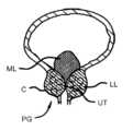
図1-図4を参照すると、人間男性被験者の泌尿器解剖学的構造の種々の特徴が提供されている。前立腺PGは、膀胱UBに隣接して位置するクルミサイズの腺である。尿道UTは、前立腺PGおよび陰茎Pを通して延びている。前立腺PGは、精子を保護し、精子に栄養を与える流体を分泌する。前立腺PGはまた、射精中に収縮して精子を追い出し、また、精液に尿が入らないようにするための弁を提供する。皮膜が、前立腺を包囲している。With reference to Figures 1-4, various features of the urinary anatomy of a human male subject are provided. The prostate PG is a walnut-sized gland located adjacent to the bladder UB. The urethra UT extends through the prostate PG and the penis P. The prostate PG secretes fluids that protect and nourish the sperm. The prostate PG also contracts during ejaculation to expel the sperm and also provides a valve to keep urine out of the semen. A capsule surrounds the prostate.
膀胱UBは、尿を保持する。輸精管VDは、精液が運ばれる輸送管を構成し、精嚢SVは、精液を分泌する。直腸Rは、排泄物を排出する大腸の端部分である。尿道は、身体から尿と精子の両方を運び出す。かくして、尿道は、膀胱UBに連結され、また、輸精管VDおよび精嚢SVに繋がる通路を提供する。精丘VMは、輸精管が入る尿道の壁のとさか部である。尿道前立腺部は、前立腺を通して延びる尿道の部分である。膀胱三角部T(図3参照)は、膀胱の滑らかな三角形領域である。膀胱三角部Tは、膨張に敏感であり、膀胱UBが一杯になったときに脳に信号を送る。The urinary bladder UB holds urine. The vas deferens VD constitutes the transport duct through which the semen is carried, and the seminal vesicles SV secrete the semen. The rectum R is the end part of the large intestine that excretes waste. The urethra carries both urine and sperm out of the body. Thus, the urethra is connected to the urinary bladder UB and also provides a passageway leading to the vas deferens VD and the seminal vesicles SV. The verumontanum VM is the crest of the wall of the urethra where the vas deferens enter. The prostatic urethra is the part of the urethra that extends through the prostate. The trigone T (see Figure 3) is a smooth triangular region of the urinary bladder. The trigone T is distension sensitive and sends a signal to the brain when the urinary bladder UB is full.
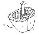
前立腺は、帯域によって分類されることができ、或いは、その葉(図4参照)に言及することによって記載されることができる。帯域分類は、代表的には病理学で使用され、葉分類は、解剖学的構造でよりしばしば使用される。前立腺の中央帯域(a)は、正常な前立腺の約25%であり、この帯域は、射精管を取り囲む。移行領域には良性前立腺肥大症のいくつかの伝播がある。繊維筋帯域(b)は、通常腺成分を欠き、その名が示唆するように、筋組織および繊維組織で構成されている。移行帯域(c)は、近位尿道の上に延び、寿命に亘って成長する前立腺の領域である。この帯域は、良性前立腺肥大の状態としばしば関連している。最後に、周辺帯域(d)は、遠位尿道を取り囲む前立腺の後部の皮膜下部分である。The prostate can be classified by zones or described by referring to its lobes (see Figure 4). Zone classification is typically used in pathology, while lobe classification is more often used in anatomy. The central zone (a) of the prostate is about 25% of the normal prostate, and this zone surrounds the ejaculatory ducts. The transition zone is where some of the spread of benign prostatic hyperplasia occurs. The fibromuscular zone (b) is usually devoid of glandular components and, as the name suggests, is composed of muscular and fibrous tissue. The transition zone (c) is the region of the prostate that extends above the proximal urethra and grows throughout life. This zone is often associated with the condition of benign prostatic hyperplasia. Finally, the peripheral zone (d) is the posterior subcapsular portion of the prostate that surrounds the distal urethra.
葉特徴化は、帯域特徴化とは異なるが、いくつかの重なりがある。後葉は、腺組織を欠き、繊維筋組織で形成されている。前葉は、大雑把に移行帯域(c)の前部に対応している。後葉は、大雑把に周辺帯域(d)に対応しており、直腸指診中に直腸を通して触診されることができる。後葉は、前立腺癌の70-80%の部位である。側葉は、前立腺の主な質量であり、尿道によって分離されている。すべての病理学的帯域は、側葉に存在し得る。最後に、中葉は、大雑把に中央帯域の一部に対応している。中要は、被験者毎にサイズが大きく変化し、いくつかのケースでは、腺組織を欠く。The lobe characterization differs from the zone characterization, although there is some overlap. The posterior lobe lacks glandular tissue and is formed of fibromuscular tissue. The anterior lobe roughly corresponds to the anterior part of the transition zone (c). The posterior lobe roughly corresponds to the peripheral zone (d) and can be palpated through the rectum during digital rectal examination. The posterior lobe is the site of 70-80% of prostate cancers. The lateral lobe is the main mass of the prostate and is separated by the urethra. All pathological zones can be present in the lateral lobe. Finally, the middle lobe roughly corresponds to part of the central zone. The middle lobe varies greatly in size from subject to subject and in some cases lacks glandular tissue.
大きな、または拡大化された中葉は、ボール弁として作用することができ、膀胱頚を閉鎖し、或いは尿道に開口する。かかる状態に対処するために、種々のアプローチが考えられる。膀胱頚開口の閉塞をなくし、或いは減少させるために、中葉を圧縮し、押しのけ、および/または、後退させることが考えられる。A large or enlarged middle lobe can act as a ball valve, closing off the bladder neck or opening into the urethra. To address this condition, various approaches are possible. Compressing, dislodging, and/or retracting the middle lobe may be considered to eliminate or reduce obstruction of the bladder neck opening.
次に、図5-図7を参照すると、種々の前立腺が断面で示されている。図5は、健康な被験者の膀胱UBおよび前立腺PGを示している。図6は、拡大化された側葉LLを有する前立腺を備える個人を示しており、図7は、拡大化された側葉LLおよび拡大化された中葉MLの両方を患う被験者を示している。かかる拡大化された解剖学的構造は、尿道UTにぶつかり、正常な膀胱の機能に悪影響を及ぼすことを理解すべきである。介入または診断処置中に前立腺組織に接近し、前立腺組織を操作するために、以下の装置を使用することができる。Referring now to Figures 5-7, various prostates are shown in cross-section. Figure 5 shows the bladder UB and prostate PG of a healthy subject. Figure 6 shows an individual with a prostate having an enlarged lateral lobe LL, and Figure 7 shows a subject suffering from both an enlarged lateral lobe LL and an enlarged middle lobe ML. It should be understood that such enlarged anatomical structures impinge on the urethra UT and adversely affect normal bladder function. The following devices can be used to access and manipulate prostate tissue during interventional or diagnostic procedures:
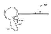
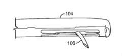
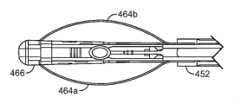
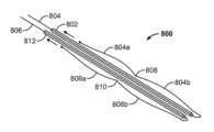
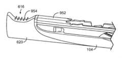
今、図8Aおよび図8Bを参照すると、治療装置100が示されている。治療装置100は、ハンドル組立体102および細長い接近組立体104を含む。細長い接近組立体104は、介入部位に接近し、また、目標組織に係合し、目標組織を操作するように構成されているのがよい。治療装置100は、患者の身体内に1つまたはそれ以上の組立体または移植片を組み付け、移植するように構成されているのがよい。装置はさらに、患者が、全身麻酔下ではなく、起きている、或いは、軽い鎮静下にある間、処置に耐えることができるように、(膀胱鏡検査のような)低侵襲技術と共に使用されるために両立可能であるように考えられている。装置はさらに、介入部位で行われているステップが医師によって観察されるように在来の遠隔視認装置(例えば、内視鏡)を受け入れるように構成されている構造体を含む。8A and 8B, a
細長い組立体104は、ニードル組立体106等の、目標組織を操作するための要素を収容するのがよい。細長い装置104はまた、目標組織を操作し、および/または、介入部位で装置を安定化させるための特徴を備えるのがよい。細長い組立体104は、図19Fまたは図21Fのようなサイズを含む、在来の膀胱鏡検査と両立可能なサイズのシースを通して挿入されることができる。細長い組立体104は、剛性または可撓性であってよい。いくつかの好ましい実施形態では、細長い組立体104は、ハンドル組立体102にてこ作用を及ぼし、或いはハンドル組立体102を押圧することによって介入部位で組織の手動圧縮を可能にするのに十分に剛性であるのがよい(或いは、介入位置にあるときに十分に剛性であるようにされているのがよい)。治療装置100の種々の実施形態は、前立腺葉を切り裂き、切除し、或いはその他の仕方で変更するための副組立体および構成要素を含むのがよい。The
前立腺を治療するに際しての1つの特定の制限的でない使用において、送達装置の細長い接近組立体は、患者の膀胱に至る尿道内に配置される。1つのアプローチでは、送達装置は、尿道に予め位置決めされている導入器シース内に配置されるのがよく、或いは、代替的には、送達装置は、尿道内に直接配置されてもよい。導入器シースを使用するときには、シースは、シースマウント組立体に取り付けられるのがよい。シースは、その先導端が前立腺に到達するまで患者内で前進される。特定のアプローチでは、治療されるべき前立腺の第1の側(すなわち、側葉)が、装置が膀胱を通して延びる間に選択され、それにしたがって、装置が調整される。細長い組織接近組立体の遠位端を使用して、内側前立腺組織を圧縮することによって尿道を前立腺内に押し下げるのがよい。前立腺の内側(すなわち、腺瘍)は、スポンジ様であり、かつ、両立的であり、前立腺の外側面は、硬い。患者の正中線に対して恥骨結合部PSを中心にして横方向に細長い組織接近組立体を回動させることによって、医師は、前立腺内に尿道を押し下げて腺瘍を圧縮し、尿道を通る所望の開口を創出することができる。関連した補完的介入処置に関する更なる詳細および背景は、出典を明示することによってその内容全体が本願の一部とされる米国特許第8,491,606号および第8,758,366号を含む種々の米国特許に記載されている。In one particular, non-limiting use in treating the prostate, the elongated access assembly of the delivery device is placed in the urethra leading to the patient's bladder. In one approach, the delivery device may be placed in an introducer sheath that is pre-positioned in the urethra, or alternatively, the delivery device may be placed directly in the urethra. When using an introducer sheath, the sheath may be attached to a sheath mount assembly. The sheath is advanced within the patient until its leading end reaches the prostate. In a particular approach, the first side (i.e., lateral lobe) of the prostate to be treated is selected while the device is extending through the bladder, and the device is adjusted accordingly. The distal end of the elongated tissue access assembly may be used to push the urethra down into the prostate by compressing the inner prostate tissue. The inside of the prostate (i.e., the adenoma) is spongy and pliable, while the outer surface of the prostate is hard. By rotating the elongated tissue access assembly laterally about the pubic symphysis PS relative to the patient's midline, the physician can depress the urethra into the prostate, compressing the glandular mass and creating the desired opening through the urethra. Further details and background regarding related complementary interventional procedures are provided in various U.S. patents, including U.S. Patent Nos. 8,491,606 and 8,758,366, the entire contents of which are incorporated herein by reference.
移植片を配備する前に、或いは、前立腺を変更する前に、前立腺の中葉のような介入部位で治療装置を使用するときに、中葉は、しばしば、移植片を受け入れるのに通じる位置内への押し入れを必要とする。前立腺の中葉を含む目標組織によりよく係合し、目標組織をよりよく操作するために、治療装置を位置決めし、安定化させることができる装置および使用方法の実施形態が、以下に説明されている。好ましい実施形態では、かかる装置は、組織に係合し、組織を操作するための装置の遠位面領域を増大させるウイング付きの拡張可能な/折り畳み可能な構造体を含むのがよい。When using a therapeutic device at an intervention site, such as the middle lobe of the prostate, prior to deploying an implant or altering the prostate, the middle lobe often requires pushing into position conducive to receiving the implant. Described below are embodiments of devices and methods of use that can position and stabilize the therapeutic device to better engage and manipulate target tissue, including the middle lobe of the prostate. In preferred embodiments, such devices may include expandable/foldable structures with wings that increase the distal surface area of the device for engaging and manipulating tissue.
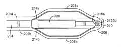
図9A-図9Cは、細長い部材の遠位端に取り付けられるように構成されているのがよい拡張可能な部材200を示している。拡張可能な部材200は、平行に(或いは、実質的に平行に)位置し、部材200の遠位端で遠位連結部材206に連結されているアーム202aおよび202bを含むのがよい。各アームは、アーム202aおよび202bの長手方向軸線を越えて外方に拡張する部分を含む。かかる拡張可能な部分は、ウイング208aおよび208bであるのがよい。いくつかの実施形態では、ウイング208aおよび208bは、遠位連結部材206に代えて、閉鎖ループ(図示せず)を形成するように連結していてもよい。遠位連結部材206は、遠位連結部材206に形成された開口部210を創出するように溝付け、チャンネル付け、或いはその他の仕方でへこみ付けされているのがよい。9A-9C show an
ウイング208aおよび208bは、拡張位置に付勢されるように構成されているのがよい。この実施形態では、ウイング208aおよび208bは、遠位連結部材206、および/または、アーム202aおよび202bを、ウイング208aおよび208bから遠ざかる長手方向に移動させることによって、引っ込め位置に移動される。もう1つの実施形態では、ウイング208aおよび208bは、引っ込め位置に構成されており、遠位連結部材206、および/または、アーム202aおよび202bを、ウイング208aおよび208bに近づくように長手方向に移動させることによって、拡張位置に移動される。いくつかの実施形態では、各ウイングは、そのウイングと同じ側のアームを、構成が必要とするようにそのウイングから遠ざかるように、或いは、そのウイングに近づくように長手方向に移動させることによって、独立的に拡張され、或いは、引っ込められることができる。The
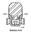
拡張可能な部材200の近位端は、アーム202aおよび202bを細長い組織接近組立体204のシャフトに連結することによって、細長い組織接近組立体204に取り付けられることができる。いくつかの実施形態では、細長い組立体204は、アーム202aの内方に向いた側212aおよびアーム202bの内方に向いた側212bが、細長い組立体204の外方に向いた側214aおよび外方に向いた側214bの一部と面一であるように拡張可能な部材200を通して挿入され、溶接或いは他の在来の取り付け手段により取り付けられる。拡張可能な部材200の遠位端は、細長い組立体204の一部と相互作用することができる。例えば、開口部210は、細長い組立体204にスナップインし、或いは他の仕方で細長い組立体204によって取り付けられるように構成されているのがよい。図9Cに示されているように、細長い組立体204は、遠位連結部材206上に取り外し可能に留められたタブ216aおよび216bを含むのがよく、それによって、拡張可能な部材200の遠位端を所定位置に保持する。いくつかの実施形態では、例えば、細長い組立体104が孔220を含むときに、ウイング208aおよび208bは、孔220に対して任意の平面上に位置決めされることができる。さらに、ウイング208aおよび208bは、各々に対して同じ平面上に位置決めされてもよく、或いは、各々に対して角度をなして位置決めされてもよい。いくつかの実施形態では、ウイングの平面の間の角度は、調節可能である。いくつかの実施形態では、細長い組立体は、ニードル組立体のような治療組立体が孔を通して延びることができるように構成されている。The proximal end of the
いくつかの実施形態では、拡張可能な部材200は、シースを使用して送達されることができる。シースは、装置が患者の介入部位に送達されるときに、拡張可能な部材200を収容し、ウイング208aおよび208bを押し潰し、或いは、圧縮するように機能する。一旦目標部位に達すると、シースの遠位開口部を通して拡張可能な部材200を延ばすことは、治療装置のハンドル組立体によって作動されることができる。拡張可能な部材200がシースを出るときに、ウイング208aおよび208bは、拡張或いはばね開放し、中央線から遠ざかる方に移動して、目標組織に接触し、目標組織を操作する。この送達は、ウイング縁による組織の外傷を防止しながら、ウイング整合を維持する。1つの実施形態によれば、アーム202aおよび202bは、細長い組立体204に固定的に取り付けられ、遠位連結部材206は、ウイング208aおよび208bに対して長手ほうこうに自由に移動し、ウイング208aおよび208bがシースを出たときに、ウイング208aおよび208bの拡張および引っ込めを可能にする。もう1つの実施形態によれば、アーム202aおよび202bは、ウイング208aおよび208bに対して長手方向に自由に移動し、遠位連結部材206は、細長い組立体204に固定的に取り付けられており、ウイング208aおよび208bがシースを出たときに、ウイング208aおよび208bの拡張および引っ込めを可能にする。In some embodiments, the
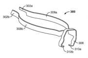
図10は、治療装置の遠位端に取り付けられることができる拡張可能な部材のもう1つの実施形態を示している。拡張可能な部材300は、平行に、或いは、実質的に平行に位置するアーム302aおよび302bを含むのがよい。各アームは、アーム302aおよび302bの長手方向軸線を越えて外方に拡張することができる部分を含むのがよい。ウイング308aおよび308bが、拡張可能な部材300の遠位端で遠位取り付け部品306に連結されているのがよい。1つの実施形態では、遠位取り付け部品306は、馬蹄形である。遠位取り付け部品306は、ウイング308aおよび308bに対して実質的に垂直構成に位置しているのがよい。遠位取り付け部品306は、タブ310aおよび310bを含むのがよい。取り付け部品306は、治療装置の遠位端の一部との確実連結部を受け入れ、形成するように構成されているのがよい。10 illustrates another embodiment of an expandable member that can be attached to a distal end of a treatment device. The
図11は、拡張可能な部材のもう1つの実施形態を示している。拡張可能な部材400は、平行に、或いは、実質的に平行に位置するアーム402aおよび402bを含むのがよい。各アームは、アーム402aおよび402bの長手方向軸線を越えて外方に拡張することができる部分を含むのがよい。ウイング408aおよび408bが、それぞれ、湾曲端410aおよび410bに終端しているのがよく、湾曲端410aおよび410bは、連結して、チャンネル406を創出する。チャンネル406は、治療装置の遠位端の一部を受け入れ、取り付けるように構成されているのがよい。11 illustrates another embodiment of an expandable member. The
図10および図11は、治療装置への拡張可能な部材の取り付けを容易にする拡張部材の遠位端の構成を示している。治療装置への拡張可能な部材の取り付けを容易にする他の構成が考えられ、本願の開示は、図10および図11に示されている取り付け構成に制限されない。10 and 11 show configurations of the distal end of the expandable member that facilitate attachment of the expandable member to a treatment device. Other configurations that facilitate attachment of the expandable member to a treatment device are contemplated, and the present disclosure is not limited to the attachment configurations shown in FIGS. 10 and 11.
再び、図8Aおよび図8Bに示されている治療装置の実施形態を参照すると、装置は、ニードルアクチュエータ108およびニードル引っ込めレバー110が、組織アンカーの送達のような治療を提供することができる待機位置にあるように構成されている。ニードルアクチュエータ108を押し下げると、ニードル組立体106が、細長い組織接近組立体104内から前進される(図8B参照)。ニードル組立体は、ニードル組立体が排出されるときに、ニードル組立体が、湾曲して、ハンドルに向かって戻るように構成されているのがよい。前立腺介入における使用に際して、ニードル組立体は、前立腺を通り越して前進される。ばね展開が、ニードルが前立腺の被膜中に留まり、被膜を穿刺し損なうことなく、前立腺のかたい外側被膜を素早く通過することを確保することを助ける。8A and 8B, the device is configured such that the
ニードルアクチュエータ108を押し下げ、ニードル引っ込めレバー110を開錠した後、ニードルレバー110は、作動されることができる。かかる動作により、ニードル組立体106は、後退される。いくつかの実施形態では、ニードル組立体106は、装置前配備内でその元の位置よりもさらに後退される。前立腺介入処置において、この動作は、前立腺組織の改変を容易にするために、インプラント、或いは組織アンカーのような種々の作動可能な部材を送達するために使用されることができる。After depressing the
拡張可能な部材は、細長い組織接近組立体104が、該細長い組織接近組立体104の遠位端の表面領域を増大させることによって、インプラントの配備および/または前立腺組織の改変前および中に中葉に係合するように、細長い組織接近組立体104を位置決めするのに使用されることができる。拡張可能な部材はまた、尿道壁を押しのけ、広げるのに使用されることができる。The expandable member can be used to position the elongated
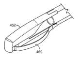
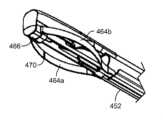
図12A-図12Gに示されているように、細長い組立体450は、拡張可能な部材460を備える遠位端452を含むのがよい。拡張可能な部材460は、平行に位置し、遠位端452のシャフトに取り付けられているアーム462aおよび462bを含むのがよい。各アームは、アーム462aおよび462bの長手方向軸線を越えて外方に拡張する拡張可能な部分、ウイング464aおよび464bを含む。拡張可能な部材460の遠位端は、細長い組立体450の遠位端の一部を受け入れ、取り付けるためのチャンネル466を含むのがよい。図12Fおよび図12Gは、細長い組立体450の遠位端452の一部上に留められるタブ468aおよび468bを備えたチャンネル466の断面図を示している。細長い組立体450をチャンネル466内に挿入することによって、拡張可能な部材460の遠位端は、組立体のシャフトに沿って摺動することが可能になり、ウイング位置の「閉鎖」(中央線に近づく圧縮)構成から「開放」(中央線から遠ざかる変位)構成への移動が容易にされる。加えて、治療装置に対する拡張可能な部材460の移動は、治療装置の長手方向軸線に束縛される。さらに、かかる整合は、膀胱鏡または内視鏡が処置中に使用されるときに、膀胱鏡または内視鏡に追加の障害物を提供しない。As shown in Figures 12A-12G, the
細長い組立体450がシース(図示せず)内に挿入されるときに、例えば、装置が患者の介入部位に送達されるときに、ウイング464aおよび464bは、治療装置の遠位端の長手方向軸線の中央線に向かって潰れることによって、輪郭が低くなる。一旦目標部位に達すると、ウイング464aおよび464bは、細長い組立体450がシースの遠位開口部を通して延ばされるときに、治療装置の中央線から遠ざかるように拡張することができる。When the
いくつかの実施形態では、拡張可能な部材460は、0.006インチ(0.015cm)の厚さを有するステンレス鋼から作られている。拡張可能な部材のアームおよび/またはウイングの断面は、非対称であるのが有利であり得る。例えば、断面は、1つの軸線がその直交軸線よりも実質的に長いような矩形または楕円形であるのがよい。堅さは、組織の捕獲および操作を容易にし、他方、可撓性は、拡張可能な部材の拡張および引っ込めを容易にする。いくつかの、または、同じ実施形態では、アームおよび/またはウイングは、使用中外傷を最小にするために、丸い縁を備えて構成されている。いくつかの実施形態では、ウイング464aおよび464bの内側面は、細長い組立体450の側部から出るニードル組立体(図示せず)のための入口位置を示す視覚ラインマーカー470を含むのがよい。In some embodiments, the
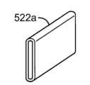
前立腺の中葉を含む目標組織によりよく係合し、目標組織をよりよく操作するために使用されることができる構造体の他の実施形態が、以下で考えられる。いくつかの実施形態では、ウイング508aおよび508bは、1対の入れ子式アームを使用して、拡張可能な部材500から展開されることができる。図13A-図13Dに示されているように、ウイング508aは、アーム502bに取り付けられているか、或いは、アーム502bと連続的である。アーム502aおよび502bは、アーム502bが、(図13Cに破線矢印で示されている)静止アーム502aに沿って滑走し、収縮状態から拡張状態に移行し、同時に、ウイング508aを収縮(「閉鎖」)状態から拡張(「開放」)状態に部材500の長手方向軸線から外方に移行させるように、スリーブ522a内で結合されているのがよい。拡張可能な部材500は、介入部位で配置するためのシース内に挿入されるのがよい。一旦装置が目標部位に位置決めされると、送達装置が作動されて、それぞれ、アーム502cおよび502dを、スリーブ502cおよび502dから展開して、目標組織に係合させる。Other embodiments of structures that can be used to better engage and manipulate target tissue, including the middle lobe of the prostate, are contemplated below. In some embodiments,
入れ子式アームの代替実施形態が、図14Aおよび図14Bに示されている。図14Aには、相互係止アーム550aおよび550bが示されている。アーム550aは、アーム550bを収容し、アーム550bの頂縁および底縁を覆う丸められた縁552を含むのがよい。図14Bでは、相互係止アーム560aおよび560bの各々が、丸められた端および平らな端を有する。相互係止されたときに、各アームの丸められた端は、反対側のアームの平らな端を覆う。An alternative embodiment of the nesting arms is shown in Figures 14A and 14B. In Figure 14A, interlocking
図13A-図13D並びに図14Aおよび図14Bの入れ子式アーム設計では、静止アームは、治療装置の細長い組立体のシャフトに取り付けられているのがよい。かかる設計は、ウイングまたは同様にループされた遠位端の、案内され、安定化された整合、および、介入部位への送達を提供する。In the telescoping arm designs of FIGS. 13A-13D and 14A-14B, the stationary arm may be attached to the shaft of the elongated assembly of the treatment device. Such a design provides guided and stabilized alignment and delivery of the winged or similarly looped distal end to the intervention site.
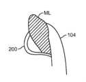
今、図15A-図15Dを参照すると、拡大化された中葉MLに係合し、拡大化された中葉MLを操作するためのアプローチが、提供されている。かかるアプローチは、側葉のための治療と相補的な療法として使用されることができ、或いは、専ら中葉MLを治療するために使用されることができる。拡大化された中葉MLは、膀胱UBに延入することがあり、正常な機能を妨げるボール弁として作用することがある(図15Aおよび図15B参照。図15Aは、尿道を通り、膀胱に入る断面図である。)ので、組織をボール弁の位置から遠ざかるように(すなわち、膀胱頚から遠ざかるように)移動させることが、望ましい。(TURPのような)そのような侵襲アプローチを回避することによって、膀胱頚の神経組織および/または平滑筋を崩壊させる危険を著しく減少させる。これらの組織の崩壊を減少させることで、射精機能および排泄抑制能力合併症がより低くなる見込みがある。15A-15D, an approach for engaging and manipulating the enlarged middle lobe ML is provided. Such an approach can be used as a complementary therapy to treatment for the lateral lobes or can be used exclusively to treat the middle lobe ML. Because the enlarged middle lobe ML can extend into the bladder UB and act as a ball valve that interferes with normal function (see Figs. 15A and 15B, Fig. 15A is a cross-sectional view through the urethra and into the bladder), it is desirable to move the tissue away from the location of the ball valve (i.e., away from the bladder neck). By avoiding such an invasive approach (such as TURP), the risk of disrupting the nerve tissue and/or smooth muscle of the bladder neck is significantly reduced. Reducing the disruption of these tissues is likely to result in fewer ejaculatory function and continence complications.
したがって、中葉MLを圧縮し、および/または、押しのけるために経尿道的に尿道前立腺部UT内に装置を挿入することを伴うアプローチが、考えられる。一旦葉が圧縮され、或いは、押しのけられた後には、中葉の圧縮または押しのけを維持するために特定の方向に組織アンカーまたはインプラントを移植するような他の処置が、考えられる。Thus, an approach involving inserting a device transurethrally into the prostatic urethra UT to compress and/or displace the middle lobe ML is contemplated. Once the lobes have been compressed or displaced, other procedures such as implanting tissue anchors or implants in specific orientations to maintain compression or displacement of the middle lobe are contemplated.
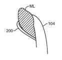
初期ステップとして、経腹的超音波または横断超音波検査法を使用して、患者の膀胱および前立腺の矢状断面図を取るのがよい。このようにして、患者の解剖学的構造を評価することができる。これに関して、膀胱内前立腺測定を取って、中葉突起の先端から膀胱の基部までの鉛直方向距離を決定する。図15C-図15Dに示されているように、解剖学的構造を評価した後に、アンカー送達装置の細長い組織接近組立体104(図8Aおよび図8B参照)を、尿道UT内で、および、中葉MLとの並置部内に前進させる。図15D図は、中葉MLの圧縮および押しのけを示す尿道UTの断面図である。As an initial step, a sagittal section view of the patient's bladder and prostate may be taken using transabdominal or transverse ultrasound. In this manner, the patient's anatomy may be evaluated. In this regard, an intravesical prostate measurement is taken to determine the vertical distance from the tip of the middle lobe process to the base of the bladder. As shown in Figures 15C-15D, after the anatomy has been evaluated, the elongated tissue approximation assembly 104 (see Figures 8A and 8B) of the anchor delivery device is advanced into the urethra UT and into apposition with the middle lobe ML. Figure 15D is a cross-sectional view of the urethra UT showing compression and displacement of the middle lobe ML.
1つの特定の一連の動作は、組織接近組立体104を、その終端112が中葉MLの前にあるように位置決めし、次いで、表面を後ろ方向に変位させて、中葉を尿道の中央線から遠ざかるように移動させる。その結果、中葉は、送達器具のまわりに組織折り畳み部(図15D参照)を形成する。拡張可能な部材が、中葉に係合、中葉を操作するために使用される実施形態では、拡張可能な部材は、中葉が押しのけられるように中葉の一時的な捕獲を容易にする図15Dの組織接近組立体104に比べて、増大された表面領域を提供する。いくつかの実施形態では、中葉の組織は、拡張可能な部材のウイングと細長い組織治療装置の遠位端の間の空間に捕獲されることができる。かくして、拡張可能な部材のウイングは、ウイングが展開および引っ込め中に受ける拡張の量を変えることによるものを含めて、かかる捕獲を促進するように構成されることができる。One particular sequence of movements positions the
図16Aおよび図16Bは、ループされた、または、拡張可能な部材のもう1つの実施形態を示している。この実施形態では、ループされた部材608は、ループされた部材608の拡張可能なウイング部分が圧縮されるようにシース610の遠位端に収容されるのがよい。細長い組立体がシースを通して移動されるときに、細長い組立体は、ループされた部材608の近位端(図示せず)に接触して、ウイング部分をシース610から押し出し、ウイング部分を拡張させ、組織を操作させる。16A and 16B show another embodiment of a looped or expandable member. In this embodiment, the looped
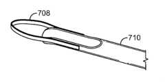
いくつかの実施形態では、図17Aおよび図17Bに示されているように、ループされた部材708が、シース710の遠位端に取り付けられているのがよい。これらの実施形態では、ループされた部材708は、細長い組立体を配備前に、介入部位で拡張し、および/または、組織を操作することができる。いくつかの実施形態では、ループされた部材708は、レベルリボン、すなわち、直線的な横断面を有するリボンで作られることができる。他の実施形態では、ループされた部材708は、装置の縁が送達中に患者の組織に接触しないようにするために、c形横断面を有する半円形リボンで作られることができる。In some embodiments, as shown in FIGS. 17A and 17B, a looped
拡張可能な部材の代替実施形態が、図18Aおよび図18B、並びに、図19Aおよび図19Bに示されている。拡張可能な部材800は、送達装置の細長い組立体のシャフト(図示せず)の両側に取り付けられた1対の管802および812を含むのがよい。かかる管は、細長い組立体の遠位端の方に付勢されているのがよい。管802および812は、それぞれ、管のルーメンの中を通され、次いで、各管の遠位端から出るワイヤ804および806を収容するのがよい。管に存在するワイヤは、各管の外側長さと実質的に平行に位置決めされており、また、ベント状に曲げられ、弓状に曲げられ、アーチ状に曲げられ、或いはその他の仕方で曲げられている。図18Aおよび図18Bに示されているように、ワイヤ804は、間に締め付けられたウエスト808を備えた1対の拡張可能な部分804aおよび804bを含み、ワイヤ806は、間に締め付けられたウエスト810を備えた1対の拡張可能な部分806aおよび806bを含む。いくつかの実施形態では、各ワイヤの締め付けられたウエストは、拡張可能な部分が、細長い組立体の中央線に近づくように潰れ、或いは、細長い組立体の中央線から遠ざかるように拡張することを可能にする圧縮ばねコイルであることができる。図18Aは、細長い組立体がシース(図示せず)内に収容されているときのような、圧縮または潰れ状態にある拡張可能な部材800を示しており、図18Bは、細長い組立体が、シース(図示せず)の遠位端から出て、ばねが自由になって伸び、次いで、各ワイヤの拡張可能な部分を治療装置の中央線から遠ざかるように移動させるときのような、拡張状態の拡張可能な部分800を示している。図19Aおよび図19Bは、拡張可能な部材800と同様に機能するが、拡張可能な部材900が3つの拡張可能な部分を含む拡張可能なワイヤ構造体のもう1つの実施形態である。Alternative embodiments of the expandable member are shown in FIGS. 18A and 18B and 19A and 19B. The
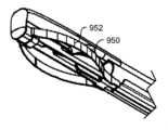
中葉、或いは他の組織によりよく係合するために、種々のアプローチが考えられる。追加の構造特徴が、目的組織と治療装置100の遠位部分の間の摩擦力を増大させるために、或いは、治療装置の表面領域を増大させるために、治療装置100の拡張可能な部材の遠位端に組み込まれることができる。ローレット面または粗面952が、治療装置100のウイング付きまたは拡張可能な部分950の面構成要素または全体を形成することができる。いくつかの実施形態では、図20Bに示されているように、スパイク、牙(fangs)、フック、とげ(barbs)、或いは他の突起954の1つまたはそれ以上、または、それらの組み合わせが、拡張可能な部材950から種々の角度および長さで延びるように構成されることができる。かかる突起は、鋭くても、或いは、鈍くてもよい。かかる構造体は、引っ込み可能、可撓性、或いは、固定的の1つまたはそれ以上であってよい。かくして、1つまたはそれ以上のアプローチでは、これらの構造体は、プラー(puller)(図示せず)に取り付けられたポップアップ特徴(pop-up feature)と関連させることができる。1つの観点では、ポップアップ特徴の制御は、シースに沿って摺動されるときに拡張可能な組織係合および操作特徴を展開し、引っ込めるための側方アクチュエータ、および、この側方アクチュエータの展開を制御するためにシースとシャフトの間の関係を変化させるためのスペーサによって達成されることができる。1つの特定のアプローチでは、スパイク、牙(fangs)、とげ(barbs)、または、突起は、送達装置が後退されるときには、向上された組織係合が提供され、送達装置が前進されるときには、向上された組織係合が解放されるように近位方向に角度付けされるのがよい。Various approaches are contemplated to better engage the middle lobe or other tissue. Additional structural features can be incorporated into the distal end of the expandable member of the
治療装置100を使用する前に、非外傷性テープが、スパイク、牙(fangs)、フック、とげ(barbs)、または、突起上に配置されるのがよい。テープは、中葉処置を行う前に除去されるのがよい。したがって、治療システムは、2つの構成で設けられることができることが考えられる。第1の構成は、治療システムが、鋭い、或いは他の組織係合特徴を覆い、鋭い、或いは他の組織係合特徴が組織と相互作用することを防止する非外傷性テープを含むような通常の使用のためであることができる。第2の構成は、治療システムが、鋭い、或いは他の係合特徴を覆う非外傷性テープなしに組み立てられ、輸送される、中葉係合および操作使用のためのものであることができる。Prior to using the
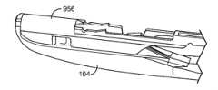
いくつかの実施形態では、組織接着材料956が、拡張可能な部分950に沿った種々の位置で外側面に付加されているのがよい(図20C参照)。いくつかの考えられるアプローチでは、かかる組織接着材料956は、接着テープから、所望のときにその接着特性を変化させるように流体が膨潤性である材料までの範囲にあることができる。圧力時接着に対するもう1つの考えられるアプローチは、拡張可能な部分950上に組み込まれたうろこ状突起(図20Fおよび図20G)を伴う。高い摩擦が、うろこに抗する方向に目標組織を引くときに創出されるが、展開後、或いは、うろこの方向で治療装置を使用するときには、摩擦が創出されないか、或いは、ほとんど創出されないか、或いは、より少ない摩擦が創出される。ここでも、かかる構造体は、引っ込み可能、可撓性、或いは、固定的の1つまたはそれ以上であってよい。In some embodiments, tissue
高められた摩擦、或いは他の係合力は、圧力に応答する接着材に基づいてもよい。例えば、治療装置100の拡張可能な遠位部分ベルクロ(登録商標)(Velcro)技術と同様なマイクロフックを追加的にまたは代替的に含んでもよく、確実な係合部を創出するために、組織は、マイクロフックに押し付けられる必要がある。The enhanced friction or other engagement force may be based on a pressure-responsive adhesive. For example, the expandable distal portion of the
上述したように、かかる係合構造体は、治療装置100の拡張可能な部分に沿った種々の位置に配置されることができる。きめ(textures)または突起は、組織が、装置の遠位端が回転されるときに装置の遠位端と共に、例えば、配備位置に巻かれるように組織に係合するように構成されているのがよい。いくつかの実施形態では、構造体は、より多くの空間がある治療装置の側方領域に位置しているのがよい。すなわち、これらの構造体は、治療装置から延びるのがよい治療要素のために、出口箇所から離れて位置しているのがよい。構造体は、「組織接触フェンス」に沿って位置するのがよいことが特に考えられる。すなわち、突起は、「組織接触フェンス」中に組織接触から隠されているのがよく、ユーザによって操作されるアクチュエータを介してこの保護フェンスを越えて延びるように構成されているのがよい。As discussed above, such engagement structures can be located at various locations along the expandable portion of the
今、図20D-図20Fを参照すると、細長い組織接近組立体104、特に、細長い組織接近組立体104の遠位端は、ローレット面または粗面952、スパイク、牙(fangs)、とげ(barbs)、または、突起、(および、随意には。非外傷性テープ)、および/または、組織接着材料956を含むのがよい。いくつかの実施形態では、細長い組織接近組立体および拡張可能な部材の一方または両方は、これらの特徴の1つまたはそれ以上を含むのがよい。すなわち、図20A-図20Hに示されている上記の実施形態は、細長い組織接近組立体、または両方の遠位端の、装置の拡張可能なまたはウイング付き部分の組織係合および操作能力を補足し、または、向上させるために、治療装置100に組み込まれているのがよい。20D-20F, the elongated
真空力がまた、組織係合および操作を容易にするために使用されてもよい。これに関して、吸引または真空源(図示せず)が、拡張可能な部分に組み込まれているか、或いは、取り付けられているのがよく、チャンネルが、治療装置100の遠位端と連通するために設けられているのがよい。Vacuum forces may also be used to facilitate tissue engagement and manipulation. In this regard, a suction or vacuum source (not shown) may be incorporated or attached to the expandable portion, and a channel may be provided to communicate with the distal end of the
図21に示されているように、もう1つのアプローチでは、ニードル230の部分的な制御された展開が、目標組織に係合し、目標組織を操作するために使用されるのがよい。
ニードル230は、例えば、アンカー装置の移植と関連させた後の十分な展開のために引っ込められるのがよい。追加的に、或いは代替的に、1つまたはそれ以上の補足的な、側方に突出するニードル609(図21に示されている)が、係合および操作目的のために設けられるのがよい。ニードル230を補強するための種々のアプローチがまた、より頑強な構造体が組織操作のために提供されるように考えられる。例えば、補足的なニードルチップ610が、ニードル230の終端部分に取り付けられることができる。1つの考えられているアプローチでは、ニードルチップ610は、送達装置ハンドルから作動されるように構成されている構造体に取り付けられることができる。ニードル610は、固い部材を構成し、それによって、組織を操作するために十分な機械的強度を提供することができ、送達装置内で延びる細長い部材に連結されることができ、この細長い部材は、送達装置に沿って、また、送達装置の外側に、しかし、導入器シース内で、或いは、導入器シースに設けられた穴を通して延びる。ニードルチップ610の使用または除去を制御するための手動または自動アプローチの両方が、考えられる。 In another approach, as shown in FIG. 21, a partial controlled deployment of
The
図22を参照すると、送達装置100の遠位端部分はまた、追加的に、或いは代替的に、中葉、或いは他の目標組織の輪郭に合い、或いは全体的に受け入れる曲率半径を有するディボットまたは凹部を含む。この凹部は、組織に係合し、組織を受け入れるための記載されている構造体も任意のものと共に構成されることができる。例えば、図22は、複数のスパイク954を含むような凹部616を示している。今、図23を参照すると、第2のまたは補足的なシース620が、送達装置100の遠位部分104のまわりに設けられ、構成されることができる。シース620はそれ自身、本願に記載されている組織係合および操作特徴の1つまたはそれ以上を含むことができる。かかる特徴は、シース620の1つまたはそれ以上の側部のために保存されることができ、或いは、シース620のまわり全体に位置決めされることができる。追加的に、シース620はそれ自身、組織の解剖学的構造に合うように構成された凹部616を含むことができる。かくして、凹部616は、凹部616を備える遠位端104を製造することによって、或いは、凹部616を備えて製造されたシース620を使用することによって、送達装置100の遠位端104に含められることができる。22, the distal end portion of the
目標組織または中葉は、特に、治療装置100との係合を容易にするために前処置されることができる。これに関して、目標組織に、その機械的輪郭を変えるために、電気焼灼、ボトックス、或いは他の治療法を施すことができることが考えられる。目標組織は、代替的に、或いは追加的に、完全に、或いは部分的に、目標組織に切開部を作ることによって前処置されることができる。最後に、目標組織は、組織を支持し、或いは所望の操作を達成するためにラッソー付けされることができる。The target tissue or lobe may be pre-treated, particularly to facilitate engagement with the
種々の材料が、開示されている装置を製造するために、本開示の範囲に入ることを認識すべきである。さらに、本願に開示されている1つまたはそれ以上の構成要素が、完全に、或いは部分的に生分解性、或いは生体内粉砕性であることができる。It should be appreciated that a variety of materials are within the scope of the present disclosure for fabricating the disclosed devices. Additionally, one or more of the components disclosed herein may be fully or partially biodegradable or biodisintegrable.
さらに、述べてきたように、本願に開示されている装置および方法は、腔または壁を含む種々の管腔または器官で種々の病理を治療するために使用されることができる。かかる管腔または器官の例には、腸、胃、食道、気管、気管支、気管支通路、静脈(例えば、静脈瘤または弁閉鎖不全を治療するための)、動脈、リンパ管、尿道、膀胱、心房若しくは寝室、子宮、輸卵管等が含まれる。Furthermore, as discussed, the devices and methods disclosed herein can be used to treat various pathologies in various lumens or organs, including cavities or walls. Examples of such lumens or organs include the intestine, stomach, esophagus, trachea, bronchi, bronchial passages, veins (e.g., to treat varicose veins or valvular insufficiency), arteries, lymphatics, urethra, bladder, atria or chambers of the heart, uterus, fallopian tubes, and the like.
最後に、本開示は、本開示のいくつかの例または実施形態に関連して上述してきたが、本開示の意図する精神および範囲から逸脱することなく、種々の付加、削除、変更、および、変形が、それらの例および実施形態に対してなされることを理解すべきである。例えば、1つの実施形態の任意の要素または属性は、そうすることがその実施形態を特許できないようにし、或いはその意図する使用に不適切にしない限り、別の実施形態または例に組み込まれ、或いは、別の実施形態または例と共に使用されることができることを理解すべきである。さらに、例えば、1つの方法のステップが、特定の順序で記載され、或いは列挙されている場合には、かかるステップの順序は、そうすることがその方法を特許できないようにし、或いはその意図する使用に不適切にしない限り、変更されることができる。すべての合理的な付加、削除、変形、および、変更は、記載されている例および実施形態の均等物であると考えられるべきであり、以下の特許請求の範囲内に含まれるべきである。Finally, although the present disclosure has been described above in connection with several examples or embodiments of the disclosure, it should be understood that various additions, deletions, modifications, and alterations may be made to those examples and embodiments without departing from the intended spirit and scope of the present disclosure. For example, it should be understood that any element or attribute of one embodiment may be incorporated into or used with another embodiment or example, unless doing so would render the embodiment unpatentable or unsuitable for its intended use. Furthermore, for example, if the steps of a method are described or enumerated in a particular order, the order of such steps may be changed unless doing so would render the method unpatentable or unsuitable for its intended use. All reasonable additions, deletions, modifications, and alterations should be considered equivalents of the described examples and embodiments, and should be included within the scope of the following claims.
簡潔にかつ一般的に、本開示は、内部身体構造に係合し、内部身体構造を操作するための装置および方法に向けられている。かかる係合および操作は、主として、或いは代替的に、多ステップ介入的処置の補足的な、または、一体的部分を形成することができる。1つの観点では、本装置は、組織を操作または再位置決めするために組織に係合するように構成されている細長い部材を含む。いくつかの実施形態では、組織は、前立腺の中葉である。Briefly and in general, the present disclosure is directed to devices and methods for engaging and manipulating internal body structures. Such engagement and manipulation may primarily or alternatively form a supplemental or integral part of a multi-step interventional procedure. In one aspect, the device includes an elongate member configured to engage tissue to manipulate or reposition the tissue. In some embodiments, the tissue is the middle lobe of the prostate.
種々のアプローチでは、装置は、装置と操作すべき組織との間の摩擦力を増大させる構造体を備える部分を含む。装置は、目標組織に係合し、目標組織を操作するように部分的に展開されることができる延長可能なニードルを追加的に、或いは代替的に含むのがよい。組織の係合および操作を容易にするための引っ込め可能なシースがまた、追加的に、或いは代替的に設けられているのがよい。さらに、装置の遠位端部分の表面領域は、表面領域を増大させ、かくして、組織に効果的に係合し、或いは組織を効果的に操作するように特に構成されている構造体を提供することを意図した構造体を含むのがよい。さらに、装置は、組織に、機械的輪郭を変更するエネルギーまたは物質を当てることによって、或いは、組織に切開部を創成することによって、目標組織を前処置するように構成され、使用されるのがよい。In various approaches, the device includes a portion with structure that increases friction between the device and the tissue to be manipulated. The device may additionally or alternatively include an extendable needle that can be partially deployed to engage and manipulate the target tissue. A retractable sheath may also additionally or alternatively be provided to facilitate engagement and manipulation of the tissue. Additionally, the surface area of the distal end portion of the device may include structure intended to increase the surface area and thus provide a structure that is specifically configured to effectively engage or manipulate the tissue. Additionally, the device may be configured and used to pretreat the target tissue by applying energy or material to the tissue that alters its mechanical profile or by creating an incision in the tissue.
かくして、本開示の送達装置は、アクチュエータ、或いは他の手動接近構造を介して動員される種々の副組立体を追加的に含むのがよい。副組立体の操作は、組織係合または操作構造体の正確な航行および配置を確保するように調整され、同期されている。Thus, the delivery devices of the present disclosure may additionally include various subassemblies that are mobilized via actuators or other manual access structures. Operation of the subassemblies is coordinated and synchronized to ensure precise navigation and placement of the tissue engaging or manipulating structures.
今、図24A-図24Bを参照すると、いくつかの実施形態では、拡張可能な部材200は、調節可能である。図24Aおよび図24Bは、細長い組織接近組立体104、および、細長い組織接近組立体104の一方の側上に存在する拡張可能な部材200の遠位端を示している。すなわち、この特定の実施形態では、1組のアームまたはウイングだけがある。図24Aは、拡張状態の拡張可能な部材200を示しており、これに対して、図24Bは、収縮状態の拡張可能な部材200を示している。中葉MLは、拡張可能な部材200と細長い組織接近組立体104の間の空間内に捕獲されるのがよい。次いで、拡張可能な部材200は、中葉MLが、拡張可能な部材200と細長い組織接近組立体104の間の空間内に取り付けられるように収縮され、または、締め付けられるのがよい。この位置で、中葉MLは、操作され、および/または、押しのけられることができる。24A-24B, in some embodiments, the
今、図25A-図25Bを参照すると、拡張可能な部材200は、拡張可能な部材200が2つまたはそれ以上の組のアームおよびウイングを含む実施形態で、より大きな寸法(図25A)からより小さい寸法(図25B)に調節されることができる。これらの実施形態では、拡張可能な部材200を使用して、組織を、拡張可能な部材200と細長い組織接近組立体104の間の空間に捕獲することができ、或いは、本願に開示されている他の実施形態により、拡張可能な部材200を使用して、組織を操作し、および/または、押しのけることができる。拡張可能な部材200の個々のアームは、個々的に、或いは、一緒に調節されることができる。25A-25B, the
拡張可能な部材200が調節されることができる実施形態では、調節は、装置を患者に配置する前に(或いは、シースが使用される実施形態ではシース内に装置を配置する前に)生じるのがよく、或いは、調節は、患者内の組織操作部位またはその近くで生じるのがよいことが考えられる。医師は、例えば、患者の解剖学的構造に基づいて拡張可能な部材200の寸法を設定するのがよい。医師は、処置前または処置中に患者の解剖学的構造の測定および/または観察に基づいて拡張可能な部材200のための所望の寸法を決定することができるのがよい。In embodiments in which the
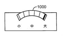
今、図26A-図26Bおよび図27Aおよび図27Bを参照すると、装置100のハンドルは、ホイール1000またはスライダ1100を含むのがよく、ホイール1000またはスライダ1100の各々は、拡張可能な部材200に結合され拡、拡張可能な部材200を拡張または収縮させるように構成されている。いくつかの実施形態では、ホイール1000およびスライダ1100は、拡張範囲が触覚表示器により表示される「小」拡張寸法から「中」拡張寸法まで「大」拡張寸法までの連続調節能力を提供する。随意には、錠が、拡張可能な部材の寸法を固定することができるように、ホイール1000またはスライダ1100上に設けられている。いくつかの他の実施形態では、ホイール1000およびスライダ1100は、「小」拡張寸法から「中」拡張寸法まで「大」拡張寸法までの離散的な調節能力を提供する。すなわち、拡張可能な部材200のための2つまたはそれ以上の予設定寸法があり、ホイール1000またはスライダ1100は、医師がそれらの寸法の間または中だけで選択を可能にする。26A-26B and 27A-27B, the handle of the
種々の代替使用方法が、考えられる。開示されている装置は、身体管腔を通る身体流体の流れを改善することを容易にし、身体管腔または腔の寸法および形状を変更し、前立腺拡大を治療し、尿失禁を治療し、組織の位置決めを支持または維持し、組織創傷、器官またはグラフトを閉じ、美容持ち上げまたは再位置決め処置を行い、解剖学的連結部を形成し、および/または、天然若しくは病理組織若しくは器官が隣接した解剖学的構造を押圧し、或いは隣接した解剖学的構造に干渉する種々の他の障害を治療するために使用されることができる。Various alternative uses are contemplated. The disclosed devices can be used to facilitate improved flow of bodily fluids through a bodily lumen, alter the size and shape of a bodily lumen or cavity, treat prostate enlargement, treat urinary incontinence, support or maintain tissue positioning, close tissue wounds, organs or grafts, perform cosmetic lifting or repositioning procedures, form anatomical connections, and/or treat a variety of other disorders in which natural or pathological tissue or organs press against or interfere with adjacent anatomical structures.
本発明の1つの観点は、前立腺の中葉に係合し、前立腺の中葉を操作するためのシステムであって、該システムは、シース、および、前記シース内に収容される組織係合または操作装置を含み、前記組織係合または操作装置は、患者の尿道内に挿入され、前立腺組織内で延びるように寸法および形状決めされており、前記組織係合または操作装置は中葉との接触を高めるために圧縮状態から拡張状態に移行することができる移動可能な係合構造体を含む、システムにおいて、前記係合構造体は、前記組織係合または操作装置の遠位端に付勢される、システムである。One aspect of the invention is a system for engaging and manipulating a middle lobe of the prostate, the system including a sheath and a tissue engagement or manipulation device housed within the sheath, the tissue engagement or manipulation device sized and shaped to be inserted into the patient's urethra and extend within the prostatic tissue, the tissue engagement or manipulation device including a movable engagement structure that can transition from a compressed state to an expanded state to enhance contact with the middle lobe, the engagement structure being biased against a distal end of the tissue engagement or manipulation device.
本発明のもう1つの観点では、前記係合構造体は、組織係合または操作構造体のシャフトの両側に固定された第1のアームおよび第2のアームを含む。In another aspect of the invention, the engagement structure includes a first arm and a second arm secured to opposite sides of a shaft of the tissue engagement or manipulation structure.
本発明のもう1つの観点では、前記第1のアームは、第1の拡張可能な部分を含み、前記第2のアームは、第2の拡張可能な部分を含み、前記第1の拡張可能な部分および前記第2の拡張可能な部分は、前記組織係合または操作装置が前記シースに収容されるときに前記シャフトに近づくように圧縮する。In another aspect of the invention, the first arm includes a first expandable portion and the second arm includes a second expandable portion, the first expandable portion and the second expandable portion compress toward the shaft when the tissue engagement or manipulation device is received in the sheath.
本発明のもう1つの観点では、前記第1の拡張可能な部分および前記第2の拡張可能な部分は、前記組織係合または操作装置が前記シースを出るときに前記シャフトから遠ざかるように拡張する。In another aspect of the invention, the first expandable portion and the second expandable portion expand away from the shaft when the tissue engagement or manipulation device exits the sheath.
本発明のもう1つの観点では、前記係合構造体は、前記組織係合または操作装置の一部を受け入れ、取り付けるためのチャンネルをさらに含む。In another aspect of the invention, the engagement structure further includes a channel for receiving and attaching a portion of the tissue engagement or manipulation device.
本発明のもう1つの観点では、前記組織係合または操作装置は、前記チャンネルに取り付けられているときに、前記係合構造体の運動は、前記前記組織係合または操作装置の長手方向軸線に束縛される。In another aspect of the invention, when the tissue engagement or manipulation device is attached to the channel, movement of the engagement structure is constrained to a longitudinal axis of the tissue engagement or manipulation device.
本発明のもう1つの観点では、前記組織係合または操作装置の長手方向軸線に沿う運動は、圧縮状態から拡張状態への前記係合構造体の移行を与える。In another aspect of the invention, movement of the tissue engaging or manipulating device along its longitudinal axis provides a transition of the engagement structure from a compressed state to an expanded state.
本発明のもう1つの観点では、前記係合構造体は、組織に対する外傷を減少させるための複数の丸い縁を備えるリボンであって、該リボンは横断面が非対称であるリボンから作られている。In another aspect of the invention, the engagement structure is a ribbon having a plurality of rounded edges for reducing trauma to tissue, the ribbon being made from a ribbon having an asymmetric cross section.
本発明のもう1つの観点では、前記係合構造体は、組織に対する外傷を減少させるc形横断面を備えるリボンから作られている。In another aspect of the invention, the engagement structure is made from a ribbon with a c-shaped cross-section that reduces trauma to tissue.
本発明のもう1つの観点では、前記組織係合または操作装置の前記シャフトは、側方孔、および、前記側方孔から出るニードル組立体を含み、前記側方孔は、前記係合構造体の前記第1の拡張可能な部分および前記第2の拡張可能な部分の間で整合している。In another aspect of the invention, the shaft of the tissue engagement or manipulation device includes a side hole and a needle assembly extending from the side hole, the side hole being aligned between the first expandable portion and the second expandable portion of the engagement structure.
本発明のもう1つの観点では、前記第1の拡張可能な部分は、第1の視覚ラインマーカーを含み、前記第2の拡張可能な部分は、第2の視覚ラインマーカーを含む。In another aspect of the invention, the first expandable portion includes a first visual line marker and the second expandable portion includes a second visual line marker.
本発明のもう1つの観点では、前記係合構造体の前記第1の拡張可能な部分および前記第2の拡張可能な部分は、前記側方孔を出た後に前記ニードル組立体のために前記入口位置を指示する。In another aspect of the invention, the first expandable portion and the second expandable portion of the engagement structure indicate the entry position for the needle assembly after it exits the side aperture.
本発明のもう1つの観点では、前記係合構造体の前記第1のアームおよび前記第2のアームは、入れ子式であるように構成されている。In another aspect of the invention, the first arm and the second arm of the engagement structure are configured to be telescopic.
本発明のもう1つの観点では、前記係合構造体は、前記組織係合または操作装置のシャフトの終端部分に取り付けられたループである。In another aspect of the invention, the engagement structure is a loop attached to a terminal portion of the shaft of the tissue engagement or manipulation device.
本発明のもう1つの観点では、前記係合構造体は、処置前または処置中に調節可能であり、前記調節は、システムのハンドル上の制御装置を介して連続的であるか、或いは、離散的であるのがよい。In another aspect of the invention, the engagement structure is adjustable before or during a procedure, and the adjustment may be continuous or discrete via a control on the system's handle.
本発明のもう1つの観点では、前記係合構造体は、中葉との摩擦接触を高めるように構成されている。In another aspect of the invention, the engagement structure is configured to enhance frictional contact with the middle leaf.
本発明のもう1つの観点では、前記係合構造体は、種々の角度をなして配置され、種々の長さを有するスパイク、牙(fangs)、フック、とげ(barbs)、或いはその他の突起の1つまたはそれ以上を含む。In another aspect of the invention, the engagement structures include one or more spikes, fangs, hooks, barbs, or other protrusions arranged at various angles and having various lengths.
本発明のもう1つの観点では、前記係合構造体は、ローレット面によって構成されている。In another aspect of the invention, the engagement structure is formed by a knurled surface.
本発明のもう1つの観点では、前記係合構造体は、随意に膨張可能である接着面によって構成されている。In another aspect of the invention, the engagement structure comprises an adhesive surface that is optionally expandable.
本発明のもう1つの観点では、前記係合構造体は、うろこによって構成されている。In another aspect of the present invention, the engagement structure is formed by scales.
本発明のもう1つの観点では、システムは、第1の突出可能なニードルおよび第2の突出可能なニードルを含む。In another aspect of the invention, the system includes a first extensible needle and a second extensible needle.
本発明のもう1つの観点では、補強用構造が、前記第1の突出可能なニードルおよび第2の突出可能なニードルの1つまたはそれ以上の終端に取り付けられている。In another aspect of the invention, a reinforcing structure is attached to one or more ends of the first and second extensible needles.
本発明のもう1つの観点では、前記係合構造体は、該係合構造体を覆うように構成されている非外傷性テープを含む。In another aspect of the invention, the engagement structure includes an atraumatic tape configured to cover the engagement structure.
本発明のもう1つの観点では、前記係合構造体は、システムの遠位端上に形成されているディボットによって構成されており、前記ディボットは、目的組織の輪郭に実質的に合うように寸法および形状決めされている。In another aspect of the invention, the engagement structure comprises a divot formed on the distal end of the system, the divot being sized and shaped to substantially match the contours of the target tissue.
本発明のもう1つの観点では、組織は、係合構造体によって操作される前に前処置され、その前処置は、電気焼灼、ボットクス、または、切開の1つまたはそれ以上を含む。In another aspect of the invention, the tissue is pretreated prior to being manipulated by the engagement structure, the pretreatment including one or more of electrocautery, botox, or incision.
かくして、本開示の特定の形態が示され、記載されてきたけれども、本開示の精神および範囲から逸脱することなく種々の変更を行うことができることが上記から明らかになるであろう、Thus, while particular forms of the present disclosure have been shown and described, it will be apparent from the above that various modifications can be made without departing from the spirit and scope of the present disclosure,
100 治療装置
102 ハンドル組立体
104 細長い組織接近組立体
106 ニードル組立体
200 拡張可能な部材 100
Claims (18)
Translated fromJapanese前立腺の中葉に係合し、前立腺の中葉を操作するように構成された組織係合構造体をさらに含み、前記組織係合構造体は、前記細長い組織接近組立体の遠位端部分または前記導入器シースに固定的に取り付けられており、前記組織係合構造体は、拡張可能な部材を備えた遠位端部分を含み、前記拡張可能な部材は、湾曲部分であって、前記組織係合構造体が、前記湾曲部分が、該湾曲部分の間の長手方向軸線によって規定される中央線に向かって圧縮されている収縮状態から、前記湾曲部分が、前記長手方向軸線によって規定される前記中央線を越えて外方に拡張する拡張状態に移行することができるような湾曲部分を含む、治療装置。 an elongated tissue accessing assembly coupled to the handle assembly and configured to be inserted into the introducer sheath, the elongated tissue accessing assembly further including an aperture and a needle assembly extendable through the aperture;
11. A treatment device,comprising: a tissue engagement structure configured to engage and manipulate a medial lobe of the prostate, the tissue engagement structure fixedly attached to a distal end portion of the elongate tissue access assembly or to the introducer sheath, the tissue engagement structure including a distal end portion with an expandable member, the expandable member including curved portions such that the tissue engagement structure is transitionable from a contracted state in which the curved portions are compressed toward a centerline defined by a longitudinal axis between the curved portions to an expanded state in which the curved portions expand outwardly beyond the centerline defined by the longitudinal axis.
前記アンカー送達装置内に収容された組織アンカーと、
前立腺に係合し、前立腺を操作するように構成された組織係合構造体と、をさらに含み、前記組織係合構造体は、前記細長い組織接近組立体の遠位端部分または前記導入器シースに固定的に取り付けられており、前記組織係合構造体は、拡張可能な部材を備えた遠位端部分を含み、前記拡張可能な部材は、湾曲部分であって、前記組織係合構造体が、前記湾曲部分が、該湾曲部分の間の長手方向軸線によって規定される中央線に向かって圧縮されている収縮状態から、前記湾曲部分が、前記長手方向軸線によって規定される前記中央線を越えて外方に拡張する拡張状態に移行することができるような湾曲部分を含む、システム。 an anchor delivery device including an elongated tissue accessing assembly configured to be inserted intoan introducer sheath, the anchor delivery device further including a needle assembly configured to bend when the needle assembly is ejected through an aperture defined by a distal end portion of the elongated tissue accessing assembly;
a tissue anchor contained within the anchor delivery device;
and a tissue engaging structure configured to engage and manipulate the prostate, the tissue engaging structurefixedly attached to a distal end portion of the elongate tissue access assembly or to the introducer sheath, the tissue engaging structure including a distal end portion with an expandable member, the expandable member including curved portions such that the tissue engaging structure is transitionable from a contracted state in which the curved portions are compressed toward a centerline defined by a longitudinal axis between the curved portions to an expanded state in which the curved portions expand outwardly beyond the centerline defined by the longitudinal axis.
Applications Claiming Priority (4)
| Application Number | Priority Date | Filing Date | Title |
|---|---|---|---|
| US201762610184P | 2017-12-23 | 2017-12-23 | |
| US62/610,184 | 2017-12-23 | ||
| JP2020554059AJP7150871B2 (en) | 2017-12-23 | 2018-12-21 | Expandable tissue engagement device and method |
| PCT/US2018/067229WO2019126718A1 (en) | 2017-12-23 | 2018-12-21 | Expandable tissue engagement apparatus and method |
Related Parent Applications (1)
| Application Number | Title | Priority Date | Filing Date |
|---|---|---|---|
| JP2020554059ADivisionJP7150871B2 (en) | 2017-12-23 | 2018-12-21 | Expandable tissue engagement device and method |
Publications (2)
| Publication Number | Publication Date |
|---|---|
| JP2022186721A JP2022186721A (en) | 2022-12-15 |
| JP7620602B2true JP7620602B2 (en) | 2025-01-23 |
Family
ID=65024142
Family Applications (2)
| Application Number | Title | Priority Date | Filing Date |
|---|---|---|---|
| JP2020554059AActiveJP7150871B2 (en) | 2017-12-23 | 2018-12-21 | Expandable tissue engagement device and method |
| JP2022155511AActiveJP7620602B2 (en) | 2017-12-23 | 2022-09-28 | Expandable tissue engagement devices and methods |
Family Applications Before (1)
| Application Number | Title | Priority Date | Filing Date |
|---|---|---|---|
| JP2020554059AActiveJP7150871B2 (en) | 2017-12-23 | 2018-12-21 | Expandable tissue engagement device and method |
Country Status (7)
| Country | Link |
|---|---|
| US (3) | US11672520B2 (en) |
| EP (3) | EP3727171B1 (en) |
| JP (2) | JP7150871B2 (en) |
| AU (1) | AU2018389236B2 (en) |
| ES (1) | ES2953556T3 (en) |
| SG (1) | SG11202005766XA (en) |
| WO (1) | WO2019126718A1 (en) |
Families Citing this family (10)
| Publication number | Priority date | Publication date | Assignee | Title |
|---|---|---|---|---|
| US8628542B2 (en) | 2005-05-20 | 2014-01-14 | Neotract, Inc. | Median lobe destruction apparatus and method |
| US7758594B2 (en) | 2005-05-20 | 2010-07-20 | Neotract, Inc. | Devices, systems and methods for treating benign prostatic hyperplasia and other conditions |
| US10195014B2 (en) | 2005-05-20 | 2019-02-05 | Neotract, Inc. | Devices, systems and methods for treating benign prostatic hyperplasia and other conditions |
| US10130353B2 (en) | 2012-06-29 | 2018-11-20 | Neotract, Inc. | Flexible system for delivering an anchor |
| ES2953556T3 (en) | 2017-12-23 | 2023-11-14 | Teleflex Life Sciences Ltd | Expandable Tissue Docking Apparatus |
| WO2021099646A1 (en) | 2019-11-22 | 2021-05-27 | ProVerum Limited | Device and method for deploying expandable implants |
| US11273025B2 (en) | 2019-11-22 | 2022-03-15 | Pro Verum Limited | Expandable implant delivery device |
| US11602621B2 (en) | 2019-11-22 | 2023-03-14 | ProVerum Limited | Device for controllably deploying expandable implants |
| DE102021133393A1 (en) | 2021-12-15 | 2023-06-15 | Olympus Winter & Ibe Gmbh | implant |
| US12201542B2 (en) | 2023-04-27 | 2025-01-21 | ProVerum Limited | Device and method for deploying expandable implants |
Citations (3)
| Publication number | Priority date | Publication date | Assignee | Title |
|---|---|---|---|---|
| US20030199860A1 (en) | 2002-04-22 | 2003-10-23 | Loeb Marvin P. | Devices and methods for directed, interstitial ablation of tissue |
| US20140005690A1 (en) | 2012-06-29 | 2014-01-02 | Neotract, Inc. | Flexible system for delivering an anchor |
| WO2017017499A1 (en) | 2015-07-29 | 2017-02-02 | Butterfly Medical Ltd. | Retracting or/and supporting periurethral tissue |
Family Cites Families (712)
| Publication number | Priority date | Publication date | Assignee | Title |
|---|---|---|---|---|
| US2734299A (en) | 1956-02-14 | Igudolph | ||
| US659422A (en) | 1900-06-12 | 1900-10-09 | George W Shidler | Surgical instrument. |
| US789467A (en) | 1903-06-12 | 1905-05-09 | Stillman A West | Method of tying knots. |
| US780392A (en) | 1903-09-14 | 1905-01-17 | Brown Straw Binder Company | Straw-bundle tie. |
| US2360164A (en) | 1943-12-23 | 1944-10-10 | Edward A Swenson | Clothespin |
| US2485531A (en) | 1948-01-13 | 1949-10-18 | Dzus William | Surgical toggle bolt |
| US2579192A (en) | 1950-08-15 | 1951-12-18 | George H Sciaroni | Suturing instrument |
| US2697624A (en) | 1951-11-03 | 1954-12-21 | John A Thomas | Portable knot tying device |
| US2646298A (en) | 1952-07-15 | 1953-07-21 | Joseph C Leary | Method of knot tying |
| US2825592A (en) | 1954-01-06 | 1958-03-04 | Semple James Mckenzie | Portable knot tying device for smooth filaments |
| US3326586A (en) | 1965-07-09 | 1967-06-20 | Robert M Frost | Snell knot tying tool |
| US3470834A (en) | 1968-03-08 | 1969-10-07 | Dennison Mfg Co | Fastener attaching device |
| US3541591A (en) | 1968-04-26 | 1970-11-17 | Henry J Hoegerman | Method and apparatus for closing wounds |
| US3521918A (en) | 1968-08-14 | 1970-07-28 | Richard L Hammond | Fishline knotting fixture and cutter |
| US3664345A (en) | 1970-07-06 | 1972-05-23 | Clyde Harwell Dabbs | Surgical buttons |
| US3716058A (en) | 1970-07-17 | 1973-02-13 | Atlanta Res Inst | Barbed suture |
| US3713680A (en) | 1971-02-09 | 1973-01-30 | S Pagano | Knot typing device for barrel knots |
| US3756638A (en) | 1972-02-22 | 1973-09-04 | L Stockberger | Knot tyer |
| US3875648A (en) | 1973-04-04 | 1975-04-08 | Dennison Mfg Co | Fastener attachment apparatus and method |
| US3886933A (en) | 1973-10-10 | 1975-06-03 | Olympus Optical Co | Ureteral catheter device |
| US3873140A (en) | 1973-10-15 | 1975-03-25 | Moodus Sports Products | Fish hook holder and knot tying device |
| US3931667A (en) | 1974-05-08 | 1976-01-13 | Dennison Manufacturing Company | Interlocking attachment device |
| US3976079A (en) | 1974-08-01 | 1976-08-24 | Samuels Peter B | Securing devices for sutures |
| US4006747A (en) | 1975-04-23 | 1977-02-08 | Ethicon, Inc. | Surgical method |
| DE2601802C3 (en) | 1976-01-20 | 1979-02-08 | Richard Wolf Gmbh, 7134 Knittlingen | Instruments for the treatment of urethral strictures |
| US4164225A (en) | 1977-12-28 | 1979-08-14 | Johnson & Lorenz, Inc. | Surgical suturing instrument |
| US4235238A (en) | 1978-05-11 | 1980-11-25 | Olympus Optical Co., Ltd. | Apparatus for suturing coeliac tissues |
| US4210148A (en) | 1978-11-03 | 1980-07-01 | Stivala Oscar G | Retention suture system |
| DE2853289C2 (en) | 1978-12-09 | 1980-12-18 | B. Braun Melsungen Ag, 3508 Melsungen | Button for surgical use |
| JPS57188231A (en) | 1981-05-14 | 1982-11-19 | Olympus Optical Co | Treating tool guide apparatus of endoscope |
| US4419094A (en) | 1981-06-08 | 1983-12-06 | The Kendall Company | Suprapubic catheter system |
| US4409974A (en) | 1981-06-29 | 1983-10-18 | Freedland Jeffrey A | Bone-fixating surgical implant device |
| AU8679382A (en) | 1981-08-10 | 1983-02-17 | Ethicon Inc. | Polyolefin surgical device |
| US4513746A (en) | 1981-10-09 | 1985-04-30 | United States Surgical Corp. | Instrument for applying plastic-like surgical fastening devices |
| SE445884B (en) | 1982-04-30 | 1986-07-28 | Medinvent Sa | DEVICE FOR IMPLANTATION OF A RODFORM PROTECTION |
| US4741330A (en) | 1983-05-19 | 1988-05-03 | Hayhurst John O | Method and apparatus for anchoring and manipulating cartilage |
| US6656182B1 (en) | 1982-05-20 | 2003-12-02 | John O. Hayhurst | Tissue manipulation |
| US5417691A (en) | 1982-05-20 | 1995-05-23 | Hayhurst; John O. | Apparatus and method for manipulating and anchoring tissue |
| US4823794A (en) | 1982-07-12 | 1989-04-25 | Pierce William S | Surgical pledget |
| US5385544A (en) | 1992-08-12 | 1995-01-31 | Vidamed, Inc. | BPH ablation method and apparatus |
| US5370675A (en) | 1992-08-12 | 1994-12-06 | Vidamed, Inc. | Medical probe device and method |
| US5435805A (en) | 1992-08-12 | 1995-07-25 | Vidamed, Inc. | Medical probe device with optical viewing capability |
| US4493323A (en) | 1982-12-13 | 1985-01-15 | University Of Iowa Research Foundation | Suturing device and method for using same |
| US4621640A (en) | 1984-01-09 | 1986-11-11 | Mulhollan James S | Mechanical needle carrier and method for its use |
| US4657461A (en) | 1984-10-26 | 1987-04-14 | Smith Gareth J | Anchoring bolt |
| US4750492A (en) | 1985-02-27 | 1988-06-14 | Richards Medical Company | Absorbable suture apparatus, method and installer |
| US4955913A (en) | 1985-03-28 | 1990-09-11 | Robinson Walter C | Surgical tie |
| US4669473A (en) | 1985-09-06 | 1987-06-02 | Acufex Microsurgical, Inc. | Surgical fastener |
| US4705040A (en) | 1985-11-18 | 1987-11-10 | Medi-Tech, Incorporated | Percutaneous fixation of hollow organs |
| US4738255A (en) | 1986-04-07 | 1988-04-19 | Biotron Labs, Inc. | Suture anchor system |
| US5123914A (en) | 1986-05-19 | 1992-06-23 | Cook Incorporated | Visceral anchor for visceral wall mobilization |
| US4893623A (en) | 1986-12-09 | 1990-01-16 | Advanced Surgical Intervention, Inc. | Method and apparatus for treating hypertrophy of the prostate gland |
| US4762128A (en) | 1986-12-09 | 1988-08-09 | Advanced Surgical Intervention, Inc. | Method and apparatus for treating hypertrophy of the prostate gland |
| US4714281A (en) | 1987-01-02 | 1987-12-22 | Peck Richard M | Device and method for tying a twisted clinch knot |
| US4935028A (en) | 1987-01-12 | 1990-06-19 | Drews Robert C | Corneal rivet |
| US4744364A (en) | 1987-02-17 | 1988-05-17 | Intravascular Surgical Instruments, Inc. | Device for sealing percutaneous puncture in a vessel |
| US4898156A (en) | 1987-05-18 | 1990-02-06 | Mitek Surgical Products, Inc. | Suture anchor |
| DK163713C (en) | 1987-09-02 | 1992-09-07 | Ole Gyring Nieben | DEVICE FOR THE POSITION OF A PARTICULAR CATHETTE IN A BODY |
| US4863439A (en) | 1987-11-02 | 1989-09-05 | S. Robert Kovac | Surgical cannula |
| US4899743A (en) | 1987-12-15 | 1990-02-13 | Mitek Surgical Products, Inc. | Suture anchor installation tool |
| US4968315A (en) | 1987-12-15 | 1990-11-06 | Mitek Surgical Products, Inc. | Suture anchor and suture anchor installation tool |
| US4926860A (en) | 1988-02-05 | 1990-05-22 | Flexmedics Corporation | ARthroscopic instrumentation and method |
| US5053046A (en) | 1988-08-22 | 1991-10-01 | Woodrow W. Janese | Dural sealing needle and method of use |
| US5159925A (en) | 1988-09-09 | 1992-11-03 | Gynelab, Inc. | Cauterizing apparatus and method for laparoscopic cholecystostomy, gallbladder ablation and treatment of benign prostate hypertrophy |
| US4994066A (en) | 1988-10-07 | 1991-02-19 | Voss Gene A | Prostatic stent |
| US5904679A (en) | 1989-01-18 | 1999-05-18 | Applied Medical Resources Corporation | Catheter with electrosurgical cutter |
| JP2722132B2 (en) | 1989-05-03 | 1998-03-04 | 日機装株式会社 | Device and method for alleviating stenosis from the lumen |
| US5620461A (en) | 1989-05-29 | 1997-04-15 | Muijs Van De Moer; Wouter M. | Sealing device |
| US4946468A (en) | 1989-06-06 | 1990-08-07 | Mitek Surgical Products, Inc. | Suture anchor and suture anchor installation tool |
| US5002550A (en) | 1989-06-06 | 1991-03-26 | Mitek Surgical Products, Inc. | Suture anchor installation tool |
| US4955859A (en) | 1989-07-07 | 1990-09-11 | C. R. Bard, Inc. | High-friction prostatic stent |
| RU2062121C1 (en) | 1989-10-13 | 1996-06-20 | Давыдов Владимир Александрович | Dilating device |
| US5919202A (en) | 1989-12-05 | 1999-07-06 | Yoon; Inbae | Surgical instrument with jaws and movable internal needle and method for use thereof |
| US5267960A (en) | 1990-03-19 | 1993-12-07 | Omnitron International Inc. | Tissue engaging catheter for a radioactive source wire |
| EP0448857A1 (en) | 1990-03-27 | 1991-10-02 | Jong-Khing Huang | An apparatus of a spinning type of resectoscope for prostatectomy |
| US5019032A (en) | 1990-04-03 | 1991-05-28 | Robertson Jack R | Refined suspension procedure with implement for treating female stress incontinence |
| US5080660A (en) | 1990-05-11 | 1992-01-14 | Applied Urology, Inc. | Electrosurgical electrode |
| US5078731A (en) | 1990-06-05 | 1992-01-07 | Hayhurst John O | Suture clip |
| US5163960A (en) | 1990-06-28 | 1992-11-17 | Bonutti Peter M | Surgical devices assembled using heat bondable materials |
| US6203565B1 (en) | 1990-06-28 | 2001-03-20 | Peter M. Bonutti | Surgical devices assembled using heat bondable materials |
| US5269809A (en) | 1990-07-02 | 1993-12-14 | American Cyanamid Company | Locking mechanism for use with a slotted suture anchor |
| US5236445A (en) | 1990-07-02 | 1993-08-17 | American Cyanamid Company | Expandable bone anchor and method of anchoring a suture to a bone |
| US5041129A (en) | 1990-07-02 | 1991-08-20 | Acufex Microsurgical, Inc. | Slotted suture anchor and method of anchoring a suture |
| US5653373A (en) | 1990-09-17 | 1997-08-05 | United States Surgical Corporation | Arcuate apparatus for applying two-part surgical fasteners |
| US5372146A (en) | 1990-11-06 | 1994-12-13 | Branch; Thomas P. | Method and apparatus for re-approximating tissue |
| US5203787A (en) | 1990-11-19 | 1993-04-20 | Biomet, Inc. | Suture retaining arrangement |
| DE9190181U1 (en) | 1990-12-10 | 1993-07-29 | Howmedica Inc. (n.d.Ges.d.Staates Delaware), New York, N.Y. | Device for interstitial laser energy delivery |
| US5129912B2 (en) | 1991-01-07 | 2000-01-11 | Urohealth Systems Inc | Device and method for applying suture |
| US5100421A (en) | 1991-02-05 | 1992-03-31 | Cyprus Endosurgical Tools, Inc. | Christoudias curved needle suture assembly |
| US5409453A (en) | 1992-08-12 | 1995-04-25 | Vidamed, Inc. | Steerable medical probe with stylets |
| DE4106823C1 (en) | 1991-03-04 | 1992-06-25 | Liebscher Kunststofftechnik, 8032 Graefelfing, De | |
| US5405352A (en) | 1991-04-09 | 1995-04-11 | Weston; Peter V. | Suture knot, method for its formation and use, and knot forming apparatus |
| US5217470A (en) | 1991-04-29 | 1993-06-08 | Weston Peter V | Apparatuses and methods for formation and use of a slipknot as a surgical suture knot |
| US5258015A (en) | 1991-05-03 | 1993-11-02 | American Cyanamid Company | Locking filament caps |
| US5127393A (en) | 1991-05-28 | 1992-07-07 | Medilase, Inc. | Flexible endoscope with rigid introducer |
| US5160339A (en) | 1991-06-18 | 1992-11-03 | Ethicon, Inc. | Endoscopic suture clip |
| US5237984A (en) | 1991-06-24 | 1993-08-24 | Xomed-Treace Inc. | Sheath for endoscope |
| US5234454A (en) | 1991-08-05 | 1993-08-10 | Akron City Hospital | Percutaneous intragastric balloon catheter and method for controlling body weight therewith |
| GB9118670D0 (en) | 1991-08-30 | 1991-10-16 | Mcnicholas Thomas A | Surgical devices and uses thereof |
| US5269802A (en) | 1991-09-10 | 1993-12-14 | Garber Bruce B | Prostatic stent |
| CA2078530A1 (en) | 1991-09-23 | 1993-03-24 | Jay Erlebacher | Percutaneous arterial puncture seal device and insertion tool therefore |
| US7549424B2 (en) | 1991-10-18 | 2009-06-23 | Pro Surg, Inc. | Method and apparatus for tissue treatment with laser and electromagnetic radiation |
| US5861002A (en) | 1991-10-18 | 1999-01-19 | Desai; Ashvin H. | Endoscopic surgical instrument |
| US5167614A (en) | 1991-10-29 | 1992-12-01 | Medical Engineering Corporation | Prostatic stent |
| US5372600A (en) | 1991-10-31 | 1994-12-13 | Instent Inc. | Stent delivery systems |
| US5411520A (en) | 1991-11-08 | 1995-05-02 | Kensey Nash Corporation | Hemostatic vessel puncture closure system utilizing a plug located within the puncture tract spaced from the vessel, and method of use |
| CA2181674A1 (en) | 1991-12-03 | 1993-10-04 | Theodore V. Benderev | Support structure for supporting and positioning medical equipment |
| MX9300607A (en) | 1992-02-06 | 1993-10-01 | American Med Syst | APPARATUS AND METHOD FOR INTERSTITIAL TREATMENT. |
| US5217486A (en) | 1992-02-18 | 1993-06-08 | Mitek Surgical Products, Inc. | Suture anchor and installation tool |
| US5171251A (en) | 1992-03-02 | 1992-12-15 | Ethicon, Inc. | Surgical clip having hole therein and method of anchoring suture |
| US5300099A (en) | 1992-03-06 | 1994-04-05 | Urologix, Inc. | Gamma matched, helical dipole microwave antenna |
| US5254126A (en) | 1992-06-24 | 1993-10-19 | Ethicon, Inc. | Endoscopic suture punch |
| US5470308A (en) | 1992-08-12 | 1995-11-28 | Vidamed, Inc. | Medical probe with biopsy stylet |
| US5667488A (en) | 1992-08-12 | 1997-09-16 | Vidamed, Inc. | Transurethral needle ablation device and method for the treatment of the prostate |
| US6048351A (en) | 1992-09-04 | 2000-04-11 | Scimed Life Systems, Inc. | Transvaginal suturing system |
| US5540704A (en) | 1992-09-04 | 1996-07-30 | Laurus Medical Corporation | Endoscopic suture system |
| US5364408A (en) | 1992-09-04 | 1994-11-15 | Laurus Medical Corporation | Endoscopic suture system |
| US7060077B2 (en) | 1992-09-04 | 2006-06-13 | Boston Scientific Scimed, Inc. | Suturing instruments and methods of use |
| US5578044A (en) | 1992-09-04 | 1996-11-26 | Laurus Medical Corporation | Endoscopic suture system |
| US5322501A (en) | 1992-10-02 | 1994-06-21 | Mahmud Durrani Ayaz | Continent urethral stent for treating and preventing urethral stricture after surgery |
| US5368599A (en) | 1992-10-08 | 1994-11-29 | United States Surgical Corporation | Surgical fastening apparatus with suture array |
| US5464416A (en) | 1992-11-10 | 1995-11-07 | Ethicon, Inc. | Ligating clip |
| US5972000A (en) | 1992-11-13 | 1999-10-26 | Influence Medical Technologies, Ltd. | Non-linear anchor inserter device and bone anchors |
| IL127978A0 (en) | 1999-01-08 | 1999-11-30 | Influence Med Tech Ltd | Incontinence device |
| IL103737A (en) | 1992-11-13 | 1997-02-18 | Technion Res & Dev Foundation | Stapler device particularly useful in medical suturing |
| US5370646A (en) | 1992-11-16 | 1994-12-06 | Reese; H. William | Bone clamp and installation tool |
| US5814073A (en) | 1996-12-13 | 1998-09-29 | Bonutti; Peter M. | Method and apparatus for positioning a suture anchor |
| US5380334A (en) | 1993-02-17 | 1995-01-10 | Smith & Nephew Dyonics, Inc. | Soft tissue anchors and systems for implantation |
| US5735861A (en) | 1995-09-06 | 1998-04-07 | Wilson-Cook Medical, Inc. | Channel mounted activating mechanism for an endoscopic ligator |
| US5334200A (en) | 1993-03-02 | 1994-08-02 | Lanny L. Johnson | Suture knot making device and method for use |
| US5330488A (en) | 1993-03-23 | 1994-07-19 | Goldrath Milton H | Verres needle suturing kit |
| SE505436C2 (en) | 1993-04-27 | 1997-08-25 | Ams Medinvent Sa | prostatic stent |
| US5282472A (en) | 1993-05-11 | 1994-02-01 | Companion John A | System and process for the detection, evaluation and treatment of prostate and urinary problems |
| US5845645A (en) | 1993-05-14 | 1998-12-08 | Bonutti; Peter M. | Method of anchoring a suture |
| US5403348A (en) | 1993-05-14 | 1995-04-04 | Bonutti; Peter M. | Suture anchor |
| US5464426A (en) | 1993-05-14 | 1995-11-07 | Bonutti; Peter M. | Method of closing discontinuity in tissue |
| US5549630A (en) | 1993-05-14 | 1996-08-27 | Bonutti; Peter M. | Method and apparatus for anchoring a suture |
| US5472446A (en) | 1993-06-02 | 1995-12-05 | De La Torre; Roger A. | Surgical instrument for tying a knot in a length of suture at a remote location |
| US5716368A (en) | 1993-06-02 | 1998-02-10 | General Surgical Innovations, Inc. | Knotmaker with curved elongate member used in tying a ligature |
| US5505735A (en) | 1993-06-10 | 1996-04-09 | Mitek Surgical Products, Inc. | Surgical anchor and method for using the same |
| DK76293D0 (en) | 1993-06-25 | 1993-06-25 | Brueel & Kjaer As | displacement transducer |
| US5500000A (en) | 1993-07-01 | 1996-03-19 | United States Surgical Corporation | Soft tissue repair system and method |
| US5921982A (en) | 1993-07-30 | 1999-07-13 | Lesh; Michael D. | Systems and methods for ablating body tissue |
| US5499994A (en) | 1993-07-30 | 1996-03-19 | American Medical Systems, Inc. | Dilation device for the urethra |
| US5391182A (en) | 1993-08-03 | 1995-02-21 | Origin Medsystems, Inc. | Apparatus and method for closing puncture wounds |
| US5354271A (en) | 1993-08-05 | 1994-10-11 | Voda Jan K | Vascular sheath |
| CA2124651C (en) | 1993-08-20 | 2004-09-28 | David T. Green | Apparatus and method for applying and adjusting an anchoring device |
| US5507754A (en) | 1993-08-20 | 1996-04-16 | United States Surgical Corporation | Apparatus and method for applying and adjusting an anchoring device |
| US5899911A (en) | 1993-08-25 | 1999-05-04 | Inlet Medical, Inc. | Method of using needle-point suture passer to retract and reinforce ligaments |
| US5542594A (en) | 1993-10-06 | 1996-08-06 | United States Surgical Corporation | Surgical stapling apparatus with biocompatible surgical fabric |
| US5536273A (en) | 1993-12-09 | 1996-07-16 | Lehrer; Theodor | Apparatus and method of extracorporeally applying and locking laparoscopic suture and loop ligatures |
| US5458612A (en) | 1994-01-06 | 1995-10-17 | Origin Medsystems, Inc. | Prostatic ablation method and apparatus for perineal approach |
| AU1011595A (en) | 1994-01-13 | 1995-07-20 | Ethicon Inc. | Spiral surgical tack |
| US5690677A (en) | 1994-02-17 | 1997-11-25 | Arthrex, Inc. | Method for installing a suture anchor through a cannulated tissue-shifting guide |
| CA2141911C (en) | 1994-02-24 | 2002-04-23 | Jude S. Sauer | Surgical crimping device and method of use |
| US5441485A (en) | 1994-02-24 | 1995-08-15 | Peters; Michael J. | Bladder catheter |
| IL108832A (en) | 1994-03-03 | 1999-12-31 | Medinol Ltd | Urological stent and deployment device therefor |
| US5562688A (en) | 1994-03-25 | 1996-10-08 | Riza; Erol D. | Apparatus facilitating suturing in laparoscopic surgery |
| US5545178A (en) | 1994-04-29 | 1996-08-13 | Kensey Nash Corporation | System for closing a percutaneous puncture formed by a trocar to prevent tissue at the puncture from herniating |
| US5531759A (en) | 1994-04-29 | 1996-07-02 | Kensey Nash Corporation | System for closing a percutaneous puncture formed by a trocar to prevent tissue at the puncture from herniating |
| US5470337A (en) | 1994-05-17 | 1995-11-28 | Moss; Gerald | Surgical fastener |
| US5540701A (en) | 1994-05-20 | 1996-07-30 | Hugh Sharkey | Passive fixation anastomosis method and device |
| US5630824A (en) | 1994-06-01 | 1997-05-20 | Innovasive Devices, Inc. | Suture attachment device |
| US5672171A (en)* | 1994-06-30 | 1997-09-30 | American Medical Systems, Inc. | Apparatus and method for interstitial laser treatment |
| US5573540A (en) | 1994-07-18 | 1996-11-12 | Yoon; Inbae | Apparatus and method for suturing an opening in anatomical tissue |
| US5501690A (en) | 1994-09-02 | 1996-03-26 | Ethicon Endo-Surgery | Suturing device |
| US5545171A (en) | 1994-09-22 | 1996-08-13 | Vidamed, Inc. | Anastomosis catheter |
| US5480406A (en) | 1994-10-07 | 1996-01-02 | United States Surgical Corporation | Method of employing surgical suturing apparatus to tie knots |
| US5531763A (en) | 1994-10-07 | 1996-07-02 | United States Surgical Corporation | Suture cinching apparatus |
| US5643321A (en) | 1994-11-10 | 1997-07-01 | Innovasive Devices | Suture anchor assembly and methods |
| US5554162A (en) | 1994-12-02 | 1996-09-10 | Delange; Gregory S. | Method and device for surgically joining luminal structures |
| IL111985A (en) | 1994-12-14 | 1999-04-11 | Medical Influence Technologies | Staple and thread assembly particularly for use in power-driven staplers for medical suturing |
| US5665109A (en) | 1994-12-29 | 1997-09-09 | Yoon; Inbae | Methods and apparatus for suturing tissue |
| US5550172A (en) | 1995-02-07 | 1996-08-27 | Ethicon, Inc. | Utilization of biocompatible adhesive/sealant materials for securing surgical devices |
| RU2112571C1 (en) | 1995-04-11 | 1998-06-10 | Педдер Валерий Викторович | Method for treatment of chronic prostatitis and device for its realization (variants) |
| US5591179A (en) | 1995-04-19 | 1997-01-07 | Applied Medical Resources Corporation | Anastomosis suturing device and method |
| US5593421A (en) | 1995-06-06 | 1997-01-14 | Bauer; William | Suture element delivery device and method |
| US6117161A (en) | 1995-06-06 | 2000-09-12 | Li Medical Tecnologies, Inc. | Fastener and fastening method, particularly for fastening sutures to bone |
| US6132438A (en) | 1995-06-07 | 2000-10-17 | Ep Technologies, Inc. | Devices for installing stasis reducing means in body tissue |
| US6086608A (en) | 1996-02-22 | 2000-07-11 | Smith & Nephew, Inc. | Suture collet |
| US5647836A (en) | 1995-09-28 | 1997-07-15 | Blake, Iii; Joseph W. | Method and means for treating female urinary incontinence |
| US6428538B1 (en) | 1995-10-20 | 2002-08-06 | United States Surgical Corporation | Apparatus and method for thermal treatment of body tissue |
| US5800445A (en) | 1995-10-20 | 1998-09-01 | United States Surgical Corporation | Tissue tagging device |
| CA2187975C (en) | 1995-10-20 | 2001-05-01 | Lisa W. Heaton | Surgical apparatus and method for marking tissue location |
| JPH09122134A (en) | 1995-11-01 | 1997-05-13 | Senko Ika Kogyo Kk | Method for suturing aperture of trachea and device for suturing the same |
| US5690649A (en) | 1995-12-05 | 1997-11-25 | Li Medical Technologies, Inc. | Anchor and anchor installation tool and method |
| US5725556A (en) | 1995-12-15 | 1998-03-10 | M & R Medical, Inc. | Suture locking apparatus |
| US5626614A (en) | 1995-12-22 | 1997-05-06 | Applied Medical Resources Corporation | T-anchor suturing device and method for using same |
| US5810853A (en) | 1996-01-16 | 1998-09-22 | Yoon; Inbae | Knotting element for use in suturing anatomical tissue and methods therefor |
| US5791022A (en) | 1996-01-29 | 1998-08-11 | Bohman; Lars | Cord locking mechanism |
| US5697950A (en) | 1996-02-07 | 1997-12-16 | Linvatec Corporation | Pre-loaded suture anchor |
| US5707394A (en) | 1996-02-07 | 1998-01-13 | Bristol-Myers Squibb Company | Pre-loaded suture anchor with rigid extension |
| US5749889A (en) | 1996-02-13 | 1998-05-12 | Imagyn Medical, Inc. | Method and apparatus for performing biopsy |
| US6436107B1 (en) | 1996-02-20 | 2002-08-20 | Computer Motion, Inc. | Method and apparatus for performing minimally invasive surgical procedures |
| US5741276A (en) | 1996-03-28 | 1998-04-21 | Innovative Surgical Instruments | Apparatus for facilitating the performance of surgical procedures such as the placement of sutures, ligatures and the like |
| US6702846B2 (en) | 1996-04-09 | 2004-03-09 | Endocare, Inc. | Urological stent therapy system and method |
| US5830179A (en) | 1996-04-09 | 1998-11-03 | Endocare, Inc. | Urological stent therapy system and method |
| DE69736174T2 (en) | 1996-04-19 | 2007-05-16 | Applied Medical Resources Corp., Laguna Hills | DEVICE FOR MOUNTING HOLDING CLIPS |
| US5746753A (en) | 1996-05-13 | 1998-05-05 | Boston Scientific Corporation | Needle grasping apparatus |
| FR2750031B1 (en) | 1996-06-19 | 1998-09-18 | Martin Jean Jacques | BONE ANCHORING DEVICE FOR SUTURE WIRE AND INSTRUMENT FOR THE IMPLEMENTATION OF THIS DEVICE |
| US5775328A (en) | 1996-06-26 | 1998-07-07 | Lowe; Robert I. | Device for use in temporary insertion of a sensor within a patient's body |
| US5782862A (en) | 1996-07-01 | 1998-07-21 | Bonutti; Peter M. | Suture anchor inserter assembly and method |
| US5718717A (en) | 1996-08-19 | 1998-02-17 | Bonutti; Peter M. | Suture anchor |
| US5752963A (en) | 1996-08-19 | 1998-05-19 | Bristol-Myers Squibb Company | Suture anchor driver |
| US5810848A (en) | 1996-08-21 | 1998-09-22 | Hayhurst; John O. | Suturing system |
| US5830221A (en) | 1996-09-20 | 1998-11-03 | United States Surgical Corporation | Coil fastener applier |
| US5948001A (en) | 1996-10-03 | 1999-09-07 | United States Surgical Corporation | System for suture anchor placement |
| US5948000A (en) | 1996-10-03 | 1999-09-07 | United States Surgical Corporation | System for suture anchor placement |
| US6053935A (en) | 1996-11-08 | 2000-04-25 | Boston Scientific Corporation | Transvaginal anchor implantation device |
| US5948002A (en) | 1996-11-15 | 1999-09-07 | Bonutti; Peter M. | Apparatus and method for use in positioning a suture anchor |
| US5814072A (en) | 1996-11-15 | 1998-09-29 | Bonutti; Peter M. | Method and apparatus for use in anchoring a suture |
| US6261320B1 (en) | 1996-11-21 | 2001-07-17 | Radiance Medical Systems, Inc. | Radioactive vascular liner |
| CA2224366C (en) | 1996-12-11 | 2006-10-31 | Ethicon, Inc. | Meniscal repair device |
| US5928252A (en) | 1997-01-21 | 1999-07-27 | Regen Biologics, Inc. | Device and method for driving a needle and meniscal repair |
| US5964732A (en) | 1997-02-07 | 1999-10-12 | Abbeymoor Medical, Inc. | Urethral apparatus with position indicator and methods of use thereof |
| US5954057A (en) | 1997-02-12 | 1999-09-21 | Li Medical Technologies, Inc. | Soft tissue suspension clip, clip assembly, emplacement tool and method |
| US6039686A (en) | 1997-03-18 | 2000-03-21 | Kovac; S. Robert | System and a method for the long term cure of recurrent urinary female incontinence |
| US5782864A (en) | 1997-04-03 | 1998-07-21 | Mitek Surgical Products, Inc. | Knotless suture system and method |
| US5846254A (en) | 1997-04-08 | 1998-12-08 | Ethicon Endo-Surgery, Inc. | Surgical instrument for forming a knot |
| US6024751A (en) | 1997-04-11 | 2000-02-15 | Coherent Inc. | Method and apparatus for transurethral resection of the prostate |
| US5873877A (en) | 1997-04-11 | 1999-02-23 | Vidamed, Inc. | Medical probe device with transparent distal extremity |
| US6156049A (en) | 1997-04-11 | 2000-12-05 | Coherent Inc. | Method and apparatus for transurethral resection of the prostate |
| US5964756A (en) | 1997-04-11 | 1999-10-12 | Vidamed, Inc. | Transurethral needle ablation device with replaceable stylet cartridge |
| US5919198A (en) | 1997-04-17 | 1999-07-06 | Ethicon Endo-Surgery, Inc. | Disposable cartridge with drivers |
| US6120539A (en) | 1997-05-01 | 2000-09-19 | C. R. Bard Inc. | Prosthetic repair fabric |
| US5941439A (en) | 1997-05-14 | 1999-08-24 | Mitek Surgical Products, Inc. | Applicator and method for deploying a surgical fastener in tissue |
| US5908428A (en) | 1997-05-27 | 1999-06-01 | United States Surgical Corporation | Stitching devices for heart valve replacement surgery |
| US6015428A (en) | 1997-06-03 | 2000-01-18 | Anthony C. Pagedas | Integrally formed suture and suture lock |
| US6071292A (en) | 1997-06-28 | 2000-06-06 | Transvascular, Inc. | Transluminal methods and devices for closing, forming attachments to, and/or forming anastomotic junctions in, luminal anatomical structures |
| FR2765732B1 (en) | 1997-07-04 | 1999-08-27 | France Telecom | VARIABLE PROPELLER ANTENNA |
| US5899921A (en) | 1997-07-25 | 1999-05-04 | Innovasive Devices, Inc. | Connector device and method for surgically joining and securing flexible tissue repair members |
| US6159207A (en) | 1997-07-31 | 2000-12-12 | Yoon; Inbae | Protected ablation method and apparatus |
| US6159234A (en) | 1997-08-01 | 2000-12-12 | Peter M. Bonutti | Method and apparatus for securing a suture |
| US5971967A (en) | 1997-08-19 | 1999-10-26 | Abbeymoor Medical, Inc. | Urethral device with anchoring system |
| US6056722A (en) | 1997-09-18 | 2000-05-02 | Iowa-India Investments Company Limited Of Douglas | Delivery mechanism for balloons, drugs, stents and other physical/mechanical agents and methods of use |
| US5868762A (en) | 1997-09-25 | 1999-02-09 | Sub-Q, Inc. | Percutaneous hemostatic suturing device and method |
| US6551328B2 (en) | 1997-11-03 | 2003-04-22 | Symbiosis Corporation | Surgical instrument for invagination and fundoplication |
| US5954747A (en) | 1997-11-20 | 1999-09-21 | Clark; Ron | Meniscus repair anchor system |
| US6280441B1 (en) | 1997-12-15 | 2001-08-28 | Sherwood Services Ag | Apparatus and method for RF lesioning |
| US6626919B1 (en) | 1997-12-29 | 2003-09-30 | Lee L. Swanstrom | Method and apparatus for attaching or locking an implant to an anatomic vessel or hollow organ wall |
| US6068648A (en) | 1998-01-26 | 2000-05-30 | Orthodyne, Inc. | Tissue anchoring system and method |
| US6295990B1 (en) | 1998-02-03 | 2001-10-02 | Salient Interventional Systems, Inc. | Methods and systems for treating ischemia |
| US5921986A (en) | 1998-02-06 | 1999-07-13 | Bonutti; Peter M. | Bone suture |
| US5908447A (en) | 1998-02-06 | 1999-06-01 | Intermedics Inc. | Breakaway structure for body implantable medical device |
| US6045551A (en) | 1998-02-06 | 2000-04-04 | Bonutti; Peter M. | Bone suture |
| US6440127B2 (en) | 1998-02-11 | 2002-08-27 | Cosman Company, Inc. | Method for performing intraurethral radio-frequency urethral enlargement |
| US6280460B1 (en) | 1998-02-13 | 2001-08-28 | Heartport, Inc. | Devices and methods for performing vascular anastomosis |
| RU2128012C1 (en) | 1998-03-11 | 1999-03-27 | Оспанов Орал Базарбаевич | Compression anastomosis apparatus |
| US5944739A (en) | 1998-03-12 | 1999-08-31 | Surgical Dynamics, Inc. | Suture anchor installation system |
| US6010514A (en) | 1998-03-17 | 2000-01-04 | Burney; Bryan T. | Suturing assembly and method of use |
| AU762604B2 (en) | 1998-03-31 | 2003-06-26 | Transvascular, Inc. | Catheters, systems and methods for percutaneous in situ arterio-venous bypass |
| US5931844A (en) | 1998-03-31 | 1999-08-03 | Smith & Nephew, Inc. | Surgical drive tool |
| US6117133A (en) | 1998-04-03 | 2000-09-12 | Zappala; Stephen M. | Multiple-lumen sheath for a resectoscope |
| US5971447A (en) | 1998-04-15 | 1999-10-26 | Steck, Iii; William F | Fisherman's knot tying device |
| US6143006A (en) | 1998-04-18 | 2000-11-07 | Chan; Kwan-Ho | Apparatus and method for tying and tensioning knots |
| US6033413A (en) | 1998-04-20 | 2000-03-07 | Endocare, Inc. | Stent delivery system |
| US6382214B1 (en) | 1998-04-24 | 2002-05-07 | American Medical Systems, Inc. | Methods and apparatus for correction of urinary and gynecological pathologies including treatment of male incontinence and female cystocele |
| US6080167A (en) | 1998-04-28 | 2000-06-27 | Lyell; Mark S. | Anastomotic instrument |
| US6216703B1 (en) | 1998-05-08 | 2001-04-17 | Thermatrx, Inc. | Therapeutic prostatic thermotherapy |
| ATE371409T1 (en) | 1998-05-21 | 2007-09-15 | Christopher J Walshe | SYSTEM FOR FIXING TISSUE |
| US6599311B1 (en) | 1998-06-05 | 2003-07-29 | Broncus Technologies, Inc. | Method and assembly for lung volume reduction |
| US6488691B1 (en) | 1998-06-24 | 2002-12-03 | Brendan J. Carroll | Laparascopic incision closure device |
| US6261302B1 (en) | 1998-06-26 | 2001-07-17 | Ethicon Endo-Surgery, Inc. | Applier for implantable surgical marker |
| AU5273599A (en) | 1998-08-10 | 2000-03-06 | Coroneo Inc. | Surgical suture and associated anchoring mechanism for tissue retraction |
| US6332889B1 (en) | 1998-08-27 | 2001-12-25 | Onux Medical, Inc. | Surgical suturing instrument and method of use |
| US6200329B1 (en) | 1998-08-31 | 2001-03-13 | Smith & Nephew, Inc. | Suture collet |
| US6093194A (en) | 1998-09-14 | 2000-07-25 | Endocare, Inc. | Insertion device for stents and methods for use |
| US6030393A (en) | 1998-09-15 | 2000-02-29 | Corlew; Earvin L. | Needle and procedure for relieving urinary incontinence |
| WO2000016701A1 (en) | 1998-09-18 | 2000-03-30 | United States Surgical Corporation | Endovascular fastener applicator |
| US6066160A (en) | 1998-11-23 | 2000-05-23 | Quickie Llc | Passive knotless suture terminator for use in minimally invasive surgery and to facilitate standard tissue securing |
| US6110183A (en) | 1998-12-22 | 2000-08-29 | Cook Incorporated | Suture anchor device |
| JP3370300B2 (en) | 1998-12-25 | 2003-01-27 | メイラ株式会社 | Transplant ligament fixation device |
| DE69931018T2 (en) | 1998-12-30 | 2006-11-23 | Ethicon, Inc. | Thread belay device |
| CA2358387C (en) | 1998-12-31 | 2007-11-13 | Jeffrey E. Yeung | Tissue fastening devices and delivery means |
| US6053908A (en) | 1999-01-07 | 2000-04-25 | Design Standards Corporation | Ratchet assembly for surgical instrument |
| US6896683B1 (en) | 1999-01-25 | 2005-05-24 | Applied Material Resources Corporation | Surgical instrument with improved handle assembly |
| US8529588B2 (en) | 1999-01-25 | 2013-09-10 | Applied Medical Resources Corporation | Multiple clip applier apparatus and method |
| US6306158B1 (en) | 1999-03-02 | 2001-10-23 | Edwin C. Bartlett | Suture anchor and associated method of implantation |
| JP2002537897A (en) | 1999-03-03 | 2002-11-12 | スミス アンド ネフュー インコーポレーテッド | Methods, systems and devices for inserting prosthetic implants |
| US8137364B2 (en) | 2003-09-11 | 2012-03-20 | Abbott Laboratories | Articulating suturing device and method |
| US6228096B1 (en) | 1999-03-31 | 2001-05-08 | Sam R. Marchand | Instrument and method for manipulating an operating member coupled to suture material while maintaining tension on the suture material |
| US20050283189A1 (en) | 1999-03-31 | 2005-12-22 | Rosenblatt Peter L | Systems and methods for soft tissue reconstruction |
| AU770243B2 (en) | 1999-04-09 | 2004-02-19 | Evalve, Inc. | Methods and apparatus for cardiac valve repair |
| US8216256B2 (en)* | 1999-04-09 | 2012-07-10 | Evalve, Inc. | Detachment mechanism for implantable fixation devices |
| US20040044350A1 (en) | 1999-04-09 | 2004-03-04 | Evalve, Inc. | Steerable access sheath and methods of use |
| US6206907B1 (en) | 1999-05-07 | 2001-03-27 | Cardia, Inc. | Occlusion device with stranded wire support arms |
| US6258124B1 (en) | 1999-05-10 | 2001-07-10 | C. R. Bard, Inc. | Prosthetic repair fabric |
| US6986784B1 (en) | 1999-05-14 | 2006-01-17 | C. R. Bard, Inc. | Implant anchor systems |
| US6461355B2 (en) | 1999-05-27 | 2002-10-08 | Ams Research Corporation | Insulated electrode and method of assembly |
| US6368346B1 (en) | 1999-06-03 | 2002-04-09 | American Medical Systems, Inc. | Bioresorbable stent |
| US6235024B1 (en) | 1999-06-21 | 2001-05-22 | Hosheng Tu | Catheters system having dual ablation capability |
| US6663639B1 (en) | 1999-06-22 | 2003-12-16 | Ndo Surgical, Inc. | Methods and devices for tissue reconfiguration |
| US6506196B1 (en) | 1999-06-22 | 2003-01-14 | Ndo Surgical, Inc. | Device and method for correction of a painful body defect |
| US6835200B2 (en) | 1999-06-22 | 2004-12-28 | Ndo Surgical. Inc. | Method and devices for tissue reconfiguration |
| US6821285B2 (en) | 1999-06-22 | 2004-11-23 | Ndo Surgical, Inc. | Tissue reconfiguration |
| US6494888B1 (en) | 1999-06-22 | 2002-12-17 | Ndo Surgical, Inc. | Tissue reconfiguration |
| US7744613B2 (en) | 1999-06-25 | 2010-06-29 | Usgi Medical, Inc. | Apparatus and methods for forming and securing gastrointestinal tissue folds |
| US7416554B2 (en) | 2002-12-11 | 2008-08-26 | Usgi Medical Inc | Apparatus and methods for forming and securing gastrointestinal tissue folds |
| US20040122456A1 (en) | 2002-12-11 | 2004-06-24 | Saadat Vahid C. | Methods and apparatus for gastric reduction |
| US6398796B2 (en) | 1999-07-13 | 2002-06-04 | Scion Cardio-Vascular, Inc. | Suture with toggle and delivery system |
| US6206895B1 (en) | 1999-07-13 | 2001-03-27 | Scion Cardio-Vascular, Inc. | Suture with toggle and delivery system |
| US7935108B2 (en) | 1999-07-14 | 2011-05-03 | Cardiofocus, Inc. | Deflectable sheath catheters |
| US6592609B1 (en) | 1999-08-09 | 2003-07-15 | Bonutti 2003 Trust-A | Method and apparatus for securing tissue |
| US6527794B1 (en) | 1999-08-10 | 2003-03-04 | Ethicon, Inc. | Self-locking suture anchor |
| US6290711B1 (en) | 1999-08-13 | 2001-09-18 | Innovasive Devices, Inc. | Connector device and method for surgically joining and securing flexible tissue repair members |
| US6425919B1 (en) | 1999-08-18 | 2002-07-30 | Intrinsic Orthopedics, Inc. | Devices and methods of vertebral disc augmentation |
| DE69924373T2 (en) | 1999-09-10 | 2005-09-15 | Zimmer Technology, Inc., Chicago | Binding element for wound healing |
| US6358258B1 (en) | 1999-09-14 | 2002-03-19 | Abbott Laboratories | Device and method for performing end-to-side anastomosis |
| WO2001026588A2 (en) | 1999-10-13 | 2001-04-19 | Yeung Jeffrey E | Methods and devices for treating urinary incontinence or obstruction |
| US6626930B1 (en) | 1999-10-21 | 2003-09-30 | Edwards Lifesciences Corporation | Minimally invasive mitral valve repair method and apparatus |
| US20030144570A1 (en) | 1999-11-12 | 2003-07-31 | Angiotech Pharmaceuticals, Inc. | Compositions and methods for treating disease utilizing a combination of radioactive therapy and cell-cycle inhibitors |
| US6641592B1 (en) | 1999-11-19 | 2003-11-04 | Lsi Solutions, Inc. | System for wound closure |
| US7887551B2 (en) | 1999-12-02 | 2011-02-15 | Smith & Nephew, Inc. | Soft tissue attachment and repair |
| EP2221007B1 (en) | 1999-12-02 | 2015-05-20 | Smith & Nephew, Inc. | Wound closure device |
| SE515932C2 (en) | 1999-12-23 | 2001-10-29 | Prostalund Operations Ab | Method and apparatus for the treatment of prostate |
| US6635073B2 (en) | 2000-05-03 | 2003-10-21 | Peter M. Bonutti | Method of securing body tissue |
| US6527702B2 (en) | 2000-02-01 | 2003-03-04 | Abbeymoor Medical, Inc. | Urinary flow control device and method |
| AU2001236731A1 (en) | 2000-02-10 | 2001-08-20 | Harmonia Medical Technologies Inc. | Transurethral volume reduction of the prostate (tuvor) |
| US6706047B2 (en) | 2000-02-15 | 2004-03-16 | Eva Corporation | Suture support assembly |
| EP1261282B1 (en) | 2000-03-03 | 2013-09-25 | C. R. Bard, Inc. | Endoscopic tissue apposition device with multiple suction ports |
| US6402750B1 (en) | 2000-04-04 | 2002-06-11 | Spinlabs, Llc | Devices and methods for the treatment of spinal disorders |
| US6908473B2 (en) | 2000-04-14 | 2005-06-21 | Jeffry B. Skiba | Tissue anchoring devices, biological vessel suspending devices and systems and methods utilizing same |
| US6682473B1 (en) | 2000-04-14 | 2004-01-27 | Solace Therapeutics, Inc. | Devices and methods for attenuation of pressure waves in the body |
| US7470228B2 (en) | 2000-04-14 | 2008-12-30 | Attenuex Technologies, Inc. | Method of treating benign hypertrophy of the prostate |
| EP1299041B1 (en) | 2000-06-12 | 2006-10-04 | Jeffrey E. Yeung | Intervertebral disc repair |
| WO2001095809A1 (en) | 2000-06-14 | 2001-12-20 | Sterilis, Inc. | Suturing method and apparatus |
| US6663589B1 (en) | 2000-06-20 | 2003-12-16 | Haim Halevy | Catheter system |
| US6716252B2 (en) | 2000-06-30 | 2004-04-06 | Wit Ip Corporation | Prostatic stent with localized tissue engaging anchoring means and methods for inhibiting obstruction of the prostatic urethra |
| US6660008B1 (en) | 2001-06-07 | 2003-12-09 | Opus Medical, Inc. | Method and apparatus for attaching connective tissues to bone using a suture anchoring device |
| US6582453B1 (en) | 2000-07-14 | 2003-06-24 | Opus Medical, Inc. | Method and apparatus for attaching connective tissues to bone using a suture anchoring device |
| US6921361B2 (en) | 2000-07-24 | 2005-07-26 | Olympus Corporation | Endoscopic instrument for forming an artificial valve |
| US7141038B2 (en) | 2000-08-07 | 2006-11-28 | Abbeymoor Medical, Inc. | Endourethral device and method |
| TW510788B (en) | 2000-08-24 | 2002-11-21 | Surgical Connections Inc | Surgical stabilizer devices and methods |
| US6530932B1 (en) | 2000-08-30 | 2003-03-11 | Ethicon Endo-Surgery, Inc. | Anastomosis device having improved tissue presentation |
| US6719709B2 (en) | 2000-08-31 | 2004-04-13 | Abbeymoor Medical, Inc. | Diagnostic urethral assembly and method |
| US6322112B1 (en) | 2000-09-14 | 2001-11-27 | Franklin R. Duncan | Knot tying methods and apparatus |
| US6736828B1 (en) | 2000-09-29 | 2004-05-18 | Scimed Life Systems, Inc. | Method for performing endoluminal fundoplication and apparatus for use in the method |
| US6638275B1 (en) | 2000-10-05 | 2003-10-28 | Medironic, Inc. | Bipolar ablation apparatus and method |
| US6616684B1 (en) | 2000-10-06 | 2003-09-09 | Myocor, Inc. | Endovascular splinting devices and methods |
| US6533796B1 (en) | 2000-10-11 | 2003-03-18 | Lsi Solutions, Inc. | Loader for surgical suturing instrument |
| US6551333B2 (en) | 2000-10-19 | 2003-04-22 | Ethicon Endo-Surgery, Inc. | Method for attaching hernia mesh |
| US6773438B1 (en) | 2000-10-19 | 2004-08-10 | Ethicon Endo-Surgery | Surgical instrument having a rotary lockout mechanism |
| US7485124B2 (en) | 2000-10-19 | 2009-02-03 | Ethicon Endo-Surgery, Inc. | Surgical instrument having a fastener delivery mechanism |
| US6425900B1 (en) | 2000-10-19 | 2002-07-30 | Ethicon Endo-Surgery | Method for attaching hernia mesh |
| WO2002032321A1 (en) | 2000-10-19 | 2002-04-25 | Yeung Jeffrey E | Urethral muscle controlled micro-invasive sphincteric closure device |
| US6572626B1 (en) | 2000-10-19 | 2003-06-03 | Ethicon Endo-Surgery, Inc. | Surgical instrument having a fastener delivery mechanism |
| US6949064B2 (en) | 2000-10-20 | 2005-09-27 | Bard Brachytherapy, Inc. | Brachytherapy seed deployment system |
| US6663633B1 (en) | 2000-10-25 | 2003-12-16 | Pierson, Iii Raymond H. | Helical orthopedic fixation and reduction device, insertion system, and associated methods |
| ATE417550T1 (en) | 2000-11-02 | 2009-01-15 | Smith & Nephew Inc | CLOSURE DEVICE FOR RECOVERING TISSUE |
| US6682555B2 (en) | 2000-11-13 | 2004-01-27 | Wit Ip Corporation | Methods for treating the prostate and inhibiting obstruction of the prostatic urethra using biodegradable stents |
| CA2436781C (en) | 2000-11-22 | 2010-11-02 | Impra, Inc. A Subsidiary Of C.R. Bard, Inc. | High density microwall expanded polytetrafluoroethylene tubular structure |
| US6821282B2 (en) | 2000-11-27 | 2004-11-23 | Scimed Life Systems, Inc. | Full thickness resection device control handle |
| US6398795B1 (en) | 2000-11-30 | 2002-06-04 | Scimed Life Systems, Inc. | Stapling and cutting in resectioning for full thickness resection devices |
| ATE483492T1 (en) | 2001-01-23 | 2010-10-15 | Abbeymoor Medical Inc | ENDOURETHRAL DEVICE |
| US20020161382A1 (en) | 2001-03-29 | 2002-10-31 | Neisz Johann J. | Implant inserted without bone anchors |
| US6500184B1 (en) | 2001-01-31 | 2002-12-31 | Yung C. Chan | Suturing apparatus and method of suturing |
| US20040030217A1 (en) | 2001-02-02 | 2004-02-12 | Yeung Jeffrey E | Urethral muscle controlled micro-invasive sphincteric closure device |
| US6802846B2 (en) | 2001-02-12 | 2004-10-12 | Ams Research Corporation | Foreign body retrieval device and method |
| US7083638B2 (en) | 2001-02-12 | 2006-08-01 | Arthrocare Corporation | Method and apparatus for attaching connective tissues to bone using a knotless suture anchoring device |
| US6770076B2 (en) | 2001-02-12 | 2004-08-03 | Opus Medical, Inc. | Method and apparatus for attaching connective tissues to bone using a knotless suture anchoring device |
| BE1013974A3 (en) | 2001-02-16 | 2003-01-14 | Janssens Jacques Phillibert | Device for taking a tissue language. |
| US6709493B2 (en) | 2001-03-26 | 2004-03-23 | Gore Enterprise Holdings, Inc. | Device for reducing the presence of moisture within an enclosure containing a heat source |
| US6595911B2 (en) | 2001-04-03 | 2003-07-22 | Lovuolo Michael | Method and device for anchor implantation and support of bodily structures |
| US6632235B2 (en) | 2001-04-19 | 2003-10-14 | Synthes (U.S.A.) | Inflatable device and method for reducing fractures in bone and in treating the spine |
| US8202315B2 (en) | 2001-04-24 | 2012-06-19 | Mitralign, Inc. | Catheter-based annuloplasty using ventricularly positioned catheter |
| AU2002258923A1 (en) | 2001-04-24 | 2002-11-05 | Edward J. Kaplan | Deflectable implantation device and method for use |
| JP3951270B2 (en) | 2001-05-09 | 2007-08-01 | 大鵬薬品工業株式会社 | Prostatic interstitial hypertrophy model animal |
| US6821291B2 (en) | 2001-06-01 | 2004-11-23 | Ams Research Corporation | Retrievable stent and method of use thereof |
| US6926732B2 (en) | 2001-06-01 | 2005-08-09 | Ams Research Corporation | Stent delivery device and method |
| EP2308391B1 (en) | 2001-06-14 | 2016-08-31 | Endoevolution, Llc | Apparatus for surgical suturing with thread management |
| ATE461657T1 (en) | 2001-06-22 | 2010-04-15 | Abbeymoor Medical Inc | URETHRAL PROFILING DEVICE |
| US7727248B2 (en) | 2001-06-25 | 2010-06-01 | Ethicon Endo-Surgery, Inc. | Surgical clip |
| US7015253B2 (en) | 2001-07-10 | 2006-03-21 | American Medical Systems, Inc. | Regimen for treating prostate tissue and surgical kit for use in the regimen |
| US20030040803A1 (en) | 2001-08-23 | 2003-02-27 | Rioux Robert F. | Maintaining an open passageway through a body lumen |
| US7104949B2 (en) | 2001-08-31 | 2006-09-12 | Ams Research Corporation | Surgical articles for placing an implant about a tubular tissue structure and methods |
| US6596013B2 (en) | 2001-09-20 | 2003-07-22 | Scimed Life Systems, Inc. | Method and apparatus for treating septal defects |
| US6790223B2 (en) | 2001-09-21 | 2004-09-14 | Scimed Life Systems, Inc. | Delivering a uretheral stent |
| US6767037B2 (en) | 2001-09-27 | 2004-07-27 | Depuy Mitek, Inc. | Sliding and locking surgical knot |
| US6773436B2 (en) | 2001-09-28 | 2004-08-10 | Depuy Mitek, Inc. | Absorbable bone anchor |
| US6770101B2 (en) | 2001-10-09 | 2004-08-03 | Scimed Life Systems, Inc. | Prostatic stent and delivery system |
| EP1434530A2 (en) | 2001-10-12 | 2004-07-07 | AMS Research Corporation | Surgical instrument and method |
| US6991596B2 (en) | 2001-10-18 | 2006-01-31 | Abbeymoor Medical, Inc. | Endourethral device and method |
| US20030078601A1 (en) | 2001-10-22 | 2003-04-24 | Oleg Shikhman | Crimping and cutting device |
| US7867251B2 (en) | 2001-11-08 | 2011-01-11 | Smith & Nephew, Inc. | Reattachment of tissue to base tissue |
| US20060167477A1 (en) | 2001-11-20 | 2006-07-27 | Arcia Rovil P | Device and method for performing end-to-side anastomosis |
| US20070198038A1 (en) | 2001-12-03 | 2007-08-23 | Cohen Adam L | Microdevices for Tissue Approximation and Retention, Methods for Using, and Methods for Making |
| DE10159470B4 (en) | 2001-12-04 | 2012-12-13 | Ovesco Endoscopy Gmbh | Surgical instrument with anchor device at the head end |
| US6572653B1 (en) | 2001-12-07 | 2003-06-03 | Rush E. Simonson | Vertebral implant adapted for posterior insertion |
| US6790213B2 (en) | 2002-01-07 | 2004-09-14 | C.R. Bard, Inc. | Implantable prosthesis |
| WO2003105670A2 (en) | 2002-01-10 | 2003-12-24 | Guided Delivery Systems, Inc. | Devices and methods for heart valve repair |
| US6855115B2 (en) | 2002-01-22 | 2005-02-15 | Cardiomems, Inc. | Implantable wireless sensor for pressure measurement within the heart |
| US7402166B2 (en) | 2002-02-15 | 2008-07-22 | A&P Feigl Family Limited Partnership | Devices and methods for positioning sutures |
| RU2221501C2 (en) | 2002-02-28 | 2004-01-20 | Оспанов Орал Базарбаевич | Method for laparoscopic performance of compression anastomosis |
| JP4381820B2 (en) | 2002-03-11 | 2009-12-09 | ポーリスツィン、ジャヌス・ビー | Micro extractor for biological tissue examination |
| US6824516B2 (en) | 2002-03-11 | 2004-11-30 | Medsci Technologies, Inc. | System for examining, mapping, diagnosing, and treating diseases of the prostate |
| US20030176883A1 (en) | 2002-03-12 | 2003-09-18 | Sauer Jude S | Tissue manipulation apparatus and method of use |
| CA2477220C (en) | 2002-03-14 | 2007-11-06 | Jeffrey E. Yeung | Suture anchor and approximating device |
| CN100574728C (en) | 2002-03-30 | 2009-12-30 | 无限整形外科有限公司 | Intervertebral disc replacement device and system |
| US6699263B2 (en) | 2002-04-05 | 2004-03-02 | Cook Incorporated | Sliding suture anchor |
| US7463934B2 (en) | 2002-04-12 | 2008-12-09 | Medtronic, Inc. | Implantable medical device with captivation fixation |
| ES2321181T3 (en) | 2002-04-15 | 2009-06-03 | Wilson-Cook Medical Inc. | HEMOSTATIC CLAMPING DEVICE. |
| AU2003223672A1 (en) | 2002-04-17 | 2003-11-03 | Tyco Healthcare Group Lp | Method and apparatus for anastomosis including an expandable anchor |
| US6951565B2 (en) | 2002-04-24 | 2005-10-04 | Linvatec Biomaterials Ltd. | Device for inserting surgical implants |
| KR20050010765A (en) | 2002-05-07 | 2005-01-28 | 에이엠에스 리써치 코오포레이션 | Urethral prosthesis with tensioning member |
| US8105342B2 (en) | 2002-05-08 | 2012-01-31 | Apollo Endosurgery, Inc. | Apparatus for ligating/suturing living tissues and system for resecting/suturing living tissues |
| US6736823B2 (en) | 2002-05-10 | 2004-05-18 | C.R. Bard, Inc. | Prosthetic repair fabric |
| US7011688B2 (en) | 2002-05-10 | 2006-03-14 | C.R. Bard, Inc. | Prosthetic repair fabric |
| US6736854B2 (en) | 2002-05-10 | 2004-05-18 | C. R. Bard, Inc. | Prosthetic repair fabric with erosion resistant edge |
| US7112172B2 (en) | 2002-05-17 | 2006-09-26 | Tyco Healthcare Group Lp | Endoscopic organ retraction system and method of using the same |
| EP2075014B9 (en) | 2002-05-24 | 2012-02-01 | Angiotech International Ag | Compositions and methods for coating medical implants |
| US8287555B2 (en) | 2003-02-06 | 2012-10-16 | Guided Delivery Systems, Inc. | Devices and methods for heart valve repair |
| US7753858B2 (en) | 2002-06-13 | 2010-07-13 | Guided Delivery Systems, Inc. | Delivery devices and methods for heart valve repair |
| US7753922B2 (en) | 2003-09-04 | 2010-07-13 | Guided Delivery Systems, Inc. | Devices and methods for cardiac annulus stabilization and treatment |
| US20050216078A1 (en) | 2002-06-13 | 2005-09-29 | Guided Delivery Systems, Inc. | Delivery devices and methods for heart valve repair |
| US7666193B2 (en) | 2002-06-13 | 2010-02-23 | Guided Delivery Sytems, Inc. | Delivery devices and methods for heart valve repair |
| US7758637B2 (en) | 2003-02-06 | 2010-07-20 | Guided Delivery Systems, Inc. | Delivery devices and methods for heart valve repair |
| US20040243227A1 (en) | 2002-06-13 | 2004-12-02 | Guided Delivery Systems, Inc. | Delivery devices and methods for heart valve repair |
| US20050107811A1 (en) | 2002-06-13 | 2005-05-19 | Guided Delivery Systems, Inc. | Delivery devices and methods for heart valve repair |
| JP4384033B2 (en) | 2002-06-19 | 2009-12-16 | タイコ ヘルスケア グループ エルピー | Method and apparatus for anastomosis |
| US20050146070A1 (en) | 2002-06-21 | 2005-07-07 | Massachusetts General Hospital | Meta lback or mesh crosslinking |
| US6715804B2 (en) | 2002-07-05 | 2004-04-06 | Daniel James Beers | Fisherman's knot tier |
| EP1380319B1 (en) | 2002-07-11 | 2006-03-01 | Nucletron B.V. | Urethral probe device |
| JP4373146B2 (en) | 2002-07-11 | 2009-11-25 | オリンパス株式会社 | Endoscopic suturing device |
| JP4580756B2 (en) | 2002-07-23 | 2010-11-17 | コヴィディエン・アクチェンゲゼルシャフト | IVS obturator instrument and procedure |
| US7041111B2 (en) | 2002-08-02 | 2006-05-09 | Boston Scientific Scimed, Inc. | Placing sutures |
| NZ538131A (en) | 2002-08-23 | 2006-12-22 | Kvinno Ct Pty Ltd | Anchoring device and its implementation |
| US7083630B2 (en) | 2002-08-29 | 2006-08-01 | Scimed Life Systems, Inc. | Devices and methods for fastening tissue layers |
| US20040044364A1 (en) | 2002-08-29 | 2004-03-04 | Devries Robert | Tissue fasteners and related deployment systems and methods |
| US7369894B2 (en) | 2002-09-06 | 2008-05-06 | Medtronic, Inc. | Method, system and device for treating disorders of the pelvic floor by electrical stimulation of the sacral and/or pudendal nerves |
| US7153849B2 (en) | 2002-09-12 | 2006-12-26 | Wyeth | Antidepressant arylpiperazine derivatives of hetrocycle-fused benzodioxans |
| CN100553590C (en) | 2002-10-01 | 2009-10-28 | 安普尔医药公司 | The device of finishing semilunar valve annulus |
| US7329264B2 (en) | 2002-10-07 | 2008-02-12 | Arthrex, Inc. | Instrument handle for storing suture and needles |
| US7794494B2 (en) | 2002-10-11 | 2010-09-14 | Boston Scientific Scimed, Inc. | Implantable medical devices |
| US6821283B2 (en) | 2002-10-16 | 2004-11-23 | Civco Medical Instruments, Inc. | Suturing system and method |
| US7105004B2 (en) | 2002-10-21 | 2006-09-12 | Start Llc | One-hand locking and releasing handheld medical instrument |
| EP1555949A4 (en) | 2002-10-21 | 2009-07-01 | Mitralign Inc | Method and apparatus for performing catheter-based annuloplasty using local plications |
| US6733536B1 (en) | 2002-10-22 | 2004-05-11 | Scimed Life Systems | Male urethral stent device |
| US20090149871A9 (en) | 2002-11-01 | 2009-06-11 | Jonathan Kagan | Devices and methods for treating morbid obesity |
| US7090690B2 (en) | 2002-11-19 | 2006-08-15 | Arthrocare Corporation | Devices and methods for repairing soft tissue |
| US8562646B2 (en) | 2002-12-19 | 2013-10-22 | Boston Scientific Scimed, Inc. | Anchoring to soft tissue |
| US7096301B2 (en) | 2003-01-13 | 2006-08-22 | Texas Instruments Incorporated | Communications interface for enabling extension of an internal common bus architecture (CBA) |
| US20040143343A1 (en) | 2003-01-17 | 2004-07-22 | Grocela Joseph A. | Post-radical prostatectomy continence implant |
| US9333102B2 (en) | 2003-02-24 | 2016-05-10 | Allium Medical Solutions Ltd. | Stent |
| US7347866B2 (en) | 2003-03-10 | 2008-03-25 | Boston Scientific Scimed, Inc. | Medical stent and related methods |
| CN2885311Y (en) | 2006-01-18 | 2007-04-04 | 郑成福 | Via urethra prostate therapeutic equipment using photodynamic therapy |
| US7131981B2 (en) | 2003-03-25 | 2006-11-07 | Angiodynamics, Inc. | Device and method for converting a balloon catheter into a cutting balloon catheter |
| GB0307082D0 (en) | 2003-03-27 | 2003-04-30 | Gyne Ideas Ltd | Drug delivery device and method |
| AU2003221978A1 (en) | 2003-04-16 | 2004-11-26 | Tyco Healthcare Group Lp | Method and apparatus for radical prostatectomy anastomosis including an anchor for engaging a body vessel and deployable sutures |
| US20060041182A1 (en) | 2003-04-16 | 2006-02-23 | Forbes Zachary G | Magnetically-controllable delivery system for therapeutic agents |
| US7340300B2 (en) | 2003-04-25 | 2008-03-04 | Medtronic, Inc. | Neurostimulation delivery during transurethral prostate treatment |
| US20040215181A1 (en) | 2003-04-25 | 2004-10-28 | Medtronic, Inc. | Delivery of fluid during transurethral prostate treatment |
| US7238182B2 (en) | 2003-04-25 | 2007-07-03 | Medtronic, Inc. | Device and method for transurethral prostate treatment |
| US7862584B2 (en) | 2003-05-07 | 2011-01-04 | Anpa Medical, Inc. | Suture lock |
| US20040230316A1 (en) | 2003-05-12 | 2004-11-18 | Iulian Cioanta | Method for treating the prostate and inhibiting obstruction of the prostatic urethra using biodegradable stents |
| CN1822794B (en) | 2003-05-16 | 2010-05-26 | C.R.巴德有限公司 | Single cannula, multiple needle endoscopic suturing system |
| JP4338440B2 (en) | 2003-05-22 | 2009-10-07 | Hoya株式会社 | Magnetic guidance means guidance system for endoscope |
| EP1683285A2 (en) | 2003-05-23 | 2006-07-26 | Symbol Technologies, Inc. | Self calibration of signal strength location system |
| US7320701B2 (en) | 2003-06-02 | 2008-01-22 | Linvatec Corporation | Push-in suture anchor, insertion tool, and method for inserting a push-in suture anchor |
| JP4166632B2 (en) | 2003-06-06 | 2008-10-15 | オリンパス株式会社 | Suture device |
| IL156945A0 (en) | 2003-07-15 | 2004-02-08 | Itzhak Tavori | Device and a method for orthopedic delivery of bone reconstruction medium |
| WO2005013855A2 (en) | 2003-08-01 | 2005-02-17 | Cook Urological, Incorporated | Implant delivery device |
| DE602004018342D1 (en) | 2003-08-26 | 2009-01-22 | Zimmer Spine Inc | ACCESS SYSTEMS FOR MINIMALLY INVASIVE SURGERY |
| US7766899B2 (en) | 2003-09-17 | 2010-08-03 | Prostalund Operations Ab | Partial-length, indwelling prostatic catheter using coiled inflation tube as an anchor and methods of draining urine and flushing clots |
| US7303108B2 (en) | 2003-09-29 | 2007-12-04 | Ethicon Endo-Surgery, Inc. | Surgical stapling instrument incorporating a multi-stroke firing mechanism with a flexible rack |
| US7556647B2 (en) | 2003-10-08 | 2009-07-07 | Arbor Surgical Technologies, Inc. | Attachment device and methods of using the same |
| EP1680027A2 (en) | 2003-10-10 | 2006-07-19 | Proximare, Inc. | Patent foramen ovale (pfo) closure devices, delivery apparatus and related methods and systems |
| US7682374B2 (en) | 2003-10-21 | 2010-03-23 | Arthrocare Corporation | Knotless suture lock and bone anchor implant method |
| US7437194B2 (en) | 2003-10-31 | 2008-10-14 | Medtronic, Inc. | Stimulating the prostate gland |
| US7056286B2 (en) | 2003-11-12 | 2006-06-06 | Adrian Ravenscroft | Medical device anchor and delivery system |
| US20050165272A1 (en) | 2003-12-01 | 2005-07-28 | Yuta Okada | Endoscope system |
| US7004965B2 (en) | 2003-12-17 | 2006-02-28 | Yosef Gross | Implant and delivery tool therefor |
| US20050273138A1 (en) | 2003-12-19 | 2005-12-08 | Guided Delivery Systems, Inc. | Devices and methods for anchoring tissue |
| US20050154401A1 (en) | 2004-01-08 | 2005-07-14 | Scimed Life Systems, Inc. | Suturing device for implantable device |
| US20050171522A1 (en) | 2004-01-30 | 2005-08-04 | Christopherson Mark A. | Transurethral needle ablation system with needle position indicator |
| US20050192652A1 (en) | 2004-02-26 | 2005-09-01 | Iulian Cioanta | Thermal treatment systems with enhanced tissue penetration depth using adjustable treatment pressures and related methods |
| US7066944B2 (en) | 2004-03-11 | 2006-06-27 | Laufer Michael D | Surgical fastening system |
| US7255675B2 (en) | 2004-03-23 | 2007-08-14 | Michael Gertner | Devices and methods to treat a patient |
| JP4643328B2 (en) | 2004-04-07 | 2011-03-02 | オリンパス株式会社 | Medical ligature suturing device |
| JP4700384B2 (en) | 2004-04-07 | 2011-06-15 | オリンパス株式会社 | Medical ligature suturing apparatus and medical ligature suturing system |
| US7938847B2 (en) | 2006-01-04 | 2011-05-10 | Tornier, Inc. | Ring cinch assembly to attach bone to tissue |
| CA2563426C (en) | 2004-05-05 | 2013-12-24 | Direct Flow Medical, Inc. | Unstented heart valve with formed in place support structure |
| US7500972B2 (en) | 2004-05-07 | 2009-03-10 | Ethicon Endo-Surgery, Inc. | Device for alternately holding, or effecting relative longitudinal movement, of members of a medical instrument |
| US8444657B2 (en) | 2004-05-07 | 2013-05-21 | Usgi Medical, Inc. | Apparatus and methods for rapid deployment of tissue anchors |
| US7736374B2 (en) | 2004-05-07 | 2010-06-15 | Usgi Medical, Inc. | Tissue manipulation and securement system |
| US8057511B2 (en) | 2004-05-07 | 2011-11-15 | Usgi Medical, Inc. | Apparatus and methods for positioning and securing anchors |
| US20050251159A1 (en) | 2004-05-07 | 2005-11-10 | Usgi Medical Inc. | Methods and apparatus for grasping and cinching tissue anchors |
| WO2005112786A2 (en) | 2004-05-14 | 2005-12-01 | Ethicon Endo-Surgery, Inc. | T-type suture anchoring devices and methods of using same |
| US7334822B1 (en) | 2004-05-20 | 2008-02-26 | Hines Jr Robert S | Bimini twist knot tying device |
| US7112186B2 (en) | 2004-05-26 | 2006-09-26 | Shah Tilak M | Gastro-occlusive device |
| US7695503B1 (en) | 2004-06-09 | 2010-04-13 | Biomet Sports Medicine, Llc | Method and apparatus for soft tissue attachment |
| US7232448B2 (en) | 2004-06-17 | 2007-06-19 | Ethicon, Inc. - Usa | Minimally invasive stitching device |
| US20050288694A1 (en) | 2004-06-23 | 2005-12-29 | Stepehen Solomon | Adjustable percutaneous stomach lumen restriction device |
| SE0401708D0 (en) | 2004-06-30 | 2004-06-30 | Wallsten Medical Sa | Balloon Catheter |
| US20060020276A1 (en) | 2004-07-23 | 2006-01-26 | Usgi Medical Inc. | Apparatus and methods for achieving prolonged maintenance of gastrointestinal tissue folds |
| KR100551740B1 (en) | 2004-07-26 | 2006-02-13 | 학교법인고려중앙학원 | Sealing Bead and Long-Term Sealing Device Using the Same |
| US20060026750A1 (en) | 2004-08-03 | 2006-02-09 | Rodney Ballance | Superscrubber |
| US7322974B2 (en) | 2004-08-10 | 2008-01-29 | Medtronic, Inc. | TUNA device with integrated saline reservoir |
| US7435251B2 (en) | 2004-08-31 | 2008-10-14 | Green David T | System for securing a suture |
| US20060079881A1 (en) | 2004-10-13 | 2006-04-13 | Christopherson Mark A | Single-use transurethral needle ablation |
| US7261710B2 (en) | 2004-10-13 | 2007-08-28 | Medtronic, Inc. | Transurethral needle ablation system |
| US7335197B2 (en) | 2004-10-13 | 2008-02-26 | Medtronic, Inc. | Transurethral needle ablation system with flexible catheter tip |
| US7261709B2 (en) | 2004-10-13 | 2007-08-28 | Medtronic, Inc. | Transurethral needle ablation system with automatic needle retraction |
| IL164563A0 (en) | 2004-10-13 | 2005-12-18 | Protech Medical Technologies L | Prostate treatment stent |
| US9463012B2 (en) | 2004-10-26 | 2016-10-11 | P Tech, Llc | Apparatus for guiding and positioning an implant |
| US20060089646A1 (en) | 2004-10-26 | 2006-04-27 | Bonutti Peter M | Devices and methods for stabilizing tissue and implants |
| US20060089636A1 (en) | 2004-10-27 | 2006-04-27 | Christopherson Mark A | Ultrasound visualization for transurethral needle ablation |
| US7905904B2 (en) | 2006-02-03 | 2011-03-15 | Biomet Sports Medicine, Llc | Soft tissue repair device and associated methods |
| US8303604B2 (en) | 2004-11-05 | 2012-11-06 | Biomet Sports Medicine, Llc | Soft tissue repair device and method |
| US8080021B2 (en) | 2005-01-11 | 2011-12-20 | Boston Scientific Scimed, Inc. | Multiple clip deployment magazine |
| US7914437B2 (en) | 2005-02-04 | 2011-03-29 | Ams Research Corporation | Transobturator methods for installing sling to treat incontinence, and related devices |
| JP5030797B2 (en) | 2005-02-07 | 2012-09-19 | アイビー スポーツ メディシン、エルエルシー | System and method for fully internal suture fixation of implant placement and soft tissue repair |
| US20060189940A1 (en) | 2005-02-24 | 2006-08-24 | Kirsch Andrew J | Implant positioning system and method |
| US20060199996A1 (en) | 2005-03-04 | 2006-09-07 | Ricardo Caraballo | Sling for supporting and occluding a tissue and method of using the same |
| US7833244B2 (en) | 2005-04-20 | 2010-11-16 | Arthroscopic Innovations Llc | Suture fixation device and method for surgical repair |
| US7608108B2 (en) | 2005-04-29 | 2009-10-27 | Jmea Corporation | Tissue repair system |
| US7935140B2 (en) | 2005-05-13 | 2011-05-03 | Merit Medical Systems, Inc. | Delivery device with anchoring features and associated method |
| US8425535B2 (en) | 2005-05-20 | 2013-04-23 | Neotract, Inc. | Multi-actuating trigger anchor delivery system |
| US8491606B2 (en) | 2005-05-20 | 2013-07-23 | Neotract, Inc. | Median lobe retraction apparatus and method |
| US9504461B2 (en) | 2005-05-20 | 2016-11-29 | Neotract, Inc. | Anchor delivery system |
| US8945152B2 (en) | 2005-05-20 | 2015-02-03 | Neotract, Inc. | Multi-actuating trigger anchor delivery system |
| US8603106B2 (en) | 2005-05-20 | 2013-12-10 | Neotract, Inc. | Integrated handle assembly for anchor delivery system |
| US9549739B2 (en) | 2005-05-20 | 2017-01-24 | Neotract, Inc. | Devices, systems and methods for treating benign prostatic hyperplasia and other conditions |
| US7909836B2 (en) | 2005-05-20 | 2011-03-22 | Neotract, Inc. | Multi-actuating trigger anchor delivery system |
| US9149266B2 (en) | 2005-05-20 | 2015-10-06 | Neotract, Inc. | Deforming anchor device |
| US8394113B2 (en) | 2005-05-20 | 2013-03-12 | Neotract, Inc. | Coiled anchor device |
| US8834492B2 (en) | 2005-05-20 | 2014-09-16 | Neotract, Inc. | Continuous indentation lateral lobe apparatus and method |
| US7645286B2 (en) | 2005-05-20 | 2010-01-12 | Neotract, Inc. | Devices, systems and methods for retracting, lifting, compressing, supporting or repositioning tissues or anatomical structures |
| US9364212B2 (en) | 2005-05-20 | 2016-06-14 | Neotract, Inc. | Suture anchoring devices and methods for use |
| US8216254B2 (en) | 2005-05-20 | 2012-07-10 | Neotract, Inc. | Anchor delivery system with replaceable cartridge |
| US8529584B2 (en) | 2005-05-20 | 2013-09-10 | Neotract, Inc. | Median lobe band implant apparatus and method |
| US7896891B2 (en) | 2005-05-20 | 2011-03-01 | Neotract, Inc. | Apparatus and method for manipulating or retracting tissue and anatomical structure |
| US8333776B2 (en) | 2005-05-20 | 2012-12-18 | Neotract, Inc. | Anchor delivery system |
| US8628542B2 (en) | 2005-05-20 | 2014-01-14 | Neotract, Inc. | Median lobe destruction apparatus and method |
| US10195014B2 (en) | 2005-05-20 | 2019-02-05 | Neotract, Inc. | Devices, systems and methods for treating benign prostatic hyperplasia and other conditions |
| US20130296889A1 (en) | 2005-05-20 | 2013-11-07 | Neotract, Inc. | Anchor delivery system |
| US8157815B2 (en) | 2005-05-20 | 2012-04-17 | Neotract, Inc. | Integrated handle assembly for anchor delivery system |
| US7758594B2 (en) | 2005-05-20 | 2010-07-20 | Neotract, Inc. | Devices, systems and methods for treating benign prostatic hyperplasia and other conditions |
| US8668705B2 (en) | 2005-05-20 | 2014-03-11 | Neotract, Inc. | Latching anchor device |
| US20060271032A1 (en) | 2005-05-26 | 2006-11-30 | Chin Albert K | Ablation instruments and methods for performing abalation |
| US8114070B2 (en) | 2005-06-24 | 2012-02-14 | Angiodynamics, Inc. | Methods and systems for treating BPH using electroporation |
| US7846179B2 (en) | 2005-09-01 | 2010-12-07 | Ovalis, Inc. | Suture-based systems and methods for treating septal defects |
| US7588583B2 (en) | 2005-09-14 | 2009-09-15 | Rhaphis Medical, Inc. | Suturing device, system and method |
| US20070073342A1 (en) | 2005-09-27 | 2007-03-29 | Innovative Spinal Technologies | Annular access device using t-anchors |
| US7875041B2 (en) | 2005-09-28 | 2011-01-25 | Olympus Medical Systems Corp. | Suturing method for penetrating hole |
| WO2007053516A2 (en) | 2005-10-28 | 2007-05-10 | Halkey-Roberts Corporation | Tapered anchor for tendon graft |
| WO2007048437A1 (en) | 2005-10-29 | 2007-05-03 | Pnn Medical Sa | Stent with anchoring portion |
| US20080065120A1 (en) | 2005-10-31 | 2008-03-13 | Zannis Anthony D | Surgical instrument, kit and method for creating mattress-type stitches |
| US7815659B2 (en) | 2005-11-15 | 2010-10-19 | Ethicon Endo-Surgery, Inc. | Suture anchor applicator |
| NL1030553C2 (en) | 2005-11-29 | 2007-05-30 | Isodose Control Intellectual P | Device for introducing catheters or needles into a body part. |
| EP2004243A4 (en) | 2005-12-01 | 2009-12-16 | Ndo Surgical Inc | Devices and methods for endoluminal gastric restriction and tissue manipulation |
| US20070179491A1 (en) | 2006-01-31 | 2007-08-02 | Medtronic, Inc. | Sensing needle for ablation therapy |
| US20070179496A1 (en) | 2006-01-31 | 2007-08-02 | Medtronic, Inc. | Flexible catheter for ablation therapy |
| CA2908132C (en) | 2006-02-16 | 2018-12-11 | Ams Research Corporation | Surgical articles and methods for treating pelvic conditions |
| US8521257B2 (en) | 2006-03-14 | 2013-08-27 | The Johns Hopkins University | Apparatus for insertion of a medical device within a body during a medical imaging process |
| US8876810B2 (en) | 2006-03-20 | 2014-11-04 | Biolitec Pharma Marketing Ltd | Benign prostatic hyperplasia treatment method and device |
| US20070255317A1 (en) | 2006-03-22 | 2007-11-01 | Fanton Gary S | Suture passer devices and uses thereof |
| JP5111363B2 (en) | 2006-04-13 | 2013-01-09 | 日本水産株式会社 | Method for producing highly unsaturated fatty acid concentrated oil |
| US7862582B2 (en) | 2006-05-02 | 2011-01-04 | Ethicon Endo-Surgery, Inc. | Suture management |
| WO2007137226A2 (en) | 2006-05-19 | 2007-11-29 | Ams Research Corporation | Method and articles for treatment of stress urinary incontinence |
| CA2654294C (en) | 2006-06-22 | 2016-09-20 | Ams Research Corporation | Adjustable tension incontinence sling assemblies |
| US8870916B2 (en) | 2006-07-07 | 2014-10-28 | USGI Medical, Inc | Low profile tissue anchors, tissue anchor systems, and methods for their delivery and use |
| US9788825B2 (en) | 2006-08-04 | 2017-10-17 | Depuy Mitek, Llc | Suture anchor with relief mechanism |
| US9750492B2 (en) | 2006-08-04 | 2017-09-05 | Depuy Mitek, Llc | Suture anchor system with tension relief mechanism |
| US20080058710A1 (en) | 2006-08-15 | 2008-03-06 | Wilk Peter J | Surgical kit, closure device, and associated method |
| US7862542B1 (en) | 2006-09-11 | 2011-01-04 | Harmon Sr James V | Flaccid tubular membrane and insertion appliance for surgical intubation and method |
| US20080082113A1 (en) | 2006-10-03 | 2008-04-03 | Alure Medical, Inc. | Minimally invasive tissue support |
| WO2008043044A2 (en) | 2006-10-04 | 2008-04-10 | Ndo Surgical, Inc. | Devices and methods for endoluminal gastric restriction tissue manipulation, and drug delivery |
| US7674275B2 (en) | 2006-10-05 | 2010-03-09 | Ethicon Endo-Surgery, Inc. | Suture anchor |
| FR2906995B1 (en) | 2006-10-11 | 2009-04-17 | Cie Euro Etude Rech Paroscopie | MULTI-NEEDLE SURGICAL HANDWOOD FOR HOLLOW MEMBER AND CORRESPONDING SYSTEM |
| US8702729B2 (en) | 2006-10-13 | 2014-04-22 | Boston Scientific Scimed, Inc. | Placing multiple sutures |
| US20080154378A1 (en) | 2006-12-22 | 2008-06-26 | Warsaw Orthopedic, Inc. | Bone implant having engineered surfaces |
| WO2008083103A1 (en) | 2006-12-26 | 2008-07-10 | Noriega Gerardo V | Methods and systems for low frequency mechanical treatment of the prostate |
| US8828035B2 (en) | 2007-01-05 | 2014-09-09 | Urokinetics Inc. | Apparatus and method for prostatic tissue removal |
| US8617185B2 (en) | 2007-02-13 | 2013-12-31 | P Tech, Llc. | Fixation device |
| US7896871B2 (en) | 2007-02-22 | 2011-03-01 | Medtronic, Inc. | Impedance computation for ablation therapy |
| US8128657B2 (en) | 2007-02-27 | 2012-03-06 | Olympus Medical Systems Corp. | Suture instrument |
| US20080221388A1 (en) | 2007-03-09 | 2008-09-11 | University Of Washington | Side viewing optical fiber endoscope |
| WO2008112942A2 (en) | 2007-03-13 | 2008-09-18 | Harris Peter S | Methods and devices for reducing gastric volume |
| US20080228202A1 (en) | 2007-03-16 | 2008-09-18 | Ethicon Endo-Surgery, Inc. | Endoscopic tissue approximation system |
| US8409270B2 (en) | 2007-04-16 | 2013-04-02 | Boston Scientific Scimed, Inc. | Radiopaque compositions, stents and methods of preparation |
| WO2008132735A1 (en) | 2007-04-25 | 2008-11-06 | Mediflow Medical Solutions Ltd. | A urethral implant and method |
| US8945114B2 (en) | 2007-04-26 | 2015-02-03 | Medtronic, Inc. | Fluid sensor for ablation therapy |
| US8814856B2 (en) | 2007-04-30 | 2014-08-26 | Medtronic, Inc. | Extension and retraction mechanism for a hand-held device |
| WO2008136005A2 (en) | 2007-05-07 | 2008-11-13 | Medi-Tate | Device for dilating the urethra of the body of a patient and device for removing prostate tissue |
| WO2008142677A2 (en) | 2007-05-18 | 2008-11-27 | Prostaplant, Ltd. | Prostate implant and methods for insertion and extraction thereof |
| US20100130815A1 (en) | 2007-05-18 | 2010-05-27 | Prostaplant Ltd. | Intraurethral and extraurethral apparatus |
| DE202007008378U1 (en) | 2007-06-15 | 2007-08-23 | Ceramoptec Gmbh | Urological diode laser systems with glass fiber application system |
| US7658311B2 (en) | 2007-06-22 | 2010-02-09 | Ethicon Endo-Surgery, Inc. | Surgical stapling instrument with a geared return mechanism |
| US9125683B2 (en)* | 2007-06-26 | 2015-09-08 | Roxwood Medical Inc. | Method and apparatus for placing a catheter within a vasculature |
| US8758366B2 (en) | 2007-07-09 | 2014-06-24 | Neotract, Inc. | Multi-actuating trigger anchor delivery system |
| US20090076502A1 (en) | 2007-09-14 | 2009-03-19 | Lazure Technologies, Llc. | Prostate cancer ablation |
| JP5315664B2 (en) | 2007-10-29 | 2013-10-16 | 沖電気工業株式会社 | POSITION EXPRESSION DETECTING DEVICE, PROGRAM, AND STORAGE MEDIUM |
| US20090112234A1 (en) | 2007-10-31 | 2009-04-30 | Lawrence Crainich | Reloadable laparoscopic fastener deploying device for use in a gastric volume reduction procedure |
| US8496684B2 (en) | 2007-10-31 | 2013-07-30 | Ethicon Endo-Surgery, Inc. | Method for deploying a device for gastric volume reduction |
| US20090118762A1 (en) | 2007-10-31 | 2009-05-07 | Lawrence Crainch | Disposable cartridge for use in a gastric volume reduction procedure |
| WO2009072131A2 (en) | 2007-12-05 | 2009-06-11 | Aquaduct Ltd - Fortis | Prostatic capsulotomy for treatment of conditions |
| US8353902B2 (en) | 2008-01-31 | 2013-01-15 | Vivant Medical, Inc. | Articulating ablation device and method |
| US20100286106A1 (en) | 2008-03-10 | 2010-11-11 | Yigal Gat | Methods and apparatus for treating the prostate |
| US8926494B1 (en) | 2008-03-26 | 2015-01-06 | Uromedica, Inc. | Method and apparatus for placement of implantable device adjacent a body lumen |
| US8603123B2 (en) | 2008-04-28 | 2013-12-10 | Urotech, Inc. | Benign prostatic hyperplasia surgical system featuring mechanical coring probe with live aspiration |
| US8361112B2 (en) | 2008-06-27 | 2013-01-29 | Ethicon Endo-Surgery, Inc. | Surgical suture arrangement |
| US20100023025A1 (en) | 2008-07-25 | 2010-01-28 | Zeiner Mark S | Reloadable laparoscopic fastener deploying device with disposable cartridge for use in a gastric volume reduction procedure |
| US20100023022A1 (en) | 2008-07-25 | 2010-01-28 | Zeiner Mark S | Reloadable laparoscopic fastener deploying device with disposable cartridge use in a gastric volume reduction procedure |
| US20100023026A1 (en) | 2008-07-25 | 2010-01-28 | Zeiner Mark S | Reloadable laparoscopic fastener deploying device with disposable cartridge for use in a gastric volume reduction procedure |
| US20110082471A1 (en) | 2009-10-06 | 2011-04-07 | Holcomb Matthew D | Reloadable Laparoscopic Fastener Deploying Device |
| US20100023024A1 (en) | 2008-07-25 | 2010-01-28 | Zeiner Mark S | Reloadable laparoscopic fastener deploying device with disposable cartridge for use in a gastric volume reduction procedure |
| WO2010014825A1 (en) | 2008-07-30 | 2010-02-04 | Neotract, Inc. | Slotted anchor device |
| CA2734493C (en) | 2008-08-19 | 2016-09-27 | Tissuegen, Inc. | Self-expanding medical device |
| US9597145B2 (en) | 2008-08-20 | 2017-03-21 | Prostacare Pty Ltd | Non-thermal ablation system for treating tissue |
| US20100063351A1 (en) | 2008-09-04 | 2010-03-11 | Coloplast A/S | System for surgical treatment of urinary incontinence and method of use |
| US8777990B2 (en) | 2008-09-08 | 2014-07-15 | Howmedica Osteonics Corp. | Knotless suture anchor for soft tissue repair and method of use |
| US8480686B2 (en) | 2008-09-25 | 2013-07-09 | Ethicon Endo-Surgery, Inc. | Methods and devices for delivering and applying suture anchors |
| US9089320B2 (en) | 2008-09-25 | 2015-07-28 | Ethicon Endo-Surgery, Inc. | Methods and devices for delivering and applying multiple suture anchors |
| AU2009305959A1 (en) | 2008-10-20 | 2010-04-29 | Keren Medical Ltd. | Urethral anastomosis device and method of using the same |
| JP2012508068A (en) | 2008-11-06 | 2012-04-05 | エヌエックスセラ インコーポレイテッド | System and method for treatment of prostate tissue |
| CN102271595A (en) | 2008-11-06 | 2011-12-07 | 恩克斯特拉公司 | Systems and methods for treatment of bph |
| CN105434039B (en) | 2008-11-06 | 2019-01-15 | 恩克斯特拉公司 | System and method for treating prostata tissue |
| WO2010073244A2 (en) | 2008-12-22 | 2010-07-01 | Medi-Tate | Radial cutter implant |
| WO2010078591A1 (en) | 2009-01-05 | 2010-07-08 | Caldera Medical, Inc. | Implants and procedures for supporting anatomical structures for treating conditions such as incontinence |
| US8388611B2 (en) | 2009-01-14 | 2013-03-05 | Nxthera, Inc. | Systems and methods for treatment of prostatic tissue |
| US20110275891A1 (en) | 2009-01-27 | 2011-11-10 | Silenseed Ltd. | Method and system for implant delivery |
| US20120010645A1 (en) | 2009-03-20 | 2012-01-12 | Proarc Medical Ltd. | Methods and devices for urethral treatment |
| US20100298948A1 (en) | 2009-04-27 | 2010-11-25 | Michael Hoey | Systems and Methods for Prostate Treatment |
| US9833277B2 (en) | 2009-04-27 | 2017-12-05 | Nxthera, Inc. | Systems and methods for prostate treatment |
| US8236011B2 (en) | 2009-10-06 | 2012-08-07 | Ethicon Endo-Surgery, Inc. | Method for deploying fasteners for use in a gastric volume reduction procedure |
| US8734469B2 (en) | 2009-10-13 | 2014-05-27 | Covidien Lp | Suture clip applier |
| US9949630B2 (en) | 2009-11-04 | 2018-04-24 | The Trustees Of The University Of Pennsylvania | Medical instrument system and method for manipulating target tissue |
| HRP20210329T1 (en) | 2009-12-17 | 2021-06-25 | Taris Biomedical Llc | DEVICE WHICH CAN BE IMPLANTED WITH INTRAVESICAL TOLERANCE |
| US20110202052A1 (en) | 2010-02-12 | 2011-08-18 | Daniel Gelbart | System for treating benign prostatic hyperplasia |
| US8683895B2 (en) | 2010-02-23 | 2014-04-01 | Kensey Nash Corporation | Single revolution snap action drive for surgical fasteners |
| US20110218387A1 (en) | 2010-03-05 | 2011-09-08 | Neotract, Inc. | Anchors for use in medical applications |
| EP2549963B1 (en) | 2010-03-25 | 2023-08-23 | Boston Scientific Scimed, Inc. | Systems for prostate treatment |
| US8419767B2 (en) | 2010-05-04 | 2013-04-16 | Mustafa H. Al-Qbandi | Steerable atrial septal occluder implantation device with flexible neck |
| EP3305209B1 (en) | 2010-06-09 | 2025-01-08 | C.R. Bard Inc. | Instruments for delivering transfascial sutures and transfascial suture assemblies. |
| CA2803278C (en) | 2010-06-25 | 2018-07-24 | Suturenetics, Inc. | Endoscopic suturing device, system and method |
| US20120041533A1 (en) | 2010-08-10 | 2012-02-16 | Boston Scientific Scimed, Inc. | Stent delivery device |
| US20120041534A1 (en) | 2010-08-10 | 2012-02-16 | Boston Scientific Scimed, Inc. | Stent delivery system with integrated camera |
| GB201014545D0 (en) | 2010-09-01 | 2010-10-13 | Smiths Medical Int Ltd | Visualisation apparatus |
| US20120059387A1 (en) | 2010-09-03 | 2012-03-08 | Schanz Ii, Llc | Delivery system and urethra stent for enlarged prostates and method |
| US9204922B2 (en) | 2010-12-01 | 2015-12-08 | Enable Urology, Llc | Method and apparatus for remodeling/profiling a tissue lumen, particularly in the urethral lumen in the prostate gland |
| CZ303056B6 (en) | 2010-12-14 | 2012-03-14 | Mikrobiologický ústav AV CR, v.v.i. | Surface modification method for preconcentration of phosphorylated peptides for desorption-ionization techniques of mass spectrometry |
| CZ303676B6 (en) | 2010-12-16 | 2013-02-27 | Lasák@Stanislav | Prostatic implant assembly |
| US9592044B2 (en) | 2011-02-09 | 2017-03-14 | C. R. Bard, Inc. | T-fastener suture delivery system |
| US10035005B2 (en) | 2011-03-17 | 2018-07-31 | Proarc Medical Ltd. | Devices for urethral treatment |
| US9161749B2 (en) | 2011-04-14 | 2015-10-20 | Neotract, Inc. | Method and apparatus for treating sexual dysfunction |
| US9168035B2 (en) | 2011-07-07 | 2015-10-27 | Caldera Medical, Inc. | Adjustable implant |
| US8465551B1 (en) | 2011-07-09 | 2013-06-18 | Bandula Wijay | Temporary prostatic stent for benign prostatic hyperplasia |
| WO2013062933A1 (en) | 2011-10-24 | 2013-05-02 | C.R. Bard, Inc. | Instruments for delivering transfascial sutures, transfascial suture assemblies and methods of transfascial suturing |
| WO2013096855A1 (en) | 2011-12-23 | 2013-06-27 | Arthrex, Inc. | Drive system for tissue repair |
| JP2013153810A (en)* | 2012-01-27 | 2013-08-15 | Terumo Corp | Knob treatment device and knob treatment method |
| KR101358119B1 (en) | 2012-01-31 | 2014-02-07 | 삼성중공업 주식회사 | Driving apparatus of variable pitch propeller and blade pitch angle control method and the ship having the same |
| WO2013130403A1 (en) | 2012-02-28 | 2013-09-06 | Scott & White Healthcare | Devices, apparatuses, and methods for tissue suspension |
| US10292801B2 (en) | 2012-03-29 | 2019-05-21 | Neotract, Inc. | System for delivering anchors for treating incontinence |
| US9220874B2 (en) | 2012-05-30 | 2015-12-29 | Vascular Access Technologies, Inc. | Transvascular access device and method |
| WO2014153219A1 (en) | 2013-03-14 | 2014-09-25 | Perryman Laura Tyler | Devices and methods for treating urological disorders |
| US9649114B2 (en) | 2012-07-24 | 2017-05-16 | Ams Research Corporation | Systems, tools, and methods for connecting to tissue |
| US8808363B2 (en) | 2012-08-31 | 2014-08-19 | Cormatrix Cardiovascular, Inc. | Vascular prosthesis |
| JP5926461B2 (en) | 2012-09-21 | 2016-05-25 | ボストン サイエンティフィック ニューロモデュレイション コーポレイション | Tissue fixture delivery device |
| US9229553B2 (en) | 2012-11-30 | 2016-01-05 | 3M Innovative Properties Company | Mesh patterns for touch sensor electrodes |
| WO2014100382A1 (en) | 2012-12-20 | 2014-06-26 | Jeremy Stigall | Implant delivery system and implants |
| US20140257026A1 (en) | 2013-03-08 | 2014-09-11 | Boston Scientific Scimed, Inc. | Adjustable implants and methods of implanting the same |
| EP2967819B1 (en) | 2013-03-14 | 2021-04-28 | Proarc Medical Ltd. | Devices for urethral treatment |
| CN110384575B (en) | 2013-03-15 | 2023-11-17 | 卡纳里医疗公司 | Bracket monitoring assembly and use method thereof |
| US20140275756A1 (en) | 2013-03-15 | 2014-09-18 | Neotract, Inc. | Devices, Systems and Methods for Treating Benign Prostatic Hyperplasia and Other Conditions |
| EP2967548B1 (en) | 2013-03-15 | 2017-11-29 | Ceterix Orthopaedics, Inc. | Suture passer devices |
| CN105073039A (en) | 2013-04-01 | 2015-11-18 | 泰尔茂株式会社 | Puncture device |
| WO2014162432A1 (en) | 2013-04-01 | 2014-10-09 | テルモ株式会社 | Medical tube and medical tube assembly |
| EP2981305B1 (en) | 2013-04-04 | 2019-06-12 | The Arizona Board of Regents on behalf of the University of Arizona | Materials, systems, devices, and methods for endoluminal electropolymeric paving and sealing |
| US10201343B2 (en) | 2013-05-03 | 2019-02-12 | Neotract, Inc. | System for delivering an anchor |
| WO2014200764A1 (en) | 2013-06-12 | 2014-12-18 | The United States Of America, As Represented By The Secretary, Department Of Health & Human Services | Encircling implant delivery systems and methods |
| RU2695535C2 (en) | 2013-12-30 | 2019-07-23 | Меди-Тейт Лтд. | Perforating implant for urethra |
| US10478283B2 (en) | 2014-01-26 | 2019-11-19 | Butterfly Medical Ltd. | Dilating device for prostatic urethra |
| US20150257908A1 (en) | 2014-03-14 | 2015-09-17 | The Board Of Trustees Of The Leland Stanford Junior University | Indwelling body lumen expander |
| US10231718B2 (en) | 2014-05-23 | 2019-03-19 | Boston Scientific Scimed, Inc. | Tissue extraction devices and related methods |
| US9877714B2 (en) | 2014-06-04 | 2018-01-30 | Neotract, Inc. | System with reloadable handle for delivering an anchor |
| GB2527747A (en) | 2014-06-19 | 2016-01-06 | Neosurgical Ltd | Laparoscopic surgical device |
| KR101534820B1 (en) | 2015-01-23 | 2015-07-08 | 심길섭 | Prostatism care implant |
| WO2016134166A1 (en) | 2015-02-20 | 2016-08-25 | Scaffold Medical Ag | Urethral patency implant |
| CA2982372A1 (en) | 2015-05-13 | 2016-11-17 | Nxthera, Inc. | Systems and methods for treating the bladder with condensable vapor |
| US10631718B2 (en) | 2015-08-31 | 2020-04-28 | Gentuity, Llc | Imaging system includes imaging probe and delivery devices |
| EP3167845A1 (en) | 2015-11-12 | 2017-05-17 | The Provost, Fellows, Foundation Scholars, & the other members of Board, of the College of Holy and Undiv. Trinity of Queen Elizabeth near Dublin | An implantable biocompatible expander suitable for treatment of constrictions of body lumen |
| EP3393372B1 (en) | 2015-12-22 | 2024-06-19 | Prodeon Medical Corporation | System for increasing a cross-sectional area of a body lumen |
| CN105852938A (en) | 2016-04-19 | 2016-08-17 | 常州优复瑞医疗器械有限公司 | Puncture sheath assembly |
| CN105919695A (en) | 2016-05-18 | 2016-09-07 | 武汉奥绿新生物科技有限公司 | Anchor for lifting prostate tissue |
| WO2018087769A1 (en) | 2016-11-13 | 2018-05-17 | Anchora Medical Ltd. | Minimally-invasive tissue suturing device |
| US10420565B2 (en)* | 2016-11-29 | 2019-09-24 | Abbott Cardiovascular Systems Inc. | Cinch and post for tricuspid valve repair |
| ES2966897T3 (en) | 2017-05-05 | 2024-04-24 | Prodeon Medical Corp | Devices and implant procedures to treat benign prostatic hyperplasia (BPH) and associated lower urinary tract symptoms (LUTS) |
| US10779829B2 (en)* | 2017-06-07 | 2020-09-22 | Evalve, Inc. | Tissue compression device for cardiac valve repair |
| IL275123B2 (en) | 2017-12-06 | 2024-06-01 | Butterfly Medical Ltd | Urological implant having extraction handle and/or arched members |
| ES2953556T3 (en) | 2017-12-23 | 2023-11-14 | Teleflex Life Sciences Ltd | Expandable Tissue Docking Apparatus |
| CN112384169A (en) | 2018-05-17 | 2021-02-19 | 真复灵公司 | Systems, devices, and methods for proper deployment and imaging of implants in the prostatic urethra |
| EP4434499A3 (en) | 2018-07-01 | 2024-11-27 | Medi-Tate Ltd. | Urethral implant |
| WO2020086124A1 (en) | 2018-10-22 | 2020-04-30 | Askeland Eric | Variable length stent |
| CN109675177A (en) | 2018-11-08 | 2019-04-26 | 浙江大学 | Device is split in visualization prostate expansion |
| ES3027511T3 (en) | 2019-01-08 | 2025-06-16 | Vascutek Ltd | Integrated imaging and medical device delivery apparatus |
| DE102019101987A1 (en) | 2019-01-28 | 2020-07-30 | Olympus Winter & Ibe Gmbh | Device for the transurethral treatment of benign prostatic hyperplasia |
| CN211156119U (en) | 2019-08-29 | 2020-08-04 | 微创优通医疗科技(上海)有限公司 | Conveying system |
| US11890213B2 (en) | 2019-11-19 | 2024-02-06 | Zenflow, Inc. | Systems, devices, and methods for the accurate deployment and imaging of an implant in the prostatic urethra |
| WO2021099646A1 (en) | 2019-11-22 | 2021-05-27 | ProVerum Limited | Device and method for deploying expandable implants |
| US11602621B2 (en) | 2019-11-22 | 2023-03-14 | ProVerum Limited | Device for controllably deploying expandable implants |
| MX2022006764A (en) | 2019-12-03 | 2022-06-17 | Prodeon Medical Corp | Devices for the treatment of benign prostatic hyperplasia and related lower urinary tract symptoms. |
| CN111407328A (en) | 2020-03-27 | 2020-07-14 | 微创优通医疗科技(上海)有限公司 | Anchor and anchoring device |
| CN115884720A (en) | 2020-04-05 | 2023-03-31 | 托德·达瓦勒 | Device and method for retracting prostate tissue of a patient |
| US11890135B2 (en) | 2020-07-02 | 2024-02-06 | Evalve, Inc. | Integrated imaging and device deployment platform |
| US20220031389A1 (en) | 2020-07-31 | 2022-02-03 | Ablative Solutions, Inc. | Catheter for peri-vascular fluid injection |
| CN114286646B (en) | 2020-08-03 | 2024-03-08 | 泰利福生命科学有限公司 | Handle and cassette system for medical intervention |
| US12357443B2 (en) | 2020-08-31 | 2025-07-15 | Cedars Health Inc. | Benign prostatic hyperplasia treatment device |
| KR102170400B1 (en) | 2020-08-31 | 2020-10-27 | 정윤호 | Prostate enlargement treatment device |
| US12349893B2 (en) | 2021-02-25 | 2025-07-08 | Cook Medical Technologies Llc | Implantable tissue anchors, kits that include a tissue anchor, and related methods of treatment |
| CN112891032B (en) | 2021-03-16 | 2025-03-25 | 张保 | A self-anchoring prostate suspension urethral stent and visual release catheter and use method |
| US20220378577A1 (en) | 2021-05-26 | 2022-12-01 | Boston Scientific Scimed, Inc. | Devices, systems, and methods for implanting and imaging cardiac devices |
| CN216221843U (en) | 2021-11-15 | 2022-04-08 | 微创优通医疗科技(嘉兴)有限公司 | Medical implant and medical device |
- 2018
- 2018-12-21ESES18834264Tpatent/ES2953556T3/enactiveActive
- 2018-12-21EPEP18834264.6Apatent/EP3727171B1/enactiveActive
- 2018-12-21AUAU2018389236Apatent/AU2018389236B2/enactiveActive
- 2018-12-21EPEP24209036.3Apatent/EP4477157A3/enactivePending
- 2018-12-21EPEP23169061.1Apatent/EP4218632B1/enactiveActive
- 2018-12-21SGSG11202005766XApatent/SG11202005766XA/enunknown
- 2018-12-21JPJP2020554059Apatent/JP7150871B2/enactiveActive
- 2018-12-21WOPCT/US2018/067229patent/WO2019126718A1/ennot_activeCeased
- 2019
- 2019-09-20USUS16/577,013patent/US11672520B2/enactiveActive
- 2022
- 2022-09-28JPJP2022155511Apatent/JP7620602B2/enactiveActive
- 2023
- 2023-03-23USUS18/188,942patent/US12121228B2/enactiveActive
- 2024
- 2024-10-14USUS18/914,997patent/US20250032111A1/enactivePending
Patent Citations (3)
| Publication number | Priority date | Publication date | Assignee | Title |
|---|---|---|---|---|
| US20030199860A1 (en) | 2002-04-22 | 2003-10-23 | Loeb Marvin P. | Devices and methods for directed, interstitial ablation of tissue |
| US20140005690A1 (en) | 2012-06-29 | 2014-01-02 | Neotract, Inc. | Flexible system for delivering an anchor |
| WO2017017499A1 (en) | 2015-07-29 | 2017-02-02 | Butterfly Medical Ltd. | Retracting or/and supporting periurethral tissue |
Also Published As
| Publication number | Publication date |
|---|---|
| EP3727171A1 (en) | 2020-10-28 |
| EP3727171B1 (en) | 2023-06-07 |
| SG11202005766XA (en) | 2020-07-29 |
| US20200022692A1 (en) | 2020-01-23 |
| EP4218632B1 (en) | 2024-10-30 |
| JP2021510341A (en) | 2021-04-22 |
| EP4477157A3 (en) | 2025-03-05 |
| US20250032111A1 (en) | 2025-01-30 |
| JP7150871B2 (en) | 2022-10-11 |
| US20230225720A1 (en) | 2023-07-20 |
| US12121228B2 (en) | 2024-10-22 |
| ES2953556T3 (en) | 2023-11-14 |
| JP2022186721A (en) | 2022-12-15 |
| EP4477157A2 (en) | 2024-12-18 |
| AU2018389236A1 (en) | 2020-07-02 |
| EP4218632A1 (en) | 2023-08-02 |
| WO2019126718A1 (en) | 2019-06-27 |
| AU2018389236B2 (en) | 2021-05-20 |
| US11672520B2 (en) | 2023-06-13 |
Similar Documents
| Publication | Publication Date | Title |
|---|---|---|
| JP7620602B2 (en) | Expandable tissue engagement devices and methods | |
| US8529584B2 (en) | Median lobe band implant apparatus and method | |
| US9931192B2 (en) | Continuous indentation lateral lobe apparatus and method | |
| US20190254799A1 (en) | System for delivering anchors for treating incontinence | |
| EP2658458B1 (en) | Median lobe retraction apparatus and method | |
| US10426509B2 (en) | Median lobe destruction apparatus and method | |
| CN114980823A (en) | Device for preventing deployment failure of an instrument | |
| US20230092775A1 (en) | Delivery and deployment of a prostatic implant | |
| US20180249991A1 (en) | Sheath assembly for a transurethral procedure |
Legal Events
| Date | Code | Title | Description |
|---|---|---|---|
| A521 | Request for written amendment filed | Free format text:JAPANESE INTERMEDIATE CODE: A523 Effective date:20221027 | |
| A621 | Written request for application examination | Free format text:JAPANESE INTERMEDIATE CODE: A621 Effective date:20221027 | |
| A131 | Notification of reasons for refusal | Free format text:JAPANESE INTERMEDIATE CODE: A131 Effective date:20230928 | |
| A601 | Written request for extension of time | Free format text:JAPANESE INTERMEDIATE CODE: A601 Effective date:20231124 | |
| A521 | Request for written amendment filed | Free format text:JAPANESE INTERMEDIATE CODE: A523 Effective date:20240326 | |
| A711 | Notification of change in applicant | Free format text:JAPANESE INTERMEDIATE CODE: A712 Effective date:20240515 Free format text:JAPANESE INTERMEDIATE CODE: A711 Effective date:20240515 | |
| A131 | Notification of reasons for refusal | Free format text:JAPANESE INTERMEDIATE CODE: A131 Effective date:20240829 | |
| A521 | Request for written amendment filed | Free format text:JAPANESE INTERMEDIATE CODE: A523 Effective date:20241128 | |
| TRDD | Decision of grant or rejection written | ||
| A01 | Written decision to grant a patent or to grant a registration (utility model) | Free format text:JAPANESE INTERMEDIATE CODE: A01 Effective date:20241212 | |
| A61 | First payment of annual fees (during grant procedure) | Free format text:JAPANESE INTERMEDIATE CODE: A61 Effective date:20250110 | |
| R150 | Certificate of patent or registration of utility model | Ref document number:7620602 Country of ref document:JP Free format text:JAPANESE INTERMEDIATE CODE: R150 |