JP7616642B2 - Suction catheter system and method of use - Google Patents
Suction catheter system and method of useDownload PDFInfo
- Publication number
- JP7616642B2 JP7616642B2JP2020564229AJP2020564229AJP7616642B2JP 7616642 B2JP7616642 B2JP 7616642B2JP 2020564229 AJP2020564229 AJP 2020564229AJP 2020564229 AJP2020564229 AJP 2020564229AJP 7616642 B2JP7616642 B2JP 7616642B2
- Authority
- JP
- Japan
- Prior art keywords
- catheter
- distal
- proximal
- lumen
- point
- Prior art date
- Legal status (The legal status is an assumption and is not a legal conclusion. Google has not performed a legal analysis and makes no representation as to the accuracy of the status listed.)
- Active
Links
- 238000000034methodMethods0.000titledescription90
- 238000005452bendingMethods0.000claimsdescription195
- 239000003550markerSubstances0.000claimsdescription63
- 239000000463materialSubstances0.000description105
- 210000003484anatomyAnatomy0.000description66
- 230000007704transitionEffects0.000description60
- 210000004004carotid artery internalAnatomy0.000description55
- 208000005189EmbolismDiseases0.000description53
- 230000002490cerebral effectEffects0.000description40
- 210000001367arteryAnatomy0.000description39
- 230000003014reinforcing effectEffects0.000description38
- 210000002376aorta thoracicAnatomy0.000description37
- 229920000642polymerPolymers0.000description31
- 210000005166vasculatureAnatomy0.000description30
- 230000001965increasing effectEffects0.000description27
- 238000007789sealingMethods0.000description26
- 210000004556brainAnatomy0.000description23
- 238000003780insertionMethods0.000description22
- 210000003657middle cerebral arteryAnatomy0.000description22
- 229910001220stainless steelInorganic materials0.000description22
- 238000010586diagramMethods0.000description21
- 230000037431insertionEffects0.000description21
- 238000011282treatmentMethods0.000description20
- 208000032382Ischaemic strokeDiseases0.000description19
- 239000010935stainless steelSubstances0.000description19
- 229920002614Polyether block amidePolymers0.000description18
- 230000008859changeEffects0.000description18
- 229920001343polytetrafluoroethylenePolymers0.000description16
- 239000004810polytetrafluoroethyleneSubstances0.000description16
- 210000001715carotid arteryAnatomy0.000description14
- 210000001168carotid artery commonAnatomy0.000description14
- BASFCYQUMIYNBI-UHFFFAOYSA-NplatinumSubstances[Pt]BASFCYQUMIYNBI-UHFFFAOYSA-N0.000description14
- 208000006011StrokeDiseases0.000description13
- 230000004087circulationEffects0.000description13
- 210000001105femoral arteryAnatomy0.000description13
- 229910001000nickel titaniumInorganic materials0.000description13
- 238000013459approachMethods0.000description12
- 238000000576coating methodMethods0.000description12
- 230000008878couplingEffects0.000description12
- 238000010168coupling processMethods0.000description12
- 238000005859coupling reactionMethods0.000description12
- 239000012530fluidSubstances0.000description12
- HLXZNVUGXRDIFK-UHFFFAOYSA-Nnickel titaniumChemical compound[Ti].[Ti].[Ti].[Ti].[Ti].[Ti].[Ti].[Ti].[Ti].[Ti].[Ti].[Ni].[Ni].[Ni].[Ni].[Ni].[Ni].[Ni].[Ni].[Ni].[Ni].[Ni].[Ni].[Ni].[Ni]HLXZNVUGXRDIFK-UHFFFAOYSA-N0.000description12
- 230000002787reinforcementEffects0.000description12
- 238000012360testing methodMethods0.000description12
- 239000010937tungstenSubstances0.000description12
- WFKWXMTUELFFGS-UHFFFAOYSA-NtungstenChemical compound[W]WFKWXMTUELFFGS-UHFFFAOYSA-N0.000description12
- 229910052721tungstenInorganic materials0.000description12
- 230000017531blood circulationEffects0.000description11
- -1polytetrafluoroethylenePolymers0.000description11
- 239000007787solidSubstances0.000description11
- 210000001636ophthalmic arteryAnatomy0.000description10
- 239000011248coating agentSubstances0.000description9
- 230000002439hemostatic effectEffects0.000description9
- 239000012783reinforcing fiberSubstances0.000description9
- 230000002792vascularEffects0.000description9
- 208000007536ThrombosisDiseases0.000description8
- 210000004204blood vesselAnatomy0.000description8
- 230000010102embolizationEffects0.000description8
- 239000000835fiberSubstances0.000description8
- 230000033001locomotionEffects0.000description8
- 229910052751metalInorganic materials0.000description8
- 239000002184metalSubstances0.000description8
- 210000002168brachiocephalic trunkAnatomy0.000description7
- 238000004891communicationMethods0.000description7
- 238000013156embolectomyMethods0.000description7
- 239000004814polyurethaneSubstances0.000description7
- 229920002635polyurethanePolymers0.000description7
- 230000008569processEffects0.000description7
- 238000002560therapeutic procedureMethods0.000description7
- 229910045601alloyInorganic materials0.000description6
- 239000000956alloySubstances0.000description6
- 210000000709aortaAnatomy0.000description6
- 230000000694effectsEffects0.000description6
- 230000000670limiting effectEffects0.000description6
- 230000007246mechanismEffects0.000description6
- 230000036961partial effectEffects0.000description6
- 229910052697platinumInorganic materials0.000description6
- 230000002829reductive effectEffects0.000description6
- DSUFPYCILZXJFF-UHFFFAOYSA-N4-[[4-[[4-(pentoxycarbonylamino)cyclohexyl]methyl]cyclohexyl]carbamoyloxy]butyl n-[4-[[4-(butoxycarbonylamino)cyclohexyl]methyl]cyclohexyl]carbamateChemical compoundC1CC(NC(=O)OCCCCC)CCC1CC1CCC(NC(=O)OCCCCOC(=O)NC2CCC(CC3CCC(CC3)NC(=O)OCCCC)CC2)CC1DSUFPYCILZXJFF-UHFFFAOYSA-N0.000description5
- 229910001182Mo alloyInorganic materials0.000description5
- 206010053648Vascular occlusionDiseases0.000description5
- 230000008901benefitEffects0.000description5
- 238000002594fluoroscopyMethods0.000description5
- 238000013467fragmentationMethods0.000description5
- 238000006062fragmentation reactionMethods0.000description5
- 229920001903high density polyethylenePolymers0.000description5
- 239000004700high-density polyethyleneSubstances0.000description5
- 238000007917intracranial administrationMethods0.000description5
- 150000002739metalsChemical class0.000description5
- 239000000203mixtureSubstances0.000description5
- 230000010410reperfusionEffects0.000description5
- 238000013151thrombectomyMethods0.000description5
- 230000002537thrombolytic effectEffects0.000description5
- 239000004696Poly ether ether ketoneSubstances0.000description4
- 208000027418Wounds and injuryDiseases0.000description4
- 239000003146anticoagulant agentSubstances0.000description4
- JUPQTSLXMOCDHR-UHFFFAOYSA-Nbenzene-1,4-diol;bis(4-fluorophenyl)methanoneChemical compoundOC1=CC=C(O)C=C1.C1=CC(F)=CC=C1C(=O)C1=CC=C(F)C=C1JUPQTSLXMOCDHR-UHFFFAOYSA-N0.000description4
- 239000008280bloodSubstances0.000description4
- 210000004369bloodAnatomy0.000description4
- 238000004364calculation methodMethods0.000description4
- 230000007423decreaseEffects0.000description4
- 229910000856hastalloyInorganic materials0.000description4
- 229920001477hydrophilic polymerPolymers0.000description4
- 230000001976improved effectEffects0.000description4
- 238000004519manufacturing processMethods0.000description4
- 229910001092metal group alloyInorganic materials0.000description4
- 230000007170pathologyEffects0.000description4
- 229920002530polyetherether ketonePolymers0.000description4
- 229920001296polysiloxanePolymers0.000description4
- 239000007779soft materialSubstances0.000description4
- 210000003270subclavian arteryAnatomy0.000description4
- 239000010963304 stainless steelSubstances0.000description3
- 208000032843HemorrhageDiseases0.000description3
- 239000004677NylonSubstances0.000description3
- 239000004698PolyethyleneSubstances0.000description3
- 229910000589SAE 304 stainless steelInorganic materials0.000description3
- FAPWRFPIFSIZLT-UHFFFAOYSA-MSodium chlorideChemical compound[Na+].[Cl-]FAPWRFPIFSIZLT-UHFFFAOYSA-M0.000description3
- 239000000654additiveSubstances0.000description3
- 230000000996additive effectEffects0.000description3
- 210000002551anterior cerebral arteryAnatomy0.000description3
- 230000008081blood perfusionEffects0.000description3
- 230000006378damageEffects0.000description3
- 238000013461designMethods0.000description3
- 238000002224dissectionMethods0.000description3
- 238000001990intravenous administrationMethods0.000description3
- 238000011068loading methodMethods0.000description3
- 238000005259measurementMethods0.000description3
- 210000002569neuronAnatomy0.000description3
- 229920001778nylonPolymers0.000description3
- 230000037361pathwayEffects0.000description3
- 229920000573polyethylenePolymers0.000description3
- 229920000036polyvinylpyrrolidonePolymers0.000description3
- 235000013855polyvinylpyrrolidoneNutrition0.000description3
- 230000002441reversible effectEffects0.000description3
- 239000011780sodium chlorideSubstances0.000description3
- 238000012144step-by-step procedureMethods0.000description3
- 210000002385vertebral arteryAnatomy0.000description3
- 238000003466weldingMethods0.000description3
- 229910000531Co alloyInorganic materials0.000description2
- 229910000684Cobalt-chromeInorganic materials0.000description2
- 239000004812Fluorinated ethylene propyleneSubstances0.000description2
- 239000004372Polyvinyl alcoholSubstances0.000description2
- 239000004809TeflonSubstances0.000description2
- 229920006362Teflon®Polymers0.000description2
- QXZUUHYBWMWJHK-UHFFFAOYSA-N[Co].[Ni]Chemical compound[Co].[Ni]QXZUUHYBWMWJHK-UHFFFAOYSA-N0.000description2
- 229920000615alginic acidPolymers0.000description2
- 235000010443alginic acidNutrition0.000description2
- 210000001841basilar arteryAnatomy0.000description2
- 210000000988bone and boneAnatomy0.000description2
- 210000004958brain cellAnatomy0.000description2
- 150000001720carbohydratesChemical class0.000description2
- 210000000711cavernous sinusAnatomy0.000description2
- 210000001627cerebral arteryAnatomy0.000description2
- 239000011651chromiumSubstances0.000description2
- 239000000788chromium alloySubstances0.000description2
- 210000000275circle of willisAnatomy0.000description2
- 239000010952cobalt-chromeSubstances0.000description2
- 150000001875compoundsChemical class0.000description2
- 230000003073embolic effectEffects0.000description2
- HQQADJVZYDDRJT-UHFFFAOYSA-Nethene;prop-1-eneChemical groupC=C.CC=CHQQADJVZYDDRJT-UHFFFAOYSA-N0.000description2
- 238000011010flushing procedureMethods0.000description2
- 210000004013groinAnatomy0.000description2
- 230000023597hemostasisEffects0.000description2
- 229920013821hydroxy alkyl cellulosePolymers0.000description2
- 239000007943implantSubstances0.000description2
- 238000001802infusionMethods0.000description2
- 208000014674injuryDiseases0.000description2
- 238000013152interventional procedureMethods0.000description2
- 230000013011matingEffects0.000description2
- 238000000968medical method and processMethods0.000description2
- 238000012986modificationMethods0.000description2
- 230000004048modificationEffects0.000description2
- DDTIGTPWGISMKL-UHFFFAOYSA-Nmolybdenum nickelChemical compound[Ni].[Mo]DDTIGTPWGISMKL-UHFFFAOYSA-N0.000description2
- 230000000149penetrating effectEffects0.000description2
- 229920009441perflouroethylene propylenePolymers0.000description2
- 230000002093peripheral effectEffects0.000description2
- 229920000412polyarylenePolymers0.000description2
- 229920000728polyesterPolymers0.000description2
- 239000002861polymer materialSubstances0.000description2
- 229920002451polyvinyl alcoholPolymers0.000description2
- 239000004800polyvinyl chlorideSubstances0.000description2
- 229920000915polyvinyl chloridePolymers0.000description2
- 239000001267polyvinylpyrrolidoneSubstances0.000description2
- 238000000926separation methodMethods0.000description2
- 238000007493shaping processMethods0.000description2
- 229910052715tantalumInorganic materials0.000description2
- GUVRBAGPIYLISA-UHFFFAOYSA-Ntantalum atomChemical compound[Ta]GUVRBAGPIYLISA-UHFFFAOYSA-N0.000description2
- 230000008733traumaEffects0.000description2
- 238000012800visualizationMethods0.000description2
- PAPBSGBWRJIAAV-UHFFFAOYSA-Nε-CaprolactoneChemical compoundO=C1CCCCCO1PAPBSGBWRJIAAV-UHFFFAOYSA-N0.000description2
- 241001408449AscaSpecies0.000description1
- 206010008111Cerebral haemorrhageDiseases0.000description1
- RYGMFSIKBFXOCR-UHFFFAOYSA-NCopperChemical compound[Cu]RYGMFSIKBFXOCR-UHFFFAOYSA-N0.000description1
- 229910000881Cu alloyInorganic materials0.000description1
- 238000012276Endovascular treatmentMethods0.000description1
- 229910000640Fe alloyInorganic materials0.000description1
- 101000666896Homo sapiens V-type immunoglobulin domain-containing suppressor of T-cell activationProteins0.000description1
- 229920000271Kevlar®Polymers0.000description1
- 229910000792MonelInorganic materials0.000description1
- 206010060860Neurological symptomDiseases0.000description1
- 229910000990Ni alloyInorganic materials0.000description1
- 239000004952PolyamideSubstances0.000description1
- 206010057765Procedural complicationDiseases0.000description1
- BQCADISMDOOEFD-UHFFFAOYSA-NSilverChemical compound[Ag]BQCADISMDOOEFD-UHFFFAOYSA-N0.000description1
- 229910000831SteelInorganic materials0.000description1
- 229920009638Tetrafluoroethylene-Hexafluoropropylene-Vinylidenefluoride CopolymerPolymers0.000description1
- 239000004433Thermoplastic polyurethaneSubstances0.000description1
- 229920008262Thermoplastic starchPolymers0.000description1
- RTAQQCXQSZGOHL-UHFFFAOYSA-NTitaniumChemical compound[Ti]RTAQQCXQSZGOHL-UHFFFAOYSA-N0.000description1
- 241000403254Turkey hepatitis virusSpecies0.000description1
- 102100038282V-type immunoglobulin domain-containing suppressor of T-cell activationHuman genes0.000description1
- 229910001080W alloyInorganic materials0.000description1
- MTHLBYMFGWSRME-UHFFFAOYSA-N[Cr].[Co].[Mo]Chemical compound[Cr].[Co].[Mo]MTHLBYMFGWSRME-UHFFFAOYSA-N0.000description1
- HZEWFHLRYVTOIW-UHFFFAOYSA-N[Ti].[Ni]Chemical compound[Ti].[Ni]HZEWFHLRYVTOIW-UHFFFAOYSA-N0.000description1
- 239000000853adhesiveSubstances0.000description1
- 238000004026adhesive bondingMethods0.000description1
- 230000001070adhesive effectEffects0.000description1
- 230000002411adverseEffects0.000description1
- 239000000783alginic acidSubstances0.000description1
- 229960001126alginic acidDrugs0.000description1
- 150000004781alginic acidsChemical class0.000description1
- 238000002583angiographyMethods0.000description1
- 229920003235aromatic polyamidePolymers0.000description1
- 229910052788bariumInorganic materials0.000description1
- DSAJWYNOEDNPEQ-UHFFFAOYSA-Nbarium atomChemical compound[Ba]DSAJWYNOEDNPEQ-UHFFFAOYSA-N0.000description1
- 230000009286beneficial effectEffects0.000description1
- 230000015572biosynthetic processEffects0.000description1
- 230000000740bleeding effectEffects0.000description1
- 230000000903blocking effectEffects0.000description1
- 210000005013brain tissueAnatomy0.000description1
- 210000000269carotid artery externalAnatomy0.000description1
- 238000005266castingMethods0.000description1
- 239000000919ceramicSubstances0.000description1
- PRQRQKBNBXPISG-UHFFFAOYSA-Nchromium cobalt molybdenum nickelChemical compound[Cr].[Co].[Ni].[Mo]PRQRQKBNBXPISG-UHFFFAOYSA-N0.000description1
- OGSYQYXYGXIQFH-UHFFFAOYSA-Nchromium molybdenum nickelChemical compound[Cr].[Ni].[Mo]OGSYQYXYGXIQFH-UHFFFAOYSA-N0.000description1
- 210000003109clavicleAnatomy0.000description1
- 239000002131composite materialSubstances0.000description1
- 238000013329compoundingMethods0.000description1
- 230000006835compressionEffects0.000description1
- 238000007906compressionMethods0.000description1
- 238000007796conventional methodMethods0.000description1
- 229920001577copolymerPolymers0.000description1
- YOCUPQPZWBBYIX-UHFFFAOYSA-Ncopper nickelChemical compound[Ni].[Cu]YOCUPQPZWBBYIX-UHFFFAOYSA-N0.000description1
- 238000005520cutting processMethods0.000description1
- 230000002939deleterious effectEffects0.000description1
- 230000001419dependent effectEffects0.000description1
- 238000003618dip coatingMethods0.000description1
- 238000009826distributionMethods0.000description1
- 230000009977dual effectEffects0.000description1
- 229910000701elgiloys (Co-Cr-Ni Alloy)Inorganic materials0.000description1
- 238000012282endovascular techniqueMethods0.000description1
- 238000005516engineering processMethods0.000description1
- 238000002474experimental methodMethods0.000description1
- 125000001153fluoro groupChemical groupF*0.000description1
- IJJVMEJXYNJXOJ-UHFFFAOYSA-NfluquinconazoleChemical compoundC=1C=C(Cl)C=C(Cl)C=1N1C(=O)C2=CC(F)=CC=C2N=C1N1C=NC=N1IJJVMEJXYNJXOJ-UHFFFAOYSA-N0.000description1
- PCHJSUWPFVWCPO-UHFFFAOYSA-NgoldChemical compound[Au]PCHJSUWPFVWCPO-UHFFFAOYSA-N0.000description1
- 229910052737goldInorganic materials0.000description1
- 239000010931goldSubstances0.000description1
- 229910001026inconelInorganic materials0.000description1
- 238000010348incorporationMethods0.000description1
- 230000001939inductive effectEffects0.000description1
- 230000000977initiatory effectEffects0.000description1
- 238000001361intraarterial administrationMethods0.000description1
- 208000020658intracerebral hemorrhageDiseases0.000description1
- 229910052741iridiumInorganic materials0.000description1
- GKOZUEZYRPOHIO-UHFFFAOYSA-Niridium atomChemical compound[Ir]GKOZUEZYRPOHIO-UHFFFAOYSA-N0.000description1
- UGKDIUIOSMUOAW-UHFFFAOYSA-Niron nickelChemical compound[Fe].[Ni]UGKDIUIOSMUOAW-UHFFFAOYSA-N0.000description1
- 230000002262irrigationEffects0.000description1
- 238000003973irrigationMethods0.000description1
- 208000028867ischemiaDiseases0.000description1
- 230000000302ischemic effectEffects0.000description1
- 239000004761kevlarSubstances0.000description1
- 210000003127kneeAnatomy0.000description1
- 210000004072lungAnatomy0.000description1
- 230000014759maintenance of locationEffects0.000description1
- 230000001404mediated effectEffects0.000description1
- 230000007971neurological deficitEffects0.000description1
- 230000000926neurological effectEffects0.000description1
- MOWMLACGTDMJRV-UHFFFAOYSA-Nnickel tungstenChemical compound[Ni].[W]MOWMLACGTDMJRV-UHFFFAOYSA-N0.000description1
- 229910000623nickel–chromium alloyInorganic materials0.000description1
- 230000008520organizationEffects0.000description1
- 230000007310pathophysiologyEffects0.000description1
- 210000001311petrous boneAnatomy0.000description1
- 229920002401polyacrylamidePolymers0.000description1
- 229920002647polyamidePolymers0.000description1
- 229920000570polyetherPolymers0.000description1
- 229920002959polymer blendPolymers0.000description1
- 210000003388posterior cerebral arteryAnatomy0.000description1
- 238000010992refluxMethods0.000description1
- 239000012779reinforcing materialSubstances0.000description1
- 239000011347resinSubstances0.000description1
- 229920005989resinPolymers0.000description1
- 229910052709silverInorganic materials0.000description1
- 239000004332silverSubstances0.000description1
- 210000003625skullAnatomy0.000description1
- 239000004628starch-based polymerSubstances0.000description1
- 239000010959steelSubstances0.000description1
- 230000008093supporting effectEffects0.000description1
- 208000024891symptomDiseases0.000description1
- 230000001225therapeutic effectEffects0.000description1
- 229920002803thermoplastic polyurethanePolymers0.000description1
- 210000000115thoracic cavityAnatomy0.000description1
- 239000010936titaniumSubstances0.000description1
- 229910052719titaniumInorganic materials0.000description1
- 238000012546transferMethods0.000description1
- 208000021331vascular occlusion diseaseDiseases0.000description1
- 210000003462veinAnatomy0.000description1
- 230000000007visual effectEffects0.000description1
Images


Classifications
- A—HUMAN NECESSITIES
- A61—MEDICAL OR VETERINARY SCIENCE; HYGIENE
- A61M—DEVICES FOR INTRODUCING MEDIA INTO, OR ONTO, THE BODY; DEVICES FOR TRANSDUCING BODY MEDIA OR FOR TAKING MEDIA FROM THE BODY; DEVICES FOR PRODUCING OR ENDING SLEEP OR STUPOR
- A61M25/00—Catheters; Hollow probes
- A61M25/0021—Catheters; Hollow probes characterised by the form of the tubing
- A61M25/0023—Catheters; Hollow probes characterised by the form of the tubing by the form of the lumen, e.g. cross-section, variable diameter
- A61M25/0026—Multi-lumen catheters with stationary elements
- A—HUMAN NECESSITIES
- A61—MEDICAL OR VETERINARY SCIENCE; HYGIENE
- A61B—DIAGNOSIS; SURGERY; IDENTIFICATION
- A61B17/00—Surgical instruments, devices or methods
- A61B17/22—Implements for squeezing-off ulcers or the like on inner organs of the body; Implements for scraping-out cavities of body organs, e.g. bones; for invasive removal or destruction of calculus using mechanical vibrations; for removing obstructions in blood vessels, not otherwise provided for
- A—HUMAN NECESSITIES
- A61—MEDICAL OR VETERINARY SCIENCE; HYGIENE
- A61M—DEVICES FOR INTRODUCING MEDIA INTO, OR ONTO, THE BODY; DEVICES FOR TRANSDUCING BODY MEDIA OR FOR TAKING MEDIA FROM THE BODY; DEVICES FOR PRODUCING OR ENDING SLEEP OR STUPOR
- A61M25/00—Catheters; Hollow probes
- A61M25/0043—Catheters; Hollow probes characterised by structural features
- A61M25/0045—Catheters; Hollow probes characterised by structural features multi-layered, e.g. coated
- A—HUMAN NECESSITIES
- A61—MEDICAL OR VETERINARY SCIENCE; HYGIENE
- A61M—DEVICES FOR INTRODUCING MEDIA INTO, OR ONTO, THE BODY; DEVICES FOR TRANSDUCING BODY MEDIA OR FOR TAKING MEDIA FROM THE BODY; DEVICES FOR PRODUCING OR ENDING SLEEP OR STUPOR
- A61M25/00—Catheters; Hollow probes
- A61M25/0043—Catheters; Hollow probes characterised by structural features
- A61M25/005—Catheters; Hollow probes characterised by structural features with embedded materials for reinforcement, e.g. wires, coils, braids
- A—HUMAN NECESSITIES
- A61—MEDICAL OR VETERINARY SCIENCE; HYGIENE
- A61M—DEVICES FOR INTRODUCING MEDIA INTO, OR ONTO, THE BODY; DEVICES FOR TRANSDUCING BODY MEDIA OR FOR TAKING MEDIA FROM THE BODY; DEVICES FOR PRODUCING OR ENDING SLEEP OR STUPOR
- A61M25/00—Catheters; Hollow probes
- A61M25/0043—Catheters; Hollow probes characterised by structural features
- A61M25/0054—Catheters; Hollow probes characterised by structural features with regions for increasing flexibility
- A—HUMAN NECESSITIES
- A61—MEDICAL OR VETERINARY SCIENCE; HYGIENE
- A61M—DEVICES FOR INTRODUCING MEDIA INTO, OR ONTO, THE BODY; DEVICES FOR TRANSDUCING BODY MEDIA OR FOR TAKING MEDIA FROM THE BODY; DEVICES FOR PRODUCING OR ENDING SLEEP OR STUPOR
- A61M25/00—Catheters; Hollow probes
- A61M25/0067—Catheters; Hollow probes characterised by the distal end, e.g. tips
- A61M25/0068—Static characteristics of the catheter tip, e.g. shape, atraumatic tip, curved tip or tip structure
- A—HUMAN NECESSITIES
- A61—MEDICAL OR VETERINARY SCIENCE; HYGIENE
- A61M—DEVICES FOR INTRODUCING MEDIA INTO, OR ONTO, THE BODY; DEVICES FOR TRANSDUCING BODY MEDIA OR FOR TAKING MEDIA FROM THE BODY; DEVICES FOR PRODUCING OR ENDING SLEEP OR STUPOR
- A61M25/00—Catheters; Hollow probes
- A61M25/01—Introducing, guiding, advancing, emplacing or holding catheters
- A61M25/0102—Insertion or introduction using an inner stiffening member, e.g. stylet or push-rod
- A—HUMAN NECESSITIES
- A61—MEDICAL OR VETERINARY SCIENCE; HYGIENE
- A61M—DEVICES FOR INTRODUCING MEDIA INTO, OR ONTO, THE BODY; DEVICES FOR TRANSDUCING BODY MEDIA OR FOR TAKING MEDIA FROM THE BODY; DEVICES FOR PRODUCING OR ENDING SLEEP OR STUPOR
- A61M25/00—Catheters; Hollow probes
- A61M25/01—Introducing, guiding, advancing, emplacing or holding catheters
- A61M25/0105—Steering means as part of the catheter or advancing means; Markers for positioning
- A61M25/0108—Steering means as part of the catheter or advancing means; Markers for positioning using radio-opaque or ultrasound markers
- A—HUMAN NECESSITIES
- A61—MEDICAL OR VETERINARY SCIENCE; HYGIENE
- A61M—DEVICES FOR INTRODUCING MEDIA INTO, OR ONTO, THE BODY; DEVICES FOR TRANSDUCING BODY MEDIA OR FOR TAKING MEDIA FROM THE BODY; DEVICES FOR PRODUCING OR ENDING SLEEP OR STUPOR
- A61M25/00—Catheters; Hollow probes
- A61M25/01—Introducing, guiding, advancing, emplacing or holding catheters
- A61M25/06—Body-piercing guide needles or the like
- A61M25/0662—Guide tubes
- A—HUMAN NECESSITIES
- A61—MEDICAL OR VETERINARY SCIENCE; HYGIENE
- A61M—DEVICES FOR INTRODUCING MEDIA INTO, OR ONTO, THE BODY; DEVICES FOR TRANSDUCING BODY MEDIA OR FOR TAKING MEDIA FROM THE BODY; DEVICES FOR PRODUCING OR ENDING SLEEP OR STUPOR
- A61M39/00—Tubes, tube connectors, tube couplings, valves, access sites or the like, specially adapted for medical use
- A61M39/02—Access sites
- A61M39/06—Haemostasis valves, i.e. gaskets sealing around a needle, catheter or the like, closing on removal thereof
- A—HUMAN NECESSITIES
- A61—MEDICAL OR VETERINARY SCIENCE; HYGIENE
- A61B—DIAGNOSIS; SURGERY; IDENTIFICATION
- A61B17/00—Surgical instruments, devices or methods
- A61B17/22—Implements for squeezing-off ulcers or the like on inner organs of the body; Implements for scraping-out cavities of body organs, e.g. bones; for invasive removal or destruction of calculus using mechanical vibrations; for removing obstructions in blood vessels, not otherwise provided for
- A61B17/221—Gripping devices in the form of loops or baskets for gripping calculi or similar types of obstructions
- A—HUMAN NECESSITIES
- A61—MEDICAL OR VETERINARY SCIENCE; HYGIENE
- A61B—DIAGNOSIS; SURGERY; IDENTIFICATION
- A61B17/00—Surgical instruments, devices or methods
- A61B17/22—Implements for squeezing-off ulcers or the like on inner organs of the body; Implements for scraping-out cavities of body organs, e.g. bones; for invasive removal or destruction of calculus using mechanical vibrations; for removing obstructions in blood vessels, not otherwise provided for
- A61B2017/22079—Implements for squeezing-off ulcers or the like on inner organs of the body; Implements for scraping-out cavities of body organs, e.g. bones; for invasive removal or destruction of calculus using mechanical vibrations; for removing obstructions in blood vessels, not otherwise provided for with suction of debris
- A—HUMAN NECESSITIES
- A61—MEDICAL OR VETERINARY SCIENCE; HYGIENE
- A61M—DEVICES FOR INTRODUCING MEDIA INTO, OR ONTO, THE BODY; DEVICES FOR TRANSDUCING BODY MEDIA OR FOR TAKING MEDIA FROM THE BODY; DEVICES FOR PRODUCING OR ENDING SLEEP OR STUPOR
- A61M25/00—Catheters; Hollow probes
- A61M2025/0004—Catheters; Hollow probes having two or more concentrically arranged tubes for forming a concentric catheter system
- A—HUMAN NECESSITIES
- A61—MEDICAL OR VETERINARY SCIENCE; HYGIENE
- A61M—DEVICES FOR INTRODUCING MEDIA INTO, OR ONTO, THE BODY; DEVICES FOR TRANSDUCING BODY MEDIA OR FOR TAKING MEDIA FROM THE BODY; DEVICES FOR PRODUCING OR ENDING SLEEP OR STUPOR
- A61M25/00—Catheters; Hollow probes
- A61M25/0021—Catheters; Hollow probes characterised by the form of the tubing
- A61M25/0023—Catheters; Hollow probes characterised by the form of the tubing by the form of the lumen, e.g. cross-section, variable diameter
- A61M2025/0024—Expandable catheters or sheaths
- A—HUMAN NECESSITIES
- A61—MEDICAL OR VETERINARY SCIENCE; HYGIENE
- A61M—DEVICES FOR INTRODUCING MEDIA INTO, OR ONTO, THE BODY; DEVICES FOR TRANSDUCING BODY MEDIA OR FOR TAKING MEDIA FROM THE BODY; DEVICES FOR PRODUCING OR ENDING SLEEP OR STUPOR
- A61M25/00—Catheters; Hollow probes
- A61M25/0021—Catheters; Hollow probes characterised by the form of the tubing
- A61M25/0023—Catheters; Hollow probes characterised by the form of the tubing by the form of the lumen, e.g. cross-section, variable diameter
- A61M25/0026—Multi-lumen catheters with stationary elements
- A61M2025/0039—Multi-lumen catheters with stationary elements characterized by lumina being arranged coaxially
- A—HUMAN NECESSITIES
- A61—MEDICAL OR VETERINARY SCIENCE; HYGIENE
- A61M—DEVICES FOR INTRODUCING MEDIA INTO, OR ONTO, THE BODY; DEVICES FOR TRANSDUCING BODY MEDIA OR FOR TAKING MEDIA FROM THE BODY; DEVICES FOR PRODUCING OR ENDING SLEEP OR STUPOR
- A61M25/00—Catheters; Hollow probes
- A61M25/0043—Catheters; Hollow probes characterised by structural features
- A61M25/0045—Catheters; Hollow probes characterised by structural features multi-layered, e.g. coated
- A61M2025/0046—Coatings for improving slidability
- A—HUMAN NECESSITIES
- A61—MEDICAL OR VETERINARY SCIENCE; HYGIENE
- A61M—DEVICES FOR INTRODUCING MEDIA INTO, OR ONTO, THE BODY; DEVICES FOR TRANSDUCING BODY MEDIA OR FOR TAKING MEDIA FROM THE BODY; DEVICES FOR PRODUCING OR ENDING SLEEP OR STUPOR
- A61M25/00—Catheters; Hollow probes
- A61M25/01—Introducing, guiding, advancing, emplacing or holding catheters
- A61M2025/0183—Rapid exchange or monorail catheters
- A—HUMAN NECESSITIES
- A61—MEDICAL OR VETERINARY SCIENCE; HYGIENE
- A61M—DEVICES FOR INTRODUCING MEDIA INTO, OR ONTO, THE BODY; DEVICES FOR TRANSDUCING BODY MEDIA OR FOR TAKING MEDIA FROM THE BODY; DEVICES FOR PRODUCING OR ENDING SLEEP OR STUPOR
- A61M25/00—Catheters; Hollow probes
- A61M25/01—Introducing, guiding, advancing, emplacing or holding catheters
- A61M25/06—Body-piercing guide needles or the like
- A61M25/0662—Guide tubes
- A61M2025/0681—Systems with catheter and outer tubing, e.g. sheath, sleeve or guide tube
- A—HUMAN NECESSITIES
- A61—MEDICAL OR VETERINARY SCIENCE; HYGIENE
- A61M—DEVICES FOR INTRODUCING MEDIA INTO, OR ONTO, THE BODY; DEVICES FOR TRANSDUCING BODY MEDIA OR FOR TAKING MEDIA FROM THE BODY; DEVICES FOR PRODUCING OR ENDING SLEEP OR STUPOR
- A61M39/00—Tubes, tube connectors, tube couplings, valves, access sites or the like, specially adapted for medical use
- A61M39/02—Access sites
- A61M39/06—Haemostasis valves, i.e. gaskets sealing around a needle, catheter or the like, closing on removal thereof
- A61M2039/062—Haemostasis valves, i.e. gaskets sealing around a needle, catheter or the like, closing on removal thereof used with a catheter
- A—HUMAN NECESSITIES
- A61—MEDICAL OR VETERINARY SCIENCE; HYGIENE
- A61M—DEVICES FOR INTRODUCING MEDIA INTO, OR ONTO, THE BODY; DEVICES FOR TRANSDUCING BODY MEDIA OR FOR TAKING MEDIA FROM THE BODY; DEVICES FOR PRODUCING OR ENDING SLEEP OR STUPOR
- A61M2210/00—Anatomical parts of the body
- A61M2210/12—Blood circulatory system
- A61M2210/127—Aorta
- A—HUMAN NECESSITIES
- A61—MEDICAL OR VETERINARY SCIENCE; HYGIENE
- A61M—DEVICES FOR INTRODUCING MEDIA INTO, OR ONTO, THE BODY; DEVICES FOR TRANSDUCING BODY MEDIA OR FOR TAKING MEDIA FROM THE BODY; DEVICES FOR PRODUCING OR ENDING SLEEP OR STUPOR
- A61M25/00—Catheters; Hollow probes
- A61M25/0021—Catheters; Hollow probes characterised by the form of the tubing
- A—HUMAN NECESSITIES
- A61—MEDICAL OR VETERINARY SCIENCE; HYGIENE
- A61M—DEVICES FOR INTRODUCING MEDIA INTO, OR ONTO, THE BODY; DEVICES FOR TRANSDUCING BODY MEDIA OR FOR TAKING MEDIA FROM THE BODY; DEVICES FOR PRODUCING OR ENDING SLEEP OR STUPOR
- A61M25/00—Catheters; Hollow probes
- A61M25/0043—Catheters; Hollow probes characterised by structural features
- A61M25/005—Catheters; Hollow probes characterised by structural features with embedded materials for reinforcement, e.g. wires, coils, braids
- A61M25/0052—Localized reinforcement, e.g. where only a specific part of the catheter is reinforced, for rapid exchange guidewire port
- A—HUMAN NECESSITIES
- A61—MEDICAL OR VETERINARY SCIENCE; HYGIENE
- A61M—DEVICES FOR INTRODUCING MEDIA INTO, OR ONTO, THE BODY; DEVICES FOR TRANSDUCING BODY MEDIA OR FOR TAKING MEDIA FROM THE BODY; DEVICES FOR PRODUCING OR ENDING SLEEP OR STUPOR
- A61M25/00—Catheters; Hollow probes
- A61M25/0097—Catheters; Hollow probes characterised by the hub
Landscapes
- Health & Medical Sciences (AREA)
- Life Sciences & Earth Sciences (AREA)
- Heart & Thoracic Surgery (AREA)
- Animal Behavior & Ethology (AREA)
- Veterinary Medicine (AREA)
- Public Health (AREA)
- Engineering & Computer Science (AREA)
- Biomedical Technology (AREA)
- General Health & Medical Sciences (AREA)
- Anesthesiology (AREA)
- Hematology (AREA)
- Pulmonology (AREA)
- Biophysics (AREA)
- Surgery (AREA)
- Orthopedic Medicine & Surgery (AREA)
- Vascular Medicine (AREA)
- Nuclear Medicine, Radiotherapy & Molecular Imaging (AREA)
- Medical Informatics (AREA)
- Molecular Biology (AREA)
- Media Introduction/Drainage Providing Device (AREA)
- Surgical Instruments (AREA)
Description
Translated fromJapanese本技術は、一般に、医療機器及び方法、より具体的には、吸引カテーテルシステム及びそれらの使用方法に関する。The present technology relates generally to medical devices and methods, and more specifically to aspiration catheter systems and methods of their use.
一般に、急性虚血性脳卒中(AIS)は、脳への動脈が閉塞されたときに発生し、心臓や肺から脳への新鮮な酸素化された血液の供給が妨げられる。これらの閉塞は、通常、血栓又は塞栓が動脈に留まり、脳組織領域に栄養を与える動脈が塞がれることによって引き起こされる。動脈が塞がれると、虚血による損傷が起き、脳細胞が機能しなくなる虞がある。さらに、動脈が数分以上閉塞したままの場合、脳細胞が死に、永久に神経が欠損し、死につながるかもしれない。したがって、即時の治療が重要である。Generally, acute ischemic stroke (AIS) occurs when an artery to the brain becomes blocked, preventing the brain from receiving fresh, oxygenated blood from the heart or lungs. These blockages are usually caused by a blood clot or embolus that lodges in an artery, blocking an artery that nourishes an area of brain tissue. When an artery becomes blocked, ischemic damage can occur and brain cells can stop functioning. Furthermore, if an artery remains blocked for more than a few minutes, brain cells can die, leading to permanent neurological deficits and death. Therefore, immediate treatment is important.
虚血性脳卒中には、血栓溶解療法と血管内治療の2つの主要な治療法が施される。血流の再確立又は脳卒中領域の再灌流に用いられる最も一般的な治療は、静脈内(IV)血栓溶解療法である。血栓溶解療法を実施する時間枠は、静脈内注入では症状発現から3時間以内(選択された患者で4.5時間)、部位特異的動脈内注入では6時間以内である。その後に治療を開始しても有効性は証明されておらず、血栓溶解作用による出血のリスクが高まる虞がある。血管内治療では、血栓溶解療法を用いずに、塞栓を機械的に除去する一連のツールを使用するのが最も一般的である。There are two main treatments for ischemic stroke: thrombolytic therapy and endovascular therapy. The most common treatment used to re-establish blood flow or reperfuse the stroke area is intravenous (IV) thrombolytic therapy. The time window for thrombolytic therapy is within 3 hours (4.5 hours in selected patients) of symptom onset for IV infusion and 6 hours for site-specific intra-arterial infusion. Treatment initiation later has no proven efficacy and may increase the risk of hemorrhage due to thrombolytic effects. Endovascular therapy most commonly involves the use of a series of tools to mechanically remove emboli without thrombolytic therapy.
血管内治療の範囲は、機械的塞栓摘出術を含み、機械的塞栓摘出術は、回収可能な構造、例えば、コイル先端を回収可能なステント(「stent retriever」又は「STENTRIEVER」としても知られる)、編まれたワイヤーステント、又は脳の解剖学的構造の凝血塊内に広げられて、凝血塊をステントの支柱に係合させ、塞栓内にチャネルを形成して、一定量の血流を回復させ、その後吸引技術とともに解剖学的構造から引き抜くことによって回収可能な構造を回収できる支柱を有するレーザ切断ステントを利用する。AIS関連塞栓を機械的に除去する他の血管内技術には、用手的吸引血栓除去術(MAT)(「ADAPT」技法としても知られる)がある。ADAPT/MATは、大口径カテーテルを大腿動脈から挿入し、複雑な解剖学的構造を介して頭蓋外頸動脈、椎骨動脈、又は頭蓋内動脈の塞栓レベルまで操作する血管内手技である。大口径カテーテルを介して塞栓を除去するために吸引技術を用いることができる。他の血管内処置は、STENTRIEVER仲介手動吸引血栓摘出術(SMAT)(STENTRIEVER補助の「Solumbra」技術と同様)である。SMATはMATと同様、大腿動脈を介して塞栓に到達する。しかしながら、アクセスされた後、回収可能な構造が、塞栓を大口径カテーテル内に引き戻すために利用される。The range of endovascular treatments includes mechanical embolectomy, which utilizes a retrievable structure, such as a coil-tipped retrievable stent (also known as a "stent retriever" or "STENTRIEVER"), a braided wire stent, or a laser-cut stent with struts that can be deployed into a clot in the brain anatomy to engage the clot with the stent struts, form a channel in the embolus, restoring a certain amount of blood flow, and then retrieve the retrievable structure by withdrawing it from the anatomy with an aspiration technique. Other endovascular techniques to mechanically remove AIS-associated emboli include manual aspiration thrombectomy (MAT) (also known as the "ADAPT" technique). ADAPT/MAT is an endovascular procedure in which a large-bore catheter is inserted through the femoral artery and manipulated through complex anatomy to the level of the embolus in the extracranial carotid, vertebral, or intracranial arteries. Aspiration techniques can be used to remove the embolus through the large-bore catheter. Another endovascular procedure is STENTRIEVER-Mediated Manual Aspiration Thrombectomy (SMAT) (similar to the STENTRIEVER-assisted "Solumbra" technique). SMAT, like MAT, accesses the embolus via the femoral artery. However, once accessed, a retrievable structure is utilized to pull the embolus back into a large-bore catheter.
脳の解剖学的構造にアクセスするために、ガイドカテーテル又はガイドシースを使用して、動脈アクセス部位(一般には大腿動脈)からターゲットの解剖学的構造にインターベンション装置をガイドする。ガイドの長さは、アクセス部位とガイド遠位先端部の所望の位置との間の距離によって決定される。準選択的ガイド及び吸引のために使用されるガイドワイヤ、マイクロカテーテル、及び中間カテーテルなどの介入装置は、ガイドを介して挿入され、標的部位まで前進される。多くの場合、装置は同軸様式で使用され、すなわち、中間カテーテル内部のマイクロカテーテル内部のガイドワイヤは、内部の最も非外傷性の要素との段階的様式で標的部位へのアセンブリとして前進され、最初に遠位に前進し、外部要素の前進のための支持を提供する。同軸アセンブリの各要素の長さは、ガイドの長さ、カテーテル上の近位コネクタの長さ、及び遠位端から延びるのに必要な長さを考慮する。To access the brain anatomy, a guide catheter or guide sheath is used to guide the interventional device from an arterial access site (commonly the femoral artery) to the target anatomy. The length of the guide is determined by the distance between the access site and the desired location of the guide distal tip. Interventional devices such as guidewires, microcatheters, and intermediate catheters used for semi-selective guiding and aspiration are inserted through the guide and advanced to the target site. Often the devices are used in a coaxial fashion, i.e., a guidewire inside a microcatheter inside an intermediate catheter is advanced as an assembly to the target site in a stepwise fashion with the inner most atraumatic element advanced distally first and providing support for the advancement of the outer element. The length of each element of the coaxial assembly takes into account the length of the guide, the length of the proximal connector on the catheter, and the length required to extend from the distal end.
ステントレトリバー及び他の介入装置の吸引又は送達のための典型的な三軸システムは、近位端に回転する止血弁(RHV)をそれぞれ有する、重複した一連のカテーテルを必要とする。例えば、ガイドワイヤは、第1近位RHVを有するPenumbra Velocityマイクロカテーテルを介して挿入することができ、第2近位RHVを有するPenumbra ACE 68を介して挿入することができ、大腿骨導入器を介して高頸動脈に位置する第3近位RHVを有するPenumbra NeuronMAX088アクセスカテーテルを介して挿入できる。これらのカテーテル間の同軸関係を維持することは技術的に難しい。3つのRHVは常に2つの手、又はより一般的には4つの手(すなわち2人のオペレータ)で調節しなければならない。さらに、吸引及び/又は頭蓋内装置の送達のための一般的な三軸システムの作業領域は、操作テーブルの基部で3フィートから5フィートの作業領域を必要とする。A typical triaxial system for aspiration or delivery of stent retrievers and other interventional devices requires a series of overlapping catheters, each with a rotating hemostasis valve (RHV) at the proximal end. For example, a guidewire can be inserted through a Penumbra Velocity microcatheter with a first proximal RHV, a Penumbra ACE 68 with a second proximal RHV, and a Penumbra NeuronMAX088 access catheter with a third proximal RHV located in the high carotid artery via a femoral introducer. Maintaining a coaxial relationship between these catheters is technically challenging. The three RHVs must always be adjusted with two hands, or more commonly, four hands (i.e., two operators). Furthermore, the working area of a typical triaxial system for aspiration and/or delivery of intracranial devices requires a working area of 3 to 5 feet at the base of the operating table.
閉塞部位にアクセスし、血管への流れを部分的であっても回復させるのに必要な時間は、このような処置の成功した結果を決定するのに重要である。同様に、手技中の遠位塞栓の発生、潜在的な陰性の神経学的影響、及び穿孔や脳内出血などの手技合併症も、手技の成功の限界である。ブロックされた脳血管への流れを完全に復元するために、迅速なアクセス、最適化されたカテーテル吸引及び治療を可能にする装置及び方法のシステムに対する必要性が存在する。The time required to access the site of occlusion and restore even partial flow to the vessel is critical in determining the successful outcome of such procedures. Similarly, the occurrence of distal embolism during the procedure, potential negative neurological effects, and procedural complications such as perforation and intracerebral hemorrhage are also limitations to the success of the procedure. A need exists for a system of devices and methods that allows rapid access, optimized catheter aspiration, and treatment to fully restore flow to blocked cerebral vessels.
一態様では、カテーテル及びカテーテル前進要素を含む同軸カテーテルシステムが開示されている。カテーテルは、管腔を有する遠位カテーテル部と、管腔からの開口部を有する遠位端とを含み、管腔は遠位端に少なくとも約0.052インチの内径を有し、近位延長部は、遠位カテーテル部に連結され、遠位カテーテル部から近位方向に延び、遠位カテーテル部よりも可撓性が低い。カテーテル前進要素は、少なくとも約0.014インチから約0.024インチまでの内径と、少なくとも1つのスナグポイントのある外径とを有する管状部を含む。このようなスナグポイントにおける遠位カテーテル部の内径と管状部の外径との差は、約0.010インチ以下である。カテーテル前進要素は、管状部に連結され、管状部から近位方向に延在する近位延長部を含み、近位延長部は、管状部よりも可撓性が低く、先端部は、管状部の少なくとも1つのスナグポイントより遠位に位置する。同軸カテーテルシステムは、遠位カテーテル部の管腔内に同軸に配置されたカテーテル前進要素を特徴とする前進構造を有し、管状部の少なくとも1つのスナグポイントは、遠位カテーテル部の遠位端とほぼ一致している。前進構造はまた、先端部が、その長手方向に間隔を空けて配置された少なくとも3つの点を有する点を特徴とする。少なくとも3つの点は、前記カテーテル前進要素の最も遠位端から近位側に位置する遠位点を含み、前記遠位点は、約0.05ニュートン以下の第1曲げ力を有し、遠位点から近位に位置する少なくとも3つの点の中間点は第2曲げ力を有し、少なくとも3つの点の近位点は中間点から近位の距離に位置し、近位点は第3曲げ力を有する。前進構造はまた、同軸システムの長手方向に沿う少なくとも2つのシステムポイントを有する同軸システムを特徴とする。少なくとも2つのシステムポイントは、カテーテル部の遠位端に近接して配置された少なくとも2つのシステムポイントの第1システムポイントを含み、第1システムポイントは、第1システム曲げ力を有し、前記カテーテル部の遠位端から少なくとも約1mm遠位の距離だけ前記第1システムポイントから遠位に位置する前記少なくとも2つのシステムポイントの第2システムポイントであって、前記第2システムポイントは前記近位点と同じであっても異なっていてもよく、前記第2システムポイントは第2システム曲げ力を有する。In one aspect, a coaxial catheter system is disclosed that includes a catheter and a catheter advancement element. The catheter includes a distal catheter section having a lumen and a distal end having an opening from the lumen, the lumen having an inner diameter of at least about 0.052 inches at the distal end, and a proximal extension connected to the distal catheter section and extending proximally from the distal catheter section and being less flexible than the distal catheter section. The catheter advancement element includes a tubular section having an inner diameter of at least about 0.014 inches to about 0.024 inches and an outer diameter with at least one snug point. The difference between the inner diameter of the distal catheter section and the outer diameter of the tubular section at such snug point is about 0.010 inches or less. The catheter advancement element includes a proximal extension connected to the tubular section and extending proximally from the tubular section, the proximal extension being less flexible than the tubular section, and a tip located distal to the at least one snug point of the tubular section. The coaxial catheter system has an advancement structure characterized by a catheter advancement element coaxially disposed within a lumen of a distal catheter section, with at least one snug point of the tubular section generally coinciding with a distal end of the distal catheter section. The advancement structure is also characterized by a tip having at least three points spaced apart along its length, including a distal point located proximally from a most distal end of the catheter advancement element, the distal point having a first bending force of about 0.05 Newtons or less, an intermediate point of the at least three points located proximally from the distal point having a second bending force, and a proximal point of the at least three points located a proximal distance from the intermediate point, the proximal point having a third bending force. The advancement structure is also characterized by a coaxial system having at least two system points along the length of the coaxial system. The at least two system points include a first system point of the at least two system points disposed proximate to the distal end of the catheter section, the first system point having a first system bending force, and a second system point of the at least two system points located distally from the first system point by a distance at least about 1 mm distal to the distal end of the catheter section, the second system point being the same as or different from the proximal point, and the second system point having a second system bending force.
第2曲げ力と第1曲げ力との差を遠位点と中間点との距離で除した値を第1可撓性勾配とできる。第3曲げ力と第2曲げ力との差を中間点と基端点との間の距離で除した値が第2可撓性勾配となる。第1可撓性スロープと第2可撓性スロープの平均により、平均先端部可撓性スロープを定義できる。第1システム曲げ力と第2システム曲げ力との差を第1システムポイントと第2システムポイントとの間の距離で除した値は、第3可撓性勾配に等しくなる。平均先端部分の可撓性勾配に対する第3可撓性勾配の比は、約25未満とできる。The difference between the second bending force and the first bending force divided by the distance between the distal point and the midpoint can be the first flexibility slope. The difference between the third bending force and the second bending force divided by the distance between the midpoint and the proximal point can be the second flexibility slope. The average of the first flexibility slope and the second flexibility slope can define an average distal flexibility slope. The difference between the first system bending force and the second system bending force divided by the distance between the first system point and the second system point equals the third flexibility slope. The ratio of the third flexibility slope to the average distal flexibility slope can be less than about 25.
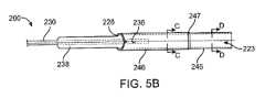
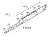
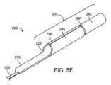
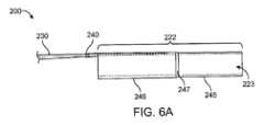
カテーテル前進要素の近位延長部は、カテーテル前進要素の最遠位端から約125cm以内に位置する少なくとも1つの剛性点を有することができ、少なくとも1つの剛性点は曲げ力を有する。遠位点の第1曲げ力に対する少なくとも1つの剛性点の曲げ力の比は、少なくとも約100である。カテーテル前進要素の近位延長部は、カテーテル前進要素の遠位端から約125cm以内に位置する少なくとも1つの剛性点を有することができ、少なくとも1つの剛性点は、曲げ力を有し、ここで、遠位点の第1曲げ力に対する少なくとも1つの剛性点の曲げ力の比は少なくとも約200である。カテーテル前進要素の近位延長部は、カテーテル前進要素の遠位端から約125cm以内に位置する少なくとも1つの剛性点を有することができ、少なくとも1つの剛性点は曲げ力を有する。遠位点の第1曲げ力に対する少なくとも1つの剛性点の曲げ力の比は、少なくとも約300より大きくできる。先端部の長さは、少なくとも約1cmから約4cmとできる。遠位点の第1曲げ力に対する近位点の第3曲げ力の比は、少なくとも2とすることができる。遠位点の第1曲げ力に対する第1システム曲げ力の比は、少なくとも2とすることができる。遠位カテーテル部は、遠位端から近位に少なくとも5mmの距離に配置されたカテーテルポイントを有する。カテーテルポイントはカテーテル曲げ力を有する。遠位点の第1曲げ力は、カテーテルの曲げ力の約5%から15%である。近位点の第3曲げ力は、カテーテル曲げ力の約50%から約90%である。遠位点における第1曲げ力と近位点における第3曲げ力の差は、壁厚の関数とすることができる。遠位カテーテル部の遠位端における内径は約0.054インチとすることができ、スナグポイントでの差は約0.006インチから約0.008インチとすることができる。遠位カテーテル部の遠位端における内径は、約0.070インチから約0.088インチとすることができ、スナグポイントでの差は、約0.006インチから約0.008インチとすることができる。The proximal extension of the catheter advancement element can have at least one rigid point located within about 125 cm of the distal most end of the catheter advancement element, the at least one rigid point having a bending force. A ratio of the bending force of the at least one rigid point to a first bending force of the distal point is at least about 100. The proximal extension of the catheter advancement element can have at least one rigid point located within about 125 cm of the distal end of the catheter advancement element, the at least one rigid point having a bending force, where the ratio of the bending force of the at least one rigid point to a first bending force of the distal point is at least about 200. The proximal extension of the catheter advancement element can have at least one rigid point located within about 125 cm of the distal end of the catheter advancement element, the at least one rigid point having a bending force. A ratio of the bending force of the at least one rigid point to a first bending force of the distal point can be at least greater than about 300. The length of the tip can be at least about 1 cm to about 4 cm. A ratio of the third bending force at the proximal point to the first bending force at the distal point can be at least 2. A ratio of the first system bending force to the first bending force at the distal point can be at least 2. The distal catheter section has a catheter point located at a distance of at least 5 mm proximally from the distal end. The catheter point has a catheter bending force. The first bending force at the distal point is about 5% to 15% of the catheter bending force. The third bending force at the proximal point is about 50% to about 90% of the catheter bending force. The difference between the first bending force at the distal point and the third bending force at the proximal point can be a function of wall thickness. The inner diameter at the distal end of the distal catheter section can be about 0.054 inches and the difference at the snug point can be about 0.006 inches to about 0.008 inches. The inner diameter at the distal end of the distal catheter section can be about 0.070 inches to about 0.088 inches, with a difference at the snug point of about 0.006 inches to about 0.008 inches.
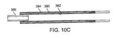
カテーテル前進要素の管状部は、管状部の壁内に埋め込まれるか又はその上に配置される放射線不透過性マーカーバンドを有することができ、放射線不透過性マーカーバンドは、スナグポイントに配置される。放射線不透過性マーカーバンドは、近位端、遠位端、及び近位端と遠位端との間の幅部を有する。前進構造の場合、放射線不透過性マーカーバンドの近位端は、放射線不透過性マーカーバンドが遠位カテーテル部の管腔の外部に残るように、遠位カテーテル部の遠位端と実質的に位置合わせできる。管状部の外径は、少なくとも約5cmから約10cmまでの長さを有する。スナグポイントは、長手方向の少なくとも一部に沿って配置できる。外径は、長手方向にほぼ均一である。外径は、長さに沿って実質的に不均一である。遠位点は、カテーテル前進要素の遠位端から少なくとも5mm近位に配置できる。第1システムポイントは、カテーテル部の遠位端から少なくとも約5mm近位に配置できる。The tubular section of the catheter advancement element can have a radiopaque marker band embedded in or disposed on the wall of the tubular section, the radiopaque marker band disposed at a snug point. The radiopaque marker band has a proximal end, a distal end, and a width between the proximal and distal ends. In the advancement configuration, the proximal end of the radiopaque marker band can be substantially aligned with the distal end of the distal catheter section such that the radiopaque marker band remains outside the lumen of the distal catheter section. The outer diameter of the tubular section has a length of at least about 5 cm to about 10 cm. The snug point can be disposed along at least a portion of the length. The outer diameter is substantially uniform in the length. The outer diameter is substantially non-uniform along the length. The distal point can be disposed at least 5 mm proximal from the distal end of the catheter advancement element. The first system point can be disposed at least about 5 mm proximal from the distal end of the catheter section.
関連する態様には、カテーテル及びカテーテル前進要素を含む同軸カテーテルシステムが記載されている。カテーテルは、管腔を有する遠位カテーテル部と、管腔からの開口部を有する遠位端とを含み、管腔は遠位端に少なくとも約0.052インチの内径を有する。カテーテルは、遠位カテーテル部に連結され、遠位カテーテル部から近位に延びる近位延長部を含み、近位延長部は遠位カテーテル部よりも可撓性が低い。カテーテル前進要素は、外径が少なくとも約0.014インチから約0.024インチまでの内径を有する管状部を含む。外径は少なくとも1つのスナグポイントを有する。先端のカテーテル部の内径と、スナグポイントにおける管状部の外径との差は、約0.010インチ以下である。カテーテル前進要素は、管状部の少なくとも1つのスナグポイントより遠位に位置する先端部を含む。先端部は、所定長さを有し、少なくとも一部が先細りになっている。先端部は、カテーテル前進要素の最遠位端から少なくとも5mm近位に位置する遠位点を有し、遠位点は、約0.05ニュートン以下の曲げ力を有する。A related aspect describes a coaxial catheter system including a catheter and a catheter advancement element. The catheter includes a distal catheter section having a lumen and a distal end having an opening from the lumen, the lumen having an inner diameter of at least about 0.052 inches at the distal end. The catheter includes a proximal extension connected to the distal catheter section and extending proximally from the distal catheter section, the proximal extension being less flexible than the distal catheter section. The catheter advancement element includes a tubular section having an inner diameter with an outer diameter of at least about 0.014 inches to about 0.024 inches. The outer diameter has at least one snug point. The difference between the inner diameter of the distal catheter section and the outer diameter of the tubular section at the snug point is about 0.010 inches or less. The catheter advancement element includes a tip section located distal to the at least one snug point of the tubular section. The tip section has a length and is at least partially tapered. The tip has a distal point located at least 5 mm proximal to the distal-most end of the catheter advancement element, and the distal point has a bending force of about 0.05 Newtons or less.
関連する態様には、患者の脳血管において医療処置を行う方法が記載されている。この方法は、装置の第1組立同軸システムを脳血管内の閉塞に向かって前進させるステップを含む。装置の第1組立同軸システムは、管腔を有する第1カテーテル部と、管腔への近位開口と、管腔から遠位開口と、遠位端とを有する第1カテーテルを含む。第1近位延長部は、第1カテーテル部に連結され、第1カテーテル部から近位方向に延び、第1カテーテル部よりも可撓性が低い。装置の第1組立同軸システムは、可撓性のある細長い本体と、柔軟で先細りの先端部とを有する第1送達要素を含む。第1カテーテル部の管腔内に配置された細長い本体の少なくとも一部分と、第1カテーテル部の遠位端から遠位側に延在する先細りの遠位先端部分とを有する。この方法は、第1送達要素を第1カテーテル部の内腔から近位側に引き抜き、第1カテーテル部の内腔を通って、内腔から遠位開口部を出て、脳血管内の閉塞の近位面に近い位置まで、装置の第2組立同軸システムを前進させることを含む。装置の第2組立同軸システムは、第2カテーテル及び第2送達要素を含む。第2カテーテルは、管腔と、管腔への近位開口と、管腔から遠位開口と、遠位端とを有する第2カテーテル部を含む。第2近位延長部は、第2カテーテル部に連結され、第2カテーテル部から近位方向に延び、第2近位延長部は、第2カテーテル部よりも可撓性が低い。第2送達要素は、可撓性の細長い本体と、柔軟で先細りの遠位先端部と、第2カテーテル部の管腔内に配置された細長い本体の少なくとも一部分と、第2カテーテル部の遠位端まで遠位に延びる先細りの遠位先端部とを含む。装置の第2組立同軸システムは、第2カテーテル部の遠位端が内頸動脈の錐体部分に対して遠位になった後、一緒に前進される。この方法は、第2送達要素を第2カテーテル部の管腔から近位側に引き出すステップを含む。第2カテーテル部の管腔を介して吸引圧を加え、吸引圧を介して第2カテーテル部の遠位端を閉塞上に固定し、第2カテーテル部の遠位端が咬合に固定された状態で、第2カテーテルに近位方向の力を加えて、第2カテーテルにおける周囲の解剖学的構造に対する緩みを減少させる。A related aspect describes a method of performing a medical procedure in a cerebral vasculature of a patient. The method includes advancing a first assembled coaxial system of the device toward an occlusion in the cerebral vasculature. The first assembled coaxial system of the device includes a first catheter portion having a lumen, a first catheter having a proximal opening to the lumen, a distal opening from the lumen, and a distal end. A first proximal extension is coupled to the first catheter portion, extends proximally from the first catheter portion, and is less flexible than the first catheter portion. The first assembled coaxial system of the device includes a first delivery element having a flexible elongated body and a soft, tapered tip portion. The first delivery element has at least a portion of the elongated body disposed within the lumen of the first catheter portion and a tapered distal tip portion extending distally from the distal end of the first catheter portion. The method includes withdrawing the first delivery element proximally from the lumen of the first catheter portion and advancing a second assembled coaxial system of the device through the lumen of the first catheter portion, out a distal opening from the lumen, to a location proximal to the proximal aspect of the occlusion in the cerebral vessel. The second assembled coaxial system of the device includes a second catheter and a second delivery element. The second catheter includes a second catheter portion having a lumen, a proximal opening to the lumen, a distal opening from the lumen, and a distal end. A second proximal extension is coupled to the second catheter portion and extends proximally from the second catheter portion, the second proximal extension being less flexible than the second catheter portion. The second delivery element includes a flexible elongate body, a soft tapered distal tip, at least a portion of the elongate body disposed within the lumen of the second catheter portion, and a tapered distal tip extending distally to the distal end of the second catheter portion. The second assembled coaxial system of the device is advanced together after the distal end of the second catheter portion is distal to the petrous portion of the internal carotid artery. The method includes withdrawing the second delivery element proximally from the lumen of the second catheter portion. Applying suction pressure through the lumen of the second catheter portion to secure the distal end of the second catheter portion over the occlusion via the suction pressure, and applying a proximal force to the second catheter with the distal end of the second catheter portion secured in the occlusion to reduce slack in the second catheter against the surrounding anatomical structures.
前記方法は、第2カテーテルを脳血管から引き抜くステップをさらに含むことができ、第2カテーテル部の遠位端には閉塞材料が取り付けられている。この方法は、吸引圧により第2カテーテル部の遠位端を閉塞して固定しながら、第1カテーテルを第2カテーテル上に前進させることをさらに含むことができる。本方法はさらに、第1カテーテル部の遠位端を閉塞の近位面の近くに配置することを含むことができる。この方法は、第2カテーテルを第1カテーテルの管腔に引き込むことをさらに含むことができる。第2カテーテルを管腔に引き抜くと、第1カテーテル部の管腔を介して吸引圧が自動的に加えられる。第1カテーテル部の管腔及び第2カテーテル部の管腔を介して加えられる吸引圧は、単一の吸引源から得ることができる。第2カテーテル部の遠位端には、閉塞材料を取り付けることができる。The method may further include withdrawing the second catheter from the cerebral vasculature, the second catheter portion having an occlusion material attached to a distal end thereof. The method may further include advancing the first catheter over the second catheter portion while occluding and securing the distal end of the second catheter portion with suction pressure. The method may further include positioning the distal end of the first catheter portion proximate the proximal aspect of the occlusion. The method may further include withdrawing the second catheter into the lumen of the first catheter. Withdrawing the second catheter into the lumen automatically applies suction pressure through the lumen of the first catheter portion. The suction pressure applied through the lumen of the first catheter portion and the lumen of the second catheter portion may be obtained from a single suction source. The distal end of the second catheter portion may have an occlusion material attached to it.
前記方法は、第1カテーテルが第1カテーテル部の管腔を介して吸引圧を維持する間に、第2カテーテルを第1カテーテルの管腔から引き抜くことをさらに含むことができる。本方法は、さらに、ガイドシースをアクセス位置から前進させることを含むことができる。ここで、ガイドシースは、中心管腔、近位端、遠位開口、及びシース本体の近位端に動作可能に接続されたコネクタを有する管状シース本体を含む。第1組立同軸カテーテルシステムを前進させることは、第1組立同軸カテーテルシステムを、ガイドシースを介して前進させることを含むことができる。第1カテーテル部は、吸引圧力の印加時にガイドシースの中心内腔とシールするように構成された外径を有する。第2カテーテル部は、吸引圧力の印加時に第1カテーテルの内腔とシールするように構成された外径を有する。第2カテーテル部の外面は、第1カテーテル部の内面とシールすることができ、第2カテーテル部の遠位開口部とガイドシースの近位端との間に連続する管腔を形成する。コネクタは、1又は2の頭部を有する回転止血弁を含むことができる。第1及び第2組立同軸カテーテルシステムの両方を、コネクタを介して前進させることができる。本方法はさらに、ガイドシースを前進させるステップは、ガイドシースの遠位開口部を遠位内頸動脈(ICA)内の位置まで前進させるステップを含むことができる。第2カテーテルは0.054インチから0.070インチの内径を有することができ、第1カテーテルは0.072インチから0.088インチの内径を有し、ガイドシースは6Frから8Frの間である。本方法はさらに、閉塞を横切ってガイドワイヤを前進させることを含むことができる。この方法では咬合を貫通させる必要はない。第1組立同軸カテーテルシステムは、ガイドワイヤをさらに含むことができる。組み立てられると、ガイドワイヤは、第1送達要素の可撓性の細長い本体の管腔内に配置でき、第1送達要素は、テーパ状の遠位先端部が第1カテーテル部の遠位端まで延び、ガイドワイヤがテーパ状の遠位先端部まで延びるように、第1カテーテル部の管腔内に配置できる。第2カテーテル部の外面は、第2カテーテル部の遠位開口部と第1カテーテル部の近位開口部との間に連続する管腔を形成する第1カテーテル部の内面とシールできる。The method may further include withdrawing the second catheter from the lumen of the first catheter while the first catheter maintains suction pressure through the lumen of the first catheter portion. The method may further include advancing a guide sheath from the access location. Here, the guide sheath includes a tubular sheath body having a central lumen, a proximal end, a distal opening, and a connector operably connected to the proximal end of the sheath body. Advancing the first assembled coaxial catheter system may include advancing the first assembled coaxial catheter system through the guide sheath. The first catheter portion has an outer diameter configured to seal with the central lumen of the guide sheath upon application of suction pressure. The second catheter portion has an outer diameter configured to seal with the lumen of the first catheter upon application of suction pressure. An outer surface of the second catheter portion may seal with an inner surface of the first catheter portion, forming a continuous lumen between the distal opening of the second catheter portion and the proximal end of the guide sheath. The connector may include a rotating hemostatic valve having one or two heads. Both the first and second assembled coaxial catheter systems may be advanced through the connector. The method may further include advancing the guide sheath to a position within the distal internal carotid artery (ICA). The second catheter may have an inner diameter of 0.054 inches to 0.070 inches, the first catheter may have an inner diameter of 0.072 inches to 0.088 inches, and the guide sheath is between 6 Fr and 8 Fr. The method may further include advancing a guidewire across the occlusion. This method does not require penetrating an occlusion. The first assembled coaxial catheter system may further include a guidewire. When assembled, the guidewire may be positioned within the lumen of the flexible elongate body of the first delivery element, and the first delivery element may be positioned within the lumen of the first catheter section such that the tapered distal tip extends to the distal end of the first catheter section and the guidewire extends to the tapered distal tip. The outer surface of the second catheter section can be sealed with the inner surface of the first catheter section to form a continuous lumen between the distal opening of the second catheter section and the proximal opening of the first catheter section.
関連する態様では、患者の脳血管内で医療処置を行う方法が記載される。この方法は、第1組立同軸カテーテルシステムを、脳血管内の閉塞の近位面に近い位置まで前進させることを含む。第1組立同軸カテーテルシステムは、第1カテーテルと第1送達要素とを含む。第1カテーテルは、管腔と、管腔への近位開口と、管腔から遠位開口と、遠位端とを有する第1カテーテル部を含む。第1カテーテル部に連結され、第1カテーテル部から近位方向に延在する第1近位延長部であって、第1近位延長部は、第1カテーテル部よりも可撓性が低い。第1送達要素は、可撓性の細長い本体と、柔軟で先細りの遠位先端部とを含み、細長い本体の少なくとも一部は、第1カテーテル部の管腔内に配置され、先細りの遠位先端部は、第1カテーテル部の遠位端に遠位に延びる。装置の第1組立同軸システムは、第1カテーテル部の遠位端が内頸動脈の錐体部分に対して遠位になった後、一緒に前進される。この方法は、第1送達要素を第1カテーテル部の管腔から近位側に引き出すステップを含む。第1カテーテル部の管腔を介して吸引圧力を加え、吸引圧により第1カテーテル部の遠位端を閉塞して固定し、第1カテーテルに近位方向の力を加えて、第1カテーテルの遠位端が閉塞して固定された状態で、周囲の解剖学的構造に対して第1カテーテルの緩みを減少させ、第1カテーテル部の遠位端を吸引圧により閉塞して固定しながら、第1カテーテル上で第2カテーテルを前進させる。第2カテーテルは、管腔、管腔への近位開口、管腔から遠位開口及び遠位端を有する第2カテーテル部と、第2カテーテル部に連結され、第2カテーテル部から基端側に延在する第2基端側延出部とを含む。第2近位延長部は、第2カテーテル部よりも可撓性が低い。In a related aspect, a method of performing a medical procedure within a cerebral vasculature of a patient is described. The method includes advancing a first assembled coaxial catheter system to a position proximate a proximal aspect of an occlusion within the cerebral vasculature. The first assembled coaxial catheter system includes a first catheter and a first delivery element. The first catheter includes a first catheter portion having a lumen, a proximal opening to the lumen, a distal opening from the lumen, and a distal end. A first proximal extension coupled to the first catheter portion and extending proximally from the first catheter portion, the first proximal extension being less flexible than the first catheter portion. The first delivery element includes a flexible elongate body and a soft tapered distal tip, at least a portion of the elongate body disposed within the lumen of the first catheter portion and the tapered distal tip extending distally to the distal end of the first catheter portion. The first assembled coaxial system of the device is advanced together after the distal end of the first catheter portion is distal to the petrous portion of the internal carotid artery. The method includes withdrawing a first delivery element proximally from a lumen of a first catheter portion; applying suction pressure through the lumen of the first catheter portion to occlude and secure a distal end of the first catheter portion with the suction pressure; applying a proximal force to the first catheter to reduce slack in the first catheter relative to the surrounding anatomical structure with the distal end of the first catheter occluded and secured; and advancing a second catheter over the first catheter while the distal end of the first catheter portion is occluded and secured with the suction pressure. The second catheter includes a second catheter portion having a lumen, a proximal opening to the lumen, a distal opening from the lumen and a distal end, and a second proximal extension coupled to the second catheter portion and extending proximally from the second catheter portion. The second proximal extension is less flexible than the second catheter portion.
前記方法はさらに、第1カテーテルを脳血管から引き抜くことを含むことができる。第1カテーテル部の遠位端は、取り付けられる閉塞材料を有してもよい。本方法ではさらに、第2カテーテル部の遠位端を閉塞の近位面の近くに配置することができる。この方法は、第1カテーテルを第2カテーテルの管腔に引き込むことをさらに含み、第1カテーテルがルーメン内に引き抜かれると、第2カテーテル部のルーメンを通って吸引圧が自動的に加えられる。吸引圧は、第1カテーテル部の管腔を介して作用させることができ、第2カテーテル部の管腔には、単一の吸引源から作用する。第1カテーテルの遠位端は、取り付けられる閉塞材料を有する。この方法は、第2カテーテルが第2カテーテル部の管腔を介して吸引圧を維持している間に、第1カテーテルを第2カテーテルの管腔から引き出すことをさらに含むことができる。本方法はさらに、アクセス位置からガイドシースを前進させることを含むことができる。ガイドシースは、中心管腔と、近位端と、遠位開口と、シース本体の近位端に動作可能に接続されたコネクタとを有する管状シース本体を含むことができる。第2カテーテル部は、吸引圧力の印加時にガイドシースの中心内腔をシールするように構成された外径を含むことができる。第1カテーテル部は、吸引圧力の印加時に第2カテーテルの内腔をシールするように構成された外径を有する。第1カテーテル部の外面は、第1カテーテル部の遠位開口部とガイドシースの近位端との間に連続する管腔を形成する第2カテーテル部の内面とシールできる。コネクタは、1又は2の頭部を有する回転止血弁を含むことができる。第1組立同軸カテーテルシステム及び第2カテーテルの両方を、コネクタを介して前進させることができる。The method may further include withdrawing the first catheter from the cerebral vasculature. The distal end of the first catheter portion may have an occlusion material attached thereto. The method may further include positioning the distal end of the second catheter portion near the proximal aspect of the occlusion. The method may further include withdrawing the first catheter into the lumen of the second catheter portion, and suction pressure is automatically applied through the lumen of the second catheter portion as the first catheter is withdrawn into the lumen. Suction pressure may be applied through the lumen of the first catheter portion, and the lumen of the second catheter portion may be applied from a single suction source. The distal end of the first catheter has an occlusion material attached thereto. The method may further include withdrawing the first catheter from the lumen of the second catheter portion while the second catheter maintains suction pressure through the lumen of the second catheter portion. The method may further include advancing a guide sheath from the access location. The guide sheath may include a tubular sheath body having a central lumen, a proximal end, a distal opening, and a connector operably connected to the proximal end of the sheath body. The second catheter section can include an outer diameter configured to seal the central lumen of the guide sheath upon application of suction pressure. The first catheter section has an outer diameter configured to seal the lumen of the second catheter upon application of suction pressure. The outer surface of the first catheter section can be sealed with an inner surface of the second catheter section forming a continuous lumen between the distal opening of the first catheter section and the proximal end of the guide sheath. The connector can include a rotating hemostatic valve having one or two heads. Both the first assembled coaxial catheter system and the second catheter can be advanced through the connector.
本方法は、ガイドシースを前進させるステップが、ガイドシースの遠位開口部を遠位内頸動脈(ICA)内の位置まで前進させるステップを含むことができる。第1カテーテルは、0.054インチから0.070インチの内径を有する。第2カテーテルは、0.072インチと0.088インチの間の内径を有する。ガイドシースは、6Fr~8Frである。本方法はさらに、閉塞を横切ってガイドワイヤを前進させることを含むことができる。この方法では咬合を貫通させる必要はない。第2カテーテル部の内面は、第1カテーテル部の遠位開口部と第2カテーテル部の近位開口部との間に隣接する吸引管腔を形成する第1カテーテル部の外面とシールできる。The method may include advancing the guide sheath to a position with a distal opening of the guide sheath within the distal internal carotid artery (ICA). The first catheter has an inner diameter of 0.054 inches to 0.070 inches. The second catheter has an inner diameter between 0.072 inches and 0.088 inches. The guide sheath is 6 Fr to 8 Fr. The method may further include advancing a guidewire across the occlusion. The method does not require penetrating an occlusion. The inner surface of the second catheter portion may be sealed with the outer surface of the first catheter portion forming an adjacent aspiration lumen between the distal opening of the first catheter portion and the proximal opening of the second catheter portion.
関連する態様には、第1カテーテル及び第2カテーテルを含む患者の脳血管内で医療処置を行うための装置のシステムが開示される。第1カテーテルは、管腔と、管腔への近位開口と、管腔から遠位開口と、遠位端とを有する第1カテーテル部を含む。第1カテーテル部に連結され、第1カテーテル部から近位方向に延在する第1近位延長部は、第1カテーテル部よりも可撓性が低い。第2カテーテルは、第1カテーテル部の管腔内に同軸に配置されるように構成される。第2カテーテルは、管腔と、管腔への近位開口と、管腔から遠位開口と、遠位端とを有する第2カテーテル部を含む。第2近位延長部は、第2カテーテル部に連結され、第2カテーテル部から近位方向に延び、第2カテーテル部よりも可撓性が低い。システムは、中心管腔を有する管状シース本体と、近位端と、遠位開口と、シース本体の近位端に動作可能に接続されたコネクタとを有するガイドシースを含む。単一の真空源が、ガイドシースのコネクタに連結され、ガイドシースの中心管腔、第1カテーテル部の管腔、及び第2カテーテル部の管腔を介して吸引圧を印加するように構成される。第2カテーテル部の外面は、第1カテーテル部の遠位開口部と第2カテーテル部の近位開口部との間に連続する管腔を形成する第1カテーテル部の内面とシールできる。In a related aspect, a system of apparatus for performing a medical procedure in a patient's cerebral vasculature is disclosed that includes a first catheter and a second catheter. The first catheter includes a first catheter portion having a lumen, a proximal opening to the lumen, a distal opening from the lumen, and a distal end. A first proximal extension is coupled to the first catheter portion and extends proximally from the first catheter portion and is less flexible than the first catheter portion. The second catheter is configured to be coaxially disposed within the lumen of the first catheter portion. The second catheter includes a second catheter portion having a lumen, a proximal opening to the lumen, a distal opening from the lumen, and a distal end. The second proximal extension is coupled to the second catheter portion and extends proximally from the second catheter portion and is less flexible than the second catheter portion. The system includes a guide sheath having a tubular sheath body having a central lumen, a proximal end, a distal opening, and a connector operably connected to the proximal end of the sheath body. A single vacuum source is coupled to the connector of the guide sheath and configured to apply suction pressure through the central lumen of the guide sheath, the lumen of the first catheter portion, and the lumen of the second catheter portion. The outer surface of the second catheter portion can be sealed with the inner surface of the first catheter portion to form a continuous lumen between the distal opening of the first catheter portion and the proximal opening of the second catheter portion.
関連する態様では、患者の脳血管内で医療処置を行う方法が開示されている。本方法は、第1カテーテルを脳血管内の閉塞に向かって前進させるステップを含む。第1カテーテルは、管腔と、管腔への近位開口と、管腔から遠位開口と、遠位端とを有する第1カテーテル部を含む。第1近位延長部は、第1カテーテル部に連結され、第1カテーテル部よりも可撓性が低い。この方法は、第2カテーテルを、第1カテーテル部の内腔を通り、内腔から遠位開口部を出て、脳血管内の閉塞の近位面に近い位置まで前進させることを含む。第2カテーテルは、管腔と、管腔への近位開口と、管腔から遠位開口と、遠位端とを有する第2カテーテル部を含む。第2近位延長部は、第2カテーテル部に連結され、第2近位延長部は、第2カテーテル部よりも可撓性が低い。この方法は、第2カテーテル部の外径と第1カテーテル部の内径との間にシールを形成するステップを含む。第2カテーテル部の管腔、第1カテーテル部の管腔、又は第1及び第2カテーテル部の管腔によって形成される隣接する吸引管腔のうちの少なくとも1つを介して吸引圧を印加する。隣接する吸引管腔は、第2カテーテル部の遠位端から第1カテーテル部の近位開口に向かって延びる。この方法は、吸引圧を介して第2カテーテル部の遠位端を閉塞上に固定することをさらに含むことができる。この方法はさらに、第2カテーテル部の遠位端が閉塞上に固定されたままである間、第2カテーテルに近位方向の力を加えて、周囲の解剖学的構造に対する第2カテーテルの緩みを減少させることを含むことができる。In a related aspect, a method of performing a medical procedure in a cerebral vasculature of a patient is disclosed. The method includes advancing a first catheter toward an occlusion in the cerebral vasculature. The first catheter includes a first catheter portion having a lumen, a proximal opening to the lumen, a distal opening from the lumen, and a distal end. A first proximal extension is coupled to the first catheter portion and is less flexible than the first catheter portion. The method includes advancing a second catheter through the lumen of the first catheter portion and out the distal opening from the lumen to a location proximal to a proximal aspect of the occlusion in the cerebral vasculature. The second catheter includes a second catheter portion having a lumen, a proximal opening to the lumen, a distal opening from the lumen, and a distal end. The second proximal extension is coupled to the second catheter portion and is less flexible than the second catheter portion. The method includes forming a seal between an outer diameter of the second catheter portion and an inner diameter of the first catheter portion. Applying suction pressure through at least one of a lumen of the second catheter portion, a lumen of the first catheter portion, or an adjacent suction lumen formed by the lumens of the first and second catheter portions. The adjacent suction lumen extends from a distal end of the second catheter portion toward a proximal opening of the first catheter portion. The method may further include securing the distal end of the second catheter portion over the occlusion via suction pressure. The method may further include applying a proximal force to the second catheter while the distal end of the second catheter portion remains secured over the occlusion to reduce slack in the second catheter against the surrounding anatomical structures.
いくつかの変形例では、上記の方法、装置、及びシステムにおける任意の実現可能な組み合わせに、以下のうちの1以上を任意に含めることができる。方法、装置、装置、及びシステムの詳細は、添付の図面及び以下の説明に記載されている。他の特徴及び利点は、説明及び図面から明らかとなる。In some variations, any possible combination of the above methods, devices, and systems may optionally include one or more of the following: Details of the methods, devices, apparatus, and systems are set forth in the accompanying drawings and the following description. Other features and advantages will be apparent from the description and drawings.
これら及び他の態様について、以下の図面を参照して詳細に説明する。一般的に言って、図は絶対的な用語又は比較的な尺度ではなく、説明のためのものである。また、説明を明確にするために、特徴及び要素の相対的な配置を修正してもよい。These and other aspects are described in detail with reference to the following drawings. Generally speaking, the figures are for illustrative purposes and not in absolute terms or to relative scale. Also, the relative placement of features and elements may be modified for clarity of illustration.
図面は一例であり、原寸に比例することを意図するものでもない。本明細書に記載の装置は、各図に必ずしも図示されていない特徴を含んでいてもよい。The drawings are illustrative and are not intended to be drawn to scale. Devices described herein may include features that are not necessarily shown in each figure.
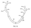
急性虚血性脳卒中(AIS)のような脳動脈レベルで種々の神経血管病理を治療するために頚動脈解剖的構造を誘導するには、可撓性及び送達性に優れたカテーテルシステムが必要である。内頸動脈(ICA)は、総頸動脈(CCA)のC3椎骨とC4椎骨の間の椎間板レベルでの分岐部から現れる。図1Aに示すように、ICAの経路は、頚部Cr、錐体Pt、海綿体Cv及び大脳Cbの4つの部分に分割される。前方循環では、一定の蛇行した終末頸動脈は骨性要素によってその位置に固定される。頸部頸動脈Crは錐体骨に入り、骨に覆われるようにして一連の折り返し部に固定される。海綿状頸動脈は静脈床、海綿状静脈洞を通過する動脈であり、可撓性はあるが、海綿状静脈洞から出る際に別の骨性要素によって固定される。骨性要素は頭蓋腔への入口を取り囲んで固定する。これらの骨性の固定点のため、錐体及び海綿体頸動脈(Pt及びCv)と、それより上の部分は、曲がり度が比較的一致している。頚動脈サイフォンCSは末端ICAのS字部であり、海綿状ICAの後方湾曲部から始まり、前大脳動脈ACAと中大脳動脈MCAへのICA分岐部で終端している。眼動脈は大脳ICAから現れ、前方循環にアクセスする際にカテーテルが引っかかる一般的な点を示す。MCAはまず、単一のM1セグメントによって定義され、次に2又は3のM2セグメントに分岐し、さらに分岐してM3セグメントを形成する。これらのカテーテルが引っかかる点は、脳への血液灌流を回復するために必要な時間を著しく増加させ、AISの治療において深刻な結果を伴う欠点となる。A flexible and deliverable catheter system is required to navigate the carotid anatomy to treat various neurovascular pathologies at the cerebral arterial level, such as acute ischemic stroke (AIS). The internal carotid artery (ICA) emerges from the bifurcation of the common carotid artery (CCA) at the disc level between the C3 and C4 vertebrae. As shown in Figure 1A, the course of the ICA is divided into four parts: cervical Cr, petrous Pt, cavernous Cv, and cerebral Cb. In the anterior circulation, the terminal carotid artery, which is a constant tortuousness, is fixed in place by bony elements. The cervical carotid artery Cr enters the petrous bone and is fixed in a series of turns covered by bone. The cavernous carotid artery is an artery that passes through a venous bed, the cavernous sinus, and although flexible, is fixed by another bony element as it exits the cavernous sinus. The bony element surrounds and fixes its entrance to the cranial cavity. Due to these bony anchorage points, the petrous and cavernous carotid arteries (Pt and Cv) and their superior portions are relatively consistent in their tortuosity. The carotid siphon CS is an S-shaped portion of the terminal ICA, originating from the posterior curvature of the cavernous ICA and terminating at the ICA bifurcation into the anterior cerebral artery ACA and the middle cerebral artery MCA. The ophthalmic artery emerges from the cerebral ICA and represents a common catheter trapping point when accessing the anterior circulation. The MCA is first defined by a single M1 segment, which then branches into two or three M2 segments, which further branch to form the M3 segment. These catheter trapping points significantly increase the time required to restore blood perfusion to the brain, a drawback with serious consequences in the treatment of AIS.
加齢に伴い、大きな血管は拡大して長くなる。頸部内頸動脈は近位及び遠位で固定されているので、加齢とともに蛇行する。総頸動脈CCAは、鎖骨によって頸部領域に出るように、胸腔内で相対的に固定される。外頚動脈ECA及び内頚動脈ICAは総頚動脈CCAには固定されておらず、加齢と共に全頚動脈系が延び、蛇行する。これにより、伸長、ねじれ及び蛇行、又は最悪の場合、完全なループ、すなわち、いわゆる「頸管ループ」を発生させる。これらのねじれた領域、又は湾曲した領域を通過するために使用されるカテーテルが硬すぎて可撓性がない場合、血管の集中したねじれ及び折り畳みを引き起こす、「理髪店のポール」のような血管の包み込みをもたらし、これらの領域は真っ直ぐになる可能性がある。このような極度の蛇行は、特に高齢者において、脳への血液灌流を回復するのに必要な時間を著しく増加させてしまう。特定の状況では、血管のねじれが血管自体に生じたり、ねじれていない動脈にねじれが生じたりすると、正常な順行性血流が停止して虚血を引き起こすことがある。頸部ICAのような血管の湾曲又はループの解除を管理することも、処置にかかる時間を増加させてしまう。With age, large blood vessels expand and lengthen. The cervical internal carotid artery becomes tortuous with age because it is fixed proximally and distally. The common carotid artery CCA is relatively fixed in the thoracic cavity as it exits the cervical region by the clavicle. The external carotid artery ECA and the internal carotid artery ICA are not fixed to the common carotid artery CCA, and the entire carotid system becomes elongated and tortuous with age. This causes elongation, twisting and tortuousness, or in the worst case, complete loops, i.e., so-called "cervical loops." If the catheter used to traverse these twisted or curved areas is too stiff and inflexible, it can result in a "barber's pole" wrapping of the vessels, causing concentrated twisting and folding of the vessels, which can then be straightened out. Such extreme tortuosity significantly increases the time required to restore blood perfusion to the brain, especially in the elderly. In certain circumstances, kinks in the blood vessel itself or in a non-kinked artery can halt normal antegrade blood flow and cause ischemia. Managing the release of curved or looped vessels, such as the cervical ICA, can also increase procedure time.
脳卒中介入処置のための現在のカテーテルシステムの主な欠点は、脳への血液灌流を回復するのに必要な時間であり、これには、大脳動脈の閉塞部位にアクセスするのに要する時間及び動脈の閉塞を完全に除去するのに要する時間が含まれる。閉塞を完全に除去するために複数回の試行が必要となることが多いため、試行回数を減らすとともに、追加の試行のために装置を交換するのに必要な時間を減らすことは、全体の時間を最小にするための重要な要素である。加えて、各試みは繊細な脳血管系における装置の前進による潜在的な処置リスクと関連している。他の制限は、従来のガイド及び遠位アクセスカテーテルと共に一般に使用される複数のRHVを有する長い3軸システムを送って、効果的に操作するために複数のオペレータが必要なことである。The main drawback of current catheter systems for stroke intervention procedures is the time required to restore blood perfusion to the brain, including the time required to access the site of occlusion in a cerebral artery and the time required to completely clear the artery. Because multiple attempts are often required to completely clear the occlusion, reducing the number of attempts as well as the time required to exchange the device for additional attempts are key factors in minimizing the overall time. In addition, each attempt is associated with potential procedural risk due to advancement of the device in the delicate cerebral vasculature. Another limitation is the need for multiple operators to deliver and effectively operate long triaxial systems with multiple RHVs commonly used with conventional guides and distal access catheters.
急性虚血性脳卒中(AIS)のような種々の神経血管病理を治療するためのカテーテルシステム及び方法を本明細書に記載する。本明細書に記載されるシステムは、遠位標的の解剖学的構造、特に単一の操作点における脳血管系の曲がりくねった解剖学的構造への迅速で簡単な一人のオペレータによるアクセスを提供する。本明細書に記載される医療方法、装置及びシステムは、複雑で曲がりくねった解剖学的構造を誘導して、急性虚血性脳卒中の治療のための脳閉塞の迅速かつ安全な吸引及び除去を行うことを可能にする。本明細書に記載される医療方法、装置及びシステムはまた、急性虚血性脳卒中の治療での脳閉塞の除去のため、吸引又は非吸引で頭蓋内医療装置を送達するために使用できる。本明細書に記載されるシステムは、ユーザがAISのための最前線の治療として吸引のみを実施しようとするかどうかに拘わらず、特にAISの治療には有効である。さらに、本明細書に記載される遠位アクセスカテーテルシステムの最高の可撓性及び送達可能性により、カテーテルは、新しい解剖学的構造を作り出す矯正力を発揮するのではなく、曲がりくねった解剖学的構造の形状をとることができる。本明細書に記載されている遠位アクセスカテーテルシステムは、その中の解剖学的構造の自然な曲線を維持しながら、曲がりくねったループを通過することができ、血管をまっすぐにするリスクを低減する。これにより、本明細書に記載される遠位アクセスカテーテルシステムは、他のカテーテルが通るための解剖学的構造の自然な屈曲性を維持する神経血管系を通る安全な導管(例えば、より大きな口径の吸引カテーテル)を形成できる。Described herein are catheter systems and methods for treating various neurovascular pathologies, such as acute ischemic stroke (AIS). The systems described herein provide rapid and simple single operator access to distal target anatomies, particularly the tortuous anatomy of the cerebral vasculature at a single point of operation. The medical methods, devices and systems described herein allow for navigating complex and tortuous anatomies to perform rapid and safe aspiration and removal of cerebral occlusions for the treatment of acute ischemic stroke. The medical methods, devices and systems described herein can also be used to deliver intracranial medical devices with or without aspiration for removal of cerebral occlusions in the treatment of acute ischemic stroke. The systems described herein are particularly effective for the treatment of AIS, regardless of whether the user intends to perform aspiration only as the first line of treatment for AIS. Furthermore, the ultimate flexibility and deliverability of the distal access catheter system described herein allows the catheter to take the shape of the tortuous anatomy rather than exerting corrective forces to create a new anatomy. The distal access catheter system described herein can pass through tortuous loops while maintaining the natural curvature of the anatomy therein, reducing the risk of straightening the vessel. This allows the distal access catheter system described herein to form a safe conduit through the neurovasculature that maintains the natural curvature of the anatomy for passage of other catheters (e.g., larger caliber aspiration catheters).
本明細書に記載されている装置、システム、及び使用方法は、2012年8月3日に出願された米国特許出願公開第2013/0035628号、2014年12月19日出願の米国特許公開第2015/0173782号及び米国特許出願公開第2016/0220741号(2016年2月4日)に記載されている装置、システム、及び使用方法に関連し、それらと組み合わせて、又はそれらと代替して使用できる。これらの各文献の開示は、その全体が参照により本明細書に組み込まれる。The devices, systems, and methods of use described herein are related to, and may be used in combination with, or in place of, the devices, systems, and methods of use described in U.S. Patent Application Publication No. 2013/0035628, filed Aug. 3, 2012, U.S. Patent Application Publication No. 2015/0173782, filed Dec. 19, 2014, and U.S. Patent Application Publication No. 2016/0220741 (Feb. 4, 2016). The disclosures of each of these documents are incorporated herein by reference in their entirety.
本明細書には、吸引するために神経血管解剖学的構造にアクセスすることについて、いくつかの実施形態が記載されているが、これに限定すべきではなく、他の用途にも適用できる。例えば、本明細書に記載されるカテーテルシステムは、冠動脈構造又は他の脈管構造の標的血管に作動装置を送達するために使用できる。「遠位アクセスカテーテル」又は「吸引カテーテル」という用語が本明細書で使用される場合、カテーテルは、吸引、治療部位への又は支持カテーテルとしての流体の送達、又はガイドワイヤなどの他の装置又はステントレトリバーなどの介入装置の送達又は交換を容易にし、ガイドする導管を提供する遠位アクセスに使用することができる。また、本明細書に記載されるアクセスシステムは、脈管構造の外側の身体の他の部分へのアクセスにも有効である。同様に、作動装置が拡張可能な脳治療装置であると記載されている場合、本明細書に記載されている送達システムを使用して、ステントレトリバー又は自己拡張型ステントなどの他の介入装置を送達できる。Although some embodiments are described herein for accessing the neurovascular anatomy for aspiration, they should not be limited thereto and may be applied to other applications. For example, the catheter system described herein may be used to deliver an actuating device to a target vessel in the coronary anatomy or other vasculature. When the term "distal access catheter" or "aspiration catheter" is used herein, the catheter may be used for aspiration, delivery of fluids to a treatment site or as a support catheter, or distal access to provide a conduit to facilitate and guide the delivery or exchange of other devices such as guidewires or interventional devices such as stent retrievers. The access systems described herein are also useful for accessing other parts of the body outside the vasculature. Similarly, when an actuating device is described as being an expandable brain treatment device, the delivery system described herein may be used to deliver other interventional devices such as stent retrievers or self-expanding stents.
ここで図2A及び図2Bを参照して、急性虚血性脳卒中を治療するために脳閉塞にアクセスして除去する装置を含むシステム100を示す。システム100は、各構成要素及びシステムが、手の動きが最小限でよい単一の操作点で、単一のオペレータにより送達及び使用できるような単一のオペレータシステムとすることができる。以下に詳述するように、全てのワイヤ及びカテーテルの操作は、同じ装置内に共存する単一又はそれ以上の回転止血弁(RHV)434又はその近傍で行うことができる。システム100は、カテーテル200及びカテーテル前進要素300を有する1以上のカテーテル送達システム150を含むことができる。カテーテル送達システム150は、アクセスガイドシース400を通って前進するように構成されている。カテーテル200は、ガイドシース400内に受け入れられるように構成されており、優れた送達が可能となるように設計されている。カテーテル200は、ガイドシース400の管腔と同軸の回転遠位アクセスカテーテルとして導管内の内径を拡張することができる。カテーテル200は、カテーテル前進要素300を用いてカテーテル200の管腔223に挿入して送達することができる。システム100は、経皮的動脈切開術(例えば、大腿動脈又は他の入口点)における入口点から遠位カテーテルの標的制御点までの長さを変更可能な遠位アクセスシステムとすることができる。脳卒中介入のための従来の遠位アクセスシステムは、通常、鼠径部において、(例えば、長さ11cmから30cmの)より短いシースを介して配置された長いガイドシース又はガイドカテーテルを含む。長いガイドシースは、一般に、脳卒中塞栓摘出術(「血栓除去術」と呼ばれることもある)を含む神経血管介入を支持するためにICA内に配置される。さらなる支持のため、これらは骨端錐体まで前進させることができ、可能であれば、海綿骨又は床突起又は床突起上の骨端ICAまで前進させることができる。ADAPA/MAT又はSolumbra/SMATアプローチのためのM1又はM2分布内の標的に到達するために、追加のカテーテルを長いガイドカテーテルを介して挿入できる。これらのカテーテルは、一般的には、例えば、長さが130cm以上の大口径吸引カテーテルである。以下により詳細に説明するように、本明細書に記載する遠位アクセスシステム100は、アクセスポイント、一般には総大腿動脈から測定されるシステムとして採用された場合、例えば、長さを115cmだけ短くできる。さらに、単一のオペレータは、ガイドシース(RHV)434上の単一の回転止血弁400、又は双頭RHVのような同じ装置内に共存する1以上のRHVを介してそれらを挿入することによって、本明細書に記載されたシステムを使用することができる。したがって、以前二人で行っていた手順を一人で行うことが可能となる。2A and 2B, a
次に、各種システムの各構成要素についてより詳細に説明する。Next, we'll explain each component of the various systems in more detail.
アクセスガイドシースAccess guide sheath
再び図2Aから図2Dについて、遠位アクセスシステム100は、アクセスガイドシース400を含むことができる。アクセスガイドシースは本体402を有している。作業用内腔は本体402を通って近位端領域403に連結された近位止血弁434から遠位端領域の遠位開口408まで延びている。作業用管腔は、カテーテル200を受け入れるように構成されている。カテーテル200の遠位端は、シース400の遠位端を越えて遠位開口408から延びている。ガイドシース400は、本明細書に記載のカテーテル及び当技術分野で公知の種々の作業装置を送達するために使用できる。例えば、作業装置は、血栓症治療するように構成でき、大口径カテーテル、吸引塞栓摘出術(又は血栓摘出術)、先端カテーテル、ワイヤ、バルーン、コイル先端の回収可能なステント「ステントレトリバー」のような回収可能な構造を含むことができる。ガイドシース400をカテーテル200と組み合わせて使用することにより、以下により詳細に説明されるように、遠位吸引を行うことができる。2A-2D, the
ガイドシース400は、様々な市販のガイドシースのいずれであってもよい。例えば、ガイドシース400は、0.087から0.089インチのID、例えば、Cook SHUTTLE 6F(Cook Medical,Inc.(イリノイ州ブルーミントン))、Terumo DESTINATION 6F(テルモ・ヨーロッパ)、Cordis VISTA BRITE TIP(コーディス,ハイアリア,フロリダ州(アメリカ合衆国))、及びPenumbra NEURON MAX 088(カリフォルニア州アラメダのPenumbra,Inc.)、Stryker Infinity (Stryker Neurovascular, Fremont,カリフォルニア州(アメリカ合衆国))又は同等の市販のガイドシースである。一般に、シースサイズは、本明細書ではフレンチ(F)スケールを用いて記載される。例えば、シースが6フレンチと記載されている場合、そのシースの内径は、約1.98mm又は0.078インチである6F外径を有するカテーテルを受け入れることができる。本明細書では、カテーテルは、別のカテーテルの外径を受け入れる内径とするために、特定のフレンチサイズを有するものとして記載できる。また、本明細書では、カテーテルは、その外径が特定の内径を有する別のカテーテルと適合することに言及するために、特定のフレンチサイズを有するものとして記載できる。The
ガイドシース400は、カテーテル200のような種々の作業装置を受け入れるための種々のサイズとし、オペレータの好みに合わせることができる。ガイドシース400の作業管腔は、摺動嵌めで各カテーテル200を受け入れるようなサイズにできる。一般に、ガイドシース400の外径を0.122インチ未満に制限することによって、血管挿入部位の全体のサイズを最小化するのが望ましい。また、カテーテル200とガイドシース400との間の摺動嵌合を良好にするために、対応する外径及び内径を選択するのが望ましい。ガイドシースの作業管腔は、特にカテーテルが吸引に使用される場合、受け入れられることが意図される任意のカテーテル200の最大外径よりも少なくとも0.001インチ大きい内径を有する。作業用内腔は、少なくとも6フレンチのカテーテル(1.98mm又は0.078インチ)、又は少なくとも6.3フレンチのカテーテル(外径2.079mm又は0.082インチ)、又は少なくとも7フレンチ(外径2.31mm又は0.091インチ)のカテーテル、又は8フレンチ(外径2.64mm又は0.104インチ)以上のカテーテルを収容する大きさの内径を有する。しかし、ガイドシース400の内径は、他のカテーテルサイズと適合するように、小さくても大きくてもよい。長さ及び内径に拘わらず、ガイドシース400は、血管を通って遠位に前進する際のねじれに対して耐性がある。The
本明細書に記載される吸引カテーテルは、0.054インチから0.088インチの間のIDを有する。カテーテル200は、0.088インチの内径を有し、0.105インチと0.107インチの間の最大外径を有する。次に、ガイドシース400は、0.106インチから0.108インチの内径の作業管腔を有する。一般に、カテーテル200の最大外径とガイドシース400の内径の差すなわちクリアランスは、約0.002インチ未満、例えば0.001インチから0.002インチである。カテーテル200の最大外径とガイドシース400の内径のクリアランスが小さい領域は、局所領域に限定できる。つまり、両者間の低クリアランス嵌合により、カテーテル200及びシース400の円筒長の一部のみを延長できる。したがって、カテーテル200とガイドシース400との間のOD-ID差は、使用中に2つの装置が重なった第1円筒長さに沿って0.002インチ以上とし、重なった異なる円筒長さに沿って0.002インチ未満とすることができ、それによって、重なった部分に低クリアランスの局所領域を設けることができる。これは、以下により詳細に説明されるように、吸引圧力下に置かれたときに、便利な相対的摺動及び十分な密封を可能にする。例えば、ガイドシース400の遠位領域は、カテーテル200との摺動嵌合の低いクリアランスがその長さに沿って変化するように、遠位端領域に第1内径部を有し、近位端領域に異なる第2内径部を有する。いくつかの実施態様では、カテーテル200は、遠位端領域に第1外径部を有し、近位端領域により大きい第2外径部を有する。第2外径部は、0.002インチ未満のシース400の内径部、及び0.002インチを超えるシース400の内径部とすることができる。これにより、ガイドシース400とカテーテル200の近位端領域との間で、第2外径部の位置全体がより緊密に嵌合される。The aspiration catheters described herein have an ID between 0.054 inches and 0.088 inches. The
再び図2A-図2Dについて、シース本体402は、近位端領域403の近位分岐部又は回転止血弁(RHV)434から本体402の遠位端の先端部406まで延びる。近位RHV434は、コネクタ本体に成形されてガイドシース402の本体400の作業用内腔に接続される1以上の内腔を含む。作業用内腔は、カテーテル200及び/又は標的解剖学的構造に送達するための種々の作業装置を受け入れることができる。RHV434は、厚壁ポリマー管又は強化ポリマー管で構成できる。RHV434は、出血を防止又は最小化し、ガイドシース400への空気の流入を防止しながら、ガイドシース400を介して血管系への装置の導入を可能にする。RHV434は、ガイドシース400に一体化されていてもよい。ガイドシース400は、受動シール弁、Tuohy-Borst弁又は回転止血弁などの別個の止血弁構成要素が取り付けられる雌ルアーアダプターの近位端で終端してもよい。RHV434は、除去中に凝血塊を移動させることなく、先端に付着凝血塊を有する装置を除去可能とする十分な大きさの調節可能な開口部を有する。代替的に、RHV434は、例えばシース400から装置を取り外して血餅の移動を防止する場合のように取り外し可能である。RHV434は、デュアルRHVである。2A-2D, the
RHV434は、シース400の近位端403上にYコネクタを形成することができる。この結果、RHV434の第1ポートを、シース400の作業管腔への作業カテーテルの挿入のために使用することができる。第2ポートは、アーム412への別の目的のために使用できる。例えば、シリンジ又は他の装置を、コネクタ432を介してアーム412に接続し、前方ドリップ、造影剤のためのフラッシュライン又は生理食塩水を、本体402を介して先端部406に向けて標的の解剖学的構造に注入できる。アーム412はまた、吸引源505(図2B参照)に接続できる。吸引源505は、吸引ポンプ、通常又はロック式注射器、手持ち式吸引器、病院吸引器などの作業用内腔を介して吸引するように構成された吸引駆動源とすることができる。一実施形態では、吸引源505は、流量コントローラに取り付けられたロッキングシリンジ(例えばVacLokシリンジ)である。処置の塞栓除去ステップの前にフローラインへの接続が閉じられている間、シリンジ上のプランジャは、ユーザによってロック位置に引き戻される。処置の間、カテーテルの先端が閉塞の近くにあるか閉塞の表面にあるとき、ユーザは吸引シリンジに接続することができる。これにより、シース400の作業用内腔を介して加えられる吸引力を最大にでき、シース400を通って延在している任意のカテーテルは、その遠位端において血管と連絡する。この吸引は、単一の共有ソースにより単一のユーザで迅速に行うことができる。別の実施形態では、アーム412は、吸引源505に接続することができる、吸引源は、吸引力がガイドシース400の作業管腔を介して印加するように構成されたポンプである。アーム412はまた、処置の間、ガイドシース400を生理食塩水又は放射線不透過性造影剤で洗い流すことを可能とする。作業用内腔は、シース本体403の近位端領域402の遠位端から作業用近位ポートまで延びている。The
カテーテル本体402の長さは、本体402の遠位先端部406を、例えば、大腿動脈から内頸動脈(ICA)内で遠位に配置できるように構成されており、必要に応じて調節するための追加の長さを備えている。いくつかの実施態様(例えば大腿部又は橈骨部の経皮的アクセス)では、本体402の長さは、80cmから90cmの範囲であってもよく、又はそれより長くてもよく、例えば、約100cm又は約105cmまでであってもよい。一実施例では、本体402の長さは、頸動脈の分岐部への経頸動脈アプローチに適しており、その範囲は20cmから25cmである。他の実施例では、本体402の長さは、CCA又は近位ICAへの経皮経頸動脈アプローチに適しており、10cmから15cmの範囲である。本体402は、例えば高い吸引力にさらされた場合であっても、屈曲、虚脱、又は血管外傷を引き起こすことなく、脈管構造の屈曲を想定して誘導するように構成されている。The length of the
いくつかの実装形態では、システム100は、ガイドシース400を前進させるための選択ツールをさらに含むことができる。選択ツールは、遠位端領域がガイドシース400の遠位端まで延び、ガイドシース400の作業管腔内に受け入れられるように構成された外径部を有する。選択ツールの外径は、ガイドシース400の遠位開口408を最小化するように、ガイドシース400の作業管腔の内径を十分に満たす。例えば、ガイドシース400の作業管腔が約0.087インチから約0.113インチの内径を有する場合、選択ツールの外径は、これよりも約0.006インチ小さい、すなわち約0.081インチから約0.107インチとすることができる。腕頭動脈離開(BT)は、右側脳循環(図1Cに示す)を求めて経大腿的に送達されるカテーテルのための大動脈弓AAの非常に厳しい折り返し部である。カテーテルは、大腿動脈から腸骨循環を経て下行大動脈DAに入り、大動脈弓AAに近づくにつれて旋回し、他の大血管の分岐を通過して、大動脈弓AAAの大血管の最も遠い「到達点」である腕頭動脈分岐(BT)に到達する。図1Cは、その解剖学的構造によって作られる実質的かつ必須のSターンを示す。カテーテルは、内頸動脈(ICA)に到達するために、大腿動脈挿入位置から挿入経路に沿ってこのSターンを通過しなければならない。左内頚動脈は腕頭動脈から離脱することが多く、同様の課題を抱えており、Sターンをさらにきつくできる。もし左ICAが腕頭BTと左鎖骨下動脈LSA離陸の間に一般的な分岐があるならば、到達はそれほど重度ではないかもしれないが、Sターンはまだより軽度の重症度で発生する。選択ツールの遠位端領域は、この折り返し部の周りでシース400をICAに前進させるために支持してガイドするように先細りに成形できる。いくつかの実施例において、選択ツールは、当該技術分野で公知のように、Bernstein Selectスタイル又はSimmonsスタイル逆曲線カテーテル先端を有する。In some implementations, the
ガイドシース400の先端部406は、遠位端に至る本体402の部分と同一又は同様の外径を有する。したがって、先端部406は、本体402を通過する長軸に直交する遠位面を有し、遠位面は、本体402の断面外寸法にほぼ等しい外径を有してもよい。実施形態では、先端部406は、面取り、隅肉又はテーパを含み、遠位面直径を本体402の断面寸法より僅かに小さくする。さらなる実施形態では、先端部406は、本体402の均一な外径を有する領域に遠位に延在する細長い管状部であってもよい。この結果、細長い管状部は、本体402の均一な外径と比較して径が小さくなる。したがって、先端部406は、細長い形状にできるか、あるいは、よりはっきりとした形状にできる。従って、先端部406は、脈管構造内を滑らかに追跡し、あるいは、脈管構造内を追跡する際に脈管制限を拡張するように構成できる。作業用内腔は、遠位開口408を形成する遠位端を有する。The
ガイドシース400は、本体402の部分から遠位端に至るまで先細りになる先端部406を含む。すなわち、本体402の外面は、遠位端でより大きな寸法からより小さな寸法に減少する直径を有する。例えば、先端部406は、約0.114インチから約0.035インチ、又は約0.110インチから約0.035インチ、又は約0.106インチから約0.035インチの外径から先細りになる。先端部406のテーパの角度は、先細りの先端部406の長さに応じて変化する。例えば、いくつかの実装形態では、先端部406は、約50mmの長さにわたって0.110インチから0.035インチまで先細りになる。The
一実施形態では、ガイドシース400は、1又は複数の放射線不透過性マーカー411を含む。放射線不透過性マーカー411は、遠位の先端部406近傍に配置できる。例えば、一対の放射線不透過性バンドは、かしめ、塗装、埋め込み、又はその他の方法で配置できる。いくつかの実装形態では、放射線不透過性マーカー411は、装置の遠位端の可撓性を維持し、ガイドシースの長さに沿った遷移及びねじれに対する耐性を改善するバリウムポリマー、タングステンポリマーブレンド、タングステン充填又はプラチナ充填マーカーを含む。いくつかの実装形態では、放射線不透過性マーカー411は、本体402に熱溶接されたタングステン充填PEBAX又はポリウレタンである。マーカー411は、本体402の1又は複数の領域周りのリングとして図示されている。マーカー411は、血管内の遠位開口408の位置についてオペレータに方向付けする他の形状を有するか又は様々なパターンを作成できる。したがって、オペレータは、蛍光透視法の下で遠位開口408の位置を視認して、カテーテル200が送達される標的の解剖学的構造に遠位開口408が向けられていることを確認できる。例えば放射線不透過性マーカー411は、遠位開口が、例えばICAに進められたカテーテル及びワイヤ等の後続の作業装置がICAにアクセスするように、オペレータが解剖学的アクセスポイント、例えば患者の鼠径部でガイドシース400の本体402を回転させることを可能にする。いくつかの実装形態では、放射線不透過性マーカー411は、白金、金、タンタル、タングステン、又はX線透視下で見える他の任意の物質を含む。本明細書に記載のシステムの様々な構成要素のいずれも、放射線不透過性マーカーを組み込むことができる。In one embodiment, the
いくつかの実施形態では、ガイドシース400は、運動性、放射線不透過性、カラム強度、及び可撓性について、頸動脈アクセス及びAIS手順で使用される他のシースと同様の性能特性を有する。インナーライナは、PTFE(ポリテトラフルオロエチレン)又はFEP(フッ素化エチレンプロピレン)のような低摩擦ポリマーから構成され、内腔を介して装置を前進させるための滑らかな表面を提供することができる。アウタージャケット材料は、インナーライナに機械的完全性を提供することができ、PEBAX、熱可塑性ポリウレタン、ポリエチレン、ナイロンなどの材料から構成できる。本体402は、親水性コーティングを含むことができる。インナーライナとアウタージャケットの間を補強できる第3層を組み込むことが可能である。補強層は、本体402の内部管腔の平坦化又はよじれを防止し、脈管構造の屈曲、吸引又は逆流により装置のナビゲーションが妨げられないようにすることができる。本体402は、円周方向に補強される。補強層は、ステンレス鋼、ニチノール、ニチノール組紐、螺旋状リボン、螺旋状ワイヤ、切断ステンレス鋼等のような金属、又はPEEKのような硬質なポリマーから作ることができる。補強層は、可撓性を有するようにレーザ切断又は機械切断されたコイル若しくは組紐、又はチューブのような構造とすることができる。別の実施形態では、補強層は、ニチノールハイポチューブ又はカットリジッドポリマーなどのカットハイポチューブであってもよい。本体402のアウタージャケットは、遠位端に向かって徐々に軟らかくなる材料で形成できる。本体402の可撓性は、本体402の遠位部分に向かって増加させることができる、可撓性の変化は、様々な方法により達成される。例えば、アウタージャケットは、様々な部分でデュロメータ及び/又は材料が変化する。ガイドシースの他の部分と比較して、ガイドシースの遠位部分において、より低いデュロメータのアウタージャケット材料を使用することができる。また、ジャケット材料の壁厚を薄くし、及び/又は補強層の密度を変化させて可撓性を高めることができる。例えば、コイル又は編組のピッチを引き伸ばしてもよいし、チューブ内の切断パターンをより柔軟に変化させてもよい。補強構造又は材料は、シース本体402の長手部で変化させてもよい。別の実施形態では、最遠部と近位部との間に遷移部があり、最遠部とシース本体402の残りの部分との間で可撓性が変化する1以上の部分がある。この実施形態では、最遠部は、約2cmから約5cmであり、遷移部は、約2cmから約10cmであり、近位部は、シース長の残りの部分を占める。いくつかの実施形態では、本体402の近位領域は、ナイロンのような材料で形成することができ、本体402の近位の領域から遠位の領域は、72D硬度の材料とすることができる。一方、より遠位の領域は、より可撓性を増し、遠位先端部406に向かって延びる55D、45D、35D硬度の材料、例えば、35D硬度の材料で形成できる。In some embodiments, the
ガイドシース400の作業管腔は、異なる外径のカテーテル200を受け入れるように構成された異なる内径を有する。いくつかの実施形態において、第1ガイドシース400の作業管腔は、6Fカテーテルを受け入れるような大きさの内径を有し、第2ガイドシース400の作業管腔は、8Fカテーテルを受け入れるような大きさの内径を有する。ガイドシース400は、その内径寸法にぴったり合う、少なくとも長さに沿った外径を有するカテーテルを受け入れることができる。ガイドシース400(同様に、シース400と組み合わせて使用される種々の構成要素)は、オーバーザワイヤ(OTW)又は迅速交換型装置であってもよく、これについては以下に詳述する。The working lumens of the
シース400は、潤滑性のインナーライナ、補強層、及びアウタージャケット層を有し、ほぼ3層から形成される本体402を含むことができる。補強層は、良好な耐ねじれ性を提供するために、コイルによって任意に重ね合わせた良好なトルクを提供する編組を含むことができる。補強層が編組のみであるシースでは、外部ジャケット層のポリマーは、ねじれの問題を回避するために、一般に、デュロメータをより高くし、より厚くすることができる。より厚いポリマーで編まれたそのようなシースの壁厚は、約0.011インチである。コイルオーバーレイを有する編組を有し、本明細書に記載されるシース400の壁厚は、可縮性及び耐ねじれ性の両方を提供し、一般的により薄い壁、例えば約0.0085インチの壁厚を有する。これにより、近位端の外径を0.112インチ未満、例えば約0.107インチの外径まで小さくできる。一般に、ガイドシース400の全ODを制限して、患者への侵入創傷(例えば大腿動脈で)を最小サイズに維持できるようにすることが有益である。したがって、シース400は、良好なトルク及び耐ねじれ性を有する高性能シース400であり、薄い壁と全体的に低い輪郭をシステムに提供する。より薄い壁及びより低い輪郭は、全体的な内腔サイズに影響を与えることなく、血管を介してより小さい挿入孔を可能にする。いくつかの実施形態では、ガイドシース400の壁厚は、シースの遠位端に向かって近位端よりも薄くなるようにゆっくりと下方に段階的に小さくできる。The
システムは、ガイドシース400とそれを通って延びるカテーテル200との間に低クリアランスの局所点を含むことができる。低クリアランスの局所点は、構造間の局所化されたシールを提供できる。一部の実施形態では、局所化されたシールは、ガイドシース400の遠位端領域の近傍にでき、他の実施形態では、局所化されたシールは、ガイドシース400の遠位端から離れた距離とすることができる。カテーテル200は、近位端領域の近傍にODを増加させることができ、ガイドシース400のIDとの止まり嵌め(例えば、約0.002インチ未満のクリアランス)となるカテーテルの円筒領域を形成する。この低クリアランスのシール領域の長さは、変化させることができ、それはIDとODとの間の全体的なクリアランスに依存する。ID/ODのより大きな差(すなわち高クリアランス)は、円筒状のシール領域の長さを増加させることによって、吸引圧で十分なシールを提供できる。ID/ODの差が小さいほど(すなわち、低いクリアランス)、より短い円筒長さを有しながら、吸引圧下で十分なシールを提供できる。換言すれば、より短い密封ゾーンは、より緊密な嵌合又はより低いクリアランスとすることができる。一方、より長い密封ゾーンは、そのような緊密な嵌合を有する必要はなく、より高いクリアランスを有している。The system can include a localized point of low clearance between the
ガイドシース400はまた、その作業管腔を通って延びるカテーテルを有する追加の局所的なシール点を含むことができる。一部の実施形態では、局所的なシールは、シース400の遠位端406から離れた距離で生じる。一部の実施形態では、局所的なシールは、シース406の遠位端400で生じる。例えば、ガイドシース400は、その作業管腔を通って延びるカテーテルの外径で十分にシールするように設計された遠位先端部406を含む。遠位先端部406は、ライナ層及び補強層の両方を欠いた軟質材料で形成できる。潤滑性ライナ層及び補強層は、本体402の大部分を通って延びることができるが、遠位先端部406の長さは例外である(図2C参照)。シース400の遠位先端部406の裏打ちにより補強されていない部分の長さは変化する。いくつかの実装形態では、長さは、シース400の遠位端領域の約3mmから約6mmの間である。したがって、シース400のライナ409は、シース400の遠位端から少なくとも約3mm離れて終端することができ、最後の3mmの裏打ちされていない軟質材料は、遠位先端部406を形成する。いくつかの実装形態では、補強層のコイル及び編組は、放射線不透過性マーカー411、例えば、シース400の遠位端の近くに配置されたマーカーバンドによって、その端を適所に保持できる。ライナ層409は、マーカーバンド411に対して少なくとも遠位を伸長してから、例えば、約1mmの長さで終端できる。壁層の千鳥状の終端は、マーカーバンド411から遠位先端部407の軟質ポリマー材料407への移行を助けることができる。軟質ポリマー材料407は、ライナ層409を超えて伸長することができる。遠位先端部406を形成する裏打ちされていない軟質材料407は、約40D以下、約35D以下、約62A以下、又は約25D以下のデュロメータを有するPEBAX材料であってもよい。材料の可撓性及びシース406の裏打ちされていない遠位先端部400の長さは変更できる。一般にこの材料は、カテーテル200の外径上に圧縮されてシース400の内腔を通って延在し、例えば内腔を通って負圧が印加されたときには十分に柔らかい。遠位先端部407の裏打ちにより補強されていない領域406の長さは、良好なシールを提供するのに十分な長さであるが、シース管腔を塞ぐか、又はシース管腔内のカテーテル200の摺動性に悪影響を及ぼす可能性があるシース400とカテーテル200との間の相対的な摺動中の折畳みに関する問題を引き起こすほどには長くはない。The
遠位先端部406は、シース200を通って延びるカテーテル400の外径に接近する内径を有する。一部の実施形態では、遠位先端部406の内径は、使用されるカテーテルのサイズに応じて変化する。例えば、遠位先端部406におけるシースの内径は、近位端部近くのカテーテルの外径が約0.106インチである場合に約0.101インチとすることができる。この結果、直径の差は約0.005インチとなる。真空引きすると、内張りされておらず補強されていない軟質な遠位先端部406が移動し、この0.005インチのギャップを除去し、カテーテル200の遠位開口部408から延ばすと、カテーテル200の近位端領域付近の外径に圧縮することができる。遠位先端部406の内径とカテーテルの外径との間の差は、約0.002インチから0.006インチである。また、遠位先端部406の内径は、開口部408の遠位端における内径が、作業用内腔を通って延びるカテーテル200の近位端の外径よりも0.001インチから0.002インチだけ大きくなるようにテーパを付けることができる。いくつかの実施形態では、遠位先端部406は、壁がシース400の中心軸に対して約60度などの角度で傾斜するように形成されている。The
場合によっては、シース本体402は、例えば、遠位塞栓を生じる処置中に、それが配置される動脈を閉塞できるのが望ましい。動脈を閉塞すると順行性の血流が止まるため、TIAや脳卒中などの神経症状を引き起こす遠位塞栓のリスクが低下する。図2Dは、動脈アクセス装置すなわちシース400を示している。このシースは、シース遠位先端部440の位置で膨張して動脈を閉塞する遠位閉塞バルーン406を有する。例えば、吸引及び/又はステントレトリバー又は他の介入装置の送達による閉塞の除去中など、処置の任意の時点で、閉塞バルーン440を膨張させて、脳血管に対する遠位塞栓のリスクを低減できる。シース400は、シース440の作業用管腔に加えて、閉塞バルーン400を膨張させるための流体を送達するように構成された膨張管腔を含むことができる。膨張管腔は、バルーン440、例えば、近位アダプタ上のアーム412に流体接続できる。このアーム412は、血管閉塞が要求されるとき、流体でバルーン440を膨張させるために、シリンジのような膨張装置に取り付けることができる。アーム412は、遠位塞栓のリスクをさらに低減するために、受動的又は能動的な吸引源に接続されてもよい。In some cases, it may be desirable for the
いくつかの実施形態によれば、ガイドシース400の長さは、標的の解剖学的構造にアクセスし、調節のために患者の体外に余分な長さを有し、それは動脈アクセス部位から出るのに十分な長さである。例えば、(遠位閉塞バルーン440を有するあるいは有していない)ガイドシース400は、大腿動脈から錐体ICAにアクセスするのに十分な長さとすることができる。この結果、余分な長さを調整のために利用できる。According to some embodiments, the length of the
ガイドシース400とカテーテル200の間のシーリングクリアランスは、ID/ODにおける局所的な低クリアランス領域の関数とすることができる。クリアランスのサイズは、システムを介して吸引圧が印加されるかどうかに応じて変化する。例えば、カテーテル200は、吸引源から吸引すると僅かに広がるように構成された管腔部222内にスリット236を含むことができ(図5Bに示す)、カテーテル200とガイドシース400との間のシールを改善する。加えて又は代替として、シース406の遠位先端部400は、カテーテル200の外径上を下方に移動してシールを改善するように設計できる。達成される局所化されたシールの強度は、カテーテル200の遠位端からガイドシース403の近位端400までの連続的な吸引管腔を可能にする。そこでは、それが吸引源に接続され、より低い吸引力の下であっても、漏れを最小限にするか、あるいは全く生じない。一般に、カテーテル200とガイドシース400の間が十分に重複している場合、実質的な漏れはない。しかしながら、遠位の解剖学的構造に到達しようとするとき、カテーテル200はその限界まで進めてもよく、カテーテル200とガイドシース400との間の重複は最小となる。したがって、カテーテル200の周りのシース400内への漏れを防止するために、シールを追加するのが望ましいことがある。カテーテル200とガイドシース400との間のシールは、カテーテル200がシース400に対して最大限に延びる際の漏れを防止できる。The sealing clearance between the
デジタルアクセスカテーテルDigital access catheter
再び図2A及び図2Bについて、遠位アクセスシステム100は、ガイドシース400の遠位端を介して外に延びるように構成された1以上のカテーテル200を含むことができる。カテーテル200は、実施される方法に応じて、遠位アクセス、支持体、又は吸引カテーテルとすることができる。図3は、カテーテル200の実施形態の側面図を示す。カテーテル200は、剛性及びねじれ抵抗のある近位延長部又は近位制御要素230に連結された比較的柔軟な遠位管腔部222を含むことができる。本明細書で使用される用語「制御要素」は、使用者が遠位方向に押す運動及び近位方向に引く運動を引き起こすように構成された近位領域を指すことができる。本明細書に記載される制御要素は、スパイン、テザー、プッシュワイヤ、プッシュチューブ、又は様々な構成のいずれかを有する他の要素とも呼ばれる。近位制御要素は、中空又は管状要素である。また近位制御要素は、中実ロッド、リボン又は他の中実ワイヤ型要素のような内腔を有さない中実であってもよい。一般に、本明細書に記載される近位制御要素は、それぞれの(それが取り付けられるか、又は一体化されるかもしれない)構成要素を、管腔を介して双方向に移動させるように構成されている。2A and 2B, the
カテーテル200は、脳血管系の極端な蛇行であっても、簡単に脳卒中部位に迅速にアクセスする方法を提供する。本明細書に記載されるカテーテルは、ヘアピンの回転を誘導するときでさえ、ねじれや楕円化を生じることなく、脳血管の解剖学的構造で前進させるのに最適な程度の可撓性及び送達性を有する。例えば、遠位内腔部222は、180度の回転(頸動脈サイフォン付近の図1Bに示されるターンTを参照)を行い、ねじれや楕円化を生じることなく4.0mmの幅を通過して折り畳まれた状態を維持できる。さらに、遠位内腔部222は、使用中に解剖学的構造の自然な形状及び湾曲が維持されるように、矯正力を加えることなく、それを通る血管の自然な屈曲を維持する程度の可撓性を有する。カテーテル200は、特に、以下に詳述されるカテーテル前進要素300と組み合わせて、吸引力を標的ストローク部位に送達することを可能にするとともに、別の吸引カテーテルのようなストローク介入装置、ステントレトリバー、ステント、フローダイバータ、又は他の作業装置のような装置の送達を可能にし、回旋された解剖学的構造を介して例外的な送達性を有し、ガイドシース400を越えて延長された導管を提供する。The
単一の内側管腔223が、管腔部222の近位端と遠位端の間で管腔部222を通って延びる。カテーテル200の内側管腔223は、第1内径を有することができ、ガイドシース400の作業管腔は、より大きい第2内径を有する。カテーテル200がシース400の作業用内腔に挿入されると、カテーテル200の内腔223は、シース400の作業用内腔と流体接続されて隣接するように構成できる。これにより、近位端でシステム100に連結された吸引源から吸引する等により、システム100に対する流体の流入及び/又は流出が可能となる。シース400とカテーテル200との組み合わせにより、カテーテル200の前進及び引き抜きを伴う近位端での吸引の間、血流と連続的に連絡できる。A single
この回転カテーテルシステムは、特に吸引について、従来の全長カテーテルよりも遠位アクセスに有利である。カテーテル(すなわち、カテーテルの内側管腔223からシース400の作業管腔へ)の内径の段階的な変化は、従来のガイドカテーテルと組み合わせて回転カテーテル200によって生成される吸引流及び吸引力に大きな利点をもたらす。例えば、内径0.070インチの回転カテーテル200が標準的な外径6F/内径0.088インチのガイドカテーテル(例:Penumbra Neuron MAX 088)と対になっている場合、0.088インチのカテーテルの直径が支配的となる吸引物理学を作り出し、システム全体に0.080当量の流れを作り出すことができる。This rotating catheter system offers advantages for distal access over conventional full length catheters, especially for aspiration. The gradual change in inner diameter of the catheter (i.e., from the
吸引手順に加えて、カテーテル200及び遠位アクセスシステム100は、ツール及び介入作業装置の送達のために使用できる。例えば、カテーテル200を介して送達される一般的なステントレトリバーは、長い(例えば、180cmの)プッシュワイヤ制御要素を有する。回転サポートカテーテル100を有する遠位アクセスシステム200は、はるかに短い長さ(例:120cmから150cm)を使用して遠位脳卒中部位に到達することを可能にする。全長は、カテーテルを介して吸引する際の直径及び半径と同様に重要である。より短い長さと3軸系に一般的な多重THVの除去との組み合わせにより、単一のオペレータでの使用が可能になる。In addition to aspiration procedures, the
カテーテルが吸引カテーテルとして本明細書に記載されていても、吸引のみに限定されるべきではない。同様に、本明細書において、カテーテルが、ステントレトリバー又は他の作業装置を送達する方法として記載されていたとしても、それ自体に限定されるべきではない。本明細書に記載されるシステムは、治療の組み合わせを組み込む手順を実施するために使用できる。例えば、カテーテル200は、必要に応じた吸引をステントレトリバー送達システムの送達のために使用できる。別の例として、ユーザは、吸引塞栓摘出術(「血栓除去術」と呼ばれることもある)など、本明細書に記載されたシステムを使用して最初の介入処置の実施を開始し、ステントレトリバー又はインプラントの送達など、別の介入処置に切り替えることができる。Although the catheter is described herein as an aspiration catheter, it should not be limited to aspiration alone. Similarly, although the catheter is described herein as a method for delivering a stent retriever or other working device, it should not be limited to that alone. The systems described herein can be used to perform procedures incorporating a combination of therapies. For example, the
「サポートカテーテル」、「回転カテーテル」、「テザーカテーテル」、「遠位アクセスカテーテル」、「吸引カテーテル」、及び「中間カテーテル」という用語は、本明細書において置き換えて使用してもよい。The terms "support catheter," "rotation catheter," "tether catheter," "distal access catheter," "suction catheter," and "intermediate catheter" may be used interchangeably herein.
吸引力を最適化するため、及び/又は作業装置の送達のための十分なクリアランスを提供すべく、閉塞部位まで安全に誘導できる可能な限り大きな内径を有するカテーテル200を有するのが望ましい。遠位内腔部222の内径のための適切なサイズは、患者の解剖学的構造及び凝血塊のサイズ及び組成に応じて、0.040インチと0.100インチの間、又はより好ましくは0.054インチと0.088インチの間の範囲である。遠位内腔部222の外径は、例えば、脳血管のM1セグメント又はM2セグメントのレベルで、脳動脈に誘導するための大きさとすることができる。外径(OD)は、カテーテル200の機械的な完全性を維持しつつ、可能な限り小さくすべきである。実施形態において、カテーテル200の遠位管腔部222のODと、ガイドシース400の作業管腔の内径との差は、0.001インチと0.002インチの間である。別の実施形態では、その差は0.001インチと0.004インチの間である。ガイドシース400の内径とカテーテル200の外径との間のクリアランスは、カテーテル200の全長にわたって変化させることができる。例えば、カテーテル200の遠位管腔部222は、拡大された外径の局所領域を有し、システムを通る吸引圧の印加時に局所的なシールのために構成された局所的な低クリアランス領域(例えば、約0.001インチの差)を生成できる。To optimize suction and/or provide sufficient clearance for delivery of a working device, it is desirable to have the
いくつかの実施態様では、カテーテル222の遠位内腔部200は、6F導入シース(0.070インチから0.071インチ)を通って適合するように構成された最大外径(OD)を有し、内腔223は、0.054インチのカテーテルを受け入れる大きさの内径(ID)を有する。いくつかの実施形態では、遠位管腔部222は、管腔と、管腔からの開口部を有する遠位端とを有し、管腔は、遠位端において少なくとも約0.052インチの内径(ID)を有する。いくつかの実施態様では、カテーテル222の遠位内腔部200は、8F導入シース(0.088インチ)を介して適合するように構成された最大ODを有し、管腔223は、0.070インチ又は0.071インチのカテーテルを受け入れるようなサイズのIDを有する。いくつかの実施形態では、遠位管腔部222の最大ODは2.1mmであり、管腔223は0.071インチのIDを有する。いくつかの実施態様では、管腔223は、0.070インチから0.073インチのIDを有する。ガイドシース400の外径は、脳に向かって頸動脈より遠位の閉塞を治療するためにカテーテル200に通路を提供するのに適したサイズの作業用内腔を有する、少なくとも頸動脈への挿入に適したものとすることができる。いくつかの実施において、作業管腔のIDは約0.074インチであり、ガイドシース400の本体のODは約0.090インチであり、5フレンチサイズに対応する。いくつかの実施において、作業管腔のIDは約0.087インチであり、ガイドシース400の本体のODは約0.104インチであり、6フレンチサイズに対応する。いくつかの実施形態では、作業管腔のIDは約0.100インチであり、ガイドシース400の本体のODは約0.117インチであり、7フレンチサイズに対応する。いくつかの実施形態では、ガイドシース400のIDは、0.087インチと0.088インチの間であり、カテーテル222の遠位内腔部200のODは、直径の差が、約0.082インチと0.086インチの間になるように、約0.001インチと0.005インチの間である。シースサイズが小さいか大きいかを考慮する。例えば、いくつかの実施形態では、管腔223のIDは約0.088インチであり、遠位管腔部分のODは0.101インチから0.102インチの間である。しかしながら、従来の7フレンチシースのIDはわずか約0.100インチであり、従来の8フレンチシースのIDは約0.113インチであるため、吸引塞栓摘出のためのカテーテルの遠位内腔部のODとの適切な密封適合(つまり、0.011インチのクリアランス)を提供しない。したがって、ガイドシース400は、0.088インチのカテーテル、すなわち0.106インチと0.107インチの間により適した内径を有するように設計できる。加えて、0.088インチのカテーテルは、近位端領域の近くで、ODが0.101インチと0.102インチの間から約0.105インチと0.107インチの間へと段階的に大きくなって、高圧の印加中にガイドシースでシールするために最適化された局所領域を提供できる。In some embodiments, the
一実施形態では、カテーテル200の管腔部222は、近位端から遠位端まで均一な直径を有する。他の実施形態では、カテーテル200の管腔部222は傾斜しており、遠位管腔部222の遠位端に向かって段階的に小さくなって、カテーテル200の最遠位端が、カテーテル200のより近位領域と比較して、例えば、遠位管腔部222がガイドシース400とシールする近傍で、より小さい外径を有している。別の実施形態では、カテーテルODの管腔部222は、以下により詳細に記載されるように、重複部分で又はその近傍で段階的に大きくなって、シース内径とより密接に整合する。段階的に大きくなる外径は、カテーテル200の壁厚の変化に起因する。例えば、カテーテル200は、近位端の近くでは、遠位端の近くでのカテーテル200の壁厚と比較してシースとのより良好なシールを提供するために、僅かに厚い壁厚を有する。カテーテル200は、均一な内径を維持しながら、この位置でより厚い壁を有する。この実施形態は、単一のアクセスシースサイズでの使用に適した複数のカテーテルを有するシステムにおいて特に有用である。一部の実施形態では、放射線不透過性材料(例えばタングステン)を埋め込むことによって、より厚い壁を作製することができる。この結果、ODが局所的に段階的に大きくなることを処置の間に視覚化できる。カテーテル200は、より厚い壁とならないように近位端領域付近の外径が段階的に大きくなってもよい。例えば、管腔の内径は、壁の厚さが均一に保たれるように段階的に大きくなってもよいが、管腔のサイズは増大し、それによって、この位置での全体のODが増大する。In one embodiment, the
管腔部222の長さは、目標位置に向かって前進すると、管腔部222と作業管腔(図2B参照)との間が重複領域348となり、ガイドシース400の作業管腔を短くできる。重複領域348の長さは、遠位管腔部222の長さ及びガイドシース400の遠位端に対する標的位置までの距離に応じて変化する。閉塞部位及びガイドシース400の遠位先端部406が配置される部位の変化を考慮すると、管腔部222の長さは、約10cmから約80cm、35cmから約75cm、又は約45cmから約60cmの範囲である。いくつかの実施形態では、カテーテル200の遠位内腔部222は45cmから70cmの間であり、カテーテル230の制御要素200は約90cmから約100cmの間である。いくつかの実施形態では、カテーテル200は、約115cmの全長を有する。他の実施形態では、カテーテルの近位端とカテーテルの遠位端の間のカテーテル200の作用長さは、115cmより大きく、約130cmまでとすることができる。いくつかの実施形態では、カテーテル200は、近位タブ234(又は近位ハブ)と遠位先端部との間に130cmを超える作動長さ、例えば133cmの長さを有する。遠位管腔部222は、約40cm±3cmのシャフト長を有する。遠位管腔部222は、少なくとも約45cmのシャフト長を有し、シース400の作動長より短い長さまでとすることができ、ガイドシース402の本体400は約80cmから約90cmの間とすることができる。The length of the
管腔部222の長さは、カテーテル200が作業管腔から延びるとき、カテーテル348の重複領域200及び作業管腔の内径部分が残るように、ガイドシース402の本体400の長さよりも短くすることができる。重複領域348の領域内にシールを形成できる。いくつかの実施形態では、管腔部222の長さは、中大脳動脈(MCA)のM1セグメントの領域及び内頸動脈の領域から他の主要血管に到達するのに十分であるが、カテーテル200の管腔部222の近位端領域は、特定の蛇行解剖学的構造(例えば、腕頭動脈離陸BT、大動脈弓AA、又は下行大動脈DA内)の近位に維持される。一実施形態では、カテーテルの管腔部222は、その遠位端をMCAのM1セグメント内に配置するのに十分な長さを有し、大動脈弓からの取出しに対して近位端を大動脈弓内に配置するのに十分な長さを有する。実施において、カテーテルの管腔部分222は、その遠位端をMCAのM1セグメント内に配置するのに十分な長さを有し、近位端を、大動脈弓AAの近位にある下行大動脈DA内に配置するのに十分な長さを有する。カテーテル200がICAに到達し、塞栓までの距離が20cm未満である実施例では、シース本体402及び作業管腔を有するガイドシース400と組み合わせて使用される。The length of the
遠位内腔部222は、80cm未満、例えば、約45cmから約70cmまでの長さを有する。遠位管腔部分122は、本体402との重複領域348を可能にし、シースをシールする一方、頭蓋内血管に十分に到達することができる。頸動脈サイフォンCSは、海綿状ICAの後方屈曲部で始まり、前大脳動脈ACA及び中大脳動脈MCAへのICA分岐部で終わる、末端ICAのS字状部分である。いくつかの実施態様において、遠位内腔部222は、約35cmから80cm、40cmから75cm、又は45cmから60cmの長さであって、カテーテル200の遠位端が少なくとも中大脳動脈に伸長することを可能にし、近位制御要素230及び/又は遠位内腔部222の近位端領域上の密封要素は、頸動脈サイフォンの近位のままであり、好ましくは、以下により詳細に記載されるように、大動脈内にある。The
遠位管腔部222は、近位制御要素230への取付点から、その遠位端までの長さを有する。その長さは、頸動脈サイフォンに近位である内頸動脈(ICA)の領域から、脳の少なくともM1領域を含む頸動脈サイフォンに遠位であるICAの領域まで延びるのに十分な長さである。重複領域348は、カテーテル200の管腔部222とガイドシース400の作業管腔との間に、管腔部222を標的の解剖学的構造に伸長させる際に存在する。注入又は吸引される流体に対するシールは、重複領域348内で達成することができ、カテーテル200の遠位管腔部222の少なくとも一部分に沿ったODは、ガイドシース400の内径とほぼ一致するか、あるいは差が0.001インチから0.002インチの間となる。カテーテルODとガイドシース400の内径の差は、例えば、1から2千分の1インチの間、1から4千分の1インチの間、又は1から1万2千分の1インチの間で変化する。シースとカテーテルとの間のODとIDの差は、遠位管腔部222の全長に沿って存在してもよく、遠位管腔部222の別個の領域、例えば、遠位管腔部222の円筒状の近位端領域であってもよい。いくつかの実施形態では、カテーテルとシースとの間で注入又は吸引される流体へのシールを、実質的に同様な寸法の重複領域348で、いかなる別個のシール構造又はシール特徴も組み込むことなく達成できる。いくつかの実施形態では、遠位管腔部222の近位端領域の近くに配置された追加のシール構造が、シースの内径とカテーテルの外径との間をシールする。The
シースと遠位管腔部との間の重複領域348の長さは、シースの遠位端と塞栓の距離、並びにその近位端と遠位端の間の管腔部222の長さに依存して変化する。重複領域348は、カテーテル200の遠位先端領域からガイドシース403の近位端領域400まで連続的な吸引管腔を可能にするシールを形成するようにサイズを決定し、吸引源に接続することができる。いくつかの実施形態では、達成されるシールの強度は、カテーテル200の外径と作業用内腔の内径の差、並びに重複領域348の長さ、加えられる吸引力、及び構成要素の材料の関数とすることができる。例えば、重複領域348の長さを増加させることによってシールを改善できる。しかしながら、重複領域348の長さを増加させることにより、カテーテルはより長くなり、吸引が作業用内腔のより大きな直径ではなく、より小さな直径の内腔部222から行われることになる。別の例として、吸引源によってより高い吸引力を作用させれば、より短い重複領域348であっても、管腔部222と作業管腔との間により強いシールを形成することができる。さらに、管腔部分及び/又は本体402を形成する比較的柔らかい材料は、吸引力が小さく、重複領域348が短い場合であっても、十分なシールを提供できる。一実施形態では、重複領域348の間隙は、漏れが最小であるか又は全くない状態で、約28inHgまでの真空に対してシールが可能である。重複領域のクリアランスにより、約730mmHgまでの真空に対して漏れを最小限に抑えてシールできる。The length of the
他の実施形態では、重複領域348自体は、本体402と管腔部222との間をシールしない。むしろ、重複領域348内に配置された追加のシール要素、例えば、管腔部222の領域に沿った別個の位置は、重複領域348内のシール要素によってシールされるように、それらのIDとODの隙間を小さくする。この実施形態では、管腔部222と本体402の間のシールの位置は、解剖学的構造の特定の蛇行領域に対してより近位に配置することができる。例えば、管腔部222の近位端領域は、管腔部222のODと本体402のIDとの間のギャップを小さくし、外径を不連続で段階的に大きくすることができ、管腔部222の外径を段階的に大きくすることにより、2つの構成要素間のシール領域が急激に変化するのを回避するように、管腔部222の全長に対して位置決めすることができる。例えば、シール領域は、カテーテルの遠位先端から一定の距離だけ離れた管腔部222の近位端領域を含むことができる。このシール領域は、管腔部222の遠位端領域が大動脈弓を通って腕頭動脈幹BT、右総頸動脈RCC、内頸動脈の錐体部のレベルやそれを越えて前進するとき、下行大動脈DA内に留まるように設計することができる。カテーテルの遠位端が、例えば、MCAのM1領域内に配置されている間、大動脈弓のレベルより下のシール領域を維持することは、管腔部222の長さと同様に、カテーテル上のシール部分の長さ及び位置の関数である。管腔部222上のシール領域は、カテーテルの遠位先端から少なくとも約40cm、45cm、50cm、55cm、60cm、65cm、70cmであり、カテーテルの遠位先端から約75cmまでの距離に配置することができる。In other embodiments, the
カテーテル及びガイドシースの説明で、用語「シール」の使用は、吸引力の印加時に、流体がシールの一方の側から他方の側へ実質的に通過することが阻止される状態を指す。例えば、シール時のカテーテルのODとシースのIDの間の低クリアランスは、システムへの吸引圧の印加時に、カテーテルの外面とシースの内面との間の血液の実質的な通過を防止することによりシールすることができる。シールは、カテーテルシステム全体がシールされていることを必ずしも意味しない。例えば、カテーテルがシースで「シール」されている場合であっても、血液はカテーテルの内腔に吸引され、ガイドシースを通って(システム全体の完全なシールが生じるカテーテル200の遠位端が少なくとも「閉栓」されるまで)吸引される。In describing catheters and guide sheaths, the use of the term "seal" refers to a condition in which fluid is prevented from substantially passing from one side of the seal to the other upon application of suction. For example, a low clearance between the OD of the catheter and the ID of the sheath when sealed can seal by preventing substantial passage of blood between the outer surface of the catheter and the inner surface of the sheath upon application of suction pressure to the system. Sealing does not necessarily mean that the entire catheter system is sealed. For example, even when the catheter is "sealed" with the sheath, blood can be aspirated into the lumen of the catheter and through the guide sheath (at least until the distal end of the
カテーテル200は、シース(及び/又は別のカテーテル200)にはめ込むことができる。この結果、遠位内腔部222の遠位端は、例えば、M1、M2領域内の脳血管標的に到達することができる。一方、遠位内腔部222の近位端は、挿入経路に沿った急激な折り返しレベルの近位又は下方に留まる。例えば、カテーテルシステムの入口位置は、大腿動脈内であり、標的塞栓は、右側の中大脳動脈のM1セグメント内など、右総頸動脈(RCC)より遠位である。遠位内腔部222の近位端領域(例えば、シール要素が配置される場所及び/又は材料が近位制御要素230に移行する場所)は、重度のねじれた解剖学的構造、すなわち、頸動脈サイフォン、右総頸動脈RCC、腕頭動脈BT、大動脈弓からの腕頭動脈の離陸、下行大動脈DAから移行するときの大動脈弓AAの近位血管内に留まることができる。図1Cは、上行大動脈SscAと下行大動脈DAとを隔てる大動脈弓AAを示す図である。大腿動脈アクセスポイントから最も遠位の頸動脈は、腕頭動脈BTから離脱する(すなわち、左総頸動脈LCCで、一部の患者では同じ腕頭動脈BTから離脱する―いわゆる「牛の解剖」)右総頸動脈RCCである。遠位内腔部222は、RCC内に挿入されたときに、M1又はM2領域内の標的位置から上腕頭動脈幹BTまで、又は大動脈弓AAのレベルまで、又は下行大動脈DAまで下行するように構成された長さを有してもよく、これは、本明細書では上腕頭動脈幹BTの「離脱」と呼ばれることもある。これにより、より剛性の高い近位制御要素230の挿入、又はより剛性の高い近位制御要素230と遠位内腔部222の間の材料転移が、非常に厳しい大動脈弓又は腕頭動脈の回転から回避される。大動脈弓の回転と腕頭動脈の離脱は、カテーテルがRCC動脈を介して脳に上昇する際に通過する可能性が高い最初の厳しい回転となることがよくある。カテーテルセグメントの柔軟でない部分は、内頸動脈レベルの近傍で増加した蛇行領域を回避することができる。遠位管腔部222は、近位領域に向かって可撓性を変化させて、より剛性の高い近位制御要素230の可撓性に接近できる。カテーテルの遠位端は、カテーテル200の近位制御要素230だけでなく、遠位管腔部222の近位制御要素230付近での材料の変化が、腕頭回動(例えば、大動脈内では、左総頸動脈(LCC)の出発点の近傍、好ましくは下行大動脈DA内で)の蛇行のレベルよりも低いままで、左脳循環を標的にするために使用できる。同様に、シール領域すなわち遠位管腔部222とシースとの間のシール領域の大部分は、これらの厳しい回転部分に接近したままであるのが好ましい。The
いくつかの実施形態では、遠位管腔部222は、その遠位端が内頸動脈の脳部分内へ頸動脈サイフォンより遠位に到達する長さとすることができ、(例えば、以下により詳細に説明するように、近位制御要素230に移行する場合)同時に、遠位管腔部222の近位端は、腕頭動脈BTの離陸に近位の大動脈内、例えば下行大動脈DA内(図2C参照)に留まる。この実施形態では、遠位内腔部は、長さが約35cmから75cm、例えば、長さが45cmから70cm、又は65cmである。In some embodiments, the
より剛性の高い近位制御要素230と、より可撓性のある遠位管腔部222の間の取付領域は、ねじれを生じやすい材料での転移やたわみをもたらす。したがって、付着領域が極端な曲率になることを回避するのが好ましい。例えば、遠位内腔部222は、カテーテルが大腿動脈アクセス部位から前進されるときに、取付点が、頸動脈サイフォンの最初の回転部分、腕頭動脈離陸610、大動脈弓AA、又は下行大動脈DAまで前進されない長さである。いくつかの実施態様では、遠位内腔部222は、神経血管系のM1又はM2領域にアクセスしている間に、付着点が下行大動脈DA内に留まることを可能にするのに十分な長さを有する。遠位内腔部222が約35cmから約75cm、45cmから70cm、又は65cmの長さを有する場合、大動脈弓AAからの腕頭型離陸BTの極端な回転部分に材料転移部を配置することは一般に回避される。The attachment region between the stiffer
シールは、遠位管腔部222とシース本体402の間の重複領域348又はその内部に形成される。一般に、シールを、神経血管系の極端な曲率の外側の、遠位管腔部222とシース本体402との間に配置することが望ましい。いくつかの実施形態では、遠位内腔部222は、遠位内腔部222の遠位端が、頸動脈サイフォンより遠位で内頸動脈の脳部内に延びることを可能にする長さとすることができる一方、シース本体402との密封領域が、腕頭動脈テイクオフBT、大動脈弓AA、又は下行大動脈DA内に近位のままである。この実施形態では、長さは、約35cmから約75cm、約40cmから約65cm、又は40cmを超えて、シース本体402の作動長さよりも短くできる。A seal is formed at or within the
図2Cについて、シース400の遠位先端部406の非補強領域407は、負圧の印加時にカテーテル200の外面に十分なシール力を提供することを可能にする長さを有する。シース400の実施形態と共に使用されるカテーテル200の遠位内腔部222は、60cmより短い長さ、50cmより短い長さ、40cmより短い長さ、35cmより短い長さ、又は30cmより短い約10cmの長さを有する。例えば、カテーテル200の遠位内腔部222は、カテーテル200の外径に対してシールするように構成された非補強領域407を有するシース400と共に使用される場合、約30cm未満、例えば、約10cmから約30cmである。2C, the
重複領域348内のシールは、内径と外径の僅かな違いに起因する可能性がある。遠位管腔部222の近位端領域は、外径を段階的に増大(例えば壁厚を増大)させることができ、ガイドシースの内径を局所的にシールする領域を提供する。加えて又は代わりに、局所的なシールを、遠位管腔部の外面又はシース本体の内面に配置された追加のシール要素に起因させることができる。シール要素は、重複領域内に段階的に増大する直径又は突出特徴部を含むことができる。シール要素は、1又は複数の外部リッジ特徴部を含むことができる。1以上のリッジ特徴部は、管腔部がシース本体の管腔に挿入されるときに圧縮可能である。リッジ形状は、シール要素がOリング、クワッドリング、又は他のピストンのシール設計のように振舞うようにすることができる。シール要素は、シース本体内腔の内面に対して付勢された1以上の傾斜面を含むことができる。シール要素は、シールするために動作する1以上の拡張可能な部材を含むことができる。膨張又は拡張可能な部材は、膨張又は拡張させることができ、カテーテルが所望の部位に配置された後を含む任意の時点で2つの装置間にシールを提供できるバルーン又は被覆された編組構造であってもよい。したがって、位置決め中にカテーテルにシール力を加える必要はないが、カテーテルが位置決めされた後にシールするために適用又は作動させる必要がある。シール要素は、遠位管腔部の外面、例えば、遠位管腔部の近位端領域の近傍、例えば、重複領域内に配置することができる。カテーテルには複数のシール要素を配置することができる。The seal in the
遠位管腔部222の付加的なシール要素は、カップシール、バルーンシール、又は、付加的なシールを提供するために重複領域付近の遠位管腔部の外周に配置された軟質ポリマーで形成されたディスクシールである。シール要素は、シース本体内腔の内径にほぼ一致する外径を有する薄肉管である。チューブの一端をシールしてカップシールを形成することも、両端をシールしてディスクシール又はバルーンシールを形成することもできる。バルーンシールは、収縮可能な空間を形成する閉じ込められた空気を含むことができる。バルーンシールが折り畳み可能であり、RHVをより容易に通過できるように、1以上のスリットを壁チューブに形成することができる。バルーンシールは、閉じ込められた空気を維持する、折り畳みの少ないシール要素のためのスリットを含む必要はない。シール要素は、シース嵌合及び圧潰のために調節可能である。The additional sealing element in the
いくつかの実施形態では、システムは、得られる重複領域348が最適になり、カテーテル200が過剰に挿入されることが防止されるように、カテーテル200のシース400に対する拡張を特定の距離に制限する1以上の特徴を含む。例えば、カテーテル200の領域上にタブを配置してカテーテル200をシース400から挿入する際、タブは、カテーテル200が挿入されるポートに当接するように構成されたサイズを有し、カテーテル200がシース400からさらに遠位側に延びないようにすることができる。また、タブをカテーテル前進要素300の領域上に配置し、カテーテル200が頭蓋内血管内に前進するのを補助するため、カテーテル200の遠位端に対してカテーテル前進要素300の最適な伸長を保証することもできる。In some embodiments, the system includes one or more features that limit the expansion of the
再び図3について、近位制御要素230は、遠位内腔部222をガイドシース400の作業管腔を介して双方向に移動させ、遠位内腔部222をガイドシース400から、標的血管内での治療のための標的位置に前進させることができるように構成されている。いくつかの実施形態では、図3に示すように、カテーテル200の近位制御要素230は、近位の脊椎又はカテーテル222までの範囲を形成する遠位管腔部分222の外径よりも小さい外径を有する。近位制御要素230の外径が遠位管腔部222の外径よりも小さいことは、シース400のより大きい直径の作業用内腔が、あるいはカテーテル200のより小さい直径の管腔部222によって提供されるよりも大きい吸引力を維持することを可能にし、摩擦力がより小さい内腔を介して作業装置を送達することを可能にする。管腔部222の顕著に短い長さは、特に吸引管腔が吸引カテーテルの内径全体に沿って延びる他のシステムと比較して、作業装置の送達及び/又は凝血塊の吸引のための顕著に増加した半径及び管腔面積を提供する、作業管腔に隣接する管腔部222の間の管腔直径を段階的に増大させる。より詳細には、カテーテル200の管腔領域と、遠位管腔部222に近位の作業管腔の管腔領域とを合わせた容積は、システムの全長に沿った大口径カテーテルの管腔領域よりも大きい。したがって、1回の吸引で塞栓が除去される可能性が高くなる。より詳細には、近位制御要素230に沿って段階的に増大された管腔直径は、より大きな吸引力が得られることを可能にし、塞栓の吸引を改善する。更に、カテーテル200及び近位制御要素230の構成は、カテーテル200及び/又は作業用装置を、作業用内腔を介して遠位開口部408の外へ後退させ且つ再前進させるのに必要な時間を大幅に短縮する。カテーテル200の近位制御要素230は、シースガイド400の作業用内腔を介してシステム100の近位端まで延びる長さ及び構造を有し、近位制御要素230を使用して、作業用内腔を介してカテーテル200を前進させ且つ後退させることができる。しかしながら、カテーテル200の近位制御要素230は、システム100の管腔空間の一部しか占有しない。この結果、作業装置の吸引及び/又は送達のための管腔面積が増大する。また、段階的に増大された管腔直径は、造影剤、生理食塩水、又は他の溶液の前方フラッシングに利用可能な環状領域を増加させる。一方、マイクロカテーテル等の装置又は他の装置は、カテーテル200の管腔部222及び/又は作業管腔に同軸に配置される。これは、装置のナビゲーション中での血管造影を容易にし、その能力を増大させる。3, the
一実施形態では、カテーテル200の遠位内腔部222は、標的位置に安全に誘導できるように、可撓性と潤滑性があるように構成されている。遠位管腔部222は、高い吸引力にさらされたとき、ねじれ抵抗性と虚脱抵抗性があり、血栓を効果的に吸引することができる。内腔部222は、遠位端に向かって可撓性が大きくなり、長手方向に沿う滑らかな材料転移がある。例えば、大動脈-腸骨接合部、大動脈AAからの左鎖骨下動脈LSA離陸、上行大動脈AscAからの腕頭(無名)動脈BTの離陸、及び頸動脈サイフォンと同様な他の多くの周辺位置で、90°から180°旋回するもの等、大きく屈曲させて誘導している最中に、その構造のねじれ、屈曲角形成、又は鋭い曲げを防止できる。遠位管腔部222は、近位制御要素230との接合部付近での可撓性の低い状態から、最も遠位端での可撓性の高い状態に移行できる。例えば、遠位管腔部222の第1部分は、第1長さに沿って72Dの材料硬度を有する材料で形成することができ、第2部分は、第2長さに沿って55Dの材料硬度を有する材料で形成することができ、第3部分は、第3長さに沿って40Dの材料硬度を有するPebax又はMX 1205のような材料で形成することができ、第4の部分は、第4の長さに沿って35Dの材料硬度を有する材料で形成することができ、第5の部分は、第5の長さに沿って25Dの材料硬度を有する材料で形成することができ、第6の部分は、第6の長さに沿って85Aの材料硬度を有するTecoflexのような材料で形成でき、カテーテル222の遠位内腔部222の最終的な遠位部分は、62Aの材料硬度を有するテコタンのような材料で形成できる。したがって、遠位内腔部222は、近位制御要素230との接合部付近では、より可撓性が低い状態から、例えば、カテーテル前進要素300の遠位先端がカテーテル200の遠位端から延びる遠位端では、より可撓性がある状態に移行する。本明細書に記載される他の処置用カテーテルは、本明細書の他の箇所に記載されるように、近位端から遠位端に向かって移行する可変の相対剛性を提供する同様の構造を有する。遠位管腔部222の近位端から遠位端への可撓性の変化は、様々な方法のいずれかによって達成できる。In one embodiment, the
遠位管腔部222は、複数の層を含むことができる。いくつかの実施形態では、遠位内腔部222は、内部潤滑性ライナ、補強層、及び外部ジャケット層を含み、これらの各層についてより詳細に説明する。The
潤滑性インナーライナは、可撓性の可変部分に沿った1以上の厚さを有するPTFEライナとできる。PTFEライナは、当該技術分野で公知の銀めっき銅ワイヤのような除去可能なマンドレルを浸漬被覆又はフィルムキャスティングすることによって形成された管状ライナとすることができ、種々の層を異なる厚さで適用できる。例えば、エッチングされたPTFEのベース層を約0.005インチの厚さで形成できる。第2中間層は、約0.0004インチの厚さを有するTecoflex SG-80 Aである基層の上に形成できる。第三の最上層は、約0.0001インチ以下の厚さを有するTecoflex SG-93 Aである中間層の上に形成できる。補強層及び/又は補強繊維をインナーライナに塗布し、次いでアウタージャケット層及び/又は追加の外側コーティングを塗布してから、軸方向伸長によってマンドレルを除去できる。The lubricious inner liner can be a PTFE liner having one or more thicknesses along the flexible variable portion. The PTFE liner can be a tubular liner formed by dip coating or film casting a removable mandrel such as silver plated copper wire as known in the art, and the various layers can be applied at different thicknesses. For example, a base layer of etched PTFE can be formed at a thickness of about 0.005 inches. A second intermediate layer can be formed over a base layer that is Tecoflex SG-80 A having a thickness of about 0.0004 inches. A third top layer can be formed over an intermediate layer that is Tecoflex SG-93 A having a thickness of about 0.0001 inches or less. Reinforcing layers and/or reinforcing fibers can be applied to the inner liner, followed by an outer jacket layer and/or additional outer coatings, before removing the mandrel by axial stretching.
補強層は、例えば、巻かれたリボン又はワイヤコイル又は編組から形成されるほぼ管状の構造である。補強構造のための材料は、ステンレス鋼、例えば304ステンレス鋼、ニチノール、コバルトクロム合金、又は強度、可撓性、及び破砕に対する抵抗の所望の組み合わせを提供する他の金属合金であってもよい。いくつかの実施態様では、遠位内腔部222は、コイルに巻かれたニチノールリボンである補強構造を有する。例えば、コイル補強材は、特定の内径(例えば、内径が0.078インチから0.085インチ)に設定され、ピッチ(例:0.012インチと0.016インチの間)を有するニチノールのテーパ状リボンである。リボンは304ステンレス鋼(例:約0.012インチ×0.020インチ)であることができる。コイルをカテーテル上に移す前に、コイルを加熱セットできる。コイルのピッチは、遠位管腔部222の近位端から遠位端に向かって増加させることができる。例えば、リボンコイルは、それらの間にギャップを有することができ、ギャップのサイズは、遠位管腔部222の遠位端に向かって移動することによって増加させることができる。例えば、リボンコイル間のギャップのサイズは、遠位管腔部222の近位端付近で約0.016インチのギャップであり、遠位端付近のリボンコイル間のギャップのサイズは、0.036インチの大きなギャップである。このピッチの変化は、遠位管腔部222の遠位端付近の可撓性を増大させる。遠位管腔部222は、以下により詳細に説明されるように、コイルの伸長を防止するように構成された一つ以上の強化繊維(図8B、図8C参照)をさらに組み込むことができる。補強構造は、遠位管腔部222の長さに沿って可撓性を変えるために、複数の材料及び/又はデザインを含むことができる。The reinforcing layer is a generally tubular structure formed, for example, from a wound ribbon or wire coil or braid. The material for the reinforcing structure may be stainless steel, e.g., 304 stainless steel, Nitinol, cobalt chrome alloy, or other metal alloy that provides the desired combination of strength, flexibility, and resistance to crushing. In some embodiments, the
アウタージャケット層は、遠位管腔部222の長さに沿って可撓性を変化させるために、異なるデュロメータ、組成、及び/又は厚さを有するポリマーの個別部分から構成されてもよい。The outer jacket layer may be constructed from discrete portions of polymer having different durometers, compositions, and/or thicknesses to vary flexibility along the length of the
カテーテル200の外面の少なくとも一部は、親水性コーティングのような潤滑性コーティングでコーティングできる。いくつかの実施形態では、コーティングは、トラッキング中の摩擦を低減するために、内側表面及び/又は外側表面上にあってもよい。コーティングは、当技術分野で公知の種々の材料を含むことができ、近位制御要素230はまた、作業管腔を通るトラッキングを改善するためにコーティングされてもよい。適切な潤滑性ポリマーは当技術分野で周知であり、シリコンなど、高密度ポリエチレン(HDPE)、ポリテトラフルオロエチレン(PTFE)、ポリアリーレンオキシド、ポリビニルピロリドン、ポリビニルアルコール、ヒドロキシアルキルセルロース、アルギン酸、サッカリド、カプロラクトン、HYDAKコーティング(例:B-23 K、HydroSleek)などの親水性ポリマー、及びそれらの混合物及び組合せを含むことができる。親水性ポリマーは、それらの間で、又は配合量の水不溶性化合物(ポリマーを含む)とブレンドされて、適切な潤滑性、連結性、及び溶解性を有するコーティングを生じ得る。At least a portion of the outer surface of the
いくつかの実施形態では、遠位管腔部222は、複数の層を含む。いくつかの実施態様では、遠位内腔部222は、内側潤滑性ライナ、補強層、及びアウタージャケット層を含む。アウタージャケット層は、遠位管腔部222の長さに沿って可撓性を変化させるために、異なるデュロメータ、組成、及び/又は厚さを有するポリマーの個別部分から構成されてもよい。実施形態において、潤滑性インナーライナは、可撓性の可変部分に沿って1以上の厚さを有するPTFEライナである。一実施形態では、補強層は、例えば、巻かれたリボン又はワイヤコイル又は編組から形成されたほぼ管状の構造である。補強構造のための材料は、ステンレス鋼、例えば304ステンレス鋼、ニチノール、コバルトクロム合金、又は強度、可撓性、及び破砕に対する抵抗の所望の組み合わせを提供する他の金属合金であってもよい。実施形態では、補強構造は、遠位管腔部222の長さに沿って可撓性を変化させるために、複数の材料及び/又は設計を含む。実施形態では、カテーテル200の外面は、親水性コーティングなどの潤滑性コーティングで被覆されている。また近位制御要素230は、作業管腔での追従性を改善するために被覆されてもよい。適切な潤滑性ポリマーは当技術分野で周知であり、シリコン、高密度ポリエチレン(HDPE)、ポリテトラフルオロエチレン(PTFE)、ポリアリーレンオキシド、ポリビニルピロリドン、ポリビニルアルコール、ヒドロキシアルキルセルロース、アルギン酸、サッカリド、カプロラクトンなどの親水性ポリマー、及びそれらの混合物及び組合せを含むことができる。In some embodiments, the
再び図2A及び図2Bについて、カテーテル200の遠位内腔部222は、複数の放射線不透過性マーキングを有する。第1放射線不透過性マーカー224aを遠位先端領域の近くに配置して、透視下での先端のナビゲーション及び適切な位置決めを補助できる。さらに、カテーテル200の近位領域は、一つ以上の近位放射線不透過性マーカー224bを有する。その結果、重複領域348は、ガイドシース411上の放射線不透過性マーカー400とカテーテル200上の放射線不透過性マーカー200bとの間の関係として可視化できる。カテーテル200の近位領域は、例えば、以下により詳細に説明されるように、カテーテルの単一の管腔への近位開口部の可視化を提供する1以上の放射線不透過性マーカーを有する。一実施形態では、2つの放射線不透過性マーカー(遠位先端のマーカー224a及びより近位のマーカー224bは、蛍光透視画像の混乱を最小限に抑えるように区別され、例えば、カテーテル近位マーカー224bは単一バンドであってもよく、ガイドシース411上のマーカー400は二重バンドであってもよく、遠位アクセスシステムを介して送達される作業装置上の任意のマーカーは、別のタイプのバンド又はマークを有してもよい。遠位内腔部222の放射線不透過性マーカー224、特に極めて曲がりくねった解剖学的構造を誘導する遠位先端領域付近のマーカーは、遠位先端領域付近の遠位内腔部222の全体的な可撓性に影響を及ぼさないように比較的可撓性である。放射線不透過性マーカー224は、可撓性が最も重要でない装置で使用される他のタイプの放射線不透過性マーカーと比較して比較的可撓性のあるタングステン又は白金を装填したマーカーである。いくつかの実施形態では、放射線不透過性マーカーは、35Dのデュロメータを有するタングステン充填PEBAXのバンドである。2A and 2B, the
図8B及び図8Cに最もよく示されるように、少なくとも1つの補強ファイバ801を、遠位管腔部222の壁内に組み込んで、コイル状の補強層803の伸長を防止できる。ファイバ801は、ライナ層805と補強層803の間に配置できる。ファイバ801は、カテーテル200の長軸Aに沿って、遠位管腔部222の近位端領域から遠位端領域まで延びる。ファイバ801の近位端は、遠位管腔部222の近位端と、近位制御要素230との連結部近傍の領域に連結できる。ファイバ801の遠位端は、遠位管腔部222の遠位端付近で終端できる。ファイバ801の遠位端は、遠位マーカーバンド224aと補強層803の端との間に捕捉できる。ファイバ222の遠位端は、補強層803の最後のコイルまで遠位に延び、マーカーバンド224aの下を通って、バンド224aの周りを近位方向にループしている。これにより、ファイバ801の自由端が補強層803及びマーカーバンド224aの下に捕捉される。したがって、補強ファイバ801は、補強層803が終端する位置で終端し、最遠位先端領域に補強されていない約10cmから12cmの長さを残す。カテーテル200は、遠位管腔部222に沿って長手方向に延びる複数の補強ファイバ801を含むことができる。例えば、2、3、4又はそれ以上のファイバ801が、遠位管腔部222の周囲に分布し、互いに平行にかつカテーテル200の長軸Aに整列している。補強ファイバ801は、遠位末端マーカー224aの位置よりも遠位の位置又は近位の位置で終端することもできる。強化ファイバ801の材料は、限定されるものではないが、ポリエステル、PEEK、及び他の類似の材料のような種々の高靭性ポリマーを含むことができる。8B and 8C, at least one reinforcing
カテーテル200の遠位内腔部222は、近位開口部の近くで遠位内腔部222の単一の内腔に連結された近位制御要素230を有することができる。遠位内腔部222と近位制御要素230は、連結バンド901(図9Aから図9C参照)によって互いに取り付けることができる。連結バンド905の近位端は、近位制御要素230の遠位端に取り付けることができる。連結バンド901の遠位端は、遠位管腔部222に取り付けることができる。連結バンド901の近位端905は、カテーテル907の近位制御要素230と溶接されるように構成されたスロット200を含むことができる。カテーテル200は、タングステン搭載PEBAXのようなスキュー長さに沿った歪み軽減体を含むことができる。連結バンド901の遠位端は、複数の螺旋部903を形成するように切断できる。これらの螺旋部903は、遠位管腔部803の近位端領域において、補強層222のコイルと交差するように構成される。連結バンド901の螺旋部903の間のギャップサイズは、局所的な壁の重なりによる増大領域を生じることなく、互いに密接に交差できるように補強層803のコイルの間のギャップのサイズとほぼ近似させることができる。螺旋部903の厚さは、必ずしも必要ではないが、補強層803を形成するリボンの厚さと近似させることもできる。例えば、コイル状の補強層803は、約0.003インチの厚さを有するニチノールリボンから形成できる。連結バンド901は、螺旋部903及び補強層803のコイルの材料厚さが近似するように、約0.003インチの壁厚とすることができる。コイルと螺旋部903の間の材料厚さを近似させることにより、この連結領域で壁厚が実質的に増加することを回避して最小に維持することができ、ほぼ均一な外径輪郭となる。遠位管腔部222の低い輪郭の近位端は、例えば、ガイドシースの内径が最小(例えば、約0.113インチ又は約0.107インチ未満)となるように、外径を可能な限り小さく維持しながら内径を最大にするのを助ける。連結バンド901は、遠位管腔部222を通って長手方向に延びる補強ファイバ801の近位端を受け入れるように構成された開口部911を介して中間領域909を含むことができる。近位制御要素の遠位端と遠位管腔部222との間の重複領域は変更でき、滑らかで均一な移行を提供するために、少なくとも、約5mm、約7mm、あるいは約10mmとすることができる。近位制御要素と遠位管腔部222との間の重複は、約5mmから約15mmであってもよい。The
前述のように、近位制御要素230の遠位端は、連結バンド905の近位端901に溶接できる。幾つかの実施形態では、近位制御要素230の遠位端領域は、所定の場所では薄くされ、他の場所では平坦である。近位制御要素230は、(その長手方向に沿った、例えば、0.012インチ×0.020インチ又は0.014インチ×0.020インチの)ステンレス鋼リボンであってもよい。近位制御要素230の遠位端領域は、リボンを0.012インチ又は0.014インチの厚さから、遠位内腔部903の近位端領域に取り付けられた連結バンド901上の螺旋222の厚さに一致するか又はそれと有意に異ならない厚さまで変化させる不連続テーパを有する。不連続テーパは、テーパ長さによって近位端と遠位端に連結された平坦長さを含むことができる。平坦長さにより、遠位管腔部222と近位制御要素230との間に均一で最小の材料厚さを可能とし、ねじれを生じやすい弱点となるのを回避する。例えば、近位制御要素230の遠位端領域は、厚さが0.012インチから厚さ0.008インチに変化する第1テーパ長さと、平坦長さの厚さから約0.003インチまで小さく変化する第2テーパ長さとを有する。他の実施形態では、近位制御要素230の遠位端領域は、厚さが0.014インチから厚さ0.010インチに変化する第1テーパ長と、平坦長さの厚さから約0.003インチまで小さく変化する第2テーパ長とを有する。連結バンド903の螺旋部901は、近位制御要素230の終端と一致する厚さにすることができ、テーパ部及び平坦部の長さは変化する。いくつかの実施形態では、第1テーパ長は約0.12cmであり、平坦長は約0.2cmであり、第2テーパ長は約0.15cmである。平坦長さに沿った均一な厚さは、カテーテルを製造する上で有用な標的を提供する。カテーテルは、リボン近位制御要素230を組み込む必要はなく、本明細書の別の箇所に記載されるような種々の構成のいずれかを有する。As previously mentioned, the distal end of the
前述のように、近位制御要素230は、遠位開口部400からの通路を含む、ガイドシース400の作業管腔を介してカテーテル200の遠位前進及び近位後退を可能にするように構成される。実施形態において、近位制御要素230の長さは、ガイドシース400の全長(遠位先端から近位弁まで)よりも、例えば、約5cmから15cm長い。本体402の長さは、80から90cm、約100cm又は約105cmまでの範囲とすることができ、近位制御要素230の長さは、90から100cmの間とすることができる。As previously described, the
図3に戻って、近位制御要素230は、1以上のマーカー232を含む。これは、カテーテル200の遠位内腔部222と、遠位内腔部222を通って延びる他の介入装置との間の重なりと同様に、カテーテルの遠位内腔部222とシース本体402との間の重なりを示す。少なくとも第1マーク232aは、ガイドシース400を通過するカテーテル200の挿入中にシース近位止血弁434と位置合わせされるとき、カテーテル200が作業管腔との間にシールを作成するために必要な最小の重なり長さを有する最も遠位に配置されたRHV近接マーカーである。少なくとも第2マーク232bは、制御要素230上に配置され、遠位管腔部222の遠位先端から距離を置いて配置されるフルオロセーバーマーカーである。いくつかの実施形態において、マーク232は、遠位管腔部222の遠位先端部から約100cm離れて配置される。Returning to FIG. 3, the
近位制御要素230は、容易に把持して前進又は後退させるために、近位端にタブ234のような把持の特徴を含むことができる。タブ234は、以下に詳述するように、システムの1以上の他の構成要素と連結できる。近位タブ234は、シース近位弁434に挿入される、ガイドワイヤ又は回収可能なステント装置ワイヤのような任意の他の装置の間で容易に識別できるように設計される。近位制御要素230及び/又はタブ234の一部は、ガイドワイヤや回収可能なステントテザー等と容易に区別できるように、明るい色に着色するか、又は明るい色で印を付けることができる。複数のカテーテル200が、脳内のより遠位の位置に到達するように入れ子式で一緒に使用される場合、各近位制御要素230及び/又はタブ234は、色分けされるか、又は他の方法でラベル表示されて、どの近位制御要素230にカテーテル200が連結されているかをオペレータに明確に示すことができる。また、カテーテル前進要素300の近位部366は、カテーテル200の近位制御要素230と区別するための色を含むことができる。The
タブ234は、制御要素230の近位端に連結された近位ハブと一体化されるか、又はこれに加えて一体化される。例えば、以下により詳細に説明されるように、近位制御要素230は、管腔を有するハイポチューブである。ハイポチューブの管腔は、吸引力及び/又は流体が近位ハブを介してハイポチューブを介して送達されるように、制御要素230の近位端において近位ハブと流体連通できる。近位制御要素230はまた、固体要素であってもよく、吸引力をカテーテル200の遠位端に向けるための管腔を含む必要はない。The
近位制御要素230は、カテーテル222の遠位内腔部200の前進及び後退を可能にするのに十分な剛性を有するように構成できるが、ねじれを生じることなく、必要に応じて脳の解剖学的構造を誘導するのに十分な可撓性も有する。近位制御要素230の構成は変化する。いくつかの実施形態では、近位制御要素230は、典型的なカテーテル装置と同様の遠位管腔部222とほぼ同一外径を有する管状要素とすることができる。他の実施形態では、近位制御要素230の外径は、吸引のために内径を段階的に拡張するために、ガイドシース400の内腔に過度に多くの内腔面積を占めることを避けるような大きさとなっている。The
近位制御要素230は、円形、長方形、台形、D形、又は楕円形の断面形状(図4Aから図4G参照)の中実金属ワイヤとすることができる。近位制御要素230は、図4Aに示されるような矩形断面形状を有するワイヤの平坦されたリボンである。ワイヤのリボンは、正方形、矩形、又は他の断面形状とすることもできる。ワイヤのリボンは、円弧に沿って、円形、楕円形、C形、又は四分円又は他の断面領域で湾曲させることができる。このように、リボンの内側に面する表面は、実質的に平坦であり、リボンの外側に面する表面(すなわち、それが貫通するアクセスシースの内径に当接するように構成された表面)は、実質的に湾曲している(図4F及び図4G参照)。表面の曲率は、アクセスシースの内面の曲率とほぼ一致させることができる。このようなリボンの断面形状は、一般的に台形である。リボンの全体寸法は、その断面形状及び遠位管腔部のサイズに応じて変化する。0.054インチサイズのカテーテル200は、断面が台形又はD字形の近位制御要素230を有する。内側を向いた平坦な表面は、約0.020インチの幅を有することができ、台形形状の実施形態の場合、外側を向いた曲面は、約0.030インチの長さの円弧に沿って延ばすことができる。0.070インチサイズのカテーテル200は、断面が台形又はD字形の近位延長部を有することができ、内側に面する平坦な表面の幅は、例えば約0.025インチとわずかに大きく、台形形状の実施品の場合、外側に面する湾曲した表面は、長さ約0.040インチの円弧に沿って延びることができる。0.088インチサイズのカテーテル200は、断面が台形又はD字形の近位延長部を有することができ、内側を向いた平坦な表面の幅は約0.035インチであり、台形形状の実施品の外側を向いた曲面は、長さが約0.050インチの円弧に沿って延びることができる。The
近位制御要素230は、例えば、図4Bに示すハイポチューブを通って延びる管腔235を有する中空ワイヤであってもよい。一実施形態では、近位制御要素230は、約0.012インチ×0.020インチの寸法を有するステンレス鋼のリボンである。一実施形態では、近位制御要素230は、約0.014インチ×0.020インチの寸法を有するステンレス鋼のリボンである。一実施形態では、近位制御要素230は、0.014インチから0.018インチの寸法を有する丸線である。別の実施形態では、近位制御要素230は、厚さ0.010インチから0.015インチ、厚さ0.015インチから0.025インチの範囲の寸法を有するリボンである。一実施形態では、近位制御要素230は、管腔235を有するように管状に圧延された剛性材料の平坦化されたリボンから形成されたハイポチューブである。いくつかの実施形態では、近位制御要素230は、ステンレス鋼を平坦化したリボンから形成され、ハイポチューブに圧延されて、近位制御要素230が、約0.007インチの壁厚、約0.004インチの内径及び約0.018インチの外径を有するようにして、ハイポチューブが長円形断面形状に変形される。楕円形のハイポチューブは、少なくとも第1寸法に沿って少なくとも0.001インチの内径及び少なくとも第1寸法に沿って少なくとも0.015インチの外径を維持できる。一実施形態では、近位制御要素230の材料は、ステンレス鋼又はニチノール等の金属、又は様々なポリマー等の樹脂である。The
一実施形態では、近位制御要素230は、楕円形の断面形状(図4B参照)を有するステンレス鋼ハイポチューブである。楕円形の管状形状により、曲がりくねった解剖学的構造での前進を改善するために、近位制御要素230のカラム強度、押圧容易性及びねじれ抵抗を増大させることができる。楕円形のハイポチューブの断面積は、カテーテル200がシース400の作業用内腔を通る他のツールの移動に及ぼす影響を最小限にする。図4Cは、シース400の作業用内腔の断面図であり、シースを通過して延びる近位制御要素230を有する。近位制御要素230は矩形断面形状である。図4Dは、楕円形ハイポチューブ近位制御要素230と、そこを通って延びるカテーテル前進要素300とを有する作業管腔の断面図を示す。図4Eは、長方形状リボンと楕円形ハイポチューブとの間の表面積の比較を示す。楕円形のハイポチューブは、長方形のリボンと比較して表面積が小さく、例えば吸引力の印加中に、作業管腔を通る流量を増大させることができる。近位制御要素230の材料、寸法、及び形状は、遠位管腔部222の材料、寸法、及び形状に基づいて選択することができる。例えば、近位制御要素230は、0.012インチ×0.020インチの340ステンレス鋼の長方形リボンであってもよく、遠位管腔部222は、約0.054インチから約0.072インチの内径を有していてもよい。さらなる実施形態では、近位制御要素230は、0.014インチ×0.020インチの340ステンレス鋼の長方形リボンであってもよく、遠位管腔部222は、約0.088インチの内径を有してもよい。ステンレス鋼リボン230の追加高さは、ねじれを生じることなくより大きな内径のカテーテルを前進させるのに有効である。In one embodiment, the
ここで、図5Aから図5Fを参照すると、カテーテル222の遠位内腔部200と近位制御要素230の間の接合部は、捩れすなわち欠陥を生じさせないように、2つの部分の可撓性を円滑に移行できるように構成することができる。遠位管腔部222と近位制御要素230の間の接合部における円滑な移行はまた、ガイドシース400の作業管腔とカテーテルの管腔部222の管腔223とによって形成される連続する内側管腔を通る装置の円滑な通過を可能にする。実施形態において、遠位管腔部222は、近位開口部の近くに単一管腔への遷移部226を有し、管腔部222は、近位制御要素230に連結する(図5A参照)。遷移部226は、ガイドシース400の作業用内腔からカテーテル200の内部内腔223へと急激な階段状の移行がないように、傾斜カット部を有することができる。傾斜カッド部はほぼ平坦である。別の実施形態では、傾斜カット部は、より緩やかな移行領域を提供するために曲線状又は階段状である。遠位管腔部222の近位端領域は、カテーテル200の長軸に対して傾斜させることができる。近位端及び管腔の近位開口は、カテーテル200の長軸に対して90°以外の角度、例えば、約0°、5°、10°、15°、20°、25°、30°、35°、40°又は45°から90°の間である。また、遠位管腔部222の近位端領域は、カテーテル200の長手方向軸線に対してほぼ垂直に整列させて、管腔への近位端及び近位開口部がカテーテル200の長軸に対してほぼ90°にすることができる。同様に、遠位内腔部222の遠位端領域は、カテーテル200の長軸に対して傾斜させることができる。遠位端及び内腔223からの遠位開口は、カテーテル200の長軸に対して90°以外の角度、例えば、約0°、5°、10°、15°、20°、25°、30°、35°、40°又は45°から90°の間にある。また、遠位管腔部222の遠位端領域は、カテーテル200の長軸に対してほぼ垂直に整列させて、管腔への遠位端及び遠位開口部がカテーテル200の長軸に対してほぼ90°とすることができる。5A-5F, the junction between the
近位制御要素230は、カテーテル200の近位端領域に連結でき、遠位管腔部222の少なくとも一部分に沿って延在して、近位制御要素230が、例えば連結バンド901を介して、近位開口を画定する近位端から離れた距離で遠位管腔部222に連結できる。近位制御要素230は、遠位管腔部222に、連結、溶接、接着、サンドイッチ、糸掛け、繋ぎ、又は近位制御要素230及び/又は部分222を構成する1以上の構成要素を結ぶことを含む種々の機構によって連結できる。遠位管腔部222及び近位制御要素230は、溶接連結、機械的連結、接着連結、又はそれらのいくつかの組合せによって連結できる。いくつかの実施形態では、近位制御要素230と管腔部222は、近位制御要素230を遠位管腔部222の層の間に挟むことによって一緒に連結される。例えば、近位制御要素230は、遠位端が積層されるように、又は他の方法で近位端領域近くのカテーテルの部分222の層に取り付けられ得るように、スキビングされ、接地され、又は切断された遠位端を有するハイポチューブ又はロッドである。近位制御要素230のスキュー長さは約7mmとすることができ、その長さに沿ってタングステン負荷PEBAX歪解放を組み込むことができる。近位制御要素230の遠位端と部分222との間の重複領域は、少なくとも約1cmとできる。このタイプの連結は、近位制御要素230から管腔部222への滑らかで均一な移行を可能にする。The
依然、図5Aから図5Fについて、遠位内腔部222の遷移部226は、その近傍に延在する近位テール228に向かって開くことができる。一実施形態では、近位テール238は、実質的に湾曲した断面形状を有する。例えば、近位テール238は、カテーテル200の長軸の円弧に沿って約20°から約90°の間で延びることができる。いくつかの実施形態では、近位テール238は、漏斗形状を形成するように湾曲し、カテーテル前進要素300をカテーテル200の管腔に装填及び再装填するのを助ける。他の実施形態では、近位テール238の縁部は、実質的に平坦でないように湾曲する。湾曲した形状は、カテーテル前進要素300の内腔への円滑な移行及びより良好な装填/再装填を可能にし、挿入時に構成要素に当接してそれをキャッチする可能性のある平坦な縁部を回避する涙滴形状を含むことができる。他の実施形態では、近位テール238は実質的に平坦である。近位テール238は、装置が強制的に曲げられる場合に、遠位管腔部222と近位制御要素230との間の円滑な移行を提供できる。これにより、ねじれの可能性を低減し、抵抗に対する押し当てを容易にできる。5A-5F, the
近位テール238の寸法は変化する。図5Aから図5Fに示される近位テール238は、近位制御要素230の幅と比較して比較的広く、次に、他の装置が近位開口部を通って遷移部226の管腔に挿入する能力に負の影響を与えることのない長さを有する。他の実施形態では、近位テール238は、補強層803のコイルによって支持されず、連結バンド901の近位に位置する領域によって画定され、より短い長さを有する。この近位テール238の幅は、近位制御要素230の幅までこの短い長さに沿って先細りにすることができ、先細りにされた短い近位テール238は、近位開口部へのツールの挿入に関する問題を軽減できる。一般的に言えば、幅の広い近位テール238は、近位制御要素230の幅まで漸減する近位テール238よりも長くできる。Dimensions of the
遠位管腔部222の近位領域は、カテーテル前進要素300の装填/再装填中にフルオロの下で可視化を提供するために、1以上のマーカーを組み込むことができる。例えば、近位端領域は、放射線不透過性のためにタングステン(80%)を装填したPEBAX(例えば、35D)の領域を含むことができる。いくつかの実施形態では、近位テール228及び/又は遷移部226は、管腔部222の管腔への近位開口部を画定し、使用中に管腔への開口部が完全に可視化できるように、放射線不透過性材料で被覆又は埋め込まれる。この近位端領域に埋め込まれた放射線不透過性材料は、外径が段階的に増大する。The proximal region of the
近位制御要素230及び/又は遠位内腔部222の遠位端は、テクスチャ処理された表面、突出特徴、又はカットアウト特徴など、溶接中の機械的接合を容易にする特徴を有してもよい。熱溶接プロセスの間、これらの特徴は、例えば図6Aから6Fに示すように、ポリマー遠位管腔部222と近位制御要素230との間の機械的連結を容易にする。遠位管腔部222の近位端は、遠位管腔部222の近位端221に連結された短い嵌合スリーブ240を含むことができる。スリーブ240は、近位開口部242と遠位開口部241との間に延びる内側管腔を含むことができる。近位制御要素230の遠位端は、近位開口部242からスリーブ240の内側管腔内に挿入し、近位制御要素230を遠位管腔部222に連結することができる。いくつかの実施形態では、近位制御要素230を形成するハイポチューブの遠位開口部231が、例えば、遠位管腔部222の管腔223と連通できるように、近位制御要素230は、遠位管腔部222の管腔と連結可能なスリーブ242の遠位開口部240を通る。またスリーブ240には、遠位管腔部222と近位テール238の間と同様に、遠位管腔部222と近位制御要素230の間の遷移部が設けられている。遠位管腔部240は、近位制御要素242と連結する嵌合スリーブ241を含む必要はない。例えば、近位制御要素230の遠位端は、近位テール238の近位端の壁を通って遠位管腔部222(図5Aから図5F参照)の近位端に挿入できる。近位制御要素230の遠位端は、近位テール238と、少なくとも遠位管腔部222の壁に沿って延びることができる。The distal end of the
カテーテル200の管腔部222は、近位端から遠位端まで均一な直径又は壁厚を有することができ、管腔部222は、その長さに沿って異なる外径又は壁厚を有することができる。例えば、遠位管腔部222の最遠位端は、遠位管腔部222のより近位領域に比べて小さい外径を有する。図5A-図5B、図5E-図5Fと同様に、図6A-図6B、図6E-図6F及び図8Aは、より小さい外径を有する遠位管状領域又は遠位チューブ222と、より大きい外径を有する近位管状領域又は近位チューブ246とを有する遠位管腔部245を示す。遠位管腔部245は、ステップアップ247を介して近位管腔部246に移行する。図5A及び図6Aに最もよく示されるように、遠位チューブ245及び近位チューブ246の内径はほぼ同じであり、内腔223に滑らかな内壁面を提供する。遠位チューブ245の外径は、近位チューブ246の外径よりも小さくてもよい。ステップアップ247は、遠位チューブ245と近位チューブ246との間の壁厚の変化によって形成される。いくつかの実施形態では、遠位チューブ245の外径は約0.080インチから約0.084インチであり、近位チューブ246の外径は約0.087インチから約0.088インチである。他の実施形態では、近位チューブ246の外径は、0.106インチから約0.107インチとすることができる。近位チューブ245及び遠位チューブ246の相対的な長さは、本明細書の別の箇所に記載されるように変化してもよい。例えば、近位チューブ246は、おおよそ1mm、2mm、3mm、4mm、5mm、10mm又は15mmの長さを有する円筒形セグメントである近位シール領域を形成できる。近位チューブ246の近位シール領域は、遠位チューブのODに比べて大きなODを有する。いくつかの実施形態では、遠位チューブは、約0.082インチのODを有することができ、近位密封ゾーンにおける近位チューブ246は、約0.087インチのODを有することができる。他の実施形態では、遠位チューブは約0.102インチのODを有することができ、近位密封ゾーンの近位チューブ246は約0.105インチのODを有することができる。The
近位チューブ246の外径が大きい方の壁の少なくとも一部は、スリット236(図5A-図5C、図5E-図5F、図6A-図6C、図6E-図6F)を含むように不連続である。スリット236は、近位チューブ246の長さに沿って延びている。スリット236は、近位チューブ246の縁221から少なくとも約2cmまで延びている。スリット236は、近位チューブ246の全長に沿って、ステップアップ247の位置まで延びることができるが、必ずしもそうでなくてもよい。さらに、近位チューブ246は、複数のスリット236を含むことができる。スリット236は、大径の近位チューブ246内で、近位制御要素230の遠位端が遠位管腔部222の壁と連結する位置とは反対に配置できる。近位チューブ246の壁内に埋め込まれた近位制御要素230の遠位端は、スリット236と対向する(図5C及び図6C参照)。スリット236は、近位チューブ246の周りの別の位置に配置できる。At least a portion of the larger outer diameter wall of the
スリット236は、近位チューブ246が僅かに膨張することを可能にし、スリット236を形成する壁の端部が分離し、その間にギャップを形成する。例えば、カテーテル200をシース400の作業用内腔に挿入すると、少なくとも重複領域348が残るように、外径を摺動嵌めで受け入れることができる。例えば、ガイドシース400の基端部403に連結された吸引源から吸引力を作用させ、作動管腔から吸引力を付与すると、重複領域348に設けたシールは、スリット236によって形成されたギャップを僅かに広げることによって強化できる。この僅かな拡張によって、近位管246の外径とシース400の作動管腔の内径との間をより良好にシールすることができる。これは、カテーテル200の壁の外面が、作動管腔の内面に押しつけられて、カテーテル200とシース400の間に緊密な嵌合を形成できるからである。このように改良されたカテーテル200の外面と作動管腔の内面の間のシールにより、血管から遠位開口部408を直接通過して作動管腔に血液が浸入することを最小限に抑制する。スリット236は、壁が離れているか否かに応じて、近位チューブ246の外径を効果的に大きくできる。スリット236を形成する壁が離間することにより、スリットの幅を増加させることができる。スリット236を形成する壁の分離により増大した幅を含む近位チューブ246の外径は、近位チューブ246が挿入されるシースの内径と同じサイズか、あるいはそれよりも大きくすることができる。これにより、単一のカテーテルをより広い範囲の内径に適合させることが可能となる。いくつかの実施形態では、近位チューブ246の外径は、スリット236を形成する壁が互いに当接し、ギャップが存在しない場合、0.081インチ又は約0.100インチとすることができる。近位チューブ246の外径は、スリット236を形成する壁が互いに最大距離だけ離れている場合、約0.087インチ又は約0.106インチまで増大することができる。また、近位チューブ246の壁厚を増大させることにより、カテーテルの遠位管腔部222と近位制御要素230との間に、より強固な接合部を形成することが可能となる。The
加えて又は代替として、シース400の遠位先端部406は、シース400の作業管腔の内径とカテーテル200の近位端領域の外径との間のシールを改善する1以上の特徴を含むことができる。Additionally or alternatively, the
カテーテル前進要素Catheter advancement element
遠位アクセスシステム100は、必ずしも必要ではないが、カテーテル200を遠位解剖学的構造に送達するためのカテーテル前進要素300を含むことができる。同様に、カテーテル前進要素300は、本明細書に記載されるカテーテル200以外の他のカテーテルを前進させるために使用できる。例えば、カテーテル前進要素300は、5MAX再灌流カテーテル(Penumbra, Inc.アラメダ、カリフォルニア州(アメリカ合衆国))を送達して、急性虚血性脳卒中又は当該技術分野で公知の他の再灌流カテーテルを有する患者における血栓除去のために使用することができる。カテーテル前進要素300は、ここではカテーテル200を参照して説明するが、他のカテーテルを前進させるために使用することができ、又その使用に限定されるものではない。The
遠位アクセスシステム100は、遠位標的解剖学的構造、特に脳血管系の蛇行解剖学的構造への迅速かつ簡単なアクセスを提供できる。遠位アクセスカテーテル200の可撓性及び送達可能性により、カテーテル200は、曲がりくねった解剖学的構造の形状をとることができ、新たな解剖学的構造を形成する矯正力の行使を回避できる。遠位アクセスカテーテル200は、その管腔を通って延びるカテーテル前進要素300の存在下でさえ、これが可能である。したがって、カテーテル前進要素300の可撓性及び送達可能性は、遠位アクセスカテーテル222の遠位管腔部200の可撓性及び送達可能性と同等又はそれよりも良好であり、共に途中で解剖学的構造の曲線を矯正することなく中大脳動脈(MCA)循環に到達するように構成されている。The
カテーテル前進要素300は、近位部360に連結された非膨張性で可撓性の細長本体366を含むことができる。本明細書に記載されるカテーテル前進要素300及びカテーテル200は、迅速な交換又はワイヤを利用した方法のために構成できる。例えば、可撓性細長本体360は、カテーテル前進要素300の全長に延びる管状部であってもよく、使用中に患者の体外に延びるように構成された可撓性細長本体360の内腔からの近位開口部を有していてもよい。また管状部は、近位開口部が使用中に患者の体内に残るように位置決めされた近位開口部を有する。近位部366は、遠位管状部に連結され、遠位管状部から近位方向に延びる近位要素とすることができる。管状部からの近位開口は、近位要素が管状部に連結する場所の近くに配置することができる。また近位部366は、カテーテル前進要素300(すなわち患者の体外)の近位末端近くの近位開口部まで延びる管状部の近位延長部とすることができる。The
近位部366の構成は変更可能である。いくつかの実施形態では、近位部366は、構造が大きく変化せず、可撓性が大きく変化する可撓性細長本体360の近位延長部にすぎない。例えば、近位部366は、カテーテル前進要素300の非常に可撓性のある遠位領域からカテーテル前進要素300の可撓性の低い近位領域へと移行する。近位部366には、カテーテル前進要素300のさらに遠位領域を操作してトルクを作用させるのに適した比較的剛性の高い近位端が設けられている。他の実施形態では、近位部366はハイポチューブである。ハイポチューブは、露出していてもよいし、ポリマーによって被覆されていてもよい。さらに別の実施形態では、近位部366は、コイル状リボンによって補強されたポリマー部分であってもよい。近位部366は、可撓性細長本体と同一外径とすることができるし、可撓性細長本体よりも小さい外径とすることもできる。The configuration of the
近位部366は管腔を含む必要はない。例えば、近位部366は、管状の細長本体360に連結されて延びる管腔のない中実のロッド、リボン、又はワイヤとすることができる。近位部366が管腔を有するものとして説明される場合であっても、近位部366も中実で、管腔を有さない構成としてもよい。一般に、近位部366は細長本体360よりも可撓性が低く、最近位端に向かってさらに剛性が高くなっている。つまり、カテーテル前進要素300は、遠位の細長本体360を押すのに十分に適した剛性を有する近位部366の近位に移動する非常に柔軟で可撓性のある遠位先端部を有する。カテーテル前進要素300及びシステム全体の可撓性の移行については、以下の実施例で詳述する。The
細長本体360は、カテーテル200の遠位管腔部222の内部管腔内に受け入れられ、その内部管腔223を通って伸張可能である(図2B参照)。細長本体360又は管状部は外径を有する。管状部の外径は、少なくとも1つのスナグポイントを有することができ、カテーテル200の内径とスナグポイントにおける管状部の外径との差は、約0.010インチ以下、例えば、0.003インチから約0.010インチまで、好ましくは、約0.006インチから約0.008インチまでとすることができる。また以下に詳述するように、カテーテル前進要素300は、管状部の少なくとも一つのスナグポイントより遠位に配置された先端部分又は遠位先端部346を含むことができる。先端部は、少なくとも一部にテーパを有する。カテーテル前進要素300の遠位先端部346は、図2Bに示すように、カテーテル200の遠位端を越えて伸張可能である。カテーテル前進要素300の近位部366又は近位延長部は、細長本体360の近位端領域に連結され、そこから近位方向に伸張する。近位部366は、細長本体360よりも可撓性が低く、カテーテル前進要素300の細長本体360をカテーテルの管腔部222内で双方向に移動させ、カテーテルシステム100全体を移動させるために構成されている。細長本体360は、管腔部222の内部管腔223を介して同軸に挿入することができ、細長本体360の少なくとも一領域の外径は、管腔部222の内部管腔223の少なくとも一部分をほぼ満たすような大きさにすることができる。The
一般的に、カテーテル前進要素300の全長(例えば、近位端から遠位端までの間)は変更できるが、支持カテーテル200に少なくとも遠位端を越えた距離を加えた長さが、ガイドシース400の近位端の外側で患者の体外に留まる長さであれば十分である。いくつかの実施態様では、カテーテル前進要素300の全長は、約145cmから約150cmであり、近位タブ又はハブから最遠位先端まで約140cmから約145cmの作用長さを有する。細長本体360は、カテーテル200の管腔部222と少なくとも同じ長さを有するが、細長本体360は、少なくとも最小長さが管腔部222の遠位端から遠位に延長されてカテーテルとのスナグポイント又はスナグ領域を形成するときに管腔部222の内側に残る限り、管腔部222よりも短くすることができる。いくつかの実施形態では、遠位先端部360がその最適前進構造に配置されたときに管腔部222内に残る場合の細長本体360の最小長は、少なくとも約5cm、少なくとも約6cm、少なくとも約7cm、少なくとも約8cm、少なくとも約9cm、少なくとも約10cm、少なくとも約11cm、又は少なくとも約12cmから約50cmである。いくつかの実施態様では、遠位内腔部222のシャフト長は、約35cmから約75cmまでであり、ガイドシースの作動長よりも短くすることができ、細長本体360のインサート長は、少なくとも約45cm、46cm、47cm、48cm、48.5cm、49cm、49.5cmから約85cmまですることができる。In general, the overall length of the catheter advancement element 300 (e.g., from the proximal end to the distal end) can vary, but it is sufficient that the length of the
細長本体360の長さは、遠位端が、例えば、M1又はM2領域内の脳血管標的に到達することを可能にし、細長本体360の近位端領域は、挿入経路に沿ったきゅう劇な回転部の近位又は下方に留まることができる。例えば、カテーテルシステムの入口位置は、大腿動脈内であり、標的塞栓は、右側の中大脳動脈のM1セグメント内など、右総頸動脈RCC動脈よりも遠位である。細長本体360の近位端領域は、それが近位部366に移行するところで、頸動脈サイフォン、右総頸動脈RCC動脈、腕頭動脈BT、大動脈弓から腕頭動脈への離陸、下行大動脈DAからの移行時の大動脈弓AAなどの重度のねじれた解剖学的構造に近位の血管内に残ることができる。これにより、大動脈弓の回転又は大動脈弓から離脱する腕頭動脈の回転から、より硬い近位部366の挿入又はより硬い近位部366と細長本体360との間の材料移行が回避される。遠位管腔部222について本明細書に記載される長さは、カテーテル前進要素の細長本体360にも適用できる。The length of the
近位部366は、同様に変化する長さを有する。いくつかの実施態様では、近位部366は、約90cmから約95cmまでである。内腔部222の遠位端から遠位に延びる遠位部分は、遠位先端部346を含むことができ、この遠位先端部は、カテーテル前進要素300の使用中に、内腔部222の遠位端を超えて突出する。細長本体360の遠位先端部346は、以下に詳述するように、脳血管の蛇行解剖学的構造を通ってカテーテル200が前進する間に、内腔部222の遠位端から遠位に突出するように構成されている。細長本体360に連結され、細長本体360から近位側に延在する近位部分は、カテーテル200の近位制御要素230とほぼ平行に整列させることができる。細長本体360と管腔部222の間の配置は、遠位血管内の治療用に標的位置に到達するために、蛇行解剖学的構造を介してカテーテル200を前進させる間、維持することができ、以下に詳述されるように、カテーテル200の遠位端が蛇行分岐血管に引っ掛かるのを防止することを補助する。The
いくつかの実施形態では、細長本体360は、その長さの少なくとも一部に沿って延びる比較的均一な外径の領域を有することができる。遠位先端部346は、均一な外径から下方に傾斜する。細長本体360の外径は、その長さに沿った位置で段階的に小さくなる。例えば、細長本体360が近位部366に連結する近位端領域で外径が段階的に小さくなる。カテーテル200の内径に依存して、カテーテル200と細長本体360の長さの少なくとも一部分に沿ったその外径との間のクリアランスは、約0.010インチ以下、例えば、約0.003インチから0.010インチ又は0.006インチから0.008インチの範囲とすることができる。In some embodiments, the
細長本体360は、第1長さを有する第1外径から第2長さを有する先細りの外径に移行する近位端から遠位端までの全体的な輪郭を有する。この第1外径領域(すなわち、遠位管腔部222と細長本体360との間のスナグ嵌合領域)の第1長さは、少なくとも約5cm、又は10cm、最大で約50cmとできる。テーパ外径の長さは、1cmから4cmの間とすることができる。カテーテル前進要素300がカテーテル200を介して挿入されると、このテーパ状の遠位先端部346は、管腔部222の遠位端を超えて延び、管腔部の遠位端を通って突出し、本体360のより近位の領域(すなわち、上述の第1長さ)は、管腔部222内に残る。The
前述のように、管腔部222の遠位端は、鈍端であってもよく、外径の寸法に変化を有していなくてもよい。一方、遠位先端部346は、カテーテルシステムの全体的に細長いテーパ形状を提供するようにテーパを付けることができる。細長本体360の外径はまた、管腔部222の内径に接近して、細長本体360から管腔部222の外径へのステップアップが最小化される。このステップアップを最小限に抑えることにより、内腔部222の遠位端が、眼動脈分岐付近の頸動脈サイフォンの周囲のような、曲がりくねった神経血管構造に引っ掛かることによって形成されるリップに関する問題が、遠位先端部346がカテーテル200の遠位端領域と組み合わされて、血管解剖学的構造内に沿って屈曲及び湾曲するときに防止される。いくつかの実施形態では、管腔部222の内径は、少なくとも約0.052インチ、約0.054インチとすることができ、細長本体360の最大外径は、それらの間の差が約0.048インチとなるように約0.006インチとすることができる。いくつかの実施形態では、管腔部222の内径は、約0.070インチであり得、細長本体360の最大外径は、それらの間の差が約0.062インチであるように、約0.008インチである。いくつかの実施形態では、管腔部222の内径は、約0.088インチであり、細長本体360の最大外径は、それらの間の差が約0.080インチであるように、約0.008インチである。いくつかの実施形態では、管腔部222の内径は約0.072インチとすることができ、細長本体360の最大外径は約0.070インチであり、その差はわずか2千分の1インチである。他の実施形態では、細長本体360の最大外径は、それらの間の差が約0.062インチであるように、約0.010インチである。細長本体360の外径が管腔部222の管腔を通って延びているにも拘わらず、管腔部222及びそれを通って同軸的に延びている細長本体360は、屈曲することなく且つ血管を損傷することなくM1又はM2動脈のレベルに至る蛇行した解剖学的構造を誘導するのに十分な可撓性を有する。As previously mentioned, the distal end of the
ここで提供される寸法はおおよそのものであり、各寸法は工学的公差又は許容変動限界を有してもよい。「約」という用語の使用は、参照される寸法に対してそのような許容公差を提供することを意図している。「約」は、本明細書において、その寸法が正確である必要がない特定の寸法と共には使用されない。The dimensions provided herein are approximate and each dimension may have an engineering tolerance or allowable limit of variation. The use of the term "about" is intended to provide such allowable tolerance for the referenced dimension. "About" is not used herein in conjunction with a specific dimension where that dimension need not be exact.
遠位先端部346の長さ(例えば、カテーテル前進要素300の領域は、最適な前進構造を得るために、使用中にカテーテル200の遠位端まで遠位に延びるように構成される)は、変化する。いくつかの実施形態では、遠位先端部346の長さは、細長本体360の最遠位端から約0.50cmから約4.0cmの範囲、又は約1.0cmから約3.0cmの範囲とすることができる。他の実施形態では、遠位先端部346の長さは、2.0cmから約2.5cmの間である。一部の実施形態では、遠位先端部346の長さは、カテーテル前進要素200が使用されるカテーテル300の内径に応じて変化する。例えば、遠位先端部346の長さは、内径が約0.054インチのカテーテル200と共に使用される大きさのカテーテル前進要素300の場合は(例えば、1.2cm)短く、内径が約0.088インチのカテーテル200と共に使用される大きさのカテーテル前進要素300の場合は(例えば、2.5cm)長くできる。遠位先端部346は、細長本体360(例えば、マーカー344bの遠位端)の外径から、図7Cに示されるように、遠位端部(例えば、マーカー344aの近位端)での第2より小さい外径まで、一定のテーパとできる。一部の実施形態では、遠位先端部346の一定のテーパは、約1cmの長さにわたって、約0.048インチの外径から約0.031インチの外径までとできる。いくつかの実施形態では、遠位先端部346の一定のテーパは、約2cmの長さにわたって0.062インチの外径から約0.031インチの外径とできる。さらに別の実施形態では、遠位先端部346の一定のテーパは、0.080インチの外径から約0.031インチの外径まで、約2.5cmの長さに亘っている。遠位先端部346の一定テーパの長さは、例えば、0.8cmから約2.5cmの間、又は1cmから3cmの間、又は2.0cmから2.5cmの間で変えることができる。テーパの角度は、細長本体360の外径に応じて変化することができ、例えば、テーパは、水平に対して0.9°から1.6°の角度とすることができる。テーパは、細長本体360の中心線から2°から3°の角度とすることができる。The length of the distal tip 346 (e.g., the region of the
カテーテル前進要素300は、先細りになる遠位先端部346を含むことができる。カテーテル前進要素300の細長い本体360は、遠位先端部346が先細りしていても、内径は変化しない。これにより、カテーテル前進要素300の管状部を通って延びる管腔の内径は均一に保たれ、遠位先端部346の壁厚は先細りのために薄くなり、肉厚はテーパの長さに沿って遠位側で薄くすることができる。したがって、テーパの壁厚、角度及び長さと組み合わせた材料特性は全て、遠位先端部346の最遠位端部全体の可撓性の最大化に寄与する。カテーテル前進要素300は、最遠位端部からスナグポイントに向かって可撓性が遷移し、カテーテル200の内径と約0.010インチ以下の差の外径を達成する。The
遠位先端部346は先細りさせる必要はなく、蛇行する解剖学的構造における塞栓への血管内ナビゲーションを容易にするための外形寸法の変化以外の材料特性により、柔軟性、非外傷性及び可撓性の特性を達成することができる。加えて又は代替的に、細長本体346の遠位先端部360は、その長さに沿って可撓性が変化する。遠位先端部346の最も可撓性のある領域は、その遠位末端とすることができ、それは遠位末端から遠位末端の近位領域に向かって遠位先端部346の長手方向に移動する。例えば、遠位先端部346は、35D以下又は約62A以下の材料硬度を有する材料で形成され、近位側に遷移して、近位部366まで55D以下及び72D以下の材料硬度を有する材料で形成される近位では、より可撓性の低い材料で形成され、それはステンレス鋼ハイポチューブ、又は材料特性とテーパ形状との組み合わせである。細長本体360の領域を形成するために使用される材料は、Mobilize(例えば、PEBAX 25D、35D、55D、72D)などの潤滑性添加剤化合物を含むPEBAX(Compounding Solutions、ルイストン、メイン州)を含むことができる。いくつかの実装形態では、細長本体360の領域を形成するために使用される材料は、テコタン62Aである。ポリマーの細長本体に直接潤滑性添加剤を組み込むことは、テフロン(登録商標)ライナのような別個の潤滑性ライナを組み込むことを意味する。これにより、より柔軟な要素を遠位の大脳の解剖学的構造に誘導してねじれにくくすることが可能となる。同様の利点を提供するカテーテル222の遠位内腔部200を形成するために、同様の材料を使用できる。遠位先端部346の可撓性は、可撓性の潤滑性材料とテーパ形状との組み合わせによって達成できる。例えば、先端部346の長さは、2cmから3cm短くできるが、最も遠位の先端部からより近位の領域に向かって柔軟な材料が変化するため、最適な送達性を維持できる。実施形態では、細長本体360は、最高度の可撓性を維持するように設計されたPEBAX(ポリエーテルブロックアミド)埋め込みシリコンから形成されている。また、管腔部222の遠位端の壁厚は、管腔部222の遠位端によって細長本体360に対して形成されるリップが最小化されるように十分に薄くすることができる。The
細長本体360は、カテーテル200の遠位内腔部222の内径よりも僅か0.003インチから0.010インチ小さい比較的大きな外径を有することができ、曲がりくねった解剖学的構造を誘導するために高度の可撓性を維持するという点で、マイクロカテーテルよりも有利である。2つの構成要素間のギャップが殆どない場合(例えば、約0.003インチ未満)、カテーテル前進要素300をカテーテル200に対して摺動させるのに必要な力は、構成要素の一方又は両方に損傷をもたらし、処置中の患者に対するリスクを増大させる。このようなギャップは、最適な相対摺動とするには狭すぎる。2つの構成要素間のギャップが非常に大きい場合(例えば、約0.010インチより大きい)、カテーテル200の遠位端は、眼動脈が分岐する頸動脈サイフォンの周囲のような曲がりくねった神経脈管構造を通って前進する間に分岐している血管に引っかかりやすいリップを形成してしまう。The
細長本体360と遠位管腔部222との間のID/ODのギャップは、その長さの大部分に沿って、このサイズ範囲(例えば、0.003インチから0.010インチ)となっている。例えば、細長本体360は、近位端領域から遠位端領域まで、遠位先端部346のテーパが始まる点まで、約0.048インチから約0.080インチの間の比較的均一な外径を有している。同様に、カテーテル222の遠位内腔部200は、近位端領域から遠位端領域まで約0.054インチから約0.088インチの間の比較的均一な内径を有している。したがって、それらの長さの大部分に沿ったそれぞれの内径と外径の差は0.003インチから0.010インチであり、このギャップサイズの範囲内とすることができる。テーパが付けられた細長本体360の遠位先端部346は、遠位管腔部222の内径に対してより大きなギャップサイズを有するが、使用中、このテーパが付けられた遠位先端部346は、カテーテル200の遠位端に延びるように構成され、遠位管腔部222の内径に適合するような寸法の外径を有する細長本体360の領域が、カテーテル200の遠位端のリップを最小化できるように、カテーテル200の管腔内に位置決めされる。The ID/OD gap between the
細長本体360は、適切な可撓性及び潤滑性を提供する種々の材料で形成できる。材料の例としては、高密度ポリエチレン、72D PEBAX、90D PEBAX、又は同等の剛性及び潤滑性材料が挙げられる。細長本体360の少なくとも一部は、誘導及びトルク(例えば編組補強層)を改善するために補強される。細長本体360の可撓性は、遠位先端部346に向かって増大することができる。細長本体360の遠位領域は、より柔らかく、より可撓性があり、より近位領域よりも容易に関節接合して屈曲する。例えば、細長本体の近位領域は、ねじれを生じることなく頸動脈サイフォンのような曲がりくねった解剖学的構造を誘導するのに十分な可撓性を有する曲げ剛性を有する。細長本体360が、その長さの少なくとも一部に沿って編組補強層を有する場合、編組補強層は、遠位先端部346に近位の距離で終端することができる。例えば、編組の端部から遠位先端部までの距離を、約10cmから約15cm、又は約4cmから約10cm、又は約4cmから約15cmとすることができる。The
いくつかの実施形態では、細長本体360は、カテーテル前進要素300の長軸に平行に延びる単一の管腔368(図7から図7C及び図10Aから図10C参照)を有するように、その長さの少なくとも一部に沿ったほぼ管状となっている。一実施形態では、細長本体360の単一の管腔368は、ガイドワイヤを収容するようなサイズである。しかしながら、一般にカテーテル前進要素300の使用は、ガイドワイヤのリードの必要性を排除する。脳の遠位領域にカテーテルを送達するためにガイドワイヤなしでカテーテル前進要素300を使用する方法を以下に詳述する。In some embodiments, the
一般に、ガイドワイヤは、単一の管腔368を通って近位開口部から遠位開口部まで同心円状に延びることができる。いくつかの実施形態では、近位開口部は、カテーテル前進要素300がオーバーワイヤ(OTW)方法のために構成されるように、カテーテル前進要素300の近位端にある。他の実施形態では、近位開口部は、カテーテル前進要素300の壁を貫通する迅速交換用の開口部362であり、カテーテル前進要素300は、OTWではなく追加で迅速交換用として構成されている。この実施形態では、近位開口部362は、細長本体の側壁を通って延び、近位タブ364から離れて近位部366の遠位に位置する(図7A-図7B及び図7D参照)。近位開口部362は、遠位先端部346から約10cmから約20cmの距離に配置することができる。いくつかの実装形態では、近位開口部362は、細長本体360が近位部366に接合される領域の近く、例えば、ハイポチューブ(図7B参照)の端部の遠位に配置することができる。他の実施形態では、近位開口362は、細長本体360の最遠位端から約10cmから約18cmの遠位側に配置される(図7D参照)。遠位先端部362に近接して配置された近位開口部346は、カテーテル前進要素300をカテーテル200から容易に除去し、ガイドワイヤを「迅速交換」タイプの処置のために適所に残すことを可能にする。迅速交換は1人で行うことができる。カテーテル前進要素300は、残ったガイドワイヤを使用して、別の装置に容易に置き換えることができる。細長本体360の単一の管腔368は、0.014インチから0.018インチの範囲、又は0.014インチから0.022インチの範囲のガイドワイヤを受け入れるように構成できる。この実施形態では、細長本体360の内径は、0.020インチと0.024インチの間とすることができる。ガイドワイヤ、カテーテル前進要素300、及びカテーテル200は全て、ガイドシース400の作業管腔を介して挿入するために同軸的に組み立てることができる。細長本体360の管腔368の内径は、0.019インチから約0.021インチとすることができる。Generally, the guidewire can extend concentrically from the proximal opening to the distal opening through a
図7Dは、迅速交換のために構成されたカテーテル前進要素300の別の実施形態を示す。迅速な交換構成により、装置の長さを大幅に短縮し、スタッフの数を減らし、X線透視検査を減らすことができる。本明細書に記載される他の実施形態と同様に、カテーテル前進要素300は、近位タブ364又はハブ366に連結された近位部375に連結された非膨張性で可撓性の細長本体360を含むことができる。遠位先端部346の近位領域は、外径が約1cmから約4cmの長さにわたって先細りにすることができる。いくつかの実施形態では、遠位テーパ長は2.5cmである。いくつかの実施形態では、遠位先端部346は、約0.080インチから約0.031インチまで先細りとしている。また、遠位先端部346は、ある材料硬度(例えば、62A及び35D)を有する材料から形成することができ、材料硬度は、近位部366まで次第に硬い材料に向かって近位方向に変化する(例えば、55D及び72D)。例えば、図7Dは、細長本体360のセグメント371を示し、このセグメントは、材料硬度35D及び長さ約10cmから約12.5cmを有する材料から形成されている。細長本体360のセグメント371は先端部346を含み、材料硬度62A及び長さ約10cmから約12.5cmの材料で形成されている。細長本体360のセグメント372は、55Dの硬度を有する材料から形成され、約5cmから約8cmの長さを有する。硬度72Dを有する材料で形成された細長本体360のセグメント373は、長さが約25cmから約35cmである。連結された3つのセグメント371、372、373は、細長本体360の挿入長を形成することができ、近位部366は、長さ約49cmである遠位先端部346の末端に向かって細長本体360に連結する。FIG. 7D illustrates another embodiment of a
図10Aから図10Cは、補強層380を組み込んだカテーテル前進要素300の実施例を示している。補強層380は、カテーテル前進要素300の可撓性を改善し、可撓性の差を有するカテーテル前進要素300の構成要素を架橋するのを助けるために、編組又は他のタイプの補強とすることができる。補強層380は、剛性を有する近位部366から可撓性のある細長本体360への遷移部を架橋することができる。いくつかの実施形態では、補強層380は、Pebax382の内側層と外側層との間に配置された編組(図10C参照)とすることができる。補強層380は、遠位先端部346の近位で終端させることができる。例えば、図10Aは、セグメント371と、その近位に位置するセグメント373とを有する細長本体を示す。セグメント371は、遠位先端部346を約35Dの硬度を有する材料で形成することができる。セグメント371は、約4cmから約12.5cmの長さを有する非補強ポリマーである。セグメント371に近接して配置される細長本体360のセグメント373は補強層380を含むことができ、全体で約37cmの長さで、補強されていない遠位セグメント371まで延ばすことができる。補強層380の近位端領域は、ハイポチューブと補強とのわずかな重複が、近位部366と細長本体360との間の遷移付近に存在するように、近位部366の遠位端領域と重複させることができる。10A-10C show an example of a
再び図7Dについて、処置ガイドワイヤのための入口ポート362は、細長本体360の最遠端から離れた距離に配置することができる。いくつかの実施形態において、入口/出口ポート362は、最遠端から約18cmであり、迅速交換ワイヤ入口/出口セグメント370を形成することができる。細長本体360のセグメント370(セグメント371及び372)の外径は、約0.080インチから0.082インチであるが、この迅速交換ワイヤ入口/出口セグメント370に近接するセグメント373は、例えば、約0.062インチから訳0.064インチに外径を段階的に小さくすることができる。7D, the
カテーテル前進要素の管状部は、少なくとも1つのスナグポイントを有する外径を備える。スナグポイントにおける外径と、遠位カテーテル部の遠位端における管腔の内径との差は、約0.010インチ以下である。管状部の少なくとも1つのスナグポイントは、管状部の長さに沿った点である。管状部の少なくとも1つのスナグポイントは、少なくとも約5cmから約50cmまでの長さとすることができ、例えば、少なくとも約6cm、少なくとも約7cm、少なくとも約8cm、少なくとも約9cm、少なくとも約10cm、少なくとも約11cm、又は少なくとも約12cmから約50cmまでを含む。この長さは均一である必要はなく、その長さだけが全体の長さにぴったり合う必要もない。例えば、スナグポイント領域は、リッジ、溝、スリット、又は他の表面特徴を含むことができる。The tubular portion of the catheter advancement element has an outer diameter with at least one snug point. The difference between the outer diameter at the snug point and the inner diameter of the lumen at the distal end of the distal catheter portion is about 0.010 inches or less. The at least one snug point of the tubular portion is a point along the length of the tubular portion. The at least one snug point of the tubular portion can be at least about 5 cm to about 50 cm in length, including at least about 6 cm, at least about 7 cm, at least about 8 cm, at least about 9 cm, at least about 10 cm, at least about 11 cm, or at least about 12 cm to about 50 cm. The length need not be uniform, nor does the length need to fit exactly along the entire length. For example, the snug point region can include ridges, grooves, slits, or other surface features.
他の実施形態では、カテーテル前進要素300の全体は、近位部366と細長本体360の両方を介してガイドワイヤを受けるように構成された管状要素とできる。例えば、近位部366は、細長本体360を通って延びる管腔366(図3に示す)と連通する管腔を有するハイポチューブ又は管状要素とできる。いくつかの実施形態では、近位部366は、0.026インチの外径を有するPTFEでコーティングされたステンレス鋼のスキー加工されたハイポチューブとできる。他の実施形態では、外径は0.024インチから0.030インチとすることができる。オーバーワイヤバージョンのような幾つかの実装形態では、近位部366は、近位ハブ375に連結されたスキブハイポチューブとすることができる。近位部366は、遠位管腔部222に対して偏心又は同心に延ばすことができる。図7Eに最もよく示されているように、近位部366は、ステンレス鋼ハイポチューブとすることができる。近位部366は、円形又は楕円形の断面形状の中実金属ワイヤとすることができる。近位部366は、長方形の断面形状を有するワイヤの平らなリボンとすることができる。ワイヤのリボンは、円弧に沿って、円形、楕円形、C形、又は1/4円、又は他の断面形状に湾曲させることができる。近位部366は、内腔がそこを通って延びるか否かに拘わらず、円形、楕円形、C形、D形、又は他の形状を含む、任意の種々の断面形状となっている。いくつかの実施態様では、近位部366は、内側に面する側が平らであり、外側に面する側が丸くなるようなD形状を有するハイポチューブとなっている。近位部366の丸い側面は、シース400の対応する丸い内面と係合するように形成することができる。ハイポチューブは、PTFEのような滑らかなコーティングを施されている。ハイポチューブは、約0.021インチの内径、約0.0275インチの外径、及び約94cmの全長とすることができ、カテーテル前進要素300の作動長さは約143cmである。近位ハブ375を含めて、カテーテル前進要素300は約149cmの全長である。いくつかの実施形態では、ハイポチューブは、約100mmの長さであり、近位側で0.3mmの厚さで始まり、0.10mmから0.15mmの厚さで終わるテーパ部とすることができる。さらに別の実施形態では、細長本体360は、ガイドワイヤ内腔を有しない近位部366に連結された中実要素とすることができる。In other embodiments, the entire
近位部366が、細長本体360と比較して外径が小さくなるように、図2A、図7Aから図7D及び図10Aに示されている。近位部366は、外径が小さくなる必要はなく、細長本体360の外径と同じ外径を有することもできる。例えば、近位部366は、1以上のポリマー層によって被覆されたハイポチューブ又は他の補強要素を組み込むことができる。この結果、近位部366は、細長本体360とほぼ同じ外径を有することになる。2A, 7A-7D, and 10A as having a reduced outer diameter as compared to the
図7Fから図7Jに最もよく示されるように、近位部366の近位端を近位ハブ375に連結することができる。近位ハブ375は、ルアースレッド377と、近位ハブ375の内側に形成されたルアーテーパ378とを有するオーバーモールド部品であってもよい。近位ハブ375は、使用者が容易に把持するためにタブ364を組み込むことができる。近位ハブ375は、カテーテル前進要素300及びカテーテル200が基部シース400又はガイドカテーテルの遠位先端を越えて前進するのを、近位RHV434への挿入を制限することによって防止し、システム10の適切な動作のための重要な機能及び安全を提供する。7F-7J, the proximal end of the
細長い遠位先端部346等の中実細長本体360の少なくとも一部は、遠位端部でより小さい寸法となる可鍛性材料で形成されるか、可鍛性材料に埋め込まれるか、あるいは可鍛性材料に取り付けられる。遠位先端部346は、ガイドワイヤの使用方法に類似した所望の角度又は形状に成形することができる。細長本体360の可鍛長さは、少なくとも約1cm、3cm、5cm、10cm、15cm、又はそれ以上とすることができる。ある実施形態では、可鍛長さは、細長本体360の全長の約1%、2%、5%、10%、20%、25%、50%又はそれ以上である。ある実施形態では、カテーテル前進要素300は、約140cmから約143cmの作動長さを有し、細長本体360は、約49cmの挿入長さを有する。インサートの長さは、細長本体360の約49.5cmのPEBAX部とすることができる。このように、細長本体360の可鍛性のある長さは、約0.5cmから約25cm又はそれ以上である。形状変化は、挿入前にユーザが手動で可鍛性の長さを成形する機能であってもよいし、製造時に先端を特定の角度又は曲線に予め成形してもよい。代替的に、形状変化は、ユーザによる作動時に先端が形状を形成し、ユーザが形状変化を所望するまで、先端をストレート形式で使用できるように、可逆的で作動可能なものであってもよい。カテーテル前進要素300は、使用時に医師が遠位先端部360を所望の形状に成形できるように、細長本体360の管腔を通って延びる成形マンドレルを含むこともできる。したがって、成形可能な遠位先端部346は、ガイドワイヤ内腔を有する細長本体360上に組み込むことができる。At least a portion of the solid
細長本体360は、カテーテル200の全長に沿って延び、遠位管腔部222及び近位制御要素230又は細長本体360は、近位部366を組み込むことができる。この近位部366は、カテーテル200の近位制御要素230とほぼ並んでいる。細長本体360の近位部366は、細長本体360と同軸又は偏心して位置決めできる。細長本体360の近位部366は、それを通って延びる管腔を有する。あるいは、近位部366は、管腔を有さない中実ロッド又はリボンであってもよい。The
再び図7Aから図7Dについて、カテーテル222の遠位内腔部200と同様に、細長本体360は、その長さに沿って一つ以上の放射線不透過性マーカー344を有する。1以上のマーカー344は、サイズ、形状及び位置が変化する。1以上のマーカー344は、カテーテル前進要素300の1以上の部分に沿って組み込むことができ、例えば、先端対先端マーカー、先端対テーパマーカー、RHV近接マーカー、フルオロセーバーマーカー、又はカテーテル前進要素300及びその構成要素の相対位置に関する様々な情報を提供する他のマーカーである。いくつかの実施形態では、図7Cに最もよく示されるように、遠位端領域は、第1放射線不透過性マーカー344aを有することができ、第2放射線不透過性マーカー344bは、遠位先端部346の先細りと、均一又は最大外径を有する細長本体360のより近位領域との間の境界を示すように配置することができる。これは、曲がりくねった解剖学的構造を通って前進するために、管腔部346のこの遠位端におけるリップを最小にし、管腔部346の遠位端に対する遠位先端部222の最適な伸長に関する情報をユーザに提供する。他の実施形態では、例えば、遠位先端部346が必ずしも先細りではなく、その長さに沿って全体的に可撓性が変化する場合、第2放射線不透過性マーカー344bは、細長本体360(又は細長本体360の遠位先端部346)と管腔部222の遠位端との相対的な可撓性が実質的に同じである領域を示すように配置できる。マーカー材料は、白金/イリジウムバンド、タングステン、白金、又はタンタルを含浸させたポリマー、又は遠位先端部346及び細長本体360の可撓性に影響を及ぼさない他の放射線不透過性マーカーであってもよい。いくつかの実施態様では、放射線不透過性マーカーは、放射線不透過性のためにタングステンを充填したPEBAXを押し出す。いくつかの実施形態では、近位マーカーバンドは、約2.0mm幅であり、遠位マーカーバンドは、約2.5mm幅であり、遠位先端部346に関する識別可能な情報を提供する。7A-7D, similar to the
カテーテル200の近位制御要素230は、近位制御要素234の近位端に近位タブ230を含むことができる。同様に、細長本体360に連結された近位部366は、タブ346を含むことができる。タブ234、364は、互いに取り外し可能かつ調節可能に連結され、それらの対応する近位部分に連結されるように構成することができる。この連結により、カテーテル前進要素300は、カテーテル200と可逆的に連結して、遠位管腔部222及び細長本体360の相対的な伸長をロック(及びロック解除)することができる。これにより、カテーテル200及びカテーテル前進要素300を単一のユニットとして前進させることが可能となる。ロックされた構成では、タブ364又は近位部366は、カテーテルタブ234と係合できる。ロックされていない構成では、タブ364は、カテーテルタブ234から係合を解除することができる。タブ364又は近位部366は、細長本体360及びカテーテル200の対応する部分の関係をロックされた構成で維持するような態様で、カテーテルタブ234に、例えば、クリック又はロックすることによって取り付けることができる。タブ364は、図7Fから図7Jに示すハブ375のような近位ハブ375を特徴とすることができる。The
そのようなロックは、例えば、タブ364上の戻り止めを使用して、カテーテルタブ234内に形成された凹部内の適所にスナップ嵌めして得ることができ、又その逆も可能である。例えば、カテーテル200のタブ234は、それを通って延びる中央開口を有するリングを形成することができる。本体360のタブ364は、タブ234の中央開口部を介して挿入する大きさの中央ポストを備えた環状戻り止めを有することができる。この結果、タブ234のリングがタブ364の環状戻り止め内に受け入れられ、使用者がアクセスシースを介してカテーテルシステムを前進及び/又は引き抜くための特異な把持要素が形成される。タブ234、364は、細長本体360とカテーテル222の管腔部200との間の異なる相対位置を収容するために、固定されていてもよいし、滑動可能であってもよい。いくつかの実施形態では、カテーテル200の近位制御要素230の近位端は、クリップ、クランプ、c形要素、又はカテーテル前進要素の近位部366を受け入れるように構成された他のコネクタ(図2A参照)などの連結機構334を含むことができる。連結機構334は、近位部366をタブ234のクリップに挿入するために第1レベルの力が必要とされ、タブ234のクリップから近位部366を除去するためにより大きな第2レベルの力が必要とされるように、干渉嵌合を介して近位部に係止するように構成することができる。しかし、近位部366を連結機構334に挿入すると、カテーテル前進要素300及びカテーテル200は、システムの長軸に沿って互いに摺動させて調整することができる。2つの構成要素の相対位置を摺動可能に調整するために必要な力は、不注意な調整を回避し、使用中の相対位置を維持することができるが、意識的な修正で調整することができる。カテーテル前進要素300の近位部366とカテーテル200の近位制御要素230との間の連結構成は変化する。しかしながら、一般的に、カップリングは、比較的使いやすく(例えば、片手での使用を可能にする)、構成要素の近位端を組織化する(例えば、近位制御要素230及び近位部366が互いにねじれたり絡まったりするのを防止する)方法で、二つの要素間に十分な保持力を提供しながら、可逆的かつ調節可能であるように構成される。絡み合いを防止し、近位部分の構成を補助するように構成された連結特徴334は、タブと一体化されてもよく、それらの近位端領域に沿って配置された別個の特徴であってもよい。Such locking can be achieved, for example, by using a detent on the
カテーテル前進要素300は、急性虚血性脳卒中における曲がりくねった病気の血管系での追跡を改善するように構成されたカテーテル200と共にロックされた構成に配置することができる。他の構成も本明細書では考慮される。例えば、細長本体360は、外面上に1以上の戻り止めを含むことができる。戻り止めは、細長本体360の近位端領域及び/又は遠位端領域の近くに配置することができる。戻り止めは、細長本体360を通って延びる管腔部222の内面の対応する形状の表面特徴にロックするように構成されている。カテーテル前進要素300及びカテーテル200は、それらの間に複数のロック接続点を組み込むことができる。例えば、クリップ、クランプ、c形要素、又は他のコネクタ等の連結機構334は、カテーテル前進要素300とカテーテルの近位制御要素230又はカテーテル200のタブ234とを一緒に保持するように構成されている。The
いくつかの実施形態では、カテーテル200の近位制御要素230は、近位部366の特殊化されたチャネルに沿って又はその内部を移動することができる。チャネルは、近位部366の長さに沿って配置することができ、カテーテル200の近位制御要素230がチャネル内に受け入れられ、チャネルに沿って双方向に滑らかに摺動可能となるように、カテーテル近位制御要素230の断面形状に一致する断面形状を有する。カテーテル200及び細長本体360が一旦固定されると、組合せシステム、すなわち、カテーテル200カテーテル前進要素300は、例えば、本明細書の別の箇所に記載されているガイドシース400の作業管腔を介して標的部位に送達される。In some embodiments, the
カテーテル200の管腔内に装填されたカテーテル前進要素300(補強層の有無に拘わらず)は、カテーテル200を脳の遠位領域(例えばMCAレベル)に前進させるために使用される。ウィリス環に対する従来のアプローチは、中間カテーテル内に配置された従来のマイクロカテーテル内に配置されたガイドワイヤを含む三軸システムを使用することである。同軸システム全体は、ベースカテーテル又はシースを介して延ばすことができる。一般にシースは、その遠位先端が高い頸部頸動脈内に配置されるように配置される。同軸システムは末端頸動脈付近まで一体に前進され、それは従来の同軸システムが別々の投球で段階的に前進されなければならない位置である。これは、180°以上の回転が連続しているためである(図1Aから図1C参照)。最初の180°の回転は、錐体から海綿状内頚動脈のレベルである。180°の次の回転は、骨性要素を通過し、前大脳動脈ACA及び中大脳動脈MCAへの分岐に到達する際の終末海綿状頸動脈である。このS字形領域は、本明細書では「頚動脈サイフォン」と呼ばれている。眼動脈は大脳ICAから発生し,前方循環へのアクセスにおけるカテーテルのハングアップの共通点である。The catheter advancement element 300 (with or without a reinforcing layer) loaded into the lumen of the
従来のマイクロカテーテルシステムは、ガイドワイヤを介して前方循環に進めることができる。従来のマイクロカテーテルの内径は、ガイドワイヤの外径よりも著しく大きく、それによって、サイフォンを通過する間にこれらの側枝を捕捉可能なシステムの遠位端領域上にリップを形成する。従来のマイクロカテーテルシステム(すなわちガイドワイヤ、マイクロカテーテル、及び中間カテーテル)は、頚動脈サイフォンの屈曲部を通って、単一の平滑な通路ではなく、遠位標的部位まで連続的に進められる。頸動脈サイフォンの湾曲部は、段階的な前進法で1つずつ得られる。例えば、頸動脈サイフォンを通過するために、従来のマイクロカテーテルは、固定された状態に保持され、ガイドワイヤは、単独で第1距離だけ(すなわちサイフォンの最初の湾曲部を)前進する。次に、従来のマイクロカテーテルがガイドワイヤ上の第1湾曲部を通って単独で進められる間、ガイドワイヤは固定される。次に、従来のマイクロカテーテルとガイドワイヤとを固定したまま、中間カテーテルのみをマイクロカテーテルとガイドワイヤとの第1湾曲部を介して前進させる。この過程はサイフォンの第2湾曲部を通過するために繰り返されるが、これは一般に脳血管へのより困難なターンと考えられている。マイクロカテーテルと中間カテーテルを固定したまま、ガイドワイヤを第2距離だけ(すなわち、サイフォンの第2湾曲部)を前進させる。次に、マイクロカテーテルがガイドワイヤ上の第2湾曲部を通って単独で前進される間、ガイドワイヤ及び介入カテーテルが固定される。次に、ガイドワイヤ及びマイクロカテーテルを固定したまま、介入カテーテルを単独で第2湾曲部まで進める。この多段の段階的な手順は、複数の人が部品に対して複数の手で変更することを必要とする時間のかかるプロセスである。例えば、2つの手で構成要素を固定して互いに押し付けると、ユーザに段階的な手順を実行させる必要がある。これらの構成要素(例えば、ガイドワイヤ、マイクロカテーテル、及び中間カテーテル)間の段階的遷移は前進を困難にするため、段階的な手順が必要である。A conventional microcatheter system can be advanced over the guidewire into the anterior circulation. The inner diameter of the conventional microcatheter is significantly larger than the outer diameter of the guidewire, thereby forming a lip on the distal end region of the system that can capture these side branches while passing through the siphon. The conventional microcatheter system (i.e., guidewire, microcatheter, and intermediate catheter) is advanced sequentially through the bends of the carotid siphon to the distal target site, rather than in a single smooth passage. The bends of the carotid siphon are obtained one at a time in a stepwise advancement manner. For example, to pass through the carotid siphon, the conventional microcatheter is held stationary and the guidewire is advanced alone a first distance (i.e., through the first bend of the siphon). The guidewire is then stationary while the conventional microcatheter is advanced alone through the first bend on the guidewire. Then, while the conventional microcatheter and guidewire are held stationary, only the intermediate catheter is advanced through the first bend of the microcatheter and guidewire. This process is repeated to pass through the second bend of the siphon, which is generally considered a more difficult turn into the cerebral vessels. While holding the microcatheter and intermediate catheter stationary, the guidewire is advanced a second distance (i.e., the second bend of the siphon). The guidewire and interventional catheter are then held stationary while the microcatheter is advanced independently over the guidewire through the second bend. The interventional catheter is then advanced independently to the second bend while holding the guidewire and microcatheter stationary. This multi-step, step-by-step procedure is a time-consuming process that requires multiple people to make multiple hand changes to the parts. For example, using two hands to hold the components together and press them together would require the user to perform the step-by-step procedure. The step-by-step procedure is necessary because the step-by-step transitions between these components (e.g., guidewire, microcatheter, and intermediate catheter) make advancement difficult.
これに対し、カテーテル200及びカテーテル前進要素300は、サイフォンを横切って遠位部位にアクセスするために、この段階的な構成要素の前進手順を排除する。カテーテル200及びカテーテル前進要素300は、頸動脈サイフォンCSの両方の湾曲部を通って単一のユニットとして前進させることができ、両方の湾曲部は、それらの相対的な伸長の段階的な調整なしに、従来のマイクロカテーテルによる従来の段階的な前進技術に依存することなく、脳血管内の標的に向かって単一で滑らかに通過させて横断させることができる。そこを延びるカテーテル前進要素300を有するカテーテルは、使用者がそれらをサイフォンの第1湾曲部から末端海綿状頸動脈を越えて第2湾曲部を通ってACA及びMCAに同一の相対位置の関係で同時に前進させることを可能にする。2つの構成要素の前進は、手の位置を変えることなく、両方の湾曲部を通って単一の滑らかな動きで行うことができることが重要である。In contrast, the
カテーテル前進要素300は、単一の円滑な前進のために2つの構成要素間の最適な相対的伸長を提供するカテーテル200に対して並置することができる。カテーテル前進要素300は、カテーテル200の管腔を介して、その遠位先端部346がカテーテル200の遠位端を越えて延びるように位置決めすることができる。カテーテル前進要素300の遠位先端部346は、内側部材と外側カテーテル200との間の階段状の遷移を除去し、それによって、カテーテル200が頸動脈サイフォンCSの複数の角を有する湾曲部を容易に通過できるように、脈管構造の領域内で分岐血管を捕捉する問題を回避する。最適な相対的な伸長は、例えば、カテーテル200の遠位端まで伸長する細長本体360の遠位先端部346である。前進中にカテーテル200の遠位端まで伸長する遠位先端部346の長さは、0.5cmから約4cmの間である。この並置は、機械要素とのロックされた係合であってもよく、単に2つの構成要素を保持するユーザによって行われてもよい。The
構成要素は、ガイドワイヤとともに前進させることも、あらかじめ位置決めされたガイドワイヤ上で前進させることも、ガイドワイヤなしで前進させることもできる。いくつかの実施形態では、ガイドワイヤは、カテーテル200の管腔を通って装填されるガイドワイヤが、全てが患者に挿入される前にカテーテル前進要素300の管腔を通って延びるように、カテーテル前進要素300及びカテーテル200と予め組み立てられる。予め組み立てられた構成要素は、シース400内に同時に挿入され、頸動脈サイフォンの湾曲部を通過するように前進する。The components can be advanced with a guidewire, advanced over a pre-positioned guidewire, or advanced without a guidewire. In some embodiments, the guidewire is pre-assembled with the
さらに、カテーテル200及びカテーテル前進要素300の最適な相対的伸長は、材料転移の千鳥配列に依存させることができる。図11は、カテーテル前進要素300における材料遷移の概略位置と、カテーテル200における材料遷移の概略位置とを示す概略図である。例えば、カテーテル前進要素300は、ハイポチューブとすることができ、硬度が約72Dである近位部366を含むことができる。近位部366は、位置1101aにおいて、硬度が約55Dである領域に移動し、位置1101bにおいて、材料硬度が約35Dである領域に移動し、位置1101cにおいて、材料硬度が約35Dである領域に移動する。同様に、カテーテル200は、ステンレス鋼リボンである近位制御要素230を含むことができる。近位制御要素230は、位置1103aにおいて、硬度72Dを有する領域に移動し、位置1103bにおいて、硬度55Dを有する領域に移動し、位置1103cにおいて、硬度約40Dを有する領域に移動し、位置1103dにおいて、硬度約35Dを有する領域に移動し、位置1103eにおいて、硬度25Dを有する領域に移動し、位置1103fにおいて、硬度約85Aを有する領域に移動し、位置1103gにおいて、硬度約80Aを有する領域に移動する。カテーテル前進要素300の最遠位領域は、材料硬度約62Aを有するテコタンで形成できる。カテーテル前進要素300の位置1101とカテーテル200の位置1103とは、位置が互いに離れて設定されるように千鳥配置される。カテーテル前進要素及びカテーテル内には、より多くの又はより少ない材料が移動する。Additionally, the optimal relative extension of the
カテーテル200及びカテーテル前進要素300は、カテーテル前進要素300の最適な長さがカテーテル200の遠位端まで延び、材料遷移が千鳥状になるように、製造時に予め組み立てることができる。カテーテル前進要素300の先細りの遠位先端部の全長が、カテーテル200の遠位端の外側に延びて、少なくとも1つのスナグポイントが位置するカテーテル前進要素300の外径がカテーテル200の遠位端とほぼ整列するようにしたときが最適な長さである。これにより、細長本体360のスナグ外径領域がカテーテル200の遠位端と実質的に整列して、カテーテル200の管腔の内側に留まり、遠位先端部346の先細り領域のみがカテーテル200の管腔の遠位側に延びるようになる。この相対的な配置は、システムの遠位端部のリップが最大の困難をもたらすであろう蛇行血管を通って前進するための最良の配置を提供する。この最適な予め組み立てられた配置は、カテーテル200の近位制御要素230とカテーテル前進要素300の近位部346の両方と係合するように構成された、処置中に使用可能な連結器によって維持される。別法として、手順の前にカプラーを除去することができる。The
カテーテル前進要素300の遠位管腔部及び細長本体360の異なる領域は、異なる曲げ剛性を有する。Nmm2の曲げ剛性は、特定のゲージ長を使用して装置を特定の距離だけ偏向させたときに発生する力をニュートン(N)で評価することによって測定することができる。図12は、本明細書に記載される様々な構成要素の曲げ剛性又は曲げ力を評価するための試験システム1205の概略図である。試験システム1205は、当技術分野で公知のものに変更することができ、図12に示す試験システムは、固定点を形成するピン1210と、レバー1215によってゲージ点を形成するひずみゲージ1220に接続されたアンビル1222とを含む。ピン1210は、被試験片1201のゲージ長1225が露出するように被試験片1201を保持することができる。アンビル1215は、レバー1222を介してひずみゲージ1220に取り付けられ、被試験片1201のピン1210から離れた位置に対してゲージ長1225だけ付勢することができる。アンビル1215は、この部分を変位させてひずみゲージ1220によって測定可能な力を付与できる。ゲージ長1225は約5mmである。アンビル1215は、例えば、約2mmの幅を有し、約3mmの最小ゲージ長である。長さはテストシステムによって異なる。 Different regions of the distal lumen and
曲げ剛性(弾性率×断面2次モーメント)は、式EI=FL3/3δに従って計算することができる。ここで、Fはたわみ力、Lはゲージ長、δはたわみである。例えば、3mmのゲージ長(L=3mm)を使用し、カテーテルの先端を2mm(δ=2mm)偏向させると、0.05Nから0.5Nの力を発生させることができる。一部の実施形態では、遠位内腔部の遠位端は、0.225Nから2.25N-mm2の曲げ剛性の範囲にある。比較として、同様の偏向測定及び計算に基づくカテーテル前進要素300の可撓性は、以下の通りである。2mmの撓み及び3mmの力ゲージ長さで、カテーテル前進要素300の遠位先端部は、0.005Nから0.05Nの曲げ力の範囲、又は0.0225Nから0.225N-mm2の曲げ剛性の範囲とすることができる。本明細書に記載される他の処置用カテーテルは、本明細書の他の箇所に記載されるように、近位端から遠位端に向かって遷移する可変の相対剛性を提供する同様の可撓性範囲を有する。 The bending stiffness (modulus of elasticity x moment of area) can be calculated according to the formula EI=FL3 /3δ, where F is the deflection force, L is the gauge length, and δ is the deflection. For example, using a gauge length of 3 mm (L=3 mm) and deflecting the tip of the
図13A及び図13Bは、図12に示す試験システムを用いて試験することができるカテーテルシステムに沿った点を概略的に示す。これらの点は、カテーテルシステムの全体的なサイズに応じて変化する。カテーテルシステム150が前進構造に配置されている場合、カテーテルシステム150の様々な点を試験することができる。カテーテル200は、管腔と、管腔223からの開口を有する遠位端とを有することができ、管腔は、遠位端において少なくとも約0.052インチの内径を有する。カテーテル前進要素300の管状部360は、少なくとも1つのスナグポイントのある外径を有することができ、このスナグポイントでの内径と外径との差は約0.010インチ以下である。少なくとも1つのスナグポイントは、先細りの遠位先端部346のすぐ近位に位置するカテーテル前進要素300上の点である。カテーテル前進要素300が前進構造にあり、カテーテル200の遠位内腔部222の内腔内に同軸に配置される場合、管状部360の少なくとも1つのスナグポイントは、カテーテル200の遠位端と実質的に位置合わせすることができる。この構成では、カテーテル前進要素300の遠位先端部346は、カテーテル200の遠位端まで延びている。いくつかの実施形態では、スナグポイントにおける差は、約0.010インチ以下、又は約0.006インチから0.008インチである。13A and 13B show schematic diagrams of points along a catheter system that can be tested using the testing system shown in FIG. 12. These points will vary depending on the overall size of the catheter system. When the
カテーテル前進要素300の管状部360は、遠位端領域に近い管状部360の壁内に埋め込まれるか、又はその壁の上に配置される放射線不透過性マーカーバンドを有する。第1放射線不透過性マーカーバンド344aは、先細りの先端部346の遠位端に見られ、第2放射線不透過性マーカーバンド344bは、先細りの先端部346の近位端に見られ、近位端及び遠位端を備え、近位端と遠位端との間の幅を有する。前進構造では、放射線不透過性マーカーバンド344bの近位端は、放射線不透過性マーカーバンド344bが遠位カテーテル部222の管腔223の外部に留まるように、遠位カテーテル部222の遠位端と実質的に位置合わせすることができる。放射線不透過性マーカーバンド344bの少なくとも一部は、スナグポイント、又はカテーテル前進要素300の外径が約0.010インチ以下、好ましくは約0.006インチと0.008インチの間で、カテーテル200の内径よりも小さい点に配置することができる。管状部360の少なくとも1つのスナグポイントは、先端部346の近位に配置することができ、先端部346のテーパが実質的に終了する場所である。これにより、先細りの先端部346を完全にカテーテル200の遠位端の外側に伸張することができ、先端のカテーテル部222の管腔223からほぼ遠位開口内にスナグポイントを位置合わせすることが可能となる。これにより、カテーテル200によって形成される遠位側に向いたリップを最小限に抑えることができる。スナグポイントは、少なくとも約5cmから約10cmの長さを有する管状部360の外径の長さの少なくとも一部に沿って配置することができ、外径は実質的に均一又は不均一である。The
先端部346は、その長手方向に間隔を置いて配置された少なくとも3つの点(例えば、図13A及び図13BのP1、P2、P3を参照)を含むことができる。少なくとも3つの点の遠位点P1は、カテーテル前進要素300の遠位端から近位の距離に配置することができ、この距離は、試験システムのゲージ長を形成するのに必要な最小距離、例えば、少なくとも約3mmから約5mmとすることができる。少なくとも3つの点の中間点P2は、遠位点P1に近接して配置することができ、少なくとも3つの点の近位点P3は、中間点P2に近接して配置することができ、追加の点をテーパ状先端部346上で測定でき、これらは例示として提供される。The
図13Aは、システム150全体で試験可能な点も示している。前進構造の同軸カテーテルシステム150は、同軸システム150の長さに沿って少なくとも2つのシステムポイントを含むことができる。少なくとも2つのシステムポイントの第1システムポイントS1は、カテーテル200の遠位端に近接して配置することができる。一般に、第1システムポイントS1では、カテーテル200の曲げ力と、カテーテル200を通って延びるカテーテル前進要素300の曲げ力との組み合わせを考慮する(図13Bの破線参照)。第1システムポイントS1は、カテーテル200の遠位端の近位側に約5mmのゲージ長で配置することができる。少なくとも2つのシステムポイントの第2システムポイントS2は、例えば、カテーテル部の遠位端から少なくとも約1mmだけ、第1システムポイントS1より遠位に配置することができる。第2システムポイントS2は、カテーテル200の外側に延びるカテーテル前進要素300の曲げ力を考慮することができる。第2システムポイントS2は、図13Aにおいて、P3とは異なる点として示されているが、第2システムポイントS2は、近位部P3又はP4のような他の点と同じであってもよい。これらの点は、例示目的のために設けられており、限定することを意図するものではない。ここでは、その他の点について考察する。13A also illustrates testable points throughout the
図13Bは、カテーテルシステム150の長さに沿った種々の点における理論的な曲げ力(N)を示す。先端部346の少なくとも3つの点は、P1、P2、P3と、カテーテル前進要素300の先端部346の長さに沿って間隔を置いて配置されたP4とを含む(一点鎖線で示す。)。これらの各点は、前進構造の場合、カテーテル200の遠位端(実線)より遠位に位置する。したがって、カテーテル前進要素300のみの曲げ力を考慮することができる。カテーテル前進要素300の長さに沿った第5点P5も図13Bに示されており、システム200が前進構造の場合、カテーテル150内に配置することができる。しかし、P5では、カテーテル前進要素300の曲げ力測定のみを考慮する。13B shows theoretical bending forces (N) at various points along the length of the
カテーテル200及びカテーテル前進要素300は、カテーテル前進要素300が前進構造においてカテーテル200内に同軸に配置されたとき、システム150の可撓性がカテーテル前進要素300の遠位先端の可撓性からシステム150のより近位領域に向かって直線的に遷移するように、遠位端から近位端への様々な材料の遷移のいずれかを有する。換言すれば、カテーテル前進要素300の遠位先端部346(前進構造の場合にカテーテル200の遠位端の外部に配置される)からシステム150全体の可撓性(すなわち、カテーテル前進要素300とカテーテル200)への可撓性の遷移は、有意なステップ増加を含まず、実質的に一定である傾斜によって規定することができる。The
図13Bは、長手方向に沿って近位方向に移動するシステム150の付加的な剛性を示す。図13Bは、P1におけるカテーテル前進要素300の遠位端が、それが延びるカテーテル200の遠位端の曲げ力よりも有意に低い曲げ力を有することを示す(例えば、少なくとも約2Xより大きい)。いくつかの実施形態では、カテーテル前進要素300の遠位端のP1における曲げ力は、約0.05N以下である。カテーテル前進要素300の曲げ力は、遠位先端部346の長手方向に増加し、カテーテル200の遠位端のより高い曲げ力に近づく。例えば、遠位先端部346は、カテーテル200の遠位端のより高い曲げ力に近づくように、その長手方向に剛性を少なくとも2倍に増大させることができる。カテーテルシステム150の長手方向の曲げ力は、ほぼ一定の勾配を有する。カテーテル前進要素300(図13Bに一点鎖線で示す。)の遠位先端部に対する曲げ力の増加を示す一定の傾斜は、両者の間に有意な段階的又は傾斜の変化がないように、連結システム150(図13Bに破線で示す)に対する曲げ力の増加のほぼ一定の傾斜となる。先端部346の曲げ力は、システム150全体の一定の傾斜(すなわち、点線によって示されるカテーテル200とカテーテル前進要素300との間の付加的な曲げ力)に遷移するまで一定の傾斜を有する。カテーテル前進要素300の曲げ力は、先端部346の長手方向に増加し続け(例えば、P1からP2、P3からP4)、その後、例えば少なくとも1つのスナグポイント(例えば、図13BのP5参照)の近位で再び減少する。カテーテル前進要素300の曲げ力は、このスナグポイントの近位側で減少し、一定の長さに亘ってカテーテル200の曲げ力よりも有意に低いままとなる。いくつかの実施形態では、カテーテル前進要素300の先細りの遠位先端部346の近位端を識別する近位マーカーバンド300bは、カテーテル前進要素300の剛性を局所的に増加させることにより(例えば、図13BのP3、P4、S2近傍)、カテーテル前進要素300の遠位先端部346から組立てシステムのカテーテル200の組立てシステム150の遠位端まで段階的に最小化できる。以下の実験の実施例1の表1、3、4、及び5には、異なるカテーテルシステムの長手方向の様々な点の曲げ力が記載されている。FIG. 13B illustrates the additional stiffness of the
カテーテルシステム150の長手方向の曲げ力は、様々なセグメントの勾配を計算するために使用することができる。P2の曲げ力とP1の曲げ力の差をP2とP1の間の距離で割った値と、P3の曲げ力とP2の曲げ力との差をP3とP2の間の距離で割った値とは、第1可撓性勾配を提供する。P3の曲げ力とP2の曲げ力との差をP3とP2の間の距離で割った値が第2可撓性傾斜を提供する。第1可撓性勾配と第2可撓性勾配の平均により、平均先端部可撓性勾配を定義することができる。いくつかの実施形態では、平均先端部可撓性勾配が、平均勾配を計算する際にこの追加セグメントを考慮できるように、第4の遠位先端点を測定することができる(例えば、P3とP4の間のセグメント)。S1の曲げ力とS2の曲げ力(P3かP4)の差をS1とS2の間の距離(P3かP4)で除算することにより、第1システム可撓性勾配を得ることができる。The longitudinal bending force of the
いくつかの実施形態では、カテーテル前進要素300の遠位端の曲げ力は、約0.05N以下である。平均先端部分可撓性勾配は、少なくとも約0.005N/mmである。システム可撓性勾配は、約0.01N/mmから約0.03N/mmである。システム可撓性勾配と平均先端部分可撓性勾配との比は、約25未満、約15未満、好ましくは約5未満、例えば約3から約1未満とすることができる。勾配は、カテーテルシステム150の長手方向のあるセグメントから次のセグメントへの曲げ力勾配の段階的増加が一定又はほぼ一定であり、実質的に存在しない。特に、本明細書に記載されるカテーテルシステムは、カテーテルの遠位端に延びる部分の可撓性からの勾配の大きな段階的な増加、及び全体としてのシステムの可撓性(実施例1で後述する図14Aから図14D参照)を回避する。In some embodiments, the bending force at the distal end of the
P1におけるカテーテル前進要素300の遠位先端部の曲げ力は、カテーテル200の遠位端部の曲げ力の一部、例えば約5%から15%であってもよい。これに対し、カテーテル前進要素300の遠位先端部346(例えば、P3、P4又はS2)の近位端部近傍のスナグポイントの曲げ力は、カテーテル200の遠位端部の曲げ力(又は可撓性)の約50%から90%とすることができる。したがって、カテーテル前進要素300のスナグポイントにおける曲げ力は、カテーテル200の遠位端部の曲げ力により近いものとすることができる。いくつかの実装形態において、カテーテル前進要素300のテーパ状の遠位先端部346に近位の部分は、約5cmから約10cmの長さを有し、この部分は、曲げ力(又は可撓性)があり、カテーテル200の遠位端部の曲げ力の約40%から90%とすることができる。The bending force of the distal tip of the
本明細書に記載されるカテーテルシステムの長さにわたる可撓性の円滑な遷移は、ねじれのリスクなしに最適な誘導性を提供する。本明細書に記載されているカテーテルシステムは、非常に柔軟な遠位端、及び最適なトルク及び操作のために非常に硬い近位端に向かって移動する遠位端を有する。したがって、カテーテル前進要素300のP1の曲げ力は、カテーテル前進要素300の近位部366の曲げ力の一部とすることができる。近位部366は、管状部360の近位側約20cm以内にある少なくとも1つの剛性点を含むことができる。剛性点は、例えば、約5Nから約15Nまでの比較的高い曲げ力を有する。P1の第1曲げ力に対する少なくとも1つの剛性点の曲げ力の比は、少なくとも約100、少なくとも約200、又は少なくとも約300とできる。カテーテル前進要素300の近位部366は、カテーテル前進要素の遠位先端部P1の少なくとも300倍以上硬くすることができる。遠位先端部P1におけるカテーテル前進要素300の曲げ力は、例えば、近位部366の曲げ力の約0.30%以下、例えば、約0.10%から約0.50%とすることができる。The smooth transition of flexibility over the length of the catheter system described herein provides optimal guidability without risk of kinking. The catheter system described herein has a very flexible distal end and a distal end that transitions towards a very stiff proximal end for optimal torque and manipulation. Thus, the bending force of P1 of the
使用方法How to use
図15に示す実施形態では、ガイドシース400は、シース400の遠位端が、例えば、内頸動脈(ICA)内の位置まで前進するように配置される。シース400は、ガイドワイヤを介して前進ツールを用いて頸動脈に前進させることができる。ガイドワイヤ及び前進ツールは、もし使用されるならば、以下に詳述されるように、脳血管内にさらに遠位に前進させることができるか、又は全くガイドワイヤなしであるより小さなガイドワイヤに取り替えることができる。第1カテーテル200aは、ガイドシース400の作業用内腔を通って遠位端から出て行くことができる。第1カテーテル200aは、近位制御要素222aに連結された遠位管腔部230aを有する。近位制御要素230aは、遠位管腔部222aの外径よりも小さい外径とすることができ、遠位管腔部222aの管腔から近位開口部近くに連結することができる。第1カテーテル200aは、ガイドワイヤの助けを借りずに、カテーテル前進要素を用いて前進させることができる。カテーテル前進要素は、血管を通る第1カテーテル200aの前進を、急激な屈曲部や分岐血管を懸垂することなく補助することができる。第1カテーテル200aは、ガイドシース400の作業管腔を通り、次いで、血管を通って第1標的位置まで前進される。カテーテル前進要素300は、第1カテーテル200aの管腔から取り外すことができる。第2カテーテル前進要素200を有する第2カテーテル200bは、第1カテーテル200aの管腔を通って前進させることができる。また第2カテーテル200bは、遠位管腔部222bの管腔から近位開口部付近で近位制御要素230bに連結された遠位管腔部222bを含むことができる。第1カテーテル200aと同様に、第2カテーテル230bの近位制御要素200bは、遠位管腔部222bよりも外径が小さい。第2カテーテル200bの遠位端は、小径の第2カテーテル200bは、第1カテーテル200aの位置よりも狭い寸法を有する遠位血管に位置する標的部位に到達できるように、近位制御要素230bを使用して、第1カテーテル200aの遠位端を越えて延ばすことができる。この実施形態では、第1回転カテーテル200aは、第2回転カテーテル200bのための支持カテーテルとして作用できる。第2カテーテル200bは、ガイドワイヤが所定の位置にある場合とない場合とで前進させることができる。第2カテーテル前進要素300を除去すると、第2回転カテーテル200bの内腔は、第1回転カテーテル200aの内腔と流体的に連通することができ、この内腔は、ガイドシース400の作業用内腔と流体的に連通し、カテーテルシステムの近位端に向かってますます大きな寸法の3つの部分から形成される連続した内腔を形成する。例えば、第1カテーテル200aは、約0.008インチの内径を有する遠位管腔部222aを有し、第2カテーテル200bは、約0.070インチの内径を有する遠位管腔部222bを有する。3以上の入れ子式スパインカテーテルが考慮され、それらのそれぞれの内径及び外径は、一緒に使用するために互いに受け入れるようなサイズとなっている。カテーテルの対応するID及びODは、それらが互いに摺動するようなサイズとすることができるが、それでも十分なシールを提供する。例えば、入れ子式配置によって形成された隣接する内腔を介して吸引され、適切な圧力が入れ子式カテーテルを介して適用され、例えば、遠隔血栓の吸引塞栓摘出に十分な吸引力を達成できるように、互いにシールすることができる。In the embodiment shown in FIG. 15, the
入れ子式カテーテルシステムの近位端には、様々な把持、組織化、及び取り付けの特徴を組み込むことができる。例えば、ガイドシース400は、カテーテルを挿入できる作業管腔へのアクセスを提供する回転止血弁343に連結された近位端を含むことができる。カテーテルシステムの各構成要素は、バルブ434から近位側に延在してもよい。例えば、カテーテル230a、230bの近位側制御要素200a、200bは、バルブ434を通って延在してもよい。また、それぞれのカテーテル前進要素(図15には示さず)の基端側延在部も、バルブ434を通って近位側に延在してもよい。入れ子式又は入れ子式カテーテルセット内のこれらの構成要素のそれぞれは、それらを互いに区別する識別特徴を基端部領域に組み込むことができる。例えば、各近位制御要素230a、230bは、その特定のカテーテルに特有の顕著な形状、色、又は他の視覚特性を有するタブ234a、234bを含むことができる。各近位制御要素230a、230bは、様々な制御要素を構成し、絡み合いを防止するクリップ又は他のコネクタのような連結機構を含むことができる。ネスティングカテーテル及びそれらのそれぞれのカテーテル前進要素は、キット内に組み込むことができる。The proximal end of the nested catheter system may incorporate various gripping, organization, and attachment features. For example, the
本明細書に記載のシステムは、シースを通過することができ、脳内標的に到達するのに十分な長さを有する中大脳動脈のM1セグメントのようなベースシース及びカテーテルを使用することを意図している。先細りの先端部を有する送達カテーテル又はカテーテル前進要素の使用により、全長「オーバーワイヤ」カテーテル、又は近位延長部を有する、本明細書に記載されているようなカテーテルの大口径カテーテル送達が可能となる。カテーテル前進要素は、オペレータがシステムを送達するのを補助するように構成された一対の放射線不透過性マーカーを含むことができる。カテーテル前進要素の最遠位端付近の遠位マーカーは、X線透視下でのその特徴的な外観と、カテーテルに対するカテーテル前進要素の関係及び位置を理解するために非外傷性カテーテル前進要素を前後に単に少し動かすこととによって、カテーテル上の遠位マーカーと区別することができるようになっている。最遠位先端部マーカーに近接するカテーテル前進要素上の第2マーカーは、遠位先端部にテーパを描くことを可能とする。すなわち、カテーテル前進要素の外径は、カテーテル前進要素とそれが挿入されて送達されるように構成されたカテーテルとの間の遷移部の「リップ」を低減するのに十分な大きさを有する。マーカーは、カテーテルの先端がカテーテル前進要素のテーパと整列し、最良の整列が容易になるように、カテーテル前進要素を大口径カテーテルの遠位端に対して位置決めを補助する。The system described herein is intended to use a base sheath and catheter, such as the M1 segment of the middle cerebral artery, that can pass through the sheath and has a sufficient length to reach the target in the brain. The use of a delivery catheter or catheter advancement element with a tapered tip allows for large diameter catheter delivery of a full length "over the wire" catheter, or a catheter such as described herein with a proximal extension. The catheter advancement element can include a pair of radiopaque markers configured to assist the operator in delivering the system. The distal marker near the most distal end of the catheter advancement element can be distinguished from the distal marker on the catheter by its distinctive appearance under fluoroscopy and by simply moving the atraumatic catheter advancement element back and forth slightly to understand the relationship and location of the catheter advancement element relative to the catheter. A second marker on the catheter advancement element proximal to the most distal tip marker allows for a tapered distal tip. That is, the outer diameter of the catheter advancement element is large enough to reduce the "lip" of the transition between the catheter advancement element and the catheter into which it is configured to be inserted and delivered. The markers assist in positioning the catheter advancement element relative to the distal end of the large bore catheter so that the tip of the catheter aligns with the taper of the catheter advancement element, facilitating best alignment.
大口径カテーテルの遠位先端マーカー間の関係は、カテーテル前進要素のテーパマーカー(すなわち、先細りの開始を識別する近位マーカー)にあるか、又は理想的にはちょうど近位であることが、タンデムマーカーシステムによって識別可能である。一対の要素は「先端から先細り」の位置にある。カテーテル前進要素とカテーテルの間の相対的伸張は、システムをRHVに挿入する際に調整することができ、シース又はガイドカテーテルを前進させることによって変化させることができる。システムがガイドカテーテルを出るとき、大口径カテーテル及びカテーテル前進要素は、システムが蛇行する近位血管(例えば頸部内頸動脈)をより遠位の標的に向かって通過する際、先端から先細りの位置が仮定されるように調整される。大口径カテーテルとカテーテル前進要素のシステムは、カテーテル前進要素と大口径カテーテルの並置が維持されるように、それらの相対的伸長内に固定することできる。大口径カテーテルがシースの遠位端内、又はシースの遠位端を僅かに超えて可視化されると、前進が再開する前に、カテーテル前進要素は、カテーテルに対して適切な位置になるように調整される。カテーテル前進要素上のテーパマーカーに対するカテーテルの遠位マーカー間の最適な相対的伸長は、カテーテル前進要素の送達能力を最大化して、両方の蛇行を誘導し、眼動脈のような側枝を回避するために、できるだけ多くの解剖学的構造を介して維持される。一旦ターゲットに到達すると、カテーテル前進要素は固定され、次いで大口径カテーテルはターゲットを横切ることなくカテーテル前進要素を越えてターゲットに向かって前進する。The relationship between the distal tip markers of the large bore catheter can be identified by the tandem marker system to be at or ideally just proximal to the taper marker of the catheter advancement element (i.e., the proximal marker identifying the start of taper). The pair of elements are in a "taper-tip" position. The relative extension between the catheter advancement element and the catheter can be adjusted as the system is inserted into the RHV and can be changed by advancing the sheath or guide catheter. As the system exits the guide catheter, the large bore catheter and catheter advancement element are adjusted to assume a tapered-tip position as the system passes through a tortuous proximal vessel (e.g., the cervical internal carotid artery) toward a more distal target. The large bore catheter and catheter advancement element system can be fixed in their relative extension such that apposition of the catheter advancement element and the large bore catheter is maintained. Once the large bore catheter is visualized within or slightly beyond the distal end of the sheath, the catheter advancement element is adjusted to be in the proper position relative to the catheter before advancement resumes. An optimal relative extension between the distal markers of the catheter relative to the taper markers on the catheter advancement element is maintained through as much of the anatomy as possible to maximize the delivery capability of the catheter advancement element to both induce tortuosity and avoid side branches such as the ophthalmic artery. Once the target is reached, the catheter advancement element is anchored and then the large bore catheter is advanced toward the target over the catheter advancement element without crossing the target.
カテーテル前進要素は、ガイドワイヤを必要とせずにカテーテルを送達できるように特別に設計されている。塞栓を横断することなくガイドワイヤを使用せずに送達する能力は、カテーテル前進要素の外径と大口径カテーテルとの間の円滑な遷移と、2つの間の可撓性の円滑な遷移に基づく。カテーテル前進要素が180度を超える円弧に曲げられると、柔軟性及び可撓性により、カテーテルの幾何学的形状に極端な屈曲又はねじれのない滑らかな円弧が形成される。このようにして、カテーテル前進要素は、より大きな内腔を求めて、血流の大部分が小さな枝動脈内とは反対側へと移動する。カテーテル前進要素の遠位先端部は、遠位血管内へと前進する間、より大きな血管を探し出す強い嗜好性を促進する。この主チャネル内に留まる傾向は、ガイドワイヤの補助なしに大口径カテーテルを前進させることを可能にする。血流の主要な経路をたどる傾向は、急性虚血性脳卒中の病態生理と一致しており、主要な塞栓がこれらと同じ経路をたどり、塞栓が詰まって順行性の血流を遮断する点に至る傾向がある。同様に、これらの導管動脈は、治療的介入のために、より小さなカテーテルを特定の標的動脈に通すことを可能にするので、これらの主要なチャネルは、アクセスカテーテルの配置にとって理想的である。The catheter advancement element is specifically designed to allow catheter delivery without the need for a guidewire. The ability to deliver without the use of a guidewire without crossing an embolus is based on the smooth transition between the outer diameter of the catheter advancement element and the large diameter catheter, and the smooth transition in flexibility between the two. When the catheter advancement element is bent in an arc of more than 180 degrees, the flexibility and suppleness create a smooth arc without extreme bending or twisting in the catheter geometry. In this way, the catheter advancement element seeks a larger lumen and moves the majority of the blood flow away from the smaller branch arteries. The distal tip of the catheter advancement element promotes a strong preference to seek out larger vessels while advancing into the distal vessels. This tendency to remain in the main channel allows the large diameter catheter to be advanced without the assistance of a guidewire. The tendency to follow the main pathways of blood flow is consistent with the pathophysiology of acute ischemic stroke, where the main emboli tend to follow these same pathways to the point where they become lodged and block antegrade blood flow. Similarly, these major channels are ideal for the placement of access catheters, as these conduit arteries allow smaller catheters to be passed to specific target arteries for therapeutic intervention.
標準的な神経血管介入及び殆ど全ての血管内介入は、ガイドワイヤがカテーテルを標的部位に導くという考えに基づいている。一般にガイドワイヤは予め成形されており、標的から外れた位置の側枝をよく見つける。この場合、ガイドワイヤが隆起又は逸脱し、これを解消するためにオペレータによってガイドワイヤの反復的な方向転換が必要となる介入中に時間のかかる厄介な問題が引き起こされる。さらに、ガイドワイヤが側枝に入るこの性質は危険となり得る。ガイドワイヤは、神経解剖学的構造において、通常0.014インチから0.018インチであり、このサイズに適応する小さな枝を見つけて、それを損傷させることがよくあり、それが小さな出血又は解離及び閉塞を引き起こす。脳のような敏感な領域では、これらの事象は破滅的なものとなり得る。また、隆起及び逸脱するガイドワイヤの性質により、ガイドワイヤの先導前縁が、それ自体又は三軸システムの一部として切開面を形成し、小血管を損傷させる可能性がある。Standard neurovascular interventions and almost all endovascular interventions are based on the idea that a guidewire will guide the catheter to the target site. The guidewire is generally pre-shaped and often finds side branches off-target. This causes time-consuming complications during the intervention where the guidewire bulges or deviates, requiring repeated redirection of the guidewire by the operator to clear it. Furthermore, this tendency for the guidewire to enter the side branch can be dangerous. The guidewire, which is usually 0.014 inches to 0.018 inches in the neuroanatomy, often finds and damages small branches that fit this size, which causes small hemorrhages or dissections and occlusions. In sensitive areas such as the brain, these events can be catastrophic. Also, the tendency of the guidewire to bulge and deviate can cause the leading edge of the guidewire, either by itself or as part of a triaxial system, to form a cutting surface, potentially damaging small vessels.
これに対し、本明細書に記載されるカテーテル前進要素は、導管血管のより大きな管腔に優先的に留まる。脳卒中治療の設定では、塞栓は、脳の解剖学的構造において動脈系が引き込む血流のために、特定の解剖学的構造に駆動される。カテーテル前進要素は、塞栓が取るであろう経路と同一の経路、特に心臓又は頸動脈病因のような位置から駆動される塞栓を横断する性質がある。カテーテル前進要素は、非常に曲がりくねった解剖学的構造及び誘導される曲線があっても、解剖学的構造内の最大の管腔に送達される。カテーテル前進要素は、分岐部でより大きな管腔に優先的に選択することができ、同時に最大血流の流れに追従することにより、親血管の全体的な方向及び角度を維持することができる。In contrast, the catheter advancement elements described herein preferentially reside in the larger lumens of conduit vessels. In a stroke treatment setting, emboli are driven to specific anatomical structures due to the blood flow drawn by the arterial system in the brain anatomy. The catheter advancement elements have the propensity to traverse the same path that emboli would take, especially emboli driven from locations such as the heart or carotid arteries. The catheter advancement elements are delivered to the largest lumen within the anatomy, even with highly tortuous anatomy and induced curves. The catheter advancement elements can preferentially select the larger lumen at bifurcations while maintaining the overall direction and angle of the parent vessel by following the maximum blood flow flow.
脳血管系に見られる標準的な解剖学的構造を見ると、ウィリス動脈輪は2本の椎骨動脈と2本の頸動脈導管動脈によって導かれている。これらの4本の動脈は脳の解剖学的構造へのアクセスポイントであるため、カテーテル前進要素の経路を特定し、標準的な脳の解剖学的構造モデルで検証することが可能である。脳血管内処置のための導管動脈入口点が内頸動脈(ICA)である前方循環において、カテーテル前進要素は、大口径カテーテルを中大脳動脈(MCA)のM1セグメントに導くことができる。カテーテル前進要素の非常に柔軟な性質は、大部分の脳カテーテルの遠位の柔軟な性質と組み合わされて、極端な屈曲部での送達を可能にする。動脈の走行経路の蛇行性とは無関係に、カテーテル前進要素は、回転部で誘導され、親動脈から子動脈、例えば、ICAからMCAのM1セグメントへと送達される傾向がある。M1のM2レベル分岐は可変であるが、2つの主要なM2分岐(上下)を有することがよくあると見られ、解剖学に依存して、患者間で有意に変動し得るが、分岐は「等しく」又は「等しくない」と見られることがある。M2分岐の口径が同様のサイズ及び角度を有する場合、カテーテル前進要素は、2つの分岐のうちの1つを取ることができる。カテーテルを配置するための標的が動脈の好適な角度又はサイズでない場合、(例えば、可鍛性の遠位先端部の成形により)カテーテル前進要素を湾曲させ、方向付けるか、あるいはガイドワイヤを使用する必要がある。Looking at the standard anatomy found in the cerebral vasculature, the Circle of Willis is guided by two vertebral arteries and two carotid conduit arteries. Since these four arteries are the access points to the brain anatomy, it is possible to identify the path of the catheter advancement element and validate it in a standard brain anatomy model. In the anterior circulation, where the conduit artery entry point for endovascular procedures is the internal carotid artery (ICA), the catheter advancement element can guide a large-diameter catheter to the M1 segment of the middle cerebral artery (MCA). The very flexible nature of the catheter advancement element, combined with the distal flexible nature of most brain catheters, allows delivery at extreme bends. Regardless of the tortuosity of the arterial path, the catheter advancement element tends to be guided at turns and delivered from the parent artery to the child artery, e.g., from the ICA to the M1 segment of the MCA. The M2 level bifurcation of the M1 is variable, but is often seen to have two main M2 branches (upper and lower), and depending on the anatomy, the bifurcation may be seen as "equal" or "unequal", although this may vary significantly between patients. If the calibers of the M2 branches have similar sizes and angles, the catheter advancement element can take one of the two branches. If the target for catheter placement is not the preferred angle or size of the artery, it may be necessary to curve and orient the catheter advancement element (e.g., by shaping the malleable distal tip) or use a guidewire.
M2分岐部が「偶数」であるいくつかの解剖学的構造において、前後運動は、ガイドワイヤ又はカテーテル前進要素の湾曲した遠位先端部の必要性又は使用を回避しながら、一方の分岐部を選択し、他方の分岐部を選択するのに役立たせることができる。前後運動により、カテーテル前進要素をM2のいずれかの枝に向けることができる。カテーテル前進要素は、最初は直線であっても、分岐血管に向かわせるのを補助する曲率となっている。したがって、オペレータがM2分岐部に遭遇し、均等に分割された分岐部のいずれかの分岐部にカニューレを挿入したいと望む場合、ガイドワイヤなしでカテーテル前進要素を用いていずれかの分岐部を選択することが可能である。In some anatomies with an "even" number of M2 bifurcations, a back and forth motion can help select one branch and then the other while avoiding the need or use of a guidewire or curved distal tip of the catheter advancement element. The back and forth motion allows the catheter advancement element to be aimed at either branch of the M2. The catheter advancement element may be initially straight, but has a curvature that helps it to be aimed at the branched vessel. Thus, if the operator encounters an M2 bifurcation and wishes to cannulate either branch of the evenly divided bifurcation, it is possible to select either branch with the catheter advancement element without a guidewire.
このように、前方循環におけるICA、中大脳動脈及びその支流のような主要チャネルは、説明されたカテーテル前進要素及びサブシーケンス大口径カテーテル送達(ICAからのアクセスを介して)のための優先経路である。同様の現象は、右及び左鎖骨下動脈から生じる椎骨動脈を介してアクセスされる後方循環でも起こる。カテーテル前進要素は、椎骨動脈を脳底動脈及び脳底の主要な支流、すなわち後循環の後大脳動脈及び上小脳動脈を横断させることによって、この循環の主要なチャンネルをも取り得る。Thus, major channels such as the ICA, middle cerebral artery and its tributaries in the anterior circulation are the preferred pathways for the described catheter advancement elements and subsequence large bore catheter delivery (via access from the ICA). A similar phenomenon occurs in the posterior circulation accessed via the vertebral arteries arising from the right and left subclavian arteries. The catheter advancement elements may also take the major channels of this circulation by crossing the vertebral arteries through the basilar artery and the major tributaries of the basilar artery, i.e. the posterior cerebral artery and superior cerebellar artery of the posterior circulation.
カテーテル前進要素を用いた誘導により、最小の血管外傷で最大の送達性を提供することができる。カテーテルは湾曲した血管に「カミソリ」効果を生じさせることができる。これは、大口径カテーテルの鈍端が、オペレータによって押されたときに、血管を丸める際により大きな湾曲をとる傾向があることに起因する。この鈍端は、多層性の中型又は大型の動脈又は静脈(例えば、カテーテルCardiovasc Interv.2014年2月、83(2)、211-20参照)内の解剖学的平面に沿った解離のリスクを増大させる鋭利な端とともに、より大きな曲線を削ったり、「剃ったり」することができる。この設定でカテーテルを介して部分的に膨張させたバルーン及びガイドワイヤを配置することは、大口径カテーテルの縁を外すことによってこの「カミソリ」効果を緩和することができる。同様に、カテーテル前進要素は、これらのカテーテルの縁を最小化するのに役立たせることができる。カテーテル前進要素のテーパマーカーがカテーテルの遠位先端マーカーと適切に位置合わせされるように、カテーテル前進要素を大口径カテーテルの管腔内に位置決めすると、端部が最小化されることにより、大口径カテーテルが血管の回転部を通って前進されるときの「カミソリ」状態が排除される。これは特に脳の解剖に有用である。一般に脳卒中治療は、頸動脈サイフォンの遠位の領域、特に、頸動脈サイフォンの最終ターン「前膝」、一般には末端内頸動脈(ICA)の一部として見られる頸動脈サイフォンの極端な屈曲による大きな曲線から離れる眼動脈の遠位の領域で必要とされる。カテーテル前進要素のテーパマーカーに対して大口径カテーテル(遠位先端マーカーによって示される「先細り」位置)内で適切に位置合わせされたカテーテル前進要素の仕様は、眼動脈へのカミソリ掛け及び懸垂を最大限回避する。カテーテル前進要素のテーパマーカーは、眼動脈の離断部又はそれを越えて配置することができ、これらの有害な影響を最小にし、大口径カテーテルが眼動脈を入射することなく通過することを可能にする。サイフォンを通過した後に一般的な比較的直線のセグメントでは、大口径カテーテルをカテーテル前進要素上で前進させることができ、このカテーテル前進要素は標的への案内要素として機能する。カテーテル前進要素と大口径カテーテルの遠位端との間の移行は、特に、眼動脈のような分岐上のハンドアップを防止しない一般的なマイクロカテーテル又はガイドワイヤに存在する段階的な変化と比較して有意ではない。カテーテル前進要素は、マイクロカテーテル又はガイドワイヤを使用することなく、またいかなる方法でも塞栓を横断及び/又は断片化することなく、塞栓の表面での明確な大口径カテーテルの操作を可能にする。Guidance with a catheter advancement element can provide maximum deliverability with minimal vascular trauma. Catheters can create a "razor" effect in curved vessels. This is due to the tendency of the blunt end of a large-bore catheter to assume a greater curvature in rounding the vessel when pushed by the operator. This blunt end can scrape or "shave" a greater curve with a sharp end increasing the risk of dissection along anatomical planes in multi-layered medium or large arteries or veins (see, e.g., Catheter Cardiovasc Interv. 2014 Feb. 83(2), 211-20). Placing a partially inflated balloon and guidewire through the catheter in this setting can mitigate this "razor" effect by off-setting the edges of the large-bore catheter. Similarly, catheter advancement elements can help minimize the edges of these catheters. Positioning the catheter advancement element within the lumen of the large bore catheter such that the taper marker of the catheter advancement element is properly aligned with the distal tip marker of the catheter minimizes the end and thus eliminates the "razor" condition as the large bore catheter is advanced through the turn of the vessel. This is particularly useful in brain anatomy. Stroke treatment is generally required in the distal region of the carotid siphon, particularly in the distal region of the ophthalmic artery away from the final turn "knee" of the carotid siphon, the large curve due to the extreme bending of the carotid siphon commonly seen as part of the terminal internal carotid artery (ICA). The use of a catheter advancement element properly aligned within the large bore catheter (the "tapered" position indicated by the distal tip marker) relative to the taper marker of the catheter advancement element maximally avoids razor and hanging in the ophthalmic artery. The taper marker of the catheter advancement element can be placed at or beyond the transection of the ophthalmic artery, minimizing these deleterious effects and allowing the large bore catheter to pass without entering the ophthalmic artery. In the generally relatively straight segment after passing through the siphon, the large bore catheter can be advanced over the catheter advancement element, which acts as a guide element to the target. The transition between the catheter advancement element and the distal end of the large bore catheter is not significant, especially compared to the gradual changes present in typical microcatheters or guidewires that do not prevent hand-up on branches such as the ophthalmic artery. The catheter advancement element allows for clear manipulation of the large bore catheter at the surface of the embolus without the use of a microcatheter or guidewire and without in any way traversing and/or fragmenting the embolus.
ステントレトリバー、吸引法、又は両者のいずれを用いても、AISを治療するための従来の技術は、標的閉塞をガイドワイヤ及びマイクロカテーテルと交差させることを必要とする。塞栓をガイドワイヤで通過させ、その後マイクロカテーテルで通過させると、閉塞の断片化が生じ、血栓を形成する可能性がある。したがって、塞栓をいかなる装置とも関係させることなく、完全に除去する吸引技術が有利である。Conventional techniques for treating AIS, whether using stent retrievers, aspiration techniques, or both, require crossing the target occlusion with a guidewire and a microcatheter. Passing the embolus over the guidewire and then the microcatheter can cause fragmentation of the occlusion and lead to thrombus formation. Therefore, an aspiration technique that completely removes the embolus without engaging any device would be advantageous.
ステントレトリバーは、塞栓を横切るステントレトリバーの 「アンスリービング」なしでは標的閉塞と交差又は係合できない。ADAPTは、ステント留置器で標的閉塞を横断することを回避し、それにより断片化のリスクを軽減する最前線の吸引のみの技術である。しかしながら, ADAPTアプローチは依然として、ガイドワイヤとマイクロカテーテル(Turk et al.J.Neurointerv.Surg.2014年4月1日参照、6(3)、231-7)の両方で標的閉塞を横断する必要がある。Neuron Max(Penumbra)等のガイドカテーテルを可能な限り遠位に配置する。マイクロカテーテル及びマイクロガイドワイヤは、ガイドカテーテルを通って、閉塞に対して遠位に進められる。マイクロカテーテルとマイクロガイドワイヤを支持体として用い、Penumbra 5 Maxなどの再潅流カテーテルをこう合まで進める。吸引は、閉塞が再灌流カテーテルの先端内でコルク又は楔形になるまで行う。再灌流カテーテルは、吸引及びカテーテル先端内の閉塞を維持しながら引き抜かれ、除去される。The stent retriever cannot cross or engage the target occlusion without "unsleeving" the stent retriever across the embolus. ADAPT is a frontline aspiration-only technique that avoids crossing the target occlusion with a stent placer, thereby reducing the risk of fragmentation. However, the ADAPT approach still requires crossing the target occlusion with both a guidewire and a microcatheter (Turk et al. J. Neurointerv. Surg. 2014-04-01, 6(3), 231-7). A guide catheter such as a Neuron Max (Penumbra) is placed as distally as possible. A microcatheter and microguidewire are advanced through the guide catheter distal to the occlusion. Using the microcatheter and microguidewire as support, a reperfusion catheter such as a Penumbra 5 Max is advanced to the occlusion. Aspiration is applied until the occlusion corkscrews or wedges within the tip of the reperfusion catheter. The reperfusion catheter is withdrawn and removed while maintaining suction and occlusion in the catheter tip.
本明細書に記載されるシステムは、ガイドワイヤ又はマイクロカテーテルを組み込む必要はない。また、ガイドワイヤやマイクロカテーテルを使用すれば、標的こう合を横断するために前進させる必要がない。したがって、本明細書に記載されるシステムは、標的閉塞を乱すことなく送達され、閉塞の断片化による脳卒中及び下流効果のリスクを低減し、改善されて効率的な比較的大口径のカテーテルを組み込むことができる。さらに、本明細書に記載されるシステムは、オペレータが単一のRHVで作業することを可能にする単一のオペレータシステムである。回転構成要素の場合、片手の「ピンチ」を用いて解剖学的構造を誘導するために使用される全ての要素を操作できる。これは「モノポイント」と呼ばれることもある。The systems described herein do not require the incorporation of a guidewire or microcatheter, nor do they require advancement to cross the target occlusion. Thus, the systems described herein can incorporate relatively large diameter catheters that are delivered without disturbing the target occlusion, reducing the risk of stroke and downstream effects due to fragmentation of the occlusion, and are improved and efficient. Additionally, the systems described herein are single operator systems that allow the operator to work with a single RHV. In the case of rotating components, a "pinch" of one hand can be used to manipulate all elements used to navigate the anatomy. This is sometimes referred to as "mono-point."
上述のように、カテーテル前進要素は、同軸カテーテルシステムを形成する遠位カテーテル部の単一管腔内に同軸上に配置することができる。カテーテルは、遠位カテーテル部及び近位延長部を有する。遠位カテーテル部は、単一の管腔を画定する内径と、管腔から遠位開口を画定する遠位端とを有する。近位延長部は、遠位カテーテル部に連結され、遠位カテーテル部から近位方向に延ばすことができる。カテーテル前進要素は、管腔を画定する内径と、長さに沿ってほぼ均一である第1外径と、管状ポリマー部分の壁内に埋め込まれるか、又はその壁上に配置される放射線不透過性マーカーバンドとを有する管状ポリマー部を有する。放射線不透過性マーカーバンドは、第1外径より遠位に位置し、第1外径よりも大きい第2外径を形成することができる。先細りのポリマー先端部は、管状部の内腔から遠位開口部で終端する第2外径の遠位に配置することができる。先細りのポリマー先端部は、約0.5cmから約4cm、例えば約1cmから約3cm、又は約2.5cmの長さを有する。またカテーテル前進要素は、管状のポリマー部に連結され、そこから近位方向に延びる近位延長部を含むことができる。As discussed above, the catheter advancement element can be coaxially disposed within a single lumen of the distal catheter section forming a coaxial catheter system. The catheter has a distal catheter section and a proximal extension. The distal catheter section has an inner diameter defining a single lumen and a distal end defining a distal opening from the lumen. The proximal extension can be coupled to the distal catheter section and extend proximally from the distal catheter section. The catheter advancement element has a tubular polymer section having an inner diameter defining a lumen, a first outer diameter that is substantially uniform along its length, and a radiopaque marker band embedded within or disposed on the wall of the tubular polymer section. The radiopaque marker band can be located distal to the first outer diameter and define a second outer diameter greater than the first outer diameter. A tapered polymer tip can be located distal to the second outer diameter terminating in a distal opening from the lumen of the tubular section. The tapered polymer tip has a length of about 0.5 cm to about 4 cm, e.g., about 1 cm to about 3 cm, or about 2.5 cm. The catheter advancement element can also include a proximal extension coupled to the tubular polymer portion and extending proximally therefrom.
同軸カテーテルシステムは、カテーテル前進要素の先細りのポリマー先端部が、遠位カテーテル部の遠位端まで遠位に延在し、放射線不透過性マーカーバンドが、遠位カテーテル部の遠位端と実質的に位置合わせされ、第1外径が、遠位カテーテル部の管腔内に位置決めされる前進構造を有する。The coaxial catheter system has an advancement structure in which a tapered polymeric tip of the catheter advancement element extends distally to a distal end of the distal catheter section, a radiopaque marker band is substantially aligned with the distal end of the distal catheter section, and a first outer diameter is positioned within the lumen of the distal catheter section.
オペレータは、ガイドシースの単一のRHVからカテーテル及びカテーテル前進要素の両方を操作できる。同軸カテーテルシステムは、前進構造を維持しながら一緒に前進させることができる。カテーテル前進要素上の放射線不透過性マーカーバンドとカテーテルの遠位端付近の第2マーカーバンドとの間の相対的関係は、送達中に前進構造を維持するのを助けることができる。オペレータは、単一のピンチを用いて2つを互いに相対位置に保持し、この同軸カテーテルシステムを前進させることができる。The operator can manipulate both the catheter and the catheter advancement element from a single RHV of the guide sheath. The coaxial catheter system can be advanced together while maintaining the advancement configuration. The relative relationship between a radiopaque marker band on the catheter advancement element and a second marker band near the distal end of the catheter can help maintain the advancement configuration during delivery. The operator can hold the two in position relative to each other using a single pinch and advance this coaxial catheter system.
同軸カテーテルシステムは、カテーテル前進要素が塞栓の表面に配置されるまで、可能な限り遠位に前進させることができる。カテーテル前進要素の近位延長部は、RHVにおいて「固定」することができる。カテーテルは、遠位カテーテル部の遠位端がカテーテル前進要素を越えて塞栓の表面まで前進するように、カテーテル前進要素上を前進する。同軸カテーテルシステムを塞栓の表面まで前進させるためにガイドワイヤ又はマイクロカテーテルを必要としない。重要なことは、塞栓を通過する装置がないことである。The coaxial catheter system can be advanced as far as possible distally until the catheter advancement element is positioned at the surface of the embolus. The proximal extension of the catheter advancement element can be "anchored" in the RHV. The catheter is advanced over the catheter advancement element such that the distal end of the distal catheter portion advances past the catheter advancement element to the surface of the embolus. No guidewire or microcatheter is required to advance the coaxial catheter system to the surface of the embolus. Importantly, no device passes through the embolus.
カテーテル前進要素は、カテーテルを塞栓の表面の位置に保持したまま、同軸カテーテルシステムから引き抜き、単一のRHVから除去することができる。システムは、以下に詳述されるように、吸引を開始する準備ができている。The catheter advancement element can be withdrawn from the coaxial catheter system and removed from the single RHV while still holding the catheter in position on the surface of the embolus. The system is now ready to begin aspiration, as described in more detail below.
いつくかの実施形態では、第1同軸カテーテルシステムは塞栓の表面に到達することができない場合がある。これにより、同軸カテーテルシステムのカテーテル前進要素は、第1カテーテルを介して第2同軸カテーテルシステムを前進させるために、遠位カテーテル部の管腔を開放したまま除去することができる。第2同軸カテーテルシステムは、第2カテーテルと第2カテーテル前進要素とを含むことができ、第1同軸カテーテルシステム(つまりモノポイント操作によって)と同様であるが、支持カテーテルとして作用する第1カテーテルを介して前進させることができる。In some embodiments, the first coaxial catheter system may not be able to reach the surface of the embolus. This allows the catheter advancement element of the coaxial catheter system to be removed leaving the lumen of the distal catheter portion open for advancing the second coaxial catheter system through the first catheter. The second coaxial catheter system may include a second catheter and a second catheter advancement element, and may be advanced similarly to the first coaxial catheter system (i.e., by monopoint manipulation) but through the first catheter acting as a support catheter.
一旦カテーテルが塞栓の表面の位置に置かれると、RHVは密封され、サイドアーム等の同じRHVを介して吸引を開始することができる。塞栓は吸引圧のみでカテーテルを介して体内から吸引することができる。別の方法として、カテーテルの遠位開口部に塞栓を有するカテーテルは、塞栓除去を行うために吸引されたときに、例えばより大きな口径のカテーテルの管腔に向かってゆっくりと引き抜くことができる。Once the catheter is in place on the surface of the embolus, the RHV is sealed and aspiration can be initiated through the same RHV, such as a side arm. The embolus can be aspirated from the body through the catheter using suction pressure alone. Alternatively, the catheter with the embolus at its distal opening can be slowly withdrawn, for example towards the lumen of a larger caliber catheter, as aspiration is applied to effect embolus removal.
カテーテルの遠位開口部の塞栓をガイドシースの遠位開口部に引き戻すことは、内腔に完全に封入される前に、引き抜かなければならない距離に応じて、断片化及び塞栓のリスクを増大させる可能性がある。したがって、単一のRHVを介して単一の手術点から作動する吸引カテーテルのファミリーを形成するために、連続的により大きなカテーテルサイズの入れ子式システムを使用するのが望ましい。これにより、最も遠位に進んだ最小ボアカテーテルは、より大きなボアカテーテル内に短い距離だけ引き抜くことができる。そして、塞栓を体から吸引できるより大きなボアカテーテル内に、さらに短い距離だけ引き抜くことが可能となる。これにより、捕捉された凝血塊が断片化する可能性が低減され、凝血塊が吸引される可能性が増大する。Pulling emboli from the distal opening of the catheter back to the distal opening of the guide sheath can increase the risk of fragmentation and embolization depending on the distance they must be withdrawn before being fully enclosed in the lumen. Therefore, it is desirable to use a nested system of successively larger catheter sizes to form a family of aspiration catheters that operate from a single surgical point through a single RHV. This allows the smallest bore catheter advanced most distally to be withdrawn a short distance into a larger bore catheter, which in turn allows the emboli to be withdrawn an even shorter distance into a larger bore catheter that can aspirate the emboli from the body. This reduces the chance of fragmentation of the trapped clot and increases the chance of the clot being aspirated.
ガイドシースは、約0.088インチの内径を有するより大口径カテーテルを受け入れるように構成された大きな7Fシースであり、約0.070インチの内径を有する中間口径カテーテルを受け入れることができ、約0.054インチの内径を有する小口径カテーテルを受け入れることができる。種々のサイズのいずれも本明細書では考慮され、これらの実施例は限定することを意図しない。The guide sheath is a large 7F sheath configured to accept a larger diameter catheter having an inner diameter of about 0.088 inches, can accept a medium diameter catheter having an inner diameter of about 0.070 inches, and can accept a small diameter catheter having an inner diameter of about 0.054 inches. Any of a variety of sizes are contemplated herein and these examples are not intended to be limiting.
大口径カテーテルは、吸引により塞栓をより効率的に捕捉できるが、大口径カテーテルの実質的に大きな寸法は、さらなる操作なしに塞栓に到達するために前進されるのを防止する可能性がある。いくつかの実施形態では、大口径カテーテルの管腔を通って延びる小口径カテーテルを塞栓の表面まで前進させることができる。小口径カテーテル(カテーテル前進要素)は、大口径カテーテルが小口径カテーテル上を可能な限り遠位に前進するように固定することができる。小口径カテーテル上をゆっくり進む大口径カテーテルがこの方法で塞栓に到達できるならば、カテーテル前進要素を小口径カテーテルから除去し、シールして吸引を開始することができる。小口径カテーテルの遠位端と大口径カテーテルの遠位端の両方が塞栓の表面に位置する。小口径カテーテルは、連続吸引下でシステムから取り外すことができる。吸引により(例えば、吸引源への流れに見られるように)自由流が確立された場合、オペレータは大口径カテーテルを所定の位置に残しておき、大口径カテーテルが積極的な吸引及び洗浄により完全にデブリがないことを確認した後、順行性流の確立再構成及び閉塞の解消を確認するために血管造影を実施するかどうかを検討することができる。自由流が確立されない場合には、吸引を継続することができ、小口径カテーテルの除去に続いて、大口径カテーテルを除去することができる。これにより、小口径カテーテルがその管腔内に配置されて流れを妨げることなく、大口径カテーテルが完全吸引下で生成した大口径カテーテルによって生成された高流量及び力を使用して体内から塞栓を除去する。単一の共有された吸引源が、小口径カテーテル及び大口径カテーテルの両方の除去中に使用される。Although a larger bore catheter can capture the embolus more efficiently with aspiration, the substantially larger dimensions of the larger bore catheter may prevent it from being advanced to reach the embolus without further manipulation. In some embodiments, a smaller bore catheter extending through the lumen of the larger bore catheter can be advanced to the surface of the embolus. The smaller bore catheter (catheter advancement element) can be fixed so that the larger bore catheter advances as distally as possible over the smaller bore catheter. If a larger bore catheter slowly advancing over the smaller bore catheter can reach the embolus in this manner, the catheter advancement element can be removed from the smaller bore catheter, sealed and aspiration can be initiated. Both the distal end of the smaller bore catheter and the distal end of the larger bore catheter are located on the surface of the embolus. The smaller bore catheter can be removed from the system under continuous aspiration. If free flow is established with aspiration (e.g., as seen by flow into the aspiration source), the operator may leave the large bore catheter in place and, after ensuring that the large bore catheter is completely free of debris by aggressive aspiration and irrigation, may consider performing an angiogram to confirm establishment of antegrade flow and resolution of the occlusion. If free flow is not established, aspiration may be continued and, following removal of the small bore catheter, the large bore catheter may be removed. This removes emboli from the body using the high flow rate and force generated by the large bore catheter under full aspiration without the small bore catheter being positioned within its lumen to obstruct flow. A single shared aspiration source is used during removal of both the small and large bore catheters.
大口径カテーテルが小口径カテーテルをゆっくりと進んで標的塞栓に到達できない場合、大口径カテーテルと小口径カテーテル用のカテーテル前進要素をRHVに固定して、小口径カテーテルをその間で塞栓の表面まで前進させることができる。次いで、小口径カテーテルは、大口径カテーテルを塞栓の表面に向かって前進させる別の試みのためのレールを提供できる。これらのステップは、小口径カテーテルの遠位端及び大口径カテーテルの遠位端の両方が塞栓の表面近くに配置されるまで、塞栓に向かって大口径カテーテルを「インチ」単位で順次に繰り返すことができる。この結果、吸引塞栓摘出が実施される。If the larger bore catheter cannot be advanced slowly through the smaller bore catheter to reach the target embolus, the catheter advancement elements for the larger and smaller bore catheters can be secured to the RHV and the smaller bore catheter advanced between them to the surface of the embolus. The smaller bore catheter can then provide a rail for another attempt to advance the larger bore catheter toward the surface of the embolus. These steps can be repeated sequentially, advancing the larger bore catheter "inch by inch" toward the embolus until both the distal end of the smaller bore catheter and the distal end of the larger bore catheter are positioned near the surface of the embolus. This results in an aspiration embolectomy.
小口径カテーテルを塞栓に取り付け、大口径カテーテルに向かって塞栓を引き抜くことができる。したがって、塞栓が小口径カテーテルの内腔に完全に入るには大きすぎる場合、大口径カテーテルの内腔に回収される。吸引により遠位端に付着した塞栓を有する小口径カテーテルは、吸引下で大口径カテーテルを介して塞栓を捕捉して引き抜くことにより、直ちに引き抜くことができる。A small bore catheter can be attached to the embolus and the embolus can be withdrawn towards the larger bore catheter. Thus, if the embolus is too large to enter entirely into the lumen of the small bore catheter, it is retrieved into the lumen of the larger bore catheter. The small bore catheter with the embolus attached to its distal end by suction can be immediately withdrawn by capturing and withdrawing the embolus through the larger bore catheter under suction.
ねじれた脳の解剖学的構造を介してカテーテルシステムに張力を蓄積できる。カテーテルの遠位先端部が抵抗にあうと、支持カテーテルシステムに下方及び横方向の力が生じる。カテーテルの遠位先端がこれらの抵抗点を越えて解放されると、システム全体を前進することができる。抵抗は、血管内の屈曲部、閉塞部若しくは分岐部、又は既存のインプラント若しくは他の原因によって生じ得る。通常、張力は位置の消失なしに保存される。結果として生じる効果は、目標に到達するためのシステムの往復運動と、ガイドシースが容赦なく下方(すなわち、挿入方向に逆らって近位)に押されるときのガイドシース内の張力の定常的な蓄積である。Tension can build up in the catheter system through the tortuous brain anatomy. When the distal tip of the catheter meets resistance, downward and lateral forces are created in the supporting catheter system. When the distal tip of the catheter is released past these resistance points, the entire system can be advanced. Resistance can be caused by bends, blockages or branches in the vessel, or by existing implants or other causes. Usually tension is preserved without loss of position. The resulting effect is a reciprocating motion of the system to reach the target and a constant buildup of tension in the guide sheath as it is relentlessly pushed downward (i.e., proximally against the direction of insertion).
脳卒中のための吸引システムの場合、同じ蓄積張力が、大口径カテーテル(例えば、0.088インチIDカテーテル)の前進で生じる。大口径カテーテルが蓄積された張力を発生させ、オペレータがそれを標的塞栓(例えばMCAのM1分岐に)に前進させようとすると、大口径カテーテルは「より大きな曲線からより大きな曲線へ」という風に解剖学的構造を横断し、システムに一定量の緩みをもたらす。蓄積された張力により、カテーテルをある点で詰まって標的に到達しないようにする抵抗が生成される。大口径カテーテルが標的に到達しない場合、小口径カテーテルを前進させて塞栓に到達させることができるが、これは、小径で送達性が良好であるからである。小口径カテーテルは、大口径カテーテルと同様に、貯蔵された張力を誘導し、「より大きな曲線からより大きな曲線へ」という風に解剖学的構造を誘導することができる。小口径カテーテルは、システムを介して適用される吸引圧により塞栓上に固定することができる。小口径カテーテルは閉塞点に固定することができ、吸引を最大にする(例えばポンプを介して)。これは、小口径カテーテルの遠位端が閉塞上に固定されたままである間に、オペレータがシステム全体を「まっすぐに」することができ、それにより、小口径カテーテルに近位方向の力を加えて、周囲の解剖学的構造に対してたるみを除去するように、小口径カテーテルを塞栓に固定する。たるみが一旦減少すると、カテーテルは、「より小さな曲線からより小さな曲線」という風ではなく「大きな曲線から大きな曲線へ」という風に解剖学的構造を横断し、それによって、周囲の解剖学的構造に対してその経路をまっすぐにする。直線化された経路は、大口径カテーテルを固定された小口径ボアカテーテル上で前進させることを可能にし、その遠位端も塞栓ターゲットに到達できるようにする。小口径カテーテルを介した吸引及び固定が維持され、オペレータは、小口径カテーテルを介して大口径カテーテルを「レール」状にするために、より直線的で張力のない経路を形成する。大口径カテーテルが一旦留置されると(たとえ大口径カテーテルが塞栓に到達しなくても)、実質的な塞栓保護が得られ、特に大口径カテーテルが塞栓部位の近位側の最も近い分岐部より遠位に留置されている場合には、標的外塞栓は回避される。In the case of an aspiration system for stroke, the same stored tension occurs with the advancement of a large bore catheter (e.g., a 0.088 inch ID catheter). When the large bore catheter generates stored tension and the operator attempts to advance it to the target embolus (e.g., to the M1 bifurcation of the MCA), the large bore catheter traverses the anatomy in a "bigger curve to bigger curve" fashion, introducing a certain amount of slack into the system. The stored tension creates resistance that causes the catheter to clog at a point and not reach the target. If the large bore catheter does not reach the target, a small bore catheter can be advanced to reach the embolus because of its smaller diameter and better deliverability. The small bore catheter can induce stored tension and navigate the anatomy in a "bigger curve to bigger curve" fashion, just like the large bore catheter. The small bore catheter can be anchored on the embolus by the suction pressure applied through the system. The small bore catheter can be anchored at the point of occlusion, maximizing aspiration (e.g., via a pump). This allows the operator to "straighten" the entire system while the distal end of the small bore catheter remains fixed on the occlusion, thereby applying a proximal force to the small bore catheter to secure it to the embolus so as to remove slack against the surrounding anatomy. Once the slack is reduced, the catheter traverses the anatomy in a "large curve to large curve" fashion rather than a "smaller curve to smaller curve" fashion, thereby straightening its path against the surrounding anatomy. The straightened path allows the large bore catheter to be advanced over the fixed small bore catheter so that its distal end can also reach the embolic target. Suction and fixation are maintained through the small bore catheter, creating a straighter, tension-free path for the operator to "rail" the large bore catheter through the small bore catheter. Once the large-bore catheter is placed, substantial embolic protection is achieved (even if the large-bore catheter does not reach the embolus), and off-target embolization is avoided, especially if the large-bore catheter is placed distal to the nearest bifurcation proximal to the embolization site.
大口径カテーテルが所定の位置に配置されると、それを通って延びる小口径カテーテルを引き出すことができる。吸引は、単一吸引源によって作用させてもよく、小口径カテーテルが大口径カテーテルの遠位端に向かって引き出されるときに、コルク栓付き塞栓間のシールが維持される。小口径カテーテルが大口径カテーテルの遠位端に引き込まれると、塞栓が大口径カテーテルの管腔内に捕捉されるように、大口径カテーテル内で吸引圧を自動的にオンすることができる。より大きな内径のために、単一の共有された吸引源を介した連続的な吸引圧力下の塞栓により、塞栓を排出する可能性が高くなる。小口径カテーテルと大口径カテーテルの両方をシステムから引き出すことができる。Once the large bore catheter is in place, the small bore catheter extending through it can be withdrawn. Suction may be applied by a single suction source, maintaining a seal between the cork-stoppered embolus as the small bore catheter is withdrawn towards the distal end of the large bore catheter. As the small bore catheter is withdrawn into the distal end of the large bore catheter, suction pressure can be automatically turned on in the large bore catheter such that the embolus is captured within the lumen of the large bore catheter. Due to the larger inner diameter, embolus under continuous suction pressure via a single shared suction source increases the chances of expelling the embolus. Both small and large bore catheters can be withdrawn from the system.
材料Materials
本明細書に記載されるカテーテルの1以上の構成要素は、金属、金属合金、ポリマー、金属-ポリマー複合体、セラミック、親水性ポリマー、ポリアクリルアミド、ポリエーテル、ポリアミド、ポリエチレン、ポリウレタン、それらのコポリマー、ポリ塩化ビニル(PC)、PEO、ポリウレタン、例えばヒドロエタン、親油性ポリウレタン、テコタン、テコフレックスとブレンドされたPEOソフトセグメント化ポリウレタン、熱可塑性デンプン、PVP、及びそれらの組合せなどの1以上を含む種々の材料、又はそれらと同様の他の適切な材料を含む種々の材料から作製される。One or more components of the catheters described herein may be made from a variety of materials including one or more of metals, metal alloys, polymers, metal-polymer composites, ceramics, hydrophilic polymers, polyacrylamides, polyethers, polyamides, polyethylene, polyurethanes, copolymers thereof, polyvinyl chloride (PC), PEO, polyurethanes such as hydroethane, oleophilic polyurethanes, Tecothane, PEO soft segmented polyurethanes blended with Tecoflex, thermoplastic starch, PVP, and combinations thereof, or other suitable materials of the same.
適切な金属及び金属合金のいくつかの例は、304V、304L及び316LVステンレス鋼などのステンレス鋼、軟鋼、線形弾性及び/又は超弾性ニチノール等のニッケル―チタン合金、ニッケル・クロム・モリブデン合金(例えば、UNS:INCONEL(登録商標) 625のようなN 06625、UNS:HASTELLOY(登録商標) C-22(登録商標)のようなN 06022、UNS:HASTELLOY(登録商標) C276(登録商標)のようなN 10276、他のHASTELLOY(登録商標)合金等)、ニッケル-銅合金(例えば、UNS:N 04400 (MONEL(登録商標) 400、NICKELVAC(登録商標) 400、NICORROS(登録商標) 400等))、ニッケル・コバルト・クロム・モリブデン合金(例えば、UNS:R 30035 (MP 35-N(登録商標)等))、ニッケル―モリブデン合金(例:UNS:N 10665 (例:HASTELLOY(登録商標) ALLOY B2(登録商標)))、他のニッケル―クロム合金、他のニッケル―モリブデン合金、他のニッケル―コバルト合金、他のニッケル-鉄合金、他のニッケル-銅合金、他のニッケル-タングステン又はタングステン合金等のような他のニッケル合金、コバルトクロム合金、コバルト・クロム・モリブデン合金(例えば、UNS:R 30003、例えばELGILOY(登録商標)、PHYNOX(登録商標)等)、白金強化ステンレス鋼、チタン、組み合わせ等、又は任意の他の適切な材料であって、本明細書の別の箇所に記載されているものを含む。Some examples of suitable metals and metal alloys are stainless steels such as 304V, 304L and 316LV stainless steels, mild steels, nickel-titanium alloys such as linear elastic and/or superelastic Nitinol, nickel-chromium-molybdenum alloys (e.g., UNS:N 06625 such as INCONEL® 625, N 06022 such as HASTELLOY® C-22®, N 10276 such as HASTELLOY® C276®, other HASTELLOY® alloys, etc.), nickel-cobalt alloys (e.g., UNS:N 04400 (MONEL® 400, NICKELVAC® 400, NICORROS® 400, etc.)), nickel-cobalt-chromium-molybdenum alloys (e.g., UNS:R 30035 (MP 35-N (registered trademark, etc.)), nickel-molybdenum alloys (e.g., UNS:N 10665 (e.g., HASTELLOY (registered trademark) ALLOY B2 (registered trademark)), other nickel alloys such as other nickel-chromium alloys, other nickel-molybdenum alloys, other nickel-cobalt alloys, other nickel-iron alloys, other nickel-copper alloys, other nickel-tungsten or tungsten alloys, etc., cobalt chromium alloys, cobalt chromium molybdenum alloys (e.g., UNS:R 30003, such as ELGILOY (registered trademark), PHYNOX (registered trademark), etc.), platinum strengthened stainless steels, titanium, combinations, etc., or any other suitable material, including those described elsewhere herein.
本明細書に記載されるカテーテルのインナーライナ材料は、PTFE(ポリテトラフルオロエチレン)又はFEP(フッ素化エチレンプロピレン)、ポリウレタン層付きPTFE(テコフレックス)などの低摩擦ポリマーを含むことができる。本明細書に記載されるカテーテルの補強層材料は、トルクを加えるための機械的完全性を提供するため、及び/又はステンレス鋼、ニチノール、ニチノール編組、ヘリカルリボン、ヘリカルワイヤ、切断ステンレス鋼などを含む金属、又はPEEKなどの硬質ポリマーなどの平坦化又はねじれを防止するために組み込まれ得る。本明細書に記載されるカテーテルの補強繊維材料は、ケブラー、ポリエステル、メタパラアラミド、PEEK、単繊維、多繊維束、高張力ポリマー、金属、又は合金などのような種々の高靭性ポリマーを含み得る。本明細書に記載されるカテーテルのアウタージャケット材料は、機械的完全性を提供することができ、ポリエチレン、ポリウレタン、PEBAX、ナイロン、テコタンなどのような種々の材料で収縮させることができる。本明細書に記載されるカテーテルの他のコーティング材料には、パラレン、テフロン(登録商標)、シリコン、ポリイミド-ポリテトラフルオロエチレンなどが含まれる。The inner liner materials of the catheters described herein can include low friction polymers such as PTFE (polytetrafluoroethylene) or FEP (fluorinated ethylene propylene), PTFE with a polyurethane layer (Tecoflex). The reinforcing layer materials of the catheters described herein can be incorporated to provide mechanical integrity for torque application and/or to prevent flattening or kinking, such as metals including stainless steel, nitinol, nitinol braid, helical ribbon, helical wire, cut stainless steel, etc., or hard polymers such as PEEK. The reinforcing fiber materials of the catheters described herein can include various high toughness polymers such as Kevlar, polyester, meta-para-aramid, PEEK, monofilament, multifilament bundle, high tensile polymers, metals, or alloys, etc. The outer jacket materials of the catheters described herein can provide mechanical integrity and can be contracted with various materials such as polyethylene, polyurethane, PEBAX, nylon, Tecothane, etc. Other coating materials for the catheters described herein include Paralene, Teflon, silicone, polyimide-polytetrafluoroethylene, etc.
実施例Example
本明細書に記載されるカテーテルシステムは、曲げ力を測定するために、図12に示されるシステムを用いて試験され、データは以下に提供される。第1カテーテルシステムは、約0.054インチ(バージョンA)の内径を有するカテーテルを貫通する大きさの外径を有するカテーテル前進要素を組み込んでいる。第2カテーテルシステムは、約0.070インチ(バージョンB1及びB2)の内径を有するカテーテルを貫通する大きさの外径を有するカテーテル前進要素を組み込んでいる。第3カテーテルシステムは、約0.088インチ(バージョンC)の内径を有するカテーテルを貫通する大きさの外径を有するカテーテル前進要素を組み込んでいる。The catheter systems described herein were tested using the system shown in FIG. 12 to measure bending forces, and the data is provided below. The first catheter system incorporates a catheter advancement element having an outer diameter sized to pass through a catheter having an inner diameter of about 0.054 inches (Version A). The second catheter system incorporates a catheter advancement element having an outer diameter sized to pass through a catheter having an inner diameter of about 0.070 inches (Versions B1 and B2). The third catheter system incorporates a catheter advancement element having an outer diameter sized to pass through a catheter having an inner diameter of about 0.088 inches (Version C).
カテーテル前進要素の先細り先端部分に沿った少なくとも3点を試験した(図13A及び図13BのP1、P2、P3、P4参照)。少なくとも3つの点のうち遠位点P1は、カテーテル前進要素の最遠位端から約5mm近傍に位置する。遠位先端部の第2点P2は、遠位点P1近傍にある少なくとも3つの点の中間点であり、バージョンAでは、第2点P2はカテーテル前進要素の遠位端から約12mm近傍であった。バージョンB1、B2及びCでは、第2点P2はカテーテル前進要素の最遠位端から約20mm近傍であった。少なくとも近位点P3は、中間点P2近傍に配置でき、バージョンAについてはカテーテル前進要素の遠位端から約13mm近傍であり、バージョンB1、B2及びCについては約25mm近傍であった。カテーテルシステムのいくつかのバージョンは、第4の点P4を含む。P4は、バージョンB1、B2及びCについて、それぞれ約27mm、27mm及び28mmで測定された。At least three points along the tapered tip portion of the catheter advancement element were tested (see P1, P2, P3, P4 in Figures 13A and 13B). Of the at least three points, a distal point P1 was located approximately 5 mm from the most distal end of the catheter advancement element. A second point P2 on the distal tip was the midpoint of the at least three points near the distal point P1, and in version A, the second point P2 was approximately 12 mm from the distal end of the catheter advancement element. In versions B1, B2, and C, the second point P2 was approximately 20 mm from the most distal end of the catheter advancement element. At least a proximal point P3 could be located near the midpoint P2, which was approximately 13 mm from the distal end of the catheter advancement element for version A and approximately 25 mm from the distal end of the catheter advancement element for versions B1, B2, and C. Some versions of the catheter system include a fourth point P4. P4 measured approximately 27mm, 27mm and 28mm for versions B1, B2 and C, respectively.
カテーテルシステムを前進形態に配置し、試験前に37℃の槽に一定時間浸漬した。実験のための前進構造は、カテーテル前進要素がカテーテルの遠位カテーテル部の管腔内に同軸的に配置され、管状部の少なくとも1つのスナグポイントがカテーテルの遠位端にほぼ整列し、カテーテル前進要素の遠位先端部がカテーテルの遠位端まで遠位に延びるようになっている場合である。カテーテル前進要素上のP3付近の領域は、カテーテルとのスナグポイントを形成し、このスナグポイントにおける内径と外径との差は約0.010インチ以下であった。バージョンAでは約0.006インチ、バージョンB1及びCでは約0.008インチの差であった。点P1、P2、P3及びP4(適宜)の各々は、カテーテル前進要素の先端部分に位置し、カテーテルの遠位端の遠位側に延びていた。The catheter system was placed in an advancement configuration and immersed in a 37°C bath for a period of time prior to testing. The advancement configuration for the experiments was where the catheter advancement element was coaxially positioned within the lumen of the distal catheter section of the catheter such that at least one snug point of the tubular section was approximately aligned with the distal end of the catheter and the distal tip of the catheter advancement element extended distally to the distal end of the catheter. The area on the catheter advancement element near P3 formed a snug point with the catheter and the difference between the inner and outer diameters at this snug point was about 0.010 inches or less. The difference was about 0.006 inches for version A and about 0.008 inches for versions B1 and C. Each of points P1, P2, P3 and P4 (as appropriate) was located at the tip portion of the catheter advancement element and extended distally to the distal end of the catheter.
第5の点P5は、スナグポイント(図13B参照)の近傍にのみカテーテル前進要素に曲げ力を提供する全てのバージョンについて測定された。第5の点P5は、システムが前進構造にあるとき、カテーテル内に位置する位置にあったが、カテーテル前進要素のみの曲げ力測定を考慮した。バージョンAでは、この第5の点P5は、カテーテル前進要素の遠位末端から約16mm近位で測定された。バージョンB1及びCでは、第5の点P5は、カテーテル前進要素の遠位末端から約30mm近傍であった。カテーテルシステムの構成要素が前進構造にある場合、カテーテル前進要素上のこの第5の点P5は、カテーテルの内部に配置できる。したがって、P5は、カテーテルが存在しない状態で、カテーテル前進要素上で測定された。The fifth point P5 was measured for all versions that provided bending forces on the catheter advancement element only near the snag point (see FIG. 13B). The fifth point P5 was located within the catheter when the system was in the advancement configuration, but only the catheter advancement element bending force measurements were considered. In version A, this fifth point P5 was measured approximately 16 mm proximal to the distal end of the catheter advancement element. In versions B1 and C, the fifth point P5 was approximately 30 mm proximal to the distal end of the catheter advancement element. This fifth point P5 on the catheter advancement element can be located inside the catheter when the catheter system components are in the advancement configuration. Thus, P5 was measured on the catheter advancement element without the catheter present.
カテーテルシステムに沿った少なくとも2つの点についても試験し、前進構造(図13A及び図13BのS1及びS2参照)でのシステムの曲げ力を測定した。第1システムポイントS1は、カテーテルの曲げ力とカテーテルを通って延びるカテーテル前進要素の曲げ力との組み合わせを考慮したものであり、図13Bに点線で示されている。少なくとも2つのシステムポイントの第1システムポイントS1は、バージョンAについてはカテーテルシステムの最遠位端から約5mm又は約19mmのゲージ長さで、バージョンB1、B2及びCについてはカテーテルシステムの最遠位端からそれぞれ約33mm及び34mmのゲージ長さで、カテーテルの遠位端の近傍に位置していた。At least two points along the catheter system were also tested to measure the bending force of the system in the advancement configuration (see S1 and S2 in Figures 13A and 13B). The first system point S1, which takes into account the combination of the bending force of the catheter and the bending force of the catheter advancement element extending through the catheter, is shown in dotted line in Figure 13B. The first system point S1 of the at least two system points was located near the distal end of the catheter, approximately 5 mm or approximately 19 mm gauge length from the most distal end of the catheter system for version A, and approximately 33 mm and 34 mm gauge length from the most distal end of the catheter system for versions B1, B2, and C, respectively.
少なくとも2つのシステムポイントのうちの第2システムポイントS2は、第1システムポイントS1に対して距離だけ遠位に位置していた。第2システムポイントS2は、カテーテルの外側に延びるカテーテル前進要素の曲げ力を考慮したものである。いくつかの試験において、第2システムポイントS2はカテーテルの遠位端から遠位であり、近位点P3又は近位点P4と同じであった(利用可能な場合)。A second system point S2 of the at least two system points was located a distance distal to the first system point S1. The second system point S2 took into account the bending forces of the catheter advancement element extending outside the catheter. In some tests, the second system point S2 was distal from the distal end of the catheter and was the same as the proximal point P3 or the proximal point P4 (if available).
以下の表1は、前進構造におけるカテーテルシステムの長さに沿った種々の点のニュートン(N)で測定した曲げ力を示す。表中で参照される点は、一般に、図13A及び図13Bに示す点に相関する。ただし、スケールは示していない。これらの点は単なる例示であり、異なる点を測定できる。Table 1 below shows the bending force measured in Newtons (N) at various points along the length of the catheter system in the advanced configuration. The points referenced in the table generally correlate to the points shown in Figures 13A and 13B, but are not shown to scale. These points are merely exemplary and different points can be measured.
テーブル1
Table 1
表1は、P1におけるカテーテル前進要素の遠位端の曲げ力が約0.05N以下であることを示す。P2の曲げ力とP1の曲げ力の差をP2とP1の間の距離で割った値と、P3の曲げ力とP2の曲げ力の差をP3とP2の間の距離で割った値とは、第1可撓性勾配となった。最初の可撓性勾配は,バージョンAでは約0.008M/mm、バージョンB1では0.010N/mm、バージョンCでは0.012N/mmであった。P3の曲げ力とP2の曲げ力の差をP3とP2の間の距離で割った値は、第2可撓性勾配となった。第2可撓性傾斜は、バージョンAで約0.008N/mm、バージョンB1で0.006N/mm、バージョンCで0.007N/mmであった。第1可撓性勾配と第2可撓性勾配の平均は、平均先端部分可撓性勾配を規定した。いくつかの試験では、平均先端部可撓性勾配が、平均勾配を計算する際にこの追加セグメントを考慮するように、第4の遠位先端点が測定された(例えば、P3とP4の間のセグメント)。試験したカテーテルシステムの各バージョンの平均先端部可撓性勾配は、少なくとも0.005N/mmであった。例えば、バージョンA及びバージョンB1についての平均先端部分可撓性勾配は約0.008N/mmであり、バージョンCについては約0.010N/mmであった。S1の曲げ力とS2の曲げ力(P3又はP4)との差をS1とS2の間の距離(P3又はP4)で割った値が、第1システム可撓性勾配となった。P3をS2として用いた最初のシステム可撓性勾配は,バージョンAで約0.024N/mm、バージョンB1で0.013N/mm、バージョンCで約0.009N/mmであった。この計算では、バージョンAの平均先端部可撓性勾配に対する最初のシステム可撓性勾配の比率は約3.0、バージョンBの比率は約1.6、バージョンCの比率は約0.9であった。S2としてP4を使用した最初のシステム可撓性勾配は、バージョンB1で約0.034N/mm、バージョンCで約0.02N/mmであった。この計算では、バージョンB1の平均先端部可撓性勾配に対する最初のシステム可撓性勾配の比率は約4.4、バージョンCの比率は約2.0であったS2(P3又はP4)にどの点を使用しても、平均先端部可撓性勾配に対する最初のシステム可撓性勾配の比率は、各バージョンで約5未満であった。Table 1 shows that the bending force of the distal end of the catheter advancement element at P1 was about 0.05 N or less. The difference between the bending force of P2 and the bending force of P1 divided by the distance between P2 and P1 and the difference between the bending force of P3 and the bending force of P2 divided by the distance between P3 and P2 resulted in a first flexibility gradient. The first flexibility gradient was about 0.008 N/mm for version A, 0.010 N/mm for version B1, and 0.012 N/mm for version C. The difference between the bending force of P3 and the bending force of P2 divided by the distance between P3 and P2 resulted in a second flexibility gradient. The second flexibility gradient was about 0.008 N/mm for version A, 0.006 N/mm for version B1, and 0.007 N/mm for version C. The average of the first flexibility gradient and the second flexibility gradient defined the average tip section flexibility gradient. In some tests, a fourth distal tip point was measured (e.g., the segment between P3 and P4) so that the average tip flexibility gradient was taken into account when calculating the average gradient. The average tip flexibility gradient for each version of the catheter system tested was at least 0.005 N/mm. For example, the average tip section flexibility gradient for version A and version B1 was about 0.008 N/mm, and for version C it was about 0.010 N/mm. The difference between the bending force of S1 and the bending force of S2 (P3 or P4) divided by the distance between S1 and S2 (P3 or P4) gave the first system flexibility gradient. The initial system flexibility gradient using P3 as S2 was about 0.024 N/mm for version A, 0.013 N/mm for version B1, and about 0.009 N/mm for version C. In this calculation, the ratio of the initial system flexibility gradient to the average tip flexibility gradient for version A was about 3.0, for version B it was about 1.6, and for version C it was about 0.9. The initial system flexibility gradient using P4 as S2 was about 0.034 N/mm for version B1 and about 0.02 N/mm for version C. In this calculation, the ratio of the initial system flexibility gradient to the average tip flexibility gradient for version B1 was about 4.4, and for version C it was about 2.0. Regardless of which point was used for S2 (P3 or P4), the ratio of the initial system flexibility gradient to the average tip flexibility gradient was less than about 5 for each version.
図14Aから図14Dは、上述した種々の点の曲げ力対システムの長さに沿った距離(mm)を示す。図14Aは、カテーテル前進要素300を組み込んだバージョンAのシステムに関するデータを示している。このカテーテル前進要素は、約0.054インチの内径を有するカテーテルを通って延びるように寸法決めされた外径を有している。図14Bは、カテーテル前進要素300を組み込んだバージョンB1システムのデータを示しており、このカテーテル前進要素は、内径が約0.070インチのカテーテルを通って延びるような外径を有する。図14Cは、カテーテル前進要素300を組み込んだカテーテルシステムの別のバージョン(バージョンB2)のデータを示しており、このカテーテル前進要素は、内径が約0.070インチのカテーテルを通って延びるような外径を有している。図14Dは、カテーテル前進要素300を組み込んだバージョンCシステムのデータを示しており、このカテーテル前進要素は、約0.088インチの内径を有するカテーテルを通って延びるように寸法決めされた外径を有している。14A-14D show bending force versus distance (mm) along the length of the system for the various points mentioned above. FIG. 14A shows data for a version A system incorporating a
図14Aから図14Dは、線の勾配が、カテーテルシステムの長手方向に沿ってあるセグメントから次のセグメントへの曲げ力勾配の段階的な増加が一定又はほぼ一定であり、実質的にないことを示す。特に、試験した各バージョンは、遠位先端点(すなわち、カテーテルの遠位端まで延在するカテーテル前進要素の点)とシステムポイント(すなわち、カテーテルとカテーテル前進要素との組み合わせ)との間の曲げ力は段階的には増加しなかった。このように、前進構造におけるシステムの全体的な傾斜は、システムの長手方向、例えば、最も遠位の末端(0mm)からシステムの最も遠位の末端に近接する約35mmまでほぼ一定である。14A-14D show that the slope of the lines is constant or nearly constant, with substantially no incremental increase in bending force gradient from one segment to the next along the length of the catheter system. In particular, each version tested did not have a stepwise increase in bending force between the distal tip point (i.e., the point of the catheter advancement element that extends to the distal end of the catheter) and the system point (i.e., the combination of the catheter and catheter advancement element). Thus, the overall slope of the system in the advancement configuration is nearly constant along the length of the system, e.g., from the most distal end (0 mm) to approximately 35 mm proximate the most distal end of the system.
試験した各バージョンにおいて、P1におけるカテーテル前進要素の遠位端の曲げ力は、それが貫通するカテーテルの遠位端の曲げ力よりも有意に低かった(例えば、少なくとも約2倍より大きい)(下記の表4も参照)。P1におけるカテーテル前進要素の遠位端の曲げ力は、約0.05N以下であった。カテーテル前進要素の曲げ力は、カテーテルの遠位端のより高い曲げ力に近づくように、遠位先端部分の長さにわたって増加した。例えば、遠位先端部は、カテーテルの遠位端の曲げ力に接近するために、その長さにわたって剛性が少なくとも2倍増加した。その長さにわたるカテーテルシステムの曲げ力は、ほぼ一定の勾配を有していた。カテーテル前進要素(図13Bに一点鎖線で示す)の遠位先端部に対する曲げ力の増加の一定の傾斜は、連結システム(図13Bに点線で示す)に対する曲げ力の増加の一定の傾斜に移行する。その結果、両者の間の傾斜の有意な段階的増加はない。遠位先端部の曲げ力は、あるセグメントから次のセグメントへの傾斜の有意な段階的増加なしに、システム全体の傾斜に遷移する傾斜(すなわち、カテーテルとカテーテル前進要素との間の付加的な曲げ力を点線で示す)を有していた。In each version tested, the bending force of the distal end of the catheter advancement element at P1 was significantly lower (e.g., at least about 2 times greater) than the bending force of the distal end of the catheter through which it passed (see also Table 4 below). The bending force of the distal end of the catheter advancement element at P1 was about 0.05 N or less. The bending force of the catheter advancement element increased over the length of the distal tip section to approach the higher bending force of the distal end of the catheter. For example, the distal tip increased in stiffness by at least 2 times over its length to approach the bending force of the distal end of the catheter. The bending force of the catheter system over its length had an approximately constant gradient. The constant slope of the increase in bending force for the distal tip of the catheter advancement element (shown in dashed lines in FIG. 13B) transitions to a constant slope of the increase in bending force for the coupling system (shown in dotted lines in FIG. 13B). As a result, there is no significant stepwise increase in gradient between the two. The bending force at the distal tip had a slope that transitioned to the slope of the entire system (i.e., the additional bending force between the catheter and the catheter advancement element is shown by the dotted line) without a significant stepwise increase in slope from one segment to the next.
異なるカテーテルシステム(ガイデライナナビゲーションカテーテルシステム、血管ソリューション、ミネアポリス、ミネソタ州(アメリカ合衆国))上の対応点を比較として試験した。データは、以下の表2及び図14Eにも示されており、これは、GUIDELINERシステムの長さに沿ったmm単位の距離に対する様々な点の曲げ力を示している。Corresponding points on a different catheter system (GUIDELINER Navigation Catheter System, Vascular Solutions, Minneapolis, MN (USA)) were tested as a comparison. The data is also shown in Table 2 below and in Figure 14E, which shows the bending force at various points versus distance in mm along the length of the GUIDELINER system.
テーブル2
Table 2
P1の最初の曲げ力は0.05Nより大きく、具体的に約0.114Nであった。表2に示す曲げ力データを用いて種々の計算を行った。GUIDELINERシステムの最初の可撓性勾配は約0.005N/mmであった。GUIDELINERシステムの第2可撓性勾配は約0.001N/mmであった。GUIDELINERシステムの平均先端部柔軟傾斜は約0.003N/mmであった。GUIDELINERシステムの最初のシステム可撓性性勾配は約0.10N/mmであった。ガイデライナシステムのための平均先端部分可撓性勾配に対する第1システム可撓性勾配の比は、30大きかった。この比は、図14Eのグラフに示す内容を数値的に例示している。図14E及び図14Eは、ガイデライナカテーテルシステムが、それが延びるカテーテルの遠位端まで延びる部分の可撓性及びシステム全体の可撓性から、大きな勾配の段階的増加を受けることを示している。傾斜のこれらの大きなステップの増加は、ガイデライナカテーテルシステムが頭蓋骨の骨の解剖学的構造を横切る曲がりくねった血管の曲線の周りを前進するのを妨げるカテーテルシステムの長さに沿って剛性の鋭い遷移があることを意味する。カテーテル先端は、このような曲線を通過することが防止される。対照的に、本明細書に記載されるカテーテルシステムは、より小さい傾斜比を有し、これは、遠位端から近位端への剛性の最も平滑な可能性のある遷移を有することを示す。したがって、ねじれた解剖学的構造を介して送達するのに適している。The initial bending force of P1 was greater than 0.05 N, specifically about 0.114 N. Various calculations were performed using the bending force data shown in Table 2. The initial flexibility gradient of the GUIDELINER system was about 0.005 N/mm. The second flexibility gradient of the GUIDELINER system was about 0.001 N/mm. The average tip flexibility gradient of the GUIDELINER system was about 0.003 N/mm. The initial system flexibility gradient of the GUIDELINER system was about 0.10 N/mm. The ratio of the first system flexibility gradient to the average tip flexibility gradient for the GUIDELINER system was 30 greater. This ratio numerically illustrates what is shown in the graph of FIG. 14E. FIGS. 14E and 14E show that the GUIDELINER catheter system undergoes a large gradient step increase from the flexibility of the portion extending to the distal end of the catheter through which it extends and the flexibility of the entire system. These large step increases in slope mean that there is a sharp transition in stiffness along the length of the catheter system that prevents the Guideliner catheter system from advancing around the tortuous vascular curves that traverse the bony anatomy of the skull. The catheter tip is prevented from passing through such curves. In contrast, the catheter system described herein has a smaller slope ratio, which indicates that it has the smoothest possible transition in stiffness from the distal to the proximal end. It is therefore suitable for delivery through tortuous anatomy.
以下の表3は、GUIDELINERナビゲーション・カテーテルシステムの傾斜と比較して、本明細書に記載されるカテーテルシステムの傾斜を示す。「平均先端部可撓性傾斜」を算出した。より具体的には、平均先端部可撓性勾配は、利用可能な場合、セグメントP1~P2、P2~P3、及びP3~P4の勾配の平均である。先端部分の追加セグメントを含めることができる。「セグメントS2-S1の勾配」は、システムポイントS2とS1の間のセグメントの勾配である。システムポイントS2は、他の点(例えば、P4又はP3)と同じ点又は異なる点とすることができる。「セグメントP1-S1の勾配」は、最遠点P1から直接システムポイントS1まで引かれた線の勾配である。Table 3 below shows the slope of the catheter system described herein compared to the slope of the GUIDELINER navigation catheter system. The "average tip flexibility slope" was calculated. More specifically, the average tip flexibility slope is the average of the slopes of segments P1-P2, P2-P3, and P3-P4, if available. Additional segments of the tip portion can be included. The "slope of segment S2-S1" is the slope of the segment between system points S2 and S1. System point S2 can be the same point as the other point (e.g., P4 or P3) or a different point. The "slope of segment P1-S1" is the slope of a line drawn from the farthest point P1 directly to system point S1.
テーブル3
Table 3
先細り先端部は長手方向に曲げ力の変化を受け、それは少なくとも2倍増加した。バージョンA、B1、B2及びCのカテーテル前進要素の先細り遠位先端部は、少なくとも0.005N/mmの最小勾配を有していた。対照的に、長手方向の曲げ力のGUIDELINER増加はわずか約1.2倍であった。さらに、ガイドライナは、約0.003N/mm未満の平均先端部可撓性勾配を有していた。各カテーテルシステム(バージョンA、B1、B2及びC)の傾斜は、遠位点P1からシステムポイントS1まで約5倍しか増加しなかったが、ガイドライナは、0.114から30倍以上増加した。The tapered tips underwent a change in bending force in the longitudinal direction that increased by at least a factor of two. The tapered distal tips of the catheter advancement elements of versions A, B1, B2 and C had a minimum gradient of at least 0.005 N/mm. In contrast, the GUIDELINER increase in bending force in the longitudinal direction was only about 1.2-fold. Furthermore, the GUIDELINER had an average tip flexibility gradient of less than about 0.003 N/mm. The slope of each catheter system (versions A, B1, B2 and C) increased only about 5-fold from distal point P1 to system point S1, while the GUIDELINER increased by more than 30-fold from 0.114.
以下の表4は、カテーテル先端部の曲げ力に対するカテーテル前進要素の先端部の曲げ力を示す。以下の表4において「カテーテルポイントC1」として測定され言及される点は、本明細書に記載のシステムを用いて測定可能な、遠位端に最も近いカテーテルの長さに沿った点、例えば、カテーテルの遠位端からカテーテルのゲージ長さが少なくとも約5mmにある点である。P1での遠位先端の曲げ力(又は可撓性)は,バージョンAではC1の曲げ力(又は可撓性)の約16%、バージョンB1では約10%、バージョンCでは約8%であった。したがって、P1での曲げ力は、C1での曲げ力の約5%と15%の間であった。遠位先端(例えば、P3又はP4)の近位端近くのスナグポイントの曲げ力(又は可撓性)は、バージョンAでは約59%、バージョンB1では約91%、バージョンCでは約82%であった。したがって、スナグポイントでの曲げ力(又は可撓性)は、バージョンA、B1又はCでの同じ点と比較して、約50%と90%の間であった。対照的に、GUIDELINERでの遠位先端点P1は、その遠位先端点P1と比較して、より大きい曲げ力を有していたが、この曲げ力はカテーテルポイントC1の曲げ力(すなわち、カテーテルポイントC1の剛性のわずか23%)より低かった。したがって、本明細書に記載されるカテーテルシステムのバージョンは、C21におけるカテーテルの曲げ力と比較して、P3におけるナビゲーションカテーテルの曲げ力の間には、より大きな差があった。この差は、図14Eに最もよく示すように、ガイデライナカテーテルシステムの傾斜におけるより段階的な増大に寄与する。Table 4 below shows the bending force of the tip of the catheter advancement element relative to the bending force of the catheter tip. The point measured and referred to in Table 4 below as "catheter point C1" is the point along the length of the catheter closest to the distal end, e.g., at least about 5 mm of the catheter gauge length from the distal end of the catheter, measurable using the system described herein. The bending force (or flexibility) of the distal tip at P1 was about 16% of the bending force (or flexibility) of C1 for version A, about 10% for version B1, and about 8% for version C. Thus, the bending force at P1 was between about 5% and 15% of the bending force at C1. The bending force (or flexibility) of the snug point near the proximal end of the distal tip (e.g., P3 or P4) was about 59% for version A, about 91% for version B1, and about 82% for version C. Thus, the bending force (or flexibility) at the snug point was between about 50% and 90% compared to the same point in versions A, B1, or C. In contrast, the distal tip point P1 in the GUIDELINER had a larger bending force compared to its distal tip point P1, but this bending force was lower than the bending force of catheter point C1 (i.e., only 23% of the stiffness of catheter point C1). Thus, the versions of the catheter system described herein had a larger difference between the bending force of the navigation catheter at P3 compared to the bending force of the catheter at C21. This difference contributes to a more gradual increase in the slope of the GUIDELINER catheter system, as best shown in FIG. 14E.
テーブル4
Table 4
以下の表5は、カテーテル前進要素の近位延長部の一部の曲げ力に対するカテーテル前進要素のP1の曲げ力を示す。試験のために選択された近位延長部上の点E1は、カテーテル前進要素の管状部の近位約20cm以内であった。点E1は約9.21Nの曲げ力を有していた。遠位先端部P1におけるカテーテル前進要素の曲げ力は、E1における近位延長部の曲げ力の約0.30%以下であった。遠位先端部P1における最初の曲げ力に対する点E1の曲げ力の比は、少なくとも約300であった。バージョンCのカテーテル前進要素の近位延長部は、P1におけるカテーテル前進要素の遠位先端部より約318倍剛性があり、バージョンB1は約368倍剛性があり、バージョンAは約400倍剛性があった。Table 5 below shows the bending force at P1 of the catheter advancement element relative to the bending force of a portion of the proximal extension of the catheter advancement element. The point E1 on the proximal extension selected for testing was within about 20 cm proximal to the tubular portion of the catheter advancement element. Point E1 had a bending force of about 9.21 N. The bending force of the catheter advancement element at the distal tip P1 was about 0.30% or less of the bending force of the proximal extension at E1. The ratio of the bending force at point E1 to the initial bending force at the distal tip P1 was at least about 300. The proximal extension of the catheter advancement element in version C was about 318 times stiffer than the distal tip of the catheter advancement element at P1, version B1 was about 368 times stiffer, and version A was about 400 times stiffer.
テーブル5
他の2つのカテーテルシステムを比較として分析した。E1におけるGUIDELINERナビゲーションカテーテルの曲げ力は、約17.52Nにおいて試験されたバージョンのいずれよりも大きかった。P1における遠位先端部の曲げ力も、本明細書に記載されたカテーテルシステムのバージョンと比較して高かった。先端P1でのナビゲーションカテーテルの曲げ力は、E1での近位延長部の曲げ力の約0.65%であった。さらに、GUIDELINERナビゲーションカテーテルのE1点での曲げ力は、遠位先端P1よりわずかに154倍であった。ORION-21カテーテル(メドトロニック、ミネアポリス、ミネソタ州(アメリカ合衆国))は、先端P1での曲げ力がE1での曲げ力の約1.64%であった。ORION-21のE1点での曲げ力は、P1の先端よりわずかに60倍であった。Two other catheter systems were analyzed as a comparison. The bending force of the GUIDELINER navigation catheter at E1 was greater than either of the versions tested at about 17.52 N. The bending force of the distal tip at P1 was also higher compared to the versions of the catheter system described herein. The bending force of the navigation catheter at tip P1 was about 0.65% of the bending force of the proximal extension at E1. Furthermore, the bending force of the GUIDELINER navigation catheter at E1 was only 154 times greater than the distal tip P1. The ORION-21 catheter (Medtronic, Minneapolis, MN (USA)) had a bending force at tip P1 of about 1.64% of the bending force at E1. The bending force of the ORION-21 at E1 was only 60 times greater than the tip P1.
本明細書に記載されるデータは、ねじれのリスクなしに最適なナビゲーション可能性を提供する、本明細書に記載される様々なカテーテルシステムの可撓性の滑らかな移行の数値で示したものである。本明細書に記載されるカテーテルシステムは、最適なトルク及び操作のために非常に硬い近位端に向かって移行する非常に柔軟な遠位端を有する。システムの長さに沿った可撓性の遷移は、2つの構成部品があたかも1つの構成部品であるかのようにシームレスに一緒に動作し、1つのセグメントから別のセグメントへの剛性の大きなステップアップがないように管理される。The data presented herein quantifies the smooth transition in flexibility of the various catheter systems described herein, providing optimal navigability without the risk of kinking. The catheter systems described herein have a very flexible distal end that transitions towards a very stiff proximal end for optimal torque and manipulation. The transition in flexibility along the length of the system is managed such that the two components work together seamlessly as if they were one component, and there is no large step-up in stiffness from one segment to another.
実施形態は、カテーテル、送達システム及びカテーテルを標的解剖に送達する方法を記載する。しかしながら、いくつかの実施形態は、脳血管などの神経血管解剖学的構造の標的血管にカテーテルを送達することについて具体的に記載されているが、実施形態はそれほど限定されず、特定の実施形態は、他の用途にも適用可能である。例えば、カテーテルは、鎖骨下、脊椎、頸動脈などの異なる神経解剖学的構造、ならびに冠動脈解剖学又は末梢血管解剖学的構造に送達するように適合させることができ、いくつかの可能な用途を挙げるだけである。本明細書に記載のシステムは、特定の状態又は病理を治療するために有用であると記載されているが、治療される状態又は病理は変化してもよく、限定することを意図していない。本明細書に記載の装置を用いた治療のための標的に関連する「塞栓」、「血栓」、「閉塞」などの用語の使用は、本発明を限定することを意図しない。これらの用語は、互換的に使用することができ、限定されるものではないが、凝血塊、気泡、小さな脂肪沈着物、又は血流内で離れた部位に運ばれるか、又は血管内の位置で形成される他の物体を含むことができる。本明細書では、これらの用語は、血管を通る又は血管内の血流の部分的又は完全な閉塞を引き起こすことができる何かを指すために交換可能に使用される。The embodiments describe catheters, delivery systems, and methods of delivering catheters to target anatomy. However, while some embodiments are specifically described for delivering catheters to target vessels in neurovascular anatomies, such as cerebral vessels, the embodiments are not so limited and certain embodiments are applicable to other applications. For example, catheters can be adapted to deliver to different neuroanatomies, such as subclavian, spinal, carotid, as well as coronary or peripheral vascular anatomies, just to name a few possible applications. Although the systems described herein are described as useful for treating certain conditions or pathologies, the conditions or pathologies treated may vary and are not intended to be limiting. The use of terms such as "embolus," "thrombus," "occlusion," and the like, in relation to targets for treatment with the devices described herein, is not intended to limit the invention. These terms may be used interchangeably and may include, but are not limited to, blood clots, air bubbles, small fatty deposits, or other objects that are carried to distant sites in the bloodstream or formed at a location within a blood vessel. In this specification, these terms are used interchangeably to refer to anything that can cause partial or complete obstruction of blood flow through or within a blood vessel.
種々の実施例について、図を参照して説明する。しかしながら、特定の実施例は、1又は複数の具体的な詳細なしに、又は他の既知の方法及び構成と組み合わせて実施してもよい。この説明では、実施例を完全に理解するために、特定の構成、寸法、及びプロセスなどの多数の特定の詳細を示している。他の例では、周知のプロセス及び製造技術は、説明を不必要に不明瞭にしないように、特に詳細に説明されていない。本明細書全体を通じて、「一実施形態」、「ある実施形態」、「一実施例」、「ある実施例」等という言及は、記載された特定の特徴、構造、構成、又は特性が、少なくとも一の実施形態又は実施例に含まれることを意味する。したがって、本明細書中の様々な場所における「一実施形態」、「ある実施形態」、「一実施例」、「ある実施例」等の語句の出現は、必ずしも同じ実施形態又は実施例を指しているわけではない。さらに、特定の特徴、構造、構成、又は特性は、1又は複数の実施例において任意の適切な方法で組み合わせることができる。Various examples are described with reference to the figures. However, a particular example may be practiced without one or more specific details or in combination with other known methods and configurations. In the description, numerous specific details, such as specific configurations, dimensions, and processes, are set forth in order to provide a thorough understanding of the examples. In other examples, well-known processes and manufacturing techniques have not been described in particular detail so as not to unnecessarily obscure the description. Throughout this specification, references to "one embodiment," "an embodiment," "an example," "an example," and the like mean that a particular feature, structure, configuration, or characteristic described is included in at least one embodiment or example. Thus, the appearance of the phrases "one embodiment," "an embodiment," "an example," "an example," and the like in various places throughout this specification do not necessarily refer to the same embodiment or example. Furthermore, particular features, structures, configurations, or characteristics may be combined in any suitable manner in one or more examples.
明細書全体にわたる相対的用語の使用は、相対的位置又は方向を示す。例えば、「遠位」は、基準点から離れる第1方向を示す。同様に、「近位」は、第1方向とは反対の第2方向における位置を示す。本明細書で使用される基準点は、用語「近位」及び「遠位」が装置を使用するオペレータを指す。オペレータに近い装置の領域は、本明細書では「近位」と記載し、オペレータから離れた装置の領域は、本明細書では「遠位」と記載する。同様に、「近位」及び「遠位」という用語は、本明細書において、オペレータの観点から、又は入口点の観点から、又はシステムの入口点からの挿入経路に沿って、患者の解剖学的位置を指すために使用する。したがって、近位の位置は、標的への挿入経路に沿った装置の入口点に近い患者内の位置を意味し、遠位の位置は、標的位置への挿入経路に沿った装置の入口点から離れた患者内の位置を意味する。しかしながら、そのような用語は、相対的な基準フレームを確立するために提供され、カテーテル及び/又は送達システムの使用又は配向を、様々な実施形態に記載される特定の構成に限定することを意図しない。Use of relative terms throughout the specification indicates relative positions or directions. For example, "distal" refers to a first direction away from a reference point. Similarly, "proximal" refers to a position in a second direction opposite the first direction. As used herein, the terms "proximal" and "distal" refer to the operator using the device. The region of the device closer to the operator is described herein as "proximal" and the region of the device further from the operator is described herein as "distal". Similarly, the terms "proximal" and "distal" are used herein to refer to the anatomical location of the patient from the perspective of the operator, or from the perspective of the entry point, or along the insertion path from the entry point of the system. Thus, a proximal location means a location within the patient closer to the entry point of the device along the insertion path to the target, and a distal location means a location within the patient further from the entry point of the device along the insertion path to the target location. However, such terms are provided to establish a relative frame of reference and are not intended to limit the use or orientation of the catheter and/or delivery system to the specific configurations described in the various embodiments.
本明細書は多くの詳細を含むが、これらは、特許請求の範囲を限定するものと解釈されるべきではなく、むしろ特定の実施形態に特有の特徴の記述として解釈されるべきである。個別の実施形態の文脈において本明細書に記載される特定の特徴も、単一の実施形態において組み合わせて実施できる。逆に、単一の実施形態に関連して説明される種々の特徴は、複数の実施形態において別々に、又は任意の適切な補助的な組み合わせで実施することもできる。さらに、特徴は、特定の組合せにおいて作用するものとして上述されてもよく、また、そのようなものとして最初にクレームされてもよいが、クレームされた組合せからの1以上の特徴は、場合によっては、その組合せから除外することができ、クレームされた組合せは、サブ組合せ又はサブ組合せの変形に向けられてもよい。同様に、動作は、特定の順序で図面に示されているが、これは、望ましい結果を達成するために、そのような動作が、示された特定の順序又は連続した順序で実行されること、又は、すべての図示された動作が実行されることを要求するものとして理解されるべきではない。いくつかの例及び実施例のみが開示される。開示されている内容に基づいて、記載されている実施例及び実装ならびに他の実装に対する変形、修正及び拡張を行うことができる。Although the specification contains many details, these should not be construed as limiting the scope of the claims, but rather as a description of features specific to a particular embodiment. Certain features described in the specification in the context of separate embodiments can also be implemented in combination in a single embodiment. Conversely, various features described in the context of a single embodiment can also be implemented in multiple embodiments separately or in any suitable subcombination. Furthermore, although features may be described above as acting in a particular combination and may be initially claimed as such, one or more features from the claimed combination may, in some cases, be excluded from that combination, and the claimed combination may be directed to a subcombination or variation of the subcombination. Similarly, although operations are shown in the figures in a particular order, this should not be understood as requiring such operations to be performed in the particular order or sequential order shown, or that all of the illustrated operations be performed, to achieve desired results. Only some examples and implementations are disclosed. Variations, modifications, and extensions to the described examples and implementations, as well as other implementations, may be made based on what is disclosed.
上記の説明及び特許請求の範囲において、「少なくとも1の」又は「1以上の」のような句は、要素又は特徴の連結リストに続いて生じる。「及び/又は」という用語は、2以上の要素又は特徴のリストに含まれてもよい。それが使用される文脈によって他に暗黙的又は明示的に矛盾しない限り、そのような句は、列挙された要素又は特徴のいずれかを個別に、又は列挙された要素又は特徴のいずれかを、他の列挙された要素又は特徴のいずれかと組み合わせて意味することを意図する。例えば、「AとBの少なくとも一方」、「AとBの1以上」及び「A及び/又はB」という語句は、それぞれ「A単独、B単独、又はAとBの併用」を意味することを意図している。同様の解釈は、3以上の項目を含むリストについても意図している。例えば、「A、B及びCの少なくとも1つ」、「A、B及びCの1以上」及び「A、B及び/又はC」という語句は、それぞれ「A単独、B単独、C単独、AとB併用、AとC併用、BとC併用、又はAとBとC併用」を意味することを意図している。In the above description and in the claims, phrases such as "at least one" or "one or more" occur following a linked list of elements or features. The term "and/or" may be included in a list of more than one element or feature. Unless otherwise implicitly or explicitly contradicted by the context in which it is used, such a phrase is intended to mean any of the listed elements or features individually, or any of the listed elements or features in combination with any of the other listed elements or features. For example, the phrases "at least one of A and B," "one or more of A and B," and "A and/or B" are intended to mean "A alone, B alone, or A and B together," respectively. A similar interpretation is intended for lists containing more than two items. For example, the phrases "at least one of A, B, and C," "one or more of A, B, and C," and "A, B, and/or C" are intended to mean "A alone, B alone, C alone, A and B together, A and C together, B and C together, or A, B, and C together," respectively.
上記及び特許請求の範囲における用語「基づいて」の使用は、記載されていない特徴又は要素も許容されるように、「少なくとも部分的には」を意味することを意図している。The use of the term "based on" above and in the claims is intended to mean "at least in part," allowing for unrecited features or elements.
Claims (20)
Translated fromJapaneseカテーテル前進要素と、
を備え、
前記カテーテルは、
管腔を有する遠位カテーテル部、及び管腔からの開口部を有する遠位端を含み、前記管腔は、前記遠位端に少なくとも0.052インチの内径を有し、
遠位カテーテル部に連結されて近位に延び、前記遠位カテーテル部よりも可撓性が低い近位延長部を備え、
前記カテーテル前進要素は、
内径が少なくとも0.014インチから0.024インチであり、外径が少なくとも1つのスナグポイントを有する管状部と、
前記管状部の少なくとも1つのスナグポイントの遠位に位置し、長手部、及び前記長手部の少なくとも一部に沿って先細りになっている管状ポリマー先端部と、
を備えた同軸カテーテルシステムであって、
前記同軸カテーテルシステムは、前進構造を備え、
前記前進構造は、
a)前記カテーテル前進要素は、遠位カテーテル部の管腔内に同軸に配置され、前記管状部の少なくとも1つのスナグポイントが、遠位カテーテル部の遠位端と整列し、
b)前記前進構造の先端部は、長手方向に間隔を置いて配置された少なくとも3つの点を有し、
前記少なくとも3つの点は、
カテーテル前進要素の最遠位端から近位の距離に位置し、第1曲げ力を有する遠位点と、
前記遠位点から近位の距離に位置し、第2曲げ力を有する中間点と、
前記中間点から近位の距離に位置し、第3曲げ力を有する近位点と、
を備え、
c)前記同軸カテーテルシステムは、前記同軸カテーテルシステムの長手方向に沿って少なくとも2つのシステムポイントを有し、
前記少なくとも2つのシステムポイントは、
前記遠位カテーテル部の遠位端の近位に位置し、第1システム曲げ力を有する第1システムポイントと、
前記遠位カテーテル部の遠位端から少なくとも1mm遠位の距離だけ第1システムポイントの遠位に位置し、前記近位点と同一又は相違し、第2システム曲げ力を有する第2システムポイントと、
を備え、
前記第2曲げ力と前記第1曲げ力の差を前記遠位点と前記中間点の距離で除算した値が、第1可撓性勾配に等しく、
前記第3曲げ力と前記第2曲げ力の差を前記中間点と前記近位点の距離で除算した値は、第2可撓性勾配に等しく、
前記第1可撓性勾配及び前記第2可撓性勾配の平均は、平均先端部の可撓性勾配を定義し、
前記第1システム曲げ力と前記第2システム曲げ力の差を前記第1システムポイントと前記第2システムポイントの距離で除算した値は、第3可撓性勾配に等しく、
前記平均先端部の可撓性勾配に対する第3可撓性勾配の比は、25未満である、同軸カテーテルシステム。 A catheter;
a catheter advancement element; and
Equipped with
The catheter comprises:
a distal catheter section having a lumen and a distal end having an opening from the lumen, the lumen having an inner diameter of at least 0.052 inches at the distal end;
a proximal extension connected to the distal catheter section and extending proximally, the proximal extension being less flexible than the distal catheter section;
The catheter advancement element includes:
a tubular section having an inner diameter of at least 0.014 inches to 0.024 inches and an outer diameter having at least one snug point;
a tubular polymeric tip portion located distal to the at least one snug point of the tubular portion and tapered along a length and at least a portion of the length;
A coaxial catheter system comprising:
The coaxial catheter system includes an advancement structure;
The advancement structure includes:
a) the catheter advancement element is coaxially disposed within a lumen of a distal catheter section, and at least one snug point of the tubular section is aligned with a distal end of the distal catheter section;
b) the tip of the advancement structure has at least three longitudinally spaced apart points;
The at least three points are:
a distal point located a proximal distance from a distal-most end of the catheter advancement element and having a first bending force;
an intermediate point located a proximal distance from the distal point and having a second bending force;
a proximal point located a proximal distance from the intermediate point and having a third bending force;
Equipped with
c) the coaxial catheter system has at least two system points along a longitudinal axis of the coaxial catheter system;
The at least two system points are:
a first system point located proximal to a distal end of the distal catheter section and having a first system bending force;
a second system point located distal to the first system point by a distance of at least 1 mm distal from the distal end of the distal catheter section, the second system point being the same as or different from the proximal point and having a second system bending force;
Equipped with
a difference between the second bending force and the first bending force divided by a distance between the distal point and the intermediate point equals a first flexibility gradient;
a difference between the third bending force and the second bending force divided by a distance between the intermediate point and the proximal point equals a second flexibility gradient;
an average of the first flexibility gradient and the second flexibility gradient defines an average tip flexibility gradient;
a difference between the first system bending force and the second system bending force divided by a distance between the first system point and the second system point equals a third flexibility gradient;
A coaxial catheter system, wherein a ratio of the third flexibility gradient to the average tip flexibility gradient is less than 25.
前記カテーテル前進要素の前記近位延長部は、前記カテーテル前進要素の最遠位端部から125cm以内に位置する少なくとも1つの剛性点を有し、前記遠位点の第1曲げ力に対する前記少なくとも1つの剛性点の曲げ力との比は、少なくとも100である、請求項1に記載の同軸カテーテルシステム。 the catheter advancement element further comprises a proximal extension coupled to and extending proximally from the tubular portion, the proximal extension being less flexible than the tubular portion;
2. The coaxial catheter system of claim 1, wherein the proximal extension of the catheter advancement element has at least one stiffness point located within 125 cm of a distal most end of the catheter advancement element, and wherein a ratio of a bending force of the at least one stiffness point to a first bending force of the distal point is at least 100.
前記カテーテル前進要素の前記近位延長部は、前記カテーテル前進要素の最遠位端から125cm以内に位置する少なくとも1つの剛性点を有し、前記遠位点の第1曲げ力に対する前記少なくとも1つの剛性点の曲げ力の比は、少なくとも200である、請求項1に記載の同軸カテーテルシステム。 the catheter advancement element further comprises a proximal extension coupled to and extending proximally from the tubular portion, the proximal extension being less flexible than the tubular portion;
2. The coaxial catheter system of claim 1, wherein the proximal extension of the catheter advancement element has at least one stiffness point located within 125 cm of a distal-most end of the catheter advancement element, and wherein a ratio of a bending force of the at least one stiffness point to a first bending force of the distal point is at least 200.
前記カテーテル前進要素の前記近位延長部は、前記カテーテル前進要素の最遠位端から125cm以内に位置する少なくとも1つの剛性点を有し、前記遠位点の第1曲げ力に対する前記少なくとも1つの剛性点の曲げ力の比は、少なくとも300より大きい、請求項1に記載の同軸カテーテルシステム。 the catheter advancement element further comprises a proximal extension coupled to and extending proximally from the tubular portion, the proximal extension being less flexible than the tubular portion;
2. The coaxial catheter system of claim 1, wherein the proximal extension of the catheter advancement element has at least one stiffness point located within 125 cm of a distal-most end of the catheter advancement element, and wherein a ratio of a bending force of the at least one stiffness point to a first bending force of the distal point is at least greater than 300.
前記遠位カテーテル部の遠位端の内径が0.054インチであり、スナグポイントでの差が0.010インチ以下、又は0.006インチから0.008インチである、請求項1に記載の同軸カテーテルシステム。 a difference between an inner diameter of the distal catheter portion and an outer diameter of the tubular portion at the snug point;
2. The coaxial catheter system of claim 1, wherein the inner diameter of the distal end of the distal catheter section is 0.054 inches and the difference at the snug point is 0.010 inches or less, or 0.006 inches to 0.008 inches.
前記遠位カテーテル部の遠位端の内径が0.070インチから0.088インチであり、スナグポイントでの差が0.010インチ以下、又は0.006インチから0.008インチである、請求項1に記載の同軸カテーテルシステム。 a difference between an inner diameter of the distal catheter portion and an outer diameter of the tubular portion at the snug point;
10. The coaxial catheter system of claim 1, wherein the inner diameter of the distal end of the distal catheter section is 0.070 inches to 0.088 inches with a difference at the snug point of 0.010 inches or less, or 0.006 inches to 0.008 inches.
Priority Applications (1)
| Application Number | Priority Date | Filing Date | Title |
|---|---|---|---|
| JP2024186561AJP2025011322A (en) | 2018-05-17 | 2024-10-23 | Suction catheter system and method of use |
Applications Claiming Priority (3)
| Application Number | Priority Date | Filing Date | Title |
|---|---|---|---|
| US201862673009P | 2018-05-17 | 2018-05-17 | |
| US62/673,009 | 2018-05-17 | ||
| PCT/US2019/032694WO2019222518A2 (en) | 2018-05-17 | 2019-05-16 | Aspiration catheter systems and methods of use |
Related Child Applications (1)
| Application Number | Title | Priority Date | Filing Date |
|---|---|---|---|
| JP2024186561ADivisionJP2025011322A (en) | 2018-05-17 | 2024-10-23 | Suction catheter system and method of use |
Publications (2)
| Publication Number | Publication Date |
|---|---|
| JP2021523793A JP2021523793A (en) | 2021-09-09 |
| JP7616642B2true JP7616642B2 (en) | 2025-01-17 |
Family
ID=68290327
Family Applications (2)
| Application Number | Title | Priority Date | Filing Date |
|---|---|---|---|
| JP2020564229AActiveJP7616642B2 (en) | 2018-05-17 | 2019-05-16 | Suction catheter system and method of use |
| JP2024186561APendingJP2025011322A (en) | 2018-05-17 | 2024-10-23 | Suction catheter system and method of use |
Family Applications After (1)
| Application Number | Title | Priority Date | Filing Date |
|---|---|---|---|
| JP2024186561APendingJP2025011322A (en) | 2018-05-17 | 2024-10-23 | Suction catheter system and method of use |
Country Status (7)
| Country | Link |
|---|---|
| US (6) | US11607523B2 (en) |
| EP (2) | EP4527441A3 (en) |
| JP (2) | JP7616642B2 (en) |
| CN (4) | CN115999019B (en) |
| AU (2) | AU2019269606B2 (en) |
| ES (1) | ES3009763T3 (en) |
| WO (1) | WO2019222518A2 (en) |
Families Citing this family (43)
| Publication number | Priority date | Publication date | Assignee | Title |
|---|---|---|---|---|
| US11065019B1 (en) | 2015-02-04 | 2021-07-20 | Route 92 Medical, Inc. | Aspiration catheter systems and methods of use |
| WO2017147493A1 (en) | 2016-02-24 | 2017-08-31 | Incept, Llc | Enhanced flexibility neurovascular catheter |
| WO2018017981A1 (en) | 2016-07-22 | 2018-01-25 | Route 92 Medical, Inc. | Endovascular interventions in neurovascular anatomy |
| JP2019531787A (en) | 2016-08-30 | 2019-11-07 | ザ リージェンツ オブ ザ ユニバーシティ オブ カリフォルニア | Biomedical targeting and delivery method and apparatus and system for performing the same |
| JP7264581B2 (en) | 2017-01-06 | 2023-04-25 | インセプト、リミテッド、ライアビリティ、カンパニー | Antithrombotic coating for aneurysm treatment devices |
| CN110392591B (en) | 2017-01-10 | 2022-06-03 | 92号医疗公司 | Aspiration catheter system and method of use |
| CN111132626B (en) | 2017-07-17 | 2024-01-30 | 沃雅戈治疗公司 | track array guidance system |
| AU2019262972B2 (en) | 2018-05-01 | 2025-02-27 | Incept, Llc | Devices and methods for removing obstructive material from an intravascular site |
| US11395665B2 (en) | 2018-05-01 | 2022-07-26 | Incept, Llc | Devices and methods for removing obstructive material, from an intravascular site |
| US20200289136A1 (en)* | 2018-05-17 | 2020-09-17 | Route 92 Medical, Inc. | Aspiration catheter systems and methods of use |
| JP7616642B2 (en) | 2018-05-17 | 2025-01-17 | ルート92メディカル・インコーポレイテッド | Suction catheter system and method of use |
| US11471582B2 (en) | 2018-07-06 | 2022-10-18 | Incept, Llc | Vacuum transfer tool for extendable catheter |
| US11517335B2 (en) | 2018-07-06 | 2022-12-06 | Incept, Llc | Sealed neurovascular extendable catheter |
| US11400255B1 (en) | 2018-11-15 | 2022-08-02 | Route 92 Medical, Inc. | Aspiration catheter systems and methods of use |
| WO2020162287A1 (en)* | 2019-02-06 | 2020-08-13 | 株式会社カネカ | Extension catheter and method for producing same |
| US11766539B2 (en) | 2019-03-29 | 2023-09-26 | Incept, Llc | Enhanced flexibility neurovascular catheter |
| WO2020257125A1 (en) | 2019-06-15 | 2020-12-24 | Maduro Discovery, Llc | Catheter construction |
| US11134859B2 (en) | 2019-10-15 | 2021-10-05 | Imperative Care, Inc. | Systems and methods for multivariate stroke detection |
| CN114727811A (en)* | 2019-10-31 | 2022-07-08 | 丝路医疗公司 | System and method for neurovascular intervention |
| GB201917184D0 (en)* | 2019-11-26 | 2020-01-08 | Univ Sheffield | Guiding device for a vascular catheter |
| EP4076611A4 (en) | 2019-12-18 | 2023-11-15 | Imperative Care, Inc. | Methods and systems for treating venous thromboembolic disease |
| US20210316127A1 (en) | 2019-12-18 | 2021-10-14 | Imperative Care, Inc. | Hemostasis valve |
| US11638637B2 (en) | 2019-12-18 | 2023-05-02 | Imperative Care, Inc. | Method of removing embolic material with thrombus engagement tool |
| US20230248502A1 (en) | 2019-12-18 | 2023-08-10 | Imperative Care, Inc. | Sterile field clot capture module for use in thrombectomy system |
| EP4081787A4 (en)* | 2019-12-26 | 2023-12-27 | Merit Medical Systems, Inc. | CATHETER TESTING APPARATUS, KITS AND METHODS |
| CN113747934B (en) | 2020-03-10 | 2024-07-09 | 因普瑞缇夫护理公司 | Enhanced flexibility neurovascular catheter |
| WO2021236530A1 (en)* | 2020-05-16 | 2021-11-25 | Avia Vascular, Llc | Blood collection devices, systems, and methods |
| US20220040454A1 (en)* | 2020-08-06 | 2022-02-10 | Canon U.S.A., Inc. | Optimized Catheter Sheath for Rx Catheter |
| WO2022076893A1 (en)* | 2020-10-09 | 2022-04-14 | Route 92 Medical, Inc. | Aspiration catheter systems and methods of use |
| US11844893B2 (en) | 2021-01-17 | 2023-12-19 | Inspire M.D Ltd. | Shunts with blood-flow indicators |
| JPWO2022196601A1 (en)* | 2021-03-16 | 2022-09-22 | ||
| US12070542B2 (en) | 2021-03-25 | 2024-08-27 | Inspire M.D Ltd. | Device for shunting blood between the arterial and venous systems |
| EP4319660A1 (en) | 2021-04-07 | 2024-02-14 | Expanse Technology Partners, LLC | Aspiration catheter |
| US11679195B2 (en) | 2021-04-27 | 2023-06-20 | Contego Medical, Inc. | Thrombus aspiration system and methods for controlling blood loss |
| US20230052862A1 (en) | 2021-08-12 | 2023-02-16 | Imperative Care, Inc. | Sterile packaging assembly for robotic interventional device |
| USD1077996S1 (en) | 2021-10-18 | 2025-06-03 | Imperative Care, Inc. | Inline fluid filter |
| CN119384302A (en)* | 2022-02-07 | 2025-01-28 | 库克医疗科技有限责任公司 | Medical devices for interventional magnetic resonance imaging |
| JP2025040442A (en)* | 2022-02-15 | 2025-03-25 | テルモ株式会社 | Dilator and catheter assembly and method of operating the catheter assembly |
| USD1041651S1 (en) | 2022-03-16 | 2024-09-10 | Route 92 Medical, Inc. | Catheter clip |
| JP2025519139A (en) | 2022-05-27 | 2025-06-24 | ルート92メディカル・インコーポレイテッド | ROBOTIC DRIVEN SYSTEMS AND METHODS OF USE TO FACILITATE NEUROVASCULAR TREATMENT - Patent application |
| WO2024044290A2 (en)* | 2022-08-26 | 2024-02-29 | Deinde Medical Corp. | Endocisternal kit and method for creation of a cerebral third floor ventriculostomy using minimally invasive techniques |
| WO2025038578A1 (en)* | 2023-08-11 | 2025-02-20 | Echo Medical, LLC | Introducer system with valve |
| US12171917B1 (en) | 2024-01-08 | 2024-12-24 | Imperative Care, Inc. | Devices for blood capture and reintroduction during aspiration procedure |
Citations (1)
| Publication number | Priority date | Publication date | Assignee | Title |
|---|---|---|---|---|
| US20170020540A1 (en) | 2015-07-24 | 2017-01-26 | Route 92 Medical, Inc. | Anchoring delivery system and methods |
Family Cites Families (1016)
| Publication number | Priority date | Publication date | Assignee | Title |
|---|---|---|---|---|
| US2623520A (en) | 1950-03-08 | 1952-12-30 | Jr Joseph C Bamford | Hypodermic needle |
| US2730101A (en) | 1954-02-23 | 1956-01-10 | Roy D Hoffman | Teat bistoury with expansible cutter knives |
| US3565076A (en) | 1968-05-22 | 1971-02-23 | Daniel A Kadan | Evacuator system and apparatus |
| US3631848A (en) | 1968-09-04 | 1972-01-04 | Us Catheter & Instr Corp | Extensible catheter |
| US3589368A (en) | 1969-02-07 | 1971-06-29 | David S Sheridan | Postsurgical tubes with capped proximal end |
| US3612050A (en) | 1969-04-01 | 1971-10-12 | David S Sheridan | Intravascular catheters |
| US3949757A (en) | 1974-05-13 | 1976-04-13 | Sabel George H | Catheter for atrio-ventricular pacemaker |
| US4013080A (en) | 1974-10-03 | 1977-03-22 | Froning Edward C | Cannula connector and direction indicator means for injection system |
| US3996938A (en) | 1975-07-10 | 1976-12-14 | Clark Iii William T | Expanding mesh catheter |
| US4033331A (en) | 1975-07-17 | 1977-07-05 | Guss Stephen B | Cardiac catheter and method of using same |
| US4020829A (en) | 1975-10-23 | 1977-05-03 | Willson James K V | Spring guide wire with torque control for catheterization of blood vessels and method of using same |
| US4174715A (en) | 1977-03-28 | 1979-11-20 | Hasson Harrith M | Multi-pronged laparoscopy forceps |
| US4900303A (en) | 1978-03-10 | 1990-02-13 | Lemelson Jerome H | Dispensing catheter and method |
| US4323071A (en) | 1978-04-24 | 1982-04-06 | Advanced Catheter Systems, Inc. | Vascular guiding catheter assembly and vascular dilating catheter assembly and a combination thereof and methods of making the same |
| DE2821048C2 (en) | 1978-05-13 | 1980-07-17 | Willy Ruesch Gmbh & Co Kg, 7053 Kernen | Medical instrument |
| US4319580A (en) | 1979-08-28 | 1982-03-16 | The Board Of Regents Of The University Of Washington | Method for detecting air emboli in the blood in an intracorporeal blood vessel |
| US4406656A (en) | 1981-06-01 | 1983-09-27 | Brack Gillium Hattler | Venous catheter having collapsible multi-lumens |
| DE3235974A1 (en) | 1981-11-24 | 1983-06-01 | Volkmar Dipl.-Ing. Merkel (FH), 8520 Erlangen | DEVICE FOR REMOVAL OR FOR THE EXPANSION OF CONSTRAINTS IN BODY LIQUID LEADING VESSELS |
| US4494531A (en) | 1982-12-06 | 1985-01-22 | Cook, Incorporated | Expandable blood clot filter |
| US4571240A (en) | 1983-08-12 | 1986-02-18 | Advanced Cardiovascular Systems, Inc. | Catheter having encapsulated tip marker |
| US4619263A (en) | 1984-05-30 | 1986-10-28 | Advanced Cardiovascular Systems, Inc. | Adjustable rotation limiter device for steerable dilatation catheters |
| US4790812A (en) | 1985-11-15 | 1988-12-13 | Hawkins Jr Irvin F | Apparatus and method for removing a target object from a body passsageway |
| FR2595938A1 (en) | 1986-03-18 | 1987-09-25 | Hanna Khalil | MICROSURGICAL INSTRUMENT FOR THE USE OF CLAMPS OR SCISSORS |
| US4728319A (en) | 1986-03-20 | 1988-03-01 | Helmut Masch | Intravascular catheter |
| US4834709A (en) | 1986-03-26 | 1989-05-30 | Sherwood Medical Company | Preformable catheter |
| FR2597131B1 (en) | 1986-04-15 | 1988-06-24 | Tech Realisations Et | METHOD AND DEVICE FOR PNEUMATIC TRANSPORT OF SNOW |
| US4676249A (en) | 1986-05-19 | 1987-06-30 | Cordis Corporation | Multi-mode guidewire |
| US4739768B2 (en) | 1986-06-02 | 1995-10-24 | Target Therapeutics Inc | Catheter for guide-wire tracking |
| US4840690A (en) | 1986-08-25 | 1989-06-20 | Becton, Dickinson And Company | Method of constructing a blood flow conduit |
| US4723549A (en) | 1986-09-18 | 1988-02-09 | Wholey Mark H | Method and apparatus for dilating blood vessels |
| AU8035687A (en) | 1986-09-19 | 1988-04-07 | Medtronic Versaflex Inc. | Outer exchange catheter system |
| US4762128A (en) | 1986-12-09 | 1988-08-09 | Advanced Surgical Intervention, Inc. | Method and apparatus for treating hypertrophy of the prostate gland |
| US4771777A (en) | 1987-01-06 | 1988-09-20 | Advanced Cardiovascular Systems, Inc. | Perfusion type balloon dilatation catheter, apparatus and method |
| US4923462A (en) | 1987-03-17 | 1990-05-08 | Cordis Corporation | Catheter system having a small diameter rotatable drive member |
| US4784636A (en) | 1987-04-30 | 1988-11-15 | Schneider-Shiley (U.S.A.) Inc. | Balloon atheroectomy catheter |
| US4865581A (en) | 1987-05-29 | 1989-09-12 | Retroperfusion Systems, Inc. | Retroperfusion control apparatus, system and method |
| US4799496A (en) | 1987-06-03 | 1989-01-24 | Lake Region Manufacturing Company, Inc. | Guide wire handle |
| US4794928A (en) | 1987-06-10 | 1989-01-03 | Kletschka Harold D | Angioplasty device and method of using the same |
| WO1989000829A1 (en) | 1987-07-23 | 1989-02-09 | Terumo Kabushiki Kaisha | Catheter tube |
| US4898575A (en) | 1987-08-31 | 1990-02-06 | Medinnovations, Inc. | Guide wire following tunneling catheter system and method for transluminal arterial atherectomy |
| US4795434A (en) | 1987-09-10 | 1989-01-03 | C. R. Bard, Inc. | Apparatus for positioning a sensor in vivo |
| US5217705A (en) | 1987-09-25 | 1993-06-08 | Neorx Corporation | Method of diagnosing blood clots using fibrin-binding proteins |
| US4921479A (en) | 1987-10-02 | 1990-05-01 | Joseph Grayzel | Catheter sheath with longitudinal seam |
| FR2622117B1 (en) | 1987-10-27 | 1997-01-31 | Cordis Sa | CANNULA AND PEN CATHETER INTRODUCER |
| US5273042A (en) | 1987-10-28 | 1993-12-28 | Medical Parameters, Inc. | Guidewire advancement method |
| DE8714529U1 (en) | 1987-10-31 | 1988-12-08 | Lindenberg, Josef, 7500 Karlsruhe | Thromboembolectomy device |
| USRE34633E (en) | 1987-11-13 | 1994-06-07 | Cook Incorporated | Balloon guide |
| US4887613A (en) | 1987-11-23 | 1989-12-19 | Interventional Technologies Inc. | Cutter for atherectomy device |
| US4873978A (en) | 1987-12-04 | 1989-10-17 | Robert Ginsburg | Device and method for emboli retrieval |
| US4921478A (en) | 1988-02-23 | 1990-05-01 | C. R. Bard, Inc. | Cerebral balloon angioplasty system |
| US4863431A (en) | 1988-03-03 | 1989-09-05 | Vaillancourt Vincent L | Catheter assembly |
| US4883460A (en) | 1988-04-25 | 1989-11-28 | Zanetti Paul H | Technique for removing deposits from body vessels |
| US4921484A (en) | 1988-07-25 | 1990-05-01 | Cordis Corporation | Mesh balloon catheter device |
| JPH0255064A (en) | 1988-08-03 | 1990-02-23 | Toa O | Skin removal for throm bus in blood vessel using catheter and throm bus removing system in blood vessel using catheter |
| US5328471A (en) | 1990-02-26 | 1994-07-12 | Endoluminal Therapeutics, Inc. | Method and apparatus for treatment of focal disease in hollow tubular organs and other tissue lumens |
| CH676426A5 (en) | 1988-09-27 | 1991-01-31 | Schneider Shiley Ag | |
| US4946440A (en) | 1988-10-05 | 1990-08-07 | Hall John E | Evertible membrane catheter and method of use |
| US4957117A (en) | 1988-11-03 | 1990-09-18 | Ramsey Foundation | One-handed percutaneous transluminal angioplasty steering device and method |
| US5011488A (en) | 1988-12-07 | 1991-04-30 | Robert Ginsburg | Thrombus extraction system |
| US5499975A (en) | 1989-01-31 | 1996-03-19 | Cook Incorporated | Smooth transitioned dilator-sheath assembly and method |
| US4994067A (en) | 1989-02-17 | 1991-02-19 | American Biomed, Inc. | Distal atherectomy catheter |
| US5312356A (en) | 1989-05-22 | 1994-05-17 | Target Therapeutics | Catheter with low-friction distal segment |
| US4994033A (en) | 1989-05-25 | 1991-02-19 | Schneider (Usa) Inc. | Intravascular drug delivery dilatation catheter |
| US5211651A (en) | 1989-08-18 | 1993-05-18 | Evi Corporation | Catheter atherotome |
| DE8910603U1 (en) | 1989-09-06 | 1989-12-07 | Günther, Rolf W., Prof. Dr. | Device for removing blood clots from arteries and veins |
| AU6376190A (en) | 1989-10-25 | 1991-05-02 | C.R. Bard Inc. | Occluding catheter and methods for treating cerebral arteries |
| US5011490A (en) | 1989-12-07 | 1991-04-30 | Medical Innovative Technologies R&D Limited Partnership | Endoluminal tissue excision catheter system and method |
| US7033325B1 (en) | 1989-12-19 | 2006-04-25 | Scimed Life Systems, Inc. | Guidewire with multiple radiopaque marker sections |
| ATE128405T1 (en) | 1990-02-20 | 1995-10-15 | Procter & Gamble | OPEN CAPILLARY CHANNELS STRUCTURE, METHOD FOR MAKING SAME AND EXTRUSION NOZZLE FOR USE THEREIN. |
| US5267960A (en) | 1990-03-19 | 1993-12-07 | Omnitron International Inc. | Tissue engaging catheter for a radioactive source wire |
| US5135484A (en) | 1990-05-09 | 1992-08-04 | Pioneering Technologies, Inc. | Method of removing plaque from vessels |
| US6676627B1 (en) | 1990-08-06 | 2004-01-13 | Possis Medical, Inc. | Crossflow thrombectomy catheter and system |
| US5108419A (en) | 1990-08-16 | 1992-04-28 | Evi Corporation | Endovascular filter and method for use thereof |
| US5217482A (en) | 1990-08-28 | 1993-06-08 | Scimed Life Systems, Inc. | Balloon catheter with distal guide wire lumen |
| WO1992007606A1 (en) | 1990-10-29 | 1992-05-14 | Scimed Life Systems, Inc. | Guide catheter system for angioplasty balloon catheter |
| US5527292A (en) | 1990-10-29 | 1996-06-18 | Scimed Life Systems, Inc. | Intravascular device for coronary heart treatment |
| EP0556316B1 (en) | 1990-11-09 | 1997-01-22 | Boston Scientific Corporation | Guidewire for crossing occlusions in blood vessels |
| US5053008A (en) | 1990-11-21 | 1991-10-01 | Sandeep Bajaj | Intracardiac catheter |
| US5103827A (en) | 1990-12-14 | 1992-04-14 | Medasonics, Inc. | Apparatus for and a method of distinguishing ultrasound signals returned from bubbles and particles moving in a fluid from signals due to ultrasound transducer motion |
| US5207648A (en) | 1990-12-14 | 1993-05-04 | The Kendall Company | Multilumen catheter |
| ATE157269T1 (en) | 1990-12-17 | 1997-09-15 | Cardiovascular Imaging Systems | VASCULAR CATHETER HAVING A LOW PROFILE DISTAL END |
| US5916192A (en) | 1991-01-11 | 1999-06-29 | Advanced Cardiovascular Systems, Inc. | Ultrasonic angioplasty-atherectomy catheter and method of use |
| US5957882A (en) | 1991-01-11 | 1999-09-28 | Advanced Cardiovascular Systems, Inc. | Ultrasound devices for ablating and removing obstructive matter from anatomical passageways and blood vessels |
| US5445625A (en) | 1991-01-23 | 1995-08-29 | Voda; Jan | Angioplasty guide catheter |
| US5454788A (en) | 1991-04-24 | 1995-10-03 | Baxter International Inc. | Exchangeable integrated-wire balloon catheter |
| US6309379B1 (en) | 1991-05-23 | 2001-10-30 | Lloyd K. Willard | Sheath for selective delivery of multiple intravascular devices and methods of use thereof |
| US5185004A (en) | 1991-06-03 | 1993-02-09 | Danforth Biomedical, Inc. | Turn-limiting proximal adaptor for steerable catheter systems |
| US5188621A (en) | 1991-08-26 | 1993-02-23 | Target Therapeutics Inc. | Extendable guidewire assembly |
| CA2117088A1 (en) | 1991-09-05 | 1993-03-18 | David R. Holmes | Flexible tubular device for use in medical applications |
| US5161534A (en) | 1991-09-05 | 1992-11-10 | C. R. Bard, Inc. | Tool for manipulating a medical guidewire |
| AU662357B2 (en) | 1991-10-18 | 1995-08-31 | Ethicon Inc. | Adhesion barrier applicator |
| US5364358A (en) | 1991-11-05 | 1994-11-15 | Cardio-Search Limited | Device for controlling the inflation of a balloon catheter |
| US5395349A (en) | 1991-12-13 | 1995-03-07 | Endovascular Technologies, Inc. | Dual valve reinforced sheath and method |
| US5935122A (en) | 1991-12-13 | 1999-08-10 | Endovascular Technologies, Inc. | Dual valve, flexible expandable sheath and method |
| US5318032A (en) | 1992-02-05 | 1994-06-07 | Devices For Vascular Intervention | Guiding catheter having soft tip |
| US5269297A (en) | 1992-02-27 | 1993-12-14 | Angiosonics Inc. | Ultrasonic transmission apparatus |
| WO1993017750A1 (en) | 1992-02-28 | 1993-09-16 | Scimed Life Systems, Inc. | Intravascular catheter and method for use thereof |
| US5509900A (en) | 1992-03-02 | 1996-04-23 | Kirkman; Thomas R. | Apparatus and method for retaining a catheter in a blood vessel in a fixed position |
| US5352197A (en) | 1992-03-18 | 1994-10-04 | The Spectranetics Corporation | Turn limiter for a catheter with twistable tip |
| WO1993019679A1 (en) | 1992-04-07 | 1993-10-14 | The Johns Hopkins University | A percutaneous mechanical fragmentation catheter system |
| US5334160A (en) | 1992-05-04 | 1994-08-02 | Scimed Life Systems, Inc. | Intravascular catheter with sleeve and method for use thereof |
| US5250060A (en) | 1992-06-26 | 1993-10-05 | Carbo Paul L | Angioplasty apparatus |
| US5257979A (en) | 1992-07-27 | 1993-11-02 | Ravindar Jagpal | Instrument for catheterization |
| US5328472A (en) | 1992-07-27 | 1994-07-12 | Medtronic, Inc. | Catheter with flexible side port entry |
| US5707376A (en) | 1992-08-06 | 1998-01-13 | William Cook Europe A/S | Stent introducer and method of use |
| US5257974A (en) | 1992-08-19 | 1993-11-02 | Scimed Life Systems, Inc. | Performance enhancement adaptor for intravascular balloon catheter |
| US5243997A (en) | 1992-09-14 | 1993-09-14 | Interventional Technologies, Inc. | Vibrating device for a guide wire |
| US6090072A (en) | 1992-10-15 | 2000-07-18 | Scimed Life Systems, Inc. | Expandable introducer sheath |
| US5571122A (en) | 1992-11-09 | 1996-11-05 | Endovascular Instruments, Inc. | Unitary removal of plaque |
| US5836868A (en) | 1992-11-13 | 1998-11-17 | Scimed Life Systems, Inc. | Expandable intravascular occlusion material removal devices and methods of use |
| US5490859A (en) | 1992-11-13 | 1996-02-13 | Scimed Life Systems, Inc. | Expandable intravascular occlusion material removal devices and methods of use |
| US5501694A (en) | 1992-11-13 | 1996-03-26 | Scimed Life Systems, Inc. | Expandable intravascular occlusion material removal devices and methods of use |
| US5219332A (en) | 1992-11-30 | 1993-06-15 | Merit Medical Systems, Inc. | Rotation tool for medical guidewire |
| US5312338A (en) | 1992-11-30 | 1994-05-17 | Merit Medical Systems, Inc. | Rotation tool for medical guidewire |
| US5389073A (en) | 1992-12-01 | 1995-02-14 | Cardiac Pathways Corporation | Steerable catheter with adjustable bend location |
| US5443454A (en) | 1992-12-09 | 1995-08-22 | Terumo Kabushiki Kaisha | Catheter for embolectomy |
| US5324262A (en) | 1993-02-09 | 1994-06-28 | Cathco, Inc. | Introducer sheath with expandable outer tube and method of use |
| US5538512A (en) | 1993-02-25 | 1996-07-23 | Zenzon; Wendy J. | Lubricious flow directed catheter |
| US5370623A (en) | 1993-03-01 | 1994-12-06 | Kreamer; Jeffry W. | Catheter with protective cover and method of catheterization |
| WO1994023786A1 (en) | 1993-04-13 | 1994-10-27 | Boston Scientific Corporation | Prosthesis delivery system |
| US5897567A (en) | 1993-04-29 | 1999-04-27 | Scimed Life Systems, Inc. | Expandable intravascular occlusion material removal devices and methods of use |
| US5325868A (en) | 1993-05-04 | 1994-07-05 | Kimmelstiel Carey D | Self-gripping medical wire torquer |
| US5308318A (en) | 1993-05-07 | 1994-05-03 | Plassche Jr Walter M | Easy-exchange drainage catheter system with integral obturator channel |
| US5437632A (en) | 1993-06-02 | 1995-08-01 | Target Therapeutics, Inc. | Variable stiffness balloon catheter |
| DE4320962C2 (en) | 1993-06-24 | 1997-04-17 | Osypka Peter | Catheter made of a flexible plastic tube |
| ES2123019T3 (en) | 1993-06-24 | 1999-01-01 | Schneider Europ Gmbh | ASPIRATION CATHETER. |
| US5599307A (en) | 1993-07-26 | 1997-02-04 | Loyola University Of Chicago | Catheter and method for the prevention and/or treatment of stenotic processes of vessels and cavities |
| US5392778A (en) | 1993-08-11 | 1995-02-28 | B. Braun Medical, Inc. | Guidewire torque device for single-hand manipulation |
| US5380284A (en) | 1993-08-13 | 1995-01-10 | Don Michael; T. Anthony | Obstruction dissolution catheter with variably expanding blocking balloons and method of use |
| AU7567394A (en) | 1993-08-18 | 1995-03-14 | Technology Development Center | Treatment chamber catheter |
| US5476450A (en) | 1993-11-04 | 1995-12-19 | Ruggio; Joseph M. | Apparatus and method for aspirating intravascular, pulmonary and cardiac obstructions |
| WO1995013110A1 (en) | 1993-11-12 | 1995-05-18 | Micro Interventional Systems | Small diameter, high torque catheter |
| US5465716A (en) | 1993-11-22 | 1995-11-14 | Avitall; Boaz | Catheter control handle |
| US6673025B1 (en) | 1993-12-01 | 2004-01-06 | Advanced Cardiovascular Systems, Inc. | Polymer coated guidewire |
| US5429605A (en) | 1994-01-26 | 1995-07-04 | Target Therapeutics, Inc. | Microballoon catheter |
| US5492530A (en) | 1994-02-07 | 1996-02-20 | Cathco, Inc. | Method for accessing the coronary arteries from the radial or brachial artery in the arm |
| US6858024B1 (en) | 1994-02-14 | 2005-02-22 | Scimed Life Systems, Inc. | Guide catheter having selected flexural modulus segments |
| US5591194A (en) | 1994-02-18 | 1997-01-07 | C. R. Bard, Inc. | Telescoping balloon catheter and method of use |
| ATE295127T1 (en) | 1994-03-03 | 2005-05-15 | Boston Scient Ltd | DEVICE FOR DETECTING THE DIVISION OF A VASS OCCLUSION DEVICE |
| US5485667A (en) | 1994-03-03 | 1996-01-23 | Kleshinski; Stephen J. | Method for attaching a marker to a medical instrument |
| US5466222A (en) | 1994-03-30 | 1995-11-14 | Scimed Life Systems, Inc. | Longitudinally collapsible and exchangeable catheter |
| US5546958A (en) | 1994-03-31 | 1996-08-20 | Lake Region Manufacturing Company, Inc. | Guidewire extension system with tactile connection indication |
| US5413575A (en) | 1994-04-19 | 1995-05-09 | Innovative Medical Technologies, Ltd. | Multifunction electrocautery tool |
| US5484412A (en) | 1994-04-19 | 1996-01-16 | Pierpont; Brien E. | Angioplasty method and means for performing angioplasty |
| US5533985A (en) | 1994-04-20 | 1996-07-09 | Wang; James C. | Tubing |
| DE9409484U1 (en) | 1994-06-11 | 1994-08-04 | Naderlinger, Eduard, 50127 Bergheim | Vena cava thrombus filter |
| US5542937A (en) | 1994-06-24 | 1996-08-06 | Target Therapeutics, Inc. | Multilumen extruded catheter |
| US5454795A (en) | 1994-06-27 | 1995-10-03 | Target Therapeutics, Inc. | Kink-free spiral-wound catheter |
| US5522836A (en) | 1994-06-27 | 1996-06-04 | Target Therapeutics, Inc. | Electrolytically severable coil assembly with movable detachment point |
| US5496294A (en) | 1994-07-08 | 1996-03-05 | Target Therapeutics, Inc. | Catheter with kink-resistant distal tip |
| US5578009A (en) | 1994-07-20 | 1996-11-26 | Danforth Biomedical Incorporated | Catheter system with push rod for advancement of balloon along guidewire |
| US5549119A (en) | 1994-09-13 | 1996-08-27 | Cordis Corporation | Vibrating tip catheter |
| US5667499A (en) | 1994-10-04 | 1997-09-16 | Scimed Life Systems, Inc. | Guide catheter unibody |
| US5549601A (en) | 1994-10-11 | 1996-08-27 | Devices For Vascular Intervention, Inc. | Delivery of intracorporeal probes |
| US5814029A (en) | 1994-11-03 | 1998-09-29 | Daig Corporation | Guiding introducer system for use in ablation and mapping procedures in the left ventricle |
| US5658264A (en) | 1994-11-10 | 1997-08-19 | Target Therapeutics, Inc. | High performance spiral-wound catheter |
| US5558635A (en) | 1994-12-06 | 1996-09-24 | Medtronic, Inc. | Exchangeable guide system |
| US5599326A (en) | 1994-12-20 | 1997-02-04 | Target Therapeutics, Inc. | Catheter with multi-layer section |
| US5549626A (en) | 1994-12-23 | 1996-08-27 | New York Society For The Ruptured And Crippled Maintaining The Hospital For Special Surgery | Vena caval filter |
| US5441051A (en) | 1995-02-09 | 1995-08-15 | Hileman; Ronald E. | Method and apparatus for the non-invasive detection and classification of emboli |
| DE19504261A1 (en) | 1995-02-09 | 1996-09-12 | Krieg Gunther | Angioplasty catheter for dilating and / or opening blood vessels |
| US5542936A (en) | 1995-03-20 | 1996-08-06 | Razi; Dean M. | Sheath for introducing catheter |
| JP3169790B2 (en) | 1995-03-27 | 2001-05-28 | シャープ株式会社 | Magneto-optical recording medium and recording / reproducing method thereof |
| US5662622A (en) | 1995-04-04 | 1997-09-02 | Cordis Corporation | Intravascular catheter |
| US5702373A (en) | 1995-08-31 | 1997-12-30 | Target Therapeutics, Inc. | Composite super-elastic alloy braid reinforced catheter |
| US6824553B1 (en) | 1995-04-28 | 2004-11-30 | Target Therapeutics, Inc. | High performance braided catheter |
| US5658309A (en) | 1995-05-01 | 1997-08-19 | C. R. Bard, Inc. | Guidewire/inflation tube locking apparatus and method of use |
| US5658263A (en) | 1995-05-18 | 1997-08-19 | Cordis Corporation | Multisegmented guiding catheter for use in medical catheter systems |
| US5938645A (en) | 1995-05-24 | 1999-08-17 | Boston Scientific Corporation Northwest Technology Center Inc. | Percutaneous aspiration catheter system |
| US5827229A (en) | 1995-05-24 | 1998-10-27 | Boston Scientific Corporation Northwest Technology Center, Inc. | Percutaneous aspiration thrombectomy catheter system |
| US5833650A (en) | 1995-06-05 | 1998-11-10 | Percusurge, Inc. | Catheter apparatus and method for treating occluded vessels |
| US5693029A (en) | 1995-07-10 | 1997-12-02 | World Medical Manufacturing Corporation | Pro-cell intra-cavity therapeutic agent delivery device |
| US5846253A (en) | 1995-07-14 | 1998-12-08 | C. R. Bard, Inc. | Wound closure apparatus and method |
| US5628754A (en) | 1995-08-01 | 1997-05-13 | Medtronic, Inc. | Stent delivery guide catheter |
| US6562052B2 (en) | 1995-08-24 | 2003-05-13 | Sutura, Inc. | Suturing device and method |
| US6117144A (en) | 1995-08-24 | 2000-09-12 | Sutura, Inc. | Suturing device and method for sealing an opening in a blood vessel or other biological structure |
| US5776141A (en) | 1995-08-28 | 1998-07-07 | Localmed, Inc. | Method and apparatus for intraluminal prosthesis delivery |
| US6440097B1 (en) | 1995-10-06 | 2002-08-27 | Target Therapeutics, Inc. | Balloon catheter with delivery side holes |
| JPH09120238A (en) | 1995-10-25 | 1997-05-06 | Canon Inc | Output device |
| US5989281A (en) | 1995-11-07 | 1999-11-23 | Embol-X, Inc. | Cannula with associated filter and methods of use during cardiac surgery |
| US5882334A (en) | 1995-12-04 | 1999-03-16 | Target Therapeutics, Inc. | Balloon/delivery catheter assembly with adjustable balloon positioning |
| JP3026200U (en) | 1995-12-22 | 1996-07-02 | 日本シャーウッド株式会社 | Assembly of vasodilator and sheath tube |
| US5895398A (en) | 1996-02-02 | 1999-04-20 | The Regents Of The University Of California | Method of using a clot capture coil |
| US5885209A (en) | 1996-02-02 | 1999-03-23 | Green; Anthony D. | Endoscopic working channel and method of making same |
| NL1002423C2 (en) | 1996-02-22 | 1997-08-25 | Cordis Europ | Temporary filter catheter. |
| US6228052B1 (en) | 1996-02-29 | 2001-05-08 | Medtronic Inc. | Dilator for introducer system having injection port |
| US5997508A (en) | 1996-03-28 | 1999-12-07 | Medtronic, Inc. | Expandable percutaneous introducer sheath |
| US5846251A (en) | 1996-07-22 | 1998-12-08 | Hart; Charles C. | Access device with expandable containment member |
| US6053904A (en) | 1996-04-05 | 2000-04-25 | Robert M. Scribner | Thin wall catheter introducer system |
| US5944687A (en) | 1996-04-24 | 1999-08-31 | The Regents Of The University Of California | Opto-acoustic transducer for medical applications |
| EP0901393B1 (en) | 1996-04-26 | 2003-12-10 | Medtronic, Inc. | Intravascular balloon occlusion device and vessel punch member |
| US5682894A (en) | 1996-04-26 | 1997-11-04 | Orr; Gregory C. | Guide wire |
| US5935139A (en) | 1996-05-03 | 1999-08-10 | Boston Scientific Corporation | System for immobilizing or manipulating an object in a tract |
| US5836926A (en) | 1996-05-13 | 1998-11-17 | Schneider (Usa) Inc | Intravascular catheter |
| US20010049517A1 (en) | 1997-03-06 | 2001-12-06 | Gholam-Reza Zadno-Azizi | Method for containing and removing occlusions in the carotid arteries |
| US6958059B2 (en) | 1996-05-20 | 2005-10-25 | Medtronic Ave, Inc. | Methods and apparatuses for drug delivery to an intravascular occlusion |
| US6544276B1 (en) | 1996-05-20 | 2003-04-08 | Medtronic Ave. Inc. | Exchange method for emboli containment |
| US6022336A (en) | 1996-05-20 | 2000-02-08 | Percusurge, Inc. | Catheter system for emboli containment |
| US6652480B1 (en) | 1997-03-06 | 2003-11-25 | Medtronic Ave., Inc. | Methods for reducing distal embolization |
| US6270477B1 (en) | 1996-05-20 | 2001-08-07 | Percusurge, Inc. | Catheter for emboli containment |
| US20050245894A1 (en) | 1996-05-20 | 2005-11-03 | Medtronic Vascular, Inc. | Methods and apparatuses for drug delivery to an intravascular occlusion |
| US6152909A (en) | 1996-05-20 | 2000-11-28 | Percusurge, Inc. | Aspiration system and method |
| US5851189A (en) | 1996-05-24 | 1998-12-22 | B. Braun Medical, Inc. | Torque device for angioplasty guidewire |
| US5899892A (en) | 1996-05-31 | 1999-05-04 | Scimed Life Systems, Inc. | Catheter having distal fiber braid |
| US5913848A (en) | 1996-06-06 | 1999-06-22 | Luther Medical Products, Inc. | Hard tip over-the-needle catheter and method of manufacturing the same |
| US5843002A (en) | 1996-06-10 | 1998-12-01 | Baxter International Inc. | Guide wire dispenser apparatus and method |
| US5827242A (en) | 1996-06-21 | 1998-10-27 | Medtronic, Inc. | Reinforced catheter body and method for its fabrication |
| US5899890A (en) | 1996-06-21 | 1999-05-04 | Medtronic, Inc. | Flow-directed catheter system and method of use |
| US5662671A (en) | 1996-07-17 | 1997-09-02 | Embol-X, Inc. | Atherectomy device having trapping and excising means for removal of plaque from the aorta and other arteries |
| US5972019A (en) | 1996-07-25 | 1999-10-26 | Target Therapeutics, Inc. | Mechanical clot treatment device |
| US6905505B2 (en) | 1996-07-26 | 2005-06-14 | Kensey Nash Corporation | System and method of use for agent delivery and revascularizing of grafts and vessels |
| US6080170A (en) | 1996-07-26 | 2000-06-27 | Kensey Nash Corporation | System and method of use for revascularizing stenotic bypass grafts and other occluded blood vessels |
| US6606515B1 (en) | 1996-09-13 | 2003-08-12 | Scimed Life Systems, Inc. | Guide wire insertion and re-insertion tools and methods of use |
| US6254628B1 (en) | 1996-12-09 | 2001-07-03 | Micro Therapeutics, Inc. | Intracranial stent |
| US5976178A (en) | 1996-11-07 | 1999-11-02 | Vascular Science Inc. | Medical grafting methods |
| US5730734A (en) | 1996-11-14 | 1998-03-24 | Scimed Life Systems, Inc. | Catheter systems with interchangeable parts |
| US5810869A (en) | 1996-11-18 | 1998-09-22 | Localmed, Inc. | Methods for loading coaxial catheters |
| US6221038B1 (en) | 1996-11-27 | 2001-04-24 | Pharmasonics, Inc. | Apparatus and methods for vibratory intraluminal therapy employing magnetostrictive transducers |
| US5968052A (en) | 1996-11-27 | 1999-10-19 | Scimed Life Systems Inc. | Pull back stent delivery system with pistol grip retraction handle |
| US5794629A (en) | 1996-11-27 | 1998-08-18 | The Regents Of The University Of California | Method for treating ischemic brain stroke |
| US6051008A (en) | 1996-12-02 | 2000-04-18 | Angiotrax, Inc. | Apparatus having stabilization members for percutaneously performing surgery and methods of use |
| US5876367A (en) | 1996-12-05 | 1999-03-02 | Embol-X, Inc. | Cerebral protection during carotid endarterectomy and downstream vascular protection during other surgeries |
| US5776142A (en) | 1996-12-19 | 1998-07-07 | Medtronic, Inc. | Controllable stent delivery system and method |
| US6582440B1 (en) | 1996-12-26 | 2003-06-24 | Misonix Incorporated | Non-clogging catheter for lithotrity |
| US6210370B1 (en) | 1997-01-10 | 2001-04-03 | Applied Medical Resources Corporation | Access device with expandable containment member |
| JP3523765B2 (en) | 1997-01-24 | 2004-04-26 | テルモ株式会社 | Living organ dilator |
| US6391044B1 (en) | 1997-02-03 | 2002-05-21 | Angioguard, Inc. | Vascular filter system |
| US6295989B1 (en) | 1997-02-06 | 2001-10-02 | Arteria Medical Science, Inc. | ICA angioplasty with cerebral protection |
| US5882329A (en) | 1997-02-12 | 1999-03-16 | Prolifix Medical, Inc. | Apparatus and method for removing stenotic material from stents |
| US5843103A (en) | 1997-03-06 | 1998-12-01 | Scimed Life Systems, Inc. | Shaped wire rotational atherectomy device |
| US20020026145A1 (en) | 1997-03-06 | 2002-02-28 | Bagaoisan Celso J. | Method and apparatus for emboli containment |
| AU6450198A (en) | 1997-03-06 | 1998-09-22 | Percusurge, Inc. | Catheter system for containing and removing vascular occlusions |
| US6849068B1 (en) | 1997-03-06 | 2005-02-01 | Medtronic Ave, Inc. | Aspiration catheter |
| US5814064A (en) | 1997-03-06 | 1998-09-29 | Scimed Life Systems, Inc. | Distal protection device |
| WO1998038929A1 (en) | 1997-03-06 | 1998-09-11 | Percusurge, Inc. | Intravascular aspiration system |
| US5817101A (en) | 1997-03-13 | 1998-10-06 | Schneider (Usa) Inc | Fluid actuated stent delivery system |
| US5851210A (en) | 1997-03-21 | 1998-12-22 | Torossian; Richard | Stent delivery system and method |
| US6261260B1 (en) | 1997-04-15 | 2001-07-17 | Terumo Kabushiki Kaisha | Balloon for medical tube and medical tube equipped with the same |
| EP1011532B1 (en) | 1997-04-23 | 2014-05-07 | Ethicon Endo-Surgery, Inc. | Bifurcated stent and distal protection system |
| US5911734A (en) | 1997-05-08 | 1999-06-15 | Embol-X, Inc. | Percutaneous catheter and guidewire having filter and medical device deployment capabilities |
| US6758854B1 (en) | 1997-05-09 | 2004-07-06 | St. Jude Medical | Splittable occlusion balloon sheath and process of use |
| US6176844B1 (en) | 1997-05-22 | 2001-01-23 | Peter Y. Lee | Catheter system for the isolation of a segment of blood vessel |
| US6228046B1 (en) | 1997-06-02 | 2001-05-08 | Pharmasonics, Inc. | Catheters comprising a plurality of oscillators and methods for their use |
| ATE265247T1 (en) | 1997-06-10 | 2004-05-15 | Schneider Europ Gmbh | CATHETER SYSTEM |
| US6152912A (en) | 1997-06-10 | 2000-11-28 | Target Therapeutics, Inc. | Optimized high performance spiral-wound vascular catheter |
| US6258080B1 (en) | 1997-07-01 | 2001-07-10 | Target Therapeutics, Inc. | Kink-free spiral-wound catheter |
| US6030369A (en) | 1997-07-03 | 2000-02-29 | Target Therapeutics Inc. | Micro catheter shaft |
| US5928260A (en) | 1997-07-10 | 1999-07-27 | Scimed Life Systems, Inc. | Removable occlusion system for aneurysm neck |
| US6090118A (en) | 1998-07-23 | 2000-07-18 | Mcguckin, Jr.; James F. | Rotational thrombectomy apparatus and method with standing wave |
| WO1999004826A1 (en) | 1997-07-24 | 1999-02-04 | The Australian National University | Method for detection of fibrin clots |
| US5928192A (en) | 1997-07-24 | 1999-07-27 | Embol-X, Inc. | Arterial aspiration |
| US5908407A (en) | 1997-07-25 | 1999-06-01 | Neuroperfusion, Inc. | Retroperfusion catheter apparatus and method |
| DE69829921T2 (en) | 1997-08-04 | 2006-05-04 | Ethicon, Inc. | Device for the treatment of body tissue |
| US6030358A (en) | 1997-08-08 | 2000-02-29 | Odland; Rick Matthew | Microcatheter and method for site specific therapy |
| US6711436B1 (en) | 1997-08-08 | 2004-03-23 | Duke University | Compositions, apparatus and methods for facilitating surgical procedures |
| US5921952A (en) | 1997-08-14 | 1999-07-13 | Boston Scientific Corporation | Drainage catheter delivery system |
| US5911725A (en) | 1997-08-22 | 1999-06-15 | Boury; Harb N. | Intraluminal retrieval catheter |
| JPH11221229A (en) | 1997-09-24 | 1999-08-17 | Eclipse Surgical Technol Inc | Catheter |
| US6565594B1 (en) | 1997-09-24 | 2003-05-20 | Atrium Medical Corporation | Tunneling device |
| US6361545B1 (en) | 1997-09-26 | 2002-03-26 | Cardeon Corporation | Perfusion filter catheter |
| US6395014B1 (en) | 1997-09-26 | 2002-05-28 | John A. Macoviak | Cerebral embolic protection assembly and associated methods |
| US5891114A (en) | 1997-09-30 | 1999-04-06 | Target Therapeutics, Inc. | Soft-tip high performance braided catheter |
| US6066149A (en) | 1997-09-30 | 2000-05-23 | Target Therapeutics, Inc. | Mechanical clot treatment device with distal filter |
| US6889082B2 (en) | 1997-10-09 | 2005-05-03 | Orqis Medical Corporation | Implantable heart assist system and method of applying same |
| US6146373A (en) | 1997-10-17 | 2000-11-14 | Micro Therapeutics, Inc. | Catheter system and method for injection of a liquid embolic composition and a solidification agent |
| US6110139A (en) | 1997-10-21 | 2000-08-29 | Loubser; Paul Gerhard | Retrograde perfusion monitoring and control system |
| CA2306867A1 (en) | 1997-10-21 | 1999-04-29 | The Regents Of The University Of California | Photoacoustic removal of occlusions from blood vessels |
| US5908435A (en) | 1997-10-23 | 1999-06-01 | Samuels; Shaun L. W. | Expandable lumen device and method of use |
| DE69725324T2 (en) | 1997-10-23 | 2004-08-05 | Schneider (Europe) Gmbh | Seal for a catheter device with dilatation and occlusion balloon |
| US6120480A (en) | 1997-10-28 | 2000-09-19 | Medtronic Ave, Inc. | Catheter introducer |
| US6013085A (en) | 1997-11-07 | 2000-01-11 | Howard; John | Method for treating stenosis of the carotid artery |
| DE69839888D1 (en) | 1997-11-12 | 2008-09-25 | Genesis Technologies Llc | DEVICE FOR REMOVING OCCLUSIONS IN BIOLOGICAL PASSES |
| US6183432B1 (en) | 1997-11-13 | 2001-02-06 | Lumend, Inc. | Guidewire and catheter with rotating and reciprocating symmetrical or asymmetrical distal tip |
| US6217527B1 (en) | 1998-09-30 | 2001-04-17 | Lumend, Inc. | Methods and apparatus for crossing vascular occlusions |
| JP2003521261A (en) | 1997-12-22 | 2003-07-15 | マイクルス コーポレイション | Variable rigidity optical fiber shaft |
| JP3315931B2 (en) | 1998-01-05 | 2002-08-19 | 旭光学工業株式会社 | Drainage tube indwelling device for endoscope |
| US6074398A (en) | 1998-01-13 | 2000-06-13 | Datascope Investment Corp. | Reduced diameter stent/graft deployment catheter |
| US6827730B1 (en) | 1998-01-13 | 2004-12-07 | Endovascular Technologies, Inc. | Reduced diameter stent/graft deployment catheter |
| US6044845A (en) | 1998-02-03 | 2000-04-04 | Salient Interventional Systems, Inc. | Methods and systems for treating ischemia |
| US6622367B1 (en) | 1998-02-03 | 2003-09-23 | Salient Interventional Systems, Inc. | Intravascular device and method of manufacture and use |
| US6295990B1 (en) | 1998-02-03 | 2001-10-02 | Salient Interventional Systems, Inc. | Methods and systems for treating ischemia |
| US6755803B1 (en) | 1998-02-06 | 2004-06-29 | Possis Medical, Inc. | Single operator exchange fluid jet thrombectomy device |
| US7226433B2 (en) | 1998-02-06 | 2007-06-05 | Possis Medical, Inc. | Thrombectomy catheter device having a self-sealing hemostasis valve |
| WO1999039648A1 (en) | 1998-02-10 | 1999-08-12 | Dubrul William R | Entrapping apparatus and method for use |
| US7004924B1 (en) | 1998-02-11 | 2006-02-28 | Nxstage Medical, Inc. | Methods, systems, and kits for the extracorporeal processing of blood |
| US6159195A (en) | 1998-02-19 | 2000-12-12 | Percusurge, Inc. | Exchange catheter and method of use |
| US6030349A (en) | 1998-02-23 | 2000-02-29 | Cartika Medical, Inc. | Medical guide wire torquer |
| US6949106B2 (en) | 1998-02-24 | 2005-09-27 | Endovia Medical, Inc. | Surgical instrument |
| US6824550B1 (en) | 2000-04-06 | 2004-11-30 | Norbon Medical, Inc. | Guidewire for crossing occlusions or stenosis |
| US6555057B1 (en) | 1998-02-25 | 2003-04-29 | Coaxia, Inc. | Intravascular methods and apparatus for isolation and selective cooling of the cerebral vasculature during surgical procedures |
| US6736790B2 (en) | 1998-02-25 | 2004-05-18 | Denise R. Barbut | Method and system for selective or isolated integrate cerebral perfusion and cooling |
| IL123646A (en) | 1998-03-11 | 2010-05-31 | Refael Beyar | Remote control catheterization |
| EP1061846B1 (en) | 1998-03-13 | 2010-03-03 | Gore Enterprise Holdings, Inc. | Protective device against embolization in carotid angioplasty |
| US6206868B1 (en) | 1998-03-13 | 2001-03-27 | Arteria Medical Science, Inc. | Protective device and method against embolization during treatment of carotid artery disease |
| US6540712B1 (en) | 1998-03-13 | 2003-04-01 | Arteria Medical Science, Inc. | Methods and low profile apparatus for reducing embolization during treatment of carotid artery disease |
| AR017498A1 (en) | 1998-03-13 | 2001-09-12 | Arteria Medical Science Llc | DEVICE FOR PROTECTION AGAINST EMBOLIZATIONS, IN ANGIOPLASTIA DE CAROTIDA |
| US20050131453A1 (en) | 1998-03-13 | 2005-06-16 | Parodi Juan C. | Apparatus and methods for reducing embolization during treatment of carotid artery disease |
| US6423032B2 (en) | 1998-03-13 | 2002-07-23 | Arteria Medical Science, Inc. | Apparatus and methods for reducing embolization during treatment of carotid artery disease |
| US6482217B1 (en) | 1998-04-10 | 2002-11-19 | Endicor Medical, Inc. | Neuro thrombectomy catheter |
| US6033388A (en) | 1998-04-10 | 2000-03-07 | Medtronic Ave, Inc. | Catheter introducer with thin walled sheath |
| US6511492B1 (en) | 1998-05-01 | 2003-01-28 | Microvention, Inc. | Embolectomy catheters and methods for treating stroke and other small vessel thromboembolic disorders |
| US6592581B2 (en)* | 1998-05-05 | 2003-07-15 | Cardiac Pacemakers, Inc. | Preformed steerable catheter with movable outer sleeve and method for use |
| US6093203A (en) | 1998-05-13 | 2000-07-25 | Uflacker; Renan | Stent or graft support structure for treating bifurcated vessels having different diameter portions and methods of use and implantation |
| US6645222B1 (en) | 1998-05-13 | 2003-11-11 | Arteria Medical Science, Inc. | Puncture resistant branch artery occlusion device and methods of use |
| US6908474B2 (en) | 1998-05-13 | 2005-06-21 | Gore Enterprise Holdings, Inc. | Apparatus and methods for reducing embolization during treatment of carotid artery disease |
| US6936060B2 (en) | 1998-05-13 | 2005-08-30 | Arteria Medical Sciences, Inc. | Apparatus and methods for removing emboli during a surgical procedure |
| US6582396B1 (en) | 1998-05-13 | 2003-06-24 | Arteria Medical Science, Inc. | Puncture resistant balloon for use in carotid artery procedures and methods of use |
| US6740104B1 (en) | 1998-05-15 | 2004-05-25 | Advanced Cardiovascular Systems, Inc. | Enhanced catheter with alignment means |
| US6368316B1 (en) | 1998-06-11 | 2002-04-09 | Target Therapeutics, Inc. | Catheter with composite stiffener |
| US7815626B1 (en) | 1998-06-12 | 2010-10-19 | Target Therapeutics, Inc. | Catheter with knit section |
| US6004310A (en) | 1998-06-17 | 1999-12-21 | Target Therapeutics, Inc. | Multilumen catheter shaft with reinforcement |
| US6306163B1 (en) | 1998-08-04 | 2001-10-23 | Advanced Cardiovascular Systems, Inc. | Assembly for collecting emboli and method of use |
| EP1447057A1 (en) | 1998-09-30 | 2004-08-18 | Bard Peripheral Vascular, Inc. | Delivery mechanism for implantable stent |
| FR2784299B1 (en) | 1998-10-13 | 2001-02-16 | Vygon | DEVICE FOR PLACING A CATHETER TUBE INTO A VEIN |
| US6139524A (en) | 1998-10-16 | 2000-10-31 | Scimed Life Systems, Inc. | Stent delivery system with perfusion |
| US7044134B2 (en) | 1999-11-08 | 2006-05-16 | Ev3 Sunnyvale, Inc | Method of implanting a device in the left atrial appendage |
| US6196972B1 (en) | 1998-11-11 | 2001-03-06 | Spentech, Inc. | Doppler ultrasound method and apparatus for monitoring blood flow |
| IT1304770B1 (en) | 1998-12-03 | 2001-03-29 | Gioacchino Coppi | ENDOVASCULAR SYSTEM FOR THE TREATMENT OF ECATETERE CAROTID STENOSIS FOR SUCH SYSTEM. |
| US6142958A (en) | 1998-12-23 | 2000-11-07 | Radi Medical Systems Ab | Sensor and guide wire assembly |
| US6165199A (en) | 1999-01-12 | 2000-12-26 | Coaxia, Inc. | Medical device for removing thromboembolic material from cerebral arteries and methods of use |
| US6161547A (en) | 1999-01-15 | 2000-12-19 | Coaxia, Inc. | Medical device for flow augmentation in patients with occlusive cerebrovascular disease and methods of use |
| US6171295B1 (en) | 1999-01-20 | 2001-01-09 | Scimed Life Systems, Inc. | Intravascular catheter with composite reinforcement |
| WO2000044428A1 (en) | 1999-01-28 | 2000-08-03 | Ansamed Limited | Catheter with an expandable end portion |
| US20020138094A1 (en) | 1999-02-12 | 2002-09-26 | Thomas Borillo | Vascular filter system |
| US6464632B1 (en) | 1999-02-13 | 2002-10-15 | James M. Taylor | Flexible inner liner for the working channel of an endoscope |
| US6248122B1 (en) | 1999-02-26 | 2001-06-19 | Vascular Architects, Inc. | Catheter with controlled release endoluminal prosthesis |
| US7001400B1 (en) | 1999-03-04 | 2006-02-21 | Abbott Laboratories | Articulating suturing device and method |
| US6368338B1 (en) | 1999-03-05 | 2002-04-09 | Board Of Regents, The University Of Texas | Occlusion method and apparatus |
| US6146396A (en) | 1999-03-05 | 2000-11-14 | Board Of Regents, The University Of Texas System | Declotting method and apparatus |
| US6632236B2 (en) | 1999-03-12 | 2003-10-14 | Arteria Medical Science, Inc. | Catheter having radially expandable main body |
| US6682505B2 (en) | 1999-03-12 | 2004-01-27 | Arteria Medical Science, Inc. | Catheter for removing emboli from saphenous vein grafts and native coronary arteries |
| US6641573B1 (en) | 1999-03-25 | 2003-11-04 | Arteria Medical Science, Inc. | Device and method of guide wire balloon inflation and deflation to prevent cerebral embolization during carotid stenting |
| US6287319B1 (en) | 1999-03-30 | 2001-09-11 | Amed Systems, Inc. | Cannula with balloon tip |
| US6277139B1 (en) | 1999-04-01 | 2001-08-21 | Scion Cardio-Vascular, Inc. | Vascular protection and embolic material retriever |
| US6383172B1 (en) | 1999-04-02 | 2002-05-07 | Coaxia, Inc. | Retrograde venous perfusion with isolation of cerebral circulation |
| US6146370A (en) | 1999-04-07 | 2000-11-14 | Coaxia, Inc. | Devices and methods for preventing distal embolization from the internal carotid artery using flow reversal by partial occlusion of the external carotid artery |
| EP1171032A4 (en) | 1999-04-15 | 2008-10-29 | Surgi Vision | METHODS FOR IN VIVO MAGNETIC RESONANCE IMAGING |
| WO2000065987A1 (en) | 1999-04-30 | 2000-11-09 | Applied Medical Resources Corporation | Guidewire |
| US6146415A (en) | 1999-05-07 | 2000-11-14 | Advanced Cardiovascular Systems, Inc. | Stent delivery system |
| GB2365356A (en) | 1999-05-07 | 2002-02-20 | Salviac Ltd | An embolic protection device |
| WO2000072781A2 (en) | 1999-06-02 | 2000-12-07 | Sethel Interventional, Inc. | Intracorporeal occlusive device |
| US6355027B1 (en) | 1999-06-09 | 2002-03-12 | Possis Medical, Inc. | Flexible microcatheter |
| US6692482B2 (en) | 1999-06-14 | 2004-02-17 | Scimed Life Systems, Inc. | Adjustable length conversion adapter for dilatation catheters |
| EP2236170B1 (en) | 1999-06-14 | 2013-03-27 | Gore Enterprise Holdings, Inc. | Low profile apparatus for reducing embolization during treatment of carotid artery disease |
| US6364900B1 (en) | 1999-07-14 | 2002-04-02 | Richard R. Heuser | Embolism prevention device |
| CA2378720A1 (en) | 1999-07-23 | 2001-02-01 | Tfx Medical Extrusion Products | Catheter device having multi-lumen reinforced shaft and method of manufacture for same |
| US7229463B2 (en) | 1999-07-30 | 2007-06-12 | Angioguard, Inc. | Vascular filter system for cardiopulmonary bypass |
| US6203561B1 (en) | 1999-07-30 | 2001-03-20 | Incept Llc | Integrated vascular device having thrombectomy element and vascular filter and methods of use |
| US6346116B1 (en) | 1999-08-03 | 2002-02-12 | Medtronic Ave, Inc. | Distal protection device |
| US6142987A (en) | 1999-08-03 | 2000-11-07 | Scimed Life Systems, Inc. | Guided filter with support wire and methods of use |
| US6168579B1 (en) | 1999-08-04 | 2001-01-02 | Scimed Life Systems, Inc. | Filter flush system and methods of use |
| WO2001015629A1 (en) | 1999-08-27 | 2001-03-08 | Microvena Corporation | Slideable vascular filter |
| JP2003522560A (en) | 1999-08-27 | 2003-07-29 | コラゾン テクノロジーズ, インコーポレイテッド | Catheter devices and their use in treating calcified vascular occlusion |
| US7022100B1 (en) | 1999-09-03 | 2006-04-04 | A-Med Systems, Inc. | Guidable intravascular blood pump and related methods |
| US6458151B1 (en) | 1999-09-10 | 2002-10-01 | Frank S. Saltiel | Ostial stent positioning device and method |
| US6454775B1 (en) | 1999-12-06 | 2002-09-24 | Bacchus Vascular Inc. | Systems and methods for clot disruption and retrieval |
| JP2003521971A (en) | 1999-10-12 | 2003-07-22 | ウィル,アラン アール | Method and apparatus for protecting passages in the body |
| AU1361901A (en) | 1999-11-03 | 2001-05-14 | Endocare, Inc. | Method of loading a stent on a delivery catheter |
| US7037267B1 (en) | 1999-11-10 | 2006-05-02 | David Lipson | Medical diagnostic methods, systems, and related equipment |
| US6371971B1 (en) | 1999-11-15 | 2002-04-16 | Scimed Life Systems, Inc. | Guidewire filter and methods of use |
| US7018406B2 (en) | 1999-11-17 | 2006-03-28 | Corevalve Sa | Prosthetic valve for transluminal delivery |
| US6511470B1 (en) | 1999-11-30 | 2003-01-28 | Scimed Life Systems, Inc. | Apparatus and method for steering a guidewire and connecting to an extension guidewire |
| DE19959230C1 (en) | 1999-12-08 | 2001-04-05 | Fresenius Medical Care De Gmbh | Two-sheet pouch, used as disposable haemodialysis container, comprises top and bottom closed by convex weld seams and is repeatedly folded parallel to its vertical axis |
| US6368344B1 (en) | 1999-12-16 | 2002-04-09 | Advanced Cardiovascular Systems, Inc. | Stent deployment system with reinforced inner member |
| US6579484B1 (en) | 1999-12-16 | 2003-06-17 | Advanced Cardiovascular Systems, Inc. | Co-extruded taper shaft |
| US6579246B2 (en) | 1999-12-22 | 2003-06-17 | Sarcos, Lc | Coronary guidewire system |
| JP2003517870A (en) | 1999-12-22 | 2003-06-03 | ボストン サイエンティフィック リミテッド | Endovascular occlusion irrigation catheter and its use |
| US6702834B1 (en) | 1999-12-30 | 2004-03-09 | Advanced Cardiovascular Systems, Inc. | Embolic protection devices |
| WO2001049341A2 (en) | 1999-12-31 | 2001-07-12 | Bacchus Vascular Inc. | Method and system for re-infusing filtered bodily aspirates |
| US6394976B1 (en) | 2000-01-31 | 2002-05-28 | Intraluminal Therapeutics, Inc. | Catheter for controlling the advancement of a guide wire |
| JP2003523270A (en) | 2000-02-01 | 2003-08-05 | ノボステ・コーポレーション | Radiation induction catheter |
| US6482172B1 (en) | 2000-02-09 | 2002-11-19 | Jeffrey J. Thramann | Flow-by channel catheter and method of use |
| US6397850B1 (en) | 2000-02-09 | 2002-06-04 | Scimed Life Systems Inc | Dual-mode apparatus and method for detection of embolic device detachment |
| JP3915862B2 (en) | 2000-02-09 | 2007-05-16 | テルモ株式会社 | catheter |
| US6540768B1 (en) | 2000-02-09 | 2003-04-01 | Cordis Corporation | Vascular filter system |
| US6508824B1 (en) | 2000-02-18 | 2003-01-21 | Transvascular, Inc. | Catheter-based methods for enlarging blood vessels to facilitate the formation of penetration tracts, fistulas and/or blood flow channels |
| US6648874B2 (en) | 2000-02-28 | 2003-11-18 | Scimed Life Systems, Inc. | Guide catheter with lubricious inner liner |
| US6562049B1 (en) | 2000-03-01 | 2003-05-13 | Cook Vascular Incorporated | Medical introducer apparatus |
| US6554820B1 (en) | 2000-03-08 | 2003-04-29 | Scimed Life Systems, Inc. | Composite flexible tube for medical applications |
| US6719717B1 (en) | 2000-03-17 | 2004-04-13 | Advanced Research & Technology Institute, Inc. | Thrombectomy treatment system and method |
| US6645160B1 (en) | 2000-03-17 | 2003-11-11 | Christian M. Heesch | Guide support catheter |
| WO2001070320A1 (en) | 2000-03-20 | 2001-09-27 | Corazon Technologies, Inc. | Methods and systems for enhancing fluid flow through an obstructed vascular site |
| US6695865B2 (en) | 2000-03-20 | 2004-02-24 | Advanced Bio Prosthetic Surfaces, Ltd. | Embolic protection device |
| US6485500B1 (en) | 2000-03-21 | 2002-11-26 | Advanced Cardiovascular Systems, Inc. | Emboli protection system |
| US6514273B1 (en) | 2000-03-22 | 2003-02-04 | Endovascular Technologies, Inc. | Device for removal of thrombus through physiological adhesion |
| US6589262B1 (en) | 2000-03-31 | 2003-07-08 | Medamicus, Inc. | Locking catheter introducing system |
| US7497844B2 (en) | 2000-03-31 | 2009-03-03 | Medtronic, Inc. | System and method for positioning implantable medical devices within coronary veins |
| US6468219B1 (en) | 2000-04-24 | 2002-10-22 | Philip Chidi Njemanze | Implantable telemetric transcranial doppler device |
| US6592616B1 (en) | 2000-04-28 | 2003-07-15 | Advanced Cardiovascular Systems, Inc. | System and device for minimizing embolic risk during an interventional procedure |
| US8133698B2 (en) | 2000-05-15 | 2012-03-13 | Silver James H | Sensors for detecting substances indicative of stroke, ischemia, infection or inflammation |
| US20010044600A1 (en) | 2000-05-17 | 2001-11-22 | Elkins John I. | Closed catheter suction system |
| JP2003533335A (en) | 2000-05-22 | 2003-11-11 | オーバス メディカル テクノロジーズ インク. | Self-expanding stent |
| AU2001275100A1 (en) | 2000-05-31 | 2001-12-11 | Courtney, Brian K. | Embolization protection system for vascular procedures |
| US6364894B1 (en) | 2000-06-12 | 2002-04-02 | Cordis Corporation | Method of making an angioplasty balloon catheter |
| US6306106B1 (en) | 2000-06-19 | 2001-10-23 | Advanced Cardiovascular Systems, Inc. | Diagnostic sheath for reduced embolic risk |
| US7285126B2 (en) | 2000-06-29 | 2007-10-23 | Concentric Medical, Inc. | Systems, methods and devices for removing obstructions from a blood vessel |
| US8298257B2 (en) | 2000-06-29 | 2012-10-30 | Concentric Medical, Inc. | Systems, methods and devices for removing obstructions from a blood vessel |
| US6824545B2 (en) | 2000-06-29 | 2004-11-30 | Concentric Medical, Inc. | Systems, methods and devices for removing obstructions from a blood vessel |
| US6730104B1 (en) | 2000-06-29 | 2004-05-04 | Concentric Medical, Inc. | Methods and devices for removing an obstruction from a blood vessel |
| US6663650B2 (en) | 2000-06-29 | 2003-12-16 | Concentric Medical, Inc. | Systems, methods and devices for removing obstructions from a blood vessel |
| US6685672B1 (en) | 2000-07-13 | 2004-02-03 | Edwards Lifesciences Corporation | Multi-balloon drug delivery catheter for angiogenesis |
| US20050107738A1 (en) | 2000-07-21 | 2005-05-19 | Slater Charles R. | Occludable intravascular catheter for drug delivery and method of using the same |
| KR100387384B1 (en) | 2000-07-26 | 2003-06-12 | 규 호 이 | Embolic material detachment detection system and method and assembly for embolic treatments |
| US6527746B1 (en) | 2000-08-03 | 2003-03-04 | Ev3, Inc. | Back-loading catheter |
| US6485501B1 (en) | 2000-08-11 | 2002-11-26 | Cordis Corporation | Vascular filter system with guidewire and capture mechanism |
| US6482221B1 (en) | 2000-08-21 | 2002-11-19 | Counter Clockwise, Inc. | Manipulatable delivery catheter for occlusive devices (II) |
| US6551273B1 (en) | 2000-08-23 | 2003-04-22 | Scimed Life Systems, Inc. | Catheter having a shaft keeper |
| US6558405B1 (en) | 2000-08-29 | 2003-05-06 | Advanced Cardiovascular Systems, Inc. | Embolic filter |
| US6524303B1 (en) | 2000-09-08 | 2003-02-25 | Stereotaxis, Inc. | Variable stiffness magnetic catheter |
| US6616681B2 (en) | 2000-10-05 | 2003-09-09 | Scimed Life Systems, Inc. | Filter delivery and retrieval device |
| JP2004510524A (en) | 2000-10-12 | 2004-04-08 | メドトロニック バスキュラー インコーポレイテッド | Method and apparatus for protecting a proximal end of a medical device |
| US6761708B1 (en) | 2000-10-31 | 2004-07-13 | Advanced Cardiovascular Systems, Inc. | Radiopaque marker for a catheter and method of making |
| WO2002064012A2 (en) | 2000-11-07 | 2002-08-22 | Artemis Medical, Inc. | Target tissue localization assembly and method |
| US6582390B1 (en) | 2000-11-08 | 2003-06-24 | Endovascular Technologies, Inc. | Dual lumen peel-away sheath introducer |
| US6893451B2 (en) | 2000-11-09 | 2005-05-17 | Advanced Cardiovascular Systems, Inc. | Apparatus for capturing objects beyond an operative site utilizing a capture device delivered on a medical guide wire |
| US20020087076A1 (en) | 2000-11-14 | 2002-07-04 | C-I-Medic Co., Ltd. | Catheter assemble |
| US6679893B1 (en) | 2000-11-16 | 2004-01-20 | Chestnut Medical Technologies, Inc. | Grasping device and method of use |
| US20030065353A1 (en) | 2000-11-22 | 2003-04-03 | Danforth Biomedical, Inc, A California Corporation | Radially-expandable tubular elements for use in the construction of medical devices |
| US20060100530A1 (en) | 2000-11-28 | 2006-05-11 | Allez Physionix Limited | Systems and methods for non-invasive detection and monitoring of cardiac and blood parameters |
| US6884235B2 (en) | 2000-12-06 | 2005-04-26 | Rex Medical, L.P. | Introducer sheath with retainer and radiopaque insert |
| JP2004534557A (en) | 2000-12-06 | 2004-11-18 | レックス メディカル リミテッド パートナーシップ | Introducer sheath with retainer |
| US6623504B2 (en) | 2000-12-08 | 2003-09-23 | Scimed Life Systems, Inc. | Balloon catheter with radiopaque distal tip |
| US6554827B2 (en) | 2000-12-11 | 2003-04-29 | Scimed Life Systems, Inc. | Radio frequency ablation system |
| US6488662B2 (en) | 2000-12-19 | 2002-12-03 | Laksen Sirimanne | Percutaneous catheter assembly |
| US6517520B2 (en) | 2000-12-21 | 2003-02-11 | Ethicon Endo Surgery, Inc. | Peripherally inserted catheter with flushable guide-tube |
| US6582448B1 (en) | 2000-12-21 | 2003-06-24 | Advanced Cardiovascular Systems, Inc. | Vessel occlusion device for embolic protection system |
| US6511471B2 (en) | 2000-12-22 | 2003-01-28 | Biocardia, Inc. | Drug delivery catheters that attach to tissue and methods for their use |
| WO2002055146A1 (en) | 2001-01-09 | 2002-07-18 | Microvention, Inc. | Embolectomy catheters and method for treatment |
| US7169165B2 (en) | 2001-01-16 | 2007-01-30 | Boston Scientific Scimed, Inc. | Rapid exchange sheath for deployment of medical devices and methods of use |
| US8979801B2 (en) | 2001-01-17 | 2015-03-17 | Medtronic Vascular, Inc. | Microcatheter devices and methods for targeted substance delivery |
| US6602241B2 (en) | 2001-01-17 | 2003-08-05 | Transvascular, Inc. | Methods and apparatus for acute or chronic delivery of substances or apparatus to extravascular treatment sites |
| US6749627B2 (en) | 2001-01-18 | 2004-06-15 | Ev3 Peripheral, Inc. | Grip for stent delivery system |
| US6623491B2 (en) | 2001-01-18 | 2003-09-23 | Ev3 Peripheral, Inc. | Stent delivery system with spacer member |
| US6610077B1 (en) | 2001-01-23 | 2003-08-26 | Endovascular Technologies, Inc. | Expandable emboli filter and thrombectomy device |
| US20020128680A1 (en) | 2001-01-25 | 2002-09-12 | Pavlovic Jennifer L. | Distal protection device with electrospun polymer fiber matrix |
| US6743210B2 (en) | 2001-02-15 | 2004-06-01 | Scimed Life Systems, Inc. | Stent delivery catheter positioning device |
| US6595980B1 (en) | 2001-02-23 | 2003-07-22 | Coaxia, Inc. | Devices and methods for preventing distal embolization using flow reversal by occlusion of the brachiocephalic artery |
| US6623518B2 (en) | 2001-02-26 | 2003-09-23 | Ev3 Peripheral, Inc. | Implant delivery system with interlock |
| US6537295B2 (en) | 2001-03-06 | 2003-03-25 | Scimed Life Systems, Inc. | Wire and lock mechanism |
| US20020128679A1 (en) | 2001-03-08 | 2002-09-12 | Embol-X, Inc. | Cerebral protection during carotid endarterectomy and methods of use |
| US7806906B2 (en) | 2001-03-12 | 2010-10-05 | Don Michael T Anthony | Vascular filter with improved strength and flexibility |
| US20020133111A1 (en) | 2001-03-19 | 2002-09-19 | Shadduck John H. | Neuro-thrombectomy catheter and method of use |
| US6623444B2 (en) | 2001-03-21 | 2003-09-23 | Advanced Medical Applications, Inc. | Ultrasonic catheter drug delivery method and device |
| JP2002291756A (en) | 2001-04-03 | 2002-10-08 | Next:Kk | Atheroma removing and stent indwelling device, stent recovery device, and operating device thereof |
| US6911036B2 (en) | 2001-04-03 | 2005-06-28 | Medtronic Vascular, Inc. | Guidewire apparatus for temporary distal embolic protection |
| US6706055B2 (en) | 2001-04-03 | 2004-03-16 | Medtronic Ave Inc. | Guidewire apparatus for temporary distal embolic protection |
| US6866677B2 (en) | 2001-04-03 | 2005-03-15 | Medtronic Ave, Inc. | Temporary intraluminal filter guidewire and methods of use |
| WO2002085092A2 (en) | 2001-04-16 | 2002-10-31 | Don Michael T Anthony | Angioplasty device and method |
| US20020156460A1 (en) | 2001-04-20 | 2002-10-24 | Scimed Life Systems, Inc | Microcatheter with improved distal tip and transitions |
| US20020188170A1 (en) | 2001-04-27 | 2002-12-12 | Santamore William P. | Prevention of myocardial infarction induced ventricular expansion and remodeling |
| US7374560B2 (en) | 2001-05-01 | 2008-05-20 | St. Jude Medical, Cardiology Division, Inc. | Emboli protection devices and related methods of use |
| US7604612B2 (en) | 2001-05-01 | 2009-10-20 | St. Jude Medical, Cardiology Division, Inc. | Emboli protection devices and related methods of use |
| US6830579B2 (en) | 2001-05-01 | 2004-12-14 | Coaxia, Inc. | Devices and methods for preventing distal embolization using flow reversal and perfusion augmentation within the cerebral vasculature |
| US7422579B2 (en) | 2001-05-01 | 2008-09-09 | St. Jude Medical Cardiology Divison, Inc. | Emboli protection devices and related methods of use |
| US7635342B2 (en) | 2001-05-06 | 2009-12-22 | Stereotaxis, Inc. | System and methods for medical device advancement and rotation |
| US6635070B2 (en) | 2001-05-21 | 2003-10-21 | Bacchus Vascular, Inc. | Apparatus and methods for capturing particulate material within blood vessels |
| US6746466B2 (en) | 2001-05-22 | 2004-06-08 | Scimed Life Systems, Inc. | Method and apparatus for managing multiple guidewires |
| US6692473B2 (en) | 2001-05-24 | 2004-02-17 | John A. St. Cyr | Dual lumen adjustable length cannulae for liquid perfusion or lavage |
| US20020183783A1 (en) | 2001-06-04 | 2002-12-05 | Shadduck John H. | Guidewire for capturing emboli in endovascular interventions |
| US6596011B2 (en) | 2001-06-12 | 2003-07-22 | Cordis Corporation | Emboli extraction catheter and vascular filter system |
| US6733517B1 (en) | 2001-06-13 | 2004-05-11 | Alsius Corporation | Angling introducer sheath for catheter having temperature control system |
| US6638245B2 (en) | 2001-06-26 | 2003-10-28 | Concentric Medical, Inc. | Balloon catheter |
| US6702782B2 (en) | 2001-06-26 | 2004-03-09 | Concentric Medical, Inc. | Large lumen balloon catheter |
| US20030125751A1 (en) | 2001-06-27 | 2003-07-03 | Patrick Griffin | Catheter |
| AU2002320187A1 (en) | 2001-06-29 | 2003-03-03 | A.B. Korkor Medical, Inc. | Catheter introducer having an expandable tip |
| US6951570B2 (en) | 2001-07-02 | 2005-10-04 | Rubicon Medical, Inc. | Methods, systems, and devices for deploying a filter from a filter device |
| US7011647B2 (en)* | 2001-07-13 | 2006-03-14 | Scimed Life Systems, Inc. | Introducer sheath |
| US8252040B2 (en) | 2001-07-20 | 2012-08-28 | Microvention, Inc. | Aneurysm treatment device and method of use |
| US8715312B2 (en) | 2001-07-20 | 2014-05-06 | Microvention, Inc. | Aneurysm treatment device and method of use |
| US20030023263A1 (en) | 2001-07-24 | 2003-01-30 | Incept Llc | Apparatus and methods for aspirating emboli |
| US6533800B1 (en) | 2001-07-25 | 2003-03-18 | Coaxia, Inc. | Devices and methods for preventing distal embolization using flow reversal in arteries having collateral blood flow |
| US6929634B2 (en) | 2001-08-22 | 2005-08-16 | Gore Enterprise Holdings, Inc. | Apparatus and methods for treating stroke and controlling cerebral flow characteristics |
| US7063714B2 (en) | 2001-08-22 | 2006-06-20 | Gore Enterprise Holdings, Inc. | Apparatus and methods for treating stroke and controlling cerebral flow characteristics |
| US20030040762A1 (en) | 2001-08-22 | 2003-02-27 | Gerald Dorros | Apparatus and methods for treating stroke and controlling cerebral flow characteristics |
| US7029488B2 (en) | 2001-08-22 | 2006-04-18 | Gore Enterprise Holdings, Inc. | Mechanical thrombectomy device for use in cerebral vessels |
| JP4313197B2 (en) | 2001-08-22 | 2009-08-12 | ゴア エンタープライズ ホールディングス,インコーポレイティド | Apparatus and method for treating a stroke and controlling blood flow characteristics of the brain |
| US6902540B2 (en) | 2001-08-22 | 2005-06-07 | Gerald Dorros | Apparatus and methods for treating stroke and controlling cerebral flow characteristics |
| US6878151B2 (en) | 2001-09-27 | 2005-04-12 | Scimed Life Systems, Inc. | Medical retrieval device |
| US6755847B2 (en) | 2001-10-05 | 2004-06-29 | Scimed Life Systems, Inc. | Emboli capturing device and method of manufacture therefor |
| US6866669B2 (en) | 2001-10-12 | 2005-03-15 | Cordis Corporation | Locking handle deployment mechanism for medical device and method |
| US7052500B2 (en) | 2001-10-19 | 2006-05-30 | Scimed Life Systems, Inc. | Embolus extractor |
| US20050021075A1 (en) | 2002-12-30 | 2005-01-27 | Bonnette Michael J. | Guidewire having deployable sheathless protective filter |
| AU2002350164A1 (en) | 2001-11-08 | 2003-05-19 | William D. Hare | Rapid exchange catheter with stent deployment, therapeutic infusion, and lesion sampling features |
| US6656152B2 (en) | 2001-11-16 | 2003-12-02 | Ad-Tech Medical Instrument Corp. | Drug delivery catheter assembly with inflatable balloon |
| US20050228479A1 (en) | 2001-11-29 | 2005-10-13 | Cook Incorporated | Medical device delivery system |
| US6612999B2 (en) | 2001-12-06 | 2003-09-02 | Cardiac Pacemakers, Inc. | Balloon actuated guide catheter |
| US6755812B2 (en) | 2001-12-11 | 2004-06-29 | Cardiac Pacemakers, Inc. | Deflectable telescoping guide catheter |
| WO2003061457A2 (en) | 2002-01-24 | 2003-07-31 | The Johns Hopkins University | Methods and devices for percutaneous and surgical interventions |
| US6689144B2 (en) | 2002-02-08 | 2004-02-10 | Scimed Life Systems, Inc. | Rapid exchange catheter and methods for delivery of vaso-occlusive devices |
| US6997938B2 (en) | 2002-02-12 | 2006-02-14 | Scimed Life Systems, Inc. | Embolic protection device |
| US7118539B2 (en) | 2002-02-26 | 2006-10-10 | Scimed Life Systems, Inc. | Articulating guide wire for embolic protection and methods of use |
| US6773448B2 (en) | 2002-03-08 | 2004-08-10 | Ev3 Inc. | Distal protection devices having controllable wire motion |
| US20070135832A1 (en) | 2002-03-12 | 2007-06-14 | Wholey Michael H | Vascular catheter with aspiration capabilities and expanded distal tip |
| US20030212304A1 (en) | 2002-03-16 | 2003-11-13 | Lattouf Omar M. | Systems and method for practicing counter-cardiac retrograde perfusion |
| US6790043B2 (en) | 2002-03-28 | 2004-09-14 | Board Of Trustees Of The University Of Arkansas | Method and apparatus for surgical training |
| US7033336B2 (en) | 2002-03-29 | 2006-04-25 | Gore Enterprise Holdings, Inc. | Proximal catheter assembly having a relief valve |
| US6960189B2 (en) | 2002-03-29 | 2005-11-01 | Gore Enterprise Holdings | Proximal catheter assembly allowing for natural and suction-assisted aspiration |
| US6855136B2 (en) | 2002-04-03 | 2005-02-15 | Gore Enterprise Holdings, Inc. | Infusion catheter having an atraumatic tip |
| US20030191492A1 (en) | 2002-04-05 | 2003-10-09 | Scimed Life Systems, Inc. | Radial coil expandable medical wire |
| US20030212384A1 (en) | 2002-05-10 | 2003-11-13 | Hayden Scott William | Expandable interventional system |
| WO2003094790A2 (en) | 2002-05-13 | 2003-11-20 | Salviac Limited | Catheter system with procedural catheter and embolic proctection system |
| US6949104B2 (en) | 2002-05-31 | 2005-09-27 | Jack Griffis | Guide wire steering handle |
| US7549974B2 (en) | 2002-06-01 | 2009-06-23 | The Board Of Trustees Of The Leland Stanford Junior University | Device and method for medical interventions of body lumens |
| US7717934B2 (en) | 2002-06-14 | 2010-05-18 | Ev3 Inc. | Rapid exchange catheters usable with embolic protection devices |
| JP3813112B2 (en) | 2002-06-26 | 2006-08-23 | テルモ株式会社 | Catheter and medical tubing |
| US7534250B2 (en) | 2002-06-28 | 2009-05-19 | Cook Critical Care | Introducer sheath |
| US7329268B2 (en) | 2002-07-02 | 2008-02-12 | Warsaw Orthopedic, Inc. | Expandable percutaneous sheath |
| US7232452B2 (en) | 2002-07-12 | 2007-06-19 | Ev3 Inc. | Device to create proximal stasis |
| US7166120B2 (en) | 2002-07-12 | 2007-01-23 | Ev3 Inc. | Catheter with occluding cuff |
| US7449010B1 (en) | 2002-07-15 | 2008-11-11 | Motoya Hayase | Material removal catheter and method |
| US7029467B2 (en) | 2002-07-16 | 2006-04-18 | Edwards Lifesciences Corporation | Multiple lumen catheter having a soft tip |
| US7115134B2 (en) | 2002-07-22 | 2006-10-03 | Chambers Technology, Llc. | Catheter with flexible tip and shape retention |
| US7309334B2 (en) | 2002-07-23 | 2007-12-18 | Von Hoffmann Gerard | Intracranial aspiration catheter |
| US8425549B2 (en) | 2002-07-23 | 2013-04-23 | Reverse Medical Corporation | Systems and methods for removing obstructive matter from body lumens and treating vascular defects |
| US7223253B2 (en) | 2002-07-29 | 2007-05-29 | Gore Enterprise Holdings, Inc. | Blood aspiration system and methods of use |
| US7303575B2 (en) | 2002-08-01 | 2007-12-04 | Lumen Biomedical, Inc. | Embolism protection devices |
| US6969395B2 (en) | 2002-08-07 | 2005-11-29 | Boston Scientific Scimed, Inc. | Electroactive polymer actuated medical devices |
| US7115138B2 (en) | 2002-09-04 | 2006-10-03 | Boston Scientific Scimed, Inc. | Sheath tip |
| US6972030B2 (en) | 2002-09-17 | 2005-12-06 | Lee Don W | Stent with combined distal protection device |
| US20040087933A1 (en) | 2002-09-18 | 2004-05-06 | Lee Jeong S. | Stiff guiding catheter liner material |
| US7056328B2 (en) | 2002-09-18 | 2006-06-06 | Arnott Richard J | Apparatus for capturing objects beyond an operative site utilizing a capture device delivered on a medical guide wire |
| WO2004034767A2 (en) | 2002-09-20 | 2004-04-29 | Flowmedica, Inc. | Catheter system for renal therapy |
| US20040059243A1 (en) | 2002-09-23 | 2004-03-25 | Volcano Therapeutics, Inc. | Thermography catheters allowing for rapid exchange and methods of use |
| US20070043333A1 (en) | 2002-10-03 | 2007-02-22 | Scimed Life Systems, Inc. | Method for forming a medical device with a polymer coated inner lumen |
| US7374564B2 (en) | 2002-10-08 | 2008-05-20 | Brown Joe D | Apparatus and method for causing deflection of a surgical instrument |
| CA2501147A1 (en) | 2002-10-10 | 2004-04-22 | Micro Therapeutics, Inc. | Wire braid-reinforced microcatheter |
| EP1556117A1 (en) | 2002-10-25 | 2005-07-27 | NMT Medical, Inc. | Expandable sheath tubing |
| US7575571B2 (en) | 2002-10-29 | 2009-08-18 | Medtronic, Inc. | Indexing cell delivery catheter |
| WO2004039287A2 (en) | 2002-10-29 | 2004-05-13 | Peacock James C Iii | Embolic filter device and related systems and methods |
| US6970734B2 (en)* | 2002-12-02 | 2005-11-29 | Boston Scientific Scimed, Inc. | Flexible marker bands |
| US8509916B2 (en) | 2002-12-16 | 2013-08-13 | Medtronic, Inc. | Bilumen guide catheters for accessing cardiac sites |
| US6945956B2 (en) | 2002-12-23 | 2005-09-20 | Medtronic, Inc. | Steerable catheter |
| US8016752B2 (en) | 2003-01-17 | 2011-09-13 | Gore Enterprise Holdings, Inc. | Puncturable catheter |
| US7625337B2 (en) | 2003-01-17 | 2009-12-01 | Gore Enterprise Holdings, Inc. | Catheter assembly |
| US20070185522A1 (en) | 2003-01-21 | 2007-08-09 | Gareth Davies | Dilator |
| JP4409179B2 (en) | 2003-01-22 | 2010-02-03 | ニプロ株式会社 | Thrombus aspiration catheter with improved suction and crossability |
| US7166088B2 (en) | 2003-01-27 | 2007-01-23 | Heuser Richard R | Catheter introducer system |
| US7220271B2 (en) | 2003-01-30 | 2007-05-22 | Ev3 Inc. | Embolic filters having multiple layers and controlled pore size |
| US6893393B2 (en) | 2003-02-19 | 2005-05-17 | Boston Scientific Scimed., Inc. | Guidewire locking device and method |
| JP2004275435A (en) | 2003-03-14 | 2004-10-07 | Terumo Corp | Catheter |
| EP1608295B1 (en) | 2003-03-28 | 2017-05-03 | Covidien LP | Double ended intravascular medical device |
| US7316678B2 (en) | 2003-03-28 | 2008-01-08 | Kensey Nash Corporation | Catheter with associated extension lumen |
| US7527632B2 (en) | 2003-03-31 | 2009-05-05 | Cordis Corporation | Modified delivery device for coated medical devices |
| US7850654B2 (en) | 2003-04-24 | 2010-12-14 | St. Jude Medical Puerto Rico B.V. | Device and method for positioning a closure device |
| US7241283B2 (en) | 2003-04-25 | 2007-07-10 | Ad-Tech Medical Instrument Corp. | Method for intracranial catheter treatment of brain tissue |
| DK1631343T3 (en) | 2003-04-28 | 2008-01-28 | Cook Inc | Flexible insertion casing with varying durometer value |
| JP4212949B2 (en) | 2003-05-06 | 2009-01-21 | 朝日インテック株式会社 | Chemical injection device |
| US20040243095A1 (en) | 2003-05-27 | 2004-12-02 | Shekhar Nimkar | Methods and apparatus for inserting multi-lumen spit-tip catheters into a blood vessel |
| US20060129091A1 (en) | 2004-12-10 | 2006-06-15 | Possis Medical, Inc. | Enhanced cross stream mechanical thrombectomy catheter with backloading manifold |
| DE602004020341D1 (en) | 2003-06-10 | 2009-05-14 | Lumend Inc | CATHETER SYSTEM AND METHOD FOR OPENING OCCLUSIONS OF BLOOD VESSELS |
| EP1670534B1 (en) | 2003-07-02 | 2010-01-27 | Cook Incorporated | Coaxial Catheters |
| US20050004553A1 (en) | 2003-07-02 | 2005-01-06 | Medtronic Ave, Inc. | Sheath catheter having variable over-the-wire length and methods of use |
| US7879062B2 (en) | 2003-07-22 | 2011-02-01 | Lumen Biomedical, Inc. | Fiber based embolism protection device |
| US8048042B2 (en) | 2003-07-22 | 2011-11-01 | Medtronic Vascular, Inc. | Medical articles incorporating surface capillary fiber |
| US20050027236A1 (en) | 2003-07-30 | 2005-02-03 | Medtronic Ave, Inc. | Aspiration catheter having a variable over-the-wire length and methods of use |
| US7776008B2 (en) | 2003-08-08 | 2010-08-17 | Playtex Products, Inc. | Manual breast pump |
| US7763012B2 (en) | 2003-09-02 | 2010-07-27 | St. Jude Medical, Cardiology Division, Inc. | Devices and methods for crossing a chronic total occlusion |
| US20050209674A1 (en) | 2003-09-05 | 2005-09-22 | Kutscher Tuvia D | Balloon assembly (V) |
| US8636714B2 (en) | 2003-09-22 | 2014-01-28 | Boston Scientific Scimed, Inc. | Microcatheter with sleeved guidewire port |
| US7035680B2 (en) | 2003-09-23 | 2006-04-25 | Cardiac Pacemakers, Inc. | Catheter lead placement system and method |
| US7588555B2 (en) | 2003-09-24 | 2009-09-15 | Enpath Medical, Inc. | Bi-directional catheter assembly and method therefor |
| US20070287956A1 (en) | 2003-09-24 | 2007-12-13 | Tal Michael G | Method and apparatus for treatment of thrombosed hemodialysis access grafts and arterio venous fistulas |
| WO2005030292A2 (en) | 2003-09-26 | 2005-04-07 | The Trustees Of The University Of Pennsylvania | Methods, compositions and apparatus for delivering heterologous molecules to cells |
| US8414524B2 (en) | 2003-10-01 | 2013-04-09 | Micrus Endovascular Corporation | Long nose manipulatable catheter |
| US7591813B2 (en) | 2003-10-01 | 2009-09-22 | Micrus Endovascular Corporation | Long nose manipulatable catheter |
| GB0323971D0 (en) | 2003-10-13 | 2003-11-19 | Angiomed Gmbh & Co | Retrieval catheter |
| US20050085746A1 (en) | 2003-10-16 | 2005-04-21 | Adams Leland R. | Retractable sheath introducer |
| WO2005042081A1 (en) | 2003-10-28 | 2005-05-12 | Peacock James C Iii | Embolic filter device and related systems and methods |
| US20050103332A1 (en) | 2003-11-17 | 2005-05-19 | Bruce Gingles | Airway exchange catheter |
| US7998104B2 (en) | 2003-11-21 | 2011-08-16 | Silk Road Medical, Inc. | Method and apparatus for treating a carotid artery |
| US20050153309A1 (en) | 2003-12-22 | 2005-07-14 | David Hoon | Method and apparatus for in vivo surveillance of circulating biological components |
| US20050182386A1 (en) | 2004-02-17 | 2005-08-18 | Steen Aggerholm | Catheter with stiffening element |
| US7651521B2 (en) | 2004-03-02 | 2010-01-26 | Cardiomind, Inc. | Corewire actuated delivery system with fixed distal stent-carrying extension |
| US8252014B2 (en) | 2004-03-03 | 2012-08-28 | Innovational Holdings Llc. | Rapid exchange balloon catheter with braided shaft |
| US7753951B2 (en) | 2004-03-04 | 2010-07-13 | Y Med, Inc. | Vessel treatment devices |
| US8052636B2 (en) | 2004-03-05 | 2011-11-08 | Hansen Medical, Inc. | Robotic catheter system and methods |
| US20060100610A1 (en) | 2004-03-05 | 2006-05-11 | Wallace Daniel T | Methods using a robotic catheter system |
| WO2005087128A1 (en) | 2004-03-05 | 2005-09-22 | Hansen Medical, Inc. | Robotic catheter system |
| US8092483B2 (en) | 2004-03-06 | 2012-01-10 | Medtronic, Inc. | Steerable device having a corewire within a tube and combination with a functional medical component |
| US7988705B2 (en) | 2004-03-06 | 2011-08-02 | Lumen Biomedical, Inc. | Steerable device having a corewire within a tube and combination with a functional medical component |
| JP2007529281A (en) | 2004-03-19 | 2007-10-25 | メドトロニック ヴァスキュラー インコーポレイテッド | Apparatus and method for treating chronic total obstruction |
| US8235968B2 (en) | 2004-04-13 | 2012-08-07 | Gyrus Acmi, Inc. | Atraumatic ureteral access sheath |
| US7815627B2 (en) | 2004-05-27 | 2010-10-19 | Abbott Laboratories | Catheter having plurality of stiffening members |
| US8409237B2 (en) | 2004-05-27 | 2013-04-02 | Medtronic, Inc. | Emboli filter export system |
| IL162318A (en) | 2004-06-03 | 2011-07-31 | Tal Wenderow | Transmission for a remote catheterization system |
| WO2006007410A2 (en) | 2004-06-16 | 2006-01-19 | Medtronic, Inc. | Minimally invasive coring vein harvester |
| US9289576B2 (en) | 2004-06-17 | 2016-03-22 | W. L. Gore & Associates, Inc. | Catheter assembly |
| US7524303B1 (en) | 2004-06-21 | 2009-04-28 | T. Anthony Don Michael | Arterial obstruction treatment kit |
| US20060030835A1 (en) | 2004-06-29 | 2006-02-09 | Sherman Darren R | Catheter shaft tubes and methods of making |
| US7972292B2 (en) | 2005-07-06 | 2011-07-05 | Percutaneous Systems, Inc. | Methods and apparatus for deploying ureteral stents |
| US7811228B2 (en) | 2004-07-26 | 2010-10-12 | Medtronic Xomed, Inc. | Disposable endoscope sheath having adjustable length |
| EP4197447A1 (en) | 2004-08-16 | 2023-06-21 | Corindus, Inc. | Image-guided navigation for catheter-based interventions |
| DE102004040868A1 (en) | 2004-08-23 | 2006-03-09 | Miloslavski, Elina | Device for removing thrombi |
| AU2005276980B2 (en) | 2004-08-25 | 2011-04-21 | Microvention, Inc. | Thermal detachment system for implantable devices |
| US20060047301A1 (en) | 2004-09-02 | 2006-03-02 | Ogle Matthew F | Emboli removal system with oxygenated flow |
| US20060058837A1 (en) | 2004-09-10 | 2006-03-16 | Arani Bose | System and method for treating ischemic stroke |
| US9655633B2 (en) | 2004-09-10 | 2017-05-23 | Penumbra, Inc. | System and method for treating ischemic stroke |
| US7931659B2 (en) | 2004-09-10 | 2011-04-26 | Penumbra, Inc. | System and method for treating ischemic stroke |
| US20060064036A1 (en) | 2004-09-21 | 2006-03-23 | Cook Incorporated | Variable flexibility wire guide |
| US7172621B2 (en) | 2004-09-24 | 2007-02-06 | Laurence Theron | Method of performing protected angioplasty and stenting at a carotid bifurcation |
| JP2006087643A (en) | 2004-09-24 | 2006-04-06 | Terumo Corp | Apparatus for sucking foreign substance from blood vessel |
| US20060079787A1 (en) | 2004-09-30 | 2006-04-13 | Whiting James S | Transmembrane access systems and methods |
| US7306585B2 (en) | 2004-09-30 | 2007-12-11 | Engineering Resources Group, Inc. | Guide catheter |
| US7621904B2 (en) | 2004-10-21 | 2009-11-24 | Boston Scientific Scimed, Inc. | Catheter with a pre-shaped distal tip |
| EP1809363A4 (en) | 2004-11-01 | 2010-01-20 | Medical Components Inc | Universal catheter tunneler |
| US20060217664A1 (en) | 2004-11-15 | 2006-09-28 | Hattler Brack G | Telescoping vascular dilator |
| EP1827555A4 (en) | 2004-11-18 | 2010-03-10 | David W Chang | Endoluminal delivery of anesthesia |
| WO2006055967A2 (en) | 2004-11-19 | 2006-05-26 | Lumen Biomedical, Inc. | Extendable device on an aspiration catheter |
| US7402151B2 (en) | 2004-12-17 | 2008-07-22 | Biocardia, Inc. | Steerable guide catheters and methods for their use |
| US9782185B2 (en) | 2005-01-25 | 2017-10-10 | Thermopeutix, Inc. | Variable length catheter for treating a vessel containing thrombus |
| US7476232B2 (en) | 2005-03-04 | 2009-01-13 | Boston Scientific Scimed, Inc. | Access catheter having dilation capability and related methods |
| US20070005101A1 (en) | 2005-04-18 | 2007-01-04 | Salviac Limited | Filter catheter |
| JP5275791B2 (en) | 2005-04-20 | 2013-08-28 | クック メディカル テクノロジーズ エルエルシー | Internal fittings for medical devices |
| US20060259063A1 (en) | 2005-04-25 | 2006-11-16 | Bates Brian L | Wire guides having distal anchoring devices |
| US8257302B2 (en) | 2005-05-10 | 2012-09-04 | Corindus, Inc. | User interface for remote control catheterization |
| US20060258987A1 (en) | 2005-05-10 | 2006-11-16 | Cook Incorporated | Catheter stiffening member |
| US9014786B2 (en) | 2005-05-11 | 2015-04-21 | Eyoca Medical Ltd. | Device and method for opening vascular obstructions |
| US7771358B2 (en) | 2005-05-20 | 2010-08-10 | Spentech, Inc. | System and method for grading microemboli monitored by a multi-gate doppler ultrasound system |
| EP1909877A2 (en) | 2005-05-23 | 2008-04-16 | Abbott Laboratories | Catheter having plurality of stiffening members |
| US20060270977A1 (en) | 2005-05-26 | 2006-11-30 | Conor Medsystems, Inc. | Rapid exchange balloon catheter with reinforced shaft |
| JP4549933B2 (en) | 2005-06-08 | 2010-09-22 | ジョンソン・エンド・ジョンソン株式会社 | Vascular catheter |
| US20120330196A1 (en) | 2005-06-24 | 2012-12-27 | Penumbra Inc. | Methods and Apparatus for Removing Blood Clots and Tissue from the Patient's Head |
| US8926633B2 (en) | 2005-06-24 | 2015-01-06 | Abbott Laboratories | Apparatus and method for delivering a closure element |
| US20120078140A1 (en) | 2005-06-24 | 2012-03-29 | Penumbra, Inc. | Method and Apparatus for Removing Blood Clots and Tissue from the Patient's Head |
| US20070005002A1 (en) | 2005-06-30 | 2007-01-04 | Intuitive Surgical Inc. | Robotic surgical instruments for irrigation, aspiration, and blowing |
| JP2009500086A (en) | 2005-07-01 | 2009-01-08 | ハンセン メディカル,インク. | Robotic guide catheter system |
| US8221348B2 (en) | 2005-07-07 | 2012-07-17 | St. Jude Medical, Cardiology Division, Inc. | Embolic protection device and methods of use |
| US20080172066A9 (en) | 2005-07-29 | 2008-07-17 | Galdonik Jason A | Embolectomy procedures with a device comprising a polymer and devices with polymer matrices and supports |
| WO2007021340A1 (en) | 2005-08-09 | 2007-02-22 | C.R. Bard Inc | Embolus blood clot filter and delivery system |
| US7938820B2 (en) | 2005-08-18 | 2011-05-10 | Lumen Biomedical, Inc. | Thrombectomy catheter |
| US8021351B2 (en) | 2005-08-18 | 2011-09-20 | Medtronic Vascular, Inc. | Tracking aspiration catheter |
| US20070060888A1 (en) | 2005-09-06 | 2007-03-15 | Kerberos Proximal Solutions, Inc. | Methods and apparatus for assisted aspiration |
| US8298210B2 (en) | 2005-10-26 | 2012-10-30 | Medtronic Vascular, Inc. | Catheter having oval aspiration lumen and method of making |
| US7850623B2 (en) | 2005-10-27 | 2010-12-14 | Boston Scientific Scimed, Inc. | Elongate medical device with continuous reinforcement member |
| US8066036B2 (en) | 2005-11-17 | 2011-11-29 | Microvention, Inc. | Three-dimensional complex coil |
| US20080262350A1 (en) | 2005-11-18 | 2008-10-23 | Imarx Therapeutics, Inc. | Ultrasound Apparatus and Method to Treat an Ischemic Stroke |
| US20070185521A1 (en) | 2005-12-05 | 2007-08-09 | Cook Incorporated | Rapid exchange assembly |
| US7892186B2 (en) | 2005-12-09 | 2011-02-22 | Heraeus Materials S.A. | Handle and articulator system and method |
| US20070208302A1 (en) | 2006-01-26 | 2007-09-06 | Webster Mark W | Deflection control catheters, support catheters and methods of use |
| BRPI0707681A2 (en) | 2006-02-01 | 2011-05-10 | Cleveland Clinic Foudation | Method and apparatus for increasing blood flow through a blocked artery |
| EP2001388A2 (en) | 2006-02-02 | 2008-12-17 | Releaf Medical Ltd. | Shock-wave generating device, such as for the treatment of calcific aortic stenosis |
| EP1986568B1 (en) | 2006-02-03 | 2017-04-05 | Covidien LP | Methods and devices for restoring blood flow within blocked vasculature |
| US20080058839A1 (en) | 2006-02-07 | 2008-03-06 | Nobles Anthony A | Reverse tapered guidewire and method of use |
| RU2297861C1 (en) | 2006-02-13 | 2007-04-27 | Иван Васильевич Максимович | Method for carrying out transluminal laser-induced brain revascularization in atherosclerotic injury cases |
| EP1986563B1 (en) | 2006-02-22 | 2012-12-26 | Hansen Medical, Inc. | System and apparatus for measuring distal forces on a working instrument |
| US7608063B2 (en) | 2006-02-23 | 2009-10-27 | Medrad, Inc. | Dual lumen aspiration catheter system |
| US7785286B2 (en) | 2006-03-30 | 2010-08-31 | Volcano Corporation | Method and system for imaging, diagnosing, and/or treating an area of interest in a patient's body |
| US20070227543A1 (en) | 2006-04-03 | 2007-10-04 | Cory Peichel | Suction catheter for endotube and methods of manufacture and operation thereof |
| US9615832B2 (en) | 2006-04-07 | 2017-04-11 | Penumbra, Inc. | Aneurysm occlusion system and method |
| JP5061181B2 (en) | 2006-04-07 | 2012-10-31 | ピナンブラ、インク | System and method for occluding an aneurysm |
| US20120150147A1 (en) | 2010-12-08 | 2012-06-14 | Penumbra, Inc. | System and method for treating ischemic stroke |
| US8308712B2 (en) | 2006-04-19 | 2012-11-13 | Medronic Vascular, Inc. | Composite laminated catheter with flexible segment and method of making same |
| JP4504941B2 (en) | 2006-04-25 | 2010-07-14 | テルモ株式会社 | Thrombus aspiration catheter |
| US8048032B2 (en) | 2006-05-03 | 2011-11-01 | Vascular Solutions, Inc. | Coaxial guide catheter for interventional cardiology procedures |
| US7905877B1 (en) | 2006-05-12 | 2011-03-15 | Micrus Design Technology, Inc. | Double helix reinforced catheter |
| US7558622B2 (en) | 2006-05-24 | 2009-07-07 | Bao Tran | Mesh network stroke monitoring appliance |
| US7794402B2 (en) | 2006-05-15 | 2010-09-14 | Advanced Cardiovascular Systems, Inc. | Echogenic needle catheter configured to produce an improved ultrasound image |
| US7803136B2 (en) | 2006-06-05 | 2010-09-28 | Schatz Richard A | Myocardial injector |
| US20080097251A1 (en) | 2006-06-15 | 2008-04-24 | Eilaz Babaev | Method and apparatus for treating vascular obstructions |
| CA2655026C (en) | 2006-06-15 | 2016-08-02 | Microvention, Inc. | Embolization device constructed from expansible polymer |
| NZ599528A (en) | 2006-06-30 | 2013-11-29 | Cvdevices Llc | Percutaneous intravascular access to cardiac tissue |
| US8682411B2 (en) | 2007-01-22 | 2014-03-25 | Cvdevices, Llc | Devices, systems and methods for epicardial cardiac monitoring system |
| CA2656388C (en) | 2006-07-05 | 2011-03-29 | Wilson-Cook Medical Inc. | Suction clip |
| WO2008006111A2 (en) | 2006-07-07 | 2008-01-10 | The Spectranetics Corporation | Single lumen support catheter for rapid exchange and over the wire use |
| US20100004607A1 (en) | 2006-07-21 | 2010-01-07 | Scott Wilson | Devices and methods for accessing a cerebral vessel |
| US20080082107A1 (en) | 2006-07-21 | 2008-04-03 | John Miller | Devices and methods for removing obstructions from a cerebral vessel |
| CN101495172A (en) | 2006-07-28 | 2009-07-29 | 泰勒医疗公司 | Catheter components formed from composites of polymers and particles or fibers |
| JP2008043445A (en) | 2006-08-11 | 2008-02-28 | Medikit Kk | Catheter, hollow needle and dwelling needle assembly |
| CN101500651B (en) | 2006-08-11 | 2012-08-08 | 皇家飞利浦电子股份有限公司 | Ultrasound system for brain blood flow imaging and microbubble-enhanced clot lysis |
| DE102006039236A1 (en) | 2006-08-21 | 2008-02-28 | Variomed Ag | Apparatus and method for reducing or removing stenoses |
| WO2008031077A2 (en) | 2006-09-08 | 2008-03-13 | Hansen Medical, Inc. | Robotic surgical system with forward-oriented field of view guide instrument navigation |
| WO2008033929A2 (en) | 2006-09-13 | 2008-03-20 | Wilson-Cook Medical Inc. | Medical catheter with stress riser at access port to reduce rupture force |
| US7557167B2 (en) | 2006-09-28 | 2009-07-07 | Gore Enterprise Holdings, Inc. | Polyester compositions, methods of manufacturing said compositions, and articles made therefrom |
| US20080195140A1 (en) | 2006-12-08 | 2008-08-14 | Cook Incorporated | Delivery system for an embolic protection device |
| JP5221032B2 (en) | 2006-12-11 | 2013-06-26 | 株式会社グツドマン | Insertion aid, catheter assembly and catheter set |
| US7625207B2 (en) | 2006-12-15 | 2009-12-01 | Kimberly-Clark Worldwide, Inc. | Yankauer suction device with sleeve and wiper |
| IL180221A0 (en) | 2006-12-20 | 2007-05-15 | Chermoni Israel | Improved rapid-exchange catheter |
| US9114229B2 (en) | 2006-12-29 | 2015-08-25 | St. Jude Medical, Af Division, Inc. | Dual braid reinforcement deflectable device |
| US7914549B2 (en) | 2007-01-05 | 2011-03-29 | Hesham Morsi | Mechanical embolectomy and suction catheter |
| WO2008089264A1 (en) | 2007-01-16 | 2008-07-24 | Reviveflow, Inc. | Arterial-venous switching |
| US20080183128A1 (en) | 2007-01-24 | 2008-07-31 | John Morriss | Methods, devices and systems for treatment and/or diagnosis of disorders of the ear, nose and throat |
| US7922696B2 (en) | 2007-01-24 | 2011-04-12 | Access Scientific, Inc. | Access device |
| EP2069528B1 (en) | 2007-02-05 | 2013-03-27 | Region Nordjylland | A method for diagnosing atherosclerotic plaques by measurement of cd36 |
| US11547835B2 (en) | 2018-09-17 | 2023-01-10 | Seigla Medical, Inc. | Systems, methods and apparatus for guiding and supporting catheters and methods of manufacture |
| US11433216B2 (en) | 2018-09-17 | 2022-09-06 | Seigla Medical, Inc. | Methods for fabricating medical devices and portions of medical devices |
| WO2008101206A2 (en) | 2007-02-15 | 2008-08-21 | St. Jude Medical, Atrial Fibrillation Division, Inc. | Catheter and method of manufacture |
| US8333783B2 (en) | 2007-02-16 | 2012-12-18 | Reverse Medical Corporation | Occlusion device and method of use |
| US8398696B2 (en) | 2007-03-01 | 2013-03-19 | Boston Scientific Scimed, Inc. | Microcatheter introducer sheath |
| US7815608B2 (en) | 2007-04-02 | 2010-10-19 | William Cook Australia Pty. Ltd. | High flex introducer assembly |
| US8398576B2 (en) | 2007-04-02 | 2013-03-19 | University of Pittsburgh—of the Commonwealth System of Higher Education | Removal of contrast agents from blood |
| WO2008124618A1 (en) | 2007-04-05 | 2008-10-16 | Nmt Medical, Inc. | Implant recovery device |
| US8951274B2 (en) | 2007-04-06 | 2015-02-10 | Hologic, Inc. | Methods of high rate, low profile tissue removal |
| US7833218B2 (en) | 2007-04-17 | 2010-11-16 | Medtronic Vascular, Inc. | Catheter with reinforcing layer having variable strand construction |
| US20090024089A1 (en) | 2007-04-25 | 2009-01-22 | Levine Jonathan A | Long tapered dilator |
| EP2155310A1 (en) | 2007-05-16 | 2010-02-24 | Coloplast A/S | Coupling arrangement for a telescopic device |
| US8221483B2 (en) | 2007-05-18 | 2012-07-17 | Stryker Corporation | Medical implant detachment systems and methods |
| US7927309B2 (en) | 2007-05-29 | 2011-04-19 | Cordis Corporation | Expandable sheath introducer |
| US20110022149A1 (en) | 2007-06-04 | 2011-01-27 | Cox Brian J | Methods and devices for treatment of vascular defects |
| US20080312639A1 (en) | 2007-06-13 | 2008-12-18 | Jan Weber | Hardened polymeric lumen surfaces |
| US7988646B2 (en) | 2007-06-26 | 2011-08-02 | Mark Taber | Catheter apparatus and methods for treating vasculatures |
| EP2170220A2 (en) | 2007-07-11 | 2010-04-07 | Itgi Medical Ltd. | Implantable graft-assembly |
| US20090018525A1 (en) | 2007-07-13 | 2009-01-15 | Cook Incorporated | Tapered catheter devices |
| US8858490B2 (en) | 2007-07-18 | 2014-10-14 | Silk Road Medical, Inc. | Systems and methods for treating a carotid artery |
| US20090198172A1 (en) | 2008-02-05 | 2009-08-06 | Garrison Michi E | Interventional sheath with retention features |
| EP2497520B1 (en) | 2007-07-18 | 2022-04-13 | Silk Road Medical, Inc. | Systems for establishing retrograde carotid arterial blood flow |
| US20090030400A1 (en) | 2007-07-25 | 2009-01-29 | Arani Bose | System and method for intracranial access |
| CA2692962C (en) | 2007-07-27 | 2016-09-13 | Microvention, Inc. | Detachable coil incorporating stretch resistance |
| JP5122894B2 (en) | 2007-09-18 | 2013-01-16 | オリンパスメディカルシステムズ株式会社 | Endoscopic treatment tool |
| US9034007B2 (en) | 2007-09-21 | 2015-05-19 | Insera Therapeutics, Inc. | Distal embolic protection devices with a variable thickness microguidewire and methods for their use |
| US8066757B2 (en) | 2007-10-17 | 2011-11-29 | Mindframe, Inc. | Blood flow restoration and thrombus management methods |
| US20100256600A1 (en) | 2009-04-04 | 2010-10-07 | Ferrera David A | Neurovascular otw pta balloon catheter and delivery system |
| US9198687B2 (en) | 2007-10-17 | 2015-12-01 | Covidien Lp | Acute stroke revascularization/recanalization systems processes and products thereby |
| US20090105809A1 (en) | 2007-10-19 | 2009-04-23 | Lee Michael J | Implantable and lumen-supporting stents and related methods of manufacture and use |
| US20090209857A1 (en) | 2007-11-06 | 2009-08-20 | Frank Secretain | Method and system for identifying and quantifying particles in flow systems |
| EP2628499B1 (en) | 2007-11-29 | 2019-05-22 | Boston Scientific Scimed, Inc. | Drainage catheter |
| US9144383B2 (en) | 2007-12-13 | 2015-09-29 | The Board Of Trustees Of The University Of Arkansas | Device and method for in vivo noninvasive magnetic manipulation of circulating objects in bioflows |
| US9451884B2 (en) | 2007-12-13 | 2016-09-27 | Board Of Trustees Of The University Of Arkansas | Device and method for in vivo detection of clots within circulatory vessels |
| US8734374B2 (en) | 2007-12-20 | 2014-05-27 | Angiodynamics, Inc. | Systems and methods for removing undesirable material within a circulatory system during a surgical procedure |
| EP2227284A2 (en) | 2007-12-20 | 2010-09-15 | Bayer Schering Pharma Aktiengesellschaft | Catheter having a core wire and low profile bond |
| WO2009086208A2 (en) | 2007-12-21 | 2009-07-09 | Microvention, Inc. | Hydrogel filaments for biomedical uses |
| JP5366974B2 (en) | 2007-12-21 | 2013-12-11 | マイクロベンション インコーポレイテッド | System and method for determining the position of a separation zone of a separable implant |
| US8545526B2 (en) | 2007-12-26 | 2013-10-01 | Lazarus Effect, Inc. | Retrieval systems and methods for use thereof |
| US8684999B2 (en) | 2007-12-31 | 2014-04-01 | St. Jude Medical, Atrial Fibrillation Division, Inc. | Catheter shaft and method of manufacture |
| JP2011510796A (en) | 2008-02-05 | 2011-04-07 | シルク・ロード・メディカル・インコーポレイテッド | Intervention catheter system and method |
| US8801670B2 (en) | 2008-02-27 | 2014-08-12 | Entellus Medical, Inc. | Apparatus and method for accessing a sinus cavity |
| US20090234321A1 (en) | 2008-03-12 | 2009-09-17 | James Edward Shapland | Visualization of coronary vein procedure |
| US8066926B2 (en) | 2008-03-20 | 2011-11-29 | Fogarty Terence M | Method of manufacturing reinforced medical tubing |
| JP3142466U (en) | 2008-04-03 | 2008-06-12 | 川澄化学工業株式会社 | Thrombus suction catheter device |
| US8029533B2 (en) | 2008-04-04 | 2011-10-04 | Accessclosure, Inc. | Apparatus and methods for sealing a vascular puncture |
| CN101977650A (en) | 2008-04-11 | 2011-02-16 | 曼德弗雷姆公司 | Monorail neural microcatheter for delivery of medical device for treatment of stroke, methods and products thereof |
| US7947012B2 (en) | 2008-04-24 | 2011-05-24 | Medtronic Vascular, Inc. | Aspiration catheter having selectively deformable tip |
| WO2009137410A1 (en) | 2008-05-06 | 2009-11-12 | Corindus Ltd. | Catheter system |
| US8974411B2 (en) | 2008-05-21 | 2015-03-10 | Becton, Dickinson And Company | Conical diffuser tip |
| US20090312786A1 (en) | 2008-06-12 | 2009-12-17 | Terumo Medical Corporation | Guide Sheath Dilator And Method Of Using The Same |
| US8591460B2 (en) | 2008-06-13 | 2013-11-26 | Cardiosolutions, Inc. | Steerable catheter and dilator and system and method for implanting a heart implant |
| US10166317B2 (en) | 2008-06-26 | 2019-01-01 | Surgical Design Corporation | Surgical hand piece with dual lumen work tip for use with infusion cannula |
| US8070694B2 (en) | 2008-07-14 | 2011-12-06 | Medtronic Vascular, Inc. | Fiber based medical devices and aspiration catheters |
| US8333796B2 (en) | 2008-07-15 | 2012-12-18 | Penumbra, Inc. | Embolic coil implant system and implantation method |
| EP2341845B1 (en) | 2008-07-22 | 2016-01-06 | Neuravi Limited | Clot capture systems |
| US8465456B2 (en) | 2008-07-31 | 2013-06-18 | Boston Scientific Scimed, Inc. | Extendable aspiration catheter |
| EP2323566A2 (en) | 2008-08-13 | 2011-05-25 | Silk Road Medical, Inc. | Suture delivery device |
| US8574245B2 (en) | 2008-08-13 | 2013-11-05 | Silk Road Medical, Inc. | Suture delivery device |
| US9731094B2 (en) | 2008-08-20 | 2017-08-15 | Cook Medical Technologies Llc | Introducer sheath having dual reinforcing elements |
| US8343136B2 (en) | 2008-08-26 | 2013-01-01 | Cook Medical Technologies Llc | Introducer sheath with encapsulated reinforcing member |
| US8172831B2 (en) | 2008-09-02 | 2012-05-08 | Abbott Cardiovascular Systems Inc. | Catheter configured for incremental rotation |
| US20100063479A1 (en) | 2008-09-10 | 2010-03-11 | Boston Scientific Scimed, Inc. | Small profile, tubular component design and method of manufacture |
| US8535243B2 (en) | 2008-09-10 | 2013-09-17 | Boston Scientific Scimed, Inc. | Medical devices and tapered tubular members for use in medical devices |
| US8597454B2 (en) | 2008-09-23 | 2013-12-03 | Cook Medical Technologies Llc | Catheter tip assembly |
| CN102271747B (en) | 2008-11-03 | 2014-06-04 | 先端导管治疗公司 | occlusive perfusion catheter |
| US8257420B2 (en) | 2008-11-14 | 2012-09-04 | Abbott Cardiovascular Systems Inc. | Stent delivery catheter system with high tensile strength |
| US8725249B2 (en) | 2008-12-09 | 2014-05-13 | Nephera Ltd. | Stimulation of the urinary system |
| US8795254B2 (en) | 2008-12-10 | 2014-08-05 | Boston Scientific Scimed, Inc. | Medical devices with a slotted tubular member having improved stress distribution |
| WO2010075445A1 (en) | 2008-12-23 | 2010-07-01 | Silk Road Medical, Inc. | Methods and systems for treatment of acute ischemic stroke |
| US8764813B2 (en) | 2008-12-23 | 2014-07-01 | Cook Medical Technologies Llc | Gradually self-expanding stent |
| US10046141B2 (en)* | 2008-12-30 | 2018-08-14 | Biosense Webster, Inc. | Deflectable sheath introducer |
| US20100204684A1 (en) | 2009-01-13 | 2010-08-12 | Garrison Michi E | Methods and systems for performing neurointerventional procedures |
| US20100204672A1 (en) | 2009-02-12 | 2010-08-12 | Penumra, Inc. | System and method for treating ischemic stroke |
| WO2010099437A1 (en) | 2009-02-27 | 2010-09-02 | Silk Road Medical, Inc. | Vessel closure clip device |
| US9717500B2 (en) | 2009-04-15 | 2017-08-01 | Microvention, Inc. | Implant delivery system |
| US20100268029A1 (en) | 2009-04-21 | 2010-10-21 | Xlumena, Inc. | Methods and apparatus for advancing a device from one body lumen to another |
| US10639396B2 (en) | 2015-06-11 | 2020-05-05 | Microvention, Inc. | Polymers |
| US8517955B2 (en) | 2009-05-08 | 2013-08-27 | Broncus Medical Inc. | Tissue sampling devices, systems and methods |
| US8167845B2 (en) | 2009-06-02 | 2012-05-01 | St. Jude Medical, Atrial Fibrillation Division, Inc. | Catheter having distal sealing member |
| US8353858B2 (en) | 2009-06-03 | 2013-01-15 | Medrad, Inc. | Rapid exchange fluid jet catheter and method |
| US20110082373A1 (en) | 2009-06-04 | 2011-04-07 | Gurley John C | Methods and apparatus for the detection of cardiopulmonary defects |
| TR201907891T4 (en) | 2009-06-15 | 2019-06-21 | Perflow Medical Ltd | Apparatus for maintaining blood flow through a blocked vein. |
| US8795317B2 (en) | 2009-07-08 | 2014-08-05 | Concentric Medical, Inc. | Embolic obstruction retrieval devices and methods |
| WO2011011493A1 (en) | 2009-07-23 | 2011-01-27 | Neurointerventional Therapeutics, Inc. | System and method for removing a blood clot |
| US8057497B1 (en) | 2009-07-28 | 2011-11-15 | Seshadri Raju | Thrombectomy removal device kit |
| US20130006225A1 (en) | 2009-08-05 | 2013-01-03 | Rocin Laboratories, Inc. | Twin-type cannula assemblies for hand-held power-assisted tissue aspiration instruments |
| US20110046709A1 (en) | 2009-08-18 | 2011-02-24 | Abbott Cardiovascular Systems, Inc. | Methods for implanting a stent using a guide catheter |
| EP2475417B1 (en) | 2009-09-11 | 2018-10-10 | Onset Medical Corporation | Expandable cerebrovascular sheath |
| US8911487B2 (en) | 2009-09-22 | 2014-12-16 | Penumbra, Inc. | Manual actuation system for deployment of implant |
| US9375223B2 (en) | 2009-10-06 | 2016-06-28 | Cardioprolific Inc. | Methods and devices for endovascular therapy |
| US11039845B2 (en) | 2009-10-06 | 2021-06-22 | Cardioprolific Inc. | Methods and devices for endovascular therapy |
| US8444624B2 (en) | 2009-10-19 | 2013-05-21 | Vatrix Medical, Inc. | Vascular medical devices with sealing elements and procedures for the treatment of isolated vessel sections |
| US20110106200A1 (en) | 2009-10-29 | 2011-05-05 | Medtronic, Inc. | Stroke risk monitoring system including implantable medical device |
| CN102665814B (en) | 2009-11-26 | 2014-12-10 | 科洛普拉斯特公司 | a telescopic device |
| US8016872B2 (en) | 2009-12-22 | 2011-09-13 | Cook Medical Technologies Llc | Deployment and dilation with an expandable roll sock delivery system |
| US8545552B2 (en) | 2010-02-26 | 2013-10-01 | Silk Road Medical, Inc. | Systems and methods for transcatheter aortic valve treatment |
| US20110264133A1 (en) | 2010-03-01 | 2011-10-27 | Tyco Healthcare Group Lp | Introducer sheaths, thrombus collection devices and associated methods |
| US20110238041A1 (en) | 2010-03-24 | 2011-09-29 | Chestnut Medical Technologies, Inc. | Variable flexibility catheter |
| EP2558005B1 (en) | 2010-04-13 | 2022-03-30 | MIVI Neuroscience, Inc | Embolectomy devices for treatment of acute ischemic stroke condition |
| AU2011240927B2 (en) | 2010-04-14 | 2015-07-16 | Microvention, Inc. | Implant delivery device |
| US10238362B2 (en) | 2010-04-26 | 2019-03-26 | Gary And Mary West Health Institute | Integrated wearable device for detection of fetal heart rate and material uterine contractions with wireless communication capability |
| US9023070B2 (en) | 2010-05-13 | 2015-05-05 | Rex Medical, L.P. | Rotational thrombectomy wire coupler |
| US8663259B2 (en) | 2010-05-13 | 2014-03-04 | Rex Medical L.P. | Rotational thrombectomy wire |
| US8764779B2 (en) | 2010-05-13 | 2014-07-01 | Rex Medical, L.P. | Rotational thrombectomy wire |
| US20120116350A1 (en) | 2010-07-07 | 2012-05-10 | Reverse Medical Corporation | Translation dilator and stand alone vascular guide catheter |
| US20140214003A1 (en) | 2010-07-16 | 2014-07-31 | Yi Yang | Guidewire and Catheter System and Method for Treating a Blood Clot |
| WO2012021406A2 (en) | 2010-08-12 | 2012-02-16 | Silk Road Medical, Inc. | Systems and methods for treating a carotid artery |
| WO2012021407A2 (en) | 2010-08-13 | 2012-02-16 | Morehouse School Of Medicine | Biomarkers for stroke |
| WO2012031275A2 (en) | 2010-09-03 | 2012-03-08 | University Of Washington | Neurosurgical devices and associated systems and methods |
| WO2012035633A1 (en) | 2010-09-16 | 2012-03-22 | 株式会社グッドマン | Catheter and catheter assembly |
| US20120071752A1 (en) | 2010-09-17 | 2012-03-22 | Sewell Christopher M | User interface and method for operating a robotic medical system |
| US20120071838A1 (en) | 2010-09-22 | 2012-03-22 | Control Medical Technology, Llc | Rapid exchange aspiration catheters with lumens configured for optimized flow |
| JP5922129B2 (en) | 2010-09-22 | 2016-05-24 | アクラレント インコーポレイテッド | Medical device for treatment of sinus opening |
| CA2813067C (en) | 2010-10-04 | 2017-01-24 | Lynn Miyeko Shimada | Distal access aspiration guide catheter |
| US8932315B2 (en) | 2010-10-18 | 2015-01-13 | W. L. Gore & Associates, Inc. | Systems and methods for percutaneous occlusion crossing |
| US9107691B2 (en) | 2010-10-19 | 2015-08-18 | Distal Access, Llc | Apparatus for rotating medical devices, systems including the apparatus, and associated methods |
| ES2566166T3 (en) | 2010-10-20 | 2016-04-11 | Stryker Corporation | Stent delivery catheter with rapid exchange capabilities |
| WO2012064726A1 (en) | 2010-11-12 | 2012-05-18 | Stryker Corporation | Axially variable radial pressure cages for clot capture |
| US20130274783A1 (en) | 2010-11-15 | 2013-10-17 | Jason B. Wynberg | Percutaneous renal access system |
| US8961549B2 (en) | 2010-11-18 | 2015-02-24 | John Miller Conn | Retrograde entry antegrade placement for femoral artery access |
| EP2462976A1 (en) | 2010-12-08 | 2012-06-13 | Biotronik AG | High-pressure-tight slide bearing device for minimally-invasive instruments |
| US9867725B2 (en) | 2010-12-13 | 2018-01-16 | Microvention, Inc. | Stent |
| WO2012087301A1 (en) | 2010-12-21 | 2012-06-28 | Microvention, Inc. | Stent |
| WO2012079581A1 (en) | 2010-12-15 | 2012-06-21 | Coloplast A/S | Urinary catheter assembly |
| US9492264B2 (en) | 2010-12-30 | 2016-11-15 | Claret Medical, Inc. | Embolic protection device for protecting the cerebral vasculature |
| US20120179097A1 (en) | 2011-01-06 | 2012-07-12 | Cully Edward H | Methods and apparatus for an adjustable stiffness catheter |
| WO2012096816A1 (en) | 2011-01-10 | 2012-07-19 | Spotlight Technology Partners Llc | Apparatus and methods for accessing and treating a body cavity, lumen, or ostium |
| US8932286B2 (en) | 2011-02-04 | 2015-01-13 | Nathan Andrew Terry | Apparatus and method for electrosurgical suction |
| WO2012112967A1 (en) | 2011-02-18 | 2012-08-23 | Guided Delivery Systems Inc. | Systems and methods for variable stiffness tethers |
| JP5763263B2 (en) | 2011-04-08 | 2015-08-12 | コビディエン エルピー | Flexible microwave catheter for natural or artificial lumens |
| US20120296275A1 (en) | 2011-05-16 | 2012-11-22 | Vivasure Medical Limited | Sheath-dilator system and uses thereof |
| EP2524653A1 (en) | 2011-05-17 | 2012-11-21 | Carag AG | Occluder |
| ES2683178T3 (en) | 2011-05-23 | 2018-09-25 | Covidien Lp | Extraction systems |
| US8535294B2 (en) | 2011-06-01 | 2013-09-17 | Fischell Innovations Llc | Carotid sheath with flexible distal section |
| TWI577523B (en) | 2011-06-17 | 2017-04-11 | 三菱麗陽股份有限公司 | Mold having an uneven structure on its surface, optical article, and manufacturing method thereof, transparent base material for surface light emitter, and surface light emitter |
| US9555165B2 (en) | 2011-06-30 | 2017-01-31 | Cordis Corporation | Medical tubing for catheters |
| US10130789B2 (en) | 2011-06-30 | 2018-11-20 | Covidien Lp | Distal access aspiration guide catheter |
| US9486605B2 (en) | 2011-07-15 | 2016-11-08 | Cook Medical Technologies Llc | Introducer sheath with braided filament securement mechanism |
| WO2013016275A1 (en) | 2011-07-22 | 2013-01-31 | Cook Medical Technologies Llc | Irrigation devices adapted to be used with a light source for the identification and treatment of bodily passages |
| US8702678B2 (en) | 2011-08-03 | 2014-04-22 | Venous Therapy, Inc. | Assemblies, systems, and methods for infusing therapeutic agents into the body |
| EP4101399B1 (en) | 2011-08-05 | 2025-04-09 | Route 92 Medical, Inc. | System for treatment of acute ischemic stroke |
| US10779855B2 (en) | 2011-08-05 | 2020-09-22 | Route 92 Medical, Inc. | Methods and systems for treatment of acute ischemic stroke |
| US8787944B2 (en) | 2011-08-18 | 2014-07-22 | Rivada Research, Llc | Method and system for providing enhanced location based information for wireless handsets |
| US20130053792A1 (en) | 2011-08-24 | 2013-02-28 | Ablative Solutions, Inc. | Expandable catheter system for vessel wall injection and muscle and nerve fiber ablation |
| ES2847868T3 (en) | 2011-09-09 | 2021-08-04 | Nordic Med Com Ab | Catheter Introducer Set |
| GB2494632A (en) | 2011-09-09 | 2013-03-20 | Isis Innovation | Stent and method of inserting a stent into a delivery catheter |
| US10149697B2 (en) | 2011-10-04 | 2018-12-11 | Angioworks Medical, B.V. | Devices and methods for percutaneous tissue removal |
| US9523792B2 (en) | 2011-10-14 | 2016-12-20 | Lg Chem, Ltd. | Polarizer having protection films in two sides and optical device comprising the same |
| WO2013070758A2 (en) | 2011-11-09 | 2013-05-16 | Boston Scientific Scimed, Inc. | Guide extension catheter |
| WO2013095986A1 (en) | 2011-12-20 | 2013-06-27 | Cook Medical Technologies Llc | Biliary system catheter |
| US9561121B2 (en) | 2012-01-04 | 2017-02-07 | Rapid Medical Ltd. | Devices and methods for assisting medical treatments |
| US20160081825A1 (en) | 2012-01-04 | 2016-03-24 | Rapid Medical Ltd. | Heat-treated braided intravascular devices and methods |
| US8747428B2 (en) | 2012-01-12 | 2014-06-10 | Fischell Innovations, Llc | Carotid sheath with entry and tracking rapid exchange dilators and method of use |
| EP2805515B1 (en) | 2012-01-17 | 2022-10-05 | Lumen Biomedical, Inc. | Aortic arch filtration system for carotid artery protection |
| EP3266488B1 (en) | 2012-01-31 | 2019-02-27 | Boston Scientific Scimed, Inc. | Guide extension catheter |
| US20130226278A1 (en) | 2012-02-23 | 2013-08-29 | Tyco Healthcare Group Lp | Methods and apparatus for luminal stenting |
| US20140257186A1 (en) | 2013-03-06 | 2014-09-11 | Marshall Kerr | Antimicrobial introducer and needle |
| CA2867181C (en) | 2012-03-16 | 2020-08-11 | Microvention, Inc. | Stent and stent delivery device |
| US9084857B2 (en) | 2012-04-16 | 2015-07-21 | W. L. Gore & Associates, Inc. | Single access flow-reversal catheter devices and methods |
| US8961550B2 (en)* | 2012-04-17 | 2015-02-24 | Indian Wells Medical, Inc. | Steerable endoluminal punch |
| WO2013158781A1 (en) | 2012-04-18 | 2013-10-24 | Microvention, Inc. | Embolic devices |
| US20150151090A1 (en) | 2012-06-08 | 2015-06-04 | GMedix, Inc | Coaxial guide coil for interventional cardiology procedures |
| EP3984556B8 (en) | 2012-06-14 | 2025-07-09 | Microvention, Inc. | Polymeric treatment compositions |
| US9211132B2 (en) | 2012-06-27 | 2015-12-15 | MicoVention, Inc. | Obstruction removal system |
| US9445828B2 (en) | 2012-07-05 | 2016-09-20 | Cognition Medical Corp. | Methods, devices, and systems for postconditioning with clot removal |
| US20140012281A1 (en) | 2012-07-09 | 2014-01-09 | Boston Scientific Scimed, Inc. | Expandable guide extension catheter |
| JP5676054B2 (en) | 2012-07-10 | 2015-02-25 | パナソニックIpマネジメント株式会社 | CONTROL DEVICE AND OPERATION METHOD FOR INSERTION DEVICE, INSERTION DEVICE HAVING CONTROL DEVICE, CONTROL PROGRAM FOR INSERTION DEVICE, AND INTEGRATED ELECTRONIC CIRCUIT FOR CONTROLLING INSERTION DEVICE |
| EP2872210A1 (en) | 2012-07-13 | 2015-05-20 | Boston Scientific Scimed, Inc. | Guide extension catheter |
| US20140025043A1 (en) | 2012-07-17 | 2014-01-23 | Boston Scientific Scimed, Inc. | Guide extension catheter |
| JP6270839B2 (en) | 2012-07-19 | 2018-01-31 | ボストン サイエンティフィック サイムド,インコーポレイテッドBoston Scientific Scimed,Inc. | Guide extension catheter and medical device assembly |
| CN104768603B (en) | 2012-08-01 | 2017-10-27 | 波士顿科学西美德公司 | Guiding extension conduit and its manufacture method |
| EP2882350B1 (en) | 2012-08-13 | 2019-09-25 | MicroVention, Inc. | Shaped removal device |
| CN104812420B (en) | 2012-08-17 | 2017-09-22 | 波士顿科学西美德公司 | Guiding extension conduit |
| EP2895227B1 (en) | 2012-09-17 | 2018-06-20 | Boston Scientific Scimed, Inc. | Collarless guide extension catheter |
| GB2512016A (en) | 2012-09-24 | 2014-09-24 | Arterius Ltd | Methods |
| US9504476B2 (en) | 2012-10-01 | 2016-11-29 | Microvention, Inc. | Catheter markers |
| EP2906254B1 (en) | 2012-10-15 | 2020-01-08 | Microvention, Inc. | Polymeric treatment compositions |
| US9044575B2 (en) | 2012-10-22 | 2015-06-02 | Medtronic Adrian Luxembourg S.a.r.l. | Catheters with enhanced flexibility and associated devices, systems, and methods |
| US9694161B2 (en)* | 2012-11-14 | 2017-07-04 | Biosense Webster (Israel), Ltd. | Catheter with flat beam providing nonsymmetrical curve bi-directional deflection |
| US8784434B2 (en)* | 2012-11-20 | 2014-07-22 | Inceptus Medical, Inc. | Methods and apparatus for treating embolism |
| US9539022B2 (en) | 2012-11-28 | 2017-01-10 | Microvention, Inc. | Matter conveyance system |
| US20140163367A1 (en) | 2012-12-07 | 2014-06-12 | Covidien Lp | Microcatheter |
| TW201438672A (en) | 2012-12-18 | 2014-10-16 | Sumitomo Bakelite Co | Medical instrument |
| CA3094655C (en) | 2013-01-22 | 2023-01-24 | Anuncia, Inc. | Systems and methods for shunting fluid |
| WO2014130563A1 (en) | 2013-02-19 | 2014-08-28 | Beth Israel Deaconess Medical Center | Adjustable stiffness catheter |
| EP2961458B1 (en) | 2013-03-01 | 2018-01-10 | Boston Scientific Scimed, Inc. | Guide extension catheter with a retractable wire |
| US10688230B2 (en) | 2013-03-07 | 2020-06-23 | Circulite, Inc. | Malleable cannula |
| US20140276923A1 (en) | 2013-03-12 | 2014-09-18 | Volcano Corporation | Vibrating catheter and methods of use |
| US9370639B2 (en) | 2013-03-12 | 2016-06-21 | Cook Medical Technologies, LLC | Variable stiffness catheter |
| US9883885B2 (en) | 2013-03-13 | 2018-02-06 | The Spectranetics Corporation | System and method of ablative cutting and pulsed vacuum aspiration |
| US8690907B1 (en) | 2013-03-15 | 2014-04-08 | Insera Therapeutics, Inc. | Vascular treatment methods |
| US9421329B2 (en) | 2013-03-15 | 2016-08-23 | Tandem Diabetes Care, Inc. | Infusion device occlusion detection system |
| CN105228688B (en) | 2013-03-15 | 2019-02-19 | 伊瑟拉医疗公司 | Vascular treatment devices and methods |
| US11096717B2 (en) | 2013-03-15 | 2021-08-24 | Avinger, Inc. | Tissue collection device for catheter |
| ES2774327T3 (en) | 2013-03-15 | 2020-07-20 | Qxmedical Llc | Reinforcement catheter |
| BR112015023602A2 (en) | 2013-03-15 | 2017-07-18 | Microvention Inc | embolic protection device |
| US8715314B1 (en) | 2013-03-15 | 2014-05-06 | Insera Therapeutics, Inc. | Vascular treatment measurement methods |
| WO2014160887A2 (en) | 2013-03-28 | 2014-10-02 | Silk Road Medical, Inc. | Methods and systems for establishing retrograde carotid arterial blood flow |
| US9693789B2 (en) | 2013-03-29 | 2017-07-04 | Silk Road Medical, Inc. | Systems and methods for aspirating from a body lumen |
| US20140330286A1 (en) | 2013-04-25 | 2014-11-06 | Michael P. Wallace | Methods and Devices for Removing Obstructing Material From the Human Body |
| US9205226B2 (en) | 2013-05-08 | 2015-12-08 | Embolx, Inc. | Device and methods for transvascular tumor embolization with integrated flow regulation |
| US9629978B2 (en) | 2013-05-20 | 2017-04-25 | Clph, Llc | Catheters with intermediate layers and methods for making them |
| WO2014188509A1 (en) | 2013-05-21 | 2014-11-27 | テルモ株式会社 | Catheter |
| US10322260B2 (en) | 2013-05-30 | 2019-06-18 | Terumo Kabushiki Kaisha | Treatment method for treating lower limbs using multi-member catheter assembly |
| CN103284775B (en) | 2013-06-04 | 2015-06-10 | 加奇生物科技(上海)有限公司 | Encephalic thrombosis removing device |
| WO2014203336A1 (en) | 2013-06-18 | 2014-12-24 | 住友ベークライト株式会社 | Medical device and method for manufacturing medical device |
| JP6207902B2 (en) | 2013-07-09 | 2017-10-04 | 株式会社グッドマン | catheter |
| US9468737B2 (en) | 2013-07-12 | 2016-10-18 | Cook Medical Technologies Llc | Perfusion regulation system |
| US10953193B2 (en) | 2013-07-16 | 2021-03-23 | Covidien Lp | Microcatheter with modified PTFE liner |
| WO2015017714A2 (en) | 2013-07-31 | 2015-02-05 | Cvdevices, Llc | Unitary body systems and devices and methods to use the same for retroperfusion |
| US9078658B2 (en) | 2013-08-16 | 2015-07-14 | Sequent Medical, Inc. | Filamentary devices for treatment of vascular defects |
| EP3035876B1 (en) | 2013-09-18 | 2018-11-14 | Xablecath Inc. | Device and system for crossing and treating an occlusion |
| WO2015042462A1 (en) | 2013-09-19 | 2015-03-26 | Microvention, Inc. | Polymer films |
| US9546236B2 (en) | 2013-09-19 | 2017-01-17 | Terumo Corporation | Polymer particles |
| WO2015042368A2 (en) | 2013-09-19 | 2015-03-26 | W.L. Gore & Associates, Inc. | Dilator systems and methods |
| JP6554475B2 (en) | 2013-10-16 | 2019-07-31 | メッドワークス, エルエルシーMedwerks, Llc | Non-invasive medical device |
| US9913961B2 (en) | 2013-10-24 | 2018-03-13 | St. Jude Medical, Cardiology Division, Inc. | Flexible catheter shaft and method of manufacture |
| US10695025B2 (en) | 2013-11-07 | 2020-06-30 | The Board Of Trustees Of The Leland Stanford Junior University | Wearable ultrasonic device for circulating tumor cell detection |
| CA2929235C (en) | 2013-11-08 | 2018-07-17 | Terumo Corporation | Polymer particles |
| US10639061B2 (en) | 2013-11-11 | 2020-05-05 | Cook Medical Technologies Llc | Devices and methods for modifying veins and other bodily vessels |
| US9629981B2 (en) | 2013-12-13 | 2017-04-25 | Dolcera Information Technology Services Private Limited | Drainage catheter |
| EP3082930B1 (en) | 2013-12-20 | 2019-07-17 | Boston Scientific Scimed, Inc. | Integrated catheter system |
| US9265512B2 (en) | 2013-12-23 | 2016-02-23 | Silk Road Medical, Inc. | Transcarotid neurovascular catheter |
| US9955987B2 (en) | 2014-01-03 | 2018-05-01 | Legacy Ventures LLC | Clot retrieval system |
| US9808271B2 (en) | 2014-01-03 | 2017-11-07 | Legacy Ventures LLC | Clot retrieval system |
| US9913585B2 (en) | 2014-01-15 | 2018-03-13 | Medtronic Vascular, Inc. | Catheter for providing vascular pressure measurements |
| US9855071B2 (en) | 2014-02-03 | 2018-01-02 | Covidien Lp | Thrombectomy catheter system with reference member |
| US9675371B2 (en) | 2014-03-03 | 2017-06-13 | The Spectranetics Corporation | Dilator sheath set |
| JP2015181485A (en) | 2014-03-20 | 2015-10-22 | テルモ株式会社 | catheter assembly |
| US9820761B2 (en) | 2014-03-21 | 2017-11-21 | Route 92 Medical, Inc. | Rapid aspiration thrombectomy system and method |
| US9241699B1 (en) | 2014-09-04 | 2016-01-26 | Silk Road Medical, Inc. | Methods and devices for transcarotid access |
| US9433427B2 (en) | 2014-04-08 | 2016-09-06 | Incuvate, Llc | Systems and methods for management of thrombosis |
| WO2015175176A1 (en) | 2014-05-13 | 2015-11-19 | Acclarent, Inc. | Apparatus for treating disorders of the ear, nose, and throat |
| WO2015175537A1 (en) | 2014-05-16 | 2015-11-19 | Silk Road Medical, Inc. | Vessel access and closure assist system and method |
| JP6344762B2 (en) | 2014-05-21 | 2018-06-20 | 朝日インテック株式会社 | catheter |
| US20150352330A1 (en) | 2014-06-09 | 2015-12-10 | Boston Scientific Scimed, Inc. | Deliver assist device for guide catheter |
| JP6595513B2 (en) | 2014-06-13 | 2019-10-23 | ニューラヴィ・リミテッド | Device for removal of acute occlusions from blood vessels |
| CN204158457U (en) | 2014-07-14 | 2015-02-18 | 业聚医疗器械(深圳)有限公司 | The conduit of band cutting atherosclerotic plaque and pumping function |
| US10315007B2 (en) | 2014-07-15 | 2019-06-11 | Stryker Corporation | Vascular access system and method of use |
| US10456552B2 (en) | 2014-07-28 | 2019-10-29 | Mayank Goyal | System and methods for intracranial vessel access |
| EP3177237A4 (en) | 2014-08-07 | 2018-03-14 | Perflow Medical Ltd. | Aneurysm treatment device and method |
| US9351747B2 (en) | 2014-09-10 | 2016-05-31 | Vascular Solutions, Inc. | Capture assembly and method |
| EP3200718A4 (en) | 2014-09-30 | 2018-04-25 | Auris Surgical Robotics, Inc | Configurable robotic surgical system with virtual rail and flexible endoscope |
| JP6854754B2 (en) | 2014-10-07 | 2021-04-07 | キューエックスメディカル リミテッド ライアビリティ カンパニー | Separated catheter structure and improved catheter tip and related systems, methods, and devices |
| GB201418368D0 (en) | 2014-10-16 | 2014-12-03 | Creo Medical Ltd | Surgical snare |
| CN107148293B (en) | 2014-10-31 | 2020-08-11 | 西瑞维斯克有限责任公司 | Method and system for treating hydrocephalus |
| US20160129221A1 (en) | 2014-11-07 | 2016-05-12 | Boston Scientific Scimed, Inc. | Medical device having an atraumatic distal tip |
| US20160135829A1 (en) | 2014-11-13 | 2016-05-19 | Angiodynamics, Inc. | Systems and methods for en bloc removal of undesirable material from passageways |
| US9414857B2 (en) | 2014-11-20 | 2016-08-16 | Medtronic, Inc. | Delivery system assemblies for implantable medical devices |
| WO2016081950A1 (en) | 2014-11-21 | 2016-05-26 | Microvention, Inc. | Improved reinforced balloon catheter |
| US9943321B2 (en) | 2014-12-16 | 2018-04-17 | Penumbra, Inc. | Methods and devices for removal of thromboembolic material |
| US20160166265A1 (en) | 2014-12-16 | 2016-06-16 | Penumbra Inc. | Methods and Devices for Removal of Thromboembolic Material |
| US10518066B2 (en) | 2015-01-09 | 2019-12-31 | Mivi Neuroscience, Inc. | Medical guidewires for tortuous vessels |
| CN107427377B (en) | 2015-01-12 | 2019-09-03 | 微仙美国有限公司 | bracket |
| US11065019B1 (en) | 2015-02-04 | 2021-07-20 | Route 92 Medical, Inc. | Aspiration catheter systems and methods of use |
| CN119949953A (en) | 2015-02-04 | 2025-05-09 | 92号医疗公司 | Intravascular access system, dilator and system including dilator |
| JP2018504238A (en) | 2015-02-06 | 2018-02-15 | ラピッド メディカル リミテッド | System and method for removing intravascular obstructions |
| JP7020920B2 (en) | 2015-02-24 | 2022-02-16 | シルク・ロード・メディカル・インコーポレイテッド | Suture delivery device |
| US20160242893A1 (en) | 2015-02-25 | 2016-08-25 | Microvention, Inc. | Stent And Filter |
| EP3265064A4 (en) | 2015-03-04 | 2018-11-07 | Microvention, Inc. | Drug delivery device |
| CN104739486B (en) | 2015-03-12 | 2017-11-07 | 湖南瑞康通科技发展有限公司 | A kind of thrombectomby system |
| US9907880B2 (en) | 2015-03-26 | 2018-03-06 | Microvention, Inc. | Particles |
| US10238853B2 (en) | 2015-04-10 | 2019-03-26 | Silk Road Medical, Inc. | Methods and systems for establishing retrograde carotid arterial blood flow |
| CN104758029B (en) | 2015-04-16 | 2016-03-16 | 黄经春 | A kind of thrombus suction catheter and application thereof |
| ES2929709T3 (en) | 2015-04-30 | 2022-12-01 | Silk Road Medical Inc | Systems for the treatment of transcatheter aortic valve |
| EP4233738B1 (en) | 2015-05-29 | 2025-07-23 | Microvention, Inc. | Catheter circuit |
| US10398874B2 (en) | 2015-05-29 | 2019-09-03 | Covidien Lp | Catheter distal tip configuration |
| WO2016196161A1 (en) | 2015-06-01 | 2016-12-08 | Boston Scientific Scimed, Inc. | Guide extension catheter |
| WO2017004194A1 (en) | 2015-06-30 | 2017-01-05 | Boston Scientific Scimed, Inc. | Medical device having outer polymeric member including one or more cuts |
| US10631893B2 (en) | 2015-07-10 | 2020-04-28 | Warsaw Orthopedic, Inc. | Nerve and soft tissue removal device |
| US11147699B2 (en) | 2015-07-24 | 2021-10-19 | Route 92 Medical, Inc. | Methods of intracerebral implant delivery |
| WO2017019900A1 (en) | 2015-07-28 | 2017-02-02 | Andrew Ho, M.D., Inc. | Guide catheter extension device and methods of use for cardiology procedures |
| US9579116B1 (en) | 2015-08-06 | 2017-02-28 | Kp Medcure, Inc. | Axial lengthening thrombus capture system |
| US10307168B2 (en) | 2015-08-07 | 2019-06-04 | Terumo Corporation | Complex coil and manufacturing techniques |
| US10492938B2 (en) | 2015-08-11 | 2019-12-03 | Terumo Corporation | System and method for implant delivery |
| US10702292B2 (en) | 2015-08-28 | 2020-07-07 | Incuvate, Llc | Aspiration monitoring system and method |
| WO2017040681A1 (en) | 2015-09-01 | 2017-03-09 | Mivi Neuroscience, Inc. | Thrombectomy devices and treatment of acute ischemic stroke with thrombus engagement |
| US20170072163A1 (en) | 2015-09-11 | 2017-03-16 | Cathera, Inc. | Catheter shaft and associated devices, systems, and methods |
| US20170072165A1 (en) | 2015-09-11 | 2017-03-16 | Cathera, Inc. | Catheter shaft and associated devices, systems, and methods |
| CN113952095B (en) | 2015-09-18 | 2025-06-06 | 微仙美国有限公司 | Implant retention, detachment and delivery systems |
| EP3349669B1 (en) | 2015-09-18 | 2020-10-21 | Terumo Corporation | Vessel prosthesis |
| JP6854282B2 (en) | 2015-09-18 | 2021-04-07 | テルモ株式会社 | Pressable implant delivery system |
| JP6816126B2 (en) | 2015-09-18 | 2021-01-20 | マイクロベンション インコーポレイテッドMicrovention, Inc. | Releasable delivery system |
| US10639456B2 (en) | 2015-09-28 | 2020-05-05 | Microvention, Inc. | Guidewire with torque transmission element |
| WO2017087816A1 (en) | 2015-11-19 | 2017-05-26 | Penumbra, Inc. | Systems and methods for treatment of stroke |
| US10716915B2 (en) | 2015-11-23 | 2020-07-21 | Mivi Neuroscience, Inc. | Catheter systems for applying effective suction in remote vessels and thrombectomy procedures facilitated by catheter systems |
| US10226263B2 (en) | 2015-12-23 | 2019-03-12 | Incuvate, Llc | Aspiration monitoring system and method |
| US20170189033A1 (en) | 2016-01-06 | 2017-07-06 | Microvention, Inc. | Occlusive Embolic Coil |
| FR3046543B1 (en) | 2016-01-07 | 2018-02-02 | Robocath | ROBOTISABLE MODULE FOR DRIVING AN ELONGATED SOFT MEDICAL DEVICE, MEDICAL ROBOT AND SYSTEM COMPRISING SUCH A MODULE |
| HK1256995A1 (en) | 2016-01-14 | 2019-10-11 | Bigfoot Biomedical, Inc. | Occlusion resolution in medication delivery devices, systems, and methods |
| KR102837372B1 (en) | 2016-02-10 | 2025-07-24 | 마이크로벤션, 인코포레이티드 | Intravascular treatment site access |
| AU2017218115B2 (en) | 2016-02-10 | 2020-03-05 | Microvention, Inc. | Devices for vascular occlusion |
| WO2017147493A1 (en) | 2016-02-24 | 2017-08-31 | Incept, Llc | Enhanced flexibility neurovascular catheter |
| WO2017152086A1 (en) | 2016-03-03 | 2017-09-08 | Boston Scientific Scimed, Inc. | Accessory devices for use with catheters |
| WO2017151988A1 (en) | 2016-03-03 | 2017-09-08 | Boston Scientific Scimed, Inc. | Guide extension catheter with expandable balloon |
| US10368885B2 (en) | 2016-03-04 | 2019-08-06 | Well Lead Co., Ltd. | Suction evacuation sheath |
| US10583270B2 (en) | 2016-03-14 | 2020-03-10 | Covidien Lp | Compound curve navigation catheter |
| US10492805B2 (en) | 2016-04-06 | 2019-12-03 | Walk Vascular, Llc | Systems and methods for thrombolysis and delivery of an agent |
| EP3590446B1 (en) | 2016-04-25 | 2021-01-06 | Stryker Corporation | Anti-jamming and macerating thrombectomy apparatuses |
| EP3448276B1 (en) | 2016-04-25 | 2020-03-04 | Stryker Corporation | Clot-engulfing mechanical thrombectomy apparatuses |
| EP3457954A4 (en) | 2016-05-18 | 2020-01-08 | Microvention, Inc. | EMBOLIC INCLUSION |
| US20170333237A1 (en) | 2016-05-18 | 2017-11-23 | Walzman Innovations, Llc | Vessel access catheter |
| US20170333060A1 (en) | 2016-05-19 | 2017-11-23 | Justin P. Panian | Catheter assembly for blood clots removal |
| US10786659B2 (en) | 2016-06-01 | 2020-09-29 | Microvention, Inc. | Reinforced balloon catheter |
| US10485956B2 (en) | 2016-06-08 | 2019-11-26 | Medtronic Vascular, Inc. | Guide extension catheter with grooved push member segment |
| US10543006B2 (en) | 2016-06-09 | 2020-01-28 | Boston Scientific Scimed, Inc. | Infusion catheter |
| US10470773B2 (en) | 2016-06-10 | 2019-11-12 | Terumo Corporation | Vessel occluder |
| CN105920720A (en) | 2016-06-14 | 2016-09-07 | 河南亚都实业有限公司 | Suction catheter for thrombolysis and suction of thrombotic substance |
| CN106039526A (en)* | 2016-07-07 | 2016-10-26 | 王子亮 | Catheter system for trans-radial cerebro vascular interventional diagnosis and treatment |
| US10980555B2 (en) | 2016-07-12 | 2021-04-20 | Cardioprolific Inc. | Methods and devices for clots and tissue removal |
| EP4032578A1 (en) | 2016-07-13 | 2022-07-27 | Perfuze Limited | High flexibility, kink resistant catheter shaft |
| WO2018017981A1 (en) | 2016-07-22 | 2018-01-25 | Route 92 Medical, Inc. | Endovascular interventions in neurovascular anatomy |
| WO2018019829A1 (en) | 2016-07-26 | 2018-02-01 | Neuravi Limited | A clot retrieval system for removing occlusive clot from a blood vessel |
| US10368874B2 (en) | 2016-08-26 | 2019-08-06 | Microvention, Inc. | Embolic compositions |
| US11027093B2 (en) | 2016-12-08 | 2021-06-08 | Sanford Health | Slide guide catheter and methods for use thereof |
| CN110392591B (en)* | 2017-01-10 | 2022-06-03 | 92号医疗公司 | Aspiration catheter system and method of use |
| US10864350B2 (en) | 2017-01-20 | 2020-12-15 | Route 92 Medical, Inc. | Single operator intracranial medical device delivery systems and methods of use |
| US10478535B2 (en) | 2017-05-24 | 2019-11-19 | Mivi Neuroscience, Inc. | Suction catheter systems for applying effective aspiration in remote vessels, especially cerebral arteries |
| US11234723B2 (en) | 2017-12-20 | 2022-02-01 | Mivi Neuroscience, Inc. | Suction catheter systems for applying effective aspiration in remote vessels, especially cerebral arteries |
| CN107260252A (en) | 2017-06-29 | 2017-10-20 | 武汉大学 | Encephalic phlebothrombosis is aspirated and venous return device |
| US10792420B2 (en) | 2017-10-24 | 2020-10-06 | Carefusion 383, Inc. | Occlusion detection for infusion pumps |
| US20190224456A1 (en)* | 2018-01-24 | 2019-07-25 | University Of Maryland, Baltimore | Graded stiffness guidewire |
| US11642500B2 (en) | 2018-02-20 | 2023-05-09 | Crossliner, Inc. | Intravascular delivery system and method for percutaneous coronary intervention |
| US12167867B2 (en) | 2018-04-19 | 2024-12-17 | Avinger, Inc. | Occlusion-crossing devices |
| JP7616642B2 (en) | 2018-05-17 | 2025-01-17 | ルート92メディカル・インコーポレイテッド | Suction catheter system and method of use |
| US20200289136A1 (en) | 2018-05-17 | 2020-09-17 | Route 92 Medical, Inc. | Aspiration catheter systems and methods of use |
| US11471582B2 (en) | 2018-07-06 | 2022-10-18 | Incept, Llc | Vacuum transfer tool for extendable catheter |
| EP3840815B1 (en) | 2018-08-21 | 2024-11-06 | Medtronic Vascular, Inc. | Guide extension catheter assemblies and systems |
| CN117958987A (en) | 2018-09-19 | 2024-05-03 | 科林达斯公司 | Robot-assisted movement of elongate medical devices |
| US11400255B1 (en) | 2018-11-15 | 2022-08-02 | Route 92 Medical, Inc. | Aspiration catheter systems and methods of use |
| US12420069B2 (en) | 2018-12-19 | 2025-09-23 | Covidien Lp | Internal carotid artery thrombectomy devices and methods |
| US11857277B2 (en) | 2019-02-08 | 2024-01-02 | Auris Health, Inc. | Robotically controlled clot manipulation and removal |
- 2019
- 2019-05-16JPJP2020564229Apatent/JP7616642B2/enactiveActive
- 2019-05-16AUAU2019269606Apatent/AU2019269606B2/enactiveActive
- 2019-05-16WOPCT/US2019/032694patent/WO2019222518A2/ennot_activeCeased
- 2019-05-16CNCN202310115124.3Apatent/CN115999019B/enactiveActive
- 2019-05-16EPEP24204745.4Apatent/EP4527441A3/enactivePending
- 2019-05-16CNCN202310114939.XApatent/CN115920204B/enactiveActive
- 2019-05-16CNCN201980047301.0Apatent/CN112423824B/enactiveActive
- 2019-05-16ESES19790337Tpatent/ES3009763T3/enactiveActive
- 2019-05-16EPEP19790337.0Apatent/EP3793660B1/enactiveActive
- 2019-05-16USUS16/414,532patent/US11607523B2/enactiveActive
- 2019-05-16CNCN202510730596.9Apatent/CN120285413A/enactivePending
- 2019-08-02USUS16/530,845patent/US11229770B2/enactiveActive
- 2023
- 2023-03-17USUS18/185,710patent/US20230277805A1/enactivePending
- 2023-04-07USUS18/297,429patent/US11925770B2/enactiveActive
- 2023-04-28USUS18/309,173patent/US12383702B2/enactiveActive
- 2023-10-11USUS18/485,168patent/US20240042170A1/enactivePending
- 2024
- 2024-10-23JPJP2024186561Apatent/JP2025011322A/enactivePending
- 2024-10-24AUAU2024227593Apatent/AU2024227593A1/enactivePending
Patent Citations (1)
| Publication number | Priority date | Publication date | Assignee | Title |
|---|---|---|---|---|
| US20170020540A1 (en) | 2015-07-24 | 2017-01-26 | Route 92 Medical, Inc. | Anchoring delivery system and methods |
Also Published As
| Publication number | Publication date |
|---|---|
| US20200023160A1 (en) | 2020-01-23 |
| US20240042170A1 (en) | 2024-02-08 |
| US11229770B2 (en) | 2022-01-25 |
| CN115920204A (en) | 2023-04-07 |
| US20230241347A1 (en) | 2023-08-03 |
| EP3793660B1 (en) | 2024-11-13 |
| CN115999019A (en) | 2023-04-25 |
| US11607523B2 (en) | 2023-03-21 |
| JP2025011322A (en) | 2025-01-23 |
| CN112423824B (en) | 2023-02-21 |
| EP4527441A2 (en) | 2025-03-26 |
| CN115999019B (en) | 2025-05-27 |
| US11925770B2 (en) | 2024-03-12 |
| AU2019269606A1 (en) | 2020-12-03 |
| AU2019269606B2 (en) | 2024-08-22 |
| JP2021523793A (en) | 2021-09-09 |
| CN115920204B (en) | 2025-06-20 |
| WO2019222518A2 (en) | 2019-11-21 |
| EP3793660A2 (en) | 2021-03-24 |
| US12383702B2 (en) | 2025-08-12 |
| EP4527441A3 (en) | 2025-06-18 |
| WO2019222518A3 (en) | 2020-02-20 |
| US20230277805A1 (en) | 2023-09-07 |
| CN112423824A (en) | 2021-02-26 |
| US20190351182A1 (en) | 2019-11-21 |
| CN120285413A (en) | 2025-07-11 |
| AU2024227593A1 (en) | 2024-11-14 |
| ES3009763T3 (en) | 2025-03-31 |
| US20230277806A1 (en) | 2023-09-07 |
Similar Documents
| Publication | Publication Date | Title |
|---|---|---|
| JP7616642B2 (en) | Suction catheter system and method of use | |
| AU2023203650B2 (en) | Aspiration catheter systems and methods of use | |
| US11065019B1 (en) | Aspiration catheter systems and methods of use | |
| US20200289136A1 (en) | Aspiration catheter systems and methods of use | |
| HK40079378A (en) | System for performing a medical procedure in an intracranial vessel, catheter and catheter advancement device | |
| HK40079893A (en) | System for performing a medical procedure in an cerebral vessel and intravascular advancement system | |
| HK40049039A (en) | Aspiration catheter systems | |
| HK40049039B (en) | Aspiration catheter systems | |
| HK40015164A (en) | Aspiration catheter systems and methods of use |
Legal Events
| Date | Code | Title | Description |
|---|---|---|---|
| A521 | Request for written amendment filed | Free format text:JAPANESE INTERMEDIATE CODE: A523 Effective date:20210118 | |
| A621 | Written request for application examination | Free format text:JAPANESE INTERMEDIATE CODE: A621 Effective date:20220426 | |
| A977 | Report on retrieval | Free format text:JAPANESE INTERMEDIATE CODE: A971007 Effective date:20230329 | |
| A131 | Notification of reasons for refusal | Free format text:JAPANESE INTERMEDIATE CODE: A131 Effective date:20230404 | |
| A601 | Written request for extension of time | Free format text:JAPANESE INTERMEDIATE CODE: A601 Effective date:20230630 | |
| A521 | Request for written amendment filed | Free format text:JAPANESE INTERMEDIATE CODE: A523 Effective date:20230904 | |
| A131 | Notification of reasons for refusal | Free format text:JAPANESE INTERMEDIATE CODE: A131 Effective date:20231219 | |
| A521 | Request for written amendment filed | Free format text:JAPANESE INTERMEDIATE CODE: A523 Effective date:20240301 | |
| A02 | Decision of refusal | Free format text:JAPANESE INTERMEDIATE CODE: A02 Effective date:20240702 | |
| A521 | Request for written amendment filed | Free format text:JAPANESE INTERMEDIATE CODE: A523 Effective date:20241023 | |
| A521 | Request for written amendment filed | Free format text:JAPANESE INTERMEDIATE CODE: A821 Effective date:20241023 | |
| A911 | Transfer to examiner for re-examination before appeal (zenchi) | Free format text:JAPANESE INTERMEDIATE CODE: A911 Effective date:20241112 | |
| TRDD | Decision of grant or rejection written | ||
| A01 | Written decision to grant a patent or to grant a registration (utility model) | Free format text:JAPANESE INTERMEDIATE CODE: A01 Effective date:20241210 | |
| A61 | First payment of annual fees (during grant procedure) | Free format text:JAPANESE INTERMEDIATE CODE: A61 Effective date:20241223 | |
| R150 | Certificate of patent or registration of utility model | Ref document number:7616642 Country of ref document:JP Free format text:JAPANESE INTERMEDIATE CODE: R150 |