JP7607803B2 - Medical support system, report creation support method and information processing device - Google Patents
Medical support system, report creation support method and information processing deviceDownload PDFInfo
- Publication number
- JP7607803B2 JP7607803B2JP2023573811AJP2023573811AJP7607803B2JP 7607803 B2JP7607803 B2JP 7607803B2JP 2023573811 AJP2023573811 AJP 2023573811AJP 2023573811 AJP2023573811 AJP 2023573811AJP 7607803 B2JP7607803 B2JP 7607803B2
- Authority
- JP
- Japan
- Prior art keywords
- image
- computer
- lesion
- captured
- images
- Prior art date
- Legal status (The legal status is an assumption and is not a legal conclusion. Google has not performed a legal analysis and makes no representation as to the accuracy of the status listed.)
- Active
Links
Images


Classifications
- G—PHYSICS
- G16—INFORMATION AND COMMUNICATION TECHNOLOGY [ICT] SPECIALLY ADAPTED FOR SPECIFIC APPLICATION FIELDS
- G16H—HEALTHCARE INFORMATICS, i.e. INFORMATION AND COMMUNICATION TECHNOLOGY [ICT] SPECIALLY ADAPTED FOR THE HANDLING OR PROCESSING OF MEDICAL OR HEALTHCARE DATA
- G16H15/00—ICT specially adapted for medical reports, e.g. generation or transmission thereof
- A—HUMAN NECESSITIES
- A61—MEDICAL OR VETERINARY SCIENCE; HYGIENE
- A61B—DIAGNOSIS; SURGERY; IDENTIFICATION
- A61B1/00—Instruments for performing medical examinations of the interior of cavities or tubes of the body by visual or photographical inspection, e.g. endoscopes; Illuminating arrangements therefor
- A61B1/04—Instruments for performing medical examinations of the interior of cavities or tubes of the body by visual or photographical inspection, e.g. endoscopes; Illuminating arrangements therefor combined with photographic or television appliances
- A61B1/045—Control thereof
- G—PHYSICS
- G06—COMPUTING OR CALCULATING; COUNTING
- G06T—IMAGE DATA PROCESSING OR GENERATION, IN GENERAL
- G06T7/00—Image analysis
- G06T7/0002—Inspection of images, e.g. flaw detection
- G06T7/0012—Biomedical image inspection
- G06T7/0014—Biomedical image inspection using an image reference approach
- G—PHYSICS
- G06—COMPUTING OR CALCULATING; COUNTING
- G06T—IMAGE DATA PROCESSING OR GENERATION, IN GENERAL
- G06T7/00—Image analysis
- G06T7/50—Depth or shape recovery
- G06T7/55—Depth or shape recovery from multiple images
- G—PHYSICS
- G06—COMPUTING OR CALCULATING; COUNTING
- G06T—IMAGE DATA PROCESSING OR GENERATION, IN GENERAL
- G06T7/00—Image analysis
- G06T7/60—Analysis of geometric attributes
- G—PHYSICS
- G16—INFORMATION AND COMMUNICATION TECHNOLOGY [ICT] SPECIALLY ADAPTED FOR SPECIFIC APPLICATION FIELDS
- G16H—HEALTHCARE INFORMATICS, i.e. INFORMATION AND COMMUNICATION TECHNOLOGY [ICT] SPECIALLY ADAPTED FOR THE HANDLING OR PROCESSING OF MEDICAL OR HEALTHCARE DATA
- G16H30/00—ICT specially adapted for the handling or processing of medical images
- G16H30/20—ICT specially adapted for the handling or processing of medical images for handling medical images, e.g. DICOM, HL7 or PACS
- G—PHYSICS
- G16—INFORMATION AND COMMUNICATION TECHNOLOGY [ICT] SPECIALLY ADAPTED FOR SPECIFIC APPLICATION FIELDS
- G16H—HEALTHCARE INFORMATICS, i.e. INFORMATION AND COMMUNICATION TECHNOLOGY [ICT] SPECIALLY ADAPTED FOR THE HANDLING OR PROCESSING OF MEDICAL OR HEALTHCARE DATA
- G16H30/00—ICT specially adapted for the handling or processing of medical images
- G16H30/40—ICT specially adapted for the handling or processing of medical images for processing medical images, e.g. editing
- G—PHYSICS
- G06—COMPUTING OR CALCULATING; COUNTING
- G06T—IMAGE DATA PROCESSING OR GENERATION, IN GENERAL
- G06T2200/00—Indexing scheme for image data processing or generation, in general
- G06T2200/24—Indexing scheme for image data processing or generation, in general involving graphical user interfaces [GUIs]
- G—PHYSICS
- G06—COMPUTING OR CALCULATING; COUNTING
- G06T—IMAGE DATA PROCESSING OR GENERATION, IN GENERAL
- G06T2207/00—Indexing scheme for image analysis or image enhancement
- G06T2207/20—Special algorithmic details
- G06T2207/20092—Interactive image processing based on input by user
- G—PHYSICS
- G06—COMPUTING OR CALCULATING; COUNTING
- G06T—IMAGE DATA PROCESSING OR GENERATION, IN GENERAL
- G06T2207/00—Indexing scheme for image analysis or image enhancement
- G06T2207/30—Subject of image; Context of image processing
- G06T2207/30004—Biomedical image processing
- G06T2207/30096—Tumor; Lesion
Landscapes
- Engineering & Computer Science (AREA)
- Health & Medical Sciences (AREA)
- General Health & Medical Sciences (AREA)
- Medical Informatics (AREA)
- Physics & Mathematics (AREA)
- Public Health (AREA)
- Nuclear Medicine, Radiotherapy & Molecular Imaging (AREA)
- Radiology & Medical Imaging (AREA)
- Epidemiology (AREA)
- Primary Health Care (AREA)
- General Physics & Mathematics (AREA)
- Computer Vision & Pattern Recognition (AREA)
- Theoretical Computer Science (AREA)
- Life Sciences & Earth Sciences (AREA)
- Surgery (AREA)
- Geometry (AREA)
- Quality & Reliability (AREA)
- Optics & Photonics (AREA)
- Pathology (AREA)
- Biophysics (AREA)
- Biomedical Technology (AREA)
- Heart & Thoracic Surgery (AREA)
- Molecular Biology (AREA)
- Animal Behavior & Ethology (AREA)
- Veterinary Medicine (AREA)
- Endoscopes (AREA)
Description
Translated fromJapanese本開示は、レポートの作成を支援するための医療支援システムおよび医療支援方法に関する。The present disclosure relates to a medical support system and a medical support method for assisting in the creation of reports.
内視鏡検査において、医師は、表示装置に表示される内視鏡画像を観察し、病変を見つけると内視鏡のレリーズスイッチを操作して、当該病変を撮影した内視鏡画像をキャプチャ(保存)する。検査終了後、医師は、検査結果をレポート入力画面に入力するとともに、キャプチャした複数枚の内視鏡画像の中から、レポートに添付する内視鏡画像を選択して、レポートを作成する。特許文献1は、キャプチャした複数の内視鏡画像の一覧を、添付候補画像として表示するレポート入力画面を開示する。During an endoscopic examination, a doctor observes the endoscopic image displayed on a display device, and when he finds a lesion, he operates the release switch of the endoscope to capture (save) the endoscopic image capturing that lesion. After the examination is completed, the doctor enters the examination results into a report input screen, and selects from the multiple captured endoscopic images an endoscopic image to attach to the report, and creates a report.
従来、レポート入力画面に一覧表示される内視鏡画像は、医師によるキャプチャ操作(レリーズスイッチ操作)によりキャプチャされた画像に限定される。したがって、医師が患者に負担をかけないよう短時間で内視鏡検査を行う中で、キャプチャ操作を忘れた画像をレポートに添付することはできない。近年研究が進められているコンピュータ支援診断(CAD:computer-aided diagnosis)システムを利用して、CADシステムが病変検出した画像を添付候補画像としてレポート入力画面に表示することも可能であるが、CADシステムが病変検出した画像が大量に存在する場合、医師がレポート入力画面からレポート添付画像を選択する手間が増えることになる。Conventionally, the endoscopic images displayed in a list on the report input screen are limited to those captured by the doctor through a capture operation (release switch operation). Therefore, when the doctor performs an endoscopic examination in a short time so as not to burden the patient, images that the doctor forgot to capture cannot be attached to the report. Using a computer-aided diagnosis (CAD) system, which has been researched in recent years, it is possible to display images in which the CAD system has detected lesions as candidate images to be attached on the report input screen. However, if there are a large number of images in which the CAD system has detected lesions, it increases the effort required for the doctor to select images to attach to the report from the report input screen.
本開示はこうした状況に鑑みてなされたものであり、その目的は、CADシステムなどのコンピュータによりキャプチャされた画像を効率的に表示することのできる医療支援技術を提供することにある。This disclosure has been made in light of these circumstances, and its purpose is to provide medical support technology that can efficiently display images captured by a computer such as a CAD system.
上記課題を解決するために、本発明のある態様の医療支援システムは、ハードウェアを有する1つ以上のプロセッサを備える。1つ以上のプロセッサは、ユーザによるキャプチャ操作によりキャプチャされた第1画像と病変を含んでコンピュータによりキャプチャされたコンピュータキャプチャ画像とを取得し、第1画像に含まれていない病変を含むコンピュータキャプチャ画像を第2画像として特定し、レポートに添付する画像を選択するための選択画面であって、第1画像と第2画像とを含む選択画面を生成する。In order to solve the above problems, a medical support system according to an embodiment of the present invention includes one or more processors having hardware. The one or more processors acquire a first image captured by a user's capture operation and a computer-captured image including a lesion captured by a computer, identify a computer-captured image including a lesion not included in the first image as a second image, and generate a selection screen for selecting an image to be attached to a report, the selection screen including the first image and the second image.
本発明の別の態様は、医療支援方法である。この方法は、ユーザによるキャプチャ操作によりキャプチャされた第1画像を取得し、病変を含んでコンピュータによりキャプチャされたコンピュータキャプチャ画像を取得し、第1画像に含まれていない病変を含むコンピュータキャプチャ画像を第2画像として特定し、レポートに添付する画像を選択するための選択画面であって、第1画像と第2画像とを含む選択画面を生成する。Another aspect of the present invention is a medical support method. The method includes obtaining a first image captured by a user's capture operation, obtaining a computer-captured image including a lesion captured by a computer, identifying a computer-captured image including a lesion not included in the first image as a second image, and generating a selection screen for selecting an image to be attached to a report, the selection screen including the first image and the second image.
なお、以上の構成要素の任意の組み合わせ、本開示の表現を方法、装置、システム、記録媒体、コンピュータプログラムなどの間で変換したものもまた、本開示の態様として有効である。In addition, any combination of the above components, and conversions of the expressions of this disclosure between methods, devices, systems, recording media, computer programs, etc., are also valid aspects of the present disclosure.
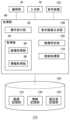
図1は、実施例にかかる医療支援システム1の構成を示す。医療支援システム1は、内視鏡検査を行う病院などの医療施設に設けられる。医療支援システム1において、サーバ装置2、画像解析装置3、画像蓄積装置8、内視鏡システム9および端末装置10bは、LAN(ローカルエリアネットワーク)などのネットワーク4を介して、通信可能に接続される。内視鏡システム9は検査室に設けられ、内視鏡観察装置5および端末装置10aを有する。医療支援システム1において、サーバ装置2、画像解析装置3および画像蓄積装置8は、医療施設の外部に、たとえばクラウドサーバとして設けられてもよい。Figure 1 shows the configuration of a
内視鏡観察装置5は、患者の消化管に挿入される内視鏡7を接続される。内視鏡7は、内視鏡観察装置5から供給される照明光を伝送して、消化管内を照明するためのライトガイドを有し、先端部には、ライトガイドにより伝送される照明光を生体組織へ出射するための照明窓と、生体組織を所定の周期で撮影して撮像信号を内視鏡観察装置5に出力する撮影部が設けられる。撮影部は、入射光を電気信号に変換する固体撮像素子(たとえばCCDイメージセンサまたはCMOSイメージセンサ)を含む。The
内視鏡観察装置5は、内視鏡7の固体撮像素子により光電変換された撮像信号に対して画像処理を施して内視鏡画像を生成し、表示装置6にリアルタイムに表示する。内視鏡観察装置5は、A/D変換、ノイズ除去などの通常の画像処理に加えて、強調表示等を目的とする特別な画像処理を実施する機能を備えてよい。内視鏡観察装置5は、内視鏡画像を所定の周期(たとえば1/60秒)で生成する。内視鏡観察装置5は、専用ハードウェアを有する1つ以上のプロセッサによって構成されてよいが、汎用ハードウェアを有する1つ以上のプロセッサによって構成されてもよい。The
医師は、検査手順にしたがって、表示装置6に表示されている内視鏡画像を観察する。医師は、内視鏡7を動かしながら内視鏡画像を観察し、病変が表示装置6に映し出されると、内視鏡7のレリーズスイッチを操作する。レリーズスイッチが操作されると、内視鏡観察装置5は、レリーズスイッチが操作されたタイミングで内視鏡画像をキャプチャ(保存)して、キャプチャした内視鏡画像を、当該内視鏡画像を識別する情報(画像ID)とともに画像蓄積装置8に送信する。なお内視鏡観察装置5は、検査終了後に、キャプチャした複数の内視鏡画像をまとめて画像蓄積装置8に送信してもよい。画像蓄積装置8は、内視鏡検査を識別する検査IDに紐付けて、内視鏡観察装置5から送信された内視鏡画像を記録する。画像蓄積装置8に蓄積される内視鏡画像は、医師による検査レポートの作成に利用される。The doctor observes the endoscopic image displayed on the
端末装置10aは、情報処理装置11aおよび表示装置12aを備えて、検査室に設けられる。端末装置10aは、医師や看護師等が内視鏡検査中に、病変に関する情報をリアルタイムに確認するために利用される。情報処理装置11aは、サーバ装置2および/または画像解析装置3から、内視鏡検査中、病変に関する情報を取得して、表示装置12aに表示する。たとえば表示装置12aには、画像解析装置3により画像解析された病変のサイズ、病変の深達度および病変の質的診断結果などが表示されてよい。The
端末装置10bは、情報処理装置11bおよび表示装置12bを備えて、検査室以外の部屋に設けられる。端末装置10bは、医師が内視鏡検査のレポートを作成する際に利用される。端末装置10a、10bは、汎用ハードウェアを有する1つ以上のプロセッサによって構成される。The
実施例の医療支援システム1において、内視鏡観察装置5は、内視鏡画像を表示装置6からリアルタイムに表示させるとともに、内視鏡画像を、当該画像のメタ情報とともに、画像解析装置3にリアルタイムに供給する。ここでメタ情報は、画像のフレーム番号、撮影時刻情報を少なくとも含み、フレーム番号は、内視鏡7が撮影を開始してから何フレーム目であるかを示す情報である。つまりフレーム番号は、撮影順を示すシリアルな番号であってよく、たとえば最初に撮影された内視鏡画像のフレーム番号は「1」、2番目に撮影された内視鏡画像のフレーム番号は「2」に設定されている。In the
画像解析装置3は内視鏡画像を解析して、内視鏡画像に含まれる病変を検出して、検出した病変を質的診断する電子計算機(コンピュータ)である。画像解析装置3はAI(artificial intelligence)診断機能を有するCAD(computer-aided diagnosis)システムであってよい。画像解析装置3は専用ハードウェアを有する1つ以上のプロセッサによって構成されてよいが、汎用ハードウェアを有する1つ以上のプロセッサによって構成されてもよい。The
画像解析装置3は、学習用の内視鏡画像および内視鏡画像に含まれる病変領域に関する情報を教師データとして用いた機械学習により生成された学習済みモデルを利用する。内視鏡画像のアノテーション作業は、医師などの専門知識を有するアノテータにより実施され、機械学習には、ディープラーニングの一種であるCNN、RNN、LSTMなどを使用してよい。この学習済みモデルは、内視鏡画像を入力すると、撮影された臓器を示す情報、撮影された部位を示す情報と、撮影された病変に関する情報(病変情報)とを出力する。画像解析装置3が出力する病変情報は、内視鏡画像に病変が含まれているか否かを示す病変有無情報を少なくとも含む。病変が含まれている場合、病変情報は、病変のサイズを示す情報、病変の輪郭の位置を示す情報、病変の形状を示す情報、病変の深達度を示す情報および病変の質的診断結果を含んでよい。病変の質的診断結果は、病変の種類を含む。内視鏡検査中、画像解析装置3は、内視鏡観察装置5から内視鏡画像をリアルタイムに提供されて、内視鏡画像ごとに、臓器を示す情報、部位を示す情報および病変情報を出力する。以下、臓器を示す情報、部位を示す情報、病変情報を、まとめて「画像メタ情報」と呼ぶ。The
実施例の画像解析装置3は、医師(以下「ユーザ」と呼ぶこともある)が病変を観察していた時間を計時する機能を有する。ユーザがレリーズスイッチを操作して内視鏡画像をキャプチャする場合、画像解析装置3は、当該内視鏡画像に含まれる病変が、それ以前の内視鏡画像に最初に含まれてから、レリーズスイッチが操作されるまでの時間を、当該病変をユーザが観察していた時間として計測する。つまり画像解析装置3は、当該病変が最初に撮影されてから、ユーザがキャプチャ操作(レリーズスイッチ操作)するまでの時間を、当該病変の観察時間として特定する。なお実施例において「撮影」は、内視鏡7の固体撮像素子が入射光を電気信号に変換する動作を意味し、「キャプチャ」は、内視鏡観察装置5が生成した内視鏡画像を保存(記録)する動作を意味する。The
ユーザがキャプチャ操作をすると、内視鏡観察装置5は、キャプチャ操作をしたことを示す情報(キャプチャ操作情報)とともに、キャプチャした内視鏡画像のフレーム番号、撮影時刻および画像IDを画像解析装置3に提供する。画像解析装置3はキャプチャ操作情報を取得すると、病変の観察時間を特定して、画像ID、フレーム番号、撮影時刻情報、病変の観察時間および提供されたフレーム番号の画像メタ情報を、サーバ装置2に提供する。サーバ装置2は、内視鏡画像の画像IDに紐付けて、フレーム番号、撮影時刻情報、病変の観察時間および画像メタ情報を記録する。When the user performs a capture operation, the
実施例の画像解析装置3は、内視鏡画像内に病変を検出すると、当該内視鏡画像を自動でキャプチャする機能を有する。なお、1つの同じ病変が10秒間分の内視鏡動画に含まれている場合、画像解析装置3は、当該病変を最初に検出したときの内視鏡画像を自動でキャプチャしてよく、後続の内視鏡画像から同じ病変を検出しても、自動キャプチャを行わなくてよい。なお画像解析装置3は、最終的に、検出した病変を含む内視鏡画像を1枚キャプチャすればよく、たとえば同じ病変を含む複数枚の内視鏡画像をキャプチャした後、最も病変が鮮明に撮影されている1枚の内視鏡画像を選択して、他のキャプチャした内視鏡画像を破棄してもよい。以下、キャプチャ主体を明確にするために、ユーザによるキャプチャ操作によりキャプチャされた内視鏡画像を「ユーザキャプチャ画像」と呼び、画像解析装置3により自動キャプチャされた内視鏡画像を「コンピュータキャプチャ画像」と呼ぶこともある。The
画像解析装置3は、コンピュータキャプチャ画像を取得すると、当該コンピュータキャプチャ画像に含まれる病変の観察時間を事後的に特定する。画像解析装置3は、病変が撮影されている時間を、当該病変の観察時間として特定してよい。たとえば当該病変が、10秒間分の動画に含まれている場合(つまり10秒間撮影されて、表示装置12aに表示されている場合)、画像解析装置3は、当該病変の観察時間を10秒と特定してよい。したがって画像解析装置3は、当該病変がフレームインして撮影開始されてから、フレームアウトして撮影終了するまでの時間を計時して、当該病変の観察時間として特定する。画像解析装置3は、コンピュータキャプチャ画像を、当該コンピュータキャプチャ画像のフレーム番号、撮影時刻情報、病変の観察時間および画像メタ情報とともに、サーバ装置2に提供する。When the
ユーザは内視鏡検査を終了すると、内視鏡観察装置5の検査終了ボタンを操作する。検査終了ボタンの操作情報は、サーバ装置2および画像解析装置3に供給されて、サーバ装置2および画像解析装置3は、当該内視鏡検査の終了を認識する。When the user finishes the endoscopic examination, he or she operates the examination end button on the
図2は、サーバ装置2の機能ブロックを示す。サーバ装置2は、通信部20、処理部30および記憶装置60を備える。通信部20は、ネットワーク4を介して、画像解析装置3、内視鏡観察装置5、画像蓄積装置8、端末装置10aおよび端末装置10bとの間でデータや指示などの情報を送受信する。処理部30は、第1情報取得部40、第2情報取得部42、画像ID設定部44および画像送信部46を備える。記憶装置60は、オーダ情報記憶部62、第1情報記憶部64および第2情報記憶部66を有する。オーダ情報記憶部62は、内視鏡検査オーダの情報を記憶する。Figure 2 shows functional blocks of the
サーバ装置2はコンピュータを備え、コンピュータがプログラムを実行することによって、図2に示す各種機能が実現される。コンピュータは、プログラムをロードするメモリ、ロードされたプログラムを実行する1つ以上のプロセッサ、補助記憶装置、その他のLSIなどをハードウェアとして備える。プロセッサは、半導体集積回路やLSIを含む複数の電子回路により構成され、複数の電子回路は、1つのチップ上に搭載されてよく、または複数のチップ上に搭載されてもよい。図2に示す機能ブロックは、ハードウェアとソフトウェアとの連携によって実現され、したがって、これらの機能ブロックがハードウェアのみ、ソフトウェアのみ、またはそれらの組合せによっていろいろな形で実現できることは、当業者には理解されるところである。The
第1情報取得部40は、画像解析装置3から、ユーザキャプチャ画像の画像ID、フレーム番号、撮影時刻情報、観察時間および画像メタ情報を取得し、ユーザキャプチャ画像であることを示す情報とともに、第1情報記憶部64に記憶する。たとえばユーザが、内視鏡検査中に7回キャプチャ操作をした場合、第1情報取得部40は、画像解析装置3から、7枚分のユーザキャプチャ画像の画像ID、フレーム番号、撮影時刻情報、観察時間および画像メタ情報を取得して、ユーザキャプチャ画像であることを示す情報とともに、第1情報記憶部64に記憶する。第1情報記憶部64は、画像IDに紐付けて、ユーザキャプチャ画像であることを示す情報、フレーム番号、撮影時刻情報、観察時間および画像メタ情報を記憶してよい。なお画像IDは、内視鏡観察装置5によってユーザキャプチャ画像に付与されており、内視鏡観察装置5は、画像IDを、撮影時刻順に1から順に付与している。したがって、この場合、7枚のユーザキャプチャ画像には、それぞれ画像ID1~7が割り当てられている。The first
第2情報取得部42は、画像解析装置3から、コンピュータキャプチャ画像、コンピュータキャプチャ画像のフレーム番号、撮影時刻情報、観察時間および画像メタ情報を取得し、コンピュータキャプチャ画像であることを示す情報とともに、第2情報記憶部66に記憶する。この時点で、コンピュータキャプチャ画像には画像IDが付与されていないため、画像ID設定部44は、撮影時刻に応じた画像IDを、コンピュータキャプチャ画像に設定してよい。具体的に画像ID設定部44は、ユーザキャプチャ画像の画像IDと重ならないように、コンピュータキャプチャ画像の画像IDを設定する。上記したように、7枚のユーザキャプチャ画像に対して、画像ID1~7が付与されている場合、画像ID設定部44は、コンピュータキャプチャ画像に対して、画像IDを、撮影時刻順に8から順に付与してよい。画像解析装置3が、7回の自動キャプチャを行った場合(つまり内視鏡検査において合計7つの病変を検出して、7枚の内視鏡画像を自動キャプチャした場合)、画像ID設定部44は、画像IDを撮影時刻の早い順に8から順に設定し、したがって、この場合、画像ID8~14が、7枚のコンピュータキャプチャ画像に対して設定される。第2情報記憶部66は、画像IDに紐付けて、コンピュータキャプチャ画像であることを示す情報、フレーム番号、撮影時刻情報、観察時間および画像メタ情報を記憶する。The second
画像送信部46は、コンピュータキャプチャ画像を、付与された画像IDとともに、画像蓄積装置8に送信する。これにより画像蓄積装置8は、内視鏡検査でキャプチャされた画像ID1~7のユーザキャプチャ画像および画像ID8~14のコンピュータキャプチャ画像の両方を蓄積する。The
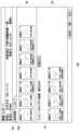
図3は、キャプチャ画像に紐付けられる情報の例を示す。画像ID1~7のユーザキャプチャ画像に関する情報は、第1情報記憶部64に記憶され、画像ID8~14のコンピュータキャプチャ画像に関する情報は、第2情報記憶部66に記憶されている。Figure 3 shows an example of information associated with a capture image. Information relating to user-captured images with
「画像種別」の項目には、ユーザキャプチャ画像であるか、コンピュータキャプチャ画像であるかを示す情報が記憶される。「臓器」の項目には、画像に含まれる臓器を示す情報、つまり撮影された臓器を示す情報が記憶され、「部位」の項目には、画像に含まれる臓器の部位を示す情報、つまり撮影された臓器の部位を示す情報が記憶される。The "Image type" field stores information indicating whether the image is a user-captured image or a computer-captured image. The "Organ" field stores information indicating the organ contained in the image, i.e., information indicating the organ that was photographed, and the "Part" field stores information indicating the part of the organ contained in the image, i.e., information indicating the part of the organ that was photographed.
病変情報のうち、「有無」の項目には、画像解析装置3により病変が検出されたか否かを示す情報が記憶される。全てのコンピュータキャプチャ画像は病変を含んでいるため、コンピュータキャプチャ画像の「有無」の項目には、「有」が記憶されている。「サイズ」の項目には、病変の底面の最長径を示す情報が記憶され、「形状」の項目には、病変の輪郭形状を表現する座標情報が記憶され、「診断」の項目には、病変の質的診断結果が記憶される。「観察時間」の項目には、画像解析装置3が導出した観察時間が記憶される。「撮影時刻」の項目には、画像の撮影時刻を示す情報が記憶される。「撮影時刻」の項目に、フレーム番号が含まれてもよい。In the lesion information, the "presence/absence" field stores information indicating whether or not a lesion has been detected by the
図4は、情報処理装置11bの機能ブロックを示す。情報処理装置11bは、レポート入力画面に表示する内視鏡画像を選別する機能を有し、通信部76、入力部78、処理部80および記憶装置120を備える。通信部76は、ネットワーク4を介して、サーバ装置2、画像解析装置3、内視鏡観察装置5、画像蓄積装置8および端末装置10aとの間でデータや指示などの情報を送受信する。処理部80は、操作受付部82、取得部84、表示画面生成部100、画像特定部102および登録処理部110を備え、取得部84は、画像取得部86および情報取得部88を有する。記憶装置120は、画像記憶部122、情報記憶部124および優先度記憶部126を有する。Figure 4 shows functional blocks of
情報処理装置11bはコンピュータを備え、コンピュータがプログラムを実行することによって、図4に示す各種機能が実現される。コンピュータは、プログラムをロードするメモリ、ロードされたプログラムを実行する1つ以上のプロセッサ、補助記憶装置、その他のLSIなどをハードウェアとして備える。プロセッサは、半導体集積回路やLSIを含む複数の電子回路により構成され、複数の電子回路は、1つのチップ上に搭載されてよく、または複数のチップ上に搭載されてもよい。図4に示す機能ブロックは、ハードウェアとソフトウェアとの連携によって実現され、したがって、これらの機能ブロックがハードウェアのみ、ソフトウェアのみ、またはそれらの組合せによっていろいろな形で実現できることは、当業者には理解されるところである。The
内視鏡検査の終了後、医師であるユーザは情報処理装置11bにユーザIDおよびパスワードを入力して、ログインする。ユーザがログインすると、検査レポートを作成するためのアプリケーションが起動して、表示装置12bには、実施済み検査の一覧が表示される。この実施済み検査一覧には、患者名、患者ID、検査日時、検査項目などの検査情報がリスト表示され、ユーザは、マウスやキーボードなどの入力部78を操作して、レポート作成の対象となる検査を選択する。操作受付部82が、検査の選択操作を受け付けると、画像取得部86が、画像蓄積装置8から、選択された検査の検査IDに紐付けられている複数の内視鏡画像を取得して、画像記憶部122に記憶する。After the endoscopic examination is completed, the doctor user logs in by entering a user ID and password into the
実施例において画像取得部86は、ユーザによるキャプチャ操作によりキャプチャされた画像ID1~7のユーザキャプチャ画像と、病変を含んで画像解析装置3によりキャプチャされた画像ID8~14のコンピュータキャプチャ画像とを取得して、画像記憶部122に記憶する。表示画面生成部100は、レポートに添付する内視鏡画像を選択するための選択画面を生成して、表示装置12bに表示する。In the embodiment, the
上記したようにコンピュータキャプチャ画像は、画像解析装置3が検出した病変の数だけ存在する。実施例では、7枚の内視鏡画像が画像解析装置3により自動キャプチャされているが、内視鏡検査によっては、画像解析装置3が数十~数百の病変を検出して、数十~数百枚の内視鏡画像を自動キャプチャすることも想定される。そのような場合に、自動キャプチャされた全ての内視鏡画像を選択画面に表示すると、ユーザが選択する手間が大きく、好ましくない。そこで実施例では画像特定部102が、複数のコンピュータキャプチャ画像の中から、選択画面に表示するコンピュータキャプチャ画像を絞り込む。以下、選択画面に表示するコンピュータキャプチャ画像を、「添付候補画像」と呼ぶ。As described above, there are as many computer-captured images as the number of lesions detected by the
情報取得部88は、サーバ装置2の記憶装置60から、キャプチャ画像に紐付けられた情報を取得して、情報記憶部124に記憶する。画像特定部102は、キャプチャ画像に紐付けられた情報を参照して、ユーザキャプチャ画像に含まれていない病変を含むコンピュータキャプチャ画像を、「添付候補画像」として特定する。画像特定部102が、ユーザキャプチャ画像に含まれていない病変を含むコンピュータキャプチャ画像を添付候補画像として特定することで、ユーザキャプチャ画像に含まれる同じ病変を含むコンピュータキャプチャ画像が、重複して選択画面に表示されないようになる。The
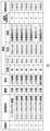
図5は、レポートに添付する内視鏡画像を選択するための選択画面の例を示す。選択画面は、レポート入力画面の一部を構成する。内視鏡画像の選択画面は、記録画像タブ54aが選択された状態で、表示装置12bに表示される。選択画面の上段には、患者氏名、患者ID、生年月日、検査項目、検査日、実施医の情報が表示される。これらの情報は検査オーダ情報に含まれており、サーバ装置2から取得されてよい。Figure 5 shows an example of a selection screen for selecting endoscopic images to attach to a report. The selection screen forms part of the report input screen. The selection screen for endoscopic images is displayed on the
表示画面生成部100は、ユーザキャプチャ画像と、絞り込まれたコンピュータキャプチャ画像(添付候補画像)とを含む選択画面を生成して、表示装置12bに表示する。コンピュータキャプチャ画像(添付候補画像)に含まれる病変は、ユーザキャプチャ画像に含まれる病変と重複していないため、ユーザは、レポートに添付する画像を効率的に選択できる。The display
表示画面生成部100は、ユーザキャプチャ画像を表示する第1領域50と、コンピュータキャプチャ画像(添付候補画像)を表示する第2領域52とを別個に設けた選択画面を生成して、表示装置12bに表示する。表示画面生成部100は、第1領域50に、画像ID1~7のユーザキャプチャ画像を撮影順序にしたがって並べて表示し、第2領域52に、画像ID8,10,13,14のコンピュータキャプチャ画像を撮影順序にしたがって並べて表示する。表示装置12bが、ユーザキャプチャ画像と、添付候補画像とを同一画面に表示することで、ユーザは、レポートに添付する画像を効率的に選択できる。The display
以下、コンピュータキャプチャ画像を絞り込む処理について説明する。
画像特定部102は、情報記憶部124に記憶したキャプチャ画像に関する情報(図3参照)にもとづいて、ユーザキャプチャ画像に含まれていない病変を含むコンピュータキャプチャ画像を、「添付候補画像」として特定する。画像特定部102は、画像ID1~7のユーザキャプチャ画像に含まれる臓器を示す情報と、コンピュータキャプチャ画像に含まれる臓器を示す情報にもとづいて、コンピュータキャプチャ画像に含まれる臓器と同じ臓器を含むユーザキャプチャ画像があるか否かを判定し、コンピュータキャプチャ画像に含まれる臓器と同じ臓器を含むユーザキャプチャ画像がない場合、当該コンピュータキャプチャ画像を、添付候補画像として特定してよい。コンピュータキャプチャ画像に含まれる臓器と同じ臓器を含むユーザキャプチャ画像がない場合、当該コンピュータキャプチャ画像に含まれる病変は、ユーザキャプチャ画像に含まれていないことが確実であるため、画像特定部102は、当該コンピュータキャプチャ画像を、添付候補画像として特定してよい。 The process of narrowing down computer captured images will be described below.
The
画像特定部102は、画像ID1~7のユーザキャプチャ画像に含まれる臓器の部位を示す情報と、コンピュータキャプチャ画像に含まれる臓器の部位を示す情報にもとづいて、コンピュータキャプチャ画像に含まれる部位と同じ部位を含むユーザキャプチャ画像があるか否かを判定し、コンピュータキャプチャ画像に含まれる部位と同じ部位を含むユーザキャプチャ画像がない場合、当該コンピュータキャプチャ画像を、添付候補画像として特定してよい。コンピュータキャプチャ画像に含まれる部位と同じ部位を含むユーザキャプチャ画像がない場合、当該コンピュータキャプチャ画像に含まれる病変は、ユーザキャプチャ画像に含まれていないことが確実であるため、画像特定部102は、当該コンピュータキャプチャ画像を、添付候補画像として特定してよい。The
画像特定部102は、画像ID1~7のユーザキャプチャ画像に含まれる病変のサイズを示す情報と、コンピュータキャプチャ画像に含まれる病変のサイズを示す情報にもとづいて、コンピュータキャプチャ画像に含まれる病変のサイズと実質的に同じサイズの病変を含むユーザキャプチャ画像があるか否かを判定し、コンピュータキャプチャ画像に含まれる病変のサイズと実質的に同じサイズの病変を含むユーザキャプチャ画像がない場合、当該コンピュータキャプチャ画像を、添付候補画像として特定してよい。コンピュータキャプチャ画像に含まれる病変のサイズと同じサイズの病変を含むユーザキャプチャ画像がない場合、当該コンピュータキャプチャ画像に含まれる病変は、ユーザキャプチャ画像に含まれていないことが確実であるため、画像特定部102は、当該コンピュータキャプチャ画像を、添付候補画像として特定してよい。The
画像特定部102は、画像ID1~7のユーザキャプチャ画像に含まれる病変の形状を示す情報と、コンピュータキャプチャ画像に含まれる病変の形状を示す情報にもとづいて、コンピュータキャプチャ画像に含まれる病変の形状と実質的に同じ形状の病変を含むユーザキャプチャ画像があるか否かを判定し、コンピュータキャプチャ画像に含まれる病変の形状と実質的に同じ形状の病変を含むユーザキャプチャ画像がない場合、当該コンピュータキャプチャ画像を、添付候補画像として特定してよい。コンピュータキャプチャ画像に含まれる病変の形状と同じ形状の病変を含むユーザキャプチャ画像がない場合、当該コンピュータキャプチャ画像に含まれる病変は、ユーザキャプチャ画像に含まれていないことが確実であるため、画像特定部102は、当該コンピュータキャプチャ画像を、添付候補画像として特定してよい。The
画像特定部102は、画像ID1~7のユーザキャプチャ画像に含まれる病変の種類を示す情報と、コンピュータキャプチャ画像に含まれる病変の種類を示す情報にもとづいて、コンピュータキャプチャ画像に含まれる病変の種類と実質的に同じ種類の病変を含むユーザキャプチャ画像があるか否かを判定し、コンピュータキャプチャ画像に含まれる病変の種類と実質的に同じ種類の病変を含むユーザキャプチャ画像がない場合、当該コンピュータキャプチャ画像を、添付候補画像として特定してよい。コンピュータキャプチャ画像に含まれる病変の種類と同じ種類の病変を含むユーザキャプチャ画像がない場合、当該コンピュータキャプチャ画像に含まれる病変は、ユーザキャプチャ画像に含まれていないことが確実であるため、画像特定部102は、当該コンピュータキャプチャ画像を、添付候補画像として特定してよい。The
画像特定部102は、画像ID1~7のユーザキャプチャ画像に含まれる臓器を示す情報、部位を示す情報、病変のサイズを示す情報、病変の形状を示す情報、病変の種類を示す情報と、コンピュータキャプチャ画像に含まれる臓器を示す情報、部位を示す情報、病変のサイズを示す情報、病変の形状を示す情報、病変の種類を示す情報とにもとづいて、コンピュータキャプチャ画像に含まれる臓器、部位、病変と実質的に同じ臓器、部位、病変を含むユーザキャプチャ画像があるか否かを判定し、コンピュータキャプチャ画像に含まれる臓器、部位、病変と実質的に同じ臓器、部位、病変を含むユーザキャプチャ画像がない場合、当該コンピュータキャプチャ画像を、添付候補画像として特定してよい。コンピュータキャプチャ画像に含まれる臓器、部位、病変と実質的に同じ臓器、部位、病変を含むユーザキャプチャ画像がない場合、当該コンピュータキャプチャ画像に含まれる病変は、ユーザキャプチャ画像に含まれていないことが確実であるため、画像特定部102は、当該コンピュータキャプチャ画像を、添付候補画像として特定してよい。The
以上のように画像特定部102は、画像ID1~7のユーザキャプチャ画像に含まれていない病変を含むコンピュータキャプチャ画像を、添付候補画像として特定する。図3を参照して、画像特定部102は、画像ID9のコンピュータキャプチャ画像に含まれる病変と画像ID3のユーザキャプチャ画像に含まれる病変は同一であり、画像ID11のコンピュータキャプチャ画像に含まれる病変と画像ID5のユーザキャプチャ画像に含まれる病変は同一であり、画像ID12のコンピュータキャプチャ画像に含まれる病変と画像ID6のユーザキャプチャ画像に含まれる病変は同一であることを判定する。したがって画像特定部102は、画像ID9,11,12のコンピュータキャプチャ画像を選択画面に含めないことを決定し、画像ID8,10,13,14のコンピュータキャプチャ画像を、添付候補画像として決定する。以上の絞り込みが行われた後、表示画面生成部100は、第1領域50に、画像ID1~7のユーザキャプチャ画像を表示し、第2領域52に、画像ID8,10,13,14のコンピュータキャプチャ画像を表示する。As described above, the
選択画面に表示される内視鏡画像にはチェックボックスが設けられ、ユーザがマウスを操作してマウスポインタをチェックボックスに配置し右クリックすると、当該内視鏡画像がレポートの添付画像として選択される。選択画面において、操作受付部82は、ユーザがユーザキャプチャ画像または添付候補画像を選択する操作を受け付ける。ユーザは、マウスポインタを内視鏡画像上に配置して右クリックすると、当該内視鏡画像を拡大表示させることができ、拡大表示された内視鏡画像を見て、レポートに添付するべきか判断してよい。A check box is provided on the endoscopic image displayed on the selection screen, and when the user operates the mouse to place the mouse pointer on the check box and right-clicks, the endoscopic image is selected as an image to be attached to the report. On the selection screen, the
図5に示す例では、画像ID3のユーザキャプチャ画像と、画像ID13,14のコンピュータキャプチャ画像のチェックボックスに、選択されたことを示すチェックマークが表示されている。ユーザが入力部78を用いて一時保存ボタンを操作すると、登録処理部110が、選択した画像ID3,13,14の内視鏡画像を、レポート添付画像として画像記憶部122に仮登録する。ユーザは添付画像を選択した後、レポートタブ54bを選択して、レポート入力画面を表示装置12bに表示させる。In the example shown in Figure 5, check marks are displayed in the check boxes for the user-captured image with
図6は、検査結果を入力するためのレポート作成画面の一例を示す。レポート作成画面は、レポート入力画面の一部を構成する。レポートタブ54bが選択されると、表示画面生成部100は、レポート作成画面を生成して、表示装置12bに表示する。レポート作成画面は、2つの領域で構成され、左側に添付画像を表示する添付画像表示領域56が、右側にユーザが検査結果を入力するための入力領域58が配置される。この例では画像ID3,13,14の内視鏡画像が添付画像として選択されて、添付画像表示領域56に表示されている。Figure 6 shows an example of a report creation screen for inputting test results. The report creation screen constitutes part of the report input screen. When the
図5に示す第2領域52において、表示されるコンピュータキャプチャ画像の枚数には、上限が設定されていてよい。第2領域52に表示されるコンピュータキャプチャ画像は、画像解析装置3が自動キャプチャしたものであり、ユーザがキャプチャしたものではないため、表示枚数が多すぎると、ユーザが選択する手間が増える。そこで画像特定部102は、ユーザキャプチャ画像に含まれていない病変を含むコンピュータキャプチャ画像の数が所定の第1上限数を超えている場合に、病変の種類に応じて設定された優先順位にもとづいて、第1上限数以下のコンピュータキャプチャ画像を、添付候補画像として特定してよい。5, an upper limit may be set for the number of computer-captured images displayed in the
図7は、優先度記憶部126に記憶されたテーブルの例を示す。優先度記憶部126は、病変の種類を示す質的診断結果と、優先順位との対応関係を定めたテーブルを記憶する。この例では、大腸の質的診断(病変の種類)に関して、大腸がんの優先順位が1位、悪性ポリープの優先順位が2位、悪性黒色腫の優先順位が3位、非腫瘍性ポリープの優先順位が4位と定められている。以下、第2領域52に表示する表示枚数の上限が、3枚に設定されている(第1上限数=3)場合について説明する。Figure 7 shows an example of a table stored in the
実施例では、画像ID8,10,13,14のコンピュータキャプチャ画像が、ユーザキャプチャ画像に含まれていない病変を含む画像として特定されている。以下に、各コンピュータキャプチャ画像の質的診断結果を示す。
画像ID8 :悪性黒色腫瘍
画像ID10:非腫瘍性ポリープ
画像ID13:悪性ポリープ
画像ID14:悪性黒色腫瘍 In the example, computer-captured images with
Image ID 8: Malignant melanoma Image ID 10: Non-neoplastic polyp Image ID 13: Malignant polyp Image ID 14: Malignant melanoma
画像特定部102は、病変の種類に応じて設定された優先順位にもとづいて、3枚以下のコンピュータキャプチャ画像を、添付候補画像として特定する。実施例では、ユーザキャプチャ画像に含まれていない病変を含むコンピュータキャプチャ画像の数が4枚であり、第1上限数(3枚)を超えているため、画像特定部102は、優先順位にもとづいて3枚以下のコンピュータキャプチャ画像に絞り込む必要がある。以下に、各コンピュータキャプチャ画像の質的診断結果に対応する優先順位を示す。The
画像ID8 :悪性黒色腫瘍 3位
画像ID10:非腫瘍性ポリープ 4位
画像ID13:悪性ポリープ 2位
画像ID14:悪性黒色腫瘍 3位 Image ID 8: Malignant melanoma 3rd place Image ID 10: Non-neoplastic polyp 4th place Image ID 13: Malignant polyp 2nd place Image ID 14: Malignant melanoma 3rd place
画像特定部102は、各画像の質的診断結果に対応する優先順位にもとづき、画像ID10のコンピュータキャプチャ画像を外して、画像ID8,13,14のコンピュータキャプチャ画像を、添付候補画像として特定してよい。このように第2領域52に表示する表示枚数の上限が設定され、質的診断に応じて設定された優先順位にもとづいて添付候補画像が選択されることで、表示画面生成部100は、重要な病変を含むコンピュータキャプチャ画像を優先的に第2領域52に表示することが可能となる。The
なお上記のように、優先順位にもとづいて添付候補画像を特定した場合であっても、添付候補画像の数が、第1上限数を超えることも生じうる。たとえば第2領域52に表示する表示枚数の上限が、2枚に設定されている(第1上限数=2)場合、上記の例では、3枚の添付候補画像が特定されており、依然として第1上限数を超えていることになる。そこで画像特定部102は、さらに観察時間を加味して、添付候補画像の数が第1上限数以下となるように、コンピュータキャプチャ画像を選択してよい。Even when candidate images to be attached are identified based on priority as described above, the number of candidate images to be attached may exceed the first upper limit. For example, if the upper limit of the number of images to be displayed in the
まず画像特定部102は、ユーザキャプチャ画像に含まれていない病変を含むコンピュータキャプチャ画像の数が第1上限数(2枚)を超えている場合に、優先順位にもとづいて、添付候補画像の候補となるコンピュータキャプチャ画像を特定する。ここでは、画像ID8,13,14のコンピュータキャプチャ画像が、添付候補画像の候補として特定される。添付候補画像の候補として特定したコンピュータキャプチャ画像の数(3枚)が第1上限数を超えている場合、画像特定部102は、ユーザが観察した時間の短いコンピュータキャプチャ画像を添付候補画像の候補から外して、第1上限数以下のコンピュータキャプチャ画像を、添付候補画像として特定してよい。First, when the number of computer capture images including a lesion not included in the user capture image exceeds a first upper limit (two images), the
画像ID13の画像の優先順位は2位であり、画像ID8,14の画像の優先順位は3位であるため、画像特定部102は、画像ID13の画像が添付候補画像であることを確定した後、画像ID8,14の画像の観察時間を比較する。ここで画像ID8の画像の観察時間は16秒、画像ID14の画像の観察時間は12秒である。観察時間が長い方が、検査中にユーザが注目していたことが予想されるため、画像特定部102は、観察時間の短い画像ID14のコンピュータキャプチャ画像を添付候補画像の候補から外し、画像ID8のコンピュータキャプチャ画像を添付候補画像として特定する。したがって画像特定部102は、画像ID8,13のコンピュータキャプチャ画像を、添付候補画像として特定してよい。Because the image with
図8は、レポートに添付する内視鏡画像を選択するための選択画面の別の例を示す。図5に示す選択画面と比較すると、第2領域52に表示されるコンピュータキャプチャ画像の枚数が2枚に制限されている。第2領域52の表示枚数を制限することで、ユーザは、レポートに添付する画像を効率的に選択できる。なお第2領域52に、コンピュータキャプチャ画像を全数表示するための切替ボタン70が設けられてもよい。Figure 8 shows another example of a selection screen for selecting endoscopic images to attach to a report. Compared to the selection screen shown in Figure 5, the number of computer-captured images displayed in the
図9は、コンピュータキャプチャ画像を全て表示したときの選択画面の例を示す。切替ボタン70は、コンピュータキャプチャ画像を制限した数だけ表示するモードと、コンピュータキャプチャ画像を全数表示するモードとを切り替えるために使用される。ユーザは全てのコンピュータキャプチャ画像を第2領域52に表示させることで、検出された全ての病変を含む内視鏡画像を見ることができる。Figure 9 shows an example of a selection screen when all computer-captured images are displayed. The
なお表示画面生成部100は、第1表示モードで、ユーザキャプチャ画像と添付候補画像とを同一画面に表示し、第2表示モードで、添付候補画像のみを表示し、ユーザキャプチャ画像を表示しなくてもよい。このモード切替は、切替ボタン70とは別の操作ボタンによって実施されてよい。表示画面生成部100が、様々なモードで選択画面を生成することで、ユーザは、自分が望むキャプチャ画像を含む選択画面から、レポートに添付する画像を選択できるようになる。In addition, the display
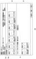
図10は、キャプチャ画像に紐付けられる情報の別の例を示す。この例では、画像ID1のユーザキャプチャ画像に関する情報が、第1情報記憶部64に記憶され、画像ID2~7のコンピュータキャプチャ画像に関する情報が、第2情報記憶部66に記憶されている。Figure 10 shows another example of information associated with a capture image. In this example, information about the user-captured image with
図10を参照すると、画像ID1のユーザキャプチャ画像と画像ID2~5のコンピュータキャプチャ画像が、大腸の上行結腸の非腫瘍性ポリープを含んでいる。画像特定部102は、同じ部位の所定の種類の病変(実施例では、非腫瘍性ポリープ)を含む1枚以上のユーザキャプチャ画像と、1枚以上のコンピュータキャプチャ画像との合計枚数が所定の第2上限数を超えている場合に、当該1枚以上のコンピュータキャプチャ画像を、添付候補画像として特定しない。10, the user-captured image with
同じ部位の非腫瘍性ポリープを含むキャプチャ画像が第2上限数(たとえば4枚)を超える場合、ユーザが、意図的に非腫瘍性ポリープをキャプチャしなかったことが想定される。このような場合に、ユーザがキャプチャしていない画像を、添付候補画像として表示することは好ましくないため、画像特定部102は、同じ部位の所定の種類の病変(非腫瘍性ポリープ)を含むユーザキャプチャ画像とコンピュータキャプチャ画像との合計枚数が所定の第2上限数を超えている場合に、コンピュータキャプチャ画像を、添付候補画像として選択しない。図10の例で、画像特定部102は、画像ID6,7のコンピュータ画像を添付候補画像として特定し、画像ID2~5のコンピュータキャプチャ画像を、添付候補画像として特定しないことが好ましい。If the number of captured images including non-neoplastic polyps in the same location exceeds the second upper limit (e.g., four), it is assumed that the user intentionally did not capture non-neoplastic polyps. In such a case, since it is not preferable to display images not captured by the user as candidate images to be attached, the
以上、本開示を複数の実施例をもとに説明した。これらの実施例は例示であり、それらの各構成要素や各処理プロセスの組合せにいろいろな変形例が可能なこと、またそうした変形例も本開示の範囲にあることは当業者に理解されるところである。実施例では、内視鏡観察装置5がユーザキャプチャ画像を画像蓄積装置8に送信しているが、変形例では、画像解析装置3がユーザキャプチャ画像を画像蓄積装置8に送信してもよい。また実施例では情報処理装置11bが画像特定部102を有しているが、変形例ではサーバ装置2が画像特定部102を有してもよい。The present disclosure has been described above based on a number of examples. These examples are merely illustrative, and those skilled in the art will understand that various modifications are possible in the combination of each of the components and each of the processing processes, and that such modifications are also within the scope of the present disclosure. In the examples, the
本開示は、レポートの作成を支援する技術分野に利用できる。This disclosure can be used in the technical field of assisting in the creation of reports.
1・・・医療支援システム、2・・・サーバ装置、3・・・画像解析装置、4・・・ネットワーク、5・・・内視鏡観察装置、6・・・表示装置、7・・・内視鏡、8・・・画像蓄積装置、9・・・内視鏡システム、10a,10b・・・端末装置、11a,11b・・・情報処理装置、12a,12b・・・表示装置、20・・・通信部、30・・・処理部、40・・・第1情報取得部、42・・・第2情報取得部、44・・・画像ID設定部、46・・・画像送信部、50・・・第1領域、52・・・第2領域、54a・・・記録画像タブ、54b・・・レポートタブ、56・・・添付画像表示領域、58・・・入力領域、60・・・記憶装置、62・・・オーダ情報記憶部、64・・・第1情報記憶部、66・・・第2情報記憶部、70・・・切替ボタン、76・・・通信部、78・・・入力部、80・・・処理部、82・・・操作受付部、84・・・取得部、86・・・画像取得部、88・・・情報取得部、100・・・表示画面生成部、102・・・画像特定部、110・・・登録処理部、120・・・記憶装置、122・・・画像記憶部、124・・・情報記憶部、126・・・優先度記憶部。1: medical support system, 2: server device, 3: image analysis device, 4: network, 5: endoscopic observation device, 6: display device, 7: endoscope, 8: image storage device, 9: endoscopic system, 10a, 10b: terminal device, 11a, 11b: information processing device, 12a, 12b: display device, 20: communication unit, 30: processing unit, 40: first information acquisition unit, 42: second information acquisition unit, 44: image ID setting unit, 46: image transmission unit, 50: first area, 52: second area, 54a: recorded image tab, 54b ...Report tab, 56...attached image display area, 58...input area, 60...storage device, 62...order information storage unit, 64...first information storage unit, 66...second information storage unit, 70...switching button, 76...communication unit, 78...input unit, 80...processing unit, 82...operation reception unit, 84...acquisition unit, 86...image acquisition unit, 88...information acquisition unit, 100...display screen generation unit, 102...image identification unit, 110...registration processing unit, 120...storage device, 122...image storage unit, 124...information storage unit, 126...priority storage unit.
Claims (20)
Translated fromJapanese前記1つ以上のプロセッサは、
ユーザによるキャプチャ操作によりキャプチャされた第1画像と、病変を含んでコンピュータによりキャプチャされたコンピュータキャプチャ画像とを取得し、
前記第1画像に含まれていない病変を含む前記コンピュータキャプチャ画像を、第2画像として特定し、
レポートに添付する画像を選択するための選択画面であって、前記第1画像と前記第2画像とを含む前記選択画面を生成する、
ことを特徴とする医療支援システム。 1. A medical support system, comprising: one or more processors having hardware;
The one or more processors:
Obtaining a first image captured by a user's capture operation and a computer-captured image including the lesion and captured by a computer;
identifying the computer-captured image containing a lesion not included in the first image as a second image;
generating a selection screen for selecting an image to be attached to a report, the selection screen including the first image and the second image;
A medical support system comprising:
前記選択画面において、ユーザが前記第1画像または前記第2画像を選択する操作を受け付ける、
ことを特徴とする請求項1に記載の医療支援システム。 The one or more processors:
accepting an operation by a user to select the first image or the second image on the selection screen;
2. The medical support system according to claim 1.
前記第1画像に含まれる臓器を示す情報と、前記コンピュータキャプチャ画像に含まれる臓器を示す情報にもとづいて、前記コンピュータキャプチャ画像に含まれる臓器と同じ臓器を含む前記第1画像があるか否かを判定し、
前記コンピュータキャプチャ画像に含まれる臓器と同じ臓器を含む前記第1画像がない場合、前記コンピュータキャプチャ画像を、前記第2画像として特定する、
ことを特徴とする請求項1に記載の医療支援システム。 The one or more processors:
determining whether or not there is a first image including an organ that is the same as an organ included in the computer-captured image, based on information indicating an organ included in the first image and information indicating an organ included in the computer-captured image;
identifying the computer-captured image as the second image if there is no first image that includes the same organ as the organ included in the computer-captured image;
2. The medical support system according to claim 1.
前記第1画像に含まれる臓器の部位を示す情報と、前記コンピュータキャプチャ画像に含まれる臓器の部位を示す情報にもとづいて、前記コンピュータキャプチャ画像に含まれる部位と同じ部位を含む前記第1画像があるか否かを判定し、
前記コンピュータキャプチャ画像に含まれる部位と同じ部位を含む前記第1画像がない場合、前記コンピュータキャプチャ画像を、前記第2画像として特定する、
ことを特徴とする請求項1に記載の医療支援システム。 The one or more processors:
determining whether or not there is a first image including a part that is the same as a part included in the computer-captured image, based on information indicating a part of an organ included in the first image and information indicating a part of an organ included in the computer-captured image;
identifying the computer-captured image as the second image if there is no first image that includes the same part as the part included in the computer-captured image;
2. The medical support system according to claim 1.
前記第1画像に含まれる病変のサイズを示す情報と、前記コンピュータキャプチャ画像に含まれる病変のサイズを示す情報にもとづいて、前記コンピュータキャプチャ画像に含まれる病変のサイズと同じサイズの病変を含む前記第1画像があるか否かを判定し、
前記コンピュータキャプチャ画像に含まれる病変のサイズと同じサイズの病変を含む前記第1画像がない場合、前記コンピュータキャプチャ画像を、前記第2画像として特定する、
ことを特徴とする請求項1に記載の医療支援システム。 The one or more processors:
determining whether or not there is a first image including a lesion having the same size as the lesion included in the computer-captured image based on information indicating a size of the lesion included in the first imageand information indicating a size of the lesion included in the computer-captured image;
identifying the computer-captured image as the second image if no first image contains a lesion ofthe same size as the lesion contained in the computer-captured image;
2. The medical support system according to claim 1.
前記第1画像に含まれる病変の形状を示す情報と、前記コンピュータキャプチャ画像に含まれる病変の形状を示す情報にもとづいて、前記コンピュータキャプチャ画像に含まれる病変の形状と同じ形状の病変を含む前記第1画像があるか否かを判定し、
前記コンピュータキャプチャ画像に含まれる病変の形状と同じ形状の病変を含む前記第1画像がない場合、前記コンピュータキャプチャ画像を、前記第2画像として特定する、
ことを特徴とする請求項1に記載の医療支援システム。 The one or more processors:
determining whether or not there is a first image including a lesion having thesame shape as the shape of the lesion included in the computer-captured image based on information indicating the shape of the lesion included in the first image and information indicating the shape of the lesion included in the computer-captured image;
identifying the computer-captured image as the second image if there is no first image that includes a lesion havingthe same shape as the lesion included in the computer-captured image;
2. The medical support system according to claim 1.
前記第1画像に含まれる病変の種類を示す情報と、前記コンピュータキャプチャ画像に含まれる病変の種類を示す情報にもとづいて、前記コンピュータキャプチャ画像に含まれる病変の種類と同じ種類の病変を含む前記第1画像があるか否かを判定し、
前記コンピュータキャプチャ画像に含まれる病変の種類と同じ種類の病変を含む前記第1画像がない場合、前記コンピュータキャプチャ画像を、前記第2画像として特定する、
ことを特徴とする請求項1に記載の医療支援システム。 The one or more processors:
determining whether or not there is a first image including a lesion of the same type as the lesion included in the computer-captured image based on information indicating the type of lesion included in the first image and information indicating the type of lesion included in the computer-captured image;
identifying the computer-captured image as the second image if there is no first image containing a lesion of the same type as the lesion contained in the computer-captured image;
2. The medical support system according to claim 1.
前記第1画像に含まれる臓器を示す情報、部位を示す情報、病変のサイズを示す情報、病変の形状を示す情報および病変の種類を示す情報と、前記コンピュータキャプチャ画像に含まれる臓器を示す情報、部位を示す情報、病変のサイズを示す情報、病変の形状を示す情報および病変の種類を示す情報とにもとづいて、前記コンピュータキャプチャ画像に含まれる臓器、部位、病変と同じ臓器、部位、病変を含む前記第1画像があるか否かを判定し、
前記コンピュータキャプチャ画像に含まれる臓器、部位、病変と同じ臓器、部位、病変を含む前記第1画像がない場合、前記コンピュータキャプチャ画像を、前記第2画像として特定する、
ことを特徴とする請求項1に記載の医療支援システム。 The one or more processors:
determining whether or not there is a first image including an organ, a site,and a lesion that are the same as those included in the computer-captured image, based on information included in the first image that indicates an organ, a site, a size of a lesion, a shape of a lesion,and a type of a lesion, and information included in the computer-captured image that indicates an organ, a site, a lesionthat are the same as those included in the computer-captured image;
identifying the computer-captured image as the second image if there is no first image that includesthe same organ, site, or lesion as the organ, site, or lesion included in the computer-captured image;
2. The medical support system according to claim 1.
前記第1画像に含まれていない病変を含む前記コンピュータキャプチャ画像の数が所定の第1上限数を超えている場合に、病変の種類に応じて設定された優先順位にもとづいて、前記第1上限数以下の前記コンピュータキャプチャ画像を、前記第2画像として特定する、
ことを特徴とする請求項1に記載の医療支援システム。 The one or more processors:
when the number of the computer-captured images including a lesion not included in the first image exceeds a predetermined first upper limit number, identifying the computer-captured images not exceeding the first upper limit number as the second images based on a priority order set according to the type of lesion;
2. The medical support system according to claim 1.
前記コンピュータキャプチャ画像に含まれる病変をユーザが観察した時間を示す情報を取得し、
前記優先順位にもとづいて、前記第2画像の候補となる前記コンピュータキャプチャ画像を特定しても、前記第2画像の候補の数が前記第1上限数を超えている場合に、ユーザが観察した時間の短い前記コンピュータキャプチャ画像を前記第2画像の候補から外して、前記第1上限数以下の前記コンピュータキャプチャ画像を、前記第2画像として特定する、
ことを特徴とする請求項9に記載の医療支援システム。 The one or more processors:
obtaining information indicative of a time at which a user observed a lesion included in the computer-captured image;
when the number of the candidate second images exceeds the first upper limit numbereven if the computer-captured images to be the candidates for the second image are identified based on the priority order, the computer-captured images that have been observed for a shorttime by the user are removed from the candidates for the second image, and the computer-captured images that are equal to or less than the first upper limit number are identified as the second images;
The medical support system according to claim 9 .
同じ部位の所定の種類の病変を含む1枚以上の前記第1画像と、1枚以上の前記コンピュータキャプチャ画像との合計枚数が所定の第2上限数を超えている場合に、前記コンピュータキャプチャ画像を、前記第2画像として特定しない、
ことを特徴とする請求項1に記載の医療支援システム。 The one or more processors:
When a total number of the one or more first images including a predetermined type of lesion in the same site and the one or more computer-captured images exceeds a predetermined second upper limit number, the computer-captured images are not identified as the second images.
2. The medical support system according to claim 1.
前記第1画像を表示する第1領域と、前記第2画像を表示する第2領域とを別個に設けた前記選択画面を生成する、
ことを特徴とする請求項1に記載の医療支援システム。 The one or more processors:
generating the selection screen having a first area for displaying the first image and a second area for displaying the second image, the selection screen being provided separately;
2. The medical support system according to claim 1.
前記第1画像と前記第2画像とを、同一画面に表示する、
ことを特徴とする請求項12に記載の医療支援システム。 The one or more processors:
Displaying the first image and the second image on the same screen.
The medical support system according to claim 12 .
第1表示モードで、前記第1画像と前記第2画像とを同一画面に表示し、
第2表示モードで、前記第2画像を表示し、前記第1画像を表示しない、
ことを特徴とする請求項1に記載の医療支援システム。 The one or more processors:
In a first display mode, the first image and the second image are displayed on the same screen;
In a second display mode, the second image is displayed and the first image is not displayed.
2. The medical support system according to claim 1.
病変を含んでコンピュータによりキャプチャされたコンピュータキャプチャ画像を取得し、
前記第1画像に含まれていない病変を含む前記コンピュータキャプチャ画像を、第2画像として特定し、
レポートに添付する画像を選択するための選択画面であって、前記第1画像と前記第2画像とを含む前記選択画面を生成する、
ことを特徴とするレポート作成支援方法。 acquiring a first image captured by a user's capture operation;
obtaining a computer-captured image including the lesion;
identifying the computer-captured image containing a lesion not included in the first image as a second image;
generating a selection screen for selecting an image to be attached to a report, the selection screen including the first image and the second image;
A report creation support method comprising:
ことを特徴とする請求項15に記載のレポート作成支援方法。16. The report creation support method according to claim 15.
ユーザによるキャプチャ操作によりキャプチャされた第1画像を取得する機能と、
病変を含んで別のコンピュータによりキャプチャされたコンピュータキャプチャ画像を取得する機能と、
前記第1画像に含まれていない病変を含む前記コンピュータキャプチャ画像を、第2画像として特定する機能と、
レポートに添付する画像を選択するための選択画面であって、前記第1画像と前記第2画像とを含む前記選択画面を生成する機能と、
を実現させるためのプログラム。 On the computer,
A function of acquiring a first image captured by a user's capture operation;
acquiring a computer-captured image including the lesion and captured by a separate computer;
identifying the computer-captured image as a second image that includes a lesion not included in the first image;
generating a selection screen for selecting an image to be attached to a report, the selection screen including the first image and the second image;
A program to achieve this.
前記選択画面において、ユーザが前記第1画像または前記第2画像を選択する操作を受け付ける機能を実現させる、realizing a function of accepting an operation by a user to select the first image or the second image on the selection screen;
ことを特徴とする請求項17に記載のプログラム。18. The program according to claim 17 .
前記1つ以上のプロセッサは、The one or more processors:
ユーザによるキャプチャ操作によりキャプチャされた第1画像と、病変を含んでコンピュータによりキャプチャされたコンピュータキャプチャ画像とを取得し、Obtaining a first image captured by a user's capture operation and a computer-captured image including the lesion and captured by a computer;
前記第1画像に含まれていない病変を含む前記コンピュータキャプチャ画像を、第2画像として特定し、identifying the computer-captured image containing a lesion not included in the first image as a second image;
レポートに添付する画像を選択するための選択画面であって、前記第1画像と前記第2画像とを含む前記選択画面を生成する、generating a selection screen for selecting an image to be attached to a report, the selection screen including the first image and the second image;
ことを特徴とする情報処理装置。23. An information processing apparatus comprising:
前記選択画面において、ユーザが前記第1画像または前記第2画像を選択する操作を受け付ける、accepting an operation by a user to select the first image or the second image on the selection screen;
ことを特徴とする請求項19に記載の情報処理装置。20. The information processing apparatus according to claim 19,
Applications Claiming Priority (1)
| Application Number | Priority Date | Filing Date | Title |
|---|---|---|---|
| PCT/JP2022/001462WO2023135816A1 (en) | 2022-01-17 | 2022-01-17 | Medical assistance system and medical assistance method |
Publications (3)
| Publication Number | Publication Date |
|---|---|
| JPWO2023135816A1 JPWO2023135816A1 (en) | 2023-07-20 |
| JPWO2023135816A5 JPWO2023135816A5 (en) | 2024-05-22 |
| JP7607803B2true JP7607803B2 (en) | 2024-12-27 |
Family
ID=87278702
Family Applications (1)
| Application Number | Title | Priority Date | Filing Date |
|---|---|---|---|
| JP2023573811AActiveJP7607803B2 (en) | 2022-01-17 | 2022-01-17 | Medical support system, report creation support method and information processing device |
Country Status (3)
| Country | Link |
|---|---|
| US (1) | US20240339187A1 (en) |
| JP (1) | JP7607803B2 (en) |
| WO (1) | WO2023135816A1 (en) |
Citations (3)
| Publication number | Priority date | Publication date | Assignee | Title |
|---|---|---|---|---|
| JP2017086274A (en) | 2015-11-05 | 2017-05-25 | オリンパス株式会社 | Medical support system |
| JP6425868B1 (en) | 2017-09-29 | 2018-11-21 | オリンパス株式会社 | ENDOSCOPIC IMAGE OBSERVATION SUPPORT SYSTEM, ENDOSCOPIC IMAGE OBSERVATION SUPPORT DEVICE, AND ENDOSCOPIC IMAGE OBSERVATION SUPPORT METHOD |
| JP2022502150A (en) | 2018-10-02 | 2022-01-11 | インダストリー アカデミック コオペレーション ファウンデーション、ハルリム ユニヴァーシティ | Devices and methods for diagnosing gastric lesions using deep learning of gastroscopy images |
- 2022
- 2022-01-17WOPCT/JP2022/001462patent/WO2023135816A1/ennot_activeCeased
- 2022-01-17JPJP2023573811Apatent/JP7607803B2/enactiveActive
- 2024
- 2024-06-18USUS18/746,897patent/US20240339187A1/enactivePending
Patent Citations (3)
| Publication number | Priority date | Publication date | Assignee | Title |
|---|---|---|---|---|
| JP2017086274A (en) | 2015-11-05 | 2017-05-25 | オリンパス株式会社 | Medical support system |
| JP6425868B1 (en) | 2017-09-29 | 2018-11-21 | オリンパス株式会社 | ENDOSCOPIC IMAGE OBSERVATION SUPPORT SYSTEM, ENDOSCOPIC IMAGE OBSERVATION SUPPORT DEVICE, AND ENDOSCOPIC IMAGE OBSERVATION SUPPORT METHOD |
| JP2022502150A (en) | 2018-10-02 | 2022-01-11 | インダストリー アカデミック コオペレーション ファウンデーション、ハルリム ユニヴァーシティ | Devices and methods for diagnosing gastric lesions using deep learning of gastroscopy images |
Also Published As
| Publication number | Publication date |
|---|---|
| WO2023135816A1 (en) | 2023-07-20 |
| JPWO2023135816A1 (en) | 2023-07-20 |
| US20240339187A1 (en) | 2024-10-10 |
Similar Documents
| Publication | Publication Date | Title |
|---|---|---|
| JP6641172B2 (en) | Endoscope business support system | |
| JP6284439B2 (en) | Medical information processing system | |
| EP2742847A1 (en) | Image management device, method, and program for image reading | |
| US11482318B2 (en) | Medical information processing system | |
| CN113365545B (en) | Image recording device, image recording method, and image recording program | |
| JP2017086685A (en) | Endoscope work support system | |
| KR100751160B1 (en) | Medical image recording systems | |
| JP2017099509A (en) | Endoscope support system | |
| JP2008259661A (en) | Inspection information processing system and inspection information processing apparatus | |
| US20240382067A1 (en) | Medical assistance system and medical assistance method | |
| US20250005904A1 (en) | Medical assistance system and image display method | |
| JP7607803B2 (en) | Medical support system, report creation support method and information processing device | |
| WO2022080141A1 (en) | Endoscopic imaging device, method, and program | |
| JP2019197271A (en) | Medical information processing system | |
| JP7695406B2 (en) | Medical support system, report creation support method and information processing device | |
| JP5872976B2 (en) | Medical image management device | |
| JP7314394B2 (en) | Endoscopy support device, endoscopy support method, and endoscopy support program | |
| JP6785557B2 (en) | Endoscope report creation support system | |
| WO2023282144A1 (en) | Information processing device, information processing method, endoscope system, and report preparation assistance device | |
| JP6548498B2 (en) | Inspection service support system | |
| CN113994435B (en) | Image recording device, information processing device, information processing method, and recording medium | |
| US20230420115A1 (en) | Medical care assistance system and input assistance method for medical care information | |
| JP6249908B2 (en) | Information processing system | |
| WO2013150419A1 (en) | Quality-check during medical imaging procedure | |
| US20240428410A1 (en) | Medical assistance system and image display method |
Legal Events
| Date | Code | Title | Description |
|---|---|---|---|
| A521 | Request for written amendment filed | Free format text:JAPANESE INTERMEDIATE CODE: A523 Effective date:20240227 | |
| A621 | Written request for application examination | Free format text:JAPANESE INTERMEDIATE CODE: A621 Effective date:20240227 | |
| TRDD | Decision of grant or rejection written | ||
| A01 | Written decision to grant a patent or to grant a registration (utility model) | Free format text:JAPANESE INTERMEDIATE CODE: A01 Effective date:20241119 | |
| A61 | First payment of annual fees (during grant procedure) | Free format text:JAPANESE INTERMEDIATE CODE: A61 Effective date:20241217 | |
| R150 | Certificate of patent or registration of utility model | Ref document number:7607803 Country of ref document:JP Free format text:JAPANESE INTERMEDIATE CODE: R150 |