JP7493684B2 - System and Program - Google Patents
System and ProgramDownload PDFInfo
- Publication number
- JP7493684B2 JP7493684B2JP2023535109AJP2023535109AJP7493684B2JP 7493684 B2JP7493684 B2JP 7493684B2JP 2023535109 AJP2023535109 AJP 2023535109AJP 2023535109 AJP2023535109 AJP 2023535109AJP 7493684 B2JP7493684 B2JP 7493684B2
- Authority
- JP
- Japan
- Prior art keywords
- tissue
- image
- energy
- information
- control unit
- Prior art date
- Legal status (The legal status is an assumption and is not a legal conclusion. Google has not performed a legal analysis and makes no representation as to the accuracy of the status listed.)
- Active
Links
Images


Classifications
- A—HUMAN NECESSITIES
- A61—MEDICAL OR VETERINARY SCIENCE; HYGIENE
- A61B—DIAGNOSIS; SURGERY; IDENTIFICATION
- A61B5/00—Measuring for diagnostic purposes; Identification of persons
- A61B5/0093—Detecting, measuring or recording by applying one single type of energy and measuring its conversion into another type of energy
- G—PHYSICS
- G06—COMPUTING OR CALCULATING; COUNTING
- G06V—IMAGE OR VIDEO RECOGNITION OR UNDERSTANDING
- G06V40/00—Recognition of biometric, human-related or animal-related patterns in image or video data
- G06V40/60—Static or dynamic means for assisting the user to position a body part for biometric acquisition
- G06V40/67—Static or dynamic means for assisting the user to position a body part for biometric acquisition by interactive indications to the user
- A—HUMAN NECESSITIES
- A61—MEDICAL OR VETERINARY SCIENCE; HYGIENE
- A61B—DIAGNOSIS; SURGERY; IDENTIFICATION
- A61B17/00—Surgical instruments, devices or methods
- A—HUMAN NECESSITIES
- A61—MEDICAL OR VETERINARY SCIENCE; HYGIENE
- A61B—DIAGNOSIS; SURGERY; IDENTIFICATION
- A61B18/00—Surgical instruments, devices or methods for transferring non-mechanical forms of energy to or from the body
- A61B18/04—Surgical instruments, devices or methods for transferring non-mechanical forms of energy to or from the body by heating
- A61B18/12—Surgical instruments, devices or methods for transferring non-mechanical forms of energy to or from the body by heating by passing a current through the tissue to be heated, e.g. high-frequency current
- A61B18/14—Probes or electrodes therefor
- A61B18/1442—Probes having pivoting end effectors, e.g. forceps
- A61B18/1445—Probes having pivoting end effectors, e.g. forceps at the distal end of a shaft, e.g. forceps or scissors at the end of a rigid rod
- A—HUMAN NECESSITIES
- A61—MEDICAL OR VETERINARY SCIENCE; HYGIENE
- A61B—DIAGNOSIS; SURGERY; IDENTIFICATION
- A61B34/00—Computer-aided surgery; Manipulators or robots specially adapted for use in surgery
- A61B34/20—Surgical navigation systems; Devices for tracking or guiding surgical instruments, e.g. for frameless stereotaxis
- A—HUMAN NECESSITIES
- A61—MEDICAL OR VETERINARY SCIENCE; HYGIENE
- A61N—ELECTROTHERAPY; MAGNETOTHERAPY; RADIATION THERAPY; ULTRASOUND THERAPY
- A61N7/00—Ultrasound therapy
- A61N7/02—Localised ultrasound hyperthermia
- G—PHYSICS
- G06—COMPUTING OR CALCULATING; COUNTING
- G06T—IMAGE DATA PROCESSING OR GENERATION, IN GENERAL
- G06T7/00—Image analysis
- G06T7/0002—Inspection of images, e.g. flaw detection
- G06T7/0012—Biomedical image inspection
- G—PHYSICS
- G06—COMPUTING OR CALCULATING; COUNTING
- G06T—IMAGE DATA PROCESSING OR GENERATION, IN GENERAL
- G06T7/00—Image analysis
- G06T7/10—Segmentation; Edge detection
- G06T7/11—Region-based segmentation
- G—PHYSICS
- G06—COMPUTING OR CALCULATING; COUNTING
- G06T—IMAGE DATA PROCESSING OR GENERATION, IN GENERAL
- G06T7/00—Image analysis
- G06T7/70—Determining position or orientation of objects or cameras
- G06T7/73—Determining position or orientation of objects or cameras using feature-based methods
- G—PHYSICS
- G06—COMPUTING OR CALCULATING; COUNTING
- G06V—IMAGE OR VIDEO RECOGNITION OR UNDERSTANDING
- G06V10/00—Arrangements for image or video recognition or understanding
- G06V10/20—Image preprocessing
- G06V10/25—Determination of region of interest [ROI] or a volume of interest [VOI]
- G—PHYSICS
- G16—INFORMATION AND COMMUNICATION TECHNOLOGY [ICT] SPECIALLY ADAPTED FOR SPECIFIC APPLICATION FIELDS
- G16H—HEALTHCARE INFORMATICS, i.e. INFORMATION AND COMMUNICATION TECHNOLOGY [ICT] SPECIALLY ADAPTED FOR THE HANDLING OR PROCESSING OF MEDICAL OR HEALTHCARE DATA
- G16H20/00—ICT specially adapted for therapies or health-improving plans, e.g. for handling prescriptions, for steering therapy or for monitoring patient compliance
- G16H20/40—ICT specially adapted for therapies or health-improving plans, e.g. for handling prescriptions, for steering therapy or for monitoring patient compliance relating to mechanical, radiation or invasive therapies, e.g. surgery, laser therapy, dialysis or acupuncture
- G—PHYSICS
- G16—INFORMATION AND COMMUNICATION TECHNOLOGY [ICT] SPECIALLY ADAPTED FOR SPECIFIC APPLICATION FIELDS
- G16H—HEALTHCARE INFORMATICS, i.e. INFORMATION AND COMMUNICATION TECHNOLOGY [ICT] SPECIALLY ADAPTED FOR THE HANDLING OR PROCESSING OF MEDICAL OR HEALTHCARE DATA
- G16H30/00—ICT specially adapted for the handling or processing of medical images
- G16H30/40—ICT specially adapted for the handling or processing of medical images for processing medical images, e.g. editing
- G—PHYSICS
- G16—INFORMATION AND COMMUNICATION TECHNOLOGY [ICT] SPECIALLY ADAPTED FOR SPECIFIC APPLICATION FIELDS
- G16H—HEALTHCARE INFORMATICS, i.e. INFORMATION AND COMMUNICATION TECHNOLOGY [ICT] SPECIALLY ADAPTED FOR THE HANDLING OR PROCESSING OF MEDICAL OR HEALTHCARE DATA
- G16H40/00—ICT specially adapted for the management or administration of healthcare resources or facilities; ICT specially adapted for the management or operation of medical equipment or devices
- G16H40/40—ICT specially adapted for the management or administration of healthcare resources or facilities; ICT specially adapted for the management or operation of medical equipment or devices for the management of medical equipment or devices, e.g. scheduling maintenance or upgrades
- G—PHYSICS
- G16—INFORMATION AND COMMUNICATION TECHNOLOGY [ICT] SPECIALLY ADAPTED FOR SPECIFIC APPLICATION FIELDS
- G16H—HEALTHCARE INFORMATICS, i.e. INFORMATION AND COMMUNICATION TECHNOLOGY [ICT] SPECIALLY ADAPTED FOR THE HANDLING OR PROCESSING OF MEDICAL OR HEALTHCARE DATA
- G16H40/00—ICT specially adapted for the management or administration of healthcare resources or facilities; ICT specially adapted for the management or operation of medical equipment or devices
- G16H40/60—ICT specially adapted for the management or administration of healthcare resources or facilities; ICT specially adapted for the management or operation of medical equipment or devices for the operation of medical equipment or devices
- G16H40/63—ICT specially adapted for the management or administration of healthcare resources or facilities; ICT specially adapted for the management or operation of medical equipment or devices for the operation of medical equipment or devices for local operation
- G—PHYSICS
- G16—INFORMATION AND COMMUNICATION TECHNOLOGY [ICT] SPECIALLY ADAPTED FOR SPECIFIC APPLICATION FIELDS
- G16H—HEALTHCARE INFORMATICS, i.e. INFORMATION AND COMMUNICATION TECHNOLOGY [ICT] SPECIALLY ADAPTED FOR THE HANDLING OR PROCESSING OF MEDICAL OR HEALTHCARE DATA
- G16H50/00—ICT specially adapted for medical diagnosis, medical simulation or medical data mining; ICT specially adapted for detecting, monitoring or modelling epidemics or pandemics
- G16H50/20—ICT specially adapted for medical diagnosis, medical simulation or medical data mining; ICT specially adapted for detecting, monitoring or modelling epidemics or pandemics for computer-aided diagnosis, e.g. based on medical expert systems
- A—HUMAN NECESSITIES
- A61—MEDICAL OR VETERINARY SCIENCE; HYGIENE
- A61B—DIAGNOSIS; SURGERY; IDENTIFICATION
- A61B17/00—Surgical instruments, devices or methods
- A61B2017/0042—Surgical instruments, devices or methods with special provisions for gripping
- A—HUMAN NECESSITIES
- A61—MEDICAL OR VETERINARY SCIENCE; HYGIENE
- A61B—DIAGNOSIS; SURGERY; IDENTIFICATION
- A61B18/00—Surgical instruments, devices or methods for transferring non-mechanical forms of energy to or from the body
- A61B2018/00571—Surgical instruments, devices or methods for transferring non-mechanical forms of energy to or from the body for achieving a particular surgical effect
- A61B2018/00577—Ablation
- A—HUMAN NECESSITIES
- A61—MEDICAL OR VETERINARY SCIENCE; HYGIENE
- A61B—DIAGNOSIS; SURGERY; IDENTIFICATION
- A61B18/00—Surgical instruments, devices or methods for transferring non-mechanical forms of energy to or from the body
- A61B2018/00636—Sensing and controlling the application of energy
- A61B2018/00696—Controlled or regulated parameters
- A61B2018/00702—Power or energy
- A—HUMAN NECESSITIES
- A61—MEDICAL OR VETERINARY SCIENCE; HYGIENE
- A61B—DIAGNOSIS; SURGERY; IDENTIFICATION
- A61B18/00—Surgical instruments, devices or methods for transferring non-mechanical forms of energy to or from the body
- A61B2018/00636—Sensing and controlling the application of energy
- A61B2018/00773—Sensed parameters
- A61B2018/00791—Temperature
- A—HUMAN NECESSITIES
- A61—MEDICAL OR VETERINARY SCIENCE; HYGIENE
- A61B—DIAGNOSIS; SURGERY; IDENTIFICATION
- A61B18/00—Surgical instruments, devices or methods for transferring non-mechanical forms of energy to or from the body
- A61B2018/00636—Sensing and controlling the application of energy
- A61B2018/00898—Alarms or notifications created in response to an abnormal condition
- A—HUMAN NECESSITIES
- A61—MEDICAL OR VETERINARY SCIENCE; HYGIENE
- A61B—DIAGNOSIS; SURGERY; IDENTIFICATION
- A61B5/00—Measuring for diagnostic purposes; Identification of persons
- A61B5/05—Detecting, measuring or recording for diagnosis by means of electric currents or magnetic fields; Measuring using microwaves or radio waves
- A61B5/053—Measuring electrical impedance or conductance of a portion of the body
- A61B5/0538—Measuring electrical impedance or conductance of a portion of the body invasively, e.g. using a catheter
- A—HUMAN NECESSITIES
- A61—MEDICAL OR VETERINARY SCIENCE; HYGIENE
- A61B—DIAGNOSIS; SURGERY; IDENTIFICATION
- A61B5/00—Measuring for diagnostic purposes; Identification of persons
- A61B5/48—Other medical applications
- A61B5/4848—Monitoring or testing the effects of treatment, e.g. of medication
- A—HUMAN NECESSITIES
- A61—MEDICAL OR VETERINARY SCIENCE; HYGIENE
- A61B—DIAGNOSIS; SURGERY; IDENTIFICATION
- A61B5/00—Measuring for diagnostic purposes; Identification of persons
- A61B5/68—Arrangements of detecting, measuring or recording means, e.g. sensors, in relation to patient
- A61B5/6846—Arrangements of detecting, measuring or recording means, e.g. sensors, in relation to patient specially adapted to be brought in contact with an internal body part, i.e. invasive
- A61B5/6847—Arrangements of detecting, measuring or recording means, e.g. sensors, in relation to patient specially adapted to be brought in contact with an internal body part, i.e. invasive mounted on an invasive device
- G—PHYSICS
- G06—COMPUTING OR CALCULATING; COUNTING
- G06T—IMAGE DATA PROCESSING OR GENERATION, IN GENERAL
- G06T2207/00—Indexing scheme for image analysis or image enhancement
- G06T2207/10—Image acquisition modality
- G06T2207/10068—Endoscopic image
- G—PHYSICS
- G06—COMPUTING OR CALCULATING; COUNTING
- G06T—IMAGE DATA PROCESSING OR GENERATION, IN GENERAL
- G06T2207/00—Indexing scheme for image analysis or image enhancement
- G06T2207/20—Special algorithmic details
- G06T2207/20081—Training; Learning
- G—PHYSICS
- G06—COMPUTING OR CALCULATING; COUNTING
- G06T—IMAGE DATA PROCESSING OR GENERATION, IN GENERAL
- G06T2207/00—Indexing scheme for image analysis or image enhancement
- G06T2207/20—Special algorithmic details
- G06T2207/20084—Artificial neural networks [ANN]
- G—PHYSICS
- G06—COMPUTING OR CALCULATING; COUNTING
- G06T—IMAGE DATA PROCESSING OR GENERATION, IN GENERAL
- G06T2207/00—Indexing scheme for image analysis or image enhancement
- G06T2207/30—Subject of image; Context of image processing
- G06T2207/30004—Biomedical image processing
- G06T2207/30024—Cell structures in vitro; Tissue sections in vitro
- G—PHYSICS
- G06—COMPUTING OR CALCULATING; COUNTING
- G06V—IMAGE OR VIDEO RECOGNITION OR UNDERSTANDING
- G06V10/00—Arrangements for image or video recognition or understanding
- G06V10/70—Arrangements for image or video recognition or understanding using pattern recognition or machine learning
- G—PHYSICS
- G06—COMPUTING OR CALCULATING; COUNTING
- G06V—IMAGE OR VIDEO RECOGNITION OR UNDERSTANDING
- G06V2201/00—Indexing scheme relating to image or video recognition or understanding
- G06V2201/03—Recognition of patterns in medical or anatomical images
- G06V2201/031—Recognition of patterns in medical or anatomical images of internal organs
Landscapes
- Engineering & Computer Science (AREA)
- Health & Medical Sciences (AREA)
- Medical Informatics (AREA)
- General Health & Medical Sciences (AREA)
- Public Health (AREA)
- Biomedical Technology (AREA)
- Life Sciences & Earth Sciences (AREA)
- Surgery (AREA)
- Physics & Mathematics (AREA)
- Nuclear Medicine, Radiotherapy & Molecular Imaging (AREA)
- General Physics & Mathematics (AREA)
- Theoretical Computer Science (AREA)
- Animal Behavior & Ethology (AREA)
- Veterinary Medicine (AREA)
- Epidemiology (AREA)
- Primary Health Care (AREA)
- Heart & Thoracic Surgery (AREA)
- Molecular Biology (AREA)
- Radiology & Medical Imaging (AREA)
- Computer Vision & Pattern Recognition (AREA)
- Pathology (AREA)
- Otolaryngology (AREA)
- Plasma & Fusion (AREA)
- Multimedia (AREA)
- General Business, Economics & Management (AREA)
- Business, Economics & Management (AREA)
- Quality & Reliability (AREA)
- Data Mining & Analysis (AREA)
- Databases & Information Systems (AREA)
- Biophysics (AREA)
- Robotics (AREA)
- Urology & Nephrology (AREA)
- Human Computer Interaction (AREA)
- Surgical Instruments (AREA)
- Endoscopes (AREA)
Description
Translated fromJapanese本発明は、システム及びプログラム等に関する。 The present invention relates to a system,a program, and the like.
特許文献1には、エネルギーデバイスのエネルギー出力データと、組織位置、患者状態又は光学組織センサ情報とに基づいて、エネルギーデバイスが把持している組織のタイプを決定する手術システムが開示されている。例えば、血管であるか非血管であるか、又は神経の有無等が、組織のタイプとして認識される。この手術システムは、認識したタイプに対する処置内容が不適切である場合に、エネルギー出力を停止すると共にユーザに警告する。Patent Document 1 discloses a surgical system that determines the type of tissue grasped by an energy device based on energy output data from the energy device and tissue position, patient condition, or optical tissue sensor information. For example, tissue types are recognized as being vascular or non-vascular, or the presence or absence of nerves. If the treatment content for the recognized type is inappropriate, this surgical system stops energy output and warns the user.
特許文献1では、処置される組織又はその周辺への熱拡散の度合い又は熱拡散の防止が考慮されていないため、エネルギーデバイスによる処置時の組織又はデバイス状態等に応じて適切にエネルギー出力を調整することができないという課題がある。例えば、処置される組織の厚み、又は体液等の浸漬による濡れ等の影響により、熱拡散の度合いは変化する。或いは、エネルギーデバイスによる組織の把持量又は牽引の強さ等の各種操作量によって、熱拡散の度合いは変化する。特許文献1では、これらの変化に対応して適切にエネルギー出力を調整することができない。また、光学組織センサ等の各種センサを設けるのは、滅菌性の観点から好ましくない。Patent Document 1 does not take into consideration the degree of thermal diffusion or prevention of thermal diffusion into the tissue to be treated or its surroundings, and therefore has the problem that it is not possible to appropriately adjust the energy output according to the state of the tissue or device during treatment with the energy device. For example, the degree of thermal diffusion changes due to the influence of the thickness of the tissue to be treated or wetting due to immersion in body fluids, etc. Alternatively, the degree of thermal diffusion changes depending on various operation amounts such as the amount of tissue gripped by the energy device or the strength of traction. Patent Document 1 is not able to appropriately adjust the energy output in response to these changes. In addition, providing various sensors such as optical tissue sensors is not preferable from the standpoint of sterility.
本開示の一態様は、エネルギー供給を受けてエネルギー出力を行う少なくとも1つのエネルギーデバイス及び少なくとも1つの生体組織が撮像された画像である学習用デバイス組織画像、又は前記少なくとも1つの生体組織が撮像された画像である学習用組織画像から、前記少なくとも1つの生体組織に関する組織情報、又は前記少なくとも1つの生体組織に対する処置に関する処置情報の少なくとも1つである画像認識情報を出力するように学習した学習済みモデルを記憶する記憶部と、制御部と、を含み、前記制御部は、前記少なくとも1つのエネルギーデバイス及び前記少なくとも1つの生体組織が撮像された画像である撮像画像を取得し、前記記憶部に記憶された前記学習済みモデルに基づく処理により、前記撮像画像から前記画像認識情報を推定し、推定した前記画像認識情報に基づくエネルギー出力調整指示を、エネルギーデバイスへのエネルギー供給量を前記エネルギー出力調整指示に基づいて制御するジェネレータに対して、出力するシステムに関係する。One aspect of the present disclosure relates to a system that includes a memory unit that stores a trained model that has been trained to output image recognition information, which is at least one of tissue information related to the at least one biological tissue or treatment information related to a treatment for the at least one biological tissue, from a learning device tissue image, which is an image of at least one energy device that receives energy supply and outputs energy and at least one biological tissue, or from a learning tissue image, which is an image of the at least one biological tissue, the image recognition information being at least one of tissue information related to the at least one biological tissue, and treatment information related to the at least one biological tissue; and a control unit, wherein the control unit acquires a captured image, which is an image of the at least one energy device and the at least one biological tissue, estimates the image recognition information from the captured image by processing based on the trained model stored in the memory unit, and outputs an energy output adjustment instruction based on the estimated image recognition information to a generator that controls the amount of energy supply to the energy device based on the energy output adjustment instruction.
また本開示の他の態様は、エネルギー供給を受けてエネルギー出力を行う少なくとも1つのエネルギーデバイス及び少なくとも1つの生体組織が撮像された画像である撮像画像を取得することと、前記少なくとも1つのエネルギーデバイス及び前記少なくとも1つの生体組織が撮像された画像である学習用デバイス組織画像、又は前記少なくとも1つの生体組織が撮像された画像である学習用組織画像から、前記少なくとも1つの生体組織に関する組織情報、又は前記少なくとも1つの生体組織に対する処置に関する処置情報の少なくとも1つである画像認識情報を出力するように学習した学習済みモデルに基づく処理により、前記撮像画像から前記画像認識情報を推定することと、推定した前記画像認識情報に基づくエネルギー出力調整指示を、エネルギーデバイスへのエネルギー供給量を前記エネルギー出力調整指示に基づいて制御するジェネレータに対して、出力することと、をコンピュータに実行させるプログラムに関係する。Another aspect of the present disclosure relates to a program for causing a computer to execute the following steps: obtain an image of at least one energy device and at least one biological tissue that receives energy supply and outputs energy; estimate the image recognition information from the acquired image by processing based on a trained model that has been trained to output image recognition information, which is at least one of tissue information regarding the at least one biological tissue or treatment information regarding a treatment for the at least one biological tissue, from a learning device tissue image, which is an image of the at least one energy device and the at least one biological tissue, or a learning tissue image, which is an image of the at least one biological tissue; and output an energy output adjustment instruction based on the estimated image recognition information to a generator that controls the amount of energy supply to the energy device based on the energy output adjustment instruction.
また本開示の更に他の態様は、エネルギー供給を受けてエネルギー出力を行う少なくとも1つのエネルギーデバイス及び少なくとも1つの生体組織が撮像された画像である撮像画像を取得することと、前記少なくとも1つのエネルギーデバイス及び前記少なくとも1つの生体組織が撮像された画像である学習用デバイス組織画像、又は前記少なくとも1つの生体組織が撮像された画像である学習用組織画像から、前記少なくとも1つの生体組織に関する組織情報、又は前記少なくとも1つの生体組織に対する処置に関する処置情報の少なくとも1つである画像認識情報を出力するように学習した学習済みモデルに基づく処理により、前記撮像画像から前記画像認識情報を推定することと、推定した前記画像認識情報に基づくエネルギー出力調整指示を、エネルギーデバイスへのエネルギー供給量を前記エネルギー出力調整指示に基づいて制御するジェネレータに対して、出力することと、を含むエネルギー出力調整方法に関係する。Yet another aspect of the present disclosure relates to an energy output adjustment method including: acquiring an image of at least one energy device and at least one biological tissue that receive energy supply and output energy; estimating the image recognition information from the captured image by processing based on a trained model that has been trained to output image recognition information, which is at least one of tissue information regarding the at least one biological tissue or treatment information regarding a treatment for the at least one biological tissue, from a learning device tissue image, which is an image of the at least one energy device and the at least one biological tissue, or a learning tissue image, which is an image of the at least one biological tissue; and outputting an energy output adjustment instruction based on the estimated image recognition information to a generator that controls the amount of energy supply to the energy device based on the energy output adjustment instruction.
以下、本実施形態について説明する。なお、以下に説明する本実施形態は、請求の範囲に記載された内容を不当に限定するものではない。また本実施形態で説明される構成の全てが、本開示の必須構成要件であるとは限らない。The present embodiment will be described below. Note that the present embodiment described below does not unduly limit the contents described in the claims. Furthermore, not all of the configurations described in the present embodiment are necessarily essential components of the present disclosure.
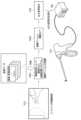
1.システム
図1は、本実施形態におけるシステム10の構成例である。図1には、内視鏡により術野を撮影する場合のシステム構成例を示す。図1に示すシステム10は、コントローラ100と内視鏡システム200とジェネレータ300とエネルギーデバイス310とを含む。システム10は、内視鏡下において少なくとも1つのエネルギーデバイスを用いて手術を行うための手術システムである。ここではシステム10が1つのエネルギーデバイス310を含む例を示すが、システム10が複数のエネルギーデバイスを含んでもよい。 1. System Fig. 1 shows an example of the configuration of a
内視鏡システム200は、内視鏡による撮影、内視鏡画像の画像処理、及び内視鏡画像のモニタ表示を行うシステムである。内視鏡システム200は、内視鏡210と本体装置220とディスプレイ230とを含む。ここでは、外科手術用の硬性鏡を例に説明する。The
内視鏡210は、体腔に挿入される挿入部と、挿入部の基端に接続される操作部と、操作部の基端に接続されるユニバーサルコードと、ユニバーサルコードの基端に接続されるコネクタ部とを含む。挿入部は、硬性管と対物光学系と撮像素子と照明光学系と伝送ケーブルとライトガイドとを含む。体腔内を撮影するための対物光学系及び撮像素子と、体腔内を照明するための照明光学系は、細長い円筒形状の硬性管の先端部に設けられる。硬性管の先端部は、湾曲可能に構成されてもよい。撮像素子により取得された画像信号を伝送する伝送ケーブル、及び照明光を照明光学系へ導光するライトガイドは、硬性管の内部に設けられる。操作部は、ユーザにより把持され、ユーザからの操作を受け付ける。操作部には、様々な機能が割り当てられたボタンが設けられる。挿入部先端が湾曲可能である場合には、操作部に、アングル操作レバーが設けられる。コネクタ部は、伝送ケーブルを本体装置220に着脱可能に接続するビデオコネクタと、ライトガイドを本体装置220に着脱可能に接続するライトガイドコネクタとを含む。The
本体装置220は、内視鏡の制御、内視鏡画像の画像処理及び内視鏡画像の表示処理を行う処理装置と、照明光の生成及び照明光の制御を行う光源装置とを含む。本体装置220はビデオシステムセンターとも呼ばれる。処理装置は、CPU等のプロセッサにより構成され、内視鏡210から送信される画像信号を画像処理して内視鏡画像を生成し、その内視鏡画像をディスプレイ230とコントローラ100に出力する。光源装置が出射した照明光は、ライトガイドにより照明光学系へ導光され、照明光学系から体腔内へ出射される。The
エネルギーデバイス310は、その先端部から高周波電力又は超音波等によりエネルギーを出力することで、その先端部が接する組織に対して凝固、封止、止血、切開、切離又は剥離等の処置を行うデバイスである。エネルギーデバイス310は、エネルギー処置具とも呼ばれる。エネルギーデバイス310は、デバイス先端の電極と体外の電極の間に高周波電力を通電させるモノポーラデバイス、2つのジョウの間に高周波電力を通電させるバイポーラデバイス、プローブとジョウが設けられると共にプローブから超音波を出射する超音波デバイス、又はプローブとジョウの間に高周波電力を通電させると共にプローブから超音波を出射する併用デバイス等である。The
ジェネレータ300は、エネルギーデバイス310へのエネルギー供給、エネルギー供給の制御、及びエネルギーデバイス310からの電気的情報の取得を行う。エネルギーデバイス310が高周波エネルギーを出力する場合、ジェネレータ300は高周波電力を供給し、エネルギーデバイス310は、その高周波電力を電極又はジョウから出力する。エネルギーデバイス310が超音波エネルギーを出力する場合、ジェネレータ300は電力を供給し、エネルギーデバイス310のプローブは、その電力を超音波に変換して出力する。The
電気的情報は、エネルギーデバイス310の電極又はジョウが接する組織の電気的情報であり、具体的には、エネルギーデバイス310が組織に高周波電力を出力した応答として得られる情報である。電気的情報は、例えば、エネルギーデバイス310により処置される組織のインピーダンス情報である。但し、後述のように電気的情報はインピーダンス情報に限らない。The electrical information is electrical information of the tissue in contact with the electrodes or jaws of the
ジェネレータ300は、出力シーケンスに従って、エネルギーデバイス310からのエネルギー出力を時間的に変化させる制御を行う。ジェネレータ300は、インピーダンス情報の時間的変化に応じてエネルギー出力を変化させてもよい。この場合、出力シーケンスは、インピーダンス情報の変化に対してどのようにエネルギー出力を変化させるかを、規定してもよい。また、ジェネレータ300は、インピーダンス情報の時間的変化に応じてエネルギー出力を自動オフしてもよい。例えば、ジェネレータ300は、インピーダンスが一定以上に上昇したとき、処置終了と判断してエネルギー出力をオフしてもよい。The
コントローラ100は、機械学習等を用いた画像認識処理により内視鏡画像から組織情報、処置情報又はそれら両方を認識し、その認識した情報に基づいてエネルギー出力調整指示をジェネレータに出力する。内視鏡画像から認識される組織情報、処置情報又はそれら両方を画像認識情報とも呼ぶこととする。これらの情報は、具体的には、エネルギーデバイス310による処置において熱拡散の度合いに影響を与える事項に関する情報である。The
より具体的には、組織情報は、エネルギーデバイス310により処置される組織に関する情報であり、例えば組織種類又は組織状態を含む。処置情報は、組織に対する処置に関する情報であり、具体的には、処置に用いられるデバイスが組織に及ぼす作用、その作用の結果、又は、デバイスと組織の位置関係等の情報である。ここでのデバイスは、エネルギーデバイス310又はそれ以外のデバイスであってよい。処置情報は、例えば、組織把持量、牽引量、組織テンション、又はエネルギーデバイス310の先端部と周辺組織との距離等を含む。More specifically, tissue information is information about the tissue to be treated by the
ジェネレータ300は、エネルギー出力調整指示に従って、エネルギーデバイス310のエネルギー出力を調整する。即ち、本実施形態のシステム10は、内視鏡画像に基づいてエネルギーデバイス310からのエネルギー出力を自動調整するシステムである。ジェネレータ300は、エネルギー出力調整指示により指示されるエネルギー供給量でエネルギーデバイス310にエネルギーを供給し、エネルギーデバイス310が、そのエネルギー供給を受けてエネルギー出力することで、エネルギー出力がエネルギー出力調整指示により調整される。The
エネルギー出力調整指示は、出力シーケンスの波形全体としての出力を増減する指示、或いは、選択可能な複数の出力シーケンスのうち、いずれかの出力シーケンスに設定する指示等である。例えば、エネルギーデバイス310からのエネルギー出力が段階的な倍率により調整可能であるとき、エネルギー出力調整指示は、そのエネルギー出力の段階的な倍率を示す指示である。ジェネレータ300は、その指示された倍率に応じて高周波出力又は超音波出力を増減させる。出力シーケンスの開始前にエネルギー出力調整指示が行われる場合、出力シーケンスの波形全体に倍率が乗算され、出力シーケンス全体として出力が増加又は減少する。なお、倍率は連続的に調整可能であってもよい。或いは、複数の出力シーケンスが設けられているとき、エネルギー出力調整指示は、その複数の出力シーケンスのいずれかを示す指示である。ジェネレータ300は、その指示された出力シーケンスに従ってエネルギーデバイス310からのエネルギー出力を行う。なお、エネルギー出力調整指示は、エネルギー出力の増減と出力シーケンスの変更の両方の指示を含んでもよい。The energy output adjustment instruction is an instruction to increase or decrease the output of the entire waveform of the output sequence, or an instruction to set one of a plurality of selectable output sequences. For example, when the energy output from the
ここで、一般的に外科手術においてエネルギー処置を行う際のポイントとして、エネルギーデバイスからの熱拡散を抑制して周囲臓器の熱損傷を避けることが挙げられる。しかし、処置される組織は一様ではないため、組織種類の違い、組織状態の違い又は患者の個人差等により、切離等の処置に要する時間にバラつきが生じ、熱拡散の程度も様々となる。これらに対処し、熱拡散を抑制すべく、医師は組織把持量や組織テンションの調整を行っているが、経験が必要な操作であり、特に非エキスパートにおいては適切な調整が困難な場合がある。In general, a key point when performing energy treatment in surgery is to suppress thermal diffusion from the energy device to avoid thermal damage to surrounding organs. However, the tissue being treated is not uniform, and differences in tissue type, tissue condition, and individual patient differences result in variations in the time required for treatment such as resection, and the degree of thermal diffusion also varies. To address these issues and suppress thermal diffusion, doctors adjust the amount of tissue gripped and the tissue tension, but this is an operation that requires experience, and it can be difficult for non-experts to make appropriate adjustments.
このように、エネルギーデバイスを用いた処置においては、しばしば周囲への熱拡散が問題となるため、医師が拡散の程度を推測しながら処置を行う。上記特許文献1では、エネルギーデバイスのエネルギー出力データ等に基づいて血管であるか非血管であるか等の組織タイプを認識している。しかし、熱拡散の程度は、血管であるか非血管であるかのみで二分されるものではなく、例えば、組織の厚み、血液等への浸漬等の組織状態、又は、デバイスによる把持量又は牽引の強さ等の医師の操作にも影響される。具体的には、エネルギーデバイスがエネルギーを印加したことで生じた組織の熱が、周囲組織の内部又は表面を拡散することで、熱拡散が生じる。或いは、エネルギーデバイスが出力するエネルギーが把持組織の周囲にも拡散し、そのエネルギーが拡散した周囲組織が発熱することで、熱拡散が生じる。この熱拡散の程度は、組織種類、組織状態、組織把持量、又は組織テンション等に応じて異なる。In this way, in treatments using energy devices, heat diffusion to the surroundings is often a problem, so doctors perform treatments while estimating the degree of diffusion. In the above-mentioned Patent Document 1, the tissue type, such as whether it is vascular or non-vascular, is recognized based on the energy output data of the energy device. However, the degree of heat diffusion is not only divided into vascular and non-vascular, but is also influenced by, for example, the tissue thickness, tissue condition such as immersion in blood, or the doctor's operation such as the amount of gripping or traction by the device. Specifically, heat diffusion occurs when the heat of the tissue generated by the application of energy by the energy device diffuses inside or on the surface of the surrounding tissue. Alternatively, heat diffusion occurs when the energy output by the energy device diffuses to the surroundings of the gripped tissue, and the surrounding tissue to which the energy is diffused generates heat. The degree of this heat diffusion varies depending on the tissue type, tissue condition, tissue gripping amount, tissue tension, etc.
この点、本実施形態によれば、システム10が、組織種類、組織状態、組織把持量、又は組織テンション等の画像認識情報に基づいて、組織に適したエネルギーを印加する。これにより、エネルギーデバイスの処置対象から周囲組織への熱拡散を低減できる。また、従来医師が行っていた把持量やテンションの調整に代わりシステム10がエネルギーの出力調整を行うことで、医師の負担を軽減できる。また、システム10が自律的に出力調整を行うことで、経験の浅い医師においても安定した処置が可能となる。以上により、手術の安定性向上が図れる、或いは医師の経験値に依らない手技の均てん化が図れる。In this regard, according to the present embodiment, the
2.コントローラ
図2は、コントローラ100の構成例である。コントローラ100は、制御部110と記憶部120とI/Oデバイス180とI/Oデバイス190とを含む。図1と図2には、コントローラ100がジェネレータ300と別体の装置で構成される例を示す。その場合、コントローラ100は、PC又はサーバ装置等の情報処理装置により構成される。或いは、コントローラ100は、ネットワークを介して接続された1又は複数の情報処理装置によって処理を実行するクラウドシステムで実現されてもよい。 2. Controller Fig. 2 shows a configuration example of the
I/Oデバイス180は、内視鏡システム200の本体装置220から内視鏡画像の画像データを受信する。I/Oデバイス180は、画像伝送用のケーブルが接続されるコネクタ、又は、そのコネクタに接続され、本体装置220との通信を行うインターフェース回路である。The I/
制御部110は、学習済みモデル121を用いた画像認識処理により内視鏡画像から組織情報又は処置情報の少なくとも1つを認識し、その画像認識情報に基づいてエネルギー出力調整指示を出力する。制御部110は、ハードウェアとして1又は複数のプロセッサを含む。プロセッサは、CPU(Central Processing Unit)、GPU(Graphical Processing Unit)又はDSP(Digital Signal Processor)等の汎用プロセッサである。或いは、プロセッサは、ASIC(Application Specific Integrated Circuit)又はFPGA(Field Programmable Gate Array)等の専用プロセッサであってもよい。The
記憶部120は、画像認識処理に用いられる学習済みモデル121を記憶する。汎用プロセッサにより画像認識処理が行われる場合には、記憶部120は、推論アルゴリズムが記述されたプログラムと、その推論アルゴリズムに用いられるパラメータと、を学習済みモデル121として記憶する。推論アルゴリズムがハードウェア化された専用プロセッサにより画像認識処理が行われる場合には、記憶部120は、推論アルゴリズムに用いられるパラメータを学習済みモデル121として記憶する。記憶部120は、半導体メモリ、ハードディスクドライブ、又は光学ディスクドライブ等の記憶装置である。半導体メモリは、RAM、ROM又は不揮発性メモリ等である。The
画像認識処理の推論アルゴリズムとしては、例えばニューラルネットワークを採用できる。ニューラルネットワークにおけるノード間接続の重み係数とバイアスがパラメータである。ニューラルネットワークは、画像データが入力される入力層と、入力層を通じて入力されたデータに対し演算処理を行う中間層と、中間層から出力される演算結果に基づいて認識結果を出力する出力層と、を含む。画像認識処理に用いられるニューラルネットワークとして、例えばCNN(Convolutional Neural Network)を採用できる。As an inference algorithm for image recognition processing, for example, a neural network can be adopted. The weight coefficients and biases of the node connections in the neural network are parameters. A neural network includes an input layer to which image data is input, an intermediate layer that performs calculation processing on the data input through the input layer, and an output layer that outputs recognition results based on the calculation results output from the intermediate layer. As a neural network used for image recognition processing, for example, a CNN (Convolutional Neural Network) can be adopted.
また制御部110は、画像取得部111と組織情報認識部112と出力設定部113とを含む。記憶部120は、画像取得部111、組織情報認識部112及び出力設定部113の各部の機能が記述されたプログラムを記憶する。制御部の1又は複数のプロセッサが、記憶部120からプログラムを読み出し、そのプログラムを実行することで、画像取得部111、組織情報認識部112及び出力設定部113の各部の機能を実現する。この各部の機能が記述されたプログラムは、コンピュータにより読み取り可能な媒体である非一時的な情報記憶媒体に格納できる。情報記憶媒体は、例えば光ディスク、メモリカード、HDD、或いは半導体メモリなどにより実現できる。半導体メモリは例えばROM又は不揮発性メモリである。The
I/Oデバイス190は、ジェネレータ300へエネルギー出力調整指示の信号を送信する。I/Oデバイス190は、信号伝送用のケーブルが接続されるコネクタ、又は、そのコネクタに接続され、ジェネレータ300との通信を行うインターフェース回路である。The I/
図3は、コントローラ100及びシステム10が行う処理を説明するフローチャートである。Figure 3 is a flowchart explaining the processing performed by the
ステップS1において、画像取得部111が、内視鏡システム200の本体装置220からI/Oデバイス180を介して内視鏡画像を取得する。ステップS2において、組織情報認識部112は、学習済みモデル121を用いた画像認識処理を内視鏡画像に対して行うことで、組織情報又は処置情報の少なくとも1つを画像認識情報として取得する。ステップS3において、出力設定部113は、画像認識情報に応じたエネルギー出力調整指示を、I/Oデバイス190を介してジェネレータ300へ出力する。In step S1, the
ステップS4において、ジェネレータ300は、エネルギー出力調整指示を受けてエネルギー出力シーケンスを設定し、医師が行う出力操作に応じてエネルギー出力を行う。例えば、医師は、エネルギーデバイス310の操作部に設けられた出力開始ボタンを操作し、ジェネレータ300は、その操作入力を受けてエネルギーデバイス310へのエネルギー供給を開始する。ステップS5において、エネルギーデバイス310は、ジェネレータ300からのエネルギー供給を受けて、把持している組織へエネルギーを印加する。In step S4, the
なお、上記ではコントローラ100とジェネレータ300が別体の装置で構成され、コントローラ100を構成する装置に制御部110全体が含まれるとしたが、コントローラ100とジェネレータ300のハードウェア構成はこれに限定されない。例えば、コントローラ100とジェネレータ300は一体の装置で構成されてもよい。或いは、システム10が第1装置と第2装置とを含み、第1装置が、制御部110のうちの画像取得部111と組織情報認識部112を含み、第2装置が、制御部110のうちの出力設定部113とジェネレータ300とを含んでもよい。この場合、第1装置は、組織情報又は処置情報の少なくとも1つである画像認識情報を第2装置へ出力し、第2装置は、画像認識情報に応じてジェネレータのエネルギー出力を制御する。In the above, the
3.エネルギーデバイス
以下、エネルギーデバイス310の一例として、モノポーラデバイス320、バイポーラデバイス330、超音波デバイス340及び併用デバイスについて説明する。 3. Energy Devices As examples of the
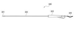
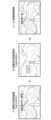
図4は、モノポーラデバイス320の構成例である。モノポーラデバイス320は、細長い円筒形状の挿入部322と、挿入部322の先端に設けられる電極321と、挿入部322の基端に接続される操作部323と、操作部323と不図示のコネクタとを接続するケーブル325とを含む。コネクタはジェネレータ300に着脱可能に接続される。Figure 4 shows an example of the configuration of the
ジェネレータ300が出力した高周波電力はケーブル325により伝送され、電極321から出力される。患者の体外に対極板が設けられており、電極321と対極板の間で通電する。これにより、電極321が接する組織に高周波エネルギーが印加され、その組織にジュール熱が発生する。電極321は、処置の内容に応じて様々な形状の電極が採用される。モノポーラデバイス320は、通電パターンの変更により、凝固と切開の程度を調整可能である。モノポーラデバイス320の処置対象は、電極321が接する組織であるが、その電極321が接する組織の周囲に拡散した熱によって周囲組織に影響を与える可能性がある。The high-frequency power output by the
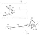
図5は、バイポーラデバイス330の構成例である。バイポーラデバイス330は、細長い円筒形状の挿入部332と、挿入部332の先端部331に設けられる2つのジョウ337、338と、挿入部332の基端に接続される操作部333と、操作部333と不図示のコネクタとを接続するケーブル335とを含む。コネクタはジェネレータ300に着脱可能に接続される。ジョウ337、338は、組織を把持すると共に把持された組織にエネルギーを印加するための可動部のことであり、基端336に設けられた軸を中心として開閉可能に構成されている。操作部333は、ジョウ337、338の開閉を操作するための把持部が設けられている。医師が把持部を握り込むことで、ジョウ337、338が閉まり組織を把持する。5 is a configuration example of the
ジェネレータ300が出力した高周波電力はケーブル335により伝送され、ジョウ337、338が組織を把持したとき2つのジョウ337、338の間に通電する。これにより、2つのジョウ337、338に挟まれた組織に高周波エネルギーが印加され、その組織にジュール熱が発生し、その組織が凝固する。ジェネレータ300は、ジョウ337、338に把持された組織のインピーダンス情報を計測し、そのインピーダンス情報に基づいて処置完了を検知し、エネルギー出力を自動停止してもよい。また、ジェネレータ300は、インピーダンス情報に基づいて、組織への印加エネルギーを自動調整してもよい。バイポーラデバイスの熱拡散に関しては、例えば、バイポーラデバイス330のデバイス温度は摂氏100度程度までしか上がらないが、ジョウ337、338により把持された部分の周辺に回り込み電流が生じ、その回り込み電流により熱拡散が生じる可能性がある。The high-frequency power output by the
なお、バイポーラデバイスの派生デバイスとしてベッセルシーリングデバイスがある。ベッセルシーリングデバイスは、バイポーラデバイスのジョウにカッターが設けられたデバイスであり、通電により組織を凝固した後にカッターを走らせることで組織を切離する。A derivative of the bipolar device is the vessel sealing device. The vessel sealing device is a device with a cutter attached to the jaws of the bipolar device, and after coagulating the tissue by passing an electric current through it, the tissue is separated by running the cutter over it.
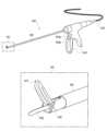
図6は、超音波デバイス340の構成例である。超音波デバイス340は、細長い円筒形状の挿入部342と、挿入部342の先端部341に設けられるジョウ347及びプローブ348と、挿入部342の基端に接続される操作部343と、操作部343と不図示のコネクタとを接続するケーブル345とを含む。コネクタはジェネレータ300に着脱可能に接続される。ジョウ347は、基端346に設けられた軸を中心として可動であり、非可動のプローブ348に対して開閉可能に構成されている。操作部343は、ジョウ347の開閉を操作するための把持部が設けられている。医師が把持部を握り込むことで、ジョウ347が閉まり、ジョウ347とプローブ348が組織を把持する。操作部343には、第1出力モードが割り当てられた操作ボタン344aと、第2出力モードが割り当てられた操作ボタン344bとが設けられる。出力モードは処置内容に応じて選択されるものであり、各出力モードの操作ボタンが押されると、そのモードの出力シーケンスで超音波エネルギーが出力される。6 is a configuration example of the
ジェネレータ300が出力した電力はケーブル335により伝送され、操作ボタン344a又は344bが押されたときにプローブ348が電力を超音波に変換して出力する。これにより、ジョウ347とプローブ348に挟まれた組織に摩擦熱が発生し、その組織が凝固する又は組織が切開される。高周波デバイスの熱拡散に関しては、例えば、高周波デバイスの熱拡散に比べて、超音波デバイス340の熱拡散は少ないが、超音波デバイス340のデバイス温度は摂氏200度近くまで上昇する。超音波デバイス340の熱拡散は、プローブ348の先端方向に生じやすいという特性がある。The power output by the
高周波電力と超音波を併用する併用デバイスは、例えば図6の超音波デバイスと同様の構成である。但し、併用デバイスは、ジョウとプローブの間に高周波電力を通電することで、ジョウとプローブに把持された組織にジュール熱を発生させ、その組織を凝固させることができる。また併用デバイスは、超音波デバイスと同様に、プローブから超音波を出力することで、ジョウとプローブに把持された組織を切開できる。操作部に設けられた2つの操作ボタンの一方には、高周波モードが割り当てられ、他方にはシール&カットモードが割り当てられる。高周波モードは、高周波エネルギー出力のみで凝固等の処置を行うモードである。シール&カットモードは、高周波エネルギーと超音波エネルギーを併用するモードであり、高周波エネルギー出力により組織を凝固させると共に組織を切離するモードである。併用デバイスの熱拡散に関しては、例えば、バイポーラデバイスと同様な熱拡散、超音波デバイスと同様な熱拡散、又はそれら両方が生じる可能性がある。The combination device that uses high-frequency power and ultrasound is configured similarly to the ultrasound device in FIG. 6. However, the combination device can generate Joule heat in the tissue held by the jaws and the probe by passing high-frequency power between the jaws and the probe, and can coagulate the tissue. The combination device can also incise the tissue held by the jaws and the probe by outputting ultrasound from the probe, just like the ultrasound device. The high-frequency mode is assigned to one of the two operation buttons provided on the operation unit, and the seal and cut mode is assigned to the other. The high-frequency mode is a mode in which treatment such as coagulation is performed only by outputting high-frequency energy. The seal and cut mode is a mode in which high-frequency energy and ultrasound energy are used in combination, and the tissue is coagulated and cut by outputting high-frequency energy. With regard to the thermal diffusion of the combination device, for example, thermal diffusion similar to that of a bipolar device, thermal diffusion similar to that of an ultrasound device, or both may occur.
なお、以下の実施形態においては、主にバイポーラデバイス330をエネルギーデバイス310として用いる場合を例に説明する。但し、本実施形態は、熱拡散が生じる可能性がある上記の様々なエネルギーデバイスを用いる場合に適用可能である。In the following embodiment, the
4.組織種類を認識する場合の処理例
図7は、制御部110が組織種類を認識する場合の処理例である。S1Aに示すように、制御部110に内視鏡画像が入力される。具体的には、内視鏡により撮影された動画像の各フレーム画像が順次に制御部110に入力される。制御部110に入力される内視鏡画像には、1又は複数の組織及び1又は複数のエネルギーデバイスが写っている。 7 shows an example of processing when the
S2Aaに示すように、制御部110は、機械学習によって調整された組織認識プログラムを実行することで、内視鏡画像から組織種類を認識する。具体的には、制御部110は、バイポーラデバイスのジョウに把持された組織の種類、或いは、その把持された組織の周辺組織の種類を認識する。組織種類とは、臓器内外に存在する組織の種類、或いは、臓器そのものの種類のことである。臓器内外に存在する組織は、例えば、動脈、静脈、血管を含む膜、肝胃間膜、腸間膜、薄膜、神経又は脂肪等である。臓器は、例えば、食道、胃、膵臓、肝臓又は小腸等である。S2Abに示すように、制御部110は、内視鏡画像から認識した組織種類を出力する。図7には、血管を含む膜が認識された例を示す。As shown in S2Aa, the
S3Aに示すように、制御部110は、内視鏡画像から認識された組織種類に応じて出力変更指示を行う。具体的には、記憶部120が、各組織種類にエネルギー出力調整指示が対応付けられたテーブルデータを記憶し、制御部110は、そのテーブルデータを参照することで、組織種類に対応したエネルギー出力調整指示を出力する。ジェネレータ300は、制御部110が出力したエネルギー出力調整指示に従ってバイポーラデバイスの出力シーケンスを調整する。なお、組織種類に応じたエネルギー出力調整指示を出力するアルゴリズムは、上記に限定されない。As shown in S3A, the
エネルギー出力調整指示は、基準エネルギー出力を基準として、エネルギー出力を増加させる、減少させる又は維持する指示である。ジェネレータ300は、エネルギー出力の設定操作を受け付ける操作部を有しており、その操作部によりエネルギー出力を例えば強度1~5の5段階のいずれかに設定可能である。強度1が最低のエネルギー出力、強度5が最高のエネルギー出力を示す。このとき、基準エネルギー出力は、例えば、予め決められた「強度3」等のエネルギー出力である。その場合、基準エネルギー出力よりも増加させる指示は、「強度4」又は「強度5」に設定する指示であり、基準エネルギー出力よりも減少させる指示は、「強度2」又は「強度1」に設定する指示である。或いは、基準エネルギー出力は、ジェネレータ300の操作部により設定されている現在のエネルギー出力であってもよい。その場合、基準エネルギー出力よりも増加させる指示は、現在設定されているエネルギー出力よりも高いエネルギー出力に設定する指示であり、基準エネルギー出力よりも減少させる指示は、現在設定されているエネルギー出力よりも低いエネルギー出力に設定する指示である。或いは、基準エネルギー出力は、ジェネレータ300に設定可能な強度1~5の出力範囲であってもよい。その場合、基準エネルギー出力よりも増加させる指示は、「強度5」よりも高いエネルギー出力に設定する指示であり、基準エネルギー出力よりも減少させる指示は、「強度1」よりも低いエネルギー出力に設定する指示である。The energy output adjustment instruction is an instruction to increase, decrease, or maintain the energy output based on the reference energy output. The
図8は、組織認識処理の機械学習を行う学習装置500の構成例である。学習装置500は、処理部510と記憶部520とを含む。学習装置500は、例えばPC又はサーバ装置等の情報処理装置によって実現される。或いは、学習装置500は、ネットワークを介して接続された1又は複数の情報処理装置によって処理を実行するクラウドシステムで実現されてもよい。Figure 8 shows an example configuration of a
処理部510はCPU等のプロセッサであり、記憶部520は半導体メモリ又はハードディスクドライブ等の記憶装置である。記憶部520は学習モデル522と教師データ521とを記憶しており、処理部510が教師データ521を用いて学習モデル522を学習させることで、学習済みモデル121を生成する。具体的には、教師データは、複数の学習用画像の画像データと、各学習用画像に付された正解データとを含む。複数の学習用画像は、1又は複数の組織及び1又は複数のエネルギーデバイスが写る内視鏡画像を含む。この内視鏡画像を学習用デバイス組織画像とも呼ぶ。また複数の学習用画像は、1又は複数の組織が写り、且つエネルギーデバイスが写らない内視鏡画像を含んでもよい。この内視鏡画像を学習用組織画像とも呼ぶ。正解データは、セグメンテーション(領域検出)におけるアノテーション、ディテクション(位置検出)におけるアノテーション、クラシフィケーション(分類)における正解ラベル、又はリグレッション(回帰分析)における正解ラベルである。処理部510は、学習モデル522による推論処理に学習用画像を入力し、その推論処理の結果と正解データの誤差に基づいて学習モデル522にフィードバックを行い、それを多数の教師データで繰り返すことで、学習済みモデル121を生成する。生成された学習済みモデル121は、コントローラ100の記憶部120に転送される。The
制御部110が組織種類を認識する場合、教師データ521は、学習用画像として、血管を含む膜の画像、肝胃間膜の画像、血管(動脈)の画像、実質臓器(膵臓)の画像、腸間膜の画像、薄膜の画像、実質臓器(肝臓)の画像、又は実質臓器(食道)の画像等を含む。また教師データ521は、正解データとして、各学習用画像に写る組織の領域に付されたアノテーション、又は、各学習用画像に写る組織の種類を示すラベルを含む。When the
なお、図7と図8では機械学習を用いた認識処理の推論段階と学習段階を分けて記載したが、以下では、推論段階と学習段階を混在させて記載する場合がある。その場合、制御部110が推論段階の処理を実行し、学習装置500が学習段階の処理を実行することは、上記と同様である。Note that in Figures 7 and 8, the inference stage and the learning stage of the recognition process using machine learning are described separately, but below, the inference stage and the learning stage may be described together. In that case, the
図9は、制御部110が組織種類を認識する場合の第1認識処理例である。制御部110は、セグメンテーションによる組織領域認識と、セグメンテーションによる処置具先端領域検出とを行い、それらの結果を合成することで、ジョウに把持された組織の種類を認識する。図9には、「組織種類は膜組織である」と認識された例を示す。なお、制御部110は、セグメンテーションの結果、組織種類の認識結果、又はそれら両方をモニタ表示してもよい。Figure 9 is a first recognition processing example when the
具体的には、学習済みモデル121は、組織領域認識を行うための第1学習済みモデルと、処置具先端領域検出を行うための第2学習済みモデルとを含む。制御部110は、第1学習済みモデルを用いた画像認識処理により、内視鏡画像から組織種類毎の領域を検出する。これにより、内視鏡画像に組織種類がマッピングされる。図9には、内視鏡画像から胃の領域、肝臓の領域及び膜の領域が検出された例を示す。学習段階においては、学習装置500は、組織種類毎の領域にアノテーションが付された内視鏡画像を教師データとして、第1学習済みモデルを生成する。制御部110は、第2学習済みモデルを用いた画像認識処理により、内視鏡画像からエネルギーデバイス先端のジョウの領域を検出する。これにより、内視鏡画像においてジョウが把持している位置が特定される。学習段階においては、学習装置500は、エネルギーデバイス先端のジョウの領域にアノテーションが付された内視鏡画像を教師データとして、第2学習済みモデルを生成する。なお、処理毎に学習済みモデルを分離せずに1つの学習済みモデルにより組織領域認識と処置具先端領域検出を実現するように、システムが構成されてもよい。Specifically, the trained
制御部110は、処置具先端領域検出により検出されたジョウの領域が、組織領域認識により検出された1又は複数の組織領域のうち、いずれの組織領域に重なるかを判定することで、ジョウに把持された組織の種類を認識する。The
図10は、制御部110が組織種類を認識する場合の第2認識処理例である。制御部110は、ディテクションによる処置具先端の検出と、クラシフィケーションによる把持組織種類の分類とを行うことで、ジョウに把持された組織の種類を認識する。Figure 10 is a second recognition processing example in which the
具体的には、学習済みモデル121は、処置具先端の検出を行うための第1学習済みモデルと、把持組織種類の分類を行うための第2学習済みモデルとを含む。制御部110は、第1学習済みモデルを用いた画像認識処理により、内視鏡画像からジョウの位置を検出し、そのジョウを内包するバウンディングボックスを生成する。これにより、把持位置とその周辺の注目領域が特定される。学習段階においては、学習装置500は、ジョウを内包するバウンディングボックスが付された内視鏡画像を教師データとして、第1学習済みモデルを生成する。制御部110は、内視鏡画像からバウンディングボックス内の画像を切り出し、その画像を、第2学習済みモデルを用いた画像認識処理に入力することで、バウンディングボックス内の画像に写る組織の種類を分類する。これにより、バウンディングボックス内の画像が、いずれの種類の組織が把持された画像であるかが、特定される。学習段階においては、学習装置500は、ジョウに把持された組織の種類を示すラベルが付された内視鏡画像を教師データとして、第2学習済みモデルを生成する。Specifically, the trained
制御部110は、把持組織種類の分類の結果を、ジョウに把持された組織の種類の認識結果として出力する。The
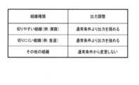
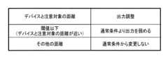
図11は、制御部110が組織種類を認識する場合の出力調整例である。制御部110は、組織種類の認識結果が、エネルギーデバイスにより切りやすい薄膜等の組織であるとき、通常条件よりエネルギー出力を弱める。通常条件は、上述した基準エネルギー出力である。制御部110は、組織種類の認識結果が、エネルギーデバイスにより切りにくい食道等の組織であるとき、通常条件よりエネルギー出力を強める。制御部110は、組織種類の認識結果が、上記以外のその他の組織であるとき、エネルギー出力を通常条件から変更しない。Figure 11 is an example of output adjustment when the
なお、図31に、併用デバイスを用いる場合の出力調整例を示す。出力の欄に複数の出力調整が記載されている場合、制御部110は、そのいずれかの出力調整を行う。例えば、画像から「薄膜」が認識されたとき、制御部110は、(1)「HF出力レベルを下げる」、(2)「HF出力時間を短くする、S&Cモードのレベルを下げる」、又は(3)「US単独出力にする」のうちいずれかの出力調整を行う。なお、「HF」は高周波を意味し、「S&C」はシール&カットを意味し、「US」は超音波を意味する。なお、制御部110は画像認識処理の結果に基づいてエネルギー出力を調整するが、後述のように、更にデバイス情報を併用してエネルギー出力を調整してもよい。FIG. 31 shows an example of output adjustment when using a combined device. When multiple output adjustments are listed in the output column, the
5.組織状態を認識する場合の処理例
図12は、制御部110が組織状態を認識する場合の処理例である。なお、図7の処理例と異なる部分を主に説明し、図7の処理例と同様な部分について説明を適宜に省略する。 5. Example of processing when recognizing tissue state Fig. 12 shows an example of processing when the
S1Bに示すように、制御部110に内視鏡画像が入力される。S2Baに示すように、制御部110は、機械学習によって調整された組織認識プログラムを実行することで、内視鏡画像から組織状態を認識する。具体的には、制御部110は、バイポーラデバイスのジョウに把持された組織の状態を認識する。組織状態とは、エネルギーデバイスによる処置中の熱拡散に影響を与えうる状態、即ち、その状態に応じて熱拡散の程度が変化するような状態のことである。組織状態は、例えば、ジョウに把持された組織の周囲組織量、ジョウに把持された組織又はその周辺組織の浸漬量、又は、ジョウに把持された組織の脂肪量等である。浸漬量は、組織を覆う液体の量であり、例えば血液又はリンパ液等の体液による浸漬量である。S2Bbに示すように、制御部110は、内視鏡画像から認識した組織状態を出力する。図12には、血液浸漬量が多い、即ち組織表面がウェットであると認識された例を示す。As shown in S1B, an endoscopic image is input to the
S3Bに示すように、制御部110は、内視鏡画像から認識された組織状態に応じて出力変更指示を行う。出力変更指示の手法は図7のS3Aと同様である。As shown in S3B, the
学習段階における教師データ521は、学習用画像として、周囲組織量が多い又は少ない画像、液体浸漬量が多い又は少ない画像、又は、脂肪量が多い又は少ない画像等を含む。また教師データ521は、正解データとして、各学習用画像に写る組織の状態を示すラベルを含む。The
図13~図15は、制御部110が組織状態を認識する場合の第1認識処理例である。図13には、組織状態として周辺組織量が認識される例を示す。制御部110は、セグメンテーションによる組織状態領域認識と、セグメンテーションによる処置具先端領域検出とを行い、それらの結果を合成することで、ジョウに把持された組織の状態を認識する。図13には、「周辺組織量が少ない」と認識された例を示す。なお、制御部110は、セグメンテーションの結果、組織状態の認識結果、又はそれら両方をモニタ表示してもよい。Figures 13 to 15 are examples of a first recognition process in which the
具体的には、学習済みモデル121は、組織状態領域認識を行うための第1学習済みモデルと、処置具先端領域検出を行うための第2学習済みモデルとを含む。制御部110は、第1学習済みモデルを用いた画像認識処理により、内視鏡画像から組織状態毎の領域を検出する。これにより、内視鏡画像に組織状態がマッピングされる。図13には、血管周囲組織が少ない部分の領域と、血管周囲組織が多い部分の領域とが検出された例を示す。学習段階においては、学習装置500は、組織状態毎の領域にアノテーションが付された内視鏡画像を教師データとして、第1学習済みモデルを生成する。図13では、教師データは、血管周辺組織の量が多い領域、少ない領域又はそれら両方の領域にアノテーションされた画像を含む。制御部110は、第2学習済みモデルを用いた画像認識処理により、内視鏡画像からエネルギーデバイス先端のジョウの領域を検出する。この処理は、図9の処置具先端領域検出と同様である。Specifically, the trained
制御部110は、処置具先端領域検出により検出されたジョウの領域が、組織状態領域認識により検出された1又は複数の組織状態領域のうち、いずれの組織状態領域に重なるかを判定することで、ジョウに把持された組織の状態を認識する。The
図14には、組織状態として脂肪量が認識される例を示す。図14には、制御部110が、組織状態領域認識において脂肪の多い領域を検出し、組織状態の認識において「脂肪量が多い」と認識した例を示す。学習段階の教師データは、脂肪量が多い領域、少ない領域又はそれら両方の領域にアノテーションされた画像を含む。図15には、組織状態として浸漬量が認識される例を示す。図15には、組織状態領域認識において浸漬量が多い領域つまりウェットな領域が検出され、組織状態の認識において「浸漬量が多い」と認識された例を示す。学習段階の教師データは、浸漬量が多い領域、少ない領域又はそれら両方の領域にアノテーションされた画像を含む。Figure 14 shows an example in which fat mass is recognized as the tissue state. Figure 14 shows an example in which the
図16は、制御部110が組織状態を認識する場合の第2認識処理例である。制御部110は、ディテクションによる処置具先端の検出と、クラシフィケーションによる把持組織状態の分類とを行うことで、ジョウに把持された組織の状態を認識する。Figure 16 is a second recognition processing example in which the
具体的には、学習済みモデル121は、処置具先端の検出を行うための第1学習済みモデルと、把持組織状態の分類を行うための第2学習済みモデルとを含む。制御部110は、第1学習済みモデルを用いた画像認識処理により、内視鏡画像からジョウの位置を検出し、そのジョウを内包するバウンディングボックスを生成する。この処理は、図10の処置具先端の検出と同様である。制御部110は、内視鏡画像からバウンディングボックス内の画像を切り出し、その画像を、第2学習済みモデルを用いた画像認識処理に入力することで、バウンディングボックス内の画像に写る組織の状態を分類する。これにより、バウンディングボックス内の画像が、いずれの状態の組織が把持された画像であるかが、特定される。学習段階においては、学習装置500は、ジョウに把持された組織の状態を示すラベルが付された内視鏡画像を教師データとして、第2学習済みモデルを生成する。Specifically, the trained
制御部110は、把持組織状態の分類の結果を、ジョウに把持された組織の状態の認識結果として出力する。The
図17は、制御部110が組織状態を認識する場合の出力調整例である。制御部110は、組織状態の認識結果が、エネルギーデバイスにより時間をかけてしっかり封止すべき状態であるとき、通常条件よりエネルギー出力を弱める。その状態は、例えば周辺組織量が少ない状態である。制御部110は、組織状態の認識結果が、エネルギーデバイスにより切りにくい状態であるとき、通常条件よりエネルギー出力を強める。その状態は、例えば脂肪量が多い状態又は血液浸漬量が多い状態である。制御部110は、組織状態の認識結果が、上記以外のその他の状態であるとき、エネルギー出力を通常条件から変更しない。
17 is an example of output adjustment when the
なお、図32に、併用デバイスを用いる場合の出力調整例を示す。出力の欄に複数の出力調整が記載されている場合、制御部110は、そのいずれかの出力調整を行う。なお、制御部110は画像認識処理の結果に基づいてエネルギー出力を調整するが、後述のように、更にデバイス情報を併用してエネルギー出力を調整してもよい。Figure 32 shows an example of output adjustment when using a combined device. If multiple output adjustments are listed in the output column, the
6.組織把持量を認識する場合の処理例
図18は、制御部110が組織把持量を認識する場合の処理例である。なお、図7の処理例と異なる部分を主に説明し、図7の処理例と同様な部分について説明を適宜に省略する。 18 shows a processing example in which the
S1Cに示すように、制御部110に内視鏡画像が入力される。S2Caに示すように、制御部110は、機械学習によって調整された組織認識プログラムを実行することで、内視鏡画像から組織把持量を認識する。具体的には、制御部110は、バイポーラデバイスのジョウが組織を把持しているときの把持量を認識する。把持量は、ジョウが組織を把持している部分の長さ、又はジョウの全長のうち組織を把持している部分の割合である。ジョウで浅く組織を把持した状態はショートピッチと呼ばれ、ジョウで深く組織を把持した状態はロングピッチと呼ばれる。ここでは、把持量が閾値以下である場合をショートピッチ、把持量が閾値以上である場合をロングピッチと呼ぶこととする。S2Cbに示すように、制御部110は、内視鏡画像から認識した把持量と閾値を比較することでショートピッチかロングピッチかを認識する。図18には、組織把持量がショートピッチと認識された例を示す。As shown in S1C, an endoscopic image is input to the
S3Cに示すように、制御部110は、内視鏡画像から認識された組織把持量に応じて出力変更指示を行う。出力変更指示の手法は図7のS3Aと同様である。As shown in S3C, the
学習段階における教師データ521は、学習用画像として、様々な把持量の画像を含む。例えば、教師データ521は、把持量が閾値以下であるショートピッチの画像と、把持量が閾値以上であるロングピッチの画像とを含む。また教師データ521は、正解データとして、各学習用画像に写る組織把持量を示すラベルを含む。ラベルは、ジョウが組織を把持している部分の長さ、ジョウの全長のうち組織を把持している部分の割合、又はショートピッチかロングピッチかを示す情報等である。The
図19は、制御部110が組織把持量を認識する場合の第1認識処理例である。制御部110は、ディテクションによる処置具先端の検出と、クラシフィケーションによる組織把持量の分類とを行うことで、ジョウの組織把持量を認識する。組織把持量の分類とは、内視鏡画像に写るジョウの把持量が、複数のクラスのいずれのクラスに属するかを分類することである。複数のクラスは、段階的に区切られた把持量範囲を示す。例えば、クラスは、1mm刻み等の所定長さの刻みで区切られる、或いはジョウの全長を100%としたとき10%刻み等の所定割合の刻みで区切られる。Figure 19 is a first recognition processing example in which the
具体的には、学習済みモデル121は、処置具先端の検出を行うための第1学習済みモデルと、組織把持量の分類を行うための第2学習済みモデルとを含む。制御部110は、第1学習済みモデルを用いた画像認識処理により、内視鏡画像からジョウの位置を検出し、そのジョウを内包するバウンディングボックスを生成する。この処理は、図10の処置具先端の検出と同様である。制御部110は、内視鏡画像からバウンディングボックス内の画像を切り出し、その画像を、第2学習済みモデルを用いた画像認識処理に入力することで、バウンディングボックス内の画像に写るジョウの組織把持量を分類する。これにより、バウンディングボックス内の画像が、どのような組織把持量の画像であるかが、特定される。学習段階においては、学習装置500は、ジョウの組織把持量を示すラベルが付された内視鏡画像を教師データとして、第2学習済みモデルを生成する。Specifically, the trained
制御部110は、組織把持量の分類の結果と閾値とを比較することで、ショートピッチかロングピッチかを判定し、その判定結果を組織把持量の認識結果として出力する。図19には、ショートピッチと判定された例を示す。The
図20は、制御部110が組織把持量を認識する場合の第2認識処理例である。制御部110は、クラシフィケーション又はリグレッションにより、処置具先端部の特徴点と、ジョウにより把持された組織との間の定量的な位置関係を推定し、その推定結果から組織把持量を算出する。Figure 20 shows a second recognition processing example in which the
具体的には、制御部110は、学習済みモデル121を用いた画像認識処理により、ジョウの基端から把持組織近位端までの距離x[mm]を推定する。把持組織近位端とは、ジョウに把持された組織においてジョウの基端に最も近い端である。クラシフィケーションが用いられる場合には、制御部110は、距離xが、複数のクラスのいずれのクラスに属するかを分類する。複数のクラスは、段階的に区切られた距離範囲を示す。リグレッションが用いられる場合には、制御部110は、ジョウの基端から把持組織近位端までの距離xそのものを内視鏡画像から推定する。学習段階においては、学習装置500は、ジョウの基端から把持組織近位端までの距離xを示す距離情報がラベルとして付された内視鏡画像を教師データとして、学習済みモデル121を生成する。距離情報は、クラシフィケーションが用いられる場合には距離xが属するクラスであり、リグレッションが用いられる場合には距離xである。Specifically, the
制御部110は、認識された距離xを用いて把持量=ジョウ全長-xを算出する。制御部110は、その把持量と閾値とを比較することで、ショートピッチかロングピッチかを判定し、その判定結果を組織把持量の認識結果として出力する。図20には、ショートピッチと判定された例を示す。The
図21は、制御部110が組織把持量を認識する場合の出力調整例である。制御部110は、組織把持量の認識結果が、エネルギーデバイスにより切りやすい把持量であるとき、通常条件よりエネルギー出力を弱める。例えば、制御部110は、組織把持量が閾値以下であるショートピッチと認識したとき、通常条件よりエネルギー出力を弱める。制御部110は、組織把持量の認識結果が、上記以外の組織把持量であるとき、エネルギー出力を通常条件から変更しない。Figure 21 shows an example of output adjustment when the
なお、図33に、併用デバイスを用いる場合の出力調整例を示す。出力の欄に複数の出力調整が記載されている場合、制御部110は、そのいずれかの出力調整を行う。なお、制御部110は画像認識処理の結果に基づいてエネルギー出力を調整するが、後述のように、更にデバイス情報を併用してエネルギー出力を調整してもよい。Figure 33 shows an example of output adjustment when using a combined device. If multiple output adjustments are listed in the output column, the
7.組織テンションを認識する場合の処理例
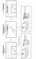
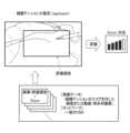
図22は、制御部110が組織テンションを認識する場合の処理例である。なお、図7の処理例と異なる部分を主に説明し、図7の処理例と同様な部分について説明を適宜に省略する。 7. Processing example when recognizing tissue tension Fig. 22 shows a processing example when the
S1Dに示すように、制御部110に内視鏡画像が入力される。S2Daに示すように、制御部110は、機械学習によって調整された組織認識プログラムを実行することで、内視鏡画像から組織テンションを認識する。具体的には、組織テンションは、バイポーラデバイスのジョウに把持された組織にかかるテンションのことである。このテンションは、バイポーラデバイスにより組織が牽引されることで、或いは鉗子等の処置具により組織が牽引されることで発生する。組織に適切なテンションが加えられることで、エネルギーデバイスによる適切な処置が可能になる。しかし、テンションが弱い等の不適切な組織テンションである場合には、エネルギーデバイスによる処置の時間がかかり、熱拡散が生じやすくなる。制御部110は、ジョウに把持された組織にかかるテンションの評価値であるスコアを内視鏡画像から認識する。S2Dbに示すように、制御部110は、内視鏡画像から認識した組織テンションのスコアと閾値とを比較し、その結果を出力する。図22には、組織テンションのスコアが閾値以下である、即ち組織テンションが適切でないと認識された例を示す。As shown in S1D, an endoscopic image is input to the
S3Dに示すように、制御部110は、内視鏡画像から認識された組織テンションに応じて出力変更指示を行う。出力変更指示の手法は図7のS3Aと同様である。As shown in S3D, the
学習段階における教師データ521は、学習用画像として、様々な組織テンションの画像を含む。また教師データ521は、正解データとして、スコアの算出対象である評価領域と、その評価領域の画像から算出されるスコアとを含む。The
図23は、制御部110が組織テンションを認識する場合の第1認識処理例である。制御部110は、リグレッションによる組織テンションの推定を行うことで、組織テンションのスコアを出力する。Figure 23 shows a first recognition processing example in which the
具体的には、制御部110は、学習済みモデルを用いた画像認識処理により、テンション評価の対象となる評価領域を内視鏡画像から検出すると共に、その評価領域内の画像から組織テンションを推定する。制御部110は、内視鏡画像に写る処置において組織に適切なテンションが加えられている場合に高いスコアを出力する。学習段階においては、学習装置500は、評価領域を指定する情報と、組織テンションのスコアとが付された内視鏡画像を教師データとして、学習済みモデルを生成する。又は、教師データは、動画即ち時系列画像であってもよい。例えば、動画には、エネルギーデバイス又は鉗子により組織を牽引する操作が撮影されており、その動画に対して評価領域と1つのスコアが対応付けられる。スコアは、内視鏡画像又は動画に写る組織の色相、彩度、明度、輝度、又は牽引による組織の移動情報等に基づいて定量化される。その定量化により得られるスコアが、学習用の各内視鏡画像又は各動画に付される。Specifically, the
図24は、制御部110が組織テンションを認識する場合の第2認識処理例である。制御部110は、ディテクションによる処置具先端の検出を行い、その検出結果に基づいて評価領域を設定し、その評価領域内の画像に対してリグレッションによる組織テンションの推定を行う。Figure 24 is a second recognition processing example in which the
具体的には、制御部110は、処置具先端の検出を行うための第1学習済みモデルと、組織テンションの推定を行うための第2学習済みモデルとを含む。制御部110は、第1学習済みモデルを用いた画像認識処理により、内視鏡画像からジョウの位置を検出する。制御部110は、検出されたジョウの位置に基づいて、所定ルールに従ってジョウの周囲に評価領域を設定する。所定ルールは、例えば、ジョウの位置を中心とする所定距離内の範囲を評価領域に設定する等のルールである。学習段階においては、学習装置500は、デバイス先端の位置、即ちバイポーラデバイスのジョウの位置を示すアノテーションが付された内視鏡画像を教師データとして、第1学習済みモデルを生成する。制御部110は、第2学習済みモデルを用いた画像認識処理により、評価領域内の画像から組織テンションを推定することで組織テンションのスコアを出力する。学習段階においては、学習装置500は、組織テンションのスコアが付された内視鏡画像又は動画を教師データとして、学習済みモデルを生成する。Specifically, the
図25は、制御部110が組織テンションを認識する場合の出力調整例である。制御部110は、組織テンションの認識結果が、テンションが弱い、又はエネルギーデバイスにより組織を切りにくい条件を示すとき、通常条件よりエネルギー出力を強める。例えば、制御部110は、組織テンションのスコアが閾値以下であるとき、通常条件よりエネルギー出力を強める。制御部110は、組織テンションの認識結果が、上記以外の組織テンションであるとき、エネルギー出力を通常条件から変更しない。Figure 25 shows an example of output adjustment when the
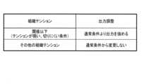
なお、図34に、併用デバイスを用いる場合の出力調整例を示す。出力の欄に複数の出力調整が記載されている場合、制御部110は、そのいずれかの出力調整を行う。なお、制御部110は画像認識処理の結果に基づいてエネルギー出力を調整するが、後述のように、更にデバイス情報を併用してエネルギー出力を調整してもよい。Figure 34 shows an example of output adjustment when using a combined device. If multiple output adjustments are listed in the output column, the
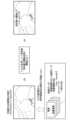
8.処置具先端部と注意対象の距離を認識する場合の処理例
図26は、制御部110が処置具先端部と注意対象の距離を認識する場合の処理例である。なお、図7の処理例と異なる部分を主に説明し、図7の処理例と同様な部分について説明を適宜に省略する。 8. Example of processing when recognizing the distance between the distal end of the treatment tool and the attention target Fig. 26 shows an example of processing when the
S1Eに示すように、制御部110に内視鏡画像が入力される。S2Eaに示すように、制御部110は、機械学習によって調整された組織認識プログラムを実行することで、内視鏡画像から、処置具先端部と注意対象の距離を認識する。具体的には、処置具先端部と注意対象の距離は、内視鏡画像においてジョウの領域内の任意の点と注意対象内の任意の点との距離のうち最短距離のことである。注意対象は、熱拡散の影響に注意すべき臓器、組織、又は器具である。例えば、注意対象は、膵臓、主要血管又はクリップ等である。注意対象が臓器又は組織である場合、その臓器又は組織に対して熱拡散の影響が及ぶ可能性がある。注意対象が器具である場合、熱拡散により高温になった器具が、その器具に接する組織に影響を及ぼす可能性がある。S2Ebに示すように、制御部110は、内視鏡画像から認識した距離と閾値とを比較し、その結果を出力する。図26には、距離が閾値以下である、即ち処置具先端部と注意対象が近いと認識された例を示す。As shown in S1E, an endoscopic image is input to the
S3Eに示すように、制御部110は、内視鏡画像から認識された距離に応じて出力変更指示を行う。出力変更指示の手法は図7のS3Aと同様である。As shown in S3E, the
学習段階における教師データ521は、学習用画像として、ジョウと様々な注意対象が写る画像を含む。また教師データ521は、正解データとして、画像に写るジョウと注意対象の距離を示す距離情報を含む。The
図27は、制御部110が処置具先端部と注意対象の距離を認識する場合の第1認識処理例である。制御部110は、セグメンテーションによる処置具先端及び組織の領域認識と、リグレッションによる2点間の距離の推定とを行う。制御部110は、推定された2点間の距離と閾値とを比較し、2点間の距離が閾値以上であるか閾値未満であるかを判定する。Figure 27 shows a first recognition processing example in which the
具体的には、学習済みモデル121は、処置具先端及び組織の領域認識を行うための第1学習済みモデルと、2点間の距離の推定を行うための第2学習済みモデルとを含む。制御部110は、第1学習済みモデルを用いた画像認識処理により、内視鏡画像からジョウの領域と組織種類毎の領域とクリップの領域を検出する。図27には、内視鏡画像からジョウの領域、膜の領域、膵臓の領域、及び肝臓の領域が検出された例を示す。学習段階においては、学習装置500は、ジョウの領域、組織種類毎の領域、及びクリップの領域にアノテーションが付された内視鏡画像を教師データとして、第1学習済みモデルを生成する。制御部110は、第2学習済みモデルを用いた画像認識処理により、ジョウの先端位置と、注意対象の領域内でジョウの先端位置に最も近い位置との間の距離を推定する。制御部110は、推定された距離と閾値とを比較する。注意対象は、セグメンテーションにより検出された組織、臓器又は器具のうち、予め注意対象と定められた組織、臓器又は器具である。図27には、膵臓が注意対象に設定された例を示す。学習段階においては、学習装置500は、ジョウと注意対象が写ると共にジョウと注意対象の距離を示す距離情報がラベルとして付された内視鏡画像を教師データとして、第2学習済みモデルを生成する。Specifically, the trained
図28は、制御部110が処置具先端部と注意対象の距離を認識する場合の出力調整例である。制御部110は、距離の認識結果が、デバイスと注意対象の距離が近いことを示すとき、通常条件よりエネルギー出力を弱める。例えば、制御部110は、認識された距離が閾値以下であるとき、通常条件よりエネルギー出力を弱める。制御部110は、距離の認識結果が、上記以外の距離であるとき、エネルギー出力を通常条件から変更しない。Figure 28 shows an example of output adjustment when the
なお、図35に、併用デバイスを用いる場合の出力調整例を示す。出力の欄に複数の出力調整が記載されている場合、制御部110は、そのいずれかの出力調整を行う。Figure 35 shows an example of output adjustment when using a combined device. If multiple output adjustments are listed in the output column, the
9.表示例
図29に、エネルギー出力調整に関する情報の表示例を示す。制御部110は、エネルギー出力調整に関する情報を内視鏡画像に重畳して表示画像150を生成し、その表示画像150をモニタ表示する処理を行う。表示画像150は内視鏡システム200のディスプレイ230に表示されてもよいし、或いはナビゲーション表示用に別途設けられたディスプレイに表示されてもよい。 29 shows a display example of information related to energy output adjustment. The
内視鏡画像には、エネルギー出力調整に関する情報として、画像認識処理の認識結果、その認識結果に基づいて選択された出力情報、又はそれら両方が重畳される。画像認識処理の認識結果は、例えば、検出された組織の領域151、ジョウの領域152、又は、ジョウに把持された組織又は組織把持量等を示す文字情報153等である。ジョウの領域152の代わりに、ジョウの位置を示すバウンディングボックスが表示されてもよい。出力情報は、例えば、選択された出力シーケンスを示す画像154である。或いは、出力情報は、選択されたエネルギー出力設定を示すアイコン表示155であってもよい。アイコン表示155は、例えば、複数のエネルギー出力設定を示す複数のアイコンと、決定されたエネルギー出力設定のアイコンを強調する強調表示156と、を含む。On the endoscopic image, the recognition result of the image recognition process, the output information selected based on the recognition result, or both are superimposed as information related to the energy output adjustment. The recognition result of the image recognition process is, for example, the area 151 of the detected tissue, the area 152 of the jaw, or text information 153 indicating the tissue grasped by the jaw or the amount of tissue grasped. Instead of the area 152 of the jaw, a bounding box indicating the position of the jaw may be displayed. The output information is, for example, an image 154 indicating the selected output sequence. Alternatively, the output information may be an icon display 155 indicating the selected energy output setting. The icon display 155 includes, for example, a plurality of icons indicating a plurality of energy output settings and a highlighting display 156 that highlights the icon of the determined energy output setting.
10.画像認識結果とデバイス情報を併用したエネルギー出力調整
以上では、制御部110が画像認識結果に基づいてエネルギー出力調整を行う実施形態を説明したが、制御部110は画像認識結果とデバイス情報を併用してエネルギー出力調整を行ってもよい。図30は、画像認識結果とデバイス情報を併用する場合の処理のフローチャートである。 10. Energy output adjustment using both image recognition results and device information In the above, an embodiment in which the
ステップS21において、制御部110に、内視鏡システム200の本体装置220から内視鏡画像が入力され、エネルギーデバイス310を制御するジェネレータ300からデバイス情報が入力される。具体的には、図2において、I/Oデバイス190がジェネレータ300からデバイス情報を受信し、その受信されたデバイス情報が制御部110に入力されるように構成すればよい。In step S21, an endoscopic image is input to the
ステップS22において、制御部110は、内視鏡画像からエネルギーデバイス310の視認性を判断する。制御部110は、例えば、エネルギーデバイス310が画像の奥行方向を向いている場合、エネルギーデバイス310のジョウが他の処置具で隠れている場合、又は内視鏡視野内のミスト量が一定以上である場合等に、エネルギーデバイス310の視認性が悪いと判断し、それ以外の場合にエネルギーデバイス310の視認性が良いと判断する。In step S22, the
ステップS22においてエネルギーデバイス310の視認性が悪いと判断された場合、ステップS23において、制御部110は、画像認識結果を使用せずにデバイス情報を使用する、と決定する。即ち、視認性が悪い場合には、画像認識結果が信用できないと判断し、デバイス情報の使用を決定する。例えば、制御部110は、画像認識結果に基づくエネルギー出力調整指示をジェネレータ300に出力せず、ジェネレータ300がインピーダンス情報に基づく出力自動オフ等の出力制御を行う。或いは、制御部110は、デバイス情報に基づいてエネルギー出力調整指示をジェネレータ300に出力してもよい。但し、画像認識結果に基づいて組織情報又は処置情報を判断する方が、デバイス情報から組織情報又は処置情報を判断する場合よりも様々な又は高精度な情報判断が可能である。この点については、整合性の説明と合わせて図31~図34を用いて後述する。ステップS22においてエネルギーデバイス310の視認性が良いと判断された場合、ステップS24において、制御部110は、機械学習を用いた画像認識の推定確度が第1閾値以上か否かを判断する。ここでは第1閾値を60%とする。If it is determined in step S22 that the visibility of the
ステップS24において推定確度が60%未満と判断された場合、ステップS23において、制御部110は、画像認識結果を使用せずにデバイス情報を使用する、と決定する。即ち、推定確度が低い場合には、画像認識結果が信用できないと判断し、デバイス情報の使用を決定する。ステップS24において推定確度が60%以上と判断された場合、ステップS25において、制御部110は、推定確度が第2閾値以上か否かを判断する。第2閾値は第1閾値より高い確度である。ここでは第2閾値を90%とする。
If it is determined in stepS24 that the estimation accuracy is less than 60%, then in step S23, the
ステップS25において推定確度が90%以上と判断された場合、ステップS26において、制御部110は画像認識結果に基づいてエネルギー出力調整指示をジェネレータ300に出力する。即ち、推定確度が十分高い場合には、画像認識結果を信用し、画像認識結果を使用する。ジェネレータ300は、エネルギー出力調整指示により設定された出力値又は出力シーケンスでエネルギーデバイス310にエネルギーを供給する。このとき、ジェネレータ300は、エネルギー出力調整指示により設定された出力値又は出力シーケンスのもとで、インピーダンス情報に基づく出力自動オフ等の出力制御を行ってもよい。ステップS25において推定確度が90%未満と判断された場合、ステップS27において、制御部110は、画像認識結果とデバイス情報の整合性を判断する。If the estimation accuracy is determined to be 90% or more in step S25, the
ステップS27において、画像認識結果とデバイス情報の整合性があると判断された場合、ステップS26において、制御部110は画像認識結果に基づいてエネルギー出力調整指示をジェネレータ300に出力する。即ち、画像認識の推定確度が十分には高くない場合であっても、画像認識結果とデバイス情報が整合している場合には、画像認識結果を信用し、画像認識結果を使用する。ステップS27において、画像認識結果とデバイス情報の整合性がないと判断された場合、ステップS23において、制御部110は、画像認識結果を使用せずにデバイス情報を使用する、と決定する。即ち、画像認識結果とデバイス情報が整合しない場合には、画像認識結果が信用できないと判断し、デバイス情報の使用を決定する。If it is determined in step S27 that the image recognition result and the device information are consistent, then in step S26 the
デバイス情報は、エネルギーデバイスの先端部が接する組織の電気的情報であり、例えば、エネルギーデバイスの先端部が接する組織のインピーダンス情報である。例えば、エネルギーデバイスがバイポーラデバイスである場合には、電気的情報は、2つのジョウに把持された組織のインピーダンス情報である。ジェネレータ300は、バイポーラデバイスの2つのジョウに処置用の高周波電力を出力し、その高周波電力の電圧と電流を測定し、その測定された電圧と電流からインピーダンス情報を取得する。但し、電気的情報はインピーダンス情報に限定されず、組織の種類や処置の進行に応じて変化する情報であればよい。電気的情報は、例えば、電流、電圧、又は電流と電圧の間の位相であってもよい。或いは、電気的情報は、電力、電力量、インピーダンス、レジスタンス(抵抗)、リアクタンス、アドミタンス(インピーダンスの逆数)、コンダクタンス(アドミタンスの実数部)、又はサセプタンス(アドミタンスの虚数部)であってもよい。或いは、電気的情報は、上記の経時的変化、各パラメータ間の変化、各パラメータ間の微分積分(Pをパラメータとしたとき、経時的な微分はdP/dtであり、抵抗による微分はdP/dRである)、塊毎の和差など初等演算により導かれる値、又は、それぞれの閾値を跨いだかどうかといったトリガ情報であってもよい。The device information is electrical information of the tissue that the tip of the energy device contacts, for example, impedance information of the tissue that the tip of the energy device contacts. For example, when the energy device is a bipolar device, the electrical information is impedance information of the tissue held by the two jaws. The
画像認識の推定確度は、推定結果がどの程度確からしいかを示す度合いのことである。クラシフィケーションを行うニューラルネットワークを例にとると、出力層には各クラスに対応したノードが設けられており、入力層の入力データが各クラスに該当する確率が出力層の各ノードに出力される。そのうち最も高い確率が出力されたノードのクラスが分類結果として出力されるが、そのノードに出力されている確率が、分類結果の推定確度として用いられる。或いは、機械学習を用いた画像認識処理において精度評価を行う例では、処置の場面毎に予め精度を取得しておき、その場面と精度を組織情報又は処置情報と共に学習モデルに学習させることで学習済みモデルを生成する。この学習済みモデルを用いた画像認識処理により、組織情報又は処置情報の認識結果と共に、その認識精度が出力され、その認識精度が組織情報又は処置情報の推定確度として用いられる。処置の場面は、例えばエネルギーデバイスの把持方向で定義された場面、或いは手技シーンで定義された場面等である。把持方向で定義された場面は、例えば、エネルギーデバイスが内視鏡画像の奥行方向に向いた状態で組織を把持している場面等である。手技シーンで定義された場面は、例えば、幽門下におけるエネルギーデバイスを用いた処理等である。The estimated accuracy of image recognition is the degree to which the estimated result is likely. Taking a neural network that performs classification as an example, the output layer is provided with nodes corresponding to each class, and the probability that the input data of the input layer corresponds to each class is output to each node of the output layer. The class of the node that outputs the highest probability among them is output as the classification result, and the probability output to that node is used as the estimated accuracy of the classification result. Alternatively, in an example of accuracy evaluation in image recognition processing using machine learning, the accuracy is acquired in advance for each treatment scene, and the scene and accuracy are trained in a learning model together with tissue information or treatment information to generate a trained model. The image recognition processing using this trained model outputs the recognition accuracy of the tissue information or treatment information together with the recognition result, and the recognition accuracy is used as the estimated accuracy of the tissue information or treatment information. The treatment scene is, for example, a scene defined by the gripping direction of the energy device, or a scene defined by a procedure scene. The scene defined by the gripping direction is, for example, a scene in which the energy device is gripping tissue while facing the depth direction of the endoscopic image. An example of a scene defined as a procedure scene is treatment using an energy device below the pylorus.
画像認識結果とデバイス情報の整合性は、画像認識情報とデバイス情報の適切な組み合わせが予め決められており、その組み合わせに合致するか否かということである。具体的には、整合性は、画像認識結果が示す組織情報又は処置情報と、デバイス情報が示す組織情報又は処置情報とが整合するか否かを意味する。図31~図34には、画像認識結果とデバイス情報の整合性の例が示されている。例えば図31において、画像認識結果が薄膜又は太い血管であり、デバイス情報が低インピーダンスである場合、画像認識結果とデバイス情報が整合していると判断される。その場合、制御部110は、画像認識結果を信用し、画像認識結果を使用してエネルギー出力調整を行う。低インピーダンスの場合、インピーダンス情報だけでは薄膜と血管が区別されないが、画像認識結果を用いることで、薄膜と血管を区別してエネルギー出力調整できる。逆に、画像認識結果が薄膜又は血管であり、デバイス情報が高インピーダンスである場合、画像認識結果とデバイス情報が整合していないと判断される。その場合、制御部110は、画像認識結果が信用できないと判断し、デバイス情報の使用を決定する。The consistency between the image recognition result and the device information refers to whether or not an appropriate combination of the image recognition information and the device information is determined in advance and matches that combination. Specifically, the consistency means whether or not the tissue information or treatment information indicated by the image recognition result matches the tissue information or treatment information indicated by the device information. Figures 31 to 34 show examples of the consistency between the image recognition result and the device information. For example, in Figure 31, if the image recognition result is a thin film or a thick blood vessel and the device information is low impedance, it is determined that the image recognition result and the device information are consistent. In that case, the
以上に説明した本実施形態のシステム10は、学習済みモデル121を記憶する記憶部120と、制御部110と、を含む。学習済みモデル121は、学習用デバイス組織画像又は学習用組織画像から画像認識情報を出力するように学習される。学習用デバイス組織画像は、エネルギー供給を受けてエネルギー出力を行う少なくとも1つのエネルギーデバイス310及び少なくとも1つの生体組織が撮像された画像である。学習用組織画像は、少なくとも1つの生体組織が撮像された画像である。画像認識情報は、少なくとも1つの生体組織に関する組織情報、又は少なくとも1つの生体組織に対する処置に関する処置情報の少なくとも1つである。制御部110は、少なくとも1つのエネルギーデバイス310及び少なくとも1つの生体組織が撮像された画像である撮像画像を取得する。制御部110は、記憶部120に記憶された学習済みモデル121に基づく処理により、撮像画像から画像認識情報を推定する。制御部110は、推定した画像認識情報に基づくエネルギー出力調整指示をジェネレータ300に対して出力する。ジェネレータ300は、エネルギーデバイス310へのエネルギー供給量をエネルギー出力調整指示に基づいて制御する。The
本実施形態によれば、撮像画像から画像認識された組織情報又は処置情報に基づいてエネルギーデバイス310のエネルギー出力が調整される。これにより、インピーダンス情報等のデバイス情報のみからでは判別できない様々な情報に基づいて、エネルギー出力を調整することが可能になる。例えば、図31等で上述したように、インピーダンス情報では区別できない組織種類を画像認識によって判別可能になり、その判別した組織種類に応じたエネルギー出力調整が可能になる。According to this embodiment, the energy output of the
そして、画像認識された様々な情報を用いることで、エネルギーデバイス310による処置における熱拡散を考慮したエネルギー出力調整が可能になる。例えば、画像認識された組織情報又は処置情報から予想される熱拡散が大きい場合にはエネルギー出力を下げる等のエネルギー出力調整が可能になる。デバイス情報を用いる場合よりも詳細な情報判別が可能であることから、様々な熱拡散の状況を考慮したエネルギー出力調整が可能になる。なお、画像認識情報、組織情報、処置情報、及びエネルギー出力調整指示については、例えば「1.システム」で説明されている。By using various image-recognized information, it becomes possible to adjust the energy output taking into account thermal diffusion during treatment by the
また本実施形態では、制御部110は、画像認識情報に基づいて、エネルギー出力を基準エネルギー出力から増加させる、減少させる又は維持する調整のいずれかを決定し、決定した調整の指示をエネルギー出力調整指示として出力してもよい。In addition, in this embodiment, the
本実施形態によれば、画像認識情報に基づいてエネルギー出力が増加、減少又は維持されることで、画像認識情報から予想される熱拡散に応じてエネルギー出力を増加、減少又は維持できる。例えば、エネルギー印加時間が長くなることが画像認識情報から予想される場合には、エネルギー出力を増加させることで、エネルギー印加時間を短くして熱拡散を低減できる。なお、「エネルギー出力を基準エネルギー出力から増加させる、減少させる又は維持する調整」については、例えば「4.組織種類を認識する場合の処理例」で説明されている。According to this embodiment, the energy output is increased, decreased or maintained based on the image recognition information, so that the energy output can be increased, decreased or maintained according to the thermal diffusion predicted from the image recognition information. For example, when the image recognition information predicts that the energy application time will be long, the energy output can be increased to shorten the energy application time and reduce thermal diffusion. Note that "adjusting the energy output to increase, decrease or maintain it from the reference energy output" is described, for example, in "4. Processing example when recognizing tissue type."
また本実施形態では、制御部110は、予め設定されたエネルギー出力、又はジェネレータ300のリアルタイムのエネルギー出力を基準エネルギー出力として、エネルギー出力調整指示を出力してもよい。In addition, in this embodiment, the
「予め設定されたエネルギー出力」とは、エネルギー出力調整の基準エネルギー出力として予め設定されており、リアルタイムのエネルギー出力とは関係無く決まった基準エネルギー出力が用いられるという意味である。「リアルタイムのエネルギー出力」とは、制御部110がエネルギー出力調整指示を出力するときに、ジェネレータ300に設定されていたエネルギー出力のことである。即ち、その基準エネルギー出力は毎回異なる可能性がある。"Preset energy output" means that the standard energy output is preset as the standard energy output for energy output adjustment, and a fixed standard energy output is used regardless of the real-time energy output. "Real-time energy output" means the energy output that was set in the
また本実施形態では、制御部110は、内視鏡210からの内視鏡画像を撮像画像として取得し、内視鏡画像を取得した時点において設定されているエネルギー出力を基準エネルギー出力として、エネルギー出力調整指示を出力してもよい。In addition, in this embodiment, the
「内視鏡画像を取得した時点において設定されているエネルギー出力」とは、内視鏡画像から画像認識した情報に基づいてエネルギー出力調整指示が出力されるときに、その内視鏡画像を取得した時点において設定されていたエネルギー出力のことである。"Energy output set at the time the endoscopic image was acquired" refers to the energy output that was set at the time the endoscopic image was acquired when an energy output adjustment instruction was output based on image recognition information from the endoscopic image.
また本実施形態では、エネルギーデバイス310は、組織を把持可能な2つのジョウを有するデバイスであり、ジェネレータ300からエネルギー供給を受けて2つのジョウからエネルギー出力を行うデバイスであってもよい。In addition, in this embodiment, the
即ち、エネルギーデバイス310は、バイポーラデバイス330であってもよい。バイポーラデバイスについては、例えば「3.エネルギーデバイス」の図5で説明されている。That is, the
また本実施形態では、組織情報は、少なくとも1つのエネルギーデバイスにより処置される組織の組織種類又は組織状態を含んでもよい。In this embodiment, the tissue information may also include the tissue type or tissue condition of the tissue to be treated by at least one energy device.
本実施形態によれば、撮像画像から画像認識された組織種類又は組織状態に基づいてエネルギーデバイス310のエネルギー出力が調整される。エネルギーデバイス310により処置されるときの熱拡散の度合いは、組織種類又は組織状態に応じて異なっている。画像認識された組織種類又は組織状態を用いることで、その組織種類又は組織状態における熱拡散を考慮したエネルギー出力調整が可能になる。なお、組織種類については、例えば「4.組織種類を認識する場合の処理例」で説明されている。組織状態については、例えば「5.組織状態を認識する場合の処理例」で説明されている。According to this embodiment, the energy output of the
また本実施形態では、処置情報は、少なくとも1つのエネルギーデバイスによる組織の把持量、又は、少なくとも1つのエネルギーデバイス又はその他のデバイスによる組織の牽引量を含んでもよい。In this embodiment, the treatment information may also include the amount of tissue gripped by at least one energy device or the amount of tissue retracted by at least one energy device or other device.
本実施形態によれば、撮像画像から画像認識された把持量又は牽引量に基づいてエネルギーデバイス310のエネルギー出力が調整される。エネルギーデバイス310により処置されるときの熱拡散の度合いは、把持量又は牽引量に応じて異なっている。画像認識された把持量又は牽引量を用いることで、その把持量又は牽引量における熱拡散を考慮したエネルギー出力調整が可能になる。なお、把持量については、例えば「6.組織把持量を認識する場合の処理例」で説明されている。牽引については、例えば「7.組織テンションを認識する場合の処理例」で説明されている。According to this embodiment, the energy output of the
また本実施形態では、処置情報は、少なくとも1つのエネルギーデバイスにより処置される組織のテンション、又は少なくとも1つのエネルギーデバイスと注意対象との距離を含んでもよい。In this embodiment, the treatment information may also include tension of the tissue being treated by at least one energy device, or distance between at least one energy device and the object of attention.
本実施形態によれば、撮像画像から画像認識された組織のテンション又はエネルギーデバイスと注意対象との距離に基づいてエネルギーデバイス310のエネルギー出力が調整される。エネルギーデバイス310により処置されるときの熱拡散の度合いは、組織のテンション又はエネルギーデバイスと注意対象との距離に応じて異なっている。画像認識された組織のテンション又はエネルギーデバイスと注意対象との距離を用いることで、その組織のテンション又はエネルギーデバイスと注意対象との距離における熱拡散を考慮したエネルギー出力調整が可能になる。なお、組織のテンションについては、例えば「7.組織テンションを認識する場合の処理例」で説明されている。エネルギーデバイスと注意対象との距離については、例えば「8.処置具先端部と注意対象の距離を認識する場合の処理例」で説明されている。According to this embodiment, the energy output of the
また本実施形態では、制御部110は、画像認識情報を推定するときの推定確度に基づいて、エネルギー出力の制御における、画像認識情報と少なくとも1つのエネルギーデバイス310から得られる電気的情報との使用優先度を変更してもよい。In addition, in this embodiment, the
本実施形態によれば、画像認識情報に基づくエネルギー出力調整が行われることと、画像認識情報に基づくエネルギー出力調整が行われずに電気的情報に基づくエネルギー出力の制御が行われることとの、優先度が、推定確度に応じて変更される。これにより、画像認識情報が信用できるか否かに応じて、上記のいずれを優先するかを制御できる。なお、「エネルギー出力の制御」は必ずしも制御部110が行う必要はない。例えば、画像認識情報を優先する場合には、制御部110が画像認識情報に基づいてエネルギー出力調整指示を出力し、電気的情報を優先する場合には、ジェネレータ300が電気的情報に基づいてエネルギー出力を制御してもよい。この後者の場合、制御部110は、画像認識情報に基づくエネルギー出力調整指示を出力しないことを決定することで、電気的情報の使用を優先していることになる。なお、使用優先度の変更については、例えば「10.画像認識結果とデバイス情報を併用したエネルギー出力調整」で説明されている。According to this embodiment, the priority between performing energy output adjustment based on image recognition information and performing energy output control based on electrical information without performing energy output adjustment based on image recognition information is changed according to the estimated accuracy. This allows control of which of the above is given priority depending on whether the image recognition information is reliable or not. Note that the "control of energy output" does not necessarily have to be performed by the
また本実施形態では、制御部110は、少なくとも1つのエネルギーデバイス310からの電気的情報を取得してもよい。制御部110は、画像認識情報と電気的情報の整合性に基づいて、エネルギー出力の制御における画像認識情報と電気的情報の使用優先度を変更してもよい。In this embodiment, the
本実施形態によれば、画像認識情報と電気的情報の整合性に基づいて、画像認識情報が信用できるか否かを判断できる。これにより、画像認識情報が信用できるか否かに応じて、画像認識情報と電気的情報のいずれを優先するかを制御できる。「画像認識情報と電気的情報の整合性」については、例えば「10.画像認識結果とデバイス情報を併用したエネルギー出力調整」で説明されている。According to this embodiment, it is possible to determine whether the image recognition information is trustworthy or not based on the consistency between the image recognition information and the electrical information. This makes it possible to control whether the image recognition information or the electrical information is given priority depending on whether the image recognition information is trustworthy or not. "Consistency between image recognition information and electrical information" is explained, for example, in "10. Energy output adjustment using both image recognition results and device information."
また本実施形態では、制御部110は、撮像画像と、画像認識情報の内容又はエネルギー出力調整指示の内容のうち少なくとも1つとを表示部に表示する処理を行う。In addition, in this embodiment, the
本実施形態によれば、ユーザは、表示部に表示された画像認識情報の内容又はエネルギー出力調整指示の内容のうち少なくとも1つを見ることで、エネルギー出力の自動制御において装置内部で処理された内容を知ることができる。なお、情報が表示される表示部は、内視鏡システム200のディスプレイ230、或いは、それ以外に設けられるディスプレイのいずれであってもよい。表示については、例えば「9.表示例」で説明されている。According to this embodiment, the user can know the contents processed inside the device in the automatic control of the energy output by looking at at least one of the contents of the image recognition information or the contents of the energy output adjustment instructions displayed on the display unit. The display unit on which the information is displayed may be either the
また本実施形態では、学習済みモデル121は、第1学習済みモデルと第2学習済みモデルを含んでもよい。第1学習済みモデルは、学習用デバイス組織画像から、少なくとも1つのエネルギーデバイスの先端部を示すバウンディングボックスを検出するように学習される。第2学習済みモデルは、バウンディングボックス内の学習用デバイス組織画像から画像認識情報を出力するように学習される。制御部110は、第1学習済みモデルに基づく処理により、撮像画像からバウンディングボックスを検出し、第2学習済みモデルに基づく処理により、バウンディングボックス内の撮像画像から画像認識情報を推定する。In this embodiment, the trained
本実施形態によれば、機械学習を用いたディテクションとクラシフィケーション又はリグレッションとの組み合わせにより、撮像画像から組織情報又は処置情報を画像認識できる。なお、本処理については、例えば「4.組織種類を認識する場合の処理例」の図10、「5.組織状態を認識する場合の処理例」の図14、「6.組織把持量を認識する場合の処理例」の図19、又は「7.組織テンションを認識する場合の処理例」の図24等で説明されている。According to this embodiment, by combining detection using machine learning with classification or regression, tissue information or treatment information can be image-recognized from a captured image. This process is described, for example, in FIG. 10 in "4. Example of a process for recognizing tissue type," FIG. 14 in "5. Example of a process for recognizing tissue state," FIG. 19 in "6. Example of a process for recognizing tissue gripping amount," or FIG. 24 in "7. Example of a process for recognizing tissue tension."
また本実施形態では、学習済みモデル121は、学習用デバイス組織画像又は学習用組織画像から、少なくとも1つの生体組織の各生体組織の領域を検出し、学習用デバイス組織画像から、少なくとも1つのエネルギーデバイスの先端部領域を検出するように学習したモデルであってもよい。制御部110は、学習済みモデル121に基づく処理により、撮像画像から各生体組織の領域と先端部領域を検出し、検出した各生体組織の領域と先端部領域に基づいて画像認識情報を推定してもよい。In this embodiment, the trained
本実施形態によれば、機械学習を用いたセグメンテーションにより、撮像画像から組織情報又は処置情報を画像認識できる。なお、本処理については、例えば「4.組織種類を認識する場合の処理例」の図9、又は「5.組織状態を認識する場合の処理例」の図13~図15等で説明されている。According to this embodiment, tissue information or treatment information can be image-recognized from a captured image by segmentation using machine learning. This process is described, for example, in FIG. 9 in "4. Processing example for recognizing tissue type" or in FIG. 13 to FIG. 15 in "5. Processing example for recognizing tissue state."
また本実施形態では、システム10は、少なくとも1つのエネルギーデバイス310と、ジェネレータ300と、を含んでもよい。In this embodiment, the
また本実施形態では、システム10は、撮像画像として内視鏡画像を撮像する内視鏡210を含んでもよい。制御部110は、内視鏡210から内視鏡画像を取得し、学習済みモデル121に基づく処理により内視鏡画像から画像認識情報を推定してもよい。In this embodiment, the
また以上の処理はプログラムとして記述されてもよい。即ち、本実施形態のプログラムは、撮像画像を取得することと、学習済みモデル121に基づく処理により撮像画像から画像認識情報を推定することと、推定した画像認識情報に基づくエネルギー出力調整指示をジェネレータ300に対して出力することと、をコンピュータに実行させる。The above processing may also be written as a program. That is, the program of this embodiment causes a computer to acquire a captured image, estimate image recognition information from the captured image by processing based on the trained
また以上の処理はエネルギー出力調整方法として実行されてもよい。即ち、本実施形態のエネルギー出力調整方法は、撮像画像を取得することと、学習済みモデル121に基づく処理により撮像画像から画像認識情報を推定することと、推定した画像認識情報に基づくエネルギー出力調整指示をジェネレータ300に対して出力することと、を含む。The above processing may also be executed as an energy output adjustment method. That is, the energy output adjustment method of this embodiment includes acquiring a captured image, estimating image recognition information from the captured image by processing based on the trained
以上、本実施形態及びその変形例について説明したが、本開示は、各実施形態やその変形例そのままに限定されるものではなく、実施段階では、要旨を逸脱しない範囲内で構成要素を変形して具体化することができる。また、上記した各実施形態や変形例に開示されている複数の構成要素を適宜組み合わせることができる。例えば、各実施形態や変形例に記載した全構成要素からいくつかの構成要素を削除してもよい。更に、異なる実施の形態や変形例で説明した構成要素を適宜組み合わせてもよい。このように、本開示の主旨を逸脱しない範囲内において種々の変形や応用が可能である。また、明細書又は図面において、少なくとも一度、より広義又は同義な異なる用語と共に記載された用語は、明細書又は図面のいかなる箇所においても、その異なる用語に置き換えることができる。Although the present embodiment and its modified examples have been described above, the present disclosure is not limited to each embodiment or its modified example as it is, and in the implementation stage, the components can be modified and embodied within the scope of the gist. In addition, multiple components disclosed in each of the above-mentioned embodiments and modified examples can be appropriately combined. For example, some components may be deleted from all the components described in each embodiment or modified example. Furthermore, components described in different embodiments or modified examples may be appropriately combined. In this way, various modifications and applications are possible within the scope of the gist of the present disclosure. In addition, a term described at least once in the specification or drawings together with a different term having a broader meaning or the same meaning can be replaced with that different term anywhere in the specification or drawings.
10 システム、100 コントローラ、110 制御部、111 画像取得部、112 組織情報認識部、113 出力設定部、120 記憶部、121 モデル、150 表示画像、151,152 領域、153 文字情報、154 画像、155 アイコン表示、156 強調表示、180,190 I/Oデバイス、200 内視鏡システム、210 内視鏡、220 本体装置、230 ディスプレイ、300 ジェネレータ、310 エネルギーデバイス、320 モノポーラデバイス、321 電極、322 挿入部、323 操作部、325 ケーブル、330 バイポーラデバイス、331 先端部、332 挿入部、333 操作部、335 ケーブル、336 基端、337,338 ジョウ、340 超音波デバイス、341 先端部、342 挿入部、343 操作部、344a,344b 操作ボタン、345 ケーブル、346 基端、347 ジョウ、348 プローブ、500 学習装置、510 処理部、520 記憶部、521 教師データ、522 学習モデル10 System, 100 Controller, 110 Control unit, 111 Image acquisition unit, 112 Tissue information recognition unit, 113 Output setting unit, 120 Memory unit, 121 Model, 150 Display image, 151, 152 Area, 153 Text information, 154 Image, 155 Icon display, 156 Highlighting, 180, 190 I/O device, 200 Endoscope system, 210 Endoscope, 220 Main unit, 230 Display, 300 Generator, 310 Energy device, 320 Monopolar device, 321 Electrode, 322 Insertion unit, 323 Operation unit, 325 Cable, 330 Bipolar device, 331 Tip, 332 Insertion unit, 333 Operation unit, 335 Cable, 336 Base end, 337, 338 Jaw, 340 Ultrasonic device, 341 Distal end portion, 342 Insertion portion, 343 Operation portion, 344a, 344b Operation button, 345 Cable, 346 Base end, 347 Jaw, 348 Probe, 500 Learning device, 510 Processing portion, 520 Memory portion, 521 Teacher data, 522 Learning model
Claims (17)
Translated fromJapanese制御部と、
を含み、
前記制御部は、
前記少なくとも1つのエネルギーデバイス及び前記少なくとも1つの生体組織が撮像された画像である撮像画像を取得し、
前記記憶部に記憶された前記学習済みモデルに基づく処理により、前記撮像画像から前記画像認識情報を推定し、
推定した前記画像認識情報に基づくエネルギー出力調整指示を、エネルギーデバイスへのエネルギー供給量を前記エネルギー出力調整指示に基づいて制御するジェネレータに対して、出力し、
前記学習済みモデルは、
前記学習用デバイス組織画像から、前記少なくとも1つのエネルギーデバイスの先端部を示すバウンディングボックスを検出するように学習した第1学習済みモデルと、
前記バウンディングボックス内の前記学習用デバイス組織画像から前記画像認識情報を出力するように学習した第2学習済みモデルと、
を含み、
前記制御部は、
前記推定において、前記第1学習済みモデルに基づく処理により、前記撮像画像から前記バウンディングボックスを検出し、前記第2学習済みモデルに基づく処理により、前記バウンディングボックス内の前記撮像画像から前記画像認識情報を推定することを特徴とするシステム。 a storage unit that stores a trained model that has been trained to output image recognition information, which is at least one of tissue information regarding the at least one living tissue or treatment information regarding a treatment for the at least one living tissue, from a learning device tissue image that is an image of at least one energy device that receives energy supply and outputs energy and at least one living tissue, or a learning tissue image that is an image of the at least one living tissue;
A control unit;
Including,
The control unit is
acquiring an image of the at least one energy device and the at least one biological tissue;
Estimating the image recognition information from the captured image by processing based on the trained model stored in the storage unit;
outputting an energy output adjustment instruction based on the estimated image recognition information to a generator that controls an amount of energy supplied to an energy device based on the energy output adjustment instruction;
The trained model is
a first trained model trained to detect a bounding box indicating a tip of the at least one energy device from the training device tissue image;
a second trained model trained to output the image recognition information from the training device tissue image within the bounding box;
Including,
The control unit is
A system characterized in that, in the estimation, the bounding box is detected from the captured image by processing based on the first trained model, and the image recognition information is estimated from the captured image within the bounding box by processing based on the second trained model.
前記制御部は、
前記画像認識情報に基づいて、前記エネルギー出力を基準エネルギー出力から増加させる、減少させる又は維持する調整のいずれかを決定し、
前記決定した調整の指示を前記エネルギー出力調整指示として出力することを特徴とするシステム。 2. The system of claim 1,
The control unit is
determining an adjustment to either increase, decrease, or maintain the energy output from a baseline energy output based on the image recognition information;
The system outputs the determined adjustment instruction as the energy output adjustment instruction.
前記制御部は、
予め設定されたエネルギー出力、又は前記ジェネレータのリアルタイムの前記エネルギー出力を前記基準エネルギー出力として、前記エネルギー出力調整指示を出力することを特徴とするシステム。 3. The system according to claim 2,
The control unit is
A system that outputs the energy output adjustment instruction based on a preset energy output or the real-time energy output of the generator as the reference energy output.
前記制御部は、
内視鏡からの内視鏡画像を前記撮像画像として取得し、前記内視鏡画像を取得した時点において設定されている前記エネルギー出力を前記基準エネルギー出力として、前記エネルギー出力調整指示を出力することを特徴とするシステム。 3. The system according to claim 2,
The control unit is
A system characterized by acquiring an endoscopic image from an endoscope as the captured image, setting the energy output set at the time the endoscopic image was acquired as the reference energy output, and outputting the energy output adjustment instruction.
前記エネルギーデバイスは、
組織を把持可能な2つのジョウを有するデバイスであり、前記ジェネレータから前記エネルギー供給を受けて前記2つのジョウからエネルギー出力を行うデバイスであることを特徴とするシステム。 2. The system of claim 1,
The energy device comprises:
A system comprising a device having two jaws capable of grasping tissue, the device receiving the energy supply from the generator and outputting energy from the two jaws.
前記組織情報は、前記少なくとも1つのエネルギーデバイスにより処置される組織の組織種類又は組織状態を含むことを特徴とするシステム。 2. The system of claim 1,
The system, wherein the tissue information includes a tissue type or tissue condition of the tissue to be treated by the at least one energy device.
前記処置情報は、前記少なくとも1つのエネルギーデバイスによる組織の把持量、又は、前記少なくとも1つのエネルギーデバイス又はその他のデバイスによる組織の牽引量を含むことを特徴とするシステム。 2. The system of claim 1,
The system, wherein the treatment information includes an amount of tissue gripped by the at least one energy device or an amount of tissue retracted by the at least one energy device or other device.
前記処置情報は、前記少なくとも1つのエネルギーデバイスにより処置される組織のテンション、又は前記少なくとも1つのエネルギーデバイスと注意対象との距離を含むことを特徴とするシステム。 2. The system of claim 1,
The system, wherein the treatment information includes tension of tissue being treated by the at least one energy device or a distance between the at least one energy device and an object of interest.
前記制御部は、
前記画像認識情報を推定するときの推定確度に基づいて、前記エネルギー出力の制御における、前記画像認識情報と前記少なくとも1つのエネルギーデバイスから得られる電気的情報との使用優先度を変更することを特徴とするシステム。 2. The system of claim 1,
The control unit is
A system characterized by changing the usage priority of the image recognition information and electrical information obtained from at least one energy device in controlling the energy output based on the estimation accuracy when estimating the image recognition information.
前記制御部は、
前記少なくとも1つのエネルギーデバイスからの電気的情報を取得し、
前記画像認識情報と前記電気的情報との整合性に基づいて、前記エネルギー出力の制御における前記画像認識情報と前記電気的情報の使用優先度を変更することを特徴とするシステム。 2. The system of claim 1,
The control unit is
obtaining electrical information from the at least one energy device;
A system that changes the priority of use of the image recognition information and the electrical information in controlling the energy output based on the consistency between the image recognition information and the electrical information.
前記制御部は、
前記撮像画像と、前記画像認識情報の内容又は前記エネルギー出力調整指示の内容のうち少なくとも1つとを表示部に表示する処理を行うことを特徴とするシステム。 2. The system of claim 1,
The control unit is
A system that performs processing to display the captured image and at least one of the contents of the image recognition information and the contents of the energy output adjustment instructions on a display unit.
前記学習済みモデルは、
前記学習用デバイス組織画像又は前記学習用組織画像から、前記少なくとも1つの生体組織の各生体組織の領域を検出し、前記学習用デバイス組織画像から、前記少なくとも1つのエネルギーデバイスの先端部領域を検出するように学習したモデルであり、
前記制御部は、
前記学習済みモデルに基づく処理により、前記撮像画像から前記各生体組織の領域と前記先端部領域を検出し、
検出した前記各生体組織の領域と前記先端部領域に基づいて前記画像認識情報を推定することを特徴とするシステム。 2. The system of claim 1,
The trained model is
a model that is trained to detect a region of each biological tissue of the at least one biological tissue from the training device tissue image or the training tissue image, and to detect a tip region of the at least one energy device from the training device tissue image;
The control unit is
Detecting the region of each biological tissue and the tip region from the captured image by processing based on the trained model;
A system that estimates the image recognition information based on the detected region of each biological tissue and the tip region.
前記少なくとも1つのエネルギーデバイスと、
前記ジェネレータと、
を含むことを特徴とするシステム。 2. The system of claim 1,
said at least one energy device;
The generator;
A system comprising:
前記撮像画像として内視鏡画像を撮像する内視鏡を含み、
前記制御部は、
前記内視鏡から前記内視鏡画像を取得し、前記学習済みモデルに基づく処理により前記内視鏡画像から前記画像認識情報を推定することを特徴とするシステム。 14. The system according to claim 13,
an endoscope that captures an endoscopic image as the captured image,
The control unit is
A system characterized by acquiring an endoscopic image from the endoscope and estimating the image recognition information from the endoscopic image by processing based on the trained model.
前記少なくとも1つのエネルギーデバイス及び前記少なくとも1つの生体組織が撮像された画像である学習用デバイス組織画像、又は前記少なくとも1つの生体組織が撮像された画像である学習用組織画像から、前記少なくとも1つの生体組織に関する組織情報、又は前記少なくとも1つの生体組織に対する処置に関する処置情報の少なくとも1つである画像認識情報を出力するように学習した学習済みモデルに基づく処理により、前記撮像画像から前記画像認識情報を推定することと、
推定した前記画像認識情報に基づくエネルギー出力調整指示を、エネルギーデバイスへのエネルギー供給量を前記エネルギー出力調整指示に基づいて制御するジェネレータに対して、出力することと、
前記学習済みモデルは、前記学習用デバイス組織画像から、前記少なくとも1つのエネルギーデバイスの先端部を示すバウンディングボックスを検出するように学習した第1学習済みモデルと、前記バウンディングボックス内の前記学習用デバイス組織画像から前記画像認識情報を出力するように学習した第2学習済みモデルと、を含み、前記推定において、前記学習済みモデルの前記第1学習済みモデルに基づく処理により、前記撮像画像から前記バウンディングボックスを検出し、前記第2学習済みモデルに基づく処理により、前記バウンディングボックス内の前記撮像画像から前記画像認識情報を推定することと、
をコンピュータに実行させることを特徴とするプログラム。 acquiring an image of at least one energy device that receives energy supply and outputs energy and at least one living tissue;
From a learning device tissue image, which is an image of the at least one energy device and the at least one biological tissue, or a learning tissue image, which is an image of the at least one biological tissue, an image recognition information is output, which is at least one of tissue information regarding the at least one biological tissue and treatment information regarding a treatment for the at least one biological tissue. The image recognition information is estimated from the captured image by processing based on a trained model that has been trained to output the image recognition information, which is at least one of tissue information regarding the at least one biological tissue and treatment information regarding a treatment for the at least one biological tissue;
outputting an energy output adjustment instruction based on the estimated image recognition information to a generator that controls an amount of energy supply to an energy device based on the energy output adjustment instruction;
The trained model includes a first trained model trained to detect a bounding box indicating a tip of the at least one energy device from the training device tissue image, and a second trained model trained to output the image recognition information from the training device tissue image within the bounding box, and in the estimation, the bounding box is detected from the captured image by processing based on the first trained model of the trained model, and the image recognition information is estimated from the captured image within the bounding box by processing based on the second trained model;
A program characterized by causing a computer to execute the above.
前記画像認識情報に基づいて、前記エネルギー出力を基準エネルギー出力から増加させる、減少させる又は維持する調整のいずれかを決定することと、
前記決定した調整の指示を前記エネルギー出力調整指示として出力することと、
をコンピュータに実行させることを特徴とするプログラム。 The program according to claim 15,
determining an adjustment based on the image recognition information to either increase, decrease, or maintain the energy output from a baseline energy output;
outputting the determined adjustment instruction as the energy output adjustment instruction;
A program characterized by causing a computer to execute the above.
予め設定されたエネルギー出力、又は前記ジェネレータのリアルタイムの前記エネルギー出力を前記基準エネルギー出力として、前記エネルギー出力調整指示を出力すること、
をコンピュータに実行させることを特徴とするプログラム。 The program according to claim 16,
outputting the energy output adjustment instruction based on a preset energy output or the real-time energy output of the generator as the reference energy output;
A program characterized by causing a computer to execute the above.
Applications Claiming Priority (5)
| Application Number | Priority Date | Filing Date | Title |
|---|---|---|---|
| US202163221128P | 2021-07-13 | 2021-07-13 | |
| US63/221,128 | 2021-07-13 | ||
| US202163222252P | 2021-07-15 | 2021-07-15 | |
| US63/222,252 | 2021-07-15 | ||
| PCT/JP2022/009691WO2023286334A1 (en) | 2021-07-13 | 2022-03-07 | System, program, and energy output regulation method |
Publications (3)
| Publication Number | Publication Date |
|---|---|
| JPWO2023286334A1 JPWO2023286334A1 (en) | 2023-01-19 |
| JPWO2023286334A5 JPWO2023286334A5 (en) | 2023-11-07 |
| JP7493684B2true JP7493684B2 (en) | 2024-05-31 |
Family
ID=84919163
Family Applications (4)
| Application Number | Title | Priority Date | Filing Date |
|---|---|---|---|
| JP2023535109AActiveJP7493684B2 (en) | 2021-07-13 | 2022-03-07 | System and Program |
| JP2023535110AActiveJP7465413B2 (en) | 2021-07-13 | 2022-03-07 | System, program and information processing method |
| JP2023535112AActiveJP7602646B2 (en) | 2021-07-13 | 2022-03-07 | System, program and information processing method |
| JP2023535111AActiveJP7602645B2 (en) | 2021-07-13 | 2022-03-07 | system |
Family Applications After (3)
| Application Number | Title | Priority Date | Filing Date |
|---|---|---|---|
| JP2023535110AActiveJP7465413B2 (en) | 2021-07-13 | 2022-03-07 | System, program and information processing method |
| JP2023535112AActiveJP7602646B2 (en) | 2021-07-13 | 2022-03-07 | System, program and information processing method |
| JP2023535111AActiveJP7602645B2 (en) | 2021-07-13 | 2022-03-07 | system |
Country Status (3)
| Country | Link |
|---|---|
| US (4) | US20230380695A1 (en) |
| JP (4) | JP7493684B2 (en) |
| WO (4) | WO2023286336A1 (en) |
Families Citing this family (4)
| Publication number | Priority date | Publication date | Assignee | Title |
|---|---|---|---|---|
| JP2025058623A (en)* | 2023-09-28 | 2025-04-09 | オムロン株式会社 | Model generation method and inference program |
| WO2025115599A1 (en) | 2023-11-28 | 2025-06-05 | オリンパスメディカルシステムズ株式会社 | Assistance device, assistance method, and recording medium |
| EP4574007A1 (en)* | 2023-12-22 | 2025-06-25 | Olympus Winter & Ibe GmbH | Medical camera system and method for capturing images and processing said images |
| CN118680657B (en)* | 2024-06-05 | 2025-06-13 | 河北壹雪制冷科技有限公司 | A freezing pen control device based on sensor feedback |
Citations (2)
| Publication number | Priority date | Publication date | Assignee | Title |
|---|---|---|---|---|
| US20170252095A1 (en) | 2016-03-01 | 2017-09-07 | Covidien Lp | Surgical instruments and systems incorporating machine learning based tissue identification and methods thereof |
| WO2020051444A1 (en) | 2018-09-07 | 2020-03-12 | Ethicon Llc | Grounding arrangement of energy modules |
Family Cites Families (14)
| Publication number | Priority date | Publication date | Assignee | Title |
|---|---|---|---|---|
| US9259287B2 (en) | 2013-04-02 | 2016-02-16 | Siemens Aktiengesellschaft | Patient specific planning and simulation of ablative procedures |
| JP6776258B2 (en)* | 2015-04-02 | 2020-10-28 | ハートフロー, インコーポレイテッド | Systems and methods that provide personalized estimates of bioheat transfer |
| WO2017090165A1 (en)* | 2015-11-26 | 2017-06-01 | オリンパス株式会社 | Treatment system and treatment instrument |
| US11908584B2 (en)* | 2017-05-15 | 2024-02-20 | Ne Scientific, Llc | Methods and systems for modeling a necrotized tissue volume in an ablation procedure |
| CN119184842A (en)* | 2018-02-23 | 2024-12-27 | 生物相容英国有限公司 | Volumetric imaging |
| JP7043317B2 (en)* | 2018-03-29 | 2022-03-29 | 富士フイルムヘルスケア株式会社 | Medical system |
| WO2020095389A1 (en)* | 2018-11-07 | 2020-05-14 | オリンパス株式会社 | Medical device, residual-heat determination method, and residual-heat determination program |
| CN113164196A (en)* | 2018-11-20 | 2021-07-23 | 皇家飞利浦有限公司 | Temporal mapping of thermal ablation zones |
| US20210038314A1 (en)* | 2019-08-11 | 2021-02-11 | phenoMapper, LLC | Method and System for Personalized Treatment Planning in Ablation of Cancerous Tissue |
| JP2021029258A (en)* | 2019-08-13 | 2021-03-01 | ソニー株式会社 | Surgery support system, surgery support method, information processing device, and information processing program |
| JP2021029979A (en)* | 2019-08-29 | 2021-03-01 | 国立研究開発法人国立がん研究センター | Teaching data generation device, teaching data generation program, and teaching data generation method |
| US20210085387A1 (en)* | 2019-09-22 | 2021-03-25 | Biosense Webster (Israel) Ltd. | Guiding cardiac ablation using machine learning (ml) |
| JP7438731B2 (en)* | 2019-11-29 | 2024-02-27 | キヤノンメディカルシステムズ株式会社 | Medical image processing device, operating method of the medical image processing device, and medical image processing program |
| JP7482690B2 (en)* | 2020-06-04 | 2024-05-14 | リンナイ株式会社 | Cooking system, cooking method, learning device and learning method |
- 2022
- 2022-03-07JPJP2023535109Apatent/JP7493684B2/enactiveActive
- 2022-03-07JPJP2023535110Apatent/JP7465413B2/enactiveActive
- 2022-03-07WOPCT/JP2022/009693patent/WO2023286336A1/ennot_activeCeased
- 2022-03-07JPJP2023535112Apatent/JP7602646B2/enactiveActive
- 2022-03-07WOPCT/JP2022/009691patent/WO2023286334A1/ennot_activeCeased
- 2022-03-07WOPCT/JP2022/009694patent/WO2023286337A1/ennot_activeCeased
- 2022-03-07JPJP2023535111Apatent/JP7602645B2/enactiveActive
- 2022-03-07WOPCT/JP2022/009692patent/WO2023286335A1/ennot_activeCeased
- 2023
- 2023-08-15USUS18/234,168patent/US20230380695A1/enactivePending
- 2023-08-24USUS18/237,599patent/US20230404652A1/enactivePending
- 2023-08-24USUS18/237,732patent/US20230410298A1/enactivePending
- 2023-09-14USUS18/368,100patent/US20230419486A1/enactivePending
Patent Citations (2)
| Publication number | Priority date | Publication date | Assignee | Title |
|---|---|---|---|---|
| US20170252095A1 (en) | 2016-03-01 | 2017-09-07 | Covidien Lp | Surgical instruments and systems incorporating machine learning based tissue identification and methods thereof |
| WO2020051444A1 (en) | 2018-09-07 | 2020-03-12 | Ethicon Llc | Grounding arrangement of energy modules |
Also Published As
| Publication number | Publication date |
|---|---|
| US20230404652A1 (en) | 2023-12-21 |
| JP7602645B2 (en) | 2024-12-18 |
| US20230419486A1 (en) | 2023-12-28 |
| JP7465413B2 (en) | 2024-04-10 |
| JPWO2023286334A1 (en) | 2023-01-19 |
| JPWO2023286335A1 (en) | 2023-01-19 |
| JP7602646B2 (en) | 2024-12-18 |
| US20230410298A1 (en) | 2023-12-21 |
| US20230380695A1 (en) | 2023-11-30 |
| JPWO2023286336A1 (en) | 2023-01-19 |
| JPWO2023286337A1 (en) | 2023-01-19 |
| WO2023286335A1 (en) | 2023-01-19 |
| WO2023286336A1 (en) | 2023-01-19 |
| WO2023286337A1 (en) | 2023-01-19 |
| WO2023286334A1 (en) | 2023-01-19 |
Similar Documents
| Publication | Publication Date | Title |
|---|---|---|
| JP7493684B2 (en) | System and Program | |
| JP7308936B2 (en) | indicator system | |
| US8968314B2 (en) | Apparatus, system and method for performing an electrosurgical procedure | |
| JP5789387B2 (en) | System and method for determining proximity to critical structures | |
| JP5814569B2 (en) | System and method for determining proximity to critical structures | |
| JP2003116871A (en) | Surgical tool | |
| US20240135542A1 (en) | Surgery details evaluation system, surgery details evaluation method, and computer program | |
| US12324616B2 (en) | Tissue therapy energy delivery using optical imaging | |
| US20230071343A1 (en) | Energy-based surgical systems and methods based on an artificial-intelligence learning system | |
| CN118742969A (en) | Medical imaging device, medical system, method for operating a medical imaging device, and method for medical imaging | |
| US20240260811A1 (en) | Medical system and notification method | |
| WO2017187523A1 (en) | Energy treatment tool, treatment system, and control device | |
| CN116887771A (en) | System, program, and energy output adjustment method | |
| US20230042032A1 (en) | Energy-based surgical systems and methods based on an artificial-intelligence learning system | |
| CN117460474A (en) | Tissue treatment energy delivery using optical imaging | |
| EP3329874A1 (en) | Energy treatment tool, treatment system, and control device | |
| JP6184649B1 (en) | Treatment system and control device | |
| US20250169876A1 (en) | Assistance device, assistance method, and recording medium |
Legal Events
| Date | Code | Title | Description |
|---|---|---|---|
| A521 | Request for written amendment filed | Free format text:JAPANESE INTERMEDIATE CODE: A523 Effective date:20230810 | |
| A621 | Written request for application examination | Free format text:JAPANESE INTERMEDIATE CODE: A621 Effective date:20230810 | |
| A131 | Notification of reasons for refusal | Free format text:JAPANESE INTERMEDIATE CODE: A131 Effective date:20240312 | |
| A521 | Request for written amendment filed | Free format text:JAPANESE INTERMEDIATE CODE: A523 Effective date:20240403 | |
| TRDD | Decision of grant or rejection written | ||
| A01 | Written decision to grant a patent or to grant a registration (utility model) | Free format text:JAPANESE INTERMEDIATE CODE: A01 Effective date:20240507 | |
| A61 | First payment of annual fees (during grant procedure) | Free format text:JAPANESE INTERMEDIATE CODE: A61 Effective date:20240521 | |
| R150 | Certificate of patent or registration of utility model | Ref document number:7493684 Country of ref document:JP Free format text:JAPANESE INTERMEDIATE CODE: R150 |