JP7423081B2 - Bone repair devices and surgical kits - Google Patents
Bone repair devices and surgical kitsDownload PDFInfo
- Publication number
- JP7423081B2 JP7423081B2JP2021156438AJP2021156438AJP7423081B2JP 7423081 B2JP7423081 B2JP 7423081B2JP 2021156438 AJP2021156438 AJP 2021156438AJP 2021156438 AJP2021156438 AJP 2021156438AJP 7423081 B2JP7423081 B2JP 7423081B2
- Authority
- JP
- Japan
- Prior art keywords
- nonwoven fabric
- stem cells
- mesenchymal stem
- bone repair
- space
- Prior art date
- Legal status (The legal status is an assumption and is not a legal conclusion. Google has not performed a legal analysis and makes no representation as to the accuracy of the status listed.)
- Active
Links
Images

Classifications
- A—HUMAN NECESSITIES
- A61—MEDICAL OR VETERINARY SCIENCE; HYGIENE
- A61F—FILTERS IMPLANTABLE INTO BLOOD VESSELS; PROSTHESES; DEVICES PROVIDING PATENCY TO, OR PREVENTING COLLAPSING OF, TUBULAR STRUCTURES OF THE BODY, e.g. STENTS; ORTHOPAEDIC, NURSING OR CONTRACEPTIVE DEVICES; FOMENTATION; TREATMENT OR PROTECTION OF EYES OR EARS; BANDAGES, DRESSINGS OR ABSORBENT PADS; FIRST-AID KITS
- A61F2/00—Filters implantable into blood vessels; Prostheses, i.e. artificial substitutes or replacements for parts of the body; Appliances for connecting them with the body; Devices providing patency to, or preventing collapsing of, tubular structures of the body, e.g. stents
- A61F2/02—Prostheses implantable into the body
- A61F2/30—Joints
- A61F2/44—Joints for the spine, e.g. vertebrae, spinal discs
- A61F2/442—Intervertebral or spinal discs, e.g. resilient
- A—HUMAN NECESSITIES
- A61—MEDICAL OR VETERINARY SCIENCE; HYGIENE
- A61F—FILTERS IMPLANTABLE INTO BLOOD VESSELS; PROSTHESES; DEVICES PROVIDING PATENCY TO, OR PREVENTING COLLAPSING OF, TUBULAR STRUCTURES OF THE BODY, e.g. STENTS; ORTHOPAEDIC, NURSING OR CONTRACEPTIVE DEVICES; FOMENTATION; TREATMENT OR PROTECTION OF EYES OR EARS; BANDAGES, DRESSINGS OR ABSORBENT PADS; FIRST-AID KITS
- A61F2/00—Filters implantable into blood vessels; Prostheses, i.e. artificial substitutes or replacements for parts of the body; Appliances for connecting them with the body; Devices providing patency to, or preventing collapsing of, tubular structures of the body, e.g. stents
- A61F2/02—Prostheses implantable into the body
- A61F2/30—Joints
- A61F2/44—Joints for the spine, e.g. vertebrae, spinal discs
- A61F2/4455—Joints for the spine, e.g. vertebrae, spinal discs for the fusion of spinal bodies, e.g. intervertebral fusion of adjacent spinal bodies, e.g. fusion cages
- A61F2/447—Joints for the spine, e.g. vertebrae, spinal discs for the fusion of spinal bodies, e.g. intervertebral fusion of adjacent spinal bodies, e.g. fusion cages substantially parallelepipedal, e.g. having a rectangular or trapezoidal cross-section
- A—HUMAN NECESSITIES
- A61—MEDICAL OR VETERINARY SCIENCE; HYGIENE
- A61K—PREPARATIONS FOR MEDICAL, DENTAL OR TOILETRY PURPOSES
- A61K35/00—Medicinal preparations containing materials or reaction products thereof with undetermined constitution
- A61K35/12—Materials from mammals; Compositions comprising non-specified tissues or cells; Compositions comprising non-embryonic stem cells; Genetically modified cells
- A61K35/28—Bone marrow; Haematopoietic stem cells; Mesenchymal stem cells of any origin, e.g. adipose-derived stem cells
- A—HUMAN NECESSITIES
- A61—MEDICAL OR VETERINARY SCIENCE; HYGIENE
- A61K—PREPARATIONS FOR MEDICAL, DENTAL OR TOILETRY PURPOSES
- A61K35/00—Medicinal preparations containing materials or reaction products thereof with undetermined constitution
- A61K35/12—Materials from mammals; Compositions comprising non-specified tissues or cells; Compositions comprising non-embryonic stem cells; Genetically modified cells
- A61K35/32—Bones; Osteocytes; Osteoblasts; Tendons; Tenocytes; Teeth; Odontoblasts; Cartilage; Chondrocytes; Synovial membrane
- A—HUMAN NECESSITIES
- A61—MEDICAL OR VETERINARY SCIENCE; HYGIENE
- A61L—METHODS OR APPARATUS FOR STERILISING MATERIALS OR OBJECTS IN GENERAL; DISINFECTION, STERILISATION OR DEODORISATION OF AIR; CHEMICAL ASPECTS OF BANDAGES, DRESSINGS, ABSORBENT PADS OR SURGICAL ARTICLES; MATERIALS FOR BANDAGES, DRESSINGS, ABSORBENT PADS OR SURGICAL ARTICLES
- A61L27/00—Materials for grafts or prostheses or for coating grafts or prostheses
- A61L27/14—Macromolecular materials
- A61L27/18—Macromolecular materials obtained otherwise than by reactions only involving carbon-to-carbon unsaturated bonds
- A—HUMAN NECESSITIES
- A61—MEDICAL OR VETERINARY SCIENCE; HYGIENE
- A61L—METHODS OR APPARATUS FOR STERILISING MATERIALS OR OBJECTS IN GENERAL; DISINFECTION, STERILISATION OR DEODORISATION OF AIR; CHEMICAL ASPECTS OF BANDAGES, DRESSINGS, ABSORBENT PADS OR SURGICAL ARTICLES; MATERIALS FOR BANDAGES, DRESSINGS, ABSORBENT PADS OR SURGICAL ARTICLES
- A61L27/00—Materials for grafts or prostheses or for coating grafts or prostheses
- A61L27/14—Macromolecular materials
- A61L27/20—Polysaccharides
- A—HUMAN NECESSITIES
- A61—MEDICAL OR VETERINARY SCIENCE; HYGIENE
- A61L—METHODS OR APPARATUS FOR STERILISING MATERIALS OR OBJECTS IN GENERAL; DISINFECTION, STERILISATION OR DEODORISATION OF AIR; CHEMICAL ASPECTS OF BANDAGES, DRESSINGS, ABSORBENT PADS OR SURGICAL ARTICLES; MATERIALS FOR BANDAGES, DRESSINGS, ABSORBENT PADS OR SURGICAL ARTICLES
- A61L27/00—Materials for grafts or prostheses or for coating grafts or prostheses
- A61L27/14—Macromolecular materials
- A61L27/22—Polypeptides or derivatives thereof, e.g. degradation products
- A61L27/227—Other specific proteins or polypeptides not covered by A61L27/222, A61L27/225 or A61L27/24
- A—HUMAN NECESSITIES
- A61—MEDICAL OR VETERINARY SCIENCE; HYGIENE
- A61L—METHODS OR APPARATUS FOR STERILISING MATERIALS OR OBJECTS IN GENERAL; DISINFECTION, STERILISATION OR DEODORISATION OF AIR; CHEMICAL ASPECTS OF BANDAGES, DRESSINGS, ABSORBENT PADS OR SURGICAL ARTICLES; MATERIALS FOR BANDAGES, DRESSINGS, ABSORBENT PADS OR SURGICAL ARTICLES
- A61L27/00—Materials for grafts or prostheses or for coating grafts or prostheses
- A61L27/36—Materials for grafts or prostheses or for coating grafts or prostheses containing ingredients of undetermined constitution or reaction products thereof, e.g. transplant tissue, natural bone, extracellular matrix
- A61L27/38—Materials for grafts or prostheses or for coating grafts or prostheses containing ingredients of undetermined constitution or reaction products thereof, e.g. transplant tissue, natural bone, extracellular matrix containing added animal cells
- A61L27/3804—Materials for grafts or prostheses or for coating grafts or prostheses containing ingredients of undetermined constitution or reaction products thereof, e.g. transplant tissue, natural bone, extracellular matrix containing added animal cells characterised by specific cells or progenitors thereof, e.g. fibroblasts, connective tissue cells, kidney cells
- A61L27/3821—Bone-forming cells, e.g. osteoblasts, osteocytes, osteoprogenitor cells
- A—HUMAN NECESSITIES
- A61—MEDICAL OR VETERINARY SCIENCE; HYGIENE
- A61L—METHODS OR APPARATUS FOR STERILISING MATERIALS OR OBJECTS IN GENERAL; DISINFECTION, STERILISATION OR DEODORISATION OF AIR; CHEMICAL ASPECTS OF BANDAGES, DRESSINGS, ABSORBENT PADS OR SURGICAL ARTICLES; MATERIALS FOR BANDAGES, DRESSINGS, ABSORBENT PADS OR SURGICAL ARTICLES
- A61L27/00—Materials for grafts or prostheses or for coating grafts or prostheses
- A61L27/36—Materials for grafts or prostheses or for coating grafts or prostheses containing ingredients of undetermined constitution or reaction products thereof, e.g. transplant tissue, natural bone, extracellular matrix
- A61L27/38—Materials for grafts or prostheses or for coating grafts or prostheses containing ingredients of undetermined constitution or reaction products thereof, e.g. transplant tissue, natural bone, extracellular matrix containing added animal cells
- A61L27/3804—Materials for grafts or prostheses or for coating grafts or prostheses containing ingredients of undetermined constitution or reaction products thereof, e.g. transplant tissue, natural bone, extracellular matrix containing added animal cells characterised by specific cells or progenitors thereof, e.g. fibroblasts, connective tissue cells, kidney cells
- A61L27/3834—Cells able to produce different cell types, e.g. hematopoietic stem cells, mesenchymal stem cells, marrow stromal cells, embryonic stem cells
- A—HUMAN NECESSITIES
- A61—MEDICAL OR VETERINARY SCIENCE; HYGIENE
- A61L—METHODS OR APPARATUS FOR STERILISING MATERIALS OR OBJECTS IN GENERAL; DISINFECTION, STERILISATION OR DEODORISATION OF AIR; CHEMICAL ASPECTS OF BANDAGES, DRESSINGS, ABSORBENT PADS OR SURGICAL ARTICLES; MATERIALS FOR BANDAGES, DRESSINGS, ABSORBENT PADS OR SURGICAL ARTICLES
- A61L27/00—Materials for grafts or prostheses or for coating grafts or prostheses
- A61L27/50—Materials characterised by their function or physical properties, e.g. injectable or lubricating compositions, shape-memory materials, surface modified materials
- A61L27/56—Porous materials, e.g. foams or sponges
- A—HUMAN NECESSITIES
- A61—MEDICAL OR VETERINARY SCIENCE; HYGIENE
- A61P—SPECIFIC THERAPEUTIC ACTIVITY OF CHEMICAL COMPOUNDS OR MEDICINAL PREPARATIONS
- A61P19/00—Drugs for skeletal disorders
- A61P19/08—Drugs for skeletal disorders for bone diseases, e.g. rachitism, Paget's disease
- A—HUMAN NECESSITIES
- A61—MEDICAL OR VETERINARY SCIENCE; HYGIENE
- A61P—SPECIFIC THERAPEUTIC ACTIVITY OF CHEMICAL COMPOUNDS OR MEDICINAL PREPARATIONS
- A61P43/00—Drugs for specific purposes, not provided for in groups A61P1/00-A61P41/00
- A—HUMAN NECESSITIES
- A61—MEDICAL OR VETERINARY SCIENCE; HYGIENE
- A61F—FILTERS IMPLANTABLE INTO BLOOD VESSELS; PROSTHESES; DEVICES PROVIDING PATENCY TO, OR PREVENTING COLLAPSING OF, TUBULAR STRUCTURES OF THE BODY, e.g. STENTS; ORTHOPAEDIC, NURSING OR CONTRACEPTIVE DEVICES; FOMENTATION; TREATMENT OR PROTECTION OF EYES OR EARS; BANDAGES, DRESSINGS OR ABSORBENT PADS; FIRST-AID KITS
- A61F2/00—Filters implantable into blood vessels; Prostheses, i.e. artificial substitutes or replacements for parts of the body; Appliances for connecting them with the body; Devices providing patency to, or preventing collapsing of, tubular structures of the body, e.g. stents
- A61F2/02—Prostheses implantable into the body
- A61F2/30—Joints
- A61F2002/30001—Additional features of subject-matter classified in A61F2/28, A61F2/30 and subgroups thereof
- A61F2002/30316—The prosthesis having different structural features at different locations within the same prosthesis; Connections between prosthetic parts; Special structural features of bone or joint prostheses not otherwise provided for
- A61F2002/30535—Special structural features of bone or joint prostheses not otherwise provided for
- A61F2002/30593—Special structural features of bone or joint prostheses not otherwise provided for hollow
- A—HUMAN NECESSITIES
- A61—MEDICAL OR VETERINARY SCIENCE; HYGIENE
- A61F—FILTERS IMPLANTABLE INTO BLOOD VESSELS; PROSTHESES; DEVICES PROVIDING PATENCY TO, OR PREVENTING COLLAPSING OF, TUBULAR STRUCTURES OF THE BODY, e.g. STENTS; ORTHOPAEDIC, NURSING OR CONTRACEPTIVE DEVICES; FOMENTATION; TREATMENT OR PROTECTION OF EYES OR EARS; BANDAGES, DRESSINGS OR ABSORBENT PADS; FIRST-AID KITS
- A61F2/00—Filters implantable into blood vessels; Prostheses, i.e. artificial substitutes or replacements for parts of the body; Appliances for connecting them with the body; Devices providing patency to, or preventing collapsing of, tubular structures of the body, e.g. stents
- A61F2/02—Prostheses implantable into the body
- A61F2/30—Joints
- A61F2/30767—Special external or bone-contacting surface, e.g. coating for improving bone ingrowth
- A61F2/30907—Nets or sleeves applied to surface of prostheses or in cement
- A61F2002/30909—Nets
- A61F2002/30914—Details of the mesh structure, e.g. disposition of the woven warp and weft wires
- A—HUMAN NECESSITIES
- A61—MEDICAL OR VETERINARY SCIENCE; HYGIENE
- A61F—FILTERS IMPLANTABLE INTO BLOOD VESSELS; PROSTHESES; DEVICES PROVIDING PATENCY TO, OR PREVENTING COLLAPSING OF, TUBULAR STRUCTURES OF THE BODY, e.g. STENTS; ORTHOPAEDIC, NURSING OR CONTRACEPTIVE DEVICES; FOMENTATION; TREATMENT OR PROTECTION OF EYES OR EARS; BANDAGES, DRESSINGS OR ABSORBENT PADS; FIRST-AID KITS
- A61F2/00—Filters implantable into blood vessels; Prostheses, i.e. artificial substitutes or replacements for parts of the body; Appliances for connecting them with the body; Devices providing patency to, or preventing collapsing of, tubular structures of the body, e.g. stents
- A61F2/02—Prostheses implantable into the body
- A61F2/30—Joints
- A61F2/30767—Special external or bone-contacting surface, e.g. coating for improving bone ingrowth
- A61F2002/3092—Special external or bone-contacting surface, e.g. coating for improving bone ingrowth having an open-celled or open-pored structure
- A—HUMAN NECESSITIES
- A61—MEDICAL OR VETERINARY SCIENCE; HYGIENE
- A61F—FILTERS IMPLANTABLE INTO BLOOD VESSELS; PROSTHESES; DEVICES PROVIDING PATENCY TO, OR PREVENTING COLLAPSING OF, TUBULAR STRUCTURES OF THE BODY, e.g. STENTS; ORTHOPAEDIC, NURSING OR CONTRACEPTIVE DEVICES; FOMENTATION; TREATMENT OR PROTECTION OF EYES OR EARS; BANDAGES, DRESSINGS OR ABSORBENT PADS; FIRST-AID KITS
- A61F2/00—Filters implantable into blood vessels; Prostheses, i.e. artificial substitutes or replacements for parts of the body; Appliances for connecting them with the body; Devices providing patency to, or preventing collapsing of, tubular structures of the body, e.g. stents
- A61F2/02—Prostheses implantable into the body
- A61F2/30—Joints
- A61F2/30767—Special external or bone-contacting surface, e.g. coating for improving bone ingrowth
- A61F2002/3093—Special external or bone-contacting surface, e.g. coating for improving bone ingrowth for promoting ingrowth of bone tissue
- A—HUMAN NECESSITIES
- A61—MEDICAL OR VETERINARY SCIENCE; HYGIENE
- A61F—FILTERS IMPLANTABLE INTO BLOOD VESSELS; PROSTHESES; DEVICES PROVIDING PATENCY TO, OR PREVENTING COLLAPSING OF, TUBULAR STRUCTURES OF THE BODY, e.g. STENTS; ORTHOPAEDIC, NURSING OR CONTRACEPTIVE DEVICES; FOMENTATION; TREATMENT OR PROTECTION OF EYES OR EARS; BANDAGES, DRESSINGS OR ABSORBENT PADS; FIRST-AID KITS
- A61F2/00—Filters implantable into blood vessels; Prostheses, i.e. artificial substitutes or replacements for parts of the body; Appliances for connecting them with the body; Devices providing patency to, or preventing collapsing of, tubular structures of the body, e.g. stents
- A61F2/02—Prostheses implantable into the body
- A61F2/30—Joints
- A61F2/44—Joints for the spine, e.g. vertebrae, spinal discs
- A61F2/442—Intervertebral or spinal discs, e.g. resilient
- A61F2002/4445—Means for culturing intervertebral disc tissue
- A—HUMAN NECESSITIES
- A61—MEDICAL OR VETERINARY SCIENCE; HYGIENE
- A61F—FILTERS IMPLANTABLE INTO BLOOD VESSELS; PROSTHESES; DEVICES PROVIDING PATENCY TO, OR PREVENTING COLLAPSING OF, TUBULAR STRUCTURES OF THE BODY, e.g. STENTS; ORTHOPAEDIC, NURSING OR CONTRACEPTIVE DEVICES; FOMENTATION; TREATMENT OR PROTECTION OF EYES OR EARS; BANDAGES, DRESSINGS OR ABSORBENT PADS; FIRST-AID KITS
- A61F2/00—Filters implantable into blood vessels; Prostheses, i.e. artificial substitutes or replacements for parts of the body; Appliances for connecting them with the body; Devices providing patency to, or preventing collapsing of, tubular structures of the body, e.g. stents
- A61F2/02—Prostheses implantable into the body
- A61F2/30—Joints
- A61F2/44—Joints for the spine, e.g. vertebrae, spinal discs
- A61F2002/4495—Joints for the spine, e.g. vertebrae, spinal discs having a fabric structure, e.g. made from wires or fibres
- A—HUMAN NECESSITIES
- A61—MEDICAL OR VETERINARY SCIENCE; HYGIENE
- A61L—METHODS OR APPARATUS FOR STERILISING MATERIALS OR OBJECTS IN GENERAL; DISINFECTION, STERILISATION OR DEODORISATION OF AIR; CHEMICAL ASPECTS OF BANDAGES, DRESSINGS, ABSORBENT PADS OR SURGICAL ARTICLES; MATERIALS FOR BANDAGES, DRESSINGS, ABSORBENT PADS OR SURGICAL ARTICLES
- A61L2430/00—Materials or treatment for tissue regeneration
- A61L2430/02—Materials or treatment for tissue regeneration for reconstruction of bones; weight-bearing implants
- A—HUMAN NECESSITIES
- A61—MEDICAL OR VETERINARY SCIENCE; HYGIENE
- A61L—METHODS OR APPARATUS FOR STERILISING MATERIALS OR OBJECTS IN GENERAL; DISINFECTION, STERILISATION OR DEODORISATION OF AIR; CHEMICAL ASPECTS OF BANDAGES, DRESSINGS, ABSORBENT PADS OR SURGICAL ARTICLES; MATERIALS FOR BANDAGES, DRESSINGS, ABSORBENT PADS OR SURGICAL ARTICLES
- A61L2430/00—Materials or treatment for tissue regeneration
- A61L2430/38—Materials or treatment for tissue regeneration for reconstruction of the spine, vertebrae or intervertebral discs
Landscapes
- Health & Medical Sciences (AREA)
- Engineering & Computer Science (AREA)
- Life Sciences & Earth Sciences (AREA)
- Biomedical Technology (AREA)
- Veterinary Medicine (AREA)
- Public Health (AREA)
- General Health & Medical Sciences (AREA)
- Animal Behavior & Ethology (AREA)
- Chemical & Material Sciences (AREA)
- Orthopedic Medicine & Surgery (AREA)
- Transplantation (AREA)
- Oral & Maxillofacial Surgery (AREA)
- Medicinal Chemistry (AREA)
- Neurology (AREA)
- Epidemiology (AREA)
- Cell Biology (AREA)
- Dermatology (AREA)
- Vascular Medicine (AREA)
- Cardiology (AREA)
- Heart & Thoracic Surgery (AREA)
- Chemical Kinetics & Catalysis (AREA)
- Zoology (AREA)
- Developmental Biology & Embryology (AREA)
- Pharmacology & Pharmacy (AREA)
- Immunology (AREA)
- Physical Education & Sports Medicine (AREA)
- Bioinformatics & Cheminformatics (AREA)
- Urology & Nephrology (AREA)
- Botany (AREA)
- Biotechnology (AREA)
- Virology (AREA)
- Rheumatology (AREA)
- Hematology (AREA)
- General Chemical & Material Sciences (AREA)
- Nuclear Medicine, Radiotherapy & Molecular Imaging (AREA)
- Organic Chemistry (AREA)
- Dispersion Chemistry (AREA)
- Prostheses (AREA)
- Materials For Medical Uses (AREA)
- Micro-Organisms Or Cultivation Processes Thereof (AREA)
Description
Translated fromJapanese本発明は、骨修復デバイス及び手術用キットに関する。 TECHNICAL FIELD The present invention relates to bone repair devices and surgical kits.
脊柱管狭窄症や脊椎すべり症の治療のために後方椎体間固定術が広く行われている。後方椎体間固定術では、椎間板を郭清し、椎間板を郭清した箇所に椎間ケージと呼ばれるインプラントを挿入し、スクリュー及びロッドを用いて隣接する椎体同士を固定することで、脊柱管の狭窄を解消させる。術後に椎体間の安定性を確保するには、隣接する椎体と椎間ケージとがしっかりと癒合する必要があり、椎間ケージにはそのための工夫が施されている。例えば、特許文献1には、人体の骨片が収容される孔が形成された椎間ケージが開示されている。 Posterior interbody fusion surgery is widely performed for the treatment of spinal canal stenosis and spondylolisthesis. Posterior interbody fusion surgery involves dissecting the intervertebral disc, inserting an implant called an intervertebral cage into the dissected area, and fixing adjacent vertebral bodies using screws and rods, thereby improving the spinal canal. Eliminate stenosis. In order to ensure stability between vertebral bodies after surgery, it is necessary for the adjacent vertebral bodies and the intervertebral cage to fuse securely, and the intervertebral cage is designed to achieve this. For example, Patent Document 1 discloses an intervertebral cage in which a hole is formed in which a bone fragment of a human body is accommodated.
特許文献1の椎間ケージでは、骨片による骨修復の作用により隣接する椎体に癒合するまで、ある程度の治療期間を要する。患者の早期の社会復帰を実現させるには、治療期間のさらなる短縮が必要である。そして、このような問題は、隣接する椎体と椎間ケージが癒合する場合に限られず、他の部位において骨を修復する場合にも存在している。 In the intervertebral cage of Patent Document 1, a certain amount of treatment time is required until the cage fuses to the adjacent vertebral body due to bone repair using bone fragments. In order to realize early social reintegration of patients, it is necessary to further shorten the treatment period. Such problems are not limited to cases where adjacent vertebral bodies and intervertebral cages fuse, but also exist when bones are repaired at other sites.
本発明は、このような背景に基づいてなされたものであり、骨の修復を促進する骨修復デバイス及び手術用キットを提供することを目的とする。 The present invention was made based on this background, and an object of the present invention is to provide a bone repair device and a surgical kit that promote bone repair.
上記目的を達成するために、本発明の第1の観点に係る骨修復デバイスは、
外部から体液が流入可能な空間が内部に設けられたインプラントと、
前記インプラントの前記空間に収容され、互いに絡み合った繊維に幹細胞が付着している不織布と、
を備える。 In order to achieve the above object, the bone repair device according to the first aspect of the present invention includes:
An implant that has a space inside that allows body fluid to flow in from the outside,
a nonwoven fabric that is accommodated in the space of the implant and has stemcells attachedto intertwined fibers ;
Equipped with.
前記幹細胞は、間葉系幹細胞であってもよい。 The stem cells may bemesenchymal stem cells.
前記不織布の繊維径は、0.1μm~30μmの範囲内であってもよい。 The fiber diameter of the nonwoven fabric may be in the range of 0.1 μm to 30 μm.
前記不織布の繊維密度は、0.2g/m2~150g/m2の範囲内であってもよい。 The fiber density of the nonwoven fabric may be in the range of 0.2 g/m2 to 150 g/m2 .
前記不織布は、少なくともポリ乳酸、ポリグリコール酸、シルクフィブロイン、ポリ-ε-カプロラクトン、キチン及びキトサンのいずれか1つを含む材料で形成されてもよい。 The nonwoven fabric may be made of a material containing at least one of polylactic acid, polyglycolic acid, silk fibroin, poly-ε-caprolactone, chitin, and chitosan.
前記骨修復デバイスは、前記インプラントの前記空間に前記不織布と共に収容された骨片をさらに備えてもよい。 The bone repair device may further include bone fragments housed in the space of the implant together with the nonwoven fabric.
前記インプラントは、
前記空間が内部に形成された本体と、
前記本体に設けられ、前記本体の表面から前記空間に向かって貫通し、前記空間内に体液を流入させる貫通孔と、を備えてもよい。 The implant includes:
a main body in which the space is formed;
A through hole may be provided in the main body, penetrate from the surface of the main body toward the space, and allow body fluid to flow into the space.
前記インプラントは、椎間板が郭清された一対の椎体の間に挿入される椎間ケージであってもよい。 The implant may be an intervertebral cage inserted between a pair of vertebral bodies in which the intervertebral disc has been dissected.
上記目的を達成するために、本発明の第2の観点に係る手術用キットは、
外部から体液が流入可能な空間が内部に設けられたインプラントと、
前記インプラントの前記空間に収容可能であり、互いに絡み合った繊維に幹細胞が付着した状態で包装された不織布を含む不織布パッケージと、
を備える。 In order to achieve the above object, a surgical kit according to a second aspect of the present invention includes:
An implant that has a space inside that allows body fluid to flow in from the outside,
a nonwoven fabric package that can be accommodated in the space of the implant and includes a nonwoven fabric wrapped with stemcells attachedto intertwined fibers ;
Equipped with.
本発明によれば、骨の修復を促進する骨修復デバイス及び手術用キットを提供できる。 According to the present invention, it is possible to provide a bone repair device and a surgical kit that promote bone repair.
以下、本発明の実施の形態に係る骨修復デバイス及び手術用キットを、図面を参照しながら詳細に説明する。各図面では、同一又は同等の部分に同一の符号を付す。 Bone repair devices and surgical kits according to embodiments of the present invention will be described in detail below with reference to the drawings. In each drawing, the same or equivalent parts are given the same reference numerals.
骨修復デバイスは、骨欠損部に埋設され、骨欠損部からの荷重を受け止めると共に骨欠損部における骨の修復を促進する器具である。以下、後方椎体間固定術において椎間板が郭清された欠損部に骨修復デバイスを埋設する場合を例に説明するが、骨修復デバイスは骨欠損部の態様に応じて様々な形状や大きさで形成され得る。 A bone repair device is an instrument that is embedded in a bone defect, receives a load from the bone defect, and promotes bone repair in the bone defect. The following will explain the case where a bone repair device is implanted in a defect where an intervertebral disc has been dissected in posterior interbody fusion, but bone repair devices come in various shapes and sizes depending on the aspect of the bone defect. can be formed by
骨欠損部は、体内の骨や軟骨に生じた欠損であり、例えば、骨や軟骨を人為的に除去した部位のみならず、骨折部位、軟骨の損傷部位、他の骨の疾患により生じた欠損部位を含む。また、骨の修復には、骨欠損部に本来あった骨組織や機能を元に戻すことのみならず、骨欠損部の全部又は一部を人工物で置き換えたり、骨欠損部に本来あった機能の一部を回復させたりすることが含まれるものとする。 A bone defect is a defect that occurs in bone or cartilage within the body, and includes, for example, not only areas where bone or cartilage has been artificially removed, but also fracture sites, damaged cartilage areas, and defects caused by other bone diseases. Including parts. In addition, bone repair involves not only restoring the bone tissue and function that originally existed in the bone defect, but also replacing all or part of the bone defect with an artificial material, or This shall include restoring some functionality.
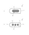
図1(a)、(b)は、それぞれ本発明の実施の形態に係る骨修復デバイス1の構成を示す平面図、正面図である。骨修復デバイス1は、後方椎体間固定術により椎間板が摘出された隣接する椎体間に挿入される椎間ケージ11と、椎間ケージ11内に収容され、間葉系幹細胞又は間葉系幹細胞から分化した骨芽細胞が付着した不織布12と、を備える。 FIGS. 1A and 1B are a plan view and a front view, respectively, showing the configuration of a bone repair device 1 according to an embodiment of the present invention. The bone repair device 1 includes an
椎間ケージ11は、体内に埋設されるインプラントの一例であり、隣り合う2つの椎骨の椎体間に装着され、椎体からの荷重を受け止めるかご状の器具である。椎間ケージ11は、生体適合性を有する材料、例えば、チタン又はチタン合金のような金属材料で形成されている。椎間ケージ11は、例えば、ブロック状の中実材に加工を施すことで形成され、隣接する椎体間で荷重を受けても変形しない程度の剛性を有する。また、椎間ケージ11の上面部及び下面部には、椎間ケージ11が椎体間から滑り落ちないように、例えば、溝又は突起からなる滑り止めが形成されている。 The
椎間ケージ11は、上面部及び下面部で開口し、不織布12が収容される収容孔11aと、側面部から収容孔11aに向けて延びる複数の細孔11bと、を備える。収容孔11aは、不織布12が収容される空間の一例であり、例えば、平面視で楕円形又は長円形に形成されている。細孔11bは、貫通孔の一例であり、収容孔11aに収容された不織布12に細胞や栄養素を含む体液が均一に供給されるように、側面部に間隔を空けて複数個設けられている。図1(b)の背面側にも、表面側と同様に複数の細孔11bが設けられている。 The
椎間ケージ11の先端部は、隣り合う椎体間に押し込めるように丸みを帯びた形状を備える。他方、椎間ケージ11の基端部には、長尺な取り付け治具を取り付け可能な取り付け溝11cが形成されている。術者が取り付け治具を操作し、図2(a)に示す椎間板が除去された隣り合う椎体間の隙間に椎間ケージ11を押し込むことで、図2(b)に示すように骨修復デバイス1の椎間ケージ11が隣り合う椎体間に埋設される。 The distal end of the
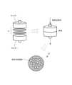
図3は、実施の形態に係る不織布12の一例を走査型電子顕微鏡により撮影した図である。不織布12は、細径の繊維を織らずに絡み合わせたシートであり、各繊維に間葉系幹細胞を付着させることができる。不織布12は、任意に変形が可能であるため、椎間ケージ11の内部に収容するのに好適である。 FIG. 3 is a diagram showing an example of the
不織布12では、1つ1つの繊維に間葉系幹細胞が付着すると共に、繊維の間の空間に細胞懸濁液や体液が流入するように、その繊維径及び繊維密度が調整されている。細胞が繊維に付着するには、不織布12の繊維径を細くすればよい。他方で、不織布12の繊維径を細くしすぎすると強度が弱くなり、繊維密度を大きくしすぎると、細胞が詰まることで、その取り扱いが困難になるおそれがある。間葉系幹細胞の付着性及び不織布12の製造の容易性を考慮すると、不織布12の繊維径は、0.1μm~30μmの範囲内であることが好ましく、0.1μm~3μmの範囲内であることがより一層好ましい。また、不織布12の繊維密度は、0.2g/m2~150g/m2の範囲内であることが好ましい。 In the
不織布12は、生体吸収性材料、例えば、ポリ乳酸、ポリグリコール酸、ポリエチレングリコール、シルクフィブロイン、ポリ-ε-カプロラクトン、キチン、キトサン、ポリ乳酸及びポリグリコール酸の共重合体、ポリ乳酸及びポリエチレングリコールの共重合体、又はこれらの2つ以上の組合せから形成されている。間葉系幹細胞における成長因子の産生を促進する観点から、不織布12は、ポリ乳酸、シルクフィブロイン、ポリεカプロラクトン、キチン又はキトサンで形成されていることが好ましい。 The
間葉系幹細胞は、間葉系に属する細胞への分化能を有する体性幹細胞である。不織布12に付着させる間葉系幹細胞としては、骨芽細胞の成長因子を産生することで骨欠損部における骨芽細胞の成長を促進するか、骨芽細胞に分化し得るものが選択される。不織布12に付着させる間葉系幹細胞は、上記の条件を満たして入れば、脂肪由来、骨膜由来、滑膜由来、海綿骨由来、骨髄由来、羊膜由来、臍帯血由来、胎盤由来のいずれであってもよい。また、不織布12には、複数種類の間葉系幹細胞を一緒に付着させてもよい。 Mesenchymal stem cells are somatic stem cells that have the ability to differentiate into cells belonging to the mesenchymal system. The mesenchymal stem cells to be attached to the
不織布12に付着させる間葉系幹細胞は、患者から採取したものをin vitroで培養するとよい。間葉系幹細胞をin vitroで培養するには、例えば、ウシ胎児血清(Fetal Bovine Serum:FBS)又はヒト血清、好ましくは患者由来のヒト血清を含む培地あるいは無血清培地を用いるとよい。培地にて培養された間葉系幹細胞はそのまま不織布12に付着させてもよく、間葉系幹細胞を分化させて骨芽細胞を発現させてから不織布12に付着させてもよい。骨芽細胞を発現させるには、例えば、間葉系幹細胞の培地に分化誘導因子を添加すればよい。 The mesenchymal stem cells to be attached to the
不織布12に間葉系幹細胞を付着させるには、間葉系幹細胞を含む細胞混濁液をしばらくの間、不織布に接触させればよい。以下、図4を参照して、不織布に間葉系幹細胞を付着させる具体的な手順を説明する。 In order to attach mesenchymal stem cells to the
まず、濾過装置を構成する一対のホルダーの間に不織布12を装着する。各ホルダーは、細胞混濁液を流出又は流入可能に構成されている。一対のホルダーで不織布12を挟み込むことで、一方のホルダーから他方のホルダーに向けて細胞懸濁液を流す際に、細胞混濁液が不織布12に接触する。なお、一対のホルダーに挟み込む不織布12は複数枚であってもよい。 First, the
次に、間葉系幹細胞を含む細胞混濁液を上からゆっくりと滴下させ、不織布12で細胞混濁液を濾過する。すると、間葉系幹細胞が次第に不織布12の繊維に付着する。厳密なメカニズムは不明であるが、間葉系幹細胞を含む細胞混濁液を、時間を掛けて不織布12に濾過させると、不織布12に付着した間葉系幹細胞が活性化する。 Next, a cell suspension containing mesenchymal stem cells is slowly dropped from above, and the cell suspension is filtered through the
なお、不織布12に間葉系幹細胞が付着していれば、間葉系幹細胞から骨組織の修復作用を有する成長因子の産生を期待できるため、必ずしも図4に示す手順により間葉系幹細胞を活性化させる必要はない。 Note that if mesenchymal stem cells are attached to the
以下、実施の形態に係る骨修復デバイス1の製造方法を説明する。まず、間葉系幹細胞が付着した不織布12を入手する。間葉系幹細胞が付着した不織布12を入手するには、例えば、患者又は患者以外の人体から採取した間葉系幹細胞を事前に培養し、不織布12に付着させておくとよい。不織布12は、間葉系幹細胞を付着させる前後のいずれかのタイミングで、椎間ケージ11に適したサイズに切断するとよい。 Hereinafter, a method for manufacturing the bone repair device 1 according to the embodiment will be described. First, a
間葉系幹細胞を付着させた不織布12は、そのまま椎間ケージ11の内部に収容させてもよく、包材により包装された不織布パッケージとして保管してもよい。幹細胞が付着した状態で包装された不織布12を含む不織布パッケージは、椎間ケージ11と共に手術用キットとして術者に提供してもよい。 The
次に、間葉系幹細胞が付着した不織布12を椎間ケージ11内に収容する。例えば、ピンセットで不織布12を把持し、椎間ケージ11内で不織布12の偏りが生じないように椎間ケージ11内に押し込めばよい。なお、不織布12が収容された椎間ケージ11は、そのまま隣り合う椎体間に埋め込まれてもよく、不織布12が収容された椎間ケージ11を包装した上で手術まで保管してもよい。
以上が、骨修復デバイス1の製造方法の流れである。 Next, the
The above is the flow of the method for manufacturing the bone repair device 1.
以下、実施の形態に係る骨修復デバイス1を用いて術者が実行する後方椎体間固定術の手順について説明する。まず、術者は、対象部位の皮膚を切開し、切開部から脊椎後方組織の両側を展開し、図2(a)に示すように上下方向に並んで配置された複数の椎弓根にそれぞれスクリューを挿入する。次に、脊椎神経に対する圧迫を解除する除圧を実施する。次に、片側または両側から椎間板を郭清し、代わりに図2(b)に示すように骨修復デバイス1を1つ又は2つ埋設する。次に、複数の椎弓根に挿入された複数のスクリューに上下方向に配置されたロッドを固定し、脊椎を背側から固定する。次に、皮膚を縫合して切開部を閉じる。
以上が、後方椎体間固定術の一連の手順である。 Hereinafter, a procedure for posterior interbody fusion performed by an operator using the bone repair device 1 according to the embodiment will be described. First, the surgeon makes an incision in the skin of the target area, expands both sides of the posterior spinal tissue from the incision, and inserts each into multiple vertebral pedicles arranged vertically, as shown in Figure 2(a). Insert the screw. Next, decompression is performed to relieve pressure on the spinal nerves. Next, the intervertebral disc is dissected from one or both sides, and one or two bone repair devices 1 are implanted in its place, as shown in FIG. 2(b). Next, vertically arranged rods are fixed to multiple screws inserted into multiple vertebral pedicles to fix the spine from the dorsal side. The skin is then sutured to close the incision.
The above is the series of steps for posterior interbody fusion surgery.
後方椎体間固定術により隣り合う椎体間に埋設された骨修復デバイス1では、椎間ケージ11の収容孔11aに不織布12が収容されているため、不織布12に付着した間葉系幹細胞又は骨芽細胞の作用により、不織布12を足場として骨芽細胞の増殖や成長が促進される。その結果、不織布12を収容していない椎間ケージと比較して、隣り合う椎体同士を早期に固定でき、脊椎の早期の修復を実現できる。 In the bone repair device 1 implanted between adjacent vertebral bodies by posterior interbody fusion, the
以上説明したように、実施の形態に係る骨修復デバイス1は、外部から体液が流入可能な空間が内部に設けられた椎間ケージ11と、椎間ケージ11の空間に収容され、間葉系幹細胞又は間葉系幹細胞から分化した骨芽細胞が付着している不織布12と、を備える。このため、骨の早期の修復を実現できる。 As described above, the bone repair device 1 according to the embodiment includes an
本発明は上記実施の形態に限られず、以下に述べる変形も可能である。 The present invention is not limited to the embodiments described above, and modifications described below are also possible.
(変形例)
上記実施の形態では、椎間ケージ11が収容孔11a及び細孔11bを備えていたが本発明はこれに限られない。椎間ケージ11は、外部から体液が流入可能な空間が内部に設けられていれば、いかなる形状であってもよい。(Modified example)
In the embodiment described above, the
上記実施の形態では、椎間ケージ11は金属材料で形成されていたが、本発明はこれに限られない。例えば、ポリエーテルエーテルケトン(Poly Ether Ether Ketone:PEEK)のような樹脂材料であってもよく、繊維強化プラスチック、セラミックス、人工骨であってもよい。 In the embodiment described above, the
上記実施の形態では、骨修復を促進するために不織布12に間葉系幹細胞を付着させていたが、本発明はこれに限られない。不織布12に付着させる幹細胞は、例えば、ES(Embryonic Stem)細胞、iPS(induced Pluripotent Stem)細胞であってもよい。 In the above embodiment, mesenchymal stem cells were attached to the
上記実施の形態では、間葉系細胞が付着された不織布12をそのまま椎間ケージ11内に収容していたが、本発明はこれに限られない。例えば、間葉系細胞が付着された不織布12にゲル化剤を加えて使用することもできる。ゲル化剤としては、例えば、コラーゲン、ゼラチン、細胞外マトリックス混合物、フィブリン、多糖類、コンドロイチンを用いればよい。細胞外マトリックス混合物としては、例えば、コーニング社製のマトリゲル(登録商標)を用いればよく、多糖類としては、例えば、アガロースを用いればよい。 In the embodiment described above, the
上記実施の形態では、椎間ケージ11の内部に間葉系幹細胞が付着した不織布12を収容していたが、本発明はこれに限られない。例えば、不織布12に加えて人体から採取した骨片を一緒に収容してもよい。骨片は、人体から採取した骨を粉砕したものであり、患者自身から採取した骨(局所骨または腸骨)を粉砕したものであることが好ましい。骨片の形状やサイズは、椎間ケージ11の形状やサイズ、椎間ケージ11の埋設箇所を考慮して設定される。骨片が体内で散らばることがないように、骨片を不織布12で包んで椎間ケージ11の内部に収容してもよい。また、椎間ケージ11の内部に不織布12を収容する前、又は収容した後の時点で、骨修復を促進したり免疫反応を抑制したりする薬剤を不織布12に保持させてもよい。 In the above embodiment, the
上記実施の形態では、隣り合う椎体間の欠損部に椎間ケージ11を埋設する場合を例に説明したが、本発明はこれに限られない。インプラントは椎間ケージ11に限られず、外部から体液が流入可能な空間が内部に設けられていれば、他の部位に適用可能なインプラントであってもよい。例えば、長管骨の骨切り術において骨と骨とを接合するために、間葉系幹細胞が付着した不織布を内部に収容したインプラントを骨と骨の間に埋設してもよい。 In the above embodiment, the case where the
上記実施の形態は例示であり、本発明はこれらに限定されるものではなく、特許請求の範囲に記載した発明の趣旨を逸脱しない範囲でさまざまな実施の形態が可能である。各実施の形態や変形例で記載した構成要素は自由に組み合わせることが可能である。また、特許請求の範囲に記載した発明と均等な発明も本発明に含まれる。 The above-mentioned embodiments are illustrative, and the present invention is not limited thereto, and various embodiments are possible without departing from the spirit of the invention described in the claims. The components described in each embodiment and modification example can be freely combined. Furthermore, inventions equivalent to the inventions described in the claims are also included in the present invention.
1 骨修復デバイス
11 椎間ケージ
11a 収容孔
11b 細孔
11c 取り付け溝
12 不織布
1
Claims (8)
Translated fromJapanese前記インプラントの前記空間に収容され、互いに絡み合った繊維に間葉系幹細胞が付着している不織布と、
を備え、
前記不織布への間葉系幹細胞の付着は、前記不織布を一対のホルダーに挟み込み、前記間葉系幹細胞を含む細胞混濁液を一方のホルダーから他方のホルダーに向けて流すことで行われ、
前記間葉系幹細胞を付着する際の前記不織布の形状は、前記空間の形状とは異なる、
骨修復デバイス。 An implant that has a space inside that allows body fluid to flow in from the outside,
a nonwoven fabric that is accommodated in the space of the implant and hasmesenchymal stem cells attached to intertwined fibers;
Equipped with
The attachment of mesenchymal stem cells to the nonwoven fabric is performed by sandwiching the nonwoven fabric between a pair of holders and flowing a cell suspension containing the mesenchymal stem cells from one holder to the other holder,
The shape of the nonwoven fabric when the mesenchymal stem cells are attached is different from the shape of the space,
Bone repair device.
請求項1に記載の骨修復デバイス。 The fiber diameter of the nonwoven fabric is within the range of 0.1 μm to 30 μm.
A bone repair device accordingto claim 1 .
請求項1又は2に記載の骨修復デバイス。 The fiber density of the nonwoven fabric is within the range of 0.2 g/m2 to 150 g/m2 .
The bone repair device according to claim 1or 2 .
請求項1から3のいずれか1項に記載の骨修復デバイス。 The nonwoven fabric is made of a material containing at least one of polylactic acid, polyglycolic acid, silk fibroin, poly-ε-caprolactone, chitin, and chitosan.
A bone repair device according to any one of claims 1 to3 .
請求項1から4のいずれか1項に記載の骨修復デバイス。 The bone repair device further comprises a bone fragment housed in the space of the implant together with the nonwoven fabric.
A bone repair device according to any one of claims 1 to4 .
前記空間が内部に形成された本体と、
前記本体に設けられ、前記本体の表面から前記空間に向かって貫通し、前記空間内に体液を流入させる貫通孔と、を備える、
請求項1から5のいずれか1項に記載の骨修復デバイス。 The implant includes:
a main body in which the space is formed;
a through hole provided in the main body, penetrating from the surface of the main body toward the space, and allowing body fluid to flow into the space;
A bone repair device according to any one of claims 1 to5 .
請求項1から6のいずれか1項に記載の骨修復デバイス。 The implant is an intervertebral cage inserted between a pair of vertebral bodies in which the intervertebral disc has been dissected.
A bone repair device according to any one of claims 1 to6 .
前記インプラントの前記空間に収容可能であり、互いに絡み合った繊維に間葉系幹細胞が付着した状態で包装された不織布を含む不織布パッケージと、
を備え、
前記不織布への間葉系幹細胞の付着は、前記不織布を一対のホルダーに挟み込み、前記間葉系幹細胞を含む細胞混濁液を一方のホルダーから他方のホルダーに向けて流すことで行われ、
前記間葉系幹細胞を付着する際の前記不織布の形状は、前記空間の形状とは異なる、
手術用キット。 An implant that has a space inside that allows body fluid to flow in from the outside,
a nonwoven fabric package that can be accommodated in the space of the implant and includes a nonwoven fabric wrapped withmesenchymal stem cells attached to intertwined fibers;
Equipped with
The attachment of mesenchymal stem cells to the nonwoven fabric is performed by sandwiching the nonwoven fabric between a pair of holders and flowing a cell suspension containing the mesenchymal stem cells from one holder to the other holder,
The shape of the nonwoven fabric when the mesenchymal stem cells are attached is different from the shape of the space,
surgical kit.
Priority Applications (7)
| Application Number | Priority Date | Filing Date | Title |
|---|---|---|---|
| JP2021156438AJP7423081B2 (en) | 2021-09-27 | 2021-09-27 | Bone repair devices and surgical kits |
| CN202280064911.3ACN117999049A (en) | 2021-09-27 | 2022-09-20 | Bone repair device and surgical kit |
| CA3234038ACA3234038A1 (en) | 2021-09-27 | 2022-09-20 | Bone repair device and surgical kit |
| US18/695,605US20240390156A1 (en) | 2021-09-27 | 2022-09-20 | Bone repair device and surgical kit |
| EP22880724.4AEP4410246A4 (en) | 2021-09-27 | 2022-09-20 | BONE REPAIR DEVICE AND SURGICAL KIT |
| AU2022365594AAU2022365594A1 (en) | 2021-09-27 | 2022-09-20 | Bone repair device and surgical kit |
| PCT/JP2022/034909WO2023063028A1 (en) | 2021-09-27 | 2022-09-20 | Bone repair device and surgical kit |
Applications Claiming Priority (1)
| Application Number | Priority Date | Filing Date | Title |
|---|---|---|---|
| JP2021156438AJP7423081B2 (en) | 2021-09-27 | 2021-09-27 | Bone repair devices and surgical kits |
Publications (2)
| Publication Number | Publication Date |
|---|---|
| JP2023047494A JP2023047494A (en) | 2023-04-06 |
| JP7423081B2true JP7423081B2 (en) | 2024-01-29 |
Family
ID=85779392
Family Applications (1)
| Application Number | Title | Priority Date | Filing Date |
|---|---|---|---|
| JP2021156438AActiveJP7423081B2 (en) | 2021-09-27 | 2021-09-27 | Bone repair devices and surgical kits |
Country Status (7)
| Country | Link |
|---|---|
| US (1) | US20240390156A1 (en) |
| EP (1) | EP4410246A4 (en) |
| JP (1) | JP7423081B2 (en) |
| CN (1) | CN117999049A (en) |
| AU (1) | AU2022365594A1 (en) |
| CA (1) | CA3234038A1 (en) |
| WO (1) | WO2023063028A1 (en) |
Families Citing this family (2)
| Publication number | Priority date | Publication date | Assignee | Title |
|---|---|---|---|---|
| JP7502751B2 (en) | 2022-06-22 | 2024-06-19 | 金井重要工業株式会社 | Tissue regeneration promotion sheet |
| WO2023249065A1 (en)* | 2022-06-22 | 2023-12-28 | 学校法人藤田学園 | Tissue regeneration promotion sheet |
Citations (8)
| Publication number | Priority date | Publication date | Assignee | Title |
|---|---|---|---|---|
| US20090105825A1 (en) | 2007-10-19 | 2009-04-23 | Greg Foreman | Fusion methods using autologous stem cells |
| US20110300626A1 (en) | 2007-06-18 | 2011-12-08 | New Jersey Institute Of Technology | Electrospun Ceramic-Polymer Composite As A Scaffold for Tissue Repair |
| WO2012118090A1 (en) | 2011-02-28 | 2012-09-07 | サンスター株式会社 | Non-woven fabric containing bone prosthetic material |
| JP2012192105A (en) | 2011-03-17 | 2012-10-11 | Sunstar Inc | Cell scaffold material |
| JP2019022651A (en) | 2017-07-21 | 2019-02-14 | ウォーソー・オーソペディック・インコーポレーテッド | Bone implant for enclosing bone material |
| US20190254840A1 (en) | 2016-09-16 | 2019-08-22 | Mirus Llc | Interbody fusion devices and related methods of manufacture |
| US20200093612A1 (en) | 2018-09-20 | 2020-03-26 | Amendia, Inc. d/b/a Spinal Elements | Spinal implant device |
| JP2021516074A (en) | 2018-01-31 | 2021-07-01 | インスティテュート フォー マスキュロスケレタル サイエンス アンド エジュケイション,リミテッド | Implant with diagonal insertion axis |
Family Cites Families (4)
| Publication number | Priority date | Publication date | Assignee | Title |
|---|---|---|---|---|
| JP5328051B2 (en) | 2010-07-14 | 2013-10-30 | ナカシマメディカル株式会社 | Intervertebral cage |
| US10932917B2 (en)* | 2017-12-30 | 2021-03-02 | Charles Gilbride | Implant bone on-growth structures and methods |
| JP2020195330A (en)* | 2019-06-03 | 2020-12-10 | 株式会社カネカ | Cell separation filter unit and production method of cell concentrated solution |
| JP7209773B2 (en) | 2020-05-08 | 2023-01-20 | バンドー化学株式会社 | friction transmission belt |
- 2021
- 2021-09-27JPJP2021156438Apatent/JP7423081B2/enactiveActive
- 2022
- 2022-09-20USUS18/695,605patent/US20240390156A1/enactivePending
- 2022-09-20CNCN202280064911.3Apatent/CN117999049A/enactivePending
- 2022-09-20WOPCT/JP2022/034909patent/WO2023063028A1/ennot_activeCeased
- 2022-09-20EPEP22880724.4Apatent/EP4410246A4/enactivePending
- 2022-09-20AUAU2022365594Apatent/AU2022365594A1/enactivePending
- 2022-09-20CACA3234038Apatent/CA3234038A1/enactivePending
Patent Citations (8)
| Publication number | Priority date | Publication date | Assignee | Title |
|---|---|---|---|---|
| US20110300626A1 (en) | 2007-06-18 | 2011-12-08 | New Jersey Institute Of Technology | Electrospun Ceramic-Polymer Composite As A Scaffold for Tissue Repair |
| US20090105825A1 (en) | 2007-10-19 | 2009-04-23 | Greg Foreman | Fusion methods using autologous stem cells |
| WO2012118090A1 (en) | 2011-02-28 | 2012-09-07 | サンスター株式会社 | Non-woven fabric containing bone prosthetic material |
| JP2012192105A (en) | 2011-03-17 | 2012-10-11 | Sunstar Inc | Cell scaffold material |
| US20190254840A1 (en) | 2016-09-16 | 2019-08-22 | Mirus Llc | Interbody fusion devices and related methods of manufacture |
| JP2019022651A (en) | 2017-07-21 | 2019-02-14 | ウォーソー・オーソペディック・インコーポレーテッド | Bone implant for enclosing bone material |
| JP2021516074A (en) | 2018-01-31 | 2021-07-01 | インスティテュート フォー マスキュロスケレタル サイエンス アンド エジュケイション,リミテッド | Implant with diagonal insertion axis |
| US20200093612A1 (en) | 2018-09-20 | 2020-03-26 | Amendia, Inc. d/b/a Spinal Elements | Spinal implant device |
Also Published As
| Publication number | Publication date |
|---|---|
| CN117999049A (en) | 2024-05-07 |
| WO2023063028A1 (en) | 2023-04-20 |
| US20240390156A1 (en) | 2024-11-28 |
| EP4410246A4 (en) | 2025-09-03 |
| AU2022365594A1 (en) | 2024-05-02 |
| CA3234038A1 (en) | 2023-04-20 |
| EP4410246A1 (en) | 2024-08-07 |
| JP2023047494A (en) | 2023-04-06 |
Similar Documents
| Publication | Publication Date | Title |
|---|---|---|
| US7135042B2 (en) | Surgical implant | |
| US6280473B1 (en) | Resorbable, macro-porous, non-collapsing and flexible membrane barrier for skeletal repair and regeneration | |
| KR102330141B1 (en) | Absorbent, cross-linked, shape-stable membrane | |
| RU173381U1 (en) | PERSONAL BIOACTIVE STRUCTURED IMPLANT FOR REPLACING BONE DEFECT | |
| WO1999037240A9 (en) | Resorbable, macro-porous, non-collapsing and flexible membrane barrier for skeletal repair and regeneration | |
| Zou et al. | A comparative study of autogenous, allograft and artificial bone substitutes on bone regeneration and immunotoxicity in rat femur defect model | |
| US11406739B2 (en) | Method for creating a personalized gene-activated implant for regenerating bone tissue | |
| JP7423081B2 (en) | Bone repair devices and surgical kits | |
| CN111920552A (en) | Intervertebral fusion device with a plurality of through holes | |
| JP5154416B2 (en) | Graft for tissue treatment | |
| CN104684591A (en) | Generation of isolated cartilage from fibroblasts | |
| RU173377U1 (en) | BIOACTIVE CELLULAR TRIANGULAR IMPLANT FOR REPLACEMENT OF THE TIBERAID DEFECT | |
| CN103720527A (en) | Joint fusion cage | |
| RU171825U1 (en) | IMPLANT FOR SUBSTITUTION OF BONE DEFECTS AND INTERDERBINAL DISK | |
| CN115715204A (en) | Porous bone substitute material | |
| US20190314164A1 (en) | Spinal fusion implant and related methods | |
| US20190314550A1 (en) | Bone substitute composition and related methods | |
| RU157799U1 (en) | IMPLANT FOR SUBSTITUTION AND PLASTY OF BONE AND CARTILAGE TISSUES | |
| CN210784858U (en) | Tissue engineering cervical vertebra interbody fusion cage | |
| RU2744756C1 (en) | Method of transplanting biocomposite spheroids to ensure the possibility of restoring the integrity of the bone in case of defects that exceed the critical size | |
| RU168519U1 (en) | IMPLANT FOR SUBSTITUTION OF BONE DEFECTS AND INTERDERBINAL DISK | |
| RU170271U1 (en) | IMPLANT FOR SUBSTITUTION OF BONE DEFECTS AND INTERDERBINAL DISK | |
| RU171826U1 (en) | IMPLANT FOR SUBSTITUTION OF INTERDOMBRAIN DISCS | |
| RU170272U1 (en) | IMPLANT FOR SUBSTITUTION OF INTERDOMBRAIN DISCS | |
| RU168513U1 (en) | IMPLANT FOR SUBSTITUTION OF INTERDOMBRAIN DISCS |
Legal Events
| Date | Code | Title | Description |
|---|---|---|---|
| A621 | Written request for application examination | Free format text:JAPANESE INTERMEDIATE CODE: A621 Effective date:20230105 | |
| A871 | Explanation of circumstances concerning accelerated examination | Free format text:JAPANESE INTERMEDIATE CODE: A871 Effective date:20230105 | |
| A131 | Notification of reasons for refusal | Free format text:JAPANESE INTERMEDIATE CODE: A131 Effective date:20230411 | |
| A521 | Request for written amendment filed | Free format text:JAPANESE INTERMEDIATE CODE: A523 Effective date:20230612 | |
| A131 | Notification of reasons for refusal | Free format text:JAPANESE INTERMEDIATE CODE: A131 Effective date:20230822 | |
| A601 | Written request for extension of time | Free format text:JAPANESE INTERMEDIATE CODE: A601 Effective date:20231018 | |
| RD03 | Notification of appointment of power of attorney | Free format text:JAPANESE INTERMEDIATE CODE: A7423 Effective date:20231018 | |
| A521 | Request for written amendment filed | Free format text:JAPANESE INTERMEDIATE CODE: A821 Effective date:20231020 | |
| A521 | Request for written amendment filed | Free format text:JAPANESE INTERMEDIATE CODE: A523 Effective date:20231215 | |
| TRDD | Decision of grant or rejection written | ||
| A01 | Written decision to grant a patent or to grant a registration (utility model) | Free format text:JAPANESE INTERMEDIATE CODE: A01 Effective date:20231225 | |
| A61 | First payment of annual fees (during grant procedure) | Free format text:JAPANESE INTERMEDIATE CODE: A61 Effective date:20240110 | |
| R150 | Certificate of patent or registration of utility model | Ref document number:7423081 Country of ref document:JP Free format text:JAPANESE INTERMEDIATE CODE: R150 |