JP7367038B2 - Radioligands for imaging LPA1 receptors - Google Patents
Radioligands for imaging LPA1 receptorsDownload PDFInfo
- Publication number
- JP7367038B2 JP7367038B2JP2021545267AJP2021545267AJP7367038B2JP 7367038 B2JP7367038 B2JP 7367038B2JP 2021545267 AJP2021545267 AJP 2021545267AJP 2021545267 AJP2021545267 AJP 2021545267AJP 7367038 B2JP7367038 B2JP 7367038B2
- Authority
- JP
- Japan
- Prior art keywords
- methyl
- lpa1
- pharmaceutical composition
- oxy
- pet
- Prior art date
- Legal status (The legal status is an assumption and is not a legal conclusion. Google has not performed a legal analysis and makes no representation as to the accuracy of the status listed.)
- Active
Links
Images


Classifications
- A—HUMAN NECESSITIES
- A61—MEDICAL OR VETERINARY SCIENCE; HYGIENE
- A61K—PREPARATIONS FOR MEDICAL, DENTAL OR TOILETRY PURPOSES
- A61K51/00—Preparations containing radioactive substances for use in therapy or testing in vivo
- A61K51/02—Preparations containing radioactive substances for use in therapy or testing in vivo characterised by the carrier, i.e. characterised by the agent or material covalently linked or complexing the radioactive nucleus
- A61K51/04—Organic compounds
- A61K51/041—Heterocyclic compounds
- A61K51/044—Heterocyclic compounds having nitrogen as a ring hetero atom, e.g. guanethidine, rifamycins
- A61K51/0455—Heterocyclic compounds having nitrogen as a ring hetero atom, e.g. guanethidine, rifamycins having six-membered rings with one nitrogen as the only ring hetero atom
- A—HUMAN NECESSITIES
- A61—MEDICAL OR VETERINARY SCIENCE; HYGIENE
- A61B—DIAGNOSIS; SURGERY; IDENTIFICATION
- A61B6/00—Apparatus or devices for radiation diagnosis; Apparatus or devices for radiation diagnosis combined with radiation therapy equipment
- A61B6/02—Arrangements for diagnosis sequentially in different planes; Stereoscopic radiation diagnosis
- A61B6/03—Computed tomography [CT]
- A61B6/037—Emission tomography
- A—HUMAN NECESSITIES
- A61—MEDICAL OR VETERINARY SCIENCE; HYGIENE
- A61B—DIAGNOSIS; SURGERY; IDENTIFICATION
- A61B6/00—Apparatus or devices for radiation diagnosis; Apparatus or devices for radiation diagnosis combined with radiation therapy equipment
- A61B6/48—Diagnostic techniques
- A61B6/481—Diagnostic techniques involving the use of contrast agents
- A—HUMAN NECESSITIES
- A61—MEDICAL OR VETERINARY SCIENCE; HYGIENE
- A61B—DIAGNOSIS; SURGERY; IDENTIFICATION
- A61B6/00—Apparatus or devices for radiation diagnosis; Apparatus or devices for radiation diagnosis combined with radiation therapy equipment
- A61B6/52—Devices using data or image processing specially adapted for radiation diagnosis
- A61B6/5211—Devices using data or image processing specially adapted for radiation diagnosis involving processing of medical diagnostic data
- A61B6/5217—Devices using data or image processing specially adapted for radiation diagnosis involving processing of medical diagnostic data extracting a diagnostic or physiological parameter from medical diagnostic data
- C—CHEMISTRY; METALLURGY
- C07—ORGANIC CHEMISTRY
- C07D—HETEROCYCLIC COMPOUNDS
- C07D401/00—Heterocyclic compounds containing two or more hetero rings, having nitrogen atoms as the only ring hetero atoms, at least one ring being a six-membered ring with only one nitrogen atom
- C07D401/02—Heterocyclic compounds containing two or more hetero rings, having nitrogen atoms as the only ring hetero atoms, at least one ring being a six-membered ring with only one nitrogen atom containing two hetero rings
- C07D401/04—Heterocyclic compounds containing two or more hetero rings, having nitrogen atoms as the only ring hetero atoms, at least one ring being a six-membered ring with only one nitrogen atom containing two hetero rings directly linked by a ring-member-to-ring-member bond
- A—HUMAN NECESSITIES
- A61—MEDICAL OR VETERINARY SCIENCE; HYGIENE
- A61K—PREPARATIONS FOR MEDICAL, DENTAL OR TOILETRY PURPOSES
- A61K2123/00—Preparations for testing in vivo
Landscapes
- Health & Medical Sciences (AREA)
- Life Sciences & Earth Sciences (AREA)
- Engineering & Computer Science (AREA)
- Chemical & Material Sciences (AREA)
- Physics & Mathematics (AREA)
- Veterinary Medicine (AREA)
- Public Health (AREA)
- Medical Informatics (AREA)
- General Health & Medical Sciences (AREA)
- Optics & Photonics (AREA)
- Animal Behavior & Ethology (AREA)
- Organic Chemistry (AREA)
- Surgery (AREA)
- Proteomics, Peptides & Aminoacids (AREA)
- Heart & Thoracic Surgery (AREA)
- Molecular Biology (AREA)
- Radiology & Medical Imaging (AREA)
- Pathology (AREA)
- Nuclear Medicine, Radiotherapy & Molecular Imaging (AREA)
- High Energy & Nuclear Physics (AREA)
- Biophysics (AREA)
- Biomedical Technology (AREA)
- Epidemiology (AREA)
- Medicinal Chemistry (AREA)
- Pharmacology & Pharmacy (AREA)
- Computer Vision & Pattern Recognition (AREA)
- Physiology (AREA)
- Medicines Containing Antibodies Or Antigens For Use As Internal Diagnostic Agents (AREA)
- Plural Heterocyclic Compounds (AREA)
- Nuclear Medicine (AREA)
- Magnetic Resonance Imaging Apparatus (AREA)
- Pharmaceuticals Containing Other Organic And Inorganic Compounds (AREA)
Description
Translated fromJapanese 関連出願の相互参照
本願は、米国特許仮出願第62/745,524号(2018年10月15日出願)の優先権の利益を主張し、その全体的な内容が引用によって本明細書に援用される。CROSS-REFERENCE TO RELATED APPLICATIONS This application claims the benefit of priority to U.S. Provisional Patent Application No. 62/745,524, filed October 15, 2018, the entire contents of which are incorporated herein by reference. be done.
本発明は、新規な放射性標識リゾホスファチジン酸(LPA)受容体1アンタゴニスト、および哺乳動物におけるLPA1受容体の標識および診断イメージングにおける使用に関する。 The present invention relates to novel radiolabeled lysophosphatidic acid (LPA)
陽電子放出断層撮影(PET)は、生きている対象における生物学的プロセスについての機能的な情報を提供することができる、非侵襲性イメージング技術である。インビボ分子の事象をイメージングおよびモニターすることのできる能力は、生きている生物における生化学的および生理学的プロセスについての洞察を得るために大きな価値がある。これは同様に、疾患の治療、疾患の早期発見、および新薬の設計のための新たなアプローチの開発に重要である。PETは、陽電子放出放射性同位体によって標識された分子の設計および合成に依存する。これらの分子は、放射性トレーサーまたは放射性リガンドとして既知である。PETイメージングにおいて、最も一般的に用いられる陽電子放出(PET)放射性核種は11C、18F、15O、および13Nであり、これらは全てサイクロトロンによって製造され、それぞれ20、110、2、および10分の半減期を有する。陽電子放出放射性核種によって放射性標識された後、これらのPET放射性リガンドは、一般に静脈内(i.v.)注射によって、哺乳動物に投与される。体内に入ると、放射性リガンドが崩壊するにつれ陽電子が放出され、これが電子と結合するまで短い距離を移動する。消滅事象として知られている事象が次に生じ、これによってそれぞれ511keVのエネルギーを有する2つの共線光子が生じる。放射性リガンドから放出されるガンマ線を検出することができるPETイメージングスキャナーを用いると、平面および断層画像によって、放射性トレーサーの分布が時間の関数として明らかになる。PET放射性リガンドは、ヒト受容体への標的結合、および用量依存性受容体占有率に関する、有用なインビボ情報を提供する。Positron emission tomography (PET) is a non-invasive imaging technique that can provide functional information about biological processes in living subjects. The ability to image and monitor molecular events in vivo is of great value for gaining insight into biochemical and physiological processes in living organisms. This is likewise important for the development of new approaches for disease treatment, early detection of disease, and the design of new drugs. PET relies on the design and synthesis of molecules labeled with positron-emitting radioisotopes. These molecules are known as radiotracers or radioligands. In PET imaging, the most commonly used positron-emitting (PET) radionuclides are11C ,18F ,15O , and13N , all of which are produced by cyclotrons and have 20, 110, 2, and 10, respectively. It has a half-life of minutes. After being radiolabeled with a positron-emitting radionuclide, these PET radioligands are administered to a mammal, generally by intravenous (i.v.) injection. Once in the body, as the radioligand decays, it releases positrons, which travel short distances before combining with electrons. An event known as an annihilation event then occurs, resulting in two collinear photons each with an energy of 511 keV. Using PET imaging scanners capable of detecting gamma rays emitted by radioligands, planar and tomographic images reveal the distribution of the radiotracer as a function of time. PET radioligands provide useful in vivo information regarding target binding to human receptors and dose-dependent receptor occupancy.
特発性肺線維症(IPF)は、肺における瘢痕組織の存在、息切れ、および慢性乾性咳によって特徴付けられる、慢性疾患である。IPFは、間質性肺疾患(ILD)として知られる肺疾患のファミリーに属し、通常の間質性肺線維症(UIP)として知られる病理学的パターンに関連する。ゆっくりと進行する疾患(最も一般的)、一時的な急性憎悪を特徴とする疾患、または急速に進行する疾患など、IPFについてのいくつかの潜在的な臨床経過が存在する。診断時からの生存期間の中央値は、2から5年の間である。現在IPFの治療は存在せず、利用可能な治療の選択肢は非常に限定されており、望ましくない副作用がある。IPFの病因は不明であるが、仮説の1つは、上皮性細胞への最初の損傷が、リゾホスファチジン酸(LPA)の生成を増加させるというものである。LPAは、細胞機能の様々な局面を制御する生理活性リン脂質であり、創傷治癒および組織線維症の新規なメディエーターとして認識されている。LPAは、LPA受容体を介して生物学的効果を仲介し、そのうち少なくとも6個のアイソフォームが特定されている。最近の研究では、LPA1アイソフォームが肺線維症の病因に関連づけられており、LPA1受容体がIPFの潜在的な臨床標的として特定されている。いくつかの発見によって、内皮バリアの崩壊、炎症、および線維芽細胞の動員/増殖をもたらす、LPA1受容体を活性化するIPFにおいて、LPA/LPA1経路の役割が支持される。LPAは、IPF患者の気管支肺胞洗浄(BAL)において増加する。LPA濃度は、IPFを有するヒトのBAL液(BALF)において増加し、LPA1拮抗作用によって、IPF BALFによって誘導される線維芽細胞の移動が阻害される。また、LPA1受容体欠損ノックアウトマウスは、線維症のブレオマイシンモデルにおいて、肺の血管漏出の減少およびコラーゲン蓄積の減少を示す。これらのデータに基づいて、LPA1シグナル伝達は、血管漏出の誘導および線維芽細胞移動の刺激を介して、少なくとも部分的には、肺線維症の進行に寄与すると考えられている。 Idiopathic pulmonary fibrosis (IPF) is a chronic disease characterized by the presence of scar tissue in the lungs, shortness of breath, and chronic dry cough. IPF belongs to a family of lung diseases known as interstitial lung diseases (ILD) and is associated with a pathological pattern known as common interstitial pulmonary fibrosis (UIP). There are several potential clinical courses for IPF, including a slowly progressive disease (the most common), a disease characterized by episodic acute exacerbations, or a rapidly progressive disease. Median survival from the time of diagnosis is between 2 and 5 years. There is currently no treatment for IPF, and the available treatment options are very limited and have undesirable side effects. The pathogenesis of IPF is unknown, but one hypothesis is that the initial damage to epithelial cells increases the production of lysophosphatidic acid (LPA). LPA is a bioactive phospholipid that regulates various aspects of cellular function and is recognized as a novel mediator of wound healing and tissue fibrosis. LPA mediates biological effects through the LPA receptor, of which at least six isoforms have been identified. Recent studies have implicated LPA1 isoforms in the pathogenesis of pulmonary fibrosis and identified the LPA1 receptor as a potential clinical target in IPF. Several findings support a role for the LPA/LPA1 pathway in IPF, which activates the LPA1 receptor, leading to endothelial barrier disruption, inflammation, and fibroblast recruitment/proliferation. LPA is increased in the bronchoalveolar lavage (BAL) of IPF patients. LPA concentration is increased in BAL fluid (BALF) of humans with IPF, and LPA1 antagonism inhibits IPF BALF-induced fibroblast migration. LPA1 receptor-deficient knockout mice also show reduced pulmonary vascular leakage and reduced collagen accumulation in a bleomycin model of fibrosis. Based on these data, LPA1 signaling is believed to contribute to lung fibrosis progression, at least in part, through induction of vascular leakage and stimulation of fibroblast migration.
LPA1受容体に高い親和性を有する特定のPET放射性リガンドを、補助的なイメージング技術と組み合わせて用いることで、ヒト肺LPA1、または腎臓、肝臓、心臓、もしくは皮膚などの他の臓器におけるLPA1において、LPA1アンタゴニストの標的結合および用量/占有率の関係の両方に関する、臨床的な進展のための方法が提供されうる。本明細書に記載される発明は、インビトロおよびインビボの両方における診査および診断イメージング応用、並びに放射性標識および非標識LPA1アンタゴニストを用いた競合試験に有用でありうる、放射性標識LPA1アンタゴニストに関する。 By using specific PET radioligands with high affinity for the LPA1 receptor, in combination with ancillary imaging techniques, we have been able to improve human lung LPA1, or LPA1 in other organs such as the kidney, liver, heart, or skin. Methods can be provided for clinical advancement of both target binding and dose/occupancy relationships of LPA1 antagonists. The invention described herein relates to radiolabeled LPA1 antagonists that can be useful in both in vitro and in vivo diagnostic and diagnostic imaging applications, as well as competition studies with radiolabeled and unlabeled LPA1 antagonists.
米国特許出願公開第2017/0360759号(PCT出願公開第WO2017/223016号)は、肺などの様々な臓器の線維症などの、LPA依存性またはLPA介在性病状または疾患の治療に用いるための、リゾホスファチジン酸受容体のいくつかのアゴニストを開示する。 U.S. Patent Application Publication No. 2017/0360759 (PCT Application Publication No. WO 2017/223016) discloses a method for treating LPA-dependent or LPA-mediated pathologies or diseases, such as fibrosis of various organs such as the lungs. Several agonists of lysophosphatidic acid receptors are disclosed.
本開示は、部分的に、放射性標識されたリゾホスファチジン酸(以後「LPA1」)受容体アンタゴニストが、哺乳動物種の組織において、LPA1受容体、および/またはLPA1発現、および/またはLPA1受容体を占有するための化合物の親和性の、検出および/または定量および/またはイメージングにおいて有用であるという認識に基づく。放射性同位体標識されたLPA1受容体アンタゴニストは、哺乳動物種に投与された場合、LPA1受容体において蓄積するか、またはLPA1受容体を占有し、イメージング技術によって検出することができ、それによってLPA1受容体の存在、LPA1受容体の占有についての化合物の親和性、およびLPA1受容体アンタゴニストの臨床的評価および用量選択についての、重要な診断マーカーを提供することが分かっている。さらに、本明細書に開示される、放射性標識LPA1受容体アンタゴニストは、受容体への結合について、非標識薬物と放射性標識薬物との競合によって、非標識LPA1受容体アンタゴニストの、インビボでのLPA1受容体との相互作用を研究するための研究ツールとして用いることができる。これらの種類の研究は、LPA1受容体占有率と、非標識のLPA1受容体アンタゴニストの用量との関係を決定すること、並びに、様々な用量の非標識LPA1受容体アンタゴニストによる、受容体の遮断期間を研究することに有用である。 The present disclosure provides, in part, that radiolabeled lysophosphatidic acid (hereinafter "LPA1") receptor antagonists inhibit LPA1 receptors, and/or LPA1 expression, and/or LPA1 receptors in tissues of mammalian species. It is based on the recognition that it is useful in the detection and/or quantification and/or imaging of a compound's affinity for occupancy. Radioisotope-labeled LPA1 receptor antagonists, when administered to mammalian species, accumulate at or occupy the LPA1 receptor and can be detected by imaging techniques, thereby inhibiting the LPA1 receptor. The affinity of the compound for LPA1 receptor occupancy has been found to provide important diagnostic markers for clinical evaluation and dose selection of LPA1 receptor antagonists. Furthermore, the radiolabeled LPA1 receptor antagonists disclosed herein reduce the in vivo LPA1 receptor antagonist's ability to increase the LPA1 receptor in vivo by competition between the unlabeled drug and the radiolabeled drug for binding to the receptor. It can be used as a research tool to study interactions with the body. These types of studies are designed to determine the relationship between LPA1 receptor occupancy and dose of unlabeled LPA1 receptor antagonist, as well as the duration of receptor blockade by various doses of unlabeled LPA1 receptor antagonist. It is useful for researching.
臨床ツールとして、放射性標識LPA1受容体アンタゴニストを用いて、LPA1受容体アンタゴニストの臨床的に効果的な用量を定義するのに役立てることができる。動物実験において、放射性標識LPA1受容体アゴニストを用いて、臨床開発のための選択のための潜在的な薬物候補からの選択に有用な情報を提供することができる。また、放射性標識LPA1受容体アンタゴニストを用いて、ヒトおよび動物の生きている肺組織、並びに腎臓、心臓、肝臓、および皮膚などの他の組織、並びに組織サンプルにおいて、LPA1受容体の局所分布および濃度を研究することができる。それらは、LPA1受容体濃度の変化に薬理学的に関連する、疾患の研究に用いることができる。 As a clinical tool, radiolabeled LPA1 receptor antagonists can be used to help define clinically effective doses of LPA1 receptor antagonists. Radiolabeled LPA1 receptor agonists can be used in animal studies to provide useful information for selecting among potential drug candidates for selection for clinical development. Radiolabeled LPA1 receptor antagonists have also been used to investigate the local distribution and concentration of LPA1 receptors in human and animal living lung tissue, as well as other tissues such as kidney, heart, liver, and skin, as well as in tissue samples. can be studied. They can be used to study diseases that are pharmacologically related to changes in LPA1 receptor concentration.
本発明は、式(I):
[式中、
R2およびR3は独立して、C1-4アルキルであり;
R5は独立して、F、Cl、C1-4アルキル、またはC1-4ハロアルキルであり;
nは独立して、0、1、2、または3である。]
で示される放射性標識化合物、またはその薬学的に許容可能な塩を提供する。The present invention provides formula (I):
[In the formula,
R2 and R3 are independently C1-4 alkyl;
R5 is independently F, Cl, C1-4 alkyl, or C1-4 haloalkyl;
n is independently 0, 1, 2, or 3. ]
or a pharmaceutically acceptable salt thereof.
本発明のある実施態様において、以下の式(Ia):
で示される放射性標識化合物、またはその薬学的に許容可能な塩が提供される。In some embodiments of the invention, the following formula (Ia):
A radiolabeled compound represented by: or a pharmaceutically acceptable salt thereof is provided.
本発明の別の実施態様において、以下の式(IIa):
で示される放射性標識化合物、またはその薬学的に許容可能な塩が提供される。In another embodiment of the invention, the following formula (IIa):
A radiolabeled compound represented by: or a pharmaceutically acceptable salt thereof is provided.
本発明のある実施態様によれば、医薬および診断組成物が提供される。そのような医薬および診断組成物は、放射性標識された式(I)、(Ia)、または(IIa)の化合物、またはその薬学的に許容可能な塩;およびその薬学的に許容可能な担体を含む。ある実施態様において、放射性標識された式(I)、(Ia)、または(IIa)の化合物、またはその薬学的に許容可能な塩はそれぞれ、医薬および診断組成物中に、治療上の有効量および診断上の有効量で存在する。 According to certain embodiments of the invention, pharmaceutical and diagnostic compositions are provided. Such pharmaceutical and diagnostic compositions include a radiolabeled compound of formula (I), (Ia), or (IIa), or a pharmaceutically acceptable salt thereof; and a pharmaceutically acceptable carrier thereof. include. In certain embodiments, a radiolabeled compound of formula (I), (Ia), or (IIa), or a pharmaceutically acceptable salt thereof, is present in a therapeutically effective amount in pharmaceutical and diagnostic compositions, respectively. and present in a diagnostically effective amount.
ある実施態様において、本発明は、以下のステップ:
(a)放射性標識された式(I)、(Ia)、または(IIa)の化合物、またはその薬学的に許容可能な塩を、哺乳動物種に投与すること;および
(b)陽電子放出断層撮影(PET)スキャンによって、放射性標識化合物のインビボ分布をイメージングすること
を含む、哺乳動物組織の既知のLPA1発現のインビボイメージング方法を提供する。In certain embodiments, the invention provides the following steps:
(a) administering a radiolabeled compound of formula (I), (Ia), or (IIa), or a pharmaceutically acceptable salt thereof, to a mammalian species; and (b) positron emission tomography. A method for in vivo imaging of known LPA1 expression in mammalian tissues is provided, comprising imaging the in vivo distribution of radiolabeled compounds by (PET) scanning.
別の実施態様において、本発明は、以下のステップ:
(a)放射性標識された式(I)、(Ia)、または(IIa)の化合物、またはその薬学的に許容可能な塩を、哺乳動物種に投与すること;
(b)陽電子放出断層撮影(PET)によって、組織の既知のLPA1発現をインビボイメージングして、放射性標識化合物のベースライン取り込みを決定すること;
(c)非放射性標識化合物、またはその薬学的に許容可能な塩を、哺乳動物種に投与すること;
(d)放射性標識された式(I)、(Ia)、または(IIa)の化合物、またはその薬学的に許容可能な塩の第二用量を、哺乳動物種に投与すること;
(e)LPA1受容体を発現している組織において、放射性標識された式(I)、(Ia)、または(IIa)の化合物の分布をインビボイメージングすること;
(f)LPA1を発現する組織中のベースラインにおけるPETスキャンデータからのシグナルを、LPA1受容体を発現する組織中の非放射性標識化合物を投与した後に得られるPETスキャンデータと比較すること
を含む、哺乳動物組織におけるLPA1受容体の結合部位の占有について親和性を決定するための、非放射性標識化合物のスクリーニング方法を提供する。In another embodiment, the invention provides the following steps:
(a) administering a radiolabeled compound of formula (I), (Ia), or (IIa), or a pharmaceutically acceptable salt thereof, to a mammalian species;
(b) in vivo imaging known LPA1 expression in tissues by positron emission tomography (PET) to determine baseline uptake of radiolabeled compounds;
(c) administering a non-radioactively labeled compound, or a pharmaceutically acceptable salt thereof, to a mammalian species;
(d) administering to the mammalian species a second dose of a radiolabeled compound of formula (I), (Ia), or (IIa), or a pharmaceutically acceptable salt thereof;
(e) in vivo imaging the distribution of a radiolabeled compound of formula (I), (Ia), or (IIa) in tissues expressing LPA1 receptors;
(f) comparing signals from baseline PET scan data in tissues expressing LPA1 to PET scan data obtained after administering a non-radiolabeled compound in tissues expressing LPA1 receptors; Methods are provided for screening non-radioactively labeled compounds to determine their affinity for binding site occupancy of the LPA1 receptor in mammalian tissues.
別の実施態様において、本発明は、以下のステップ:
(a)放射性標識された式(I)、(Ia)、または(IIa)の化合物、またはその薬学的に許容可能な塩を患者に投与すること;
(b)LPA1受容体を発現する患者における組織の画像を、陽電子放出断層撮影(PET)によって取得すること;および
(c)放射性標識化合物がLPA1受容体の結合部位をどの程度占有しているかを検出すること
を含む、LPA1受容体アンタゴニストで処理された哺乳動物患者の治療をモニターするための方法を提供する。In another embodiment, the invention provides the following steps:
(a) administering to a patient a radiolabeled compound of formula (I), (Ia), or (IIa), or a pharmaceutically acceptable salt thereof;
(b) obtaining images of tissue in a patient expressing the LPA1 receptor by positron emission tomography (PET); and (c) determining the extent to which a radiolabeled compound occupies the binding sites of the LPA1 receptor. A method is provided for monitoring treatment of a mammalian patient treated with an LPA1 receptor antagonist, the method comprising detecting.
別の実施態様において、本発明は、以下のステップ:
LPA1受容体を含む組織を、式(I)、(Ia)、または(IIa)で示される放射性標識化合物、またはその薬学的に許容可能な塩と接触させること;および
陽電子放出断層撮影(PET)イメージングを用いて、放射性標識化合物を検出すること
を含む、組織イメージング方法を提供する。そのような方法において、放射性標識化合物は、インビトロまたはインビボのいずれかで検出することができる。In another embodiment, the invention provides the following steps:
contacting a tissue containing LPA1 receptors with a radiolabeled compound of formula (I), (Ia), or (IIa), or a pharmaceutically acceptable salt thereof; and positron emission tomography (PET). A tissue imaging method is provided that includes detecting a radiolabeled compound using imaging. In such methods, radiolabeled compounds can be detected either in vitro or in vivo.
別の実施態様において、本発明は、以下のステップ:
(a)線維性疾患の存在に関連するLPA1受容体に結合する、式(I)、(Ia)、または(IIa)で示される放射性標識化合物、またはその薬学的に許容可能な塩を、必要な哺乳動物種に投与すること;および
(b)哺乳動物種の少なくとも一部の放射線画像を得て、放射性標識化合物の存在または非存在を検出すること
を含む、哺乳動物種における線維性疾患の存在を診断するための方法であって、バックグラウンドを超える放射性標識化合物の存在および位置は、線維性疾患の存在または非存在を示す方法を提供する。In another embodiment, the invention provides the following steps:
(a) a radiolabeled compound of formula (I), (Ia), or (IIa), or a pharmaceutically acceptable salt thereof, which binds to the LPA1 receptor associated with the presence of a fibrotic disease; and (b) obtaining a radiographic image of at least a portion of the mammalian species to detect the presence or absence of a radiolabeled compound. The presence and location of a radiolabeled compound above background provides a method for diagnosing the presence or absence of a fibrotic disease.
別の実施態様において、本発明は、以下のステップ:
(a)線維性疾患の存在に関連するLPA1受容体に結合する、式(I)、(Ia)、または(IIa)で示される放射性標識化合物、またはその薬学的に許容可能な塩を、必要な哺乳動物種に投与すること;
(b)哺乳動物種の放射性標識化合物の放射性放出を検出すること;
(c)哺乳動物種の放射性標識化合物の放射性放出を、標準値と比較すること;および
(d)哺乳動物種について検出された放射性放出を、標準値と比較して任意の有意な偏差があることを見出し、偏差を線維性疾患に帰すること
を含む、哺乳動物種における線維性疾患、例えば、特発性肺線維症の存在を診断するための方法を提供する。In another embodiment, the invention provides the following steps:
(a) a radiolabeled compound of formula (I), (Ia), or (IIa), or a pharmaceutically acceptable salt thereof, which binds to the LPA1 receptor associated with the presence of a fibrotic disease; administering to a mammalian species;
(b) detecting radioactive emissions of a radiolabeled compound in a mammalian species;
(c) comparing the radioactive emissions of the radiolabeled compound for the mammalian species with standard values; and (d) comparing the detected radioactive emissions for the mammalian species with standard values for any significant deviation. We have found that methods for diagnosing the presence of a fibrotic disease, such as idiopathic pulmonary fibrosis, in a mammalian species, including attributing deviations to a fibrotic disease.
別の実施態様において、本発明は、以下のステップ:
(a)疾患細胞または組織中に位置するLPA1受容体に結合する、式(I)、(Ia)、または(IIa)で示される放射性標識化合物、またはその薬学的に許容可能な塩を、疾患細胞または組織を有する哺乳動物種に投与すること;および
(b)疾患細胞または組織において、放射性標識化合物の放射性放出を検出すること
を含む、哺乳動物種において疾患細胞または組織を定量するための方法であって、疾患細胞または組織中の放射性放出の量および分布は、疾患細胞または組織を定量測定である方法を提供する。In another embodiment, the invention provides the following steps:
(a) administering a radiolabeled compound of formula (I), (Ia), or (IIa), or a pharmaceutically acceptable salt thereof, that binds to LPA1 receptors located in diseased cells or tissues to and (b) detecting radioactive release of a radiolabeled compound in the diseased cell or tissue. The amount and distribution of radioactive emissions in diseased cells or tissues provides a method for quantitative measurement of diseased cells or tissues.
別の実施態様において、本発明は、式(IIc)のジアステレオマーを含む混合物から、炭酸カリウム、18F-フルオライド、および相間移動触媒、例えば、kyptofix 2.2.を用いて、次いで水酸化ナトリウム中における鹸化反応を用いて、式(IIa)の好ましいジアステレオマーを分離するための方法を提供する。In another embodiment, the present invention provides for the preparation of potassium carbonate,18 F-fluoride, and a phase transfer catalyst, such as kyptofix 2.2. A method is provided for separating the preferred diastereomers of formula (IIa) using a saponification reaction in sodium hydroxide.
発明の詳細な説明
本発明は、式(I):
[式中、
R2およびR3は独立して、C1-4アルキルであり;
R5は独立して、F、Cl、C1-4アルキル、またはC1-4ハロアルキルであり;
nは独立して、0、1、2、または3である。]
で示される放射性標識化合物、またはその薬学的に許容可能な塩を提供する。DETAILED DESCRIPTION OF THE INVENTION The present invention relates to formula (I):
[In the formula,
R2 and R3 are independently C1-4 alkyl;
R5 is independently F, Cl, C1-4 alkyl, or C1-4 haloalkyl;
n is independently 0, 1, 2, or 3. ]
or a pharmaceutically acceptable salt thereof.
ある実施態様において、本開示は、式(Ia):
で示される放射性化合物、またはその薬学的に許容可能な塩を提供する。In certain embodiments, the present disclosure provides formula (Ia):
or a pharmaceutically acceptable salt thereof.
式(I)の立体異性体はまた、本発明の範囲内に含まれ、例えば、以下:
が挙げられる。Stereoisomers of formula (I) are also included within the scope of the invention, for example:
can be mentioned.
式(I)、(Ia)、または(IIa)の化合物は、LPA1受容体親和性を有する陽電子放出分子として有用な、放射性標識LPA1受容体アンタゴニストである。本明細書において用いられる用語「放射性標識LPA1受容体アンタゴニスト」は、式(I)、(Ia)、または(IIa)の化合物、またはその薬学的に許容可能な塩をいう。 Compounds of formula (I), (Ia), or (IIa) are radiolabeled LPA1 receptor antagonists useful as positron emitting molecules with LPA1 receptor affinity. The term "radiolabeled LPA1 receptor antagonist" as used herein refers to a compound of formula (I), (Ia), or (IIa), or a pharmaceutically acceptable salt thereof.
別の実施態様において、本開示は、放射性標識LPA1受容体アンタゴニスト、すなわち式(I)、(Ia)、または(IIa)の化合物、またはその薬学的に許容可能な塩、および薬学的に許容可能な担体を含む、LPA1受容体イメージングのための、診断組成物を提供する。さらに別の実施態様において、本開示は、放射性標識LPA1受容体アンタゴニスト、すなわち式(I)、(Ia)、または(IIa)の化合物、またはその薬学的に許容可能な塩、および薬学的に許容可能な担体を含む、医薬組成物を提供する。さらに別の実施態様において、本開示は、哺乳動物組織の既知のLPA1発現のオートラジオグラフィー方法を提供し、これは放射性標識LPA1受容体アンタゴニストを、哺乳動物種に投与するステップ、陽電子放出断層撮影によって組織の画像を取得するステップ、および組織における放射性標識化合物を検出して、前記組織のLPA1標的結合およびLPA1受容体占有率を決定するステップを含む。 In another embodiment, the present disclosure provides a radiolabeled LPA1 receptor antagonist, i.e., a compound of formula (I), (Ia), or (IIa), or a pharmaceutically acceptable salt thereof, and a pharmaceutically acceptable Provided is a diagnostic composition for LPA1 receptor imaging, comprising a carrier. In yet another embodiment, the present disclosure provides a radiolabeled LPA1 receptor antagonist, i.e., a compound of formula (I), (Ia), or (IIa), or a pharmaceutically acceptable salt thereof, and a pharmaceutically acceptable Pharmaceutical compositions are provided, including possible carriers. In yet another embodiment, the present disclosure provides a method for autoradiography of known LPA1 expression in mammalian tissues, which comprises the steps of: administering a radiolabeled LPA1 receptor antagonist to a mammalian species; and detecting a radiolabeled compound in the tissue to determine LPA1 target binding and LPA1 receptor occupancy in the tissue.
放射性標識LPA1受容体アンタゴニストは、適切な放射性核種によって標識されている場合、診断イメージング、基礎研究、および放射線治療応用などの、様々なインビトロおよび/またはインビボイメージング応用に、潜在的に有用である。可能な診断イメージングおよび放射性治療応用の具体的な例としては、哺乳動物、またはその臓器もしくは組織サンプルにおける、LPA1受容体の位置、相対活性、および/または定量の決定;LPA1受容体アンタゴニストのラジオイムノアッセイ;およびLPA1受容体の分布を決定するためのラジオオートグラフィーが挙げられる。 Radiolabeled LPA1 receptor antagonists, when labeled with an appropriate radionuclide, are potentially useful for a variety of in vitro and/or in vivo imaging applications, such as diagnostic imaging, basic research, and radiotherapy applications. Specific examples of possible diagnostic imaging and radiotherapeutic applications include determination of the location, relative activity, and/or quantification of LPA1 receptors in mammals, or organ or tissue samples thereof; radioimmunoassays for LPA1 receptor antagonists; ; and radioautography to determine the distribution of LPA1 receptors.
特に、当該放射性標識LPA1受容体アンタゴニストは、生きているヒトおよび実験動物の肺、心臓、腎臓、肝臓、および皮膚、および他の臓器において、LPA1受容体の陽電子放出断層(PET)イメージングに有用である。これらの放射性標識LPA1受容体アンタゴニストは、非標識のLPA1受容体アンタゴニストとLPA1受容体のインビボでの相互作用を、受容体への結合についての、非標識薬物および放射性標識化合物の競合によって研究する、研究ツールとして用いられうる。このような種類の研究は、LPA1受容体占有率と、非標識のLPA1受容体アンタゴニストの用量との関係を決定するため、並びに非標識のLPA1受容体アンタゴニストの様々な用量による、受容体の遮断期間を研究するために有用である。臨床ツールとして、放射性標識LPA1受容体アンタゴニストを用いて、LPA1受容体アンタゴニストの臨床的に有効な用量を定義する助けとなりうる。動物実験において、放射性標識LPA1受容体アンタゴニストを用いて、臨床開発のための選択のための潜在的な薬物候補の選択に有用な情報を提供することができる。放射性標識LPA1受容体アンタゴニストはまた、生きている実験動物および組織サンプル中の、ヒト肺、腎臓、肝臓、皮膚、心臓、および他の臓器におけるLPA1受容体の局所分布および濃度の研究にも用いられうる。放射性標識LPA1受容体アンタゴニストはまた、LPA1受容体濃度の変化に薬理学的に関連する疾患の研究に用いられうる。 In particular, the radiolabeled LPA1 receptor antagonists are useful for positron emission tomography (PET) imaging of the LPA1 receptor in the lungs, heart, kidneys, liver, and skin, and other organs of living humans and laboratory animals. be. These radiolabeled LPA1 receptor antagonists study the interaction of unlabeled LPA1 receptor antagonists with LPA1 receptors in vivo by competition of unlabeled drug and radiolabeled compound for binding to the receptor. It can be used as a research tool. These types of studies were conducted to determine the relationship between LPA1 receptor occupancy and dose of unlabeled LPA1 receptor antagonist, as well as to determine the relationship between LPA1 receptor occupancy and dose of unlabeled LPA1 receptor antagonist, as well as to determine the blockade of the receptor by varying doses of unlabeled LPA1 receptor antagonist. Useful for studying periods. As a clinical tool, radiolabeled LPA1 receptor antagonists can be used to help define clinically effective doses of LPA1 receptor antagonists. Radiolabeled LPA1 receptor antagonists can be used in animal studies to provide useful information in the selection of potential drug candidates for selection for clinical development. Radiolabeled LPA1 receptor antagonists have also been used to study the local distribution and concentration of LPA1 receptors in human lungs, kidneys, liver, skin, heart, and other organs in living laboratory animals and tissue samples. sell. Radiolabeled LPA1 receptor antagonists can also be used to study diseases that are pharmacologically associated with changes in LPA1 receptor concentrations.
例えば、当該放射性標識LPA1受容体アンタゴニストなどの、陽電子放出断層撮影(PET)トレーサーを、現在利用可能なPET技術と共に用いて、以下の情報を得ることができる:候補となるLPA1受容体アンタゴニストによる、受容体占有率の程度と、患者における臨床的効果の関係;長期臨床試験の開始前の、LPA1受容体アンタゴニストの臨床試験のための用量選択;構造的に新規なLPA1受容体アンタゴニストの相対力価;LPA1受容体アンタゴニストによる臨床的標的の治療中の、LPA1受容体アンタゴニストの、インビボでのトランスポーター親和性および密度への影響の調査;特発性肺線維症、心線維症、または他の線維性疾患の有効な、および有効でない治療における、LPA1受容体の密度および分布の変化。 For example, positron emission tomography (PET) tracers, such as radiolabeled LPA1 receptor antagonists, can be used with currently available PET technology to obtain the following information: Relationship between degree of receptor occupancy and clinical efficacy in patients; Dose selection for clinical trials of LPA1 receptor antagonists prior to initiation of long-term clinical trials; Relative potency of structurally novel LPA1 receptor antagonists investigating the effects of LPA1 receptor antagonists on transporter affinity and density in vivo during treatment of clinical targets with LPA1 receptor antagonists; idiopathic pulmonary fibrosis, cardiac fibrosis, or other fibrotic disorders; Changes in LPA1 receptor density and distribution in effective and ineffective treatment of disease.
当該放射性標識LPA1受容体アンタゴニストは、LPA1受容体のイメージングにおいて、またはLPA1受容体に関連する様々な障害に関する診断イメージングにおいて、有用性を有する。 The radiolabeled LPA1 receptor antagonists have utility in imaging the LPA1 receptor or in diagnostic imaging for various disorders associated with the LPA1 receptor.
本明細書において用いられる用語「線維症」または「線維性疾患」は、細胞および/またはフィブロネクチンおよび/またはコラーゲンの異常な蓄積、および/または増加した線維芽細胞の動員に関連する病状をいい、限定はされないが、特発性肺線維症、強皮症、および慢性腎症などの、心臓、腎臓、肝臓、関節、肺、胸膜組織、腹膜組織、皮膚、角膜、網膜、筋骨格、および消化管などの個々の臓器または組織の線維症が挙げられる。 The term "fibrosis" or "fibrotic disease" as used herein refers to a medical condition associated with abnormal accumulation of cells and/or fibronectin and/or collagen and/or increased fibroblast recruitment; Heart, kidney, liver, joints, lungs, pleural tissue, peritoneal tissue, skin, cornea, retina, musculoskeletal, and gastrointestinal tract, including but not limited to idiopathic pulmonary fibrosis, scleroderma, and chronic nephropathy. and fibrosis of individual organs or tissues.
線維症に関連する疾患、障害、または病状の例としては、限定されないが、線維症に関連する肺疾患、例えば、特発性肺線維症、リウマチ性関節炎、強皮症、狼瘡などの全身性炎症性疾患の二次的な肺線維症、特発性間質性肺炎、放射線誘導性線維症、慢性閉塞性肺疾患(COPD)、慢性喘息、珪肺症、アスベスト誘導性肺または胸膜線維症、急性肺損傷、および急性呼吸窮迫症(細菌性肺炎誘導性、外傷誘導性、ウイルス肺炎誘導性、人工呼吸器誘導性、非肺敗血症誘導性、および吸引誘導性など);傷害/線維症(腎線維症)に関連する慢性腎症、例えば、狼瘡および強皮症、糖尿病、糸球体腎炎、巣状分節性糸球体硬化症、IgA腎症、高血圧、同種移植、およびアルポートなどの、全身性炎症疾患の二次的な糸球体腎炎;腸線維症、例えば、強皮症、および放射線誘導性腸線維症;肝線維症、例えば、硬変、アルコール誘導性肝線維症、非アルコール性脂肪肝炎(NASH)、胆管傷害、原発性胆汁性肝硬変、感染またはウイルス誘導性肝線維症(例えば、慢性HCV感染)、および自己免疫性肝炎;頭頸部線維症、例えば、放射性誘導性;角膜瘢痕、例えば、LASIK(レーザー角膜切削形成術)、角膜移植、および線維柱帯切除術;肥厚性瘢痕およびケロイド、例えば、熱傷誘導性または外科的;並びに、他の線維性疾患、例えば、サルコイドーシス、強皮症、脊髄傷害/線維症、骨髄線維症、血管性再狭窄、アテローム性動脈硬化症、動脈硬化症、ウェゲナー肉芽腫症、混合性結合組織疾患、およびペロニー病が挙げられる。 Examples of fibrosis-related diseases, disorders, or conditions include, but are not limited to, fibrosis-related pulmonary diseases, such as idiopathic pulmonary fibrosis, systemic inflammation such as rheumatoid arthritis, scleroderma, and lupus. Pulmonary fibrosis secondary to sexually transmitted diseases, idiopathic interstitial pneumonia, radiation-induced fibrosis, chronic obstructive pulmonary disease (COPD), chronic asthma, silicosis, asbestos-induced pulmonary or pleural fibrosis, acute lung injury, and acute respiratory distress (including bacterial pneumonia-induced, trauma-induced, viral pneumonia-induced, ventilator-induced, nonpulmonary sepsis-induced, and aspiration-induced); injury/fibrosis (renal fibrosis); ) associated with systemic inflammatory diseases such as lupus and scleroderma, diabetes, glomerulonephritis, focal segmental glomerulosclerosis, IgA nephropathy, hypertension, allografts, and Alport. Secondary glomerulonephritis; intestinal fibrosis, e.g. scleroderma, and radiation-induced intestinal fibrosis; liver fibrosis, e.g. cirrhosis, alcohol-induced liver fibrosis, non-alcoholic steatohepatitis (NASH) , bile duct injury, primary biliary cirrhosis, infectious or virus-induced liver fibrosis (e.g., chronic HCV infection), and autoimmune hepatitis; head and neck fibrosis, e.g., radiation-induced; corneal scarring, e.g., LASIK ( laser keratoplasty), corneal transplants, and trabeculectomy; hypertrophic scars and keloids, e.g. burn-induced or surgical; and other fibrotic diseases, e.g. sarcoidosis, scleroderma, spinal cord injuries /fibrosis, myelofibrosis, vascular restenosis, atherosclerosis, arteriosclerosis, Wegener's granulomatosis, mixed connective tissue disease, and Peyronie's disease.
LPA1受容体が関連しうる他の疾患、障害、または病状としては、アテローム性動脈硬化症、血栓症、心臓疾患、脈管炎、瘢痕組織形成、再狭窄、静脈炎、COPD(慢性閉塞性肺疾患)、肺高血圧、肺線維症、肺炎症、腸癒着、膀胱線維症および膀胱炎、鼻腔内の線維症、副鼻腔炎、好中球介在性炎症、および線維芽細胞介在性線維症、皮膚の増殖性または炎症性障害などの皮膚障害、例えば、アトピー性皮膚炎、水疱性障害、膠原線維症、乾癬、乾癬病巣、皮膚炎、接触性皮膚炎、湿疹、酒さ、創傷治癒、瘢痕、肥厚性瘢痕、ケロイド、川崎病、酒さ、シェーグレン・ラルソン症候群、および蕁麻疹など、呼吸器疾患、例えば、喘息、成人呼吸窮迫症候群、およびアレルギー性(外因性)喘息、非アレルギー性(内因性)喘息、急性重症喘息、慢性喘息、臨床的喘息、夜間喘息、アレルゲン誘導性喘息、アスピリン感受性喘息、運動誘発性喘息、等炭酸ガス性過換気症、小児発症喘息、成人発症喘息、咳喘息、職業性喘息、ステロイド耐性喘息、季節性喘息、季節性アレルギー性鼻炎、通年性アレルギー性鼻炎、慢性閉塞性肺疾患、例えば、慢性気管支炎または気腫、肺高血圧、間質性肺線維症、および/または気道炎症、および嚢胞性線維症、および低酸素症、および炎症性/免疫障害、例えば、乾癬、リウマチ性関節炎、脈管炎、炎症性腸疾患、皮膚炎、骨関節症、喘息、炎症性筋肉疾患、アレルギー性鼻炎、膣炎、間質性膀胱炎、強皮症、湿疹、同種または異種移植(臓器、骨髄、幹細胞、および他の細胞および組織)移植片拒絶、移植片対宿主病、エリテマトーデス、炎症性疾患、I型糖尿病、肺線維症、皮膚筋炎、シェーグレン症候群、甲状腺炎(例えば、橋本および自己免疫性甲状腺炎)、重症筋無力症、自己免疫性溶血性貧血、多発性硬化症、嚢胞性線維症、慢性再発性肝炎、原発性胆汁性肝硬変、アレルギー性結膜炎、およびアトピー性皮膚炎が挙げられる。 Other diseases, disorders, or conditions that may be associated with LPA1 receptors include atherosclerosis, thrombosis, heart disease, vasculitis, scar tissue formation, restenosis, phlebitis, and COPD (chronic obstructive pulmonary disease). diseases), pulmonary hypertension, pulmonary fibrosis, pulmonary inflammation, intestinal adhesions, bladder fibrosis and cystitis, intranasal fibrosis, sinusitis, neutrophil-mediated inflammation, and fibroblast-mediated fibrosis, skin skin disorders such as proliferative or inflammatory disorders such as atopic dermatitis, bullous disorders, collagen fibrosis, psoriasis, psoriatic lesions, dermatitis, contact dermatitis, eczema, rosacea, wound healing, scarring, Hypertrophic scars, keloids, Kawasaki disease, rosacea, Sjogren-Larson syndrome, and urticaria, respiratory diseases such as asthma, adult respiratory distress syndrome, and allergic (extrinsic) asthma, non-allergic (intrinsic ) Asthma, acute severe asthma, chronic asthma, clinical asthma, nocturnal asthma, allergen-induced asthma, aspirin-sensitive asthma, exercise-induced asthma, isocapnic hyperventilation, childhood-onset asthma, adult-onset asthma, cough asthma, Occupational asthma, steroid-resistant asthma, seasonal asthma, seasonal allergic rhinitis, perennial allergic rhinitis, chronic obstructive pulmonary disease, such as chronic bronchitis or emphysema, pulmonary hypertension, interstitial pulmonary fibrosis, and /or airway inflammation and cystic fibrosis and hypoxia and inflammatory/immune disorders such as psoriasis, rheumatoid arthritis, vasculitis, inflammatory bowel disease, dermatitis, osteoarthritis, asthma, inflammation sexual muscle diseases, allergic rhinitis, vaginitis, interstitial cystitis, scleroderma, eczema, allogeneic or xenotransplantation (organs, bone marrow, stem cells, and other cells and tissues) graft rejection, graft-versus-host disease , lupus erythematosus, inflammatory diseases, type I diabetes, pulmonary fibrosis, dermatomyositis, Sjögren's syndrome, thyroiditis (e.g. Hashimoto's and autoimmune thyroiditis), myasthenia gravis, autoimmune hemolytic anemia, multiple sclerosis cystic fibrosis, chronic relapsing hepatitis, primary biliary cirrhosis, allergic conjunctivitis, and atopic dermatitis.
探索または診断用の造影剤としての当該化合物の使用について、放射性標識化合物は、単体で、または、好ましくは薬学的に許容可能な担体または希釈剤との組み合わせのいずれかで、適宜ミョウバンなどの既知のアジュバントと共に、医薬組成物において、標準的な薬務に従って、哺乳動物、好ましくはヒトに投与されうる。そのような組成物は、経口投与、または静脈内、筋肉内、腹腔内、皮下、直腸、および局所投与経路などの非経口的に投与することができる。好ましくは、投与は静脈内である。LPA1受容体アンタゴニストは、寿命の短い陽電子放出放射性核種によって標識された放射性トレーサーであり、そのため、一般に、その合成の1時間以内に静脈内注射によって投与される。これは関与する放射性核種の半減期が短いために必要である。 For use of the compound as a contrast agent for exploration or diagnosis, the radiolabeled compound can be used, either alone or preferably in combination with a pharmaceutically acceptable carrier or diluent, as appropriate, in a known manner, such as alum. may be administered to a mammal, preferably a human, in accordance with standard pharmaceutical practice in a pharmaceutical composition with an adjuvant. Such compositions can be administered orally or parenterally, including intravenous, intramuscular, intraperitoneal, subcutaneous, rectal, and topical routes of administration. Preferably administration is intravenous. LPA1 receptor antagonists are radiotracers labeled with short-lived positron-emitting radionuclides and are therefore generally administered by intravenous injection within one hour of their synthesis. This is necessary due to the short half-life of the radionuclides involved.
非標識LPA1受容体アンタゴニストの適切な投与量は、1日あたり1mgから5000mgの範囲であってもよく、好ましくは1日あたり20mgから1000mgである。当該放射性LPA1受容体アンタゴニストがヒト対象に投与された場合、イメージングに必要な用量は通常、処方する医師によって決定され、一般に用量は放射性核種からの発光量に従って変化するであろう。しかしながら、ほとんどの場合において、有効量は約1~10mCiの範囲の発光を生じるのに十分な化合物の量である。ある例示的な用途において、対象の体重に応じて哺乳動物に投与して、0.5~20mCiの間の総放射能を生じるように、投与が行われる。上限は、齧歯類または非ヒト霊長類のいずれかにおける放射性標識分子の線量測定によって設定される。 Suitable doses of unlabeled LPA1 receptor antagonists may range from 1 mg to 5000 mg per day, preferably from 20 mg to 1000 mg per day. When the radioactive LPA1 receptor antagonist is administered to a human subject, the dose required for imaging will usually be determined by the prescribing physician, and generally the dose will vary according to the amount of light emitted from the radionuclide. However, in most cases, an effective amount is an amount of compound sufficient to produce luminescence in the range of about 1-10 mCi. In one exemplary application, administration is performed to a mammal depending on the subject's weight to produce a total radioactivity of between 0.5 and 20 mCi. Upper limits are set by dosimetry of radiolabeled molecules in either rodents or non-human primates.
以下の例示的な手順は、診療所の患者においてPETイメージング検査を行う場合に用いられうる。患者は、検査日の少し前に非標識のLPA1受容体アンタゴニストを前投与され、水を自由に摂取できるようにして、少なくとも12時間絶食する。放射性トレーサー投与のために、20G、2インチの静脈カテーテルを反対側の尺骨静脈に挿入する。PETトレーサーの投与は、血中の最大(Tmax)または最小(Tmin)LPA1受容体アンタゴニスト濃度の時間が一致するように、調整されることがしばしばある。The following exemplary procedure may be used when performing a PET imaging exam on a patient in a clinic. Patients will be premedicated with an unlabeled LPA1 receptor antagonist shortly before the test day, have free access to water, and fast for at least 12 hours. A 20G, 2 inch intravenous catheter is inserted into the contralateral ulnar vein for radiotracer administration. PET tracer administration is often adjusted to coincide with the time of maximum (Tmax ) or minimum (Tmin ) LPA1 receptor antagonist concentration in the blood.
患者をPETカメラに配置し、[18F]実施例1(<20mCi)などの放射性標識LPA1受容体アンタゴニストのPETトレーサーのトレーサー用量を、静脈カテーテルによって投与する。血漿中の[18F]実施例1の代謝されていないPETトレーサーの一部を分析および定量するために、PETスキャンを通して、動脈血または静脈血サンプルのいずれかを、適切な時間間隔で採取する。画像は最大120分間取得される。放射性トレーサーの注入から10分以内、およびイメージングセッションの終了時に、PETトレーサーの前に投与されていてもよい、任意の非標識LPA1受容体アンタゴニストの血漿濃度を決定するために、1mlの血液サンプルを取得する。The patient is placed in a PET camera and a tracer dose of a radiolabeled LPA1 receptor antagonist PET tracer such as [18 F] Example 1 (<20 mCi) is administered via the intravenous catheter. To analyze and quantify the portion of the unmetabolized PET tracer of [18 F] Example 1 in plasma, either arterial or venous blood samples are taken at appropriate time intervals throughout the PET scan. Images are acquired for up to 120 minutes. Within 10 minutes of radiotracer injection and at the end of the imaging session, a 1 ml blood sample was collected to determine the plasma concentration of any unlabeled LPA1 receptor antagonist that may have been administered before the PET tracer. get.
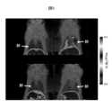
断層画像は、画像の再構成によって取得する。放射性トレーサーの分布を決定するために、限定されないが、肺、肝臓、心臓、腎臓、皮膚、または他の臓器および組織などの再構成画像上に、関心領域(ROI)が描かれる。これらの領域における経時的な放射性トレーサーの取り込みを用いて、介入の非存在下、または試験した様々な用量パラダイムにおける非標識LPA1受容体アンタゴニストの存在下で得られる時間放射能曲線(TAC)を作成する。データは、単位体積あたりの単位時間あたりの放射能(μCi/cc/mCi注入量)として表される。TACデータは、占有されていないLPA1受容体の密度に応じた、結合能(BP)または分布容積(VT)などの定量パラメーターを得るために、当技術分野において周知の様々な方法によって処理する。次いで、LPA1受容体の阻害を、非投与状態におけるBPまたはVTと比較した、様々な用量パラダイムにおけるLPA1受容体アンタゴニストの存在での平衡分析による、BPまたはVTの変化に基づいて計算する。阻害曲線は、上記データ対LPA1受容体アンタゴニストの用量(濃度)をプロットすることによって作成する。次いで、LPA1受容体の阻害を、Emax、Tmax、またはTminにおいて、阻害薬によって達成することができる最大のPET放射性リガンドのVTまたはBPの減少量、および非投与状態におけるBPまたはVTと比較した、さまざまな用量パラダイムにおけるLPA1受容体アンタゴニストの存在下での、非特異的分布容積(VND)およびBPの変化に基づいて計算する。ID50値を、線量率/阻害曲線の曲線あてはめによって得る。A tomographic image is obtained by image reconstruction. To determine the distribution of radiotracer, a region of interest (ROI) is drawn on the reconstructed image, such as, but not limited to, lungs, liver, heart, kidneys, skin, or other organs and tissues. Radiotracer uptake over time in these regions was used to generate time-activity curves (TACs) obtained in the absence of intervention or in the presence of unlabeled LPA1 receptor antagonists in the various dose paradigms tested. do. Data are expressed as radioactivity per unit volume per unit time (μCi/cc/mCi injected volume). TAC data are processed by various methods well known in the art to obtain quantitative parameters such as binding capacity (BP) or volume of distribution (VT ) depending on the density of unoccupied LPA1 receptors. . Inhibition of the LPA1 receptor is then calculated based on the change in BP orVT by equilibrium analysis in the presence of the LPA1 receptor antagonist in various dose paradigms compared to BP orVT in the undosed state. Inhibition curves are generated by plotting the above data versus the dose (concentration) of LPA1 receptor antagonist. Inhibition of the LPA1 receptor is then determined at Emax , Tmax , or Tmin , the amount of reduction inVT or BP of the maximal PET radioligand that can be achieved by the inhibitor, and the BP or V in the undosed state. Calculations are based on the changes in non-specific distribution volume (VND ) and BP in the presence of LPA1 receptor antagonists in various dose paradigms compared toT. ID50 values are obtained by curve fitting of dose rate/inhibition curves.
本開示はさらに、放射性標識LPA1受容体アンタゴニストを、医薬担体または賦形剤と組み合わせるステップを含む、必要な哺乳動物におけるLPA1受容体診断イメージングのための方法に関する。 The present disclosure further relates to a method for diagnostic imaging of LPA1 receptor in a mammal in need thereof comprising combining a radiolabeled LPA1 receptor antagonist with a pharmaceutical carrier or excipient.
定義
特に断らない限り、明細書および特許請求の範囲など、本出願において用いられる以下の用語は、以下に示される定義を有する。本明細書および付属の特許請求の範囲において用いられるように、単数形「a」、「an」、および「the」は、文脈から明らかにそうでないことが示されていない限り、複数形についての言及を含むことに注意する必要がある。特に断らない限り、質量分析、NMR、HPLC、タンパク質化学、生化学、組み換えDNA技術、および薬理学の従来の方法が用いられる。さらに、用語「など」、並びに「含む」および「含まれる」などの他の形態の使用は、限定的でない。本明細書において用いられる節の見出しは、構成上の目的のみのためであり、記載される主題を限定すると解釈されるべきではない。DEFINITIONS Unless otherwise stated, the following terms used in this application, including the specification and claims, have the definitions given below. As used in this specification and the appended claims, the singular forms "a,""an," and "the" refer to the plural unless the context clearly dictates otherwise. Care must be taken to include references. Conventional methods of mass spectrometry, NMR, HPLC, protein chemistry, biochemistry, recombinant DNA technology, and pharmacology are used, unless otherwise indicated. Additionally, the use of the term "such as" and other forms such as "comprising" and "included" is not limiting. The section headings used herein are for organizational purposes only and are not to be construed as limiting the subject matter described.
本明細書において、製剤、組成物、または成分に関して用いられる用語「許容可能な」は、治療される対象の一般的な健康に持続的に有害な影響を有しないことを意味する。 As used herein, the term "acceptable" in reference to a formulation, composition, or ingredient means that it does not have a lasting deleterious effect on the general health of the subject being treated.
本明細書において用いられる用語「調節する」は、標的の活性を変更するように、例えば、ほんの一例として、標的の活性を増強させ、標的の活性を阻害し、標的の活性を制限し、または標的の活性を拡張するように、直接的または間接的のいずれかで標的と相互作用することを意味する。 As used herein, the term "modulate" refers to altering the activity of a target, such as, by way of example only, enhancing the activity of the target, inhibiting the activity of the target, limiting the activity of the target, or It means to interact with a target, either directly or indirectly, so as to extend the activity of the target.
本明細書において用いられる用語「モジュレーター」は、直接的または間接的のいずれかで標的と相互作用する分子をいう。相互作用としては、限定されないが、アゴニスト、部分的アゴニスト、逆アゴニスト、およびアンタゴニストの相互作用が挙げられる。ある実施態様において、モジュレーターはアンタゴニストである。 The term "modulator" as used herein refers to a molecule that interacts with a target, either directly or indirectly. Interactions include, but are not limited to, agonist, partial agonist, inverse agonist, and antagonist interactions. In certain embodiments, the modulator is an antagonist.
本明細書において用いられる用語「アゴニスト」は、特定の受容体に結合し、細胞内での応答を誘発する化合物、薬物、酵素活性化因子、またはホルモンモジュレーターなどの分子をいう。アゴニストは、同じ受容体に結合する内因性リガンド(LPA、プロスタグランジン、ホルモン、または神経伝達物質など)の作用を模倣する。 The term "agonist" as used herein refers to a molecule, such as a compound, drug, enzyme activator, or hormone modulator, that binds to a specific receptor and elicits a response within a cell. Agonists mimic the effects of endogenous ligands (such as LPA, prostaglandins, hormones, or neurotransmitters) that bind to the same receptor.
本明細書において用いられる用語「アンタゴニスト」は、別の分子または受容体部位の活性を減少、抑制、または阻害する、化合物などの分子をいう。アンタゴニストとしては、限定されないが、競合的アンタゴニスト、非競合的アンタゴニスト、不競合的アンタゴニスト、部分的アゴニスト、および逆アゴニストが挙げられる。 The term "antagonist" as used herein refers to a molecule, such as a compound, that reduces, suppresses, or inhibits the activity of another molecule or receptor site. Antagonists include, but are not limited to, competitive antagonists, noncompetitive antagonists, noncompetitive antagonists, partial agonists, and inverse agonists.
本明細書において用いられる用語「LPA依存性」は、LPAの非存在下では発生しない、または同程度に発生しない病状または障害をいう。
本明細書において用いられる用語「LPA介在性」は、LPAの非存在下位では発生するかもしれないが、LPAの存在下では発生しうる病状または障害をいう。The term "LPA-dependent" as used herein refers to a medical condition or disorder that does not occur, or does not occur to the same extent, in the absence of LPA.
The term "LPA-mediated" as used herein refers to a medical condition or disorder that may occur in the absence of LPA, but that may occur in the presence of LPA.
本明細書において用いられる用語「共投与」などは、単一の患者に選択した治療剤を投与することを含むことを意味し、薬剤が同じまたは異なる投与経路によって、または同時または異なる時間において投与される、治療レジメンを含むことが意図される。 As used herein, the term "co-administration" and the like is meant to include administering selected therapeutic agents to a single patient, where the agents are administered by the same or different routes of administration, or at the same or different times. It is intended to include therapeutic regimens in which treatment is performed.
本明細書において用いられる用語「組成物」は、特定の成分を特定の分量において含む製品、並びに特定の成分の特定の分量における組み合わせから、直接的または間接的に生じる任意の製品を包含することが意図される。医薬組成物に関連するこのような用語は、担体を構成する活性成分および不活性成分、並びに任意の2つ以上の成分の組み合わせ、複合体、または凝集から、あるいは1つ以上の成分の解離から、あるいはその別の種類の反応または相互作用、またはその成分の無視によって、直接的または間接的に生じる任意の生成物を含む製品を包含することが意図される。そのため、本発明の医薬組成物は、本発明の化合物と薬学的に許容可能な担体の混合によって形成される、任意の組成物を包含する。「薬学的に許容可能な」によって、担体、希釈剤、または賦形剤が、他の製剤と適合しなければならず、その受容者に有害でないことが意味される。用語、化合物の「投与」および/または化合物を「投与すること」は、本発明の化合物、または本発明の化合物のプロドラッグを患者に提供することを意味すると理解されるべきである。 As used herein, the term "composition" is intended to encompass products containing the specified ingredients in specified amounts, as well as any product resulting directly or indirectly from the combination of the specified ingredients in specified amounts. is intended. Such terms in the context of pharmaceutical compositions include the active and inactive ingredients that constitute the carrier, as well as any combination, complex, or aggregation of two or more ingredients, or from dissociation of one or more ingredients. , or another type of reaction or interaction thereof, or any product resulting directly or indirectly from the neglect of its components. As such, the pharmaceutical compositions of the invention include any composition formed by admixing a compound of the invention and a pharmaceutically acceptable carrier. By "pharmaceutically acceptable" is meant that the carrier, diluent, or excipient must be compatible with the other formulations and not deleterious to its recipient. The terms "administration" of a compound and/or "administering" a compound are to be understood to mean providing a compound of the invention, or a prodrug of a compound of the invention, to a patient.
本明細書において用いられる用語「有効量」または「治療上の有効量」は、治療される疾患または病状の症状の1つ以上をある程度緩和する、投与される薬剤または化合物の十分な分量をいう。結果は、疾患の徴候、症状、または原因の軽減および/または緩和、または生物学的システムの他の任意の望ましい調整でありうる。例えば、治療的使用のための「有効量」は、疾患症状の臨床的に有意な軽減を提供するのに必要な、本明細書に開示される化合物を含む組成物の分量である。任意の個々の場合において、適切な「有効」量は、用量漸増試験などの技術を用いて決定されうる。 The term "effective amount" or "therapeutically effective amount" as used herein refers to a sufficient amount of the drug or compound administered to alleviate to some extent one or more symptoms of the disease or condition being treated. . The result can be a reduction and/or alleviation of the signs, symptoms, or causes of a disease, or any other desirable modulation of a biological system. For example, an "effective amount" for therapeutic use is the amount of a composition comprising a compound disclosed herein that is necessary to provide clinically significant relief of disease symptoms. In any individual case, an appropriate "effective" amount can be determined using techniques such as dose escalation studies.
本明細書において用いられる用語「組み合わせ医薬」は、1つより多い活性成分の混合または組み合わせから生じ、活性成分の固定の、および非固定の両方の組み合わせを含む生成物を意味する。用語「固定の組み合わせ」は、活性成分、例えば、式(I)、(Ia)、および(IIa)の化合物、および共薬剤の両方を、単一の形態または剤形の形態で、同時に患者に投与することを意味する。用語「非固定の組み合わせ」は、活性成分、例えば、式(I)、(Ia)、または(IIa)の化合物、および共薬剤が、同時に、同時的に、または順次のいずれかで、特定の時間間隔の制限なしで、別の形態として患者に投与されることを意味し、ここで、そのような投与は、患者の体内において2つの化合物の有効量を提供する。後者はまた、カクテル療法、例えば、3つ以上の活性成分の投与に適用される。 The term "combination medicament" as used herein means a product resulting from a mixture or combination of more than one active ingredient and containing both fixed and non-fixed combinations of active ingredients. The term "fixed combination" refers to the administration of both active ingredients, e.g. compounds of formulas (I), (Ia), and (IIa), and co-medications, in a single form or dosage form, to a patient at the same time. It means to administer. The term "non-fixed combination" means that an active ingredient, e.g. It is meant to be administered to a patient in separate forms without limitation of time interval, where such administration provides effective amounts of the two compounds in the patient's body. The latter also applies to cocktail therapy, eg the administration of three or more active ingredients.
用語「対象」または「患者」は、哺乳動物を含む。哺乳動物の例としては、限定されないが、ヒト、チンパンジー、類人猿、サル、ウシ、ウマ、ヒツジ、ヤギ、ブタ、ウサギ、イヌ、ネコ、齧歯類、ラット、マウス、モルモットなどが挙げられる。ある実施態様において、哺乳動物はヒトである。 The term "subject" or "patient" includes mammals. Examples of mammals include, but are not limited to, humans, chimpanzees, apes, monkeys, cows, horses, sheep, goats, pigs, rabbits, dogs, cats, rodents, rats, mice, guinea pigs, and the like. In certain embodiments, the mammal is a human.
本明細書で用いられる用語「治療する」、「治療すること」、または「治療」としては、疾患または病状の少なくとも1つの症状を軽減すること、寛解させること、または改善すること、さらなる症状を予防すること、疾患または病状を抑制すること、例えば、疾患または病状の発症を抑止すること、疾患または病状を緩和すること、疾患または病状の退行を引き起こすこと、疾患または病状によって引き起こされる病状を緩和すること、または疾患または病状の症状を予防的および/または治療的のいずれかで停止させることが挙げられる。 As used herein, the terms "treat," "treating," or "treatment" refer to alleviating, ameliorating, or ameliorating at least one symptom of a disease or condition; to prevent, to suppress a disease or condition, such as to arrest the onset of a disease or condition, to alleviate a disease or condition, to cause regression of a disease or condition, to alleviate the condition caused by a disease or condition; or halting the symptoms of a disease or condition, either prophylactically and/or therapeutically.
本明細書に記載される化合物は、非対称中心を有しうる。非対称に置換された原子を含むそのような化合物は、光学活性体またはラセミ体において単離されうる。ラセミ体の分割または光学活性な出発物質からの合成などによって、光学活性体を調整する方法は、当技術分野において周知である。特定の立体化学または異性体形態が具体的に示されていない限り、構造の全てのキラル、ジアステレオマー、およびラセミ体が意図される。 The compounds described herein may have asymmetric centers. Such compounds containing asymmetrically substituted atoms may be isolated in optically active or racemic form. Methods for preparing optically active forms, such as by racemic resolution or synthesis from optically active starting materials, are well known in the art. All chiral, diastereomeric, and racemic forms of a structure are intended, unless the specific stereochemistry or isomeric form is specifically indicated.
語句「薬学的に許容される」は、健全な医学的判断の範囲内で、過度な毒性、刺激、アレルギー応答、または他の問題、または合併症がない、合理的な利益/リスク比に見合った、ヒトおよび動物の組織と接触させて用いるのに適切な、これらの化合物、物質、組成物、および/または投与形態をいうように本明細書において用いられる。 The phrase "pharmaceutically acceptable" means a condition that, within the scope of sound medical judgment, is commensurate with a reasonable benefit/risk ratio, without undue toxicity, irritation, allergic response, or other problems or complications. Also used herein to refer to those compounds, substances, compositions, and/or dosage forms that are suitable for use in contact with human and animal tissue.
本明細書で用いられる「薬学的に許容可能な塩」は、親化合物の酸性塩または塩基性塩を製造することによって、親化合物が修飾されている、開示される化合物の誘導体をいう。 As used herein, "pharmaceutically acceptable salts" refer to derivatives of the disclosed compounds in which the parent compound has been modified by preparing acidic or basic salts of the parent compound.
用語、薬学的に許容される「塩」は、無機および有機塩基と共に形成される塩基性塩をいいうる。そのような塩としては、アンモニウム塩;リチウム、ナトリウム、およびカリウム塩などの、アルカリ金属塩;カルシウムおよびマグネシウム塩などの、アルカリ土類金属塩;アミン様塩(例えば、ジシクロヘキシルアミン塩、ベンザチン、N-メチル-D-グルカミン、およびヒドラバミン塩)などの、有機塩基との塩;並びに、アルギニン、リシンなどのアミノ酸との塩;並びに双性イオン、いわゆる「分子内塩」が挙げられる。非毒性の、薬学的に許容可能な塩が好ましいが、他の塩もまた、例えば生成物の単離または精製に有用である。 The term pharmaceutically acceptable "salts" refers to basic salts formed with inorganic and organic bases. Such salts include ammonium salts; alkali metal salts such as lithium, sodium, and potassium salts; alkaline earth metal salts such as calcium and magnesium salts; amine-like salts (e.g. dicyclohexylamine salt, benzathine, N -methyl-D-glucamine, and hydrabamine salts); and salts with amino acids such as arginine, lysine, etc.; and zwitterions, so-called "inner salts." Although non-toxic, pharmaceutically acceptable salts are preferred, other salts are also useful, eg, for isolation or purification of the product.
用語、薬学的に許容可能な「塩」としてはまた、酸付加塩が挙げられる。これらは、例えば、鉱酸などの強い無機酸、例えば、硫酸、リン酸、またはHClもしくはHBrなどのハロゲン化水素酸によって、強い有機カルボン酸、例えば、非置換、または例えばハロゲンによって置換された、1~4個の炭素原子のアルカンカルボン酸、例えば、酢酸、飽和または不飽和二カルボン酸など、例えば、シュウ酸、マロン酸、コハク酸、マレイン酸、フマル酸、フタル酸、またはテレフタル酸;ヒドロキシカルボン酸など、例えば、アスコルビン酸、グリコール酸、乳酸、リンゴ酸、酒石酸、またはクエン酸、アミノ酸(例えば、アスパラギン、またはグルタミン酸、またはリシン、またはアルギニン)など、または安息香酸;または有機スルホン酸、例えば、非置換、もしくは例えばハロゲンによって置換されている、(C1-C4)アルキルまたはアリールスルホン酸、例えば、メタンスルホン酸またはp-トルエンスルホン酸によって、形成される。The term pharmaceutically acceptable "salt" also includes acid addition salts. These are, for example, strong inorganic acids such as mineral acids, for example sulfuric acid, phosphoric acid or hydrohalic acids such as HCl or HBr, strong organic carboxylic acids, for example unsubstituted or substituted, for example by halogens. Alkane carboxylic acids of 1 to 4 carbon atoms, such as acetic acid, saturated or unsaturated dicarboxylic acids, such as oxalic acid, malonic acid, succinic acid, maleic acid, fumaric acid, phthalic acid, or terephthalic acid; hydroxy carboxylic acids, such as ascorbic acid, glycolic acid, lactic acid, malic acid, tartaric acid, or citric acid, amino acids such as asparagine, or glutamic acid, or lysine, or arginine, or benzoic acid; or organic sulfonic acids, such as , unsubstituted or substituted, for example by halogen, (C1 -C4 )alkyl or arylsulfonic acids, such as methanesulfonic acid or p-toluenesulfonic acid.
薬学的に許容可能な塩は、従来の化学的方法によって、塩基性または酸性基を含む親化合物から合成することができる。一般に、そのような塩は、これらの化合物の遊離酸または塩基形態を、適切な塩基または酸の化学量論と、水または有機溶媒中で、または2つの混合物中で反応させることによって調製することができ;一般に、エーテル、酢酸エチル、エタノール、イソプロパノール、またはアセトニトリルなどの非水性溶媒が好ましい。適切な塩の一覧は、Remington’s Pharmaceutical Sciences, 17th Edition, p. 1418, Mack Publishing Company, Easton, PA (1985)に存在し、この開示は引用によって本明細書に援用される。 Pharmaceutically acceptable salts can be synthesized from parent compounds containing basic or acidic groups by conventional chemical methods. Generally, such salts are prepared by reacting the free acid or base forms of these compounds with the appropriate base or acid stoichiometry in water or an organic solvent, or in a mixture of the two. generally non-aqueous solvents such as ether, ethyl acetate, ethanol, isopropanol, or acetonitrile are preferred. A list of suitable salts is found in Remington's Pharmaceutical Sciences, 17th Edition, p. 1418, Mack Publishing Company, Easton, PA (1985), the disclosure of which is incorporated herein by reference.
明細書を通して、基およびその置換基は、安定した基および化合物、並びに薬学的に許容可能な化合物として有用な化合物、および/または薬学的に許容可能な化合物の製造に有用な中間体化合物を提供するように、当業者によって選択されうる。 Throughout the specification, groups and their substituents provide stable groups and compounds useful as pharmaceutically acceptable compounds, and/or intermediate compounds useful in the preparation of pharmaceutically acceptable compounds. may be selected by a person skilled in the art to do so.
実施例
本発明の化合物の合成は、代表として実施例に開示されている化合物を用いて、以下のスキームにおいて説明する。EXAMPLES The synthesis of compounds of the invention is illustrated in the following schemes using as representatives the compounds disclosed in the Examples.
非標識の式(I)の化合物の合成手順を、以下に概説する。スキーム1は、フルオロアルキルカルバモイルオキシメチルトリアゾールシクロヘキシル酸8の合成を記載する。出発物質、シクロヘキシルエステルトリアゾールアルコール1の合成は、US2017/0360759において記載され(例えば、実施例1Eおよび10F)、本開示は引用によって本明細書に援用される。トリアゾールアルコール1を、適切な塩基の存在下で、4-ニトロフェニル クロロホルメートと反応させて、対応するトリアゾール 4-ニトロフェニルカルボネート2を得る。トリアゾール 4-ニトロフェニルカルボネート2を次いで、適切な塩基の存在下で、適切なN-ヒドロキシアルキルアミン3(n=0-2)と反応させて、トリアゾール N-ヒドロキシルアルキルカルバメート4を得る。N-ヒドロキシアルキルカルバメート4を、適切なスルホニルクロライド5(例えば、p-トルエンスルホニルクロライド)と反応させて、対応するスルホネート6を得る。スルホネート6を、適切なフルオライドアニオン源(例えば、KFまたはBu4NF)と反応させて、クロヘキシルエステルトリアゾール N-フルオロアルキルカルバメート7を得て、これを次いでエステル脱保護して、目的のフルオロアルキルカルバモイルオキシメチルトリアゾール シクロヘキシル酸8を生じる。
スキーム2は、18F-フルオロアルキルカルバモイルオキシメチルトリアゾール シクロヘキシル酸の放射性合成を記載する。スルホネート前駆体6の放射性合成は、適切なフルオライド-18アニオン源(例えば、K18FまたはBu4N18F)、適切な相間移動触媒(例えば、クリプトフィックス(登録商標)222)、および適切なイオン液体(例えば、1-ブチル-3-メチルイミダゾリウム ヘキサフルオロホスフェート)を反応させて、18F-シクロヘキシルエステルトリアゾール N-フルオロアルキルカルバメート9を得て、これが次いでエステル脱保護を受けて、目的の18F-フルオロアルキルカルバモイルオキシメチルトリアゾール シクロヘキシル酸10を得る。
HPLC条件:
方法A:以下の方法を用いた、Agilent 1100 series HPLC および Lab logic gamma ram radio-HPLC 検出器:カラム:Zorbax SB C18、4.6x250mm、3μm粒子;移動相:60%アセトニトリル/0.1%トリフルオロ酢酸水溶液;流速:1.00mL/分;検出:254nmにおけるUV。
方法B:カラム:Zorbax SB C18、4.6x250mm、3μm粒子;移動相:29%エタノール、10%アセトニトリル、および61%の100mM KH2PO4水溶液;流速:1.00mL/分;検出:254nmにおけるUV。
方法C:カラム:Chiralpak AD-H-250x4.6mm 5μm粒子;移動相:15%イソプロピルアルコール/ヘプタン;流速:0.9mL/分;検出:254nmにおけるUV。HPLC conditions:
Method A: Agilent 1100 series HPLC and Lab logic gamma ram radio-HPLC using the following method Detector: Column: Zorbax SB C18, 4.6x250mm, 3μm particles; Mobile phase: 60% acetonitrile/0.1% Tri Fluoroacetic acid aqueous solution; flow rate: 1.00 mL/min; detection: UV at 254 nm.
Method B: Column: Zorbax SB C18, 4.6x250mm, 3μm particles; Mobile phase: 29% ethanol, 10% acetonitrile, and 61% of100mMKH2PO4 in water; Flow rate: 1.00mL/min; Detection: at 254nm UV.
Method C: Column: Chiralpak AD-H-250x4.6mm 5 μm particles; Mobile phase: 15% isopropyl alcohol/heptane; Flow rate: 0.9 mL/min; Detection: UV at 254 nm.
実施例1.メチル (1S,3S)-3-((6-(5-(ヒドロキシメチル)-1-メチル-1H-1,2,3-トリアゾール-4-イル)-2-メチルピリジン-3-イル)オキシ)シクロヘキサン-1-カルボキシレート
表題の化合物は、特許出願US2017/0360759から、対応するイソプロピルエステル(実施例1E)またはエチルエステル(実施例10F)のいずれかの合成について記載される合成方法に従って合成した。表題の化合物を製造するために用いられる出発物質は、(対応するシクロヘキサンイソプロピルまたはエチルエステルではなく)(1S、3R)-メチル 3-ヒドロキシシクロヘキサン-1-カルボキシレートであった。Example 1. Methyl (1S,3S)-3-((6-(5-(hydroxymethyl)-1-methyl-1H-1,2,3-triazol-4-yl)-2-methylpyridin-3-yl)oxy ) Cyclohexane-1-carboxylate
The title compound was synthesized according to the synthetic method described for the synthesis of either the corresponding isopropyl ester (Example 1E) or ethyl ester (Example 10F) from patent application US2017/0360759. The starting material used to prepare the title compound was (1S,3R)-methyl 3-hydroxycyclohexane-1-carboxylate (rather than the corresponding cyclohexane isopropyl or ethyl ester).
実施例2.メチル (1R,3S)-3-((6-(5-(ヒドロキシメチル)-1-メチル-1H-1,2,3-トリアゾール-4-イル)-2-メチルピリジン-3-イル)オキシ)シクロヘキサン-1-カルボキシレート
実施例1化合物(1.95g、5.41mmol)のMeOH(38.6mL)中の透明な溶液を、5.4MのNaOMe溶液(4.0mL、21.64mmol)に加えた。反応液を60℃で6時間加熱し、次いで室温に冷却した。反応物を氷浴に入れ、1N HClで中性化し、次いで部分的に濃縮して、MeOHを除去した。得られた濁った混合物を、1.0M K2HPO4およびEtOAcに分配し、層を分離した。水層をEtOAc(1x)で抽出した。有機層を合わせて、ブラインで洗浄し、乾燥させ(Na2SO4)、濾過し、濃縮して、重量1.66gの白色の固形物を得た。キラルSFC Prep(カラム:Chiralpak IA、21x250mm、5ミクロン;移動相:20%MeOH/80%CO2;流速条件:85mL/分、150Bar、40℃;検出波長:254nm)による精製によって、白色の固形物の表題の化合物(740mg、収率38%)として、第二溶出ジアステレオマーを得た。キラル分析HPLC(カラム:Chiralpak IA、4.6x250mm、5ミクロン;移動相:25%MeOH/80%CO2;流速条件:2.0mL/分、150Bar、40℃;検出波長:254nm)によって、メチルエステル立体中心において、98.2%de[0.9:99.1 トランス(4.33分):シス(5.48分)]が示された。
LCMS, [M + H]+ = 361.1。1H NMR (500 MHz, CDCl3) δ 8.10 (d, J=8.8 Hz, 1H), 7.61 - 7.45 (m, 1H), 7.29 (d, J=8.8 Hz, 1H), 4.83 (s, 2H), 4.33 - 4.21 (m, 1H), 4.09 (s, 3H), 3.70 (s, 3H), 2.53 - 2.45 (m, 4H), 2.44 - 2.38 (m, 1H), 2.23 - 2.13 (m, 1H), 2.06 - 1.96 (m, 2H), 1.76 - 1.59 (m, 1H), 1.56 - 1.39 (m, 3H).Example 2. Methyl (1R,3S)-3-((6-(5-(hydroxymethyl)-1-methyl-1H-1,2,3-triazol-4-yl)-2-methylpyridin-3-yl)oxy ) Cyclohexane-1-carboxylate
A clear solution of Example 1 compound (1.95 g, 5.41 mmol) in MeOH (38.6 mL) was added to a 5.4 M NaOMe solution (4.0 mL, 21.64 mmol). The reaction was heated at 60° C. for 6 hours, then cooled to room temperature. The reaction was placed in an ice bath, neutralized with 1N HCl, and then partially concentrated to remove MeOH. The resulting cloudy mixture was partitioned between 1.0 MK2HPO4 and EtOAc and thelayers were separated. The aqueous layer was extracted with EtOAc (1x). The organic layers were combined, washed with brine, dried (Na2 SO4 ), filtered, and concentrated to give a white solid weighing 1.66 g. Purification by Chiral SFC Prep (column: Chiralpak IA, 21x250 mm, 5 micron; mobile phase: 20% MeOH/80%CO2 ; flow rate conditions: 85 mL/min, 150 Bar, 40 °C; detection wavelength: 254 nm) gave a white solid. A second eluting diastereomer was obtained as the title compound (740 mg, 38% yield). Methyl was detected by chiral analytical HPLC (column: Chiralpak IA, 4.6x250 mm, 5 micron; mobile phase: 25% MeOH/80%CO2 ; flow rate conditions: 2.0 mL/min, 150 Bar, 40°C; detection wavelength: 254 nm). At the ester stereocenter, 98.2% de[0.9:99.1 trans(4.33 min):cis(5.48 min)] was shown.
LCMS, [M + H]+ = 361.1.1 H NMR (500 MHz, CDCl3 ) δ 8.10 (d, J=8.8 Hz, 1H), 7.61 - 7.45 (m, 1H), 7.29 (d, J=8.8 Hz, 1H), 4.83 (s, 2H) , 4.33 - 4.21 (m, 1H), 4.09 (s, 3H), 3.70 (s, 3H), 2.53 - 2.45 (m, 4H), 2.44 - 2.38 (m, 1H), 2.23 - 2.13 (m, 1H) , 2.06 - 1.96 (m, 2H), 1.76 - 1.59 (m, 1H), 1.56 - 1.39 (m, 3H).
実施例3.メチル (1S,3S)-3-((2-メチル-6-(1-メチル-5-((((4-ニトロフェノキシ)カルボニル)オキシ)メチル)-1H-1,2,3-トリアゾール-4-イル)ピリジン-3-イル)オキシ)シクロヘキサン-1-カルボキシレート
実施例1化合物(970mg、2.69mmol)およびピリジン(1.09mL、13.5mmol)のDCM(17.9mL)中の0℃の溶液に、1時間にわたり、4-ニトロフェニルクロロホルメート(1.09g、5.38mmol)のDCM(3mL)中の溶液を滴下して加えた。反応液を次いで、室温に昇温させ、室温で20時間撹拌し、次いで減圧下で濃縮して、固形物を得た。DCMの最小量を加えて懸濁液を得て、固体(ピリジン ヒドロクロライド)を濾過によって除去した。濾液を減圧下で濃縮した。残留物をクロマトグラフィーにかけ(120g SiO2カラム;0-50%EtOAc/ヘキサンの連続的勾配)、表題の化合物(1.12g、収率79%)を淡黄色の固形物として得た。
LCMS, [M + H]+ = 526.3。1H NMR (500 MHz, CDCl3) δ 8.33 - 8.28 (m, 2H), 8.03 (d, J=8.5 Hz, 1H), 7.43 - 7.38 (m, 2H), 7.23 (d, J=8.5 Hz, 1H), 6.06 (s, 2H), 4.76 - 4.71 (m, 1H), 4.22 (s, 3H), 3.72 (s, 3H), 2.89 - 2.80 (m, 1H), 2.51 (s, 3H), 2.21 - 2.12 (m, 1H), 2.04 - 1.89 (m, 3H), 1.83 - 1.72 (m, 1H), 1.71 - 1.61 (m, 3H).Example 3. Methyl (1S,3S)-3-((2-methyl-6-(1-methyl-5-((((4-nitrophenoxy)carbonyl)oxy)methyl)-1H-1,2,3-triazole- 4-yl)pyridin-3-yl)oxy)cyclohexane-1-carboxylate
To a solution of Example 1 compound (970 mg, 2.69 mmol) and pyridine (1.09 mL, 13.5 mmol) in DCM (17.9 mL) at 0 °C was added 4-nitrophenylchloroformate (1 A solution of .09 g, 5.38 mmol) in DCM (3 mL) was added dropwise. The reaction was then warmed to room temperature, stirred at room temperature for 20 hours, and then concentrated under reduced pressure to obtain a solid. A minimum amount of DCM was added to obtain a suspension and the solid (pyridine hydrochloride) was removed by filtration. The filtrate was concentrated under reduced pressure. The residue was chromatographed (120 g SiO2 column; continuous gradient 0-50% EtOAc/hexanes) to give the title compound (1.12 g, 79% yield) as a pale yellow solid.
LCMS, [M + H]+ = 526.3.1 H NMR (500 MHz, CDCl3 ) δ 8.33 - 8.28 (m, 2H), 8.03 (d, J=8.5 Hz, 1H), 7.43 - 7.38 (m, 2H), 7.23 (d, J=8.5 Hz, 1H), 6.06 (s, 2H), 4.76 - 4.71 (m, 1H), 4.22 (s, 3H), 3.72 (s, 3H), 2.89 - 2.80 (m, 1H), 2.51 (s, 3H), 2.21 - 2.12 (m, 1H), 2.04 - 1.89 (m, 3H), 1.83 - 1.72 (m, 1H), 1.71 - 1.61 (m, 3H).
実施例4.メチル (1R,3S)-3-((2-メチル-6-(1-メチル-5-((((4-ニトロフェノキシ)カルボニル)オキシ)メチル)-1H-1,2,3-トリアゾール-4-イル)ピリジン-3-イル)オキシ)シクロヘキサン-1-カルボキシレート
表題の化合物は、実施例3化合物の合成について記載される方法に従って、反応に実施例2化合物を、(実施例1化合物ではなく)4-ニトロフェニルクロロホルメートと共に用いることによって、合成した。LCMS, [M + H]+ = 526.1。1H NMR (500 MHz, CDCl3) δ 8.33 - 8.27 (m, 2H), 8.02 (d, J=8.5 Hz, 1H), 7.43 - 7.38 (m, 2H), 7.22 (d, J=8.5 Hz, 1H), 6.06 (s, 2H), 4.32 - 4.19 (m, 4H), 3.70 (s, 3H), 2.54 - 2.45 (m, 4H), 2.45 - 2.36 (m, 1H), 2.22 - 2.12 (m, 1H), 2.05 - 1.95 (m, 2H), 1.77 - 1.64 (m, 1H), 1.55 - 1.38 (m, 3H).Example 4. Methyl (1R,3S)-3-((2-methyl-6-(1-methyl-5-((((4-nitrophenoxy)carbonyl)oxy)methyl)-1H-1,2,3-triazole- 4-yl)pyridin-3-yl)oxy)cyclohexane-1-carboxylate
The title compound was synthesized according to the method described for the synthesis of Example 3 compound by using Example 2 compound together with 4-nitrophenylchloroformate (rather than Example 1 compound) in the reaction. LCMS, [M + H]+ = 526.1.1 H NMR (500 MHz, CDCl3 ) δ 8.33 - 8.27 (m, 2H), 8.02 (d, J=8.5 Hz, 1H), 7.43 - 7.38 (m, 2H), 7.22 (d, J=8.5 Hz, 1H), 6.06 (s, 2H), 4.32 - 4.19 (m, 4H), 3.70 (s, 3H), 2.54 - 2.45 (m, 4H), 2.45 - 2.36 (m, 1H), 2.22 - 2.12 (m, 1H), 2.05 - 1.95 (m, 2H), 1.77 - 1.64 (m, 1H), 1.55 - 1.38 (m, 3H).
実施例5:メチル (1S,3S)-3-((6-(5-((((4-ヒドロキシブチル)(メチル)カルバモイル)オキシ)メチル)-1-メチル-1H-1,2,3-トリアゾール-4-イル)-2-メチルピリジン-3-イル)オキシ)シクロヘキサン-1-カルボキシレート
実施例3化合物(1.90g、3.62mmol)のTHF(18.1mL)中の室温溶液に、4-(メチルアミノ)ブタン-1-オール(0.448g、4.34mmol)、次いでDIEA(0.76mL、4.34mmol)を加えた。室温で18時間撹拌した後、反応液を減圧下で濃縮した。粗生成物をクロマトグラフィーにかけ(120gSiO2カラム;20分にわたり0-10%MeOH/DCMの連続的勾配、次いで20分間、10%MeOH/DCMの定組成)、表題の化合物(2.1g、収率119%)を黄色の油状物として得た。反応からの副生成物、4-ニトロフェノールもまた存在した。この物質をさらに精製することなく、次のステップで用いた。LCMS, [M + H]+ = 490.3。1H NMR (500 MHz, CDCl3) δ 7.96 (d, J=8.5 Hz, 1H), 7.21 (d, J=8.5 Hz, 1H), 5.84 - 5.71 (m, 2H), 4.74 - 4.69 (m, 1H), 4.15 (s, 3H), 3.77 - 3.65 (m, 4H), 3.54 - 3.45 (m, 1H), 3.41 - 3.31 (m, 1H), 3.27 - 3.16 (m, 1H), 2.99 - 2.80 (m, 4H), 2.52 (s, 3H), 2.22 - 2.11 (m, 1H), 2.05 - 1.87 (m, 3H), 1.84 - 1.73 (m, 1H), 1.71 - 1.36 (m, 8H)。いくつかのピークは回転異性体であると考えられる。Example 5: Methyl (1S,3S)-3-((6-(5-((((4-hydroxybutyl)(methyl)carbamoyl)oxy)methyl)-1-methyl-1H-1,2,3 -triazol-4-yl)-2-methylpyridin-3-yl)oxy)cyclohexane-1-carboxylate
A room temperature solution of Example 3 compound (1.90 g, 3.62 mmol) in THF (18.1 mL) was added with 4-(methylamino)butan-1-ol (0.448 g, 4.34 mmol) followed by DIEA ( 0.76 mL, 4.34 mmol) was added. After stirring at room temperature for 18 hours, the reaction solution was concentrated under reduced pressure. The crude product was chromatographed (120 g SiO2 column; continuous gradient 0-10% MeOH/DCM over 20 min, then isocratic 10% MeOH/DCM for 20 min) to give the title compound (2.1 g, yield 119%) as a yellow oil. A byproduct from the reaction, 4-nitrophenol, was also present. This material was used in the next step without further purification. LCMS, [M + H]+ = 490.3.1 H NMR (500 MHz, CDCl3 ) δ 7.96 (d, J=8.5 Hz, 1H), 7.21 (d, J=8.5 Hz, 1H), 5.84 - 5.71 (m, 2H), 4.74 - 4.69 (m, 1H), 4.15 (s, 3H), 3.77 - 3.65 (m, 4H), 3.54 - 3.45 (m, 1H), 3.41 - 3.31 (m, 1H), 3.27 - 3.16 (m, 1H), 2.99 - 2.80 ( m, 4H), 2.52 (s, 3H), 2.22 - 2.11 (m, 1H), 2.05 - 1.87 (m, 3H), 1.84 - 1.73 (m, 1H), 1.71 - 1.36 (m, 8H). Some peaks are believed to be rotamers.
実施例6.メチル (1S,3S)-3-((2-メチル-6-(1-メチル-5-(((メチル(4-(トシルオキシ)ブチル)カルバモイル)オキシ)メチル)-1H-1,2,3-トリアゾール-4-イル)ピリジン-3-イル)オキシ)シクロヘキサン-1-カルボキシレート
実施例5化合物(115mg、0.24mmol)、DMAP(2.9mg、0.023mmol)、およびTEA(72.0μL、0.52mmol)の、DCM(2.35mL)中の冷却した溶液(0℃)に、p-TsCl(53.7mg、0.28mmol)を加えた。得られた反応液を室温に昇温させ、室温で一晩撹拌した。反応液を水(3mL)およびEt2O(3mL)に分配し、層を分離した。水層をEt2O(1x)で抽出した。有機層を合わせて乾燥させ(MgSO4)、減圧下で濃縮した。粗生成物をクロマトグラフィーにかけ(24g SiO2カラム;25分にわたり、0-60%EtOAc/ヘキサンの連続的勾配、次いで30分間にわたり、60%EtOAc/ヘキサンの定組成)、表題の化合物(126mg、収率83%)を透明な無色の残留物として得た。LCMS, [M + H]+ = 644.2。1H NMR (500 MHz, CDCl3) δ 7.95 (d, J=8.5 Hz, 1H), 7.84 - 7.69 (m, 2H), 7.38 - 7.31 (m, 2H), 7.20 (d, J=8.5 Hz, 1H), 5.81 - 5.69 (m, 2H), 4.74 - 4.67 (m, 1H), 4.13 (s, 3H), 4.09 - 4.03 (m, 1H), 3.90 - 3.84 (m, 1H), 3.70 (s, 3H), 3.31 - 3.22 (m, 1H), 3.18 - 3.11 (m, 1H), 2.91 - 2.76 (m, 4H), 2.52 - 2.47 (m, 3H), 2.45 (s, 3H), 2.20 - 2.10 (m, 1H), 2.03 - 1.86 (m, 3H), 1.83 - 1.71 (m, 1H), 1.70 - 1.56 (m, 5H)、1.51 - 1.41 (m, 2H)。いくつかのピークは回転異性体であると考えられる。Example 6. Methyl (1S,3S)-3-((2-methyl-6-(1-methyl-5-(((methyl(4-(tosyloxy)butyl)carbamoyl)oxy)methyl)-1H-1,2,3 -triazol-4-yl)pyridin-3-yl)oxy)cyclohexane-1-carboxylate
Example 5 A chilled solution of compound (115 mg, 0.24 mmol), DMAP (2.9 mg, 0.023 mmol), and TEA (72.0 μL, 0.52 mmol) in DCM (2.35 mL) (0 °C ), p-TsCl (53.7 mg, 0.28 mmol) was added. The resulting reaction solution was heated to room temperature and stirred at room temperature overnight. The reaction was partitioned between water (3 mL) and Et2 O (3 mL) and the layers were separated. The aqueous layer was extracted with Et2 O (1x). The combined organic layers were dried (MgSO4 ) and concentrated under reduced pressure. The crude product was chromatographed (24 g SiO2 column; continuous gradient of 0-60% EtOAc/hexanes over 25 min, then isocratic of 60% EtOAc/hexanes over 30 min) to give the title compound (126 mg, 83% yield) was obtained as a clear colorless residue. LCMS, [M + H]+ = 644.2.1 H NMR (500 MHz, CDCl3 ) δ 7.95 (d, J=8.5 Hz, 1H), 7.84 - 7.69 (m, 2H), 7.38 - 7.31 (m, 2H), 7.20 (d, J=8.5 Hz, 1H), 5.81 - 5.69 (m, 2H), 4.74 - 4.67 (m, 1H), 4.13 (s, 3H), 4.09 - 4.03 (m, 1H), 3.90 - 3.84 (m, 1H), 3.70 (s, 3H), 3.31 - 3.22 (m, 1H), 3.18 - 3.11 (m, 1H), 2.91 - 2.76 (m, 4H), 2.52 - 2.47 (m, 3H), 2.45 (s, 3H), 2.20 - 2.10 ( m, 1H), 2.03 - 1.86 (m, 3H), 1.83 - 1.71 (m, 1H), 1.70 - 1.56 (m, 5H), 1.51 - 1.41 (m, 2H). Some peaks are believed to be rotamers.
実施例7.メチル (1S,3S)-3-((2-メチル-6-(1-メチル-5-(((メチル(4-(((4-ニトロ-フェニル)スルホニル)オキシ)ブチル)カルバモイル)オキシ)メチル)-1H-1,2,3-トリアゾール-4-イル)ピリジン-3-イル)オキシ)シクロヘキサン-1-カルボキシレート
実施例5化合物(90mg、0.18mmol)、DMAP(2.2mg、0.018mmol)、およびTEA(56μL、0.40mmol)のDCM(1.8mL)中の0℃の溶液に、NsCl(45mg、0.20mmol)を加えた。得られた反応液をゆっくりと室温に昇温させた。2時間室温で撹拌した後、反応液を水(3mL)およびEt2O(3mL)に分配し、層を分離した。水層をEt2O(1x)で抽出した。有機層を合わせて乾燥させ(MgSO4)、濾過し、減圧下で濃縮した。粗生成物をクロマトグラフィーにかけ(12g SiO2カラム;0-60%EtOAc/ヘキサンの連続的勾配)、表題の化合物(25mg、収率19%)を黄色の固形物として得た。LCMS, [M + H]+ = 675.2。1H NMR (500 MHz, CDCl3) δ 8.46 - 8.38 (m, 2H), 8.14 (br d, J=8.0 Hz, 1H), 8.08 - 8.00 (m, 1H), 7.97 (d, J=8.5 Hz, 1H), 7.22 (d, J=8.5 Hz, 1H), 5.81 - 5.70 (m, 2H), 4.76 - 4.69 (m, 1H), 4.24 - 4.17 (m, 1H), 4.15 (s, 3H), 3.98 - 3.91 (m, 1H), 3.72 (s, 3H), 3.34 - 3.26 (m, 1H), 3.24 - 3.13 (m, 1H), 2.96 - 2.78 (m, 4H), 2.53 - 2.46 (m, 3H), 2.22 - 2.11 (m, 1H), 2.05 - 1.88 (m, 3H), 1.85 - 1.46 (m, 8H)。 いくつかのピークは回転異性体であると考えられる。Example 7. Methyl (1S,3S)-3-((2-methyl-6-(1-methyl-5-(((methyl(4-(((4-nitro-phenyl)sulfonyl)oxy)butyl)carbamoyl)oxy) methyl)-1H-1,2,3-triazol-4-yl)pyridin-3-yl)oxy)cyclohexane-1-carboxylate
Example 5 To a solution of compound (90 mg, 0.18 mmol), DMAP (2.2 mg, 0.018 mmol), and TEA (56 μL, 0.40 mmol) in DCM (1.8 mL) at 0 °C was added NsCl (45 mg , 0.20 mmol) was added. The resulting reaction solution was slowly warmed to room temperature. After stirring for 2 hours at room temperature, the reaction was partitioned between water (3 mL) andEt2O (3 mL) and the layers were separated. The aqueous layer was extracted with Et2 O (1x). The combined organic layers were dried (MgSO4 ), filtered, and concentrated under reduced pressure. The crude product was chromatographed (12 g SiO2 column; continuous gradient 0-60% EtOAc/hexanes) to give the title compound (25 mg, 19% yield) as a yellow solid. LCMS, [M + H]+ = 675.2.1 H NMR (500 MHz, CDCl3 ) δ 8.46 - 8.38 (m, 2H), 8.14 (br d, J=8.0 Hz, 1H), 8.08 - 8.00 (m, 1H), 7.97 (d, J=8.5 Hz , 1H), 7.22 (d, J=8.5 Hz, 1H), 5.81 - 5.70 (m, 2H), 4.76 - 4.69 (m, 1H), 4.24 - 4.17 (m, 1H), 4.15 (s, 3H), 3.98 - 3.91 (m, 1H), 3.72 (s, 3H), 3.34 - 3.26 (m, 1H), 3.24 - 3.13 (m, 1H), 2.96 - 2.78 (m, 4H), 2.53 - 2.46 (m, 3H) ), 2.22 - 2.11 (m, 1H), 2.05 - 1.88 (m, 3H), 1.85 - 1.46 (m, 8H). Some peaks are believed to be rotamers.
実施例8.イソプロピル (1S,3S)-3-((6-(5-(ヒドロキシメチル)-1-メチル-1H-1,2,3-トリアゾール-4-イル)-2-メチルピリジン-3-イル)オキシ)シクロヘキサン-1-カルボキシレート
表題の化合物は、US2017/0360759、実施例1Eに記載される実験方法に従って合成した。Example 8. Isopropyl (1S,3S)-3-((6-(5-(hydroxymethyl)-1-methyl-1H-1,2,3-triazol-4-yl)-2-methylpyridin-3-yl)oxy ) Cyclohexane-1-carboxylate
The title compound was synthesized according to the experimental method described in US2017/0360759, Example 1E.
実施例9.イソプロピル (1S,3S)-3-((2-メチル-6-(1-メチル-5-((((4-ニトロフェノキシ)カルボニル)オキシ)メチル)-1H-1,2,3-トリアゾール-4-イル)ピリジン-3-イル)オキシ)シクロヘキサン-1-カルボキシレート
表題の化合物は、US2017/0360759、実施例1Fの記載される実験方法に従って合成した。Example 9. Isopropyl (1S,3S)-3-((2-methyl-6-(1-methyl-5-((((4-nitrophenoxy)carbonyl)oxy)methyl)-1H-1,2,3-triazole- 4-yl)pyridin-3-yl)oxy)cyclohexane-1-carboxylate
The title compound was synthesized according to the experimental method described in US2017/0360759, Example 1F.
実施例10.イソプロピル (1S,3S)-3-((6-(5-((((4-ヒドロキシブチル)(メチル)カルバモイル)オキシ)メチル)-1-メチル-1H-1,2,3-トリアゾール-4-イル)-2-メチルピリジン-3-イル)オキシ)シクロヘキサン-1-カルボキシレートの合成
表題の化合物は、(実施例3化合物の代わりに)実施例9化合物を4-(メチルアミノ)ブタン-1-オールとの反応に用いることによって、実施例5化合物の合成について記載される方法に従って合成した。LCMS, [M + H]+ = 518.3。1H NMR (500 MHz, CDCl3) δ 7.96 (d, J=8.5 Hz, 1H), 7.23 (d, J=8.5 Hz, 1H), 5.85 - 5.71 (m, 2H), 5.10 - 4.99 (m, 1H), 4.76 - 4.61 (m, 1H), 4.15 (br s, 3H), 3.78 - 3.63 (m, 1H), 3.55 - 3.44 (m, 1H), 3.41 - 3.30 (m, 1H), 3.26 - 3.14 (m, 1H), 2.99 - 2.82 (m, 3H), 2.82 - 2.73 (m, 1H), 2.52 (s, 3H), 2.14 - 2.07 (m, 1H), 2.02 - 1.86 (m, 3H), 1.86 - 1.35 (m, 9H), 1.32 - 1.20 (m, 6H)。 いくつかのピークは回転異性体であると考えられる。Example 10. Isopropyl (1S,3S)-3-((6-(5-((((4-hydroxybutyl)(methyl)carbamoyl)oxy)methyl)-1-methyl-1H-1,2,3-triazole-4 Synthesis of -yl)-2-methylpyridin-3-yl)oxy)cyclohexane-1-carboxylate
The title compound was prepared according to the method described for the synthesis of Example 5 compound by using Example 9 compound (instead of Example 3 compound) in the reaction with 4-(methylamino)butan-1-ol. Synthesized. LCMS, [M + H]+ = 518.3.1 H NMR (500 MHz, CDCl3 ) δ 7.96 (d, J=8.5 Hz, 1H), 7.23 (d, J=8.5 Hz, 1H), 5.85 - 5.71 (m, 2H), 5.10 - 4.99 (m, 1H), 4.76 - 4.61 (m, 1H), 4.15 (br s, 3H), 3.78 - 3.63 (m, 1H), 3.55 - 3.44 (m, 1H), 3.41 - 3.30 (m, 1H), 3.26 - 3.14 (m, 1H), 2.99 - 2.82 (m, 3H), 2.82 - 2.73 (m, 1H), 2.52 (s, 3H), 2.14 - 2.07 (m, 1H), 2.02 - 1.86 (m, 3H), 1.86 - 1.35 (m, 9H), 1.32 - 1.20 (m, 6H). Some peaks are believed to be rotamers.
実施例11.イソプロピル (1S,3S)-3-((2-メチル-6-(1-メチル-5-(((メチル(4-(トシルオキシ)ブチル)カルバモイル)オキシ)メチル)-1H-1,2,3-トリアゾール-4-イル)ピリジン-3-イル)オキシ)シクロヘキサン-1-カルボキシレート
実施例10化合物(180mg、0.35mmol)のピリジン(5mL)中の冷却した溶液(0℃)に、p-TsCl(80mg、0.42mmol)を加えた。得られた反応液を室温に昇温させ、次いで室温で23時間撹拌した。反応液を水(3mL)およびEt2O(3mL)に分配し、層を分離した。水層をEt2O(1x)で抽出した。有機層を合わせて乾燥させ(MgSO4)、真空で濃縮した。粗製物質をクロマトグラフィーにかけ(24gSiO2;0-100%EtOAc/ヘキサンの連続的勾配)、表題の化合物(75mg、収率32%)を白色のゴム状残留物として得た。LCMS, [M + H]+ = 672.3。1H NMR (500 MHz, CDCl3) δ 7.94 (d, J=8.3 Hz, 1H), 7.83 - 7.67 (m, 2H), 7.38 - 7.30 (m, 2H), 7.21 (d, J=8.5 Hz, 1H), 5.80 - 5.68 (m, 2H), 5.08 - 4.96 (m, 1H), 4.72 - 4.64 (m, 1H), 4.12 (s, 3H), 4.08 - 4.01 (m, 1H), 3.91 - 3.82 (m, 1H), 3.31 - 3.21 (m, 1H), 3.18 - 3.08 (m, 1H), 2.91 - 2.72 (m, 4H), 2.52 - 2.46 (m, 3H), 2.44 (s, 3H), 2.12 - 2.04 (m, 1H), 2.00 - 1.86 (m, 3H), 1.81 - 1.70 (m, 1H), 1.70 - 1.55 (m, 5H)、1.50 - 1.40 (m, 2H), 1.27 - 1.22 (m, 6H)。 いくつかのピークは回転異性体であると考えられる。Example 11. Isopropyl (1S,3S)-3-((2-methyl-6-(1-methyl-5-(((methyl(4-(tosyloxy)butyl)carbamoyl)oxy)methyl)-1H-1,2,3 -triazol-4-yl)pyridin-3-yl)oxy)cyclohexane-1-carboxylate
To a cooled solution (0° C.) of Example 10 compound (180 mg, 0.35 mmol) in pyridine (5 mL) was added p-TsCl (80 mg, 0.42 mmol). The resulting reaction solution was heated to room temperature and then stirred at room temperature for 23 hours. The reaction was partitioned between water (3 mL) and Et2 O (3 mL) and the layers were separated. The aqueous layer was extracted with Et2 O (1x). The combined organic layers were dried (MgSO4 ) and concentrated in vacuo. The crude material was chromatographed (24 g SiO2 ; continuous gradient 0-100% EtOAc/hexanes) to give the title compound (75 mg, 32% yield) as a white gummy residue. LCMS, [M + H]+ = 672.3.1 H NMR (500 MHz, CDCl3 ) δ 7.94 (d, J=8.3 Hz, 1H), 7.83 - 7.67 (m, 2H), 7.38 - 7.30 (m, 2H), 7.21 (d, J=8.5 Hz, 1H), 5.80 - 5.68 (m, 2H), 5.08 - 4.96 (m, 1H), 4.72 - 4.64 (m, 1H), 4.12 (s, 3H), 4.08 - 4.01 (m, 1H), 3.91 - 3.82 ( m, 1H), 3.31 - 3.21 (m, 1H), 3.18 - 3.08 (m, 1H), 2.91 - 2.72 (m, 4H), 2.52 - 2.46 (m, 3H), 2.44 (s, 3H), 2.12 - 2.04 (m, 1H), 2.00 - 1.86 (m, 3H), 1.81 - 1.70 (m, 1H), 1.70 - 1.55 (m, 5H), 1.50 - 1.40 (m, 2H), 1.27 - 1.22 (m, 6H) ). Some peaks are believed to be rotamers.
実施例12.メチル (1S,3S)-3-((6-(5-((((4-フルオロブチル)(メチル)カルバモイル)オキシ)メチル)-1-メチル-1H-1,2,3-トリアゾール-4-イル)-2-メチルピリジン-3-イル)オキシ)シクロヘキサン-1-カルボキシレート
実施例3化合物(558mg、1.06mmol)および4-フルオロ-N-メチルブタン-1-アミン-HCl塩(226mg、1.59mmol)のTHF(2.70mL)およびDCM(2.70mL)中の懸濁液に、TEA(0.59mL、4.25mmol)を滴下して加えた。得られた黄色の懸濁液を室温で撹拌した。さらにTHF(2.70mL)およびDCM(2.70mL)を加えて、混合を容易にした。室温で1時間撹拌した後、反応混合物をEtOAcで希釈し、1.0M K2HPO4水溶液(3x)、ブライン(1x)で洗浄し、乾燥させ(Na2SO4)、濾過し、減圧下で濃縮して、透明な黄色の油状物を得た。この物質をクロマトグラフィーにかけ(120g SiO2カラム;0-100%EtOAc/ヘキサンの連続的勾配)、表題の化合物(440mg、収率84%)を透明な無色の粘性油状物として得た。LCMS, [M + H]+ = 492.2。1H NMR (500 MHz, CDCl3) δ 7.97 (d, J=8.5 Hz, 1H), 7.21 (d, J=8.5 Hz, 1H), 5.78 (br d, J=12.7 Hz, 2H), 4.75 - 4.69 (m, 1H), 4.58 - 4.40 (m, 1H), 4.38 - 4.21 (m, 1H), 4.16 (br s, 3H), 3.71 (s, 3H), 3.42 - 3.31 (m, 1H), 3.27 - 3.16 (m, 1H), 2.97 - 2.81 (m, 4H), 2.52 (s, 3H), 2.22 - 2.13 (m, 1H), 2.05 - 1.88 (m, 3H), 1.84 - 1.46 (m, 8H)。いくつかのピークは回転異性体であると考えられる。19F NMR (471 MHz, CDCl3) δ -218.58。Example 12. Methyl (1S,3S)-3-((6-(5-((((4-fluorobutyl)(methyl)carbamoyl)oxy)methyl)-1-methyl-1H-1,2,3-triazole-4 -yl)-2-methylpyridin-3-yl)oxy)cyclohexane-1-carboxylate
Example 3 Compound (558 mg, 1.06 mmol) and 4-fluoro-N-methylbutan-1-amine-HCl salt (226 mg, 1.59 mmol) suspended in THF (2.70 mL) and DCM (2.70 mL). To the suspension was added TEA (0.59 mL, 4.25 mmol) dropwise. The resulting yellow suspension was stirred at room temperature. Additional THF (2.70 mL) and DCM (2.70 mL) were added to facilitate mixing. After stirring at room temperature for 1 h, the reaction mixture was diluted with EtOAc and washed with 1.0 M aqueous K2 HPO4 (3x), brine (1x), dried (Na2 SO4 ), filtered, and purified under reduced pressure. Concentration to give a clear yellow oil. This material was chromatographed (120 g SiO2 column; continuous gradient 0-100% EtOAc/hexanes) to give the title compound (440 mg, 84% yield) as a clear colorless viscous oil. LCMS, [M + H]+ = 492.2.1 H NMR (500 MHz, CDCl3 ) δ 7.97 (d, J=8.5 Hz, 1H), 7.21 (d, J=8.5 Hz, 1H), 5.78 (br d, J=12.7 Hz, 2H), 4.75 - 4.69 (m, 1H), 4.58 - 4.40 (m, 1H), 4.38 - 4.21 (m, 1H), 4.16 (br s, 3H), 3.71 (s, 3H), 3.42 - 3.31 (m, 1H), 3.27 - 3.16 (m, 1H), 2.97 - 2.81 (m, 4H), 2.52 (s, 3H), 2.22 - 2.13 (m, 1H), 2.05 - 1.88 (m, 3H), 1.84 - 1.46 (m, 8H) . Some peaks are believed to be rotamers.19F NMR (471 MHz,CDCl3 ) δ -218.58.
実施例12(別の合成).メチル (1S,3S)-3-((6-(5-((((4-フルオロブチル)(メチル)カルバモイル)オキシ)メチル)-1-メチル-1H-1,2,3-トリアゾール-4-イル)-2-メチルピリジン-3-イル)オキシ)シクロヘキサン-1-カルボキシレート
実施例6化合物(79mg、0.12mmol)のTBAF(1.0MのTHF溶液、2.45mL、2.45mmol)中の透明な淡黄色の溶液を、室温で1.5時間撹拌し、次いで水およびEt2Oに分配し、層を分離した。有機層をブラインで洗浄し、乾燥させ(Na2SO4)、濾過し、真空で濃縮して、透明な残留物を得た。粗生成物をクロマトグラフィーにかけ(12g SiO2カラム;0-60%EtOAc/ヘキサンの連続的勾配)、表題の化合物(0.0354g、収率58%)を透明な無色の油状物として得た。LCMS, [M + H]+ = 492.4。1H NMR (500 MHz, CDCl3) δ 7.97 (d, J=8.5 Hz, 1H), 7.21 (d, J=8.5 Hz, 1H), 5.78 (br d, J=12.7 Hz, 2H), 4.75 - 4.69 (m, 1H), 4.58 - 4.40 (m, 1H), 4.38 - 4.21 (m, 1H), 4.16 (br s, 3H), 3.71 (s, 3H), 3.42 - 3.31 (m, 1H), 3.27 - 3.16 (m, 1H), 2.97 - 2.81 (m, 4H), 2.52 (s, 3H), 2.22 - 2.13 (m, 1H), 2.05 - 1.88 (m, 3H), 1.84 - 1.46 (m, 8H)。いくつかのピークは回転異性体であると考えられる。19F NMR (471 MHz, CDCl3) δ -218.58。Example 12 (alternative synthesis). Methyl (1S,3S)-3-((6-(5-((((4-fluorobutyl)(methyl)carbamoyl)oxy)methyl)-1-methyl-1H-1,2,3-triazole-4 -yl)-2-methylpyridin-3-yl)oxy)cyclohexane-1-carboxylate Example 6 compound (79 mg, 0.12 mmol) in TBAF (1.0 M in THF, 2.45 mL, 2.45 mmol) The clear pale yellow solution was stirred at room temperature for 1.5 hours, then partitioned between water andEt2O and the layers were separated. The organic layer was washed with brine, dried (Na2 SO4 ), filtered and concentrated in vacuo to give a clear residue. The crude product was chromatographed (12 g SiO2 column; continuous gradient 0-60% EtOAc/hexanes) to give the title compound (0.0354 g, 58% yield) as a clear colorless oil. LCMS, [M + H]+ = 492.4.1 H NMR (500 MHz, CDCl3 ) δ 7.97 (d, J=8.5 Hz, 1H), 7.21 (d, J=8.5 Hz, 1H), 5.78 (br d, J=12.7 Hz, 2H), 4.75 - 4.69 (m, 1H), 4.58 - 4.40 (m, 1H), 4.38 - 4.21 (m, 1H), 4.16 (br s, 3H), 3.71 (s, 3H), 3.42 - 3.31 (m, 1H), 3.27 - 3.16 (m, 1H), 2.97 - 2.81 (m, 4H), 2.52 (s, 3H), 2.22 - 2.13 (m, 1H), 2.05 - 1.88 (m, 3H), 1.84 - 1.46 (m, 8H) . Some peaks are believed to be rotamers.19F NMR (471 MHz,CDCl3 ) δ -218.58.
実施例13.メチル (1R,3S)-3-((6-(5-((((4-フルオロブチル)(メチル)カルバモイル)オキシ)メチル)-1-メチル-1H-1,2,3-トリアゾール-4-イル)-2-メチルピリジン-3-イル)オキシ)シクロヘキサン-1-カルボキシレート
表題の化合物は、(実施例3化合物の代わりに)実施例4化合物を4-フルオロ-N-メチルブタン-1-アミン-HClとの反応に用いることによって、実施例12化合物の合成について記載された方法に従って合成した。LCMS, [M + H]+ = 492.3。1H NMR (500 MHz, CDCl3) δ 7.97 (d, J=8.3 Hz, 1H), 7.20 (d, J=8.5 Hz, 1H), 5.78 (br d, J=12.1 Hz, 2H), 4.58 - 4.40 (m, 1H), 4.37 - 4.20 (m, 2H), 4.16 (s, 3H), 3.70 (s, 3H), 3.41 - 3.30 (m, 1H), 3.27 - 3.16 (m, 1H), 2.98 - 2.81 (m, 3H), 2.53 - 2.44 (m, 4H), 2.44 - 2.37 (m, 1H), 2.22 - 2.12 (m, 1H), 2.05 - 1.95 (m, 2H), 1.78 - 1.64 (m, 3H), 1.58 - 1.40 (m, 5H)。Example 13. Methyl (1R,3S)-3-((6-(5-((((4-fluorobutyl)(methyl)carbamoyl)oxy)methyl)-1-methyl-1H-1,2,3-triazole-4 -yl)-2-methylpyridin-3-yl)oxy)cyclohexane-1-carboxylate
The title compound was described for the synthesis of Example 12 compound by using Example 4 compound (instead of Example 3 compound) in the reaction with 4-fluoro-N-methylbutan-1-amine-HCl. It was synthesized according to the method. LCMS, [M + H]+ = 492.3.1 H NMR (500 MHz, CDCl3 ) δ 7.97 (d, J=8.3 Hz, 1H), 7.20 (d, J=8.5 Hz, 1H), 5.78 (br d, J=12.1 Hz, 2H), 4.58 - 4.40 (m, 1H), 4.37 - 4.20 (m, 2H), 4.16 (s, 3H), 3.70 (s, 3H), 3.41 - 3.30 (m, 1H), 3.27 - 3.16 (m, 1H), 2.98 - 2.81 (m, 3H), 2.53 - 2.44 (m, 4H), 2.44 - 2.37 (m, 1H), 2.22 - 2.12 (m, 1H), 2.05 - 1.95 (m, 2H), 1.78 - 1.64 (m, 3H) ), 1.58 - 1.40 (m, 5H).
実施例14.(1S,3S)-3-((6-(5-((((4-フルオロブチル)(メチル)カルバモイル)オキシ)メチル)-1-メチル-1H-1,2,3-トリアゾール-4-イル)-2-メチルピリジン-3-イル)オキシ)シクロヘキサン-1-カルボン酸
実施例12化合物(0.300g、0.610mmol)のTHF(4.1mL)中の溶液に、1.0M LiOH水溶液(3.0mL、3.0mmol)を室温で加えた。反応液を室温で19時間撹拌し、次いで1N HCl水溶液でpH~4から5に酸性化し、次いでEtOAc(2x)で抽出した。有機層を合わせて、ブラインで洗浄し、乾燥させ(Na2SO4)、濾過し、減圧下で濃縮して、透明な無色の残留物を得た。粗生成物をクロマトグラフィーにかけ(80g SiO2カラム、0-10%MeOH/DCMの連続的勾配)、凍結乾燥の後、表題の化合物(241mg、収率82%)を白色の固形物として得た。LCMS, [M + H]+ = 478.2。1H NMR (500 MHz, DMSO-d6) δ 12.21 (br s, 1H), 7.85 (d, J=8.5 Hz, 1H), 7.48 (d, J=8.8 Hz, 1H), 5.65 (br d, J=13.5 Hz, 2H), 4.82 - 4.76 (m, 1H), 4.53 - 4.36 (m, 1H), 4.31 - 4.15 (m, 1H), 4.10 (s, 3H), 3.27 - 3.20 (m, 1H), 3.16 - 3.09 (m, 1H), 2.85 - 2.73 (m, 3H), 2.68 - 2.59 (m, 1H), 2.42 (s, 3H), 2.08 - 1.96 (m, 1H), 1.92 - 1.74 (m, 3H), 1.69 - 1.34 (m, 8H)。いくつかのピークは回転異性体であると考えられる。19F NMR (471 MHz, DMSO-d6) δ -216.79、-216.84。キラル分析HPLC(Chiralpak OJ-H、4.6x250mm、5ミクロン。移動相:10%MeOH/90%CO2。流速条件:2mL/分、150Bar、40℃。検出波長:220nm)によって、カルボン酸立体中心において、97%de[98.6:1.4 トランス(8.61分):シス(11.58分)]が示された。(US2017/0360759、実施例192を参照されたい。)Example 14. (1S,3S)-3-((6-(5-((((4-fluorobutyl)(methyl)carbamoyl)oxy)methyl)-1-methyl-1H-1,2,3-triazole-4- yl)-2-methylpyridin-3-yl)oxy)cyclohexane-1-carboxylic acid
To a solution of Example 12 compound (0.300 g, 0.610 mmol) in THF (4.1 mL) was added 1.0 M aqueous LiOH (3.0 mL, 3.0 mmol) at room temperature. The reaction was stirred at room temperature for 19 hours, then acidified with 1N aqueous HCl to pH ~4 to 5, and then extracted with EtOAc (2x). The organic layers were combined, washed with brine, dried (Na2 SO4 ), filtered, and concentrated under reduced pressure to give a clear colorless residue. The crude product was chromatographed (80 g SiO2 column, continuous gradient of 0-10% MeOH/DCM) and after lyophilization the title compound (241 mg, 82% yield) was obtained as a white solid. . LCMS, [M + H]+ = 478.2.1 H NMR (500 MHz, DMSO-d6 ) δ 12.21 (br s, 1H), 7.85 (d, J=8.5 Hz, 1H), 7.48 (d, J=8.8 Hz, 1H), 5.65 (br d, J=13.5 Hz, 2H), 4.82 - 4.76 (m, 1H), 4.53 - 4.36 (m, 1H), 4.31 - 4.15 (m, 1H), 4.10 (s, 3H), 3.27 - 3.20 (m, 1H) , 3.16 - 3.09 (m, 1H), 2.85 - 2.73 (m, 3H), 2.68 - 2.59 (m, 1H), 2.42 (s, 3H), 2.08 - 1.96 (m, 1H), 1.92 - 1.74 (m, 3H), 1.69 - 1.34 (m, 8H). Some peaks are believed to be rotamers.19F NMR (471 MHz, DMSO-d6 ) δ -216.79, -216.84. Carboxylic acid stereochemistry was determined by chiral analytical HPLC (Chiralpak OJ-H, 4.6 x 250 mm, 5 microns. Mobile phase: 10% MeOH/90% CO2 . Flow rate conditions: 2 mL/min, 150 Bar, 40° C.; detection wavelength: 220 nm). At the center, 97% de[98.6:1.4 trans(8.61 min):cis(11.58 min)] was shown. (See US2017/0360759, Example 192.)
実施例14(別の合成).(1S,3S)-3-((6-(5-((((4-フルオロブチル)(メチル)カルバモイル)オキシ)メチル)-1-メチル-1H-1,2,3-トリアゾール-4-イル)-2-メチルピリジン-3-イル)オキシ)シクロヘキサン-1-カルボン酸、トリフルオロ酢酸塩
実施例12(別の合成;0.023g、0.046mmol)のTHF(0.31mL)中の溶液に、1.0M LiOH水溶液(0.23mL、0.23mmol)を室温で加えた。反応液を室温で22時間撹拌し、次いで減圧下で濃縮し、THFを留去した。残留物を水およびMeCNに溶解させ、TFAで酸性化した。この物質を、分取HPLC(カラム:Sunfire Prep C18 OBD 5μm;30x100mm。溶媒A:10:90:0.1 MeCN:H2O:TFA;溶媒B:90:10:0.1 MeCN:H2O:TFA。40mL/分の流速、10-100%溶媒Bの勾配溶出を用いる、UVを220nmにおいてモニターする。)によって精製し、凍結乾燥後、表題の化合物をTFA塩(0.0195g、収率71%)として白色の固形物として得た。LCMS, [M + H]+ = 478.4。1H NMR (500 MHz, DMSO-d6およびD2O) δ 7.85 (d, J=8.5 Hz, 1H), 7.50 (br d, J=8.5 Hz, 1H), 5.64 (br d, J=12.4 Hz, 2H), 4.81 - 4.77 (m, 1H), 4.53 - 4.36 (m, 1H), 4.31 - 4.15 (m, 1H), 4.10 (s, 3H), 3.28 - 3.18 (m, 1H), 3.16 - 3.05 (m, 1H), 2.84 - 2.72 (m, 3H), 2.67 - 2.61 (m, 1H), 2.43 (s, 3H), 2.08 - 1.99 (m, 1H), 1.90 - 1.75 (m, 3H), 1.70 - 1.35 (m, 8H)。 いくつかのピークは回転異性体であると考えられる。19F NMR (471 MHz, DMSO-d6およびD2O) δ -74.66、-216.78、-216.84。キラル分析HPLC(Chiralpak ID、4.6x250mm、5ミクロン。移動相:20%MeOH/80%CO2。流速条件:2mL/分、150Bar、40℃。検出波長:220nm)によって、カルボン酸立体中心において83%de[91.6:8.4 トランス(8.47分):シス(9.56分)]が示された。Example 14 (alternative synthesis). (1S,3S)-3-((6-(5-((((4-fluorobutyl)(methyl)carbamoyl)oxy)methyl)-1-methyl-1H-1,2,3-triazole-4- yl)-2-methylpyridin-3-yl)oxy)cyclohexane-1-carboxylic acid, trifluoroacetate Example 12 (alternative synthesis; 0.023 g, 0.046 mmol) in THF (0.31 mL) To the solution was added 1.0 M aqueous LiOH (0.23 mL, 0.23 mmol) at room temperature. The reaction solution was stirred at room temperature for 22 hours, then concentrated under reduced pressure to remove THF. The residue was dissolved in water and MeCN and acidified with TFA. This material was subjected to preparative HPLC (column: Sunfire Prep C18 OBD 5 μm; 30x100 mm. Solvent A: 10:90:0.1 MeCN:H2O:TFA; Solvent B: 90:10 :0.1 MeCN:H2 After purification and lyophilization, the title compound was purified by TFA salt (0.0195 g, 71%) as a white solid. LCMS, [M + H]+ = 478.4.1 H NMR (500 MHz, DMSO-d6 and D2 O) δ 7.85 (d, J=8.5 Hz, 1H), 7.50 (br d, J=8.5 Hz, 1H), 5.64 (br d, J=12.4 Hz, 2H), 4.81 - 4.77 (m, 1H), 4.53 - 4.36 (m, 1H), 4.31 - 4.15 (m, 1H), 4.10 (s, 3H), 3.28 - 3.18 (m, 1H), 3.16 - 3.05 (m, 1H), 2.84 - 2.72 (m, 3H), 2.67 - 2.61 (m, 1H), 2.43 (s, 3H), 2.08 - 1.99 (m, 1H), 1.90 - 1.75 (m, 3H), 1.70 - 1.35 (m, 8H). Some peaks are believed to be rotamers.19F NMR (471 MHz, DMSO-d6 andD2O ) δ -74.66, -216.78, -216.84. at the carboxylic acid stereocenter by chiral analytical HPLC (Chiralpak ID, 4.6 x 250 mm, 5 microns. Mobile phase: 20% MeOH/80%CO2 . Flow rate conditions: 2 mL/min, 150 Bar, 40 °C. Detection wavelength: 220 nm). 83% de[91.6:8.4 trans(8.47 min):cis(9.56 min)] was shown.
実施例15.(1R,3S)-3-((6-(5-((((4-フルオロブチル)(メチル)カルバモイル)オキシ)メチル)-1-メチル-1H-1,2,3-トリアゾール-4-イル)-2-メチルピリジン-3-イル)オキシ)シクロヘキサン-1-カルボン酸
表題の化合物は、(実施例12化合物の代わりに)実施例13化合物をLiOHとの反応に用いることによって、実施例14化合物の合成について記載される方法に従って合成した。LCMS, [M + H]+ = 478.2。1H NMR (500 MHz, DMSO-d6) δ 12.28 - 12.14 (m, 1H), 7.84 (d, J=8.5 Hz, 1H), 7.53 (d, J=8.5 Hz, 1H), 5.69 - 5.60 (m, 2H), 4.54 - 4.35 (m, 2H), 4.32 - 4.14 (m, 1H), 4.10 (s, 3H), 3.28 - 3.09 (m, 2H), 2.84 - 2.71 (m, 3H), 2.48 - 2.39 (m, 1H), 2.37 (s, 3H), 2.36 - 2.19 (m, 1H), 2.11 - 2.02 (m, 1H), 1.92 - 1.79 (m, 2H), 1.67 - 1.50 (m, 2H), 1.50 - 1.23 (m, 6H)。19F NMR (377 MHz, DMSO-d6) δ -216.80、-216.84。いくつかのピークは回転異性体であると考えられる。キラル分析HPLC(Chiralpak OJ-H、4.6x250mm、5ミクロン。移動相:10%MeOH/90%CO2。流速条件:2mL/分、150Bar、40℃。検出波長:220nm)によって、カルボン酸立体中心において、>98% de[0.7:99.3 トランス(8.61分):シス(11.58分)]が示された。Example 15. (1R,3S)-3-((6-(5-((((4-fluorobutyl)(methyl)carbamoyl)oxy)methyl)-1-methyl-1H-1,2,3-triazole-4- yl)-2-methylpyridin-3-yl)oxy)cyclohexane-1-carboxylic acid
The title compound was synthesized according to the method described for the synthesis of Example 14 compound by using Example 13 compound (instead of Example 12 compound) in the reaction with LiOH. LCMS, [M + H]+ = 478.2.1 H NMR (500 MHz, DMSO-d6 ) δ 12.28 - 12.14 (m, 1H), 7.84 (d, J=8.5 Hz, 1H), 7.53 (d, J=8.5 Hz, 1H), 5.69 - 5.60 ( m, 2H), 4.54 - 4.35 (m, 2H), 4.32 - 4.14 (m, 1H), 4.10 (s, 3H), 3.28 - 3.09 (m, 2H), 2.84 - 2.71 (m, 3H), 2.48 - 2.39 (m, 1H), 2.37 (s, 3H), 2.36 - 2.19 (m, 1H), 2.11 - 2.02 (m, 1H), 1.92 - 1.79 (m, 2H), 1.67 - 1.50 (m, 2H), 1.50 - 1.23 (m, 6H).19F NMR (377 MHz, DMSO-d6 ) δ -216.80, -216.84. Some peaks are believed to be rotamers. Carboxylic acid stereochemistry was determined by chiral analytical HPLC (Chiralpak OJ-H, 4.6 x 250 mm, 5 microns. Mobile phase: 10% MeOH/90% CO2 . Flow rate conditions: 2 mL/min, 150 Bar, 40° C.; detection wavelength: 220 nm). In the center >98% de[0.7:99.3 trans(8.61 min):cis(11.58 min)] was shown.
実施例16.18F-(1S,3S)-3-((6-(5-((((4-(フルオロ)ブチル)(メチル)カルバモイル)オキシ)メチル)-1-メチル-1H-1,2,3-トリアゾール-4-イル)-2-メチルピリジン-3-イル)オキシ)シクロヘキサン-1-カルボン酸(方法A)の合成
18F-フッ化物水溶液(2.0ml、111GBq(3000mCi))を、QMA light 固相抽出カートリッジに移した(カートリッジは、使用前に、順次5mlの0.5M重炭酸カリウム、5mlの脱イオン水、および5mlのアセトニトリルで予め調整した)。移動が完了した後、炭酸カリウム(蒸留水(DI)中に2.0mg、0.1ml)、4,7,13,16,21,24-ヘキサオキサ-1,10-ジアザビシクロ[8.8.8]ヘキサコサン(10mg、0.027mmol)、および1.5mlのアセトニトリルの混合物の添加によって、[18F]-フルオライド水溶液をQMAから溶出した。溶媒を90℃で穏やかな窒素気流下および真空下で留去して、K.2.2.2/K[18F]F複合体を生成した。この工程が完了した後、2mgの実施例6化合物、(メチル (1S,3S)-3-((2-メチル-6-(1-メチル-5-(((メチル(4-(トシルオキシ)ブチル)カルバモイル)オキシ)メチル)-1H-1,2,3-トリアゾール-4-イル)ピリジン-3-イル)オキシ)シクロヘキサン-1-カルボキシレート)(2mg、3.11μmol)を、0.7ml DMSOに溶解させ、1-ブチル-3-メチルイミダゾリウム ヘキサフルオロホスフェート(300μl、1.441mmol)を乾燥クリプタンドに加えた。この生じた溶液を120℃で5分間加熱した。この加熱時間が終了した後、粗製反応混合物を45℃に冷却した。次に、0.1%トリフルオロ酢酸水溶液中に溶解させた37.5%アセトニトリルを含む、4.0mlの溶液を、70mlのDI水を含む希釈フラスコに移した。移動後、希釈フラスコの内容物を、C18 Plus(360mg)固相抽出カートリッジにロードした。このサンプルを完全に移動させた後、最後から2番目の物質を、2mlのエタノールでカートリッジから溶出させ、0.5mlの2N NaOHを含む合成反応器に移した。この生じた反応液を、70℃で10分間加熱した。この後、反応液を45℃に冷却し、この反応混合物に1mlの1N HClおよび1.5mlの50mM リン酸カリウム一塩基性緩衝液(pH5.0)を加えた。この粗製反応混合物をZorbax C18 9.6x250mm HPLCカラムにロードし、以下のHPLC精製方法を用いて精製した:29%エタノール 10%アセトニトリル/100mMリン酸カリウム一塩基性緩衝液@pH5.0、3.5ml/分、254nm&圧力:1700PSI。図1に示すように、18F-(1S,3S)-3-((6-(5-((((4-(フルオロ)ブチル)(メチル)カルバモイル)オキシ)メチル)-1-メチル-1H-1,2,3-トリアゾール-4-イル)-2-メチルピリジン-3-イル)オキシ)シクロヘキサン-1-カルボン酸を、クロマトグラムの38-41分のマークの間で単離し、このサンプルを、pH8.0の50mlの250mM Tris緩衝液を含む希釈フラスコに回収した。この溶液をC18 light(130mg)固相抽出カートリッジに移した。このカートリッジを合成前に、5mlのエタノール、次いで10mlの無菌水で予め活性化した。移動後、カートリッジを7mlの0.5mg/ml アスコルビン酸ナトリウム pH7.0、次いで7mlの無菌水で洗浄し、次いで最終的に2.0mlのエタノールで、無菌生成物バイアルに溶出した。図2に示すように、7.5GBq(204mCi)の18F-(1S,3S)-3-((6-(5-((((4-(フルオロ)ブチル)(メチル)カルバモイル)オキシ)メチル)-1-メチル-1H-1,2,3-トリアゾール-4-イル)-2-メチルピリジン-3-イル)オキシ)シクロヘキサン-1-カルボン酸を単離して、逆相HPLCによって、非放射性標準の共注入による化学的同一性、放射性化学および化学純度、並びに比放射能を解析した。単離した生成物は、非放射性参照標準と共溶出した。サンプルは、100%放射性化学的に純粋、97%化学的に純粋、100%ジアステレオマー過剰、および0.15GBq(4mCi)/nmolの比放射能であった。以下の方法を用いて、分析逆相HPLCを用いて、構造的同一性、放射性化学純度、および化学純度を決定した:Zorbax SB-C18-250x4.6mm-5umセミ分取HPLCカラム、29%エタノール、10%アセトニトリル、および61%の100mM KH2PO4水溶液から成る定組成方法を用いる、流速1.0ml/分、UVを254nmでモニターした。18F-(1S,3S)-3-((6-(5-((((4-(フルオロ)ブチル)(メチル)カルバモイル)オキシ)メチル)-1-メチル-1H-1,2,3-トリアゾール-4-イル)-2-メチルピリジン-3-イル)オキシ)シクロヘキサン-1-カルボン酸の保持時間は、26.5分であった。ジアステレオマー過剰率は、以下のパラメーターを用いて、分析キラルHPLCを用いて測定した。Chiralpak AD-H-250x4.6mmカラム、15%イソプロピルアルコール/ヘプタンの溶液を用いた定組成HPLC方法;流速 0.9ml/分、UVは254nmでモニターした。18F-(1S,3S)-3-((6-(5-((((4-(フルオロ)ブチル)(メチル)カルバモイル)オキシ)メチル)-1-メチル-1H-1,2,3-トリアゾール-4-イル)-2-メチルピリジン-3-イル)オキシ)シクロヘキサン-1-カルボン酸の保持時間は、30分であった。比放射能は、4点標準曲線(分析HPLCピーク面積(Y)対標準濃度(X:nmol))を用いて決定した。適合一次方程式は、以下のパラメーターを用いて、逆相HPLCを用いて決定した:Zorbax C18-250x4.6-5um HPLCカラム、60%アセトニトリル/0.1%TFA水溶液の移動相を用いる、1.0ml/分の流速、254nmにおけるUVでモニターした。Example 16.18 F-(1S,3S)-3-((6-(5-((((4-(fluoro)butyl)(methyl)carbamoyl)oxy)methyl)-1-methyl-1H-1,2,3 Synthesis of -triazol-4-yl)-2-methylpyridin-3-yl)oxy)cyclohexane-1-carboxylic acid (method A)
18 F-fluoride aqueous solution (2.0 ml, 111 GBq (3000 mCi)) was transferred to a QMA light solid phase extraction cartridge (the cartridge was sequentially treated with 5 ml of 0.5 M potassium bicarbonate, 5 ml of deionized water before use). , and 5 ml of acetonitrile). After the transfer was completed, potassium carbonate (2.0 mg in distilled water (DI), 0.1 ml), 4,7,13,16,21,24-hexaoxa-1,10-diazabicyclo[8.8.8 ] The aqueous [18 F]-fluoride solution was eluted from the QMA by addition of a mixture of hexacosane (10 mg, 0.027 mmol), and 1.5 ml of acetonitrile. The solvent was distilled off at 90° C. under a gentle stream of nitrogen and under vacuum to give K. 2.2.2/K[18 F]F complex was generated. After this step was completed, 2 mg of Example 6 compound, (methyl (1S,3S)-3-((2-methyl-6-(1-methyl-5-((methyl(4-(tosyloxy)butyl) ) carbamoyl)oxy)methyl)-1H-1,2,3-triazol-4-yl)pyridin-3-yl)oxy)cyclohexane-1-carboxylate) (2 mg, 3.11 μmol) in 0.7 ml DMSO 1-Butyl-3-methylimidazolium hexafluorophosphate (300 μl, 1.441 mmol) was added to the dry cryptand. The resulting solution was heated at 120° C. for 5 minutes. After this heating time was over, the crude reaction mixture was cooled to 45°C. Next, 4.0 ml of a solution containing 37.5% acetonitrile dissolved in 0.1% aqueous trifluoroacetic acid was transferred to a dilution flask containing 70 ml of DI water. After transfer, the contents of the dilution flask were loaded onto a C18 Plus (360 mg) solid phase extraction cartridge. After complete transfer of the sample, the penultimate material was eluted from the cartridge with 2 ml of ethanol and transferred to the synthesis reactor containing 0.5 ml of 2N NaOH. The resulting reaction solution was heated at 70° C. for 10 minutes. After this time, the reaction solution was cooled to 45° C., and 1 ml of 1N HCl and 1.5 ml of 50 mM potassium phosphate monobasic buffer (pH 5.0) were added to the reaction mixture. The crude reaction mixture was loaded onto a Zorbax C18 9.6x250mm HPLC column and purified using the following HPLC purification method: 29
図1は、表題の化合物のセミ分取HPLC精製を示す。 Figure 1 shows a semi-preparative HPLC purification of the title compound.
図2は、分析逆相HPLCを用いた、(1S,3S)-3-(6-(5-(((4-[18F]-フルオロブチル)(メチル)カルバモイルオキシ)メチル)-1-メチル-1H-1,2,3-トリアゾール-4-イル)-2-メチルピリジン-3-イルオキシ)シクロヘキサンカルボン酸、並びに参照標準の(1S,3S)-3-(6-(5-(((4-フルオロブチル)(メチル)カルバモイルオキシ)メチル)-1-メチル-1H-1,2,3-トリアゾール-4-イル)-2-メチルピリジン-3-イルオキシ)シクロヘキサンカルボン酸および(1R,3S)-3-(6-(5-(((4-フルオロブチル)(メチル)カルバモイルオキシ)メチル)-1-メチル-1H-1,2,3-トリアゾール-4-イル)-2-メチルピリジン-3-イルオキシ)シクロヘキサンカルボン酸の混合物の共注入を示す。Figure 2 shows (1S,3S)-3-(6-(5-(((4-[18 F]-fluorobutyl)(methyl)carbamoyloxy)methyl)-1- using analytical reverse phase HPLC. Methyl-1H-1,2,3-triazol-4-yl)-2-methylpyridin-3-yloxy)cyclohexanecarboxylic acid and the reference standard (1S,3S)-3-(6-(5-(( (4-fluorobutyl)(methyl)carbamoyloxy)methyl)-1-methyl-1H-1,2,3-triazol-4-yl)-2-methylpyridin-3-yloxy)cyclohexanecarboxylic acid and (1R, 3S)-3-(6-(5-(((4-fluorobutyl)(methyl)carbamoyloxy)methyl)-1-methyl-1H-1,2,3-triazol-4-yl)-2-methyl Figure 3 shows the co-injection of a mixture of pyridin-3-yloxy)cyclohexanecarboxylic acids.
図3は、分析キラルHPLCを用いた、(1S,3S)-3-(6-(5-(((4-[18F]-フルオロブチル)(メチル)
カルバモイルオキシ)メチル)-1-メチル-1H-1,2,3-トリアゾール-4-イル)-2-メチルピリジン-3-イルオキシ)シクロヘキサンカルボン酸、並びに(1S,3S)-3-(6-(5-(((4-フルオロブチル)(メチル)カルバモイルオキシ)メチル)-1-メチル-1H-1,2,3-トリアゾール-4-イル)-2-メチルピリジン-3-イルオキシ)シクロヘキサンカルボン酸および(1R,3S)-3-(6-(5-(((4-フルオロブチル)(メチル)カルバモイルオキシ)メチル)-1-メチル-1H-1,2,3-トリアゾール-4-イル)-2-メチルピリジン-3-イルオキシ)シクロヘキサンカルボン酸の参照標準の混合物の共注入を示す。Figure 3 shows (1S,3S)-3-(6-(5-(((4-[18 F]-fluorobutyl)(methyl)) using analytical chiral HPLC.
carbamoyloxy)methyl)-1-methyl-1H-1,2,3-triazol-4-yl)-2-methylpyridin-3-yloxy)cyclohexanecarboxylic acid, and (1S,3S)-3-(6- (5-(((4-fluorobutyl)(methyl)carbamoyloxy)methyl)-1-methyl-1H-1,2,3-triazol-4-yl)-2-methylpyridin-3-yloxy)cyclohexanecarboxylic acid and (1R,3S)-3-(6-(5-(((4-fluorobutyl)(methyl)carbamoyloxy)methyl)-1-methyl-1H-1,2,3-triazol-4-yl )-2-Methylpyridin-3-yloxy)cyclohexanecarboxylic acid reference standard mixture is shown.
実施例16(別の合成).18F-(1S,3S)-3-((6-(5-((((4-(フルオロ)ブチル)(メチル)カルバモイル)オキシ)メチル)-1-メチル-1H-1,2,3-トリアゾール-4-イル)-2-メチルピリジン-3-イル)オキシ)シクロヘキサン-1-カルボン酸の合成(方法B)
18F-フッ化物水溶液(2.0ml、111GBq、3000mCi)を、QMA light 固相抽出カートリッジに移した(カートリッジは、使用前に、5mlの0.5M 重炭酸カリウム、5mlの脱イオン水、および5mlのアセトニトリルで順次予め調整した)。この移動が完了した後、炭酸カリウム(2.0mg/蒸留水(DI)、0.1ml)、4,7,13,16,21,24-ヘキサオキサ-1,10-ジアザビシクロ[8.8.8]ヘキサコサン(10mg、0.027mmol)および1.5mlのアセトニトリルの混合物の添加によって、18F-フルオライド水溶液をQMAから溶出させた。溶媒を90℃の穏やかな窒素気流および真空下で留去し、K.2.2.2/K[18F]F複合体を生成した。この工程が完了した後、0.7ml DMSOに溶解させた、2mgのメチル (1S,3S)-3-((2-メチル-6-(1-メチル-5-(((メチル(4-(トシルオキシ)ブチル)カルバモイル)オキシ)メチル)-1H-1,2,3-トリアゾール-4-イル)ピリジン-3-イル)オキシ)シクロヘキサン-1-カルボキシレート(2mg、3.11μmol)、および1-ブチル-3-メチルイミダゾリウム ヘキサフルオロホスフェート(300μl、1.441mmol)を、乾燥クリプタンドに加えた。この生じた溶液を、120℃で5分間加熱した。この加熱期間が完了した後、粗製反応混合物を45℃に冷却した。次に、4.0mlの0.1%トリフルオロ酢酸水溶液を粗製反応液に加え、この溶液をZorbax C18 9.6x250mm HPLCカラムを用いて、以下のHPLC方法:50%アセトニトリル/0.1%トリフルオロ酢酸水溶液、5ml/分を用いて、精製した。最後から2番目の物質を、クロマトグラムの7.5-8.5分の間で単離し、このサンプルを、さらにDI水中の70mlの0.1%トリフルオロ酢酸水溶液を含む希釈フラスコに回収した。この溶液を次いで、C18 plus 360mg 固相抽出カートリッジに移した。この移動の後、希釈フラスコの内容物を、C18 Plus(360mg)固相抽出カートリッジにロードした。ロードした後、最後から2番目の物質を、5mlのDI水で洗浄し、次いで1mlのアセトニトリルで、1mlの2N NaOH溶液に溶出し、得られた溶液を70℃で25分間加熱した。この時間が完了した後、粗製反応混合物を、10mlの2N NaOHおよび40mlのDI水を含む希釈フラスコに移した。希釈フラスコの内容物を、次いで、C18 light(130mg)固相抽出カートリッジに加えた。移動が完了した後、カートリッジをさらに10mlの0.5mg/ml アスコルビン酸溶液で洗浄し、次いで注入のために10mLの無菌水で洗浄し、最終的に、精製した18F-(1S,3S)-3-((6-(5-((((4-(フルオロ)ブチル)(メチル)カルバモイル)オキシ)メチル)-1-メチル-1H-1,2,3-トリアゾール-4-イル)-2-メチルピリジン-3-イル)オキシ)シクロヘキサン-1-カルボン酸を、1mlのエタノールを用いて、カートリッジから無菌バイアルに溶出させた。7.8GBq(212mCi)の最終生成物を単離し、得られた溶液を、0.2ミクロンフィルターを介して濾過し、4mlの生理食塩水で希釈した。放射性標準の共注入による化学的同一性、放射性化学純度および化学純度、比放射能を、前記の通り分析HPLCを用いて測定した。単離した生成物は、非放射性参照標準と共溶出した。サンプルは、100%放射化学的に純粋、97%化学的に純粋であり、0.14GBq(3.8mCi)/nmolの比放射能を有していた。Example 16 (alternative synthesis).18 F-(1S,3S)-3-((6-(5-((((4-(fluoro)butyl)(methyl)carbamoyl)oxy)methyl)-1-methyl-1H-1,2,3 -Synthesis of triazol-4-yl)-2-methylpyridin-3-yl)oxy)cyclohexane-1-carboxylic acid (method B)
An aqueous18 F-fluoride solution (2.0 ml, 111 GBq, 3000 mCi) was transferred to a QMA light solid phase extraction cartridge (the cartridge was pretreated with 5 ml of 0.5 M potassium bicarbonate, 5 ml of deionized water, and 5 ml of acetonitrile). After this transfer was completed, potassium carbonate (2.0 mg/distilled water (DI), 0.1 ml), 4,7,13,16,21,24-hexaoxa-1,10-diazabicyclo[8.8.8 ]The 18 F-fluoride aqueous solution was eluted from the QMA by addition of a mixture of hexacosane (10 mg, 0.027 mmol) and 1.5 ml acetonitrile. The solvent was distilled off under a gentle stream of nitrogen and vacuum at 90°C. 2.2.2/K[18 F]F complex was generated. After this step is completed, 2 mg of methyl (1S,3S)-3-((2-methyl-6-(1-methyl-5-(((methyl(4-( tosyloxy)butyl)carbamoyl)oxy)methyl)-1H-1,2,3-triazol-4-yl)pyridin-3-yl)oxy)cyclohexane-1-carboxylate (2 mg, 3.11 μmol), and 1- Butyl-3-methylimidazolium hexafluorophosphate (300 μl, 1.441 mmol) was added to the dry cryptand. The resulting solution was heated at 120° C. for 5 minutes. After this heating period was completed, the crude reaction mixture was cooled to 45°C. Next, 4.0 ml of 0.1% aqueous trifluoroacetic acid was added to the crude reaction solution and the solution was purified using a Zorbax C18 9.6x250 mm HPLC column using the following HPLC method: 50% acetonitrile/0.1% trifluoroacetic acid. Purification was performed using an aqueous fluoroacetic acid solution, 5 ml/min. The penultimate material was isolated between 7.5-8.5 minutes of the chromatogram and the sample was further collected in a dilution flask containing 70 ml of 0.1% aqueous trifluoroacetic acid in DI water. . This solution was then transferred to a C18 plus 360 mg solid phase extraction cartridge. After this transfer, the contents of the dilution flask were loaded onto a C18 Plus (360 mg) solid phase extraction cartridge. After loading, the penultimate material was washed with 5 ml of DI water, then eluted with 1 ml of acetonitrile and 1 ml of 2N NaOH solution, and the resulting solution was heated at 70° C. for 25 min. After this time was complete, the crude reaction mixture was transferred to a dilution flask containing 10 ml of 2N NaOH and 40 ml of DI water. The contents of the dilution flask were then added to a C18 light (130 mg) solid phase extraction cartridge. After the transfer was completed, the cartridge was further washed with 10 ml of 0.5 mg/ml ascorbic acid solution, then with 10 ml of sterile water for injection, and finally purified18 F-(1S,3S) -3-((6-(5-(((4-(fluoro)butyl)(methyl)carbamoyl)oxy)methyl)-1-methyl-1H-1,2,3-triazol-4-yl)- 2-Methylpyridin-3-yl)oxy)cyclohexane-1-carboxylic acid was eluted from the cartridge into a sterile vial using 1 ml of ethanol. 7.8 GBq (212 mCi) of final product was isolated and the resulting solution was filtered through a 0.2 micron filter and diluted with 4 ml of saline. Chemical identity, radiochemical and chemical purity, and specific radioactivity by co-injection of radioactive standards were determined using analytical HPLC as described above. The isolated product co-eluted with a non-radioactive reference standard. The sample was 100% radiochemically pure, 97% chemically pure, and had a specific activity of 0.14 GBq (3.8 mCi)/nmol.
18F-(1S,3S)-3-((6-(5-((((4-(フルオロ)ブチル)(メチル)カルバモイル)オキシ)メチル)-1-メチル-1H-1,2,3-トリアゾール-4-イル)-2-メチルピリジン-3-イル)オキシ)シクロヘキサン-1-カルボン酸は、診断イメージング、基礎研究、および放射性治療応用などの、様々なインビトロおよび/またはインビボイメージング応用に用いることができる。可能な診断イメージングおよび放射性治療応用の具体的な例としては、LPA1陽性組織の位置の決定、相対的活性およびまたは定量化、LPA1陽性組織のラジオイムノアッセイ、および哺乳動物またはその臓器もしくは組織サンプルにおける、LPA1陽性組織の分布を決定するためのオートラジオグラフィーが挙げられる。特に、18F-(1S,3S)-3-((6-(5-((((4-(フルオロ)ブチル)(メチル)カルバモイル)オキシ)メチル)-1-メチル-1H-1,2,3-トリアゾール-4-イル)-2-メチルピリジン-3-イル)オキシ)シクロヘキサン-1-カルボン酸は、ヒトおよび実験動物の肺、心臓、腎臓、肝臓、および皮膚、および他の臓器におけるLPA1陽性腫瘍のポジトロン放出断層(PET)イメージングに有用である。[18F]-(1S,3S)-3-((6-(5-((((4-(フルオロ)ブチル)(メチル)カルバモイル)オキシ)メチル)-1-メチル-1H-1,2,3-トリアゾール-4-イル)-2-メチルピリジン-3-イル)オキシ)シクロヘキサン-1-カルボン酸を用いたPETイメージングを用いて、以下の情報を得ることができる:患者における、医薬品のLPA1アンタゴニスト候補による組織占有度と臨床効果との関係;長期臨床試験の開始前の、LPA1治療医薬の臨床試験の用量選択;構造的に新規なLPA1治療医薬の効力の比較;LPA1治療医薬を用いた臨床標的の治療の間の、インビボトランスポーター親和性および密度におけるLPA1治療医薬の効果の調査;効果的および非効果的な治療の間の、LPA1陽性組織の密度および分布の変化。18 F-(1S,3S)-3-((6-(5-((((4-(fluoro)butyl)(methyl)carbamoyl)oxy)methyl)-1-methyl-1H-1,2,3 -triazol-4-yl)-2-methylpyridin-3-yl)oxy)cyclohexane-1-carboxylic acid for various in vitro and/or in vivo imaging applications, such as diagnostic imaging, basic research, and radiotherapeutic applications. Can be used. Specific examples of possible diagnostic imaging and radiotherapeutic applications include localization, relative activity and or quantification of LPA1-positive tissue, radioimmunoassay of LPA1-positive tissue, and in mammals or organ or tissue samples thereof. Autoradiography is included to determine the distribution of LPA1-positive tissues. In particular,18 F-(1S,3S)-3-((6-(5-((((4-(fluoro)butyl)(methyl)carbamoyl)oxy)methyl)-1-methyl-1H-1,2 , 3-triazol-4-yl)-2-methylpyridin-3-yl)oxy)cyclohexane-1-carboxylic acid has been shown to be toxic in the lungs, heart, kidneys, liver, and skin, and other organs of humans and laboratory animals. It is useful for positron emission tomography (PET) imaging of LPA1-positive tumors. [18 F]-(1S,3S)-3-((6-(5-((((4-(fluoro)butyl)(methyl)carbamoyl)oxy)methyl)-1-methyl-1H-1,2 ,3-triazol-4-yl)-2-methylpyridin-3-yl)oxy)cyclohexane-1-carboxylic acid can be used to obtain the following information: Relationship between tissue occupancy and clinical efficacy by candidate LPA1 antagonists; Dose selection for clinical trials of LPA1 therapeutic drugs before the start of long-term clinical trials; Comparison of efficacy of structurally novel LPA1 therapeutic drugs; Use of LPA1 therapeutic drugs Investigation of the effects of LPA1 therapeutic agents on in vivo transporter affinity and density during treatment of clinical targets; changes in LPA1 positive tissue density and distribution during effective and ineffective treatment.
実施例17.[18F]-(1R,3S)-3-((6-(5-((((4-(フルオロ)ブチル)(メチル)カルバモイル)オキシ)メチル)-1-メチル-1H-1,2,3-トリアゾール-4-イル)-2-メチルピリジン-3-イル)オキシ)シクロヘキサン-1-カルボン酸(方法A)の合成
18F-フッ化物水溶液(2.0ml、111GBq(3000mCi))を、QMA light 固相抽出カートリッジに移した(カートリッジは、使用前に、5mlの0.5M 重炭酸カリウム、5mlの脱イオン水、および5mlのアセトニトリルで順次予め調整した)。この移動が完了した後、炭酸カリウム(蒸留水(DI)中に2.0mg、0.1ml)、4,7,13,16,21,24-ヘキサオキサ-1,10-ジアザビシクロ[8.8.8]ヘキサコサン(12mg、0.032mmol)、および1.5mlのアセトニトリルの混合物の添加によって、[18F]-フルオライド水溶液をQMAから溶出させた。溶媒を90℃の穏やかな窒素気流および真空下で留去させ、K.2.2.2/K[18F]F複合体を得た。この工程が完了した後、2mgのメチル (1S,3S)-3-((2-メチル-6-(1-メチル-5-(((メチル(4-(トシルオキシ)ブチル)カルバモイル)オキシ)メチル)-1H-1,2,3-トリアゾール-4-イル)ピリジン-3-イル)オキシ)シクロヘキサン-1-カルボキシレート(2mg、3.11μmol)を、0.7ml DMSOに溶解させ、1-ブチル-3-メチルイミダゾリウム ヘキサフルオロホスフェート(300μl、1.441mmol)を乾燥クリプタンドに加えた。この生じた溶液を120℃で15分間加熱した。この加熱時間が完了した後、粗製反応混合物を45℃に冷却した。次いで、0.1%トリフルオロ酢酸水溶液に溶解させた37.5%アセトニトリルを含む4.0mlの溶液を、70mlのDI水を含む希釈フラスコに移した。この移動の後、希釈フラスコの内容物をC18 Plus(360mg)固相抽出カートリッジにロードした。このサンプルを完全に移動させた後、最後から2番目の物質を、2mlのエタノールによってカートリッジから0.5mlの2N NaOHを含む合成反応器に溶出させた。この生じた反応液を70℃で10分間加熱した。この後、反応液を45℃に冷却し、この反応混合物に1mlの1N HClおよび1.5mlの50mM リン酸カリウム一塩基性緩衝液(pH5.0)を加えた。この粗製反応混合物を、Zorbax C18 9.6x250mm HPLCカラムにロードし、以下のHPLC精製方法を用いて精製した:29%エタノール 10%アセトニトリル/100mM リン酸カリウム一塩基性緩衝液@pH5.0、3.5ml/分。254nm&圧力:1700PSI。[18F]-(1R,3S)-3-((6-(5-((((4-(フルオロ)ブチル)(メチル)カルバモイル)オキシ)メチル)-1-メチル-1H-1,2,3-トリアゾール-4-イル)-2-メチルピリジン-3-イル)オキシ)シクロヘキサン-1-カルボン酸を、クロマトグラムの28-31分のマークの間で単離し、このサンプルを、pH8.0の50mlの250mM Tris緩衝液を含む希釈フラスコに回収した。この溶液をC18 light(130mg)固相抽出カートリッジに移した。このカートリッジを合成前に、5mlのエタノール、次いで10mlの無菌水で予め活性化した。移動の後、カートリッジを7mlの0.5mg/ml アスコルビン酸ナトリウム pH7.0、次いで7mlの無菌水で洗浄し、次いで最終的に2.0mlのエタノールで無菌生成物バイアルに溶出した。4.1GBq(112mCi)の[18F]-(1R,3S)-3-((6-(5-((((4-(フルオロ)ブチル)(メチル)カルバモイル)オキシ)メチル)-1-メチル-1H-1,2,3-トリアゾール-4-イル)-2-メチルピリジン-3-イル)オキシ)シクロヘキサン-1-カルボン酸を単離して、逆相HPLCによって非放射性標準の共注入による化学的同一性、放射性化学および化学純度、並びに比放射能を分析した。単離した生成物は、非放射性参照標準と共に溶出した。サンプルは100%放射化学的に純粋、97%化学的に純粋、100%ジアステレオマー過剰、0.15GBq(4mCi)/nmolの比放射能であった。分析逆相HPLCを用いて、以下の方法を用いて、構造的同一性、放射化学的純度、および化学的純度を決定した:Zorbax SB-C18-250x4.6mm-5um セミ分取HPLCカラム、29%のエタノール、10%のアセトニトリル、および61%の100mM KH2PO4水溶液の溶液から成る定組成方法を使用、254nmにおけるUVを追跡しながら、流速1.0ml/分を用いる。[18F]-(1R,3S)-3-((6-(5-((((4-(フルオロ)ブチル)(メチル)カルバモイル)オキシ)メチル)-1-メチル-1H-1,2,3-トリアゾール-4-イル)-2-メチルピリジン-3-イル)オキシ)シクロヘキサン-1-カルボン酸の保持時間は26.5分であった。ジアステレオマー過剰率は、以下のパラメーターを用いて、分析キラルHPLCを用いて測定した:Chiralpak AD-H-250x4.6mmカラム、15%イソプロピルアルコール/ヘプタン溶液を用いた定組成HPLC法を用いた;流速0.9ml/分、254nmにおいてUVを追跡。[18F]-(1R,3S)-3-((6-(5-((((4-(フルオロ)ブチル)(メチル)カルバモイル)オキシ)メチル)-1-メチル-1H-1,2,3-トリアゾール-4-イル)-2-メチルピリジン-3-イル)オキシ)シクロヘキサン-1-カルボン酸の保持時間は、37.5分であった。比放射能は、4点標準曲線(分析HPLCピーク面積(Y)対標準濃度(X:nmol))を用いて決定した。適合一次方程式は、以下のパラメーターを用いて、逆相HPLCを用いて決定した:Zorbax C18-250x4.6-5um HPLCカラム、60%アセトニトリル/0.1%TFA水溶液の移動相を用いる、1.0ml/分の流速、254nmにおけるUVでモニターした。Example 17. [18 F]-(1R,3S)-3-((6-(5-((((4-(fluoro)butyl)(methyl)carbamoyl)oxy)methyl)-1-methyl-1H-1,2 ,3-triazol-4-yl)-2-methylpyridin-3-yl)oxy)cyclohexane-1-carboxylic acid (Method A)
An aqueous18 F-fluoride solution (2.0 ml, 111 GBq (3000 mCi)) was transferred to a QMA light solid phase extraction cartridge (the cartridge was pretreated with 5 ml of 0.5 M potassium bicarbonate, 5 ml of deionized water, and 5 ml of acetonitrile). After this transfer was completed, potassium carbonate (2.0 mg in distilled water (DI), 0.1 ml), 4,7,13,16,21,24-hexaoxa-1,10-diazabicyclo [8.8. 8] The aqueous [18 F]-fluoride solution was eluted from the QMA by addition of a mixture of hexacosane (12 mg, 0.032 mmol), and 1.5 ml acetonitrile. The solvent was distilled off under a gentle stream of nitrogen and vacuum at 90°C and the K. A 2.2.2/K[18 F]F complex was obtained. After this step is completed, 2 mg of methyl (1S,3S)-3-((2-methyl-6-(1-methyl-5-(((methyl(4-(tosyloxy)butyl)carbamoyl)oxy)methyl )-1H-1,2,3-triazol-4-yl)pyridin-3-yl)oxy)cyclohexane-1-carboxylate (2 mg, 3.11 μmol) was dissolved in 0.7 ml DMSO and 1-butyl -3-Methylimidazolium hexafluorophosphate (300 μl, 1.441 mmol) was added to the dry cryptand. The resulting solution was heated at 120° C. for 15 minutes. After this heating time was completed, the crude reaction mixture was cooled to 45°C. 4.0 ml of a solution containing 37.5% acetonitrile in 0.1% aqueous trifluoroacetic acid was then transferred to a dilution flask containing 70 ml of DI water. After this transfer, the contents of the dilution flask were loaded onto a C18 Plus (360 mg) solid phase extraction cartridge. After complete transfer of the sample, the penultimate material was eluted from the cartridge with 2 ml of ethanol into the synthesis reactor containing 0.5 ml of 2N NaOH. The resulting reaction solution was heated at 70°C for 10 minutes. After this time, the reaction solution was cooled to 45° C., and 1 ml of 1N HCl and 1.5 ml of 50 mM potassium phosphate monobasic buffer (pH 5.0) were added to the reaction mixture. The crude reaction mixture was loaded onto a Zorbax C18 9.6x250mm HPLC column and purified using the following HPLC purification method: 29
あるいは、18F-フッ化物水溶液(2.0ml、111GBq(3000mCi))を、QMA light 固相抽出カートリッジに移した(カートリッジは、使用前に、5mlの0.5M重炭酸カリウム、5mlの脱イオン水、および5mlのアセトニトリルで順次、予め調整した)。この移動が完了した後、炭酸カリウム(蒸留水(DI)中に1.0mg、0.1ml)、4,7,13,16,21,24-ヘキサオキサ-1,10-ジアザビシクロ[8.8.8]ヘキサコサン(10mg、0.032mmol)、および0.9mlのアセトニトリルの混合物を添加して、[18F]-フルオライド水溶液をQMAから溶出させた。溶媒を120℃の穏やかな窒素気流および真空下で留去して、K.2.2.2/K[18F]F複合体を得た。この工程が完了した後、2mgのメチル (1S,3S)-3-((2-メチル-6-(1-メチル-5-(((メチル(4-(トシルオキシ)ブチル)カルバモイル)オキシ)メチル)-1H-1,2,3-トリアゾール-4-イル)ピリジン-3-イル)オキシ)シクロヘキサン-1-カルボキシレート(2mg、3.11μmol)を0.7mlのDMSOに溶解させ、乾燥クリプタンドに加えた。この生じた溶液を120℃で5分間加熱した。この加熱時間が完了した後、粗製反応混合物を45℃に冷却した。次に、0.1%トリフルオロ酢酸水溶液中に溶解させた37.5%アセトニトリルを含む4.0mlの溶液を、70mlのDI水を含む希釈フラスコに移した。この移動の後、希釈フラスコの内容物をC18 Plus(360mg)固相抽出カートリッジにロードした。このサンプルを完全に移動させた後、最後から二番目の物質を、1.9mlのエタノールを用いて、カートリッジから0.5mlの2N NaOHを含む合成反応器に溶出させた。この生じた反応液を70℃で10分間加熱した。この時間の後、反応液を45℃に冷却し、この反応混合物に、1mlの1N HClおよび1.5mlの50mM リン酸カリウム一塩基性緩衝液(pH5.0)を加えた。この粗製反応混合物をZorbax C18 9.6x250mm HPLCカラムにロードし、以下のHPLC精製方法を用いて精製した:29%エタノール 10%アセトニトリル/100mM リン酸カリウム一塩基性緩衝液@pH5.0、3.5ml/分。254nm&圧力:1700PSI。[18F]-(1R,3S)-3-((6-(5-((((4-(フルオロ)ブチル)(メチル)カルバモイル)オキシ)メチル)-1-メチル-1H-1,2,3-トリアゾール-4-イル)-2-メチルピリジン-3-イル)オキシ)シクロヘキサン-1-カルボン酸を、クロマトグラムの28-31分のマークで単離し、このサンプルを50mlのpH8.0の250mM Tris緩衝液、200mg アスコルビン酸ナトリウムを含む希釈フラスコに回収した。この溶液をC18 light(130mg)固相抽出カートリッジに移した。このカートリッジを、使用前に、5mlのエタノール、次いで10mlの無菌水によって予め活性化した。移動の後、カートリッジを7mlの1mg/mlのアスコルビン酸ナトリウム pH7.0で洗浄し、0.22ミクロンのフィルターを介して、1.0mlのエタノール、次いで7mgのアスコルビン酸ナトリウムを含む7mlの食塩水で、無菌生成物バイアルに溶出した。3.6GBq(97mCi)の[18F]-(1R,3S)-3-((6-(5-((((4-(フルオロ)ブチル)(メチル)カルバモイル)オキシ)メチル)-1-メチル-1H-1,2,3-トリアゾール-4-イル)-2-メチルピリジン-3-イル)オキシ)シクロヘキサン-1-カルボン酸を単離し、非放射性標準の共注入による化学的同一性、放射性化学および化学純度、および比放射能について、逆相HPLCによって分析した。単離した生成物は、非放射性参照標準と共に溶出した。サンプルは100%放射性化学的に純粋、97%化学的に純粋、100%ジアステレオマー過剰、および0.15GBq(4mCi)/nmolの比放射能であった。分析逆相HPLCを用いて、以下の方法を用いて、構造的同一性、放射化学的純度および化学的純度を決定した:カラム:Gemini NX C18、5μm、4.6x250mm;移動相:37%アセトニトリルおよび63%の0.5%酢酸を含む0.1M ギ酸アンモニウム、pH4.2;流速:2mL/分;251nmにおいてUVをモニターした。Alternatively, anaqueous 18 F-fluoride solution (2.0 ml, 111 GBq (3000 mCi)) was transferred to a QMA light solid phase extraction cartridge (the cartridge was pretreated with 5 ml of 0.5 M potassium bicarbonate, 5 ml of deionized water before use). water and 5 ml of acetonitrile). After this transfer was completed, potassium carbonate (1.0 mg in distilled water (DI), 0.1 ml), 4,7,13,16,21,24-hexaoxa-1,10-diazabicyclo [8.8. 8] A mixture of hexacosane (10 mg, 0.032 mmol), and 0.9 ml acetonitrile was added to elute the aqueous [18 F]-fluoride from the QMA. The solvent was distilled off under a gentle nitrogen stream and vacuum at 120°C to give K. A 2.2.2/K[18 F]F complex was obtained. After this step is completed, 2 mg of methyl (1S,3S)-3-((2-methyl-6-(1-methyl-5-(((methyl(4-(tosyloxy)butyl)carbamoyl)oxy)methyl )-1H-1,2,3-triazol-4-yl)pyridin-3-yl)oxy)cyclohexane-1-carboxylate (2 mg, 3.11 μmol) was dissolved in 0.7 ml of DMSO and added to dry cryptand. added. The resulting solution was heated at 120° C. for 5 minutes. After this heating time was completed, the crude reaction mixture was cooled to 45°C. Next, 4.0 ml of a solution containing 37.5% acetonitrile dissolved in 0.1% aqueous trifluoroacetic acid was transferred to a dilution flask containing 70 ml of DI water. After this transfer, the contents of the dilution flask were loaded onto a C18 Plus (360 mg) solid phase extraction cartridge. After complete transfer of the sample, the penultimate material was eluted from the cartridge with 1.9 ml of ethanol into the synthesis reactor containing 0.5 ml of 2N NaOH. The resulting reaction solution was heated at 70°C for 10 minutes. After this time, the reaction was cooled to 45° C. and 1 ml of 1N HCl and 1.5 ml of 50 mM potassium phosphate monobasic buffer (pH 5.0) were added to the reaction mixture. The crude reaction mixture was loaded onto a Zorbax C18 9.6x250mm HPLC column and purified using the following HPLC purification method: 29
実施例18.インビトロでの[18F]-(1S,3S)-3-((6-(5-((((4-(フルオロ)ブチル)(メチル)カルバモイル)オキシ)メチル)-1-メチル-1H-1,2,3-トリアゾール-4-イル)-2-メチルピリジン-3-イル)オキシ)シクロヘキサン-1-カルボン酸の全細胞結合
LPA1阻害剤としての本発明の化合物の有効性を、以下のように、LPA1結合アッセイにおいて決定することができる:ヒトLPA1を過剰発現するチャイニーズハムスター卵巣細胞を、10%ウシ胎児血清および1mg/mlハイグロマイシン(Invitrogen #10687-010)を補充した、F12培地(Gibco#11765)中に維持した。アッセイの日において、サブコンフルエントな細胞の150cm2フラスコを、TripLE Express Dissociation試薬(Gibco#12605-010)によって回収し、計測し、遠心分離し、氷冷結合アッセイ緩衝液(BAB:50mM HEPES pH7.2、100mM NaCl、2mM EDTA)中に2x10⁶細胞/mlで再懸濁させた。細胞は使用まで氷上に置いた。試験化合物希釈プレートは、試験化合物をDMSO中に段階的に希釈し(100%DMSO中に2mMストック、1:3.26希釈)、次いでBAB中に1:50に希釈して、4X最終濃度において12のハーフログ試験濃度を生じることによって調製した。結合アッセイは、非結合表面96ウェルプレート(Corning#3605)において、室温で1時間行った。アッセイは以下のように構築した:50μlの4X化合物および75μlの18Fトレーサー(4000Ci/mmol、最終アッセイ濃度3.8nM)をそれぞれのウェルに加え、次いで75μl細胞懸濁液(計150000細胞/ウェル)の添加によってアッセイを開始した。室温で1時間インキュベートした後、プレートをUnifilter-96 GF/Bフィルタープレート(Perkin-Elmer#6005177、0.3%ポリエチレンイミン/水で予め濡らす)において、真空濾過によって回収した。フィルタープレートを350μlのPBS/0.01%TritonX-100で3x洗浄し、20分間空気乾燥させた。個々のフィルターディスクをフィルタープレートから切り出し、ガンマカウンターで計測した。データを4パラメーターロジスティック方程式(GraphPad Prism,San Diego CA)に当てはめることによって、IC50値を決定した。図4は、19F-非標識PETトレーサー化合物(A)が、濃度依存性様式における、[18F]-(1S,3S)-3-((6-(5-((((4-(フルオロ)ブチル)(メチル)カルバモイル)オキシ)メチル)-1-メチル-1H-1,2,3-トリアゾール-4-イル)-2-メチルピリジン-3-イル)オキシ)シクロヘキサン-1-カルボン酸のヒトLPA1受容体過剰発現CHO細胞からの結合を阻害することができたことを示す(IC50=24.5nM)。さらに、2つの他の既知のLPA1選択的化合物(B、C)もまた、それぞれ13.9および35.2nMのIC50値で、濃度依存性様式で、トレーサーと競合することができた。これらのデータによって、F-18標識トレーサーがLPA1に結合することが示された。Example 18. [18 F]-(1S,3S)-3-((6-(5-((((4-(fluoro)butyl)(methyl)carbamoyl)oxy)methyl)-1-methyl-1H- in vitro Whole-cell binding of 1,2,3-triazol-4-yl)-2-methylpyridin-3-yl)oxy)cyclohexane-1-carboxylic acid The efficacy of the compounds of the invention as LPA1 inhibitors was demonstrated as follows: As can be determined in an LPA1 binding assay: Chinese hamster ovary cells overexpressing human LPA1 were grown in F12 medium (supplemented with 10% fetal calf serum and 1 mg/ml hygromycin (Invitrogen #10687-010)). Gibco #11765). On the day of the assay, 150 cm2 flasks of subconfluent cells were harvested with TripLE Express Dissociation reagent (Gibco #12605-010), counted, centrifuged, and added to ice-cold binding assay buffer (BAB: 50 mM HEPES pH 7. 2, 100mM NaCl, 2mM EDTA) at2x106 cells/ml. Cells were kept on ice until use. Test compound dilution plates serially dilute test compounds in DMSO (2mM stock in 100% DMSO, 1:3.26 dilution) and then dilute 1:50 in BAB at 4X final concentration. Prepared by generating 12 half-log test concentrations. Binding assays were performed in non-binding surface 96-well plates (Corning #3605) for 1 hour at room temperature. The assay was constructed as follows: 50 μl of 4X compound and 75 μl of18 F tracer (4000 Ci/mmol, final assay concentration 3.8 nM) were added to each well, followed by 75 μl cell suspension (total of 150,000 cells/well). The assay was started by the addition of ). After incubation for 1 hour at room temperature, plates were collected by vacuum filtration on Unifilter-96 GF/B filter plates (Perkin-Elmer #6005177, pre-wet with 0.3% polyethyleneimine/water). Filter plates were washed 3x with 350 μl of PBS/0.01% Triton X-100 and air dried for 20 minutes. Individual filter discs were cut from the filter plate and counted in a gamma counter. IC50 values were determined by fitting the data to a 4-parameter logistic equation (GraphPad Prism, San Diego CA). FIG. 4 shows that19 F-unlabeled PET tracer compound (A) induces [18 F]-(1S,3S)-3-((6-(5-((((4-()) in a concentration-dependent manner. fluoro)butyl)(methyl)carbamoyl)oxy)methyl)-1-methyl-1H-1,2,3-triazol-4-yl)-2-methylpyridin-3-yl)oxy)cyclohexane-1-carboxylic acid (IC50 =24.5 nM). Furthermore, two other known LPA1 selective compounds (B, C) were also able to compete with the tracer in a concentration-dependent manner withIC50 values of 13.9 and 35.2 nM, respectively. These data showed that F-18 labeled tracer binds to LPA1.
実施例19.野生型およびブレオマイシン処理ラットモデルにおける、標的の発現を検証するための、[18F]-(1S,3S)-3-((6-(5-((((4-(フルオロ)ブチル)(メチル)カルバモイル)オキシ)メチル)-1-メチル-1H-1,2,3-トリアゾール-4-イル)-2-メチルピリジン-3-イル)オキシ)シクロヘキサン-1-カルボン酸によるインビボPETイメージング
[18F]-(1S,3S)-3-((6-(5-((((4-(フルオロ)ブチル)(メチル)カルバモイル)オキシ)メチル)-1-メチル-1H-1,2,3-トリアゾール-4-イル)-2-メチルピリジン-3-イル)オキシ)シクロヘキサン-1-カルボン酸を試験して、年齢を一致させた野生型(Sprague Dawley rat, Charles River Laboratory, PA)およびブレオマイシン誘発性肺線維症ラットモデルにおいて、その特性、特異性、およびLPA1受容体の標的化を確認した。ブレオマイシン誘発性肺線維症ラットモデルは、中咽頭点滴注入によってブレオマイシン(2μL/g)を投与した、麻酔ラットにおいて生成した。中咽頭点滴注入のために、麻酔ラット(イソフルラン、4%/100%O2)を傾斜したボード/傾斜したワークステーションに置き、ブレオマイシンを声帯に点滴し、吸引を促進した。ラットを次いで、麻酔から完全に回復するまでケージに戻し、実験期間中毎日モニターした。この実験において、上記の実施例において記載されるように生成した[18F]-標識LPA1アンタゴニストを、野生型ラットの正常な肺と、ブレオマイシンラット疾患モデルのLPA1発現が増加した肺とを区別する能力について試験した。ブレオマイシン処理ラットは、ブレオマイシン処理の後14および15日目に用いた。PET画像は、14日(ベースライン)および15日(PETスキャンの30分前に投与したLPA1アンタゴニストの10mg/kg経口投与による阻害)において取得した。2つの群のラットがPETイメージング研究において用いられた-年齢を一致させた野生型ラット(グループ1、n=4)およびブレオマイシン処理ラット(グループ2、n=4)。全ての動物は、ベースラインPETイメージングスキャン、およびLPA1アンタゴニストの1回の10mg/kgの経口投与の後の、反復PETイメージングスキャンを受けた。PET画像取得のために、ラットを麻酔導入チャンバーに置き、3%イソフルラン吸入麻酔を、1-1.5L/分の速度で100%O2において行った。鎮静させた後、ラットを導入チャンバーから取り出し、PETシステム(microPET(登録商標) F220(登録商標)、Siemens Preclinical Solutions、Knoxville、TN)のガントリー内のプレキシガラス 2チャンバーホルダー(BMSが利用するバイオテクノロジーグループによる注文品)に置き、試験の間その場に置かれた。麻酔は、ノーズコーンを介して2L/分の速度で100%O2において送達される、1-1.5%イソフルラン吸入麻酔によって維持した。イメージング中の低体温を防ぐために、外部の独立型の温度制御ユニット(M2M Imaging Corp)を用いて、動物を温かく保った。ラットの呼吸は、イメージング手順の間継続的にモニターし、イソフルランは麻酔の深さに応じて調節した。最終的なPET画像の減衰補正、および関心領域(ROI)分析のための肺局在化の確認の目的のために、57Co点光源を使用して、10分間の透過画像を最初に取得した。7.6cmのmicroPETシステム軸方向視野(FOV)内に肺を配置するために、スキャン領域を、それぞれの動物の頸部から始まり、後脚に向かって配置した。透過スキャンが完了した後、それぞれのラットは、尾静脈カテーテルを介して、19.5-29.7MBq(542-803μCi)の間の[18F]-(1S,3S)-3-((6-(5-((((4-(フルオロ)ブチル)(メチル)カルバモイル)オキシ)メチル)-1-メチル-1H-1,2,3-トリアゾール-4-イル)-2-メチルピリジン-3-イル)オキシ)シクロヘキサン-1-カルボン酸トレーサーの投与を受け、次いで2時間の動的PET放出スキャンを受けた。阻害試験については、10mg/kgのLPA1アンタゴニストの経口投与の後、同じプロトコルを30分間行った。画像は、収集した透過画像を用いた減衰補正によって、最大事後(MAP)アルゴリズムを用いて再構築され、放射性同位体の減衰が補正された。次いで、解剖学的ランドマーク(肺の空気)および動物ホルダーの境界線を用いて、PET画像は対応する透過画像と共に登録された。画像は、Inveon Research Workstation(Siemens Medical Solutions USA Inc., PA)によって分析した。各ラットの分析は、共に登録された透過画像を用いて、肺全体と肝臓の部位のそれぞれに描かれた関心領域(ROI)内で行った。それぞれの肺気量(左肺全体および右肺全体)について、3~4のROIが描画され、肝臓の部位に3のROIが描画されていた。ROIは、心臓および肝臓からの波及シグナルを避けるために、肺全体の内部に描画された。同じ方法を用いて、肝臓の部位にROIを描画した。これらのROI(nCi/cc)からの定量値に基づいて、%注入量/cc(%ID/g)を計算した。放射性リガンド注射の60-90分後の時間を用いて、これらのグループの肺組織全体で、放射性トレーサーの取り込みを比較した。この方法を用いて、ベースラインでの肺組織における放射性トレーサー取り込みの平均および標準偏差(SE)は、それぞれ、野生型ラット(グループ1)で0.080+/-0.007%注射用量/グラム(%ID/g)、およびブレオマイシン処理ラット(グループ2)で0.116+/-0.011%ID/gであった。表1に示されるように、疾患誘発モデルにおける[18F]-(1S,3S)-3-((6-(5-((((4-(フルオロ)ブチル)(メチル)カルバモイル)オキシ)メチル)-1-メチル-1H-1,2,3-トリアゾール-4-イル)-2-メチルピリジン-3-イル)オキシ)シクロヘキサン-1-カルボン酸の肺取り込みは、野生型動物において見られるよりも、肺放射性リガンドシグナルの46%の上昇を表す(p<0.05、t検定:等しい分散を想定した2つのサンプル)。10mg/kgのLPA1アンタゴニストの経口投与による処理の後、肺組織における平均的な放射性リガンドの取り込みは、それぞれ、野生型ラット(グループ1)において0.036+/-0.003%ID/gであり、ブレオマイシン処理ラット(グループ2)において0.046+/-0.001%ID/gであった。ブレオマイシン処理ラットおよび野生型ラットの両方の肺組織におけるPET放射性リガンド結合を比較すると、表1および図5に示すように、動物を10mg/kgのLPA1アンタゴニストの経口投与によって予め処理した場合、放射性リガンド結合において有意な減少(ブレオマイシン処理動物において61%、p<0.05、および野生型動物において55%、p<0.05、t検定:等しい分散を想定した2つのサンプル)が、肺組織において見られた。これらの結果は、[18F]-(1S,3S)-3-((6-(5-((((4-(フルオロ)ブチル)(メチル)カルバモイル)オキシ)メチル)-1-メチル-1H-1,2,3-トリアゾール-4-イル)-2-メチルピリジン-3-イル)オキシ)シクロヘキサン-1-カルボン酸による、LPA1の特異性および標的化についてのインビボでの証拠を提供する。Example 19. [18 F]-(1S,3S)-3-((6-(5-((((4-(fluoro)butyl)) to verify target expression in wild-type and bleomycin-treated rat models. In vivo PET imaging with cyclohexane-1-carboxylic acid [18 F]-(1S,3S)-3-((6-(5-((((4-(fluoro)butyl)(methyl)carbamoyl)oxy)methyl)-1-methyl-1H-1,2, 3-Triazol-4-yl)-2-methylpyridin-3-yl)oxy)cyclohexane-1-carboxylic acid was tested in age-matched wild-type (Sprague Dawley rats, Charles River Laboratory, PA) and Its properties, specificity, and targeting of the LPA1 receptor were confirmed in a bleomycin-induced pulmonary fibrosis rat model. A bleomycin-induced pulmonary fibrosis rat model was generated in anesthetized rats that were administered bleomycin (2 μL/g) via oropharyngeal instillation. For oropharyngeal instillation, anesthetized rats (isoflurane, 4%/100% O2 ) were placed on a tilted board/tilted workstation and bleomycin was instilled into the vocal cords to facilitate aspiration. Rats were then returned to their cages until fully recovered from anesthesia and monitored daily for the duration of the experiment. In this experiment, a [18 F]-labeled LPA1 antagonist produced as described in the Examples above was used to differentiate between normal lungs of wild-type rats and lungs with increased LPA1 expression in a bleomycin rat disease model. Tested for competency. Bleomycin-treated rats were used 14 and 15 days after bleomycin treatment. PET images were acquired on days 14 (baseline) and 15 (inhibition with 10 mg/kg oral administration of LPA1 antagonist administered 30 minutes before PET scan). Two groups of rats were used in the PET imaging study - age-matched wild-type rats (
表1:野生型およびブレオマイシン処理ラットにおける取得データおよび肺取り込み

図5は、A)野生型ラット、B)ブレオマイシン処理ラット、C)PETスキャンの30分前の10mg/kgのLPA1アンタゴニストで処理した野生型ラット、D)PETスキャンの30分前の10mg/kgのLPA1アンタゴニストで処理した、ブレオマイシン処理ラットにおける、[18F]-(1S,3S)-3-((6-(5-((((4-(フルオロ)ブチル)(メチル)カルバモイル)オキシ)メチル)-1-メチル-1H-1,2,3-トリアゾール-4-イル)-2-メチルピリジン-3-イル)オキシ)シクロヘキサン-1-カルボン酸の注入後60-90分から合計した、代表的な共に登録されたPET透過画像および放出画像を示す。Figure 5 shows A) wild type rats, B) bleomycin treated rats, C) wild type rats treated with 10 mg/
実施例20:LPA1アンタゴニストのLPA1標的結合を決定するための、[18F]-(1S,3S)-3-((6-(5-((((4-(フルオロ)ブチル)(メチル)カルバモイル)オキシ)メチル)-1-メチル-1H-1,2,3-トリアゾール-4-イル)-2-メチルピリジン-3-イル)オキシ)シクロヘキサン-1-カルボン酸を用いたブレオマイシン処理ラットモデルにおけるインビボPETイメージング
[18F]-(1S,3S)-3-((6-(5-((((4-(フルオロ)ブチル)(メチル)カルバモイル)オキシ)メチル)-1-メチル-1H-1,2,3-トリアゾール-4-イル)-2-メチルピリジン-3-イル)オキシ)シクロヘキサン-1-カルボン酸を試験して、ブレオマイシン誘発性肺線維症ラットモデルにおいて用いられるLPA1アンタゴニストの標的結合を検証した。ブレオマイシン誘発性肺線維症ラットモデルの生成、[18F]標識LPA1アンタゴニストの生成、PETイメージングの時機、PETイメ-ジングのための動物操作、PET画像の取得、およびPET画像の後処理は、実施例14において記載される通りであった。この実験において、[18F]標識LPA1アンタゴニストを用いて、ブレオマイシン処理ラットへの様々な経口用量の投与における、LPA1アンタゴニストの標的結合(または%置換)を測定した。標的結合研究のために、ブレオマイシン処理ラットを5つのグループに分けた:グループ1のラット(n=6)を1mg/kgのLPA1アンタゴニストで前処理した;グループ2のラット(n=6)を3mg/kgで;グループ3(n=6)のラットを10mg/kgで;グループ4(n=7)のラットを30mg/kgで;グループ5(n=6)のラットを100mg/kgで前処理した。それぞれの動物は、ブレオマイシン処理の14日後に、ベースラインPETスキャンを受けた。翌日(ブレオマイシン処理の15日後)、それぞれのグループにおける動物は、前記の通り、[18F]-(1S,3S)-3-((6-(5-((((4-(フルオロ)ブチル)(メチル)カルバモイル)オキシ)メチル)-1-メチル-1H-1,2,3-トリアゾール-4-イル)-2-メチルピリジン-3-イル)オキシ)シクロヘキサン-1-カルボン酸の投与およびPETスキャン開始の30分前に、LPA1アンタゴニストの経口投与を受けた。実施例14に記載される方法を用いて、ブレオマイシン処理ラットの肺組織における放射性トレーサー取り込み(%ID/g)の平均値および標準誤差を、ベースラインおよび治療(阻害)画像の両方について計算した。ベースラインにおいて測定したトレーサーシグナルは、グループ1(1mg/kg)で0.12±0.01%ID/gであり;グループ2(3mg/kg)で0.10±0.01%ID/gであり;グループ3(10mg/kg)で0.12±0.02%ID/gであり;グループ4で0.09±0.01%ID/gであり;グループ5で0.10±0.01%ID/gであった。表2に示すように、LPA1アンタゴニストの経口投与の30分後に、これらの値をそれぞれのラットの肺組織について計算した%ID/gと比較した。[18F]-(1S,3S)-3-((6-(5-((((4-(フルオロ)ブチル)(メチル)カルバモイル)オキシ)メチル)-1-メチル-1H-1,2,3-トリアゾール-4-イル)-2-メチルピリジン-3-イル)オキシ)シクロヘキサン-1-カルボン酸の置換率の平均値および標準偏差は、LPA1アンタゴニストの用量依存性置換を示した:グループ1(1mg/kg)において-37.9±8.7%、グループ2(3mg/kg)において-48.8±11.5%、グループ3(10mg/kg)において-58.9±7.7%、グループ4(30mg/kg)において-66.4±2.3%、およびグループ5(100mg/kg)において-56.0±2.8%。この結果は、[18F]-(1S,3S)-3-((6-(5-((((4-(フルオロ)ブチル)(メチル)カルバモイル)オキシ)メチル)-1-メチル-1H-1,2,3-トリアゾール-4-イル)-2-メチルピリジン-3-イル)オキシ)シクロヘキサン-1-カルボン酸を用いて、LPA1標的結合およびLPA1アンタゴニストによる置換率を決定することができる、インビボでの証拠を提供する。Example 20: [18 F]-(1S,3S)-3-((6-(5-((((4-(fluoro)butyl)(methyl)) to determine LPA1 target binding of LPA1 antagonists Bleomycin treatment rat model using carbamoyl)oxy)methyl)-1-methyl-1H-1,2,3-triazol-4-yl)-2-methylpyridin-3-yl)oxy)cyclohexane-1-carboxylic acid In vivo PET imaging in [18 F]-(1S,3S)-3-((6-(5-((((4-(fluoro)butyl)(methyl)carbamoyl)oxy)methyl)-1-methyl-1H -1,2,3-triazol-4-yl)-2-methylpyridin-3-yl)oxy)cyclohexane-1-carboxylic acid was tested as an LPA1 antagonist used in a bleomycin-induced pulmonary fibrosis rat model. Target binding was verified. Generation of the bleomycin-induced pulmonary fibrosis rat model, generation of [18 F]-labeled LPA1 antagonist, timing of PET imaging, animal manipulation for PET imaging, acquisition of PET images, and post-processing of PET images were performed. As described in Example 14. In this experiment, [18 F]-labeled LPA1 antagonist was used to measure target binding (or % displacement) of LPA1 antagonist upon administration of various oral doses to bleomycin-treated rats. For target binding studies, bleomycin-treated rats were divided into five groups:
表2は、ベースラインおよびLPA1アンタゴニスト処理後の取得データ、および[18F]標識アンタゴニストの肺取り込みをまとめる。


図6は、ラットの肺における[18F]-(1S,3S)-3-((6-(5-((((4-(フルオロ)ブチル)(メチル)カルバモイル)オキシ)メチル)-1-メチル-1H-1,2,3-トリアゾール-4-イル)-2-メチルピリジン-3-イル)オキシ)シクロヘキサン-1-カルボン酸の置換率を、LPA1アンタゴニストの前投与の複数用量の関数としてグラフ表示したものを示す。表示されているエラーバーは、LPA1アンタゴニストで前処理したそれぞれのグループの標準誤差である。Figure 6 shows [18 F]-(1S,3S)-3-((6-(5-(((4-(fluoro)butyl)(methyl)carbamoyl)oxy)methyl)-1 in rat lungs. - Substitution rate of methyl-1H-1,2,3-triazol-4-yl)-2-methylpyridin-3-yl)oxy)cyclohexane-1-carboxylic acid as a function of multiple doses of preadministration of LPA1 antagonist. The graph is shown as . Error bars shown are standard errors of each group pretreated with LPA1 antagonist.
実施例21.LPA1アンタゴニストのLPA1標的結合を決定するための、非ヒト霊長類における[18F]-(1S,3S)-3-((6-(5-((((4-(フルオロ)ブチル)(メチル)カルバモイル)オキシ)メチル)-1-メチル-1H-1,2,3-トリアゾール-4-イル)-2-メチルピリジン-3-イル)オキシ)シクロヘキサン-1-カルボン酸を用いたインビボPETイメージング。
[18F]-(1S,3S)-3-((6-(5-((((4-(フルオロ)ブチル)(メチル)カルバモイル)オキシ)メチル)-1-メチル-1H-1,2,3-トリアゾール-4-イル)-2-メチルピリジン-3-イル)オキシ)シクロヘキサン-1-カルボン酸をLPA1 PET放射性リガンドとして試験して、正常な健康なカニクイサルにおいて用いられるLPA1アンタゴニストの標的結合を検証し、本薬剤のPET放射性リガンドとしての将来の臨床的使用を支持した。この実験において、LPA1への特異的結合、正常な健康なカニクイサル(n=3)におけるPETイメージングが行われた。PET画像は、ベースラインにおいて取得し、LPA1アンタゴニスト(3、10、および30mg/kg)またはビヒクル溶液(0.5%メトセルA4M:0.1%Tween80(ポリソルベート80):99.4%水)のみの経口投与の後、再び取得した。LPA1アンタゴニストまたはビヒクルの経口投与時間は、[18F]標識アンタゴニスト注入の3時間前であった。研究において、カニクイサルは、イメージングの日の朝から絶食を開始した。獣医学者は、0.02mg/kgアトロピン、および5mg/kgテラゾール、0.1mg/kgヒドロモルフォンIMの初期麻酔術前カクテルを導入した。鎮静下で、サルはイソフルランによる麻酔の維持、2本のカテーテルの挿管、装着(それぞれ、1本のカテーテルは[18F]標識アンタゴニストの注入のために伏在静脈中にあり、他方は生理食塩水注入のために頭部にある)、イメージングのための動物ホルダーへの設置によって、イメージングのために準備した。バイタルサインは、イメージング研究の間、継続的にモニターした。PET画像からの機能情報を、MRIからの分析学的情報と組み合わせるために、T2強調磁気共鳴画像を最初に取得した。この研究の全てのMR画像は、PETイメージングの前に、呼吸ゲーティングを備えた4.7-T MRI装置(Bruker Biospin、Billerica MA)において取得した。次いで、サルを腹側臥位に置き、通常のMRI互換モニターを取り付けた。動物を毛布に包み、高周波送受信のための直交21cmコイル内に置いた。最初のスカウト画像の後に、以下のパラメーターを用いて、Bruker RAREシークエンスを用いて、高解像度の冠状断面画像を取得した:TR/TE=3100/40ms、FOV=18cm2、マトリックス=2562、スライス厚=5mm、脳から肺領域をカバーするために、4つの信号平均および15分の取得時間で、33の軸方向スライスを取得した。MRIイメージングの終了時に、同じ拡張イメージングベッド内の麻酔されたサルを、MRIシステムからmicroPETシステム(microPET(登録商標) F220(登録商標)、Siemens Preclinical Solutions、Knoxville、TN)に速やかに移動させた。最終的なPET画像の減衰補正、および関心領域(ROI)分析のための肺局在化の確認の目的で、57Co点光源を用いて肺領域の10分間の透過スキャンを最初に取得した。透過スキャンが完了すると、次いで、それぞれのサルに、伏在静脈を介して100.3-59.2MBq(2.7-1.6mCi)の[18F]-(1S,3S)-3-((6-(5-((((4-(フルオロ)ブチル)(メチル)カルバモイル)オキシ)メチル)-1-メチル-1H-1,2,3-トリアゾール-4-イル)-2-メチルピリジン-3-イル)オキシ)シクロヘキサン-1-カルボン酸トレーサーを注入し、次いで2時間の動的PET放出スキャンを取得した。治療(阻害試験)イメージングについて、LPA1アンタゴニストまたはビヒクルを投与した3時間後、前記と同じプロトコルを行った。イメージングが完了した後、サルをホルダーから移動して、覆った加熱パッドで回復させ、酸素補給を行った。意識状態に戻った後、放射能がバックグラウンド量に減衰するまで、サルを餌および水と共に隔離室に移した。Example 21. [18 F]-(1S,3S)-3-((6-(5-(((4-(fluoro)butyl)(methyl)) in non-human primates to determine LPA1 target binding of LPA1 antagonists. ) In vivo PET imaging using carbamoyl)oxy)methyl)-1-methyl-1H-1,2,3-triazol-4-yl)-2-methylpyridin-3-yl)oxy)cyclohexane-1-carboxylic acid .
[18 F]-(1S,3S)-3-((6-(5-((((4-(fluoro)butyl)(methyl)carbamoyl)oxy)methyl)-1-methyl-1H-1,2 , 3-triazol-4-yl)-2-methylpyridin-3-yl)oxy)cyclohexane-1-carboxylic acid as an LPA1 PET radioligand for targeted binding of LPA1 antagonists used in normal healthy cynomolgus monkeys. were verified and supported the future clinical use of this drug as a PET radioligand. In this experiment, specific binding to LPA1, PET imaging in normal healthy cynomolgus monkeys (n=3) was performed. PET images were acquired at baseline and treated with LPA1 antagonist (3, 10, and 30 mg/kg) or vehicle solution (0.5% Methocel A4M: 0.1% Tween 80: 99.4% water) alone. was taken again after oral administration of . The time of oral administration of LPA1 antagonist or vehicle was 3 hours before [18 F]-labeled antagonist injection. In the study, cynomolgus monkeys began fasting on the morning of the imaging day. The veterinarian introduced an initial anesthetic preoperative cocktail of 0.02 mg/kg atropine, and 5 mg/kg Telazol, 0.1 mg/kg hydromorphone IM. Under sedation, the monkeys were maintained under anesthesia with isoflurane, intubated and fitted with two catheters (one catheter in each was in the saphenous vein for injection of [18F ]-labeled antagonist and the other in saline). Head for water injection) and prepared for imaging by placing in the animal holder for imaging. Vital signs were continuously monitored during the imaging study. T2-weighted magnetic resonance images were first acquired to combine functional information from PET images with analytical information from MRI. All MR images in this study were acquired on a 4.7-T MRI machine with respiratory gating (Bruker Biospin, Billerica MA) before PET imaging. The monkeys were then placed in ventral recumbency and fitted with a conventional MRI compatible monitor. Animals were wrapped in blankets and placed within orthogonal 21 cm coils for radio frequency transmission and reception. After the first scout image, high-resolution coronal images were acquired using a Bruker RARE sequence with the following parameters: TR/TE = 3100/40 ms, FOV = 18 cm2 , matrix = 2562 , slices. Thirty-three axial slices were acquired with thickness = 5 mm, 4 signal averages and 15 min acquisition time to cover the brain-to-lung region. At the end of MRI imaging, the anesthetized monkey in the same extended imaging bed was immediately transferred from the MRI system to the microPET system (microPET® F220®, Siemens Preclinical Solutions, Knoxville, TN). For the purpose of attenuation correction of the final PET images and confirmation of lung localization for region of interest (ROI) analysis, a 10-min transmission scan of the lung region was first acquired using a57 Co point source. Once the transmission scan was completed, each monkey was then given 100.3-59.2 MBq (2.7-1.6 mCi) of [18 F]-(1S,3S)-3-( (6-(5-((((4-(fluoro)butyl)(methyl)carbamoyl)oxy)methyl)-1-methyl-1H-1,2,3-triazol-4-yl)-2-methylpyridine -3-yl)oxy)cyclohexane-1-carboxylic acid tracer was injected and then a 2-hour dynamic PET emission scan was acquired. For treatment (inhibition test) imaging, the same protocol as described above was performed 3 hours after administration of LPA1 antagonist or vehicle. After imaging was completed, the monkeys were removed from the holder and allowed to recover on a covered heating pad and were provided with supplemental oxygen. After returning to consciousness, the monkeys were transferred to an isolation room with food and water until the radioactivity decayed to background levels.
PET画像は、収集した透過画像を用いた減衰補正によって、最大事後(MAP)アルゴリズムによって再構築され、放射性同位体の減衰について補正した。AMIDEソフトウェアシステム(version 0.9.1, amide.sourceforge.net)を用いて、基準および分析ランドマークに従って、PET画像を対応するMRI画像と共に手動で登録した。軸方向に共に登録したPET/MRIにおいて、両方の肺のROIを手動で描画した。動物の体重(kg)および注入した放射線量(mCi)によって標準化した、平均標準化取り込み値(SUV)を計算した。ベースラインにおいて、個々の動物の平均SUVは、0.398(動物B6203)、0.321(動物B7111)、および0.391(動物B7112)であった。ベースライン研究からの%置換を計算するために、トレーサー投与の60-90分後からの平均SUVを用いて、それぞれの個々の動物についてのベースライン値と比較した。表3は、それぞれのイメージングスキャンについて、それぞれのサルにおける取得データ、投与3時間後における血漿中のLPA1アンタゴニスト曝露、および60-90分の平均SUVを示す。図7は、ベースライン時、およびビヒクルまたは3、10、および30mg/kgのLPA1アンタゴニストの投与後の、代表的なPET画像を示す。ベースラインと比較した、それぞれの治療群について計算した置換率(平均±標準誤差)は、以下の通りであった:7.7±1.1%(ビヒクル)、-30.8±12.0%(3mg/kg LPA1アンタゴニスト)、-53.7±3.7%(10mg/kg LPA1アンタゴニスト)、および-60.3±7.4%(30mg/kg LPA1アンタゴニスト)(図6)。これらの結果は、LPA1発現および標的結合の評価のために、[18F]-(1S,3S)-3-((6-(5-((((4-(フルオロ)ブチル)(メチル)カルバモイル)オキシ)メチル)-1-メチル-1H-1,2,3-トリアゾール-4-イル)-2-メチルピリジン-3-イル)オキシ)シクロヘキサン-1-カルボン酸は、実行可能なPETイメージング放射性リガンドでありうることを示している。PET images were reconstructed by maximum a posteriori (MAP) algorithm with attenuation correction using acquired transmission images and corrected for radioisotope attenuation. PET images were manually registered with the corresponding MRI images according to the standards and analysis landmarks using the AMIDE software system (version 0.9.1, amide.sourceforge.net). ROIs of both lungs were drawn manually in axially co-registered PET/MRI. The mean standardized uptake value (SUV) was calculated, normalized by animal weight (kg) and injected radiation dose (mCi). At baseline, the average SUV for individual animals was 0.398 (animal B6203), 0.321 (animal B7111), and 0.391 (animal B7112). To calculate the % replacement from the baseline study, the average SUV from 60-90 minutes after tracer administration was used and compared to the baseline value for each individual animal. Table 3 shows the acquired data in each monkey for each imaging scan, LPA1 antagonist exposure in plasma at 3 hours post-dose, and mean SUV at 60-90 minutes. Figure 7 shows representative PET images at baseline and after administration of vehicle or 3, 10, and 30 mg/kg LPA1 antagonist. The displacement rates (mean ± standard error) calculated for each treatment group compared to baseline were: 7.7 ± 1.1% (vehicle), -30.8 ± 12.0 % (3 mg/kg LPA1 antagonist), -53.7±3.7% (10 mg/kg LPA1 antagonist), and -60.3±7.4% (30 mg/kg LPA1 antagonist) (Figure 6). These results support the use of [18 F]-(1S,3S)-3-((6-(5-((((4-(fluoro)butyl)(methyl)) for evaluation of LPA1 expression and target binding. Carbamoyl)oxy)methyl)-1-methyl-1H-1,2,3-triazol-4-yl)-2-methylpyridin-3-yl)oxy)cyclohexane-1-carboxylic acid is a viable PET imaging This indicates that it can be a radioactive ligand.
表3は、ベースラインおよびLPA1アンタゴニストによる前処理において、[18F]-(1S,3S)-3-((6-(5-((((4-(フルオロ)ブチル)(メチル)カルバモイル)オキシ)メチル)-1-メチル-1H-1,2,3-トリアゾール-4-イル)-2-メチルピリジン-3-イル)オキシ)シクロヘキサン-1-カルボン酸を用いた、健康なカニクイサルにおけるPET取得データ、および肺取り込み(60-90分の平均SUV)をまとめる。

図7は、ベースライン時、およびビヒクルまたは3、10、および30mg/kgのLPA1アンタゴニストの投与後の、カニクイサルにおける[18F]-(1S,3S)-3-((6-(5-((((4-(フルオロ)ブチル)(メチル)カルバモイル)オキシ)メチル)-1-メチル-1H-1,2,3-トリアゾール-4-イル)-2-メチルピリジン-3-イル)オキシ)シクロヘキサン-1-カルボン酸の代表的なPET/MRI画像を示す。Figure 7 shows [18 F]-(1S,3S)-3-((6-(5-( (((4-(fluoro)butyl)(methyl)carbamoyl)oxy)methyl)-1-methyl-1H-1,2,3-triazol-4-yl)-2-methylpyridin-3-yl)oxy) A representative PET/MRI image of cyclohexane-1-carboxylic acid is shown.
図8は、LPA1アンタゴニストまたはビヒクルの治療後の、カニクイサル肺組織における[18F]-(1S,3S)-3-((6-(5-((((4-(フルオロ)ブチル)(メチル)カルバモイル)オキシ)メチル)-1-メチル-1H-1,2,3-トリアゾール-4-イル)-2-メチルピリジン-3-イル)オキシ)シクロヘキサン-1-カルボン酸の置換率のグラフ表示を示す。エラーバーはそれぞれのグループの標準誤差を表す。Figure 8 shows [18 F]-(1S,3S)-3-((6-(5-(((4-(fluoro)butyl)(methyl) in cynomolgus monkey lung tissue after LPA1 antagonist or vehicle treatment. )Graphical representation of substitution rate of carbamoyl)oxy)methyl)-1-methyl-1H-1,2,3-triazol-4-yl)-2-methylpyridin-3-yl)oxy)cyclohexane-1-carboxylic acid shows. Error bars represent the standard error of each group.
本発明の化合物は、LPA1標的結合および用量依存性受容体占有率を決定するのに、さらに有用なPET放射性リガンドとなる、いくつかの利点および設定上の特性を提供することを発見した。例えば、1)本発明のPET放射性リガンドは、5-11%以内のタンパク質結合のない遊離画分を有し、これによって肺組織内において、さらに放射性リガンドシグナルによる定量化が可能になり;2)その放射性代謝は、注入の90分後に80%未代謝であり、LPA1受容体の定量化について、肺組織内でさらなる放射性リガンドシグナルを有し;3)肝臓と肺の比率が改善され(肝臓:肺の比は、野生型で8:1、ブレオマイシン処理ラットで5:1、および非ヒト霊長類で7:1)、これによってより定量可能な肺組織内でのシグナルを生じ;4)単離された放射化学収率が増加し(370mCi対25mCi)、20分の半減期を有する炭素-11と比較して、110分の半減期を有するフッ素-18によって標識され、より有効な比放射能を有する(5.0mCi/nmol対2.2mCi/nmol)。これらの要因の全てが組み合わさって、さらに定量可能な肺組織内でのシグナルが生じる。 It has been discovered that the compounds of the invention offer several advantages and set properties that make them more useful PET radioligands for determining LPA1 target binding and dose-dependent receptor occupancy. For example, 1) the PET radioligands of the present invention have a free fraction of no protein binding within 5-11%, which allows further quantification of the radioligand signal in lung tissue; 2) Its radiometabolism is 80% unmetabolized after 90 minutes of injection, with additional radioligand signal in lung tissue for LPA1 receptor quantification; 3) improved liver-to-lung ratio (liver: Lung ratios are 8:1 in wild type, 5:1 in bleomycin-treated rats, and 7:1 in non-human primates), resulting in a more quantifiable signal within lung tissue; 4) isolated increased radiochemical yield (370 mCi vs. 25 mCi) and a more effective specific radioactivity labeled with fluorine-18, which has a half-life of 110 minutes compared to carbon-11, which has a half-life of 20 minutes. (5.0 mCi/nmol vs. 2.2 mCi/nmol). All of these factors combine to produce a more quantifiable signal within the lung tissue.
Claims (12)
Translated fromJapanese(a)請求項1に記載の医薬組成物を、哺乳動物種に投与すること;および
(b)陽電子放出断層撮影(PET)スキャンによって、放射性標識化合物のインビボ分布をイメージングすること
を含む、医薬組成物。2.A pharmaceutical composition accordingto claim 1 for use in a method for in vivo imaging of mammalian tissues of known LPA1 expression, the method comprising the steps of:
(a) administeringa pharmaceutical composition accordingto claim 1 to a mammalian species; and (b) imaging the in vivo distribution of a radiolabeled compound by a positron emission tomography (PET) scan. Composition.
(a)請求項1に記載の医薬組成物を、哺乳動物種に投与すること;
(b)陽電子放出断層撮影(PET)によって、組織の既知のLPA1発現をインビボイメージングして、放射性標識化合物のベースライン取り込みを決定すること;
(c)非放射性標識化合物またはその薬学的に許容可能な塩を、哺乳動物種に投与すること;
(d)放射性標識化合物の第二用量を哺乳動物種に投与すること;
(e)LPA1受容体を発現している組織において、放射性標識化合物の分布をインビボイメージングすること;
(f)LPA1を発現する組織中のベースラインにおけるPETスキャンデータからのシグナルを、LPA1受容体を発現する組織中の非放射性標識化合物を投与した後に得られたPETスキャンデータと比較すること
を含む、医薬組成物。2.The pharmaceutical composition of claim1 for use in a method of screening non-radioactively labeled compounds to determine affinity for binding site occupancy of the LPA1 receptor in mammalian tissues, the method comprising: Steps:
(a) administeringthe pharmaceutical composition of claim1 to a mammalian species;
(b) in vivo imaging known LPA1 expression in tissues by positron emission tomography (PET) to determine baseline uptake of radiolabeled compounds;
(c) administering a non-radioactively labeled compound or a pharmaceutically acceptable salt thereof to a mammalian species;
(d) administering a second dose of the radiolabeled compound to the mammalian species;
(e) in vivo imaging the distribution of radiolabeled compounds in tissues expressing LPA1 receptors;
(f) comparing signals from PET scan data at baseline in tissues expressing LPA1 to PET scan data obtained after administering a non-radiolabeled compound in tissues expressing LPA1 receptors; , pharmaceutical composition.
(a)請求項1に記載の医薬組成物を患者に投与すること;
(b)LPA1受容体を発現する患者における組織の画像を、陽電子放出断層撮影(PET)によって取得すること;および
(c)放射性標識化合物がLPA1受容体の結合部位をどの程度占有しているかを検出すること
を含む、医薬組成物。2.The pharmaceutical composition of claim1 for use in a method of monitoring treatment of a mammalian patient treated with an LPA1 receptor antagonist, said method comprising the steps of:
(a) administering to a patienta pharmaceutical composition accordingto claim 1 ;
(b) obtaining images of tissue in a patient expressing the LPA1 receptor by positron emission tomography (PET); and (c) determining the extent to which a radiolabeled compound occupies the binding sites of the LPA1 receptor. A pharmaceutical composition comprising detecting.
LPA1受容体を含む組織を、請求項1に記載の放射性標識化合物またはその薬学的に許容可能な塩と接触させること;および
陽電子放出断層撮影(PET)イメージングを用いて、放射性標識化合物を検出すること
を含む、医薬組成物。2.The pharmaceutical composition of claim1 for use in a tissue imaging method, the method comprising the steps of:
contacting a tissue containing LPA1 receptors with a radiolabeled compound of claim1 or a pharmaceutically acceptable salt thereof; and detecting the radiolabeled compound using positron emission tomography (PET) imaging. A pharmaceutical composition comprising:
(a)線維性疾患の存在に関連するLPA1受容体に結合する、請求項1に記載の医薬組成物を、必要な哺乳動物種に投与すること;および
(b)哺乳動物種の少なくとも一部の放射線画像を得て、放射性標識化合物の存在または非存在を検出することを含み、そこでバックグラウンドを超える放射性標識化合物の存在および位置は、線維性疾患の存在または非存在を示す、医薬組成物。A pharmaceutical composition accordingto claim 1 for use in a method of diagnosing the presence of a fibrotic disease in a mammalian species, the method comprising the steps of:
(a) administering to a mammalian species in need thereofa pharmaceutical composition accordingto claim 1 that binds to the LPA1 receptor associated with the presence of a fibrotic disease; and (b) at least a portion of the mammalian species. detecting the presence or absence of a radiolabeled compound, wherein the presence and location of the radiolabeled compound above background is indicative of the presence or absence of a fibrotic disease. .
(a)線維性疾患の存在に関連するLPA1受容体に結合する、請求項1に記載の医薬組成物を、必要な哺乳動物種に投与すること;
(b)哺乳動物種の放射性標識化合物の放射性放出を検出すること;
(c)哺乳動物種の放射性標識化合物の放射性放出を、標準値と比較すること;および
(d)哺乳動物種について検出された放射性放出を、標準値と比較して任意の有意な偏差があることを見出し、その偏差を線維性疾患に帰すること
を含む、医薬組成物。A pharmaceutical composition accordingto claim 1 for use in a method of diagnosing the presence of a fibrotic disease in a mammalian species, the method comprising the steps of:
(a) administering to a mammalian species in need thereofa pharmaceutical composition accordingto claim 1 that binds to the LPA1 receptor associated with the presence of a fibrotic disease;
(b) detecting radioactive emissions of a radiolabeled compound in a mammalian species;
(c) comparing the radioactive emissions of the radiolabeled compound for the mammalian species with standard values; and (d) comparing the detected radioactive emissions for the mammalian species with the standard values for any significant deviation. A pharmaceutical composition comprising finding thatthe deviation is caused by a fibrotic disease.
(a)疾患細胞または組織中に位置するLPA1受容体に結合する、請求項1に記載の医薬組成物を、疾患細胞または組織を有する哺乳動物種に投与すること;および
(b)疾患細胞または組織において、放射性標識化合物の放射性放出を検出することを含み、そこで疾患細胞または組織中の放射性放出の量および分布は、疾患細胞または組織の定量測定である、医薬組成物。A pharmaceutical composition according to claim1 for use in a method for quantifying diseased cells or tissues in a mammalian species, the method comprising the steps of:
(a) administering to a mammalian species having a diseased cell or tissuea pharmaceutical composition accordingto claim 1 that binds to an LPA1 receptor located in a diseased cell or tissue; and (b) a diseased cell or tissue. A pharmaceutical composition comprising detecting radioactive emission of a radiolabeled compound in a tissue, wherein the amount and distribution of radioactive emission in a diseased cell or tissue is a quantitative measurement of the diseased cell or tissue.
Applications Claiming Priority (3)
| Application Number | Priority Date | Filing Date | Title |
|---|---|---|---|
| US201862745524P | 2018-10-15 | 2018-10-15 | |
| US62/745,524 | 2018-10-15 | ||
| PCT/US2019/056033WO2020081410A2 (en) | 2018-10-15 | 2019-10-14 | Radioligands for imaging the lpa1 receptor |
Publications (3)
| Publication Number | Publication Date |
|---|---|
| JP2022508699A JP2022508699A (en) | 2022-01-19 |
| JPWO2020081410A5 JPWO2020081410A5 (en) | 2022-08-30 |
| JP7367038B2true JP7367038B2 (en) | 2023-10-23 |
Family
ID=70284132
Family Applications (1)
| Application Number | Title | Priority Date | Filing Date |
|---|---|---|---|
| JP2021545267AActiveJP7367038B2 (en) | 2018-10-15 | 2019-10-14 | Radioligands for imaging LPA1 receptors |
Country Status (10)
| Country | Link |
|---|---|
| US (2) | US20210379210A1 (en) |
| EP (1) | EP3866692B1 (en) |
| JP (1) | JP7367038B2 (en) |
| KR (1) | KR20210076077A (en) |
| CN (1) | CN113260310A (en) |
| AU (1) | AU2019362770B2 (en) |
| CA (1) | CA3116129A1 (en) |
| ES (1) | ES3037591T3 (en) |
| SG (1) | SG11202103731QA (en) |
| WO (1) | WO2020081410A2 (en) |
Families Citing this family (8)
| Publication number | Priority date | Publication date | Assignee | Title |
|---|---|---|---|---|
| CN111434653A (en) | 2019-01-15 | 2020-07-21 | 武汉朗来科技发展有限公司 | Triazole compound and preparation method and application thereof |
| CN111434655A (en) | 2019-01-15 | 2020-07-21 | 武汉朗来科技发展有限公司 | Lysophosphatidic acid receptor antagonists and process for their preparation |
| CA3158743A1 (en) | 2019-11-15 | 2021-05-20 | Gilead Sciences, Inc. | Triazole carbamate pyridyl sulfonamides as lpa receptor antagonists and uses thereof |
| WO2021247215A1 (en) | 2020-06-03 | 2021-12-09 | Gilead Sciences, Inc. | Lpa receptor antagonists and uses thereof |
| TWI838626B (en) | 2020-06-03 | 2024-04-11 | 美商基利科學股份有限公司 | Lpa receptor antagonists and uses thereof |
| JP7701473B2 (en) | 2021-05-11 | 2025-07-01 | ギリアード サイエンシーズ, インコーポレイテッド | LPA receptor antagonists and their uses |
| JP7709612B2 (en) | 2021-12-08 | 2025-07-16 | ギリアード サイエンシーズ, インコーポレイテッド | LPA receptor antagonists and their uses |
| WO2025051267A1 (en)* | 2023-09-08 | 2025-03-13 | 西藏海思科制药有限公司 | Lpar1 antagonist and use thereof |
Citations (4)
| Publication number | Priority date | Publication date | Assignee | Title |
|---|---|---|---|---|
| JP2010514786A (en) | 2006-12-26 | 2010-05-06 | ランセウス メディカル イメージング, インコーポレイテッド | Ligand for imaging cardiac innervation |
| WO2013131872A1 (en) | 2012-03-05 | 2013-09-12 | Ge Healthcare Limited | Imaging neural activity |
| WO2015066456A1 (en) | 2013-10-31 | 2015-05-07 | Bristol-Myers Squibb Company | Radioligands for imaging the lpa-1 receptor |
| WO2017223016A1 (en) | 2016-06-21 | 2017-12-28 | Bristol-Myers Squibb Company | Carbamoyloxymethyl triazole cyclohexyl acids as lpa antagonists |
Family Cites Families (4)
| Publication number | Priority date | Publication date | Assignee | Title |
|---|---|---|---|---|
| US9163091B2 (en)* | 2007-05-30 | 2015-10-20 | Lpath, Inc. | Compositions and methods for binding lysophosphatidic acid |
| WO2017210335A1 (en)* | 2016-06-01 | 2017-12-07 | Bristol-Myers Squibb Company | Imaging methods using 18f-radiolabeled biologics |
| WO2019126090A1 (en)* | 2017-12-19 | 2019-06-27 | Bristol-Myers Squibb Company | Triazole n-linked carbamoyl cyclohexyl acids as lpa antagonists |
| BR112020012177A2 (en)* | 2017-12-19 | 2020-11-24 | Bristol-Myers Squibb Company | triazole azoles of cyclohexylic acid as lpa antagonists |
- 2019
- 2019-10-14JPJP2021545267Apatent/JP7367038B2/enactiveActive
- 2019-10-14AUAU2019362770Apatent/AU2019362770B2/enactiveActive
- 2019-10-14SGSG11202103731QApatent/SG11202103731QA/enunknown
- 2019-10-14WOPCT/US2019/056033patent/WO2020081410A2/ennot_activeCeased
- 2019-10-14KRKR1020217014318Apatent/KR20210076077A/enactivePending
- 2019-10-14EPEP19872408.0Apatent/EP3866692B1/enactiveActive
- 2019-10-14USUS17/285,194patent/US20210379210A1/ennot_activeAbandoned
- 2019-10-14CNCN201980067881.XApatent/CN113260310A/enactivePending
- 2019-10-14ESES19872408Tpatent/ES3037591T3/enactiveActive
- 2019-10-14CACA3116129Apatent/CA3116129A1/enactivePending
- 2024
- 2024-06-04USUS18/732,745patent/US20250057995A1/enactivePending
Patent Citations (4)
| Publication number | Priority date | Publication date | Assignee | Title |
|---|---|---|---|---|
| JP2010514786A (en) | 2006-12-26 | 2010-05-06 | ランセウス メディカル イメージング, インコーポレイテッド | Ligand for imaging cardiac innervation |
| WO2013131872A1 (en) | 2012-03-05 | 2013-09-12 | Ge Healthcare Limited | Imaging neural activity |
| WO2015066456A1 (en) | 2013-10-31 | 2015-05-07 | Bristol-Myers Squibb Company | Radioligands for imaging the lpa-1 receptor |
| WO2017223016A1 (en) | 2016-06-21 | 2017-12-28 | Bristol-Myers Squibb Company | Carbamoyloxymethyl triazole cyclohexyl acids as lpa antagonists |
Also Published As
| Publication number | Publication date |
|---|---|
| JP2022508699A (en) | 2022-01-19 |
| US20250057995A1 (en) | 2025-02-20 |
| CA3116129A1 (en) | 2020-04-23 |
| EP3866692A4 (en) | 2021-12-22 |
| CN113260310A (en) | 2021-08-13 |
| US20210379210A1 (en) | 2021-12-09 |
| EP3866692A2 (en) | 2021-08-25 |
| KR20210076077A (en) | 2021-06-23 |
| AU2019362770B2 (en) | 2025-01-23 |
| EP3866692B1 (en) | 2025-07-23 |
| SG11202103731QA (en) | 2021-05-28 |
| WO2020081410A2 (en) | 2020-04-23 |
| WO2020081410A3 (en) | 2020-07-23 |
| AU2019362770A1 (en) | 2021-06-03 |
| ES3037591T3 (en) | 2025-10-03 |
Similar Documents
| Publication | Publication Date | Title |
|---|---|---|
| JP7367038B2 (en) | Radioligands for imaging LPA1 receptors | |
| US9403829B2 (en) | Pharmaceutical compositions of 7-(6-(2-hydroxypropan-2-yl)pyridin-3-yl)-1-((trans)-4-methoxycyclohexyl)-3,4-dihydropyrazino [2,3-b]pyrazin-2(1H)-one, a solid form thereof and methods of their use | |
| US9987381B2 (en) | Radioligands for imaging the LPA-1 receptor | |
| US20220305144A1 (en) | Radioligands for imaging the id01 enzyme | |
| KR101469275B1 (en) | Heterocyclic indene derivatives and their radioisotope labeled compounds for imaging β-amyloid deposition | |
| US10052323B2 (en) | Pharmaceutical compositions of 7-(6-(2-hydroxypropan-2-yl)pyridin-3-yl)-1-(trans)-4-methoxycyclohexyl)-3,4-dihydropyrazino [2,3-b]pyrazin-2(1H)-one, a solid form thereof and methods of their use | |
| KR20170103960A (en) | PET imaging agent | |
| AU2015201807B2 (en) | Pharmaceutical compositions of 7-(6-(2-hydroxypropan-2-yl)pyridin-3-yl)-1-((trans)-4-methocyclohexyl)-3,4-dihydropyrazino[2,3-b] pyrazin-2(1H)-one, a solid form thereof and methods of their use | |
| JP2025519225A (en) | Inhibitors of fibroblast activation protein |
Legal Events
| Date | Code | Title | Description |
|---|---|---|---|
| A521 | Request for written amendment filed | Free format text:JAPANESE INTERMEDIATE CODE: A523 Effective date:20220822 | |
| A621 | Written request for application examination | Free format text:JAPANESE INTERMEDIATE CODE: A621 Effective date:20220822 | |
| A131 | Notification of reasons for refusal | Free format text:JAPANESE INTERMEDIATE CODE: A131 Effective date:20230808 | |
| A977 | Report on retrieval | Free format text:JAPANESE INTERMEDIATE CODE: A971007 Effective date:20230810 | |
| A521 | Request for written amendment filed | Free format text:JAPANESE INTERMEDIATE CODE: A523 Effective date:20230915 | |
| TRDD | Decision of grant or rejection written | ||
| A01 | Written decision to grant a patent or to grant a registration (utility model) | Free format text:JAPANESE INTERMEDIATE CODE: A01 Effective date:20231003 | |
| A61 | First payment of annual fees (during grant procedure) | Free format text:JAPANESE INTERMEDIATE CODE: A61 Effective date:20231011 | |
| R150 | Certificate of patent or registration of utility model | Ref document number:7367038 Country of ref document:JP Free format text:JAPANESE INTERMEDIATE CODE: R150 |