JP7190939B2 - Radiation imaging system, control method, and control program - Google Patents
Radiation imaging system, control method, and control programDownload PDFInfo
- Publication number
- JP7190939B2 JP7190939B2JP2019036587AJP2019036587AJP7190939B2JP 7190939 B2JP7190939 B2JP 7190939B2JP 2019036587 AJP2019036587 AJP 2019036587AJP 2019036587 AJP2019036587 AJP 2019036587AJP 7190939 B2JP7190939 B2JP 7190939B2
- Authority
- JP
- Japan
- Prior art keywords
- imaging
- force
- breast
- radiation
- compression
- Prior art date
- Legal status (The legal status is an assumption and is not a legal conclusion. Google has not performed a legal analysis and makes no representation as to the accuracy of the status listed.)
- Active
Links
Images


Classifications
- A—HUMAN NECESSITIES
- A61—MEDICAL OR VETERINARY SCIENCE; HYGIENE
- A61B—DIAGNOSIS; SURGERY; IDENTIFICATION
- A61B6/00—Apparatus or devices for radiation diagnosis; Apparatus or devices for radiation diagnosis combined with radiation therapy equipment
- A61B6/02—Arrangements for diagnosis sequentially in different planes; Stereoscopic radiation diagnosis
- A61B6/025—Tomosynthesis
- A—HUMAN NECESSITIES
- A61—MEDICAL OR VETERINARY SCIENCE; HYGIENE
- A61B—DIAGNOSIS; SURGERY; IDENTIFICATION
- A61B6/00—Apparatus or devices for radiation diagnosis; Apparatus or devices for radiation diagnosis combined with radiation therapy equipment
- A61B6/04—Positioning of patients; Tiltable beds or the like
- A61B6/0407—Supports, e.g. tables or beds, for the body or parts of the body
- A61B6/0414—Supports, e.g. tables or beds, for the body or parts of the body with compression means
- A—HUMAN NECESSITIES
- A61—MEDICAL OR VETERINARY SCIENCE; HYGIENE
- A61B—DIAGNOSIS; SURGERY; IDENTIFICATION
- A61B6/00—Apparatus or devices for radiation diagnosis; Apparatus or devices for radiation diagnosis combined with radiation therapy equipment
- A61B6/04—Positioning of patients; Tiltable beds or the like
- A61B6/0407—Supports, e.g. tables or beds, for the body or parts of the body
- A61B6/0435—Supports, e.g. tables or beds, for the body or parts of the body with means for imaging suspended breasts
- A—HUMAN NECESSITIES
- A61—MEDICAL OR VETERINARY SCIENCE; HYGIENE
- A61B—DIAGNOSIS; SURGERY; IDENTIFICATION
- A61B6/00—Apparatus or devices for radiation diagnosis; Apparatus or devices for radiation diagnosis combined with radiation therapy equipment
- A61B6/50—Apparatus or devices for radiation diagnosis; Apparatus or devices for radiation diagnosis combined with radiation therapy equipment specially adapted for specific body parts; specially adapted for specific clinical applications
- A61B6/502—Apparatus or devices for radiation diagnosis; Apparatus or devices for radiation diagnosis combined with radiation therapy equipment specially adapted for specific body parts; specially adapted for specific clinical applications for diagnosis of breast, i.e. mammography
- A—HUMAN NECESSITIES
- A61—MEDICAL OR VETERINARY SCIENCE; HYGIENE
- A61B—DIAGNOSIS; SURGERY; IDENTIFICATION
- A61B6/00—Apparatus or devices for radiation diagnosis; Apparatus or devices for radiation diagnosis combined with radiation therapy equipment
- A61B6/52—Devices using data or image processing specially adapted for radiation diagnosis
- A61B6/5258—Devices using data or image processing specially adapted for radiation diagnosis involving detection or reduction of artifacts or noise
- A—HUMAN NECESSITIES
- A61—MEDICAL OR VETERINARY SCIENCE; HYGIENE
- A61B—DIAGNOSIS; SURGERY; IDENTIFICATION
- A61B6/00—Apparatus or devices for radiation diagnosis; Apparatus or devices for radiation diagnosis combined with radiation therapy equipment
- A61B6/54—Control of apparatus or devices for radiation diagnosis
- A61B6/545—Control of apparatus or devices for radiation diagnosis involving automatic set-up of acquisition parameters
Landscapes
- Health & Medical Sciences (AREA)
- Life Sciences & Earth Sciences (AREA)
- Engineering & Computer Science (AREA)
- Medical Informatics (AREA)
- Radiology & Medical Imaging (AREA)
- Molecular Biology (AREA)
- Biophysics (AREA)
- Nuclear Medicine, Radiotherapy & Molecular Imaging (AREA)
- Optics & Photonics (AREA)
- Pathology (AREA)
- Physics & Mathematics (AREA)
- Biomedical Technology (AREA)
- Heart & Thoracic Surgery (AREA)
- High Energy & Nuclear Physics (AREA)
- Surgery (AREA)
- Animal Behavior & Ethology (AREA)
- General Health & Medical Sciences (AREA)
- Public Health (AREA)
- Veterinary Medicine (AREA)
- Dentistry (AREA)
- Oral & Maxillofacial Surgery (AREA)
- Computer Vision & Pattern Recognition (AREA)
- Apparatus For Radiation Diagnosis (AREA)
Description
Translated fromJapanese本開示は、放射線画像撮影システム、制御方法、及び制御プログラムに関する。 The present disclosure relates to a radiation imaging system, control method, and control program.
放射線源から被検者の乳房等の被写体に向けて放射線を照射させ、被写体を透過した放射線を放射線検出器により検出することにより放射線画像を撮影するマンモグラフィ装置が知られている。また、マンモグラフィ装置で行われる撮影の種類として、単純撮影及びトモシンセシス撮影が知られている。単純撮影とは、乳房に放射線Rを1回、照射して1枚の2次元画像を取得する撮影のことをいう。また、トモシンセシス撮影とは、被写体に対して異なる複数の照射角度の各々から、放射線源により順次放射線を照射して複数の照射角度毎に投影画像を取得する撮影のことをいう。 2. Description of the Related Art A mammography apparatus is known that emits radiation from a radiation source to an object such as a breast of an examinee and captures a radiographic image by detecting the radiation that has passed through the object with a radiation detector. Simple imaging and tomosynthesis imaging are known as types of imaging performed by mammography apparatuses. Simple imaging refers to imaging in which the breast is irradiated once with radiation R and one two-dimensional image is acquired. Further, tomosynthesis imaging is imaging in which a subject is sequentially irradiated with radiation from a radiation source at each of a plurality of different irradiation angles, and projection images are obtained for each of the plurality of irradiation angles.
特許文献1には、圧迫状態の乳房を被写体として、単純撮影及びトモシンセシス撮影を行うマンモグラフィ装置が開示されている。 Patent Document 1 discloses a mammography apparatus that performs simple imaging and tomosynthesis imaging with a compressed breast as an object.
とこで、一般的にマンモグラフィ装置では、圧迫部材により乳房を圧迫した状態で放射線画像の撮影が行われる。圧迫部材によって乳房を圧迫されることにより、乳房が潰されたり伸ばされたりするため、被検者は痛みを感じることが多く、被検者の負担となっている。 Generally, in a mammography apparatus, a radiographic image is captured while the breast is compressed by a compression member. When the breast is compressed by the compression member, the breast is crushed or stretched, and the subject often feels pain, which is a burden on the subject.
そこで、特許文献2には、圧迫部材により乳房を圧迫されることによる被検者の痛みを緩和する技術が記載されている。 Therefore,
しかしながら、圧迫部材により乳房を圧迫されることによる被検者の痛みのさらなる緩和が求められている。 However, there is a demand for further alleviation of the subject's pain caused by the breast being compressed by the compression member.
本開示は、上記事情を考慮して成されたもので、被検者の痛みをさらに効果的に緩和することができる、放射線画像撮影システム、制御方法、及び制御プログラムを提供することを目的とする。 The present disclosure has been made in consideration of the above circumstances, and aims to provide a radiographic imaging system, a control method, and a control program that can more effectively alleviate the pain of a subject. do.
上記目的を達成するために本開示の第1の態様の放射線画像撮影システムは、放射線源、放射線検出器、及び放射線源と放射線検出器との間に配置された乳房を圧迫状態にする圧迫部材を含み、圧迫状態の乳房に対して、所定の照射角度から放射線源により放射線を照射して放射線画像を撮影する単純撮影と、圧迫状態の乳房に対して異なる複数の照射角度の各々から、放射線源により順次放射線を照射して複数の照射角度毎に投影画像を撮影するトモシンセシス撮影と、が可能なマンモグラフィ装置と、マンモグラフィ装置が乳房を圧迫状態としたままで、単純撮影及びトモシンセシス撮影を連続して行う連続撮影の場合、単純撮影及びトモシンセシス撮影のうちの先に行う一方の撮影では、圧迫部材により乳房を圧迫する力を第1の力とし、一方の撮影の後に、圧迫部材により乳房を圧迫する力を第1の力から第1の力よりも小さく、かつ第1の力により圧迫された状態の乳房の厚みを維持することができるとして予め定められた第2の力に変更し、単純撮影及びトモシンセシス撮影のうちの後に行う他方の撮影では、圧迫部材による乳房を圧迫する力を第2の力とする制御を行う圧迫制御部を含む制御装置と、を備え、圧迫制御部は、連続撮影が、トモシンセシス撮影の後に単純撮影を行う撮影である場合、トモシンセシス撮影の後に、乳房を圧迫する力を第1の力から第2の力に変更する制御を行い、連続撮影が、単純撮影の後にトモシンセシス撮影を行う場合、単純撮影の後に乳房を圧迫する力を第1の力から第2の力に変更するのに代えて、単純撮影の後に第1の力から第1の力よりも大きい第3の力に変更し、トモシンセシス撮影では、乳房を圧迫する力を第3の力とする制御を行う。Inorder to achieve the above object, a radiographic imaging system according to afirst aspect of the present disclosure includes aradiation source, a radiation detector, and a compression member placed between the radiation source and the radiation detector for compressing the breast. Including simple radiography in which the compressed breast is irradiated with radiation from a radiation source from a predetermined irradiation angle to capture a radiographic image, and radiation from each of multiple different irradiation angles for the compressed breast. Tomosynthesis imaging that sequentially irradiates radiation from the source and captures projection images for each of a plurality of irradiation angles, and a mammography apparatus that can continuously perform simple imaging and tomosynthesis imaging while the breast is compressed. In the case of continuous imaging performed with a single imaging device, in one of simple imaging and tomosynthesis imaging performed first, the force with which the compression member compresses the breast is set as the first force, and after one imaging, the breast is compressed with the compression member. changing the force to be applied from the first force to a second force that is smaller than the first force and that can maintain the thickness of the breast in a state of being compressed by the first force; a control device that includes a compression control unit that performs control so that the force of the compression member that compresses the breast is set as a second force in the other imaging performed after the imaging and the tomosynthesis imaging, wherein the compression control unit continuously When the imaging is imaging in which simple imaging is performed after tomosynthesis imaging, after tomosynthesis imaging, control is performed to change the force for compressing the breast from the first force to the second force, and continuous imaging is performed after simple imaging. If the tomosynthesis imaging is performed later, instead of changing the force for compressing the breast from the first force to the second force after the plain imaging, the first force is greater than the first force after the plain imaging. The force is changed to the third force, and in tomosynthesis imaging, control is performed so that the force for compressing the breast is the third force.
上記目的を達成するために本開示の第2の態様の放射線画像撮影システムは、放射線源、放射線検出器、及び放射線源と放射線検出器との間に配置された乳房を圧迫状態にする圧迫部材を含み、圧迫状態の乳房に対して、所定の照射角度から放射線源により放射線を照射して放射線画像を撮影する単純撮影と、圧迫状態の乳房に対して異なる複数の照射角度の各々から、放射線源により順次放射線を照射して複数の照射角度毎に投影画像を撮影するトモシンセシス撮影と、が可能なマンモグラフィ装置と、マンモグラフィ装置が乳房を圧迫状態としたままで、単純撮影及びトモシンセシス撮影を連続して行う連続撮影の場合、単純撮影及びトモシンセシス撮影のうちの先に行う一方の撮影では、圧迫部材により乳房を圧迫する力を第1の力とし、一方の撮影の後に、圧迫部材により乳房を圧迫する力を第1の力から第1の力よりも小さく、かつ第1の力により圧迫された状態の乳房の厚みを維持することができるとして予め定められた第2の力に変更し、単純撮影及びトモシンセシス撮影のうちの後に行う他方の撮影では、圧迫部材による乳房を圧迫する力を第2の力とする制御を行う圧迫制御部を含む制御装置と、を備え、圧迫制御部は、連続撮影が、トモシンセシス撮影の後に単純撮影を行う撮影である場合、トモシンセシス撮影の後に乳房を圧迫する力を第1の力から第2の力に変更する制御を行い、連続撮影が、単純撮影の後にトモシンセシス撮影を行う撮影である場合、単純撮影の後に乳房を圧迫する力を第1の力から第2の力に変更するのに代えて、単純撮影の後に乳房を圧迫する力を第1の力のままとする制御を行う。Inorder to achieve the above object, a radiographic imaging system according to asecond aspect of the present disclosure includes aradiation source, a radiation detector, and a compression member placed between the radiation source and the radiation detector for compressing the breast. Including simple radiography in which the compressed breast is irradiated with radiation from a radiation source from a predetermined irradiation angle to capture a radiographic image, and radiation from each of multiple different irradiation angles for the compressed breast. Tomosynthesis imaging that sequentially irradiates radiation from the source and captures projection images for each of a plurality of irradiation angles, and a mammography apparatus that can continuously perform simple imaging and tomosynthesis imaging while the breast is compressed. In the case of continuous imaging performed with a single imaging device, in one of simple imaging and tomosynthesis imaging performed first, the force with which the compression member compresses the breast is set as the first force, and after one imaging, the breast is compressed with the compression member. changing the force to be applied from the first force to a second force that is smaller than the first force and that can maintain the thickness of the breast in a state of being compressed by the first force; a control device that includes a compression control unit that performs control so that the force of the compression member that compresses the breast is set as a second force in the other imaging performed after the imaging and the tomosynthesis imaging, wherein the compression control unit continuously If the imaging is imaging in which simple imaging is performed after tomosynthesis imaging, control is performed to change the force for compressing the breast from the first force to the second force after tomosynthesis imaging, and continuous imaging is performed after simple imaging. In the case of imaging using tomosynthesis imaging, instead of changing the force for compressing the breast from the first force to the second force after simple imaging, the force for compressing the breast after simple imaging is changed to the first force. control to remain
上記目的を達成するために本開示の第3の態様の放射線画像撮影システムは、放射線源、放射線検出器、及び放射線源と放射線検出器との間に配置された乳房を圧迫状態にする圧迫部材を含み、圧迫状態の乳房に対して、所定の照射角度から放射線源により放射線を照射して放射線画像を撮影する単純撮影と、圧迫状態の乳房に対して異なる複数の照射角度の各々から、放射線源により順次放射線を照射して複数の照射角度毎に投影画像を撮影するトモシンセシス撮影と、が可能なマンモグラフィ装置と、マンモグラフィ装置が乳房を圧迫状態としたままで、単純撮影及びトモシンセシス撮影を連続して行う連続撮影の場合、単純撮影及びトモシンセシス撮影のうちの先に行う一方の撮影では、圧迫部材により乳房を圧迫する力を第1の力とし、一方の撮影の後に、圧迫部材により乳房を圧迫する力を第1の力から第1の力よりも小さく、かつ第1の力により圧迫された状態の乳房の厚みを維持することができるとして予め定められた第2の力に変更し、単純撮影及びトモシンセシス撮影のうちの後に行う他方の撮影では、圧迫部材による乳房を圧迫する力を第2の力とする制御を行う圧迫制御部を含む制御装置と、を備え、圧迫制御部は、他方の撮影がトモシンセシス撮影である場合は、第1のトモシンセシス撮影モードの場合、一方の撮影の後に乳房を圧迫する力を第1の力から第2の力に変更する制御を行い、第1のトモシンセシス撮影モードよりも照射角度の範囲が広い第2のトモシンセシス撮影モードの場合、単純撮影の後に乳房を圧迫する力を第1の力から第2の力に変更するのに代えて、単純撮影の後に乳房を圧迫する力を第1の力のままとする制御を行う。Inorder to achieve the above object, a radiographic imaging system according to athird aspect of the present disclosure includes aradiation source, a radiation detector, and a compression member placed between the radiation source and the radiation detector for compressing the breast. Including simple radiography in which the compressed breast is irradiated with radiation from a radiation source from a predetermined irradiation angle to capture a radiographic image, and radiation from each of multiple different irradiation angles for the compressed breast. A mammography apparatus capable of performing tomosynthesis imaging in which radiation is sequentially emitted from a source and images are captured at multiple irradiation angles, and simple imaging and tomosynthesis imaging are performed continuously while the mammography apparatus keeps the breast in a compressed state. In the case of continuous imaging performed with a single imaging device, in one of the simple imaging and the tomosynthesis imaging performed first, the force with which the compression member compresses the breast is set as the first force, and after one imaging, the breast is compressed with the compression member. changing the force from the first force to a second force that is smaller than the first force and that is capable of maintaining the thickness of the breast compressed by the first force; a control device that includes a compression control unit that performs control such that, in the other imaging performed after the imaging and the tomosynthesis imaging, the force of the compression member that compresses the breast is set as a second force, and the compression control unit controls the other is tomosynthesis imaging, in the case of the first tomosynthesis imaging mode, control is performed to change the force for compressing the breast from the first force to the second force after one imaging, and the first tomosynthesis In the case of the second tomosynthesis imaging mode in which the irradiation angle range is wider than that in the imaging mode, instead of changing the force for compressing the breast from the first force to the second force after the simple imaging, Control is performed so that the force for compressing the breast remains the first force.
上記目的を達成するために本開示の第4の態様の放射線画像撮影システムは、放射線源、放射線検出器、及び放射線源と放射線検出器との間に配置された乳房を圧迫状態にする圧迫部材を含み、圧迫状態の乳房に対して、所定の照射角度から放射線源により放射線を照射して放射線画像を撮影する単純撮影と、圧迫状態の乳房に対して異なる複数の照射角度の各々から、放射線源により順次放射線を照射して複数の照射角度毎に投影画像を撮影するトモシンセシス撮影と、が可能なマンモグラフィ装置と、マンモグラフィ装置が乳房を圧迫状態としたままで、単純撮影及びトモシンセシス撮影を連続して行う連続撮影の場合、単純撮影及びトモシンセシス撮影のうちの先に行う一方の撮影では、圧迫部材により乳房を圧迫する力を第1の力とし、一方の撮影の後に、圧迫部材により乳房を圧迫する力を第1の力から第1の力よりも小さく、かつ第1の力により圧迫された状態の乳房の厚みを維持することができるとして予め定められた第2の力に変更し、単純撮影及びトモシンセシス撮影のうちの後に行う他方の撮影では、圧迫部材による乳房を圧迫する力を第2の力とする制御を行う圧迫制御部を含む制御装置と、を備え、圧迫制御部は、マンモグラフィ装置により撮影された放射線画像及び投影画像の少なくとも一方の画像に基づいて、一方の撮影の後に乳房を圧迫する力を第1の力から第2の力に変更するのに代えて、一方の撮影の後に乳房を圧迫する力を第1の力のままとする制御を行う。Inorder to achieve the above object, a radiographic imaging system according to afourth aspect of the present disclosure comprises aradiation source, a radiation detector, and a compression member placed between the radiation source and the radiation detector for compressing the breast. Including simple radiography in which the compressed breast is irradiated with radiation from a radiation source from a predetermined irradiation angle to capture a radiographic image, and radiation from each of multiple different irradiation angles for the compressed breast. Tomosynthesis imaging that sequentially irradiates radiation from the source and captures projection images for each of a plurality of irradiation angles, and a mammography apparatus that can continuously perform simple imaging and tomosynthesis imaging while the breast is compressed. In the case of continuous imaging performed with a single imaging device, in one of simple imaging and tomosynthesis imaging performed first, the force with which the compression member compresses the breast is set as the first force, and after one imaging, the breast is compressed with the compression member. changing the force to be applied from the first force to a second force that is smaller than the first force and that can maintain the thickness of the breast in a state of being compressed by the first force; a control device that includes a compression control unit that performs control using a force that compresses the breast by the compression member as a second force in the other imaging performed after imaging and tomosynthesis imaging, wherein the compression control unit performs mammography Based on at least one of the radiographic image and the projection image captured by the device, one of the radiographic images and the projection image is selected instead of changing the force for compressing the breast from the first force to the second force after the one of the radiographic images. After that, control is performed so that the force for compressing the breast remains the first force.
本開示の第5の態様の放射線画像撮影システムは、第4の態様の放射線画像撮影システムにおいて、圧迫制御部は、一方の画像に基づいて、乳房に人工物が含まれていないと推定される場合、一方の撮影の後に乳房を圧迫する力を第1の力から第2の力に変更する制御を行い、乳房に人工物が含まれていると推定される場合、一方の撮影の後に乳房を圧迫する力を第1の力から第2の力に変更するのに代えて、一方の撮影の後に乳房を圧迫する力を第1の力のままとする制御を行う。A radiographic imaging system according to afifth aspect of the present disclosure is the radiographic imaging system according to thefourth aspect, wherein the compression control unit estimates that the breast does not contain an artificial object based on one of the images. In this case, control is performed to change the force for compressing the breast from the first force to the second force after one imaging, and if it is estimated that the breast contains an artificial object, the breast is compressed after one imaging. Instead of changing the force for compressing the breast from the first force to the second force, control is performed to keep the force for compressing the breast after one imaging at the first force.
本開示の第6の態様の放射線画像撮影システムは、第1の態様から第5の態様のいずれか1態様の放射線画像撮影システムにおいて、一方の撮影の終了後、他方の撮影の前に放射線源を移動させる場合、放射線源の移動期間と、一方の撮影の終了後に乳房を圧迫する力を変更する変更期間とが重なる。A radiographic imaging system according to asixth aspect of the present disclosure is the radiographic imaging system according to any one aspect of the first tofifth aspects, wherein after one imaging is completed and before the other imaging, a radiation source is is moved, the period during which the radiation source is moved overlaps with the period during which the force for compressing the breast is changed after one imaging is completed.
本開示の第7の態様の制御装置は、第1の態様から第6の態様のいずれか1態様に記載の放射線画像撮影システムにおいて、マンモグラフィ装置は、散乱線を除去するグリッドと、単純撮影の場合、グリッドを放射線源と放射線検出器との間の位置である第1位置に移動し、トモシンセシス撮影の場合、グリッドを第1位置とは異なる位置でかつ放射線源から見て放射線検出器より退避された位置である第2位置に移動させるグリッド移動部と、をさらに備え、連続撮影において、グリッド移動部がグリッドを第1位置及び第2位置の一方の位置から他方の位置へ移動させる移動期間と、一方の撮影の終了後に乳房を圧迫する力を変更する変更期間とが重なる。
上記目的を達成するために本開示の第8の態様の放射線画像撮影システムは、放射線源、放射線検出器、及び放射線源と放射線検出器との間に配置された乳房を圧迫状態にする圧迫部材を含み、圧迫状態の乳房に対して、所定の照射角度から放射線源により放射線を照射して放射線画像を撮影する単純撮影と、圧迫状態の乳房に対して異なる複数の照射角度の各々から、放射線源により順次放射線を照射して複数の照射角度毎に投影画像を撮影するトモシンセシス撮影と、が可能なマンモグラフィ装置と、マンモグラフィ装置が乳房を圧迫状態としたままで、単純撮影及びトモシンセシス撮影を連続して行う連続撮影の場合、単純撮影及びトモシンセシス撮影のうちの先に行う一方の撮影では、圧迫部材により乳房を圧迫する力を第1の力とし、一方の撮影の後に、圧迫部材により乳房を圧迫する力を第1の力から第1の力よりも小さく、かつ第1の力により圧迫された状態の乳房の厚みを維持することができるとして予め定められた第2の力に変更し、単純撮影及びトモシンセシス撮影のうちの後に行う他方の撮影では、圧迫部材による乳房を圧迫する力を第2の力とする制御を行う圧迫制御部を含む制御装置と、を備え、一方の撮影の終了後、他方の撮影の前に放射線源を移動させる場合、放射線源の移動期間と、一方の撮影の終了後に乳房を圧迫する力を変更する変更期間とが重なる。
上記目的を達成するために本開示の第9の態様の制御装置は、放射線源、放射線検出器、及び放射線源と放射線検出器との間に配置された乳房を圧迫状態にする圧迫部材を含み、圧迫状態の乳房に対して、所定の照射角度から放射線源により放射線を照射して放射線画像を撮影する単純撮影と、圧迫状態の乳房に対して異なる複数の照射角度の各々から、放射線源により順次放射線を照射して複数の照射角度毎に投影画像を撮影するトモシンセシス撮影と、が可能なマンモグラフィ装置と、マンモグラフィ装置が乳房を圧迫状態としたままで、単純撮影及びトモシンセシス撮影を連続して行う連続撮影の場合、単純撮影及びトモシンセシス撮影のうちの先に行う一方の撮影では、圧迫部材により乳房を圧迫する力を第1の力とし、一方の撮影の後に、圧迫部材により乳房を圧迫する力を第1の力から第1の力よりも小さく、かつ第1の力により圧迫された状態の乳房の厚みを維持することができるとして予め定められた第2の力に変更し、単純撮影及びトモシンセシス撮影のうちの後に行う他方の撮影では、圧迫部材による乳房を圧迫する力を第2の力とする制御を行う圧迫制御部を含む制御装置と、を備え、マンモグラフィ装置は、散乱線を除去するグリッドと、単純撮影の場合、グリッドを放射線源と放射線検出器との間の位置である第1位置に移動し、トモシンセシス撮影の場合、グリッドを第1位置とは異なる位置でかつ放射線源から見て放射線検出器より退避された位置である第2位置に移動させるグリッド移動部と、をさらに備え、連続撮影において、グリッド移動部がグリッドを第1位置及び第2位置の一方の位置から他方の位置へ移動させる移動期間と、一方の撮影の終了後に乳房を圧迫する力を変更する変更期間とが重なる。A control device according to aseventh aspect of the present disclosure is the radiographic imaging system according to any one aspect of the first tosixth aspects, wherein the mammography apparatus includes a grid for removing scattered radiation, In the case of tomosynthesis imaging, the grid is moved to a position different from the first position and retracted from the radiation detector when viewed from the radiation source. a grid moving unit that moves the grid to a second position that is the position where the grid is moved, and a moving period during which the grid moving unit moves the grid from one of the first position and the second position to the other position in continuous shooting. and a change period for changing the force for compressing the breast after one imaging is completed overlap.
In order to achieve the above object, a radiographic imaging system according to an eighth aspect of the present disclosure comprises a radiation source, a radiation detector, and a compression member placed between the radiation source and the radiation detector for compressing the breast. Including simple radiography in which the compressed breast is irradiated with radiation from a radiation source from a predetermined irradiation angle to capture a radiographic image, and radiation from each of multiple different irradiation angles for the compressed breast. Tomosynthesis imaging that sequentially irradiates radiation from the source and captures projection images for each of a plurality of irradiation angles, and a mammography apparatus that can continuously perform simple imaging and tomosynthesis imaging while the breast is compressed. In the case of continuous imaging performed with a single imaging device, in one of simple imaging and tomosynthesis imaging performed first, the force with which the compression member compresses the breast is set as the first force, and after one imaging, the breast is compressed with the compression member. changing the force to be applied from the first force to a second force that is smaller than the first force and that can maintain the thickness of the breast in a state of being compressed by the first force; a control device including a compression control unit that performs control so that the force of the compressing member compressing the breast is set as a second force in the other imaging performed after the imaging and the tomosynthesis imaging, and after the end of the one imaging , when moving the radiation source before the other imaging, the radiation source movement period overlaps with the change period for changing the force for compressing the breast after the end of the one imaging.
To achieve the above object, a control device according to a ninth aspect of the present disclosure includes a radiation source, a radiation detector, and a compression member placed between the radiation source and the radiation detector for compressing the breast. , simple radiography in which the compressed breast is exposed to radiation from a radiation source at a predetermined irradiation angle and a radiographic image is captured, and the compressed breast is irradiated at multiple different irradiation angles, depending on the radiation source. A mammography apparatus capable of performing tomosynthesis imaging for sequentially irradiating radiation and capturing projection images for each of a plurality of irradiation angles, and performing simple imaging and tomosynthesis imaging continuously while the mammography apparatus keeps the breast in a compressed state. In the case of continuous imaging, in one of simple imaging and tomosynthesis imaging performed first, the force with which the compression member compresses the breast is set as the first force, and after one imaging, the force with which the compression member compresses the breast is used. is changed from the first force to a predetermined second force that is smaller than the first force and that the thickness of the breast compressed by the first force can be maintained, and simple imaging and a control device including a compression control unit that controls the force of the compression member to compress the breast as a second force in the other imaging performed after the tomosynthesis imaging, and the mammography apparatus removes scattered radiation. In the case of simple imaging, the grid is moved to a first position between the radiation source and the radiation detector, and in the case of tomosynthesis imaging, the grid is moved to a position different from the first position and away from the radiation source. a grid moving unit for moving the grid to a second position, which is a position retracted from the radiation detector when viewed, wherein the grid moving unit moves the grid from one of the first position and the second position to the other position in continuous imaging; A moving period for moving to the position of , and a changing period for changing the force for compressing the breast after one imaging is completed overlap.
本開示の第10の態様の制御装置は、第9の態様の放射線画像撮影システムにおいて、連続撮影が、単純撮影の後にトモシンセシス撮影を行う撮影である場合、グリッド移動部がグリッドを第1位置から第2位置へ移動させる移動期間と、一方の撮影の終了後に乳房を圧迫する力を変更する変更期間とが重なる。A control device according to atenth aspect of the present disclosure is a radiation imaging system according to theninth aspect, wherein when the continuous imaging is imaging in which tomosynthesis imaging is performed after simple imaging, the grid moving unit moves the grid from the first position. A movement period for moving to the second position overlaps with a change period for changing the force for compressing the breast after one imaging is completed.
本開示の第11の態様の制御装置は、第9の態様または第10の態様の放射線画像撮影システムにおいて、連続撮影が、トモシンセシス撮影の後に単純撮影を行う撮影である場合、グリッド移動部がグリッドを第2位置から第1位置へ移動させる移動期間と、一方の撮影の終了後に乳房を圧迫する力を変更する変更期間とが重なる。A control device according to aneleventh aspect of the present disclosure is a radiographic imaging system according to theninth aspect or thetenth aspect, wherein when the continuous imaging is imaging in which simple imaging is performed after tomosynthesis imaging, the grid moving unit is from the second position to the first position and a change period for changing the force for compressing the breast after one imaging is completed overlap.
本開示の第12の態様の制御装置は、第1の態様から第11の態様のいずれか1態様に記載の放射線画像撮影システムにおいて、圧迫制御部は、圧迫部材を、圧迫解除方向に移動させることにより、第1の力から第2の力に変更する制御を行う。A control device according to atwelfth aspect of the present disclosure is the radiation imaging system according to any one of the first aspect to theeleventh aspect, wherein the compression control unit moves the compression member in the compression releasing direction. Thus, control is performed to change from the first force to the second force.
本開示の第13の態様の制御装置は、第1の態様から第12の態様のいずれか1態様に記載の放射線画像撮影システムにおいて、乳房を圧迫する力は、乳房全体を圧迫する圧迫力であり、第1の力を第1圧迫力とし、第2の力を第2圧迫力とする。A control device according to athirteenth aspect of the present disclosure is the radiation imaging system according to any one of the first totwelfth aspects, wherein the force for compressing the breast is a compressive force for compressing the entire breast. Yes, the first force is the first compression force, and the second force is the second compression force.
本開示の第14の態様の制御装置は、第1の態様から第12の態様のいずれか1態様に記載の放射線画像撮影システムにおいて、乳房を圧迫する力は、単位面積当たりの圧迫力である圧迫圧力であり、第1の力を第1圧迫圧力とし、第2の力を第2圧迫圧力とする。A control device according to afourteenth aspect of the present disclosure is the radiographic imaging system according to any one of the first aspect to thetwelfth aspect, wherein the force for compressing the breast is a compression force per unit area. The first force is the first compression pressure, and the second force is the second compression pressure.
上記目的を達成するために本開示の第15の態様の放射線画像撮影システムは、放射線源、放射線検出器、及び放射線源と放射線検出器との間に配置された乳房を圧迫状態にする圧迫部材を含み、圧迫状態の乳房に対して、所定の照射角度から放射線源により放射線を照射して放射線画像を撮影する単純撮影と、圧迫状態の乳房に対して異なる複数の照射角度の各々から、放射線源により順次放射線を照射して複数の照射角度毎に投影画像を撮影するトモシンセシス撮影と、が可能なマンモグラフィ装置と、マンモグラフィ装置が乳房を圧迫状態としたままで、単純撮影及びトモシンセシス撮影を連続して行う連続撮影の場合、単純撮影及びトモシンセシス撮影のうちの先に行う一方の撮影では、圧迫部材により乳房を圧迫する力を第1の力とし、一方の撮影の後に、圧迫部材により乳房を圧迫する力を第1の力から第1の力よりも小さい第2の力に変更し、単純撮影及びトモシンセシス撮影のうちの後に行う他方の撮影では、圧迫部材による乳房を圧迫する力を第2の力とする制御を行う圧迫制御部を含む制御装置と、を備え、圧迫制御部は、連続撮影が、トモシンセシス撮影の後に単純撮影を行う撮影である場合、トモシンセシス撮影の後に、乳房を圧迫する力を第1の力から第2の力に変更する制御を行い、連続撮影が、単純撮影の後にトモシンセシス撮影を行う場合、単純撮影の後に乳房を圧迫する力を第1の力から第2の力に変更するのに代えて、単純撮影の後に第1の力から第1の力よりも大きい第3の力に変更し、トモシンセシス撮影では、乳房を圧迫する力を第3の力とする制御を行う。
上記目的を達成するために本開示の第16の態様の放射線画像撮影システムは、放射線源、放射線検出器、及び放射線源と放射線検出器との間に配置された乳房を圧迫状態にする圧迫部材を含み、圧迫状態の乳房に対して、所定の照射角度から放射線源により放射線を照射して放射線画像を撮影する単純撮影と、圧迫状態の乳房に対して異なる複数の照射角度の各々から、放射線源により順次放射線を照射して複数の照射角度毎に投影画像を撮影するトモシンセシス撮影と、が可能なマンモグラフィ装置と、マンモグラフィ装置が乳房を圧迫状態としたままで、単純撮影及びトモシンセシス撮影を連続して行う連続撮影の場合、単純撮影及びトモシンセシス撮影のうちの先に行う一方の撮影では、圧迫部材により乳房を圧迫する力を第1の力とし、一方の撮影の後に、圧迫部材により乳房を圧迫する力を第1の力から第1の力よりも小さい第2の力に変更し、単純撮影及びトモシンセシス撮影のうちの後に行う他方の撮影では、圧迫部材による乳房を圧迫する力を第2の力とする制御を行う圧迫制御部を含む制御装置と、を備え、圧迫制御部は、連続撮影が、トモシンセシス撮影の後に単純撮影を行う撮影である場合、トモシンセシス撮影の後に乳房を圧迫する力を第1の力から第2の力に変更する制御を行い、連続撮影が、単純撮影の後にトモシンセシス撮影を行う撮影である場合、単純撮影の後に乳房を圧迫する力を第1の力から第2の力に変更するのに代えて、単純撮影の後に乳房を圧迫する力を第1の力のままとする制御を行う。
上記目的を達成するために本開示の第17の態様の放射線画像撮影システムは、放射線源、放射線検出器、及び放射線源と放射線検出器との間に配置された乳房を圧迫状態にする圧迫部材を含み、圧迫状態の乳房に対して、所定の照射角度から放射線源により放射線を照射して放射線画像を撮影する単純撮影と、圧迫状態の乳房に対して異なる複数の照射角度の各々から、放射線源により順次放射線を照射して複数の照射角度毎に投影画像を撮影するトモシンセシス撮影と、が可能なマンモグラフィ装置と、マンモグラフィ装置が乳房を圧迫状態としたままで、単純撮影及びトモシンセシス撮影を連続して行う連続撮影の場合、単純撮影及びトモシンセシス撮影のうちの先に行う一方の撮影では、圧迫部材により乳房を圧迫する力を第1の力とし、一方の撮影の後に、圧迫部材により乳房を圧迫する力を第1の力から第1の力よりも小さい第2の力に変更し、単純撮影及びトモシンセシス撮影のうちの後に行う他方の撮影では、圧迫部材による乳房を圧迫する力を第2の力とする制御を行う圧迫制御部を含む制御装置と、を備え、圧迫制御部は、他方の撮影がトモシンセシス撮影である場合は、第1のトモシンセシス撮影モードの場合、一方の撮影の後に乳房を圧迫する力を第1の力から第2の力に変更する制御を行い、第1のトモシンセシス撮影モードよりも照射角度の範囲が広い第2のトモシンセシス撮影モードの場合、単純撮影の後に乳房を圧迫する力を第1の力から第2の力に変更するのに代えて、単純撮影の後に乳房を圧迫する力を第1の力のままとする制御を行う。
上記目的を達成するために本開示の第18の態様の放射線画像撮影システムは、放射線源、放射線検出器、及び放射線源と放射線検出器との間に配置された乳房を圧迫状態にする圧迫部材を含み、圧迫状態の乳房に対して、所定の照射角度から放射線源により放射線を照射して放射線画像を撮影する単純撮影と、圧迫状態の乳房に対して異なる複数の照射角度の各々から、放射線源により順次放射線を照射して複数の照射角度毎に投影画像を撮影するトモシンセシス撮影と、が可能なマンモグラフィ装置と、マンモグラフィ装置が乳房を圧迫状態としたままで、単純撮影及びトモシンセシス撮影を連続して行う連続撮影の場合、単純撮影及びトモシンセシス撮影のうちの先に行う一方の撮影では、圧迫部材により乳房を圧迫する力を第1の力とし、一方の撮影の後に、圧迫部材により乳房を圧迫する力を第1の力から第1の力よりも小さい第2の力に変更し、単純撮影及びトモシンセシス撮影のうちの後に行う他方の撮影では、圧迫部材による乳房を圧迫する力を第2の力とする制御を行う圧迫制御部を含む制御装置と、を備え、圧迫制御部は、マンモグラフィ装置により撮影された放射線画像及び投影画像の少なくとも一方の画像に基づいて、一方の撮影の後に乳房を圧迫する力を第1の力から第2の力に変更するのに代えて、一方の撮影の後に乳房を圧迫する力を第1の力のままとする制御を行う。
上記目的を達成するために本開示の第19の態様の放射線画像撮影システムは、放射線源、放射線検出器、及び放射線源と放射線検出器との間に配置された乳房を圧迫状態にする圧迫部材を含み、圧迫状態の乳房に対して、所定の照射角度から放射線源により放射線を照射して放射線画像を撮影する単純撮影と、圧迫状態の乳房に対して異なる複数の照射角度の各々から、放射線源により順次放射線を照射して複数の照射角度毎に投影画像を撮影するトモシンセシス撮影と、が可能なマンモグラフィ装置と、マンモグラフィ装置が乳房を圧迫状態としたままで、単純撮影及びトモシンセシス撮影を連続して行う連続撮影の場合、単純撮影及びトモシンセシス撮影のうちの先に行う一方の撮影では、圧迫部材により乳房を圧迫する力を第1の力とし、一方の撮影の後に、圧迫部材により乳房を圧迫する力を第1の力から第1の力よりも小さい第2の力に変更し、単純撮影及びトモシンセシス撮影のうちの後に行う他方の撮影では、圧迫部材による乳房を圧迫する力を第2の力とする制御を行う圧迫制御部を含む制御装置と、を備え、一方の撮影の終了後、他方の撮影の前に放射線源を移動させる場合、放射線源の移動期間と、一方の撮影の終了後に乳房を圧迫する力を変更する変更期間とが重なる。
上記目的を達成するために本開示の第20の態様の放射線画像撮影システムは、放射線源、放射線検出器、及び放射線源と放射線検出器との間に配置された乳房を圧迫状態にする圧迫部材を含み、圧迫状態の乳房に対して、所定の照射角度から放射線源により放射線を照射して放射線画像を撮影する単純撮影と、圧迫状態の乳房に対して異なる複数の照射角度の各々から、放射線源により順次放射線を照射して複数の照射角度毎に投影画像を撮影するトモシンセシス撮影と、が可能なマンモグラフィ装置と、マンモグラフィ装置が乳房を圧迫状態としたままで、単純撮影及びトモシンセシス撮影を連続して行う連続撮影の場合、単純撮影及びトモシンセシス撮影のうちの先に行う一方の撮影では、圧迫部材により乳房を圧迫する力を第1の力とし、一方の撮影の後に、圧迫部材により乳房を圧迫する力を第1の力から第1の力よりも小さい第2の力に変更し、単純撮影及びトモシンセシス撮影のうちの後に行う他方の撮影では、圧迫部材による乳房を圧迫する力を第2の力とする制御を行う圧迫制御部を含む制御装置と、を備え、マンモグラフィ装置は、散乱線を除去するグリッドと、単純撮影の場合、グリッドを放射線源と放射線検出器との間の位置である第1位置に移動し、トモシンセシス撮影の場合、グリッドを第1位置とは異なる位置でかつ放射線源から見て放射線検出器より退避された位置である第2位置に移動させるグリッド移動部と、をさらに備え、連続撮影において、グリッド移動部がグリッドを第1位置及び第2位置の一方の位置から他方の位置へ移動させる移動期間と、一方の撮影の終了後に乳房を圧迫する力を変更する変更期間とが重なる。In order to achieve theabove object, a radiographic imaging system according to a fifteenth aspect of the present disclosure comprises a radiation source, a radiation detector, and a compression device for compressing a breast arranged between the radiation source and the radiation detector. Simple radiography in which the compressed breast is irradiated with radiation from a radiation source at a predetermined irradiation angle and a radiographic image is captured, and from each of a plurality of different irradiation angles for the compressed breast, A mammography apparatus capable of performing tomosynthesis imaging in which radiation is sequentially irradiated from a radiation source and projection images are captured for each of a plurality of irradiation angles, and simple imaging and tomosynthesis imaging are continuously performed while the mammography apparatus keeps the breast in a compressed state. In the case of continuous imaging, in one of the simple imaging and the tomosynthesis imaging performed first, the force with which the compression member compresses the breast is set as the first force, and after the other imaging, the breast is compressed with the compression member. When the compressing force is changed from the first force to a second force smaller than the first force, and in the other imaging performed after the simple imaging and the tomosynthesis imaging, the force with which the compressing member compresses the breast is set to the second force. and a control device including a compression control unit that controls the force, and the compression control unit compresses the breast after tomosynthesis imaging when the continuous imaging is imaging in which simple imaging is performed after tomosynthesis imaging. Control is performed to change the force from the first force to the second force, and when tomosynthesis imaging is performed after simple imaging in continuous imaging, the force for compressing the breast is changed from the first force to the second force after simple imaging. Instead of changing the force, after simple imaging, the first force is changed to a third force that is greater than the first force, and in tomosynthesis imaging, the force that compresses the breast is controlled to be the third force. I do.
In order to achieve the above object, a radiographic imaging system according to a sixteenth aspect of the present disclosure comprises: a radiation source, a radiation detector, and a compression member placed between the radiation source and the radiation detector for compressing the breast. Including simple radiography in which the compressed breast is irradiated with radiation from a radiation source from a predetermined irradiation angle to capture a radiographic image, and radiation from each of multiple different irradiation angles for the compressed breast. Tomosynthesis imaging that sequentially irradiates radiation from the source and captures projection images for each of a plurality of irradiation angles, and a mammography apparatus that can continuously perform simple imaging and tomosynthesis imaging while the breast is compressed. In the case of continuous imaging performed with a single imaging device, in one of simple imaging and tomosynthesis imaging performed first, the force with which the compression member compresses the breast is set as the first force, and after one imaging, the breast is compressed with the compression member. The force for compressing the breast is changed from the first force to a second force smaller than the first force. and a control device including a compression control unit that controls the force, and the compression control unit controls the force for compressing the breast after tomosynthesis imaging when the continuous imaging is imaging in which simple imaging is performed after tomosynthesis imaging. Control is performed to change from the first force to the second force, and when the continuous imaging is imaging in which tomosynthesis imaging is performed after simple imaging, the force for compressing the breast after simple imaging is changed from the first force to the second force. Instead of changing to the force of , control is performed so that the force for compressing the breast after simple imaging remains the first force.
To achieve the above object, a radiographic imaging system according to a seventeenth aspect of the present disclosure comprises: a radiation source, a radiation detector, and a compression member placed between the radiation source and the radiation detector for compressing the breast. Including simple radiography in which the compressed breast is irradiated with radiation from a radiation source from a predetermined irradiation angle to capture a radiographic image, and radiation from each of multiple different irradiation angles for the compressed breast. Tomosynthesis imaging that sequentially irradiates radiation from the source and captures projection images for each of a plurality of irradiation angles, and a mammography apparatus that can continuously perform simple imaging and tomosynthesis imaging while the breast is compressed. In the case of continuous imaging performed with a single imaging device, in one of simple imaging and tomosynthesis imaging performed first, the force with which the compression member compresses the breast is set as the first force, and after one imaging, the breast is compressed with the compression member. The force for compressing the breast is changed from the first force to a second force smaller than the first force. a control device including a compression control unit that performs force control, wherein the compression control unit controls the breast after one imaging in the first tomosynthesis imaging mode when the other imaging is tomosynthesis imaging. Control is performed to change the pressing force from the first force to the second force, and in the case of the second tomosynthesis imaging mode in which the irradiation angle range is wider than that of the first tomosynthesis imaging mode, the breast is compressed after simple imaging. Instead of changing the pressing force from the first force to the second force, control is performed so that the force for compressing the breast after simple imaging remains at the first force.
In order to achieve the above object, a radiographic imaging system according to an eighteenth aspect of the present disclosure comprises a radiation source, a radiation detector, and a compression member placed between the radiation source and the radiation detector for compressing the breast. Including simple radiography in which the compressed breast is irradiated with radiation from a radiation source from a predetermined irradiation angle to capture a radiographic image, and radiation from each of multiple different irradiation angles for the compressed breast. Tomosynthesis imaging that sequentially irradiates radiation from the source and captures projection images for each of a plurality of irradiation angles, and a mammography apparatus that can continuously perform simple imaging and tomosynthesis imaging while the breast is compressed. In the case of continuous imaging performed with a single imaging device, in one of simple imaging and tomosynthesis imaging performed first, the force with which the compression member compresses the breast is set as the first force, and after one imaging, the breast is compressed with the compression member. The force for compressing the breast is changed from the first force to a second force smaller than the first force. and a control device including a compression control unit that performs force control, wherein the compression control unit controls the breast after one is captured based on at least one of a radiographic image and a projection image captured by the mammography apparatus. Instead of changing the pressing force from the first force to the second force, control is performed to keep the pressing force of the breast at the first force after one imaging.
In order to achieve the above object, a radiographic imaging system according to a nineteenth aspect of the present disclosure comprises a radiation source, a radiation detector, and a compression member placed between the radiation source and the radiation detector for compressing the breast. Including simple radiography in which the compressed breast is irradiated with radiation from a radiation source from a predetermined irradiation angle to capture a radiographic image, and radiation from each of multiple different irradiation angles for the compressed breast. Tomosynthesis imaging that sequentially irradiates radiation from the source and captures projection images for each of a plurality of irradiation angles, and a mammography apparatus that can continuously perform simple imaging and tomosynthesis imaging while the breast is compressed. In the case of continuous imaging performed with a single imaging device, in one of simple imaging and tomosynthesis imaging performed first, the force with which the compression member compresses the breast is set as the first force, and after one imaging, the breast is compressed with the compression member. The force for compressing the breast is changed from the first force to a second force smaller than the first force. and a control device including a compression control unit that performs control as a force, and when the radiation source is moved after the end of one imaging and before the other imaging, the period of movement of the radiation source and the end of one imaging It overlaps with a change period in which the force for compressing the breast is changed later.
In order to achieve the above object, a radiographic imaging system according to a twentieth aspect of the present disclosure comprises a radiation source, a radiation detector, and a compression member placed between the radiation source and the radiation detector for compressing the breast. Including simple radiography in which the compressed breast is irradiated with radiation from a radiation source from a predetermined irradiation angle to capture a radiographic image, and radiation from each of multiple different irradiation angles for the compressed breast. Tomosynthesis imaging that sequentially irradiates radiation from the source and captures projection images for each of a plurality of irradiation angles, and a mammography apparatus that can continuously perform simple imaging and tomosynthesis imaging while the breast is compressed. In the case of continuous imaging performed with a single imaging device, in one of simple imaging and tomosynthesis imaging performed first, the force with which the compression member compresses the breast is set as the first force, and after one imaging, the breast is compressed with the compression member. The force for compressing the breast is changed from the first force to a second force smaller than the first force. and a control device including a compression control unit that performs force control, the mammography apparatus includes a grid that removes scattered radiation, and in the case of simple imaging, the grid is a position between the radiation source and the radiation detector. a grid moving unit that moves to a first position and, in the case of tomosynthesis imaging, moves the grid to a second position that is a position that is different from the first position and that is a position that is retracted from the radiation detector when viewed from the radiation source; In addition, in continuous imaging, the grid moving unit moves the grid from one of the first position and the second position to the other position, and changes the force that compresses the breast after one of the imaging is completed. period overlaps.
本開示の第21の態様の制御方法は、放射線源、放射線検出器、及び放射線源と放射線検出器との間に配置された乳房を圧迫状態にする圧迫部材を含み、圧迫状態の乳房に対して、所定の照射角度から放射線源により放射線を照射して放射線画像を撮影する単純撮影と、圧迫状態の乳房に対して異なる複数の照射角度の各々から、放射線源により順次放射線を照射して複数の照射角度毎に投影画像を撮影するトモシンセシス撮影と、が可能なマンモグラフィ装置が乳房を圧迫状態としたままで、単純撮影及びトモシンセシス撮影を連続して行う連続撮影の場合、単純撮影及びトモシンセシス撮影のうちの先に行う一方の撮影では、圧迫部材により乳房を圧迫する力を第1の力とし、一方の撮影の後に、圧迫部材により乳房を圧迫する力を第1の力から第1の力よりも小さく、かつ第1の力により圧迫された状態の乳房の厚みを維持することができるとして予め定められた第2の力に変更し、単純撮影及びトモシンセシス撮影のうちの後に行う他方の撮影では、圧迫部材による乳房を圧迫する力を第2の力とする制御を行い、連続撮影が、トモシンセシス撮影の後に単純撮影を行う撮影である場合、トモシンセシス撮影の後に、乳房を圧迫する力を第1の力から第2の力に変更する制御を行い、連続撮影が、単純撮影の後にトモシンセシス撮影を行う場合、単純撮影の後に乳房を圧迫する力を第1の力から第2の力に変更するのに代えて、単純撮影の後に第1の力から第1の力よりも大きい第3の力に変更し、トモシンセシス撮影では、乳房を圧迫する力を第3の力とする制御を行う。A control method according to atwenty -first aspect of the present disclosure includes aradiation source, a radiation detector, and a compression member disposed between the radiation source and the radiation detector for compressing the breast, wherein: Simple radiography in which a radiographic image is captured by irradiating radiation from a radiation source from a predetermined irradiation angle, and multiple radiographs in which radiation is sequentially irradiated from a radiation source from each of a plurality of different irradiation angles to a breast in a compressed state. In the case of tomosynthesis imaging, in which projection images are captured at each irradiation angle, and continuous imaging in which simple imaging and tomosynthesis imaging are performed continuously while a mammography device capable of In one imaging performed earlier, the force with which the compression member compresses the breast is the first force, and after the one imaging, the force with which the compression member compresses the breast is changed from the first force to the first force. is smaller and is changed to a second force that is predetermined as being able to maintain the thickness of the breast in a state of being compressed by the first force, and in the other imaging performed after simple imaging and tomosynthesis imaging , control is performed so that the force of compressing the breast by the compression member is the second force, and when the continuous imaging is imaging in which simple imaging is performed after tomosynthesis imaging, the force of compressing the breast is set to the first force after tomosynthesis imaging. When the tomosynthesis imaging is performed after the continuous imaging, the force for compressing the breast is changed from the first force to the second force after the simple imaging. Instead, after simple imaging, the first force is changed to a third force that is greater than the first force, and in tomosynthesis imaging, control is performed so that the force that compresses the breast is the third force.
本開示の第22の態様の制御方法は、放射線源、放射線検出器、及び放射線源と放射線検出器との間に配置された乳房を圧迫状態にする圧迫部材を含み、圧迫状態の乳房に対して、所定の照射角度から放射線源により放射線を照射して放射線画像を撮影する単純撮影と、圧迫状態の乳房に対して異なる複数の照射角度の各々から、放射線源により順次放射線を照射して複数の照射角度毎に投影画像を撮影するトモシンセシス撮影と、が可能なマンモグラフィ装置が乳房を圧迫状態としたままで、単純撮影及びトモシンセシス撮影を連続して行う連続撮影の場合、単純撮影及びトモシンセシス撮影のうちの先に行う一方の撮影では、圧迫部材により乳房を圧迫する力を第1の力とし、一方の撮影の後に、圧迫部材により乳房を圧迫する力を第1の力から第1の力よりも小さく、かつ第1の力により圧迫された状態の乳房の厚みを維持することができるとして予め定められた第2の力に変更し、単純撮影及びトモシンセシス撮影のうちの後に行う他方の撮影では、圧迫部材による乳房を圧迫する力を第2の力とする制御を行い、連続撮影が、トモシンセシス撮影の後に単純撮影を行う撮影である場合、トモシンセシス撮影の後に乳房を圧迫する力を第1の力から第2の力に変更する制御を行い、連続撮影が、単純撮影の後にトモシンセシス撮影を行う撮影である場合、単純撮影の後に乳房を圧迫する力を第1の力から第2の力に変更するのに代えて、単純撮影の後に乳房を圧迫する力を第1の力のままとする制御を行う。A control method according to atwenty -second aspect of the present disclosure includes aradiation source, a radiation detector, and a compression member disposed between the radiation source and the radiation detector for compressing the breast, wherein: Simple radiography in which a radiation image is captured by irradiating radiation from a radiation source from a predetermined irradiation angle, and multiple radiation images by sequentially irradiating radiation from a radiation source from each of a plurality of different irradiation angles to a breast in a compressed state. In the case of tomosynthesis imaging in which projection images are captured at each irradiation angle, and in continuous imaging in which simple imaging and tomosynthesis imaging are performed continuously while a mammography device capable of In one imaging performed earlier, the force with which the compression member compresses the breast is the first force, and after the one imaging, the force with which the compression member compresses the breast is changed from the first force to the first force. is smaller and is changed to a second force that is predetermined as being able to maintain the thickness of the breast in a state of being compressed by the first force, and in the other imaging performed after simple imaging and tomosynthesis imaging , control is performed so that the force for compressing the breast by the compression member is set as the second force, and when the continuous imaging is imaging in which simple imaging is performed after tomosynthesis imaging, the force for compressing the breast after tomosynthesis imaging is set to the first force. Control is performed to change from the force to the second force, and when the continuous imaging is imaging in which tomosynthesis imaging is performed after simple imaging, the force for compressing the breast after simple imaging is changed from the first force to the second force. Instead of changing it, control is performed so that the force for compressing the breast after simple imaging remains the first force.
本開示の第23の態様の制御方法は、放射線源、放射線検出器、及び放射線源と放射線検出器との間に配置された乳房を圧迫状態にする圧迫部材を含み、圧迫状態の乳房に対して、所定の照射角度から放射線源により放射線を照射して放射線画像を撮影する単純撮影と、圧迫状態の乳房に対して異なる複数の照射角度の各々から、放射線源により順次放射線を照射して複数の照射角度毎に投影画像を撮影するトモシンセシス撮影と、が可能なマンモグラフィ装置が乳房を圧迫状態としたままで、単純撮影及びトモシンセシス撮影を連続して行う連続撮影の場合、単純撮影及びトモシンセシス撮影のうちの先に行う一方の撮影では、圧迫部材により乳房を圧迫する力を第1の力とし、一方の撮影の後に、圧迫部材により乳房を圧迫する力を第1の力から第1の力よりも小さく、かつ第1の力により圧迫された状態の乳房の厚みを維持することができるとして予め定められた第2の力に変更し、単純撮影及びトモシンセシス撮影のうちの後に行う他方の撮影では、圧迫部材による乳房を圧迫する力を第2の力とする制御を行い、他方の撮影がトモシンセシス撮影である場合は、第1のトモシンセシス撮影モードの場合、一方の撮影の後に乳房を圧迫する力を第1の力から第2の力に変更する制御を行い、第1のトモシンセシス撮影モードよりも照射角度の範囲が広い第2のトモシンセシス撮影モードの場合、単純撮影の後に乳房を圧迫する力を第1の力から第2の力に変更するのに代えて、単純撮影の後に乳房を圧迫する力を第1の力のままとする制御を行う。A control method according to atwenty -third aspect of the present disclosure includes aradiation source, a radiation detector, and a compression member disposed between the radiation source and the radiation detector for compressing the breast, wherein: Simple radiography in which a radiation image is captured by irradiating radiation from a radiation source from a predetermined irradiation angle, and multiple radiation images by sequentially irradiating radiation from a radiation source from each of a plurality of different irradiation angles to a breast in a compressed state. In the case of tomosynthesis imaging in which projection images are captured at each irradiation angle, and in continuous imaging in which simple imaging and tomosynthesis imaging are performed continuously while a mammography device capable of In one imaging performed earlier, the force with which the compression member compresses the breast is the first force, and after the one imaging, the force with which the compression member compresses the breast is changed from the first force to the first force. is smaller and is changed to a second force that is predetermined as being able to maintain the thickness of the breast in a state of being compressed by the first force, and in the other imaging performed after simple imaging and tomosynthesis imaging , the force of compressing the breast by the compression member is controlled to be the second force, and if the other imaging is tomosynthesis imaging, in the case of the first tomosynthesis imaging mode, the force that compresses the breast after one imaging. is changed from the first force to the second force, and in the case of the second tomosynthesis imaging mode in which the irradiation angle range is wider than that of the first tomosynthesis imaging mode, the force that compresses the breast after simple imaging is changed to Instead of changing the first force to the second force, control is performed so that the force for compressing the breast after simple imaging remains the first force.
本開示の第24の態様の制御方法は、放射線源、放射線検出器、及び放射線源と放射線検出器との間に配置された乳房を圧迫状態にする圧迫部材を含み、圧迫状態の乳房に対して、所定の照射角度から放射線源により放射線を照射して放射線画像を撮影する単純撮影と、圧迫状態の乳房に対して異なる複数の照射角度の各々から、放射線源により順次放射線を照射して複数の照射角度毎に投影画像を撮影するトモシンセシス撮影と、が可能なマンモグラフィ装置が乳房を圧迫状態としたままで、単純撮影及びトモシンセシス撮影を連続して行う連続撮影の場合、単純撮影及びトモシンセシス撮影のうちの先に行う一方の撮影では、圧迫部材により乳房を圧迫する力を第1の力とし、一方の撮影の後に、圧迫部材により乳房を圧迫する力を第1の力から第1の力よりも小さく、かつ第1の力により圧迫された状態の乳房の厚みを維持することができるとして予め定められた第2の力に変更し、単純撮影及びトモシンセシス撮影のうちの後に行う他方の撮影では、圧迫部材による乳房を圧迫する力を第2の力とする制御を行い、マンモグラフィ装置により撮影された放射線画像及び投影画像の少なくとも一方の画像に基づいて、一方の撮影の後に乳房を圧迫する力を第1の力から第2の力に変更するのに代えて、一方の撮影の後に乳房を圧迫する力を第1の力のままとする制御を行う。A control method according to atwenty -fourth aspect of the present disclosure includes aradiation source, a radiation detector, and a compression member disposed between the radiation source and the radiation detector for compressing the breast, wherein: Simple radiography in which a radiation image is captured by irradiating radiation from a radiation source from a predetermined irradiation angle, and multiple radiation images by sequentially irradiating radiation from a radiation source from each of a plurality of different irradiation angles to a breast in a compressed state. In the case of tomosynthesis imaging in which projection images are captured at each irradiation angle, and in continuous imaging in which simple imaging and tomosynthesis imaging are performed continuously while a mammography device capable of In one imaging performed earlier, the force with which the compression member compresses the breast is the first force, and after the one imaging, the force with which the compression member compresses the breast is changed from the first force to the first force. is smaller and is changed to a second force that is predetermined as being able to maintain the thickness of the breast in a state of being compressed by the first force, and in the other imaging performed after simple imaging and tomosynthesis imaging and performing control using the force for compressing the breast by the compression member as the second force, and the force for compressing the breast after one is imaged based on at least one of the radiographic image and the projection image captured by the mammography apparatus. is changed from the first force to the second force, control is performed to keep the force for compressing the breast after one imaging at the first force.
本開示の第25の態様の制御方法は、第22の態様の制御方法において、一方の画像に基づいて、乳房に人工物が含まれていないと推定される場合、一方の撮影の後に乳房を圧迫する力を第1の力から第2の力に変更する制御を行い、乳房に人工物が含まれていると推定される場合、一方の撮影の後に乳房を圧迫する力を第1の力から第2の力に変更するのに代えて、一方の撮影の後に乳房を圧迫する力を第1の力のままとする制御を行う。A control method according to atwenty -fifth aspect of the present disclosure is the control method according to thetwenty -second aspect, wherein if it is estimated that the breast does not contain an artificial object based on the one image, the breast is scanned after the one image is taken. Control is performed to change the compressing force from the first force to the second force, and when it is estimated that the breast contains an artificial object, the force compressing the breast after one imaging is changed to the first force. to the second force, control is performed so that the force for compressing the breast remains at the first force after one imaging.
本開示の第26の態様の制御方法は、第21の態様から第25の態様のいずれか1態様の制御方法において、一方の撮影の終了後、他方の撮影の前に放射線源を移動させる場合、放射線源の移動期間と、一方の撮影の終了後に乳房を圧迫する力を変更する変更期間とが重なる。A control method according to atwenty -sixth aspect of the present disclosure is the control method according to any one of the twenty-first totwenty -fifth aspects, in which the radiation source is moved after one imaging is completed and before the other imaging is performed. , the period for moving the radiation source overlaps with the period for changing the force for compressing the breast after the end of one imaging.
本開示の第27の態様の制御方法は、第21の態様から第26の態様のいずれか1態様の制御方法において、単純撮影の場合、マンモグラフィ装置が備える散乱線を除去するグリッドを放射線源と放射線検出器との間の位置である第1位置に移動し、トモシンセシス撮影の場合、グリッドを第1位置とは異なる位置でかつ放射線源から見て放射線検出器より退避された位置である第2位置に移動させる処理をさらに含み、連続撮影において、グリッドを第1位置及び第2位置の一方の位置から他方の位置へ移動させる移動期間と、一方の撮影の終了後に乳房を圧迫する力を変更する変更期間とが重なる。
本開示の第28の態様の制御方法は、放射線源、放射線検出器、及び放射線源と放射線検出器との間に配置された乳房を圧迫状態にする圧迫部材を含み、圧迫状態の乳房に対して、所定の照射角度から放射線源により放射線を照射して放射線画像を撮影する単純撮影と、圧迫状態の乳房に対して異なる複数の照射角度の各々から、放射線源により順次放射線を照射して複数の照射角度毎に投影画像を撮影するトモシンセシス撮影と、が可能なマンモグラフィ装置が乳房を圧迫状態としたままで、単純撮影及びトモシンセシス撮影を連続して行う連続撮影の場合、単純撮影及びトモシンセシス撮影のうちの先に行う一方の撮影では、圧迫部材により乳房を圧迫する力を第1の力とし、一方の撮影の後に、圧迫部材により乳房を圧迫する力を第1の力から第1の力よりも小さく、かつ第1の力により圧迫された状態の乳房の厚みを維持することができるとして予め定められた第2の力に変更し、単純撮影及びトモシンセシス撮影のうちの後に行う他方の撮影では、圧迫部材による乳房を圧迫する力を第2の力とする制御を行い、一方の撮影の終了後、他方の撮影の前に放射線源を移動させる場合、放射線源の移動期間と、一方の撮影の終了後に乳房を圧迫する力を変更する変更期間とが重なる。
本開示の第29の態様の制御方法は、放射線源、放射線検出器、及び放射線源と放射線検出器との間に配置された乳房を圧迫状態にする圧迫部材を含み、圧迫状態の乳房に対して、所定の照射角度から放射線源により放射線を照射して放射線画像を撮影する単純撮影と、圧迫状態の乳房に対して異なる複数の照射角度の各々から、放射線源により順次放射線を照射して複数の照射角度毎に投影画像を撮影するトモシンセシス撮影と、が可能なマンモグラフィ装置が乳房を圧迫状態としたままで、単純撮影及びトモシンセシス撮影を連続して行う連続撮影の場合、単純撮影及びトモシンセシス撮影のうちの先に行う一方の撮影では、圧迫部材により乳房を圧迫する力を第1の力とし、一方の撮影の後に、圧迫部材により乳房を圧迫する力を第1の力から第1の力よりも小さく、かつ第1の力により圧迫された状態の乳房の厚みを維持することができるとして予め定められた第2の力に変更し、単純撮影及びトモシンセシス撮影のうちの後に行う他方の撮影では、圧迫部材による乳房を圧迫する力を第2の力とする制御を行い、単純撮影の場合、マンモグラフィ装置が備える散乱線を除去するグリッドを放射線源と放射線検出器との間の位置である第1位置に移動し、トモシンセシス撮影の場合、グリッドを第1位置とは異なる位置でかつ放射線源から見て放射線検出器より退避された位置である第2位置に移動させる処理をさらに含み、連続撮影において、グリッドを第1位置及び第2位置の一方の位置から他方の位置へ移動させる移動期間と、一方の撮影の終了後に乳房を圧迫する力を変更する変更期間とが重なる。A control method according to atwenty -seventh aspect of the present disclosure is the control method according to any one of the twenty-first to twenty-sixth aspects, wherein in the case of simple imaging, a grid for removing scattered rays provided in the mammography apparatus is used as the radiation source. In the case of tomosynthesis imaging, the grid is moved to a first position which is a position between the radiation detector and a second position which is a position different from the first position and retracted from the radiation detector when viewed from the radiation source. In continuous imaging, the moving period of moving the grid from one of the first position and the second position to the other position, and changing the force of compressing the breast after one imaging is completed. overlaps with the change period to
A control method according to a twenty-eighth aspect of the present disclosure includes a radiation source, a radiation detector, and a compression member disposed between the radiation source and the radiation detector for compressing the breast, wherein: Simple radiography in which a radiographic image is captured by irradiating radiation from a radiation source from a predetermined irradiation angle, and multiple radiographs in which radiation is sequentially irradiated from a radiation source from each of a plurality of different irradiation angles to a breast in a compressed state. In the case of tomosynthesis imaging, in which projection images are captured at each irradiation angle, and continuous imaging in which simple imaging and tomosynthesis imaging are performed continuously while a mammography device capable of In one imaging performed earlier, the force with which the compression member compresses the breast is the first force, and after the one imaging, the force with which the compression member compresses the breast is changed from the first force to the first force. is smaller and is changed to a second force that is predetermined as being able to maintain the thickness of the breast in a state of being compressed by the first force, and in the other imaging performed after simple imaging and tomosynthesis imaging In the case where control is performed with the force of the compression member compressing the breast as the second force, and the radiation source is moved after one imaging is completed and before the other imaging, the movement period of the radiation source and the one imaging It overlaps with a change period in which the force that compresses the breast is changed after the end of
A control method according to the twenty-ninth aspect of the present disclosure includes a radiation source, a radiation detector, and a compression member disposed between the radiation source and the radiation detector for compressing the breast, wherein: Simple radiography in which a radiographic image is captured by irradiating radiation from a radiation source from a predetermined irradiation angle, and multiple radiographs in which radiation is sequentially irradiated from a radiation source from each of a plurality of different irradiation angles to a breast in a compressed state. In the case of tomosynthesis imaging, in which projection images are captured at each irradiation angle, and continuous imaging in which simple imaging and tomosynthesis imaging are performed continuously while a mammography device capable of In one imaging performed earlier, the force with which the compression member compresses the breast is the first force, and after the one imaging, the force with which the compression member compresses the breast is changed from the first force to the first force. is smaller and is changed to a second force that is predetermined as being able to maintain the thickness of the breast in a state of being compressed by the first force, and in the other imaging performed after simple imaging and
本開示の第30の態様の制御方法は、第29の態様の制御方法において、連続撮影が、単純撮影の後にトモシンセシス撮影を行う撮影である場合、グリッドを第1位置から第2位置へ移動させる移動期間と、一方の撮影の終了後に乳房を圧迫する力を変更する変更期間とが重なる。A control method according to athirtieth aspect of the present disclosure is the control method according to thetwenty -ninth aspect, wherein the grid is moved from the first position to the second position when the continuous imaging is imaging in which tomosynthesis imaging is performed after simple imaging. The movement period overlaps with the change period for changing the force for compressing the breast after one imaging is completed.
本開示の第31の態様の制御方法は、第29の態様または第30の態様の制御方法において、連続撮影が、トモシンセシス撮影の後に単純撮影を行う撮影である場合、グリッドを第2位置から第1位置へ移動させる移動期間と、一方の撮影の終了後に乳房を圧迫する力を変更する変更期間とが重なる。The control method of the31st aspect of the present disclosure is the control method of the29th aspect or the30th aspect, wherein when the continuous imaging is imaging in which simple imaging is performed after tomosynthesis imaging, the grid is shifted from the second position to the second position. A movement period for moving to one position overlaps with a change period for changing the force for compressing the breast after one imaging is completed.
本開示の第32の態様の制御方法は、第21の態様から第31の態様のいずれか1態様の制御方法において、圧迫部材を、圧迫解除方向に移動させることにより、第1の力から第2の力に変更する制御を行う。A control method according to a32nd aspect of the present disclosure is the control method according to any one of the 21st to31st aspects, by moving the compression member in the direction of releasing the compression from the first force to the 31st force. Control to change to the force of 2 is performed.
本開示の第33の態様の制御方法は、第21の態様から第32の態様のいずれか1態様の制御方法において、乳房を圧迫する力は、乳房全体を圧迫する圧迫力であり、第1の力を第1圧迫力とし、第2の力を第2圧迫力とする。A control method according to athirty -third aspect of the present disclosure is the control method according to any one of the twenty-first tothirty -second aspects, wherein the force for compressing the breast is a compressing force for compressing the entire breast, and the first Let the force of (1) be the first compression force, and let the second force be the second compression force.
本開示の第34の態様の制御方法は、第21の態様から第32の態様のいずれか1態様の制御方法において、乳房を圧迫する力は、単位面積当たりの圧迫力である圧迫圧力であり、第1の力を第1圧迫圧力とし、第2の力を第2圧迫圧力とする。The control method of the34th aspect of the present disclosure is the control method of any one aspect of the 21st to32nd aspects, wherein the force for compressing the breast is a compression pressure that is a compression force per unit area. , the first force is the first compression pressure, and the second force is the second compression pressure.
また、上記目的を達成するために本開示の第35の態様の制御プログラムは、コンピュータを、本開示の放射線画像撮影システムが備える制御装置として機能させるためのものである。In order to achieve the above object, a control program according to athirty -fifth aspect of the present disclosure causes a computerto function as a control device included in the radiographic imaging system of the present disclosure .
また、本開示の制御装置は、プロセッサを有する制御装置であって、プロセッサが、放射線源、放射線検出器、及び放射線源と放射線検出器との間に配置された乳房を圧迫状態にする圧迫部材を含み、圧迫状態の乳房に対して、所定の照射角度から放射線源により放射線を照射して放射線画像を撮影する単純撮影と、圧迫状態の乳房に対して異なる複数の照射角度の各々から、放射線源により順次放射線を照射して複数の照射角度毎に投影画像を撮影するトモシンセシス撮影と、が可能なマンモグラフィ装置が乳房を圧迫状態としたままで、単純撮影及びトモシンセシス撮影を連続して行う連続撮影の場合、単純撮影及びトモシンセシス撮影のうちの先に行う一方の撮影では、圧迫部材により乳房を圧迫する力を第1の力とし、一方の撮影の後に、圧迫部材により乳房を圧迫する力を第1の力から第1の力よりも小さく、かつ第1の力により圧迫された状態の乳房の厚みを維持することができるとして予め定められた第2の力に変更し、単純撮影及びトモシンセシス撮影のうちの後に行う他方の撮影では、圧迫部材による乳房を圧迫する力を第2の力とする制御を行う。
また、本開示の制御装置は、プロセッサを有する制御装置であって、プロセッサが、放射線源、放射線検出器、及び放射線源と放射線検出器との間に配置された乳房を圧迫状態にする圧迫部材を含み、圧迫状態の乳房に対して、所定の照射角度から放射線源により放射線を照射して放射線画像を撮影する単純撮影と、圧迫状態の乳房に対して異なる複数の照射角度の各々から、放射線源により順次放射線を照射して複数の照射角度毎に投影画像を撮影するトモシンセシス撮影と、が可能なマンモグラフィ装置が乳房を圧迫状態としたままで、単純撮影及びトモシンセシス撮影を連続して行う連続撮影の場合、単純撮影及びトモシンセシス撮影のうちの先に行う一方の撮影では、圧迫部材により乳房を圧迫する力を第1の力とし、一方の撮影の後に、圧迫部材により乳房を圧迫する力を第1の力から、第1の力により圧迫された状態の乳房の乳腺組織の展開具合を維持することが可能な範囲の力で、かつ第1の力よりも小さい第2の力に変更し、単純撮影及びトモシンセシス撮影のうちの後に行う他方の撮影では、圧迫部材による乳房を圧迫する力を第2の力とする制御を行う。Also, the control apparatus of the present disclosure is a control apparatus having a processor, wherein the processor includes a radiation source, a radiation detector, and a compression member placed between the radiation source and the radiation detector to compress the breast. Including simple radiography in which the compressed breast is irradiated with radiation from a radiation source from a predetermined irradiation angle to capture a radiographic image, and radiation from each of multiple different irradiation angles for the compressed breast. Tomosynthesis imaging in which radiation is sequentially emitted from the source and projection images are captured at multiple irradiation angles, and continuous imaging in which simple imaging and tomosynthesis imaging are performed continuously while the breast is compressed by a mammography device capable of In the case of , in one of the simple imaging and the tomosynthesis imaging performed first, the force of compressing the breast with the compression member is set as the first force, and after the one imaging, the force of compressing the breast with the compression member is set as the first force. Simple imaging and tomosynthesis by changing from the force of 1 to a second force that issmaller than the first force and is predetermined as being able to maintain the thickness of the breast in a state of being compressed by the first force. In the other imaging performed after the imaging, control is performed so that the force of the compression member that compresses the breast is set as the second force.
Also, the control apparatus of the present disclosure is a control apparatus having a processor, wherein the processor includes a radiation source, a radiation detector, and a compression member placed between the radiation source and the radiation detector to compress the breast. Including simple radiography in which the compressed breast is irradiated with radiation from a radiation source from a predetermined irradiation angle to capture a radiographic image, and radiation from each of multiple different irradiation angles for the compressed breast. Tomosynthesis imaging in which radiation is sequentially emitted from the source and projection images are captured at multiple irradiation angles, and continuous imaging in which simple imaging and tomosynthesis imaging are performed continuously while the breast is compressed by a mammography device capable of In the case of , in one of the simple imaging and the tomosynthesis imaging performed first, the force of compressing the breast with the compression member is set as the first force, and after the one imaging, the force of compressing the breast with the compression member is set as the first force. changing from 1 force to a secondforce within a range of force capable of maintaining the state of development of the mammary gland tissue of the breast compressed by the first force and smaller than the first force; In the other imaging performed after the simple imaging and the tomosynthesis imaging, control is performed to set the force of the compression member to compress the breast as the second force.
本開示によれば、被検者の痛みをさらに効果的に緩和することができる。 According to the present disclosure, it is possible to more effectively alleviate the pain of the subject.
以下、図面を参照して本発明の実施形態を詳細に説明する。なお、各実施形態は本発明を限定するものではない。また、各実施形態では、一例として本開示の関心物が乳腺である場合について説明する。 Hereinafter, embodiments of the present invention will be described in detail with reference to the drawings. In addition, each embodiment does not limit this invention. Also, in each embodiment, as an example, a case where the object of interest of the present disclosure is a mammary gland will be described.
[第1実施形態]
本実施形態では、乳房全体を圧迫する圧迫力、を本開示の乳房を圧迫する力の一例とした形態について説明する。
まず、本実施形態の医用撮影システムにおける、全体の構成の一例について説明する。図1には、本実施形態の医用撮影システム1における、全体の構成の一例を表す構成図が示されている。[First embodiment]
In the present embodiment, a mode will be described in which the compressing force for compressing the entire breast is used as an example of the force for compressing the breast of the present disclosure.
First, an example of the overall configuration of the medical imaging system of this embodiment will be described. FIG. 1 shows a configuration diagram showing an example of the overall configuration of a medical imaging system 1 of this embodiment.
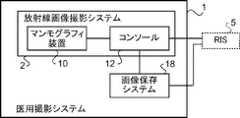
図1に示すように、本実施形態の医用撮影システム1は、放射線画像撮影システム2、及び画像保存システム18を備える。 As shown in FIG. 1 , the medical imaging system 1 of this embodiment includes a
まず、放射線画像撮影システム2の構成について説明する。放射線画像撮影システム2は、マンモグラフィ装置10及びコンソール12を含む。図2には、マンモグラフィ装置10及びコンソール12の構成の一例を表すブロック図が示されている。また、図3には、本実施形態のマンモグラフィ装置10の外観の一例を表す側面図が示されている。 First, the configuration of the
本実施形態のマンモグラフィ装置10は、被検者の乳房を被写体として、乳房に放射線R(例えば、X線)を照射して乳房の放射線画像を撮影する装置である。なお、マンモグラフィ装置10は、被検者が起立している状態(立位状態)のみならず、被検者が椅子(車椅子を含む)等に座った状態(座位状態)において、被検者の乳房を撮影する装置であってもよい。 The
図2に示すように、本実施形態のマンモグラフィ装置10は、制御部20、記憶部22、I/F(Interface)部24、操作部26、放射線検出器30、圧迫板駆動部32、圧迫力検出センサ33、圧迫板34、放射線照射部36、及び線源移動部37を備える。制御部20、記憶部22、I/F部24、操作部26、放射線検出器30、圧迫板駆動部32、圧迫力検出センサ33、放射線照射部36、及び線源移動部37は、システムバスやコントロールバス等のバス39を介して相互に各種情報の授受が可能に接続されている。 As shown in FIG. 2, the
本実施形態の制御部20は、コンソール12の制御に応じて、マンモグラフィ装置10の全体の動作を制御する。制御部20は、CPU(Central Processing Unit)20A、ROM(Read Only Memory)20B、及びRAM(Random Access Memory)20Cを備える。ROM20Bには、CPU20Aで実行される、放射線画像の撮影に関する制御を行うための撮影処理プログラム21A及び圧迫制御処理プログラム21Bを含む各種のプログラム等が予め記憶されている。RAM20Cは、各種データを一時的に記憶する。なお、本実施形態の圧迫制御処理プログラム21Bが、本開示の制御プログラムの一例である。 The
放射線検出器30は、被写体である乳房を通過した放射線Rを検出する。図3に示すように、放射線検出器30は、撮影台40の内部に配置されている。本実施形態のマンモグラフィ装置10では、撮影を行う場合、撮影台40の撮影面40A上には、被検者の乳房が医師及び技師等のユーザによってポジショニングされる。被検者の乳房が接する撮影面40A等は、放射線Rの透過性や強度の観点から、例えば、カーボン等で形成されている。 The
放射線検出器30は、被検体の乳房及び撮影台40を透過した放射線Rを検出し、検出した放射線Rに基づいて放射線画像を生成し、生成した放射線画像を表す画像データを出力する。本実施形態の放射線検出器30の種類は、特に限定されず、例えば、放射線Rを光に変換し、変換した光を電荷に変換する間接変換方式の放射線検出器であってもよいし、放射線Rを直接電荷に変換する直接変換方式の放射線検出器であってもよい。 The
記憶部22には、放射線検出器30により撮影された放射線画像の画像データや、その他の各種情報等が記憶される。記憶部22の具体例としては、HDD(Hard Disk Drive)やSSD(Solid State Drive)等が挙げられる。I/F部24は、無線通信または有線通信により、コンソール12との間で各種情報の通信を行う。マンモグラフィ装置10で放射線検出器30により撮影された放射線画像の画像データは、I/F部24を介してコンソール12に無線通信または有線通信によって送信される。 The
操作部26は、例えば、マンモグラフィ装置10の撮影台40等に複数のスイッチとして設けられている。なお、操作部26は、タッチパネル式のスイッチとして設けられていてもよいし、ユーザが足で操作するフットスイッチとして設けられていてもよい。 The
放射線照射部36は、放射線源36Rを備えている。図3に示すように放射線照射部36は、撮影台40及び圧迫ユニット46と共にアーム部42に設けられている。なお、図3に示すように本実施形態のマンモグラフィ装置10は、アーム部42と、基台44と、軸部45と、を備えている。アーム部42は、基台44によって、上下方向(Z軸方向)に移動可能に保持される。軸部45は、アーム部42を基台44に連結する。またアーム部42は、線源移動部37によって軸部45を回転軸として、基台44に対して相対的に回転可能となっている。 The
マンモグラフィ装置10においてトモシンセシス撮影を行う場合、アーム部42が回転することにより放射線照射部36の放射線源36Rが、線源移動部37により連続的に、照射角度が異なる複数の照射位置の各々に移動される。本実施形態では、図4に示すように放射線源36Rは、予め定められた角度θずつ照射角度が異なる照射位置t(t=0、1、・・・T、図4ではT=5)、換言すると放射線検出器30の検出面30Aに対する放射線Rの入射角度が異なる位置に移動される。各照射位置において、コンソール12の指示により放射線源36Rから放射線Rが照射され、放射線検出器30により放射線画像が撮影される。なお、以下では、トモシンセシス撮影において、照射角度が異なる複数の照射位置tの各々において放射線検出器30により撮影された放射線画像を「投影画像」という。放射線画像撮影システム2では、放射線源36Rを照射位置tの各々に移動させて、各照射位置tで投影画像の撮影を行うトモシンセシス撮影を行った場合、T枚の投影画像が得られる。 When performing tomosynthesis imaging in the
以下では、図4に示すように、トモシンセシス撮影における照射角度の範囲を「照射角度範囲」という。本実施形態のマンモグラフィ装置10では、トモシンセシス撮影を行う場合、標準(ST:Standard)モードと、高鮮鋭(HR:High Resolution)モードと二つのモードがある。図4に示すように、標準モードの照射角度範囲STよりも、高鮮鋭モードの照射角度範囲HRのほうが、照射角度範囲が広い。なお、一例として、本実施形態のマンモグラフィ装置10では、標準モードの場合と、高鮮鋭モードの場合とで、照射位置tの数を同一、換言すると撮影される投影画像の枚数を同一としている。そのため、標準モードでは、照射位置t毎の照射角度の差が小さく、また、高鮮鋭モードに比べて撮影時間が短くなる。一方、高鮮鋭モードでは、照射位置t毎の照射角度の差が大きく、また、標準モードに比べて深さ方向の分解能に優れる。なお、標準モードが、本開示の第1のトモシンセシス撮影モードの一例であり、高鮮鋭モードが、本開示の第2のトモシンセシス撮影モードの一例である。 Hereinafter, as shown in FIG. 4, the range of irradiation angles in tomosynthesis imaging is referred to as "irradiation angle range". The
一方、マンモグラフィ装置10において単純撮影を行う場合、放射線照射部36の放射線源36Rは、放射線検出器30の法線CLに沿った照射位置t(照射角度0度)のままとされる。なお、「単純撮影」とは、乳房に放射線Rを1回、照射して1枚の2次元画像を取得する撮影のことをいう。以下では、単純撮影において放射線検出器30により撮影された放射線画像を「2次元画像」という。また、投影画像及び2次元画像の各々を区別せずに総称する場合は、単に「放射線画像」という。 On the other hand, when simple imaging is performed in the
また、図3及び図5に示すように、圧迫板駆動部32、圧迫力検出センサ33、及び圧迫板34は、圧迫ユニット46に設けられている。圧迫ユニット46と、アーム部42とは、軸部45を回転軸として、別々に、基台44に対して相対的に回転可能となっている。本実施形態では、軸部45、アーム部42、及び圧迫ユニット46にそれぞれギア(図示省略)が設けられ、このギア同士の噛合状態及び非噛合状態を切替えることにより、アーム部42及び圧迫ユニット46の各々が軸部45に連結される。軸部45に連結されたアーム部42及び圧迫ユニット46の一方または両方が、軸部45と一体に回転する。 Further, as shown in FIGS. 3 and 5, the compression
本実施形態の圧迫板34は、板状の圧迫部材であり、圧迫板駆動部32により上下方向(Z軸方向)に移動し、撮影台40との間で被検者の乳房を圧迫する。図3に示すように、圧迫板34の移動方向について、また、乳房を圧迫する方向、換言すると、撮影面40Aに近付く方向を「圧迫方向」といい、乳房への圧迫を解除する方向、換言すると、放射線照射部36に近付く方向を「圧迫解除方向」という。 The
図5に示すように、圧迫ユニット46内には、モータ31及びボールネジ38を含む圧迫板駆動部32と、圧迫力検出センサ33と、が備えられている。圧迫力検出センサ33は、圧迫板34により乳房全体を圧迫する圧迫力を検出する機能を有する。図5に示した一例では、圧迫力検出センサ33が、圧迫板34の駆動源としてのモータ31にかかる負荷に基づいて圧迫力を検出する場合の構成を示している。圧迫板34は、ボールネジ38に支持されており、モータ31が駆動することにより、撮影台40と放射線源36Rとの間でスライド移動する。本実施形態の圧迫力検出センサ33はロードセル等の歪ゲージであり、圧迫力検出センサ33が、圧迫板34による圧迫力の反力を検出することにより、圧迫板34により乳房を圧迫する圧迫力を検出する。 As shown in FIG. 5 , the
なお、圧迫力を検出する方法は、これに限らず、例えば、圧迫力検出センサ33は、半導体式圧力センサ、及び静電容量式圧力センサ等であってもよい。また例えば、圧迫板34に圧迫力検出センサ33を設けておいてもよい。 The method of detecting the pressing force is not limited to this. For example, the pressing
圧迫板34は、乳房の圧迫において位置合わせや圧迫状態の確認を行うために光学的に透明であることが好ましく、また、放射線Rの透過性に優れた材料によって形成される。圧迫板34の材料としては、例えば、ポリメチルペンテン、ポリカーボネート、アクリル、及びポリエチレンテレフタレート等の樹脂を用いることができる。なお、圧迫板34を構成する部材は、本実施形態に限定されない。例えば、圧迫板34を構成する部材は、フィルム状の部材であってもよい。なお、本実施形態の圧迫板34が、本開示の圧迫部材の一例である。 The
さらに、図6には、本実施形態のマンモグラフィ装置10の構成の一例の機能ブロック図を示す。図6に示すように本実施形態のマンモグラフィ装置10は、圧迫制御部80及び撮影制御部82を備える。一例として本実施形態のマンモグラフィ装置10は、制御部20のCPU20AがROM20Bに記憶されている圧迫制御処理プログラム21Bを実行することにより、制御部20が、圧迫制御部80として機能する。本実施形態のマンモグラフィ装置10は、本開示の制御装置の一例である。また、一例として本実施形態のマンモグラフィ装置10は、制御部20のCPU20AがROM20Bに記憶されている撮影処理プログラム21Aを実行することにより、制御部20が、撮影制御部82として機能する。 Furthermore, FIG. 6 shows a functional block diagram of an example of the configuration of the
マンモグラフィ装置10の撮影制御部82には、放射線検出器30、線源移動部37、及び放射線源36Rが接続されている。撮影制御部82は、放射線検出器30による放射線画像の撮影の制御、線源移動部37による、放射線源36R(放射線照射部36)の移動、及び放射線源36Rからの放射線Rの照射の制御を行う。 The
一方、マンモグラフィ装置10の圧迫制御部80には、圧迫力検出センサ33の検出結果である圧迫力を表す情報が入力される。また、圧迫制御部80は、圧迫板駆動部32に、圧迫板34の移動に関する指示を出力する。 On the other hand, information indicating the compression force, which is the detection result of the compression
圧迫制御部80は、マンモグラフィ装置10が乳房を圧迫状態としたままで、単純撮影及びトモシンセシス撮影を連続して行う連続撮影の場合、単純撮影及びトモシンセシス撮影のうちの先に行う一方の撮影では、圧迫板34による乳房の圧迫力を第1圧迫力とする制御を行う。また、圧迫制御部80は、一方の撮影の後に、圧迫板34による圧迫力を第1圧迫力から第1圧迫力よりも小さい第2圧迫力に変更し、単純撮影及びトモシンセシス撮影のうちの後に行う他方の撮影では、圧迫板34による乳房の圧迫力を第2圧迫力とする制御を行う。 In the case of continuous imaging in which simple imaging and tomosynthesis imaging are continuously performed while the
本実施形態のマンモグラフィ装置10では、乳房を圧迫板34により圧迫した圧迫状態のままで連続撮影を行う場合がある。圧迫制御部80は、連続撮影の場合、圧迫板34により乳房を圧迫し続ける影時間が長くなるため、トモシンセシス撮影及び単純撮影のうち、後に行う撮影における圧迫力を、先に行う撮影における圧迫力よりも小さくすることで、乳房を圧迫されていることにより生じる被検者の痛みを緩和する。 In the
一方、本実施形態のコンソール12は、無線通信LAN(Local Area Network)等を介してRIS(Radiology Information System)5等から取得した撮影オーダ及び各種情報と、操作部56等によりユーザにより行われた指示等とを用いて、マンモグラフィ装置10の制御を行う機能を有している。 On the other hand, the
本実施形態のコンソール12は、一例として、サーバーコンピュータである。図2に示すように、コンソール12は、制御部50、記憶部52、I/F部54、操作部56、及び表示部58を備えている。制御部50、記憶部52、I/F部54、操作部56、及び表示部58はシステムバスやコントロールバス等のバス59を介して相互に各種情報の授受が可能に接続されている。 The
本実施形態の制御部50は、コンソール12の全体の動作を制御する。制御部50は、CPU50A、ROM50B、及びRAM50Cを備える。ROM50Bには、CPU50Aで実行される、後述する制御処理プログラム51を含む各種のプログラム等が予め記憶されている。RAM50Cは、各種データを一時的に記憶する。 The control unit 50 of this embodiment controls the overall operation of the
記憶部52には、マンモグラフィ装置10で撮影された放射線画像の画像データや、その他の各種情報等が記憶される。記憶部52の具体例としては、HDDやSSD等が挙げられる。 The
操作部56は、放射線Rの照射指示を含む放射線画像の撮影等に関する指示や各種情報等をユーザが入力するために用いられる。そのため、本実施形態の操作部56は、放射線Rの照射を指示する場合に、ユーザが押圧する照射指示ボタンが少なくとも含まれる。なお、操作部56は特に限定されるものではなく、例えば、各種スイッチ、タッチパネル、タッチペン、及びマウス等が挙げられる。表示部58は、各種情報を表示する。なお、操作部56と表示部58とを一体化してタッチパネルディスプレイとしてもよい。 The
I/F部54は、無線通信または有線通信により、マンモグラフィ装置10、RIS5、及び画像保存システム18との間で各種情報の通信を行う。本実施形態の放射線画像撮影システム2では、マンモグラフィ装置10で撮影された放射線画像の画像データは、コンソール12が、I/F部54を介して無線通信または有線通信によりマンモグラフィ装置10から受信する。 The I/
次に、画像保存システム18の構成について説明する。図7には、画像保存システム18の構成の一例を表すブロック図が示されている。画像保存システム18は、放射線画像撮影システム2により撮影された放射線画像の画像データを保存するシステムである。画像保存システム18は、保存している放射線画像から、コンソール12及びその他の読影装置(図示省略)等からの要求に応じた画像を取り出して、要求元の装置に送信する。画像保存システム18の具体例としては、PACS(Picture Archiving and Communication Systems)が挙げられる。 Next, the configuration of the
図7に示すように、画像保存システム18は、制御部70、記憶部72、及びI/F部74を備えている。制御部70、記憶部72、及びI/F部74はシステムバスやコントロールバス等のバス79を介して相互に各種情報の授受が可能に接続されている。 As shown in FIG. 7, the
本実施形態の制御部70は、画像保存システム18の全体の動作を制御する。制御部70は、CPU70A、ROM70B、及びRAM70Cを備える。ROM70Bには、CPU70Aで実行される、各種のプログラム等が予め記憶されている。RAM70Cは、各種データを一時的に記憶する。 The
記憶部72は、放射線画像の画像データを、撮影オーダや被検者に関する情報等と関連付けて記憶する、いわゆるデータベースである。 The
I/F部74は、無線通信または有線通信により、コンソール12との間で各種情報の通信を行う機能を有している。 The I/F unit 74 has a function of communicating various information with the
次に、本実施形態のマンモグラフィ装置10の作用について図面を参照して説明する。
本実施形態のマンモグラフィ装置10は、一例として、コンソール12から撮影オーダと、撮影開始の指示とを受信した場合、制御部20のCPU20Aが、ROM20Bに記憶されている圧迫制御処理プログラム21Bを実行することにより、図8に一例を示した圧迫制御処理を実行する。図8には、本実施形態のマンモグラフィ装置10における圧迫制御処理の流れの一例を表したフローチャートが示されている。また、図9には、本実施形態のマンモグラフィ装置10における連続撮影を行う場合の圧迫制御部80及び撮影制御部82の動作の一例を表すタイムチャートが示されている。以下、図9に示したタイムチャートを参照しつつ、圧迫制御部80における圧迫制御処理の流れを説明する。Next, the operation of the
In the
まず、図8のステップS100で圧迫制御部80は、ユーザによって操作部26により圧迫の指示が行われたか否かを判定する。本実施形態のマンモグラフィ装置10において放射線画像の撮影を行う場合、まず、ユーザは、マンモグラフィ装置10の撮影台40の撮影面40A上に、被検者の乳房をポジショニングする。ポジショニングが完了すると、ユーザは、操作部26により圧迫を指示する。圧迫の指示が未だ行われていない場合、ステップS100の判定が否定判定となる。一方、圧迫の指示が行われると、ステップS100の判定が肯定判定となり、ステップS102へ移行する。 First, in step S100 of FIG. 8, the
次のステップS102で圧迫制御部80は、圧迫の指示に応じて、圧迫板駆動部32によって圧迫方向へ圧迫板34を移動させて、圧迫板34により撮影台40の撮影面40Aとの間で乳房を第1圧迫力で圧迫する。図9のタイムチャートに示すように、タイミングt100で圧迫方向へ圧迫板34の移動を開始させると、圧迫板34による圧迫力が第1圧迫力になったタイミングt102までの圧迫期間中、圧迫制御部80が圧迫板駆動部32によって圧迫方向へ圧迫板34を移動させる。 In the next step S102, the
圧迫板34により乳房を圧迫することにより、乳腺組織の重なりを展開し、良性病変及び悪性病変のいずれであるかの判断等を行いやすくすることができる。また、圧迫板34により圧迫することで、乳房を撮影台40に固定することにより、被検者の体動を抑制するため、体動により生じる放射線画像のボケを抑制することができる。さらに、圧迫板34により乳房を圧迫することで乳房の厚みが薄くなるため、乳房の被曝を低減することができる。 By compressing the breast with the
次のステップS104で圧迫制御部80は、連続撮影を行うか否かを判定する。一例として、本実施形態の撮影制御部82は、撮影オーダに、連続撮影が指示されていた場合、連続撮影を行うと判定する。なお、本実施形態では、連続撮影を行う場合、単純撮影を行った後、トモシンセシス撮影を行う。連続撮影を行う場合、ステップS104の判定が肯定判定となり、ステップS106へ移行する。 In the next step S104, the
一方、圧迫板34により乳房が固定されると、ユーザは、コンソール12の操作部56に含まれる照射指示ボタンを押圧し、放射線Rの照射指示を行う。照射指示が行われると、コンソール12の制御に応じて撮影制御部82は、図9のタイミングチャートに示すように、タイミングt203で単純撮影を行う。具体的には、撮影制御部82は、放射線源36Rから放射線Rを、圧迫板34により圧迫された状態の乳房に向けて照射させる。そして、放射線検出器30により乳房を透過した放射線Rに応じて2次元画像を生成する。撮影された2次元画像の画像データは、コンソール12に送信される。 On the other hand, when the breast is fixed by the
そこで、ステップS106で圧迫制御部80は、単純撮影が終了したか否かを判定する。一例として本実施形態の圧迫制御部80は、放射線検出器30により撮影された2次元画像を表す画像データがコンソール12に送信された場合、単純撮影が終了したと判定するが、単純撮影の終了を判定する方法は本実施形態に限定されない。例えば、コンソール12の操作部56により指示した、単純撮影の終了指示を受け付ける形態としてもよい。 Therefore, in step S106, the
単純撮影が終了するまで、ステップS106の判定が否定判定となる。一方、単純撮影が終了するとステップS106の判定が肯定判定となり、ステップS108へ移行する。 The determination in step S106 becomes a negative determination until the simple photographing ends. On the other hand, when the simple photographing ends, the determination in step S106 becomes a positive determination, and the process proceeds to step S108.
ステップS108で圧迫制御部80は、圧迫板34により第2圧迫力で乳房を圧迫する。具体的には、図9のタイムチャートに示すように、圧迫制御部80は、圧迫板駆動部32によって、タイミングt104で圧迫解除方向へ圧迫板34の移動を開始させると、圧迫力検出センサ33により検出された圧迫力が第2圧迫力となったタイミングt106で圧迫板34の移動を停止させる。本実施形態では、圧迫力を第1圧迫力から第2圧迫力に変更する期間、図9に示した例では、タイミングt104からタイミングt106までの期間を「変更期間」という。 In step S<b>108 , the
乳房を一旦、第1圧迫力で圧迫した後、圧迫力を小さくして第2圧迫力としても、乳腺組織の展開具合がほとんど変わらない場合があることが、例えば、特開2017-225633号公報、特開2017-225634号公報、及び特開2017-225635号公報等に開示されている。これらの公報によれば、第1圧迫力で乳房を圧迫した後、圧迫板34を圧迫解除方向に動かして圧迫力を低下させても、乳房の弾性効果により乳房の厚みが戻りにくいことが開示されている。乳房の厚みが戻り難く、乳房の厚みを維持することができるため、乳腺組織の展開具合は、維持される、またはほとんど変わらない。 After the breast is once compressed with a first compression force, even if the compression force is reduced to a second compression force, the degree of expansion of the mammary gland tissue may not change in some cases. , JP-A-2017-225634, and JP-A-2017-225635. According to these publications, even if the compression force is reduced by moving the
第2圧迫力は、被検者の痛みを緩和する観点から第1圧迫力よりも小さければよい。一方、乳房の圧迫力が小さすぎると、やはり乳房の厚みが戻ってしまい乳腺組織の展開具合が異なってしまう場合がある。単純撮影(2次元画像)と、トモシンセシス撮影(投影画像)とで、乳腺組織の展開具合が異なると、例えば、石灰化等の写っている位置が変化等してしまうため、好ましくない。また乳房の圧迫力が小さすぎると、被検者の体動が生じ易くなる場合がある。 The second compressive force should be smaller than the first compressive force from the viewpoint of relieving the pain of the subject. On the other hand, if the compressive force on the breast is too small, the thickness of the breast may be restored and the degree of expansion of the mammary gland tissue may be different. If the mammary gland tissue develops differently between simple imaging (two-dimensional image) and tomosynthesis imaging (projection image), for example, the positions of calcifications appearing change, which is not preferable. Also, if the pressing force on the breast is too small, the subject may easily move.
上記公報によれば、第2圧迫力は、第1圧迫力の40%~70%であることが好ましく、第1圧迫力の50%であることがより好ましい。もしくは、第2圧迫力は、第1圧迫力よりも40N~100N少ないことが好ましく、第1圧迫力よりも50N少ないことがより好ましい。換言すると、第1圧迫力は、第2圧迫力の143%~250%であることが好ましく、第2圧迫力の200%であることがより好ましい。もしくは、第1圧迫力は、第2圧迫力よりも40N~100N大きいことが好ましく、第2圧迫力よりも50N大きいことがより好ましい。また、上記公報によれば、被検者の痛みを効果的に緩和し、かつ被検者の体動を抑制する観点から、第2圧迫力は、40N~100Nであることが好ましい。 According to the publication, the second compression force is preferably 40% to 70% of the first compression force, more preferably 50% of the first compression force. Alternatively, the second compression force is preferably 40N to 100N less than the first compression force, more preferably 50N less than the first compression force. In other words, the first compression force is preferably 143% to 250% of the second compression force, more preferably 200% of the second compression force. Alternatively, the first compression force is preferably 40N to 100N greater than the second compression force, and more preferably 50N greater than the second compression force. Further, according to the above publication, the second compression force is preferably 40N to 100N from the viewpoint of effectively alleviating the pain of the subject and suppressing the body movement of the subject.
一方、図9に示した一例のように撮影制御部82は、タイミングt205で、線源移動部37により、放射線照射部36の放射線源36Rを、単純撮影の撮影位置にあたる照射位置tから、トモシンセシス撮影の開始位置にあたる照射位置tに移動させる。なお、上述したようにトモシンセシス撮影には、標準モードと、高鮮鋭モードとがあり、各々の開始位置にあたる照射位置tが異なるため、撮影制御部82は、各モードに応じた開始位置にあたる照射位置tに放射線源36Rを移動させる。撮影制御部82は、放射線源36Rが開始位置にあたる照射位置tに達したタイミングt207で放射線源36Rの移動を停止させる。図9に示すように、本実施形態では、タイミングt205からタイミングt207の期間を放射線源36Rの「移動期間」という。 On the other hand, as in the example shown in FIG. 9, at timing t205, the
図9に示すように、この単純撮影の終了後の放射線源36Rの移動期間と、圧迫板34による圧迫力を第1圧迫力から第2圧迫力に変更する変更期間は重なっている。本実施形態のマンモグラフィ装置10では、このように、放射線源36Rの移動期間と、圧迫力の変更期間との少なくとも一部が重なることにより、乳房を圧迫し続けている期間を短縮することができる。 As shown in FIG. 9, the period during which the
圧迫板34により乳房が第2圧迫力で圧迫された状態になり、また、放射線源36Rがトモシンセシス撮影における照射位置tまで移動すると、図9に示した一例のように、撮影制御部82は、タイミングt209でトモシンセシス撮影を行う。具体的には、線源移動部37により、各照射位置tに放射線源36Rを移動させ、各照射位置において放射線源36Rから放射線Rを圧迫板34により圧迫された状態の乳房に向けて照射させる。そして、各照射位置tに応じて、放射線検出器30により乳房を透過した放射線Rに応じて投影画像を生成する。撮影された複数の投影画像の画像データは、コンソール12に送信される。 When the breast is compressed with the second compression force by the
そこで、次のステップS110で圧迫制御部80は、トモシンセシス撮影が終了したか否かを判定する。一例として本実施形態の放射線画像撮影システム2では、撮影が終了すると、ユーザがマンモグラフィ装置10の操作部26により圧迫の解除を指示する。そこで、圧迫制御部80は、操作部26により、圧迫の解除が指示された場合、トモシンセシス撮影を終了すると判定する。 Therefore, in the next step S110, the
トモシンセシス撮影が終了していない場合、換言すると、圧迫の解除が指示されていない場合、ステップS110の判定が否定判定となる。一方、トモシンセシス撮影が終了した場合、換言すると、圧迫の解除が指示された場合、ステップS110の判定が肯定判定となり、ステップS118へ移行する。 If the tomosynthesis imaging has not ended, in other words, if the command to release the pressure has not been issued, the determination in step S110 is negative. On the other hand, when the tomosynthesis imaging is completed, in other words, when the release of the compression is instructed, the determination in step S110 becomes a positive determination, and the process proceeds to step S118.
ステップS118で圧迫制御部80は、圧迫板34による乳房の圧迫を解除することにより、本圧迫制御処理を終了する。具体的には、図9のタイムチャートに示すように、圧迫制御部80は、圧迫板駆動部32によって、タイミングt108で圧迫解除方向へ圧迫板34の移動を開始させると、予め定められた初期位置まで移動したタイミングt110で圧迫板34の移動を停止させる。予め定められた位置とは、乳房の圧迫が完全に解除される位置である。本実施形態では、乳房の圧迫の解除に要する期間、図9に示した例では、タイミングt108からタイミングt110までの期間を「解除期間」という。 In step S118, the
一方、撮影オーダに、連続撮影が指示されてなかった場合、換言すると、単純撮影のみ、またはトモシンセシス撮影のみが指示されていた場合、上記ステップS104の判定が否定判定となり、ステップS112へ移行する。なお、以下では、連続撮影に対し、単純撮影及びトモシンセシス撮影のいずれか一方のみを行う撮影を「単独撮影」という。 On the other hand, if continuous imaging is not instructed in the imaging order, in other words, if only simple imaging or tomosynthesis imaging is instructed, the determination in step S104 is negative, and the process proceeds to step S112. Note that hereinafter, imaging in which only one of simple imaging and tomosynthesis imaging is performed in contrast to continuous imaging is referred to as "single imaging".
ステップS112で圧迫制御部80は、撮影オーダに、トモシンセシス撮影が指示されているか否かを判定する。撮影オーダにトモシンセシス撮影が指示されている場合、ステップS112の判定が肯定判定となり、ステップS114へ移行する。ステップS114で圧迫制御部80は、上記ステップS110と同様に、トモシンセシス撮影が終了したか否かを判定する。トモシンセシス撮影が終了するまで、ステップS114の判定が否定判定となる。一方、トモシンセシス撮影が終了すると、ステップS114の判定が肯定判定となり、上記ステップS118へ移行する。 In step S112, the
一方、撮影オーダにトモシンセシス撮影が指示されていなかった場合、換言すると、単純撮影が指示されていた場合、ステップS112の判定が否定判定となり、ステップS116へ移行する。ステップS116で圧迫制御部80は、上記ステップS106と同様に、単純撮影が終了したか否かを判定する。単純撮影が終了するまで、ステップS116の判定が否定判定となる。一方、単純撮影が終了すると、ステップS116の判定が肯定判定となり、上記ステップS118へ移行する。 On the other hand, if tomosynthesis imaging is not instructed in the imaging order, in other words, if simple imaging is instructed, the determination in step S112 is negative, and the process proceeds to step S116. In step S116, the
このように、本実施形態のマンモグラフィ装置10の圧迫制御処理では、単純撮影の後にトモシンセシス撮影を行う連続撮影において、圧迫板34により乳房を第1圧迫力で圧迫して単純撮影を行った後、圧迫力を第1圧迫力から第2圧迫力に小さくする。そして、乳房を第2圧迫力で圧迫して状態でトモシンセシス撮影を行う。従って、本実施形態のマンモグラフィ装置10によれば、被検者の痛みをさらに効果的に緩和することができる。また、本実施形態のマンモグラフィ装置10によれば、単純撮影に比べて、撮影時間が長くなる場合が多いトモシンセシス撮影における圧迫力を小さくすることができるため、被検者の痛みをより効果的に緩和することができる。 As described above, in the compression control process of the
[第2実施形態]
以下、第2実施形態について詳細に説明する。
第1実施形態では、マンモグラフィ装置10が連続撮影を行う場合、先に単純撮影を行い、後からトモシンセシス撮影を行う形態について説明した。これに対して本実施形態では、連続撮影を行う場合、先にトモシンセシス撮影を行い、後から単純撮影を行う形態について説明する。[Second embodiment]
The second embodiment will be described in detail below.
In the first embodiment, when the
本実施形態の医用撮影システム1の全体構成(図1参照)と、マンモグラフィ装置10及びコンソール12の構成(図2及び図3参照)は、第1実施形態と同様であるため説明を省略する。本実施形態では、マンモグラフィ装置10の圧迫制御部80及び撮影制御部82の動作の一部、及び圧迫制御部80が実行する圧迫制御処理の一部が第1実施形態のマンモグラフィ装置10と異なるため、異なる動作及び処理について説明する。 The overall configuration of the medical imaging system 1 (see FIG. 1) and the configuration of the
本実施形態のマンモグラフィ装置10は、一例として、コンソール12から撮影オーダと、撮影開始の指示とを受信した場合、制御部20のCPU20Aが、ROM20Bに記憶されている圧迫制御処理プログラム21Bを実行することにより、図10に一例を示した圧迫制御処理を実行する。図10には、本実施形態のマンモグラフィ装置10における圧迫制御処理の流れの一例を表したフローチャートが示されている。また、図11には、本実施形態のマンモグラフィ装置10における連続撮影を行う場合の圧迫制御部80及び撮影制御部82の動作の一例を表すタイムチャートが示されている。以下、図11に示したタイムチャートを参照しつつ、圧迫制御部80における圧迫制御処理の流れを説明する。 As an example, in the
図10に示すように、本実施形態の圧迫制御処理は、第1実施形態の圧迫制御処理(図8参照)のステップS106に代わりステップS107の処理を備え、ステップS110の代わりにステップS117の処理を備える点が、異なっている。 As shown in FIG. 10, the compression control process of the present embodiment includes the process of step S107 instead of step S106 of the compression control process of the first embodiment (see FIG. 8), and the process of step S117 instead of step S110. The difference is that the
図10に示すように、本実施形態の圧迫制御処理では、連続撮影を行う場合、換言するとステップS104の判定が肯定判定となった場合、ステップS107へ移行する。 As shown in FIG. 10, in the compression control process of this embodiment, when continuous imaging is performed, in other words, when the determination in step S104 is affirmative, the process proceeds to step S107.
実施形態のマンモグラフィ装置10における撮影制御部82は、コンソール12から受信した撮影オーダに連続撮影が指示されていた場合、撮影の開始指示に応じて、図11に一例を示したタイムチャートのタイミングt200で、線源移動部37により、放射線照射部36の放射線源36Rを、トモシンセシス撮影の開始位置にあたる照射位置tに移動させる。なお、上述したようにトモシンセシス撮影には、標準モードと、高鮮鋭モードとがあり、各々の開始位置にあたる照射位置tが異なるため、撮影制御部82は、各モードに応じた開始位置にあたる照射位置tに放射線源36Rを移動させる。撮影制御部82は、放射線源36Rが開始位置にあたる照射位置tに達したタイミングt202で放射線源36Rの移動を停止させる。図11に示すように、本実施形態では、タイミングt200からタイミングt202の期間を放射線源36Rの「移動期間」という。 When the imaging order received from the
コンソール12の制御に応じて撮影制御部82は、図11のタイミングチャートに示すように、タイミングt204で上述のようにトモシンセシス撮影を行う。 Under the control of the
そこでステップS107で圧迫制御部80は、トモシンセシス撮影が終了したか否かを判定する。トモシンセシス撮影が終了するまでステップS107の判定が否定判定となる。一方、トモシンセシス撮影が終了すると、ステップS107の判定が肯定判定となり、ステップS108へ移行する。 Therefore, in step S107, the
ステップS108で圧迫制御部80は、第1実施形態で上述したように、圧迫板34により第2圧迫力で乳房を圧迫する。具体的には、図11のタイムチャートに示すように、圧迫制御部80は、圧迫板駆動部32によって、タイミングt104で圧迫解除方向へ圧迫板34の移動を開始させると、圧迫力検出センサ33により検出された圧迫力が第2圧迫力となったタイミングt106で圧迫板34の移動を停止させる。 In step S108, the
一方、図11に示した一例のように撮影制御部82は、タイミングt204のトモシンセシス撮影が終了すると、直ちに、タイミングt206で、線源移動部37により、放射線照射部36の放射線源36Rを、トモシンセシス撮影の終了位置にあたる照射位置tから、単純撮影における照射位置tに移動させる。撮影制御部82は、放射線源36Rが単純撮影における照射位置tに達したタイミングt208で放射線源36Rの移動を停止させる。図11に示すように、本実施形態では、タイミングt206からタイミングt208の期間を放射線源36Rの「移動期間」という。 On the other hand, as in the example shown in FIG. 11, immediately after tomosynthesis imaging at timing t204 ends, the
図11に示すように、このトモシンセシス撮影の終了後の放射線源36Rの移動期間と、圧迫板34による圧迫力を第1圧迫力から第2圧迫力に変更する変更期間は重なっている。本実施形態のマンモグラフィ装置10では、このように、放射線源36Rの移動期間と、圧迫力の変更期間との少なくとも一部が重なることにより、乳房を圧迫し続けている期間を短縮することができる。 As shown in FIG. 11, the period during which the
放射線源36Rが単純撮影における照射位置tまで移動すると、コンソール12の制御に応じて撮影制御部82は、図11のタイミングチャートに示すように、タイミングt210で上述のように単純撮影を行う。 When the
そこで、ステップS117で圧迫制御部80は、単純撮影が終了したか否かを判定する。単純撮影が終了するまでステップS117の判定が否定判定となる。一方、単純撮影が終了すると、ステップS117の判定が肯定判定となり、ステップS118へ移行する。 Therefore, in step S117, the
このように、本実施形態のマンモグラフィ装置10の圧迫制御処理では、トモシンセシス撮影の後に単純撮影を行う連続撮影において、圧迫板34により乳房を第1圧迫力で圧迫してトモシンセシス撮影を行った後、圧迫力を第1圧迫力から第2圧迫力に小さくする。そして、乳房を第2圧迫力で圧迫して状態で単純撮影を行う。従って、本実施形態のマンモグラフィ装置10によれば、被検者の痛みをさらに効果的に緩和することができる。 As described above, in the compression control process of the
また、本実施形態のマンモグラフィ装置10によれば、トモシンセシス撮影の後、単純撮影を行うために撮影制御部82が放射線源36Rを移動させる移動期間中に、圧迫制御部80が、圧迫板34による乳房の圧迫力を第1圧迫力から第2圧迫力に変更する。換言すると、放射線源36Rの移動期間と、圧迫力の変更期間との少なくとも一部が重なる。従って、より多くの期間が重なることにより乳房を圧迫し続けている期間を短縮することができるため、被検者の痛みをより効果的に緩和することができる。 Further, according to the
[第3実施形態]
以下、第3実施形態について詳細に説明する。
本実施形態では、連続撮影を行う場合、後に行う撮影が単純撮影及びトモシンセシス撮影のいずれかに応じて、先に行った撮影の後に行う、圧迫力の制御方法を異ならせる形態について説明する。[Third embodiment]
The third embodiment will be described in detail below.
In the present embodiment, when continuous imaging is performed, an embodiment will be described in which the compression force control method performed after the previous imaging is changed according to whether the subsequent imaging is simple imaging or tomosynthesis imaging.
本実施形態の医用撮影システム1の全体構成(図1参照)と、マンモグラフィ装置10及びコンソール12の構成(図2及び図3参照)は、第1実施形態と同様であるため説明を省略する。本実施形態では、マンモグラフィ装置10の圧迫制御部80が実行する圧迫制御処理の一部が第1及び第2実施形態のマンモグラフィ装置10と異なるため、異なる動作及び処理について説明する。 The overall configuration of the medical imaging system 1 (see FIG. 1) and the configuration of the
図12には、本実施形態のマンモグラフィ装置10における圧迫制御処理の流れの一例を表したフローチャートが示されている。図12に示すように本実施形態の圧迫制御処理は、第1実施形態の圧迫制御処理(図8参照)と、第2実施形態の圧迫制御処理(図10参照)とを組み合わせた形態である。なお、本実施形態の圧迫制御処理では、ステップS104とステップS106との間にステップS105の処理を行う点が異なっている。 FIG. 12 shows a flowchart showing an example of the flow of compression control processing in the
図12に示したステップS105で圧迫制御部80は、連続撮影における先に行う撮影が、単純撮影であるか否かを判定する。一例として本実施形態の医用撮影システム1では、撮影オーダに、トモシンセシス撮影及び単純撮影のいずれを先に行うかが指示されている。圧迫制御部80は、撮影オーダの指示に基づいて、単純撮影を先に行うか否かを判定する。 In step S105 shown in FIG. 12, the
先に行う撮影が単純撮影ではない場合、換言すると、先に行う撮影がトモシンセシス撮影である場合、ステップS105の判定が否定判定となり、ステップS107へ移行し、第2実施形態の圧迫制御処理と同様に連続撮影を行う。一方、先に行う撮影が単純撮影の場合、ステップS105の判定が肯定判定となり、ステップS106へ移行する。 If the previous imaging is not simple imaging, in other words, if the previous imaging is tomosynthesis imaging, the determination in step S105 is a negative determination, and the process proceeds to step S107, which is the same as the compression control process of the second embodiment. continuous shooting. On the other hand, if the previous photographing is simple photographing, the determination in step S105 is affirmative, and the process proceeds to step S106.
また、本実施形態の圧迫制御処理は、ステップS106とステップS110との間で、ステップS108に代わりステップS109の処理を行う点が異なっている。 Also, the compression control process of the present embodiment is different in that the process of step S109 is performed instead of step S108 between steps S106 and S110.
単純撮影が終了するとステップS109で圧迫制御部80は、圧迫板34により第1圧迫力よりも大きな第3圧迫力で乳房を圧迫する。具体的には、圧迫制御部80は、圧迫板駆動部32により圧迫板34を圧迫方向に移動させて、圧迫板34により乳房を第3圧迫力で圧迫する。 When the simple imaging is finished, in step S109, the
トモシンセシス撮影の場合、複数の投影画像の撮影開始から終了までの間に、被検者の体動が生じると、投影画像の画質が低下する場合があるため、好ましくない。また、トモシンセシス撮影は、比較的撮影時間が長くなることが多く、単純撮影の撮影時間よりも長くなる場合が多いため、体動が生じ易い傾向がある。そのため、本実施形態の圧迫制御処理では、トモシンセシス撮影を行う場合、圧迫板34による圧迫力を第1圧迫力から小さくしないことで、乳房を圧迫板34により固定し、被検者の体動を抑制する。そのため、図12に示した圧迫制御処理のステップS109では、圧迫板34により第1圧迫力よりも大きな第3圧迫力で乳房を圧迫する。 In the case of tomosynthesis imaging, if the body of the subject moves between the start and end of imaging of a plurality of projection images, the image quality of the projection images may deteriorate, which is not preferable. In addition, tomosynthesis imaging often requires a relatively long imaging time, which is often longer than that of simple imaging, and body movement tends to occur. Therefore, in the compression control process of the present embodiment, when tomosynthesis imaging is performed, the compression force by the
また、本実施形態の圧迫制御処理では、ステップS112で肯定判定となった場合、換言すると、単独撮影がトモシンセシス撮影である場合、ステップS109へ移行する。 Further, in the compression control process of the present embodiment, if the determination in step S112 is affirmative, in other words, if the single imaging is tomosynthesis imaging, the process proceeds to step S109.
このように本実施形態の圧迫制御処理では、トモシンセシス撮影を行う場合、圧迫板34により乳房を圧迫する圧迫力を第1圧迫力よりも大きい第3圧迫力とする。従って、本実施形態の圧迫制御処理によれば、被検者の体動を抑制することができる。 As described above, in the compression control process of the present embodiment, when tomosynthesis imaging is performed, the compression force with which the
なお、図12に示した圧迫制御処理では、トモシンセシス撮影を行う場合、圧迫板34により第1圧迫力よりも大きな第3圧迫力で乳房を圧迫する形態について説明したが、本形態に限定されない。図13には、変形例の圧迫制御処理の流れの一例を表したフローチャートが示されている。 In the compression control process shown in FIG. 12, when tomosynthesis imaging is performed, a configuration has been described in which the
図13に示した変形例の圧迫制御処理では、上記本実施形態の圧迫制御処理(図12参照)のステップS109の処理が設けられていない。 In the compression control process of the modified example shown in FIG. 13, the process of step S109 of the compression control process (see FIG. 12) of the present embodiment is not provided.
そのため、ステップS106で肯定判定となった場合、換言すると単純撮影が終了した場合、ステップS110へ移行する。また、ステップS112で肯定判定となった場合、換言すると、単独撮影がトモシンセシス撮影である場合も、ステップS110へ移行する。 Therefore, when the determination in step S106 is affirmative, in other words, when simple photography is completed, the process proceeds to step S110. Also, when the determination in step S112 is affirmative, in other words, when the single imaging is tomosynthesis imaging, the process proceeds to step S110.
このように変形例の圧迫制御処理では、トモシンセシス撮影を行う場合、圧迫板34により乳房を圧迫する圧迫力を第1圧迫力のままとする。この場合の圧迫制御処理も、第2圧迫力よりも大きな第1圧迫力で乳房を圧迫したままとするため、被検者の体動を抑制することができる。 Thus, in the compression control process of the modified example, when tomosynthesis imaging is performed, the compression force with which the
また、被検者の体動を抑制するため、圧迫板34により乳房を第1圧迫力で圧迫したままで、単純撮影及びトモシンセシス撮影の連続撮影を行う場合、全体の撮影時間が長くなるため、被検者の負担が大きくなる。そのため、連続撮影を行う場合、少なくとも単純撮影では、トモシンセシス撮影よりも小さい圧迫力で圧迫板34により乳房を圧迫することで被検者の負担を軽減することができる。 In addition, in order to suppress body movement of the subject, when continuous imaging of simple imaging and tomosynthesis imaging is performed while the breast is compressed with the first compression force by the
[第4実施形態]
以下、第4実施形態について詳細に説明する。
本実施形態では、単純撮影の後にトモシンセシス撮影を行う連続撮影を行う場合、標準モード及び高鮮鋭モードのいずれであるかに応じて、先に行った撮影の後に行う、圧迫力の制御方法を異ならせる形態について説明する。[Fourth embodiment]
The fourth embodiment will be described in detail below.
In the present embodiment, when performing continuous imaging in which tomosynthesis imaging is performed after simple imaging, the compression force control method performed after the previous imaging is different depending on whether the mode is the standard mode or the high-sharp mode. The following describes the form in which the
本実施形態の医用撮影システム1の全体構成(図1参照)と、マンモグラフィ装置10及びコンソール12の構成(図2及び図3参照)は、第1実施形態と同様であるため説明を省略する。本実施形態では、マンモグラフィ装置10の圧迫制御部80が実行する圧迫制御処理の一部が第1及び第2実施形態のマンモグラフィ装置10と異なるため、異なる動作及び処理について説明する。 The overall configuration of the medical imaging system 1 (see FIG. 1) and the configuration of the
図14には、本実施形態のマンモグラフィ装置10における圧迫制御処理の流れの一例を表したフローチャートが示されている。図14に示すように本実施形態の圧迫制御処理は、第1実施形態の圧迫制御処理(図8参照)のステップS106とステップS108との間にステップS107Aの処理を行う点が異なっている。 FIG. 14 shows a flowchart showing an example of the flow of compression control processing in the
図14に示したステップS107Aで圧迫制御部80は、連続撮影における後に行う撮影であるトモシンセシス撮影のモードが、標準モードであるか否かを判定する。一例として本実施形態の医用撮影システム1では、撮影オーダに、トモシンセシス撮影のモードが標準モード及び高鮮鋭モードのいずれであるかが指示されている。圧迫制御部80は、撮影オーダの指示に基づいて、トモシンセシス撮影のモードが標準モードであるか否かを判定する。 In step S107A shown in FIG. 14, the
上述したように、トモシンセシス撮影において標準モードは、高鮮鋭モードに比べて複数の投影画像の全てを撮影し終わるまでの撮影時間が短い。換言すると、高鮮鋭モードは、標準モードに比べて、複数の投影画像の全てを撮影し終わるまでの撮影時間が長い。従って、高鮮鋭モードの場合、標準モードの場合に比べて、撮影中に被検者の体動が生じやすい。そこで、本実施形態の圧迫制御処理では、トモシンセシス撮影のモードが高鮮鋭モードの場合、被検者の体動を抑制するために、圧迫板34による乳房の圧迫力を第1圧迫力のままとする。 As described above, in the tomosynthesis imaging, the standard mode takes a shorter imaging time to complete imaging of all of the plurality of projection images than the high-sharp mode. In other words, the high-sharp mode takes longer than the standard mode to capture all of the multiple projection images. Therefore, in the high-sharp mode, body movement of the subject is more likely to occur during imaging than in the standard mode. Therefore, in the compression control process of the present embodiment, when the tomosynthesis imaging mode is the high-sharp mode, the compression force of the
そのため、トモシンセシス撮影のモードが標準モードの場合、ステップS107Aの判定が肯定判定となり、ステップS108へ移行する。一方、トモシンセシス撮影のモードが高鮮鋭モードの場合、ステップS107Aの判定が否定判定となり、ステップS110へ移行する。 Therefore, if the tomosynthesis imaging mode is the standard mode, the determination in step S107A is positive, and the process proceeds to step S108. On the other hand, if the tomosynthesis imaging mode is the high-sharp mode, the determination in step S107A is negative, and the process proceeds to step S110.
また、本実施形態の圧迫制御処理では、ステップS112で肯定判定となった場合、換言すると、単独撮影がトモシンセシス撮影である場合、ステップS107Aへ移行する。 Further, in the compression control process of the present embodiment, when the determination in step S112 is affirmative, in other words, when the single imaging is tomosynthesis imaging, the process proceeds to step S107A.
このように本実施形態の圧迫制御処理では、トモシンセシス撮影を行う場合、標準モードならば、撮影時間が短いため、圧迫板34により乳房を圧迫する圧迫力を第1圧迫力から第2圧迫力に変更し、第2圧迫力で乳房を圧迫した状態でトモシンセシス撮影を行う。一方、高鮮鋭モードならば、撮影時間が長いため、被検者の体動を抑制するために圧迫板34により乳房を圧迫する圧迫力を第1圧迫力のままとし、第1圧迫力で乳房を圧迫した状態でトモシンセシス撮影を行う。 As described above, in the compression control process of the present embodiment, when performing tomosynthesis imaging, the compression force with which the
[第5実施形態]
以下、第5実施形態について詳細に説明する。
本実施形態では、連続撮影を行う場合、乳房にインプラント等の人工物が含まれている場合と、含まれていない場合とで、圧迫力の制御方法を異ならせる形態について説明する。[Fifth embodiment]
The fifth embodiment will be described in detail below.
In the present embodiment, when continuous imaging is performed, a mode will be described in which the compression force control method is changed depending on whether the breast contains an artificial object such as an implant or not.
本実施形態の医用撮影システム1の全体構成(図1参照)と、マンモグラフィ装置10及びコンソール12の構成(図2及び図3参照)は、第1実施形態と同様であるため説明を省略する。本実施形態では、マンモグラフィ装置10の圧迫制御部80が実行する圧迫制御処理の一部が第1実施形態のマンモグラフィ装置10と異なるため、異なる動作及び処理について説明する。 The overall configuration of the medical imaging system 1 (see FIG. 1) and the configuration of the
図15には、本実施形態のマンモグラフィ装置10における圧迫制御処理の流れの一例を表したフローチャートが示されている。図15に示すように本実施形態の圧迫制御処理は、第1実施形態の圧迫制御処理(図8参照)のステップS106とステップS108との間にステップS107B及びステップS107Cの処理を行う点が異なっている。 FIG. 15 shows a flowchart showing an example of the flow of compression control processing in the
図15に示したステップS107Bで圧迫制御部80は、放射線検出器30から放射線画像を取得する。本実施形態では、連続撮影における先の撮影として、単純撮影を行っている。そのため、ステップS107Bで圧迫制御部80は、放射線検出器30から2次元画像を取得する。 The
次のステップS107Cで圧迫制御部80は、取得した放射線画像(2次元画像)に基づいて、乳房にインプラント等の人工物が含まれているか否か判定する。圧迫制御部80は、例えば、公知のCAD(Computer Aided Diagnosis)を適用することで、2次元画像から乳房に含まれる人工物の有無を推定することができる。 In the next step S107C, the
乳房に人工物が含まれていない場合、ステップS107Cの判定が否定判定となり、ステップS108へ移行する。一方、乳房に人工物が含まれている場合、ステップS107Cの判定が肯定判定となり、ステップS110へ移行する。 If the breast does not contain an artificial object, the determination in step S107C is negative, and the process proceeds to step S108. On the other hand, if the breast contains an artificial object, the determination in step S107C is affirmative, and the process proceeds to step S110.
なお、本実施形態では、連続撮影の先に行う撮影が単純撮影であるため、単純撮影により得られた2次元画像に基づいて、人工物の有無を判定する形態について説明したが、先に行う撮影がトモシンセシス撮影である場合、2次元画像に代わり、トモシンセシス撮影により得られた投影画像のうちの少なくとも1つの画像を用いればよい。 In this embodiment, simple photography is performed prior to continuous photography, and therefore the form of determining the presence or absence of an artificial object based on a two-dimensional image obtained by simple photography has been described. If the imaging is tomosynthesis imaging, at least one of projection images obtained by tomosynthesis imaging may be used instead of the two-dimensional image.
このように本実施形態の圧迫制御処理では、連続撮影を行う場合、先の撮影により得られた放射線画像に基づいて乳房に人工物が含まれていると推定される場合、圧迫板34により乳房を圧迫する圧迫力を第1圧迫力のままとし、第1圧迫力で乳房を圧迫した状態で連続撮影における後の撮影を行う。一方、乳房に人工物が含まれていないと推定される場合、圧迫板34により乳房を圧迫する圧迫力を第1圧迫力から第2圧迫力に変更し、第2圧迫力で乳房を圧迫した状態で、連続撮影における後の撮影を行う。 As described above, in the compression control process of the present embodiment, when continuous imaging is performed and it is estimated that an artificial object is included in the breast based on the radiographic image obtained in the previous imaging, the
[第6実施形態]
以下、第6実施形態について詳細に説明する。
上記各実施形態では、乳房全体を圧迫する圧迫力を本開示の乳房を圧迫する力の一例とした形態について説明したが、本実施形態では、単位面積当たりの圧迫力である圧迫圧力を本開示の乳房を圧迫する力の一例とした形態について説明する。上記各実施形態の圧迫制御部80は、圧迫板34により乳房を圧迫する圧迫力を制御するが、本実施形態の圧迫制御部80は、圧迫板34により乳房を圧迫する圧迫圧力を制御する。[Sixth embodiment]
The sixth embodiment will be described in detail below.
In each of the above embodiments, a mode in which the compression force that compresses the entire breast is used as an example of the force that compresses the breast of the present disclosure, but in the present embodiment, the compression pressure that is the compression force per unit area A form as an example of the force that presses the breast will be described. The
乳房が大きい方が同じ圧迫力で圧迫しても力が分散するため、乳房が、より小さい場合に比べて被検者が感じる痛みが軽減される傾向がある。従って、乳房の大きさに応じてきめ細かく、圧迫板34の移動を制御することが好ましい。そのため本実施形態のマンモグラフィ装置10では、圧迫板34が乳房全体を圧迫する圧迫力ではなく、単位面積当たりの圧迫力である圧迫圧力に基づいて、圧迫板34による乳房の圧迫を制御する。 If the breast is larger, the force will be distributed even if the same compression force is applied, so the pain felt by the subject tends to be less than when the breast is smaller. Therefore, it is preferable to finely control the movement of the
本実施形態の医用撮影システム1の全体構成(図1参照)は第1実施形態と同様であるため説明を省略する。本実施形態では、マンモグラフィ装置10の構成の一部について、第1実施形態のマンモグラフィ装置10と異なるため、異なる構成について説明する。 Since the overall configuration of the medical imaging system 1 of this embodiment (see FIG. 1) is the same as that of the first embodiment, the description thereof will be omitted. In this embodiment, part of the configuration of the
図16には、本実施形態のマンモグラフィ装置10及びコンソール12の構成の一例を表すブロック図が示されている。図16に示すように、本実施形態のマンモグラフィ装置10は、第1実施形態のマンモグラフィ装置10が備えていた圧迫力検出センサ33に代えて、圧力センサ35を備えている点が異なっている。 FIG. 16 shows a block diagram showing an example of the configuration of the
撮影台40の撮影面40Aには、図17に示すように、n個(本実施形態では50個)の圧力センサ35が2次元状に配列されており、圧迫板34により乳房が圧迫されて撮影台40に押しつけられることにより発生した圧力を、各圧力センサ35が検出する。本実施形態の各圧力センサ35は、圧力を検出する領域の大きさ(撮影面40Aの面積、以下「圧力検出面積」という)が予め定められている。 As shown in FIG. 17, n (50 in this embodiment)
圧力センサ35は、放射線Rを透過する材料によって形成することが好ましいが、放射線Rの一部を吸収する材料によって形成される場合、取得した放射線画像の画像データを、圧力センサ35の放射線の透過率に従って補正する。 The
図18には、本実施形態のマンモグラフィ装置10の構成の一例の機能ブロック図を示す。図18に示すように本実施形態のマンモグラフィ装置10は、第1実施形態のマンモグラフィ装置10(図6参照)と同様に、圧迫制御部80を備える。 FIG. 18 shows a functional block diagram of an example of the configuration of the
本実施形態の圧迫制御部80には、圧力センサ35の検出結果である圧力を表す情報が入力される。また、圧迫制御部80は、圧迫板駆動部32に、圧迫板34の移動に関する指示を出力する。 Information indicating the pressure, which is the detection result of the
圧迫制御部80は、マンモグラフィ装置10が乳房を圧迫状態としたままで、単純撮影及びトモシンセシス撮影を連続して行う連続撮影の場合、単純撮影及びトモシンセシス撮影のうちの先に行う一方の撮影では、圧迫板34による単位面積当たりの圧迫力である圧迫圧力を第1圧迫圧力とする制御を行う。また、圧迫制御部80は、一方の撮影の後に、圧迫板34による圧迫圧力を第1圧迫圧力から第1圧迫圧力よりも小さい第2圧迫圧力に変更し、単純撮影及びトモシンセシス撮影のうちの後に行う他方の撮影では、圧迫板34による乳房の圧迫圧力を第2圧迫圧力とする制御を行う。 In the case of continuous imaging in which simple imaging and tomosynthesis imaging are continuously performed while the
本実施形態の圧迫制御部80は、各圧力センサ35から取得した検出結果に基づいて、圧迫圧力を導出する。なお、圧迫圧力の導出方法は特に限定されない。例えば、圧迫制御部80は、n個の圧力センサ35の検出結果の中から最大値を選択し、選択した最大値と圧力検出面積とに基づいて圧迫圧力を導出してもよい。また、例えば、圧迫制御部80は、n個の圧力センサ35の検出結果のうち、大きい順に所定数の検出結果を選択し、選択した検出結果の平均値と圧力検出面積とに基づいて圧迫圧力を導出してもよい。また例えば、乳房が接触していない部分の撮影面40Aに設けられた圧力センサ35は検出結果が0N/mm2からほとんど変化しないため、圧迫制御部80は、0N/mm2または、誤差を考慮して0N/mm2から所定の範囲内である検出結果を除いた他の検出結果の平均値と圧力検出面積とに基づいて圧迫圧力を導出してもよい。このように、圧迫圧力の導出には、乳房が接触している部分の撮影面40Aに設けられた圧力センサ35の検出結果を用いることが好ましい。The
本実施形態の圧迫制御部80は、圧力センサ35による検出結果を所定の間隔(本実施形態では、0.1秒)で繰り返し取得し、圧力センサ35の検出結果に基づいて導出した圧迫圧力が第1圧迫圧力または第2圧迫圧力に達するまで、圧迫板駆動部32により圧迫板34を圧迫方向または圧迫解除方向へ移動させる。 The
本実施形態のマンモグラフィ装置10の圧迫制御部80が行う圧迫制御処理は、上記各実施形態の圧迫制御部80が行う圧迫制御処理(図8等参照)における圧迫力を、圧迫圧力に変更すればよく、全体の流れ、及び各ステップにおける処理は同様であるため、説明を省略する。 In the compression control processing performed by the
[第7実施形態]
以下、第7実施形態について詳細に説明する。
本実施形態では、グリッドを備えるマンモグラフィ装置10において、グリッドの移動期間と、圧迫力の変更期間との関係について説明する。[Seventh embodiment]
The seventh embodiment will be described in detail below.
In the present embodiment, the relationship between the grid movement period and the compression force change period in the
本実施形態の医用撮影システム1の全体構成(図1参照)は第1実施形態と同様であるため説明を省略する。本実施形態では、マンモグラフィ装置10の構成の一部について、第1実施形態のマンモグラフィ装置10と異なるため、異なる構成について説明する。 Since the overall configuration of the medical imaging system 1 of this embodiment (see FIG. 1) is the same as that of the first embodiment, the description thereof will be omitted. In this embodiment, part of the configuration of the
図19には、本実施形態のマンモグラフィ装置及びコンソール12の構成の一例を表すブロック図が示されている。図19に示したように、本実施形態のマンモグラフィ装置10は、グリッド41及びグリッド移動部43を備える点で第1実施形態のマンモグラフィ装置10(図2参照)と異なっている。 FIG. 19 shows a block diagram showing an example of the configuration of the mammography apparatus and
グリッド41は、被写体である乳房を透過した放射線Rに含まれる散乱線を除去するものであり、撮影台40の中に備えられている。グリッド移動部43は、撮影制御部82(図6及び図18参照)の制御により、グリッド41を、退避位置と所定位置との間で移動させる。本実施形態の所定位置が、本開示の第1位置の一例であり、退避位置が、本開示の第2位置の一例である。 The
本実施形態のマンモグラフィ装置10では、単純撮影を行う場合、グリッド41を放射線源36Rと、放射線検出器30との間の所定位置に配置する。所定位置にグリッド41を配置することにより、乳房を透過した放射線Rに含まれる散乱線が除去され、散乱線が除去された状態の放射線Rが、放射線検出器30によって検出される。このように所定位置とは、放射線源36Rと、放射線検出器30との間の位置である。 In the
一方、マンモグラフィ装置10では、トモシンセシス撮影では、複数の照射位置tの各々における照射角度が異なるため、撮影においてグリッド41を用いない。そのため、マンモグラフィ装置10では、トモシンセシス撮影を行う場合、グリッド41を放射線源36Rから照射された放射線Rと放射線検出器30との間を遮らない退避位置に配置する。このように、退避位置とは、放射線源36Rから見て、放射線検出器30より退避された位置である。 On the other hand, the
以下では、撮影制御部82によるよりグリッド41の位置の制御と、圧迫制御部80による圧迫力の制御との関係について説明する。 The relationship between the position control of the
まず、単独撮影を先に行った後、トモシンセシス撮影を行う連続撮影の場合について説明する。この連続撮影の場合の圧迫制御部80による圧迫制御処理は第1実施形態の圧迫制御処理(図8参照)と同様になる。一方、図20には、この連続撮影の場合における、撮影制御部82によるよりグリッド41の位置の制御と、圧迫制御部80による圧迫力の制御とを表すタイムチャートが示されている。 First, a case of continuous imaging in which tomosynthesis imaging is performed after single imaging is performed will be described. The compression control processing by the
撮影の開始指示に応じて、図20に一例を示したタイムチャートのタイミングt201Aで、撮影制御部82は、グリッド移動部43により、グリッド41の移動を開始させる。グリッド41は、グリッド移動部43により、退避位置から所定位置に向けて移動を開始する。タイミングt201Bでグリッド41が所定位置に到達すると、撮影制御部82は、グリッド41の移動を停止する。 In response to the shooting start instruction, the
このタイミングt201Aからタイミングt201Bまでのグリッドの移動期間に、図20に示すように、圧迫制御部80は、圧迫方向へ圧迫板34の移動を開始させ、圧迫板34により乳房を第1圧迫力で圧迫する。タイミングt100からタイミングt102までの圧迫期間の少なくとも一部が、所定位置まで移動させるためのグリッド41の移動期間と重なる。 During the grid movement period from timing t201A to timing t201B, as shown in FIG. 20, the
タイミングt203における単純撮影が終了すると、続くトモシンセシス撮影のために図20に一例を示したタイムチャートのタイミングt205Aで、撮影制御部82は、グリッド移動部43により、グリッド41の移動を開始させる。グリッド41は、グリッド移動部43により、所定位置から退避位置に向けて移動を開始する。タイミングt205Bでグリッド41が退避位置に到達すると、撮影制御部82は、グリッド41の移動を停止する。 After the simple imaging at timing t203 ends, the
このタイミングt205Aからタイミングt205Bまでのグリッドの移動期間に、図20に示すように、圧迫制御部80は、圧迫解除方向へ圧迫板34の移動を開始させ、圧迫板34による圧迫力を、第1圧迫力から第2圧迫力に変更する。タイミングt104からタイミングt106までの変更期間の少なくとも一部が、退避位置まで移動させるためのグリッド41の移動期間と重なる。 During the grid movement period from timing t205A to timing t205B, as shown in FIG. The compression force is changed to the second compression force. At least part of the change period from timing t104 to timing t106 overlaps with the movement period of the
次に、トモシンセシス撮影を先に行った後、単純撮影を行う連続撮影の場合について説明する。この連続撮影の場合の圧迫制御部80による圧迫制御処理は第2実施形態の圧迫制御処理(図10参照)と同様になる。一方、図21には、この連続撮影の場合における、撮影制御部82によるよりグリッド41の位置の制御と、圧迫制御部80による圧迫力の制御とを表すタイムチャートが示されている。 Next, a case of continuous imaging in which tomosynthesis imaging is first performed and then simple imaging is performed will be described. The compression control processing by the
本連続撮影の場合、タイミングt204におけるトモシンセシス撮影が終了すると、続く単純撮影のために図21に一例を示したタイムチャートのタイミングt205Cで、撮影制御部82は、グリッド移動部43により、グリッド41の移動を開始させる。グリッド41は、グリッド移動部43により、退避位置から所定位置に向けて移動を開始する。タイミングt205Dでグリッド41が所定位置に到達すると、撮影制御部82は、グリッド41の移動を停止する。 In the case of the main continuous imaging, when the tomosynthesis imaging at timing t204 ends, the
このタイミングt205Cからタイミングt205Dまでのグリッドの移動期間に、図21に示すように、圧迫制御部80は、圧迫解除方向へ圧迫板34の移動を開始させ、圧迫板34による圧迫力を、第1圧迫力から第2圧迫力に変更する。タイミングt104からタイミングt106までの変更期間の少なくとも一部が、退避位置まで移動させるためのグリッド41の移動期間と重なる。 During the grid movement period from timing t205C to timing t205D, as shown in FIG. The compression force is changed to the second compression force. At least part of the change period from timing t104 to timing t106 overlaps with the movement period of the
このように、本実施形態のマンモグラフィ装置10では、連続撮影を行う場合、グリッド41が所定位置と退避位置との間を移動する移動期間と、圧迫制御部80が、圧迫板34により乳房を圧迫する圧迫期間、及び乳房の圧迫力を第1圧迫力から第2圧迫力に変更する変更期間の少なくとも一部が重なる。従って、本実施形態のマンモグラフィ装置10によれば、グリッド41の移動期間と、乳房を圧迫板34により圧迫する圧迫期間及び圧迫力の変更期間とが重なることにより、乳房を圧迫し続けている期間を短縮することができる。 As described above, in the
以上説明したように、上記各実施形態の放射線画像撮影システム2は、マンモグラフィ装置10を備える。マンモグラフィ装置10は、放射線源36R、放射線検出器30、及び放射線源36Rと放射線検出器30との間に配置された乳房を圧迫状態にする圧迫板34を含み、圧迫状態の乳房に対して、所定の照射角度から放射線源36Rにより放射線Rを照射して放射線画像を撮影する単純撮影と、圧迫状態の乳房に対して異なる複数の照射角度の各々から、放射線源36Rにより順次放射線Rを照射して複数の照射角度毎に投影画像を撮影するトモシンセシス撮影と、が可能である。 As described above, the radiographic
またマンモグラフィ装置10は、圧迫制御部80を備える。圧迫制御部80は、乳房を圧迫状態としたままで、単純撮影及びトモシンセシス撮影を連続して行う連続撮影の場合、単純撮影及びトモシンセシス撮影のうちの先に行う一方の撮影では、圧迫板34による乳房の圧迫力を第1圧迫力とする制御を行う。また、圧迫制御部80は、一方の撮影の後に、圧迫板34による圧迫力を第1圧迫力から第1圧迫力よりも小さい第2圧迫力に変更し、単純撮影及びトモシンセシス撮影のうちの後に行う他方の撮影では、圧迫板34による乳房の圧迫力を第2圧迫力とする制御を行う。 The
もしくは、圧迫制御部80は、乳房を圧迫状態としたままで、単純撮影及びトモシンセシス撮影を連続して行う連続撮影の場合、単純撮影及びトモシンセシス撮影のうちの先に行う一方の撮影では、圧迫板34による単位面積当たりの圧迫力である圧迫圧力を第1圧迫力とする制御を行う。また、圧迫制御部80は、一方の撮影の後に、圧迫板34による圧迫圧力を第1圧迫圧力から第1圧迫圧力よりも小さい第2圧迫圧力に変更し、単純撮影及びトモシンセシス撮影のうちの後に行う他方の撮影では、圧迫板34による乳房の圧迫圧力を第2圧迫圧力とする制御を行う。 Alternatively, in the case of continuous imaging in which simple imaging and tomosynthesis imaging are successively performed while the breast is in a compressed state, the
上記構成を有することにより上記実施形態のマンモグラフィ装置10では、単純撮影及びトモシンセシス撮影の連続撮影を行う場合、単純撮影及びトモシンセシス撮影のうち先に行う一方の撮影の後、圧迫板34による乳房の圧迫力または圧迫圧力を小さくし、圧迫力または圧迫圧力を小さくした状態で、単純撮影及びトモシンセシス撮影のうちの他方の撮影を行う。従って、上記マンモグラフィ装置10によれば、被検者の痛みをさらに効果的に緩和することができる、 With the above configuration, in the
また、上記各実施形態のマンモグラフィ装置10では、連続撮影における先の撮影において、圧迫板34により乳房を第1圧迫力または第1圧迫圧力で圧迫するため、第2圧迫力または第2圧迫圧力まで圧迫を小さくしても、上述の乳房の弾性効果により、乳腺組織の展開具合を維持することができる。これにより、上記各実施形態のマンモグラフィ装置10における連続撮影の単純撮影により得られた2次元画像と、トモシンセシス撮影により得られた複数の投影画像に基づく断層画像と、の乳腺領域が同様となり、診断等を行い易くすることができる。 Further, in the
このように、本実施形態のマンモグラフィ装置10の圧迫制御処理では、トモシンセシス撮影の後に単純撮影を行う連続撮影において、圧迫板34により乳房を第1圧迫力で圧迫してトモシンセシス撮影を行った後、圧迫力を第1圧迫力から第2圧迫力に小さくする。そして、乳房を第2圧迫力で圧迫して状態で単純撮影を行う。従って、本実施形態のマンモグラフィ装置10によれば、被検者の痛みをさらに効果的に緩和することができる。 As described above, in the compression control process of the
また、本実施形態のマンモグラフィ装置10によれば、トモシンセシス撮影の後、単純撮影を行うために撮影制御部82が放射線源36Rを移動させる移動期間中に、圧迫制御部80が、圧迫板34による乳房の圧迫力を第1圧迫力から第2圧迫力に変更する。換言すると、放射線源36Rの移動期間と、圧迫力の変更期間との少なくとも一部が重なる。従って、より多くの期間が重なることにより乳房を圧迫し続けている期間を短縮することができるため、被検者の痛みをより効果的に緩和することができる。 Further, according to the
なお、上記各実施形態の圧迫制御部80による圧迫制御処理に限定されず、例えば、被検者の指示に応じて、連続撮影を行う場合に、先の撮影の後に圧迫力または圧迫圧力を、第1圧迫力または第1圧迫圧力から第2圧迫力または第2圧迫力に変更する形態としてもよい。被検者によって、乳房の圧迫され具合が異なり、痛みの感じ方も異なる。そこで、被検者自身により、圧迫力または圧迫圧力を小さくするかどうか指示する形態としてもよい。例えば、被検者がマンモグラフィ装置10の操作部26を操作することにより、第2圧迫力または第2圧迫圧力に変更することを指示した場合、指示に従い、連続撮影の先の撮影の後に、後の撮影が始まる前に上述のように、圧迫板34による乳房の圧迫力または圧迫圧力を、第1圧迫力または第1圧迫圧力から第2圧迫力または第2圧迫力に変更すればよい。なお、この場合、単純撮影及びトモシンセシス撮影、各々の撮影中は、被検者が操作部26を操作することにより、圧迫を小さくすることを指示した場合でも、圧迫制御部80は、実行中の撮影が終了するまで、第2圧迫力または第2圧迫圧力への変更を行わないことが好ましい。 Note that the compression control processing by the
なお、上記各実施形態の圧迫制御部80は、圧迫板34による圧迫力または圧迫圧力を第1圧迫力または第1圧迫圧力から第2圧迫力または第2圧迫圧力に変更する場合、圧迫力または圧迫圧力を減少させ続ける形態について説明したが、上記各実施形態に限定されない。上述したように乳房を圧迫することにより、乳腺組織の重なりを展開する。そのため、圧迫制御部80では、乳腺組織の重なり具合、換言すると乳腺組織の展開具合が変化しない程度、または変化してもその変化量が許容範囲内である程度ならば、圧迫力または圧迫圧力を変化させることが可能である。一例として、連続撮影の開始から終了するまで乳房の圧迫状態として、撮影台40の撮影面40Aに接する乳房の面積が変化しない程度に乳房を圧迫し続ければよい。従って、マンモグラフィ装置10では、撮影面40Aに接する乳房の面積に応じて、連続撮影における先の撮影の後、後の撮影を行う前に乳房の圧迫をさらに小さくしてもよい。 In addition, when the
また、上記各実施形態では、マンモグラフィ装置10が圧迫制御部80を備え、本開示の制御装置として機能する形態について説明したが、圧迫制御部80の機能の全部または一部を備える装置は、上記各実施形態に限定されない。例えば、コンソール12等、医用撮影システム1内の他の装置が圧迫制御部80の機能の全部または一部を備え、本開示の制御装置として機能してもよい。 Further, in each of the above-described embodiments, the
なお、上記各実施形態のマンモグラフィ装置10では、放射線照射部36を移動させることにより、放射線源36Rを、照射位置tに移動させる形態について説明したが、マンモグラフィ装置10は上記各実施形態に限定されない。例えば、マンモグラフィ装置10は、照射位置tの各々に対応する複数の放射線源36Rを備える形態であってもよい。 In the
なお、上記各実施形態のマンモグラフィ装置10の放射線検出器30は、AEC(Automatic Exposure Control)と呼ばれる、放射線Rの照射に応じてセンサ部で発生した電荷(電気信号)に基づいて放射線を検知して、放射線検出器30の各画素における電荷の蓄積の開始または停止等を行わせる機能を有する放射線検出器を用いてもよい。この場合、例えば、連続撮影において放射線検出器30がAECの機能を用いる場合、連続撮影において先に行う撮影の間にAECの機能を用いた動作を行い、その結果を、連続撮影における後の撮影に反映させる形態としてもよい。 The
上記各実施形態において、例えば圧迫制御部80及び撮影制御部82といった各種の処理を実行する処理部(processing unit)のハードウェア的な構造としては、次に示す各種のプロセッサ(processor)を用いることができる。上記各種のプロセッサには、上述したように、ソフトウェア(プログラム)を実行して各種の処理部として機能する汎用的なプロセッサであるCPUに加えて、FPGA(Field Programmable Gate Array)等の製造後に回路構成を変更可能なプロセッサであるプログラマブルロジックデバイス(Programmable Logic Device :PLD)、ASIC(Application Specific Integrated Circuit)等の特定の処理を実行させるために専用に設計された回路構成を有するプロセッサである専用電気回路などが含まれる。 In each of the above-described embodiments, the following various processors are used as the hardware structure of a processing unit that executes various processes such as the
1つの処理部は、これらの各種のプロセッサのうちの1つで構成されてもよいし、同種または異種の2つ以上のプロセッサの組み合わせ(例えば、複数のFPGAの組み合わせや、CPUとFPGAとの組み合わせ)で構成されてもよい。また、複数の処理部を1つのプロセッサで構成してもよい。 One processing unit may be configured with one of these various processors, or a combination of two or more processors of the same or different type (for example, a combination of multiple FPGAs, or a combination of a CPU and an FPGA). combination). Also, a plurality of processing units may be configured by one processor.
複数の処理部を1つのプロセッサで構成する例としては、第1に、クライアント及びサーバ等のコンピュータに代表されるように、1つ以上のCPUとソフトウェアの組み合わせで1つのプロセッサを構成し、このプロセッサが複数の処理部として機能する形態が挙げられる。第2に、システムオンチップ(System On Chip:SoC)等に代表されるように、複数の処理部を含むシステム全体の機能を1つのIC(Integrated Circuit)チップで実現するプロセッサを使用する形態が挙げられる。このように、各種の処理部は、ハードウェア的な構造として、上記各種のプロセッサの1つ以上を用いて構成される。 As an example of configuring a plurality of processing units with a single processor, first, as represented by computers such as clients and servers, a single processor is configured by combining one or more CPUs and software. A form in which a processor functions as a plurality of processing units is exemplified. Secondly, as typified by System On Chip (SoC), etc., there is a form of using a processor that realizes the functions of the entire system including a plurality of processing units with a single IC (Integrated Circuit) chip. mentioned. In this way, the various processing units are configured using one or more of the above various processors as a hardware structure.
さらに、これらの各種のプロセッサのハードウェア的な構造としては、より具体的には、半導体素子等の回路素子を組み合わせた電気回路(circuitry)を用いることができる。 Furthermore, as the hardware structure of these various processors, more specifically, an electric circuit in which circuit elements such as semiconductor elements are combined can be used.
また、上記各実施形態では、撮影処理プログラム21A及び圧迫制御処理プログラム21BがROM20Bに予め記憶(インストール)されている態様を説明したが、これに限定されない。撮影処理プログラム21A及び圧迫制御処理プログラム21Bの各々は、CD-ROM(Compact Disc Read Only Memory)、DVD-ROM(Digital Versatile Disc Read Only Memory)、及びUSB(Universal Serial Bus)メモリ等の記録媒体に記録された形態で提供されてもよい。また、撮影処理プログラム21A及び圧迫制御処理プログラム21Bの各々は、ネットワークを介して外部装置からダウンロードされる形態としてもよい。 Further, in each of the above-described embodiments, the photographing
その他、上記各実施形態で説明した医用撮影システム1、放射線画像撮影システム2、及びマンモグラフィ装置10等の構成及び動作等は一例であり、本発明の主旨を逸脱しない範囲内において状況に応じて変更可能であることはいうまでもない。また、上記各実施形態を適宜組み合わせてもよいこともいうまでもない。 In addition, the configurations and operations of the medical imaging system 1, the
1 医用撮影システム
2 放射線画像撮影システム
5 RIS
10 マンモグラフィ装置
12 コンソール
18 画像保存システム
20、50、70 制御部
20A、50A、70A CPU
20B、50B、70B ROM
20C、50C、70C RAM
21A 撮影処理プログラム、21B 圧迫制御処理プログラム
22、52、72 記憶部
24、54、74 I/F部
26、56、 操作部
30 放射線検出器、30A 検出面
31 モータ
32 圧迫板駆動部
33 圧迫力検出センサ
34 圧迫板、34A 上面
36 放射線照射部、36R 放射線源
37 線源移動部
38 ボールネジ
39、59、79 バス
40 撮影台、40A 撮影面
41 グリッド
42 アーム部
43 グリッド移動部
44 基台
45 軸部
46 圧迫ユニット
58 表示部
51 制御処理プログラム
80 圧迫制御部
82 撮影制御部
CL 法線
R 放射線
t 照射位置
θ 角度
HR 高鮮鋭モードの照射角度範囲
ST 標準モードの照射角度範囲1
10
20B, 50B, 70B ROMs
20C, 50C, 70C RAM
21A
Claims (35)
Translated fromJapanese前記マンモグラフィ装置が前記乳房を前記圧迫状態としたままで、前記単純撮影及び前記トモシンセシス撮影を連続して行う連続撮影の場合、前記単純撮影及び前記トモシンセシス撮影のうちの先に行う一方の撮影では、前記圧迫部材により前記乳房を圧迫する力を第1の力とし、前記一方の撮影の後に、前記圧迫部材により前記乳房を圧迫する力を前記第1の力から前記第1の力よりも小さく、かつ前記第1の力により圧迫された状態の前記乳房の厚みを維持することができるとして予め定められた第2の力に変更し、前記単純撮影及び前記トモシンセシス撮影のうちの後に行う他方の撮影では、前記圧迫部材による前記乳房を圧迫する力を前記第2の力とする制御を行う圧迫制御部を含む制御装置と、
を備え、
前記圧迫制御部は、前記連続撮影が、前記トモシンセシス撮影の後に前記単純撮影を行う撮影である場合、前記トモシンセシス撮影の後に、前記乳房を圧迫する力を前記第1の力から前記第2の力に変更する制御を行い、
前記連続撮影が、前記単純撮影の後に前記トモシンセシス撮影を行う場合、前記単純撮影の後に前記乳房を圧迫する力を前記第1の力から前記第2の力に変更するのに代えて、前記単純撮影の後に前記第1の力から前記第1の力よりも大きい第3の力に変更し、前記トモシンセシス撮影では、前記乳房を圧迫する力を前記第3の力とする制御を行う、
放射線画像撮影システム。A radiation source, a radiation detector, and a compression member arranged between the radiation source and the radiation detector for compressing the breast, wherein the breast is compressed from a predetermined irradiation angle. simple radiography in which a radiographic image is captured by irradiating radiation from a radiation source; and radiation from each of a plurality of different irradiation angles to the compressed breast sequentially irradiated from the radiation source for each of the plurality of irradiation angles. a mammography device capable of tomosynthesis imaging that captures projection images into
In the case of continuous imaging in which the simple imaging and the tomosynthesis imaging are continuously performed while the mammography apparatus keeps the breast in the compressed state, in one of the simple imaging and the tomosynthesis imaging performed first, A force with which the compression member compresses the breast is defined as a first force, and after the one imaging, a force with which the compression member compresses the breast is smaller than the first force from the first force, and the other of the simple imaging and the tomosynthesis imaging is performed after changing to a second force predetermined as being able to maintain the thickness of the breast in a state of being compressed by the first force. Then, a control device including a compression control unit that controls the force of the compression member that compresses the breast to be the second force;
with
When the continuous imaging is imaging in which the simple imaging is performed after the tomosynthesis imaging, the compression control unit changes the force for compressing the breast from the first force to the second force after the tomosynthesis imaging. control to change to
When the continuous imaging is to perform the tomosynthesis imaging after the simple imaging, instead of changing the force for compressing the breast from the first force to the second force after the simple imaging, the simple After imaging, the first force is changed to a third force that is greater than the first force, and in the tomosynthesis imaging, the force that compresses the breast is controlled to be the third force.
Radiographic imaging system.
前記マンモグラフィ装置が前記乳房を前記圧迫状態としたままで、前記単純撮影及び前記トモシンセシス撮影を連続して行う連続撮影の場合、前記単純撮影及び前記トモシンセシス撮影のうちの先に行う一方の撮影では、前記圧迫部材により前記乳房を圧迫する力を第1の力とし、前記一方の撮影の後に、前記圧迫部材により前記乳房を圧迫する力を前記第1の力から前記第1の力よりも小さく、かつ前記第1の力により圧迫された状態の前記乳房の厚みを維持することができるとして予め定められた第2の力に変更し、前記単純撮影及び前記トモシンセシス撮影のうちの後に行う他方の撮影では、前記圧迫部材による前記乳房を圧迫する力を前記第2の力とする制御を行う圧迫制御部を含む制御装置と、
を備え、
前記圧迫制御部は、前記連続撮影が、前記トモシンセシス撮影の後に前記単純撮影を行う撮影である場合、前記トモシンセシス撮影の後に前記乳房を圧迫する力を前記第1の力から前記第2の力に変更する制御を行い、
前記連続撮影が、前記単純撮影の後に前記トモシンセシス撮影を行う撮影である場合、前記単純撮影の後に前記乳房を圧迫する力を前記第1の力から前記第2の力に変更するのに代えて、前記単純撮影の後に前記乳房を圧迫する力を前記第1の力のままとする制御を行う、
放射線画像撮影システム。A radiation source, a radiation detector, and a compression member arranged between the radiation source and the radiation detector for compressing the breast, wherein the breast is compressed from a predetermined irradiation angle. simple radiography in which a radiographic image is captured by irradiating radiation from a radiation source; and radiation from each of a plurality of different irradiation angles to the compressed breast sequentially irradiated from the radiation source for each of the plurality of irradiation angles. a mammography device capable of tomosynthesis imaging that captures projection images into
In the case of continuous imaging in which the simple imaging and the tomosynthesis imaging are continuously performed while the mammography apparatus keeps the breast in the compressed state, in one of the simple imaging and the tomosynthesis imaging performed first, A force with which the compression member compresses the breast is defined as a first force, and after the one imaging, a force with which the compression member compresses the breast is smaller than the first force from the first force, and the other of the simple imaging and the tomosynthesis imaging is performed after changing to a second force predetermined as being able to maintain the thickness of the breast in a state of being compressed by the first force. Then, a control device including a compression control unit that controls the force of the compression member that compresses the breast to be the second force;
with
When the continuous imaging is imaging in which the simple imaging is performed after the tomosynthesis imaging, the compression control unit changes the force for compressing the breast after the tomosynthesis imaging from the first force to the second force. Take control to change,
When the continuous imaging is imaging in which the tomosynthesis imaging is performed after the simple imaging, instead of changing the force for compressing the breast from the first force to the second force after the simple imaging , performing control to keep the force for compressing the breast at the first force after the simple imaging;
Radiographic imaging system.
前記マンモグラフィ装置が前記乳房を前記圧迫状態としたままで、前記単純撮影及び前記トモシンセシス撮影を連続して行う連続撮影の場合、前記単純撮影及び前記トモシンセシス撮影のうちの先に行う一方の撮影では、前記圧迫部材により前記乳房を圧迫する力を第1の力とし、前記一方の撮影の後に、前記圧迫部材により前記乳房を圧迫する力を前記第1の力から前記第1の力よりも小さく、かつ前記第1の力により圧迫された状態の前記乳房の厚みを維持することができるとして予め定められた第2の力に変更し、前記単純撮影及び前記トモシンセシス撮影のうちの後に行う他方の撮影では、前記圧迫部材による前記乳房を圧迫する力を前記第2の力とする制御を行う圧迫制御部を含む制御装置と、
を備え、
前記圧迫制御部は、前記他方の撮影がトモシンセシス撮影である場合は、
第1のトモシンセシス撮影モードの場合、前記一方の撮影の後に前記乳房を圧迫する力を前記第1の力から前記第2の力に変更する制御を行い、
前記第1のトモシンセシス撮影モードよりも照射角度の範囲が広い第2のトモシンセシス撮影モードの場合、前記単純撮影の後に前記乳房を圧迫する力を前記第1の力から前記第2の力に変更するのに代えて、前記単純撮影の後に前記乳房を圧迫する力を前記第1の力のままとする制御を行う、
放射線画像撮影システム。A radiation source, a radiation detector, and a compression member arranged between the radiation source and the radiation detector for compressing the breast, wherein the breast is compressed from a predetermined irradiation angle. simple radiography in which a radiographic image is captured by irradiating radiation from a radiation source; and radiation from each of a plurality of different irradiation angles to the compressed breast sequentially irradiated from the radiation source for each of the plurality of irradiation angles. a mammography device capable of tomosynthesis imaging that captures projection images into
In the case of continuous imaging in which the simple imaging and the tomosynthesis imaging are continuously performed while the mammography apparatus keeps the breast in the compressed state, in one of the simple imaging and the tomosynthesis imaging performed first, A force with which the compression member compresses the breast is defined as a first force, and after the one imaging, a force with which the compression member compresses the breast is smaller than the first force from the first force, and the other of the simple imaging and the tomosynthesis imaging is performed after changing to a second force predetermined as being able to maintain the thickness of the breast in a state of being compressed by the first force. Then, a control device including a compression control unit that controls the force of the compression member that compresses the breast to be the second force;
with
When the other imaging is tomosynthesis imaging, the compression control unit
In the case of the first tomosynthesis imaging mode, performing control to change the force for compressing the breast from the first force to the second force after the one imaging,
In the case of a second tomosynthesis imaging mode in which the irradiation angle range is wider than that of the first tomosynthesis imaging mode, the force for compressing the breast is changed from the first force to the second force after the simple imaging. Instead of performing control to keep the force for compressing the breast at the first force after the simple imaging,
Radiographic imaging system.
前記マンモグラフィ装置が前記乳房を前記圧迫状態としたままで、前記単純撮影及び前記トモシンセシス撮影を連続して行う連続撮影の場合、前記単純撮影及び前記トモシンセシス撮影のうちの先に行う一方の撮影では、前記圧迫部材により前記乳房を圧迫する力を第1の力とし、前記一方の撮影の後に、前記圧迫部材により前記乳房を圧迫する力を前記第1の力から前記第1の力よりも小さく、かつ前記第1の力により圧迫された状態の前記乳房の厚みを維持することができるとして予め定められた第2の力に変更し、前記単純撮影及び前記トモシンセシス撮影のうちの後に行う他方の撮影では、前記圧迫部材による前記乳房を圧迫する力を前記第2の力とする制御を行う圧迫制御部を含む制御装置と、
を備え、
前記圧迫制御部は、前記マンモグラフィ装置により撮影された前記放射線画像及び投影画像の少なくとも一方の画像に基づいて、前記一方の撮影の後に前記乳房を圧迫する力を前記第1の力から前記第2の力に変更するのに代えて、前記一方の撮影の後に前記乳房を圧迫する力を前記第1の力のままとする制御を行う、
放射線画像撮影システム。A radiation source, a radiation detector, and a compression member arranged between the radiation source and the radiation detector for compressing the breast, wherein the breast is compressed from a predetermined irradiation angle. simple radiography in which a radiographic image is captured by irradiating radiation from a radiation source; and radiation from each of a plurality of different irradiation angles to the compressed breast sequentially irradiated from the radiation source for each of the plurality of irradiation angles. a mammography device capable of tomosynthesis imaging that captures projection images into
In the case of continuous imaging in which the simple imaging and the tomosynthesis imaging are continuously performed while the mammography apparatus keeps the breast in the compressed state, in one of the simple imaging and the tomosynthesis imaging performed first, A force with which the compression member compresses the breast is defined as a first force, and after the one imaging, a force with which the compression member compresses the breast is smaller than the first force from the first force, and the other of the simple imaging and the tomosynthesis imaging is performed after changing to a second force predetermined as being able to maintain the thickness of the breast in a state of being compressed by the first force. Then, a control device including a compression control unit that controls the force of the compression member that compresses the breast to be the second force;
with
The compression control unit changes the force for compressing the breast from the first force to the second force after imaging the one of the radiation image and the projection image based on at least one of the radiographic image and the projection image captured by the mammography apparatus. Instead of changing to the force of
Radiographic imaging system.
前記乳房に人工物が含まれていると推定される場合、前記一方の撮影の後に前記乳房を圧迫する力を前記第1の力から前記第2の力に変更するのに代えて、前記一方の撮影の後に前記乳房を圧迫する力を前記第1の力のままとする制御を行う、
請求項4に記載の放射線画像撮影システム。When it is estimated that the breast does not contain an artificial object based on the one image, the compression control unit changes the force for compressing the breast from the first force to the Control to change to the second force,
When the breast is presumed to contain an artificial object, instead of changing the force for compressing the breast from the first force to the second force after imaging the one, Perform control to keep the force for compressing the breast at the first force after imaging,
The radiation imaging system according to claim4 .
請求項1から請求項5のいずれか1項に記載の放射線画像撮影システム。When the radiation source is moved after one imaging is completed and before the other imaging is performed, a period for moving the radiation source and a changing period for changing the force for compressing the breast after the one imaging is completed. overlap,
The radiation imaging system according to any one of claims 1 to5 .
前記単純撮影の場合、前記グリッドを前記放射線源と前記放射線検出器との間の位置である第1位置に移動し、前記トモシンセシス撮影の場合、前記グリッドを前記第1位置とは異なる位置でかつ前記放射線源から見て前記放射線検出器より退避された位置である第2位置に移動させるグリッド移動部と、をさらに備え、
前記連続撮影において、前記グリッド移動部が前記グリッドを前記第1位置及び前記第2位置の一方の位置から他方の位置へ移動させる移動期間と、前記一方の撮影の終了後に前記乳房を圧迫する力を変更する変更期間とが重なる、
請求項1から請求項6のいずれか1項に記載の放射線画像撮影システム。The mammography apparatus includes a grid that removes scattered radiation,
In the case of simple imaging, the grid is moved to a first position between the radiation source and the radiation detector, and in the case of tomosynthesis imaging, the grid is moved to a position different from the first position and a grid moving unit that moves the grid to a second position, which is a position retracted from the radiation detector when viewed from the radiation source;
In the continuous imaging, a movement period during which the grid moving unit moves the grid from one of the first position and the second position to the other position, and a force that compresses the breast after completion of the one imaging. overlaps with the change period that changes the
The radiation imaging system according to any one of claims 1 to6 .
前記マンモグラフィ装置が前記乳房を前記圧迫状態としたままで、前記単純撮影及び前記トモシンセシス撮影を連続して行う連続撮影の場合、前記単純撮影及び前記トモシンセシス撮影のうちの先に行う一方の撮影では、前記圧迫部材により前記乳房を圧迫する力を第1の力とし、前記一方の撮影の後に、前記圧迫部材により前記乳房を圧迫する力を前記第1の力から前記第1の力よりも小さく、かつ前記第1の力により圧迫された状態の前記乳房の厚みを維持することができるとして予め定められた第2の力に変更し、前記単純撮影及び前記トモシンセシス撮影のうちの後に行う他方の撮影では、前記圧迫部材による前記乳房を圧迫する力を前記第2の力とする制御を行う圧迫制御部を含む制御装置と、
を備え、
前記一方の撮影の終了後、前記他方の撮影の前に前記放射線源を移動させる場合、前記放射線源の移動期間と、前記一方の撮影の終了後に前記乳房を圧迫する力を変更する変更期間とが重なる、
放射線画像撮影システム。A radiation source, a radiation detector, and a compression member arranged between the radiation source and the radiation detector for compressing the breast, wherein the breast is compressed from a predetermined irradiation angle. simple radiography in which a radiographic image is captured by irradiating radiation from a radiation source; and radiation from each of a plurality of different irradiation angles to the compressed breast sequentially irradiated from the radiation source for each of the plurality of irradiation angles. a mammography device capable of tomosynthesis imaging that captures projection images into
In the case of continuous imaging in which the simple imaging and the tomosynthesis imaging are continuously performed while the mammography apparatus keeps the breast in the compressed state, in one of the simple imaging and the tomosynthesis imaging performed first, A force with which the compression member compresses the breast is defined as a first force, and after the one imaging, a force with which the compression member compresses the breast is smaller than the first force from the first force, and the other of the simple imaging and the tomosynthesis imaging is performed after changing to a second force predetermined as being able to maintain the thickness of the breast in a state of being compressed by the first force. Then, a control device including a compression control unit that controls the force of the compression member that compresses the breast to be the second force;
with
When the radiation source is moved after the one imaging is completed and before the other imaging is performed, a moving period of the radiation source and a changing period of changing the force for compressing the breast after the one imaging is completed. overlap,
Radiographic imaging system.
前記マンモグラフィ装置が前記乳房を前記圧迫状態としたままで、前記単純撮影及び前記トモシンセシス撮影を連続して行う連続撮影の場合、前記単純撮影及び前記トモシンセシス撮影のうちの先に行う一方の撮影では、前記圧迫部材により前記乳房を圧迫する力を第1の力とし、前記一方の撮影の後に、前記圧迫部材により前記乳房を圧迫する力を前記第1の力から前記第1の力よりも小さく、かつ前記第1の力により圧迫された状態の前記乳房の厚みを維持することができるとして予め定められた第2の力に変更し、前記単純撮影及び前記トモシンセシス撮影のうちの後に行う他方の撮影では、前記圧迫部材による前記乳房を圧迫する力を前記第2の力とする制御を行う圧迫制御部を含む制御装置と、
を備え、
前記マンモグラフィ装置は、散乱線を除去するグリッドと、
前記単純撮影の場合、前記グリッドを前記放射線源と前記放射線検出器との間の位置である第1位置に移動し、前記トモシンセシス撮影の場合、前記グリッドを前記第1位置とは異なる位置でかつ前記放射線源から見て前記放射線検出器より退避された位置である第2位置に移動させるグリッド移動部と、をさらに備え、
前記連続撮影において、前記グリッド移動部が前記グリッドを前記第1位置及び前記第2位置の一方の位置から他方の位置へ移動させる移動期間と、前記一方の撮影の終了後に前記乳房を圧迫する力を変更する変更期間とが重なる
放射線画像撮影システム。A radiation source, a radiation detector, and a compression member arranged between the radiation source and the radiation detector for compressing the breast, wherein the breast is compressed from a predetermined irradiation angle. simple radiography in which a radiographic image is captured by irradiating radiation from a radiation source; and radiation from each of a plurality of different irradiation angles to the compressed breast sequentially irradiated from the radiation source for each of the plurality of irradiation angles. a mammography device capable of tomosynthesis imaging that captures projection images into
In the case of continuous imaging in which the simple imaging and the tomosynthesis imaging are continuously performed while the mammography apparatus keeps the breast in the compressed state, in one of the simple imaging and the tomosynthesis imaging performed first, A force with which the compression member compresses the breast is defined as a first force, and after the one imaging, a force with which the compression member compresses the breast is smaller than the first force from the first force, and the other of the simple imaging and the tomosynthesis imaging is performed after changing to a second force predetermined as being able to maintain the thickness of the breast in a state of being compressed by the first force. Then, a control device including a compression control unit that controls the force of the compression member that compresses the breast to be the second force;
with
The mammography apparatus includes a grid that removes scattered radiation,
In the case of simple imaging, the grid is moved to a first position between the radiation source and the radiation detector, and in the case of tomosynthesis imaging, the grid is moved to a position different from the first position and a grid moving unit that moves the grid to a second position, which is a position retracted from the radiation detector when viewed from the radiation source;
In the continuous imaging, a movement period during which the grid moving unit moves the grid from one of the first position and the second position to the other position, and a force that compresses the breast after completion of the one imaging. overlaps with the change period that changes the
Radiographic imaging system.
請求項9に記載の放射線画像撮影システム。When the continuous imaging is imaging in which the tomosynthesis imaging is performed after the simple imaging, a moving period during which the grid moving unit moves the grid from the first position to the second position, and an end of the one imaging. Overlaps with a change period for changing the force that compresses the breast later,
The radiation imaging system according to claim9 .
請求項9または請求項10に記載の放射線画像撮影システム。When the continuous imaging is imaging in which the simple imaging is performed after the tomosynthesis imaging, a moving period during which the grid moving unit moves the grid from the second position to the first position, and an end of the one imaging. Overlaps with a change period for changing the force that compresses the breast later,
The radiation imaging system according to claim9 or10 .
請求項1から請求項11のいずれか1項に記載の放射線画像撮影システム。The compression control unit performs control to change the first force to the second force by moving the compression member in a compression release direction.
The radiation imaging system according to any one of claims 1 to11 .
請求項1から請求項12のいずれか1項に記載の放射線画像撮影システム。The force that compresses the breast is a compression force that compresses the entire breast, the first force is a first compression force, and the second force is a second compression force,
The radiation imaging system according to any one of claims 1 to12 .
請求項1から請求項12のいずれか1項に記載の放射線画像撮影システム。The force that compresses the breast is a compression pressure that is a compression force per unit area, the first force is the first compression pressure, and the second force is the second compression pressure,
The radiation imaging system according to any one of claims 1 to12 .
前記マンモグラフィ装置が前記乳房を前記圧迫状態としたままで、前記単純撮影及び前記トモシンセシス撮影を連続して行う連続撮影の場合、前記単純撮影及び前記トモシンセシス撮影のうちの先に行う一方の撮影では、前記圧迫部材により前記乳房を圧迫する力を第1の力とし、前記一方の撮影の後に、前記圧迫部材により前記乳房を圧迫する力を前記第1の力から前記第1の力よりも小さい第2の力に変更し、前記単純撮影及び前記トモシンセシス撮影のうちの後に行う他方の撮影では、前記圧迫部材による前記乳房を圧迫する力を前記第2の力とする制御を行う圧迫制御部を含む制御装置と、
を備え、
前記圧迫制御部は、前記連続撮影が、前記トモシンセシス撮影の後に前記単純撮影を行う撮影である場合、前記トモシンセシス撮影の後に、前記乳房を圧迫する力を前記第1の力から前記第2の力に変更する制御を行い、
前記連続撮影が、前記単純撮影の後に前記トモシンセシス撮影を行う場合、前記単純撮影の後に前記乳房を圧迫する力を前記第1の力から前記第2の力に変更するのに代えて、前記単純撮影の後に前記第1の力から前記第1の力よりも大きい第3の力に変更し、前記トモシンセシス撮影では、前記乳房を圧迫する力を前記第3の力とする制御を行う
放射線画像撮影システム。A radiation source, a radiation detector, and a compression member arranged between the radiation source and the radiation detector for compressing the breast, wherein the breast is compressed from a predetermined irradiation angle. Simple radiography in which radiation is emitted from a radiation source to capture a radiographic image, and radiation is sequentially emitted from each of a plurality of different irradiation angles to the breast in a compressed state, and radiation is sequentially emitted from the radiation source for each of the plurality of irradiation angles. a mammography device capable of tomosynthesis imaging that captures projection images into
In the case of continuous imaging in which the simple imaging and the tomosynthesis imaging are continuously performed while the mammography apparatus keeps the breast in the compressed state, in one of the simple imaging and the tomosynthesis imaging performed first, The force with which the compression member compresses the breast is defined as a first force, and after the one imaging, the force with which the compression member compresses the breast is changed from the first force to a second force smaller than the first force. 2, and in the other imaging performed after the simple imaging and the tomosynthesis imaging, a compression control unit that controls the force of the compression member that compresses the breast to be the second force. a controller;
with
When the continuous imaging is imaging in which the simple imaging is performed after the tomosynthesis imaging, the compression control unit changes the force for compressing the breast from the first force to the second force after the tomosynthesis imaging. control to change to
When the continuous imaging is to perform the tomosynthesis imaging after the simple imaging, instead of changing the force for compressing the breast from the first force to the second force after the simple imaging, the simple After imaging, the first force is changed to a third force that is greater than the first force, and in the tomosynthesis imaging, the force for compressing the breast is controlled to be the third force Radiographic imaging system.
前記マンモグラフィ装置が前記乳房を前記圧迫状態としたままで、前記単純撮影及び前記トモシンセシス撮影を連続して行う連続撮影の場合、前記単純撮影及び前記トモシンセシス撮影のうちの先に行う一方の撮影では、前記圧迫部材により前記乳房を圧迫する力を第1の力とし、前記一方の撮影の後に、前記圧迫部材により前記乳房を圧迫する力を前記第1の力から前記第1の力よりも小さい第2の力に変更し、前記単純撮影及び前記トモシンセシス撮影のうちの後に行う他方の撮影では、前記圧迫部材による前記乳房を圧迫する力を前記第2の力とする制御を行う圧迫制御部を含む制御装置と、
を備え、
前記圧迫制御部は、前記連続撮影が、前記トモシンセシス撮影の後に前記単純撮影を行う撮影である場合、前記トモシンセシス撮影の後に前記乳房を圧迫する力を前記第1の力から前記第2の力に変更する制御を行い、
前記連続撮影が、前記単純撮影の後に前記トモシンセシス撮影を行う撮影である場合、前記単純撮影の後に前記乳房を圧迫する力を前記第1の力から前記第2の力に変更するのに代えて、前記単純撮影の後に前記乳房を圧迫する力を前記第1の力のままとする制御を行う
放射線画像撮影システム。A radiation source, a radiation detector, and a compression member arranged between the radiation source and the radiation detector for compressing the breast, wherein the breast is compressed from a predetermined irradiation angle. simple radiography in which a radiographic image is captured by irradiating radiation from a radiation source; and radiation from each of a plurality of different irradiation angles to the compressed breast sequentially irradiated from the radiation source for each of the plurality of irradiation angles. a mammography device capable of tomosynthesis imaging that captures projection images into
In the case of continuous imaging in which the simple imaging and the tomosynthesis imaging are continuously performed while the mammography apparatus keeps the breast in the compressed state, in one of the simple imaging and the tomosynthesis imaging performed first, The force with which the compression member compresses the breast is defined as a first force, and after the one imaging, the force with which the compression member compresses the breast is changed from the first force to a second force smaller than the first force. 2, and in the other imaging performed after the simple imaging and the tomosynthesis imaging, a compression control unit that controls the force of the compression member that compresses the breast to be the second force. a controller;
with
When the continuous imaging is imaging in which the simple imaging is performed after the tomosynthesis imaging, the compression control unit changes the force for compressing the breast after the tomosynthesis imaging from the first force to the second force. Take control to change,
When the continuous imaging is imaging in which the tomosynthesis imaging is performed after the simple imaging, instead of changing the force for compressing the breast from the first force to the second force after the simple imaging and a radiographic image capturing system that performs control such that the force for compressing the breast after the simple image capturing is kept at the first force.
前記マンモグラフィ装置が前記乳房を前記圧迫状態としたままで、前記単純撮影及び前記トモシンセシス撮影を連続して行う連続撮影の場合、前記単純撮影及び前記トモシンセシス撮影のうちの先に行う一方の撮影では、前記圧迫部材により前記乳房を圧迫する力を第1の力とし、前記一方の撮影の後に、前記圧迫部材により前記乳房を圧迫する力を前記第1の力から前記第1の力よりも小さい第2の力に変更し、前記単純撮影及び前記トモシンセシス撮影のうちの後に行う他方の撮影では、前記圧迫部材による前記乳房を圧迫する力を前記第2の力とする制御を行う圧迫制御部を含む制御装置と、
を備え、
前記圧迫制御部は、前記他方の撮影がトモシンセシス撮影である場合は、
第1のトモシンセシス撮影モードの場合、前記一方の撮影の後に前記乳房を圧迫する力を前記第1の力から前記第2の力に変更する制御を行い、
前記第1のトモシンセシス撮影モードよりも照射角度の範囲が広い第2のトモシンセシス撮影モードの場合、前記単純撮影の後に前記乳房を圧迫する力を前記第1の力から前記第2の力に変更するのに代えて、前記単純撮影の後に前記乳房を圧迫する力を前記第1の力のままとする制御を行う
放射線画像撮影システム。A radiation source, a radiation detector, and a compression member arranged between the radiation source and the radiation detector for compressing the breast, wherein the breast is compressed from a predetermined irradiation angle. simple radiography in which a radiographic image is captured by irradiating radiation from a radiation source; and radiation from each of a plurality of different irradiation angles to the compressed breast sequentially irradiated from the radiation source for each of the plurality of irradiation angles. a mammography device capable of tomosynthesis imaging that captures projection images into
In the case of continuous imaging in which the simple imaging and the tomosynthesis imaging are continuously performed while the mammography apparatus keeps the breast in the compressed state, in one of the simple imaging and the tomosynthesis imaging performed first, A force with which the compression member compresses the breast is defined as a first force, and after the one imaging, a force with which the compression member compresses the breast is changed from the first force to a second force smaller than the first force. 2, and in the other imaging performed after the simple imaging and the tomosynthesis imaging, a compression control unit that controls the force of the compression member that compresses the breast to be the second force. a controller;
with
When the other imaging is tomosynthesis imaging, the compression control unit
In the case of the first tomosynthesis imaging mode, performing control to change the force for compressing the breast from the first force to the second force after the one imaging,
In the case of a second tomosynthesis imaging mode in which the irradiation angle range is wider than that of the first tomosynthesis imaging mode, the force for compressing the breast is changed from the first force to the second force after the simple imaging. A radiographic image capturing system that controls the force for compressing the breast after the simple imaging instead of the first force.
前記マンモグラフィ装置が前記乳房を前記圧迫状態としたままで、前記単純撮影及び前記トモシンセシス撮影を連続して行う連続撮影の場合、前記単純撮影及び前記トモシンセシス撮影のうちの先に行う一方の撮影では、前記圧迫部材により前記乳房を圧迫する力を第1の力とし、前記一方の撮影の後に、前記圧迫部材により前記乳房を圧迫する力を前記第1の力から前記第1の力よりも小さい第2の力に変更し、前記単純撮影及び前記トモシンセシス撮影のうちの後に行う他方の撮影では、前記圧迫部材による前記乳房を圧迫する力を前記第2の力とする制御を行う圧迫制御部を含む制御装置と、
を備え、
前記圧迫制御部は、前記マンモグラフィ装置により撮影された前記放射線画像及び投影画像の少なくとも一方の画像に基づいて、前記一方の撮影の後に前記乳房を圧迫する力を前記第1の力から前記第2の力に変更するのに代えて、前記一方の撮影の後に前記乳房を圧迫する力を前記第1の力のままとする制御を行う、
放射線画像撮影システム。A radiation source, a radiation detector, and a compression member arranged between the radiation source and the radiation detector for compressing the breast, wherein the breast is compressed from a predetermined irradiation angle. Simple radiography in which radiation is emitted from a radiation source to capture a radiographic image, and radiation is sequentially emitted from each of a plurality of different irradiation angles to the breast in a compressed state, and radiation is sequentially emitted from the radiation source for each of the plurality of irradiation angles. a mammography device capable of tomosynthesis imaging that captures projection images into
In the case of continuous imaging in which the simple imaging and the tomosynthesis imaging are continuously performed while the mammography apparatus keeps the breast in the compressed state, in one of the simple imaging and the tomosynthesis imaging performed first, The force with which the compression member compresses the breast is defined as a first force, and after the one imaging, the force with which the compression member compresses the breast is changed from the first force to a second force smaller than the first force. 2, and in the other imaging performed after the simple imaging and the tomosynthesis imaging, a compression control unit that controls the force of the compression member that compresses the breast to be the second force. a controller;
with
The compression control unit changes the force for compressing the breast from the first force to the second force after imaging the one of the radiation image and the projection image based on at least one of the radiographic image and the projection image captured by the mammography apparatus. Instead of changing to the force of
Radiographic imaging system.
前記マンモグラフィ装置が前記乳房を前記圧迫状態としたままで、前記単純撮影及び前記トモシンセシス撮影を連続して行う連続撮影の場合、前記単純撮影及び前記トモシンセシス撮影のうちの先に行う一方の撮影では、前記圧迫部材により前記乳房を圧迫する力を第1の力とし、前記一方の撮影の後に、前記圧迫部材により前記乳房を圧迫する力を前記第1の力から前記第1の力よりも小さい第2の力に変更し、前記単純撮影及び前記トモシンセシス撮影のうちの後に行う他方の撮影では、前記圧迫部材による前記乳房を圧迫する力を前記第2の力とする制御を行う圧迫制御部を含む制御装置と、
を備え、
前記一方の撮影の終了後、前記他方の撮影の前に前記放射線源を移動させる場合、前記放射線源の移動期間と、前記一方の撮影の終了後に前記乳房を圧迫する力を変更する変更期間とが重なる、
放射線画像撮影システム。A radiation source, a radiation detector, and a compression member arranged between the radiation source and the radiation detector for compressing the breast, wherein the breast is compressed from a predetermined irradiation angle. simple radiography in which a radiographic image is captured by irradiating radiation from a radiation source; and radiation from each of a plurality of different irradiation angles to the compressed breast sequentially irradiated from the radiation source for each of the plurality of irradiation angles. a mammography device capable of tomosynthesis imaging that captures projection images into
In the case of continuous imaging in which the simple imaging and the tomosynthesis imaging are continuously performed while the mammography apparatus keeps the breast in the compressed state, in one of the simple imaging and the tomosynthesis imaging performed first, A force with which the compression member compresses the breast is defined as a first force, and after the one imaging, a force with which the compression member compresses the breast is changed from the first force to a second force smaller than the first force. 2, and in the other imaging performed after the simple imaging and the tomosynthesis imaging, a compression control unit that controls the force of the compression member that compresses the breast to be the second force. a controller;
with
When the radiation source is moved after the one imaging is completed and before the other imaging is performed, a moving period of the radiation source and a changing period of changing the force for compressing the breast after the one imaging is completed. overlap,
Radiographic imaging system.
前記マンモグラフィ装置が前記乳房を前記圧迫状態としたままで、前記単純撮影及び前記トモシンセシス撮影を連続して行う連続撮影の場合、前記単純撮影及び前記トモシンセシス撮影のうちの先に行う一方の撮影では、前記圧迫部材により前記乳房を圧迫する力を第1の力とし、前記一方の撮影の後に、前記圧迫部材により前記乳房を圧迫する力を前記第1の力から前記第1の力よりも小さい第2の力に変更し、前記単純撮影及び前記トモシンセシス撮影のうちの後に行う他方の撮影では、前記圧迫部材による前記乳房を圧迫する力を前記第2の力とする制御を行う圧迫制御部を含む制御装置と、
を備え、
前記マンモグラフィ装置は、散乱線を除去するグリッドと、
前記単純撮影の場合、前記グリッドを前記放射線源と前記放射線検出器との間の位置である第1位置に移動し、前記トモシンセシス撮影の場合、前記グリッドを前記第1位置とは異なる位置でかつ前記放射線源から見て前記放射線検出器より退避された位置である第2位置に移動させるグリッド移動部と、をさらに備え、
前記連続撮影において、前記グリッド移動部が前記グリッドを前記第1位置及び前記第2位置の一方の位置から他方の位置へ移動させる移動期間と、前記一方の撮影の終了後に前記乳房を圧迫する力を変更する変更期間とが重なる
放射線画像撮影システム。A radiation source, a radiation detector, and a compression member arranged between the radiation source and the radiation detector for compressing the breast, wherein the breast is compressed from a predetermined irradiation angle. Simple radiography in which radiation is emitted from a radiation source to capture a radiographic image, and radiation is sequentially emitted from each of a plurality of different irradiation angles to the breast in a compressed state, and radiation is sequentially emitted from the radiation source for each of the plurality of irradiation angles. a mammography device capable of tomosynthesis imaging that captures projection images into
In the case of continuous imaging in which the simple imaging and the tomosynthesis imaging are continuously performed while the mammography apparatus keeps the breast in the compressed state, in one of the simple imaging and the tomosynthesis imaging performed first, The force with which the compression member compresses the breast is defined as a first force, and after the one imaging, the force with which the compression member compresses the breast is changed from the first force to a second force smaller than the first force. 2, and in the other imaging performed after the simple imaging and the tomosynthesis imaging, a compression control unit that controls the force of the compression member that compresses the breast to be the second force. a controller;
with
The mammography apparatus includes a grid that removes scattered radiation,
In the case of simple imaging, the grid is moved to a first position that is between the radiation source and the radiation detector, and in the case of tomosynthesis imaging, the grid is moved to a position different from the first position and a grid moving unit that moves the grid to a second position, which is a position retracted from the radiation detector when viewed from the radiation source;
In the continuous imaging, a movement period during which the grid moving unit moves the grid from one of the first position and the second position to the other position, and a force that compresses the breast after completion of the one imaging. The radiographic imaging system overlaps with the change period for changing the radiographic imaging system.
前記一方の撮影の後に、前記圧迫部材により前記乳房を圧迫する力を前記第1の力から前記第1の力よりも小さく、かつ前記第1の力により圧迫された状態の前記乳房の厚みを維持することができるとして予め定められた第2の力に変更し、前記単純撮影及び前記トモシンセシス撮影のうちの後に行う他方の撮影では、前記圧迫部材による前記乳房を圧迫する力を前記第2の力とする制御を行い、
前記連続撮影が、前記トモシンセシス撮影の後に前記単純撮影を行う撮影である場合、前記トモシンセシス撮影の後に、前記乳房を圧迫する力を前記第1の力から前記第2の力に変更する制御を行い、
前記連続撮影が、前記単純撮影の後に前記トモシンセシス撮影を行う場合、前記単純撮影の後に前記乳房を圧迫する力を前記第1の力から前記第2の力に変更するのに代えて、前記単純撮影の後に前記第1の力から前記第1の力よりも大きい第3の力に変更し、前記トモシンセシス撮影では、前記乳房を圧迫する力を前記第3の力とする制御を行う、
処理をコンピュータが実行する制御方法。A radiation source, a radiation detector, and a compression member arranged between the radiation source and the radiation detector for compressing the breast, wherein the breast is compressed from a predetermined irradiation angle. simple radiography in which a radiographic image is captured by irradiating radiation from a radiation source; and radiation from each of a plurality of different irradiation angles to the compressed breast sequentially irradiated from the radiation source for each of the plurality of irradiation angles. and tomosynthesis imaging in which a projection image is captured in a continuous manner in which the simple imaging and the tomosynthesis imaging are continuously performed while the mammography apparatus capable of performing the simple imaging and the tomosynthesis imaging while the breast is in the compressed state, the simple imaging and the tomosynthesis In one of the imaging operations performed first, the force with which the compression member compresses the breast is defined as a first force,
After the one imaging, the force for compressing the breast by the compressing member is reduced from the first force to be smaller than the first force, and the thickness of the breast compressed by the first force is reduced. In the other imaging performed after the simple imaging and the tomosynthesis imaging, the force for compressing the breast by the compression member is changed to the second force predetermined as being able to be maintained. control to force,
When the continuous imaging is imaging in which the simple imaging is performed after the tomosynthesis imaging, control is performed after the tomosynthesis imaging to change the force for compressing the breast from the first force to the second force. ,
When the continuous imaging is to perform the tomosynthesis imaging after the simple imaging, instead of changing the force for compressing the breast from the first force to the second force after the simple imaging, the simple After imaging, the first force is changed to a third force that is greater than the first force, and in the tomosynthesis imaging, the force that compresses the breast is controlled to be the third force.
A control method in which processing is executed by a computer.
前記一方の撮影の後に、前記圧迫部材により前記乳房を圧迫する力を前記第1の力から前記第1の力よりも小さく、かつ前記第1の力により圧迫された状態の前記乳房の厚みを維持することができるとして予め定められた第2の力に変更し、前記単純撮影及び前記トモシンセシス撮影のうちの後に行う他方の撮影では、前記圧迫部材による前記乳房を圧迫する力を前記第2の力とする制御を行い、
前記連続撮影が、前記トモシンセシス撮影の後に前記単純撮影を行う撮影である場合、前記トモシンセシス撮影の後に前記乳房を圧迫する力を前記第1の力から前記第2の力に変更する制御を行い、
前記連続撮影が、前記単純撮影の後に前記トモシンセシス撮影を行う撮影である場合、前記単純撮影の後に前記乳房を圧迫する力を前記第1の力から前記第2の力に変更するのに代えて、前記単純撮影の後に前記乳房を圧迫する力を前記第1の力のままとする制御を行う、
処理をコンピュータが実行する制御方法。A radiation source, a radiation detector, and a compression member arranged between the radiation source and the radiation detector for compressing the breast, wherein the breast is compressed from a predetermined irradiation angle. simple radiography in which a radiographic image is captured by irradiating radiation from a radiation source; and radiation from each of a plurality of different irradiation angles to the compressed breast sequentially irradiated from the radiation source for each of the plurality of irradiation angles. and tomosynthesis imaging in which a projection image is captured in a continuous manner in which the simple imaging and the tomosynthesis imaging are continuously performed while the mammography apparatus capable of performing the simple imaging and the tomosynthesis imaging while the breast is in the compressed state, the simple imaging and the tomosynthesis In one of the imaging operations performed first, the force with which the compression member compresses the breast is defined as a first force,
After the one imaging, the force for compressing the breast by the compressing member is reduced from the first force to be smaller than the first force, and the thickness of the breast compressed by the first force is reduced. In the other imaging performed after the simple imaging and the tomosynthesis imaging, the force for compressing the breast by the compression member is changed to the second force predetermined as being able to be maintained. control to force,
When the continuous imaging is imaging in which the simple imaging is performed after the tomosynthesis imaging, performing control to change the force for compressing the breast from the first force to the second force after the tomosynthesis imaging,
When the continuous imaging is imaging in which the tomosynthesis imaging is performed after the simple imaging, instead of changing the force for compressing the breast from the first force to the second force after the simple imaging , performing control to keep the force for compressing the breast at the first force after the simple imaging;
A control method in which processing is executed by a computer.
前記一方の撮影の後に、前記圧迫部材により前記乳房を圧迫する力を前記第1の力から前記第1の力よりも小さく、かつ前記第1の力により圧迫された状態の前記乳房の厚みを維持することができるとして予め定められた第2の力に変更し、前記単純撮影及び前記トモシンセシス撮影のうちの後に行う他方の撮影では、前記圧迫部材による前記乳房を圧迫する力を前記第2の力とする制御を行い、
前記他方の撮影がトモシンセシス撮影である場合は、
第1のトモシンセシス撮影モードの場合、前記一方の撮影の後に前記乳房を圧迫する力を前記第1の力から前記第2の力に変更する制御を行い、
前記第1のトモシンセシス撮影モードよりも照射角度の範囲が広い第2のトモシンセシス撮影モードの場合、前記単純撮影の後に前記乳房を圧迫する力を前記第1の力から前記第2の力に変更するのに代えて、前記単純撮影の後に前記乳房を圧迫する力を前記第1の力のままとする制御を行う、
処理をコンピュータが実行する制御方法。A radiation source, a radiation detector, and a compression member arranged between the radiation source and the radiation detector for compressing the breast, wherein the breast is compressed from a predetermined irradiation angle. Simple radiography in which radiation is emitted from a radiation source to capture a radiographic image, and radiation is sequentially emitted from each of a plurality of different irradiation angles to the breast in a compressed state, and radiation is sequentially emitted from the radiation source for each of the plurality of irradiation angles. and tomosynthesis imaging in which projection images are captured in a continuous manner in which the simple imaging and the tomosynthesis imaging are continuously performed while a mammography apparatus capable of performing the simple imaging and the tomosynthesis imaging while the breast is in the compressed state. In one of the imaging operations performed first, the force with which the compression member compresses the breast is defined as a first force,
After the one imaging, the force for compressing the breast by the compression member is reduced from the first force to be smaller than the first force, and the thickness of the breast compressed by the first force is reduced. In the other imaging performed after the simple imaging and the tomosynthesis imaging, the force for compressing the breast by the compression member is changed to the second force predetermined as being able to be maintained, and the force for compressing the breast is changed to the second force. control to force,
If the other imaging is tomosynthesis imaging,
In the case of the first tomosynthesis imaging mode, performing control to change the force for compressing the breast from the first force to the second force after the one imaging,
In the case of a second tomosynthesis imaging mode in which the irradiation angle range is wider than that of the first tomosynthesis imaging mode, the force for compressing the breast is changed from the first force to the second force after the simple imaging. Instead of performing control to keep the force for compressing the breast at the first force after the simple imaging,
A control method in which processing is executed by a computer.
前記一方の撮影の後に、前記圧迫部材により前記乳房を圧迫する力を前記第1の力から前記第1の力よりも小さく、かつ前記第1の力により圧迫された状態の前記乳房の厚みを維持することができるとして予め定められた第2の力に変更し、前記単純撮影及び前記トモシンセシス撮影のうちの後に行う他方の撮影では、前記圧迫部材による前記乳房を圧迫する力を前記第2の力とする制御を行い、
前記マンモグラフィ装置により撮影された前記放射線画像及び投影画像の少なくとも一方の画像に基づいて、前記一方の撮影の後に前記乳房を圧迫する力を前記第1の力から前記第2の力に変更するのに代えて、前記一方の撮影の後に前記乳房を圧迫する力を前記第1の力のままとする制御を行う、
処理をコンピュータが実行する制御方法。A radiation source, a radiation detector, and a compression member arranged between the radiation source and the radiation detector for compressing the breast, wherein the breast is compressed from a predetermined irradiation angle. simple radiography in which a radiographic image is captured by irradiating radiation from a radiation source; and radiation from each of a plurality of different irradiation angles to the compressed breast sequentially irradiated from the radiation source for each of the plurality of irradiation angles. and tomosynthesis imaging in which projection images are captured in a continuous manner in which the simple imaging and the tomosynthesis imaging are continuously performed while a mammography apparatus capable of performing the simple imaging and the tomosynthesis imaging while the breast is in the compressed state. In one of the imaging operations performed first, the force with which the compression member compresses the breast is defined as a first force,
After the one imaging, the force for compressing the breast by the compression member is reduced from the first force to be smaller than the first force, and the thickness of the breast compressed by the first force is reduced. In the other imaging performed after the simple imaging and the tomosynthesis imaging, the force for compressing the breast by the compression member is changed to the second force predetermined as being able to be maintained, and the force for compressing the breast is changed to the second force. control to force,
changing the force for compressing the breast from the first force to the second force based on at least one of the radiation image and the projection image captured by the mammography apparatus after the one is captured; Instead of performing control to keep the force for compressing the breast after the one imaging as the first force,
A control method in which processing is executed by a computer.
前記乳房に人工物が含まれていると推定される場合、前記一方の撮影の後に前記乳房を圧迫する力を前記第1の力から前記第2の力に変更するのに代えて、前記一方の撮影の後に前記乳房を圧迫する力を前記第1の力のままとする制御を行う、
請求項22に記載の制御方法。When it is estimated that the breast does not contain an artificial object based on the one image, the force for compressing the breast is changed from the first force to the second force after imaging the one image. controls to
When the breast is presumed to contain an artificial object, instead of changing the force for compressing the breast from the first force to the second force after imaging the one, Perform control to keep the force for compressing the breast at the first force after imaging,
Control method according to claim22 .
請求項21から請求項25のいずれか1項に記載の制御方法。When the radiation source is moved after the one imaging is completed and before the other imaging is performed, a moving period of the radiation source and a changing period of changing the force for compressing the breast after the one imaging is completed. overlap,
26. A control method according to any one of claims21-25 .
前記連続撮影において、前記グリッドを前記第1位置及び前記第2位置の一方の位置から他方の位置へ移動させる移動期間と、前記一方の撮影の終了後に前記乳房を圧迫する力を変更する変更期間とが重なる、
請求項21から請求項26のいずれか1項に記載の制御方法。In the case of simple imaging, a grid for removing scattered radiation provided in the mammography apparatus is moved to a first position between the radiation source and the radiation detector, and in the case of tomosynthesis imaging, the grid is moved to the A process of moving to a second position, which is a position different from the first position and is a position withdrawn from the radiation detector as viewed from the radiation source,
In the continuous imaging, a movement period during which the grid is moved from one of the first position and the second position to the other position, and a change period during which the force for compressing the breast is changed after the one imaging is completed. overlaps with
27. A control method according to any one of claims21-26 .
前記一方の撮影の後に、前記圧迫部材により前記乳房を圧迫する力を前記第1の力から前記第1の力よりも小さく、かつ前記第1の力により圧迫された状態の前記乳房の厚みを維持することができるとして予め定められた第2の力に変更し、前記単純撮影及び前記トモシンセシス撮影のうちの後に行う他方の撮影では、前記圧迫部材による前記乳房を圧迫する力を前記第2の力とする制御を行い、
前記一方の撮影の終了後、前記他方の撮影の前に前記放射線源を移動させる場合、前記放射線源の移動期間と、前記一方の撮影の終了後に前記乳房を圧迫する力を変更する変更期間とが重なる、
処理をコンピュータが実行する制御方法。A radiation source, a radiation detector, and a compression member arranged between the radiation source and the radiation detector for compressing the breast, wherein the breast is compressed from a predetermined irradiation angle. Simple radiography in which radiation is emitted from a radiation source to capture a radiographic image, and radiation is sequentially emitted from each of a plurality of different irradiation angles to the breast in a compressed state, and radiation is sequentially emitted from the radiation source for each of the plurality of irradiation angles. and tomosynthesis imaging in which projection images are captured in a continuous manner in which the simple imaging and the tomosynthesis imaging are continuously performed while a mammography apparatus capable of performing the simple imaging and the tomosynthesis imaging while the breast is in the compressed state. In one of the imaging operations performed first, the force with which the compression member compresses the breast is defined as a first force,
After the one imaging, the force for compressing the breast by the compression member is reduced from the first force to be smaller than the first force, and the thickness of the breast compressed by the first force is reduced. In the other imaging performed after the simple imaging and the tomosynthesis imaging, the force for compressing the breast by the compression member is changed to the second force predetermined as being able to be maintained, and the force for compressing the breast is changed to the second force. control to force,
When the radiation source is moved after one imaging is completed and before the other imaging is performed, a period for moving the radiation source and a changing period for changing the force for compressing the breast after the one imaging is completed. overlap,
A control method in which processing is executed by a computer.
前記一方の撮影の後に、前記圧迫部材により前記乳房を圧迫する力を前記第1の力から前記第1の力よりも小さく、かつ前記第1の力により圧迫された状態の前記乳房の厚みを維持することができるとして予め定められた第2の力に変更し、前記単純撮影及び前記トモシンセシス撮影のうちの後に行う他方の撮影では、前記圧迫部材による前記乳房を圧迫する力を前記第2の力とする制御を行い、
前記単純撮影の場合、前記マンモグラフィ装置が備える散乱線を除去するグリッドを前記放射線源と前記放射線検出器との間の位置である第1位置に移動し、前記トモシンセシス撮影の場合、前記グリッドを前記第1位置とは異なる位置でかつ前記放射線源から見て前記放射線検出器より退避された位置である第2位置に移動させる処理をさらに含み、
前記連続撮影において、前記グリッドを前記第1位置及び前記第2位置の一方の位置から他方の位置へ移動させる移動期間と、前記一方の撮影の終了後に前記乳房を圧迫する力を変更する変更期間とが重なる、
処理をコンピュータが実行する制御方法。A radiation source, a radiation detector, and a compression member arranged between the radiation source and the radiation detector for compressing the breast, wherein the breast is compressed from a predetermined irradiation angle. simple radiography in which a radiographic image is captured by irradiating radiation from a radiation source; and radiation from each of a plurality of different irradiation angles to the compressed breast sequentially irradiated from the radiation source for each of the plurality of irradiation angles. and tomosynthesis imaging in which a projection image is captured in a continuous manner in which the simple imaging and the tomosynthesis imaging are continuously performed while the mammography apparatus capable of performing the simple imaging and the tomosynthesis imaging while the breast is in the compressed state, the simple imaging and the tomosynthesis In one of the imaging operations performed first, the force with which the compression member compresses the breast is defined as a first force,
After the one imaging, the force for compressing the breast by the compressing member is reduced from the first force to be smaller than the first force, and the thickness of the breast compressed by the first force is reduced. In the other imaging performed after the simple imaging and the tomosynthesis imaging, the force for compressing the breast by the compression member is changed to the second force predetermined as being able to be maintained. control to force,
In the case of simple imaging, the grid for removing scattered radiation provided in the mammography apparatus is moved to a first position between the radiation source and the radiation detector, and in the case of tomosynthesis imaging, the grid is moved to the A process of moving to a second position, which is a position different from the first position and is a position withdrawn from the radiation detector as viewed from the radiation source,
In the continuous imaging, a movement period during which the grid is moved from one of the first position and the second position to the other position, and a change period during which the force for compressing the breast is changed after the one imaging is completed. overlaps with
A control method in which processing is executed by a computer.
請求項29に記載の制御方法。When the continuous imaging is imaging in which the tomosynthesis imaging is performed after the simple imaging, a movement period for moving the grid from the first position to the second position, and compressing the breast after completion of the one imaging. overlaps with the change period that changes the force to
Control method according to claim29 .
請求項29または請求項30に記載の制御方法。When the continuous imaging is imaging in which the simple imaging is performed after the tomosynthesis imaging, a movement period for moving the grid from the second position to the first position, and compressing the breast after completion of the one imaging. overlaps with the change period that changes the force to
31. A control method according to claim29 or30 .
請求項21から請求項31のいずれか1項に記載の制御方法。performing control to change the first force to the second force by moving the compression member in a compression releasing direction;
32. A control method as claimed in any one of claims21 to31 .
請求項21から請求項32のいずれか1項に記載の制御方法。The force that compresses the breast is a compression force that compresses the entire breast, the first force is a first compression force, and the second force is a second compression force,
33. A control method as claimed in any one of claims21 to32 .
請求項21から請求項32のいずれか1項に記載の制御方法。The force that compresses the breast is a compression pressure that is a compression force per unit area, the first force is the first compression pressure, and the second force is the second compression pressure,
33. A control method as claimed in any one of claims21 to32 .
Priority Applications (2)
| Application Number | Priority Date | Filing Date | Title |
|---|---|---|---|
| JP2019036587AJP7190939B2 (en) | 2019-02-28 | 2019-02-28 | Radiation imaging system, control method, and control program |
| US16/798,442US11266369B2 (en) | 2019-02-28 | 2020-02-24 | Radiography system, control method, and control program |
Applications Claiming Priority (1)
| Application Number | Priority Date | Filing Date | Title |
|---|---|---|---|
| JP2019036587AJP7190939B2 (en) | 2019-02-28 | 2019-02-28 | Radiation imaging system, control method, and control program |
Publications (2)
| Publication Number | Publication Date |
|---|---|
| JP2020137873A JP2020137873A (en) | 2020-09-03 |
| JP7190939B2true JP7190939B2 (en) | 2022-12-16 |
Family
ID=72235993
Family Applications (1)
| Application Number | Title | Priority Date | Filing Date |
|---|---|---|---|
| JP2019036587AActiveJP7190939B2 (en) | 2019-02-28 | 2019-02-28 | Radiation imaging system, control method, and control program |
Country Status (2)
| Country | Link |
|---|---|
| US (1) | US11266369B2 (en) |
| JP (1) | JP7190939B2 (en) |
Families Citing this family (2)
| Publication number | Priority date | Publication date | Assignee | Title |
|---|---|---|---|---|
| JP7531426B2 (en) | 2021-02-26 | 2024-08-09 | 富士フイルム株式会社 | IMAGE PROCESSING DEVICE, IMAGE CAPTURE CONTROL DEVICE, RADIOLOGICAL IMAGE CAPTURE SYSTEM, IMAGE PROCESSING METHOD, AND IMAGE PROCESSING PROGRAM |
| JP7584326B2 (en)* | 2021-02-26 | 2024-11-15 | 富士フイルム株式会社 | Radiation image capturing system, image processing device, image processing method, and image processing program |
Citations (6)
| Publication number | Priority date | Publication date | Assignee | Title |
|---|---|---|---|---|
| JP2003325499A (en) | 2002-04-15 | 2003-11-18 | General Electric Co <Ge> | Multi modality x-ray and nuclear medicine mammography imaging system and imaging method |
| JP2014504918A (en) | 2010-12-14 | 2014-02-27 | ホロジック, インコーポレイテッド | System and method for superimposing three-dimensional image data from a plurality of different image processing systems for use in diagnostic imaging |
| US20140135623A1 (en) | 2012-11-15 | 2014-05-15 | General Electric Company | Systems and methods for x-ray and ultrasound imaging |
| JP2017099928A (en) | 2008-09-04 | 2017-06-08 | ホロジック,インコーポレイテッド | Integrated multi-mode mammography / tomosynthesis x-ray system and method |
| JP2017225633A (en) | 2016-06-22 | 2017-12-28 | 富士フイルム株式会社 | Mammography apparatus, control apparatus, mammography apparatus control method, and mammography apparatus control program |
| JP2017225634A (en) | 2016-06-22 | 2017-12-28 | 富士フイルム株式会社 | Mammography apparatus, control apparatus, mammography apparatus control method, and mammography apparatus control program |
Family Cites Families (2)
| Publication number | Priority date | Publication date | Assignee | Title |
|---|---|---|---|---|
| US7210099B2 (en) | 2000-06-12 | 2007-04-24 | Softview Llc | Resolution independent vector display of internet content |
| JP6643194B2 (en) | 2016-06-22 | 2020-02-12 | 富士フイルム株式会社 | Mammography device, control device, control method for mammography device, and control program for mammography device |
- 2019
- 2019-02-28JPJP2019036587Apatent/JP7190939B2/enactiveActive
- 2020
- 2020-02-24USUS16/798,442patent/US11266369B2/enactiveActive
Patent Citations (6)
| Publication number | Priority date | Publication date | Assignee | Title |
|---|---|---|---|---|
| JP2003325499A (en) | 2002-04-15 | 2003-11-18 | General Electric Co <Ge> | Multi modality x-ray and nuclear medicine mammography imaging system and imaging method |
| JP2017099928A (en) | 2008-09-04 | 2017-06-08 | ホロジック,インコーポレイテッド | Integrated multi-mode mammography / tomosynthesis x-ray system and method |
| JP2014504918A (en) | 2010-12-14 | 2014-02-27 | ホロジック, インコーポレイテッド | System and method for superimposing three-dimensional image data from a plurality of different image processing systems for use in diagnostic imaging |
| US20140135623A1 (en) | 2012-11-15 | 2014-05-15 | General Electric Company | Systems and methods for x-ray and ultrasound imaging |
| JP2017225633A (en) | 2016-06-22 | 2017-12-28 | 富士フイルム株式会社 | Mammography apparatus, control apparatus, mammography apparatus control method, and mammography apparatus control program |
| JP2017225634A (en) | 2016-06-22 | 2017-12-28 | 富士フイルム株式会社 | Mammography apparatus, control apparatus, mammography apparatus control method, and mammography apparatus control program |
Also Published As
| Publication number | Publication date |
|---|---|
| US11266369B2 (en) | 2022-03-08 |
| US20200275902A1 (en) | 2020-09-03 |
| JP2020137873A (en) | 2020-09-03 |
Similar Documents
| Publication | Publication Date | Title |
|---|---|---|
| US11103206B2 (en) | Mammography apparatus, radiation image capturing system, image processing apparatus, control method, image processing method, control program, and image processing program | |
| JP6625021B2 (en) | Mammography apparatus, control apparatus, control method for mammography apparatus, and control program for mammography apparatus | |
| JP2017225633A (en) | Mammography apparatus, control apparatus, mammography apparatus control method, and mammography apparatus control program | |
| US9693741B2 (en) | Mammographic apparatus | |
| JP7190939B2 (en) | Radiation imaging system, control method, and control program | |
| JP7354080B2 (en) | Control device, control method, and control program | |
| EP4218589B1 (en) | Image processing apparatus, image processing method, and image processing program | |
| JP7483027B2 (en) | Control device, control method, and control program | |
| US11272896B2 (en) | Radiographic image capturing system, phantom, and evaluation method | |
| JP7234029B2 (en) | Control device, radiation imaging system, medical imaging system, control method, and control program | |
| JP7370951B2 (en) | Control device and control program | |
| JP7703428B2 (en) | IMAGE PROCESSING APPARATUS, IMAGE PROCESSING METHOD, AND IMAGE PROCESSING PROGRAM | |
| JP7176981B2 (en) | Control device, radiation imaging system, medical imaging system, control method, and control program | |
| JP7362446B2 (en) | mammography equipment | |
| JP7504819B2 (en) | IMAGING CONTROL DEVICE, RADIOLOGICAL IMAGING SYSTEM, IMAGING CONTROL METHOD, AND IMAGING CONTROL PROGRAM | |
| JP7177010B2 (en) | Radiation imaging system and transmitter | |
| JP7378376B2 (en) | Display device and control program | |
| JP7376447B2 (en) | Control device, control method, and control program | |
| JP7674126B2 (en) | IMAGE PROCESSING APPARATUS, RADIOLOGICAL IMAGE CAPTURE SYSTEM, IMAGE PROCESSING METHOD, AND IMAGE PROCESSING PROGRAM | |
| JP7607485B2 (en) | IMAGE PROCESSING APPARATUS, RADIOLOGICAL IMAGE CAPTURE SYSTEM, IMAGE PROCESSING METHOD, AND IMAGE PROCESSING PROGRAM | |
| JP7581070B2 (en) | Radiography system, information processing device, and information processing method | |
| JP7348361B2 (en) | Image processing device | |
| JP7607484B2 (en) | IMAGE PROCESSING APPARATUS, RADIOLOGICAL IMAGE CAPTURE SYSTEM, IMAGE PROCESSING METHOD, AND IMAGE PROCESSING PROGRAM | |
| JP2024128884A (en) | Photographing device, photographing system, and photographing method |
Legal Events
| Date | Code | Title | Description |
|---|---|---|---|
| A621 | Written request for application examination | Free format text:JAPANESE INTERMEDIATE CODE: A621 Effective date:20210121 | |
| A977 | Report on retrieval | Free format text:JAPANESE INTERMEDIATE CODE: A971007 Effective date:20211210 | |
| A131 | Notification of reasons for refusal | Free format text:JAPANESE INTERMEDIATE CODE: A131 Effective date:20211214 | |
| A521 | Request for written amendment filed | Free format text:JAPANESE INTERMEDIATE CODE: A523 Effective date:20220210 | |
| A131 | Notification of reasons for refusal | Free format text:JAPANESE INTERMEDIATE CODE: A131 Effective date:20220614 | |
| A521 | Request for written amendment filed | Free format text:JAPANESE INTERMEDIATE CODE: A523 Effective date:20220802 | |
| TRDD | Decision of grant or rejection written | ||
| A01 | Written decision to grant a patent or to grant a registration (utility model) | Free format text:JAPANESE INTERMEDIATE CODE: A01 Effective date:20221115 | |
| A61 | First payment of annual fees (during grant procedure) | Free format text:JAPANESE INTERMEDIATE CODE: A61 Effective date:20221206 | |
| R150 | Certificate of patent or registration of utility model | Ref document number:7190939 Country of ref document:JP Free format text:JAPANESE INTERMEDIATE CODE: R150 |