JP6937696B2 - Tissue engineering type tissue substitution system - Google Patents
Tissue engineering type tissue substitution systemDownload PDFInfo
- Publication number
- JP6937696B2 JP6937696B2JP2017541349AJP2017541349AJP6937696B2JP 6937696 B2JP6937696 B2JP 6937696B2JP 2017541349 AJP2017541349 AJP 2017541349AJP 2017541349 AJP2017541349 AJP 2017541349AJP 6937696 B2JP6937696 B2JP 6937696B2
- Authority
- JP
- Japan
- Prior art keywords
- collagen
- composition
- hydrogel
- cross
- glycosaminoglycan
- Prior art date
- Legal status (The legal status is an assumption and is not a legal conclusion. Google has not performed a legal analysis and makes no representation as to the accuracy of the status listed.)
- Active
Links
Images


Classifications
- F—MECHANICAL ENGINEERING; LIGHTING; HEATING; WEAPONS; BLASTING
- F15—FLUID-PRESSURE ACTUATORS; HYDRAULICS OR PNEUMATICS IN GENERAL
- F15B—SYSTEMS ACTING BY MEANS OF FLUIDS IN GENERAL; FLUID-PRESSURE ACTUATORS, e.g. SERVOMOTORS; DETAILS OF FLUID-PRESSURE SYSTEMS, NOT OTHERWISE PROVIDED FOR
- F15B1/00—Installations or systems with accumulators; Supply reservoir or sump assemblies
- F15B1/02—Installations or systems with accumulators
- F15B1/04—Accumulators
- F15B1/08—Accumulators using a gas cushion; Gas charging devices; Indicators or floats therefor
- F15B1/10—Accumulators using a gas cushion; Gas charging devices; Indicators or floats therefor with flexible separating means
- F15B1/16—Accumulators using a gas cushion; Gas charging devices; Indicators or floats therefor with flexible separating means in the form of a tube
- F15B1/165—Accumulators using a gas cushion; Gas charging devices; Indicators or floats therefor with flexible separating means in the form of a tube in the form of a bladder
- A—HUMAN NECESSITIES
- A61—MEDICAL OR VETERINARY SCIENCE; HYGIENE
- A61L—METHODS OR APPARATUS FOR STERILISING MATERIALS OR OBJECTS IN GENERAL; DISINFECTION, STERILISATION OR DEODORISATION OF AIR; CHEMICAL ASPECTS OF BANDAGES, DRESSINGS, ABSORBENT PADS OR SURGICAL ARTICLES; MATERIALS FOR BANDAGES, DRESSINGS, ABSORBENT PADS OR SURGICAL ARTICLES
- A61L27/00—Materials for grafts or prostheses or for coating grafts or prostheses
- A61L27/14—Macromolecular materials
- A61L27/26—Mixtures of macromolecular compounds
- A—HUMAN NECESSITIES
- A61—MEDICAL OR VETERINARY SCIENCE; HYGIENE
- A61L—METHODS OR APPARATUS FOR STERILISING MATERIALS OR OBJECTS IN GENERAL; DISINFECTION, STERILISATION OR DEODORISATION OF AIR; CHEMICAL ASPECTS OF BANDAGES, DRESSINGS, ABSORBENT PADS OR SURGICAL ARTICLES; MATERIALS FOR BANDAGES, DRESSINGS, ABSORBENT PADS OR SURGICAL ARTICLES
- A61L27/00—Materials for grafts or prostheses or for coating grafts or prostheses
- A61L27/40—Composite materials, i.e. containing one material dispersed in a matrix of the same or different material
- A61L27/44—Composite materials, i.e. containing one material dispersed in a matrix of the same or different material having a macromolecular matrix
- A61L27/48—Composite materials, i.e. containing one material dispersed in a matrix of the same or different material having a macromolecular matrix with macromolecular fillers
- A—HUMAN NECESSITIES
- A61—MEDICAL OR VETERINARY SCIENCE; HYGIENE
- A61L—METHODS OR APPARATUS FOR STERILISING MATERIALS OR OBJECTS IN GENERAL; DISINFECTION, STERILISATION OR DEODORISATION OF AIR; CHEMICAL ASPECTS OF BANDAGES, DRESSINGS, ABSORBENT PADS OR SURGICAL ARTICLES; MATERIALS FOR BANDAGES, DRESSINGS, ABSORBENT PADS OR SURGICAL ARTICLES
- A61L27/00—Materials for grafts or prostheses or for coating grafts or prostheses
- A61L27/50—Materials characterised by their function or physical properties, e.g. injectable or lubricating compositions, shape-memory materials, surface modified materials
- A61L27/52—Hydrogels or hydrocolloids
- A—HUMAN NECESSITIES
- A61—MEDICAL OR VETERINARY SCIENCE; HYGIENE
- A61L—METHODS OR APPARATUS FOR STERILISING MATERIALS OR OBJECTS IN GENERAL; DISINFECTION, STERILISATION OR DEODORISATION OF AIR; CHEMICAL ASPECTS OF BANDAGES, DRESSINGS, ABSORBENT PADS OR SURGICAL ARTICLES; MATERIALS FOR BANDAGES, DRESSINGS, ABSORBENT PADS OR SURGICAL ARTICLES
- A61L27/00—Materials for grafts or prostheses or for coating grafts or prostheses
- A61L27/50—Materials characterised by their function or physical properties, e.g. injectable or lubricating compositions, shape-memory materials, surface modified materials
- A61L27/60—Materials for use in artificial skin
- A—HUMAN NECESSITIES
- A61—MEDICAL OR VETERINARY SCIENCE; HYGIENE
- A61P—SPECIFIC THERAPEUTIC ACTIVITY OF CHEMICAL COMPOUNDS OR MEDICINAL PREPARATIONS
- A61P17/00—Drugs for dermatological disorders
- A61P17/02—Drugs for dermatological disorders for treating wounds, ulcers, burns, scars, keloids, or the like
- C—CHEMISTRY; METALLURGY
- C12—BIOCHEMISTRY; BEER; SPIRITS; WINE; VINEGAR; MICROBIOLOGY; ENZYMOLOGY; MUTATION OR GENETIC ENGINEERING
- C12N—MICROORGANISMS OR ENZYMES; COMPOSITIONS THEREOF; PROPAGATING, PRESERVING, OR MAINTAINING MICROORGANISMS; MUTATION OR GENETIC ENGINEERING; CULTURE MEDIA
- C12N5/00—Undifferentiated human, animal or plant cells, e.g. cell lines; Tissues; Cultivation or maintenance thereof; Culture media therefor
- C12N5/0068—General culture methods using substrates
- E—FIXED CONSTRUCTIONS
- E21—EARTH OR ROCK DRILLING; MINING
- E21B—EARTH OR ROCK DRILLING; OBTAINING OIL, GAS, WATER, SOLUBLE OR MELTABLE MATERIALS OR A SLURRY OF MINERALS FROM WELLS
- E21B33/00—Sealing or packing boreholes or wells
- E21B33/02—Surface sealing or packing
- E21B33/03—Well heads; Setting-up thereof
- E21B33/06—Blow-out preventers, i.e. apparatus closing around a drill pipe, e.g. annular blow-out preventers
- E21B33/064—Blow-out preventers, i.e. apparatus closing around a drill pipe, e.g. annular blow-out preventers specially adapted for underwater well heads
- F—MECHANICAL ENGINEERING; LIGHTING; HEATING; WEAPONS; BLASTING
- F15—FLUID-PRESSURE ACTUATORS; HYDRAULICS OR PNEUMATICS IN GENERAL
- F15B—SYSTEMS ACTING BY MEANS OF FLUIDS IN GENERAL; FLUID-PRESSURE ACTUATORS, e.g. SERVOMOTORS; DETAILS OF FLUID-PRESSURE SYSTEMS, NOT OTHERWISE PROVIDED FOR
- F15B1/00—Installations or systems with accumulators; Supply reservoir or sump assemblies
- F15B1/02—Installations or systems with accumulators
- F15B1/04—Accumulators
- F15B1/08—Accumulators using a gas cushion; Gas charging devices; Indicators or floats therefor
- F15B1/24—Accumulators using a gas cushion; Gas charging devices; Indicators or floats therefor with rigid separating means, e.g. pistons
- A—HUMAN NECESSITIES
- A61—MEDICAL OR VETERINARY SCIENCE; HYGIENE
- A61L—METHODS OR APPARATUS FOR STERILISING MATERIALS OR OBJECTS IN GENERAL; DISINFECTION, STERILISATION OR DEODORISATION OF AIR; CHEMICAL ASPECTS OF BANDAGES, DRESSINGS, ABSORBENT PADS OR SURGICAL ARTICLES; MATERIALS FOR BANDAGES, DRESSINGS, ABSORBENT PADS OR SURGICAL ARTICLES
- A61L2300/00—Biologically active materials used in bandages, wound dressings, absorbent pads or medical devices
- A61L2300/60—Biologically active materials used in bandages, wound dressings, absorbent pads or medical devices characterised by a special physical form
- A61L2300/64—Animal cells
- A—HUMAN NECESSITIES
- A61—MEDICAL OR VETERINARY SCIENCE; HYGIENE
- A61L—METHODS OR APPARATUS FOR STERILISING MATERIALS OR OBJECTS IN GENERAL; DISINFECTION, STERILISATION OR DEODORISATION OF AIR; CHEMICAL ASPECTS OF BANDAGES, DRESSINGS, ABSORBENT PADS OR SURGICAL ARTICLES; MATERIALS FOR BANDAGES, DRESSINGS, ABSORBENT PADS OR SURGICAL ARTICLES
- A61L2400/00—Materials characterised by their function or physical properties
- A61L2400/06—Flowable or injectable implant compositions
- C—CHEMISTRY; METALLURGY
- C12—BIOCHEMISTRY; BEER; SPIRITS; WINE; VINEGAR; MICROBIOLOGY; ENZYMOLOGY; MUTATION OR GENETIC ENGINEERING
- C12N—MICROORGANISMS OR ENZYMES; COMPOSITIONS THEREOF; PROPAGATING, PRESERVING, OR MAINTAINING MICROORGANISMS; MUTATION OR GENETIC ENGINEERING; CULTURE MEDIA
- C12N2533/00—Supports or coatings for cell culture, characterised by material
- C12N2533/50—Proteins
- C12N2533/54—Collagen; Gelatin
- C—CHEMISTRY; METALLURGY
- C12—BIOCHEMISTRY; BEER; SPIRITS; WINE; VINEGAR; MICROBIOLOGY; ENZYMOLOGY; MUTATION OR GENETIC ENGINEERING
- C12N—MICROORGANISMS OR ENZYMES; COMPOSITIONS THEREOF; PROPAGATING, PRESERVING, OR MAINTAINING MICROORGANISMS; MUTATION OR GENETIC ENGINEERING; CULTURE MEDIA
- C12N2533/00—Supports or coatings for cell culture, characterised by material
- C12N2533/70—Polysaccharides
- C—CHEMISTRY; METALLURGY
- C12—BIOCHEMISTRY; BEER; SPIRITS; WINE; VINEGAR; MICROBIOLOGY; ENZYMOLOGY; MUTATION OR GENETIC ENGINEERING
- C12N—MICROORGANISMS OR ENZYMES; COMPOSITIONS THEREOF; PROPAGATING, PRESERVING, OR MAINTAINING MICROORGANISMS; MUTATION OR GENETIC ENGINEERING; CULTURE MEDIA
- C12N2537/00—Supports and/or coatings for cell culture characterised by physical or chemical treatment
- C12N2537/10—Cross-linking
- F—MECHANICAL ENGINEERING; LIGHTING; HEATING; WEAPONS; BLASTING
- F15—FLUID-PRESSURE ACTUATORS; HYDRAULICS OR PNEUMATICS IN GENERAL
- F15B—SYSTEMS ACTING BY MEANS OF FLUIDS IN GENERAL; FLUID-PRESSURE ACTUATORS, e.g. SERVOMOTORS; DETAILS OF FLUID-PRESSURE SYSTEMS, NOT OTHERWISE PROVIDED FOR
- F15B2201/00—Accumulators
- F15B2201/20—Accumulator cushioning means
- F15B2201/205—Accumulator cushioning means using gas
- F—MECHANICAL ENGINEERING; LIGHTING; HEATING; WEAPONS; BLASTING
- F15—FLUID-PRESSURE ACTUATORS; HYDRAULICS OR PNEUMATICS IN GENERAL
- F15B—SYSTEMS ACTING BY MEANS OF FLUIDS IN GENERAL; FLUID-PRESSURE ACTUATORS, e.g. SERVOMOTORS; DETAILS OF FLUID-PRESSURE SYSTEMS, NOT OTHERWISE PROVIDED FOR
- F15B2201/00—Accumulators
- F15B2201/30—Accumulator separating means
- F15B2201/31—Accumulator separating means having rigid separating means, e.g. pistons
- F—MECHANICAL ENGINEERING; LIGHTING; HEATING; WEAPONS; BLASTING
- F15—FLUID-PRESSURE ACTUATORS; HYDRAULICS OR PNEUMATICS IN GENERAL
- F15B—SYSTEMS ACTING BY MEANS OF FLUIDS IN GENERAL; FLUID-PRESSURE ACTUATORS, e.g. SERVOMOTORS; DETAILS OF FLUID-PRESSURE SYSTEMS, NOT OTHERWISE PROVIDED FOR
- F15B2201/00—Accumulators
- F15B2201/30—Accumulator separating means
- F15B2201/32—Accumulator separating means having multiple separating means, e.g. with an auxiliary piston sliding within a main piston, multiple membranes or combinations thereof
- F—MECHANICAL ENGINEERING; LIGHTING; HEATING; WEAPONS; BLASTING
- F15—FLUID-PRESSURE ACTUATORS; HYDRAULICS OR PNEUMATICS IN GENERAL
- F15B—SYSTEMS ACTING BY MEANS OF FLUIDS IN GENERAL; FLUID-PRESSURE ACTUATORS, e.g. SERVOMOTORS; DETAILS OF FLUID-PRESSURE SYSTEMS, NOT OTHERWISE PROVIDED FOR
- F15B2201/00—Accumulators
- F15B2201/40—Constructional details of accumulators not otherwise provided for
- F15B2201/41—Liquid ports
- Y—GENERAL TAGGING OF NEW TECHNOLOGICAL DEVELOPMENTS; GENERAL TAGGING OF CROSS-SECTIONAL TECHNOLOGIES SPANNING OVER SEVERAL SECTIONS OF THE IPC; TECHNICAL SUBJECTS COVERED BY FORMER USPC CROSS-REFERENCE ART COLLECTIONS [XRACs] AND DIGESTS
- Y02—TECHNOLOGIES OR APPLICATIONS FOR MITIGATION OR ADAPTATION AGAINST CLIMATE CHANGE
- Y02A—TECHNOLOGIES FOR ADAPTATION TO CLIMATE CHANGE
- Y02A50/00—TECHNOLOGIES FOR ADAPTATION TO CLIMATE CHANGE in human health protection, e.g. against extreme weather
- Y02A50/30—Against vector-borne diseases, e.g. mosquito-borne, fly-borne, tick-borne or waterborne diseases whose impact is exacerbated by climate change
Landscapes
- Health & Medical Sciences (AREA)
- Engineering & Computer Science (AREA)
- Life Sciences & Earth Sciences (AREA)
- Chemical & Material Sciences (AREA)
- General Health & Medical Sciences (AREA)
- Medicinal Chemistry (AREA)
- Dermatology (AREA)
- Animal Behavior & Ethology (AREA)
- Public Health (AREA)
- Veterinary Medicine (AREA)
- Oral & Maxillofacial Surgery (AREA)
- Transplantation (AREA)
- Epidemiology (AREA)
- Physics & Mathematics (AREA)
- Fluid Mechanics (AREA)
- General Engineering & Computer Science (AREA)
- Organic Chemistry (AREA)
- Bioinformatics & Cheminformatics (AREA)
- Mechanical Engineering (AREA)
- Mining & Mineral Resources (AREA)
- Geology (AREA)
- Wood Science & Technology (AREA)
- Genetics & Genomics (AREA)
- Zoology (AREA)
- Biomedical Technology (AREA)
- Biotechnology (AREA)
- Environmental & Geological Engineering (AREA)
- Geochemistry & Mineralogy (AREA)
- General Life Sciences & Earth Sciences (AREA)
- Composite Materials (AREA)
- Dispersion Chemistry (AREA)
- Materials Engineering (AREA)
- Biochemistry (AREA)
- Cell Biology (AREA)
- Microbiology (AREA)
- Chemical Kinetics & Catalysis (AREA)
- Pharmacology & Pharmacy (AREA)
- Nuclear Medicine, Radiotherapy & Molecular Imaging (AREA)
- General Chemical & Material Sciences (AREA)
- Materials For Medical Uses (AREA)
Description
Translated fromJapanese本発明は、創傷治癒、組織工学、細胞移植、及びポリマー科学に関する。特に、本発明は、組織代替組成物及びマトリックス代替組成物、その使用法及び作製法に関する。本発明は、創傷治癒及び細胞移植の方法を含む使用法も更に提供する。 The present invention relates to wound healing, tissue engineering, cell transplantation, and polymer science. In particular, the present invention relates to tissue-substituting compositions and matrix-substituting compositions, and methods of use and preparation thereof. The present invention also provides usage including methods of wound healing and cell transplantation.
関連出願の相互参照
本出願は、2015年2月6日に出願された、「組織工学型の組織代替系」と題する米国特許仮出願第62/112,883号の利益を主張する。Cross-reference to related applications This application claims the benefit of US Patent Provisional Application No. 62 / 112,883, filed February 6, 2015, entitled "Tissue Engineering Type Organizational Substitution System".
創傷ケアは、年齢、性別、国籍、及び他の人口学的特徴を超え、医療のうちで費用の高額な部門であり、全世界で、160億ドルに上っている。現行の創傷ケア製品が非常に革新的であるという事実にも拘らず、複雑な創傷治癒は、入院の遷延及び反復並びに切断術を依然としてもたらしている。複雑な創傷の有病率の背後にある駆動力には、人口の老化と併せて、肥満及び糖尿病等、主要な疾患が存在する。急性創傷の、通常の治癒カスケードは、凝血、再表皮化、次いで、新たに形成された組織のリモデリングからなる。慢性創傷(潰瘍)又は火傷等、複雑な創傷は、細胞の治癒及び/又は生理学的治癒の欠損に起因して、開口したままとなる。創傷が開口したままとなる時間が長くなるほど、感染及びバイオバーデン(バイオフィルム)の発生可能性が大きくなり、瘢痕形成のリスクが大きくなることが多い。創傷ケア業界の先端部分は、これらの理由で、より有効な生物学的創傷用包帯材の開発に大きな関心を寄せている。組織の修復及び再生のための1つの戦略は、その正常なアーキテクチャーの回復へと組織の増殖及び発生を促進する、生体模倣性足場の使用である。固体(シート)足場に伴う1つの主要な問題は、それらが、不規則な形状及び大きさを有する創傷に同化できないことである。注射用材料が有用でありうる場合も、現行の市販材料は、周囲の組織と比較して脆弱である。これにも拘らず、in situのゲル化細胞外マトリックスは、細胞移植及び他の手術手順の効能を改善することができる。 Wound care is a costly sector of medical care that transcends age, gender, nationality, and other demographic characteristics, amounting to $ 16 billion worldwide. Despite the fact that current wound care products are highly innovative, complex wound healing still results in prolonged and repeated hospitalizations and amputations. The driving force behind the prevalence of complex wounds, along with the aging of the population, is the presence of major diseases such as obesity and diabetes. The normal healing cascade of acute wounds consists of blood clots, reepidermalization, and then remodeling of newly formed tissue. Complex wounds, such as chronic wounds (ulcers) or burns, remain open due to lack of cell healing and / or physiological healing. The longer the wound remains open, the greater the likelihood of infection and bioburden (biofilm), and often the greater the risk of scar formation. For these reasons, the leading edge of the wound care industry is of great interest in developing more effective biological wound bandages. One strategy for tissue repair and regeneration is the use of biomimetic scaffolds that promote tissue growth and development to restore its normal architecture. One major problem with solid (sheet) scaffolds is that they cannot be assimilated into wounds of irregular shape and size. Current commercial materials are also fragile compared to the surrounding tissue, even if injectable materials may be useful. Nevertheless, in situ gelled extracellular matrix can improve the efficacy of cell transplantation and other surgical procedures.
ポリマー科学における多数の進歩は、更なる開発のために、多くの選択肢をもたらしている[例えば、US7799767、US7226611、US6833408、US6818018、US6136334、US5147344、US4664857、US4565784、WO2000/061660、WO1999/053968、EJ. Suuronenら、Toxicological Sciences(2004)、及びB. Sarti、M. Scandola、Biomaterials(1995)、及びRemi Parenteau-Bareilら、Materials(2010)]。更に、R. Hartwellらは、特徴を改善したハイドロゲル-コラーゲン複合体について記載している[Acta Biomaterialia(2011)]。これにも拘らず、改善された創傷ケア戦略、創傷ケア製品、及び創傷ケア法に対して満たされていない必要が、現在、強く存在する。 Numerous advances in polymer science offer many options for further development [eg US7799767, US7226611, US6833408, US6818018, US6136334, US5147344, US4664857, US4565784, WO2000 / 061660, WO1999 / 053968, EJ Suuronen et al., Toxicological Sciences (2004), and B. Sarti, M. Scandola, Biomaterials (1995), and Remi Parenteau-Bareil et al., Materials (2010)]. In addition, R. Hartwell et al. Describe a hydrogel-collagen complex with improved characteristics [Acta Biomaterialia (2011)]. Nonetheless, there is now a strong need to be unmet for improved wound care strategies, wound care products, and wound care methods.
本発明は部分的に、コラーゲン、グリコサミノグリカン、ハイドロゲル、及び1又は複数の架橋剤を含む、ある特定の組成物は、多種多様な使用に適する、強力で、熱的及び酵素的に安定的なマトリックスを形成することが可能であるという驚くべき発見に基づく。本発明の一部の実施形態は、このような組成物は、生理液(例えば、血清又は血漿)を含む、適切な溶媒で再構成して、強力で、熱的に安定的なマトリックスを形成しうる、乾燥粉末化形態で調製することが可能であるという偶然の発見にも更に基づく。本発明の実施形態は、このような組成物は、創傷の処置及び細胞の移植全般のための治療剤として特に有用であるという発見にも更に基づく。本発明の実施形態は、細胞が、本明細書で記載される組成物であって、コラーゲン及びグリコサミノグリカン単独による既に公知の組成物と顕著に異なる組成物の一部に好適に応答するという、偶然の知見にも更に基づく。 The present invention, in part, comprises collagen, glycosaminoglycans, hydrogels, and one or more cross-linking agents, wherein certain compositions are potent, thermally and enzymatically suitable for a wide variety of uses. Based on the surprising finding that it is possible to form a stable matrix. In some embodiments of the invention, such compositions are reconstituted with a suitable solvent, including physiological solutions (eg, serum or plasma) to form a strong, thermally stable matrix. It is also based on the accidental finding that it is possible to prepare in dry powdered form. Embodiments of the present invention are also based on the finding that such compositions are particularly useful as therapeutic agents for wound treatment and cell transplantation in general. In embodiments of the present invention, cells are suitably responsive to some of the compositions described herein that are significantly different from previously known compositions of collagen and glycosaminoglycans alone. It is also based on the accidental finding.
第1の実施形態では、(a)2〜10mg/mlの間の濃度であるコラーゲンと;(b)グリコサミノグリカンの、コラーゲンに対する比が、約4:1〜約8:1の質量比であるグリコサミノグリカンと;(c)コラーゲンとグリコサミノグリカンとの生体適合性の低分子架橋剤と;(d)最終組成物に対して0.3%〜1.2%w/volであるハイドロゲルと;(e)生体適合性で低分子のハイドロゲル架橋剤とを含む組成物が提供される。 In the first embodiment, the mass ratio of (a) collagen at a concentration between 2 and 10 mg / ml and (b) glycosaminoglycan to collagen is about 4: 1 to about 8: 1. Glycosaminoglycans; (c) Biocompatible low molecular weight cross-linking agents for collagen and glycosaminoglycans; (d) Hydrogels at 0.3% to 1.2% w / vol with respect to the final composition And; (e) A composition comprising a biocompatible, low molecular weight hydrogel crosslinker is provided.
更なる実施形態では、(a)2〜10mg/mlの間の濃度であるコラーゲンと;(b)グリコサミノグリカンの、コラーゲンに対する比が、約4:1〜約8:1の質量比であるグリコサミノグリカンと;(c)最終組成物に対して0.3%〜1.2%w/volであるハイドロゲルと;(d)生体適合性で低分子のハイドロゲル架橋剤とを含む組成物が提供される。 In a further embodiment, the ratio of (a) collagen to a concentration between 2 and 10 mg / ml; (b) glycosaminoglycan to collagen is about 4: 1 to about 8: 1 by mass ratio. A composition containing certain glycosaminoglycans; (c) a hydrogel at 0.3% to 1.2% w / vol relative to the final composition; (d) a biocompatible, low molecular weight hydrogel crosslinker. Provided.
更なる実施形態では、(a)2〜10mg/mlの間の濃度であるコラーゲンと;(b)グリコサミノグリカンの、コラーゲンに対する比が、約4:1〜約8:1の質量比であるグリコサミノグリカンと;(c)最終組成物に対して0.2%〜1.2%w/volであるハイドロゲルと;(d)生体適合性で低分子のハイドロゲル架橋剤とを含む組成物が提供される。 In a further embodiment, the ratio of (a) collagen to a concentration between 2 and 10 mg / ml; (b) glycosaminoglycan to collagen is about 4: 1 to about 8: 1 by mass ratio. A composition containing certain glycosaminoglycans; (c) a hydrogel at 0.2% to 1.2% w / vol relative to the final composition; (d) a biocompatible, low molecular weight hydrogel crosslinker. Provided.
更なる実施形態では、(a)2〜10mg/mlの間の濃度であるコラーゲンと;(b)グリコサミノグリカンの、コラーゲンに対する比が、約4:1〜約8:1の質量比であるグリコサミノグリカンと;(c)最終組成物に対して0.1%〜1.2%w/volであるハイドロゲルと;(d)生体適合性で低分子のハイドロゲル架橋剤とを含む組成物が提供される。 In a further embodiment, the ratio of (a) collagen to a concentration between 2 and 10 mg / ml; (b) glycosaminoglycan to collagen is about 4: 1 to about 8: 1 by mass ratio. A composition containing certain glycosaminoglycans; (c) a hydrogel at 0.1% to 1.2% w / vol relative to the final composition; (d) a biocompatible, low molecular weight hydrogel crosslinker. Provided.
更なる実施形態では、(a)3〜10mg/mlの間の濃度であるコラーゲンと;(b)グリコサミノグリカンの、コラーゲンに対する比が、約4:1〜約8:1の質量比であるグリコサミノグリカンと;(c)コラーゲンとグリコサミノグリカンとの生体適合性の低分子架橋剤と;(d)最終組成物に対して0.1%〜1.0%w/volであるハイドロゲルと;(e)生体適合性で低分子のハイドロゲル架橋剤とを含む凍結乾燥組成物が提供される。 In a further embodiment, the ratio of (a) collagen to a concentration between 3 and 10 mg / ml; (b) glycosaminoglycan to collagen is about 4: 1 to about 8: 1 by mass ratio. With certain glycosaminoglycans; (c) biocompatible low molecular weight cross-linking agents for collagen and glycosaminoglycans; (d) with hydrogels at 0.1% to 1.0% w / vol relative to the final composition (E) A lyophilized composition comprising a biocompatible, low molecular weight hydrogel crosslinker is provided.
ある特定の実施形態では、組成物を、コラーゲン、グリコサミノグリカン、及びハイドロゲルが、本明細書で記載される架橋マトリックスを形成するように調製するが、次いで、その後、凍結乾燥及び/又は粉末化させる。凍結乾燥させた生成物が、十分に小粒径である、複数の実施形態では、凍結乾燥させた組成物を粉末化させる必要がない場合もある。凍結乾燥は、組成物の安定性を増大させるのに有益な場合があり、また、特定の使用に最も適する溶媒による再構成も可能とする。 In certain embodiments, the composition is prepared such that collagen, glycosaminoglycans, and hydrogels form the crosslinked matrix described herein, but then lyophilized and / or Powder. In some embodiments where the lyophilized product has a sufficiently small particle size, it may not be necessary to pulverize the lyophilized composition. Lyophilization can be beneficial in increasing the stability of the composition and also allows reconstitution with the solvent most suitable for the particular use.
更なる実施形態では、本明細書で記載される組成物の投与を含む創傷処置法が提供される。 In a further embodiment, a wound treatment method comprising administration of the compositions described herein is provided.
更なる実施形態では、本明細書で記載される粉末化組成物を、溶媒中で再構成する工程と、再構成された組成物を、それを必要とする患者へと投与する工程とを含む創傷処置法が提供される。 Further embodiments include a step of reconstitution of the powdered composition described herein in a solvent and a step of administering the reconstituted composition to a patient in need thereof. Wound treatment methods are provided.
更なる実施形態では、本明細書で記載される組成物の投与を含む組織工学法又は細胞移植法が提供される。 In a further embodiment, a tissue engineering or cell transplantation method comprising administration of the compositions described herein is provided.
更なる実施形態では、組成物を調製する方法であって、(a)コラーゲンをグリコサミノグリカンと混合する工程であり、グリコサミノグリカンの、コラーゲンに対する比が、約4:1〜約8:1の質量比でありうる工程と;(b)コラーゲンとグリコサミノグリカンとを架橋する工程と;(c)ハイドロゲルを、架橋されたコラーゲン及びグリコサミノグリカンへと添加する工程であり、ハイドロゲルが、最終組成物に対して0.3%〜1.0%w/volでありうる工程と;(d)ハイドロゲルを架橋する工程とを含む方法が提供される。 In a further embodiment, it is a method of preparing a composition, (a) a step of mixing collagen with glycosaminoglycan, in which the ratio of glycosaminoglycan to collagen is about 4: 1 to about 8. A step that can have a mass ratio of 1; (b) a step of cross-linking collagen and glycosaminoglycan; (c) a step of adding a hydrogel to the cross-linked collagen and glycosaminoglycan. , A method comprising a step in which the hydrogel can be 0.3% to 1.0% w / vol relative to the final composition; (d) a step of cross-linking the hydrogel is provided.
更なる実施形態では、組成物を調製する方法であって、a)コラーゲン及びグリコサミノグリカンを、1又は複数の架橋剤と混合して、コラーゲンとグリコサミノグリカンとを架橋する工程と;b)ハイドロゲルポリマーを、a)による混合物へと添加する工程と;c)架橋剤を添加して、コラーゲン及びグリコサミノグリカンによるマトリックス中のハイドロゲルを架橋する工程とを含む方法が提供される。方法は、加熱による重合化を含みうる。 In a further embodiment, a method of preparing a composition, a) mixing collagen and glycosaminoglycans with one or more cross-linking agents to cross-link collagen and glycosaminoglycans; A method is provided that includes b) the step of adding the hydrogel polymer to the mixture according to a) and; c) the step of adding a cross-linking agent to cross-link the hydrogel in the matrix with collagen and glycosaminoglycans. NS. The method can include polymerization by heating.
更なる実施形態では、組成物を調製する方法であって、a)中性近傍のpHにおいて、コラーゲンとコンドロイチン-6-硫酸とを架橋する工程と;b)ポリビニルアルコールベースのポリマーハイドロゲルを添加する工程と;c)ボレート及びアスコルビン酸を添加する工程と;d)c)による混合物を凍結させる工程と;e)凍結させた混合物を乾燥させる工程と;f)凍結乾燥させた生成物を、粉末へと破砕する工程とを含む方法が提供される。 In a further embodiment, the method of preparing the composition is a) a step of cross-linking collagen with chondroitin-6-sulfate at a pH near neutral; b) adding a polyvinyl alcohol-based polymer hydrogel. And; c) adding borate and ascorbic acid; d) freezing the mixture according to c); e) drying the frozen mixture; f) lyophilizing the product. A method is provided that includes a step of crushing into a powder.
更なる実施形態では、本明細書で記載される方法により得られる生成物が提供される。 In a further embodiment, the product obtained by the method described herein is provided.
更なる実施形態では、(a)本明細書で記載される組成物と;(b)容器とを含む市販用パッケージ物が提供される。 In a further embodiment, a commercial package comprising (a) the composition described herein and (b) a container is provided.
更なる実施形態では、本明細書で記載される組成物の、創傷処置のための医薬の製造における使用が提供される。 In a further embodiment, the compositions described herein are provided for use in the manufacture of a medicament for wound treatment.
更なる実施形態では、本明細書で記載される組成物の、創傷処置のための使用が提供される。 In a further embodiment, the compositions described herein are provided for use in wound treatment.
更なる実施形態では、細胞を培養する方法であって、細胞の、本明細書で記載される組成物との混合を含む方法が提供される。 In a further embodiment, there is provided a method of culturing cells, comprising mixing the cells with the compositions described herein.
更なる実施形態では、(a)網状の皮膚移植片を適用する工程と;(b)網状皮膚移植片の内部を、本明細書で記載される組成物又は再構成された組成物で満たす工程とを含む皮膚移植法が提供される。 In a further embodiment, (a) a step of applying a reticulated skin graft; (b) a step of filling the inside of the reticulated skin graft with the composition described herein or a reconstituted composition. Skin graft methods including and are provided.
一部の実施形態では、酵素法、熱的方法、又は紫外線架橋法[すなわち、トランスグルタミナーゼ、脱水熱処理(DHT)、又は紫外線照射(UV)]を使用して、コラーゲンと、グリコサミノグリカンとを架橋したので、組成物は、低分子架橋剤を存在させない。 In some embodiments, collagen and glycosaminoglycans are used by enzymatic, thermal, or ultraviolet cross-linking methods [ie, transglutaminase, dehydration heat treatment (DHT), or ultraviolet irradiation (UV)]. The composition is free of low molecular weight cross-linking agents.
組成物はまた、グルタルアルデヒド架橋反応を安定化させるのに、非還元性糖として使用される場合の、微量のデキストランであって、浸透圧の平衡もまたもたらす、微量のデキストランも含有しうる。非ボレート/非PVAハイドロゲルは、高質量百分率でも働きうるが、PVA/ボレートハイドロゲルは、質量百分率が0.1%を上回って増大すると、取扱いが困難となりうる。1%w/vまでの濃度は、十分良好に働くが、細胞の増殖を遅らせる可能性がある。適切な範囲は、0.01〜0.5%でありうる。グリコサミノグリカン以外の非分枝状多糖も、使用しうるが、非硫酸化多糖(例えば、ヒアルロン酸)は、推奨されない。また、キトサン及びヒアルロナンも、使用すべきではない。 The composition may also contain trace amounts of dextran when used as a non-reducing sugar to stabilize the glutaraldehyde cross-linking reaction, which also provides osmotic equilibrium. Non-borate / non-PVA hydrogels can work at high mass percentages, but PVA / borate hydrogels can be difficult to handle if the mass percentage increases above 0.1%. Concentrations up to 1% w / v work well enough, but can slow cell growth. A suitable range can be 0.01-0.5%. Non-branched polysaccharides other than glycosaminoglycans can also be used, but non-sulfated polysaccharides (eg, hyaluronic acid) are not recommended. Also, chitosan and hyaluronan should not be used.
コラーゲンは、主に、線維状コラーゲンでありうる。線維状コラーゲンは、I、II、III、V、及びXI型コラーゲンのうちの1又は複数から選択することができる。線維状コラーゲンは、I型コラーゲンでありうる。コラーゲンは、3〜10mg/mlの間の濃度でありうる。グリコサミノグリカンの、コラーゲンに対する比は、約5:1〜約8:1の質量比でありうる。グリコサミノグリカンの、コラーゲンに対する比は、6:1の質量比でありうる。グリコサミノグリカンは、硫酸化グリコサミノグリカンでありうる。グリコサミノグリカンは、デルマタン硫酸、ケラタン硫酸、ヘパラン硫酸、及びヘパリンのうちの1又は複数から選択することができる。グリコサミノグリカンは、コンドロイチン6-硫酸でありうる。 Collagen can be primarily fibrous collagen. Fibrous collagen can be selected from one or more of type I, II, III, V, and XI collagen. The fibrous collagen can be type I collagen. Collagen can be in a concentration between 3-10 mg / ml. The ratio of glycosaminoglycans to collagen can be from about 5: 1 to about 8: 1. The ratio of glycosaminoglycans to collagen can be a mass ratio of 6: 1. The glycosaminoglycan can be a sulfated glycosaminoglycan. Glycosaminoglycans can be selected from one or more of dermatan sulfate, keratan sulfate, heparan sulfate, and heparin. The glycosaminoglycan can be chondroitin 6-sulfate.
コラーゲンとグリコサミノグリカンとの生体適合性の低分子架橋剤は、グルタルアルデヒド、1-エチル-3-(3-ジメチルアミノプロピル)カルボジイミド(EDAC)、EDC(1-エチル-3-(3-ジメチルアミノプロピル)-カルボジイミド):NHS(N-ヒドロキシスクシンイミド)、EDC(1-エチル-3-(3-ジメチルアミノプロピル)-カルボジイミド):スルホ-NHS(N-ヒドロキシスルホスクシンイミド)、ヘキサメチレンジイソシアネート、及びゲニピンのうちの1又は複数から選択することができる。コラーゲンとグリコサミノグリカンとの生体適合性の低分子架橋剤は、グルタルアルデヒド又はゲニピンでありうる。コラーゲンとグリコサミノグリカンとの生体適合性の低分子架橋剤は、グルタルアルデヒドでありうる。コラーゲンとグリコサミノグリカンとの生体適合性の低分子架橋剤は、ゲニピンでありうる。 Biocompatible low molecular weight cross-linking agents for collagen and glycosaminoglycans are glutaaldehyde, 1-ethyl-3- (3-dimethylaminopropyl) carbodiimide (EDAC), EDC (1-ethyl-3- (3-) Dimethylaminopropyl) -carbodiimide): NHS (N-hydroxysuccinimide), EDC (1-ethyl-3- (3-dimethylaminopropyl) -carbodiimide): sulfo-NHS (N-hydroxysulfosuccinimide), hexamethylenediisocyanate, And one or more of genipins can be selected. The biocompatible small molecule cross-linking agent for collagen and glycosaminoglycans can be glutaraldehyde or genipin. The biocompatible small molecule cross-linking agent for collagen and glycosaminoglycans can be glutaraldehyde. The biocompatible small molecule cross-linking agent for collagen and glycosaminoglycans can be genipin.
ハイドロゲルは、ハイドロゲルポリマーから形成され、ハイドロゲルポリマーは、ポリビニルアルコール(PVA);ポリ酢酸ビニル(PVアセテート);チオール化ポリビニルアルコール;ポリエチレングリコールを含有するポリビニルアルコールブロックポリマー(PVA-PEG);ポリビニルピロリドン(PVP);及び前出のポリマーのうちの任意の2つ以上によるこれらのコポリマーのうちの1又は複数から選択される。ハイドロゲルポリマーは、PVAでありうる。ハイドロゲルポリマーは、PVA-PEGでありうる。ハイドロゲルポリマーは、PVアセテートでありうる。ハイドロゲルポリマーは、PVPでありうる。ハイドロゲルポリマーは、チオール化ポリビニルアルコールでありうる。ハイドロゲルは、最終組成物に対して0.3%〜1.0%w/volでありうる。ハイドロゲルは、最終組成物に対して0.01〜0.5%w/volでありうる。ハイドロゲルは、最終組成物に対して0.4%〜0.8%w/volでありうる。ハイドロゲルは、最終組成物に対して0.4%〜0.7%w/volでありうる。ハイドロゲルは、最終組成物に対して0.4%〜0.6%w/volでありうる。ハイドロゲルは、最終組成物に対して0.5%〜0.6%w/volでありうる。ハイドロゲルは、最終組成物に対して0.4%〜0.5%w/volでありうる。ハイドロゲルは、最終組成物に対して0.01〜1.5%w/volでありうる。 Hydrogels are formed from hydrogel polymers, which are polyvinyl alcohol (PVA); polyvinyl acetate (PV acetate); thiolated polyvinyl alcohol; polyvinyl alcohol block polymer containing polyethylene glycol (PVA-PEG); Select from one or more of these copolymers with polyvinylpyrrolidone (PVP); and any two or more of the polymers mentioned above. The hydrogel polymer can be PVA. The hydrogel polymer can be PVA-PEG. The hydrogel polymer can be PV acetate. The hydrogel polymer can be a PVP. The hydrogel polymer can be a thiolated polyvinyl alcohol. The hydrogel can be 0.3% to 1.0% w / vol with respect to the final composition. The hydrogel can be 0.01-0.5% w / vol with respect to the final composition. The hydrogel can be 0.4% to 0.8% w / vol with respect to the final composition. The hydrogel can be 0.4% to 0.7% w / vol with respect to the final composition. The hydrogel can be 0.4% to 0.6% w / vol with respect to the final composition. The hydrogel can be 0.5% to 0.6% w / vol with respect to the final composition. The hydrogel can be 0.4% to 0.5% w / vol with respect to the final composition. The hydrogel can be 0.01-1.5% w / vol with respect to the final composition.
生体適合性で低分子のハイドロゲル架橋剤は、最終組成物に対して0.01%(w/v)〜0.0001%(w/v)でありうる。生体適合性で低分子のハイドロゲル架橋剤は、最終組成物に対して0.01%(w/v)〜0.1%(w/v)でありうる。生体適合性で低分子のハイドロゲル架橋剤は、ホウ酸ナトリウム十水和物でありうる。生体適合性で低分子のハイドロゲル架橋剤は、ホウ酸ナトリウム十水和物でありうる。 The biocompatible, small molecule hydrogel crosslinker can be 0.01% (w / v) to 0.0001% (w / v) relative to the final composition. The biocompatible, small molecule hydrogel crosslinker can be 0.01% (w / v) to 0.1% (w / v) relative to the final composition. The biocompatible, small molecule hydrogel crosslinker can be sodium borate decahydrate. The biocompatible, small molecule hydrogel crosslinker can be sodium borate decahydrate.
組成物は、溶媒を更に含みうる。溶媒は、水又は生理液でありうる。生理液は、血液、血清、又は血漿でありうる。溶媒は、培地でありうる。培地は、細胞培地でありうる。 The composition may further comprise a solvent. The solvent can be water or a physiological solution. The physiological solution can be blood, serum, or plasma. The solvent can be a medium. The medium can be a cell medium.
組成物のゲル化により形成されるマトリックスは、0.2〜2.0MPaの間の引張強度を有する。組成物のゲル化により形成されるマトリックスは、1.45〜2.0MPaの間の引張強度を有する。本明細書で記載される組成物において、線維形成は、溶媒添加の13〜16分間以内に始まる。本明細書で記載される組成物において、粉末のゲル化は、25℃〜40℃の間で生じる。本明細書で記載される組成物において、粉末のゲル化は、25℃〜37℃の間で生じる。本明細書で記載される組成物において、粉末のゲル化は、30℃〜37℃の間で生じる。本明細書で記載される組成物において、粉末のゲル化は、37℃で生じる。 The matrix formed by gelation of the composition has a tensile strength between 0.2 and 2.0 MPa. The matrix formed by gelation of the composition has a tensile strength between 1.45 and 2.0 MPa. In the compositions described herein, fibrosis begins within 13-16 minutes of solvent addition. In the compositions described herein, gelation of the powder occurs between 25 ° C and 40 ° C. In the compositions described herein, gelation of the powder occurs between 25 ° C and 37 ° C. In the compositions described herein, gelation of the powder occurs between 30 ° C and 37 ° C. In the compositions described herein, gelation of the powder occurs at 37 ° C.
コラーゲンと、グリコサミノグリカンとの架橋は、脱水熱処理(DHT)、紫外線照射(UV)、又は酵素的架橋を介しうる。コラーゲンと、グリコサミノグリカンとの架橋は、コラーゲンと、グリコサミノグリカンとの低分子架橋剤を介しうる。コラーゲンとグリコサミノグリカンとの架橋剤は、グルタルアルデヒド、1-エチル-3-(3-ジメチルアミノプロピル)カルボジイミド(EDAC)、EDC(1-エチル-3-(3-ジメチルアミノプロピル)-カルボジイミド):NHS(N-ヒドロキシスクシンイミド)、EDC(1-エチル-3-(3-ジメチルアミノプロピル)-カルボジイミド):スルホ-NHS(N-ヒドロキシスルホスクシンイミド)、ヘキサメチレンジイソシアネート、及びゲニピンのうちの1又は複数から選択することができる。コラーゲンとグリコサミノグリカンとの架橋剤は、グルタルアルデヒドでありうる。コラーゲンとグリコサミノグリカンとの架橋剤は、ゲニピンでありうる。方法は、デキストランを添加する工程を更に含みうる。 Cross-linking of collagen with glycosaminoglycans can be via dehydration heat treatment (DHT), ultraviolet irradiation (UV), or enzymatic cross-linking. Cross-linking between collagen and glycosaminoglycans can be mediated by a small molecule cross-linking agent between collagen and glycosaminoglycans. The cross-linking agents between collagen and glycosaminoglycan are glutaaldehyde, 1-ethyl-3- (3-dimethylaminopropyl) carbodiimide (EDAC), EDC (1-ethyl-3- (3-dimethylaminopropyl) -carbodiimide). ): NHS (N-Hydroxysuccinimide), EDC (1-ethyl-3- (3-dimethylaminopropyl) -carbodiimide): One of sulfo-NHS (N-hydroxysulfosuccinimide), hexamethylenediisocyanate, and genipine Or it can be selected from a plurality. The cross-linking agent between collagen and glycosaminoglycan can be glutaraldehyde. The cross-linking agent between collagen and glycosaminoglycans can be genipin. The method may further include the step of adding dextran.
方法は、組成物の凍結乾燥を更に含みうる。方法は、凍結乾燥させた組成物を粉末化させる工程を更に含みうる。方法は、粉末を、溶媒中で再構成する工程を更に含みうる。溶媒は、生理液又は水でありうる。生理液は、血液、血清、又は血漿でありうる。 The method may further include lyophilization of the composition. The method may further include the step of pulverizing the lyophilized composition. The method may further include the step of reconstitution of the powder in a solvent. The solvent can be a physiological solution or water. The physiological solution can be blood, serum, or plasma.
方法は、水性の生物学的溶媒又は非生物学的溶媒を、粉末へと添加して、再構成された組成物を形成する工程を更に含みうる。方法は、再構成された組成物の粘度を調整する工程を更に含みうる。方法は、せん断減粘剤で粘度を調整する工程を更に含みうる。せん断減粘剤は、グアーガムでありうる。粘度の調整は、せん断増粘剤を伴いうる。せん断増粘剤は、デキストランでありうる。方法は、細胞の、再構成された組成物への添加を更に含みうる。細胞は、自家細胞の場合もあり、同系細胞の場合もあり、異種細胞の場合もある。 The method may further comprise the step of adding an aqueous biological or non-biological solvent to the powder to form a reconstituted composition. The method may further include adjusting the viscosity of the reconstituted composition. The method may further include adjusting the viscosity with a shear thinning agent. The shear slimming agent can be guar gum. Viscosity adjustments can involve shear thickeners. The shear thickener can be dextran. The method may further comprise the addition of cells to the reconstituted composition. The cells may be autologous cells, syngeneic cells, or heterologous cells.
組成物は、凍結乾燥粉末でありうる。市販用パッケージは、(a)溶媒;(b)シリンジ;(c)注射針;及び(d)PDMS製の薄膜のうちの1又は複数を更に含みうる。PDMS薄膜を、包帯材として使用しうるが、代替的な包帯材もまた、市販用パッケージに組み入れることができる。 The composition can be a lyophilized powder. The commercial package may further comprise one or more of (a) a solvent; (b) a syringe; (c) a needle; and (d) a thin film made of PDMS. PDMS thin films can be used as bandages, but alternative bandages can also be incorporated into commercial packages.
市販用パッケージは、混合弁を備えるシリンジを更に含みうる。 Commercial packages may further include a syringe with a mixing valve.
細胞は、脂肪細胞;成体再生細胞;シュワン細胞;皮膚由来の神経前駆細胞;DRC;及びASCのうちの1又は複数から選択することができる。 The cells can be selected from one or more of adipocytes; adult regenerated cells; Schwann cells; skin-derived neural progenitor cells; DRC; and ASC.
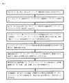
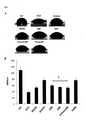
また、本明細書で記載される組成物を、非治癒創傷マウスモデルにおいて、マトリゲルに対しても査定したところ、組成物は、マトリゲルより迅速に創傷治癒を促進することが見出された。 The compositions described herein were also assessed against Matrigel in a non-healing wound mouse model and found to promote wound healing more rapidly than Matrigel.
ある特定の実施形態は、PVAハイドロゲルを、最終組成物に対して0.4%〜1.2%w/volで有する。ある特定の実施形態は、PVAハイドロゲルを、最終組成物に対して0.8%〜1.0%w/volで有するが、この場合、PVA-PEGブロックコポリマーを使用する。PVAハイドロゲルを、最終組成物に対して0.4%〜0.6%w/volで有する他の実施形態は、偶然、37℃より低温でゲル化する粘性溶液が可能であることを示した。更に、この濃度は、生細胞を存続させるためにより適した。更に、ある特定の実施形態では、PVA(pH 7.0)が10%(w/v)であり、99%が加水分解した高分子量のPVA(125kD〜200kD)と、88%だけが加水分解した中分子量のPVA(40kD〜100kD)との混合物(50/50)(「加水分解%」とは、アルコールへと加水分解したアセテート基の百分率を指す)を含む実施形態(10% HMW/MMW 50/50 99%/88% hyd)が好ましいことも示された。 Certain embodiments have PVA hydrogels at 0.4% to 1.2% w / vol with respect to the final composition. Certain embodiments have PVA hydrogels at 0.8% to 1.0% w / vol relative to the final composition, in which case PVA-PEG block copolymers are used. Other embodiments with PVA hydrogels at 0.4% to 0.6% w / vol relative to the final composition happened to show that viscous solutions that gel at temperatures below 37 ° C. are possible. In addition, this concentration was more suitable for the survival of living cells. Furthermore, in certain embodiments, PVA (pH 7.0) is 10% (w / v), with 99% hydrolyzed high molecular weight PVA (125kD-200kD) and only 88% hydrolyzed. Embodiments (10% HMW /
ポリビニルアルコール(PVA)ハイドロゲル及びその変化形(例えば、チオール化ポリビニルアルコール;ポリエチレングリコールを含有するポリビニルアルコールブロックポリマー(PVA-PEG);又はこれらのコポリマー)を有する、ある特定の実施形態を使用し、ホウ酸ナトリウム十水和物の、生体適合性のハイドロゲルの低分子架橋剤としての使用から利益を得ることを見出した。ホウ酸ナトリウム十水和物(pH8)の、最終組成物に対して0.02%(w/v)〜0.0001%(w/v)の間の濃度を使用することができる。更に、ホウ酸ナトリウム十水和物(pH8)の、最終組成物に対して0.01%(w/v)〜0.0001%(w/v)の間の濃度は、ボレート濃度が高度なPVAゲルより安定的なゲルをもたらすことが偶然見出された。ボレート濃度が0.03%を超える一部のゲルは、PVA含量が比較的高度な場合、早期にゲル化する場合があり、最適の範囲(すなわち、最終組成物に対して0.02%(w/v)〜0.0001%(w/v))は、作出時におけるゲルの取扱いを容易とする(例えば、より均一なミックスをもたらす)。PVAハイドロゲルに関する実験では、ボレートを添加する前における、PVAの添加及びコラーゲン-グリコサミノグリカン足場への混合は、その後、コラーゲン内及びコラーゲン周囲で、ボレートにより複合体化又は架橋される、相互貫入様ハイドロゲルの形成をもたらすので[又は観察(より粘性が大きく、均質なコラーゲン混合物)により]、好適であることが分かった。 Using certain embodiments with polyvinyl alcohol (PVA) hydrogels and variants thereof (eg, polyvinyl alcohol thiolated; polyvinyl alcohol block polymers containing polyethylene glycol (PVA-PEG); or copolymers thereof). , Sodium borate decahydrate, have been found to benefit from the use of biocompatible hydrogels as low molecular weight cross-linking agents. Concentrations of sodium borate decahydrate (pH 8) between 0.02% (w / v) and 0.0001% (w / v) with respect to the final composition can be used. In addition, the concentration of sodium borate decahydrate (pH 8) between 0.01% (w / v) and 0.0001% (w / v) relative to the final composition is more stable than PVA gels with high borate concentrations. It was accidentally found to bring about a gel. Some gels with borate concentrations above 0.03% may gel early if the PVA content is relatively high and are in the optimal range (ie 0.02% (w / v) relative to the final composition). ~ 0.0001% (w / v)) facilitates handling of the gel during production (eg, results in a more uniform mix). In experiments with PVA hydrogels, the addition of PVA and mixing into the collagen-glycosaminoglycan scaffold prior to the addition of borate was then complexed or cross-linked by borate in and around collagen, mutual. It was found to be suitable because it results in the formation of penetration-like hydrogels [or by observation (more viscous, homogeneous collagen mixture)].
ある特定の実施形態では、コラーゲン-GAG-ハイドロゲルマトリックスの作出の後における、1又は複数の架橋剤の添加は、ゲルマトリックスに、接着特性を付加することが偶然見出された。特に、ゲニピン(3.5〜5.0μM)及び/又はトランスグルタミナーゼ(1〜3U)の添加は、この点において、有用であることが見出されている。 In certain embodiments, it was accidentally found that the addition of one or more cross-linking agents after the production of the collagen-GAG-hydrogel matrix adds adhesive properties to the gel matrix. In particular, the addition of genipin (3.5-5.0 μM) and / or transglutaminase (1-3 U) has been found to be useful in this regard.
一部の実施形態は、本明細書で記載される組成物を、マトリックス形成後に、更に凍結乾燥させうるという偶然の発見に更に基づく。凍結乾燥させた組成物を、更に粉末化させうることが発見された。驚くべきことに、凍結乾燥させた粉末化組成物は、安定性が改善され、これは、使用及びパッケージングの容易さの利益をもたらすことが見出された。更に、粉末化させた凍結乾燥組成物は、創傷治癒等における使用の前に、水で再構成しうるだけでなく、また、患者の全血液、血清、又は血漿でも再構成することができた。偶然、粉末化させた凍結乾燥組成物は、一度再構成されてもなお、水、全血液、血清、及び血漿を使用したところ、ゲルを形成した。更に、血液又は血液成分の使用は、創傷の治癒過程の利益となるように、栄養物質に富む溶媒ももたらしうる。 Some embodiments are further based on the accidental finding that the compositions described herein can be further lyophilized after matrix formation. It has been discovered that the lyophilized composition can be further powdered. Surprisingly, the lyophilized powdered composition was found to have improved stability, which benefits from ease of use and packaging. Furthermore, the powdered lyophilized composition could not only be reconstituted with water, but also with the patient's whole blood, serum, or plasma prior to use in wound healing and the like. .. Coincidentally, the powdered lyophilized composition, once reconstituted, still formed a gel when water, whole blood, serum, and plasma were used. In addition, the use of blood or blood components can also result in nutrient-rich solvents to benefit the wound healing process.
本明細書では、組成物及び使用法を含み、それらについての実施形態が、利用可能な創傷ケア製品を上回る利点を有する、新規の多成分組織代替系が提示される。 This specification presents a novel multi-component tissue replacement system that includes compositions and usages, the embodiments of which have advantages over available wound care products.
ある特定の態様では、組織工学型の組織代替組成物であって、コラーゲン、グリコサミノグリカン、ハイドロゲル、及び1又は複数の架橋剤を含む組織代替組成物が提供される。ある特定の実施形態では、組織工学型の組織代替組成物は、I型コラーゲン、硫酸化グリコサミノグリカン、ポリビニルアルコール-ボレートハイドロゲル、グリセロール、及びアスコルビン酸の混合物を含む。ある特定の実施形態では、グリコサミノグリカンは、ヒアルロン酸である。ある特定の実施形態では、ハイドロゲルは、ポリビニルアルコール、ポリ酢酸ビニル、チオール化ポリビニルアルコール、又はこれらのコポリマーである。ある特定の実施形態では、1又は複数の架橋剤は、グルタルアルデヒド、EDC:NHS、ボレートから選択される。ある特定の実施形態では、組成物は、乾燥形態で存在する。他の実施形態では、組成物は、重合形態で存在する。他の実施形態では、組成物は、組成物が、主に、液体の非重合形態で存在するように、溶媒を更に含む。ある特定の実施形態では、組成物を加熱することにより、液体形態を、重合形態へと転換することができる。ある特定の実施形態では、乾燥形態は、適切な溶媒の添加により再構成することができる。適切な溶媒は、生理液、例えば、血液、血清、血漿等であってもよく、適切な溶媒は、非生理液、例えば、水であってもよい。 In certain aspects, tissue engineering type tissue replacement compositions are provided that include collagen, glycosaminoglycans, hydrogels, and one or more cross-linking agents. In certain embodiments, the tissue engineered tissue replacement composition comprises a mixture of type I collagen, sulfated glycosaminoglycans, polyvinyl alcohol-borate hydrogel, glycerol, and ascorbic acid. In certain embodiments, the glycosaminoglycan is hyaluronic acid. In certain embodiments, the hydrogel is polyvinyl alcohol, polyvinyl acetate, thiolated polyvinyl alcohol, or copolymers thereof. In certain embodiments, the one or more crosslinkers are selected from glutaraldehyde, EDC: NHS, borate. In certain embodiments, the composition is present in a dry form. In other embodiments, the composition exists in a polymerized form. In other embodiments, the composition further comprises a solvent such that the composition is present primarily in a non-polymerized form of liquid. In certain embodiments, heating the composition can transform the liquid form into a polymerized form. In certain embodiments, the dry form can be reconstituted by the addition of a suitable solvent. The suitable solvent may be a physiological solution such as blood, serum, plasma or the like, and the suitable solvent may be a non-physiological solution such as water.
ある特定の態様では、このような処置を要請する対象における創傷の処置の方法であって、対象へと、本明細書で記載される、組織工学型の組織代替組成物を投与する工程を含む方法が提供される。ある特定の実施形態では、創傷は、損傷、火傷、手術手順、感染症、及び潰瘍の結果としての創傷であるがこれらに限定されない創傷でありうる。 In certain embodiments, a method of treating a wound in a subject requesting such treatment comprises administering to the subject a tissue-engineered tissue replacement composition as described herein. A method is provided. In certain embodiments, the wound can be a wound as a result of injury, burns, surgical procedures, infections, and ulcers, but not limited to these.
ある特定の態様では、組織工学型の組織代替組成物の調製の方法であって、コラーゲン、グリコサミノグリカン、ハイドロゲル、及び1又は複数の架橋剤を、適切な溶媒中で混合する工程を含む方法が提供される。ある特定の実施形態では、混合された組成物は、室温を上回って加熱されるまで、非重合形態にとどまる。ある特定の実施形態では、その後、組成物を乾燥させて、溶媒を除去し、乾燥形態の調製を可能とする。その後、生理液又は水等、適切な溶媒を使用して、組成物を、液体形態へと再構成し戻すことができる。ある特定の実施形態では、1)まず、コラーゲン及びグリコサミノグリカンを、1又は複数の架橋剤と混合して、これらの成分の間の何らかの架橋を可能とし;2)ハイドロゲルポリマーを、コラーゲン-グリコサミノグリカンへと添加し、次いで、ボレート等、1又は複数の架橋剤と混合して、これらの成分の何らかの架橋を可能とし、3)工程1)及び2)による混合物を、適切な溶媒中で混合する。次いで、組成物を、加熱により重合させることができる。 In one particular embodiment, a method of preparing a tissue engineering type tissue replacement composition, in which collagen, glycosaminoglycans, hydrogels, and one or more crosslinkers are mixed in a suitable solvent. Methods to include are provided. In certain embodiments, the mixed composition remains in non-polymerized form until it is heated above room temperature. In certain embodiments, the composition is then dried to remove the solvent, allowing the preparation of a dry form. The composition can then be reconstituted back into liquid form using a suitable solvent such as physiological solution or water. In certain embodiments, 1) first, collagen and glycosaminoglycans are mixed with one or more cross-linking agents to allow some cross-linking between these components; 2) the hydrogel polymer, collagen. -Add to glycosaminoglycans and then mix with one or more cross-linking agents such as borate to allow some cross-linking of these components, and the mixture according to 3) steps 1) and 2) is suitable. Mix in solvent. The composition can then be polymerized by heating.
本明細書で具体的に定義されていない、任意の用語は、本発明の技術分野内で理解されている意味と一般に関連する意味を有すると理解されるものとする。 Any term not specifically defined herein is to be understood to have meaning generally associated with what is understood within the art of the invention.
定義
本明細書で使用される「コラーゲン」という用語は、線維性の動物組織内で一般に見出される構造タンパク質を指し、主に、線維状コラーゲン(例えば、I、II、III、V、XI型)を包摂するが、また、非線維状コラーゲン、すなわち、IV、VI、VII、VIII、IX、X、XI、XII、XIII、XIV、XV、XVI、XVII、及びXIX型も含みうる。本明細書で最も一般に使用されるコラーゲンは、I型コラーゲンであった。Definitions As used herein, the term "collagen" refers to structural proteins commonly found in fibrotic animal tissues, primarily fibrotic collagen (eg, types I, II, III, V, XI). , But may also include non-fibrotic collagen, ie IV, VI, VII, VIII, IX, X, XI, XII, XIII, XIV, XV, XVI, XVII, and XIX types. The most commonly used collagen herein was type I collagen.
本明細書で使用される「グリコサミノグリカン」(GAG)という用語は、硫酸化グリコサミノグリカンを包摂することを意図する。例えば、硫酸コンドロイチン、デルマタン硫酸、ケラタン硫酸、ヘパラン硫酸、ヘパリンである。グリコサミノグリカンファミリーのメンバーは、ヘキソサミン単位、ヘキソース単位、又はヘキスロン酸単位の種類が異なり、例えば、グルクロン酸、イズロン酸、ガラクトース、ガラクトサミン、グルコサミンを含有し、また、グリコシド連結の形状も異なる。 The term "glycosaminoglycan" (GAG) as used herein is intended to include sulfated glycosaminoglycans. For example, chondroitin sulfate, dermatan sulfate, keratan sulfate, heparan sulfate, and heparin. Members of the glycosaminoglycan family differ in the type of hexosamine unit, hexose unit, or hexuronic acid unit, including, for example, glucuronic acid, iduronic acid, galactose, galactosamine, glucosamine, and also differ in the shape of the glycosidic bond.
「コラーゲンとグリコサミノグリカンとの生体適合性の低分子架橋剤」という用語は、コラーゲンと、グリコサミノグリカンとの、任意の適切な架橋剤を包摂することを意図する。例えば、この用語は、グルタルアルデヒド、1-エチル-3-(3-ジメチルアミノプロピル)カルボジイミド(EDAC)、EDC(1-エチル-3-(3-ジメチルアミノプロピル)-カルボジイミド):NHS(N-ヒドロキシスクシンイミド)、EDC(1-エチル-3-(3-ジメチルアミノプロピル)-カルボジイミド):スルホ-NHS(N-ヒドロキシスルホスクシンイミド)、ヘキサメチレンジイソシアネート、及びゲニピンのうちの1又は複数を含みうる。用語はまた、他のカルボジイミド若しくはイソシアネート又は酵素的架橋剤も包摂しうる。 The term "biocompatible small molecule cross-linking agent for collagen and glycosaminoglycans" is intended to include any suitable cross-linking agent for collagen and glycosaminoglycans. For example, the term glutaaldehyde, 1-ethyl-3- (3-dimethylaminopropyl) carbodiimide (EDAC), EDC (1-ethyl-3- (3-dimethylaminopropyl) -carbodiimide): NHS (N- (Hydroxysuccinimide), EDC (1-ethyl-3- (3-dimethylaminopropyl) -carbodiimide): sulfo-NHS (N-hydroxysulfosuccinimide), hexamethylenediisocyanate, and one or more of genipines may be included. The term may also include other carbodiimides or isocyanates or enzymatic crosslinkers.
「コラーゲンと、グリコサミノグリカンとの非低分子架橋剤」という用語は、任意の適切なコラーゲンと、グリコサミノグリカンとの非低分子架橋剤を包摂することを意図する。例えば、脱水熱処理(DHT)、紫外線照射(UV)、及び酵素的架橋(例えば、トランスグルタミナーゼ)である。コラーゲン-グリコサミノグリカン架橋に使用される化学的技法は多様である。ホルムアルデヒド及びグルタルアルデヒド等、アルデヒドの使用については、十分に特徴づけられている。グルタルアルデヒドは、コラーゲンベースの生体材料を架橋する、最も一般に使用されている化学的方法である。代替的にまた、カルボジイミドファミリーのメンバーも、コラーゲン足場の機械的耐性及び酵素的耐性を増強するのに使用することができる。更に、これらの化学物質はまた、コラーゲンを、一部の金ナノ構造へと架橋するのに使用することもでき、エポキシと組み合わせて使用することもできる。イソシアネート、例えば、ヘキサメチレンジイソシアネートは、コラーゲン足場の架橋で知られている。市販のZimmer(商標)Collagen Repair Patchは、特許権のあるイソシアネート架橋法を使用する。化学的架橋剤であるゲニピンは、植物供給源に由来し、任意選択で、本組成物中の、グルタルアルデヒドの代わりに使用することもでき、これに加えて使用することもできる。 The term "non-small molecule cross-linking agent for collagen and glycosaminoglycans" is intended to include any suitable collagen and non-small molecule cross-linking agent for glycosaminoglycans. For example, dehydration heat treatment (DHT), ultraviolet irradiation (UV), and enzymatic cross-linking (eg, transglutaminase). The chemical techniques used for collagen-glycosaminoglycan cross-linking are diverse. The use of aldehydes, such as formaldehyde and glutaraldehyde, is well characterized. Glutaraldehyde is the most commonly used chemical method for cross-linking collagen-based biomaterials. Alternatively, members of the carbodiimide family can also be used to enhance the mechanical and enzymatic resistance of collagen scaffolds. In addition, these chemicals can also be used to crosslink collagen into some gold nanostructures, or in combination with epoxies. Isocyanates, such as hexamethylene diisocyanates, are known for cross-linking collagen scaffolds. The commercially available Zimmer ™ Collagen Repair Patch uses the patented isocyanate cross-linking method. The chemical cross-linking agent, genipin, is derived from a plant source and can optionally be used in place of or in addition to glutaraldehyde in the composition.
上記で記載した共有結合的架橋に対する代替法として、ある特定の状況下では、コラーゲン分子間のイオン結合形成は、適切でありうる。例えば、キトサン等のポリカチオン分子は、その多数のアミン基と、コラーゲンのカルボキシル基との間にイオン結合を創出する。 As an alternative to the covalent cross-linking described above, ionic bond formation between collagen molecules may be appropriate under certain circumstances. For example, a polycationic molecule such as chitosan creates an ionic bond between its many amine groups and the carboxyl group of collagen.
また、トランスグルタミナーゼ等の酵素的架橋薬剤も、コラーゲンベースのマトリックスの引張強度及び酵素的耐性を増強するのに使用することができる。酵素的架橋剤の使用は、化学的残基も、副生成物も、足場構造内に残存しないことを意味する。 Enzymatic cross-linking agents such as transglutaminase can also be used to enhance the tensile strength and enzymatic resistance of collagen-based matrices. The use of enzymatic cross-linking agents means that neither chemical residues nor by-products remain in the scaffold structure.
本明細書で使用される「デキストラン」という用語は、複合体の分枝状グルカン(すなわち、多くのグルコース分子から作られた多糖)を意味するが、これは、多様な長さ(3〜2000キロダルトン)の鎖から構成される可能性があり、浸透圧の平衡もまたもたらしうる非還元性糖として、グルタルアルデヒド架橋反応を安定化させる(とりわけ、高分子量の場合)のに役立つ。反応成分及び反応条件に応じて、デキストランを欠くと、グルタルアルデヒドは、所望よりはるかに急速に反応しうる。デキストランの使用後、組成物中には、微量が残存しうる。グルタルアルデヒドとデキストランとの典型的な混合物は、6mlのグルタルアルデヒド(25%)及び94mlのデキストラン(20%)でありうる。 As used herein, the term "dextran" refers to the complex branched glucans (ie, polysaccharides made from many glucose molecules), which vary in length (3-2000). As a non-reducing polysaccharide that may consist of (kilodalton) chains and can also result in osmotic equilibrium, it helps stabilize the glutaraldehyde cross-linking reaction (especially in the case of high molecular weight). In the absence of dextran, depending on the reaction components and reaction conditions, glutaraldehyde can react much faster than desired. After the use of dextran, trace amounts may remain in the composition. A typical mixture of glutaraldehyde and dextran can be 6 ml glutaraldehyde (25%) and 94 ml dextran (20%).
本明細書で使用される「ハイドロゲル」という用語は、ポリマー鎖の親水性ネットワークを包摂することを意図する。例えば、ハイドロゲルは、ポリビニルアルコール、ポリ酢酸ビニル、チオール化ポリビニルアルコールのポリマー、ポリエチレングリコールを含有するポリビニルアルコールブロックポリマー(PVA-PEG)、又はコポリマーの変化形及び部分的修飾形態(例えば、PVアセテート、PVP、及びチオール化形態)もまた含む、これらのコポリマーから作ることができる。ある特定の実施形態では、ハイドロゲルを、「ポリビニルアルコール、ポリ酢酸ビニル、チオール化ポリビニルアルコール、又はこれらのコポリマー」と称する。 As used herein, the term "hydrogel" is intended to include a hydrophilic network of polymer chains. For example, hydrogels can be polyvinyl alcohol, polyvinyl acetate, thiolated polyvinyl alcohol polymers, polyvinyl alcohol block polymers containing polyethylene glycol (PVA-PEG), or variants and partially modified forms of copolymers (eg, PV acetate). , PVP, and thiolated forms) can also be made from these copolymers. In certain embodiments, the hydrogel is referred to as "polyvinyl alcohol, polyvinyl acetate, thiolated polyvinyl alcohol, or copolymers thereof".
本明細書で使用される「ポリビニルアルコール-ボレートハイドロゲル」という用語は、PVAポリマーを架橋するのに、ボレートを使用するPVAハイドロゲルを指す。 As used herein, the term "polyvinyl alcohol-borate hydrogel" refers to a PVA hydrogel that uses borate to crosslink a PVA polymer.
本明細書で使用される「PVA 10% HMW/MMW 50/50 99%/88% hyd」という用語は、10%(w/v)であり、99%が加水分解した高分子量のPVA(125kD〜200kD)と、88%だけが加水分解した中分子量のPVA(40kD〜100kD)との混合物(50/50)を含むPVA溶液を指す(「加水分解%」とは、アルコールへと加水分解したアセテート基の百分率を指す)。 As used herein, the term "
「ハイドロゲルの生体適合性の低分子架橋剤」又は「生体適合性で低分子のハイドロゲル架橋剤」という用語は、ハイドロゲルポリマーを併せて連結することが可能な、任意の適切な低分子を包摂することを意図する。例えば、PVAハイドロゲルのためには、ホウ酸ナトリウム十水和物又は他のボレート供給源が好ましい。ボレートの代替物として、別の二価カチオンも使用することができる。 The term "biogel biocompatible small molecule cross-linking agent" or "biocompatible, low molecular weight hydrogel cross-linking agent" refers to any suitable small molecule to which a hydrogel polymer can be linked together. Intended to include. For example, for PVA hydrogels, sodium borate decahydrate or other borate source is preferred. As an alternative to borate, another divalent cation can also be used.
「凍結乾燥させた」、「凍結乾燥(lyophilisation)」、「凍結乾燥(lyophilization)」という用語は、凍結乾燥させる(lyophilized)対象物を、凍結乾燥させる(freeze-dried)脱水過程である、低温乾燥を包摂することを意図する。凍結乾燥機には、本質的に3つの類型:マニホールド型凍結乾燥機、ロータリー型凍結乾燥機、及びトレー型凍結乾燥機が存在する。 The terms "lyophilization," "lyophilisation," and "lyophilization" are low-temperature processes that freeze-dried objects that are lyophilized. Intended to include dryness. There are essentially three types of freeze-dryers: manifold-type freeze-dryers, rotary-type freeze-dryers, and tray-type freeze-dryers.
本明細書で使用される「再構成」又は「再構成された」という用語は、適切な溶媒の、凍結乾燥させた組成物又は凍結乾燥させた粉末化組成物への添加を指す。溶媒は、水、血液、血清、血漿、培地(例えば、細胞培地)、又は適切な緩衝液から選択することができる。生理液(例えば、血液、血清、又は血漿)は、適合性を改善するように、組成物を投与する対象から採取することができる。溶媒は、任意選択で、栄養物質、増殖因子、薬物、細胞等、1又は複数の更なる成分を含みうる。粉末組成物又は凍結乾燥組成物の適切な再構成質量は、70mg/ml〜110mg/mlの間でありうる。 As used herein, the term "reconstituted" or "reconstituted" refers to the addition of a suitable solvent to a lyophilized or lyophilized powdered composition. The solvent can be selected from water, blood, serum, plasma, medium (eg, cell medium), or a suitable buffer. Physiological fluids (eg, blood, serum, or plasma) can be taken from the subject to whom the composition is administered so as to improve compatibility. The solvent may optionally include one or more additional components such as nutrients, growth factors, drugs, cells and the like. A suitable reconstituted mass of powder composition or lyophilized composition can be between 70 mg / ml and 110 mg / ml.
本明細書で使用される「バイオハイブリッド足場」という用語は、ゲル形態の組成物、再構成組成物、又は再構成された粉末化組成物を指す。バイオハイブリッド足場は、任意選択で、特定の適用に対する必要に応じて、栄養物質、増殖因子、薬物、細胞等、1又は複数の更なる成分を含みうる。 As used herein, the term "biohybrid scaffold" refers to a composition in gel form, a reconstituted composition, or a reconstituted powdered composition. The biohybrid scaffold may optionally include one or more additional components such as nutrients, growth factors, drugs, cells, etc., as required for a particular application.
本明細書で使用される「創傷」という用語は、皮膚、火傷、手術手順、感染、潰瘍(例えば、褥瘡又は糖尿病性潰瘍)における損傷を包摂する。本明細書で記載される組成物は、慢性創傷を含む創傷治癒に適し、この場合、足場は、ベッドサイドで調製することができる。更に、本明細書で記載される組成物は、網状の皮膚移植片内の開窓の充填剤として使用することができる。本発明者らが行った実験は、治癒率及び治癒転帰の改善を示す。本明細書で記載される組成物は、3Dプリンティング適用において使用することができる。 The term "wound" as used herein includes injuries in skin, burns, surgical procedures, infections, ulcers (eg, pressure ulcers or diabetic ulcers). The compositions described herein are suitable for wound healing, including chronic wounds, in which case the scaffold can be prepared bedside. In addition, the compositions described herein can be used as a filler for fenestration within reticulated skin grafts. Experiments conducted by the present inventors show improvement in cure rate and cure outcome. The compositions described herein can be used in 3D printing applications.
組織の修復及び再生のための1つの戦略は、その正常なアーキテクチャーの回復へと組織の増殖及び発生を促進する、生体模倣性足場の使用である。細胞を伴うか又は伴わずに、ゲル化細胞外マトリックスは、細胞の移植及び他の手術手順を改善することができる。本明細書で記載される通り、多成分組織代替系の形成は、本明細書で記載される組成物を含みうる。ある特定の実施形態では、組織代替系は、in situにおいて形成することが可能であり、ある特定の実施形態では、注射可能である。他の実施形態では、組織代替系は、適用の前に、ex vivoで、部分的又は完全に形成することができる。ある特定の実施形態では、組織代替系は、in situ又はex vivoで再構成することが可能な乾燥組成物を含む。 One strategy for tissue repair and regeneration is the use of biomimetic scaffolds that promote tissue growth and development to restore its normal architecture. With or without cells, the gelled extracellular matrix can improve cell transplantation and other surgical procedures. As described herein, the formation of a multi-component tissue substitution system may include the compositions described herein. In certain embodiments, the tissue replacement system can be formed in situ and in certain embodiments, it can be injected. In other embodiments, the tissue replacement system can be partially or completely formed ex vivo prior to application. In certain embodiments, the tissue replacement system comprises a dry composition that can be reconstituted in situ or ex vivo.
組織代替系の主要成分は、バイオハイブリッド足場である。組織代替系は、任意選択で、栄養物質、増殖因子、又は薬物等、更なる成分を含みうる。システムはまた、任意選択で、細胞移植のための細胞を発現させるインドールアミン2,3ジオキシゲナーゼ(IDO)も含みうる。本明細書で記載される組織代替系は、皮膚創傷(慢性及び急性)の処置に有用でありうるが、また、組織工学及び細胞移植の他の領域における使用にも関与性でありうる。 The main component of the tissue substitution system is the biohybrid scaffold. The tissue replacement system may optionally include additional components such as nutrients, growth factors, or drugs. The system can also optionally include
本明細書で記載される組成物は、架橋されたコラーゲン-グリコサミノグリカンマトリックスを、合成ハイドロゲル系と組み合わせることにより調製しうるマトリックス成分を含みうる。マトリックスは、凍結乾燥粉末として保管することができる。水和させると結果として生じる、可溶化相互貫入ポリマーネットワークは、加熱されると、部分的に、急速な原線維形成及び広範な水素結合に起因して、ゲル足場を形成する。形成されると、バイオハイブリッド足場は、自然に、天然の真皮に類似するアーキテクチャーであって、細胞の拘縮、酵素的分解、及び細胞の増殖を緩和することが可能なアーキテクチャーを呈する。ある特定の実施形態では、組成物は、乾燥粉末形態で存在しうる。乾燥粉末組成物は、増殖因子濃縮環境を創出しようとする取組みにおいて、生理液、例えば、全血液、血清、又は血漿で可溶化させ、したがって、再構成することができる。代替的に、乾燥粉末組成物は、蒸留水等、他の適切な溶媒で再構成することもできる。ある特定の実施形態では、乾燥粉末組成物を、in situで再構成する。他の実施形態では、乾燥粉末組成物を、ex vivoで再構成する。他の実施形態では、乾燥粉末組成物を、in vitro、例えば、チューブ内又は他の容器内で再構成する。多様な実施形態では、組成物は、非乾燥形態で、例えば、可溶化流体として、又は部分的若しくは完全に重合させたゲルとして存在しうる。 The compositions described herein can include matrix components that can be prepared by combining a crosslinked collagen-glycosaminoglycan matrix with a synthetic hydrogel system. The matrix can be stored as a lyophilized powder. The resulting solubilized interpenetrating polymer network upon hydration, when heated, forms a gel scaffold, in part due to rapid fibrillogenesis and extensive hydrogen bonding. Once formed, the biohybrid scaffold naturally exhibits an architecture that resembles the natural dermis and is capable of alleviating cell contracture, enzymatic degradation, and cell proliferation. In certain embodiments, the composition may be in dry powder form. The dry powder composition can be solubilized and thus reconstituted in physiological solutions such as whole blood, serum, or plasma in an effort to create a growth factor enriched environment. Alternatively, the dry powder composition can also be reconstituted with other suitable solvents such as distilled water. In certain embodiments, the dry powder composition is reconstituted in situ. In another embodiment, the dry powder composition is reconstituted ex vivo. In other embodiments, the dry powder composition is reconstituted in vitro, eg, in a tube or in another container. In various embodiments, the composition may exist in non-dry form, eg, as a solubilized fluid, or as a partially or fully polymerized gel.
物理的に、本出願で記載される組織代替系は、単純な架橋コラーゲンによる注射用ゲルより熱的に安定であり、機械的強度が大きい、本明細書で記載される組成物を含む。これは、ハイドロゲルネットワーク内のポリマーが、コラーゲンバンドルの熱安定性を改善することを裏付ける示差走査熱量測定により明らかである。ハイドロゲル中のポリマーは、両親媒性であり、したがって、界面活性剤がそうであるように、その中のコラーゲンと複合し、安定性を改善することが可能である。酵素であるIDOを発現させる細胞と組み合わせると、皮内の細胞足場を、in situで、数分間以内に作出することができ、これは、既存の皮膚代替系より著明に有利である。 Physically, the tissue replacement system described in this application comprises the compositions described herein, which are thermally more stable and have greater mechanical strength than simple crosslinked collagen injectable gels. This is evident from differential scanning calorimetry, which confirms that the polymers in the hydrogel network improve the thermal stability of the collagen bundle. The polymer in the hydrogel is amphipathic and can therefore be combined with the collagen in it to improve stability, as is the case with surfactants. When combined with cells expressing the enzyme IDO, intradermal cell scaffolds can be created in situ within minutes, which is a significant advantage over existing skin replacement systems.
ある特定の実施形態では、本明細書で記載される組成物は、以下の成分:コラーゲン、グリコサミノグリカン、ハイドロゲル、及び1又は複数の架橋剤を含みうる。ある特定の実施形態では、コラーゲンは、3.0〜10.0mg/mLの間の濃度で存在するI型コラーゲンを含む。他の実施形態では、コラーゲンは、他の種類のコラーゲンである、II、II、IV、V型を更に含み(しかし、これらに限定されない)、0.5〜4.0mg/mlの間の最終濃度で存在する。ある特定の実施形態では、グリコサミノグリカンは、1:6w/wのグリコサミノグリカン:コラーゲン比で存在する。ある特定の実施形態では、グリコサミノグリカンは、コラーゲンに対する質量比を1:6とするコンドロイチン硫酸であるが、また、デルマタン硫酸を、1:6の比で援用することもでき、ヒアルロン酸を、0.5〜1%w/vol(最終濃度)で援用することもできる。ある特定の実施形態では、ハイドロゲルは、ポリビニルアルコール(PVA)、ポリ酢酸ビニル、ポリビニルアルコール-co-ポリエチレングリコール、チオール化ポリビニルアルコール、又はこれらの他の任意のコポリマーである。ある特定の実施形態では、ハイドロゲルは、最終組成物に対して0.1%〜1%w/vの濃度で存在する。ある特定の実施形態では、ハイドロゲルは、最終組成物に対して、50〜150uMの濃度のアスコルベート、及び/又は0.01〜0.1w/vの濃度のグリセロールを更に含む。ある特定の実施形態では、1又は複数の架橋剤は、とりわけ、高分子量のデキストラン溶液(pH7.5〜8)中に、グルタルアルデヒド及び/又はエチル(ジメチルアミノプロピル)カルボジイミド:N-ヒドロキシスクシンイミド(EDC:NHS)を含み、これは、コラーゲンを架橋するのに適する。他の実施形態では、1又は複数の架橋剤は、ホウ酸ナトリウムを、最終組成物に対して0.1%〜1%w/vの濃度、優先的には、ポリオールに対して1:4w/wで含み、これは、ハイドロゲルを架橋するのに適する。 In certain embodiments, the compositions described herein may comprise the following components: collagen, glycosaminoglycans, hydrogels, and one or more cross-linking agents. In certain embodiments, collagen comprises type I collagen, which is present at a concentration between 3.0 and 10.0 mg / mL. In other embodiments, the collagen further comprises (but is not limited to) other types of collagen, types II, II, IV, V, and is present at a final concentration between 0.5 and 4.0 mg / ml. do. In certain embodiments, glycosaminoglycans are present in a glycosaminoglycan: collagen ratio of 1: 6 w / w. In certain embodiments, the glycosaminoglycan is chondroitin sulfate having a mass ratio of 1: 6 to collagen, but dermatan sulfate can also be used in a ratio of 1: 6 to hyaluronic acid. , 0.5 to 1% w / vol (final concentration) can also be used. In certain embodiments, the hydrogel is polyvinyl alcohol (PVA), polyvinyl acetate, polyvinyl alcohol-co-polyethylene glycol, thiolated polyvinyl alcohol, or any other copolymer thereof. In certain embodiments, the hydrogel is present at a concentration of 0.1% to 1% w / v relative to the final composition. In certain embodiments, the hydrogel further comprises ascorbate at a concentration of 50-150 uM and / or glycerol at a concentration of 0.01-0.1 w / v with respect to the final composition. In certain embodiments, the one or more cross-linking agents are glutaraldehyde and / or ethyl (dimethylaminopropyl) carbodiimide: N-hydroxysuccinimide (in particular, in high molecular weight dextran solution (pH 7.5-8)). EDC: NHS), which is suitable for cross-linking collagen. In other embodiments, the one or more crosslinkers are sodium borate at a concentration of 0.1% to 1% w / v relative to the final composition, preferably 1: 4 w / w relative to the polyol. Included in, which is suitable for cross-linking hydrogels.
既に記載した通り、本明細書で記載される組成物は、成分が、非重合形態で存在するように、組成物の異なる成分を、乾燥成分又は凍結乾燥成分として、併せて混合する実施形態を含む。他の実施形態では、組成物は、重合形態で存在する。組成物の重合形態は、個々の成分を含有する溶液の混合の結果として得られる化学反応から、重合組成物を創出することにより創出することができる。他の実施形態では、バイオハイブリッド足場組成物の重合形態は、乾燥組成物又は凍結乾燥組成物の再構成であって、コラーゲン、グリコサミノグリカン、ハイドロゲル、及び1又は複数の架橋剤成分を含む乾燥組成物の可溶化が、組成物の重合形態の形成を引き起こし、このような重合化が、組成物中に存在する1又は複数の架橋剤によって引き起こされる、コラーゲン成分、グリコサミノグリカン成分、及びハイドロゲル成分の架橋の結果として生じる再構成により創出することができる。1又は複数の架橋剤は、十分な量の適切な溶媒、例えば、生理液、水、又は他の適切な溶媒との接触時における、コラーゲン成分、グリコサミノグリカン成分、及びハイドロゲル成分の架橋(又は重合化)を容易とする。 As described above, the compositions described herein include embodiments in which different components of the composition are mixed together as a dry or lyophilized component so that the components are present in a non-polymerized form. include. In other embodiments, the composition exists in a polymerized form. The polymerization form of the composition can be created by creating a polymerization composition from a chemical reaction obtained as a result of mixing solutions containing individual components. In other embodiments, the polymerized form of the biohybrid scaffold composition is a reconstruction of the dry composition or lyophilized composition, comprising collagen, glycosaminoglycans, hydrogels, and one or more cross-linking agent components. Solubilization of the dry composition comprising causes the formation of a polymerized form of the composition, and such polymerization is caused by one or more cross-linking agents present in the composition, a collagen component, a glycosaminoglycan component. , And can be created by the reconstruction resulting from the cross-linking of the hydrogel component. One or more cross-linking agents cross-link the collagen component, glycosaminoglycan component, and hydrogel component upon contact with a sufficient amount of suitable solvent, such as physiological fluid, water, or other suitable solvent. (Or polymerization) is facilitated.
ある特定の実施形態では、組成物を、コラーゲン及びグリコサミノグリカンを、グルタルアルデヒド及び/又はEDC:NHS等、適切な架橋剤を使用してあらかじめ架橋し、ハイドロゲルを、ホウ酸ナトリウム等、適切な架橋剤を使用してあらかじめ架橋した、部分的な架橋形態で提供する。コラーゲン:グリコサミノグリカン成分の、ハイドロゲル成分との著明な架橋が存在しない、この部分的な架橋形態では、組成物は、粘性の、本質的な液体形態にとどまり、例えば、創傷への適用に適する。次いで、組成物の加熱は、1又は複数の架橋試薬により触媒される、コラーゲン:グリコサミノグリカン成分と、ハイドロゲル成分との間の更なる架橋(又は重合化)を可能とし、組成物は、より固体性のゲルとなる。 In certain embodiments, the composition is pre-crosslinked with collagen and glycosaminoglycan using a suitable cross-linking agent such as glutaraldehyde and / or EDC: NHS, and the hydrogel is cross-linked with sodium borate, etc. Provided in a partially crosslinked form, pre-crosslinked using a suitable cross-linking agent. Collagen: In this partially cross-linked form, where there is no significant cross-linking of the glycosaminoglycan component with the hydrogel component, the composition remains in a viscous, essential liquid form, eg, to the wound. Suitable for application. Heating of the composition then allows further cross-linking (or polymerization) between the collagen: glycosaminoglycan component and the hydrogel component, catalyzed by one or more cross-linking reagents, the composition. , Becomes a more solid gel.
ある特定の実施形態では、組成物を、非架橋のポリマー成分、コラーゲン、グリコサミノグリカン、及びポリオールを、1/3HEPES、1/3PBS、及び1/3DMEMによる、10倍濃度に濃縮された緩衝組成物中、又は同じビタミン、栄養物質、グルコース、及びミネラルによる濃縮蘇生用流体の変化形中で混合することにより調製する。全ての架橋法の後であり、且つ、凍結乾燥の前に、アスコルビン酸を、混合物へと、1〜1000uMの範囲で添加する。 In certain embodiments, the composition is buffered with a 10-fold concentration of non-crosslinked polymer components, collagen, glycosaminoglycans, and polyols with 1/3 HEPES, 1/3 PBS, and 1/3 DMEM. Prepared by mixing in the composition or in variants of the concentrated resuscitation fluid with the same vitamins, nutrients, glucose, and minerals. Ascorbic acid is added to the mixture in the range of 1-1000 uM after all cross-linking methods and before lyophilization.
ある特定の実施形態では、組成物は、乾燥形態で存在し、再構成された重合組成物が、十分な量の適切な溶媒の添加時に、特異的な所望の性質を有することを可能とするのに十分な量で、凍結乾燥成分を含む。他の実施形態では、組成物は、ある特定の有利な物理的特性を呈示する重合形態で存在する。ある特定の実施形態では、再構成組成物又は重合組成物は、コラゲナーゼによる酵素的分解に対して耐性でありうる。ある特定の実施形態では、再構成組成物又は重合組成物は、0.2〜2.0MPa、最も一般には、1.45〜2.0の引張強度を呈示しうる。ある特定の実施形態では、乾燥組成物は、十分な量の適切な溶媒を添加して13〜16分間以内に、コラーゲン線維を形成しうる。ある特定の実施形態では、再構成組成物又は重合組成物は、細胞の増殖を調節し、細胞の直線的な増殖を促進し、細胞に媒介されるマトリックス収縮に抵抗することが可能である。 In certain embodiments, the composition is present in a dry form, allowing the reconstituted polymeric composition to have specific desired properties upon addition of a sufficient amount of the appropriate solvent. Contains lyophilized ingredients in sufficient amounts. In other embodiments, the composition is present in a superposed form that exhibits certain advantageous physical properties. In certain embodiments, the reconstituted or polymerized compositions may be resistant to enzymatic degradation by collagenase. In certain embodiments, the reconstituted or polymerized compositions can exhibit tensile strengths of 0.2-2.0 MPa, most commonly 1.45-2.0. In certain embodiments, the dry composition can form collagen fibers within 13-16 minutes with the addition of a sufficient amount of the appropriate solvent. In certain embodiments, the reconstituted or polymerized composition is capable of regulating cell proliferation, promoting linear proliferation of cells, and resisting cell-mediated matrix contractions.
本明細書で使用される「乾燥形態」とは、組成物中に、個々の成分の化学的架橋を引き起こすのに十分な溶媒が存在しないように、溶媒を本質的に含まないことを意味する。組成物の乾燥形態は、少量又は微量の溶媒をなおも含有しうるが、これらの量は、著明量の組成物の化学的架橋を引き起こさない程度に十分に低量であることが理解される。個別の成分を混合し、次いで、混合された組成物を乾燥させることもでき、代替的に、個々の成分をまず乾燥させ、次いで、混合することもできる。乾燥は、当技術分野で公知の、任意の数の手段、例えば、凍結乾燥(lyophilization、freeze drying)等により達することができる。ある特定の実施形態では、バイオハイブリッド組成物の乾燥形態は、十分量の適切な溶媒を添加し、25〜40℃の間、好ましくは、25〜37℃の間、より好ましくは、30〜37℃の間、最も好ましくは、37℃の温度へと曝露すると、ゲル化を達成する。 As used herein, "dry form" means essentially free of solvent so that there is not enough solvent in the composition to cause chemical cross-linking of the individual components. .. It is understood that the dry form of the composition may still contain small amounts or trace amounts of solvent, but these amounts are low enough not to cause chemical cross-linking of the composition in significant amounts. NS. The individual components can be mixed and then the mixed composition dried, or alternatively, the individual components can be dried first and then mixed. Drying can be achieved by any number of means known in the art, such as lyophilization, freeze drying and the like. In certain embodiments, the dry form of the biohybrid composition adds a sufficient amount of the appropriate solvent and is between 25-40 ° C, preferably between 25-37 ° C, more preferably between 30-37. Gelation is achieved upon exposure to a temperature of 37 ° C, most preferably during ° C.
多様な実施形態では、組織工学型の組織代替系は、本明細書で記載される組成物を含むことが可能であり、このような処置を要請する対象における創傷の処置に有用でありうる。対象に存在しうる創傷は、損傷、火傷、手術手順、感染、褥瘡、糖尿病性潰瘍、手術による外傷、及び手術以外の外傷を含むがこれらに限定されない、多種多様な原因に由来しうる。本明細書で記載される、組織工学型の組織代替系は、粘性液体として投与することができ、組成物が、創傷の複雑で不規則な輪郭を覆い、充填することを可能とし、連続的な固体ゲルとなりうるという点で、このような処置に有利である。組成物は、注射により、例えば、シリンジを使用して投与することができる。他の実施形態では、組成物は、乾燥粉末形態で投与し、in situで再構成することができ、数分間以内に、皮内の細胞足場の急速な形成を可能とする。粉末は、増殖因子濃縮環境を創出しようとする取組みにおいて、患者の全血液、血清、又は血漿で可溶化させることもでき、単に蒸留水と混合することもできる。形成されると、これらの足場は、天然の真皮と同様のアーキテクチャーを、自然に呈し、治癒の改善を容易とするように、細胞、マトリックスタンパク質、及び他の重要な因子の、足場アーキテクチャーへの遊走を可能とする。 In various embodiments, tissue-engineered tissue replacement systems can include the compositions described herein and may be useful in treating wounds in subjects requesting such treatment. Wounds that may be present in a subject can come from a wide variety of causes, including but not limited to injuries, burns, surgical procedures, infections, pressure ulcers, diabetic ulcers, surgical trauma, and non-surgical trauma. The tissue engineering type tissue replacement system described herein can be administered as a viscous liquid, allowing the composition to cover and fill the complex and irregular contours of the wound, continuously. It is advantageous for such treatments in that it can be a solid gel. The composition can be administered by injection, for example using a syringe. In other embodiments, the composition can be administered in dry powder form and reconstituted in situ, allowing rapid formation of intracellular cell scaffolds within minutes. The powder can be solubilized in the patient's whole blood, serum, or plasma in an effort to create a growth factor enriched environment, or simply mixed with distilled water. Once formed, these scaffolds naturally exhibit an architecture similar to that of the natural dermis, a scaffold architecture of cells, matrix proteins, and other important factors to facilitate improved healing. Allows migration to.
時間が不可欠である場合、患者への適用準備のできた皮膚代替物の利益を、十分に認識することができる。本明細書で記載される組成物を含む、組織工学型の組織代替系は、ベッドサイドで、細胞を伴うか又は伴わずに作出することができ、創傷床を充填し、創傷表面と完全に統合させるのに使用することができる。組成物の凍結乾燥粉末形態の安定性は、ハイドロゲル等、既存の水和材料及び細胞による皮膚代替物より優れ、粉末を、創傷部位に密着する足場へと再構成する能力(細胞を伴うか又は伴わない)は、市販の皮膚代替物を上回る、大きな有用性を付与する。 If time is essential, the benefits of a skin substitute ready for application to the patient can be fully recognized. Tissue-engineered tissue replacement systems, including the compositions described herein, can be produced bedside with or without cells to fill the wound bed and completely with the wound surface. Can be used to integrate. The stability of the lyophilized powder form of the composition is superior to existing hydrated materials such as hydrogels and skin substitutes with cells, and the ability to reconstitute the powder into a scaffold that adheres to the wound site (with cells? Or without) confer greater utility than commercially available skin substitutes.
組成物の調製法
(A)9工程の方法
1)中性近傍のpHにおける、コラーゲンと、コンドロイチン-6-硫酸との架橋工程
2)ポリビニルアルコールベースのポリマーハイドロゲルの添加工程
3)ボレート及びアスコルビン酸の添加工程
4)前出の工程1〜3による組合せの凍結工程
5)凍結乾燥工程
6)凍結乾燥させた生成物を、粉末へと破砕する工程
7)粉末を、水性の生物学的溶媒中又は非生物学的溶媒中に再懸濁させる工程
8)任意選択で、粘度を、グアーガム等のせん断減粘剤、又はデキストラン等のせん断増粘剤等、生物学的に不活性な薬剤で調整する工程
9)任意選択で、再懸濁させた粉末を、自家細胞中、同系細胞中、又は異種細胞中で組み合わせる工程How to prepare the composition
(A) 9-step method
1) Cross-linking process between collagen and chondroitin-6-sulfuric acid at a pH near neutral
2) Polyvinyl alcohol-based polymer hydrogel addition step
3) Borate and ascorbic acid addition process
4) Freezing process of combination by
5) Freeze-drying process
6) Step of crushing freeze-dried product into powder
7) Step of resuspending the powder in an aqueous biological or non-biological solvent
8) Optional step of adjusting the viscosity with a biologically inert agent such as a shear thickener such as guar gum or a shear thickener such as dextran.
9) Optional step of combining the resuspended powder in autologous cells, syngeneic cells, or heterologous cells
(B)代替的なコラーゲン組成物の調製法
液体としての最終保管条件:4℃で、光への曝露を最小化する
粉末としての最終保管条件:機密容器、22℃で、光への曝露を最小化する(B) Preparation of alternative collagen compositions Final storage conditions as a liquid: Minimize exposure to light at 4 ° C Final storage conditions as a powder: Secret container, exposure to light at 22 ° C Minimize
材料:
・チップ:P1000、P200、P10
・ハサミ
・TCチューブ
・1.7mlのマイクロ遠心チューブ
・ddH2O
・10倍濃度のコラーゲン緩衝液
・50%のグルタルアルデヒド(-20℃)
・1NのNaOH(滅菌濾過された)
・20%の硫酸コンドロイチン
・コラーゲン(6mg/ml以上:Advanced Biomatrix社)
・20%のデキストラン
・20%のグリシン
・5%及び10%のPVA
・0.2%w/wのボレート溶液
・10mMのアスコルベート
・1倍濃度のDMEM(完全培地)material:
・ Chip: P1000, P200, P10
・ Scissors ・ TC tube ・ 1.7 ml microcentrifugal tube ・ ddH2 O
・ 10 times concentration collagen buffer ・ 50% glutaraldehyde (-20 ℃)
1N NaOH (sterilized and filtered)
・ 20% chondroitin sulfate ・ Collagen (6mg / ml or more: Advanced Biomatrix)
・ 20% dextran ・ 20% glycine ・ 5% and 10% PVA
・ 0.2% w / w borate solution ・ 10 mM ascorbate ・ 1x concentration DMEM (complete medium)
試薬の調製:
10倍濃度のコラーゲン緩衝液
10倍濃度のDMEM(pH 7.5)10ml
10倍濃度のHEPES(pH 7.5)10ml
10倍濃度のPBS(pH 7.0)9ml
1mlの抗生剤/抗真菌剤
20%の硫酸コンドロイチン
1gの硫酸コンドロイチン[サメ軟骨:SIGMA(商標)社]
1倍濃度のPBS 5ml
溶解の一助とするように、繰り返しボルテックスし、37℃で温める。可能な場合、0.4μmのフィルターを使用して濾過する。Preparation of reagents:
10x collagen buffer
10x concentration of DMEM (pH 7.5) 10ml
10x concentration of HEPES (pH 7.5) 10ml
10x PBS (pH 7.0) 9ml
1 ml antibiotic / antifungal
20% chondroitin sulfate
1g Chondroitin Sulfate [Shark Cartilage: SIGMA ™]
1x PBS 5ml
Vortex repeatedly and warm at 37 ° C to aid in dissolution. If possible, filter using a 0.4 μm filter.
20%のデキストラン
1gのデキストラン[SIGMA(商標)社]
1倍濃度のDMEM 5ml(FBS及び抗生剤を伴わない)20% dextran
1g Dextran [SIGMA ™]
1x DMEM 5ml (without FBS and antibiotics)
20%のグリシン
4gのグリシン
FBS及び抗生剤を伴わない、1倍濃度のDMEM 20ml
pHを、7.5へと調整し、室温で保管する。20% glycine
4g glycine
1x DMEM 20ml without FBS and antibiotics
Adjust the pH to 7.5 and store at room temperature.
10%のPVA-99
5gのPVA(88%加水分解したLMW)/5gのPVA(99%加水分解したHMW)
100mlのddH2O
1.水を、オーバーヘッドミキサーで、80℃へと加熱する。
2.HMW 99%を、溶解するまで、ゆっくりと添加する。
3.LMW 88%を、溶解するまで、ゆっくりと添加する。10% PVA-99
5g PVA (88% hydrolyzed LMW) / 5g PVA (99% hydrolyzed HMW)
100 ml ddH2 O
1. Heat the water to 80 ° C with an overhead mixer.
2. Add 99% HMW slowly until dissolved.
3. Add 88% LMW slowly until dissolved.
15%のKollicoat(商標)IR
15gのKollicoat(商標)IR/100mlのddH2O
1.水を、オーバーヘッドミキサーで、80℃へと加熱する。
2.Kollicoat(商標)IRを、ゆっくりと添加し、溶解するまで混合する。
10%のPVAを、15%のKollicoat(商標)と併せて、等量(50ml/50ml)で混合し、次いで、2mlのグリセロールを添加する。10倍濃度のコラーゲン緩衝液を使用して、作業溶液(5%)へと希釈する。15% Kollicoat ™ IR
15g Kollicoat ™ IR / 100ml ddH2 O
1. Heat the water to 80 ° C with an overhead mixer.
2. Add Kollicoat ™ IR slowly and mix until dissolved.
10% PVA is mixed with 15% Kollicoat ™ in equal volumes (50 ml / 50 ml), then 2 ml of glycerol is added. Dilute to working solution (5%) using 10-fold collagen buffer.
25mMのボレート(0.2%w/wのボレート)
1.200mgの四ホウ酸ナトリウム十水和物である「ボレート」を秤量する。
2.ボレートを、1倍濃度のDMEM 10ml(サプリメントを伴わない)へと添加する。
3.よく混合する。pHは、8〜9の間とする。
4.6.25mMの作業濃度(又は25mMのボレート)のために、1:10に希釈する。
5.4℃で保管し、沈殿物が存在する場合は、作り直す。25 mM borate (0.2% w / w borate)
Weigh 1.200 mg of sodium tetraborate decahydrate "Borate".
2. Add borate to 10 ml of DMEM (without supplements) at 1x concentration.
3. Mix well. The pH should be between 8 and 9.
For a working concentration of 4.6.25 mM (or 25 mM borate), dilute to 1:10.
Store at 5.4 ° C and remake if precipitates are present.
蒸留/脱塩水(作りたて/滅菌フィルター)中に、10mMのアスコルビン酸
総容量:総容量=5.0000
9.9mg/mlのコラーゲン:(総容量×3)÷9.9=1.5152
10倍濃度の緩衝液:総容量÷10=0.5000
1NのNaOH:コラーゲン×0.01=0.0152
20%のGAG:((コラーゲン×9)×5)÷200=0.3409
1.5%のグルタミン:0.0002×総容量÷0.015=0.0667
20%のグリシン:5×グルタミン=0.3333
5%のPVA:(総容量×0.006)÷0.05=0.6000
10mMのアスコルベート:(0.0001×総容量)÷0.01=0.0500
0.5mMのボレート:(総容量×0.0005)÷0.02=0.1250
細胞容量:総容量-(反応容量)=1.4538
空隙容量(%):29%Total volume of 10 mM ascorbic acid in distilled / desalinated water (freshly made / sterilized filter): total volume = 5.000
9.9 mg / ml collagen: (total volume x 3) ÷ 9.9 = 1.5152
10 times concentration buffer: total volume ÷ 10 = 0.5000
1N NaOH: Collagen x 0.01 = 0.0152
20% GAG: ((Collagen x 9) x 5) ÷ 200 = 0.3409
1.5% glutamine: 0.0002 x total capacity ÷ 0.015 = 0.0667
20% glycine: 5 x glutamine = 0.3333
5% PVA: (total capacity x 0.006) ÷ 0.05 = 0.6000
10 mM ascorbate: (0.0001 x total capacity) ÷ 0.01 = 0.0500
0.5 mM borate: (total capacity x 0.0005) ÷ 0.02 = 0.1250
Cell volume: Total volume-(reaction volume) = 1.4538
Void capacity (%): 29%
方法
1.BSC中に氷を伴う、清浄な(70%のエタノールを伴う)250mlのビーカーを用意する。
2.2〜15mlのチューブを、氷中に入れる。
3.p1000を使用して、コラーゲンを、チューブのうちの1つへと添加する。終了したら、チップを、氷上の空のチューブへと取り置く。
4.清浄なp1000チップを使用して、コラーゲン緩衝液を、コラーゲンへと添加する。
5.15mlのチューブ内の氷上で保管したコラーゲンチップを使用して、緩衝液と、コラーゲンとを、併せて混合する。混合する前に、残りのコラーゲンを放出させ、次いで、チップの先端を切り取って、低粘性用チップを創出する。
6.p10を使用して、1NのNaOHを、溶液へと添加して、コラーゲンを、pH6.8〜7.5(桃色)へと中性化させる。一度に、最大で15ulだけを添加する。この間、切り取られたp1000チップを使用して、十分に混合する。
7.中性化させたら、p1000チップを使用して、GAG(コンドロイチン6-硫酸)を添加する。使用後、チップを廃棄する。切り取られたp1000チップを使用して混合する。Method
1. Prepare a clean (with 70% ethanol) 250 ml beaker with ice in the BSC.
Place a 2.2-15 ml tube in ice.
3. Using p1000, add collagen to one of the tubes. When finished, set aside the tip in an empty tube on ice.
4. Using a clean p1000 chip, add collagen buffer to collagen.
Using collagen chips stored on ice in a 5.15 ml tube, the buffer and collagen are mixed together. Prior to mixing, the remaining collagen is released and then the tip of the tip is cut off to create a low viscosity tip.
6. Using p10, 1N NaOH is added to the solution to neutralize collagen to pH 6.8-7.5 (pink). Add up to 15 ul at a time. During this time, the cut p1000 chips are used to mix well.
7. After neutralization, add GAG (chondroitin 6-sulfate) using the p1000 chip. Discard the tip after use. Mix using the cut p1000 chips.
消灯(暗所)
8.消灯する。マイクロ遠心チューブに、94ulのデキストラン溶液を添加する。
9.6ulのグルタルアルデヒドを、デキストランを伴うチューブへと添加する。倒立させ、手指で1回ボルテックスする。グルタルアルデヒドの作業溶液を、コラーゲンミックスへと、速やかに添加する。切り取られたピペットチップを使用して、十分に混合する(約2分間にわたり)。気泡を回避する。pHは、重要である。理想的な架橋pHは、7.5〜8である(酸性が強すぎる場合、グルタチオンは、架橋されないことに注意されたい)。
10.暗所内、氷上で22分間にわたりインキュベートする。
11.1時間後、グリシン溶液を添加し、十分に混合する。これにより、グルタルアルデヒドを中性化させる。
12.暗所内、氷上で最短1時間、又は4℃で一晩にわたりインキュベートする。Off (dark place)
8. Turn off the light. Add 94 ul of dextran solution to the microcentrifuge tube.
Add 9.6 ul of glutaraldehyde to the tube with dextran. Invert and vortex once with your fingers. The working solution of glutaraldehyde is rapidly added to the collagen mix. Use the cut pipette tip to mix well (for about 2 minutes). Avoid air bubbles. pH is important. The ideal cross-linked pH is 7.5-8 (note that glutathione is not cross-linked if it is too acidic).
10. Incubate in the dark on ice for 22 minutes.
After 11.1 hours, add the glycine solution and mix well. This neutralizes glutaraldehyde.
12. Incubate in the dark, on ice for a minimum of 1 hour, or overnight at 4 ° C.
点灯
13.アスコルベートを、コラーゲンゲルへと添加し、十分に混合する。
14.コラーゲン緩衝液を使用して、PVA(10%)を、5%の作業濃度へと希釈する。
15.PVA作業溶液を、コラーゲンゲルへと添加し、十分に混合する(約1〜2分間にわたり)。気泡の発生を回避する。
16.ボレート緩衝液を、コラーゲンゲルへと添加し、十分に混合する。
17.結果として得られるコラーゲンゲル(こうして、完全IPN(相互貫入ポリマーネットワーク)-ハイドロゲル架橋コラーゲンマトリックスとなる)を、4℃で保管する(保管条件を参照されたい)。Lit
13. Add ascorbate to the collagen gel and mix well.
14. Use collagen buffer to dilute PVA (10%) to a working concentration of 5%.
15. Add the PVA working solution to the collagen gel and mix well (over about 1-2 minutes). Avoid the generation of air bubbles.
16. Add borate buffer to the collagen gel and mix well.
17. Store the resulting collagen gel (thus a complete IPN (interpenetrating polymer network) -hydrogel cross-linked collagen matrix) at 4 ° C (see Storage Conditions).
凍結乾燥
18.15mlのチューブの目盛りを見ることにより、反応混合物の実際の容量を推定する。チューブを秤量する。測定値(容量及び質量)を記録する。
19.チューブを、-80℃の冷凍庫に寝かせて、12時間にわたり静置する。
20.12時間後、凍結乾燥機をオンにする(マニュアルを参照されたい)。
21.チューブを、液体10ml当たり36時間にわたり、凍結乾燥機に入れる。
22.凍結乾燥後、粉末を秤量し、再構成容量(平均60〜70mg/mL)を計算する。freeze drying
Estimate the actual volume of the reaction mixture by looking at the scale on the 18.15 ml tube. Weigh the tube. Record measurements (capacity and mass).
19. Place the tube in the -80 ° C freezer and let stand for 12 hours.
After 20.12 hours, turn on the lyophilizer (see manual).
21. Place the tube in the lyophilizer for 36 hours per 10 ml of liquid.
22. After lyophilization, weigh the powder and calculate the reconstitution volume (average 60-70 mg / mL).
(C)代替的な組成物
代替的な組成物は、3〜5mg/mlの間のコラーゲン;6:1の比の硫酸コンドロイチン(1倍濃度のPBS中):コラーゲン;グルタルアルデヒド(後でグリシンを添加するが、この場合、グリシンは、グルタルアルデヒドを中性化させるのに添加する);質量で0.4%〜0.6%のPVA(pH 7.0)10% HMW/MMW 50/50 99%/88% hyd;0.01%(w/v)〜0.0001%(w/v)のホウ酸ナトリウム十水和物(pH 8);100μmのアスコルビン酸ナトリウム(pH 7.0);3.5〜5μMのゲニピン(第2の架橋剤);及び1〜3Uトランスグルタミナーゼ(全ての濃度は、最終生成物に対して)を有しうる。(C) Alternative Compositions Alternative compositions are collagen between 3-5 mg / ml; chondroitin sulfate in a ratio of 6: 1 (in 1-fold pH): collagen; glutaraldehyde (later glycin). In this case, glycine is added to neutralize glutaraldehyde); 0.4% to 0.6% by mass PVA (pH 7.0) 10% HMW /
架橋剤濃度、PVA、及び時間の、原線維形成に対する効果:原液のコラーゲン濃度の変化に関して、ゲルの製剤化を最適化するために、グルタルアルデヒド濃度及び架橋インキュベーション時間の両方の、原線維形成速度の変化に対する効果を探索した。第1に、グルタルアルデヒド濃度の変化が、原液のコラーゲン濃度の変化と比例したとすれば(かつて、6mg/mlを用いた;Hartwellら、2011)、架橋反応は、氷上におけるゲルの固体化を結果としてもたらすであろう。したがって、グルタルアルデヒド濃度を増大させると、ゲル化速度は、予測されうるように増大はせず、実際は、減少することが見出された(図2A;条件C1)。更に、高濃度の架橋剤は、313nmにおいて、高度な初期吸光度を結果としてもたらした。このパターンは、反応容量をマッチさせた試料であって、低量のグルタルアルデヒド架橋剤を含有する試料(C2)内でも持続した。架橋剤を伴わないコラーゲン単独は、図2Aに示す通り、最高速度の原線維形成と併せて、最低の初期吸光度及び最高の最終吸光度を呈示した。また、t1/2maxも、容量をマッチさせた反応基内のインキュベーションの持続時間と共に、わずかに減少した(図2A)。反応物中の高グルタルアルデヒド濃度はまた、インキュベーション時間と比例するt1/2maxの低減にも対応することが明らかであった。このゲル化時間の低減は最終的に、製剤化時間及び作業混合物としてのゲル固体化のための時間の両方の低減に対応するであろう。興味深いことに、インキュベーション時間を、反応槽内で使用されるコラーゲンの原液濃度について調整した(図2B)ところ、反応速度を制御することができた。この結果は、架橋剤の最適の最終濃度は、0.02%w/vであり、架橋インキュベーション時間は、コラーゲンの原液濃度(反応槽濃度)に比例して調整すべきであることを裏付けた。 Effects of cross-linking agent concentration, PVA, and time on fibrillogenesis: Fibrillogenesis rates, both glutaraldehyde concentration and cross-linking incubation time, to optimize gel formulation with respect to changes in stock solution collagen concentration. We searched for the effect on the change of. First, if the change in glutaraldehyde concentration was proportional to the change in collagen concentration in the stock solution (once 6 mg / ml was used; Hartwell et al., 2011), the cross-linking reaction would solidify the gel on ice. Will result. Therefore, it was found that increasing the glutaraldehyde concentration did not increase the gelation rate as expected, but actually decreased (Fig. 2A; condition C1). In addition, the high concentration of cross-linking agent resulted in a high initial absorbance at 313 nm. This pattern persisted even in the sample (C2) containing a low amount of glutaraldehyde cross-linking agent, which was a sample whose reaction volume was matched. Collagen alone without a cross-linking agent exhibited the lowest initial absorbance and the highest final absorbance, along with the highest rate of fibrillogenesis, as shown in FIG. 2A. T1 / 2max also decreased slightly with duration of incubation within the volume-matched reactive groups (Fig. 2A). It was apparent that the high glutaraldehyde concentration in the reaction also corresponded to a reduction in t1 / 2max proportional to the incubation time. This reduction in gelation time will ultimately correspond to a reduction in both the formulation time and the time for gel solidification as a working mixture. Interestingly, the incubation time was adjusted for the stock solution concentration of collagen used in the reaction vessel (Fig. 2B), and the reaction rate could be controlled. This result confirmed that the optimum final concentration of the cross-linking agent was 0.02% w / v, and that the cross-linking incubation time should be adjusted in proportion to the collagen stock solution concentration (reaction tank concentration).
ポリビニルアルコール-ハイドロゲルの、ゲル混合物への添加の効果は、PVAの濃度を、0から1.0%w/vへと増大させる(これは、活性化エネルギーの著明な低減に対応した)場合の、原線維形成速度の増大により裏付けられた(図3)。TABLE 3(表1)に示す通り、1%w/vのPVAの添加は、原線維形成に要請される活性化エネルギーの、50%を超える低減を結果としてもたらした。 The effect of the addition of polyvinyl alcohol-hydrogel to the gel mixture is when the concentration of PVA is increased from 0 to 1.0% w / v (which corresponds to a significant reduction in activation energy). , Supported by an increase in the rate of fibrillogenesis (Fig. 3). As shown in TABLE 3 (Table 1), the addition of 1% w / v PVA resulted in a reduction of more than 50% in the activation energy required for fibrillogenesis.

ゲル化速度のわずかな増大はまた、全体的なゲル化時間の減少とも関連した。しかし、本アッセイでは、この変化は、著明ではなかった。創傷床内に投入することもでき、細胞の送達及び移植のために、30℃〜37℃の作業範囲で投入することもできる足場の製剤化のために、異なるPVA-ハイドロゲル濃度を含有するゲル混合物について探索した。結果は、PVA-(ボレート)ハイドロゲルが、30℃におけるゲル化を可能とするために、I型ウシコラーゲンの原線維形成反応速度を、実際に変更しうることを裏付けた。典型的な倒立試験管試験により、これらの低温におけるゲル化を裏付けうる場合、波長を313nmとする濁度の査定により、PVA-ハイドロゲル系が、アーチファクトを提示する可能性を除去する。 A slight increase in gelation rate was also associated with a decrease in overall gelation time. However, in this assay, this change was not significant. Contains different PVA-hydrogel concentrations for scaffold formulation, which can be placed into the wound bed and can be placed in the working range of 30 ° C to 37 ° C for cell delivery and transplantation. We searched for gel mixtures. The results confirmed that PVA- (borate) hydrogel could actually alter the fibril formation reaction rate of type I bovine collagen to allow gelation at 30 ° C. If typical inverted test tube tests can support these low temperature gelations, a turbidity assessment at a wavelength of 313 nm eliminates the possibility that the PVA-hydrogel system will present artifacts.
PVA-ハイドロゲルの添加により、凍結乾燥後における、ゲル混合物のゲル化及び機械的特性が保存される:ゲル混合物の安定性を増大させる試み(輸送及び保管を目的とする)として、混合物を凍結乾燥させ、粉末へとすりつぶし、次いで、再構成した。凍結乾燥混合物を、pH中性化の必要を伴わない脱塩蒸留水で再構成した。PVA-PEGコポリマーを使用して創出されたPVA-ハイドロゲル系[Kollicoat(商標)]の再構成が最も容易であった。架橋されず、PVAを含有しない、コラーゲン足場の変化形は、マッチさせた非凍結乾燥足場混合物と比較した場合、著明に長いゲル化時間を呈示した(52.6±1.52分間対36±1.73分間)(図4及び図5)。架橋だけ(xcol)の足場は、原線維を形成することが可能であったが、凍結乾燥の前の、著明に長いゲル化時間後においてであった。PVA-ハイドロゲルを含有する足場変化形の液体混合物は、凍結乾燥前よりわずかに高度な初期吸光度を呈示したが、統計学的に同等な量の時間内に、原線維を形成した(図4及び図5)。K99が最も注目すべきである、Kollicoat(商標)試料は、ゲル化が最も迅速な(15.7±1.16分間)試料の中にあり、最小の濁度変化(t=0〜tmaxにおける吸光度)を呈示したことから、PVA-PEGは、粉末形態において、コラーゲン構造に対する保護効果を及ぼし、また、再構成されても、保護効果を及ぼしたはずであることが示唆された。図4及び図5に示される通り、PVA-ハイドロゲル足場は、col変化形、xcol変化形、TW80(商標)変化形、及びTritonx100(商標)変化形と比較した場合、ゲル化時間の著明な低減を呈示した。ゲル化時間の最大の低減は、PVA-PEGハイドロゲルを含有する足場において観察された(2.02倍)。界面活性剤の、コラーゲンのゲル化反応速度に対する効果について検討したかつての研究(Fathima及びDhathathreyan、2009;Li、2009)と比較して、Tween 20(商標)と、Tween 80(商標)と、Triton-x100(商標)とを、ゲル混合物中に、これらそれぞれの中央ミセル濃度で組み合わせた(注: Tween 20(商標)の効果は、Tritonx100(商標)と同等であり、したがって、図の明確さのために省略した)。図4及び図5に示される通り、全ての界面活性剤は、凍結乾燥前試料及び凍結乾燥後試料のいずれのゲル化速度も増大させた(t1/2maxを低下させた)が、大部分のPVAより著明に低度においてであった。The addition of PVA-hydrogel preserves the gelation and mechanical properties of the gel mixture after lyophilization: freezing the mixture in an attempt to increase the stability of the gel mixture (for transport and storage purposes). It was dried, ground into a powder and then reconstituted. The lyophilized mixture was reconstituted with desalted distilled water without the need for pH neutralization. The PVA-hydrogel system [Kollicoat ™] created using the PVA-PEG copolymer was the easiest to reconstitute. Variants of collagen scaffolds that were not cross-linked and did not contain PVA exhibited significantly longer gelation times when compared to the matched lyophilized scaffold mixture (52.6 ± 1.52 min vs. 36 ± 1.73 min). (Fig. 4 and Fig. 5). Crosslink-only (xcol) scaffolds were able to form fibrils, but before lyophilization, after a significantly longer gelation time. The scaffold-modified liquid mixture containing PVA-hydrogel exhibited slightly higher initial absorbance than before lyophilization, but formed fibrils within a statistically equivalent amount of time (Fig. 4). And Fig. 5). The Kollicoat ™ sample, of which K99 is most notable, is among the samples with the fastest gelation (15.7 ± 1.16 minutes) and exhibits minimal turbidity changes (absorbance from t = 0 to tmax). Therefore, it was suggested that PVA-PEG had a protective effect on the collagen structure in the powder form, and should have a protective effect even if it was reconstituted. As shown in FIGS. 4 and 5, PVA-hydrogel scaffolds have marked gelation times when compared to col variants, xcol variants, TW80 ™ variants, and Triton x100 ™ variants. Demonstrated a significant reduction. The greatest reduction in gelation time was observed in scaffolds containing PVA-PEG hydrogel (2.02-fold).
PVA-PEG系及びPVA-ハイドロゲル系は、コラーゲンゲル混合物に対する界面活性剤様効果を呈示する:界面活性剤の主要な役割は、疎水性表面の親水性を改善することである。Tween 20(商標)、Tween 80(商標)、及びTriton-x100(商標)の、コラーゲンゲル混合物への添加は、投入されたゲル混合物の薄膜の上部にある水滴の水接触角を、108°(コラーゲン:GAG)から、Triton X100及びTween 80のそれぞれについて、51°、75°へと、著明に低減した(図6)。更に、使用されるPVAの種類に応じて、接触角の差違が見られた。興味深いことに、ボレート(PVAnb)を、系から除外したところ、接触角は、著明に(52°から72°へと)増大した。架橋だけのゲル混合物が、最小の接触角(37°)を顕示したことから、全てのゲル混合物により形成される、最も親水性の大きな表面が示唆された。PVA99、PVAnb、及びTween 80(商標)は、それぞれ、75°、76°、及び75°で、比較的同等であった。一方、Kollicoat(商標)の99及び88によるブレンドは、それぞれ、58°及び53°の接触角で、次に親水性が大きかった。予測される通り、疎水性の大きなPVAは、界面活性剤様効果が大きく、したがって、分子量の同等なPVA比較して、接触角の低減も大きかった。 PVA-PEG and PVA-hydrogel systems exhibit a surfactant-like effect on collagen gel mixtures: The main role of surfactants is to improve the hydrophilicity of hydrophobic surfaces. Addition of
細胞の生存率及び細胞の遊走:ゲル混合物の変化形中で培養された細胞を、足場内で24時間にわたり培養してから、染色した。全ての足場変化形は、in vitroにおいて、非毒性であることが見出された(図7)。線維芽細胞を密集させた同様のコラーゲン格子を創出して、遊走について査定した。足場の中央部から、パンチ生検を採取し、次いで、マッチさせた無細胞ゲルで充填した。全ての足場の間で、細胞の遊走に著明な差違は見られなかったが、非架橋のコラーゲン:GAG足場は、例外として、10日間にわたり、最高の細胞遊走速度を呈示した(図7B)。10日目には、k99変化形及びxcol変化形において、PVA50/50と比較して、遊走が著明に大きな細胞が見出された(図7B)。 Cell viability and cell migration: Cells cultured in variants of the gel mixture were cultured in scaffolds for 24 hours and then stained. All scaffold variants were found to be non-toxic in vitro (Fig. 7). A similar collagen lattice densely packed with fibroblasts was created and migration was assessed. Punch biopsies were taken from the central part of the scaffold and then filled with matched cell-free gel. There were no significant differences in cell migration among all scaffolds, with the exception of non-crosslinked collagen: GAG scaffolds, which exhibited the highest cell migration rates over a 10-day period (Fig. 7B). .. On
線維芽細胞の形状及び足場のアーキテクチャー:図8は、5つの異なる足場変化形内で培養された線維芽細胞の形状について描示する。かつての研究において見出された通り、非架橋のコラーゲン足場内で培養された細胞は、ファロイジン-488染色(顕微鏡写真は示さない)により、葉状仮足及び糸状仮足の伸長、ストレスファイバー形成(線維状アクチン)の広範な伝播を呈示する(Kimら、2014; Mattila及びLappalainen、2008)。50%wt Kollicoat(商標)/50%wt PVA99ハイドロゲル(0.4%wt)を含有する足場変化形内に培養された線維芽細胞では、顕著な平行の線維配置及び少数の糸状仮足と符合する、樹枝状外見の低減が観察された。界面活性剤を含有する足場は、狭小な糸状仮足様伸長部を伴う樹枝様外見と、細胞は生存を維持しているが、細胞ストレス及び接着不良の可能性を示唆する、小型の細胞体とを呈示した(図7)。走査電子顕微鏡による断面画像(図8)は、かつての研究における本発明者らの知見であって、非架橋足場(col)では、他の架橋足場変化形と比較して、大型の小孔構造及び細型のコラーゲン線維が明らかであるという知見を再確認した。興味深いことに、tween80(商標)(tw80)による界面活性剤処理は、小型の小孔と、足場を通る、蛇行の多い空隙路とを伴う、太型のコラーゲン線維を示す。 Fibroblast Shape and Scaffold Architecture: Figure 8 illustrates the shape of fibroblasts cultured within five different scaffold variants. As found in previous studies, cells cultured in non-crosslinked collagen scaffolds were stained with phalloidin-488 (micrographs not shown) to elongate foliar and filamentous pseudopodia and form stress fibers (stress fiber formation). It presents widespread transmission of fibrous actin) (Kim et al., 2014; Mattila and Lappalainen, 2008). Fibroblasts cultured in scaffold variants containing 50% wt Kollicoat ™ / 50% wt PVA99 hydrogel (0.4% wt) correspond to prominent parallel fibrous arrangements and a small number of filopodia , A reduction in dendritic appearance was observed. Surfactant-containing scaffolds have a dendritic appearance with narrow filamentous pseudopodia-like extensions and small cell bodies that suggest cell stress and poor adhesion, although cells remain alive. Was presented (Fig. 7). The cross-linking image (Fig. 8) by the scanning electron microscope is the finding of the present inventors in the previous study, and the non-crosslinked scaffold (col) has a large small hole structure as compared with other crosslinked scaffold variants. And reaffirmed the finding that thin collagen fibers were apparent. Interestingly, surfactant treatment with tween80 ™ (tw80) shows thick collagen fibers with small pores and meandering voids through the scaffold.
材料及び方法
材料:ウシ由来I型線維性コラーゲン[Advanced Biomatrix(商標)社、USA]、88%及び99%加水分解したポリビニルアルコール(PVA)[Alfa Aesar(商標)社、USA]、Kollicoat IR(商標)[ポリエチレン-グリコール(PEG)-PVA][Sigma Aldrich(商標)社、Oakville、Canada]、四ホウ酸ナトリウム十水和物(ボレート)[Sigma Aldrich(商標)社、Oakville、Canada]、グルタルアルデヒド[25%v/v、Sigma Aldrich(商標)社、Oakville、Canada]、ダルベッコ改変イーグル培地[10倍濃度、Life Technologies(商標)社、Canada]、コンドロイチン-6-硫酸(GAG)[Sigma Aldrich(商標)社、Oakville、Canada]、Dextran(商標)[40,000Da、Sigma Aldrich(商標)社、Oakville、Canada]、アスコルビン酸[Sigma Aldrich(商標)社、Oakville、Canada]、Tween20[Sigma Aldrich(商標)社、Oakville、Canada]、Tween80(商標)(Sigma Aldrich社、Oakville、Canada)、ドデシル硫酸ナトリウム[Sigma Aldrich(商標)社、Oakville、Canada]、Live/Dead生存率アッセイキット(Molecular Probes(商標)社、Invitrogen(商標)社、Canada)、Phalloidin-488 Alexa Fluor(商標)[Invitrogen(商標)社、Canada]。Materials and Methods Materials: Bovine-derived type I fibrous collagen [Advanced Biomatrix ™, USA], 88% and 99% hydrolyzed polyvinyl alcohol (PVA) [Alfa Aesar ™, USA], Kollicoat IR ( Trademarks) [Polyethylene-Glycol (PEG) -PVA] [Sigma Aldrich ™, Oakville, Canada], Sodium tetraborate decahydrate (Borate) [Sigma Aldrich ™, Oakville, Canada], Glutal Aldrich [25% v / v, Sigma Aldrich ™, Oakville, Canada], Dalbecco modified eagle medium [10-fold concentration, Life Technologies ™, Canada], chondroitin-6-sulfate (GAG) [Sigma Aldrich ™, Oakville, Canada], Dextran ™ [40,000Da, Sigma Aldrich ™, Oakville, Canada], Ascorbic Acid [Sigma Aldrich ™, Oakville, Canada], Tween20 [Sigma Aldrich ( Trademarks, Oakville, Canada], Tween80 ™ (Sigma Aldrich, Oakville, Canada), Sodium Dodecyl Sulfate [Sigma Aldrich, Oakville, Canada], Live / Dead Survival Assay Kit (Molecular Probes) Trademark), Invitrogen ™, Canada), Phalloidin-488 Alexa Fluor ™ [Invitrogen ™, Canada].
コラーゲン-ポリマーハイブリッド足場の作出:図1に描示される通り、1NのHCl中のウシ由来I型線維性コラーゲンを、コラーゲン緩衝液(10倍濃度のDMEM、10倍濃度のPBS、10倍濃度のHEPES、及び1倍濃度の抗生剤、pH7.5)と組み合わせ、1NのNaOHを使用して、pHを調整した。中性化させたら、コンドロイチン-6-硫酸を、足場へと添加した(コラーゲン:C6Sを1:6とする)。非架橋対照を、残りのハイドロゲル試薬又はDMEM(1倍濃度の)と組み合わせた。架橋ゲルを、高分子量デキストラン(40,000Da)-グルタルアルデヒドミックス[0.02%v/v又はTABLE 1(表2)に報告されている通り]と、完全に混合し、暗所内、氷上でインキュベートした。インキュベーション時間は、濃度と、TABLE 1(表2)に概括される実験目的とに従い変動した。架橋剤効果を最適化するため、ゲル混合物を、反応容量(c1)若しくはコラーゲンの量(c2)(図2A)に比例するグルタルアルデヒド濃度、又はインキュベーション時間(図2B)に比例するグルタルアルデヒド濃度へと曝露した。曝露が同等となるように、2つの処理条件の各々について、異なる時間を選択した。 Creation of Collagen-Polymer Hybrid Scaffolds: As illustrated in Figure 1, bovine-derived type I fibrous collagen in 1N HCl is mixed with collagen buffer (10x DMEM, 10x PBS, 10x concentration). The pH was adjusted using HEPES and 1-fold concentration antibiotic, pH 7.5) and 1N HCl. Once neutralized, chondroitin-6-sulfate was added to the scaffold (collagen: C6S 1: 6). Non-crosslinked controls were combined with the remaining hydrogel reagent or DMEM (at 1x concentration). Cross-linked gels were thoroughly mixed with high molecular weight dextran (40,000 Da) -glutaraldehyde mix [0.02% v / v or as reported in TABLE 1 (Table 2)] and incubated in the dark on ice. Incubation time varied according to concentration and experimental objectives summarized in TABLE 1 (Table 2). To optimize the cross-linking effect, move the gel mixture to a glutaraldehyde concentration proportional to the reaction volume (c1) or the amount of collagen (c2) (Fig. 2A), or to a glutaraldehyde concentration proportional to the incubation time (Fig. 2B). Was exposed. Different times were chosen for each of the two treatment conditions so that the exposures were comparable.
TABLE 2(表3)は、TABLE 1(表2)の「図2B:21分間」の変化形において見出される架橋手順と同じ架橋手順に従う他の全ての探索において使用された変化形による組成物について記載する。インキュベーション後、ポリマーハイブリッドゲルを、それぞれの量のPVA又はPVA-PEGコポリマー、及びボレート分子の、ヒドロキシル官能基に対する比を1:4とするゲル化剤(四ホウ酸ナトリウム十水和物、0.04〜05%w/v)組み合わせた。全てのゲルミックスを、1倍濃度のDMEM及びアスコルビン酸(100uM)を伴う最終容量とした。ゲルミックスは、投入するまで、4℃で保管するか、又は凍結乾燥のために-80℃で凍結させた。えり抜きの足場を、36時間にわたり凍結乾燥させ(lyophilized)[凍結乾燥させ(freeze-dried)]、次いで、すり鉢及びすりこぎを使用して、粉末へとすりつぶし、蒸留された脱塩水を使用して、それらの元の濃度へと再構成した。 TABLE 2 (Table 3) describes the composition in the variant used in all other searches that follow the same cross-linking procedure found in the "Figure 2B: 21 min" variant of TABLE 1 (Table 2). Describe. After incubation, the polymer hybrid gel is subjected to a gelling agent (sodium tetraborate decahydrate, 0.04 ~) in which the ratio of each amount of PVA or PVA-PEG copolymer and borate molecule to the hydroxyl functional group is 1: 4. 05% w / v) Combined. All gel mixes were final volume with 1x concentration of DMEM and ascorbic acid (100uM). Gel mixes were stored at 4 ° C. or frozen at -80 ° C. for lyophilization until loading. The scaffolds are lyophilized for 36 hours [freeze-dried], then ground into powder using a mortar and pestle, and using distilled demineralized water. , Reconstituted to their original concentration.
架橋剤、ポリビニルアルコール、及びコポリマーの、ゲル化反応速度への効果:ゲルミックスを、氷上の96ウェルプレートへとアリコート分割した(ウェル1つ当たり150ul)。加熱されたプレートリーダー[Tecan(商標)社、USA]を、313nmのUV/Vis極性フィルターセット及びMagellan Software(商標)と共に使用して、ゲル化反応速度を捕捉した。まず、プレートリーダーを、適切な温度(30、32、34、37℃)へと加熱し、測定値を、1又は2分間隔で捕捉した。そうでないことが報告されない限り、ゲル変化形の3つのバッチ(三連)を、各解析(n=3)のために使用した。濁度は、コラーゲン線維形成の最良の指標であり、PVA及びPEG等、透明なハイドロゲルと組み合わせると、ゲル化をよりよく示す(Valliら、1986; Li、2009; Panitchら、2011; Hartwellら、2011)。ゲル化までの時間は、313nmにおける吸光度が最大半量となる時点で表される。遅延時間とは、ゲル化の開始から、最大半量となる時点までの時間である。曲線の傾きであるdA/dtは、線維形成の速度を指し示す。アレニウス式(1)を使用し、異なる温度において、lnk対1/Tとしてプロットされる、ゲル変化形の一次反応速度[式中、kは、速度定数であり、Tは、絶対温度である]を使用して、活性化エネルギーを計算することができる。一次反応についての速度定数は、式(Terketaub、1998)
(1)lnk=lnA-Ea/T
(2)k=ln(2)/t1/2
を使用して、ゲル化時間(t1/2)から導出することができる。同様の原理を使用して、ポリマーに関する、線維形成の速度(dA/dt)の変化もまた、直線領域内の曲線の傾きの比較により決定することができる。Effect of crosslinkers, polyvinyl alcohols, and copolymers on gelation kinetics: The gel mix was aliquoted into 96-well plates on ice (150 ul per well). A heated plate reader [Tecan ™, USA] was used with a 313 nm UV / Vis polar filter set and Magellan Software ™ to capture the gelation kinetics. First, the plate reader was heated to the appropriate temperature (30, 32, 34, 37 ° C.) and the measurements were captured at 1 or 2 minute intervals. Unless otherwise reported, three batches of gel variants (triple) were used for each analysis (n = 3). Turbidity is the best indicator of collagen fibrosis and shows better gelation when combined with clear hydrogels such as PVA and PEG (Valli et al., 1986; Li, 2009; Panitch et al., 2011; Hartwell et al. , 2011). The time to gelation is expressed when the absorbance at 313 nm reaches a maximum of half. The delay time is the time from the start of gelation to the time when the amount reaches a maximum of half. The slope of the curve, dA / dt, indicates the rate of fibrosis. Using the Arrhenius equation (1), the first-order reaction rate of the gel variation is plotted as lnk vs. 1 / T at different temperatures [in the equation, k is the rate constant and T is the absolute temperature]. Can be used to calculate the activation energy. The rate constant for the first-order reaction is given by the equation (Terketaub, 1998).
(1) lnk = lnA-Ea / T
(2) k = ln (2) / t1/2
Can be derived from the gelation time (t1 / 2) using. Using a similar principle, changes in the rate of fibrosis (dA / dt) with respect to the polymer can also be determined by comparing the slopes of the curves within the linear region.
機械的強度:ゲルを、5ウェル矩形チャンバースライド(400ulずつ)に投入し、37℃で24時間にわたりインキュベートした。1kgのロードセルを伴うKES-G1 Micro-Tensile Tester(商標)[カトーテック株式会社(商標)、日本]を使用して、引張試験を行った。ローディングの前に、KimWipes(商標)[Kimberly Clark(商標)社、USA]を使用して、ゲルから、過剰な液体を除去し、秤量した。次いで、2枚のKimWipe(商標)を使用して、ゲルを、検体ホルダーへと、しっかり固定した。次いで、ゲルを、0.02cm/秒の変形速度で、破断するまで伸展させた。引張強度は、破断荷重(g)を、重合ゲルの試料幅(mm)及び面密度(g/m2)で除することにより計算した。統計学的目的で、ゲルの3つのバッチを査定した。Mechanical Strength: Gels were placed in 5-well rectangular chamber slides (400 ul each) and incubated at 37 ° C. for 24 hours. Tensile tests were performed using the KES-G1 Micro-Tensile Tester ™ [Kato Tech Co., Ltd. ™, Japan] with a 1 kg load cell. Prior to loading, excess liquid was removed from the gel and weighed using Kim Wipes ™ [Kimberly Clark ™, USA]. The gel was then firmly secured to the specimen holder using two KimWipe ™. The gel was then stretched at a deformation rate of 0.02 cm / sec until it broke. The tensile strength was calculated by dividing the breaking load (g) by the sample width (mm) and surface density (g / m2) of the polymerized gel. For statistical purposes, three batches of gel were assessed.
足場アーキテクチャー:コラーゲン足場(100μl)を、96ウェルプレートに、24時間にわたり投入した後、4%のホルマリン溶液中、4℃で、24時間にわたり固定した。固定後、ゲルを、70%のエタノール中、12時間ずつ2回にわたり脱水させ、次いで、-80℃で凍結させてから、凍結乾燥させた。次いで、凍結乾燥足場を秤量し、Q1000 Differential Scanning Calorimeter(商標)[TA Instruments(商標)社、USA]上、5℃/分、20℃〜100℃の範囲内で査定した。まず、SEM試料を、ローディングの前に、Hitachi S4700 SEM(商標)[株式会社日立製作所(商標)、日本]の真空内で、金コーティングした。 Scaffolding Architecture: Collagen scaffolding (100 μl) was placed in a 96-well plate for 24 hours and then fixed in 4% formalin solution at 4 ° C. for 24 hours. After fixation, the gel was dehydrated twice in 70% ethanol for 12 hours each, then frozen at -80 ° C and then lyophilized. The lyophilized scaffold was then weighed and assessed on the Q1000 Differential Scanning Calorimeter ™ [TA Instruments ™, USA] at 5 ° C / min, 20 ° C-100 ° C. First, the SEM sample was gold-coated in a vacuum of Hitachi S4700 SEM (trademark) [Hitachi, Ltd. (trademark), Japan] before loading.
接触角:ゲル混合物を、記載される通りに製剤化させ、次いで、37℃で、スライドガラス上の薄膜へと投入し、次いで、クリーンベンチ内で、24時間にわたり乾燥させた。接触角ツール[KSV Instruments(商標)]を使用し、Attention Theta Software V4.1(商標)を使用して、薄膜表面上の水滴の接触角を計算した。 Contact Angle: The gel mixture was formulated as described and then charged into a thin film on a glass slide at 37 ° C. and then dried in a clean bench for 24 hours. The contact angle tool [KSV Instruments ™] was used and Attention Theta Software V4.1 ™ was used to calculate the contact angle of water droplets on the thin film surface.
細胞生存率:生存率は、Live/Dead毒性アッセイを使用して評価した。細胞(初代細胞又は細胞系)を、足場内で、24時間にわたり培養した。24時間後、細胞を含有する足場を、製造元の指示書に従い、1倍濃度のPBS(pH7.0)、及びエチジウムホモ二量体とカルセイン-AMとの混合物で、3回にわたり洗浄した。30分後、足場を、1倍濃度のPBSで3回にわたり洗浄し、Zeiss Axiovert(商標) 200M蛍光顕微鏡及びAxiovision(商標)ソフトウェアを使用して可視化した。細胞カウントは、画像J(商標)ソフトウェア(National Institutes of Health、USA)を使用して得た。 Cell viability: Survival was assessed using the Live / Dead toxicity assay. Cells (primary cells or cell lines) were cultured in scaffolds for 24 hours. After 24 hours, the cell-containing scaffolds were washed 3 times with 1x PBS (pH 7.0) and a mixture of ethidium homodimer and calcein-AM according to the manufacturer's instructions. After 30 minutes, the scaffold was washed 3 times with 1x PBS and visualized using a Zeiss Axiovert ™ 200M fluorescence microscope and Axiovision ™ software. Cell counts were obtained using Image J ™ software (National Institutes of Health, USA).
細胞の接着及び遊走:細胞の付着及び拡散は、投入の24及び48時間後に、コラーゲン足場内で培養された線維芽細胞内のアクチンを染色するために、ファロイジン-Alexa-fluor 488(商標)を使用して査定した。略述すると、ゲルを創出し、細胞生存率の評価に従い、細胞と組み合わせた。細胞の遊走についての査定は、ゲルの中央部で、4mmのパンチ生検を創出し、次いで、穴を、脱細胞化されたゲルで充填することからなった。画像を10日間にわたり捕捉し、遊走する細胞を、元のゲルの周縁部から、新たなゲルへと横断する細胞と称した。画像は、Zeiss Axiovert(商標)200M蛍光顕微鏡及びAxiovision(商標)ソフトウェアを使用して捕捉した。 Cell Adhesion and Migration: Cell Adhesion and Diffusion: Phalloidin-Alexa-fluor 488 ™ to stain actin in fibroblasts cultured in collagen scaffolds 24 and 48 hours after input. Assessed using. Briefly, gels were created and combined with cells according to cell viability assessment. The assessment of cell migration consisted of creating a 4 mm punch biopsy in the middle of the gel and then filling the holes with decellularized gel. Images were captured for 10 days and the migrating cells were referred to as cells that crossed from the periphery of the original gel to the new gel. Images were captured using a Zeiss Axiovert ™ 200M fluorescence microscope and Axiovision ™ software.
統計:反復回数は、ゲルの異なるバッチを表す。実験結果は、テューキーの事後検定を伴う分散分析(ANOVA)を使用して査定した。線形回帰についての誤差の計算は、三連試料の所与のR2値についての当てはめの平均値標準誤差を表す。統計学的有意性は、アルファ値を0.05(p<0.05)として推定した。測定値は、平均値±標準偏差として報告した。Statistics: The number of iterations represents different batches of gel. Experimental results were assessed using analysis of variance (ANOVA) with Tukey's post-test. The error calculation for linear regression represents the mean standard error of the fit for agiven R 2 value of a triple sample. Statistical significance was estimated with an alpha value of 0.05 (p <0.05). The measured values were reported as mean ± standard deviation.
以下の実施例は、例示を目的として提示されるものであり、それ自体、限定的であることを意図するものではない。 The following examples are presented for purposes of illustration and are not intended to be limiting in their own right.
(実施例1)
組織工学型の組織代替物の開発
患者への適用準備のできた皮膚代替物は、創傷ケアにおける、多数の満たされていない市場の必要に対する答えとなりうるであろう。患者への適用準備のできた皮膚の、持続的な補給を伴う作出を容易とするために、皮膚代替物は、創傷内で形成される必要がある。これは、製品原価を低減するだけでなく、また、マトリックスも、創傷内で完全に適合し、創傷床と十分に統合され、移植片の生着率を改善する。組織工学により細胞外マトリックスを作製するには、多くの方法が存在するので、生理学的基準からの改変の要請が最小限である経路が好ましいであろう。これらの必要を満たしうる足場のデザインにおける本発明者らの根拠により、ハイドロゲル及びコラーゲンの両方を含有するバイオハイブリッドゲルの創出がもたらされた。早期の結果は、PVA-ボレートを含むハイドロゲルを、可溶性の架橋コラーゲン-GAGネットワークへと添加して、37℃における原線維形成(及びゲル化)を速めうることを裏付けた。コラーゲン及びポリビニルアルコールの剛性は、ゲル化足場の機械的強度を改善し、易拘縮性を低減した。更に、ゲル化足場は、単純なコラーゲン-GAG(非架橋/架橋足場)と比較した場合、細胞増殖の低減を呈示することも示された。in vivoにおける線維芽細胞の移植のために使用する場合、ゲルは、マウス、ラット、及びウサギにおける皮膚移植、並びにマウスにおける膵島移植の両方において観察される通り、組織代替物としてのその有効性を裏付けた。in situ形成型皮膚により処置された創傷(非拒絶細胞を伴う場合及びこれを伴わない場合)では、血管新生が大きな速度で生じた。また、30日間の経過にわたる、組織への神経支配も改善され、培養された脊髄細胞は、足場内で、直線的なネットワークへと増殖し、配置をとることが可能であった。細胞が、腱又は角膜において見出される極性配置(直線状)と同様の極性配置(直線状)で整列することは、この足場に固有である。(Example 1)
Development of tissue-engineered tissue substitutes Ready-to-use skin substitutes could be the answer to the many unmet market needs in wound care. Skin substitutes need to be formed within the wound to facilitate the production of ready-to-use skin with continuous replenishment. Not only does this reduce product costs, but the matrix also fits perfectly within the wound and is well integrated with the wound bed, improving graft engraftment rates. Since there are many methods for producing extracellular matrix by tissue engineering, a pathway with minimal request for modification from physiological criteria would be preferred. Our rationale for designing scaffolds that can meet these needs has led to the creation of biohybrid gels containing both hydrogels and collagen. Early results confirmed that hydrogels containing PVA-borate could be added to the soluble crosslinked collagen-GAG network to accelerate fibrillogenesis (and gelation) at 37 ° C. The stiffness of collagen and polyvinyl alcohol improved the mechanical strength of the gelled scaffold and reduced its susceptibility to contracture. Furthermore, gelled scaffolds have also been shown to exhibit reduced cell proliferation when compared to simple collagen-GAG (non-crosslinked / crosslinked scaffolds). When used for fibroblast transplantation in vivo, the gel demonstrates its effectiveness as a tissue substitute, as observed in both skin grafts in mice, rats, and rabbits, and islet transplantation in mice. Backed up. In wounds treated with in situ-forming skin (with and without non-rejective cells), angiogenesis occurred at a high rate. Over the course of 30 days, the innervation of the tissue was also improved, and the cultured spinal cord cells were able to proliferate and place in a linear network within the scaffold. It is unique to this scaffold that the cells are aligned in a polar arrangement (straight) similar to the polar arrangement found in the tendon or cornea (straight).
(実施例2)
凍結乾燥形態を含む、改善された製剤化の開発
かつて、本発明者らは、PVA-ボレートハイドロゲルと、コラーゲン-GAG足場との組合せが、単純な架橋コラーゲン-GAGゲルと比較した場合、優れた細胞外マトリックスを結果としてもたらすことを裏付けた。結果として得られる複合ゲルは、ゲル化速度の増大、拘縮の低減、及び引張強度の増大を呈示する。生体適合性は、Live/Dead染色を使用して査定し、これにより、線維芽細胞、角化細胞、上皮細胞、及び不死化細胞(HaCats)が、生存を維持し、他の足場及び培養プレート表面の両方と比較して、低減された速度で増殖することが裏付けられた。元の発見以来、本発明者らは、一体にアセンブルし、保管しうるゲルを作製するように、製剤化を洗練した。単一部分のゲルに関する長所は、任意の単純なシリンジを使用してゲルを適用しうることであり、短所は、時間経過に伴い、相分離が生じることである。第2に、未投入のゲル系は、チキソトロピー性が不十分であった。これらの問題を克服するために、製剤化に2つの変化を施した。第1の変化は、コラーゲンの濃度を増大させ、PVA/ボレート濃度の範囲を査定することによる、未投入のゲル系の増粘化の概念を探索した。結果として、本発明者らは、ようやく、最適の濃度範囲を同定し、相分離を防止した。第2に、本発明者らは、ゲル樹脂を、凍結乾燥させ、再構成しうることを発見した。架橋コラーゲン及びPVAを、乾燥させ、粉末へと破砕する場合、再構成は容易でないことが典型的である。本発明者らの予備的な結果は、単に室温でボルテックスすることにより、蒸留水、全血液、血清、及び血漿を使用して、粉末を再構成しうることを裏付けた。次いで、結果として得られる樹脂を、冷却して保管することもでき、速やかに使用して、ゲル化足場を形成することもできる。本発明者らは、1)本発明者らの皮膚代替物は、非架橋ゲルと比べて、拘縮性が小さいこと、2)コラゲナーゼ等、通常、創傷環境で見出されるプロテアーゼによる消化に対して耐性であること、3)凍結/乾燥及び再構成が可能であり、市販化を魅力的としていること、4)保管寿命が長いこと、の証拠を提示してきたが、その物理的特性及び生物学的特性を、市販されている、一部の類似する製品と比較してはいない。(Example 2)
Development of improved formulations, including lyophilized forms Once upon a time, the combination of PVA-bolate hydrogel and collagen-GAG scaffolds was superior when compared to simple cross-linked collagen-GAG gels. It was confirmed that the resulting extracellular matrix was obtained. The resulting composite gel exhibits increased gelation rate, reduced contracture, and increased tensile strength. Biocompatibility was assessed using Live / Dead staining, which allowed fibroblasts, keratinized cells, epithelial cells, and immortalized cells (HaCats) to remain alive and other scaffolds and culture plates. It was confirmed that the cells grew at a reduced rate compared to both surfaces. Since its original discovery, the inventors have refined the formulation to produce a gel that can be assembled and stored together. The advantage of a single part gel is that the gel can be applied using any simple syringe, and the disadvantage is that phase separation occurs over time. Second, the unfilled gel system had insufficient thixotropy. To overcome these problems, two changes were made to the formulation. The first change explored the concept of unfilled gel-based thickening by increasing collagen concentration and assessing the PVA / borate concentration range. As a result, we finally identified the optimal concentration range and prevented phase separation. Second, we have discovered that gel resins can be lyophilized and reconstituted. When crosslinked collagen and PVA are dried and crushed into powders, reconstitution is typically not easy. Preliminary results by the inventors confirmed that distilled water, whole blood, serum, and plasma could be used to reconstitute the powder simply by vortexing at room temperature. The resulting resin can then be cooled and stored or used promptly to form a gelled scaffold. We found that 1) our skin substitutes are less contractile than non-crosslinked gels, and 2) they are resistant to digestion by proteases normally found in wound environments, such as collagenase. We have provided evidence of resistance, 3) freezing / drying and reconstitution, which makes commercialization attractive, and 4) long shelf life, but its physical properties and biology. The properties are not compared to some similar products on the market.
(実施例3)
創傷を処置するための、組織工学型の組織代替物の使用
慢性創傷は、創傷ケアにおける最大の部門を含む。毎年糖尿病に罹患する2億5000万以上の人々に関して、糖尿病性潰瘍は、依然として、この部門の全ての創傷のうちで、最も一般的であり、処置が困難な創傷のうちの1つである。現行の適用方式は、シリンジを使用する、注射を介する。まず、シリンジを加熱して、ゲル化(原線維の変性及び水素結合)を誘発する。シリンジシステムは、適用を容易とするが、例えば、加熱されたシリンジを使用するか、又は適用後にヒートガン(37℃)を使用する、より効率的な送達方式も存在しうる。適用後、創傷には、包帯を施す。例えば、創傷用包帯材は、シリコーンシーティング、ウレタン上のスプレー、PLGAナノ線維、Meptiel(商標)、Tegaderm(商標)、又は他の従来型の創傷用包帯材を使用しうる。組織工学型の組織代替物は、1)乾燥適用、2)蒸留水による水和、3)全血液による水和、4)血清による水和、及び5)血漿による水和を含むがこれらに限定されない、多種多様な形態で投与することができる。糖尿病性創傷のための処置戦略の成功へと進みつつ、本明細書で記載される組成物の、このようなケアを要請する患者であって、ヒト患者を含む患者における創傷ケアにおける有用性の裏付けとして、本明細書で記載される、組織工学型の組織代替系を、糖尿病性マウスにおいて発生させた創傷の処置について裏付ける。かつて、本発明者らは、非肥満糖尿病性マウスにおける創傷が、ヒトにおける創傷治癒の遅延と同様の、創傷治癒の遅延を呈示することを見出した。各創傷に、インサートを入れて、拘縮を防止する。創傷を、Tegaderm(商標)包帯材で覆い、第2の皮膚代替物を、手術の24時間後に適用する。創傷の閉止が完了したら(約10〜15日後)、動物を屠殺する。糖尿病は、毎日のベースのインスリン注射を使用して、血中グルコースレベルを、20mM未満で維持するようにコントロールする。(Example 3)
Use of tissue-engineered tissue substitutes to treat wounds Chronic wounds include the largest sector of wound care. With more than 250 million people suffering from diabetes each year, diabetic ulcers are still one of the most common and difficult to treat wounds in this sector. The current application method is via injection, using a syringe. First, the syringe is heated to induce gelation (denaturation of fibrils and hydrogen bonding). Syringe systems facilitate application, but more efficient delivery methods may also exist, for example, using a heated syringe or using a heat gun (37 ° C.) after application. After application, the wound is bandaged. For example, the wound bandage may be silicone seating, spray on urethane, PLGA nanofibers, Meptiel ™, Tegaderm ™, or other conventional wound bandage material. Tissue-engineered tissue substitutes include, but are limited to, 1) dry application, 2) hydration with distilled water, 3) hydration with whole blood, 4) hydration with serum, and 5) hydration with plasma. It can be administered in a wide variety of forms. The usefulness of the compositions described herein in wound care in patients requesting such care, including human patients, while advancing to the success of treatment strategies for diabetic wounds. In support, the tissue-engineered tissue replacement system described herein supports the treatment of wounds developed in diabetic mice. Once upon a time, we found that wounds in non-obese diabetic mice exhibit delayed wound healing similar to delayed wound healing in humans. Insert an insert into each wound to prevent contracture. The wound is covered with a Tegaderm ™ bandage and a second skin substitute is applied 24 hours after surgery. After wound closure is complete (after approximately 10-15 days), the animal is sacrificed. Diabetes uses daily base insulin injections to control blood glucose levels to maintain below 20 mM.
(実施例4)
IDOを免疫調節因子として発現させる、安定的にトランスフェクトされた細胞の組入れ
アデノ随伴ウイルスベクターは、遺伝子治療のために認知された媒体であり、インドールアミン2,3ジオキシゲナーゼの遺伝子を輸送する場合、免疫調節細胞を作るのに使用することができる。本発明者らのかつての研究において、本発明者らは、IDOを発現させる線維芽細胞及び角化細胞を使用して、マクロファージ並びにCD4+ T細胞及びCD8+ T細胞による拒絶に対する耐性を付与する、固体の二層型皮膚代替物を創出しうることを裏付けた。遺伝子治療が進歩し、規制機関がこれらの進歩を熟知すれば、IDO発現細胞系の利益は、遠からず、細胞移植の分野で、臨床的に実現されうることが予測される。こうして、創傷は、実施例3で記載した通りに、処置及び査定することができる。IDO細胞の、組織工学型の組織代替系への添加は、本発明者らのかつてのデータにより証拠立てられる通り、創傷閉止の治癒転帰及び治癒率を更に改善するであろう。(Example 4)
Incorporation of Stable Transfected Cells Expressing IDO as an Immunomodulator The adeno-associated virus vector is a recognized vehicle for gene therapy and is used to transport the gene for
(実施例5)
スプレー適用法によるバイオハイブリッド足場の適用
ゲル化前に均質な混合物を維持する、ゾル形態のバイオハイブリッド足場の多用途性は、システムを、1)火傷等、大表面積にわたる、上皮細胞の層適用による層のための、細胞送達媒体及び薄型の足場として使用し、且つ/又は2)3Dバイオプリンティング適用における細胞の播種を改善するために使用する機会を提供する。更に、皮膚細胞は、本明細書で記載される組成物を含む、再構成されたバイオハイブリッド足場のスプレー適用を使用して、薄層足場を形成した後でも、生存を維持する(図10)。(Example 5)
Application of biohybrid scaffolds by spray application method The versatility of biohybrid scaffolds in sol form, which maintains a homogeneous mixture before gelation, depends on the system, 1) layer application of epithelial cells over large surfaces such as burns. It provides an opportunity to be used as a cell delivery medium and thin scaffold for layers and / or to improve cell dissemination in 2) 3D bioprinting applications. In addition, skin cells remain viable even after forming a thin scaffold using a spray application of a reconstituted biohybrid scaffold containing the compositions described herein (FIG. 10). ..
(実施例6)
全層ウサギ耳創傷に対するバイオハイブリッドの適用であって、細胞移植を伴う適用及びこれを伴わない適用は、肥厚性瘢痕形成及び治癒の転帰を改善する
細胞を伴わない(無細胞の)バイオハイブリッド足場、及びIDO発現細胞(実施例4で記載した)を伴うバイオハイブリッド足場の両方を、処置した場合でもなお、肥厚性瘢痕形成を経ることが典型的である、全層ウサギ耳創傷(6mm)へと適用した。術後20日目に、再構成されたバイオハイブリッド足場で処置された創傷が、完全な閉止を呈示したのに対し、非処置創傷及び非IDO異種移植片で処置された創傷は、閉止を呈示しなかった。35日目までに、全ての創傷は、完全に治癒していた。非IDO異種移植片及び非処置対照は、著明な肥厚性瘢痕形成(細胞充実度、瘢痕形成肥厚指数の上昇)及び免疫細胞(CD3+)の浸潤を呈示した。無細胞バイオハイブリッド足場及びIDO発現細胞足場は、瘢痕形成の兆候を呈示しなかった。バイオハイブリッド足場は、研究期間にわたり生存を維持するIDO発現細胞の移植の成功を可能とすることができた。更に、バイオハイブリッド足場は、血管新生及び新規の神経支配も可能とした。(Example 6)
Application of biohybrid to full-thickness rabbit ear wounds, with and without cell transplantation, is a cell-free (cell-free) biohybrid scaffold that improves the outcome of hypertrophic scar formation and healing. , And biohybrid scaffolds with IDO-expressing cells (described in Example 4), to full-thickness rabbit ear wounds (6 mm), which typically undergo hypertrophic scar formation even when treated. And applied. Twenty days after surgery, wounds treated with a reconstructed biohybrid scaffold presented complete closure, whereas untreated wounds and wounds treated with non-IDO heterologous grafts presented closure. I didn't. By day 35, all wounds had healed completely. Non-IDO xenografts and untreated controls exhibited marked hypertrophic scar formation (increased cell enrichment, scar formation thickening index) and infiltration of immune cells (CD3 +). Cell-free biohybrid scaffolds and IDO-expressing cell scaffolds showed no signs of scar formation. The biohybrid scaffold was able to enable successful transplantation of IDO-expressing cells that remained viable over the duration of the study. In addition, biohybrid scaffolds also enabled angiogenesis and novel innervation.
(実施例7)
K99再構成足場についての試験
足場の作出は、組織工学の中核をなす側面である。細胞移植へと進むモダリティーにより、周囲の組織に同化することが可能であり、組織特異的な機械的特性(強度、粘度、弾性等)及び物理的特徴(小孔径、表面化学反応、ゲル転移温度)を呈示しうるソフトマテリアルに対する要求がなされている[Turner R.ら、Transplantation(2010); Al-Abboodi A.ら、Advanced healthcare materials(2014); Balakrishnan B.ら、Biomaterials(2005); Meng X.ら、Journal of biomedical materials research Part A(2013); Prestwich GD、Organogenesis(2008)]。注射用足場及びハイドロゲルを創出するためには、合成生体適合性ポリマー又は改変生体材料を選び出すことが多い。また、I型コラーゲンも、注射用材料として使用しうるが、ゲル化温度(及び時間)が、in situ形成型足場としてのその有用性を損なっている。この問題を回避するため、化学的架橋の理由で、急速な分解もまた回避する、粘性の大きな材料を創出する目的で、架橋を介して、I型コラーゲンを化学的に修飾することができる。Excellagen(商標)及びIntegra Flowable(商標)は、それぞれ、皮膚充填剤及び創傷治癒モダリティーとしての使用のために、現在市販されている、2つの注射用生成物である。しかし、ソフトマテリアルは、in situで完全な固体の足場(ゲル)を形成することが不可能であるので、Excellagen(商標)及びIntegraFlowable(商標)のいずれも、市場に対して新規である。更に、組織工学の調査研究及び臨床的使用のための、大半のコラーゲン足場調製物はまず、使用の前に、コラーゲン溶液の中性化を要請する。再構成用コラーゲン足場の、ゲルスラリーと対比した有用性は、それが、in situにおける形成以外に、結紮の保持又はインプラントと組織との間のインターフェースとしての機能等、あらかじめ形成された固体足場と類似する機械的特性もまた有することである。本明細書で記載されるK99再構成足場の臨床有用性を、蒸留された脱塩水、血清、及び全血液による再構成後に調べた。再構成K99足場を、16G BD(商標)IVカテーテル内の粘度及びゲル化再構成K99足場内の結紮保持能力(データは示さない)について調べた。本明細書で記載される再構成ポリマー足場は、利用可能な代替物と比較して、有益な機械的特性及び物理的特性を示すことが見出された。例えば、本明細書で記載される再構成K99足場は、複数の結紮を保持するのに適する強度及び弾性を示した。(Example 7)
Testing for K99 reconstructed scaffolds Scaffolding is a core aspect of tissue engineering. The modality of advancing to cell transplantation allows it to be assimilated into surrounding tissues, with tissue-specific mechanical properties (strength, viscosity, elasticity, etc.) and physical characteristics (small pore size, surface chemistry, gel transition temperature, etc.). ) Has been made [Turner R. et al., Transplantation (2010); Al-Abboodi A. et al., Advanced healthcare materials (2014); Balakrishnan B. et al., Biomaterials (2005); Meng X Et al., Journal of biomedical materials research Part A (2013); Prestwich GD, Organogenesis (2008)]. Synthetic biocompatible polymers or modified biomaterials are often selected to create injectable scaffolds and hydrogels. Type I collagen can also be used as an injection material, but the gelation temperature (and time) impairs its usefulness as an in situ forming scaffold. To avoid this problem, type I collagen can be chemically modified via cross-linking for the purpose of creating a highly viscous material that also avoids rapid degradation due to chemical cross-linking. Excellagen ™ and Integra Flowable ™ are two injectable products currently on the market for use as dermal fillers and wound healing modalities, respectively. However, both Excellagen ™ and Integra Flowable ™ are new to the market, as soft materials are unable to form a complete solid scaffold (gel) in situ. In addition, most collagen scaffold preparations for tissue engineering research and clinical use first require neutralization of the collagen solution prior to use. The usefulness of the reconstituted collagen scaffold in contrast to the gel slurry is similar to that of a preformed solid scaffold, such as retention of ligatures or functioning as an interface between the implant and tissue, in addition to formation in situ. It also has mechanical properties to do. The clinical utility of the K99 reconstituted scaffolds described herein was examined after reconstitution with distilled desalinated water, serum, and whole blood. Reconstructed K99 scaffolds were examined for viscosity in 16GB BD ™ IV catheters and ligation retention capacity (data not shown) in gelled reconstructed K99 scaffolds. The reconstituted polymer scaffolds described herein have been found to exhibit beneficial mechanical and physical properties compared to available alternatives. For example, the reconstructed K99 scaffolds described herein exhibited strength and elasticity suitable for holding multiple ligatures.
(参考文献)
Claims (20)
Translated fromJapanese(b)グリコサミノグリカンの、コラーゲンに対する比が、約4:1〜約8:1の質量比であるグリコサミノグリカンと;
(c)最終組成物に対して0.4%〜1.0%w/volであるハイドロゲルと;
(d)生体適合性で低分子のハイドロゲル架橋剤と
を含み、
前記ハイドロゲルが、ハイドロゲルポリマーから形成され、ハイドロゲルポリマーが、ポリビニルアルコール(PVA);ポリ酢酸ビニル(PVアセテート);チオール化ポリビニルアルコール;ポリエチレングリコールを含有するポリビニルアルコールブロックポリマー(PVA-PEG);ポリビニルピロリドン(PVP);及び前出のポリマーのうちの任意の2つ以上によるこれらのコポリマーのうちの1又は複数から選択される、
創傷処置のために使用される、組成物。(a) With collagen at a concentration between 2 and 10 mg / ml;
(b) Glycosaminoglycan with a mass ratio of glycosaminoglycan to collagen of about 4: 1 to about 8: 1;
(c) With a hydrogel that is 0.4% to 1.0% w / vol with respect to the final composition;
(d) Contains biocompatible, small molecule hydrogel cross-linking agents
The hydrogel is formed from a hydrogel polymer, and the hydrogel polymer is polyvinyl alcohol (PVA); polyvinyl acetate (PV acetate); thiolated polyvinyl alcohol; polyvinyl alcohol block polymer (PVA-PEG) containing polyethylene glycol. Select from one or more of these copolymers with polyvinylpyrrolidone (PVP); and any two or more of the above polymers .
A compositionused for wound treatment.
(b)グリコサミノグリカンの、コラーゲンに対する比が、約4:1〜約8:1の質量比であるグリコサミノグリカンと;
(c)コラーゲンとグリコサミノグリカンとの生体適合性の低分子架橋剤と;
(d)最終組成物に対して0.4%〜1.0%w/volであるハイドロゲルと;
(e)生体適合性で低分子のハイドロゲル架橋剤とを含み、
前記ハイドロゲルが、ハイドロゲルポリマーから形成され、ハイドロゲルポリマーが、ポリビニルアルコール(PVA);ポリ酢酸ビニル(PVアセテート);チオール化ポリビニルアルコール;ポリエチレングリコールを含有するポリビニルアルコールブロックポリマー(PVA-PEG);ポリビニルピロリドン(PVP);及び前出のポリマーのうちの任意の2つ以上によるこれらのコポリマーのうちの1又は複数から選択され、
凍結乾燥粉末であり、創傷処置のために使用される、組成物。(a) With collagen at a concentration between 3 and 10 mg / ml;
(b) Glycosaminoglycan with a mass ratio of glycosaminoglycan to collagen of about 4: 1 to about 8: 1;
(c) Biocompatible small molecule cross-linking agents for collagen and glycosaminoglycans;
(d) With a hydrogel that is 0.4% to 1.0% w / vol with respect to the final composition;
(e) Contains biocompatible, small molecule hydrogel cross-linking agents
The hydrogel is formed from a hydrogel polymer, and the hydrogel polymer is polyvinyl alcohol (PVA); polyvinyl acetate (PV acetate); thiolated polyvinyl alcohol; polyvinyl alcohol block polymer (PVA-PEG) containing polyethylene glycol. Select from one or more of these copolymers with polyvinylpyrrolidone (PVP); and any two or more of the above polymers.
Lyophilized powder deris,is used for wound treatment, the composition.
(a)硫酸化グリコサミノグリカン、
(b) デルマタン硫酸、ケラタン硫酸、ヘパラン硫酸、及びヘパリンのうちの1又は複数から選択されるもの、又は
(c) コンドロイチン6-硫酸、
である、請求項1から6のいずれか一項に記載の組成物。Glycosaminoglycan,
(a) Sulfated glycosaminoglycans,
(b) Select from one or more of dermatan sulfate, keratan sulfate, heparan sulfate, and heparin, or
(c) Chondroitin 6-sulfate,
The composition according to any one of claims 1 to 6.
(a)コラーゲンをグリコサミノグリカンと混合する工程であり、グリコサミノグリカンの、コラーゲンに対する比が、約4:1〜約8:1の質量比である工程と;
(b)コラーゲンとグリコサミノグリカンとを架橋する工程と;
(c)ハイドロゲルを、架橋されたコラーゲン及びグリコサミノグリカンへと添加する工程であり、ハイドロゲルが、最終組成物に対して0.4%〜1.0%w/volである工程と;
(d)ハイドロゲルを架橋する工程と
を含み、
ハイドロゲルが、ポリビニルアルコール(PVA);ポリ酢酸ビニル(PVアセテート);チオール化ポリビニルアルコール;ポリエチレングリコールを含有するポリビニルアルコールブロックポリマー(PVA-PEG);ポリビニルピロリドン(PVP);及び前出のポリマーのうちの任意の2つ以上によるこれらのコポリマーのうちの1又は複数を含む、
方法。A method for preparingthe composition according to any one of claims 1 to 14.
(a) A step of mixing collagen with glycosaminoglycan, in which the ratio of glycosaminoglycan to collagen is about 4: 1 to about 8: 1 by mass ratio;
(b) The process of cross-linking collagen and glycosaminoglycan;
(c) The step of adding the hydrogel to the crosslinked collagen and glycosaminoglycan, and the step of hydrogel being 0.4% to 1.0% w / vol with respect to the final composition;
(d)only contains a step of cross-linkedhydrogel,
Hydrogels are polyvinyl alcohol (PVA); polyvinyl acetate (PV acetate); thiolated polyvinyl alcohol; polyvinyl alcohol block polymer containing polyethylene glycol (PVA-PEG); polyvinylpyrrolidone (PVP); and the above-mentioned polymers. Including one or more of these polymers by any two or more of them,
Method.
Applications Claiming Priority (3)
| Application Number | Priority Date | Filing Date | Title |
|---|---|---|---|
| US201562112883P | 2015-02-06 | 2015-02-06 | |
| US62/112,883 | 2015-02-06 | ||
| PCT/CA2016/000032WO2016123693A1 (en) | 2015-02-06 | 2016-02-03 | Engineered tissue substitute system |
Publications (3)
| Publication Number | Publication Date |
|---|---|
| JP2018504242A JP2018504242A (en) | 2018-02-15 |
| JP2018504242A5 JP2018504242A5 (en) | 2019-03-14 |
| JP6937696B2true JP6937696B2 (en) | 2021-09-22 |
Family
ID=56563255
Family Applications (1)
| Application Number | Title | Priority Date | Filing Date |
|---|---|---|---|
| JP2017541349AActiveJP6937696B2 (en) | 2015-02-06 | 2016-02-03 | Tissue engineering type tissue substitution system |
Country Status (7)
| Country | Link |
|---|---|
| US (1) | US10865811B2 (en) |
| EP (1) | EP3253417B1 (en) |
| JP (1) | JP6937696B2 (en) |
| AU (1) | AU2016214910B2 (en) |
| CA (1) | CA2974209A1 (en) |
| ES (1) | ES2952613T3 (en) |
| WO (1) | WO2016123693A1 (en) |
Families Citing this family (10)
| Publication number | Priority date | Publication date | Assignee | Title |
|---|---|---|---|---|
| WO2018207037A1 (en) | 2017-05-11 | 2018-11-15 | King Abdullah University Of Science And Technology | Device and method for microfluidics-based 3d bioprinting |
| US11472839B2 (en) | 2017-05-11 | 2022-10-18 | King Abdullah University Of Science And Technology | Peptide capable of forming a gel for use in tissue engineering and bioprinting |
| DE102017120470A1 (en)* | 2017-09-06 | 2019-03-07 | Rheinisch-Westfälische Technische Hochschule (Rwth) Aachen | A method for producing a three-dimensional biological structure and thus obtaining this structure |
| GB2568920A (en)* | 2017-11-30 | 2019-06-05 | Jenarron Therapeutics Ltd | Stable wound care formulation |
| EP3787702B1 (en) | 2018-05-03 | 2024-08-07 | CollPlant Ltd. | Dermal fillers and applications thereof |
| US11998654B2 (en) | 2018-07-12 | 2024-06-04 | Bard Shannon Limited | Securing implants and medical devices |
| US11673324B2 (en) | 2020-08-20 | 2023-06-13 | King Abdullah University Of Science And Technology | Nozzle for 3D bioprinting |
| US12109327B2 (en) | 2020-08-20 | 2024-10-08 | King Abdullah University Of Science And Technology | Scaffolds from self-assembling tetrapeptides support 3D spreading, osteogenic differentiation and angiogenesis of mesenchymal stem cells |
| CN114949369B (en)* | 2021-02-19 | 2023-09-05 | 清华大学 | Packaging device for artificial tissue organ and preparation method and application thereof |
| WO2022225477A1 (en)* | 2021-04-22 | 2022-10-27 | Melikoglu Ahmet | Nano-fiber wound dressing |
Family Cites Families (15)
| Publication number | Priority date | Publication date | Assignee | Title |
|---|---|---|---|---|
| US4565784A (en) | 1981-01-26 | 1986-01-21 | Trustees Of Boston University | Hydrogels capable of supporting cell growth |
| CA1181915A (en) | 1981-02-05 | 1985-02-05 | Nippon Oil Co., Ltd. | Process for preparing a hydrogel |
| DE4007668A1 (en) | 1990-03-10 | 1991-09-12 | Beiersdorf Ag | Gelatin- and water-based hydrogel foams |
| US5318780A (en) | 1991-10-30 | 1994-06-07 | Mediventures Inc. | Medical uses of in situ formed gels |
| US6833408B2 (en) | 1995-12-18 | 2004-12-21 | Cohesion Technologies, Inc. | Methods for tissue repair using adhesive materials |
| GB9808461D0 (en) | 1998-04-22 | 1998-06-17 | Innovative Tech Ltd | Solid borate-diol interaction products |
| US6818018B1 (en) | 1998-08-14 | 2004-11-16 | Incept Llc | In situ polymerizable hydrogels |
| SK45899A3 (en) | 1999-04-08 | 2001-04-09 | Sedos S R O | Polymeric water-soluble biodegradable material and process for the preparation thereof |
| EP1384774A1 (en) | 2001-04-02 | 2004-01-28 | Netech Inc. | Glycosaminoglycan/collagen complexes and use thereof |
| EP1476202B1 (en) | 2002-02-21 | 2009-01-14 | Encelle, Inc. | Cross-linked bioactive hydrogel matrices |
| US20050271727A1 (en)* | 2004-06-07 | 2005-12-08 | Callisyn Pharmaceuticals, Inc. | Biodegradable and biocompatible crosslinked polymer hydrogel prepared from PVA and/or PEG macromer mixtures |
| CN101848739A (en)* | 2007-08-28 | 2010-09-29 | T·阿萨纳夏季斯 | Surgical hydrogel |
| US7993679B2 (en)* | 2007-09-25 | 2011-08-09 | Integra Lifesciences Corporation | Flowable wound matrix and its preparation and use |
| AU2009228140B2 (en) | 2008-03-27 | 2015-07-02 | Purdue Research Foundation | Collagen-binding synthetic peptidoglycans, preparation, and methods of use |
| WO2013067195A1 (en) | 2011-11-02 | 2013-05-10 | Halscion, Inc. | Methods and compositions for wound treatment |
- 2016
- 2016-02-03ESES16746019Tpatent/ES2952613T3/enactiveActive
- 2016-02-03EPEP16746019.5Apatent/EP3253417B1/enactiveActive
- 2016-02-03JPJP2017541349Apatent/JP6937696B2/enactiveActive
- 2016-02-03WOPCT/CA2016/000032patent/WO2016123693A1/ennot_activeCeased
- 2016-02-03AUAU2016214910Apatent/AU2016214910B2/enactiveActive
- 2016-02-03USUS15/547,320patent/US10865811B2/enactiveActive
- 2016-02-03CACA2974209Apatent/CA2974209A1/enactivePending
Also Published As
| Publication number | Publication date |
|---|---|
| AU2016214910A1 (en) | 2017-08-24 |
| US20180258960A9 (en) | 2018-09-13 |
| EP3253417A4 (en) | 2018-06-13 |
| WO2016123693A1 (en) | 2016-08-11 |
| ES2952613T3 (en) | 2023-11-02 |
| CA2974209A1 (en) | 2016-08-11 |
| US20190010963A9 (en) | 2019-01-10 |
| US20180031005A1 (en) | 2018-02-01 |
| EP3253417A1 (en) | 2017-12-13 |
| AU2016214910B2 (en) | 2021-10-07 |
| JP2018504242A (en) | 2018-02-15 |
| US10865811B2 (en) | 2020-12-15 |
| EP3253417B1 (en) | 2023-06-21 |
Similar Documents
| Publication | Publication Date | Title |
|---|---|---|
| JP6937696B2 (en) | Tissue engineering type tissue substitution system | |
| US11590259B2 (en) | Composition and kits for pseudoplastic microgel matrices | |
| Safari et al. | Exosome-loaded hydrogels: a new cell-free therapeutic approach for skin regeneration | |
| Wang et al. | Shape-recoverable hyaluronic acid–waterborne polyurethane hybrid cryogel accelerates hemostasis and wound healing | |
| Singh et al. | Natural polymer-based hydrogels as scaffolds for tissue engineering | |
| Portnov et al. | Injectable hydrogel-based scaffolds for tissue engineering applications | |
| JP2018511622A5 (en) | ||
| Sahithi et al. | A review on collagen based drug delivery systems | |
| EP3620186B1 (en) | Biomaterial devices for guided tissue regeneration | |
| CN113924132B (en) | Novel polysaccharide-based hydrogel scaffold for wound care | |
| Bousalis et al. | Decellularized peripheral nerve as an injectable delivery vehicle for neural applications | |
| Priddy‐Arrington et al. | Characterization and Optimization of Injectable In Situ Crosslinked Chitosan‐Genipin Hydrogels | |
| Kim et al. | Injectable hydrogel based on gellan gum/silk sericin for application as a retinal pigment epithelium cell carrier | |
| EP3620152A1 (en) | Biomaterial devices and topical compositions for treatment of skin abnormalities | |
| Huang et al. | Multicomponent Hydrogels in Clinical and Pharmaceutical Applications | |
| Hinsenkamp | The Use of Human Blood Derived Protein Components and Crosslinked Hyaluronic Acid in Tissue Engineering and Regenerative Medicine | |
| Selvi | Halomonas levan hydrogels for skin tissue engineering applications | |
| de Carvalho | Development of hyaluronic acid, dextrin and extracellular matrix hydrogels for cell expansion | |
| Moura et al. | The potential of a thermosensitive chitosan hydrogel cross-linked in situ with different loads of genipin | |
| HK1245663B (en) | Composition and kits for pseudoplastic microgel matrices |
Legal Events
| Date | Code | Title | Description |
|---|---|---|---|
| A521 | Request for written amendment filed | Free format text:JAPANESE INTERMEDIATE CODE: A523 Effective date:20190201 | |
| A621 | Written request for application examination | Free format text:JAPANESE INTERMEDIATE CODE: A621 Effective date:20190201 | |
| A131 | Notification of reasons for refusal | Free format text:JAPANESE INTERMEDIATE CODE: A131 Effective date:20191223 | |
| A977 | Report on retrieval | Free format text:JAPANESE INTERMEDIATE CODE: A971007 Effective date:20191220 | |
| A521 | Request for written amendment filed | Free format text:JAPANESE INTERMEDIATE CODE: A523 Effective date:20200323 | |
| A131 | Notification of reasons for refusal | Free format text:JAPANESE INTERMEDIATE CODE: A131 Effective date:20200824 | |
| A601 | Written request for extension of time | Free format text:JAPANESE INTERMEDIATE CODE: A601 Effective date:20201124 | |
| A521 | Request for written amendment filed | Free format text:JAPANESE INTERMEDIATE CODE: A523 Effective date:20210224 | |
| A521 | Request for written amendment filed | Free format text:JAPANESE INTERMEDIATE CODE: A821 Effective date:20210226 | |
| TRDD | Decision of grant or rejection written | ||
| A01 | Written decision to grant a patent or to grant a registration (utility model) | Free format text:JAPANESE INTERMEDIATE CODE: A01 Effective date:20210802 | |
| A61 | First payment of annual fees (during grant procedure) | Free format text:JAPANESE INTERMEDIATE CODE: A61 Effective date:20210831 | |
| R150 | Certificate of patent or registration of utility model | Ref document number:6937696 Country of ref document:JP Free format text:JAPANESE INTERMEDIATE CODE: R150 | |
| R250 | Receipt of annual fees | Free format text:JAPANESE INTERMEDIATE CODE: R250 | |
| R250 | Receipt of annual fees | Free format text:JAPANESE INTERMEDIATE CODE: R250 | |
| S111 | Request for change of ownership or part of ownership | Free format text:JAPANESE INTERMEDIATE CODE: R313113 | |
| R350 | Written notification of registration of transfer | Free format text:JAPANESE INTERMEDIATE CODE: R350 |