JP6897547B2 - Interpretation report creation device and program - Google Patents
Interpretation report creation device and programDownload PDFInfo
- Publication number
- JP6897547B2 JP6897547B2JP2017243594AJP2017243594AJP6897547B2JP 6897547 B2JP6897547 B2JP 6897547B2JP 2017243594 AJP2017243594 AJP 2017243594AJP 2017243594 AJP2017243594 AJP 2017243594AJP 6897547 B2JP6897547 B2JP 6897547B2
- Authority
- JP
- Japan
- Prior art keywords
- interpretation report
- image
- inspection
- inspection order
- interpretation
- Prior art date
- Legal status (The legal status is an assumption and is not a legal conclusion. Google has not performed a legal analysis and makes no representation as to the accuracy of the status listed.)
- Active
Links
- 238000007689inspectionMethods0.000claimsdescription138
- 238000003745diagnosisMethods0.000claimsdescription16
- 239000000284extractSubstances0.000claimsdescription6
- 210000000056organAnatomy0.000claimsdescription4
- 238000000034methodMethods0.000description20
- 210000001015abdomenAnatomy0.000description18
- 238000004458analytical methodMethods0.000description18
- 238000004891communicationMethods0.000description18
- 230000008569processEffects0.000description14
- 230000000241respiratory effectEffects0.000description8
- 230000004044responseEffects0.000description6
- 230000006870functionEffects0.000description5
- 238000002591computed tomographyMethods0.000description4
- 201000010099diseaseDiseases0.000description4
- 208000037265diseases, disorders, signs and symptomsDiseases0.000description4
- 238000003384imaging methodMethods0.000description4
- 210000004072lungAnatomy0.000description4
- 238000002595magnetic resonance imagingMethods0.000description4
- 238000010586diagramMethods0.000description3
- 238000010606normalizationMethods0.000description3
- 238000001356surgical procedureMethods0.000description3
- 208000002177CataractDiseases0.000description2
- 210000004185liverAnatomy0.000description2
- 230000002485urinary effectEffects0.000description2
- 206010003445AscitesDiseases0.000description1
- 208000035473Communicable diseaseDiseases0.000description1
- 206010020751HypersensitivityDiseases0.000description1
- 206010028980NeoplasmDiseases0.000description1
- 230000005856abnormalityEffects0.000description1
- 208000026935allergic diseaseDiseases0.000description1
- 230000007815allergyEffects0.000description1
- 238000013473artificial intelligenceMethods0.000description1
- 239000008280bloodSubstances0.000description1
- 210000004369bloodAnatomy0.000description1
- 230000036772blood pressureEffects0.000description1
- 230000036760body temperatureEffects0.000description1
- 239000002872contrast mediaSubstances0.000description1
- 229940079593drugDrugs0.000description1
- 239000003814drugSubstances0.000description1
- 230000036541healthEffects0.000description1
- 210000004394hip jointAnatomy0.000description1
- 210000002429large intestineAnatomy0.000description1
- 239000004973liquid crystal related substanceSubstances0.000description1
- 230000002611ovarianEffects0.000description1
- 210000003800pharynxAnatomy0.000description1
- 210000002307prostateAnatomy0.000description1
- 230000029058respiratory gaseous exchangeEffects0.000description1
- 239000004065semiconductorSubstances0.000description1
- 208000024891symptomDiseases0.000description1
- 230000001960triggered effectEffects0.000description1
- 210000001635urinary tractAnatomy0.000description1
Images


Landscapes
- Medical Treatment And Welfare Office Work (AREA)
Description
Translated fromJapanese本発明は、読影レポート作成装置及びプログラムに関する。 The present invention relates to an image interpretation report creating device and a program.
従来、医療分野においては、X線撮影装置(DR、CR)、超音波診断装置(US)、CT(Computed Tomography)、MRI(Magnetic Resonance Imaging)等のモダリティーを用いて、患者に対して様々な画像検査が行われている。主治医は、患者の症状から必要な検査を判断し、医療施設内のモダリティーに対して検査オーダーを発行する。 Conventionally, in the medical field, various modalities such as X-ray imaging equipment (DR, CR), ultrasonic diagnostic equipment (US), CT (Computed Tomography), and MRI (Magnetic Resonance Imaging) have been used for patients. An image inspection is being performed. The attending physician determines the necessary tests based on the patient's symptoms and issues a test order to the modality in the medical facility.
主治医が画像検査を依頼する目的としては、手術後のフォローアップを始め、病状の状態確認や治療計画の確認が主である。主治医は、検査技師に対して目的の領域を十分カバーする範囲(例えば、腹部全体)で画像検査を依頼する。 The main purpose of requesting an imaging test by the attending physician is to confirm the condition of the medical condition and the treatment plan, including follow-up after surgery. The attending physician requests the examination engineer to perform an imaging examination within a range that sufficiently covers the target area (for example, the entire abdomen).
また、モダリティーで撮影された医用画像については、主治医が読影医に画像診断を依頼し、読影医が画像診断報告書(以下、読影レポートという。)を作成するのが通例となっている。主治医は、読影医が作成した読影レポートを参考にしながら、診断を行う。 In addition, for medical images taken by modality, it is customary for the attending physician to request an image diagnosis from an image interpretation doctor, and the image interpretation doctor prepares an image diagnosis report (hereinafter referred to as an image interpretation report). The attending physician makes a diagnosis by referring to the interpretation report prepared by the interpretation doctor.
また、依頼者からの検査依頼に基づいて撮影された医用画像について読影レポートを作成するシステムにおいて、読影医が読影結果の重要性の度合いを示す重要度を読影レポートに設定し、重要度に基づいて患者に対して緊急な対応が必要であると判定した場合に、依頼者にメッセージを送信する技術が提案されている(特許文献1参照)。 In addition, in a system that creates an interpretation report for medical images taken based on an inspection request from a client, the interpretation doctor sets the importance of the interpretation result in the interpretation report, and based on the importance. A technique for sending a message to a client when it is determined that an urgent response is required for a patient has been proposed (see Patent Document 1).
しかしながら、読影医は、主治医から読影を依頼されると、目的の領域だけでなく、撮影された全ての画像範囲の読影を行った上で、読影レポートを作成する。主治医は、読影レポートに、予測していなかった臓器に関する記載や専門領域外の内容が含まれていると、読影レポート内の指摘事項を見落とすおそれがあった。例えば、卵巣領域の診断を目的として腹部全体が撮影された医用画像に対し、読影医が読影レポートにおいて大腸の腫瘍について言及している場合等がある。この場合、患者に別の疾患があることが認知されず、放置されるという問題があった。 However, when the attending physician requests an image interpretation, the image interpretation doctor prepares an image interpretation report after interpreting not only the target area but also the entire image range captured. The attending physician could overlook the findings in the interpretation report if the interpretation report contained statements about organs that were not predicted or content outside the area of specialization. For example, an image interpreter may refer to a tumor of the large intestine in an image interpretation report for a medical image taken of the entire abdomen for the purpose of diagnosing an ovarian region. In this case, there is a problem that the patient is not recognized as having another disease and is left unattended.
特に、緊急の手術が必要な場合等には、主治医は読影医に読影を依頼するものの、主治医自身が先に読影を行い、読影医による画像診断結果を待たずに治療・手術を行うことがある。主治医が予測していなかった領域の異常を読影医が指摘した読影レポートが、患者に対する処置が終わった後で主治医に送付された場合、主治医は、当初の治療目的に対して処置が終わったものと認識しているため、読影レポートに注意を払わない可能性もある。 In particular, when urgent surgery is required, the attending physician asks the interpreting doctor to interpret the image, but the attending physician himself interprets the image first, and the treatment / surgery can be performed without waiting for the image diagnosis result by the interpreting doctor. is there. If the doctor points out an abnormality in an area that the attending physician did not anticipate and the interpretation report is sent to the attending physician after the treatment for the patient has been completed, the attending physician has completed the treatment for the original treatment purpose. Because we recognize that, we may not pay attention to the interpretation report.
特許文献1に記載の技術は、読影結果を待つ主治医に対して、緊急性の高低によって、メッセージを送信するものであるが、主治医がそもそも対応済みと認識している場合には、読影レポートの緊急性を指摘されても、見逃してしまうおそれがあった。 The technique described in
このように、読影医が気付いていた疾患を主治医が放置してしまうという問題に対し、読影医から主治医に直接連絡する機会を増やしたり、ダブルチェックしたりする等の対策が採られているが、手間がかかり、効率が悪かった。 In this way, in response to the problem that the attending physician leaves the disease that the interpreting doctor is aware of, measures such as increasing the chances of the attending physician contacting the attending physician directly or double-checking are taken. It was troublesome and inefficient.
本発明は、上記の従来技術における問題に鑑みてなされたものであって、読影レポートにおいて読影医により指摘された事項の見落としを防ぐことを課題とする。 The present invention has been made in view of the above-mentioned problems in the prior art, and an object of the present invention is to prevent oversight of matters pointed out by an image interpreting doctor in an image interpretation report.
上記課題を解決するために、請求項1に記載の発明は、画像検査に係る検査オーダーを取得する検査オーダー取得手段と、前記検査オーダーに基づいて撮影された医用画像に対して作成された第1の読影レポートを取得する読影レポート取得手段と、前記検査オーダーと前記第1の読影レポートとを比較して、前記検査オーダーと前記第1の読影レポートとの関連性の度合いを判定する判定手段と、前記第1の読影レポート内の前記検査オーダーとの関連性が所定の基準より低いと判定された部分を強調した第2の読影レポートを作成する作成手段と、を備える読影レポート作成装置である。 In order to solve the above problems, the invention according to
請求項2に記載の発明は、請求項1に記載の読影レポート作成装置において、前記作成手段は、前記第2の読影レポートにおいて、前記第1の読影レポート内の前記検査オーダーとの関連性が前記所定の基準より低いと判定された部分の色、背景色、フォントサイズ又はフォントタイプを変更する。 The invention according to
請求項3に記載の発明は、請求項1又は2に記載の読影レポート作成装置において、所定の文字列を予め複数のカテゴリーに分類した対応表を記憶する記憶手段を備え、前記判定手段は、前記検査オーダーと前記第1の読影レポートとのそれぞれから前記所定の文字列を抽出し、当該抽出された所定の文字列が属するカテゴリーを前記対応表から特定し、当該特定されたカテゴリー同士を比較することにより、前記検査オーダーと前記第1の読影レポートとの関連性の度合いを判定する。 The invention according to
請求項4に記載の発明は、請求項3に記載の読影レポート作成装置において、前記カテゴリーは、診断対象部位、臓器又は診療科を示す情報である。 The invention according to claim 4 is the image interpretation report creating apparatus according to
請求項5に記載の発明は、請求項1から4のいずれか一項に記載の読影レポート作成装置において、前記判定手段は、前記検査オーダーを生成した検査依頼者の所属に基づいて、前記検査オーダーと前記第1の読影レポートとの関連性の度合いを判定する。 The invention according to
請求項6に記載の発明は、コンピューターを、画像検査に係る検査オーダーを取得する検査オーダー取得手段、前記検査オーダーに基づいて撮影された医用画像に対して作成された第1の読影レポートを取得する読影レポート取得手段、前記検査オーダーと前記第1の読影レポートとを比較して、前記検査オーダーと前記第1の読影レポートとの関連性の度合いを判定する判定手段、前記第1の読影レポート内の前記検査オーダーとの関連性が所定の基準より低いと判定された部分を強調した第2の読影レポートを作成する作成手段、として機能させるためのプログラムである。 The invention according to
本発明によれば、読影レポートにおいて読影医により指摘された事項の見落としを防ぐことができる。 According to the present invention, it is possible to prevent oversight of matters pointed out by the interpretation doctor in the interpretation report.
以下、本発明に係る読影レポート作成装置の実施の形態について説明する。ただし、発明の範囲は、図示例に限定されない。 Hereinafter, embodiments of the interpretation report creating device according to the present invention will be described. However, the scope of the invention is not limited to the illustrated examples.
図1に、医用画像システム100のシステム構成を示す。
図1に示すように、医用画像システム100は、電子カルテサーバー10、RIS(Radiology Information System)サーバー20、読影レポート作成装置としての画像管理サーバー30、主治医端末40、読影医端末50、モダリティー60,60,・・・等から構成され、LAN(Local Area Network)等の通信ネットワークNを介してデータ通信可能に接続されている。医用画像システム100を構成する各装置は、HL7(Health Level Seven)やDICOM(Digital Image and Communications in Medicine)規格に準じており、各装置間の通信は、HL7やDICOMに則って行われる。なお、主治医端末40、読影医端末50の台数は、特に限定されない。FIG. 1 shows the system configuration of the
As shown in FIG. 1, the
電子カルテサーバー10は、主治医端末40からの操作指示に応じて、患者に対する診療行為や診断結果を記録した電子カルテ情報や、患者に対する検査を依頼するための検査オーダーを生成する。 The electronic
検査オーダーには、検査依頼者(主治医)、読影医、患者情報、検査情報等が含まれている。
患者情報は、患者に関する情報である。患者情報には、患者ID、患者氏名、生年月日、年齢、性別、身長、体重、血圧(上/下)、血液型、体温、現病歴、既往歴、入外区分、アレルギー歴、感染症等が含まれる。
検査情報は、検査に関する情報である。検査情報には、検査ID、検査日時、モダリティー(DR、CR、US、CT、MRI等)、検査部位(胸部、腹部等)、技師氏名、診療科、検査目的、読影目的、造影剤有無、受付番号等が含まれる。The examination order includes an examination requester (physician), an image interpreter, patient information, examination information, and the like.
Patient information is information about a patient. Patient information includes patient ID, patient name, date of birth, age, gender, height, weight, blood pressure (upper / lower), blood type, body temperature, current medical history, medical history, immigration classification, allergy history, infectious diseases. Etc. are included.
Inspection information is information about inspection. Examination information includes examination ID, examination date and time, modality (DR, CR, US, CT, MRI, etc.), examination site (chest, abdomen, etc.), engineer's name, clinical department, examination purpose, image interpretation purpose, presence / absence of contrast medium, etc. The reception number etc. are included.
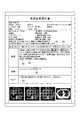
検査目的は、検査依頼者から検査技師や読影医へ検査目的を伝えるための情報である。図2(a)に、検査オーダーに含まれる検査目的の例を示す。
読影目的は、検査依頼者から読影医へ読影すべきポイントを伝えるための情報である。図2(b)に、検査オーダーに含まれる読影目的の例を示す。The inspection purpose is information for communicating the inspection purpose from the inspection requester to the inspection engineer or the image interpreting doctor. FIG. 2A shows an example of the inspection purpose included in the inspection order.
The purpose of image interpretation is information for communicating the points to be interpreted from the examination requester to the image interpretation doctor. FIG. 2B shows an example of the purpose of interpretation included in the inspection order.
RISサーバー20は、放射線機器による検査や治療の予約、検査結果等の放射線科内の情報を管理する。RISサーバー20は、電子カルテサーバー10において発行された検査オーダーを管理し、検査オーダーに基づいて、検査対象のモダリティー60に対して検査依頼を送信し、画像管理サーバー30に読影依頼を送信する。 The
画像管理サーバー30は、モダリティー60において生成された医用画像の画像データを記憶し、患者ごとに管理する。画像管理サーバー30としては、例えば、PACS(Picture Archiving and Communication System)等が挙げられる。 The
図3に、画像管理サーバー30の機能的構成を示す。
図3に示すように、画像管理サーバー30は、制御部31、通信部32、記憶部33、画像保管部34等を備えて構成されており、各部はバス35により接続されている。FIG. 3 shows the functional configuration of the
As shown in FIG. 3, the
制御部31は、CPU(Central Processing Unit)、ROM(Read Only Memory)及びRAM(Random Access Memory)等から構成され、画像管理サーバー30の各部の処理動作を統括的に制御する。具体的には、CPUは、ROMに記憶されている各種処理プログラムを読み出してRAMに展開し、当該プログラムとの協働により各種処理を行う。 The
通信部32は、ネットワークインターフェース等により構成され、通信ネットワークNを介して接続された外部機器との間でデータの送受信を行う。例えば、通信部32は、モダリティー60により患者を撮影して得られた医用画像を受信する。 The
記憶部33は、HDD(Hard Disk Drive)や不揮発性メモリー等により構成され、各種データを記憶している。例えば、記憶部33には、第1対応表331、第2対応表332、ユーザー管理テーブルT1、検査管理テーブルT2が記憶されている。 The storage unit 33 is composed of an HDD (Hard Disk Drive), a non-volatile memory, or the like, and stores various data. For example, the storage unit 33 stores the first correspondence table 331, the second correspondence table 332, the user management table T1, and the inspection management table T2.
図4に、第1対応表331のデータ構成例を示す。第1対応表331は、部位、病名等を含む所定の文字列を複数のカテゴリー(診断対象部位)に分類したものである。例えば、「白内障」、「口腔底」、「咽頭」という文字列は、「頭部」というカテゴリーに分類され、「尿路」、「前立腺」、「膀胱」、「股関節」という文字列は、「下腹部」というカテゴリーに分類されている。なお、カテゴリーとして、臓器を示す情報を用いることとしてもよい。 FIG. 4 shows an example of the data structure of the first correspondence table 331. The first correspondence table 331 classifies a predetermined character string including a site, a disease name, etc. into a plurality of categories (diagnosis target sites). For example, the strings "cataract", "bottom of the mouth", and "pharynx" are classified into the category "head", and the strings "urinary tract", "prostate", "bladder", and "hip joint" are It is classified in the category of "lower abdomen". In addition, as a category, information indicating an organ may be used.
図5に、第2対応表332のデータ構成例を示す。第2対応表332は、部位、病名等を含む所定の文字列を複数のカテゴリー(診療科)に分類したものである。例えば、「白内障」、「眼底」という文字列は、「眼科」というカテゴリーに分類されている。 FIG. 5 shows an example of the data structure of the second correspondence table 332. The second correspondence table 332 classifies a predetermined character string including a site, a disease name, etc. into a plurality of categories (clinical departments). For example, the character strings "cataract" and "fundus" are classified into the category "ophthalmology".
ユーザー管理テーブルT1は、画像管理サーバー30が提供する画像管理システムを利用するユーザーを管理するためのテーブルである。ユーザー管理テーブルT1には、ユーザー(医療従事者)ごとに、ユーザーID、パスワード、氏名、所属(診療科)、メールアドレス等が対応付けられて格納されている。 The user management table T1 is a table for managing users who use the image management system provided by the
検査管理テーブルT2は、画像検査を管理するためのテーブルである。検査管理テーブルT2には、画像検査ごとに、検査依頼者(主治医)、読影医、患者情報、検査情報、医用画像の識別情報(画像ID)、第1の読影レポートの識別情報(レポートID)、第2の読影レポートの識別情報(レポートID)等が対応付けられて格納される。
第1の読影レポートは、読影医が読影医端末50において医用画像を読影して作成したものである。
第2の読影レポートは、第1の読影レポートに対して読影レポート強調加工処理(図8参照)が施された結果、作成されたものである。The inspection management table T2 is a table for managing image inspection. In the examination management table T2, the examination requester (main doctor), the image interpreter, the patient information, the examination information, the identification information of the medical image (image ID), and the identification information of the first image interpretation report (report ID) are displayed for each image examination. , The identification information (report ID) of the second interpretation report and the like are stored in association with each other.
The first image interpretation report was created by the image interpretation doctor by interpreting a medical image on the image
The second interpretation report is created as a result of performing the interpretation report emphasis processing (see FIG. 8) on the first interpretation report.
また、記憶部33には、読影医端末50からの操作指示に応じて作成された第1の読影レポート、画像管理サーバー30において自動的に作成された第2の読影レポートが記憶される。第1の読影レポート、第2の読影レポートは、それぞれレポートIDで特定される。 Further, the storage unit 33 stores a first image interpretation report created in response to an operation instruction from the image
画像保管部34は、患者ごとに、医用画像の画像データを記憶し、保管するための記憶装置である。 The
制御部31は、読影医により第1の読影レポートが作成された場合に、当該作成された第1の読影レポートに対応する画像検査に係る検査オーダーをRISサーバー20から取得する。すなわち、制御部31は、検査オーダー取得手段として機能する。 When the first image interpretation report is created by the image interpretation doctor, the
制御部31は、この検査オーダーに基づいて撮影された医用画像に対して作成された第1の読影レポートを取得する。すなわち、制御部31は、読影レポート取得手段として機能する。 The
制御部31は、検査オーダーと第1の読影レポートとを比較して、検査オーダーと第1の読影レポートとの関連性の度合いを判定する。すなわち、制御部31は、判定手段として機能する。具体的には、制御部31は、検査オーダーと第1の読影レポートとのそれぞれから所定の文字列を抽出し、当該抽出された所定の文字列が属するカテゴリーを第1対応表331、第2対応表332から特定し、当該特定されたカテゴリー同士を比較することにより、検査オーダーと第1の読影レポートとの関連性の度合いを判定する。 The
制御部31は、第1の読影レポート内の検査オーダーとの関連性が所定の基準より低いと判定された部分を強調した第2の読影レポートを作成する。すなわち、制御部31は、作成手段として機能する。具体的には、制御部31は、第2の読影レポートにおいて、第1の読影レポート内の検査オーダーとの関連性が所定の基準より低いと判定された部分の色又は背景色を変更する。「所定の基準」の設け方については、特に限定されない。 The
制御部31は、通信部32を介して、外部機器から医用画像や読影レポートの取得要求があった場合に、当該取得要求に応じて、要求された医用画像や読影レポートを表示するための画面データを当該外部機器に送信する。制御部31は、同一の医用画像に対して、第1の読影レポートと第2の読影レポートが存在する場合には、第2の読影レポートの方を優先して表示させる。 When the
主治医端末40は、主治医により使用されるPC(Personal Computer)等のコンピューター装置である。主治医は、主治医端末40において、電子カルテの作成、検査オーダーの生成、医用画像や読影レポートの閲覧等を行う。 The attending
図6に、主治医端末40の機能的構成を示す。
図6に示すように、主治医端末40は、制御部41、操作部42、表示部43、通信部44、記憶部45等を備えて構成されており、各部はバス46により接続されている。FIG. 6 shows the functional configuration of the attending
As shown in FIG. 6, the attending
制御部41は、CPU、ROM、RAM等から構成され、主治医端末40の各部の処理動作を統括的に制御する。具体的には、CPUは、ROMに記憶されている各種処理プログラムを読み出してRAMに展開し、当該プログラムとの協働により各種処理を行う。 The control unit 41 is composed of a CPU, a ROM, a RAM, and the like, and comprehensively controls the processing operation of each unit of the attending
操作部42は、カーソルキー、文字・数字入力キー及び各種機能キー等を備えたキーボードと、マウス等のポインティングデバイスを備えて構成され、キーボードに対するキー操作やマウス操作により入力された操作信号を制御部41に出力する。
表示部43は、LCD(Liquid Crystal Display)等のモニターを備えて構成されており、制御部41から入力される表示信号の指示に従って、各種画面を表示する。The operation unit 42 includes a keyboard equipped with cursor keys, character / number input keys, various function keys, and a pointing device such as a mouse, and controls operation signals input by key operations on the keyboard and mouse operations. Output to unit 41.
The display unit 43 is configured to include a monitor such as an LCD (Liquid Crystal Display), and displays various screens according to an instruction of a display signal input from the control unit 41.
通信部44は、ネットワークインターフェース等により構成され、通信ネットワークNを介して接続された外部機器との間でデータの送受信を行う。
記憶部45は、HDDや不揮発性の半導体メモリー等により構成され、各種データを記憶している。The communication unit 44 is configured by a network interface or the like, and transmits / receives data to / from an external device connected via the communication network N.
The storage unit 45 is composed of an HDD, a non-volatile semiconductor memory, or the like, and stores various data.
読影医端末50は、読影医により使用されるPC等のコンピューター装置である。読影医は、読影医端末50において、医用画像の読影、読影レポートの作成等を行う。 The image
図6に、読影医端末50の機能的構成を示す。
図6に示すように、読影医端末50は、制御部51、操作部52、表示部53、通信部54、記憶部55等を備えて構成されており、各部はバス56により接続されている。読影医端末50の各部は、主治医端末40の各部と同様であるため、説明を省略する。FIG. 6 shows the functional configuration of the image
As shown in FIG. 6, the image
モダリティー60は、X線撮影装置(DR、CR)、超音波診断装置(US)、CT、MRI等の画像生成装置であり、患者を撮影して得られた医用画像を生成する。モダリティー60は、DICOM規格に則って、付帯情報(患者情報、検査情報等)を医用画像の画像ファイルのヘッダーに書き込むことにより、医用画像に付帯情報を付帯させる。 The
次に、医用画像システム100における動作について説明する。
図7は、医用画像システム100において実行される画像検査診断処理を示すラダーチャートである。Next, the operation in the
FIG. 7 is a ladder chart showing an image inspection diagnosis process executed in the
まず、主治医が主治医端末40から電子カルテサーバー10にアクセスし、患者や検査内容を指定して検査オーダーを発行すると(ステップS1)、電子カルテサーバー10は、RISサーバー20に検査オーダーを送信する。 First, when the attending physician accesses the electronic
RISサーバー20は、電子カルテサーバー10から受信した検査オーダーに基づいて、モダリティー60に検査依頼を送信し(ステップS2)、画像管理サーバー30に読影依頼を送信する(ステップS3)。検査依頼や読影依頼に、検査オーダーに含まれる情報のうち、どの内容を入れるかは任意に設定可能である。RISサーバー20からモダリティー60や画像管理サーバー30に、検査オーダーそのものを送信することとしてもよい。 The
モダリティー60は、RISサーバー20から検査依頼を受信すると、検査技師の操作により、検査依頼に応じて対象患者の対象部位の撮影を行い(ステップS4)、生成された医用画像を保存する(ステップS5)。モダリティー60は、医用画像を画像管理サーバー30に送信する(ステップS6)。 Upon receiving the examination request from the
画像管理サーバー30では、モダリティー60から医用画像を受信すると、制御部31は、当該医用画像の付帯情報に基づいて、RISサーバー20から受信した読影依頼の中から当該医用画像に対応する読影依頼を特定し、当該医用画像と特定された読影依頼(患者情報、検査情報等)とを対応付けて保存する。具体的には、制御部31は、医用画像を画像保管部34に記憶させるとともに、記憶部33の検査管理テーブルT2に、検査依頼者、読影医、患者情報、検査情報、医用画像の識別情報を対応付けて格納する。 When the
読影医が読影医端末50から画像管理サーバー30にアクセスし、読影対象の医用画像を選択すると、選択された医用画像が読影医端末50の表示部53に表示される。
読影医は、表示部53に表示された医用画像を読影し、操作部52から読影結果を入力し、読影レポートを作成する(ステップS7)。画像管理サーバー30において、制御部31は、作成された読影レポート(第1の読影レポート)を、患者情報及び検査情報と対応付けて管理する。具体的には、制御部31は、第1の読影レポートを記憶部33に記憶させるとともに、検査管理テーブルT2の該当レコードに、第1の読影レポートの識別情報を格納する。When the image interpretation doctor accesses the
The image interpreting doctor interprets the medical image displayed on the display unit 53, inputs the image interpretation result from the operation unit 52, and creates an image interpretation report (step S7). In the
次に、制御部31は、読影レポート強調加工処理を実行する(ステップS8)。読影レポート強調加工処理の詳細については、後述する。 Next, the
次に、画像管理サーバー30がRISサーバー20に読影が完了した旨の通知を送信すると、RISサーバー20は、電子カルテサーバー10に読影が完了した旨の通知を送信する(ステップS9)。 Next, when the
主治医が主治医端末40から画像管理サーバー30にアクセスし、読影レポートの表示を指示すると、主治医端末40の表示部43に読影レポートが表示される(ステップS10)。この際、画像検査に対して第2の読影レポート(強調加工済みの読影レポート)がある場合には、第2の読影レポートが表示され、画像検査に対して第2の読影レポートがない場合、すなわち、読影医が作成した読影レポート(第1の読影レポート)内に検査オーダーとの関連性が所定の基準より低い部分がなかった場合には、第1の読影レポートが表示される。 When the attending physician accesses the
主治医は、患者を診察するとともに(ステップS11)、主治医端末40から電子カルテサーバー10にアクセスし、電子カルテを作成する(ステップS12)。具体的には、主治医は、読影レポートを参考にして今後の治療方針を計画し、患者への説明を行う。主治医は、患者に対して処方・処置を行う(ステップS13)。
以上で、画像検査診断処理が終了する。The attending physician examines the patient (step S11), accesses the electronic
This completes the image inspection and diagnosis process.
なお、図7では、主治医が主治医端末40において、読影レポートを確認し(ステップS10)、その後、患者を診察する(ステップS11)場合について図示したが、ステップS11〜ステップS13の後で、主治医が読影レポートを確認する場合もある。 In FIG. 7, the case where the attending physician confirms the interpretation report on the attending physician terminal 40 (step S10) and then examines the patient (step S11) is shown, but after steps S11 to S13, the attending physician You may also check the interpretation report.
次に、図8を参照して、画像管理サーバー30における読影レポート強調加工処理(ステップS8)について説明する。読影レポート強調加工処理は、医用画像に対して第1の読影レポートが作成された後に行われる処理であり、承認医により第1の読影レポートが承認され、内容が確定された後に行われることとしてもよい。この処理は、制御部31のCPUとROMに記憶されているプログラムとの協働によるソフトウェア処理によって実現される。 Next, the image interpretation report emphasis processing process (step S8) in the
まず、制御部31は、通信部32を介してRISサーバー20から、第1の読影レポートに対応する検査オーダーを取得する(ステップS21)。具体的には、制御部31は、記憶部33に記憶されている検査管理テーブルT2において、今回作成された第1の読影レポートの識別情報と対応付けられている患者情報及び検査情報に基づいて、RISサーバー20に検査オーダーの取得要求を送信し、RISサーバー20から検査オーダーを取得する。 First, the
次に、制御部31は、検査オーダー内から所定文字列を抽出し、所定文字列に対応するカテゴリーを特定し、特定されたカテゴリーを保存する(ステップS22)。具体的には、制御部31は、記憶部33の第1対応表331及び第2対応表332の「文字列」フィールドに格納されている文字列を、検査オーダーの「検査目的」、「読影目的」等から抽出する。そして、制御部31は、第1対応表331及び第2対応表332を参照して、抽出された文字列に対応するカテゴリーを特定し、特定されたカテゴリーを記憶部33に記憶させる。 Next, the
次に、制御部31は、カテゴリーごとに、検査オーダーから抽出された文字列の分布を求め、正規化する(ステップS23)。なお、カテゴリーごとに、分布を求め、正規化する処理は、第1対応表331に関する処理と、第2対応表332に関する処理と、に分けて行う。 Next, the
ここで、カテゴリーごとの分布の求め方、正規化について説明する。
図9に、解析対象となる検査オーダーの例を示す。
図10(a)に、第1対応表331に基づく検査オーダーの解析結果を示す。
第1対応表331(図4参照)を参照すると、「腹部」という文字列は、「腹部」というカテゴリーに属している。図9に示す検査オーダーには、「腹部」という文字列71が2回含まれているから、図10(a)に示すように、「腹部」というカテゴリーのカウント数が「2」となる。
分布は、抽出された文字列の全カウント数のうち、該当するカテゴリーのカウント数が占める割合である。図10(a)では、「腹部」以外のカテゴリーのカウント数が「0」であるから、「腹部」というカテゴリーの分布が「100%」となる。
正規化は、分布の中で最も大きい数値(図10(a)では、「腹部」に対応する「100%」)を「100%」とした時の、分布の数値の割合である。「腹部」以外のカテゴリーの正規化値は「0%」である。Here, how to obtain the distribution for each category and normalization will be described.
FIG. 9 shows an example of an inspection order to be analyzed.
FIG. 10A shows the analysis result of the inspection order based on the first correspondence table 331.
With reference to the first correspondence table 331 (see FIG. 4), the character string "abdomen" belongs to the category "abdomen". Since the inspection order shown in FIG. 9 includes the
The distribution is the ratio of the counts of the corresponding category to the total counts of the extracted character strings. In FIG. 10A, since the count number of the categories other than "abdomen" is "0", the distribution of the category "abdomen" is "100%".
The normalization is the ratio of the numerical values of the distribution when the largest numerical value in the distribution (“100%” corresponding to the “abdomen” in FIG. 10 (a)) is set to “100%”. The normalized value for categories other than "abdomen" is "0%".
第2対応表332に関するカテゴリーの分布の求め方及び正規化についても同様である。
図10(b)に、第2対応表332に基づく検査オーダーの解析結果を示す。
第2対応表332(図5参照)を参照すると、「HCC」という文字列は、「消化器科」というカテゴリーに属している。図9に示す検査オーダーには、「HCC」という文字列72が2回含まれているから、図10(b)に示すように、「消化器科」というカテゴリーのカウント数が「2」となる。
また、図10(b)では、「消化器科」以外のカテゴリーのカウント数が「0」であるから、「消化器科」というカテゴリーの分布が「100%」となる。
また、分布の中で最も大きい数値を「100%」として正規化するから、「消化器科」というカテゴリーの正規化値は「100%」、「消化器科」以外のカテゴリーの正規化値は「0%」となる。The same applies to the method of obtaining the distribution of categories and the normalization related to the second correspondence table 332.
FIG. 10B shows the analysis result of the inspection order based on the second correspondence table 332.
With reference to the second correspondence table 332 (see FIG. 5), the character string "HCC" belongs to the category "Gastroenterology". Since the inspection order shown in FIG. 9 contains the
Further, in FIG. 10B, since the count number of the categories other than "Gastroenterology" is "0", the distribution of the category "Gastroenterology" is "100%".
Also, since the largest value in the distribution is normalized as "100%", the normalized value of the category "Gastroenterology" is "100%", and the normalized value of the categories other than "Gastroenterology" is It becomes "0%".
次に、制御部31は、記憶部33から、検査オーダーに基づいて撮影された医用画像に対して作成された第1の読影レポートを取得する(ステップS24)。具体的には、制御部31は、読影レポート強調加工処理が行われる契機となった第1の読影レポートを取得する。 Next, the
次に、制御部31は、第1の読影レポート内から所定文字列を抽出し、所定文字列に対応するカテゴリーを特定し、特定されたカテゴリーを保存する(ステップS25)。具体的には、制御部31は、記憶部33の第1対応表331及び第2対応表332の「文字列」フィールドに格納されている文字列を、第1の読影レポートの「所見」、「診断」、「コメント」等から抽出する。そして、制御部31は、第1対応表331及び第2対応表332を参照して、抽出された文字列に対応するカテゴリーを特定し、特定されたカテゴリーを記憶部33に記憶させる。 Next, the
次に、制御部31は、カテゴリーごとに、第1の読影レポートから抽出された文字列の分布を求め、正規化する(ステップS26)。ここでも、カテゴリーごとに、分布を求め、正規化する処理は、第1対応表331に関する処理と、第2対応表332に関する処理と、に分けて行う。 Next, the
図11に、解析対象となる第1の読影レポートの例を示す。
図12(a)に、第1対応表331に基づく読影レポートの解析結果を示す。
図11に示す第1の読影レポートの「所見」、「診断」、「コメント」には、「横隔膜」という文字列81が1回、「肺」という文字列82が2回含まれているから、図12(a)に示すように、「胸部」というカテゴリーのカウント数が「3」となる。
また、第1の読影レポートの「所見」、「診断」、「コメント」には、「腹水」という文字列83が1回、「肝」という文字列84が1回含まれているから、図12(a)に示すように、「腹部」というカテゴリーのカウント数が「2」となる。
第1の読影レポートから特定されたカテゴリーのカウント数は、「胸部」が3回、「腹部」が2回であるから、「胸部」というカテゴリーの分布が「60%」、「腹部」というカテゴリーの分布が「40%」となる。
「胸部」の分布「60%」を「100%」として正規化すると、「胸部」というカテゴリーの正規化値は「100%」、「腹部」というカテゴリーの正規化値は「67%」、その他のカテゴリーの正規化値は「0%」となる。FIG. 11 shows an example of the first interpretation report to be analyzed.
FIG. 12A shows the analysis result of the interpretation report based on the first correspondence table 331.
This is because the "findings", "diagnosis", and "comment" of the first interpretation report shown in FIG. 11 include the
Further, the "findings", "diagnosis", and "comment" of the first interpretation report include the
The counts of the categories identified from the first interpretation report are 3 times for "chest" and 2 times for "abdomen", so the distribution of the category "chest" is "60%" and the category "abdomen". The distribution of is "40%".
When the distribution "60%" of "chest" is normalized as "100%", the normalized value of the category "chest" is "100%", the normalized value of the category "abdomen" is "67%", and others. The normalized value of the category of is "0%".
図12(b)に、第2対応表332に基づく読影レポートの解析結果を示す。
図11に示す第1の読影レポートの「所見」、「診断」、「コメント」には、「HCC」という文字列85が1回、「肝」という文字列84が1回含まれているから、図12(b)に示すように、「消化器科」というカテゴリーのカウント数が「2」となる。
また、第1の読影レポートの「所見」、「診断」、「コメント」には、「肺」という文字列82が2回含まれているから、図12(b)に示すように、「呼吸器科」というカテゴリーのカウント数が「2」となる。
第1の読影レポートから特定されたカテゴリーのカウント数は、「消化器科」が2回、「呼吸器科」が2回であるから、「消化器科」、「呼吸器科」というカテゴリーの分布は、ともに「50%」となる。
「消化器科」、「呼吸器科」の分布「50%」を「100%」として正規化すると、「消化器科」、「呼吸器科」というカテゴリーの正規化値は「100%」、その他のカテゴリーの正規化値は「0%」となる。FIG. 12B shows the analysis results of the interpretation report based on the second correspondence table 332.
This is because the "findings", "diagnosis", and "comment" of the first interpretation report shown in FIG. 11 include the
Further, since the
The counts of the categories identified from the first interpretation report are "Gastroenterology" twice and "Respiratory Department" twice, so the categories "Gastroenterology" and "Respiratory Department" The distribution is both "50%".
When the distribution "50%" of "Gastroenterology" and "Respiratory Department" is normalized as "100%", the normalized value of the categories "Gastroenterology" and "Respiratory Department" is "100%". The normalized value of other categories is "0%".
次に、制御部31は、検査オーダーから得られたカテゴリーごとの正規化値と、第1の読影レポートから得られたカテゴリーごとの正規化値と、を比較して、関連性の度合いを判定する(ステップS27)。 Next, the
具体的には、図10(a)に示す検査オーダー解析結果のカテゴリー「腹部」の正規化値「100%」と、図12(a)に示す第1の読影レポート解析結果のカテゴリー「腹部」の正規化値「67%」と、を比較すると、「33%」の差があるが、カテゴリーが同じであるため、関連性は高い(強調表示は不要)と判定する。
図10(a)に示す検査オーダー解析結果のカテゴリー「胸部」の正規化値「0%」と、図12(a)に示す第1の読影レポート解析結果のカテゴリー「胸部」の正規化値「100%」と、を比較すると、「100%」の差があるため、関連性は低い(強調表示すべき)と判定する。Specifically, the normalized value "100%" of the inspection order analysis result category "abdomen" shown in FIG. 10A and the first interpretation report analysis result category "abdomen" shown in FIG. 12A. Comparing with the normalized value of "67%", there is a difference of "33%", but since the categories are the same, it is judged that the relevance is high (highlighting is unnecessary).
The normalized value "0%" of the inspection order analysis result category "chest" shown in FIG. 10 (a) and the normalized value "chest" of the first interpretation report analysis result category "chest" shown in FIG. 12 (a). Comparing "100%" with "100%", it is judged that the relevance is low (should be highlighted) because there is a difference of "100%".
図10(b)に示す検査オーダー解析結果のカテゴリー「消化器科」の正規化値「100%」と、図12(b)に示す第1の読影レポート解析結果のカテゴリー「消化器科」の正規化値「100%」と、を比較すると、正規化値が一致しており、且つ、カテゴリーが同じであるため、関連性は高い(強調表示は不要)と判定する。
図10(b)に示す検査オーダー解析結果のカテゴリー「呼吸器科」の正規化値「0%」と、図12(b)に示す第1の読影レポート解析結果のカテゴリー「呼吸器科」の正規化値「100%」と、を比較すると、「100%」の差があるため、関連性は低い(強調表示すべき)と判定する。The normalized value "100%" of the test order analysis result category "Gastroenterology" shown in FIG. 10 (b) and the category "Gastroenterology" of the first interpretation report analysis result shown in FIG. 12 (b). Comparing the normalized value "100%" with the normalized value, it is determined that the relevance is high (no highlighting is required) because the normalized values match and the categories are the same.
The normalized value "0%" of the test order analysis result category "respiratory department" shown in FIG. 10 (b) and the category "respiratory department" of the first interpretation report analysis result shown in FIG. 12 (b). Comparing the normalized value "100%" with that of "100%", it is determined that the relevance is low (should be highlighted) because there is a difference of "100%".
ここでは、第1の読影レポートから特定されたカテゴリーが検査オーダーからも特定されている場合には、関連性は高いと判定し、第1の読影レポートから特定されたカテゴリーが検査オーダーからは特定されていない場合には、関連性は低いと判定した。つまり、第1の読影レポートから特定されたカテゴリーが検査オーダーから特定されているか否かを「所定の基準」とした。 Here, if the category specified from the first interpretation report is also specified from the inspection order, it is judged that the relevance is high, and the category specified from the first interpretation report is specified from the inspection order. If not, the relevance was judged to be low. That is, whether or not the category specified from the first interpretation report is specified from the inspection order is set as the "predetermined standard".
また、関連性の度合いに対して、検査オーダー解析結果のカテゴリーの正規化値と、第1の読影レポートの解析結果のカテゴリーの正規化値と、の差によって、3段階以上のランク(関連性高、関連性中、関連性低等)を設けることとしてもよい。 In addition, with respect to the degree of relevance, there are three or more ranks (relevance) depending on the difference between the normalized value of the category of the inspection order analysis result and the normalized value of the category of the analysis result of the first interpretation report. High, medium relevance, low relevance, etc.) may be provided.
例えば、第1の読影レポートから特定されたカテゴリー(正規化値が0%でないカテゴリー)のそれぞれについて、以下の処理を行う。
まず、第1の読影レポートから特定された或るカテゴリー(以下、対象カテゴリーという。)に対して、検査オーダーから対象カテゴリーと同一のカテゴリーが特定されているか否か(検査オーダーに対象カテゴリーに関連する文字列が含まれているか否か)を判断する。
検査オーダーから対象カテゴリーと同一のカテゴリーが特定されている場合、すなわち、検査オーダーの対象カテゴリーと同一のカテゴリーの正規化値が0%でない場合には、検査オーダーと第1の読影レポートとでカテゴリーが一致しているから、「関連性高」と判定する。For example, the following processing is performed for each of the categories (categories whose normalized value is not 0%) specified from the first interpretation report.
First, whether or not the same category as the target category is specified from the inspection order for a certain category (hereinafter referred to as the target category) specified from the first interpretation report (related to the target category in the inspection order). Whether or not the character string to be used is included) is determined.
If the same category as the target category of the inspection order is specified from the inspection order, that is, if the normalized value of the same category as the target category of the inspection order is not 0%, the category in the inspection order and the first interpretation report Since they match, it is judged to be "highly relevant".
一方、検査オーダーから対象カテゴリーと同一のカテゴリーが特定されていない場合、すなわち、検査オーダーの対象カテゴリーと同一のカテゴリーの正規化値が0%である場合には、第1の読影レポートの対象カテゴリーの正規化値と、検査オーダーの対象カテゴリーと同一のカテゴリーの正規化値(0%)と、の差(絶対値)を求める。正規化値の差が所定の閾値より大きい場合には「関連性低」、正規化値の差が所定の閾値以下である場合には「関連性中」と判定する。 On the other hand, if the same category as the target category is not specified from the inspection order, that is, if the normalized value of the same category as the target category of the inspection order is 0%, the target category of the first interpretation report The difference (absolute value) between the normalized value of and the normalized value (0%) of the same category as the target category of the inspection order is obtained. When the difference between the normalized values is larger than the predetermined threshold value, it is determined as "low relevance", and when the difference between the normalized values is less than or equal to the predetermined threshold value, it is determined as "medium relevance".
次に、制御部31は、関連性の度合いに応じて、強調処理を実行し、第2の読影レポートを作成する(ステップS28)。具体的には、制御部31は、第1の読影レポート内の検査オーダーとの関連性が所定の基準より低いと判定された部分を強調する。制御部31は、第2の読影レポートを記憶部33に記憶させるとともに、検査管理テーブルT2の該当レコードに、第2の読影レポートの識別情報を格納する。
以上で、読影レポート強調加工処理が終了する。Next, the
This completes the interpretation report emphasis processing process.
図13に、第2の読影レポートの例を示す。
第2の読影レポートでは、第1の読影レポート内で、検査オーダーとの関連性が低いと判定された「胸部」に対応付けられた「横隔膜」、「肺」という文字列を含む文91〜93の背景色が変更されている。
また、第2の読影レポートでは、第1の読影レポート内で、検査オーダーとの関連性が低いと判定された「呼吸器科」に対応付けられた「肺」という文字列を含む文92,93の背景色が変更されている。FIG. 13 shows an example of the second interpretation report.
In the second interpretation report, sentences 911 to include the character strings "diaphragm" and "lung" associated with the "chest" determined to be less relevant to the examination order in the first interpretation report. The background color of 93 has been changed.
Further, in the second interpretation report, the
第2の読影レポートでは、検査オーダーには含まれていなかった「胸部」、「呼吸器科」に関する記載を目立たせることで、検査依頼者(主治医)に注意を喚起する。
また、検査オーダーと第1の読影レポートとの関連性の度合いに応じて、第2の読影レポート内の検査オーダーとの関連性が低いと判定された部分を、複数の段階に分けて、異なる色又は背景色で表示することとしてもよい。In the second interpretation report, the examination requester (physician) is alerted by highlighting the descriptions related to "chest" and "respiratory department" that were not included in the examination order.
In addition, depending on the degree of relevance between the inspection order and the first interpretation report, the portion of the second interpretation report that is determined to have low relevance to the inspection order is divided into a plurality of stages and differs. It may be displayed in a color or a background color.
以上説明したように、本実施の形態によれば、第1の読影レポート内の検査オーダーとの関連性が所定の基準より低いと判定された部分を強調した第2の読影レポートを作成するので、読影レポートにおいて読影医により指摘された事項の見落としを防ぐことができる。 As described above, according to the present embodiment, the second interpretation report is created by emphasizing the portion of the first interpretation report that is determined to be less relevant to the inspection order than the predetermined standard. , It is possible to prevent oversight of matters pointed out by the interpretation doctor in the interpretation report.
例えば、第2の読影レポートにおいて、第1の読影レポート内の検査オーダーとの関連性が所定の基準より低いと判定された部分の色又は背景色を変更することで、検査依頼者が予測していなかったと考えられる見落としがちな内容を、強調することができる。
なお、第2の読影レポートにおいて、検査オーダーとの関連性が低いと判定された部分を強調する方法は、上記の例に限定されない。文字の大きさ(フォントサイズ)、フォントタイプ等を変更することで、他の部分と識別可能としてもよい。For example, in the second interpretation report, the inspection requester predicts by changing the color or background color of the portion of the first interpretation report that is determined to be less relevant to the inspection order than a predetermined standard. It is possible to emphasize the content that is often overlooked, which is thought to have not been done.
In the second interpretation report, the method of emphasizing the portion determined to have low relevance to the inspection order is not limited to the above example. By changing the character size (font size), font type, etc., it may be possible to distinguish it from other parts.
また、検査オーダーと第1の読影レポートとのそれぞれから所定の文字列を抽出し、当該抽出された所定の文字列が属するカテゴリーを第1対応表331、第2対応表332から特定し、当該特定されたカテゴリー同士を比較することにより、検査オーダーと第1の読影レポートとの関連性の度合いを判定するので、第1対応表331、第2対応表332を用いて、容易に検査オーダーと第1の読影レポートとの関連性の度合いを判定することができる。 Further, a predetermined character string is extracted from each of the inspection order and the first interpretation report, and the category to which the extracted predetermined character string belongs is specified from the first correspondence table 331 and the second correspondence table 332, and the relevant character string is specified. Since the degree of relevance between the inspection order and the first interpretation report is determined by comparing the specified categories with each other, the inspection order and the inspection order can be easily obtained by using the first correspondence table 331 and the second correspondence table 332. The degree of relevance to the first interpretation report can be determined.
なお、上記実施の形態における記述は、本発明に係る読影レポート作成装置の例であり、これに限定されるものではない。装置を構成する各部の細部構成及び細部動作に関しても本発明の趣旨を逸脱することのない範囲で適宜変更可能である。 The description in the above embodiment is an example of the interpretation report creating device according to the present invention, and is not limited thereto. The detailed configuration and detailed operation of each part constituting the device can be appropriately changed without departing from the spirit of the present invention.
例えば、検査オーダーを生成した検査依頼者の所属に基づいて、検査オーダーと第1の読影レポートとの関連性の度合いを判定することとしてもよい。例えば、循環器科の医師が生成した検査オーダーに対して、第1の読影レポートに泌尿器関係の所見が記載されている場合に、第1の読影レポート内の泌尿器関係の所見を目立たせることで、検査依頼者に注意を喚起することができる。
具体的には、検査オーダーに検査依頼者の所属(診療科)が含まれている場合には、制御部31は、検査オーダーから直接検査依頼者の診療科を取得する。一方、検査オーダーに検査依頼者の所属が含まれていない場合には、制御部31は、検査オーダーから検査依頼者を取得し、記憶部33のユーザー管理テーブルT1から検査依頼者に対応する診療科を取得する。制御部31は、記憶部33の第2対応表332を用いて、第1の読影レポートに含まれる文字列に対応する診療科を特定し、検査依頼者の診療科と比較する。For example, the degree of relevance between the inspection order and the first interpretation report may be determined based on the affiliation of the inspection requester who generated the inspection order. For example, for a test order generated by a cardiologist, if the first interpretation report contains urinary findings, the urinary findings in the first interpretation report may be highlighted. , Can call attention to the inspection requester.
Specifically, when the inspection order includes the affiliation (clinical department) of the inspection requester, the
また、画像管理サーバー30において、第1対応表331、第2対応表332の「文字列」フィールドに、検査オーダーや第1の読影レポートから抽出すべき文字列を加えていくこととしてもよい。例えば、人工知能(AI:Artificial Intelligence)を用いて、検査オーダーや第1の読影レポートから、既に第1対応表331、第2対応表332に記載されている文字列と一緒に使われる頻度が高い文字列を抽出し、第1対応表331、第2対応表332に加えていくことで、より精度の高い関連性の度合いの判定を実現することが可能となる。 Further, in the
以上の説明では、各処理を実行するためのプログラムを格納したコンピューター読み取り可能な媒体としてROMを使用した例を開示したが、この例に限定されない。その他のコンピューター読み取り可能な媒体として、フラッシュメモリー等の不揮発性メモリー、CD−ROM等の可搬型記録媒体を適用することも可能である。また、プログラムのデータを通信回線を介して提供する媒体として、キャリアウェーブ(搬送波)を適用することとしてもよい。 In the above description, an example in which the ROM is used as a computer-readable medium in which a program for executing each process is stored has been disclosed, but the present invention is not limited to this example. As other computer-readable media, non-volatile memory such as flash memory and portable recording media such as CD-ROM can be applied. Further, a carrier wave may be applied as a medium for providing program data via a communication line.
10 電子カルテサーバー
20 RISサーバー
30 画像管理サーバー
31 制御部
32 通信部
33 記憶部
34 画像保管部
40 主治医端末
50 読影医端末
60 モダリティー
100 医用画像システム
331 第1対応表
332 第2対応表
N 通信ネットワーク
T1 ユーザー管理テーブル
T2 検査管理テーブル10 Electronic
Claims (6)
Translated fromJapanese前記検査オーダーに基づいて撮影された医用画像に対して作成された第1の読影レポートを取得する読影レポート取得手段と、
前記検査オーダーと前記第1の読影レポートとを比較して、前記検査オーダーと前記第1の読影レポートとの関連性の度合いを判定する判定手段と、
前記第1の読影レポート内の前記検査オーダーとの関連性が所定の基準より低いと判定された部分を強調した第2の読影レポートを作成する作成手段と、
を備える読影レポート作成装置。Inspection order acquisition means for acquiring inspection orders related to image inspection,
An image interpretation report acquisition means for acquiring a first image interpretation report created for a medical image taken based on the examination order, and an image interpretation report acquisition means.
A determination means for comparing the inspection order and the first interpretation report to determine the degree of relevance between the inspection order and the first interpretation report.
A means for creating a second interpretation report that emphasizes a portion of the first interpretation report that is determined to be less relevant to the inspection order than a predetermined criterion.
Interpretation report creation device equipped with.
前記判定手段は、前記検査オーダーと前記第1の読影レポートとのそれぞれから前記所定の文字列を抽出し、当該抽出された所定の文字列が属するカテゴリーを前記対応表から特定し、当該特定されたカテゴリー同士を比較することにより、前記検査オーダーと前記第1の読影レポートとの関連性の度合いを判定する請求項1又は2に記載の読影レポート作成装置。It is equipped with a storage means for storing a correspondence table in which a predetermined character string is classified into a plurality of categories in advance.
The determination means extracts the predetermined character string from each of the inspection order and the first interpretation report, identifies the category to which the extracted predetermined character string belongs from the correspondence table, and identifies the predetermined character string. The interpretation report creating apparatus according to claim 1 or 2, wherein the degree of relevance between the inspection order and the first interpretation report is determined by comparing the categories.
画像検査に係る検査オーダーを取得する検査オーダー取得手段、
前記検査オーダーに基づいて撮影された医用画像に対して作成された第1の読影レポートを取得する読影レポート取得手段、
前記検査オーダーと前記第1の読影レポートとを比較して、前記検査オーダーと前記第1の読影レポートとの関連性の度合いを判定する判定手段、
前記第1の読影レポート内の前記検査オーダーとの関連性が所定の基準より低いと判定された部分を強調した第2の読影レポートを作成する作成手段、
として機能させるためのプログラム。Computer,
Inspection order acquisition means for acquiring inspection orders related to image inspection,
An image interpretation report acquisition means for acquiring a first image interpretation report created for a medical image taken based on the examination order.
A determination means for determining the degree of relevance between the inspection order and the first interpretation report by comparing the inspection order with the first interpretation report.
A means for creating a second interpretation report, which emphasizes a portion of the first interpretation report that is determined to be less relevant to the inspection order than a predetermined criterion.
A program to function as.
Priority Applications (1)
| Application Number | Priority Date | Filing Date | Title |
|---|---|---|---|
| JP2017243594AJP6897547B2 (en) | 2017-12-20 | 2017-12-20 | Interpretation report creation device and program |
Applications Claiming Priority (1)
| Application Number | Priority Date | Filing Date | Title |
|---|---|---|---|
| JP2017243594AJP6897547B2 (en) | 2017-12-20 | 2017-12-20 | Interpretation report creation device and program |
Publications (2)
| Publication Number | Publication Date |
|---|---|
| JP2019109809A JP2019109809A (en) | 2019-07-04 |
| JP6897547B2true JP6897547B2 (en) | 2021-06-30 |
Family
ID=67179927
Family Applications (1)
| Application Number | Title | Priority Date | Filing Date |
|---|---|---|---|
| JP2017243594AActiveJP6897547B2 (en) | 2017-12-20 | 2017-12-20 | Interpretation report creation device and program |
Country Status (1)
| Country | Link |
|---|---|
| JP (1) | JP6897547B2 (en) |
Families Citing this family (1)
| Publication number | Priority date | Publication date | Assignee | Title |
|---|---|---|---|---|
| JP2021190017A (en)* | 2020-06-04 | 2021-12-13 | キヤノンメディカルシステムズ株式会社 | Report creation support device and report creation support program |
Family Cites Families (5)
| Publication number | Priority date | Publication date | Assignee | Title |
|---|---|---|---|---|
| JP4450695B2 (en)* | 2004-08-19 | 2010-04-14 | 富士通株式会社 | Interpretation report analysis program |
| JP2009066060A (en)* | 2007-09-11 | 2009-04-02 | Konica Minolta Medical & Graphic Inc | Medical image system, finding report generator, finding report generation method, and program |
| JP2009223595A (en)* | 2008-03-17 | 2009-10-01 | Fujifilm Corp | System, program and method for supporting preparation of medical report |
| US8548826B2 (en)* | 2010-12-30 | 2013-10-01 | Cerner Innovation, Inc. | Prepopulating clinical events with image based documentation |
| JP5613212B2 (en)* | 2012-09-28 | 2014-10-22 | 富士フイルム株式会社 | Graph display control device, method, and program |
- 2017
- 2017-12-20JPJP2017243594Apatent/JP6897547B2/enactiveActive
Also Published As
| Publication number | Publication date |
|---|---|
| JP2019109809A (en) | 2019-07-04 |
Similar Documents
| Publication | Publication Date | Title |
|---|---|---|
| JP4906404B2 (en) | Diagnosis support method, diagnosis support apparatus, diagnosis support system, and diagnosis support program | |
| JP7013841B2 (en) | Interpretation report analysis device and program | |
| US20150324523A1 (en) | System and method for indicating the quality of information to support decision making | |
| JP2008059176A (en) | MEDICAL IMAGE MANAGEMENT METHOD, MEDICAL IMAGE MANAGEMENT DEVICE, AND MEDICAL NETWORK SYSTEM | |
| US10282516B2 (en) | Medical imaging reference retrieval | |
| JP2018509689A (en) | Context generation of report content for radiation reports | |
| US20200243177A1 (en) | Medical report generating device and medical report generating method | |
| JP2013182444A (en) | Electronic medical chart device | |
| US20200043583A1 (en) | System and method for workflow-sensitive structured finding object (sfo) recommendation for clinical care continuum | |
| JP7415787B2 (en) | medical imaging system | |
| WO2008038559A1 (en) | Anatomical drawing selecting method, anatomical drawing selecting device and medical network system | |
| JP6897547B2 (en) | Interpretation report creation device and program | |
| JP2008079760A (en) | Method and apparatus for image compression processing and medical network system | |
| US20190244696A1 (en) | Medical record management system with annotated patient images for rapid retrieval | |
| JP2021111283A (en) | Medical information processing device, learning data generation program and learning data generation method | |
| US20230035575A1 (en) | Analysis device, analysis method, and recording medium | |
| US12217867B2 (en) | Medical information processing system and medical information processing apparatus | |
| US20210295983A1 (en) | Medical information management apparatus, medical information management system, storage medium, and medical information management method | |
| JP2023035610A (en) | Report creation auxiliary device and report creation auxiliary system | |
| WO2021172495A1 (en) | Medical information processing device, medical information processing method and program | |
| JP7746948B2 (en) | Image interpretation management device, image interpretation management system, image management device, image interpretation terminal, analysis device, image interpretation management method and program | |
| JP2020187542A (en) | Imaging support device | |
| JP7705768B2 (en) | Medical information processing equipment | |
| US20250166180A1 (en) | Information processing method, non-transitory storage medium, and information processing apparatus | |
| JP7697432B2 (en) | Medical information processing device, medical information processing system, medical information processing method and program |
Legal Events
| Date | Code | Title | Description |
|---|---|---|---|
| A621 | Written request for application examination | Free format text:JAPANESE INTERMEDIATE CODE: A621 Effective date:20200618 | |
| A977 | Report on retrieval | Free format text:JAPANESE INTERMEDIATE CODE: A971007 Effective date:20210422 | |
| TRDD | Decision of grant or rejection written | ||
| A01 | Written decision to grant a patent or to grant a registration (utility model) | Free format text:JAPANESE INTERMEDIATE CODE: A01 Effective date:20210511 | |
| A61 | First payment of annual fees (during grant procedure) | Free format text:JAPANESE INTERMEDIATE CODE: A61 Effective date:20210524 | |
| R150 | Certificate of patent or registration of utility model | Ref document number:6897547 Country of ref document:JP Free format text:JAPANESE INTERMEDIATE CODE: R150 |