JP6811024B2 - A method for observing cells in a cell culture well plate and a culture medium inside the well in the cell culture well plate. - Google Patents
A method for observing cells in a cell culture well plate and a culture medium inside the well in the cell culture well plate.Download PDFInfo
- Publication number
- JP6811024B2 JP6811024B2JP2016070163AJP2016070163AJP6811024B2JP 6811024 B2JP6811024 B2JP 6811024B2JP 2016070163 AJP2016070163 AJP 2016070163AJP 2016070163 AJP2016070163 AJP 2016070163AJP 6811024 B2JP6811024 B2JP 6811024B2
- Authority
- JP
- Japan
- Prior art keywords
- cells
- well
- cell
- cell culture
- plate
- Prior art date
- Legal status (The legal status is an assumption and is not a legal conclusion. Google has not performed a legal analysis and makes no representation as to the accuracy of the status listed.)
- Active
Links
- 238000004113cell cultureMethods0.000titleclaimsdescription55
- 238000000034methodMethods0.000titleclaimsdescription22
- 239000001963growth mediumSubstances0.000titleclaimsdescription18
- 210000004027cellAnatomy0.000claimsdescription99
- 230000002093peripheral effectEffects0.000claimsdescription11
- 230000032258transportEffects0.000claimsdescription11
- 230000004656cell transportEffects0.000claimsdescription6
- 238000003384imaging methodMethods0.000claimsdescription6
- 239000007788liquidSubstances0.000claimsdescription6
- 210000004748cultured cellAnatomy0.000claimsdescription2
- 230000005499meniscusEffects0.000description16
- 230000003993interactionEffects0.000description9
- 239000000463materialSubstances0.000description6
- 238000011282treatmentMethods0.000description6
- -1polyethylenePolymers0.000description5
- 239000004743PolypropyleneSubstances0.000description3
- 239000004793PolystyreneSubstances0.000description3
- 238000011109contaminationMethods0.000description3
- 238000001514detection methodMethods0.000description3
- 238000000465mouldingMethods0.000description3
- 210000001778pluripotent stem cellAnatomy0.000description3
- 229920001155polypropylenePolymers0.000description3
- 229920002223polystyrenePolymers0.000description3
- 238000012546transferMethods0.000description3
- 239000012780transparent materialSubstances0.000description3
- 229920000178Acrylic resinPolymers0.000description2
- 239000004925Acrylic resinSubstances0.000description2
- 206010028980NeoplasmDiseases0.000description2
- 239000004698PolyethyleneSubstances0.000description2
- 201000011510cancerDiseases0.000description2
- 238000012258culturingMethods0.000description2
- 239000003814drugSubstances0.000description2
- 210000002919epithelial cellAnatomy0.000description2
- 239000011521glassSubstances0.000description2
- 230000002209hydrophobic effectEffects0.000description2
- 238000005286illuminationMethods0.000description2
- 229910010272inorganic materialInorganic materials0.000description2
- 239000011147inorganic materialSubstances0.000description2
- 239000011368organic materialSubstances0.000description2
- 229920000573polyethylenePolymers0.000description2
- 239000010453quartzSubstances0.000description2
- 230000002207retinal effectEffects0.000description2
- VYPSYNLAJGMNEJ-UHFFFAOYSA-Nsilicon dioxideInorganic materialsO=[Si]=OVYPSYNLAJGMNEJ-UHFFFAOYSA-N0.000description2
- 241000124008MammaliaSpecies0.000description1
- 238000002835absorbanceMethods0.000description1
- 238000005452bendingMethods0.000description1
- 230000004663cell proliferationEffects0.000description1
- 210000004978chinese hamster ovary cellAnatomy0.000description1
- 238000007796conventional methodMethods0.000description1
- 238000011161developmentMethods0.000description1
- 230000000694effectsEffects0.000description1
- 210000003958hematopoietic stem cellAnatomy0.000description1
- 238000004020luminiscence typeMethods0.000description1
- 210000004962mammalian cellAnatomy0.000description1
- 238000012986modificationMethods0.000description1
- 230000004048modificationEffects0.000description1
- 230000002107myocardial effectEffects0.000description1
- 210000000581natural killer T-cellAnatomy0.000description1
- 210000002569neuronAnatomy0.000description1
- 210000001328optic nerveAnatomy0.000description1
- 230000002062proliferating effectEffects0.000description1
- 230000009467reductionEffects0.000description1
- 230000001172regenerating effectEffects0.000description1
- 238000004381surface treatmentMethods0.000description1
- 210000003501vero cellAnatomy0.000description1
Images


Landscapes
- Investigating Or Analysing Biological Materials (AREA)
- Apparatus Associated With Microorganisms And Enzymes (AREA)
- Measuring Or Testing Involving Enzymes Or Micro-Organisms (AREA)
Description
Translated fromJapanese本発明は、細胞培養用ウェルプレート及び当該細胞培養用ウェルプレートにおけるウェル内部の培養液中の細胞を観測する方法に関する。 The present invention relates to a cell culture well plate and a method for observing cells in a culture medium inside a well in the cell culture well plate.
従来から、細胞培養用ウェルプレートを用いて、ウェル内部の培養液中で、各種の生物の細胞を培養することが行われている。特に近年、哺乳動物の細胞培養、中でもヒトのES細胞やiPS細胞などの多能性の幹細胞、上皮細胞、癌細胞についての培養が盛んに行われており、そして、培養された細胞は、医薬品の開発や再生医療の分野で活用されている。 Conventionally, cells of various organisms have been cultured in a culture medium inside a well using a cell culture well plate. Especially in recent years, cell culture of mammals, especially pluripotent stem cells such as human ES cells and iPS cells, epithelial cells, and cancer cells has been actively performed, and the cultured cells are pharmaceuticals. It is used in the field of development and regenerative medicine.
細胞培養に関し、ウェルを複数具備する細胞培養用ウェルプレートを用いることで、同じ培養液又は異なる種類の培養液を用いた細胞の培養を、同一の保管条件下で行うことが可能となっている。ここで、細胞培養用ウェルプレートを用いる場合には、各ウェル間でのコンタミネーションが生じないように、各ウェルの内部空間を超えない量の培養液を各ウェル内に分注ピペットで分注するのが一般的であり、かつ技術常識である。 Regarding cell culture, by using a cell culture well plate having a plurality of wells, it is possible to culture cells using the same culture solution or different types of culture solutions under the same storage conditions. .. Here, when a cell culture well plate is used, an amount of culture solution that does not exceed the internal space of each well is dispensed into each well with a dispensing pipette so that contamination does not occur between the wells. It is common to do, and it is common technical knowledge.
さて、研究者や作業者においては、細胞培養用ウェルプレートにおけるウェル内部の培養液中の細胞を観測する必要がある。ただし、培養液とウェルの側壁表面との相互作用に因り、ウェル内部の培養液の液面には、凹状又は凸状の屈曲、いわゆるメニスカスが生じていた。メスニカスが生じた状態で培養液中の細胞を、ウェルの開口面側から観測すると、メスニカスの影響で、細胞の正しい大きさや形状などが正確に観測できない場合があった。 Now, it is necessary for researchers and workers to observe the cells in the culture medium inside the well in the cell culture well plate. However, due to the interaction between the culture solution and the side wall surface of the well, concave or convex bending, so-called meniscus, was generated on the liquid surface of the culture solution inside the well. When the cells in the culture medium were observed from the opening surface side of the well with the meniscus generated, the correct size and shape of the cells could not be accurately observed due to the influence of the meniscus.
かかる事態を解決するため、特許文献1には、ウェルの下方から、規則的な歪み検出用パターンを有する透明材料からなるステージを介して、ウェルに対して光を照射すること、及び、歪み検出用パターンの像に出現した歪みに基づき、ウェル内部の細胞を観測した像の歪みを補正する技術が開示されている。 In order to solve such a situation,
特許文献2には、測定装置によって検知された、ウェル内部の細胞に由来するインテグラル信号(光源からの光照射により、ウェル内部の細胞で観測される蛍光、ルミネセンス、吸光度、又はインピーダンスを意味する。)と、大きな開口絞りを介して照射された照明によって観察されたウェル内の細胞の像とを、比較する技術が開示されており、かかる技術により、細胞の定量的かつ定性的な観測を一度に行うことができること、及び、大きな開口絞りを介して照射された照明に因り、従来の小さな開口絞りを用いる手法よりも、メスニカスによる輝度の減少を軽減できることが記載されている。
上述のとおり、メスニカスの影響を排除するために、特許文献1に開示の技術においては、規則的な歪み検出用パターンを有する透明材料からなるステージが必要であり、かつ、ウェル自体も透明材料で構成されることが必要であるし、さらに、像の歪みを計算するための特別な手段も必要である。 As described above, in order to eliminate the influence of meniscus, in the technique disclosed in
また、特許文献2に開示の技術においては、メスニカスによる輝度の減少を軽減できることは開示されているものの、かかる技術の構成からみて、メスニカスから生じる細胞像の歪みが是正されるとはいえない。 Further, although the technique disclosed in
本発明は、かかる事情に鑑みて為されたものであり、細胞の観測時において、メスニカスの影響を排除するための新たな技術を提供することを目的とする。 The present invention has been made in view of such circumstances, and an object of the present invention is to provide a new technique for eliminating the influence of meniscus when observing cells.
本発明者は、観測装置自体の改良を指向するのではなく、培養液とウェルの側壁表面との相互作用を減少又は消失させて、メニスカス自体を低減又は消失させることを想起した。具体的には、プレート上面に開口面を有するウェルを具備する細胞培養用ウェルプレートにおいて、ウェルの内部空間を超える量の培養液を注入してウェル内部の培養液中の細胞を観測すること、換言すれば、培養液の液高が前記プレート上面を超えるまで培養液を加えて、培養液とウェルの側壁表面との相互作用が培養液の液面に影響しない状態にした後、ウェル内部の培養液中の細胞を観測することを想起した。かかる発想に基づき、本発明者は本発明を完成させた。 The present inventor recalled that the meniscus itself was reduced or eliminated by reducing or eliminating the interaction between the culture medium and the side wall surface of the well, rather than aiming to improve the observation device itself. Specifically, in a cell culture well plate having a well having an opening surface on the upper surface of the plate, injecting an amount of the culture solution exceeding the internal space of the well and observing the cells in the culture solution inside the well. In other words, the culture solution is added until the height of the culture solution exceeds the upper surface of the plate so that the interaction between the culture solution and the side wall surface of the well does not affect the liquid level of the culture solution, and then the inside of the well. I recalled observing the cells in the culture medium. Based on this idea, the present inventor has completed the present invention.
本発明の細胞培養用ウェルプレートは、プレート上面に、開口面を有するウェルを具備する細胞培養用ウェルプレートであって、前記プレート上面に、前記ウェルの開口面の周縁を囲いつつ、当該開口面と接しない凸状の堤部を具備することを特徴とする。 The cell culture well plate of the present invention is a cell culture well plate having a well having an opening surface on the upper surface of the plate, and the opening surface is surrounded by the peripheral edge of the opening surface of the well on the upper surface of the plate. It is characterized by having a convex bank portion that does not come into contact with.
また、本発明の観測方法は、本発明の細胞培養用ウェルプレートにおけるウェル内部の培養液中の細胞を観測する方法であって、液高が前記プレート上面を超えるまで培養液を加えた後に、前記細胞を観測することを特徴とする。 The observation method of the present invention is a method of observing cells in the culture solution inside the well in the cell culture well plate of the present invention, after adding the culture solution until the liquid height exceeds the upper surface of the plate. It is characterized by observing the cells.
本発明の細胞培養用ウェルプレートは凸状の堤部を具備する。それにより、細胞の観測時に、メスニカスの影響が排除されるまで培養液を追加することができる。本発明の観測方法によれば、メスニカスの影響を排除した状態で、細胞の観測が可能である。 The cell culture well plate of the present invention includes a convex embankment. Thereby, when observing the cells, the culture medium can be added until the influence of the meniscus is eliminated. According to the observation method of the present invention, cells can be observed in a state where the influence of meniscus is excluded.
以下に、本発明を実施するための最良の形態を説明する。
本発明の細胞培養用ウェルプレートは、プレート上面に、開口面を有するウェルを具備する細胞培養用ウェルプレートであって、前記プレート上面に、前記ウェルの開口面の周縁を囲いつつ、当該開口面と接しない凸状の堤部を具備することを特徴とする。The best mode for carrying out the present invention will be described below.
The cell culture well plate of the present invention is a cell culture well plate having a well having an opening surface on the upper surface of the plate, and the opening surface is surrounded by the peripheral edge of the opening surface of the well on the upper surface of the plate. It is characterized by having a convex bank portion that does not come into contact with.
培養される細胞としては、いかなる生物の細胞であってもよい。ヒトを含む哺乳動物の細胞としては、ES細胞やiPS細胞などの多能性の幹細胞、上皮細胞、癌細胞を例示できる。その他の具体的な細胞として、CHO細胞、HEK293細胞、HL−60細胞、HeLa細胞、MDCK細胞、NIH3T3細胞、PC12細胞、S2細胞、Sf9細胞、Vero細胞を例示できる。また、例えば、iPS細胞を特定の条件下で分化させた網膜細胞、角膜細胞、神経細胞、視神経細胞、NKT細胞、造血幹細胞、心筋細胞などを例示できる。 The cells to be cultured may be cells of any organism. Examples of mammalian cells including humans include pluripotent stem cells such as ES cells and iPS cells, epithelial cells, and cancer cells. Examples of other specific cells include CHO cells, HEK293 cells, HL-60 cells, HeLa cells, MDCK cells, NIH3T3 cells, PC12 cells, S2 cells, Sf9 cells, and Vero cells. Further, for example, retinal cells, corneal cells, nerve cells, optic nerve cells, NKT cells, hematopoietic stem cells, myocardial cells and the like obtained by differentiating iPS cells under specific conditions can be exemplified.
本明細書において、細胞培養とは、細胞を増殖させることに加えて、iPS細胞などの多能性の幹細胞を特定の条件下で特定の細胞に分化させた上で、当該細胞を増殖させることも包含する。 In the present specification, cell culture means, in addition to proliferating cells, pluripotent stem cells such as iPS cells are differentiated into specific cells under specific conditions, and then the cells are proliferated. Also includes.
ウェルプレートとは、プレート上面に、開口面を有するウェルを具備するものであって、マイクロプレートやマイクロタイタープレートなどと称されるものを包含する。ウェルとは窪みを意味し、そして、ウェルは細胞を培養するための場となる。ウェルプレートは、ウェル自体が自立用の脚としての機能を有するものでもよいし、また、自立用の脚としての機能を有する側壁面が、プレート上面の周縁に、プレート上面に対して垂直方向に存在するものでもよい。 The well plate includes a well having an opening surface on the upper surface of the plate, which is called a microplate or a microtiter plate. A well means a depression, and a well is a place for culturing cells. The well plate may be one in which the well itself has a function as a leg for self-supporting, or a side wall surface having a function as a leg for self-supporting is provided on the peripheral edge of the upper surface of the plate in a direction perpendicular to the upper surface of the plate. It may exist.
ウェルプレートの材料は公知のものでよく、ガラス、石英などの無機材料や、アクリル樹脂、ポリエチレン、ポリプロピレン、ポリスチレンなどの有機材料を例示できる。ウェルプレートの材料は、細胞及び培養液の特性に応じて、適宜適切に選択すれば良い。ウェルプレートの色としては、透明、半透明、白色、又は黒色を例示できる。ウェルプレートの色は、細胞及び培養液の色などに応じて、適宜適切に選択すれば良い。 The material of the well plate may be a known material, and examples thereof include inorganic materials such as glass and quartz, and organic materials such as acrylic resin, polyethylene, polypropylene, and polystyrene. The material of the well plate may be appropriately and appropriately selected according to the characteristics of the cells and the culture medium. Examples of the color of the well plate include transparent, translucent, white, and black. The color of the well plate may be appropriately and appropriately selected according to the color of the cells and the culture solution.
本発明の細胞培養用ウェルプレートにおいて、ウェルの数は、特に限定されない。ウェルの具体的な数として、4、6、8、12、16、24、96、384、1536、又は9600を例示できる。 In the cell culture well plate of the present invention, the number of wells is not particularly limited. Specific numbers of wells may be 4, 6, 8, 12, 16, 24, 96, 384, 1536, or 9600.
ウェルの開口面の形状は、特に限定されない。開口面の形状として、円形、矩形、又は正方形を例示できる。また、ウェルの底の形状も特に限定されない。底の形状として、U型の丸底形状、平板状の平底形状、又は、平底であるがウェルの側壁との接合部周縁が曲面を有する形状を例示できる。ウェル自体の形状も特に限定されず、円柱、直方体、立方体、円錐、四角錐、円錐台、又は四角錐台を例示できる。 The shape of the opening surface of the well is not particularly limited. Examples of the shape of the opening surface include a circle, a rectangle, and a square. Further, the shape of the bottom of the well is not particularly limited. Examples of the shape of the bottom include a U-shaped round bottom shape, a flat plate-shaped flat bottom shape, and a flat bottom shape in which the peripheral edge of the joint with the side wall of the well has a curved surface. The shape of the well itself is not particularly limited, and examples thereof include a cylinder, a rectangular parallelepiped, a cube, a cone, a quadrangular pyramid, a truncated cone, and a truncated quadrangular pyramid.
ウェルの表面は、各種の表面処理が為されていてもよい。具体的な処理として、疎水性処理、親水性処理、疎水性処理とイオン性処理を組み合わせた処理を挙げることができる。 The surface of the well may be subjected to various surface treatments. Specific treatments include hydrophobic treatments, hydrophilic treatments, and treatments that combine hydrophobic treatments and ionic treatments.
凸状の堤部は、プレート上面に、ウェルの開口面の周縁を囲いつつ、当該開口面と接しない状態で存在する。堤部の存在に因り、細胞の観測時に、メスニカスの影響が排除されるまで培養液を追加することができる。 The convex bank portion exists on the upper surface of the plate in a state of surrounding the peripheral edge of the opening surface of the well and not in contact with the opening surface. Due to the presence of the embankment, the culture medium can be added when observing the cells until the effects of the meniscus are eliminated.
堤部はウェルの開口面と接しないため、細胞の観測時に培養液と堤部との相互作用が生じたとしても、その相互作用がウェルの開口面にまで実質的に影響を与えることは想定されない。仮に、上記相互作用がウェルの開口面にまで影響したとしても、少なくとも培養液とウェルの側壁表面との相互作用よりも軽微なものに留まることは明白である。 Since the embankment does not contact the opening surface of the well, it is assumed that even if the interaction between the culture medium and the embankment occurs during cell observation, the interaction substantially affects the opening surface of the well. Not done. Even if the above interaction affects the opening surface of the well, it is clear that the interaction is at least less than the interaction between the culture medium and the side wall surface of the well.
本発明の細胞培養用ウェルプレートには、1つの堤部を具備していてもよいし、複数の堤部を具備していてもよい。例えば、ウェルの数が96である96ウェルの場合、96個のウェル全ての開口面の周縁を、1つの堤部が囲うものであってもよいし、又は、48個のウェルの開口面の周縁を第一の堤部が囲い、24個のウェルの開口面の周縁を第二の堤部が囲い、残り24個のウェルの開口面の周縁を第三の堤部が囲うものであってもよい。また、特に、ウェルの数が比較的少ない場合、例えば、ウェルの数が6である6ウェルの場合、ウェルそれぞれの開口面の周縁を、それぞれ1つの堤部が囲うものであってもよい。この場合は、堤部の数が6になる。 The cell culture well plate of the present invention may be provided with one bank or a plurality of banks. For example, in the case of 96 wells having 96 wells, one bank may surround the periphery of the opening surfaces of all 96 wells, or the opening surfaces of 48 wells. The peripheral edge is surrounded by the first bank, the peripheral edge of the opening surface of the 24 wells is surrounded by the second bank, and the peripheral edge of the opening surface of the remaining 24 wells is surrounded by the third bank. May be good. Further, in particular, when the number of wells is relatively small, for example, when the number of wells is 6 and the number of wells is 6, the peripheral edge of the opening surface of each well may be surrounded by one bank. In this case, the number of embankments is six.
複数の堤部を有する本発明の細胞培養用ウェルプレートを用いることにより、堤部ごとに異なる培養系での細胞培養を行うことができるため、他の培養系とのコンタミネーションの心配をすることなく、細胞培養や細胞の観測を行うことができる。 By using the well plate for cell culture of the present invention having a plurality of ridges, cell culture can be performed in a different culture system for each ridge, so that there is a concern about contamination with other culture systems. It is possible to perform cell culture and cell observation.
堤部の材料としては、ガラス、石英などの無機材料や、アクリル樹脂、ポリエチレン、ポリプロピレン、ポリスチレンなどの有機材料を例示できる。堤部の材料は、ウェルプレートの材料と同じものでもよいし、異なるものでもよい。 Examples of the material of the bank portion include inorganic materials such as glass and quartz, and organic materials such as acrylic resin, polyethylene, polypropylene, and polystyrene. The material of the embankment may be the same as or different from the material of the well plate.
堤部は、ウェルプレートとともに一体成型法で形成されてもよいし、ウェルプレートとは別個に製造した上で、堤部とウェルプレートとを接着させて形成させてもよい。また、堤部は着脱可能であってもよい。着脱可能な堤部であれば、本発明の細胞培養用ウェルプレートにおける堤部の数を、細胞の培養系の数に応じて適切に決定することができる。着脱可能な堤部を具備する本発明の細胞培養用ウェルプレートの一態様としては、例えば、堤部に雄部を設け、プレート上部に雌部を設けて、雄部と雌部とを嵌合させたものを挙げることができる。 The embankment portion may be formed together with the well plate by an integral molding method, or may be manufactured separately from the well plate and then formed by adhering the embankment portion and the well plate. Further, the embankment portion may be removable. If it is a detachable embankment, the number of embankments in the cell culture well plate of the present invention can be appropriately determined according to the number of cell culture systems. As one aspect of the cell culture well plate of the present invention provided with a detachable embankment, for example, a male portion is provided on the embankment portion, a female portion is provided on the upper portion of the plate, and the male portion and the female portion are fitted. I can mention what I made.
堤部の高さには特に制限が無いが、好適な高さとして、3〜15mm、4〜10mm、又は5〜8mmの範囲を例示できる。 The height of the embankment is not particularly limited, but a suitable height may be in the range of 3 to 15 mm, 4 to 10 mm, or 5 to 8 mm.
本発明の観測方法は、本発明の細胞培養用ウェルプレートにおけるウェル内部の培養液中の細胞を観測する方法であって、液高が前記プレート上面を超えるまで培養液を加えた後に、前記細胞を観測することを特徴とする。追加する培養液は、ウェル内部の培養液と同じものを採用するのが好ましい。 The observation method of the present invention is a method of observing cells in the culture medium inside the well in the cell culture well plate of the present invention, and the cells are added after the culture solution is added until the liquid height exceeds the upper surface of the plate. It is characterized by observing. It is preferable to use the same culture solution as the culture solution inside the well as the culture solution to be added.
本発明の観測方法に因り、メスニカスの影響を排除又は軽減できるため、細胞の正しい大きさや形状などが正確に観測できる。 Since the influence of meniscus can be eliminated or reduced by the observation method of the present invention, the correct size and shape of cells can be accurately observed.
本発明の観測方法においては、カメラなどの撮影手段を用いて観測することが好ましい。
また、本発明の観測方法を応用すれば、ウェル毎に、細胞の有無の判断、細胞が所望の大きさ及び形状に成長しているか否かの判断、又は、細胞が所望の数に増殖しているか否かの判断を行うことができる。さらに、本発明の観測方法を応用すれば、所望の細胞のみ選択して、ウェル外に搬送すること(以下、搬送手段ということがある。)もできる。加えて、上記判断を行う制御部及び上記搬送手段を備えた細胞搬送システム(以下、本発明の細胞搬送システムという。)を把握することもできる。In the observation method of the present invention, it is preferable to observe using a photographing means such as a camera.
Further, by applying the observation method of the present invention, the presence or absence of cells can be determined, whether or not the cells have grown to a desired size and shape, or the cells can proliferate to a desired number for each well. It is possible to judge whether or not it is. Further, by applying the observation method of the present invention, it is possible to select only desired cells and transport them out of the well (hereinafter, may be referred to as transport means). In addition, it is also possible to grasp a cell transfer system (hereinafter, referred to as the cell transfer system of the present invention) provided with the control unit that makes the above determination and the transfer means.
本発明の細胞搬送システムの一態様を以下に例示する。
本発明の細胞培養用ウェルプレートを配置する配置部と、
本発明の細胞培養用ウェルプレートに培養液を追加する培養液導入部と、
ウェルの開口面側から細胞を観測する撮影手段と、
細胞の観測結果から、所望の細胞が存在するウェルを特定する判断手段を有する制御部と、
所望の細胞を吸着させ又は吸引して搬送する搬送手段と、を備えた細胞搬送システム。One aspect of the cell transport system of the present invention is illustrated below.
An arrangement portion for arranging the cell culture well plate of the present invention and
A culture solution introduction unit for adding a culture solution to the cell culture well plate of the present invention,
Imaging means for observing cells from the open surface side of the well,
A control unit having a judgment means for identifying a well in which a desired cell is present from cell observation results,
A cell transport system comprising a transport means for adsorbing or sucking and transporting desired cells.
本発明の細胞搬送システムには、ウェル内部を照らす照明部が備えられているのが好ましい。制御部においては、細胞の観測結果から、ウェル毎に搬送手段の適切な位置を指示すること、例えば所望の細胞の中心に搬送手段を合致させることを指示することが好ましい。搬送手段は、ウェルプレートに対して、X軸、Y軸及びZ軸方向の3次元に移動可能である。搬送手段としては、シリンジ状やノズル状のものを例示できる。 The cell transport system of the present invention preferably includes a lighting unit that illuminates the inside of the well. In the control unit, it is preferable to instruct the appropriate position of the transport means for each well from the observation result of the cells, for example, to instruct the transport means to be aligned with the center of the desired cell. The transport means can move in three dimensions in the X-axis, Y-axis, and Z-axis directions with respect to the well plate. Examples of the transporting means include a syringe shape and a nozzle shape.
本発明の細胞搬送システムは、培養液導入部を介して本発明の細胞培養用ウェルプレートに培養液を追加することにより、メスニカスの影響を排除又は軽減した条件下で撮影手段を稼働できるため、細胞の正しい大きさや形状などを正確に観測することができる。そして、本発明の細胞搬送システムは、撮影手段による観測結果に基づき、制御部及び搬送手段が稼働するため、所望の条件を満足する細胞のみを選択的に且つ正確にウェル外部へ搬送することができる。 In the cell transport system of the present invention, the imaging means can be operated under conditions in which the influence of Mesnicus is eliminated or reduced by adding the culture solution to the cell culture well plate of the present invention via the culture solution introduction unit. It is possible to accurately observe the correct size and shape of cells. Then, in the cell transport system of the present invention, since the control unit and the transport means operate based on the observation result by the imaging means, it is possible to selectively and accurately transport only cells satisfying the desired conditions to the outside of the well. it can.
以下、実施例を通じて本発明をさらに詳細に説明する。これら実施例は本発明をより具体的に説明するためのものであって、本発明の範囲はこれら実施例に限定されない。本発明の要旨を逸脱しない範囲において、当業者が行い得る変更、改良等を施した種々の形態にて実施することができる。 Hereinafter, the present invention will be described in more detail through examples. These examples are for more specific explanation of the present invention, and the scope of the present invention is not limited to these examples. It can be carried out in various forms with modifications, improvements, etc. that can be made by those skilled in the art, without departing from the gist of the present invention.
(実施例1)
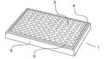
実施例1の細胞培養用ウェルプレート1の斜視図を図1に示す。
実施例1の細胞培養用ウェルプレート1には、長方形のプレート上面2に、円形の開口面を有するウェル3が96個具備されており、かつ、自立用の脚としての機能を有する側壁面5が、プレート上面2の長方形の4辺と辺を共有しつつ、プレート上面2に対して垂直方向に存在する。そして、実施例1の細胞培養用ウェルプレート1には、96個すべてのウェル3の開口面の周縁を囲いつつ、当該開口面と接しない状態で、凸状の堤部4が一つ存在する。(Example 1)
A perspective view of the cell
The cell
なお、ウェル3の形状は円柱状であり、そして、ウェル3の底の形状はU型の丸底形状である。また、実施例1の細胞培養用ウェルプレート1はポリスチレン製であり、プレート上面2、ウェル3、堤部4及び側壁面5のすべては、成形型を用いて、同時に一体成型されたものである。 The shape of the
図2に、実施例1の細胞培養用ウェルプレート1を用いた、細胞培養状態の断面図を示す。各ウェル31〜38には、培養液6中で細胞7が培養されている。培養液6と各ウェル31〜38の側壁表面とが相互作用する結果、各ウェル内の培養液6の液面には、メニスカスが生じている。そのため、培養液6中の細胞7を、ウェルの開口面側から観測する場合には、メスニカスの影響で、細胞7の正しい大きさや形状を正確に把握できない。 FIG. 2 shows a cross-sectional view of a cell culture state using the cell
図3に、実施例1の細胞培養用ウェルプレート1を用いた、細胞観測状態の断面図を示す。図3は、図2の状態の各ウェル31〜38に対して、培養液6の液高がプレート上面2を超えるまで培養液6を追加した状態である。培養液6で生じるメスニカスは堤部4との界面近傍に留まり、各ウェル31〜38の開口面には及ばない。そのため、培養液6中の細胞7をウェルの開口面側から観測する場合において、メスニカスの影響が排除されて、細胞7の正しい大きさや形状が正確に把握できる。例えば、ウェル32、ウェル33、ウェル35、ウェル37及びウェル38で培養された細胞7は、所望の大きさ及び形状となっていることが確認できる。他方、ウェル31の細胞7は所望の大きさに及ばない2つの細胞からなること、ウェル34の細胞7は所望の大きさに及ばないこと、及び、ウェル36には細胞7が存在しないことが、明確に確認できる。 FIG. 3 shows a cross-sectional view of a cell observation state using the cell
(実施例2)
実施例2の細胞培養用ウェルプレート1の斜視図を図4に示す。
実施例2の細胞培養用ウェルプレート1には、長方形のプレート上面2に、円形の開口面を有するウェル3が6個具備されており、かつ、自立用の脚としての機能を有する側壁面5が、プレート上面2の長方形の4辺と辺を共有しつつ、プレート上面2に対して垂直方向に存在する。そして、実施例2の細胞培養用ウェルプレート1には、2個のウェル3の開口面の周縁を囲いつつ、当該開口面と接しない状態で、凸状の第一堤部41、凸状の第二堤部42及び凸状の第三堤部43が存在する。第一堤部41及び第二堤部42は互いに堤部の一部を共有しており、第二堤部42及び第三堤部43も互いに堤部の一部を共有している。(Example 2)
A perspective view of the cell
The cell
なお、ウェル3の形状は円柱状であり、そして、ウェル3の底の形状は平板状の平底形状である。かかる平底形状は、網膜細胞などの膜状の細胞の培養に適している。 The shape of the
また、実施例2の細胞培養用ウェルプレート1はポリプロピレン製であり、プレート上面2、ウェル3、堤部41〜43及び側壁面5のすべては、成形型を用いて、同時に一体成型されたものである。 Further, the cell
実施例2の細胞培養用ウェルプレート1は、3つの堤部41〜43を有するため、各堤部ごとに異なる培養系での細胞培養及び細胞観測を、コンタミネーションの心配をすることなく行うことができる。例えば、培養する細胞は同一の種類とし、培養液を別個のものとすることで、培養液の違いによる細胞の増殖の変化を、同一のウェルプレートを用いて観測できる。 Since the cell
1は細胞培養用ウェルプレート、2はプレート上面、3、31、32、33、34、35、36、37、38はウェル、4、41、42、43は堤部、5は側壁面、6は培養液、7は細胞を示す。 1 is a well plate for cell culture, 2 is the upper surface of the plate, 3, 31, 32, 33, 34, 35, 36, 37, 38 are wells, 4, 41, 42, 43 are embankments, 5 is the side wall surface, 6 Indicates a culture solution, and 7 indicates cells.
Claims (3)
Translated fromJapanese前記方法は、
前記細胞培養用ウェルプレートを配置する配置部と、
前記細胞培養用ウェルプレートに前記培養液を追加する培養液導入部と、
前記ウェルの開口面側から細胞を観測する撮影手段と、
所望の細胞を吸着させ又は吸引して搬送する搬送手段と、
前記細胞の観測結果から、所望の細胞が存在するウェルを特定する判断手段を有し、前記細胞の観測結果から、所望の細胞の中心に前記搬送手段を合致させる制御部と、
を備えた細胞搬送システムにより行われるものであり、
液高が前記プレート上面を超えるまで前記培養液導入部により前記培養液を加えた後に、前記細胞を前記撮影手段により観測することを特徴とする観測方法。The upper surface of the plate is provided with aplurality of wells having aU-shaped round bottom shape and an opening surface, and the upper surface of the plate surrounds the peripheral edge of the opening surface of the well and does not come into contact with the opening surface. A method for observing cells in a culture medium inside a well in a cell culture well plate provided with a convex bank.
The method is
An arrangement portion for arranging the cell culture well plate and
A culture solution introduction section for adding the culture solution to the cell culture well plate, and
An imaging means for observing cells from the opening surface side of the well,
A transport means for adsorbing or sucking and transporting desired cells,
From observation of the cells,have a determining means for specifying the wells desired cells arepresent, from the observation result of the cell, and a control unitto match said transport means in the center of the desiredcell,
It is carried out by a cell transport system equipped with
An observation method characterized in that the cells are observed by the imaging means after the culture solution is added by the culture solution introduction unit until the liquid height exceeds the upper surface of the plate.
前記細胞培養用ウェルプレートに培養液を追加する培養液導入部と、
前記ウェルの開口面側から細胞を観測する撮影手段と、
所望の細胞を吸着させ又は吸引して搬送する搬送手段と、
前記細胞の観測結果から、所望の細胞が存在するウェルを特定する判断手段を有し、前記細胞の観測結果から、所望の細胞の中心に搬送手段を合致させる制御部と、
を備えた細胞搬送システム。On the upper surface of the plate, aplurality of wells having aU-shaped round bottom shape and having an opening surface, and a convex bank portion that surrounds the peripheral edge of the opening surface ofthe well and does not contact the opening surface. An arrangement portion for arranging a cell culture well plate which is provided and in which a culture solution and cultured cells are present inside the well.
A culture solution introduction section for adding the culture solution to the cell culture well plate, and
An imaging means for observing cells from the opening surface side of the well,
A transport means for adsorbing or sucking and transporting desired cells,
From observation of the cells,have a determining means for specifying the wells desired cells arepresent, from the observation result of the cell, and a control unitto match the conveying means to the center of the desiredcell,
Cells conveying system comprisinga.
Priority Applications (1)
| Application Number | Priority Date | Filing Date | Title |
|---|---|---|---|
| JP2016070163AJP6811024B2 (en) | 2016-03-31 | 2016-03-31 | A method for observing cells in a cell culture well plate and a culture medium inside the well in the cell culture well plate. |
Applications Claiming Priority (1)
| Application Number | Priority Date | Filing Date | Title |
|---|---|---|---|
| JP2016070163AJP6811024B2 (en) | 2016-03-31 | 2016-03-31 | A method for observing cells in a cell culture well plate and a culture medium inside the well in the cell culture well plate. |
Publications (2)
| Publication Number | Publication Date |
|---|---|
| JP2017176073A JP2017176073A (en) | 2017-10-05 |
| JP6811024B2true JP6811024B2 (en) | 2021-01-13 |
Family
ID=60002871
Family Applications (1)
| Application Number | Title | Priority Date | Filing Date |
|---|---|---|---|
| JP2016070163AActiveJP6811024B2 (en) | 2016-03-31 | 2016-03-31 | A method for observing cells in a cell culture well plate and a culture medium inside the well in the cell culture well plate. |
Country Status (1)
| Country | Link |
|---|---|
| JP (1) | JP6811024B2 (en) |
Family Cites Families (3)
| Publication number | Priority date | Publication date | Assignee | Title |
|---|---|---|---|---|
| JPS6251977A (en)* | 1985-09-02 | 1987-03-06 | Terumo Corp | Tissue culture plate |
| US7186548B2 (en)* | 2003-11-10 | 2007-03-06 | Advanced Pharmaceutical Sciences, Inc. | Cell culture tool and method |
| JP2008152044A (en)* | 2006-12-18 | 2008-07-03 | Nsk Ltd | Manipulator system, manipulator control program and inhalation method |
- 2016
- 2016-03-31JPJP2016070163Apatent/JP6811024B2/enactiveActive
Also Published As
| Publication number | Publication date |
|---|---|
| JP2017176073A (en) | 2017-10-05 |
Similar Documents
| Publication | Publication Date | Title |
|---|---|---|
| JP7558319B2 (en) | Microwell design and fabrication for generating cell culture aggregates | |
| US11708563B2 (en) | Platforms and systems for automated cell culture | |
| US11931737B2 (en) | Platforms and systems for automated cell culture | |
| CN105158887B (en) | Multi-mode micro imaging method based on programmable LED array illumination | |
| JP6502338B2 (en) | Apparatus for light sheet microscopy | |
| US10487310B2 (en) | Vessel for culturing human ES cells | |
| CN105247035A (en) | System for analyzing cells and monitoring cell culture and method for analyzing cells and monitoring cell culture using the system | |
| JP5556444B2 (en) | Microscope, culture observation equipment | |
| CN106834118A (en) | Cell culture container, cell image pickup method and cell culture system | |
| JP2017501429A (en) | Equipment for optical sheet microscopy | |
| WO2016052078A1 (en) | Plastic container | |
| US20170191013A1 (en) | Container for culturing organisms, method for monitoring the culturing of organisms inside said container, and monitoring system | |
| JP6811024B2 (en) | A method for observing cells in a cell culture well plate and a culture medium inside the well in the cell culture well plate. | |
| US20190055511A1 (en) | Microfluidic Device for Controlling the Geometry of Living Bodies | |
| JP2016013079A (en) | Cell culture container | |
| JP2011221297A (en) | Microscope | |
| EP3263219B1 (en) | Well plate and method of using the same | |
| JP7403549B2 (en) | Cell detection device and cell detection method | |
| JP2004085833A (en) | One-cell long-term observation device | |
| WO2017163378A1 (en) | Culture vessel | |
| CN204342806U (en) | Laser scanning co-focusing microscope special various kinds of cell Dual culture ware | |
| JP6535494B2 (en) | Imaging device, imaging method and culture vessel | |
| JP2012024020A (en) | Culture vessel for storing slide glass | |
| JP7037636B2 (en) | Observation device | |
| KR20250114184A (en) | Container for 3d cell culture and imaging |
Legal Events
| Date | Code | Title | Description |
|---|---|---|---|
| A621 | Written request for application examination | Free format text:JAPANESE INTERMEDIATE CODE: A621 Effective date:20190221 | |
| A977 | Report on retrieval | Free format text:JAPANESE INTERMEDIATE CODE: A971007 Effective date:20191113 | |
| A131 | Notification of reasons for refusal | Free format text:JAPANESE INTERMEDIATE CODE: A131 Effective date:20191126 | |
| A521 | Request for written amendment filed | Free format text:JAPANESE INTERMEDIATE CODE: A523 Effective date:20200114 | |
| A131 | Notification of reasons for refusal | Free format text:JAPANESE INTERMEDIATE CODE: A131 Effective date:20200407 | |
| A521 | Request for written amendment filed | Free format text:JAPANESE INTERMEDIATE CODE: A523 Effective date:20200602 | |
| TRDD | Decision of grant or rejection written | ||
| A01 | Written decision to grant a patent or to grant a registration (utility model) | Free format text:JAPANESE INTERMEDIATE CODE: A01 Effective date:20201201 | |
| A61 | First payment of annual fees (during grant procedure) | Free format text:JAPANESE INTERMEDIATE CODE: A61 Effective date:20201214 | |
| R150 | Certificate of patent or registration of utility model | Ref document number:6811024 Country of ref document:JP Free format text:JAPANESE INTERMEDIATE CODE: R150 | |
| R250 | Receipt of annual fees | Free format text:JAPANESE INTERMEDIATE CODE: R250 | |
| R250 | Receipt of annual fees | Free format text:JAPANESE INTERMEDIATE CODE: R250 |