JP6803909B2 - Detection of immobilized contrast media with dynamic threshold - Google Patents
Detection of immobilized contrast media with dynamic thresholdDownload PDFInfo
- Publication number
- JP6803909B2 JP6803909B2JP2018520401AJP2018520401AJP6803909B2JP 6803909 B2JP6803909 B2JP 6803909B2JP 2018520401 AJP2018520401 AJP 2018520401AJP 2018520401 AJP2018520401 AJP 2018520401AJP 6803909 B2JP6803909 B2JP 6803909B2
- Authority
- JP
- Japan
- Prior art keywords
- value
- candidate
- image
- values
- threshold
- Prior art date
- Legal status (The legal status is an assumption and is not a legal conclusion. Google has not performed a legal analysis and makes no representation as to the accuracy of the status listed.)
- Active
Links
Images


Classifications
- A—HUMAN NECESSITIES
- A61—MEDICAL OR VETERINARY SCIENCE; HYGIENE
- A61B—DIAGNOSIS; SURGERY; IDENTIFICATION
- A61B8/00—Diagnosis using ultrasonic, sonic or infrasonic waves
- A61B8/52—Devices using data or image processing specially adapted for diagnosis using ultrasonic, sonic or infrasonic waves
- A61B8/5215—Devices using data or image processing specially adapted for diagnosis using ultrasonic, sonic or infrasonic waves involving processing of medical diagnostic data
- A61B8/5223—Devices using data or image processing specially adapted for diagnosis using ultrasonic, sonic or infrasonic waves involving processing of medical diagnostic data for extracting a diagnostic or physiological parameter from medical diagnostic data
- A—HUMAN NECESSITIES
- A61—MEDICAL OR VETERINARY SCIENCE; HYGIENE
- A61B—DIAGNOSIS; SURGERY; IDENTIFICATION
- A61B8/00—Diagnosis using ultrasonic, sonic or infrasonic waves
- A61B8/06—Measuring blood flow
- A—HUMAN NECESSITIES
- A61—MEDICAL OR VETERINARY SCIENCE; HYGIENE
- A61B—DIAGNOSIS; SURGERY; IDENTIFICATION
- A61B8/00—Diagnosis using ultrasonic, sonic or infrasonic waves
- A61B8/48—Diagnostic techniques
- A61B8/481—Diagnostic techniques involving the use of contrast agents, e.g. microbubbles introduced into the bloodstream
- A—HUMAN NECESSITIES
- A61—MEDICAL OR VETERINARY SCIENCE; HYGIENE
- A61B—DIAGNOSIS; SURGERY; IDENTIFICATION
- A61B8/00—Diagnosis using ultrasonic, sonic or infrasonic waves
- A61B8/52—Devices using data or image processing specially adapted for diagnosis using ultrasonic, sonic or infrasonic waves
- A61B8/5215—Devices using data or image processing specially adapted for diagnosis using ultrasonic, sonic or infrasonic waves involving processing of medical diagnostic data
- A61B8/5238—Devices using data or image processing specially adapted for diagnosis using ultrasonic, sonic or infrasonic waves involving processing of medical diagnostic data for combining image data of patient, e.g. merging several images from different acquisition modes into one image
- A61B8/5246—Devices using data or image processing specially adapted for diagnosis using ultrasonic, sonic or infrasonic waves involving processing of medical diagnostic data for combining image data of patient, e.g. merging several images from different acquisition modes into one image combining images from the same or different imaging techniques, e.g. color Doppler and B-mode
- G—PHYSICS
- G06—COMPUTING OR CALCULATING; COUNTING
- G06T—IMAGE DATA PROCESSING OR GENERATION, IN GENERAL
- G06T7/00—Image analysis
- G06T7/0002—Inspection of images, e.g. flaw detection
- G06T7/0012—Biomedical image inspection
- G—PHYSICS
- G06—COMPUTING OR CALCULATING; COUNTING
- G06T—IMAGE DATA PROCESSING OR GENERATION, IN GENERAL
- G06T7/00—Image analysis
- G06T7/10—Segmentation; Edge detection
- G06T7/136—Segmentation; Edge detection involving thresholding
- G—PHYSICS
- G16—INFORMATION AND COMMUNICATION TECHNOLOGY [ICT] SPECIALLY ADAPTED FOR SPECIFIC APPLICATION FIELDS
- G16H—HEALTHCARE INFORMATICS, i.e. INFORMATION AND COMMUNICATION TECHNOLOGY [ICT] SPECIALLY ADAPTED FOR THE HANDLING OR PROCESSING OF MEDICAL OR HEALTHCARE DATA
- G16H50/00—ICT specially adapted for medical diagnosis, medical simulation or medical data mining; ICT specially adapted for detecting, monitoring or modelling epidemics or pandemics
- G16H50/30—ICT specially adapted for medical diagnosis, medical simulation or medical data mining; ICT specially adapted for detecting, monitoring or modelling epidemics or pandemics for calculating health indices; for individual health risk assessment
- G—PHYSICS
- G06—COMPUTING OR CALCULATING; COUNTING
- G06T—IMAGE DATA PROCESSING OR GENERATION, IN GENERAL
- G06T2207/00—Indexing scheme for image analysis or image enhancement
- G06T2207/10—Image acquisition modality
- G06T2207/10016—Video; Image sequence
- G—PHYSICS
- G06—COMPUTING OR CALCULATING; COUNTING
- G06T—IMAGE DATA PROCESSING OR GENERATION, IN GENERAL
- G06T2207/00—Indexing scheme for image analysis or image enhancement
- G06T2207/10—Image acquisition modality
- G06T2207/10132—Ultrasound image
Landscapes
- Health & Medical Sciences (AREA)
- Engineering & Computer Science (AREA)
- Life Sciences & Earth Sciences (AREA)
- Physics & Mathematics (AREA)
- Medical Informatics (AREA)
- General Health & Medical Sciences (AREA)
- Radiology & Medical Imaging (AREA)
- Nuclear Medicine, Radiotherapy & Molecular Imaging (AREA)
- Public Health (AREA)
- Biomedical Technology (AREA)
- Pathology (AREA)
- Biophysics (AREA)
- Molecular Biology (AREA)
- Computer Vision & Pattern Recognition (AREA)
- Veterinary Medicine (AREA)
- Animal Behavior & Ethology (AREA)
- Surgery (AREA)
- Heart & Thoracic Surgery (AREA)
- Theoretical Computer Science (AREA)
- Hematology (AREA)
- General Physics & Mathematics (AREA)
- Quality & Reliability (AREA)
- Physiology (AREA)
- Data Mining & Analysis (AREA)
- Databases & Information Systems (AREA)
- Epidemiology (AREA)
- Primary Health Care (AREA)
- Ultra Sonic Daignosis Equipment (AREA)
- Image Processing (AREA)
Description
Translated fromJapanese本開示は、医用イメージング分野に関する。より具体的には、本開示は、固定化造影剤の検出に関する。 The present disclosure relates to the field of medical imaging. More specifically, the present disclosure relates to the detection of immobilized contrast media.
本開示の背景は、以下にその文脈に関連する技術の議論と共に導入される。しかし、この議論が文書、行為、人工物などを参照する場合であっても、議論された技術が先行技術の一部であるか、本開示に関連する分野における一般的な一般知識であることを示唆するものでなく、または表すものでない。 The background of this disclosure is introduced below with a discussion of the technology related to that context. However, even if this discussion refers to documents, acts, man-made objects, etc., the technology discussed should be part of the prior art or general general knowledge in the field relevant to this disclosure. Does not suggest or represent.
医用イメージングは、実質的に非侵襲的な方法で患者の身体部分を分析することを可能にする十分に確立された技術(医療用途のための装置の分野において)である。特定の医用イメージング技術は、患者に超音波造影剤(UCA)を投与すること(例えば、リン脂質で安定化されたガス充填マイクロバブルの懸濁液を含む)に基づく。造影剤は、超音波スキャナによって取得された患者の身体部分の画像に対応する強調を提供するように、効率的な超音波反射器として機能する。 Medical imaging is a well-established technique (in the field of devices for medical applications) that allows the analysis of a patient's body part in a substantially non-invasive manner. Certain medical imaging techniques are based on administering an ultrasound contrast agent (UCA) to a patient (eg, including a suspension of phospholipid-stabilized gas-filled microbubbles). The contrast agent acts as an efficient ultrasonic reflector so as to provide the corresponding enhancement of the image of the patient's body part obtained by the ultrasonic scanner.
造影剤は、特定の(生物学的)標的(例えば、病変で発現される)に到達し、次いでその上に固定されたままであるように適合することもできる。特に、超音波分子イメージング(USMI)技術において、この結果は、対応する標的に結合するように処方された(分子的に)標的造影剤を使用することによって達成される(例えば、炎症性または腫瘍組織と相互作用することができるその製剤中にリガンドを組み込むことによって)。固定化標的造影剤の検出は、その標的(例えば、発見することが困難であった対応する病変)を同定することを可能にする。さらに、この固定化(標的化)造影剤の定量化により、標的の状態を(例えば、病変の治療的フォローアップにおいて)決定することが可能になる。 Contrast agents can also be adapted to reach a particular (biological) target (eg, expressed in a lesion) and then remain anchored on it. Especially in ultrasonic molecular imaging (USMI) techniques, this result is achieved by using (molecularly) targeted contrast agents formulated to bind to the corresponding target (eg, inflammatory or tumor). By incorporating the ligand into its formulation that can interact with the tissue). Detection of an immobilized target contrast agent makes it possible to identify the target (eg, the corresponding lesion that was difficult to detect). In addition, the quantification of this immobilized (targeted) contrast agent makes it possible to determine the condition of the target (eg, in therapeutic follow-up of the lesion).
しかしながら、画像の増強における固定化造影剤の寄与の同定または標的化増強(TE)は、標的造影剤のほんの一部が実際に標的に到達し、その上に固定化されたままであるという事実により、妨げられる。標的造影剤の残りの部分は、例えば、肺および/または患者の肝臓によって濾過されるまで、かなりの時間(最大10〜30分)循環し続ける。したがって、この循環(標的化)造影剤の大部分が消失するまで、循環造影剤から固定化造影剤を識別することは不可能である。 However, identification or targeting enhancement (TE) of the contribution of the immobilized contrast agent in image enhancement is due to the fact that only a small portion of the targeted contrast agent actually reaches the target and remains immobilized on it. , Hindered. The rest of the target contrast agent continues to circulate for a considerable amount of time (up to 10-30 minutes) until it is filtered, for example, by the lungs and / or the patient's liver. Therefore, it is not possible to distinguish an immobilized contrast agent from a circulating contrast agent until most of this circulating (targeted) contrast agent has disappeared.
標的造影剤投与後の初期段階で固定化造影剤を検出するための一般的なアプローチは、差分標的増強(dTE)技術である。この場合、高い機械的指数(MI)を有する破壊的パルスが身体部分に印加され、(固定され循環する)標的造影剤の大部分を破壊する。破壊的パルスを印加する前に取得された画像(および次に固定化造影剤と循環造影剤の両方の寄与を含む)は、主に固定化造影剤のみの寄与を保存するように、破壊的パルスの適用から短い遅延(典型的には30〜90秒)後に取得された画像を減算することによってフィルタリングされる(その後主に循環造影剤のみの寄与を含む)。しかしながら、このようにして、破壊的パルスの適用によって破壊されたので、固定化造影剤を検出するために身体部分を再度撮像することは不可能である。 A common approach for detecting immobilized contrast agents in the early stages after administration of targeted contrast agents is differential target enhancement (dTE) technology. In this case, a destructive pulse with a high mechanical index (MI) is applied to the body part, destroying most of the (fixed and circulating) target contrast agent. Images acquired prior to applying the destructive pulse (and then containing the contributions of both the immobilized and circulating contrast) are destructive, primarily to preserve the contribution of the immobilized contrast alone. It is filtered by subtracting the images acquired after a short delay (typically 30-90 seconds) from the application of the pulse (then mainly including the contribution of the circulating contrast agent only). However, it is not possible to re-image the body part to detect the immobilized contrast agent because it was thus destroyed by the application of the destructive pulse.
あるいは、特許文献1(その全開示は参照により本明細書に組み込まれる)は、画像を処理して、時間の経過伴う高い変動を示す画像の画素値を実質的に抑制する(または少なくとも減衰させる)ことによって循環造影剤の寄与を低減することを提案する時間の経過と共に変動する(同時に、時間の経過に伴う変動を示す画素値を保持する)。この目的のために、画像は、変更された最小強度射影(Min_IP)アルゴリズムを適用することによってフィルタリングされ、各画素値は、画素値自体および1つ以上の先行する画像における対応する画素値の間で最小値によって置き換えられる。 Alternatively, Patent Document 1 (the entire disclosure of which is incorporated herein by reference) processes an image to substantially suppress (or at least attenuate) pixel values of an image showing high variation over time. ) Proposes to reduce the contribution of the circulating contrast agent by fluctuating with the passage of time (at the same time, it retains pixel values indicating variation over time). To this end, images are filtered by applying a modified minimum intensity projection (Min_IP) algorithm, where each pixel value is between the pixel value itself and the corresponding pixel value in one or more preceding images. Is replaced by the minimum value.
しかしながら、そのような不完全な抑制のためにフィルタリングされた画像には、循環造影剤の残余の寄与が依然として存在している可能性がある。循環する造影剤の残余の寄与は、固定化造影剤の顕著性を低下させ、次いで検出、特にその正確な定量化を妨げることがある。 However, it is possible that residual contributions of circulating contrast media may still be present in images filtered for such incomplete suppression. The residual contribution of the circulating contrast agent may reduce the salency of the immobilized contrast agent and then prevent detection, especially its accurate quantification.
循環造影剤の残余の寄与は、一般に、比較的低い強度を有する。したがって、循環造影剤の残余の寄与を抑制する(または少なくとも低減する)ための共通のアプローチは、振幅閾値よりも低い画素値をゼロにリセットすることによってフィルタリングされた画像を閾値処理することである。例えば、特許文献1において、振幅閾値は、画素値の許容最大値の0〜5%に設定される。 The residual contribution of the circulating contrast agent generally has a relatively low intensity. Therefore, a common approach to suppressing (or at least reducing) the residual contribution of circulatory contrast agents is to threshold the filtered image by resetting pixel values below the amplitude threshold to zero. .. For example, in
しかしながら、この操作は望ましくない影響を及ぼし得る。特に、振幅閾値が低すぎる場合、フィルタリングされた画像の閾値処理は、循環造影剤の残余の寄与を低減するのに効果的でない可能性がある(例えば、超音波スキャナの高いゲインおよび/または高いダイナミックスの場合または標的造影剤が高濃度の場合)。逆に、振幅閾値が高すぎると、フィルタリングされた画像の閾値処理は、固定化造影剤の寄与を減少させる可能性がある(例えば、超音波スキャナの低い利得および/または低いダイナミクスの場合、または標的造影剤が低濃度の場合)。 However, this operation can have undesired effects. Especially if the amplitude threshold is too low, threshold processing of the filtered image may not be effective in reducing the residual contribution of the circulating contrast agent (eg, high gain and / or high of the ultrasound scanner). In the case of dynamics or in high concentration of target contrast agent). Conversely, if the amplitude threshold is too high, threshold processing of the filtered image may reduce the contribution of the immobilized contrast agent (eg, for low gain and / or low dynamics of the ultrasound scanner, or When the target contrast agent is at a low concentration).
上記のすべてが、標的造影剤の使用に基づく医用イメージング技術の臨床応用を妨げる。 All of the above hinder the clinical application of medical imaging techniques based on the use of targeted contrast media.
本開示の簡略化された概要は、その基本的な理解を提供するためにここに提示される。しかしながら、この要約の唯一の目的は、以下のより詳細な説明の前置きとして、開示のいくつかの概念を簡略化した形で紹介することであり、それはその重要な要素の識別としても、その範囲の描写としても解釈されるものではない。 A simplified overview of the disclosure is presented herein to provide a basic understanding thereof. However, the sole purpose of this summary is to introduce in a simplified form some of the concepts of disclosure as a prelude to the more detailed explanation below, which also covers its scope as an identification of its key elements. It is not interpreted as a depiction of.
一般的に言えば、本開示は、振幅閾値を動的に設定するという考えに基づいている。 Generally speaking, the present disclosure is based on the idea of dynamically setting the amplitude threshold.
特に、1つの態様は、患者の身体部分を分析する方法であって、振幅閾値に従って少なくとも1つのフィルタリングされた画像(循環造影剤の寄与が実質的に低減されている)から閾値画像を生成するステップを備え、候補画像は、複数の候補閾値に従ってフィルタリングされた画像から生成され、比較値は、異なる領域におけるそれらの値の比較に従って、候補画像から計算され、振幅閾値は、比較値のピークに従って設定される。 In particular, one embodiment is a method of analyzing a patient's body part, which produces a threshold image from at least one filtered image (with substantially reduced contribution of circulatory contrast agent) according to an amplitude threshold. With steps, candidate images are generated from images filtered according to multiple candidate thresholds, comparison values are calculated from the candidate images according to the comparison of those values in different regions, and amplitude thresholds follow the peak of the comparison values. Set.
さらなる態様は、この方法を実施するためのソフトウェアプログラムを提供する。 A further aspect provides a software program for carrying out this method.
さらなる態様は、この方法を実施するためのソフトウェアプログラム製品を提供する。 A further aspect provides a software program product for carrying out this method.
さらなる態様は、対応するシステムを提供する。 A further aspect provides a corresponding system.
さらなる態様は、対応する診断方法を提供する。 A further aspect provides a corresponding diagnostic method.
より具体的には、本開示の1つまたは複数の態様は、独立請求項に記載されており、その有利な特徴は、従属請求項に記載されており、すべての請求項の言葉を引用により本明細書に援用する(他のすべての態様において準用する任意の特定の態様を参照して提供される任意の有利な特徴を有する)。 More specifically, one or more aspects of the present disclosure are set forth in the independent claims, the advantageous features of which are set forth in the dependent claims, by quoting the terms of all claims. Incorporated herein (with any advantageous features provided with reference to any particular aspect as applied mutatis mutandis in all other aspects).
本開示の解決策、並びにさらなる特徴および利点は、添付の図面と併せて読まれるべきである、以下の詳細な説明を純粋に非限定的な表示によって与えて、最もよく理解されるであろう。(簡略化のために、対応する要素は同一または類似の参照符号で示され、その説明は繰り返されず、各エンティティの名前は一般に、その型および値、内容および表現などの属性を示すために使用される)。 The solutions of this disclosure, as well as additional features and advantages, should be read in conjunction with the accompanying drawings and will be best understood, given the following detailed description in purely non-limiting representation. .. (For simplicity, the corresponding elements are indicated by the same or similar reference codes, the description is not repeated, and the name of each entity is commonly used to indicate its type and value, content, representation, and other attributes. Will be).
図1を参照すると、本開示の一実施形態による解決策を実施するために使用され得る超音波スキャナ100の図解表現が示されている。 With reference to FIG. 1, a graphical representation of the
超音波スキャナ100は、中央ユニット105と、これに接続されたアレイタイプのハンドヘルド送受信イメージングプローブまたはトランスデューサ110とを備える。トランスデューサ110はパルスエコーモードで動作し、送信/受信マルチプレクサは、超音波パルス(例えば、中心周波数が4〜20Hz)を送信するための送信部と、超音波パルスの反射から生じる(無線周波数、RF)エコー信号を受信する受信部とを交互に可能にする。 The
中央ユニット105は、超音波スキャナ100の動作を制御する電子回路が搭載されたマザーボード115(例えば、マイクロプロセッサ、ワーキングメモリ、およびハードディスクなどの大容量メモリのためのドライブ)を収容する。さらに、マザーボード115には、1つまたは複数のドーターボード(参照符号120で全体を示す)が差し込まれている。ドーターボード120は、トランスデューサ110を駆動しエコー信号を処理するための更なる電子回路を提供する。また、中央ユニット105には、リムーバブル記憶ユニット130(例えば、光ディスク)を読み書きするためのドライブ125が装備されている。モニタ135は、進行中の分析処理に関する画像を表示する。超音波スキャナ100の動作は、従来の方法で中央ユニット105に接続されたキーボード140によって制御される。好ましくは、キーボード140には、モニタ135上のポインタ(図示せず)の位置を操作するために使用されるトラックボール145が設けられる。 The
超音波スキャナ100は、患者155の身体部分150を分析するために使用される。この目的のために、(超音波)造影剤が患者155に投与される。造影剤は、超音波反射器として作用する粒子を含む。例えば、造影剤は、液体キャリア中のガス充填された気泡の懸濁液である。典型的には、ガス充填された気泡は、患者155の脈管系内での保持を可能にするが、患者155の毛管を通過することを可能にするために、約0.1〜5μmの直径を有する。ガス充填された気泡は、一般に、リン脂質、乳化剤、油、増粘剤、糖類、タンパク質またはポリマーを含む様々な系に気体またはその前駆体を同伴または封入することによって安定化される。安定化されたガス充填気泡は、一般にマイクロベシクルと呼ばれる。特に、水性媒体中に分散され、界面活性剤(すなわち、両親媒性物質)を含む非常に薄いエンベロープによって気体/液体界面で結合されたマイクロベシクルは、マイクロバブルとしても知られている。あるいは、脂質または(天然または合成の)ポリマーによって形成された固体物質のエンベロープに囲まれたマイクロベシクルは、マイクロバルーンまたはマイクロカプセルとしても知られている。別の種類の造影剤は、微粒子の細孔内に閉じ込められた、または表面に吸着された気体の泡を運ぶ、ポリマーまたは他の固体の多孔質微粒子の懸濁液を含む。マイクロベシクル、特にマイクロバブル及びマイクロバルーンの適切な水性懸濁液の例及びその調製は、特許文献2、特許文献3、特許文献4、特許文献5、および特許文献6(その全開示内容は本明細書中に参考として援用される)に記載されている。マイクロベシクルを含む市販の造影剤の一例は、Bracco International BV(登録商標)のSono Vueである。 The
造影剤は、患者155内で実質的に自由に循環するが(例えば、0.1〜0.5秒を超えて同じ位置に留まらない)、同時に、特定の(生物学的)標的に達し、その後、実質的に固定されたままである(例えば、少なくとも10〜30秒間同じ位置に留まる)。例えば、この結果は、標的造影剤(対応する標的に結合するために製剤化された)によって、特に所望の組織または受容体に選択的に結合することができる標的特異的リガンドを組み込んだ標的特異的造影剤(例えば、生化学的親和性および/または静電的相互作用を介して)によって達成される。これらの標的特異的リガンド(マイクロバブルの膜に挿入することができる)の例は、モノクローナル抗体、ペプチド、または多糖類である。用語「組織」は、(その意味において)個々の細胞並びに膜または器官などの細胞の凝集体を含む。この用語は、正常な(健康な)細胞または異常な(病的な)細胞または細胞の凝集体のいずれかを指す。組織の例は、心筋組織(心筋細胞および心筋細胞を含む)、膜組織(内皮および上皮など)および結合組織である。病理組織の例は、梗塞した心臓組織、血餅、アテローム硬化性プラーク、炎症組織および腫瘍組織である。レセプターは、特定の物質に選択的に結合することができる組織上(例えば、細胞内または表面上)に位置する任意の分子構造を含む。例示的な受容体は、糖タンパク質GPIIbIIIaまたはフィブリン(例えば、血餅または血栓に位置する)、P−セレクチン(例えば、炎症組織の活性化内皮に位置する)またはVEGFR2(例えば、腫瘍組織に位置する)である。適切な標的特異的造影剤および標的特異的リガンドの例は、非特許文献1、および特許文献7(その全開示は参照により本明細書に組み込まれる)に記載されている。 The contrast agent circulates substantially freely within the patient 155 (eg, does not stay in the same position for more than 0.1-0.5 seconds), but at the same time reaches a specific (biological) target. It then remains substantially fixed (eg, stays in the same position for at least 10-30 seconds). For example, this result incorporates a target-specific ligand that can selectively bind to a tissue or receptor specifically desired by a target contrast agent (formulated to bind to the corresponding target). Achieved by contrast agents (eg, through biochemical affinity and / or electrostatic interactions). Examples of these target-specific ligands (which can be inserted into the membrane of microbubbles) are monoclonal antibodies, peptides, or polysaccharides. The term "tissue" includes (in that sense) individual cells as well as aggregates of cells such as membranes or organs. The term refers to either normal (healthy) cells or abnormal (pathological) cells or cell aggregates. Examples of tissues are myocardial tissue (including myocardial cells and myocardial cells), membrane tissue (endothelium and epithelium, etc.) and connective tissue. Examples of histopathological tissues are infarcted heart tissue, blood clots, atherosclerotic plaques, inflamed tissue and tumor tissue. Receptors include any molecular structure located on a tissue (eg, intracellularly or on the surface) capable of selectively binding to a particular substance. Exemplary receptors are glycoprotein GPIIbIIIa or fibrin (eg, located in a clot or thrombus), P-selectin (eg, located in the activated endothelium of inflamed tissue) or VEGFR2 (eg, located in tumor tissue). ). Examples of suitable target-specific contrast agents and target-specific ligands are described in
分析プロセスの間、(標的化)造影剤は、患者155に、例えば、静脈内ボーラスとして(すなわち、超音波スキャナ100の操作者によって注射器を用いて短時間にわたって提供される単一回用量として、2〜20秒のオーダー)投与される。結果として、造影剤は患者155の血管系内を循環して、身体部分150を灌流させる。同時に、トランスデューサ110は、身体部分150の領域において患者155の皮膚と接触して配置され、低い音響エネルギーを有する一連の超音波パルスが身体部分150に印加される(例えば、造影剤の無視できる程度の破壊、例えば、総量の20%未満、好ましくは10%未満、を含むように、低い機械的指数MI=0.01〜0.1を有する)。超音波パルスに時間的に応答して記録されたエコー信号は、分析プロセスの間、例えば、以下、原像と称する画像(またはフレーム)のシーケンスの形態での身体部分150の表示を提供する。 During the analytical process, the (targeted) contrast agent is provided to
図2A〜図2Fを参照すると、本開示の一実施形態による解決策の一般原則が示されている。 With reference to FIGS. 2A-2F, the general principles of the solution according to one embodiment of the present disclosure are shown.
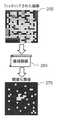
図2Aから出発して、フィルタリングされた画像205(またはそれ以上)が提供される。フィルタリングされた画像205は、身体部分の対応する位置についての複数のフィルタリングされた値を含む。各フィルタリングされた値は、実質的に低減された循環造影剤の寄与を伴って、対応する位置に固定化造影剤の表示を含む(例えば、フィルタリングされた画像205が特許文献1に記載された原画像から生成された場合)。さらに、フィルタリングされた画像205を閾値処理するために使用される振幅閾値の複数の候補値、以下、候補閾値210と称する、が提供される(例えば、候補閾値210を、フィルタリングされた画像205を生成するために使用された原画像から計算されたベース閾値の対応する割合に設定することによって)。 Starting from FIG. 2A, filtered image 205 (or higher) is provided. The filtered
この時点で、候補閾値210に対応する複数の候補画像215が生成される。各候補画像215は、フィルタリングされた値に対応する複数の候補値を含む。候補画像215は、フィルタリングされた値と候補閾値210との比較に従って、各候補値を対応するフィルタリングされた値またはリセット値、例えばゼロに等しい下限値(図では黒色)に設定することによって生成される(例えば、候補閾値210より低い全てのフィルタリングされた値をゼロにリセットすることによって)。結果として、異なる候補画像215において、ゼロにリセットされる候補値の数は、対応する候補閾値210に従って変化する。具体的には、候補閾値210が低い場合(ゼロに等しくない場合)にはリセット候補数が少なく、一方、候補閾値210が高い場合には高くなる(例えば、図に示すように左から右に移動する)。 At this point, a plurality of
図2Bに進み、候補画像215に対応する複数の比較値220を計算する。各比較値220は、固定化領域225および循環領域230における候補値間の(さらに)比較によって計算される。固定化領域225は、(例えば、手動で選択された)固定化造影剤の有意な量を含む位置のグループに対応し、循環領域230は、固定化領域225を除いた(例えば、分析プロセスのための関心領域(ROI)内の)位置に対応する。 Proceeding to FIG. 2B, a plurality of
特定の実施例では、各候補画像215について、固定化領域225内の候補値は、例えばその平均値(以下、固定化平均値と称する)に統合される。このようにして、固定化平均値は、対応する候補閾値との閾値処理後に候補画像215に残る固定化造影剤の寄与の測定値を提供する。同様に、循環領域230内の候補値も、例えばそれらの平均値(以下、循環平均値と称する)に統合される。このようにして、循環平均値は、主として、循環造影剤の残留寄与のために、対応する候補閾値との閾値処理後に候補画像215に残る他の寄与の測定値を提供する。図2Cを参照すると、縦軸に固定化平均値240iと循環平均値240cを横軸の候補閾値に対してプロットした共通図235が示されている(それらはすべて任意の単位である)。一般に、固定化平均値240iは循環平均値240cよりも高い。さらに、循環平均値240cは、それらが最小値に達するまで、循環平均値240cが一定のままである後に、(候補閾値が増加すると)かなり急速に減少する。代わりに、固定平均値240iは(循環平均値240cが最小値に達する前よりも)ゆっくりと減少する。これは、固定化平均値240iによって測定された固定化造影剤の寄与が、循環平均値240cによって測定された循環造影剤の残留寄与よりも高いという事実による(なぜなら、循環造影剤の寄与は、フィルタリングされた画像において実質的に低減される)。 In a particular embodiment, for each
固定化平均値240iおよび循環平均値240cは、例えば、ゼロの候補閾値に対応する候補画像内のそれらの値を減算することによって、固定オフセットおよび循環オフセットにそれぞれ正規化することができる(すなわち、閾値なしのフィルタリングされた画像と等しい)。各候補画像について、次に、比較値は、正規化固定化平均値と正規化循環平均値との間の差として計算される。このようにして、比較値は、対応する候補閾値の容量の測定値を提供して、循環造影剤の残留寄与からの固定化造影剤の寄与を識別する。図2Dを参照すると、横軸上の候補閾値(それらはすべて任意の単位である)に対して、縦軸に正規化固定化平均値250iおよび正規化循環平均値250cをプロットする他の共通図245が示されている。上記のように、正規化循環平均値250cは、それらが最小値に達するまで(正規化循環平均値250cが一定のままである後に)、かなり急速に(候補閾値が増加すると)減少する。代わりに、正規化固定化平均値250iは(正規化循環平均値250cが最小値に達する前よりも)ゆっくりと減少する。この場合、正規化固定化平均値250iおよび正規化循環平均値250cの両方が(正規化のために)ゼロから開始し、正規化固定化平均値250iは、正規化固定化平均値250iが対応する最小値に達した後に正規化循環平均値250cを下回る(正規化固定化平均値250iが代わりに減少し続けるため)。 The fixed mean 240i and the circular mean 240c can be normalized to a fixed offset and a circular offset, respectively (ie, by subtracting their values in the candidate image corresponding to the candidate threshold of zero, respectively). Equal to a filtered image without a threshold). For each candidate image, the comparison value is then calculated as the difference between the normalized fixed mean and the normalized circular mean. In this way, the comparative values provide a measure of the volume of the corresponding candidate threshold to identify the contribution of the immobilized contrast agent from the residual contribution of the circulating contrast agent. Referring to FIG. 2D, another common diagram plotting the normalized fixed mean 250i and the normalized cyclic mean 250c on the vertical axis against the candidate thresholds on the horizontal axis (they are all arbitrary units). 245 is shown. As mentioned above, the normalized cyclic
この時点で、比較値のピークが決定される。図2Eを参照すると、縦軸に比較値260を横軸上の候補閾値に対してプロットした図255が示されている(両方とも任意の単位)。上記を考慮して、比較値260は、候補閾値が増加するにつれて(ゼロから始まって)増加し、次に減少する(負になる)。特に、比較値260は、候補閾値の1つ、以下、ピーク閾値と称する、におけるピーク値(すなわち、それらの絶対最大値)に達する。次いで、(フィルタリングされた画像を閾値処理するために使用される)振幅閾値265が、循環造影剤の残余寄与の大部分を除去するのに十分高い値で選択されるが、固定化造影剤の寄与に実質的に影響をおよぼすにはそれほど高くはない。例えば、振幅閾値265は、ピーク閾値よりも高く、比較値260の半ピーク値(すなわち、最大/2)を提供する値に設定される。 At this point, the peak of the comparison value is determined. With reference to FIG. 2E, FIG. 255 is shown in which the
図2Fに進むと、閾値化画像270が生成される。閾値化画像270は、フィルタリングされた画像205のフィルタリングされた値に対応する複数の閾値化値を含む。閾値化画像270は、フィルタリングされた値と振幅閾値265との同一の比較に従って各閾値化値を対応するフィルタリングされた値に設定するか、またはゼロ(図では黒色)に設定することによって生成される(すなわち、振幅閾値265よりも低いフィルタリングされた値をすべてゼロにリセットすることによって)。 Proceeding to FIG. 2F, a
結果として、振幅閾値265は、特定のフィルタリングされた画像205に動的に自己適応し、その後、対応するイメージング条件(例えば、超音波スキャナの利得およびダイナミクス、標的造影剤の濃度)に自己適応する。これは、振幅閾値265が低すぎるリスクを回避し(または少なくとも実質的に低減する)、次いで、循環造影剤の残留寄与を低減するのに効果がないか、または高すぎるリスクを回避し、次いで、固定化造影剤の寄与も低減するので、分析プロセスの堅牢性を著しく高める。 As a result, the
上記のすべてが、標的造影剤の使用に基づく医用イメージング技術の臨床応用を促進する。 All of the above facilitate the clinical application of medical imaging techniques based on the use of targeted contrast media.
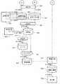
図3を参照すると、本開示の一実施形態に係る解決策を実施するために使用され得る主なソフトウェアコンポーネントの役割を表す連携図が示されている。 With reference to FIG. 3, a linkage diagram showing the role of the main software components that can be used to implement the solution according to one embodiment of the present disclosure is shown.
すべてのソフトウェアコンポーネント(プログラムおよびデータ)は、全体として参照番号300で示されている。ソフトウェアコンポーネントは、通常、オペレーティングシステムおよび他のアプリケーションプログラム(図示せず)と共に、プログラムが実行されているときに大容量メモリに格納され、(少なくとも部分的に)超音波スキャナのワーキングメモリにロードされる。プログラムは、最初に、例えばリムーバブル記憶装置またはネットワークから大容量メモリにインストールされる。この点において、各ソフトウェアコンポーネントは、指定された論理機能を実施するための1つまたは複数の実行可能命令を含むモジュール、セグメントまたはコード部分を表すことができる。特に、この図は、ソフトウェアコンポーネントの静的構造とその動的挙動(交換された一連のメッセージによって、それぞれが対応する動作を表し、記号”A”が前に付されたシーケンス番号で示される)を記述する。 All software components (programs and data) are designated by
TX/RXコントローラ303は、超音波スキャナのトランスデューサを制御する。例えば、TX/RXコントローラ303は、送信ビームフォーマを有するTXコントローラと、連続する取得時点(例えば、毎秒10〜30のレート)で超音波パルス(低MI)を生成するためのパルサとを備える。TX/RXコントローラ303は、各取得時点(選択された走査面内の対応する位置に対して)で対応する(アナログRF)エコー信号を受信するRXプロセッサをさらに備える。RXプロセッサは、エコー信号を事前増幅し、予備時間利得補償(TGC)を適用する。RXプロセッサは、エコー信号をアナログデジタル変換部(ADC)によってデジタル値に変換し、それらを受信ビームフォーマを介して集束されたビーム信号に結合する。RXプロセッサは、好ましくは、さらなるデジタルアルゴリズムおよび他の線形または非線形信号調整部(例えば、ポストビーム形成TGC)を介して得られた(デジタルRF)エコー信号を処理する。具体的には、TX/RXコントローラ303は、(循環して固定された)造影剤の(非線形)寄与に関して、エコー信号における組織の支配的な(直線的な)寄与を実質的に除去するか、または少なくとも低減するように、造影特有のイメージングモードで動作する。造影特有のイメージングモードの例は、例えば、非特許文献2に記載されているように、高調波イメージング(HI)、パルス反転(PI)、パワー変調(PM)および造影パルスシーケンス(CPS)技術(その全体の開示は参照により本明細書に組み込まれる)を含む。TX/RXコントローラ303は、各取得時点で走査面によって画定された身体部分のスライスの標準輝度モード(Bモード)で(デジタル)画像を生成するように、エコー信号を復調し、対数圧縮し、走査変換してビデオフォーマットにするビデオ変換部をさらに含む。各画像は、それぞれの画素の値を格納するセルのマトリクス(例えば、512行と512列)を含むビットマップによって定義される。すなわち、身体部分の基本部分からなる位置に対応する基本ピクチャ要素。各画素値は、取得時点での位置について記録されたエコー信号の強度の関数として画素の輝度を定義する。例えば、グレースケールタイプの画像では、エコー信号の強度が増加するにつれて、0(黒)から255(白)に増加する8ビットで画素値を符号化することができる。 The TX /
任意の分析プロセス(簡略化のために図には示されていない)の開始時に、超音波スキャナのオペレータはトランスデューサを作動させ、次にそれを分析すべき身体部分の周りに移動させる(造影剤を投与する前に)。取得された対応する画像は、その後、身体部分の解剖学的表現を提供し、以下、解剖学的画像と称する。これらの解剖学的画像は、リアルタイムで超音波スキャナのモニタ上に連続して表示される。次いで、オペレータは、解析される身体部分のスライスを表す走査面、およびおそらく関心のある領域(例えば、疑わしい病変を含む)を選択する(任意に選択された解剖学的画像において)。この時点で、オペレータは、(選択された走査面に対応する固定位置に変換部を維持しながら)造影剤を患者に投与し、身体部分の分析を開始するためのコマンドを入力する。それに応答して、TX/RXコントローラ303は、取得された対応する画像を原画像と定義し、原画像リポジトリ306に連続して保存する(動作”A1.取得する”)。 At the beginning of any analytical process (not shown in the figure for simplicity), the ultrasonic scanner operator activates the transducer and then moves it around the body part to be analyzed (contrast agent). Before administration). The acquired corresponding image then provides an anatomical representation of the body part, hereinafter referred to as an anatomical image. These anatomical images are continuously displayed on the monitor of the ultrasonic scanner in real time. The operator then selects a scanning surface representing a slice of the body part to be analyzed, and perhaps an area of interest (including, for example, a suspicious lesion) (in an arbitrarily selected anatomical image). At this point, the operator enters a command to administer the contrast agent to the patient (while maintaining the transform at a fixed position corresponding to the selected scanning surface) and initiate analysis of the body part. In response, the TX /
フィルタ309は、特許文献1に記載されているように、原画像(原画像リポジトリ306から抽出された)からフィルタリングされた画像を生成する。簡単に述べると、フィルタ309は、注目領域外のそれらの元の値をゼロにリセットすることによって原画像をマスクし、(マスクされた)原画像をサブサンプルし、各(サブサンプリングされマスクされた)原画像に対して、修正されたMin_IPアルゴリズムを適用することによって対応する(サブサンプルされた)フィルタリングされた画像を生成する。この目的のために、フィルタ309は、フィルタリングされた画像の各フィルタリングされた値を、対応する原画像と1つまたは複数の先行する原画像からなる原画像のフィルタリングセット内の対応する原画像の中で最小値に設定する(例えば、0.1〜0.5秒のフィルタリングウィンドウに対応する1〜5個の先行する原画像)。フィルタ309は、フィルタリングされた画像を対応する原画像上に重ね合わせ、次いで、それらの全サイズを復元する。フィルタ309は、(オーバーレイされた)フィルタリングされた画像をフィルタ画像リポジトリ312に連続して保存する。 The
同時に(簡略化のために図示していない)、フィルタリングされた画像は、フィルタリングウィンドウに対応する短い遅延で、実質的にリアルタイムで超音波スキャナのモニタ上に連続して表示される。(動作”A2.フィルタ”)。代替的には(図示せず)、フィルタリングされた画像は、他の装置から(例えば、リムーバブル記憶装置またはデジタル、アナログまたはネットワーク接続を介して)または他のソフトウェアプログラム(同じまたは異なる装置で実行している)から単純に受信され、フィルタリングされた画像リポジトリ312に直接格納される。いずれにしても、フィルタリングされた画像は、任意の他のフィルタリング技術(例えば、差分標的増強法)を適用することによって、または循環する造影剤の大部分が消失するまで単に待つことで得ることができる(すなわち、原画像によって直接的に遅延位相で定義されるフィルタリングされた画像で)。言い換えれば、フィルタリングされた画像は、(実際に生成されたかどうかにかかわらず、ローカルにまたは遠隔で、または既にこの形式で提供されているかどうかにかかわらず)出発点である。 At the same time (not shown for brevity), the filtered images are displayed continuously on the monitor of the ultrasound scanner in substantially real time with a short delay corresponding to the filtering window. (Operation "A2. Filter"). Alternatively (not shown), filtered images are run from other devices (eg, via removable storage devices or digital, analog or network connections) or other software programs (on the same or different devices). Is simply received from) and stored directly in the filtered
ドロワ315は、循環領域および固定化領域を(フィルタリングされた画像リポジトリ312から抽出された1つの任意に選択されたフィルタリングされた画像に)オペレータが描画するために使用される。好ましくは、循環領域は、できるだけ大きく描かれているが、鏡面反射器は除かれている(単に関心領域に等しいと考える可能性がある)。次に、固定化領域は循環領域内に引き込まれる。固定化領域および循環領域は、それぞれ、フィルタリングされた画像と同じサイズの細胞のマトリックスによって画定される、固定化マスクおよび循環マスクによって表される。固定化マスクの各セルは、対応する位置が固定化領域の内部にあるときにアサートされる(すなわち、論理値1)、またはアサート解除される(すなわち、論理値0)フラグ(すなわち、2進値)を格納する。そうでなければ、循環マスクの各セルには、対応する位置が循環領域の内側であるが固定領域の外側にあるときにアサートされるか、またはそうでない場合にアサート解除されるフラグが格納される(すなわち、対応する位置が循環領域外または固定化領域内にあるとき)。ドロワ315は、固定化マスクおよび循環マスクをそれぞれ固定化マスクリポジトリ318および循環マスクリポジトリ321に保存する(動作”A3.ドロー”)。 The
乗算部324は、最後のフィルタリングされた画像、すなわち対応するフィルタリングウィンドウ(原画像リポジトリ306から抽出された)を定義する最後の原画像を生成するために使用された各原画像とセル毎に(循環マスクリポジトリ321から抽出された)循環マスクとを乗算して、対応するマスク画像を生成する。その結果、マスクされた画像は、原画像と同じサイズのセルのマトリックスによって定義される。マスクされた画像は循環領域内にある元の値のみを含み、他の元の値はゼロにリセットされる。乗算部324は、こうして得られたマスクされた画像をマスクされた画像リポジトリ327に保存する(動作”A4.マスク”)。リニアライザ330は、(マスクされた画像リポジトリ327から抽出された)各マスクされた画像から線形化画像を生成する。線形化画像は、マスクされた画像と同じサイズのセルのマトリックスによって定義される。循環領域内の線形化画像の各セル(すなわち循環マスク内のフラグがアサートされる)は、マスクされた画像内の対応する(元の)値を造影剤の局所濃度に直接比例させることによって計算される値を記憶する(例えば、逆対数圧縮を適用し、特許文献8に記載されているように得られた値を二乗することによって、その全開示を本明細書に援用する)。一方、他のセルはゼロのままである。リニアライザ330は、そのようにして得られた線形化画像を線形化画像リポジトリ333に保存する(動作”A5.線形化”)。マスクされた画像および/または線形化画像が(フィルタリングされた画像を生成するための)フィルタ309によって既に提供されている場合、原画像をマスクする動作および/またはマスクされた画像を線形化する動作は省略されてもよい。 The multiplication unit 324 (for each original image and cell used to generate the last filtered image, that is, the last original image defining the corresponding filtering window (extracted from the original image repository 306)). Multiply by the circular mask (extracted from the circular mask repository 321) to generate the corresponding mask image. As a result, the masked image is defined by a matrix of cells that are the same size as the original image. The masked image contains only the original values that are in the circular region, the other original values are reset to zero. The
統合部336は、(線形化画像リポジトリ333から抽出された)各線形化画像の循環領域内のセルの(線形化された元の)値の中央値を計算する。統合部336は、異なる線形化画像についてこのようにして得られた中央値の平均に等しいベース閾値(候補閾値を計算するために使用される)を設定する。統合部336は、ベース閾値をベース閾値変数339に格納する(動作”A6.統合”)。生成部342は、振幅閾値の対応する候補値によって定義されるように、所定の数の候補閾値(例えば、100〜300個、好ましくは150〜250個、さらに好ましくは175〜225個、例えば200個)を計算する。具体的には、候補閾値は、ベース閾値(ベース閾値変数339から抽出された)に、対応する割合(例えば、0〜50%から150〜250%まで、好ましくは0〜20%から170〜230%まで、さらにより好ましくは0〜10%から190〜210%まで、例えば0%〜199%)を乗算することによって計算される。例えば、200個の候補閾値は、0%〜199%の範囲の割合によってベース閾値に1%のピッチを乗じて計算される。生成部342は、そのようにして得られた候補閾値を候補閾値ベクトル345に保存する(動作”A7.生成する”)。上記のすべてが、方法の堅牢性と精度を向上させる。特に、ベース閾値を計算するために使用される中央値は、それらのアウトライヤーによって過度に影響されない対応する値の中心的な傾向の尺度を提供する。結果として、循環領域の選択における不正確さの影響(明るい鏡面反射器に起因して高い値を導入する可能性がある)は、大幅に緩和される。さらに、候補閾値を生成するためのベース閾値の使用は、フィルタリングされた画像を生成するために使用される原画像に依存する。その結果、候補閾値は、イメージング条件に動的に自己適応する。代わりに(図示せず)、候補閾値は、原画像とは無関係に、予め定義されたベース閾値から同じ方法で計算される。例えば、ベース閾値は、(カスタマイズ可能な)固定値であるか、またはイメージング条件(原画像の平均品質など)に応じて、手動または自動のいずれかで複数の(カスタマイズ可能な)固定値から選択される。さらなる代替として、候補閾値は直接的に予め定義される。例えば、候補閾値は(カスタマイズ可能な)固定値であるか、またはそれらは上記のように(カスタマイズ可能な)固定値の複数のセットの中から選択される。 The
閾値化部348は、各候補閾値(候補閾値ベクトル345から抽出された)毎に候補画像を生成する。この目的のために、閾値化部348は、最後のフィルタリングされた画像(フィルタリングされた画像リポジトリ312から抽出された)から候補閾値に対応する閾値マスクを生成する。閾値マスクは、(最後の)フィルタリングされた画像と同じサイズのセルのマトリックスによって定義される。閾値マスクの各セルは、対応するフィルタリングされた値が候補閾値より高い(おそらく厳密に)場合にアサートされるフラグを格納するか、またはそうでない場合にはアサート解除される。次に、閾値化部348は、候補画像を生成するために、フィルタリングされた画像と閾値マスクとをセル毎に乗算する。その結果、候補画像は、フィルタリングされた画像と同じサイズのセルのマトリックスによって定義される。候補画像は候補閾値よりも高いフィルタリングされた値のみを含むが、他のフィルタリングされた値はゼロにリセットされる。閾値化部348は、異なる候補閾値についてそのように取得された候補画像を候補画像リポジトリ351に保存する(動作”A8.閾値”)。 The
(さらに)統合部354は、固定領域内のそのセルの(フィルタリングされた)値の平均として(候補画像リポジトリ351から抽出された)各候補画像の固定平均値を計算する(すなわち、固定化マスクリポジトリ318から抽出された固定化マスク内にフラグがアサートされる)。統合部354は、異なる候補閾値について得られた固定平均値を固定化平均値ベクトル357に格納する(動作”A9.統合”)。同様に、統合部354は、循環領域内のセルの(フィルタリングされた)値の平均として(候補画像リポジトリ351から再び抽出された)各候補画像の循環平均値を計算する(すなわち、循環マスクリポジトリ321から抽出された循環マスク内にフラグがアサートされる)。統合部354は、このようにして得られた異なる候補閾値についての循環平均値を循環平均値ベクトル360に格納する(動作”A10.統合”)。 (Furthermore) the
正規化部363は、固定化平均値ベクトル357における固定化平均値を正規化する。この目的のために、正規化部363は、固定化平均値から固定化オフセット(候補閾値0に対応する固定化平均値で定義される固定化平均値、すなわち第1の固定化閾値によって定義される)を減算した後、固定化平均値をこの演算の結果で置き換える(動作”A1.正規化”)。同様に、正規化部363は循環平均値ベクトル360内の循環平均値を正規化し、この目的のために、正規化部363は、各循環平均値から循環オフセット(候補閾値0に対応する循環平均値、すなわち第1の循環閾値によって定義される)を減算した後、この循環平均値をこの演算の結果に置き換える(動作”A12.正規化”)。 The
比較部366は、対応する(正規化された)固定平均値(固定平均値ベクトル357から抽出された)から各循環平均値を減算して、(正規化された)固定平均値(循環平均値ベクトル360から抽出された)から対応する候補閾値の比較値を得る。比較部366は、異なる候補閾値についてこのように取得された比較値を比較値ベクトル369に格納する(動作”A13.比較”)。平滑化部372は、比較値ベクトル369内の比較値を平滑化する。この目的のために、平滑化部372は、平滑化アルゴリズムをその変動を減少させるために比較値に適用し(例えば、移動平均フィルタまたはローパスフィルタ)、比較値をこの演算の結果で置き換える。(動作”A14.平滑化”)。 The
検出部375は(平滑化された)比較値を(比較値ベクトル369内で)走査してその最大値(ピーク値を定義する)を決定する。検出部375は、ピーク値と、それを提供する(ピーク閾値を定義する)候補閾値の指示、例えば、ピーク閾値自体または比較値ベクトル369におけるその位置を示す指標を、ピーク変数378に格納する(動作”A15.検出”)。セレクタ381は、ピーク値(ピーク変数378から抽出した)を2で割って、比較値の半ピーク値を計算する。セレクタ381は、半ピーク値以下の比較値が見つかるまで、ピーク閾値から始まる候補閾値の高い順に比較値(比較値ベクトル369)を走査する(比較値ベクトル369の終わりに達する前に常に実際に発生する)。見つかった比較値が半ピーク値と等しい場合、セレクタ381は振幅閾値を、それを提供する候補閾値に直接設定し、そうでなければ、セレクタ381は、振幅閾値を、候補値と前の(より低い)候補閾値との間の半ピーク値に近い比較値を与える候補閾値に設定する。セレクタ381は、決定した振幅閾値を振幅閾値変数384に格納する(動作”A16.選択”)。この振幅閾値の選択は、ほとんどの実用的な状況において良好な妥協点(循環造影剤の残留寄与を除去し、固定化造影剤の寄与に影響を及ぼさない対抗要件の間にある)を提供することが分かっている。 The
この時点で、(おそらく上記と同じ)閾値化部387は、(フィルタリングされた画像リポジトリ312から抽出された)同じ(最後の)フィルタリングされた画像に(閾値変数384から抽出された)振幅閾値を適用する。この目的のために、閾値化部387は、振幅閾値に対応する(さらに)閾値マスクを生成する。閾値マスクは、フィルタリングされた画像と同じサイズのセルのマトリックスによって定義される。関心領域内の閾値マスクの各セル(すなわち、対応するマスク内のフラグがアサートされる)は、対応するフィルタリングされた値が振幅閾値より高い(おそらく厳密に)ときにアサートされるフラグを格納するか、またはそうでない場合にはアサート解除され、一方、関心領域外の閾値マスクの各セルは、常にアサートされるフラグを格納する。次に、閾値化部387は、フィルタリングされた画像と閾値マスクをセル毎に乗算して、対応する閾値化画像を生成する。結果として、閾値化画像は、フィルタリングされた画像と同じサイズのセルのマトリックスによって定義される。関心領域の内側では、閾値化画像は、振幅閾値よりも高いフィルタリングされた値のみを含み、他のフィルタリングされた値はゼロにリセットされ、関心領域の外側では、閾値化された画像は、フィルタリングされた値を含む(すなわち、対応する元の値)。閾値化部387は、そのようにして得られた閾値化画像を閾値化画像リポジトリ390に保存する(動作”A17.閾値”)。表示部393は、超音波スキャナのモニタに閾値化画像(閾値化画像リポジトリ390から抽出された)を表示する(動作”A18.表示”)。このようにして、関心領域において、閾値化画像は、(固定化造影剤の検出のために)有意なフィルタリングされた値のみを示す。これらのフィルタリングされた値は、(固定化造影剤の定量化を容易にするために)所与のカラーマップパレットに従ってレンダリングされてもよい。関心領域の外側ではなく、代わりに、閾値化画像は、(固定化造影剤に関する情報を文脈化するための)身体部分の解剖学的表現を常に示す。 At this point, the thresholding unit 387 (probably the same as above) sets the amplitude threshold (extracted from the threshold variable 384) to the same (last) filtered image (extracted from the filtered image repository 312). Apply. For this purpose, the
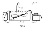
図4を参照すると、本開示の一実施形態に係る試験管内での溶液を試験するために使用されたセットアップ400が示されている。 With reference to FIG. 4, the
セットアップ400は、(滑動する)フローセル405を含む。フローセル405は、ヒトP−セレクチンFcで被覆されたガラスプレート410と、音響透過性のマイラーフィルム415からなる上壁とからなる底壁を有するチャンバによって形成される。フローセル405をセットアップ400に設置する前に、ガラスプレート410上にセレクチン標的化マイクロバブル(MB)を含む(標的化された)造影剤をインキュベートして、その上に固定化したままにした。蠕動ポンプ425を用いて、BR38マイクロバブル(1・105バブル/mL)を含む(標的化されていない)造影剤をフローセル405を通ってリザーバ420から循環させた。このようにして、固定化造影剤が循環造影剤によって取り囲まれた試験管内条件が模倣される。フローセル405(固定化造影剤と循環造影剤の両方を含む)の原画像は、CPS造影剤特異的モードの線形トランスデューサ15L8を備えたSequoia512からなる超音波スキャナ430を用いて5つの異なる位置で取得した。超音波スキャナ430のイメージング設定は、MI0.08、深さ25mm、焦点15mmおよび17mm、フレームレート4Hzであった。超音波スキャナ430によって提供されたデータは、DICOMシーケンスとしてエクスポートされた。対数圧縮されたビデオデータは、較正ファイル15L8,83dB、PP4、デルタ2、+1/M:2(V1.1)を使用して造影剤の局所濃度に比例するエコーパワー信号を提供する画素レベルで線形化された。分析結果はXLSファイルとして格納された。同じ操作を4つの異なるバージョンのガラスプレート410で繰り返し、それぞれが2つの異なる濃度の(標的化された)造影剤をインキュベートし、その結果、固定化造影剤の8つの異なる表面密度(3.3〜97.5・106>μm2/mL)を得た。固定化造影剤の表面密度は、そのマイクロバブル(平均5カ所)を光学的に計数することによって決定された。
図5A〜図5Fを参照すると、本開示の一実施形態に係る解決策の試験管内での適用の異なる例が示されている。 With reference to FIGS. 5A-5F, different examples of in vitro application of the solution according to one embodiment of the present disclosure are shown.
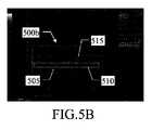
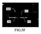
図5Aおよび図5Bから出発して、固定化造影剤(因子10)の異なる表面密度を有するフローセルを画像化することによって上記のセットアップで取得された2つの原画像500aおよび500bが示されている。すなわち、図5Aでは低く、図5Bでは高くなっている。両方の場合において、同じ循環領域505を描いてフローセルの大部分(固定化造影剤および循環造影剤の両方を含む)を囲み、同じ固定化領域510をフローセルの下部に(主として一部の循環造影剤を有する固定化造影剤を含む)に引っ張り、制御領域515をフローセル(排他的に循環造影剤を含む)の内腔内に引き込んだ。原画像500aおよび500bにおける循環造影剤の濃度は、実質的に同じである(固定化造影剤の異なる表面密度とは無関係に)。 Starting from FIGS. 5A and 5B, two
図5Cおよび図5Dに進むと、対応するフィルタリングされた画像500cおよび500dが示されており、これらの画像は、原画像(固定化造影剤の低および高のそれぞれの表面密度に対応する)をフィルタリングして得られ、循環造影剤の寄与を低減する。両方の場合において、固定化造影剤は正確に検出される(固定化領域510に見えるように)。しかしながら、予想されるように、循環造影剤は完全に除去されない(制御領域515に見られるように)。これは、固定化造影剤の表面密度が低い場合と高い場合のそれぞれ3dBおよび13dBに等しい固定化/循環比の比較的低い値(固定化領域510内のフィルタリングされた値の平均を、制御領域515内のフィルタリングされた値の平均で除算することによって計算される)に反映される。 Proceeding to FIGS. 5C and 5D, the corresponding filtered
図5Eおよび図5Fに進むと、本開示の一実施形態に係る解決策に従う決定された振幅閾値でこれらのフィルタリングされた画像(固定化造影剤のそれぞれ低い表面濃度および高い表面濃度に対応する)を閾値処理することによって得られた対応する閾値化画像500eおよび500fが示されている。両方の場合において、固定化造影剤は、再び正確に検出される(固定化領域510に見えるように)。しかしながら、循環造影剤は、(制御領域515に見えるように)ほぼ完全に除去される。これは、固定化/循環比(固定化領域510内の閾値化値の平均を制御領域515の閾値化値の平均で除算することによって計算される)のはるかに高い値が固定化された造影剤の表面密度のそれぞれの低および高については16dBおよび27dBに等しくなることを反映する。上記の全ては、本開示の一実施形態に係る解決策が、固定化造影剤の顕著性(したがってそれを検出する能力)を有意に改善することを確認する。 Proceeding to FIGS. 5E and 5F, these filtered images with determined amplitude thresholds according to the solution according to one embodiment of the present disclosure (corresponding to low and high surface concentrations of the immobilized contrast agent, respectively). The corresponding
図6A〜図6Bを参照すると、本開示の一実施形態に係る解決策の試験管内での適用の異なる例に関する図が示される。 Referring to FIGS. 6A-6B, diagrams relating to different examples of in vitro application of the solution according to one embodiment of the present disclosure are shown.
特に、固定化領域内のフィルタリングされた値の平均値および閾値化値の平均値(以下、フィルタリングされた平均値および閾値化平均値とそれぞれ称する)は、フィルタリングされた画像および閾値化処理された画像においてそれぞれ計算された。それらは、固定化造影剤の全ての(8個の)異なる表面密度でフローセルを画像化することにより、上記のセットアップで取得された原画像から得られた。図6Aは、横軸(mm−2)上の固定化造影剤の表面密度に対する縦軸(任意の単位)上のフィルタリングされた平均値をプロットした図600aを示し、図6Bは、横軸(mm−2)上の固定化造影剤の表面密度に対して縦軸(任意の単位)上の閾値化平均値をプロットした図600bを示す。得られた結果は非常に類似しており、固定化造影剤(x)の表面密度に対するフィルタリングされた平均値(y)および閾値化平均値(y’)の観察され得る直接比例関係は、それぞれ、y=0.616x−43.542および決定係数R2=0.9511、並びにy’=0.6212x−64.401および決定係数R2=0.9495である。フィルタリングされた平均値と閾値化平均値との間の類似性(それらが固定化領域に少量の循環造影剤を含む制御された環境で得られたという事実による)は、本開示の一実施形態に係る解決策が、検出される固定化造影剤の量に影響を与えないことを確認し、次いでそれを定量化する能力を保存する。In particular, the average value of the filtered values and the average value of the thresholded values in the fixed region (hereinafter referred to as the filtered average value and the thresholded average value, respectively) were filtered images and the thresholded values were processed. Each was calculated in the image. They were obtained from the original images obtained in the above setup by imaging the flow cells at all (8) different surface densities of the immobilized contrast agent. FIG. 6A shows FIG. 600a plotting the filtered mean on the vertical axis (arbitrary unit) with respect to the surface density of the immobilized contrast agent on the horizontal axis (mm-2 ), and FIG. 6B shows the horizontal axis (the horizontal axis (mm-2 )). FIG. 600b is a plot of the thresholded mean value on the vertical axis (arbitrary unit) with respect to the surface density of the immobilized contrast agent on mm-2 ). The results obtained are very similar, with the observable direct proportional relationships of the filtered mean (y) and the thresholded mean (y') to the surface density of the immobilized contrast agent (x), respectively. , Y = 0.616x-43.542 and the coefficient of determination R2 = 0.9511, and y'= 0.6212x-64.401 and the coefficient of determination R2 = 0.9495. The similarity between the filtered mean and the thresholded mean (due to the fact that they were obtained in a controlled environment with a small amount of circulating contrast agent in the immobilized region) is an embodiment of the present disclosure. Make sure that the solution according to does not affect the amount of immobilized contrast agent detected, and then preserve the ability to quantify it.
図7A〜図7Cを参照すると、本開示の一実施形態に係る解決策の生体内での適用の例が示されている。 With reference to FIGS. 7A-7C, an example of in vivo application of the solution according to one embodiment of the present disclosure is shown.
ヒト結腸癌の同所性動物モデルを使用した。FLK1(VEGFR2としても知られている)に標的特異的であるマイクロバブルを含む(標的化された)造影剤を、1.6μL/kg(気体容積として表示)の用量で静脈内投与した。癌を含む身体部分の原画像を、CPS造影剤特異モードにおいて線形トランスデューサ15L8を取り付けたシーメンスセコイア(Siemens Sequoia)512からなる超音波スキャナを用いて(後期増強の間、造影剤の投与後10分を超えて)取得した。超音波スキャナのイメージング設定は、MI0.25、フレームレート4Hzであった。超音波スキャナによって提供されたデータは、DICOMシーケンスとしてエクスポートされた。対数圧縮されたビデオデータは、較正ファイル15L8、CPS、83dB、PP4、デルタ2、+1/M:2(v1.1)を使用して、造影剤の局所濃度に比例するエコーパワー信号を提供する画素レベルで線形化された。 An orthotopic animal model of human colon cancer was used. A (targeted) contrast medium containing microbubbles that are target-specific to FLK1 (also known as VEGFR2) was administered intravenously at a dose of 1.6 μL / kg (shown as gas volume). Original images of body parts, including cancer, were captured in CPS contrast agent specific mode using an ultrasound scanner consisting of Siemens Sequoia 512 equipped with a linear transducer 15L8 (during late enhancement, 10 minutes after administration of contrast agent). (Beyond) acquired. The imaging settings of the ultrasonic scanner were MI 0.25 and a frame rate of 4 Hz. The data provided by the ultrasonic scanner was exported as a DICOM sequence. The log-compressed video data uses calibration files 15L8, CPS, 83dB, PP4,
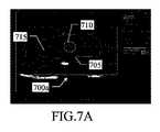
図7Aから出発して、(造影剤の投与の10分後に身体部分を撮像することによって取得された)例示的な原画像700aが示されている。循環領域705は、身体部分(癌腫および周囲組織を含むが、鏡面反射器を除く)の大部分を含むように描かれ、固定領域710は、循環領域705内に引き込まれ、癌腫のみを含み、制御領域715は、循環領域705の内部に引き込まれるが(主に循環造影剤のみを含むように)、癌腫から遠く離れている。循環造影剤が除去されることを期待する実質的な遅延期にもかかわらず、原画像700aは、固定化領域710内の固定造影剤に加えて、それの外側に相当量の循環造影剤をなお含む。これは、3dBに等しい固定化/循環比の低い値(固定化領域705内の元の値の平均値を制御領域715内の元の値の平均値で除算することによって計算される)によって反映されるように、固定化造影剤の顕著性(したがってそれを検出する能力)を制限する。 Starting from FIG. 7A, an exemplary
図7Bに進むと、循環造影剤の寄与を減少させるために、この原画像をフィルタリングすることによって得られた、対応するフィルタリングされた画像700bが示されている。固定化/循環比(固定化領域705のフィルタリングされた値の平均値を制御領域715のフィルタリングされた値の平均値で除算することによって計算される)は11dBに増加するが、循環造影剤はまだ完全には除去されない(制御領域715に見えるように)。 Proceeding to FIG. 7B, the corresponding filtered image 700b obtained by filtering this original image to reduce the contribution of the circulating contrast agent is shown. The immobilization / circulation ratio (calculated by dividing the mean of the filtered values in the
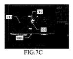
図7Cに進むと、本開示の一実施形態に係る解決法に従って決定された振幅閾値でこのフィルタリングされた画像を閾値処理することによって得られた、対応する閾値化画像700cが示されている。固定化/循環比(固定化領域705における閾値化値の平均値を制御領域715における閾値化値の平均値で除算することによって計算される)は、今や21dBまで増加し、循環造影剤はほぼ完全に除去される(制御領域715に見えるように)。これは、固定化造影剤の顕著性を有意に改善し、したがって、癌腫に対応してそれを検出する能力を向上させる。 Proceeding to FIG. 7C, a corresponding
変更
当然のことながら、局所的および特定の要件を満たすために、当業者は、本開示に多くの論理的および/または物理的な改変および変更を適用することができる。より具体的には、本開示は、その1つ以上の実施形態を参照してある程度の詳細性で説明されているが、形態および詳細における様々な省略、置換および変更、ならびに他の実施形態が可能であることを理解されたい。特に、本開示の異なる実施形態は、そのより完全な理解を提供するために、前の説明に記載された特定の詳細(数値など)なしに実施されてもよい。逆に、周知の特徴は、不必要な詳細で説明を不明瞭にしないために、省略または簡略化されている可能性がある。さらに、本開示の任意の実施形態に関連して説明される特定の要素および/または方法ステップは、一般的な設計選択の問題として他の実施形態に組み込まれてもよいことは明らかである。いずれの場合においても、各数値は、(既に行われていない限り)約という用語によって修正されて読まれるべきであり、数値の各範囲は、(終点を含む)範囲内の連続に沿った可能な数を明示的に指定するものとする。さらに、序数やその他の修飾子は、単に同じ名前の要素を区別するためのラベルとして使用されるが、それ自体で優先順位、先行または順序を暗示しない。含む、備えている、有する、含む、および含む(またはその任意の形態)という用語は、オープンで非包括的な意味(すなわち、列挙された項目に限定されない)を意図するものであり、に基づく、に依存する、従って、の機能(またはその任意の形態)という用語は、非排他的な関係(すなわち、可能性のあるさらなる変数を含む)を意図するものであり、用語a/anは(別段の明示的な指示がない限り)1つまたは複数の項目として意図するものであり、ための手段という用語(または任意の手段プラス機能の形式)は、関連する機能を実行するために適合または構成された任意の構造を意図するものとする。Modifications Of course, one of ordinary skill in the art may apply many logical and / or physical modifications and alterations to the present disclosure to meet local and specific requirements. More specifically, the present disclosure is described with some degree of detail with reference to one or more embodiments thereof, but various omissions, substitutions and modifications in embodiments and details, as well as other embodiments. Please understand that it is possible. In particular, different embodiments of the present disclosure may be implemented without the particular details (such as numerical values) described in the previous description to provide a more complete understanding thereof. Conversely, well-known features may be omitted or simplified in order not to obscure the description with unnecessary details. Furthermore, it is clear that the specific element and / or method steps described in connection with any embodiment of the present disclosure may be incorporated into other embodiments as a matter of general design choice. In any case, each number should be modified and read by the term about (unless already done), and each range of numbers can be along a continuum within the range (including the end point). Number shall be explicitly specified. In addition, ordinal numbers and other modifiers are simply used as labels to distinguish elements with the same name, but do not imply priority, precedence, or order by themselves. The terms include, include, have, include, and include (or any form thereof) are intended and based on an open, non-inclusive meaning (ie, not limited to the listed items). , Therefore, the term function (or any form thereof) is intended for non-exclusive relationships (ie, including possible additional variables), and the term a / an is (i.e., including additional variables). It is intended as one or more items (unless otherwise explicitly indicated), and the term means for (or any means plus a form of function) is adapted or adapted to perform the relevant function. It is intended for any configured structure.
例えば、実施形態は、患者の身体部分を分析するための方法を提供する。身体部分は、患者内を循環し、生物学的標的上に実質的に固定され得る造影剤を用いて方法を実施する前に灌流されている。しかしながら、この方法は、任意の患者の身体部分を分析するために使用することができる(下記参照)。さらに、造影剤は、非特異的相互作用によって標的に運搬または蓄積される場合、標的型ではなくても、任意の型(例えば、磁気共鳴画像法またはX線コンピュータ断層撮影法を画像化するために特有)であってもよいし、標的型でなくてもよい(例えば、造影剤が患者の免疫系によって異物として認識され、その後、その代謝および排除のために肝臓に輸送される場合)。さらに、造影剤は、いかなる方法(例えば、ポンプによる連続注入)および任意の時点(例えば、方法を実施する直前)で投与されてもよい。 For example, embodiments provide a method for analyzing a patient's body part. The body part is perfused prior to performing the procedure with a contrast agent that circulates within the patient and can be substantially immobilized on the biological target. However, this method can be used to analyze the body part of any patient (see below). In addition, the contrast agent is to image any type of non-target type (eg, magnetic resonance imaging or X-ray computed tomography) when transported or accumulated at the target by non-specific interaction. It may be (specific to) or not targeted (eg, if the contrast agent is recognized as a foreign body by the patient's immune system and then transported to the liver for its metabolism and elimination). In addition, the contrast agent may be administered by any method (eg, continuous pumping) and at any time point (eg, just before the method is performed).
いずれにしても、これは患者との相互作用とは独立して実施できるデータ処理方法である。さらに、造影剤は、非侵襲的な方法で患者に投与することもできる(例えば、胃腸管を画像化するためには経口的に、または気道に噴霧器を介して経口的に)、またはいずれの場合にも専門的な医学知識を必要とするか、または患者のあらゆる健康リスクを伴う実質的な身体的介入を伴わずに行うことができる(例えば、筋肉内に)。この方法は、医師の仕事を容易にすることができるが、(例えば、診断目的のために)身体部分を検査するのに役立つ中間結果を提供するだけであるが、医師自身が常に行う治療目的の診断のための診断を提供する。 In any case, this is a data processing method that can be performed independently of patient interaction. In addition, the contrast agent can be administered to the patient in a non-invasive manner (eg, orally to image the gastrointestinal tract or orally through a nebulizer into the airways), or either. It can also be done without the need for specialized medical knowledge or with substantial physical intervention with any health risk of the patient (eg, intramuscularly). This method can facilitate the work of the physician, but only provides intermediate results that are useful for examining body parts (eg, for diagnostic purposes), but for therapeutic purposes that the physician himself always performs. Provides a diagnosis for the diagnosis of.
一実施形態では、本方法は、身体部分の対応する位置について複数のフィルタリングされた値を含む少なくとも1つのフィルタリングされた画像を提供することと、各フィルタリングされた値は、実質的に低減された循環造影剤の寄与を伴う、対応する位置における固定化造影剤の表示を含む。しかし、フィルタリングされた画像は、いかなる方法で提供されてもよい(例えば、局所的に生成するか、またはこの形式で既に受信する)。任意の数のフィルタリングされた画像を提供することができる(例えば、フィルタリングされた画像のシーケンス全体にこの方法を適用することによって)。フィルタリングされた各画像は、任意のサイズおよび形状(マトリックス全体からその1つまたは複数の部分まで)を有してもよく、そのフィルタリングされた値は、身体部分の任意のタイプの位置(例えば、フィルタリングされた画像はサブサンプリングされたときの画素、ボクセルまたはそれらのグループ)に対応することができる。フィルタリングされた値は、どのような方法でも(例えば、エコー信号の強度と共に減少するときに負の形式で)対応する位置に(もしあれば)固定化された造影剤を示すことができ、循環造影剤の寄与は、たとえフィルタリングされた画像が遅延期で取得された場合であっても、自動的にさえ、どのような任意のレベル(例えば、少なくとも50〜90%)においても低減されている可能性がある。 In one embodiment, the method provides at least one filtered image containing a plurality of filtered values for the corresponding positions of the body part, and each filtered value is substantially reduced. Includes display of the immobilized contrast agent at the corresponding position with the contribution of the circulating contrast agent. However, the filtered image may be provided in any way (eg, locally generated or already received in this format). Any number of filtered images can be provided (eg, by applying this method to the entire sequence of filtered images). Each filtered image may have any size and shape (from the entire matrix to one or more parts thereof), the filtered value of which is the position of any type of body part (eg, eg). The filtered image can correspond to the pixels, voxels or groups thereof when subsampled. The filtered values can indicate the contrast medium (if any) immobilized in any way (eg, in a negative form as it decreases with the intensity of the echo signal) and circulates. Contrast contribution is reduced at any level (eg, at least 50-90%), even automatically, even if the filtered image is acquired in a delayed period. there is a possibility.
一実施形態では、この方法は、フィルタリングされた値に対応する複数の閾値化値を含む閾値化画像を生成することを含む。閾値化画像は、各閾値化値を対応するフィルタリングされた値に設定することによって、またはフィルタリングされた値と振幅閾値との比較に従ってリセット値に生成される。しかしながら、リセット値は、フィルタリングされた値の任意のより低いまたはより高い境界値(例えば、フィルタリングされた画像が負の形式である場合の最大値)であってもよい。さらに、振幅閾値との比較は、任意の方法(例えば、フィルタされた画像のサブサンプリングされたバージョン、フィルタリングされた画像全体、またはその1つまたは複数の部分のみ)で実行されてもよい。 In one embodiment, the method comprises generating a thresholded image containing a plurality of thresholded values corresponding to the filtered values. The thresholded image is generated to a reset value by setting each thresholded value to the corresponding filtered value or by comparing the filtered value with the amplitude threshold. However, the reset value may be any lower or higher boundary value of the filtered value (eg, the maximum value if the filtered image is in negative format). In addition, the comparison with the amplitude threshold may be performed in any way (eg, a subsampled version of the filtered image, the entire filtered image, or just one or more portions thereof).
一実施形態では、この方法は、振幅閾値の対応する候補値によって定義される複数の候補閾値を提供することを含む。しかしながら、候補閾値は、任意の数であってもよく、原画像とは無関係に、いかなる方法で提供されてもよい。 In one embodiment, the method comprises providing a plurality of candidate thresholds defined by the corresponding candidate values of the amplitude threshold. However, the candidate thresholds may be any number and may be provided in any way independent of the original image.
一実施形態では、この方法は、フィルタリングされた値に対応する複数の候補値をそれぞれが含む候補閾値に対応する複数の候補画像を生成することを含む。各候補画像は、フィルタリングされた値と候補閾値との前記比較に従って、対応するフィルタリングされた値またはリセット値に各候補値を設定することによって生成される。しかしながら、比較は、(例えば、フィルタリングされた画像の異なる部分のレベルで、閾値化画像を生成するために使用されたものと等しいかまたは等しくない)いずれかの方法で実行されてもよい。 In one embodiment, the method comprises generating a plurality of candidate images corresponding to candidate thresholds, each containing a plurality of candidate values corresponding to the filtered values. Each candidate image is generated by setting each candidate value to a corresponding filtered or reset value according to the comparison between the filtered value and the candidate threshold. However, the comparison may be performed in any way (eg, at the level of different parts of the filtered image, equal to or not equal to that used to generate the thresholded image).
一実施形態では、この方法は、候補画像に対応する複数の比較値を計算することを含む。有意な量の固定化造影剤を含む位置のグループに対応する固定化領域における候補値と、固定化領域を除いた位置の少なくとも一部分に対応する循環領域における候補値との更なる比較に基づいて、各候補画像の比較値が計算される。しかしながら、比較値は、固定化領域および/または循環領域における候補値を集約することなく(例えば、固定化領域内の候補値から循環領域内の候補値を差し引いて単純に合計することによって)任意の方法で計算することができる。固定化領域および循環領域は、(例えば、原画像上に)定義されてもよく、それらは任意のサイズおよび位置を有してもよい(例えば、循環領域の外側または重複する固定化領域を有し、循環領域は、関心領域と同じかまたは異なる)。 In one embodiment, the method comprises calculating a plurality of comparison values corresponding to the candidate image. Based on a further comparison of the candidate values in the immobilized region corresponding to the group of positions containing a significant amount of immobilized contrast agent with the candidate values in the circulating region corresponding to at least a portion of the position excluding the immobilized region. , The comparison value of each candidate image is calculated. However, the comparison value is arbitrary without aggregating the candidate values in the fixed region and / or the circulating region (eg, by simply subtracting the candidate values in the circulating region from the candidate values in the fixed region and summing them up). It can be calculated by the method of. Immobilization regions and circulation regions may be defined (eg, on the original image) and they may have any size and position (eg, have immobilization regions outside or overlapping the circulation region). And the circulating area is the same as or different from the area of interest).
一実施形態では、この方法は、比較値のピークを決定することを含む。しかしながら、ピークは任意のタイプ(例えば、負の形式の絶対最小値)であってもよく、任意の方法で(例えば、分析的に)決定されてもよい。 In one embodiment, the method comprises determining the peak of the comparative value. However, the peak may be of any type (eg, absolute minimum in negative form) and may be determined in any way (eg, analytically).
一実施形態では、この方法は、比較値のピークに従って振幅閾値を設定することを含む。しかしながら、振幅閾値は、ピークに応じて(例えば、それを提供する候補閾値に単純に等しい)任意の方法で設定することができる。 In one embodiment, the method comprises setting the amplitude threshold according to the peak of the comparison value. However, the amplitude threshold can be set in any way depending on the peak (eg, simply equal to the candidate threshold that provides it).
一実施形態では、この方法は、閾値化画像を表示することを含む。しかし、閾値化画像は、任意の形式(例えば、印刷出力の形態)で任意の方法(例えば、単独で、関心領域外のすべての値がリセット値に等しく、対応する原画像または基本的なBモード画像に重複される)。いずれにしても、閾値化画像は、異なる方法で(例えば、関心領域内の固定化造影剤の定量を計算して出力するために)使用することもできる。 In one embodiment, the method comprises displaying a thresholded image. However, the thresholded image can be in any format (eg, in the form of printout) in any way (eg, alone, all values outside the region of interest are equal to the reset value, and the corresponding original image or basic B. Duplicates in the mode image). In any case, the thresholded image can also be used in different ways (eg, to calculate and output a quantification of the immobilized contrast agent in the region of interest).
一実施形態では、少なくとも1つのフィルタリングされた画像を提供する前記ステップは、フィルタリングされた値に対応する複数の元の値をそれぞれが含む複数の原画像(造影剤の投与後の身体部分の分析期間中の連続的な取得時点に対応する)を提供することを含む。各元の値は、対応する位置の質問信号に対する応答を示す。しかしながら、元の画像は、たとえそれを局所的に取得しなくても(例えば、元の画像が他の装置から受信されたときなど)、何らかの方法で提供されてもよい。元の画像は、任意の数の質問信号(例えば、磁気パルス)に基づいて、任意の数であり、任意の周波数で取得することができる。さらに、原画像は、(例えば、基本的なBモードの)任意のタイプのものであってもよく(例えば、モーションアーチファクトを補償するため、または背景画像を減算するために、それらを前処理する)任意の方法で取得されてもよい。 In one embodiment, the step of providing at least one filtered image is a plurality of original images, each containing a plurality of original values corresponding to the filtered values (analysis of body parts after administration of contrast agent). Includes providing (corresponding to consecutive acquisition points during the period). The value of each element indicates the response to the question signal at the corresponding position. However, the original image may be provided in some way, even if it is not acquired locally (eg, when the original image is received from another device). The original image can be obtained at any number and at any frequency, based on any number of question signals (eg, magnetic pulses). In addition, the original images may be of any type (eg, in basic B mode) and preprocess them (eg, to compensate for motion artifacts or to subtract background images). ) It may be obtained by any method.
一実施形態では、少なくとも1つのフィルタリングされた画像を提供する前記ステップは、循環造影剤の寄与を実質的に低減することによって原画像の少なくとも一部からフィルタリングされた画像を生成することを含む。しかしながら、フィルタリングされた画像は、任意の数の原画像(それらのすべてまで)から何らかの方法で生成されてもよい(例えば、差分標的増強技術を用いて)。 In one embodiment, the step of providing at least one filtered image comprises producing a filtered image from at least a portion of the original image by substantially reducing the contribution of the circulating contrast agent. However, the filtered images may be generated in some way from any number of original images (up to all of them) (eg, using differential target enhancement techniques).
一実施形態では、複数の候補閾値を提供する前記ステップは、原画像の前記少なくとも一部の元の値の少なくとも一部に従ってベース閾値を計算することを含む。しかしながら、ベース閾値は、(例えば、常に原画像のすべてに従って、またはフィルタリングされた画像に従って)何らかの方法で計算されてもよい。 In one embodiment, the step of providing a plurality of candidate thresholds comprises calculating the base threshold according to at least a portion of the original value of at least a portion of the original image. However, the base threshold may be calculated in some way (eg, always according to all of the original image or according to the filtered image).
一実施形態では、複数の候補閾値を提供する前記ステップは、ベース閾値に従って候補閾値を計算することを含む。しかしながら、候補閾値は、他の方法(例えば、ベース閾値を計算せずに原画像から直接的に)で計算することができる。 In one embodiment, the step of providing a plurality of candidate thresholds comprises calculating the candidate threshold according to a base threshold. However, the candidate threshold can be calculated by other methods (eg, directly from the original image without calculating the base threshold).
一実施形態では、身体部分は組織を含む。少なくとも1つのフィルタリングされた画像を提供する前記ステップは、フィルタリングされた画像を生成する前記ステップの前に組織の寄与を実質的に減少させるために原画像を処理することを含む。しかしながら、組織は、任意のタイプであることができる(上記参照)。さらに、組織の寄与は、任意のレベル(例えば、少なくとも50〜90%)および任意の方法(例えば、HI、PI、MPモード)で低下させることができ、この処理を全く省略することができる。 In one embodiment, the body part comprises tissue. The step of providing at least one filtered image comprises processing the original image to substantially reduce the contribution of the tissue prior to the step of producing the filtered image. However, the tissue can be of any type (see above). In addition, tissue contribution can be reduced at any level (eg, at least 50-90%) and in any way (eg, HI, PI, MP mode), and this process can be omitted altogether.
一実施形態では、フィルタリングされた画像を生成する前記ステップは、各フィルタリングされた値を、元の値のうちの1つに設定することを含む。元の値のうちの1つは、原画像のフィルタリングセット内の対応する位置の質問信号に対する応答のうちの最低のものを示し、フィルタリングされた画像に対応する原画像の1つと、フィルタリングされた画像に対応する元の画像に先行する少なくとも1つの原画像とからなる。しかしながら、質問信号に対する最低応答は、(例えば、負の形式の最大値に対応する元の値の加重平均に基づいて)何らかの方法で決定することができる。さらに、フィルタリングセットは、任意の方法で選択された任意の数の原画像を含むことができる(例えば、時間的にサブサンプリングされる)。 In one embodiment, the step of generating a filtered image comprises setting each filtered value to one of the original values. One of the original values indicates the lowest response to the question signal at the corresponding position in the original image's filtering set, with one of the original images corresponding to the filtered image and filtered. It consists of at least one original image that precedes the original image corresponding to the image. However, the lowest response to the interrogation signal can be determined in some way (eg, based on the weighted average of the original values corresponding to the maximum values in the negative form). In addition, the filtering set can include any number of original images selected in any way (eg, subsampled in time).
一実施形態では、候補閾値を計算する前記ステップは、候補閾値をベース閾値の対応する割合に設定することを含む。しかしながら、候補閾値は、任意の方法で(例えば、任意の線形または非線形関数に従って)ベース閾値に従って設定することができる。 In one embodiment, the step of calculating a candidate threshold comprises setting the candidate threshold to a corresponding percentage of the base threshold. However, the candidate threshold can be set according to the base threshold in any way (eg, according to any linear or non-linear function).
一実施形態では、前記割合は0〜50%から150〜250%まで均一に分布している。しかしながら、割合は、異なる範囲(例えば、イメージング条件に従って構成可能)および任意の方法(例えば、ベース閾値から離れる方向にピッチが増加する)で分布することができる。 In one embodiment, the proportions are evenly distributed from 0-50% to 150-250%. However, the proportions can be distributed in different ranges (eg, configurable according to imaging conditions) and in any way (eg, the pitch increases away from the base threshold).
一実施形態では、ベース閾値を計算する前記ステップは、原画像の前記少なくとも一部の循環領域内の元の値に従ってベース閾値を設定することを含む。しかしながら、ベース閾値は、原画像の任意の部分(その全体まで)に従って設定することができる。 In one embodiment, the step of calculating the base threshold comprises setting the base threshold according to the original value in the circulation region of at least a part of the original image. However, the base threshold can be set according to any part of the original image (up to the whole).
一実施形態では、ベース閾値を計算する前記ステップは、原画像の前記少なくとも一部の各部分の循環領域内の元の値のメジアンに従って前記ベース閾値を設定することを含む。しかしながら、ベース閾値は、任意の方法でこれらのメジアンに従って(例えば、それらのメジアンを再び計算することによって)、またはより一般的には、(等しくまたは異なる)中心傾向統計パラメータ(例えば、モード、平均、メジアン)の他の任意の組合せに従って設定することができる。 In one embodiment, the step of calculating the base threshold comprises setting the base threshold according to the median of the original value in the circulation region of each portion of the at least a portion of the original image. However, the base thresholds follow these medians in any way (eg, by recalculating those medians), or more generally (equal or different) central tendency statistical parameters (eg, mode, mean). , Median) can be set according to any other combination.
一実施形態では、複数の比較値を計算する前記ステップは、(各候補画像について)固定化領域内の候補値に従って統合固定化値を計算することを含む。しかしながら、各候補画像の固定化領域内の候補値は、(例えば、異なる統計的パラメータを組み合わせることによって)任意のタイプおよび数の統合固定化値で統合することができる。 In one embodiment, the step of calculating a plurality of comparison values includes calculating the integrated immobilization value according to the candidate values in the immobilization region (for each candidate image). However, the candidate values within the fixed region of each candidate image can be integrated with any type and number of integrated fixed values (eg, by combining different statistical parameters).
一実施形態では、複数の比較値を計算する前記ステップは、(各候補画像について)循環領域内の候補値に従って統合循環値を計算することを含む。しかしながら、各候補画像の循環領域内の候補値は、任意のタイプおよび数の統合循環値(統合固定化値に関して同じであっても異なっていてもよい)で統合することができる。 In one embodiment, the step of calculating a plurality of comparison values includes calculating the integrated circulation value according to the candidate values in the circulation region (for each candidate image). However, the candidate values in the circular region of each candidate image can be integrated with any type and number of integrated circular values (which may be the same or different with respect to the integrated fixed values).
一実施形態では、複数の比較値を計算する前記ステップは、統合固定化値と統合循環値との間の比較に従って比較値を計算する(候補画像ごとに)ことを含む。しかしながら、この比較は、(例えば、統合固定化値と循環値の複数対を比較し、その後、対応する結果を集計することによって)任意の方法で行うことができる。 In one embodiment, the step of calculating a plurality of comparison values includes calculating the comparison values (for each candidate image) according to a comparison between the integrated fixed value and the integrated circulating value. However, this comparison can be made in any way (eg, by comparing multiple pairs of integrated fixed values and circulating values and then aggregating the corresponding results).
一実施形態では、複数の比較値を計算する前記ステップは、固定化領域内の候補値の平均値に統合固定化値を設定すること(各候補画像について)を含む。しかしながら、平均値は任意のタイプ(例えば、算術、幾何学、高調波、切捨て、ミッドレンジ)であることができ、異なる中心傾向の統計的パラメータ(例えば、モード、メジアン)で置き換えることができる。 In one embodiment, the step of calculating a plurality of comparison values includes setting an integrated immobilization value (for each candidate image) to the average value of the candidate values in the immobilization region. However, the mean can be of any type (eg arithmetic, geometry, harmonics, truncation, midrange) and can be replaced by statistical parameters of different central tendencys (eg mode, median).
一実施形態では、複数の比較値を計算する前記ステップは、(各候補画像について)統合循環値を循環領域内の候補値の平均値に設定することを含む。しかしながら、平均値は、任意のタイプであることができ、または、異なる中心傾向の統計的パラメータ(統合固定化値に関して同じであっても異なっていてもよい)によって置き換えることができる。 In one embodiment, the step of calculating a plurality of comparison values includes setting the integrated circulation value (for each candidate image) to the average value of the candidate values in the circulation region. However, the mean can be of any type or can be replaced by statistical parameters of different central tendencys, which may be the same or different with respect to the integrated immobilization value.
一実施形態では、複数の比較値を計算する前記ステップは、(各候補画像について)統合固定化値と統合循環値との間の差に比較値を設定することを含む。しかしながら、比較値は、他の方法(例えば、統合固定化値と統合循環値の比率)に設定することができる。 In one embodiment, the step of calculating a plurality of comparison values includes setting the comparison value in the difference between the integrated fixation value and the integrated circulation value (for each candidate image). However, the comparison value can be set in other methods (eg, the ratio of the integrated fixed value to the integrated circulating value).
一実施形態では、複数の比較値を計算する前記ステップは、(比較値を計算する前記ステップの前に)統合固定化値を候補閾値の境界値に対応する統合固定化値に正規化することを含む。しかしながら、統合固定化値は、任意の方法(例えば、それらを除算することによって)でおよびそれに依存するかどうかにかかわらず(例えば固定化値の平均値に等しい)固定化オフセットに対して正規化することができ、この処理を全く省略することができる。 In one embodiment, the step of calculating a plurality of comparison values normalizes the integrated fixation value (before the step of calculating the comparison value) to an integrated fixation value corresponding to the boundary value of the candidate threshold value. including. However, the integrated immobilization values are normalized to the immobilization offset in any way (eg, by dividing them) and whether or not they depend on it (eg, equal to the mean of the immobilization values). This process can be omitted altogether.
一実施形態では、複数の比較値を計算する前記ステップは、(比較値を計算する前記ステップの前に)統合循環値を候補閾値の結合値に対応する統合循環値に正規化することを含む。しかしながら、統合循環値は、任意の方法で循環オフセット(統合固定化値に関して同じであっても異なっていてもよい)に正規化することができ、この処理を全く省略することができる。 In one embodiment, the step of calculating a plurality of comparison values includes normalizing the integrated circulation value (before the step of calculating the comparison value) to the integrated circulation value corresponding to the combined value of the candidate thresholds. .. However, the integrated circular value can be normalized to the circular offset (which may be the same or different with respect to the integrated fixed value) in any way, and this process can be omitted altogether.
一実施形態では、複数の比較値を計算する前記ステップは、ピークを決定する前記ステップの前に比較値を平滑化することを含む。しかしながら、比較値は、(例えば、ピークの検出まで最大強度投影(MIP)アルゴリズムを適用することによって)任意の方法でも平滑化することができ、この処理を全く省略することができる。 In one embodiment, the step of calculating a plurality of comparison values includes smoothing the comparison values prior to the step of determining a peak. However, the comparison values can be smoothed by any method (eg, by applying the Maximum Intensity Projection (MIP) algorithm until peak detection), and this process can be omitted altogether.
一実施形態では、振幅閾値を設定する前記ステップは、ピーク値を提供する候補閾値のピーク閾値の閾値レベルより高い閾値レベルを有する比較値のピーク値の割合を提供するための振幅閾値を決定するステップを含む。しかしながら、振幅閾値は、他の方法で、たとえピーク閾値に直接的にでも決定することができる(例えば、振幅閾値をその割合に設定することによって)。 In one embodiment, the step of setting the amplitude threshold determines an amplitude threshold to provide a percentage of the peak value of the comparative value having a threshold level higher than the threshold level of the peak threshold of the candidate threshold that provides the peak value. Includes steps. However, the amplitude threshold can be determined in other ways, even directly to the peak threshold (eg, by setting the amplitude threshold to that proportion).
一実施形態では、ピーク値の前記割合はピーク値の40〜60%である。しかしながら、異なる割合の使用は、特定のイメージング条件で除外されない。 In one embodiment, the percentage of the peak value is 40-60% of the peak value. However, the use of different proportions is not excluded under certain imaging conditions.
一実施形態では、振幅閾値を設定する前記ステップは、振幅閾値をピーク閾値よりも高い候補閾値に設定することと、ピーク値の割合に最も近い比較値を提供することを含む。しかしながら、振幅閾値は、候補閾値の1つに対応しなくても(例えば、補間技術によって)他の値に設定することができる。 In one embodiment, the step of setting the amplitude threshold comprises setting the amplitude threshold to a candidate threshold higher than the peak threshold and providing a comparison value closest to the percentage of peak values. However, the amplitude threshold can be set to another value (eg, by interpolation techniques) without corresponding to one of the candidate thresholds.
一般に、同じ解決策が同等の方法(より多くのステップまたはその一部の同じ機能を有する同様のステップを使用し、いくつかのステップを削除することが必須ではない、またはオプションのステップを追加することによって)を実施する場合、一般に同様の考慮が適用される。さらに、これらのステップは、異なる順序で、同時にまたはインタリーブされた方法で(少なくとも部分的に)実行されてもよい。 In general, the same solution uses an equivalent method (using similar steps with more steps or some of them with the same functionality, it is not mandatory to remove some steps, or add optional steps By doing so), similar considerations generally apply. Moreover, these steps may be performed (at least partially) in different orders, simultaneously or in an interleaved manner.
一実施形態は、コンピュータシステムがコンピュータシステム上で実行されるときに、コンピュータシステムに上記の方法を実行させるように構成されたコンピュータプログラムを提供する。一実施形態は、コンピュータプログラムを具現化するコンピュータ可読記憶媒体を含むコンピュータプログラム製品を提供し、コンピュータプログラムは、コンピュータシステムのワーキングメモリにロード可能であり、それによってコンピュータシステムが同じ方法を実行するように構成される。しかしながら、プログラムは、既存のプログラム(例えば、超音波スキャナの制御プログラム)のためのプラグインとして、または後者の中で直接的に、スタンドアロンモジュールとして実施することができる。どのような場合でも、ネットワークを介してアクセスするサービス(たとえば、インターネット)と同じ解決策を展開することも可能である。 One embodiment provides a computer program configured to cause a computer system to perform the above method when it is run on the computer system. One embodiment provides a computer program product that includes a computer-readable storage medium that embodies the computer program so that the computer program can be loaded into the working memory of the computer system so that the computer system performs the same method. It is composed of. However, the program can be implemented as a plug-in for an existing program (eg, a control program for an ultrasonic scanner) or directly within the latter as a stand-alone module. In any case, it is possible to deploy the same solution as a service accessed over a network (eg, the Internet).
一般に、プログラムが異なる方法で構成されている場合、または追加のモジュールまたは機能が提供されている場合は、同様の考慮が適用される。同様に、メモリ構造は他のタイプのものであってもよいし、同等のエンティティ(必ずしも物理的な記憶媒体ではない)で置き換えてもよい。プログラムは、任意のコンピューティング(またはデータ処理、命令実行)システムまたはそれに関連して(例えば、仮想マシン内で)使用されるのに適した任意の形態をとることができ、これにより、コンピューティングシステムを構成し、所望の演算を実行することができる。特に、プログラムは、外部または常駐のソフトウェア、ファームウェア、またはマイクロコード(オブジェクトコードまたはソースコードのいずれかで、例えば、コンパイルまたは解釈される)の形態であってもよい。さらに、プログラムをコンピュータ読み取り可能な記録媒体に提供することも可能である。記憶媒体は、コンピューティングシステムが使用するための命令を保持し格納する任意の有形の媒体(一時的な信号自体とは異なる)である。例えば、記憶媒体は、電子、磁気、光学、電磁気、赤外線、または半導体タイプのものであってもよい。そのような記憶媒体の例は、固定ディスク(プログラムが予めロードされ得る場所)、リムーバブルディスク、テープ、カードなどである。プログラムは、記憶媒体から、またはネットワーク(例えば、インターネット、広域ネットワークおよび/または伝送ケーブル、光ファイバ、無線接続、ネットワーク装置を含むローカルエリアネットワーク)を介してコンピューティングシステムにダウンロードすることができる。コンピューティングシステム内の1つまたは複数のネットワークアダプタは、ネットワークからプログラムを受信し、コンピューティングシステムの1つまたは複数の記憶装置に格納するためにそれを転送する。いずれにしても、本開示の一実施形態による解決策は、ハードウェア構造(例えば、半導体材料の1つまたは複数のチップに統合されている)であっても、または適切にプログラムされた、または他の方法で構成されたソフトウェアおよびハードウェアの組み合わせによっても実現される。 In general, similar considerations apply if the program is configured differently or if additional modules or features are provided. Similarly, the memory structure may be of any other type or replaced with an equivalent entity (not necessarily a physical storage medium). The program can take any form suitable for use in any computing (or data processing, instruction execution) system or in connection with it (eg, in a virtual machine), thereby computing. The system can be configured to perform the desired computation. In particular, the program may be in the form of external or resident software, firmware, or microcode (either object code or source code, eg, compiled or interpreted). Further, it is possible to provide the program on a computer-readable recording medium. A storage medium is any tangible medium (unlike the temporary signal itself) that holds and stores instructions for use by a computing system. For example, the storage medium may be of electronic, magnetic, optical, electromagnetic, infrared, or semiconductor type. Examples of such storage media are fixed disks (where programs can be preloaded), removable disks, tapes, cards, and the like. Programs can be downloaded to computing systems from storage media or over networks (eg, local area networks including the Internet, wide area networks and / or transmission cables, fiber optics, wireless connections, network devices). One or more network adapters in a computing system receive a program from the network and transfer it for storage in one or more storage devices in the computing system. In any case, the solution according to one embodiment of the present disclosure may be a hardware structure (eg, integrated into one or more chips of semiconductor material), or may be properly programmed or properly programmed. It can also be achieved by a combination of software and hardware configured in other ways.
上述の方法のステップを実行するように構成された手段を含むシステムを提供する。一実施形態は、同じ方法の各ステップを実行するための回路(すなわち、例えば、ソフトウェアによって適切に構成された任意のハードウェア)を含むシステムを提供する。しかしながら、システムは、任意のタイプ(例えば、磁気共鳴イメージング(MRI)またはX線コンピュータ断層撮影(CT)に基づくような異なる画像診断システム)であってもよい。あるいは、同じ解決策は、(超音波スキャナのような)取得装置と別個の(汎用の)計算機とを備えるシステムに適用されてもよい。この場合、情報(例えば、フィルタリングされた画像または原画像)は、処理のために(例えば、リムーバブルストレージユニットまたはデジタル、アナログまたはネットワーク接続を介して)取得装置からコンピューティングマシンに転送される。いずれにしても、システムは他のアーキテクチャ(例えば、クライアント/サーバタイプ)を有していてもよく、同様の要素(プログラムまたはその一部を一時的に記憶するキャッシュメモリ)を含んでもよい。 Provided is a system that includes means configured to perform the steps of the method described above. One embodiment provides a system that includes circuits for performing each step of the same method (ie, eg, any hardware properly configured by software). However, the system may be of any type (eg, different diagnostic imaging systems such as those based on magnetic resonance imaging (MRI) or computed tomography (CT)). Alternatively, the same solution may be applied to a system with an acquisition device (such as an ultrasonic scanner) and a separate (general purpose) computer. In this case, the information (eg, filtered or original image) is transferred from the acquisition device to the computing machine for processing (eg, via a removable storage unit or digital, analog or network connection). In any case, the system may have other architectures (eg, client / server type) and may include similar elements (cache memory for temporarily storing a program or part thereof).
一般に、システムが異なる構造を有するか、同等のコンポーネントを含むか、または他の動作特性を有する場合、同様の考慮が適用される。いずれの場合でも、そのすべてのコンポーネントはより多くの要素に分離されてもよく、または2つ以上のコンポーネントが一緒になって単一の要素に結合されてもよい。さらに、各コンポーネントは、対応する演算の実行を並行してサポートするために複製することができる。いずれにしても、特に明記しない限り、異なるコンポーネント間の相互作用は、一般的に連続的である必要はなく、1つまたは複数の媒介によって直接的または間接的であり得る。 In general, similar considerations apply if the system has different structures, contains equivalent components, or has other operating characteristics. In either case, all of its components may be separated into more elements, or two or more components may be combined together into a single element. In addition, each component can be duplicated to support the execution of the corresponding operation in parallel. In any case, unless otherwise stated, the interactions between different components generally do not have to be continuous and can be direct or indirect by one or more mediators.
一実施形態は、患者の身体部分を分析するための診断方法を提供し、該方法は、患者に造影剤を投与して、身体部分を造影剤で灌流させ、造影剤を患者内で循環させることができ、生物学的標的上に実質的に固定化することができ、前記少なくとも1つのフィルタリングされた画像を取得し、フィルタリングされた画像を上記の方法に従って処理して、対応する閾値化画像を取得し、閾値化画像に従って身体部位の状態を評価することを含む。しかしながら、同じ方法は、任意の種類の診断用途(例えば、新しい病変を発見するか、または既知の病変の監視をすることを目的とする、この用語の最も広い意味において)において、および任意の(ヒトまたは動物)患者の任意の種類の身体部分(例えば、肝臓、前立腺または心臓のような器官、領域または組織)を分析するための用途を見いだすことができる。 One embodiment provides a diagnostic method for analyzing a patient's body part, which method administers a contrast agent to the patient, perfuses the body part with the contrast agent, and circulates the contrast agent within the patient. Can be substantially immobilized on a biological target, said at least one filtered image can be obtained, the filtered image processed according to the method described above, and the corresponding thresholded image. And assessing the condition of the body part according to the thresholded image. However, the same method is used in any kind of diagnostic application (eg, in the broadest sense of the term, aimed at discovering new lesions or monitoring known lesions) and in any (in the broadest sense of the term). Applications can be found for analyzing any type of body part (eg, an organ, region or tissue such as the liver, prostate or heart) of a patient (human or animal).
Claims (20)
Translated fromJapanese前記方法は、
身体部分の対応する位置についての複数のフィルタリングされた値を含む少なくとも1つのフィルタリングされた画像を提供するステップ(A1〜A2)と、
前記フィルタリングされた値に対応する複数の閾値化値を含む閾値化画像を生成するステップ(A17)と、を含み、
各フィルタリングされた値は、循環造影剤の寄与が実質的に低減された、対応する位置における固定化造影剤の表示を含み、
前記閾値化画像は、各閾値化値を対応するフィルタリングされた値に設定することによって生成されるか、またはフィルタリングされた値と振幅閾値との比較に従ってリセット値に設定される、方法において、
振幅閾値の対応する候補値によって定義される複数の候補閾値を提供するステップ(A3〜A7)と、
前記フィルタリングされた値に対応する複数の候補値をそれぞれ含む候補閾値に対応する複数の候補画像を生成するステップ(A8)と、
前記候補画像に対応する複数の比較値を計算するステップ(A9〜A14)と、
前記比較値のピークを決定するステップ(A15)と、
前記比較値の前記ピークに従って振幅閾値を設定するステップ(A16)と、を含み、
各候補画像は、各候補値を対応するフィルタリングされた値に設定することによって生成されるか、またはフィルタリングされた値と候補閾値との前記比較に従って、リセット値に設定され、
各候補画像の前記比較値は、有意な量の固定化造影剤を含む位置のグループに対応する固定化領域における候補値と、前記固定化領域を除いた位置の少なくとも一部分に対応する循環領域における候補値とのさらなる比較に従って、計算される、
ことを特徴とする方法。A method of analyzing a patient's body parts (A1 to A18), wherein the body parts are perfused and substantially on a biological target prior to performing the method with a contrast agent that can circulate in the patient's body. Is fixed to
The method is
Steps (A1 to A2) to provide at least one filtered image containing a plurality of filtered values for the corresponding positions of the body part, and
A step (A17) of generating a thresholded image including a plurality of thresholded values corresponding to the filtered values is included.
Each filtered value included an indication of the immobilized contrast agent at the corresponding location, where the contribution of the circulating contrast agent was substantially reduced.
In a method, the thresholded image is generated by setting each threshold value to a corresponding filtered value, or is set to a reset value according to a comparison between the filtered value and the amplitude threshold.
Steps (A3 to A7) that provide a plurality of candidate thresholds defined by the corresponding candidate values of the amplitude thresholds, and
A step (A8) of generating a plurality of candidate images corresponding to a candidate threshold value including a plurality of candidate values corresponding to the filtered values, and a step (A8).
Steps (A9 to A14) for calculating a plurality of comparison values corresponding to the candidate image, and
In the step (A15) of determining the peak of the comparison value,
The step (A16) of setting the amplitude threshold value according to the peak of the comparison value is included.
Each candidate image is generated by setting each candidate value to the corresponding filtered value, or is set to a reset value according to the comparison of the filtered value with the candidate threshold.
The comparative values of each candidate image are in the candidate value in the immobilized region corresponding to the group of positions containing a significant amount of the immobilized contrast medium and in the circulating region corresponding to at least a part of the position excluding the immobilized region. Calculated according to further comparison with candidate values,
A method characterized by that.
前記閾値化画像を表示するステップ(A18)を含む、
請求項1の方法(400)。The method (400)
A step (A18) of displaying the thresholded image is included.
The method of claim 1 (400).
造影剤の投与後の身体部分の分析期間中の連続する取得時点に対応する複数の原画像を提供するステップ(A1)と、
循環造影剤の寄与を実質的に低減することによって、前記原画像の少なくとも一部から前記フィルタリングされた画像を生成するステップ(A2)と、を含み、
それぞれがフィルタリングされた値に対応する複数の元の値を含み、各元の値は、対応する位置の質問信号に対する応答を示し、
前記複数の候補閾値を提供するステップ(A3〜A7)は、
前記原画像の少なくとも一部の元の値の少なくとも一部に従ってベース閾値を計算するステップ(A3〜A6)と、
前記ベース閾値に従って候補閾値を計算するステップ(A7)と、を含む、
請求項1または2の方法(400)。The steps (A1 to A2) of providing the at least one filtered image are
A step (A1) of providing a plurality of original images corresponding to consecutive acquisition time points during the analysis period of the body part after administration of the contrast medium, and
A step (A2) of generating the filtered image from at least a portion of the original image by substantially reducing the contribution of the circulatory contrast agent.
Each contains multiple original values corresponding to the filtered values, each original value indicating a response to the interrogation signal at the corresponding position.
The steps (A3 to A7) for providing the plurality of candidate thresholds are
The steps (A3 to A6) of calculating the base threshold according to at least a part of the original values of at least a part of the original image, and
A step (A7) of calculating a candidate threshold according to the base threshold is included.
The method of claim 1 or 2 (400).
前記原画像を処理し(A1)、前記フィルタリングされた画像を生成する(A2)前に、組織の寄与を実質的に低減するステップを含む、
請求項3の方法(400)。The steps (A1 to A2), wherein the body part comprises tissue and provides the at least one filtered image.
A step of substantially reducing the contribution of the tissue before processing the original image (A1) and generating the filtered image (A2).
The method of claim 3 (400).
フィルタリングされた画像に対応する原画像の1つと、フィルタリングされた画像に対応する原画像に先行する少なくとも1つの原画像とからなる原画像のフィルタリングセット内の対応する位置の質問信号に対応する応答のうち最も低いものを示す元の値のうちの1つに、それぞれのフィルタリングされた値を設定するステップ(A2)を含む、
請求項3または4の方法(400)。The step (A2) of generating the filtered image is
The response corresponding to the question signal at the corresponding position in the filtering set of the original image consisting of one of the original images corresponding to the filtered image and at least one original image preceding the original image corresponding to the filtered image. One of the original values indicating the lowest of the values includes a step (A2) of setting each filtered value.
The method of claim 3 or 4 (400).
前記候補閾値を前記ベース閾値の対応する割合に設定するステップ(A7)を含む、
請求項3ないし5のうちいずれか1項の方法(400)。The step (A7) of calculating the candidate threshold is
The step (A7) of setting the candidate threshold value to the corresponding ratio of the base threshold value is included.
The method (400) according to any one of claims 3 to 5.
請求項6の方法(400)。The proportions are evenly distributed from 0 to 50% to 150 to 250%.
The method of claim 6 (400).
前記原画像の少なくとも一部の循環領域内の元の値に従ってベース閾値を設定するステップ(A4〜A6)を含む、
請求項3ないし7のうちいずれか1項の方法(400)。The steps (A3 to A6) for calculating the base threshold are
The step (A4 to A6) of setting the base threshold value according to the original value in at least a part of the circulation region of the original image is included.
The method (400) according to any one of claims 3 to 7.
前記原画像の少なくとも一部のそれぞれの循環領域内の元の値のメジアンに従って前記ベース閾値を設定するステップ(A4〜A6)を含む、
請求項8の方法(400)。The steps (A3 to A6) for calculating the base threshold are
A step (A4 to A6) of setting the base threshold value according to the median of the original value in each circulation region of at least a part of the original image is included.
8. The method of claim 8 (400).
固定化領域内の候補値に従って統合固定化値を計算するステップ(A9,A11)と、
循環領域内の候補値に従って統合循環値を計算するステップ(A10,A12)と、
前記統合固定化値と前記統合循環値との間の比較に従って前記比較値を計算するステップ(A13〜A14)と、を含む、
請求項1ないし9のうちいずれか1項の方法(400)。The steps (A9 to A14) for calculating the plurality of comparison values are performed for each candidate image.
Steps (A9, A11) to calculate the integrated immobilization value according to the candidate values in the immobilization region, and
Steps (A10, A12) to calculate the integrated circulation value according to the candidate value in the circulation region,
A step (A13 to A14) of calculating the comparison value according to a comparison between the integrated fixation value and the integrated circulation value is included.
The method (400) according to any one of claims 1 to 9.
前記固定化領域内の候補値の平均値に前記統合固定化値を設定するステップ(A9)と、
前記循環領域内の候補値の平均値に前記統合循環値を設定するステップ(A10)と、
前記統合固定化値と前記統合循環値との間の差分に前記比較値を設定するステップ(A13)と、を含む、
請求項10の方法(400)。The steps (A9 to A14) for calculating the plurality of comparison values are performed for each candidate image.
In the step (A9) of setting the integrated fixation value to the average value of the candidate values in the fixation region,
In the step (A10) of setting the integrated circulation value to the average value of the candidate values in the circulation region,
A step (A13) of setting the comparison value in the difference between the integrated fixation value and the integrated circulation value is included.
10. The method of claim 10 (400).
統合固定化値を、候補閾値の境界値に対応する統合固定化値に正規化するステップ(A11)と、
統合循環値を、候補閾値の境界値に対応する統合循環値に正規化するステップ(A12)と、を含む、
請求項10または11の方法(400)。The steps (A9 to A14) for calculating the plurality of comparison values are performed before the steps (A11 to A13) for calculating the comparison values.
The step (A11) of normalizing the integrated fixation value to the integrated fixation value corresponding to the boundary value of the candidate threshold value, and
A step (A12) of normalizing the integrated cyclic value to the integrated circular value corresponding to the boundary value of the candidate threshold value is included.
The method of claim 10 or 11 (400).
前記ピークを決定するステップ(A16〜A17)の前に、前記比較値を平滑化するステップ(A14)を含む、
請求項1ないし12のうちいずれか1項の方法(400)。The steps (A9 to A14) for calculating the plurality of comparison values are
A step (A14) for smoothing the comparison value is included before the step (A16 to A17) for determining the peak.
The method (400) according to any one of claims 1 to 12.
前記ピーク値を提供する候補閾値のピーク閾値の閾値レベルがより高い閾値レベルで前記比較値のピーク値の割合を提供するために、前記振幅閾値を決定するステップ(A16)を含む、
請求項1ないし13のうちいずれか1項の方法(400)。The step (A16) for setting the amplitude threshold is
A step (A16) of determining the amplitude threshold is included in order to provide a percentage of the peak value of the comparison value at a threshold level where the peak threshold of the candidate threshold that provides the peak value is higher.
The method (400) according to any one of claims 1 to 13.
請求項14の方法(400)。The ratio of the peak value is 40 to 60% of the peak value.
14. The method of claim 14 (400).
前記振幅閾値を前記ピーク閾値よりも高い前記候補閾値に設定し(A16)、前記ピーク値の割合に最も近い前記比較値を提供するステップを含む、
請求項14または15の方法(400)。The step (A16) for setting the amplitude threshold is
A step of setting the amplitude threshold to the candidate threshold higher than the peak threshold (A16) and providing the comparison value closest to the ratio of the peak value is included.
The method of claim 14 or 15 (400).
Applications Claiming Priority (3)
| Application Number | Priority Date | Filing Date | Title |
|---|---|---|---|
| EP15199217.9 | 2015-12-10 | ||
| EP15199217 | 2015-12-10 | ||
| PCT/EP2016/079836WO2017097738A1 (en) | 2015-12-10 | 2016-12-06 | Detection of immobilized contrast agent with dynamic thresholding |
Publications (2)
| Publication Number | Publication Date |
|---|---|
| JP2018536460A JP2018536460A (en) | 2018-12-13 |
| JP6803909B2true JP6803909B2 (en) | 2020-12-23 |
Family
ID=55024746
Family Applications (1)
| Application Number | Title | Priority Date | Filing Date |
|---|---|---|---|
| JP2018520401AActiveJP6803909B2 (en) | 2015-12-10 | 2016-12-06 | Detection of immobilized contrast media with dynamic threshold |
Country Status (6)
| Country | Link |
|---|---|
| US (1) | US10433817B2 (en) |
| EP (1) | EP3386397B1 (en) |
| JP (1) | JP6803909B2 (en) |
| KR (1) | KR102720431B1 (en) |
| CN (1) | CN108289654B (en) |
| WO (1) | WO2017097738A1 (en) |
Families Citing this family (8)
| Publication number | Priority date | Publication date | Assignee | Title |
|---|---|---|---|---|
| US10130342B2 (en) | 2007-12-28 | 2018-11-20 | Bracco Suisse Sa | Initialization of fitting parameters for perfusion assessment based on bolus administration |
| JP6523326B2 (en) | 2014-04-07 | 2019-05-29 | ブラッコ・シュイス・ソシエテ・アノニムBracco Suisse SA | In-Situ Estimation of Sound Level by Non-fundamental Analysis |
| WO2017042280A1 (en)* | 2015-09-09 | 2017-03-16 | Technische Universiteit Eindhoven | Imaging of dispersion and velocity of contrast agents |
| US11766243B2 (en)* | 2018-03-13 | 2023-09-26 | Trust Bio-Sonics, Inc. | Composition and methods for sensitive molecular analysis |
| JP7330705B2 (en)* | 2019-01-17 | 2023-08-22 | キヤノンメディカルシステムズ株式会社 | Image analysis device |
| CN113116374A (en)* | 2019-12-30 | 2021-07-16 | 重庆融海超声医学工程研究中心有限公司 | Device and method for detecting fading of ultrasonic contrast agent |
| US11386530B2 (en)* | 2020-02-26 | 2022-07-12 | Flir Systems Ab | Digital filter for turbulence reduction and gas detection in thermal images |
| US12408815B2 (en)* | 2021-03-31 | 2025-09-09 | Riken | Tomographic image processing device, tomographic image processing method, program, information recording medium, and puncture member |
Family Cites Families (77)
| Publication number | Priority date | Publication date | Assignee | Title |
|---|---|---|---|---|
| JPS5443760B2 (en)* | 1972-11-30 | 1979-12-21 | ||
| JPS5313915B2 (en)* | 1973-01-25 | 1978-05-13 | ||
| JPS5292952A (en)* | 1976-01-31 | 1977-08-04 | Nippon Keisokuki Kk | Automatic temperature controll method and apparatus for drying mushroom |
| US5287273A (en) | 1990-03-15 | 1994-02-15 | Mount Sinai School Of Medicine | Functional organ images |
| IN172208B (en) | 1990-04-02 | 1993-05-01 | Sint Sa | |
| US5445813A (en) | 1992-11-02 | 1995-08-29 | Bracco International B.V. | Stable microbubble suspensions as enhancement agents for ultrasound echography |
| AU636481B2 (en) | 1990-05-18 | 1993-04-29 | Bracco International B.V. | Polymeric gas or air filled microballoons usable as suspensions in liquid carriers for ultrasonic echography |
| US5203337A (en) | 1991-05-08 | 1993-04-20 | Brigham And Women's Hospital, Inc. | Coronary artery imaging system |
| IL104084A (en) | 1992-01-24 | 1996-09-12 | Bracco Int Bv | Long-lasting aqueous suspensions of pressure-resistant gas-filled microvesicles their preparation and contrast agents consisting of them |
| PL182223B1 (en) | 1993-12-15 | 2001-11-30 | Bracco Research Sa | Biocompatible dispersion phase for ultrasound contrast agent preparation, ultrasound contrast agent, dry contrast agent preparation and two-component ultrasound contrast agent kit PL EN EN |
| GB9503787D0 (en) | 1995-02-24 | 1995-04-12 | Monitoring Tech Ltd | A method of and apparatus for analysing a signal |
| JPH08336531A (en) | 1995-06-12 | 1996-12-24 | Hitachi Medical Corp | Ultrasonic diagnostic system |
| US5583902A (en) | 1995-10-06 | 1996-12-10 | Bhb General Partnership | Method of and apparatus for predicting computed tomography contrast enhancement |
| US5687208A (en) | 1995-10-06 | 1997-11-11 | Bhb General Partnership | Method of and apparatus for predicting computed tomography contrast enhancement with feedback |
| US5833613A (en)* | 1996-09-27 | 1998-11-10 | Advanced Technology Laboratories, Inc. | Ultrasonic diagnostic imaging with contrast agents |
| JP2000511449A (en) | 1996-06-03 | 2000-09-05 | マリンクロット・インコーポレイテッド | Ultrasonic apparatus and method for calibrating control reagents and apparatus therefor |
| US6537222B1 (en)* | 1997-08-26 | 2003-03-25 | Koninklijke Philips Electronics N.V. | Methods for the detection of contrast agents in ultrasonic imaging |
| US5924987A (en) | 1997-10-06 | 1999-07-20 | Meaney; James F. M. | Method and apparatus for magnetic resonance arteriography using contrast agents |
| JP3884151B2 (en) | 1997-12-03 | 2007-02-21 | ジーイー横河メディカルシステム株式会社 | Ultrasonic imaging device |
| AU3103099A (en) | 1998-03-20 | 1999-10-11 | Thomas Jefferson University | Microbubble-based ultrasonic contrast agents for pressure measurements |
| US6708055B2 (en)* | 1998-08-25 | 2004-03-16 | University Of Florida | Method for automated analysis of apical four-chamber images of the heart |
| US7069068B1 (en) | 1999-03-26 | 2006-06-27 | Oestergaard Leif | Method for determining haemodynamic indices by use of tomographic data |
| US6340348B1 (en) | 1999-07-02 | 2002-01-22 | Acuson Corporation | Contrast agent imaging with destruction pulses in diagnostic medical ultrasound |
| US6740039B1 (en)* | 1999-08-20 | 2004-05-25 | Koninklijke Philips Electronics N.V. | Methods and apparatus for displaying information relating to delivery and activation of a therapeutic agent using ultrasound energy |
| JP2001178717A (en) | 1999-10-15 | 2001-07-03 | Toshiba Medical System Co Ltd | Ultrasound diagnostic equipment |
| US6540680B1 (en) | 1999-10-15 | 2003-04-01 | Kabushiki Kaisha Toshiba | Ultrasonic diagnostic apparatus |
| US6508767B2 (en)* | 2000-02-16 | 2003-01-21 | Koninklijke Philips Electronics N.V. | Ultrasonic harmonic image segmentation |
| US6445945B1 (en) | 2000-06-26 | 2002-09-03 | André Arsenault | Non-invasive detection of endothelial dysfunction by blood flow measurement in opposed limbs using tracer injection |
| US7179449B2 (en)* | 2001-01-30 | 2007-02-20 | Barnes-Jewish Hospital | Enhanced ultrasound detection with temperature-dependent contrast agents |
| AU2002253454A1 (en) | 2001-04-06 | 2002-10-21 | Bracco Research S.A. | Method for improved measurement of local physical parameters in afluid-filled cavity |
| US6547738B2 (en)* | 2001-05-03 | 2003-04-15 | Ge Medical Systems Global Technology Company, Llc | Methods and apparatus for using ultrasound with contrast agent |
| US6579239B1 (en) | 2002-04-05 | 2003-06-17 | Ge Medical Systems Global Technology Company, Llc | System and method for automatic adjustment of brightness and contrast in images |
| US6676606B2 (en)* | 2002-06-11 | 2004-01-13 | Koninklijke Philips Electronics N.V. | Ultrasonic diagnostic micro-vascular imaging |
| JP2004147823A (en)* | 2002-10-30 | 2004-05-27 | Ge Medical Systems Global Technology Co Llc | Processing method for ultrasonic imaging signal and ultrasonic diagnostic device |
| JP2004195228A (en) | 2002-12-18 | 2004-07-15 | Koninkl Philips Electronics Nv | Adaptive filtering in ultrasonic imaging using optimized transmission sequence |
| GB0300922D0 (en) | 2003-01-15 | 2003-02-12 | Mirada Solutions Ltd | System for controlling medical data acquistion processes |
| EP1635709B1 (en) | 2003-06-12 | 2013-10-30 | Bracco Suisse SA | Blood flow estimates through replenishment curve fitting in ultrasound contrast imaging |
| EP1516896B1 (en) | 2003-09-22 | 2009-01-21 | FUJIFILM Corporation | Organic pigment fine-particle, and method of producing the same |
| JP4494744B2 (en) | 2003-09-25 | 2010-06-30 | ジーイー・メディカル・システムズ・グローバル・テクノロジー・カンパニー・エルエルシー | Ultrasonic diagnostic equipment |
| WO2005106517A2 (en) | 2004-05-04 | 2005-11-10 | Stiftelsen Universitetsforskning Bergen | Blind determination of the arterial input and tissue residue functions in perfusion mri |
| US8617072B2 (en) | 2004-05-28 | 2013-12-31 | Koninklijke Philips N.V. | System for the noninvasive determination of tracer concentration in blood |
| CN1977186A (en) | 2004-06-30 | 2007-06-06 | 皇家飞利浦电子股份有限公司 | Nonlinear Ultrasonic Diagnostic Imaging Using Intermodulation Component Signals |
| CA2573217C (en) | 2004-08-09 | 2013-04-09 | Bracco Research Sa | An image registration method and apparatus for medical imaging based on mulptiple masks |
| AU2005273865B2 (en) | 2004-08-18 | 2011-02-24 | Bracco Suisse S.A. | Gas-filled microvesicles composition for contrast imaging |
| US8496591B2 (en) | 2004-12-23 | 2013-07-30 | Bracco Suisse S.A. | Perfusion assessment method and system based on bolus administration |
| CN101128154B (en) | 2005-02-23 | 2011-03-09 | 皇家飞利浦电子股份有限公司 | Ultrasonic diagnostic imaging system and method for detecting lesions of the liver |
| WO2006094951A1 (en)* | 2005-03-03 | 2006-09-14 | Bracco Research Sa | Medical imaging system based on a targeted contrast agent |
| CN100584277C (en) | 2005-04-14 | 2010-01-27 | 伯拉考开发股份有限公司 | Perfusion assessment based on animated perfusion imaging |
| US20070016069A1 (en) | 2005-05-06 | 2007-01-18 | Sorin Grunwald | Ultrasound sensor |
| JP2006325746A (en) | 2005-05-24 | 2006-12-07 | Olympus Medical Systems Corp | Ultrasonic diagnostic equipment |
| US7837626B2 (en)* | 2005-08-05 | 2010-11-23 | Siemens Medical Solutions Usa, Inc. | Contrast agent manipulation with medical ultrasound imaging |
| US7998076B2 (en)* | 2005-09-27 | 2011-08-16 | Siemens Medical Solutions Usa, Inc. | Advanced characterization of contrast agents with ultrasound |
| JP5443760B2 (en)* | 2005-11-10 | 2014-03-19 | ブラッコ・シュイス・ソシエテ・アノニム | Detection of immobilized contrast agents in medical imaging applications based on rheological analysis |
| US20070232909A1 (en) | 2006-03-28 | 2007-10-04 | Washington University | Ultrasonic Characterization of Internal Body Conditions Using Information Theoretic Signal Receivers |
| US7881554B2 (en) | 2006-06-05 | 2011-02-01 | Stmicroelectronics S.R.L. | Method for correcting a digital image |
| JP3928978B1 (en) | 2006-09-22 | 2007-06-13 | 国立大学法人岐阜大学 | Medical image processing apparatus, medical image processing method, and program |
| US8512249B2 (en)* | 2006-12-21 | 2013-08-20 | Bracco International Bv | Detection of the detachment of immobilized contrast agent in medical imaging applications |
| WO2008136201A1 (en) | 2007-04-27 | 2008-11-13 | Hitachi Medical Corporation | Ultrasonic diagnostic apparatus |
| JP2009028194A (en) | 2007-07-26 | 2009-02-12 | Ge Medical Systems Global Technology Co Llc | Ultrasonic imaging system |
| JP2009100971A (en) | 2007-10-24 | 2009-05-14 | Ge Medical Systems Global Technology Co Llc | Ultrasonic imaging apparatus |
| JP5095380B2 (en) | 2007-12-26 | 2012-12-12 | ジーイー・メディカル・システムズ・グローバル・テクノロジー・カンパニー・エルエルシー | Ultrasonic imaging device |
| JP5524860B2 (en) | 2007-12-28 | 2014-06-18 | ブラッコ・シュイス・ソシエテ・アノニム | Quantitative analysis of immobilized contrast agents in medical imaging applications |
| US10130342B2 (en) | 2007-12-28 | 2018-11-20 | Bracco Suisse Sa | Initialization of fitting parameters for perfusion assessment based on bolus administration |
| WO2010014712A1 (en) | 2008-07-29 | 2010-02-04 | Board Of Trustees Of Michigan State University | System and method for differentiating benign from malignant contrast-enhanced lesions |
| EP2189112A1 (en) | 2008-11-24 | 2010-05-26 | Bracco Research S.A. | Real-time perfusion imaging and quantification |
| JP5455379B2 (en) | 2009-01-07 | 2014-03-26 | 株式会社東芝 | Medical image processing apparatus, ultrasonic diagnostic apparatus, and medical image processing program |
| CA2761596C (en) | 2009-06-08 | 2017-11-07 | Bracco Suisse S.A. | Auto-scaling of parametric images |
| US8929634B2 (en) | 2009-09-01 | 2015-01-06 | Bracco Suisse Sa | Parametric images based on dynamic behavior over time |
| US20120165670A1 (en) | 2009-09-03 | 2012-06-28 | Koninklijke Philips Electronics N.V. | Contralateral array based correction of transcranial ultrasound aberration |
| EP2544593B1 (en) | 2010-03-09 | 2014-12-31 | Bracco Suisse SA | Initialization of fitting parameters for perfusion assessment based on bolus administration |
| JP2011140527A (en) | 2011-04-20 | 2011-07-21 | Acusphere Inc | Dosage formulation of ultrasound contrast agent |
| US10702244B2 (en) | 2011-06-29 | 2020-07-07 | Sunnybrook Research Institute | System and method for controlling focused ultrasound treatment |
| KR102166648B1 (en)* | 2012-12-21 | 2020-10-19 | 브라코 스위스 에스.에이. | Segmentation in diagnostic imaging applications based on statistical analysis over time |
| GB2509168B (en)* | 2012-12-21 | 2020-04-01 | James Shue Min Yeh | Method of quantifying the characteristics of an object treated with a contrast agent |
| US9168027B2 (en) | 2013-02-22 | 2015-10-27 | Siemens Medical Solutions Usa, Inc. | Adaptive acoustic pressure estimation in medical ultrasound |
| US9658119B2 (en)* | 2014-03-11 | 2017-05-23 | GM Global Technology Operations LLC | Method and system for detection of erratic sensor using a dynamic threshold |
| JP6523326B2 (en) | 2014-04-07 | 2019-05-29 | ブラッコ・シュイス・ソシエテ・アノニムBracco Suisse SA | In-Situ Estimation of Sound Level by Non-fundamental Analysis |
- 2016
- 2016-12-06JPJP2018520401Apatent/JP6803909B2/enactiveActive
- 2016-12-06USUS15/781,088patent/US10433817B2/enactiveActive
- 2016-12-06WOPCT/EP2016/079836patent/WO2017097738A1/ennot_activeCeased
- 2016-12-06CNCN201680070255.2Apatent/CN108289654B/enactiveActive
- 2016-12-06EPEP16812693.6Apatent/EP3386397B1/enactiveActive
- 2016-12-06KRKR1020187018572Apatent/KR102720431B1/enactiveActive
Also Published As
| Publication number | Publication date |
|---|---|
| KR102720431B1 (en) | 2024-10-23 |
| WO2017097738A1 (en) | 2017-06-15 |
| CN108289654B (en) | 2021-03-30 |
| US10433817B2 (en) | 2019-10-08 |
| EP3386397B1 (en) | 2020-02-05 |
| KR20180091025A (en) | 2018-08-14 |
| CN108289654A (en) | 2018-07-17 |
| US20180353158A1 (en) | 2018-12-13 |
| JP2018536460A (en) | 2018-12-13 |
| EP3386397A1 (en) | 2018-10-17 |
Similar Documents
| Publication | Publication Date | Title |
|---|---|---|
| JP6803909B2 (en) | Detection of immobilized contrast media with dynamic threshold | |
| US9072492B2 (en) | Quantification analysis of immobilized contrast agent in medical imaging applications | |
| KR102166648B1 (en) | Segmentation in diagnostic imaging applications based on statistical analysis over time | |
| Tang et al. | Quantitative contrast-enhanced ultrasound imaging: a review of sources of variability | |
| JP5593382B2 (en) | Automatic scaling of parametric images | |
| JP5355417B2 (en) | Method and system for quantifying tumor angiogenesis | |
| US11607196B2 (en) | Initialization of fitting parameters for perfusion assessment based on bolus administration | |
| WO2007054561A1 (en) | Instantaneous visualization of contrast agent concentration in imaging applications | |
| EP2544593B1 (en) | Initialization of fitting parameters for perfusion assessment based on bolus administration | |
| JP6595434B2 (en) | Drug imaging | |
| CN102596038B (en) | Functional imaging |
Legal Events
| Date | Code | Title | Description |
|---|---|---|---|
| A621 | Written request for application examination | Free format text:JAPANESE INTERMEDIATE CODE: A621 Effective date:20190906 | |
| A977 | Report on retrieval | Free format text:JAPANESE INTERMEDIATE CODE: A971007 Effective date:20200714 | |
| A131 | Notification of reasons for refusal | Free format text:JAPANESE INTERMEDIATE CODE: A131 Effective date:20200721 | |
| A521 | Request for written amendment filed | Free format text:JAPANESE INTERMEDIATE CODE: A523 Effective date:20201015 | |
| TRDD | Decision of grant or rejection written | ||
| A01 | Written decision to grant a patent or to grant a registration (utility model) | Free format text:JAPANESE INTERMEDIATE CODE: A01 Effective date:20201104 | |
| A61 | First payment of annual fees (during grant procedure) | Free format text:JAPANESE INTERMEDIATE CODE: A61 Effective date:20201201 | |
| R150 | Certificate of patent or registration of utility model | Ref document number:6803909 Country of ref document:JP Free format text:JAPANESE INTERMEDIATE CODE: R150 | |
| R250 | Receipt of annual fees | Free format text:JAPANESE INTERMEDIATE CODE: R250 | |
| R250 | Receipt of annual fees | Free format text:JAPANESE INTERMEDIATE CODE: R250 |