JP6316496B1 - Artificial pericardium sheet - Google Patents
Artificial pericardium sheetDownload PDFInfo
- Publication number
- JP6316496B1 JP6316496B1JP2017226343AJP2017226343AJP6316496B1JP 6316496 B1JP6316496 B1JP 6316496B1JP 2017226343 AJP2017226343 AJP 2017226343AJP 2017226343 AJP2017226343 AJP 2017226343AJP 6316496 B1JP6316496 B1JP 6316496B1
- Authority
- JP
- Japan
- Prior art keywords
- artificial
- sheet
- pericardial membrane
- pericardium
- value
- Prior art date
- Legal status (The legal status is an assumption and is not a legal conclusion. Google has not performed a legal analysis and makes no representation as to the accuracy of the status listed.)
- Active
Links
Images


Classifications
- A—HUMAN NECESSITIES
- A61—MEDICAL OR VETERINARY SCIENCE; HYGIENE
- A61F—FILTERS IMPLANTABLE INTO BLOOD VESSELS; PROSTHESES; DEVICES PROVIDING PATENCY TO, OR PREVENTING COLLAPSING OF, TUBULAR STRUCTURES OF THE BODY, e.g. STENTS; ORTHOPAEDIC, NURSING OR CONTRACEPTIVE DEVICES; FOMENTATION; TREATMENT OR PROTECTION OF EYES OR EARS; BANDAGES, DRESSINGS OR ABSORBENT PADS; FIRST-AID KITS
- A61F2/00—Filters implantable into blood vessels; Prostheses, i.e. artificial substitutes or replacements for parts of the body; Appliances for connecting them with the body; Devices providing patency to, or preventing collapsing of, tubular structures of the body, e.g. stents
- A61F2/02—Prostheses implantable into the body
- A—HUMAN NECESSITIES
- A61—MEDICAL OR VETERINARY SCIENCE; HYGIENE
- A61L—METHODS OR APPARATUS FOR STERILISING MATERIALS OR OBJECTS IN GENERAL; DISINFECTION, STERILISATION OR DEODORISATION OF AIR; CHEMICAL ASPECTS OF BANDAGES, DRESSINGS, ABSORBENT PADS OR SURGICAL ARTICLES; MATERIALS FOR BANDAGES, DRESSINGS, ABSORBENT PADS OR SURGICAL ARTICLES
- A61L27/00—Materials for grafts or prostheses or for coating grafts or prostheses
- A61L27/14—Macromolecular materials
- A61L27/16—Macromolecular materials obtained by reactions only involving carbon-to-carbon unsaturated bonds
- A—HUMAN NECESSITIES
- A61—MEDICAL OR VETERINARY SCIENCE; HYGIENE
- A61L—METHODS OR APPARATUS FOR STERILISING MATERIALS OR OBJECTS IN GENERAL; DISINFECTION, STERILISATION OR DEODORISATION OF AIR; CHEMICAL ASPECTS OF BANDAGES, DRESSINGS, ABSORBENT PADS OR SURGICAL ARTICLES; MATERIALS FOR BANDAGES, DRESSINGS, ABSORBENT PADS OR SURGICAL ARTICLES
- A61L27/00—Materials for grafts or prostheses or for coating grafts or prostheses
- A61L27/50—Materials characterised by their function or physical properties, e.g. injectable or lubricating compositions, shape-memory materials, surface modified materials
Landscapes
- Health & Medical Sciences (AREA)
- General Health & Medical Sciences (AREA)
- Public Health (AREA)
- Veterinary Medicine (AREA)
- Chemical & Material Sciences (AREA)
- Oral & Maxillofacial Surgery (AREA)
- Transplantation (AREA)
- Animal Behavior & Ethology (AREA)
- Life Sciences & Earth Sciences (AREA)
- Epidemiology (AREA)
- Medicinal Chemistry (AREA)
- Dermatology (AREA)
- Chemical Kinetics & Catalysis (AREA)
- Cardiology (AREA)
- Engineering & Computer Science (AREA)
- Biomedical Technology (AREA)
- Heart & Thoracic Surgery (AREA)
- Vascular Medicine (AREA)
- Materials For Medical Uses (AREA)
- Prostheses (AREA)
Abstract
Translated fromJapaneseDescription
Translated fromJapanese本発明は,人工心嚢膜シートに関する。より詳しく説明すると,本発明は,心臓とは癒着せずに心嚢膜と結合する人工心嚢膜シートに関する。 The present invention relates to an artificial pericardial membrane sheet. More specifically, the present invention relates to an artificial pericardial membrane sheet that bonds to the pericardial membrane without adhering to the heart.
特許4445697号公報及び特許5505752号公報には,生体修復材料が開示されている。 Japanese Patent No. 4445697 and Japanese Patent No. 5505572 disclose a biological repair material.
片面(表面)が心膜と強固に癒着(融合)して心嚢液の漏れ出しを起こさず,かつ残りの面(裏面)が心筋と癒着を起こさない人工心嚢膜シートが望まれた。外科手術で心嚢膜の一部を除去した場合,人工心嚢膜シートが露出することとなるものの,露出した人工心嚢膜シート上に心膜が形成される人工心嚢膜シートが望まれた。上記の公報に記載された生体修復材料は,イオン衝撃がされている面とそうでない面との区別がつきにくかった。生体修復材料や人工膜は,生体内に埋入等される。このため,着色料を用いることは難しく,また組成が変わると改めて承認を取り直す必要がある。このため,イオン衝撃が施された面(部分)と,イオン衝撃が施されていな面(部分)とを区別できる医療用シートが望まれた。人工心嚢膜シートにおいても,緊急手術時に,表面と裏面の区別を迅速に行えることが望まれる。 An artificial pericardial membrane sheet was desired in which one side (front side) was firmly adhered (fused) with the pericardium and no pericardial effusion leaked, and the remaining side (back side) did not adhere to the myocardium. When a part of the pericardium is removed by surgery, an artificial pericardial membrane sheet is exposed, but an artificial pericardial membrane sheet in which the pericardium is formed on the exposed artificial pericardial membrane sheet is desired. It was. In the biomedical repair material described in the above publication, it is difficult to distinguish between the surface subjected to ion bombardment and the surface not subjected to ion bombardment. Living body repair materials and artificial membranes are embedded in the living body. For this reason, it is difficult to use colorants, and it is necessary to reapproval when the composition changes. Therefore, a medical sheet that can distinguish a surface (part) subjected to ion bombardment from a surface (part) not subjected to ion bombardment is desired. It is desirable for artificial pericardium sheets to be able to quickly distinguish between front and back surfaces during emergency surgery.
本発明は,基本的には,ePTFE(延伸ポリテトラフルオロエチレン)などの樹脂シートの一方の面の全体又は一部にイオン衝撃などにより粗面化処理を行った後に加熱処理を施すことで,粗面化処理を施した部分のみが物性を変えずに変色するという知見に基づく。また,本発明は,イオン衝撃を行う際に,イオン衝撃装置の真空度を一定の値に維持し続け,イオン衝撃を連続的に行うことで,イオン衝撃の効果を高め,高い粗面化を達成できるという知見に基づく。さらに,本発明は,第1の面は粗面化部が心膜と強固に癒着(融合)して心嚢液の漏れ出しを起こさず,かつ第2の面は心筋と癒着を起こさないという実施例に基づく知見や,外科手術で心嚢膜の一部を除去した場合,人工心嚢膜シートが露出することとなるものの,露出した人工心嚢膜シート上に心膜が形成されたという知見に基づく。 In the present invention, basically, the whole or a part of one surface of a resin sheet such as ePTFE (stretched polytetrafluoroethylene) is subjected to a surface treatment by ion bombardment and the like, and then a heat treatment is performed. This is based on the knowledge that only the roughened portion changes color without changing the physical properties. In addition, the present invention continuously maintains the degree of vacuum of the ion bombardment device at a constant value when performing ion bombardment, and continuously performs ion bombardment to enhance the effect of ion bombardment and achieve high surface roughness. Based on the knowledge that it can be achieved. Further, the present invention is such that the roughened portion of the first surface firmly adheres (fuses) with the pericardium and does not leak pericardial fluid, and the second surface does not adhere to the myocardium. Knowledge based on examples and knowledge that, when a part of the pericardium is removed by surgery, the pericardium sheet is exposed, but the pericardium is formed on the exposed artificial pericardium sheet based on.
本発明は,人工心嚢膜シート1に関する。この人工心嚢膜シート1は,例えば心臓手術後に,除去又は切除された心嚢膜内部に埋め込まれる医療用シートである。
この人工心嚢膜シートは,心嚢膜側に配置され,少なくとも心嚢膜と接する部位に粗面化部3を有する第1の面5と, 第1の面5反対に位置する第2の面7を有する。そして,この人工心嚢膜シート1は,ポリテトラフルオロエチレンを含む。
実施例により示された通り,第1の面は粗面化部が心膜と強固に癒着(融合)して心嚢液の漏れ出しを起こさず,かつ第2の面は心筋と癒着を起こさない。また,外科手術で心嚢膜の一部を除去した場合,人工心嚢膜シートが露出することとなるものの,実施例により示された通り,露出した人工心嚢膜シート上に心膜が形成された。ポリテトラフルオロエチレンは,2軸延伸ポリテトラフルオロエチレン(ePTFE)でもよい。粗面化部の例は,イオン衝撃部により改質された部分である。イオン衝撃部によりePTFEを改質する方法や条件は,特許4445697号公報及び特許5505752号公報に記載されている。The present invention relates to an artificial
The artificial pericardial membrane sheet is disposed on the pericardial membrane side, and has a
As shown by the examples, the first surface does not cause pericardial fluid leakage because the roughened surface firmly adheres (fuses) with the pericardium, and the second surface does not adhere to the myocardium. . In addition, when a part of the pericardium is removed by surgery, an artificial pericardium sheet is exposed, but as shown in the examples, a pericardium is formed on the exposed artificial pericardium sheet. It was done. The polytetrafluoroethylene may be biaxially stretched polytetrafluoroethylene (ePTFE). An example of the roughened portion is a portion modified by the ion bombardment portion. Methods and conditions for modifying ePTFE with an ion bombardment part are described in Japanese Patent No. 4445697 and Japanese Patent No. 5505552.
この人工心嚢膜シートは,例えば,平均膜厚が20μm以上200μm以下である。人工心嚢膜シートは,通常の医療用シートより薄くした。あまりに薄いと強度が足りなくなり,あまりに厚いと剛性が高くなり,心臓の動きを妨げてしまう。人工心嚢膜シートの厚みは30μm以上150μm以下でもよいし,40μm以上150μm以下でもよいし,50μm以上150μm以下でもよいし,50μm以上130μm以下でもよいし,60μm以上100μm以下でもよい。 For example, the artificial pericardium sheet has an average film thickness of 20 μm or more and 200 μm or less. The artificial pericardium sheet was made thinner than a normal medical sheet. If it is too thin, the strength will be insufficient, and if it is too thick, it will become stiff and impede the movement of the heart. The thickness of the artificial pericardium sheet may be 30 μm or more and 150 μm or less, 40 μm or more and 150 μm or less, 50 μm or more and 150 μm or less, 50 μm or more and 130 μm or less, or 60 μm or more and 100 μm or less.
この人工心嚢膜シートは,粗面化部3が,例えば,表面粗さの値であるRz値が8以上14以下であり,Ra値が0.65以上1.5以下である。実施例において示された通り,粗面化部の粗さがこの範囲の場合に,心嚢膜と人工心嚢膜の粗面化部分とがより強く癒着(結合)し,さらに露出した人工心嚢膜に心膜が形成された。第2の面は,例えば,表面粗さの値であるRz値が1以上5以下であり,Ra値が0.1以上0.5以下である。また,この人工心嚢膜シートは,粗面化部3のRz値をRz1とし,第2の面のRz値をRz2としたときに,Rz2/Rz1が0.01以上0.5以下(0.01以上0.1以下,0.05以上0.3以下,0.1以上0.4以下,又は0.3以上0.5以下)であることが好ましい。実施例において示された通り,心筋側に位置する第2の面が粗面化されていないことにより,人工心嚢膜シートと心筋とが癒着しない。In the artificial pericardium sheet, the roughened
この人工心嚢膜シートは,粗面化部3は第1の面5の全面であることが好ましい,粗面化部が第1の面全体である場合,人工心嚢膜シートと心嚢膜とが接している部分は強固に癒着(融合)し,人工心嚢膜シートのうち露出した部分については,人工心嚢膜シート上に心膜が形成される。 In this artificial pericardium sheet, it is preferable that the roughened
この人工心嚢膜シートは,粗面化部3のb値であるb1値と,第2の面7のb値であるb2値の差をΔbとしたときに,Δbが2以上11以下であることが好ましい。ePTFEシートは,粗面化しても通常白色をしており,粗面化部を有している面とそうでない面との区別がつかなかった。そのため従来の医療用シートをそのまま用いると,手術の際に向きを誤る可能性がある。すると,比較例において示された通り,人工心嚢膜シートが心筋と癒着し,心嚢膜と癒着しにくいという最悪の事態を招く。一方,人工心嚢膜シートは医療用のシートであるから染料等を用いることは難しい。この発明では,粗面化部が着色しているので,緊急手術の際にも,第1の面と第2の面とを区別することができ,非常に利用価値が高い。The artificial heart嚢膜sheet, b1 value and a b value of
本発明によれば,イオン衝撃が施された面(部分)と,粗面化処理が施されていな面(部分)を区別できる人工心嚢膜シートを提供できる。また,本発明は,片面(表面)が心膜と強固に癒着(融合)して心嚢液の漏れ出しを起こさず,かつ残りの面(裏面)が心筋と癒着を起こさない人工心嚢膜シートを提供できる。 ADVANTAGE OF THE INVENTION According to this invention, the artificial pericardial membrane sheet which can distinguish the surface (part) to which ion bombardment was given, and the surface (part) which has not been roughened can be provided. In addition, the present invention provides an artificial pericardial membrane sheet in which one side (front surface) is firmly adhered (fused) to the pericardium and does not leak pericardial fluid, and the remaining surface (back side) does not cause adhesion to the myocardium. Can provide.
以下,図面を用いて本発明を実施するための形態について説明する。本発明は,以下に説明する形態に限定されるものではなく,以下の形態から当業者が自明な範囲で適宜修正したものも含む。 Hereinafter, embodiments for carrying out the present invention will be described with reference to the drawings. The present invention is not limited to the embodiments described below, but includes those appropriately modified by those skilled in the art from the following embodiments.
本発明は,人工心嚢膜シートに関する。この人工心嚢膜シート1は,例えば心臓手術後に,除去又は切除された心嚢膜内部に埋め込まれる医療用シートである。本発明の人工心嚢膜シートは,生体組織への癒着性・接着性が高い部分と,低い部分を有し,医療において用いることができる。このシートは,例えば,特定の組織を癒着させる一方,反対の面は組織とは癒着しないといった用い方をすることができる。この人工心嚢膜シートは,心嚢膜側に配置され,少なくとも心嚢膜と接する部位に粗面化部3を有する第1の面5と, 第1の面5反対に位置する第2の面7を有する。第2の面7は心臓側に位置する。人工心嚢膜シートは,特許4445697号公報に記載された生体修復材料であってもよいし,特許5505752号公報に記載された人工硬膜や筋肉に対する接着性を有する補填材料であってもよい。実施例により示された通り,第1の面は粗面化部が心膜と強固に癒着(融合)して心嚢液の漏れ出しを起こさず,かつ第2の面は心筋と癒着を起こさない。また,外科手術で心嚢膜の一部を除去した場合,人工心嚢膜シートが露出することとなるものの,実施例により示された通り,露出した人工心嚢膜シート上に心膜が形成された。ポリテトラフルオロエチレンは,2軸延伸ポリテトラフルオロエチレン(ePTFE)でもよい。粗面化部の例は,イオン衝撃部により改質された部分である。イオン衝撃部によりePTFEを改質する方法や条件は,特許4445697号公報及び特許5505752号公報に記載されている。 The present invention relates to an artificial pericardial membrane sheet. The artificial
図1に示されるように,この人工心嚢膜シート1は,粗面化部3を有する第1の面5と,第1の面5の反対に位置する第2の面7を有し,ポリテトラフルオロエチレンを含む人工心嚢膜シート1である。図1は,本発明の人工心嚢膜シートを示す概念図である。図1(a)は,第1の面を示し,図1(b)は第2の面を示す。粗面化部3とは,第2の面に比べて表面粗さが粗い部分を意味する。粗面化部3はイオン衝撃により改質された部分であってもよい。粗面化部3は,第1の面5全体であってもよいし,第1の面の特定の部分であってもよい。第1の面の特定の部分のみを粗面化したい場合は,例えば,粗面化しない部分をマスクした状態で,粗面化処理を施せばよい。粗面化部3は表面粗さの値であるRz値が8以上14以下(好ましくは,9以上13以下,又は9.5以上12.5以下,又は10以上12以下)であり,Ra値が0.65以上1.5以下(好ましくは0.7以上1.4以下,又は0.8以上1.3以下)であってもよい。これらの値は,JIS B 0601−2001に準拠して求めればよい。 As shown in FIG. 1, this artificial
このような表面粗さを有する人工心嚢膜シートは,加熱処理を行うことで,粗面化部とそうでない部分との変色の程度が異なるので,粗面化部とそうでない部分とを有する人工心嚢膜シートを得ることができる。 An artificial pericardial membrane sheet having such a surface roughness has a roughened portion and a portion that is not so because the degree of discoloration differs between the roughened portion and the portion that is not so by performing heat treatment. An artificial pericardial membrane sheet can be obtained.
この人工心嚢膜シートは,粗面化部とそうでない部分とを色味の違いに基づいて目視により判別できるものであることが好ましい。 The artificial pericardial membrane sheet is preferably one that can visually distinguish a roughened portion and a portion that is not so based on the difference in color.
Lab表色法は,JIS Z8730に規定される三刺激値X,Y,Zを用いて下記式から求められる。 The Lab colorimetric method is obtained from the following equation using tristimulus values X, Y, and Z defined in JIS Z8730.
L=10Y1/2 …(1)
a=17.5(1.02X−Y)/Y1/2 …(2)
b=7.0(Y−0.847Z)/Y1/2 …(3)
L:ハンター(R. S. Hunter)の色差式における明度指数。
a,b:ハンターの色差式における色座標。
X,Y,Z:X,Y,Z表色示における三刺激値X,Y,Zの値。L = 10Y1/2 (1)
a = 17.5 (1.02X−Y) / Y1/2 (2)
b = 7.0 (Y−0.847Z) / Y1/2 (3)
L: Lightness index in the color difference formula of Hunter (R. S. Hunter).
a, b: Color coordinates in Hunter's color difference formula.
X, Y, Z: Tristimulus values X, Y, Z in the X, Y, Z color display.
上記Lab表色のうち,Lは明度を表し,一般的には100〜0までの値である。明度とは色の明暗の状況,即ち明るさの度合をいう。このL値が大きいほど明るいことを意味する。 Among the Lab color specifications, L represents lightness and is generally a value from 100 to 0. Lightness refers to the lightness or darkness of a color, that is, the degree of brightness. A larger L value means brighter.
また,a,bは色彩を表し,a値は赤−緑方向,b値は黄−青方向を表す。従って,a値が大きくなると赤味が強くなり,小さくなると緑味が強くなり,b値が大きくなると黄味が強くなり,小さくなると青味が強くなる。 Further, a and b represent colors, the a value represents the red-green direction, and the b value represents the yellow-blue direction. Therefore, the red value becomes stronger as the value a becomes larger, the greenness becomes stronger when the value a becomes smaller, the yellowness becomes stronger as the value b becomes larger, and the blueness becomes stronger when the value a becomes smaller.
そして,この人工心嚢膜シートは,粗面化部3のb値であるb1値と,第2の面7のb値であるb2値の差をΔbとしたときに,Δbが2以上11以下である。Δbは,通常b1値−b2値により求められる。Δbが4以上10以下であってもよいし,Δbが5以上,9.5以下でもよいし,Δbが6以上,9.5以下でもよいし,Δbが6以上,9以下でもよいし,Δbが7以上,8.5以下でもよい。このように本発明の人工心嚢膜シートは,粗面化部が黄色味かかるので,粗面化部を有する面と,粗面化部を有していない面と区別することができる(なお,両面共に部分的に粗面化部を有していて,粗面化部の色味により粗面化部か否かを判断できるものであってもよい)。 In this artificial pericardium sheet, Δb is 2 or more and 11 when the difference between the b1 value that is the b value of the roughened
この人工心嚢膜シートは,粗面化部が,若干赤みがかった色を呈することによっても区別することができる。特に,黄色系の蛍光灯のもとで,粗面化部を判別するためには,赤みがかった色味と,緑がかった色味とで区別を行うことができる。従来の人工心嚢膜シートは,若干緑がかった色味をしていた。一方,本発明の人工心嚢膜シートは若干赤みがかった色を呈した。この人工心嚢膜シートは,粗面化部3のa値であるa1値と,第2の面7のa値であるa2値の差をΔaとしたときに,Δaが0.1以上1以下であることが好ましい。Δaは,通常a1値−a2値により求められる。Δaが0.15以上0.9以下であってもよいし,Δaが0.2以上,0.5以下でもよい。 This artificial pericardial membrane sheet can also be distinguished by the fact that the roughened portion has a slightly reddish color. In particular, in order to determine the roughened surface under a yellow fluorescent lamp, it is possible to distinguish between a reddish hue and a greenish hue. Conventional artificial pericardial membrane sheets had a slightly greenish color. On the other hand, the artificial pericardial membrane sheet of the present invention exhibited a slightly reddish color. In this artificial pericardial membrane sheet, Δa is 0.1 or more when the difference between the a1 value that is the a value of the roughened
ポリテトラフルオロエチレンの例は,延伸ポリテトラフルオロエチレン(ePTFE)である。延伸ポリテトラフルオロエチレンの例は,米国特許第3953566号及び第4187390号に従って作られた,延伸多孔質ポリテトラフルオロエチレンである。なお,実施例では,基本的には,延伸ポリテトラフルオロエチレンを用いて実験を行った。一方,実施例で実証された効果は,ポリテトラフルオロエチレン系のシート全般に有効であると考えられる。 An example of polytetrafluoroethylene is expanded polytetrafluoroethylene (ePTFE). An example of expanded polytetrafluoroethylene is expanded porous polytetrafluoroethylene made according to US Pat. Nos. 3,953,566 and 4,187,390. In the examples, experiments were basically performed using expanded polytetrafluoroethylene. On the other hand, the effects demonstrated in the examples are considered to be effective for all polytetrafluoroethylene-based sheets.
次に本発明は,人工心嚢膜シートの製造方法に関する。この方法は,
ポリテトラフルオロエチレンを含むシートの第1の面5の全部又は一部を粗面化し粗面化部5を形成する粗面化工程を含む。そして,粗面化工程は,10−7気圧以上10−1気圧以下の真空度にて連続的にイオン衝撃を行う。この方法は,粗面化工程を経たポリテトラフルオロエチレンを含むシートを加熱し,人工心嚢膜シートを得る加熱工程と,を含んでもよい。従来,シートの表面を有機溶媒で洗浄していた。一方,シートの表面を有機溶媒で洗浄すると,有機溶媒がシート表面に残留し,シート表面に有機溶媒に由来する化合物が付着していた。このため,本発明は,シートの切断や,シートを粗面化する作業をクリーンルーム内で行い,シート表面を有機溶媒で洗浄しないことが好ましい。イオン衝撃は,後述のとおり,真空チャンバ内で連続的に行うものが好ましい。Next, the present invention relates to a method for producing an artificial pericardial membrane sheet. This method
A roughening step of roughening all or part of the
粗面化工程における粗面化処理の例は,イオン注入(イオン衝撃),プラズマ処理,コロナ処理,UV処理,ケミカルブラスト及びサンドブラストである。この製造方法は,粗面化部3について,表面粗さの値であるRz値が8以上14以下であり,Ra値が0.65以上1.5以下であってもよい。つまり,従来の人工心嚢膜シートよりやや表面を粗くした状態で,次の加工工程を行うことが好ましい。イオン注入については,例えば,従来のイオン注入法においてイオン衝撃中の真空チャンバ内(イオン衝撃装置内)の真空度を一定の範囲に保ち,連続的にイオン衝撃を行うこと,及びx軸方向及びy軸方向のイオン衝撃量を制御することにより,好ましい表面粗さを達成できる。イオン密度(ドース量φ)の例は,1×1013イオン/cm2以上1×1016イオン/cm2以下であり,1×1014イオン/cm2以上1×1015イオン/cm2以下でもよい。イオンの加速電圧の例は,30keV以上2000keV以下であり,70keV以上300keV以下でもよいし,100keV以上250keV以下でもよい。イオン照射時間の例は,1分以上5時間以下であり,10分以上2時間以下でもよいし,30分以上1時間以下でもよい。Examples of the roughening treatment in the roughening step are ion implantation (ion bombardment), plasma treatment, corona treatment, UV treatment, chemical blasting and sandblasting. In this manufacturing method, the roughened
チャンバ内の真空度の例は,10−7気圧以上10−1気圧以下であり,10−6気圧以上10−2気圧以下でもよいし,10−5気圧以上5×10⁻³気圧以下でもよいし,10−4気圧以上10−3気圧以下でもよい。真空系自体は公知であり,真空チャンバと真空チャンバに接続されたポンプとを含む真空系を用いることで,所望の真空度を達成できる。適切に排気を行いつつイオン衝撃を行うことで,イオン衝撃を連続的に行うことができる。これにより良好な表面粗さを達成できたと考えられる。また,イオン照射量をx軸方向及びy軸方向で均一となるように制御することが好ましい。このためには,イオン照射用のノズルの位置や方向を制御すればよい。このようにすると,好ましい表面粗さを達成できる。例えば,このような状態に表面粗さを制御した後に,加熱処理を行うことで,人工心嚢膜シートをより黄色みがからせることができる。An example of the degree of vacuum in the chamber is 10−7 atm to 10−1 atm, 10−6 atm to 10−2 atm, or 10−5 atm to 5 × 10⁻³ atm.However , it may be 10−4 atm or more and 10−3 atm or less. The vacuum system itself is known, and a desired degree of vacuum can be achieved by using a vacuum system including a vacuum chamber and a pump connected to the vacuum chamber. By performing ion bombardment while properly evacuating, ion bombardment can be performed continuously. This is considered to have achieved good surface roughness. Further, it is preferable to control the ion irradiation amount to be uniform in the x-axis direction and the y-axis direction. For this purpose, the position and direction of the ion irradiation nozzle may be controlled. In this way, a preferable surface roughness can be achieved. For example, the artificial pericardial membrane sheet can be made more yellowish by performing the heat treatment after controlling the surface roughness in such a state.
PTFEの表面改質の例は,O2やArガスを利用して、基材表面に官能基の導入や表面のエッチングを行う手法,有機モノマーをプラズマ下で重合させ,基材表面に薄膜を形成させるプラズマ重合法である。Examples of surface modification of PTFE are methods of introducing functional groups on the surface of the substrate and etching the surface using O2 or Ar gas, polymerizing organic monomers under plasma, and forming a thin film on the surface of the substrate. This is a plasma polymerization method to be formed.
加熱工程が,60℃以上300℃以下の温度で10秒以上1時間以下シートを加熱する工程であってもよい。加熱温度は,例えば,60℃以上150℃以下でもよく,100℃以上130℃以下でもよい。特にエチレンオキシドガス(EOG)滅菌を行う場合は,60℃以上100℃以下でもよく,65℃以上80℃以下でもよい。また,加熱温度は,110℃以上140℃以下でもよいし,110℃以上130℃以下でもよい。加熱時間は,加熱温度に合わせて適宜調整すればよく,10分以上45分以下でもよいし,15分以上30分以下でもよいし,20秒以上5分以下でもよいし,40秒以上2分以下でもよい。加熱工程を経たシートを冷却した後,粗面化部3のb値であるb1値と,第1の面5反対に位置する第2の面7のb値であるb2値の差をΔbとしたときに,Δbが2以上11以下となるようにシートを加熱することが好ましい。 The heating step may be a step of heating the sheet at a temperature of 60 ° C. or higher and 300 ° C. or lower for 10 seconds to 1 hour. The heating temperature may be, for example, 60 ° C. or more and 150 ° C. or less, or 100 ° C. or more and 130 ° C. or less. In particular, when performing ethylene oxide gas (EOG) sterilization, the temperature may be 60 ° C. or higher and 100 ° C. or lower, and may be 65 ° C. or higher and 80 ° C. or lower. The heating temperature may be 110 ° C. or higher and 140 ° C. or lower, or 110 ° C. or higher and 130 ° C. or lower. The heating time may be appropriately adjusted according to the heating temperature, may be 10 minutes or more and 45 minutes or less, 15 minutes or more and 30 minutes or less, 20 seconds or more and 5 minutes or less, or 40 seconds or more and 2 minutes. It may be the following. After cooling the sheet that has undergone the heating process, the difference between the b1 value that is the b value of the roughened
人工心嚢膜シートの使用方法(人工心嚢膜シートを用いた外科手術方法)
心臓手術を行った際に,心嚢膜が切断又は部分的に切除されている。切断箇所や切除箇所の大きさを考慮し,それらの部位より例えば2mm〜5cm(又は5mm〜3cm,1cm〜2cm,5mm〜2cm,又は1cm〜3cm)の心嚢膜との結合領域を有する形状となるように,片面がイオン衝撃により粗面化されたePTFEシートを切断し,人工心嚢膜シートを得る。人工心嚢膜シートの具体的な大きさの例は,対象となる部位の大きさに応じて適宜調整すればよい。人工心嚢膜シートの最も長い部分の長さが5mm以上15cm以下であり,1cm以上10cm以下でもよいし,3cm以上8cm以下でもよい。人工心嚢膜シートの幅の例は,5mm以上10cm以下であり,1cm以上10cm以下でもよいし,1cm以上8cm以下でもよいし,3cm以上5cm以下でもよい。人工心嚢膜シートの色をみて,第1の面5又は第2の面7を確認する。粗面化部3を有する第1の面5が,外側(心嚢膜側)になり,粗面化処理を行っていない第2の面7が心筋側となるように,心嚢膜と人工心嚢膜とを癒着させる。この際,心嚢膜と人工心嚢膜シートとを縫い付けてもよい。このようにして,例えば,心嚢膜が部分的に切除された部位に,人工心嚢膜シートが取り付けられることとなる。本発明は,上記の心臓手術の方法をも提供するものである。さらに,人工心嚢膜シートを製造するためのポリテトラフルオロエチレンの使用をも提供する。How to use artificial pericardial membrane sheet (surgical method using artificial pericardial membrane sheet)
During cardiac surgery, the pericardium is cut or partially removed. Considering the size of the cut or excised part, a shape having a bonding area with the pericardial membrane of 2 mm to 5 cm (or 5 mm to 3 cm, 1 cm to 2 cm, 5 mm to 2 cm, or 1 cm to 3 cm), for example, from those parts Then, the ePTFE sheet whose one surface is roughened by ion bombardment is cut to obtain an artificial pericardial membrane sheet. What is necessary is just to adjust suitably the example of the specific magnitude | size of an artificial pericardial membrane sheet according to the magnitude | size of the site | part used as object. The length of the longest part of the artificial pericardial membrane sheet is 5 mm or more and 15 cm or less, 1 cm or more and 10 cm or less, or 3 cm or more and 8 cm or less. An example of the width of the artificial pericardial membrane sheet is 5 mm or more and 10 cm or less, 1 cm or more and 10 cm or less, 1 cm or more and 8 cm or less, or 3 cm or more and 5 cm or less. The
クリーンルーム内で,ゴア社製ePTFEであるゴアテックス(登録商標)を10cm×10cmに切り取りePTFEシートを得た。ePTFEシートの表面を有機溶媒で洗浄せず,イオン注入装置を用いて,ePTFEシートにイオン衝撃を施し,表面を改質した。イオン衝撃の条件は以下の通りであった。
イオン:Ar+
エネルギー:150KeV
イオン密度:5×10¹⁴イオン/cm2In a clean room, Gore-Tex (registered trademark), which is Gore's ePTFE, was cut into 10 cm × 10 cm to obtain an ePTFE sheet. The surface of the ePTFE sheet was not washed with an organic solvent, and an ion bombardment was applied to the ePTFE sheet using an ion implantation apparatus to modify the surface. The ion bombardment conditions were as follows.
Ion: Ar+
Energy: 150 KeV
Ion density: 5 × 10¹⁴ ions / cm2
イオン衝撃を行う間,イオン注入装置内の真空度を10−5気圧以上10−4気圧に維持した。また,イオン衝撃がシートのx軸方向及びy軸方向で均一となるように,イオンの照射量を調整した。During the ion bombardment, the degree of vacuum in the ion implantation apparatus was maintained at 10−5 atm or more and 10−4 atm. Further, the ion irradiation amount was adjusted so that the ion bombardment was uniform in the x-axis direction and the y-axis direction of the sheet.
表面を改質したePTFEシートをオートクレーブ(120℃)にて,20分間加熱処理を行った。オートクレーブによる加熱後常温になるまでePTFEシートを静置した。このようにして人工心嚢膜シートを得た。 The surface-modified ePTFE sheet was heat-treated in an autoclave (120 ° C.) for 20 minutes. The ePTFE sheet was allowed to stand until heating to normal temperature after heating with an autoclave. Thus, an artificial pericardial membrane sheet was obtained.
[参考例1]
オートクレーブによる加熱を行わなかった以外は,実施例1と同様にして人工心嚢膜シートを得た(参考例1)。[Reference Example 1]
An artificial pericardial membrane sheet was obtained in the same manner as in Example 1 except that heating by an autoclave was not performed (Reference Example 1).
得られた人工心嚢膜シートの表面粗さを測定した。表面粗さは,オリンパス社製3D測定レーザー顕微鏡OLYMPUS OLS4000を用いた。得られた結果を表1に示す。 The surface roughness of the obtained artificial pericardial membrane sheet was measured. For the surface roughness, a Olympus 3D measurement laser microscope OLYMPUS OLS4000 was used. The obtained results are shown in Table 1.

得られた人工心嚢膜シートの色調を測定した。色調の測定には,コニカミノルタ社製分光測色計を用いた。得られた結果を表2に示す。 The color tone of the obtained artificial pericardial membrane sheet was measured. A spectral colorimeter manufactured by Konica Minolta was used for the measurement of color tone. The obtained results are shown in Table 2.
目視により観察したところ,イオン衝撃を施した部分は,参考例1のものにくらべ明らかに黄色みがかっており,イオン衝撃の有無を明確に区別できる状態となっていた。 As a result of visual observation, the portion subjected to ion bombardment was clearly yellowish compared to that of Reference Example 1, and the presence or absence of ion bombardment was clearly distinguishable.
[実施例2]
オートクレープの温度を130℃をとした以外は実施例1と同様にして人工心嚢膜シートを得た。[Example 2]
An artificial pericardial membrane sheet was obtained in the same manner as in Example 1 except that the temperature of the autoclave was changed to 130 ° C.
[実施例3]
オートクレープの時間を3分をとした以外は実施例1と同様にして人工心嚢膜シートを得た。[Example 3]
An artificial pericardial membrane sheet was obtained in the same manner as in Example 1 except that the autoclave time was 3 minutes.
[実施例4]
ePTFEシートの代わりに,PTFEシートを用いた以外は実施例1と同様にして人工心嚢膜シートを得た。[Example 4]
An artificial pericardial membrane sheet was obtained in the same manner as in Example 1 except that a PTFE sheet was used instead of the ePTFE sheet.
[実施例5]
粗面化処理部を部分的とした以外は実施例1と同様にして人工心嚢膜シートを得た。[Example 5]
An artificial pericardial membrane sheet was obtained in the same manner as in Example 1 except that the roughening treatment part was partially used.
[実施例6]
イオン注入の代わりにサンドブラストを用いた以外は実施例1と同様にして人工心嚢膜シートを得た。[Example 6]
An artificial pericardial membrane sheet was obtained in the same manner as in Example 1 except that sandblasting was used instead of ion implantation.
[実施例7]
イオン注入の代わりにプラズマエッチングを施した以外は実施例1と同様にして人工心嚢膜シートを得た。[Example 7]
An artificial pericardial membrane sheet was obtained in the same manner as in Example 1 except that plasma etching was performed instead of ion implantation.
[実施例8]
オートクレープの代わりに70℃のEOG滅菌を施した以外は,実施例1と同様にして人工心嚢膜シートを得た。[Example 8]
An artificial pericardial membrane sheet was obtained in the same manner as in Example 1 except that 70 ° C. EOG sterilization was performed instead of the autoclave.
[実施例9]
ゴア社製ePTFEであるゴアテックス(登録商標)の代わりに,住友電工社製ポアフロン(登録商標)を用いた以外は,実施例1と同様にして人工心嚢膜シートを得た。[Example 9]
An artificial pericardial membrane sheet was obtained in the same manner as in Example 1, except that Poreflon (registered trademark) manufactured by Sumitomo Electric was used instead of Gore-Tex (registered trademark) which is ePTFE manufactured by Gore.
実施例2〜7及び9のものは,実施例1のものに近いほど黄色みがかっていたが,実施例1のものが最も黄色が強く処理の有無を明確に把握できた。実施例8のものは,参考例1のものに比べて若干黄色みがみられ,注意して観察することにより,処理の有無を把握することができた。また,実施例1におけるイオン衝撃条件を種々変えて同様の実験を行ったところ,上記の実施例1〜9及び参考例1と同様の傾向を示した。 In Examples 2 to 7 and 9, the closer to that in Example 1, the more yellowish, but in Example 1, the yellowish color was the strongest and the presence or absence of treatment could be clearly understood. The sample of Example 8 was slightly yellowish compared to the sample of Reference Example 1, and the presence or absence of treatment could be grasped by careful observation. Moreover, when the same experiment was conducted by changing various ion bombardment conditions in Example 1, the same tendency as in Examples 1 to 9 and Reference Example 1 was shown.
[比較例1]
特許4445697号公報の実施例について,実際に行われた条件に従って人工心嚢膜シートを作成した。
延伸ポリテトラフルオロエチレン(ePTFE)の表面を有機溶媒で洗浄した。その後,200KeVにてイオンビームを照射(Ne+, 150keV, 5×10¹⁴ ions/cm2)した。イオン衝撃を行うたびに真空度が落ちるため,イオン衝撃とイオン衝撃を中止して排気を行う作業を繰り返した。また,イオン衝撃の方向面での均一性を調整しなかった。得られた人工心嚢膜シートの色調を測定した。色調の測定には,コニカミノルタ社製分光測色計を用いた。[Comparative Example 1]
About the Example of the patent 4445697 gazette, the artificial pericardial membrane sheet was created according to the conditions actually performed.
The surface of expanded polytetrafluoroethylene (ePTFE) was washed with an organic solvent. Thereafter, irradiation with an ion beam was performed at 200 KeV (Ne+ , 150 keV, 5 × 10¹⁴ ions / cm2 ). Every time ion bombardment was performed, the degree of vacuum dropped. Therefore, ion bombardment and ion bombardment were stopped and evacuation was repeated. Also, the uniformity in the direction of ion bombardment was not adjusted. The color tone of the obtained artificial pericardial membrane sheet was measured. A spectral colorimeter manufactured by Konica Minolta was used for the measurement of color tone.
比較例1において得られた人工心嚢膜シートの表面粗さは,Rz値が平均値5(標準偏差0.5)であり,Ra値が平均値0.52(標準偏差0.05)であった。このシートは,イオンビームを照射した面と照射しない面との色味を区別することができなかった。このシートの粗面化部分のb値の平均は負の値であると考えられる。 As for the surface roughness of the artificial pericardial membrane sheet obtained in Comparative Example 1, the Rz value is an average value 5 (standard deviation 0.5), and the Ra value is an average value 0.52 (standard deviation 0.05). there were. This sheet could not distinguish the color of the surface irradiated with the ion beam and the surface not irradiated. The average b value of the roughened portion of the sheet is considered to be a negative value.
[実施例10]
比較例1において得られた人工心嚢膜シートをオートクレープ(120℃)にて,20分間加熱処理を行った。オートクレープによる加熱後常温になるまでePTFEシートを静置した。このようにして人工心嚢膜シートを得た。目視により観察したところ,イオン衝撃を施した部分は,比較例2のものにくらべ若干識別できるように色味がついていた。このシートのb値は2〜3程度であった。[Example 10]
The artificial pericardium sheet obtained in Comparative Example 1 was heat-treated for 20 minutes in an autoclave (120 ° C.). The ePTFE sheet was allowed to stand until the temperature reached room temperature after heating with an autoclave. Thus, an artificial pericardial membrane sheet was obtained. As a result of visual observation, the portion subjected to ion bombardment was colored so that it could be distinguished slightly from that of Comparative Example 2. The b value of this sheet was about 2 to 3.
上記した実施例,参考例及び比較例におけるシートを用い,内径が4mm,6mmの人工血管(医療用チューブ)を作成した。作成した人工血管にヒト全血を1,2,4,8,24,48時間流入した際の血液の凝固成分を観察した。その結果,実施例1のものは,極めて早期に血栓膜が形成され,その血栓膜の上に血管内皮細胞が定着し良好な人工血管であった。一方,比較例1のものは,血液の漏れ等はなく人工血管として機能するものの,血管内皮細胞が定着するまで長時間かかった。それ以外の実施例や参考例は,実施例1と比較例1の間の性能を発揮した。
[実施例11]Artificial blood vessels (medical tubes) having inner diameters of 4 mm and 6 mm were prepared using the sheets in the above-described Examples, Reference Examples and Comparative Examples. The blood coagulation component was observed when human whole blood flowed into the created artificial blood vessel for 1, 2, 4, 8, 24, 48 hours. As a result, in Example 1, a thrombus film was formed very early, and vascular endothelial cells were established on the thrombus film, which was a good artificial blood vessel. On the other hand, the sample of Comparative Example 1 did not leak blood and functioned as an artificial blood vessel, but it took a long time for vascular endothelial cells to settle. The other examples and reference examples exhibited the performance between Example 1 and Comparative Example 1.
[Example 11]
ブタを用いた人工心嚢膜シート有効性及び安全性確認試験
ブタの心嚢膜に新規人工心嚢膜を移植し,その有効性及び安全性を確認する。
この試験は「動物の愛護及び管理に関する法律」(昭和48年法律第105号)を遵守し,また動物福祉について定めた「京都動物検査センター動物実験規定」に基づいて実施した。試験受託者は,株式会社 京都動物検査センターであり秘密保持契約を締結した後に試験を行った。
医療機器として承認申請予定の新規人工心嚢膜の有効性及び安全性確認のため,ブタ10頭を用いて心嚢膜移植及び剖検を実施し,その有効性及び安全性を確認した。なお外科処置及び処置後の動物管理(処置前3日〜処置後10日)を株式会社アイビーテック 神戸ラボで,動物管理(処置後10日以降)及び剖検を株式会社京都動物検査センターで実施した。
供試動物は,動物種:ブタ,品種:LWD,性別 :メス
導入時日齢・体重 :70から90日齢,導入時体重:25.5kg〜53.6kg
導入頭数 :11棟(予備1等含む)Examination of effectiveness and safety of artificial pericardial membrane sheet using pigs A new artificial pericardial membrane is transplanted into the pericardial membrane of pigs, and its effectiveness and safety are confirmed.
This test was conducted in compliance with the “Act on the Protection and Control of Animals” (Act No. 105 of 1973) and based on the “Kyoto Animal Testing Center Animal Experiment Regulations” established for animal welfare. The test contractor was Kyoto Animal Testing Center Co., Ltd. and conducted the test after concluding a confidentiality agreement.
In order to confirm the effectiveness and safety of the new artificial pericardium that is scheduled to be submitted for approval as a medical device, pericardial transplantation and autopsy were performed using 10 pigs to confirm its effectiveness and safety. Surgical treatment and post-treatment animal management (3 days before treatment-10 days after treatment) were conducted at Ivy Tech Kobe Lab, and animal management (after 10 days after treatment) and autopsy were conducted at Kyoto Animal Research Center, Inc. .
Test animals are animal species: pig, breed: LWD, sex: female, age and weight at introduction: 70 to 90 days, body weight at introduction: 25.5 kg to 53.6 kg
Number of heads introduced: 11 (including 1 spare)
術前処置
(1)ケタミン(10 mg/kg)[ケタラール筋注用500 mg,第一三共プロファーマ株式会社〕及びキシラジン(2 mg/kg)〔セラクタール2 %注射液,バイエルメディカル株式会社〕の混合液を筋肉内投与し,鎮静させると同時に,流延防止と除脈予防のために硫酸アトロピン(0.5 mg/head)[アトロピン硫酸塩注0.5mg「田辺」,田辺三菱製薬株式会社〕を筋肉内投与した。
(2)麻酔導入のために,マスクを用いて吸入麻酔薬イソフルレン(5 %濃度,酸素流量3 L)〔エスカイン,ファイザー〕を吸入させ,顎筋が弛緩し気管チューブが挿入し易くなるまで吸入を続けた。
(3)気管チューブを挿管し,イソフルレン1〜3 %濃度で維持した。
(4)生体モニタリングの電極ホック添付のため,両肘,左最下肋骨上及び処置部の剃毛を行った。
(5)耳介静脈に留置針を挿入し血管を確保した。
(6)動物を術室に移動させ仰臥位または側臥位に固定した。
(7)心電図及び呼吸数モニター用の電極ホックを貼付し,リードクリップを装着した。体温測定用プローブを直腸内に挿入し,動脈酸素飽和度測定用のセンサーを耳介,舌または尻尾の動脈に沿って装着した。Preoperative treatment (1) Ketamine (10 mg / kg) [Ketaral intramuscular injection 500 mg, Daiichi Sankyo Propharma Co., Ltd.] and Xylazine (2 mg / kg) [Seractal 2% injection, Bayer Medical Co., Ltd.] Intramuscularly administered sedative solution, atropine sulfate (0.5 mg / head) [Atropine sulfate injection 0.5 mg “Tanabe”, Mitsubishi Tanabe Pharma Co., Ltd. Company] was administered intramuscularly.
(2) For induction of anesthesia, use a mask to inhale the inhalation anesthetic isoflurane (5% concentration, oxygen flow rate 3 L) [Escaine, Pfizer] until the jaw muscles relax and the tracheal tube can be easily inserted. Continued.
(3) The tracheal tube was intubated and maintained at a concentration of 1 to 3% isoflurane.
(4) In order to attach an electrode hook for biological monitoring, shaving of both elbows, the left lower rib and the treatment area was performed.
(5) An indwelling needle was inserted into the auricular vein to secure a blood vessel.
(6) The animal was moved to the operating room and fixed in the supine position or the lateral position.
(7) An electrode hook for electrocardiogram and respiratory rate monitoring was attached, and a lead clip was attached. A temperature measuring probe was inserted into the rectum and a sensor for measuring arterial oxygen saturation was attached along the pinna, tongue or tail artery.
術中管理
(1)麻酔配管を麻酔器〔アコマ動物用麻酔器NS−5000A :アコマ医科工業〕に接続し,イソフルレン濃度を2〜3 %,酸素流量2〜3 Lの設定で自発呼吸状態の維持,管理を行ったなお麻酔濃度は動物の状態に応じて適宜変更することとした。
(2)ベンチレーター〔アコマ動物用人工呼吸器 :アコマ医科工業〕は,TI(吸気時間)1.5 sec,f(呼吸回数)12回/min,Vt(1回換気量)400mL,ip(木道内圧)20かける100Paの設定で使用した。なお各設定は動物の状態に応じて適宜変更することとした。
(3)耳介静脈より乳酸リンゲル液〔ラクテック注 :株式会社大塚製薬工場〕の点滴を60 mL/hで行った。
なお動物の状態に応じて適宜,流速,流量の調整及び点滴薬剤の変更を行うこととした。
3)開胸及び心臓露出(肋間切開)
深麻酔下において,右側臥位に固定された動物の肋間を,電気メスを用いて切開させ,開胸した。開胸器を用いて心臓を露出させた。Intraoperative management (1) Connect the anesthesia pipe to an anesthesia machine [Akoma animal anesthesia machine NS-5000A: Akoma Medical Industry] and maintain a spontaneous breathing state with an isoflurane concentration of 2-3% and an oxygen flow rate of 2-3L. The anesthetic concentration that was managed was changed as appropriate according to the condition of the animal.
(2) Ventilator [Acoma ventilator: Acoma Medical Industry] TI (inspiratory time) 1.5 sec, f (respiration frequency) 12 times / min, Vt (tidal volume) 400 mL, ip (tree) (Internal pressure) Used at a setting of 20 to 100 Pa. Each setting was appropriately changed according to the condition of the animal.
(3) Lactated Ringer's solution [Lactec Note: Otsuka Pharmaceutical Factory] was instilled from the auricular vein at 60 mL / h.
It should be noted that the flow rate and flow rate were adjusted as appropriate according to the condition of the animals, and the infusion drug was changed.
3) Thoracotomy and cardiac exposure (intercostal incision)
Under deep anesthesia, the intercostal space of the animal fixed in the right lateral position was opened using an electric knife and the thoracotomy was performed. The heart was exposed using a thoracotomy.
移植・縫合
左心室前壁領域の心嚢膜を3×3cmで切開し,5×5cmの新規人工心嚢膜を,各辺の縁1cmが心嚢膜の切開縁と重なるように静置し,縫合糸〔プロリーン6−0,エチコン社〕を用いて単結紮で縫合した。
なお,T01群についてはイオンビーム照射面を心嚢膜側(外向き)に,T02群についてはイオンビーム照射面を心臓側(内向き)に静置し,縫合した。
胸壁数箇所を縫合糸〔エチボンド2−0,エチコンン社〕で単結紮し,筋肉層及び皮下組織を吸収縫合糸〔バイクリル3−0,エチコン社〕を用いて,順次縫合した。
セファメジン〔セファメジンα筋注用,アステラス製薬株式会社〕を筋肉内投与した。覚醒を確認後,ケージで保温に努めながら,経過観察を行った。Transplant and suture Cut the pericardium in the anterior wall of the left ventricle at 3 x 3 cm, and leave a new 5 x 5 cm artificial pericardium so that the edge of each side overlaps the cut edge of the pericardium. Then, suture was performed with a single ligature using a suture thread (Proline 6-0, Ethicon).
For the T01 group, the ion beam irradiation surface was left on the pericardial membrane side (outward), and for the T02 group, the ion beam irradiation surface was left on the heart side (inward) and sutured.
Several chest walls were single-ligated with sutures [Ethibond 2-0, Ethicon Co., Ltd.], and the muscle layer and subcutaneous tissue were sequentially sutured with absorbent sutures [Bikrill 3-0, Ethicon Co., Ltd.].
Cefamedin [cefamedin α intramuscular injection, Astellas Pharma Inc.] was administered intramuscularly. After confirming arousal, follow-up was performed while trying to keep warm in the cage.
1)一般状態及び処置部位の観察
元気,摂餌状況,呼吸状態,糞便性状などの一般状態及び処置部位の外観を目視にて,剖検日まで毎日観察した。
2)体重測定
導入時,処置時及び剖検時に体重を測定した。
3)超音波検査
処置後14日から49日まで1週間に一度,メシル酸マホプラジン〔マフロパン注射液 :DSファーマアニマルヘルス株式会社〕0.05 mL/kg及びミタゾラム〔ドルミカム注射液10 mg :アステラス製薬株式会社〕0.2 mL/kgを筋肉内投与し,鎮静状態を確認し,心臓の超音波検査を行い,下記表に従い,スコアリングした〔使用機器 :カラー汎用超音波画像診断装置(UF−760AG+),フクダ電子〕。1) Observation of general condition and treatment site General condition such as spirit, feeding status, respiratory condition, fecal properties and appearance of treatment site were visually observed until the day of autopsy.
2) Body weight was measured at the time of introduction of body weight measurement, treatment, and autopsy.
3) Once a week from the 14th to 49th days after ultrasonography treatment, mahoprazine mesylate [maflopan injection solution: DS Pharma Animal Health Co., Ltd.] 0.05 mL / kg and mitazolam [dolmicam injection solution 10 mg: Astellas Pharma Inc. Ltd.] 0.2 mL / kg was administered intramuscularly, the sedated state was confirmed, the heart was subjected to ultrasonic examination, and was scored according to the following table. [Equipment used: color universal ultrasonic diagnostic equipment (UF- 760AG +), Fukuda Electronics].
剖検
処置後56日にメシル酸マホプラジン〔マフロパン注射液 :DSファーマアニマルヘルス〕0.05 mL/kg及びミタゾラム〔ドルミカム注射液10 mg :アステラス製薬〕0.2 mL/kgを筋肉内投与し鎮静状態を確認後,ペントバルビタールナトリウム〔ソムノペンチル :共立製薬〕0.2 mL/kgを静脈内投与して意識消失後,放血により安楽死処置した。
外観を観察後,胸部全域を剥皮し縫合領域を観察した。左右の肋骨肋軟骨関節部を頭側から尾側に向かって順次切断し,胸壁と胸腔内臓器の癒着がみられたら鈍性剥離をして,胸骨を切除した。必要に応じて癒着部位を鈍性剥離しながら心臓及び新規人工心嚢膜を露出し,下記表に従い,スコアリングした。また人工心嚢膜移植部位を中心に採材し,10 %中性緩衝ホルマリン液で固定した。56 days after necropsy treatment, mahoprazine mesilate [maflopan injection: DS Pharma Animal Health] 0.05 mL / kg and mitazolam [Dormicum injection 10 mg: Astellas Pharma Inc.] 0.2 mL / kg administered intramuscularly After confirming the above, 0.2 mL / kg of pentobarbital sodium [Somnopentyl: Kyoritsu Pharmaceutical] was intravenously administered, and after euthanasia, the patient was euthanized by exsanguination.
After observing the appearance, the entire chest was peeled and the sutured area was observed. The left and right ribs and cartilage joints were cut sequentially from the cranial side to the caudal side. When adhesion between the chest wall and the intrathoracic organ was observed, blunt dissection was performed, and the sternum was excised. The heart and new artificial pericardium were exposed while bluntly peeling the adhesion site as needed, and scored according to the following table. In addition, samples were collected mainly from the site of artificial pericardium transplantation and fixed with 10% neutral buffered formalin solution.
5)病理組織学的検査
ホルマリン固定後,人工心嚢膜移植部位を心嚢膜ごと切り出し,その後常法に従い,脱水,透徹及びパラフィン包埋しパラフィンブロックを作成した。パラフィンブロックから薄切した切片はトリクローム染色を施し,光学顕微鏡を用いて病理組織学的検査を実施した。
処置部位は,炎症性変化(好中球,リンパ球,マクロファージ,異物巨細胞等の細胞浸潤程度),線維芽細胞浸潤程度及びそれに伴う線維化の程度,細菌の有無の各項目について,下表に従って評価を行った。
なおホルマリン固定標本は試験終了後1年間保存した後,廃棄処分することとした。5) Histopathological examination After fixation with formalin, the part of the artificial pericardial membrane transplant was excised together with the pericardial membrane, and then dehydrated, perforated and embedded in paraffin according to the usual method to prepare a paraffin block. Sections sliced from paraffin blocks were trichrome stained and histopathological examination was performed using an optical microscope.
The treatment sites are shown in the table below for each item of inflammatory changes (degree of neutrophil, lymphocyte, macrophage, foreign body giant cell, etc. cell infiltration), fibroblast infiltration and accompanying fibrosis, and the presence or absence of bacteria. Evaluation was performed according to
Formalin-fixed specimens were stored for one year after completion of the test and then discarded.
試験結果
1. 一般状態及び処置部位の観察
処置した10頭すべてにおいて試験期間中,活力,食欲,呼吸状態,糞便性状等の一般状態 で異常は認められなかった。ただし,T02群のNo.8は処置後28日の不慮の事故による後肢の創傷のため,それ以降軽度の活力低下・食欲不振が確認された。処置部位の観察では処置直後は炎症反応の影響で,発赤が確認されたが,処置後10日での抜糸後は異常は認められなかった。Test results 1. Observation of general condition and treatment site No abnormalities were observed in general conditions such as vitality, appetite, respiratory condition, and fecal characteristics in all 10 animals treated during treatment. However, No. 8 in the T02 group was a hind limb wound due to an accident on the 28th day after the treatment, and since then a slight decline in vitality and loss of appetite were confirmed. In the observation of the treatment site, redness was confirmed immediately after the treatment due to the inflammatory reaction, but no abnormality was observed after 10 days after the removal of the thread.
2. 体重測定結果
イオンビーム外向き照射群(以下T01群)では処置時で31.2kg〜33.9kg,剖検時で64.2kg〜69.5kg,イオンビーム内向き照射群(以下T02軍)では処置時で28.8kg〜58.5kg,剖検時で61.4kg〜77.6kg,ゴアテックス群(以下T03群)では処置時で31.6kg〜33.2kg,剖検時で65.1kg〜66.8kgであった。処置から剖検時までの増体量(8週間)はT01群で31.1kg〜35.6kg,T02群では19.1kg〜34.9kg,T03群では31.9kg〜35.2kgであった。2. Results of body weight measurement In the ion beam outward irradiation group (hereinafter referred to as T01 group), 31.2 kg to 33.9 kg at the time of treatment, 64.2 kg to 69.5 kg at autopsy, and in the ion beam inward irradiation group (hereinafter referred to as the T02 army) 28.8 kg to 58.5 kg at the time of autopsy, 61.4 kg to 77.6 kg at the time of necropsy, 31.6 kg to 33.2 kg at the time of treatment in the Gore-Tex group (hereinafter referred to as T03 group), and 65.1 kg to 66.2 kg at the time of necropsy. It was 8 kg. The weight gain (8 weeks) from treatment to autopsy was 31.1 kg to 35.6 kg in the T01 group, 19.1 kg to 34.9 kg in the T02 group, and 31.9 kg to 35.2 kg in the T03 group.
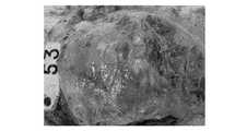
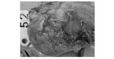
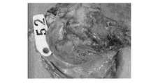
3. 超音波検査結果
T01群では4頭中1頭(No.53)で軽度の心嚢水の貯留が確認されたが,癒着や拍動状態の異常は確認されなかった。T02群では4頭中3頭(No.8,No.25,No.55)で軽度から中程度の心嚢水の貯留,4頭中1頭(No.25)で軽度の癒着が確認された。拍動状態の異常は確認されなかった。T03群では2頭中1頭(No.51)で軽度の心嚢水の貯留が確認されたが,癒着や拍動状態の異常は 確認されなかった。3. Ultrasonic test results
In the T01 group, mild pericardial effusion was confirmed in 1 out of 4 (No. 53), but no abnormal adhesion or pulsatile status was confirmed. In T02 group, mild to moderate pericardial effusion was observed in 3 of 4 (No.8, No.25, No.55), and mild adhesion was confirmed in 1 of 4 (No.25). . Abnormal pulsation was not confirmed. In the T03 group, mild pericardial effusion was confirmed in 1 of 2 animals (No. 51), but no abnormal adhesion or pulsatile status was observed.
4. 剖検所見スコア
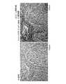
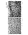
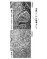
写真を図2〜図31示した。4). Necropsy findings score photographs are shown in FIGS.
T01群では人工心嚢膜と心臓,肺及び心嚢膜との癒着は認められなかったが,軽度の心嚢水貯留が4頭中1頭(No.53),軽度の胸水貯留が4頭中2頭(No.53,No.59)で認められた。また,胸腔内で処置時の開胸に起因すると思われる胸骨・胸膜の軽度の癒着が4頭中3頭(No.53,No.54,No.59)で認められた。
T02群では人工心嚢膜と心臓との中程度の癒着が4頭中2頭(No.25,No.57),肺及び心嚢膜との中程度の癒着が4頭中1頭(No.25),軽度の心嚢水貯留が4頭全頭,胸水貯留が4頭中2頭(No.8,No.25),胸骨・胸膜の軽度の癒着が4頭中2頭(No.8,No.25)で認められた。
T03群では人工心嚢膜と心臓との中程度の癒着,肺及び心嚢膜との軽度の癒着,軽度の心嚢水及び胸水貯留が2頭中1頭(No.51),胸骨・胸膜の中程度の癒着が2頭とも認められた。
合計剖検所見スコアの群別平均はT01群で1.8,T02群で4.0,T03群で5.0であった。In the T01 group, there was no adhesion between the artificial pericardium and the heart, lungs, and pericardial membrane, but 1 of 4 cases of mild pericardial effusion (No. 53) and 4 cases of mild pleural effusion Recognized in 2 animals (No.53, No.59). In addition, mild adhesions of the sternum and pleura, which were probably caused by thoracotomy during treatment in the thoracic cavity, were observed in 3 out of 4 animals (No. 53, No. 54, No. 59).
In T02 group, moderate adhesion between artificial pericardium and heart was 2 out of 4 (No.25, No.57), moderate adhesion between lung and pericardial membrane was 1 out of 4 (No. .25), all 4 heads with mild pericardial effusion, 2 out of 4 pleural effusions (No.8, No.25), and 2 out of 4 mild sternal and pleural adhesions (No.8) , No.25).
In T03 group, moderate adhesion between artificial pericardium and heart, mild adhesion with lung and pericardial membrane, mild pericardial effusion and pleural effusion were observed in 1 of 2 (No. 51), sternum and pleural Two moderate adhesions were observed.
The average of autopsy findings by group was 1.8 in the T01 group, 4.0 in the T02 group, and 5.0 in the T03 group.
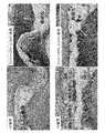
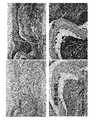
5. 病理組織学的スコア
写真を図32から図41に示した。
病理所見はイオン照射面と非照射面でスコアが違うので,照射面と非照射面でそれぞれ評価した。
イオンビーム外向き照射は照射面が心嚢膜側,非照射面が心筋(心臓)側,イオンビーム内向き照射は照射面が心筋(心臓)側,非照射面が心嚢膜側となる。ゴアテックス群については,イオンビーム照射はないので心筋側と心嚢膜側のスコアは同じである。
T01群では照射面(心嚢膜側)では好中球,リンパ球,マクロファージ及び異物巨細胞等の滲出細胞の浸潤が軽度から重度の幅広い範囲で認められ,線維芽細胞の浸潤も中程度から重度認められ線維化の進行が示唆された。これに対し非照射面(心筋側)は滲出細胞の浸潤,線維芽細胞の浸潤及び線維化は軽度であった。
T02群では照射面(心筋側)では好中球,リンパ球,マクロファージ及び異物巨細胞等の滲出細胞の浸潤が軽度から中程度の範囲で認められ,線維芽細胞の浸潤も中程度認められ線維化の進行が示唆された。これに対し非照射面(心筋側)は滲出細胞の浸潤,線維芽細胞の浸潤及び線維化は軽度であった。
T03群では心嚢膜側と心筋側はおなじスコアであり,好中球,リンパ球,マクロファージ及び異物巨細胞等の滲出細胞の浸潤が軽度から中程度の範囲で認められ,線維芽細胞の浸潤も軽度から中程度認められ線維化の進行が示唆された。
平均線維化スコアはT01群で心嚢膜側照射で5.0,心筋側非照射で2.0,T02群で心筋側照射で4.0,心嚢膜側非照射で2.0,T03群で心嚢膜側,心筋側共に3.0であった。
線維芽細胞の浸潤による線維化は異物に対する生体の正常な異物処理反応であり,線維化が進行する(スコアが高い)程修復過程にあるものと判断される。なのでT01群は心嚢膜側で修復過程が顕著でまわりの組織との融合が進行していると判断できる。5. Histopathological score photographs are shown in FIGS.
The pathological findings were evaluated on the irradiated and non-irradiated surfaces because the scores were different between the ion-irradiated surface and the non-irradiated surface.
For ion beam outward irradiation, the irradiated surface is the pericardial membrane side, the non-irradiated surface is the myocardial (heart) side, and for ion beam inward irradiation, the irradiated surface is the myocardial (heart) side, and the non-irradiated surface is the pericardial membrane side. In the Gore-Tex group, there is no ion beam irradiation, so the scores on the myocardium side and pericardial side are the same.
In T01 group, infiltration of exudate cells such as neutrophils, lymphocytes, macrophages and foreign body giant cells was observed in a wide range from mild to severe on the irradiated surface (pericardial side), and fibroblast infiltration was also moderate It was severe and suggests the progression of fibrosis. In contrast, exudate cell infiltration, fibroblast infiltration and fibrosis were mild on the non-irradiated side (myocardial side).
In the T02 group, infiltration of exudate cells such as neutrophils, lymphocytes, macrophages and foreign body giant cells was observed in the mild to moderate range on the irradiated surface (myocardial side), and fibroblast infiltration was also observed moderately. The progress of conversion was suggested. In contrast, exudate cell infiltration, fibroblast infiltration and fibrosis were mild on the non-irradiated side (myocardial side).
In the T03 group, the pericardial side and the myocardial side had the same score, and infiltration of exudate cells such as neutrophils, lymphocytes, macrophages and foreign body giant cells was observed in a mild to moderate range, and fibroblast infiltration Mild to moderate, suggesting the progression of fibrosis.
The average fibrosis score was 5.0 for the T01 group when irradiated with the pericardial membrane side, 2.0 when the myocardial side was not irradiated, 4.0 when the T02 group was irradiated with myocardial side, 2.0 when the pericardial side was not irradiated, and 2.0 Both sides were 3.0.
Fibrosis due to infiltration of fibroblasts is a normal foreign body processing reaction to foreign bodies, and it is judged that the fibrosis progresses (the score is higher) and is in a repair process. Therefore, in the T01 group, it can be judged that the repair process is remarkable on the pericardial side and fusion with surrounding tissues is progressing.
まとめ及び考察
2017年2月から6月にかけて,人体用医療機器の承認申請予定の新規人工心嚢膜の有効性及び安全性を確認するために,ブタ10頭を供試して人工心嚢膜移植を実施し,術後8週間の経過観察後剖検,病理組織学的検査を実施して,その有効性及び安全性を確認した。
その結果,一般状態の異常は認められず,処置部位の観察でも特筆すべき異常は認められなかった。増体量も通常の想定内の増体量であり,問題はなかった。超音波検査ではイオンビーム照射面が外向き(心嚢膜側)の群は4頭中1頭で軽度の心嚢水貯留が認められたのみで,癒着や拍動の異常は認めらなかった。イオンビーム照射面が内向き(心筋側)の群は軽度から中程度の心嚢水貯留が認められ,軽度の癒着も1頭で認められたが,拍動の異常は認められなかった。ゴアテックス群は2頭中1頭でごく軽微な心嚢水貯留が認められたが,癒着や拍動の異常は認められなかった。
剖検所見ではイオンビーム照射面が外向き(心嚢膜側)の群は人工心嚢膜と心臓,肺,心嚢膜との癒着は認められず心嚢水及び胸水の軽度の貯留が4頭中1頭で認められたのみであった。
イオンビーム照射面が内向き(心筋側)の群は人工心嚢膜と心臓の中程度の癒着が4頭中2頭,肺及び心嚢膜との中程度の癒着が4頭中1頭で認められ,心嚢水の及び胸水の軽度の貯留が4頭中1頭で認められた。
ゴアテックス群は2頭中1頭で心臓の中程度の癒着,肺及び心嚢膜との軽度の癒着,心嚢水の及び胸水の軽度の貯留が認められた。
病理組織学的検査ではイオンビーム照射面が外向き(心嚢膜側)の群は心嚢膜側の滲出細胞の浸潤や繊維芽細胞の浸潤等による線維化が活発であった。線維芽細胞の浸潤による線維化は異物に対する生体の正常な異物処理反応であり,線維化が活発である程修復過程にあるものと判断される。このことは人工心嚢膜と既存の心嚢膜との融合が活発で心臓領域の心嚢水が縫合部分から漏れ出す可能性低くなることであり,結果的に胸水の貯留や肺等の臓器との癒着の危険性も軽減できることが示唆された。
これに対しイオンビーム照射面が内向き(心筋側)の群は心筋側で滲出細胞の浸潤や繊維芽細胞 の浸潤等による線維化が活発であり,胸腔臓器間の癒着,心嚢水あるいは胸水の貯留等の危険性が否定できない。
ただ,対照物質のゴアテックス群もイオンビーム照射面が内向き(心筋側)の群と同様の結果であることから安全性に問題はないと思われる。Summary and discussion
From February to June 2017, in order to confirm the effectiveness and safety of the new artificial pericardium scheduled to be applied for approval of medical devices for human bodies, we conducted artificial pericardial transplantation using 10 pigs. After the follow-up for 8 weeks after the operation, autopsy and histopathological examination were performed to confirm its efficacy and safety.
As a result, no abnormalities were observed in the general condition, and no abnormalities were noted in observation of the treatment site. The weight gain was also within the normal assumption and there was no problem. Ultrasonography showed that only one of four animals in the group with the ion beam irradiation surface facing outward (pericardial side) had mild pericardial effusion, and no adhesion or pulsation abnormality was observed. In the group in which the ion beam irradiation surface was inward (myocardial side), mild to moderate pericardial effusion was observed and mild adhesion was observed in one animal, but no abnormal pulsation was observed. In the Gore-Tex group, only one of the two animals had a slight pericardial effusion, but no adhesion or pulsation abnormality was observed.
Necropsy findings showed that there was no adhesion between the artificial pericardium and the heart, lungs, and pericardium in the group with the ion beam irradiation surface facing outward (pericardial side), and there was slight accumulation of pericardial effusion and pleural effusion in 4 Only one was recognized.
In the group whose ion beam irradiation surface is facing inward (myocardial side), medium perfusion of artificial pericardium and heart is 2 out of 4 heads, moderate adhesion of lung and pericardium is 1 out of 4 heads A slight accumulation of pericardial effusion and pleural effusion was observed in 1 of 4 animals.
In the Gore-Tex group, moderate adhesion of the heart, mild adhesion to the lungs and pericardium, mild pericardial effusion and pleural effusion were observed in 1 of 2 animals.
In the histopathological examination, the group with the ion beam irradiation surface facing outward (pericardial side) showed active fibrosis due to infiltration of exudate cells on the pericardial membrane side or infiltration of fibroblasts. Fibrosis due to infiltration of fibroblasts is a normal foreign body treatment reaction to foreign bodies, and it is judged that the more fibrosis is active, the more it is in the repair process. This means that the fusion between the artificial pericardium and the existing pericardial membrane is active, and the possibility that the pericardial effusion in the heart region leaks from the sutured area is reduced. It was suggested that the risk of adhesions can be reduced.
In contrast, in the group with the ion beam irradiation surface facing inward (myocardial side), fibrosis due to exudate cell infiltration or fibroblast infiltration is active on the myocardium side, and adhesion between thoracic organs, pericardial effusion, or pleural effusion The risk of storage cannot be denied.
However, the Gore-Tex group of the control substance is similar to the group with the ion beam irradiation surface facing inward (myocardial side), so there seems to be no problem in safety.
以上のことから新規人工心嚢膜はイオンビーム照射面が外向き(心嚢膜側)になるように移植することにより,イオンビーム照射の作用で線維化が活発し既存の心嚢膜との融合が活発となり,心臓領域の心嚢水が縫合部分から漏れ出す可能性低くなるため,結果的に胸水の貯留や肺等の臓器との癒着の危険性も軽減できることから高い有効性及び安全性が確認された。なお,イオンビーム照射面を内向き(心筋側)に移植すると,心筋側で滲出細胞の浸潤や繊維芽細胞の浸潤等による線維化が活発になり,胸腔臓器間の癒着,心嚢水あるいは胸水の貯留等の危険性はあるが,対照物質のゴアテックス群も同様の結果のため,内向き移植でも効果・安全性は担保できるものと推察される。 From the above, the new artificial pericardium is transplanted so that the ion beam irradiation surface faces outward (pericardial membrane side), and fibrosis is activated by the action of ion beam irradiation. Because fusion becomes active and the pericardial effusion in the heart region is less likely to leak from the sutured part, the risk of pleural effusion accumulation and adhesion to organs such as the lungs can be reduced, resulting in high effectiveness and safety. confirmed. When the ion beam irradiation surface is transplanted inward (myocardial side), fibrosis becomes active on the myocardial side due to infiltration of exudate cells and fibroblasts, and adhesion between thoracic organs, pericardial effusion or pleural effusion Although there is a risk of storage, etc., the Gore-Tex group of the control substance has the same result, so it is presumed that the efficacy and safety can be guaranteed even with inward transplantation.
本発明は,医療機器の分野で利用されうる。 The present invention can be used in the field of medical devices.
1 人工心嚢膜シート, 3 粗面化部, 5 第1の面, 7 第2の面 DESCRIPTION OF
Claims (6)
Translated fromJapanese前記人工心嚢膜シートは,
前記心嚢膜側に配置され,少なくとも前記心嚢膜と接する部位に粗面化部(3)を有する第1の面(5)と,
第1の面(5)反対に位置する第2の面(7)を有し,ポリテトラフルオロエチレンを含む,人工心嚢膜シートであって,
前記粗面化部(3)のb値であるb1値と,第2の面(7)のb値であるb2値の差をΔbとしたときに,Δbが2以上11以下である人工心嚢膜シート。An artificial pericardial membrane sheet (1) embedded within the removed or resected pericardial membrane,
The artificial pericardium sheet is:
A first surface (5) disposed on the pericardial membrane side and having a roughened portion (3) at least at a site in contact with the pericardial membrane;
Anartificial pericardial membrane sheet having a second surface (7) located opposite the first surface (5) and comprising polytetrafluoroethylene,
Wherein b1value anda b value of roughening portion(3),the difference between theb2valuesis b value of the second surface (7)when the [Delta] b, [Delta] b is 2 to 11 Artificial pericardium sheet .
Priority Applications (3)
| Application Number | Priority Date | Filing Date | Title |
|---|---|---|---|
| JP2017226343AJP6316496B1 (en) | 2017-11-24 | 2017-11-24 | Artificial pericardium sheet |
| PCT/JP2018/039620WO2019102779A1 (en) | 2017-11-24 | 2018-10-25 | Artificial pericardial membrane sheet |
| TW107141495ATW201924731A (en) | 2017-11-24 | 2018-11-21 | Artificial pericardial membrane sheet |
Applications Claiming Priority (1)
| Application Number | Priority Date | Filing Date | Title |
|---|---|---|---|
| JP2017226343AJP6316496B1 (en) | 2017-11-24 | 2017-11-24 | Artificial pericardium sheet |
Related Child Applications (1)
| Application Number | Title | Priority Date | Filing Date |
|---|---|---|---|
| JP2018018874ADivisionJP2019093098A (en) | 2018-02-06 | 2018-02-06 | Artificial pericardial sheet |
Publications (2)
| Publication Number | Publication Date |
|---|---|
| JP6316496B1true JP6316496B1 (en) | 2018-04-25 |
| JP2019092950A JP2019092950A (en) | 2019-06-20 |
Family
ID=62069299
Family Applications (1)
| Application Number | Title | Priority Date | Filing Date |
|---|---|---|---|
| JP2017226343AActiveJP6316496B1 (en) | 2017-11-24 | 2017-11-24 | Artificial pericardium sheet |
Country Status (3)
| Country | Link |
|---|---|
| JP (1) | JP6316496B1 (en) |
| TW (1) | TW201924731A (en) |
| WO (1) | WO2019102779A1 (en) |
Cited By (1)
| Publication number | Priority date | Publication date | Assignee | Title |
|---|---|---|---|---|
| JP3313510B2 (en) | 1994-05-31 | 2002-08-12 | 津田駒工業株式会社 | Shearless processing equipment for shuttleless loom |
Family Cites Families (3)
| Publication number | Priority date | Publication date | Assignee | Title |
|---|---|---|---|---|
| JP3563216B2 (en)* | 1996-10-14 | 2004-09-08 | 株式会社アムニオテック | Medical substitute membrane and method for producing the same |
| JP5505752B2 (en)* | 2001-04-23 | 2014-05-28 | 独立行政法人理化学研究所 | Artificial dura mater having cell adhesion and method for producing the same |
| JP4445697B2 (en)* | 2002-08-30 | 2010-04-07 | 独立行政法人理化学研究所 | Biological repair material having affinity with biological tissue adhesive |
- 2017
- 2017-11-24JPJP2017226343Apatent/JP6316496B1/enactiveActive
- 2018
- 2018-10-25WOPCT/JP2018/039620patent/WO2019102779A1/ennot_activeCeased
- 2018-11-21TWTW107141495Apatent/TW201924731A/enunknown
Cited By (1)
| Publication number | Priority date | Publication date | Assignee | Title |
|---|---|---|---|---|
| JP3313510B2 (en) | 1994-05-31 | 2002-08-12 | 津田駒工業株式会社 | Shearless processing equipment for shuttleless loom |
Also Published As
| Publication number | Publication date |
|---|---|
| TW201924731A (en) | 2019-07-01 |
| WO2019102779A1 (en) | 2019-05-31 |
| JP2019092950A (en) | 2019-06-20 |
Similar Documents
| Publication | Publication Date | Title |
|---|---|---|
| JP7012779B2 (en) | Surgical method using purified amphipathic peptide composition | |
| Carrel | On the experimental surgery of the thoracic aorta and the heart | |
| KR100645120B1 (en) | Submucosal graft structure of the tube | |
| TW200924803A (en) | Use of a regenerative biofunctional collagen biomatrix for treating visceral or parietal defects | |
| US20160121031A1 (en) | Antiadhesive Kit and Method of Adhesion Prevention | |
| JP6316496B1 (en) | Artificial pericardium sheet | |
| CN111227989A (en) | Method for making mouse myocardial infarction model | |
| JP2019093098A (en) | Artificial pericardial sheet | |
| Bendinelli et al. | Spontaneous pneumothorax in two dogs undergoing combined laparoscopic ovariectomy and total laparoscopic gastropexy | |
| JPWO2005113030A1 (en) | Tissue closure agent | |
| JP6343390B2 (en) | Dental shielding film using scissors and method for producing the same | |
| Moro et al. | The effect of fibrin glue on inhibition of pericardial adhesions | |
| CN111378970B (en) | Method for preparing high-frequency electric knife insulating coating based on micro-arc oxidation method | |
| RU2654601C1 (en) | Method for modeling plastics of trachea wall epithelial defect | |
| CARREL | ON THE EXPERIMENTAL SURGERY OF THE | |
| Raut et al. | Anterior tracheal injury during sternotomy | |
| CN108126241A (en) | Tissue repair sticking patch, main body and preparation method | |
| RU2177262C1 (en) | Surgical method for stopping pulmonary hemorrhages | |
| RU2195192C1 (en) | Method for surgical treatment of hepatic echinococcus | |
| Shigematsu et al. | Thoracoscopic surgery using local and epidural anesthesia for intractable pneumothorax after BMT | |
| TWI610651B (en) | A magnesium alloy sewing nail | |
| Mitsuyama et al. | Response to: VATS for refractory pneumothorax: a minimal access curative surgery | |
| SU1409234A1 (en) | Method of investigating the functional ability of myocardium in case of through defect of the heart | |
| Goodman | A HISTOLOGICAL STUDY OF THE CIRCULAR SUTURE OF THE BLOOD-VESSELS | |
| CN120571072A (en) | Gradient self-crosslinking to construct asymmetric adhesive patch, construction method and application thereof |
Legal Events
| Date | Code | Title | Description |
|---|---|---|---|
| A621 | Written request for application examination | Free format text:JAPANESE INTERMEDIATE CODE: A621 Effective date:20171218 | |
| A871 | Explanation of circumstances concerning accelerated examination | Free format text:JAPANESE INTERMEDIATE CODE: A871 Effective date:20171218 | |
| A975 | Report on accelerated examination | Free format text:JAPANESE INTERMEDIATE CODE: A971005 Effective date:20180111 | |
| A131 | Notification of reasons for refusal | Free format text:JAPANESE INTERMEDIATE CODE: A131 Effective date:20180130 | |
| A521 | Request for written amendment filed | Free format text:JAPANESE INTERMEDIATE CODE: A523 Effective date:20180206 | |
| TRDD | Decision of grant or rejection written | ||
| A01 | Written decision to grant a patent or to grant a registration (utility model) | Free format text:JAPANESE INTERMEDIATE CODE: A01 Effective date:20180313 | |
| A61 | First payment of annual fees (during grant procedure) | Free format text:JAPANESE INTERMEDIATE CODE: A61 Effective date:20180327 | |
| R150 | Certificate of patent or registration of utility model | Ref document number:6316496 Country of ref document:JP Free format text:JAPANESE INTERMEDIATE CODE: R150 | |
| R250 | Receipt of annual fees | Free format text:JAPANESE INTERMEDIATE CODE: R250 | |
| R250 | Receipt of annual fees | Free format text:JAPANESE INTERMEDIATE CODE: R250 | |
| R250 | Receipt of annual fees | Free format text:JAPANESE INTERMEDIATE CODE: R250 | |
| R250 | Receipt of annual fees | Free format text:JAPANESE INTERMEDIATE CODE: R250 | |
| R250 | Receipt of annual fees | Free format text:JAPANESE INTERMEDIATE CODE: R250 |