JP6273940B2 - Image analysis apparatus, image photographing system, and image analysis program - Google Patents
Image analysis apparatus, image photographing system, and image analysis programDownload PDFInfo
- Publication number
- JP6273940B2 JP6273940B2JP2014056119AJP2014056119AJP6273940B2JP 6273940 B2JP6273940 B2JP 6273940B2JP 2014056119 AJP2014056119 AJP 2014056119AJP 2014056119 AJP2014056119 AJP 2014056119AJP 6273940 B2JP6273940 B2JP 6273940B2
- Authority
- JP
- Japan
- Prior art keywords
- heart
- index
- deriving
- indices
- image analysis
- Prior art date
- Legal status (The legal status is an assumption and is not a legal conclusion. Google has not performed a legal analysis and makes no representation as to the accuracy of the status listed.)
- Active
Links
Images


Classifications
- G—PHYSICS
- G06—COMPUTING OR CALCULATING; COUNTING
- G06T—IMAGE DATA PROCESSING OR GENERATION, IN GENERAL
- G06T7/00—Image analysis
- G06T7/0002—Inspection of images, e.g. flaw detection
- G06T7/0012—Biomedical image inspection
- G06T7/0014—Biomedical image inspection using an image reference approach
- G06T7/0016—Biomedical image inspection using an image reference approach involving temporal comparison
- G—PHYSICS
- G06—COMPUTING OR CALCULATING; COUNTING
- G06T—IMAGE DATA PROCESSING OR GENERATION, IN GENERAL
- G06T2207/00—Indexing scheme for image analysis or image enhancement
- G06T2207/10—Image acquisition modality
- G06T2207/10116—X-ray image
- G—PHYSICS
- G06—COMPUTING OR CALCULATING; COUNTING
- G06T—IMAGE DATA PROCESSING OR GENERATION, IN GENERAL
- G06T2207/00—Indexing scheme for image analysis or image enhancement
- G06T2207/30—Subject of image; Context of image processing
- G06T2207/30004—Biomedical image processing
- G06T2207/30048—Heart; Cardiac
- G—PHYSICS
- G06—COMPUTING OR CALCULATING; COUNTING
- G06T—IMAGE DATA PROCESSING OR GENERATION, IN GENERAL
- G06T2207/00—Indexing scheme for image analysis or image enhancement
- G06T2207/30—Subject of image; Context of image processing
- G06T2207/30004—Biomedical image processing
- G06T2207/30061—Lung
Landscapes
- Engineering & Computer Science (AREA)
- Physics & Mathematics (AREA)
- Theoretical Computer Science (AREA)
- General Physics & Mathematics (AREA)
- Medical Informatics (AREA)
- Radiology & Medical Imaging (AREA)
- Quality & Reliability (AREA)
- Computer Vision & Pattern Recognition (AREA)
- Nuclear Medicine, Radiotherapy & Molecular Imaging (AREA)
- Health & Medical Sciences (AREA)
- General Health & Medical Sciences (AREA)
- Apparatus For Radiation Diagnosis (AREA)
- Measurement Of The Respiration, Hearing Ability, Form, And Blood Characteristics Of Living Organisms (AREA)
Description
Translated fromJapanese本発明は、肺機能を示す指標を導出する画像解析装置、画像撮影システム及び画像解析プログラムに関する。 The present invention relates to an image analysis device, an image capturing system, and an image analysis program for deriving an index indicating lung function.
慢性閉塞性肺疾患(COPD)等の肺疾患への罹患の有無を診断するのにあたってスパイロメトリーが行われる場合がある。スパイロメトリーにおいては、検査される人の換気量が測定される。測定された換気量からは、肺活量、1秒率等の指標が導出される場合もあるし、スパイログラフと呼ばれる図が作成される場合もある。 Spirometry may be performed in diagnosing the presence or absence of pulmonary diseases such as chronic obstructive pulmonary disease (COPD). In spirometry, the ventilation of a person being examined is measured. An index such as vital capacity, a rate of one second, or the like may be derived from the measured ventilation volume, or a diagram called a spirograph may be created.
特許文献1に記載された発明は、血流情報を導出する。特許文献1に記載された発明においては、胸部X線動画像から心壁の部位が自動検出され、心壁の部位の変化量が心壁移動量とされる。心壁移動量からは、心拍位相が推定される。 The invention described in
スパイトメトリーは、COPD等の肺疾患への罹患の有無を診断するのに役立つ。しかし、スパイロメトリーは、検査される人に大きな負担を与え、検査結果の再現性が良好でないという問題を有する。 Spitometry is useful for diagnosing the presence or absence of pulmonary diseases such as COPD. However, spirometry has a problem that it places a heavy burden on the person being inspected and the reproducibility of the inspection result is not good.
本発明は、この問題を解決するためになされる。本発明の目的は、大きな負担を検査される人に与えることなく肺機能を再現性よく検査することである。 The present invention is made to solve this problem. An object of the present invention is to examine lung function with high reproducibility without giving a heavy burden to a person to be examined.
医用動画像が取得される。医用動画像は、心臓を撮像することにより得られ、2個以上のフレーム画像を有する。2個以上のフレーム画像について2個以上の第1の指標が導出される。2個以上の第1の指標の各々は、心臓状態を示す。2個以上の第1の指標から第2の指標が導出される。第2の指標は、肺機能を示す。 A medical moving image is acquired. A medical moving image is obtained by imaging the heart and has two or more frame images. Two or more first indices are derived for two or more frame images. Each of the two or more first indicators indicates a heart condition. A second index is derived from two or more first indices. The second index indicates lung function.
大きな負担を検査される人に与えることなく肺機能が再現性よく検査される。 Lung function is tested with good reproducibility without imposing a heavy burden on the person being tested.
これらの及びこれら以外の本発明の目的、特徴、局面及び利点は、添付図面とともに考慮されたときに下記の本発明の詳細な説明によってより明白となる。 These and other objects, features, aspects and advantages of the invention will become more apparent from the following detailed description of the invention when considered in conjunction with the accompanying drawings.
(1)着想
(1.1)呼吸状態
呼吸が行われる場合は、横隔膜の収縮及び弛緩が交互に繰り返される。横隔膜が収縮する場合は、肺の内部の気圧が低下し、肺の外部から肺の内部へ空気が流入する。横隔膜が弛緩する場合は、肺の内部の気圧が上昇し、肺の内部から肺の外部へ空気が流出する。(1) Idea (1.1) Breathing state When breathing is performed, contraction and relaxation of the diaphragm are repeated alternately. When the diaphragm contracts, the air pressure inside the lungs decreases, and air flows from the outside of the lungs into the lungs. When the diaphragm relaxes, the air pressure inside the lungs rises and air flows out of the lungs to the outside of the lungs.
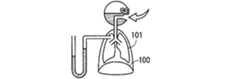
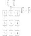
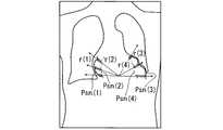
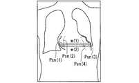
図1の模式図は、呼気期から吸気期へ遷移する頃における胸部を示す。図2の模式図は、吸気期における胸部を示す。図3の模式図は、吸気期から呼気期へ遷移する頃における胸部を示す。 The schematic diagram of FIG. 1 shows the chest at the time of transition from the expiration period to the inspiration period. The schematic diagram of FIG. 2 shows the chest during the inspiratory period. The schematic diagram of FIG. 3 shows the chest at the time of transition from the inspiratory period to the expiratory period.
図1に示されるように、呼気期から吸気期へ遷移する頃においては、横隔膜100が相対的に上方にあり、肺101が相対的に小さく、肺101の内部の気圧が肺の外部の気圧と同じである。図2に示されるように、吸気期においては、時間が経過するにつれて横隔膜100が下がり、時間が経過するにつれて肺101が大きくなり、肺101の内部の気圧が肺101の外部の気圧より低く、肺101の外部から肺101の内部へ空気が流入する。図3に示されるように、吸気期から呼気期へ遷移する頃においては、横隔膜100が相対的に下方にあり、肺101が相対的に大きく、肺101の内部の圧力が肺101の外部の気圧と同じである。このように、呼吸が行われる場合は、肺の大きさ、肺の内部の気圧等が変化する。肺の大きさ、肺の内部の気圧等の変化により、肺と心臓との位置関係も変化する。 As shown in FIG. 1, at the time of transition from the expiration phase to the inspiration phase, the
(1.2)心臓状態の呼吸状態による変化
心臓は、肺に接し、肺の影響を受けながら拍動する。このため、心臓の大きさ、心壁の位置等は、肺の大きさ、肺の内部の気圧、肺と心臓との位置関係等の影響を受ける。心臓の大きさ、心壁の位置等の心拍による変化も肺の大きさ、肺の内部の気圧、肺と心臓との位置関係等の影響を受ける。(1.2) Changes in heart condition due to respiratory condition The heart touches the lungs and beats while being affected by the lungs. For this reason, the size of the heart, the position of the heart wall, and the like are affected by the size of the lung, the air pressure inside the lung, the positional relationship between the lung and the heart, and the like. Changes due to heartbeat such as the size of the heart and the position of the heart wall are also affected by the size of the lung, the pressure inside the lung, the positional relationship between the lung and the heart, and the like.
図4の模式図は、胸部X線画像、心臓の大きさの時間変化、心壁の位置の時間変化及び肺の内部の空気量の時間変化の例を示す。図4は、心臓の大きさ及び心壁の位置の心拍による変化(心壁の動き)が肺の内部の気圧の影響を受けることを示す。 The schematic diagram of FIG. 4 shows an example of a chest X-ray image, a temporal change in the size of the heart, a temporal change in the position of the heart wall, and a temporal change in the amount of air in the lung. FIG. 4 shows that changes in heart size and heart wall position due to heartbeat (heart wall motion) are affected by the pressure inside the lungs.
図4に示される例においては、肺の内部の気圧が低くなる吸気期に心臓の大きさが相対的に大きくなり心壁の動きが相対的に大きくなり、肺の内部の気圧が高くなる呼気期に心臓の大きさが相対的に小さくなり心壁の動きが相対的に小さくなる。このため、吸気期と呼気期との間の心臓の大きさ又は心壁の動きの変化から吸気期と呼気期との間の肺の内部の気圧の変化が推定される。 In the example shown in FIG. 4, during the inspiration period when the air pressure inside the lungs becomes low, the heart size becomes relatively large and the movement of the heart wall becomes relatively large, so that the air pressure inside the lungs becomes high. During the period, the size of the heart becomes relatively small and the movement of the heart wall becomes relatively small. For this reason, the change in the air pressure inside the lung between the inspiration period and the expiration period is estimated from the change in the heart size or the movement of the heart wall between the inspiration period and the expiration period.
(1.3)心臓状態の呼吸状態による変化の利用
慢性閉塞性肺疾患(COPD)は、有毒な粒子、ガス等を吸入することにより生じる肺の炎症を伴う進行性の気流制限を呈する。気流制限のため、COPD患者における吸気期と呼気期との間の肺の内部の気圧の変化は、健常者における吸気期と呼気期との間の肺の内部の気圧の変化より大きい。このため、吸気期と呼気期との間の肺の内部の圧力の変化を知ることにより、COPDへの罹患の有無を推定できる。吸気期と呼気期との間の肺の内部の圧力の変化は、吸気期と呼気期との間の心臓の大きさ又は心壁の動きの変化から推定されるため、吸気期と呼気期との間の心臓の大きさ又は心壁の動きの変化は、COPDへの罹患の有無を推定するのに役立つ。例えば、吸気期と呼気期との間の心臓の大きさ又は心壁の動きの変化が大きい場合は、COPDへの罹患の疑いがある。(1.3) Utilization of changes in cardiac state due to respiratory condition Chronic obstructive pulmonary disease (COPD) presents progressive airflow limitation with lung inflammation caused by inhalation of toxic particles, gases, and the like. Due to airflow limitation, the change in air pressure inside the lungs between the inspiratory phase and the expiratory phase in COPD patients is greater than the change in air pressure inside the lungs between the inspiratory phase and the expiratory phase in healthy individuals. For this reason, the presence or absence of COPD can be estimated by knowing the change in the internal pressure of the lung between the inspiration period and the expiration period. Changes in the internal pressure of the lungs between inspiratory and expiratory phases are estimated from changes in heart size or heart wall motion between inspiratory and expiratory phases, so inspiratory and expiratory phases Changes in heart size or heart wall motion during the period help to estimate the presence or absence of COPD. For example, if there is a large change in heart size or heart wall motion between inspiratory and expiratory phases, there is a suspicion of suffering from COPD.
吸気期と呼気期との間の心臓の大きさ又は心壁の動きの変化は、COPDへの罹患の有無の推定以外の肺機能の解析にも役立つ。心臓の大きさ及び心壁の動き以外の心臓状態を示す指標が用いられてもよい。吸気期と呼気期との間の変化以外の呼吸状態による変化が用いられてもよい。一般的には、心臓状態を示す指標の呼吸状態による変化から肺機能を示す指標が導出される。 Changes in heart size or heart wall motion between the inspiratory phase and the expiratory phase are also useful for lung function analysis other than estimation of the presence or absence of COPD. An index indicating a heart state other than the size of the heart and the movement of the heart wall may be used. Changes due to respiratory conditions other than changes between the inspiratory period and the expiratory period may be used. In general, an index indicating pulmonary function is derived from a change in an index indicating a heart state depending on a respiratory state.
(2)第1実施形態
(2.1)画像撮影システム
第1実施形態は、画像撮影システムに関する。(2) First Embodiment (2.1) Image Shooting System The first embodiment relates to an image shooting system.
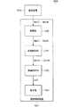
図5のブロック図は、第1実施形態の画像撮影システムを示す。 The block diagram of FIG. 5 shows the image photographing system of the first embodiment.
図5に示される画像撮影システム1000は、撮影装置1010及び画像解析装置1011を備える。撮影装置1010は、医用動画像を生成する。画像解析装置1011は、医用動画像を解析し、肺機能を示す指標を導出する。画像解析装置1011によれば、大きな負担を検査される人に与えることなく肺機能を再現性よく検査できる。 An
(2.2)撮影装置
撮影装置1010は、X線撮影により医用動画像を生成する。撮影装置1010は、X線管によりX線を発生し、発生したX線に人体を透過させ、人体を透過したX線を平面X線検出器(FPD)により検出する。これにより、撮影装置1010は、人の体内の構造物を撮影し、人の体内の構造物を描写したフレーム画像を生成する。撮影装置1010が人以外の動物の体内の構造物を撮影してもよい。撮影装置1010は、2回以上の撮影を行い、2個以上のフレーム画像を有する医用動画像を生成する。医用動画像は、2次元動画像であり、デジタルデーターで表現される。2個以上のフレーム画像の各々は、2次元画像であり、2次元画像データーで表現される。撮影装置1010がX線撮影以外により医用動画像を生成してもよい。例えば、撮影装置1010が超音波撮影、核磁気共鳴画像法(MRI)、コンピューター断層撮影(CT)等により医用動画像を生成してもよい。医用動画像が3次元動画像であってもよい。医用動画像が3次元動画像である場合は、2個以上のフレーム画像の各々は、3次元画像であり、3次元ボリュームデーターで表現される。(2.2) Imaging Device The
(2.3)被撮影物
医用動画像は、胸部X線動画像であり、自然呼吸をしている人の心臓を撮影することにより得られる。医用動画像が人以外の動物の心臓を撮影することにより得られてもよい。医用動画像が努力呼吸をしている人の心臓を撮影することにより得られてもよい。(2.3) Object to be photographed A medical moving image is a chest X-ray moving image, which is obtained by photographing the heart of a person who is breathing naturally. The medical moving image may be obtained by photographing the heart of an animal other than a human. The medical moving image may be obtained by photographing the heart of a person who is breathing hard.
(2.4)撮影方向
胸部は、正面から撮影される。これにより、医用動画像において、心臓と肺とが接する部分が描写された領域が心臓が描写された領域(心臓領域)及び肺が描写された領域(肺領域)と重なりにくくなり、心臓と肺とが接する部分が描写された領域を解析しやすくなる。これに対して、胸部が側面から撮影された場合は、心臓と肺とが接する部分が描写された領域が心臓領域及び肺領域と重なりやすくなり、心臓と肺とが接する部分を解析しにくくなる。(2.4) Imaging direction The chest is imaged from the front. As a result, in the medical moving image, the region in which the portion where the heart and the lung are in contact with each other is difficult to overlap with the region in which the heart is depicted (heart region) and the region in which the lung is depicted (lung region). This makes it easier to analyze the area where the part that touches is drawn. On the other hand, when the chest is photographed from the side, the region in which the portion where the heart and the lung are in contact is easily overlapped with the heart region and the lung region, and it is difficult to analyze the portion where the heart and the lung are in contact with each other. .
ただし、胸部が正面以外から撮影されることも許される。例えば、胸部が側面から撮影されることも許される。胸部が側面から撮影される場合は、望ましくは、心臓の肺と接しない部分が描写された領域が補助的に解析される。心臓の肺と接しない部分が描写された領域が解析の対象になりうるのは、心臓の肺と接しない部分が描写された領域が心臓領域及び肺領域と重なりにくいためであり、肺が心臓を圧迫した場合に心臓の肺と接しない部分がふくらむためである。 However, the chest may be taken from other than the front. For example, the chest can be taken from the side. When the chest is photographed from the side, preferably, an area in which a portion of the heart that does not contact the lungs is depicted is supplementarily analyzed. The region in which the portion of the heart that does not contact the lungs can be analyzed because the region in which the portion of the heart that does not contact the lungs is difficult to overlap with the heart region and the lung region. This is because the portion of the heart that does not come into contact with the lungs is inflated.
(2.5)撮影範囲
心臓状態を示す指標を導出するために心臓の全体を描写する必要がある場合は、心臓の全体を含む範囲が撮影される。例えば、心臓状態を示す指標として心臓の全体の面積が導出される場合は、心臓の大きさが最大となった場合でも心臓の全体を含む範囲が撮影され、2次元動画像が生成される。心臓状態を示す指標として心臓の全体の体積が導出される場合は、心臓の大きさが最大となった場合でも心臓の全体を含む範囲が撮影され、3次元動画像が生成される。心臓状態を示す指標を導出するために心臓の全体を描写する必要がない場合においても、望ましくは心臓の全体を含む範囲が撮影される。これにより、心臓状態を示す指標がロバストに導出される。(2.5) Imaging range When it is necessary to describe the entire heart in order to derive an index indicating the heart state, an area including the entire heart is captured. For example, when the entire area of the heart is derived as an index indicating the heart state, a range including the entire heart is captured even when the size of the heart is maximized, and a two-dimensional moving image is generated. When the entire volume of the heart is derived as an index indicating the heart state, a range including the entire heart is captured even when the size of the heart is maximized, and a three-dimensional moving image is generated. Even in the case where it is not necessary to depict the entire heart in order to derive an indication of the heart condition, a range that includes the entire heart is desirably imaged. Thereby, an index indicating the heart condition is robustly derived.
心臓の一部のみを含む範囲が撮影される場合もある。この場合は、心臓状態を示す指標を導出するのに必要な部分を含む範囲が撮影される。例えば、心壁の位置が導出される場合は、心壁を常に含む範囲が撮影される。心臓の幅が導出される場合は、心臓の幅が最大となった場合でも心臓の幅を導出するのに必要な部分を含む範囲が撮影される。心臓領域の画素値が導出される場合は、画素値が導出される部分を常に含む範囲が撮影される。 A range including only a part of the heart may be captured. In this case, a range including a portion necessary for deriving an index indicating the heart state is photographed. For example, when the position of the heart wall is derived, a range that always includes the heart wall is photographed. When the width of the heart is derived, a range including a portion necessary for deriving the width of the heart is photographed even when the width of the heart is maximized. When the pixel value of the heart region is derived, a range that always includes a portion from which the pixel value is derived is captured.
呼吸状態が医用動画像から検出される場合は、心臓に加えて横隔膜及び肺の両方又は片方を含む範囲が撮影される。これにより、呼吸状態がロバストに検出される。 When the respiratory state is detected from a medical moving image, a range including both or one of the diaphragm and the lung in addition to the heart is imaged. Thereby, the respiratory state is detected robustly.
(2.6)撮影時間
医用動画像は、1呼吸周期以上にわたって撮影され、望ましくは2呼吸周期以上にわたって撮影され、さらに望ましくは3呼吸周期以上にわたって撮影される。1呼吸周期以上にわたって医用動画像が撮影される場合は、吸気期に撮影されたフレーム画像及び呼気期に撮影されたフレーム画像の両方を医用動画像が有し、吸気期の心臓状態及び呼気期の心臓状態が特定される。心臓状態は吸気期と呼気期との間に顕著に変化するため、吸気期の心臓状態及び呼気期の心臓状態が特定されることは、心臓状態を示す指標の呼吸状態による変化を特定することを容易にする。2呼吸周期以上又は3呼吸周期以上にわたって医用動画像が撮影される場合は、撮影される人が緊張により自然呼吸をなかなかできない場合であっても、自然呼吸が行われた状態を撮影できる可能性が高まる。ただし、医用動画像を撮影する時間が1呼吸周期未満である場合でも心臓状態を示す指標の呼吸状態による変化が特定される場合もある。(2.6) Imaging Time The medical moving image is imaged over one respiratory cycle or more, preferably over two respiratory cycles, and more preferably over three respiratory cycles. When a medical moving image is captured over one respiratory cycle or more, the medical moving image has both a frame image captured in the inspiratory period and a frame image captured in the expiratory period, and the heart state and the expiratory period in the inspiratory period The heart condition is identified. Since the cardiac state changes markedly between the inspiratory period and the expiratory period, the heart condition in the inspiratory period and the cardiac condition in the expiratory period are specified to identify changes in the index indicating the cardiac condition due to the respiratory condition To make it easier. When medical moving images are taken over 2 breathing cycles or more than 3 breathing cycles, there is a possibility that the state in which natural breathing is performed can be taken even if the person being photographed cannot easily breathe naturally due to tension Will increase. However, even when the time for taking a medical moving image is less than one respiratory cycle, a change due to the respiratory state of the index indicating the heart state may be specified.
医用動画像を撮影する時間は、5呼吸周期未満とされる。これにより、過度の被爆が回避される。ただし、医用動画像を撮影する時間が5呼吸周期以上である場合でも被爆が問題とならないこともある。例えば、医用動画像が超音波撮影により生成される場合は、被爆は問題とならない。 The time for taking a medical moving image is less than 5 breathing cycles. Thereby, an excessive exposure is avoided. However, even when the time for taking a medical moving image is 5 breathing cycles or more, exposure may not be a problem. For example, when a medical moving image is generated by ultrasonic imaging, exposure is not a problem.
(2.7)フレームレート
フレームレートは、30fps以上である。これにより、心臓パラメーターの心拍による変化と心臓パラメーターの呼吸状態による変化とが識別され、心臓パラメーターから心拍の影響を除去できる。ただし、フレームレートが30fps未満である場合でも心臓パラメーターから心拍の影響を除去できる場合もある。(2.7) Frame rate The frame rate is 30 fps or more. Thereby, a change due to the heartbeat of the heart parameter and a change due to the respiratory state of the heart parameter are distinguished, and the influence of the heartbeat can be removed from the heart parameter. However, even when the frame rate is less than 30 fps, the influence of the heartbeat may be removed from the heart parameter.
2個以上のフレーム画像の各々が撮影される場合に、矩形波状の波形を有するX線が1心拍周期以上にわたって連続して発生させられてもよい。これにより、心臓が拍動する間の全部の状態が2個以上のフレーム画像の各々に描写され、2個以上のフレーム画像の各々から心拍の影響が把握され、2個以上のフレーム画像の各々から心拍の影響を除去できる。矩形波状の波形を有するX線が発生させられる場合のフレームレートは、一の呼吸状態における心臓が一のフレーム画像に描写され他の呼吸状態における心臓が他のフレーム画像に描写されるように設定される。例えば、吸気期及び呼気期の間の心臓状態の変化が導出される場合は、吸気期における心臓が一のフレーム画像に描写され吸気期における心臓が他のフレーム画像に描写されるように、フレームレートが2fps以上4fps以下に設定される。 When each of two or more frame images is captured, X-rays having a rectangular waveform may be continuously generated over one heartbeat period or more. As a result, the entire state during the heart beat is depicted in each of the two or more frame images, the influence of the heartbeat is grasped from each of the two or more frame images, and each of the two or more frame images is displayed. Can remove the effects of heartbeat. The frame rate when X-rays having a rectangular waveform are generated is set so that the heart in one breathing state is drawn in one frame image and the heart in the other breathing state is drawn in another frame image Is done. For example, if a change in heart condition between inspiration and expiration is derived, the frame is such that the heart during inspiration is depicted in one frame image and the heart during inspiration is depicted in another frame image. The rate is set to 2 fps or more and 4 fps or less.
(2.8)撮影の終了
医用動画像の撮影は、心臓状態を示す指標の呼吸状態による変化を導出するのに必要な呼吸状態がとらえられたと画像撮影システム1000が判定した場合に終了する。判定は、例えば、医用動画像における横隔膜、肺等の動きに基づいて行われる。撮影装置1010以外の検出器の検出結果に基づいて判定が行われてもよい。例えば、換気量の検出器の検出結果に基づいて判定が行われてもよい。医用動画像における横隔膜、肺等の動き及び撮影装置1010以外の検出器の検出結果を総合して判定が行われてもよい。操作者の操作にしたがって医用動画像の撮影が終了してもよい。(2.8) End of Imaging Imaging of a medical moving image ends when the
(2.9)画像解析装置
画像解析装置1011は、取得部1020、画像解析部1021、指標解析部1022、表示部1023等を備える。取得部1020は、医用動画像を取得する。画像解析部1021は、医用動画像を解析し、心臓状態を示す指標を導出する。指標解析部1022は、心臓状態を示す指標を解析し、肺機能を示す指標を導出する。表示部1023は、肺機能を示す指標を表示する。(2.9) Image Analysis Device The
(2.10)取得部
取得部1020は、通信を行い、撮影装置1010から直接的に又は撮影装置1010から画像サーバー等の撮影装置1010以外の装置を経由して医用動画像を取得する。医用動画像が記録された光ディスク等の記録媒体を読み取ることにより医用動画像が取得されてもよい。(2.10) Acquisition Unit The
(2.11)画像解析部
画像解析部1021は、2個以上のフレーム画像Img(1),・・・,Img(m)の各々のフレーム画像Img(i)について心臓状態を示す指標Idx1(i)を導出する。これにより、2個以上のフレーム画像Img(1),・・・,Img(m)について2個以上の心臓状態を示す指標Idx1(1),・・・,Idx1(m)が導出される。(2.11) Image analysis unit The
心臓状態を示す指標Idx1(i)は、心壁の位置、心臓の大きさ、心臓領域の画素値等であって、心拍の影響が除去されたものである。心臓の大きさは、心臓の全体の大きさである。心臓の大きさが心臓の一部の大きさであってもよい。例えば、心臓の大きさが左心室、左心房、右心室又は右心房の大きさであってもよい。心臓の大きさは、心臓の幅により特定される。心臓の大きさが心臓の面積等により特定されてもよい。医用動画像が3次元動画像である場合は、心臓の大きさが心臓の体積により特定されてもよい。心臓領域の画素値は、X線が透過する方向の心臓の厚さを反映する。X線が透過する方向の心臓の厚さが大きくなるほどX線の透過量が減少し、心臓領域の画素値が大きくなるためである。 The index Idx1 (i) indicating the heart state is the position of the heart wall, the size of the heart, the pixel value of the heart region, and the like, and the influence of the heartbeat is removed. The size of the heart is the total size of the heart. The size of the heart may be the size of a part of the heart. For example, the heart size may be the size of the left ventricle, left atrium, right ventricle, or right atrium. The size of the heart is specified by the width of the heart. The size of the heart may be specified by the area of the heart or the like. When the medical moving image is a three-dimensional moving image, the size of the heart may be specified by the volume of the heart. The pixel value in the heart region reflects the thickness of the heart in the direction in which X-rays are transmitted. This is because the amount of X-ray transmission decreases and the pixel value of the heart region increases as the thickness of the heart in the direction through which X-rays pass increases.
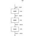
(2.12)画像解析部
図6のブロック図は、第1実施形態の画像解析部を示す。図7の模式図は、第1実施形態の画像解析の内容を示す。(2.12) Image Analysis Unit The block diagram of FIG. 6 shows the image analysis unit of the first embodiment. The schematic diagram of FIG. 7 shows the content of the image analysis of 1st Embodiment.
図6に示される画像解析部1021は、抽出部1030、導出部1031及び除去部1032を備える。抽出部1030は、フレーム画像から心臓領域を抽出する。導出部1031は、心臓領域から心臓パラメーターを導出する。除去部1032は、心臓パラメーターから心拍の影響を除去し心臓状態を示す指標を導出する。 The
(2.13)抽出部
抽出部1030は、2個以上のフレーム画像Img(1),・・・,Img(m)の各々のフレーム画像Img(i)から心臓領域Rgn(i)を抽出する。これにより、2個以上の心臓領域Rgn(1),・・・,Rgn(m)が抽出される。2個以上の心臓領域Rgn(1),・・・,Rgn(m)が抽出されなくても2個以上の心臓状態を示す指標Idx1(1),・・・,Idx1(m)が導出される場合は、抽出部1030が省略されてもよい。(2.13) Extraction Unit The
心臓領域Rgn(i)が抽出される場合は、テンプレートマッチング等によりフレーム画像Img(i)から大まかな心臓領域が抽出され、大まかな心臓領域において画素値が大きく変化するエッジ部が検出され、エッジ部が心臓領域の輪郭とみなされ、エッジ部に囲まれる領域が心臓領域Rgn(i)とされる。他のアルゴリズムにより心臓領域Rgn(i)が抽出されてもよい。 When the heart region Rgn (i) is extracted, a rough heart region is extracted from the frame image Img (i) by template matching or the like, and an edge portion in which the pixel value greatly changes in the rough heart region is detected. The region is regarded as the outline of the heart region, and the region surrounded by the edge portion is the heart region Rgn (i). The heart region Rgn (i) may be extracted by another algorithm.
医用動画像が3次元動画像である場合は、確率アポラス等によりフレーム画像Img(i)から大まかな心臓領域が抽出され、大まかな心臓領域において3次元グラフカットにより心臓領域Rgn(i)が抽出される。他のアルゴリズムにより心臓領域Rgn(i)が抽出されてもよい。例えば、フレーム画像Img(i)の断面画像から2次元画像の場合と同様に心臓領域Rgn(i)が抽出されてもよい。 When the medical moving image is a three-dimensional moving image, a rough heart region is extracted from the frame image Img (i) by probability aporus, etc., and a heart region Rgn (i) is extracted by a three-dimensional graph cut in the rough heart region Is done. The heart region Rgn (i) may be extracted by another algorithm. For example, the heart region Rgn (i) may be extracted from the cross-sectional image of the frame image Img (i) as in the case of the two-dimensional image.
(2.14)導出部
導出部1031は、2個以上のフレーム画像Img(1),・・・,Img(m)の各々のフレーム画像Img(i)について、心臓領域Rgn(i)から心臓パラメーターIdx3(i)を導出する。これにより、2個以上の心臓パラメーターIdx3(1),・・・,Idx3(m)が導出される。(2.14) Deriving Unit The
心臓パラメーターIdx3(i)は、心壁の位置、心臓の大きさ、心臓領域の画素値等のそのものであり、心臓状態を示すが心拍の影響を含む指標である。このため、心臓パラメーターIdx3(i)は、呼吸状態及び心拍の両方により変化し、心臓状態の呼吸状態による変化を把握するのに必ずしも適さない。 The heart parameter Idx3 (i) is the heart wall position, the heart size, the pixel value of the heart region, and the like, and is an index indicating the heart state but including the influence of the heartbeat. For this reason, the cardiac parameter Idx3 (i) changes depending on both the respiratory state and the heartbeat, and is not necessarily suitable for grasping the change of the cardiac state due to the respiratory state.
心臓の同じ部分について2個以上の心臓パラメーターIdx3(1),・・・,Idx3(m)が導出されるようにするために、2個以上のフレーム画像Img(1),・・・,Img(m)において心臓の同じ部分が同じ位置に描写されるように2個以上のフレーム画像Img(1),・・・,Img(m)が位置合わせされる。位置合わせに代えて、2個以上のフレーム画像Img(1),・・・,Img(m)において心臓の同じ部分が描写された位置が対応づけられてもよい。 In order to derive two or more heart parameters Idx3 (1), ..., Idx3 (m) for the same part of the heart, two or more frame images Img (1), ..., Img Two or more frame images Img (1),..., Img (m) are aligned so that the same part of the heart is depicted at the same position in (m). Instead of alignment, positions where the same part of the heart is depicted in two or more frame images Img (1),..., Img (m) may be associated.
(2.15)心壁の位置
心壁の位置は、心壁の座標により表現される。心壁の座標は、心臓領域と心臓領域外との境界の座標である。心壁の座標が心壁の特定の部分の座標であってもよい。心壁は、左心房壁、左心室壁、右心房壁及び右心室壁のいずれでもよい。望ましくは、肺機能の解析の対象となる肺に接触する心壁の座標が心臓パラメーターになる。(2.15) Position of the heart wall The position of the heart wall is expressed by the coordinates of the heart wall. The coordinates of the heart wall are the coordinates of the boundary between the heart region and the outside of the heart region. The coordinates of the heart wall may be the coordinates of a specific part of the heart wall. The heart wall may be any of the left atrial wall, the left ventricular wall, the right atrial wall, and the right ventricular wall. Desirably, the coordinates of the heart wall in contact with the lung to be analyzed for lung function are cardiac parameters.
医用動画像が2次元動画像であり座標系が直交座標系である場合は、x方向の位置を示す座標値x及びy方向の位置を示す座標値yの組で座標が与えられる。典型的には、x方向は2個以上のフレーム画像の各々の横方向であり、y方向は2個以上のフレーム画像の各々の縦方向である。典型的には、横方向は撮影される人における水平方向を示し、縦方向は撮影される人における鉛直方向を示す。医用動画像が2次元動画像であり座標が2個の座標値x及びyの組で与えられる場合は、次元が2次元のまま2個の座標値x及びyの組が心臓パラメーターとされてもよいし、次元が1次元にされ2個の座標値x及びyから選択される1個の座標値x又はyが心臓パラメーターとされてもよい。直交座標系における座標値が他の座標系における座標値に変換されてもよい。例えば、直交座標系における座標値が極座標系における座標値に変換されてもよい。座標系が極座標系である場合は、原点が基準点に設定され、基準点からの距離が心臓パラメーターとされてもよい。座標値は、画素数で表現されてもよいし、撮影された人における現実の長さで表現されてもよい。 When the medical moving image is a two-dimensional moving image and the coordinate system is an orthogonal coordinate system, coordinates are given by a set of a coordinate value x indicating a position in the x direction and a coordinate value y indicating a position in the y direction. Typically, the x direction is the horizontal direction of each of the two or more frame images, and the y direction is the vertical direction of each of the two or more frame images. Typically, the horizontal direction indicates the horizontal direction of the person being photographed, and the vertical direction indicates the vertical direction of the person being photographed. When the medical moving image is a two-dimensional moving image and the coordinates are given as a set of two coordinate values x and y, the set of the two coordinate values x and y is used as a cardiac parameter while the dimension is two-dimensional. Alternatively, the dimension may be one dimension, and one coordinate value x or y selected from two coordinate values x and y may be used as the heart parameter. A coordinate value in the orthogonal coordinate system may be converted into a coordinate value in another coordinate system. For example, coordinate values in the orthogonal coordinate system may be converted into coordinate values in the polar coordinate system. When the coordinate system is a polar coordinate system, the origin may be set as the reference point, and the distance from the reference point may be the heart parameter. The coordinate value may be expressed by the number of pixels, or may be expressed by the actual length of the photographed person.
医用動画像が3次元動画像であり座標が3個の座標値の組で与えられる場合は、次元が3次元のまま3個の座標値の組が心臓パラメーターとされてもよいし、次元が2次元又は1次元にされ3個の座標値から選択される2個の座標値の組又は1個の座標値が心臓パラメーターとされてもよい。 When the medical moving image is a three-dimensional moving image and the coordinates are given as a set of three coordinate values, the set of three coordinate values may be used as a heart parameter while the dimension is three-dimensional. A two-dimensional or one-dimensional set of two coordinate values selected from three coordinate values or one coordinate value may be used as the heart parameter.
(2.16)心臓の幅
心臓の幅は、心壁の一の部分から心壁の他の部分までの長さである。医用動画像が2次元動画像である場合は、望ましくは脊椎、横隔膜等の心臓以外の構造物が描写された領域と重ならない領域から心臓の幅が導出される。これにより、心臓の幅が精度よく導出される。医用動画像が2次元動画像である場合は、望ましくはy方向の位置が同じ一の部分から他の部分までのx方向の長さが心臓の幅とされる。2個以上のy方向の位置の各々について一の部分から他の部分までのx方向の長さを求めることにより2個以上のx方向の長さが求められ、2個以上のx方向の長さの平均値が心臓の幅とされてもよい。y方向の位置が同じ一の部分から他の部分までのx方向の長さに代えて、x方向の位置が同じ一の部分から他の部分までのy方向の長さが心臓の幅とされてもよい。x方向及びy方向の両方に平行でない一の斜め方向及び他の斜め方向が設定され、一の斜め方向の位置が同じ一の部分から他の部分までの他の斜め方向の長さが心臓の幅とされてもよい。(2.16) Heart Width The width of the heart is the length from one part of the heart wall to the other part of the heart wall. When the medical moving image is a two-dimensional moving image, the width of the heart is desirably derived from a region that does not overlap with a region where structures other than the heart such as the spine and the diaphragm are depicted. Thereby, the width of the heart is derived with high accuracy. When the medical moving image is a two-dimensional moving image, the length in the x direction from one part having the same position in the y direction to the other part is preferably set as the width of the heart. By obtaining the length in the x direction from one part to the other part for each of the two or more positions in the y direction, two or more x direction lengths are obtained, and two or more x direction lengths are obtained. The average value may be the width of the heart. Instead of the length in the x direction from the same part in the y direction to the other part, the length in the y direction from the same part in the x direction to the other part is the width of the heart. May be. One oblique direction and another oblique direction that are not parallel to both the x direction and the y direction are set, and the length of the other oblique direction from one part to the other part in the same oblique direction is the same as that of the heart. It may be a width.
(2.17)心臓の面積
フレーム画像Img(i)における心臓の面積は、心臓領域の面積である。心臓の面積は、心臓の全体の面積である。心臓の面積が心臓の一部の面積であってもよい。例えば、心臓の面積が左心室、左心房、右心室又は右心房の面積であってもよい。(2.17) Heart Area The heart area in the frame image Img (i) is the area of the heart region. The area of the heart is the total area of the heart. The area of the heart may be a partial area of the heart. For example, the area of the heart may be the area of the left ventricle, left atrium, right ventricle, or right atrium.
(2.18)心臓の体積
心臓の体積は、3次元画像における心臓領域の体積である。心臓の体積が2次元画像から推定されてもよい。心臓の体積が2次元画像から推定される場合は、心臓領域の面積と心臓領域の画素値との積から心臓の体積が推定される。心臓領域の画素値は、X線が透過する方向の心臓の厚さを反映するからである。心臓の体積は、心臓の全体の体積である。心臓の体積が心臓の一部の体積であってもよい。例えば、心臓の体積が左心室、左心房、右心室又は右心房の体積であってもよい。(2.18) Heart Volume The heart volume is the volume of the heart region in the three-dimensional image. The volume of the heart may be estimated from the two-dimensional image. When the volume of the heart is estimated from the two-dimensional image, the volume of the heart is estimated from the product of the area of the heart region and the pixel value of the heart region. This is because the pixel value in the heart region reflects the thickness of the heart in the direction in which X-rays are transmitted. The heart volume is the total volume of the heart. The volume of the heart may be a partial volume of the heart. For example, the heart volume may be the volume of the left ventricle, left atrium, right ventricle, or right atrium.
(2.19)心臓領域の画素値
心臓領域の画素値は、胸部X線動画像等の透過画像における心臓領域に属する画素の画素値である。心臓領域の画素値は、X線が透過する方向の心臓の厚さにより変化するため、心臓パラメーターとなりうる。望ましくは、脊椎、横隔膜等の心臓以外の構造物が描写された領域と重ならない領域に属し心拍により心臓領域外に属することがない画素の画素値が心臓パラメーターとされる。望ましくは、2個以上の画素の各々についての画素値を特定することにより2個以上の画素値が特定され、特定された2個以上の画素値の平均値が心臓領域の画素値とされる。これにより、心臓領域の画素値がロバストに導出される。平均値が他の種類の代表値に置き換えられてもよい。例えば、平均値が中央値に置き換えられてもよい。(2.19) Pixel value of heart region The pixel value of the heart region is a pixel value of a pixel belonging to the heart region in a transmission image such as a chest X-ray moving image. Since the pixel value of the heart region changes depending on the thickness of the heart in the direction in which X-rays are transmitted, it can be a heart parameter. Desirably, the pixel value of a pixel that belongs to a region that does not overlap with a region in which structures other than the heart such as the spine and diaphragm are depicted does not belong outside the heart region due to a heartbeat is used as a heart parameter. Preferably, two or more pixel values are specified by specifying a pixel value for each of two or more pixels, and an average value of the two or more specified pixel values is set as a pixel value of the heart region. . Thereby, the pixel value of the heart region is derived robustly. The average value may be replaced with another type of representative value. For example, the average value may be replaced with the median value.
(2.20)除去部
除去部1032は、2個以上の心臓パラメーターIdx3(1),・・・,Idx3(m)から心拍の影響を除去する。これにより、2個以上の心臓状態を示す指標Idx1(1),・・・,Idx1(m)が導出される。2個以上の心臓状態を示す指標Idx1(1),・・・,Idx1(m)は、心臓状態を示す指標Idx1(i)の時間変化を示す。(2.20) Removal unit The
2個以上の心臓状態を示す指標Idx1(1),・・・,Idx1(m)の各々の心臓状態を示す指標Idx1(i)は、心壁の位置、心臓の大きさ、心臓領域の画素値等から心拍の影響を除去したものであり、心臓状態を示し、心拍の影響を含まない。このため、心臓状態を示す指標Idx1(i)は、専ら呼吸状態により変化し、心臓状態の呼吸状態による変化を把握するのに適する。 The index Idx1 (i) indicating the heart state of each of the indices Idx1 (1),..., Idx1 (m) indicating two or more heart states is the position of the heart wall, the size of the heart, and the pixels of the heart region This value is obtained by removing the influence of the heartbeat from the value and the like, indicates the heart state, and does not include the influence of the heartbeat. For this reason, the index Idx1 (i) indicating the heart state changes only depending on the respiratory state, and is suitable for grasping the change of the heart state due to the respiratory state.
図7に示されるように、心臓状態を示す指標Idx1(i)が導出される場合は、フレーム画像Img(i)が撮影された時点を含む1心拍周期以上の間に撮影された複数のフレーム画像Img(i-p),・・・,Img(i),・・・,Img(i+q)について求められた複数の心臓パラメーターIdx3(i-p),・・・,Idx3(i),・・・,Idx3(i+q)の平均値が求められ、求められた平均値がフレーム画像Img(i)についての心臓状態を示す指標Idx1(i)とされる。これにより、心拍が起こった時間に撮影されたフレーム画像から導出された心臓パラメーターの寄与が抑制され、心拍の影響が除去される。平均値が他の種類の代表値に置き換えられてもよい。例えば、平均値が最小値、最大値、中央値等に変更されてもよい。 As shown in FIG. 7, when the index Idx1 (i) indicating the heart state is derived, a plurality of frames taken during one heartbeat period or more including the time point when the frame image Img (i) was taken. A plurality of cardiac parameters Idx3 (ip), ..., Idx3 (i), ... determined for the image Img (ip), ..., Img (i), ..., Img (i + q) , Idx3 (i + q) is obtained, and the obtained average value is used as an index Idx1 (i) indicating the heart state of the frame image Img (i). Thereby, the contribution of the heart parameter derived from the frame image taken at the time when the heartbeat occurs is suppressed, and the influence of the heartbeat is removed. The average value may be replaced with another type of representative value. For example, the average value may be changed to a minimum value, a maximum value, a median value, or the like.
2個以上の心臓パラメーターIdx3(1),・・・,Idx3(m)に時間領域のローパスフィルターを適用することにより心拍の影響が除去されてもよい。 The influence of the heartbeat may be removed by applying a time-domain low-pass filter to two or more heart parameters Idx3 (1),..., Idx3 (m).
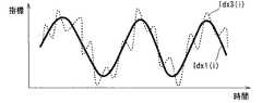
図8の模式図は、心臓パラメーター及び心臓状態を示す指標の時間変化の例を示す。 The schematic diagram of FIG. 8 shows an example of a time change of an index indicating a heart parameter and a heart state.
図8に示される心臓パラメーターIdx3(i)は、相対的に長い呼吸周期に一致する周期で時間変化するとともに、相対的に短い心拍周期に一致する周期で時間変化する。このため、呼吸周波数と心拍周波数との中間の遮断周波数を有する時間領域のローパスフィルターが心臓パラメーターIdx3(i)に適用された場合は、心拍周期に一致する周期の時間変化が除去され、呼吸周期に一致する周期の時間変化が維持され、図8に示される心臓状態を示す指標Idx1(i)が導出される。 The cardiac parameter Idx3 (i) shown in FIG. 8 changes with time in a cycle that matches a relatively long breathing cycle, and changes with time in a cycle that matches a relatively short heartbeat cycle. For this reason, if a time-domain low-pass filter with a cut-off frequency intermediate between the breathing frequency and the heartbeat frequency is applied to the heart parameter Idx3 (i), the time change of the cycle that matches the heartbeat cycle is removed, and the breathing cycle Is maintained, and the index Idx1 (i) indicating the heart state shown in FIG. 8 is derived.
(2.21)指標解析部
指標解析部1022は、2個以上の心臓状態を示す指標Idx1(1),・・・,Idx1(m)の呼吸状態による変化を肺機能を示す指標Idx2とする。(2.21) Index Analysis Unit The index analysis unit 1022 uses a change in the index Idx1 (1),..., Idx1 (m) indicating two or more heart states as an index Idx2 indicating lung function. .
肺機能を示す指標Idx2が導出される場合は、2個以上の心臓状態を示す指標Idx1(1),・・・,Idx1(m)の最大値Maxと最小値Minとの差Max-Minが導出され、導出された差Max-Minが肺機能を示す指標Idx2とされる。差Max-Minが他の種類の最大値Maxと最小値Minとの相違に置き換えられてもよい。例えば、差Max-Minが最大値Maxと最小値Minとの比率Max/Min又はMin/Maxに置き換えられてもよい。最大値Maxと最小値Minとの相違が2個以上の心臓状態を示す指標Idx1(1),・・・,Idx1(m)のばらつきに置き換えられてもよい。例えば、最大値Maxと最小値Minとの相違が2個以上の心臓状態を示す指標Idx1(1),・・・,Idx1(m)の分散、標準偏差等に置き換えられてもよい。 When the index Idx2 indicating the lung function is derived, the difference Max-Min between the maximum value Max and the minimum value Min of the index Idx1 (1), ..., Idx1 (m) indicating two or more heart states is The derived difference Max-Min is used as an index Idx2 indicating lung function. The difference Max-Min may be replaced with a difference between the maximum value Max and the minimum value Min of another type. For example, the difference Max-Min may be replaced with the ratio Max / Min or Min / Max between the maximum value Max and the minimum value Min. The difference between the maximum value Max and the minimum value Min may be replaced with a variation in indices Idx1 (1),..., Idx1 (m) indicating two or more heart states. For example, the difference between the maximum value Max and the minimum value Min may be replaced with the variance, standard deviation, and the like of indices Idx1 (1),..., Idx1 (m) indicating two or more heart conditions.
呼吸状態により肺状態及び心臓状態は変化するため、肺の内部の気圧だけでなく肺の体積、心臓の位置等も呼吸状態により変化する。このため、呼吸状態と心臓状態との対応関係は複雑であり、呼吸状態と心臓状態とを正確に対応づける処理は複雑な処理である。医用動画像が不鮮明な2次元動画像である場合、通常と異なる呼吸状態が検出された場合等においては、呼吸状態と心臓状態とを正確に対応づける処理はさらに複雑になる。個人差、疾病、呼吸条件等により人そのものが通常と異なる呼吸を行っている場合に通常と異なる呼吸状態が検出される。人そのものが通常の呼吸を行っている場合でも撮影条件により通常と異なる呼吸状態が検出される場合がある。 Since the lung state and the heart state change depending on the respiratory state, not only the air pressure inside the lung but also the volume of the lung, the position of the heart, and the like change depending on the respiratory state. For this reason, the correspondence between the respiratory state and the heart state is complicated, and the process for accurately associating the respiratory state with the heart state is a complicated process. When the medical moving image is an unclear two-dimensional moving image, when a breathing state different from normal is detected, the process of accurately associating the breathing state with the heart state is further complicated. A breathing state different from normal is detected when the person is breathing differently from normal due to individual differences, disease, respiratory conditions, and the like. Even when the person itself is performing normal breathing, a breathing state different from normal may be detected depending on imaging conditions.
しかし、2個以上の心臓状態を示す指標Idx1(1),・・・,Idx1(m)の最大値Maxと最小値Minとの相違又は2個以上の心臓状態を示す指標Idx1(1),・・・,Idx1(m)のばらつきが肺機能を示す指標Idx2とされる場合は、呼吸状態と心臓状態とを正確に対応づける処理は不要であるため、肺機能を示す指標Idx2が簡単な処理で導出される。また、このように呼吸状態と心臓状態とを正確に対応づける処理を経ないで導出される肺機能を示す指標Idx2は、呼吸状態と心臓状態とを正確に対応づける処理を経て導出される肺機能を示す指標と同程度に肺機能を反映している場合が多く、呼吸状態と心臓状態とを正確に対応づける処理を経て導出される肺機能を示す指標よりよく肺機能を反映している場合もある。 However, the index Idx1 (1), which indicates two or more heart states, the difference between the maximum value Max and the minimum value Min of Idx1 (m), or the index Idx1 (1), which indicates two or more heart states ..., when the variation in Idx1 (m) is the index Idx2 indicating the lung function, the process of accurately associating the respiratory state and the heart state is not necessary, so the index Idx2 indicating the lung function is simple Derived by processing. In addition, the index Idx2 indicating the lung function that is derived without the process of accurately associating the respiratory state and the heart state is the lung index that is derived through the process of accurately associating the respiratory state and the heart state. In many cases, it reflects the lung function to the same extent as the index indicating the function, and reflects the lung function better than the index indicating the lung function derived through the process of accurately correlating the respiratory state and the heart state. In some cases.
肺機能を示す指標Idx2が大きい場合は、心臓状態を示す指標Idx1(i)の呼吸状態による変化が大きいため、撮影された人がCOPDに罹患している可能性が高い。このため、肺機能を示す指標Idx2は、COPDへの罹患の有無を推定するのに役立つ。例えば、肺機能を示す指標Idx2が閾値より大きい場合は、COPDへの罹患の疑いがあると判定される。この判定は、指標解析部1022が行ってもよいし、肺機能を示す指標Idx2を参照した医師が行ってもよい。 If the index Idx2 indicating the lung function is large, the index Idx1 (i) indicating the heart state varies greatly depending on the respiratory state, and thus the photographed person is highly likely to have COPD. For this reason, the index Idx2 indicating lung function is useful for estimating the presence or absence of COPD. For example, when the index Idx2 indicating the lung function is larger than the threshold value, it is determined that there is a suspicion of suffering from COPD. This determination may be performed by the index analysis unit 1022 or may be performed by a doctor who refers to the index Idx2 indicating lung function.
2個以上の心臓状態を示す指標Idx1(1),・・・,Idx1(m)の最大値Maxと平均値Avgとの差Max-Avg、2個以上の心臓状態を示す指標Idx1(1),・・・,Idx1(m)の平均値Avgと最小値Minとの差Avg-Min、2個以上の心臓状態を示す指標Idx1(1),・・・,Idx1(m)の最大値Maxと平均値Avgとの比率Max/Avg又はAvg/Max、2個以上の心臓状態を示す指標Idx1(1),・・・,Idx1(m)の平均値Avgと最小値Minとの比率Min/Avg又はAvg/Min等が肺機能を示す指標Idx2とされてもよい。 The difference between the maximum value Max and the average value Avg of indices Idx1 (1),..., Idx1 (m) indicating two or more heart conditions Max-Avg, index Idx1 (1) indicating two or more heart conditions , ..., the difference Avg-Min between the average value Avg and the minimum value Min of Idx1 (m), the maximum value Max of the indices Idx1 (1), ..., Idx1 (m) indicating two or more heart states A ratio Max / Avg or Avg / Max between the average value Avg and Avg / Max, a ratio Min / between the average value Avg and the minimum value Min of the indices Idx1 (1),. Avg or Avg / Min or the like may be used as the index Idx2 indicating the lung function.
指標解析部1022が肺機能を示す指標Idx2を肺機能を示す第1次指標とし肺機能を示す第2次指標をさらに導出してもよい。例えば、第1次指標である肺機能を示す指標Idx2から第2次指標であるCOPDへの罹患の疑いの有無が導出されてもよい。COPDへの罹患の疑いの有無が導出される場合は、例えば、肺機能を示す指標Idx2が閾値以下である場合はCOPDへの罹患の疑いがないと判定され、肺機能を示す指標Idx2が閾値より大きい場合はCOPDへの罹患の疑いがあると判定される。COPDへの罹患の疑いの程度が3段階以上にランク分けされてもよい。例えば、肺機能を示す指標Idx2が第1の閾値以下である場合はCOPDへの罹患の疑いがないと判定され、肺機能を示す指標Idx2が第1の閾値より大きく第2の閾値以下である場合はCOPDへの罹患の疑いがあると判定され、肺機能を示す指標Idx2が第2の閾値より大きい場合はCOPDへの罹患の強い疑いがあると判定される。COPDへの罹患の疑いの程度は、例えば、「疑いがない」「弱い疑いがある」「疑いがある」「強い疑いがある」等のように、疑いの有無及び疑いの強さにより示される。COPDへの疑いの程度に検査又は治療の要否の示唆が付加されてもよい。例えば、COPDへの罹患の強い疑いがある場合に検査が必要であることの示唆が付加されてもよい。COPDへの罹患の疑いの程度が数値で表現されてもよい。例えば、COPDの罹患の疑いの程度を示す数値の計算式1-exp(-x)のxに肺機能を示す指標Idx2が反映させられ、COPDの罹患の疑いの程度を示す数値が求められてもよい。計算式1-exp(-x)により計算される数値は0から1までの範囲に収まるため、計算式1-exp(-x)により計算される数値はCOPDへの罹患の疑いの強さを百分率で表現するのに役立つ。例えば、肺機能を示す指標Idx2が2であり、COPDの罹患の疑いの程度を示す数値の計算式1-exp(-x)のxがIdx2/2とされた場合は、1-exp(-2/2)=0.63であるため、COPDへの罹患の疑いの強さが百分率で63%であると表現される。肺機能を示す指標Idx2が4であり、COPDの罹患の疑いの程度を示す数値の計算式1-exp(-x)のxがIdx2/2とされた場合は、1-exp(-4/2)=0.86であるため、COPDへの罹患の疑いの強さが百分率で86%であると表現される。計算式が変更されてもよい。 The index analysis unit 1022 may further derive a secondary index indicating the lung function using the index Idx2 indicating the lung function as the primary index indicating the lung function. For example, whether or not there is a suspicion of suffering from COPD as the secondary index may be derived from the index Idx2 indicating the lung function as the primary index. When the presence / absence of suspicion of COPD is derived, for example, when the index Idx2 indicating lung function is equal to or lower than the threshold, it is determined that there is no suspicion of COPD, and the index Idx2 indicating lung function is the threshold. If larger, it is determined that COPD is suspected. The degree of suspicion of suffering from COPD may be ranked in three or more stages. For example, when the index Idx2 indicating the lung function is equal to or lower than the first threshold, it is determined that there is no suspicion of suffering from COPD, and the index Idx2 indicating the lung function is greater than the first threshold and equal to or less than the second threshold. In the case, it is determined that there is a suspicion of suffering from COPD, and when the index Idx2 indicating lung function is greater than the second threshold, it is determined that there is a strong suspicion of suffering from COPD. The degree of suspicion of suffering from COPD is indicated by the presence of suspicion and the strength of suspicion, such as “no suspicion”, “weak suspicion”, “suspicion”, “strong suspicion”, etc. . An indication of the necessity of examination or treatment may be added to the degree of suspicion of COPD. For example, an indication may be added that a test is necessary if there is a strong suspicion of having COPD. The degree of suspicion of suffering from COPD may be expressed numerically. For example, the numerical expression 1-exp (-x) indicating the degree of suspicion of COPD is reflected in the index Idx2 indicating pulmonary function in x in 1-exp (-x), and a numerical value indicating the degree of suspicion of COPD is obtained. Also good. Since the numerical value calculated by the calculation formula 1-exp (-x) falls within the range from 0 to 1, the numerical value calculated by the calculation formula 1-exp (-x) indicates the strength of suspicion of suffering from COPD. Useful for expressing as a percentage. For example, if the index Idx2 indicating lung function is 2 and x in the numerical expression 1-exp (-x) indicating the degree of suspected COPD is 1dexp2 / 2, 1-exp (- Since 2/2) = 0.63, the intensity of suspicion of suffering from COPD is expressed as 63% in percentage. When the index Idx2 indicating lung function is 4 and x in the numerical formula 1-exp (-x) indicating the degree of suspected COPD is 1dexp2 / 2, 1-exp (-4 / 2) Since 0.86, the intensity of suspicion of suffering from COPD is expressed as 86% in percentage. The calculation formula may be changed.
2個以上の第1次指標を反映する第2次指標が導出されてもよい。例えば、心臓の大きさの呼吸状態による変化から導出されるCOPDへの罹患の疑いの有無と心臓の大きさの心拍による変化の呼吸状態による変化から導出されるCOPDへの罹患の疑いの有無とのOR演算又はAND演算の結果が第2次指標とされてもよい。心臓の大きさの呼吸状態による変化と心臓の大きさの心拍による変化の呼吸状態による変化との和又は積が第2次指標とされてもよい。 A secondary index reflecting two or more primary indices may be derived. For example, whether or not there is a suspicion of suffering from COPD derived from a change in the heart size due to a respiratory state, and whether or not there is a suspicion of suffering from COPD derived from a change in the heart size due to a heart rate The result of the OR operation or AND operation may be used as the secondary index. The sum or product of the change in the heart size due to the respiratory state and the change in the heart size due to the heartbeat due to the respiratory state may be used as the secondary index.
(2.22)表示部
表示部1023は、解析結果をディスプレイに表示する。解析結果は、医用動画像の撮影が完了した後に医師が診断を行う場合に表示されてもよいし、医用動画像が撮影されている間に又は医用動画像の撮影が完了した直後に表示されてもよい。医用動画像が撮影されている間に又は医用動画像の撮影が完了した直後に解析結果が表示される場合は、医用動画像の撮影の成否を技師が確認でき、撮影のやり直しの要否を技師が判断できる。(2.22) Display unit The
解析結果が紙等の印刷媒体に印刷されてもよい。解析結果、解析結果の表示内容、解析結果の印刷内容を記述したデーターがハードディスクドライブ、光ディスク等の記録媒体に記録されてもよい。 The analysis result may be printed on a print medium such as paper. Data describing analysis results, display contents of analysis results, and print contents of analysis results may be recorded on a recording medium such as a hard disk drive or an optical disk.
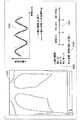
図9の模式図は、解析結果の表示例を示す。 The schematic diagram of FIG. 9 shows a display example of the analysis result.
図9に示される表示例は、医用動画像の再生欄1040、心臓パラメーター及び心臓状態を示す指標の表示欄1041、肺機能を示す指標の表示欄1042、肺機能の判定基準の表示欄1043及び肺機能の判定結果の表示欄1044を有する。肺機能の判定基準は、閾値の情報を含む。図9に示される表示例は、心臓状態を示す指標Idx1(i)がフレーム画像Img(i)における心臓の面積であり、肺機能を示す第1次指標Idx2が2個以上の心臓状態を示す指標Idx1(1),・・・,Idx1(m)の最大値Maxと最小値Minとの差Max-Minである場合のものである。図9に示される表示例は、第2次指標としてCOPDへの罹患の疑いの有無が導出された場合のものである。2種類以上の心臓状態を示す指標についての解析結果が別々に又は統合して表示されてもよい。 The display example shown in FIG. 9 includes a medical moving
(2.23)コンピューター
図10は、コンピューターのブロック図である。(2.23) Computer FIG. 10 is a block diagram of a computer.
図10に示されるコンピューター1050は、CPU1060、メモリー1061、ハードディスクドライブ1062等を備え、画像解析装置1011となる。ハードディスクドライブ1062が他の種類の補助記憶装置に置き換えられてもよい。ハードディスクドライブ1062には、画像解析プログラム1070がインストールされる。画像解析装置1011の機能は、CPU1060が画像解析プログラム1070をメモリー1061にロードして実行することにより実現される。画像解析装置1011の機能の全部又は一部がソフトウェアを伴わないハードウェアにより実現されてもよい。画像解析装置1011の機能の全部又は一部が2台以上のコンピューターにより実現されてもよい。画像解析プログラム1070は、コンピューター1050の出荷前にハードディスクドライブ1062にプリインストールされてもよいし、コンピューター1050の出荷後にハードディスクドライブ1062にインストールされてもよい。画像解析プログラム1070のインストールは、画像解析プログラム1070が記録された光ディスク等の記録媒体1080を読み取ることにより行われてもよし、ネットワーク1090を経由してダウンロードすることにより行われてもよい。 A
(3)第2実施形態
第2実施形態は、第1実施形態の画像解析部を置き換える画像解析部に関する。第2実施形態の画像解析部は、主に心拍の影響の除去の点で第1実施形態の画像解析部と異なる。(3) Second Embodiment The second embodiment relates to an image analysis unit that replaces the image analysis unit of the first embodiment. The image analysis unit of the second embodiment is different from the image analysis unit of the first embodiment mainly in terms of removing the influence of heartbeat.
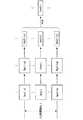
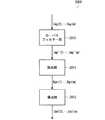
図11のブロック図は、第2実施形態の画像解析部を示す。図12の模式図は、第2実施形態の画像解析の内容を示す。 The block diagram of FIG. 11 shows the image analysis unit of the second embodiment. The schematic diagram of FIG. 12 shows the content of the image analysis of 2nd Embodiment.
図11に示される画像解析部2000は、ローパスフィルター部2010、抽出部2011及び導出部2012を備える。 The
ローパスフィルター部2010は、2個以上のフレーム画像Img(1),・・・,Img(m)に時間領域のローパスフィルターを適用する。これにより、時間領域のローパスフィルターが適用された2個以上のフレーム画像Img'(1),・・・,Img'(m)が得られる。ローパスフィルターの遮断周波数は、第1実施形態の場合と同様に、呼吸周波数と心拍周波数との中間である。これにより、2個以上のフレーム画像Img(1),・・・,Img(m)から心拍の影響が除去され、最終的に導出される2個以上の心臓状態を示す指標Idx1(1),・・・,Idx1(m)から心拍の影響が除去される。 The low-
抽出部2011は、ローパスフィルターが適用された2個以上のフレーム画像Img'(1),・・・,Img'(m)の各々のフレーム画像Img'(i)から心臓領域Rgn(i)を抽出する。これにより、2個以上の心臓領域Rgn(1),・・・,Rgn(m)が抽出される。第1実施形態の場合と同様に、2個以上の心臓領域Rgn(1),・・・,Rgn(m)が抽出されなくても2個以上の心臓状態を示す指標Idx1(1),・・・,Idx1(m)が導出される場合は、抽出部2011が省略されてもよい。 The
導出部2012は、2個以上のフレーム画像Img(1),・・・,Img(m)の各々のフレーム画像Img(i)について、心臓領域Rgn(i)から心臓状態を示す指標Idx1(i)を導出する。これにより、2個以上の心臓状態を示す指標Idx1(1),・・・,Idx1(m)が導出される。心臓状態を示す指標Idx1(i)が導出される場合は、第1実施形態において心臓領域Rgn(i)から心臓パラメーターIdx3(i)が導出されたのと同じように心臓パラメーターIdx3(i)が導出され、心臓パラメーターIdx3(i)がそのまま心臓状態を示す指標Idx1(i)とされる。 For each frame image Img (i) of two or more frame images Img (1),..., Img (m), the
(4)第3実施形態
第3実施形態は、第1実施形態の画像解析部を置き換える画像解析部に関する。第3実施形態の画像解析部は、主に心拍の影響の除去の点で第1実施形態の画像解析部と異なる。(4) Third Embodiment The third embodiment relates to an image analysis unit that replaces the image analysis unit of the first embodiment. The image analysis unit of the third embodiment is different from the image analysis unit of the first embodiment mainly in removing the influence of the heartbeat.
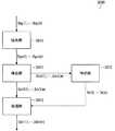
図13のブロック図は、第3実施形態の画像解析部を示す。図14の模式図は、第3実施形態の画像解析の内容を示す。 The block diagram of FIG. 13 shows the image analysis unit of the third embodiment. The schematic diagram of FIG. 14 shows the content of the image analysis of 3rd Embodiment.
図13に示される画像解析部3000は、抽出部3010、ローパスフィルター部3011及び導出部3012を備える。 The
抽出部3010は、2個以上のフレーム画像Img(1),・・・,Img(m)の各々のフレーム画像Img(i)から心臓領域Rgn(i)を抽出する。これにより、2個以上の心臓領域Rgn(1),・・・,Rgn(m)が抽出される。 The
ローパスフィルター部3011は、2個以上の心臓領域Rgn(1),・・・,Rgn(m)に時間領域のローパスフィルターを適用する。これにより、時間領域のローパスフィルターが適用された2個以上の心臓領域Rgn'(1),・・・,Rgn'(m)が得られる。ローパスフィルターの遮断周波数は、第1実施形態の場合と同様に、呼吸周波数と心拍周波数との中間である。これにより、2個以上の心臓領域Rgn(1),・・・,Rgn(m)から心拍の影響が除去され、最終的に導出される2個以上の心臓状態を示す指標Idx1(1),・・・,Idx1(m)から心拍の影響が除去される。 The low-
導出部3012は、2個以上のフレーム画像Img(1),・・・,Img(m)の各々のフレーム画像Img(i)について、心臓領域Rgn'(i)から心臓状態を示す指標Idx1(i)を導出する。これにより、2個以上の心臓状態を示す指標Idx1(1),・・・,Idx1(m)が導出される。心臓状態を示す指標Idx1(i)が導出される場合は、第1実施形態において心臓領域Rgn(i)から心臓パラメーターIdx3(i)が導出されたのと同じように心臓パラメーターIdx3(i)が導出され、心臓パラメーターIdx3(i)がそのまま心臓状態を示す指標Idx1(i)とされる。 For each frame image Img (i) of the two or more frame images Img (1),..., Img (m), the
(5)第4実施形態
(5.1)指標解析部
第4実施形態は、第1実施形態の指標解析部を置き換える指標解析部に関する。第4実施形態の指標解析部は、主に肺機能を示す指標の導出の点で第1実施形態の指標解析部と異なる。第2実施形態又は第3実施形態において説明した置き換えともに第4実施形態において説明する置き換えが行われてもよい。(5) Fourth Embodiment (5.1) Index Analysis Unit The fourth embodiment relates to an index analysis unit that replaces the index analysis unit of the first embodiment. The index analysis unit according to the fourth embodiment differs from the index analysis unit according to the first embodiment mainly in deriving an index indicating lung function. The replacement described in the fourth embodiment may be performed together with the replacement described in the second embodiment or the third embodiment.
図15のブロック図は、第4実施形態の指標解析部を示す。 The block diagram of FIG. 15 shows the index analysis unit of the fourth embodiment.
図15に示される指標解析部4000は、区間解析部4010及び全体解析部4011を備える。区間解析部4010は、心臓状態を示す指標から区間指標を導出する。全体解析部4011は、区間指標から肺機能を示す指標を導出する。 The
(5.2)区間解析部
区間解析部4010は、2個以上のフレーム画像Img(1),・・・,Img(m)のうちの吸気期に属する少なくとも1個のフレーム画像Img(a1),・・・,Img(ap)について導出される少なくとも1個の心臓状態を示す指標Idx1(a1),・・・,Idx1(ap)の平均値を吸気期の区間指標Idx4(a)とする。また、区間解析部4010は、2個以上のフレーム画像Img(1),・・・,Img(m)のうちの呼気期に属する少なくとも1個のフレーム画像Img(b1),・・・,Img(bq)について導出される少なくとも1個の心臓状態を示す指標Idx1(b1),・・・,Idx1(bq)の平均値を呼気期の区間指標Idx4(b)とする。これにより、吸気期の区間指標Idx4(a)及び呼気期の区間指標Idx4(b)が導出される。吸気期の区間指標Idx4(a)は吸気期の典型的な心臓状態を示し、呼気期の区間指標Idx4(b)は呼気期の典型的な心臓状態を示す。吸気期の区間指標Idx4(a)及び呼気期の区間指標Idx4(b)により、吸気期と呼気期との間の心臓状態を示す指標の変化を導出可能になる。(5.2) Interval Analysis Unit The interval analysis unit 4010 includes at least one frame image Img (a1) belonging to the inspiratory period among two or more frame images Img (1),..., Img (m). ,..., Img (ap) derived at least one heart condition index Idx1 (a1),..., Idx1 (ap) average value is set as the inspiratory interval index Idx4 (a) . The section analysis unit 4010 also includes at least one frame image Img (b1),..., Img belonging to the expiration period among the two or more frame images Img (1),. The average value of the indices Idx1 (b1),..., Idx1 (bq) indicating at least one heart state derived for (bq) is set as the interval index Idx4 (b) in the expiration period. Thereby, the interval index Idx4 (a) in the inspiration period and the interval index Idx4 (b) in the expiration period are derived. The interval index Idx4 (a) in the inspiration period indicates a typical heart condition in the inspiration period, and the interval index Idx4 (b) in the expiration period indicates a typical heart condition in the expiration period. By using the interval index Idx4 (a) in the inspiration period and the interval index Idx4 (b) in the expiration period, it is possible to derive a change in the index indicating the heart state between the inspiration period and the expiration period.
平均値が他の種類の代表値に置き換えられてもよい。例えば、平均値が中央値、最小値、最大値等に置き換えられてもよい。平均値が2個以上の心臓状態を示す指標から無作為に選択された1個の心臓状態を示す指標に置き換えられてもよい。無作為な選択が行われる場合は、精度がやや低下する可能性があるが、区間指標を導出する処理が簡略化される。 The average value may be replaced with another type of representative value. For example, the average value may be replaced with a median value, a minimum value, a maximum value, or the like. The average value may be replaced with an index indicating one randomly selected heart condition from an index indicating two or more heart conditions. When random selection is performed, the accuracy may be slightly reduced, but the process of deriving the section index is simplified.
(5.3)互いに異なる呼吸速度の割り当て
呼吸速度の絶対値は典型的には単位時間当たりの換気量であり、呼吸速度の符号は典型的には吸気期においては正であり呼気期においては負である。呼吸速度の絶対値及び符号の定義が変更されてもよい。例えば、呼吸速度の絶対値が医用動画像における横隔膜の移動速度の絶対値であってもよい。呼吸速度の符号が吸気期において負であり呼気期においては正であることも許される。(5.3) Assignment of different respiratory rates The absolute value of the respiratory rate is typically the ventilation volume per unit time, and the sign of the respiratory rate is typically positive in the inspiratory period and in the expiratory period Is negative. The definition of the absolute value and sign of the respiration rate may be changed. For example, the absolute value of the respiratory rate may be the absolute value of the moving speed of the diaphragm in the medical moving image. It is allowed that the sign of the respiration rate is negative in the inspiration period and positive in the expiration period.
吸気期は符号が正である呼吸速度が割り当てられた区間であり、呼気期は符号が負である呼吸速度が割り当てられた区間である。したがって、吸気期及び呼気期は、互いに異なる呼吸速度の符号が割り当てられた2個の区間である。互いに異なる呼吸速度の符号を2個の区間に割り当てることに代えて、又は、互いに異なる呼吸速度の符号を2個の区間に割り当てることに加えて、互いに異なる呼吸速度の絶対値が2個以上の区間に割り当てられてもよい。より一般的には、互いに異なる呼吸速度が2個以上の区間に割り当てられる。呼吸速度は肺の内部の気圧と連動して変化するため、2個以上の区間に互いに異なる呼吸速度を割り当てることは、心臓状態の肺の内部の気圧による変化を特定することを可能にする。 The inspiration period is a section to which a respiration rate having a positive sign is assigned, and the expiration period is a section to which a respiration speed having a negative sign is assigned. Therefore, the inspiratory period and the expiratory period are two sections to which signs of different respiratory rates are assigned. Instead of assigning different respiratory rate codes to the two intervals, or in addition to assigning different respiratory rate symbols to the two intervals, the absolute values of the different respiratory rates are two or more. It may be assigned to a section. More generally, different respiration rates are assigned to two or more sections. Since the respiration rate changes in conjunction with the air pressure inside the lung, assigning different respiration rates to two or more sections makes it possible to identify changes due to the air pressure inside the lung in the heart state.
望ましくは、呼吸速度が0に近い状態は吸気期及び呼気期から除外される。吸気期及び呼気期とは別に呼吸速度が0に近い区間が設定されてもよい。呼吸速度が0に近くなるのは、吸気期から呼気期へ遷移する頃及び呼気期から吸気期へ遷移する頃である。 Desirably, the state where the respiration rate is close to 0 is excluded from the inspiration period and the expiration period. A section in which the respiration rate is close to 0 may be set separately from the inspiration period and the expiration period. The respiration rate is close to 0 when the transition from the inspiratory period to the expiratory period and when the expiratory period transitions to the inspiratory period.
吸気期の一部が一の区間とされ呼気期の一部が他の区間とされてもよい。望ましくは、一の区間及び他の区間には同程度の肺の大きさが割り当てられる。これにより、肺の大きさの影響が除去され、心臓状態の呼吸速度による変化を示す指標が導出される。 A part of the inspiration period may be set as one section and a part of the expiration period may be set as another section. Desirably, similar lung sizes are assigned to one segment and the other segment. Thereby, the influence of the size of the lung is removed, and an index indicating a change due to the respiratory rate of the heart state is derived.
(5.4)互いに異なる肺の大きさの割り当て
互いに異なる呼吸速度を2個以上の区間に割り当てることに代えて、又は、互いに異なる呼吸速度を2個以上の区間に割り当てることに加えて、呼吸速度とは別の観点から互いに異なる呼吸状態が2個以上の区間に割り当てられてもよい。例えば、互いに異なる肺の大きさが2個以上の区間に割り当てられてもよい。(5.4) Assignment of different lung sizes Breathing instead of assigning different breathing rates to two or more sections, or in addition to assigning different breathing rates to two or more sections Two or more sections may be assigned different breathing states from a viewpoint different from speed. For example, different lung sizes may be assigned to two or more sections.
互いに異なる肺の大きさが2個以上の区間に割り当てられる場合は、互いに異なる肺の幅、面積、大きさ等が2個以上の区間に割り当てられ、望ましくは互いに異なる肺の縦方向の幅が2個以上の区間に割り当てられる。 When different lung sizes are assigned to two or more sections, different lung widths, areas, sizes, etc. are assigned to two or more sections, preferably with different lung longitudinal widths. It is assigned to two or more sections.
(5.5)互いに異なる呼吸位相の割り当て
互いに異なる呼吸位相が2個以上の区間に割り当てられてもよい。互いに異なる呼吸位相が2個以上の区間に割り当てられる場合は、例えば、吸気期の初期、中期及び後期並びに呼気期の初期、中期及び後期という6個の区間の各々について区間指標が導出される。これにより、心臓状態の呼吸位相による変化を示す指標が導出される。吸気期に含まれる区間の数が増減されてもよい。呼気期に含まれる区間の数が増減されてもよい。呼吸位相は、撮影される人の換気量から特定されてもよいし、医用動画像における横隔膜、肺等の動きから特定されてもよい。(5.5) Assignment of Respiratory Phases Different from Each Other Respiration phases different from each other may be assigned to two or more sections. When different respiratory phases are assigned to two or more sections, for example, section indices are derived for each of the six sections, that is, the early period, the middle period, and the late period of the inspiratory period, and the early period, the middle period, and the late period of the expiratory period. As a result, an index indicating a change in the cardiac state due to the respiratory phase is derived. The number of sections included in the inspiratory period may be increased or decreased. The number of sections included in the expiration period may be increased or decreased. The respiratory phase may be specified from the ventilation volume of the person to be imaged, or may be specified from the movement of the diaphragm, lung, etc. in the medical moving image.
(5.6)呼吸周期の全体からなる区間
2個以上の区間が呼吸周期の全体からなる区間を含んでもよい。例えば、吸気期及び呼気期の一方が一の区間とされ吸気期及び呼気期を合わせた区間が他の区間とされてもよい。これにより、呼吸周期の全体と特定の呼吸状態との間の心臓状態の変化が導出される。(5.6) Section consisting of the entire respiratory cycle Two or more sections may include a section consisting of the entire respiratory cycle. For example, one of the inspiratory period and the expiratory period may be set as one section, and a section obtained by combining the inspiratory period and the expiratory period may be set as the other section. This derives a change in heart condition between the entire breathing cycle and a particular breathing state.
(5.7)その他の割り当て
互いに異なる呼吸方法が2個以上の区間に割り当てられてもよい。例えば、自然呼吸が一の区間に割り当てられ、努力呼吸が他の区間に割り当てられてもよい。これにより、心臓状態の呼吸方法による変化を示す指標が導出される。(5.7) Other assignments Different breathing methods may be assigned to two or more sections. For example, natural breathing may be assigned to one section and forced breathing may be assigned to another section. Thereby, an index indicating a change in the heart state due to the breathing method is derived.
互いに異なる呼吸状態を2個以上の区間に割り当てることに加えて、呼吸状態とは別の観点から異なる状態が2個以上の区間に割り当てられてもよい。例えば、互いに異なる姿勢が2個以上の区間に割り当てられてもよい。例えば、立位が一の区間に割り当てられ、臥位が他の区間に割り当てられてもよい。 In addition to assigning different respiratory states to two or more sections, a state different from a breathing state may be assigned to two or more sections. For example, different postures may be assigned to two or more sections. For example, the standing position may be assigned to one section and the prone position may be assigned to another section.
(5.8)全体解析部
全体解析部4011は、吸気期の区間指標Idx4(a)及び呼気期の区間指標Idx4(b)の差Idx4(a)-Idx4(b)の絶対値を肺機能を示す指標Idx2とする。差Idx4(a)-Idx4(b)の絶対値以外が肺機能を示す指標Idx2とされてもよい。例えば、比率Idx4(a)/Idx4(b)又はIdx4(b)/Idx4(a)等の吸気期の区間指標Idx4(a)と呼気期の区間指標Idx4(b)との相違が肺機能を示す指標Idx2とされてもよい。3個以上の区間指標が導出される場合は、3個以上の区間指標の分散、標準偏差等の3個以上の区間指標のばらつきが肺機能を示す指標Idx2とされてもよい。(5.8) Overall Analysis Unit The overall analysis unit 4011 calculates the absolute value of the difference Idx4 (a) -Idx4 (b) between the interval index Idx4 (a) in the inspiratory period and the interval index Idx4 (b) in the expiration period. An index Idx2 indicating Other than the absolute value of the difference Idx4 (a) -Idx4 (b) may be the index Idx2 indicating the lung function. For example, the difference between the inspiratory interval index Idx4 (a) such as the ratio Idx4 (a) / Idx4 (b) or Idx4 (b) / Idx4 (a) and the expiratory interval index Idx4 (b) The index Idx2 may be indicated. When three or more section indices are derived, the dispersion of three or more section indices such as the dispersion of three or more section indices and the standard deviation may be used as an index Idx2 indicating lung function.
(5.9)2呼吸周期以上
2呼吸周期以上にわたって心臓状態を示す指標が導出される場合は、2呼吸周期以上にわたって導出された心臓状態を示す指標が1呼吸周期にマッピングされ、2呼吸周期以上にわたって導出された心臓状態を示す指標が1呼吸周期について導出された心臓状態を示す指標と同様に処理される。これにより、心臓状態を示す指標の呼吸状態による変化がロバストに導出される。望ましくは、呼吸が同様に行われた呼吸周期について導出された心臓状態を示す指標のみがマッピングの対象になる。例えば、自然呼吸が行われた呼吸周期について導出された心臓状態を示す指標のみ又は努力呼吸が行われた呼吸周期について導出された心臓状態を示す指標のみがマッピングの対象とされる。呼吸が乱れた呼吸周期について導出された心臓状態を示す指標がマッピングの対象から除外されてもよい。(5.9) Two or more breathing cycles or more When an index indicating a heart state is derived over two or more breathing cycles, the index indicating the heart state derived over two or more breathing cycles is mapped to one breathing cycle. The index indicating the heart condition derived over the above is processed in the same manner as the index indicating the heart condition derived for one respiratory cycle. Thereby, the change by the respiratory state of the parameter | index which shows a heart state is derived | led-out robustly. Desirably, only the index indicating the heart state derived for the breathing cycle in which breathing is performed in the same manner is subjected to mapping. For example, only the index indicating the heart state derived for the breathing cycle in which natural breathing is performed or only the index indicating the heart state derived for the breathing cycle in which forced breathing is performed is the target of mapping. The index indicating the heart state derived for the respiratory cycle in which the breathing is disturbed may be excluded from the mapping target.
吸気期の区間指標及び呼気期の区間指標が導出される場合にマッピングが行われるときは、2個以上の吸気期が統合された1個の吸気期とされ、2以上の呼気期が統合された1個の呼気期とされる。2個以上の吸気期について導出された心臓状態を示す指標の平均値が統合された1個の吸気期の区間指標とされ、2個以上の呼気期について導出された心臓状態を示す指標の平均値が統合された1個の呼気期の区間指標とされる。2呼吸周期以上の各々について吸気期の区間指標及び呼気期の区間指標が導出され、2個以上の吸気期の区間指標の平均値が統合された1個の区間指標とされ、2個以上の呼気期の区間指標の平均値が統合された1個の区間指標とされてもよい。平均値が他の種類の代表値に置き換えられてもよい。例えば、平均値が中央値、最小値、最大値等に置き換えられてもよい。代表値が平均値又は中央値である場合は心臓状態を示す指標の呼吸状態による変化がロバストに導出される。代表値が最大値又は最小値である場合は特異な心臓状態の呼吸状態による変化が導出される。 When mapping is performed when the interval index of the inspiration period and the interval index of the expiration period are derived, two or more inspiration periods are combined into one inspiration period, and two or more expiration periods are integrated. One exhalation period. The average value of the index indicating the heart state derived for two or more inspiratory periods is the integrated interval index of one inspiratory period, and the average of the indices indicating the heart condition derived for two or more exhalation periods The interval index of one expiration period in which the values are integrated is used. For each of two or more respiratory cycles, the interval index for the inspiration period and the interval index for the expiration period are derived, and the average value of the interval indices for the two or more inspiration periods is integrated into one interval index. The average value of the interval index in the expiration period may be integrated into one interval index. The average value may be replaced with another type of representative value. For example, the average value may be replaced with a median value, a minimum value, a maximum value, or the like. When the representative value is an average value or a median value, a change in the index indicating the heart state due to the respiratory state is robustly derived. When the representative value is the maximum value or the minimum value, a change in the peculiar heart state due to the respiratory state is derived.
(6)第5実施形態
(6.1)画像解析部
第5実施形態は、第1実施形態の画像解析部を置き換える画像解析部に関する。第5実施形態の画像解析部は、主に心臓状態を示す指標の点で第1実施形態の画像解析部と異なる。第2実施形態から第4実施形態までにおいて説明した置き換えとともに第5実施形態において説明する置き換えが行われてもよい。(6) Fifth Embodiment (6.1) Image Analysis Unit The fifth embodiment relates to an image analysis unit that replaces the image analysis unit of the first embodiment. The image analysis unit of the fifth embodiment is different from the image analysis unit of the first embodiment mainly in terms of an index indicating the heart state. The replacement described in the fifth embodiment may be performed together with the replacement described in the second to fourth embodiments.
図16のブロック図は、第5実施形態の画像解析部を示す。図17の模式図は、第5実施形態の画像解析の内容を示す。 The block diagram of FIG. 16 shows the image analysis unit of the fifth embodiment. The schematic diagram of FIG. 17 shows the content of the image analysis of 5th Embodiment.
図16に示される画像解析部5000は、抽出部5010、導出部5011、特定部5012及び処理部5013を備える。抽出部5010は、心臓領域を抽出する。導出部5011は、心臓領域から心臓パラメーターを導出する。特定部は、心拍が起こった時間を特定する。処理部は、心拍が起こった時間における心臓パラメーターの変化を心臓状態を示す指標にする。 The
(6.2)抽出部
抽出部5010は、第1実施形態の場合と同様に、2個以上のフレーム画像Img(1),・・・,Img(m)の各々のフレーム画像Img(i)から心臓領域Rgn(i)を抽出する。これにより、2個以上の心臓領域Rgn(1),・・・,Rgn(m)が抽出される。(6.2) Extraction Unit The
(6.3)導出部
導出部5011は、第1実施形態の場合と同様に、2個以上のフレーム画像Img(1),・・・,Img(m)の各々のフレーム画像Img(i)について、心臓領域Rgn(i)から心臓パラメーターIdx3(i)を導出する。これにより、2個以上の心臓パラメーターIdx3(1),・・・,Idx3(m)が導出される。2個以上の心臓パラメーターIdx3(1),・・・,Idx3(m)は、心臓パラメーターIdx3(i)の時間変化を示す。(6.3) Deriving Unit The
(6.4)心臓パラメーターの導出例
図18の模式図は、心壁の座標の時間変化の例を示す。(6.4) Example of Derivation of Heart Parameter The schematic diagram of FIG. 18 shows an example of the time change of the coordinates of the heart wall.
図18に示される心壁の座標の時間変化が導出された場合は、一のフレーム画像における心壁の座標と他のフレーム画像における心壁の座標との差が導出される。 When the temporal change in the coordinates of the heart wall shown in FIG. 18 is derived, the difference between the coordinates of the heart wall in one frame image and the coordinates of the heart wall in another frame image is derived.
(6.5)特定部
特定部5012は、心臓パラメーターIdx3(i)の変化が基準以上となる2個以上の時間を2個以上の心拍が起こった時間Tm(1),・・・,Tm(n)とする。2個以上の心拍が起こった時間Tm(1),・・・,Tm(n)が他のアルゴリズムにより特定されてもよい。例えば、心拍周期に一致する周期で繰り返される心臓パラメーターIdx3(i)の変化が特定され、特定された変化が発生した2個以上の時間が2個以上の心拍が起こった時間Tm(1),・・・,Tm(n)とされてもよい。一般成人の心拍周波数は概ね60〜100回/分であるから、60〜100Hzの周波数で繰り返される心臓パラメーターIdx3(i)の変化を特定することにより、体動に起因するノイズの影響が抑制され、2個以上の心拍が起こった時間Tm(1),・・・,Tm(n)が特定される。撮影装置1010以外の検出器の検出結果に基づいて2個以上の心拍が起こった時間Tm(1),・・・,Tm(n)が特定されてもよい。例えば、心電計の検出結果に基づいて2個以上の心拍が起こった時間Tm(1),・・・,Tm(n)が特定されてもよい。(6.5) Specific Part The
(6.6)処理部
処理部5013は、2個以上の心拍が起こった時間Tm(1),・・・,Tm(n)の各々の時間Tm(i)を含む心拍周期における心臓パラメーターIdx3(i)の変化を心臓状態を示す指標Idx1(i)とする。これにより、2個以上の心臓状態を示す指標Idx1(1),・・・,Idx1(n)が導出される。2個以上の心臓状態を示す指標Idx1(1),・・・,Idx1(n)が他のアルゴリズムにより導出されてもよい。(6.6) Processing Unit The
心臓状態を示す指標Idx1(i)は、心壁の位置の心拍による変化、心臓の大きさの心拍による変化、心臓領域の画素値の心拍による変化等である。心臓状態を示す指標Idx1(i)において正規化が行われてもよい。例えば、心臓の幅の心拍による変化が心臓の幅により正規化されてもよい。心臓の幅以外の心臓の大きさを反映する値により正規化が行われてもよい。例えば、フレーム画像が2次元画像である場合の心臓領域の周長、フレーム画像が3次元画像である場合の心臓領域の表面積等により正規化が行われてもよい。 The index Idx1 (i) indicating the heart state is a change in the position of the heart wall due to the heartbeat, a change in the size of the heart due to the heartbeat, a change in the pixel value of the heart region due to the heartbeat, or the like. Normalization may be performed on the index Idx1 (i) indicating the heart state. For example, the change in the width of the heart due to the heartbeat may be normalized by the width of the heart. Normalization may be performed with a value that reflects the size of the heart other than the width of the heart. For example, normalization may be performed based on the circumference of the heart region when the frame image is a two-dimensional image, the surface area of the heart region when the frame image is a three-dimensional image, and the like.
一の心臓パラメーターから2個以上の心拍が起こった時間Tm(1),・・・,Tm(n)が特定され、2個以上の心拍が起こった時間Tm(1),・・・,Tm(n)の各々の時間Tm(i)を含む心拍周期における他の心臓パラメーターの変化が心臓状態を示す指標Idx1(i)とされてもよい。例えば、心臓領域の画素値の変化が基準以上となる2個以上の心拍が起こった時間Tm(1),・・・,Tm(n)が特定され、2個以上の心拍が起こった時間Tm(1),・・・,Tm(n)の各々の時間Tm(i)を含む心拍周期における心壁の座標の変化が心臓状態を示す指標Idx1(i)とされてもよい。 Time Tm (1), ..., Tm (n) when two or more heartbeats occurred from one heart parameter, and time Tm (1), ..., Tm when two or more heartbeats occurred Changes in other cardiac parameters in the cardiac cycle including each time Tm (i) in (n) may be used as an index Idx1 (i) indicating a cardiac condition. For example, the time Tm (1), Tm (n) when two or more heartbeats where the change in the pixel value of the heart region is equal to or greater than the reference is specified, and the time Tm when two or more heartbeats occur Changes in the coordinates of the heart wall in the heartbeat cycle including each time Tm (i) of (1),..., Tm (n) may be used as an index Idx1 (i) indicating the heart state.
(6.7)心拍が起こった時間の特定及び心臓状態を示す指標の導出の例
図19の模式図は、心壁の座標の時間変化の例を示す。図19は、心臓パラメーターとして心壁の座標が導出された場合における4個の心拍が起こった時間の特定及び4個の心臓状態を示す指標の導出の例を示す。(6.7) Example of specifying time when heartbeat occurred and derivation of index indicating heart state The schematic diagram of FIG. 19 shows an example of the time change of the coordinates of the heart wall. FIG. 19 shows an example of specifying the time when four heartbeats occurred and deriving indices indicating the four heart states when the heart wall coordinates are derived as heart parameters.
図19に示される心壁の座標は、概ね、心拍が起こった時間Tm(1),Tm(2),Tm(3)及びTm(4)においては時間が経過するにつれて急に大きくなり、一の心拍が終わってから続く他の心拍が始まるまでの時間においては時間が経過するにつれて緩やかに小さくなる。このような心壁の座標の時間変化の特徴を利用して、心拍が起こった時間Tm(1),Tm(2),Tm(3)及びTm(4)が特定され、心壁の座標の心拍による変化Dfr(1),Dfr(2),Dfr(3)及びDfr(4)が導出される。 The coordinates of the heart wall shown in FIG. 19 generally increase rapidly as time elapses at times Tm (1), Tm (2), Tm (3), and Tm (4) when the heartbeat occurs. The time from the end of one heartbeat to the start of another heartbeat that follows continues to decrease gradually as time elapses. Using the characteristics of the temporal change in the coordinates of the heart wall, the times Tm (1), Tm (2), Tm (3) and Tm (4) when the heartbeat occurred are identified, and the coordinates of the heart wall coordinates are determined. Changes Dfr (1), Dfr (2), Dfr (3) and Dfr (4) due to the heartbeat are derived.
心拍が起こった時間Tm(1),Tm(2),Tm(3)及びTm(4)が特定される場合は、心壁の座標が時間微分され、心壁の座標の微分係数が求められ、心壁の座標の微分係数が基準以上となる時間が心拍が起こった時間Tm(1),Tm(2),Tm(3)及びTm(4)とされる。 When the heartbeat times Tm (1), Tm (2), Tm (3), and Tm (4) are specified, the heart wall coordinates are time-differentiated to obtain the heart wall coordinate derivative. The time when the differential coefficient of the coordinate of the heart wall is equal to or greater than the reference is the time Tm (1), Tm (2), Tm (3) and Tm (4) when the heartbeat occurs.
心壁の座標の心拍による変化Dfr(1),Dfr(2),Dfr(3)及びDfr(4)が導出される場合は、時間が心拍周期Prd(1),Prd(2),Prd(3)及びPrd(4)に区切られる。心拍周期Prd(1),Prd(2),Prd(3)及びPrd(4)は、それぞれ、時間Tm(1),Tm(2),Tm(3)及びTm(4)を含む。心拍周期Prd(1),Prd(2),Prd(3)及びPrd(4)の各々の心拍周期Prd(i)において心壁の座標の最大値と最小値との差Dfr(i)が導出される。これにより、心壁の座標の最大値と最小値との差Dfr(1),Dfr(2),Dfr(3)及びDfr(4)が導出される。心壁の座標の最大値と最小値との差Dfr(1),Dfr(2),Dfr(3)及びDfr(4)は、それぞれ、心臓状態を示す指標Idx1(1),Idx1(2),Idx1(3)及びIdx1(4)とされる。心壁の座標の最大値と最小値との差Dfr(i)が他の種類の心壁の座標の変化を示す値に置き換えられてもよい。例えば、差が比率に置き換えられてもよい。心壁の座標以外の心臓パラメーターの時間変化から心拍が起こった時間が特定されてもよい。例えば、心臓領域における画素値の時間変化から心拍が起こった時間が特定されてもよい。 When heart rate changes Dfr (1), Dfr (2), Dfr (3) and Dfr (4) are derived, the time is the heart rate period Prd (1), Prd (2), Prd ( 3) and Prd (4). The cardiac cycles Prd (1), Prd (2), Prd (3) and Prd (4) include times Tm (1), Tm (2), Tm (3) and Tm (4), respectively. The difference Dfr (i) between the maximum and minimum values of the heart wall coordinates is derived in each heartbeat period Prd (i) of the heartbeat periods Prd (1), Prd (2), Prd (3) and Prd (4). Is done. As a result, differences Dfr (1), Dfr (2), Dfr (3), and Dfr (4) between the maximum value and the minimum value of the coordinates of the heart wall are derived. Differences between the maximum and minimum values of the coordinates of the heart wall Dfr (1), Dfr (2), Dfr (3) and Dfr (4) are indicators Idx1 (1), Idx1 (2) indicating the heart state, respectively. , Idx1 (3) and Idx1 (4). The difference Dfr (i) between the maximum value and the minimum value of the coordinates of the heart wall may be replaced with a value indicating a change in the coordinates of another type of heart wall. For example, the difference may be replaced with a ratio. The time when the heartbeat occurred may be identified from the time change of the cardiac parameters other than the coordinates of the heart wall. For example, the time when the heartbeat occurred may be specified from the time change of the pixel value in the heart region.
(6.8)心臓パラメーターの心拍による変化の例
図20の模式図は、心壁の座標の心拍による変化の例を示す。(6.8) Example of change in heart parameter due to heartbeat The schematic diagram of FIG. 20 shows an example of change in heart wall coordinates due to heartbeat.
図20に示される例においては、一のフレーム画像において心壁の一の部分が位置Psn(1)に描写され心壁の他の部分が位置Psn(3)に描写され、他のフレーム画像において心壁の一の部分が位置Psn(2)に描写され心壁の他の部分が位置Psn(4)に描写される。位置Psn(1),Psn(2),Psn(3)及びPsn(4)がそれぞれx方向の座標値x(1),x(2),x(3)及びx(4)で表現される場合は、心壁の一の部分の座標の心拍による変化は一のフレーム画像における座標値x(1)と他のフレーム画像における座標値x(2)との差x(2)-x(1)又はx(1)-x(2)であり、心壁の他の部分の座標の心拍による変化は一のフレーム画像における座標値x(3)と他のフレーム画像における座標値x(4)との差x(4)-x(3)又はx(3)-x(4)である。 In the example shown in FIG. 20, one part of the heart wall is depicted at position Psn (1) in one frame image and the other part of the heart wall is depicted at position Psn (3). One part of the heart wall is depicted at position Psn (2) and the other part of the heart wall is depicted at position Psn (4). The positions Psn (1), Psn (2), Psn (3) and Psn (4) are represented by x-direction coordinate values x (1), x (2), x (3) and x (4), respectively. The change in the coordinates of one part of the heart wall due to the heartbeat is the difference between the coordinate value x (1) in one frame image and the coordinate value x (2) in the other frame image x (2) -x (1 ) Or x (1) -x (2), and the change due to the heartbeat in the coordinates of other parts of the heart wall is the coordinate value x (3) in one frame image and the coordinate value x (4) in the other frame image. X (4) -x (3) or x (3) -x (4).
図21の模式図は、心壁の座標の心拍による変化の例を示す。 The schematic diagram of FIG. 21 shows an example of a change in heart wall coordinates due to a heartbeat.
図21に示される例においては、一のフレーム画像において心壁の一の部分が位置Psn(1)に描写され心壁の他の部分が位置Psn(3)に描写され、他のフレーム画像において心壁の一の部分が位置Psn(2)に描写され心壁の他の部分が位置Psn(4)に描写される。位置Psn(1),Psn(3),Psn(2)及びPsn(4)がそれぞれ基準点からの距離r(1),r(2),r(3)及びr(4)で表現される場合は、心壁の一の部分の座標の心拍による変化は一のフレーム画像における距離r(1)と他のフレーム画像における距離r(2)との差r(2)-r(1)又はr(1)-r(2)であり、心壁の他の部分の座標の心拍による変化は一のフレーム画像における距離r(3)と他のフレーム画像における距離r(4)との差r(4)-r(3)又はr(3)-r(4)である。 In the example shown in FIG. 21, one part of the heart wall is depicted at position Psn (1) and the other part of the heart wall is depicted at position Psn (3) in one frame image. One part of the heart wall is depicted at position Psn (2) and the other part of the heart wall is depicted at position Psn (4). The positions Psn (1), Psn (3), Psn (2) and Psn (4) are expressed by the distances r (1), r (2), r (3) and r (4) from the reference point, respectively. The change in the coordinates of one part of the heart wall due to the heartbeat is the difference between the distance r (1) in one frame image and the distance r (2) in the other frame image r (2) -r (1) or r (1) -r (2), the change due to the heartbeat in the coordinates of other parts of the heart wall is the difference r between the distance r (3) in one frame image and the distance r (4) in the other frame image (4) -r (3) or r (3) -r (4).
図22の模式図は、心壁の幅の心拍による変化の例を示す。 The schematic diagram of FIG. 22 shows an example of a change in the width of the heart wall due to the heartbeat.
図22に示される例においては、一のフレーム画像において心壁の一の部分が位置Psn(1)に描写され心壁の他の部分が位置Psn(3)に描写され、他のフレーム画像において心壁の一の部分が位置Psn(2)に描写され心壁の他の部分が位置Psn(4)に描写される。一のフレーム画像における心臓の幅は心壁の一の部分から心壁の他の部分までのx方向の距離w(1)であり、他のフレーム画像における心臓の幅は心壁の一の部分から心壁の他の部分までのx方向の距離w(2)であり、心臓の幅の心拍による変化は一のフレーム画像における心臓の幅w(1)と他のフレーム画像における心臓の幅w(2)との差w(2)-w(1)又はw(1)-w(2)である。 In the example shown in FIG. 22, one part of the heart wall is depicted at position Psn (1) in one frame image and the other part of the heart wall is depicted at position Psn (3). One part of the heart wall is depicted at position Psn (2) and the other part of the heart wall is depicted at position Psn (4). The width of the heart in one frame image is the distance w (1) in the x direction from one part of the heart wall to the other part of the heart wall, and the width of the heart in one frame image is one part of the heart wall The distance w (2) in the x-direction from the heart wall to the other part of the heart wall, and the change of the heart width due to the heartbeat is the width w (1) of the heart in one frame image and the width w of the heart in the other frame image The difference from (2) is w (2) -w (1) or w (1) -w (2).
図23の模式図は、心臓の面積の心拍による変化の例を示す。 The schematic diagram of FIG. 23 shows an example of the change of the area of the heart due to the heartbeat.
図23に示される例においては、一のフレーム画像において心壁が位置Psn(1)に描写され、他のフレーム画像において心壁が位置Psn(2)に描写される。心臓の面積の心拍による変化は一のフレーム画像における心臓の面積s(1)と他のフレーム画像における心臓の面積s(2)との差s(1)-s(2)又はs(2)-s(1)である。 In the example shown in FIG. 23, the heart wall is drawn at the position Psn (1) in one frame image, and the heart wall is drawn at the position Psn (2) in the other frame image. The change of the heart area due to the heartbeat is the difference between the heart area s (1) in one frame image and the heart area s (2) in the other frame image s (1) -s (2) or s (2) -s (1).
(6.9)心臓状態を示す指標及び肺機能を示す指標の例
図24の模式図は、心臓状態を示す指標及び肺機能を示す指標の例を示す。図24は、COPD患者及び健常者の各々について、心臓状態を示す指標として心拍による心臓パラメーターの心拍による変化を「心拍1」から「心拍6」までの欄に示し、6個の心臓パラメーターの心拍による変化の最大値、最小値、平均値及び中間値をそれぞれ「Max」「Min」「Average」及び「Median」の欄に示し、肺機能を示す指標として最大値と最小値との差、最大値と最小値との比率及び標準偏差をそれぞれ「Max-Min(差)」「Max/Min(比率)」及び「標準偏差σ」の欄に示す。(6.9) Example of index indicating heart condition and index indicating lung function The schematic diagram of FIG. 24 illustrates an example of an index indicating the heart condition and an index indicating the lung function. FIG. 24 shows changes in heart parameters due to heart beats in the columns from “
図24に示される例によれば、COPD患者の肺機能を示す指標は、健常者の肺機能を示す指標より大きくなる。このため、肺機能を示す指標は、COPDへの罹患の有無を推定するのに役立ち、肺機能を示す指標の全部又は一部が閾値より大きい場合にはCOPDへの罹患の疑いがある。例えば、最大値と最小値との比率が閾値の1.5以上である場合は、COPDへの罹患の疑いがある。 According to the example shown in FIG. 24, the index indicating the lung function of the COPD patient is larger than the index indicating the lung function of the healthy person. Therefore, the index indicating lung function is useful for estimating the presence or absence of COPD, and if all or a part of the index indicating lung function is greater than the threshold, there is a suspicion of suffering from COPD. For example, if the ratio between the maximum value and the minimum value is equal to or greater than the threshold value of 1.5, there is a suspicion of suffering from COPD.
(6.10)2呼吸周期以上
2呼吸周期以上にわたって心臓状態を示す指標が導出される場合は、2呼吸周期以上にわたって導出された心臓状態を示す指標が1呼吸周期にマッピングされる。第5実施形態においては、心拍が起こった時間の心臓状態を示す指標を導出可能であるが任意の時間の心臓状態を導出することはできないため、心拍周期より短い時間間隔で心臓状態を示す指標を導出することはできない。しかし、2呼吸周期以上にわたって導出された心臓状態を示す指標が1呼吸周期にマッピングされる場合は、2呼吸周期以上の各々において心臓状態を示す指標が導出される呼吸状態が異なることが期待されるため、多くの呼吸状態の各々における心臓状態を示す指標が導出される。(6.10) Two or more respiratory cycles or more When an index indicating a heart state is derived over two or more respiratory cycles, the index indicating the heart state derived over two or more respiratory cycles is mapped to one respiratory cycle. In the fifth embodiment, since an index indicating the heart state at the time when the heartbeat occurred can be derived but the heart state at an arbitrary time cannot be derived, the index indicating the heart state at a time interval shorter than the heartbeat cycle. Cannot be derived. However, when the index indicating the heart state derived over two respiratory cycles is mapped to one respiratory cycle, it is expected that the respiratory state from which the index indicating the heart state is derived in each of two or more respiratory cycles is different. Therefore, an index indicating the heart state in each of many respiratory states is derived.
図25の模式図及び図26の模式図の各々は、マッピングが行われる前後の心壁の座標の心拍による変化(心壁の動き)の例を示す。図25は、マッピングが行われる前の状態を示し、図26はマッピングが行われた後の状態を示す。 Each of the schematic diagram of FIG. 25 and the schematic diagram of FIG. 26 shows an example of the change (heart wall motion) due to the heartbeat of the coordinates of the heart wall before and after the mapping is performed. FIG. 25 shows a state before mapping is performed, and FIG. 26 shows a state after mapping is performed.
図25に示されるマッピングが行われる前の状態においては、心拍周期より短い時間間隔で心壁の動きを示す指標を導出することができないことに起因して、心壁の動きの導出の頻度は疎である。これに対して、図26に示されるマッピングが行われた後の状態においては、心壁の動きの導出の頻度が密になっている。 In the state before the mapping shown in FIG. 25 is performed, since the index indicating the motion of the heart wall cannot be derived at a time interval shorter than the heartbeat cycle, the frequency of the motion of the heart wall is Sparse. On the other hand, in the state after the mapping shown in FIG. 26 is performed, the frequency of deriving the motion of the heart wall is dense.
(6.11)医用動画像
第5実施形態の画像解析部が第1実施形態の画像解析部を置き換える場合は、望ましくは心臓の全体を含む範囲が撮影される。これにより、互いに異なる動きをする左心室、左心房、右心室及び右心房の各々について心臓状態を示す指標が導出され、目的に応じた心臓状態を示す指標が導出される。例えば、心臓状態を示す指標を精度よく導出するためには、心拍による動きが大きい部位について心臓状態を示す指標が導出される。多数の呼吸状態における心臓状態を示す指標を導出するためには、互いに異なるタイミングで動く2個以上の部位の各々について心臓状態を示す指標が導出される。(6.11) Medical Moving Image When the image analysis unit of the fifth embodiment replaces the image analysis unit of the first embodiment, a range including the entire heart is desirably captured. Thereby, an index indicating the heart state is derived for each of the left ventricle, the left atrium, the right ventricle, and the right atrium that move differently, and an index indicating the heart state corresponding to the purpose is derived. For example, in order to derive an index indicating the heart condition with high accuracy, an index indicating the heart condition is derived for a portion where movement due to the heartbeat is large. In order to derive an index indicating a heart condition in a large number of respiratory states, an index indicating a heart condition is derived for each of two or more parts that move at different timings.
医用動画像は、1呼吸周期以上にわたって撮影される。加えて、心臓状態を示す指標の呼吸状態による変化を導出するのに必要な2個以上の呼吸状態の各々における心拍をとらえるように医用動画像は撮影される。これにより、心臓パラメーターの心拍による変化が導出され、心臓状態が導出される。 A medical moving image is taken over one respiratory cycle or more. In addition, the medical moving image is captured so as to capture the heartbeat in each of two or more respiratory states necessary for deriving a change in the index indicating the heart state depending on the respiratory state. As a result, changes in heart parameters due to the heartbeat are derived, and the heart state is derived.
医用動画像の撮影は、心臓状態を示す指標の呼吸状態による変化を導出するのに必要な2個以上の呼吸状態における撮影が完了し当該2個以上の呼吸状態の各々における心拍をとらえたと画像撮影システムが判定した場合に終了する。心拍をとらえたことの判定は、例えば、医用動画像における心臓の動きに基づいて行われる。撮影装置以外の検出器の検出結果に基づいて心拍をとらえたことの判定が行われてもよい。例えば、心電計の検出結果に基づいて心拍をとらえたことの判定が行われてもよい。操作者の操作に応じて医用動画像の撮影が終了してもよい。 Imaging of a medical moving image is an image obtained by capturing the heart rate in each of the two or more respiratory states after the imaging in the two or more respiratory states necessary for deriving a change in the index indicating the heart state depending on the respiratory state is completed. The process ends when the photographing system determines. The determination that the heartbeat is captured is performed based on, for example, the motion of the heart in the medical moving image. It may be determined that the heartbeat has been captured based on the detection result of a detector other than the imaging device. For example, it may be determined that the heartbeat has been captured based on the detection result of the electrocardiograph. The capturing of the medical moving image may be terminated according to the operation of the operator.
矩形波状の波形を有するX線が1心拍周期にわたって連続して発生させられる場合は、心臓の大きさが最大になった状態と心臓の大きさが最小になった状態とが2個以上のフレーム画像の各々に描写されるため、2個以上のフレーム画像の各々から心臓パラメーターの心拍による変化が導出される。 When X-rays having a rectangular waveform are continuously generated over one heartbeat cycle, the state where the heart size is maximized and the state where the heart size is minimized are two or more frames. Since each of the images is depicted, a heart rate change in cardiac parameters is derived from each of the two or more frame images.
本発明は詳細に示され記述されたが、上記の記述は全ての局面において例示であって限定的ではない。したがって、本発明の範囲からはずれることなく無数の修正及び変形が案出されうると解される。 While the invention has been shown and described in detail, the above description is illustrative in all aspects and not restrictive. Accordingly, it is understood that numerous modifications and variations can be devised without departing from the scope of the present invention.
1000 画像撮影システム
2000 画像解析部
3000 画像解析部
4000 指標解析部
5000 画像解析部1000
Claims (25)
Translated fromJapanese前記2個以上のフレーム画像の各々について心臓状態を示す第1の指標を導出することにより2個以上の第1の指標を導出する画像解析部と、
前記2個以上の第1の指標の呼吸状態による変化から肺機能を示す第2の指標を導出する指標解析部と、
を備え、
前記2個以上の第1の指標の各々が心臓の大きさ又は心臓が描写された領域の画素値である
画像解析装置。An acquisition unit for acquiring a moving image having two or more frame images obtained by imaging the heart;
An image analysis unit for deriving two or more first indices by deriving a first index indicating a heart condition for each of the two or more frame images;
An index analysis unit for deriving a second index indicating lung function from a change in respiratory state of the two or more first indices;
With
Each of the two or more first indicators is aheart size or a pixel value ofa region where the heart is depicted.
Image analysis device.
前記2個以上のフレーム画像の各々について心臓状態を示すが心拍の影響を含む第3の指標を導出することにより2個以上の第3の指標を導出する導出部と、
前記2個以上の第3の指標から心拍の影響を除去することにより前記2個以上の第1の指標を導出する除去部と、
を備える
請求項1の画像解析装置。The image analysis unit
A derivation unit that derives two or more third indices by deriving a third index that indicates a heart condition for each of the two or more frame images but includes an influence of a heartbeat;
A removing unit that derives the two or more first indices by removing the influence of the heartbeat from the two or more third indices;
With
The image analysis apparatus according toclaim 1 .
前記2個以上のフレーム画像の各々について心臓状態を示す第1の指標を導出することにより2個以上の第1の指標を導出する画像解析部と、
前記2個以上の第1の指標の呼吸状態による変化から肺機能を示す第2の指標を導出する指標解析部と、
を備え、
前記画像解析部は、
前記2個以上のフレーム画像の各々について心臓状態を示すが心拍の影響を含む第3の指標を導出することにより2個以上の第3の指標を導出する導出部と、
前記2個以上の第3の指標から心拍の影響を除去することにより前記2個以上の第1の指標を導出する除去部と、
を備える画像解析装置。An acquisition unit for acquiring a moving image having two or more frame images obtained by imaging the heart;
An image analysis unit for deriving two or more first indices by deriving a first index indicating a heart condition for each of the two or more frame images;
An index analysis unit for deriving a second index indicating lung function from a change in respiratory state of the two or more first indices;
With
The image analysis unit
A derivation unit that derives two or more third indices by deriving a third index that indicates a heart condition for each of the two or more frame images but includes an influence of a heartbeat;
A removing unit that derives the two or more first indices by removing the influence of the heartbeat from the two or more third indices;
Animage analysis apparatus comprising:
請求項3の画像解析装置。Each of the two or more first indicators is a position of a heart wall, a heart size, or a pixel value of a region where the heart is depicted.
The image analysis apparatus according toclaim 3 .
前記2個以上のフレーム画像の各々が撮影された時点を含む1心拍周期以上の間に撮影された複数のフレーム画像について求められた複数の第3の指標の代表値を求め、前記代表値を前記2個以上のフレーム画像の各々についての第1の指標とすることにより心拍の影響を除去する
請求項2から4までのいずれかの画像解析装置。The removing unit is
A representative value of a plurality of third indices obtained for a plurality of frame images taken during one heartbeat period or more including a time point at which each of the two or more frame images is taken is obtained, and the representative value is obtained. The influence of the heartbeat is removed by using the first index for each of the two or more frame images.
The image analysis apparatus accordingto claim 2 .
請求項5の画像解析装置。The representative value is an average value, a minimum value, a maximum value, or a median value.
The image analysis apparatus according toclaim 5 .
前記2個以上の第3の指標に時間領域のローパスフィルターを適用することにより心拍の影響を除去する
請求項2から4までのいずれかの画像解析装置。The removing unit is
Applying a time-domain low-pass filter to the two or more third indicators removes the influence of the heartbeat
The image analysis apparatus accordingto claim 2 .
前記2個以上のフレーム画像に時間領域のローパスフィルターを適用しローパスフィルターが適用された2個以上のフレーム画像を得るローパスフィルター部と、
前記ローパスフィルターが適用された2個以上のフレーム画像の各々について第1の指標を導出することにより前記2個以上の第1の指標を導出する導出部と、
を備える
請求項1の画像解析装置。The image analysis unit
A low pass filter unit that applies a low pass filter in a time domain to the two or more frame images to obtain two or more frame images to which the low pass filter is applied;
A derivation unit for deriving the two or more first indices by deriving a first index for each of the two or more frame images to which the low-pass filter is applied;
With
The image analysis apparatus according toclaim 1 .
前記2個以上のフレーム画像の各々について心臓状態を示す第1の指標を導出することにより2個以上の第1の指標を導出する画像解析部と、
前記2個以上の第1の指標の呼吸状態による変化から肺機能を示す第2の指標を導出する指標解析部と、
を備え、
前記画像解析部は、
前記2個以上のフレーム画像に時間領域のローパスフィルターを適用しローパスフィルターが適用された2個以上のフレーム画像を得るローパスフィルター部と、
前記ローパスフィルターが適用された2個以上のフレーム画像の各々について第1の指標を導出することにより前記2個以上の第1の指標を導出する導出部と、
を備える画像解析装置。An acquisition unit for acquiring a moving image having two or more frame images obtained by imaging the heart;
An image analysis unit for deriving two or more first indices by deriving a first index indicating a heart condition for each of the two or more frame images;
An index analysis unit for deriving a second index indicating lung function from a change in respiratory state of the two or more first indices;
With
The image analysis unit
A low pass filter unit that applies a low pass filter in a time domain to the two or more frame images to obtain two or more frame images to which the low pass filter is applied;
A derivation unit for deriving the two or more first indices by deriving a first index for each of the two or more frame images to which the low-pass filter is applied;
Animage analysis apparatus comprising:
前記2個以上のフレーム画像の各々から心臓領域を抽出することにより2個以上の心臓領域を抽出する抽出部と、
前記2個以上の心臓領域に時間領域のローパスフィルターを適用することによりローパスフィルターが適用された2個以上の心臓領域を得るローパスフィルター部と、
前記ローパスフィルターが適用された2個以上の心臓領域の各々から第1の指標を求めることにより前記2個以上の第1の指標を導出する導出部と、
を備える請求項1の画像解析装置。The image analysis unit
An extraction unit for extracting two or more heart regions by extracting a heart region from each of the two or more frame images;
A low-pass filter unit that obtains two or more heart regions to which a low-pass filter is applied by applying a time-domain low-pass filter to the two or more heart regions;
A derivation unit for deriving the two or more first indices by obtaining a first index from each of the two or more heart regions to which the low-pass filter is applied;
An image analysis apparatus according toclaim 1 .
前記2個以上のフレーム画像の各々について心臓状態を示す第1の指標を導出することにより2個以上の第1の指標を導出する画像解析部と、
前記2個以上の第1の指標の呼吸状態による変化から肺機能を示す第2の指標を導出する指標解析部と、
を備え、
前記画像解析部は、
前記2個以上のフレーム画像の各々から心臓領域を抽出することにより2個以上の心臓領域を抽出する抽出部と、
前記2個以上の心臓領域に時間領域のローパスフィルターを適用することによりローパスフィルターが適用された2個以上の心臓領域を得るローパスフィルター部と、
前記ローパスフィルターが適用された2個以上の心臓領域の各々から第1の指標を求めることにより前記2個以上の第1の指標を導出する導出部と、
を備える画像解析装置。An acquisition unit for acquiring a moving image having two or more frame images obtained by imaging the heart;
An image analysis unit for deriving two or more first indices by deriving a first index indicating a heart condition for each of the two or more frame images;
An index analysis unit for deriving a second index indicating lung function from a change in respiratory state of the two or more first indices;
With
The image analysis unit
An extraction unit for extracting two or more heart regions by extracting a heart region from each of the two or more frame images;
A low-pass filter unit that obtains two or more heart regions to which a low-pass filter is applied by applying a time-domain low-pass filter to the two or more heart regions;
A derivation unit for deriving the two or more first indices by obtaining a first index from each of the two or more heart regions to which the low-pass filter is applied;
Animage analysis apparatus comprising:
前記2個以上のフレーム画像の各々について心臓状態を示す第1の指標を導出することにより2個以上の第1の指標を導出する画像解析部と、
前記2個以上の第1の指標の呼吸状態による変化から肺機能を示す第2の指標を導出する指標解析部と、
を備え、
前記2個以上の第1の指標の各々が心壁の位置の心拍による変化、心臓の大きさの心拍による変化又は心臓が描写された領域の画素値の心拍による変化である
画像解析装置。An acquisition unit for acquiring a moving image having two or more frame images obtained by imaging the heart;
An image analysis unit for deriving two or more first indices by deriving a first index indicating a heart condition for each of the two or more frame images;
An index analysis unit for deriving a second index indicating lung function from a change in respiratory state of the two or more first indices;
With
Each of the two or more first indicators is a change in heart wall position due to a heartbeat, a change in heart size due to a heartbeat, or a change in pixel value of a region where the heart is depicted due to a heartbeat.
Image analysis device.
前記2個以上のフレーム画像の各々について心臓状態を示すが心拍の影響を含む第3の指標を導出することにより2個以上の第3の指標を導出する導出部と、
2個以上の心拍が起こった時間を特定する特定部と、
前記2個以上の心拍が起こった時間の各々を含む心拍周期における第3の指標の変化を第1の指標とすることにより前記2個以上の第1の指標を求める処理部と、
を備える請求項12の画像解析装置。The image analysis unit
A derivation unit that derives two or more third indices by deriving a third index that indicates a heart condition for each of the two or more frame images but includes an influence of a heartbeat;
A specific part that identifies the time when two or more heartbeats occurred;
A processing unit that obtains the two or more first indices by using, as a first index, a change in the third index in a cardiac cycle including each of the times when the two or more heartbeats have occurred;
An image analysis apparatus according toclaim 12 .
第3の指標の変化が基準以上となる2個以上の時間を前記2個以上の心拍が起こった時間とする
請求項13の画像解析装置。The specific part is:
Two or more times when the change of the third index is equal to or greater than the reference is the time when the two or more heartbeats occur
The image analysis apparatus according toclaim 13 .
請求項1から14までのいずれかの画像解析装置。The second index is a difference between a maximum value and a minimum value of the two or more first indices or a variation of the two or more first indices.
The image analysis apparatus accordingto claim 1 .
前記2個以上のフレーム画像の各々について心臓状態を示す第1の指標を導出することにより2個以上の第1の指標を導出する画像解析部と、
前記2個以上の第1の指標の呼吸状態による変化から肺機能を示す第2の指標を導出する指標解析部と、
を備え、
前記第2の指標が前記2個以上の第1の指標の最大値と最小値との相違又は前記2個以上の第1の指標のばらつきである
画像解析装置。An acquisition unit for acquiring a moving image having two or more frame images obtained by imaging the heart;
An image analysis unit for deriving two or more first indices by deriving a first index indicating a heart condition for each of the two or more frame images;
An index analysis unit for deriving a second index indicating lung function from a change in respiratory state of the two or more first indices;
With
The second index is a difference between a maximum value and a minimum value of the two or more first indices or a variation of the two or more first indices.
Image analysis device.
前記2個以上の第1の指標の最大値と最小値との相違又は前記2個以上の第1の指標のばらつきを反映する第1次指標を導出し、前記第1次指標から肺機能を示す第2次指標をさらに導出する
請求項1から16までのいずれかの画像解析装置。The indicator analysis unit
Deriving a primary index reflecting the difference between the maximum value and the minimum value of the two or more first indices or the variation of the two or more first indices, and determining lung function from the primary index Deriving further secondary indices to show
The image analysis device accordingto claim 1 .
前記2個以上のフレーム画像の各々について心臓状態を示す第1の指標を導出することにより2個以上の第1の指標を導出する画像解析部と、
前記2個以上の第1の指標の呼吸状態による変化から肺機能を示す第2の指標を導出する指標解析部と、
を備え、
前記指標解析部は、
前記2個以上の第1の指標の最大値と最小値との相違又は前記2個以上の第1の指標のばらつきを反映する第1次指標を導出し、前記第1次指標から肺機能を示す第2次指標をさらに導出する
画像解析装置。An acquisition unit for acquiring a moving image having two or more frame images obtained by imaging the heart;
An image analysis unit for deriving two or more first indices by deriving a first index indicating a heart condition for each of the two or more frame images;
An index analysis unit for deriving a second index indicating lung function from a change in respiratory state of the two or more first indices;
With
The indicator analysis unit
Deriving a primary index reflecting the difference between the maximum value and the minimum value of the two or more first indices or the variation of the two or more first indices, and determining lung function from the primary index Deriving further secondary indices to show
Image analysis device.
2個以上の区間の各々に属する少なくとも1個のフレーム画像について導出される少なくとも1個の第1の指標から前記2個以上の区間の各々の区間指標を導出することにより2個以上の区間指標を導出する区間解析部と、
前記2個以上の区間指標から前記第2の指標を導出する全体解析部と、
を備える請求項1から18までのいずれかの画像解析装置。The indicator analysis unit
Two or more section indices by deriving each section index of the two or more sections from at least one first index derived for at least one frame image belonging to each of the two or more sections. An interval analysis unit for deriving
An overall analysis unit for deriving the second index from the two or more section indices;
An image analysis apparatus according toany one ofclaims 1 to 18 .
前記2個以上のフレーム画像の各々について心臓状態を示す第1の指標を導出することにより2個以上の第1の指標を導出する画像解析部と、
前記2個以上の第1の指標の呼吸状態による変化から肺機能を示す第2の指標を導出する指標解析部と、
を備え、
前記指標解析部は、
2個以上の区間の各々に属する少なくとも1個のフレーム画像について導出される少なくとも1個の第1の指標から前記2個以上の区間の各々の区間指標を導出することにより2個以上の区間指標を導出する区間解析部と、
前記2個以上の区間指標から前記第2の指標を導出する全体解析部と、
を備える画像解析装置。An acquisition unit for acquiring a moving image having two or more frame images obtained by imaging the heart;
An image analysis unit for deriving two or more first indices by deriving a first index indicating a heart condition for each of the two or more frame images;
An index analysis unit for deriving a second index indicating lung function from a change in respiratory state of the two or more first indices;
With
The indicator analysis unit
Two or more section indices by deriving each section index of the two or more sections from at least one first index derived for at least one frame image belonging to each of the two or more sections. An interval analysis unit for deriving
An overall analysis unit for deriving the second index from the two or more section indices;
Animage analysis apparatus comprising:
請求項20の画像解析装置。Assign different respiratory states to the two or more intervals
The image analysis apparatus according toclaim 20 .
請求項21の画像解析装置。The different respiratory states are different respiratory rates, different lung sizes, or different respiratory phases.
The image analysis apparatus according toclaim 21 .
請求項1から22までのいずれかの画像解析装置。The moving image is obtained by photographing the heart over one respiratory cycle.
The image analysis apparatus accordingto claim 1 .
請求項1から23までのいずれかの画像解析装置と、
を備える画像撮影システム。An imaging device for generating a medical moving image having two or more frame images obtained by imaging a heart;
An image analysis device according toany one ofclaims 1 to 23 ;
An image photographing system comprising:
前記2個以上のフレーム画像の各々について心臓状態を示す第1の指標を導出することにより2個以上の第1の指標を導出する第2の工程と、
前記2個以上の第1の指標の呼吸状態による変化から肺機能を示す第2の指標を導出する第3の工程と、
をコンピューターに実行させる画像解析プログラムであって、
前記2個以上の第1の指標の各々が、心臓の大きさ又は心臓が描写された領域の画素値である画像解析プログラム。A first step of obtaining a moving image having two or more frame images obtained by imaging the heart;
A second step ofderiving two or more first indices by deriving a first index indicative of a heart condition for each of the two or more frame images ;
A third step of deriving a second index indicating lung function from a change in respiratory state of the two or more first indices;
An image analysis program for executing on acomputer,
An image analysis program in which each of the two or more first indicators is a pixel value of a heart size or a region where a heart is depicted .
Priority Applications (3)
| Application Number | Priority Date | Filing Date | Title |
|---|---|---|---|
| JP2014056119AJP6273940B2 (en) | 2014-03-19 | 2014-03-19 | Image analysis apparatus, image photographing system, and image analysis program |
| CN201510111616.0ACN104921741A (en) | 2014-03-19 | 2015-03-13 | Image analysis device, imaging system, and image analysis method |
| US14/661,453US9569687B2 (en) | 2014-03-19 | 2015-03-18 | Image analysis device, imaging system and non-transitory recording medium generating an index indicating a cardiac status with respect to frame images |
Applications Claiming Priority (1)
| Application Number | Priority Date | Filing Date | Title |
|---|---|---|---|
| JP2014056119AJP6273940B2 (en) | 2014-03-19 | 2014-03-19 | Image analysis apparatus, image photographing system, and image analysis program |
Publications (2)
| Publication Number | Publication Date |
|---|---|
| JP2015177856A JP2015177856A (en) | 2015-10-08 |
| JP6273940B2true JP6273940B2 (en) | 2018-02-07 |
Family
ID=54109425
Family Applications (1)
| Application Number | Title | Priority Date | Filing Date |
|---|---|---|---|
| JP2014056119AActiveJP6273940B2 (en) | 2014-03-19 | 2014-03-19 | Image analysis apparatus, image photographing system, and image analysis program |
Country Status (3)
| Country | Link |
|---|---|
| US (1) | US9569687B2 (en) |
| JP (1) | JP6273940B2 (en) |
| CN (1) | CN104921741A (en) |
Families Citing this family (5)
| Publication number | Priority date | Publication date | Assignee | Title |
|---|---|---|---|---|
| CN106897993B (en)* | 2017-01-12 | 2019-07-26 | 华东师范大学 | Construction of a probability map of human brain gray matter nuclei based on quantitative magnetic susceptibility imaging |
| JP2018122093A (en)* | 2017-02-02 | 2018-08-09 | キヤノンメディカルシステムズ株式会社 | Medical image processing apparatus, X-ray CT apparatus, and medical image processing method |
| US12026832B2 (en)* | 2018-06-08 | 2024-07-02 | Data Integrity Advisors, Llc | System and method for gating radiation exposure |
| JP2020141841A (en)* | 2019-03-06 | 2020-09-10 | コニカミノルタ株式会社 | Dynamic analysis device and program |
| US20230248285A1 (en)* | 2020-08-07 | 2023-08-10 | Fitbit, Inc. | Stress Determination and Management Techniques Related Applications |
Family Cites Families (10)
| Publication number | Priority date | Publication date | Assignee | Title |
|---|---|---|---|---|
| JPH04307049A (en)* | 1991-04-03 | 1992-10-29 | Koonan:Kk | Analyzer on image of heart and other pulsating body |
| JP4789237B2 (en)* | 2005-07-15 | 2011-10-12 | 株式会社日立メディコ | Magnetic resonance imaging system |
| EP1970009B1 (en)* | 2006-01-05 | 2013-02-13 | National University Corporation Kanazawa University | Continuous x-ray image screening examination device, program, and recording medium |
| US8170312B2 (en)* | 2008-06-17 | 2012-05-01 | Siemens Medical Solutions Usa, Inc. | Respiratory motion compensated cardiac wall motion determination system |
| JP5454846B2 (en)* | 2008-10-15 | 2014-03-26 | 株式会社東芝 | Magnetic resonance imaging system |
| JP5521392B2 (en)* | 2009-05-22 | 2014-06-11 | コニカミノルタ株式会社 | Dynamic image diagnosis support system and program |
| JP2012110400A (en)* | 2010-11-22 | 2012-06-14 | Konica Minolta Medical & Graphic Inc | Dynamic diagnosis support information generation system |
| JP5672147B2 (en)* | 2011-05-24 | 2015-02-18 | コニカミノルタ株式会社 | Chest diagnosis support information generation system |
| EP2724319B1 (en)* | 2011-06-21 | 2020-03-18 | Koninklijke Philips N.V. | Respiratory motion determination apparatus |
| US20120330557A1 (en)* | 2011-06-22 | 2012-12-27 | Siemens Medical Solutions Usa, Inc. | System for Cardiac Condition Analysis Based on Cardiac Operation Patterns |
- 2014
- 2014-03-19JPJP2014056119Apatent/JP6273940B2/enactiveActive
- 2015
- 2015-03-13CNCN201510111616.0Apatent/CN104921741A/enactivePending
- 2015-03-18USUS14/661,453patent/US9569687B2/enactiveActive
Also Published As
| Publication number | Publication date |
|---|---|
| US9569687B2 (en) | 2017-02-14 |
| JP2015177856A (en) | 2015-10-08 |
| US20150269726A1 (en) | 2015-09-24 |
| CN104921741A (en) | 2015-09-23 |
Similar Documents
| Publication | Publication Date | Title |
|---|---|---|
| JP6413927B2 (en) | Dynamic analysis apparatus and dynamic analysis system | |
| JP6597548B2 (en) | Dynamic analysis system | |
| JP5844187B2 (en) | Image analysis apparatus and method, and program | |
| JP5874636B2 (en) | Diagnosis support system and program | |
| JP5919717B2 (en) | Dynamic medical image generation system | |
| JP6418091B2 (en) | Chest image display system and image processing apparatus | |
| US9576354B2 (en) | Heart imaging method | |
| CN105188541A (en) | Image processing device and program | |
| JP6701880B2 (en) | Dynamic analysis device, dynamic analysis system, dynamic analysis method and program | |
| JP6273940B2 (en) | Image analysis apparatus, image photographing system, and image analysis program | |
| JP6743662B2 (en) | Dynamic image processing system | |
| KR20220124166A (en) | Systems and Methods for Determination of Radiation Parameters | |
| JP2017176202A (en) | Dynamics analysis system | |
| JP2020014562A (en) | Dynamic image analysis apparatus, dynamic image analysis method, and program | |
| JP2009153678A (en) | Kinetic image processing system | |
| JP6662428B2 (en) | Dynamic analysis system | |
| CN111837196A (en) | Ultrasonic determination of dynamic air bronchography and associated devices, systems and methods | |
| JP2019180899A (en) | Medical image processing apparatus | |
| JP2020044445A (en) | Dynamic analysis system, program and dynamic analyzer | |
| JP2020171475A (en) | Dynamic image analysis apparatus, dynamic image analysis method, and program | |
| JP2020081185A (en) | Kinetics image analysis device and kinetics image analysis system | |
| JP2017169830A (en) | Dynamic analysis device | |
| JP7255329B2 (en) | Dynamic image analysis device, dynamic image analysis system and program | |
| CN117237323B (en) | Image detection method and device | |
| JP2020062394A (en) | Image processing device |
Legal Events
| Date | Code | Title | Description |
|---|---|---|---|
| A621 | Written request for application examination | Free format text:JAPANESE INTERMEDIATE CODE: A621 Effective date:20160926 | |
| A977 | Report on retrieval | Free format text:JAPANESE INTERMEDIATE CODE: A971007 Effective date:20170511 | |
| A131 | Notification of reasons for refusal | Free format text:JAPANESE INTERMEDIATE CODE: A131 Effective date:20170530 | |
| A521 | Request for written amendment filed | Free format text:JAPANESE INTERMEDIATE CODE: A523 Effective date:20170728 | |
| TRDD | Decision of grant or rejection written | ||
| A01 | Written decision to grant a patent or to grant a registration (utility model) | Free format text:JAPANESE INTERMEDIATE CODE: A01 Effective date:20171212 | |
| A61 | First payment of annual fees (during grant procedure) | Free format text:JAPANESE INTERMEDIATE CODE: A61 Effective date:20171225 | |
| R150 | Certificate of patent or registration of utility model | Ref document number:6273940 Country of ref document:JP Free format text:JAPANESE INTERMEDIATE CODE: R150 |