JP6231678B2 - Diluted plasma separation method using blood dilution storage container with plasma separation gelling agent - Google Patents
Diluted plasma separation method using blood dilution storage container with plasma separation gelling agentDownload PDFInfo
- Publication number
- JP6231678B2 JP6231678B2JP2016527571AJP2016527571AJP6231678B2JP 6231678 B2JP6231678 B2JP 6231678B2JP 2016527571 AJP2016527571 AJP 2016527571AJP 2016527571 AJP2016527571 AJP 2016527571AJP 6231678 B2JP6231678 B2JP 6231678B2
- Authority
- JP
- Japan
- Prior art keywords
- blood
- plasma
- storage container
- dilution
- diluted
- Prior art date
- Legal status (The legal status is an assumption and is not a legal conclusion. Google has not performed a legal analysis and makes no representation as to the accuracy of the status listed.)
- Active
Links
Images


Classifications
- G—PHYSICS
- G01—MEASURING; TESTING
- G01N—INVESTIGATING OR ANALYSING MATERIALS BY DETERMINING THEIR CHEMICAL OR PHYSICAL PROPERTIES
- G01N1/00—Sampling; Preparing specimens for investigation
- G01N1/28—Preparing specimens for investigation including physical details of (bio-)chemical methods covered elsewhere, e.g. G01N33/50, C12Q
- G01N1/38—Diluting, dispersing or mixing samples
- G—PHYSICS
- G01—MEASURING; TESTING
- G01N—INVESTIGATING OR ANALYSING MATERIALS BY DETERMINING THEIR CHEMICAL OR PHYSICAL PROPERTIES
- G01N33/00—Investigating or analysing materials by specific methods not covered by groups G01N1/00 - G01N31/00
- G01N33/48—Biological material, e.g. blood, urine; Haemocytometers
- G01N33/483—Physical analysis of biological material
- G01N33/487—Physical analysis of biological material of liquid biological material
- G01N33/49—Blood
- G01N33/491—Blood by separating the blood components
- A—HUMAN NECESSITIES
- A61—MEDICAL OR VETERINARY SCIENCE; HYGIENE
- A61B—DIAGNOSIS; SURGERY; IDENTIFICATION
- A61B10/00—Instruments for taking body samples for diagnostic purposes; Other methods or instruments for diagnosis, e.g. for vaccination diagnosis, sex determination or ovulation-period determination; Throat striking implements
- A61B10/0096—Casings for storing test samples
- A—HUMAN NECESSITIES
- A61—MEDICAL OR VETERINARY SCIENCE; HYGIENE
- A61B—DIAGNOSIS; SURGERY; IDENTIFICATION
- A61B5/00—Measuring for diagnostic purposes; Identification of persons
- A61B5/14—Devices for taking samples of blood ; Measuring characteristics of blood in vivo, e.g. gas concentration within the blood, pH-value of blood
- B—PERFORMING OPERATIONS; TRANSPORTING
- B01—PHYSICAL OR CHEMICAL PROCESSES OR APPARATUS IN GENERAL
- B01L—CHEMICAL OR PHYSICAL LABORATORY APPARATUS FOR GENERAL USE
- B01L3/00—Containers or dishes for laboratory use, e.g. laboratory glassware; Droppers
- B01L3/50—Containers for the purpose of retaining a material to be analysed, e.g. test tubes
- B01L3/502—Containers for the purpose of retaining a material to be analysed, e.g. test tubes with fluid transport, e.g. in multi-compartment structures
- B01L3/5021—Test tubes specially adapted for centrifugation purposes
- B01L3/50215—Test tubes specially adapted for centrifugation purposes using a float to separate phases
- G—PHYSICS
- G01—MEASURING; TESTING
- G01N—INVESTIGATING OR ANALYSING MATERIALS BY DETERMINING THEIR CHEMICAL OR PHYSICAL PROPERTIES
- G01N1/00—Sampling; Preparing specimens for investigation
- G01N1/02—Devices for withdrawing samples
- G01N1/10—Devices for withdrawing samples in the liquid or fluent state
- G01N1/20—Devices for withdrawing samples in the liquid or fluent state for flowing or falling materials
- G—PHYSICS
- G01—MEASURING; TESTING
- G01N—INVESTIGATING OR ANALYSING MATERIALS BY DETERMINING THEIR CHEMICAL OR PHYSICAL PROPERTIES
- G01N33/00—Investigating or analysing materials by specific methods not covered by groups G01N1/00 - G01N31/00
- G01N33/48—Biological material, e.g. blood, urine; Haemocytometers
- G—PHYSICS
- G01—MEASURING; TESTING
- G01N—INVESTIGATING OR ANALYSING MATERIALS BY DETERMINING THEIR CHEMICAL OR PHYSICAL PROPERTIES
- G01N1/00—Sampling; Preparing specimens for investigation
- G01N1/28—Preparing specimens for investigation including physical details of (bio-)chemical methods covered elsewhere, e.g. G01N33/50, C12Q
- G01N1/38—Diluting, dispersing or mixing samples
- G01N2001/381—Diluting, dispersing or mixing samples by membrane diffusion; Permeation tubes
Landscapes
- Health & Medical Sciences (AREA)
- Life Sciences & Earth Sciences (AREA)
- Engineering & Computer Science (AREA)
- Chemical & Material Sciences (AREA)
- General Health & Medical Sciences (AREA)
- Biomedical Technology (AREA)
- Pathology (AREA)
- Physics & Mathematics (AREA)
- Hematology (AREA)
- Analytical Chemistry (AREA)
- Molecular Biology (AREA)
- Immunology (AREA)
- General Physics & Mathematics (AREA)
- Biochemistry (AREA)
- Biophysics (AREA)
- Animal Behavior & Ethology (AREA)
- Food Science & Technology (AREA)
- Urology & Nephrology (AREA)
- Veterinary Medicine (AREA)
- Public Health (AREA)
- Medicinal Chemistry (AREA)
- Heart & Thoracic Surgery (AREA)
- Medical Informatics (AREA)
- Surgery (AREA)
- Chemical Kinetics & Catalysis (AREA)
- Clinical Laboratory Science (AREA)
- Ecology (AREA)
- Hydrology & Water Resources (AREA)
- Investigating Or Analysing Biological Materials (AREA)
- Sampling And Sample Adjustment (AREA)
Description
Translated fromJapanese本発明は、微量の血液を血液希釈緩衝液によって希釈された血液試料からの血漿の分離方法に関する。 The present invention relates to a method for separating plasma from a blood sample obtained by diluting a trace amount of blood with a blood dilution buffer.
採取した血液を血球と血漿に分離することは従来から知られている(例えば、特開平7−294516号(特許文献1)。 Separation of collected blood into blood cells and plasma is conventionally known (for example, JP-A-7-294516 (Patent Document 1)).
この血液の分離操作に用いられる分離剤としては、シリコーンオイル、塩素化ポリブテン、ポリイソブテン、アクリル系重合体、炭素数が6−20のα−オレフィンとマレイン酸ジメチルエステルとの共重合体、スチレン・マレイン酸ジメチルエステル共重合体等の分離層形成高分子化合物(a)を主成分とし、これにチキソトロピー性付与剤(b)を配合したものが知られ、かつ、実用化されている。 Examples of the separating agent used in the blood separation operation include silicone oil, chlorinated polybutene, polyisobutene, an acrylic polymer, a copolymer of an α-olefin having 6 to 20 carbon atoms and maleic acid dimethyl ester, styrene / A compound comprising a separation layer forming polymer compound (a) such as a maleic acid dimethyl ester copolymer as a main component and a thixotropic agent (b) added thereto is known and put into practical use.
チキソトロピー性付与剤としては、比重、粘度調整用としてシリカ、粘土等の無機微粒子や、ソルビトールとベンズアルデヒドとの縮合物(ジベンジリデンソルビトール)、水添ヒマシ油、12-ヒドロキシステアリン酸、水溶性蛋白のニトロフミン酸付加物、グルタミン酸アミド、ジメタノールオクタヒドロナフタレン系共重合体等の有機ゲル化剤が知られている。 Thixotropy imparting agents include inorganic fine particles such as silica and clay for adjusting specific gravity and viscosity, condensates of sorbitol and benzaldehyde (dibenzylidene sorbitol), hydrogenated castor oil, 12-hydroxystearic acid, water-soluble protein Organic gelling agents such as nitrohumic acid adducts, glutamic acid amides, and dimethanol octahydronaphthalene copolymers are known.
また、従来から、採取した血液を試料として血液希釈緩衝液の入った容器に収容し、容器内の試料血液中の生体成分を測定する方法が知られている。
採取した血液試料を収容した容器内の微量の血液を用いて血液中の生体成分を測定することにより、健康状態や疾病早期発見や未病の検出に利用することができる。Conventionally, a method is known in which collected blood is stored as a sample in a container containing a blood dilution buffer, and biological components in the sample blood in the container are measured.
By measuring a biological component in blood using a very small amount of blood in a container containing the collected blood sample, it can be used for early detection of a health condition, disease, or detection of non-disease.
この手法では、血液検査用の血液を採取するための時間と場所を自由に設定できるという利点がある。 This technique has the advantage that the time and place for collecting blood for blood tests can be set freely.
この場合、採取した血液試料を収容した容器を採取した場所から試料分析を行う場所に輸送する必要がある。すなわち、本件発明の血液試料を容器に収容して保存することが必要となり、採取血液試料の正確な分析結果を得るためには希釈緩衝容器に保存期間中における血液試料の状態変化を最小に止めることが肝要となる。 In this case, it is necessary to transport from the place where the container containing the collected blood sample is collected to the place where the sample analysis is performed. That is, it is necessary to store the blood sample of the present invention in a container, and in order to obtain an accurate analysis result of the collected blood sample, the change in the state of the blood sample during the storage period is minimized in the dilution buffer container. It is important.
試料血液を保存するための手法として、血液を内部標準入りの血液希釈緩衝液で希釈して、希釈血球成分と希釈血漿成分をフィルター(濾過手段)で分離して容器中に保存する方法が知られている。(例えば、特許文献2参照)。 As a method for storing sample blood, a method is known in which blood is diluted with a blood dilution buffer containing an internal standard, and the diluted blood cell component and diluted plasma component are separated by a filter (filtering means) and stored in a container. It has been. (For example, refer to Patent Document 2).
しかし、フィルターによる血球の分離は物理的な圧力がかかるため、血球が物理的圧力で溶血して希釈血漿成分を正確に測定することができない場合もある。
微量の血液から生体成分を測定するには血液を内部標準入りの血液希釈緩衝液で5〜10倍に希釈して、希釈血球成分をフィルターで分離する必要があった。フィルターによる血球の分離は物理的な圧力がかかるため、血球が物理的圧力で溶血して、血漿の希釈倍数上昇や測定時のヘモグロビンの干渉など希釈血漿成分の測定において正確に測定することができないことがある。またフィルター内に希釈血漿が残存して希釈血漿量の回収量が低下し、再希釈などにより精度の低下などの問題があった。However, since the separation of blood cells using a filter applies physical pressure, the blood cells may be hemolyzed by physical pressure and the diluted plasma component may not be accurately measured.
In order to measure a biological component from a very small amount of blood, it was necessary to dilute the
フィルターによる希釈血漿成分の分離では、ろ過時の物理的な圧力で血球が破裂して溶血を起こすため、血液希釈緩衝液の浸透圧を高くして血球を収縮させて溶血を防ぐ必要があった。このため、血球内の水分が血漿中に遊出するため生体成分の測定に誤差が生じることから、検査項目毎に測定係数を必要するなどの問題があった。 Separation of diluted plasma components using a filter caused hemolysis by rupturing blood cells with the physical pressure during filtration. Therefore, it was necessary to prevent hemolysis by increasing the osmotic pressure of the blood dilution buffer to shrink the blood cells. . For this reason, since the water | moisture content in a blood cell flows into plasma, the measurement of a biological component produces an error, and there existed problems, such as requiring a measurement coefficient for every test | inspection item.
従来の血液試料を高分子ゲルで遠心分離により血球と血漿との分離方法では、血球と血漿との比重の差があまり大きくないために必ずしも分離効率が良くなかった。一部の赤血球が血漿中に残存し、保存により溶血を来して測定時に影響を及ぼす。 In the conventional method of separating blood cells and plasma by centrifuging a blood sample with a polymer gel, the separation efficiency is not always good because the difference in specific gravity between blood cells and plasma is not so large. Some red blood cells remain in the plasma, and hemolysis occurs upon storage, affecting the measurement.
本件発明者は上記のような事情を踏まえ20〜100μL程度の微量の採取された血液に血液希釈緩衝液を加えて、血球成分と血漿成分とを分けて保存する場合において、血液希釈緩衝液を入れた容器に血漿分離用高分子ゲルにより、容器内の血球と希釈された血漿とを効果的に分離する手法を見いだした。この場合希釈血球は当該高分子ゲル化剤の下部側の領域に、希釈血漿は当該ゲル化剤上部側の領域に分離される。 In consideration of the above circumstances, the present inventor adds a blood dilution buffer to a small amount of collected blood of about 20 to 100 μL, and separates and stores the blood cell component and the plasma component. A method for effectively separating blood cells in a container from diluted plasma was found by using a polymer gel for plasma separation in the container. In this case, the diluted blood cells are separated into a region on the lower side of the polymer gelling agent, and the diluted plasma is separated into a region on the upper side of the gelling agent.
本件発明によれば微量な血液を希釈緩衝液で希釈した試料について、その希釈血漿分離に血漿分離用高分子ゲルを用いて遠心分離する方法と血漿分離用高分子ゲルを内蔵した微量血液希釈保存容器が提供される。 According to the present invention, for a sample obtained by diluting a trace amount of blood with a dilution buffer, a method of centrifuging using a polymer gel for plasma separation for the diluted plasma separation and a micro blood dilution storage incorporating the polymer gel for plasma separation A container is provided.
本件発明によれば、遠心分離により血球と希釈血漿とを分離するのに使用する血液希釈保存容器が用意される。 According to the present invention, there is provided a blood dilution storage container used for separating blood cells and diluted plasma by centrifugation.
この容器には血液中の血漿を希釈するための希釈緩衝液と、採取した血液を注入される。 This container is filled with a dilution buffer for diluting plasma in blood and the collected blood.
さらに該容器には比重が、前記血球と前記希釈血漿の中間である所定量の高分子ゲルが導入される。すなわち、容器中には、前記希釈緩衝液と、前記高分子ゲルとが収容される。 Further, a predetermined amount of the polymer gel having a specific gravity intermediate between the blood cell and the diluted plasma is introduced into the container. That is, the container contains the dilution buffer and the polymer gel.
この場合、血液希釈緩衝液を入れた容器には血漿分離用高分子ゲル化剤を両者が直接接触する態様で共に容器内で共存することとなるが両者が混在する状況は生じることない。すなわち血液希釈緩衝液と血漿分離用高分子ゲル化剤は分離した状態で別々の領域を占有した状態で容器内に存在する。 In this case, the high-molecular gelling agent for plasma separation coexists in the container in a state in which both are in direct contact with the container containing the blood dilution buffer solution, but the situation where both are mixed does not occur. That is, the blood dilution buffer and the plasma separating polymer gelling agent are present in the container in a state where they are separated and occupy separate areas.
容器内の内容物は、1つの態様では上部開口端の密封蓋の下側に希釈緩衝液の層があり、その下側に血漿分離用高分子ゲル化剤の層が形成される。 In one embodiment, the contents in the container have a layer of dilution buffer below the sealing lid at the upper open end, and a layer of a polymer gelling agent for plasma separation is formed below the layer.
別の態様では上部開口端側に血漿分離用高分子ゲル化剤の層が形成され、その下側に希釈緩衝液の層が形成される。 In another embodiment, a layer of a polymer gelling agent for plasma separation is formed on the upper opening end side, and a layer of dilution buffer is formed on the lower side thereof.
そして、両者が収容されている容器に対して、採取された血液試料が容器の開口端側から容器に導入される。容器には上方から下方に向かって力がかかるように遠心力が付加される。これによって希釈緩衝液によって希釈された血球と希釈された血漿とに分離される。この場合、容器に付加される遠心力は約1300Gで約10分間、付加される。 And with respect to the container in which both are accommodated, the collected blood sample is introduced into the container from the open end side of the container. Centrifugal force is applied to the container so that force is applied from above to below. This separates blood cells diluted with dilution buffer and diluted plasma. In this case, the centrifugal force applied to the container is applied at about 1300 G for about 10 minutes.
好ましい態様では、希釈緩衝液の比重は、血漿の比重よりも小さい。希釈緩衝液によって希釈された希釈血漿の比重は約1.012〜1.014であり、血球と希釈血漿の比重差が大きくなり、血球(比重約1.095)と血漿(比重約1.027)の分離よりも希釈された血球と希釈された血漿との分離の方が容易となる。使用された希釈液は血液と等張性を有する浸透圧が約285mOsm/Lの液体であり、その比重は約1.0106であり、高い浸透圧を有する希釈緩衝液の場場合、その浸透圧は約500mOsm/L程度であり、その比重は約1.011である。好ましくは、血液試料を収容する容器は透明なプラスチックで形成される。 In preferred embodiments, the specific gravity of the dilution buffer is less than the specific gravity of plasma. The specific gravity of diluted plasma diluted with the dilution buffer is about 1.012 to 1.014, and the specific gravity difference between blood cells and diluted plasma is larger, which is more dilute than the separation of blood cells (specific gravity about 1.095) and plasma (specific gravity about 1.027) Separation of blood cells and diluted plasma is easier. The diluted solution used is a liquid having an osmotic pressure of about 285 mOsm / L having isotonicity with blood, its specific gravity is about 1.0106, and in the case of a dilution buffer having a high osmotic pressure, the osmotic pressure is about It is about 500 mOsm / L, and its specific gravity is about 1.011. Preferably, the container for storing the blood sample is formed of a transparent plastic.
そして容器には、所定量の血液が入ったことを示すための目印が形成されるのが好ましい。この場合、血液希釈緩衝液の容器の外周には20〜100μLの範囲内で、例えば65μL程度の血液が入ったことを示す印を付けることで所定の血液量の採取を目視で確認できるようにすることが好ましい。好ましくは、容器は密閉可能の蓋、例えばスクリュー式の蓋を用いて密封性を高めつつ、容器上端の開口部を開閉することができる。また、容器の底部が、開閉可能または脱着可能にすることもできる。このようにすることにより血液希釈緩衝液の容器に対して遠心力が付加され、血球が容器の下部に沈降し底部に付着した血球を取り出して、血球を用いる糖尿病などの糖化ヘモグロビンの検査に利用できる。 底蓋は血球を希釈できる容器を構成した形状となっているので希釈溶血液の容器としても利用することできる。したがって糖化ヘモグロビンの検査について血液分析用構造体をそのまま変更を加えることなく適用することが可能となる。 The container is preferably formed with a mark for indicating that a predetermined amount of blood has entered. In this case, the outer circumference of the blood dilution buffer container is marked within the range of 20 to 100 μL, for example, indicating that about 65 μL of blood has entered, so that collection of a predetermined blood volume can be confirmed visually. It is preferable to do. Preferably, the container can open and close the opening at the upper end of the container while improving the sealing performance using a sealable lid, for example, a screw-type lid. In addition, the bottom of the container can be opened and closed or removable. In this way, centrifugal force is applied to the blood dilution buffer container, the blood cells settle at the bottom of the container and the blood cells attached to the bottom are taken out and used for testing glycated hemoglobin such as diabetes using the blood cells. it can. Since the bottom lid has a shape constituting a container capable of diluting blood cells, it can also be used as a container for diluted hemolyzed blood. Therefore, it is possible to apply the blood analysis structure as it is without changing the glycated hemoglobin test.
例えば容器は、外径14±2mm、高さ75±5mmの円筒形状とし、容器の底部には、外径14±2mm、高さ25±2mmの脱着可能な円筒形底蓋を取り付けた構造を持つ。容器の内面の形状ついては、最上部の内径が10mm、最底部の内径が5mmであり、最上部から前記最低部の深さが30mmであり、最上部から前記最低部にかけて先細り形状とすることができる。
この場合、血液採取容器内腔の上部内径は10mmの円形で深さは30mmで底の内径は5mmであり、底に向かって逆円錐形で細くなる構造を有する。For example, the container has a cylindrical shape with an outer diameter of 14 ± 2 mm and a height of 75 ± 5 mm, and a removable cylindrical bottom lid with an outer diameter of 14 ± 2 mm and a height of 25 ± 2 mm is attached to the bottom of the container. Have. As for the shape of the inner surface of the container, the inner diameter of the uppermost part is 10 mm, the inner diameter of the lowermost part is 5 mm, the depth of the lowest part is 30 mm, and the shape is tapered from the uppermost part to the lowest part. it can.
In this case, the upper inner diameter of the blood sampling container lumen is a circle of 10 mm, the depth is 30 mm, the inner diameter of the bottom is 5 mm, and has a structure that narrows in an inverted conical shape toward the bottom.
血漿分離用高分子ゲル組成はシクロペンタジエン系樹脂、ジベンジリデンソルビトール、シリカ及びフタル酸(2−エチルヘキシル)を混練して得られたチキソトロピー性ゲル状分離剤(特許文献1)などを利用する。その他、類似するチキソトロピー性ゲル状分離剤でも良い。 The polymer gel composition for plasma separation uses a thixotropic gel separating agent (Patent Document 1) obtained by kneading cyclopentadiene resin, dibenzylidene sorbitol, silica and phthalic acid (2-ethylhexyl). In addition, a similar thixotropic gel separating agent may be used.
血漿分離用高分子ゲルの物理学的特性はHLB値として4.02〜9.0、比重は25℃で1.02〜1.08であり、分子量 GPC法による分子量分布は700〜850である。 The physical properties of the polymer gel for plasma separation are 4.02 to 9.0 as the HLB value, the specific gravity is 1.02 to 1.08 at 25 ° C., and the molecular weight distribution by the GPC method is 700 to 850.
ゲル状分離剤の特性は希釈血漿を分離条件である遠心力と遠心時間は1,300G、10分間で希釈血漿と血球を分離できる。 The characteristic of the gel-like separating agent is that the diluted plasma and blood cells can be separated at a centrifugal force and centrifugation time of 1,300 G for 10 minutes, which are conditions for separating diluted plasma.
本発明においては、試料の比重が原血漿よりも軽い希釈された血漿を分離するので、血球の分離効率が向上する。また、血球を破壊することがないため採取された血液を正確に検査することができる。 In the present invention, since the diluted plasma whose sample specific gravity is lighter than the original plasma is separated, the separation efficiency of blood cells is improved. Further, since the blood cells are not destroyed, the collected blood can be accurately inspected.
本件発明者らは上記のような事情を踏まえ20〜100μL程度の微量の採取された血液に血液希釈緩衝液を加えて、血球成分と血漿成分とを分けて保存し、血液希釈緩衝液を入れた血液希釈保存容器に血漿分離用高分子ゲル化剤により、容器内の血球と希釈された血漿とを効果的に分離する手法を見いだした。 In light of the above circumstances, the present inventors add a blood dilution buffer to a small amount of collected blood of about 20 to 100 μL, store the blood cell component and the plasma component separately, and put the blood dilution buffer into the blood. We have found a method for effectively separating blood cells in a container from diluted plasma using a polymer gelling agent for plasma separation in a diluted blood storage container.
図1及び図2を参照すると、本件発明の好ましい実施形態によれば、血液希釈保存容器1が用意される。そして該容器1の中には、所定量の希釈緩衝液2と血漿分離用高分子ゲル化剤3とが収容され、血液分析用構造体を構成する。 1 and 2, according to a preferred embodiment of the present invention, a blood
該希釈緩衝液2は採取された血液4の中の血漿を希釈するものである。 The
上記血漿分離用高分子ゲル化剤3の比重は、血液4中の血球5と前記希釈緩衝液2によって希釈された血漿の間の値を有するように構成される。 The specific gravity of the plasma separating
この場合、血液希釈緩衝液2を入れた容器1には血漿分離用高分子ゲル化剤3を両者が直接接触する態様で共に容器1内で共存することとなるが両者が混在する状況は生じることない。すなわち希釈緩衝液2と血漿分離用高分子ゲル化剤3は分離した状態で独立した別々の領域を占有した状態で容器1内に存在する。 In this case, the
容器1内の内容物は、一つの態様では上部開口6の密封蓋7の下側に希釈緩衝液2の層があり、その下側に血漿分離用高分子ゲル化剤3の層が形成される。 In one embodiment, the contents in the
別の態様では上部開口端側に血漿分離用高分子ゲル化剤3の層が形成され、その下側に希釈緩衝液2の層が形成される。 In another aspect, a layer of the plasma separating
容器内の内容物は、1つの態様では図2に示すように上部開口6の下方に希釈緩衝液2の層があり、その下側に血漿分離用高分子ゲル化剤3の層が形成される。 In one embodiment, the contents in the container have a layer of the
別の態様では図3に示すように上部開口側に血漿分離用高分子ゲル化剤3の層が形成され、その下側に希釈緩衝液の層が形成される。 In another embodiment, as shown in FIG. 3, a layer of plasma separating
そして、両者が収容されている容器1に対して、採取された血液試料が容器の開口6側から容器1に導入される。容器1には上方から下方に向かって力がかかるように遠心力が付加される。これによって希釈緩衝液2によって希釈された血球5の層と希釈された血漿層8とに分離される。この場合、容器1に付加される遠心力は約1300Gで約10分間、付加される。この場合、遠心力を与えるためにポータブル遠心器(図示せず)に希釈緩衝液2、血漿分離用高分子ゲル化剤3及び採取され血液試料を容器1に収容した状態で容器1を、容器1の上部から下部の方向に作用力が生じるように遠心する。 And with respect to the
好ましい態様では、希釈緩衝液2の比重は、血漿の比重よりも小さい。希釈緩衝液2によって希釈された希釈血漿8の比重は約1.012〜1.014であり、血球と希釈血漿の比重差が大きくなり、血球5(比重約1.095)と血漿(比重約1.027)の分離よりも希釈された血球と希釈された血漿との分離の方が容易となる。使用された希釈液は血液と等張性を有する浸透圧が約285mOsm/Lの液体であり、その比重は約1.0106であり、高い浸透圧を有する希釈緩衝液の場合、その浸透圧は約500mOsm/L程度であり、その比重は約1.011である。好ましくは、血液試料を収容する容器は透明なプラスチックで形成される。 In a preferred embodiment, the specific gravity of the
そして容器1には、所定量の血液が入ったことを示すための目印9が形成されるのが好ましい。この場合、血液希釈緩衝液の容器の外周には20~100μLの範囲内で、例えば65μL程度の血液が入ったことを示す印を付けることで所定の血液量の採取を目視で確認できるようにすることが好ましい。 The
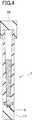
図4に示すように、好ましくは、容器は密閉可能の蓋、例えばスクリュー式上蓋10を用いて密封性を高めつつ、容器上端の開口部を開閉することができる。また、容器1の底部に同様のスクリュー式の蓋11を用いて開閉可能または脱着可能にすることもできる。このようにすることにより血液希釈緩衝液の容器に対して遠心力を付加して、容器1の下部に沈降し底部に付着した血球5を取り出して、検査することができるという利便性が生じる。 As shown in FIG. 4, preferably, the container can open and close the opening at the upper end of the container while improving the sealing performance using a sealable lid, for example, a screw-type
例えば容器1は、外径14±2mm、高さ75±5mmの円筒形状とし、容器1の下端部には、外径14±2mm、高さ25±2mmの脱着可能な円筒形スクリュー式底蓋11を取り付けた構造を持つ。容器1の内面の形状については、上端部の内径が10mm、下端部の内径が5mmであり、最上部から前記下端部の深さが30mmであり、上端部から下端部にかけて先細り形状とすることができる。
この場合、血液採取容器内腔の上部内径は10mmの円形で深さは30mmで底の内径は5mmであり、底に向かって逆円錐形で細くなる構造を有する。For example, the
In this case, the upper inner diameter of the blood sampling container lumen is a circle of 10 mm, the depth is 30 mm, the inner diameter of the bottom is 5 mm, and has a structure that narrows in an inverted conical shape toward the bottom.
血漿分離用高分子ゲル組成はシクロペンタジエン系樹脂、ジベンジリデンソルビトール、シリカ及びフタル酸(2−エチルヘキシル)を混練して得られたチキソトロピー性ゲル〔公開番号〕特許公開平7−294516)などを利用する。その他、類似するチキソトロピー性ゲル状分離剤を用いて構成することもできる。 The polymer gel composition for plasma separation uses a thixotropic gel obtained by kneading cyclopentadiene resin, dibenzylidene sorbitol, silica and phthalic acid (2-ethylhexyl) [Publication No. 7-294516] To do. In addition, a similar thixotropic gel separating agent can be used.
血漿分離用高分子ゲルの物理学的特性はHLB値として4.02〜9.0、比重は25℃で1.02〜1.08であり、分子量 GPC法による分子量分布は700〜850である。 The physical properties of the polymer gel for plasma separation are 4.02 to 9.0 as the HLB value, the specific gravity is 1.02 to 1.08 at 25 ° C., and the molecular weight distribution by the GPC method is 700 to 850.
好ましい態様では、ゲル状分離剤の特性は希釈血漿を分離条件である遠心力と遠心時間は1,300G、10分間で希釈血漿と血球を分離する。 In a preferred embodiment, the gel-like separating agent is characterized in that the diluted plasma and blood cells are separated at a centrifugal force and a centrifugation time of 1,300 G for 10 minutes, which are conditions for separating diluted plasma.
また、本件発明によれば微量な血液を希釈緩衝液で希釈した試料について、その希釈血漿分離に血漿分離用高分子ゲルを用いて遠心分離する方法が提供される。 In addition, according to the present invention, there is provided a method of centrifuging a sample obtained by diluting a small amount of blood with a dilution buffer using a polymer gel for plasma separation for the diluted plasma separation.
血漿分離ゲル化剤入り血液希釈保存容器を用いた生化学検査での実施例を示す。 The Example in the biochemical test | inspection using the blood dilution storage container containing a plasma separation gelatinizer is shown.
EDTA添加上腕静脈血を試料として遠心分離で得た血漿とこの血液の希釈された血漿測定値に希釈倍数を乗じて求めた各検査項目との相関データを示す。
表1は血漿分離ゲル化剤の層が希釈緩衝液の層の下側に位置している血漿分離ゲル化剤底封入タイプ(y)と血漿(x)との相関統計データ、図5はその相関図を示す。
表1 血漿分離ゲル化剤底封入タイプ
Table 1 shows the correlation statistical data between the plasma separation and gelling agent bottom encapsulation type (y) and the plasma (x), in which the plasma separation and gelling agent layer is located below the dilution buffer layer, and FIG. A correlation diagram is shown.
Table 1 Bottom type of plasma separation gelling agent
表2は血漿分離ゲル化剤の層が希釈緩衝液の層の上側に位置している血漿分離ゲル化剤希釈液上面封入タイプ(y)と静脈血漿(x)との相関統計データ、図6はその相関図を示す。
表2 血漿分離ゲル化剤希釈液上面封入タイプ
Table 2 Type of plasma separation gelling agent dilution top surface
表1と表2に示すように、血漿との相関性は非常に良い結果で、この血漿分離ゲル化剤入り血液希釈保存容器による希釈血漿分離法は血漿を試料とした測定値と遜色のない結果を得ることが可能である。 As shown in Table 1 and Table 2, the correlation with plasma is very good, and the diluted plasma separation method using the blood dilution storage container containing the plasma separation gelling agent is inferior to the measured value using plasma as a sample. It is possible to obtain a result.
この微量血液採取方法は血液採取の時間と場所に制限がないため、医療機関に出向く時間がない場合、災害時、遠隔医療、健康管理などに応用でき、未病の人を早期発見できるなど、医療費の節減にも貢献できる。 This micro blood collection method has no restrictions on the time and place of blood collection, so if you do not have time to go to a medical institution, it can be applied to disasters, telemedicine, health management, etc. It can also contribute to the reduction of medical costs.
1 血液希釈保存容器
2 希釈緩衝液
3 血漿分離用高分子ゲル化剤
4 試料血液
5 血球
6 上部開口
7 密封蓋
8 希釈血漿
9 目印
10 スクリュー式上蓋
11 スクリュー式底蓋1 Blood
10 Screw-
Claims (16)
Translated fromJapanese前記採取された血液中の血漿を希釈するための希釈緩衝液と、
該希釈された血漿を前記採取された血液中の血球と分離するための血漿分離用高分子ゲルと、
前記希釈緩衝液と血漿分離用高分子ゲルを収容するとともに、前記採取された血液を導入して収容し、保存するための血液希釈保存容器と、
前記血液希釈保存容器の上部開口部から導入される前記血液を前記希釈緩衝液と血漿分離用高分子ゲルとともに前記血液希釈保存容器に封入するための密閉蓋とを備え、
前記希釈緩衝液と血漿分離用高分子ゲルとが前記血液希釈保存容器の内部空間内で上下方向にそれぞれ独立した層を形成するようになっており、
前記採取された血液が前記上部開口部から導入され、前記血液希釈保存容器に対して上方から下方に向かって作用する遠心力を付加することによって、希釈された前記採取された血液の血漿を含む希釈緩衝液の血漿層と前記血漿分離用高分子ゲルの層と前記血球の層とがそれぞれ独立した層として前記血液希釈保存容器内に形成されるように構成され、
採取された血液において希釈された血漿の前記血漿層の比重がほぼ1.012〜1.014である、血液分析用構造体。A structure for blood analysis for containing a biological analysis sample produced by diluting a collected trace amount of blood,
A dilution buffer for diluting the plasma in the collected blood;
A plasma separating polymer gel for separating the diluted plasma from the blood cells in the collected blood;
A blood dilution storage container for storing the dilution buffer and the polymer gel for plasma separation, and introducing and storing the collected blood, and storing it;
A hermetic lid for sealing the blood introduced from the upper opening of the blood dilution storage container together with the dilution buffer and the polymer gel for plasma separation into the blood dilution storage container;
The dilution buffer and the polymer gel for plasma separation form independent layers in the vertical direction in the internal space of the blood dilution storage container,
The collected blood is introduced from the upperopening, by adding a centrifugal force that acts downward from above the hemodilution storage container, including plasma diluted the collected blood A plasma layer of dilution buffer, a layer of the polymer gel for plasma separation, and a layer of blood cells are formed in the blood dilution storage container as independent layers,
A structure for blood analysis, wherein the specific gravity of the plasma layer of plasma diluted in collected blood is approximately 1.012 to 1.014 .
前記採取された血液中の血漿を希釈するための、内部標準入りの希釈緩衝液と、A dilution buffer with an internal standard for diluting plasma in the collected blood;
該希釈された血漿を前記採取された血液中の血球と分離するための血漿分離用高分子ゲルと、A plasma separating polymer gel for separating the diluted plasma from the blood cells in the collected blood;
前記希釈緩衝液と血漿分離用高分子ゲルを収容するとともに、前記採取された血液を導入して収容し、保存するための血液希釈保存容器と、A blood dilution storage container for storing the dilution buffer and the polymer gel for plasma separation, and introducing and storing the collected blood, and storing it;
前記血液希釈保存容器の上部開口部から導入される前記血液を前記希釈緩衝液と血漿分離用高分子ゲルとともに前記血液希釈保存容器に封入するための密閉蓋とを備え、A hermetic lid for sealing the blood introduced from the upper opening of the blood dilution storage container together with the dilution buffer and the polymer gel for plasma separation into the blood dilution storage container;
前記希釈緩衝液と血漿分離用高分子ゲルとが前記血液希釈保存容器の内部空間内で上下方向にそれぞれ独立した層を形成するようになっており、The dilution buffer and the polymer gel for plasma separation form independent layers in the vertical direction in the internal space of the blood dilution storage container,
前記採取された血液が前記上部開口部から導入され、前記血液希釈保存容器に対して上方から下方に向かって作用する遠心力を付加することによって、希釈された前記採取された血液の血漿を含む希釈緩衝液の血漿層と前記血漿分離用高分子ゲルの層と前記血球の層とがそれぞれ独立した層として前記血液希釈保存容器内に形成されるように構成された血液分析用構造体。The collected blood is introduced from the upper opening, and the diluted blood plasma is diluted by applying a centrifugal force acting downward from above to the blood dilution storage container. A blood analysis structure configured such that a plasma layer of dilution buffer, a layer of the polymer gel for plasma separation, and a layer of blood cells are formed as independent layers in the blood dilution storage container.
前記血液希釈保存容器の底部は、外径がほぼ14mm、高さがほぼ25mmの脱着可能な底蓋が形成されている請求項12に記載の血液分析構造体。The blood dilution storage container has a cylindrical shape with an outer diameter of approximately 14 mm and a height of approximately 75 mm,
13. The blood analysis structure according to claim12 , wherein a removable bottom lid having an outer diameter of approximately 14 mm and a height of approximately 25 mm is formed at the bottom of the blood dilution storage container.
上端部の内径がほぼ10mm、下端部の内径がほぼ5mmであり、
前記上端部から前記下端部の深さがほぼ30mmであり、
前記上端部から前記下端部にかけて内径が漸減する形状を成している請求項1〜8のいずれか1項に記載の血液分析構造体。The shape of the inner surface of the blood dilution storage container is
The inner diameter of the upper end is approximately 10 mm, and the inner diameter of the lower end is approximately 5 mm.
The depth from the upper end to the lower end is approximately 30 mm;
Theblood analysis structure according to any one of claims 1to 8 , wherein theblood analysis structure has a shape in which an inner diameter gradually decreases from the upper end to the lower end.
前記採取された血液中の血漿を希釈するための希釈緩衝液を用意し、
該希釈された血漿を前記採取された血液中の血球と分離するための血漿分離用高分子ゲルを用意し、
前記希釈緩衝液と血漿分離用高分子ゲルを収容するとともに、前記採取された血液を導入して収容し、保存するための血液希釈保存容器を用意し、
前記血液希釈保存容器の上部開口部から導入される前記血液を前記希釈緩衝液と血漿分離用高分子ゲルとともに前記血液希釈保存容器に封入するための密閉蓋を用意し、
前記希釈緩衝液と血漿分離用高分子ゲルとが前記血液希釈保存容器の内部空間内で上下方向にそれぞれ独立した層を形成するように前記血液希釈保存容器に導入し、
前記採取された血液が前記上部開口部から導入され、前記血液希釈保存容器に対して上方から下方に向かって作用する遠心力を付加し、
希釈された前記採取された血液の血漿を含む希釈緩衝液の血漿層と前記血漿分離用高分子ゲルの層と前記血球の層とがそれぞれ独立した層として前記血液希釈保存容器内に形成する段階を備え、採取された血液において希釈された血漿の前記血漿層の比重がほぼ1.012〜1.014になることを特徴とする希釈血液の分離方法。A method for separating diluted blood that contains a bioanalytical sample produced by diluting a small amount of collected blood and separates diluted plasma and blood cells,
Preparing a dilution buffer for diluting the plasma in the collected blood,
Preparing a polymer gel for plasma separation for separating the diluted plasma from blood cells in the collected blood,
While containing the dilution buffer and the polymer gel for plasma separation, preparing a blood dilution storage container for introducing and storing the collected blood and storing it,
Preparing a hermetic lid for sealing the blood introduced from the upper opening of the blood dilution storage container together with the dilution buffer and the polymer gel for plasma separation into the blood dilution storage container;
The dilution buffer and the polymer gel for plasma separation are introduced into the blood dilution storage container so as to form independent layers in the vertical direction in the internal space of the blood dilution storage container,
The collected blood is introduced from the upperopening, adds a centrifugal force that acts downward from above the hemodilution storage container,
A step of forming the diluted plasma layer of the diluted buffer containing the collected blood plasma, the layer of the polymer gel for plasma separation, and the layer of the blood cells in the blood dilution storage container as independent layers; Andthe specific gravity of the plasma layer of plasma diluted in the collected blood is approximately 1.012 to 1.014 .
前記採取された血液中の血漿を希釈するための、内部標準入りの希釈緩衝液を用意し、Prepare a dilution buffer with an internal standard for diluting plasma in the collected blood,
該希釈された血漿を前記採取された血液中の血球と分離するための血漿分離用高分子ゲルを用意し、Preparing a polymer gel for plasma separation for separating the diluted plasma from blood cells in the collected blood,
前記希釈緩衝液と血漿分離用高分子ゲルを収容するとともに、前記採取された血液を導入して収容し、保存するための血液希釈保存容器を用意し、While containing the dilution buffer and the polymer gel for plasma separation, preparing a blood dilution storage container for introducing and storing the collected blood and storing it,
前記血液希釈保存容器の上部開口部から導入される前記血液を前記希釈緩衝液と血漿分離用高分子ゲルとともに前記血液希釈保存容器に封入するための密閉蓋を用意し、Preparing a hermetic lid for sealing the blood introduced from the upper opening of the blood dilution storage container together with the dilution buffer and the polymer gel for plasma separation into the blood dilution storage container;
前記希釈緩衝液と血漿分離用高分子ゲルとが前記血液希釈保存容器の内部空間内で上下方向にそれぞれ独立した層を形成するように前記血液希釈保存容器に導入し、The dilution buffer and the polymer gel for plasma separation are introduced into the blood dilution storage container so as to form independent layers in the vertical direction in the internal space of the blood dilution storage container,
前記採取された血液が前記上部開口部から導入され、前記血液希釈保存容器に対して上方から下方に向かって作用する遠心力を付加し、The collected blood is introduced from the upper opening, and a centrifugal force acting from above to below is applied to the blood dilution storage container,
希釈された前記採取された血液の血漿を含む希釈緩衝液の血漿層と前記血漿分離用高分子ゲルの層と前記血球の層とがそれぞれ独立した層として前記血液希釈保存容器内に形成する段階を備えた、ことを特徴とする希釈血液の分離方法。A step of forming the diluted plasma layer of the diluted buffer containing the collected blood plasma, the layer of the polymer gel for plasma separation, and the layer of the blood cells in the blood dilution storage container as independent layers; A method for separating diluted blood, comprising:
Applications Claiming Priority (1)
| Application Number | Priority Date | Filing Date | Title |
|---|---|---|---|
| PCT/JP2014/065628WO2015189961A1 (en) | 2014-06-12 | 2014-06-12 | Method for dilute plasma separation using container for blood dilution and storage containing gelling agent for plasma separation |
Publications (2)
| Publication Number | Publication Date |
|---|---|
| JPWO2015189961A1 JPWO2015189961A1 (en) | 2017-04-20 |
| JP6231678B2true JP6231678B2 (en) | 2017-11-15 |
Family
ID=54833089
Family Applications (1)
| Application Number | Title | Priority Date | Filing Date |
|---|---|---|---|
| JP2016527571AActiveJP6231678B2 (en) | 2014-06-12 | 2014-06-12 | Diluted plasma separation method using blood dilution storage container with plasma separation gelling agent |
Country Status (5)
| Country | Link |
|---|---|
| US (1) | US10241012B2 (en) |
| EP (1) | EP3156794A4 (en) |
| JP (1) | JP6231678B2 (en) |
| CN (1) | CN106461635B (en) |
| WO (1) | WO2015189961A1 (en) |
Families Citing this family (3)
| Publication number | Priority date | Publication date | Assignee | Title |
|---|---|---|---|---|
| WO2017006962A1 (en)* | 2015-07-06 | 2017-01-12 | 富士フイルム株式会社 | Blood test kit and blood analysis method |
| KR102080417B1 (en) | 2015-07-06 | 2020-02-21 | 후지필름 가부시키가이샤 | Blood test kit and blood analysis method |
| EP4071470A4 (en)* | 2019-12-05 | 2024-01-03 | Sekisui Medical Co., Ltd. | BLOOD COLLECTION CONTAINER AND PLASMA SEPARATION PROCESS |
Family Cites Families (18)
| Publication number | Priority date | Publication date | Assignee | Title |
|---|---|---|---|---|
| US4823624A (en)* | 1987-07-01 | 1989-04-25 | Becton Dickinson & Company | Material layer volume determination with correction band |
| US4867887A (en) | 1988-07-12 | 1989-09-19 | Becton Dickinson And Company | Method and apparatus for separating mononuclear cells from blood |
| JPH07294516A (en) | 1994-04-21 | 1995-11-10 | Sekisui Chem Co Ltd | Blood collecting tube |
| JPH08108096A (en)* | 1994-08-17 | 1996-04-30 | Toshiki Uchida | Vessel for centrifugal separation |
| JPH08201391A (en)* | 1995-01-20 | 1996-08-09 | Olympus Optical Co Ltd | Immunological measuring method with marker grain |
| DE19647674C1 (en)* | 1996-11-19 | 1998-08-27 | Sarstedt Walter Geraete | Method for automatically pushing a filter into a blood container |
| EP1125591A1 (en)* | 2000-01-05 | 2001-08-22 | Maehata, Eisuke | Instrument and method for blood separation |
| US6936473B2 (en) | 2000-01-05 | 2005-08-30 | Leisure, Inc. | Method of preparing a biological sample for quantification |
| AT414209B (en)* | 2000-03-17 | 2006-10-15 | Greiner Bio One Gmbh | COLLECTION TANK FOR LIQUIDS |
| AU2002346247B2 (en)* | 2001-06-06 | 2007-04-26 | Perfusion Partners & Associates, Inc. | Centrifuge tube assembly |
| JP3898632B2 (en)* | 2001-12-04 | 2007-03-28 | 積水化学工業株式会社 | Serum or plasma separation composition and blood test container containing the same |
| CA2444434A1 (en) | 2001-12-04 | 2003-06-12 | Sekisui Chemical Co., Ltd. | Composition for blood serum or plasma separation and vessel for blood examination containing the same |
| KR100430893B1 (en)* | 2002-01-28 | 2004-05-17 | 최은용 | A syringe for deviding a blood serum |
| US6770883B2 (en)* | 2002-01-30 | 2004-08-03 | Beckman Coulter, Inc. | Sample level detection system |
| US7323144B2 (en)* | 2002-03-18 | 2008-01-29 | Leisure, Inc. | Apparatus for separating biological sample and separating method of the same |
| JP2010169543A (en)* | 2009-01-23 | 2010-08-05 | Olympus Corp | Blood separation vessel and method of separating blood |
| KR101069877B1 (en) | 2009-10-28 | 2011-10-05 | 임기표 | Kit of centrifuge separation and methods for centrifuging using the same |
| WO2013111130A1 (en) | 2012-01-23 | 2013-08-01 | Estar Technologies Ltd | A system and method for obtaining a cellular sample enriched with defined cells such as platelet rich plasma(prp) |
- 2014
- 2014-06-12EPEP14894697.3Apatent/EP3156794A4/ennot_activeWithdrawn
- 2014-06-12USUS15/318,084patent/US10241012B2/ennot_activeExpired - Fee Related
- 2014-06-12CNCN201480079806.2Apatent/CN106461635B/ennot_activeExpired - Fee Related
- 2014-06-12WOPCT/JP2014/065628patent/WO2015189961A1/enactiveApplication Filing
- 2014-06-12JPJP2016527571Apatent/JP6231678B2/enactiveActive
Also Published As
| Publication number | Publication date |
|---|---|
| US10241012B2 (en) | 2019-03-26 |
| WO2015189961A1 (en) | 2015-12-17 |
| JPWO2015189961A1 (en) | 2017-04-20 |
| US20170131189A1 (en) | 2017-05-11 |
| EP3156794A1 (en) | 2017-04-19 |
| CN106461635A (en) | 2017-02-22 |
| CN106461635B (en) | 2019-12-03 |
| EP3156794A4 (en) | 2018-01-24 |
Similar Documents
| Publication | Publication Date | Title |
|---|---|---|
| CN106456075B (en) | Sampling and assay kits, sample holders and methods | |
| US3780935A (en) | Serum separating method | |
| NL1033365C2 (en) | Device and method for separating and analyzing blood. | |
| AU2005236435B2 (en) | Specimen collecting, processing and analytical assembly | |
| US3914985A (en) | Centrifuging device and method | |
| JP6901194B1 (en) | Blood collection container and plasma separation method | |
| JPH0771518B2 (en) | Quantification of fibrinogen in whole blood samples | |
| JP6231678B2 (en) | Diluted plasma separation method using blood dilution storage container with plasma separation gelling agent | |
| JP2011502623A (en) | Transdermal body fluid sampling and pretreatment apparatus and method | |
| JP2013068631A (en) | Method and apparatus for analyzing individual cells or particulates using fluorescent quenching and/or fluorescent bleaching | |
| CN1882835A (en) | Liquid sample analysis device with sealable reservoir | |
| CN1292685A (en) | Analysis of quiescent anticoagulated whole blood samples | |
| CN107966327B (en) | Collection vials and methods for testing sample properties | |
| SE539853C2 (en) | An arrangement for collection and separation of a body fluidfor purposes of analysis and a method relating thereto | |
| US3963119A (en) | Serum separating apparatus | |
| CN200996897Y (en) | Sampler collecting container | |
| US4043928A (en) | Serum separating composition of matter | |
| Ahn et al. | Comparison of Improvacuter EDTA tube with BD Vacutainer EDTA tube for routine hematological analysis: Clinical significance of differences, stability study, and effects of K2 and K3 EDTA | |
| US9678088B2 (en) | Multiphase systems for diagnosis of sickle cell disease | |
| WO2015189960A1 (en) | Blood-sample collection instrument | |
| Ross et al. | Dilutional effect of ethylenediaminetetraacetic acid on packed cell volume in healthy dogs | |
| EP3908404A1 (en) | Container for sample collection | |
| US20170071582A1 (en) | Fluid sample collection and preservation system | |
| CN106176216A (en) | Blood sampling test tube | |
| OA19075A (en) | Automatic device for measuring the speed of blood sedimentation. |
Legal Events
| Date | Code | Title | Description |
|---|---|---|---|
| A521 | Request for written amendment filed | Free format text:JAPANESE INTERMEDIATE CODE: A523 Effective date:20161130 | |
| A621 | Written request for application examination | Free format text:JAPANESE INTERMEDIATE CODE: A621 Effective date:20161130 | |
| TRDD | Decision of grant or rejection written | ||
| A01 | Written decision to grant a patent or to grant a registration (utility model) | Free format text:JAPANESE INTERMEDIATE CODE: A01 Effective date:20170926 | |
| A61 | First payment of annual fees (during grant procedure) | Free format text:JAPANESE INTERMEDIATE CODE: A61 Effective date:20171019 | |
| R150 | Certificate of patent or registration of utility model | Ref document number:6231678 Country of ref document:JP Free format text:JAPANESE INTERMEDIATE CODE: R150 | |
| R250 | Receipt of annual fees | Free format text:JAPANESE INTERMEDIATE CODE: R250 | |
| R250 | Receipt of annual fees | Free format text:JAPANESE INTERMEDIATE CODE: R250 | |
| R250 | Receipt of annual fees | Free format text:JAPANESE INTERMEDIATE CODE: R250 | |
| R250 | Receipt of annual fees | Free format text:JAPANESE INTERMEDIATE CODE: R250 | |
| R250 | Receipt of annual fees | Free format text:JAPANESE INTERMEDIATE CODE: R250 |