JP5849065B2 - Detection method of influenza A virus H5 subtype - Google Patents
Detection method of influenza A virus H5 subtypeDownload PDFInfo
- Publication number
- JP5849065B2 JP5849065B2JP2013093233AJP2013093233AJP5849065B2JP 5849065 B2JP5849065 B2JP 5849065B2JP 2013093233 AJP2013093233 AJP 2013093233AJP 2013093233 AJP2013093233 AJP 2013093233AJP 5849065 B2JP5849065 B2JP 5849065B2
- Authority
- JP
- Japan
- Prior art keywords
- antibody
- influenza
- duck
- virus
- subtype
- Prior art date
- Legal status (The legal status is an assumption and is not a legal conclusion. Google has not performed a legal analysis and makes no representation as to the accuracy of the status listed.)
- Expired - Fee Related
Links
Images


Classifications
- G—PHYSICS
- G01—MEASURING; TESTING
- G01N—INVESTIGATING OR ANALYSING MATERIALS BY DETERMINING THEIR CHEMICAL OR PHYSICAL PROPERTIES
- G01N33/00—Investigating or analysing materials by specific methods not covered by groups G01N1/00 - G01N31/00
- G01N33/48—Biological material, e.g. blood, urine; Haemocytometers
- G01N33/50—Chemical analysis of biological material, e.g. blood, urine; Testing involving biospecific ligand binding methods; Immunological testing
- G01N33/53—Immunoassay; Biospecific binding assay; Materials therefor
- G01N33/569—Immunoassay; Biospecific binding assay; Materials therefor for microorganisms, e.g. protozoa, bacteria, viruses
- G01N33/56983—Viruses
- G—PHYSICS
- G01—MEASURING; TESTING
- G01N—INVESTIGATING OR ANALYSING MATERIALS BY DETERMINING THEIR CHEMICAL OR PHYSICAL PROPERTIES
- G01N2333/00—Assays involving biological materials from specific organisms or of a specific nature
- G01N2333/005—Assays involving biological materials from specific organisms or of a specific nature from viruses
- G01N2333/08—RNA viruses
- G01N2333/11—Orthomyxoviridae, e.g. influenza virus
Landscapes
- Health & Medical Sciences (AREA)
- Life Sciences & Earth Sciences (AREA)
- Immunology (AREA)
- Engineering & Computer Science (AREA)
- Urology & Nephrology (AREA)
- Molecular Biology (AREA)
- Biomedical Technology (AREA)
- Chemical & Material Sciences (AREA)
- Virology (AREA)
- Hematology (AREA)
- Medicinal Chemistry (AREA)
- Analytical Chemistry (AREA)
- Cell Biology (AREA)
- Biotechnology (AREA)
- Food Science & Technology (AREA)
- Tropical Medicine & Parasitology (AREA)
- Physics & Mathematics (AREA)
- Microbiology (AREA)
- Biochemistry (AREA)
- General Health & Medical Sciences (AREA)
- General Physics & Mathematics (AREA)
- Pathology (AREA)
- Peptides Or Proteins (AREA)
- Apparatus Associated With Microorganisms And Enzymes (AREA)
- Preparation Of Compounds By Using Micro-Organisms (AREA)
Description
Translated fromJapanese本発明は、A型インフルエンザウイルス感染のうち、A型インフルエンザウイルスH5亜型感染を検出することができる検出法、詳しくは、サンドイッチ式免疫測定法、特に、イムノクロマトグラフィー測定法およびイムノクロマトグラフィー測定装置に関するものであり、高病原性鳥インフルエンザウイルスなどのインフルエンザウイルスH5亜型の感染、特に、ヒトにおけるインフルエンザウイルスH5N1亜型感染を迅速かつ簡便に診断するために有用な検出法に関する。The present invention relates to a detection method capable of detecting an influenza A virus H5 subtype infection among influenza A virus infections, and more particularly to a sandwich immunoassay, particularly an immunochromatography assay and an immunochromatography assay device. The present invention relates to a detection method useful for quickly and easily diagnosing infection of influenza virus H5 subtype such as highly pathogenic avian influenza virus, particularly influenza virus H5N1 subtype infection in humans.
高病原性鳥インフルエンザは、鶏などに高致死性の病原性を示すインフルエンザウイルスによる感染症で、家きんペストとも呼ばれ、わが国では家畜伝染病予防法の法定伝染病に指定されており、世界獣医事務局(OIE)ではリストA疾病として掲げられている。Highly pathogenic avian influenza is an infectious disease caused by influenza virus that is highly lethal to chickens, and is also called poultry plague. In Japan, it is designated as a legal infectious disease in the Livestock Infectious Disease Prevention Law. Listed as List A disease by the Veterinary Secretariat (OIE).
今までに高病原性鳥インフルエンザを引き起こしたインフルエンザウイルスはインフルエンザA型ウイルスのH5亜型及びH7亜型のみである。これらの亜型が総て強毒性であるわけではないが、わが国では、強毒性及び弱毒性であるかにかかわらずこれらの亜型に家畜が感染した場合、すべて屠殺処分することとされている。To date, the only influenza viruses that have caused highly pathogenic avian influenza are H5 and H7 subtypes of influenza A virus. Although not all of these subtypes are highly toxic, in Japan, all livestock infected with these subtypes, regardless of whether they are strong or weakly toxic, are to be slaughtered. .
また人への鳥インフルエンザ感染はH5亜型、H7亜型およびH9亜型の感染が報告されている。このうちインフルエンザウイルスが原因で死亡者が報告されているのはH5N1亜型のみである。H5N1亜型感染は重篤な肺炎を引き起こし、死に至ることが報告されている。
H5N1亜型による高病原性鳥インフルエンザは、1997年に香港で流行し、さらに、2003年から現在までアジアの数カ国で大流行し、人への感染及び死亡例も報告された。
現在、H5N1亜型の人への感染は世界規模で問題となっており、H1N1亜型、H3N2亜型に続く新型インフルエンザとして世界規模で警戒されている。In addition, avian influenza infection in humans has been reported as H5 subtype, H7 subtype and H9 subtype. Of these, only the H5N1 subtype has been reported dead due to influenza virus. H5N1 subtype infection has been reported to cause severe pneumonia and to death.
The highly pathogenic avian influenza caused by the H5N1 subtype was epidemic in Hong Kong in 1997, and was also pandemic in several Asian countries from 2003 to the present, and cases of human infection and death were also reported.
Currently, infection of people with H5N1 subtype is a problem on a global scale, and it is warned worldwide as a new influenza following the H1N1 subtype and H3N2 subtype.
2005年11月17日のWHOの報告によると、ヒトのインフルエンザH5N1亜型感染確定症例数は130例であり、内死亡者は67例である。2004年12月以降、ヒトへの感染確定症例数は急激に増加し、死亡例数も増加している。H5N1ウイルスに感染後の死亡率は51.3%(67/130)である。
上記からも明らかなように、H5N1亜型の人への感染が現実となりつつある現在、H5N1亜型感染を迅速に発見することは、大規模なインフルエンザ感染拡大を防ぐ上で重要な課題である。また迅速に診断することは治療においても重要である。According to a WHO report of November 17, 2005, the number of confirmed cases of human influenza H5N1 subtype infection is 130 cases, and 67 deaths. Since December 2004, the number of confirmed human cases has increased rapidly and the number of deaths has also increased. The mortality rate after infection with H5N1 virus is 51.3% (67/130).
As is clear from the above, now that the infection of H5N1 subtypes is becoming a reality, the rapid discovery of H5N1 subtypes is an important issue in preventing the spread of large-scale influenza infections. . Rapid diagnosis is also important in treatment.
鳥における高病原性鳥インフルエンザ診断は、現状では、ウイルス感染が疑われる病鳥から気管スワブまたはクロアカスワブ(総排泄腔スワブ)を採取し、これを発育鶏卵に接種して培養した後、ウイルスを分離する方法により行われている。しかし、この方法は、結果が得られるまでに数日を要し、迅速に結果が求められない点で不利である。また、近年開発された遺伝子検査(PCR法、LAMP法)を用いることにより、結果を得るまでに要する時間は大幅に短縮されるが、かかる遺伝子検査には特別な機器及び技術を要するため、養鶏現場等で検査を行うことは出来ない。Currently, the diagnosis of highly pathogenic avian influenza in birds is that tracheal swabs or cloacaswabs (total excretory swabs) are collected from diseased birds with suspected viral infections, inoculated into cultured chicken eggs, and then isolated. It is done by the method. However, this method is disadvantageous in that it takes several days to obtain a result and the result cannot be obtained quickly. In addition, the time required to obtain results is greatly shortened by using recently developed genetic tests (PCR method, LAMP method). However, because such genetic tests require special equipment and technology, poultry farming It is not possible to inspect on site.
インフルエンザウイルスはヒト上気道、下気道などに感染し、1〜2日の潜伏期をおいた後、発熱(38〜39℃)と共に頭痛、腰痛、筋肉痛、全身倦怠、消化器症状などを引き起こす。インフルエンザで最も問題となる合併症は高齢者の肺炎と小児の脳炎・脳症で、特に小児の脳炎・脳症は高熱、意識障害、痙攣を特徴とし、極めて予後が悪いうえに初発症状から中枢神経系症状の発現及び死に至る期間が極めて短いので、迅速な診断と処置が必要である。Influenza virus infects the upper and lower respiratory tracts of humans and causes a headache, low back pain, muscle pain, general malaise, digestive symptoms, etc. along with fever (38-39 ° C.) after a latent period of 1-2 days. The most common complications of influenza are pneumonia in the elderly and encephalitis / encephalopathy in children. Especially in children, encephalitis / encephalopathy is characterized by high fever, disturbance of consciousness, and convulsions. Rapid diagnosis and treatment is required because the time to onset of symptoms and death is very short.
従来は、上記のような臨床症状のみでインフルエンザ様疾患として診断されることが多かったが、近年、インフルエンザウイルス抗原を迅速に検出するキットが開発され普及し始めたことから、インフルエンザウイルス感染症として早期に診断することが可能となってきた。このような迅速診断キットとしては、酵素免疫法(EIA)やイムノクロマトグラフィー測定法を原理として用いたものが挙げられ、A型インフルエンザウイルスのみを検出するもの、A型とB型をまとめて検出するもの、A型とB型をそれぞれ検出するものなどがある。
しかしながら現在行われているインフルエンザ抗原検査はA型ウイルス全てに共通な核タンパク(ヌクレオプロテイン)に対する抗体を使用している。そのためインフルエンザA型ウイルスであるH5亜型感染とH1N1またはH3N2亜型感染との鑑別診断ができない。
H5N1亜型感染は重篤な肺炎を引き起こし、且つ死亡率が非常に高い。初期診断時にH5亜型感染を鑑別診断することは、患者に対する治療方針を立てる上でも重要である。さらに新たな感染を防ぐために患者を早期に隔離するなどの処置を講ずることができ、更なるH5N1亜型感染の拡大を防ぐ上でも重要である。
また、近年開発された遺伝子検査(PCR法、LAMP法)を用いることにより、H5N1亜型を鑑別して検出することは可能であるが特別な機器及び技術を要するため簡便な方法ではない。Conventionally, it was often diagnosed as an influenza-like disease only with the above clinical symptoms. However, since a kit for rapidly detecting an influenza virus antigen has recently been developed and spread, It has become possible to diagnose early. Examples of such rapid diagnosis kits include those using enzyme immunoassay (EIA) or immunochromatography assay as a principle, and those that detect only influenza A virus, or those that detect A and B together. And those that detect A type and B type, respectively.
However, current influenza antigen tests use antibodies against nucleoproteins common to all type A viruses. Therefore, differential diagnosis between H5 subtype infection which is influenza A virus and H1N1 or H3N2 subtype infection cannot be performed.
H5N1 subtype infection causes severe pneumonia and has a very high mortality rate. Differential diagnosis of H5 subtype infection at the initial diagnosis is also important in formulating a treatment policy for patients. Furthermore, in order to prevent new infections, it is possible to take measures such as isolating patients at an early stage, which is important in preventing further spread of H5N1 subtype infection.
In addition, it is possible to distinguish and detect H5N1 subtypes by using recently developed genetic tests (PCR method, LAMP method), but this is not a simple method because it requires special equipment and technology.
このように、インフルエンザウイルスは感染が速く、発見が遅れると広い範囲にわたって急速に蔓延するので、感染をできる限り早期に、且つ、迅速に発見する必要がある。特にA型インフルエンザウイルスH5N1亜型は重篤な肺炎を引き起こし、死亡率も高い。しかしながら、現在のところ、A型インフルエンザウイルスH5亜型は従来のインフルエンザウイルス抗原検出試薬では他のA型インフルエンザウイルス感染と鑑別して検出することはできない。
本発明の目的は、A型インフルエンザウイルスH5亜型を迅速かつ簡便に鑑別診断できる検出試薬、及びそれを用いた検出法、とりわけ、サンドイッチ式免疫測定法、特に、イムノクロマトグラフィー測定法およびイムノクロマトグラフィー測定装置を提供することを目的とする。In this way, influenza viruses are fast infecting and spread rapidly over a wide range when discovery is delayed, so it is necessary to detect infection as early as possible and quickly. In particular, influenza A virus H5N1 subtype causes severe pneumonia and has a high mortality rate. However, at present, influenza A virus H5 subtype cannot be detected by distinguishing it from other influenza A virus infections using conventional influenza virus antigen detection reagents.
An object of the present invention is to provide a detection reagent that can quickly and easily differentially diagnose influenza A virus H5 subtype, and a detection method using the same, especially a sandwich immunoassay, particularly an immunochromatographic assay and an immunochromatographic assay. An object is to provide an apparatus.
本発明者等は、インフルエンザウイルスH5亜型を免疫原としてマウスを免疫して該ウイルスのヘマグルチニン(HA)蛋白に対して特異的に反応する抗体を取得することに成功し、当該抗体を免疫測定法、特にサンドイッチ式免疫測定法、とりわけイムノクロマトグラフィー測定法で使用することにより、インフルエンザウイルスH5亜型を特異的に検出し得ることを見出し、本発明を完成するに至った。The present inventors succeeded in obtaining an antibody that specifically reacts with the hemagglutinin (HA) protein of the virus by immunizing a mouse using the influenza virus H5 subtype as an immunogen, and immunoassay the antibody. It was found that the influenza virus H5 subtype can be specifically detected by using the method, particularly a sandwich immunoassay, particularly an immunochromatographic assay, and the present invention has been completed.
すなわち、本発明の一局面によれば、全てのA型インフルエンザウイルス亜型に対して反応する第一の抗体を用いた第一の免疫測定と、A型インフルエンザウイルスH5亜型に対して特異的に反応する第二の抗体を用いた第二の免疫測定とを併用してなることを特徴とするA型インフルエンザウイルスH5亜型の検出法が提供される。That is, according to one aspect of the present invention, a first immunoassay using a first antibody that reacts against all influenza A virus subtypes and specific for influenza A virus H5 subtype There is provided a method for detecting influenza A virus H5 subtype, characterized in that it is used in combination with a second immunoassay using a second antibody that reacts with A.
この検出法における免疫測定法としては、特に限定されるものではないが、サンドイッチ式免疫測定法、とりわけELISA(Enzyme−linked immunosorbent assay)法、イムノクロマトグラフィー測定法などが好ましい。
したがって、本発明の上記検出法の好ましい実施形態によれば、前記免疫測定法は、前記第一の抗体とこの第一の抗体が反応する抗原に対して反応する第三の抗体とを用いたサンドイッチ式免疫測定法、及び/又は、前記第二の抗体と該第二の抗体が反応する抗原に対して反応する第四の抗体とを用いたサンドイッチ式免疫測定法から構成される。これらのサンドイッチ式免疫測定法は、前記第一の抗体および第二または第四の抗体を担体に固定して実施すると好都合である。この場合、担体として膜担体を使用すると、イムノクロマトグラフィー測定法を実施することができる。The immunoassay method in this detection method is not particularly limited, but a sandwich immunoassay method, particularly an ELISA (Enzyme-linked immunosorbent assay) method, an immunochromatographic assay method and the like are preferable.
Therefore, according to a preferred embodiment of the detection method of the present invention, the immunoassay method uses the first antibody and a third antibody that reacts with an antigen to which the first antibody reacts. It comprises a sandwich immunoassay and / or a sandwich immunoassay using the second antibody and a fourth antibody that reacts with an antigen to which the second antibody reacts. These sandwich immunoassays are conveniently carried out with the first antibody and the second or fourth antibody immobilized on a carrier. In this case, when a membrane carrier is used as the carrier, an immunochromatographic measurement method can be carried out.
したがって、本発明の更に他の局面によれば、全てのA型インフルエンザウイルス亜型に対して反応する第一の抗体、及び、A型インフルエンザウイルスH5亜型に対して特異的に反応する第二の抗体をそれぞれ予め所定位置に固定せしめて形成された第一及び第二の捕捉部位を備える膜担体を用意し、前記第一の抗体が反応する抗原に対して反応する第三の抗体と被験試料との第一の混合液、及び、前記第二の抗体が反応する抗原に対して反応する第四の抗体と前記被験試料との第二の混合液を、それぞれ第一及び第二の捕捉部位に向けて前記膜担体にてクロマト展開せしめるか、または、第三の抗体と第四の抗体と被験試料との混合液を前記捕捉部位の双方に向けて前記膜担体にてクロマト展開せしめ、前記第一の捕捉部位における第一の抗体との反応が陽性であることをもってA型インフルエンザウイルスを検出し、さらに第二の捕捉部位における第二の抗体との反応が陽性であることをもってA型インフルエンザウイルスH5亜型を検出できるようにしたことを特徴とするイムノクロマトグラフィー測定法が提供される。Therefore, according to yet another aspect of the present invention, a first antibody that reacts against all influenza A virus subtypes and a second that specifically reacts with influenza A virus H5 subtype. A membrane carrier having first and second capture sites formed by preliminarily fixing each of the antibodies at a predetermined position, and a test with a third antibody that reacts with an antigen to which the first antibody reacts A first mixture with the sample, and a second mixture with the fourth antibody that reacts with the antigen to which the second antibody reacts and the test sample are respectively captured in the first and second captures. Chromatographic development on the membrane carrier toward the site, or the chromatographic development on the membrane carrier to the mixture of the third antibody and the fourth antibody and the test sample toward both of the capture site, First in the first capture site Influenza A virus is detected when the reaction with the antibody is positive, and influenza A virus H5 subtype can be detected when the reaction with the second antibody at the second capture site is positive. An immunochromatographic measurement method characterized by the above is provided.
また、本発明の更に他の局面によれば、全てのA型インフルエンザウイルス亜型に対して反応する第一の抗体と、A型インフルエンザウイルスH5亜型に対して特異的に反応する第二の抗体と、前記第一の抗体が反応する抗原に対して反応する第三の抗体と、前記第二の抗体が反応する抗原に対して反応する第四の抗体と、膜担体とを少なくとも備え、前記第一及び第二の抗体はそれぞれ予め膜担体の所定位置に固定されて第一及び第二の捕捉部位を形成し、前記第三及び第四の抗体は適当な標識物質で標識され、かつ、前記第三及び第四の抗体は前記捕捉部位の双方から離隔した位置からそれぞれ第一及び第二の捕捉部位に向けて前記膜担体にてクロマト展開可能なように用意されてなるA型インフルエンザウイルスH5亜型検出用イムノクロマトグラフィー測定装置が提供される。According to still another aspect of the present invention, a first antibody that reacts with all influenza A virus subtypes and a second antibody that specifically reacts with influenza A virus H5 subtype. An antibody, a third antibody that reacts with an antigen to which the first antibody reacts, a fourth antibody that reacts with an antigen with which the second antibody reacts, and a membrane carrier, The first and second antibodies are respectively preliminarily fixed at predetermined positions of the membrane carrier to form first and second capture sites, the third and fourth antibodies are labeled with an appropriate labeling substance, and The third and fourth antibodies are prepared so that they can be chromatographed on the membrane carrier from positions separated from both of the capture sites toward the first and second capture sites, respectively. Immuno for detecting virus H5 subtype Chromatography measurement device is provided.
前記膜担体は、第一の捕捉部位と第二の捕捉部位とを互いに離隔させて備えてなる単一の膜担体からなるものであってもよく、または、第一の抗体が固定された第一の膜担体と、第二の抗体が固定された第二の膜担体とからなるものであってもよいが、後者の方が反応特異性に優れているので好ましい。後者の場合、第三の抗体は第一の膜担体上に用意しておき、第四の抗体は第二の膜担体上に用意しておくと、それぞれの膜担体に被験試料を注入するだけで測定が行えるので好都合である。第三及び第四の抗体は、それぞれ、第一及び第二の膜担体上に配置された含浸部材に含浸させて用意しておくと好都合である。別法として、第一及び第二の膜担体の双方に連接した単一の含浸部材を設け、該含浸部材に第三の抗体及び第四の抗体を一緒に含浸させておいてもよい。第一の膜担体及び前記第二の膜担体は何れも細長形状すなわち帯状形状を備えることが好ましく、この場合、本発明の測定装置は、所謂イムノクロマト法テストストリップの形態として提供することができる。The membrane carrier may be a single membrane carrier comprising a first capture site and a second capture site spaced apart from each other, or a first antibody on which a first antibody is immobilized. Although it may be composed of one membrane carrier and a second membrane carrier to which the second antibody is immobilized, the latter is preferred because of its excellent reaction specificity. In the latter case, if the third antibody is prepared on the first membrane carrier and the fourth antibody is prepared on the second membrane carrier, the test sample is simply injected into each membrane carrier. It is convenient because it can be measured with. The third and fourth antibodies are conveniently prepared by impregnating the impregnating members disposed on the first and second membrane carriers, respectively. Alternatively, a single impregnation member connected to both the first and second membrane carriers may be provided, and the impregnation member may be impregnated with the third antibody and the fourth antibody together. Both the first membrane carrier and the second membrane carrier preferably have an elongated shape, that is, a strip shape, and in this case, the measuring apparatus of the present invention can be provided in the form of a so-called immunochromatographic test strip.
本発明の検出法及び測定方法において、第一の免疫測定は、キャピリア(登録商標)FluA+B(株式会社タウンズ製)などの市販のA型及びB型インフルエンザ鑑別測定用のイムノクロマト法テストストリップまたはキットを用いて実施でき、上記第一の膜担体としてかかる市販のテストストリップまたはキットを使用しても良い。この場合、第二の免疫測定のために、上記第二の膜担体のみを入手すれば、本発明の検出法及び測定方法を実施することができる。In the detection method and measurement method of the present invention, the first immunoassay is performed using a commercially available immunochromatographic test strip for differential measurement of influenza A and B influenza such as Capilia (registered trademark) FluA + B (manufactured by Towns Co., Ltd.) A commercially available test strip or kit may be used as the first membrane carrier. In this case, if only the second membrane carrier is obtained for the second immunoassay, the detection method and measurement method of the present invention can be carried out.
本発明で使用する上記第一の抗体は、全てのA型インフルエンザウイルス亜型に対して反応するであればよいが、好ましくは、インフルエンザウイルスの核タンパクに対する抗体である。第一の抗体は、ポリクローナル抗体であっても、モノクローナル抗体であってもよいが、反応特異性の観点から、モノクローナル抗体であることが好ましい。
ポリクローナル抗体は、例えば、配列番号6に記載されるアミノ酸配列をコードするDNA配列のうち、エピトープを構成するアミノ酸残基を含む部分に対応するDNA断片をクローニングし、当該クローン化遺伝子を大腸菌などの宿主で遺伝子工学的に発現させて発現蛋白を抽出および精製し、この精製蛋白を抗原として常法に従って動物を免疫し、その抗血清から取得することができる。Although the said 1st antibody used by this invention should just react with all the influenza A virus subtypes, Preferably, it is an antibody with respect to the nucleoprotein of influenza virus. The first antibody may be a polyclonal antibody or a monoclonal antibody, but is preferably a monoclonal antibody from the viewpoint of reaction specificity.
For example, a polyclonal antibody clones a DNA fragment corresponding to a portion containing an amino acid residue constituting an epitope in a DNA sequence encoding the amino acid sequence described in SEQ ID NO: 6, and the cloned gene is expressed in E. coli or the like. The expressed protein can be extracted and purified by genetic engineering expression in a host, and the animal can be immunized according to a conventional method using the purified protein as an antigen, and obtained from the antiserum.
モノクローナル抗体は、例えば、上記と同様に得られた精製蛋白を抗原としてマウスのような動物を免疫したのち、この免疫された動物の脾臓細胞とミエローマ細胞とを細胞融合して得られた融合細胞をHAT含有培地でセレクトした後に増殖せしめ、増殖せしめた株を前記のようにして得られた精製蛋白を使用して、たとえば、酵素標識免疫法などにより選別することで、取得することができる。また、モノクローナル抗体として、公知のイムノクロマト法テストストリップにおいて用いられているA型インフルエンザウイルスの核タンパクに対するモノクローナル抗体を用いてもよい。The monoclonal antibody is, for example, a fused cell obtained by immunizing an animal such as a mouse with the purified protein obtained in the same manner as described above as an antigen, and then cell-splitting spleen cells and myeloma cells of the immunized animal. Can be obtained by selecting, for example, by enzyme-labeled immunoassay using the purified protein obtained as described above. Moreover, as a monoclonal antibody, you may use the monoclonal antibody with respect to the nuclear protein of the influenza A virus currently used in the well-known immunochromatography test strip.
本発明で使用する上記第二の抗体は、A型インフルエンザウイルスH5亜型に対して特異的に反応する抗体であり、好ましくは、A型インフルエンザウイルスH5亜型のヘマグルチニン蛋白に対する抗体である。第二の抗体は、ポリクローナル抗体であっても、モノクローナル抗体であってもよいが、反応特異性の観点から、モノクローナル抗体であることが好ましい。例えば、インフルエンザウイルスH5N1型(A/Hong Kong/156/97(H5N1)))のヘマグルチニン蛋白の全アミノ酸配列は、配列番号7に示されるとおり公知である(Science 279 (5349), 393-396 (1998))。したがって、第二の抗体は、かかるA型インフルエンザウイルスH5亜型のヘマグルチニン蛋白の全アミノ酸配列からエピトープを構成する領域を選択してDNAをクローニングして発現させ、抽出、精製した精製蛋白を免疫原として第一の抗体に関して上述したと同様の方法で得ることができる。The second antibody used in the present invention is an antibody that specifically reacts with influenza A virus H5 subtype, and preferably an antibody against hemagglutinin protein of influenza A virus H5 subtype. The second antibody may be a polyclonal antibody or a monoclonal antibody, but is preferably a monoclonal antibody from the viewpoint of reaction specificity. For example, the entire amino acid sequence of hemagglutinin protein of influenza virus H5N1 type (A / Hong Kong / 156/97 (H5N1)) is known as shown in SEQ ID NO: 7 (Science 279 (5349), 393-396 ( 1998)). Therefore, the second antibody selects the region constituting the epitope from the entire amino acid sequence of the hemagglutinin protein of the influenza A virus H5 subtype, clones the DNA for expression, extracts and purifies the purified protein that has been extracted and purified. Can be obtained in the same manner as described above for the first antibody.
本発明で使用する好ましい第二の抗体は、配列番号7に示されるヘマグルチニン蛋白のアミノ酸配列の168番目のアミノ酸を含むエピトープを認識する抗体、特にモノクローナル抗体(以下、「モノクローナル抗体AA168」ともいう)である。このエピトープは、上記アミノ酸配列の168番目のアミノ酸残基を含むその前後の数個のアミノ酸残基から構成されるものと考えられ、通常、当該168番目のアミノ酸残基を中心とする6〜10個のアミノ酸残基から構成されると考えられる。A preferred second antibody used in the present invention is an antibody that recognizes an epitope containing the amino acid at position 168 of the amino acid sequence of the hemagglutinin protein shown in SEQ ID NO: 7, particularly a monoclonal antibody (hereinafter also referred to as “monoclonal antibody AA168”). It is. This epitope is considered to be composed of several amino acid residues before and after the 168th amino acid residue of the above amino acid sequence, and usually 6 to 10 centered on the 168th amino acid residue. It is considered to be composed of amino acid residues.
しかしながら、当該モノクローナル抗体AA168は、蛍光抗体法によれば、インフルエンザウイルスH5弱毒株に対して全般的に反応するが、A/tn/S.Africa/61、A/swan/Shima/449/83 (24a5b)、A/HongKong/156/97、A/HongKong/483/97、 A/duck/Yokohama/aq-10/2003 及びA/chicken/Yamaguchi/7/04からなる群より選ばれた強毒株の少なくとも1つには反応しないか又は弱毒株に対するよりも反応性が弱い。However, according to the fluorescent antibody method, the monoclonal antibody AA168 generally reacts with an influenza virus H5 attenuated strain, but A / tn / S. Africa / 61, A / swan / Shima / 449/83 ( 24a5b), A / HongKong / 156/97, A / HongKong / 483/97, A / duck / Yokohama / aq-10 / 2003 and A / chicken / Yamaguchi / 7/04 Does not respond to or is less reactive than the attenuated strain.
したがって、本発明で使用する他の好ましい第二の抗体は、インフルエンザウイルスH5亜型であるA/tn/S.Africa/61、A/swan/Shima/449/83 (24a5b)、A/HongKong/156/97、A/HongKong/483/97、 A/duck/Yokohama/aq-10/2003 及びA/chicken/Yamaguchi/7/04の全てに対して反応するモノクローナル抗体(以下、「モノクローナル抗体WS」ともいう)である。モノクローナル抗体WSは、蛍光抗体法によれば、上記強毒株の全てに対して強く反応する点で、モノクローナル抗体AA168と明確に区別されるものであり、モノクローナル抗体AA168と異なるエピトープを認識するものと考えられる。モノクローナル抗体WSは、広範囲の強毒株及び弱毒株に対して反応するので、インフルエンザウイルスH5亜型を広く検出するのに好適であり、とりわけ、強毒株の検出用に好適である。Accordingly, other preferred second antibodies for use in the present invention are influenza virus H5 subtypes A / tn / S. Africa / 61, A / swan / Shima / 449/83 (24a5b), A / HongKong / Monoclonal antibodies that react with all of 156/97, A / HongKong / 483/97, A / duck / Yokohama / aq-10 / 2003 and A / chicken / Yamaguchi / 7/04 (hereinafter referred to as “monoclonal antibody WS”) It is also called). Monoclonal antibody WS is clearly distinguished from monoclonal antibody AA168 in that it reacts strongly with all of the above virulent strains according to the fluorescent antibody method, and recognizes an epitope different from monoclonal antibody AA168. it is conceivable that. The monoclonal antibody WS reacts with a wide range of highly virulent and attenuated strains and is therefore suitable for detecting influenza virus H5 subtype widely, and particularly suitable for detecting virulent strains.
なお、モノクローナル抗体AA168及びモノクローナル抗体WSの何れも、インフルエンザウイルスH5亜型以外のインフルエンザウイルスとは反応しないものであり、したがって、インフルエンザウイルスH5亜型のヘマグルチニン蛋白に対して特異的に反応する抗体である。Note that neither the monoclonal antibody AA168 nor the monoclonal antibody WS reacts with influenza viruses other than the influenza virus H5 subtype, and is therefore an antibody that specifically reacts with the hemagglutinin protein of the influenza virus H5 subtype. is there.
本発明において、イムノクロマトグラフィー測定法などのサンドイッチ式免疫測定法で使用する第三の抗体は、第一の抗体が反応する抗原に対して反応する抗体であればよく、該抗原が第一の抗体に対するエピトープを複数備えている場合には第一の抗体と同一の抗体であってもよく、該抗原が他のエピトープを備えている場合には第一の抗体以外の抗体であってもよい。
本発明において、イムノクロマトグラフィー測定法などのサンドイッチ式免疫測定法で使用する第四の抗体は、第二の抗体が反応する抗原に対して反応する抗体であればよく、該抗原が第二の抗体に対するエピトープを複数備えている場合には第二の抗体と同一の抗体であってもよく、該抗原が他のエピトープを備えている場合には第二の抗体以外の抗体であってもよい。
また、第三及び第四の抗体は、それぞれ、モノクローナル抗体でもポリクローナル抗体でもよいが、反応特異性の点から、モノクローナル抗体であることが好ましい。In the present invention, the third antibody used in the sandwich immunoassay method such as an immunochromatographic assay method may be an antibody that reacts with the antigen to which the first antibody reacts, and the antigen is the first antibody. The antibody may be the same antibody as the first antibody in the case where a plurality of epitopes are provided, and may be an antibody other than the first antibody if the antigen is provided with other epitopes.
In the present invention, the fourth antibody used in a sandwich immunoassay such as an immunochromatography assay may be an antibody that reacts with an antigen to which the second antibody reacts, and the antigen is the second antibody. The antibody may be the same antibody as the second antibody when a plurality of epitopes are provided, and may be an antibody other than the second antibody when the antigen is provided with another epitope.
The third and fourth antibodies may be monoclonal antibodies or polyclonal antibodies, respectively, but are preferably monoclonal antibodies from the viewpoint of reaction specificity.
現在のところ、A型インフルエンザウイルス亜型としてH(ヘマグルチニン)蛋白については16種類、N(ノイラミダーゼ)蛋白については9種の亜型が知られており、理論上は16×9=144種の亜型が考えられるが、一般的には、このうち、H1〜15亜型の代表例に対して反応する抗体が「全てのA型インフルエンザウイルス亜型に対して反応する抗体」と理解されている。したがって、本明細書において、「全てのA型インフルエンザウイルス亜型」とは、現在知られているH1〜15亜型の代表例を意味し、具体的には、本明細書の後記表3に挙げられた株を意味する。また、A型インフルエンザウイルスの核タンパクに対する抗体は、B型及びC型インフルエンザウイルスの何れの核タンパクとも交差反応性を示さないものであり、同様に、本発明の第一の抗体も、B型及びC型インフルエンザウイルスの何れの核タンパクとも交差反応性を示さないものである。
また、本明細書において、「抗体との反応が陽性」とは、抗原が抗体と抗原抗体反応することを意味する。At present, 16 subtypes of H (hemagglutinin) protein and 9 subtypes of N (neuramidase) protein are known as influenza A virus subtypes, and theoretically 16 × 9 = 144 subtypes. Although types are considered, in general, an antibody that reacts with a representative example of the H1-15 subtype is understood as "an antibody that reacts with all influenza A virus subtypes". . Therefore, in the present specification, “all influenza A virus subtypes” means representative examples of the currently known H1-15 subtypes, and specifically, in the following Table 3 of the present specification. Means the listed strain. Moreover, the antibody against the nucleoprotein of influenza A virus does not show cross-reactivity with any of the nucleoproteins of influenza B virus and influenza C virus. Similarly, the first antibody of the present invention is also of type B. And no nucleoprotein of influenza C virus show cross-reactivity.
In the present specification, “positive reaction with an antibody” means that an antigen reacts with an antibody by an antigen-antibody.
本発明によれば、A型インフルエンザウイルス感染という結果だけでなく、更に進んでA型インフルエンザウイルスH5亜型の感染が特定できるので、ヒト、鳥等のA型インフルエンザウイルスH5亜型による感染症の診断に広く適用できる。特にヒトの場合、現在ヒトへの感染が確認されているのはH5N1亜型のみであるので、H5N1亜型感染の早期の発見、予防、治療に貢献することができる。According to the present invention, not only the result of influenza A virus infection, but also the infection of influenza A virus H5 subtype can be specified further, so that infections caused by influenza A virus H5 subtype in humans, birds, etc. Widely applicable for diagnosis. Particularly in the case of humans, only the H5N1 subtype is currently confirmed to be infected with humans, which can contribute to early detection, prevention and treatment of H5N1 subtype infection.
本発明の好ましい実施形態によれば、第一の抗体としてA型インフルエンザウイルスの核タンパクに対する抗体が使用され、第二の抗体としてA型インフルエンザウイルスH5亜型のヘマグルチニン蛋白に対する抗体が使用される。この実施形態では、第一の抗体と第二の抗体の検出対象が異なるため、互いの検出感度に影響を及ぼすことがなく、特異性の高い結果を得ることができる。According to a preferred embodiment of the present invention, an antibody against the influenza A virus nucleoprotein is used as the first antibody, and an antibody against the influenza A virus H5 subtype hemagglutinin protein is used as the second antibody. In this embodiment, since the detection targets of the first antibody and the second antibody are different, the detection sensitivity of each other is not affected, and a highly specific result can be obtained.
また、本発明のイムノクロマトグラフィー測定法およびイムノクロマトグラフィー測定装置によれば、特殊な機器及び熟練した技術を必要とすることなく、病院、養鶏現場等において簡便かつ迅速にインフルエンザウイルスH5亜型の検出及び該ウイルスによる感染を診断することが可能となる。Moreover, according to the immunochromatography measurement method and the immunochromatography measurement apparatus of the present invention, the detection and detection of influenza virus H5 subtype can be performed easily and quickly at hospitals, poultry farms, etc. without requiring special equipment and skilled techniques. It becomes possible to diagnose infection by the virus.
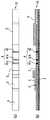
本発明のイムノクロマトグラフィー測定法を実施するために使用できる測定装置は、公知のイムノクロマト法テストストリップの構成に準拠して容易に実施できる。以下、その具体例を図面に基づいて説明する。A measuring apparatus that can be used for carrying out the immunochromatographic measuring method of the present invention can be easily carried out according to the configuration of a known immunochromatographic test strip. Specific examples thereof will be described below with reference to the drawings.
(1)単一のイムノクロマト法テストストリップを用いる実施形態
イムノクロマト法テストストリップの具体例としては、例えば図1に示されるテストストリップ10が挙げられる。図1において、数字1は粘着シート、2は含浸部材、3は膜担体、4は捕捉部位、5は吸収用部材、6は試料添加用部材を示している。
図示の例では、膜担体3は、幅5mm、長さ36mmの細長い帯状のニトロセルロース製メンブレンフィルターからなり、同幅の粘着シート1の中程に貼り付けられている。膜担体3には、そのクロマト展開始点側、すなわち図1の左側(以下「上流側」と記す。なお、その逆の側、すなわち図1におけるクロマト展開方向側すなわち右側は以下「下流側」と記す。)の末端から下流側に7.5mmの位置に第一の抗体が固定され、第三の抗体とA型インフルエンザウイルスの核タンパク等のウイルス抗原との複合体を捕捉するための第一の捕捉部位41aが形成されている。さらに、膜担体3の上流側の末端から下流側に10.5mmの位置に第二の抗体が固定され、第四の抗体とH5亜型のヘマグルチニン蛋白等のウイルス抗原との複合体を捕捉するための第二の捕捉部位41bが形成されている。さらに、膜担体3の上流側の末端から下流側に15.0mmの位置に所謂コントロールライン42が設けられている。このコントロールライン42は、分析対象物質の存否に係わらず反応が行われたことを確認するためのものであり、通常、前記第三及び/又は第四の抗体と免疫学的に特異的に結合する物質(分析対象物質を除く)を膜担体3に固定化することによって形成することができる。例えば、第三及び/又は第四の抗体としてマウス抗体を用いた場合は、該マウス抗体に対する抗体を用いることができる。(1) Embodiment using a single immunochromatographic test strip As a specific example of the immunochromatographic test strip, there is a
In the example shown in the figure, the
図1の装置では、膜担体3は、ニトロセルロース製メンブレンフィルターを用いているが、被験試料に含まれる分析対象物質をクロマト展開可能で、かつ、捕捉部位4を形成する抗体を固定可能なものであれば、いかなるものであってもよく、他のセルロース類膜、ナイロン膜、ガラス繊維膜なども使用できる。
図1の装置において、第一の抗体としては、全てのA型インフルエンザウイルス亜型の核タンパクに対して反応するモノクローナル抗体を用いることが好ましい。第二の抗体としては、H5亜型に対して特異的に反応するモノクローナル抗体を用いることが好ましい。In the apparatus of FIG. 1, the
In the apparatus of FIG. 1, it is preferable to use a monoclonal antibody that reacts with all the influenza A virus subtype nucleoproteins as the first antibody. As the second antibody, it is preferable to use a monoclonal antibody that reacts specifically with the H5 subtype.
図1の装置において、第三の抗体は、第一の抗体が反応する抗原に反応する抗体であり、好ましくは、全てのA型インフルエンザウイルス亜型の核タンパクに対して反応する抗体であり、多くの場合、第一の抗体と同一の抗体を用いることができる。また、第四の抗体は、第二の抗体が反応する抗原に反応する抗体であり、例えば、全てのA型インフルエンザウイルスH5亜型のヘマグルチニン蛋白に対して反応するモノクローナル抗体を用いることができる。多くの場合、第四の抗体は、第二の抗体と同一の抗体とすることができる。In the apparatus of FIG. 1, the third antibody is an antibody that reacts with the antigen to which the first antibody reacts, preferably an antibody that reacts with all influenza A virus subtype nucleoproteins, In many cases, the same antibody as the first antibody can be used. The fourth antibody is an antibody that reacts with the antigen to which the second antibody reacts. For example, a monoclonal antibody that reacts with hemagglutinin protein of all influenza A virus H5 subtypes can be used. In many cases, the fourth antibody can be the same antibody as the second antibody.
第三及び第四の抗体は、イムノクロマト法テストストリップ10とは別体の適当な容器内で、被験試料及び展開溶媒と混合して混合液とした後、この混合液を図示のイムノクロマト法テストストリップ10の試料添加用部材6に注入して膜担体3をクロマト展開させてもよい。しかし、被験試料と展開溶媒とを混合して調製した混合液を試料添加用部材6に注入した時、第三及び第四の抗体が、該混合液と混合して膜担体3へクロマト展開されるように、イムノクロマト法テストストリップ10と一体化して配置しておくことが好ましい。このために、図1の装置では、第三及び第四の抗体は、適当な標識物質で予め標識された状態で、膜担体3の上流側の端部に連接した含浸部材2に含浸させてイムノクロマト法テストストリップ10の一部を構成している。すなわち、第三及び第四の抗体は、含浸部材2に混合状態で含浸されている。なお、本明細書において、標識された状態の第三の抗体及び/又は第四の抗体を標識抗体と称することがある。
図示の例では、含浸部材2として、5mm×15mmの帯状のガラス繊維不織布を用いているが、これに限定されるものではなく、例えば、セルロース類布(濾紙、ニトロセルロース膜等)、ポリエチレン、ポリプロピレン等の多孔質プラスチック布類なども使用できる。The third and fourth antibodies are mixed with a test sample and a developing solvent in a suitable container separate from the
In the illustrated example, a 5 mm × 15 mm strip-shaped glass fiber nonwoven fabric is used as the impregnating
第三及び第四の抗体の標識物質としては、使用可能なものであればいかなる物質であってもよく、呈色標識物質、酵素標識物質、放射線標識物質などが挙げられる。
このうち、捕捉部位4での色の変化を肉眼で観察することにより迅速かつ簡便に判定できる点から、呈色標識物質を用いることが好ましい。
呈色標識物質としては、金コロイド、白金コロイド等の金属コロイドの他、赤色および青色などのそれぞれの顔料で着色されたポリスチレンラテックスなどの合成ラテックスや、天然ゴムラテックスなどのラテックスが挙げられ、このうち、金コロイドなどの金属コロイドが特に好ましい。
含浸部材2は、標識抗体の懸濁液を前記ガラス繊維不織布等の部材に含浸せしめ、これを乾燥させることなどによって作製できる。The labeling substance for the third and fourth antibodies may be any substance that can be used, and examples thereof include a color labeling substance, an enzyme labeling substance, and a radiation labeling substance.
Among these, it is preferable to use a color labeling substance from the viewpoint that the color change at the
Examples of the color labeling substance include metal colloids such as gold colloid and platinum colloid, synthetic latex such as polystyrene latex colored with respective pigments such as red and blue, and latex such as natural rubber latex. Of these, metal colloids such as gold colloid are particularly preferred.
The impregnated
イムノクロマト法テストストリップ10は、図1に示されるように、膜担体3を粘着シート1の中程に貼着し、該膜担体3の上流側の末端の上に、含浸部材2の下流側の末端を重ね合わせて連接するとともに、この含浸部材2の上流側部分を粘着シート1に貼着して作成できる。
さらに、図1の装置では、含浸部材2の上面に試料添加用部材6の下流側部分を載置するとともに、該試料添加用部材6の上流側部分を粘着シート1に貼着しており、また、膜担体3の下流側部分の上面に吸収用部材5の上流側部分を載置するとともに、該吸収用部材5の下流側部分を粘着シート1に貼着せしめてイムノクロマト法テストストリップ10を構成している。As shown in FIG. 1, the
Furthermore, in the apparatus of FIG. 1, while mounting the downstream part of the sample addition member 6 on the upper surface of the
試料添加用部材6としては、例えば、多孔質ポリエチレンおよび多孔質ポリプロピレンなどのような多孔質合成樹脂のシートまたはフィルム、ならびに、濾紙および綿布などのようなセルロース製の紙または織布もしくは不織布を用いることができる。
吸収用部材5は、液体をすみやかに吸収、保持できる材質のものであればよく、綿布、濾紙、およびポリエチレン、ポリプロピレン等からなる多孔質プラスチック不織布等を挙げることができるが、特に濾紙が最適である。As the sample addition member 6, for example, a porous synthetic resin sheet or film such as porous polyethylene and porous polypropylene, and a paper or woven or non-woven fabric made of cellulose such as filter paper and cotton cloth are used. be able to.
The
図1のイムノクロマト法テストストリップ10は、そのまま、ディップスティック形式の測定装置として用いることもでき、別法としては、適当なプラスチックシートで裏打ち又はサンドイッチしたディップスティック形式の測定装置として提供してもよい。好ましくは、図1のイムノクロマト法テストストリップ10は、試料添加用部材6と捕捉部位4の上方にそれぞれ被験試料注入部と判定部が開口された適当なプラスチック製ケース内に収容された測定装置として提供される。上述のとおり、本発明において、第三及び第四の抗体は、イムノクロマト法テストストリップ10とは別体の適当な容器内で、被験試料及び展開溶媒と混合して混合液とした後、この混合液をイムノクロマト法テストストリップ10の試料添加用部材6に注入して膜担体3をクロマト展開させてもよいので、イムノクロマト法テストストリップ10をケース内に収容させた場合、第三及び第四の抗体は、含浸部材2に含浸させてケース内に収容してもよいし、また、該ケースとは別体の適当な容器内に収容してケース外に収容しておいてもよい。The
かくして、A型インフルエンザウイルスH5亜型感染が疑われる患者の生体試料などからなる被験試料を必要に応じて適当な展開溶媒と混合してクロマト展開可能な混合液を得た後、当該混合液を図1に示されるイムノクロマト法テストストリップ10の試料添加用部材6上に注入すると、該混合液は、該試料添加用部材6を通過して含浸部材2において、標識抗体と混合する。
その際、該混合液中にA型インフルエンザウイルスH5亜型が存在すれば、抗原抗体反応により該H5亜型ウイルスのウイルス抗原と第三の抗体との複合体、及び、該ウイルス抗原と第四の抗体との複合体が形成される。Thus, after obtaining a mixed solution capable of chromatographic development by mixing a test sample composed of a biological sample of a patient suspected of having influenza A virus H5 subtype with an appropriate developing solvent as necessary, When injected onto the sample addition member 6 of the
At this time, if the influenza A virus H5 subtype is present in the mixed solution, a complex of the virus antigen of the H5 subtype virus and the third antibody by the antigen-antibody reaction, and the virus antigen and the fourth A complex with the antibody is formed.
これらの複合体は、膜担体3中をクロマト展開されて捕捉部位4に到達し、第一の捕捉部位41aに固定された第一の抗体と抗原抗体反応して捕捉され陽性の結果を呈するとともに、第二の捕捉部位41bに固定された第二の抗体と抗原抗体反応して捕捉され陽性の結果を呈する。これに対し、該混合液中にH5亜型以外のA型インフルエンザウイルスが存在すれば、第一の捕捉部位41aのみにおいて陽性の結果が示され、第二の捕捉部位41bにおいては陰性の結果が示される。したがって、第一及び第二の捕捉部位の結果を総合することで、H5亜型の感染か、H5亜型以外のA型インフルエンザウイルスの感染かを鑑別判定できる。
陽性か陰性かの判定は、標識物質として金コロイドなどの呈色標識物質が使用されていれば、当該呈色標識物質の集積により第一及び第二の捕捉部位41a,41bが発色するので、直ちに、判定することができる。These complexes are chromatographed in the
In the determination of positive or negative, if a colored labeling substance such as colloidal gold is used as the labeling substance, the first and
被験試料としては、特に制限はないが、例えば、クロアカスワブ、気管スワブ、糞便、鼻腔吸引液、鼻腔ぬぐい液および咽頭ぬぐい液、血液(全血でも、血清でも、血漿でもよい)、唾液、尿、臓器乳剤等が挙げられる。被験試料は、展開溶媒などの適当な希釈液で希釈して膜担体に注入してもよい。
なお、全血を被験試料として用いるときで、特に標識抗体の標識物質として金コロイドなどの呈色標識物質が用いられる場合、前記試料添加用部材に血球捕捉膜部材を配置しておくことが好ましい。血球捕捉膜部材は、前記含浸部材と前記試料添加用部材との間に積層することが好ましい。これにより、赤血球が膜担体に展開されるのが阻止されるので、膜担体の捕捉部位における呈色標識の集積の確認が容易になる。血球捕捉膜部材としては、カルボキシメチルセルロース膜が用いられ、具体的には、アドバンテック東洋株式会社から販売されているイオン交換濾紙CM(商品名)や、ワットマンジャパン株式会社から販売されているイオン交換セルロースペーパーなどを用いることができる。The test sample is not particularly limited, for example, cloacaswab, tracheal swab, stool, nasal aspirate, nasal swab and throat swab, blood (whether whole blood, serum or plasma), saliva, urine, And organ emulsions. The test sample may be diluted with an appropriate diluent such as a developing solvent and injected into the membrane carrier.
When whole blood is used as a test sample, and particularly when a colored labeling substance such as gold colloid is used as the labeling substance of the labeled antibody, it is preferable to dispose a blood cell capturing membrane member on the sample addition member. . The blood cell trapping membrane member is preferably laminated between the impregnation member and the sample addition member. This prevents red blood cells from being developed on the membrane carrier, so that it is easy to confirm the accumulation of the colored label at the capture site of the membrane carrier. As the blood cell capturing membrane member, a carboxymethylcellulose membrane is used. Specifically, ion exchange filter paper CM (trade name) sold by Advantech Toyo Co., Ltd. or ion exchange cellulose sold by Whatman Japan Co., Ltd. Paper or the like can be used.
(2)2つのイムノクロマト法テストストリップを用いる実施形態
図2の装置は、2本のイムノクロマト法テストストリップ10及び20からなるイムノクロマトグラフィー測定装置である。各イムノクロマト法テストストリップ10及び20は、捕捉部位4及び標識抗体の構成を除いて図1のイムノクロマト法テストストリップ10の構成と同様である。図2のイムノクロマト法テストストリップ10は、膜担体3の上流側の末端から7.5mmの位置に第一の抗体が固定され、第三の抗体と分析対象物質との複合体を捕捉するための第一の捕捉部位41aが形成されている点は図1と同様であるが、膜担体3の上流側の末端から10.5mmの位置に抗B型インフルエンザウイルス抗体が固定され、B型インフルエンザウイルスを検出する捕捉部位41bが設けられている点で図1と異なる。そして、イムノクロマト法テストストリップ10の含浸部材2には、標識された第三の抗体及び標識された抗B型インフルエンザウイルス抗体が、混合状態で標識抗体として含浸されている。ここで、第三の抗体として、多くの場合、第一の抗体と同様の抗体が使用できる。抗B型インフルエンザウイルス抗体としては、公知のイムノクロマトグラフィー測定装置に使用されている抗体が使用できる。(2) Embodiment using two immunochromatographic test strips The apparatus of FIG. 2 is an immunochromatographic measuring apparatus comprising two
図2のイムノクロマト法テストストリップ20は、膜担体3の上流側の末端から9.0mmの位置に第二の抗体が固定され、第四の抗体と分析対象物質との複合体を捕捉するための第二の捕捉部位41cが形成されている点で図1のイムノクロマト法テストストリップ10と異なる。そして、イムノクロマト法テストストリップ20の含浸部材2には、標識された第四の抗体が、標識抗体として含浸されている。ここで、第四の抗体として、多くの場合、第二の抗体と同様の抗体を使用できる。In the
かくして、図1を参照して上記したと同様の方法で被験試料を含む混合液を得た後、当該混合液を図2に示されるイムノクロマト法テストストリップ10及び20のそれぞれの試料添加用部材6上に注入すると、該混合液は、該試料添加用部材6を通過して含浸部材2において、標識抗体と混合する。
その際、該混合液中にA型インフルエンザウイルスH5亜型が存在すれば、イムノクロマト法テストストリップ10においては、抗原抗体反応により該H5亜型ウイルス抗原と第三の抗体との複合体が形成される。この複合体は、膜担体3中をクロマト展開されて捕捉部位41に到達し、第一の捕捉部位41aに固定された第一の抗体と抗原抗体反応して捕捉され陽性の結果を呈するが、捕捉部位41bに固定された抗B型インフルエンザウイルス抗体には捕捉されず陰性の結果を呈する。イムノクロマト法テストストリップ20においては、該H5亜型ウイルス抗原と第四の抗体との複合体が形成され、該複合体は膜担体3中をクロマト展開されて第二の捕捉部位41cに到達し、そこに固定された第二の抗体と抗原抗体反応して捕捉され陽性の結果を呈する。Thus, after obtaining a mixed solution containing the test sample in the same manner as described above with reference to FIG. 1, the mixed solution is used for each sample addition member 6 of the
At this time, if influenza A virus H5 subtype is present in the mixed solution, a complex of the H5 subtype virus antigen and the third antibody is formed in the
これに対し、該混合液中にH5亜型以外のA型インフルエンザウイルスが存在すれば、イムノクロマト法テストストリップ10においては、抗原抗体反応によりそのウイルス抗原と第三の抗体との複合体が形成される。この複合体は、膜担体3中をクロマト展開されて捕捉部位4に到達し、第一の捕捉部位41aに固定された第一の抗体と抗原抗体反応して捕捉され陽性の結果を呈するが、捕捉部位41bに固定された抗B型インフルエンザウイルス抗体には捕捉されず陰性の結果を呈する。イムノクロマト法テストストリップ20においては、該ウイルス抗原と第四の抗体との複合体は形成されず、両者はそれぞれ単独に膜担体3中をクロマト展開されて第二の捕捉部位41cに到達するが、そこに固定された第二の抗体に捕捉されず、陰性の結果を呈する。On the other hand, if an influenza A virus other than the H5 subtype is present in the mixed solution, a complex of the virus antigen and the third antibody is formed in the
また、該混合液中にB型インフルエンザウイルスが存在すれば、イムノクロマト法テストストリップ10においては、抗原抗体反応によりそのウイルス抗原と抗B型インフルエンザウイルス抗体との複合体が形成される。この複合体は、膜担体3中をクロマト展開されて捕捉部位4に到達し、捕捉部位41bに固定された抗B型インフルエンザウイルス抗体と抗原抗体反応して捕捉され陽性の結果を呈するが、第一の捕捉部位41aに固定された第一の抗体には捕捉されず陰性の結果を呈する。イムノクロマト法テストストリップ20においては、該ウイルス抗原と第四の抗体との複合体が形成されず、両者はそれぞれ単独に膜担体3中をクロマト展開されて第二の捕捉部位41cに到達するが、そこに固定された第二の抗体に捕捉されず陰性の結果を呈する。
したがって、第一の捕捉部位41a、第二の捕捉部位41c及び捕捉部位41bにおける結果をもって、H5亜型の感染か、H5亜型以外のA型インフルエンザウイルスの感染か、B型インフルエンザウイルスの感染かを鑑別判定できる。If influenza B virus is present in the mixed solution, the
Therefore, depending on the results at the
図2の装置は、2つのイムノクロマト法テストストリップ10及び20を少なくとも備えていればよく、2つのストリップが1つのケースに収容されたものであっても、各ストリップが数本ずつ各ケースに収容され、使用時に1つずつ取り出して使用するような形態のものであってもよい。前者の場合、1つのケース中に、イムノクロマト法テストストリップ10及び20の両膜担体の含浸部材を互いに所定の間隔をおいて離隔させ、両膜担体を相互に並行して配置させた一体製品としてもよい。
なお、図2のイムノクロマト法テストストリップ10としては、キャピリア(登録商標)FluA+B(株式会社タウンズ製)などの市販のA型及びB型インフルエンザ鑑別測定用のイムノクロマト法テストストリップまたはキットをそのまま用いることもできる。したがって、かかる市販のテストストリップまたはキットと併用して本発明の検出法又は測定法を実施できるように、図2のイムノクロマト法テストストリップ20のみを単品で供給することもできる。The apparatus of FIG. 2 only needs to include at least two
As the
図3の装置は、イムノクロマト法テストストリップ10及び20が共通する単一の含浸部材2及び試料添加用部材6を備える以外、図2の構成と同様の構成を備えている。すなわち、イムノクロマト法テストストリップ10及び20の両膜担体3は、互いに隣接してほぼ平行に配置され、上流側の末端に幅広の(幅約12mm)の単一の含浸部材2が両膜単体3にまたがって連接されている。この含浸部材2には、それぞれ標識された第三の抗体と第四の抗体と抗B型インフルエンザウイルス抗体とが混合状態で、上記と同様にクロマト展開可能なように含浸されている。The apparatus of FIG. 3 has the same configuration as that of FIG. 2 except that the
かくして、図2を参照して上記したと同様の方法で被験試料を含む混合液を得た後、当該混合液を図3に示されるイムノクロマト法テストストリップ10及び20に共通の試料添加用部材6上に注入すると、該混合液は、該試料添加用部材6を通過して含浸部材2において、標識抗体と混合する。
その際、該混合液中にA型インフルエンザウイルスH5亜型が存在すれば、イムノクロマト法テストストリップ10においては、図2と同様に第一の捕捉部位41aにおいて陽性の結果を呈し、捕捉部位41bでは陰性の結果を呈する。イムノクロマト法テストストリップ20においては、該H5亜型ウイルス抗原と標識抗体との複合体が、第二の捕捉部位41cに固定された第二の抗体に捕捉されて陽性の結果を呈する。
該混合液中にH5亜型以外のA型インフルエンザウイルスが存在すれば、図2と同様に、イムノクロマト法テストストリップ10においては、第一の捕捉部位41で陽性の結果を呈するが、捕捉部位41bでは陰性の結果を呈する。イムノクロマト法テストストリップ20においては、該ウイルス抗原と標識抗体との複合体が形成されないため、両者は単独に膜担体3中をクロマト展開されて第二の捕捉部位41cに到達するが、そこに固定された第二の抗体に捕捉されず、陰性の結果を呈する。Thus, after obtaining a mixed solution containing the test sample by the same method as described above with reference to FIG. 2, the sample addition member 6 common to the
At that time, if influenza A virus H5 subtype is present in the mixed solution, the
If influenza A virus other than H5 subtype is present in the mixed solution, the
また、該混合液中にB型インフルエンザウイルスが存在すれば、図2と同様に、イムノクロマト法テストストリップ10においては、捕捉部位41bで陽性の結果を呈するが、第一の捕捉部位41aで陰性の結果を呈する。イムノクロマト法テストストリップ20においては、図2と同様に、第二の捕捉部位41cにおいて陰性の結果を呈する。
したがって、前記第一の捕捉部位41a、第二の捕捉部位41c及び捕捉部位41bにおける結果をもって、H5亜型の感染か、H5亜型以外のA型インフルエンザウイルスの感染か、B型インフルエンザウイルスの感染かを鑑別判定できる。
図3の装置は、適当なケースに収容した形態のものであってもよい。In addition, if influenza B virus is present in the mixed solution, the
Therefore, depending on the results at the
The apparatus of FIG. 3 may be in the form of being housed in a suitable case.
図4の装置は、イムノクロマト法テストストリップ10及び20が共通する単一の粘着シート1及び単一の試料添加用部材6を備える以外、図2の構成と同様の構成を備えている。すなわち、イムノクロマト法テストストリップ10及び20の両膜担体3は、相互に反対向きにほぼ一直線状に配置されている。具体的には、イムノクロマト法テストストリップ10及び20は、両者の含浸部材2の上流側端部が適当な間隔をおいて相互に対向するようにすなわち反対方向に一直線上に配置されており、単一の試料添加用部材6が両含浸部材2の間にまたがって連接するように配置されている。別法として、イムノクロマト法テストストリップ10及び20の両膜担体が互いに角度を為して延在するように、放射方向に配置してもよい。
かくして、図2を参照して上記したと同様の方法で被験試料を含む混合液を得た後、当該混合液を図4に示されるイムノクロマト法テストストリップ10及び20に共通の試料添加用部材6上に注入すると、該混合液は、該試料添加用部材6を通過して両テストストリップ10及び20の含浸部材2において、標識抗体と混合する。そして、図2と同様に、前記第一の捕捉部位41a、第二の捕捉部位41c及び捕捉部位41bにおける結果をもって、H5亜型の感染か、H5亜型以外のA型インフルエンザウイルスの感染か、B型インフルエンザウイルスの感染かを鑑別判定できる。The apparatus of FIG. 4 has the same configuration as that of FIG. 2 except that the
Thus, after obtaining a mixed solution containing the test sample by the same method as described above with reference to FIG. 2, the sample addition member 6 common to the
図4の装置は、適当なケースに収容した形態のものであってもよい。
図4の装置の変形として、粘着シート1及び試料添加用部材6の他に、含浸部材2をテストストリップ10及び20に共通のものとしてもよい。この場合、図3の態様に準じて標識抗体を含浸部材に含浸させることができる。
図4の装置は、上記被験試料を含む混合液がテストストリップ10及び20に均等に浸透するので、測定感度が向上するため好都合である。The device of FIG. 4 may be in the form of being housed in a suitable case.
As a modification of the apparatus of FIG. 4, the
The apparatus of FIG. 4 is advantageous in that the measurement sensitivity is improved because the liquid mixture containing the test sample penetrates the
下記の実施例により本発明をさらに具体的に説明するが、本発明はこの実施例に限定されるものではない。The present invention will be described more specifically with reference to the following examples, but the present invention is not limited to these examples.
実施例1(A型およびB型インフルエンザウイルスの組換え核タンパク(NP)遺伝子のクローニング)
インフルエンザウイルスA型であるA/Puerto Rico/8/34(H1N1)およびインフルエンザウイルスB型であるB/Lee/40を発育鶏卵に接種し、培養した。数日後に漿尿液を採取しウイルスを得た。このウイルスにRNA抽出試薬(TRIzol Reagent, Invitrogen)を加え、それぞれのウイルスRNAを抽出した。抽出したRNAから逆転写反応を行いNP遺伝子のcDNAを作製した。逆転写反応に用いたプライマーとしてUni 12 Primer : 5’-AGCAAAAGCAGG-3’(配列番号1)を用いた。Example 1 (Cloning of recombinant nucleoprotein (NP) genes of influenza A and B viruses)
A / Puerto Rico / 8/34 (H1N1), which is influenza virus type A, and B / Lee / 40, which is influenza virus type B, were inoculated into cultured eggs and cultured. Several days later, chorioallantoic fluid was collected to obtain a virus. RNA extraction reagent (TRIzol Reagent, Invitrogen) was added to this virus, and each viral RNA was extracted. Reverse transcription reaction was performed from the extracted RNA to prepare cDNA of NP gene. Uni 12 Primer: 5′-AGCAAAAGCAGG-3 ′ (SEQ ID NO: 1) was used as a primer used for the reverse transcription reaction.
得られたA/Puerto Rico/8/34およびB/Lee/40のNP遺伝子cDNAよりポリメラーゼ連鎖反応(PCR)法を用いてNP遺伝子を増幅した。プライマーとしてA/Puerto Rico/8/34においては、sense primer: 5’-GGAATTCCATATGGCGTCCCAAGGCACC-3’(配列番号2)およびantisense primer: 5’-CCGCTCGAGTTAATTGTCGTACTCCTCTGC-3’(配列番号3)を用いた。またB/Lee/40においては、sense primer: 5’-CGGGATCCATATGTCCAACATGGATATTG-3’(配列番号4)およびantisense primer: 5’-CCGCTCGAGTTAATAATCGAGGTCATCATA-3’(配列番号5)を用いた。用いたプライマーはベクターpET15bに組み込むためにupper primerとlower primerにそれぞれNde IとXho Iサイトを付加した。
得られたPCR産物の一部をDNA電気泳動で増幅を確認した後、それぞれの増幅産物を制限酵素NdeIおよびXbaIにて切断し、同じ制限酵素で切断したベクターpET15bに挿入した。The NP gene was amplified from the obtained A / Puerto Rico / 8/34 and B / Lee / 40 NP gene cDNAs using the polymerase chain reaction (PCR) method. In A / Puerto Rico / 8/34, sense primer: 5′-GGAATTCCATATGGCGTCCCAAGGCACC-3 ′ (SEQ ID NO: 2) and antisense primer: 5′-CCGCTCGAGTTAATTGTCGTACTCCTCTGC-3 ′ (SEQ ID NO: 3) were used as primers. In B / Lee / 40, sense primer: 5′-CGGGATCCATATGTCCAACATGGATATTG-3 ′ (SEQ ID NO: 4) and antisense primer: 5′-CCGCTCGAGTTAATAATCGAGGTCATCATA-3 ′ (SEQ ID NO: 5) were used. The primers used were Nde I and Xho I sites added to the upper primer and lower primer, respectively, for incorporation into the vector pET15b.
After confirming amplification of a part of the obtained PCR product by DNA electrophoresis, each amplified product was cleaved with restriction enzymes NdeI and XbaI and inserted into the vector pET15b cleaved with the same restriction enzymes.
実施例2(組換え核タンパク(NP蛋白)の発現と精製)
目的遺伝子が挿入された実施例1のベクターpET15bでE.coli BL21CodonPlusを形質転換し、アンピシリン選択培地上で培養し、コロニーを得た。アンピシリン(100μg/ml)とクロラムフェニコール(25μg/ml)を含むL-Broth中にて一晩培養を行った後、アンピシリン(100μg/ml)を含むL-brothに培養液を加えた。37℃にて3時間の培養を行った後、1 M IPTGを最終濃度1 mMになるように添加し組換えNP蛋白の発現を誘導した。IPTGによるタンパク質の発現はSDS-PAGEにて確認した。
遠心分離により回収した菌体を可溶化バッファーに懸濁させた。さらにPMSFおよびリゾチーム処理を行い完全に溶菌させた。得られた溶液の遠心上清から組換えNP蛋白をNi-NTA樹脂を用いたカラムに通し精製した。この精製蛋白をPBSに対して透析し、目的の組換えNP蛋白とした。Example 2 (Expression and purification of recombinant nucleoprotein (NP protein))
E. coli BL21 CodonPlus was transformed with the vector pET15b of Example 1 into which the target gene had been inserted, and cultured on ampicillin selective medium to obtain colonies. After overnight culture in L-Broth containing ampicillin (100 μg / ml) and chloramphenicol (25 μg / ml), the culture solution was added to L-broth containing ampicillin (100 μg / ml). After culturing at 37 ° C. for 3 hours, 1 M IPTG was added to a final concentration of 1 mM to induce expression of the recombinant NP protein. Protein expression by IPTG was confirmed by SDS-PAGE.
The cells recovered by centrifugation were suspended in a solubilization buffer. Furthermore, PMSF and lysozyme treatment were performed to completely lyse. Recombinant NP protein was purified from the centrifuged supernatant of the resulting solution through a column using Ni-NTA resin. This purified protein was dialyzed against PBS to obtain the desired recombinant NP protein.
実施例3(モノクローナル抗体の作製)
実施例2で得られたA型インフルエンザウイルス組換えNP蛋白(FluA NP)およびB型インフルエンザウイルス組換えNP蛋白(FluB NP)を抗原として、FluA NPおよびFluB NPに対するモノクローナル抗体(抗FluA NP抗体および抗FluB NP抗体)を作出した。モノクローナル抗体の作出は常法に従って行った。それぞれ100μgの組換えNP蛋白と等量のAdjuvant Complete Freund(Difco)を混合して、マウス(BALB/c、5週齢、日本SLC)に3回免疫し、その脾臓細胞を細胞融合に用いた。
細胞融合には、マウスの骨髄腫細胞であるSp2/0-Ag14細胞(Shulmanら、1978)を用いた。細胞の培養には、Dulbecco’s Modified Eagle Medium(Gibco)にL-グルタミン0.3mg/ml、ペニシリンGカリウム100単位/ml、硫酸ストレプトマイシン100μg/ml、Gentacin 40μg/mlを添加し(DMEM)、これに牛胎児血清(JRH)を10%となるように加えた培養液を用いた。Example 3 (Preparation of monoclonal antibody)
Using the influenza A virus recombinant NP protein (FluA NP) and influenza B virus recombinant NP protein (FluB NP) obtained in Example 2 as antigens, monoclonal antibodies against anti-FluA NP and FluB NP (anti-FluA NP antibody and Anti-FluB NP antibody). Production of the monoclonal antibody was performed according to a conventional method. 100 μg of each recombinant NP protein and equal amount of Adjuvant Complete Freund (Difco) were mixed, and mice (BALB / c, 5 weeks old, Japan SLC) were immunized 3 times, and the spleen cells were used for cell fusion. .
Sp2 / 0-Ag14 cells (Shulman et al., 1978), which are mouse myeloma cells, were used for cell fusion. For cell culture, Dulbecco's Modified Eagle Medium (Gibco) is supplemented with 0.3 mg / ml L-glutamine, 100 units / ml penicillin G potassium, 100 μg / ml streptomycin sulfate, and 40 μg / ml Gentacin (DMEM). A culture solution to which fetal serum (JRH) was added to 10% was used.
細胞融合は、免疫マウスの脾臓細胞とSp2/0-Ag14細胞を混合し、そこにPolyethylene glycol solution(Sigma)を添加することにより行った。細胞融合はHAT-DMEM(0.1mM Sodium Hypoxanthine、0.4μM Aminopeterinおよび0.016mM Tymidine(Gibco)を含む血清加DMEM)で培養し、抗FluA NP抗体のスクリーニングには、不活化したInfluenza Virus, Type A (H1N1)を固相化したプレートを用いた酵素結合抗体法(ELISA)により培養上清中の抗体産生を確認した。また抗FluB NP抗体のスクリーニングには不活化したInfluenza B virusを固相化したプレートを用い、同様の方法にて培養上清中の抗体産生を確認した。Cell fusion was performed by mixing spleen cells and Sp2 / 0-Ag14 cells of immunized mice and adding polyethylene glycol solution (Sigma) thereto. Cell fusion was cultured in HAT-DMEM (serum-added DMEM containing 0.1 mM Sodium Hypoxanthine, 0.4 μM Aminopeterin and 0.016 mM Tymidine (Gibco)), and inactivated Influenza Virus, Type A ( Antibody production in the culture supernatant was confirmed by enzyme-linked antibody method (ELISA) using a plate on which H1N1) had been immobilized. For screening of anti-FluB NP antibody, an inactivated Influenza B virus-immobilized plate was used, and antibody production in the culture supernatant was confirmed by the same method.
抗体産生陽性の細胞をHT-DMEM[0.1mM Sodium Hypoxanthineおよび0.016mM Thymidine(Gibco)を含む血清加DMEM]で培養し、さらに血清加DMEMで培養を続けた。ELISA法により抗体産生能の高い細胞を選択しクローニングした。クローニングした細胞は、2,6,10,14-Tetramethylpentadecane(Sigma)を接種しておいたマウス(BALB/c、リタイア、日本SLC)に腹腔内接種し、腹水を採取した。さらに、常法によりプロテインG吸着体を用いたIgG精製を行い、モノクローナル抗体を得た。作出したモノクローナル抗体のアイソタイプは、Mouse Monoclonal Antibody Isotyping Reagents(Sigma)を用いてELISAで同定した。Antibody-positive cells were cultured in HT-DMEM [serum-added DMEM containing 0.1 mM sodium hypoxanthine and 0.016 mM thymidine (Gibco)], and further cultured in serum-added DMEM. Cells with high antibody-producing ability were selected and cloned by ELISA. The cloned cells were inoculated intraperitoneally into mice (BALB / c, Retire, Japan SLC) that had been inoculated with 2,6,10,14-Tetramethylpentadecane (Sigma), and ascites was collected. Furthermore, IgG purification using a protein G adsorbent was performed by a conventional method to obtain a monoclonal antibody. The isotype of the produced monoclonal antibody was identified by ELISA using Mouse Monoclonal Antibody Isotyping Reagents (Sigma).
最終的にFluA NPに対するモノクローナル抗体産生細胞3クローンF/169、B/347及びF/211が得られた。このうち、抗体産生細胞F/211は、A型インフルエンザウイルスH5N1亜型に対して反応しないため、以下、抗体産生細胞F/169及びB/347を用いた。FluB NPに対するモノクローナル抗体産生細胞が4クローン得られ、以下、F/171のみを用いた。Finally, 3 clones F / 169, B / 347 and F / 211 producing monoclonal antibodies against FluA NP were obtained. Among these, since the antibody producing cell F / 211 does not react with the influenza A virus H5N1 subtype, antibody producing cells F / 169 and B / 347 were used hereinafter. Four clones of monoclonal antibody producing cells against FluB NP were obtained. Hereinafter, only F / 171 was used.
実施例4(抗インフルエンザウイルスH5亜型モノクローナル抗体の作出)
インフルエンザウイルスH5亜型であるA/duck/Pennsylvania/10128/84(H5N2)株を11日齢の孵化鶏卵の羊膜腔に接種し、培養した。数日後に羊水を採取しウイルスを得た。
得られたウイルスを抗原として、当該ウイルスに対するモノクローナル抗体を作出した。モノクローナル抗体の作出は常法に従っておこなった。
すなわち、100μgのウイルス抗原と等量のAdjuvant Complete Freund (Difco)を混合して、マウス(BALB/c、5週齢、日本SLC)に3回免疫し、その脾臓細胞を細胞融合に用いた。細胞融合には、マウスの骨髄腫細胞であるSp2/0-Ag14細胞(Shulmanら、1978)を用いた。Example 4 (Production of anti-influenza virus H5 subtype monoclonal antibody)
Influenza virus H5 subtype A / duck / Pennsylvania / 10128/84 (H5N2) strain was inoculated into the amniotic cavity of 11-day-old embryonated chicken eggs and cultured. Several days later, the amniotic fluid was collected to obtain the virus.
Using the obtained virus as an antigen, a monoclonal antibody against the virus was produced. Monoclonal antibodies were produced according to a conventional method.
Specifically, 100 μg of viral antigen and an equal amount of Adjuvant Complete Freund (Difco) were mixed, and mice (BALB / c, 5 weeks old, Japan SLC) were immunized 3 times, and the spleen cells were used for cell fusion. Sp2 / 0-Ag14 cells (Shulman et al., 1978), which are mouse myeloma cells, were used for cell fusion.
得られた融合細胞をHAT含有培地でセレクトした後に増殖せしめ、増殖せしめた融合細胞から、インフルエンザウイルスH5亜型の上記株と反応するモノクローナル抗体産生細胞を最終的に15クローン得た。
作出した15クローンのモノクローナル抗体と各種インフルエンザウイルスH5亜型との反応性を蛍光抗体法により確認し、結果を表1に示した。なお、蛍光抗体法は下記手順に従った。The obtained fused cells were selected after being grown in a HAT-containing medium, and finally 15 clones of monoclonal antibody producing cells that reacted with the above strain of influenza virus H5 subtype were obtained from the grown fused cells.
The reactivity of the produced 15 clone monoclonal antibodies with various influenza virus H5 subtypes was confirmed by the fluorescent antibody method, and the results are shown in Table 1. The fluorescent antibody method followed the following procedure.
蛍光抗体法
イヌ腎臓由来株化細胞(Madin-Darby canine kidney cell : MDCK細胞)を用いて蛍光抗体法を行った。MDCK細胞の培養にはイーグル最小必須培地(日水製薬)に56℃で30分間加熱非動化した10% Bovine calf serum(Roche)、0.3mg/ml L-グルタミン、100単位/mLペニシリンGカリウム、100μg/ml硫酸ストレプトマイシンを加えた後、炭酸水素ナトリウムでpHを調整した培地を用いた。
蛍光抗体法はOffice International Des Epizooties (2000)の方法に従った。Lab-Tek II Chamber Slide System (Nunc)に単層を形成させたMDCK細胞に104 TCID50/mlでウイルスを接種し、35℃で8時間培養後、培養上清を除去し、感染細胞をPBSで1回洗浄した。細胞を20分間100%冷アセトン(和光純薬)に浸し固定した後、乾燥させた。モノクローナル抗体を含むマウス腹水、もしくは培養上清を抗体希釈液(1% BSA及び0.05% Tween20を含むPBS)で800倍に希釈し、これを1次抗体液として添加し、室温で1時間反応させた。PBSで1回洗浄後、抗体希釈液で1000倍に希釈したFluerescence-conjugated Goat IgG Fraction to Mouse IgG (ICN Biomedicals)を2次抗体液として添加し、室温で1時間反応させた。PBSで1回洗浄後、細胞を封入剤(Glycerol for fluerescence microscopy (Merck) と0.5M 炭酸重炭酸バッファー(pH9.5)を比率3:1で混合したもの)で封入した後、蛍光顕微鏡(Axiovert 200, ZEISS)を用い、FITC Filter (Chroma) で観察した。判定は目視により核および細胞質に緑色の特異的な細胞内蛍光を確認することで陽性(+)と判定した。Fluorescent antibody method Fluorescent antibody method was performed using a canine kidney-derived cell line (Madin-Darby canine kidney cell: MDCK cell). 10% Bovine calf serum (Roche), heat-immobilized for 30 minutes at 56 ° C in Eagle's minimum essential medium (Nissui Pharmaceutical) for MDCK cell culture, 0.3 mg / ml L-glutamine, 100 units / mL penicillin G potassium After adding 100 μg / ml streptomycin sulfate, a medium whose pH was adjusted with sodium bicarbonate was used.
The fluorescent antibody method followed the method of Office International Des Epizooties (2000). MDCK cells with a monolayer formed in Lab-Tek II Chamber Slide System (Nunc) were inoculated with virus at 104 TCID50 / ml, cultured at 35 ° C for 8 hours, the culture supernatant was removed, and the infected cells were removed. Washed once with PBS. The cells were fixed by soaking in 100% cold acetone (Wako Pure Chemical Industries) for 20 minutes and then dried. Mouse ascites containing monoclonal antibody or culture supernatant was diluted 800 times with antibody diluent (PBS containing 1% BSA and 0.05% Tween20), added as primary antibody solution, and allowed to react at room temperature for 1 hour. It was. After washing once with PBS, Fluorescence-conjugated Goat IgG Fraction to Mouse IgG (ICN Biomedicals) diluted 1000-fold with an antibody diluent was added as a secondary antibody solution and reacted at room temperature for 1 hour. After washing once with PBS, the cells were sealed with a mounting medium (Glycerol for fluerescence microscopy (Merck) and 0.5M carbonate bicarbonate buffer (pH 9.5) mixed at a ratio of 3: 1), and then fluorescence microscope (Axiovert 200, ZEISS) and observed with FITC Filter (Chroma). The determination was positive (+) by visually confirming specific intracellular fluorescence of green in the nucleus and cytoplasm.

表1において、A/duck/Mi/54/76 (H5N3)、A/duck/HongKong/342/78 (H5N2)、A/duck/HongKong/698/79 (H5N3)、A/duck/Pennsylvania/10128/84 (H5N2)、A/swan/Hokkaido/4/96 (H5N3)、A/swan/Hokkaido/51/96 (H5N3)、A/swan/Hok/kaido/67/96 (H5N3)、A/duck/Hokkaido/69/00 (H5N3)、A/duck/Hokkaido/447/00 (H5N3)、A/duck/Mongolia/54/01 (H5N2)、A/duck/Mongolia/500/01 (H5N3)、A/duck/Mongolia/596/01 (H5N3)及びA/duck/Hokkaido/84/02 (H5N3)の13種はインフルエンザウイルスH5弱毒株であり、A/tn/S.Africa/61 (H5N3)、A/swan/Shima/449/83 (24a5b) (H5N3)、A/HongKong/156/97 (H5N1)、A/HongKong/483/97 (H5N1)、 A/duck/Yokohama/aq-10/2003 (H5N1) 及びA/chicken/Yamaguchi/7/04 (H5N1)の6種はインフルエンザウイルスH5強毒株である。
なお、表1において、A/duck/Mongolia/500/01(H5N3)及びA/duck/Mongolia/596/01(H5N3)は、それぞれ、A/duck/Mongolia/3/01(H5N3)及びA/duck/Mongolia/10/01(H5N3)と同等であることが知られている。In Table 1, A / duck / Mi / 54/76 (H5N3), A / duck / HongKong / 342/78 (H5N2), A / duck / HongKong / 698/79 (H5N3), A / duck / Pennsylvania / 10128 / 84 (H5N2), A / swan / Hokkaido / 4/96 (H5N3), A / swan / Hokkaido / 51/96 (H5N3), A / swan / Hok / kaido / 67/96 (H5N3), A / duck / Hokkaido / 69/00 (H5N3), A / duck / Hokkaido / 447/00 (H5N3), A / duck / Mongolia / 54/01 (H5N2), A / duck / Mongolia / 500/01 (H5N3), A 13 species of / duck / Mongolia / 596/01 (H5N3) and A / duck / Hokkaido / 84/02 (H5N3) are influenza virus H5 attenuated strains, A / tn / S. Africa / 61 (H5N3), A / swan / Shima / 449/83 (24a5b) (H5N3), A / HongKong / 156/97 (H5N1), A / HongKong / 483/97 (H5N1), A / duck / Yokohama / aq-10 / 2003 (H5N1 ) And A / chicken / Yamaguchi / 7/04 (H5N1) are influenza virus H5 virulent strains.
In Table 1, A / duck / Mongolia / 500/01 (H5N3) and A / duck / Mongolia / 596/01 (H5N3) are A / duck / Mongolia / 3/01 (H5N3) and A / d, respectively. It is known to be equivalent to duck / Mongolia / 10/01 (H5N3).
表1から、クローンNo.25, 40, 48, 64, A25, B168及びD31は、上記蛍光抗体法において、表1に示される13種の弱毒株及び6種の強毒株の全てに対して反応するものであることがわかる。これに対し、クローンNo.145, B220, 3, A27, B9, A310 B29及びB59は、上記蛍光抗体法において、表1に示される13種の弱毒株の全てに対して反応するが、6種の強毒株の少なくとも一つに対しては反応しないか又は弱毒株よりも反応性が低いことがわかる。From Table 1, clones Nos. 25, 40, 48, 64, A25, B168 and D31 were obtained for all 13 attenuated strains and 6 highly toxic strains shown in Table 1 in the fluorescent antibody method. It turns out that it reacts. In contrast, clones Nos. 145, B220, 3, A27, B9, A310 B29 and B59 react with all 13 attenuated strains shown in Table 1 in the fluorescent antibody method. It can be seen that at least one of the highly virulent strains does not react or is less reactive than the attenuated strain.
表1に示した15クローンより12種のクローンNo.3, A27, B29, 25, 40, 48, A25, 64, D31, B9, A310及びB168を選定し(なお、3種のクローンNo.3、A27及びB29については、以下、それぞれ、クローン3/3、A27/1及びB29/1と表記する)、これら12種のクローンのアイソタイプ及びA/duck/Pennsylvania/10128/84(H5N2)株を固相化抗原に用いたELISA法における力価を調べ、その結果を表2に示した。なお、表2中、Mabはクローンの種類を示し、Isotypeはモノクローナル抗体のイムノグロブリンアイソタイプを示し、ELISA titerはA/duck/Pennsylvania/10128/84(H5N2)株に対するELISA法による力価を示す。作出したモノクローナル抗体のアイソタイプは、Mouse Monoclonal Antibody Isotyping Reagents(Sigma)を用いてELISAで同定した。From the 15 clones shown in Table 1, 12 clone Nos. 3, A27, B29, 25, 40, 48, A25, 64, D31, B9, A310 and B168 were selected (in addition, 3 clones No. 3 A27 and B29 are hereinafter referred to as
また、上記選定した12種のクローンについて、インフルエンザウイルスH5亜型以外のインフルエンザウイルスとの反応性をELISA法により確認し、結果を表3に示した。なお、表3の結果は12種のクローンに共通である。The 12 selected clones were confirmed for reactivity with influenza viruses other than the influenza virus H5 subtype by ELISA, and the results are shown in Table 3. The results in Table 3 are common to 12 clones.
表3から明らかなように、上記12種のクローンはインフルエンザウイルスH5亜型以外のインフルエンザウイルスとは反応せず、したがって、インフルエンザウイルスH5亜型と特異的に反応するものであることがわかった。
また、上記クローンのうち、クローン3/3、A27/1及びB29/1から得られたモノクローナル抗体とA/duck/Pennsylvania/10128/84(H5N2)株とを混合して11日齢の孵化鶏卵の羊膜腔に接種し、培養し、数日後に羊水を採取しウイルスを得た。得られたウイルスは、上記モノクローナル抗体の選択圧がかかっているため、上記モノクローナル抗体が結合する部位に変異を生じている可能性が高い。As is apparent from Table 3, the 12 clones did not react with influenza viruses other than the influenza virus H5 subtype, and thus were found to react specifically with the influenza virus H5 subtype.
In addition, among the above clones, 11 days old embryonated chicken eggs mixed with monoclonal antibodies obtained from
そこで、得られたウイルスのヘマグルチニン遺伝子をシークエンスし、そのアミノ酸配列を配列番号7の配列と比較したところ、前者のアミノ酸配列は、配列番号7のアミノ酸配列の168番目のアミノ酸残基が変異していることがわかった。
したがって、上記3種のクローン3/3、A27/1及びB29/1は、インフルエンザウイルスH5亜型のヘマグルチニン蛋白に特異的に結合するものであり、さらには、配列番号7の168番目のアミノ酸残基を含むエピトープを認識するものであることが示された。Thus, the hemagglutinin gene of the obtained virus was sequenced and its amino acid sequence was compared with the sequence of SEQ ID NO: 7. As a result, the former amino acid sequence was mutated at the 168th amino acid residue of the amino acid sequence of SEQ ID NO: 7. I found out.
Therefore, the above three
実施例5(イムノクロマトグラフィー測定装置の作製)
(1)抗FluA NP抗体、抗FluB NP抗体および抗H5抗体の調製
実施例3で得られた抗FluA NP抗体産生細胞F/169およびB/347のそれぞれを、マウス腹腔に接種し、抗FluA NP抗体を含んだ腹水を得た。また、実施例3で得られた抗FluB NP抗体産生クローンF/171をマウス腹腔に接種し、抗FluB NP抗体を含んだ腹水を得た。さらに実施例4で得られた12種の抗H5抗体産生細胞のそれぞれを、マウス腹腔に接種し、A型インフルエンザウイルスH5亜型に対して特異的に反応する抗体(以下、「抗H5抗体」)を含んだ腹水を得た。さらに、常法によりプロテインG吸着体を用いたIgG精製を行い、それぞれの抗体を得た。Example 5 (Production of immunochromatography measuring device)
(1)Preparation of anti-FluA NP antibody, anti-FluB NP antibody and anti-H5 antibody The anti-FluA NP antibody-producing cells F / 169 and B / 347 obtained in Example 3 were inoculated into the peritoneal cavity of mice. Ascites fluid containing anti-FluA NP antibody was obtained. Further, the anti-FluB NP antibody-producing clone F / 171 obtained in Example 3 was inoculated into the abdominal cavity of the mouse to obtain ascites containing the anti-FluB NP antibody. Furthermore, each of the 12 types of anti-H5 antibody-producing cells obtained in Example 4 was inoculated into the peritoneal cavity of a mouse, and an antibody specifically reacting with influenza A virus H5 subtype (hereinafter referred to as “anti-H5 antibody”). ) Containing ascites. Furthermore, IgG purification using a protein G adsorbent was performed by a conventional method to obtain each antibody.
(2)金コロイド溶液の調製
加熱によって沸騰させた超純水99mlに、1%(v/w)塩化金酸水溶液1mlを加え、さらに、その1分後に1%(v/w)クエン酸ナトリウム水溶液1.5mlを加えて加熱し5分間沸騰させた後、室温に放置して冷却した。次いで、この溶液に200mM炭酸カリウム水溶液を加えてpH9.0に調製し、これに超純水を加えて全量を100mlとして金コロイド溶液を得た。(2) Preparation ofgold colloid solution To 99 ml of ultrapure water boiled by heating, 1 ml of 1% (v / w) chloroauric acid aqueous solution was added, and then 1% (v / w) 1 minute later. ) After adding 1.5 ml of sodium citrate aqueous solution and heating to boil for 5 minutes, it was allowed to cool to room temperature. Subsequently, 200 mM potassium carbonate aqueous solution was added to this solution to adjust to pH 9.0, and ultrapure water was added thereto to make a total volume of 100 ml to obtain a gold colloid solution.
(3)金コロイド標識抗体溶液の調製
上記(1)にて得られた抗体産生細胞B347由来の抗FluA NP抗体(以下、抗FluA NP抗体B347)、抗体産生細胞F/171由来の抗FluB NP抗体(以下、抗FluB NP抗体F/171)および12種の抗体産生細胞由来の抗H5抗体を下記の手順でそれぞれ金コロイド標識した。
抗FluA NP抗体の蛋白換算重量3μgと上記(2)の金コロイド溶液1mlとを混合し、室温で2分間静置してこの抗体のことごとくを金コロイド粒子表面に結合させた後、金コロイド溶液における最終濃度が1%になるように10%BSA水溶液を加え、この金コロイド粒子の残余の表面をことごとくこのBSAでブロックして、金コロイド標識抗FluA NP抗体溶液を調製した。この溶液を遠心分離(5600XG、30分間)して金コロイド標識抗体を沈殿せしめ、上清液を除いて金コロイド標識抗体を得た。この金コロイド標識抗体を10%サッカロース・1%BSA・0.5%トリトン(Triton)-X100を含有する50mMトリス塩酸緩衝液(pH7.4)に懸濁して金コロイド標識抗FluA NP抗体溶液を調製した。また抗FluB NP抗体の蛋白換算重量1μgを上記方法にて金コロイド粒子表面に結合させ金コロイド標識抗FluB NP抗体溶液を調製した。さらに抗H5抗体の蛋白換算重量1μgを上記方法にて金コロイド粒子表面に結合させ金コロイド標識抗H5抗体溶液を調製した。(3)Preparation of colloidal gold labeled antibody solution Anti-FluA NP antibody derived from antibody producing cell B347 obtained in (1) above (hereinafter referred to as anti-FluA NP antibody B347), derived from antibody producing cell F / 171 Anti-FluB NP antibody (hereinafter referred to as anti-FluB NP antibody F / 171) and anti-H5 antibody derived from 12 types of antibody-producing cells were labeled with colloidal gold according to the following procedure.
After mixing 3 μg of protein equivalent weight of anti-FluA NP antibody and 1 ml of colloidal gold solution of (2) above, let stand for 2 minutes at room temperature to bind all of these antibodies to the surface of colloidal gold particles. A 10% BSA aqueous solution was added so that the final concentration of the gold colloid was 1%, and the remaining surface of the gold colloid particles was blocked with the BSA to prepare a gold colloid-labeled anti-FluA NP antibody solution. This solution was centrifuged (5600 × G, 30 minutes) to precipitate the colloidal gold labeled antibody, and the supernatant was removed to obtain a colloidal gold labeled antibody. The colloidal gold labeled antibody was suspended in 50 mM Tris-HCl buffer (pH 7.4) containing 10% saccharose, 1% BSA, 0.5% Triton-X100 to prepare a colloidal gold labeled anti-FluA NP antibody solution. . Further, 1 μg of the protein equivalent weight of the anti-FluB NP antibody was bound to the gold colloid particle surface by the above method to prepare a colloidal gold labeled anti-FluB NP antibody solution. Further, 1 μg of protein equivalent weight of anti-H5 antibody was bound to the gold colloid particle surface by the above method to prepare a gold colloid labeled anti-H5 antibody solution.
(4)イムノクロマトグラフィー測定装置の作製
図4に示されるイムノクロマト法テストストリップを下記の手順で作製した。
(4-1)膜担体の第一の捕捉部位の形成
幅5mm、長さ36mmの細長い帯状のニトロセルロース膜を膜担体3として用意した。
抗FluA NP抗体B347、1.0mg/mlが含有されてなる溶液0.5μlを、この膜担体3における上流側の末端から5.0mmの位置にライン状に塗布して、これを室温で乾燥させ、A型インフルエンザウイルス抗原と標識抗体との複合体の捕捉部位41a(第一の捕捉部位)を形成した。また、抗FluB NP抗体F/171、1.0mg/mlが含有されてなる溶液0.5μlを、この膜担体3の上流側の末端から8.0mmの位置にライン状に塗布して、これを室温で乾燥し、B型インフルエンザウイルス抗原と標識抗体との複合体の捕捉部位41bを形成した。(4)Production of immunochromatography measuring device An immunochromatographic test strip shown in Fig. 4 was produced by the following procedure.
(4-1)Formation of the first capture site of the membrane carrier An elongated strip-like nitrocellulose membrane having a width of 5 mm and a length of 36 mm was prepared as the
0.5 μl of a solution containing 1.0 mg / ml of anti-FluA NP antibody B347, 1.0 mg / ml, was applied in a line at a position 5.0 mm from the upstream end of the
(4-2)膜担体の第二の捕捉部位の形成
上記(4-1)で用いた膜担体と同様の膜担体3をもう一つ用意した。
上記(1)で得られた抗H5抗体1.0mg/mlが含有されてなる溶液0.5μlを、この膜担体3の上流側の末端から5.0mmの位置にライン状に塗布して、これを室温で乾燥し、インフルエンザウイルスH5亜型と標識抗体との複合体の捕捉部位41c(第二の捕捉部位)とした。(4-2)Formation of second capture site of membrane carrier Another
0.5 μl of the solution containing 1.0 mg / ml of anti-H5 antibody obtained in (1) above was applied in a line at a position 5.0 mm from the upstream end of the
(4-3)金コロイド標識抗体含浸部材の作製
5mmX15mmの帯状のガラス繊維不織布に、上記(3)で得られた金コロイド標識抗FluA NP抗体溶液および金コロイド標識抗FluB NP抗体溶液を混合した溶液37.5μlを含浸せしめ、これを室温で乾燥させて金コロイド標識抗FluA NP抗体および抗FluB NP抗体含浸部材とした。また別の5mmX15mmの帯状のガラス繊維不織布に、上記(3)で得られた金コロイド標識抗H5抗体溶液37.5μlを含浸せしめ、これを室温で乾燥させて金コロイド標識抗H5抗体含浸部材とした。前者の標識抗体含浸部材をテストストリップ10の含浸部材2として用い、後者の標識抗体含浸部材をテストストリップ20の含浸部材2として用い、上記(4−1)で得られた膜担体をテストストリップ10の膜担体3として用い、上記(4−2)で得られた膜担体をテストストリップ20の膜担体3として用い、その他、各テストストリップの試料添加用部材6として綿布を、吸収用部材5として濾紙を用いて、図4と同様の配置のイムノクロマトグラフィー測定装置を作製した。(4-3)Preparation of colloidal gold labeled antibody impregnated material
Impregnate 37.5 μl of a 5 mm x 15 mm strip of glass fiber nonwoven fabric mixed with the colloidal gold-labeled anti-FluA NP antibody solution and the colloidal gold-labeled anti-FluB NP antibody solution obtained in (3) above, and allow to dry at room temperature. Thus, a colloidal gold-labeled anti-FluA NP antibody and anti-FluB NP antibody impregnated member were obtained. Further, 37.5 μl of the gold colloid-labeled anti-H5 antibody solution obtained in the above (3) was impregnated into another 5 mm × 15 mm strip-shaped glass fiber nonwoven fabric, which was dried at room temperature to obtain a colloidal gold-labeled anti-H5 antibody-impregnated member . The former labeled antibody impregnated member is used as the impregnated
実施例6(インフルエンザウイルスH5亜型に対する反応性試験)
実施例5にて作製したイムノクロマトグラフィー測定装置(第二の捕捉部位に固定する抗H5抗体としてB29/1を用い、金コロイド標識抗体の抗H5抗体としてA27/1を用いた)を使用し、インフルエンザウイルスH5亜型に対する反応性試験を実施した。試験は、インフルエンザウイルスH5亜型、A/duck/Miyagi/54/76 (H5N3)、A/duck/Hong Kong/342/78 (H5N2)、A/duck/Hong Kong/698/79、A/swan/Hokkaido/4/96 (H5N3)、A/swan/Hokkaido/67/96 (H5N3)、A/swan/Hokkaido/51/96 (H5N3)、A/duck/Hokkaido/69/00 (H5N3)、A/duck/Hokkaido/447/00 (H5N3)、A/duck/Mongolia/54/01 (H5N2)、A/duck/Mongolia/596/00 (H5N3)、A/duck/Mongolia/500/01 (H5N3)、A/duck/Hokkaido/84/02 (H5N3)を検体希釈液にて調製し被験試料とした。そして、被験試料100μlを実施例5にて得られたイムノクロマトグラフィー測定装置の試料添加用部材6にマイクロピペットで滴下してクロマト展開し、室温で15分放置後、第二の捕捉部位41cで捕捉されたウイルス抗原と金コロイド標識抗体との複合体の捕捉量を肉眼で観察した。捕捉量は、その量に比例して増減する赤紫色の呈色度合いを肉眼で、−(着色なし)、±(微弱な着色)、+(明確な着色)、++(より明確な着色)、+++(顕著な着色)の5段階に区分して判定した。結果を表4に示した。Example 6 (Reactivity test against influenza virus H5 subtype)
Using the immunochromatography measuring device prepared in Example 5 (using B29 / 1 as the anti-H5 antibody immobilized on the second capture site and A27 / 1 as the anti-H5 antibody of the colloidal gold labeled antibody), A reactivity test against influenza virus H5 subtype was performed. The tests were influenza virus H5 subtype, A / duck / Miyagi / 54/76 (H5N3), A / duck / Hong Kong / 342/78 (H5N2), A / duck / Hong Kong / 698/79, A / swan / Hokkaido / 4/96 (H5N3), A / swan / Hokkaido / 67/96 (H5N3), A / swan / Hokkaido / 51/96 (H5N3), A / duck / Hokkaido / 69/00 (H5N3), A / duck / Hokkaido / 447/00 (H5N3), A / duck / Mongolia / 54/01 (H5N2), A / duck / Mongolia / 596/00 (H5N3), A / duck / Mongolia / 500/01 (H5N3) A / duck / Hokkaido / 84/02 (H5N3) was prepared in a sample diluent and used as a test sample. Then, 100 μl of the test sample is dropped with a micropipette onto the sample addition member 6 of the immunochromatography measurement device obtained in Example 5 and chromatographed, left at room temperature for 15 minutes, and then captured at the
表4から明らかなように試験に供したインフルエンザウイルスH5亜型に対して高い反応性を示した。よってインフルエンザウイルスH5亜型を検出できることがわかった。As is clear from Table 4, it showed high reactivity against the influenza virus H5 subtype used in the test. Thus, it was found that influenza virus H5 subtype can be detected.
実施例7(A型インフルエンザウイルス各種亜型との反応性試験)
実施例5にて作製したイムノクロマトグラフィー測定装置(インフルエンザA型およびB型判定部位とH5亜型判定部位を有するイムノクロマトキットで、第二の捕捉部位に固定する抗H5抗体としてB29/1を用い、金コロイド標識抗体の抗H5抗体としてA27/1を用いた)を使用し、A型インフルエンザウイルス15種類の亜型に対する反応性試験を実施した。試験はインフルエンザウイルスA型を発育鶏卵にて増殖させ得た漿尿液を用い、検体希釈液で調製して被験試料とした。そして、被験試料150μlを実施例5で得られたイムノクロマトグラフィー測定装置の試料添加用部材6にマイクロピペットで滴下してクロマト展開し、室温で15分放置後、第一の捕捉部位41aおよび第二の捕捉部位41cで捕捉されたウイルス抗原と金コロイド標識抗体との複合体の捕捉量を肉眼で観察した。捕捉量は実施例6と同様の基準にて判定した。Example 7 (Reactivity test with various subtypes of influenza A virus)
The immunochromatographic assay device prepared in Example 5 (in an immunochromatography kit having influenza A and B determination sites and H5 subtype determination site, using B29 / 1 as an anti-H5 antibody immobilized on the second capture site, A colloidal gold labeled antibody (A27 / 1 was used as an anti-H5 antibody) was used to conduct a reactivity test against 15 subtypes of influenza A virus. The test was carried out using a chorioallantoic fluid obtained by growing influenza virus type A in growing chicken eggs, and prepared as a test sample using a sample dilution solution. Then, 150 μl of the test sample is dropped with a micropipette onto the sample addition member 6 of the immunochromatography measurement device obtained in Example 5 and chromatographed. After leaving at room temperature for 15 minutes, the
なお、A型インフルエンザウイルス亜型として、A/duck/Tottori/723/80 (H1N1)、A/duck/Hokkaido/17/01 (H2N2)、A/duck/Mongolia/4/03 (H3N8)、A/duck/Czech/56 (H4N6)、A/duck/Pennsylvania/10128/83 (H5N2)、A/Hong Kong/156/97 (H5N1)、A/Hong Kong/483/97 (H5N1)、A/turey/Massachusetts/3740/65 (H6N2)、A/seal/Massachusetts/1/80 (H7N7)、A/turey/Ontario/67 (H8N4)、A/turey/Wisconsin/66 (H9N2)、A/chicken/Germany/N/49 (H10N7)、A/duck/England/56 (H11N6)、A/duck/Alberta/60/76 (H12N5)、A/gull/Maryland/704/77 (H13N6)、A/mallard/Astrakhan/263/82 (H14N5)、A/duck/Australia/341/83 (H15N8)を試験に供した。結果を表5に示す。As influenza A virus subtypes, A / duck / Tottori / 723/80 (H1N1), A / duck / Hokkaido / 17/01 (H2N2), A / duck / Mongolia / 4/03 (H3N8), A / duck / Czech / 56 (H4N6), A / duck / Pennsylvania / 10128/83 (H5N2), A / Hong Kong / 156/97 (H5N1), A / Hong Kong / 483/97 (H5N1), A / turey / Massachusetts / 3740/65 (H6N2), A / seal / Massachusetts / 1/80 (H7N7), A / turey / Ontario / 67 (H8N4), A / turey / Wisconsin / 66 (H9N2), A / chicken / Germany / N / 49 (H10N7), A / duck / England / 56 (H11N6), A / duck / Alberta / 60/76 (H12N5), A / gull / Maryland / 704/77 (H13N6), A / mallard / Astrakhan / 263/82 (H14N5) and A / duck / Australia / 341/83 (H15N8) were used for the test. The results are shown in Table 5.
表5から明らかなように、第一の捕捉部位41aにおいては試験に供したウイルス株全てにおいて顕著な呈色が確認された。また、捕捉部位41bにおいてはいずれのウイルス亜型においても呈色は見られなかった。一方、第二の捕捉部位41cにおいては試験に供したH5株においてのみ明らかな呈色が確認され、他の亜型においては呈色は確認されなかった。従って、本発明の実施例5記載のイムノクロマトグラフィー測定装置は、核タンパクを検出する第一の捕捉部位41aにおいては全てのインフルエンザウイルスA型に対して反応性を示し、ヘマグルチニン蛋白を検出する第二の捕捉部位41cにおいてはインフルエンザウイルスH5亜型に対してのみ反応することが示された。両捕捉部位41a及び41cの判定部位を総合することにより、インフルエンザウイルスH5亜型を高い特異性で確実に検出することが可能となった。As is clear from Table 5, remarkable coloration was confirmed in all the virus strains subjected to the test at the
実施例8(モノクローナル抗体AA168を用いたインフルエンザウイルスH5弱毒株検出)
実施例5にて作製したイムノクロマトグラフィー測定装置において、第二の捕捉部位に固定する抗H5抗体及び金コロイド標識抗体の抗H5抗体として表6記載の組合せを用いた全5種類の測定装置を用意した。
そして、A/duck/Pennsylvania/10128/84(H5N2)株(HA価:512)を検体希釈液で希釈して2.5μg/mLに調整し、被験試料とした。そして、被験試料100μlを上記測定装置のテストストリップの試料添加用部材6にマイクロピペットで滴下してクロマト展開し、室温で15分放置後、第二の捕捉部位41cで捕捉されたA/duck/Pennsylvania/10128/84 (H5N2)株と金コロイド標識抗体との複合体の捕捉量を肉眼で観察した。捕捉量は、その量に比例して増減する赤紫色の呈色度合いを肉眼で、−(着色なし)、±(微弱な着色)、+(明確な着色)、++(顕著な着色)の4段階(但し、wは弱めを示す)に区分して判定した。対照として、検体希釈液100μlを同様にクロマト展開し呈色度合いを観察した。その結果を表6に示した。Example 8 (Detection of influenza virus H5 attenuated strain using monoclonal antibody AA168)
In the immunochromatography measuring device produced in Example 5, all five types of measuring devices using combinations shown in Table 6 were prepared as anti-H5 antibodies immobilized on the second capture site and colloidal gold-labeled antibody anti-H5 antibodies. did.
Then, the A / duck / Pennsylvania / 10128/84 (H5N2) strain (HA value: 512) was diluted with a specimen diluent and adjusted to 2.5 μg / mL to prepare a test sample. Then, 100 μl of the test sample is dropped with a micropipette onto the sample addition member 6 of the test strip of the above measuring apparatus and chromatographed. After standing at room temperature for 15 minutes, the A / duck / captured at the
表6から明らかなように、表6の何れの組合せの場合でも、インフルエンザウイルスH5亜型を検出できることがわかった。As is apparent from Table 6, it was found that the influenza virus H5 subtype can be detected in any combination of Table 6.
実施例9(各種抗H5抗体を用いた反応性試験)
実施例5にて作製したイムノクロマトグラフィー測定装置において、第二の捕捉部位に固定する抗H5抗体及び金コロイド標識抗体の抗H5抗体として表7記載の組合せを用いた全144種類の測定装置を用意した。なお、表7中、第二の捕捉部位に固定された抗体はメンブレン固相化抗体と表記している。
インフルエンザウイルスH5強毒株であるA/Chicken/Yamaguchi/7/04(H5N1)株(HA価:1024HA)を検体希釈液で625倍に希釈して被験試料とした。そして、被験試料100μlを上記測定装置のテストストリップの試料添加用部材6にマイクロピペットで滴下してクロマト展開し、室温で15分放置後、第二の捕捉部位41cで捕捉されたA/Chicken/Yamaguchi/7/04(H5N1)株と金コロイド標識抗体との複合体の捕捉量を肉眼で観察した。捕捉量は、その量に比例して増減する赤紫色の呈色度合いを肉眼で−(着色なし)、±(微弱な着色)、+(明確な着色)の3段階に区分して判定した。対照として、検体希釈液100μlを同様にクロマト展開し呈色度合いを観察した。その結果を表7に示した。Example 9 (Reactivity test using various anti-H5 antibodies)
In the immunochromatography measuring device prepared in Example 5, 144 types of measuring devices using combinations shown in Table 7 were prepared as anti-H5 antibodies immobilized on the second capture site and gold colloid-labeled antibody anti-H5 antibodies. did. In Table 7, the antibody immobilized on the second capture site is denoted as membrane-immobilized antibody.
A / Chicken / Yamaguchi / 7/04 (H5N1) strain (HA value: 1024HA), an influenza virus H5 virulence strain, was diluted 625 times with a sample diluent to prepare a test sample. Then, 100 μl of the test sample is dropped with a micropipette onto the sample addition member 6 of the test strip of the above measuring apparatus and chromatographed. After left at room temperature for 15 minutes, A / Chicken / captured at the
表7から明らかなように、クローン64, A25, 25, 48の4種がインフルエンザウイルスH5強毒株の検出に好適であることがわかった。なお、クローンA27及びA310については、表1の結果において少なくとも1つのインフルエンザウイルスH5強毒株に対して反応しないので、インフルエンザウイルスH5強毒株の検出には好ましくないと思われる。As is clear from Table 7, it was found that four types of clones 64, A25, 25, and 48 were suitable for detection of influenza virus H5 virulent strains. It should be noted that clones A27 and A310 do not react with at least one influenza virus H5 virulent strain in the results shown in Table 1, and thus are not preferable for detection of influenza virus H5 virulent strain.
実施例10(モノクローナル抗体WSを用いたインフルエンザウイルスH5強毒株検出)
実施例5にて作製したイムノクロマトグラフィー測定装置(第二の捕捉部位に固定する抗H5抗体としてNo.64を用い、金コロイド標識抗体の抗H5抗体としてNo.64を用いた)を使用し、インフルエンザウイルスH5強毒株に対する反応性試験を実施した。インフルエンザウイルスH5亜型の原液として、A/Vietnam/1194/04 (H5N1)、A/Chicken/Suphanburi/1/04 (H5N1)、A/Chicken/Yamaguchi/7/04 (H5N1)、A/Whooperswan/Mongolia/3/05 (H5N1)、A/HongKong/483/97 (H5N1)、 A/Chicken/Ibaraki/1/05 (H5N2) 及びA/Swan/Hokkaido/51/96 (H5N3)を用い、対照としてA/Chicken/Italy/99 (H7N1) 及びA/Chicken/Netherlands/03 (H7N7)を用いた。
そして、被験試料100μlを上記測定装置のテストストリップの試料添加用部材6にマイクロピペットで滴下してクロマト展開し、室温で15分放置後、第一の捕捉部位41a及び第二の捕捉部位41cで捕捉された各ウイルス株と金コロイド標識抗体との複合体の捕捉量を肉眼で観察した。捕捉量は、その量に比例して増減する赤紫色の呈色度合いを肉眼で−(着色なし)、±(微弱な着色)、+(明確な着色)、++(より明確な着色)、+++(顕著な着色)の5段階に区分して判定した。
結果を表8に示した。表8から明らかなように、両抗H5抗体としてNo.64を用いたイムノクロマトキットは、対照としたインフルエンザウイルスH7亜型に対しては反応性を示さなかったが、インフルエンザウイルスH5強毒株及び弱毒株と良好に反応し、とりわけ、インフルエンザウイルスH5強毒株の検出に好適であることが示された。Example 10 (Detection of influenza virus H5 virulent strain using monoclonal antibody WS)
Using the immunochromatography measuring device prepared in Example 5 (No. 64 was used as the anti-H5 antibody immobilized on the second capture site, and No. 64 was used as the anti-H5 antibody of the colloidal gold labeled antibody), A reactivity test against influenza virus H5 virulent strain was performed. As a stock solution of influenza virus H5 subtype, A / Vietnam / 1194/04 (H5N1), A / Chicken / Suphanburi / 1/04 (H5N1), A / Chicken / Yamaguchi / 7/04 (H5N1), A / Whooperswan / Mongolia / 3/05 (H5N1), A / HongKong / 483/97 (H5N1), A / Chicken / Ibaraki / 1/05 (H5N2) and A / Swan / Hokkaido / 51/96 (H5N3) as controls A / Chicken / Italy / 99 (H7N1) and A / Chicken / Netherlands / 03 (H7N7) were used.
Then, 100 μl of the test sample is dropped with a micropipette onto the sample addition member 6 of the test strip of the measuring device and chromatographed. After standing at room temperature for 15 minutes, the
The results are shown in Table 8. As is clear from Table 8, the immunochromatography kit using No. 64 as both anti-H5 antibodies did not show reactivity with the influenza virus H7 subtype as a control, but the influenza virus H5 highly toxic strain and It reacted well with attenuated strains and was shown to be particularly suitable for the detection of influenza virus H5 virulent strains.
本発明は、全てのA型インフルエンザウイルス亜型に対して反応する第一の抗体、及び、A型インフルエンザウイルスH5亜型に対して特異的に反応する第二の抗体を用い、第一の抗体との反応が陽性で且つ第二の抗体との反応が陽性である抗原を免疫測定法によって検出することからなるA型インフルエンザウイルスH5亜型の検出法、特に、サンドイッチ式免疫測定法、イムノクロマトグラフィー測定法および測定装置を提供するものであり、インフルエンザウイルスH5亜型に属するウイルスを特異的に簡単な方法で迅速に検出できるので、当該ウイルスに起因するヒト、鳥等の疾病を迅速かつ簡便に診断するために有用である。The present invention uses a first antibody that reacts against all influenza A virus subtypes and a second antibody that specifically reacts against influenza A virus H5 subtype, A method for detecting influenza A virus H5 subtype comprising detecting an antigen positive for the reaction with a second antibody and positive for a reaction with a second antibody, particularly a sandwich immunoassay, immunochromatography A measurement method and a measurement apparatus are provided, and viruses belonging to the influenza virus H5 subtype can be detected quickly and specifically by a simple method, so that diseases such as humans and birds caused by the virus can be detected quickly and easily. Useful for diagnosis.
1 粘着シート
2 含浸部材
3 膜担体
4 捕捉部位
42 コントロールライン
5 吸収用部材
6 試料添加用部材
10 イムノクロマト法テストストリップ
20 イムノクロマト法テストストリップDESCRIPTION OF SYMBOLS 1
Claims (16)
Translated fromJapanesePriority Applications (1)
| Application Number | Priority Date | Filing Date | Title |
|---|---|---|---|
| JP2013093233AJP5849065B2 (en) | 2005-12-26 | 2013-04-26 | Detection method of influenza A virus H5 subtype |
Applications Claiming Priority (3)
| Application Number | Priority Date | Filing Date | Title |
|---|---|---|---|
| JP2005373585 | 2005-12-26 | ||
| JP2005373585 | 2005-12-26 | ||
| JP2013093233AJP5849065B2 (en) | 2005-12-26 | 2013-04-26 | Detection method of influenza A virus H5 subtype |
Related Parent Applications (1)
| Application Number | Title | Priority Date | Filing Date |
|---|---|---|---|
| JP2007551981ADivisionJP5351418B2 (en) | 2005-12-26 | 2006-12-26 | Detection method of influenza A virus H5 subtype |
Publications (2)
| Publication Number | Publication Date |
|---|---|
| JP2013174607A JP2013174607A (en) | 2013-09-05 |
| JP5849065B2true JP5849065B2 (en) | 2016-01-27 |
Family
ID=38218032
Family Applications (2)
| Application Number | Title | Priority Date | Filing Date |
|---|---|---|---|
| JP2007551981AActiveJP5351418B2 (en) | 2005-12-26 | 2006-12-26 | Detection method of influenza A virus H5 subtype |
| JP2013093233AExpired - Fee RelatedJP5849065B2 (en) | 2005-12-26 | 2013-04-26 | Detection method of influenza A virus H5 subtype |
Family Applications Before (1)
| Application Number | Title | Priority Date | Filing Date |
|---|---|---|---|
| JP2007551981AActiveJP5351418B2 (en) | 2005-12-26 | 2006-12-26 | Detection method of influenza A virus H5 subtype |
Country Status (2)
| Country | Link |
|---|---|
| JP (2) | JP5351418B2 (en) |
| WO (1) | WO2007074812A1 (en) |
Families Citing this family (17)
| Publication number | Priority date | Publication date | Assignee | Title |
|---|---|---|---|---|
| US8574830B2 (en) | 2008-02-05 | 2013-11-05 | Temasek Life Sciences Laboratory Limited | Binding protein and epitope-blocking ELISA for the universal detection of H5-subtype influenza viruses |
| JP5586064B2 (en)* | 2008-03-28 | 2014-09-10 | 国立大学法人北海道大学 | Anti-H5 subtype influenza A virus hemagglutinin monoclonal antibody |
| JP2010085126A (en)* | 2008-09-29 | 2010-04-15 | Adtec Kk | Determination instrument for determining neutralizing antibody titer of rabies virus, and method for measuring neutralizing antibody titer of rabies virus |
| JP2010261912A (en)* | 2009-05-11 | 2010-11-18 | Bl:Kk | Immunology-detection method of human influenza virus h3 subtype |
| CN102360013B (en)* | 2011-04-26 | 2014-01-01 | 四川农业大学 | ELISA kit for detecting duck plague virus antibody, and antibody detection method thereof |
| JP2013087069A (en)* | 2011-10-17 | 2013-05-13 | Toyobo Co Ltd | Monoclonal antibody specifically recognizing h5 subtype influenza virus |
| CN102645538B (en)* | 2012-03-28 | 2014-05-07 | 广西壮族自治区兽医研究所 | HA epitope colloidal gold rapid detection test strip of H5 subtype avian influenza virus antibody |
| WO2015045053A1 (en)* | 2013-09-26 | 2015-04-02 | 株式会社 富山研究所 | Drug resistant influenza virus detection kit |
| CN103869067B (en)* | 2014-04-01 | 2015-12-02 | 开封市疾病预防控制中心 | A kind of influenza A virus colloidal gold diagnosis Test paper and preparation method thereof |
| KR101587645B1 (en)* | 2014-07-08 | 2016-01-22 | 주식회사 녹십자엠에스 | Multi-influenza Detection Kit and Method for Detecting Influenza Using the Same |
| WO2017072078A1 (en)* | 2015-10-29 | 2017-05-04 | Thomas Bruderer | Subtractive immunoassay method and lateral flow immunochromatography assay strip for performing the method |
| JP2018058812A (en)* | 2016-06-01 | 2018-04-12 | パナソニックIpマネジメント株式会社 | Antibody binding to influenza virus nuclear protein, complex, detection apparatus and detection method using the same |
| JP7178608B2 (en) | 2017-09-12 | 2022-11-28 | パナソニックIpマネジメント株式会社 | ANTIBODY BINDING INFLUENZA VIRUS NUCLEAR PROTEIN, COMPLEX, DETECTION DEVICE AND DETECTION METHOD USING THE SAME |
| WO2019054198A1 (en)* | 2017-09-12 | 2019-03-21 | パナソニックIpマネジメント株式会社 | Antibody biding to intranuclear protein of influenza virus, complex, and detection device and detection method using same |
| EP3686600A4 (en) | 2017-09-20 | 2021-06-30 | Teijin Limited | Substrate for chromatography medium, chromatography medium, and strip for immunochromatograph |
| JP7308477B2 (en)* | 2019-03-11 | 2023-07-14 | パナソニックIpマネジメント株式会社 | Antibody, complex, detection device and detection method using the same |
| CN116693675B (en)* | 2023-07-31 | 2023-10-20 | 南京佰抗生物科技有限公司 | Monoclonal antibody composition of anti-influenza A virus nucleocapsid protein and application thereof |
Family Cites Families (2)
| Publication number | Priority date | Publication date | Assignee | Title |
|---|---|---|---|---|
| ATE435305T1 (en)* | 2000-10-05 | 2009-07-15 | Hai Kang Life Corp Ltd | KIT FOR DETECTING NON-PATHOGENIC OR PATHOGENIC INFLUENZA A VIRUS OF SUBTYPE H5 |
| KR101133910B1 (en)* | 2003-07-23 | 2012-04-13 | 후지레비오 가부시키가이샤 | Anti-influenza type a virus monoclonal antibody and immunoassay instrument using the antibody |
- 2006
- 2006-12-26JPJP2007551981Apatent/JP5351418B2/enactiveActive
- 2006-12-26WOPCT/JP2006/325887patent/WO2007074812A1/enactiveApplication Filing
- 2013
- 2013-04-26JPJP2013093233Apatent/JP5849065B2/ennot_activeExpired - Fee Related
Also Published As
| Publication number | Publication date |
|---|---|
| JP2013174607A (en) | 2013-09-05 |
| JP5351418B2 (en) | 2013-11-27 |
| JPWO2007074812A1 (en) | 2009-06-04 |
| WO2007074812A1 (en) | 2007-07-05 |
Similar Documents
| Publication | Publication Date | Title |
|---|---|---|
| JP5849065B2 (en) | Detection method of influenza A virus H5 subtype | |
| JP5432453B2 (en) | Detection method of influenza A virus virulent strain | |
| CN101553732B (en) | Specific Rapid Diagnosis Method for Avian Influenza Virus | |
| CN103214570B (en) | Monoclonal antibodies specific to hemagglutinin and neuraminidase from influenza virus H5-subtype or Nl-subtype and uses thereof | |
| US9115188B2 (en) | Monoclonal antibodies specific to hemagglutinin from influenza virus H5-subtype and uses thereof | |
| JP5435844B2 (en) | Immunodetection method for influenza virus H5 subtype | |
| JP5827701B2 (en) | Immunodetection method for influenza virus H5 subtype | |
| JP2010261912A (en) | Immunology-detection method of human influenza virus h3 subtype | |
| JP2011069800A (en) | Method of immunologically discriminating and detecting human influenza virus h1 subtype | |
| JP2006067979A (en) | Immunity detecting method for influenza a type virus | |
| JP6824026B2 (en) | Influenza virus H5 subtype immunoassay | |
| HK1124916A (en) | Method for detection of virulent strain of influenza type-a virus | |
| AU2013224734A1 (en) | Monoclonal antibodies specific to hemagglutinin and neuraminidase from influenza virus h5-subtype or n1-subtype and uses thereof |
Legal Events
| Date | Code | Title | Description |
|---|---|---|---|
| A521 | Request for written amendment filed | Free format text:JAPANESE INTERMEDIATE CODE: A523 Effective date:20130426 | |
| A621 | Written request for application examination | Free format text:JAPANESE INTERMEDIATE CODE: A621 Effective date:20130501 | |
| A131 | Notification of reasons for refusal | Free format text:JAPANESE INTERMEDIATE CODE: A131 Effective date:20140709 | |
| A521 | Request for written amendment filed | Free format text:JAPANESE INTERMEDIATE CODE: A523 Effective date:20140905 | |
| A02 | Decision of refusal | Free format text:JAPANESE INTERMEDIATE CODE: A02 Effective date:20150218 | |
| A521 | Request for written amendment filed | Free format text:JAPANESE INTERMEDIATE CODE: A523 Effective date:20150518 | |
| C60 | Trial request (containing other claim documents, opposition documents) | Free format text:JAPANESE INTERMEDIATE CODE: C60 Effective date:20150518 | |
| C11 | Written invitation by the commissioner to file amendments | Free format text:JAPANESE INTERMEDIATE CODE: C11 Effective date:20150609 | |
| A521 | Request for written amendment filed | Free format text:JAPANESE INTERMEDIATE CODE: A523 Effective date:20150709 | |
| A521 | Request for written amendment filed | Free format text:JAPANESE INTERMEDIATE CODE: A821 Effective date:20150710 | |
| A911 | Transfer to examiner for re-examination before appeal (zenchi) | Free format text:JAPANESE INTERMEDIATE CODE: A911 Effective date:20150803 | |
| C21 | Notice of transfer of a case for reconsideration by examiners before appeal proceedings | Free format text:JAPANESE INTERMEDIATE CODE: C21 Effective date:20150821 | |
| TRDD | Decision of grant or rejection written | ||
| A01 | Written decision to grant a patent or to grant a registration (utility model) | Free format text:JAPANESE INTERMEDIATE CODE: A01 Effective date:20151124 | |
| A61 | First payment of annual fees (during grant procedure) | Free format text:JAPANESE INTERMEDIATE CODE: A61 Effective date:20151130 | |
| R150 | Certificate of patent or registration of utility model | Ref document number:5849065 Country of ref document:JP Free format text:JAPANESE INTERMEDIATE CODE: R150 | |
| S111 | Request for change of ownership or part of ownership | Free format text:JAPANESE INTERMEDIATE CODE: R313111 | |
| S531 | Written request for registration of change of domicile | Free format text:JAPANESE INTERMEDIATE CODE: R313531 | |
| R360 | Written notification for declining of transfer of rights | Free format text:JAPANESE INTERMEDIATE CODE: R360 | |
| R370 | Written measure of declining of transfer procedure | Free format text:JAPANESE INTERMEDIATE CODE: R370 | |
| S111 | Request for change of ownership or part of ownership | Free format text:JAPANESE INTERMEDIATE CODE: R313111 | |
| R350 | Written notification of registration of transfer | Free format text:JAPANESE INTERMEDIATE CODE: R350 | |
| R250 | Receipt of annual fees | Free format text:JAPANESE INTERMEDIATE CODE: R250 | |
| R250 | Receipt of annual fees | Free format text:JAPANESE INTERMEDIATE CODE: R250 | |
| R250 | Receipt of annual fees | Free format text:JAPANESE INTERMEDIATE CODE: R250 | |
| R250 | Receipt of annual fees | Free format text:JAPANESE INTERMEDIATE CODE: R250 | |
| S111 | Request for change of ownership or part of ownership | Free format text:JAPANESE INTERMEDIATE CODE: R313111 | |
| R350 | Written notification of registration of transfer | Free format text:JAPANESE INTERMEDIATE CODE: R350 | |
| R250 | Receipt of annual fees | Free format text:JAPANESE INTERMEDIATE CODE: R250 | |
| R250 | Receipt of annual fees | Free format text:JAPANESE INTERMEDIATE CODE: R250 | |
| LAPS | Cancellation because of no payment of annual fees |