JP5693428B2 - Device for determining the amount of contrast medium during medical image formation - Google Patents
Device for determining the amount of contrast medium during medical image formationDownload PDFInfo
- Publication number
- JP5693428B2 JP5693428B2JP2011229839AJP2011229839AJP5693428B2JP 5693428 B2JP5693428 B2JP 5693428B2JP 2011229839 AJP2011229839 AJP 2011229839AJP 2011229839 AJP2011229839 AJP 2011229839AJP 5693428 B2JP5693428 B2JP 5693428B2
- Authority
- JP
- Japan
- Prior art keywords
- image forming
- forming apparatus
- interface
- input
- amount
- Prior art date
- Legal status (The legal status is an assumption and is not a legal conclusion. Google has not performed a legal analysis and makes no representation as to the accuracy of the status listed.)
- Active
Links
- 239000002872contrast mediaSubstances0.000titleclaimsdescription59
- 230000015572biosynthetic processEffects0.000titleclaimsdescription9
- 238000007689inspectionMethods0.000claimsdescription15
- 238000002591computed tomographyMethods0.000claimsdescription10
- 238000003384imaging methodMethods0.000claimsdescription7
- 238000010586diagramMethods0.000description3
- 230000014509gene expressionEffects0.000description3
- 230000000747cardiac effectEffects0.000description2
- 238000002347injectionMethods0.000description2
- 239000007924injectionSubstances0.000description2
- 230000002123temporal effectEffects0.000description2
- 230000037396body weightEffects0.000description1
- 210000003734kidneyAnatomy0.000description1
- 230000007246mechanismEffects0.000description1
- 238000000034methodMethods0.000description1
- 239000007787solidSubstances0.000description1
- 238000003325tomographyMethods0.000description1
Images


Classifications
- A—HUMAN NECESSITIES
- A61—MEDICAL OR VETERINARY SCIENCE; HYGIENE
- A61M—DEVICES FOR INTRODUCING MEDIA INTO, OR ONTO, THE BODY; DEVICES FOR TRANSDUCING BODY MEDIA OR FOR TAKING MEDIA FROM THE BODY; DEVICES FOR PRODUCING OR ENDING SLEEP OR STUPOR
- A61M5/00—Devices for bringing media into the body in a subcutaneous, intra-vascular or intramuscular way; Accessories therefor, e.g. filling or cleaning devices, arm-rests
- A61M5/007—Devices for bringing media into the body in a subcutaneous, intra-vascular or intramuscular way; Accessories therefor, e.g. filling or cleaning devices, arm-rests for contrast media
- A—HUMAN NECESSITIES
- A61—MEDICAL OR VETERINARY SCIENCE; HYGIENE
- A61M—DEVICES FOR INTRODUCING MEDIA INTO, OR ONTO, THE BODY; DEVICES FOR TRANSDUCING BODY MEDIA OR FOR TAKING MEDIA FROM THE BODY; DEVICES FOR PRODUCING OR ENDING SLEEP OR STUPOR
- A61M2205/00—General characteristics of the apparatus
- A61M2205/33—Controlling, regulating or measuring
- A—HUMAN NECESSITIES
- A61—MEDICAL OR VETERINARY SCIENCE; HYGIENE
- A61M—DEVICES FOR INTRODUCING MEDIA INTO, OR ONTO, THE BODY; DEVICES FOR TRANSDUCING BODY MEDIA OR FOR TAKING MEDIA FROM THE BODY; DEVICES FOR PRODUCING OR ENDING SLEEP OR STUPOR
- A61M2205/00—General characteristics of the apparatus
- A61M2205/35—Communication
- A61M2205/3546—Range
- A61M2205/3569—Range sublocal, e.g. between console and disposable
- A—HUMAN NECESSITIES
- A61—MEDICAL OR VETERINARY SCIENCE; HYGIENE
- A61M—DEVICES FOR INTRODUCING MEDIA INTO, OR ONTO, THE BODY; DEVICES FOR TRANSDUCING BODY MEDIA OR FOR TAKING MEDIA FROM THE BODY; DEVICES FOR PRODUCING OR ENDING SLEEP OR STUPOR
- A61M2205/00—General characteristics of the apparatus
- A61M2205/50—General characteristics of the apparatus with microprocessors or computers
- A61M2205/502—User interfaces, e.g. screens or keyboards
Landscapes
- Health & Medical Sciences (AREA)
- Vascular Medicine (AREA)
- Engineering & Computer Science (AREA)
- Anesthesiology (AREA)
- Biomedical Technology (AREA)
- Heart & Thoracic Surgery (AREA)
- Hematology (AREA)
- Life Sciences & Earth Sciences (AREA)
- Animal Behavior & Ethology (AREA)
- General Health & Medical Sciences (AREA)
- Public Health (AREA)
- Veterinary Medicine (AREA)
- Apparatus For Radiation Diagnosis (AREA)
- Radiation-Therapy Devices (AREA)
Description
Translated fromJapanese本発明は、医用画像形成時、特にコンピュータ断層撮影による検査時の造影剤量の決定装置に関する。この装置は、患者に画像形成時に造影剤を注入しなければならない他の画像形成装置に接続して使用することもでき、例えば磁気共鳴断層撮影において使用することもできる。 The present invention relates to an apparatus for determining the amount of contrast medium at the time of medical image formation, particularly at the time of examination by computer tomography. This apparatus can also be used in connection with other image forming apparatuses that must inject a contrast medium into a patient at the time of image formation, for example, in magnetic resonance tomography.
CTスキャン(CT:コンピュータ断層撮影)の実施および計画は、満足のいく画像結果を得るために、確かな知識および経験を必要とする。操作者が入力した検査シナリオもしくは選択したスキャンプロトコルに応じてCT装置自体がCT装置の複雑な設定を行なう範囲が次第に増えている。例えばX線電圧の自動的又は半自動的な選択のように部分的に自動化されメカニズムも知られている。 The implementation and planning of CT scans (CT: computed tomography) requires solid knowledge and experience to obtain satisfactory image results. The range in which the CT apparatus itself performs complicated settings of the CT apparatus according to the inspection scenario input by the operator or the selected scanning protocol is gradually increasing. Also known are partially automated mechanisms such as automatic or semi-automatic selection of X-ray voltages.
しかし、造影剤を注入しながらCTスキャンを実施する際には、CT装置の設定パラメータのほかにスキャンに必要な造影剤量も決定しなければならない。造影剤流量も造影剤量も従来では完全に手作業にて患者ごとに個々に計算されていた。多くの場合、このために造影剤注入のための計算を引き受ける経験豊かな医師が必要である。その他の場合には、たいてい必要な量の旧式の概算が行なわれ、この方法では確かに十分な量ではあるが、もちろん一般には過剰な量の造影剤が投与される。例えば体重、年齢、性別、腎臓状態等の患者に直接的に関係する情報のほかに、CT装置の設定パラメータも、造影剤量を正しく決定するために重要である。これらのデータは、操作者によって手作業で引き受けられて、造影剤量の計算に利用される。これはもちろん労力を要し、とりわけ計算が医師によって行なわれない場合間違い易い。 However, when performing a CT scan while injecting a contrast agent, the amount of contrast agent necessary for the scan must be determined in addition to the set parameters of the CT apparatus. Conventionally, both the contrast medium flow rate and the contrast medium amount have been calculated individually for each patient completely manually. In many cases, this requires an experienced physician to undertake calculations for contrast agent injection. In other cases, an old-fashioned estimate of the required amount is usually made, and although this method is certainly sufficient, of course, generally an excessive amount of contrast agent is administered. In addition to information directly related to the patient, such as weight, age, gender, kidney status, etc., the set parameters of the CT apparatus are also important for correctly determining the amount of contrast agent. These data are manually received by the operator and used to calculate the contrast medium amount. This is, of course, labor intensive and is prone to error, especially if the calculations are not performed by a physician.
本発明の課題は、画像形成装置の操作者のために医用画像形成時の造影剤量の決定を容易にし、決定の際の間違い易さを低減することにある。 SUMMARY OF THE INVENTION An object of the present invention is to facilitate determination of a contrast medium amount during medical image formation for an operator of an image forming apparatus, and to reduce the susceptibility to determination.
この課題は、本発明によれば、
実施すべき画像形成検査のためのパラメータを操作者が入力できる入力ユニットと、
その検査に必要な造影剤量を決定するための複数の計算式が少なくとも記憶されているメモリユニットと、
画像形成装置の設定パラメータが照会により画像形成装置から取り出されるか、又は画像形成装置に伝達されることを可能にする、画像形成装置に対するインターフェースと、
このインターフェースを介して計算のためになおも必要な設定パラメータを照会により画像形成装置から取り出すか又はインターフェースを介して入力された設定パラメータを画像形成装置に伝達し、入力されたパラメータおよび/または照会されたパラメータに応じて、割り当てられた計算式を呼び出し、この計算式を用いて、入力されたパラメータおよび/または照会されたパラメータに基づいて造影剤量を計算する計算ユニットと、
この計算ユニットによって計算された造影剤量を表示する出力ユニットとを備え、
計算された造影剤量に関して検査に必要な造影剤量の最小値が定められている、医用画像形成時、特にコンピュータ断層撮影による検査時の造影剤量の決定装置により解決される(請求項1)。
本発明の有利な実施態様は次の通りである。
・インターフェースは画像形成装置としてのCT装置との通信用に構成され、計算ユニットは、インターフェースを介して、画像形成検査のために計画されたスキャン時間を画像形成装置から取り出す(請求項2)。
・計算ユニットは、インターフェースを介して、画像形成検査のために画像形成装置に設定されたX線電圧を照会する(請求項3)。
・計算ユニットは、入力ユニットにより入力された画像形成装置のための設定パラメータ、特にスキャンプロトコル、スキャン時間および/又はCTスキャンの場合におけるX線電圧(つまり、スキャンプロトコル、スキャン時間およびCTスキャンの場合におけるX線電圧のうちの少なくとも1つ)をインターフェースを介して画像形成装置に伝達する(請求項4)。
・計算ユニットは、インターフェースを介して、画像形成装置により決定されたテストボーラスの遅れ時間を照会により画像形成装置から取り出す(請求項5)。
・計算ユニットは、インターフェースを介して、画像形成装置によってトポグラムから求められた被検査患者の体重を照会する(請求項6)。
・計算ユニットは、実施されるべき画像形成検査のための造影剤流量を算出する(請求項7)。
According to the present invention, this problem is
An input unit that allows an operator to input parameters for image formation inspection to be performed;
A memory unit storing at least a plurality of calculation formulas for determining the amount of contrast agent necessary for the examination;
An interface to the image forming device that allows the configuration parameters of the image forming device to be retrieved from or transmitted to the image forming device by inquiry;
Through this interface, setting parameters that are still necessary for calculation are retrieved from the image forming apparatus by inquiry, or setting parameters input via the interface are transmitted to the image forming apparatus, and the input parameters and / or inquiry are input. A calculation unit that calls up an assigned calculation formula according to the assigned parameter, and uses this calculation formula to calculate the contrast agent amount based on the input parameter and / or the queried parameter;
An output unit for displaying the contrast medium amount calculated by the calculation unit;
The minimum value of the contrast medium amount necessary for the inspection is determined with respect to the calculated contrast medium amount, which is solved by a determination apparatus for the contrast medium amount at the time of medical image formation, particularly at the time of inspection by computer tomography. ).
Advantageous embodiments of the present invention are as follows.
The interface is configured for communication with a CT apparatus as an image forming apparatus, and the calculation unit takes out the scan time scheduled for the image forming inspection from the image forming apparatus via the interface.
The calculation unit inquires the X-ray voltage set in the image forming apparatus for the image forming inspection via the interface (claim 3).
The calculation unit is configured by the input unit for the image forming apparatus, in particular the X-ray voltage in the case of a scan protocol, scan time and / or CT scan (ie for scan protocol, scan time and CT scan) (At least one of the X-ray voltages) is transmitted to the image forming apparatus via the interface.
The calculation unit retrieves the delay time of the test bolus determined by the image forming apparatus through the interface from the image forming apparatus by inquiry (claim 5).
The calculation unit inquires the weight of the patient to be examined obtained from the topogram by the image forming apparatus via the interface (claim 6).
The calculation unit calculates the contrast agent flow rate for the imaging examination to be carried out (claim 7);
本発明による装置は、少なくとも、入力ユニット、メモリユニット、画像形成装置に対するインターフェース、計算ユニットならびに出力ユニットを有する。入力ユニットを介して操作者によって、実施すべき画像形成検査(画像化検査)のためのパラメータが入力される。これらのパラメータは、とりわけ又は専ら、造影剤量を決定するために必要なパラメータである。これらは、一方では患者データ、特に患者の体重であり、他方では画像形成装置用の設定パラメータ、例えば特定のスキャンプロトコルの入力又は選択、又は予想スキャン時間、又はCTスキャンの場合における選択されたX線電圧である。造影剤流量が本発明による装置によって自動的に計算されない場合は造影剤流量も入力することができる。メモリユニット内には造影剤量の計算のための計算式が記憶されている。このメモリユニットには、操作者によって入力されたパラメータ、又は画像形成装置からインターフェースを介して得られたデータ、例えば現在の設定値もしくは設定パラメータも、あるいはその他の情報も記憶することができる。画像形成装置に対するインターフェースは、該インターフェースを介して画像形成装置の現在の設定パラメータもしくは設定値を照会し、又は所望の設定パラメータもしくは設定値を画像形成装置に伝達することができるように構成されている。計算ユニットは、計算式に基づいて造影剤量を決定するためになおも必要な設定パラメータをインターフェースを介して画像形成装置から取り出すように、又は、これらの設定パラメータが操作者によって直接に本発明による装置に入力された場合にはこれらの入力された設定パラメータをインターフェースを介して画像形成装置に伝達するように構成され、画像形成装置は後で画像形成検査を実施するためにこれらのパラメータを受け取る。この場合に、画像形成装置は、インターフェースを介して設定パラメータを照会し、又は設定パラメータを受信して受け取るべく構成されている。計算ユニットは、入力されたパラメータおよび/または照会されたパラメータに応じて正しい計算式を呼び出し、この計算式を用いて、入力されたパラメータおよび/または照会されたパラメータに基づいて造影剤量を算出する。この場合に、造影剤量の計算に関しては、一般に次の近似式しか、即ち、例えばコンピュータ断層撮影の分野では、検査すべき身体部分およびスキャンプロトコルの種類に応じて異なる近似式しか知られていないことが考慮される。従って、正しい計算式が、計算ユニットによって、このために入力されたパラメータに応じて識別されて呼び出される。従って、計算式ごとに相応に当該計算式が成り立つパラメータ範囲も記憶されている。算出された造影剤量は計算ユニットによって出力ユニットに転送され、算出された造影剤量が出力ユニットによって操作者に対して表示される。出力ユニットは、入力されたパラメータおよび/または照会されたパラメータをその入力時および/又は出力時に表示することができる画面であるとよい。 The apparatus according to the present invention has at least an input unit, a memory unit, an interface to the image forming apparatus, a calculation unit, and an output unit. A parameter for an image forming inspection (imaging inspection) to be performed is input by an operator via the input unit. These parameters are those parameters necessary, inter alia, or exclusively for determining the amount of contrast agent. These are on the one hand patient data, in particular the patient's weight, and on the other hand settings parameters for the imaging device, for example the input or selection of a specific scan protocol, or the expected scan time, or the selected X in the case of a CT scan. Line voltage. If the contrast agent flow rate is not automatically calculated by the device according to the invention, the contrast agent flow rate can also be entered. A calculation formula for calculating the amount of contrast medium is stored in the memory unit. This memory unit can also store parameters input by the operator or data obtained from the image forming apparatus via the interface, for example, current setting values or setting parameters, or other information. An interface to the image forming apparatus is configured to inquire a current setting parameter or setting value of the image forming apparatus via the interface, or to transmit a desired setting parameter or setting value to the image forming apparatus. Yes. The calculation unit can retrieve the setting parameters still necessary for determining the amount of contrast medium based on the calculation formula from the image forming apparatus via the interface, or these setting parameters can be directly set by the operator. The input setting parameters are transmitted to the image forming apparatus via the interface when the image forming apparatus inputs the parameters to the image forming apparatus, and the image forming apparatus transmits these parameters to perform the image forming inspection later. receive. In this case, the image forming apparatus is configured to inquire the setting parameter via the interface or to receive and receive the setting parameter. The calculation unit calls the correct calculation formula according to the input parameter and / or the queried parameter, and uses this calculation formula to calculate the contrast agent amount based on the input parameter and / or the queried parameter To do. In this case, with respect to the calculation of the contrast medium amount, generally only the following approximate expression is known, that is, in the field of computer tomography, for example, only an approximate expression that differs depending on the body part to be examined and the type of scan protocol is known. It is considered. Thus, the correct calculation formula is identified and called by the calculation unit according to the parameters entered for this purpose. Therefore, the parameter range in which the calculation formula is established corresponding to each calculation formula is also stored. The calculated contrast agent amount is transferred to the output unit by the calculation unit, and the calculated contrast agent amount is displayed to the operator by the output unit. The output unit may be a screen that can display input parameters and / or queried parameters upon input and / or output.
従って、本発明による装置によって、操作者は画像形成検査(画像化検査)のために必要な造影剤量の決定が容易になり、操作者は造影剤量をもはや手作業で計算する必要がない。必要な計算式は、一度だけ経験豊かな医師又は技師によって本発明による装置内に記憶され、その後は全ての操作者によって使用することができる。それによって、経験の少ない操作者も画像形成検査を実施することができる。画像形成装置または本発明による装置に入力された画像形成装置ための設定パラメータを自動的に受信するか又は送信することによって、これらのパラメータを手動的に伝達する場合に起こる間違いを回避することができる。設定パラメータは、一度だけ画像形成装置自体または本発明による装置に入力すればよく、同時に造影剤量の算出と画像形成装置の設定とのために使用することができる。 Thus, the device according to the invention makes it easier for the operator to determine the amount of contrast agent required for the imaging examination (imaging examination) and the operator no longer has to manually calculate the contrast agent amount. . The necessary calculation formulas are stored in the device according to the invention once by an experienced doctor or technician and can then be used by all operators. As a result, an operator with little experience can also perform an image forming inspection. By automatically receiving or transmitting the setting parameters for the image forming apparatus or the image forming apparatus input to the apparatus according to the present invention, it is possible to avoid mistakes that occur when these parameters are transmitted manually. it can. The setting parameters need only be input once to the image forming apparatus itself or to the apparatus according to the present invention, and can be used at the same time for calculating the amount of contrast medium and setting the image forming apparatus.
CT装置を用いた用途のための本発明による装置の好ましい実施態様では、インターフェースを介して、計画されたスキャンの時間が少なくとも照会される。随意的に、計画されたスキャンのためのX線電圧も、これが計算のために必要であるならば照会することができる。 In a preferred embodiment of the device according to the invention for use with a CT device, at least the time of the planned scan is queried via the interface. Optionally, the x-ray voltage for the planned scan can also be queried if this is necessary for the calculation.
本発明による装置の他の実施態様においては、入力ユニットが、この入力ユニットを介して操作者が画像形成装置のために必要な設定パラメータを入力することができるように構成されている。計算ユニットはこれらのデータを画像形成装置に伝達するので、そこでは特別な入力を行なう必要がない。特に、画像形成装置のためのこれらの入力データは、特定のスキャンプロトコルの選択又は指定、および/又はスキャン時間、および/又はX線電圧である。 In another embodiment of the apparatus according to the present invention, the input unit is configured such that an operator can input necessary setting parameters for the image forming apparatus via the input unit. Since the calculation unit transmits these data to the image forming apparatus, there is no need to perform special input there. In particular, these input data for the image forming apparatus are the selection or designation of a particular scan protocol, and / or scan time, and / or x-ray voltage.
他の有利な実施態様においては、前もって画像形成装置により患者においてテストボーラスが実施された際に求められたテストボーラスの遅れ時間が、インターフェースを介して照会によりその画像形成装置から取り出される。 In another advantageous embodiment, the delay time of the test bolus determined when the test bolus is performed on the patient in advance by the image forming device is retrieved from the image forming device by inquiry via the interface.
テストボーラスは、時間的進展の知識から注入と撮影期間との間の時間的な同調を改善するために、少量の造影剤量およびX線量により実施される。テストボーラスの遅れ時間は造影剤量の計算時に重要である。 A test bolus is performed with a small amount of contrast agent and x-ray dose to improve temporal synchronization between injection and imaging period from knowledge of temporal evolution. The test bolus delay time is important when calculating the contrast agent amount.
他の実施態様においては、患者の体重が、本発明による装置又はCT装置によって自動的に、予め作成された患者のトポグラムに基づいて算出される。この種の算出は既に従来技術から知られている。この際に求められた体重は造影剤量の計算に利用される。従って、この場合には操作者が患者の体重を特別に入力する必要がない。 In another embodiment, the patient's weight is calculated automatically based on a pre-created patient's topogram by a device according to the invention or a CT device. This type of calculation is already known from the prior art. The body weight determined at this time is used to calculate the contrast medium amount. Therefore, in this case, the operator does not need to input the patient's weight specially.
造影剤流量は、操作者によって手動的に入力されてもよいし、本発明による装置の他の有利な実施態様に従って本装置によって自動的に計算されてもよい。このために、ここでも相応の公知の計算式が記憶されている。本発明による装置は、画像形成装置から分離して構成することができ、画像形成装置とのケーブル接続又は無線接続により必要なデータを交換することができる。しかし、有利な実施態様では、本発明による装置が画像形成装置に組み込まれている。この場合に画像形成装置の設定パラメータのための入力ユニットは同時に、本発明による装置の入力ユニットであってよい。 The contrast agent flow rate may be entered manually by the operator or calculated automatically by the device according to other advantageous embodiments of the device according to the invention. For this purpose, a corresponding known calculation formula is stored here as well. The apparatus according to the present invention can be configured separately from the image forming apparatus, and necessary data can be exchanged by cable connection or wireless connection with the image forming apparatus. However, in an advantageous embodiment, the device according to the invention is incorporated in an image forming apparatus. In this case, the input unit for the setting parameters of the image forming apparatus may simultaneously be the input unit of the apparatus according to the invention.
以下において、図面を参照しながら実施例に基づいて、本発明による装置をもう一度簡潔に説明する。 In the following, the device according to the invention will be briefly described again on the basis of embodiments with reference to the drawings.
図1は本発明による装置の原理的構成の一例を概略的に示す。この装置は、画像形成装置の操作者によって検査パラメータを入力するための入力ユニット1と、メモリユニット2と、画像形成装置、例えばCT装置6のためのインターフェース3と、計算ユニット4と、出力ユニット5とを有する。メモリユニット2内には造影剤量を計算するための計算式が記憶されている。本発明による装置はインターフェース3を介してCT装置6に接続され、このインターフェース3を介してCT装置6の設定パラメータが照会可能であり、又はCT装置6に伝達可能である。計算ユニット4は、設定パラメータのこの伝達又は照会を実行し、操作者によって入力され計算に必要である設定パラメータおよびその他のパラメータに基づいて造影剤量を計算し、場合によって造影剤流量も計算する。 FIG. 1 schematically shows an example of the basic configuration of the device according to the invention. The apparatus includes an
実現可能な非常に簡単な実施態様においては、心臓検査時の造影剤量の計算のために、次の非常に簡単な計算式を使用することができる。しかし、予め与えられた造影剤流量F、および照会によりCT装置から取り出された推定スキャン時間Sの場合に、造影剤量S×Fは、少なくとも60mlであり、且つ最大120mlである。包括的な式として、心臓検査のために選択されたスキャンプロトコルに関しては、必要な造影剤量Cの計算のために次の式が本発明による装置内に記憶されているとよい。
C=MIN[MAX[S×F,60],120]In a very simple implementation that is feasible, the following very simple calculation can be used for the calculation of the contrast agent amount during a cardiac examination. However, for a pre-given contrast agent flow rate F and an estimated scan time S retrieved from the CT device by inquiry, the contrast agent amount S × F is at least 60 ml and a maximum of 120 ml. As a comprehensive formula, for the scanning protocol selected for the cardiac examination, the following formula may be stored in the device according to the invention for the calculation of the required contrast agent amount C:
C = MIN [MAX [S × F, 60], 120]
造影剤量Cについてのこの値は自動的に計算されて、操作者に対して提示される。 This value for contrast agent amount C is automatically calculated and presented to the operator.
以下において2つの他の例に基づいて考えられ得る簡単な実現が示されている。この場合に入力パラメータとしては、
計画されたスキャンの予想時間S、
CT装置の設定電圧V、
患者の体重W(手動で入力されるか、又はトポグラムにより計算される)、
テストボーラスによって決定される遅れ時間D、
使用される造影剤の濃度K、
造影剤流量F(固定設定又は同様に式による自動決定が選択可能である)、
が使用される。In the following, a simple realization that can be considered based on two other examples is shown. In this case, as an input parameter,
The expected time S of the planned scan,
CT device setting voltage V,
Patient weight W (entered manually or calculated by topogram),
Delay time D determined by test bolus,
Concentration K of the contrast agent used,
Contrast agent flow F (fixed setting or automatic determination by formula as well can be selected),
Is used.
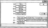
図2は、可能な入力、式および結果に関する第1の例を示す。ここで使用される画面は、入力ユニットにおける入力と照会によりCT装置から取り出された設定パラメータとの表示用と同時に、出力ユニットの表示用としても使用されている。この例では流量Fは手動で書き込まれ、5.5ml/sに決定された。これらのパラメータに対して必要な造影剤量Cが、本発明による装置内に記憶された式から算出される。この例では造影剤量Cは流量を乗算されたスキャン時間Sから算出されるが、しかし最小量10×Fが必要とされ、付加的にテストボーラスの遅れ時間Dが20sより大きい場合には、その量が20mlだけ高められる。限界条件として100mlを上回る造影剤が使用されないことが要求される。操作者は、入力値が自動的に照会によりCT装置から取り出されない場合に限り、該当入力値を入力する。計算キーBを押すことによって同じ表示面に結果が出力される。計算に使用される式が、この例および次の例では、同様に画面上に表示される。その量Mはこの例では24.8mgである。 FIG. 2 shows a first example regarding possible inputs, expressions and results. The screen used here is used not only for displaying the input in the input unit and the setting parameter retrieved from the CT apparatus by the inquiry, but also for displaying the output unit. In this example, the flow rate F was manually written and determined to be 5.5 ml / s. The required contrast medium amount C for these parameters is calculated from the formula stored in the device according to the invention. In this example, the contrast agent amount C is calculated from the scan time S multiplied by the flow rate, but a minimum amount of 10 × F is required, and additionally if the test bolus delay time D is greater than 20 s, The amount is increased by 20 ml. As a limit condition, it is required that no contrast medium exceeding 100 ml is used. The operator inputs the corresponding input value only when the input value is not automatically retrieved from the CT apparatus by inquiry. Pressing the calculation key B outputs the result on the same display surface. The formula used for the calculation is displayed on the screen as well in this and the following examples. The amount M is 24.8 mg in this example.
図3による第2の例では、付加的に造影剤流量Fが、画面に表示されている式から患者の体重Wに依存して計算される。更にここでは、なおも幾つかの値、特にスキャン時間Sおよび遅れ時間Dならびに造影剤の濃度Kが変更されている。 In the second example according to FIG. 3, the contrast agent flow rate F is additionally calculated depending on the weight W of the patient from the equation displayed on the screen. Furthermore, some values are still changed here, in particular the scan time S and the delay time D and the contrast agent concentration K.
1 入力ユニット
2 メモリユニット
3 インターフェース
4 計算ユニット
5 出力ユニット
6 CT装置1 Input unit 2
Claims (8)
Translated fromJapaneseその検査に必要な造影剤量を決定するための複数の計算式が少なくとも記憶されているメモリユニット(2)と、
画像形成装置(6)に対するインターフェース(3)とを備え、
このインターフェース(3)を介して、画像形成装置(6)の設定パラメータが、照会により画像形成装置(6)から取り出されるか、又は画像形成装置(6)に伝達され、
さらに、
計算のために必要な設定パラメータをこのインターフェース(3)を介して照会により画像形成装置(6)から取り出すか又は入力された設定パラメータをインターフェース(3)を介して画像形成装置(6)に伝達し、入力されたパラメータまたは照会されたパラメータに応じて、割り当てられた計算式を呼び出し、この計算式を用いて、入力されたパラメータまたは照会されたパラメータに基づいて造影剤量を計算する計算ユニット(4)と、
この計算ユニット(4)によって計算された造影剤量を表示する出力ユニット(5)とを備え、
計算された造影剤量に関して検査に必要な造影剤量の最小値が定められている
医用画像形成時の造影剤量の決定装置。An input unit (1) through which an operator can input parameters for image formation inspection to be performed;
A memory unit (2) that stores at least a plurality of calculation formulas for determining the amount of contrast agent necessary for the examination;
An interface (3) to the image forming apparatus (6),
Via this interface (3), the setting parameters of the image forming apparatus (6) are retrieved from the image forming apparatus (6) by inquiry or transmitted to the image forming apparatus (6),
further,
Setting parameters necessary for the calculation are retrieved from the image forming apparatus (6) by inquiry through the interface (3), or input setting parameters are transmitted to the image forming apparatus (6) through the interface (3). A calculation unit that calls an assigned calculation formula according to the input parameter or the queried parameter, and uses this calculation formula to calculate the contrast medium amount based on the input parameter or the queried parameter (4) and
An output unit (5) for displaying the amount of contrast medium calculated by the calculation unit (4),
An apparatus for determining a contrast medium amount at the time of medical image formation,in which a minimum value of a contrast medium amount necessary for the inspection is determined with respect to the calculated contrast medium amount.
Applications Claiming Priority (2)
| Application Number | Priority Date | Filing Date | Title |
|---|---|---|---|
| DE102010042931.7 | 2010-10-26 | ||
| DE102010042931 | 2010-10-26 |
Publications (2)
| Publication Number | Publication Date |
|---|---|
| JP2012090979A JP2012090979A (en) | 2012-05-17 |
| JP5693428B2true JP5693428B2 (en) | 2015-04-01 |
Family
ID=45973562
Family Applications (1)
| Application Number | Title | Priority Date | Filing Date |
|---|---|---|---|
| JP2011229839AActiveJP5693428B2 (en) | 2010-10-26 | 2011-10-19 | Device for determining the amount of contrast medium during medical image formation |
Country Status (3)
| Country | Link |
|---|---|
| US (1) | US8537969B2 (en) |
| JP (1) | JP5693428B2 (en) |
| CN (1) | CN102551778B (en) |
Families Citing this family (8)
| Publication number | Priority date | Publication date | Assignee | Title |
|---|---|---|---|---|
| DE102010027311B4 (en)* | 2010-07-16 | 2016-09-01 | Siemens Healthcare Gmbh | CT system for scanning a patient with a computer system for controlling the CT system |
| CN104068886B (en)* | 2013-03-28 | 2018-02-06 | 上海联影医疗科技有限公司 | Medical photography system and its method for imaging |
| JP6245897B2 (en)* | 2013-08-28 | 2017-12-13 | ジーイー・メディカル・システムズ・グローバル・テクノロジー・カンパニー・エルエルシー | Radiation tomography apparatus, radiation tomography system and program |
| DE102014203463B3 (en)* | 2014-02-26 | 2015-07-09 | Siemens Aktiengesellschaft | Patient-dependent optimization of contrast agent quantity |
| DE102015205493B4 (en)* | 2015-03-26 | 2023-12-28 | Siemens Healthcare Gmbh | Operating a medical imaging device |
| DE102015222853A1 (en)* | 2015-11-19 | 2017-05-24 | Siemens Healthcare Gmbh | Method for automatically determining a contrast agent injection protocol |
| DE102016202335A1 (en)* | 2016-02-16 | 2017-08-17 | Siemens Healthcare Gmbh | Medical examination system |
| CN108478227B (en)* | 2018-01-03 | 2022-05-10 | 东软医疗系统股份有限公司 | Method and device for determining medicine and dosage value thereof |
Family Cites Families (17)
| Publication number | Priority date | Publication date | Assignee | Title |
|---|---|---|---|---|
| JP2007502140A (en)* | 2003-08-12 | 2007-02-08 | コーニンクレッカ フィリップス エレクトロニクス エヌ ヴィ | Apparatus and method for producing an image of the heart |
| US7756324B2 (en)* | 2004-11-24 | 2010-07-13 | Kabushiki Kaisha Toshiba | 3-dimensional image processing apparatus |
| HUE034171T2 (en)* | 2004-11-24 | 2018-02-28 | Bayer Healthcare Llc | Devices, systems and methods for fluid delivery |
| DE102005004383B4 (en)* | 2005-01-31 | 2007-04-12 | Siemens Ag | Method and device for controlling an imaging modality |
| JP4901243B2 (en) | 2005-03-30 | 2012-03-21 | 株式会社東芝 | Contrast medium injection management apparatus, diagnostic imaging apparatus, and contrast medium injection apparatus |
| JP2006325615A (en)* | 2005-05-23 | 2006-12-07 | Hitachi Medical Corp | Medical image diagnostic apparatus |
| DE102005041626A1 (en)* | 2005-09-01 | 2007-03-15 | Siemens Ag | Method and system for generating tomographic images of a patient using contrast agent injections |
| EP1928320A2 (en)* | 2005-09-22 | 2008-06-11 | Philips Intellectual Property & Standards GmbH | Ct-imaging system |
| JP4745080B2 (en)* | 2006-02-20 | 2011-08-10 | 猛 中浦 | X-ray diagnostic apparatus, image processing apparatus, and program |
| DE102006032991B4 (en) | 2006-07-17 | 2015-03-26 | Siemens Aktiengesellschaft | Method and computer unit for setting a syringe pump for image acquisition |
| WO2008081830A1 (en)* | 2006-12-27 | 2008-07-10 | Nemoto Kyorindo Co., Ltd. | Liquid drug injection device and liquid drug injection method |
| JP5248026B2 (en)* | 2007-03-20 | 2013-07-31 | 株式会社東芝 | X-ray diagnostic system |
| US8428694B2 (en)* | 2007-07-17 | 2013-04-23 | Medrad, Inc. | Methods for determination of parameters for a procedure, for estimation of cardiopulmonary function and for fluid delivery |
| JP5394371B2 (en)* | 2008-04-02 | 2014-01-22 | 株式会社根本杏林堂 | Chemical injection device |
| JP5361410B2 (en)* | 2009-01-22 | 2013-12-04 | 株式会社東芝 | Image processing device |
| JP2010167189A (en)* | 2009-01-26 | 2010-08-05 | Toshiba Corp | Bed device, and x-ray ct device |
| JP5416761B2 (en) | 2009-03-04 | 2014-02-12 | 株式会社根本杏林堂 | Chemical solution injection device and X-ray CT system |
- 2011
- 2011-10-19JPJP2011229839Apatent/JP5693428B2/enactiveActive
- 2011-10-20USUS13/277,245patent/US8537969B2/enactiveActive
- 2011-10-24CNCN201110325030.6Apatent/CN102551778B/enactiveActive
Also Published As
| Publication number | Publication date |
|---|---|
| JP2012090979A (en) | 2012-05-17 |
| US20120101376A1 (en) | 2012-04-26 |
| CN102551778B (en) | 2015-02-18 |
| US8537969B2 (en) | 2013-09-17 |
| CN102551778A (en) | 2012-07-11 |
Similar Documents
| Publication | Publication Date | Title |
|---|---|---|
| JP5693428B2 (en) | Device for determining the amount of contrast medium during medical image formation | |
| US9230021B2 (en) | Image diagnostic apparatus, image diagnostic method, medical image server and medical image storage method | |
| JP5258174B2 (en) | Method of planning inspection of inspection object in magnetic resonance apparatus and magnetic resonance apparatus | |
| US7526063B2 (en) | System for generating, evaluating and distributing computer-tomographical 4D representations of the heart of a patient | |
| JP5288844B2 (en) | Image data acquisition method by medical modality and medical modality | |
| JP5416761B2 (en) | Chemical solution injection device and X-ray CT system | |
| US20110153255A1 (en) | Measurement protocol for a medical technology apparatus | |
| US20160091583A1 (en) | Patient-Specific Estimation of Specific Absorption Rate | |
| US20110317806A1 (en) | Method to select a value of a voltage to be set at an x-ray tube, computer tomography apparatus and data medium | |
| JP2017051662A (en) | Contrast-based imaging | |
| CN105266812A (en) | Method for ascertaining an absolute scan region on a patient, | |
| CN101299068A (en) | System and method for displaying computer tomographic scanning radiation dose | |
| JP6462257B2 (en) | Medical image diagnostic apparatus, medical information processing server, and medical image diagnostic system | |
| WO2007116892A1 (en) | Liquid drug infusion device | |
| JP6274515B2 (en) | Medical image processing device | |
| RU2552696C2 (en) | Device and method for obtaining diagnostic information | |
| EP2448490B1 (en) | Colonography | |
| JP2021521942A (en) | Automatic subject monitoring for medical imaging | |
| US8461839B2 (en) | Magnetic resonance control system, method and apparatus for producing a 2D MR image | |
| JP5044330B2 (en) | Medical image processing apparatus and medical image processing system | |
| US11786200B2 (en) | Method and image recording apparatus for obtaining image data from a patient involving administration of a contrast medium to the patient | |
| CN111904768B (en) | Medical equipment scanning intra-aperture image display method and medical equipment | |
| KR101990837B1 (en) | Work station, medical imaging apparatus, and method for controllinf thereof | |
| JP2025060166A (en) | Examination support device, operation method of examination support device, program, and medical image capture device | |
| JP6301096B2 (en) | Medical diagnostic imaging equipment |
Legal Events
| Date | Code | Title | Description |
|---|---|---|---|
| A131 | Notification of reasons for refusal | Free format text:JAPANESE INTERMEDIATE CODE: A131 Effective date:20130326 | |
| A521 | Request for written amendment filed | Free format text:JAPANESE INTERMEDIATE CODE: A523 Effective date:20130626 | |
| A131 | Notification of reasons for refusal | Free format text:JAPANESE INTERMEDIATE CODE: A131 Effective date:20140318 | |
| A601 | Written request for extension of time | Free format text:JAPANESE INTERMEDIATE CODE: A601 Effective date:20140618 | |
| A602 | Written permission of extension of time | Free format text:JAPANESE INTERMEDIATE CODE: A602 Effective date:20140623 | |
| A521 | Request for written amendment filed | Free format text:JAPANESE INTERMEDIATE CODE: A523 Effective date:20140710 | |
| TRDD | Decision of grant or rejection written | ||
| A01 | Written decision to grant a patent or to grant a registration (utility model) | Free format text:JAPANESE INTERMEDIATE CODE: A01 Effective date:20150106 | |
| A61 | First payment of annual fees (during grant procedure) | Free format text:JAPANESE INTERMEDIATE CODE: A61 Effective date:20150203 | |
| R150 | Certificate of patent or registration of utility model | Ref document number:5693428 Country of ref document:JP Free format text:JAPANESE INTERMEDIATE CODE: R150 | |
| R250 | Receipt of annual fees | Free format text:JAPANESE INTERMEDIATE CODE: R250 | |
| R250 | Receipt of annual fees | Free format text:JAPANESE INTERMEDIATE CODE: R250 | |
| R250 | Receipt of annual fees | Free format text:JAPANESE INTERMEDIATE CODE: R250 | |
| R250 | Receipt of annual fees | Free format text:JAPANESE INTERMEDIATE CODE: R250 | |
| S111 | Request for change of ownership or part of ownership | Free format text:JAPANESE INTERMEDIATE CODE: R313113 | |
| R360 | Written notification for declining of transfer of rights | Free format text:JAPANESE INTERMEDIATE CODE: R360 | |
| R250 | Receipt of annual fees | Free format text:JAPANESE INTERMEDIATE CODE: R250 | |
| R360 | Written notification for declining of transfer of rights | Free format text:JAPANESE INTERMEDIATE CODE: R360 | |
| R371 | Transfer withdrawn | Free format text:JAPANESE INTERMEDIATE CODE: R371 | |
| S111 | Request for change of ownership or part of ownership | Free format text:JAPANESE INTERMEDIATE CODE: R313113 | |
| R360 | Written notification for declining of transfer of rights | Free format text:JAPANESE INTERMEDIATE CODE: R360 | |
| R360 | Written notification for declining of transfer of rights | Free format text:JAPANESE INTERMEDIATE CODE: R360 | |
| R371 | Transfer withdrawn | Free format text:JAPANESE INTERMEDIATE CODE: R371 | |
| S111 | Request for change of ownership or part of ownership | Free format text:JAPANESE INTERMEDIATE CODE: R313113 | |
| R350 | Written notification of registration of transfer | Free format text:JAPANESE INTERMEDIATE CODE: R350 | |
| R250 | Receipt of annual fees | Free format text:JAPANESE INTERMEDIATE CODE: R250 | |
| R250 | Receipt of annual fees | Free format text:JAPANESE INTERMEDIATE CODE: R250 |