JP5567840B2 - Cell injection device - Google Patents
Cell injection deviceDownload PDFInfo
- Publication number
- JP5567840B2 JP5567840B2JP2010009173AJP2010009173AJP5567840B2JP 5567840 B2JP5567840 B2JP 5567840B2JP 2010009173 AJP2010009173 AJP 2010009173AJP 2010009173 AJP2010009173 AJP 2010009173AJP 5567840 B2JP5567840 B2JP 5567840B2
- Authority
- JP
- Japan
- Prior art keywords
- syringe
- tube
- cell
- injection device
- way valve
- Prior art date
- Legal status (The legal status is an assumption and is not a legal conclusion. Google has not performed a legal analysis and makes no representation as to the accuracy of the status listed.)
- Active
Links
Images


Classifications
- A—HUMAN NECESSITIES
- A61—MEDICAL OR VETERINARY SCIENCE; HYGIENE
- A61M—DEVICES FOR INTRODUCING MEDIA INTO, OR ONTO, THE BODY; DEVICES FOR TRANSDUCING BODY MEDIA OR FOR TAKING MEDIA FROM THE BODY; DEVICES FOR PRODUCING OR ENDING SLEEP OR STUPOR
- A61M25/00—Catheters; Hollow probes
- A61M25/10—Balloon catheters
- A61M25/1011—Multiple balloon catheters
- A—HUMAN NECESSITIES
- A61—MEDICAL OR VETERINARY SCIENCE; HYGIENE
- A61B—DIAGNOSIS; SURGERY; IDENTIFICATION
- A61B1/00—Instruments for performing medical examinations of the interior of cavities or tubes of the body by visual or photographical inspection, e.g. endoscopes; Illuminating arrangements therefor
- A61B1/00064—Constructional details of the endoscope body
- A61B1/00071—Insertion part of the endoscope body
- A61B1/0008—Insertion part of the endoscope body characterised by distal tip features
- A61B1/00094—Suction openings
- A—HUMAN NECESSITIES
- A61—MEDICAL OR VETERINARY SCIENCE; HYGIENE
- A61B—DIAGNOSIS; SURGERY; IDENTIFICATION
- A61B1/00—Instruments for performing medical examinations of the interior of cavities or tubes of the body by visual or photographical inspection, e.g. endoscopes; Illuminating arrangements therefor
- A61B1/04—Instruments for performing medical examinations of the interior of cavities or tubes of the body by visual or photographical inspection, e.g. endoscopes; Illuminating arrangements therefor combined with photographic or television appliances
- A—HUMAN NECESSITIES
- A61—MEDICAL OR VETERINARY SCIENCE; HYGIENE
- A61B—DIAGNOSIS; SURGERY; IDENTIFICATION
- A61B17/00—Surgical instruments, devices or methods
- A61B17/0057—Implements for plugging an opening in the wall of a hollow or tubular organ, e.g. for sealing a vessel puncture or closing a cardiac septal defect
- A—HUMAN NECESSITIES
- A61—MEDICAL OR VETERINARY SCIENCE; HYGIENE
- A61B—DIAGNOSIS; SURGERY; IDENTIFICATION
- A61B17/00—Surgical instruments, devices or methods
- A61B17/12—Surgical instruments, devices or methods for ligaturing or otherwise compressing tubular parts of the body, e.g. blood vessels or umbilical cord
- A61B17/12022—Occluding by internal devices, e.g. balloons or releasable wires
- A61B17/12027—Type of occlusion
- A61B17/1204—Type of occlusion temporary occlusion
- A61B17/12045—Type of occlusion temporary occlusion double occlusion, e.g. during anastomosis
- A—HUMAN NECESSITIES
- A61—MEDICAL OR VETERINARY SCIENCE; HYGIENE
- A61B—DIAGNOSIS; SURGERY; IDENTIFICATION
- A61B17/00—Surgical instruments, devices or methods
- A61B17/12—Surgical instruments, devices or methods for ligaturing or otherwise compressing tubular parts of the body, e.g. blood vessels or umbilical cord
- A61B17/12022—Occluding by internal devices, e.g. balloons or releasable wires
- A61B17/12099—Occluding by internal devices, e.g. balloons or releasable wires characterised by the location of the occluder
- A61B17/12109—Occluding by internal devices, e.g. balloons or releasable wires characterised by the location of the occluder in a blood vessel
- A—HUMAN NECESSITIES
- A61—MEDICAL OR VETERINARY SCIENCE; HYGIENE
- A61B—DIAGNOSIS; SURGERY; IDENTIFICATION
- A61B17/00—Surgical instruments, devices or methods
- A61B17/12—Surgical instruments, devices or methods for ligaturing or otherwise compressing tubular parts of the body, e.g. blood vessels or umbilical cord
- A61B17/12022—Occluding by internal devices, e.g. balloons or releasable wires
- A61B17/12131—Occluding by internal devices, e.g. balloons or releasable wires characterised by the type of occluding device
- A61B17/12136—Balloons
- A—HUMAN NECESSITIES
- A61—MEDICAL OR VETERINARY SCIENCE; HYGIENE
- A61M—DEVICES FOR INTRODUCING MEDIA INTO, OR ONTO, THE BODY; DEVICES FOR TRANSDUCING BODY MEDIA OR FOR TAKING MEDIA FROM THE BODY; DEVICES FOR PRODUCING OR ENDING SLEEP OR STUPOR
- A61M39/00—Tubes, tube connectors, tube couplings, valves, access sites or the like, specially adapted for medical use
- A61M39/02—Access sites
- A61M39/0247—Semi-permanent or permanent transcutaneous or percutaneous access sites to the inside of the body
- A—HUMAN NECESSITIES
- A61—MEDICAL OR VETERINARY SCIENCE; HYGIENE
- A61M—DEVICES FOR INTRODUCING MEDIA INTO, OR ONTO, THE BODY; DEVICES FOR TRANSDUCING BODY MEDIA OR FOR TAKING MEDIA FROM THE BODY; DEVICES FOR PRODUCING OR ENDING SLEEP OR STUPOR
- A61M39/00—Tubes, tube connectors, tube couplings, valves, access sites or the like, specially adapted for medical use
- A61M39/22—Valves or arrangement of valves
- A61M39/223—Multiway valves
- A—HUMAN NECESSITIES
- A61—MEDICAL OR VETERINARY SCIENCE; HYGIENE
- A61M—DEVICES FOR INTRODUCING MEDIA INTO, OR ONTO, THE BODY; DEVICES FOR TRANSDUCING BODY MEDIA OR FOR TAKING MEDIA FROM THE BODY; DEVICES FOR PRODUCING OR ENDING SLEEP OR STUPOR
- A61M5/00—Devices for bringing media into the body in a subcutaneous, intra-vascular or intramuscular way; Accessories therefor, e.g. filling or cleaning devices, arm-rests
- A61M5/178—Syringes
- A—HUMAN NECESSITIES
- A61—MEDICAL OR VETERINARY SCIENCE; HYGIENE
- A61B—DIAGNOSIS; SURGERY; IDENTIFICATION
- A61B17/00—Surgical instruments, devices or methods
- A61B17/00234—Surgical instruments, devices or methods for minimally invasive surgery
- A61B2017/00238—Type of minimally invasive operation
- A61B2017/00243—Type of minimally invasive operation cardiac
- A—HUMAN NECESSITIES
- A61—MEDICAL OR VETERINARY SCIENCE; HYGIENE
- A61B—DIAGNOSIS; SURGERY; IDENTIFICATION
- A61B17/00—Surgical instruments, devices or methods
- A61B17/0057—Implements for plugging an opening in the wall of a hollow or tubular organ, e.g. for sealing a vessel puncture or closing a cardiac septal defect
- A61B2017/00575—Implements for plugging an opening in the wall of a hollow or tubular organ, e.g. for sealing a vessel puncture or closing a cardiac septal defect for closure at remote site, e.g. closing atrial septum defects
- A61B2017/00588—Rigid or stiff implements, e.g. made of several rigid parts linked by hinges
- A—HUMAN NECESSITIES
- A61—MEDICAL OR VETERINARY SCIENCE; HYGIENE
- A61B—DIAGNOSIS; SURGERY; IDENTIFICATION
- A61B17/00—Surgical instruments, devices or methods
- A61B17/0057—Implements for plugging an opening in the wall of a hollow or tubular organ, e.g. for sealing a vessel puncture or closing a cardiac septal defect
- A61B2017/00575—Implements for plugging an opening in the wall of a hollow or tubular organ, e.g. for sealing a vessel puncture or closing a cardiac septal defect for closure at remote site, e.g. closing atrial septum defects
- A61B2017/00597—Implements comprising a membrane
- A—HUMAN NECESSITIES
- A61—MEDICAL OR VETERINARY SCIENCE; HYGIENE
- A61B—DIAGNOSIS; SURGERY; IDENTIFICATION
- A61B17/00—Surgical instruments, devices or methods
- A61B17/0057—Implements for plugging an opening in the wall of a hollow or tubular organ, e.g. for sealing a vessel puncture or closing a cardiac septal defect
- A61B2017/00575—Implements for plugging an opening in the wall of a hollow or tubular organ, e.g. for sealing a vessel puncture or closing a cardiac septal defect for closure at remote site, e.g. closing atrial septum defects
- A61B2017/0061—Implements located only on one side of the opening
- A—HUMAN NECESSITIES
- A61—MEDICAL OR VETERINARY SCIENCE; HYGIENE
- A61B—DIAGNOSIS; SURGERY; IDENTIFICATION
- A61B17/00—Surgical instruments, devices or methods
- A61B17/12—Surgical instruments, devices or methods for ligaturing or otherwise compressing tubular parts of the body, e.g. blood vessels or umbilical cord
- A61B17/12022—Occluding by internal devices, e.g. balloons or releasable wires
- A61B2017/12127—Double occlusion, e.g. for creating blood-free anastomosis site
- A—HUMAN NECESSITIES
- A61—MEDICAL OR VETERINARY SCIENCE; HYGIENE
- A61M—DEVICES FOR INTRODUCING MEDIA INTO, OR ONTO, THE BODY; DEVICES FOR TRANSDUCING BODY MEDIA OR FOR TAKING MEDIA FROM THE BODY; DEVICES FOR PRODUCING OR ENDING SLEEP OR STUPOR
- A61M25/00—Catheters; Hollow probes
- A61M25/10—Balloon catheters
- A61M2025/1043—Balloon catheters with special features or adapted for special applications
- A61M2025/1047—Balloon catheters with special features or adapted for special applications having centering means, e.g. balloons having an appropriate shape
- A—HUMAN NECESSITIES
- A61—MEDICAL OR VETERINARY SCIENCE; HYGIENE
- A61M—DEVICES FOR INTRODUCING MEDIA INTO, OR ONTO, THE BODY; DEVICES FOR TRANSDUCING BODY MEDIA OR FOR TAKING MEDIA FROM THE BODY; DEVICES FOR PRODUCING OR ENDING SLEEP OR STUPOR
- A61M25/00—Catheters; Hollow probes
- A61M25/10—Balloon catheters
- A61M2025/1043—Balloon catheters with special features or adapted for special applications
- A61M2025/1052—Balloon catheters with special features or adapted for special applications for temporarily occluding a vessel for isolating a sector
- A—HUMAN NECESSITIES
- A61—MEDICAL OR VETERINARY SCIENCE; HYGIENE
- A61M—DEVICES FOR INTRODUCING MEDIA INTO, OR ONTO, THE BODY; DEVICES FOR TRANSDUCING BODY MEDIA OR FOR TAKING MEDIA FROM THE BODY; DEVICES FOR PRODUCING OR ENDING SLEEP OR STUPOR
- A61M39/00—Tubes, tube connectors, tube couplings, valves, access sites or the like, specially adapted for medical use
- A61M39/02—Access sites
- A61M39/0247—Semi-permanent or permanent transcutaneous or percutaneous access sites to the inside of the body
- A61M2039/0276—Semi-permanent or permanent transcutaneous or percutaneous access sites to the inside of the body for introducing or removing fluids into or out of the body
- A—HUMAN NECESSITIES
- A61—MEDICAL OR VETERINARY SCIENCE; HYGIENE
- A61M—DEVICES FOR INTRODUCING MEDIA INTO, OR ONTO, THE BODY; DEVICES FOR TRANSDUCING BODY MEDIA OR FOR TAKING MEDIA FROM THE BODY; DEVICES FOR PRODUCING OR ENDING SLEEP OR STUPOR
- A61M25/00—Catheters; Hollow probes
- A61M25/0067—Catheters; Hollow probes characterised by the distal end, e.g. tips
- A61M25/0082—Catheter tip comprising a tool
- A61M25/0084—Catheter tip comprising a tool being one or more injection needles
- A—HUMAN NECESSITIES
- A61—MEDICAL OR VETERINARY SCIENCE; HYGIENE
- A61M—DEVICES FOR INTRODUCING MEDIA INTO, OR ONTO, THE BODY; DEVICES FOR TRANSDUCING BODY MEDIA OR FOR TAKING MEDIA FROM THE BODY; DEVICES FOR PRODUCING OR ENDING SLEEP OR STUPOR
- A61M25/00—Catheters; Hollow probes
- A61M25/01—Introducing, guiding, advancing, emplacing or holding catheters
- A61M25/02—Holding devices, e.g. on the body
- A61M25/04—Holding devices, e.g. on the body in the body, e.g. expansible
Landscapes
- Health & Medical Sciences (AREA)
- Life Sciences & Earth Sciences (AREA)
- Heart & Thoracic Surgery (AREA)
- Surgery (AREA)
- General Health & Medical Sciences (AREA)
- Public Health (AREA)
- Animal Behavior & Ethology (AREA)
- Engineering & Computer Science (AREA)
- Biomedical Technology (AREA)
- Veterinary Medicine (AREA)
- Molecular Biology (AREA)
- Nuclear Medicine, Radiotherapy & Molecular Imaging (AREA)
- Medical Informatics (AREA)
- Hematology (AREA)
- Biophysics (AREA)
- Anesthesiology (AREA)
- Vascular Medicine (AREA)
- Reproductive Health (AREA)
- Pulmonology (AREA)
- Pathology (AREA)
- Optics & Photonics (AREA)
- Radiology & Medical Imaging (AREA)
- Physics & Mathematics (AREA)
- Gastroenterology & Hepatology (AREA)
- Cardiology (AREA)
- Child & Adolescent Psychology (AREA)
- Endoscopes (AREA)
- Media Introduction/Drainage Providing Device (AREA)
- Surgical Instruments (AREA)
- Infusion, Injection, And Reservoir Apparatuses (AREA)
Description
Translated fromJapanese本発明は、細胞注入デバイスに関するものである。 The present invention relates to a cell injection device.
従来、心筋梗塞等の心疾患に対して幹細胞を疾患部位に注入するための細胞治療の実現が求められている。
眼球組織に薬剤を注入するデバイスとしては、硬性シャフト中で軸方向に移動可能な針を硬性シャフトの先端から出没させて組織への穿刺侵入角度を変更するものが知られている(例えば、特許文献1参照。)。Conventionally, realization of cell therapy for injecting stem cells into a disease site for heart diseases such as myocardial infarction has been demanded.
As a device for injecting a drug into an eyeball tissue, a device that changes the puncture intrusion angle into a tissue by causing a needle that can move in the axial direction in the rigid shaft to protrude from the distal end of the rigid shaft is known (for example, a patent) Reference 1).
しかしながら、眼球組織への薬剤の注入を主用途とする特許文献1の注入デバイスとは異なり、心疾患の細胞治療のための細胞注入デバイスとしては、心臓の異なる部位に複数回にわたって細胞注入を行う必要がある。心臓への細胞注入は、剣状突起下に貫通形成したアクセスポートを介して細胞注入デバイスの先端を心膜腔内に挿入することにより行われるが、細胞注入の都度、細胞注入デバイスをアクセスポートから出し入れするのでは、手術時間が増加し、患者にかかる負担が大きいという不都合がある。 However, unlike the injection device of
本発明は上述した事情に鑑みてなされたものであって、剣状突起下に貫通形成したアクセスポートを介して先端を心膜腔内に挿入したままの状態で複数回分にわたる量の細胞を注入することができる細胞注入デバイスを提供することを目的としている。 The present invention has been made in view of the circumstances described above, and injects a plurality of doses of cells with the tip inserted into the pericardial cavity via an access port formed under the xiphoid process. The object is to provide a cell injection device that can.
上記目的を達成するために、本発明は以下の手段を提供する。
本発明は、生体組織に穿刺される注射針と、該注射針を先端に備えたチューブと、該チューブの基端側に設けられ、細胞を予め収容した細胞用シリンジを着脱可能に接続するシリンジ接続部と、該シリンジ接続部と前記注射針との間の前記チューブに設けられ、開放端を有する三方弁とを備え、該三方弁が、前記シリンジ接続部から前記注射針への前記チューブ内の流路を接続する第1の流路と、前記シリンジ接続部からの流路を前記開放端を介して外部に対して開放する第2の流路とを択一的に選択可能である細胞注入デバイスを提供する。In order to achieve the above object, the present invention provides the following means.
The present invention relates to an injection needle that is punctured into a living tissue, a tube having the injection needle at its distal end, and a syringe that is provided on the proximal end side of the tube and removably connects a cell syringe that contains cells inadvance. A connecting portion, and a three-way valve provided on the tube between the syringe connecting portion and the injection needle, and having an open end, and the three-way valve is disposed in the tube from the syringe connecting portion to the injection needle. a first flow path connecting flow path, Rualternatively selectable der and the second flow path to open the flow path from the syringe connecting portion relative to the outside through the open end A cell injection device is provided.
本発明によれば、患者の皮膚を貫通して形成されたアクセスポートを介して、チューブを体内に挿入し、チューブの先端に設けられた注射針を体内組織に穿刺して、チューブの基端側に設けられたシリンジ接続部に細胞を収容した細胞用シリンジを接続し、細胞用シリンジによって細胞をチューブ内に押し出すことにより、チューブを介して体内組織内に細胞を注入することができる。 According to the present invention, the tube is inserted into the body through the access port formed through the patient's skin, the injection needle provided at the distal end of the tube is punctured into the body tissue, and the proximal end of the tube A cell syringe containing cells is connected to a syringe connecting portion provided on the side, and the cells can be injected into the body tissue through the tube by pushing the cells into the tube with the cell syringe.
この場合において、シリンジ接続部に細胞用シリンジを接続する際に、細胞用シリンジとシリンジ接続部との間に挟まれた空気が細胞用シリンジ内に混入する可能性がある。そして、混入した空気は気泡となって細胞内に配されるが、シリンジ接続部と注射針との間のチューブに設けられた三方弁を開放端側に設定した状態で、細胞用シリンジから細胞を押し出すことにより、混入していた気泡を三方弁の開放端から外部に放出することができる。 In this case, when the cell syringe is connected to the syringe connection portion, air sandwiched between the cell syringe and the syringe connection portion may be mixed into the cell syringe. The mixed air is bubbled and arranged in the cell, but with the three-way valve provided on the tube between the syringe connection part and the injection needle set to the open end side, the cell syringe moves the cell. By extruding, the mixed bubbles can be discharged to the outside from the open end of the three-way valve.
すなわち、細胞の注入毎に細胞用シリンジをシリンジ接続部に付け替えて注入作業を行うことができ、その都度に混入した気泡を排出できる。したがって、注入位置を変更しながら複数回にわたって細胞を注入する細胞治療において、チューブの先端を体内に挿入したままの状態で細胞を追加して供給することができ、患者にかかる負担を軽減することができる。 That is, every time the cells are injected, the cell syringe can be replaced with the syringe connection portion to perform the injection operation, and the bubbles mixed in each time can be discharged. Therefore, in cell therapy in which cells are injected multiple times while changing the injection position, it is possible to supply additional cells with the tip of the tube inserted into the body, reducing the burden on the patient. Can do.
上記発明においては、前記チューブの基端に、媒体を収容した媒体用シリンジが接続され、該媒体用シリンジと前記シリンジ接続部との間にバルブが配置されていてもよい。
このようにすることで、バルブを開放して、媒体用シリンジ内からチューブ内に媒体を供給し充填した状態で、バルブを閉止して、シリンジ接続部に接続された細胞用シリンジを加圧すると、細胞用シリンジをシリンジ接続部に接続する際に混入した気泡が、媒体中に押し出される。この状態で、三方弁を開放端側に設定し、バルブを開いて媒体用シリンジを加圧することにより、媒体中に押し出された気泡を媒体によって三方弁の開放端から外部に放出することができる。In the said invention, the syringe for media which accommodated the medium may be connected to the base end of the said tube, and the valve | bulb may be arrange | positioned between this syringe for media and the said syringe connection part.
By doing so, when the valve is opened and the medium is supplied from the medium syringe into the tube and filled, the valve is closed and the cell syringe connected to the syringe connector is pressurized. The air bubbles mixed when the cell syringe is connected to the syringe connector are pushed out into the medium. In this state, the three-way valve is set to the open end side, and the valve is opened to pressurize the medium syringe, whereby bubbles pushed into the medium can be discharged from the open end of the three-way valve to the outside by the medium. .
上記発明においては、前記シリンジ接続部よりも基端側のチューブに、該チューブを振動させる振動手段を備えていてもよい。
このようにすることで、振動手段によってチューブを振動させ、チューブの内壁に付着している気泡を移動させやすくすることができる。これにより、三方弁の開放端からチューブ内の気泡をより確実に外部に放出することができる。In the said invention, you may provide the vibration means which vibrates this tube in the tube of the base end side rather than the said syringe connection part.
By doing in this way, a tube can be vibrated by a vibration means and it can make it easy to move the bubble adhering to the inner wall of a tube. Thereby, the bubble in a tube can be more reliably discharge | released outside from the open end of a three-way valve.
また、本発明の参考例は、生体組織に穿刺される注射針と、該注射針を先端に備えたチューブと、該チューブの基端側に設けられ、細胞を収容した細胞用シリンジを着脱可能に接続するシリンジ接続部と、該シリンジ接続部と前記注射針との間の前記チューブに設けられ、チューブ内面において上方に窪む凹部とを備える細胞注入デバイスを提供する。
In addition,the reference example of the present invention includes an injection needle that is punctured into a living tissue, a tube that includes the injection needle at the distal end, and a cell syringe that contains cells and is provided on the proximal end side of the tube. There is provided a cell injection device provided with a syringe connection part connected to the tube, and a recess provided in the tube between the syringe connection part and the injection needle and recessed upward on the inner surface of the tube.
本発明によれば、シリンジ接続部に細胞用シリンジを付け替えることにより、チューブの先端を体内に挿入したままの状態で、生体組織に注入する細胞量を任意に変更することができる。この場合において、細胞用シリンジをシリンジ接続部に接続する際に、細胞用シリンジとシリンジ接続部との間に空気が混入することが考えられる。この状態で、細胞用シリンジによって細胞を押し出すと、混入した空気がチューブ内に進行することとなるが、その進行の途中においてチューブ内面に設けられた上方に窪む凹部内にトラップされ、チューブ先端の注射針まで到達しない容易することができる。 According to the present invention, the amount of cells to be injected into a living tissue can be arbitrarily changed by replacing the syringe for cells with the syringe connection portion while the distal end of the tube is inserted into the body. In this case, it is conceivable that air is mixed between the cell syringe and the syringe connector when the cell syringe is connected to the syringe connector. In this state, when the cells are pushed out by the cell syringe, the mixed air travels into the tube, but is trapped in an upwardly recessed recess provided on the inner surface of the tube during the travel, and the tip of the tube It can be as easy as not reaching the needle.
また、上記発明においては、前記チューブ内を流動する気泡を検出する検出部と、該検出部により前記チューブ内に気泡が検出されたときに、これを報知する報知部とを備えていてもよい。
このようにすることで、細胞用シリンジから細胞を注入する際に、混入した空気がチューブ内を気泡となって流動すると、検出部によって気泡が検出され、報知部によって外部に報知される。施術者は、これにより細胞の注入を迅速に停止することができ、気泡の生体組織内への注入をより確実に防止することができる。Moreover, in the said invention, you may provide the detection part which detects the bubble which flows through the inside of the said tube, and the alerting | reporting part which alert | reports this when a bubble is detected in the said tube by this detection part. .
By doing in this way, when injecting cells from the cell syringe, if the mixed air flows as bubbles in the tube, the detection unit detects the bubbles and notifies the outside by the notification unit. The practitioner can thereby quickly stop the injection of cells, and can more reliably prevent the injection of bubbles into the living tissue.
本発明によれば、剣状突起下に貫通形成したアクセスポートを介して先端を心膜腔内に挿入したままの状態で複数回分にわたる量の細胞を注入することができるという効果を奏する。 According to the present invention, it is possible to inject a plurality of doses of cells through the access port formed under the xiphoid process with the tip inserted into the pericardial cavity.
本発明の一実施形態に係る細胞注入デバイス1について、図面を参照して以下に説明する。
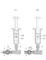
本実施形態に係る細胞注入デバイス1は、図1に示されるように、剣状突起下の表皮組織を貫通して形成されたアクセスポートを介して、先端部が心膜腔内に挿入される硬性シャフト2と、該硬性シャフト2内に収容され、先端に注射針3を有し基端側に細胞用シリンジ4を接続するためのシリンジ接続部5を有するチューブ6と、該チューブ6の先端に設けられた注射針3をその長手方向に移動させて硬性シャフト2の先端から出没させる針駆動ハンドル7と、チューブ6に設けられた三方弁8とを備えている。A
As shown in FIG. 1, the
硬性シャフト2およびチューブ6は、チューブ6内を流れる幹細胞を含む細胞液を外部から視認できるように、透明な材質によって構成されている。シリンジ接続部5はシリンジを着脱可能に接続するポートである。
針駆動ハンドル7は、広げる方向に移動させる(図中鎖線で示す位置に移動させる)と注射針3を硬性シャフト2の先端から突出させ、狭める方向に移動させる(図中、実線で示す位置に移動させる)と硬性シャフト2内に引っ込ませるようになっている。また、針駆動ハンドル7は、図示しないバネによって、狭める方向に付勢されていて、手を放した状態で、注射針3が硬性シャフト2内に収容されるようになっている。The
When the
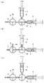
三方弁8は、注射針3とシリンジ接続部5との間のチューブ6の途中位置に設けられている。三方弁8は、図2に示されるように、(a)シリンジ接続部5から注射針3へのチューブ6内の流路を接続する第1の流路と、(b)シリンジ接続部5からの流路を外部に対して開放する第2の流路とを備えている。 The three-
三方弁8を操作して第1の流路を選択することにより、細胞用シリンジ4内の細胞液をチューブ6を介して注射針3から吐出させることができるようになっている。また、三方弁8を操作して第2の流路を選択することにより、シリンジ接続部5から吐出された細胞液に含まれる気泡を三方弁8の開放端8aから外部に放出することができるようになっている。 By operating the three-
このように構成された本実施形態に係る細胞注入デバイス1の作用について、以下に説明する。
本実施形態に係る細胞注入デバイス1を用いて、幹細胞を含む細胞液Bを心疾患の病変部位に注入するには、まず、シリンジ接続部5に生理食塩水(媒体)Cを収容した媒体用シリンジ9を接続し、三方弁8を第1の流路に切り替えた状態で、媒体用シリンジ9を操作してチューブ8内の流路全体に生理食塩水を充填させておく。The operation of the
In order to inject a cell fluid B containing stem cells into a lesion site of heart disease using the
次いで、シリンジ接続部5の媒体用シリンジ9を取り外して細胞用シリンジ4を接続する。媒体用シリンジ9を取り外すことにより、シリンジ接続部5は大気開放され、細胞用シリンジ4を取り付けることにより、シリンジ接続部5が再度密閉される。この際に、細胞用シリンジ4の先端とシリンジ接続部5との間に気泡が挟まれて、細胞用シリンジ4が取り付られて密閉された流路内に気泡が混入することがある。 Subsequently, the
次に、図2(b)に示されるように、三方弁8を第2の流路に切り替えた状態で、細胞用シリンジ4を操作して細胞液の一部をシリンジ接続部5からチューブ6内に押し出す。細胞用シリンジ4を接続した際に流路内に混入してしまった空気は、細胞液によってチューブ6内に押し出されてくるが、三方弁8の切り替えによって第2の流路が三方弁8の開放端8aにおいて大気開放されているので、チューブ6内に押し出された気泡は、三方弁8の開放端8aから外部に放出されることになる。 Next, as shown in FIG. 2B, in a state where the three-
この状態で、三方弁8を、図2(a)に示されるように、再度第1の流路に切り替えることにより、その後、細胞用シリンジ4の操作によって押し出される細胞液B内には気泡が混入しておらず、気泡が心臓の病変部位に注入されてしまう不都合の発生を未然に防止することができる。
また、この状態において細胞液Bがチューブ6内に残留しているので、細胞用シリンジ4をシリンジ接続部5から取り外して、シリンジ接続部5に媒体用シリンジ9を付け替える。この際にも、空気が混入する可能性があるので、同様にして、三方弁8を第2の流路に切り替えて、媒体用シリンジ9を操作することにより、混入した気泡を三方弁8の開放端8aから外部に放出することができる。In this state, by switching the three-
In this state, since the cell liquid B remains in the
そして、気泡が放出された後には、三方弁8を第1の流路に切り替えて、媒体用シリンジ9を操作して生理食塩水Cを押し出すことにより、チューブ6内に残留していた細胞液Bを注射針3から心臓の病変部位に注入することができる。
生理食塩水Cがチューブ6全体に行き渡るまで注入した時点で、上記操作を繰り返すことにより、複数回にわたる注入作業を、細胞液Bを継ぎ足しながら行うことができ、その際に、気泡が病変部位に注入されてしまう不都合の発生を防止することができるという利点がある。Then, after the bubbles are released, the three-
When the physiological saline C is injected until it reaches the
このように、本実施形態に係る細胞注入デバイス1によれば、細胞用シリンジ4をシリンジ接続部5に着脱することができ、かつ、着脱によって空気が混入しても、その空気を外部に放出することができる。したがって、例えば、1回分の細胞液Bを収容した細胞用シリンジ4をシリンジ接続部5に付け替えることにより、硬性シャフト2の先端をアクセスポートを介して心膜腔内に挿入したままの状態で、任意の回数にわたって必要なだけ細胞注入を行うことができる。
その結果、硬性シャフト2の複数回にわたる挿脱によって手術が長期化することを防止して、患者にかかる負担を軽減することができるという利点がある。As described above, according to the
As a result, there is an advantage that it is possible to prevent the operation from being prolonged by inserting and removing the rigid shaft 2 a plurality of times and to reduce the burden on the patient.
なお、本実施形態においては、単一のシリンジ接続部5に細胞用シリンジ4と媒体用シリンジ9とを付け替えることとしたが、これに代えて、図3に示されるように、もう1つの三方弁10によって、細胞用シリンジ4と媒体用シリンジ9とを択一的に選択できるように、両方取り付けることにしてもよい。この場合においても、細胞用シリンジ4については、1回以上の注入毎に細胞液を使い切るので、新たな細胞用シリンジ4をシリンジ接続部5に付け替える必要がある。 In the present embodiment, the
すなわち、まず、図3(a)に示されるように、三方弁8,10を操作して、媒体用シリンジ9のみがチューブ6を介して注射針に接続される流路を構成し、媒体用シリンジ9内の生理食塩水Cをチューブ6内に充填した状態で、硬性シャフト2の先端を心膜腔内に挿入する。
そして、図3(c)に示されるように、流路を細胞用シリンジ4側に切り替えて、所定量の細胞液Bを押し出し、チューブ6内の生理食塩水Cを注射針3の先端から吐出させることにより、チューブ6内に細胞液Bを充填する。That is, first, as shown in FIG. 3 (a), the three-
Then, as shown in FIG. 3 (c), the flow path is switched to the
この状態で、注射針3を心臓の病変部位に穿刺し、三方弁10を図3(a)に示されるように操作することで、媒体用シリンジ9から注射針3に接続する流路を確保して、生理食塩水Cをチューブ6内に供給する。これにより、チューブ6内に充填されていた細胞液Bが注射針3から吐出されて、病変部位に注入される。 In this state, the
チューブ6内に充填されていた分の細胞液Bが全て吐出され終わった場合には、細胞用シリンジ4をシリンジ接続部5から取り外して、新たな細胞液Bが充填されている細胞用シリンジ4に付け替えるとともに、図3(b)に示されるように、三方弁8,10を操作して、細胞用シリンジ4および媒体用シリンジ9がいずれも三方弁8の開放端8aに接続するように設定する。 When all of the cell liquid B filled in the
この状態で、まず、細胞用シリンジ4を加圧して、細胞液Bと生理食塩水Cとの境界面に溜まった気泡を三方弁10内に押し出し、次いで、媒体用シリンジ9を加圧して、三方弁10内に押し出されてきた気泡を三方弁8の開放端8aに向けて生理食塩水Cによって押し出す。これにより、気泡は開放端8aから吐出されるので、気泡が心臓の病変部位に注入されることを未然に防止できる。また、気泡が開放端8aから吐出される際に、開放端8aからは生理食塩水Cを吐出するだけで済み、貴重な細胞液Bを無駄にせずに済むという利点もある。 In this state, first, the
また、本実施形態においては、図4に示されるように、シリンジ接続部5と媒体用シリンジ8との間のチューブ6にバルブ11を設けることにしてもよい。
このようにすることで、バルブ11を閉止した状態で、細胞用シリンジ4から細胞液Bによって気泡をシリンジ接続部5内に押し出し、次いで、バルブ11を開放して生理食塩水Cによって、三方弁8の開放端8aから気泡を放出することができる。Further, in the present embodiment, as shown in FIG. 4, a
In this way, with the
また、本実施形態においては、三方弁8に代えて、図5に示されるように、チューブ6内面の途中位置に、上方に窪む凹部12を設けることにしてもよい。
このようにすることで、細胞用シリンジ4の付け替えによって混入した気泡Dは、チューブ内を流動する間に、凹部12において捕捉される。これにより、気泡Dが混入しても注射針3まで到達することがなく、病変部位に注入されてしまうことを防止できる。Further, in the present embodiment, instead of the three-
By doing in this way, the bubble D mixed by the replacement of the
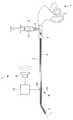
また、本実施形態においては、図6に示されるように、三方弁8または凹部12の前段位置に、チューブ6を加振する振動手段13を設けることにしてもよい。このようにすることで、振動手段13の作動によってチューブ6が振動させられるので、チューブ6の内壁に付着している気泡が細胞液Bあるいは生理食塩水Cによって移動され易くすることができ、容易に放出あるいは捕捉することができるという利点がある。 Further, in the present embodiment, as shown in FIG. 6, vibration means 13 that vibrates the
また、本実施形態においては、図7に示されるように、チューブ6を半径方向に挟んで配置された発光部14aと受光部14bとからなるセンサ14と、該センサ14からの出力信号を処理する信号処理部15と、該信号処理部15により処理された信号を音に変換して出力するスピーカ16とを備えていてもよい。 Further, in the present embodiment, as shown in FIG. 7, a
このようにすることで、チューブ6内を流動する細胞液Bあるいは生理食塩水Cの透過率の変化が、スピーカ16から音として報知される。すなわち、生理食塩水Cのみの場合には音の変動は少なく、細胞液Bの場合には、生理食塩水Cとは異なる音が発生し、気泡Dが混入している場合には、断続的な音の変化となって報知される。これにより、施術者は音の変化によって、注入されている液体の状態を認識することができ、内視鏡画像を表示しているモニタや患者から目を離すことなく施術することが可能となる。 By doing so, a change in the transmittance of the cell fluid B or physiological saline C flowing in the
生理食塩水Cが検知されている状態から細胞液Bの検出状態に変化した場合には、施術者は、注射針3を病変部位に穿刺することができ、逆の場合には、病変部位に穿刺されている注射針3を引き抜く必要があることを認識できる。また、細胞液Bの注入中に、チューブ6内に気泡Dが検出された場合には、細胞液Bの注入を即時停止することができ、これによって、気泡Dの病変部位への注入を回避することができる。
また、チューブ6内を流動している液体の状態を音によって報知することとしたが、これに代えて、光によって報知したり、モニタ上に表示することによって報知してもよい。When the state is changed from the state in which the physiological saline C is detected to the state in which the cell fluid B is detected, the practitioner can puncture the lesion site with the
Further, although the state of the liquid flowing in the
B 細胞液(細胞)
D 気泡
1 細胞注入デバイス
3 注射針
4 細胞用シリンジ
5 シリンジ接続部
6 チューブ
8 三方弁
8a 開放端
9 媒体用シリンジ
11 バルブ
12 凹部
13 振動手段
14 センサ(検出部)
16 スピーカ(報知部)B cell fluid (cells)
16 Speaker (notification part)
Claims (4)
Translated fromJapanese該注射針を先端に備えたチューブと、
該チューブの基端側に設けられ、細胞を予め収容した細胞用シリンジを着脱可能に接続するシリンジ接続部と、
該シリンジ接続部と前記注射針との間の前記チューブに設けられ、開放端を有する三方弁とを備え、
該三方弁が、前記シリンジ接続部から前記注射針への前記チューブ内の流路を接続する第1の流路と、前記シリンジ接続部からの流路を前記開放端を介して外部に対して開放する第2の流路とを択一的に選択可能である細胞注入デバイス。An injection needle to be pierced into a living tissue;
A tube equipped with the injection needle at the tip;
A syringe connecting part that is provided on the proximal end side of the tube and detachably connects a cell syringe containing cells inadvance ;
A three-way valve provided on the tube between the syringe connection and the injection needle and having an open end;
The three-way valve connects the flow path in the tube from the syringe connection portion to the injection needle, and the flow path from the syringe connection portion to the outside via the open end. second flow path and the alternatively selectable der Ru cell injection devicefor opening.
該媒体用シリンジと前記シリンジ接続部との間にバルブが配置されている請求項1に記載の細胞注入デバイス。A medium syringe containing a medium is connected to the proximal end of the tube,
The cell injection device according to claim 1, wherein a valve is disposed between the syringe for medium and the syringe connection part.
該検出部により前記チューブ内に気泡が検出されたときに、これを報知する報知部とを備える請求項1から請求項3のいずれかに記載の細胞注入デバイス。A detection unit for detecting bubbles flowing in the tube;
The cell injection device according to any one of claims 1 to3 , further comprising an informing unit that informs when a bubble is detected in the tube by the detecting unit.
Priority Applications (4)
| Application Number | Priority Date | Filing Date | Title |
|---|---|---|---|
| CN2010800421034ACN102510764A (en) | 2009-09-22 | 2010-09-14 | Device for injecting therapeutic solution |
| EP10818721AEP2481444A4 (en) | 2009-09-22 | 2010-09-14 | Device for injecting therapeutic solution |
| PCT/JP2010/065851WO2011037046A1 (en) | 2009-09-22 | 2010-09-14 | Device for injecting therapeutic solution |
| US12/884,845US20110190710A1 (en) | 2009-09-22 | 2010-09-17 | Therapeutic fluid injection device |
Applications Claiming Priority (2)
| Application Number | Priority Date | Filing Date | Title |
|---|---|---|---|
| US24458609P | 2009-09-22 | 2009-09-22 | |
| US61/244,586 | 2009-09-22 |
Publications (2)
| Publication Number | Publication Date |
|---|---|
| JP2011067588A JP2011067588A (en) | 2011-04-07 |
| JP5567840B2true JP5567840B2 (en) | 2014-08-06 |
Family
ID=43795855
Family Applications (2)
| Application Number | Title | Priority Date | Filing Date |
|---|---|---|---|
| JP2010009173AActiveJP5567840B2 (en) | 2009-09-22 | 2010-01-19 | Cell injection device |
| JP2011532996AActiveJP4996772B2 (en) | 2009-09-22 | 2010-09-21 | Medical device |
Family Applications After (1)
| Application Number | Title | Priority Date | Filing Date |
|---|---|---|---|
| JP2011532996AActiveJP4996772B2 (en) | 2009-09-22 | 2010-09-21 | Medical device |
Country Status (5)
| Country | Link |
|---|---|
| US (1) | US8348891B2 (en) |
| EP (3) | EP2481336B1 (en) |
| JP (2) | JP5567840B2 (en) |
| CN (3) | CN102665528B (en) |
| WO (1) | WO2011037108A1 (en) |
Families Citing this family (19)
| Publication number | Priority date | Publication date | Assignee | Title |
|---|---|---|---|---|
| CA2717821A1 (en)* | 2008-03-07 | 2009-09-11 | London Equitable Limited In Its Capacity As Trustee Of The Think Tank Trust | A dilation catheter |
| CN103190877B (en)* | 2013-02-01 | 2015-04-22 | 上海交通大学 | Flexible endoscope robot with adsorption capability |
| KR20140142920A (en)* | 2013-06-05 | 2014-12-15 | 이근호 | Apparatus for inserting the amnion |
| JP6185294B2 (en)* | 2013-06-11 | 2017-08-23 | オリンパス株式会社 | Endoscope |
| EP3133975A4 (en)* | 2014-04-21 | 2018-01-03 | Clph, Llc | Imaging apparatus and systems and methods for using them |
| CN104224271B (en)* | 2014-09-11 | 2018-11-23 | 中国人民解放军第三军医大学第二附属医院 | Branchofiberoscope biopsy hemostatic ball cuff pipe |
| WO2016079803A1 (en)* | 2014-11-18 | 2016-05-26 | オリンパス株式会社 | Medical instrument |
| EP3320866A4 (en)* | 2015-07-09 | 2019-02-06 | Olympus Corporation | Medical instrument introduction device |
| WO2017203582A1 (en) | 2016-05-23 | 2017-11-30 | オリンパス株式会社 | Endoscope-use device, and endoscopic system |
| US10350395B2 (en)* | 2017-06-23 | 2019-07-16 | Cook Medical Technologies Llc | Introducer for lumen support or dilation |
| TWI666008B (en)* | 2017-07-17 | 2019-07-21 | 林志豪 | Ostomy connecting and storing device |
| US10750936B2 (en)* | 2017-11-02 | 2020-08-25 | Olympus Corporation | Pericardial-cavity observing method |
| JP2021118755A (en)* | 2018-03-20 | 2021-08-12 | オリンパス株式会社 | Endoscope apparatus and operation method of endoscope apparatus |
| CN112020333B (en) | 2018-04-26 | 2024-06-25 | 奥林巴斯株式会社 | Treatment system and distention device |
| CN113101522A (en)* | 2021-04-26 | 2021-07-13 | 上海联心医疗科技有限公司 | An implantable cardiac pacing device |
| CN113117240A (en)* | 2021-04-26 | 2021-07-16 | 上海联心医疗科技有限公司 | Implanted bioenergy power supply system |
| CN113713194B (en)* | 2021-09-18 | 2025-02-18 | 北京大学深圳医院 | Rectovaginal fistula negative pressure drainage device |
| JP7529746B2 (en)* | 2021-11-18 | 2024-08-06 | オリンパス株式会社 | Medical system and method for controlling medical system |
| WO2023190640A1 (en)* | 2022-03-30 | 2023-10-05 | 株式会社カネカ | Method for preparing syringe for cell administration use, and method for transporting cells using syringe for cell administration use |
Family Cites Families (66)
| Publication number | Priority date | Publication date | Assignee | Title |
|---|---|---|---|---|
| JPS6235318A (en) | 1985-08-09 | 1987-02-16 | Canon Inc | Endoscope operating method |
| JP2575395B2 (en) | 1987-07-15 | 1997-01-22 | オリンパス光学工業株式会社 | Antenna device for NMR measurement |
| US5035231A (en)* | 1987-04-27 | 1991-07-30 | Olympus Optical Co., Ltd. | Endoscope apparatus |
| US4884567A (en)* | 1987-12-03 | 1989-12-05 | Dimed Inc. | Method for transvenous implantation of objects into the pericardial space of patients |
| JPH0255960A (en) | 1988-08-20 | 1990-02-26 | Fujikura Ltd | photocurrent sensor |
| JPH0255960U (en)* | 1988-10-14 | 1990-04-23 | ||
| US5048537A (en)* | 1990-05-15 | 1991-09-17 | Medex, Inc. | Method and apparatus for sampling blood |
| US5370134A (en)* | 1991-05-29 | 1994-12-06 | Orgin Medsystems, Inc. | Method and apparatus for body structure manipulation and dissection |
| CA2123317C (en) | 1991-11-19 | 2004-07-20 | Albert K. Chin | Endoscopic inflatable retraction devices for separating layers of tissue, and methods of using |
| US5336252A (en)* | 1992-06-22 | 1994-08-09 | Cohen Donald M | System and method for implanting cardiac electrical leads |
| US5297536A (en)* | 1992-08-25 | 1994-03-29 | Wilk Peter J | Method for use in intra-abdominal surgery |
| US5324266A (en)* | 1992-12-23 | 1994-06-28 | Abbott Laboratories | In-line sampling system incorporating an improved blood sampling device |
| US5725525A (en)* | 1993-03-16 | 1998-03-10 | Ep Technologies, Inc. | Multiple electrode support structures with integral hub and spline elements |
| SE9303253D0 (en) | 1993-10-05 | 1993-10-05 | Siemens Elema Ab | Instruments for peephole surgery |
| US5522804A (en)* | 1994-02-15 | 1996-06-04 | Lynn; Lawrence A. | Aspiration, mixing, and injection syringe |
| JPH08117232A (en) | 1994-10-24 | 1996-05-14 | Olympus Optical Co Ltd | Biopsy needle |
| JPH08280815A (en)* | 1995-04-19 | 1996-10-29 | Sumitomo Bakelite Co Ltd | Catheter for stent delivery |
| US5827216A (en) | 1995-06-07 | 1998-10-27 | Cormedics Corp. | Method and apparatus for accessing the pericardial space |
| US5759150A (en)* | 1995-07-07 | 1998-06-02 | Olympus Optical Co., Ltd. | System for evulsing subcutaneous tissue |
| US5836311A (en) | 1995-09-20 | 1998-11-17 | Medtronic, Inc. | Method and apparatus for temporarily immobilizing a local area of tissue |
| US5697916A (en)* | 1995-11-21 | 1997-12-16 | Stat Medical Devices Inc. | Hypodermic dosage measuring device |
| ES2128936B1 (en) | 1996-05-22 | 2000-01-16 | Patentes Novedades Sa | PROCEDURE FOR OBTAINING PENTAERITRITOL. |
| JP3017451B2 (en) | 1996-11-11 | 2000-03-06 | オリンパス光学工業株式会社 | Hood for endoscope |
| US5931810A (en) | 1996-12-05 | 1999-08-03 | Comedicus Incorporated | Method for accessing the pericardial space |
| JP3036686B2 (en)* | 1997-02-27 | 2000-04-24 | 政夫 高橋 | Hemostatic holding device for vascular anastomosis used for coronary artery bypass surgery |
| US6592552B1 (en) | 1997-09-19 | 2003-07-15 | Cecil C. Schmidt | Direct pericardial access device and method |
| US5968017A (en)* | 1997-10-14 | 1999-10-19 | Merit Medical Systems, Inc. | Pulse fluid infusion systems |
| US6017332A (en)* | 1997-10-20 | 2000-01-25 | Urrutia; Hector | Medical dye delivery system |
| JPH11276422A (en) | 1998-03-31 | 1999-10-12 | Fuji Photo Optical Co Ltd | Ultrasonic endoscope |
| EP1082052A4 (en) | 1998-05-26 | 2008-04-09 | Comedicus Inc | Intrapericardial procedures and apparatuses |
| CZ164098A3 (en)* | 1998-05-28 | 2000-02-16 | Milan Mudr. Csc. Krajíček | Stabilizer of heart muscle |
| US6309374B1 (en) | 1998-08-03 | 2001-10-30 | Insite Vision Incorporated | Injection apparatus and method of using same |
| JP2000176011A (en)* | 1998-12-18 | 2000-06-27 | Urrutia Hector | Medical pigment transmitting system |
| US6977080B1 (en)* | 1999-08-10 | 2005-12-20 | Allergan, Inc. | Intrapericardial botulinum toxin treatment for bradycardia |
| US7398781B1 (en)* | 1999-08-10 | 2008-07-15 | Maquet Cardiovascular, Llc | Method for subxiphoid endoscopic access |
| EP1272239B1 (en) | 2000-04-12 | 2012-08-08 | Covidien AG | Thoracentesis device with hyper-sensitive detection mechanism |
| JP4517321B2 (en)* | 2000-06-05 | 2010-08-04 | 有限会社エスアールジェイ | Overtube |
| JP2002017854A (en) | 2000-07-11 | 2002-01-22 | Daiken Iki Kk | Flow controller for chemical injection device |
| US6666861B1 (en)* | 2000-10-05 | 2003-12-23 | James R. Grabek | Atrial appendage remodeling device and method |
| US6706013B1 (en)* | 2001-06-29 | 2004-03-16 | Advanced Cardiovascular Systems, Inc. | Variable length drug delivery catheter |
| US20030083546A1 (en)* | 2001-10-18 | 2003-05-01 | John Butler | Device |
| JP3859491B2 (en)* | 2001-11-15 | 2006-12-20 | オリンパス株式会社 | Endoscope sheath |
| JP2004033525A (en) | 2002-07-04 | 2004-02-05 | Fuji Photo Optical Co Ltd | Hardness variable treatment instrument |
| WO2004012586A2 (en) | 2002-08-05 | 2004-02-12 | Infraredx, Inc. | Near-infrared spectroscopic analysis of blood vessel walls |
| JP4385118B2 (en) | 2002-08-07 | 2009-12-16 | 独立行政法人産業技術総合研究所 | Needle-piercing device and method for detecting the operating state of the needle of the needle-piercing device |
| JP4334838B2 (en) | 2002-09-06 | 2009-09-30 | オリンパス株式会社 | Endoscope device |
| JP4088918B2 (en)* | 2002-09-13 | 2008-05-21 | 東レ・メディカル株式会社 | Hemodialysis machine |
| JP4638694B2 (en)* | 2003-08-21 | 2011-02-23 | テルモ株式会社 | Infusion device |
| CA2439667A1 (en)* | 2003-09-04 | 2005-03-04 | Andrew Kenneth Hoffmann | Low frequency vibration assisted blood perfusion system and apparatus |
| JP2007505680A (en) | 2003-09-23 | 2007-03-15 | ガンブロ・ルンディア・エービー | Apparatus, system and method related to hemodialysis, hemodiafiltration, hemofiltration or peritoneal dialysis |
| JP2005169009A (en)* | 2003-12-15 | 2005-06-30 | Olympus Corp | Endoscope system and endoscope |
| WO2005110192A1 (en)* | 2004-05-14 | 2005-11-24 | Olympus Corporation | Insertion device |
| EP1827484B1 (en) | 2004-12-02 | 2015-06-17 | Bradley H. Strauss | Augmentation of intraluminal microvessel formation to facilitate guide wire crossing in chronic total occlusions |
| JP4000485B2 (en)* | 2005-02-28 | 2007-10-31 | フジノン株式会社 | Endoscope device |
| JP2006271831A (en)* | 2005-03-30 | 2006-10-12 | Terumo Corp | Medical treatment device and its usage |
| WO2006124634A1 (en)* | 2005-05-16 | 2006-11-23 | Mallinckrodt Inc. | Multi-barrel syringe having integral manifold |
| JP2007054333A (en) | 2005-08-25 | 2007-03-08 | Pentax Corp | OCT probe and OCT system |
| WO2007052354A1 (en)* | 2005-11-04 | 2007-05-10 | Olympus Medical Systems Corp. | Endoscope system, endoscope, supporting member and method of using endoscope system |
| WO2007078003A1 (en)* | 2006-01-06 | 2007-07-12 | Olympus Medical Systems Corp. | Trans-natural opening based or transcutaneous medical system |
| JP5116985B2 (en)* | 2006-04-13 | 2013-01-09 | 富士フイルム株式会社 | Endoscope |
| FR2914840A1 (en)* | 2007-04-16 | 2008-10-17 | Lk2 Soc Responsabilite Limitee | Connection for blood sampling device, has sampling valve sampling fluid sample contained in sampling path, where path has Luer lock type syringe that drives part of fluid contained in sampling path towards catheter path |
| EP2148608A4 (en)* | 2007-04-27 | 2010-04-28 | Voyage Medical Inc | Complex shape steerable tissue visualization and manipulation catheter |
| JP2008284108A (en)* | 2007-05-16 | 2008-11-27 | Olympus Corp | Stabilizer |
| CN101687069B (en)* | 2007-06-29 | 2013-01-16 | 株式会社Jms | Hemodialyzer |
| US8311620B2 (en)* | 2007-08-22 | 2012-11-13 | Cardiac Pacemakers, Inc. | Methods and apparatus to treat and prevent atrial tachyarrhythmias |
| JP3143693U (en)* | 2008-05-22 | 2008-07-31 | 財団法人ヒューマンサイエンス振興財団 | Endoscope device and endoscope hood |
- 2010
- 2010-01-19JPJP2010009173Apatent/JP5567840B2/enactiveActive
- 2010-08-30CNCN201080052838.5Apatent/CN102665528B/enactiveActive
- 2010-08-30EPEP10818652.9Apatent/EP2481336B1/ennot_activeNot-in-force
- 2010-09-14EPEP10818721Apatent/EP2481444A4/ennot_activeWithdrawn
- 2010-09-14CNCN2010800421034Apatent/CN102510764A/enactivePending
- 2010-09-16USUS12/883,773patent/US8348891B2/ennot_activeExpired - Fee Related
- 2010-09-21CNCN2010800421157Apatent/CN102497824B/ennot_activeExpired - Fee Related
- 2010-09-21WOPCT/JP2010/066321patent/WO2011037108A1/enactiveApplication Filing
- 2010-09-21EPEP10818781.6Apatent/EP2481355B1/ennot_activeNot-in-force
- 2010-09-21JPJP2011532996Apatent/JP4996772B2/enactiveActive
Also Published As
| Publication number | Publication date |
|---|---|
| JP4996772B2 (en) | 2012-08-08 |
| CN102497824A (en) | 2012-06-13 |
| CN102665528A (en) | 2012-09-12 |
| US8348891B2 (en) | 2013-01-08 |
| WO2011037108A1 (en) | 2011-03-31 |
| EP2481355A1 (en) | 2012-08-01 |
| EP2481444A1 (en) | 2012-08-01 |
| EP2481336A4 (en) | 2013-05-29 |
| CN102510764A (en) | 2012-06-20 |
| JPWO2011037108A1 (en) | 2013-02-21 |
| US20110190584A1 (en) | 2011-08-04 |
| EP2481336B1 (en) | 2018-07-25 |
| EP2481336A1 (en) | 2012-08-01 |
| JP2011067588A (en) | 2011-04-07 |
| EP2481355A4 (en) | 2013-05-29 |
| CN102665528B (en) | 2015-05-06 |
| EP2481444A4 (en) | 2013-03-06 |
| CN102497824B (en) | 2013-04-03 |
| EP2481355B1 (en) | 2014-08-20 |
Similar Documents
| Publication | Publication Date | Title |
|---|---|---|
| JP5567840B2 (en) | Cell injection device | |
| WO2011037046A1 (en) | Device for injecting therapeutic solution | |
| EP1626764B1 (en) | Fluid delivery system and related methods of use | |
| RU2687770C2 (en) | Ophthalmologic lubrication system and associated devices, systems and methods | |
| ES2585634T3 (en) | Device to facilitate and to introduce a transplant or an implant into the living body, in particular for ophthalmologic interventions | |
| JP6071880B2 (en) | Medical liquid circuit kit and liquid circuit system using the same | |
| CN116271271A (en) | System for aspiration of thrombus | |
| JP2000070376A (en) | Hub for artery puncture needle | |
| US11738154B2 (en) | Micro-volume injectors with dose guidance and methods for use | |
| US9277934B2 (en) | Liquid injection device and surgical instrument including liquid injection device | |
| US20120203200A1 (en) | Device and method for delivering medicine into the tympanic cavity | |
| EP2959822A1 (en) | Means for controlled release of fluid, and endoscope and endoscopic surgical instrument comprising same | |
| KR101546488B1 (en) | suction and irrigator | |
| JP2005237617A (en) | Body liquid collecting device | |
| JP5752901B2 (en) | Intraocular lens insertion device | |
| JP6098052B2 (en) | Surgical instruments | |
| CN212307961U (en) | Guide sheath | |
| JP4964976B2 (en) | Portable injection injector | |
| JP2015163088A (en) | Medical device | |
| KR102699355B1 (en) | Electro surgical device | |
| CN102397128A (en) | Dacryosyrinx | |
| CN210408514U (en) | Medicine and water injection integrated hemostatic forceps for digestive tract | |
| US20150283320A1 (en) | Medical treatment method, medical treatment apparatus, and medical treatment system for bloodstream disorder | |
| JP2018202035A (en) | Indwelling needle and medical connector device | |
| JP2005205121A (en) | Portable spraying and injecting tool |
Legal Events
| Date | Code | Title | Description |
|---|---|---|---|
| A621 | Written request for application examination | Free format text:JAPANESE INTERMEDIATE CODE: A621 Effective date:20121115 | |
| A131 | Notification of reasons for refusal | Free format text:JAPANESE INTERMEDIATE CODE: A131 Effective date:20131119 | |
| A521 | Request for written amendment filed | Free format text:JAPANESE INTERMEDIATE CODE: A523 Effective date:20140120 | |
| TRDD | Decision of grant or rejection written | ||
| A01 | Written decision to grant a patent or to grant a registration (utility model) | Free format text:JAPANESE INTERMEDIATE CODE: A01 Effective date:20140527 | |
| A61 | First payment of annual fees (during grant procedure) | Free format text:JAPANESE INTERMEDIATE CODE: A61 Effective date:20140620 | |
| R151 | Written notification of patent or utility model registration | Ref document number:5567840 Country of ref document:JP Free format text:JAPANESE INTERMEDIATE CODE: R151 | |
| S531 | Written request for registration of change of domicile | Free format text:JAPANESE INTERMEDIATE CODE: R313531 | |
| R350 | Written notification of registration of transfer | Free format text:JAPANESE INTERMEDIATE CODE: R350 | |
| R250 | Receipt of annual fees | Free format text:JAPANESE INTERMEDIATE CODE: R250 | |
| R250 | Receipt of annual fees | Free format text:JAPANESE INTERMEDIATE CODE: R250 | |
| R250 | Receipt of annual fees | Free format text:JAPANESE INTERMEDIATE CODE: R250 | |
| R250 | Receipt of annual fees | Free format text:JAPANESE INTERMEDIATE CODE: R250 | |
| R250 | Receipt of annual fees | Free format text:JAPANESE INTERMEDIATE CODE: R250 | |
| R250 | Receipt of annual fees | Free format text:JAPANESE INTERMEDIATE CODE: R250 | |
| R250 | Receipt of annual fees | Free format text:JAPANESE INTERMEDIATE CODE: R250 |