JP5512125B2 - Recurrent gene fusion in prostate cancer - Google Patents
Recurrent gene fusion in prostate cancerDownload PDFInfo
- Publication number
- JP5512125B2 JP5512125B2JP2008530030AJP2008530030AJP5512125B2JP 5512125 B2JP5512125 B2JP 5512125B2JP 2008530030 AJP2008530030 AJP 2008530030AJP 2008530030 AJP2008530030 AJP 2008530030AJP 5512125 B2JP5512125 B2JP 5512125B2
- Authority
- JP
- Japan
- Prior art keywords
- gene
- tmprss2
- erg
- fusion
- etv1
- Prior art date
- Legal status (The legal status is an assumption and is not a legal conclusion. Google has not performed a legal analysis and makes no representation as to the accuracy of the status listed.)
- Active
Links
Images


Classifications
- C—CHEMISTRY; METALLURGY
- C12—BIOCHEMISTRY; BEER; SPIRITS; WINE; VINEGAR; MICROBIOLOGY; ENZYMOLOGY; MUTATION OR GENETIC ENGINEERING
- C12Q—MEASURING OR TESTING PROCESSES INVOLVING ENZYMES, NUCLEIC ACIDS OR MICROORGANISMS; COMPOSITIONS OR TEST PAPERS THEREFOR; PROCESSES OF PREPARING SUCH COMPOSITIONS; CONDITION-RESPONSIVE CONTROL IN MICROBIOLOGICAL OR ENZYMOLOGICAL PROCESSES
- C12Q1/00—Measuring or testing processes involving enzymes, nucleic acids or microorganisms; Compositions therefor; Processes of preparing such compositions
- C12Q1/68—Measuring or testing processes involving enzymes, nucleic acids or microorganisms; Compositions therefor; Processes of preparing such compositions involving nucleic acids
- C12Q1/6876—Nucleic acid products used in the analysis of nucleic acids, e.g. primers or probes
- C12Q1/6883—Nucleic acid products used in the analysis of nucleic acids, e.g. primers or probes for diseases caused by alterations of genetic material
- C12Q1/6886—Nucleic acid products used in the analysis of nucleic acids, e.g. primers or probes for diseases caused by alterations of genetic material for cancer
- A—HUMAN NECESSITIES
- A01—AGRICULTURE; FORESTRY; ANIMAL HUSBANDRY; HUNTING; TRAPPING; FISHING
- A01K—ANIMAL HUSBANDRY; AVICULTURE; APICULTURE; PISCICULTURE; FISHING; REARING OR BREEDING ANIMALS, NOT OTHERWISE PROVIDED FOR; NEW BREEDS OF ANIMALS
- A01K67/00—Rearing or breeding animals, not otherwise provided for; New or modified breeds of animals
- A01K67/027—New or modified breeds of vertebrates
- A01K67/0275—Genetically modified vertebrates, e.g. transgenic
- C—CHEMISTRY; METALLURGY
- C07—ORGANIC CHEMISTRY
- C07K—PEPTIDES
- C07K14/00—Peptides having more than 20 amino acids; Gastrins; Somatostatins; Melanotropins; Derivatives thereof
- C07K14/82—Translation products from oncogenes
- C—CHEMISTRY; METALLURGY
- C12—BIOCHEMISTRY; BEER; SPIRITS; WINE; VINEGAR; MICROBIOLOGY; ENZYMOLOGY; MUTATION OR GENETIC ENGINEERING
- C12N—MICROORGANISMS OR ENZYMES; COMPOSITIONS THEREOF; PROPAGATING, PRESERVING, OR MAINTAINING MICROORGANISMS; MUTATION OR GENETIC ENGINEERING; CULTURE MEDIA
- C12N15/00—Mutation or genetic engineering; DNA or RNA concerning genetic engineering, vectors, e.g. plasmids, or their isolation, preparation or purification; Use of hosts therefor
- C12N15/09—Recombinant DNA-technology
- C12N15/11—DNA or RNA fragments; Modified forms thereof; Non-coding nucleic acids having a biological activity
- C12N15/113—Non-coding nucleic acids modulating the expression of genes, e.g. antisense oligonucleotides; Antisense DNA or RNA; Triplex- forming oligonucleotides; Catalytic nucleic acids, e.g. ribozymes; Nucleic acids used in co-suppression or gene silencing
- C—CHEMISTRY; METALLURGY
- C12—BIOCHEMISTRY; BEER; SPIRITS; WINE; VINEGAR; MICROBIOLOGY; ENZYMOLOGY; MUTATION OR GENETIC ENGINEERING
- C12N—MICROORGANISMS OR ENZYMES; COMPOSITIONS THEREOF; PROPAGATING, PRESERVING, OR MAINTAINING MICROORGANISMS; MUTATION OR GENETIC ENGINEERING; CULTURE MEDIA
- C12N9/00—Enzymes; Proenzymes; Compositions thereof; Processes for preparing, activating, inhibiting, separating or purifying enzymes
- C12N9/14—Hydrolases (3)
- C12N9/48—Hydrolases (3) acting on peptide bonds (3.4)
- C12N9/50—Proteinases, e.g. Endopeptidases (3.4.21-3.4.25)
- C12N9/64—Proteinases, e.g. Endopeptidases (3.4.21-3.4.25) derived from animal tissue
- C12N9/6421—Proteinases, e.g. Endopeptidases (3.4.21-3.4.25) derived from animal tissue from mammals
- C12N9/6424—Serine endopeptidases (3.4.21)
- C12N9/6445—Kallikreins (3.4.21.34; 3.4.21.35)
- G—PHYSICS
- G01—MEASURING; TESTING
- G01N—INVESTIGATING OR ANALYSING MATERIALS BY DETERMINING THEIR CHEMICAL OR PHYSICAL PROPERTIES
- G01N33/00—Investigating or analysing materials by specific methods not covered by groups G01N1/00 - G01N31/00
- G01N33/48—Biological material, e.g. blood, urine; Haemocytometers
- G01N33/50—Chemical analysis of biological material, e.g. blood, urine; Testing involving biospecific ligand binding methods; Immunological testing
- G01N33/53—Immunoassay; Biospecific binding assay; Materials therefor
- G01N33/574—Immunoassay; Biospecific binding assay; Materials therefor for cancer
- G01N33/57407—Specifically defined cancers
- G01N33/57434—Specifically defined cancers of prostate
- A—HUMAN NECESSITIES
- A01—AGRICULTURE; FORESTRY; ANIMAL HUSBANDRY; HUNTING; TRAPPING; FISHING
- A01K—ANIMAL HUSBANDRY; AVICULTURE; APICULTURE; PISCICULTURE; FISHING; REARING OR BREEDING ANIMALS, NOT OTHERWISE PROVIDED FOR; NEW BREEDS OF ANIMALS
- A01K2217/00—Genetically modified animals
- A01K2217/07—Animals genetically altered by homologous recombination
- A01K2217/072—Animals genetically altered by homologous recombination maintaining or altering function, i.e. knock in
- A—HUMAN NECESSITIES
- A01—AGRICULTURE; FORESTRY; ANIMAL HUSBANDRY; HUNTING; TRAPPING; FISHING
- A01K—ANIMAL HUSBANDRY; AVICULTURE; APICULTURE; PISCICULTURE; FISHING; REARING OR BREEDING ANIMALS, NOT OTHERWISE PROVIDED FOR; NEW BREEDS OF ANIMALS
- A01K2217/00—Genetically modified animals
- A01K2217/20—Animal model comprising regulated expression system
- A01K2217/206—Animal model comprising tissue-specific expression system, e.g. tissue specific expression of transgene, of Cre recombinase
- A—HUMAN NECESSITIES
- A01—AGRICULTURE; FORESTRY; ANIMAL HUSBANDRY; HUNTING; TRAPPING; FISHING
- A01K—ANIMAL HUSBANDRY; AVICULTURE; APICULTURE; PISCICULTURE; FISHING; REARING OR BREEDING ANIMALS, NOT OTHERWISE PROVIDED FOR; NEW BREEDS OF ANIMALS
- A01K2227/00—Animals characterised by species
- A01K2227/10—Mammal
- A01K2227/105—Murine
- A—HUMAN NECESSITIES
- A01—AGRICULTURE; FORESTRY; ANIMAL HUSBANDRY; HUNTING; TRAPPING; FISHING
- A01K—ANIMAL HUSBANDRY; AVICULTURE; APICULTURE; PISCICULTURE; FISHING; REARING OR BREEDING ANIMALS, NOT OTHERWISE PROVIDED FOR; NEW BREEDS OF ANIMALS
- A01K2267/00—Animals characterised by purpose
- A01K2267/03—Animal model, e.g. for test or diseases
- A01K2267/0331—Animal model for proliferative diseases
- C—CHEMISTRY; METALLURGY
- C07—ORGANIC CHEMISTRY
- C07K—PEPTIDES
- C07K2319/00—Fusion polypeptide
- C—CHEMISTRY; METALLURGY
- C12—BIOCHEMISTRY; BEER; SPIRITS; WINE; VINEGAR; MICROBIOLOGY; ENZYMOLOGY; MUTATION OR GENETIC ENGINEERING
- C12N—MICROORGANISMS OR ENZYMES; COMPOSITIONS THEREOF; PROPAGATING, PRESERVING, OR MAINTAINING MICROORGANISMS; MUTATION OR GENETIC ENGINEERING; CULTURE MEDIA
- C12N2310/00—Structure or type of the nucleic acid
- C12N2310/10—Type of nucleic acid
- C12N2310/11—Antisense
- C—CHEMISTRY; METALLURGY
- C12—BIOCHEMISTRY; BEER; SPIRITS; WINE; VINEGAR; MICROBIOLOGY; ENZYMOLOGY; MUTATION OR GENETIC ENGINEERING
- C12N—MICROORGANISMS OR ENZYMES; COMPOSITIONS THEREOF; PROPAGATING, PRESERVING, OR MAINTAINING MICROORGANISMS; MUTATION OR GENETIC ENGINEERING; CULTURE MEDIA
- C12N2310/00—Structure or type of the nucleic acid
- C12N2310/10—Type of nucleic acid
- C12N2310/14—Type of nucleic acid interfering nucleic acids [NA]
- C—CHEMISTRY; METALLURGY
- C12—BIOCHEMISTRY; BEER; SPIRITS; WINE; VINEGAR; MICROBIOLOGY; ENZYMOLOGY; MUTATION OR GENETIC ENGINEERING
- C12Q—MEASURING OR TESTING PROCESSES INVOLVING ENZYMES, NUCLEIC ACIDS OR MICROORGANISMS; COMPOSITIONS OR TEST PAPERS THEREFOR; PROCESSES OF PREPARING SUCH COMPOSITIONS; CONDITION-RESPONSIVE CONTROL IN MICROBIOLOGICAL OR ENZYMOLOGICAL PROCESSES
- C12Q2600/00—Oligonucleotides characterized by their use
- C12Q2600/106—Pharmacogenomics, i.e. genetic variability in individual responses to drugs and drug metabolism
- C—CHEMISTRY; METALLURGY
- C12—BIOCHEMISTRY; BEER; SPIRITS; WINE; VINEGAR; MICROBIOLOGY; ENZYMOLOGY; MUTATION OR GENETIC ENGINEERING
- C12Q—MEASURING OR TESTING PROCESSES INVOLVING ENZYMES, NUCLEIC ACIDS OR MICROORGANISMS; COMPOSITIONS OR TEST PAPERS THEREFOR; PROCESSES OF PREPARING SUCH COMPOSITIONS; CONDITION-RESPONSIVE CONTROL IN MICROBIOLOGICAL OR ENZYMOLOGICAL PROCESSES
- C12Q2600/00—Oligonucleotides characterized by their use
- C12Q2600/112—Disease subtyping, staging or classification
- C—CHEMISTRY; METALLURGY
- C12—BIOCHEMISTRY; BEER; SPIRITS; WINE; VINEGAR; MICROBIOLOGY; ENZYMOLOGY; MUTATION OR GENETIC ENGINEERING
- C12Q—MEASURING OR TESTING PROCESSES INVOLVING ENZYMES, NUCLEIC ACIDS OR MICROORGANISMS; COMPOSITIONS OR TEST PAPERS THEREFOR; PROCESSES OF PREPARING SUCH COMPOSITIONS; CONDITION-RESPONSIVE CONTROL IN MICROBIOLOGICAL OR ENZYMOLOGICAL PROCESSES
- C12Q2600/00—Oligonucleotides characterized by their use
- C12Q2600/118—Prognosis of disease development
- C—CHEMISTRY; METALLURGY
- C12—BIOCHEMISTRY; BEER; SPIRITS; WINE; VINEGAR; MICROBIOLOGY; ENZYMOLOGY; MUTATION OR GENETIC ENGINEERING
- C12Q—MEASURING OR TESTING PROCESSES INVOLVING ENZYMES, NUCLEIC ACIDS OR MICROORGANISMS; COMPOSITIONS OR TEST PAPERS THEREFOR; PROCESSES OF PREPARING SUCH COMPOSITIONS; CONDITION-RESPONSIVE CONTROL IN MICROBIOLOGICAL OR ENZYMOLOGICAL PROCESSES
- C12Q2600/00—Oligonucleotides characterized by their use
- C12Q2600/136—Screening for pharmacological compounds
- C—CHEMISTRY; METALLURGY
- C12—BIOCHEMISTRY; BEER; SPIRITS; WINE; VINEGAR; MICROBIOLOGY; ENZYMOLOGY; MUTATION OR GENETIC ENGINEERING
- C12Q—MEASURING OR TESTING PROCESSES INVOLVING ENZYMES, NUCLEIC ACIDS OR MICROORGANISMS; COMPOSITIONS OR TEST PAPERS THEREFOR; PROCESSES OF PREPARING SUCH COMPOSITIONS; CONDITION-RESPONSIVE CONTROL IN MICROBIOLOGICAL OR ENZYMOLOGICAL PROCESSES
- C12Q2600/00—Oligonucleotides characterized by their use
- C12Q2600/156—Polymorphic or mutational markers
- C—CHEMISTRY; METALLURGY
- C12—BIOCHEMISTRY; BEER; SPIRITS; WINE; VINEGAR; MICROBIOLOGY; ENZYMOLOGY; MUTATION OR GENETIC ENGINEERING
- C12Q—MEASURING OR TESTING PROCESSES INVOLVING ENZYMES, NUCLEIC ACIDS OR MICROORGANISMS; COMPOSITIONS OR TEST PAPERS THEREFOR; PROCESSES OF PREPARING SUCH COMPOSITIONS; CONDITION-RESPONSIVE CONTROL IN MICROBIOLOGICAL OR ENZYMOLOGICAL PROCESSES
- C12Q2600/00—Oligonucleotides characterized by their use
- C12Q2600/158—Expression markers
- Y—GENERAL TAGGING OF NEW TECHNOLOGICAL DEVELOPMENTS; GENERAL TAGGING OF CROSS-SECTIONAL TECHNOLOGIES SPANNING OVER SEVERAL SECTIONS OF THE IPC; TECHNICAL SUBJECTS COVERED BY FORMER USPC CROSS-REFERENCE ART COLLECTIONS [XRACs] AND DIGESTS
- Y10—TECHNICAL SUBJECTS COVERED BY FORMER USPC
- Y10S—TECHNICAL SUBJECTS COVERED BY FORMER USPC CROSS-REFERENCE ART COLLECTIONS [XRACs] AND DIGESTS
- Y10S435/00—Chemistry: molecular biology and microbiology
- Y10S435/81—Packaged device or kit
Landscapes
- Health & Medical Sciences (AREA)
- Life Sciences & Earth Sciences (AREA)
- Chemical & Material Sciences (AREA)
- Engineering & Computer Science (AREA)
- Genetics & Genomics (AREA)
- Organic Chemistry (AREA)
- Zoology (AREA)
- Biomedical Technology (AREA)
- Molecular Biology (AREA)
- Wood Science & Technology (AREA)
- Biotechnology (AREA)
- General Health & Medical Sciences (AREA)
- Bioinformatics & Cheminformatics (AREA)
- Biochemistry (AREA)
- Immunology (AREA)
- Microbiology (AREA)
- General Engineering & Computer Science (AREA)
- Proteomics, Peptides & Aminoacids (AREA)
- Medicinal Chemistry (AREA)
- Analytical Chemistry (AREA)
- Pathology (AREA)
- Physics & Mathematics (AREA)
- Oncology (AREA)
- Biophysics (AREA)
- Hospice & Palliative Care (AREA)
- Urology & Nephrology (AREA)
- Hematology (AREA)
- Environmental Sciences (AREA)
- Food Science & Technology (AREA)
- General Physics & Mathematics (AREA)
- Cell Biology (AREA)
- Plant Pathology (AREA)
- Gastroenterology & Hepatology (AREA)
- Animal Behavior & Ethology (AREA)
- Animal Husbandry (AREA)
- Biodiversity & Conservation Biology (AREA)
- Veterinary Medicine (AREA)
- Measuring Or Testing Involving Enzymes Or Micro-Organisms (AREA)
- Medicines That Contain Protein Lipid Enzymes And Other Medicines (AREA)
Description
Translated fromJapanese発明の分野
本発明は、限定されるわけではないが、癌マーカーを含む、癌診断、研究、および治療のための組成物ならびに方法に関する。特に、本発明は、前立腺癌についての診断マーカーおよび臨床的標的としての再発性遺伝子融合に関する。The present invention relates to compositions and methods for cancer diagnosis, research, and treatment, including but not limited to cancer markers. In particular, the present invention relates to recurrent gene fusions as diagnostic markers and clinical targets for prostate cancer.
本出願は、2005年9月12日に出願された仮特許出願第60/716,436号、2006年3月3日に出願された仮特許出願第60/779,041号、2005年10月27日に出願された仮特許出願第60/730,358号、および2006年4月28日に出願された仮特許出願第60/795,590号の優先権を主張し、それらは、全体が参照により本明細書に組み入れられている。 This application is provisional patent application No. 60 / 716,436 filed on September 12, 2005, provisional patent application No. 60 / 779,041 filed on March 3, 2006, filed on October 27, 2005. Claimed provisional patent application No. 60 / 730,358, and provisional patent application No. 60 / 795,590 filed Apr. 28, 2006, which are incorporated herein by reference in their entirety. ing.
本発明は、National Institutes of Health Prostate SPORE P50CA69568、RO1 CA97063、U01 CA111275、およびNIH認可番号AG022404により一部援助された。政府は本発明に一定の権利を有しうる。 This invention was supported in part by National Institutes of Health Prostate SPORE P50CA69568, RO1 CA97063, U01 CA111275, and NIH grant number AG022404. The government may have certain rights in the invention.
発明の背景
癌研究における中心的な目標は、原因として発癌に結びつけられる変化した遺伝子を同定することである。塩基置換、挿入、欠失、転位置、ならびに染色体の増加および消失を含む体細胞性突然変異のいくつかの型が同定されており、それらのすべては、結果として、発癌遺伝子または腫瘍抑制遺伝子の活性の変化を生じる。1900年代前半に初めて仮説として取り上げられたが、今、癌における染色体再配列の因果的役割についての有力な証拠がある(Rowley, Nat Rev Cancer 1:245 (2001)(非特許文献1))。再発性染色体異常は、主として、白血病、リンパ腫、および肉腫の特徴を示していると考えられた。よりずっと一般的で、ヒト癌に関連した罹患率および死亡率の相対的に大きな割合に寄与する上皮性腫瘍(癌腫)は、既知の疾患特異的染色体再配列の1%未満を占める(Mitelman, Mutat Res 462:247 (2000)(非特許文献2))。血液学的悪性腫瘍は、しばしば、バランスのとれた疾患特異的染色体再配列により特徴付けられるが、たいていの固形腫瘍は、過多の非特異的染色体異常を有する。固形腫瘍の核型複雑性は、癌進化または進行を通して獲得される二次変化によると考えられる。BACKGROUND OF THE INVENTION A central goal in cancer research is to identify altered genes that are linked to carcinogenesis as a cause. Several types of somatic mutations have been identified, including base substitutions, insertions, deletions, translocations, and chromosomal gains and losses, all of which result in oncogenes or tumor suppressor genes It causes a change in activity. First taken up as a hypothesis in the early 1900s, there is now strong evidence for the causal role of chromosomal rearrangements in cancer (Rowley, Nat Rev Cancer 1: 245 (2001)). Recurrent chromosomal abnormalities were thought to be primarily characteristic of leukemia, lymphoma, and sarcoma. Much more common and epithelial tumors (carcinomas) that contribute to a relatively large proportion of human cancer-related morbidity and mortality account for less than 1% of known disease-specific chromosomal rearrangements (Mitelman, Mutat Res 462: 247 (2000) (Non-Patent Document 2)). While hematological malignancies are often characterized by balanced disease-specific chromosomal rearrangements, most solid tumors have excessive nonspecific chromosomal abnormalities. The karyotypic complexity of solid tumors is thought to be due to secondary changes acquired through cancer evolution or progression.
染色体再配列の2つの一次機構が記載されている。1つの機構において、1つの遺伝子のプロモーター/エンハンサーエレメントが、プロトオンコジーンに隣接して再配列され、それに従って、発癌性タンパク質の発現の変化を引き起こす。この型の転座は、免疫グロブリン(IG)およびT細胞受容体(TCR)遺伝子のMYCへの並置により例示され、それぞれ、B細胞悪性腫瘍およびT細胞悪性腫瘍におけるこの発癌遺伝子の活性化へ導く(Rabbitts, Nature 372:143 (1994)(非特許文献3))。第二の機構において、再配列は、結果として、2つの遺伝子の融合を生じ、それが、新しい機能または変化した活性を有する可能性がある融合タンパク質を産生する。この転座の原型的例は、慢性骨髄性白血病(CML)におけるBCR-ABL遺伝子融合である(Rowley, Nature 243:290 (1973)(非特許文献4); de Klein et al., Nature 300:765 (1982)(非特許文献5))。重要なことには、この所見は、BCR-ABLキナーゼを標的にするのに成功しているメシル酸イマチニブ(Gleevec)の合理的開発へ導いた(Deininger et al., Blood 105:2640 (2005)(非特許文献6))。従って、一般的な上皮性腫瘍において再発性遺伝子再配列を同定することは、患者処置はもちろん、制癌剤発見の試みにとっても重大な意味を有しうる。 Two primary mechanisms of chromosomal rearrangement have been described. In one mechanism, the promoter / enhancer element of one gene is rearranged adjacent to the proto-oncogene, causing a change in the expression of the oncogenic protein accordingly. This type of translocation is exemplified by juxtaposition of immunoglobulin (IG) and T cell receptor (TCR) genes to MYC, leading to activation of this oncogene in B cell and T cell malignancies, respectively. (Rabbitts, Nature 372: 143 (1994) (Non-Patent Document 3)). In the second mechanism, rearrangement results in the fusion of two genes, which produces a fusion protein that may have a new function or altered activity. A prototypical example of this translocation is the BCR-ABL gene fusion in chronic myelogenous leukemia (CML) (Rowley, Nature 243: 290 (1973); de Klein et al., Nature 300: 765 (1982) (Non-Patent Document 5)). Importantly, this finding led to the rational development of imatinib mesylate (Gleevec), which has been successfully targeted to BCR-ABL kinase (Deininger et al., Blood 105: 2640 (2005) (Non-patent document 6)). Therefore, identifying recurrent gene rearrangements in common epithelial tumors can have significant implications not only for patient treatment but also for attempts to find anti-cancer agents.
発明の概要
本発明は、限定されるわけではないが、患者由来の試料を供給する段階;ならびに、試料における、アンドロゲン制御遺伝子(androgen regulated gene)(ARG)の転写制御領域由来の5'部分およびETSファミリーメンバー遺伝子由来の3'部分を有する遺伝子融合の存在または非存在を検出する段階を含む、患者において前立腺癌を診断するための方法であって、試料における遺伝子融合の存在が患者における前立腺癌を示す、方法を提供する。ARGはTMPRSS2またはPSAでありうる。ETSファミリーメンバー遺伝子は、ERG、ETV1(ER81)、FLI1、ETS1、ETS2、ELK1、ETV6(TEL1)、ETV7(TEL2)、GABPα、ELF1、ETV4(E1AF; PEA3)、ETV5(ERM)、ERF、PEA3/E1AF、PU.1、ESE1/ESX、SAP1(ELK4)、ETV3(METS)、EWS/FLI1、ESE1、ESE2(ELF5)、ESE3、PDEF、NET(ELK3; SAP2)、NERF(ELF2)、またはFEVでありうる。ARGの転写制御領域は、ARGのプロモーター領域を含みうる。ARGのプロモーター領域はさらに、ARGのアンドロゲン応答エレメント(ARE)を含みうる。SUMMARY OF THE INVENTION The present invention includes, but is not limited to, providing a sample from a patient; and a 5 ′ portion from the transcriptional control region of an androgen regulated gene (ARG) in the sample and A method for diagnosing prostate cancer in a patient comprising detecting the presence or absence of a gene fusion having a 3 ′ portion from an ETS family member gene, wherein the presence of the gene fusion in the sample is prostate cancer in the patient Provide a method. The ARG can be TMPRSS2 or PSA. ETS family member genes are ERG, ETV1 (ER81), FLI1, ETS1, ETS2, ELK1, ETV6 (TEL1), ETV7 (TEL2), GABPα, ELF1, ETV4 (E1AF; PEA3), ETV5 (ERM), ERF, PEA3 / E1AF, PU.1, ESE1 / ESX, SAP1 (ELK4), ETV3 (METS), EWS / FLI1, ESE1, ESE2 (ELF5), ESE3, PDEF, NET (ELK3; SAP2), NERF (ELF2), or FEV It can be. The transcription control region of ARG can include the promoter region of ARG. The promoter region of ARG can further comprise an androgen response element (ARE) of ARG.
試料における遺伝子融合の存在または非存在を検出する段階は、ARGの転写制御領域由来の5'部分およびETSファミリーメンバー遺伝子由来の3'部分を有するゲノムDNAの染色体再配列を検出する段階を含みうる。核酸シーケンシング、核酸ハイブリダイゼーション、および核酸増幅を含む様々な技術が、ゲノムDNAの染色体再配列を検出するために用いられうる。核酸ハイブリダイゼーション技術には、インサイチューハイブリダイゼーション(ISH)、マイクロアレイ、およびサザンブロットが挙げられる。核酸増幅技術には、ポリメラーゼ連鎖反応(PCR)、逆転写ポリメラーゼ連鎖反応(RT-PCR)、転写介在増幅(transcription-mediated amplification)(TMA)、リガーゼ連鎖反応(LCR)、鎖置換増幅(strand displacement amplification)(SDA)、および核酸配列に基づいた増幅(nucleic acid sequence based amplification)(NASBA)が挙げられる。 Detecting the presence or absence of gene fusion in a sample can include detecting a chromosomal rearrangement of genomic DNA having a 5 ′ portion from the transcriptional control region of ARG and a 3 ′ portion from an ETS family member gene. . A variety of techniques can be used to detect chromosomal rearrangements of genomic DNA, including nucleic acid sequencing, nucleic acid hybridization, and nucleic acid amplification. Nucleic acid hybridization techniques include in situ hybridization (ISH), microarrays, and Southern blots. Nucleic acid amplification techniques include polymerase chain reaction (PCR), reverse transcription polymerase chain reaction (RT-PCR), transcription-mediated amplification (TMA), ligase chain reaction (LCR), strand displacement amplification (strand displacement). amplification) (SDA), and nucleic acid sequence based amplification (NASBA).
試料における遺伝子融合の存在または非存在を検出する段階は、代わりとして、ARGの転写制御領域由来の5'部分およびETSファミリーメンバー遺伝子由来の3'部分を有するキメラmRNA転写産物を検出する段階を含みうる。核酸シーケンシング、核酸ハイブリダイゼーション、および核酸増幅を含む様々な技術が、キメラmRNAを検出するために用いられうる。核酸ハイブリダイゼーション技術には、インサイチューハイブリダイゼーション(ISH)(例えば、蛍光インサイチューハイブリダイゼーション(FISH))、マイクロアレイ、およびノーザンブロットが挙げられる。核酸増幅技術には、ポリメラーゼ連鎖反応(PCR)、逆転写ポリメラーゼ連鎖反応(RT-PCR)、転写介在増幅(transcription-mediated amplification)(TMA)、リガーゼ連鎖反応(LCR)、鎖置換増幅(strand displacement amplification)(SDA)、および核酸配列に基づいた増幅(nucleic acid sequence based amplification)(NASBA)が挙げられる。 Detecting the presence or absence of gene fusion in the sample, alternatively, includes detecting a chimeric mRNA transcript having a 5 ′ portion from the transcriptional control region of ARG and a 3 ′ portion from an ETS family member gene. sell. A variety of techniques can be used to detect chimeric mRNA, including nucleic acid sequencing, nucleic acid hybridization, and nucleic acid amplification. Nucleic acid hybridization techniques include in situ hybridization (ISH) (eg, fluorescence in situ hybridization (FISH)), microarrays, and northern blots. Nucleic acid amplification techniques include polymerase chain reaction (PCR), reverse transcription polymerase chain reaction (RT-PCR), transcription-mediated amplification (TMA), ligase chain reaction (LCR), strand displacement amplification (strand displacement). amplification) (SDA), and nucleic acid sequence based amplification (NASBA).
試料における遺伝子融合の存在または非存在を検出する段階はまた、代わりとして、ARGの転写制御領域のETSファミリーメンバー遺伝子への融合に起因するアミノ末端が切り詰められたETSファミリーメンバータンパク質を検出する段階、またはARGの転写制御領域由来のアミノ末端部分およびETSファミリーメンバー遺伝子由来のカルボキシ末端部分を有するキメラタンパク質を検出する段階を含みうる。以下を含む様々な技術が、切り詰められたETSファミリーメンバータンパク質またはキメラタンパク質を検出するために用いられうる:タンパク質シーケンシング;およびイムノアッセイ。イムノアッセイ技術には、免疫沈降、ウェスタンブロット、ELISA、免疫組織化学、免疫細胞化学、フローサイトメトリー、およびイムノPCRが挙げられる。 Detecting the presence or absence of a gene fusion in the sample, alternatively, detecting an ETS family member protein truncated at the amino terminus resulting from the fusion of the transcriptional control region of ARG to the ETS family member gene; Alternatively, detecting a chimeric protein having an amino terminal portion from the transcriptional control region of ARG and a carboxy terminal portion from an ETS family member gene can be included. A variety of techniques may be used to detect truncated ETS family member proteins or chimeric proteins: protein sequencing; and immunoassays. Immunoassay techniques include immunoprecipitation, Western blot, ELISA, immunohistochemistry, immunocytochemistry, flow cytometry, and immunoPCR.
本発明はさらに、限定されるわけではないが、患者において前立腺癌を診断するための組成物およびキットを提供する。組成物およびキットは以下を含みうる:ARGの転写制御領域由来の5'部分がETSファミリーメンバー遺伝子由来の3'部分に融合する接合部にハイブリダイズする配列を含む単一の標識プローブ;第一標識プローブがARGの転写制御領域にハイブリダイズする配列を含み、かつ第二標識プローブがETSファミリーメンバー遺伝子にハイブリダイズする配列を含む、1組の標識プローブ;第一増幅オリゴヌクレオチドがARGの転写制御領域にハイブリダイズする配列を含み、かつ第二増幅オリゴヌクレオチドがETSファミリーメンバー遺伝子にハイブリダイズする配列を含む、1組の増幅オリゴヌクレオチド;ARGの転写制御領域のETSファミリーメンバー遺伝子への融合に起因するアミノ末端が切り詰められたETSファミリーメンバータンパク質に対する抗体;またはARGの転写制御領域由来のアミノ末端部分およびETSファミリーメンバー遺伝子由来のカルボキシ末端部分を有するキメラタンパク質に対する抗体。 The present invention further provides, without limitation, compositions and kits for diagnosing prostate cancer in a patient. The compositions and kits may include: a single labeled probe comprising a sequence that hybridizes to the junction where the 5 ′ portion from the transcriptional control region of ARG is fused to the 3 ′ portion from an ETS family member gene; A set of labeled probes, wherein the labeled probe comprises a sequence that hybridizes to the transcriptional control region of ARG and the second labeled probe hybridizes to an ETS family member gene; the first amplification oligonucleotide is a transcriptional control of ARG A set of amplification oligonucleotides comprising a sequence that hybridizes to a region and a second amplification oligonucleotide comprising a sequence that hybridizes to an ETS family member gene; resulting from fusion of the transcriptional control region of ARG to an ETS family member gene An antibody against an ETS family member protein truncated at the amino terminus; or A An antibody against a chimeric protein having an amino terminal portion derived from the transcriptional regulatory region of RG and a carboxy terminal portion derived from an ETS family member gene.
本発明はまた、限定されるわけではないが、以下の段階を含む、患者において前立腺癌を処置するための方法を提供する:アンドロゲン制御遺伝子(ARG)の転写制御領域由来の5'部分およびETSファミリーメンバー遺伝子由来の3'部分を有する遺伝子融合の少なくとも1つの生物活性を阻害する作用物質を患者に投与する段階。ARGはTMPRSS2またはPSAでありうる。ETSファミリーメンバー遺伝子は、ERG、ETV1(ER81)、FLI1、ETS1、ETS2、ELK1、ETV6(TEL1)、ETV7(TEL2)、GABPα、ELF1、ETV4(E1AF; PEA3)、ETV5(ERM)、ERF、PEA3/E1AF、PU.1、ESE1/ESX、SAP1(ELK4)、ETV3(METS)、EWS/FLI1、ESE1、ESE2(ELF5)、ESE3、PDEF、NET(ELK3; SAP2)、NERF(ELF2)、またはFEVでありうる。ARGの転写制御領域は、ARGのプロモーター領域を含みうる。ARGのプロモーター領域はさらに、ARGのアンドロゲン応答エレメント(ARE)を含みうる。作用物質は、小分子、siRNA、アンチセンス核酸、または抗体でありうる。 The present invention also provides a method for treating prostate cancer in a patient comprising, but not limited to, the following steps: a 5 ′ portion from the transcriptional control region of an androgen regulatory gene (ARG) and ETS Administering to the patient an agent that inhibits at least one biological activity of a gene fusion having a 3 ′ portion from a family member gene. The ARG can be TMPRSS2 or PSA. ETS family member genes are ERG, ETV1 (ER81), FLI1, ETS1, ETS2, ELK1, ETV6 (TEL1), ETV7 (TEL2), GABPα, ELF1, ETV4 (E1AF; PEA3), ETV5 (ERM), ERF, PEA3 / E1AF, PU.1, ESE1 / ESX, SAP1 (ELK4), ETV3 (METS), EWS / FLI1, ESE1, ESE2 (ELF5), ESE3, PDEF, NET (ELK3; SAP2), NERF (ELF2), or FEV It can be. The transcription control region of ARG can include the promoter region of ARG. The promoter region of ARG can further comprise an androgen response element (ARE) of ARG. The agent can be a small molecule, siRNA, antisense nucleic acid, or antibody.
本発明のさらなる態様は、下記の説明および実施例に提供されている。 Further aspects of the invention are provided in the description and examples below.
定義
本発明の理解を促進するために、いくつかの用語および句が下記に定義される。Definitions To facilitate an understanding of the present invention, a number of terms and phrases are defined below.
本明細書に用いられる場合、「遺伝子融合」という用語は、キメラゲノムDNA、キメラメッセンジャーRNA、第一遺伝子の少なくとも一部の第二遺伝子の少なくとも一部への融合に起因する切り詰められたタンパク質、またはキメラタンパク質を指す。遺伝子融合は、遺伝子全体または遺伝子のエキソンを含む必要はない。 As used herein, the term “gene fusion” refers to chimeric genomic DNA, chimeric messenger RNA, a truncated protein resulting from the fusion of at least a portion of a first gene to at least a portion of a second gene, Or it refers to a chimeric protein. A gene fusion need not include an entire gene or an exon of a gene.
本明細書に用いられる場合、「転写制御領域」という用語は、5'非翻訳領域(5'UTR)とも呼ばれる、遺伝子の非コード上流制御配列を指す。 As used herein, the term “transcriptional regulatory region” refers to a non-coding upstream regulatory sequence of a gene, also referred to as a 5 ′ untranslated region (5 ′ UTR).
本明細書に用いられる場合、「アンドロゲン制御遺伝子」という用語は、発現がアンドロゲン(例えば、テストステロン)によって惹起または増強される遺伝子または遺伝子の部分を指す。アンドロゲン制御遺伝子のプロモーター領域は、アンドロゲンまたはアンドロゲンシグナル伝達分子(例えば、下流シグナル伝達分子)と相互作用する「アンドロゲン応答エレメント」を含みうる。 As used herein, the term “androgen regulated gene” refers to a gene or portion of a gene whose expression is caused or enhanced by an androgen (eg, testosterone). The promoter region of an androgen regulated gene can include an “androgen response element” that interacts with androgen or an androgen signaling molecule (eg, a downstream signaling molecule).
本明細書に用いられる場合、「検出する」、「検出すること」、または「検出」という用語は、検出可能に標識された組成物を発見するもしくは識別する一般的な行為かまたは検出可能に標識された組成物の特定の観察のいずれかを表現しうる。 As used herein, the terms “detect”, “detecting”, or “detection” are general acts of discovering or identifying detectably labeled compositions or detectably. Any of the specific observations of the labeled composition can be expressed.
本明細書に用いられる場合、「遺伝子融合の少なくとも1つの生物活性を阻害する」という用語は、直接的に遺伝子融合タンパク質を接触させること、遺伝子融合mRNAもしくはゲノムDNAを接触させること、遺伝子融合ポリペプチドの立体構造的変化を引き起こすこと、遺伝子融合タンパク質レベルを減少させること、またはシグナル伝達パートナーとの遺伝子融合の相互作用に干渉して、遺伝子融合の標的遺伝子の発現に影響を及ぼすことを介して、(例えば、本明細書に記載の活性を含むがそれらに限定されない)本発明の遺伝子融合の任意の活性を減少させる任意の作用物質を指す。インヒビターはまた、上流シグナル伝達分子を妨害することにより遺伝子融合生物活性を間接的に制御する分子を含む。 As used herein, the term “inhibits at least one biological activity of gene fusion” refers to contacting a gene fusion protein directly, contacting a gene fusion mRNA or genomic DNA, gene fusion poly Through causing conformational changes in the peptide, reducing gene fusion protein levels, or interfering with gene fusion interactions with signaling partners and affecting the expression of target genes in gene fusions , Refers to any agent that decreases any activity of the gene fusions of the invention (eg, including but not limited to the activities described herein). Inhibitors also include molecules that indirectly control gene fusion biological activity by interfering with upstream signaling molecules.
本明細書に用いられる場合、「siRNA」という用語は、低分子干渉RNAを指す。いくつかの態様において、siRNAは、約18〜25ヌクレオチド長の二重鎖、または二本鎖領域を含む;しばしば、siRNAは、各鎖の3'末端に約2個から4個までの不対ヌクレオチドを含む。siRNAの二重鎖または二本鎖領域の少なくとも1つの鎖は、標的RNA分子と実質的に相同である、または実質的に相補的である。標的RNA分子と相補的な鎖は、「アンチセンス鎖」である;標的RNA分子と相同な鎖は、「センス鎖」であり、siRNAアンチセンス鎖と相補的でもある。siRNAはまた、追加の配列を含みうる;そのような配列の非限定的例には、連結配列、またはループ、加えてステム、および他の折り畳み構造が挙げられる。siRNAは、無脊椎動物で、および脊椎動物でRNA干渉を誘発する際、および植物で転写後遺伝子サイレンシング中に配列特異的RNA分解を誘発する際、重要な媒介物として機能するようだ。 As used herein, the term “siRNA” refers to small interfering RNA. In some embodiments, the siRNA comprises a duplex, or double stranded region, of about 18-25 nucleotides in length; often the siRNA has from about 2 to 4 unpaired at the 3 ′ end of each strand. Contains nucleotides. At least one strand of the duplex or double-stranded region of the siRNA is substantially homologous to or substantially complementary to the target RNA molecule. The strand complementary to the target RNA molecule is the “antisense strand”; the strand homologous to the target RNA molecule is the “sense strand” and is also complementary to the siRNA antisense strand. siRNAs can also include additional sequences; non-limiting examples of such sequences include linking sequences, or loops, as well as stems and other folded structures. siRNA appears to function as an important mediator in invertebrates and in inducing vertebrate RNA interference and in inducing sequence-specific RNA degradation during post-transcriptional gene silencing in plants.
「RNA干渉」、または「RNAi」という用語は、siRNAによる遺伝子発現のサイレンシングまたは減少させることを指す。それは、サイレンシングされる遺伝子の配列と二重鎖領域において相同であるsiRNAにより惹起される、動物および植物における配列特異的、転写後遺伝子サイレンシングの過程である。遺伝子は、染色体へ組み込まれて存在する、もしくはゲノムへ組み込まれていないトランスフェクションベクターに存在し、生物体に内因性または外因性でありうる。遺伝子の発現は、完全にかまたは部分的にかのいずれかで阻害される。RNAiはまた、標的RNAの機能を阻害すると考えられうる;標的RNAの機能は完全または部分的でありうる。 The term “RNA interference” or “RNAi” refers to silencing or decreasing gene expression by siRNA. It is a process of sequence-specific, post-transcriptional gene silencing in animals and plants, triggered by siRNA that is homologous in the duplex region with the sequence of the gene to be silenced. A gene is present in a transfection vector that is integrated into the chromosome or not integrated into the genome, and can be endogenous or exogenous to the organism. Gene expression is inhibited either completely or partially. RNAi can also be considered to inhibit the function of the target RNA; the function of the target RNA can be complete or partial.
本明細書に用いられる場合、「癌の病期」という用語は、癌の進行のレベルの定性的または定量的評価を指す。癌の病期を決定するために用いられる基準には、限定されるわけではないが、腫瘍のサイズおよび転移の程度(例えば、局部または遠隔)が挙げられる。 As used herein, the term “cancer stage” refers to a qualitative or quantitative assessment of the level of cancer progression. Criteria used to determine the stage of a cancer include, but are not limited to, the size of the tumor and the extent of metastasis (eg, local or distant).
本明細書に用いられる場合、「遺伝子移入系」という用語は、核酸配列を含む組成物を細胞または組織へ送達する任意の手段を指す。例えば、遺伝子移入系には、限定されるわけではないが、ベクター(例えば、レトロウイルスの、アデノウイルスの、アデノ随伴ウイルスの、および他の核酸に基づいた送達系)、裸の核酸のマイクロインジェクション、ポリマーに基づいた送達系(例えば、リポソームに基づいた、および金属粒子に基づいた系)、微粒子銃注入などが挙げられる。本明細書に用いられる場合、「ウイルスの遺伝子移入系」は、試料の所望の細胞または組織への送達を促進するウイルスのエレメント(例えば、無傷ウイルス、改変ウイルス、および核酸またはタンパク質のようなウイルスの成分)を含む遺伝子移入系を指す。本明細書に用いられる場合、「アデノウイルス遺伝子移入系」という用語は、アデノウイルス科に属する無傷または変化したウイルスを含む遺伝子移入系を指す。 As used herein, the term “gene transfer system” refers to any means of delivering a composition comprising a nucleic acid sequence to a cell or tissue. For example, gene transfer systems include, but are not limited to, vectors (e.g., retroviral, adenoviral, adeno-associated viral, and other nucleic acid based delivery systems), naked nucleic acid microinjection. Polymer-based delivery systems (eg, liposome-based and metal particle-based systems), particle gun injection, and the like. As used herein, a “viral gene transfer system” is a viral element that facilitates delivery of a sample to a desired cell or tissue (eg, intact viruses, modified viruses, and viruses such as nucleic acids or proteins). Gene transfer system). As used herein, the term “adenovirus gene transfer system” refers to a gene transfer system comprising an intact or altered virus belonging to the family Adenoviridae.
本明細書に用いられる場合、「部位特異的組換え標的配列」という用語は、組換え因子についての認識配列および組換えが起こる位置を提供する核酸配列を指す。 As used herein, the term “site-specific recombination target sequence” refers to a nucleic acid sequence that provides a recognition sequence for a recombination factor and a location where recombination occurs.
本明細書に用いられる場合、「核酸分子」という用語は、限定されるわけではないが、DNAまたはRNAを含む、任意の核酸含有分子を指す。その用語は、限定されるわけではないが、4-アセチルシトシン、8-ヒドロキシ-N6-メチルアデノシン、アジリジニルシトシン、プソイドイソシトシン、5-(カルボキシヒドロキシルメチル)ウラシル、5-フルオロウラシル、5-ブロモウラシル、5-カルボキシメチルアミノメチル-2-チオウラシル、5-カルボキシメチルアミノメチルウラシル、ジヒドロウラシル、イノシン、N6-イソペンテニルアデニン、1-メチルアデニン、1-メチルプソイドウラシル、1-メチルグアニン、1-メチルイノシン、2,2-ジメチルグアニン、2-メチルアデニン、2-メチルグアニン、3-メチルシトシン、5-メチルシトシン、N6-メチルアデニン、7-メチルグアニン、5-メチルアミノメチルウラシル、5-メトキシアミノメチル-2-チオウラシル、β-D-マンノシルキューオシン、5'-メトキシカルボニルメチルウラシル、5-メトキシウラシル、2-メチルチオ-N6-イソペンテニルアデニン、ウラシル-5-オキシ酢酸メチルエステル、ウラシル-5-オキシ酢酸、オキシブトキソシン、プソイドウラシル、キューオシン、2-チオシトシン、5-メチル-2-チオウラシル、2-チオウラシル、4-チオウラシル、5-メチルウラシル、N-ウラシル-5-オキシ酢酸メチルエステル、ウラシル-5-オキシ酢酸、プソイドウラシル、キューオシン、2-チオシトシン、および2,6-ジアミノプリンを含む、DNAおよびRNAの既知塩基類似体のいずれかを含む配列を含む。 As used herein, the term “nucleic acid molecule” refers to any nucleic acid-containing molecule, including but not limited to DNA or RNA. The term includes, but is not limited to, 4-acetylcytosine, 8-hydroxy-N6-methyladenosine, aziridinylcytosine, pseudoisocytosine, 5- (carboxyhydroxylmethyl) uracil, 5-fluorouracil, 5 -Bromouracil, 5-carboxymethylaminomethyl-2-thiouracil, 5-carboxymethylaminomethyluracil, dihydrouracil, inosine, N6-isopentenyladenine, 1-methyladenine, 1-methylpseudouracil, 1-methylguanine 1-methylinosine, 2,2-dimethylguanine, 2-methyladenine, 2-methylguanine, 3-methylcytosine, 5-methylcytosine, N6-methyladenine, 7-methylguanine, 5-methylaminomethyluracil, 5-methoxyaminomethyl-2-thiouracil, β-D-mannosyl cuocin, 5'-methoxycarbonyl Tiluracil, 5-methoxyuracil, 2-methylthio-N6-isopentenyladenine, uracil-5-oxyacetic acid methyl ester, uracil-5-oxyacetic acid, oxybutoxocin, pseudouracil, cuocin, 2-thiocytosine, 5-methyl- 2-thiouracil, 2-thiouracil, 4-thiouracil, 5-methyluracil, N-uracil-5-oxyacetic acid methyl ester, uracil-5-oxyacetic acid, pseudouracil, cuocin, 2-thiocytosine, and 2,6-diaminopurine Including sequences containing any of the known base analogs of DNA and RNA.
「遺伝子」という用語は、ポリペプチド、前駆体、またはRNA(例えば、rRNA、tRNA)の産生に必要なコード配列を含む核酸配列(例えば、DNA)を指す。ポリペプチドは、完全長コード配列により、または完全長もしくは断片の所望の活性もしくは機能的性質(例えば、酵素活性、リガンド結合、シグナル伝達、免疫原性など)が保持される限りコード配列の任意の部分によりコードされうる。その用語はまた、構造遺伝子のコード領域、ならびに遺伝子が完全長mRNAの長さに対応するようにどちらの末端にも約1kbまたはそれ以上の距離にわたって、5'末端および3'末端の両方においてコード領域に隣接して位置する配列を含む。コード領域の5'側に位置し、かつmRNA上に存在する配列は、5'非翻訳配列と呼ばれる。コード領域の3'側または下流に位置し、かつmRNA上に存在する配列は、3'非翻訳配列と呼ばれる。「遺伝子」という用語は、遺伝子のcDNA型およびゲノム型の両方を含む。遺伝子のゲノム型またはクローンは、「イントロン」または「介在領域」もしくは「介在配列」と名付けられた非コード領域で分断されるコード領域を含む。イントロンは、核RNA(hnRNA)へ転写される遺伝子のセグメントである;イントロンは、エンハンサーのような制御エレメントを含みうる。イントロンは、核または一次転写産物から除去される、または「スプライシングによって出される(spliced out)」;それゆえに、イントロンはメッセンジャーRNA(mRNA)転写産物に存在しない。mRNAは、翻訳中、新生ポリペプチドにおけるアミノ酸の配列または順序を特定するように機能する。 The term “gene” refers to a nucleic acid sequence (eg, DNA) that includes coding sequences necessary for the production of a polypeptide, precursor, or RNA (eg, rRNA, tRNA). A polypeptide can be any coding sequence as long as the full-length coding sequence or the desired activity or functional properties of the full-length or fragment (e.g., enzymatic activity, ligand binding, signal transduction, immunogenicity, etc.) are retained. Can be coded by part. The term also encodes the coding region of the structural gene, as well as both the 5 'and 3' ends, over a distance of about 1 kb or more at either end so that the gene corresponds to the length of the full-length mRNA. Contains a sequence located adjacent to the region. A sequence located 5 'to the coding region and present on the mRNA is referred to as a 5' untranslated sequence. Sequences located 3 ′ or downstream of the coding region and present on the mRNA are referred to as 3 ′ untranslated sequences. The term “gene” includes both cDNA and genomic forms of a gene. A genomic form or clone of a gene contains a coding region that is interrupted by a non-coding region termed an “intron” or “intervening region” or “intervening sequence”. Introns are segments of a gene that are transcribed into nuclear RNA (hnRNA); introns can contain regulatory elements such as enhancers. Introns are removed from the nucleus or primary transcript, or “spliced out”; therefore, introns are not present in messenger RNA (mRNA) transcripts. The mRNA functions during translation to specify the sequence or order of amino acids in a nascent polypeptide.
本明細書に用いられる場合、「異種性遺伝子」という用語は、その天然の環境にない遺伝子を指す。例えば、異種性遺伝子は、一つの種由来の、もう一つの種へ導入された遺伝子を含む。異種性遺伝子はまた、どこか変化している(例えば、突然変異した、複数コピーに追加された、非天然制御配列に連結されたなど)生物体原産の遺伝子を含む。異種性遺伝子は、異種性遺伝子配列が、典型的には、染色体においてその遺伝子配列と付随しているのを天然では見出されないDNA配列に連結されている、または天然では見出されない染色体の部分と付随している(例えば、遺伝子が正常には発現しない座位に発現した遺伝子)点において、内因性遺伝子から区別される。 As used herein, the term “heterologous gene” refers to a gene that is not in its natural environment. For example, a heterologous gene includes a gene derived from one species and introduced into another species. Heterologous genes also include genes that are native to an organism that are altered somewhere (eg, mutated, added to multiple copies, linked to non-native regulatory sequences, etc.). A heterologous gene is a portion of a chromosome where the heterologous gene sequence is linked to a DNA sequence that is not found in nature, typically associated with that gene sequence in the chromosome, or is not found in nature Associated with (for example, a gene expressed at a locus where the gene does not normally express) is distinguished from the endogenous gene.
本明細書に用いられる場合、「オリゴヌクレオチド」という用語は、短い長さの一本鎖ポリヌクレオチド鎖を指す。オリゴヌクレオチドは、典型的には、長さが200残基未満(例えば、15と100の間)であるが、本明細書に用いられる場合、その用語はまた、より長いポリヌクレオチド鎖を含むことを意図される。オリゴヌクレオチドは、それらの長さによって呼ばれる。例えば、24残基のオリゴヌクレオチドは「24-mer」と呼ばれる。オリゴヌクレオチドは、自己ハイブリダイズすることにより、または他のポリヌクレオチドにハイブリダイズすることにより、二次および三次構造を形成することができる。そのような構造には、限定されるわけではないが、二重鎖、ヘアピン、十字形、屈曲、および三重鎖を挙げることができる。 As used herein, the term “oligonucleotide” refers to a short-length single-stranded polynucleotide chain. Oligonucleotides are typically less than 200 residues in length (e.g., between 15 and 100), but as used herein, the term also includes longer polynucleotide strands. Intended. Oligonucleotides are referred to by their length. For example, a 24-residue oligonucleotide is called a “24-mer”. Oligonucleotides can form secondary and tertiary structures by self-hybridizing or by hybridizing to other polynucleotides. Such structures can include, but are not limited to, duplexes, hairpins, crosses, bends, and triplexes.
本明細書に用いられる場合、「相補的な」または「相補性」という用語は、塩基対形成ルールによって関係付けられるポリヌクレオチド(すなわち、ヌクレオチドの配列)に関して用いられる。例えば、配列「5'-A-G-T-3'」は、配列「3'-T-C-A-5'」と相補的である。相補性は、核酸の塩基の一部のみが塩基対形成ルールに従ってマッチしている「部分的」でありうる。または、核酸間に「完全な」または「全体の」相補性がありうる。核酸鎖間の相補性の程度は、核酸鎖間のハイブリダイゼーションの効率および強度に有意な効果を生じる。これは、増幅反応、および核酸間の結合に依存する検出方法において特に重要である。 As used herein, the terms “complementary” or “complementarity” are used in reference to polynucleotides (ie, sequences of nucleotides) that are related by base pairing rules. For example, the sequence “5′-A-G-T-3 ′” is complementary to the sequence “3′-T-C-A-5 ′”. Complementarity can be “partial” in which only a portion of the bases of a nucleic acid are matched according to the base pairing rules. Or, there may be “complete” or “total” complementarity between the nucleic acids. The degree of complementarity between nucleic acid strands has a significant effect on the efficiency and strength of hybridization between nucleic acid strands. This is particularly important in detection methods that rely on amplification reactions and binding between nucleic acids.
「相同性」という用語は、相補性の程度を指す。部分的相同性または完全な相同性(すなわち、同一性)がありうる。完全に相補的な核酸分子が標的核酸にハイブリダイズするのを少なくとも部分的に阻害する核酸分子である部分的に相補的な配列は、「実質的に相同性」である。完全に相補的な配列の標的配列へのハイブリダイゼーションの阻害は、ハイブリダイゼーションアッセイ(サザンまたはノーザンブロット、溶液ハイブリダイゼーションなど)を用いて低ストリンジェンシーの条件下で試験されうる。実質的に相同的な配列またはプローブは、低ストリンジェンシーの条件下で、完全に相同的な核酸分子の標的への結合(すなわち、ハイブリダイゼーション)において競合する、および結合を阻害する。これは、低ストリンジェンシーの条件が非特異的結合が許されるほどのものであると言うことではない;低ストリンジェンシー条件は、2つの配列のお互いとの結合が特異的な(すなわち、選択的な)相互作用であることを必要とする。非特異的結合の非存在は、実質的に非相補的である(例えば、約30%未満の同一性)第二の標的の使用により試験されうる;非特異的結合の非存在下において、プローブは第二の非相補的標的にハイブリダイズしない。 The term “homology” refers to a degree of complementarity. There may be partial homology or complete homology (ie identity). A partially complementary sequence that is a nucleic acid molecule that at least partially inhibits a fully complementary nucleic acid molecule from hybridizing to a target nucleic acid is “substantially homologous”. Inhibition of hybridization of a perfectly complementary sequence to a target sequence can be tested under conditions of low stringency using hybridization assays (Southern or Northern blots, solution hybridization, etc.). A substantially homologous sequence or probe competes for and inhibits binding of a completely homologous nucleic acid molecule to a target (ie, hybridization) under conditions of low stringency. This is not to say that low stringency conditions are such that non-specific binding is allowed; low stringency conditions are specific for binding of two sequences to each other (ie, selective It needs to be an interaction. The absence of non-specific binding can be tested by the use of a second target that is substantially non-complementary (eg, less than about 30% identity); in the absence of non-specific binding, the probe Does not hybridize to the second non-complementary target.
cDNAまたはゲノムクローンのような二本鎖核酸配列に関して用いられる場合、「実質的に相同的な」という用語は、上記のような低ストリンジェンシーの条件下で二本鎖核酸配列の一方の、または両方の鎖にハイブリダイズすることができる任意のプローブを指す。 The term “substantially homologous” when used in reference to a double stranded nucleic acid sequence, such as a cDNA or genomic clone, is one of the double stranded nucleic acid sequences under conditions of low stringency as described above, or Refers to any probe that can hybridize to both strands.
遺伝子は、一次RNA転写産物の示差的なスプライシングにより生成される複数のRNA種を産生しうる。同じ遺伝子のスプライスバリアントであるcDNAは、配列同一性または完全な相同性(両方のcDNA上に同じエキソンまたは同じエキソンの部分の存在を表す)領域、および完全な非同一性(例えば、cDNA 1上にエキソン「A」の存在を表し、cDNA 2は代わりにエキソン「B」を含む)の領域を含む。2つのcDNAは配列同一性の領域を含むため、それらは、両方のcDNA上に見出される配列を含む遺伝子全体または遺伝子の部分に由来するプローブに両方ともハイブリダイズする;2つのスプライスバリアントは、それゆえに、そのようなプローブと、およびお互いと実質的に相同である。 A gene can produce multiple RNA species that are generated by differential splicing of the primary RNA transcript. CDNAs that are splice variants of the same gene have sequence identity or complete homology (representing the presence of the same exon or part of the same exon on both cDNAs) and complete non-identity (e.g., on cDNA 1). Represents the presence of exon “A”, and
一本鎖核酸配列に関して用いられる場合、「実質的に相同的な」という用語は、上記のような低ストリンジェンシーの条件下で一本鎖核酸配列をハイブリダイズすることができる任意のプローブ(すなわち、それは一本鎖核酸配列の相補体である)を指す。 The term “substantially homologous” when used in reference to a single stranded nucleic acid sequence is any probe that can hybridize to a single stranded nucleic acid sequence under conditions of low stringency as described above (ie, , Which is the complement of a single stranded nucleic acid sequence).
本明細書に用いられる場合、「ハイブリダイゼーション」という用語は、相補的な核酸の対形成に関して用いられる。ハイブリダイゼーションおよびハイブリダイゼーションの強度(すなわち、核酸間の会合の強度)は、核酸間の相補性の程度、含まれる条件のストリンジェンシー、形成されたハイブリッドのTm、および核酸内のG:C比のような因子により影響を及ぼされる。構造内に相補的な核酸の対形成を含む単一分子は、「自己ハイブリダイズ」していると言われる。As used herein, the term “hybridization” is used in reference to complementary nucleic acid pairing. Hybridization and the strength of hybridization (i.e., the strength of the association between the nucleic acids), the degree of complementary between the nucleic acids, stringency of the conditions involved included, formed hybrid of Tm, and G in the nucleic acid: C ratio It is influenced by factors such as A single molecule that contains complementary nucleic acid pairings within a structure is said to be “self-hybridized”.
本明細書に用いられる場合、「ストリンジェンシー」という用語は、核酸ハイブリダイゼーションが行われる、温度、イオン強度、および有機溶媒のような他の化合物の存在
の条件に関して用いられる。「低ストリンジェンシー条件」下において、対象となる核酸配列は、その厳密な相補体、単一の塩基ミスマッチを含む配列、密接に関連した配列(例えば、90%またはそれ以上の相同性をもつ配列)、および部分的相同性のみを有する配列(例えば、50〜90%相同性をもつ配列)にハイブリダイズする。「中位のストリンジェンシー条件」下において、対象となる核酸配列は、その厳密な相補体、単一の塩基ミスマッチを含む配列、および密接に関連した配列(例えば、90%またはそれ以上の相同性)にのみハイブリダイズする。「高ストリンジェンシー条件」下において、対象となる核酸配列は、その厳密な相補体、および(温度のような条件に依存して)単一の塩基ミスマッチを含む配列にのみハイブリダイズする。換言すれば、高ストリンジェンシーの条件下において、単一の塩基ミスマッチを含む配列へのハイブリダイゼーションを排除するために温度が上げられうる。As used herein, the term “stringency” is used in reference to the conditions of temperature, ionic strength, and the presence of other compounds, such as organic solvents, at which nucleic acid hybridization takes place. Under `` low stringency conditions '', the nucleic acid sequence of interest is its exact complement, a sequence containing a single base mismatch, a closely related sequence (e.g., a sequence with 90% or more homology) ), And sequences with only partial homology (eg, sequences with 50-90% homology). Under `` moderate stringency conditions '', the nucleic acid sequence of interest is the exact complement, a sequence containing a single base mismatch, and a closely related sequence (e.g., 90% or more homology). ) Only. Under “high stringency conditions”, the nucleic acid sequence of interest hybridizes only to its exact complement and sequences that contain a single base mismatch (depending on conditions such as temperature). In other words, under conditions of high stringency, the temperature can be raised to eliminate hybridization to sequences that contain a single base mismatch.
核酸ハイブリダイゼーションに関して用いられる場合の「高ストリンジェンシー条件」は、約500ヌクレオチド長のプローブが用いられる場合、5X SSPE(43.8g/l NaCl、6.9g/l NaH2PO4H2O、および1.85g/l EDTA、NaOHで7.4に調整されたpH)、0.5% SDS、5Xデンハルト試薬、および100μg/ml変性サケ精子DNAからなる溶液における42℃での結合またはハイブリダイゼーション、続いて、0.1X SSPE、1.0% SDSを含む溶液における42℃での洗浄と等価な条件を含む。`` High stringency conditions '' when used in connection with nucleic acid hybridization are 5X SSPE (43.8 g / l NaCl, 6.9 g / l NaH2 PO4 H2 O, and 1.85 when a probe about 500 nucleotides long is used. g / l EDTA, pH adjusted to 7.4 with NaOH), binding or hybridization at 42 ° C in a solution consisting of 0.5% SDS, 5X Denhardt's reagent, and 100 μg / ml denatured salmon sperm DNA, followed by 0.1X SSPE And conditions equivalent to washing at 42 ° C. in a solution containing 1.0% SDS.
核酸ハイブリダイゼーションに関して用いられる場合の「中位のストリンジェンシー条件」は、約500ヌクレオチド長のプローブが用いられる場合、5X SSPE(43.8g/l NaCl、6.9g/l NaH2PO4H2O、および1.85g/l EDTA、NaOHで7.4に調整されたpH)、0.5% SDS、5Xデンハルト試薬、および100μg/ml変性サケ精子DNAからなる溶液における42℃での結合またはハイブリダイゼーション、続いて、1.0X SSPE、1.0% SDSを含む溶液における42℃での洗浄と等価な条件を含む。`` Medium stringency conditions '' when used for nucleic acid hybridization are 5X SSPE (43.8 g / l NaCl, 6.9 g / l NaH2 PO4 H2 O, when a probe of about 500 nucleotides in length is used. And 1.85 g / l EDTA, pH adjusted to 7.4 with NaOH), binding or hybridization at 42 ° C in a solution consisting of 0.5% SDS, 5X Denhardt's reagent, and 100 μg / ml denatured salmon sperm DNA, followed by 1.0 Includes conditions equivalent to washing at 42 ° C in a solution containing XSSPE, 1.0% SDS.
「低ストリンジェンシー条件」は、約500ヌクレオチド長のプローブが用いられる場合、5X SSPE(43.8g/l NaCl、6.9g/l NaH2PO4H2O、および1.85g/l EDTA、NaOHで7.4に調整されたpH)、0.1% SDS、5Xデンハルト試薬[50Xデンハルト液は、500mlあたり以下を含む:5gフィコール(Ficoll)(Type 400, Pharmacia)、5g BSA(フラクションV;Sigma)]、および100μg/ml変性サケ精子DNAからなる溶液における42℃での結合またはハイブリダイゼーション、続いて、5X SSPE、0.1% SDSを含む溶液における42℃での洗浄と等価な条件を含む。`` Low stringency conditions '' are 5X SSPE (43.8 g / l NaCl, 6.9 g / l NaH2 PO4 H2 O, and 1.85 g / l EDTA, 7.4 NaOH, when approximately 500 nucleotides long probe is used. PH adjusted to 0.1) SDS, 5X Denhardt's reagent [50X Denhardt's solution contains the following per 500 ml: 5 g Ficoll (
多数の等価な条件が低ストリンジェンシー条件を含むように用いられうることは当技術分野において周知である;プローブの長さおよび性質(DNA、RNA、塩基組成)、ならびに標的の性質(DNA、RNA、塩基組成、溶液中に存在した、または固定化された、など)、ならびに塩および他の成分の濃度(例えば、ホルムアミド、デキストラン硫酸、ポリエチレングリコールの存在または非存在)のような因子が考慮され、ハイブリダイゼーション溶液は、上で列挙された条件と異なるが等価な低ストリンジェンシーハイブリダイゼーションの条件を生じるように変化されうる。加えて、高ストリンジェンシーの条件下でハイブリダイゼーションを促進する条件(例えば、ハイブリダイゼーションおよび/または洗浄段階の温度を増加させる、ハイブリダイゼーション溶液におけるホルムアミドの使用など)は、当技術分野において公知である(「ストリンジェンシー」についての上記の定義を参照)。 It is well known in the art that a number of equivalent conditions can be used to include low stringency conditions; the length and nature of the probe (DNA, RNA, base composition), and the nature of the target (DNA, RNA Factors such as the concentration of salts and other components (e.g., presence or absence of formamide, dextran sulfate, polyethylene glycol), base composition, present or immobilized in solution, etc.) The hybridization solution can be varied to produce conditions of low stringency hybridization that differ from, but are equivalent to, the conditions listed above. In addition, conditions that promote hybridization under conditions of high stringency (e.g., use of formamide in hybridization solutions that increase the temperature of the hybridization and / or wash steps) are known in the art. (See definition above for “stringency”).
本明細書に用いられる場合、「増幅オリゴヌクレオチド」という用語は、標的核酸、またはその相補体にハイブリダイズし、核酸増幅反応に関与するオリゴヌクレオチドを指す。増幅オリゴヌクレオチドの例は、鋳型核酸にハイブリダイズし、増幅過程でポリメラーゼにより伸長される3'OHを含む「プライマー」である。増幅オリゴヌクレオチドのもう一つの例は、ポリメラーゼにより伸長されないが(例えば、それが3'ブロック化末端を有するため)、増幅に関与する、または増幅を促進するオリゴヌクレオチドである。増幅オリゴヌクレオチドは、任意で、増幅反応に関与するが、標的核酸と相補的ではない、または標的核酸に含まれない、修飾ヌクレオチドもしくは類似体、または追加のヌクレオチドを含んでもよい。増幅オリゴヌクレオチドは、標的または鋳型配列と相補的ではない配列を含みうる。例えば、プライマーの5'領域は、標的核酸に非相補的であるプロモーター配列を含みうる(「プロモーター-プライマー」と呼ばれる)。当業者は、プライマーとして機能する増幅オリゴヌクレオチドが、5'プロモーター配列を含むように改変され、それに従って、プロモーター-プライマーとして機能しうることを理解していると思われる。同様に、プロモーター-プライマーは、プロモーター配列の除去、またはプロモーター配列を含まない合成により改変され、それでもなおプライマーとして機能しうる。3'ブロック化増幅オリゴヌクレオチドは、プロモーター配列を供給し、重合のための鋳型として働きうる(「プロモータープロバイダー」と呼ばれる)。 As used herein, the term “amplification oligonucleotide” refers to an oligonucleotide that hybridizes to a target nucleic acid, or its complement, and participates in a nucleic acid amplification reaction. An example of an amplification oligonucleotide is a “primer” that contains a 3′OH that hybridizes to a template nucleic acid and is extended by a polymerase during the amplification process. Another example of an amplification oligonucleotide is an oligonucleotide that is not extended by a polymerase (eg, because it has a 3 ′ blocked end) but participates in or facilitates amplification. Amplification oligonucleotides may optionally include modified nucleotides or analogs, or additional nucleotides that participate in the amplification reaction but are not complementary to the target nucleic acid or are not included in the target nucleic acid. An amplification oligonucleotide can include a sequence that is not complementary to the target or template sequence. For example, the 5 ′ region of a primer can include a promoter sequence that is non-complementary to the target nucleic acid (referred to as a “promoter-primer”). One skilled in the art will appreciate that an amplification oligonucleotide that functions as a primer can be modified to include a 5 ′ promoter sequence and function accordingly as a promoter-primer. Similarly, a promoter-primer can be modified by removal of the promoter sequence, or synthesis without the promoter sequence, and still function as a primer. The 3 ′ blocked amplification oligonucleotide provides a promoter sequence and can serve as a template for polymerization (referred to as a “promoter provider”).
本明細書に用いられる場合、「プライマー」という用語は、核酸鎖に相補的であるプライマー伸長産物の合成が誘導される条件下に(すなわち、ヌクレオチド、およびDNAポリメラーゼのような誘導剤の存在下で、かつ適切な温度およびpHに)置かれた場合、合成の開始点として働く能力がある、精製された制限酵素消化物においてのように自然発生したか、または合成によって作製されたかを問わず、オリゴヌクレオチドを指す。プライマーは、好ましくは、増幅における最大の効率のために一本鎖であるが、代わりに、二本鎖であってもよい。二本鎖である場合には、プライマーは最初、伸長産物を調製するために用いられる前にその鎖を分離するように処理される。好ましくは、プライマーはオリゴデオキシリボヌクレオチドである。プライマーは、誘導剤の存在下で伸長産物の合成をプライムするのに十分、長くなければならない。プライマーの正確な長さは、温度、プライマーの源、および方法の用途を含む多くの因子に依存する。 As used herein, the term “primer” is used under conditions that induce the synthesis of a primer extension product that is complementary to a nucleic acid strand (ie, in the presence of an inducer such as a nucleotide and DNA polymerase). And at the proper temperature and pH), whether generated naturally or as synthetically, as in a purified restriction enzyme digest, capable of acting as a starting point for synthesis. Refers to an oligonucleotide. The primer is preferably single stranded for maximum efficiency in amplification, but may alternatively be double stranded. If double stranded, the primer is first treated to separate its strands before being used to prepare extension products. Preferably, the primer is an oligodeoxyribonucleotide. The primer must be long enough to prime the synthesis of the extension product in the presence of an inducer. The exact length of the primer depends on many factors, including temperature, primer source, and method application.
本明細書に用いられる場合、「プローブ」という用語は、対象となるもう一つのオリゴヌクレオチドの少なくとも一部にハイブリダイズする能力がある、精製された制限酵素消化物においてのように自然発生したか、または合成によって、組換え技術によって、もしくはPCR増幅によって作製されたかを問わず、オリゴヌクレオチド(すなわち、ヌクレオチドの配列)を指す。プローブは、一本鎖または二本鎖でありうる。プローブは、特定の遺伝子配列の検出、同定、および単離に有用である。本発明に用いられる任意のプローブは、限定されるわけではないが、酵素(例えば、ELISA、および酵素に基づいた組織化学的アッセイ)、蛍光、放射性、および発光系を含む任意の検出系で検出可能であるように、任意の「レポーター分子」で標識されることが企図される。本発明が何か特定の検出系または標識に限定されることは意図されない。 As used herein, the term “probe” is naturally occurring as in a purified restriction enzyme digest that is capable of hybridizing to at least a portion of another oligonucleotide of interest. Or refers to an oligonucleotide (ie, a sequence of nucleotides), whether produced synthetically, by recombinant techniques, or by PCR amplification. The probe can be single-stranded or double-stranded. Probes are useful for the detection, identification, and isolation of specific gene sequences. Any probe used in the present invention is detected by any detection system including, but not limited to, enzymes (eg, ELISA, and enzyme-based histochemical assays), fluorescent, radioactive, and luminescent systems. It is contemplated that it will be labeled with any “reporter molecule” as possible. It is not intended that the present invention be limited to any particular detection system or label.
「単離されたオリゴヌクレオチド」または「単離されたポリヌクレオチド」においてのような、核酸に関して用いられる場合の「単離された」という用語は、それが通常、その天然源において付随している少なくとも1つの成分または混入物から同定および分離されている核酸配列を指す。単離された核酸は、それが天然で見出されるそれとは異なる形または設定でそのように存在する。対照的に、単離されていない核酸は、それらが天然で存在する状態で見出されるDNAおよびRNAのような核酸である。例えば、与えられたDNA配列(例えば、遺伝子)は、隣接遺伝子に近接して宿主細胞染色体上に見出される;特定のタンパク質をコードする特定のmRNA配列のようなRNA配列は、多数のタンパク質をコードする多数の他のmRNAとの混合物として細胞に見出される。しかしながら、与えられたタンパク質をコードする単離された核酸は、例として、核酸が天然細胞のとは異なる染色体位置にある、または別なふうに、天然で見出されるものとは異なる核酸配列に隣接している、与えられたタンパク質を通常発現する細胞におけるそのような核酸を含む。単離された核酸、オリゴヌクレオチド、またはポリヌクレオチドは、一本鎖または二本鎖の形で存在しうる。単離された核酸、オリゴヌクレオチド、またはポリヌクレオチドが、タンパク質を発現するために利用されることになっている場合、オリゴヌクレオチドまたはポリヌクレオチドは、最低でもセンス鎖またはコード鎖を含むが(すなわち、オリゴヌクレオチドまたはポリヌクレオチドは一本鎖でありうる)、センス鎖およびアンチセンス鎖の両方を含みうる(すなわち、オリゴヌクレオチドまたはポリヌクレオチドは二本鎖でありうる)。 The term "isolated" when used in reference to a nucleic acid, such as in an "isolated oligonucleotide" or "isolated polynucleotide", is usually associated with its natural source. Refers to a nucleic acid sequence that has been identified and separated from at least one component or contaminant. An isolated nucleic acid is so present in a form or setting that is different from that in which it is found in nature. In contrast, non-isolated nucleic acids are nucleic acids such as DNA and RNA that are found in the state they exist in nature. For example, a given DNA sequence (eg, a gene) is found on the host cell chromosome in close proximity to adjacent genes; an RNA sequence, such as a specific mRNA sequence encoding a specific protein, encodes a number of proteins Found in cells as a mixture with many other mRNAs. However, an isolated nucleic acid encoding a given protein may, for example, be in a chromosomal location different from that of natural cells, or otherwise adjacent to a nucleic acid sequence that is different from that found in nature. Such nucleic acids in cells that normally express a given protein. An isolated nucleic acid, oligonucleotide, or polynucleotide can exist in single-stranded or double-stranded form. Where an isolated nucleic acid, oligonucleotide, or polynucleotide is to be utilized to express a protein, the oligonucleotide or polynucleotide contains at least a sense strand or a coding strand (i.e., Oligonucleotides or polynucleotides can be single stranded) and can include both sense and antisense strands (ie, oligonucleotides or polynucleotides can be double stranded).
本明細書に用いられる場合、「精製された」または「精製すること」という用語は、試料からの成分(例えば、混入物)の除去を指す。例えば、抗体は、混入している非免疫グロブリンタンパク質の除去により精製される;それらはまた、標的分子に結合しない免疫グロブリンの除去により精製される。非免疫グロブリンタンパク質の除去および/または標的分子に結合しない免疫グロブリンの除去は、結果として、試料における標的反応性免疫グロブリンのパーセントの増加を生じる。もう一つの例において、組換えポリペプチドは、細菌宿主細胞に発現し、ポリペプチドは、宿主細胞タンパク質の除去により精製される;組換えポリペプチドのパーセントはそれにより、試料において増加する。 As used herein, the term “purified” or “purifying” refers to the removal of components (eg, contaminants) from a sample. For example, antibodies are purified by removal of contaminating non-immunoglobulin proteins; they are also purified by removal of immunoglobulin that does not bind to the target molecule. Removal of non-immunoglobulin protein and / or removal of immunoglobulin that does not bind to the target molecule results in an increase in the percent of target reactive immunoglobulin in the sample. In another example, the recombinant polypeptide is expressed in a bacterial host cell and the polypeptide is purified by removal of the host cell protein; the percentage of recombinant polypeptide is thereby increased in the sample.
発明の詳細な説明
本発明は、前立腺癌における再発性遺伝子融合の発見に基づいている。本発明は、遺伝子融合を直接的にかまたは間接的にかのいずれかで検出またはターゲットする、診断方法、研究方法、および治療方法を提供する。本発明はまた、診断、研究、および治療の目的のための組成物を提供する。Detailed Description of the Invention The present invention is based on the discovery of recurrent gene fusions in prostate cancer. The present invention provides diagnostic, research, and therapeutic methods that detect or target gene fusions either directly or indirectly. The present invention also provides compositions for diagnostic, research and therapeutic purposes.
I. 遺伝子融合
本発明は、前立腺癌を示す再発性遺伝子融合を同定する。遺伝子融合は、アンドロゲン制御遺伝子(ARG)およびETSファミリーメンバー遺伝子の染色体再配列の結果である。それらの再発にも関わらず、ARGがETSファミリーメンバー遺伝子に融合する接合部は様々である。遺伝子融合は、典型的には、ARGの転写制御領域由来の5'部分およびETSファミリーメンバー遺伝子由来の3'部分を含む。再発性遺伝子融合は、前立腺癌についての診断マーカーおよび臨床的標的としての用途をもつ。I. Gene fusions The present invention identifies recurrent gene fusions indicative of prostate cancer. Gene fusion is the result of chromosomal rearrangements of the androgen regulated gene (ARG) and ETS family member genes. Despite their recurrence, the junctions where ARG fuses to ETS family member genes vary. Gene fusions typically include a 5 ′ portion from the transcriptional control region of ARG and a 3 ′ portion from an ETS family member gene. Recurrent gene fusions have uses as diagnostic markers and clinical targets for prostate cancer.
A. アンドロゲン制御遺伝子
アンドロゲンホルモンにより制御される遺伝子は、ヒト前立腺の正常な生理学的機能にとって決定的重要性をもつ。それらはまた、前立腺癌腫の発生および進行に寄与する。認められているARGには、限定されるわけではないが、以下が挙げられる:TMPRSS2;PSA;PSMA;KLK2;SNRK;Seladin-1;およびFKBP51(Paoloni-Giacobino et al., Genomics 44:309 (1997); Velasco et al., Endocrinology 145(8):3913 (2004))。特に、TMPRSS2(NM_005656)は、他の正常なヒト組織と比較して前立腺上皮において高く発現していることが実証されている(Lin et al., Cancer Research 59:4180 (1999))。TMPRSS2遺伝子は第21染色体上に位置する。この遺伝子は、pterから41,750,797〜41,801,948bpに位置する(合計51,151bp;マイナス鎖配向)。ヒトTMPRSS2タンパク質配列はGenBankアクセッション番号AAC51784(Swiss Proteinアクセッション番号O15393)に、対応するcDNAはGenBankアクセッション番号U75329に見出される(Paoloni-Giacobino, et al., Genomics 44:309 (1997)も参照)。A. Androgen-regulated genes Genes regulated by androgen hormones are critical to the normal physiological function of the human prostate. They also contribute to the development and progression of prostate carcinoma. Accepted ARGs include, but are not limited to: TMPRSS2; PSA; PSMA; KLK2; SNRK; Seladin-1; and FKBP51 (Paoloni-Giacobino et al., Genomics 44: 309 ( 1997); Velasco et al., Endocrinology 145 (8): 3913 (2004)). In particular, TMPRSS2 (NM_005656) has been demonstrated to be highly expressed in prostate epithelium compared to other normal human tissues (Lin et al., Cancer Research 59: 4180 (1999)). The TMPRSS2 gene is located on
ARGの転写制御領域は、プロモーター領域を含む、ARGのコード領域または非コード領域を含みうる。ARGのプロモーター領域はさらに、ARGのアンドロゲン応答エレメント(ARE)を含みうる。特に、TMPRSS2についてのプロモーター領域が、GenBankアクセッション番号AJ276404により提供される。 The transcriptional control region of ARG can include a coding region or a non-coding region of ARG, including a promoter region. The promoter region of ARG can further comprise an androgen response element (ARE) of ARG. In particular, the promoter region for TMPRSS2 is provided by GenBank accession number AJ276404.
B. ETSファミリーメンバー遺伝子
転写因子のETSファミリーは、遺伝子発現を調節する細胞内シグナル伝達経路を制御する。下流エフェクターとして、それらは、特定の標的遺伝子を活性化する、または抑制する。上流エフェクターとして、それらは、多数の成長因子受容体の空間的および時間的発現に関与する。このファミリーのほぼ30個のメンバーが同定され、生理学的および病理学的過程の幅広い範囲に関与している。これらには、限定されるわけではないが、以下が挙げられる:ERG;ETV1(ER81);FLI1;ETS1;ETS2;ELK1;ETV6(TEL1);ETV7(TEL2);GABPα;ELF1;ETV4(E1AF; PEA3);ETV5(ERM);ERF;PEA3/E1AF;PU.1;ESE1/ESX;SAP1(ELK4);ETV3(METS);EWS/FLI1;ESE1;ESE2(ELF5);ESE3;PDEF;NET(ELK3; SAP2);NERF(ELF2);およびFEV。例示的なETSファミリーメンバー遺伝子配列は図9に示されている。B. ETS family member genes The ETS family of transcription factors regulates intracellular signaling pathways that regulate gene expression. As downstream effectors, they activate or repress specific target genes. As upstream effectors, they are involved in the spatial and temporal expression of numerous growth factor receptors. Nearly 30 members of this family have been identified and are involved in a wide range of physiological and pathological processes. These include, but are not limited to: ERG; ETV1 (ER81); FLI1; ETS1; ETS2; ELK1; ETV6 (TEL1); ETV7 (TEL2); GABPα; ELF1; ETV4 (E1AF; ETV5 (ERM); ERF; PEA3 / E1AF; PU.1; ESE1 / ESX; SAP1 (ELK4); ETV3 (METS); EWS / FLI1; ESE1; ESE2 (ELF5); ESE3; PDEF; NET (ELK3 SAP2); NERF (ELF2); and FEV. An exemplary ETS family member gene sequence is shown in FIG.
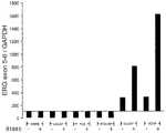
特に、ERG(NM_004449)は、他の正常なヒト組織と比較して前立腺上皮において高く発現していることが実証されている。ERG遺伝子は第21染色体上に位置する。その遺伝子は、pterから38,675,671〜38,955,488塩基対に位置する。ERG遺伝子は合計279,817bp;マイナス鎖配向である。対応するERG cDNAおよびタンパク質配列は、それぞれ、GenBankアクセッション番号M17254およびGenBankアクセッション番号NP04440(Swiss Proteinアクセッション番号P11308)で与えられている。 In particular, ERG (NM — 004449) has been demonstrated to be highly expressed in prostate epithelium compared to other normal human tissues. The ERG gene is located on
ETV1遺伝子は第7染色体上に位置する(GenBankアクセッション番号NC_000007.11;NC_086703.11;およびNT_007819.15)。その遺伝子は、pterから13,708,330〜13,803,555塩基対に位置する。ETV1遺伝子は合計95,225bp、マイナス鎖配向である。対応するETV1 cDNAおよびタンパク質配列は、それぞれ、GenBankアクセッション番号NM_004956およびGenBankアクセッション番号NP_004947(Swiss Proteinアクセッション番号P50549)で与えられている。 The ETV1 gene is located on chromosome 7 (GenBank accession numbers NC_000007.11; NC_086703.11; and NT_007819.15). The gene is located 13,708,330-13,803,555 base pairs from pter. The ETV1 gene has a total of 95,225 bp and a negative strand orientation. The corresponding ETV1 cDNA and protein sequences are given by GenBank accession number NM_004956 and GenBank accession number NP_004947 (Swiss Protein accession number P50549), respectively.
ヒトETV4遺伝子は第14染色体上に位置する(GenBankアクセッション番号NC_000017.9;NT_010783.14;およびNT_086880.1)。その遺伝子は、pterから38,960,740〜38,979,228塩基対にある。ETV4遺伝子は合計18,488bp、マイナス鎖配向である。対応するETV4 cDNAおよびタンパク質配列は、それぞれ、GenBankアクセッション番号NM_001986およびGenBankアクセッション番号NP_01977(Swiss Proteinアクセッション番号P43268)で与えられている。 The human ETV4 gene is located on chromosome 14 (GenBank accession numbers NC_000017.9; NT_010783.14; and NT_086880.1). The gene is 38,960,740 to 38,979,228 base pairs from pter. The ETV4 gene has a total of 18,488 bp and a negative strand orientation. The corresponding ETV4 cDNA and protein sequences are given by GenBank accession number NM_001986 and GenBank accession number NP_01977 (Swiss Protein accession number P43268), respectively.
C. ARG/ETS遺伝子融合
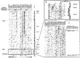
上で記載されているように、本発明は、ARGのETSファミリーメンバー遺伝子への融合を提供する。例示的な遺伝子融合配列は、図36に示されている。すべての関連する遺伝子(TMPRSS2、ERG、ETV1、およびETV4)について、GenBank参照配列IDが提供され、エキソンは、UCSCヒトゲノムの2004年5月のアセンブリ(May 2004 assembly of the UCSC Human Genome)を用いて整列している。すべての同定された融合について、図36は、TMPRSS2遺伝子の始めから融合およびETSファミリーメンバー遺伝子のストップコドンまでの完全な配列を提供する。公開されたバリアントのそれぞれについての寄託されたGenBank配列もまた提供されている。いくつかのTMPRSS2:ERGおよびTMPRSS2:ETV1融合は、TMPRSS2およびETSファミリーメンバー遺伝子の限界点エキソンにより記載されている。例えば、TMPRSS2:ERGaは、TMPRSS2のエキソン1をERGのエキソン4〜11へ融合しているのだが、TMPRSS2:ERG(1,4)として同定される。C. ARG / ETS Gene Fusion As described above, the present invention provides for the fusion of ARG to ETS family member genes. An exemplary gene fusion sequence is shown in FIG. GenBank reference sequence IDs are provided for all related genes (TMPRSS2, ERG, ETV1, and ETV4) and exons are used using the May 2004 assembly of the UCSC Human Genome. Aligned. For all identified fusions, FIG. 36 provides the complete sequence from the beginning of the TMPRSS2 gene to the fusion and stop codon of the ETS family member gene. Deposited GenBank sequences for each of the published variants are also provided. Several TMPRSS2: ERG and TMPRSS2: ETV1 fusions have been described by TMPRSS2 and ETS family member gene limit exons. For example, TMPRSS2: ERGa, which fuses
ARGのETSファミリーメンバー遺伝子への融合は、DNA、RNA、またはタンパク質として検出できる。最初に、遺伝子融合は、ARGの転写制御領域由来の5'部分およびETSファミリーメンバー遺伝子由来の3'部分を有するゲノムDNAの染色体再配列として検出できる。いったん転写されたならば、遺伝子融合は、ARGの転写制御領域由来の5'部分およびETSファミリーメンバー遺伝子由来の3'部分を有するキメラmRNAとして検出できる。いったん翻訳されたならば、遺伝子融合は、ARGの転写制御領域のETSファミリーメンバー遺伝子への融合に起因するアミノ末端が切り詰められたETSファミリーメンバータンパク質;ARGの転写制御領域由来のアミノ末端部分およびETSファミリーメンバー遺伝子由来のカルボキシ末端部分を有するキメラタンパク質;または上方制御されているが、他の点では区別がつかない天然のETSファミリーメンバータンパク質として検出できる。切り詰められたETSファミリーメンバータンパク質およびキメラタンパク質は、アミノ酸配列、翻訳後プロセシング、および/または二次、三次、もしくは四次構造においてそれらのそれぞれの天然タンパク質と異なりうる。そのような違いは、存在するならば、遺伝子融合の存在を同定するために用いられうる。検出の特定の方法は下により詳細に記載されている。 ARG fusions to ETS family member genes can be detected as DNA, RNA, or protein. Initially, gene fusion can be detected as a chromosomal rearrangement of genomic DNA having a 5 ′ portion from the transcriptional control region of ARG and a 3 ′ portion from an ETS family member gene. Once transcribed, the gene fusion can be detected as a chimeric mRNA having a 5 ′ portion from the transcriptional control region of ARG and a 3 ′ portion from an ETS family member gene. Once translated, the gene fusion is a truncated ETS family member protein resulting from the fusion of the transcriptional control region of ARG to the ETS family member gene; the aminoterminal portion from the transcriptional control region of ARG and the ETS It can be detected as a chimeric protein with a carboxy-terminal portion from a family member gene; or as a naturally occurring ETS family member protein that is upregulated but otherwise indistinguishable. Truncated ETS family member proteins and chimeric proteins can differ from their respective native proteins in amino acid sequence, post-translational processing, and / or secondary, tertiary, or quaternary structure. Such differences, if present, can be used to identify the presence of a gene fusion. Specific methods of detection are described in more detail below.
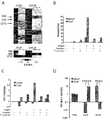
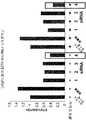
ある特定の遺伝子融合は、前立腺癌において他のものより多く見られる。本発明は、前立腺癌の50〜80%をTMPRSS2のERG、ETV1、ETV4、またはFLI1との再発性遺伝子融合を有すると同定している。それらのうち、50〜70%はTMPRSS2-ERGであって、その50%〜60%が第21染色体上のTMPRSS2座とERG座の間の遺伝的情報の欠失に起因し(下により詳細に記載されている)、5〜10%はTMPRSS2-ETV1であり、1〜2%はTMPRSS2-ETV4であり、1〜2%はTMPRSS2-FLI1である。 Certain gene fusions are more common than others in prostate cancer. The present invention identifies 50-80% of prostate cancer as having recurrent gene fusions of TMPRSS2 with ERG, ETV1, ETV4, or FLI1. Of those, 50-70% are TMPRSS2-ERG, 50% -60% of which are due to the loss of genetic information between the TMPRSS2 and ERG loci on chromosome 21 (more details below) 5-10% is TMPRSS2-ETV1, 1-2% is TMPRSS2-ETV4, and 1-2% is TMPRSS2-FLI1.
本発明の開発の経過中に行われた実験は、ある特定の融合遺伝子が融合転写産物を発現するが、他のものは機能的転写産物を発現しないことを示した(Tomlins et al., Science, 310:644-648 (2005); Tomlins et al., Cancer Research 66:3396-3400 (2006))。 Experiments conducted during the course of the development of the present invention showed that certain fusion genes expressed fusion transcripts while others did not express functional transcripts (Tomlins et al., Science 310: 644-648 (2005); Tomlins et al., Cancer Research 66: 3396-3400 (2006)).
本発明の開発の経過中に行われたさらなる実験は、染色体21q22.2-3上のTMPRSS2とERGの間に位置する有意なゲノム欠失を同定した。欠失は、TMPRSS2:ERG融合陽性PCA試料に見られた。欠失は、コンセンサス領域に現れているが、この領域内で可変性を示す。Parisらによる以前に発表された研究(Hum. Mol. Genet. 13:1303-13(2004))において、CGH分析は、TMPRSS2から6kbセントロメア側であるCTD-2103O7 BACにおける欠失を検出した。これらの欠失は、臨床的に局在したPCA試料の12.5%(9/27)に、および転移性PCA試料の33%(5/15)に観察された。これらの結果は、最新研究からのSNPアレイデータを支持し、どちらのPCA欠失も進行と共により多く見られるようになること、または欠失がより急速に進行する傾向にあるPCAにおいてより頻繁に同定されることを示唆する。TMPRSS2:ERG再配列の顕著な腫瘍内均一性を考慮すれば、これらの分子サブタイプが異なる疾患進行特性と関連している可能性がより高い。 Further experiments conducted during the course of the development of the present invention identified a significant genomic deletion located between TMPRSS2 and ERG on chromosome 21q22.2-3. Deletions were found in TMPRSS2: ERG fusion positive PCA samples. Deletions appear in the consensus region, but show variability within this region. In a previously published study by Paris et al. (Hum. Mol. Genet. 13: 1303-13 (2004)), CGH analysis detected a deletion in CTD-2103O7 BAC, which is 6 kb centromeric from TMPRSS2. These deletions were observed in 12.5% (9/27) of clinically localized PCA samples and 33% (5/15) of metastatic PCA samples. These results support SNP array data from the latest studies, with both PCA deletions becoming more common with progression, or more frequently in PCA where deletions tend to progress more rapidly Suggests to be identified. Given the significant intratumoral homogeneity of TMPRSS2: ERG rearrangements, these molecular subtypes are more likely to be associated with different disease progression characteristics.
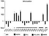
49.2%がERGの再配列を含む118個の臨床的に局在したPCA症例が評価された。イントロンの欠失が、これらのTMPRSS2:ERG融合陽性症例の60.3%に観察された。ERGの著しい過剰発現をもつほとんどすべてのPCA試料は、再配列を有し、過剰発現は、再配列とほぼ同じ数の症例において生じている。Oncomine、公的に利用可能な遺伝子発現データの一覧、を用いて、共通の欠失部位の領域に位置する4個の有意に下方制御された遺伝子が同定された(図16)。 118 clinically localized PCA cases with 49.2% containing ERG rearrangements were evaluated. Intron deletion was observed in 60.3% of these TMPRSS2: ERG fusion positive cases. Almost all PCA samples with significant overexpression of ERG have rearrangements, with overexpression occurring in about the same number of cases as rearrangements. Using Oncomine, a list of publicly available gene expression data, four significantly down-regulated genes located in the region of the common deletion site were identified (FIG. 16).
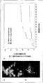
本発明は特定の機構に限定されない。実際、機構の理解は、本発明を実施するのに必要ではない。とはいえ、結果は、全PCAの半分近くがTMPRSS2:ERG再配列によって定義されうることを示唆している。これらの腫瘍の大多数は、オリゴヌクレオチドSNPアレイゲノム分析によればサイズが可変性である、イントロンの欠失を示す。しかしながら、およそ30〜40%は欠失を示さず、従って、TMRPSS2およびERGの平衡転座を含む可能性があった。この欠失の程度における可変性は、CMLについて観察されているように、疾患進行と関連している可能性がある。最新の研究は、腫瘍病期およびリンパ節状態との有意な臨床的関連を同定した。欠失を含むTMPRSS2:ERG再配列された腫瘍はまた、PSA生化学的不全のより高い率への傾向を示した。 The present invention is not limited to a particular mechanism. In fact, an understanding of the mechanism is not necessary to practice the present invention. Nonetheless, the results suggest that nearly half of the total PCA can be defined by TMPRSS2: ERG rearrangement. The majority of these tumors show intron deletions that are variable in size according to oligonucleotide SNP array genomic analysis. However, approximately 30-40% did not show a deletion and could therefore contain an equilibrium translocation of TMRPSS2 and ERG. This variability in the extent of the deletion may be associated with disease progression, as has been observed for CML. The latest studies have identified a significant clinical association with tumor stage and lymph node status. TMPRSS2: ERG rearranged tumors containing deletions also showed a trend towards higher rates of PSA biochemical failure.
本発明の開発の経過中に行われたさらなる実験は、長期追跡調査で初期前立腺癌の静観コホートにおけるTMPRSS2:ERG遺伝子融合の存在に基づく転移または前立腺癌特異的死を生じるリスクを探究した。TMPRSS2:ERG遺伝子融合の頻度は92個の症例を用いて評価された。この集団ベースのコホートにおけるTMPRSS2:ERG遺伝子融合の頻度は、15.2%(14/92)であり、2つの病院ベースのコホートで観察された50%頻度より低かった。本発明は特定の機構に限定されない。実際、機構の理解は、本発明を実施するのに必要ではない。とはいえ、TMPRSS2:ERG遺伝子融合前立腺癌におけるこの違いは、民族および人種の遺伝的違いによる可能性がある。これらの違いはまた、他の非集団ベースの研究と比較した、この静観コホートにおける高悪性度症例のより低いパーセンテージによって説明されうる。 Further experiments conducted during the course of the development of the present invention explored the risk of metastasis or prostate cancer-specific death based on the presence of the TMPRSS2: ERG gene fusion in a static cohort of early prostate cancer in a long-term follow-up. The frequency of TMPRSS2: ERG gene fusion was evaluated using 92 cases. The frequency of TMPRSS2: ERG gene fusion in this population-based cohort was 15.2% (14/92), which was lower than the 50% frequency observed in the two hospital-based cohorts. The present invention is not limited to a particular mechanism. In fact, an understanding of the mechanism is not necessary to practice the present invention. Nonetheless, this difference in TMPRSS2: ERG gene fusion prostate cancer may be due to ethnic and racial genetic differences. These differences can also be explained by the lower percentage of high-grade cases in this static cohort compared to other non-population based studies.
TMPRSS2:ERG遺伝子融合と遠隔転移および前立腺癌特異的死の発生との間の有意な関連が観察され、3.6の累積発生率(P=0.004、95%信頼区間=1.5〜8.9)であった。これらのデータは、TMPRSS2:ERG遺伝子融合前立腺癌がより攻撃的な表現型をもつことを示唆している。さらなる実験は、TMPRSS2:ERG遺伝子融合におけるゲノム欠失が進行性および/または転移性前立腺癌と相関することを示した(例えば、実施例5参照)。 A significant association was observed between TMPRSS2: ERG gene fusion and the occurrence of distant metastases and prostate cancer-specific death, with a cumulative incidence of 3.6 (P = 0.004, 95% confidence interval = 1.5-8.9). These data suggest that TMPRSS2: ERG gene fusion prostate cancer has a more aggressive phenotype. Further experiments have shown that genomic deletions in the TMPRSS2: ERG gene fusion correlate with advanced and / or metastatic prostate cancer (see, eg, Example 5).
本発明はまた、アンドロゲンが、TMPRSS2:ERG陽性細胞系において、おそらくAREを通して、ERGの過剰発現を誘導できることを実証している。本発明は特定の機構に限定されない。実際、機構の理解は、本発明を実施するのに必要ではない。とはいえ、集合的に、結果は、TMPRSS2の上流のAREを通してのETSファミリー活性の調節不全が前立腺癌発生を促進しうることを示唆している。 The present invention also demonstrates that androgens can induce ERG overexpression, possibly through AREs, in TMPRSS2: ERG positive cell lines. The present invention is not limited to a particular mechanism. In fact, an understanding of the mechanism is not necessary to practice the present invention. However, collectively, the results suggest that dysregulation of ETS family activity through ARE upstream of TMPRSS2 may promote prostate cancer development.
遺伝子融合発現の存在、分子サブタイプ、または量は疾患の病期、攻撃性、または進行と相関することが予想される。ETSファミリーメンバー遺伝子を含む類似した再発性遺伝子融合は他の上皮癌に起こることがさらに予想される。 The presence, molecular subtype, or amount of gene fusion expression is expected to correlate with disease stage, aggressiveness, or progression. It is further expected that similar recurrent gene fusions involving ETS family member genes will occur in other epithelial cancers.
II. 抗体
本発明の遺伝子融合タンパク質は、それらの断片、誘導体、および類似体を含めて、下記の診断方法、研究方法、および治療方法において用途をもつ抗体を産生するための免疫原として用いられうる。抗体は、ポリクローナル、モノクローナル、キメラ、ヒト化、一本鎖、またはFab断片でありうる。当業者に公知の様々な手順が、そのような抗体および断片の作製ならびに標識に用いられうる。例えば、Burns, ed., Immunochemical Protocols, 3rd ed., Humana Press (2005); Harlow and Lane, Antibodies: A Laboratory Manual, Cold Spring Harbor Laboratory (1988); Kozbor et al., Immunology Today 4:72 (1983); Kohler and Milstein, Nature 256:495 (1975)を参照。切り詰められたETSファミリーメンバータンパク質またはキメラタンパク質とそれらのそれぞれの天然タンパク質の間の違いを利用する抗体または断片が特に好ましい。II. Antibodies The gene fusion proteins of the present invention, including fragments, derivatives, and analogs thereof, are used as immunogens to produce antibodies with applications in the following diagnostic, research, and therapeutic methods. sell. Antibodies can be polyclonal, monoclonal, chimeric, humanized, single chain, or Fab fragments. Various procedures known to those skilled in the art can be used to make and label such antibodies and fragments. For example, Burns, ed., Immunochemical Protocols, 3rd ed., Humana Press (2005); Harlow and Lane, Antibodies: A Laboratory Manual, Cold Spring Harbor Laboratory (1988); Kozbor et al., Immunology Today 4:72 ( 1983); Kohler and Milstein, Nature 256: 495 (1975). Particularly preferred are antibodies or fragments that take advantage of the differences between truncated ETS family member proteins or chimeric proteins and their respective native proteins.
III. 診断適用
本発明は、遺伝子融合を直接的にかまたは間接的にかのいずれかで検出する、DNA、RNA、およびタンパク質に基づいた診断方法を提供する。本発明はまた、診断目的での組成物およびキットを提供する。III. Diagnostic Applications The present invention provides DNA, RNA, and protein based diagnostic methods that detect gene fusions either directly or indirectly. The present invention also provides compositions and kits for diagnostic purposes.
本発明の診断方法は、定性的または定量的でありうる。定量的診断方法は、例えば、カットオフまたは閾値により緩慢性癌と攻撃的癌の間を識別するために、用いられうる。適用可能な場合、定性的または定量的診断方法はまた、標的、シグナル、または媒介物の増幅を含みうる(例えば、ユニバーサルプライマー)。 The diagnostic methods of the present invention can be qualitative or quantitative. Quantitative diagnostic methods can be used, for example, to distinguish between indolent and aggressive cancers by cut-off or threshold. Where applicable, qualitative or quantitative diagnostic methods may also include amplification of targets, signals, or mediators (eg, universal primers).
最初のアッセイは、遺伝子融合の存在を確認しうるが、特定の融合を同定しない。第二のアッセイは、その後、必要に応じて、特定の融合のアイデンティティを決定するために行われる。第二アッセイは最初のアッセイとは異なる検出テクノロジーを用いうる。 The initial assay can confirm the presence of a gene fusion but does not identify a specific fusion. A second assay is then performed to determine the identity of the particular fusion, if necessary. The second assay may use a different detection technology than the first assay.
本発明の遺伝子融合は、多重またはパネル形式で他のマーカーと共に検出されうる。マーカーは、単独または遺伝子融合と組み合わせてのそれらの予測値について選択される。例示的な前立腺癌マーカーには、限定されるわけではないが、以下が挙げられる:AMACR/P504S(米国特許第6,262,245号);PCA3(米国特許第7,008,765号);PCGEM1(米国特許第6,828,429号);タンパク質/P501S、P503S、P504S、P509S、P510S、前立腺/P703P、P710P(米国公開特許第20030185830号);ならびに米国特許第5,854,206号および第6,034,218号および米国公開特許第20030175736号に開示されたもの、それぞれの特許は全体として参照により本明細書に組み入れられている。他の癌、疾患、感染症、および代謝状態についてのマーカーもまた、多重またはパネル形式における包含として企図される。 The gene fusions of the present invention can be detected with other markers in a multiplex or panel format. Markers are selected for their predicted value alone or in combination with gene fusion. Exemplary prostate cancer markers include, but are not limited to: AMACR / P504S (US Pat. No. 6,262,245); PCA3 (US Pat. No. 7,008,765); PCGEM1 (US Pat. No. 6,828,429) Protein / P501S, P503S, P504S, P509S, P510S, prostate / P703P, P710P (US Publication No. 20030185830); and those disclosed in US Pat. Nos. 5,854,206 and 6,034,218 and US Publication No. 20030175736, Each patent is incorporated herein by reference in its entirety. Markers for other cancers, diseases, infections, and metabolic states are also contemplated as inclusion in multiple or panel format.
本発明の診断方法はまた、疾患の病期、攻撃性、もしくは進行、または転移の存在もしくはリスクと特定の遺伝子融合を相関させるデータを参照して改変されうる。究極的には、本発明の方法により提供される情報は、医師が特定の患者について最善の処置方法を選択するのを援助する。 The diagnostic methods of the invention can also be modified with reference to data that correlates specific gene fusions with disease stage, aggressiveness, or progression, or the presence or risk of metastasis. Ultimately, the information provided by the method of the present invention assists the physician in selecting the best treatment method for a particular patient.
A. 試料
遺伝子融合を含むのではないかと疑われる任意の患者試料が、本発明の方法に従って試験されうる。非限定的例として、試料は、組織(例えば、前立腺生検試料、または前立腺摘除術により得られる組織試料)、血液、尿、精液、前立腺分泌物、またはそれらの画分(例えば、血漿、血清、尿上清、尿細胞ペレット、または前立腺細胞)でありうる。尿試料は、好ましくは、前立腺由来の前立腺細胞を尿路へ流れさせる、細心の直腸指診(DRE)直後に収集される。A. Sample Any patient sample suspected of containing a gene fusion can be tested according to the methods of the invention. By way of non-limiting example, the sample can be a tissue (e.g., a prostate biopsy sample or a tissue sample obtained by prostatectomy), blood, urine, semen, prostate secretion, or a fraction thereof (e.g., plasma, serum) Urine supernatant, urine cell pellet, or prostate cell). The urine sample is preferably collected immediately after a fine digital rectal examination (DRE) that causes prostate cells from the prostate to flow into the urinary tract.
患者試料は、典型的には、遺伝子融合もしくは遺伝子融合を含む細胞を単離する、または遺伝子融合もしくは遺伝子融合を含む細胞について試料を濃縮するように設計された予備処理を必要とする。限定されるわけではないが、以下を含む、当業者に公知の様々な技術がこの目的のために用いられうる:遠心分離;免疫捕獲;細胞溶解;および核酸標的捕獲(例えば、全体として参照により本明細書に組み入れられる、欧州特許第1 409 727号を参照)。 Patient samples typically require pretreatment designed to isolate cells containing gene fusions or gene fusions, or to enrich samples for cells containing gene fusions or gene fusions. Various techniques known to those skilled in the art can be used for this purpose, including but not limited to: centrifugation; immunocapture; cell lysis; and nucleic acid target capture (eg, by reference as a whole). (See
B. DNAおよびRNA検出
本発明の遺伝子融合は、限定されるわけではないが、以下を含む当業者に公知の様々な核酸技術を用いてゲノムDNAまたはキメラmRNAの染色体再配列として検出されうる:核酸シーケンシング;核酸ハイブリダイゼーション;および核酸増幅。B. DNA and RNA Detection The gene fusions of the present invention can be detected as a chromosomal rearrangement of genomic DNA or chimeric mRNA using various nucleic acid techniques known to those of skill in the art including, but not limited to: Nucleic acid sequencing; nucleic acid hybridization; and nucleic acid amplification.
1. シーケンシング
核酸シーケンシング技術の例証となる非限定的例には、限定されるわけではないが、チェーンターミネーター(サンガー)シーケンシングおよびダイターミネーターシーケンシングが挙げられる。当業者は、RNAは細胞においてより安定性が低く、ヌクレアーゼ攻撃をより受けやすいため、実験的には、RNAは通常、シーケンシングの前にDNAへ逆転写される。1. Sequencing Non-limiting examples that illustrate nucleic acid sequencing techniques include, but are not limited to, chain terminator (Sanger) sequencing and dye terminator sequencing. Those skilled in the art experimentally RNA is usually reverse transcribed into DNA prior to sequencing because RNA is less stable in cells and more susceptible to nuclease attack.
チェーンターミネーターシーケンシングは、修飾ヌクレオチド基質を用いるDNA合成反応の配列特異的停止を用いる。伸長は、鋳型DNA上の特定の部位で、その領域における鋳型に相補的な短い放射性、または他の標識の、オリゴヌクレオチドプライマーを用いることにより開始される。オリゴヌクレオチドプライマーは、DNAポリメラーゼ、標準の4つのデオキシヌクレオチド塩基、および低濃度の1つの連鎖停止ヌクレオチド、最も一般的にはジデオキシヌクレオチド、を用いて伸長される。この反応は、4つの別々のチューブにおいて、塩基のそれぞれが交代でジデオキシヌクレオチドとなって、繰り返される。DNAポリメラーゼによる連鎖停止ヌクレオチドの制限された取り込みは、結果として、その特定のジデオキシヌクレオチドが用いられている位置においてのみ停止される一連の関連DNA断片を生じる。各反応チューブについて、断片が、スラブポリアクリルアミドゲルまたは粘性ポリマーで充填されたキャピラリーチューブにおける電気泳動によりサイズ分離される。配列は、ゲルの上端から下端まで走査する時、どのレーンが標識プライマーからの可視化マークを生じるかを読み取ることにより決定される。 Chain terminator sequencing uses sequence specific termination of DNA synthesis reactions using modified nucleotide substrates. Extension is initiated at specific sites on the template DNA by using short radioactive or other labeled oligonucleotide primers complementary to the template in that region. Oligonucleotide primers are extended with DNA polymerase, standard four deoxynucleotide bases, and a low concentration of one chain terminating nucleotide, most commonly a dideoxynucleotide. This reaction is repeated in four separate tubes, with each of the bases alternating in dideoxynucleotides. Limited incorporation of chain-terminating nucleotides by DNA polymerase results in a series of related DNA fragments that are only terminated at the position where that particular dideoxynucleotide is used. For each reaction tube, the fragments are size separated by electrophoresis in a capillary tube filled with slab polyacrylamide gel or viscous polymer. The sequence is determined by reading which lane produces a visualization mark from the labeled primer when scanning from the top to the bottom of the gel.
ダイターミネーターシーケンシングは、代わりに、ターミネーターを標識する。完全なシーケンシングは、ジデオキシヌクレオチドチェーンターミネーターのそれぞれを、異なる波長で蛍光を発する別々の蛍光色素で標識することにより単一反応で行われうる。 Dye terminator sequencing instead labels the terminator. Complete sequencing can be performed in a single reaction by labeling each of the dideoxynucleotide chain terminators with a separate fluorescent dye that fluoresces at a different wavelength.
2. ハイブリダイゼーション
核酸ハイブリダイゼーション技術の例証となる非限定的例には、限定されるわけではないが、インサイチューハイブリダイゼーション(ISH)、マイクロアレイ、およびサザンまたはノーザンブロットが挙げられる。2. Hybridization Non-limiting examples that illustrate nucleic acid hybridization techniques include, but are not limited to, in situ hybridization (ISH), microarrays, and Southern or Northern blots.
インサイチューハイブリダイゼーション(ISH)は、組織(インサイチュー)の一部もしくは切片、または組織が十分小さい場合には、組織全体(ホールマウントISH)において特定のDNAまたはRNA配列の位置を特定するために標識相補的DNAまたはRNA鎖を用いるハイブリダイゼーションの一種である。DNA ISHは、染色体の構造を決定するために用いられうる。RNA ISHは、組織切片またはホールマウント内でmRNAおよび他の転写産物を測定し、位置を特定するために用いられる。試料細胞および組織は、通常、標的転写産物を所定の場所に固定するために、およびプローブの接近を増加させるために、処理される。プローブは、高温で標的配列にハイブリダイズし、その後、過剰プローブは洗い流される。放射性標識、蛍光標識、または抗原標識のいずれかの塩基で標識されたプローブは、それぞれ、オートラジオグラフィー、蛍光顕微鏡法、または免疫組織化学法のいずれかを用いて、組織において位置特定化および定量化される。ISHはまた、同時に2つまたはそれ以上の転写産物を検出するために、放射性または他の非放射性標識で標識された2つまたはそれ以上のプローブを用いることができる。 In situ hybridization (ISH) is labeled to locate a particular DNA or RNA sequence in a portion or section of a tissue (in situ) or, if the tissue is small enough, the entire tissue (hole mount ISH) A type of hybridization that uses complementary DNA or RNA strands. DNA ISH can be used to determine the structure of a chromosome. RNA ISH is used to measure and locate mRNA and other transcripts in tissue sections or whole mounts. Sample cells and tissues are typically processed to fix target transcripts in place and to increase probe access. The probe hybridizes to the target sequence at an elevated temperature, after which excess probe is washed away. Probes labeled with either radiolabeled, fluorescently labeled, or antigenic labeled bases are localized and quantified in tissues using either autoradiography, fluorescence microscopy, or immunohistochemistry, respectively. It becomes. ISH can also use two or more probes labeled with radioactive or other non-radioactive labels to detect two or more transcripts simultaneously.
2.1 FISH
いくつかの態様において、融合配列は、蛍光インサイチューハイブリダイゼーション(FISH)を用いて検出される。本発明のための好ましいFISHアッセイは、細菌人工染色体(BAC)を利用する。これらは、ヒトゲノムシーケンシングプロジェクトにおいて広範に用いられており(Nature 409:953-958 (2001)参照)、多くの情報源、例えば、NCBI、を通して位置づけられうる特定のBACを含むクローンは、配給業者を通して入手できる。ヒトゲノム由来の各BACクローンは、明確にそれを同定する参照名が与えられている。これらの名前は、対応するGenBank配列を見つけるために、および配給業者にクローンのコピーを注文するために用いられうる。2.1 FISH
In some embodiments, the fusion sequence is detected using fluorescence in situ hybridization (FISH). A preferred FISH assay for the present invention utilizes a bacterial artificial chromosome (BAC). These are widely used in human genome sequencing projects (see Nature 409: 953-958 (2001)), and clones containing specific BACs that can be located through many sources such as NCBI Available through. Each BAC clone from the human genome is given a reference name that clearly identifies it. These names can be used to find the corresponding GenBank sequence and to order a copy of the clone from the distributor.
いくつかの態様において、検出アッセイは、ETV1についてのプローブ(例えば、bac RP11-692L4)、c-ERG:t-ERG分裂についての1セットのプローブ(例えば、bac RP11-24A11およびt-ERGについてのプローブとしてRP11-372O17またはRP11-137J13)を利用するFISHアッセイである。他の態様において、FISHアッセイは、ETV1欠失または増幅について1セットのプローブで試験することにより行われ、一方のプローブはETV1座にまたがる(例えば、bac RP11-692L4)、かつ他方のプローブは第7染色体にハイブリダイズする(例えば、その染色体のセントロメア上のプローブ)。なおさらなる態様において、方法は、ERG欠失または増幅について1セットのプローブ、ERG座にまたがる1つ(例えば、bac RP11-476D17)および第21染色体上の1つの参照プローブ(例えば、PR11-32L6;RP11-752M23;RP11-1107H21;RP11-639A7;またはRP11-1077M21)、で試験することにより行われる。さらに他の態様において、方法は、TMPRSS2欠失/増幅について1セットのプローブ、TMPRSS2(例えばRP11-121A5;RP11-120C17;PR11-814F13;またはRR11-535H11)座にまたがる1つおよび第21染色体上の1つの参照プローブ(例えば、PR11-32L6;RP11-752M23;RP11-1107H21;RP11-639A7;またはRP11-1077M21)、で試験することにより行われる。いくつかの態様において、方法はさらに、限定されるわけではないが、RP11-121A5;RP11-120C17;PR11-814F13;およびRR11-535H11を含む群より選択されるプローブを用いるハイブリダイゼーションを含む。 In some embodiments, the detection assay comprises a probe for ETV1 (e.g., bac RP11-692L4), a set of probes for c-ERG: t-ERG division (e.g., for bac RP11-24A11 and t-ERG). FISH assay using RP11-372O17 or RP11-137J13) as a probe. In other embodiments, the FISH assay is performed by testing with a set of probes for ETV1 deletion or amplification, where one probe spans the ETV1 locus (e.g., bac RP11-692L4) and the other probe is Hybridizes to chromosome 7 (eg, a probe on the centromere of that chromosome). In still further embodiments, the method comprises a set of probes for ERG deletion or amplification, one spanning the ERG locus (eg, bac RP11-476D17) and one reference probe on chromosome 21 (eg, PR11-32L6; RP11-752M23; RP11-1107H21; RP11-639A7; or RP11-1077M21). In yet another embodiment, the method comprises a set of probes for TMPRSS2 deletion / amplification, one spanning TMPRSS2 (e.g. RP11-121A5; RP11-120C17; PR11-814F13; or RR11-535H11) loci and on
本発明はさらに、ヒト前立腺細胞、ヒト前立腺組織上で、または該ヒト前立腺細胞もしくはヒト前立腺組織を囲む体液上でFISHアッセイを行う方法を提供する。いくつかの態様において、アッセイは、限定されるわけではないが、RP11-372O17;RP11-137J13;RP11-692L4;RP11-476D17;PR11-32L6;RP11-752M23;RP11-1107H21;RP11-639A7;RP11-1077M21;RP11-121A5;RP11-120C17;PR11-814F13;およびRR11-535H11を含む群より選択されるプローブを利用するハイブリダイゼーション段階を含む。 The invention further provides a method of performing a FISH assay on human prostate cells, human prostate tissue, or body fluid surrounding the human prostate cells or human prostate tissue. In some embodiments, the assay is not limited to: RP11-372O17; RP11-137J13; RP11-692L4; RP11-476D17; PR11-32L6; RP11-752M23; RP11-1107H21; RP11-639A7; RP11 A hybridization step utilizing a probe selected from the group comprising: -1077M21; RP11-121A5; RP11-120C17; PR11-814F13; and RR11-535H11.
本発明に関する再配列を検出するためのFISHプロトコールに用いられうる特定のBACクローンは以下のとおりである:
・ETV1-TMPRSS2融合について試験するために、ETV1にまたがる1つのプローブおよびTMPRSS2座にまたがる1つが用いられうる:
ETV1についてのBAC:RP11-692L4
TMPRSS2についてのBAC:RP11-121A5、(RP11-120C17、PR11-814F13、RR11-535H11)
・ERG転座をc-ERG:t-ERG分裂についてのセットのプローブで試験すること:
c-ERGについてのBAC:RP11-24A11
t-ERGについてのBAC:RP11-372O17、RP11-137J13
・ETV1欠失/増幅をセットのプローブ、ETV1座にまたがる1つおよび第7染色体上の1つの参照プローブ、で試験すること:
ETV1についてのBAC:RP11-692L4
・ERG欠失/増幅をセットのプローブ、ERG座にまたがる1つおよび第21染色体上の1つの参照プローブ、で試験すること:
ERGについてのBAC:RP11-476D17
第21染色体上の参照プローブについてのBAC:*
・TMPRSS2欠失/増幅をセットのプローブ、TMPRSS2座にまたがる1つおよび第21染色体上の1つの参照プローブ、で試験すること:
TMPRSS2についてのBAC:RP11-121A5、(RP11-120C17、PR11-814F13、RR11-535H11)
第21染色体上の参照プローブについてのBAC:PR11-32L6、RP11-752M23、RP11-1107H21、RP11-639A7、(RP11-1077M21)。Specific BAC clones that can be used in the FISH protocol for detecting rearrangements in accordance with the present invention are as follows:
To test for ETV1-TMPRSS2 fusions, one probe that spans ETV1 and one that spans the TMPRSS2 locus can be used:
BAC about ETV1: RP11-692L4
BAC for TMPRSS2: RP11-121A5, (RP11-120C17, PR11-814F13, RR11-535H11)
Testing ERG translocation with a set of probes for c-ERG: t-ERG splitting:
BAC about c-ERG: RP11-24A11
BAC for t-ERG: RP11-372O17, RP11-137J13
Test ETV1 deletion / amplification with a set of probes, one across the ETV1 locus and one reference probe on
BAC about ETV1: RP11-692L4
Test ERG deletion / amplification with a set of probes, one across the ERG locus and one reference probe on chromosome 21:
BAC about ERG: RP11-476D17
BAC for reference probe on chromosome 21:*
Test TMPRSS2 deletion / amplification with a set of probes, one across the TMPRSS2 locus and one reference probe on chromosome 21:
BAC for TMPRSS2: RP11-121A5, (RP11-120C17, PR11-814F13, RR11-535H11)
BAC for reference probes on chromosome 21: PR11-32L6, RP11-752M23, RP11-1107H21, RP11-639A7, (RP11-1077M21).
TMPRSS2とERGの間の融合を結果として生じる欠失突然変異を検出するための最も好ましいプローブは、RP11-24A11およびRP11-137J13である。これらのプローブまたは上記のものは、適切な蛍光または他のマーカーで標識され、その後、ハイブリダイゼーションに用いられる。本明細書に提供された実施例セクションは、欠失を測定するために効果的である1つの特定のプロトコールを示すが、当業者は、このアッセイの多くのバリエーションが等しく十分に用いられうることを認識していると思われる。特定のプロトコールは当技術分野において周知であり、本発明に容易に適応しうる。方法に関する手引きは、以下を含む多くの参照文献から得られうる:In situ Hybridization: Medical Applications (eds. G.R. Coulton and J. de Belleroche), Kluwer Academic Publishers, Boston (1992);In situ Hybridization: In Neurobiology; Advances in Methodology (eds. J.H. Eberwine, K.L. Valentino, and J.D. Barchas), Oxford University Press Inc., England (1994);In situ Hybridization: A Practical Approach (ed. D.G. Wilkinson), Oxford University Press Inc., England (1992); Kuo, et al., Am. J. Hum. Genet. 49:112-119 (1991); Klinger, et al., Am. J. Hum. Genet. 51:55-65 (1992);およびWard, et al., Am. J. Hum. Genet. 52:854-865 (1993)。市販されているキット、およびFISHアッセイを行うためのプロトコールを提供するキットもある(例えば、Oncor, Inc., Gaithersburg, MDから入手できる)。方法に関する手引きを提供する特許には、米国特許第5,225,326号;第5,545,524号;第6,121,489号;および第6,573,043号が挙げられる。これらの参照文献のすべては、全体として参照により本明細書に組み入れられ、当技術分野における類似した参照文献、および本明細書における実施例セクションに提供された情報と共に、特定の実験室にとって都合の良い手順の段階を確立するために用いられうる。The most preferred probes for detecting deletion mutations that result in a fusion between TMPRSS2 and ERG are RP11-24A11 and RP11-137J13. These probes or those described above are labeled with appropriate fluorescence or other markers and then used for hybridization. While the example section provided herein shows one specific protocol that is effective for measuring deletions, those skilled in the art will recognize that many variations of this assay can be equally well used. Seems to be aware of. Certain protocols are well known in the art and can be readily adapted to the present invention. Guidance on methods can be obtained from a number of references including:In situ Hybridization: Medical Applications (eds. GR Coulton and J. de Belleroche), Kluwer Academic Publishers, Boston (1992);In situ Hybridization: In Neurobiology ; Advances in Methodology (eds. JH Eberwine, KL Valentino, and JD Barchas), Oxford University Press Inc., England (1994);In situ Hybridization: A Practical Approach (ed. DG Wilkinson), Oxford University Press Inc., England (1992); Kuo, et al., Am. J. Hum. Genet. 49: 112-119 (1991); Klinger, et al., Am. J. Hum. Genet. 51: 55-65 (1992); And Ward, et al., Am. J. Hum. Genet. 52: 854-865 (1993). There are also commercially available kits and kits that provide protocols for performing FISH assays (eg, available from Oncor, Inc., Gaithersburg, MD). Patents that provide guidance on methods include US Pat. Nos. 5,225,326; 5,545,524; 6,121,489; and 6,573,043. All of these references are incorporated herein by reference in their entirety, and are convenient for a particular laboratory, with similar references in the art, and information provided in the Examples section herein. Can be used to establish good procedural steps.
下の表13は、FISHプローブとして使用を見出す追加のBACクローンを示す。 Table 13 below shows additional BAC clones that find use as FISH probes.
(表13)
2.2 マイクロアレイ
異なる種類の生物学的アッセイはマイクロアレイと呼ばれ、限定されるわけではないが、以下が挙げられる:DNAマイクロアレイ(例えば、cDNAマイクロアレイおよびオリゴヌクレオチドマイクロアレイ);タンパク質マイクロアレイ;組織マイクロアレイ;トランスフェクションまたは細胞マイクロアレイ;化合物マイクロアレイ;および抗体マイクロアレイ。遺伝子チップ、DNAチップ、またはバイオチップとして一般的に知られているDNAマイクロアレイは、発現プロファイリング、または数千個の遺伝子についての発現レベルを同時にモニターすることを目的としてアレイを形成する固体表面(例えばガラス、プラスチック、またはシリコンチップ)へ付着した微視的DNAスポットの収集物である。貼り付けられたDNAセグメントはプローブとして知られ、その数千個が単一のDNAマイクロアレイに用いられうる。マイクロアレイは、疾患細胞および正常細胞における遺伝子発現を比較することにより疾患遺伝子を同定するために用いられうる。マイクロアレイは、限定されるわけではないが、以下を含む様々なテクノロジーを用いて組み立てられうる:鋭く尖ったピンでのスライドグラス上へのプリンティング;あらかじめ作製されたマスクを用いるフォトリソグラフィー;動的マイクロミラーデバイスを用いるフォトリソグラフィー;インクジェットプリンティング;または微小電極アレイ上の電気化学。2.2 Microarrays Different types of biological assays are called microarrays and include, but are not limited to: DNA microarrays (eg, cDNA microarrays and oligonucleotide microarrays); protein microarrays; tissue microarrays; Cell microarrays; compound microarrays; and antibody microarrays. DNA microarrays, commonly known as gene chips, DNA chips, or biochips, are solid surfaces that form arrays for the purpose of expression profiling or simultaneously monitoring the expression levels for thousands of genes (e.g. A collection of microscopic DNA spots attached to a glass, plastic, or silicon chip. The affixed DNA segments are known as probes, and thousands of them can be used in a single DNA microarray. Microarrays can be used to identify disease genes by comparing gene expression in disease and normal cells. The microarray can be assembled using a variety of technologies including, but not limited to: printing onto a slide glass with sharp pointed pins; photolithography using a prefabricated mask; dynamic micro Photolithography using mirror devices; inkjet printing; or electrochemistry on microelectrode arrays.
サザンおよびノーザンブロッティングは、それぞれ、特定のDNAまたはRNA配列を検出するために用いられる。試料から抽出されたDNAまたはRNAを断片化し、マトリックスゲル上で電気泳動的に分離し、膜フィルターへ転写する。フィルターに結合したDNAまたはRNAを、対象となる配列と相補的な標識プローブとのハイブリダイゼーションに供する。フィルターに結合した、ハイブリダイズされたプローブを検出する。手順の変形は逆ノーザンブロットであり、膜に貼り付いている基質核酸が、単離されたDNA断片の収集物であり、プローブが組織から抽出され、標識されたRNAである。 Southern and Northern blotting are used to detect specific DNA or RNA sequences, respectively. DNA or RNA extracted from the sample is fragmented, electrophoretically separated on a matrix gel, and transferred to a membrane filter. The DNA or RNA bound to the filter is subjected to hybridization with a labeled probe complementary to the sequence of interest. The hybridized probe bound to the filter is detected. A variation of the procedure is a reverse Northern blot, where the substrate nucleic acid attached to the membrane is a collection of isolated DNA fragments and the probe is RNA extracted from the tissue and labeled.
3. 増幅
ゲノムDNAおよびキメラmRNAの染色体再配列は、検出の前に、または検出と同時に増幅されうる。核酸増幅技術の例証となる非限定的な例には、限定されるわけではないが、ポリメラーゼ連鎖反応(PCR)、逆転写ポリメラーゼ連鎖反応(RT-PCR)、転写介在増幅(TMA)、リガーゼ連鎖反応(LCR)、鎖置換増幅(SDA)、および核酸配列に基づいた増幅(NASBA)が挙げられる。当業者は、ある特定の増幅技術(例えば、PCR)はRNAが増幅の前にDNAに逆転写されることを必要とするが(例えば、RT-PCR)、他の増幅技術はRNAを直接的に増幅する(例えば、TMAおよびNASBA)ことを認識していると思われる。3. Amplification Chromosomal rearrangements of genomic DNA and chimeric mRNA can be amplified prior to or simultaneously with detection. Non-limiting examples that illustrate nucleic acid amplification techniques include, but are not limited to, polymerase chain reaction (PCR), reverse transcription polymerase chain reaction (RT-PCR), transcription-mediated amplification (TMA), ligase chain Reaction (LCR), strand displacement amplification (SDA), and nucleic acid sequence based amplification (NASBA). One skilled in the art will recognize that certain amplification techniques (e.g., PCR) require that RNA be reverse transcribed into DNA prior to amplification (e.g., RT-PCR), while other amplification techniques directly apply RNA. It seems to be aware that it will amplify (eg, TMA and NASBA).
一般的にPCRと呼ばれるポリメラーゼ連鎖反応(その全体が参照により本明細書に組み入れられる、米国特許第4,683,195号、第4,683,202号、第4,800,159号、および第4,965,188号)は、標的核酸配列のコピー数を指数関数的に増加させるために、変性、プライマー対の逆鎖へのアニーリング、およびプライマー伸長の複数サイクルを用いる。RT-PCRと呼ばれるバリエーションにおいて、逆転写酵素(RT)がmRNAから相補DNA(cDNA)を生成するために用いられ、その後、cDNAはPCRにより増幅され、DNAの複数コピーを生じる。PCRの他の様々な置換について、例えば、米国特許第4,683,195号、第4,683,202号、および第4,800,159号;Mullis et al., Meth. Enzymol. 155:335 (1987);およびMurakawa et al., DNA 7:287 (1988)(それぞれ、全体として参照により本明細書に組み入れられている)を参照。 The polymerase chain reaction, commonly referred to as PCR (U.S. Pat.Nos. 4,683,195, 4,683,202, 4,800,159, and 4,965,188, which is hereby incorporated by reference in its entirety) determines the copy number of a target nucleic acid sequence. To increase exponentially, multiple cycles of denaturation, annealing of the primer pair to the reverse strand, and primer extension are used. In a variation called RT-PCR, reverse transcriptase (RT) is used to generate complementary DNA (cDNA) from mRNA, which is then amplified by PCR to produce multiple copies of the DNA. For various other substitutions of PCR, see, eg, US Pat. Nos. 4,683,195, 4,683,202, and 4,800,159; Mullis et al., Meth. Enzymol. 155: 335 (1987); and Murakawa et al., DNA 7 : 287 (1988), each incorporated herein by reference in its entirety.
一般的にTMAと呼ばれる転写介在増幅(例えば、米国特許第5,480,784号および第5,399,491号、それぞれは全体として参照により本明細書に組み入れられている)は、標的配列の複数のRNAコピーが自己触媒的に追加のコピーを生成する実質的に一定の温度、イオン強度、およびpHの条件下で、標的核酸配列の複数コピーを自己触媒的に合成する。例えば、米国特許第5,399,491号および第5,824,518号(それぞれは全体として参照により本明細書に組み入れられている)を参照。米国公開特許第20060046265号(全体として参照により本明細書に組み入れられている)に記載されたバリエーションにおいて、TMAは、任意で、TMA方法感度および精度を向上させるために遮断成分、停止成分、および他の変更成分の使用を組み入れる。 Transcription-mediated amplification, commonly referred to as TMA (e.g., U.S. Pat.Nos. 5,480,784 and 5,399,491, each incorporated herein by reference in its entirety) allows multiple RNA copies of a target sequence to be autocatalytic. Multiple copies of the target nucleic acid sequence are synthesized autocatalytically under conditions of substantially constant temperature, ionic strength, and pH that produce additional copies. See, for example, US Pat. Nos. 5,399,491 and 5,824,518, each incorporated herein by reference in its entirety. In variations described in U.S. Patent Publication No. 20060046265 (incorporated herein by reference in its entirety), the TMA optionally includes a blocking component, a stopping component, and a TMA method to improve sensitivity and accuracy. Incorporate the use of other modifying ingredients.
一般的にLCRと呼ばれるリガーゼ連鎖反応(全体として参照により本明細書に組み入れられている、Weiss, R., Science 254:1292 (1991))は、標的核酸の隣接領域にハイブリダイズする2セットの相補的なDNAオリゴヌクレオチドを用いる。DNAオリゴヌクレオチドは、熱変性、ハイブリダイゼーション、およびライゲーションの反復サイクルにおいて、DNAリガーゼにより共有結合し、検出可能な二本鎖のライゲーションされたオリゴヌクレオチド産物を生じる。 A ligase chain reaction commonly referred to as LCR (Weiss, R., Science 254: 1292 (1991), which is incorporated herein by reference in its entirety) consists of two sets of hybridizing hybrids to adjacent regions of a target nucleic acid. Complementary DNA oligonucleotides are used. DNA oligonucleotides are covalently linked by DNA ligase in repeated cycles of heat denaturation, hybridization, and ligation, resulting in a detectable double-stranded ligated oligonucleotide product.
一般的にSDAと呼ばれる鎖置換増幅(Walker, G. et al., Proc. Natl. Acad. Sci. USA 89:392-396 (1992);米国特許第5,270,184号および第5,455,166号、それぞれは全体として参照により本明細書に組み入れられている)は、対のプライマー配列を標的配列の逆鎖へアニールすること、二重鎖ヘミホスホロチオネート化プライマー伸長産物を生じうるdNTPαSの存在下におけるプライマー伸長、半修飾化制限エンドヌクレアーゼ認識部位のエンドヌクレアーゼ媒介性ニッキング、ならびに存在する鎖を置換し、プライマーアニーリング、ニッキング、および鎖置換の次のラウンドのための鎖を生じ、結果として、産物の幾何級数的な増幅を生じるためのニックの3'末端からのポリメラーゼ媒介性プライマー伸長のサイクルを用いる。好熱性SDA(tSDA)は、本質的に同じ方法においてより高い温度で好熱性エンドヌクレアーゼおよびポリメラーゼを用いる(欧州特許第0 684 315号)。 Strand displacement amplification, commonly referred to as SDA (Walker, G. et al., Proc. Natl. Acad. Sci. USA 89: 392-396 (1992); U.S. Patent Nos. 5,270,184 and 5,455,166, each as a whole (Incorporated herein by reference) anneals a pair of primer sequences to the reverse strand of the target sequence, primer extension in the presence of dNTPαS, which can result in a double-stranded hemiphosphorothioated primer extension product , Endonuclease-mediated nicking of the semi-modified restriction endonuclease recognition site, and replacing the existing strand, resulting in a strand for the next round of primer annealing, nicking, and strand displacement, resulting in a product geometric series A cycle of polymerase-mediated primer extension from the 3 ′ end of the nick to produce a typical amplification is used. Thermophilic SDA (tSDA) uses thermophilic endonucleases and polymerases at higher temperatures in essentially the same way (
他の増幅方法には、例えば、以下が挙げられる:一般的にNASBAと呼ばれる、核酸配列に基づいた増幅(全体として参照により本明細書に組み入れられている、米国特許第5,130,238号);一般的にQβレプリカーゼと呼ばれる、プローブ分子自身を増幅するためにRNAレプリカーゼを用いるもの(全体として参照により本明細書に組み入れられている、Lizardi et al., BioTechnol. 6:1197 (1988));転写に基づいた増幅方法(Kwoh et al., Proc. Natl. Acad. Sci. USA 86:1173 (1989));および自律的配列複製(Guatelli et al., Proc. Natl. Acad. Sci. USA 87:1874 (1990)、それぞれは全体として参照により本明細書に組み入れられている)。公知の増幅方法のさらなる考察について、Persing, David H., 「In Vitro Nucleic Acid Amplification Techniques」, Diagnostic Medical Microbiology: Principles and Applications (Persing et al., Eds.), pp. 51-87 (American Society for Microbiology, Washington, DC (1993))を参照。 Other amplification methods include, for example: nucleic acid sequence-based amplification, commonly referred to as NASBA (US Pat. No. 5,130,238, incorporated herein by reference in its entirety); Called Qβ replicase, which uses RNA replicase to amplify the probe molecule itself (Lizardi et al., BioTechnol. 6: 1197 (1988), which is incorporated herein by reference in its entirety); Based amplification method (Kwoh et al., Proc. Natl. Acad. Sci. USA 86: 1173 (1989)); and autonomous sequence replication (Guatelli et al., Proc. Natl. Acad. Sci. USA 87: 1874). (1990), each incorporated herein by reference in its entirety). For further discussion of known amplification methods, see Persing, David H., “In Vitro Nucleic Acid Amplification Techniques”, Diagnostic Medical Microbiology: Principles and Applications (Persing et al., Eds.), Pp. 51-87 (American Society for See Microbiology, Washington, DC (1993)).
4. 検出方法
増幅されていない、または増幅された遺伝子融合核酸は、任意の通常の手段により検出されうる。例えば、遺伝子融合は、検出可能に標識されたプローブとのハイブリダイゼーションおよびその結果生じるハイブリッドの測定により検出されうる。検出方法の例証となる非限定的例は下に記載されている。4. Detection Methods Unamplified or amplified gene fusion nucleic acids can be detected by any conventional means. For example, gene fusions can be detected by hybridization with a detectably labeled probe and measurement of the resulting hybrid. Non-limiting examples illustrating detection methods are described below.
一つの例証となる検出方法、ハイブリダイゼーションプロテクションアッセイ(HPA)は、化学発光オリゴヌクレオチドプローブ(例えば、アクリジニウムエステル標識(AE)プローブ)を標的配列にハイブリダイズする段階、ハイブリダイズされていないプローブ上に存在する化学発光標識を選択的に加水分解する段階、および照度計において残りのプローブから生じる化学発光を測定する段階を含む。例えば、米国特許第5,283,174号およびNorman C. Nelson et al., Nonisotopic Probing, Blotting and Sequencing, ch. 17 (Larry J. Kricka ed., 2d ed. 1995)を参照(それぞれは全体として参照により本明細書に組み入れられている)。 One illustrative detection method, the Hybridization Protection Assay (HPA), involves the step of hybridizing a chemiluminescent oligonucleotide probe (e.g., an acridinium ester labeled (AE) probe) to a target sequence, an unhybridized probe. Selectively hydrolyzing the chemiluminescent label present above and measuring the chemiluminescence resulting from the remaining probes in a luminometer. See, e.g., U.S. Patent No. 5,283,174 and Norman C. Nelson et al., Nonisotopic Probing, Blotting and Sequencing, ch. 17 (Larry J. Kricka ed., 2d ed. 1995), each of which is hereby incorporated by reference in its entirety. Incorporated in the book).
もう一つの例証的な検出方法は、リアルタイムでの増幅過程の定量的評価を提供する。「リアルタイム」での増幅過程の評価は、増幅反応中に連続的にかまたは定期的にかのいずれかで反応混合物における単位複製配列の量を測定する段階、および測定された値を用いて、試料に最初に存在した標的配列の量を計算する段階を含む。リアルタイム増幅に基づいた、試料に存在する最初の標的配列の量を測定するための様々な方法は当技術分野において周知である。これらには、米国特許第6,303,305号および第6,541,205号(それぞれは全体として参照により本明細書に組み入れられている)に開示された方法が挙げられる。試料に最初に存在する標的配列の量を測定するための方法であるが、リアルタイム増幅に基づいていないもう一つの方法は、全体として参照により本明細書に組み入れられている、米国特許第5,710,029号に開示されている。 Another illustrative detection method provides a quantitative assessment of the amplification process in real time. Evaluation of the amplification process in “real time” involves measuring the amount of amplicon in the reaction mixture, either continuously or periodically during the amplification reaction, and using the measured value, Calculating the amount of target sequence initially present in the sample. Various methods for measuring the amount of initial target sequence present in a sample based on real-time amplification are well known in the art. These include the methods disclosed in US Pat. Nos. 6,303,305 and 6,541,205, each incorporated herein by reference in its entirety. Another method for measuring the amount of target sequence initially present in a sample, but not based on real-time amplification, is described in US Pat. No. 5,710,029, which is incorporated herein by reference in its entirety. Is disclosed.
増幅産物は、大多数がステム-ループ構造を有する様々な自己ハイブリダイズ性プローブの使用を通してリアルタイムに検出されうる。そのような自己ハイブリダイズ性プローブは、プローブが自己ハイブリダイズした状態であるか、または標的配列へのハイブリダイゼーションによって変化した状態かに依存して、それらが異なって検出できるシグナルを放射するように標識される。非限定的例として、「分子トーチ」は、連結領域(例えば非ヌクレオチドリンカー)により接続され、かつ所定のハイブリダイゼーションアッセイ条件下でお互いにハイブリダイズする、自己相補性の別個の領域(「標的結合ドメイン」および「標的閉鎖ドメイン」と呼ばれる)を含む一種の自己ハイブリダイズ性プローブである。好ましい態様において、分子トーチは、長さが1塩基から約20塩基までである、標的結合ドメインにおいて一本鎖塩基領域を含み、鎖置換条件下で増幅反応物に存在する標的配列へのハイブリダイゼーションにとって接近しやすい。鎖置換条件下で、分子トーチの、完全または部分的に相補的でありうる2つの相補的領域のハイブリダイゼーションは、標的結合ドメインに存在する一本鎖領域に結合して標的閉鎖ドメインの全部または一部を置換する標的配列の存在下を除いては、支持される。分子トーチの標的結合ドメインおよび標的閉鎖ドメインは、検出可能な標識、または分子トーチが自己ハイブリダイズした場合、分子トーチが標的配列にハイブリダイズした場合とは異なるシグナルが生じ、それにより、ハイブリダイズされていない分子トーチの存在下で試験試料においてプローブ:標的二重鎖の検出を可能にするように位置したペアの相互作用性標識(例えば、発光/消光剤)を含む。分子トーチおよび相互作用性標識ペアの様々な型は、全体として参照により本明細書に組み入れられている、米国特許第6,534,274号に開示されている。 Amplification products can be detected in real time through the use of various self-hybridizing probes, the majority of which have a stem-loop structure. Such self-hybridizing probes will emit differently detectable signals depending on whether the probes are in a self-hybridized state or altered by hybridization to a target sequence. Be labeled. As a non-limiting example, a “molecular torch” is a self-complementary discrete region (“target binding”) that is connected by a linking region (eg, a non-nucleotide linker) and hybridizes to each other under a given hybridization assay condition. A type of self-hybridizing probe comprising a domain (referred to as "target closing domain"). In a preferred embodiment, the molecular torch comprises a single stranded base region in the target binding domain that is from 1 base to about 20 bases in length and hybridizes to a target sequence present in the amplification reaction under strand displacement conditions Easy to approach. Under strand displacement conditions, the hybridization of two complementary regions of a molecular torch, which can be fully or partially complementary, binds to a single stranded region present in the target binding domain and either all of the target closing domain or It is supported except in the presence of a target sequence that replaces part. The target binding domain and target closure domain of the molecular torch will produce a detectable label, or a different signal when the molecular torch self-hybridizes than when the molecular torch hybridizes to the target sequence, thereby hybridizing. A pair of interactive labels (eg, luminescence / quencher) positioned to allow detection of the probe: target duplex in the test sample in the presence of a non-molecular torch. Various types of molecular torches and interactive label pairs are disclosed in US Pat. No. 6,534,274, which is incorporated herein by reference in its entirety.
自己相補性を有する検出プローブのもう一つの例は、「分子ビーコン」である。分子ビーコンは、標的相補性配列を有する核酸分子、増幅反応物に存在する標的配列の非存在下においてプローブを閉鎖型立体構造に保持する親和性ペア(または核酸アーム)、およびプローブが閉鎖型立体構造である場合に相互作用する標識ペアを含む。標的配列および標的相補性配列のハイブリダイゼーションは、親和性ペアのメンバーを分離させ、それにより、プローブを開放型立体構造へシフトする。開放型立体構造へのシフトは、例えば、フルオロフォアおよび消光剤(例えば、DABCYLおよびEDANS)でありうる、標識ペアの相互作用の低下により検出できる。分子ビーコンは、全体として参照により本明細書に組み入れられている、米国特許第5,925,517号および第6,150,097号に開示されている。 Another example of a detection probe having self-complementarity is a “molecular beacon”. A molecular beacon is a nucleic acid molecule with a target-complementary sequence, an affinity pair (or nucleic acid arm) that holds the probe in a closed conformation in the absence of the target sequence present in the amplification reaction, and the probe is in a closed conformation. It includes a pair of labels that interact when they are structures. Hybridization of the target sequence and target complementing sequence separates the members of the affinity pair, thereby shifting the probe to an open conformation. The shift to the open conformation can be detected by a decrease in the interaction of the label pair, which can be, for example, a fluorophore and a quencher (eg, DABCYL and EDANS). Molecular beacons are disclosed in US Pat. Nos. 5,925,517 and 6,150,097, which are incorporated herein by reference in their entirety.
他の自己ハイブリダイズ性プローブは当業者に周知である。非限定的例として、米国特許第5,928,862号(全体として参照により本明細書に組み入れられている)に開示されたもののような相互作用標識を有するプローブ結合ペアは、本発明で用いるのに適応しうる。一塩基多型(SNPs)を検出するために用いられるプローブ系もまた、本発明に利用されうる。さらなる検出系には、全体として参照により本明細書に組み入れられている米国公開特許第20050042638に開示されているような、「分子スイッチ」が挙げられる。インターカレート性色素および/または蛍光色素を含むもののような他のプローブもまた、本発明において増幅産物の検出に有用である。例えば、米国特許第5,814,447号(全体として参照により本明細書に組み入れられている)を参照。 Other self-hybridizing probes are well known to those skilled in the art. As a non-limiting example, probe binding pairs with interacting labels such as those disclosed in U.S. Pat.No. 5,928,862 (incorporated herein by reference in its entirety) are adapted for use in the present invention. sell. Probe systems used to detect single nucleotide polymorphisms (SNPs) can also be utilized in the present invention. Additional detection systems include “molecular switches” as disclosed in US Patent Publication 20050042638, which is incorporated herein by reference in its entirety. Other probes such as those containing intercalating dyes and / or fluorescent dyes are also useful in the present invention for detection of amplification products. See, for example, US Pat. No. 5,814,447 (incorporated herein by reference in its entirety).
C. タンパク質検出
本発明の遺伝子融合は、限定されるわけではないが、以下を含む当業者に公知の様々なタンパク質技術を用いて、切り詰められたETSファミリーメンバータンパク質またはキメラタンパク質として検出されうる:タンパク質シーケンシング;およびイムノアッセイ。C. Protein Detection The gene fusions of the present invention can be detected as a truncated ETS family member protein or chimeric protein using various protein techniques known to those skilled in the art including, but not limited to: Protein sequencing; and immunoassay.
1. シーケンシング
タンパク質シーケンシング技術の例証となる非限定的例には、限定されるわけではないが、質量分析およびエドマン分解が挙げられる。1. Sequencing Non-limiting examples that illustrate protein sequencing techniques include, but are not limited to, mass spectrometry and Edman degradation.
質量分析は、原理上は、任意のサイズのタンパク質をシーケンシングするが、計算的にはサイズが増加するにつれてより困難になる。タンパク質はエンドプロテアーゼにより消化され、結果として生じる溶液は、高圧液体クロマトグラフィーカラムに通過させる。このカラムの終端において、溶液は、狭いノズルから出て、高陽電位に荷電して、質量分析計へスプレーされる。液滴上の電荷は、単一イオンのみが残るまでそれらを断片化させる。ペプチドは、その後、断片化され、断片の質量-電荷比が測定される。質量スペクトルは、コンピューターにより分析され、しばしば、断片の配列を決定するために以前にシーケンシングされたタンパク質のデータベースに対して比較される。その後、その過程は、異なる消化酵素で繰り返され、配列における重複はタンパク質についての配列を構築するために用いられる。 Mass spectrometry, in principle, sequences proteins of any size, but becomes computationally more difficult as the size increases. The protein is digested by the endoprotease and the resulting solution is passed through a high pressure liquid chromatography column. At the end of the column, the solution exits the narrow nozzle and is charged to a high positive potential and sprayed onto the mass spectrometer. The charge on the droplets causes them to fragment until only a single ion remains. The peptide is then fragmented and the mass-to-charge ratio of the fragment is measured. Mass spectra are analyzed by a computer and often compared against a database of previously sequenced proteins to determine the sequence of the fragments. The process is then repeated with different digestive enzymes and the overlap in the sequence is used to construct the sequence for the protein.
エドマン分解反応において、シーケンシングされるべきペプチドは固体表面(例えば、ポリブレンでコーティングされたガラス繊維)上へ吸着される。デドマン試薬、フェニルイソチオシアネート(PTC)が、吸着されたペプチドへ12%トリメチルアミンの弱塩基性の緩衝溶液と共に添加され、N末端アミノ酸のアミン基と反応する。末端アミノ酸誘導体は、その後、無水酸の添加により選択的に切り離されうる。誘導体は異性体になって、置換フェニルチオヒダントインを生じるが、それは洗い出されて、クロマトグラフィーにより同定されることができ、そのサイクルが繰り返されうる。各段階の効率は、約98%であり、約50個のアミノ酸が信頼性を以て決定されるのを可能にする。 In the Edman degradation reaction, the peptide to be sequenced is adsorbed onto a solid surface (eg, glass fiber coated with polybrene). A dedman reagent, phenylisothiocyanate (PTC), is added to the adsorbed peptide along with a weakly basic buffer solution of 12% trimethylamine and reacts with the amine group of the N-terminal amino acid. The terminal amino acid derivative can then be selectively cleaved by the addition of anhydride. Derivatives become isomers to give substituted phenylthiohydantoins, which can be washed out and identified by chromatography, and the cycle can be repeated. The efficiency of each stage is about 98%, allowing about 50 amino acids to be determined reliably.
2. イムノアッセイ
イムノアッセイの例証となる非限定的例には、限定されるわけではないが、以下が挙げられる:免疫沈降;ウェスタンブロット;ELISA;免疫組織化学;免疫細胞化学;フローサイトメトリー;およびイムノPCR。当業者に公知の様々な技術を用いて検出可能に標識された(例えば、比色性、蛍光性、化学発光性、または放射性)ポリクローナルまたはモノクローナル抗体がイムノアッセイで用いるのに適している。2. Immunoassays Illustrative non-limiting examples of immunoassays include, but are not limited to: immunoprecipitation; Western blot; ELISA; immunohistochemistry; immunocytochemistry; flow cytometry; PCR. Polyclonal or monoclonal antibodies that are detectably labeled (eg, colorimetric, fluorescent, chemiluminescent, or radioactive) using various techniques known to those skilled in the art are suitable for use in the immunoassay.
免疫沈降は、抗原を、その抗原に特異的な抗体を用いて溶液から沈降させる技術である。その方法は、細胞抽出物に存在するタンパク質複合体を、複合体中にあると考えられるタンパク質を標的にすることにより、用いられうる。複合体は、プロテインAおよびプロテインGのような最初は細菌から単離された不溶性抗体結合性タンパク質により溶液から引き出される。抗体はまた、溶液から外へ容易に単離されうるセファロースビーズと結合しうる。洗浄後、沈殿物は、質量分析、ウェスタンブロッティング、または複合体中の成分を同定するための他の方法のいくつでも用いて分析されうる。 Immunoprecipitation is a technique in which an antigen is precipitated from a solution using an antibody specific for that antigen. The method can be used by targeting the protein complex present in the cell extract to a protein that is thought to be in the complex. The complex is withdrawn from solution by insoluble antibody-binding proteins originally isolated from bacteria, such as protein A and protein G. The antibody may also bind to sepharose beads that can be easily isolated out of solution. After washing, the precipitate can be analyzed using mass spectrometry, Western blotting, or any number of other methods for identifying components in the complex.
ウェスタンブロットまたはイムノブロットは、組織ホモジネートまたは抽出物の与えられた試料においてタンパク質を検出するための方法である。それは、質量により変性タンパク質を分離するためにゲル電気泳動を用いる。タンパク質はその後、ゲルから、典型的にはポリビニルジフルオリドまたはニトロセルロース膜上へ転写され、そこで、それらは対象となるタンパク質に特異的な抗体を用いて探索される。結果として、研究者は、与えられた試料においてタンパク質の量を調べ、いくつかの群間でレベルを比較することができる。 Western blot or immunoblot is a method for detecting proteins in a given sample of tissue homogenate or extract. It uses gel electrophoresis to separate denatured proteins by mass. The proteins are then transferred from the gel, typically onto a polyvinyl difluoride or nitrocellulose membrane, where they are probed using antibodies specific for the protein of interest. As a result, researchers can examine the amount of protein in a given sample and compare levels between several groups.
酵素結合免疫吸着アッセイの省略形のELISAは、試料において抗体または抗原の存在を検出するための生化学的技術である。それは最低限の2つの抗体を利用し、その1つは抗原に特異的であり、他方は酵素に結合している。第二抗体は、色素生産性または蛍光発生性基質にシグナルを生じさせる。ELISAのバリエーションには、サンドイッチELISA、競合的ELISA、およびELISPOTが挙げられる。ELISAは試料において抗原の存在かまたは抗体の存在かのいずれかを評価するために行われうるため、それは、血清抗体濃度を測定するにとっても、およびまた抗原の存在を検出するにとっても有用なツールである。 Abbreviated ELISA for enzyme-linked immunosorbent assays is a biochemical technique for detecting the presence of antibodies or antigens in a sample. It utilizes a minimum of two antibodies, one specific for the antigen and the other bound to the enzyme. The second antibody generates a signal on the chromogenic or fluorogenic substrate. Variations on ELISA include sandwich ELISA, competitive ELISA, and ELISPOT. Since ELISA can be performed to assess either the presence of antigen or antibody in a sample, it is a useful tool for measuring serum antibody concentrations and also for detecting the presence of antigen. It is.
免疫組織化学および免疫細胞化学は、組織または細胞における抗原の、それらのそれぞれの抗体への結合の原理によって、それぞれ、組織切片または細胞においてタンパク質の位置を特定する方法を指す。可視化は、抗体を発色タグまたは蛍光タグでタグを付けることにより可能にされる。色タグの典型的な例には、限定されるわけではないが、西洋ワサビペルオキシダーゼおよびアルカリホスファターゼが挙げられる。フルオロフォアタグの典型的な例には、限定されるわけではないが、フルオレセインイソチオシアネート(FITC)またはフィコエリトリン(PE)が挙げられる。 Immunohistochemistry and immunocytochemistry refer to methods of locating proteins in tissue sections or cells, respectively, by the principle of binding of antigens in tissues or cells to their respective antibodies. Visualization is made possible by tagging the antibody with a chromogenic or fluorescent tag. Typical examples of color tags include, but are not limited to, horseradish peroxidase and alkaline phosphatase. Typical examples of fluorophore tags include, but are not limited to, fluorescein isothiocyanate (FITC) or phycoerythrin (PE).
フローサイトメトリーは、液体の流れに懸濁した微視的粒子を数える、調べる、および選別するための技術である。それは、光学的/電子的検出装置を通して流れる単細胞の物理的および/または化学的特性の同時的多パラメーター的分析を可能にする。単一周波数または色の光(例えば、レーザー)ビームが、流体力学的に集中された液体流上へ向けられる。いくつかの検出器は、流れが光ビームを通過する地点に照準される;光ビームと一致した1種(前方散乱)、およびそれと直角をなす数種(側方散乱(SSC)またはFSC)、および1種または複数の蛍光の検出器。ビームを通過する各懸濁粒子は、どうかして光を散乱させ、粒子中の蛍光化学物質は、励起されて、光源より低い周波数の光を放射しうる。散乱光および蛍光の組み合わせは、検出器によって捉えられ、各検出器における輝度、各蛍光放射ピークについてのもの、の変動を分析することにより、各個々の粒子の物理的および化学的構造について様々な事実を推定することが可能である。FSCは細胞容積と相関し、SSCは粒子の密度または内部複雑性(例えば、核の形、細胞質顆粒の型、または膜粗度)と相関する。 Flow cytometry is a technique for counting, examining, and sorting microscopic particles suspended in a liquid stream. It allows for simultaneous multi-parameter analysis of the physical and / or chemical properties of single cells flowing through optical / electronic detection devices. A single frequency or color light (eg, laser) beam is directed onto a hydrodynamically concentrated liquid stream. Some detectors are aimed at the point where the flow passes through the light beam; one that coincides with the light beam (forward scatter), and several that are perpendicular to it (side scatter (SSC) or FSC), And one or more fluorescence detectors. Each suspended particle passing through the beam somehow scatters light, and the fluorescent chemical in the particle can be excited to emit light at a lower frequency than the light source. The combination of scattered light and fluorescence is captured by the detector, and by analyzing the variation in brightness at each detector, for each fluorescence emission peak, a variety of physical and chemical structures for each individual particle is obtained. It is possible to estimate the facts. FSC correlates with cell volume and SSC correlates with particle density or internal complexity (eg, nuclear shape, cytoplasmic granule type, or membrane roughness).
免疫ポリメラーゼ連鎖反応(IPCR)は、抗体に基づいたイムノアッセイにおいてシグナル発生を増加させるために核酸増幅技術を利用する。PCRのタンパク質等価が存在しない、すなわち、タンパク質は、核酸がPCR中に複製されるのと同じ様式で複製されることができないため、検出感度を増加させる唯一の方法は、シグナル増幅による。標的タンパク質は、オリゴヌクレオチドに直接的または間接的に結合している抗体に結合する。結合していない抗体は洗い流され、残りの結合した抗体が増幅されたそれらのオリゴヌクレオチドを有する。タンパク質検出は、リアルタイム方法を含む標準核酸検出方法を用いる増幅されたオリゴヌクレオチドの検出を介して生じる。 Immunopolymerase chain reaction (IPCR) utilizes nucleic acid amplification techniques to increase signal generation in antibody-based immunoassays. The only way to increase detection sensitivity is by signal amplification, as there is no protein equivalence of PCR, ie, proteins cannot be replicated in the same way that nucleic acids are replicated during PCR. The target protein binds to an antibody that is directly or indirectly bound to the oligonucleotide. Unbound antibodies are washed away and the remaining bound antibodies have their amplified oligonucleotides. Protein detection occurs via detection of amplified oligonucleotides using standard nucleic acid detection methods, including real-time methods.
D. データ分析
いくつかの態様において、コンピューターに基づいた分析プログラムは、検出アッセイにより生じた生データ(例えば、所定のマーカーの存在、非存在、または量)を臨床医のための予測値のデータへ翻訳するために用いられる。臨床医は、任意の適切な手段を用いて予測値を評価できる。従って、いくつかの好ましい態様において、本発明は、遺伝学または分子生物学の教育を受けている可能性が高くない臨床医が、生データを理解する必要がないというさらなる恩恵を提供する。データは、臨床医にその最も有用な形で直接的に提示される。その後、臨床医は、被験体の治療を最適化するためにすぐに情報を利用することができる。D. Data analysis In some embodiments, the computer-based analysis program uses the raw data generated by the detection assay (e.g., the presence, absence, or amount of a given marker) as predictive data for the clinician. Used to translate into The clinician can evaluate the predicted value using any suitable means. Thus, in some preferred embodiments, the present invention provides the additional benefit that clinicians who are not likely to have a genetics or molecular biology education do not need to understand the raw data. The data is presented directly to the clinician in its most useful form. The clinician can then immediately use the information to optimize the treatment of the subject.
本発明は、アッセイを行う実験室、情報提供者、医療関係者、および被験体との往復で、情報を受信する、処理する、および発信する能力がある任意の方法を企図する。例えば、本発明のいくつかの態様において、試料(例えば、生検、または血清もしくは尿試料)は被験体から採取され、生データを作成するために、世界のどこに位置しようが(例えば、被験体が居住する、または情報が最終的に用いられる国とは異なる国に)、プロファイリングサービス(例えば、医療施設における臨床検査室、ゲノムプロファイリング企業など)へ付託される。試料が組織または他の生体試料を含む場合、被験体は、試料を採取され、プロファイリングセンターへ送るために医療センターを訪れてもよい、または被験体は試料(例えば、尿試料)を自ら収集し、それをプロファイリングセンターへ直接送ってもよい。試料が以前に測定された生物学的情報を含む場合、情報は、被験体によりプロファイリングサービスへ直接、送ってもよい(例えば、情報を含む情報カードは、コンピューターによりスキャンされ、データが電子通信システムを用いてプロファイリングセンターのコンピューターへ送信されうる)。いったんプロファイリングサービスに受領されたならば、試料は処理され、被験体にとって望まれる診断的または予後的情報に特異的なプロファイルが作成される(すなわち、発現データ)。 The present invention contemplates any method capable of receiving, processing, and transmitting information in a round trip to the laboratory, information provider, medical personnel, and subject performing the assay. For example, in some embodiments of the invention, a sample (e.g., a biopsy, or a serum or urine sample) is taken from a subject and no matter where it is located in the world (e.g., subject) To a profiling service (eg, a clinical laboratory in a medical facility, a genome profiling company, etc.). If the sample includes a tissue or other biological sample, the subject may collect the sample and visit a medical center to send it to the profiling center, or the subject may collect the sample (e.g., a urine sample) himself. You may send it directly to the profiling center. If the sample contains previously measured biological information, the information may be sent directly to the profiling service by the subject (e.g., an information card containing information is scanned by a computer and the data is transmitted to an electronic communication system). Can be sent to a profiling center computer). Once received by the profiling service, the sample is processed and a profile specific to the diagnostic or prognostic information desired for the subject is created (ie, expression data).
その後、プロファイルデータは、処置を行う臨床医による解釈に適した形式に整えられる。例えば、生発現データを提供するよりむしろ、整えられた形式は、被験体についての診断またはリスク評価(例えば、癌が存在していることの可能性)を特定の処置選択肢についての推奨と共に提示されうる。データは、任意の適切な方法により臨床医に示されうる。例えば、いくつかの態様において、プロファイリングサービスは、臨床医のために印刷されうる(例えば、治療の時点で)、またはコンピューターモニター上で臨床医に示されうる報告を作成する。 The profile data is then formatted into a format suitable for interpretation by the treating clinician. For example, rather than providing live expression data, a well-formed format is presented with a diagnosis or risk assessment for a subject (e.g., the likelihood that cancer is present) with recommendations for specific treatment options. sell. Data can be presented to the clinician by any suitable method. For example, in some embodiments, the profiling service creates a report that can be printed for the clinician (eg, at the time of treatment) or presented to the clinician on a computer monitor.
いくつかの態様において、情報は、治療の時点で、または地方の施設において、最初に分析される。その後、生データはさらなる分析のために、および/または臨床医もしくは患者に有用な情報へ生データを変換するために、中央処理施設へ送られる。中央処理施設は、データ分析のプライバシー(全データは統一セキュリティプロトコールを以て中央施設に保存される)、速さ、および均一性の利点を提供する。中央処理施設は、その後、被験体の処置後のデータの行方を管理することができる。例えば、電子通信システムを用いて、中央施設は、臨床医、被験体、または研究者へデータを提供することができる。 In some embodiments, the information is first analyzed at the time of treatment or at a local facility. The raw data is then sent to a central processing facility for further analysis and / or to convert the raw data into information useful to the clinician or patient. The central processing facility provides the benefits of data analysis privacy (all data is stored in a central facility with a unified security protocol), speed, and uniformity. The central processing facility can then manage the whereabouts of data after the subject's treatment. For example, using an electronic communication system, a central facility can provide data to a clinician, subject, or researcher.
いくつかの態様において、被験体は、電子通信システムを用いてデータに直接、アクセスすることができる。被験体は、その結果に基づいてさらなる治療介入またはカウンセリングを選択しうる。いくつかの態様において、データは、研究用途に用いられる。例えば、データは、疾患の特定の状態または病期の有用な指標としてのマーカーの包含または排除をさらに最適化するために用いられうる。 In some embodiments, the subject can access the data directly using the electronic communication system. The subject may select further therapeutic intervention or counseling based on the results. In some embodiments, the data is used for research applications. For example, the data can be used to further optimize the inclusion or exclusion of markers as useful indicators of a particular state or stage of disease.
E. インビボイメージング
本発明の遺伝子融合はまた、限定されるわけではないが、以下を含むインビボイメージング技術を用いて検出されうる:放射性核種イメージング;陽電子放出断層撮影法(PET);コンピューター体軸断層撮影法;X線または磁気共鳴映像法;蛍光検出;および化学発光検出。いくつかの態様において、インビボイメージング技術は、動物(例えば、ヒトまたは非ヒド哺乳動物)において癌マーカーの存在または発現を可視化するために用いられる。例えば、いくつかの態様において、癌マーカーmRNAまたはタンパク質は、癌マーカーに特異的な標識抗体を用いて標識される。特異的に結合し、かつ標識された抗体は、限定されるわけではないが、放射性核種イメージング、陽電子放出断層撮影法、コンピューター体軸断層撮影法、X線または磁気共鳴映像法、蛍光検出、および化学発光検出を含むインビボイメージング方法を用いて個体において検出されうる。本発明の癌マーカーに対する抗体を作製するための方法は下に記載されている。E. In Vivo Imaging The gene fusions of the present invention can also be detected using in vivo imaging techniques including, but not limited to: radionuclide imaging; positron emission tomography (PET); X-ray or magnetic resonance imaging; fluorescence detection; and chemiluminescence detection. In some embodiments, in vivo imaging techniques are used to visualize the presence or expression of a cancer marker in an animal (eg, a human or non-human mammal). For example, in some embodiments, the cancer marker mRNA or protein is labeled with a labeled antibody specific for the cancer marker. Specifically bound and labeled antibodies include, but are not limited to, radionuclide imaging, positron emission tomography, computed tomography, X-ray or magnetic resonance imaging, fluorescence detection, and It can be detected in an individual using in vivo imaging methods including chemiluminescent detection. Methods for making antibodies against the cancer markers of the present invention are described below.
本発明のインビボイメージング方法は、本発明の癌マーカーを発現する癌(例えば、前立腺癌)の診断に有用である。インビボイメージングは、癌を示すマーカーの存在を可視化するために用いられる。そのような技術は、不快な生検の使用なしで診断を可能にする。本発明のインビボイメージング方法はまた、癌患者へ予後を提供するのに有用である。例えば、転移する可能性の高い癌を示すマーカーの存在が検出されうる。本発明のインビボイメージング方法はさらに、身体の他の部分において転移性癌を検出するために用いられうる。 The in vivo imaging method of the present invention is useful for diagnosis of a cancer (for example, prostate cancer) expressing the cancer marker of the present invention. In vivo imaging is used to visualize the presence of markers indicative of cancer. Such techniques allow diagnosis without the use of unpleasant biopsies. The in vivo imaging methods of the present invention are also useful for providing prognosis to cancer patients. For example, the presence of a marker indicating a cancer that is likely to metastasize can be detected. The in vivo imaging method of the present invention can further be used to detect metastatic cancer in other parts of the body.
いくつかの態様において、本発明の癌マーカーに特異的な試薬(例えば、抗体)は蛍光標識される。標識抗体は、被験体へ導入される(例えば、経口で、または非経口で)。蛍光標識抗体は、任意の適切な方法を用いて(例えば、参照により本明細書に組み入れられている、米国特許第6,198,107号に記載された装置を用いて)検出される。 In some embodiments, reagents (eg, antibodies) specific for the cancer markers of the present invention are fluorescently labeled. The labeled antibody is introduced into the subject (eg, orally or parenterally). The fluorescently labeled antibody is detected using any suitable method (eg, using the apparatus described in US Pat. No. 6,198,107, incorporated herein by reference).
他の態様において、抗体は放射性標識される。インビボ診断のための抗体の使用は、当技術分野において周知である。Sumerdonら(Nucl. Med. Biol. 17:247-254 [1990])は、標識としてインジウム-111を用いる腫瘍の放射免疫シンチグラフィックイメージングについての最適化抗体-キレート剤を記載している。Griffinら(J Clin Onc 9:631-640 [1991])は、再発性結腸直腸癌を有するのではないかと疑われる患者で腫瘍を検出するにおけるこの作用物質の使用を記載している。磁気共鳴映像法のための標識と類似した常磁性イオンを含む作用物質の使用は、当技術分野において公知である(Lauffer, Magnetic Resonance in Medicine 22:339-342 [1991])。用いられる標識は、選択されるイメージング様式に依存する。インジウム-111、テクネチウム-99m、またはヨード-131のような放射性標識は、平面スキャンまたは単光子放出コンピューター断層撮影法(SPECT)について用いられる。フッ素-19のような陽電子放出型標識もまた、陽電子放出断層撮影法(PET)に用いられうる。MRIについて、ガドリニウム(III)またはマンガン(II)のような常磁性イオンが用いられうる。 In other embodiments, the antibody is radiolabeled. The use of antibodies for in vivo diagnosis is well known in the art. Sumerdon et al. (Nucl. Med. Biol. 17: 247-254 [1990]) describe an optimized antibody-chelating agent for radioimmunoscintigraphic imaging of tumors using indium-111 as a label. Griffin et al. (J Clin Onc 9: 631-640 [1991]) describe the use of this agent in detecting tumors in patients suspected of having recurrent colorectal cancer. The use of agents containing paramagnetic ions similar to labels for magnetic resonance imaging is known in the art (Lauffer, Magnetic Resonance in Medicine 22: 339-342 [1991]). The label used depends on the imaging mode selected. Radiolabels such as indium-111, technetium-99m, or iodo-131 are used for planar scanning or single photon emission computed tomography (SPECT). Positron emitting labels such as fluorine-19 can also be used for positron emission tomography (PET). For MRI, paramagnetic ions such as gadolinium (III) or manganese (II) can be used.
スカンジウム-47(3.5日間)、ガリウム-67(2.8日間)、ガリウム-68(68分間)、テクネチウム-99m(6時間)、およびインジウム-111(3.2日間)のような1時間から3.5日までの範囲である半減期をもつ放射性金属は、抗体への結合に利用できるが、そのうち、ガリウム-67、テクネチウム-99m、およびインジウム-111はγカメライメージングに好ましく、ガリウム-68は陽電子放出断層撮影法に好ましい。 1 hour to 3.5 days, such as scandium-47 (3.5 days), gallium-67 (2.8 days), gallium-68 (68 minutes), technetium-99m (6 hours), and indium-111 (3.2 days) Radiometals with half-lives in the range can be used for antibody binding, of which gallium-67, technetium-99m, and indium-111 are preferred for gamma camera imaging, and gallium-68 is positron emission tomography Is preferable.
そのような放射性金属で抗体を標識する有用な方法は、例えば、In-111およびTc-99mについてKhawら(Science 209:295 [1980])により、およびScheinbergら(Science 215:1511 [1982])により記載されているように、ジエチレントリアミンペンタ酢酸(DTPA)のような二官能性キレート剤を用いる。他のキレート剤もまた用いることができるが、1-(p-カルボキシメトキシベンジル)EDTAおよびDTPAの無水カルボキシ炭酸は、それらの使用が抗体の免疫反応性に実質的に影響を及ぼすことなく結合を可能にするため、有利である。 Useful methods of labeling antibodies with such radiometals are described, for example, by Khaw et al. (Science 209: 295 [1980]) for In-111 and Tc-99m, and by Scheinberg et al. (Science 215: 1511 [1982]). A bifunctional chelator such as diethylenetriaminepentaacetic acid (DTPA) is used. Other chelating agents can also be used, but 1- (p-carboxymethoxybenzyl) EDTA and DTPA's anhydrous carboxycarbonate binds without their use substantially affecting the immunoreactivity of the antibody. It is advantageous to make it possible.
DPTAをタンパク質へ結合するためのもう一つの方法は、アルブミンのIn-111での標識についてだが、抗体の標識に適応しうるHnatowichら(Int. J. Appl. Radiat. Isot. 33:327 [1982])により記載されているように、DTPAの環状無水物の使用による。DPTAとのキレート化を用いない、抗体をTc-99mで標識する適切な方法は、Crockfordら(参照により本明細書に組み入れられている、米国特許第4,323,546号)の予備ハンダ付け(pretinning)方法である。 Another method for conjugating DPTA to proteins is for labeling albumin with In-111, but can be adapted for antibody labeling, such as Hnatowich et al. (Int. J. Appl. Radiat. Isot. 33: 327 [1982 ]) By the use of a cyclic anhydride of DTPA. A suitable method for labeling antibodies with Tc-99m without chelation with DPTA is the pretinning method of Crockford et al. (U.S. Pat.No. 4,323,546, incorporated herein by reference). It is.
免疫グロブリンをTc-99mで標識する好ましい方法は、血漿タンパク質についてWongら(Int. J. Appl. Radiat. Isot., 29:251 [1978])により記載されたものであり、最近、抗体を標識することについてWongら(J. Nucl. Med., 23:229 [1981])によって適用するのに成功した。 A preferred method of labeling immunoglobulins with Tc-99m is that described by Wong et al. (Int. J. Appl. Radiat. Isot., 29: 251 [1978]) for plasma proteins and recently labeled antibodies. Has been successfully applied by Wong et al. (J. Nucl. Med., 23: 229 [1981]).
特異的抗体に結合した放射性金属の場合、できる限り高い比率の放射性標識を抗体分子へその免疫特異性を破壊することなく導入することが同様に望ましい。さらなる改善は、抗体上の抗原結合部位が保護されるのを保証するために、本発明の特定の癌マーカーの存在下で放射標識を実施することにより達成されうる。抗原は標識後、切り離される。 In the case of a radiometal bound to a specific antibody, it is equally desirable to introduce as high a proportion of the radiolabel into the antibody molecule without destroying its immunospecificity. Further improvement can be achieved by performing radiolabeling in the presence of certain cancer markers of the present invention to ensure that the antigen binding site on the antibody is protected. The antigen is cleaved after labeling.
なおさらなる態様において、インビボバイオフォトニックイメージング(Xenogen, Almeda, CA)がインビボイメージングに利用される。このリアルタイムインビボイメージングはルシフェラーゼを利用する。ルシフェラーゼ遺伝子は、細胞、微生物、および動物へ取り込まれる(例えば、本発明の癌マーカーとの融合タンパク質として)。活性である時、それは光を放射する反応へ導く。CCDカメラおよびソフトウェアは、イメージを捕捉し、それを分析するために用いられる。 In still further embodiments, in vivo biophotonic imaging (Xenogen, Almeda, CA) is utilized for in vivo imaging. This real-time in vivo imaging utilizes luciferase. The luciferase gene is taken up by cells, microorganisms, and animals (eg, as a fusion protein with the cancer marker of the present invention). When active, it leads to a reaction that emits light. CCD cameras and software are used to capture the image and analyze it.
F. 組成物およびキット
本発明の診断方法で用いる組成物は、限定されるわけではないが、プローブ、増幅オリゴヌクレオチド、および抗体を含む。特に好ましい組成物は、ARGがETSファミリーメンバー遺伝子に融合する場合のみ産物を検出する。これらの組成物は以下を含む:ARGの転写制御領域由来の5'部分がETSファミリーメンバー遺伝子由来の3'部分に融合する接合部にハイブリダイズする(すなわち、遺伝子融合接合部にまたがる)配列を含む単一の標識プローブ;第一増幅オリゴヌクレオチドがARGの転写制御領域にハイブリダイズする配列を含み、かつ第二増幅オリゴヌクレオチドがETSファミリーメンバー遺伝子にハイブリダイズする配列を含む、1対の増幅オリゴヌクレオチド;ARGの転写制御領域のETSファミリーメンバー遺伝子への融合に起因するアミノ末端が切り詰められたETSファミリーメンバータンパク質に対する抗体;またはARGの転写制御領域由来のアミノ末端部分およびETSファミリーメンバー遺伝子由来のカルボキシ末端部分を有するキメラタンパク質に対する抗体。F. Compositions and Kits Compositions used in the diagnostic methods of the present invention include, but are not limited to, probes, amplification oligonucleotides, and antibodies. Particularly preferred compositions detect the product only when ARG is fused to an ETS family member gene. These compositions include: a sequence that hybridizes to a junction where the 5 ′ portion from the transcriptional regulatory region of ARG is fused to the 3 ′ portion from an ETS family member gene (ie, spans a gene fusion junction). A single labeled probe comprising: a pair of amplification oligos, wherein the first amplification oligonucleotide comprises a sequence that hybridizes to the transcriptional control region of ARG and the second amplification oligonucleotide comprises a sequence that hybridizes to the ETS family member gene Nucleotides; antibodies against ETS family member proteins truncated at the amino terminus resulting from fusion of ARG transcriptional regulatory region to ETS family member gene; or amino terminal portion from ARG transcriptional regulatory region and carboxy from ETS family member gene An antibody against a chimeric protein having a terminal portion.
しかしながら、他の有用な組成物は以下を含む:第一標識プローブがARGの転写制御領域にハイブリダイズする配列を含み、かつ第二標識プローブがETSファミリーメンバー遺伝子にハイブリダイズする配列を含む、1対の標識プローブ。 However, other useful compositions include: a first labeled probe comprising a sequence that hybridizes to the transcriptional control region of ARG, and a second labeled probe comprising a sequence that hybridizes to an ETS family member gene. Pair of labeled probes.
単独で、または本発明の他の組成物と組み合わせての、これらの組成物のいずれかがキットの形で提供されうる。例えば、単一の標識プローブおよび増幅オリゴヌクレオチドの対は、本発明の遺伝子融合の増幅および検出のためのキットにおいて提供されうる。キットはさらに、適切な対照および/または検出試薬を含みうる。 Either of these compositions, alone or in combination with other compositions of the invention, can be provided in the form of a kit. For example, a single labeled probe and amplification oligonucleotide pair can be provided in a kit for amplification and detection of a gene fusion of the invention. The kit can further include appropriate controls and / or detection reagents.
本発明のプローブおよび抗体組成物はまた、アレイの形で提供されうる。 The probe and antibody compositions of the invention can also be provided in the form of an array.
IV. 予後診断適用
本発明の開発の経過中に行われた実験は、本発明の遺伝子融合と前立腺癌をもつ患者の予後の間の密接な相関を実証した(例えば、下の実施例5参照)。特に、融合がTMPRSS2とERGの間にあるゲノムDNAの欠失に起因する症例において、癌細胞がより攻撃的な表現型を呈することが見出されている。従って、いくつかの態様において、介在DNAの欠失があるTMPRSS2とERGの間の遺伝子融合を検出する能力があるアッセイが、予後を提供し、医師が適切な治療ストラテジーを決定するのを助けるために用いられる。例えば、いくつかの態様において、この特定の再配列を有する腫瘍をもつ患者は、彼らの予後はその再配列を欠く患者より有意に悪いため、より集中的に処置される。IV. Prognostic Applications Experiments conducted during the course of the development of the present invention demonstrated a close correlation between the gene fusion of the present invention and the prognosis of patients with prostate cancer (see, eg, Example 5 below) ). In particular, cancer cells have been found to exhibit a more aggressive phenotype in cases where the fusion is due to a deletion of genomic DNA between TMPRSS2 and ERG. Thus, in some embodiments, assays capable of detecting gene fusions between TMPRSS2 and ERG with intervening DNA deletions provide a prognosis and help physicians determine an appropriate therapeutic strategy. Used for. For example, in some embodiments, patients with tumors having this particular rearrangement are treated more intensively because their prognosis is significantly worse than patients lacking that rearrangement.
任意のアッセイが、上で考察された型の再配列(例えば、上で記載されたもの)を有する細胞が存在するかどうかを決定するために用いられうる。 Any assay can be used to determine whether cells with the types of rearrangements discussed above (eg, those described above) are present.
本発明は、最も好ましくは、前立腺癌患者についての予後を得ることに関連して用いられるが、他の上皮細胞腫瘍もまた調べることができ、本明細書に記載されたアッセイおよびプローブは、これらの腫瘍由来の癌性細胞が、それらを特に攻撃的にさせる可能性が高い、すなわち、侵襲性および転移性である可能性が高い、再配列を有するかどうかを決定するのに用いられうる。この手順を用いて特徴付けられうる腫瘍の例には、乳房、肺、結腸、卵巣、子宮、食道、胃、肝臓、腎臓、脳、皮膚、および筋肉の腫瘍が挙げられる。アッセイはまた、細胞系および動物モデルにおいてこれらの癌を研究する研究者にとって価値がある。 Although the present invention is most preferably used in connection with obtaining a prognosis for prostate cancer patients, other epithelial cell tumors can also be examined, and the assays and probes described herein are Can be used to determine if cancerous cells from a tumor of the present have rearrangements that are likely to make them particularly aggressive, ie, are likely to be invasive and metastatic. Examples of tumors that can be characterized using this procedure include breast, lung, colon, ovary, uterus, esophagus, stomach, liver, kidney, brain, skin, and muscle tumors. The assay is also valuable for investigators studying these cancers in cell lines and animal models.
本発明の開発の経過中に行われたさらなる実験は、染色体欠失が、欠失した領域に位置する1つまたは複数の遺伝子の発現の喪失があるかどうかを決定するために試料をアッセイすることにより検出されうることを実証した。例えば、TMPRSS2とERGの間に融合を形成するにおいて、典型的には、約2.8メガベースのゲノムDNAが欠失し、これが起こった時、この領域にある少なくとも4つの遺伝子が失われる。これらは、ETS2遺伝子、WRB遺伝子、PCP4遺伝子、およびMX1遺伝子である。癌性前立腺細胞におけるこれらのうちの1つまたは複数の減少が不良な予後を示唆する。 Further experiments conducted during the course of the development of the present invention assay the sample to determine whether a chromosomal deletion is a loss of expression of one or more genes located in the deleted region It was demonstrated that it can be detected. For example, in forming a fusion between TMPRSS2 and ERG, typically about 2.8 megabases of genomic DNA is deleted, and when this occurs, at least four genes in this region are lost. These are the ETS2 gene, the WRB gene, the PCP4 gene, and the MX1 gene. A decrease in one or more of these in cancerous prostate cells suggests a poor prognosis.
従って、いくつかの態様において、本発明は、少なくとも第一および第二プローブを用いてFISHアッセイを行う段階であって、第一プローブが少なくとも15ヌクレオチド長(例えば、少なくとも15、20、35など)である;第一蛍光標識に結合している;および、第一配列がアンドロゲン応答性遺伝子(例えば、TMPRSS2遺伝子)またはETSファミリー遺伝子(例えば、ERG遺伝子、ETV1遺伝子、またはETV4遺伝子)のいずれかの少なくとも一部を含む、ヒトゲノムにおいて第一配列にストリンジェントな条件下でハイブリダイズする;ならびに第二プローブが少なくとも15ヌクレオチド長である;第一蛍光標識とは異なる第二蛍光標識に結合している;および、第一配列とは異なり、かつアンドロゲン応答性遺伝子(例えば、TMPRSS2遺伝子)またはETSファミリー遺伝子(例えば、ERG遺伝子、ETV1遺伝子、またはETV4遺伝子)の少なくとも一部を含む第二配列にストリンジェントな条件下でハイブリダイズする、段階を含む、癌関連再配列を示す染色体DNAの欠失について上皮細胞をアッセイする方法を提供する。 Thus, in some embodiments, the invention involves performing a FISH assay using at least a first and second probe, wherein the first probe is at least 15 nucleotides in length (eg, at least 15, 20, 35, etc.). Bound to a first fluorescent label; and the first sequence is either an androgen responsive gene (eg, TMPRSS2 gene) or an ETS family gene (eg, ERG gene, ETV1 gene, or ETV4 gene) Hybridizes under stringent conditions to the first sequence in the human genome, including at least a portion; and the second probe is at least 15 nucleotides long; binds to a second fluorescent label different from the first fluorescent label And different from the first sequence and androgen responsive gene (eg TMPRSS2 gene) or ETS family gene ( For example, epithelial cells for deletion of chromosomal DNA exhibiting a cancer-related rearrangement, including the step of hybridizing under stringent conditions to a second sequence comprising at least a portion of an ERG gene, ETV1 gene, or ETV4 gene) A method of assaying is provided.
さらなる態様において、本発明は、上皮細胞の検査試料を得る段階;限定されるわけではないが、ETS2;WRB;PCP4;およびMX1を含む群より選択される1つまたは複数の遺伝子の発現のレベルを測定するように上皮細胞の試料をアッセイする段階;段階b)で測定された発現レベルを対照試料におけるレベルと比較する段階;および検査試料における遺伝子について測定された発現のレベルが対照試料についてのそれより低い場合には、欠失が起きていると結論づける段階を含む、癌関連再配列を示すゲノムDNAの欠失について上皮細胞(例えば、前立腺細胞)をアッセイするための方法を提供する。 In a further embodiment, the present invention provides obtaining a test sample of epithelial cells; the level of expression of one or more genes selected from the group comprising, but not limited to, ETS2; WRB; PCP4; and MX1 Assaying a sample of epithelial cells to measure the level; comparing the expression level measured in step b) with the level in a control sample; and the level of expression measured for the gene in the test sample If lower, a method is provided for assaying epithelial cells (eg, prostate cells) for a deletion of genomic DNA exhibiting a cancer-associated rearrangement, including the step of concluding that a deletion has occurred.
V. 薬物スクリーニング適用
いくつかの態様において、本発明は、薬物スクリーニングアッセイ(例えば、抗癌薬についてスクリーニングするための)を提供する。本発明のスクリーニング方法は、本発明の方法を用いて同定された癌マーカー(例えば、限定されるわけではないが、TMPRSS2とのERG、ETV1、ETV4、およびFLI1遺伝子融合を含む)を利用する。例えば、いくつかの態様において、本発明は、癌マーカー遺伝子の発現を変化させる(例えば、減少させる)化合物についてスクリーニングする方法を提供する。化合物または作用物質は、例えば、プロモーター領域と相互作用することにより転写に干渉しうる。化合物または作用物質は、融合から生じたmRNAに干渉しうる(例えば、RNA干渉、アンチセンステクノロジーなどにより)。化合物または作用物質は、融合の生物活性の上流または下流である経路に干渉しうる。いくつかの態様において、候補化合物は、癌マーカーに対して向けられるアンチセンスまたは干渉RNA剤(例えば、オリゴヌクレオチド)である。他の態様において、候補化合物は、本発明の癌マーカー制御因子もしくは発現産物に特異的に結合し、かつその生物学的機能を阻害する抗体または小分子である。V. Drug Screening Applications In some embodiments, the present invention provides drug screening assays (eg, for screening for anti-cancer drugs). The screening methods of the present invention utilize cancer markers identified using the methods of the present invention (including, but not limited to, ERG, ETV1, ETV4, and FLI1 gene fusions with TMPRSS2). For example, in some embodiments, the present invention provides methods of screening for compounds that alter (eg, reduce) the expression of cancer marker genes. A compound or agent can interfere with transcription, for example, by interacting with a promoter region. The compound or agent can interfere with the mRNA resulting from the fusion (eg, by RNA interference, antisense technology, etc.). The compound or agent can interfere with a pathway that is upstream or downstream of the biological activity of the fusion. In some embodiments, candidate compounds are antisense or interfering RNA agents (eg, oligonucleotides) that are directed against cancer markers. In other embodiments, candidate compounds are antibodies or small molecules that specifically bind to a cancer marker regulator or expression product of the invention and inhibit its biological function.
一つのスクリーニング方法において、候補化合物は、癌マーカーを発現する細胞と化合物を接触させ、その後、候補化合物の発現への効果についてアッセイすることにより、癌マーカー発現を変化させるそれらの能力について評価される。いくつかの態様において、候補化合物の癌マーカー遺伝子の発現への効果は、細胞により発現する癌マーカーmRNAのレベルを検出することによりアッセイされる。mRNA発現は、任意の適切な方法により検出されうる。他の態様において、候補化合物の、癌マーカー遺伝子の発現への効果は、癌マーカーによりコードされるポリペプチドのレベルを測定することによりアッセイされる。発現したポリペプチドのレベルは、限定されるわけではないが、本明細書に開示されたものを含む任意の適切な方法を用いて測定されうる。 In one screening method, candidate compounds are evaluated for their ability to alter cancer marker expression by contacting the compound with cells that express the cancer marker and then assaying for effects on the expression of the candidate compound. . In some embodiments, the effect of a candidate compound on the expression of a cancer marker gene is assayed by detecting the level of cancer marker mRNA expressed by the cell. mRNA expression can be detected by any suitable method. In other embodiments, the effect of a candidate compound on the expression of a cancer marker gene is assayed by measuring the level of a polypeptide encoded by the cancer marker. The level of expressed polypeptide can be measured using any suitable method, including but not limited to those disclosed herein.
具体的には、本発明は、本発明の癌マーカーに結合する、例えば癌マーカー発現もしくは癌マーカー活性、へ阻害(または刺激)効果を生じる、または、例えば癌マーカー基質の発現もしくは活性、へ刺激もしくは阻害効果を生じる、モジュレーター、すなわち、候補もしくは試験化合物または作用物質(例えば、タンパク質、ペプチド、ペプチド模倣体、ペプトイド、小分子、または他の薬物)を同定するためのスクリーニング方法を提供する。このように同定された化合物は、治療プロトコールにおいて標的遺伝子(例えば、癌マーカー遺伝子)産物の活性を直接的かまたは間接的にかのいずれかで調節するために、標的遺伝子産物の生物学的機能を詳しく述べるために、または正常な標的遺伝子相互作用を乱す化合物を同定するために、用いられうる。癌マーカーの活性または発現を阻害する化合物は、増殖性障害、例えば、癌、特に前立腺癌、の処置において有用である。 Specifically, the invention binds to a cancer marker of the invention, produces an inhibitory (or stimulatory) effect on, for example, cancer marker expression or cancer marker activity, or stimulates, for example, expression or activity of a cancer marker substrate Alternatively, a screening method is provided to identify modulators, ie candidate or test compounds or agents (eg, proteins, peptides, peptidomimetics, peptoids, small molecules, or other drugs) that produce an inhibitory effect. Compounds identified in this way can be used to modulate the biological function of the target gene product in order to modulate the activity of the target gene (e.g., cancer marker gene) product either directly or indirectly in a therapeutic protocol. Can be used to detail or to identify compounds that disrupt normal target gene interactions. Compounds that inhibit the activity or expression of cancer markers are useful in the treatment of proliferative disorders, such as cancer, particularly prostate cancer.
一つの態様において、本発明は、癌マーカータンパク質もしくはポリペプチド、またはその生物活性部分の基質である候補または試験化合物をスクリーニングするためのアッセイを提供する。もう一つの態様において、本発明は、癌マーカータンパク質もしくはポリペプチドまたはその生物活性部分に結合する、または癌マーカータンパク質もしくはポリペプチドまたはその生物活性部分の活性を調節する、候補または試験化合物をスクリーニングするためのアッセイを提供する。 In one embodiment, the present invention provides an assay for screening candidate or test compounds that are substrates for cancer marker proteins or polypeptides, or biologically active portions thereof. In another embodiment, the present invention screens candidates or test compounds that bind to or modulate the activity of a cancer marker protein or polypeptide or biologically active portion thereof. An assay is provided.
本発明の試験化合物は、生物学的ライブラリー;ペプトイドライブラリー(ペプチドの機能性を有するが、酵素分解に抵抗性であるがそれにもかかわらず生物活性を保持する新規な非ペプチドバックボーンをもつ分子のライブラリー;例えば、Zuckennann et al., J. Med. Chem. 37:2678-85 [1994]参照);空間的アドレス可能な並列固相または溶液相ライブラリー;逆重畳積分を必要とする合成ライブラリー方法;「1ビーズ1化合物」ライブラリー方法;およびアフィニティークロマトグラフィー選択を用いる合成ライブラリー方法を含む当技術分野において公知のコンビナトリアルライブラリー方法における多数のアプローチのいずれかを用いて得られうる。生物学的ライブラリーおよびペプトイドライブラリーアプローチは、ペプチドライブラリーと共に用いるのに好ましいが、他の4つのアプローチは、化合物のペプチド、非ペプチドオリゴマー、または小分子ライブラリーに適用できる(Lam (1997) Anticancer Drug Des. 12:145)。 Test compounds of the present invention are biological libraries; peptoid libraries (with a novel non-peptide backbone that has peptide functionality but is resistant to enzymatic degradation but nevertheless retains biological activity. Libraries of molecules; see, for example, Zuckennann et al., J. Med. Chem. 37: 2678-85 [1994]); spatially addressable parallel solid or solution phase libraries; requires deconvolution Obtained using any of a number of approaches in combinatorial library methods known in the art, including synthetic library methods; “one bead one compound” library methods; and synthetic library methods using affinity chromatography selection. sell. Biological and peptoid library approaches are preferred for use with peptide libraries, but the other four approaches are applicable to peptide, non-peptide oligomer, or small molecule libraries of compounds (Lam (1997 ) Anticancer Drug Des. 12: 145).
分子ライブラリーの合成のための方法の例は、当技術分野において、例えば、以下に見出されうる:DeWitt et al., Proc. Natl. Acad. Sci. U.S.A. 90:6909 [1993]; Erb et al., Proc. Nad. Acad. Sci. USA 91:11422 [1994]; Zuckermann et al., J. Med. Chem. 37:2678 [1994]; Cho et al., Science 261:1303 [1993]; Carrell et al., Angew. Chem. Int. Ed. Engl. 33:2059 [1994]; Carell et al., Angew. Chem. Int. Ed. Engl. 33:2061 [1994];およびGallop et al., J. Med. Chem. 37:1233 [1994]。 Examples of methods for the synthesis of molecular libraries can be found in the art, for example: DeWitt et al., Proc. Natl. Acad. Sci. USA 90: 6909 [1993]; Erb et al., Proc. Nad. Acad. Sci. USA 91: 11422 [1994]; Zuckermann et al., J. Med. Chem. 37: 2678 [1994]; Cho et al., Science 261: 1303 [1993]; Carrell et al., Angew. Chem. Int. Ed. Engl. 33: 2059 [1994]; Carell et al., Angew. Chem. Int. Ed. Engl. 33: 2061 [1994]; and Gallop et al., J. Med. Chem. 37: 1233 [1994].
化合物のライブラリーは、溶液中に(例えば、Houghten, Biotechniques 13:412-421 [1992])、またはビーズ(Lam, Nature 354:82-84 [1991])、チップ(Fodor, Nature 364:555-556 [1993])、細菌もしくは胞子(米国特許第5,223,409号;参照により本明細書に組み入れられている)、プラスミド(Cull et al., Proc. Nad. Acad. Sci. USA 89:1865-1869 [1992])上に、またはファージ(Scott and Smith, Science 249:386-390 [1990]; Devlin Science 249:404-406 [1990]; Cwirla et al., Proc. Natl. Acad. Sci. 87:6378-6382 [1990]; Felici, J. Mol. Biol. 222:301 [1991])上に提示されうる。 Compound libraries can be in solution (eg, Houghten, Biotechniques 13: 412-421 [1992]), or beads (Lam, Nature 354: 82-84 [1991]), chips (Fodor, Nature 364: 555- 556 [1993]), bacteria or spores (US Pat. No. 5,223,409; incorporated herein by reference), plasmids (Cull et al., Proc. Nad. Acad. Sci. USA 89: 1865-1869 [ 1992)) or phage (Scott and Smith, Science 249: 386-390 [1990]; Devlin Science 249: 404-406 [1990]; Cwirla et al., Proc. Natl. Acad. Sci. 87: 6378 -6382 [1990]; Felici, J. Mol. Biol. 222: 301 [1991]).
一つの態様において、アッセイは、癌マーカーmRNAもしくはタンパク質またはその生物活性部分を発現する細胞を試験化合物と接触させ、試験化合物の、癌マーカーの活性を調節する能力を測定する、細胞に基づいたアッセイである。試験化合物の、癌マーカー活性を調節する能力を測定することは、例えば、酵素活性における変化、mRNAの破壊などをモニターすることにより達成されうる。 In one embodiment, the assay comprises contacting a cell expressing a cancer marker mRNA or protein or biologically active portion thereof with a test compound and measuring the ability of the test compound to modulate the activity of the cancer marker. It is. Measuring the ability of a test compound to modulate cancer marker activity can be accomplished, for example, by monitoring changes in enzyme activity, mRNA destruction, and the like.
化合物、例えば、癌マーカー基質またはモジュレーター、への癌マーカーの結合を調節する試験化合物の能力もまた評価されうる。これは、例えば、化合物、例えば基質、の癌マーカーへの結合が複合体において標識された化合物、例えば基質、を検出することにより測定されうるように、化合物、例えば基質、を放射性同位元素または酵素標識と結合させることにより、達成されうる。 The ability of a test compound to modulate the binding of a cancer marker to a compound, eg, a cancer marker substrate or modulator, can also be evaluated. This can be accomplished, for example, by removing the compound, eg, substrate, radioisotope or enzyme such that binding of the compound, eg, substrate, to the cancer marker can be measured by detecting the labeled compound, eg, substrate, in the complex. This can be achieved by coupling with a label.
または、癌マーカーは、複合体における癌マーカーの癌マーカー基質への結合を調節する試験化合物の能力をモニターするために、放射性同位元素または酵素標識と結合する。例えば、化合物(例えば、基質)は、125I、35S、14C、または3Hで、直接的にかまたは間接的にかのいずれかで標識され、放射性同位元素は、放射線放射の直接的カウンティングにより、またはシンチレーションカウンティングにより検出されうる。または、化合物は、例えば、西洋ワサビペルオキシダーゼ、アルカリホスファターゼ、またはルシフェラーゼで酵素的に標識され、酵素標識が、適切な基質の生成物への変換の測定により検出されうる。Alternatively, the cancer marker is coupled with a radioisotope or enzyme label to monitor the ability of the test compound to modulate the binding of the cancer marker to the cancer marker substrate in the complex. For example, the compound (eg, substrate) is labeled with125 I,35 S,14 C, or3 H, either directly or indirectly, and the radioisotope is a direct It can be detected by counting or by scintillation counting. Alternatively, the compound can be enzymatically labeled with, for example, horseradish peroxidase, alkaline phosphatase, or luciferase, and the enzyme label can be detected by measuring the conversion of the appropriate substrate to product.
化合物(例えば、癌マーカー基質)の癌マーカーと相互作用する能力は、その反応体のいずれかの標識の有無に関わらず、評価されうる。例えば、マイクロフィジオメーターが、化合物の癌マーカーとの相互作用を、化合物も癌マーカーもどちらの標識も無しで、検出するために用いられうる(McConnell et al., Science 257:1906-1912 [1992])。本明細書に用いられる場合、「マイクロフィジオメーター」(例えば、Cytosensor)は、細胞がその環境を酸性化する速度を光アドレス電位差測定センサー(light-addressable potentiometric sensor)(LAPS)を用いて測定する分析装置である。この酸性化速度における変化は、化合物と癌マーカーの間の相互作用の指標として用いられうる。 The ability of a compound (eg, a cancer marker substrate) to interact with a cancer marker can be assessed with or without any label of the reactant. For example, a microphysiometer can be used to detect the interaction of a compound with a cancer marker without the label of either the compound or the cancer marker (McConnell et al., Science 257: 1906-1912 [1992 ]). As used herein, a “microphysiometer” (eg, Cytosensor) measures the rate at which cells acidify their environment using a light-addressable potentiometric sensor (LAPS). It is an analysis device. This change in acidification rate can be used as an indicator of the interaction between the compound and the cancer marker.
さらにもう一つの態様において、癌マーカータンパク質またはその生物活性部分を試験化合物に接触させ、試験化合物の癌マーカータンパク質、mRNA、またはその生物活性部分に結合する能力が評価される、無細胞アッセイが提供される。本発明のアッセイで用いられうる癌マーカーまたはmRNAの好ましい生物活性部分は、基質または他のタンパク質との相互作用に関与する断片、例えば、高い表面確率スコアをもつ断片、を含む。 In yet another embodiment, a cell-free assay is provided in which a cancer marker protein or biologically active portion thereof is contacted with a test compound and the ability of the test compound to bind to the cancer marker protein, mRNA, or biologically active portion thereof is assessed. Is done. Preferred biologically active portions of cancer markers or mRNA that can be used in the assays of the present invention include fragments that are involved in interactions with substrates or other proteins, eg, fragments with high surface probability scores.
無細胞アッセイは、標的遺伝子タンパク質および試験化合物の反応混合物を、2つの成分が相互作用して結合し、それに従って、取り出しおよび/または検出されうる複合体を形成するのを可能にするのに十分な条件下および時間で調製することを含む。 The cell-free assay is sufficient to allow the reaction mixture of the target gene protein and test compound to allow the two components to interact and bind, thus forming a complex that can be removed and / or detected. Preparing under appropriate conditions and time.
2つの分子間の相互作用はまた、例えば、蛍光エネルギー移動(FRET)を用いて、検出されうる(例えば、Lakowicz et al., 米国特許第5,631,169号;Stavrianopoulos et al., 米国特許第4,968,103号参照;それぞれは参照により本明細書に組み入れられている)。フルオロフォア標識は、第一ドナー分子の放出した蛍光エネルギーが第二の「アクセプター」分子上の蛍光標識により吸収され、その蛍光標識が立ち代わって、吸収されたエネルギーにより蛍光を発することができるように、選択される。 The interaction between the two molecules can also be detected using, for example, fluorescence energy transfer (FRET) (see, eg, Lakowicz et al., US Pat. No. 5,631,169; Stavrianopoulos et al., US Pat. No. 4,968,103). Each of which is incorporated herein by reference). The fluorophore label allows the fluorescent energy emitted by the first donor molecule to be absorbed by the fluorescent label on the second “acceptor” molecule, and the fluorescent label can replace and emit fluorescence by the absorbed energy. Is selected.
または、「ドナー」タンパク質分子は、単に、トリプトファン残基の自然蛍光エネルギーを利用しうる。「アクセプター」分子標識が「ドナー」のそれと識別されうるように異なる波長の光を放射する標識が選択される。標識間のエネルギー移動の効率は、分子を分離する距離に関連しているため、分子間の空間的関係が評価されうる。結合が分子間で起きる状況において、「アクセプター」分子標識の蛍光放射が最大になるはずである。FRET結合事象は、当技術分野において周知の標準蛍光検出手段を通して(例えば、蛍光計を用いて)便利に測定されうる。 Alternatively, the “donor” protein molecule may simply utilize the natural fluorescent energy of tryptophan residues. A label that emits light of a different wavelength is selected so that the “acceptor” molecular label can be distinguished from that of the “donor”. Since the efficiency of energy transfer between labels is related to the distance separating the molecules, the spatial relationship between the molecules can be evaluated. In situations where binding occurs between molecules, the fluorescence emission of the “acceptor” molecular label should be maximized. FRET binding events can be conveniently measured through standard fluorescence detection means well known in the art (eg, using a fluorimeter).
もう一つの態様において、癌マーカータンパク質またはmRNAの標的分子に結合する能力を測定することは、リアルタイム生体分子相互作用分析(BIA)を用いて達成されうる(例えば、Sjolander and Urbaniczky, Anal. Chem. 63:2338-2345 [1991]およびSzabo et al., Curr. Opin. Struct. Biol. 5:699-705 [1995]参照)。「表面プラズモン共鳴」または「BIA」は、反応体のいずれをも標識することなく、生体特異的相互作用をリアルタイムに検出する(例えば、BIAcore)。結合表面における質量の変化は、結果として、表面近くでの光の屈折率の変化を生じ(表面プラズモン共鳴(SPR)の光学現象)、それが結果として、生物学的分子間のリアルタイム反応の表示として用いられうる検出可能なシグナルを生じる。 In another embodiment, measuring the ability of a cancer marker protein or mRNA to bind to a target molecule can be accomplished using real-time biomolecular interaction analysis (BIA) (e.g., Sjolander and Urbaniczky, Anal. Chem. 63: 2338-2345 [1991] and Szabo et al., Curr. Opin. Struct. Biol. 5: 699-705 [1995]). “Surface plasmon resonance” or “BIA” detects biospecific interactions in real time without labeling any of the reactants (eg, BIAcore). The change in mass at the binding surface results in a change in the refractive index of light near the surface (surface plasmon resonance (SPR) optical phenomenon), which results in the display of real-time reactions between biological molecules. Produces a detectable signal that can be used as
一つの態様において、標的遺伝子産物または試験物質は、固相上に繋ぎとめられる。固相上に繋ぎとめられた標的遺伝子産物/試験化合物複合体は、反応の終わりに検出されうる。好ましくは、標的遺伝子産物が固相上に繋ぎとめられうる、かつ試験化合物(繋ぎとめられていない)は、本明細書に考察された検出可能な標識で直接的にかまたは間接的にかのいずれかで標識されうる。 In one embodiment, the target gene product or test substance is tethered on a solid phase. The target gene product / test compound complex tethered onto the solid phase can be detected at the end of the reaction. Preferably, the target gene product can be anchored on the solid phase, and the test compound (not anchored) is either directly or indirectly with a detectable label discussed herein. It can be labeled with either.
癌マーカー、抗癌マーカー抗体、またはその標的分子を固定することが、そのタンパク質の一方または両方の非複合体化型から複合体化型の分離を容易にするために、加えてアッセイの自動化に適応させるために、望ましくありうる。試験化合物の癌マーカータンパク質への結合、または候補化合物の存在下および非存在下における癌マーカータンパク質の標的分子との相互作用は、反応物を含むのに適した任意の容器内で達成されうる。そのような容器の例には、マイクロタイタープレート、試験管、および微小遠心管が挙げられる。一つの態様において、タンパク質の一方または両方がマトリックスに結合するのを可能にするドメインを付加する融合タンパク質が提供されうる。例えば、グルタチオン-S-トランスフェラーゼ-癌マーカー融合タンパク質またはグルタチオン-S-トランスフェラーゼ/標的融合タンパク質は、グルタチオンセファロースビーズ(Sigma Chemical, St. Louis, MO)またはグルタチオン誘導体化マイクロタイタープレート上に吸着されることができ、その後、それは、試験化合物、または試験化合物および非吸着標的タンパク質かもしくは癌マーカータンパク質のいずれかと結合し、混合物は、複合体形成の助けとなる条件下で(例えば、塩およびpHについての生理学的状態で)インキュベートされる。インキュベーション後、ビーズまたはマイクロタイタープレートウェルは、いずれの非結合成分も除去するために洗浄され、ビーズの場合マトリックスは固定され、複合体は、例えば上記のように、直接的にかまたは間接的にかのいずれかで測定される。 Immobilizing a cancer marker, anti-cancer marker antibody, or target molecule thereof, in addition to automating the assay, facilitates separation of the complexed form from one or both uncomplexed forms of the protein. It may be desirable to adapt. Binding of the test compound to the cancer marker protein or the interaction of the cancer marker protein with the target molecule in the presence and absence of the candidate compound can be accomplished in any container suitable for containing the reactants. Examples of such containers include microtiter plates, test tubes, and microcentrifuge tubes. In one embodiment, a fusion protein can be provided that adds a domain that allows one or both of the proteins to be bound to a matrix. For example, glutathione-S-transferase-cancer marker fusion protein or glutathione-S-transferase / target fusion protein is adsorbed onto glutathione sepharose beads (Sigma Chemical, St. Louis, MO) or glutathione derivatized microtiter plates. And then it binds to the test compound, or either the test compound and either the non-adsorbed target protein or the cancer marker protein, and the mixture is subjected to conditions that aid in complex formation (e.g., for salt and pH). Incubated (in physiological condition). After incubation, the beads or microtiter plate wells are washed to remove any unbound components, in the case of beads the matrix is fixed and the complex can be directly or indirectly, eg, as described above. Is measured by either.
または、複合体は、マトリックスから解離され、癌マーカー結合または活性のレベルが標準技術を用いて測定されうる。癌マーカータンパク質かまたは標的分子かのいずれかをマトリックス上に固定するための他の技術には、ビオチンおよびストレプトアビジンの結合を用いることが挙げられる。ビオチン化癌マーカータンパク質または標的分子は、当技術分野において公知の技術(例えば、ビオチン化キット、Pierce Chemicals, Rockford, EL)を用いてビオチン-NHS(N-ヒドロキシ-スクシンイミド)から調製され、ストレプトアビジンコーティング化96ウェルプレート(Pierce Chemical)のウェルに固定されうる。 Alternatively, the complex can be dissociated from the matrix and the level of cancer marker binding or activity measured using standard techniques. Other techniques for immobilizing either a cancer marker protein or a target molecule on a matrix include using biotin and streptavidin binding. Biotinylated cancer marker protein or target molecule is prepared from biotin-NHS (N-hydroxy-succinimide) using techniques known in the art (e.g., biotinylation kit, Pierce Chemicals, Rockford, EL) and streptavidin. Can be fixed to wells of a coated 96 well plate (Pierce Chemical).
アッセイを行うために、非固定化成分は、繋ぎとめられた成分を含むコーティング化表面へ加えられる。反応が完了した後に、反応していない成分は、いずれの形成された複合体も固体表面上に固定されたままであるような条件下で除去される(例えば、洗浄により)。固相表面上に繋ぎとめられた複合体の検出は、いくつかの方法で達成されうる。前もって固定されていない成分があらかじめ標識されている場合、表面上に固定された標識の検出は、複合体が形成されたことを示す。前もって固定されていない成分があらかじめ標識されていない場合、間接的標識が、表面上に繋ぎとめられた複合体を検出するために用いられうる;例えば、固定化成分に特異的な標識抗体を用いて(抗体は、立ち代わって、例えば、標識抗IgG抗体で直接的に標識されうる、または間接的に標識されうる)。 To perform the assay, the non-immobilized component is added to the coated surface containing the anchored component. After the reaction is complete, unreacted components are removed (eg, by washing) under conditions such that any formed complexes remain immobilized on the solid surface. Detection of complexes anchored on the solid surface can be accomplished in several ways. If the component not previously immobilized is pre-labeled, detection of the label immobilized on the surface indicates that a complex has been formed. If the pre-immobilized component is not pre-labeled, indirect labeling can be used to detect complexes anchored on the surface; for example, using a labeled antibody specific for the immobilized component (The antibody can in turn be labeled directly, eg, with a labeled anti-IgG antibody, or indirectly labeled).
このアッセイは、癌マーカータンパク質または標的分子に反応性であるが、癌マーカータンパク質のその標的分子への結合に干渉しない抗体を利用して行われる。そのような抗体は、プレートのウェルへ誘導体化され、結合していない標的または癌マーカータンパク質は抗体結合によりウェルに捕捉されうる。GST固定化複合体について上で記載されたものに加えて、そのような複合体を検出するための方法には、癌マーカータンパク質または標的分子に反応性の抗体を用いる複合体の免疫検出、および癌マーカータンパク質または標的分子に関連した酵素活性を検出することに頼る酵素結合アッセイが挙げられる。 This assay is performed utilizing antibodies that are reactive to a cancer marker protein or target molecule, but do not interfere with the binding of the cancer marker protein to its target molecule. Such antibodies can be derivatized to the wells of the plate and unbound target or cancer marker proteins can be captured in the wells by antibody binding. In addition to those described above for GST-immobilized complexes, methods for detecting such complexes include immunodetection of complexes using antibodies reactive to cancer marker proteins or target molecules, and Enzyme binding assays that rely on detecting enzyme activity associated with a cancer marker protein or target molecule.
または、無細胞アッセイは、液相で行われうる。そのようなアッセイにおいて、反応生成物は、限定されるわけではないが、以下を含むいくつかの標準技術のいずれかにより、未反応成分から分離される:分画遠心法(例えば、Rivas and Minton, Trends Biochem Sci 18:284-7 [1993]参照);クロマトグラフィー(ゲル濾過クロマトグラフィー、イオン交換クロマトグラフィー);電気泳動(例えば、Ausubel et al., eds. Current Protocols in Molecular Biology 1999, J. Wiley: New York参照);および免疫沈降(例えば、Ausubel et al., eds. Current Protocols in Molecular Biology 1999, J. Wiley: New York参照)。そのような樹脂およびクロマトグラフィー技術は当業者に公知である(例えば、Heegaard J. Mol. Recognit 11:141-8 [1998]; Hageand Tweed J. Chromatogr. Biomed. Sci. Appl 699:499-525 [1997]参照)。さらに、蛍光エネルギー移動もまた、溶液からの複合体のさらなる精製なしに結合を検出するために、本明細書に記載されているように、便利に利用されうる。 Alternatively, the cell free assay can be performed in a liquid phase. In such assays, reaction products are separated from unreacted components by any of several standard techniques including, but not limited to: differential centrifugation (eg, Rivas and Minton , Trends Biochem Sci 18: 284-7 [1993]); chromatography (gel filtration chromatography, ion exchange chromatography); electrophoresis (eg Ausubel et al., Eds. Current Protocols in Molecular Biology 1999, J. Wiley: New York); and immunoprecipitation (see, eg, Ausubel et al., Eds. Current Protocols in Molecular Biology 1999, J. Wiley: New York). Such resins and chromatographic techniques are known to those skilled in the art (eg, Heegaard J. Mol. Recognit 11: 141-8 [1998]; Hageand Tweed J. Chromatogr. Biomed. Sci. Appl 699: 499-525 [ 1997]). In addition, fluorescence energy transfer can also be conveniently utilized, as described herein, to detect binding without further purification of the complex from solution.
アッセイは、癌マーカータンパク質、mRNA、またはその生物活性部分を、癌マーカーを結合してアッセイ混合物を形成する既知の化合物と接触させる段階、アッセイ混合物を試験化合物と接触させる段階、および癌マーカータンパク質またはmRNAと相互作用する試験化合物の能力を測定する段階を含み、癌マーカータンパク質またはmRNAと相互作用する試験化合物の能力を測定する段階が、既知の化合物と比較して、癌マーカーもしくはその生物活性部分に優先的に結合する、または標的分子の活性を調節する、試験化合物の能力を測定する段階を含む。 The assay comprises contacting a cancer marker protein, mRNA, or biologically active portion thereof with a known compound that binds the cancer marker to form an assay mixture, contacting the assay mixture with a test compound, and a cancer marker protein or measuring the ability of a test compound to interact with an mRNA, wherein the step of measuring the ability of a test compound to interact with a cancer marker protein or mRNA comprises comparing the cancer marker or biologically active portion thereof to a known compound Measuring the ability of the test compound to bind preferentially to or modulate the activity of the target molecule.
癌マーカーがインビボでタンパク質のような1つもしくは複数の細胞または細胞外高分子と相互作用できる範囲内において、そのような相互作用のインヒビターは有用である。均一なアッセイはインヒビターを同定するために用いられうる。 Inhibitors of such interactions are useful to the extent that cancer markers can interact with one or more cells such as proteins or extracellular macromolecules in vivo. A homogeneous assay can be used to identify inhibitors.
例えば、標的遺伝子産物および相互作用的な細胞または細胞外結合パートナー産物のあらかじめ形成された複合体は、標的遺伝子産物またはそれらの結合パートナーのいずれかが標識されるが、標識により生じるシグナルが複合体形成により消されるように調製される(例えば、イムノアッセイについてこのアプローチを利用する、参照により本明細書に組み入れられている、米国特許第4,109,496号を参照)。あらかじめ形成された複合体由来の種の1つと競合して、置換する試験物質の添加は、結果として、バックグラウンドを超えるシグナルの発生を生じる。このようにして、標的遺伝子産物-結合パートナー相互作用を乱す試験物質が同定されうる。または、癌マーカータンパク質は、癌マーカーに結合する、または相互作用して、癌マーカー活性に関与する他のタンパク質(「癌マーカー結合タンパク質」または「癌マーカー-bp」)を同定するために、ツーハイブリッドアッセイまたはスリーハイブリッドアッセイ(例えば、米国特許第5,283,317号;Zervos et al., Cell 72:223-232 [1993]; Madura et al., J. Biol. Chem. 268:12046-12054 [1993];Bartel et al., Biotechniques 14:920-924[1993];Iwabuchi et al., Oncogene 8:1693-1696[1993];およびBrent WO 94/10300参照;それぞれは参照により本明細書に組み入れられている)において「おとりタンパク質」として用いられうる。そのような癌マーカー-bpは、癌マーカータンパク質もしくは、例えば、癌マーカー媒介性シグナル伝達経路の下流エレメントのような標的によるシグナルのアクチベーターまたはインヒビターでありうる。 For example, a preformed complex of a target gene product and an interactive cell or extracellular binding partner product is labeled with either the target gene product or their binding partner, but the signal produced by the label is complex Prepared to disappear upon formation (see, eg, US Pat. No. 4,109,496, which utilizes this approach for immunoassays, incorporated herein by reference). Addition of a test substance that replaces and competes with one of the pre-formed complex-derived species results in the generation of a signal that exceeds background. In this way, test substances that disrupt the target gene product-binding partner interaction can be identified. Alternatively, a cancer marker protein can be coupled to or interact with a cancer marker to identify other proteins involved in cancer marker activity (“cancer marker binding protein” or “cancer marker-bp”). Hybrid or three hybrid assays (eg, US Pat. No. 5,283,317; Zervos et al., Cell 72: 223-232 [1993]; Madura et al., J. Biol. Chem. 268: 12046-12054 [1993]; See Bartel et al., Biotechniques 14: 920-924 [1993]; Iwabuchi et al., Oncogene 8: 1693-1696 [1993]; and Brent WO 94/10300, each of which is incorporated herein by reference. ) Can be used as a “decoy protein”. Such cancer marker-bp can be an activator or inhibitor of a signal by a cancer marker protein or a target, such as, for example, a downstream element of a cancer marker-mediated signaling pathway.
癌マーカー発現のモジュレーターもまた同定されうる。例えば、細胞混合物または無細胞混合物を、候補化合物と接触させ、癌マーカーmRNAまたはタンパク質の発現を、候補化合物の非存在下における癌マーカーmRNAまたはタンパク質の発現のレベルと比較して評価する。癌マーカーmRNAまたはタンパク質の発現が、候補化合物の存在下において、その非存在下においてより多い場合、候補化合物は、癌マーカーmRNAまたはタンパク質発現の刺激剤と同定される。または、癌マーカーmRNAまたはタンパク質の発現が、候補化合物の存在下において、その非存在下においてより少ない(すなわち、統計学的に有意に、より少ない)場合、候補化合物は、癌マーカーmRNAまたはタンパク質発現のインヒビターと同定される。癌マーカーmRNAまたはタンパク質発現のレベルは、癌マーカーmRNAまたはタンパク質を検出するための本明細書に記載された方法により測定されうる。 Modulators of cancer marker expression can also be identified. For example, a cell mixture or a cell-free mixture is contacted with a candidate compound and the expression of the cancer marker mRNA or protein is assessed relative to the level of expression of the cancer marker mRNA or protein in the absence of the candidate compound. A candidate compound is identified as a stimulator of cancer marker mRNA or protein expression if the expression of the cancer marker mRNA or protein is greater in the presence of the candidate compound and in the absence thereof. Alternatively, if the expression of a cancer marker mRNA or protein is less in the presence of the candidate compound (i.e., statistically significantly less) in the absence, the candidate compound is expressed in cancer marker mRNA or protein Identified as an inhibitor. The level of cancer marker mRNA or protein expression can be measured by the methods described herein for detecting cancer marker mRNA or protein.
調節剤は、細胞に基づいた、または無細胞アッセイを用いて同定され、癌マーカータンパク質の活性を調節する作用物質の能力は、インビボで、例えば、疾患についての動物モデルのような動物(例えば、前立腺癌もしくは転移性前立腺癌をもつ動物;または動物(例えば、ヒト)由来の前立腺癌の異種移植片、もしくは前立腺癌の転移(例えば、リンパ節、骨、または肝臓への)に起因する癌由来の細胞、もしくは前立腺癌細胞系由来の細胞を有する動物)において、確認されうる。 Modulators are identified using cell-based or cell-free assays, and the ability of an agent to modulate the activity of a cancer marker protein is determined in vivo by, for example, an animal such as an animal model for a disease (e.g., Animals with prostate cancer or metastatic prostate cancer; or prostate cancer xenografts from animals (eg, humans), or cancers resulting from prostate cancer metastasis (eg, to lymph nodes, bones, or liver) Or an animal having cells derived from a prostate cancer cell line).
本発明はさらに、上記のスクリーニングアッセイにより同定された新規な作用物質に関する(例えば、癌治療の下記の説明を参照)。従って、そのような作用物質での処置の、効力、毒性、副作用、または作用の機構を測定するために適切な動物モデル(本明細書に記載されたもののような)において本明細書に記載されているように同定された作用物質(例えば、癌マーカー調節剤、アンチセンス癌マーカー核酸分子、siRNA分子、癌マーカー特異的抗体、または癌マーカー結合パートナー)をさらに用いることは、本発明の範囲内である。さらになお、上記のスクリーニングアッセイにより同定された新規な作用物質は、例えば、本明細書に記載されているような処置に用いられうる。 The present invention further relates to novel agents identified by the above screening assays (see, eg, the following description of cancer treatment). Thus, as described herein in an appropriate animal model (such as those described herein) to measure the efficacy, toxicity, side effects, or mechanism of action of treatment with such agents. It is within the scope of the present invention to further use an agent identified as such (e.g., a cancer marker modulator, antisense cancer marker nucleic acid molecule, siRNA molecule, cancer marker specific antibody, or cancer marker binding partner). It is. Furthermore, the novel agents identified by the screening assays described above can be used, for example, for treatment as described herein.
VI. 治療的適用
いくつかの態様において、本発明は、癌(例えば、前立腺癌)についての治療を提供する。いくつかの態様において、治療は、癌マーカー(例えば、限定されるわけではないが、TMPRSS2とのERG、ETV1、およびETV4遺伝子融合を含む)を直接的にまたは間接的にターゲットする。VI. Therapeutic Applications In some embodiments, the present invention provides treatment for cancer (eg, prostate cancer). In some embodiments, the treatment targets directly or indirectly a cancer marker, including, but not limited to, ERG, ETV1, and ETV4 gene fusions with TMPRSS2.
A. RNA干渉およびアンチセンス治療
いくつかの態様において、本発明は、癌マーカーの発現をターゲットする。例えば、いくつかの態様において、本発明は、本発明の癌マーカーをコードする核酸分子の融合を調節し、最終的には発現される癌マーカーの量を調節するのに用いる、オリゴマーアンチセンスまたはRNAi化合物、特にオリゴヌクレオチド(例えば、上記の薬物スクリーニング方法で同定されたもの)を含む組成物を用いる。A. RNA interference and antisense therapy In some embodiments, the present invention targets the expression of cancer markers. For example, in some embodiments, the invention provides for oligomeric antisense or for use in modulating the fusion of a nucleic acid molecule encoding a cancer marker of the invention and ultimately modulating the amount of cancer marker expressed. Compositions comprising RNAi compounds, particularly oligonucleotides (eg, those identified by the drug screening methods described above) are used.
1. RNA干渉(RNAi)
いくつかの態様において、RNAiは、融合タンパク質機能を阻害するために利用される。RNAiは、ヒトを含むたいていの真核生物において外来遺伝子の発現を制御するための進化的に保存された細胞防御を表す。RNAiは、典型的には、二本鎖RNA(dsRNA)により引き起こされ、dsRNAに応答して一本鎖標的RNA相同体の配列特異的mRNA分解をもたらす。mRNA分解の媒介物は、低分子干渉RNA二重鎖(siRNA)であり、通常には、細胞において酵素切断により長いdsRNAから生成される。siRNAは、一般的に、約21ヌクレオチド長(例えば、21〜23ヌクレオチド長)であり、2ヌクレオチドの3'-オーバーハングにより特徴付けられる塩基対形成構造を有する。小さなRNA、またはRNAiの細胞への導入後、その配列は、RISC(RNA誘導サイレンシング複合体)と呼ばれる酵素複合体へ送達されると考えられている。RISCは、標的を認識し、それをエンドヌクレアーゼで切断する。より大きなRNA配列が細胞へ送達された場合には、RNアーゼIII酵素(ダイサー)がより長いdsRNAを21〜23nt ds siRNA断片に変えることを言及しておく。いくつかの態様において、RNAiオリゴヌクレオチドは、融合タンパク質の接合領域をターゲットするように設計される。1. RNA interference (RNAi)
In some embodiments, RNAi is utilized to inhibit fusion protein function. RNAi represents an evolutionarily conserved cellular defense to control the expression of foreign genes in most eukaryotes, including humans. RNAi is typically caused by double-stranded RNA (dsRNA) and results in sequence-specific mRNA degradation of single-stranded target RNA homologues in response to dsRNA. Mediators of mRNA degradation are small interfering RNA duplexes (siRNAs), usually produced from long dsRNA by enzymatic cleavage in cells. siRNAs are generally about 21 nucleotides long (eg, 21-23 nucleotides long) and have a base-pairing structure characterized by a 2
化学合成されたsiRNAは、培養体細胞における哺乳動物遺伝子機能のゲノムワイド分析のための強力な試薬になっている。遺伝子機能の確証についてのそれらの価値以上に、siRNAはまた、遺伝子特異的治療剤として大きな可能性をもっている(参照により本明細書に組み入れられている、Tuschl and Borkhardt, Molecular Intervent. 2002;2(3):158-67)。 Chemically synthesized siRNAs have become powerful reagents for genome-wide analysis of mammalian gene function in cultured somatic cells. Beyond their value for confirmation of gene function, siRNAs also have great potential as gene-specific therapeutic agents (Tuschl and Borkhardt, Molecular Intervent. 2002; 2 (incorporated herein by reference). 3): 158-67).
siRNAの動物細胞へのトランスフェクションは、結果として、特定の遺伝子の強力な長期的転写後サイレンシングを生じる(Caplen et al., Proc Natl Acad Sci U.S.A. 2001;98:9742-7; Elbashir et al., Nature 2001;411:494-8; Elbashir et al., Genes Dev. 2001;15:188-200;およびElbashir et al., EMBO J. 2001;20:6877-88、すべては参照により本明細書に組み入れられている)。siRNAでRNAiを行うための方法および組成物は、例えば、参照により本明細書に組み入れられている、米国特許第6,506,559号に記載されている。 Transfection of siRNA into animal cells results in strong long-term post-transcriptional silencing of specific genes (Caplen et al., Proc Natl
siRNAは、ターゲットされたRNA、およびひいては、タンパク質の量を、しばしば、検出不可能なレベルまで、低下させるのに並はずれて効果的である。サイレンシング効果は、数ヶ月間、持続することができ、標的RNAとsiRNAの中央領域の間の1ヌクレオチドミスマッチがしばしば、サイレンシングを妨げるのに十分であるため、並はずれて特異的である(Brummelkamp et al., Science 2002;296:550-3;およびHolen et al., Nucleic Acids Res. 2002;30:1757-66、両方は参照により本明細書に組み入れられている)。siRNAの設計における重要な因子は、siRNA結合のための接近可能な部位の存在である。Bahoia(J. Biol. Chem., 2003;278:15991-15997;参照により本明細書に組み入れられている)らは、効果的なsiRNAを設計するためにmRNAにおいて接近可能な部位を見出すためのスキャニングアレイと呼ばれる一種のDNAアレイの使用を記載する。これらのアレイは、配列における各塩基の段階的付加による物的障壁(マスク)を用いて合成される、サイズが単量体からある特定の最大値までの範囲であるオリゴヌクレオチド、通常にはコマー(Comer)、を含む。従って、アレイは、標的遺伝子の領域の完全オリゴヌクレオチド相補体を表す。標的mRNAのこれらのアレイへのハイブリダイゼーションは、標的mRNAのこの領域の徹底的な接近しやすさプロフィールを提供する。そのようなデータは、効力および標的特異性を保持するようにオリゴヌクレオチドの長さと結合親和性の間の妥協に達することが重要である、アンチセンスオリゴヌクレオチド(7mersから25mersまでの範囲である)の設計において有用である(Sohail et al., Nucleic Acids Res., 2001;29(10):2041-2045)。siRNAを選択するためのさらなる方法および事柄は、例えば、WO 05054270、WO 05038054A1、WO 03070966A2、J Mol Biol 2005 May 13;348(4):883-93、J Mol Biol 2005 May 13;348(4):871-81、およびNucleic Acids Res. 2003 Aug 1;31(15):4417-24(それぞれは全体として参照により本明細書に組み入れられている)に記載されている。加えて、siRNAの選択に用いるソフトウェア(例えば、MWGオンラインsiMAX siRNA設計ツール)が、市販されている、または公的に利用可能である。 siRNAs are exceptionally effective in reducing the amount of targeted RNA, and thus the protein, often to undetectable levels. The silencing effect can last for months and is extraordinarily specific because a single nucleotide mismatch between the target RNA and the central region of the siRNA is often sufficient to prevent silencing ( Brummelkamp et al.,
2. アンチセンス
他の態様において、融合タンパク質発現は、本発明の癌マーカーをコードする1つまたは複数の核酸と特異的にハイブリダイズするアンチセンス化合物を用いて調節される。オリゴヌクレオチド化合物のその標的核酸との特異的なハイブリダイゼーションは、核酸の正常な機能に干渉する。標的核酸の機能の、それに特異的にハイブリダイズする化合物によるこの調節は、一般的に、「アンチセンス」と呼ばれる。干渉されうるDNAの機能には、複製および転写が挙げられる。干渉されうるRNAの機能は、例えば、RNAのタンパク質翻訳の部位への転位置、RNAからのタンパク質の翻訳、1つまたは複数のmRNA種を生じうるRNAのスプライシング、およびRNAに関与しうる、またはRNAにより促進されうる触媒活性のようなすべての重要な機能を含む。標的核酸機能へのそのような干渉の全体的効果は、本発明の癌マーカーの発現の調節である。本発明との関連において、「調節」とは、遺伝子の発現における増加(刺激)かまたは減少(阻害)のいずれかを意味する。例えば、発現が阻害されて、腫瘍増殖を防ぐ可能性がある。2. Antisense In other embodiments, fusion protein expression is modulated using an antisense compound that specifically hybridizes with one or more nucleic acids encoding a cancer marker of the invention. Specific hybridization of an oligonucleotide compound with its target nucleic acid interferes with the normal function of the nucleic acid. This modulation of the function of a target nucleic acid by a compound that specifically hybridizes to it is commonly referred to as “antisense”. DNA functions that can be interfered with include replication and transcription. RNA functions that can be interfered with can involve, for example, translocation of RNA to a site of protein translation, translation of protein from RNA, splicing of RNA that can yield one or more mRNA species, and RNA, or Includes all important functions such as catalytic activity that can be promoted by RNA. The overall effect of such interference on target nucleic acid function is modulation of the expression of the cancer marker of the present invention. In the context of the present invention, “modulation” means either an increase (stimulation) or a decrease (inhibition) in the expression of a gene. For example, expression may be inhibited to prevent tumor growth.
アンチセンスとして特定の核酸をターゲットすることが好ましい。本発明との関連において、アンチセンス化合物を特定の核酸へ「ターゲットすること」は、多段階過程である。その過程は、通常、機能が調節されることになっている核酸配列の同定から始まる。これは、例えば、発現が、特定の障害もしくは疾患状態に関連している細胞遺伝子(または遺伝子から転写されたmRNA)、または感染体由来の核酸分子でありうる。本発明において、標的は、本発明の癌マーカーをコードする核酸分子である。ターゲティング過程もまた、所望の効果、例えば、タンパク質の発現の検出または調節、が結果として生じるように起こるためのアンチセンス相互作用についてのこの遺伝子内の部位の決定を含む。本発明の関連内において、好ましい遺伝子内部位は、遺伝子のオープンリーディングフレーム(ORF)の翻訳開始または終結コドンを含む領域である。翻訳開始コドンは、典型的には5'-AUG(転写されたmRNA分子において;対応するDNA分子においては5'-ATG)であるため、翻訳開始コドンはまた、「AUGコドン」、「スタートコドン」、または「AUGスタートコドン」と呼ばれている。少数の遺伝子は、RNA配列5'-GUG、5'-UUG、または5'-CUGを有する翻訳開始コドンをもち、5'-AUA、5'-ACG、および5'-CUGは、インビボで機能することが示されている。従って、「翻訳開始コドン」および「スタートコドン」という用語は、どの場合にも開始アミノ酸は典型的にはメチオニン(真核生物において)またはホルミルメチオニン(原核生物において)であるにしても、多くのコドン配列を含みうる。真核生物および原核生物の遺伝子は、2つまたはそれ以上の選択的スタートコドンを有する場合があり、それらのいずれか1つが、特定の細胞型もしくは組織において、または特定の1組の条件下で、翻訳開始に優先的に利用されうる。本発明との関連において、「スタートコドン」および「翻訳開始コドン」は、そのようなコドンの配列に関わらず、本発明の腫瘍抗原をコードする遺伝子から転写されたmRNA分子の翻訳を開始するためにインビボで用いられるコドンを指す。 It is preferred to target specific nucleic acids as antisense. In the context of the present invention, “targeting” an antisense compound to a specific nucleic acid is a multi-step process. The process usually begins with the identification of a nucleic acid sequence whose function is to be regulated. This can be, for example, a cellular gene (or mRNA transcribed from the gene) whose expression is associated with a particular disorder or disease state, or a nucleic acid molecule from an infectious agent. In the present invention, the target is a nucleic acid molecule encoding the cancer marker of the present invention. The targeting process also includes the determination of sites within this gene for antisense interactions in order for the desired effect, eg, detection or regulation of protein expression, to occur. Within the context of the present invention, the preferred intragenic site is the region containing the translation initiation or termination codon of the open reading frame (ORF) of the gene. Since the translation initiation codon is typically 5'-AUG (in the transcribed mRNA molecule; 5'-ATG in the corresponding DNA molecule), the translation initiation codon is also referred to as "AUG codon", "start codon Or “AUG start codon”. A few genes have translation initiation codons with RNA sequences 5'-GUG, 5'-UUG, or 5'-CUG, 5'-AUA, 5'-ACG, and 5'-CUG function in vivo Has been shown to do. Thus, the terms “translation initiation codon” and “start codon” indicate that in many cases, the starting amino acid is typically methionine (in eukaryotes) or formylmethionine (in prokaryotes). Codon sequences can be included. Eukaryotic and prokaryotic genes may have two or more selective start codons, any one of which is in a specific cell type or tissue, or under a specific set of conditions It can be preferentially used for starting translation. In the context of the present invention, “start codon” and “translation initiation codon” are used to initiate translation of mRNA molecules transcribed from the gene encoding the tumor antigen of the present invention, regardless of the sequence of such codons. Refers to codons used in vivo.
遺伝子の翻訳終結コドン(または「ストップコドン」)は、3つの配列(すなわち、5'-UAA、5'-UAG、および5'-UGA;対応するDNA配列は、それぞれ、5'TAA、5'-TAG、および5'-TGAである)のうちの1つを有しうる。「スタートコドン領域」および「翻訳開始コドン領域」という用語は、翻訳開始コドンからいずれかの方向(すなわち、5'または3')に約25個から約50個までの連続したヌクレオチドを含むそのようなmRNAまたは遺伝子の一部を指す。同様に、「ストップコドン領域」および「翻訳終結コドン領域」という用語は、翻訳終結コドンからいずれかの方向(すなわち、5'または3')に約25個から約50個までの連続したヌクレオチドを含むそのようなmRNAまたは遺伝子の一部を指す。 The translation termination codon (or “stop codon”) of a gene has three sequences (ie, 5′-UAA, 5′-UAG, and 5′-UGA; the corresponding DNA sequences are 5′TAA, 5 ′, respectively) -TAG, and 5'-TGA). The terms `` start codon region '' and `` translation start codon region '' are those that include from about 25 to about 50 consecutive nucleotides in either direction (i.e., 5 'or 3') from the translation start codon. Refers to a part of an mRNA or gene. Similarly, the terms “stop codon region” and “translation termination codon region” refer to about 25 to about 50 contiguous nucleotides in either direction (ie, 5 ′ or 3 ′) from the translation termination codon. A portion of such mRNA or gene comprising.
翻訳開始コドンと翻訳終結コドンの間の領域を指す、オープンリーディングフレーム(ORF)または「コード領域」もまた、効果的にターゲットされうる領域である。他の標的領域には、翻訳開始コドンから5'方向であって、それに従って、mRNAまたは遺伝子上の対応するヌクレオチドの5'キャップ部位と翻訳開始コドンの間のヌクレオチドを含むmRNAの部分を指す5'非翻訳領域(5'UTR)、および翻訳終結コドンから3'方向であって、それに従って、mRNAまたは遺伝子上の対応するヌクレオチドの翻訳終結コドンと3'末端の間のヌクレオチドを含むmRNAの部分を指す3'非翻訳領域(3'UTR)が挙げられる。mRNAの5'キャップは、5'-5'三リン酸結合を介してmRNAの最も5'側の残基に連結されたN7-メチル化グアノシン残基を含む。mRNAの5'キャップ領域は、5'キャップ構造それ自身、加えてキャップに隣接した最初の50ヌクレオチドを含むとみなされる。キャップ領域もまた好ましい標的領域でありうる。 An open reading frame (ORF) or “coding region”, which refers to the region between the translation initiation codon and translation termination codon, is also a region that can be effectively targeted. Other target regions refer to the portion of the mRNA that is 5 ′ from the translation initiation codon and accordingly includes nucleotides between the 5 ′ cap site of the corresponding nucleotide on the mRNA or gene and the translation initiation codon. 'Untranslated region (5'UTR) and the part of the mRNA that is 3' to the translation termination codon and includes the nucleotide between the translation termination codon and the 3 'end of the corresponding nucleotide on the mRNA or gene accordingly 3 'untranslated region (3'UTR). The 5 ′ cap of an mRNA contains an N7-methylated guanosine residue linked to the 5 ′ most residue of the mRNA via a 5′-5 ′ triphosphate bond. The 5 ′ cap region of an mRNA is considered to include the 5 ′ cap structure itself plus the first 50 nucleotides adjacent to the cap. A cap region may also be a preferred target region.
いくつかの真核生物のmRNA転写産物は直接的に翻訳されるが、多くは、それが翻訳される前に転写産物から切除される、「イントロン」として知られた、1つまたは複数の領域を含みうる。残りの(かつ、それゆえに翻訳された)領域は、「エキソン」として知られ、共にスプライスされて、連続的なmRNA配列を形成する。mRNAスプライス部位(すなわち、イントロン-エキソン接合部)もまた好ましい標的領域であることができ、異常なスプライシングが疾患に結びつけられる、または特定のmRNAスプライス生成物の過剰生成が疾患に結びつけられる状況において特に有用である。再配列または欠失による異常な融合接合部もまた好ましい標的である。イントロンもまた、例えばDNAまたはプレmRNAへターゲットされるアンチセンス化合物についての効果的な、かつそれゆえに好ましい、標的領域でありうることがまた見出されている。 Several eukaryotic mRNA transcripts are translated directly, but many are one or more regions known as "introns" that are excised from the transcript before it is translated Can be included. The remaining (and hence translated) regions are known as “exons” and are spliced together to form a continuous mRNA sequence. mRNA splice sites (i.e., intron-exon junctions) can also be preferred target regions, particularly in situations where abnormal splicing is linked to disease, or overproduction of specific mRNA splice products is linked to disease. Useful. Abnormal fusion junctions due to rearrangements or deletions are also preferred targets. It has also been found that introns can also be effective and therefore preferred target regions for antisense compounds targeted to, for example, DNA or pre-mRNA.
いくつかの態様において、アンチセンス阻害についての標的部位は、市販されているソフトウェアプログラム(例えば、Biognostik, Gottingen, Germany; SysArris Software, Bangalore, India; Antisense Research Group, University of Liverpool, Liverpool, England; GeneTrove, Carlsbad, CA)を用いて同定される。他の態様において、アンチセンス阻害についての標的部位は、参照により本明細書に組み入れられている、PCT公開第WO 0198537A2に記載された接近可能部位方法を用いて同定される。 In some embodiments, the target site for antisense inhibition is a commercially available software program (e.g., Biognostik, Gottingen, Germany; SysArris Software, Bangalore, India; Antisense Research Group, University of Liverpool, Liverpool, England; GeneTrove , Carlsbad, CA). In other embodiments, target sites for antisense inhibition are identified using the accessible site method described in PCT Publication No. WO 0198537A2, which is incorporated herein by reference.
いったん1つまたは複数の標的部位が同定されたならば、所望の効果を与えるのに十分、標的に相補的である(すなわち、十分に良く、かつ十分な特異性を以てハイブリダイズする)オリゴヌクレオチドが選択される。例えば、本発明の好ましい態様において、アンチセンスオリゴヌクレオチドは、スタートコドンへ、または近くへターゲットされる。 Once one or more target sites have been identified, oligonucleotides that are sufficiently complementary to the target to give the desired effect (i.e., sufficiently good and hybridize with sufficient specificity) Selected. For example, in a preferred embodiment of the invention, the antisense oligonucleotide is targeted to or near the start codon.
本発明との関連において、アンチセンス組成物および方法に関しての「ハイブリダイゼーション」は、相補的ヌクレオシドまたはヌクレオチド塩基間のワトソン-クリック、フーグスティーン、または逆フーグスティーン水素結合でありうる水素結合を意味する。例えば、アデニンおよびチミンは、水素結合の形成を通して対形成する相補的核酸塩基である。アンチセンス化合物の配列は、特異的にハイブリダイズできるために、その標的核酸のそれと100%相補的である必要はない。アンチセンス化合物は、化合物の標的DNAまたはRNA分子への結合が、有用性の損失を引き起こす標的DNAまたはRNAの正常機能に干渉し、かつ特異的結合が望まれる条件下で(すなわち、インビボのアッセイまたは治療的処置の場合の生理学的条件下で、およびインビトロアッセイの場合、アッセイが行われる条件下で)アンチセンス化合物の非標的配列への非特異的結合を避けるのに十分な程度の相補性がある場合、特異的にハイブリダイズできる。 In the context of the present invention, “hybridization” with respect to antisense compositions and methods refers to hydrogen bonding, which can be Watson-Crick, Hoogsteen, or reverse Hoogsteen hydrogen bonding between complementary nucleoside or nucleotide bases. means. For example, adenine and thymine are complementary nucleobases that pair through the formation of hydrogen bonds. The sequence of an antisense compound need not be 100% complementary to that of its target nucleic acid in order to be able to specifically hybridize. Antisense compounds are those under conditions where binding of the compound to the target DNA or RNA molecule interferes with the normal function of the target DNA or RNA causing loss of utility and specific binding is desired (i.e., in vivo assays). Or sufficient complementarity to avoid non-specific binding of antisense compounds to non-target sequences (under physiological conditions for therapeutic treatment and under in-vitro assays, under the conditions under which the assay is performed). When there is, it can hybridize specifically.
アンチセンス化合物は、一般的に、研究試薬および診断として用いられる。例えば、特異性を以て遺伝子発現を阻害できるアンチセンスオリゴヌクレオチドは、特定の遺伝子の機能を解明するために用いられうる。アンチセンス化合物はまた、例えば、生物学的経路の様々なメンバーの機能の間を区別するために、用いられる。 Antisense compounds are commonly used as research reagents and diagnostics. For example, antisense oligonucleotides that can inhibit gene expression with specificity can be used to elucidate the function of a particular gene. Antisense compounds are also used, for example, to distinguish between the functions of various members of a biological pathway.
アンチセンスの特異性および感度はまた、治療的用途として適用される。例えば、アンチセンスオリゴヌクレオチドは、動物およびヒトでの疾患状態の処置における治療的部分として用いられている。アンチセンスオリゴヌクレオチドはヒトへ安全にかつ効果的に投与されており、多数の臨床試験が、現在、進行中である。オリゴヌクレオチドが、細胞、組織、および動物、特にヒト、の処置のための処置計画に有用であるように構成されうる有用な治療様式であることは、このように確立されている。 Antisense specificity and sensitivity are also applied as therapeutic uses. For example, antisense oligonucleotides have been used as therapeutic moieties in the treatment of disease states in animals and humans. Antisense oligonucleotides have been safely and effectively administered to humans and numerous clinical trials are currently underway. It is thus established that oligonucleotides are useful therapeutic modalities that can be configured to be useful in treatment regimes for the treatment of cells, tissues, and animals, particularly humans.
アンチセンスオリゴヌクレオチドは、アンチセンス化合物の好ましい型であるが、本発明は、限定されるわけではないが、下に記載されているようなオリゴヌクレオチド模倣体を含む他のオリゴマーアンチセンス化合物を含む。本発明によるアンチセンス化合物は、好ましくは、約8個から約30個までの核酸塩基を含む(すなわち、約8個から約30個までの結合した塩基)が、より長いおよびより短い配列の両方とも本発明との使用を見出しうる。特に好ましいアンチセンス化合物は、アンチセンスオリゴヌクレオチドであり、よりいっそう好ましいのは、約12個から約25個の核酸塩基を含むものである。 Antisense oligonucleotides are a preferred type of antisense compound, but the present invention includes other oligomeric antisense compounds including, but not limited to, oligonucleotide mimetics as described below. . Antisense compounds according to the present invention preferably comprise from about 8 to about 30 nucleobases (i.e. from about 8 to about 30 linked bases), but both longer and shorter sequences. Both may find use with the present invention. Particularly preferred antisense compounds are antisense oligonucleotides, even more preferred are those containing from about 12 to about 25 nucleobases.
本発明に関して有用な好ましいアンチセンス化合物の特定の例には、改変バックボーンまたは非天然ヌクレオシド間結合を含むオリゴヌクレオチドが挙げられる。本明細書に定義されているように、改変バックボーンを有するオリゴヌクレオチドには、バックボーンにリン原子を保持するもの、およびバックボーンにリン原子を有しないものが挙げられる。本明細書の目的のために、ヌクレオシド間バックボーンにリン原子を有しない改変オリゴヌクレオチドもまた、オリゴヌクレオシドであるとみなされうる。 Particular examples of preferred antisense compounds useful in connection with the present invention include oligonucleotides containing modified backbones or non-natural internucleoside linkages. As defined herein, oligonucleotides having modified backbones include those that retain a phosphorus atom in the backbone and those that do not have a phosphorus atom in the backbone. For the purposes of this specification, modified oligonucleotides that do not have a phosphorus atom in the internucleoside backbone can also be considered to be oligonucleosides.
好ましい改変オリゴヌクレオチドバックボーンには、例えば、ホスホロチオネート、キラルホスホロチオネート、ホスホロジチオネート、ホスホトリエステル、アミノアルキルホスホトリエステル、3'-アルキレンホスホネートおよびキラルホスホネートを含むメチルならびに他のアルキルホスホネート、ホスフィネート、3'-アミノホスホラミデートおよびアミノアルキルホスホラミデートを含むホスホラミデート、チオノホスホオラミデート、チオノアルキルホスホネート、チオノアルキルホスホトリエステル、および通常の3'-5'結合を有するボラノホスフェート、これらの2'-5'結合型類似体、ヌクレオシド単位の隣接するペアが、3'-5'から5'-3'へ、または2'-5'から5'-2'へ結合している逆位方向性を有するものが挙げられる。様々な塩、混合塩、および遊離酸型もまた含まれる。 Preferred modified oligonucleotide backbones include, for example, phosphorothioates, chiral phosphorothionates, phosphorodithionates, phosphotriesters, aminoalkylphosphotriesters, methyls including 3'-alkylene phosphonates and chiral phosphonates and other Alkylphosphonates, phosphinates, phosphoramidates including 3'-aminophosphoramidates and aminoalkylphosphoramidates, thionophosphoolamidates, thionoalkylphosphonates, thionoalkylphosphotriesters, and conventional 3'-5 'Boranophosphates with bonds, these 2'-5' linked analogs, adjacent pairs of nucleoside units are 3'-5 'to 5'-3' or 2'-5 'to 5' And those having an inversion direction bonded to -2 '. Various salts, mixed salts, and free acid forms are also included.
リン原子を含まない好ましい改変オリゴヌクレオチドバックボーンは、短鎖アルキルもしくはシクロアルキルヌクレオシド間結合、混合ヘテロ原子およびアルキルもしくはシクロアルキルヌクレオシド間結合、または1つもしくは複数の短鎖ヘテロ原子もしくは複素環式ヌクレオシド間結合により形成されるバックボーンを有する。これらには、モルホリノ結合(ヌクレオシドの糖部分から一部形成される);シロキサンバックボーン;スルフィド、スルホキシド、およびスルホンバックボーン;ホルムアセチルおよびチオホルムアセチルバックボーン;メチレンホルムアセチルおよびチオホルムアセチルバックボーン;アルケン含有バックボーン;スルファメートバックボーン;メチレンイミノおよびメチレンヒドラジノバックボーン;スルホネートおよびスルホンアミドバックボーン;アミドバックボーン;ならびに混合N、O、S、およびCH2成分部分を有する他のものが挙げられる。 Preferred modified oligonucleotide backbones that do not contain a phosphorus atom are short alkyl or cycloalkyl internucleoside linkages, mixed heteroatoms and alkyl or cycloalkyl internucleoside linkages, or one or more short heteroatoms or heterocyclic nucleosides. It has a backbone formed by bonding. These include morpholino linkages (formed in part from the sugar portion of the nucleoside); siloxane backbones; sulfide, sulfoxide, and sulfone backbones; formacetyl and thioformacetyl backbones; methyleneformacetyl and thioformacetyl backbones; alkene-containing backbones Sulfamate backbones; methyleneimino and methylenehydrazino backbones; sulfonate and sulfonamide backbones; amide backbones; and others with mixed N, O, S, and CH2 component moieties.
他の好ましいオリゴヌクレオチド模倣体において、ヌクレオチド単位の糖およびヌクレオシド間結合(すなわち、バックボーン)は新規な基と置換される。塩基単位は、適切な核酸標的化合物とのハイブリダイゼーションのために維持される。一つのそのようなオリゴマー化合物、優秀なハイブリダイゼーション性質をもつことが示されているオリゴヌクレオチド模倣体、はペプチド核酸(PNA)と呼ばれている。PNA化合物において、オリゴヌクレオチドの糖-バックボーンは、アミド含有バックボーン、特にアミノエチルグリシンバックボーン、と置換される。核酸塩基は、保持され、バックボーンのアミド部分のアザ窒素原子へ直接的にまたは間接的に結合している。PNA化合物の調製を教示する代表的な米国特許には、限定されるわけではないが、米国特許第5,539,082号;第5,714,331号;および第5,719,262号(それぞれは参照により本明細書に組み入れられている)が挙げられる。PNA化合物のさらなる教示は、Nielsen et al., Science 254:1497 (1991)に見出されうる。 In other preferred oligonucleotide mimetics, the sugar and internucleoside linkages (ie, backbone) of the nucleotide units are replaced with new groups. Base units are maintained for hybridization with the appropriate nucleic acid target compound. One such oligomeric compound, an oligonucleotide mimetic that has been shown to have excellent hybridization properties, is called peptide nucleic acid (PNA). In PNA compounds, the sugar-backbone of the oligonucleotide is replaced with an amide-containing backbone, in particular an aminoethylglycine backbone. The nucleobase is retained and bound directly or indirectly to the aza nitrogen atom of the backbone amide moiety. Representative US patents that teach the preparation of PNA compounds include, but are not limited to, US Pat. Nos. 5,539,082; 5,714,331; and 5,719,262, each of which is incorporated herein by reference. ). Further teachings of PNA compounds can be found in Nielsen et al., Science 254: 1497 (1991).
本発明の最も好ましい態様は、ホスホロチオネートバックボーンをもつオリゴヌクレオチド、ならびにヘテロ原子バックボーン、および特に、上記の米国特許第5,489,677号の--CH2、--NH--O--CH2--、--CH2--N(CH3)--O--CH2--[メチレン(メチルイミノ)またはMMIバックボーンとして知られている]、--CH2--O--N(CH3)--CH2--、--CH2-N(CH3)--N(CH3)--CH2--、および--O--N(CH3)--CH2-CH2--[天然ホスホジエステルバックボーンは--O--P--O--CH2--と表される]、および上記の米国特許第5,602,240号のアミドバックボーンをもつオリゴヌクレオシドである。上記の米国特許第5,034,506号のモルホリノバックボーン構造を有するオリゴヌクレオチドもまた好ましい。The most preferred embodiments of the present invention include oligonucleotides having a phosphorothioate backbone, as well as heteroatom backbones, and in particular, --CH2 , --NH--O--CH2- of U.S. Pat. No. 5,489,677 above. -, --CH2 --N (CH3 )-O--CH2- [known as methylene (methylimino) or MMI backbone], --CH2 --O--N (CH3) - CH 2 -, - CH 2 -N (CH 3) - N (CH 3) - CH 2 -, and--O - N (CH 3) - CH 2 -CH 2 -[Natural phosphodiester backbone is represented as --O--P--O--CH2- ], and oligonucleoside with the amide backbone of US Pat. No. 5,602,240 mentioned above. Also preferred are oligonucleotides having a morpholino backbone structure of the aforementioned US Pat. No. 5,034,506.
改変オリゴヌクレオチドはまた、1つまたは複数の置換糖部分を含みうる。好ましいオリゴヌクレオチドは、2'位に以下のうちの1つを含む:OH;F;O-、S-、もしくはN-アルキル;O-、S-、もしくはN-アルケニル;O-、S-、もしくはN-アルキニル;またはO-アルキル-O-アルキル、ただし、アルキル、アルケニル、およびアルキニルは、置換型または非置換型のC1〜C10アルキル、もしくはC2〜C10アルケニルおよびアルキニルでありうる。特に好ましいのは、O[(CH2)nO]mCH3、O(CH2)nOCH3、O(CH2)nNH2、O(CH2)nCH3、O(CH2)nONH2、およびO(CH2)nON[(CH2)nCH3]2、(nおよびmは1から約10までである)である。他の好ましいオリゴヌクレオチドは、2'位に以下のうちの1つを含む:C1〜C10低級アルキル、置換低級アルキル、アルカリル、アラルキル、O-アルカリルまたはO-アラルキル、SH、SCH3、OCN、Cl、Br、CN、CF3、OCF3、SOCH3、SO2CH3、ONO2、NO2、N3、NH2、ヘテロシクロアルキル、ヘテロシクロアルカリル、アミノアルキルアミノ、ポリアルキルアミノ、置換シリル、RNA切断基、レポーター基、インターカレーター、オリゴヌクレオチドの薬物動態学的性質を改善するための基またはオリゴヌクレオチドの薬力学的性質を改善するための基、および類似した性質をもつ他の置換基。好ましい改変には、2'-メトキシエトキシ(2'-O--CH2CH2OCH3、また2'-O-(2-メトキシエチル)または2'-MOEとしても知られている)(Martin et al., Helv. Chim. Acta 78:486 [1995])、すなわち、アルコキシアルコキシ基が挙げられる。さらなる好ましい改変には、2'-DMAOEとしても知られている2'-ジメチルアミノオキシエトキシ(すなわち、O(CH2)2ON(CH3)2基)、および2'-ジメチルアミノエトキシエトキシ(当技術分野において、2'-O-ジメチルアミノエトキシエチルまたは2'-DMAEOEとしても知られている)、すなわち、2'-O--CH2--O--CH2--N(CH2)2が挙げられる。Modified oligonucleotides can also include one or more substituted sugar moieties. Preferred oligonucleotides contain one of the following at the 2 ′ position: OH; F; O-, S-, or N-alkyl; O-, S-, or N-alkenyl; O-, S-, or N- alkynyl; or O- alkyl -O- alkyl, wherein the alkyl, alkenyl and alkynyl may be substituted or unsubstituted C1 -C10 alkyl or C2 -C10 alkenyl and alkynyl . Particularly preferred are O [(CH2 )n O]m CH3 , O (CH2 )n OCH3 , O (CH2 )n NH2 , O (CH2 )n CH3 , O (CH2 )n ONH2 , and O (CH2 )n ON [(CH2 )n CH3 ]2 , where n and m are from 1 to about 10. Other preferred oligonucleotides contain one of the following at the 2 ′ position: C1 -C10 lower alkyl, substituted lower alkyl, alkaryl, aralkyl, O-alkaryl or O-aralkyl, SH, SCH3 , OCN , Cl, Br, CN, CF3 , OCF3 , SOCH3 , SO2 CH3 , ONO2 , NO2 , N3 , NH2 , heterocycloalkyl, heterocycloalkaryl, aminoalkylamino, polyalkylamino, Substituted silyls, RNA cleaving groups, reporter groups, intercalators, groups to improve the pharmacokinetic properties of oligonucleotides or groups to improve the pharmacodynamic properties of oligonucleotides, and others with similar properties Substituents. Preferred modifications include 2'-methoxyethoxy (2'-O--CH2 CH2 OCH3 , also known as 2'-O- (2-methoxyethyl) or 2'-MOE) (Martin et al., Helv. Chim. Acta 78: 486 [1995]), ie, alkoxyalkoxy groups. Further preferred modifications include 2′-dimethylaminooxyethoxy (ie, O (CH2 )2 ON (CH3 )2 groups), also known as 2′-DMAOE, and 2′-dimethylaminoethoxyethoxy ( Also known in the art as 2'-O-dimethylaminoethoxyethyl or 2'-DMAEOE), i.e. 2'-O--CH2 --O--CH2 --N (CH22 ).
他の好ましい改変には、2'-メトキシ(2'-O--CH3)、2'-アミノプロポキシ(2'-OCH2CH2CH2NH2)および2'-フルオロ(2'-F)が挙げられる。類似した改変もまた、オリゴヌクレオチド上の他の位置、特に3'末端ヌクレオチド上または2'-5'結合型オリゴヌクレオチドにおける糖の3'位、および5'末端ヌクレオチドの5'位、になされうる。オリゴヌクレオチドはまた、ペントフラノシル糖の代わりにシクロブチル部分のような糖模倣体を有しうる。Other preferred modifications include 2'-methoxy (2'-O--CH3 ), 2'-aminopropoxy (2'-OCH2 CH2 CH2 NH2 ) and 2'-fluoro (2'-F ). Similar modifications can also be made at other positions on the oligonucleotide, particularly the 3 'position of the sugar on the 3' terminal nucleotide or in the 2'-5 'linked oligonucleotide, and the 5' position of the 5 'terminal nucleotide. . Oligonucleotides can also have sugar mimetics such as cyclobutyl moieties in place of the pentofuranosyl sugar.
オリゴヌクレオチドはまた、核酸塩基(当技術分野においてしばしば、単に「塩基」と呼ばれる)修飾または置換を含みうる。本明細書に用いられる場合、「非修飾型」または「天然の」核酸塩基は、プリン塩基アデニン(A)およびグアニン(G)、ならびにピリミジン塩基チミン(T)、シトシン(C)、およびウラシル(U)を含む。修飾核酸塩基は、5-メチルシトシン(5-me-C)、5-ヒドロキシメチルシトシン、キサンチン、ヒポキサンチン、2-アミノアデニン、アデニンおよびグアニンの6-メチルならびに他のアルキル誘導体、アデニンおよびグアニンの2-プロピルおよび他のアルキル誘導体、2-チオウラシル、2-チオチミン、および2-チオシトシン、5-ハロウラシルおよび5-ハロシトシン、5-プロピニルウラシルおよび5-プロピニルシトシン、6-アゾウラシル、6-アゾシトシン、および6-アゾチミン、5-ウラシル(プソイドウラシル)、4-チオウラシル、8-ハロ、8-アミノ、8-チオール、8-チオアルキル、8-ヒドロキシル、および他の8-置換アデニンならびにグアニン、5-ハロ、特に5-ブロモ、5-トリフルオロメチル、および他の5-置換ウラシルならびにシトシン、7-メチルグアニンおよび7-メチルアデニン、8-アザグアニンおよび8-アザアデニン、7-デアザグアニンおよび7-デアザアデニン、ならびに3-デアザグアニンおよび3-デアザアデニンのような他の合成および天然の核酸塩基を含む。さらなる核酸塩基は、米国特許第3,687,808号に開示されたものを含む。これらの核酸塩基の若干数は、本発明のオリゴマー化合物の結合親和性を増加させるために特に有用である。これらには、2-アミノプロピルアデニン、5-プロピニルウラシル、および5-プロピニルシトシンを含む、5-置換ピリミジン、6-アザピリミジン、ならびにN-2、N-6、およびO-6置換プリンが挙げられる。5-メチルシトシン置換は、核酸二重鎖安定性を0.6〜1.2℃、増加させることが示されており、現在、好ましい塩基置換であり、特に、2'-O-メトキシエチル糖修飾と組み合わされた場合、よりいっそう好ましい。 Oligonucleotides can also include nucleobase (often referred to in the art as simply “base”) modifications or substitutions. As used herein, “unmodified” or “natural” nucleobases include purine bases adenine (A) and guanine (G), and pyrimidine bases thymine (T), cytosine (C), and uracil ( Including U). Modified nucleobases include 5-methylcytosine (5-me-C), 5-hydroxymethylcytosine, xanthine, hypoxanthine, 2-aminoadenine, adenine and guanine 6-methyl and other alkyl derivatives of adenine and guanine. 2-propyl and other alkyl derivatives, 2-thiouracil, 2-thiothymine, and 2-thiocytosine, 5-halouracil and 5-halocytosine, 5-propynyluracil and 5-propynylcytosine, 6-azouracil, 6-azocytosine, and 6 -Azothymine, 5-uracil (pseudouracil), 4-thiouracil, 8-halo, 8-amino, 8-thiol, 8-thioalkyl, 8-hydroxyl, and other 8-substituted adenines and guanine, 5-halo, especially 5 -Bromo, 5-trifluoromethyl, and other 5-substituted uracils and cytosine, 7-methylguanine and 7-methyl Including adenine, 8-azaguanine and 8-azaadenine, 7-deazaguanine and 7-deazaadenine, as well as other synthetic and natural nucleobases such as 3-deazaguanine and 3-deazaadenine. Additional nucleobases include those disclosed in US Pat. No. 3,687,808. Some of these nucleobases are particularly useful for increasing the binding affinity of the oligomeric compounds of the invention. These include 5-substituted pyrimidines, 6-azapyrimidines, and N-2, N-6, and O-6 substituted purines, including 2-aminopropyladenine, 5-propynyluracil, and 5-propynylcytosine. It is done. 5-methylcytosine substitution has been shown to increase nucleic acid duplex stability by 0.6-1.2 ° C. and is currently the preferred base substitution, especially in combination with 2′-O-methoxyethyl sugar modification More preferred.
本発明のオリゴヌクレオチドのもう一つの改変は、オリゴヌクレオチドの活性、細胞分布、または細胞摂取を増強する1つもしくは複数の部分または複合物をオリゴヌクレオチドへ化学的に連結することを含む。そのような部分には、限定されるわけではないが、コレステロール部分、コール酸、チオエーテル(例えば、ヘキシル-S-トリチルチオール)、チオコレステロール、脂肪鎖(例えば、ドデカンジオールまたはウンデシル残基)、リン脂質(例えば、ジ-ヘキサデシル-ラック-グリセロールまたはトリエチルアンモニウム、1,2-ジ-O-ヘキサデシル-ラック-グリセロ-3-H-ホスホネート)、ポリアミン、またはポリエチレングリコール鎖、またはアダマンタン酢酸、パルミチル部分、またはオクタデシルアミンまたはヘキシルアミノ-カルボニル-オキシコレステロール部分のような脂質部分が挙げられる。 Another modification of the oligonucleotides of the invention involves chemically linking one or more moieties or complexes that enhance the activity, cellular distribution, or cellular uptake of the oligonucleotide to the oligonucleotide. Such moieties include, but are not limited to, cholesterol moieties, cholic acid, thioethers (e.g. hexyl-S-tritylthiol), thiocholesterols, fatty chains (e.g. dodecanediol or undecyl residues), phosphorus Lipids (e.g., di-hexadecyl-lac-glycerol or triethylammonium, 1,2-di-O-hexadecyl-lac-glycero-3-H-phosphonate), polyamines, or polyethylene glycol chains, or adamantaneacetic acid, palmityl moieties, Or lipid moieties such as octadecylamine or hexylamino-carbonyl-oxycholesterol moieties.
当業者は、上記の改変を含むオリゴヌクレオチドを作製する方法を良く知っている。本発明は、上記のアンチセンスオリゴヌクレオチドに限定されない。任意の適切な改変または置換が利用されうる。 Those skilled in the art are well aware of methods for making oligonucleotides containing the above modifications. The present invention is not limited to the above-described antisense oligonucleotide. Any suitable modification or substitution can be utilized.
与えられた化合物におけるすべての位置が均一に改変されることは必要ではなく、実際、1つより多い前記の改変が、単一の化合物に、またはオリゴヌクレオチド内の単一のヌクレオシドにでも、組み入れられうる。本発明はまた、キメラ化合物であるアンチセンス化合物を含む。本発明との関連における「キメラの」アンチセンス化合物または「キメラ」は、2つまたはそれ以上の化学的に異なる領域を含み、それぞれが少なくとも1つの単量体単位、すなわち、オリゴヌクレオチド化合物の場合、ヌクレオチド、から構成される、アンチセンス化合物、特にオリゴヌクレオチドである。これらのオリゴヌクレオチドは、典型的には、少なくとも1つの領域を含み、オリゴヌクレオチドは、ヌクレアーゼ分解に対する抵抗性の増加、細胞摂取の増加、および/または標的核酸に対する結合親和性の増加をオリゴヌクレオチドへ与えるように改変されている。オリゴヌクレオチドの追加的領域は、RNA:DNAまたはRNA:RNAハイブリッドを切断する能力がある酵素についての基質として働きうる。例として、RNアーゼHは、RNA:DNA二重鎖のRNA鎖を切断する細胞エンドヌクレアーゼである。それゆえに、RNアーゼHの活性化は、結果として、RNA標的の切断を生じ、それにより、遺伝子発現のオリゴヌクレオチド阻害の効力を大いに増強する。その結果として、同じ標的領域にハイブリダイズするホスホロチオネートデオキシオリゴヌクレオチドと比較して、キメラオリゴヌクレオチドが用いられる場合、より短いオリゴヌクレオチドで、匹敵する結果がしばしば、得られうる。RNA標的の切断は、ゲル電気泳動、および必要ならば、当技術分野において公知の関連核酸ハイブリダイゼーション技術により日常的に検出されうる。 It is not necessary for all positions in a given compound to be uniformly modified; in fact, more than one of the above modifications is incorporated into a single compound or even a single nucleoside within an oligonucleotide. Can be. The invention also includes antisense compounds that are chimeric compounds. A “chimeric” antisense compound or “chimera” in the context of the present invention comprises two or more chemically distinct regions, each of which is at least one monomer unit, ie an oligonucleotide compound. , Nucleotides, antisense compounds, in particular oligonucleotides. These oligonucleotides typically include at least one region, and the oligonucleotides exhibit increased resistance to nuclease degradation, increased cellular uptake, and / or increased binding affinity for the target nucleic acid to the oligonucleotide. Has been modified to give. An additional region of the oligonucleotide can serve as a substrate for an enzyme capable of cleaving RNA: DNA or RNA: RNA hybrids. As an example, RNase H is a cellular endonuclease that cleaves the RNA strand of an RNA: DNA duplex. Therefore, activation of RNase H results in cleavage of the RNA target, thereby greatly enhancing the efficacy of oligonucleotide inhibition of gene expression. As a result, comparable results can often be obtained with shorter oligonucleotides when chimeric oligonucleotides are used compared to phosphorothioate deoxyoligonucleotides that hybridize to the same target region. Cleavage of the RNA target can be routinely detected by gel electrophoresis and, if necessary, associated nucleic acid hybridization techniques known in the art.
本発明のキメラアンチセンス化合物は、上記のように、2つまたはそれ以上のオリゴヌクレオチド、改変オリゴヌクレオチド、オリゴヌクレオシド、および/またはオリゴヌクレオチド模倣体の複合構造として形成されうる。 The chimeric antisense compounds of the invention can be formed as a composite structure of two or more oligonucleotides, modified oligonucleotides, oligonucleosides, and / or oligonucleotide mimetics, as described above.
本発明はまた、下記のように、本発明のアンチセンス化合物を含む薬学的組成物および製剤を含む。 The invention also includes pharmaceutical compositions and formulations comprising the antisense compounds of the invention as described below.
B. 遺伝子治療
本発明は、本発明の癌マーカーの発現を調節するのに用いる任意の遺伝子操作の使用を企図する。遺伝子操作の例には、限定されるわけではないが、遺伝子ノックアウト(例えば、組換えを用いて染色体から融合遺伝子を除去すること)、誘導性プロモーター有りまたは無しでのアンチセンス構築物の発現などが挙げられる。インビトロまたはインビボでの核酸構築物の細胞への送達は、任意の適切な方法を用いて行われうる。適切な方法は、所望の事象が起こる(例えば、アンチセンス構築物の発現)ように核酸構築物を細胞へ導入することである。遺伝子治療はまた、インビボで発現する(例えば、誘導性プロモーター(例えば、アンドロゲン応答性プロモーター)による刺激で)siRNAまたは他の干渉分子を送達するために用いられうる。B. Gene Therapy The present invention contemplates the use of any genetic manipulation used to modulate the expression of the cancer markers of the present invention. Examples of genetic manipulation include, but are not limited to, gene knockout (e.g., removal of a fusion gene from a chromosome using recombination), expression of an antisense construct with or without an inducible promoter, and the like. Can be mentioned. Delivery of nucleic acid constructs to cells in vitro or in vivo can be performed using any suitable method. A suitable method is to introduce the nucleic acid construct into the cell so that the desired event occurs (eg, expression of the antisense construct). Gene therapy can also be used to deliver siRNA or other interfering molecules that are expressed in vivo (eg, upon stimulation with an inducible promoter (eg, an androgen responsive promoter)).
遺伝情報を有する分子の細胞への導入は、限定されるわけではないが、裸のDNA構築物の直接的注入、該構築物を搭載した金粒子での砲撃、および例えば、リポソーム、バイオポリマーを用いる高分子媒介性遺伝子移入などを含む様々な方法のいずれかにより達成される。好ましい方法は、限定されるわけではないが、アデノウイルス、レトロウイルス、ワクシニアウイルス、およびアデノ随伴ウイルスを含むウイルス由来の遺伝子送達媒体を用いる。レトロウイルスと比較してより高い効率のために、アデノウイルス由来のベクターが、インビボで核酸分子を宿主細胞へ移入するための好ましい遺伝子送達媒体である。アデノウイルスベクターは、動物モデルにおける様々な固形腫瘍への、および免疫不全マウスにおけるヒト固形腫瘍異種移植片への非常に効率的なインビボ遺伝子移入を提供することが示されている。遺伝子移入のためのアデノウイルスベクターおよび方法の例は、PCT公開WO 00/12738およびWO 00/09675、ならびに米国特許出願第6,033,908号、第6,019,978号、第6,001,557号、第5,994,132号、第5,994,128号、第5,994,106号、第5,981,225号、第5,885,808号、第5,872,154号、第5,830,730号、および第5,824,544号(それぞれは全体として参照により本明細書に組み入れられている)に記載されている。 Introduction of molecules with genetic information into cells is not limited, but includes direct injection of naked DNA constructs, bombardment with gold particles carrying the constructs, and high levels using, for example, liposomes, biopolymers. This can be accomplished by any of a variety of methods including molecular mediated gene transfer and the like. Preferred methods use gene delivery vehicles derived from viruses including, but not limited to, adenoviruses, retroviruses, vaccinia viruses, and adeno-associated viruses. Due to their higher efficiency compared to retroviruses, adenovirus-derived vectors are the preferred gene delivery vehicle for transferring nucleic acid molecules into host cells in vivo. Adenoviral vectors have been shown to provide highly efficient in vivo gene transfer to various solid tumors in animal models and to human solid tumor xenografts in immunodeficient mice. Examples of adenoviral vectors and methods for gene transfer include PCT publications WO 00/12738 and WO 00/09675, and U.S. patent applications 6,033,908, 6,019,978, 6,001,557, 5,994,132, 5,994,128, Nos. 5,994,106, 5,981,225, 5,885,808, 5,872,154, 5,830,730, and 5,824,544, each incorporated herein by reference in its entirety.
ベクターは、様々な方法で被験体に投与されうる。例えば、本発明のいくつかの態様において、ベクターは直接的注入を用いて腫瘍または腫瘍に関連した組織へ投与される。他の態様において、投与は、血液またはリンパの循環を通してである(例えば、全体として参照により本明細書に組み入れられている、PCT公開99/02685参照)。アデノウイルスベクターの例示的な用量レベルは、好ましくは、灌流液に加えられる108個〜1011個のベクター粒子である。The vector can be administered to the subject in a variety of ways. For example, in some embodiments of the invention, the vector is administered to a tumor or tumor-related tissue using direct injection. In other embodiments, administration is through blood or lymph circulation (see, eg, PCT Publication 99/02685, which is incorporated herein by reference in its entirety). An exemplary dose level of adenoviral vector is preferably 108 to 1011 vector particles added to the perfusate.
C. 抗体治療
いくつかの態様において、本発明は、本発明の癌マーカー(例えば、TMPRSS2とのERG、ETV1、またはETV4融合)を発現する前立腺腫瘍をターゲットする抗体を提供する。任意の適切な抗体(例えば、モノクローナル、ポリクローナル、または合成)は、本明細書に開示された治療方法に利用されうる。好ましい態様において、癌治療に用いられる抗体はヒト化抗体である。抗体をヒト化するための方法は、当技術分野において周知である(例えば、米国特許第6,180,370号、第5,585,089号、第6,054,297号、および第5,565,332号参照;それぞれは参照により本明細書に組み入れられている)。C. Antibody Therapy In some embodiments, the invention provides antibodies that target prostate tumors that express a cancer marker of the invention (eg, an ERG, ETV1, or ETV4 fusion with TMPRSS2). Any suitable antibody (eg, monoclonal, polyclonal, or synthetic) can be utilized in the therapeutic methods disclosed herein. In a preferred embodiment, the antibody used for cancer treatment is a humanized antibody. Methods for humanizing antibodies are well known in the art (see, eg, US Pat. Nos. 6,180,370, 5,585,089, 6,054,297, and 5,565,332; each of which is incorporated herein by reference. ing).
いくつかの態様において、治療抗体は、本発明の癌マーカー(例えば、TMPRSS2とのERG、ETV1、またはETV4融合)に対して産生される抗体を含み、抗体は細胞毒性剤に結合している。そのような態様において、正常細胞をターゲットしない腫瘍特異的治療剤が作製され、それに従って、伝統的な化学療法の有害な副作用の多くを低減する。特定の適用について、治療剤は、抗体への付着として有用な作用物質として働く薬理的作用物質、特に、内皮細胞の成長もしくは細胞分裂を絶つまたは抑制する能力を有する細胞毒性または別なふうに抗細胞性の作用物質、であることを構想される。本発明は、抗体に結合し、かつ活性型で送達されうる任意の薬理的作用物質の使用を企図する。例示的な抗細胞性作用物質には、化学療法剤、放射性同位元素、および細胞毒素が挙げられる。本発明の治療抗体は、限定されるわけではないが、放射性同位元素(例えば、ヨード-131、ヨード-123、テクネチウム-99m、インジウム-111、レニウム-188、レニウム-186、ガリウム-67、銅-67、イットリウム-90、ヨード-125、またはアスタチン-211)、ステロイドのようなホルモン、代謝拮抗剤(例えば、シトシンアラビノシド、フルオロウラシル、メトトレキセート、またはアミノプテリン;アントラサイクリン;マイトマイシンC)、ビンカアルカロイド(例えば、デメコルシン;エトポシド;ミトラマイシン)、およびクロラムブシルまたはメルファランのような抗腫瘍アルキル化剤を含む様々な細胞毒性部分を含みうる。他の態様は、凝固剤、サイトカイン、成長因子、細菌内毒素、または細菌内毒素のリピドA部分のような作用物質を含みうる。例えば、いくつかの態様において、治療剤は、少し例を挙げるだけでも、A鎖毒素、リボソーム不活性化タンパク質、α-サルシン、アスペルギリン、レストリクトシン、リボヌクレアーゼ、ジフテリア毒素、またはシュードモナス外毒素のような植物由来、真菌由来、または細菌由来の毒素を含む。いくつかの好ましい態様において、脱グルコシル化リシンA鎖が利用される。 In some embodiments, the therapeutic antibody comprises an antibody produced against a cancer marker of the invention (eg, an ERG, ETV1, or ETV4 fusion with TMPRSS2), and the antibody is conjugated to a cytotoxic agent. In such embodiments, tumor-specific therapeutic agents that do not target normal cells are made, thus reducing many of the harmful side effects of traditional chemotherapy. For certain applications, a therapeutic agent is a pharmacological agent that acts as an agent useful as an attachment to an antibody, particularly a cytotoxic or otherwise resistant agent that has the ability to disrupt or inhibit endothelial cell growth or cell division. It is envisioned to be a cellular agent. The present invention contemplates the use of any pharmacological agent that can bind to the antibody and be delivered in an active form. Exemplary anti-cellular agents include chemotherapeutic agents, radioisotopes, and cytotoxins. The therapeutic antibodies of the present invention include, but are not limited to, radioisotopes (e.g., iodo-131, iodo-123, technetium-99m, indium-111, rhenium-188, rhenium-186, gallium-67, copper -67, yttrium-90, iodo-125, or astatine-211), hormones such as steroids, antimetabolites (eg, cytosine arabinoside, fluorouracil, methotrexate, or aminopterin; anthracycline; mitomycin C), vinca Various cytotoxic moieties may be included, including alkaloids (eg, demecorsin; etoposide; mitramycin) and antitumor alkylating agents such as chlorambucil or melphalan. Other embodiments may include agents such as clotting agents, cytokines, growth factors, bacterial endotoxins, or the lipid A portion of bacterial endotoxins. For example, in some embodiments, the therapeutic agent is like A chain toxin, ribosome inactivating protein, α-sarcin, aspergillin, restrictocin, ribonuclease, diphtheria toxin, or Pseudomonas exotoxin, to name a few examples. Plant-derived, fungal- or bacterial-derived toxins. In some preferred embodiments, a deglucosylated ricin A chain is utilized.
いずれにしても、これらのような作用物質は、必要に応じて、要求されるようなそれらのターゲティング、内部移行、ターゲットされた腫瘍細胞の部位での遊離または血液成分への提示を可能にする様式で、公知の結合テクノロジーを用いて、うまく抗体へ結合することが提案される(例えば、Ghose et al., Methods Enzymol., 93:280 [1983]参照)。 In any case, agents such as these allow their targeting, internalization, release at the site of the targeted tumor cell or presentation to blood components as required. In a manner, it is proposed to successfully bind antibodies using known binding technologies (see, eg, Ghose et al., Methods Enzymol., 93: 280 [1983]).
例えば、いくつかの態様において、本発明は、本発明の癌マーカー(例えば、ERGまたはETV1融合)にターゲットされる免疫毒素を提供する。免疫毒素は、特異的ターゲティング剤、典型的には、腫瘍に方向づけられた抗体または断片の、毒素部分のような細胞毒性剤との結合体である。ターゲティング剤は、毒素を、ターゲットされる抗原を有する細胞へ向け、それにより選択的に殺す。いくつかの態様において、治療抗体は、高いインビボ安定性を提供する架橋剤を用いる(Thorpe et al, Cancer Res., 48:6396 [1988])。 For example, in some embodiments, the present invention provides immunotoxins targeted to the cancer markers of the present invention (eg, ERG or ETV1 fusion). An immunotoxin is a conjugate of a specific targeting agent, typically a tumor-directed antibody or fragment, with a cytotoxic agent, such as a toxin moiety. The targeting agent directs the toxin to cells with the targeted antigen, thereby selectively killing it. In some embodiments, the therapeutic antibody employs a cross-linking agent that provides high in vivo stability (Thorpe et al, Cancer Res., 48: 6396 [1988]).
他の態様、特に、固形腫瘍の処置を含むこと、において、抗体は、血管内皮細胞の成長または細胞分裂を抑制することにより腫瘍血管構造に対して細胞毒性または別なふうな抗細胞性効果を生じるように設計される。この攻撃は、腫瘍局在的血管虚脱へ導き、腫瘍細胞、特に血管構造から遠位の腫瘍細胞、から酸素および栄養分を奪い、最終的には、細胞死および腫瘍壊死へ導くことを意図される。 In other embodiments, particularly including the treatment of solid tumors, the antibody has cytotoxicity or another anti-cellular effect on tumor vasculature by inhibiting the growth or cell division of vascular endothelial cells. Designed to result. This attack is intended to lead to tumor-localized vascular collapse, depriving oxygen and nutrients from tumor cells, especially tumor cells distant from the vasculature, and ultimately to cell death and tumor necrosis .
好ましい態様において、抗体に基づいた治療は、下記のように薬学的組成物として製剤化される。好ましい態様において、本発明の抗体組成物の投与は、結果として、癌における測定可能な減少(例えば、腫瘍の減少または消失)を生じる。 In a preferred embodiment, antibody-based therapy is formulated as a pharmaceutical composition as described below. In a preferred embodiment, administration of an antibody composition of the invention results in a measurable reduction in cancer (eg, reduction or disappearance of a tumor).
D. 薬学的組成物
本発明はさらに、薬学的組成物(例えば、本発明の遺伝子融合の発現または活性を調節する薬剤を含む)を提供する。本発明の薬学的組成物は、局所的処置が望まれるかまたは全身的処置が望まれるかに、および処置されるべき領域に依存して、いくつかの方法で投与されうる。投与は、局所(眼、ならびに膣および直腸送達を含む粘膜へを含む)、肺(例えば、噴霧器による;気管内、鼻腔内、表皮、および経皮を含む、粉末もしくはエアゾールの吸入または通気による)、経口、または非経口でありうる。非経口投与には、静脈内、動脈内、皮下、腹腔内、もしくは筋肉内注射または注入;頭蓋内、例えば、髄腔内または脳室内、投与が挙げられる。D. Pharmaceutical Compositions The present invention further provides pharmaceutical compositions (eg, comprising agents that modulate the expression or activity of the gene fusions of the invention). The pharmaceutical compositions of the invention can be administered in a number of ways depending on whether local or systemic treatment is desired and on the area to be treated. Administration is topical (including to the eye and mucosa including vaginal and rectal delivery), lung (eg, by nebulizer; by inhalation or insufflation of powder or aerosol, including intratracheal, intranasal, epidermis, and transdermal) Oral, or parenteral. Parenteral administration includes intravenous, intraarterial, subcutaneous, intraperitoneal, or intramuscular injection or infusion; intracranial, eg, intrathecal or intraventricular, administration.
局所投与のための薬学的組成物および製剤には、経皮パッチ、軟膏、ローション、クリーム、ゲル、滴剤、坐剤、スプレー、液体、および粉末が挙げられうる。通常の薬学的担体、水性、粉末、または油性基剤、増粘剤などが必要なまたは望ましい場合がある。 Pharmaceutical compositions and formulations for topical administration can include transdermal patches, ointments, lotions, creams, gels, drops, suppositories, sprays, liquids, and powders. Conventional pharmaceutical carriers, aqueous, powder or oily bases, thickeners and the like may be necessary or desirable.
経口投与のための組成物および製剤には、粉末もしくは顆粒、水もしくは非水性媒質における懸濁液もしくは溶液、カプセル、サッシェ、または錠剤が挙げられる。増粘剤、香味剤、希釈剤、乳化剤、分散剤、または結合剤が望ましい場合がある。 Compositions and formulations for oral administration include powders or granules, suspensions or solutions in water or non-aqueous media, capsules, sachets, or tablets. Thickeners, flavorings, diluents, emulsifiers, dispersants, or binders may be desirable.
非経口、髄腔内、または脳室内投与のための組成物および製剤には、緩衝剤、希釈剤、ならびに限定されるわけではないが、浸透促進剤、担体化合物、および他の薬学的に許容される担体または賦形剤のような他の適切な添加剤も含みうる滅菌水溶液が挙げられうる。 Compositions and formulations for parenteral, intrathecal, or intracerebroventricular administration include, but are not limited to, penetration enhancers, carrier compounds, and other pharmaceutically acceptable agents. Mention may be made of sterile aqueous solutions which may also contain other suitable additives such as added carriers or excipients.
本発明の薬学的組成物には、限定されるわけではないが、溶液、乳濁液、およびリポソーム含有製剤が挙げられる。これらの組成物は、限定されるわけではないが、あらかじめ形成された液体、自己乳化固体、および自己乳化半固体を含む様々な成分から作製されうる。 The pharmaceutical compositions of the present invention include, but are not limited to, solutions, emulsions, and liposome-containing formulations. These compositions can be made from a variety of components including, but not limited to, preformed liquids, self-emulsifying solids, and self-emulsifying semisolids.
便利には単位用量の形をとって提供されうるが、本発明の薬学的製剤は、製薬工業において周知の通常の技術に従って調製されうる。そのような技術は、活性成分を薬学的担体または賦形剤と会合させる段階を含む。一般的に、製剤は、活性成分を液体担体もしくは微粉化した固体担体または両方と均一かつ密接に会合させること、およびその後、必要に応じて、生成物を成形することにより調製される。 While conveniently may be provided in the form of unit doses, the pharmaceutical formulations of the present invention may be prepared according to conventional techniques well known in the pharmaceutical industry. Such techniques include the step of bringing into association the active ingredient with a pharmaceutical carrier or excipient. In general, the formulations are prepared by uniformly and intimately bringing into association the active ingredient with liquid carriers or finely divided solid carriers or both, and then, if necessary, shaping the product.
本発明の組成物は、限定されるわけではないが、錠剤、カプセル、液体シロップ、軟ゲル、坐剤、および浣腸剤のような多くの可能な剤形のいずれかへ製剤化されうる。本発明の組成物はまた、水性、非水性、または混合媒質における懸濁液として製剤化されうる。水性懸濁液はさらに、例えば、カルボキシメチルセルロースナトリウム、ソルビトール、および/またはデキストランを含む懸濁液の粘性を増加させる物質を含みうる。懸濁液はまた安定剤を含みうる。 The compositions of the present invention can be formulated into any of a number of possible dosage forms such as, but not limited to, tablets, capsules, liquid syrups, soft gels, suppositories, and enemas. The compositions of the invention can also be formulated as suspensions in aqueous, non-aqueous, or mixed media. Aqueous suspensions can further comprise substances that increase the viscosity of the suspension including, for example, sodium carboxymethylcellulose, sorbitol, and / or dextran. The suspension may also contain stabilizers.
本発明の一つの態様において、薬学的組成物は、泡として製剤化され、用いられうる。薬学的泡は、限定されるわけではないが、乳濁液、マイクロエマルジョン、クリーム、ゼリー、およびリポソームのような製剤が挙げられる。実際は基本的に類似しているが、これらの製剤は、最終生成物の成分および粘稠度の点で異なる。 In one embodiment of the invention, the pharmaceutical composition can be formulated and used as a foam. Pharmaceutical foams include, but are not limited to, formulations such as emulsions, microemulsions, creams, jellies, and liposomes. In fact, they are basically similar, but these formulations differ in terms of final product components and consistency.
細胞レベルでオリゴヌクレオチドの取り込みを増強する作用物質もまた、本発明の薬学的および他の組成物へ加えられうる。例えば、リポフェクチン(米国特許第5,705,188号)、陽イオン性グリセロール誘導体、のような陽イオン性脂質、およびポリリシン(WO 97/30731)のようなポリカチオン性分子もまた、オリゴヌクレオチドの細胞取り込みを増強する。 Agents that enhance oligonucleotide uptake at the cellular level can also be added to the pharmaceutical and other compositions of the invention. For example, cationic lipids such as lipofectin (US Pat. No. 5,705,188), cationic glycerol derivatives, and polycationic molecules such as polylysine (WO 97/30731) also enhance cellular uptake of oligonucleotides. To do.
本発明の組成物は、薬学的組成物に通常見出される他の補助成分を追加的に含みうる。従って、例えば、組成物は、例えば、止痒剤、収斂剤、局所的麻酔剤、もしくは抗炎症剤のような追加の、適合性の、薬学的活性物質を含みうる、または、色素、香味剤、保存剤、酸化防止剤、乳白剤、増粘剤、および安定剤のような本発明の組成物の様々な剤形を物理的に製剤化するのに有用な追加の物質を含みうる。しかしながら、加えられる場合、そのような物質は、本発明の組成物の成分の生物活性に過度には干渉するべきではない。製剤は滅菌され、必要に応じて、製剤の核酸と有害に相互作用しない助剤、例えば、潤滑剤、保存剤、安定剤、湿潤剤、乳化剤、浸透圧に影響を及ぼすための塩、緩衝剤、着色剤、調味料、および/または芳香物質、などと混合されうる。 The compositions of the present invention may additionally contain other adjunct ingredients normally found in pharmaceutical compositions. Thus, for example, the composition may contain additional, compatible, pharmaceutically active substances, such as, for example, antipruritics, astringents, local anesthetics, or anti-inflammatory agents, or pigments, flavoring agents Additional materials useful for physically formulating various dosage forms of the compositions of the present invention, such as preservatives, antioxidants, opacifiers, thickeners, and stabilizers may be included. However, when added, such materials should not unduly interfere with the biological activity of the components of the composition of the invention. The formulation is sterilized and, if necessary, auxiliary agents that do not adversely interact with the nucleic acid of the formulation, such as lubricants, preservatives, stabilizers, wetting agents, emulsifiers, salts to affect osmotic pressure, buffers , Colorants, seasonings, and / or fragrances, and the like.
本発明の特定の態様は、(a)1つまたは複数のアンチセンス化合物、および(b)非アンチセンス機構により機能する1つまたは複数の他の化学療法剤を含む薬学的組成物を提供する。そのような化学療法剤の例には、限定されるわけではないが、ダウノルビシン、ダクチノマイシン、ドキソルビシン、ブレオマイシン、マイトマイシン、ナイトロジェンマスタード、クロラムブシル、メルファラン、シクロホスファミド、6-メルカプトプリン、6-チオグアニン、シタラビン(CA)、5-フルオロウラシル(5-FU)、フロクスウリジン(5-FUdR)、メトトレキセート(MTX)、コルヒチン、ビンクリスチン、ビンブラスチン、エトポシド、テニポシド、シスプラチン、およびジエチルスチルベストロール(DES)のような抗癌薬が挙げられる。限定されるわけではないが、非ステロイド性抗炎症薬およびコルチコステロイドを含む抗炎症薬、ならびに限定されるわけではないが、リビビリン、ビダラビン、アシクロビル、およびガンシクロビルを含む抗ウイルス薬もまた、本発明の組成物に組み合わせられうる。他の非アンチセンス化学療法剤もまた、本発明の範囲内である。2つまたはそれ以上の組み合わされる化合物は、いっしょに、または逐次的に、用いられうる。 Certain aspects of the invention provide pharmaceutical compositions comprising (a) one or more antisense compounds and (b) one or more other chemotherapeutic agents that function by a non-antisense mechanism. . Examples of such chemotherapeutic agents include, but are not limited to, daunorubicin, dactinomycin, doxorubicin, bleomycin, mitomycin, nitrogen mustard, chlorambucil, melphalan, cyclophosphamide, 6-mercaptopurine, 6-thioguanine, cytarabine (CA), 5-fluorouracil (5-FU), floxuridine (5-FUdR), methotrexate (MTX), colchicine, vincristine, vinblastine, etoposide, teniposide, cisplatin, and diethylstilbestrol ( Anti-cancer drugs such as DES). Anti-inflammatory drugs including, but not limited to, non-steroidal anti-inflammatory drugs and corticosteroids, and antiviral drugs including but not limited to ribavirin, vidarabine, acyclovir, and ganciclovir are also described in this document. It can be combined with the inventive composition. Other non-antisense chemotherapeutic agents are also within the scope of the present invention. Two or more combined compounds may be used together or sequentially.
用量は、数日間から数ヶ月間まで続く処置の経過とともに、または治癒がもたらされる、もしくは疾患状態の減少が達成されるまで、処置されるべき疾患状態の重症度および応答性に依存する。最適な投薬計画は、患者の身体における薬物蓄積の測定から計算されうる。投与を行う医師は、最適な用量、投薬方法、および繰り返し率を容易に決定することができる。最適な用量は、個々のオリゴヌクレオチドの相対的効力に依存して変わる可能性があり、一般的に、インビトロおよびインビボの動物モデルにおいて効果的であることが見出されたEC50に基づいて、または本明細書に記載された実施例に基づいて、推定されうる。一般的に、用量は、体重の1kgあたり0.01μgから100gまでであり、1日に、1週間に、1ヶ月に、または1年に、1回または複数回、与えられうる。処置を行う医師は、体液または組織における薬物の測定された滞留時間および濃度に基づいて投薬についての繰り返し率を推定できる。処置の成功後、疾患状態の再発を防ぐために維持治療を被験体に受けさせることが望ましい場合があり、オリゴヌクレオチドは、1日に1回または複数回から20年間ごとに1回まで、体重の1kgあたり0.01μgから100gまでの範囲である維持用量で投与される。The dosage depends on the severity and responsiveness of the disease state to be treated, with the course of treatment lasting from days to months, or until healing is achieved or a reduction in disease state is achieved. Optimal dosing schedules can be calculated from measurements of drug accumulation in the patient's body. The administering physician can easily determine optimum dosages, dosing methodologies and repetition rates. The optimal dose may vary depending on the relative potency of the individual oligonucleotides and is generally based on the EC50 found to be effective in in vitro and in vivo animal models, Or it can be estimated based on the examples described herein. In general, dosage is from 0.01 μg to 100 g per kg of body weight and may be given once or more daily, weekly, monthly or yearly. The treating physician can estimate the repetition rate for dosing based on the measured residence time and concentration of the drug in bodily fluids or tissues. After successful treatment, it may be desirable to have the subject receive maintenance therapy to prevent recurrence of the disease state, and the oligonucleotide may be administered once or more times a day up to once every 20 years. Administered at maintenance doses ranging from 0.01 μg to 100 g per kg.
VII. トランスジェニック動物
本発明は、本発明の外因性癌マーカー遺伝子(例えば、遺伝子融合)、またはその突然変異体およびバリアント(例えば、切り詰めまたは一塩基多型)を含むトランスジェニック動物の作製を企図する。好ましい態様において、トランスジェニック動物は、野生型動物と比較して変化した表現型(例えば、マーカーの増加または減少した存在)を示す。そのような表現型の存在または非存在を分析するための方法には、限定されるわけではないが、本明細書に開示されたものが挙げられる。いくつかの好ましい態様において、トランスジェニック動物はさらに、腫瘍の増加もしくは減少した成長、または癌の徴候を示す。VII. Transgenic Animals The present invention contemplates the generation of transgenic animals comprising the exogenous cancer marker genes of the present invention (eg, gene fusions), or mutants and variants thereof (eg, truncations or single nucleotide polymorphisms). To do. In preferred embodiments, the transgenic animals exhibit an altered phenotype (eg, increased or decreased presence of markers) compared to wild type animals. Methods for analyzing the presence or absence of such phenotypes include, but are not limited to, those disclosed herein. In some preferred embodiments, the transgenic animal further exhibits increased or decreased growth of tumors, or signs of cancer.
本発明のトランスジェニック動物は、薬物(例えば、癌治療)スクリーニングにおいて使用を見出す。いくつかの態様において、試験化合物(癌を処置するのに有用ではないかと思われる薬物)および対照化合物(例えば、プラシーボ)は、トランスジェニック動物および対照動物へ投与され、効果が評価される。 The transgenic animals of the present invention find use in drug (eg, cancer therapy) screening. In some embodiments, test compounds (drugs suspected of being useful in treating cancer) and control compounds (eg, placebo) are administered to transgenic and control animals and the effect is assessed.
トランスジェニック動物は、様々な方法によって作製されうる。いくつかの態様において、様々な発生段階における胚性細胞が、トランスジェニック動物の産生のためのトランスジーンを導入するために用いられる。胚性細胞の発生の段階に依存して異なる方法が用いられる。接合体は、マイクロインジェクションについての最良の標的である。マウスにおいて、雄性前核は、1〜2ピコリットル(pl)のDNA溶液の再現可能な注入を可能にする直径が約20マイクロメートルのサイズに達する。遺伝子移入のための標的としての接合体の使用は、たいていの場合、注入されたDNAが最初の卵割の前に宿主ゲノムへ組み入れられるという点で大きな利点をもつ(Brinster et al., Proc. Natl. Acad. Sci. USA 82:4438-4442 [1985])。結果として、トランスジェニック非ヒト動物の全細胞は、組み入れられたトランスジーンを有する。これはまた、一般的に、生殖細胞の50%がトランスジーンを有するため、トランスジーンの創始者の子孫への効率的な伝達に反映される。米国特許第4,873,191号は、接合体のマイクロインジェクションについての方法を記載している;この特許の開示は全体として本明細書に組み入れられている。 Transgenic animals can be generated by a variety of methods. In some embodiments, embryonic cells at various developmental stages are used to introduce a transgene for the production of transgenic animals. Different methods are used depending on the stage of embryonic cell development. The conjugate is the best target for microinjection. In mice, the male pronucleus reaches a size of about 20 micrometers in diameter that allows reproducible injection of 1-2 picoliters (pl) of DNA solution. The use of a zygote as a target for gene transfer has a major advantage in that the injected DNA is often integrated into the host genome prior to the first cleavage (Brinster et al., Proc. Natl. Acad. Sci. USA 82: 4438-4442 [1985]). As a result, all cells of the transgenic non-human animal have an incorporated transgene. This is also reflected in efficient transmission to the offspring of the founder of the transgene, since typically 50% of germ cells have a transgene. US Pat. No. 4,873,191 describes a method for microinjection of conjugates; the disclosure of this patent is incorporated herein in its entirety.
他の態様において、レトロウイルス感染は、トランスジーンを非ヒト動物へ導入するために用いられる。いくつかの態様において、レトロウイルスベクターは、レトロウイルスベクターを卵母細胞の囲卵腔へ注入することにより卵母細胞をトランスフェクションするために利用される(参照により本明細書に組み入れられている米国特許第6,080,912号)。他の態様において、発生中の非ヒト胚は、胞胚期へインビトロで培養されうる。この間に、卵割球はレトロウイルス感染のための標的でありうる(Janenich, Proc. Natl. Acad. Sci. USA 73:1260 [1976])。卵割球の効率的な感染は、透明帯を除去するための酵素処理により得られる(Hogan et al., Manipulating the Mouse Embryo, Cold Spring Harbor Laboratory Press, Cold Spring Harbor, N.Y. [1986])。トランスジーンを導入するために用いられるウイルスベクター系は、典型的には、トランスジーンを有する複製欠損レトロウイルスである(Jahner et al., Proc. Natl. Acad. Sci. USA 82:6927 [1985])。トランスフェクションは、ウイルス産生細胞の単層上で卵割球を培養することにより容易かつ効率的に得られる(Stewart, et al., EMBO J., 6:383 [1987])。または、感染は、後期に行われうる。ウイルスまたはウイルス産生細胞は、胞胚腔へ注入されうる(Jahner et al., Nature 298:623 [1982])。組み入れがトランスジェニック動物を形成する細胞のサブセットにおいてのみ生じるため、創始者の大部分は、トランスジーンについてモザイクである。さらに、創始者は、子孫において一般的に分離する、ゲノムにおける異なる位置にトランスジーンの様々なレトロウイルス挿入を含みうる。加えて、妊娠中期胚の子宮内レトロウイルス感染により、たとえ低効率性でも、生殖細胞系列へトランスジーンを導入することも可能である(Jahner et al., 前記 [1982])。当技術分野にとって公知のトランスジェニック動物を作製するためにレトロウイルスまたはレトロウイルスベクターを用いるさらなる手段は、レトロウイルス粒子またはレトロウイルスを産生するマイトマイシンC処理細胞の、受精卵または初期胚の囲卵腔へのマイクロインジェクションを含む(PCT国際出願WO 90/08832 [1990]およびHaskell and Bowen, Mol. Reprod. Dev., 40:386 [1995])。 In other embodiments, retroviral infection is used to introduce a transgene into a non-human animal. In some embodiments, the retroviral vector is utilized to transfect an oocyte by injecting the retroviral vector into the oviduct of the oocyte (incorporated herein by reference). US Pat. No. 6,080,912). In other embodiments, the developing non-human embryo can be cultured in vitro to the blastocyst stage. During this time, the blastomeres may be targets for retroviral infection (Janenich, Proc. Natl. Acad. Sci. USA 73: 1260 [1976]). Efficient infection of blastomeres is obtained by enzymatic treatment to remove the zona pellucida (Hogan et al., Manipulating the Mouse Embryo, Cold Spring Harbor Laboratory Press, Cold Spring Harbor, N.Y. [1986]). The viral vector system used to introduce the transgene is typically a replication defective retrovirus with a transgene (Jahner et al., Proc. Natl. Acad. Sci. USA 82: 6927 [1985]. ). Transfection is easily and efficiently obtained by culturing blastomeres on a monolayer of virus-producing cells (Stewart, et al., EMBO J., 6: 383 [1987]). Alternatively, infection can occur late. Virus or virus-producing cells can be injected into the blastocoel (Jahner et al., Nature 298: 623 [1982]). Most of the founders are mosaic for the transgene, since incorporation occurs only in the subset of cells that form the transgenic animal. In addition, the founder can include various retroviral insertions of the transgene at different locations in the genome that generally segregate in the offspring. In addition, transgenes can be introduced into the germline, even with low efficiency, by in utero retroviral infection of midgestation embryos (Jahner et al., Supra [1982]). A further means of using retroviruses or retroviral vectors to generate transgenic animals known to the art is the use of retroviral particles or retrovirus-producing mitomycin C-treated cells in the fertilized or early embryonic periplasmic space (PCT international application WO 90/08832 [1990] and Haskell and Bowen, Mol. Reprod. Dev., 40: 386 [1995]).
他の態様において、トランスジーンは、胚性幹細胞へ導入され、トランスフェクションされた幹細胞は、胚を形成するために利用される。ES細胞は、インビトロの適切な条件下で着床前胚を培養することにより得られる(Evans et al., Nature 292:154 [1981]; Bradley et al., Nature 309:255 [1984]; Gossler et al., Proc. Acad. Sci. USA 83:9065 [1986];およびRobertson et al., Nature 322:445 [1986])。トランスジーンは、リン酸カルシウム共沈、プロトプラストまたはスフェロプラスト融合、リポフェクション、およびDEAE-デキストラン媒介性トランスフェクションを含む当技術分野にとって公知の様々な方法によるDNAトランスフェクションによりES細胞へ効率的に導入されうる。トランスジーンはまた、レトロウイルス媒介性形質転換により、またはマイクロインジェクションによりES細胞へ導入されうる。そのようなトランスフェクションされたES細胞は、その後、胞胚期胚の胞胚腔へのそれらの導入後、胚にコロニーを形成することができ、結果として生じるキメラ動物の生殖系列に寄与する(概説として、Jaenisch, Science 240:1468 [1988])。トランスフェクションされたES細胞の胞胚腔への導入の前に、トランスフェクションされたES細胞は、トランスジーンを組み込んでいるES細胞について濃縮するための様々な選択プロトコールに供せられうるが、トランスジーンがそのような選択のための手段を提供するとすればである。または、ポリメラーゼ連鎖反応は、トランスジーンを組み込んでいるES細胞についてスクリーニングするために用いられうる。この技術は、胞胚腔への移入の前の適切な選択条件下でのトランスフェクションされたES細胞の増殖についての必要性を除く。 In other embodiments, the transgene is introduced into embryonic stem cells and the transfected stem cells are utilized to form an embryo. ES cells are obtained by culturing preimplantation embryos under appropriate conditions in vitro (Evans et al., Nature 292: 154 [1981]; Bradley et al., Nature 309: 255 [1984]; Gossler et al., Proc. Acad. Sci. USA 83: 9065 [1986]; and Robertson et al., Nature 322: 445 [1986]). The transgene can be efficiently introduced into ES cells by DNA transfection by various methods known to the art, including calcium phosphate coprecipitation, protoplast or spheroplast fusion, lipofection, and DEAE-dextran mediated transfection. . The transgene can also be introduced into ES cells by retrovirus-mediated transformation or by microinjection. Such transfected ES cells can then colonize the embryo after their introduction into the blastocoel of the blastocyst embryo and contribute to the germline of the resulting chimeric animal (as reviewed) , Jaenisch, Science 240: 1468 [1988]). Prior to introduction of the transfected ES cells into the blastocoel, the transfected ES cells can be subjected to various selection protocols to enrich for ES cells incorporating the transgene. Would provide a means for such a selection. Alternatively, the polymerase chain reaction can be used to screen for ES cells incorporating the transgene. This technique eliminates the need for the growth of transfected ES cells under appropriate selection conditions prior to transfer to the blastocoel.
なお他の態様において、相同組換えは、遺伝子機能をノックアウトする、または欠失突然変異体(例えば、切り詰め突然変異体)を作製するために利用される。相同組換えのための方法は、参照により本明細書に組み入れられている米国特許第5,614,396号に記載されている。 In yet other embodiments, homologous recombination is utilized to knock out gene function or create deletion mutants (eg, truncation mutants). Methods for homologous recombination are described in US Pat. No. 5,614,396, which is incorporated herein by reference.
実験
以下の実施例は、本発明の特定の好ましい態様および局面を実証し、かつさらに例証するために提供され、その範囲を限定すると解釈されるべきではない。The following examples are provided to demonstrate and further illustrate certain preferred embodiments and aspects of the present invention and are not to be construed as limiting its scope.
実施例1
ERGおよびETV1遺伝子融合
A. 材料および方法
癌アウトライアープロファイル分析(Cancer Outlier Profile Analysis)(COPA)
COPA分析は、10,486個のマイクロアレイ実験を含むOncomine 3.0における132個の遺伝子発現データセットに関して行われた。加えて、99個の増幅レーザーキャプチャーマイクロダイセクションされた前立腺組織試料由来のデータがCOPA分析に含まれた。COPAは3段階を有する。第一に、遺伝子発現値は、中央値中心とし、各遺伝子の中央値発現値をゼロに設定する。第二に、中央絶対偏差(MAD)が計算され、各発現値をそのMADで割ることにより1に対してスケールされる。中央値およびMADは、平均値および標準偏差とは対照的に、アウトライアー発現値が分布推定値に不当に影響を及ぼさず、それに従って、事後正規化を保存するように、変換に用いられた。第三に、変換された発現値の75th、90th、および95thパーセンタイルは、各遺伝子について表にされ、その後、それらのパーセンタイルスコアにより順位付けられ、アウトライアープロファイルの優先順位リストを提供する。Example 1
ERG and ETV1 gene fusion
A. Materials and Methods Cancer Outlier Profile Analysis (COPA)
COPA analysis was performed on 132 gene expression data sets in Oncomine 3.0, including 10,486 microarray experiments. In addition, data from 99 amplified laser capture microdissectioned prostate tissue samples were included in the COPA analysis. COPA has three stages. First, the gene expression value is set to the center of the median value, and the median expression value of each gene is set to zero. Second, the median absolute deviation (MAD) is calculated and scaled to 1 by dividing each expression value by its MAD. Median and MAD were used in the transformation so that outlier expression values did not unduly affect the distribution estimates, and thus preserved post-normalization, as opposed to mean and standard deviation . Third, the 75th, 90th, and 95th percentiles of the transformed expression values are tabulated for each gene and then ranked by their percentile score to provide a priority list of outlier profiles.
試料
利用される組織は、University of Michiganにおける前立腺全摘出術シリーズ由来、および迅速剖検プログラム(Rapid Autopsy Program)(Shah et al., Cancer Res 64, 9209(Dec 15, 2004))由来であり、両方とも、University of Michigan Prostate Cancer Specialized Program of Research Excellence (S.P.O.R.E.) Tissue Coreの一部である。Samples The tissues used are from the Total Prostatectomy Series at the University of Michigan and from the Rapid Autopsy Program (Shah et al., Cancer Res 64, 9209 (
組織はまた、University Hospital Ulm (Ulm, Germany)における前立腺全摘出術シリーズから得られた。すべての試料は、各それぞれの機関において、事前の機関内審査委員会の承認を得て、同意した患者から収集された。すべての試料由来の全RNAを、Trizol(Invitrogen)で製造会社の使用説明書に従って単離した。全RNAをまた、RWPE、PC3、PC3+AR(Dai et al., Steroids 61, 531 (1996))、LNCaP、VCaP、およびDuCaP細胞系から単離した。RNA完全性を、ホルムアルデヒド変性ゲル電気泳動またはAgilent Bioanalyzer 2100により検証した。市販されている良性前立腺組織全RNAのプール(CPP, Clontech)もまた用いた。 Tissue was also obtained from a total prostatectomy series at University Hospital Ulm (Ulm, Germany). All samples were collected from consenting patients with prior institutional review board approval at each respective institution. Total RNA from all samples was isolated with Trizol (Invitrogen) according to the manufacturer's instructions. Total RNA was also isolated from RWPE, PC3, PC3 + AR (Dai et al.,
定量的PCR(QPCR)
定量的PCR(QPCR)は、本質的には記載されているように(Chinnaiyan et al., Cancer Res 65, 3328(2005); Rubin et al., Cancer Res 64, 3814 (2004))、Applied Biosystems 7300リアルタイムPCRシステム上でSYBR Green色素を用いて行われた。簡単には、1〜5μgの全RNAを、ランダムプライマーまたはランダムプライマーおよびオリゴdTプライマーの存在下でSuperScript III(Invitrogen)を用いてcDNAへ逆転写した。すべての反応を、SYBR Green Master Mix(Applied Biosystems)ならびにフォワードプライマーおよびリバースプライマーの両方の25ngで、製造会社に推奨されるサーモサイクリング条件を用いて行った。すべての反応を、融解曲線分析に供し、選択された実験からの生成物を、1.5%アガロースゲル上の電気泳動により分離した。各実験について、閾値レベルを、Sequence Detection Software version 1.2.2 (Applied Biosystems)を用いてQPCR反応の対数期中に設定した。各試料についてのハウスキーピング遺伝子グリセルアルデヒド-3-リン酸デヒドロゲナーゼ(GAPDH)に対する各標的遺伝子の量を、比較閾値サイクル(Ct)方法(Applied Biosystems User Bulletin #2)を用いて測定し、cDNA試料は、図凡例に記載された各実験についてのキャリブレーターとしての役割を果たした。すべてのオリゴヌクレオチドプライマーは、Integrated DNA Technologiesにより合成された。GAPDHプライマーは記載されているとおりであり(Vandesompele et al., Genome Biol 3, RESEARCH0034 (2002))、すべての他のプライマーは列挙されている(表4)。プライマーのほぼ等しい効率を、比較Ct方法を用いるために、前立腺癌cDNAまたはプラスミド鋳型の段階希釈を用いて確認した。Quantitative PCR (QPCR)
Quantitative PCR (QPCR) is essentially as described (Chinnaiyan et al.,
cDNA末端のRNAリガーゼ媒介性迅速増幅(RLM-RACE)
cDNA末端のRNAリガーゼ媒介性迅速増幅を、GeneRacer RLM-RACEキット(Invitrogen)を用いて製造会社の使用説明書に従って行った。最初に、試料を、QPCRによるERGまたはETV1の発現に基づいて選択した。5マイクログラムの全RNAを、切り詰められたmRNAおよび非mRNAから5'リン酸を除去するために子ウシ腸ホスファターゼで処理し、タバコ酸性ピロホスファターゼでキャップ構造を除去した。GeneRace RNA Oligoを完全長転写産物へライゲーションし、SuperScript IIIを用いて逆転写した。5'末端を得るために、第一鎖cDNAを、ETV1についてGeneRacer 5' PrimerおよびETV1エキソン4-5_r、またはERGについてGeneRacer 5' PrimerおよびERGエキソン4a_rもしくはERGエキソン4b_rを用いるPlatinum Taq High Fidelity(Invitrogen)で増幅した。プライマー配列は示されている(表S2)。産物を、1.5%アガロースゲル上で電気泳動により分離し、バンドを切除し、精製し、pCR 4-TOPOへTOPO TAクローニングした。少なくとも4つのコロニー由来の精製されたプラスミドDNAを、University of Michigan DNA Sequencing CoreによるABI Model 3730自動シーケンサー上でM13リバースおよびM13フォワード(-20)プライマーまたはT3およびT7プライマーを用いて双方向でシーケンシングした。RLM-RACEされたcDNAを他のアッセイには用いなかった。RNA ligase-mediated rapid amplification of cDNA ends (RLM-RACE)
RNA ligase-mediated rapid amplification of cDNA ends was performed using the GeneRacer RLM-RACE kit (Invitrogen) according to the manufacturer's instructions. Initially, samples were selected based on ERG or ETV1 expression by QPCR. Five micrograms of total RNA was treated with calf intestinal phosphatase to remove 5 ′ phosphate from the truncated mRNA and non-mRNA, and the cap structure was removed with tobacco acid pyrophosphatase. GeneRace RNA Oligo was ligated to the full-length transcript and reverse transcribed using SuperScript III. To obtain the 5 ′ end, the first strand cDNA can be obtained from Platinum Taq High Fidelity (Invitrogen) ). Primer sequences are shown (Table S2). Products were separated by electrophoresis on a 1.5% agarose gel, the band was excised, purified and TOPO TA cloned into pCR 4-TOPO. Sequencing of purified plasmid DNA from at least 4 colonies in both directions using M13 reverse and M13 forward (-20) primers or T3 and T7 primers on an ABI Model 3730 automated sequencer with the University of Michigan DNA Sequencing Core did. RLM-RACE cDNA was not used in other assays.
TMPRSS2:ERG融合についての逆転写PCR
上記のようにQPCRを用いてTMPRSS2:ERG陽性症例を同定した後、同じcDNA試料を、Platinum Taq High FidelityおよびTPRSS2:ERGプライマーでPCR増幅した。産物を、上記のように、電気泳動により分離し、pCR 4-TOPOへクローニングし、シーケンシングした。Reverse transcription PCR for TMPRSS2: ERG fusion
After identifying TMPRSS2: ERG positive cases using QPCR as described above, the same cDNA samples were PCR amplified with Platinum Taq High Fidelity and TPRSS2: ERG primers. Products were separated by electrophoresis, cloned into pCR 4-TOPO and sequenced as described above.
インビトロのアンドロゲン応答性
RWPE、LNCaP、VCap DuCaP、PC3、およびヒトアンドロゲン受容体で安定にトランスフェクションされたPC3細胞(PC3+AR)(3)を、1%エタノール対照または1nMの合成アンドロゲンR1881で24時間、処理した。全RNAを、単離し、逆転写、ならびにERGエキソン5-6_fおよび_rプライマーでの上記のようなQPCRに供した。各試料についてのERG/GAPDHの相対量を、RWPE対照試料に対して較正した。In vitro androgen responsiveness
PC3 cells stably transfected with RWPE, LNCaP, VCap DuCaP, PC3, and human androgen receptor (PC3 + AR) (3) were treated with 1% ethanol control or 1 nM synthetic androgen R1881 for 24 hours. Total RNA was isolated and subjected to reverse transcription and QPCR as described above with ERG exons 5-6_f and _r primers. The relative amount of ERG / GAPDH for each sample was calibrated against the RWPE control sample.
蛍光インサイチューハイブリダイゼーション(FISH)
正常な末梢リンパ球および転移性前立腺癌試料MET-26およびMET-28由来のホルマリン固定パラファイン包埋(FFPE)組織切片が、間期蛍光インサイチューハイブリダイゼーション(FISH)分析に用いられた。加えて、間期FISHは、13個の臨床的に局在した前立腺癌および16個の転移性前立腺癌試料のFFPE切片由来のコアを含む組織マイクロアレイについて行われた。それぞれの遺伝子座の大部分にまたがるプローブを用いる2色、2シグナルアプローチが、TMPRSS2およびETV1の融合を評価するために用いられた。ビオチン-14-dCTP BACクローンRP11-124L22がETV1座に用いられ、ジゴキシン-dUTP標識BACクローンRPP11-35CDがTMPRSS2座に用いられた。ERGを含む遺伝子再配列を分析するために、ERG座にまたがる2つのプローブ(ビオチン-14-dCTP標識BACクローンRP11-476D17およびジゴキシン-dUTP標識BACクローンRPP11-95I21)を用いる分裂シグナルプローブストラテジーが用いられた。すべてのBACクローンは、Children's Hospital of Oakland Research Institute(CHORI)から得られた。組織分析の前に、すべてのプローブの完全性および純度が、正常な末梢リンパ球の中期スプレッドへのハイブリダイゼーションにより検証された。組織ハイブリダイゼーション、洗浄、および色検出は記載されているように(Rubin et al., Cancer Res 64, 3814 (2004); Garraway et al., Nature 436, 117 (2005))行われた。Fluorescence in situ hybridization (FISH)
Formalin fixed parafine embedded (FFPE) tissue sections from normal peripheral lymphocytes and metastatic prostate cancer samples MET-26 and MET-28 were used for interphase fluorescence in situ hybridization (FISH) analysis. In addition, interphase FISH was performed on tissue microarrays containing cores from FFPE sections of 13 clinically localized prostate cancer and 16 metastatic prostate cancer samples. A two-color, two-signal approach using probes that span most of each locus was used to assess the fusion of TMPRSS2 and ETV1. Biotin-14-dCTP BAC clone RP11-124L22 was used for the ETV1 locus and digoxin-dUTP labeled BAC clone RPP11-35CD was used for the TMPRSS2 locus. A split signal probe strategy using two probes spanning the ERG locus (biotin-14-dCTP labeled BAC clone RP11-476D17 and digoxin-dUTP labeled BAC clone RPP11-95I21) was used to analyze gene rearrangements containing ERG It was. All BAC clones were obtained from Children's Hospital of Oakland Research Institute (CHORI). Prior to histological analysis, the integrity and purity of all probes were verified by hybridization to a metaphase spread of normal peripheral lymphocytes. Tissue hybridization, washing, and color detection were performed as described (Rubin et al., Cancer Res 64, 3814 (2004); Garraway et al., Nature 436, 117 (2005)).
B. 結果
癌アウトライアープロファイル分析
ここ数年、DNAマイクロアレイでの遺伝子発現プロファイリングは、癌トランスクリプトームを研究するための一般的な方法になってきた。マイクロアレイ研究は、癌の分子不均一性への大きな洞察力を提供しており、しばしば、腫瘍組織学、患者転帰、および処置応答に対応する疾患の新規な分子サブタイプを同定した(valk et al., N Engl J Med 350, 1617 (2004))。しかしながら、一般的に、トランスクリプトーム分析は、新規な原因の癌遺伝子の発見へ導いていない。結果として発癌遺伝子の著しい過剰発現を生じる再配列および高レベルコピー数変化は、トランスクリプトームデータにおいて明らかであるはずと仮定されたが、伝統的な分析アプローチによっては必ずしもそうではなかった。B. Results Cancer Outlier Profile Analysis In recent years, gene expression profiling with DNA microarrays has become a popular method for studying cancer transcriptomes. Microarray studies have provided great insights into the molecular heterogeneity of cancer, often identifying new molecular subtypes of disease that correspond to tumor histology, patient outcomes, and treatment response (valk et al ., N
癌型の大多数において、発癌遺伝子活性化の不均一なパターンが観察されており、従って、癌試料群に渡る遺伝子の一般的な活性化について探索する伝統的な分析方法(例えば、t検定またはシグナル対ノイズ比)は、そのような発癌遺伝子発現プロファイルを見出すことができない。その代わりとして、症例のサブセットにおける著しい過剰発現について探索する方法が必要とされている。本発明の開発の経過中に行われた実験は、結果として、癌アウトライアープロファイル分析(COPA)の開発を生じた。COPAは、遺伝子発現プロファイルの中央値および中央絶対偏差に基づいた単純数値変換を適用することによりアウトライアープロファイルを際立たせて同定しようと努める(Ross et al., Blood 102, 2951 (2003))。このアプローチは、図5Aに図解されている。COPAは、10,486個のマイクロアレイ実験を表す132個の遺伝子発現データセットの寄せ集めを含むOncomineデータベース(Bittner et al., Nature 406, 536 (2000))に適用された。COPAは、再発性再配列または高レベル増幅が起こることが知られている特定の癌型における遺伝子についていくつかのアウトライアープロファイルを正しく同定した。分析は、Cancer Gene Census(Vasselli et al., Proc Natl Acad Sci USA 100, 6958 (2003))により定義されているように、Oncomineデータセットにおいて上位10個のアウトライアープロファイルにランク付けされた既知の原因の癌遺伝子のアウトライアープロファイルに焦点を当てられた(表1および表3)。例えば、Valkらの急性骨髄性白血病(AML)データセットにおいて、RUNX1T1(ETO)は、95thパーセンタイルにおける最も強いアウトライアープロファイルを有し、AMLのサブセットにおけるこの遺伝子の既知の転座および発癌性活性と一致した(Davis et al., Proc Natl Acad Sci USA 100, 6051 (2003))(表1)。アウトライアープロファイルは、RUNX1(AML1)およびRUNX1T1(ETO)を融合する文書化されたt(8;21)転座を有する症例と正確に結びついた(図5B)。同様に、Rossらの急性リンパ芽球性白血病(ALL)データセットにおいて、PBX1は、90thパーセンタイルにおける最も強いアウトライアープロファイルを示し、ALLのサブセットにおいて起こることが知られているE2A-PBX1転座と一致した(Segal et al., J Clin Oncol 21, 1775 (2003))(表1)。この場合もやはり、アウトライアー発現プロファイルは、ALLのこのパネルにおける特徴付けられたt(1;19)E2A-PBX1転座と完全に相関した(図S1C)。 In the majority of cancer types, a heterogeneous pattern of oncogene activation has been observed, and therefore traditional analytical methods (e.g. t-test or Signal-to-noise ratio) cannot find such an oncogene expression profile. Instead, there is a need for methods that explore for significant overexpression in a subset of cases. Experiments conducted during the course of the development of the present invention resulted in the development of cancer outlier profile analysis (COPA). COPA seeks to distinguish and identify outlier profiles by applying simple numerical transformations based on the median and median absolute deviation of gene expression profiles (Ross et al., Blood 102, 2951 (2003)). This approach is illustrated in Figure 5A. COPA was applied to the Oncomine database (Bittner et al., Nature 406, 536 (2000)) containing a collection of 132 gene expression data sets representing 10,486 microarray experiments. COPA correctly identified several outlier profiles for genes in specific cancer types known to cause recurrent rearrangements or high-level amplification. Analyzes are known to rank in the top 10 outlier profiles in the Oncomine dataset as defined by Cancer Gene Census (Vasselli et al., Proc Natl
前立腺癌におけるETSファミリーメンバーERGおよびETV1についてのアウトライアープロファイルの同定
新規なCOPA予測を次に調べた。いくつかの独立したデータセットにおいて、COPAは、ERGおよびETV1、ユーイング肉腫および骨髄性白血病における発癌性転座に関与することが知られている2つのETSファミリー転写因子、についての前立腺癌における強いアウトライアープロファイルを同定した(Lapointe et al., Proc Natl Acad Sci USA 101, 811 (2004); Tian et al., N Engl J Med 349, 2483 (2003))。Dhanasekaranら(Keats et al., Blood 105, 4060 (2005))、Welshら(Dhanasekaran et al., Faseb J 19, 243 (2005))、およびLapointeら(Wang et al., Lancet 365, 671 (2005))の前立腺癌遺伝子発現データセットにおいて、ERGは、75thパーセンタイルにおける最も高いスコアをつけるアウトライアープロファイルを有したが、LapointeらおよびTomlinsら(Welsh et al., Cancer Res 61, 5974 (2001))のデータセットにおいて、ETV1は、90thパーセンタイルにおける最も高いスコアをつけるアウトライアープロファイルを有した(表1)。合計で、COPAは、7つの独立した前立腺癌プロファイリング研究において、上位10個のアウトライアー遺伝子内にERGまたはETV1を9回、ランク付けた。ERGおよびETV1の両方は、ユーイング肉腫における発癌性転座に関与している。EWS遺伝子の5'活性化ドメインの、ERG(t(21;22)(q22;q12))またはETV1(t(7;22)(p21;q12))のようなETSファミリーメンバーの高度に保存された3'DNA結合ドメインへの融合は、ユーイング肉腫の特徴を示す(Lapoint et al., 前記; Zhan et al., Blood 99, 1745 (2002); Fonseca et al., Cancer Res 64, 1546 (2004))。ETSファミリーメンバーに関係する転座は発癌性形質転換において機能的に重複性であるため、典型的には、転座の1つの型のみが、ユーイング肉腫の各症例に観察される。Identification of Outlier Profiles for ETS Family Members ERG and ETV1 in Prostate Cancer A new COPA prediction was next examined. In several independent data sets, COPA is a strong out in prostate cancer for ERG and ETV1, two ETS family transcription factors known to be involved in oncogenic translocation in Ewing sarcoma and myeloid leukemia A Lier profile was identified (Lapointe et al., Proc Natl Acad Sci USA 101, 811 (2004); Tian et al., N Engl J Med 349, 2483 (2003)). Dhanasekaran et al. (Keats et al., Blood 105, 4060 (2005)), Welsh et al. (Dhanasekaran et al.,
ERGおよびETV1が同じように前立腺癌の発生に関与する場合には、それらのアウトライアープロファイルはお互いに排他的であるはずである、すなわち、各症例は2つの遺伝子のうちの1つのみを過剰発現するはずであることが予想された。機能的重複性遺伝子、または同じ発癌経路における遺伝子での突然変異は、腫瘍の進行において同時選択されそうにない。ERGおよびETV1の接合発現プロファイルがいくつかの前立腺癌データセットに渡って調べられ、それらはお互いに排除的なアウトライアープロファイルを示すことが見出された。異なるマイクロアレイプラットフォームを用いて肉眼的に解剖された前立腺組織をプロファイリングした2つの大規模トランスクリプトーム研究(Wang et al., 前記; Cheok et al., Nat Genet 34, 85 (2003))からのERGおよびETV1発現プロファイルが同定された(図1A、左および中央パネル)。Lapointeらの研究は、良性前立腺組織、臨床的に局在した前立腺癌、および転移性前立腺癌をプロフィリングし、ERGおよびETV1アウトライアー発現が前立腺癌に限定されており、一方、Glinskyらによる研究は、臨床的に局在した前立腺癌試料のみをプロファイリングした。両方の研究において、前立腺癌は、ERGまたはETV1を排他的に発現した(図1A、右パネル)。同様の結果は、レーザーキャプチャーマイクロダイセクション(LCM)により得られた99個の前立腺組織試料のプロファイリング研究に見出された(Welsh et al., 前記)。ERGかまたはETV1のいずれかの排他的アウトライアー発現(図1B、右パネル)に加えて、LCM研究からの結果は、ETV1およびERGが、前立腺癌または転移性前立腺癌由来の上皮細胞においてのみ過剰発現するが、推定の前駆病変前立腺上皮内腫瘍(PIN)または隣接した良性上皮においては過剰発現しないことを実証した。観察された排他的アウトライアーパターンが、活性化遺伝子が複数のパートナーと融合できる他の転座と一致しているかどうかを直接的に決定するために、Zhanらの多発性骨髄腫データセット(Dhanasekaran et al., Nature 412, 822 (2001))を調べた。免疫グロブリン重鎖プロモーターのCCND1またはFGFR3への再発性融合、それぞれ、t(11,14)またはt(4,14)は、多発性骨髄腫の特定のサブセットを特徴付ける(Wigle et al., Cancer Res 62, 3005 (2002))。CCND1は75thパーセンタイルにおける最高スコアリングアウトライアーであり、FGFR3は95thパーセンタイルにおける3番目に高いスコアリングアウトライアーであったように(表1)、これらの転座は、アウトライアープロファイル分析において反映された(図1C)。2つの症例を除いて、骨髄腫試料は、CCND1またはFGFR3の排他的過剰発現を示した(図1C、右パネル)。総合すれば、複数の前立腺癌データセットに渡るERGおよびETV1のアウトライアープロファイルは、様々なヒト悪性腫瘍における他の原因の突然変異と一致している。個々の前立腺癌試料におけるERGまたはETV1の排他的過剰発現は、活性化遺伝子が、多発性骨髄腫においてのように、生物学的重複性パートナー遺伝子と融合できる他の腫瘍と一致している。 If ERG and ETV1 are equally involved in the development of prostate cancer, their outlier profiles should be mutually exclusive, i.e., each case only excesses one of the two genes It was expected to be expressed. Mutations in functionally redundant genes or genes in the same carcinogenic pathway are unlikely to be co-selected for tumor progression. The combined expression profiles of ERG and ETV1 were examined across several prostate cancer data sets and found to exhibit mutually exclusive outlier profiles. ERG from two large transcriptome studies (Wang et al., Supra; Cheok et al.,
前立腺癌におけるTMPRSS2のERGまたはETV1への再発性遺伝子融合の発見
個々の前立腺癌試料におけるERGおよびETV1過剰発現の機構を次に決定した。ERGまたはETV1を過剰発現する前立腺癌細胞系および臨床検体を、定量的PCR(QPCR)を行うことにより同定した(図2A)。LNCaP前立腺癌細胞系およびホルモン抵抗性転移性前立腺癌で死亡した患者から得られた2つの検体(MET-26RP、前立腺における残存原発性癌腫、およびMET-26LN、リンパ節転移)はQPCRによりETV1を著しく過剰発現した(図2A)。この患者由来の前立腺における残存癌腫のみならず、異なる解剖学的部位由来の5つの独立した転移巣もまた、DNAマイクロアレイ分析によりETV1を過剰発現し(Welsh et al., 前記)、ETV1活性化が広範な転移の前に原発腫瘍に起こったことを示唆した。リンパ節転移もまた、ホルモン抵抗性転移性前立腺癌(MET-28LN)で死亡した第二の患者、およびERGを過剰発現する2つの前立腺癌細胞系、VCaPおよびDuCaPから同定された(図2A)。これらの細胞系は、ホルモン抵抗性前立腺癌をもつ第三の患者由来の脊椎転移(VCaP)および硬膜転移(DuCaP)から独立して単離された(Golub et al., Science 286, 531 (1999); Rosenwald et al., Cancer Cell 3,185 (2003))。これらの2つの細胞系における共通のERGの過剰発現は、この場合もやはり、ERG活性化が広範な転移前に起こったことを示唆する。総合すれば、これらの結果は、特定の遺伝的事象が、前立腺腫瘍形成中に個々の試料においてERGまたはETV1を活性化しうることを示唆する。Discovery of recurrent gene fusions of TMPRSS2 to ERG or ETV1 in prostate cancer The mechanism of ERG and ETV1 overexpression in individual prostate cancer samples was next determined. Prostate cancer cell lines and clinical specimens that overexpress ERG or ETV1 were identified by performing quantitative PCR (QPCR) (FIG. 2A). Two specimens from patients who died of LNCaP prostate cancer cell line and hormone refractory metastatic prostate cancer (MET-26RP, residual primary carcinoma in the prostate, and MET-26LN, lymph node metastasis) were tested for ETV1 by QPCR. Remarkably overexpressed (Figure 2A). Not only residual carcinoma in the prostate from this patient but also 5 independent metastases from different anatomical sites also overexpressed ETV1 by DNA microarray analysis (Welsh et al., Supra) and showed that ETV1 activation was Suggested what happened to the primary tumor before extensive metastasis. Lymph node metastases were also identified from a second patient who died of hormone refractory metastatic prostate cancer (MET-28LN) and two prostate cancer cell lines overexpressing ERG, VCaP and DuCaP (Figure 2A) . These cell lines were isolated independently from spinal metastases (VCaP) and dural metastases (DuCaP) from a third patient with hormone refractory prostate cancer (Golub et al., Science 286, 531 ( 1999); Rosenwald et al., Cancer Cell 3,185 (2003)). The common ERG overexpression in these two cell lines again suggests that ERG activation occurred before extensive metastasis. Taken together, these results suggest that certain genetic events can activate ERG or ETV1 in individual samples during prostate tumorigenesis.
これらの遺伝的事象を特徴付けるための努力において、高ERGまたはETV1発現をもつ試料を、それらのそれぞれの座(7p21.2および21q22.3)における染色体増幅について試験した。ゲノムDNAにおけるQPCRにより、それぞれの転写産物過剰発現を有する試料(Sotiriou et al., Proc Natl Acad Sci USA 100, 10393 (2003))におけるERGまたはETV1の増幅は見出されなかった。次に、DNA再配列の発生をアッセイした。上記のQPCRに用いられるプライマーは、ユーイング肉腫におけるERGおよびETV1についての既知の切断点の5'側に位置していたため、同じ転座が前立腺癌に起こることはありそうになかった。従って、ETV1エキソンの発現レベルを、ETV1過剰発現を示した、上で同定された試料においてQPCRをエキソンウォーキングすることにより測定した。ETV1エキソン2〜7にまたがる5つのプライマー対を用い、LNCaP細胞は、すべての測定されたETV1エキソンの本質的に均一な過剰発現を示し、両方のMET26検体は、ETV1エキソン4〜7と比較してETV1エキソン2および3の発現における>90%低下を示した(図2B)。この結果についての可能性のある説明は、選択的スプライシング、新規な癌特異的アイソフォーム、または報告されていない再配列を含む。 In an effort to characterize these genetic events, samples with high ERG or ETV1 expression were tested for chromosomal amplification at their respective loci (7p21.2 and 21q22.3). QPCR on genomic DNA found no amplification of ERG or ETV1 in samples with respective transcript overexpression (Sotiriou et al., Proc Natl
完全長ETV1転写産物を特徴付けるために、cDNA末端の5'RNAリガーゼ媒介性迅速増幅(RLM-RACE)を、LNCaP細胞およびMET26-LNについて行った。加えて、RLM-RACEを、MET28-LNにおいてERGの完全長転写産物を得るために行った。RLM-RACE cDNAからのETV1のPCR増幅について、完全転写産物の5'末端にライゲーションされたRNAオリゴヌクレオチドに相補的なフォワードプライマー、およびエキソン4、LNCaP細胞およびMET26-LNにおいて過剰発現した最も5'側のエキソン、におけるリバースプライマーを用いた。上記と類似したストラテジーを利用して、ERGのエキソン4がMET28-LNにおいて過剰発現することが測定された。このエキソンにおけるリバースプライマーを、RLM-RACE cDNAのPCR増幅に利用した。クローニングされた産物のシーケンシングにより、前立腺特異的遺伝子TMPRSS2(28)(21q22.2)のMET26-LNにおけるETV1との、およびMET28-LNにおけるERGとの融合が明らかにされた(図2C)。MET26-LNにおいて、2つのRLM-RACE PCR産物を同定した。第一の産物、TMPRSS2:ETV1aは、結果として、TMPRSS2の完全なエキソン1の、ETV1のエキソン4の始めとの融合を生じた(図2C)。第二の産物、TMPRSS2:ETV1bは、結果として、TMPRSS2のエキソン1および2の、ETV1のエキソン4の始めとの融合を生じた(図6)。両方の産物は、MET26-LNがエキソン2および3における過剰発現の喪失を示した上記のエキソンウォーキングQPCRと一致した。MET28-LNにおいて、単一のRLM-RACE PCR産物を同定し、シーケンシングにより、TMPRSS2の完全なエキソン1の、ERGのエキソン4の始めとの融合(TMPRSS2:ERGa)が明らかにされた(図2C)。 To characterize the full-length ETV1 transcript, 5 ′ RNA ligase-mediated rapid amplification of cDNA ends (RLM-RACE) was performed on LNCaP cells and MET26-LN. In addition, RLM-RACE was performed to obtain a full-length transcript of ERG in MET28-LN. For PCR amplification of ETV1 from RLM-RACE cDNA, a forward primer complementary to the RNA oligonucleotide ligated to the 5 'end of the complete transcript, and the most 5' overexpressed in
前立腺癌におけるTMPRSS2:ERGおよびTMPRSS2:ETV1遺伝子融合の確証
これらの結果に基づいて、TMPRSS2におけるフォワードプライマーならびにERGおよびETV1のエキソン4におけるリバースプライマーを有するQPCRプライマー対を設計した。SYBR Green QPCRを、臨床的に局在した前立腺癌および転移性前立腺癌の42個の症例由来の試料のパネルに渡って両方のプライマーを用いて行い、代表的な結果を描いた(図2、DおよびE)。これらの結果は、高レベルのETV1またはERG発現をもつ試料のみが、TMPRSS2とのそれぞれの融合産物を発現することを実証している。QPCRは結果として、いくつかの陰性試料において35サイクル後の測定可能な産物を生じたが、融解曲線分析により、陽性および陰性試料における別個の産物が明らかにされ、40サイクルQPCR分析後の産物のゲル電気泳動により、陰性融合試料においてプライマー二量体のみが明らかにされた(図2、DおよびE)。プライマー二量体の形成は、高GC含量(80.3%)によりTMPRSS2のエキソン1において全体にプライマーを設計することの困難によって一部、説明されうる。しかしながら、TMPRSS2:ERGa、TMPRSS2:ETV1a、およびTMPRSS2:ETV1b融合の特異的発現は、それぞれの融合にまたがるフォワードプライマーでのTaqman QPCRを用いて確認され、いずれの場合でも、産物は、SYBR Green QPCRと同じ症例において検出されるのみであった(Sotiriou et al., 前記)。SYBR Green QPCRに用いられたプライマーの特異性および単位複製配列をさらに確認するために、標準逆転写PCRを、TMPRSS2:ERGaを発現する試料のパネルについてSYBR Green QPCRと同じプライマーを用いて行った。類似したサイズの産物が得られ、クローニングされた産物のシーケンシングにより、TMPRSS2:ERGaの存在が確認された。高レベルのETV1またはERGをそれぞれ、発現したが、QPCRにより転座の証拠を示さなかった2つの症例、PCA16およびPCA17(図2、DおよびE)を同定した。PCA16においてETV1プライマーで産生された産物のシーケンシングにより、融合転写産物の証拠が明らかにされず、PCA17においてERGプライマーでいずれの産物も得ることができなかったため、RLM-RACEはこれらの結果を支持した。同様の結果はLNCaP細胞について得られ、上記のエキソンウォーキングQPCRと一致して、RLMRACEまたはQPCRによる融合の証拠はなかった。Validation of TMPRSS2: ERG and TMPRSS2: ETV1 gene fusions in prostate cancer Based on these results, a QPCR primer pair with a forward primer in TMPRSS2 and a reverse primer in
前立腺癌試料におけるETSファミリーメンバーとのTMPRSS2融合転写産物の証拠の要約
RLM-RACE産物のシーケンシング、QPCR、およびRT-PCR産物のシーケンシングを含むTMPRSS2:ERGおよびTMPRSS2:ETV1融合転写産物についての3つの異なるアッセイからの結果は、表2に要約されている。すべての試料において行われることになっているTMPRSS2融合についてのQPCRに加えて、これらの融合の存在を、選択された試料についていくつかの技術を用いて確認した。例えば、PCA1(前立腺癌試料1)において、TMPRSS2:ERGaは、RLMRACE産物のシーケンシング、QPCR、およびRT-PCR産物のシーケンシングを用いて同定された。QPCR融解曲線分析およびQPCR産物のゲル電気泳動により、PCA4は、予想より大きい単位複製配列を産生した。その後のRLM-RACE分析は、TMPRSS2の完全なエキソン1の、ERGのエキソン2の始めとの融合(TMPRSS2:ERGb)を確認した(図6)。TMPRSS2:ERGb接合部にまたがるフォワードプライマーでのTaqman QPCRは、PCA4においてのみTMPRSS2:ERGbの存在を確認し、TMPRSS2:ERGa接合部にまたがるフォワードプライマーでのTaqman QPCRは、この検体において産物を生じなかった(27)。TMPRSS2:ERGおよびTMPRSS2:ETV1融合の証拠は、QPCRまたはDNAマイクロアレイにより、それぞれ、ERGまたはETV1を過剰発現した症例においてのみ見出された。これらの結果は、アウトライアー分析に観察された排他的発現と一致している。Summary of evidence for TMPRSS2 fusion transcripts with ETS family members in prostate cancer samples
The results from three different assays for TMPRSS2: ERG and TMPRSS2: ETV1 fusion transcripts, including sequencing of RLM-RACE products, QPCR, and RT-PCR products are summarized in Table 2. In addition to QPCR for TMPRSS2 fusions to be performed in all samples, the presence of these fusions was confirmed using several techniques for selected samples. For example, in PCA1 (prostate cancer sample 1), TMPRSS2: ERGa was identified using RLMRACE product sequencing, QPCR, and RT-PCR product sequencing. By QPCR melting curve analysis and gel electrophoresis of QPCR products, PCA4 produced a larger amplicon than expected. Subsequent RLM-RACE analysis confirmed the fusion of
蛍光インサイチューハイブリダイゼーション(FISH)はTMPRSS2:ETV1転座およびERG再配列を確認する
TMPRSS2:ETV1およびTMPRSS2:ERG融合転写産物の存在を確認した後、染色体レベルにおけるこれらの再配列の証拠は、ホルマリン固定パラフィン包埋(FFPE)検体上での間期蛍光インサイチューハイブリダイゼーション(FISH)を用いて得られた。以下の2つの異なるプローブストラテジーが用いられた:TMPRSS2:ETV1転座を検出するための2色の融合シグナルアプローチ、およびERG座の再配列を検出するための2色の分裂シグナルアプローチ。これらのプローブストラテジーは、RLM-RACE、MET26、およびMET28に最初に用いられた2つの症例について確認された(図3)。TMPRSS2およびETV1についてのプローブを用いて、正常な末梢リンパ球(NPL)は、1ペアの赤色シグナルおよび1ペアの緑色シグナルを示した(図3A)。MET26は、プローブ重複を示す1ペアのシグナルの融合を示し(図3B、黄色矢じり)、この試料におけるTMPRSS2:ETV1転写産物の発現と一致した。加えて、ETV1についての2つの残りのシグナルにより示されているように、ETV1座の一貫した低レベル増幅が同定された(図3B、赤色矢じり)。同様に、ERG座の5'および3'領域にまたがるプローブを用いて、NPLにおいて1ペアの黄色シグナルが観察された(図3C)。MET28において、1ペアのプローブは、ERG座における再配列を示す離れた緑色シグナルおよび赤色シグナルへ分裂した(図3D、緑色および赤色矢印)。この結果は、この症例におけるTMPRSS2:ERG転写産物の発現と一致している。これらの結果に基づいて、上記の個々のFISH分析を、局在性前立腺癌の13個の症例および転移性前立腺癌の16個の症例からのコアを含む連続的組織マイクロアレイについて行った(図3E)。マトリックスにより示されているように、29個の症例のうちの23個(79.3%)は、TMPRSS2:ETV1融合(7個の症例)またはERG再配列(16個の症例)の証拠を示した。加えて、29個の症例のうちの12個(41.4%)がETV1座における低レベル増幅の証拠を示した。以前の報告は、ETV1のゲノム位置、7pを局在性および転移性前立腺癌において最もよく増幅される領域の一つであると同定している(Slamon et al., Science 235, 177 (1987))。しかしながら、ETV1増幅はERG再配列を有する6個の症例において起こり、本発明の転写産物データは、高ERG発現およびTMPRSS2:ERG融合を有する19個の試料のうちのいずれもまた高ETV1発現を有しないことを実証しているため、7p増幅がETV1発現を作動させるとは思われない。さらになお、ETV1増幅およびTMPRSS2:ETV1融合の両方がFISHにより存在する場合、個々のETV1シグナルのみが増幅され、融合したシグナルは増幅されない。いずれにせよ、このFISH分析からの結果は、上記の転写産物データと一致した、ゲノムレベルにおけるTMPRSS2:ETV1およびERG再配列の存在を実証している。Fluorescence in situ hybridization (FISH) confirms TMPRSS2: ETV1 translocation and ERG rearrangement
After confirming the presence of the TMPRSS2: ETV1 and TMPRSS2: ERG fusion transcripts, evidence of these rearrangements at the chromosomal level was confirmed by interphase fluorescence in situ hybridization (FISH) on formalin-fixed paraffin-embedded (FFPE) specimens. Obtained. Two different probe strategies were used: a two-color fusion signal approach to detect the TMPRSS2: ETV1 translocation, and a two-color split signal approach to detect rearrangement of the ERG locus. These probe strategies were confirmed for the two cases initially used for RLM-RACE, MET26, and MET28 (FIG. 3). Using probes for TMPRSS2 and ETV1, normal peripheral lymphocytes (NPL) showed one pair of red and one pair of green signals (FIG. 3A). MET26 showed a fusion of a pair of signals indicating probe overlap (FIG. 3B, yellow arrowhead), consistent with the expression of TMPRSS2: ETV1 transcript in this sample. In addition, consistent low level amplification of the ETV1 locus was identified, as shown by the two remaining signals for ETV1 (FIG. 3B, red arrowhead). Similarly, a pair of yellow signals were observed in the NPL using probes that spanned the 5 ′ and 3 ′ regions of the ERG locus (FIG. 3C). In MET28, a pair of probes split into separate green and red signals indicating rearrangement at the ERG locus (FIG. 3D, green and red arrows). This result is consistent with the expression of TMPRSS2: ERG transcript in this case. Based on these results, the individual FISH analysis described above was performed on a continuous tissue microarray containing cores from 13 cases of localized prostate cancer and 16 cases of metastatic prostate cancer (Figure 3E). ). As shown by the matrix, 23 out of 29 cases (79.3%) showed evidence of TMPRSS2: ETV1 fusion (7 cases) or ERG rearrangement (16 cases). In addition, 12 of 29 cases (41.4%) showed evidence of low level amplification at the ETV1 locus. Previous reports have identified the genomic location of ETV1, 7p, as one of the most commonly amplified regions in localized and metastatic prostate cancer (Slamon et al., Science 235, 177 (1987) ). However, ETV1 amplification occurs in 6 cases with ERG rearrangement, and the transcript data of the present invention indicates that any of the 19 samples with high ERG expression and TMPRSS2: ERG fusion also has high ETV1 expression. It is unlikely that 7p amplification will drive ETV1 expression, as it has demonstrated. Furthermore, when both ETV1 amplification and TMPRSS2: ETV1 fusion are present by FISH, only individual ETV1 signals are amplified and the fused signals are not amplified. In any case, the results from this FISH analysis demonstrate the presence of TMPRSS2: ETV1 and ERG rearrangements at the genomic level, consistent with the transcript data above.
TMPRSS2はアンドロゲン制御遺伝子であり、ERGとの融合は、結果として、ERGのアンドロゲン制御を生じる。TMPRSS2は、最初に、LNCaP細胞において発現がアンドロゲンにより増加する前立腺特異的遺伝子であると同定され、また、そのプロモーターにアンドロゲン応答性エレメント(ARE)を含む(Huang et al., Lancet 361, 1590 (2003); Schwartz et al., Cancer Res 62, 4722 (2002))。その後の研究は、正常および腫瘍性前立腺組織における高発現を確認し、TMPRSS2は、アンドロゲン感受性前立腺細胞系においてアンドロゲン制御されることを実証した(Schwartz et al., Cancer Res 62, 4722 (2002); Ferrando et al., Cancer Cell 1, 75 (2002); Chen et al., Mol Biol Cell 14, 3208 (2003); LaTulippe et al., Cancer Res 62, 4499 (2002))。加えて、アンドロゲンは、アンドロゲン非感受性前立腺癌細胞系PC3においてTMPRSS2の発現を増加させないが、PC3細胞におけるアンドロゲン受容体の安定な発現は、結果として、TMPRSS2がアンドロゲン応答性になることを生じる(Schwartz et al., 前記; Ferrando et al., 前記; Chen et al., 前記; LaTulippe et al., 前記)。対照的に、アンドロゲンで処理されたLNCaP前立腺細胞系のマイクロアレイ研究は、ERGまたはETV1をアンドロゲン応答性であると同定せず(Jain et al., Cancer Res 64, 3907 (2004))、それらのプロモーター配列の調査により、コンセンサスAREが明らかにされなかった(Sotiriou et al., 前記)。各細胞系において3つの独立したアッセイにより確認された(表2)、DuCaPおよびVCaP細胞系におけるTMPRSS2:ERGa融合は、結果として、ERGのアンドロゲン制御を生じるだろうと予想された。ERG発現についてアッセイするためにQPCRを用いて、ERGがVCaPおよびDuCaP細胞の両方において高く発現していたにもかかわらず、合成アンドロゲンR1881での処理は、未処理の対照と比較して、DuCaP細胞において2.57倍、およびVCaP細胞において5.02倍、ERGの発現を増加させた(図4)。ERGの発現は、R1881処理後、未処理の対照と比較して、RWPE(1.37倍)、LnCaP(0.86倍)、PC3(1.28倍)、およびアンドロゲン受容体を発現するPC3細胞(0.73倍)において、最小で、本質的に変化しなかった。 TMPRSS2 is an androgen-regulated gene, and fusion with ERG results in androgen regulation of ERG. TMPRSS2 was first identified as a prostate-specific gene whose expression is increased by androgen in LNCaP cells and contains an androgen-responsive element (ARE) in its promoter (Huang et al.,
同じ試料のマイクロアレイ分析は、ERGが、DuCaPおよびVCaP細胞においてのみアンドロゲンに応答して上方制御されることを確認した(Sotiriou et al., 前記)。本発明は特定の機構に限定されない。実際、機構の理解は本発明を実施するために必要ではない。とはいえ、これらの結果は、TMPRSS2とのそれぞれの融合が存在する場合、前立腺癌におけるERGまたはETV1の異常な発現についての可能性のある機構を示唆することが予想される。 Microarray analysis of the same sample confirmed that ERG was upregulated in response to androgen only in DuCaP and VCaP cells (Sotiriou et al., Supra). The present invention is not limited to a particular mechanism. In fact, an understanding of the mechanism is not necessary to practice the present invention. Nonetheless, these results are expected to suggest a possible mechanism for aberrant expression of ERG or ETV1 in prostate cancer when the respective fusion with TMPRSS2 is present.
表1. 癌アウトライアープロファイル分析(COPA)。強いアウトライアープロファイルを有した、癌において原因の突然変異を起こすことが知られた遺伝子。「X」は、獲得された特徴的な転座の文献証拠を表す。「XX」は特定の転座、加えてその転座について特徴付けられた特定の研究における試料の文献証拠を表す。「Y」は既知の増幅と一致していることを表す。「**」は前立腺癌におけるERGおよびETV1アウトライアープロファイルを表す。 Table 1. Cancer Outlier Profile Analysis (COPA). A gene known to cause causative mutations in cancer with a strong outlier profile. “X” represents literature evidence of acquired characteristic translocation. “XX” represents the literature evidence of the sample in a particular study characterized for that particular translocation, as well as that translocation. “Y” represents a match with known amplification. “**” represents ERG and ETV1 outlier profiles in prostate cancer.
(表1)
表2は、前立腺癌試料および細胞系におけるETSファミリーメンバーの地位へのTMPRSS2融合の要約を示す。すべてのアッセイについて、陽性結果は「+]で示され、陰性結果は「−」で示される。空白の細胞は、特定のアッセイがその試料について行われなかったことを示す。定量的PCR(QPCR)によるERGまたはETV1の過剰発現が示され、アスタリスクでマークを付けられた試料は、試料がまたcDNAマイクロアレイによりアッセイされて、過剰発現が確認されたことを示す。TMPRSS2:ERGまたはTMPRSS2:ETV1遺伝子融合を検出するために、過剰発現したETSファミリーメンバーについて選択された試料を、RLM-RACEに供し、シーケンシング後、TMPRSS2融合をもつ試料が示されている。すべての試料は、QPCRによりTMPRSS2:ETV1およびTMPRSS2:ERG発現についてアッセイされた。選択された症例はまた、QPCRについてと同じTMPRSS2融合プライマーを用いて逆転写PCR(RT-PCR)により増幅され、単位複製配列はシーケンシングされた。TMPRSS2:ETV1またはTMPRSS2:ERG融合についての証拠を有する試料は最終列に示されている。 Table 2 shows a summary of TMPRSS2 fusions to the status of ETS family members in prostate cancer samples and cell lines. For all assays, positive results are indicated by “+” and negative results are indicated by “−”. Blank cells indicate that a particular assay was not performed on the sample. Quantitative PCR (QPCR) shows overexpression of ERG or ETV1 and the sample marked with an asterisk indicates that the sample was also assayed by cDNA microarray to confirm overexpression. To detect TMPRSS2: ERG or TMPRSS2: ETV1 gene fusions, samples selected for overexpressed ETS family members were subjected to RLM-RACE, and after sequencing, samples with TMPRSS2 fusion are shown. All samples were assayed for TMPRSS2: ETV1 and TMPRSS2: ERG expression by QPCR. Selected cases were also amplified by reverse transcription PCR (RT-PCR) using the same TMPRSS2 fusion primer as for QPCR, and amplicons were sequenced. Samples with evidence for TMPRSS2: ETV1 or TMPRSS2: ERG fusion are shown in the last column.
(表2)
表3. 癌アウトライアープロファイル分析(COPA)。Oncomineにおける研究の上位10個にアウトライアープロファイルを有する癌において原因の突然変異を起こすことが知られている遺伝子が示されている。「X」は、獲得された特徴的な転座の文献証拠を表す。「XX」は特定の転座、加えて特定の研究における試料がその転座について特徴付けられたことの文献証拠を表す。「Y」は既知の増幅と一致していることを表す。「**」は前立腺癌におけるERGおよびETV1アウトライアープロファイルを表す。 Table 3. Cancer Outlier Profile Analysis (COPA). The top 10 studies in Oncomine show genes that are known to cause causative mutations in cancers with outlier profiles. “X” represents literature evidence of acquired characteristic translocation. “XX” represents a specific translocation, as well as literature evidence that samples in a specific study were characterized for that translocation. “Y” represents a match with known amplification. “**” represents ERG and ETV1 outlier profiles in prostate cancer.
(表3)
表4. この研究に用いられたオリゴヌクレオチドプライマー。すべてのプライマーについて、遺伝子、塩基、およびエキソン(UCSC Genome Browserを用いるヒトゲノムの2004年5月アセンブリ(May 2004 assembly of the Human Genome)に関するテキストに記載された参照配列のアラインメントによる)が列挙されている。フォワードプライマーは「f」で、リバースプライマーは「r」で示されている。 Table 4. Oligonucleotide primers used in this study. For all primers, genes, bases, and exons are listed (by alignment of reference sequences described in the May 2004 assembly of the Human Genome text using the UCSC Genome Browser) . The forward primer is indicated by “f” and the reverse primer is indicated by “r”.
(表4)
実施例2
ETV4遺伝子融合
A. 材料および方法
プロファイリング研究におけるETSファミリー発現
前立腺癌においてETSファミリーメンバーの発現を調べるために、Oncomineデータベース(Rhodes et al., Neoplasia 2004;6:1-6)に存在する2つの前立腺癌プロファイリング研究が利用された(Lapointe et al., Proc Natl Acad Sci USA 2004;101:811-6およびTomlins et al., Science 2005;310:644-8)。ETSドメインを含む遺伝子は、Interproフィルター「Ets」(Interpro ID:IPR000418)により同定された。ヒートマップ表示は、「遺伝子につき中央値中心」オプションを用いてOncomineにおいて作成され、色の対比は、ERGおよびETV1示差的発現を際立たせるために設定された。Example 2
ETV4 gene fusion
A. ETS family expression in material and method profiling studies Two prostate cancer profiling studies that exist in the Oncomine database (Rhodes et al.,
試料
前立腺癌組織(PCA1〜5)は、Research Excellence (S.P.O.R.E.) Tissue CoreのUniversity of Michigan Prostate Cancer Specialized Programの一部である、University of Michiganにおける前立腺全摘出術シリーズ由来であった。すべての試料は、患者のインフォームドコンセントおよび事前の機関内審査委員会承認を得て収集された。全RNAは、Trizol(Invitrogen, Carlsbad, CA)で製造会社の使用説明書に従って単離された。市販されている良性前立腺組織全RNAのプール(CPP, Clontech, Mountain View, CA)もまた用いられた。Sample Prostate cancer tissues (PCA1-5) were derived from the Total Prostatectomy Series at the University of Michigan, part of the University of Michigan Prostate Cancer Specialized Program of Research Excellence (SPORE) Tissue Core. All samples were collected with patient informed consent and prior institutional review board approval. Total RNA was isolated with Trizol (Invitrogen, Carlsbad, CA) according to the manufacturer's instructions. A commercially available pool of benign prostate tissue total RNA (CPP, Clontech, Mountain View, CA) was also used.
定量的PCR(QPCR)
QPCRは、記載されているように(Tomlins et al., 前記)、Applied Biosystems 7300リアルタイムPCRシステム(Applied Biosystems, Foster City, CA)上でSYBR Green色素を用いて行われた。各試料についてハウスキーピング遺伝子グルセルアルデヒド-3-リン酸デヒドロゲナーゼ(GAPDH)に対する各標的遺伝子の量が報告された。標的遺伝子の相対量は、良性前立腺組織(CPP)のプールからの相対量に対して較正された。すべてのオリゴヌクレオチドプライマーは、Integrated DNA Technologies(Coralville, IA)により合成された。GAPDHプライマーは記載されている(Vandesompele et al., Genome Biol 2002;3:RESEARCH0034)。ETV4のエキソンについてのプライマーは以下のとおりである(5'から3'へ列挙されている):
QPCR was performed using SYBR Green dye on an Applied Biosystems 7300 real-time PCR system (Applied Biosystems, Foster City, Calif.) As described (Tomlins et al., Supra). The amount of each target gene against the housekeeping gene glyceraldehyde-3-phosphate dehydrogenase (GAPDH) was reported for each sample. The relative amount of target gene was calibrated against the relative amount from a pool of benign prostate tissue (CPP). All oligonucleotide primers were synthesized by Integrated DNA Technologies (Coralville, IA). GAPDH primers have been described (Vandesompele et al.,
cDNA末端のRNAリガーゼ媒介性迅速増幅(RLM-RACE)
RLM-RACEは、記載されているように(Tomlins et al., 前記)、GeneRacer RLM-RACEキット(Invitrogen)を用いて製造会社の使用説明書に従って行われた。ETV4の5'末端を得るために、PCA5由来の第一鎖cDNAを、GeneRacer 5'プライマーおよびETV4_exon4-rまたはETV4_exon7-r
RLM-RACE was performed according to the manufacturer's instructions using the GeneRacer RLM-RACE kit (Invitrogen) as described (Tomlins et al., Supra). To obtain the 5 ′ end of ETV4, first strand cDNA from PCA5 was generated using
蛍光インサイチューハイブリダイゼーション(FISH)
ホルマリン固定パラフィン包埋(FFPE)組織切片が間期FISHに用いられた。脱パラフィンされた組織を、80℃において、0.2M HClで10分間、2x SSCで10分間、処理し、Proteinase K(Invitrogen)で10分間、消化した。組織およびBACプローブを、94℃で5分間、同時変性させ、37℃で一晩、ハイブリダイズさせた。ハイブリダイゼーション後、洗浄は、0.1% Tween-20を含む2x SSCで5分間であり、蛍光検出は、フルオレセインに結合した抗ジゴキシゲニン(Roche Applied Science, Indianapolis, IN)およびAlexa Fluor 594に結合したストレプトアビジン(Invitrogen)を用いて行われた。スライドは、DAPIを含むProLong Gold Antifade Reagent(Invitrogen)において対比染色され、マウントされた。スライドは、Leica DMRA蛍光顕微鏡(Leica, Deerfield, IL)を用いて調べられ、CytoVisionソフトウェアシステム(Applied Imaging, Santa Clara, CA)を用いてCCDカメラでイメージングされた。Fluorescence in situ hybridization (FISH)
Formalin-fixed paraffin-embedded (FFPE) tissue sections were used for interphase FISH. Deparaffinized tissue was treated with 0.2 M HCl for 10 minutes, 2 × SSC for 10 minutes at 80 ° C., and digested with Proteinase K (Invitrogen) for 10 minutes. Tissue and BAC probes were co-denatured at 94 ° C. for 5 minutes and hybridized overnight at 37 ° C. After hybridization, washing is for 2 min with 2x SSC containing 0.1% Tween-20, and fluorescence detection is performed for anti-digoxigenin conjugated to fluorescein (Roche Applied Science, Indianapolis, IN) and streptavidin conjugated to Alexa Fluor 594. (Invitrogen). Slides were counterstained and mounted in ProLong Gold Antifade Reagent (Invitrogen) with DAPI. Slides were examined using a Leica DMRA fluorescence microscope (Leica, Deerfield, IL) and imaged with a CCD camera using the CytoVision software system (Applied Imaging, Santa Clara, CA).
すべてのBACは、BACPAC Resource Center(Oakland, CA)から得られ、プローブ位置は、正常な末梢リンパ球の中期スプレッドへのハイブリダイゼーションにより検証された。TMPRSS2:ETV4融合の検出について、RP11-35C4(TMPRSS2の5'側)が、ETV4の3'側に位置した複数のBAC(ETV4の遠位から近位へ:RP11-266I24、RP11-242D8、およびRP11-100E5)と共に用いられた。ETV4再配列の検出について、RP11-436J4(ETV4の5'側)が、ETV4の3'側の複数のBACと共に用いられた。各ハイブリダイゼーションについて、癌性細胞の領域は、病理学者により同定され、試料あたり100個の細胞が数えられた。TMPRSS2:ETV4融合についての報告された細胞数はRP11-242D8を用い、同様の結果は、すべての3'ETV4 BACで得られた。PCA5において追加の再配列を排除するために、FISHは、ETV4の3'側の2つのプローブ(RP11-266I24およびRP11-242D8)、ERG分裂シグナルプローブ(RP11-95I21およびRP11-476D17)、およびTMPRSS2:ETV1融合プローブ(RP11-35C4およびRP11-124L22)で行われた。BAC DNAは、QIAFilter Maxi Prepキット(Qiagen, Valencia, CA)を用いて単離され、プローブは、ジゴキシゲニン-またはビオチン-ニックトランスレーション混合物(Roche Applied Science)を用いて合成された。 All BACs were obtained from the BACPAC Resource Center (Oakland, Calif.) And probe positions were verified by hybridization to normal peripheral lymphocyte metaphase spreads. For detection of TMPRSS2: ETV4 fusion, RP11-35C4 (5 ′ side of TMPRSS2) has multiple BACs located 3 ′ side of ETV4 (from distal to proximal of ETV4: RP11-266I24, RP11-242D8, and RP11-100E5). For detection of ETV4 rearrangements, RP11-436J4 (
B. 結果
最初のCOPAスクリーニングは、ERGまたはETV1とのTMPRSS2融合の特徴付けへと導いた(実施例1)。これらの遺伝子融合について陰性の前立腺癌が他のETSファミリーメンバーに関わる再配列を有することがさらに予想された。Oncomineデータベースから前立腺癌プロファイリング研究においてモニターされたすべてのETSファミリーメンバーの発現を質問することにより(Rhodes et al., 前記)、ETSファミリーメンバーETV4の著しい過剰発現が、2つの研究 − 肉眼的に解剖された組織をプロファイリングした1つ(Lapointe et al., 前記)(図7A)、およびレーザーキャプチャーマイクロダイセクション(LCM)された組織1をプロファイリングした他方(図7B)のそれぞれから単一の前立腺癌症例において同定された。これらの症例はERGまたはETV1を過剰発現せず、かついずれの良性前立腺組織も過剰発現を示さなかったため、TMPRSS2との融合はこれらの症例においてETV4の過剰発現の原因であることが予想された。ELF3もまた、わずかの前立腺癌症例において過剰発現したが、両方の研究において、正常な前立腺組織試料もまた、著しいELF3過剰発現を示し、良性および癌性組織の両方における遺伝子融合作動性発現はありそうにないことを示している。従って、ETV4過剰発現症例(ここではPCA5と名付けられている)はさらに分析された。B. Results Initial COPA screening led to characterization of TMPRSS2 fusion with ERG or ETV1 (Example 1). It was further expected that prostate cancer negative for these gene fusions would have rearrangements involving other ETS family members. By interrogating the expression of all ETS family members monitored in the prostate cancer profiling study from the Oncomine database (Rhodes et al., Supra), two studies-macroscopic dissection Single prostate cancer from each of one profiled tissue (Lapointe et al., Supra) (Figure 7A) and the other (Figure 7B) profiled laser-captured microdissection (LCM)
全RNAを、PCA5から単離し、ETV4の過剰発現を確認するためにエキソンウォーキング定量的PCR(QPCR)を用いた。QPCRは、ETV4のエキソン3'〜エキソン2が、プールされた良性前立腺組織(CPP)(〜900倍)およびETV4を過剰発現しない前立腺癌と比較してこの症例において著しく過剰発現し、TMPRSS2:ERG陽性(PCA1〜2)かまたは陰性(PCA3〜4)のいずれかであることを実証した(図8A)。しかしながら、PCA5における遠位領域に対してETV4のエキソン2の発現における劇的な減少(>99%)が観察され、TMPRSS2:ERGおよびTMPRSS2:ETV1陽性症例において前に観察されているように(Tomlins et al., 前記)、可能性のあるTMPRSS2との融合を示している。 Total RNA was isolated from PCA5 and exon walking quantitative PCR (QPCR) was used to confirm overexpression of ETV4. QPCR showed that exon 3 'to
PCA5においてETV4転写産物の5'末端を同定するために、cDNA末端のRNAリガーゼ媒介性迅速増幅(RLM-RACE)を、エキソン7におけるリバースプライマーを用いて行った。RLM-RACEにより、2つの転写産物が明らかにされ、それぞれは、ETV4由来の配列に融合したTMPRSS2の約8kb上流に位置する配列からなる5'末端を含んだ(図8B)。具体的には、TMPRSS2:ETV4aの5'末端は、TMPRSS2の上流のこの領域由来の47塩基対を有し、一方、TMPRSS2:ETV4bの5'末端は同じ末端の13塩基対を有した。両方の転写産物のこれらの5'末端は、ETV4のエキソン3のすぐ5'側のイントロンの9塩基対、およびエキソン3の報告された参照配列からETV4のエキソン7におけるリバースプライマーまでからなる同じ連続したひと続きに融合した。 To identify the 5 ′ end of the ETV4 transcript in PCA5, RNA ligase-mediated rapid amplification of cDNA ends (RLM-RACE) was performed using the reverse primer in
PCA5における両方の転写産物の存在ならびにCPPおよびPCA1〜4におけるそれらの非存在は、QPCRを用いて確認された。TMPRSS2由来の既知のエキソンに関わる融合転写産物の存在をさらに排除するために、QPCRを、TMPRSS2のエキソン1におけるフォワードプライマーおよびETV4エキソン4リバースプライマーを用いて行い、予想どおり、CPPまたはPCA1〜5においていずれの産物も検出されなかった。 The presence of both transcripts in PCA5 and their absence in CPP and PCA1-4 were confirmed using QPCR. To further eliminate the presence of fusion transcripts related to known exons from TMPRSS2, QPCR was performed with the forward primer in
ETV4制御不全をもつ他の前立腺癌が、TMPRSS2:ERGおよびTMPRSS2:ETV1転写産物と構造的により類似したTMPRSS2:ETV4融合転写産物(TMPRSS2由来の既知のエキソンを含む)を含むかどうかは知られていない。本明細書で報告されたTMPRSS2:ETV4融合は、TMPRSS2のすぐ上流のよく特徴付けられたAREを含まない。しかしながら、本明細書に記載されたTMPRSS2:ETV4転写産物に存在するTMPRSS2配列の上流に位置するアンドロゲン応答性エンハンサーについての証拠が存在する(Rabbitts, Nature 1994;372:143-9)。いずれにせよ、この融合転写産物に関わるETV4エキソンのみの著しい過剰発現は、遺伝子融合がETV4の制御不全の原因であることを強く示唆している。合わせて、TMPRSS2:ETV4融合転写産物の構造は、転写されたTMPRSS2配列よりむしろ、TMPRSS2の上流の制御エレメントがETSファミリーメンバーの制御不全を作動させるという結論を支持する。 It is known whether other prostate cancers with ETV4 dysregulation contain TMPRSS2: ETV4 fusion transcripts (including known exons from TMPRSS2) that are structurally more similar to TMPRSS2: ERG and TMPRSS2: ETV1 transcripts Absent. The TMPRSS2: ETV4 fusion reported here does not include a well-characterized ARE immediately upstream of TMPRSS2. However, there is evidence for an androgen-responsive enhancer located upstream of the TMPRSS2 sequence present in the TMPRSS2: ETV4 transcript described herein (Rabbitts, Nature 1994; 372: 143-9). In any case, the marked overexpression of only the ETV4 exon involved in this fusion transcript strongly suggests that gene fusion is responsible for the dysregulation of ETV4. Together, the structure of the TMPRSS2: ETV4 fusion transcript supports the conclusion that the regulatory elements upstream of TMPRSS2 activate the dysregulation of ETS family members, rather than the transcribed TMPRSS2 sequence.
RLM-RACEおよびQPCRにより実証されているように、TMPRSS2(21q22)およびETV4(17q21)を囲むゲノム座の融合を確認するために、間期蛍光インサイチューハイブリダイゼーション(FISH)が用いられた。TMPRSS2の5'側およびETV4の3'側のプローブを用いて、TMPRSS2およびETV4座の融合は、PCA5由来の癌性細胞の65%に観察された(図8D)。ETV4の再配列のさらなる確認として、ETV4の5'側および3'側のプローブを用いて、PCA5由来の癌性細胞の64%が分裂シグナルを示した。FISHはまた、追加の再配列を排除するためにETV4の3'側の2つのプローブ、ERG分裂シグナルプローブ、およびTMPRSS2:ETV1融合プローブを用いてPCA5に関して行われ、各ハイブリダイゼーションについて陰性結果が得られた。 Interphase fluorescence in situ hybridization (FISH) was used to confirm the fusion of genomic loci surrounding TMPRSS2 (21q22) and ETV4 (17q21), as demonstrated by RLM-RACE and QPCR. Using the
総合すれば、たいていの分析方法は一貫した制御解除を示さないプロファイルを無視し(Eisen et al., Proc Natl Acad Sci USA 1998;95:14863-8; Golub et al., Science 1999;286:531-7; Tusher et al., Proc Natl Acad Sci USA 2001;98:5116-21)、それに従って、まれ(98個の症例のうち2個)と思われる前立腺癌におけるETV4を同定することができないだろうため、その結果は、腫瘍遺伝子発現においてアウトライアープロファイルを注意深く調べることの効用を浮き彫りにしている。TMPRSS2:ERGおよびTMPRSS2:ETV1融合の同定と組み合わせて、本明細書に提示された結果は、AREの破壊またはTMPRSS2の上流のエンハンサーにより媒介されるETSファミリーメンバーの制御不全が前立腺腫瘍形成の顕著な特徴であることを示している。 Taken together, most analytical methods ignore profiles that do not show consistent deregulation (Eisen et al., Proc Natl Acad Sci USA 1998; 95: 14863-8; Golub et al., Science 1999; 286: 531 -7; Tusher et al., Proc Natl
実施例3
遺伝子融合RNAの検出
この実施例は、APTIMA製剤試薬ならびに適切な標的特異的オリゴヌクレオチド、プライマーおよびプローブでそれぞれスパイクされるHPA検出を用いる、4つの別々の定性的アッセイにおける4つの遺伝子融合:TMPRSS2:ETV1a、TMPRSS2:ETV1b、TMPRSS2:ERGa、およびTMPRSS2:ERGbの配列を含むRNA(IVT)の標的捕獲、増幅、ならびに定性的検出を記載する。表5は、アッセイで用いられるオリゴヌクレオチドの配列を示す。Example 3
Detection of Gene Fusion RNA This example demonstrates four gene fusions in four separate qualitative assays using APTIMA formulation reagents and HPA detection spiked with appropriate target specific oligonucleotides, primers and probes, respectively: TMPRSS2: Describes target capture, amplification, and qualitative detection of RNA (IVT) containing sequences of ETV1a, TMPRSS2: ETV1b, TMPRSS2: ERGa, and TMPRSS2: ERGb. Table 5 shows the sequence of the oligonucleotides used in the assay.
(表5)
A. 材料および方法
RNA標的捕獲
溶解緩衝液は、15mMリン酸二水素ナトリウム一水和物、15mMリン酸水素二ナトリウム無水物、1.0mM EDTA二ナトリウム二水和物、1.0mM EGTA遊離酸、および110mMラウリル硫酸リチウム、pH6.7を含んだ。A. Materials and methods
RNA target capture lysis buffer consists of 15 mM sodium dihydrogen phosphate monohydrate, 15 mM disodium hydrogen phosphate anhydrous, 1.0 mM EDTA disodium dihydrate, 1.0 mM EGTA free acid, and 110 mM lithium lauryl sulfate, pH 6.7 was included.
標的捕獲試薬は、pH6.4で、250mM HEPES、310mM水酸化リチウム、1.88M塩化リチウム、100mM EDTA遊離酸、および250μg/ml 共有結合したdT14オリゴマーを有する1ミクロン磁気粒子SERA-MAG MG-CM Carboxylate Modified (Seradyn, Inc., Indianapolis, Indiana)を含んだ。The target capture reagent is pH 6.4, 1 micron magnetic particle SERA-MAG MG-CM with 250 mM HEPES, 310 mM lithium hydroxide, 1.88 M lithium chloride, 100 mM EDTA free acid, and 250 μg / ml covalently bound dT14 oligomer Carboxylate Modified (Seradyn, Inc., Indianapolis, Indiana) was included.
洗浄溶液は、pH7.5で、10mM HEPES、6.5mM水酸化ナトリウム、1mM EDTA、0.3%(v/v)エタノール、0.02%(w/v)メチルパラベン、0.01%(w/v)プロピルパラベン、150mM塩化ナトリウム、0.1%(w/v)ラウリル硫酸ナトリウム(SDS)を含んだ。 The washing solution is pH 7.5, 10 mM HEPES, 6.5 mM sodium hydroxide, 1 mM EDTA, 0.3% (v / v) ethanol, 0.02% (w / v) methylparaben, 0.01% (w / v) propylparaben, 150 mM Sodium chloride, 0.1% (w / v) sodium lauryl sulfate (SDS) was included.
RNA増幅および検出
増幅試薬は、pH7.5で、26.7mM rATP、5.0mM rCTP、33.3mM rGTP、および5.0mM rUTP、125mM HEPES、8%(w/v)トレハロース二水和物、1.33mM dATP、1.33mM dCTP、1.33mM dGTP、および1.33mM dTTPを含む3.6mL溶液の凍結乾燥形であった。増幅試薬は、9.7mLの増幅試薬再構成溶液において再構成された(下記参照)。使用前に、15pmolの各プライマーオリゴマーが加えられた。RNA amplification and detectionAmplification reagents at pH 7.5, 26.7 mM rATP, 5.0 mM rCTP, 33.3 mM rGTP, and 5.0 mM rUTP, 125 mM HEPES, 8% (w / v) trehalose dihydrate, 1.33 mM dATP It was a lyophilized form of a 3.6 mL solution containing 1.33 mM dCTP, 1.33 mM dGTP, and 1.33 mM dTTP. The amplification reagent was reconstituted in 9.7 mL of amplification reagent reconstitution solution (see below). Prior to use, 15 pmol of each primer oligomer was added.
増幅試薬再構成溶液は、0.4%(v/v)エタノール、0.10%(w/v)メチルパラベン、0.02%(w/v)プロピルパラベン、33mM KCl、30.6mM MgCl2、0.003%フェノールレッドを含んだ。Amplification reagent reconstitution solution contained 0.4% (v / v) ethanol, 0.10% (w / v) methylparaben, 0.02% (w / v) propyl paraben, 33 mM KCl, 30.6 mm MgCl2, 0.003% phenol red .
酵素試薬は、pH7.0で、20mM HEPES、125mM N-アセチル-L-システイン、0.1mM EDTA二ナトリウム二水和物、0.2%(v/v) TRITON7 X-100界面活性剤、0.2Mトレハロース二水和物、0.90RTU/mLモロニーマウス白血病ウイルス(MMLV)逆転写酵素、および0.20U/mL T7 RNAポリメラーゼを含む1.45mL溶液の凍結乾燥形であった。1ユニット(RTU)の活性は、MMLV逆転写酵素について37℃で15分間に5.75fmol cDNAの合成および放出として定義され、T7 RNAポリメラーゼについて、1ユニット(U)の活性は、37℃で20分間に5.0fmol RNA転写産物の産生として定義される。酵素試薬は、3.6mLの酵素試薬再構成溶液において再構成された(下記参照)。 The enzyme reagent is pH 7.0, 20 mM HEPES, 125 mM N-acetyl-L-cysteine, 0.1 mM EDTA disodium dihydrate, 0.2% (v / v) TRITON7 X-100 surfactant, 0.2 M trehalose dihydrate. It was a lyophilized form of a 1.45 mL solution containing hydrate, 0.90 RTU / mL Moloney murine leukemia virus (MMLV) reverse transcriptase and 0.20 U / mL T7 RNA polymerase. One unit (RTU) activity is defined as 5.75 fmol cDNA synthesis and release for 15 minutes at 37 ° C for MMLV reverse transcriptase, and one unit (U) activity for 20 minutes at 37 ° C for T7 RNA polymerase. Defined as 5.0 fmol RNA transcript production. The enzyme reagent was reconstituted in 3.6 mL enzyme reagent reconstitution solution (see below).
酵素試薬再構成溶液は、pH7.0で、50mM HEPES、1mM EDTA、10%(v/v) TRITON7 X-100、120mM塩化カリウム、20%(v/v)グリセロール無水物を含んだ。 The enzyme reagent reconstitution solution had pH 7.0 and contained 50 mM HEPES, 1 mM EDTA, 10% (v / v) TRITON7 X-100, 120 mM potassium chloride, 20% (v / v) glycerol anhydride.
ハイブリダイゼーション試薬は、pH4.7で、100mMコハク酸遊離酸、2%(w/v)ラウリル硫酸リチウム、100mM水酸化リチウム、15mMアルドリチオール-2、1.2M塩化リチウム、20mM EDTA遊離酸、3.0%(v/v)エタノールを含んだ。 The hybridization reagent is pH 4.7, 100 mM succinic acid free acid, 2% (w / v) lithium lauryl sulfate, 100 mM lithium hydroxide, 15 mM aldolthiol-2, 1.2 M lithium chloride, 20 mM EDTA free acid, 3.0 % (v / v) ethanol was included.
選択試薬は、pH8.5で、600mMホウ酸、182.5mM水酸化ナトリウム、1%(v/v) TRITON7 X-100を含んだ。 The selection reagent was pH 8.5 and contained 600 mM boric acid, 182.5 mM sodium hydroxide, 1% (v / v) TRITON7 X-100.
検出試薬は、1mM硝酸および32mM過酸化水素を含む検出試薬I、ならびに1.5M水酸化ナトリウムを含む検出試薬IIを含んだ。 The detection reagents included detection reagent I containing 1 mM nitric acid and 32 mM hydrogen peroxide, and detection reagent II containing 1.5 M sodium hydroxide.
B. アッセイプロトコール
標的捕獲
1. IVT原液の希釈液を、反応チューブあたり400μL試料について指示されたコピーレベルでのSTMへ作製することにより試料を調製する。
2. 繰り返しピペッターを用いて、100μLのTCRをTCOと共に適切な反応チューブへ加える。
3. マイクロピペッターを用いて、400μLの各試料を適切にラベル表示されているものへ加える。
4. チューブをシーリングカードで覆い、手で穏やかにラックを振る。ボルテックスしてはいけない。ラックを水浴中、62°±1℃で30±5分間、インキュベートする。
5. ラックを水浴から取り出し、チューブの底を吸収材料でふき取って乾燥させる。
6. シーリングカードがしっかりと取り付けられていることを確実にする。必要ならば、新しいシーリングカードと取り替え、ぴったりと密閉する。
7. シーリングカードを取り外さずに、ラックを室温で30±5分間、インキュベートする。
8. ラックをTCS磁気基盤上に5〜10分間、置く。
9. APTIMA洗浄溶液を分配マニホールドを通してポンプで汲み出すことにより分配装置ポンプラインに呼び水を入れる。ライン中に気泡がなく、すべての10個のノズルが液体の定常流を送達しているようにシステムを通して十分な液体をポンプで汲み出す。
10. 真空ポンプを作動させ、吸引マニホールドとトラップボトルの間の最初のコネクターで吸引マニホールドを外す。真空計が25in. Hgより大きい値を示すことを確実にする。この示度に達するのに15秒間かかりうる。マニホールドを再び接続し、真空計が7in. Hgと12in. Hgの間であることを確実にする。すべての標的捕獲段階が完了するまで真空ポンプを点けたままにしておく。
11. 吸引マニホールドをチップの最初のセットへしっかりと取り付ける。チップがチューブの底と短時間、接触するまでチップを最初のTTUへ下げることにより全液体を吸引する。チップをチューブの底に接触した状態にしておいてはいけない。
12. 吸引が完了した後、チップをそれらの元のチップカセットへと取り出す。残りのTTUについて、各検体のための専用チップを用いて吸引段階を繰り返す。
13. 各TTUの上に分配マニホールドを置き、分配装置ポンプを用いて、1.0mLのAPTIMA洗浄溶液をTTUの各チューブへ送達する。
14. チューブをシーリングカードで覆い、TCSからラックを取り出す。マルチチューブボルテックスミキサー上で1回、ボルテックスする。
15. ラックをTCS磁気基盤上に5〜10分間、置く。
16. 全液体を段階13および14においてのように吸引する。
17. 最終吸引後、ラックをTCS基盤から取り出し、全液体が吸引されていることを確実にするために視覚的にチューブを点検する。少しの液体でも目に見える場合には、ラックをTCS基盤上へ戻して2分間、置き、各検体について前に用いられた同じチップを用いてそのTTUについて吸引を繰り返す。B. Assay protocol Target capture
1. Prepare samples by making dilutions of IVT stock solution to STM at the indicated copy level for 400 μL samples per reaction tube.
2. Using a repeat pipettor, add 100 μL TCR with TCO to the appropriate reaction tube.
3. Using a micropipette, add 400 μL of each sample to the appropriately labeled one.
4. Cover the tube with a sealing card and gently shake the rack with your hand. Do not vortex. Incubate the rack in a water bath at 62 ° ± 1 ° C for 30 ± 5 minutes.
5. Remove the rack from the water bath and wipe the bottom of the tube with absorbent material to dry.
6. Ensure that the sealing card is securely installed. If necessary, replace with a new sealing card and seal tightly.
7. Incubate the rack for 30 ± 5 minutes at room temperature without removing the sealing card.
8. Place the rack on the TCS magnetic base for 5-10 minutes.
9. Fill the dispenser pump line with priming water by pumping APTIMA wash solution through the dispense manifold. Sufficient liquid is pumped through the system so that there are no bubbles in the line and all 10 nozzles are delivering a steady stream of liquid.
10. Activate the vacuum pump and disconnect the suction manifold at the first connector between the suction manifold and the trap bottle. Ensure that the gauge shows a value greater than 25 in. Hg. It can take 15 seconds to reach this reading. Reconnect the manifold and ensure that the gauge is between 7 in. Hg and 12 in. Hg. Leave the vacuum pump on until all target capture steps are complete.
11. Secure the suction manifold to the first set of tips. Aspirate all liquid by lowering the tip to the first TTU until the tip is briefly in contact with the bottom of the tube. Do not leave the tip in contact with the bottom of the tube.
12. After aspiration is complete, remove the chips into their original chip cassette. Repeat the aspiration phase for the remaining TTUs using a dedicated tip for each specimen.
13. Place a distribution manifold on top of each TTU and deliver 1.0 mL of APTIMA wash solution to each tube of the TTU using a distributor pump.
14. Cover the tube with a sealing card and remove the rack from the TCS. Vortex once on a multi-tube vortex mixer.
15. Place the rack on the TCS magnetic base for 5-10 minutes.
16. Aspirate all liquid as in
17. After the final aspiration, remove the rack from the TCS base and visually inspect the tube to ensure that all liquid is aspirated. If even a small amount of liquid is visible, place the rack back on the TCS base for 2 minutes and repeat the aspiration for that TTU using the same tip previously used for each specimen.
プライマーアニーリングおよび増幅
1. 繰り返しピペッターを用いて、分析物特異的プライマーを含む再構成された増幅試薬の75μLを各反応チューブへ加える。ラックにおけるすべての反応混合物はその時、色が赤であるはずである。
2. 繰り返しピペッターを用いて、200μLの油試薬(Oil Reagent)を加える。
3. チューブをシーリングカードで覆い、マルチチューブボルテックスミキサー上でボルテックスする。
4. ラックを水浴中、62°±1℃で10±5分間、インキュベートする。
5. ラックを42°±1℃で5±2分間における水浴へ移す。
6. ラックが水浴中のまま、注意深くシーリングカードを取り外し、繰り返しピペッターを用いて、25μLの再構成された酵素試薬を各反応混合物へ加える。すべての反応物はその時、色がオレンジであるはずである。
7. すぐにチューブを新しいシーリングカードで覆い、水浴から取り出し、ラックを手で穏やかに振ることにより反応物を混合する。
8. ラックを42°±1℃で60±15分間、インキュベートする。Primer annealing and amplification
1. Using a repeat pipettor, add 75 μL of reconstituted amplification reagent containing analyte-specific primers to each reaction tube. All reaction mixtures in the rack should then be red in color.
2. Using a pipettor repeatedly, add 200 μL of oil reagent.
3. Cover the tube with a sealing card and vortex on a multi-tube vortex mixer.
4. Incubate the rack in a water bath at 62 ° ± 1 ° C for 10 ± 5 minutes.
5. Transfer the rack to a water bath at 42 ° ± 1 ° C for 5 ± 2 minutes.
6. With the rack still in the water bath, carefully remove the sealing card and repeatedly add 25 μL of the reconstituted enzyme reagent to each reaction mixture using a pipettor. All reactants should then be orange in color.
7. Immediately cover the tube with a new sealing card, remove from the water bath, and mix the reaction by gently shaking the rack by hand.
8. Incubate the rack at 42 ° ± 1 ° C for 60 ± 15 minutes.
ハイブリダイゼーション
1. ラックを前増幅水浴から取り出し、後増幅区域へ移す。100μLの再構成されたプローブ試薬を分析物特異的プローブと共に繰り返しピペッターを用いて加える。すべての反応混合物はその時、色が黄であるはずである。
2. チューブをシーリングカードで覆い、マルチチューブボルテックスミキサー上で10秒間、ボルテックスする。
3. ラックを62°±1℃の水浴中、20±5分間、インキュベートする。
4. ラックを水浴から取り出し、室温で5±1分間、インキュベートする。Hybridization
1. Remove the rack from the pre-amplification water bath and move to the post-amplification zone. 100 μL of reconstituted probe reagent is added repeatedly with an analyte-specific probe using a pipettor. All reaction mixtures should then be yellow in color.
2. Cover the tube with a sealing card and vortex for 10 seconds on a multi-tube vortex mixer.
3. Incubate the rack in a 62 ° ± 1 ° C water bath for 20 ± 5 minutes.
4. Remove the rack from the water bath and incubate at room temperature for 5 ± 1 minutes.
選択
1. 繰り返しピペッターを用いて、250μLの選択試薬を各チューブへ加える。すべての反応物はその時、色が赤であるはずである。
2. チューブをシーリングカードで覆い、10秒間、または色が均一になるまでボルテックスし、ラックを水浴中、62°±1℃で10±1分間、インキュベートする。
3. ラックを水浴から取り出す。ラックを室温で15±3分間、インキュベートする。Choice
1. Using a repeat pipettor, add 250 μL of selection reagent to each tube. All reactants should then be red in color.
2. Cover the tube with a sealing card, vortex for 10 seconds or until the color is uniform, and incubate the rack in a water bath at 62 ° ± 1 ° C for 10 ± 1 minutes.
3. Remove the rack from the water bath. Incubate the rack at room temperature for 15 ± 3 minutes.
TTUの読み取り
1. 試験を完了するための自動検出試薬IおよびIIの十分な容量があることを確実にする。
2. 1つの空のTTUをカセット位置番号1に置くことによりリーダー照度計を整備し、洗浄プロトコールを行う。
3. TTUを照度計へ入れ、HC+Rev Bプロトコールを実行する。Read TTU
1. Ensure that there is sufficient capacity for autodetection reagents I and II to complete the test.
2. Prepare reader illuminometer by placing one empty TTU in
3. Place the TTU in the luminometer and run the HC + Rev B protocol.
C. 結果
結果は、TMPRSS2:ERGおよびTMPRSS2:ETV1遺伝子融合IVTのそれぞれがTCRへスパイクされている、4つのアッセイについて表6〜9に示されている。C. Results The results are shown in Tables 6-9 for four assays in which each of the TMPRSS2: ERG and TMPRSS2: ETV1 gene fusion IVTs are spiked into the TCR.
(表6)TMPRSS2:ETV1a(コピーIVT/反応)
(表7)
(表8)
(表9)
実施例4
遺伝子融合についてのFISHアッセイ
この実施例は、29個の前立腺癌試料のうちの23個がERGまたはETV1における再配列を有することを実証するために蛍光インサイチューハイブリダイゼーション(FISH)の使用を記載する。細胞系実験は、TMPRSS2のアンドロゲン応答性プロモーターエレメントが前立腺癌においてETSファミリーメンバーの過剰発現を媒介することを示唆している。これらの結果は、癌腫の発生、ならびに前立腺癌の分子診断および処置において意味をもっている。Example 4
FISH Assay for Gene Fusion This example describes the use of fluorescence in situ hybridization (FISH) to demonstrate that 23 of 29 prostate cancer samples have rearrangements in ERG or ETV1. Cell line experiments suggest that the androgen-responsive promoter element of TMPRSS2 mediates overexpression of ETS family members in prostate cancer. These results have implications in the development of carcinomas and the molecular diagnosis and treatment of prostate cancer.
下記は、FISHアッセイに用いられた特異的BACプローブのリストである。 Below is a list of specific BAC probes used in the FISH assay.
FISHによりETSファミリーメンバーにおける異常を検査するための臨床的FISHアッセイ
・ETV1-TMPRSS2融合を、ETV1にまたがる1つのプローブおよびTMPRSS2座にまたがる1つを用いて検査すること
ETV1についてのBAC:RP11-692L4
TMPRSS2についてのBAC:RP11-121A5、(RP11-120C17、PR11-814F13、RR11-535H11)
・ERG転座をc-ERG:t-ERG分裂についてのセットのプローブを用いて検査すること:
c-ERGについてのBAC:RP11-24A11
t-ERGについてのBAC:RP11-372O17、RP11-137J13
・ETV1欠失/増幅をセットのプローブ、ETV1座にまたがる1つおよび第7染色体上の1つの参照プローブ、を用いて検査すること:
ETV1についてのBAC:RP11-692L4
第7染色体上の参照プローブについてのBAC:染色体のセントロメア上の市販のプローブ
・ERG欠失/増幅をセットのプローブ、ERG座にまたがる1つおよび第21染色体上の1つの参照プローブ、を用いて検査すること:
ERGについてのBAC:RP11-476D17
第21染色体上の参照プローブについてのBAC:*
・TMPRSS2欠失/増幅をセットのプローブ、TMPRSS2座にまたがる1つおよび第21染色体上の1つの参照プローブ、を用いて検査すること:
TMPRSS2についてのBAC:RP11-121A5、(RP11-120C17、PR11-814F13、RR11-535H11)
第21染色体上の参照プローブについてのBAC:*
*第21染色体上の参照プローブについてのBAC:PR11-32L6、RP11-752M23、RP11-1107H21、RP11-639A7、(RP11-1077M21)Examining ETV1-TMPRSS2 fusion with FISH assay for testing abnormalities in ETS family members with one probe spanning ETV1 and one spanning TMPRSS2 locus
BAC about ETV1: RP11-692L4
BAC for TMPRSS2: RP11-121A5, (RP11-120C17, PR11-814F13, RR11-535H11)
Check ERG translocation with a set of probes for c-ERG: t-ERG splitting:
BAC about c-ERG: RP11-24A11
BAC for t-ERG: RP11-372O17, RP11-137J13
Check ETV1 deletion / amplification with a set of probes, one across the ETV1 locus and one reference probe on
BAC about ETV1: RP11-692L4
BAC for a reference probe on chromosome 7: using a commercially available probe on the centromere of the chromosome, a set of ERG deletion / amplification probes, one across the ERG locus and one reference probe on
BAC about ERG: RP11-476D17
BAC for reference probe on chromosome 21:*
Check TMPRSS2 deletion / amplification with a set of probes, one across the TMPRSS2 locus and one reference probe on chromosome 21:
BAC for TMPRSS2: RP11-121A5, (RP11-120C17, PR11-814F13, RR11-535H11)
BAC for reference probe on chromosome 21:*
* BAC for reference probes on chromosome 21: PR11-32L6, RP11-752M23, RP11-1107H21, RP11-639A7, (RP11-1077M21)
実施例5
TMPRSS2:ERG融合関連性欠失
この実施例は、TMPRSS2:ERG融合に関連した、染色体21q22.2-3上のERGとTMPRSS2の間に位置する一般的な欠失の存在を記載する。疾患進行と臨床アウトカムの間の関係は、ヒトPCA試料、6個の細胞系、および13個の異種移植片の幅広い範囲を用いて調べられた。Example 5
This example describes the presence of a general deletion located between ERG and TMPRSS2 on chromosome 21q22.2-3 that is associated with the TMPRSS2: ERG fusion. The relationship between disease progression and clinical outcome was examined using a wide range of human PCA samples, 6 cell lines, and 13 xenografts.
A. 材料および方法
臨床試料
この研究に用いられた前立腺試料は、IRB承認プロトコール下で収集された。すべての臨床的に局在したPCA試料は、病理学報告における観察者間の差を排除するために、1人の病理学者により特徴付けられ、グリーソン(Gleason)スコアを割り当てられた。臨床的に局在したPCA試料は、University of Ulmにおいて進行中の研究プロトコールの一部として収集された。ホルモン抵抗性試料は、University of Michiganの迅速剖検プログラムから採られた。A. Materials and Methods Clinical Samples Prostate samples used in this study were collected under an IRB approved protocol. All clinically localized PCA samples were characterized by one pathologist and assigned a Gleason score to eliminate differences between observers in pathology reports. Clinically localized PCA samples were collected as part of an ongoing research protocol at the University of Ulm. Hormone resistance samples were taken from the University of Michigan rapid autopsy program.
FISH実験は、214人の患者由来の897個の組織コア(組織スポット)から構成された2つのPCAアウトカムアレイ上で行われた。患者人口統計の要約は表10に提示されている。すべての患者は、1989年と2001年の間にUniversity of Ulm(Ulm, Germany)において骨盤内リンパ節切除術と共に前立腺全摘出術を受けている。術前PSAは、1ng/mlと314ng/mlの間(平均36ng/ml)の範囲であった。平均および最大追跡調査は、それぞれ、3.4年間および8.4年間であった。 FISH experiments were performed on two PCA outcome arrays composed of 897 tissue cores (tissue spots) from 214 patients. A summary of patient demographics is presented in Table 10. All patients have undergone total prostatectomy with pelvic lymphadenectomy at the University of Ulm (Ulm, Germany) between 1989 and 2001. Preoperative PSA ranged between 1 ng / ml and 314 ng / ml (average 36 ng / ml). Mean and maximum follow-up were 3.4 and 8.4 years, respectively.
細胞系および異種移植片
アンドロゲン非依存性(PC-3、DU-145、HPV10、および22Rv1)およびアンドロゲン感受性(LNCaP)PCA細胞系は、American Type Culture Collection(Manassas, VA)から購入され、それらの合成培地で維持された。HPV10は、HPV18 DNA(18)でのトランスフェクションにより形質転換された高悪性度PCA(グリーソンスコア4+4=8)からの細胞に由来した。22Rv1は、マウスにおいて親のアンドロゲン依存性CWR22異種移植片の去勢誘導性退行および再発後に連続的に増殖した異種移植片由来のヒトPCA上皮細胞系である。VCAP細胞系は、University of Michiganにおける迅速剖検プログラムの一部としての脊椎転移性病変由来であった。Cell lines and xenografts Androgen independent (PC-3, DU-145, HPV10, and 22Rv1) and androgen sensitive (LNCaP) PCA cell lines are purchased from the American Type Culture Collection (Manassas, VA) Maintained in synthetic medium. HPV10 was derived from cells from high grade PCA (
LuCaP 23.1、35、73、77、81、86.2、92.1、および105は、アンドロゲン非依存性ホルモン抵抗性疾患PCAをもつ患者由来であった。LuCaP 49および115は、アンドロゲン依存性PCAをもつ患者由来であった。LuCaP 58は、臨床的に進行した転移性疾患をもつ未処置の患者由来であり、LuCaP 96は、ホルモン抵抗性PCAをもつ患者において増殖した前立腺由来腫瘍から樹立された。LuCap 49(大網塊から樹立された)およびLuCap 93はホルモン非感受性(アンドロゲン受容体[AR]陰性)小細胞PCAである。これらの2つの異種移植片は、神経内分泌性表現型を示す。LuCaP 23.1は、SCIDマウスにおいて維持され、他の異種移植片は、雄BALB/c nu/nuマウスにおいて腫瘍を移植することにより維持される。 LuCaP 23.1, 35, 73, 77, 81, 86.2, 92.1, and 105 were from patients with androgen independent hormone resistant disease PCA. LuCaP 49 and 115 were from patients with androgen-dependent PCA.
間期FISHを用いるTMPRSS2:ERG融合状態の測定
TMPRSS2:ERGの転座についてのFISH分析は上でおよび以前に記載されている(Tomlins, et al., Science 310:644-8 (2005))。この分裂アッセイは図11および14に示されている。染色体21q22.2上のERG再配列を分析するために、それぞれ、ERG座の隣接するセントロメア領域およびテロメア領域にまたがる、ビオチン-14-dCTP標識BACクローンRP11-24A11(最終的に、結合して赤色シグナルを生じる)およびジゴキシゲニン-dUTP標識BACクローンRP11-137J13(最終的に、結合して緑色シグナルを生じる)からなる、分裂プローブ系が適用された。すべてのBACクローンは、BACPAC Resource Center, Children's Hospital Oakland Research Institute (CHORI), Oakland, CAから得られた。Measurement of TMPRSS2: ERG fusion state using interphase FISH
FISH analysis for the translocation of TMPRSS2: ERG has been described above and previously (Tomlins, et al., Science 310: 644-8 (2005)). This splitting assay is shown in FIGS. To analyze the ERG rearrangement on chromosome 21q22.2, the biotin-14-dCTP-labeled BAC clone RP11-24A11 (finally bound to red, spans the adjacent centromeric and telomeric regions of the ERG locus, respectively) A splitting probe system was applied consisting of a digoxigenin-dUTP-labeled BAC clone RP11-137J13 (which eventually binds to give a green signal). All BAC clones were obtained from the BACPAC Resource Center, Children's Hospital Oakland Research Institute (CHORI), Oakland, CA.
この分裂プローブ系を用いて、ERG再配列を含まない核は、並列した赤色および緑色シグナルの2つのペアを示す。並列した赤色-緑色シグナルは、黄色の融合シグナルを形成する。ERG再配列を含む核は、各細胞において、転座対立遺伝子についての単一の赤色および緑色シグナルならびに非転座対立遺伝子についての組み合わされた黄色シグナルを結果として生じる、1つの並列した赤色-緑色シグナルペアの分裂を示す。組織分析の前に、すべてのプローブの完全性および純度が、正常な末梢リンパ球中期スプレッドへのハイブリダイゼーションにより検証された。組織ハイブリダイゼーション、洗浄、および蛍光検出は、以前に記載されているように行われた(Garraway, et al., Nature 436:117-22 (2005); Rubin, et al., Cancer Res. 64:3814-22 (2004))。少なくとも1つのTMAコアが、2つのTMAから59%PCA症例において評価されることができた。このアッセイに関する技術的困難は、評価するべき診断材料の不在、弱いプローブシグナル、および正確な診断を妨げる重なった細胞を含んだ。分析の残りは、評価されうる臨床的に局在したPCAの118個の症例に焦点を当てた。15個の症例は、同様に評価されうる、対応するホルモン未処置の転移性リンパ節試料を有した。 Using this split probe system, nuclei without ERG rearrangement show two pairs of red and green signals in parallel. A side-by-side red-green signal forms a yellow fusion signal. Nuclei containing ERG rearrangements in each cell result in one parallel red-green, resulting in a single red and green signal for the translocation allele and a combined yellow signal for the non-translocation allele Shows signal pair splitting. Prior to histological analysis, the integrity and purity of all probes were verified by hybridization to normal peripheral lymphocyte metaphase spreads. Tissue hybridization, washing, and fluorescence detection were performed as previously described (Garraway, et al., Nature 436: 117-22 (2005); Rubin, et al., Cancer Res. 64: 3814-22 (2004)). At least one TMA core could be evaluated in 59% PCA cases from two TMAs. Technical difficulties with this assay included the absence of diagnostic material to be evaluated, weak probe signal, and overlapping cells that prevented accurate diagnosis. The remainder of the analysis focused on 118 cases of clinically localized PCA that could be evaluated. Fifteen cases had corresponding hormone-naïve metastatic lymph node samples that could be evaluated as well.
試料は、適切なフィルター、CCD(電荷結合素子)カメラ、およびCytoVision FISHイメージングおよび捕捉ソフトウェア(Applied Imaging, San Jose, CA)を備えたOlympus BX-51蛍光顕微鏡用いて100x油浸対物レンズ下で分析された。検査の評価は、両方とも間期FISH実験を分析する経験を有する2人の病理学者により独立して行われた。各症例について、症例あたり少なくとも100個の核にスコアをつけようと試みられた。両方の病理学者の結果の間の有意な差が見出された場合には、その症例は第三の病理学者により審査された。 Samples are analyzed under a 100x oil immersion objective using an Olympus BX-51 fluorescence microscope with appropriate filters, CCD (charge coupled device) camera, and CytoVision FISH imaging and capture software (Applied Imaging, San Jose, CA) It was done. The assessment of the examination was performed independently by two pathologists who both had experience analyzing interphase FISH experiments. For each case, an attempt was made to score at least 100 nuclei per case. If a significant difference was found between the results of both pathologists, the case was reviewed by a third pathologist.
オリゴヌクレオチドSNPアレイ分析
SNPアレイは、対立遺伝子の遺伝子型を特定することを意図されるが、SNPアレイデータは、ヘテロ接合性の消失(Lieberfarb, et al., Cancer Res 63:4781-5 (2003); Lin, et al., Bioinformatics 20:1233-40 (2004))、およびコピー数変化の検出(Zhao, et al., Cancer Cell 3:483-95 (2003))に関する情報を提供できる。SNPアレイ分析を用いて、黒色腫(MITF)(Garraway, et al., Nature 436:117-22 (2005))およびPCA(TPD52)(Rubin, et al., Cancer Res. 64:3814-22 (2004))を含む様々な癌において増幅された遺伝子を同定および確証することが可能であった。Oligonucleotide SNP array analysis
Although SNP arrays are intended to identify genotypes of alleles, SNP array data are based on loss of heterozygosity (Lieberfarb, et al., Cancer Res 63: 4781-5 (2003); Lin, et al., Bioinformatics 20: 1233-40 (2004)) and detection of copy number changes (Zhao, et al., Cancer Cell 3: 483-95 (2003)). Using SNP array analysis, melanoma (MITF) (Garraway, et al., Nature 436: 117-22 (2005)) and PCA (TPD52) (Rubin, et al., Cancer Res. 64: 3814-22 ( It was possible to identify and validate the amplified genes in various cancers including 2004)).
100Kアレイ上でのSNP検出は、ゲノム発現量における低下から始まった。250ngのゲノムDNAの2つのアリコートを、XbaI、HindIIIで別々に消化した。消化された断片を独立して、オリゴヌクレオチドリンカーに連結した。結果として生じた生成物を、単一のPCRプライマーを用いて、200〜2000bp PCR断片が増幅される条件下で増幅した。これらの断片はゲノムの細画分を表す。アレイ上にタイリングされたSNPは、それらはこれらのXbaIおよびHindIII断片内にあるように前もって選択されており、アレイ上でロバストに検出されるように確証されている。DNAの引き出された増幅プールを、その後、HindIIIおよびXbaIオリゴヌクレオチドSNPアレイを分離するために、標識し、さらに断片化し、ハイブリダイズした。 SNP detection on the 100K array began with a decrease in genomic expression. Two aliquots of 250 ng genomic DNA were digested separately with XbaI, HindIII. The digested fragment was independently ligated to the oligonucleotide linker. The resulting product was amplified using a single PCR primer under conditions that amplified a 200-2000 bp PCR fragment. These fragments represent a sub-fraction of the genome. The SNPs tiled on the array are pre-selected so that they are within these XbaI and HindIII fragments and are validated to be detected robustly on the array. The extracted amplified pool of DNA was then labeled, further fragmented and hybridized to separate HindIII and XbaI oligonucleotide SNP arrays.
アレイはGeneChip Scanner 3000でスキャンされた。ジェノタイピングコールおよびシグナル定量化は、GeneChip Operating System 1.1.1およびAffymetrix Genotyping Tools 2.0ソフトウェアで得られた。90%を超えるジェノタイピングコール率をもつアレイのみがさらに分析された。生データファイルの予備的処理をし、dChipSNPにおいて可視化した(Lin, et al., Bioinformatics 20:1233-40 (2004))。特に、予備的処理は、1セットの不変プローブを用いるベースラインアレイに対するアレイデータ標準化、およびモデルベース(PM/MM)方法を用いる各試料に関する各SNPについての単一強度値を得るためのその後の処理を含んだ(Li, et al., Proc. Nat'l Acad. Sci. USA 98:31-6 (2001))。 The array was scanned with a
TMPRSS2:ERGおよびTMPRSS2:ETV1融合転写産物についての定量的PCR
QPCRは、MJ ResearchからのDNAエンジンOpticon 2機上でSYBR Green色素(Qiagen)を用いて行われた。全RNAを、ランダム六量体の存在下でTAQMAN逆転写試薬(Applied Biosystems)を用いてcDNAへ逆転写した。すべてのQPCR反応は、SYBR Green Master Mix(Qiagen)を用いて行われた。すべてのオリゴヌクレオチドプライマーは、Integrated DNA Technologiesで設計された。Tomlinら(Science 310:644-8 (2005))により記載され、かつ融合に特異的であるプライマーが利用された。
QPCR was performed using SYBR Green dye (Qiagen) on a
GAPDHプライマーは以前に記載された(Vandesompele, et al., Genome Biol 3:RESEARCH 0034 (2002))。10μMolのフォワードおよびリバースプライマーが用いられ、手順は、製造会社の推奨のサーモサイクリング条件に従って行われた。閾値レベルは、Opticon Monitor分析ソフトウェアversion 2.02を用いてQPCR反応の対数期中に設定された。各試料についてのハウスキーピング遺伝子グリセルアルデヒド-3-リン酸デヒドロゲナーゼ(GAPDH)に対する各標的遺伝子の量を、比較閾値サイクル(Ct)方法(Applied Biosystems User Bulletin #2)を用いて測定した。すべての反応を、融解曲線分析に供し、選択された実験からの産物を、2%アガロースゲル上での電気泳動により分離した。 GAPDH primers have been previously described (Vandesompele, et al., Genome Biol 3: RESEARCH 0034 (2002)). 10 μMol forward and reverse primers were used and the procedure was performed according to the manufacturer's recommended thermocycling conditions. The threshold level was set during the log phase of the QPCR reaction using Opticon Monitor analysis software version 2.02. The amount of each target gene relative to the housekeeping gene glyceraldehyde-3-phosphate dehydrogenase (GAPDH) for each sample was measured using a comparative threshold cycle (Ct) method (Applied Biosystems User Bulletin # 2). All reactions were subjected to melting curve analysis and products from selected experiments were separated by electrophoresis on a 2% agarose gel.
統計学
臨床的および病理学的パラメーターは、再配列状態および欠失の存在との関連について探査された。カイ二乗検定およびフィッシャーの直接確率検定が適切に用いられた。カプラン-マイヤー分析は、病理学およびゲノム変化パラメーターの前立腺特異的抗原無再発生存曲線を作成するために用いられた。ログランク検定は、関連性の統計学的有意性を評価するために用いられた。事前の新補助ホルモンアブレーション治療をもつ患者は排除された。すべての統計学は、0.05の有意水準を以て、Windows用のSPSS 13.0(SPSS Inc., Chicago, IL)を用いて行われた。Statistics Clinical and pathological parameters were explored for association with rearrangement status and presence of deletions. Chi-square test and Fisher's exact test were used appropriately. Kaplan-Meier analysis was used to generate prostate-specific antigen-free recurrence curves for pathology and genomic alteration parameters. The log rank test was used to assess the statistical significance of associations. Patients with prior neoadjuvant ablation treatment were excluded. All statistics were performed using SPSS 13.0 for Windows (SPSS Inc., Chicago, IL) with a significance level of 0.05.
B. 結果
TMPRSS2:ERG遺伝子再配列と関連した第21染色体上の欠失の検出
PCAにおいてTMPRSS2:ERG再配列の頻度を特徴付けるために、Tomlinsら(Science 310:644-8 (2005))により記載されたアッセイから改変されたFISHアッセイが利用された。最初のFISHアッセイは、ERG上のセントロメア3'末端およびテロメア5'末端に位置する2つのプローブを用いた。新しいアッセイは、5'プローブをテロメア方向に移動させた(図14)。PCAスクリーニング組織マイクロアレイ(TMA)を用いて、TMPRSS2:ERG再配列(図11Aおよび11B)を示すPCAの約70%がまた、テロメア5'ERGプローブに対応する緑色シグナルの喪失を示し(図11Cおよび11D)、この染色体領域が欠失したことを示唆した。100KオリゴヌクレオチドSNPアレイは、これらの欠失の程度を特徴付けるために用いられた。細胞系、異種移植片、ならびにホルモン未処置およびホルモン抵抗性転移性PCA試料を含む30個のPCA試料を取り調べることにより、染色体21q23上のERGとTMPRSS2の間のゲノム損失が同定された(図12A〜C)。B. Results
TMPRSS2: Detection of deletion on
To characterize the frequency of TMPRSS2: ERG rearrangements in PCA, a modified FISH assay from the assay described by Tomlins et al. (Science 310: 644-8 (2005)) was utilized. The first FISH assay used two probes located at the
TMPRSS2:ERGおよびTMPRSS2:ETV1についての再配列状態は、これらの30個のPCAについてFISHおよび/またはqPCRにより測定された(図12A、灰色および明るい青色の棒)。LuCaP 49、LuCaP 93、ULM LN 13、MET6-9、MET18-2、MET24-28、およびMET28-27についてTMPRSS2座とERG座の間の領域を含む別個のゲノム損失が、TMPRSS2:ERG再配列陽性試料に観察された。これらの別個の欠失の程度は不均一であった。TMPRSS2座とERG座の間の領域を含む第21染色体上のより広範なゲノム損失は、LuCaP 35、LuCaP 86.2、LuCaP 92.1、およびMET3-81に観察された。VCaP細胞系および異種移植片LuCap 23.1は、この領域に損失を示さなかった。試料のサブセット45%(11個のうちの5個)について、欠失は、ERGイントロン3の近くで起こる。試料の大多数64%(11個のうちの7個)について、欠失は、TMPRSS2上に位置したSNPの近くで終わる(テロメア方向での次のSNPは約100Kbp離れている)。VCaP細胞系は、第21染色体全体に沿ってコピー数増加を示す。 The rearrangement status for TMPRSS2: ERG and TMPRSS2: ETV1 was measured by FISH and / or qPCR for these 30 PCAs (FIG. 12A, gray and light blue bars). Distinct genomic loss involving the region between TMPRSS2 and ERG loci for
TMPRSS2:ERG再配列陽性腫瘍について、71%(7個のうちの5個)のホルモン抵抗性PCAは、TMPRSS2座とERG座の間の欠失を示すが、欠失は、25%(4個のうちの1個)のホルモン未処置の転移性PCA試料において同定されるのみであった(ULM LN 13)。欠失の開始点 − 38.765Mbにおいてかまたは38.911Mbにおいてかのいずれか − により区別される2つの別個のサブクラスを有する欠失境界についての有意な均一性がある。標準PCA細胞系(PC-3、LNCaP、DU-145、またはCWR22(22Rv1))のいずれも、TMPRSS2:ERGまたはTMPRSS2:ETV1融合を示さなかった。LuCaP 49(大網塊から樹立された)およびLuCaP 93、両方ともホルモン非感受性(アンドロゲン受容体[AR]陰性)小細胞PCA、を含むLuCap異種移植片のいくつかは、欠失を含むTMPRSS2:ERG融合を示す。 For TMPRSS2: ERG rearrangement-positive tumors, 71% (5 out of 7) hormone-resistant PCA shows a deletion between the TMPRSS2 and ERG loci, but the deletion is 25% (4 1 of them) was only identified in hormone-naïve metastatic PCA samples (ULM LN 13). There is significant homogeneity for the deletion boundary with two distinct subclasses distinguished by the starting point of the deletion-either at 38.765 Mb or at 38.911 Mb. None of the standard PCA cell lines (PC-3, LNCaP, DU-145, or CWR22 (22Rv1)) showed TMPRSS2: ERG or TMPRSS2: ETV1 fusion. Some of the LuCap xenografts containing LuCaP 49 (established from the greater omentum) and LuCaP 93, both hormone insensitive (androgen receptor [AR] negative) small cell PCA, contain the deletion TMPRSS2: ERG fusion is shown.
ERGのコピー数増加が、TMPRSS2:ERG再配列有りおよび無しの両方の症例の小さなサブセットに観察された。ホルモン抵抗性PCA由来のVCaP細胞系は、第21染色体上に有意なコピー数増加を示し(図12A〜C)、FISHにより確認された。 Increased ERG copy number was observed in a small subset of cases with and without TMPRSS2: ERG rearrangement. The hormone-resistant PCA-derived VCaP cell line showed a significant copy number increase on chromosome 21 (FIGS. 12A-C) and was confirmed by FISH.
原発前立腺癌試料およびホルモン未処置のリンパ節転移におけるTMPRSS2:ERG再配列
これらの観察の頻度および可能性のある臨床的有意性を特徴付けるために、118個の臨床的に局在したPCA症例がFISHにより調べられた。臨床的および病理学的人口統計は、表10に提示されている。この患者のコホートは、高い腫瘍悪性度(グリーソン悪性度)、病理学的病期、および治療前のPSAレベルにより示されているように、疾患再発の高いリスクがある。PCAの広い領域が顕微鏡的に研究されうる、このコホートからの標準組織切片を用いて、TMPRSS2:ERG再配列が、与えられた腫瘍について均一であることが観察された。TMA実験はこれらの観察を確認した。3〜6個のコアが腫瘍の異なる領域から採取されたPCA症例において、100%一致が、TMPRSS2:ERG再配列状態(すなわち、存在または非存在)について観察された。欠失を含むTMPRSS2:ERG再配列を有する症例において、その症例の97.9%(94/96)で、同じ患者由来のTMAコアの全部に欠失が観察されたことも観察された。TMPRSS2: ERG rearrangement in primary prostate cancer samples and hormone-naive lymph node metastases To characterize the frequency and potential clinical significance of these observations, 118 clinically localized PCA cases were FISH It was investigated by. Clinical and pathological demographics are presented in Table 10. This patient cohort is at high risk of disease recurrence, as indicated by high tumor grade (Gleason grade), pathological stage, and PSA levels prior to treatment. Using standard tissue sections from this cohort where a large area of PCA can be studied microscopically, it was observed that the TMPRSS2: ERG rearrangement was uniform for a given tumor. TMA experiments confirmed these observations. In PCA cases where 3-6 cores were taken from different areas of the tumor, 100% agreement was observed for the TMPRSS2: ERG rearrangement status (ie, present or absent). It was also observed that in cases with TMPRSS2: ERG rearrangements containing deletions, deletions were observed in all of the TMA cores from the same patient in 97.9% (94/96) of the cases.
(表10)前立腺全摘出術により処置された、臨床的に局在した前立腺癌を有する118人の男性の臨床的および病理学的人口統計*
TMPRSS2:ERG再配列は、原発PCA試料の49.2%、およびホルモン未処置の転移性LN試料の41.2%において同定された(図13A)。テロメアプローブ(緑色シグナル)の欠失(図1C〜D)は、TMPRSS2:ERG再配列を有する、原発PCA試料の60.3%(35/58)およびホルモン未処置のリンパ節の42.9%(3/7)において観察された。 TMPRSS2: ERG rearrangements were identified in 49.2% of primary PCA samples and 41.2% of hormone-naïve metastatic LN samples (FIG. 13A). Deletion of the telomere probe (green signal) (FIGS. 1C-D) was found in 60.3% (35/58) of primary PCA samples and 42.9% (3/7) of hormone-naïve lymph nodes with TMPRSS2: ERG rearrangement. ).
マッチさせた原発腫瘍およびホルモン未処置のリンパ節腫瘍がある15個の症例において、TMPRSS2:ERG再配列状態について100%一致があり、ペアの47%(15個のうちの7個)が再配列を示した。テロメア(緑色シグナル)プローブの欠失は、そのペアの42.9%(7個のうちの3個)において一致して見られた。 In 15 cases with matched primary tumors and hormone-naive lymph node tumors, there is a 100% match for TMPRSS2: ERG rearrangement status and 47% (7 out of 15) of the pairs rearrange showed that. Deletions of telomere (green signal) probes were seen consistently in 42.9% (3 out of 7) of the pair.
TMPRSS2:ERG再配列状態および前立腺癌進行
再配列状態と臨床的および病理学的パラメーターとの間の関連が観察された(図13)。欠失を含むTMPRSS2:ERG再配列は、腫瘍の進行期(pT)(p=0.03)(図13B)、および局所的骨髄リンパ節への転移性疾患の存在(pN0対pN1-2)(p=0.02)をもつPCA症例のより高いパーセンテージにおいて観察された。追跡調査データが入手できた70人の患者についての欠失有りおよび無しのTMPRSS2:ERG再配列と前立腺特異的抗原(PSA)生化学的不全により測定されるような臨床アウトカムとの関連もまた評価された。グリーソン悪性度、腫瘍病期、核グレードおよびリンパ節状態は、PSA生化学的不全の良い予測変数であった(すべてのp値<0.0005)。一変量レベルにおいて、FISHアッセイにより測定されるような欠失を含まないTMPRSS2:ERG再配列したPCA症例におけるPSA無再発延命効果を示唆する傾向が観察された。An association between TMPRSS2: ERG rearrangement status and prostate cancer progression rearrangement status and clinical and pathological parameters was observed (FIG. 13). TMPRSS2: ERG rearrangements involving deletions are associated with tumor progression (pT) (p = 0.03) (Figure 13B) and the presence of metastatic disease to local bone marrow lymph nodes (pN0 vs pN1-2 ) Observed in a higher percentage of PCA cases with (p = 0.02). Assessing the association of TMPRSS2: ERG rearrangement with and without deletion in 70 patients with follow-up data available and clinical outcome as measured by prostate specific antigen (PSA) biochemical failure It was done. Gleason grade, tumor stage, nuclear grade and lymph node status were good predictors of PSA biochemical failure (all p-values <0.0005). At the univariate level, a trend was observed suggesting a PSA-free life-prolonging effect in PCA cases with TMPRSS2: ERG rearrangement without deletion as measured by FISH assay.
実施例6
致死性前立腺癌と関連したTMPRSS2:ERG遺伝子融合
前の研究において、TMPRSS2(21q22.3)の、5'非翻訳領域のETS転写因子ファミリーメンバー、ERG(21q22.2)、ETV1(7p21.2)(Tomlins, et al., Science 310:644-8 (2005))、またはETV4(Tomlins, et al., Cancer Res. 66(7):3396-400 (2006))のいずれかとの遺伝子融合が、前立腺癌の大多数においてETS遺伝子の過剰発現についての機構を提供している。さらになお、アンドロゲン制御遺伝子、TMPRSS2、の発癌遺伝子との融合は、疾患進行がこれらの分子サブタイプに基づいて変わりうることを示唆している。遺伝子融合についての最も一般的な機構は、TMPRSS2とERGの間のゲノムDNAの約2.8メガベースの損失である(図17AおよびB)。この実施例は、一般的なTMPRSS2:ERG遺伝子融合の存在に基づいた転移または前立腺癌特異的死のリスクを記載する。Example 6
TMPRSS2: ERG gene fusion associated with fatal prostate cancer (Tomlins, et al., Science 310: 644-8 (2005)), or ETV4 (Tomlins, et al., Cancer Res. 66 (7): 3396-400 (2006)) It provides a mechanism for overexpression of the ETS gene in the majority of prostate cancers. Furthermore, the fusion of the androgen-regulated gene, TMPRSS2, with an oncogene suggests that disease progression can vary based on these molecular subtypes. The most common mechanism for gene fusion is an approximately 2.8 megabase loss of genomic DNA between TMPRSS2 and ERG (FIGS. 17A and B). This example describes the risk of metastasis or prostate cancer specific death based on the presence of a general TMPRSS2: ERG gene fusion.
A. 方法
研究集団は、Andrenら(J. Urol. 175(4):1337-40 (2006))により記載されているように、経尿道的前立腺切除(TURP)または症候性良性前立腺過形成についての経膀胱腺腫摘出により1977年と1991年の間にOrebro University Hospital, Swedenにおいて初期前立腺癌(T1a-b、Nx、M0)と診断された男性を含む。診断時でのベースライン評価は、理学的検査、胸部X線撮影法、骨スキャン、および骨格X線撮影法(必要に応じて)を含んだ。結節病期診断は行われなかった。この評価は遠隔転移についての証拠を提供しなかったため、患者は期待をもって追跡され、診断から最初の2年間は6ヶ月間ごとに、およびその後、12ヶ月間隔で、臨床検査、検査室検査、および骨スキャンを受けた。骨スキャンにより測定されるような転移を発生した患者は、それらが症状を示した場合には、アンドロゲン枯渇療法で処置された。A. Methods The study population has been identified for transurethral prostatectomy (TURP) or symptomatic benign prostatic hyperplasia as described by Andren et al. (J. Urol. 175 (4): 1337-40 (2006)). Including men diagnosed with early prostate cancer (T1a-b, Nx, M0) between 1977 and 1991 at Orebro University Hospital, Sweden by excision of transvesical adenoma. Baseline assessment at diagnosis included physical examination, chest radiography, bone scan, and skeletal radiography (if necessary). No nodal staging was performed. Because this assessment did not provide evidence for distant metastases, patients were followed with expectation, and every 6 months for the first 2 years from diagnosis, and thereafter at 12-month intervals, laboratory tests, laboratory tests, and I had a bone scan. Patients who developed metastases as measured by bone scan were treated with androgen deprivation therapy if they showed symptoms.
コホートにおける死因は、研究調査員による医療記録の再調査により決定された。Swedish Death Registerと比較した死因に関する確証研究は、90%より高い一致を示し、いずれの死因の組織的な過小または過大報告もなかった(Johansson, et al., Lancet 1(8642):799-803 (1989))。死亡率に関するコホートの追跡調査は、100%であり、2005年10月まで追跡調査に失われた患者はいなかった。研究終了点は、遠隔転移または前立腺癌特異的死の発生と定義された(中央値追跡調査期間9.1年間、最大27年間)。 The cause of death in the cohort was determined by a review of medical records by research investigators. Confirmatory studies on cause of death compared to the Swedish Death Register showed greater than 90% agreement and no systematic under or over reporting of any cause of death (Johansson, et al., Lancet 1 (8642): 799-803 (1989)). The cohort follow-up on mortality was 100% and no patients were lost to follow-up until October 2005. The study endpoint was defined as the occurrence of distant metastases or prostate cancer-specific death (median follow-up period 9.1 years, maximum 27 years).
すべてのTURP試料は、以前に記載されているように(J. Urol. 175(4):1337-40 (2006))、前立腺癌の診断を確認する、グリーソンスコアおよび核グレードを決定する、ならびに全身腫瘍組織量を推定するために1人の病理学者により再検査された。組織マイクロアレイは手動アレイヤーを用いて構築された(Rubin, et al., Cancer Epidemiol. Biomarkers Prev. 14(6):1424-32 (2005))。前立腺癌におけるTMPRSS2:ERG再配列の頻度は、Tomlinsら(Science 310:644-8 (2005))により最初に記載されたアッセイから改変された蛍光インサイチューハイブリダイゼーション(FISH)アッセイを用いて評価された。新しいアッセイは、5'プローブをテロメア方向に約600kb、移動させた。少なくとも1つのTMAコアが前立腺癌症例のうちの92個において評価されることができた。 All TURP samples will confirm prostate cancer diagnosis, determine Gleason score and nuclear grade, as previously described (J. Urol. 175 (4): 1337-40 (2006)), and Re-examined by a pathologist to estimate systemic tumor tissue volume. Tissue microarrays were constructed using manual arrayers (Rubin, et al., Cancer Epidemiol. Biomarkers Prev. 14 (6): 1424-32 (2005)). The frequency of TMPRSS2: ERG rearrangements in prostate cancer was assessed using a fluorescence in situ hybridization (FISH) assay modified from the assay originally described by Tomlins et al. (Science 310: 644-8 (2005)). . The new assay moved the 5 ′ probe about 600 kb in the telomere direction. At least one TMA core could be evaluated in 92 of prostate cancer cases.
B. 結果
局部癌と診断された男性のこの集団に基づいたコホートにおいて、TMPRSS2:ERG融合の頻度は15.2%(14/92)であった(図17AおよびB)。TMPRSS2:ERG融合陽性腫瘍は、より高いグリーソンスコアを有する可能性がより高かった(両側P=0.14)(表11)。融合状態および致死性前立腺癌の関係を評価するために、累積発生率回帰が用いられた。3.6の累積発生率(CIR)(P=0.004、95%信頼区間[CI]=1.5〜8.9)をもつTMPRSS2:ERG遺伝子融合と転移または疾患特異的死の間の有意な関連(図17C)が観察された。グリーソンスコアに合わせて調整される場合、CIRは2.4であった(P=0.07および95%CI=0.9〜6.1)。本発明は特定の機構に限定されない。実際、機構の理解は、本発明を実施するのに必要ではない。とはいえ、与えられた腫瘍での細胞におけるTMPRSS2:ERG遺伝子融合の均一性、および浸潤性前立腺癌においてのみ(前立腺上皮内腫瘍と比較して)のその存在に基づいて、これが、グリーソンパターンの表現型の背後の生物学に、一部、寄与している可能性がある初期事象であることが予想される。B. Results In a cohort based on this population of men diagnosed with local cancer, the frequency of TMPRSS2: ERG fusion was 15.2% (14/92) (FIGS. 17A and B). TMPRSS2: ERG fusion positive tumors were more likely to have higher Gleason scores (bilateral P = 0.14) (Table 11). Cumulative incidence regression was used to assess the relationship between fusion status and lethal prostate cancer. TMPRSS2 with a cumulative incidence (CIR) of 3.6 (P = 0.004, 95% confidence interval [CI] = 1.5-8.9): a significant association between ERG gene fusion and metastasis or disease-specific death (FIG. 17C) Observed. When adjusted for Gleason score, CIR was 2.4 (P = 0.07 and 95% CI = 0.9-6.1). The present invention is not limited to a particular mechanism. In fact, an understanding of the mechanism is not necessary to practice the present invention. Nonetheless, based on the homogeneity of the TMPRSS2: ERG gene fusion in cells in a given tumor and its presence only in invasive prostate cancer (compared to prostate intraepithelial neoplasia), this is a Gleason pattern. It is expected to be an early event that may contribute in part to the biology behind the phenotype.
(表11)TMPRSS2:ERG遺伝子融合状態により層別化された局在性前立腺癌について期待をもって管理された男性のコホートについての予後因子
**グリーソンスコアは、主要および少数のグリーソンパターンを合計することにより得られる。
***1つの症例について核グレードは評価されなかった。
****2005年10月現在で、少なくとも12年間生き、かつ転移を発生していないまたは前立腺癌で死んでいない個体は、長期生存者として分類される。12年間より少なく生き、かつ転移を発生しなかった個体は、短期生存者として分類される。TABLE 11 Prognostic factors for a cohort of men managed with expectation for localized prostate cancer stratified by TMPRSS2: ERG gene fusion status
** Gleason score is obtained by summing major and minor Gleason patterns.
*** Nuclear grade was not assessed for one case.
***** As of October 2005, individuals who have lived for at least 12 years and have not developed metastases or died of prostate cancer are classified as long-term survivors. Individuals who live for less than 12 years and have not developed metastases are classified as short-term survivors.
実施例7
前立腺癌をもつ患者の尿におけるTMPRSS2:ETS融合の検出
A. 材料および方法
尿収集、RNA単離および増幅
尿試料は、針生検かまたは前立腺全摘出術のいずれかの前に直腸指診後患者から得られた。尿は、DNA/RNA保存剤を含む尿収集コップ(Sierra Diagnostics)へ排泄された。RNAの単離について、最低限の30mlの尿を、400rpm、4℃で15分間、遠心分離した。RNAlater(Ambion)を尿沈殿物へ加え、RNA単離まで-20℃で保存した。全RNAを、Qiagen RNeasy Microキットを用いて製造会社の使用説明書に従って単離した。全RNAを、OmniPlex Whole Transcriptome Amplification(WTA)キット(Rubicon Genomics)を用いて製造会社の使用説明書に従って増幅した(Tomlins et al., Neoplasia 8:153 [2006])。25ナノグラムの全RNAをWTAライブラリー合成に用い、cDNAライブラリーを、WTA PCR増幅の1ラウンドに供した。増幅産物を、QIAquick PCR Purificationキット(Qiagen)を用いて精製した。細胞系概念実証実験については、VCaPまたはLNCaP細胞の指示された数を1mlの滅菌尿へスパイクし、試料を排泄された尿についてのように処理した。Example 7
Detection of TMPRSS2: ETS fusion in urine of patients with prostate cancer
A. Materials and Methods Urine collection, RNA isolation and amplification Urine samples were obtained from patients after digital rectal examination before either needle biopsy or radical prostatectomy. Urine was excreted into a urine collection cup (Sierra Diagnostics) containing a DNA / RNA preservative. For RNA isolation, a minimum of 30 ml urine was centrifuged at 400 rpm, 4 ° C. for 15 minutes. RNAlater (Ambion) was added to the urine precipitate and stored at −20 ° C. until RNA isolation. Total RNA was isolated using the Qiagen RNeasy Micro kit according to the manufacturer's instructions. Total RNA was amplified using the OmniPlex Whole Transcriptome Amplification (WTA) kit (Rubicon Genomics) according to the manufacturer's instructions (Tomlins et al., Neoplasia 8: 153 [2006]). 25 nanograms of total RNA was used for WTA library synthesis, and the cDNA library was subjected to one round of WTA PCR amplification. The amplification product was purified using the QIAquick PCR Purification kit (Qiagen). For cell line proof-of-concept experiments, the indicated number of VCaP or LNCaP cells were spiked into 1 ml of sterile urine and the samples were processed as for excreted urine.
定量的PCR
定量的PCR(QPCR)は、本質的には記載されているように(Tomlins et al., Neoplasia 8:153 [2006], Tomlins et al., Science 310:644 [2005], 上記の実施例1)、WTA増幅cDNAからERG、ETV1、およびTMPRSS2:ERG転写産物を検出するために用いられた。各QPCR反応について、10ngのWTA増幅cDNAを鋳型として用いた。ERG、ETV1、PSA、およびGAPDHについての反応は、2x Power SYBR Green Master Mix(Applied Biosystems)ならびにフォワードおよびリバースプライマーの両方の25ngを用いた。TMPRSS2:ERGaについての反応は、2x Taqman Universal PCR Master Mixならびに最終濃度の900nMのフォワードおよびリバースプライマー、ならびに250nMプローブを用いた。Taqmanアッセイについて、38サイクルより大きいCt値をもつ試料は、増幅を示さないとみなされた。すべての試料について、ERGおよびETV1の量は、GAPDHの量に対して標準化された。尿における前立腺細胞の不十分な回収を示すPSAの不適当な増幅をもつ試料は、さらなる分析から排除された。ERG(エキソン5_6フォワード)およびETV1(エキソン6_7)2、GAPDH3、ならびにPSA4のプライマーは記載されているとおりであった。TMPRSS2:ERGaに特異的なTaqmanプライマーおよびプローブ(MGB標識)は以下のとおりである。
Quantitative PCR (QPCR) was performed essentially as described (Tomlins et al., Neoplasia 8: 153 [2006], Tomlins et al., Science 310: 644 [2005], Example 1 above. ), Was used to detect ERG, ETV1, and TMPRSS2: ERG transcripts from WTA amplified cDNA. For each QPCR reaction, 10 ng of WTA amplified cDNA was used as a template. Reactions for ERG, ETV1, PSA, and GAPDH used 2 ng Power SYBR Green Master Mix (Applied Biosystems) and 25 ng of both forward and reverse primers. Reactions for TMPRSS2: ERGa used 2x Taqman Universal PCR Master Mix and final concentrations of 900 nM forward and reverse primers and 250 nM probe. For the Taqman assay, samples with Ct values greater than 38 cycles were considered to show no amplification. For all samples, the amounts of ERG and ETV1 were normalized to the amount of GAPDH. Samples with inadequate amplification of PSA indicating poor recovery of prostate cells in urine were excluded from further analysis. Primers for ERG (exon 5_6 forward) and ETV1 (exon 6_7)2 , GAPDH3 , and PSA4 were as described. Taqman primers and probes (MGB label) specific for TMPRSS2: ERGa are as follows.
蛍光インサイチューハイブリダイゼーション(FISH)
マッチさせた針生検からの4μm厚さのホルマリン固定パラフィン包埋(FFPE)は、前に記載されているように(実施例2およびTomlins et al., Cancer Res 66:3396 [2006])、間期蛍光インサイチューハイブリダイゼーション(FISH)に用いられ、処理され、ハイブリダイズされた。ERG再配列を検出するためのBACプローブ、RP11-95I21(ERGに対する5')およびRP11-476D17(ERGに対する3')は、前に記載されているように(Tomlin et al., Cancer Res 66:3396 [2006]; Tomlins et al., Science 310:644 [2005]; 上記の実施例1および2)、調製された。Fluorescence in situ hybridization (FISH)
4 μm thick formalin-fixed paraffin embedded (FFPE) from matched needle biopsies was performed as described previously (Example 2 and Tomlins et al., Cancer Res 66: 3396 [2006]). Used for phase fluorescence in situ hybridization (FISH), processed and hybridized. BAC probes for detecting ERG rearrangements, RP11-95I21 (5 ′ to ERG) and RP11-476D17 (3 ′ to ERG) were previously described (Tomlin et al., Cancer Res 66: 3396 [2006]; Tomlins et al., Science 310: 644 [2005]; Examples 1 and 2) above were prepared.
B. 結果
この実施例は、直腸指診後、尿へ脱落した前立腺癌細胞におけるTMPRSS2:ETS融合転写産物の存在により前立腺癌を検出するための非侵襲性方法を記載する。結果は図33に示されている。概念実証として、高レベルのERGおよびTMPRSS2:ERG(VCaP)または高レベルのETV1(LNCaP)を発現する前立腺癌細胞系をスパイクされた滅菌尿が用いられた。図33Aに示されているように、定量的PCR(QPCR)により、1,600個の細胞でのVCaPにおいて排他的にERG過剰発現を、16,000個の細胞でのLNCaPにおいて排他的にETV1過剰発現を検出することは可能であった。B. Results This example describes a non-invasive method for detecting prostate cancer by the presence of TMPRSS2: ETS fusion transcript in prostate cancer cells that have fallen into urine after digital rectal examination. The results are shown in FIG. As a proof of concept, sterile urine spiked with prostate cancer cell lines expressing high levels of ERG and TMPRSS2: ERG (VCaP) or high levels of ETV1 (LNCaP) was used. Quantitative PCR (QPCR) detects ERG overexpression exclusively in VCaP on 1,600 cells and ETV1 overexpression exclusively in LNCaP on 16,000 cells, as shown in Figure 33A It was possible to do.
スパイクされたVCaPおよびLNCaP細胞の数をGAPDH Ct(閾値サイクル)値に相関させることにより、場合によっては、直腸指診後に患者から得られた尿がERGまたはETV1過剰発現を信頼性をもって検出するには不十分な細胞数を含むことが観察された。従って、前立腺癌を有するのではないかと疑われる患者の尿から収集された全RNAは、QPCR分析の前にOmniPlex Whole Transcriptome Amplificationを用いて増幅された。このストラテジーを用いて、尿が前立腺癌を検出するための針生検の前の直腸指診後に得られた、16人の患者のコホートが評価された。その後の針生検の評価は、このコホートが、良性前立腺をもつ4人の患者、高悪性度前立腺上皮内腫瘍(HGPIN)をもつ1人、および前立腺癌をもつ11人を含むことを実証した。加えて、尿が前立腺全摘出術の前の直腸指診後に収集された、前立腺癌をもつ3人の患者のコホートが評価された。By correlating the number of spiked VCaP and LNCaP cells with GAPDH Ct (threshold cycle) values, in some cases urine obtained from patients after digital rectal examination reliably detects ERG or ETV1 overexpression Was observed to contain an insufficient number of cells. Thus, total RNA collected from the urine of patients suspected of having prostate cancer was amplified using OmniPlex Whole Transcriptome Amplification prior to QPCR analysis. Using this strategy, a cohort of 16 patients whose urine was obtained after digital rectal examination before needle biopsy to detect prostate cancer was evaluated. Subsequent assessment of needle biopsy demonstrated that this cohort included 4 patients with benign prostate, 1 with high-grade prostate intraepithelial neoplasia (HGPIN), and 11 with prostate cancer. In addition, a cohort of three patients with prostate cancer where urine was collected after a digital rectal exam prior to radical prostatectomy was evaluated.
コホート特徴は表12に提示されている。各尿検体は、一意的な患者由来であり、IDを割り当てられた。試料の源(生検前または前立腺全摘出術前(RP))が示されている。針生検後の診断(良性、高悪性度前立腺上皮内腫瘍(HGPIN)、および前立腺癌(PCa))が示されている。針生検後に前立腺癌を有すると診断された患者について、主要なグリーソン、少数グリーソン、およびグリーソン合計のスコアが示されている。すべての患者について、生検前のPSA(ng/ml)および年齢が、入手可能な場合には、報告されている。 Cohort features are presented in Table 12. Each urine sample was derived from a unique patient and assigned an ID. The source of the sample is shown (before biopsy or total prostatectomy (RP)). Diagnosis after needle biopsy (benign, high-grade prostate intraepithelial neoplasia (HGPIN), and prostate cancer (PCa)) is shown. For patients diagnosed with prostate cancer after needle biopsy, scores for major Gleason, minority Gleason, and Gleason sum are shown. For all patients, pre-biopsy PSA (ng / ml) and age are reported when available.
(表12)
針生検コホートから、ERGの著しい過剰発現をもつ5人の患者が同定され、そのうちの1人は針生検によりHGPINを有すると診断され、一方、残りの4人は前立腺癌を有すると診断された。前立腺全摘出術コホートから、前立腺癌をもつ3人の患者のうちの1人は高いERG発現を有すると同定された(図33B)。ETV1過剰発現は、どちらのコホートからのいかなる患者においても検出されなかった。ERGを過剰発現する試料におけるTMPRSS2:ERGの発現を確認するために、TMPRSS2:ERGaを特異的に増幅するために設計されたTaqManプライマー/プローブが利用された。このアッセイは、TMPRSS2:ERGaを発現するVCaP細胞由来の産物をロバストに増幅した(Tomlins et al., Science 310:644 [2005])。加えて、ERGを過剰発現した前立腺癌をもつ患者由来の6個の尿試料のうちの5個はまた、TMPRSS2:ERGaを発現したが(Ct値29.8〜38.9)、ERG過剰発現をもたない患者由来の10個の試料のうちのどれもTMPRSS2:ERGaを発現しなかった。1個の試料が、TMPRSS2:ERGaの発現なしでERGを過剰発現したため、この試料は、TMPRSS2:ERGbまたはつい最近、同定された融合転写産物のような融合転写産物の他のアイソフォームを発現する可能性が高い(Soller et al., Genes Chromosomes Cancer 45:717 [2006]; Yoshimoto et al., Neoplasia 8:465:2006)。TMPRSS2:ERG融合転写産物の存在がTMPRSS2:ERG陽性癌性組織の存在を示すことを確認するために、蛍光インサイチューハイブリダイゼーション(FISH)が、それぞれの症例由来のマッチさせた組織切片上でERG再配列を検出するために設計されたプローブを用いて行われた。マッチさせた組織は、尿における検出可能なTMPRSS2:ERG転写産物および癌の診断を有する3人の患者、尿における検出可能なTMPRSS2:ERG転写産物および高悪性度PINの診断を有する1人の患者、ならびに検出可能なTMPRSS2:ERG転写産物無しで、癌の診断を有する2人の患者から得られた。図33Bに示されているように、癌と診断されたが尿に検出可能なTMPRSS2:ERG転写産物をもたない両方の患者は、FISHにより癌性組織においてERG再配列を含まなかった。癌と診断され、かつ尿に検出可能なTMPRSS2:ERG転写産物を有するすべての3人の患者もまた、FISHにより癌性組織においてERG再配列を示した。最後に、高悪性度PINの診断をもち、尿に検出可能なTMPRSS2:ERG転写産物を有する患者は、高悪性度PIN組織においてERG再配列を示さなかった。これは、この患者が、結果として尿に検出可能なTMPRSS2:ERG転写産物の存在を生じる、前立腺の他の場所に診断されていない癌を有する可能性があることを示している。 A needle biopsy cohort identified five patients with significant overexpression of ERG, one of whom was diagnosed by needle biopsy as having HGPIN, while the remaining four were diagnosed as having prostate cancer . From a total prostatectomy cohort, one of three patients with prostate cancer was identified as having high ERG expression (FIG. 33B). ETV1 overexpression was not detected in any patient from either cohort. TaqMan primers / probes designed to specifically amplify TMPRSS2: ERGa were utilized to confirm the expression of TMPRSS2: ERG in samples overexpressing ERG. This assay robustly amplified products from VCaP cells expressing TMPRSS2: ERGa (Tomlins et al., Science 310: 644 [2005]). In addition, 5 out of 6 urine samples from patients with prostate cancer that overexpressed ERG also expressed TMPRSS2: ERGa (Ct values 29.8-38.9) but no ERG overexpression None of the 10 samples from patients expressed TMPRSS2: ERGa. Because one sample overexpressed ERG without TMPRSS2: ERGa expression, this sample expresses TMPRSS2: ERGb or other isoforms of fusion transcripts, such as recently identified fusion transcripts It is likely (Soller et al., Genes Chromosomes Cancer 45: 717 [2006]; Yoshimoto et al., Neoplasia 8: 465: 2006). To confirm that the presence of the TMPRSS2: ERG fusion transcript is indicative of the presence of TMPRSS2: ERG positive cancerous tissue, fluorescence in situ hybridization (FISH) was performed on the matched tissue sections from each case. This was done using a probe designed to detect the sequence. Matched tissues were 3 patients with detectable TMPRSS2: ERG transcript and cancer diagnosis in urine, 1 patient with detectable TMPRSS2: ERG transcript and high grade PIN diagnosis in urine , As well as without detectable TMPRSS2: ERG transcript, were obtained from two patients with a diagnosis of cancer. As shown in FIG. 33B, both patients diagnosed with cancer but lacking detectable TMPRSS2: ERG transcripts in urine did not contain ERG rearrangements in cancerous tissues by FISH. All three patients who were diagnosed with cancer and had a TMPRSS2: ERG transcript detectable in urine also showed ERG rearrangements in cancerous tissues by FISH. Finally, patients with a diagnosis of high-grade PIN and having a detectable TMPRSS2: ERG transcript in urine did not show ERG rearrangements in high-grade PIN tissue. This indicates that this patient may have undiagnosed cancer elsewhere in the prostate resulting in the presence of a detectable TMPRSS2: ERG transcript in the urine.
実施例8
前立腺癌におけるTMPRSS2およびETSファミリー遺伝子融合
この研究は、臨床的に局在した前立腺癌について外科的に処置された111人のアメリカ人男性のスクリーニングに基づいたコホートにおけるTMPRSS2およびETSファミリー遺伝子再配列についての頻度の総合的分析を記載する。Example 8
TMPRSS2 and ETS Family Gene Fusions in Prostate Cancer Describe a comprehensive analysis of the frequency.
A. 材料および方法
研究対象集団、臨床データ、および前立腺試料収集:
臨床的に局在した前立腺癌の供給源として、癌および良性組織を表すコア−−を含む組織マイクロアレイ(TMA)が、最初の単独療法(すなわち、補助もしくは新補助ホルモン療法または放射線療法無し)としてUniversity of Michiganにおいて前立腺全摘出術を受けた111人の男性から構築された。前立腺全摘出術シリーズは、University of Michigan Prostate Cancer Specialized Program of Research Excellence (SPORE) Tissue Coreの一部である。3つのコア(直径0.6mm)が、TMAを構築するためにそれぞれの代表的な組織ブロックから採取された。TMA構築プロトコールは記載されている(Kononen et al., Nat. Med. 4:844 [1998]; Rubin et al., Am J surg Pathol 26:312 [2002])。詳細な臨床的、病理学的、およびTMAデータは、前に記載されているように(Manley et al., Am J. Pathol. 159:837 [2001])、安全なリレーショナルデータベース上で維持される。A. Materials and Methods Study population, clinical data, and prostate sample collection:
As a source of clinically localized prostate cancer, a tissue microarray (TMA) containing a core representing cancer and benign tissue is the first monotherapy (ie, no adjuvant or neoadjuvant hormone therapy or radiation therapy) Built from 111 men who underwent radical prostatectomy at the University of Michigan. The radical prostatectomy series is part of the University of Michigan Prostate Cancer Specialized Program of Research Excellence (SPORE) Tissue Core. Three cores (0.6 mm diameter) were taken from each representative tissue block to construct a TMA. The TMA construction protocol has been described (Kononen et al., Nat. Med. 4: 844 [1998]; Rubin et al., Am J surg Pathol 26: 312 [2002]). Detailed clinical, pathological, and TMA data is maintained on a secure relational database as previously described (Manley et al., Am J. Pathol. 159: 837 [2001]) .
間期蛍光インサイチューハイブリダイゼーションアッセイを用いるTMPRSS2-ETS遺伝子融合の評価
4μm厚さの組織マイクロアレイ切片が、前に記載されているように(Tomlins et al., Science 310:644 [2005]; Tomlins et al., Cancer Res 66:3396 [2006])、間期蛍光インサイチューハイブリダイゼーション(FISH)に用いられ、処理され、ハイブリダイズされた。スライドを、Axioplan ImagingZ1顕微鏡(Carl Zeiss)を用いて調べ、Metaferイメージ分析システム(Meta Systems, Altlussheim, Germany)においてISISソフトウェアシステムを用いてCCDカメラでイメージングした。FISHシグナルは、形態学的に無傷で、かつ重なっていない核において病理学者により手作業で(100x油浸)スコアを付けられ、症例由来の3つのコアにおいて利用可能な最低限30個の細胞または最大数の癌細胞が記録された。30個の評価可能な細胞を含まない症例は、不十分なハイブリダイゼーションとして報告された。すべてのBACは、BACPAC Resource Center (Oakland, CA)から得られ、プローブ位置は、正常な末梢リンパ球の中期スプレッドへのハイブリダイゼーションにより検証された。TMPRSS2、ERG、およびETV4再配列の検出について、本発明者らは、以下のプローブを用いた:RP11-35C4(TMPRSS2の5'側)およびRP11-120C17(TMPRSS2の3'側)、RP11-95I21(ERGの5'側)およびRP11-476D17(ERGの3'側)、ならびにRP11-100E5(ETV4の5'側)およびRP11-436J4(ETV4の3'側)。TMPRSS2-ETV1融合の検出について、RP11-35C4(TMPRSS2の5'側)がRP11-124L22(ETV1の3'側)と共に用いられた。BAC DNAは、QIAFilter Maxi Prepキット(Qiagen, Valencia, CA)を用いて単離され、プローブは、ジゴキシゲニン-またはビオチン-ニックトランスレーション混合物(Roche Applied Science, Indianapolis, IN)を用いて合成された。ジゴキシゲニンおよびビオチン標識プローブは、それぞれ、フルオレセイン結合型抗ジゴキシゲニン抗体(Roche Applied Science)およびAlexa 594結合型ストレプトアビジン(Invitrogen, Carlsbad, CA)を用いて検出された。Evaluation of TMPRSS2-ETS gene fusion using interphase fluorescence in situ hybridization assay
A 4 μm thick tissue microarray section was prepared as described previously (Tomlins et al., Science 310: 644 [2005]; Tomlins et al., Cancer Res 66: 3396 [2006]). Used for chewing hybridization (FISH), processed and hybridized. Slides were examined using an Axioplan Imaging Z1 microscope (Carl Zeiss) and imaged with a CCD camera using the ISIS software system in the Metafer image analysis system (Meta Systems, Altlussheim, Germany). FISH signals are scored manually (100x oil immersion) by pathologists in morphologically intact and non-overlapping nuclei, with a minimum of 30 cells or available in 3 case-derived cores The maximum number of cancer cells was recorded. Cases without 30 evaluable cells were reported as inadequate hybridization. All BACs were obtained from the BACPAC Resource Center (Oakland, Calif.) And probe positions were verified by hybridization to normal peripheral lymphocyte metaphase spreads. For detection of TMPRSS2, ERG, and ETV4 rearrangements, we used the following probes: RP11-35C4 (5 ′ side of TMPRSS2) and RP11-120C17 (3 ′ side of TMPRSS2), RP11-95I21 (5 'side of ERG) and RP11-476D17 (3' side of ERG), and RP11-100E5 (5 'side of ETV4) and RP11-436J4 (3' side of ETV4). For detection of TMPRSS2-ETV1 fusion, RP11-35C4 (5 ′ side of TMPRSS2) was used with RP11-124L22 (3 ′ side of ETV1). BAC DNA was isolated using the QIAFilter Maxi Prep kit (Qiagen, Valencia, Calif.) And the probes were synthesized using a digoxigenin- or biotin-nick translation mixture (Roche Applied Science, Indianapolis, IN). Digoxigenin and biotin-labeled probes were detected using fluorescein-conjugated anti-digoxigenin antibody (Roche Applied Science) and Alexa 594-conjugated streptavidin (Invitrogen, Carlsbad, Calif.), Respectively.
分裂(TMPRSS2、ERG、ETV4)または融合(TMPRSS2-ETV1)プローブストラテジーが、染色体レベルで再配列を検出するために用いられた。DAPI染色核におけるTMPRSS2、ERG、およびETV4についての正常なシグナルパターンは、共局在した緑色および赤色シグナルの2ペアにより示された。これらのプローブについて、再配列は、その2つの共局在したシグナルの1つの分裂により確認された。TMPRSS2-ETV1融合について、離れた赤色および緑色の2ペアが正常として記録されたが、離れたものの1ペアおよび共局在したシグナルの1ペアが再配列として記録された。 Division (TMPRSS2, ERG, ETV4) or fusion (TMPRSS2-ETV1) probe strategies were used to detect rearrangements at the chromosomal level. Normal signal patterns for TMPRSS2, ERG, and ETV4 in DAPI stained nuclei were shown by two pairs of colocalized green and red signals. For these probes, the rearrangement was confirmed by the division of one of its two colocalized signals. For the TMPRSS2-ETV1 fusion, two distant red and green pairs were recorded as normal, but one distant and one pair of colocalized signals were recorded as rearrangements.
B. 結果および考察
この実施例は、臨床的に局在した前立腺癌について外科的に処置されたアメリカ人男性の大きなスクリーニングに基づいたコホートにおけるTMPRSS2およびETS転写因子遺伝子再配列のシグネチャーを鳥瞰する総合的分析を記載する。TMPRSS2分裂プローブFISHアッセイアプローチは、図34に示されているように、既知のETSファミリーパートナー、ERG、ETV1、ETV4、および他の未知のパートナーとの前立腺癌における遺伝子再配列の全体的頻度を検出するために用いられた。3つの既知のETSパートナー(ERG、ETV1、およびETV4)について陰性の前立腺癌は他のETSファミリーメンバーに関わる再配列を含みうると仮定された。結果は、臨床的に局在した前立腺癌におけるTMPRSSおよびETSファミリー遺伝子再配列の複雑な分子シグネチャーを実証している(図35AおよびB)。全体のTMPRSS2は評価可能な症例の65%において再配列していたが、ERG、ETV1、およびETV4は、評価可能な症例の55%、2%、および2%において再配列していた(図35A)。TMPRSS2再配列をもつ症例の40.5%において、3'プローブの喪失が観察され、遺伝子融合の機構としてのTMPRSS2とERGの間の染色体欠失と一致した。これらの結果は、前立腺癌における高頻度のTMPRSS2:ETS融合を確認し、TMPRSS2:ERGがはるかに最もよく起こる型であることを示した以前の研究を確認した(Tomlins et al., Science 310:644; Pemer et al., Cancer Res 66:3396 [2006]; Yoshimoto et al., Neoplasia 8:4665 [2006]; Soller et al., Genes Chromosomes Cancer 45:717 [2006]; Wang et al., Cancer Res 66:8347 [2006]および上記実施例)。B. Results and Discussion This example provides a bird's-eye view of the signature of TMPRSS2 and ETS transcription factor gene rearrangements in a cohort based on a large screen of American men surgically treated for clinically localized prostate cancer Descriptive analysis is described. The TMPRSS2 split probe FISH assay approach detects the overall frequency of gene rearrangements in prostate cancer with known ETS family partners, ERG, ETV1, ETV4, and other unknown partners, as shown in Figure 34 Used to do. It was hypothesized that prostate cancer negative for three known ETS partners (ERG, ETV1, and ETV4) could contain rearrangements involving other ETS family members. The results demonstrate a complex molecular signature of TMPRSS and ETS family gene rearrangements in clinically localized prostate cancer (FIGS. 35A and B). Overall TMPRSS2 was rearranged in 65% of evaluable cases, while ERG, ETV1, and ETV4 were rearranged in 55%, 2%, and 2% of evaluable cases (Figure 35A). ). In 40.5% of cases with TMPRSS2 rearrangement, loss of the 3 ′ probe was observed, consistent with a chromosomal deletion between TMPRSS2 and ERG as a mechanism of gene fusion. These results confirmed the frequent TMPRSS2: ETS fusion in prostate cancer and confirmed previous studies that showed that TMPRSS2: ERG was by far the most common type (Tomlins et al., Science 310: 644; Pemer et al., Cancer Res 66: 3396 [2006]; Yoshimoto et al., Neoplasia 8: 4665 [2006]; Soller et al., Genes Chromosomes Cancer 45: 717 [2006]; Wang et al., Cancer Res 66: 8347 [2006] and the examples above).
同様の結果は、コホートが、すべての4つのプローブが評価可能である症例だけに限定された場合に観察された(図35AおよびB)。この分析は、どの症例も複数のETSファミリーメンバーの再配列を示さなかったため、TMPRSS2:ETS再配列がお互いに排他的であることを確認した。この分析はまた、ERG、ETV1、またはETV4再配列をもつ24個の症例のうちの23個がTMPRSS2アッセイにより検出されたため、単一のTMPRSS2アッセイがほとんど全部のETS再配列を効果的に検出できることを実証している。5'ERGプローブが欠失していたすべての9個の症例において、3'TMPRSS2プローブの欠失が同定された。 Similar results were observed when the cohort was limited to cases where all four probes could be evaluated (FIGS. 35A and B). This analysis confirmed that the TMPRSS2: ETS rearrangements were mutually exclusive, as no case showed multiple ETS family member rearrangements. This analysis also demonstrates that a single TMPRSS2 assay can effectively detect almost all ETS rearrangements because 23 of 24 cases with ERG, ETV1, or ETV4 rearrangements were detected by the TMPRSS2 assay. Has been demonstrated. In all nine cases where the 5′ERG probe was deleted, a deletion of the 3′TMPRSS2 probe was identified.
さらになお、ERG、ETV1、またはETV4の再配列無しで、再配列の存在を示すTMPRSS2プローブの分裂を有する2個の症例(症例32および36)、ならびにETV1および/またはETV4は評価されることができない場合のERG再配列無しのTMPRSS2再配列を有する症例が同定された。これらの症例は、TMPRSS2が、前立腺癌において、新規なETSファミリーメンバーまたは関係のない発癌遺伝子とパートナーを組んでいる可能性があることを示唆する。 Furthermore, two cases (
合わせると、これらの結果は、単一のTMPRSS2アッセイが前立腺癌において診断的および予後的情報を提供できることを示唆している。 Taken together, these results suggest that a single TMPRSS2 assay can provide diagnostic and prognostic information in prostate cancer.
実施例9
PSA遺伝子融合
FISHは、PSAの5'側および3'側に位置するプローブについてのFISHにより分裂シグナルを示す症例を同定するために用いられた。PSA分裂を検出するために用いられる5'BACおよび3'BACは、それぞれ、RP11-510I16およびRP11-26P14である。PSA遺伝子融合のパートナーはまだ同定されていない。これらの同じプローブはまた、ETSファミリーメンバーSPIBにおいて、それがPSAに非常に近く位置しているため、分裂を拾い上げる。Example 9
PSA gene fusion
FISH was used to identify cases showing split signals by FISH for probes located 5 'and 3' of PSA. The 5′BAC and 3′BAC used to detect PSA division are RP11-510I16 and RP11-26P14, respectively. PSA gene fusion partners have not yet been identified. These same probes also pick up the division in ETS family member SPIB because it is located very close to PSA.
実施例10
FLI1過剰発現
FLI1発現は、FLI1遺伝子融合を含まない異なる細胞試料においてアッセイされた。FLI1の5'および3'エキソンの発現は、高FLI1発現をもつ症例から測定された。結果は図18に示されている。5'および3'転写産物存在量における差は検出されなかった。RACEもまた、融合転写産物を示さなかった。FLI1は、対照試料と比較して前立腺癌において過剰発現する。Fli1増幅のプライマー、加えてTaqManプローブは図37に示されている。Example 10
FLI1 overexpression
FLI1 expression was assayed in different cell samples without FLI1 gene fusion.
FISHはまた、再配列の存在を示す、FLI1についての分裂シグナルを有する試料を同定するために用いられたが、これらの症例はFISHによりTMPRSS2:FLI1を有しない。BACプローブは表13に示されている。これらの症例はまた高FLI1発現を有する。 FISH was also used to identify samples with a splitting signal for FLI1 indicating the presence of rearrangements, but these cases do not have TMPRSS2: FLI1 by FISH. BAC probes are shown in Table 13. These cases also have high FLI1 expression.
実施例11
組織マイクロアレイ
組織マイクロアレイは、遺伝子融合の存在についてアッセイするために用いられた。用いられたTMAは、前立腺癌進行アレイ、前立腺癌アウトカムアレイ、温剖検アレイ、前立腺癌スクリーニングアレイ、Erg陰性前立腺癌アレイ、および個々の前立腺癌症例を含んだ。以下の遺伝子プローブは、組織マイクロアレイ上で用いられた:TMPRSS2-ETV1融合プローブ、Erg分裂プローブ、TMPRSS2分裂プローブ、ETV1分裂プローブ、ETV4分裂プローブ、およびFL1分裂プローブ。Example 11
Tissue microarray The tissue microarray was used to assay for the presence of gene fusions. The TMA used included prostate cancer progression array, prostate cancer outcome array, warm autopsy array, prostate cancer screening array, Erg negative prostate cancer array, and individual prostate cancer cases. The following gene probes were used on the tissue microarray: TMPRSS2-ETV1 fusion probe, Erg split probe, TMPRSS2 split probe, ETV1 split probe, ETV4 split probe, and FL1 split probe.
加えて、Erg分裂プローブがアウトカムアレイ上で用いられた。結果は以下のとおりである:陰性症例:30個、陽性症例:29個、境界症例:1個。Erg陽性症例のより高いグリーソンスコア(≧7)との弱い関連があった。 In addition, an Erg splitting probe was used on the outcome array. The results are as follows: negative cases: 30 positive cases: 29, border cases: 1. There was a weak association with higher Gleason score (≧ 7) in Erg positive cases.
タンパク質アレイおよび質量分析は、ERG2についての核内相互作用物質を同定するために用いられた。結果は図21に示されている。 Protein arrays and mass spectrometry were used to identify nuclear interactors for ERG2. The results are shown in FIG.
実施例12
Erg発現のアンドロゲン制御
この実施例はErg発現のアンドロゲン制御を記載する。LNCap(TMPRSS2-ERG−)およびVCaP(TMPRSS2-ERG+)細胞系が用いられた。細胞を、様々な量のR1881と48時間、接触させた。Erg、PSA(+対照)、およびβ-チューブリン(−対照)の発現をアッセイした。結果は図19に示されている。ERG発現は、VCaP細胞においてアンドロゲン依存性であるが、LNCaP細胞においてはそうではないことを見出された。Example 12
This example describes the androgen regulation of Erg expression. LNCap (TMPRSS2-ERG-) and VCaP (TMPRSS2-ERG +) cell lines were used. Cells were contacted with various amounts of R1881 for 48 hours. Expression of Erg, PSA (+ control), and β-tubulin (−control) was assayed. The results are shown in FIG. ERG expression was found to be androgen dependent in VCaP cells, but not in LNCaP cells.
実施例13
ペプチド抗体およびアクアプローブ作製
図22〜25は、ペプチド抗体作製に用いるための、およびアクアプローブを作製するための、ERG1、ETV1、FLI-1、およびETV4の配列(下線を引いた)を示す。プライマーは、すべてのETSファミリーメンバーについてApplied Biosystemsにより設計される。発現は、前立腺癌症例においてモニターされ、高発現が、可能性のある遺伝子融合の指標およびFISHについての指標である。Example 13
Peptide Antibody and Aqua Probe Production FIGS. 22-25 show the sequences of ERG1, ETV1, FLI-1, and ETV4 (underlined) for use in peptide antibody production and for production of aqua probes. Primers are designed by Applied Biosystems for all ETS family members. Expression is monitored in prostate cancer cases, with high expression being an indicator of possible gene fusions and an indicator for FISH.
実施例14
LnCaP細胞におけるETV1
この実施例は、VCaPおよびLNCaPにおけるアンドロゲンに対する転写応答の分析を記載する。PSAのような、両方の細胞系において異なって発現したいくつかの転写産物を検出することに加えて、VCaPまたはLNCaP細胞における独特に制御不全のいくつかの転写産物もまた同定された。この分析は、ETV1をLNCaP細胞においてアンドロゲンに対して排他的に応答すると同定した。LNCaP細胞におけるETV1の過剰発現と組み合わせて、FISHは、LNCaP細胞においてETV1座を取り調べるために用いられた。Example 14
ETV1 in LnCaP cells
This example describes the analysis of transcriptional responses to androgens in VCaP and LNCaP. In addition to detecting some transcripts that were differentially expressed in both cell lines, such as PSA, some uniquely dysregulated transcripts in VCaP or LNCaP cells were also identified. This analysis identified ETV1 as an exclusive response to androgen in LNCaP cells. Combined with overexpression of ETV1 in LNCaP cells, FISH was used to probe the ETV1 locus in LNCaP cells.
A. 材料および方法
細胞系
前立腺癌細胞系LNCaP(もともとリンパ節前立腺癌転移由来)およびVCaP(Korenchuk, S. et al., In vivo 15, 163-8 (2001))(もともと脊椎前立腺癌転移由来)がこの研究に用いられた。マイクロアレイ研究について、0.1%エタノールまたはエタノールに溶解された1nMの合成アンドロゲンメチルトリエノロン(R1881, NEN Life Science Products, Boston, MA)での48時間の処理前に、VCaPおよびLNCaP細胞を、活性炭ストリッピング化血清含有培地中で、24時間増殖させた。定量的PCR(QPCR)研究について、細胞を、活性炭ストリッピング化血清含有培地中で24時間、増殖させ、0.1%エタノール、アセトンに溶解されたCasodex(10uM、ビカルタミド、AstraZeneca Pharmaceuticals, Wilmington, DE)、またはエタノールに溶解されたflutamide(10uM, Sigma, St. Louis, MO)とプレインキュベートした。2時間後、0.1%エタノールまたは0.5nMのR1881を加え、細胞を48時間後、収集した。全RNAをすべての試料からTrizol(Invitrogen, Carlsbad, CA)で製造会社の使用説明書に従って単離した。RNAの完全性を、ホルムアルデヒド変性ゲル電気泳動またはAgilent Bioanalyzer 2100 (Agilent Technologies, Palo Alto, CA)により検証した。A. Materials and Methods Cell lines Prostate cancer cell lines LNCaP (originally derived from lymph node prostate cancer metastasis) and VCaP (Korenchuk, S. et al., In
マイクロアレイ分析
この研究に用いられたcDNAマイクロアレイは、アレイが32,448個のフィーチャーを含むことを除いて、本質的には記載されているように構築された。アレイのプリンティングおよび後処理についてのプロトコールはインターネット上で入手できる。cDNAマイクロアレイ分析は本質的には記載されているように行われた。簡単には、対照およびR1881処理のVCaPならびにLNCaP細胞系由来の全RNAを逆転写し、cy5蛍光色素で標識した。対照のVCaPまたはLNCaP試料由来のプールされた全RNAを逆転写し、それぞれの細胞系からのすべてのハイブリダイゼーションについてcy3蛍光色素で標識した。標識産物をその後、混合し、cDNAアレイにハイブリダイズさせた。イメージにフラグを付け、Genepixソフトウェアパッケージ(Axon Instruments Inc., Union City, CA)を用いて標準化した。データは、アレイにより中央値中心とし、試料の少なくとも80%において発現値を有する遺伝子のみが分析に用いられた。Microarray analysis The cDNA microarray used in this study was constructed essentially as described, except that the array contained 32,448 features. Protocols for array printing and post-processing are available on the Internet. cDNA microarray analysis was performed essentially as described. Briefly, total RNA from control and R1881-treated VCaP and LNCaP cell lines were reverse transcribed and labeled with cy5 fluorescent dye. Pooled total RNA from control VCaP or LNCaP samples was reverse transcribed and labeled with cy3 fluorescent dye for all hybridizations from each cell line. The labeled product was then mixed and hybridized to the cDNA array. Images were flagged and standardized using the Genepix software package (Axon Instruments Inc., Union City, CA). Data were centered by the array and only genes with expression values in at least 80% of the samples were used for analysis.
定量的PCR(QPCR)
QPCRは、記載されているように(Tomlins et al., Cancer Res 66, 3396-400 (2006); Tomlins et al., Science 310, 644-8 (2005))、Applied Biosystems 7300 Real Time PCRシステム(Applied Biosystems, Foster City, CA)上でSYBR Green色素を用いて行われた。各試料についてハウスキーピング遺伝子グルセルアルデヒド-3-リン酸デヒドロゲナーゼ(GAPDH)に対する各標的遺伝子の量が報告された。各細胞系および/または実験における標的遺伝子の相対量は、対照に対して較正された。すべてのオリゴヌクレオチドプライマーは、Integrated DNA Technologies(Coralville, IA)により合成された。GAPDH(Vandesompele et al., Genome Biol 3, RESEARCH0034 (2002))、PSA(Specht et al., Am J Pathol 158, 419-29 (2001))、ERG(エキソン5-6_fおよびエキソン5-6_r)、およびETV1(エキソン6-7_fおよびエキソン6-7_r)プライマー(Tomlins et al., Science 310, 644-8 (2005))は記載されているとおりであった。Quantitative PCR (QPCR)
QPCR was performed as described (Tomlins et al., Cancer Res 66, 3396-400 (2006); Tomlins et al., Science 310, 644-8 (2005)), Applied Biosystems 7300 Real Time PCR system ( Applied Biosystems, Foster City, CA) with SYBR Green dye. The amount of each target gene against the housekeeping gene glyceraldehyde-3-phosphate dehydrogenase (GAPDH) was reported for each sample. The relative amount of target gene in each cell line and / or experiment was calibrated against the control. All oligonucleotide primers were synthesized by Integrated DNA Technologies (Coralville, IA). GAPDH (Vandesompele et al.,
蛍光インサイチューハイブリダイゼーション(FISH)
中期スプレッドは、標準技術を用いて正常な末梢リンパ球(NPL)およびLNCaP細胞から調製された。スライドを、プローブの添加前に、2x SSCで2分間、70%エタノールで2分間、および100%エタノールで2分間、処理した。スライドにカバースリップを載せ、75℃で2分間、インキュベートし、37℃で一晩、ハイブリダイズさせた。ハイブリダイゼーション後の洗浄は、2x SSCで42℃、5分間、続いてPBST中の3回洗浄であった。蛍光検出は、フルオレセインに結合した抗ジゴキシゲニン(Roche Applied Science, Indianapolis, IN)、およびAlexa Fluor 594に結合したストレプトアビジン(Invitrogen, Carlsbad, CA)を用いて行われた。スライドは、DAPIを含むProLong Gold Antifade Reagent(Invitrogen)において対比染色され、マウントされた。スライドは、Zeiss Axio Imager Z1蛍光顕微鏡(Zeiss, Thornwood, NY)を用いて調べられ、ISISソフトウェア(Metasystems, Altlussheim, Germany)を用いてCCDカメラでイメージングされた。Fluorescence in situ hybridization (FISH)
Metaphase spreads were prepared from normal peripheral lymphocytes (NPL) and LNCaP cells using standard techniques. Slides were treated with 2x SSC for 2 minutes, 70% ethanol for 2 minutes, and 100% ethanol for 2 minutes prior to probe addition. A cover slip was placed on the slide, incubated at 75 ° C. for 2 minutes, and hybridized overnight at 37 ° C. The post-hybridization wash was 2 × SSC at 42 ° C. for 5 minutes followed by 3 washes in PBST. Fluorescence detection was performed using anti-digoxigenin conjugated to fluorescein (Roche Applied Science, Indianapolis, IN) and streptavidin conjugated to Alexa Fluor 594 (Invitrogen, Carlsbad, CA). Slides were counterstained and mounted in ProLong Gold Antifade Reagent (Invitrogen) with DAPI. Slides were examined using a Zeiss Axio Imager Z1 fluorescence microscope (Zeiss, Thornwood, NY) and imaged with a CCD camera using ISIS software (Metasystems, Altlussheim, Germany).
すべてのBACは、BACPAC Resource Center(Oakland, CA)から得られ、プローブ位置は、正常な末梢リンパ球の中期スプレッドへのハイブリダイゼーションにより検証された。染色体7p上のETV1領域へのハイブリダイゼーションについて、以下の4つのBACが用いられた(テロメアからセントロメアへ):RP11-124L22、RP11-313C20、RP11-703A4、およびRP11-1149J13。染色体14qへの局在化について、FISHは、BAC RP11-483K13をマッピングしたが、本発明者らはまた、NPLを用いて14qにハイブリダイズすることを確認した。BAC DNAは、QIAFilter Maxi Prepキット(Qiagen, Valencia, CA)を用いて単離され、プローブは、ジゴキシゲニン-またはビオチン-ニックトランスレーション混合物(Roche Applied Science)を用いて合成された。 All BACs were obtained from the BACPAC Resource Center (Oakland, Calif.) And probe positions were verified by hybridization to normal peripheral lymphocyte metaphase spreads. For hybridization to the ETV1 region on chromosome 7p, the following four BACs were used (from telomere to centromere): RP11-124L22, RP11-313C20, RP11-703A4, and RP11-1149J13. For localization to chromosome 14q, FISH mapped BAC RP11-483K13, but we also confirmed that it hybridizes to 14q using NPL. BAC DNA was isolated using the QIAFilter Maxi Prep kit (Qiagen, Valencia, Calif.) And the probes were synthesized using a digoxigenin- or biotin-nick translation mixture (Roche Applied Science).
B. 結果
結果は、図26〜28に示されている。図26は、LNCaP前立腺癌細胞系におけるETV1の過剰発現およびアンドロゲン制御を示す。図26Aは、VCaPおよびLNCaP前立腺癌細胞系におけるアンドロゲン制御遺伝子の発現シグネチャーを示す。媒体処理(灰色)と比較した、1nM合成アンドロゲンR1881(緑色)によりいずれかの細胞系において誘導または抑制を示す遺伝子のヒートマップ(3,499個のフィーチャー、p<0.05および倍数変化比率>=1.5)。各行は遺伝子を表す;各列は試料を表す。黄色および青色セルは、カラースケールに従って、それぞれ、過剰発現または発現不足を示す。灰色セルは欠測データを示す。各細胞系についてのデータは、対応する対照試料を中心とする。ヒートマップにおけるPSA、ERG、およびETV1の位置が示され、それらの発現は挿入図に示されている。図26Bは、定量的PCR(QPCR)によるVCaPおよびLNCaP細胞の両方におけるアンドロゲンによるPSA誘導の確認を示す。LNCaP(赤色)およびVCaP(青色)細胞系におけるPSAの相対的発現(GAPDHに対して標準化された)はQPCRにより測定された。細胞は、示されているように、抗アンドロゲンCasodexもしくはFlutamideの存在下または非存在下において媒体または1nM R1881で48時間、処理された。各試料におけるPSAの相対量は、各細胞系についての対照試料における量に対して較正された。図26Cは、LNCaP細胞におけるアンドロゲンによるETV1誘導を示す。Bと同じ試料を用いて、ETV1の相対量はQPCRにより測定された。図26Dは、ETV1がLNCaP細胞において著しく過剰発現することを示す。PSA、ETV1、およびERGの相対的発現は、QPCRにより各細胞系からの48時間対照試料において測定された。各試料における標的遺伝子の相対量は、両方の細胞系からのPSAの平均量に対して較正された。LNCaPとVCaPの間のERGおよびETV1における倍数差が示されている。B. Results The results are shown in FIGS. FIG. 26 shows ETV1 overexpression and androgen regulation in the LNCaP prostate cancer cell line. FIG. 26A shows androgen-regulated gene expression signatures in VCaP and LNCaP prostate cancer cell lines. Heat map of genes showing induction or suppression in either cell line by 1 nM synthetic androgen R1881 (green) compared to vehicle treatment (grey) (3,499 features, p <0.05 and fold change ratio> = 1.5). Each row represents a gene; each column represents a sample. Yellow and blue cells indicate overexpression or underexpression, respectively, according to the color scale. Gray cells indicate missing data. Data for each cell line is centered on the corresponding control sample. The positions of PSA, ERG, and ETV1 in the heat map are shown and their expression is shown in the inset. FIG. 26B shows confirmation of PSA induction by androgen in both VCaP and LNCaP cells by quantitative PCR (QPCR). The relative expression of PSA (normalized to GAPDH) in LNCaP (red) and VCaP (blue) cell lines was measured by QPCR. Cells were treated with vehicle or 1 nM R1881 for 48 hours in the presence or absence of antiandrogens Casodex or Flutamide as indicated. The relative amount of PSA in each sample was calibrated relative to the amount in the control sample for each cell line. FIG. 26C shows ETV1 induction by androgen in LNCaP cells. Using the same sample as B, the relative amount of ETV1 was measured by QPCR. FIG. 26D shows that ETV1 is significantly overexpressed in LNCaP cells. The relative expression of PSA, ETV1, and ERG was measured in 48 hour control samples from each cell line by QPCR. The relative amount of target gene in each sample was calibrated against the average amount of PSA from both cell lines. The fold difference in ERG and ETV1 between LNCaP and VCaP is shown.
図27はLNCaP細胞におけるETV1の再配列を示す。図27Aは、蛍光インサイチューハイブリダイゼーション(FISH)にプローブとして用いられたBACの概略図を示す。7p21(ETV1座および周囲のBACを含む)および14q32における位置および座標は、UCSC Genome Browserを用いてヒトゲノムの2004年5月フリーズ(May 2004 freeze)において決定された。この研究に用いられるBACは、番号が付けられた長方形として示されている。ETV1およびDGKBの位置が示され、矢じり形が転写の方向を示している。図27Bは、RP11-124L22およびRP11-1149J13が正常な末梢リンパ球(NPL)において第7染色体へ共局在することを示す。中期スプレッド(上部パネル)または間期細胞(下部パネル)上のRP11-124L22(BAC#1)およびRP11-1149J13(BAC#4)の局在は、NPLにおいてFISHにより測定された。すべての中期写真について、第7染色体上のシグナルは矢印により示されており、第14染色体上のシグナルは対応するプローブ色の矢じり形により示されている。中期スプレッドの情報領域のより高い倍率は、ボックスに示されている。図27Cは、中期スプレッド(上部パネル)および間期細胞(下部パネル)上のBAC#1およびBAC#4の局在がニア(near)4倍体LNCaP細胞系において測定されたことを示す。第7染色体上の2つの共局在したシグナル、第7染色体上の2つの赤色シグナル、および異なる染色体上の2つの緑色シグナルが観察された。図27Dは、LNCaP細胞における第14染色体へ局在したRP11-124L22からのシグナルを示す。RP11-124L22(BAC#1)がLNCaP中期スプレッド上でRP11-483K13(BAC#5、染色体14qへFISHマッピングされた)と共ハイブリダイズしたことを除いて、Cにおいてのとおりであった。RP11-483K13からの4つの赤色シグナルは染色体14qへ局在する;2つの緑色シグナルは染色体7pへ局在し、2つの緑色シグナルは染色体14qへ局在する。 FIG. 27 shows the rearrangement of ETV1 in LNCaP cells. FIG. 27A shows a schematic diagram of BAC used as a probe in fluorescence in situ hybridization (FISH). Positions and coordinates at 7p21 (including ETV1 locus and surrounding BAC) and 14q32 were determined in the May 2004 freeze of the human genome using the UCSC Genome Browser. The BAC used for this study is shown as a numbered rectangle. The positions of ETV1 and DGKB are shown, and the arrowhead indicates the direction of transcription. FIG. 27B shows that RP11-124L22 and RP11-1149J13 co-localize to
図28は、ETV1座全体がLNCaP細胞において第14染色体へ挿入されていることを示す。図28Aは、この実験に用いられたBACの概略図を示す。図28Bは、中期スプレッド(上部パネル)および間期細胞(下部パネル)上のRP11-124L22(BAC#1)およびRP11-313C20(BAC#2)の局在がLNCaP細胞においてFISHにより測定されたことを示す。中期スプレッドにおいて、共局在したシグナルの2ペアは、第7染色体(黄色矢印)および第14染色体(黄色矢じり形)上で観察された。 FIG. 28 shows that the entire ETV1 locus has been inserted into
これらの結果は、ETV1座全体が第7染色体から第14染色体へ転座していることを実証する。第14染色体上の挿入の上流のゲノム配列は知られていないが、この領域が、LNCaP細胞においてのみ観察された高レベルのETV1およびアンドロゲン応答性を作動させる、AREを含む可能性が高い。これらの結果は、LNCaP細胞が、ヒト前立腺癌に見られたETS遺伝子融合のインビトロモデルとしての使用を見出すことを示唆している。 These results demonstrate that the entire ETV1 locus is translocated from
実施例15
PCAにおけるETSファミリーメンバーのノックダウン
この実施例は、前立腺癌におけるETSファミリーメンバーのノックダウンを記載する。siRNAが、LnCaPおよびVCAPにおいてETV1およびERGの発現をノックダウンするために用いられた。定量的PCRはノックダウンを確認するために用いられた。結果は図29および30に示されている。ノックダウンは増殖に影響を及ぼさなかった。shRNAを発現するレンチウイルスは、安定なノックダウンのために作製される。Example 15
ETS Family Member Knockdown in PCA This example describes ETS family member knockdown in prostate cancer. siRNA was used to knock down ETV1 and ERG expression in LnCaP and VCAP. Quantitative PCR was used to confirm knockdown. The results are shown in FIGS. 29 and 30. Knockdown did not affect proliferation. Lentiviruses expressing shRNA are generated for stable knockdown.
マイクロアレイは、ERG発現がVCaP細胞(TMPRSS2:ERG融合を有する)においてノックダウンされた場合、どの遺伝子が異なって発現するかを測定するためにAgilent 44K Whole Genomeアレイで行われた。この実験について、以下の3つの状態が用いられた:ERGについてのDharmacon siRNA(ERGsi)を用いるノックダウン、ルシフェラーゼ(対照)のノックダウン、およびトランスフェクションされていない(untrans)VCaP細胞。ERG/untransの3つのハイブリダイゼーションおよび対照/untransの2つが行われた。遺伝子はすべての5つの実験において存在しているように呼ばれ、0.5未満の標準偏差(両方の状態についての平均の)をもち、<0.75または>1.5のERGと対照の間の倍数差を示した。ERGdifフィールドは、ERGと対照ノックダウン実験の間の倍数差を示し、1未満の値は、その遺伝子がERGノックダウンにおいて発現が不足していることを意味する(ERG自身、この分析において第81位にある)。 Microarrays were performed on an Agilent 44K Whole Genome array to determine which genes are differentially expressed when ERG expression is knocked down in VCaP cells (with TMPRSS2: ERG fusion). For this experiment, the following three states were used: knockdown with Dharmacon siRNA (ERGsi) for ERG, luciferase (control) knockdown, and untransfected VCaP cells. Three hybridizations of ERG / untrans and two of control / untrans were performed. Genes are referred to as present in all five experiments, have a standard deviation of less than 0.5 (average for both conditions) and show a fold difference between ERG and control of <0.75 or> 1.5 It was. The ERGdif field shows the fold difference between ERG and control knockdown experiments, a value less than 1 means that the gene is under-expressed in ERG knockdown (ERG itself, the 81st in this analysis). ).
実施例16
トランスジェニックマウス
本発明の遺伝子融合、加えてETSおよびアンドロゲン応答性遺伝子を過剰発現するトランスジェニックマウスが作製される。図31は、マウスを作製するのに用いるウイルス過剰発現系を示す。図32は、トランスジェニックマウスにおけるゲノム挿入の概略図を示す。そのようなマウスは、研究(例えば、機構研究)および薬物スクリーニング適用において使用を見出す。Example 16
Transgenic mice Transgenic mice are produced that overexpress the gene fusions of the present invention as well as ETS and androgen responsive genes. FIG. 31 shows a virus overexpression system used to generate mice. FIG. 32 shows a schematic representation of genomic insertion in transgenic mice. Such mice find use in research (eg, mechanistic studies) and drug screening applications.
実施例17
TMPRSS2:ERGaの同定
上で記載されているように(実施例1)、TMPRSS2のERGへの融合が観察された。TMPRSS2:ERGa遺伝子融合からの発現したタンパク質を測定するために、PCRが、VCaP前立腺癌細胞系から、エキソン4の始めにおける融合切断点からエキソン11における推定ストップコドンまでのERG(NM_004449)の部分を増幅し、ストップコドンのすぐ上流に3x Flagタグを挿入するために用いられた。産物を、pCR8/GW/TOPO TA(Invitrogen)へTAクローニングし、双方向でシーケンシングした。シーケンシングにより、2つの別個のアイソフォームの存在が明らかにされ、本明細書では、ERG1(ERGアイソフォーム1由来のエキソン6
Identification of TMPRSS2: ERGa As described above (Example 1), fusion of TMPRSS2 to ERG was observed. To measure the expressed protein from the TMPRSS2: ERGa gene fusion, PCR was used to determine the portion of ERG (NM_004449) from the VCaP prostate cancer cell line from the fusion break at the beginning of
A. 方法
トランスフェクションアッセイ:Phinx細胞を、ERG2かまたは空ベクターのいずれかでFugeneトランスフェクション試薬(Roche)を用いて製造会社の使用説明書のとおりトランスフェクションした。合計10個の150mm直径プレートを各構築物に用いた。細胞をトランスフェクションから48時間後、収集し、下記のように、免疫沈降アッセイに用いた。A. Methods Transfection assay: Phinx cells were transfected with either ERG2 or empty vector using Fugene transfection reagent (Roche) as per manufacturer's instructions. A total of 10 150 mm diameter plates were used for each construct. Cells were harvested 48 hours after transfection and used for immunoprecipitation assays as described below.
タンパク質溶解および免疫沈降:細胞を、プロテアーゼインヒビターを含む氷冷PBS中で洗浄し、1%NP40を含むTBS中でのホモジナイゼーションにより溶解した。タンパク質を含む上清を、Bradfords Protein Assay(Biorad Laboratories, Hercules, CA)を用いて製造会社の使用説明書のとおり、それらのタンパク質含量を測定した。すべての試料からの等量のタンパク質(15ml緩衝液中約30mg)を免疫沈降研究に用いた。EZVIEW Red ANTI-FLAG M2 Affinity Gel(Sigma, St Louis, MO)の50%スラリーの約200マイクロリットルを各試料へ加え、4Cで一晩、インキュベートした。免疫沈降物を、0.1%NP40を含むTBSおよびTBS単独でそれぞれ3回、洗浄した。結合したタンパク質を、FLAGペプチド(Sigma, St Louis, MO)を用いて製造会社の使用説明書のとおり、溶出させた。溶出を3回、行った。溶出物におけるタンパク質を、50%TCA(Sigma, St Louis, MO)を用いて沈殿させた。沈殿物を、氷冷アセトンで3回、洗浄し、Laemmeli緩衝液に再懸濁し、4〜20%BIS-TRISゲル(Invitrogen Corporation, Carlbad, CA)上で電気泳動した。ゲルを質量分析適合性銀染色(Silver Quest, Invitrogen Corporation, Carsbad, CA)で染色した。ERG2に対応するバンドおよびベクターレーンにおける対応する領域をそれぞれ、6個の1cmの小片へと切り取った。ゲル小片のそれぞれを、より高い分子量領域から始まってゲル上を下降して、バンド1〜6と名付けた。従って、バンド1は、高分子量タンパク質を含む領域に対応し、一方、バンド6は低分子量の領域に対応する。ERG2のその天然の分子量(約55kDa)に基づいて、バンド4および5中に移動すると思われた。ERG2配列同定を3回繰り返し、データをすべての実験から確かなものにした。 Protein lysis and immunoprecipitation: Cells were washed in ice-cold PBS containing protease inhibitors and lysed by homogenization in TBS containing 1% NP40. The supernatants containing the proteins were measured for their protein content using Bradfords Protein Assay (Biorad Laboratories, Hercules, CA) as per manufacturer's instructions. Equal amounts of protein from all samples (approximately 30 mg in 15 ml buffer) were used for immunoprecipitation studies. Approximately 200 microliters of a 50% slurry of EZVIEW Red ANTI-FLAG M2 Affinity Gel (Sigma, St Louis, MO) was added to each sample and incubated overnight at 4C. The immunoprecipitates were washed 3 times each with TBS containing 0.1% NP40 and TBS alone. Bound protein was eluted using FLAG peptide (Sigma, St Louis, MO) as per manufacturer's instructions. Elution was performed 3 times. Proteins in the eluate were precipitated using 50% TCA (Sigma, St Louis, MO). The precipitate was washed 3 times with ice cold acetone, resuspended in Laemmeli buffer, and electrophoresed on a 4-20% BIS-TRIS gel (Invitrogen Corporation, Carlbad, Calif.). Gels were stained with mass spectrometric compatible silver stain (Silver Quest, Invitrogen Corporation, Carsbad, CA). The band corresponding to ERG2 and the corresponding region in the vector lane were each cut into six 1 cm pieces. Each of the gel pieces was named bands 1-6 starting from the higher molecular weight region and descending on the gel. Thus,
タンパク質同定
ゲルバンドを収集し、Silver Stain Kitに提供されている脱染溶液を用いて製造会社の使用説明書のとおり(Invitrogen Corporation, Carsbad, CA)、脱染した。ゲル消化を、1M重炭酸アンモニウム、pH9中、Porcine Trypsin(1:50, Promega Corporation, Madison, WI)を用いて行った。消化は、37℃で16時間、行われた。24時間の終わりにトリプシン活性を3%ギ酸を用いて停止させた。ペプチドを50%アセトニトリルを用いて抽出した。ペプチドを乾燥させ、0.1%ギ酸を含む2%アセトニトリル中に再懸濁し、Paradigm HPLCポンプに取り付けられた0.075mm×150mm C18カラム(Michrome Bio Resources Inc.)を用いる逆相クロマトグラフィーにより分離した。ペプチドを、溶媒Aが0.1%ギ酸/2%アセトリトリルである、45-min 5〜95%B(0.1%ギ酸/95%アセトリトリル)勾配を用いて溶出した。Finnigan LTQ質量分析計(Thermo Electron Corp.)は、スペクトルを得るために用いられ、その器械は、動的排除を可能にしてデータ依存性様式で作動した。完全MSスキャンにおける3つの最も大量のペプチドイオン上のMS/MSスペクトルが得られた。スペクトルは、MASCOT検索ツールを用いて複合の同一ではないNCBIヒト参照配列データベースに対して検索される。これらのデータベース検索結果は、PeptideProphetプログラムを用いてペプチド割当精度について確認される。これは混合モデルである;検索結果スコア、およびトリプシン末端の数を含む様々なペプチド特徴に基づいて正しいペプチド同定の確率を割り当てる期待値最大化評価。第二プログラム、ProteinProphetは、タンパク質によりペプチドを分類し、正しいタンパク質割当の確率を割り当てうるそれらの確率を組み合わせるために用いられる。識別力は、結局ペプチド分類情報および可能なマルチヒットタンパク質の状態ということになるNSP値、すなわち同胞ペプチドの数として個々のペプチド確率のその後の再推定で増加する。Protein identification Gel bands were collected and destained using the destaining solution provided in the Silver Stain Kit as per manufacturer's instructions (Invitrogen Corporation, Carsbad, CA). Gel digestion was performed using Porcine Trypsin (1:50, Promega Corporation, Madison, Wis.) In 1M ammonium bicarbonate,
結果:
(表14)範囲マップ(ERG2)
注:E*BAND*-*はERG1実験におけるERG2ペプチドを表す
(Table 14) Range map (ERG2)
Note: E * BAND *-* represents ERG2 peptide in ERG1 experiment
表は、3つの異なる実験に渡って得られたERG2についての範囲マップを示す。下線が引かれたアミノ酸配列は、VCAP細胞からクローニングされたERG1のコンピューター内で翻訳された配列に対応する。アミノ酸配列GGAAFIFPNTSVYPEATQRITTRP(SEQ ID NO:196)は、ERG1に特異的であり、かつERG2に欠けているエキソンに対応する。残りのアミノ酸配列は、3つの実験のそれぞれにおいて同定されたERG2配列に対応する。ERG2はすべての実験においてバンド1〜5で同定された。これらのバンドのそれぞれにおいて得られたERG2のペプチド配列は例証されている。非常に高い範囲のERG2タンパク質が3つの実験に渡って得られた。範囲マップは、最初の50個のアミノ酸残基に対応する、クローニングされたタンパク質のN末端領域におけるペプチドの範囲は、質量分析範囲マップでほとんど観察されなかった。しかしながら、アミノ酸バリンから始まるペプチドVPQQDWLSQP(SEQ ID NO:197)は、非常に豊富であることが見出され、従って、すべての実験において同定された。より厳密な評価は、47位におけるアミノ酸がインフレームでメチオニンであることを示唆した。複数の実験における47位メチオニンの上流(N末端)のいかなるペプチドもの欠損は、それがERG2のN末端アミノ酸であることを確認する。さらに、50位におけるアルギニン残基の存在は、それを可能性のあるトリプシン切断部位にする。この部位におけるトリプシンによる消化は、結果として、イオントラップ質量分析計による同定には小さすぎる、短い方のN末端ペプチドMSPR、およびすべての実験において同定された長い方のC末端ペプチドVPQQDWLSQP(SEQ IN NO:198)を生じるものと思われる。ペプチド配列MIQTVPDPAA HI(SEQ ID NO:199)もまた、非常に低い確率スコアでたった1つの実験において同定された。これは、NCBIに報告されているようにERGのN末端をマッピングする。この配列は、VCAP細胞からクローニングされた異所的に発現した構築物の一部ではなかった。これは、PHINX細胞に発現するインビボERGから得られているはずであり、従って、良性細胞と関連したERGの部分を表しうる。 The table shows the range map for ERG2 obtained over three different experiments. The underlined amino acid sequence corresponds to the sequence translated in the computer of ERG1 cloned from VCAP cells. The amino acid sequence GGAAFIFPNTSVYPEATQRITTRP (SEQ ID NO: 196) corresponds to an exon that is specific for ERG1 and lacks ERG2. The remaining amino acid sequence corresponds to the ERG2 sequence identified in each of the three experiments. ERG2 was identified in bands 1-5 in all experiments. The peptide sequence of ERG2 obtained in each of these bands is illustrated. A very high range of ERG2 proteins was obtained over three experiments. The range map shows that the range of peptides in the N-terminal region of the cloned protein corresponding to the first 50 amino acid residues was hardly observed in the mass spectrometry range map. However, the peptide VPQQDWLSQP (SEQ ID NO: 197) starting from the amino acid valine was found to be very abundant and was therefore identified in all experiments. A more rigorous evaluation suggested that the amino acid at
従って、要約すれば、結果は、第三のメチオニンはTMPRSS2-ERG融合産物についての翻訳スタート部位であることを示す。
第一メチオニンは、内因性ERGについての翻訳スタート部位である。
MIQTVPDPAA HI(SEQ ID NO:201)The first methionine is the translation start site for endogenous ERG.
MIQTVPDPAA HI (SEQ ID NO: 201)
図20は内因性および融合ポリペプチドの概略図を示す。 FIG. 20 shows a schematic diagram of endogenous and fusion polypeptides.
実施例18
尿試料におけるFISH分析
尿から前立腺細胞を単離および調製するために、〜30mlの尿を注意深い直腸指診後、収集する。すぐに、15mlのPreservCytを加え、試料を、室温で10分間、50mlチューブにおいて4000rpmで遠心分離する。上清を捨て、ペレットを、室温で15分間、0.75M KClの15mlに再懸濁し、室温で10分間、50mlチューブにおいて4000rpmで遠心分離する。上清を捨て、ペレットを、メタノール:氷酢酸の3:1比の10mlに再懸濁する。これをその後、4000rpmで8分間、遠心分離する。200μlを除いては上清を捨て、ペレットを再懸濁する。再懸濁されたペレットを、その後、スライドグラス上へ滴らし、空気乾燥させておく。ERG5'/3'およびTMPRSS5'/3'プローブ対に関するハイブリダイゼーションおよびプローブ調製は、上の実施例2においてのとおりである。Example 18
FISH analysis in urine samples To isolate and prepare prostate cells from urine, ˜30 ml of urine is collected after careful digital rectal examination. Immediately, 15 ml PreservCyt is added and the sample is centrifuged at 4000 rpm in a 50 ml tube for 10 minutes at room temperature. The supernatant is discarded and the pellet is resuspended in 15 ml of 0.75 M KCl for 15 minutes at room temperature and centrifuged at 4000 rpm in a 50 ml tube for 10 minutes at room temperature. Discard the supernatant and resuspend the pellet in 10 ml of a 3: 1 ratio of methanol: glacial acetic acid. This is then centrifuged at 4000 rpm for 8 minutes. Discard the supernatant except 200 μl and resuspend the pellet. The resuspended pellet is then dropped onto a glass slide and allowed to air dry. Hybridization and probe preparation for the ERG5 ′ / 3 ′ and TMPRSS5 ′ / 3 ′ probe pairs are as in Example 2 above.
上の明細書において挙げられたすべての刊行物、特許、特許出願、およびアクセッション番号は、全体として参照により本明細書に組み入れられている。本発明は特定の態様に関連して記載されているが、主張されているような本発明がそのような特定の態様に不当に限定されるべきではないことは理解されているはずである。実際、本発明の記載された組成物および方法の様々な改変ならびにバリエーションは、当業者にとって明らかであると思われ、特許請求の範囲の範囲内にあると意図される。 All publications, patents, patent applications, and accession numbers mentioned in the above specification are hereby incorporated by reference in their entirety. Although the invention has been described in connection with specific embodiments, it should be understood that the invention as claimed should not be unduly limited to such specific embodiments. Indeed, various modifications and variations of the described compositions and methods of the invention will be apparent to those skilled in the art and are intended to be within the scope of the claims.
Claims (36)
Translated fromJapanese(b)試料における、TMPRSS2遺伝子の転写制御領域由来の5'部分およびERG、ETV1およびETV4から成る群より選択されるETSファミリーメンバー遺伝子由来の3'部分を有する遺伝子融合の存在または非存在を検出する段階
を含む、患者から採取された試料において前立腺癌細胞を検出するための方法であって、試料における遺伝子融合の存在が患者における前立腺癌を示す、方法。(a) providing a sample taken from the patient; and
(b) Detecting the presence or absence of a gene fusion having a 5 ′ portion from the transcriptional regulatory region of the TMPRSS2 gene and a 3 ′ portion from an ETS family member gene selected from the group consisting of ERG, ETV1, and ETV4 in the sample A method for detecting prostate cancer cells in a sample taken from a patient, wherein the presence of a gene fusion in the sample indicates prostate cancer in the patient.
(a)TMPRSS2遺伝子の転写制御領域由来の5'部分がERG遺伝子由来の3'部分に融合する接合部にハイブリダイズする少なくとも15ヌクレオチドの配列を含む標識プローブ;
(b)TMPRSS2遺伝子の転写制御領域のERG遺伝子への融合に起因するアミノ末端が切り詰められたETSファミリーメンバータンパク質に対する抗体;または
(c)TMPRSS2遺伝子の転写制御領域由来のアミノ末端部分およびERG遺伝子由来のカルボキシ末端部分を有するキメラタンパク質に対する抗体。A composition for diagnosing prostate cancer in a patient comprising at least one of the following:
(a) a labeled probe comprising a sequence of at least 15 nucleotides that hybridizes to the junction where the 5 ′ portion from the transcriptional regulatory region of the TMPRSS2 gene is fused to the 3 ′ portion from the ERG gene;
(b) an antibody against an ETS family member protein truncated at the amino terminus resulting from fusion of the transcriptional regulatory region of the TMPRSS2 gene to the ERG gene; or
(c) An antibody against a chimeric protein having an amino terminal part derived from the transcriptional regulatory region of the TMPRSS2 gene and a carboxy terminal part derived from the ERG gene.
(a)TMPRSS2遺伝子の転写制御領域由来の5'部分がETV1遺伝子由来の3'部分に融合する接合部にハイブリダイズする少なくとも15ヌクレオチドの配列を含む標識プローブ;
(b)TMPRSS2遺伝子の転写制御領域のETV1 遺伝子への融合に起因するアミノ末端が切り詰められたETSファミリーメンバータンパク質に対する抗体;または
(c)TMPRSS2遺伝子の転写制御領域由来のアミノ末端部分およびETV1遺伝子由来のカルボキシ末端部分を有するキメラタンパク質に対する抗体。A composition for diagnosing prostate cancer in a patient comprising at least one of the following:
(a) a labeled probe comprising a sequence of at least 15 nucleotides that hybridizes to the junction where the 5 ′ portion from the transcriptional regulatory region of the TMPRSS2 gene is fused to the 3 ′ portion from the ETV1 gene;
(b) an antibody against an ETS family member protein truncated at the amino terminus resulting from fusion of the transcriptional regulatory region of the TMPRSS2 gene to the ETV1 gene; or
(c) An antibody against a chimeric protein having an amino terminal part derived from the transcriptional regulatory region of the TMPRSS2 gene and a carboxy terminal part derived from the ETV1 gene.
(a)TMPRSS2遺伝子の転写制御領域由来の5'部分がETV4 遺伝子由来の3'部分に融合する接合部にハイブリダイズする少なくとも15ヌクレオチドの配列を含む標識プローブ;
(b)TMPRSS2遺伝子の転写制御領域のETV4遺伝子への融合に起因するアミノ末端が切り詰められたETSファミリーメンバータンパク質に対する抗体;または
(c)TMPRSS2遺伝子の転写制御領域由来のアミノ末端部分およびETV4 遺伝子由来のカルボキシ末端部分を有するキメラタンパク質に対する抗体。A composition for diagnosing prostate cancer in a patient comprising at least one of the following:
(a) a labeled probe comprising a sequence of at least 15 nucleotides that hybridizes to the junction where the 5 ′ portion from the transcriptional regulatory region of the TMPRSS2 gene is fused to the 3 ′ portion from the ETV4 gene;
(b) an antibody against an ETS family member protein truncated at the amino terminus resulting from fusion of the transcriptional regulatory region of the TMPRSS2 gene to the ETV4 gene; or
(c) An antibody against a chimeric protein having an amino terminal part derived from the transcriptional regulatory region of the TMPRSS2 gene and a carboxy terminal part derived from the ETV4 gene.
(a) TMPRSS2遺伝子の転写制御領域由来の5’部分およびERG遺伝子由来の3’部分を含む遺伝子融合物;
(b)前記遺伝子融合物の5’部分に相補的であり、前記遺伝子融合物の5’部分にハイブリダイズした少なくとも15ヌクレオチドの配列を含む第一標識プローブ;および
(c)前記遺伝子融合物の3’部分に相補的であり、前記遺伝子融合物の3’部分にハイブリダイズした少なくとも15ヌクレオチドの配列を含む第二標識プローブ。A composition comprising:
(a) a gene fusion comprising a 5 ′ portion derived from the transcriptional regulatory region of the TMPRSS2 gene and a 3 ′ portion derived from the ERG gene;
(b) a first labeled probethat is complementary to the 5 ′ portion of the gene fusion and comprises a sequence of at least 15 nucleotides hybridized to the 5 ′ portion of the gene fusion; and
(c) a second labeled probethat is complementary to the 3 ′ portion of the gene fusion and comprises a sequence of at least 15 nucleotides hybridized to the 3 ′ portion of the gene fusion.
(a) TMPRSS2遺伝子の転写制御領域由来の5’部分およびETV1遺伝子由来の3’部分を含む遺伝子融合物;
(b)前記遺伝子融合物の5’部分に相補的であり、前記遺伝子融合物の5’部分にハイブリダイズした少なくとも15ヌクレオチドの配列を含む第一標識プローブ;および
(c)前記遺伝子融合物の3’部分に相補的であり、前記遺伝子融合物の3’部分にハイブリダイズした少なくとも15ヌクレオチドの配列を含む第二標識プローブ。A composition comprising:
(a) a gene fusion comprising a 5 ′ portion derived from the transcriptional regulatory region of the TMPRSS2 gene and a 3 ′ portion derived from the ETV1 gene;
(b) a first labeled probethat is complementary to the 5 ′ portion of the gene fusion and comprises a sequence of at least 15 nucleotides hybridized to the 5 ′ portion of the gene fusion; and
(c) a second labeled probethat is complementary to the 3 ′ portion of the gene fusion and comprises a sequence of at least 15 nucleotides hybridized to the 3 ′ portion of the gene fusion.
(a) TMPRSS2遺伝子の転写制御領域由来の5’部分およびETV4遺伝子由来の3’部分を含む遺伝子融合物;
(b)前記遺伝子融合物の5’部分に相補的であり、前記遺伝子融合物の5’部分にハイブリダイズした少なくとも15ヌクレオチドの配列を含む第一標識プローブ;および
(c)前記遺伝子融合物の3’部分に相補的であり、前記遺伝子融合物の3’部分にハイブリダイズした少なくとも15ヌクレオチドの配列を含む第二標識プローブ。A composition comprising:
(a) a gene fusion comprising a 5 ′ portion derived from the transcriptional regulatory region of the TMPRSS2 gene and a 3 ′ portion derived from the ETV4 gene;
(b) a first labeled probethat is complementary to the 5 ′ portion of the gene fusion and comprises a sequence of at least 15 nucleotides hybridized to the 5 ′ portion of the gene fusion; and
(c) a second labeled probethat is complementary to the 3 ′ portion of the gene fusion and comprises a sequence of at least 15 nucleotides hybridized to the 3 ′ portion of the gene fusion.
Applications Claiming Priority (9)
| Application Number | Priority Date | Filing Date | Title |
|---|---|---|---|
| US71643605P | 2005-09-12 | 2005-09-12 | |
| US60/716,436 | 2005-09-12 | ||
| US73035805P | 2005-10-27 | 2005-10-27 | |
| US60/730,358 | 2005-10-27 | ||
| US77904106P | 2006-03-03 | 2006-03-03 | |
| US60/779,041 | 2006-03-03 | ||
| US79559006P | 2006-04-28 | 2006-04-28 | |
| US60/795,590 | 2006-04-28 | ||
| PCT/US2006/035507WO2007033187A2 (en) | 2005-09-12 | 2006-09-12 | Recurrent gene fusions in prostate cancer |
Publications (3)
| Publication Number | Publication Date |
|---|---|
| JP2009507492A JP2009507492A (en) | 2009-02-26 |
| JP2009507492A5 JP2009507492A5 (en) | 2009-08-27 |
| JP5512125B2true JP5512125B2 (en) | 2014-06-04 |
Family
ID=37865521
Family Applications (1)
| Application Number | Title | Priority Date | Filing Date |
|---|---|---|---|
| JP2008530030AActiveJP5512125B2 (en) | 2005-09-12 | 2006-09-12 | Recurrent gene fusion in prostate cancer |
Country Status (8)
| Country | Link |
|---|---|
| US (9) | US7718369B2 (en) |
| EP (2) | EP1934363B1 (en) |
| JP (1) | JP5512125B2 (en) |
| AU (1) | AU2006291054B2 (en) |
| CA (2) | CA2814598A1 (en) |
| DE (1) | DE06814528T1 (en) |
| IL (1) | IL190095A (en) |
| WO (1) | WO2007033187A2 (en) |
Families Citing this family (57)
| Publication number | Priority date | Publication date | Assignee | Title |
|---|---|---|---|---|
| US8008012B2 (en)* | 2002-01-24 | 2011-08-30 | Health Discovery Corporation | Biomarkers downregulated in prostate cancer |
| US11105808B2 (en) | 2004-11-12 | 2021-08-31 | Health Discovery Corporation | Methods for screening, predicting and monitoring prostate cancer |
| CA2814598A1 (en) | 2005-09-12 | 2007-03-22 | The Regents Of The University Of Michigan | Recurrent gene fusions in prostate cancer |
| US9957569B2 (en)* | 2005-09-12 | 2018-05-01 | The Regents Of The University Of Michigan | Recurrent gene fusions in prostate cancer |
| US8273539B2 (en)* | 2006-09-25 | 2012-09-25 | Mayo Foundation For Medical Education And Research | Extracellular and membrane-associated prostate cancer markers |
| US8541169B2 (en) | 2006-10-10 | 2013-09-24 | The Henry M. Jackson Foundation For The Advancement Of Military Medicine, Inc. | Prostate cancer-specific alternations in ERG gene expression and detection and treatment methods based on those alterations |
| US8338109B2 (en) | 2006-11-02 | 2012-12-25 | Mayo Foundation For Medical Education And Research | Predicting cancer outcome |
| JP2010508855A (en)* | 2006-11-08 | 2010-03-25 | ザ リージェンツ オブ ザ ユニバーシティ オブ ミシガン | SPINK1 and its use as a prostate cancer marker |
| US8999634B2 (en)* | 2007-04-27 | 2015-04-07 | Quest Diagnostics Investments Incorporated | Nucleic acid detection combining amplification with fragmentation |
| CA2814246A1 (en)* | 2007-07-06 | 2009-01-15 | The Regents Of The University Of Michigan | Solute carrier family 45 member 3 (slc45a3) and erg family gene fusions in prostate cancer |
| CA2692793C (en)* | 2007-07-06 | 2014-05-20 | The Regents Of The University Of Michigan | Mipol1-etv1 gene rearrangements |
| CA2693973A1 (en)* | 2007-07-18 | 2009-01-22 | Gen-Probe Incorporated | Compositions and methods to detect tmprss2/erg transcript variants in prostate cancer |
| WO2009020521A2 (en) | 2007-08-03 | 2009-02-12 | The Brigham And Women's Hospital, Inc. | Identification and treatment of estrogen responsive prostate tumors |
| WO2009051734A1 (en)* | 2007-10-17 | 2009-04-23 | The General Hospital Corporation | Microchip-based devices for capturing circulating tumor cells and methods of their use |
| WO2009126122A1 (en)* | 2008-04-10 | 2009-10-15 | The Henry M. Jackson Foundation For The Advancement Of Military Medicine, Inc | Prostate cancer-specific alterations in erg gene expression and detection and treatment methods based on those alterations |
| WO2009135019A2 (en)* | 2008-05-01 | 2009-11-05 | Beth Israel Deaconess Medical Center | Methods and compositions for prostate cancer immunotherapy |
| EP2806054A1 (en) | 2008-05-28 | 2014-11-26 | Genomedx Biosciences Inc. | Systems and methods for expression-based discrimination of distinct clinical disease states in prostate cancer |
| US10407731B2 (en) | 2008-05-30 | 2019-09-10 | Mayo Foundation For Medical Education And Research | Biomarker panels for predicting prostate cancer outcomes |
| CN102439174B (en) | 2009-02-19 | 2015-02-04 | 康奈尔大学 | Compositions and methods for diagnosing prostate cancer based on detection of slc45a3-elk4 fusion transcript |
| WO2010101994A2 (en)* | 2009-03-03 | 2010-09-10 | The Regents Of The University Of Michigan | Gene fusion targeted therapy |
| EP2799453B1 (en) | 2009-04-29 | 2019-04-03 | The Henry M. Jackson Foundation for the Advancement of Military Medicine, Inc. | ERG2 monoclonal antibodies and their therapeutic use |
| EP3133168B1 (en) | 2009-05-26 | 2019-01-23 | Quest Diagnostics Investments Incorporated | Methods for detecting gene dysregulations |
| EP2478120B1 (en)* | 2009-09-17 | 2015-09-02 | The Regents Of The University Of Michigan | Recurrent gene fusions in prostate cancer |
| WO2011079191A1 (en) | 2009-12-23 | 2011-06-30 | Quest Diagnostics Investments Incorporated | Tmprss2 for the diagnosis of prostate disease |
| JP2013520419A (en) | 2010-02-19 | 2013-06-06 | ザ リージェンツ オブ ザ ユニバーシティ オブ ミシガン | Treatment targeting gene fusion |
| US20120028264A1 (en)* | 2010-07-27 | 2012-02-02 | Steven Shak | Method for using gene expression to determine prognosis of prostate cancer |
| US8945556B2 (en) | 2010-11-19 | 2015-02-03 | The Regents Of The University Of Michigan | RAF gene fusions |
| US20130267443A1 (en) | 2010-11-19 | 2013-10-10 | The Regents Of The University Of Michigan | ncRNA AND USES THEREOF |
| US9593377B2 (en) | 2010-12-02 | 2017-03-14 | Dana-Farber Cancer Institute, Inc. | Signatures and determinants associated with cancer and methods of use thereof |
| AU2012222164B2 (en)* | 2011-02-24 | 2015-05-21 | Ventana Medical Systems, Inc. | Presence of ERG gene rearrangements and protein over-expression in low grade pin (LG-PIN) in prostate biopsies |
| CA2833534A1 (en) | 2011-04-18 | 2012-10-26 | Cornell University | Molecular subtyping, prognosis and treatment of prostate cancer |
| CN103874706B (en)* | 2011-04-28 | 2016-06-08 | 贝勒医学院 | Regenerated chimeric RNAs enriched in human prostate cancer as biomarkers |
| US9335327B2 (en)* | 2011-08-08 | 2016-05-10 | The Henry M. Jackson Foundation For The Advancement Of Military Medicine, Inc. | Isolation and detection of cancer cells |
| EP2791359B1 (en) | 2011-12-13 | 2020-01-15 | Decipher Biosciences, Inc. | Cancer diagnostics using non-coding transcripts |
| JP6203752B2 (en) | 2011-12-30 | 2017-09-27 | アボツト・モレキユラー・インコーポレイテツド | Materials and methods for diagnosis, prediction and evaluation of prostate cancer therapeutic / preventive treatment |
| EP3179393B1 (en) | 2012-01-31 | 2020-07-08 | Genomic Health, Inc. | Gene expression profile algorithm and test for determining prognosis of prostate cancer |
| CA2881627A1 (en) | 2012-08-16 | 2014-02-20 | Genomedx Biosciences Inc. | Cancer diagnostics using biomarkers |
| JP6044935B2 (en) | 2013-03-11 | 2016-12-14 | 学校法人日本大学 | New PI polyamide |
| US20160000946A1 (en)* | 2013-03-14 | 2016-01-07 | The Board Of Trustees Of The Leland Stanford Junior University | Radiolabeled anti-glypican-3 immunoconjugates for immuno-pet imaging of hepatocellular carcinoma |
| WO2014151511A2 (en) | 2013-03-15 | 2014-09-25 | Abbott Molecular Inc. | Systems and methods for detection of genomic copy number changes |
| CN114592060A (en)* | 2013-03-15 | 2022-06-07 | 詹森药业有限公司 | Assays for Predictive Biomarkers |
| EP3121291A4 (en) | 2014-03-20 | 2017-08-30 | Konica Minolta, Inc. | Probe reagent and fish using probe reagent |
| CN103923925B (en)* | 2014-05-07 | 2016-06-08 | 杨举伦 | siRNA for inhibiting ER81 gene expression and application thereof in breast cancer cells |
| US9994912B2 (en) | 2014-07-03 | 2018-06-12 | Abbott Molecular Inc. | Materials and methods for assessing progression of prostate cancer |
| WO2017061128A1 (en) | 2015-10-08 | 2017-04-13 | 凸版印刷株式会社 | Method and kit for concentration of target double-stranded nucleic acid molecules using pyrrole-imidazole polyamide |
| AU2017315425B2 (en) | 2016-08-24 | 2023-11-09 | The Regents Of The University Of Michigan | Use of genomic signatures to predict responsiveness of patients with prostate cancer to post-operative radiation therapy |
| US10736976B2 (en) | 2016-12-01 | 2020-08-11 | Regeneron Pharmaceuticals, Inc. | Radiolabeled anti-PD-L1 antibodies for immuno-PET imaging |
| EP3571322B9 (en) | 2017-01-20 | 2023-10-04 | VERACYTE SD, Inc. | Molecular subtyping, prognosis, and treatment of bladder cancer |
| EP3593140A4 (en) | 2017-03-09 | 2021-01-06 | Decipher Biosciences, Inc. | SUBTYPING PROSTATE CANCER TO PREDICT RESPONSE TO HORMONE THERAPY |
| US10774122B2 (en) | 2017-03-21 | 2020-09-15 | The Regents Of The University Of Michigan | ERG targeted therapy |
| US11078542B2 (en) | 2017-05-12 | 2021-08-03 | Decipher Biosciences, Inc. | Genetic signatures to predict prostate cancer metastasis and identify tumor aggressiveness |
| TWI852977B (en) | 2019-01-10 | 2024-08-21 | 美商健生生物科技公司 | Prostate neoantigens and their uses |
| WO2022009052A2 (en) | 2020-07-06 | 2022-01-13 | Janssen Biotech, Inc. | Prostate neoantigens and their uses |
| US11741686B2 (en) | 2020-12-01 | 2023-08-29 | Raytheon Company | System and method for processing facility image data |
| CN112522404B (en)* | 2020-12-10 | 2023-06-09 | 珠海中科先进技术研究院有限公司 | Multiplex fluorescence PCR kit for detecting prostate cancer |
| CN113025721A (en)* | 2021-04-28 | 2021-06-25 | 苏州宏元生物科技有限公司 | Prostate cancer diagnosis and prognosis evaluation kit |
| CN115083516B (en)* | 2022-07-13 | 2023-03-21 | 北京先声医学检验实验室有限公司 | Panel design and evaluation method for detecting gene fusion based on targeted RNA sequencing technology |
Family Cites Families (138)
| Publication number | Priority date | Publication date | Assignee | Title |
|---|---|---|---|---|
| US3687808A (en) | 1969-08-14 | 1972-08-29 | Univ Leland Stanford Junior | Synthetic polynucleotides |
| US4109496A (en) | 1977-12-20 | 1978-08-29 | Norris Industries | Trapped key mechanism |
| US4323546A (en) | 1978-05-22 | 1982-04-06 | Nuc Med Inc. | Method and composition for cancer detection in humans |
| US4873191A (en) | 1981-06-12 | 1989-10-10 | Ohio University | Genetic transformation of zygotes |
| US4683203A (en) | 1984-04-14 | 1987-07-28 | Redco N.V. | Immobilized enzymes, processes for preparing same, and use thereof |
| US5034506A (en) | 1985-03-15 | 1991-07-23 | Anti-Gene Development Group | Uncharged morpholino-based polymers having achiral intersubunit linkages |
| US4683202A (en) | 1985-03-28 | 1987-07-28 | Cetus Corporation | Process for amplifying nucleic acid sequences |
| US4683195A (en) | 1986-01-30 | 1987-07-28 | Cetus Corporation | Process for amplifying, detecting, and/or-cloning nucleic acid sequences |
| US4965188A (en) | 1986-08-22 | 1990-10-23 | Cetus Corporation | Process for amplifying, detecting, and/or cloning nucleic acid sequences using a thermostable enzyme |
| US4839296A (en) | 1985-10-18 | 1989-06-13 | Chem-Elec, Inc. | Blood plasma test method |
| EP0232967B1 (en) | 1986-01-10 | 1993-04-28 | Amoco Corporation | Competitive homogeneous assay |
| US4800159A (en) | 1986-02-07 | 1989-01-24 | Cetus Corporation | Process for amplifying, detecting, and/or cloning nucleic acid sequences |
| US5080891A (en) | 1987-08-03 | 1992-01-14 | Ddi Pharmaceuticals, Inc. | Conjugates of superoxide dismutase coupled to high molecular weight polyalkylene glycols |
| US4853219A (en)* | 1987-08-06 | 1989-08-01 | President And Fellows Of Harvard College | Antibodies to angiogenin: immunotherapeutic agents |
| US5283174A (en) | 1987-09-21 | 1994-02-01 | Gen-Probe, Incorporated | Homogenous protection assay |
| US5130238A (en) | 1988-06-24 | 1992-07-14 | Cangene Corporation | Enhanced nucleic acid amplification process |
| US4968103A (en) | 1988-07-22 | 1990-11-06 | Photofinish Cosmetics Inc. | Method of making a brush |
| US5225326A (en) | 1988-08-31 | 1993-07-06 | Research Development Foundation | One step in situ hybridization assay |
| US5223409A (en) | 1988-09-02 | 1993-06-29 | Protein Engineering Corp. | Directed evolution of novel binding proteins |
| US5530101A (en) | 1988-12-28 | 1996-06-25 | Protein Design Labs, Inc. | Humanized immunoglobulins |
| GB8901778D0 (en) | 1989-01-27 | 1989-03-15 | Univ Court Of The University O | Manipulatory technique |
| CA2020958C (en) | 1989-07-11 | 2005-01-11 | Daniel L. Kacian | Nucleic acid sequence amplification methods |
| ATE282716T1 (en) | 1989-07-11 | 2004-12-15 | Gen Probe Inc | METHOD FOR AMPLIFICATION OF NUCLEIC ACID SEQUENCES |
| US5614396A (en) | 1990-06-14 | 1997-03-25 | Baylor College Of Medicine | Methods for the genetic modification of endogenous genes in animal cells by homologous recombination |
| US5602240A (en) | 1990-07-27 | 1997-02-11 | Ciba Geigy Ag. | Backbone modified oligonucleotide analogs |
| US5489677A (en) | 1990-07-27 | 1996-02-06 | Isis Pharmaceuticals, Inc. | Oligonucleoside linkages containing adjacent oxygen and nitrogen atoms |
| US5455166A (en) | 1991-01-31 | 1995-10-03 | Becton, Dickinson And Company | Strand displacement amplification |
| US5539082A (en) | 1993-04-26 | 1996-07-23 | Nielsen; Peter E. | Peptide nucleic acids |
| US5714331A (en) | 1991-05-24 | 1998-02-03 | Buchardt, Deceased; Ole | Peptide nucleic acids having enhanced binding affinity, sequence specificity and solubility |
| US5719262A (en) | 1993-11-22 | 1998-02-17 | Buchardt, Deceased; Ole | Peptide nucleic acids having amino acid side chains |
| WO1994004679A1 (en) | 1991-06-14 | 1994-03-03 | Genentech, Inc. | Method for making humanized antibodies |
| ES2136092T3 (en) | 1991-09-23 | 1999-11-16 | Medical Res Council | PROCEDURES FOR THE PRODUCTION OF HUMANIZED ANTIBODIES. |
| US5270184A (en) | 1991-11-19 | 1993-12-14 | Becton, Dickinson And Company | Nucleic acid target generation |
| US5545524A (en) | 1991-12-04 | 1996-08-13 | The Regents Of The University Of Michigan | Compositions and methods for chromosome region-specific probes |
| EP0552108B1 (en) | 1992-01-17 | 1999-11-10 | Lakowicz, Joseph R. | Energy transfer phase-modulation fluoro-immunoassay |
| ATE256738T1 (en) | 1992-10-30 | 2004-01-15 | Gen Hospital Corp | A NEW CELL CYCLE CONTROL PROTEIN |
| GB9223084D0 (en) | 1992-11-04 | 1992-12-16 | Imp Cancer Res Tech | Compounds to target cells |
| DE69422163T2 (en) | 1993-02-19 | 2000-06-15 | Nippon Shinyaku Co., Ltd. | GLYCEROL DERIVATIVE, DEVICE AND PHARMACEUTICAL COMPOSITION |
| US5925517A (en) | 1993-11-12 | 1999-07-20 | The Public Health Research Institute Of The City Of New York, Inc. | Detectably labeled dual conformation oligonucleotide probes, assays and kits |
| JP3448090B2 (en) | 1994-02-16 | 2003-09-16 | 浜松ホトニクス株式会社 | Energy transfer detection method and apparatus |
| US20050214309A1 (en) | 1994-03-18 | 2005-09-29 | Hinrichs Steven H | Methods and compositions for modulating transcription factor activity |
| US5648211A (en) | 1994-04-18 | 1997-07-15 | Becton, Dickinson And Company | Strand displacement amplification using thermophilic enzymes |
| WO1995034671A1 (en) | 1994-06-10 | 1995-12-21 | Genvec, Inc. | Complementary adenoviral vector systems and cell lines |
| WO1996013597A2 (en) | 1994-10-28 | 1996-05-09 | The Trustees Of The University Of Pennsylvania | Improved adenovirus and methods of use thereof |
| JP3189000B2 (en) | 1994-12-01 | 2001-07-16 | 東ソー株式会社 | Specific nucleic acid sequence detection method |
| US5872154A (en) | 1995-02-24 | 1999-02-16 | The Trustees Of The University Of Pennsylvania | Method of reducing an immune response to a recombinant adenovirus |
| US5707618A (en) | 1995-03-24 | 1998-01-13 | Genzyme Corporation | Adenovirus vectors for gene therapy |
| AU6261696A (en) | 1995-06-05 | 1996-12-24 | Trustees Of The University Of Pennsylvania, The | A replication-defective adenovirus human type 5 recombinant as a vaccine carrier |
| US5856125A (en)* | 1995-06-05 | 1999-01-05 | The United States Of America As Represented By The Department Of Health And Human Services | ETS2 repressor factor (ERF) genetic locus and its products |
| US5710029A (en) | 1995-06-07 | 1998-01-20 | Gen-Probe Incorporated | Methods for determining pre-amplification levels of a nucleic acid target sequence from post-amplification levels of product |
| IL160406A0 (en) | 1995-06-15 | 2004-07-25 | Crucell Holland Bv | A cell harbouring nucleic acid encoding adenoritus e1a and e1b gene products |
| US5854206A (en) | 1995-08-25 | 1998-12-29 | Corixa Corporation | Compounds and methods for treatment and diagnosis of prostate cancer |
| US5994316A (en) | 1996-02-21 | 1999-11-30 | The Immune Response Corporation | Method of preparing polynucleotide-carrier complexes for delivery to cells |
| US6121489A (en) | 1996-03-05 | 2000-09-19 | Trega Biosciences, Inc. | Selectively N-alkylated peptidomimetic combinatorial libraries and compounds therein |
| KR19990087802A (en) | 1996-03-15 | 1999-12-27 | 길리스 스티브 | Compounds and methods for immunotherapy and immunodiagnosis of prostate cancer |
| AU713667B2 (en) | 1996-04-12 | 1999-12-09 | Phri Properties, Inc. | Detection probes, kits and assays |
| EP0929814A1 (en) | 1996-10-07 | 1999-07-21 | Meat And Livestock Commission | Assay for duroc muscle fibre type |
| US5994132A (en) | 1996-10-23 | 1999-11-30 | University Of Michigan | Adenovirus vectors |
| US7125969B1 (en)* | 1996-11-27 | 2006-10-24 | The Regents Of The University Of California | ETS-related gene overexpressed in human breast and epithelial cancers |
| US6395278B1 (en)* | 1997-02-25 | 2002-05-28 | Corixa Corporation | Prostate specific fusion protein compositions |
| US6943236B2 (en)* | 1997-02-25 | 2005-09-13 | Corixa Corporation | Compositions and methods for the therapy and diagnosis of prostate cancer |
| US20030185830A1 (en) | 1997-02-25 | 2003-10-02 | Corixa Corporation | Compositions and methods for the therapy and diagnosis of prostate cancer |
| IL131560A0 (en) | 1997-02-25 | 2001-01-28 | Corixa Corp | Compounds for immunotherapy of prostate cancer and methods for their use |
| US6043033A (en)* | 1997-02-27 | 2000-03-28 | Incyte Pharmaceuticals, Inc. | Human prostate-associated protease |
| US20030103981A1 (en)* | 1997-02-27 | 2003-06-05 | Incyte Genomics, Inc. | Methods of use of a prostate-associated protease in the diagnosis and treatment of prostate cancer |
| US20020119531A1 (en)* | 1997-02-27 | 2002-08-29 | Olga Bandman | Prostate-associated protease antibody |
| EP0965034B1 (en) | 1997-03-07 | 2007-05-30 | Clare Chemical Research, Inc. | Fluorometric detection using visible light |
| US6080912A (en) | 1997-03-20 | 2000-06-27 | Wisconsin Alumni Research Foundation | Methods for creating transgenic animals |
| ES2324503T3 (en) | 1997-04-10 | 2009-08-07 | Stichting Katholieke Universiteit University Medical Centre Nijmegen | PCA3, PCA3 GENES AND METHODS OF USE. |
| US5830730A (en) | 1997-05-08 | 1998-11-03 | The Regents Of The University Of California | Enhanced adenovirus-assisted transfection composition and method |
| US6187799B1 (en) | 1997-05-23 | 2001-02-13 | Onyx Pharmaceuticals | Inhibition of raf kinase activity using aryl ureas |
| WO1999002685A1 (en) | 1997-07-11 | 1999-01-21 | Introgene B.V. | Interleukin-3 gene therapy for cancer |
| US6506559B1 (en) | 1997-12-23 | 2003-01-14 | Carnegie Institute Of Washington | Genetic inhibition by double-stranded RNA |
| US5981225A (en) | 1998-04-16 | 1999-11-09 | Baylor College Of Medicine | Gene transfer vector, recombinant adenovirus particles containing the same, method for producing the same and method of use of the same |
| US7037667B1 (en)* | 1998-06-01 | 2006-05-02 | Agensys, Inc. | Tumor antigen useful in diagnosis and therapy of prostate and colon cancer |
| IL139988A0 (en) | 1998-06-01 | 2002-02-10 | Urogenesys Inc | Tumor antigen useful in diagnosis and therapy of prostate and colon cancer |
| EP1087985A1 (en) | 1998-06-16 | 2001-04-04 | The Regents of the University of California | Exons 4 and 7 encode separate transactivating and chromatin localizing domains in esx |
| US6166194A (en)* | 1998-06-29 | 2000-12-26 | Myriad Genetics, Inc. | TMPRSS2 is a tumor suppressor |
| JP4646404B2 (en) | 1998-07-02 | 2011-03-09 | ジェン−プローブ・インコーポレーテッド | Molecular torch |
| HUP0203035A3 (en) | 1998-07-14 | 2007-12-28 | Corixa Corp | Compositions and methods for therapy and diagnosis of prostate cancer |
| AU748523B2 (en) | 1998-08-14 | 2002-06-06 | Aventis Pharmaceuticals Inc. | Adenovirus formulations for gene therapy |
| WO2000012738A1 (en) | 1998-08-27 | 2000-03-09 | Aventis Pharma S.A. | Targeted adenovirus vectors for delivery of heterologous genes |
| US7737255B1 (en) | 1998-09-02 | 2010-06-15 | Diadexus, Inc. | Method of diagnosing, monitoring, staging, imaging and treating various cancers |
| US6872811B1 (en)* | 1998-09-30 | 2005-03-29 | Millennium Pharmaceuticals, Inc. | HRPCa9 and HRPCa10 nucleic acids and polypeptides |
| JP2002531061A (en) | 1998-09-30 | 2002-09-24 | ミレニアム ファーマシューティカルズ インク. | Expression analysis of specific nucleic acids and polypeptides useful in diagnosis and treatment of prostate cancer |
| US6573043B1 (en) | 1998-10-07 | 2003-06-03 | Genentech, Inc. | Tissue analysis and kits therefor |
| JP2002527758A (en) | 1998-10-19 | 2002-08-27 | ダイアデクスアス・インコーポレーテッド | Methods for diagnosing, monitoring, staging, imaging and treating prostate cancer |
| US6902892B1 (en)* | 1998-10-19 | 2005-06-07 | Diadexus, Inc. | Method of diagnosing, monitoring, staging, imaging and treating prostate cancer |
| US6828429B1 (en) | 1999-03-26 | 2004-12-07 | Henry M. Jackson Foundation For The Advancement Of Military Medicine | Prostate-specific gene, PCGEM1, and the methods of using PCGEM1 to detect, treat, and prevent prostate cancer |
| US6303305B1 (en) | 1999-03-30 | 2001-10-16 | Roche Diagnostics, Gmbh | Method for quantification of an analyte |
| WO2000065067A2 (en) | 1999-04-23 | 2000-11-02 | University Of Washington | Prostate-specific polynucleotides, polypeptides and their methods of use |
| AU5132800A (en) | 1999-05-14 | 2000-12-05 | Chiron Corporation | Expression of ets-domain proteins in cancer |
| DE60014762T2 (en) | 1999-05-24 | 2005-10-13 | Tosoh Corp., Shinnanyo | Method for the detection of ribonucleic acids |
| WO2001053836A2 (en) | 2000-01-24 | 2001-07-26 | Millennium Pharmaceuticals, Inc. | Identification, assessment, prevention, and therapy of prostate cancer |
| DE10004102A1 (en)* | 2000-01-31 | 2002-06-20 | Metagen Pharmaceuticals Gmbh | Nucleic acids differentially expressed between tumor and normal cells, useful for diagnosis or therapy of tumors and for screening active agents |
| WO2001060860A2 (en)* | 2000-02-17 | 2001-08-23 | Millennium Predictive Medicine, Inc. | Genes differentially expressed in human prostate cancer and their use |
| WO2001088124A2 (en) | 2000-05-16 | 2001-11-22 | Ribozyme Pharmaceuticals, Inc. | Method and reagent for the inhibition of erg |
| WO2001098537A2 (en) | 2000-06-17 | 2001-12-27 | Third Wave Technologies, Inc. | Nucleic acid accessible hybridization sites |
| CA2416952A1 (en) | 2000-07-27 | 2002-02-07 | The Australian National University | Combinatorial probes and uses therefor |
| MXPA03002449A (en) | 2000-09-21 | 2003-06-19 | Smithkline Beecham Plc | Imidazole derivatives as raf kinase inhibitors. |
| US7892730B2 (en)* | 2000-12-22 | 2011-02-22 | Sagres Discovery, Inc. | Compositions and methods for cancer |
| US6897024B2 (en) | 2001-05-31 | 2005-05-24 | Stichting Katholieke Universiteit More Particularly The University Medical Centre Nijmegen | Nucleic acid molecules comprising the promoter for PCA3, and uses thereof |
| US20040009481A1 (en)* | 2001-06-11 | 2004-01-15 | Millennium Pharmaceuticals, Inc. | Compositions, kits, and methods for identification, assessment, prevention, and therapy of human prostate cancer |
| WO2003009814A2 (en) | 2001-07-25 | 2003-02-06 | Millennium Pharmaceuticals, Inc. | Novel genes, compositions, kits, and methods for identification, assessment, prevention, and therapy of prostate cancer |
| US6537811B1 (en) | 2001-08-01 | 2003-03-25 | Isis Pharmaceuticals, Inc. | Antisense inhibition of SAP-1 expression |
| US7229774B2 (en) | 2001-08-02 | 2007-06-12 | Regents Of The University Of Michigan | Expression profile of prostate cancer |
| DE50204883D1 (en) | 2001-09-06 | 2005-12-15 | Adnagen Ag | PROCESS FOR THE QUALITATIVE AND / OR QUANTITATIVE DETECTION OF CELLS |
| US6949342B2 (en) | 2001-12-21 | 2005-09-27 | Whitehead Institute For Biomedical Research | Prostate cancer diagnosis and outcome prediction by expression analysis |
| JP2005517450A (en) | 2002-02-20 | 2005-06-16 | サーナ・セラピューティクス・インコーポレイテッド | RNA interference-mediated target discovery and target evaluation using short interfering nucleic acids (siNA) |
| AU2003289716A1 (en) | 2002-09-12 | 2004-04-30 | Incyte Corporation | Molecules for diagnostics and therapeutics |
| US7790867B2 (en) | 2002-12-05 | 2010-09-07 | Rosetta Genomics Inc. | Vaccinia virus-related nucleic acids and microRNA |
| ES2427853T3 (en) | 2003-02-07 | 2013-11-04 | Diagnocure Inc. | Procedure to detect prostate cancer in a sample |
| US20060228710A1 (en) | 2003-02-14 | 2006-10-12 | Morris David W | Novel therapeutic targets in cancer |
| US20070275915A1 (en) | 2003-04-15 | 2007-11-29 | Cell Genesys, Inc. | Tmprss2 Regulatory Sequences and Uses Thereof |
| JP2006525027A (en) | 2003-05-01 | 2006-11-09 | ジェン−プロウブ インコーポレイテッド | Oligonucleotides containing molecular switches |
| WO2004097358A2 (en) | 2003-05-02 | 2004-11-11 | Bayer Healthcare Ag | Diagnostics and therapeutics for diseases associated with human transmembrane serine protease 2 (tmprss2) |
| KR20060031809A (en) | 2003-06-09 | 2006-04-13 | 더 리젠츠 오브 더 유니버시티 오브 미시간 | Compositions and methods for treating and diagnosing cancer |
| JP2007527699A (en) | 2003-06-26 | 2007-10-04 | エグゾニ・テラピューティック・ソシエテ・アノニム | Prostate specific genes and their use as targets for prostate cancer treatment and diagnosis |
| CA2432365A1 (en) | 2003-06-30 | 2004-12-30 | Jack A. Schalken | Specific method of prostate cancer detection based on pca3, and kits therefore |
| WO2005007090A2 (en) | 2003-07-03 | 2005-01-27 | President And Fellows Of Harvard College | Inhibitors of the map kinase pathway |
| US20070048738A1 (en) | 2003-07-14 | 2007-03-01 | Mayo Foundation For Medical Education And Research | Methods and compositions for diagnosis, staging and prognosis of prostate cancer |
| DE602004024549D1 (en) | 2003-09-05 | 2010-01-21 | Univ Jefferson | DIAGNOSIS AND MONITORING OF HEPATOCELLULAR CARCINOMA |
| WO2005038054A1 (en) | 2003-10-20 | 2005-04-28 | Zicai Liang | METHOD OF MEASURING THE EFFICACY OF siRNA MOLECULES |
| US7510874B2 (en)* | 2003-10-28 | 2009-03-31 | Wisconsin Alumni Research Foundation | Stable cell lines expressing HERG1a and HERG1b |
| GB0327726D0 (en) | 2003-11-28 | 2003-12-31 | Isis Innovation | Method |
| EP2383350B1 (en) | 2004-05-07 | 2018-07-11 | Henry M. Jackson Foundation For The Advancement Of Military Medicine, Inc. | Methods of diagnosing or treating prostate cancer using the erg gene, alone or in combination with other over or under expressed genes in prostate cancer |
| EP1784511A4 (en) | 2004-08-13 | 2009-03-11 | Millennium Pharm Inc | Genes, compositions, kits, and methods for identification, assessment, prevention, and therapy of prostate cancer |
| CA2957197C (en) | 2004-08-27 | 2017-12-12 | Gen-Probe Incorporated | Single-primer nucleic acid amplification methods |
| US20060275794A1 (en) | 2005-03-07 | 2006-12-07 | Invitrogen Corporation | Collections of matched biological reagents and methods for identifying matched reagents |
| US7638278B2 (en) | 2005-05-13 | 2009-12-29 | The Board Of Trustees Of The Leland Stanford Junior University | Markers of DNA copy number alteration for improved prognostication in prostate cancer |
| CA2814598A1 (en) | 2005-09-12 | 2007-03-22 | The Regents Of The University Of Michigan | Recurrent gene fusions in prostate cancer |
| US9957569B2 (en)* | 2005-09-12 | 2018-05-01 | The Regents Of The University Of Michigan | Recurrent gene fusions in prostate cancer |
| US8541169B2 (en) | 2006-10-10 | 2013-09-24 | The Henry M. Jackson Foundation For The Advancement Of Military Medicine, Inc. | Prostate cancer-specific alternations in ERG gene expression and detection and treatment methods based on those alterations |
| CA2814246A1 (en) | 2007-07-06 | 2009-01-15 | The Regents Of The University Of Michigan | Solute carrier family 45 member 3 (slc45a3) and erg family gene fusions in prostate cancer |
| WO2009044899A1 (en) | 2007-10-03 | 2009-04-09 | Kyowa Hakko Kirin Co., Ltd. | Nucleic acid capable of regulating the proliferation of cell |
| CN102439174B (en) | 2009-02-19 | 2015-02-04 | 康奈尔大学 | Compositions and methods for diagnosing prostate cancer based on detection of slc45a3-elk4 fusion transcript |
| EP2799453B1 (en)* | 2009-04-29 | 2019-04-03 | The Henry M. Jackson Foundation for the Advancement of Military Medicine, Inc. | ERG2 monoclonal antibodies and their therapeutic use |
| EP2478120B1 (en) | 2009-09-17 | 2015-09-02 | The Regents Of The University Of Michigan | Recurrent gene fusions in prostate cancer |
- 2006
- 2006-09-12CACA2814598Apatent/CA2814598A1/ennot_activeAbandoned
- 2006-09-12EPEP06814528Apatent/EP1934363B1/enactiveActive
- 2006-09-12CACA2622295Apatent/CA2622295C/enactiveActive
- 2006-09-12AUAU2006291054Apatent/AU2006291054B2/enactiveActive
- 2006-09-12JPJP2008530030Apatent/JP5512125B2/enactiveActive
- 2006-09-12USUS11/519,397patent/US7718369B2/enactiveActive
- 2006-09-12EPEP20130159513patent/EP2612870A1/ennot_activeCeased
- 2006-09-12WOPCT/US2006/035507patent/WO2007033187A2/enactiveApplication Filing
- 2006-09-12DEDE6814528Tpatent/DE06814528T1/enactivePending
- 2007
- 2007-02-21USUS11/708,858patent/US20100257617A1/ennot_activeAbandoned
- 2007-07-06USUS11/825,552patent/US10041123B2/ennot_activeExpired - Fee Related
- 2008
- 2008-03-11ILIL190095Apatent/IL190095A/enactiveIP Right Grant
- 2009
- 2009-12-30USUS12/650,164patent/US8211645B2/enactiveActive
- 2012
- 2012-05-30USUS13/483,176patent/US8580509B2/enactiveActive
- 2012-05-30USUS13/483,142patent/US8969527B2/enactiveActive
- 2012-05-30USUS13/483,157patent/US9284609B2/enactiveActive
- 2015
- 2015-09-24USUS14/864,466patent/US9745635B2/enactiveActive
- 2016
- 2016-05-09USUS15/149,780patent/US10190173B2/enactiveActive
Also Published As
| Publication number | Publication date |
|---|---|
| US20160319369A1 (en) | 2016-11-03 |
| US20070212702A1 (en) | 2007-09-13 |
| US10190173B2 (en) | 2019-01-29 |
| EP1934363B1 (en) | 2013-03-20 |
| IL190095A0 (en) | 2008-08-07 |
| US9284609B2 (en) | 2016-03-15 |
| US20120295809A1 (en) | 2012-11-22 |
| CA2622295A1 (en) | 2007-03-22 |
| WO2007033187A2 (en) | 2007-03-22 |
| US20130040858A1 (en) | 2013-02-14 |
| US8969527B2 (en) | 2015-03-03 |
| US7718369B2 (en) | 2010-05-18 |
| JP2009507492A (en) | 2009-02-26 |
| EP1934363A4 (en) | 2011-08-10 |
| EP1934363A2 (en) | 2008-06-25 |
| US20110034677A1 (en) | 2011-02-10 |
| AU2006291054A1 (en) | 2007-03-22 |
| DE06814528T1 (en) | 2012-01-05 |
| EP2612870A1 (en) | 2013-07-10 |
| CA2622295C (en) | 2019-01-15 |
| US8211645B2 (en) | 2012-07-03 |
| US8580509B2 (en) | 2013-11-12 |
| WO2007033187A3 (en) | 2008-02-21 |
| CA2814598A1 (en) | 2007-03-22 |
| US10041123B2 (en) | 2018-08-07 |
| US20090208937A1 (en) | 2009-08-20 |
| US9745635B2 (en) | 2017-08-29 |
| IL190095A (en) | 2012-12-31 |
| US20120296070A1 (en) | 2012-11-22 |
| US20100257617A1 (en) | 2010-10-07 |
| US20160097104A1 (en) | 2016-04-07 |
| AU2006291054B2 (en) | 2011-10-13 |
Similar Documents
| Publication | Publication Date | Title |
|---|---|---|
| JP5512125B2 (en) | Recurrent gene fusion in prostate cancer | |
| JP6404378B2 (en) | Recurrent gene fusion in prostate cancer | |
| US9957569B2 (en) | Recurrent gene fusions in prostate cancer | |
| CN101341256B (en) | Recurrent gene fusions in prostate cancer | |
| HK1187055A (en) | Recurrent gene fusions in prostate cancer |
Legal Events
| Date | Code | Title | Description |
|---|---|---|---|
| A521 | Request for written amendment filed | Free format text:JAPANESE INTERMEDIATE CODE: A523 Effective date:20090707 | |
| A621 | Written request for application examination | Free format text:JAPANESE INTERMEDIATE CODE: A621 Effective date:20090707 | |
| A131 | Notification of reasons for refusal | Free format text:JAPANESE INTERMEDIATE CODE: A131 Effective date:20120201 | |
| A601 | Written request for extension of time | Free format text:JAPANESE INTERMEDIATE CODE: A601 Effective date:20120427 | |
| A521 | Request for written amendment filed | Free format text:JAPANESE INTERMEDIATE CODE: A523 Effective date:20120511 | |
| A602 | Written permission of extension of time | Free format text:JAPANESE INTERMEDIATE CODE: A602 Effective date:20120514 | |
| A131 | Notification of reasons for refusal | Free format text:JAPANESE INTERMEDIATE CODE: A131 Effective date:20120829 | |
| A601 | Written request for extension of time | Free format text:JAPANESE INTERMEDIATE CODE: A601 Effective date:20121128 | |
| A602 | Written permission of extension of time | Free format text:JAPANESE INTERMEDIATE CODE: A602 Effective date:20121205 | |
| A521 | Request for written amendment filed | Free format text:JAPANESE INTERMEDIATE CODE: A523 Effective date:20130221 | |
| A131 | Notification of reasons for refusal | Free format text:JAPANESE INTERMEDIATE CODE: A131 Effective date:20130501 | |
| A601 | Written request for extension of time | Free format text:JAPANESE INTERMEDIATE CODE: A601 Effective date:20130726 | |
| A602 | Written permission of extension of time | Free format text:JAPANESE INTERMEDIATE CODE: A602 Effective date:20130802 | |
| A521 | Request for written amendment filed | Free format text:JAPANESE INTERMEDIATE CODE: A523 Effective date:20130823 | |
| TRDD | Decision of grant or rejection written | ||
| A01 | Written decision to grant a patent or to grant a registration (utility model) | Free format text:JAPANESE INTERMEDIATE CODE: A01 Effective date:20140303 | |
| A61 | First payment of annual fees (during grant procedure) | Free format text:JAPANESE INTERMEDIATE CODE: A61 Effective date:20140326 | |
| R150 | Certificate of patent or registration of utility model | Ref document number:5512125 Country of ref document:JP Free format text:JAPANESE INTERMEDIATE CODE: R150 | |
| RD02 | Notification of acceptance of power of attorney | Free format text:JAPANESE INTERMEDIATE CODE: R3D02 | |
| R250 | Receipt of annual fees | Free format text:JAPANESE INTERMEDIATE CODE: R250 | |
| R250 | Receipt of annual fees | Free format text:JAPANESE INTERMEDIATE CODE: R250 | |
| R250 | Receipt of annual fees | Free format text:JAPANESE INTERMEDIATE CODE: R250 | |
| R250 | Receipt of annual fees | Free format text:JAPANESE INTERMEDIATE CODE: R250 | |
| R250 | Receipt of annual fees | Free format text:JAPANESE INTERMEDIATE CODE: R250 | |
| R250 | Receipt of annual fees | Free format text:JAPANESE INTERMEDIATE CODE: R250 | |
| R250 | Receipt of annual fees | Free format text:JAPANESE INTERMEDIATE CODE: R250 | |
| R250 | Receipt of annual fees | Free format text:JAPANESE INTERMEDIATE CODE: R250 | |
| R250 | Receipt of annual fees | Free format text:JAPANESE INTERMEDIATE CODE: R250 |