JP5489462B2 - Compositions for gene therapy of bone pathology and methods of use thereof - Google Patents
Compositions for gene therapy of bone pathology and methods of use thereofDownload PDFInfo
- Publication number
- JP5489462B2 JP5489462B2JP2008537891AJP2008537891AJP5489462B2JP 5489462 B2JP5489462 B2JP 5489462B2JP 2008537891 AJP2008537891 AJP 2008537891AJP 2008537891 AJP2008537891 AJP 2008537891AJP 5489462 B2JP5489462 B2JP 5489462B2
- Authority
- JP
- Japan
- Prior art keywords
- cells
- particles
- seq
- bone
- ygp
- Prior art date
- Legal status (The legal status is an assumption and is not a legal conclusion. Google has not performed a legal analysis and makes no representation as to the accuracy of the status listed.)
- Expired - Fee Related
Links
- 239000000203mixtureSubstances0.000titleclaimsabstractdescription156
- 238000000034methodMethods0.000titleclaimsabstractdescription78
- 210000000988bone and boneAnatomy0.000titleabstractdescription97
- 238000001415gene therapyMethods0.000titledescription16
- 230000007170pathologyEffects0.000titledescription5
- 108010035042OsteoprotegerinProteins0.000claimsabstractdescription58
- 102000008108OsteoprotegerinHuman genes0.000claimsabstractdescription57
- XXUPLYBCNPLTIW-UHFFFAOYSA-Noctadec-7-ynoic acidChemical compoundCCCCCCCCCCC#CCCCCCC(O)=OXXUPLYBCNPLTIW-UHFFFAOYSA-N0.000claimsabstractdescription54
- 238000011282treatmentMethods0.000claimsabstractdescription45
- 208000029725Metabolic bone diseaseDiseases0.000claimsabstractdescription31
- 239000002245particleSubstances0.000claimsdescription194
- 210000004027cellAnatomy0.000claimsdescription169
- 108020004414DNAProteins0.000claimsdescription79
- 210000002540macrophageAnatomy0.000claimsdescription69
- 108090000765processed proteins & peptidesProteins0.000claimsdescription56
- 102000004196processed proteins & peptidesHuman genes0.000claimsdescription53
- 210000002997osteoclastAnatomy0.000claimsdescription52
- 230000014509gene expressionEffects0.000claimsdescription49
- 229920001184polypeptidePolymers0.000claimsdescription47
- 229920002873PolyethyleniminePolymers0.000claimsdescription42
- 208000001132OsteoporosisDiseases0.000claimsdescription35
- 210000005253yeast cellAnatomy0.000claimsdescription34
- FWMNVWWHGCHHJJ-SKKKGAJSSA-N4-amino-1-[(2r)-6-amino-2-[[(2r)-2-[[(2r)-2-[[(2r)-2-amino-3-phenylpropanoyl]amino]-3-phenylpropanoyl]amino]-4-methylpentanoyl]amino]hexanoyl]piperidine-4-carboxylic acidChemical compoundC([C@H](C(=O)N[C@H](CC(C)C)C(=O)N[C@H](CCCCN)C(=O)N1CCC(N)(CC1)C(O)=O)NC(=O)[C@H](N)CC=1C=CC=CC=1)C1=CC=CC=C1FWMNVWWHGCHHJJ-SKKKGAJSSA-N0.000claimsdescription31
- 240000004808Saccharomyces cerevisiaeSpecies0.000claimsdescription31
- 239000013604expression vectorSubstances0.000claimsdescription30
- 229920002498Beta-glucanPolymers0.000claimsdescription28
- 229920001661ChitosanPolymers0.000claimsdescription28
- 229920001503GlucanPolymers0.000claimsdescription24
- FYGDTMLNYKFZSV-URKRLVJHSA-N(2s,3r,4s,5s,6r)-2-[(2r,4r,5r,6s)-4,5-dihydroxy-2-(hydroxymethyl)-6-[(2r,4r,5r,6s)-4,5,6-trihydroxy-2-(hydroxymethyl)oxan-3-yl]oxyoxan-3-yl]oxy-6-(hydroxymethyl)oxane-3,4,5-triolChemical compoundO[C@@H]1[C@@H](O)[C@H](O)[C@@H](CO)O[C@H]1OC1[C@@H](CO)O[C@@H](OC2[C@H](O[C@H](O)[C@H](O)[C@H]2O)CO)[C@H](O)[C@H]1OFYGDTMLNYKFZSV-URKRLVJHSA-N0.000claimsdescription22
- GJCOSYZMQJWQCA-UHFFFAOYSA-N9H-xantheneChemical compoundC1=CC=C2CC3=CC=CC=C3OC2=C1GJCOSYZMQJWQCA-UHFFFAOYSA-N0.000claimsdescription21
- 239000003814drugSubstances0.000claimsdescription21
- 238000000338in vitroMethods0.000claimsdescription21
- 229920000729poly(L-lysine) polymerPolymers0.000claimsdescription21
- 229920001285xanthan gumPolymers0.000claimsdescription21
- LZZYPRNAOMGNLH-UHFFFAOYSA-MCetrimonium bromideChemical compound[Br-].CCCCCCCCCCCCCCCC[N+](C)(C)CLZZYPRNAOMGNLH-UHFFFAOYSA-M0.000claimsdescription18
- 208000003076OsteolysisDiseases0.000claimsdescription18
- 235000010443alginic acidNutrition0.000claimsdescription18
- 229920000615alginic acidPolymers0.000claimsdescription18
- 239000012634fragmentSubstances0.000claimsdescription18
- 208000029791lytic metastatic bone lesionDiseases0.000claimsdescription18
- 229920000057MannanPolymers0.000claimsdescription17
- FHVDTGUDJYJELY-UHFFFAOYSA-N6-{[2-carboxy-4,5-dihydroxy-6-(phosphanyloxy)oxan-3-yl]oxy}-4,5-dihydroxy-3-phosphanyloxane-2-carboxylic acidChemical compoundO1C(C(O)=O)C(P)C(O)C(O)C1OC1C(C(O)=O)OC(OP)C(O)C1OFHVDTGUDJYJELY-UHFFFAOYSA-N0.000claimsdescription15
- 206010028980NeoplasmDiseases0.000claimsdescription15
- 229940072056alginateDrugs0.000claimsdescription15
- 239000000654additiveSubstances0.000claimsdescription14
- 102000039446nucleic acidsHuman genes0.000claimsdescription14
- 108020004707nucleic acidsProteins0.000claimsdescription14
- 150000007523nucleic acidsChemical class0.000claimsdescription14
- 208000015872Gaucher diseaseDiseases0.000claimsdescription13
- 238000004519manufacturing processMethods0.000claimsdescription13
- 210000001539phagocyteAnatomy0.000claimsdescription13
- 206010049088OsteopeniaDiseases0.000claimsdescription11
- 108020004511Recombinant DNAProteins0.000claimsdescription11
- 208000005475Vascular calcificationDiseases0.000claimsdescription11
- 108700026244Open Reading FramesProteins0.000claimsdescription10
- 208000002193PainDiseases0.000claimsdescription10
- 208000037265diseases, disorders, signs and symptomsDiseases0.000claimsdescription10
- 210000001616monocyteAnatomy0.000claimsdescription10
- 230000036407painEffects0.000claimsdescription10
- 206010005949Bone cancerDiseases0.000claimsdescription9
- 208000018084Bone neoplasmDiseases0.000claimsdescription9
- 206010027476MetastasesDiseases0.000claimsdescription9
- 201000010099diseaseDiseases0.000claimsdescription9
- 239000002552dosage formSubstances0.000claimsdescription9
- 230000002685pulmonary effectEffects0.000claimsdescription9
- 208000022559Inflammatory bowel diseaseDiseases0.000claimsdescription8
- 208000017670Juvenile Paget diseaseDiseases0.000claimsdescription8
- 108091034117OligonucleotideProteins0.000claimsdescription8
- 208000002903ThalassemiaDiseases0.000claimsdescription8
- 230000000840anti-viral effectEffects0.000claimsdescription8
- 206010003246arthritisDiseases0.000claimsdescription8
- 108020004999messenger RNAProteins0.000claimsdescription8
- 230000009401metastasisEffects0.000claimsdescription8
- 210000001986peyer's patchAnatomy0.000claimsdescription8
- 102000040430polynucleotideHuman genes0.000claimsdescription8
- 108091033319polynucleotideProteins0.000claimsdescription8
- 239000002157polynucleotideSubstances0.000claimsdescription8
- 230000000996additive effectEffects0.000claimsdescription7
- 230000014759maintenance of locationEffects0.000claimsdescription7
- 235000013336milkNutrition0.000claimsdescription7
- 239000008267milkSubstances0.000claimsdescription7
- 210000004080milkAnatomy0.000claimsdescription7
- 206010061218InflammationDiseases0.000claimsdescription5
- 210000004443dendritic cellAnatomy0.000claimsdescription5
- 230000004054inflammatory processEffects0.000claimsdescription5
- 210000000440neutrophilAnatomy0.000claimsdescription5
- 239000002243precursorSubstances0.000claimsdescription5
- 210000003714granulocyteAnatomy0.000claimsdescription4
- 210000001865kupffer cellAnatomy0.000claimsdescription4
- 210000001821langerhans cellAnatomy0.000claimsdescription4
- 210000003024peritoneal macrophageAnatomy0.000claimsdescription4
- 210000003979eosinophilAnatomy0.000claimsdescription3
- 210000000274microgliaAnatomy0.000claimsdescription3
- 206010052306Periprosthetic osteolysisDiseases0.000claimsdescription2
- JLCPHMBAVCMARE-UHFFFAOYSA-N[3-[[3-[[3-[[3-[[3-[[3-[[3-[[3-[[3-[[3-[[3-[[5-(2-amino-6-oxo-1H-purin-9-yl)-3-[[3-[[3-[[3-[[3-[[3-[[5-(2-amino-6-oxo-1H-purin-9-yl)-3-[[5-(2-amino-6-oxo-1H-purin-9-yl)-3-hydroxyoxolan-2-yl]methoxy-hydroxyphosphoryl]oxyoxolan-2-yl]methoxy-hydroxyphosphoryl]oxy-5-(5-methyl-2,4-dioxopyrimidin-1-yl)oxolan-2-yl]methoxy-hydroxyphosphoryl]oxy-5-(6-aminopurin-9-yl)oxolan-2-yl]methoxy-hydroxyphosphoryl]oxy-5-(6-aminopurin-9-yl)oxolan-2-yl]methoxy-hydroxyphosphoryl]oxy-5-(6-aminopurin-9-yl)oxolan-2-yl]methoxy-hydroxyphosphoryl]oxy-5-(6-aminopurin-9-yl)oxolan-2-yl]methoxy-hydroxyphosphoryl]oxyoxolan-2-yl]methoxy-hydroxyphosphoryl]oxy-5-(5-methyl-2,4-dioxopyrimidin-1-yl)oxolan-2-yl]methoxy-hydroxyphosphoryl]oxy-5-(4-amino-2-oxopyrimidin-1-yl)oxolan-2-yl]methoxy-hydroxyphosphoryl]oxy-5-(5-methyl-2,4-dioxopyrimidin-1-yl)oxolan-2-yl]methoxy-hydroxyphosphoryl]oxy-5-(5-methyl-2,4-dioxopyrimidin-1-yl)oxolan-2-yl]methoxy-hydroxyphosphoryl]oxy-5-(6-aminopurin-9-yl)oxolan-2-yl]methoxy-hydroxyphosphoryl]oxy-5-(6-aminopurin-9-yl)oxolan-2-yl]methoxy-hydroxyphosphoryl]oxy-5-(4-amino-2-oxopyrimidin-1-yl)oxolan-2-yl]methoxy-hydroxyphosphoryl]oxy-5-(4-amino-2-oxopyrimidin-1-yl)oxolan-2-yl]methoxy-hydroxyphosphoryl]oxy-5-(4-amino-2-oxopyrimidin-1-yl)oxolan-2-yl]methoxy-hydroxyphosphoryl]oxy-5-(6-aminopurin-9-yl)oxolan-2-yl]methoxy-hydroxyphosphoryl]oxy-5-(4-amino-2-oxopyrimidin-1-yl)oxolan-2-yl]methyl [5-(6-aminopurin-9-yl)-2-(hydroxymethyl)oxolan-3-yl] hydrogen phosphatePolymersCc1cn(C2CC(OP(O)(=O)OCC3OC(CC3OP(O)(=O)OCC3OC(CC3O)n3cnc4c3nc(N)[nH]c4=O)n3cnc4c3nc(N)[nH]c4=O)C(COP(O)(=O)OC3CC(OC3COP(O)(=O)OC3CC(OC3COP(O)(=O)OC3CC(OC3COP(O)(=O)OC3CC(OC3COP(O)(=O)OC3CC(OC3COP(O)(=O)OC3CC(OC3COP(O)(=O)OC3CC(OC3COP(O)(=O)OC3CC(OC3COP(O)(=O)OC3CC(OC3COP(O)(=O)OC3CC(OC3COP(O)(=O)OC3CC(OC3COP(O)(=O)OC3CC(OC3COP(O)(=O)OC3CC(OC3COP(O)(=O)OC3CC(OC3COP(O)(=O)OC3CC(OC3COP(O)(=O)OC3CC(OC3COP(O)(=O)OC3CC(OC3CO)n3cnc4c(N)ncnc34)n3ccc(N)nc3=O)n3cnc4c(N)ncnc34)n3ccc(N)nc3=O)n3ccc(N)nc3=O)n3ccc(N)nc3=O)n3cnc4c(N)ncnc34)n3cnc4c(N)ncnc34)n3cc(C)c(=O)[nH]c3=O)n3cc(C)c(=O)[nH]c3=O)n3ccc(N)nc3=O)n3cc(C)c(=O)[nH]c3=O)n3cnc4c3nc(N)[nH]c4=O)n3cnc4c(N)ncnc34)n3cnc4c(N)ncnc34)n3cnc4c(N)ncnc34)n3cnc4c(N)ncnc34)O2)c(=O)[nH]c1=OJLCPHMBAVCMARE-UHFFFAOYSA-N0.000claims2
- 210000004392genitaliaAnatomy0.000claims1
- 108090000623proteins and genesProteins0.000description105
- 101000798130Homo sapiens Tumor necrosis factor receptor superfamily member 11BProteins0.000description86
- 102000004169proteins and genesHuman genes0.000description80
- LFQSCWFLJHTTHZ-UHFFFAOYSA-NEthanolChemical compoundCCOLFQSCWFLJHTTHZ-UHFFFAOYSA-N0.000description78
- 235000018102proteinsNutrition0.000description77
- 241000699670Mus sp.Species0.000description61
- 210000001519tissueAnatomy0.000description55
- 241000699666Mus <mouse, genus>Species0.000description44
- 102100032236Tumor necrosis factor receptor superfamily member 11BHuman genes0.000description42
- 102000052781human TNFRSF11BHuman genes0.000description42
- LOKCTEFSRHRXRJ-UHFFFAOYSA-Idipotassium trisodium dihydrogen phosphate hydrogen phosphate dichlorideChemical compoundP(=O)(O)(O)[O-].[K+].P(=O)(O)([O-])[O-].[Na+].[Na+].[Cl-].[K+].[Cl-].[Na+]LOKCTEFSRHRXRJ-UHFFFAOYSA-I0.000description32
- 239000002953phosphate buffered salineSubstances0.000description32
- 229920000642polymerPolymers0.000description31
- CSCPPACGZOOCGX-UHFFFAOYSA-NAcetoneChemical compoundCC(C)=OCSCPPACGZOOCGX-UHFFFAOYSA-N0.000description30
- 239000000243solutionSubstances0.000description30
- 239000000725suspensionSubstances0.000description29
- 238000011534incubationMethods0.000description28
- 235000014680Saccharomyces cerevisiaeNutrition0.000description27
- HEMHJVSKTPXQMS-UHFFFAOYSA-MSodium hydroxideChemical compound[OH-].[Na+]HEMHJVSKTPXQMS-UHFFFAOYSA-M0.000description27
- 239000003826tabletSubstances0.000description27
- 238000005119centrifugationMethods0.000description26
- 235000019441ethanolNutrition0.000description26
- 102000014128RANK LigandHuman genes0.000description25
- 108091032973(ribonucleotides)n+mProteins0.000description24
- 108010025832RANK LigandProteins0.000description24
- 238000001727in vivoMethods0.000description24
- 239000008194pharmaceutical compositionSubstances0.000description24
- XLYOFNOQVPJJNP-UHFFFAOYSA-NwaterSubstancesOXLYOFNOQVPJJNP-UHFFFAOYSA-N0.000description24
- 230000001225therapeutic effectEffects0.000description23
- 230000000694effectsEffects0.000description22
- 238000009472formulationMethods0.000description22
- KFZMGEQAYNKOFK-UHFFFAOYSA-NIsopropanolChemical compoundCC(C)OKFZMGEQAYNKOFK-UHFFFAOYSA-N0.000description21
- FAPWRFPIFSIZLT-UHFFFAOYSA-MSodium chlorideChemical compound[Na+].[Cl-]FAPWRFPIFSIZLT-UHFFFAOYSA-M0.000description21
- 125000002091cationic groupChemical group0.000description21
- 239000000463materialSubstances0.000description21
- 239000000843powderSubstances0.000description21
- 238000002360preparation methodMethods0.000description21
- 238000004458analytical methodMethods0.000description20
- 241001465754MetazoaSpecies0.000description19
- 239000004480active ingredientSubstances0.000description19
- 150000001875compoundsChemical class0.000description18
- 108010048367enhanced green fluorescent proteinProteins0.000description18
- 239000000427antigenSubstances0.000description17
- 239000002609mediumSubstances0.000description17
- 239000008188pelletSubstances0.000description17
- 108091007433antigensProteins0.000description16
- 102000036639antigensHuman genes0.000description16
- 239000002775capsuleSubstances0.000description16
- 235000002639sodium chlorideNutrition0.000description16
- 239000007788liquidSubstances0.000description15
- WSFSSNUMVMOOMR-UHFFFAOYSA-NFormaldehydeChemical compoundO=CWSFSSNUMVMOOMR-UHFFFAOYSA-N0.000description14
- 229920002101ChitinPolymers0.000description13
- 230000004069differentiationEffects0.000description13
- 229910052500inorganic mineralInorganic materials0.000description13
- 239000011707mineralSubstances0.000description13
- 239000013612plasmidSubstances0.000description13
- -1polyethylenePolymers0.000description13
- 102000005962receptorsHuman genes0.000description13
- 108020003175receptorsProteins0.000description13
- 238000001890transfectionMethods0.000description13
- 108091003079Bovine Serum AlbuminProteins0.000description12
- 108010017544GlucosylceramidaseProteins0.000description12
- PEDCQBHIVMGVHV-UHFFFAOYSA-NGlycerineChemical compoundOCC(O)COPEDCQBHIVMGVHV-UHFFFAOYSA-N0.000description12
- 239000000872bufferSubstances0.000description12
- 239000007924injectionSubstances0.000description12
- 238000002347injectionMethods0.000description12
- 102100040840C-type lectin domain family 7 member AHuman genes0.000description11
- 230000027455bindingEffects0.000description11
- 210000001185bone marrowAnatomy0.000description11
- 229920006317cationic polymerPolymers0.000description11
- 238000010586diagramMethods0.000description11
- 230000035772mutationEffects0.000description11
- 210000000689upper legAnatomy0.000description11
- 229940079593drugDrugs0.000description10
- 238000002073fluorescence micrographMethods0.000description10
- 230000002068genetic effectEffects0.000description10
- 239000005090green fluorescent proteinSubstances0.000description10
- 239000011859microparticleSubstances0.000description10
- 239000000047productSubstances0.000description10
- 239000002904solventSubstances0.000description10
- 239000000126substanceSubstances0.000description10
- 229920000936AgarosePolymers0.000description9
- 238000002965ELISAMethods0.000description9
- 206010017076FractureDiseases0.000description9
- 102000004547GlucosylceramidaseHuman genes0.000description9
- 108010043121Green Fluorescent ProteinsProteins0.000description9
- 102000004144Green Fluorescent ProteinsHuman genes0.000description9
- 206010057249PhagocytosisDiseases0.000description9
- DNIAPMSPPWPWGF-UHFFFAOYSA-NPropylene glycolChemical compoundCC(O)CODNIAPMSPPWPWGF-UHFFFAOYSA-N0.000description9
- 238000010521absorption reactionMethods0.000description9
- 108010059297beta-glucan receptorProteins0.000description9
- 230000015572biosynthetic processEffects0.000description9
- 229940011871estrogenDrugs0.000description9
- 239000000262estrogenSubstances0.000description9
- 239000004615ingredientSubstances0.000description9
- 125000005647linker groupChemical group0.000description9
- 210000000963osteoblastAnatomy0.000description9
- 239000012188paraffin waxSubstances0.000description9
- 230000008782phagocytosisEffects0.000description9
- 239000011780sodium chlorideSubstances0.000description9
- 239000006228supernatantSubstances0.000description9
- 210000002303tibiaAnatomy0.000description9
- 102000004190EnzymesHuman genes0.000description8
- 108090000790EnzymesProteins0.000description8
- 238000002835absorbanceMethods0.000description8
- 235000001014amino acidNutrition0.000description8
- 150000001413amino acidsChemical class0.000description8
- 238000013459approachMethods0.000description8
- 230000001054cortical effectEffects0.000description8
- 229940088598enzymeDrugs0.000description8
- 238000011156evaluationMethods0.000description8
- 238000000370laser capture micro-dissectionMethods0.000description8
- 238000005259measurementMethods0.000description8
- 229910052757nitrogenInorganic materials0.000description8
- 239000003755preservative agentSubstances0.000description8
- 230000002829reductive effectEffects0.000description8
- 239000000523sampleSubstances0.000description8
- 230000008685targetingEffects0.000description8
- 208000006386Bone ResorptionDiseases0.000description7
- 102000007651Macrophage Colony-Stimulating FactorHuman genes0.000description7
- 108010046938Macrophage Colony-Stimulating FactorProteins0.000description7
- 108010031099Mannose ReceptorProteins0.000description7
- 102000007591Tartrate-Resistant Acid PhosphataseHuman genes0.000description7
- 108010032050Tartrate-Resistant Acid PhosphataseProteins0.000description7
- 238000003556assayMethods0.000description7
- 230000024279bone resorptionEffects0.000description7
- 210000004556brainAnatomy0.000description7
- 239000003085diluting agentSubstances0.000description7
- 238000000605extractionMethods0.000description7
- 230000001404mediated effectEffects0.000description7
- 238000007911parenteral administrationMethods0.000description7
- 229920002401polyacrylamidePolymers0.000description7
- 229920000867polyelectrolytePolymers0.000description7
- 230000008569processEffects0.000description7
- 230000000717retained effectEffects0.000description7
- 150000003839saltsChemical class0.000description7
- 210000000813small intestineAnatomy0.000description7
- 239000007787solidSubstances0.000description7
- 210000000952spleenAnatomy0.000description7
- 238000002560therapeutic procedureMethods0.000description7
- 230000032258transportEffects0.000description7
- 239000013598vectorSubstances0.000description7
- QTBSBXVTEAMEQO-UHFFFAOYSA-NAcetic acidChemical compoundCC(O)=OQTBSBXVTEAMEQO-UHFFFAOYSA-N0.000description6
- 108700028369AllelesProteins0.000description6
- VTYYLEPIZMXCLO-UHFFFAOYSA-LCalcium carbonateChemical compound[Ca+2].[O-]C([O-])=OVTYYLEPIZMXCLO-UHFFFAOYSA-L0.000description6
- ZHNUHDYFZUAESO-UHFFFAOYSA-NFormamideChemical compoundNC=OZHNUHDYFZUAESO-UHFFFAOYSA-N0.000description6
- 108010010803GelatinProteins0.000description6
- 238000011529RT qPCRMethods0.000description6
- 108060008683Tumor Necrosis Factor ReceptorProteins0.000description6
- 125000000129anionic groupChemical group0.000description6
- 239000007864aqueous solutionSubstances0.000description6
- 230000037182bone densityEffects0.000description6
- 210000002421cell wallAnatomy0.000description6
- 239000003795chemical substances by applicationSubstances0.000description6
- 230000003247decreasing effectEffects0.000description6
- 230000002950deficientEffects0.000description6
- 239000002270dispersing agentSubstances0.000description6
- 239000000284extractSubstances0.000description6
- 238000000799fluorescence microscopyMethods0.000description6
- 239000008273gelatinSubstances0.000description6
- 229920000159gelatinPolymers0.000description6
- 235000019322gelatineNutrition0.000description6
- 235000011852gelatine dessertsNutrition0.000description6
- 210000004408hybridomaAnatomy0.000description6
- 230000000968intestinal effectEffects0.000description6
- 238000011068loading methodMethods0.000description6
- 230000037361pathwayEffects0.000description6
- 239000003380propellantSubstances0.000description6
- 230000009261transgenic effectEffects0.000description6
- 238000011830transgenic mouse modelMethods0.000description6
- 102000003298tumor necrosis factor receptorHuman genes0.000description6
- 102000007350Bone Morphogenetic ProteinsHuman genes0.000description5
- 108010007726Bone Morphogenetic ProteinsProteins0.000description5
- WQZGKKKJIJFFOK-QTVWNMPRSA-ND-mannopyranoseChemical compoundOC[C@H]1OC(O)[C@@H](O)[C@@H](O)[C@@H]1OWQZGKKKJIJFFOK-QTVWNMPRSA-N0.000description5
- 102000016911DeoxyribonucleasesHuman genes0.000description5
- 108010053770DeoxyribonucleasesProteins0.000description5
- 108010051696Growth HormoneProteins0.000description5
- 229940121710HMGCoA reductase inhibitorDrugs0.000description5
- VEXZGXHMUGYJMC-UHFFFAOYSA-NHydrochloric acidChemical compoundClVEXZGXHMUGYJMC-UHFFFAOYSA-N0.000description5
- 241000124008MammaliaSpecies0.000description5
- 101100261207Mus musculus Tnfrsf11b geneProteins0.000description5
- 108020004459Small interfering RNAProteins0.000description5
- 102100038803SomatotropinHuman genes0.000description5
- 230000005856abnormalityEffects0.000description5
- 230000008901benefitEffects0.000description5
- 230000000903blocking effectEffects0.000description5
- 229940112869bone morphogenetic proteinDrugs0.000description5
- BBBFJLBPOGFECG-VJVYQDLKSA-NcalcitoninChemical compoundN([C@H](C(=O)N[C@@H](CC(C)C)C(=O)NCC(=O)N[C@@H](CCCCN)C(=O)N[C@@H](CC(C)C)C(=O)N[C@@H](CO)C(=O)N[C@@H](CCC(N)=O)C(=O)N[C@@H](CCC(O)=O)C(=O)N[C@@H](CC(C)C)C(=O)N[C@@H](CC=1NC=NC=1)C(=O)N[C@@H](CCCCN)C(=O)N[C@@H](CC(C)C)C(=O)N[C@@H](CCC(N)=O)C(=O)N[C@@H]([C@@H](C)O)C(=O)N[C@@H](CC=1C=CC(O)=CC=1)C(=O)N1[C@@H](CCC1)C(=O)N[C@@H](CCCNC(N)=N)C(=O)N[C@@H]([C@@H](C)O)C(=O)N[C@@H](CC(N)=O)C(=O)N[C@@H]([C@@H](C)O)C(=O)NCC(=O)N[C@@H](CO)C(=O)NCC(=O)N[C@@H]([C@@H](C)O)C(=O)N1[C@@H](CCC1)C(N)=O)C(C)C)C(=O)[C@@H]1CSSC[C@H](N)C(=O)N[C@@H](CO)C(=O)N[C@@H](CC(N)=O)C(=O)N[C@@H](CC(C)C)C(=O)N[C@@H](CO)C(=O)N[C@@H]([C@@H](C)O)C(=O)N1BBBFJLBPOGFECG-VJVYQDLKSA-N0.000description5
- 235000014633carbohydratesNutrition0.000description5
- 239000006285cell suspensionSubstances0.000description5
- 238000000576coating methodMethods0.000description5
- 239000003937drug carrierSubstances0.000description5
- 239000012894fetal calf serumSubstances0.000description5
- 230000006870functionEffects0.000description5
- 239000011521glassSubstances0.000description5
- 239000000122growth hormoneSubstances0.000description5
- 238000009396hybridizationMethods0.000description5
- 230000003053immunizationEffects0.000description5
- 238000003364immunohistochemistryMethods0.000description5
- 239000002198insoluble materialSubstances0.000description5
- 150000002632lipidsChemical class0.000description5
- 210000004185liverAnatomy0.000description5
- 239000003550markerSubstances0.000description5
- 239000012528membraneSubstances0.000description5
- 239000004005microsphereSubstances0.000description5
- 230000011164ossificationEffects0.000description5
- 229920001223polyethylene glycolPolymers0.000description5
- 210000002966serumAnatomy0.000description5
- 239000002002slurrySubstances0.000description5
- 239000007921spraySubstances0.000description5
- 235000000346sugarNutrition0.000description5
- 238000012360testing methodMethods0.000description5
- 229960005486vaccineDrugs0.000description5
- 238000001262western blotMethods0.000description5
- 239000000080wetting agentSubstances0.000description5
- PUPZLCDOIYMWBV-UHFFFAOYSA-N(+/-)-1,3-ButanediolChemical compoundCC(O)CCOPUPZLCDOIYMWBV-UHFFFAOYSA-N0.000description4
- 244000215068Acacia senegalSpecies0.000description4
- 208000010392Bone FracturesDiseases0.000description4
- BVKZGUZCCUSVTD-UHFFFAOYSA-LCarbonateChemical compound[O-]C([O-])=OBVKZGUZCCUSVTD-UHFFFAOYSA-L0.000description4
- 241000792859EnemaSpecies0.000description4
- 229920000084Gum arabicPolymers0.000description4
- 102100024023Histone PARylation factor 1Human genes0.000description4
- 101001047783Homo sapiens Histone PARylation factor 1Proteins0.000description4
- 108060003951ImmunoglobulinProteins0.000description4
- 108010021625Immunoglobulin FragmentsProteins0.000description4
- 102000008394Immunoglobulin FragmentsHuman genes0.000description4
- 208000026350Inborn Genetic diseaseDiseases0.000description4
- GUBGYTABKSRVRQ-QKKXKWKRSA-NLactoseNatural productsOC[C@H]1O[C@@H](O[C@H]2[C@H](O)[C@@H](O)C(O)O[C@@H]2CO)[C@H](O)[C@@H](O)[C@H]1OGUBGYTABKSRVRQ-QKKXKWKRSA-N0.000description4
- 241000699660Mus musculusSpecies0.000description4
- 108090000099Neurotrophin-4Proteins0.000description4
- 238000002123RNA extractionMethods0.000description4
- CDBYLPFSWZWCQE-UHFFFAOYSA-LSodium CarbonateChemical compound[Na+].[Na+].[O-]C([O-])=OCDBYLPFSWZWCQE-UHFFFAOYSA-L0.000description4
- CZMRCDWAGMRECN-UGDNZRGBSA-NSucroseChemical compoundO[C@H]1[C@H](O)[C@@H](CO)O[C@@]1(CO)O[C@@H]1[C@H](O)[C@@H](O)[C@H](O)[C@@H](CO)O1CZMRCDWAGMRECN-UGDNZRGBSA-N0.000description4
- 229930006000SucroseNatural products0.000description4
- 235000010489acacia gumNutrition0.000description4
- 239000000205acacia gumSubstances0.000description4
- 230000001464adherent effectEffects0.000description4
- 230000000692anti-sense effectEffects0.000description4
- 239000011230binding agentSubstances0.000description4
- 210000004369bloodAnatomy0.000description4
- 239000008280bloodSubstances0.000description4
- 230000018678bone mineralizationEffects0.000description4
- 229940098773bovine serum albuminDrugs0.000description4
- 229960003773calcitonin (salmon synthetic)Drugs0.000description4
- 201000011510cancerDiseases0.000description4
- 150000001720carbohydratesChemical class0.000description4
- 230000034303cell buddingEffects0.000description4
- 230000001413cellular effectEffects0.000description4
- 230000000112colonic effectEffects0.000description4
- 239000002299complementary DNASubstances0.000description4
- 238000013270controlled releaseMethods0.000description4
- 229920001577copolymerPolymers0.000description4
- 210000000805cytoplasmAnatomy0.000description4
- 238000011161developmentMethods0.000description4
- 230000018109developmental processEffects0.000description4
- 235000014113dietary fatty acidsNutrition0.000description4
- 238000010790dilutionMethods0.000description4
- 239000012895dilutionSubstances0.000description4
- 238000001493electron microscopyMethods0.000description4
- 239000003995emulsifying agentSubstances0.000description4
- 239000000839emulsionSubstances0.000description4
- 239000007920enemaSubstances0.000description4
- 238000002474experimental methodMethods0.000description4
- 239000000194fatty acidSubstances0.000description4
- 229930195729fatty acidNatural products0.000description4
- 239000012091fetal bovine serumSubstances0.000description4
- 235000013305foodNutrition0.000description4
- 210000001035gastrointestinal tractAnatomy0.000description4
- 208000016361genetic diseaseDiseases0.000description4
- 229910052739hydrogenInorganic materials0.000description4
- 102000018358immunoglobulinHuman genes0.000description4
- 230000001976improved effectEffects0.000description4
- 238000007373indentationMethods0.000description4
- 239000008101lactoseSubstances0.000description4
- 239000002502liposomeSubstances0.000description4
- 239000000314lubricantSubstances0.000description4
- 210000004072lungAnatomy0.000description4
- HQKMJHAJHXVSDF-UHFFFAOYSA-Lmagnesium stearateChemical compound[Mg+2].CCCCCCCCCCCCCCCCCC([O-])=O.CCCCCCCCCCCCCCCCCC([O-])=OHQKMJHAJHXVSDF-UHFFFAOYSA-L0.000description4
- 238000012423maintenanceMethods0.000description4
- 238000010603microCTMethods0.000description4
- 230000005012migrationEffects0.000description4
- 238000013508migrationMethods0.000description4
- 238000010172mouse modelMethods0.000description4
- 210000001672ovaryAnatomy0.000description4
- 230000002018overexpressionEffects0.000description4
- 239000008363phosphate bufferSubstances0.000description4
- 229920001282polysaccharidePolymers0.000description4
- 239000005017polysaccharideSubstances0.000description4
- 150000004804polysaccharidesChemical class0.000description4
- 239000002244precipitateSubstances0.000description4
- 238000000746purificationMethods0.000description4
- 238000007634remodelingMethods0.000description4
- 230000004044responseEffects0.000description4
- 108010068072salmon calcitoninProteins0.000description4
- ATHGHQPFGPMSJY-UHFFFAOYSA-NspermidineChemical compoundNCCCCNCCCNATHGHQPFGPMSJY-UHFFFAOYSA-N0.000description4
- 238000010186stainingMethods0.000description4
- 238000007920subcutaneous administrationMethods0.000description4
- 239000000758substrateSubstances0.000description4
- 239000005720sucroseSubstances0.000description4
- 150000008163sugarsChemical class0.000description4
- 239000004094surface-active agentSubstances0.000description4
- 238000001356surgical procedureMethods0.000description4
- 239000000375suspending agentSubstances0.000description4
- 238000013268sustained releaseMethods0.000description4
- 239000012730sustained-release formSubstances0.000description4
- 239000000057synthetic resinSubstances0.000description4
- 229920003002synthetic resinPolymers0.000description4
- 238000013518transcriptionMethods0.000description4
- 230000035897transcriptionEffects0.000description4
- 238000012546transferMethods0.000description4
- 230000010474transient expressionEffects0.000description4
- 238000005406washingMethods0.000description4
- IIZPXYDJLKNOIY-JXPKJXOSSA-N1-palmitoyl-2-arachidonoyl-sn-glycero-3-phosphocholineChemical compoundCCCCCCCCCCCCCCCC(=O)OC[C@H](COP([O-])(=O)OCC[N+](C)(C)C)OC(=O)CCC\C=C/C\C=C/C\C=C/C\C=C/CCCCCIIZPXYDJLKNOIY-JXPKJXOSSA-N0.000description3
- LDGWQMRUWMSZIU-LQDDAWAPSA-M2,3-bis[(z)-octadec-9-enoxy]propyl-trimethylazanium;chlorideChemical compound[Cl-].CCCCCCCC\C=C/CCCCCCCCOCC(C[N+](C)(C)C)OCCCCCCCC\C=C/CCCCCCCCLDGWQMRUWMSZIU-LQDDAWAPSA-M0.000description3
- GUBGYTABKSRVRQ-XLOQQCSPSA-NAlpha-LactoseChemical compoundO[C@@H]1[C@@H](O)[C@@H](O)[C@@H](CO)O[C@H]1O[C@@H]1[C@@H](CO)O[C@H](O)[C@H](O)[C@H]1OGUBGYTABKSRVRQ-XLOQQCSPSA-N0.000description3
- 229940122361BisphosphonateDrugs0.000description3
- 206010065687Bone lossDiseases0.000description3
- 238000011746C57BL/6J (JAX™ mouse strain)Methods0.000description3
- 241000283707CapraSpecies0.000description3
- FBPFZTCFMRRESA-FSIIMWSLSA-ND-GlucitolNatural productsOC[C@H](O)[C@H](O)[C@@H](O)[C@H](O)COFBPFZTCFMRRESA-FSIIMWSLSA-N0.000description3
- FBPFZTCFMRRESA-JGWLITMVSA-ND-glucitolChemical compoundOC[C@H](O)[C@@H](O)[C@H](O)[C@H](O)COFBPFZTCFMRRESA-JGWLITMVSA-N0.000description3
- 229920002307DextranPolymers0.000description3
- SHIBSTMRCDJXLN-UHFFFAOYSA-NDigoxigeninNatural productsC1CC(C2C(C3(C)CCC(O)CC3CC2)CC2O)(O)C2(C)C1C1=CC(=O)OC1SHIBSTMRCDJXLN-UHFFFAOYSA-N0.000description3
- IAZDPXIOMUYVGZ-UHFFFAOYSA-NDimethylsulphoxideChemical compoundCS(C)=OIAZDPXIOMUYVGZ-UHFFFAOYSA-N0.000description3
- 108010067770Endopeptidase KProteins0.000description3
- XEKOWRVHYACXOJ-UHFFFAOYSA-NEthyl acetateChemical compoundCCOC(C)=OXEKOWRVHYACXOJ-UHFFFAOYSA-N0.000description3
- 206010020100Hip fractureDiseases0.000description3
- 101000798141Mus musculus Tumor necrosis factor receptor superfamily member 11BProteins0.000description3
- KWYHDKDOAIKMQN-UHFFFAOYSA-NN,N,N',N'-tetramethylethylenediamineChemical compoundCN(C)CCN(C)CKWYHDKDOAIKMQN-UHFFFAOYSA-N0.000description3
- ZMXDDKWLCZADIW-UHFFFAOYSA-NN,N-DimethylformamideChemical compoundCN(C)C=OZMXDDKWLCZADIW-UHFFFAOYSA-N0.000description3
- 108010057466NF-kappa BProteins0.000description3
- 102000003945NF-kappa BHuman genes0.000description3
- 108010025020Nerve Growth FactorProteins0.000description3
- 238000010222PCR analysisMethods0.000description3
- 102000003982Parathyroid hormoneHuman genes0.000description3
- 108090000445Parathyroid hormoneProteins0.000description3
- 235000019483Peanut oilNutrition0.000description3
- 239000002202Polyethylene glycolSubstances0.000description3
- 229920001213Polysorbate 20Polymers0.000description3
- 238000002105Southern blottingMethods0.000description3
- 108060008682Tumor Necrosis FactorProteins0.000description3
- 102100040247Tumor necrosis factorHuman genes0.000description3
- 239000002253acidSubstances0.000description3
- 230000004913activationEffects0.000description3
- 239000000783alginic acidSubstances0.000description3
- 229960001126alginic acidDrugs0.000description3
- 150000004781alginic acidsChemical class0.000description3
- 238000010171animal modelMethods0.000description3
- 229920001448anionic polyelectrolytePolymers0.000description3
- WPIHMWBQRSAMDE-YCZTVTEBSA-Nbeta-D-galactosyl-(1->4)-beta-D-galactosyl-N-(pentacosanoyl)sphingosineChemical compoundCCCCCCCCCCCCCCCCCCCCCCCCC(=O)N[C@@H](CO[C@@H]1O[C@H](CO)[C@H](O[C@@H]2O[C@H](CO)[C@H](O)[C@H](O)[C@H]2O)[C@H](O)[C@H]1O)[C@H](O)\C=C\CCCCCCCCCCCCCWPIHMWBQRSAMDE-YCZTVTEBSA-N0.000description3
- 229920002988biodegradable polymerPolymers0.000description3
- 239000004621biodegradable polymerSubstances0.000description3
- 230000033228biological regulationEffects0.000description3
- 150000004663bisphosphonatesChemical class0.000description3
- 238000009835boilingMethods0.000description3
- 210000002805bone matrixAnatomy0.000description3
- 210000000481breastAnatomy0.000description3
- 229910000019calcium carbonateInorganic materials0.000description3
- 239000001506calcium phosphateSubstances0.000description3
- 239000001768carboxy methyl celluloseSubstances0.000description3
- 210000000170cell membraneAnatomy0.000description3
- 238000006243chemical reactionMethods0.000description3
- 239000011248coating agentSubstances0.000description3
- 230000001447compensatory effectEffects0.000description3
- 230000006378damageEffects0.000description3
- 239000008367deionised waterSubstances0.000description3
- 229910021641deionized waterInorganic materials0.000description3
- 230000003111delayed effectEffects0.000description3
- 238000013461designMethods0.000description3
- 229960002086dextranDrugs0.000description3
- 229960000633dextran sulfateDrugs0.000description3
- QONQRTHLHBTMGP-UHFFFAOYSA-NdigitoxigeninNatural productsCC12CCC(C3(CCC(O)CC3CC3)C)C3C11OC1CC2C1=CC(=O)OC1QONQRTHLHBTMGP-UHFFFAOYSA-N0.000description3
- SHIBSTMRCDJXLN-KCZCNTNESA-NdigoxigeninChemical compoundC1([C@@H]2[C@@]3([C@@](CC2)(O)[C@H]2[C@@H]([C@@]4(C)CC[C@H](O)C[C@H]4CC2)C[C@H]3O)C)=CC(=O)OC1SHIBSTMRCDJXLN-KCZCNTNESA-N0.000description3
- 239000006185dispersionSubstances0.000description3
- 239000012153distilled waterSubstances0.000description3
- 238000009826distributionMethods0.000description3
- 230000012202endocytosisEffects0.000description3
- 229940095399enemaDrugs0.000description3
- 238000005516engineering processMethods0.000description3
- 230000002255enzymatic effectEffects0.000description3
- 150000004665fatty acidsChemical class0.000description3
- 210000002950fibroblastAnatomy0.000description3
- 239000000796flavoring agentSubstances0.000description3
- 235000013355food flavoring agentNutrition0.000description3
- 235000003599food sweetenerNutrition0.000description3
- 239000008187granular materialSubstances0.000description3
- 239000003102growth factorSubstances0.000description3
- 239000001963growth mediumSubstances0.000description3
- 230000002209hydrophobic effectEffects0.000description3
- 238000001114immunoprecipitationMethods0.000description3
- 239000007943implantSubstances0.000description3
- 238000007901in situ hybridizationMethods0.000description3
- 239000003701inert diluentSubstances0.000description3
- 239000003112inhibitorSubstances0.000description3
- 230000002401inhibitory effectEffects0.000description3
- 230000003993interactionEffects0.000description3
- 238000001990intravenous administrationMethods0.000description3
- 238000002955isolationMethods0.000description3
- 210000003734kidneyAnatomy0.000description3
- 235000010445lecithinNutrition0.000description3
- 239000000787lecithinSubstances0.000description3
- 229940067606lecithinDrugs0.000description3
- 239000011159matrix materialSubstances0.000description3
- 230000007246mechanismEffects0.000description3
- 210000004379membraneAnatomy0.000description3
- 230000000813microbial effectEffects0.000description3
- 230000002025microglial effectEffects0.000description3
- 239000003068molecular probeSubstances0.000description3
- 229940126619mouse monoclonal antibodyDrugs0.000description3
- 210000000214mouthAnatomy0.000description3
- 210000005088multinucleated cellAnatomy0.000description3
- 239000003921oilSubstances0.000description3
- 235000019198oilsNutrition0.000description3
- 239000004006olive oilSubstances0.000description3
- 235000008390olive oilNutrition0.000description3
- 210000000056organAnatomy0.000description3
- 239000000199parathyroid hormoneSubstances0.000description3
- 229960001319parathyroid hormoneDrugs0.000description3
- 108010089193pattern recognition receptorsProteins0.000description3
- 239000000312peanut oilSubstances0.000description3
- 238000002823phage displayMethods0.000description3
- 229920000768polyaminePolymers0.000description3
- 239000005518polymer electrolyteSubstances0.000description3
- 235000010486polyoxyethylene sorbitan monolaurateNutrition0.000description3
- 239000000256polyoxyethylene sorbitan monolaurateSubstances0.000description3
- 239000001267polyvinylpyrrolidoneSubstances0.000description3
- 229920000036polyvinylpyrrolidonePolymers0.000description3
- 235000013855polyvinylpyrrolidoneNutrition0.000description3
- 229960004063propylene glycolDrugs0.000description3
- 230000009467reductionEffects0.000description3
- 230000028327secretionEffects0.000description3
- 239000000333selective estrogen receptor modulatorSubstances0.000description3
- 239000007909solid dosage formSubstances0.000description3
- 239000000600sorbitolSubstances0.000description3
- 206010041569spinal fractureDiseases0.000description3
- 210000004989spleen cellAnatomy0.000description3
- 210000004988splenocyteAnatomy0.000description3
- 239000011550stock solutionSubstances0.000description3
- 238000006467substitution reactionMethods0.000description3
- 239000000829suppositorySubstances0.000description3
- 230000004083survival effectEffects0.000description3
- 239000003765sweetening agentSubstances0.000description3
- 239000006188syrupSubstances0.000description3
- 235000020357syrupNutrition0.000description3
- 230000009885systemic effectEffects0.000description3
- MPLHNVLQVRSVEE-UHFFFAOYSA-Ntexas redChemical compound[O-]S(=O)(=O)C1=CC(S(Cl)(=O)=O)=CC=C1C(C1=CC=2CCCN3CCCC(C=23)=C1O1)=C2C1=C(CCC1)C3=[N+]1CCCC3=C2MPLHNVLQVRSVEE-UHFFFAOYSA-N0.000description3
- 239000012096transfection reagentSubstances0.000description3
- 239000003981vehicleSubstances0.000description3
- YBJHBAHKTGYVGT-ZKWXMUAHSA-N(+)-BiotinChemical compoundN1C(=O)N[C@@H]2[C@H](CCCCC(=O)O)SC[C@@H]21YBJHBAHKTGYVGT-ZKWXMUAHSA-N0.000description2
- VBICKXHEKHSIBG-UHFFFAOYSA-N1-monostearoylglycerolChemical compoundCCCCCCCCCCCCCCCCCC(=O)OCC(O)COVBICKXHEKHSIBG-UHFFFAOYSA-N0.000description2
- KWVJHCQQUFDPLU-YEUCEMRASA-N2,3-bis[[(z)-octadec-9-enoyl]oxy]propyl-trimethylazaniumChemical compoundCCCCCCCC\C=C/CCCCCCCC(=O)OCC(C[N+](C)(C)C)OC(=O)CCCCCCC\C=C/CCCCCCCCKWVJHCQQUFDPLU-YEUCEMRASA-N0.000description2
- QKNYBSVHEMOAJP-UHFFFAOYSA-N2-amino-2-(hydroxymethyl)propane-1,3-diol;hydron;chlorideChemical compoundCl.OCC(N)(CO)COQKNYBSVHEMOAJP-UHFFFAOYSA-N0.000description2
- IZHVBANLECCAGF-UHFFFAOYSA-N2-hydroxy-3-(octadecanoyloxy)propyl octadecanoateChemical compoundCCCCCCCCCCCCCCCCCC(=O)OCC(O)COC(=O)CCCCCCCCCCCCCCCCCIZHVBANLECCAGF-UHFFFAOYSA-N0.000description2
- WHBMMWSBFZVSSR-UHFFFAOYSA-N3-hydroxybutyric acidChemical compoundCC(O)CC(O)=OWHBMMWSBFZVSSR-UHFFFAOYSA-N0.000description2
- 102000007469ActinsHuman genes0.000description2
- 108010085238ActinsProteins0.000description2
- 229920001817AgarPolymers0.000description2
- 102000002260Alkaline PhosphataseHuman genes0.000description2
- 108020004774Alkaline PhosphataseProteins0.000description2
- 239000005995Aluminium silicateSubstances0.000description2
- CIWBSHSKHKDKBQ-JLAZNSOCSA-NAscorbic acidChemical compoundOC[C@H](O)[C@H]1OC(=O)C(O)=C1OCIWBSHSKHKDKBQ-JLAZNSOCSA-N0.000description2
- IJGRMHOSHXDMSA-UHFFFAOYSA-NAtomic nitrogenChemical compoundN#NIJGRMHOSHXDMSA-UHFFFAOYSA-N0.000description2
- 241000972773AulopiformesSpecies0.000description2
- 102000004506Blood ProteinsHuman genes0.000description2
- 108010009575CD55 AntigensProteins0.000description2
- 229920002134Carboxymethyl cellulosePolymers0.000description2
- VEXZGXHMUGYJMC-UHFFFAOYSA-MChloride anionChemical compound[Cl-]VEXZGXHMUGYJMC-UHFFFAOYSA-M0.000description2
- 229920002261Corn starchPolymers0.000description2
- NBSCHQHZLSJFNQ-QTVWNMPRSA-ND-Mannose-6-phosphateChemical compoundOC1O[C@H](COP(O)(O)=O)[C@@H](O)[C@H](O)[C@@H]1ONBSCHQHZLSJFNQ-QTVWNMPRSA-N0.000description2
- 241000702421DependoparvovirusSpecies0.000description2
- ROSDSFDQCJNGOL-UHFFFAOYSA-NDimethylamineChemical compoundCNCROSDSFDQCJNGOL-UHFFFAOYSA-N0.000description2
- KCXVZYZYPLLWCC-UHFFFAOYSA-NEDTAChemical compoundOC(=O)CN(CC(O)=O)CCN(CC(O)=O)CC(O)=OKCXVZYZYPLLWCC-UHFFFAOYSA-N0.000description2
- 238000008157ELISA kitMethods0.000description2
- 241000710188Encephalomyocarditis virusSpecies0.000description2
- 241000283074Equus asinusSpecies0.000description2
- 108090000394ErythropoietinProteins0.000description2
- 102000003951ErythropoietinHuman genes0.000description2
- 241000588724Escherichia coliSpecies0.000description2
- 102100031181Glyceraldehyde-3-phosphate dehydrogenaseHuman genes0.000description2
- DHMQDGOQFOQNFH-UHFFFAOYSA-NGlycineChemical compoundNCC(O)=ODHMQDGOQFOQNFH-UHFFFAOYSA-N0.000description2
- 102000003886GlycoproteinsHuman genes0.000description2
- 108090000288GlycoproteinsProteins0.000description2
- 108010017213Granulocyte-Macrophage Colony-Stimulating FactorProteins0.000description2
- 102100039620Granulocyte-macrophage colony-stimulating factorHuman genes0.000description2
- 229920002907Guar gumPolymers0.000description2
- WZUVPPKBWHMQCE-UHFFFAOYSA-NHaematoxylinChemical compoundC12=CC(O)=C(O)C=C2CC2(O)C1C1=CC=C(O)C(O)=C1OC2WZUVPPKBWHMQCE-UHFFFAOYSA-N0.000description2
- 208000032843HemorrhageDiseases0.000description2
- 241000282412HomoSpecies0.000description2
- 108700005091Immunoglobulin GenesProteins0.000description2
- 102000012745Immunoglobulin SubunitsHuman genes0.000description2
- 108010079585Immunoglobulin SubunitsProteins0.000description2
- 102000004877InsulinHuman genes0.000description2
- 108090001061InsulinProteins0.000description2
- 108010050904InterferonsProteins0.000description2
- 102000014150InterferonsHuman genes0.000description2
- 102100020873Interleukin-2Human genes0.000description2
- 102000004388Interleukin-4Human genes0.000description2
- JVTAAEKCZFNVCJ-REOHCLBHSA-NL-lactic acidChemical compoundC[C@H](O)C(O)=OJVTAAEKCZFNVCJ-REOHCLBHSA-N0.000description2
- 108090001090LectinsProteins0.000description2
- 102000004856LectinsHuman genes0.000description2
- CERQOIWHTDAKMF-UHFFFAOYSA-NMethacrylic acidChemical compoundCC(=C)C(O)=OCERQOIWHTDAKMF-UHFFFAOYSA-N0.000description2
- VVQNEPGJFQJSBK-UHFFFAOYSA-NMethyl methacrylateChemical groupCOC(=O)C(C)=CVVQNEPGJFQJSBK-UHFFFAOYSA-N0.000description2
- BAVYZALUXZFZLV-UHFFFAOYSA-NMethylamineChemical compoundNCBAVYZALUXZFZLV-UHFFFAOYSA-N0.000description2
- 229920000168Microcrystalline cellulosePolymers0.000description2
- 241001529936MurinaeSpecies0.000description2
- 102000007072Nerve Growth FactorsHuman genes0.000description2
- 108090000742Neurotrophin 3Proteins0.000description2
- 102100029268Neurotrophin-3Human genes0.000description2
- 102000003683Neurotrophin-4Human genes0.000description2
- 102100033857Neurotrophin-4Human genes0.000description2
- 108091028043Nucleic acid sequenceProteins0.000description2
- 108010043958PeptoidsProteins0.000description2
- ISWSIDIOOBJBQZ-UHFFFAOYSA-NPhenolChemical compoundOC1=CC=CC=C1ISWSIDIOOBJBQZ-UHFFFAOYSA-N0.000description2
- 206010035226Plasma cell myelomaDiseases0.000description2
- 108010038512Platelet-Derived Growth FactorProteins0.000description2
- 102000010780Platelet-Derived Growth FactorHuman genes0.000description2
- 229920003171Poly (ethylene oxide)Polymers0.000description2
- 108010039918PolylysineProteins0.000description2
- 241000700159RattusSpecies0.000description2
- VYPSYNLAJGMNEJ-UHFFFAOYSA-NSilicium dioxideChemical compoundO=[Si]=OVYPSYNLAJGMNEJ-UHFFFAOYSA-N0.000description2
- DBMJMQXJHONAFJ-UHFFFAOYSA-MSodium laurylsulphateChemical compound[Na+].CCCCCCCCCCCCOS([O-])(=O)=ODBMJMQXJHONAFJ-UHFFFAOYSA-M0.000description2
- 229920002472StarchPolymers0.000description2
- 238000000692Student's t-testMethods0.000description2
- 208000002847Surgical WoundDiseases0.000description2
- NKANXQFJJICGDU-QPLCGJKRSA-NTamoxifenChemical compoundC=1C=CC=CC=1C(/CC)=C(C=1C=CC(OCCN(C)C)=CC=1)/C1=CC=CC=C1NKANXQFJJICGDU-QPLCGJKRSA-N0.000description2
- 102100033571Tissue-type plasminogen activatorHuman genes0.000description2
- 108090001012Transforming Growth Factor betaProteins0.000description2
- 102000004887Transforming Growth Factor betaHuman genes0.000description2
- 108010009583Transforming Growth FactorsProteins0.000description2
- 102000009618Transforming Growth FactorsHuman genes0.000description2
- 102000000852Tumor Necrosis Factor-alphaHuman genes0.000description2
- 108010073929Vascular Endothelial Growth Factor AProteins0.000description2
- 102000005789Vascular Endothelial Growth FactorsHuman genes0.000description2
- 108010019530Vascular Endothelial Growth FactorsProteins0.000description2
- 108091005971Wild-type GFPProteins0.000description2
- 230000009471actionEffects0.000description2
- 230000003213activating effectEffects0.000description2
- 230000001154acute effectEffects0.000description2
- 230000002411adverseEffects0.000description2
- 239000008272agarSubstances0.000description2
- 235000010419agarNutrition0.000description2
- 230000032683agingEffects0.000description2
- 235000012211aluminium silicateNutrition0.000description2
- 125000000539amino acid groupChemical group0.000description2
- 229940121375antifungal agentDrugs0.000description2
- 239000003963antioxidant agentSubstances0.000description2
- 235000006708antioxidantsNutrition0.000description2
- 238000000889atomisationMethods0.000description2
- SESFRYSPDFLNCH-UHFFFAOYSA-Nbenzyl benzoateChemical compoundC=1C=CC=CC=1C(=O)OCC1=CC=CC=C1SESFRYSPDFLNCH-UHFFFAOYSA-N0.000description2
- 230000005540biological transmissionEffects0.000description2
- 210000002459blastocystAnatomy0.000description2
- 208000034158bleedingDiseases0.000description2
- 230000000740bleeding effectEffects0.000description2
- 230000037180bone healthEffects0.000description2
- 230000010072bone remodelingEffects0.000description2
- 235000019437butane-1,3-diolNutrition0.000description2
- 235000010216calcium carbonateNutrition0.000description2
- FUFJGUQYACFECW-UHFFFAOYSA-Lcalcium hydrogenphosphateChemical compound[Ca+2].OP([O-])([O-])=OFUFJGUQYACFECW-UHFFFAOYSA-L0.000description2
- 229910000389calcium phosphateInorganic materials0.000description2
- 235000011010calcium phosphatesNutrition0.000description2
- 235000010948carboxy methyl celluloseNutrition0.000description2
- 125000002843carboxylic acid groupChemical group0.000description2
- 239000008112carboxymethyl-celluloseSubstances0.000description2
- 239000003093cationic surfactantSubstances0.000description2
- 238000004113cell cultureMethods0.000description2
- OSASVXMJTNOKOY-UHFFFAOYSA-NchlorobutanolChemical compoundCC(C)(O)C(Cl)(Cl)ClOSASVXMJTNOKOY-UHFFFAOYSA-N0.000description2
- 230000001684chronic effectEffects0.000description2
- 210000001072colonAnatomy0.000description2
- 239000003086colorantSubstances0.000description2
- 238000002591computed tomographyMethods0.000description2
- 238000011109contaminationMethods0.000description2
- 238000007796conventional methodMethods0.000description2
- 230000009260cross reactivityEffects0.000description2
- 230000007423decreaseEffects0.000description2
- 108010025838dectin 1Proteins0.000description2
- 230000001419dependent effectEffects0.000description2
- 238000009792diffusion processMethods0.000description2
- 125000002228disulfide groupChemical group0.000description2
- 239000006196dropSubstances0.000description2
- 238000012377drug deliveryMethods0.000description2
- 238000004520electroporationMethods0.000description2
- 210000001163endosomeAnatomy0.000description2
- 210000002889endothelial cellAnatomy0.000description2
- 210000000981epitheliumAnatomy0.000description2
- 229940105423erythropoietinDrugs0.000description2
- 150000002148estersChemical class0.000description2
- 102000015694estrogen receptorsHuman genes0.000description2
- 108010038795estrogen receptorsProteins0.000description2
- 238000012869ethanol precipitationMethods0.000description2
- BEFDCLMNVWHSGT-UHFFFAOYSA-NethenylcyclopentaneChemical compoundC=CC1CCCC1BEFDCLMNVWHSGT-UHFFFAOYSA-N0.000description2
- 125000001495ethyl groupChemical group[H]C([H])([H])C([H])([H])*0.000description2
- 238000000855fermentationMethods0.000description2
- 230000004151fermentationEffects0.000description2
- 239000000945fillerSubstances0.000description2
- 239000000834fixativeSubstances0.000description2
- 238000003304gavageMethods0.000description2
- 239000007903gelatin capsuleSubstances0.000description2
- 108020004445glyceraldehyde-3-phosphate dehydrogenaseProteins0.000description2
- 230000012010growthEffects0.000description2
- 235000010417guar gumNutrition0.000description2
- 239000000665guar gumSubstances0.000description2
- 229960002154guar gumDrugs0.000description2
- 230000003394haemopoietic effectEffects0.000description2
- 239000007902hard capsuleSubstances0.000description2
- BXWNKGSJHAJOGX-UHFFFAOYSA-Nhexadecan-1-olChemical compoundCCCCCCCCCCCCCCCCOBXWNKGSJHAJOGX-UHFFFAOYSA-N0.000description2
- 238000010562histological examinationMethods0.000description2
- 230000013632homeostatic processEffects0.000description2
- 229940088597hormoneDrugs0.000description2
- 239000005556hormoneSubstances0.000description2
- 238000001794hormone therapyMethods0.000description2
- 239000002471hydroxymethylglutaryl coenzyme A reductase inhibitorSubstances0.000description2
- 235000010979hydroxypropyl methyl celluloseNutrition0.000description2
- 239000001866hydroxypropyl methyl celluloseSubstances0.000description2
- 229920003088hydroxypropyl methyl cellulosePolymers0.000description2
- UFVKGYZPFZQRLF-UHFFFAOYSA-Nhydroxypropyl methyl celluloseChemical compoundOC1C(O)C(OC)OC(CO)C1OC1C(O)C(O)C(OC2C(C(O)C(OC3C(C(O)C(O)C(CO)O3)O)C(CO)O2)O)C(CO)O1UFVKGYZPFZQRLF-UHFFFAOYSA-N0.000description2
- 238000002649immunizationMethods0.000description2
- 238000003018immunoassayMethods0.000description2
- 238000001802infusionMethods0.000description2
- 230000005764inhibitory processEffects0.000description2
- 102000006495integrinsHuman genes0.000description2
- 108010044426integrinsProteins0.000description2
- 229940079322interferonDrugs0.000description2
- 238000001361intraarterial administrationMethods0.000description2
- 230000003834intracellular effectEffects0.000description2
- 238000007918intramuscular administrationMethods0.000description2
- 238000007912intraperitoneal administrationMethods0.000description2
- 239000007928intraperitoneal injectionSubstances0.000description2
- 238000010253intravenous injectionMethods0.000description2
- 230000002262irrigationEffects0.000description2
- 238000003973irrigationMethods0.000description2
- NLYAJNPCOHFWQQ-UHFFFAOYSA-NkaolinChemical compoundO.O.O=[Al]O[Si](=O)O[Si](=O)O[Al]=ONLYAJNPCOHFWQQ-UHFFFAOYSA-N0.000description2
- 101150109249lacI geneProteins0.000description2
- 238000011031large-scale manufacturing processMethods0.000description2
- 239000002523lectinSubstances0.000description2
- 239000003446ligandSubstances0.000description2
- 239000008297liquid dosage formSubstances0.000description2
- 229940057995liquid paraffinDrugs0.000description2
- 210000003563lymphoid tissueAnatomy0.000description2
- 210000003126m-cellAnatomy0.000description2
- 235000019359magnesium stearateNutrition0.000description2
- 210000004962mammalian cellAnatomy0.000description2
- 230000010534mechanism of actionEffects0.000description2
- XJRBAMWJDBPFIM-UHFFFAOYSA-Nmethyl vinyl etherChemical compoundCOC=CXJRBAMWJDBPFIM-UHFFFAOYSA-N0.000description2
- 239000008108microcrystalline celluloseSubstances0.000description2
- 229940016286microcrystalline celluloseDrugs0.000description2
- 235000019813microcrystalline celluloseNutrition0.000description2
- 238000001000micrographMethods0.000description2
- 238000007431microscopic evaluationMethods0.000description2
- 238000000386microscopyMethods0.000description2
- 239000001788mono and diglycerides of fatty acidsSubstances0.000description2
- 238000000465mouldingMethods0.000description2
- 238000002703mutagenesisMethods0.000description2
- 231100000350mutagenesisToxicity0.000description2
- 201000000050myeloid neoplasmDiseases0.000description2
- 239000013642negative controlSubstances0.000description2
- 230000000926neurological effectEffects0.000description2
- 239000003900neurotrophic factorSubstances0.000description2
- 210000001331noseAnatomy0.000description2
- 230000000474nursing effectEffects0.000description2
- 239000002674ointmentSubstances0.000description2
- 208000002865osteopetrosisDiseases0.000description2
- 230000001009osteoporotic effectEffects0.000description2
- 238000004806packaging method and processMethods0.000description2
- 210000002741palatine tonsilAnatomy0.000description2
- 230000036961partial effectEffects0.000description2
- 102000007863pattern recognition receptorsHuman genes0.000description2
- 230000002093peripheral effectEffects0.000description2
- 210000000680phagosomeAnatomy0.000description2
- 229940068196placeboDrugs0.000description2
- 239000000902placeboSubstances0.000description2
- 229920000371poly(diallyldimethylammonium chloride) polymerPolymers0.000description2
- 229920000656polylysinePolymers0.000description2
- OXCMYAYHXIHQOA-UHFFFAOYSA-Npotassium;[2-butyl-5-chloro-3-[[4-[2-(1,2,4-triaza-3-azanidacyclopenta-1,4-dien-5-yl)phenyl]phenyl]methyl]imidazol-4-yl]methanolChemical compound[K+].CCCCC1=NC(Cl)=C(CO)N1CC1=CC=C(C=2C(=CC=CC=2)C2=N[N-]N=N2)C=C1OXCMYAYHXIHQOA-UHFFFAOYSA-N0.000description2
- 230000002265preventionEffects0.000description2
- 230000001737promoting effectEffects0.000description2
- 238000002818protein evolutionMethods0.000description2
- 238000002601radiographyMethods0.000description2
- 229960004622raloxifeneDrugs0.000description2
- GZUITABIAKMVPG-UHFFFAOYSA-NraloxifeneChemical compoundC1=CC(O)=CC=C1C1=C(C(=O)C=2C=CC(OCCN3CCCCC3)=CC=2)C2=CC=C(O)C=C2S1GZUITABIAKMVPG-UHFFFAOYSA-N0.000description2
- 230000001105regulatory effectEffects0.000description2
- CVHZOJJKTDOEJC-UHFFFAOYSA-NsaccharinChemical compoundC1=CC=C2C(=O)NS(=O)(=O)C2=C1CVHZOJJKTDOEJC-UHFFFAOYSA-N0.000description2
- 235000019515salmonNutrition0.000description2
- 229940095743selective estrogen receptor modulatorDrugs0.000description2
- 230000019491signal transductionEffects0.000description2
- 229910000029sodium carbonateInorganic materials0.000description2
- 238000002415sodium dodecyl sulfate polyacrylamide gel electrophoresisMethods0.000description2
- 235000019333sodium laurylsulphateNutrition0.000description2
- 239000004334sorbic acidSubstances0.000description2
- 235000010199sorbic acidNutrition0.000description2
- 229940075582sorbic acidDrugs0.000description2
- 229940063673spermidineDrugs0.000description2
- PFNFFQXMRSDOHW-UHFFFAOYSA-NspermineChemical compoundNCCCNCCCCNCCCNPFNFFQXMRSDOHW-UHFFFAOYSA-N0.000description2
- 210000001845splenic macrophageAnatomy0.000description2
- 238000005507sprayingMethods0.000description2
- 238000010561standard procedureMethods0.000description2
- 235000019698starchNutrition0.000description2
- 239000008107starchSubstances0.000description2
- 229940032147starchDrugs0.000description2
- 238000003860storageMethods0.000description2
- 230000009469supplementationEffects0.000description2
- 238000003786synthesis reactionMethods0.000description2
- 239000000454talcSubstances0.000description2
- 229910052623talcInorganic materials0.000description2
- 235000012222talcNutrition0.000description2
- ZRKFYGHZFMAOKI-QMGMOQQFSA-NtgfbetaChemical compoundC([C@H](NC(=O)[C@H](C(C)C)NC(=O)CNC(=O)[C@H](CCC(O)=O)NC(=O)[C@H](CCCNC(N)=N)NC(=O)[C@H](CC(N)=O)NC(=O)[C@H](CC(C)C)NC(=O)[C@H]([C@@H](C)O)NC(=O)[C@H](CCC(O)=O)NC(=O)[C@H]([C@@H](C)O)NC(=O)[C@H](CC(C)C)NC(=O)CNC(=O)[C@H](C)NC(=O)[C@H](CO)NC(=O)[C@H](CCC(N)=O)NC(=O)[C@@H](NC(=O)[C@H](C)NC(=O)[C@H](C)NC(=O)[C@@H](NC(=O)[C@H](CC(C)C)NC(=O)[C@@H](N)CCSC)C(C)C)[C@@H](C)CC)C(=O)N[C@@H]([C@@H](C)O)C(=O)N[C@@H](C(C)C)C(=O)N[C@@H](CC=1C=CC=CC=1)C(=O)N[C@@H](C)C(=O)N1[C@@H](CCC1)C(=O)N[C@@H]([C@@H](C)O)C(=O)N[C@@H](CC(N)=O)C(=O)N[C@@H](CCC(O)=O)C(=O)N[C@@H](C)C(=O)N[C@@H](CC=1C=CC=CC=1)C(=O)N[C@@H](CCCNC(N)=N)C(=O)N[C@@H](C)C(=O)N[C@@H](CC(C)C)C(=O)N1[C@@H](CCC1)C(=O)N1[C@@H](CCC1)C(=O)N[C@@H](CCCNC(N)=N)C(=O)N[C@@H](CCC(O)=O)C(=O)N[C@@H](CCCNC(N)=N)C(=O)N[C@@H](CO)C(=O)N[C@@H](CCCNC(N)=N)C(=O)N[C@@H](CC(C)C)C(=O)N[C@@H](CC(C)C)C(O)=O)C1=CC=C(O)C=C1ZRKFYGHZFMAOKI-QMGMOQQFSA-N0.000description2
- 230000031998transcytosisEffects0.000description2
- 238000013519translationMethods0.000description2
- QORWJWZARLRLPR-UHFFFAOYSA-Htricalcium bis(phosphate)Chemical compound[Ca+2].[Ca+2].[Ca+2].[O-]P([O-])([O-])=O.[O-]P([O-])([O-])=OQORWJWZARLRLPR-UHFFFAOYSA-H0.000description2
- 235000015112vegetable and seed oilNutrition0.000description2
- 239000008158vegetable oilSubstances0.000description2
- 239000013603viral vectorSubstances0.000description2
- JNYAEWCLZODPBN-JGWLITMVSA-N(2r,3r,4s)-2-[(1r)-1,2-dihydroxyethyl]oxolane-3,4-diolChemical compoundOC[C@@H](O)[C@H]1OC[C@H](O)[C@H]1OJNYAEWCLZODPBN-JGWLITMVSA-N0.000description1
- LNAZSHAWQACDHT-XIYTZBAFSA-N(2r,3r,4s,5r,6s)-4,5-dimethoxy-2-(methoxymethyl)-3-[(2s,3r,4s,5r,6r)-3,4,5-trimethoxy-6-(methoxymethyl)oxan-2-yl]oxy-6-[(2r,3r,4s,5r,6r)-4,5,6-trimethoxy-2-(methoxymethyl)oxan-3-yl]oxyoxaneChemical compoundCO[C@@H]1[C@@H](OC)[C@H](OC)[C@@H](COC)O[C@H]1O[C@H]1[C@H](OC)[C@@H](OC)[C@H](O[C@H]2[C@@H]([C@@H](OC)[C@H](OC)O[C@@H]2COC)OC)O[C@@H]1COCLNAZSHAWQACDHT-XIYTZBAFSA-N0.000description1
- OPCHFPHZPIURNA-MFERNQICSA-N(2s)-2,5-bis(3-aminopropylamino)-n-[2-(dioctadecylamino)acetyl]pentanamideChemical compoundCCCCCCCCCCCCCCCCCCN(CC(=O)NC(=O)[C@H](CCCNCCCN)NCCCN)CCCCCCCCCCCCCCCCCCOPCHFPHZPIURNA-MFERNQICSA-N0.000description1
- MWRBNPKJOOWZPW-NYVOMTAGSA-N1,2-dioleoyl-sn-glycero-3-phosphoethanolamine zwitterionChemical compoundCCCCCCCC\C=C/CCCCCCCC(=O)OC[C@H](COP(O)(=O)OCCN)OC(=O)CCCCCCC\C=C/CCCCCCCCMWRBNPKJOOWZPW-NYVOMTAGSA-N0.000description1
- 2299400580151,3-butylene glycolDrugs0.000description1
- YYLPVHZCJZPFFG-UHFFFAOYSA-N1,3-dioxa-2$l^{6}-thiacycloundecane 2,2-dioxideChemical compoundO=S1(=O)OCCCCCCCCO1YYLPVHZCJZPFFG-UHFFFAOYSA-N0.000description1
- IXPNQXFRVYWDDI-UHFFFAOYSA-N1-methyl-2,4-dioxo-1,3-diazinane-5-carboximidamideChemical compoundCN1CC(C(N)=N)C(=O)NC1=OIXPNQXFRVYWDDI-UHFFFAOYSA-N0.000description1
- WNWHHMBRJJOGFJ-UHFFFAOYSA-N16-methylheptadecan-1-olChemical classCC(C)CCCCCCCCCCCCCCCOWNWHHMBRJJOGFJ-UHFFFAOYSA-N0.000description1
- 10802000446318S ribosomal RNAProteins0.000description1
- WLEAHMOLLIDTKD-UHFFFAOYSA-N2,3,4,5-tetrahydroxyhexanediamideChemical compoundNC(=O)C(O)C(O)C(O)C(O)C(N)=OWLEAHMOLLIDTKD-UHFFFAOYSA-N0.000description1
- WALUVDCNGPQPOD-UHFFFAOYSA-M2,3-di(tetradecoxy)propyl-(2-hydroxyethyl)-dimethylazanium;bromideChemical compound[Br-].CCCCCCCCCCCCCCOCC(C[N+](C)(C)CCO)OCCCCCCCCCCCCCCWALUVDCNGPQPOD-UHFFFAOYSA-M0.000description1
- 1250000030062-dimethylaminoethyl groupChemical group[H]C([H])([H])N(C([H])([H])[H])C([H])([H])C([H])([H])*0.000description1
- 1250000009542-hydroxyethyl groupChemical group[H]C([*])([H])C([H])([H])O[H]0.000description1
- ZDTNHRWWURISAA-UHFFFAOYSA-N4',5'-dibromo-3',6'-dihydroxyspiro[2-benzofuran-3,9'-xanthene]-1-oneChemical compoundO1C(=O)C2=CC=CC=C2C21C1=CC=C(O)C(Br)=C1OC1=C(Br)C(O)=CC=C21ZDTNHRWWURISAA-UHFFFAOYSA-N0.000description1
- XZKIHKMTEMTJQX-UHFFFAOYSA-N4-Nitrophenyl PhosphateChemical compoundOP(O)(=O)OC1=CC=C([N+]([O-])=O)C=C1XZKIHKMTEMTJQX-UHFFFAOYSA-N0.000description1
- 208000030507AIDSDiseases0.000description1
- 108010059616ActivinsProteins0.000description1
- OGSPWJRAVKPPFI-UHFFFAOYSA-NAlendronic AcidChemical compoundNCCCC(O)(P(O)(O)=O)P(O)(O)=OOGSPWJRAVKPPFI-UHFFFAOYSA-N0.000description1
- 235000019489Almond oilNutrition0.000description1
- 101800002011Amphipathic peptideProteins0.000description1
- 241000945470ArcturusSpecies0.000description1
- 241000416162Astragalus gummiferSpecies0.000description1
- 101800001288Atrial natriuretic factorProteins0.000description1
- 101800001890Atrial natriuretic peptideProteins0.000description1
- 102400001282Atrial natriuretic peptideHuman genes0.000description1
- 241000271566AvesSpecies0.000description1
- 102100024222B-lymphocyte antigen CD19Human genes0.000description1
- 102100022005B-lymphocyte antigen CD20Human genes0.000description1
- 102000015081Blood Coagulation FactorsHuman genes0.000description1
- 108010039209Blood Coagulation FactorsProteins0.000description1
- 108010017384Blood ProteinsProteins0.000description1
- 206010006002Bone painDiseases0.000description1
- BTBUEUYNUDRHOZ-UHFFFAOYSA-NBorateChemical compound[O-]B([O-])[O-]BTBUEUYNUDRHOZ-UHFFFAOYSA-N0.000description1
- 241000283690Bos taurusSpecies0.000description1
- 206010006187Breast cancerDiseases0.000description1
- 208000026310Breast neoplasmDiseases0.000description1
- 206010006313Breast tendernessDiseases0.000description1
- 102100031092C-C motif chemokine 3Human genes0.000description1
- 101710155856C-C motif chemokine 3Proteins0.000description1
- 108050005493CD3 protein, epsilon/gamma/delta subunitProteins0.000description1
- 102000017420CD3 protein, epsilon/gamma/delta subunitHuman genes0.000description1
- 208000004434CalcinosisDiseases0.000description1
- 102000055006CalcitoninHuman genes0.000description1
- 108060001064CalcitoninProteins0.000description1
- UXVMQQNJUSDDNG-UHFFFAOYSA-LCalcium chlorideChemical compound[Cl-].[Cl-].[Ca+2]UXVMQQNJUSDDNG-UHFFFAOYSA-L0.000description1
- 241000282472Canis lupus familiarisSpecies0.000description1
- 102000018208Cannabinoid ReceptorHuman genes0.000description1
- 108050007331Cannabinoid receptorProteins0.000description1
- 102100028892Cardiotrophin-1Human genes0.000description1
- 102000014914Carrier ProteinsHuman genes0.000description1
- 108010078791Carrier ProteinsProteins0.000description1
- 241000700198CaviaSpecies0.000description1
- 241000282693CercopithecidaeSpecies0.000description1
- 102000019034ChemokinesHuman genes0.000description1
- 108010012236ChemokinesProteins0.000description1
- 206010068051ChimerismDiseases0.000description1
- 241001227713ChironSpecies0.000description1
- 102100021809Chorionic somatomammotropin hormone 1Human genes0.000description1
- 108010005939Ciliary Neurotrophic FactorProteins0.000description1
- 102100031614Ciliary neurotrophic factorHuman genes0.000description1
- 102100022641Coagulation factor IXHuman genes0.000description1
- 108091026890Coding regionProteins0.000description1
- 108020004705CodonProteins0.000description1
- 102000008186CollagenHuman genes0.000description1
- 108010035532CollagenProteins0.000description1
- 102000001187Collagen Type IIIHuman genes0.000description1
- 108010069502Collagen Type IIIProteins0.000description1
- 201000003883Cystic fibrosisDiseases0.000description1
- FBPFZTCFMRRESA-KVTDHHQDSA-ND-MannitolChemical compoundOC[C@@H](O)[C@@H](O)[C@H](O)[C@H](O)COFBPFZTCFMRRESA-KVTDHHQDSA-N0.000description1
- SHZGCJCMOBCMKK-UHFFFAOYSA-ND-mannomethyloseNatural productsCC1OC(O)C(O)C(O)C1OSHZGCJCMOBCMKK-UHFFFAOYSA-N0.000description1
- 238000007400DNA extractionMethods0.000description1
- 102000007260Deoxyribonuclease IHuman genes0.000description1
- 108010008532Deoxyribonuclease IProteins0.000description1
- 235000019739DicalciumphosphateNutrition0.000description1
- RTZKZFJDLAIYFH-UHFFFAOYSA-NDiethyl etherChemical classCCOCCRTZKZFJDLAIYFH-UHFFFAOYSA-N0.000description1
- 239000006144Dulbecco’s modified Eagle's mediumSubstances0.000description1
- 102000001039DystrophinHuman genes0.000description1
- 108010069091DystrophinProteins0.000description1
- 238000012286ELISA AssayMethods0.000description1
- 239000004278EU approved seasoningSubstances0.000description1
- LVGKNOAMLMIIKO-UHFFFAOYSA-NElaidinsaeure-aethylesterNatural productsCCCCCCCCC=CCCCCCCCC(=O)OCCLVGKNOAMLMIIKO-UHFFFAOYSA-N0.000description1
- BRLQWZUYTZBJKN-UHFFFAOYSA-NEpichlorohydrinChemical compoundClCC1CO1BRLQWZUYTZBJKN-UHFFFAOYSA-N0.000description1
- 241000283086EquidaeSpecies0.000description1
- 108010076282Factor IXProteins0.000description1
- 108010054218Factor VIIIProteins0.000description1
- 102000001690Factor VIIIHuman genes0.000description1
- 241000282326Felis catusSpecies0.000description1
- 102000018233Fibroblast Growth FactorHuman genes0.000description1
- 108050007372Fibroblast Growth FactorProteins0.000description1
- 108090000386Fibroblast Growth Factor 1Proteins0.000description1
- 102100031706Fibroblast growth factor 1Human genes0.000description1
- 102100024785Fibroblast growth factor 2Human genes0.000description1
- 108090000379Fibroblast growth factor 2Proteins0.000description1
- 102000012673Follicle Stimulating HormoneHuman genes0.000description1
- 108010079345Follicle Stimulating HormoneProteins0.000description1
- 230000005526G1 to G0 transitionEffects0.000description1
- 102400000321GlucagonHuman genes0.000description1
- 108060003199GlucagonProteins0.000description1
- WHUUTDBJXJRKMK-UHFFFAOYSA-NGlutamic acidNatural productsOC(=O)C(N)CCC(O)=OWHUUTDBJXJRKMK-UHFFFAOYSA-N0.000description1
- SXRSQZLOMIGNAQ-UHFFFAOYSA-NGlutaraldehydeChemical compoundO=CCCCC=OSXRSQZLOMIGNAQ-UHFFFAOYSA-N0.000description1
- 239000004471GlycineSubstances0.000description1
- 102000006771GonadotropinsHuman genes0.000description1
- 108010086677GonadotropinsProteins0.000description1
- 108010017080Granulocyte Colony-Stimulating FactorProteins0.000description1
- 102000004269Granulocyte Colony-Stimulating FactorHuman genes0.000description1
- 239000000095Growth Hormone-Releasing HormoneSubstances0.000description1
- 102100031573Hematopoietic progenitor cell antigen CD34Human genes0.000description1
- HTTJABKRGRZYRN-UHFFFAOYSA-NHeparinChemical compoundOC1C(NC(=O)C)C(O)OC(COS(O)(=O)=O)C1OC1C(OS(O)(=O)=O)C(O)C(OC2C(C(OS(O)(=O)=O)C(OC3C(C(O)C(O)C(O3)C(O)=O)OS(O)(=O)=O)C(CO)O2)NS(O)(=O)=O)C(C(O)=O)O1HTTJABKRGRZYRN-UHFFFAOYSA-N0.000description1
- 239000004705High-molecular-weight polyethyleneSubstances0.000description1
- 108010033040HistonesProteins0.000description1
- 101000980825Homo sapiens B-lymphocyte antigen CD19Proteins0.000description1
- 101000897405Homo sapiens B-lymphocyte antigen CD20Proteins0.000description1
- 101100099884Homo sapiens CD40 geneProteins0.000description1
- 101000777663Homo sapiens Hematopoietic progenitor cell antigen CD34Proteins0.000description1
- 101000614436Homo sapiens Keratin, type I cytoskeletal 14Proteins0.000description1
- 101000997662Homo sapiens Lysosomal acid glucosylceramidaseProteins0.000description1
- 101000950669Homo sapiens Mitogen-activated protein kinase 9Proteins0.000description1
- 101000716102Homo sapiens T-cell surface glycoprotein CD4Proteins0.000description1
- 101000946843Homo sapiens T-cell surface glycoprotein CD8 alpha chainProteins0.000description1
- 108010001336Horseradish PeroxidaseProteins0.000description1
- 108010000521Human Growth HormoneProteins0.000description1
- 102000002265Human Growth HormoneHuman genes0.000description1
- 239000000854Human Growth HormoneSubstances0.000description1
- 108091006905Human Serum AlbuminProteins0.000description1
- 102000008100Human Serum AlbuminHuman genes0.000description1
- 238000009015Human TaqMan MicroRNA Assay kitMethods0.000description1
- MPBVHIBUJCELCL-UHFFFAOYSA-NIbandronateChemical compoundCCCCCN(C)CCC(O)(P(O)(O)=O)P(O)(O)=OMPBVHIBUJCELCL-UHFFFAOYSA-N0.000description1
- 102000001706Immunoglobulin Fab FragmentsHuman genes0.000description1
- 108010054477Immunoglobulin Fab FragmentsProteins0.000description1
- 102000018071Immunoglobulin Fc FragmentsHuman genes0.000description1
- 108010091135Immunoglobulin Fc FragmentsProteins0.000description1
- 102100026818Inhibin beta E chainHuman genes0.000description1
- 108090000723Insulin-Like Growth Factor IProteins0.000description1
- 102000004218Insulin-Like Growth Factor IHuman genes0.000description1
- 102100034343IntegraseHuman genes0.000description1
- 102000006992Interferon-alphaHuman genes0.000description1
- 108010047761Interferon-alphaProteins0.000description1
- 102000003996Interferon-betaHuman genes0.000description1
- 108090000467Interferon-betaProteins0.000description1
- 102000008070Interferon-gammaHuman genes0.000description1
- 108010074328Interferon-gammaProteins0.000description1
- 108091029795Intergenic regionProteins0.000description1
- 102000003814Interleukin-10Human genes0.000description1
- 108090000174Interleukin-10Proteins0.000description1
- 102000003815Interleukin-11Human genes0.000description1
- 108090000177Interleukin-11Proteins0.000description1
- 102000013462Interleukin-12Human genes0.000description1
- 108010065805Interleukin-12Proteins0.000description1
- 102000003816Interleukin-13Human genes0.000description1
- 108090000176Interleukin-13Proteins0.000description1
- 102000003812Interleukin-15Human genes0.000description1
- 108090000172Interleukin-15Proteins0.000description1
- 108010002350Interleukin-2Proteins0.000description1
- 108010038453Interleukin-2 ReceptorsProteins0.000description1
- 102100039064Interleukin-3Human genes0.000description1
- 108010002386Interleukin-3Proteins0.000description1
- 108090000978Interleukin-4Proteins0.000description1
- 108010038486Interleukin-4 ReceptorsProteins0.000description1
- 102100039897Interleukin-5Human genes0.000description1
- 108010002616Interleukin-5Proteins0.000description1
- 102000004889Interleukin-6Human genes0.000description1
- 108090001005Interleukin-6Proteins0.000description1
- 102100021592Interleukin-7Human genes0.000description1
- 108010002586Interleukin-7Proteins0.000description1
- 102100026871Interleukin-9Human genes0.000description1
- 108010002335Interleukin-9Proteins0.000description1
- 241000581650IvesiaSpecies0.000description1
- WHUUTDBJXJRKMK-VKHMYHEASA-NL-glutamic acidChemical compoundOC(=O)[C@@H](N)CCC(O)=OWHUUTDBJXJRKMK-VKHMYHEASA-N0.000description1
- 108010001831LDL receptorsProteins0.000description1
- 108090001030LipoproteinsProteins0.000description1
- 102000004895LipoproteinsHuman genes0.000description1
- 229920000161Locust bean gumPolymers0.000description1
- 102100024640Low-density lipoprotein receptorHuman genes0.000description1
- 102000009151Luteinizing HormoneHuman genes0.000description1
- 108010073521Luteinizing HormoneProteins0.000description1
- 102000004083Lymphotoxin-alphaHuman genes0.000description1
- 108090000542Lymphotoxin-alphaProteins0.000description1
- 102000009571Macrophage Inflammatory ProteinsHuman genes0.000description1
- 108010009474Macrophage Inflammatory ProteinsProteins0.000description1
- 102100028198Macrophage colony-stimulating factor 1 receptorHuman genes0.000description1
- 101710150918Macrophage colony-stimulating factor 1 receptorProteins0.000description1
- 235000019759Maize starchNutrition0.000description1
- 240000003183Manihot esculentaSpecies0.000description1
- 235000016735Manihot esculenta subsp esculentaNutrition0.000description1
- 229930195725MannitolNatural products0.000description1
- 102000018697Membrane ProteinsHuman genes0.000description1
- 108010052285Membrane ProteinsProteins0.000description1
- 102100026632MimecanHuman genes0.000description1
- 241001024304MinoSpecies0.000description1
- 102100037809Mitogen-activated protein kinase 9Human genes0.000description1
- 101000749330Mus musculus C-type lectin domain family 7 member AProteins0.000description1
- 101100514855Mus musculus Mtx1 geneProteins0.000description1
- 101100046557Mus musculus Tnfrsf11a geneProteins0.000description1
- 241001553014Myrsine salicinaSpecies0.000description1
- WHNWPMSKXPGLAX-UHFFFAOYSA-NN-Vinyl-2-pyrrolidoneChemical compoundC=CN1CCCC1=OWHNWPMSKXPGLAX-UHFFFAOYSA-N0.000description1
- 230000004988N-glycosylationEffects0.000description1
- 108010049175N-substituted GlycinesProteins0.000description1
- 206010028851NecrosisDiseases0.000description1
- 229930193140NeomycinNatural products0.000description1
- 102000003729NeprilysinHuman genes0.000description1
- 108090000028NeprilysinProteins0.000description1
- 108090000095Neurotrophin-6Proteins0.000description1
- 239000000020NitrocelluloseSubstances0.000description1
- 238000000636Northern blottingMethods0.000description1
- 239000004677NylonSubstances0.000description1
- 102000004140Oncostatin MHuman genes0.000description1
- 108090000630Oncostatin MProteins0.000description1
- 241000283973Oryctolagus cuniculusSpecies0.000description1
- 101800002327Osteoinductive factorProteins0.000description1
- 208000008558OsteophyteDiseases0.000description1
- 102100040557OsteopontinHuman genes0.000description1
- 108010081689OsteopontinProteins0.000description1
- 208000012868OvergrowthDiseases0.000description1
- 108091006006PEGylated ProteinsProteins0.000description1
- 229910019142PO4Inorganic materials0.000description1
- 241000282579PanSpecies0.000description1
- 108020002230Pancreatic RibonucleaseProteins0.000description1
- 102000005891Pancreatic ribonucleaseHuman genes0.000description1
- 241001494479PecoraSpecies0.000description1
- 208000016012Phenotypic abnormalityDiseases0.000description1
- OAICVXFJPJFONN-UHFFFAOYSA-NPhosphorusChemical compound[P]OAICVXFJPJFONN-UHFFFAOYSA-N0.000description1
- 108010003044Placental LactogenProteins0.000description1
- 239000000381Placental LactogenSubstances0.000description1
- 102000013566PlasminogenHuman genes0.000description1
- 108010051456PlasminogenProteins0.000description1
- 229920001244Poly(D,L-lactide)Polymers0.000description1
- 229920002845Poly(methacrylic acid)Polymers0.000description1
- 239000004698PolyethyleneSubstances0.000description1
- 108010020346Polyglutamic AcidProteins0.000description1
- 229920001710PolyorthoesterPolymers0.000description1
- 239000004793PolystyreneSubstances0.000description1
- 108010076181ProinsulinProteins0.000description1
- 108010057464ProlactinProteins0.000description1
- 102000003946ProlactinHuman genes0.000description1
- OFOBLEOULBTSOW-UHFFFAOYSA-NPropanedioic acidNatural productsOC(=O)CC(O)=OOFOBLEOULBTSOW-UHFFFAOYSA-N0.000description1
- 206010060862Prostate cancerDiseases0.000description1
- 208000000236Prostatic NeoplasmsDiseases0.000description1
- 101800004937Protein CProteins0.000description1
- 102000017975Protein CHuman genes0.000description1
- 108010076504Protein Sorting SignalsProteins0.000description1
- 208000008425Protein deficiencyDiseases0.000description1
- 238000010802RNA extraction kitMethods0.000description1
- 239000013614RNA sampleSubstances0.000description1
- 108010092799RNA-directed DNA polymeraseProteins0.000description1
- 238000012180RNAeasy kitMethods0.000description1
- 108010038036Receptor Activator of Nuclear Factor-kappa BProteins0.000description1
- 102000010498Receptor Activator of Nuclear Factor-kappa BHuman genes0.000description1
- 102000007056Recombinant Fusion ProteinsHuman genes0.000description1
- 108010008281Recombinant Fusion ProteinsProteins0.000description1
- 102000018120RecombinasesHuman genes0.000description1
- 108010091086RecombinasesProteins0.000description1
- 101800000074Relaxin A chainProteins0.000description1
- 102400000834Relaxin A chainHuman genes0.000description1
- 102400000610Relaxin B chainHuman genes0.000description1
- 101710109558Relaxin B chainProteins0.000description1
- 206010057190Respiratory tract infectionsDiseases0.000description1
- 241000219061RheumSpecies0.000description1
- 241000223252RhodotorulaSpecies0.000description1
- 241001030146Rhodotorula sp.Species0.000description1
- IIDJRNMFWXDHID-UHFFFAOYSA-NRisedronic acidChemical compoundOP(=O)(O)C(P(O)(O)=O)(O)CC1=CC=CN=C1IIDJRNMFWXDHID-UHFFFAOYSA-N0.000description1
- 241000283984RodentiaSpecies0.000description1
- 101800001700Saposin-DProteins0.000description1
- 108010071390Serum AlbuminProteins0.000description1
- 102000007562Serum AlbuminHuman genes0.000description1
- 208000032023Signs and SymptomsDiseases0.000description1
- 229920002125Sokalan®Polymers0.000description1
- 244000061456Solanum tuberosumSpecies0.000description1
- 235000002595Solanum tuberosumNutrition0.000description1
- 102100022831SomatoliberinHuman genes0.000description1
- 101710142969SomatoliberinProteins0.000description1
- 108010073771Soybean ProteinsProteins0.000description1
- 108091081024Start codonProteins0.000description1
- 235000021355Stearic acidNutrition0.000description1
- 108010090804StreptavidinProteins0.000description1
- 241000282887SuidaeSpecies0.000description1
- 108091008874T cell receptorsProteins0.000description1
- 102000016266T-Cell Antigen ReceptorsHuman genes0.000description1
- 102100036011T-cell surface glycoprotein CD4Human genes0.000description1
- 102100034922T-cell surface glycoprotein CD8 alpha chainHuman genes0.000description1
- 210000001744T-lymphocyteAnatomy0.000description1
- 102000003714TNF receptor-associated factor 6Human genes0.000description1
- 108090000009TNF receptor-associated factor 6Proteins0.000description1
- 241000906446TherapsSpecies0.000description1
- 102000036693ThrombopoietinHuman genes0.000description1
- 108010041111ThrombopoietinProteins0.000description1
- 102000011923ThyrotropinHuman genes0.000description1
- 108010061174ThyrotropinProteins0.000description1
- 108090000373Tissue Plasminogen ActivatorProteins0.000description1
- 108050006955Tissue-type plasminogen activatorProteins0.000description1
- 229920001615TragacanthPolymers0.000description1
- 102000046299Transforming Growth Factor beta1Human genes0.000description1
- 102000011117Transforming Growth Factor beta2Human genes0.000description1
- 102400001320Transforming growth factor alphaHuman genes0.000description1
- 101800004564Transforming growth factor alphaProteins0.000description1
- 101800002279Transforming growth factor beta-1Proteins0.000description1
- 101800000304Transforming growth factor beta-2Proteins0.000description1
- 108090000097Transforming growth factor beta-3Proteins0.000description1
- 102000056172Transforming growth factor beta-3Human genes0.000description1
- 108700019146TransgenesProteins0.000description1
- 239000007983Tris bufferSubstances0.000description1
- 239000013504Triton X-100Substances0.000description1
- 229920004890Triton X-100Polymers0.000description1
- 102100024568Tumor necrosis factor ligand superfamily member 11Human genes0.000description1
- 101710097161Tumor necrosis factor ligand superfamily member 11Proteins0.000description1
- 108010041865Ulex europaeus lectinsProteins0.000description1
- 102000003990Urokinase-type plasminogen activatorHuman genes0.000description1
- 108090000435Urokinase-type plasminogen activatorProteins0.000description1
- 208000002495Uterine NeoplasmsDiseases0.000description1
- 241000700605VirusesSpecies0.000description1
- 206010048049Wrist fractureDiseases0.000description1
- 240000008042Zea maysSpecies0.000description1
- 235000005824Zea mays ssp. parviglumisNutrition0.000description1
- 235000002017Zea mays subsp maysNutrition0.000description1
- 239000001089[(2R)-oxolan-2-yl]methanolSubstances0.000description1
- 230000002159abnormal effectEffects0.000description1
- 239000008351acetate bufferSubstances0.000description1
- DPXJVFZANSGRMM-UHFFFAOYSA-Nacetic acid;2,3,4,5,6-pentahydroxyhexanal;sodiumChemical compound[Na].CC(O)=O.OCC(O)C(O)C(O)C(O)C=ODPXJVFZANSGRMM-UHFFFAOYSA-N0.000description1
- 239000003929acidic solutionSubstances0.000description1
- 150000007513acidsChemical class0.000description1
- 239000012190activatorSubstances0.000description1
- 239000013543active substanceSubstances0.000description1
- 239000000488activinSubstances0.000description1
- 229960002964adalimumabDrugs0.000description1
- 239000002671adjuvantSubstances0.000description1
- 239000003463adsorbentSubstances0.000description1
- 239000000443aerosolSubstances0.000description1
- 238000000246agarose gel electrophoresisMethods0.000description1
- 238000013019agitationMethods0.000description1
- 239000000556agonistSubstances0.000description1
- 230000001476alcoholic effectEffects0.000description1
- 229940062527alendronateDrugs0.000description1
- 125000002947alkylene groupChemical group0.000description1
- 239000008168almond oilSubstances0.000description1
- 102000015395alpha 1-AntitrypsinHuman genes0.000description1
- 108010050122alpha 1-AntitrypsinProteins0.000description1
- 229940024142alpha 1-antitrypsinDrugs0.000description1
- SHZGCJCMOBCMKK-SXUWKVJYSA-Nalpha-L-fucoseChemical groupC[C@@H]1O[C@@H](O)[C@@H](O)[C@H](O)[C@@H]1OSHZGCJCMOBCMKK-SXUWKVJYSA-N0.000description1
- 150000003863ammonium saltsChemical class0.000description1
- 230000003321amplificationEffects0.000description1
- 230000001195anabolic effectEffects0.000description1
- 230000036592analgesiaEffects0.000description1
- 210000003484anatomyAnatomy0.000description1
- 230000033115angiogenesisEffects0.000description1
- 229920006318anionic polymerPolymers0.000description1
- 239000003945anionic surfactantSubstances0.000description1
- 239000005557antagonistSubstances0.000description1
- 239000003242anti bacterial agentSubstances0.000description1
- 230000000844anti-bacterial effectEffects0.000description1
- 230000000843anti-fungal effectEffects0.000description1
- 230000001857anti-mycotic effectEffects0.000description1
- 230000000169anti-osteoclastic effectEffects0.000description1
- 230000000259anti-tumor effectEffects0.000description1
- 239000012984antibiotic solutionSubstances0.000description1
- 230000010100anticoagulationEffects0.000description1
- 239000003429antifungal agentSubstances0.000description1
- 230000030741antigen processing and presentationEffects0.000description1
- 210000000612antigen-presenting cellAnatomy0.000description1
- 230000000890antigenic effectEffects0.000description1
- 239000002543antimycoticSubstances0.000description1
- 230000006907apoptotic processEffects0.000description1
- 239000008365aqueous carrierSubstances0.000description1
- 239000012736aqueous mediumSubstances0.000description1
- 239000011668ascorbic acidSubstances0.000description1
- 235000010323ascorbic acidNutrition0.000description1
- 229960005070ascorbic acidDrugs0.000description1
- 230000000712assemblyEffects0.000description1
- 238000000429assemblyMethods0.000description1
- 239000012298atmosphereSubstances0.000description1
- 239000005667attractantSubstances0.000description1
- 238000000376autoradiographyMethods0.000description1
- 210000000270basal cellAnatomy0.000description1
- 210000002469basement membraneAnatomy0.000description1
- 235000013871bee waxNutrition0.000description1
- 239000012166beeswaxSubstances0.000description1
- 230000003542behavioural effectEffects0.000description1
- 230000009286beneficial effectEffects0.000description1
- 239000000440bentoniteSubstances0.000description1
- 229910000278bentoniteInorganic materials0.000description1
- SVPXDRXYRYOSEX-UHFFFAOYSA-NbentoquatamChemical compoundO.O=[Si]=O.O=[Al]O[Al]=OSVPXDRXYRYOSEX-UHFFFAOYSA-N0.000description1
- 229960002903benzyl benzoateDrugs0.000description1
- 235000013361beverageNutrition0.000description1
- 230000003115biocidal effectEffects0.000description1
- 238000006065biodegradation reactionMethods0.000description1
- 230000007321biological mechanismEffects0.000description1
- 229920001222biopolymerPolymers0.000description1
- 229960002685biotinDrugs0.000description1
- 235000020958biotinNutrition0.000description1
- 239000011616biotinSubstances0.000description1
- 238000004159blood analysisMethods0.000description1
- 210000000601blood cellAnatomy0.000description1
- 239000003114blood coagulation factorSubstances0.000description1
- 230000036760body temperatureEffects0.000description1
- 210000002449bone cellAnatomy0.000description1
- 230000008468bone growthEffects0.000description1
- 210000002798bone marrow cellAnatomy0.000description1
- 210000004271bone marrow stromal cellAnatomy0.000description1
- 230000004097bone metabolismEffects0.000description1
- 208000019664bone resorption diseaseDiseases0.000description1
- 230000008416bone turnoverEffects0.000description1
- 108010006025bovine growth hormoneProteins0.000description1
- 235000008429breadNutrition0.000description1
- 239000006172buffering agentSubstances0.000description1
- 239000004067bulking agentSubstances0.000description1
- 238000010804cDNA synthesisMethods0.000description1
- DEGAKNSWVGKMLS-UHFFFAOYSA-NcalceinChemical compoundO1C(=O)C2=CC=CC=C2C21C1=CC(CN(CC(O)=O)CC(O)=O)=C(O)C=C1OC1=C2C=C(CN(CC(O)=O)CC(=O)O)C(O)=C1DEGAKNSWVGKMLS-UHFFFAOYSA-N0.000description1
- 230000002308calcificationEffects0.000description1
- 229960004015calcitoninDrugs0.000description1
- 239000001110calcium chlorideSubstances0.000description1
- 229910001628calcium chlorideInorganic materials0.000description1
- CJZGTCYPCWQAJB-UHFFFAOYSA-Lcalcium stearateChemical compound[Ca+2].CCCCCCCCCCCCCCCCCC([O-])=O.CCCCCCCCCCCCCCCCCC([O-])=OCJZGTCYPCWQAJB-UHFFFAOYSA-L0.000description1
- 235000013539calcium stearateNutrition0.000description1
- 239000008116calcium stearateSubstances0.000description1
- 208000035269cancer or benign tumorDiseases0.000description1
- 108010041776cardiotrophin 1Proteins0.000description1
- NSQLIUXCMFBZME-MPVJKSABSA-NcarperitideChemical compoundC([C@H]1C(=O)NCC(=O)NCC(=O)N[C@@H](CCCNC(N)=N)C(=O)N[C@@H](CCSC)C(=O)N[C@@H](CC(O)=O)C(=O)N[C@@H](CCCNC(N)=N)C(=O)N[C@H](C(NCC(=O)N[C@@H](C)C(=O)N[C@@H](CCC(N)=O)C(=O)N[C@@H](CO)C(=O)NCC(=O)N[C@@H](CC(C)C)C(=O)NCC(=O)N[C@@H](CSSC[C@@H](C(=O)N1)NC(=O)[C@H](CO)NC(=O)[C@H](CO)NC(=O)[C@H](CCCNC(N)=N)NC(=O)[C@H](CCCNC(N)=N)NC(=O)[C@H](CC(C)C)NC(=O)[C@@H](N)CO)C(=O)N[C@@H](CC(N)=O)C(=O)N[C@@H](CO)C(=O)N[C@@H](CC=1C=CC=CC=1)C(=O)N[C@@H](CCCNC(N)=N)C(=O)N[C@@H](CC=1C=CC(O)=CC=1)C(O)=O)=O)[C@@H](C)CC)C1=CC=CC=C1NSQLIUXCMFBZME-MPVJKSABSA-N0.000description1
- 239000000969carrierSubstances0.000description1
- 239000004359castor oilSubstances0.000description1
- 235000019438castor oilNutrition0.000description1
- 230000015556catabolic processEffects0.000description1
- 230000007910cell fusionEffects0.000description1
- 230000006041cell recruitmentEffects0.000description1
- 230000007248cellular mechanismEffects0.000description1
- 230000036755cellular responseEffects0.000description1
- 235000010980celluloseNutrition0.000description1
- 229920002678cellulosePolymers0.000description1
- 239000001913celluloseSubstances0.000description1
- 235000013339cerealsNutrition0.000description1
- 229960000541cetyl alcoholDrugs0.000description1
- 230000008859changeEffects0.000description1
- 238000012512characterization methodMethods0.000description1
- 239000003153chemical reaction reagentSubstances0.000description1
- 230000035605chemotaxisEffects0.000description1
- 229960004926chlorobutanolDrugs0.000description1
- 230000004186co-expressionEffects0.000description1
- 229940110456cocoa butterDrugs0.000description1
- 235000019868cocoa butterNutrition0.000description1
- 239000003240coconut oilSubstances0.000description1
- 235000019864coconut oilNutrition0.000description1
- 229920001436collagenPolymers0.000description1
- 230000002281colonystimulating effectEffects0.000description1
- 238000002648combination therapyMethods0.000description1
- 230000002860competitive effectEffects0.000description1
- 230000000295complement effectEffects0.000description1
- 108010047295complement receptorsProteins0.000description1
- 102000006834complement receptorsHuman genes0.000description1
- 239000007891compressed tabletSubstances0.000description1
- 238000007906compressionMethods0.000description1
- 230000006835compressionEffects0.000description1
- 238000010411cookingMethods0.000description1
- 235000014510cookyNutrition0.000description1
- 235000005822cornNutrition0.000description1
- 239000008120corn starchSubstances0.000description1
- 235000012343cottonseed oilNutrition0.000description1
- 239000002385cottonseed oilSubstances0.000description1
- 235000012495crackersNutrition0.000description1
- 238000007405data analysisMethods0.000description1
- 230000007547defectEffects0.000description1
- 230000007812deficiencyEffects0.000description1
- 238000006731degradation reactionMethods0.000description1
- 239000000412dendrimerSubstances0.000description1
- 229920000736dendritic polymerPolymers0.000description1
- 229960001251denosumabDrugs0.000description1
- 108700001680des-(1-3)- insulin-like growth factor 1Proteins0.000description1
- 238000001514detection methodMethods0.000description1
- 238000000502dialysisMethods0.000description1
- 229910000390dicalcium phosphateInorganic materials0.000description1
- 235000019700dicalcium phosphateNutrition0.000description1
- 229940038472dicalcium phosphateDrugs0.000description1
- 235000015872dietary supplementNutrition0.000description1
- UGMCXQCYOVCMTB-UHFFFAOYSA-Kdihydroxy(stearato)aluminiumChemical compoundCCCCCCCCCCCCCCCCCC(=O)O[Al](O)OUGMCXQCYOVCMTB-UHFFFAOYSA-K0.000description1
- 208000035475disorderDiseases0.000description1
- 231100000673dose–response relationshipToxicity0.000description1
- 239000008298dragéeSubstances0.000description1
- 229940000406drug candidateDrugs0.000description1
- 239000003596drug targetSubstances0.000description1
- 238000010218electron microscopic analysisMethods0.000description1
- 238000001962electrophoresisMethods0.000description1
- 230000009881electrostatic interactionEffects0.000description1
- 238000010828elutionMethods0.000description1
- 239000012149elution bufferSubstances0.000description1
- 239000003974emollient agentSubstances0.000description1
- 230000001804emulsifying effectEffects0.000description1
- 229940079360enema for constipationDrugs0.000description1
- 239000003623enhancerSubstances0.000description1
- 239000002702enteric coatingSubstances0.000description1
- 238000009505enteric coatingMethods0.000description1
- 230000007515enzymatic degradationEffects0.000description1
- YQGOJNYOYNNSMM-UHFFFAOYSA-NeosinChemical compound[Na+].OC(=O)C1=CC=CC=C1C1=C2C=C(Br)C(=O)C(Br)=C2OC2=C(Br)C(O)=C(Br)C=C21YQGOJNYOYNNSMM-UHFFFAOYSA-N0.000description1
- 230000003628erosive effectEffects0.000description1
- 229940093499ethyl acetateDrugs0.000description1
- LVGKNOAMLMIIKO-QXMHVHEDSA-Nethyl oleateChemical compoundCCCCCCCC\C=C/CCCCCCCC(=O)OCCLVGKNOAMLMIIKO-QXMHVHEDSA-N0.000description1
- 229940093471ethyl oleateDrugs0.000description1
- 210000003527eukaryotic cellAnatomy0.000description1
- 230000005284excitationEffects0.000description1
- 210000003722extracellular fluidAnatomy0.000description1
- 210000001723extracellular spaceAnatomy0.000description1
- 239000011536extraction bufferSubstances0.000description1
- 210000003414extremityAnatomy0.000description1
- 230000001815facial effectEffects0.000description1
- 229960004222factor ixDrugs0.000description1
- 229960000301factor viiiDrugs0.000description1
- 239000003925fatSubstances0.000description1
- 235000019197fatsNutrition0.000description1
- 150000002191fatty alcoholsChemical class0.000description1
- 230000002550fecal effectEffects0.000description1
- 206010016766flatulenceDiseases0.000description1
- 238000000684flow cytometryMethods0.000description1
- 239000012530fluidSubstances0.000description1
- 238000001943fluorescence-activated cell sortingMethods0.000description1
- 239000011888foilSubstances0.000description1
- 229940028334follicle stimulating hormoneDrugs0.000description1
- 235000012041food componentNutrition0.000description1
- 239000005417food ingredientSubstances0.000description1
- 235000011194food seasoning agentNutrition0.000description1
- 239000003205fragranceSubstances0.000description1
- 239000012737fresh mediumSubstances0.000description1
- 235000012055fruits and vegetablesNutrition0.000description1
- 244000053095fungal pathogenSpecies0.000description1
- UPBDXRPQPOWRKR-UHFFFAOYSA-Nfuran-2,5-dione;methoxyetheneChemical classCOC=C.O=C1OC(=O)C=C1UPBDXRPQPOWRKR-UHFFFAOYSA-N0.000description1
- 108020001507fusion proteinsProteins0.000description1
- 102000037865fusion proteinsHuman genes0.000description1
- 230000005021gaitEffects0.000description1
- 229940044627gamma-interferonDrugs0.000description1
- 229920000370gamma-poly(glutamate) polymerPolymers0.000description1
- 210000005095gastrointestinal systemAnatomy0.000description1
- 239000000499gelSubstances0.000description1
- 238000001476gene deliveryMethods0.000description1
- 238000003209gene knockoutMethods0.000description1
- 238000010363gene targetingMethods0.000description1
- 102000034356gene-regulatory proteinsHuman genes0.000description1
- 108091006104gene-regulatory proteinsProteins0.000description1
- 238000010353genetic engineeringMethods0.000description1
- MASNOZXLGMXCHN-ZLPAWPGGSA-NglucagonChemical compoundC([C@@H](C(=O)N[C@H](C(=O)N[C@@H](CCC(N)=O)C(=O)N[C@@H](CC=1C2=CC=CC=C2NC=1)C(=O)N[C@@H](CC(C)C)C(=O)N[C@@H](CCSC)C(=O)N[C@@H](CC(N)=O)C(=O)N[C@@H]([C@@H](C)O)C(O)=O)C(C)C)NC(=O)[C@H](CC(O)=O)NC(=O)[C@H](CCC(N)=O)NC(=O)[C@H](C)NC(=O)[C@H](CCCNC(N)=N)NC(=O)[C@H](CCCNC(N)=N)NC(=O)[C@H](CO)NC(=O)[C@H](CC(O)=O)NC(=O)[C@H](CC(C)C)NC(=O)[C@H](CC=1C=CC(O)=CC=1)NC(=O)[C@H](CCCCN)NC(=O)[C@H](CO)NC(=O)[C@H](CC=1C=CC(O)=CC=1)NC(=O)[C@H](CC(O)=O)NC(=O)[C@H](CO)NC(=O)[C@@H](NC(=O)[C@H](CC=1C=CC=CC=1)NC(=O)[C@@H](NC(=O)CNC(=O)[C@H](CCC(N)=O)NC(=O)[C@H](CO)NC(=O)[C@@H](N)CC=1NC=NC=1)[C@@H](C)O)[C@@H](C)O)C1=CC=CC=C1MASNOZXLGMXCHN-ZLPAWPGGSA-N0.000description1
- 229960004666glucagonDrugs0.000description1
- 235000013922glutamic acidNutrition0.000description1
- 239000004220glutamic acidSubstances0.000description1
- ZEMPKEQAKRGZGQ-XOQCFJPHSA-Nglycerol triricinoleateNatural productsCCCCCC[C@@H](O)CC=CCCCCCCCC(=O)OC[C@@H](COC(=O)CCCCCCCC=CC[C@@H](O)CCCCCC)OC(=O)CCCCCCCC=CC[C@H](O)CCCCCCZEMPKEQAKRGZGQ-XOQCFJPHSA-N0.000description1
- 229940074045glyceryl distearateDrugs0.000description1
- 229940075507glyceryl monostearateDrugs0.000description1
- 229930182470glycosideNatural products0.000description1
- 150000002338glycosidesChemical class0.000description1
- 230000013595glycosylationEffects0.000description1
- 238000006206glycosylation reactionMethods0.000description1
- 239000002622gonadotropinSubstances0.000description1
- 230000037308hair colorEffects0.000description1
- 238000003306harvestingMethods0.000description1
- 230000035876healingEffects0.000description1
- 230000036541healthEffects0.000description1
- 210000002216heartAnatomy0.000description1
- 238000010438heat treatmentMethods0.000description1
- 230000002489hematologic effectEffects0.000description1
- 210000003958hematopoietic stem cellAnatomy0.000description1
- 229960002897heparinDrugs0.000description1
- 229920000669heparinPolymers0.000description1
- 239000000833heterodimerSubstances0.000description1
- FBPFZTCFMRRESA-UHFFFAOYSA-Nhexane-1,2,3,4,5,6-hexolChemical compoundOCC(O)C(O)C(O)C(O)COFBPFZTCFMRRESA-UHFFFAOYSA-N0.000description1
- 230000001744histochemical effectEffects0.000description1
- 229920001519homopolymerPolymers0.000description1
- 102000044954human KRT14Human genes0.000description1
- 239000003906humectantSubstances0.000description1
- 229940048921humiraDrugs0.000description1
- XMBWDFGMSWQBCA-UHFFFAOYSA-Nhydrogen iodideChemical compoundIXMBWDFGMSWQBCA-UHFFFAOYSA-N0.000description1
- XLYOFNOQVPJJNP-UHFFFAOYSA-MhydroxideChemical compound[OH-]XLYOFNOQVPJJNP-UHFFFAOYSA-M0.000description1
- 229910052588hydroxylapatiteInorganic materials0.000description1
- 229940015872ibandronateDrugs0.000description1
- 239000005457ice waterSubstances0.000description1
- 239000012729immediate-release (IR) formulationSubstances0.000description1
- 230000008073immune recognitionEffects0.000description1
- 230000028993immune responseEffects0.000description1
- 210000000987immune systemAnatomy0.000description1
- 230000000984immunochemical effectEffects0.000description1
- 238000003125immunofluorescent labelingMethods0.000description1
- 230000002163immunogenEffects0.000description1
- 229940072221immunoglobulinsDrugs0.000description1
- 238000002991immunohistochemical analysisMethods0.000description1
- 230000002055immunohistochemical effectEffects0.000description1
- 239000002955immunomodulating agentSubstances0.000description1
- 230000002584immunomodulatorEffects0.000description1
- 229940121354immunomodulatorDrugs0.000description1
- 238000012744immunostainingMethods0.000description1
- 230000002637immunotoxinEffects0.000description1
- 229940051026immunotoxinDrugs0.000description1
- 239000002596immunotoxinSubstances0.000description1
- 231100000608immunotoxinToxicity0.000description1
- 230000006872improvementEffects0.000description1
- 238000010249in-situ analysisMethods0.000description1
- 238000010348incorporationMethods0.000description1
- 229910052738indiumInorganic materials0.000description1
- 230000006698inductionEffects0.000description1
- 208000015181infectious diseaseDiseases0.000description1
- 208000027866inflammatory diseaseDiseases0.000description1
- 230000002757inflammatory effectEffects0.000description1
- 239000000893inhibinSubstances0.000description1
- ZPNFWUPYTFPOJU-LPYSRVMUSA-NiniprolChemical compoundC([C@H]1C(=O)NCC(=O)NCC(=O)N[C@H]2CSSC[C@H]3C(=O)N[C@@H](CCCCN)C(=O)N[C@@H](C)C(=O)N[C@@H](CCCNC(N)=N)C(=O)N[C@H](C(N[C@H](C(=O)N[C@@H](CCCNC(N)=N)C(=O)N[C@@H](CC=4C=CC(O)=CC=4)C(=O)N[C@@H](CC=4C=CC=CC=4)C(=O)N[C@@H](CC=4C=CC(O)=CC=4)C(=O)N[C@@H](CC(N)=O)C(=O)N[C@@H](C)C(=O)N[C@@H](CCCCN)C(=O)N[C@@H](C)C(=O)NCC(=O)N[C@@H](CC(C)C)C(=O)N[C@@H](CSSC[C@H](NC(=O)[C@H](CC(O)=O)NC(=O)[C@H](CCC(O)=O)NC(=O)[C@H](C)NC(=O)[C@H](CO)NC(=O)[C@H](CCCCN)NC(=O)[C@H](CC=4C=CC=CC=4)NC(=O)[C@H](CC(N)=O)NC(=O)[C@H](CC(N)=O)NC(=O)[C@H](CCCNC(N)=N)NC(=O)[C@H](CCCCN)NC(=O)[C@H](C)NC(=O)[C@H](CCCNC(N)=N)NC2=O)C(=O)N[C@@H](CCSC)C(=O)N[C@@H](CCCNC(N)=N)C(=O)N[C@@H]([C@@H](C)O)C(=O)N[C@@H](CSSC[C@H](NC(=O)[C@H](CC=2C=CC=CC=2)NC(=O)[C@H](CC(O)=O)NC(=O)[C@H]2N(CCC2)C(=O)[C@@H](N)CCCNC(N)=N)C(=O)N[C@@H](CC(C)C)C(=O)N[C@@H](CCC(O)=O)C(=O)N2[C@@H](CCC2)C(=O)N2[C@@H](CCC2)C(=O)N[C@@H](CC=2C=CC(O)=CC=2)C(=O)N[C@@H]([C@@H](C)O)C(=O)NCC(=O)N2[C@@H](CCC2)C(=O)N3)C(=O)NCC(=O)NCC(=O)N[C@@H](C)C(O)=O)C(=O)N[C@@H](CCC(N)=O)C(=O)N[C@H](C(=O)N[C@@H](CC=2C=CC=CC=2)C(=O)N[C@H](C(=O)N1)C(C)C)[C@@H](C)O)[C@@H](C)CC)=O)[C@@H](C)CC)C1=CC=C(O)C=C1ZPNFWUPYTFPOJU-LPYSRVMUSA-N0.000description1
- 239000007972injectable compositionSubstances0.000description1
- 229940102223injectable solutionDrugs0.000description1
- 230000015788innate immune responseEffects0.000description1
- 108091022911insulin-like growth factor bindingProteins0.000description1
- 102000028416insulin-like growth factor bindingHuman genes0.000description1
- 230000002608insulinlikeEffects0.000description1
- 230000010354integrationEffects0.000description1
- 108700027921interferon tauProteins0.000description1
- 229940076144interleukin-10Drugs0.000description1
- 229940074383interleukin-11Drugs0.000description1
- 229940117681interleukin-12Drugs0.000description1
- 229940076264interleukin-3Drugs0.000description1
- 229940028885interleukin-4Drugs0.000description1
- 229940100602interleukin-5Drugs0.000description1
- 229940100601interleukin-6Drugs0.000description1
- 229940100994interleukin-7Drugs0.000description1
- 229940118526interleukin-9Drugs0.000description1
- 210000004347intestinal mucosaAnatomy0.000description1
- 238000010255intramuscular injectionMethods0.000description1
- 239000007927intramuscular injectionSubstances0.000description1
- 238000011835investigationMethods0.000description1
- 239000003456ion exchange resinSubstances0.000description1
- 229920003303ion-exchange polymerPolymers0.000description1
- 150000002500ionsChemical class0.000description1
- 239000007951isotonicity adjusterSubstances0.000description1
- 238000011813knockout mouse modelMethods0.000description1
- 238000002372labellingMethods0.000description1
- 210000002429large intestineAnatomy0.000description1
- 208000032839leukemiaDiseases0.000description1
- 210000000265leukocyteAnatomy0.000description1
- 238000012417linear regressionMethods0.000description1
- 238000001638lipofectionMethods0.000description1
- 239000004973liquid crystal related substanceSubstances0.000description1
- 239000012669liquid formulationSubstances0.000description1
- 244000144972livestockSpecies0.000description1
- 235000010420locust bean gumNutrition0.000description1
- 239000000711locust bean gumSubstances0.000description1
- 235000011475lollipopsNutrition0.000description1
- 238000011866long-term treatmentMethods0.000description1
- 230000007774longtermEffects0.000description1
- 239000007937lozengeSubstances0.000description1
- 239000003580lung surfactantSubstances0.000description1
- 229940040129luteinizing hormoneDrugs0.000description1
- 210000004698lymphocyteAnatomy0.000description1
- 210000005004lymphoid follicleAnatomy0.000description1
- 239000006166lysateSubstances0.000description1
- 125000003588lysine groupChemical group[H]N([H])C([H])([H])C([H])([H])C([H])([H])C([H])([H])C([H])(N([H])[H])C(*)=O0.000description1
- 239000012139lysis bufferSubstances0.000description1
- VZCYOOQTPOCHFL-UPHRSURJSA-Nmaleic acidChemical compoundOC(=O)\C=C/C(O)=OVZCYOOQTPOCHFL-UPHRSURJSA-N0.000description1
- 239000011976maleic acidSubstances0.000description1
- FPYJFEHAWHCUMM-UHFFFAOYSA-Nmaleic anhydrideChemical compoundO=C1OC(=O)C=C1FPYJFEHAWHCUMM-UHFFFAOYSA-N0.000description1
- 239000000594mannitolSubstances0.000description1
- 235000010355mannitolNutrition0.000description1
- 210000002901mesenchymal stem cellAnatomy0.000description1
- 208000037819metastatic cancerDiseases0.000description1
- 208000011575metastatic malignant neoplasmDiseases0.000description1
- 229920000609methyl cellulosePolymers0.000description1
- 125000002496methyl groupChemical group[H]C([H])([H])*0.000description1
- JZMJDSHXVKJFKW-UHFFFAOYSA-Mmethyl sulfate(1-)Chemical compoundCOS([O-])(=O)=OJZMJDSHXVKJFKW-UHFFFAOYSA-M0.000description1
- 239000001923methylcelluloseSubstances0.000description1
- 235000010981methylcelluloseNutrition0.000description1
- LXCFILQKKLGQFO-UHFFFAOYSA-NmethylparabenChemical compoundCOC(=O)C1=CC=C(O)C=C1LXCFILQKKLGQFO-UHFFFAOYSA-N0.000description1
- 239000002480mineral oilSubstances0.000description1
- 235000010446mineral oilNutrition0.000description1
- 238000002156mixingMethods0.000description1
- 230000004048modificationEffects0.000description1
- 238000012986modificationMethods0.000description1
- 239000007932molded tabletSubstances0.000description1
- 230000009456molecular mechanismEffects0.000description1
- CQDGTJPVBWZJAZ-UHFFFAOYSA-Nmonoethyl carbonateChemical compoundCCOC(O)=OCQDGTJPVBWZJAZ-UHFFFAOYSA-N0.000description1
- 239000000178monomerSubstances0.000description1
- 210000005087mononuclear cellAnatomy0.000description1
- 230000000921morphogenic effectEffects0.000description1
- 230000003562morphometric effectEffects0.000description1
- 238000013425morphometryMethods0.000description1
- 210000004400mucous membraneAnatomy0.000description1
- 210000000107myocyteAnatomy0.000description1
- 239000002114nanocompositeSubstances0.000description1
- 210000003928nasal cavityAnatomy0.000description1
- 210000000822natural killer cellAnatomy0.000description1
- 230000017074necrotic cell deathEffects0.000description1
- 229960004927neomycinDrugs0.000description1
- 230000001722neurochemical effectEffects0.000description1
- 229940032018neurotrophin 3Drugs0.000description1
- JPXMTWWFLBLUCD-UHFFFAOYSA-Nnitro blue tetrazolium(2+)Chemical compoundCOC1=CC(C=2C=C(OC)C(=CC=2)[N+]=2N(N=C(N=2)C=2C=CC=CC=2)C=2C=CC(=CC=2)[N+]([O-])=O)=CC=C1[N+]1=NC(C=2C=CC=CC=2)=NN1C1=CC=C([N+]([O-])=O)C=C1JPXMTWWFLBLUCD-UHFFFAOYSA-N0.000description1
- 229920001220nitrocellulosPolymers0.000description1
- 230000024121nodulationEffects0.000description1
- 231100000344non-irritatingToxicity0.000description1
- 230000009871nonspecific bindingEffects0.000description1
- 231100000252nontoxicToxicity0.000description1
- 230000003000nontoxic effectEffects0.000description1
- 239000000346nonvolatile oilSubstances0.000description1
- 235000021590normal dietNutrition0.000description1
- 238000003199nucleic acid amplification methodMethods0.000description1
- 229920001778nylonPolymers0.000description1
- QIQXTHQIDYTFRH-UHFFFAOYSA-Noctadecanoic acidChemical compoundCCCCCCCCCCCCCCCCCC(O)=OQIQXTHQIDYTFRH-UHFFFAOYSA-N0.000description1
- OQCDKBAXFALNLD-UHFFFAOYSA-Noctadecanoic acidNatural productsCCCCCCCC(C)CCCCCCCCC(O)=OOQCDKBAXFALNLD-UHFFFAOYSA-N0.000description1
- 229960002378oftasceineDrugs0.000description1
- 229940046166oligodeoxynucleotideDrugs0.000description1
- 238000002515oligonucleotide synthesisMethods0.000description1
- 238000003305oral gavageMethods0.000description1
- 150000002895organic estersChemical class0.000description1
- 230000003204osmotic effectEffects0.000description1
- 201000008482osteoarthritisDiseases0.000description1
- 230000001582osteoblastic effectEffects0.000description1
- 230000001599osteoclastic effectEffects0.000description1
- 230000001097osteosynthetic effectEffects0.000description1
- 238000012858packaging processMethods0.000description1
- 230000003076paracrineEffects0.000description1
- 244000052769pathogenSpecies0.000description1
- 230000008506pathogenesisEffects0.000description1
- 230000001717pathogenic effectEffects0.000description1
- 230000001575pathological effectEffects0.000description1
- 102000013234pattern recognition receptor activity proteinsHuman genes0.000description1
- 210000004197pelvisAnatomy0.000description1
- XYJRXVWERLGGKC-UHFFFAOYSA-Dpentacalcium;hydroxide;triphosphateChemical compound[OH-].[Ca+2].[Ca+2].[Ca+2].[Ca+2].[Ca+2].[O-]P([O-])([O-])=O.[O-]P([O-])([O-])=O.[O-]P([O-])([O-])=OXYJRXVWERLGGKC-UHFFFAOYSA-D0.000description1
- 102000013415peroxidase activity proteinsHuman genes0.000description1
- 108040007629peroxidase activity proteinsProteins0.000description1
- 239000012071phaseSubstances0.000description1
- 229960003742phenolDrugs0.000description1
- WVDDGKGOMKODPV-ZQBYOMGUSA-Nphenyl(114C)methanolChemical compoundO[14CH2]C1=CC=CC=C1WVDDGKGOMKODPV-ZQBYOMGUSA-N0.000description1
- 239000010452phosphateSubstances0.000description1
- NBIIXXVUZAFLBC-UHFFFAOYSA-KphosphateChemical compound[O-]P([O-])([O-])=ONBIIXXVUZAFLBC-UHFFFAOYSA-K0.000description1
- 150000003904phospholipidsChemical class0.000description1
- DHRLEVQXOMLTIM-UHFFFAOYSA-Nphosphoric acid;trioxomolybdenumChemical compoundO=[Mo](=O)=O.O=[Mo](=O)=O.O=[Mo](=O)=O.O=[Mo](=O)=O.O=[Mo](=O)=O.O=[Mo](=O)=O.O=[Mo](=O)=O.O=[Mo](=O)=O.O=[Mo](=O)=O.O=[Mo](=O)=O.O=[Mo](=O)=O.O=[Mo](=O)=O.OP(O)(O)=ODHRLEVQXOMLTIM-UHFFFAOYSA-N0.000description1
- 229910052698phosphorusInorganic materials0.000description1
- 239000011574phosphorusSubstances0.000description1
- 230000000704physical effectEffects0.000description1
- 230000001766physiological effectEffects0.000description1
- 239000002504physiological saline solutionSubstances0.000description1
- 239000006187pillSubstances0.000description1
- 210000002381plasmaAnatomy0.000description1
- 238000007747platingMethods0.000description1
- 229920001432poly(L-lactide)Polymers0.000description1
- 229920001606poly(lactic acid-co-glycolic acid)Polymers0.000description1
- 239000002745poly(ortho ester)Substances0.000description1
- 229920002627poly(phosphazenes)Polymers0.000description1
- 229920000333poly(propyleneimine)Polymers0.000description1
- 229920000172poly(styrenesulfonic acid)Polymers0.000description1
- 108010064470polyaspartateProteins0.000description1
- 229920001610polycaprolactonePolymers0.000description1
- 229920000573polyethylenePolymers0.000description1
- 229920005862polyolPolymers0.000description1
- 150000003077polyolsChemical class0.000description1
- 235000010482polyoxyethylene sorbitan monooleateNutrition0.000description1
- 239000000244polyoxyethylene sorbitan monooleateSubstances0.000description1
- 229920000053polysorbate 80Polymers0.000description1
- 229920002223polystyrenePolymers0.000description1
- 239000013641positive controlSubstances0.000description1
- 208000001685postmenopausal osteoporosisDiseases0.000description1
- 229920001592potato starchPolymers0.000description1
- 238000003825pressingMethods0.000description1
- 235000020991processed meatNutrition0.000description1
- 238000012545processingMethods0.000description1
- 229940097325prolactinDrugs0.000description1
- 230000002035prolonged effectEffects0.000description1
- 230000000069prophylactic effectEffects0.000description1
- QLNJFJADRCOGBJ-UHFFFAOYSA-NpropionamideChemical compoundCCC(N)=OQLNJFJADRCOGBJ-UHFFFAOYSA-N0.000description1
- QELSKZZBTMNZEB-UHFFFAOYSA-NpropylparabenChemical compoundCCCOC(=O)C1=CC=C(O)C=C1QELSKZZBTMNZEB-UHFFFAOYSA-N0.000description1
- 108010087851prorelaxinProteins0.000description1
- 210000002307prostateAnatomy0.000description1
- 230000001681protective effectEffects0.000description1
- 238000002731protein assayMethods0.000description1
- 229960000856protein cDrugs0.000description1
- 238000000751protein extractionMethods0.000description1
- 230000012846protein foldingEffects0.000description1
- 238000011002quantificationMethods0.000description1
- 150000003856quaternary ammonium compoundsChemical class0.000description1
- 125000001453quaternary ammonium groupChemical group0.000description1
- 230000005855radiationEffects0.000description1
- 238000003753real-time PCRMethods0.000description1
- 108091006084receptor activatorsProteins0.000description1
- 238000010188recombinant methodMethods0.000description1
- 230000006798recombinationEffects0.000description1
- 238000011084recoveryMethods0.000description1
- 102000037983regulatory factorsHuman genes0.000description1
- 108091008025regulatory factorsProteins0.000description1
- 230000008521reorganizationEffects0.000description1
- 238000009256replacement therapyMethods0.000description1
- 230000004043responsivenessEffects0.000description1
- 206010039073rheumatoid arthritisDiseases0.000description1
- 210000003705ribosomeAnatomy0.000description1
- 229940089617risedronateDrugs0.000description1
- 229940081974saccharinDrugs0.000description1
- 235000019204saccharinNutrition0.000description1
- 239000000901saccharin and its Na,K and Ca saltSubstances0.000description1
- 238000005070samplingMethods0.000description1
- 230000003248secreting effectEffects0.000description1
- 238000012163sequencing techniqueMethods0.000description1
- 125000003607serino groupChemical group[H]N([H])[C@]([H])(C(=O)[*])C(O[H])([H])[H]0.000description1
- 239000008159sesame oilSubstances0.000description1
- 235000011803sesame oilNutrition0.000description1
- 230000011664signalingEffects0.000description1
- 150000004760silicatesChemical class0.000description1
- RMAQACBXLXPBSY-UHFFFAOYSA-Nsilicic acidChemical compoundO[Si](O)(O)ORMAQACBXLXPBSY-UHFFFAOYSA-N0.000description1
- 239000000377silicon dioxideSubstances0.000description1
- 235000012239silicon dioxideNutrition0.000description1
- 230000009645skeletal growthEffects0.000description1
- 150000003384small moleculesChemical class0.000description1
- 210000000329smooth muscle myocyteAnatomy0.000description1
- 235000012177snack cakesNutrition0.000description1
- 235000010413sodium alginateNutrition0.000description1
- 239000000661sodium alginateSubstances0.000description1
- 229940005550sodium alginateDrugs0.000description1
- 235000019812sodium carboxymethyl celluloseNutrition0.000description1
- 229920001027sodium carboxymethylcellulosePolymers0.000description1
- 239000001509sodium citrateSubstances0.000description1
- NLJMYIDDQXHKNR-UHFFFAOYSA-Ksodium citrateChemical compoundO.O.[Na+].[Na+].[Na+].[O-]C(=O)CC(O)(CC([O-])=O)C([O-])=ONLJMYIDDQXHKNR-UHFFFAOYSA-K0.000description1
- 239000001488sodium phosphateSubstances0.000description1
- 229910000162sodium phosphateInorganic materials0.000description1
- 239000012064sodium phosphate bufferSubstances0.000description1
- 235000011008sodium phosphatesNutrition0.000description1
- 229940079832sodium starch glycolateDrugs0.000description1
- 239000008109sodium starch glycolateSubstances0.000description1
- 229920003109sodium starch glycolatePolymers0.000description1
- 239000007901soft capsuleSubstances0.000description1
- 239000008247solid mixtureSubstances0.000description1
- 239000007790solid phaseSubstances0.000description1
- 229940001941soy proteinDrugs0.000description1
- 229940063675spermineDrugs0.000description1
- 239000012798spherical particleSubstances0.000description1
- 235000013599spicesNutrition0.000description1
- 210000000278spinal cordAnatomy0.000description1
- 239000003381stabilizerSubstances0.000description1
- 230000000087stabilizing effectEffects0.000description1
- 238000007619statistical methodMethods0.000description1
- 239000008117stearic acidSubstances0.000description1
- 239000008223sterile waterSubstances0.000description1
- 230000004936stimulating effectEffects0.000description1
- 238000010254subcutaneous injectionMethods0.000description1
- 239000007929subcutaneous injectionSubstances0.000description1
- 125000000542sulfonic acid groupChemical group0.000description1
- QAOWNCQODCNURD-UHFFFAOYSA-Nsulfuric acid groupChemical groupS(O)(O)(=O)=OQAOWNCQODCNURD-UHFFFAOYSA-N0.000description1
- 230000000153supplemental effectEffects0.000description1
- 230000001629suppressionEffects0.000description1
- 208000024891symptomDiseases0.000description1
- 210000001179synovial fluidAnatomy0.000description1
- 210000005222synovial tissueAnatomy0.000description1
- 229920001059synthetic polymerPolymers0.000description1
- 238000012353t testMethods0.000description1
- 229960001603tamoxifenDrugs0.000description1
- BSYVTEYKTMYBMK-UHFFFAOYSA-Ntetrahydrofurfuryl alcoholChemical compoundOCC1CCCO1BSYVTEYKTMYBMK-UHFFFAOYSA-N0.000description1
- 229940124597therapeutic agentDrugs0.000description1
- 230000004797therapeutic responseEffects0.000description1
- 229920001169thermoplasticPolymers0.000description1
- 239000004416thermosoftening plasticSubstances0.000description1
- 239000002562thickening agentSubstances0.000description1
- 210000001541thymus glandAnatomy0.000description1
- 238000004448titrationMethods0.000description1
- 229950003937toloniumDrugs0.000description1
- HNONEKILPDHFOL-UHFFFAOYSA-Mtolonium chlorideChemical compound[Cl-].C1=C(C)C(N)=CC2=[S+]C3=CC(N(C)C)=CC=C3N=C21HNONEKILPDHFOL-UHFFFAOYSA-M0.000description1
- VZCYOOQTPOCHFL-UHFFFAOYSA-Ntrans-butenedioic acidNatural productsOC(=O)C=CC(O)=OVZCYOOQTPOCHFL-UHFFFAOYSA-N0.000description1
- 238000010361transductionMethods0.000description1
- 230000026683transductionEffects0.000description1
- 108010042974transforming growth factor beta4Proteins0.000description1
- 230000014621translational initiationEffects0.000description1
- 238000011269treatment regimenMethods0.000description1
- 150000003626triacylglycerolsChemical class0.000description1
- LENZDBCJOHFCAS-UHFFFAOYSA-NtrisChemical compoundOCC(N)(CO)COLENZDBCJOHFCAS-UHFFFAOYSA-N0.000description1
- RYFMWSXOAZQYPI-UHFFFAOYSA-Ktrisodium phosphateChemical compound[Na+].[Na+].[Na+].[O-]P([O-])([O-])=ORYFMWSXOAZQYPI-UHFFFAOYSA-K0.000description1
- 201000008827tuberculosisDiseases0.000description1
- 230000007306turnoverEffects0.000description1
- 241000701161unidentified adenovirusSpecies0.000description1
- 238000011144upstream manufacturingMethods0.000description1
- VBEQCZHXXJYVRD-GACYYNSASA-NuroantheloneChemical compoundC([C@@H](C(=O)N[C@H](C(=O)N[C@@H](CS)C(=O)N[C@@H](CC(N)=O)C(=O)N[C@@H](CS)C(=O)N[C@H](C(=O)N[C@@H]([C@@H](C)CC)C(=O)NCC(=O)N[C@@H](CC=1C=CC(O)=CC=1)C(=O)N[C@@H](CO)C(=O)NCC(=O)N[C@@H](CC(O)=O)C(=O)N[C@@H](CCCNC(N)=N)C(=O)N[C@@H](CS)C(=O)N[C@@H](CCC(N)=O)C(=O)N[C@@H]([C@@H](C)O)C(=O)N[C@@H](CCCNC(N)=N)C(=O)N[C@@H](CC(O)=O)C(=O)N[C@@H](CC(C)C)C(=O)N[C@@H](CCCNC(N)=N)C(=O)N[C@@H](CC=1C2=CC=CC=C2NC=1)C(=O)N[C@@H](CC=1C2=CC=CC=C2NC=1)C(=O)N[C@@H](CCC(O)=O)C(=O)N[C@@H](CC(C)C)C(=O)N[C@@H](CCCNC(N)=N)C(O)=O)C(C)C)[C@@H](C)O)NC(=O)[C@H](CO)NC(=O)[C@H](CC(O)=O)NC(=O)[C@H](CC(C)C)NC(=O)[C@H](CO)NC(=O)[C@H](CCC(O)=O)NC(=O)[C@@H](NC(=O)[C@H](CC=1NC=NC=1)NC(=O)[C@H](CCSC)NC(=O)[C@H](CS)NC(=O)[C@@H](NC(=O)CNC(=O)CNC(=O)[C@H](CC(N)=O)NC(=O)[C@H](CC(C)C)NC(=O)[C@H](CS)NC(=O)[C@H](CC=1C=CC(O)=CC=1)NC(=O)CNC(=O)[C@H](CC(O)=O)NC(=O)[C@H](CC=1C=CC(O)=CC=1)NC(=O)[C@H](CO)NC(=O)[C@H](CO)NC(=O)[C@H]1N(CCC1)C(=O)[C@H](CS)NC(=O)CNC(=O)[C@H]1N(CCC1)C(=O)[C@H](CC=1C=CC(O)=CC=1)NC(=O)[C@H](CO)NC(=O)[C@@H](N)CC(N)=O)C(C)C)[C@@H](C)CC)C1=CC=C(O)C=C1VBEQCZHXXJYVRD-GACYYNSASA-N0.000description1
- 229960005356urokinaseDrugs0.000description1
- 206010046766uterine cancerDiseases0.000description1
- 210000003934vacuoleAnatomy0.000description1
- 230000004218vascular functionEffects0.000description1
- 230000037197vascular physiologyEffects0.000description1
- 210000003462veinAnatomy0.000description1
- 230000028973vesicle-mediated transportEffects0.000description1
- 230000003612virological effectEffects0.000description1
- 238000012800visualizationMethods0.000description1
- 239000000341volatile oilSubstances0.000description1
- 108010047303von Willebrand FactorProteins0.000description1
- 102100036537von Willebrand factorHuman genes0.000description1
- 229960001134von willebrand factorDrugs0.000description1
- 239000001993waxSubstances0.000description1
- 238000009736wettingMethods0.000description1
Images


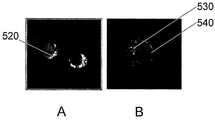

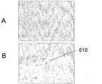

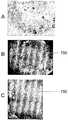

Classifications
- A—HUMAN NECESSITIES
- A61—MEDICAL OR VETERINARY SCIENCE; HYGIENE
- A61K—PREPARATIONS FOR MEDICAL, DENTAL OR TOILETRY PURPOSES
- A61K38/00—Medicinal preparations containing peptides
- A61K38/16—Peptides having more than 20 amino acids; Gastrins; Somatostatins; Melanotropins; Derivatives thereof
- A61K38/17—Peptides having more than 20 amino acids; Gastrins; Somatostatins; Melanotropins; Derivatives thereof from animals; from humans
- A61K38/177—Receptors; Cell surface antigens; Cell surface determinants
- A61K38/1793—Receptors; Cell surface antigens; Cell surface determinants for cytokines; for lymphokines; for interferons
- A—HUMAN NECESSITIES
- A61—MEDICAL OR VETERINARY SCIENCE; HYGIENE
- A61K—PREPARATIONS FOR MEDICAL, DENTAL OR TOILETRY PURPOSES
- A61K9/00—Medicinal preparations characterised by special physical form
- A61K9/0012—Galenical forms characterised by the site of application
- A61K9/0019—Injectable compositions; Intramuscular, intravenous, arterial, subcutaneous administration; Compositions to be administered through the skin in an invasive manner
- A—HUMAN NECESSITIES
- A61—MEDICAL OR VETERINARY SCIENCE; HYGIENE
- A61K—PREPARATIONS FOR MEDICAL, DENTAL OR TOILETRY PURPOSES
- A61K31/00—Medicinal preparations containing organic active ingredients
- A61K31/70—Carbohydrates; Sugars; Derivatives thereof
- A61K31/7088—Compounds having three or more nucleosides or nucleotides
- A61K31/711—Natural deoxyribonucleic acids, i.e. containing only 2'-deoxyriboses attached to adenine, guanine, cytosine or thymine and having 3'-5' phosphodiester links
- A—HUMAN NECESSITIES
- A61—MEDICAL OR VETERINARY SCIENCE; HYGIENE
- A61K—PREPARATIONS FOR MEDICAL, DENTAL OR TOILETRY PURPOSES
- A61K48/00—Medicinal preparations containing genetic material which is inserted into cells of the living body to treat genetic diseases; Gene therapy
- A61K48/0008—Medicinal preparations containing genetic material which is inserted into cells of the living body to treat genetic diseases; Gene therapy characterised by an aspect of the 'non-active' part of the composition delivered, e.g. wherein such 'non-active' part is not delivered simultaneously with the 'active' part of the composition
- A61K48/0025—Medicinal preparations containing genetic material which is inserted into cells of the living body to treat genetic diseases; Gene therapy characterised by an aspect of the 'non-active' part of the composition delivered, e.g. wherein such 'non-active' part is not delivered simultaneously with the 'active' part of the composition wherein the non-active part clearly interacts with the delivered nucleic acid
- A61K48/0041—Medicinal preparations containing genetic material which is inserted into cells of the living body to treat genetic diseases; Gene therapy characterised by an aspect of the 'non-active' part of the composition delivered, e.g. wherein such 'non-active' part is not delivered simultaneously with the 'active' part of the composition wherein the non-active part clearly interacts with the delivered nucleic acid the non-active part being polymeric
- A—HUMAN NECESSITIES
- A61—MEDICAL OR VETERINARY SCIENCE; HYGIENE
- A61K—PREPARATIONS FOR MEDICAL, DENTAL OR TOILETRY PURPOSES
- A61K48/00—Medicinal preparations containing genetic material which is inserted into cells of the living body to treat genetic diseases; Gene therapy
- A61K48/005—Medicinal preparations containing genetic material which is inserted into cells of the living body to treat genetic diseases; Gene therapy characterised by an aspect of the 'active' part of the composition delivered, i.e. the nucleic acid delivered
- A—HUMAN NECESSITIES
- A61—MEDICAL OR VETERINARY SCIENCE; HYGIENE
- A61K—PREPARATIONS FOR MEDICAL, DENTAL OR TOILETRY PURPOSES
- A61K9/00—Medicinal preparations characterised by special physical form
- A61K9/0012—Galenical forms characterised by the site of application
- A61K9/0053—Mouth and digestive tract, i.e. intraoral and peroral administration
- A—HUMAN NECESSITIES
- A61—MEDICAL OR VETERINARY SCIENCE; HYGIENE
- A61K—PREPARATIONS FOR MEDICAL, DENTAL OR TOILETRY PURPOSES
- A61K9/00—Medicinal preparations characterised by special physical form
- A61K9/14—Particulate form, e.g. powders, Processes for size reducing of pure drugs or the resulting products, Pure drug nanoparticles
- A61K9/16—Agglomerates; Granulates; Microbeadlets ; Microspheres; Pellets; Solid products obtained by spray drying, spray freeze drying, spray congealing,(multiple) emulsion solvent evaporation or extraction
- A61K9/1605—Excipients; Inactive ingredients
- A61K9/1629—Organic macromolecular compounds
- A61K9/1652—Polysaccharides, e.g. alginate, cellulose derivatives; Cyclodextrin
- A—HUMAN NECESSITIES
- A61—MEDICAL OR VETERINARY SCIENCE; HYGIENE
- A61K—PREPARATIONS FOR MEDICAL, DENTAL OR TOILETRY PURPOSES
- A61K9/00—Medicinal preparations characterised by special physical form
- A61K9/48—Preparations in capsules, e.g. of gelatin, of chocolate
- A61K9/50—Microcapsules having a gas, liquid or semi-solid filling; Solid microparticles or pellets surrounded by a distinct coating layer, e.g. coated microspheres, coated drug crystals
- A61K9/5005—Wall or coating material
- A61K9/5063—Compounds of unknown constitution, e.g. material from plants or animals
- A61K9/5068—Cell membranes or bacterial membranes enclosing drugs
- A—HUMAN NECESSITIES
- A61—MEDICAL OR VETERINARY SCIENCE; HYGIENE
- A61P—SPECIFIC THERAPEUTIC ACTIVITY OF CHEMICAL COMPOUNDS OR MEDICINAL PREPARATIONS
- A61P1/00—Drugs for disorders of the alimentary tract or the digestive system
- A—HUMAN NECESSITIES
- A61—MEDICAL OR VETERINARY SCIENCE; HYGIENE
- A61P—SPECIFIC THERAPEUTIC ACTIVITY OF CHEMICAL COMPOUNDS OR MEDICINAL PREPARATIONS
- A61P1/00—Drugs for disorders of the alimentary tract or the digestive system
- A61P1/04—Drugs for disorders of the alimentary tract or the digestive system for ulcers, gastritis or reflux esophagitis, e.g. antacids, inhibitors of acid secretion, mucosal protectants
- A—HUMAN NECESSITIES
- A61—MEDICAL OR VETERINARY SCIENCE; HYGIENE
- A61P—SPECIFIC THERAPEUTIC ACTIVITY OF CHEMICAL COMPOUNDS OR MEDICINAL PREPARATIONS
- A61P19/00—Drugs for skeletal disorders
- A—HUMAN NECESSITIES
- A61—MEDICAL OR VETERINARY SCIENCE; HYGIENE
- A61P—SPECIFIC THERAPEUTIC ACTIVITY OF CHEMICAL COMPOUNDS OR MEDICINAL PREPARATIONS
- A61P19/00—Drugs for skeletal disorders
- A61P19/02—Drugs for skeletal disorders for joint disorders, e.g. arthritis, arthrosis
- A—HUMAN NECESSITIES
- A61—MEDICAL OR VETERINARY SCIENCE; HYGIENE
- A61P—SPECIFIC THERAPEUTIC ACTIVITY OF CHEMICAL COMPOUNDS OR MEDICINAL PREPARATIONS
- A61P19/00—Drugs for skeletal disorders
- A61P19/08—Drugs for skeletal disorders for bone diseases, e.g. rachitism, Paget's disease
- A—HUMAN NECESSITIES
- A61—MEDICAL OR VETERINARY SCIENCE; HYGIENE
- A61P—SPECIFIC THERAPEUTIC ACTIVITY OF CHEMICAL COMPOUNDS OR MEDICINAL PREPARATIONS
- A61P19/00—Drugs for skeletal disorders
- A61P19/08—Drugs for skeletal disorders for bone diseases, e.g. rachitism, Paget's disease
- A61P19/10—Drugs for skeletal disorders for bone diseases, e.g. rachitism, Paget's disease for osteoporosis
- A—HUMAN NECESSITIES
- A61—MEDICAL OR VETERINARY SCIENCE; HYGIENE
- A61P—SPECIFIC THERAPEUTIC ACTIVITY OF CHEMICAL COMPOUNDS OR MEDICINAL PREPARATIONS
- A61P25/00—Drugs for disorders of the nervous system
- A—HUMAN NECESSITIES
- A61—MEDICAL OR VETERINARY SCIENCE; HYGIENE
- A61P—SPECIFIC THERAPEUTIC ACTIVITY OF CHEMICAL COMPOUNDS OR MEDICINAL PREPARATIONS
- A61P25/00—Drugs for disorders of the nervous system
- A61P25/02—Drugs for disorders of the nervous system for peripheral neuropathies
- A—HUMAN NECESSITIES
- A61—MEDICAL OR VETERINARY SCIENCE; HYGIENE
- A61P—SPECIFIC THERAPEUTIC ACTIVITY OF CHEMICAL COMPOUNDS OR MEDICINAL PREPARATIONS
- A61P29/00—Non-central analgesic, antipyretic or antiinflammatory agents, e.g. antirheumatic agents; Non-steroidal antiinflammatory drugs [NSAID]
- A—HUMAN NECESSITIES
- A61—MEDICAL OR VETERINARY SCIENCE; HYGIENE
- A61P—SPECIFIC THERAPEUTIC ACTIVITY OF CHEMICAL COMPOUNDS OR MEDICINAL PREPARATIONS
- A61P31/00—Antiinfectives, i.e. antibiotics, antiseptics, chemotherapeutics
- A61P31/12—Antivirals
- A61P31/14—Antivirals for RNA viruses
- A61P31/18—Antivirals for RNA viruses for HIV
- A—HUMAN NECESSITIES
- A61—MEDICAL OR VETERINARY SCIENCE; HYGIENE
- A61P—SPECIFIC THERAPEUTIC ACTIVITY OF CHEMICAL COMPOUNDS OR MEDICINAL PREPARATIONS
- A61P35/00—Antineoplastic agents
- A61P35/04—Antineoplastic agents specific for metastasis
- A—HUMAN NECESSITIES
- A61—MEDICAL OR VETERINARY SCIENCE; HYGIENE
- A61P—SPECIFIC THERAPEUTIC ACTIVITY OF CHEMICAL COMPOUNDS OR MEDICINAL PREPARATIONS
- A61P7/00—Drugs for disorders of the blood or the extracellular fluid
- A61P7/06—Antianaemics
- A—HUMAN NECESSITIES
- A61—MEDICAL OR VETERINARY SCIENCE; HYGIENE
- A61P—SPECIFIC THERAPEUTIC ACTIVITY OF CHEMICAL COMPOUNDS OR MEDICINAL PREPARATIONS
- A61P9/00—Drugs for disorders of the cardiovascular system
- A61P9/10—Drugs for disorders of the cardiovascular system for treating ischaemic or atherosclerotic diseases, e.g. antianginal drugs, coronary vasodilators, drugs for myocardial infarction, retinopathy, cerebrovascula insufficiency, renal arteriosclerosis
- A—HUMAN NECESSITIES
- A61—MEDICAL OR VETERINARY SCIENCE; HYGIENE
- A61K—PREPARATIONS FOR MEDICAL, DENTAL OR TOILETRY PURPOSES
- A61K48/00—Medicinal preparations containing genetic material which is inserted into cells of the living body to treat genetic diseases; Gene therapy
Landscapes
- Health & Medical Sciences (AREA)
- Life Sciences & Earth Sciences (AREA)
- Chemical & Material Sciences (AREA)
- Public Health (AREA)
- Veterinary Medicine (AREA)
- Medicinal Chemistry (AREA)
- Pharmacology & Pharmacy (AREA)
- Animal Behavior & Ethology (AREA)
- General Health & Medical Sciences (AREA)
- Engineering & Computer Science (AREA)
- Bioinformatics & Cheminformatics (AREA)
- Epidemiology (AREA)
- Chemical Kinetics & Catalysis (AREA)
- General Chemical & Material Sciences (AREA)
- Organic Chemistry (AREA)
- Nuclear Medicine, Radiotherapy & Molecular Imaging (AREA)
- Molecular Biology (AREA)
- Physical Education & Sports Medicine (AREA)
- Rheumatology (AREA)
- Genetics & Genomics (AREA)
- Biotechnology (AREA)
- Virology (AREA)
- Biochemistry (AREA)
- Orthopedic Medicine & Surgery (AREA)
- Cell Biology (AREA)
- Zoology (AREA)
- Immunology (AREA)
- Biophysics (AREA)
- Physiology (AREA)
- Neurosurgery (AREA)
- Dermatology (AREA)
- Biomedical Technology (AREA)
- Neurology (AREA)
- Nutrition Science (AREA)
- Oncology (AREA)
- Botany (AREA)
- Gastroenterology & Hepatology (AREA)
- Proteomics, Peptides & Aminoacids (AREA)
- Tropical Medicine & Parasitology (AREA)
- Diabetes (AREA)
Abstract
Description
Translated fromJapanese発明の分野
本発明は、低骨密度の治療のための組成物および方法に関する。本発明はまた、オステオプロテジェリン依存性病状の治療のための組成物および方法にも関する。The present invention relates to compositions and methods for the treatment of low bone density. The invention also relates to compositions and methods for the treatment of osteoprotegerin-dependent conditions.
発明の背景
骨の健康および骨粗鬆症に関する米国公衆衛生総監報告(Surgeon General's Report on Bone Health and Osteoporosis)は、2020年には年齢50歳以上の米国人のおよそ半数が骨粗鬆症を有する、またはそれを発症するリスクがあると推定している。米国骨粗鬆症基金(National Osteoporosis Foundation)は、米国には骨粗鬆症を現在有する症例が約1000万人おり、うち800万人が女性、200万人が男性であり、さらに3400万人の米国人が低骨量のために骨粗鬆症の深刻なリスクを有すると推定している。骨粗鬆症は年間150万件を上回る骨折の原因となっており、これには300,000件を上回る股関節部骨折;およそ700,000件の脊椎骨折;250,000件の手関節骨折;および300,000件の他部位の骨折が含まれる。骨粗鬆症性の股関節部骨折に対する米国全体での直接的費用の推定値(病院およびナーシングホーム)は、2002年に180億ドルであった。股関節部骨折の患者は今後さらに別の骨折を経験する可能性が非常に高い。これらの統計の背後にある生活の質の低下の大きさは筆舌に尽くしがたい。さらに、急性および部位特異的な低骨密度病状には、炎症媒介性骨溶解、腫瘍誘発性骨溶解、人工インプラントの弛み、歯周病(periodonitis)または骨関節炎が含まれる。Background of the invention The Surgeon General's Report on Bone Health and Osteoporosis states that in 2020, approximately half of Americans age 50 and older have or will develop osteoporosis Estimated to be at risk. The National Osteoporosis Foundation has about 10 million cases of osteoporosis currently in the United States, of which 8 million are women, 2 million are men, and another 34 million Americans are low bone Estimated to have a serious risk of osteoporosis because of the amount. Osteoporosis causes more than 1.5 million fractures annually, including more than 300,000 hip fractures; approximately 700,000 vertebral fractures; 250,000 wrist fractures; and 300,000 other fractures included. Estimated direct costs (hospital and nursing home) across the United States for osteoporotic hip fractures (hospital and nursing home) were $ 18 billion in 2002. Patients with hip fractures are very likely to experience additional fractures in the future. The magnitude of the decline in quality of life behind these statistics is hard to beat. In addition, acute and site-specific low bone density pathologies include inflammation-mediated osteolysis, tumor-induced osteolysis, prosthetic implant slack, periodonitis or osteoarthritis.
骨ホメオスタシスの細胞生物学
骨は、骨格内で絶え間ないリモデリング(吸収および置換)を受ける動的な組織である。骨格の維持を担う主要な細胞種は、吸収性の破骨細胞および骨合成性の骨芽細胞であり、それらはどちらもホルモン、増殖因子および炎症メディエーターによって影響される。間葉幹細胞由来の骨芽細胞による骨形成、ならびに単球/マクロファージ系列の造血前駆細胞から生じる破骨細胞によるそのモデリングおよびリモデリングは、1つの密接に調節された系である(Huang, W., et al., A rapid multiparameter approach to study factors that regulate osteoclastogenesis: Demonstration of the combinatorial dominant effects of TNF-a and TGF-β in RANKL-mediated osteoclastogenesis. Calcif. Tissue Int. 73:584-593 (2003)。正常骨量の維持は、局所性および全身性の両方の因子およびシグナルがかかわる、形成と吸収との間のホメオスタシス的な複雑な均衡に依存する。これらの2つのプロセス間に不均衡がある場合には、骨密度の上昇(大理石骨病)または低下(骨粗鬆症)が起こる。慢性の低骨密度は閉経後、加齢性および炎症性の疾患でみられ、一方、急性の低骨密度は人工関節(prosthesis)の弛みまたは腫瘍誘発性骨溶解において観察される。Bone Homeostasis Cell Biology Bone is a dynamic tissue that undergoes constant remodeling (resorption and replacement) within the skeleton. The main cell types responsible for skeletal maintenance are resorbable osteoclasts and osteosynthetic osteoblasts, both of which are affected by hormones, growth factors and inflammatory mediators. Bone formation by mesenchymal stem cell-derived osteoblasts and its modeling and remodeling by osteoclasts arising from hematopoietic progenitors of the monocyte / macrophage lineage are one closely regulated system (Huang, W. , et al., A rapid multiparameter approach to study factors that regulate osteoclastogenesis: Demonstration of the combinatorial dominant effects of TNF-a and TGF-β in RANKL-mediated osteoclastogenesis. Calcif. Tissue Int. 73: 584-593 (2003). The maintenance of normal bone mass depends on a complex homeostasis balance between formation and resorption involving both local and systemic factors and signals, where there is an imbalance between these two processes Causes increased bone density (marble bone disease) or decreased (osteoporosis), chronic low bone density is seen in post-menopausal age-related and inflammatory diseases, while acute low bone density is artificial joint It is observed in slack or tumor-induced osteolysis of prosthesis).
破骨細胞
破骨細胞は、特定のシグナルに応答して、単球/マクロファージ系列F4-80陽性細胞の造血前駆細胞から生じる(Boyle, W. J., et al., Osteoclast differentiation and activation. Nature 423, 337-342 (2003))。これが起こるためには、コロニー刺激因子-1(CSF-1;M-CSF)およびTNFスーパーファミリーのメンバーであるRANKL(NF-κBリガンドの受容体活性化因子;TRANCE、OPGLおよびODFとも呼ばれる)という2つの増殖因子が必要である。図1は、破骨細胞の分化に関与するシグナル伝達機構の概略図であり、ここでRANKLはその受容体であるRANKを活性化することによって破骨細胞の分化を活性化し、一方、オステオプロテジェリン(OPG、これは破骨細胞形成抑制因子としても知られる)は、RANKLを、細胞表面に対するその結合を阻止することで封鎖状態にする。Osteoclasts Osteoclasts arise from hematopoietic progenitors of monocyte / macrophage lineage F4-80 positive cells in response to specific signals (Boyle, WJ, et al., Osteoclast differentiation and activation. Nature 423, 337 -342 (2003)). For this to happen, it is called colony-stimulating factor-1 (CSF-1; M-CSF) and RANKL, a member of the TNF superfamily (also known as receptor activator of NF-κB ligand; TRANCE, OPGL and ODF) Two growth factors are required. Fig. 1 is a schematic diagram of the signal transduction mechanism involved in osteoclast differentiation, where RANKL activates osteoclast differentiation by activating its receptor RANK, while osteoprotease Phosphorus (OPG, also known as osteoclastogenesis inhibitor) blocks RANKL by blocking its binding to the cell surface.
骨格では、CSF-1およびRANKLがいずれも骨芽細胞によって供給されるが、さらなる細胞性供給源が他の組織にある。CSF-1はその受容体である原癌遺伝子c-Fmsを介して前破骨細胞と結合して、RANKL受容体であるRANKの発現を刺激し、RANKLに対する応答性をそれらの細胞に付与する。RANKの活性化は、RANK関連因子であるTRAF6を介してNF-κB依存性遺伝子の発現を刺激する上、Junキナーゼ経路およびホスホイノシトール経路も活性化する。これらの経路は一緒にアポトーシスを阻害するほか、破骨細胞に骨を吸収する準備をさせる他の細胞応答を宿主に惹起させる。これらには、吸収の部位へのケモカイン誘発型走化性、多核細胞を生じさせる細胞融合、αvβインテグリンを介した骨への付着を促進させる特化したアクチンリングの形成、骨基質を溶解するためのプロテアーゼおよびプロトンポンプの発現、ならびに、分解性分子を分泌するためならびに溶解した骨基質を摂取および輸送するための極めて活発な小胞輸送の発達が含まれる。RANKLは、これらの複数の分化段階の上流で作用してインビボでのそれらの分化を阻害する、破骨細胞活性を調節するための魅力的で可能性のある標的である。 In the skeleton, both CSF-1 and RANKL are supplied by osteoblasts, but there are additional cellular sources in other tissues. CSF-1 binds to pre-osteoclasts through its receptor proto-oncogene c-Fms, stimulates the expression of RANK, a RANKL receptor, and confers responsiveness to RANKL on these cells . Activation of RANK stimulates the expression of NF-κB-dependent genes through TRAF6, a RANK-related factor, and also activates the Jun kinase pathway and the phosphoinositol pathway. Together, these pathways inhibit apoptosis and elicit other cellular responses in the host that prepare osteoclasts to resorb bone. These include chemokine-induced chemotaxis to the site of resorption, cell fusion that gives rise to multinucleated cells, formation of specialized actin rings that promote attachment to bone via αvβ integrins, to dissolve bone matrix And the development of a highly active vesicle transport to secrete degradable molecules and to ingest and transport dissolved bone matrix. RANKL is an attractive and potential target for modulating osteoclast activity that acts upstream of these multiple stages of differentiation to inhibit their differentiation in vivo.
RANKL/RANK/OPG経路のシグナル伝達は、内皮細胞の生存、血管新生、単球または内皮細胞の動員、ならびに平滑筋細胞の骨形成および石灰化に関して、血管の生理現象および病理現象においても重要な可能性がある。諸研究の結果は、RANKLは加齢、骨粗鬆症または疾患に伴う骨塩形成(bone mineralization)の低下と同時に起こる血管の石灰化を促進しうるが、OPGはそれを防御しうると考えられることを示唆している(Collin-Osdoby, P., Regulation of vascular calcification by osteoclast regulatory factors RANKL and osteoprotegerin. Circulation Res. 2004 95(11):1046-1057)。 RANKL / RANK / OPG pathway signaling is also important in vascular physiology and pathological phenomena with respect to endothelial cell survival, angiogenesis, monocyte or endothelial cell recruitment, and smooth muscle cell bone formation and mineralization there is a possibility. The results of studies have shown that RANKL can promote vascular calcification that coincides with bone mineralization reduction associated with aging, osteoporosis or disease, but OPG is thought to be able to protect it. (Collin-Osdoby, P., Regulation of vascular calcification by osteoclast regulatory factors RANKL and osteoprotegerin. Circulation Res. 2004 95 (11): 1046-1057).
オステオプロテジェリン
OPGは腫瘍壊死因子受容体(TNFR)スーパーファミリーのメンバーであり、約60kDの単量体形態およびジスルフィド結合した約120kDのホモ二量体形態として存在する分泌性で塩基性の401アミノ酸の糖タンパク質である。OPGは骨芽細胞および骨髄ストロマ細胞によって産生される。OPGは、RANKLがRANKと結合するのを妨げる可溶性「デコイ」受容体として機能することにより、破骨細胞形成を用量依存的な様式で阻害する(図1)。Schoppet, M., et al., RANK ligand and osteoprotegerin: paracrine regulators of bone metabolism and vascular function, Arterioscier Thromb Vasc Biol. 2002 Apr 1;22(4):549-53を参照。オステオプロテジェリンは1997年にSimonetらによって報告され、彼らはそれをインビトロおよびインビボで防御的な骨作用を有する腫瘍壊死因子受容体(TNFR)スーパーファミリーの分泌性メンバーとして同定し、特徴決定した(Simonet, W. S., et al. Osteoprotegerin: A Novel Secreted Protein Involved in the Regulation of Bone Density. Cell 1997 89, 309-319)。組換えOPGタンパク質の静脈内注射、およびOPG(-/-)マウスにおけるOPGのトランスジェニック性過剰発現はいずれも、OPG欠損マウスで観察される骨粗鬆症性の骨表現型を効果的に救済する。Min, H., et al., Osteoprotegerin reverses osteoporosis by inhibiting endosteal osteoclasts and prevents vascular calcification by blocking a process resembling osteoclastogenesis. J Exp Med 2000 192, 463-474を参照。Osteoprotegerin
OPG is a member of the Tumor Necrosis Factor Receptor (TNFR) superfamily, a secreted, basic 401 amino acid glycoprotein that exists as a monomeric form of approximately 60 kD and a homodimeric form of approximately 120 kD disulfide bonded It is. OPG is produced by osteoblasts and bone marrow stromal cells. OPG inhibits osteoclast formation in a dose-dependent manner by functioning as a soluble “decoy” receptor that prevents RANKL from binding to RANK (FIG. 1). See Schoppet, M., et al., RANK ligand and osteoprotegerin: paracrine regulators of bone metabolism and vascular function, Arterioscier Thromb Vasc Biol. 2002 Apr 1; 22 (4): 549-53. Osteoprotegerin was reported by Simonet et al. In 1997, who identified and characterized it as a secreted member of the tumor necrosis factor receptor (TNFR) superfamily with protective bone action in vitro and in vivo ( Simonet, WS, et al. Osteoprotegerin: A Novel Secreted Protein Involved in the Regulation of Bone Density. Cell 1997 89, 309-319). Both intravenous injection of recombinant OPG protein and transgenic overexpression of OPG in OPG (− / −) mice effectively rescue the osteoporotic bone phenotype observed in OPG-deficient mice. Min, H., et al., Osteoprotegerin reverses osteoporosis by inhibiting endosteal osteoclasts and prevents vascular calcification by blocking a process resembling osteoclastogenesis. See J Exp Med 2000 192, 463-474.
トランスジェニックマウスにおけるOPGの過剰発現は、おそらくはRANKL/RANK相互作用を阻止することにより、骨格質量の増加ならびに破骨細胞の数および活性の低減をもたらすことが実証されており(Simonet, W. S., et al.(1997))、一方、OPGの欠損は骨粗鬆症をもたらす。Bucay, N., et al., Osteoprotegerin-deficient mice develop early onset osteoporosis and arterial calcification. Genes Dev 1998 12, 1260-1268、およびMizuno, A., et al,. Severe osteoporosis in mice lacking osteoclastogenesis inhibitory factor/osteoprotegerin. Biochem Biophys Res Commun 1998 247, 610-615を参照。 Overexpression of OPG in transgenic mice has been demonstrated to result in increased skeletal mass and decreased osteoclast number and activity, presumably by blocking the RANKL / RANK interaction (Simonet, WS, et al. (1997)), whereas OPG deficiency results in osteoporosis. Bucay, N., et al., Osteoprotegerin-deficient mice develop early onset osteoporosis and arterial calcification. Genes Dev 1998 12, 1260-1268 and Mizuno, A., et al ,. Severe osteoporosis in mice lacking osteoclastogenesis inhibitory factor / osteoprotegerin See Biochem Biophys Res Commun 1998 247, 610-615.
骨粗鬆症に加えて、人工関節周囲(periprosthetic)骨溶解(Yang, S.Y., et al., Adeno-associated virus-mediated osteoprotegerin gene transfer protects against particulate polyethylene-induced osteolysis in a murine model, Arthritis Rheum. 2002 Sep; 46(9):2514-23)、腫瘍転移、若年性パジェット病、ゴーシェ病、HIVの抗ウイルス治療、廃用性骨減少症、サラセミアおよび炎症性腸疾患に伴う骨溶解を含むいくつかの他の骨病状も、骨量の損失を伴う。 In addition to osteoporosis, periprosthetic osteolysis (Yang, SY, et al., Adeno-associated virus-mediated osteoprotegerin gene transfer protects against particulate polyethylene-induced osteolysis in a murine model, Arthritis Rheum. 2002 Sep; 46 (9): 2514-23), tumor metastasis, juvenile Paget's disease, Gaucher disease, antiviral treatment of HIV, disuse osteopenia, thalassemia and osteolysis associated with inflammatory bowel disease Bone pathology also involves loss of bone mass.
骨癌性疼痛は、乳房、前立腺または肺で生じた腫瘍が長骨、脊椎骨および/または骨盤に転移した場合に起こることが最も多い。骨にかかわる原発性および転移性の癌は、米国だけでも年間400,000例の新たな癌症例を占め、進行性の乳癌または前立腺癌の患者の70%超は骨格転移を有する。骨痛の動物モデルにおける諸研究で報告されている結果は、オステオプロテジェリン治療がそれ以上の破壊を停止させ、進行中および運動誘発性の疼痛を軽減し、脊髄の神経化学的再組織化のいくつかの局面を元の方向に戻させることを示している。Luger, N.M., et al., Osteoprotegerin diminishes advanced bone cancer pain, Cancer Res. 2001 May 15; 61 (10):4038-47を参照。 Bone cancer pain most often occurs when a tumor arising in the breast, prostate, or lung has metastasized to the long bones, vertebrae, and / or pelvis. Primary and metastatic cancers involving bone account for 400,000 new cancer cases annually in the United States alone, with more than 70% of patients with advanced or prostate cancer having skeletal metastases. Results reported in studies in animal models of bone pain show that osteoprotegerin treatment stops further destruction, reduces ongoing and exercise-induced pain, and neurochemical reorganization of the spinal cord Some aspects are shown to return to their original direction. See Luger, N.M., et al., Osteoprotegerin diminishes advanced bone cancer pain, Cancer Res. 2001 May 15; 61 (10): 4038-47.
低骨密度を是正するための治療アプローチは、骨吸収の阻害か骨形成の刺激を目標としている。低骨密度の治療および予防に対して現在承認されている薬剤の大半、すなわちビスフォスフォネート、エストロゲン、サケカルシトニンおよび選択的エストロゲン受容体モジュレーターであるラロキシフェンは、破骨細胞性の骨吸収を阻害することによって骨量を増加させるように作用するが、以下に考察するように、最近では、骨形成タンパク質(Bone morphogenic protein)およびスタチン系薬剤を含む同化促進薬剤(anabolic drug)の探索への関心が急速に高まっている。エストロゲンまたは副甲状腺ホルモンを含むホルモン療法を用いることもできる。これらの治療法は臨床的に使用されてきており、ビスフォスフォネートおよびエストロゲンの場合には数十年にわたるが、老年集団において幅広く骨粗鬆症が存続していることを考慮すると、それらの効能が限定的なことは明白である。 Therapeutic approaches to correct low bone density are aimed at inhibiting bone resorption or stimulating bone formation. Most of the drugs currently approved for the treatment and prevention of low bone density, namely bisphosphonates, estrogens, salmon calcitonin and selective estrogen receptor modulators, raloxifene, inhibits osteoclastic bone resorption However, as discussed below, recently, interest in the search for anabolic drugs including bone morphogenic proteins and statins Is growing rapidly. Hormone therapy including estrogen or parathyroid hormone can also be used. These therapies have been used clinically for bisphosphonates and estrogens for decades, but their efficacy is limited given the widespread existence of osteoporosis in the aging population The obvious thing is obvious.
アレンドロネート、リセドロネート、イバンドロン酸などのビスフォスフォネートは、ヒドロキシアパタイト骨塩中に組み込まれ、以下を含む複数の機序によって骨吸収を阻害するように作用する:a)破骨細胞の骨付着と干渉する、b)破骨細胞前駆細胞の分化を阻害する、およびc)破骨細胞による選択的な取込みの後に破骨細胞機能を阻害する。エストロゲンは、閉経後女性における骨粗鬆症の治療のために長年にわたって広く用いられている。エストロゲンの作用の機序については依然としてさらに調査を要するが、諸研究により、閉経後女性におけるエストロゲン補充が骨格リモデリングを低下させるとともにその損失を減じさせ、さらには骨梁骨量および皮質骨量の両方を増加させることさえできることが示されている。閉経後女性における骨密度に対するエストロゲン療法の有益な効果にもかかわらず、その使用は乳癌および子宮癌のリスク増大を伴い、出血、乳房圧痛および鼓腸を引き起こす。選択的エストロゲン受容体モジュレーターは、エストロゲン療法の合併症およびリスクを理由として、骨粗鬆症の治療のために開発された。エストロゲンと同様に、タモキシフェンおよびラロキシフェンなどのSERMは、骨ではエストロゲン受容体に対するアゴニストであるが、乳房組織ではエストロゲン受容体のアンタゴニストである。 Bisphosphonates such as alendronate, risedronate, and ibandronate are incorporated into hydroxyapatite bone salt and act to inhibit bone resorption by several mechanisms, including: a) osteoclast bone Interfere with adhesion, b) inhibit osteoclast precursor differentiation, and c) inhibit osteoclast function after selective uptake by osteoclasts. Estrogens have been widely used for many years for the treatment of osteoporosis in postmenopausal women. The mechanism of action of estrogen still needs further investigation, but studies have shown that estrogen supplementation in postmenopausal women reduces and reduces skeletal remodeling, as well as trabecular and cortical bone mass. It has been shown that both can even be increased. Despite the beneficial effects of estrogen therapy on bone density in postmenopausal women, its use is associated with an increased risk of breast and uterine cancer, causing bleeding, breast tenderness and flatulence. Selective estrogen receptor modulators have been developed for the treatment of osteoporosis because of the complications and risks of estrogen therapy. Like estrogen, SERMs such as tamoxifen and raloxifene are agonists for the estrogen receptor in bone but are antagonists of the estrogen receptor in breast tissue.
いくつかの臨床試験により、鼻腔内サケカルシトニン療法が骨量損失を予防し、脊椎骨折の比率を低下させるために有効であることが示されている。サケカルシトニンは、骨粗鬆症および骨折既往のある閉経後女性において脊椎骨折のリスクを36%低下させ、その安全性プロファイルは長期使用を通じてプラセボと同等であることが示されている。サケカルシトニンは忍容性が良好であり、骨折の場合にはある程度の鎮痛をもたらし、ホルモン療法に代わる妥当な選択肢である。 Several clinical trials have shown that intranasal salmon calcitonin therapy is effective in preventing bone loss and reducing the rate of vertebral fractures. Salmon calcitonin has been shown to reduce the risk of vertebral fractures by 36% in postmenopausal women with a history of osteoporosis and fractures, and its safety profile has been shown to be comparable to placebo through long-term use. Salmon calcitonin is well tolerated and provides some analgesia in the case of fractures and is a reasonable alternative to hormone therapy.
RANKL/OPG/RANK経路の発見は、改良された抗吸収療法を開発するための新たな機会を切り開いた。上述したように、トランスジェニックマウスにおけるOPGの恒常的な過剰発現は軽症の大理石骨病を招いており、OPG-/-マウスは重症の骨粗鬆症である。OPGのトランスジェニック性過剰発現はノックアウト表現型を救済する(Min, H., et al., 2000)。破骨細胞性の骨吸収を低下させるための、直接的なRANKL阻害または可溶性OPGのレベルを高めることのいずれかによるRANKL経路の阻害は、骨粗鬆症治療に対する有望な方法論である。 The discovery of the RANKL / OPG / RANK pathway has opened up new opportunities for developing improved anti-absorption therapies. As described above, constant overexpression of OPG in transgenic mice leads to mild marble bone disease, and OPG − / − mice have severe osteoporosis. Transgenic overexpression of OPG rescues the knockout phenotype (Min, H., et al., 2000). Inhibition of the RANKL pathway, either by direct RANKL inhibition or by increasing the level of soluble OPG, to reduce osteoclast bone resorption is a promising methodology for the treatment of osteoporosis.
骨形成タンパク質
最近、同化促進アプローチ、例えば、骨形成タンパク質(BMP)の、または副甲状腺ホルモンのパルス状IV注入投与の使用への関心が急速に高まっている。これらの治療戦略は大いに有望であり、BMPの初期評価は期待の持てるものである。しかし、これらの治療アプローチに、骨の過成長、骨棘、異所性骨、血管石灰化またはさらには新生物のリスクがないわけではない。Bone morphogenetic proteins Recently there has been a rapid increase in interest in the use of anabolic-promoting approaches such as the administration of pulsed IV infusions of bone morphogenetic protein (BMP) or parathyroid hormone. These treatment strategies are very promising and the initial evaluation of BMP is promising. However, these therapeutic approaches are not without the risk of bone overgrowth, osteophytes, ectopic bone, vascular calcification or even neoplasm.
モノクローナル抗体
最近の1件の小規模な臨床試験では、RANKLに対するヒトモノクローナル抗体の単回注射が、骨代謝回転マーカーを最長6カ月間にわたって低下させることが示されている(Bekker, P. J., et al., A single-dose placebo-controlled study of AMG 162, a fully human monoclonal antibody to RANKL, in postmenopausal women. J Bone Miner Res 2004 19, 1059-1066)。ヒトモノクローナル坑腫瘍壊死因子(TNF)抗体であるアダリムマブ(Humira(登録商標))は、関節リウマチの症状および徴候を効果的に軽減し、放射線検査で認められるびらん性関節変化の進行を予防する。Monoclonal antibodies A recent small clinical trial has shown that a single injection of a human monoclonal antibody against RANKL reduces bone turnover markers for up to 6 months (Bekker, PJ, et al , A single-dose placebo-controlled study of AMG 162, a fully human monoclonal antibody to RANKL, in postmenopausal women. J Bone Miner Res 2004 19, 1059-1066). Adalimumab (Humira®), a human monoclonal antitumor necrosis factor (TNF) antibody, effectively reduces the symptoms and signs of rheumatoid arthritis and prevents the progression of erosive joint changes seen on radiography.
スタチン
大部分が男性の退役軍人である大規模コホートの1件の試験では、スタチンの使用に伴って、脂質低下療法を行わない場合と比較して骨折リスクの36%の低下がみられ、他の脂質低下治療法と比較した場合には32%のリスク低下がみられた。スタチン系薬剤と、微小血管機能の改善を介した炎症軽減および新骨成長の促進を含む骨の健康状態との間の関連を説明するために、いくつかの生物学的機序が提唱されている(Scranton, R.E. (2005). Statin use and fracture risk: study of a US veterans population. Arch. Intern. Med. 165: 2007-2012)。Statins In one trial of a large cohort, mostly male veterans, there was a 36% reduction in fracture risk with statin use compared to no lipid-lowering therapy, others There was a 32% risk reduction when compared to other lipid-lowering treatments. Several biological mechanisms have been proposed to explain the link between statin drugs and bone health, including reducing inflammation through promoting microvascular function and promoting new bone growth (Scranton, RE (2005). Statin use and fracture risk: study of a US veterans population. Arch. Intern. Med. 165: 2007-2012).
ウイルスベクターを用いたオステオプロテジェリン遺伝子治療
オステオプロテジェリンは破骨細胞の形成、機能および生存の阻害によって骨吸収を防止するタンパク質であり、これらの活性のために、組換えOPGは、骨粗鬆症などの慢性的な骨吸収疾患の治療のための魅力ある薬剤候補となっている。遺伝子治療は、抗吸収性タンパク質の遺伝子を送達することによって長期的治療を実現する可能性がある。OPGはアデノ随伴ウイルスおよびアデノウイルスにより、いずれもOPGのみまたはIgG Fc鎖との融合タンパク質をコードするDNAとして送達されている。rAAV-OPG-IRES-EGFPのインビボ投与は、注射部位での筋細胞の検出可能な形質導入および注射2日後の血清OPG発現レベルの有意な増加をもたらし、骨折リモデリングの低下も伴うが、治癒中の骨折の構造強度に対してはほとんど影響を及ぼさなかった。Osteoprotegerin gene therapy using viral vectors Osteoprotegerin is a protein that prevents bone resorption by inhibiting osteoclast formation, function and survival, and because of these activities, recombinant OPG is It has become an attractive drug candidate for the treatment of chronic bone resorption disease. Gene therapy may achieve long-term treatment by delivering genes for anti-absorbable proteins. OPG is delivered by adeno-associated virus and adenovirus both as DNA encoding OPG alone or a fusion protein with IgG Fc chain. In vivo administration of rAAV-OPG-IRES-EGFP resulted in detectable transduction of myocytes at the injection site and a significant increase in serum OPG expression level 2 days after injection, with reduced fracture remodeling but healing There was little effect on the structural strength of the middle fracture.
抽出酵母細胞壁粒子は、容易に入手可能な、生分解性の、直径約2〜4μmである、実質的に球状の粒子である。抽出酵母細胞壁粒子の調製は当技術分野で公知であり、例えば、米国特許第4,992,540号;第5,082,936号;第5,028,703号;第5,032,401号;第5,322,841号;第5,401,727号;第5,504,079号;第5,968,811号;第6,444,448 B1号;第6,476,003 B1号;公開米国出願第2003/0216346 A1号、第2004/0014715 A1号およびPCT公開出願WO 02/12348 A2号に記載されている。「全グルカン粒子」と称する抽出酵母細胞壁粒子の一形態が送達媒体として提案されているが、粒子からの有効成分の単純拡散による放出、または粒子マトリックスの生分解による全グルカン粒子と化学的に架橋された作用物質の放出のいずれも限定的である。米国特許第5,032,401号および第5,607,677号を参照。これらの限界を克服する、改良された酵母細胞壁薬物送達系が、米国公開特許出願第US2005281781号および公開PCT国際特許出願第WO2006007372 A3号に開示されている。「酵母細胞壁粒子」(YCWP)には、酵母グルカン粒子(YGP)および酵母グルカン-マンナン粒子(YGMP)が含まれる。 Extracted yeast cell wall particles are readily available, biodegradable, substantially spherical particles having a diameter of about 2-4 μm. The preparation of extracted yeast cell wall particles is known in the art, for example, U.S. Pat. Nos. 4,992,540; 5,082,936; 5,028,703; 5,032,401; 5,322,841; 5,401,727; 6,444,448 B1; 6,476,003 B1; published US applications 2003/0216346 A1, 2004/0014715 A1 and PCT published application WO 02/12348 A2. A form of extracted yeast cell wall particles called “total glucan particles” has been proposed as a delivery vehicle, but it is chemically cross-linked with all glucan particles by release of the active ingredient from the particles by simple diffusion or by biodegradation of the particle matrix. Any release of the active agent is limited. See U.S. Pat. Nos. 5,032,401 and 5,607,677. An improved yeast cell wall drug delivery system that overcomes these limitations is disclosed in US published patent application US2005281781 and published PCT international patent application WO2006007372 A3. “Yeast cell wall particles” (YCWP) include yeast glucan particles (YGP) and yeast glucan-mannan particles (YGMP).
GI免疫系のもう1つの重要な構成要素は、M細胞またはミクロフォールド細胞である。M細胞は、種々のタンパク質およびペプチド抗原のエンドサイトーシスを行う、腸管上皮内のリンパ濾胞上にある特殊な細胞種である。これらのタンパク質を消化する代わりに、M細胞はそれらを下方の組織に輸送し、そこでそれらは局所樹状細胞細胞およびマクロファージによって取り込まれる。 Another important component of the GI immune system is M cells or microfold cells. M cells are a specialized cell type on lymphoid follicles in the intestinal epithelium that perform endocytosis of various protein and peptide antigens. Instead of digesting these proteins, M cells transport them to the underlying tissue where they are taken up by local dendritic cell and macrophages.
M細胞は、分子および粒子を消化管の内腔からエンドサイトーシスまたは食作用によって取り込む。この物質は続いて細胞の内部を通って小胞内から基底細胞膜に輸送され、そこで細胞外空間に放出される。この過程はトランスサイトーシスとして知られている。その基底表面で、M細胞の細胞膜は下方のリンパ球および抗原提示細胞の周りで広範囲にわたって折りたたまれており、それらがM細胞から放出された輸送材料を取り込み、抗原提示のためにそれを処理する。 M cells take up molecules and particles from the lumen of the digestive tract by endocytosis or phagocytosis. This substance is then transported through the interior of the cell from within the vesicle to the basal cell membrane where it is released into the extracellular space. This process is known as transcytosis. At its basal surface, the cell membrane of M cells is extensively folded around the underlying lymphocytes and antigen-presenting cells, which take up transport material released from M cells and process it for antigen presentation .
ある研究により、パイエル板のM細胞による酵母粒子(直径3.4+/-0.8ミクロン)のトランスサイトーシスには1時間未満しか要しないことが示されている(Beier, R., & Gebert, A., Kinetics of particle uptake in the domes of Peyer's patches, Am J Physiol. 1998 Jul; 275(1 Pt 1):G130-7)。上皮内マクロファージによる明らかな食作用がないと、酵母粒子は下方に移動して2.5〜4時間以内に基底膜を通過し、そこでそれらは急速に食作用を受けてパイエル板ドームの外に輸送される。ヒト鼻咽頭リンパ組織(扁桃および咽頭扁桃)に認められるM細胞は、呼吸器感染症を引き起こすウイルスの捕集に関与することが示されている。インビトロのM細胞モデルの研究により、蛍光標識されたミクロスフェア(Fluosphere、0.2μm)およびキトサン微粒子(0.2μm)の取込みが示されている(van der Lubben I.M., et al., Transport of chitosan microparticles for mucosal vaccine delivery in a human intestinal M-cell model, J Drug Target, 2002 Sep;10(6):449-56)。レクチンの一つであるハリエニシダ凝集素1(Ulex europaeus agglutinin 1;UEA1、α-L-フコース残基に特異的)は、ポリスチレンミクロスフェア(0.5μm)または重合リポソームをM細胞(0.2μm)へと向かわせるために用いられている(Clark, M.A., et al., Targeting polymerised liposome vaccine carriers to intestinal M cells, Vaccine, 2001 Oct 12;20(12):208-17)。マウスにおけるインビボ試験では、ポリ-D,L-乳酸(PDLLA)ミクロスフェアまたはゼラチンミクロスフェア(GM)がマクロファージおよびM細胞によって効率的に取り込まれうることが報告されている(Nakase, H., et al., Biodegradable microspheres targeting mucosal immune-regulating cells: new approach for treatment of inflammatory bowel disease, J Gastroenterol. 2003 Mar;38 Suppl 15:59-62)。 One study shows that transcytosis of yeast particles (3.4 +/- 0.8 microns in diameter) by Peyer's patch M cells takes less than an hour (Beier, R., & Gebert, A. , Kinetics of particle uptake in the domes of Peyer's patches, Am J Physiol. 1998 Jul; 275 (1 Pt 1): G130-7). Without apparent phagocytosis by intraepithelial macrophages, the yeast particles migrate downward and pass through the basement membrane within 2.5-4 hours, where they are rapidly phagocytosed and transported out of the Peyer's patch dome. The M cells found in human nasopharyngeal lymphoid tissues (tonsils and tonsils) have been shown to be involved in the collection of viruses that cause respiratory infections. In vitro M cell model studies have shown uptake of fluorescently labeled microspheres (Fluosphere, 0.2 μm) and chitosan microparticles (0.2 μm) (van der Lubben IM, et al., Transport of chitosan microparticles for mucosal vaccine delivery in a human intestinal M-cell model, J Drug Target, 2002 Sep; 10 (6): 449-56). One of the lectins, Ulex europaeus agglutinin 1 (UEA1, specific for α-L-fucose residues) is used to convert polystyrene microspheres (0.5 μm) or polymerized liposomes into M cells (0.2 μm). (Clark, MA, et al., Targeting polymerised liposome vaccine carriers to intestinal M cells, Vaccine, 2001
しかし、ポリ(DL-ラクチド-co-グリコリド)微粒子およびリポソームを含む合成微粒子(particulate)送達媒体の取込みはばらつきが大きく、それは粒子およびM細胞の両方の物理的性質によって決まることが報告されている。Clark, M.A., et al., Exploiting M cells for drug and vaccine delivery, Adv Drug Deliv Rev. 2001 Aug 23;50(1-2):81-106。同じ研究では、粒子またはリポソームを、M細胞の表面と選択的に結合する適切なレクチン、微生物アドヘシンおよび免疫グロブリンでコーティングすることによって、送達性を向上させうることも報告されている。同じく、Florence, A.T., The oral absorption of micro- and nanoparticulates: neither exceptional nor unusual, Pharm Res. 1997 Mar; 14(3):259-66も参照のこと。 However, the uptake of synthetic delivery media including poly (DL-lactide-co-glycolide) microparticles and liposomes is highly variable, which is reported to depend on the physical properties of both the particles and M cells . Clark, M.A., et al., Exploiting M cells for drug and vaccine delivery, Adv Drug Deliv Rev. 2001 Aug 23; 50 (1-2): 81-106. The same study reports that delivery can be improved by coating particles or liposomes with appropriate lectins, microbial adhesins and immunoglobulins that selectively bind to the surface of M cells. See also Florence, A.T., The oral absorption of micro- and nanoparticulates: neither exceptional nor unusual, Pharm Res. 1997 Mar; 14 (3): 259-66.
病原体パターン認識受容体(PRR)は、微生物表面に存在する共通の構造モチーフおよび分子モチーフを認識し、先天性免疫応答の誘導に寄与する。マンノース受容体およびβグルカン受容体は真菌病原体の認識に部分的に関与する。マクロファージのサブセット上に発現される糖質結合受容体の一つであるマンノース受容体(MR)は、そのようなPRRの一つと考えられている。マクロファージは、マンノースおよびマンノース-6-リン酸の両方に対する受容体を有し、これらの糖を提示している分子と結合してそれらを内部移行させることができる。それらの分子はエンドサイトーシスによってプレリソソーム性エンドソームへの内部移行を受ける。この内部移行は、マンノース-6-リン酸で修飾され、修飾された3'末端に対するジスルフィド架橋によってオリゴデオキシヌクレオチドと結合したウシ血清アルブミンを用いて、マクロファージへのオリゴヌクレオチドの進入を向上させるために用いられている;Bonfils, E., et al., Nucl. Acids Res. 1992 20, 4621-4629を参照。マクロファージはまた、CR3(Ross, G.D., et al., Specificity of membrane complement receptor type three (CR3) for β-glucans. Complement lnflamm. 1987 4:61)、デクチン-1(Brown, G.D. and S. Gordon. Immune recognition. A new receptor for β-glucans. Nature 2001 413:36)およびラクトシルセラミド(Zimmerman J.W., et al., A novel carbohydrate-glycosphinglipid interaction between a beta-(1-3)-glucan immunomodulator, PGG-glucan, and lactosylceramide of human leukocytes. J Biol Chem. 1998 273(34):22014-20)を含む、βグルカン受容体も発現する。βグルカン受容体であるCR3は主として単球、好中球およびNK細胞上で発現されるが、デクチン-1は主としてマクロファージの細胞表面に発現される。ラクトシルセラミドはM細胞において高レベルで認められる。ミクログリアもβグルカン受容体を発現しうる(Muller, C.D., et al. Functional beta-glucan receptor expression by a microglial cell line, Res Immunol.1994 145(4):267-75)。Pathogen pattern recognition receptors (PRRs) recognize common structural and molecular motifs present on microbial surfaces and contribute to the induction of innate immune responses. Mannose receptors and β-glucan receptors are partly involved in recognition of fungal pathogens. Mannose receptor (MR), one of the carbohydrate-coupled receptors expressed on a subset of macrophages, is considered one such PRR. Macrophages have receptors for both mannose and mannose-6-phosphate and are able to bind and internalize molecules presenting these sugars. These molecules undergo internalization into prelysosomal endosomes by endocytosis. This internalization is modified with mannose-6-phosphate to improve oligonucleotide entry into macrophages using bovine serum albumin conjugated to oligodeoxynucleotides by disulfide bridges to the modified 3 'end Used; see Bonfils, E., et al., Nucl. Acids Res. 1992 20, 4621-4629. Macrophages also, CR3 (Ross, GD, et al., Specificity of membrane complement receptor type three (CR 3) for β-glucans. Complement lnflamm. 1987 4:61), dectin -1 (Brown, GD and S. Gordon Immune recognition. A new receptor for β-glucans. Nature 2001 413: 36) and lactosylceramide (Zimmerman JW, et al., A novel carbohydrate-glycosphinglipid interaction between a beta- (1-3) -glucan immunomodulator, PGG β-glucan receptors are also expressed, including -glucan, and lactosylceramide of human leukocytes. J Biol Chem. 1998 273 (34): 22014-20). CR3 mainly monocytes is β-glucan receptors are expressed on neutrophils and NK cells, Dectin-1 is mainly expressed on the cell surface of macrophages. Lactosylceramide is found at high levels in M cells. Microglia can also express β-glucan receptor (Muller, CD, et al. Functional beta-glucan receptor expression by a microglial cell line, Res Immunol. 1994 145 (4): 267-75).
マンノース受容体およびβグルカン受容体の両方に対する結合が食作用に及ぼす相加効果については証拠がある。Giaimisらは、マウスのマクロファージ様細胞株ならびにマウスの腹腔常在マクロファージによる非オプソニン化熱殺菌酵母(S. cerevisiae)の食作用が、マンノース受容体およびβグルカン受容体の両方によって媒介されることを示唆する観察所見を報告している。非オプソニン化熱殺菌酵母の最大限の食作用を実現するためには、マンノース受容体およびβグルカン受容体の両方の共発現が必要である(Giaimis, J., et al., Both mannose and beta-glucan receptors are involved in phagocytosis of unopsonized, heat-killed Saccharomyces cerevisiae by murine macrophages, J Leukoc Biol. 1993 54(6):564-71)。 There is evidence for the additive effect of binding to both mannose and β-glucan receptors on phagocytosis. Giaimis et al. Show that the phagocytosis of non-opsonized heat-killed yeast (S. cerevisiae) by mouse macrophage-like cell lines and mouse peritoneal resident macrophages is mediated by both mannose and β-glucan receptors. Reported observational findings suggestive. Co-expression of both mannose and β-glucan receptors is necessary to achieve maximal phagocytosis of non-opsonized heat-killed yeast (Giaimis, J., et al., Both mannose and beta -glucan receptors are involved in phagocytosis of unopsonized, heat-killed Saccharomyces cerevisiae by murine macrophages, J Leukoc Biol. 1993 54 (6): 564-71).
発明の概要
ある好ましい態様において、本発明は、骨の損失を伴う骨病状の治療のための組成物および方法を提供する。好ましい態様において、本発明は、オステオプロテジェリン反応性病状の治療のための組成物および方法を提供する。好ましい態様において、治療は、本発明の組成物および方法を用いた経口投与による、オステオプロテジェリンまたはその機能的等価物のマクロファージ標的指向性発現によって媒介される。好ましい態様において、オステオプロテジェリンまたはその機能的等価物を発現するプラスミドDNAは、酵母グルカン粒子(YGP)または酵母グルカン-マンナン粒子(YGMP)を含む組成物中に、カチオン性ポリマー-DNAナノ複合体の形態で組み入れられる。これらのYGP-DNA微粒子およびYGMP-DNA微粒子は、粒子表面のグルカンおよびマンナン多糖に対する糖質受容体の結合を介した組織、粘膜および腸管付属リンパ組織(GALT)のマクロファージへの受容体媒介性取込みにより、全身的、粘膜的および経口的な生物学的利用能を有する。食作用を受けると粒子はエンドソーム区画に飲み込まれ、そこでカチオン性ポリマーがDNAを放出し、エンドソームを膨張させてDNAを細胞質中に放出させる。YGP-DNA製剤およびYGMP-DNA製剤への添加剤の組入れは、エンドソームDNA放出および核取込みを促進する。SUMMARY OF THE INVENTION In certain preferred embodiments, the present invention provides compositions and methods for the treatment of bone conditions involving bone loss. In preferred embodiments, the present invention provides compositions and methods for the treatment of osteoprotegerin-responsive conditions. In preferred embodiments, treatment is mediated by macrophage-targeted expression of osteoprotegerin or a functional equivalent thereof by oral administration using the compositions and methods of the invention. In a preferred embodiment, the plasmid DNA expressing osteoprotegerin or a functional equivalent thereof is a cationic polymer-DNA nanocomplex in a composition comprising yeast glucan particles (YGP) or yeast glucan-mannan particles (YGMP). It is incorporated in the form of These YGP-DNA and YGMP-DNA microparticles allow receptor-mediated uptake of tissues, mucous membranes and intestinal lymphoid tissue (GALT) into macrophages through binding of carbohydrate receptors to glucan and mannan polysaccharides on the particle surface. With systemic, mucosal and oral bioavailability. Upon phagocytosis, the particles are engulfed into the endosomal compartment where the cationic polymer releases the DNA, causing the endosome to swell and release the DNA into the cytoplasm. Incorporation of additives into YGP-DNA and YGMP-DNA formulations promotes endosomal DNA release and nuclear uptake.
好ましい態様において、本発明は、オリゴヌクレオチド、アンチセンス構築物、siRNA、酵素RNA、mRNA、組換えDNA構築物、線状DNA断片、阻害された線状DNA断片およびそれらの混合物からなる群より選択される核酸を含むペイロード(payload)分子;キトサン、ポリエチレンイミン、ポリ-L-リジン、アルギン酸塩、キサンタン、ヘキサデシルトリメチルアンモニウムブロミドおよびそれらの混合物からなる群より選択されるペイロード捕捉分子;ならびに酵母グルカン粒子または酵母グルカン-マンナン粒子より選択される担体を含む組成物を提供する。特に好ましい態様において、組換えDNA構築物は、オステオプロテジェリンまたはその機能的等価物をコードするオープンリーディングフレームと機能的に連結された制御エレメントを含む発現ベクターである。ある態様において、発現ベクターはpIRES2DsRED2-hOPGである。他の態様において、発現ベクターは、SEQ ID NO:1のポリヌクレオチドを含む。他の態様において、発現ベクターは、SEQ ID NO:2のポリペプチド、本質的にSEQ ID NO:2の28〜124位の残基からなるポリペプチド、本質的にSEQ ID NO:2の124〜185位の残基からなるポリペプチド、および本質的にSEQ ID NO:2の28〜185位の残基からなるポリペプチドからなる群より選択されるポリペプチドをコードする。典型的には、担体は内部空間を規定しかつ約6〜約90重量パーセントのβグルカンを含む抽出酵母細胞壁である。 In a preferred embodiment, the present invention is selected from the group consisting of oligonucleotides, antisense constructs, siRNA, enzyme RNA, mRNA, recombinant DNA constructs, linear DNA fragments, inhibited linear DNA fragments and mixtures thereof. A payload molecule comprising nucleic acid; a payload capture molecule selected from the group consisting of chitosan, polyethylenimine, poly-L-lysine, alginate, xanthan, hexadecyltrimethylammonium bromide and mixtures thereof; and yeast glucan particles or Compositions comprising a carrier selected from yeast glucan-mannan particles are provided. In particularly preferred embodiments, the recombinant DNA construct is an expression vector comprising a control element operably linked to an open reading frame encoding osteoprotegerin or a functional equivalent thereof. In certain embodiments, the expression vector is pIRES2DsRED2-hOPG. In other embodiments, the expression vector comprises the polynucleotide of SEQ ID NO: 1. In other embodiments, the expression vector is a polypeptide of SEQ ID NO: 2, a polypeptide consisting essentially of residues 28 to 124 of SEQ ID NO: 2, consisting essentially of 124 to SEQ ID NO: 2. It encodes a polypeptide selected from the group consisting of a polypeptide consisting of
好ましい態様において、本発明は、治療を必要とする対象における低骨密度を特徴とする病状を治療する方法であって、経口、口腔、舌下、肺または経粘膜用の剤形である、上記の組成物および薬学的に許容される添加剤を提供する段階を含む方法を提供する。好ましい態様において、本方法は、組成物の有効量を対象に投与する段階を含む。病状は、骨粗鬆症、人工関節周囲骨溶解、廃用性骨減少症、動脈石灰化、または、腫瘍転移、骨癌性疼痛、若年性パジェット病、ゴーシェ病、HIVの抗ウイルス治療、関節炎、サラセミアもしくは炎症性腸疾患に伴う骨溶解でありうる。 In a preferred embodiment, the present invention is a method of treating a medical condition characterized by low bone density in a subject in need of treatment, wherein the dosage form is oral, buccal, sublingual, pulmonary or transmucosal And a method comprising providing a pharmaceutically acceptable additive. In a preferred embodiment, the method comprises administering to the subject an effective amount of the composition. The condition may be osteoporosis, periarticular osteolysis, disuse osteopenia, arterial calcification, or tumor metastasis, bone cancer pain, juvenile Paget's disease, Gaucher disease, HIV antiviral treatment, arthritis, thalassemia or It can be osteolysis associated with inflammatory bowel disease.
さらなる態様において、本発明は、細胞におけるオステオプロテジェリン発現を増加させる方法であって、本発明の組成物を提供する段階および細胞を組成物と接触させる段階を含む方法を提供する。一般に、細胞は、マクロファージ、破骨細胞、破骨細胞前駆細胞、パイエル板のM細胞、単球、好中球、樹状細胞、ランゲルハンス細胞、クッパー細胞、肺胞食細胞、腹腔マクロファージ、乳マクロファージ、ミクログリア細胞、好酸球、顆粒球、メサンギウム食細胞または滑膜A細胞である。好ましい態様において、本方法はさらに、オステオプロテジェリンを細胞において発現させる段階を含む。好ましい態様において、本方法はさらに、オステオプロテジェリンを細胞から分泌させる段階を含む。分泌されたオステオプロテジェリンは、細胞外液中に、好ましくは細胞と接触している細胞液中に、少なくとも2pmol/lの濃度で存在する。 In a further aspect, the present invention provides a method of increasing osteoprotegerin expression in a cell, comprising providing a composition of the invention and contacting the cell with the composition. Generally, cells are macrophages, osteoclasts, osteoclast precursor cells, Peyer's patch M cells, monocytes, neutrophils, dendritic cells, Langerhans cells, Kupffer cells, alveolar phagocytes, peritoneal macrophages, milk macrophages Microglial cells, eosinophils, granulocytes, mesangial phagocytes or synovial A cells. In a preferred embodiment, the method further comprises the step of expressing osteoprotegerin in the cell. In a preferred embodiment, the method further comprises secreting osteoprotegerin from the cell. Secreted osteoprotegerin is present in the extracellular fluid, preferably in the cellular fluid in contact with the cells, at a concentration of at least 2 pmol / l.
他の局面において、本組成物は、低骨密度を特徴とする病状の治療用の医薬の製造のために用いることができる。病状は、骨粗鬆症、人工関節周囲骨溶解、廃用性骨減少症、動脈石灰化、または、腫瘍転移、骨癌性疼痛、若年性パジェット病、ゴーシェ病、HIVの抗ウイルス治療、関節炎、サラセミアもしくは炎症性腸疾患に伴う骨溶解でありうる。 In another aspect, the composition can be used for the manufacture of a medicament for the treatment of a medical condition characterized by low bone density. The condition may be osteoporosis, periarticular osteolysis, disuse osteopenia, arterial calcification, or tumor metastasis, bone cancer pain, juvenile Paget's disease, Gaucher disease, HIV antiviral treatment, arthritis, thalassemia or It can be osteolysis associated with inflammatory bowel disease.
さらなる態様において、本発明は、細胞におけるオステオプロテジェリン発現を増加させる方法であって、内部空間を規定しかつ約6〜約90重量パーセントのβグルカンを含む抽出酵母細胞壁、ペイロード捕捉分子およびペイロード分子を含む送達系の有効量を提供する段階であって、ペイロード分子がオリゴヌクレオチド、アンチセンス構築物、siRNA、酵素RNA、mRNA、組換えDNA構築物、線状DNA断片、阻害された線状DNA断片およびそれらの混合物からなる群より選択される核酸である段階;細胞を送達系と接触させる段階;ならびにオステオプロテジェリンを発現させる段階を含む方法を提供する。接触の段階はインビトロまたはインビボのいずれで行ってもよい。好ましくは、組換えDNA構築物は、オステオプロテジェリンまたはその機能的等価物をコードするオープンリーディングフレームと機能的に連結された制御エレメントを含む発現ベクター、例えばpIRES2DsRED2-hOPGなどである。ある態様において、発現ベクターは、SEQ ID NO:1のポリヌクレオチドを含む。好ましい態様において、発現ベクターは、SEQ ID NO:2のポリペプチド、本質的にSEQ ID NO:2の28〜124位の残基からなるポリペプチド、本質的にSEQ ID NO:2の124〜185位の残基からなるポリペプチド、および本質的にSEQ ID NO:2の28〜185位の残基からなるポリペプチドからなる群より選択されるポリペプチドをコードする。一般に、細胞は、マクロファージ、破骨細胞、破骨細胞前駆細胞、パイエル板のM細胞、単球、好中球、樹状細胞、ランゲルハンス細胞、クッパー細胞、肺胞食細胞、腹腔マクロファージ、乳マクロファージ、ミクログリア細胞、好酸球、顆粒球、メサンギウム食細胞または滑膜A細胞である。 In a further aspect, the present invention is a method for increasing osteoprotegerin expression in a cell, comprising an extracted yeast cell wall, a payload capture molecule and a payload molecule defining an internal space and comprising about 6 to about 90 weight percent β-glucan Providing an effective amount of a delivery system comprising: a payload molecule comprising an oligonucleotide, an antisense construct, siRNA, enzymatic RNA, mRNA, a recombinant DNA construct, a linear DNA fragment, an inhibited linear DNA fragment and There is provided a method comprising: a nucleic acid selected from the group consisting of a mixture thereof; contacting a cell with a delivery system; and expressing osteoprotegerin. The contacting step may be performed either in vitro or in vivo. Preferably, the recombinant DNA construct is an expression vector comprising a control element operably linked to an open reading frame encoding osteoprotegerin or a functional equivalent thereof, such as pIRES2DsRED2-hOPG. In certain embodiments, the expression vector comprises the polynucleotide of SEQ ID NO: 1. In a preferred embodiment, the expression vector is a polypeptide of SEQ ID NO: 2, a polypeptide consisting essentially of residues 28-124 of SEQ ID NO: 2, consisting essentially of 124-185 of SEQ ID NO: 2. Encodes a polypeptide selected from the group consisting of a polypeptide consisting of residues at the position, and a polypeptide consisting essentially of residues 28 to 185 of SEQ ID NO: 2. Generally, cells are macrophages, osteoclasts, osteoclast precursor cells, Peyer's patch M cells, monocytes, neutrophils, dendritic cells, Langerhans cells, Kupffer cells, alveolar phagocytes, peritoneal macrophages, milk macrophages Microglial cells, eosinophils, granulocytes, mesangial phagocytes or synovial A cells.
さらなる態様において、本発明は、治療を必要とする対象におけるオステオプロテジェリン反応性病状を治療する方法であって、経口、口腔内、舌下、肺または経粘膜用の剤形である本発明の組成物および薬学的に許容される添加剤を提供する段階を含む方法を提供する。典型的には、本方法はまた、組成物の有効量を対象に投与する段階も含む。一般に、病状とは、骨粗鬆症、人工関節周囲骨溶解、廃用性骨減少症、動脈石灰化、または、腫瘍転移、骨癌性疼痛、若年性パジェット病、ゴーシェ病、HIVの抗ウイルス治療、関節炎、サラセミアもしくは炎症性腸疾患に伴う骨溶解である。 In a further aspect, the present invention provides a method of treating an osteoprotegerin-responsive condition in a subject in need of treatment, wherein the dosage form is oral, buccal, sublingual, pulmonary or transmucosal. A method is provided that includes providing a composition and a pharmaceutically acceptable additive. Typically, the method also includes administering to the subject an effective amount of the composition. In general, medical conditions include osteoporosis, periarticular osteolysis, disuse osteopenia, arterial calcification, or tumor metastasis, bone cancer pain, juvenile Paget's disease, Gaucher disease, HIV antiviral treatment, arthritis Osteolysis associated with thalassemia or inflammatory bowel disease.
さらなる別の態様において、本発明は、オステオプロテジェリン送達系を作製する方法であって、オリゴヌクレオチド、アンチセンス構築物、siRNA、酵素RNA、mRNA、組換えDNA構築物、線状DNA断片、阻害された線状DNA断片およびそれらの混合物からなる群より選択される核酸を含むペイロード分子を、キトサン、ポリエチレンイミン、ポリ-L-リジン、アルギン酸塩、キサンタン、ヘキサデシルトリメチルアンモニウムブロミドおよびそれらの混合物からなる群より選択されるペイロード捕捉分子;ならびに酵母グルカン粒子または酵母グルカン-マンナン粒子より選択される担体と接触させる段階を含む方法を提供する。好ましくは、組換えDNA構築物は、オステオプロテジェリンまたはその機能的等価物をコードするオープンリーディングフレームと機能的に連結された制御エレメントを含む発現ベクターである。ある態様において、発現ベクターはpIRES2DsRED2-hOPGである。他の態様において、発現ベクターは、SEQ ID NO:1のポリヌクレオチドを含む。他の態様において、発現ベクターは、SEQ ID NO:2のポリペプチド、本質的にSEQ ID NO:2の28〜124位の残基からなるポリペプチド、本質的にSEQ ID NO:2の124〜185位の残基からなるポリペプチド、および本質的にSEQ ID NO:2の28〜185位の残基からなるポリペプチドからなる群より選択されるポリペプチドをコードする。典型的には、担体は、内部空間を規定しかつ約6〜約90重量パーセントのβグルカンを含む抽出酵母細胞壁である。 In yet another embodiment, the present invention is a method of making an osteoprotegerin delivery system comprising an oligonucleotide, an antisense construct, an siRNA, an enzymatic RNA, an mRNA, a recombinant DNA construct, a linear DNA fragment, an inhibited A payload molecule comprising a nucleic acid selected from the group consisting of linear DNA fragments and mixtures thereof, the group consisting of chitosan, polyethyleneimine, poly-L-lysine, alginate, xanthan, hexadecyltrimethylammonium bromide and mixtures thereof There is provided a method comprising the step of contacting with a carrier selected from a payload capture molecule selected from: a yeast glucan particle or a yeast glucan-mannan particle. Preferably, the recombinant DNA construct is an expression vector comprising a control element operably linked to an open reading frame encoding osteoprotegerin or a functional equivalent thereof. In certain embodiments, the expression vector is pIRES2DsRED2-hOPG. In other embodiments, the expression vector comprises the polynucleotide of SEQ ID NO: 1. In other embodiments, the expression vector is a polypeptide of SEQ ID NO: 2, a polypeptide consisting essentially of residues 28 to 124 of SEQ ID NO: 2, consisting essentially of 124 to SEQ ID NO: 2. It encodes a polypeptide selected from the group consisting of a polypeptide consisting of
ある好ましい態様において、オープンリーディングフレームによってコードされるタンパク質とは、骨粗鬆症、人工関節周囲骨溶解、廃用性骨減少症、動脈石灰化、または、腫瘍転移、骨癌性疼痛、若年性パジェット病、ゴーシェ病、HIVの抗ウイルス治療、関節炎、サラセミアもしくは炎症性腸疾患に伴う骨溶解を有する対象における治療効果を生じさせるタンパク質である。特に好ましい態様において、オープンリーディングフレームによってコードされるタンパク質はヒトオステオプロテジェリンまたはその機能的等価物である。 In certain preferred embodiments, the protein encoded by the open reading frame includes osteoporosis, periarticular osteolysis, disuse osteopenia, arterial calcification, or tumor metastasis, bone cancer pain, juvenile Paget's disease, A protein that produces a therapeutic effect in subjects with osteolysis associated with Gaucher disease, antiviral treatment of HIV, arthritis, thalassemia or inflammatory bowel disease. In particularly preferred embodiments, the protein encoded by the open reading frame is human osteoprotegerin or a functional equivalent thereof.
他の態様において、本発明は、オステオプロテジェリンまたは機能的等価物、および薬学的に許容される添加剤を含む薬学的組成物を提供する。好ましい態様において、本組成物は経口投与に適する。他の好ましい態様において、本組成物は非経口的投与、最も好ましくは皮下または筋肉内投与のために製剤化される。他の好ましい態様において、本組成物は粘膜投与のために製剤化される。 In another aspect, the present invention provides a pharmaceutical composition comprising osteoprotegerin or a functional equivalent and a pharmaceutically acceptable additive. In a preferred embodiment, the composition is suitable for oral administration. In other preferred embodiments, the composition is formulated for parenteral administration, most preferably subcutaneous or intramuscular administration. In other preferred embodiments, the composition is formulated for mucosal administration.
本発明はまた、低骨密度を伴う病状を治療する方法であって、βグルカンを含む抽出酵母細胞壁、ペイロード捕捉分子およびペイロード分子を含む治療用送達系の有効量を提供する段階であって、ペイロード分子が、欠損性の骨タンパク質、例えばオステオプロテジェリンなどをコードするオープンリーディングフレームと機能的に連結された制御エレメントを含む発現ベクターである段階;および、そのような骨タンパク質欠損を有する細胞を治療用送達系と接触させる段階を含む方法も提供する。細胞を接触させる段階は、インビトロまたはインビボのいずれで行ってもよい。好ましい態様において、治療用送達系は、典型的には食作用により、細胞への内部移行を受ける。 The present invention also provides a method for treating a condition associated with low bone density, comprising providing an effective amount of an extracted yeast cell wall comprising β-glucan, a payload capture molecule and a payload molecule comprising a therapeutic delivery system comprising: The payload molecule is an expression vector comprising a control element operably linked to an open reading frame encoding a defective bone protein, such as osteoprotegerin; and a cell having such a bone protein defect. Also provided is a method comprising contacting with a therapeutic delivery system. The step of contacting the cells may be performed either in vitro or in vivo. In preferred embodiments, the therapeutic delivery system undergoes internalization into cells, typically by phagocytosis.
適切に治療されうる細胞とは、マクロファージ、パイエル板のM細胞、単球、好中球、樹状細胞、ランゲルハンス細胞、クッパー細胞、肺胞食細胞、腹腔マクロファージ、乳マクロファージ、ミクログリア細胞、好酸球、顆粒球、メサンギウム食細胞または滑膜A細胞でありうる。ある好ましい態様において、細胞は破骨細胞または破骨細胞前駆細胞である。 Cells that can be appropriately treated include macrophages, Peyer's patch M cells, monocytes, neutrophils, dendritic cells, Langerhans cells, Kupffer cells, alveolar phagocytes, peritoneal macrophages, milk macrophages, microglia cells, eosinic acid It can be a sphere, granulocyte, mesangial phagocyte or synovial A cell. In certain preferred embodiments, the cells are osteoclasts or osteoclast precursor cells.
本微粒子薬物送達系および方法の上記および他の特徴および利点は、さまざまな図の全体を通じて同様の参照用記号が同じ部分を指している添付の図面に例示されている通り、システムおよび方法の好ましい態様に関する以下のより詳細な説明から明らかになると考えられる。 The above and other features and advantages of the present particulate drug delivery system and method are preferred for the system and method as illustrated in the accompanying drawings, wherein like reference numerals refer to like parts throughout the various views. It will be apparent from the following more detailed description of the embodiments.
本発明の上記および他の目的、特徴および利点は、さまざまな図面を通じて同様の参照用記号が同じ部分を指している添付の図面に例示されている通り、本発明の好ましい態様に関する以下のより詳細な説明から明らかになると考えられる。図面は必ずしも縮尺通りではなく、むしろ本発明の原理を図式的に説明することに重点がおかれている。 The above and other objects, features and advantages of the present invention will become more apparent from the following more detailed description of the preferred embodiments of the invention as illustrated in the accompanying drawings in which like reference characters refer to the same parts throughout the various views. It will be clear from the explanation. The drawings are not necessarily to scale, emphasis instead being placed upon schematically illustrating the principles of the present invention.
発明の詳細な説明
酵母細胞壁粒子を送達媒体として用いるOPG遺伝子治療
好ましい態様において、本発明は、低骨密度および骨粗鬆症の治療法の現在の限界を克服するための、ヒトオステオプロテジェリンをコードするDNAを含むミクロンサイズの酵母細胞壁粒子の経口投与のための組成物および方法を提供する。好ましい態様において、骨中のマクロファージおよび破骨細胞におけるオステオプロテジェリンの有効な発現がみられる。Detailed Description of the Invention OPG Gene Therapy Using Yeast Cell Wall Particles as a Delivery Medium In a preferred embodiment, the present invention provides DNA encoding human osteoprotegerin to overcome the current limitations of low bone density and osteoporosis treatments Compositions and methods for oral administration of micron sized yeast cell wall particles comprising In a preferred embodiment, effective expression of osteoprotegerin is found in macrophages and osteoclasts in bone.
この送達系は、オリゴヌクレオチド、アンチセンス構築物、siRNA、発現ベクターを含むDNA構築物などの核酸、ならびにペプチドおよびタンパク質を含む、広範囲にわたるペイロード分子のインビボまたはインビトロでの送達のために有用である。マクロファージを標的とするこの革新的な送達系の潜在的な用途は、マクロファージの遺伝子発現をアップレギュレートおよびダウンレギュレートしうるペイロードを送達するYCMPの能力と、経口投与されたペイロードを感染、炎症、腫瘍または他の病態の部位に運ぶためのマクロファージ輸送の使用との組み合わせが基になって、広範囲にわたる。 This delivery system is useful for in vivo or in vitro delivery of a wide range of payload molecules, including nucleic acids such as oligonucleotides, antisense constructs, siRNA, DNA constructs including expression vectors, and peptides and proteins. Potential applications for this innovative delivery system targeting macrophages include YCMP's ability to deliver payloads that can up-regulate and down-regulate macrophage gene expression and infection or inflammation of orally administered payloads Based on a combination with the use of macrophage transport to deliver to the site of a tumor or other pathology.
本発明は、βグルカンを含む抽出酵母細胞壁、ペイロード捕捉分子およびペイロード分子を含む治療用送達系であって、ペイロード分子およびペイロード捕捉分子が同じ溶媒系の中に可溶性であり、ペイロード分子が欠損性の抗破骨細胞形成性骨タンパク質の機能を補うような治療用送達系を提供する。特に好ましいタンパク質はオステオプロジェリンである。本発明はさらに、治療用送達系を作製する方法および使用する方法も提供する。 The present invention is a therapeutic delivery system comprising an extracted yeast cell wall containing β-glucan, a payload capture molecule and a payload molecule, wherein the payload molecule and payload capture molecule are soluble in the same solvent system and the payload molecule is deficient A therapeutic delivery system is provided that complements the function of the anti-osteoclastogenic bone protein. A particularly preferred protein is osteoprogelin. The present invention further provides methods of making and using therapeutic delivery systems.
好都合なことに、本発明の組成物および方法は、本質的にマクロファージを直接に標的とし、好ましい態様においては、抗破骨細胞形成性タンパク質を提供する。特に好ましい抗破骨細胞形成性タンパク質はOPGである。経口的または粘膜的または非経口的な経路による本発明の治療用送達系の投与は、静脈内への酵素またはタンパク質補充療法の有害作用を回避するのに役立つ。コードされるタンパク質それ自体の代わりに発現ベクターを供給することによってタンパク質欠乏を補うことは、抗原反応を最小化または回避するために役立つ。 Conveniently, the compositions and methods of the invention essentially target macrophages directly, and in a preferred embodiment provide anti-osteoclastogenic proteins. A particularly preferred anti-osteoclast forming protein is OPG. Administration of the therapeutic delivery system of the present invention by oral or mucosal or parenteral route helps to avoid the adverse effects of intravenous enzyme or protein replacement therapy. Compensating for protein deficiency by supplying an expression vector in place of the encoded protein itself serves to minimize or avoid antigenic responses.
好都合なことに、マクロファージおよび他の貪食細胞を標的とすることにより、本発明は、治療システムを骨、腎臓、肺、胃腸管および脳といった多様な範囲にわたる位置に送達する手段を提供する。特定の理論に固執するわけではないが、ある部位へのマクロファージおよび他の貪食細胞の遊走は、部分的には、炎症、脂質または他の生理的マクロファージ誘引物質といった1つまたは複数の刺激によって決定されると考えられている。このモデルの下では、任意の特定の組織における本発明の治療用送達系を保有する貪食細胞の集団は、他の組織中の同様の集団との動的平衡状態にあると考えられる。このため、任意の特定の組織における治療用送達系を保有しかつそれ故に欠損性タンパク質の補給を受ける貪食細胞の集団は、少なくとも一部には、マクロファージおよび他の貪食細胞の分布および活性を調節するように作用する生理的影響に応答して経時的に変動すると思われる。 Conveniently, by targeting macrophages and other phagocytic cells, the present invention provides a means to deliver therapeutic systems to a diverse range of locations such as bone, kidney, lung, gastrointestinal tract and brain. Without being bound to a particular theory, the migration of macrophages and other phagocytic cells to a site is determined in part by one or more stimuli such as inflammation, lipids or other physiological macrophage attractants. It is thought to be done. Under this model, populations of phagocytes that carry the therapeutic delivery system of the invention in any particular tissue are considered to be in dynamic equilibrium with similar populations in other tissues. Thus, a population of phagocytic cells that possess a therapeutic delivery system in any particular tissue and thus receive deficient protein supplementation, at least in part, regulates the distribution and activity of macrophages and other phagocytic cells It appears to fluctuate over time in response to physiological effects acting like
一般に、本発明の組成物および方法は、好ましくは経口投与によって、インビボにおける治療薬の簡単で有効かつ効率的な送達を提供する。本組成物は、入手可能な組成物に比べて改良された安定性を有し、患者の利便性(およびそれ故に患者のコンプライアンス)、より低いコスト、ならびに副作用の低下または軽減というさらなる利点も有する。 In general, the compositions and methods of the present invention provide simple, effective and efficient delivery of therapeutic agents in vivo, preferably by oral administration. The composition has improved stability compared to available compositions and has the added benefit of patient convenience (and hence patient compliance), lower cost, and reduced or reduced side effects. .
定義
「対象」とは、哺乳動物および非哺乳動物を意味する。「哺乳動物」とは、ヒト、チンパンジーおよび他の無尾猿(ape)および有尾猿(monkey)種などの非ヒト霊長動物;ウシ、ウマ、ヒツジ、ヤギおよびブタなどの家畜;ウサギ、イヌおよびネコなどのペット(domestic animal);ラット、マウスおよびモルモットなどの齧歯動物を含む実験動物などを非限定的に含む、哺乳綱の任意のメンバーを意味する。非哺乳動物の例には、鳥類などが非限定的に含まれる。「対象」という用語は、特定の年齢または性別を表さない。Definitions “Subject” means mammals and non-mammals. “Mammal” refers to non-human primates such as humans, chimpanzees and other ape and monkey species; livestock such as cattle, horses, sheep, goats and pigs; rabbits, dogs Means any member of the mammal family, including but not limited to domestic animals such as cats; laboratory animals including rodents such as rats, mice and guinea pigs. Examples of non-mammals include but are not limited to birds. The term “subject” does not denote a particular age or sex.
「治療効果」とは、症状の改善または疾患の進行の低下を意味するが、破骨細胞形成制御において、「治療効果」とは、骨量または骨密度の検出可能な増加を意味する。「治療有効量」とは、疾患を治療するために対象に投与された場合に、そのような治療効果を引き起こすのに十分な化合物の量を意味する。「治療有効量」は、化合物、治療される疾病状態、治療される疾患または重症度、対象の年齢および相対的健康度、投与の経路および形態、担当する医師または獣医の判断、ならびに他の因子に応じて異なると考えられる。タンパク質の「機能的等価物」とは、そのタンパク質と構造的に異なるが等しい条件下でそのタンパク質と同じ機能を果たす分子、タンパク質、または非タンパク質を意味する。オステオプロテジェリンの「機能的等価物」とは、オステオプロテジェリンタンパク質と構造的に異なりかつ等しい条件下でRANKLを封鎖するように作用する分子、タンパク質、または非タンパク質を意味する。オステオプロテジェリンは、腫瘍壊死因子受容体スーパーファミリーのメンバーである。オステオプロテジェリンタンパク質の好ましい機能的等価物には、本質的にSEQ ID NO:2の28〜124位の残基からなるポリペプチド、本質的にSEQ ID NO:2の124〜185位の残基からなるポリペプチド、および本質的にSEQ ID NO:2の28〜185位の残基からなるポリペプチドといった、少なくとも1つの腫瘍壊死因子(TNTFR)ドメインを含むタンパク質が含まれる。 “Therapeutic effect” means an improvement in symptoms or a decrease in the progression of disease, while in the control of osteoclast formation, “therapeutic effect” means a detectable increase in bone mass or bone density. “Therapeutically effective amount” means the amount of a compound that, when administered to a subject for treating a disease, is sufficient to cause such therapeutic effect. “Therapeutically effective amount” refers to the compound, the disease state being treated, the disease or severity being treated, the age and relative health of the subject, the route and form of administration, the judgment of the attending physician or veterinarian, and other factors It is thought that it varies according to. A “functional equivalent” of a protein means a molecule, protein, or non-protein that performs the same function as the protein under structurally different but equal conditions. By “functional equivalent” of osteoprotegerin is meant a molecule, protein, or non-protein that acts to sequester RANKL under conditions that are structurally different and equal to the osteoprotegerin protein. Osteoprotegerin is a member of the tumor necrosis factor receptor superfamily. A preferred functional equivalent of an osteoprotegerin protein is a polypeptide consisting essentially of residues 28-124 of SEQ ID NO: 2, consisting essentially of residues 124-185 of SEQ ID NO: 2. And a protein comprising at least one tumor necrosis factor (TNTFR) domain, such as a polypeptide consisting essentially of residues 28-185 of SEQ ID NO: 2.
本明細書で用いる場合、「ポリプレックス(polyplex)」とは、高分子電解質複合体、特に、プラスミドDNAなどのポリヌクレオチドおよびカチオン性ポリマーなどの多価イオンポリマーを含む高分子電解質複合体を意味する。本発明の好ましいポリプレックスは、オープンリーディングフレームと機能的に連結された制御エレメントを含む発現ベクターを含むペイロード分子、およびペイロード捕捉分子を含む。 As used herein, “polyplex” means a polyelectrolyte complex, particularly a polyelectrolyte complex comprising a polynucleotide such as plasmid DNA and a polyvalent ion polymer such as a cationic polymer. To do. Preferred polyplexes of the present invention comprise a payload molecule comprising an expression vector comprising a control element operably linked to an open reading frame, and a payload capture molecule.
ペイロード捕捉分子
ペイロード捕捉分子は、好ましくは、薬学的に許容される添加剤である。ペイロードおよび捕捉分子はいずれも溶媒系の中に可溶性である;溶媒系は、酵母細胞粒子糖質マトリックスを通して吸収されて、ペイロードおよび捕捉ポリマーの吸収を可能にしなければならない。ペイロードおよび捕捉分子は水溶性であることが好ましい。好ましい態様において、捕捉分子は生分解性である。Payload Capture Molecule The payload capture molecule is preferably a pharmaceutically acceptable additive. Both payload and capture molecules are soluble in the solvent system; the solvent system must be absorbed through the yeast cell particle carbohydrate matrix to allow absorption of the payload and capture polymer. The payload and capture molecule are preferably water soluble. In a preferred embodiment, the capture molecule is biodegradable.
所定のペイロードとの捕捉反応の作用機序が、ペイロード捕捉分子の選択を決定づける。静電相互作用のためには、ペイロードと反対の電荷を持つ荷電ペイロード捕捉分子が必要である。物理的封じ込めのためには、ペイロード捕捉分子は、ペイロードの拡散を低下させるマトリックスの形成に関与することが好適である。他の態様において、ペイロード捕捉分子は、ペイロードの保持に寄与する疎水結合特性に寄与する。さらなる態様において、ペイロード捕捉分子はペイロードと選択的に結合し、ペイロードの保持に寄与する親和性相互作用を与える。 The mechanism of action of the capture reaction with a given payload determines the choice of payload capture molecule. For electrostatic interactions, a charged payload capture molecule with a charge opposite to the payload is required. For physical containment, it is preferred that the payload capture molecule participates in the formation of a matrix that reduces payload diffusion. In other embodiments, the payload capture molecule contributes to hydrophobic binding properties that contribute to payload retention. In a further embodiment, the payload capture molecule selectively binds to the payload and provides an affinity interaction that contributes to payload retention.
一般に、高分子電解質は適切なペイロード捕捉分子となりうる。いくつかの適切な高分子電解質が米国特許第6,133,229号に開示されている。高分子電解質は、カチオン性またはアニオン性の高分子電解質であってよい。両性高分子電解質を用いることもできる。カチオン性高分子電解質とは、好ましくは、分子鎖に沿って分布したカチオン基を有するポリマーである。カチオン基は、ある態様においては4級アンモニウム由来部分を含むことができ、これは鎖から張り出して側鎖中に配置されてもよく、またはその内部に組み込まれてもよい。カチオン性高分子電解質の例には以下が含まれる:ビニルピロリドンと4級メタクリル酸メチルとのコポリマー、例えば、ISPから入手可能なGAFQUAT(登録商標)シリーズ(755N、734、HS-100);置換ポリアクリルアミド;ポリエチレンイミン、ポリプロピレンイミンおよび置換誘導体;ポリアミンホモポリマー(GOLCHEM(登録商標)CL118);ポリアミンコポリマー(例えば、エピクロロヒドリンとモノメチルアミンまたはジメチルアミンとの縮合物);ポリジアリルジメチルアンモニウムクロリド(polyDADMAC);置換デキストラン;変性グアーガム(ヒドロキシプロピトリモニウムクロリドにより置換);置換タンパク質(例えば、ダイズタンパク質および加水分解コラーゲン上に4級基が置換);ポリアミノ酸(例えば、ポリリジン);低分子量ポリアミノ化合物(例えば、スペルミンおよびスペルミジン)。天然または人工のポリマーのいずれを用いてもよい。MWが150〜5,000,000、好ましくは5000〜500,000、より好ましくは5000〜100,000であるカチオン性高分子電解質を用いることができる。0.01〜10%の量が好ましく、より好ましくは0.1〜2%w/v、特に0.05〜5%である。 In general, polyelectrolytes can be suitable payload capture molecules. Some suitable polyelectrolytes are disclosed in US Pat. No. 6,133,229. The polyelectrolyte may be a cationic or anionic polyelectrolyte. Amphoteric polymer electrolytes can also be used. The cationic polyelectrolyte is preferably a polymer having cationic groups distributed along the molecular chain. The cationic group can in some embodiments comprise a quaternary ammonium-derived moiety, which can extend from the chain, be located in the side chain, or be incorporated therein. Examples of cationic polyelectrolytes include: copolymers of vinyl pyrrolidone and quaternary methyl methacrylate, such as the GAFQUAT® series (755N, 734, HS-100) available from ISP; Polyacrylamide; polyethyleneimine, polypropyleneimine and substituted derivatives; polyamine homopolymers (GOLCHEM® CL118); polyamine copolymers (eg condensates of epichlorohydrin with monomethylamine or dimethylamine); polydiallyldimethylammonium chloride (PolyDADMAC); substituted dextran; denatured guar gum (substituted by hydroxypropytrimonium chloride); substituted protein (eg, quaternary group substituted on soy protein and hydrolyzed collagen); polyamino acid (eg, polylysine); low molecular weight poly Mino compounds (e.g., spermine and spermidine). Either natural or artificial polymers may be used. A cationic polymer electrolyte having an MW of 150 to 5,000,000, preferably 5000 to 500,000, more preferably 5000 to 100,000 can be used. An amount of 0.01 to 10% is preferred, more preferably 0.1 to 2% w / v, especially 0.05 to 5%.
アニオン性高分子電解質とは、好ましくは、分子鎖に沿って分布したアニオン基を有するポリマーである。アニオン基は、カルボン酸基、スルホン酸基、硫酸基または他の負に荷電したイオン化しうる基を含むことができ、これは鎖から張り出して基の上に配置されてもよく、またはポリマー骨格と直接結合していてもよい。天然または人工のポリマーを用いてもよい。 The anionic polyelectrolyte is preferably a polymer having anionic groups distributed along the molecular chain. Anionic groups can include carboxylic acid groups, sulfonic acid groups, sulfuric acid groups, or other negatively charged ionizable groups, which may extend from the chain and be placed on top of the group or polymer backbone And may be directly bonded to. Natural or artificial polymers may be used.
アニオン性高分子電解質の例には以下が含まれる:メチルビニルエーテルと無水マレイン酸とのコポリマー、メチルビニルエーテルとマレイン酸とのコポリマー(それぞれGantrez ANシリーズおよびSシリーズ、International Specialty Products, Wayne, NJ);アルギン酸および塩;カルボキシメチルセルロースおよび塩;置換 ポリアクリルアミド(例えば、カルボン酸基により置換);ポリアクリル酸および塩;ポリスチレンスルホン酸および塩;デキストラン硫酸;置換糖類、例えば、オクト硫酸スクロース(sucrose octosulfate);ヘパリン。MWが150〜5,000,000、好ましくは5000〜500,000、より好ましくは5000〜100,000であるアニオン性高分子電解質を用いることができる。0.01%〜10%の量が好ましく、特に0.05〜5%、より特別には0.1〜2%w/vである。 Examples of anionic polyelectrolytes include: copolymers of methyl vinyl ether and maleic anhydride, copolymers of methyl vinyl ether and maleic acid (Gantrez AN series and S series, International Specialty Products, Wayne, NJ, respectively); Carboxymethylcellulose and salts; substituted polyacrylamides (eg, substituted by carboxylic acid groups); polyacrylic acids and salts; polystyrene sulfonic acids and salts; dextran sulfate; substituted sugars such as sucrose octosulfate; Heparin. An anionic polymer electrolyte having an MW of 150 to 5,000,000, preferably 5000 to 500,000, more preferably 5000 to 100,000 can be used. An amount of 0.01% to 10% is preferred, in particular 0.05 to 5%, more particularly 0.1 to 2% w / v.
多糖などの生体ポリマーは、好ましい捕捉ポリマーである。好ましくは、ポリマーは100,000ダルトン未満の平均分子量となるように処理される。ポリマーは好ましくは、カチオン性またはアニオン性の特徴を与えるために誘導体化される。適切な多糖には、キトサン(脱アセチル化キチン)、アルギン酸塩、2-(ジエチルアミノ)エチルエーテルデキストラン(DEAE-デキストラン)およびデキストラン硫酸などのデキストラン、キサンタン、ローカストビーンガムならびにグアーガムが含まれる。 Biopolymers such as polysaccharides are preferred capture polymers. Preferably, the polymer is treated to an average molecular weight of less than 100,000 daltons. The polymer is preferably derivatized to provide cationic or anionic characteristics. Suitable polysaccharides include chitosan (deacetylated chitin), alginate, dextran such as 2- (diethylamino) ethyl ether dextran (DEAE-dextran) and dextran sulfate, xanthan, locust bean gum and guar gum.
以下の2つの一般的クラスのカチオン性分子が、核酸などの負に荷電したペイロードに対する捕捉分子として用いるために適している:カチオン性ポリマーおよびカチオン性脂質。 Two general classes of cationic molecules are suitable for use as capture molecules for negatively charged payloads such as nucleic acids: cationic polymers and cationic lipids.
多岐にわたるカチオン性ポリマーがインビトロのトランスフェクションを媒介することが示されており、これはタンパク質[ヒストン(Fritz, J. D., et al, (1996) Hum. Gene Ther. 7, 1395-1404)および高速移動群(HMG)タンパク質(Mistry, A. R., et al. (1997) BioTechniques 22, 718-729)など]およびポリペプチド[ポリリジン(Wu, G. Y. & Wu, C. H. (1987) J. Biol. Chem. 262,4429-4432, Wagner, E., et al., (1991) Bioconjugate Chem. 2, 226-231)、短い合成ペプチド(Gottschalk, S.,et al., (1996) Gene Ther. 3, 448-457;Wadhwa, M. S., et al., (1997) Bioconjugate Chem. 8, 81-88)およびらせん状両親媒性ペプチド(Legendre, J. Y., et al., (1997) Bioconjugate Chem. 8,57-63;Wyman, T. B., et al., (1997) Biochemistry 36,3008-3017)など]から、合成ポリマー[ポリエチレンイミン(Boussif, O., et al., (1996) Gene Ther. 3, 1074-1080)、カチオン性デンドリマー(Tang, M. X., et al., (1996) Bioconjugate Chem. 7, 703-714;Haensler, J. et al., (1993) Bioconjugate Chem. 4, 372-379)およびグルカラミドポリマー (Goldman, C. K., et al., (1997) Nat. Biotech. 15, 462-466)など]までの範囲にわたる。その他の適切なカチオン性ポリマーには、N-置換グリシンオリゴマー(ペプトイド)(Murphy, J.E., et al, A combinatorial approach to the discovery of efficient cationic peptoid reagents for gene delivery, Proc Natl Acad Sci. USA, 1998 95 (4)1517-1522)、pDAMAと略記されるポリ(2-メチル-アクリル酸2-[(2-ジメチルアミノ)エチル)-メチル-アミノ]-エチルエーテル)およびポリ(2-ジメチルアミノエチル)-メタクリレート(pDMAEMA)(Funhoff, A.M., et al., 2004 Biomacromolecules, 5, 32-39)が含まれる。 A wide variety of cationic polymers have been shown to mediate in vitro transfection, which includes proteins [Histone (Fritz, JD, et al, (1996) Hum. Gene Ther. 7, 1395-1404) and fast migration Group (HMG) proteins (Mistry, AR, et al. (1997) BioTechniques 22, 718-729) and the like] and polypeptides [polylysine (Wu, GY & Wu, CH (1987) J. Biol. Chem. 262,4429 -4432, Wagner, E., et al., (1991) Bioconjugate Chem. 2, 226-231), short synthetic peptides (Gottschalk, S., et al., (1996) Gene Ther. 3, 448-457; Wadhwa, MS, et al., (1997) Bioconjugate Chem. 8, 81-88) and helical amphipathic peptides (Legendre, JY, et al., (1997) Bioconjugate Chem. 8,57-63; Wyman, TB, et al., (1997) Biochemistry 36,3008-3017) etc.] from synthetic polymers [polyethyleneimine (Boussif, O., et al., (1996) Gene Ther. 3, 1074-1080), cationic Dendrimer (Tan g, MX, et al., (1996) Bioconjugate Chem. 7, 703-714; Haensler, J. et al., (1993) Bioconjugate Chem. 4, 372-379) and glucaramide polymers (Goldman, CK, et al., (1997) Nat. Biotech. 15, 462-466) etc.]. Other suitable cationic polymers include N-substituted glycine oligomers (peptoids) (Murphy, JE, et al, A combinatorial approach to the discovery of efficient reactive peptoid reagents for gene delivery, Proc Natl Acad Sci. USA, 1998 95 (4) 1517-1522), poly (2-methyl-acrylic acid 2-[(2-dimethylamino) ethyl) -methyl-amino] -ethyl ether) and poly (2-dimethylaminoethyl), abbreviated as pDAMA -Methacrylate (pDMAEMA) (Funhoff, AM, et al., 2004 Biomacromolecules, 5, 32-39).
カチオン性脂質もトランスフェクションに適していることが当技術分野で公知である。Feigner, P.Ll, et al., Lipofection: a highly efficient, lipid-mediated DNA-transfection procedure.Proc Natl Acad Sci U S A. 1987 84(21):7413-7。適切なカチオン性脂質には、N-[1-(2,3-ジオレイルオキシ)プロピル]-N,N,N-トリメチルアンモニウムクロリド(DOTMA)、[N,N,N',N'-テトラメチル-N,N'-ビス(2-ヒドロキシエチル)-2,3-ジ(オレオイルオキシ)-1,4-ブタンジアンモニウムヨージド](Promega Madison, WI, USA)、ジオクタデシルアミドグリシルスペルミン(Promega Madison, WI, USA)、N-[1-(2,3-ジオレオイルオキシ)]-N,N,N-トリメチルアンモニウムプロパンメチルスルフェート(DOTAP)、N-[1-(2,3-ジオレイルオキシ)プロピル]-N,N,N-トリメチルアンモニウムクロリド、1,2-ジミリスチルオキシプロピル-3-ジメチル-ヒドロキシエチルアンモニウムブロミド(DMRIE)、ジミリストオレオイルホスホメチルトリメチルアンモニウム(DMPTA)(Floch et al. 1997. Cationic phosphonolipids as non-viral vectors for DNA transfection in hematopoietic cell lines and CD34+ cells. Blood Cells, Molec.& Diseases 23: 69-87を参照)、1,2-ジオレオイル-sn-グリセロ-3-ホスホエタノールアミン-N-(7-ニトロ-2-1,3-ベンゾオキサジアゾール-4-イル)、アンモニウム塩(Avanti Polar Lipids, Inc. Alabaster, AL, US)、1,2-ジオレオイル-3-トリメチルアンモニウム-プロパンクロリド(Avanti Polar Lipids, Inc. Alabaster, AL, US)、1,2-ジオレオイル-sn-グリセロ-3-ホスホエタノールアミン(Avanti Polar Lipids, Inc. Alabaster, AL, US)および1,3-ジオレオイルオキシ-2-(6-カルボキシスペルミル)プロピルアミド(DOSPER)が含まれる。 It is known in the art that cationic lipids are also suitable for transfection. Feigner, P.Ll, et al., Lipofection: a highly efficient, lipid-mediated DNA-transfection procedure. Proc Natl Acad Sci US A. 1987 84 (21): 7413-7. Suitable cationic lipids include N- [1- (2,3-dioleyloxy) propyl] -N, N, N-trimethylammonium chloride (DOTMA), [N, N, N ', N'-tetra Methyl-N, N'-bis (2-hydroxyethyl) -2,3-di (oleoyloxy) -1,4-butanediammonium iodide] (Promega Madison, WI, USA), dioctadecylamidoglycyl Spermine (Promega Madison, WI, USA), N- [1- (2,3-dioleoyloxy)]-N, N, N-trimethylammoniumpropane methyl sulfate (DOTAP), N- [1- (2 , 3-Dioleyloxy) propyl] -N, N, N-trimethylammonium chloride, 1,2-dimyristyloxypropyl-3-dimethyl-hydroxyethylammonium bromide (DMRIE), dimyristooleoylphosphomethyltrimethylammonium ( DMPTA) (Floch et al. 1997. Cationic phosphonolipids as non-viral vectors for DNA transfection in hematopoietic cell lines and CD34 + cells. See Blood Cells, Molec. & Diseases 23: 69-87), 1,2-dioleoyl-sn-glycero-3-phosphoethanolamine-N- (7-nitro-2-1,3-benzoxa Diazol-4-yl), ammonium salt (Avanti Polar Lipids, Inc. Alabaster, AL, US), 1,2-dioleoyl-3-trimethylammonium-propane chloride (Avanti Polar Lipids, Inc. Alabaster, AL, US) 1,2-dioleoyl-sn-glycero-3-phosphoethanolamine (Avanti Polar Lipids, Inc. Alabaster, AL, US) and 1,3-dioleoyloxy-2- (6-carboxyspermyl) propylamide (DOSPER) is included.
カチオン性捕捉分子として適切なポリアミンは、米国特許第6,379,965号および第6,372,499号に記載されている。 Polyamines suitable as cationic capture molecules are described in US Pat. Nos. 6,379,965 and 6,372,499.
ペイロード分子
本発明の微粒子送達系は、オリゴヌクレオチド、アンチセンス構築物、siRNA、酵素RNA、および発現ベクターを含む組換えDNA構築物などの核酸を非限定的に含むペイロード分子のインビボまたはインビトロにおける送達のために有用である。Payload molecules The particulate delivery system of the present invention is for in vivo or in vitro delivery of payload molecules comprising, but not limited to, nucleic acids such as recombinant DNA constructs including oligonucleotides, antisense constructs, siRNA, enzymatic RNA, and expression vectors. Useful for.
他の好ましい態様において、本発明の微粒子送達系は、アミノ酸、ペプチドおよびタンパク質などのペイロード分子のインビボまたはインビトロにおける送達のために有用である。「タンパク質」とは、より高レベルの三次および/または四次構造を生じるのに十分な鎖長を有するアミノ酸の配列を意味する。これはそのような構造を持たない「ペプチド」または他の低分子量薬物と区別するためである。典型的には、本明細書中のタンパク質は少なくとも約15〜20kD、好ましくは少なくとも約20kDの分子量を有する。 In other preferred embodiments, the microparticle delivery systems of the invention are useful for in vivo or in vitro delivery of payload molecules such as amino acids, peptides and proteins. “Protein” means a sequence of amino acids having a chain length sufficient to produce a higher level of tertiary and / or quaternary structure. This is to distinguish them from “peptides” or other low molecular weight drugs that do not have such a structure. Typically, the proteins herein have a molecular weight of at least about 15-20 kD, preferably at least about 20 kD.
本明細書における定義の範囲に含まれるタンパク質の例には、哺乳動物タンパク質、例えば、オステオプロテジェリン、ヒト成長ホルモン、ウシ成長ホルモンおよびGHスーパー遺伝子ファミリーの他のメンバーを含む成長ホルモン(GH);成長ホルモン放出因子;副甲状腺ホルモン;甲状腺刺激ホルモン;リポタンパク質;α-1-アンチトリプシン;インスリンA鎖;インスリンB鎖;プロインスリン;卵胞刺激ホルモン;カルシトニン;黄体形成ホルモン;グルカゴン;第VIIIC因子、第IX組織因子およびフォンビルブラント因子などの凝固因子;プロテインCなどの抗凝固因子;心房性ナトリウム利尿因子;肺サーファクタント;ウロキナーゼまたは組織型プラスミノーゲン活性化因子(t-PA)などのプラスミノーゲン活性化因子;ボンバジン;トロンビン;腫瘍壊死因子α、腫瘍壊死因子β;エンケファリナーゼ;RANTES(regulated on activation normally T cell expressed and secreted);ヒトマクロファージ炎症タンパク質(MIP-1-α);ヒト血清アルブミンなどの血清アルブミン;ミューラー管阻害物質;リラキシンA鎖;リラキシンB鎖;プロリラキシン;マウスゴナドトロピン関連ペプチド;DNase;インヒビン;アクチビン;血管内皮増殖因子(VEGF);ホルモンまたは増殖因子に対する受容体;インテグリン;プロテインAまたはD;リウマトイド因子;骨由来神経栄養因子(BDNF)、ニューロトロフィン-3、-4、-5もしくは-6(NT-3、NT-4、NT-5またはNT-6)などの神経栄養因子、またはNGF-βなどの神経成長因子;血小板由来増殖因子(PDGF);aFGFおよびbFGFなどの線維芽細胞増殖因子;上皮増殖因子(EGF);TGF-α、およびTGF-β1、TGF-β2、TGF-β3、TGF-β4またはTGF-β5を含むTGF-βなどのトランスフォーミング増殖因子(TGF);インスリン様増殖因子-Iおよび-II(IGF-IおよびIGF-II);des(1-3)-IGF-I(脳IGF-D;インスリン様増殖因子結合タンパク質;CD3、CD4、CD8、CD19およびCD20などのCDタンパク質;骨誘導因子;イムノトキシン;骨形成タンパク質(BMP);T細胞受容体;表面膜タンパク質;崩壊促進因子(DAF);ウイルス抗原、例えば、AIDSエンベロープの一部分など;輸送タンパク質;ホーミング受容体;アドレシン;調節タンパク質;イムノアドヘシン;抗体;ならびに以上に列記したポリペプチドのいずれかの生物活性断片または変異体が含まれる。好ましい態様において、タンパク質はオステオプロテジェリンまたはその機能的等価物である。 Examples of proteins within the scope of the definition herein include mammalian proteins such as osteoprotegerin, human growth hormone, bovine growth hormone and growth hormone (GH) including other members of the GH supergene family; Growth hormone releasing factor; parathyroid hormone; thyroid stimulating hormone; lipoprotein; alpha-1-antitrypsin; insulin A chain; insulin B chain; proinsulin; follicle stimulating hormone; calcitonin; luteinizing hormone; glucagon; Coagulation factors such as factor IX and von Willebrand factor; anticoagulation factors such as protein C; atrial natriuretic factor; pulmonary surfactant; plasminogen such as urokinase or tissue-type plasminogen activator (t-PA) Gen activator; Bombazin; Toro Tumor necrosis factor α, tumor necrosis factor β; enkephalinase; RANTES (regulated on activation normally T cell expressed and secreted); human macrophage inflammatory protein (MIP-1-α); serum albumin such as human serum albumin; Mueller Tuberculosis inhibitor; relaxin A chain; relaxin B chain; prorelaxin; mouse gonadotropin-related peptide; DNase; inhibin; activin; vascular endothelial growth factor (VEGF); receptor for hormone or growth factor; integrin; Factors: neurotrophic factors such as bone-derived neurotrophic factor (BDNF), neurotrophin-3, -4, -5 or -6 (NT-3, NT-4, NT-5 or NT-6), or NGF nerve growth factors such as -β; platelet derived growth factor (PDGF); fibroblast growth factors such as aFGF and bFGF; epidermal growth factor (EGF); TGF-α And transforming growth factors (TGF) such as TGF-β1, TGF-β2, TGF-β3, TGF-β4 or TGF-β including TGF-β5; insulin-like growth factors-I and -II (IGF-I and IGF) -II); des (1-3) -IGF-I (brain IGF-D; insulin-like growth factor binding protein; CD proteins such as CD3, CD4, CD8, CD19 and CD20; osteoinductive factor; immunotoxin; bone formation Protein (BMP); T cell receptor; surface membrane protein; decay accelerating factor (DAF); viral antigen such as part of AIDS envelope; transport protein; homing receptor; addressin; regulatory protein; immunoadhesin; As well as biologically active fragments or variants of any of the polypeptides listed above. In a preferred embodiment, the protein is osteoprotegerin or a functional equivalent thereof.
GHスーパー遺伝子ファミリーのメンバーには、成長ホルモン、プロラクチン、胎盤性ラクトゲン、エリスロポエチン、トロンボポエチン、インターロイキン-2、インターロイキン-3、インターロイキン-4、インターロイキン-5、インターロイキン-6、インターロイキン-7、インターロイキン-9、インターロイキン-10、インターロイキン-11、インターロイキン-12(p35サブユニット)、インターロイキン-13、インターロイキン-15、オンコスタチンM、毛様体神経栄養因子、白血病抑制因子、αインターフェロン、βインターフェロン、γインターフェロン、ωインターフェロン、τインターフェロン、顆粒球コロニー刺激因子、顆粒球マクロファージコロニー刺激因子、マクロファージコロニー刺激因子、カルジオトロフィン-1、ならびにファミリーのメンバーとして同定および分類された他のタンパク質が含まれる。 Members of the GH supergene family include growth hormone, prolactin, placental lactogen, erythropoietin, thrombopoietin, interleukin-2, interleukin-3, interleukin-4, interleukin-5, interleukin-6, interleukin- 7, Interleukin-9, Interleukin-10, Interleukin-11, Interleukin-12 (p35 subunit), Interleukin-13, Interleukin-15, Oncostatin M, Ciliary neurotrophic factor, Leukemia suppression Factor, alpha interferon, beta interferon, gamma interferon, omega interferon, tau interferon, granulocyte colony stimulating factor, granulocyte macrophage colony stimulating factor, macrophage colony stimulating factor, cardiotrophin-1, and family members It includes other proteins identified and classified as over.
タンパク質ペイロード分子は、好ましくは本質的に純粋であり、望ましくは本質的に均質である(すなわち、混入性のタンパク質などを含まない)。「本質的に純粋な」タンパク質とは、組成物の総重量を基準にして、タンパク質を重量比で少なくとも90%、好ましくは重量比で少なくとも約95%含む組成物を意味する。「本質的に均質な」タンパク質とは、組成物の総重量を基準にして、タンパク質を重量比で少なくとも約99%含む組成物を意味する。タンパク質は天然供給源に由来してもよく、または組換え技術によって産生されうる。タンパク質には、アミノ酸置換によってまたは定方向的タンパク質進化(Kurtzman, A.L., et al., Advances in directed protein evolution by recursive genetic recombination: applications to therapeutic proteins, Curr Opin Biotechnol. 2001 12(4): 361-70)によって作製されたタンパク質変異体、ならびにPEG化タンパク質などの誘導体が含まれる。 The protein payload molecule is preferably essentially pure and desirably essentially homogeneous (ie free of contaminating proteins and the like). By “essentially pure” protein is meant a composition comprising at least 90% by weight protein, preferably at least about 95% by weight, based on the total weight of the composition. By “essentially homogeneous” protein is meant a composition comprising at least about 99% protein by weight based on the total weight of the composition. The protein may be derived from natural sources or may be produced by recombinant techniques. Proteins include amino acid substitutions or directed protein evolution (Kurtzman, AL, et al., Advances in directed protein evolution by recursive genetic recombination: applications to therapeutic proteins, Curr Opin Biotechnol. 2001 12 (4): 361-70 ), And variants such as PEGylated proteins.
抗体
ある態様において、タンパク質ペイロード分子は抗体である。本明細書で用いる場合、「抗体」(Ab)または「モノクローナル抗体」(Mab)という用語は、無傷の分子、ならびにタンパク質と特異的に結合しうる抗体断片(例えば、Fab断片およびF(ab')2断片など)を含むことを意味する。Fab断片およびF(ab')2断片は、無傷の抗体のFc断片を欠いており、循環血中からより急速に消失し、無傷の抗体よりも非特異的組織結合性が低い可能性がある。したがって、これらの断片が好ましく、Fabまたは他の免疫グロブリン発現ライブラリーの生成物も同様である。さらに、本発明の抗体にはキメラ抗体、一本鎖抗体およびヒト化抗体も含まれる。Antibodies In some embodiments, the protein payload molecule is an antibody. As used herein, the term “antibody” (Ab) or “monoclonal antibody” (Mab) refers to intact molecules as well as antibody fragments capable of specifically binding to proteins (eg, Fab fragments and F (ab ′ ) 2 fragments etc.). Fab and F (ab ') 2 fragments lack the Fc fragment of intact antibodies, may disappear more rapidly from the circulation, and have less nonspecific tissue binding than intact antibodies. . Accordingly, these fragments are preferred, as are the products of Fab or other immunoglobulin expression libraries. Furthermore, the antibodies of the present invention also include chimeric antibodies, single chain antibodies and humanized antibodies.
抗体は、当技術分野で公知の任意のいくつかのさまざまな手法を用いて調製することができる。適切な手法については以下に簡単に考察する。抗体はポリクローナル性でもモノクローナル性でもよい。ポリクローナル抗体は、産生の迅速さおよび複数のエピトープに対する特異性、強力な免疫蛍光染色および抗原捕捉が保証されることを含め、初期開発のためには大きな利点がある。モノクローナル抗体は大規模産生に対して適応可能である;好ましい態様には、標的抗原のエピトープに特異的な少なくとも1つのモノクローナル抗体が含まれる。ポリクローナル調製物は大規模産生のために容易に再産生することはできないため、別の態様は少なくとも4種のモノクローナル抗体の混合物を用いている。 Antibodies can be prepared using any of a number of different techniques known in the art. A suitable approach is briefly discussed below. The antibody may be polyclonal or monoclonal. Polyclonal antibodies have significant advantages for initial development, including rapid production and specificity for multiple epitopes, ensuring strong immunofluorescence staining and antigen capture. Monoclonal antibodies are adaptable for large-scale production; preferred embodiments include at least one monoclonal antibody specific for the epitope of the target antigen. Another embodiment uses a mixture of at least four monoclonal antibodies, since polyclonal preparations cannot be easily reproduced for large scale production.
一本鎖Fv(「scFv」または「sFv」)ポリペプチドとは、直接的に連結された、またはペプチドをコードするリンカーによって連結された、VHおよびVLをコードする配列を含む核酸から発現されうる、共有結合したVH:VLヘテロ二量体のことである。Huston, et al. Proc. Nat. Acad. Sci. USA, 85: 5879-5883 (1988)。抗体V領域由来の自然に凝集しているが化学的には分離している軽鎖および重鎖ポリペプチドを、抗原結合部位の構造と実質的に同様な三次元構造にフォールディングするscFv分子へと変換するためのさまざまな構造。例えば、米国特許第6,512,097号、第5,091,513号および第5,132,405号および第4,956,778号を参照。Single chain Fv (“scFv” or “sFv”) polypeptide is expressed from a nucleic acid comprising sequences encoding VH and VL directly linked or linked by a linker encoding the peptide It can be a covalently linked VH : VL heterodimer. Huston, et al. Proc. Nat. Acad. Sci. USA, 85: 5879-5883 (1988). Naturally aggregated but chemically separated light and heavy chain polypeptides from the antibody V region into scFv molecules that fold into a three-dimensional structure substantially similar to the structure of the antigen binding site. Various structures to convert. See, for example, U.S. Patent Nos. 6,512,097, 5,091,513 and 5,132,405 and 4,956,778.
1つのクラスの態様においては、組換え体の設計方法を用いて、抗体可変領域由来の2つの自然に会合しているが化学的に分離している重鎖および軽鎖ポリペプチドを、本来の抗体構造と実質的に同様な三次元構造へとフォールディングするsFv分子へと変換するための適切な化学構造(リンカー)を開発することができる。設計の基準には、一方の鎖のC末端ともう一方の鎖のN末端との間の距離を埋めるための適切な長さの決定が含まれ、この際、リンカーは一般に、らせん化および二次構造を形成する傾向のない小型の親水性アミノ酸残基から形成される。このような方法は当技術分野で記載されている。例えば、Hustonらに対する米国特許第5,091,513号および第5,132,405号;ならびにLadnerらに対する米国特許第4,946,778号を参照のこと。 In one class of embodiments, recombinant design methods are used to convert two naturally associated but chemically separated heavy and light chain polypeptides from an antibody variable region into their native Appropriate chemical structures (linkers) can be developed for conversion into sFv molecules that fold into a three-dimensional structure substantially similar to the antibody structure. Design criteria include determining the appropriate length to fill the distance between the C-terminus of one strand and the N-terminus of the other strand, in which the linker is generally helical and dilated. It is formed from small hydrophilic amino acid residues that do not tend to form secondary structures. Such methods have been described in the art. See, for example, US Pat. Nos. 5,091,513 and 5,132,405 to Huston et al .; and US Pat. No. 4,946,778 to Ladner et al.
この点に関して、リンカー設計の最初の一般的な段階は、連結するのにふさわしい部位の同定を含む。VHおよびVLポリペプチドドメインのそれぞれにおける適切な連結部位には、ポリペプチドドメインからの残基の損失を最小限にし、かつ分子安定性に対する要求と合致する最小限の数の残基を含むリンカーを必要とするものが含まれる。一対の部位が、連結される「ギャップ」を規定する。1つのドメインのC末端を隣のドメインのN末端とつなぐリンカーは一般に、生理的溶液中で構造化されていない配置をとる親水性アミノ酸を含み、好ましくは、VHおよびVL鎖の適正なフォールディングを妨げる恐れのある大きな側基を有する残基を含まない。したがって、本発明における適切なリンカーは一般に、グリシン残基およびセリン残基が交互になったセットでできたポリペプチド鎖を含み、溶解性を向上させるためにグルタミン酸残基およびリジン残基を含んでもよい。このようなリンカー部分をコードするヌクレオチド配列は、当技術分野で公知のさまざまなオリゴヌクレオチド合成手法を用いて容易に得ることができる。In this regard, the first general stage of linker design involves the identification of suitable sites for ligation. Appropriate linking sites in each of the VH and VL polypeptide domains include a minimal number of residues that minimize loss of residues from the polypeptide domain and meet the requirements for molecular stability. Includes those that require a linker. A pair of sites define a “gap” to be connected. Linkers that connect the C-terminus of one domain to the N-terminus of an adjacent domain generally comprise hydrophilic amino acids that take an unstructured configuration in physiological solution, preferably the appropriate VH and VL chain Does not contain residues with large side groups that may prevent folding. Accordingly, suitable linkers in the present invention generally comprise polypeptide chains made up of alternating sets of glycine and serine residues, and may also include glutamic acid and lysine residues to improve solubility. Good. Nucleotide sequences encoding such linker moieties can be readily obtained using various oligonucleotide synthesis techniques known in the art.
または、ヒト化抗体断片が、マウスモノクローナル抗体の抗原結合部位、およびヒト抗体由来の可変領域断片(抗原結合部位を欠く)を含んでもよい。キメラおよびそのほかの人工改変モノクローナル抗体の作製のための手順には、Riechmann et al.(Nature 332: 323,1988), Liu et al.(PNAS 84 3439,1987)、Larrick et al.(Bio Technology 7: 934,1989)、およびWinter and Harris (TIPS 14: 139, May, 1993)に記載されたものが含まれる。 Alternatively, the humanized antibody fragment may comprise an antigen binding site of a mouse monoclonal antibody and a variable region fragment derived from a human antibody (devoid of an antigen binding site). Procedures for the production of chimeras and other engineered monoclonal antibodies include Riechmann et al. (Nature 332: 323, 1988), Liu et al. (PNAS 84 3439, 1987), Larrick et al. (Bio Technology 7 : 934, 1989), and Winter and Harris (TIPS 14: 139, May, 1993).
ヒト抗体を作製するための1つの方法は、トランスジェニックマウスなどの非ヒト動物に標的抗原による免疫処置を行って、前記動物において標的抗原を対象とする抗体を産生させることを含む。非ヒト動物においてヒト抗体を産生させるための手順が開発されている。抗体は部分的にヒト性であってもよく、または好ましくは、完全にヒト性であってもよい。1つまたは複数のヒト免疫グロブリン鎖をコードする遺伝材料が導入された非ヒト動物(トランスジェニックマウスなど)を用いることもできる。このようなトランスジェニックマウスは、さまざまなやり方で遺伝的に改変されうる。遺伝的操作は、免疫処置の後に動物によって産生される少なくとも一部(好ましくは事実上すべて)の抗体において、内因性免疫グロブリン鎖をヒト免疫グロブリンポリペプチド鎖で置き換えうる。標的抗原でトランスジェニック動物を免疫処置することによって産生される抗体は本明細書中に提供される。 One method for producing human antibodies involves immunizing a non-human animal such as a transgenic mouse with a target antigen to produce antibodies directed against the target antigen in said animal. Procedures have been developed for producing human antibodies in non-human animals. The antibody may be partially human or, preferably, fully human. Non-human animals (such as transgenic mice) into which genetic material encoding one or more human immunoglobulin chains has been introduced can also be used. Such transgenic mice can be genetically modified in a variety of ways. Genetic manipulation can replace the endogenous immunoglobulin chain with a human immunoglobulin polypeptide chain in at least some (preferably virtually all) antibodies produced by the animal after immunization. Antibodies produced by immunizing a transgenic animal with a target antigen are provided herein.
1つまたは複数の内因性免疫グロブリン遺伝子がさまざまな手段によって不活性化されたマウスが作製されている。不活性化されたマウス遺伝子を置き換えるためにヒト免疫グロブリン遺伝子がマウスに導入されている。それらの動物で産生される抗体は、動物に導入されたヒト遺伝材料によってコードされるヒト免疫グロブリンポリペプチド鎖を組み込んでいる。このようなトランスジェニック動物の作製および使用のための手法の例は、米国特許第5,814,318号、第5,569,825号および第5,545,806号に記載されており、それらは参照により本明細書に組み入れられる。 Mice have been generated in which one or more endogenous immunoglobulin genes have been inactivated by various means. Human immunoglobulin genes have been introduced into mice to replace inactivated mouse genes. Antibodies produced in those animals incorporate human immunoglobulin polypeptide chains encoded by human genetic material introduced into the animal. Examples of techniques for the generation and use of such transgenic animals are described in US Pat. Nos. 5,814,318, 5,569,825, and 5,545,806, which are hereby incorporated by reference.
モノクローナル抗体は、従来の手順によって、例えば、免疫処置スケジュールの完了後にトランスジェニック動物から収集した脾細胞を不死化させることによって作製することができる。脾細胞を、従来の手順により、骨髄腫細胞と融合させてハイブリドーマを作製することができる。 Monoclonal antibodies can be made by conventional procedures, for example, by immortalizing splenocytes collected from the transgenic animals after completion of the immunization schedule. Spleen cells can be fused with myeloma cells by conventional procedures to produce hybridomas.
ハイブリドーマ細胞株を作製するための方法は、そのようなトランスジェニック動物に対して標的抗原の少なくとも7個の連続したアミノ酸残基を含む免疫原による免疫処置を行う段階;免疫処置を受けた動物から脾細胞を収集する段階;収集した脾細胞を骨髄腫細胞株と融合させ、それによってハイブリドーマ細胞を生じさせる段階;および、標的抗原と結合するモノクローナル抗体を産生するハイブリドーマ細胞株を同定する段階を含む。そのようなハイブリドーマ細胞株およびそれらから産生されるモノクローナル抗体は、本発明の範囲に含まれる。ハイブリドーマ細胞株によって分泌されたモノクローナル抗体は従来の手法によって精製する。 A method for generating a hybridoma cell line comprises immunizing such a transgenic animal with an immunogen comprising at least 7 consecutive amino acid residues of a target antigen; from the immunized animal Collecting splenocytes; fusing the collected splenocytes with a myeloma cell line, thereby generating hybridoma cells; and identifying a hybridoma cell line that produces a monoclonal antibody that binds to the target antigen. . Such hybridoma cell lines and monoclonal antibodies produced therefrom are within the scope of the invention. Monoclonal antibodies secreted by the hybridoma cell line are purified by conventional techniques.
もう1つの態様においては、競合性の抗体セットの存在下における、1つのセットの抗原に対する非免疫性ファージディスプレイ抗体レパートリーの選択によって、抗体断片を作製する(Stausbol-Gron, B., et al., De novo identification of cell-type specific antibody-antigen pairs by phage display subtraction. Isolation of a human single chain antibody fragment against human keratin 14. Eur J Biochem 2001 May;268(10):3099-107)。このアプローチは、標的抗原を対象とするファージ抗体を作製するために用いることができる。このプロトコールは概ね、Stausbol-Gron, B., et al., 2001に記載されたものを基にしている。手短に述べると、免疫処置を受けていない半合成ファージディスプレイ抗体レパートリーを用いる。このレパートリーは、loxライブラリー由来の重鎖および軽鎖領域を再クローニングすることによって構築された一本鎖Fv(scFv)ファージミドレパートリーである(Griffiths, A.D., et al. (1994) Isolation of high affinity human antibodies directly from large synthetic repertoires. EMBO J. 13, 3245-3260)。大腸菌(Escherichia coli)TG1(supE hsdD5 Δ(lac-proAB)thi F'{traD36 proAB+ lacIq lacZAM15])はアンバーサプレッサー株(supE)であり、ファージ粒子の増殖のために用いられる。大腸菌HB2151(ara Δ(lac-proAB)thi F'{proAB+ lacIq lacZΔM15])は非サプレッサー株であり、可溶性scFvの発現のために用いられる。もう1つの態様において、ヒト一本鎖Fv(scFv)ライブラリーを増幅させてレスキューすることもできる(Gao, at al., Making chemistry selectable by linking it to infectivity, Proc. Natl. Acad. Sci. USA, Vol. 94, pp. 11777-11782, October 1997)。このライブラリーを、PBS(10mMリン酸、150mM NaCl、pH 7.4)中に懸濁した標的抗原に対してパニングし、陽性scFv-ファージを固相酵素免疫アッセイ(ELISA)によって選択する。In another embodiment, antibody fragments are generated by selection of a non-immune phage display antibody repertoire against a set of antigens in the presence of a competitive set of antibodies (Stausbol-Gron, B., et al. , De novo identification of cell-type specific antibody-antigen pairs by phage display subtraction. Isolation of a human single chain antibody fragment against
他の好ましい態様において、抗体は、組換え抗体、好ましくは一本鎖Fv抗体をコードする発現ベクターを提供することによって供給される。 In other preferred embodiments, the antibody is provided by providing an expression vector encoding a recombinant antibody, preferably a single chain Fv antibody.
遺伝子治療
ヒトゲノムプロジェクトによって、疾患の遺伝的基盤に関する本発明者らの知識が増加した。全般については、http://www.ornl.gov/sci/techresources/Human_Genome/medicine/assist.shtmlを参照のこと。好ましい態様において、本発明は、遺伝性障害または遺伝要素を有する病状の治療のための組成物および方法を提供する。さらなる好ましい態様において、本発明は、遺伝性障害または遺伝要素を有する病状の治療用の医薬の製造のために有用な組成物を提供する。Gene Therapy The Human Genome Project has increased our knowledge about the genetic basis of disease. For general information, seehttp://www.ornl.gov/sci/techresources/Human_Genome/medicine/assist.shtml . In preferred embodiments, the present invention provides compositions and methods for the treatment of medical conditions having genetic disorders or genetic elements. In a further preferred embodiment, the present invention provides a composition useful for the manufacture of a medicament for the treatment of a disease state having a genetic disorder or genetic element.
好ましい態様において、本発明の微粒子送達系は、代償性遺伝子を含む少なくとも1つの核酸を投与するために用いられる。他の好ましい態様において、本発明の微粒子送達系は、欠失遺伝子の遺伝子産物をコードする少なくとも1つの核酸を投与するために用いられ、この際、遺伝子産物の発現は、遺伝性障害の、または病状の遺伝要素の治療において有用である。好ましい態様において、所望のペイロード分子を含む本発明の微粒子送達系は、遺伝性障害の、または病状の遺伝要素の治療用の医薬の製造において有用である。そのような医薬品は、経口的、直腸的、非経口的(例えば、静脈内、筋肉内または皮下)、槽内、腟内、腹腔内、膀胱内、局所的(例えば、粉剤、軟膏または滴剤)に、または口腔もしくは鼻への噴霧薬として投与するのに好適である。医薬品は好ましくは経口的、口腔内および非経口的に、より好ましくは経口的に投与される。併用療法のためには、種々のペイロード、例えば、核酸、核酸発現ベクターまたは低分子治療薬が装入された粒子を適切な比率で混合し、例えばカプセル中にて一緒に投与することができる。 In a preferred embodiment, the microparticle delivery system of the invention is used to administer at least one nucleic acid comprising a compensatory gene. In another preferred embodiment, the microparticle delivery system of the invention is used to administer at least one nucleic acid encoding a gene product of a deleted gene, wherein the expression of the gene product is of an inherited disorder, or Useful in the treatment of the genetic component of a medical condition. In a preferred embodiment, the microparticle delivery system of the present invention comprising the desired payload molecule is useful in the manufacture of a medicament for the treatment of a genetic component of a genetic disorder or disease state. Such medicaments are orally, rectally, parenterally (eg intravenously, intramuscularly or subcutaneously), in the bath, intravaginally, intraperitoneally, intravesically, topically (eg powders, ointments or drops) Or as a spray to the mouth or nose. The medicament is preferably administered orally, buccally and parenterally, more preferably orally. For combination therapy, various payloads, such as particles loaded with nucleic acids, nucleic acid expression vectors or small molecule therapeutics, can be mixed in appropriate ratios and administered together, eg, in a capsule.
遺伝子治療に関係する本発明の諸局面において、核酸組成物は、代償性遺伝子、または治療用タンパク質をコードする遺伝子のいずれかを含む。代償性遺伝子の例には、ジストロフィンまたは機能的断片をコードする遺伝子、嚢胞性線維症に罹患した対象における欠損遺伝子を代償するための遺伝子、ADAに罹患した対象における欠損遺伝子を代償するための遺伝子、および第VIII因子をコードする遺伝子が含まれる。治療用タンパク質をコードする遺伝子の例には、オステオプロテジェリン、エリスロポエチン、インターフェロン、LDL受容体、GM-CSF、IL-2、IL-4またはTNFをコードする遺伝子が含まれる。好ましい態様において、タンパク質はオステオプロテジェリンまたはその機能的等価物である。 In aspects of the invention relating to gene therapy, the nucleic acid composition includes either a compensatory gene or a gene encoding a therapeutic protein. Examples of compensatory genes include genes encoding dystrophin or functional fragments, genes to compensate for defective genes in subjects with cystic fibrosis, genes to compensate for defective genes in subjects with ADA And the gene encoding factor VIII. Examples of genes encoding therapeutic proteins include genes encoding osteoprotegerin, erythropoietin, interferon, LDL receptor, GM-CSF, IL-2, IL-4 or TNF. In a preferred embodiment, the protein is osteoprotegerin or a functional equivalent thereof.
投与の経路
投与の経路には、経口的、口腔内、舌下、肺、経皮的、経粘膜的、ならびに皮下、腹腔内、静脈内および筋肉内注射が非限定的に含まれる。好ましい投与の経路は、経口的、口腔内、舌下、肺および経粘膜的である。Routes of administration Routes of administration include, but are not limited to, oral, buccal, sublingual, pulmonary, transdermal, transmucosal, and subcutaneous, intraperitoneal, intravenous and intramuscular injection. Preferred routes of administration are oral, buccal, sublingual, pulmonary and transmucosal.
本発明の微粒子送達系は、対象に対して治療有効量で投与される。微粒子送達系は、単独で、または薬学的に許容される組成物の一部として投与することができる。加えて、化合物または組成物は、例えばボーラス注射によってすべて一度に、一連の錠剤などによって複数回にわたり、または例えば制御放出製剤を用いてある期間にわたって実質的に均一に送達することができる。化合物の投与量を経時的に変化させうることも知られている。微粒子送達系は、即時放出製剤、制御放出製剤またはそれらの組み合わせを用いて投与することができる。「制御放出」という用語は、持続放出、遅延放出およびそれらの組み合わせを含む。 The microparticle delivery system of the present invention is administered to a subject in a therapeutically effective amount. The particulate delivery system can be administered alone or as part of a pharmaceutically acceptable composition. In addition, the compound or composition can be delivered substantially uniformly all at once, eg, by bolus injection, multiple times, such as by a series of tablets, or over a period of time, eg, using a controlled release formulation. It is also known that the dosage of a compound can be changed over time. The particulate delivery system can be administered using an immediate release formulation, a controlled release formulation, or a combination thereof. The term “controlled release” includes sustained release, delayed release, and combinations thereof.
本発明の薬学的組成物は、一回単位用量として、または複数の一回単位用量として、大量に調製すること、包装すること、または販売することができる。本明細書で用いる場合、「単位用量」とは、あらかじめ決定された量の有効成分を含む、薬学的組成物の離散的な量である。有効成分の量は一般に、対象に対して投与されると考えられる有効成分の用量、またはそのような用量の好都合な割合、例えばそのような用量の2分の1または3分の1と等しい。 The pharmaceutical compositions of the invention can be prepared, packaged, or sold in bulk, as a single unit dose, or as multiple single unit doses. As used herein, a “unit dose” is a discrete amount of a pharmaceutical composition that contains a predetermined amount of an active ingredient. The amount of active ingredient is generally equal to the dose of active ingredient that is considered to be administered to the subject, or a convenient proportion of such dose, such as one half or one third of such dose.
本発明の薬学的組成物における有効成分、薬学的に許容される担体および任意の付加的な成分の相対的な量は、治療されるヒトの独自性、サイズおよび状態によって、さらには組成物が投与される経路によっても異なると考えられる。一例として、組成物は、0.1%〜100%(w/w)の有効成分を含みうる。本発明の薬学的組成物の単位用量は一般に約100ミリグラム〜約2グラムの有効成分を含むと考えられ、好ましくは約200ミリグラム〜約1.0グラムの有効成分を含む。 The relative amounts of the active ingredient, pharmaceutically acceptable carrier and any additional ingredients in the pharmaceutical composition of the present invention will depend on the identity, size and condition of the person being treated, as well as the composition. It will also vary depending on the route of administration. By way of example, the composition may comprise between 0.1% and 100% (w / w) active ingredient. A unit dose of a pharmaceutical composition of the invention will generally contain from about 100 milligrams to about 2 grams of active ingredient, preferably from about 200 milligrams to about 1.0 gram of active ingredient.
加えて、本発明の微粒子送達系は、単独で、種々のペイロードを用いた微粒子送達系と、または他の薬学活性化合物と併用して投与することができる。他の薬学活性化合物は、微粒子送達系と同じ病状または異なる病状を治療するために選択することができる。 In addition, the particulate delivery system of the present invention can be administered alone, in combination with particulate delivery systems using various payloads, or in combination with other pharmaceutically active compounds. Other pharmaceutically active compounds can be selected to treat the same or different conditions as the particulate delivery system.
対象が複数の薬学活性化合物を投与される予定であるかまたは投与されている場合には、それらの化合物を同時にまたは任意の順序で逐次的に投与することができる。例えば、錠剤の場合には、複数の活性化合物が1つの錠剤中に存在しても別々の錠剤中に存在してもよく、それらを一度に投与しても任意の順序で逐次的に投与してもよい。加えて、組成物は複数の異なる形態でありうることが認識されるべきである。例えば、1つまたは複数の化合物を錠剤を介して送達する一方で、別の化合物を、注射を介してまたはシロップ剤として経口的に投与してもよい。 If the subject is to be or is being administered multiple pharmaceutically active compounds, the compounds can be administered simultaneously or sequentially in any order. For example, in the case of tablets, the plurality of active compounds may be present in one tablet or in separate tablets, which may be administered at once or sequentially in any order. May be. In addition, it should be appreciated that the composition can be in a number of different forms. For example, one or more compounds may be delivered via a tablet while another compound may be administered orally via injection or as a syrup.
本発明のもう1つの局面は、本発明の薬学的組成物および説明用材料を含むキットに関する。説明用材料には、ヒトにおける本明細書中に示された目的の1つに関する本発明の薬学的組成物の有用性を伝えるために用いられる、刊行物、記録、図表または任意の他の表現媒体が含まれる。説明用材料はまた、例えば、本発明の薬学的組成物の適切な用量を記載することもできる。本発明のキットの説明用材料は、例えば、本発明の薬学的組成物を含む容器に添付すること、または薬学的組成物を含む容器と一緒に出荷することができる。または、説明用材料および薬学的組成物が受取人によって協同的に用いられることを意図して、説明用材料を容器とは別個に出荷することもできる。 Another aspect of the present invention relates to a kit comprising the pharmaceutical composition of the present invention and an explanatory material. The explanatory material includes a publication, record, chart or any other representation used to convey the usefulness of the pharmaceutical composition of the present invention for one of the purposes indicated herein in humans. Media included. The explanatory material can also describe an appropriate dose of the pharmaceutical composition of the invention, for example. The explanatory material of the kit of the present invention can be attached to a container containing the pharmaceutical composition of the present invention, or shipped together with a container containing the pharmaceutical composition, for example. Alternatively, the instructional material and the pharmaceutical composition can be shipped separately from the container with the intention of being used cooperatively by the recipient.
本発明はまた、本発明の薬学的組成物、および組成物をヒトに対して送達するための送達デバイスを含むキットも含む。一例として、送達デバイスは、スクイーズ噴霧ボトル、定量噴霧ボトル、エアロゾル噴霧デバイス、アトマイザ、乾燥粉末送達デバイス、自己噴霧式溶媒/粉末分配デバイス、シリンジ、針、タンポンまたは計量式容器でありうる。キットはさらに、本明細書に記載した説明用材料を含むことができる。 The invention also includes a kit comprising a pharmaceutical composition of the invention and a delivery device for delivering the composition to a human. As an example, the delivery device can be a squeeze spray bottle, metered spray bottle, aerosol spray device, atomizer, dry powder delivery device, self-spraying solvent / powder dispensing device, syringe, needle, tampon or metered container. The kit can further include an explanatory material as described herein.
例えば、キットは、微粒子送達系および薬学的に許容される担体を含む第1の組成物;ならびに第2の薬学活性化合物および薬学的に許容される担体を含む組成物をそれぞれ含む、2つの別個の薬学的組成物を含んでもよい。キットはまた、別個の組成物のための容器、例えば、分割されたボトルまたは分割されたホイールパケットを含む。容器のそのほかの例には、シリンジ、箱、バッグなどが含まれる。典型的には、キットは、別個の成分の投与のための指示を含む。このキット形態は、別個の成分が好ましくは異なる剤形で(例えば、経口的および非経口的に)投与される場合、異なる投与間隔で投与される場合、または処方医が組み合わせの個々の成分の滴定を所望する場合に、特に有益である。 For example, the kit includes two separate compositions, each comprising a first composition comprising a particulate delivery system and a pharmaceutically acceptable carrier; and a composition comprising a second pharmaceutically active compound and a pharmaceutically acceptable carrier. Of the pharmaceutical composition. The kit also includes a container for a separate composition, such as a divided bottle or a divided wheel packet. Other examples of containers include syringes, boxes, bags and the like. Typically, the kit includes instructions for administration of the separate components. This kit form may be used when the separate components are preferably administered in different dosage forms (eg, orally and parenterally), at different dosage intervals, or by the prescribing physician for the individual components of the combination. This is particularly useful when titration is desired.
キットの一例は、ブリスターパックである。ブリスターパックは包装産業で周知であり、医薬品の単位用量剤形(錠剤、カプセル剤など)の包装に広く用いられている。ブリスターパックは一般に、好ましくは透明な合成樹脂材料のホイールで覆われた比較的堅い材料のシートからなる。包装の工程中に、くぼみが合成樹脂ホイールに形成される。くぼみは、包装される錠剤またはカプセル剤のサイズおよび形状を有する。次に、錠剤またはカプセル剤をくぼみの中に入れ、比較的堅い材料のシートを、くぼみが形成された向きとは反対にあるホイール面で合成樹脂ホイルに対して封止する。その結果として、錠剤およびカプセル剤は、合成樹脂ホイールとシートとの間のくぼみの中に密封される。好ましくは、シートの強度は、くぼみに手で圧力を与えることによって錠剤またはカプセル剤をブリスターパックから取り出すことのできる程度であり、それによってシート中のくぼみの場所に開口部が形成される。錠剤またはカプセル剤をその後、前記開口部を介して取り出すことができる。 An example of a kit is a blister pack. Blister packs are well known in the packaging industry and are widely used for packaging pharmaceutical unit dosage forms (tablets, capsules, etc.). Blister packs generally consist of a sheet of relatively stiff material, preferably covered with a wheel of transparent synthetic resin material. During the packaging process, indentations are formed in the synthetic resin wheel. The indentation has the size and shape of the tablet or capsule to be packaged. The tablet or capsule is then placed in the recess and the sheet of relatively stiff material is sealed against the synthetic resin foil at the wheel face opposite the orientation in which the recess was formed. As a result, the tablets and capsules are sealed in the recesses between the synthetic resin wheel and the sheet. Preferably, the strength of the sheet is such that the tablet or capsule can be removed from the blister pack by manually applying pressure to the indentation, thereby forming an opening at the indentation location in the sheet. The tablet or capsule can then be removed through the opening.
キット上に記憶補助物を、例えば、錠剤またはカプセル剤の隣に数字の形態で、その数字が指定された錠剤またはカプセル剤を服用すべきレジメン中の日程と対応するように提供することが望ましいこともある。このような記憶補助物の別の例には、カード上に印刷されたカレンダーがあり、これは例えば「第1週、月曜日、火曜日等々・・第2週、月曜日、火曜日」のようなものである。記憶補助物の他の変形物は容易に明らかとなろう。「一日量」とは、所定の日に摂取されるべき1個の錠剤もしくはカプセル剤またはいくつかの丸剤もしくはカプセル剤でありうる。また、微粒子送達系の一日量が1つの錠剤またはカプセル剤からなり、同時に第2の化合物の一日量がいくつかの錠剤またはカプセル剤からなることもでき、その反対も可能である。記憶補助物はこれを反映して、正しい投与の手助けとなるべきである。 It is desirable to provide a memory aid on the kit, for example in the form of a number next to the tablet or capsule so that the number corresponds to the schedule in the regimen to take the specified tablet or capsule. Sometimes. Another example of such a memory aid is a calendar printed on a card, such as "1st week, Monday, Tuesday, etc ... 2nd week, Monday, Tuesday". is there. Other variations of memory aids will be readily apparent. A “daily dose” can be a tablet or capsule or several pills or capsules to be taken on a given day. It is also possible that the daily dose of the particulate delivery system consists of one tablet or capsule and at the same time the daily dose of the second compound consists of several tablets or capsules and vice versa. Memory aids should reflect this and aid in correct administration.
本発明のもう1つの態様においては、意図された使用の順に一度に1回、一日量を分配するように設計されたディスペンサーが提供される。好ましくは、ディスペンサーは、投薬レジメンの遵守をさらに促すように記憶補助物を備えている。このような記憶補助物の一例は機械的カウンターであり、これは分配された一日量の数を示す。このような記憶補助物の別の例は、例えば、最後の一日量が摂取された日を表示する、および/または次の投与をいつ摂取すべきかを思い出させる、液晶表示または音響式通知信号と連結された電池式マイクロチップメモリである。 In another embodiment of the present invention, a dispenser is provided that is designed to dispense a daily dose once at a time in the order of intended use. Preferably, the dispenser comprises a memory aid to further promote compliance with the dosing regimen. An example of such a memory aid is a mechanical counter, which indicates the number of daily doses that have been dispensed. Another example of such a memory aid is, for example, a liquid crystal display or an acoustic notification signal that displays the day when the last daily dose was taken and / or reminded when the next dose should be taken Is a battery-powered microchip memory connected to the.
微粒子送達系は、任意で他の薬学活性化合物を含んでもよく、対象に対して、経口、直腸、非経口(例えば、静脈内、筋内内、または皮下)、槽内、膣内、腹腔内、局所的(例えば、粉剤、軟膏または滴剤)、または口腔もしくは鼻への噴霧薬として投与することができる。 The particulate delivery system may optionally include other pharmaceutically active compounds and is administered to the subject orally, rectally, parenterally (eg, intravenously, intramuscularly or subcutaneously), in the bath, intravaginally, intraperitoneally. Can be administered topically (eg, powders, ointments or drops) or as a spray to the mouth or nose.
薬学的組成物の非経口投与には、ヒトの組織の物理的侵害、および組織中の侵害部を通しての薬学的組成物の投与を特徴とする、任意の投与経路が含まれる。このため、非経口投与には、組成物の注射、外科的切開を介した組成物の適用、組織を貫通する非外科的創傷を介した組成物の適用などによる薬学的組成物の投与が含まれる。特に、非経口投与には、皮下、腹腔内、静脈内、動脈内、筋内内または胸骨内注射、および静脈内、動脈内または腎臓透析注入法が含まれる。 Parenteral administration of a pharmaceutical composition includes any route of administration characterized by physical infringement of human tissue and administration of the pharmaceutical composition through an infringing site in the tissue. For this reason, parenteral administration includes administration of the pharmaceutical composition by injection of the composition, application of the composition through a surgical incision, application of the composition through a non-surgical wound that penetrates tissue, etc. It is. In particular, parenteral administration includes subcutaneous, intraperitoneal, intravenous, intraarterial, intramuscular or intrasternal injection, and intravenous, intraarterial or renal dialysis infusion methods.
非経口的注射に適した組成物は、生理的に許容される無菌の水性または非水性の溶液、分散液、懸濁液もしくは乳濁液のような薬学的に許容される担体と組み合わされた有効成分を含み、または無菌の注射可能な溶液または分散液中での再構成のための無菌粉剤を含んでもよい。適切な水性および非水性の担体、希釈剤、溶媒または媒体の例には、水、等張食塩水、エタノール、ポリオール(プロピレングリコール、ポリエチレングリコール、グリセロールなど)、適切なそれらの混合物、オリーブ油などの植物油を含むトリグリセリド、オレイン酸エチルなどの注射可能な有機エステルが含まれる。例えば、レシチンなどのコーティングの使用によって、分散液の場合には要求される粒度の維持によって、および/または界面活性剤の使用によって、適正な流動性を維持することができる。このような製剤は、ボーラス投与または連続投与に適した形態で調製され、包装され、または販売されうる。注射可能な製剤は、単位用量形態、例えばアンプル、保存剤を含む多回用量容器、または自己注射もしくは医師による注射のための単回使用デバイスとして調製、包装または販売することができる。 Compositions suitable for parenteral injection are combined with a pharmaceutically acceptable carrier such as a sterile physiologically acceptable aqueous or non-aqueous solution, dispersion, suspension or emulsion. It may contain the active ingredient or a sterile powder for reconstitution in a sterile injectable solution or dispersion. Examples of suitable aqueous and non-aqueous carriers, diluents, solvents or vehicles include water, isotonic saline, ethanol, polyols (such as propylene glycol, polyethylene glycol, glycerol), suitable mixtures thereof, olive oil and the like Injectable organic esters such as triglycerides including vegetable oils, ethyl oleate and the like are included. The proper fluidity can be maintained, for example, by the use of a coating such as lecithin, by the maintenance of the required particle size in the case of dispersion and / or by the use of surfactants. Such formulations can be prepared, packaged, or sold in a form suitable for bolus administration or continuous administration. Injectable formulations can be prepared, packaged, or sold in unit dose forms such as ampoules, multi-dose containers containing preservatives, or single-use devices for self-injection or injection by a physician.
非経口投与のための製剤には、懸濁剤、液剤、油性または水性媒体中の乳剤、ペースト剤、および埋め込み型の持続放出性または生分解性の製剤が含まれる。このような製剤はさらに、懸濁化剤、安定化剤または分散剤を含む、1つまたは複数の成分を含むことができる。非経口投与のための製剤の1つの態様において、有効成分は、再構成組成物の非経口投与前の適切な媒体(例えば、発熱物質非含有無菌水)による再構成のための、乾燥した(すなわち、粉末または顆粒状の)形態で提供される。薬学的組成物は、無菌の注射可能な水性または油性の懸濁液または溶液の形態で調製、包装または販売することができる。この懸濁液または溶液は公知の技術に従って製剤化することができ、有効成分に加えて、付加的な成分、例えば、本明細書に記載された分散剤、湿潤剤または懸濁化剤を含むことができる。このような無菌の注射可能な製剤は、非毒性の非経口的に許容される希釈剤または溶媒、例えば、水または1,3-ブタンジオールなどを用いて調製することができる。他の許容される希釈剤または溶媒には、リンゲル液、等張塩化ナトリウム溶液、および合成モノグリセリドまたはジグリセリドなどの不揮発性油が含まれる。有用である他の非経口的に投与可能な製剤には、微結晶形態の、リポソーム調製物中の、または生分解性ポリマー系の一成分としての、有効成分を含むものが含まれる。持続放出性または埋め込み型の組成物は、薬学的に許容される重合性または疎水性の材料、例えば乳濁液、イオン交換樹脂、難溶性ポリマーまたは難溶性塩などを含むことができる。 Formulations for parenteral administration include suspensions, solutions, emulsions in oily or aqueous media, pastes, and implantable sustained release or biodegradable formulations. Such formulations can further comprise one or more ingredients, including suspending, stabilizing or dispersing agents. In one embodiment of the formulation for parenteral administration, the active ingredient is dried (for reconstitution with a suitable vehicle (eg, pyrogen-free sterile water) prior to parenteral administration of the reconstituted composition ( Ie in powder or granular form). The pharmaceutical compositions can be prepared, packaged, or sold in the form of a sterile injectable aqueous or oleaginous suspension or solution. This suspension or solution can be formulated according to known techniques and contains, in addition to the active ingredient, additional ingredients such as the dispersing, wetting or suspending agents described herein. be able to. Such sterile injectable preparations can be prepared using non-toxic parenterally acceptable diluents or solvents such as water or 1,3-butanediol. Other acceptable diluents or solvents include Ringer's solution, isotonic sodium chloride solution, and fixed oils such as synthetic mono- or diglycerides. Other parenterally administrable formulations that are useful include those containing the active ingredient in microcrystalline form, in a liposomal preparation, or as a component of a biodegradable polymer system. Sustained release or implantable compositions can include pharmaceutically acceptable polymerizable or hydrophobic materials such as emulsions, ion exchange resins, sparingly soluble polymers or sparingly soluble salts.
これらの組成物はまた、保存剤、湿潤剤、乳化剤および/または分散剤などの補助剤を含んでもよい。組成物の微生物汚染の防止は、種々の抗菌剤および抗真菌剤、例えばパラベン、クロロブタノール、フェノール、ソルビン酸などの添加によって達成することができる。また、等張剤、例えば、糖類、塩化ナトリウムなどを含めることが望まれることもある。注射可能な薬学的組成物の持続吸収は、吸収を遅らせることが可能な薬剤、例えばモノステアリン酸アルミニウムおよび/またはゼラチンの使用によってもたらすことができる。 These compositions may also contain adjuvants such as preservatives, wetting agents, emulsifying agents, and / or dispersing agents. Prevention of microbial contamination of the composition can be achieved by the addition of various antibacterial and antifungal agents such as parabens, chlorobutanol, phenol, sorbic acid and the like. It may also be desirable to include isotonic agents such as sugars, sodium chloride and the like. Prolonged absorption of injectable pharmaceutical compositions can be brought about by the use of agents capable of delaying absorption, for example, aluminum monostearate and / or gelatin.
剤形には、固体または注射可能なインプラントまたはデポー剤が含まれうる。好ましい態様において、インプラントは、微粒子送達系および生分解性ポリマーのアリコートを含む。好ましい態様において、適切な生分解性ポリマーは、ポリアスパラギン酸塩、ポリグルタミン酸塩、ポリ(L-ラクチド)、ポリ(D,L-ラクチド)、ポリ(ラクチド-co-グリコリド)、ポリ(ε-カプロラクトン)、ポリ案ヒドリド、ポリ(β-ヒドロキシ酪酸)、ポリ(オルトエステル)およびポリホスファゼンからなる群より選択することができる。 Dosage forms can include solid or injectable implants or depots. In a preferred embodiment, the implant comprises an aliquot of a particulate delivery system and a biodegradable polymer. In preferred embodiments, suitable biodegradable polymers are polyaspartate, polyglutamate, poly (L-lactide), poly (D, L-lactide), poly (lactide-co-glycolide), poly (ε- Caprolactone), polyhydride, poly (β-hydroxybutyric acid), poly (orthoester) and polyphosphazene.
経口投与のための固形剤形には、カプセル剤、錠剤、粉剤および顆粒剤が含まれる。このような固体剤形では、微粒子送達系は任意で、クエン酸ナトリウムもしくは第二リン酸カルシウムなどの少なくとも1つの不活性な慣例的な添加剤(または担体)、または(a)充填剤もしくは増量剤、例えば、スターチ、ラクトース、スクロース、マンニトールまたはケイ酸;(b)結合剤、例えば、カルボキシメチルセルロース、アルギン酸、ゼラチン、ポリビニルピロリドン、スクロースまたはアラビアゴム;(c)保湿剤、例えば、グリセロール;(d)崩壊剤、例えば、寒天、炭酸カルシウム、ジャガイモもしくはタピオカのデンプン、アルギン酸、ある種の複合ケイ酸塩または炭酸ナトリウム;(e)溶解遅延剤(solution retarder)、例えば、パラフィン;(f)吸収促進剤、例えば、第4級アンモニウム化合物;(g)湿潤剤、例えば、セチルアルコールまたはモノステアリン酸グリセロール;(h)吸着剤、例えば、カオリンまたはベントナイト;および/または(i)潤滑剤、例えば、タルク、ステアリン酸カルシウム、ステアリン酸マグネシウム、固体ポリエチレングリコール、ラウリル硫酸ナトリウム、またはそれらの混合物と混合される。カプセル剤および錠剤の場合には、剤形が緩衝剤を含んでもよい。 Solid dosage forms for oral administration include capsules, tablets, powders and granules. In such solid dosage forms, the particulate delivery system optionally has at least one inert conventional additive (or carrier) such as sodium citrate or dicalcium phosphate, or (a) a filler or bulking agent, For example, starch, lactose, sucrose, mannitol or silicic acid; (b) binders such as carboxymethylcellulose, alginic acid, gelatin, polyvinylpyrrolidone, sucrose or gum arabic; (c) humectants such as glycerol; (d) disintegration Agents such as agar, calcium carbonate, potato or tapioca starch, alginic acid, certain complex silicates or sodium carbonate; (e) solution retarders such as paraffin; (f) absorption enhancers; For example, a quaternary ammonium compound; (g) a wetting agent, for example (H) adsorbents such as kaolin or bentonite; and / or (i) lubricants such as talc, calcium stearate, magnesium stearate, solid polyethylene glycol, sodium lauryl sulfate, or they To be mixed with. In the case of capsules and tablets, the dosage form may contain a buffer.
微粒子送達系を含む錠剤は、例えば、有効成分を、任意で1つまたは複数の付加的な成分とともに、圧縮または成形することによって作製することができる。圧縮錠剤は、粉末または顆粒調製物などの自由流動形態の有効成分を、任意で1つまたは複数の結合剤、潤滑剤、添加剤、表面活性剤および分散剤と混合して、適切なデバイス内で圧縮することによって、調製することができる。成形錠剤は、有効成分、薬学的に許容される担体および混合物を湿らせるのに少なくとも十分な液体の混合物を適切なデバイス内で成形することによって、作製することができる。錠剤の製造に用いられる薬学的に許容される添加剤には、不活性な希釈剤、造粒剤および崩壊剤、結合剤ならびに潤滑剤が含まれる。公知の分散剤には、ジャガイモデンプンおよびデンプングリコール酸ナトリウムが含まれる。公知の表面活性剤にはラウリル硫酸ナトリウムが含まれる。公知の希釈剤には、炭酸カルシウム、炭酸ナトリウムラクトース、微結晶性セルロース、リン酸カルシウム、リン酸水素カルシウムおよびリン酸ナトリウムが含まれる。公知の造粒剤および崩壊剤には、コーンスターチおよびアルギン酸が含まれる。公知の結合剤には、ゼラチン、アラビアゴム、アルファ化トウモロコシ(maize)デンプン、ポリビニルピロリドンおよびヒドロキシプロピルメチルセルロースが含まれる。公知の潤滑剤には、ステアリン酸マグネシウム、ステアリン酸、シリカおよびタルクが含まれる。 Tablets containing the particulate delivery system can be made, for example, by compressing or molding the active ingredient, optionally with one or more additional ingredients. Compressed tablets can be combined in a suitable device with the active ingredient in free-flowing form, such as a powder or granule preparation, optionally mixed with one or more binders, lubricants, additives, surfactants and dispersants. Can be prepared by compression with Molded tablets can be made by molding in a suitable device at least a liquid mixture sufficient to wet the active ingredient, pharmaceutically acceptable carrier and mixture. Pharmaceutically acceptable additives used in the manufacture of tablets include inert diluents, granulating and disintegrating agents, binders and lubricants. Known dispersing agents include potato starch and sodium starch glycolate. Known surfactants include sodium lauryl sulfate. Known diluents include calcium carbonate, sodium carbonate lactose, microcrystalline cellulose, calcium phosphate, calcium hydrogen phosphate and sodium phosphate. Known granulating and disintegrating agents include corn starch and alginic acid. Known binders include gelatin, gum arabic, pregelatinized maize starch, polyvinyl pyrrolidone and hydroxypropyl methylcellulose. Known lubricants include magnesium stearate, stearic acid, silica and talc.
錠剤は、コーティングしなくともよく、または、ヒトの胃腸管における遅延崩壊を実現し、それにより、例えば小腸内のパイエル板の領域における微粒子送達系の持続的な放出および吸収を提供するために、公知の方法を用いてコーティングすることもできる。一例として、モノステアリン酸グリセリルまたはジステアリン酸グリセリルなどの材料を、錠剤をコーティングするために用いることができる。さらなる一例として、錠剤を、浸透圧性放出制御錠剤を形成するために、米国特許第4,256,108号;第4,160,452号;および第4,265,874号に記載された方法を用いてコーティングすることもできる。錠剤は、薬学的に上品で口当たりのよい調剤品を提供するために、甘味剤、香味剤、着色剤、保存剤またはこれらのいくつかの組み合わせをさらに含むことができる。 The tablets may be uncoated or achieve delayed disintegration in the human gastrointestinal tract, thereby providing sustained release and absorption of the particulate delivery system, for example in the area of Peyer's patches in the small intestine. It can also coat using a well-known method. As an example, materials such as glyceryl monostearate or glyceryl distearate can be used to coat tablets. As a further example, tablets can also be coated using the methods described in US Pat. Nos. 4,256,108; 4,160,452; and 4,265,874 to form osmotic controlled release tablets. The tablets can further comprise sweetening agents, flavoring agents, coloring agents, preservatives or some combination thereof in order to provide a pharmaceutically elegant and palatable preparation.
錠剤、糖衣錠、カプセル剤および顆粒剤などの固体剤形は、腸溶性コーティングおよび当技術分野で周知の他のものなどのコーティングまたは外殻(shell)を用いて調製することができる。それらはまた乳白剤(opacifying agent)を含んでもよく、また、それらが遅延された様式で微粒子送達系を放出するような組成物であってもよい。用いうる包埋組成物の例には、ポリマー性物質および蝋状物質がある。有効成分はまた、1つまたは複数の上述した添加剤を適宜伴う、マイクロカプセル化形態にあってもよい。 Solid dosage forms such as tablets, dragees, capsules and granules can be prepared with coatings or shells such as enteric coatings and others well known in the art. They may also contain opacifying agents and may be compositions such that they release the particulate delivery system in a delayed manner. Examples of embedding compositions that can be used include polymeric substances and waxy substances. The active ingredient may also be in microencapsulated form, optionally with one or more of the above-described additives.
また、類似したタイプの固体組成物を、ラクトースまたは乳糖、ならびに高分子量ポリエチレングリコールなどのような添加剤を用いて軟質または硬質充填ゼラチンカプセルの充填剤として用いることもできる。微粒子送達系を含む硬質カプセル剤は、ゼラチンなどの生理的に分解しうる組成物を用いて作製することができる。このような硬質カプセル剤は微粒子送達系を含み、例えば、炭酸カルシウム、リン酸カルシウムまたはカオリンなどの不活性な固体希釈剤を含む付加的な成分をさらに含むこともできる。微粒子送達系を含む軟質ゼラチンカプセル剤は、ゼラチンなどの生理的に分解しうる組成物を用いて作製することができる。このような軟質カプセル剤は微粒子送達系を含み、水、またはピーナッツ油、流動パラフィンもしくはオリーブ油などの油性媒体と混合することができる。 Similar types of solid compositions can also be used as fillers in soft or hard-filled gelatin capsules with additives such as lactose or lactose, and high molecular weight polyethylene glycols. Hard capsules containing the particulate delivery system can be made using a physiologically degradable composition such as gelatin. Such hard capsules comprise a particulate delivery system and can further comprise additional components including, for example, an inert solid diluent such as calcium carbonate, calcium phosphate or kaolin. Soft gelatin capsules containing a particulate delivery system can be made using a physiologically degradable composition such as gelatin. Such soft capsules contain a particulate delivery system and can be mixed with water or an oil medium such as peanut oil, liquid paraffin or olive oil.
経口投与された薬剤をヒト対象の小腸または大腸において特異的に放出する経口用組成物を、公知の技術を用いて作製することができる。例えば、結腸を含む胃腸系への送達のための製剤は、ポリマーが小腸に入ってから初めて溶解し始めるようにpH6およびそれ以上でのみ可溶性である、例えばポリ(メタクリル酸、メタクリル酸メチル)などのメタクリレートコポリマーを基剤とする腸溶コーティングされた系を含む。このようなポリマー製剤が崩壊する部位は、腸管輸送の速度および存在するポリマーの量に依存する。例えば、比較的厚いポリマーコーティングは、近位結腸への送達のために用いられる(Hardy et al., 1987 Aliment. Pharmacol. Therap. 1:273-280)。部位特異的な結腸輸送をもたらしうるポリマーを用いることもでき、この際、このポリマーは大腸の細菌叢に依拠してポリマーコーティングの酵素的分解およびそれ故に薬物放出をもたらす。例えば、アゾポリマー(米国特許第4,663,308号)、グリコシド(Friend et al., 1984, J. Med. Chem. 27:261-268)ならびに種々の天然に入手可能かつ修飾された多糖類(PCT出願PCT/GB89/00581号を参照)を、このような製剤に用いることができる。 An oral composition that specifically releases an orally administered drug in the small or large intestine of a human subject can be prepared using known techniques. For example, formulations for delivery to the gastrointestinal system, including the colon, are only soluble at pH 6 and above so that the polymer begins to dissolve only after entering the small intestine, such as poly (methacrylic acid, methyl methacrylate), etc. Enteric-coated systems based on methacrylate copolymers of The site at which such a polymer formulation disintegrates depends on the rate of intestinal transit and the amount of polymer present. For example, relatively thick polymer coatings are used for delivery to the proximal colon (Hardy et al., 1987 Aliment. Pharmacol. Therap. 1: 273-280). Polymers that can lead to site-specific colonic transport can also be used, which rely on the colonic flora to cause enzymatic degradation of the polymer coating and hence drug release. For example, azopolymers (US Pat. No. 4,663,308), glycosides (Friend et al., 1984, J. Med. Chem. 27: 261-268) and various naturally available and modified polysaccharides (PCT application PCT / GB89 / 00581) can be used for such formulations.
米国特許第4,777,049号に記載されたものなどのパルス放出技術を、胃腸管内部の特定の場所に微粒子送達系を投与するために用いることもできる。このようなシステムはあらかじめ決定された時点における送達を可能にし、かつこれを使用して、任意で、安定性および取込みを促進するために局所微小環境を変化させうる他の添加剤ととともに、インビボ放出を与えるために水の存在以外の外部条件に依拠することなしに直接に、微粒子送達系を送達することができる。 Pulsed release techniques such as those described in US Pat. No. 4,777,049 can also be used to administer the particulate delivery system to a specific location within the gastrointestinal tract. Such a system allows for delivery at a predetermined time point and uses it in vivo, optionally with other additives that can alter the local microenvironment to promote stability and uptake. The particulate delivery system can be delivered directly without relying on external conditions other than the presence of water to provide release.
経口投与用の液体剤形には、薬学的に許容される乳剤、液剤、懸濁剤、シロップ剤およびエリキシル剤が含まれる。活性化合物に加えて、液体剤形は、当技術分野で一般的に用いられる不活性な希釈剤、例えば、水もしくは他の溶媒、等張食塩水、可溶化剤および乳化剤、例えば、エチルアルコール、イソプロピルアルコール、炭酸エチル、酢酸エチル、ベンジルアルコール、安息香酸ベンジル、プロピレングリコール、1,3-ブチレングリコール、ジメチルホルムアミド、油、特にアーモンド油、ラッカセイ油、ココナッツ油、綿実油、ピーナッツ油、コーン胚芽油、オリーブ油、ヒマシ油、ゴマ油、MIGLYOL(商標)、グリセロール、分画された植物油、流動パラフィンなどの鉱油、テトラヒドロフルフリルアルコール、ポリエチレングリコール、ソルビタンの脂肪酸エステル、またはこれらの物質の混合物などを含みうる。このような不活性な希釈剤のほかに、組成物はまた、湿潤剤、乳化剤および懸濁化剤、粘滑剤、保存剤、緩衝剤、塩、甘味剤、香味剤、着色剤および芳香剤などの補助剤も含むことができる。懸濁剤は、活性化合物に加えて、例えば、エトキシル化イソステアリルアルコール、ポリオキシエチレンソルビトールもしくはソルビタンエステル、微結晶性セルロース、水素化食用脂、アルギン酸ナトリウム、ポリビニルピロリドン、トラガカントゴム、アラビアゴム、寒天、およびセルロース誘導体、例えばカルボキシメチルセルロースナトリウム、メチルセルロース、ヒドロキシプロピルメチルセルロースを含んでもよい。経口投与に適した本発明の薬学的組成物の液体製剤は、液体の形態、または使用前に水もしくは別の適切な媒体による再構成を意図した乾燥生成物の形態のいずれかで調製、包装および販売することができる。 Liquid dosage forms for oral administration include pharmaceutically acceptable emulsions, solutions, suspensions, syrups and elixirs. In addition to the active compound, liquid dosage forms contain inert diluents commonly used in the art, such as water or other solvents, isotonic saline, solubilizers and emulsifiers, such as ethyl alcohol, Isopropyl alcohol, ethyl carbonate, ethyl acetate, benzyl alcohol, benzyl benzoate, propylene glycol, 1,3-butylene glycol, dimethylformamide, oils, especially almond oil, peanut oil, coconut oil, cottonseed oil, peanut oil, corn germ oil, Olive oil, castor oil, sesame oil, MIGLYOL ™, glycerol, fractionated vegetable oil, mineral oil such as liquid paraffin, tetrahydrofurfuryl alcohol, polyethylene glycol, fatty acid ester of sorbitan, or a mixture of these substances. In addition to such inert diluents, the compositions also include wetting agents, emulsifying and suspending agents, demulcents, preservatives, buffers, salts, sweeteners, flavoring agents, coloring agents, and fragrances. Can also be included. Suspending agents include, in addition to the active compound, for example, ethoxylated isostearyl alcohol, polyoxyethylene sorbitol or sorbitan ester, microcrystalline cellulose, hydrogenated edible fat, sodium alginate, polyvinyl pyrrolidone, gum tragacanth, gum arabic, agar, And cellulose derivatives such as sodium carboxymethylcellulose, methylcellulose, hydroxypropylmethylcellulose. Liquid formulations of the pharmaceutical composition of the present invention suitable for oral administration are prepared and packaged either in liquid form or in the form of a dry product intended for reconstitution with water or another suitable medium prior to use. And can be sold.
公知の分散剤または湿潤剤には、レシチンなどの天然に存在するリン脂質、アルキレンオキシドと脂肪酸との、直鎖脂肪族アルコールとの、脂肪酸およびヘキシトールから誘導される部分的エステルとの、または脂肪酸およびヘキシトール無水物から誘導される部分的エステルとの縮合物(それぞれ、例えば、ステアリン酸ポリオキシエチレン、ヘプタデカエチレンエオキシセタノール、モノステアリン酸ポリオキシエチレンソルビトール、およびモノオレイン酸ポリオキシエチレンソルビタン)が含まれる。公知の乳化剤にはレシチンおよびアラビアゴムが含まれる。公知の保存剤には、メチル、エチル、またはn-プロピル-パラ-ヒドロキシベンゾエート、アスコルビン酸およびソルビン酸が含まれる。公知の甘味剤には、例えば、グリセロール、プロピレングリコール、ソルビトール、スクロースおよびサッカリンが含まれる。油性懸濁剤のための公知の増粘剤には、例えば、蜜蝋、硬質パラフィンおよびセチルアルコールが含まれる。 Known dispersing or wetting agents include naturally occurring phospholipids such as lecithin, alkylene oxides and fatty acids, linear fatty alcohols, partial esters derived from fatty acids and hexitol, or fatty acids. And condensates with partial esters derived from hexitol anhydrides (eg, polyoxyethylene stearate, heptadecaethyleneeoxycetanol, polyoxyethylene sorbitol monostearate, and polyoxyethylene sorbitan monooleate, respectively) Is included. Known emulsifiers include lecithin and gum arabic. Known preservatives include methyl, ethyl, or n-propyl-para-hydroxybenzoate, ascorbic acid and sorbic acid. Known sweetening agents include, for example, glycerol, propylene glycol, sorbitol, sucrose and saccharin. Known thickening agents for oily suspensions include, for example, beeswax, hard paraffin, and cetyl alcohol.
他の態様において、薬学的組成物は、栄養補助食品として、すなわち、食物(例えば、直接的な消費を意図した加工品)、または食材(例えば、摂取前に食物に組み入れることを意図した可食成分)の形態の、またはそれに添加されるものとして調製することができる。適切な食物の例には、棒付きキャンディー、焼いた食品、例えばクラッカー、パン、クッキーおよびスナックケーキ、丸ごとの、裏ごししたまたは茹でたフルーツおよび野菜、飲料、ならびに加工された肉製品が含まれる。適切な食材の例には、製粉機にかけられた穀物および糖類、スパイスおよび他の調味料、ならびにシロップが含まれる。本明細書に記載した微粒子送達系は、化合物の分解を最小限にするために、長期間にわたって高い調理温度にさらさないことが好ましい。 In other embodiments, the pharmaceutical composition is as a dietary supplement, ie, food (eg, a processed product intended for direct consumption), or an ingredient (eg, edible that is intended to be incorporated into the food prior to consumption) Component) or in addition to it. Examples of suitable foods include lollipops, baked foods such as crackers, breads, cookies and snack cakes, whole, lined or boiled fruits and vegetables, beverages, and processed meat products. Examples of suitable food ingredients include milled cereals and sugars, spices and other seasonings, and syrup. The particulate delivery system described herein is preferably not exposed to high cooking temperatures for extended periods of time to minimize compound degradation.
通常の室温では固体であるが体温では液体となり、そのため直腸または膣の内腔で融解して微粒子送達系を放出する、カカオ脂、ポリエチレングリコールまたは座剤ワックスなどの安定な非刺激性の添加剤または担体と、微粒子送達系とを混合することによって、直腸内または膣内投与のための組成物を調製することができる。このような組成物は、例えば、座剤、停留浣腸製剤、および直腸または結腸洗浄用の液剤の形態でありうる。座剤製剤はさらに、抗酸化剤および保存剤を含む種々の付加的な成分を含みうる。停留浣腸製剤または直腸もしくは結腸洗浄用の液剤は、有効成分を薬学的に許容される液体担体と組み合わせることによって調製することができる。当技術分野で公知であるように、浣腸製剤は、ヒトの直腸の解剖学的構造に適合した送達デバイスを用いて投与することができ、その内部に包装することができる。浣腸製剤はさらに、抗酸化剤および保存剤を含む種々の付加的な成分を含むことができる。 Stable non-irritating additives such as cocoa butter, polyethylene glycols or suppository waxes that are solid at normal room temperature but liquid at body temperature and therefore melt in the rectal or vaginal lumen to release the particulate delivery system Alternatively, a composition for rectal or vaginal administration can be prepared by mixing the carrier and a particulate delivery system. Such compositions can be in the form of, for example, suppositories, retention enemas, and solutions for rectal or colonic irrigation. Suppository formulations may further comprise various additional ingredients including antioxidants and preservatives. Retained enema preparations or solutions for rectal or colonic irrigation can be prepared by combining the active ingredient with a pharmaceutically acceptable liquid carrier. As is known in the art, enema preparations can be administered and packaged therein using a delivery device adapted to the human rectal anatomy. Enema formulations can further include various additional ingredients including antioxidants and preservatives.
本発明の薬学的組成物は、口腔を介した肺投与に適した製剤として調製、包装または販売することができる。このような組成物は、粉末を分散させるために噴射剤の流れを向けることができる乾燥粉末貯蔵部を含むデバイスを用いる、または密封された容器内に低沸点噴射剤中に懸濁化された微粒子送達系を含むデバイスなどの自己噴霧式溶媒/粉末分配容器を用いる、投与のための乾燥粉末形態であることが好都合である。乾燥粉末組成物は糖などの固体微粉末希釈剤を含んでもよく、単位用量剤形で提供されることが好都合である。低沸点噴射剤には一般に、大気圧下での沸点が華氏65度未満である液体噴射剤が含まれる。一般に噴射剤は、組成物の50〜99.9%(w/w)を構成することができ、有効成分は組成物の0.1〜20%(w/w)を構成することができる。噴射剤はさらに、液体非イオン性もしくは固体アニオン性の界面活性剤または固体希釈剤(好ましくは、微粒子送達系を含む粒子と同じ程度の粒度を有する)などの付加的な成分を含むことができる。 The pharmaceutical composition of the present invention can be prepared, packaged or sold as a preparation suitable for pulmonary administration via the oral cavity. Such a composition uses a device that includes a dry powder reservoir that can direct the propellant flow to disperse the powder, or is suspended in a low boiling propellant in a sealed container. Conveniently, it is in a dry powder form for administration using a self-spraying solvent / powder dispensing container such as a device containing a particulate delivery system. The dry powder composition may contain a solid fine powder diluent such as sugar and is conveniently provided in a unit dosage form. Low boiling propellants generally include liquid propellants having a boiling point of less than 65 degrees Fahrenheit at atmospheric pressure. Generally, the propellant can constitute 50-99.9% (w / w) of the composition, and the active ingredient can constitute 0.1-20% (w / w) of the composition. The propellant can further include additional components such as liquid nonionic or solid anionic surfactants or solid diluents, preferably having the same particle size as the particles comprising the particulate delivery system. .
肺送達のために製剤化される本発明の薬学的組成物はまた、溶液または懸濁液の液滴の形態で有効成分を提供することもできる。このような製剤は、微粒子送達系を含む、任意で無菌の水性または希アルコール性溶液として調製、包装または販売することができ、任意の霧化または微粒化装置を用いて投与しうることが好都合である。このような製剤はさらに、サッカリンナトリウムなどの香味剤、精油、緩衝剤、界面活性剤またはメチルヒドロキシベンゾエートなどの保存剤を含む、1つまたは複数の付加的な成分を含むことができる。 The pharmaceutical compositions of the invention formulated for pulmonary delivery can also provide the active ingredients in the form of solutions or suspension droplets. Such formulations can be prepared, packaged or sold, optionally as sterile aqueous or dilute alcoholic solutions, including microparticle delivery systems, and can be conveniently administered using any atomization or atomization device. It is. Such formulations can further comprise one or more additional ingredients including flavoring agents such as sodium saccharin, essential oils, buffering agents, surfactants or preservatives such as methyl hydroxybenzoate.
肺送達のために有用である本明細書に記載した製剤はまた、本発明の薬学的組成物の鼻腔内送達のためにも有用である。鼻腔内投与に適したもう1つの製剤は、微粒子送達系を含む粗粉末である。このような製剤は、鼻から吸う様式で、すなわち、鼻孔近くに保持した粉末の容器からの鼻腔を介した急速吸入により、投与される。 The formulations described herein that are useful for pulmonary delivery are also useful for intranasal delivery of the pharmaceutical compositions of the invention. Another formulation suitable for intranasal administration is a coarse powder comprising a particulate delivery system. Such formulations are administered in a nasal manner, i.e. by rapid inhalation through the nasal cavity from a container of powder held close to the nostril.
本発明の薬学的組成物は、口腔内投与に適した製剤として調製、包装または販売することができる。このような製剤は、例えば、従来の方法を用いて作製された錠剤またはロゼンジ剤の形態であることができ、例えば0.1〜20%(w/w)の微粒子送達系、経口的に溶解または分解しうる組成物を含む残りのもの(balance)、および任意で、本明細書に記載した1つまたは複数の付加的な成分を含むことができる。または、口腔内投与に適した製剤が、微粒子送達系を含む粉末またはエアロゾル化もしくは微粒化された液体または懸濁液を含むこともできる。 The pharmaceutical composition of the present invention can be prepared, packaged or sold as a preparation suitable for buccal administration. Such formulations can be, for example, in the form of tablets or lozenges made using conventional methods, eg 0.1-20% (w / w) microparticulate delivery system, orally dissolved or degraded The balance comprising a possible composition, and optionally one or more additional ingredients as described herein can be included. Alternatively, formulations suitable for buccal administration can include powders comprising a particulate delivery system or aerosolized or atomized liquids or suspensions.
低骨密度に対する治療法の評価のための動物モデル
骨減少性C57Bl/6Jマウス
ヒト集団におけるばらつきは最大骨塩密度および骨格量の著しい違いに結び付いており、これらの違いの70%ほどが遺伝的差異を原因とする。驚くことではないが、最大骨塩密度と骨粗鬆症のリスクとの間には逆相関関係がある。骨塩密度、機械的強度および骨の質的パラメーターもまた、マウスの異なる近交系の間で極めて著しい違いがあり、これはこのような11種の系統の表現型の綿密な比較によって明らかになっている(Turner, C. H., et al., (2001) Variation in bone biomechanical properties, microstructure, and density in BXH recombinant inbred mice. J Bone Miner Res 16, 206-213;Beamer, W. G., et al., (1996). Genetic variability in adult bone density among inbred系統s of mice. Bone 18, 397-403)。これらの違いの遺伝的基盤は調べられており、骨格の成長および維持の遺伝的制御には数多くの遺伝子座が必要であること、またさらに、脊椎および肢などの別々の骨格部位での骨量は、別々の遺伝因子に影響されることが明白になっている。全体的にみて、調べられた系統の中で最も低い骨密度、最も低い骨梁体積分率、および最も薄い皮質骨がみられたのはC57Bl/6J(B6)系統であり、最も高かったのはC3H/HEJ系統であった。B6の総大腿骨骨塩密度はC3H/HEJ(C3H)の66%未満であり、一方、骨長および総体質量に有意な違いはなかった。さらなる研究により、インビボにおける骨形成および骨塩付着の速度として、ならびにインビトロにおけるアルカリホスファターゼ活性および石灰化結節形成速度として測定した骨芽細胞活性も、B6の方がC3Hに比して低いことが示された(Sheng, M. H., et al., (2004). In vivo and in vitro evidence that the high osteoblastic activity in C3H/HeJ mice compared to C57BL/6J mice is intrinsic to bone cells. Bone 35, 711-719)。したがって、B6系統は、全身性骨減少症に対する有用かつ十分に特徴決定されたモデルである。この種の成体低骨量によって骨粗鬆症のリスクが最も大きいヒト集団も同定されるので、B6系統は治療的および/または予防的な戦略を検証するための適切なモデルとなる。Animal model osteopenic C57Bl / 6J mice for the evaluation of treatments for low bone density Variations in the human population are linked to significant differences in maximum bone mineral density and skeletal mass, with about 70% of these differences being genetic Due to differences. Not surprisingly, there is an inverse correlation between maximum bone mineral density and the risk of osteoporosis. Bone mineral density, mechanical strength, and bone qualitative parameters also differed significantly between different inbred strains of mice, as revealed by a close comparison of the phenotypes of these 11 strains (Turner, CH, et al., (2001) Variation in bone biomechanical properties, microstructure, and density in BXH recombinant inbred mice. J
3カ月齢の雄性および雌性C57B1/J6マウスをこの試験に用いる(The Jackson Laboratory, Bar Harbor, ME)。hOPG発現構築物(YCWP-hOPG)を装入した酵母細胞壁粒子(YCWP)の経口投与の効果を、X線撮影、マイクロCTおよびpQCTにより、ならびに免疫組織化学および酵素組織化学によって決定する。YCWPまたは酵母細胞壁粒子は、YGMPおよびYGPを包含する粒子の一般的記述である。 Three month old male and female C57B1 / J6 mice are used for this study (The Jackson Laboratory, Bar Harbor, ME). The effect of oral administration of yeast cell wall particles (YCWP) loaded with hOPG expression construct (YCWP-hOPG) is determined by radiography, micro-CT and pQCT, and by immunohistochemistry and enzyme histochemistry. YCWP or yeast cell wall particles are a general description of particles that include YGMP and YGP.
動物を対照群または実験群に無作為に割り当てる。少なくとも10匹の動物で構成される1つの群には、下記のように設計したYCWP-hOPG構築物を摂取させ、もう1つの群にはベクターDNAを装入したYCWPを摂取させる。適切な用量はおおよそ約400μg/日であり、所望の結果を得るために必要に応じて変更する。進行をモニターするためにX線像を2週毎に撮影する。治療を2カ月間続け、その時点で大腿骨のマイクロCT分析および脛骨の組織学的分析のために動物を安楽死させる。高分解能の全身X線像を入手し(Faxitron Micro 50)、大腿骨を外部組織を除いて摘出し、冷ホルムアミドのPBS溶液で一晩固定し、その後にマイクロCT分析のためにそれらを70%エタノールで交換する。脛骨を長軸方向に分割し、4%パラホルムアミド中で固定し、EDTA中で脱灰して、その後の免疫化学分析および組織化学分析のためにパラフィン包埋用の処理を行う。いくつかの切片をhOPG発現およびマクロファージF4/80マーカーに関して免疫染色し、hOPGのマクロファージ発現に関してFACSによって分析する。他の切片を破骨細胞マーカーTRAPに関して染色し、近位脛骨骨幹端におけるTRAP陽性細胞を算定する。近位脛骨骨幹端の2つの固定領域におけるTRAP陽性細胞を、各動物由来の少なくとも3つの切片において少なくとも2人の観察者によって算定し、実験群からの結果をt検定によって平均の有意差に関して検定する。大腿骨の総骨塩密度および体積骨塩密度を、Stratec XCT 960M装置を用いる末梢骨定量コンピュータ断層法(pQCT)によって測定する。非骨組織を他の組織と区別し、さらに皮質骨を低密度骨と区別するため、ならびに総皮質幅を計算するための閾値は、この種の骨減少性マウスに関して以前に記載された通りにする。骨微細構造は、同じく記載された通りに(Turner et al., 2001)、卓上マイクロCT装置(iCT 20, Scanco Medical, Basserdorf, Switzerland)を用いるマイクロCTによって評価する。得られるパラメーターは、骨体積密度、骨表面密度、骨梁数、骨梁幅、骨梁間隙および骨梁数である。組織学的検査を脛骨に対して行う。 Animals are randomly assigned to control or experimental groups. One group consisting of at least 10 animals receives the YCWP-hOPG construct designed as follows, and the other group receives YCWP loaded with vector DNA. A suitable dose is approximately about 400 μg / day, varying as necessary to achieve the desired result. X-ray images are taken every 2 weeks to monitor progress. Treatment is continued for 2 months at which time the animals are euthanized for micro-CT analysis of the femur and histological analysis of the tibia. Obtain high-resolution whole-body X-ray images (Faxitron Micro 50), remove femur from external tissues, fix overnight with cold formamide in PBS, and then 70% of them for micro-CT analysis Change with ethanol. The tibia is divided longitudinally, fixed in 4% paraformamide, decalcified in EDTA, and processed for paraffin embedding for subsequent immunochemical and histochemical analysis. Several sections are immunostained for hOPG expression and macrophage F4 / 80 marker and analyzed by FACS for macrophage expression of hOPG. Other sections are stained for the osteoclast marker TRAP and TRAP positive cells at the proximal tibia metaphysis are counted. TRAP positive cells in the two fixed regions of the proximal tibia metaphysis were calculated by at least 2 observers in at least 3 sections from each animal, and results from experimental groups were tested for mean significant difference by t-test To do. The total bone mineral density and volume bone mineral density of the femur are measured by peripheral bone quantitative computer tomography (pQCT) using a Stratec XCT 960M instrument. The threshold for differentiating non-bone tissue from other tissues and cortical bone from low density bone, as well as calculating the total cortical width, is as previously described for this type of osteopenic mouse. To do. Bone microstructure is assessed by micro CT using a tabletop micro CT device (iCT 20, Scanco Medical, Basserdorf, Switzerland) as also described (Turner et al., 2001). The resulting parameters are bone volume density, bone surface density, trabecular number, trabecular width, trabecular gap and trabecular number. Histological examination is performed on the tibia.
卵巣切除マウス
卵巣切除(OVX)マウスは、閉経後骨粗鬆症を模した低骨量を研究するための、十分に確立され広く利用されているもう1つのモデルである。若齢(典型的には、9〜16週齡)成体雌性マウスからの卵巣の除去は、破骨細胞性骨吸収の促進のために数週間以内で再現性のある骨粗鬆症を引き起こす。低骨密度は最も多くの場合、大腿骨または脛骨のいずれかの骨塩密度として、骨体積/総体積ならびに骨梁の幅および数のpQCT評価とともに測定される。骨表面当たりの破骨細胞および骨芽細胞が実験群間で異なるか否かを判定するために組織形態計測的評価を用いることもできる。このモデルを用いた最近の一例では、9週時点で実施したOVXから3週間後に、骨塩密度が10%以上低下し、骨体積/総体積(BV/TV)がおおよそ40%低下し、骨梁幅が10%以上減少し、骨梁数が30%以上減少した(Idris, A. I., et al., (2005) Regulation of bone mass, bone loss and osteoclast activity by cannabinoid receptors. Nat Med 11: 774-779)。最近のもう1つの研究では、OVXマウスモデルが、アデノウイルスOPG遺伝子治療の有効性を評価するために用いられた(Kostenuik, P.J., et al., (2004) Gene therapy with human recombinant osteoprotegerin reverses established osteopenia in ovariectomized mice. Bone 34:656-664)。このOVXマウスモデルは、低骨密度および治療介入の効果に関する多くの研究で同様に役立っている。したがって、OVXマウスは、hOPGを骨髄に送達するために経口摂取された、hOPG-発現構築物が装入された酵母細胞壁粒子の有効性を検証するための許容されるモデルである。Ovariectomized Mice Ovariectomized (OVX) mice are another well-established and widely used model for studying low bone mass that mimics postmenopausal osteoporosis. Removal of the ovaries from young (typically 9-16 week old) adult female mice causes reproducible osteoporosis within a few weeks to promote osteoclast bone resorption. Low bone density is most often measured as bone mineral density of either the femur or tibia, along with pQCT assessment of bone volume / total volume and trabecular width and number. Histomorphometric evaluation can also be used to determine whether osteoclasts and osteoblasts per bone surface differ between experimental groups. In a recent example using this model, bone mineral density decreased by more than 10%, bone volume / total volume (BV / TV) decreased by approximately 40%, 3 weeks after OVX at 9 weeks. Nat Med 11: 774- Beam width decreased by more than 10% and trabecular number decreased by more than 30% (Idris, AI, et al., (2005) Regulation of bone mass, bone loss and osteoclast activity by cannabinoid receptors. 779). In another recent study, the OVX mouse model was used to evaluate the efficacy of adenoviral OPG gene therapy (Kostenuik, PJ, et al., (2004) Gene therapy with human recombinant osteoprotegerin reverses established osteopenia in ovariectomized mice. Bone 34: 656-664). This OVX mouse model is equally useful in many studies on low bone density and the effects of therapeutic intervention. Thus, OVX mice are an acceptable model for verifying the efficacy of yeast cell wall particles loaded with hOPG-expression constructs taken orally to deliver hOPG to the bone marrow.
若齢(典型的には9〜16週齡)成体雌性マウスからの卵巣の除去は、破骨細胞性骨吸収の促進のために数週間以内に再現性のある骨粗鬆症を引き起こす標準的な手順によって行われる。低骨密度は、大腿骨における骨塩密度として、骨体積/総体積ならびに骨梁の幅および数のpQCT評価とともに測定される。典型的には、これらの試験では3つの群のマウスを用いる:無手術の野生型マウス;偽手術を受ける野生型マウス;およびOVX手術を受けるマウス。偽手術マウスは切開を受け、卵巣の操作は受けるが、その後は卵巣を除去することなく切開部が閉鎖される。手術から回復した後に、マウスには通常の食餌を与えるか、またはベクターDNAのみが装入されたYCWPの胃管投与を毎日行う、またはYCWP-hOPG組成物の胃管投与を毎日行う。放射線学的分析を2週毎に行い、6週間後に動物を安楽死させて、C57B1/6マウスに関して上述した通りに骨格応答を評価する。組織形態計測的評価を、骨表面当たりの破骨細胞および骨芽細胞が実験群間で異なるか否かを判定するために用いることもできる。 Removal of the ovaries from young (typically 9-16 week old) adult female mice is a standard procedure that causes reproducible osteoporosis within a few weeks to promote osteoclast bone resorption. Done. Low bone density is measured as bone mineral density in the femur along with pQCT assessment of bone volume / total volume and trabecular width and number. Typically, these groups use three groups of mice: wild-type mice without surgery; wild-type mice undergoing sham surgery; and mice undergoing OVX surgery. Sham-operated mice undergo an incision and undergo ovary manipulation, but the incision is then closed without removing the ovary. After recovery from surgery, the mice are fed a normal diet, or daily gavage of YCWP loaded with vector DNA alone, or daily gavage of YCWP-hOPG composition. Radiological analysis is performed every 2 weeks and after 6 weeks the animals are euthanized and the skeletal response is assessed as described above for C57B1 / 6 mice. Histomorphometric evaluation can also be used to determine whether osteoclasts and osteoblasts per bone surface differ between experimental groups.
組換え的に作製されたゴーシェマウス
ゴーシェ病では骨格系の合併症が高頻度に観察されるが、それらは治療が困難であることが多い。ヒトゴーシェ表現型の長期生存マウスモデルは、新たな治療戦略を開発するために有意義である(Xu YH, et al., (1996) Turnover and distribution of intravenously administered mannose-terminated human acid beta-glucosidase in murine and human tissues. Pediatr Res. 39(2):313-22;Willemsen R, et al. (1995) A biochemical and ultrastructural evaluation of the type 2 Gaucher mouse. Mol Chem Neuropathol. 24(2-3):179-92)。同じ突然変異を有するゴーシェ病患者に類似した、骨減少症を含む生化学的および表現型上の異常を有するこれらの長期生存性L444Pゴーシェマウスが入手可能であることは、ゴーシェ病において観察される骨格病態の是正における経口投与による遺伝子治療の有効性を検証するための手段を与える(Hermann, G., et al., (1997) Gaucher disease: assessment of skeletal involvement and therapeutic responses to enzyme replacement. Skeletal Radiol 26:687-696)。マウスグルコセレブロシダーゼにおいてアミノ酸置換が行われて正常同腹仔における酵素活性レベルの半分未満まで内因性GC発現が有意に減少した、ゴーシェ病のトランスジェニックマウスモデルを用いた。マウス試料におけるグルコセレブロシダーゼ活性のアッセイは、蛍光標識基質である4-メチルウンベレフェリル-グルコピラノシド(4MUGP)を用いて行った。より軽症のゴーシェ病患者に認められるものに類似した点突然変異を、PCR突然変異誘発法によってマウスグルコセレブロシダーゼのゲノムクローンに導入した。これらの改変クローンを適切なベクターに挿入して、電気穿孔法によってRW4マウス胚性幹(ES)細胞にトランスフェクトした。グルコセレブロシダーゼ遺伝子の一方の対立遺伝子中に正しいターゲティングを受けた突然変異を含むESクローンを、C57BL/6マウス由来の胚盤胞に標準的な手法を用いて注入し、その後に仮親マウスに移した。これらの注入による雄性子孫をC57BL/6雌に対して試験交配させ、その後代を突然変異型グルコセレブロシダーゼ対立遺伝子の伝達に関してPCRおよびサザン分析によってスクリーニングした。Recombinantly produced Gaucher mice Gaucher disease is frequently observed in skeletal complications, but they are often difficult to treat. The long-lived mouse model of the human Gaucher phenotype is useful for developing new therapeutic strategies (Xu YH, et al., (1996) Turnover and distribution of intravenously administered mannose-terminated human acid beta-glucosidase in murine and Pediatr Res. 39 (2): 313-22; Willemsen R, et al. (1995) A biochemical and ultrastructural evaluation of the type 2 Gaucher mouse. Mol Chem Neuropathol. 24 (2-3): 179-92 ). The availability of these long-lived L444P Gaucher mice with biochemical and phenotypic abnormalities, including osteopenia, similar to Gaucher patients with the same mutation is observed in Gaucher disease Provides a means to verify the effectiveness of oral gene therapy in correcting skeletal pathology (Hermann, G., et al., (1997) Gaucher disease: assessment of skeletal involvement and therapeutic responses to enzyme replacement. 26: 687-696). A transgenic mouse model of Gaucher disease was used in which amino acid substitutions were made in mouse glucocerebrosidase and endogenous GC expression was significantly reduced to less than half the level of enzyme activity in normal littermates. Assay of glucocerebrosidase activity in mouse samples was performed using 4-methylumbelliferyl-glucopyranoside (4MUGP), a fluorescently labeled substrate. Point mutations similar to those found in milder Gaucher disease patients were introduced into the mouse glucocerebrosidase genomic clone by PCR mutagenesis. These modified clones were inserted into an appropriate vector and transfected into RW4 mouse embryonic stem (ES) cells by electroporation. An ES clone containing a correctly targeted mutation in one allele of the glucocerebrosidase gene is injected into blastocysts derived from C57BL / 6 mice using standard techniques and then transferred to foster mother mice. did. Male offspring from these injections were test bred to C57BL / 6 females, and progeny screened by PCR and Southern analysis for transmission of mutant glucocerebrosidase alleles.
L444P、R463CおよびN370S突然変異は、ゴーシェ患者で最も高い頻度で認められる3種の突然変異を構成する。L444P突然変異は、神経学的異常を有する患者により高い頻度で認められる。マウスメタキシン(metaxin)とグルコセレブロシダーゼとの間の遺伝子間領域に挿入されたloxP配列に挟まれたネオマイシン耐性(NeoR)カセットを含む、陽性/陰性選択を用いる補充ターゲティングベクターを構築した。L444P突然変異を、PCR突然変異誘発法によってマウスグルコセレブロシダーゼのゲノムクローンに導入した。構築物を電気穿孔法によってRW4マウス胚性幹(ES)細胞に導入し、ES細胞を、以前の記載の通りにG418を伴う培養下での薬剤選択に供した。個々のG418耐性クローンにおける遺伝子ターゲティング事象の正しさをサザンブロット法およびPCR分析によって確認した。グルコセレブロシダーゼ遺伝子の一方の対立遺伝子中に正しいターゲティングを受けたL444P突然変異を含むESクローン由来の細胞を、C57BL/6マウス由来の胚盤胞に注入し、その後に仮親マウスに移した。これらの注入に由来する、毛色キメラ性が30%を上回る雄性子孫を、C57BL/6雌に対して試験交配させ、その後代を突然変異型L444Pグルコセレブロシダーゼ対立遺伝子の伝達に関してPCRおよびサザン分析によってスクリーニングした。L444P突然変異型対立遺伝子を含む2系統のマウスが同定され、第9エキソンに導入された突然変異を含むPCR増幅されたDNAの直接シークエンシングによってDNA配列が確認された。L444P突然変異型グルコセレブロシダーゼ遺伝子に関してヘテロ接合性であるマウスを交配させ、ホモ接合性の突然変異型後代をサザンブロット法およびPCR分析によって同定した。加えて、ヘテロ接合性L444Pマウスを、CRE DNAリコンビナーゼの導入遺伝子を保有するマウスと交配させ、NeoRマーカーの切り出しが生じて34bpのloxP配列が残るようにした。ターゲティングを受けたL444P突然変異はメンデル様式で伝達された。蛍光標識基質である4-メチルウンベレフェリル-グルコピラノシド(4MUGP)を用いたマウス尾部試料におけるグルコセレブロシダーゼ活性のアッセイにより、ホモ接合性突然変異マウスにおけるグルコセレブロシダーゼ活性は正常同腹仔における酵素活性のおよそ35%であることが示された。 The L444P, R463C, and N370S mutations constitute the three mutations most frequently seen in Gaucher patients. The L444P mutation is more common in patients with neurological abnormalities. A supplemental targeting vector using positive / negative selection was constructed containing a neomycin resistance (NeoR) cassette flanked by loxP sequences inserted in the intergenic region between mouse metaxin and glucocerebrosidase. The L444P mutation was introduced into a genomic clone of mouse glucocerebrosidase by PCR mutagenesis. The construct was introduced into RW4 mouse embryonic stem (ES) cells by electroporation and the ES cells were subjected to drug selection in culture with G418 as previously described. The correctness of gene targeting events in individual G418 resistant clones was confirmed by Southern blotting and PCR analysis. Cells from ES clones containing the L444P mutation that received the correct targeting in one allele of the glucocerebrosidase gene were injected into blastocysts from C57BL / 6 mice and then transferred to foster mothers. Male offspring with hair color chimerism greater than 30% derived from these injections are test-mated to C57BL / 6 females, and progeny are analyzed by PCR and Southern analysis for transmission of the mutant L444P glucocerebrosidase allele. Screened. Two strains of mice containing the L444P mutant allele were identified and the DNA sequence was confirmed by direct sequencing of PCR-amplified DNA containing the mutation introduced in the ninth exon. Mice heterozygous for the L444P mutant glucocerebrosidase gene were bred and homozygous mutant progeny were identified by Southern blotting and PCR analysis. In addition, heterozygous L444P mice were mated with mice carrying the CRE DNA recombinase transgene so that the NeoR marker was excised, leaving a 34 bp loxP sequence. The targeted L444P mutation was transmitted in Mendelian fashion. An assay for glucocerebrosidase activity in mouse tail samples using 4-methylumbelliferyl-glucopyranoside (4MUGP), a fluorescently labeled substrate, showed that glucocerebrosidase activity in homozygous mutant mice was It was shown to be approximately 35%.
オステオプロテジェリンノックアウトマウス
OPGの標的破壊に関してホモ接合性であるマウスは、骨粗鬆症の発生病理を研究するために有意義である上、低骨密度に対する新たな治療法の開発のための重要な手段でもある。重症の骨粗鬆症に典型的なように、8週齡よりも高齢のホモ接合性マウスでは、野生型同腹仔のそれと比較して、大腿骨における骨梁が有意に少なく、骨塩密度、乾燥重量、骨塩含量、硬度および強度が低い。OPG-/-ホモ接合性マウスで観察される高度の骨異常は、破骨細胞数の顕著な増加を伴う。野生型またはヘテロ接合性同腹仔とは対照的に、OPG-/-マウスでは骨梁および皮質骨の全体にわたって大量の破骨細胞が存在する。OPG-/-の骨では、野生型の親系統C57BL/6Jマウスと比較して、骨端の石灰化前線におけるTRAPおよびオステオポンチン染色の両方、さらにはカルセインも増加していることが報告されている。13週齡OPG-/-マウスでは、尾部、遠位大腿骨および脛骨の放射線濃度が低下している。OPG-/-マウスのマイクロCTは、骨梁の欠如、骨端線の破壊および異常な大腿骨皮質骨を示す。OPG-/-マウスで見られる骨異常は、重症の骨粗鬆症に典型的である。Osteoprotegerin knockout mouse
Mice that are homozygous for the targeted destruction of OPG are valuable for studying the pathogenesis of osteoporosis and are also an important tool for the development of new therapies for low bone density. As is typical for severe osteoporosis, homozygous mice older than 8 weeks of age have significantly less trabeculae in the femur compared to that of wild-type littermates, bone mineral density, dry weight, Low bone mineral content, hardness and strength. The severe bone abnormalities observed in OPG-/-homozygous mice are accompanied by a marked increase in osteoclast number. In contrast to wild-type or heterozygous littermates, OPG-/-mice have large amounts of osteoclasts throughout the trabecular and cortical bone. OPG-/-bones have been reported to have increased both TRAP and osteopontin staining, as well as calcein at the epiphyseal calcification front, compared to wild-type parental strain C57BL / 6J mice . 13-week-old OPG-/-mice have reduced radiation levels in the tail, distal femur and tibia. Micro-CT of OPG-/-mice shows a lack of trabecular bone, destruction of epiphyseal lines and abnormal femoral cortical bone. Bone abnormalities seen in OPG − / − mice are typical of severe osteoporosis.
OPG-/-マウスの群体を、Harvard Medical School, Boston, MAのDepartment of Otology and LaryngologyのDr. Michael J. McKennaおよびArthur G. Kristiansenによって寄贈されたOPGノックアウト対立遺伝子を保有する雄性マウスを用いて樹立した。OPGの機能的遺伝子ノックアウト系統は、マウスOPG遺伝子における第2エキソンの標的破壊および親B6系統に対する樹立マウスの戻し交雑によって作製した。Mizuno, A., et al., Severe osteoporosis in mice lacking osteoclastogenesis inhibitory factor/osteoprotegerin, Biochem Biophys Res Commun. 1998 Jun 29;247(3):610-5。これらのホモ接合性OPG-/-マウスで観察される骨リモデリングの高度の異常および骨粗鬆症は、骨リモデリング変化および骨格脆弱性の細胞性および分子的な機序を決定するための優れたモデルを与えるとともに、骨粗鬆症に対する治療を開発するための貴重な手段でもある。 Using a male mouse carrying an OPG knockout allele donated by Dr. Michael J. McKenna and Arthur G. Kristiansen of Department of Otology and Laryngology at Harvard Medical School, Boston, MA Established. A functional gene knockout strain of OPG was generated by targeted disruption of the second exon in the mouse OPG gene and backcrossing of established mice to the parental B6 strain. Mizuno, A., et al., Severe osteoporosis in mice lacking osteoclastogenesis inhibitory factor / osteoprotegerin, Biochem Biophys Res Commun. 1998 Jun 29; 247 (3): 610-5. The high degree of bone remodeling abnormalities and osteoporosis observed in these homozygous OPG-/-mice is an excellent model for determining the cellular and molecular mechanisms of bone remodeling changes and skeletal vulnerability And a valuable tool for developing treatments for osteoporosis.
インビトロでの破骨細胞分化
J774細胞株を用いた予備的研究を、骨髄単球、マクロファージおよび分化中の破骨細胞におけるhOPG発現を評価するために初代マウス骨髄細胞培養物へと拡張する。YCWPは効率的に貪食され、細胞の生存および分化に対する明白な有害作用を伴うことなしに、破骨細胞前駆細胞によって保持される。破骨細胞の分化は以下の通りに実施する。2〜4週齡の正常マウスから新鮮な骨髄を入手する。単核細胞をHistopaque 1077(Sigma)による勾配遠心分離法によって分離する。続いて細胞を洗浄し、10%FBS(Invitrogen Life Technologies, Grand Island, NY)および1%抗生物質/抗真菌剤(Sigma)を加えたα-MEM中に再懸濁して、75cm2フラスコ(Coming)内で3×105個/mlの密度で24時間インキュベートし、その後に穏やかな撹拌によって非付着細胞を採取する。この細胞画分を、破骨細胞分化培地:10%FBS、抗真菌剤/抗生物質溶液(Sigma)、75ng/mLのCSF-1(Chiron)および30ng/mLの組換えマウスRANKリガンド(R&D Systems)を含むα-MEM中にて、1ウェル当たりおおよそ5×10⁵個の密度で12ウェルプレートに(または比例させて他の培養容器に対して)プレーティングする。培地を隔日で交換しながら、培養物を95%空気および5%CO2の加湿雰囲気中にて37℃で6日間インキュベートすると、その時点で多数の大きな多核細胞を観察することができる。Osteoclast differentiation in vitro
Preliminary studies with the J774 cell line are extended to primary mouse bone marrow cell cultures to assess hOPG expression in bone marrow monocytes, macrophages and differentiating osteoclasts. YCWP is efficiently phagocytosed and retained by osteoclast progenitors without any apparent adverse effects on cell survival and differentiation. Osteoclast differentiation is performed as follows. Obtain fresh bone marrow from normal mice 2-4 weeks old. Mononuclear cells are separated by gradient centrifugation with Histopaque 1077 (Sigma). Cells were then washed and resuspended in α-MEM supplemented with 10% FBS (Invitrogen Life Technologies, Grand Island, NY) and 1% antibiotic / antimycotic (Sigma), and a 75 cm2 flask (Coming Incubate for 24 hours at a density of 3 × 10⁵ cells / ml in) followed by harvesting non-adherent cells by gentle agitation. This cell fraction was divided into osteoclast differentiation medium: 10% FBS, antifungal / antibiotic solution (Sigma), 75 ng / mL CSF-1 (Chiron) and 30 ng / mL recombinant mouse RANK ligand (R & D Systems ) In a 12-well plate (or in proportion to other culture vessels) at a density of approximately 5 × 10⁵ per well. If the culture is incubated every other day for 6 days at 37 ° C. in a humidified atmosphere of 95% air and 5% CO2 , then many large multinucleated cells can be observed.
骨髄単球、マクロファージおよび分化した破骨細胞を、hOPG発現および細胞種マーカーに関して免疫染色し、各細胞種におけるhOPG発現に関して分析する。培地中に分泌されたhOPGを市販のELISAキット(Immunodiagnostic Systems;BioVendor)を用いて定量する。破骨細胞は記載の通りに、酒石酸耐性酸ホスファターゼ(TRAP)陽性の多核細胞として算定する。プレーティングの際、破骨細胞分化が起こる前に細胞にはYCWPを与える。さまざまな時点で、ウェルをTRAP染色のため(p-ニトロフェノールリン酸法)、ならびにRNAおよびタンパク質の抽出のために収集する。hOPG mRNAは、Light Cycler system(Roche)を用いて、GAPDHに対して標準化したSYBRグリーンの組込みを用いるリアルタイムRT-PCRによって定量する。 Bone marrow monocytes, macrophages and differentiated osteoclasts are immunostained for hOPG expression and cell type markers and analyzed for hOPG expression in each cell type. HOPG secreted into the medium is quantified using a commercially available ELISA kit (Immunodiagnostic Systems; BioVendor). Osteoclasts are counted as tartrate-resistant acid phosphatase (TRAP) positive multinucleated cells as described. During plating, cells are given YCWP before osteoclast differentiation occurs. At various time points, wells are collected for TRAP staining (p-nitrophenol phosphate method) and for RNA and protein extraction. hOPG mRNA is quantified by real-time RT-PCR using SYBR green integration standardized to GAPDH using the Light Cycler system (Roche).
放射線学的分析
大腿骨の総骨塩密度および体積骨塩密度を、Stratec XCT 960M装置を用いる末梢骨定量コンピュータ断層法(pQCT)によって測定する。非骨組織を他の組織と区別しさらに皮質骨を低密度骨と区別するため、ならびに総皮質幅を計算するための閾値は、この種の骨減少性マウスに関して以前に記載された通りにする。骨微細構造は、同じく記載された通りに(Turner et al., 2001)、卓上マイクロCT装置(iCT 20, Scanco Medical, Basserdorf, Switzerland)を用いるマイクロCTによって評価する。得られるパラメーターは、骨体積密度、骨表面密度、骨梁数、骨梁幅、骨梁間隙および骨梁数である。組織学的検査を脛骨に対して行う。Radiologic analysis Total bone mineral density and volume bone mineral density of the femur are measured by peripheral bone quantitative computer tomography (pQCT) using a Stratec XCT 960M instrument. Threshold values for distinguishing non-bone tissue from other tissues and cortical bone from low density bone, as well as calculating total cortical width, are as previously described for this type of osteopenic mouse . Bone microstructure is assessed by micro CT using a tabletop micro CT device (iCT 20, Scanco Medical, Basserdorf, Switzerland) as also described (Turner et al., 2001). The resulting parameters are bone volume density, bone surface density, trabecular number, trabecular width, trabecular gap and trabecular number. Histological examination is performed on the tibia.
全身組織の分析
組織中のヒトOPGの測定により、経口投与されたマクロファージ/破骨細胞を標的とする遺伝子治療がマウス組織におけるOPGの発現を引き起こす時間的経過およびレベルに関するデータが提供される。ELISA、ウエスタンブロット法およびRT qPCR測定は、転写物およびタンパク質の両方のレベルでの酵素復元に関する情報を提供する。免疫組織化学分析および電子顕微鏡検査分析は、組織中の破骨細胞集団の程度に関するデータを提供する。Whole body tissue analysis Measurement of human OPG in tissues provides data on the time course and level at which gene therapy targeting orally administered macrophages / osteoclasts causes OPG expression in mouse tissues. ELISA, Western blotting and RT qPCR measurements provide information on enzyme restoration at both transcript and protein levels. Immunohistochemical analysis and electron microscopy analysis provide data on the extent of osteoclast population in the tissue.
骨組織の分析
マウス中の骨の種々の位置でのヒトOPGの測定により、マクロファージ/破骨細胞を標的とする遺伝子治療がOPG発現を引き起こす時間的経過およびレベルに関するデータが提供される。ELISA、ウエスタンブロット法およびRT qPCR測定は、転写物およびタンパク質の両方のレベルでのOPGに関する情報を提供すると考えられる。種々の骨位置由来の試料を用いた免疫組織化学分析および電子顕微鏡検査分析は、ヒトOPGを発現するマクロファージの数および位置に関するデータを提供する。Analysis of bone tissue Measurement of human OPG at various locations of bone in mice provides data on the time course and level at which gene therapy targeting macrophages / osteoclasts causes OPG expression. ELISA, Western blot and RT qPCR measurements are believed to provide information about OPG at both the transcript and protein levels. Immunohistochemical and electron microscopy analysis using samples from various bone locations provides data regarding the number and location of macrophages expressing human OPG.
表現型の評価
OPG遺伝子治療に起因する変化を検出するために、野生型および低骨密度マウスの臨床的状態を追跡する。マウスを神経学的異常、歩行異常および他の異常に関して観察する。マウスには適宜、行動試験および運動試験を行う。これらのマウスに関する生理的検査には、ルーチン的な血液生化学検査および血液学的検査が含まれる。Evaluation of phenotype
Follow the clinical status of wild-type and low bone density mice to detect changes due to OPG gene therapy. Mice are observed for neurological, gait and other abnormalities. Mice are subjected to behavioral and motor tests as appropriate. Physiological tests for these mice include routine blood biochemistry and hematology.
組織採取
実験の組織試料採取の時点で、承認されたプロトコールを用いて動物を安楽死させ、組織試料を全臓器(例えば、骨、骨髄、脾臓、胸腺、肝臓、肺、心臓、腎臓、脳、その他)から収集し、凍結するか分析のために固定する。組織をヒトOPGの発現に関して分析する。アッセイには、ELISA、リアルタイムqPCR、サザンブロット法、ノーザンブロット法および免疫組織化学が含まれる。Tissue collection At the time of tissue sampling for the experiment, animals are euthanized using an approved protocol and the tissue sample is removed from all organs (e.g., bone, bone marrow, spleen, thymus, liver, lung, heart, kidney, brain, Others) and freeze or fix for analysis. Tissue is analyzed for expression of human OPG. Assays include ELISA, real-time qPCR, Southern blot, Northern blot and immunohistochemistry.
アッセイのための組織抽出
組織を、0.1%Triton X-100を含むリン酸緩衝食塩水(pH 7.5)中で均質化する(20%wt/vol)。組織ホモジネートを40℃、48,000×gで20分間遠心分離し、上清を-20℃で保存する。タンパク質含有量を、Bradfordの方法によって決定する。Tissue Extraction for Assay Tissue is homogenized (20% wt / vol) in phosphate buffered saline (pH 7.5) containing 0.1% Triton X-100. Tissue homogenate is centrifuged at 48,000 × g for 20 minutes at 40 ° C. and the supernatant is stored at −20 ° C. Protein content is determined by the method of Bradford.
オステオプロテジェリンELISAアッセイ
組織培養細胞においてインビトロで産生されたOPGは培地中に分泌されるため、細胞および培地の両方をOPGに関してアッセイする。ヒトオステオプロテジェリンELISAは、血清、血漿、滑膜液または組織培養液におけるヒトオステオプロテジェリンの定量的測定を提供する、ビオチン標識抗体を利用したサンドイッチ酵素イムノアッセイである(Bio Vendor LLC, Candler, NC)。このヒトオステオプロテジェリンELISAでは、標準物質または試料を、マイクロタイターウェル内にコーティングされたマウスモノクローナル抗ヒトオステオプロテジェリン抗体とともにインキュベートする。1時間のインキュベーションおよび洗浄の後に、ビオチン標識したポリクローナル抗ヒトオステオプロテジェリン抗体を添加し、捕捉されたOPGとともにインキュベートする。十分な洗浄の後に、ストレプトアビジン・西洋ワサビペルオキシダーゼ結合物を添加する。30分間のインキュベーションおよび最終洗浄段階の後に、結合した結合物を基質であるH2O2-テトラメチルベンジジンと反応させる。酸性溶液の添加によって反応を停止させ、得られた黄色生成物の吸光度を450nmで測定する。この吸光度はオステオプロテジェリンの濃度に比例する。未知の試料の濃度は、吸光度の値をオステオプロテジェリン標準物質の濃度に対してプロットすることによって作成した標準曲線を用いて決定する。検出限界(ブランクの平均吸光度+ブランクの吸光度の3標準誤差よりも高い吸光度を与えるOPGの濃度と定義される)は、0.4pmol/試料1リットルよりも良好である。組換えマウスOPGとの交差反応はおよそ1%未満であり、組換えヒトCD40、組換えヒトsTNF RIまたはsTNF RIIとの交差反応は0.06%に過ぎない。ヒトオステオプロテジェリンおよびヒトIgGのFcドメインからなる組換えキメラタンパク質(OPG/Fc)を標準物質として用いる。成熟OPG/Fcは、ジスルフィド結合したホモ二量体タンパク質である。各モノマーは、成熟OPG由来の380残基、ならびにFcタンパク質およびリンカー由来の243残基を含む。グリコシル化の結果として、OPG/Fcは還元条件下のSDS-PAGEでは77kDaタンパク質(以前はこれは100kDaと称していた)として移動する。Osteoprotegerin ELISA assay Because OPG produced in vitro in tissue culture cells is secreted into the medium, both the cells and the medium are assayed for OPG. The human osteoprotegerin ELISA is a sandwich enzyme immunoassay utilizing a biotin-labeled antibody that provides a quantitative measurement of human osteoprotegerin in serum, plasma, synovial fluid or tissue culture (Bio Vendor LLC, Candler, NC ). In this human osteoprotegerin ELISA, standards or samples are incubated with a mouse monoclonal anti-human osteoprotegerin antibody coated in microtiter wells. After 1 hour incubation and washing, a biotinylated polyclonal anti-human osteoprotegerin antibody is added and incubated with the captured OPG. After thorough washing, streptavidin / horseradish peroxidase conjugate is added. After a 30 minute incubation and final wash step, the bound conjugate is reacted with the substrate H2 O2 -tetramethylbenzidine. The reaction is stopped by addition of an acidic solution and the absorbance of the resulting yellow product is measured at 450 nm. This absorbance is proportional to the concentration of osteoprotegerin. The concentration of the unknown sample is determined using a standard curve generated by plotting the absorbance value against the concentration of osteoprotegerin standard. The limit of detection (defined as the concentration of OPG giving an absorbance higher than 3 standard errors of blank average absorbance + blank absorbance) is better than 0.4 pmol / liter of sample. The cross-reactivity with recombinant mouse OPG is less than approximately 1% and the cross-reactivity with recombinant human CD40, recombinant human sTNF RI or sTNF RII is only 0.06%. A recombinant chimeric protein (OPG / Fc) consisting of human osteoprotegerin and the Fc domain of human IgG is used as a standard substance. Mature OPG / Fc is a disulfide bonded homodimeric protein. Each monomer contains 380 residues from mature OPG and 243 residues from the Fc protein and linker. As a result of glycosylation, OPG / Fc migrates as a 77 kDa protein (previously called 100 kDa) on SDS-PAGE under reducing conditions.
免疫沈降
提唱されたインビボおよびインビボの遺伝子移入実験におけるヒトOPGの増加を判定するために、プロテインG免疫沈降キットを製造元のプロトコール(Sigma)に従って用いて、ヒトOPG細胞および組織抽出物を精製する。ヒトOPGに対するポリクローナル抗体またはモノクローナル抗体をこの免疫沈降手順に従って用い、続いてウエスタンブロット分析を行う。Immunoprecipitation To determine the increase in human OPG in the proposed in vivo and in vivo gene transfer experiments, protein O immunoprecipitation kit is used according to the manufacturer's protocol (Sigma) to purify human OPG cells and tissue extracts. A polyclonal or monoclonal antibody against human OPG is used according to this immunoprecipitation procedure followed by Western blot analysis.
血液分析
ルーチン的な化学検査、血液学検査ならびにOPG ELISAを含む他のアッセイのために、マウス血液をマウスの尾静脈または後眼窩の出血によって入手する。Blood analysis Mouse blood is obtained by bleeding in the tail vein or retroorbital of mice for routine assays, hematology tests, and other assays including OPG ELISA.
血液組織学
ルーチン的な組織学的評価のために、脛骨を冷4%パラホルムアミド中で一晩固定し、EDTA中で脱灰して、その後にパラフィン中に包埋する。下記のように、切片をH&Eもしくはトルイジンブルーで染色するか、または免疫組織化学実験に用いる。骨塩形成の評価のために、脱灰されていないパラフィンブロックをクリオトームで切り出し、フォンコッサ染色した切片に対して、デジタル顕微鏡写真および画像解析ソフトウエア(ZeissのAxiovisionおよびOsteomeasure)を用いて、脛骨骨幹端における石灰化骨の形態計測学的測定を行う。一部の切片を、類骨と石灰化骨基質を対比して可視化するためにマッソントリクロムにより染色する。Hematological histology For routine histological evaluation, the tibia is fixed overnight in cold 4% paraformamide, decalcified in EDTA, and then embedded in paraffin. Sections are stained with H & E or toluidine blue as described below or used for immunohistochemistry experiments. For evaluation of bone mineralization, undecalcified paraffin blocks were cut with a cryotome and von Kossa-stained sections were analyzed using digital micrographs and image analysis software (Zeiss Axiovision and Osteomeasure). Perform morphometric measurements of calcified bone at the edges. Some sections are stained with Masson trichrome for visualization of osteoid and calcified bone matrix.
組織学
組織学的分析のための非骨格組織試料を10%ホルマリン中で一晩固定し、PBS中ですすぎ洗いして、エタノールの強度を段階的に高めながら脱水して清浄化し、パラフィン中に包埋し、5ミクロン切片として切り出す。連続切片をヘマトキシリンおよびエオジンで染色する。Histology Non-skeletal tissue samples for histological analysis are fixed overnight in 10% formalin, rinsed in PBS, dehydrated and purified in steps of increasing ethanol strength, and in paraffin Embed and cut as 5 micron sections. Serial sections are stained with hematoxylin and eosin.
免疫組織化学
野生型および低骨密度のマウスを安楽死させ、4%パラホルムアミドを含むリン酸緩衝生理食塩水、pH 7.4中で採取した組織を一晩固定し、パラフィン中に包埋する。免疫組織化学検査のための組織切片をクリオスタット(5〜10ミクロン)で切り出し、スライドガラスに乗せて脱パラフィン処理および再水和を行う。内因性ペルオキシダーゼを阻害するために、切片を5%H2O2のPBS溶液で5分間処理する。非特異的結合を防止するための1%ウシ血清アルブミン/PBS中での60分間のインキュベーションの後に、ヒトOPGに特異的なポリクローナル抗体またはマウスモノクローナル抗体、ビオチン化ヤギ抗ウサギ二次血清またはヤギ抗マウス二次血清およびABC複合体(Vectastain Eliteキット、Vector)を用いて切片を処理し、製造元のプロトコールに従ってDAB発色団により可視化する。CCDカメラを装着したZeiss顕微鏡およびScanalyticsソフトウエアを用いて画像を取り込む。一次抗体を用いない、または免疫前血清を用いる免疫染色を、陰性対照として用いる。Immunohistochemistry Wild-type and low bone density mice are euthanized and tissues collected in phosphate buffered saline, pH 7.4 containing 4% paraformamide are fixed overnight and embedded in paraffin. Tissue sections for immunohistochemistry are cut with a cryostat (5-10 microns) and placed on a glass slide for deparaffinization and rehydration. To inhibit endogenous peroxidase, sections are treated with 5% H2 O2 in PBS for 5 minutes. Polyclonal or mouse monoclonal antibodies specific for human OPG, biotinylated goat anti-rabbit secondary serum or goat anti after 60 min incubation in 1% bovine serum albumin / PBS to prevent non-specific binding Sections are processed with mouse secondary serum and ABC complex (Vectastain Elite kit, Vector) and visualized with the DAB chromophore according to the manufacturer's protocol. Images are captured using a Zeiss microscope equipped with a CCD camera and Scanalytics software. Immunostaining with no primary antibody or with preimmune serum is used as a negative control.
電子顕微鏡検査
電子顕微鏡分析は、OPG発現の細胞性供給源のさらなる説明、ならびにOPG遺伝子治療に起因する破骨細胞構造の変化の特性決定を可能にする。ルーチン的な電子顕微鏡検査のための組織試料はグルタルアルデヒド中に固定する。免疫電子顕微鏡検査のためには試料を以前に記載された通りに固定し、抗ヒトOPG抗体とのインキュベーションによって免疫染色する。Electron Microscopy Electron microscopic analysis allows for further explanation of the cellular source of OPG expression as well as characterization of osteoclast structure changes resulting from OPG gene therapy. Tissue samples for routine electron microscopy are fixed in glutaraldehyde. For immunoelectron microscopy, samples are fixed as previously described and immunostained by incubation with anti-human OPG antibody.
インサイチューハイブリダイゼーション
インサイチューハイブリダイゼーション試験は、組織中でのヒトOPG発現の程度および持続時間の決定の一部として、処理および非処理マウスに対して1、3、6および12カ月の時点で行われる。マウスに麻酔を施し、次に生理的食塩水で、続いて4%パラホルムアミドのPBS溶液によって灌流を行う。骨組織の処理は、Marks, Jr., S.C., et al., (1999) Facial development and type III collagen RNA expression: concurrent repression in the osteopetrotic (toothless, tl) rat and rescue after treatment with colony-stimulating factor-l. Dev. Dyn. 215: 117-125によって記載された通りである。他の組織は摘出して、4%パラホルムアミドのPBS溶液中に1時間浸漬し、パラフィン包埋して、5μm切片をスライド上にマウントする。切片を脱パラフィン処理し、再水和させ、脱水して乾燥させる。インサイチュー分析のためのジゴキシゲニン標識したセンスおよびアンチセンスのリボプローブは、マウスまたはヒトのOPG cDNAの600〜700bpのサブクローニング断片から作製する。AmpliScribeTM T7高収量転写ジゴキシゲニンRNA標識キット(Epicentre, Inc.)を以前の記載の通りに用いる(Odgren, P.A., et al., (2003) Production of high-activity digoxigenin-labeled riboprobes for in-situ hybridization using the AmpliScribe T7 high yield transcription kit. Epicentre Forum 10: 6-7)。パラフィン中に包埋された組織切片を脱パラフィン処理し、ハイブリダイゼーションミックス(50%ホルムアミド、5×SSC、10%デキストラン硫酸、1×デンハルト液、1μl/mlのRNAアーゼ阻害剤および500μg/mlのtRNA)中の1:200希釈DIG標識プローブとハイブリダイズさせ、アルカリホスファターゼを結合させた抗ジゴキシゲニン抗体および比色基質NBT/BCIP(ニトロブルーテトラゾリウム/5-ブロモ-4-クロロ-3-インドリルリン酸)をプロトコール(Roche)を用いて検出する。対照ハイブリダイゼーションは、切片をRNaseA(100ug/ml)により37℃で30分間処理し、その後にアンチセンスプローブとのハイブリダイゼーションを行うことによって行う。In situ hybridization In situ hybridization studies are performed at 1, 3, 6 and 12 months on treated and untreated mice as part of determining the extent and duration of human OPG expression in tissues. Mice are anesthetized and then perfused with physiological saline followed by 4% paraformamide in PBS. The treatment of bone tissue is described in Marks, Jr., SC, et al., (1999) Facial development and type III collagen RNA expression: concurrent repression in the osteopetrotic (toothless, tl) rat and rescue after treatment with colony-stimulating factor- l. Dev. Dyn. 215: as described by 117-125. Other tissues are excised and immersed in 4% paraformamide in PBS for 1 hour, embedded in paraffin, and 5 μm sections mounted on slides. Sections are deparaffinized, rehydrated, dehydrated and dried. Digoxigenin-labeled sense and antisense riboprobes for in situ analysis are generated from 600-700 bp subcloning fragments of mouse or human OPG cDNA. AmpliScribeTM T7 high yield transcription digoxigenin RNA labeling kit (Epicentre, Inc.) is used as previously described (Odgren, PA, et al., (2003) Production of high-activity digoxigenin-labeled riboprobes for in-situ hybridization using the AmpliScribe T7 high yield transcription kit. Epicentre Forum 10: 6-7). Tissue sections embedded in paraffin were deparaffinized and hybridization mix (50% formamide, 5 × SSC, 10% dextran sulfate, 1 × Denhardt's solution, 1 μl / ml RNAase inhibitor and 500 μg / ml anti-digoxigenin antibody hybridized with 1: 200 diluted DIG labeled probe in tRNA) and conjugated with alkaline phosphatase and colorimetric substrate NBT / BCIP (nitroblue tetrazolium / 5-bromo-4-chloro-3-indolylline) Acid) is detected using the protocol (Roche). Control hybridization is performed by treating sections with RNase A (100 ug / ml) at 37 ° C. for 30 minutes, followed by hybridization with an antisense probe.
ウエスタンブロット分析
インビトロの細胞および培地においてならびにインビボのマウス組織において遺伝子移入がヒトOPGの発現を引き起こすことを確かめるために、ウエスタンブロット分析を行うことができる。試料を上記の通りに抽出する。電気泳動の前に、試料のタンパク質濃度を測定する(BioRadタンパク質アッセイ)。試料(25μgの全タンパク質)を12% SDS-PAGEで泳動させ、エレクトロブロット法によってニトロセルロース膜に移行させ、続いて、0.1%ウシ血清アルブミンのPBS溶液中にて、RTで60分間インキュベートする。膜を、0.1%ウシ血清アルブミンのPBS溶液中にて、ヒトOPGに対する適切な希釈度の抗血清とともに4℃で一晩インキュベートする。0.05% Tween 20を含むPBSによる5分間ずつの3回の洗浄の後に、Western Breezeキット(Invitrogen)を製造元のプロトコールに従って用いてブロットを処理する。化学発光はXAR-5フィルム(Kodak)を用いて検出する。Western Blot Analysis Western blot analysis can be performed to confirm that gene transfer causes expression of human OPG in cells and media in vitro and in mouse tissues in vivo. Samples are extracted as described above. Prior to electrophoresis, the protein concentration of the sample is measured (BioRad protein assay). Samples (25 μg total protein) are run on 12% SDS-PAGE, transferred to a nitrocellulose membrane by electroblotting, followed by incubation in 0.1% bovine serum albumin in PBS for 60 minutes at RT. Membranes are incubated overnight at 4 ° C. in 0.1% bovine serum albumin in PBS with an appropriate dilution of antiserum against human OPG. After three washes of 5 minutes each with PBS containing 0.05% Tween 20, the blots are processed using the Western Breeze kit (Invitrogen) according to the manufacturer's protocol. Chemiluminescence is detected using XAR-5 film (Kodak).
RNA抽出プロトコール
血液からのRNAの抽出は、QAIAmp RNA血液ミニキットを用いて行う。試料をRNase非含有DNaseで処理する。RNAを凍結組織からRNAeasy MiniまたはMicroキット中の動物組織用プロトコールを用いて抽出する。組織を採取し、-80℃で保存して、液体窒素下で粉砕する。溶解緩衝液を粉砕組織に添加した後に、溶解産物をQlAshredderカラムを用いて均質化し、RNase非含有DNase処理を含むQiagenプロトコールを推奨された通りに実行する。凍結組織をRNAlater-ICEを用いて転換させ、同様に処理する。28Sおよび18S rRNAバンドの完全性を、それぞれの全RNA試料の無傷性を決定するために用いる。RNA extraction protocol RNA is extracted from blood using the QAIAmp RNA blood mini kit. Samples are treated with DNase-free DNase. RNA is extracted from frozen tissue using the animal tissue protocol in the RNAeasy Mini or Micro kit. Tissues are harvested, stored at -80 ° C and ground under liquid nitrogen. After addition of lysis buffer to the ground tissue, the lysate is homogenized using a QlAshredder column and the Qiagen protocol including RNase-free DNase treatment is performed as recommended. Frozen tissues are converted using RNAlater-ICE and processed similarly. The integrity of the 28S and 18S rRNA bands is used to determine the integrity of each total RNA sample.
リアルタイム定量的PCR
リアルタイムqPCR発現の測定は、Qiagen RNAeasyキットを製造元の指示に従って用いて単離した全RNAに対して、ABI 7900HT装置を用いて行われる。200ngの全RNAからのcDNA合成の前に、ゲノムDNAを除去するためにDNaseI処理を用いる。ランダムヘキサマーを、Qiagen Sensiscript逆転写酵素を製造元のプロトコールに従って用いる20μl中での第1鎖合成を惹起するために用いる。それぞれのTaqManアッセイを、転写レベルを測定するために3回ずつ行う。これらの測定は、経口投与による遺伝子治療後のヒトおよびマウスのOPG転写の時間的経過およびレベルに関するデータを提供する。Real-time quantitative PCR
Measurement of real-time qPCR expression is performed using ABI 7900HT instrument on total RNA isolated using Qiagen RNAeasy kit according to manufacturer's instructions. DNase I treatment is used to remove genomic DNA prior to cDNA synthesis from 200 ng of total RNA. Random hexamers are used to initiate first strand synthesis in 20 μl using Qiagen Sensiscript reverse transcriptase according to the manufacturer's protocol. Each TaqMan assay is performed in triplicate to measure transcription levels. These measurements provide data on the time course and level of human and mouse OPG transcription after gene therapy by oral administration.
ノーザン分析
製造元のプロトコールに従ってRNAeasy(Qiagen)を用いて、野生型ならびに処理および非処理マウス組織から全RNAを単離する。8マイクログラムの全RNAを0.8%アガロースホルムアミドゲルの各レーンに対してローディングし、電気泳動性に分離されたRNAをナイロン膜(Hybond N, Amersham)に移行させる。ハイブリダイゼーションをExpressHyb溶液(BD Clontech)中にて68℃で1時間行い、洗浄を行って、オートラジオグラフィーを製造元のプロトコールに従って行う。マウスまたはヒトのOPGに特有のPCR断片に由来する32P標識プローブをハイブリダイゼーションのために用い、32P標識したα-アクチンおよび/またはGAPDHプローブを試料間の標準化のために用いる。Northern analysis Total RNA is isolated from wild type and treated and untreated mouse tissues using RNAeasy (Qiagen) according to the manufacturer's protocol. Eight micrograms of total RNA is loaded into each lane of a 0.8% agarose formamide gel and the electrophoretically separated RNA is transferred to a nylon membrane (Hybond N, Amersham). Hybridization is performed in ExpressHyb solution (BD Clontech) at 68 ° C. for 1 hour, washed, and autoradiography is performed according to the manufacturer's protocol.32 P-labeled probes derived from mouse or human OPG-specific PCR fragments are used for hybridization, and32 P-labeled α-actin and / or GAPDH probes are used for standardization between samples.
レーザー捕捉マイクロダイセクション
レーザー捕捉マイクロダイセクション(LCM)は、細胞の位置、特定の細胞種(マクロファージ-対-破骨細胞など)の内部でのヒトOPGの発現の程度および持続時間に関する、分子レベルでの追加的なデータを入手するために用いられる。これらの検討はPixCell Ile LCM System(Arcturus Inc.)を用いて行われ、特定の組織標的からのRNAおよび/またはDNAの捕捉、単離、増幅および定量に対するLCMの広範囲にわたる経験を基にしている。LCM法は、凍結された、ホルマリン固定されてパラフィン包埋された、および蛍光標識された切片を含む、多岐にわたるスライド固定法と適合性がある。LCMは、DNAおよび/またはRNA分析のための試料を入手する目的で、関心対象の細胞集団を同定してそこに導くために用いられる。手短に述べると、PixCell Ile LCM Systemを用いて細胞を捕捉して生体分子を回収するための工程は、関心対象の細胞の位置を特定し、その後にLCM Capを標的領域に配置することを含む。キャップを通したレーザーのパルス照射によって、熱可塑性フィルムは、キャップと組織の間のギャップを架橋して標的細胞に付着する薄い突起を形成する。キャップを挙上すると、今やキャップに付着している標的細胞が取り出され、その後、捕捉された細胞を、さらなる処理のために0.5mlのDNase/RNase非含有エッペンドルフ微量遠心分離管の中に溶出させる。Laser Capture Microdissection Laser capture microdissection (LCM) is a molecular level that relates to cell location, the extent and duration of human OPG expression within a particular cell type (such as macrophages versus osteoclasts). Used to obtain additional data at These studies are performed using the PixCell Ile LCM System (Arcturus Inc.) and are based on LCM's extensive experience in the capture, isolation, amplification and quantification of RNA and / or DNA from specific tissue targets . The LCM method is compatible with a wide variety of slide fixation methods, including frozen, formalin-fixed, paraffin-embedded, and fluorescently labeled sections. LCM is used to identify and direct a cell population of interest for the purpose of obtaining samples for DNA and / or RNA analysis. Briefly, the process for capturing cells and recovering biomolecules using the PixCell Ile LCM System involves locating the cells of interest and then placing the LCM Cap in the target area. . Upon pulsed laser irradiation through the cap, the thermoplastic film bridges the gap between the cap and the tissue to form thin protrusions that attach to target cells. Raising the cap removes the target cells now attached to the cap, and then elutes the captured cells into a 0.5 ml DNase / RNase-free Eppendorf microcentrifuge tube for further processing .
CapSure Macro LCMで捕捉された試料を用いるDNA抽出プロトコール
LCM捕捉された細胞を伴うCapSure Macro LCM Capを、50ulのプロテイナーゼK抽出溶液を含む0.5ml微量遠心分離管の上に配置する。Cap Sure Capが挿入された微量遠心分離管を転置して、容量50μlのプロテイナーゼK溶液がキャップ集成体(assembly)の内部表面を確実に完全に覆うように穏やかに振盪する。65℃でのインキュベーション後に、キャップ-管の集成体を1,000×gで1分間遠心分離する。CapSure LCMキャップを除去して、抽出物を含む微量遠心分離管をプロテイナーゼKを失活させるために95℃で10分間加熱し、室温まで冷却して、PCR分析のために用いる。DNA extraction protocol using samples captured with CapSure Macro LCM
CapSure Macro LCM Cap with LCM captured cells is placed on a 0.5 ml microcentrifuge tube containing 50 ul proteinase K extraction solution. Transpose the microcentrifuge tube with the Cap Sure Cap inserted, and gently shake to ensure that the 50 μl proteinase K solution completely covers the internal surface of the cap assembly. After incubation at 65 ° C., the cap-tube assembly is centrifuged at 1,000 × g for 1 minute. The CapSure LCM cap is removed and the microcentrifuge tube containing the extract is heated at 95 ° C. for 10 minutes to inactivate proteinase K, cooled to room temperature and used for PCR analysis.
RNA抽出プロトコール
手短に述べると、CapSure HS LCM Cap上に捕捉された細胞から、以下の通りにPicoPure RNA Isolation Kitプロトコールを用いてRNAを調製する。10マイクロリットルの抽出緩衝液を、CapSure-ExtracSure集成体の緩衝液ウェルに添加する。0.5mL微量遠心分離管をCapSure-ExtracSure集成体の上に配置し、集成体全体を42℃で30分間インキュベートする。CapSure-ExtracSure集成物を有する遠心分離管を800×gで2分間遠心分離することによって、細胞抽出物を収集する。続いて抽出物を直ちにRNA単離のために用いるか(以下を参照)、または-80℃で保存する。RNA extraction protocol Briefly, RNA is prepared from cells captured on CapSure HS LCM Cap using the PicoPure RNA Isolation Kit protocol as follows. Add 10 microliters of extraction buffer to the buffer wells of the CapSure-ExtracSure assembly. A 0.5 mL microcentrifuge tube is placed over the CapSure-ExtracSure assembly and the entire assembly is incubated at 42 ° C. for 30 minutes. Cell extracts are collected by centrifuging centrifuge tubes with CapSure-ExtracSure assemblies at 800 × g for 2 minutes. The extract is then used immediately for RNA isolation (see below) or stored at -80 ° C.
緩衝液によるRNA精製カラムのプレコンディショニングを室温で5分間行い、続いて採取管内で16,000×gで1分間遠心分離する。細胞抽出物に10マイクロリットルの70%エタノールを添加した後に、エタノールを伴う細胞抽出物をRNA精製カラムに添加し、通過物(flowthrough)を除去するために100×gで2分間、続いて16,000×gで30秒間遠心分離する。結合したRNAを伴う精製カラムを緩衝液で洗浄し、DNaseで処理して、再び洗浄した後に、11〜30μlの溶出緩衝液を用いてRNAを溶出させる。この単離されたRNAを続いて直ちに増幅するか、または使用時まで-80℃で保存する。ヒトOPG構築物に特異的なプライマーを用いるPCRによって生じた増幅RNAを、発現のレベルを決定するためにアガロースゲル電気泳動または定量的リアルタイムPCRを用いて分析する。 Precondition the RNA purification column with buffer for 5 minutes at room temperature, followed by centrifugation at 16,000 xg for 1 minute in a collection tube. After adding 10 microliters of 70% ethanol to the cell extract, the cell extract with ethanol is added to the RNA purification column and 2 minutes at 100 × g to remove flowthrough, followed by 16,000. Centrifuge at xg for 30 seconds. The purification column with bound RNA is washed with buffer, treated with DNase, washed again, and then RNA is eluted using 11-30 μl of elution buffer. The isolated RNA is then immediately amplified or stored at −80 ° C. until use. Amplified RNA generated by PCR using primers specific for the human OPG construct is analyzed using agarose gel electrophoresis or quantitative real-time PCR to determine the level of expression.
統計分析
データ解析は、SigmaPlot、SAS(SAS Institute Inc., Cary, NC)、NIH ImageおよびSPSSソフトウエア(SPSS Inc., Chicago, IL)を含む統計分析ソフトウエアパッケージを用い、一般的には、一般的な線形回帰およびスチューデントt検定を分析のために用いて行う。Statistical analysis Data analysis uses statistical analysis software packages including SigmaPlot, SAS (SAS Institute Inc., Cary, NC), NIH Image and SPSS software (SPSS Inc., Chicago, IL). General linear regression and student t-test are used for analysis.
WGP粒子の調製
全グルカン粒子(WGP、Lot W0282)は、Alpha-Beta Technologyから以前に入手した。一般に、全グルカン粒子は、酵母細胞壁からのアルカリ不溶性グルカン画分の抽出および精製によって酵母細胞から調製される。酵母細胞壁を破壊することなく酵母細胞を細胞のタンパク質および細胞内部分を消化する水酸化物の水溶液で処理し、有意なタンパク質混入がなく、かつβ(1-6)およびβ(1-3)結合グルカンの改変されていない細胞壁構造を実質的に有するグルカン壁成分が残るようにする。酵母細胞(S.セレビシエ(S. cerevisae)R4株)を、最小培地中にてフェドバッチ発酵条件下で中間対数期(midlog phase)になるまで増殖させた。細胞(乾燥細胞重量が約90g/L)を2000rpmで10分間のバッチ遠心分離によって採取した。続いて細胞を蒸留水で1回洗浄し、1リットルの1M NaOH中に再懸濁して90℃まで加熱した。細胞懸濁液をこの温度で1時間激しく撹拌した。細胞壁を含む不溶性材料を2000rpmで10分間の遠心分離によって回収した。続いてこの材料を1リットルの1M NaOH中に懸濁して、再び90℃まで加熱した。懸濁液をこの温度で1時間激しく撹拌した。続いて懸濁液を室温まで冷却し、抽出をさらに16時間続けた。不溶性残留物を2000rpmで10分間の遠心分離によって回収した。この材料を最後に、HClでpH 4.5にした水1リットル中にて75℃で1時間にわたり抽出した。不溶性残留物を遠心分離によって回収し、200ミリリットルの水で3回、200ミリリットルのイソプロパノールで4回、さらに200ミリリットルのアセトンで2回洗浄した。その結果得られたスラリーをガラス皿に入れて、減圧下にて55℃で乾燥させたところ、7.7gの白色微粉末が生じた。Preparation of WGP particles All glucan particles (WGP, Lot W0282) were previously obtained from Alpha-Beta Technology. In general, total glucan particles are prepared from yeast cells by extraction and purification of an alkali-insoluble glucan fraction from the yeast cell wall. Treat yeast cells with aqueous solution of hydroxide that digests cell proteins and intracellular parts without destroying the yeast cell wall, no significant protein contamination, and β (1-6) and β (1-3) A glucan wall component substantially having an unmodified cell wall structure of bound glucan is left. Yeast cells (S. cerevisae strain R4) were grown in minimal medium until fed into the midlog phase under fed-batch fermentation conditions. Cells (dry cell weight about 90 g / L) were harvested by batch centrifugation at 2000 rpm for 10 minutes. Cells were subsequently washed once with distilled water, resuspended in 1 liter of 1M NaOH and heated to 90 ° C. The cell suspension was stirred vigorously at this temperature for 1 hour. Insoluble material including cell walls was recovered by centrifugation at 2000 rpm for 10 minutes. This material was then suspended in 1 liter of 1M NaOH and heated again to 90 ° C. The suspension was stirred vigorously at this temperature for 1 hour. The suspension was subsequently cooled to room temperature and extraction continued for a further 16 hours. The insoluble residue was recovered by centrifugation at 2000 rpm for 10 minutes. This material was finally extracted for 1 hour at 75 ° C. in 1 liter of water brought to pH 4.5 with HCl. The insoluble residue was collected by centrifugation and washed three times with 200 milliliters of water, four times with 200 milliliters of isopropanol and twice with 200 milliliters of acetone. The resulting slurry was placed in a glass dish and dried at 55 ° C. under reduced pressure, yielding 7.7 g of white fine powder.
全グルカン粒子およびそれらを調製するための工程のより詳細な説明は、米国特許第4,810,646号;第4,992,540号;第5,028,703号;第5,607,677号および第5,741,495号に示されており、それらの教示は参照により本明細書に組み入れられる。例えば、米国特許第5,028,703号は、酵母WGP粒子を発酵培養下にある酵母細胞から産生させうることを開示している。Sorval RC2-B遠心分離機における8000rpmでの20分間のバッチ遠心分離によって細胞を採取した。続いて細胞を、全グルカンの抽出用に調製するために蒸留水で2回洗浄した。第1の段階は、細胞塊を1リットルの4%w/v NaOH中に再懸濁して100℃に加熱することを伴った。細胞懸濁液をこの温度で1時間激しく撹拌した。細胞壁を含む不溶性材料を、2000rpmでの15分間の遠心分離によって回収した。続いてこの材料を2リットルの3%w/v NaOH中に懸濁して、75℃に加熱した。懸濁液をこの温度で3時間激しく撹拌した。続いて懸濁液を室温まで冷却して、抽出をさらに16時間続けた。不溶性残留物を2000rpmでの15分間の遠心分離によって回収した。この材料を最後に、HClでpH 4.5にした2リットルの3%w/v NaOH中にて75℃で1時間にわたり抽出した。不溶性残留物を遠心分離によって回収し、200ミリリットルの水で3回、200ミリリットルの脱水エタノールで1回、さらに200ミリリットルの脱水エチルエーテルで2回洗浄した。その結果得られたスラリーをペトリ皿に入れて乾燥させた。 A more detailed description of all glucan particles and processes for preparing them is given in US Pat. Nos. 4,810,646; 4,992,540; 5,028,703; 5,607,677 and 5,741,495, the teachings of which are referenced Is incorporated herein by reference. For example, US Pat. No. 5,028,703 discloses that yeast WGP particles can be produced from yeast cells in fermentation culture. Cells were harvested by batch centrifugation for 20 minutes at 8000 rpm in a Sorval RC2-B centrifuge. Cells were subsequently washed twice with distilled water to prepare for extraction of total glucan. The first stage involved resuspending the cell mass in 1 liter of 4% w / v NaOH and heating to 100 ° C. The cell suspension was stirred vigorously at this temperature for 1 hour. Insoluble material, including cell walls, was recovered by centrifugation at 2000 rpm for 15 minutes. This material was then suspended in 2 liters of 3% w / v NaOH and heated to 75 ° C. The suspension was stirred vigorously at this temperature for 3 hours. The suspension was subsequently cooled to room temperature and extraction continued for a further 16 hours. The insoluble residue was recovered by centrifugation at 2000 rpm for 15 minutes. This material was finally extracted for 1 hour at 75 ° C. in 2 liters of 3% w / v NaOH brought to pH 4.5 with HCl. The insoluble residue was collected by centrifugation and washed three times with 200 ml of water, once with 200 ml of dehydrated ethanol and twice with 200 ml of dehydrated ethyl ether. The resulting slurry was placed in a petri dish and dried.
YGP粒子の調製
S.セレビシエ(100gのFleishmansパン酵母)を1リットルの1M NaOH中に懸濁して、55℃に加熱した。細胞懸濁液をこの温度で1時間混合した。細胞壁を含む不溶性材料を、2000rpmでの10分間の遠心分離によって回収した。続いてこの材料を水1リットル中に懸濁し、HClでpH 4〜5にした上で、55℃で1時間インキュベートした。不溶性残留物を遠心分離によって回収し、1000ミリリットルの水で1回、200ミリリットルの脱水イソプロパノールで4回、さらに200ミリリットルのアセトンで2回洗浄した。その結果得られたスラリーをガラス皿に入れて室温で乾燥させたところ、12.4gのわずかに淡白色の微粉末が生じた。Preparation of YGP particles
S. cerevisiae (100 g Fleishmans baker's yeast) was suspended in 1 liter of 1M NaOH and heated to 55 ° C. The cell suspension was mixed for 1 hour at this temperature. Insoluble material including cell walls was recovered by centrifugation for 10 minutes at 2000 rpm. This material was then suspended in 1 liter of water, brought to pH 4-5 with HCl and incubated at 55 ° C. for 1 hour. The insoluble residue was collected by centrifugation and washed once with 1000 milliliters of water, four times with 200 milliliters of dehydrated isopropanol and twice with 200 milliliters of acetone. The resulting slurry was placed in a glass dish and dried at room temperature, yielding 12.4 g of a slightly pale white fine powder.
YGMP粒子の調製
S.セレビシエ(75gのSAF-マンナン)を水1リットル中に懸濁して、1M NaOHでpH 12〜12.5に調整した上で、55℃に加熱した。細胞懸濁液をこの温度で1時間混合した。細胞壁を含む不溶性材料を2000rpmでの10分間の遠心分離によって回収した。続いてこの材料を水1リットル中に懸濁し、HClでpH 4〜5にした上で、55℃で1時間インキュベートした。不溶性残留物を遠心分離によって回収し、1000ミリリットルの水で1回、200ミリリットルの脱水イソプロパノールで4回、さらに200ミリリットルのアセトンで2回洗浄した。その結果得られたスラリーをガラス皿に入れて室温で乾燥させたところ、15.6gのわずかに淡白色の微粉末が生じた。Preparation of YGMP particles
S. cerevisiae (75 g of SAF-mannan) was suspended in 1 liter of water, adjusted to pH 12-12.5 with 1M NaOH, and heated to 55 ° C. The cell suspension was mixed for 1 hour at this temperature. Insoluble material, including cell walls, was recovered by centrifugation at 2000 rpm for 10 minutes. This material was then suspended in 1 liter of water, brought to pH 4-5 with HCl and incubated at 55 ° C. for 1 hour. The insoluble residue was collected by centrifugation and washed once with 1000 milliliters of water, four times with 200 milliliters of dehydrated isopropanol and twice with 200 milliliters of acetone. The resulting slurry was placed in a glass dish and dried at room temperature, yielding 15.6 g of a slightly pale white fine powder.
YCP粒子の調製
American Type Culture Collection(ATCC, Manassas, VA)から入手した培養物に由来する酵母細胞(ロドトルラ属(Rhodotorula sp.))を、YPD中にて30℃で、定常期になるまで有酸素下で増殖させた。ATCCから入手可能なロドトルラ属培養物には、第886号、第917号、第9336号、第18101号、第20254号、第20837号および第28983号が含まれる。細胞(1L)を2000rpmでの10分間のバッチ遠心分離によって採取した。続いて細胞を蒸留水で1回洗浄し、その後HClでpH 4.5にした水中に再懸濁し、75℃で1時間おいた。細胞壁を含む不溶性材料を2000rpmでの10分間の遠心分離によって回収した。その後この材料を1リットルの1M NaOH中に懸濁して、1時間にわたり90℃に加熱した。続いて懸濁液を室温まで冷却して、抽出をさらに16時間続けた。不溶性残留物を2000rpmでの15分間の遠心分離によって回収し、1000ミリリットルの水で2回、200ミリリットルのイソプロパノールで4回、さらに200ミリリットルのアセトンで2回洗浄した。その結果得られたスラリーをガラス皿に入れて室温で乾燥させたところ、2.7gの淡褐色の微粉末が生じた。Preparation of YCP particles
Grow yeast cells (Rhodotorula sp.) From cultures obtained from the American Type Culture Collection (ATCC, Manassas, VA) in YPD at 30 ° C under aerobic conditions until stationary phase I let you. Rhodotorula cultures available from ATCC include 886, 917, 9336, 18101, 20254, 20837 and 28983. Cells (1 L) were harvested by batch centrifugation for 10 minutes at 2000 rpm. Subsequently, the cells were washed once with distilled water, then resuspended in water adjusted to pH 4.5 with HCl and placed at 75 ° C. for 1 hour. Insoluble material, including cell walls, was recovered by centrifugation at 2000 rpm for 10 minutes. This material was then suspended in 1 liter of 1M NaOH and heated to 90 ° C. for 1 hour. The suspension was subsequently cooled to room temperature and extraction continued for a further 16 hours. The insoluble residue was recovered by centrifugation at 2000 rpm for 15 minutes and washed twice with 1000 milliliters of water, 4 times with 200 milliliters of isopropanol, and twice with 200 milliliters of acetone. The resulting slurry was placed in a glass dish and dried at room temperature, yielding 2.7 g of a light brown fine powder.
図2は、酵母細胞壁の横断面の概略図100であり、外側から内側の順に、外部細線維層(fibrillar layer)110、外部マンノタンパク質層120、βグルカン層130、βグルカン層-キチン層140、内部マンノタンパク質層150、原形質膜160および細胞質170を示している。 FIG. 2 is a schematic diagram 100 of a cross section of a yeast cell wall, in order from the outside to the inside, an external fibrillar layer (fibrillar layer) 110, an
図3Aは、YGP βグルカン粒子420の構造の概略図であり、β1,3グルカン細線維、キチンを含む出芽痕およびキチン細線維を示している。図3Bは、YGMP βグルカンマンナン粒子430の構造の概略図であり、β1,3グルカン細線維、キチンを含む出芽痕、マンナン細線維およびキチン細線維を示している。 FIG. 3A is a schematic diagram of the structure of YGP β-
表1は、上記の通りに調製したWGP粒子、YGP粒子、YGMP粒子およびYCP粒子の化学組成の分析の結果をまとめたものである。YGP粒子およびYGMP粒子は、先行技術のWGP粒子と比較して、βグルカン含有率がより低くて一般に約6〜約90重量パーセントであり、タンパク質含有率がより高いことに注目されたい。YGMP粒子は、他の粒子型と比較してマンナン含有率がかなり高く、一般に約30重量パーセントを上回り、より好ましくは約30〜約90重量パーセントのマンナンを有する。YCP粒子は、他の粒子型と比較してキチン+キトサン含有率がかなり高く、一般に50重量パーセントを上回り、より好ましくは約50〜約75重量パーセントである。 Table 1 summarizes the results of analysis of the chemical composition of WGP particles, YGP particles, YGMP particles and YCP particles prepared as described above. Note that YGP particles and YGMP particles have lower β-glucan content, generally about 6 to about 90 weight percent, and higher protein content compared to prior art WGP particles. YGMP particles have a significantly higher mannan content compared to other particle types, generally greater than about 30 weight percent, more preferably from about 30 to about 90 weight percent mannan. YCP particles have a significantly higher chitin + chitosan content compared to other particle types, generally greater than 50 weight percent, more preferably from about 50 to about 75 weight percent.
(表1)酵母細胞壁材料の化学組成
**結果は%w/w糖質として報告されている
WGP‐全グルカン粒子‐先行技術;YGMP‐酵母グルカン-マンナン粒子;YGP‐酵母グルカン粒子;YCP‐酵母キチン粒子(Table 1) Chemical composition of yeast cell wall material
** Results are reported as% w / w carbohydrates
WGP-all glucan particles-prior art; YGMP-yeast glucan-mannan particles; YGP-yeast glucan particles; YCP-yeast chitin particles
例示的なペイロード捕捉分子
キトサンが装入されたYGP粒子の調製
カチオン性捕捉ポリマーであるキトサンと共にYGP粒子を調製した。1%w/vキトサン溶液を、高分子量(HMW)キトサン(ほぼ70,000Mw、Sigma Chemical St. Louis, Mo)または低分子量(HMW)キトサン(ほぼ10,000Mw、Sigma Chemical St. Louis, Mo)のいずれかを用いて0.1M酢酸中で調製した。1%w/vのHMWおよびLMWキトサン溶液の両方を0.1M酢酸中で調製した。4mlのHMWまたはLMWのキトサン溶液を2gのYGPを有する50mlコニカル遠心分離管内に添加し、滑らかなペーストが形成されるまで混合した。この混合物を室温で1時間インキュベートして、液体を吸収させた。NaOH(40ml、0.1M)を各チューブに添加し、直ちにボルテックス処理して、キトサンをYGP内部に沈着させた。YGP:キトサン懸濁液を18ゲージ針に通過させ、YGP:キトサン粒子の微細懸濁液を作製した。YGP:キトサン粒子を遠心分離(2,000rpmで10分間)によって収集し、その後に、上清のpHが7〜8になるまでペレットを脱イオン水で洗浄した。続いてYGP:キトサン粒子をペレット体積の2倍のイソプロパノールで4回洗浄し、続いてペレット体積の2倍のアセトンで2回洗浄した。続いてYGP:キトサン粒子をフード内で室温で乾燥させた。この手順により、1.2g のYGP:LMWキトサン粒子および1.4gのYGP:HMWキトサン粒子が得られた。Preparation of YGP Particles Charged with Exemplary Payload Capture Molecule Chitosan YGP particles were prepared with chitosan, a cationic capture polymer. A 1% w / v chitosan solution can be either high molecular weight (HMW) chitosan (approximately 70,000 Mw, Sigma Chemical St. Louis, Mo) or low molecular weight (HMW) chitosan (approximately 10,000 Mw, Sigma Chemical St. Louis, Mo). Was prepared in 0.1 M acetic acid. Both 1% w / v HMW and LMW chitosan solutions were prepared in 0.1 M acetic acid. 4 ml of HMW or LMW chitosan solution was added into a 50 ml conical centrifuge tube with 2 g YGP and mixed until a smooth paste was formed. This mixture was incubated at room temperature for 1 hour to absorb the liquid. NaOH (40 ml, 0.1 M) was added to each tube and immediately vortexed to deposit chitosan inside the YGP. The YGP: chitosan suspension was passed through an 18 gauge needle to prepare a fine suspension of YGP: chitosan particles. YGP: chitosan particles were collected by centrifugation (2,000 rpm for 10 minutes), after which the pellet was washed with deionized water until the pH of the supernatant was 7-8. Subsequently, the YGP: chitosan particles were washed 4 times with 2 times the pellet volume of isopropanol, followed by 2 times with 2 times the pellet volume of acetone. Subsequently, YGP: chitosan particles were dried in a hood at room temperature. This procedure yielded 1.2 g YGP: LMW chitosan particles and 1.4 g YGP: HMW chitosan particles.
CytoPure(商標)が装入されたYGP粒子の調製
専売されている水溶性のカチオン性ポリマートランスフェクション試薬である生分解性カチオン性捕捉ポリマー、CytoPure(商標)(Qbiogene, Inc., CA)とともにYGP粒子を調製した。20μlのCytoPure(商標)を0.5mlの脱イオン水で希釈し、0.5gのYGPを有する50mlコニカル遠心分離管内に添加し、滑らかなペーストが形成されるまで混合した。この混合物を4℃で15分間インキュベートして、液体を吸収させた。25mlのエタノールを各チューブに添加し、直ちにボルテックス処理して、CytoPure(商標)をYGPの内部に沈着させた。YGP:CytoPure(商標)懸濁液を超音波処理して、YGP:CytoPure(商標)粒子の微細懸濁液を作製した。YGP:CytoPure(商標)粒子を遠心分離(2,000rpmで10分間)によって収集し、その後にペレットをペレット体積の2倍のイソプロパノールで4回洗浄し、続いてペレット体積の2倍のアセトンで2回洗浄した。続いてYGP:CytoPure(商標)粒子をフード内で室温で乾燥させた。この手順により、0.45gのYGP:CytoPure(商標)粒子が得られた。Preparation of YGP particles loaded with CytoPure (TM) YGP together with a proprietary water-soluble cationic polymer transfection reagent, biodegradable cationic capture polymer, CytoPure (TM) (Qbiogene, Inc., CA) Particles were prepared. 20 μl of CytoPure ™ was diluted with 0.5 ml deionized water and added into a 50 ml conical centrifuge tube with 0.5 g YGP and mixed until a smooth paste was formed. This mixture was incubated at 4 ° C. for 15 minutes to absorb the liquid. 25 ml of ethanol was added to each tube and immediately vortexed to deposit CytoPure ™ inside the YGP. The YGP: CytoPure ™ suspension was sonicated to produce a fine suspension of YGP: CytoPure ™ particles. YGP: CytoPure ™ particles are collected by centrifugation (2,000 rpm for 10 minutes), after which the pellet is washed 4 times with 2 times the pellet volume of isopropanol followed by 2 times with 2 times the pellet volume of acetone. Washed. Subsequently, YGP: CytoPure ™ particles were dried in a hood at room temperature. This procedure yielded 0.45 g of YGP: CytoPure ™ particles.
ポリエチレンイミンが装入されたYGP粒子の調製
カチオン性捕捉ポリマーとしてのポリエチレンイミン(PEI)とともにYGP粒子を調製した。2%w/vのPEI(ほぼ50,000Mw、Sigma Chemical Co., St. Louis, MO)水溶液の0.5mlアリコートを、0.5g YGPを有する50mlコニカル遠心分離管内に添加して、滑らかなペーストが形成されるまで混合した。この混合物を室温で1時間インキュベートして、液体を吸収させた。25mlのエタノールを各チューブに添加し、直ちにボルテックス処理して、PEIをYGPの内部に沈着させた。YGP:PEI懸濁液を18ゲージ針に通過させ、YGP:PEI粒子の微細懸濁液を作製した。YGP:PEI粒子を遠心分離(2,000rpmで10分間)によって収集し、その後に、ペレットをペレット体積の2倍のイソプロパノールで4回洗浄し、続いてペレット体積の2倍のアセトンで2回洗浄した。続いてYGP:PEI粒子をフード内で室温で乾燥させた。この手順により、0.48gのYGP:PEI粒子が得られた。Preparation of YGP Particles Charged with Polyethyleneimine YGP particles were prepared with polyethyleneimine (PEI) as the cationic capture polymer. A 0.5 ml aliquot of 2% w / v PEI (approximately 50,000 Mw, Sigma Chemical Co., St. Louis, MO) aqueous solution is added into a 50 ml conical centrifuge tube with 0.5 g YGP to form a smooth paste Until mixed. This mixture was incubated at room temperature for 1 hour to absorb the liquid. 25 ml of ethanol was added to each tube and immediately vortexed to deposit PEI inside the YGP. The YGP: PEI suspension was passed through an 18 gauge needle to prepare a fine suspension of YGP: PEI particles. YGP: PEI particles were collected by centrifugation (2,000 rpm for 10 minutes), after which the pellet was washed 4 times with 2 times the pellet volume of isopropanol followed by 2 times with 2 times the pellet volume of acetone. . Subsequently, the YGP: PEI particles were dried in a hood at room temperature. This procedure yielded 0.48 g of YGP: PEI particles.
アルギン酸塩が装入されたYGP粒子の調製
アニオン性捕捉ポリマーとしてのアルギン酸塩(F200またはF200L、Multi-Kem Corp., Ridgefield, NJ)とともにYGP粒子を調製した。1%w/vのアルギン酸水溶液の2mlアリコートを、1gのYGPを有する50mlコニカル遠心分離管内に添加し、滑らかなペーストが形成されるまで混合した。この混合物を室温で1時間インキュベートして、液体を吸収させた。混合物を40mlの1%w/v塩化カルシウム水溶液で希釈した。YGP:アルギン酸塩懸濁液を18ゲージ針に通過させ、YGP:アルギン酸塩粒子の微細懸濁液を作製した。YGP:アルギン酸塩粒子を遠心分離(2,000rpmで10分間)によって収集した。YGP:アルギン酸塩粒子をペレット体積の2倍のイソプロパノールで4回洗浄し、続いてペレット体積の2倍のアセトンで2回洗浄した。続いてYGP:アルギン酸塩粒子をフード内で室温で乾燥させた。この手順により、0.95gのYGP:F200アルギン酸塩粒子および0.86gのYGP:F200Lアルギン酸塩粒子が得られた。Preparation of YGP particles loaded with alginate YGP particles were prepared with alginate (F200 or F200L, Multi-Kem Corp., Ridgefield, NJ) as an anionic capture polymer. A 2 ml aliquot of 1% w / v alginate aqueous solution was added into a 50 ml conical centrifuge tube with 1 g YGP and mixed until a smooth paste was formed. This mixture was incubated at room temperature for 1 hour to absorb the liquid. The mixture was diluted with 40 ml of 1% w / v aqueous calcium chloride. The YGP: alginate suspension was passed through an 18 gauge needle to make a fine suspension of YGP: alginate particles. YGP: Alginate particles were collected by centrifugation (2,000 rpm for 10 minutes). YGP: Alginate particles were washed 4 times with 2 times the pellet volume of isopropanol, followed by 2 times with 2 times the pellet volume of acetone. Subsequently, the YGP: alginate particles were dried in a hood at room temperature. This procedure yielded 0.95 g YGP: F200 alginate particles and 0.86 g YGP: F200L alginate particles.
ポリ-L-リジンが装入されたYGP粒子およびYGMP粒子の調製
捕捉ポリマーとしてのポリ-L-リジン(PLL)とともにYGP粒子およびYGMP粒子を調製した。1%w/vのPLL(Sigma Chemical Co., St. Louis, MO)水溶液の4mlアリコートを、1gのYGPまたはYGMPを有する50mlコニカル遠心分離管内に添加した。この混合物を55℃で30分間インキュベートして、液体を吸収させた。10mlのエタノールを各チューブに添加し、それを均質化して(Polytronホモジナイザー)、YGP:PLL粒子またはYGMP:PLL粒子の微細懸濁液を作製した。YGP:PLL粒子またはYGMP:PLL粒子を遠心分離(2,000rpmで10分間)によって収集した。YGP:PLLまたはYGMP:PLLをペレット体積の2倍のイソプロパノールで4回洗浄し、続いてペレット体積の2倍のアセトンで2回洗浄した。続いてYGP:PLL粒子またはYGMP:PLL粒子をフード内で室温で乾燥させた。この手順により、1.3gのYGP:PLL粒子および1.1gのYGMP:PLL粒子が得られた。顕微鏡による評価では、遊離PLL凝集物はなく、YGP:PLL粒子またはYGMP:PLL粒子のみが示された。Preparation of YGP particles and YGMP particles loaded with poly-L-lysine YGP particles and YGMP particles were prepared with poly-L-lysine (PLL) as the capture polymer. A 4 ml aliquot of 1% w / v PLL (Sigma Chemical Co., St. Louis, MO) aqueous solution was added into a 50 ml conical centrifuge tube with 1 g YGP or YGMP. This mixture was incubated at 55 ° C. for 30 minutes to absorb the liquid. 10 ml of ethanol was added to each tube and it was homogenized (Polytron homogenizer) to make a fine suspension of YGP: PLL particles or YGMP: PLL particles. YGP: PLL particles or YGMP: PLL particles were collected by centrifugation (2,000 rpm for 10 minutes). YGP: PLL or YGMP: PLL was washed 4 times with 2 times the pellet volume of isopropanol, followed by 2 times with 2 times the pellet volume of acetone. Subsequently, YGP: PLL particles or YGMP: PLL particles were dried in a hood at room temperature. This procedure resulted in 1.3 g YGP: PLL particles and 1.1 g YGMP: PLL particles. Microscopic evaluation showed no free PLL aggregates, only YGP: PLL particles or YGMP: PLL particles.
キサンタンが装入されたYGP粒子およびYGMP粒子の調製
アニオン性捕捉ポリマーとしてのキサンタンとともにYGP粒子およびYGMP粒子を調製した。1%w/vのキサンタン水溶液の4mlアリコートを、粘性を低下させるために55℃に加熱して、1gのYGPまたはYGMPを有する50mlコニカル遠心分離管内に添加した。この混合物を55℃で30分間インキュベートした。10mlのエタノールを各チューブに添加し、それを均質化して(Polytronホモジナイザー)、YGP:キサンタン粒子またはYGMP:キサンタン粒子の微細懸濁液を作製した。YGP:キサンタン粒子またはYGMP:キサンタン粒子を遠心分離(2,000rpmで10分間)によって収集した。YGP:キサンタン粒子またはYGMP:キサンタン粒子をペレット体積の2倍のイソプロパノールで4回洗浄し、続いてペレット体積の2倍のアセトンで2回洗浄した。続いてYGP:キサンタン粒子またはYGMP:キサンタン粒子をフード内で室温で乾燥させた。この手順により、1.2gのYGP:キサンタン粒子および1.1gのYGMP:キサンタン粒子が得られた。顕微鏡による評価では、遊離キサンタン凝集物はなく、YGP:キサンタン粒子またはYGMP:キサンタン粒子のみが示された。Preparation of YGP particles and YGMP particles charged with xanthan YGP particles and YGMP particles were prepared with xanthan as an anionic capture polymer. A 4 ml aliquot of 1% w / v aqueous xanthan was heated to 55 ° C. to reduce viscosity and added into a 50 ml conical centrifuge tube with 1 g YGP or YGMP. This mixture was incubated at 55 ° C. for 30 minutes. 10 ml of ethanol was added to each tube and homogenized (Polytron homogenizer) to make a fine suspension of YGP: xanthan particles or YGMP: xanthan particles. YGP: xanthan particles or YGMP: xanthan particles were collected by centrifugation (2,000 rpm for 10 minutes). YGP: xanthan particles or YGMP: xanthan particles were washed 4 times with 2 times the pellet volume of isopropanol followed by 2 times with 2 times the pellet volume of acetone. Subsequently, YGP: xanthan particles or YGMP: xanthan particles were dried in a hood at room temperature. This procedure yielded 1.2 g YGP: xanthan particles and 1.1 g YGMP: xanthan particles. Microscopic evaluation showed no free xanthan aggregates, only YGP: xanthan particles or YGMP: xanthan particles.
物理的封じ込めによる分子の捕捉のためのYGP:アガロースの使用
YGP中にペイロードを捕捉するための手段としての物理的封じ込めについて評価するために、YGP:アガロースを調製した。2%w/vアガロース(Sigma Chemical Co., St. Louis, MO)を含むTE溶液を調製して、50℃に冷却した。1mg/mlのサケ精子DNAを含むTEの貯蔵溶液を希釈し、50℃のTE中または1%アガロース中で0.5mg/ml DNAにした。YGPの500mgアリコートを、500μlのDNAを含むTEまたは500μlのDNAを含むアガロースと50℃で混合し、混合物を50℃で1時間インキュベートした。続いてこの混合物を、アガロースを固化させるために冷蔵庫で1時間冷却した。1時間後に、10mlのTEを添加し、混合物を冷蔵庫内で一晩インキュベートした。続いて混合物を遠心分離し、260nmでの吸収によって上清中のDNAを測定した。適用したDNAの約80%超がYGP:アガロースによって保持され、これに対してYGP:TE対照によって保持されたのは1%未満であった。これらの結果は、アガロースが物理的封じ込めによってDNAをYGPの内部に効果的に捕捉することを示している。Use of YGP: Agarose for trapping molecules by physical containment
To assess physical containment as a means to capture payload during YGP, YGP: Agarose was prepared. A TE solution containing 2% w / v agarose (Sigma Chemical Co., St. Louis, MO) was prepared and cooled to 50 ° C. A stock solution of TE containing 1 mg / ml salmon sperm DNA was diluted to 0.5 mg / ml DNA in TE at 50 ° C. or in 1% agarose. A 500 mg aliquot of YGP was mixed with TE containing 500 μl DNA or agarose containing 500 μl DNA at 50 ° C. and the mixture was incubated at 50 ° C. for 1 hour. The mixture was then cooled in a refrigerator for 1 hour to solidify the agarose. After 1 hour, 10 ml of TE was added and the mixture was incubated overnight in the refrigerator. Subsequently, the mixture was centrifuged and the DNA in the supernatant was measured by absorption at 260 nm. More than about 80% of the applied DNA was retained by YGP: agarose, whereas less than 1% was retained by the YGP: TE control. These results indicate that agarose effectively captures DNA inside YGP by physical containment.
物理的封じ込めによる分子の捕捉のためのYGP:ポリアクリルアミドの使用
YGP中にペイロードを捕捉するための手段としての物理的封じ込めについて評価するために、YGP:ポリアクリルアミドを調製した。1mg/mlのサケ精子DNAを含むTEの貯蔵溶液を、TE中または30%ポリアクリルアミド/bis(Sigma Chemical Co., St. Louis, MO)中で0.5mg/ml DNAとなるように希釈した。各DNA混合物にTEMED(N,N,N',N'-テトラメチルエチレンジアミン)を添加し(5mlのDNA溶液に対して1μlのTEMED)、各溶液の2mlアリコートを1gのYGPに添加した。この生成物を混合して均一な懸濁液を形成させ、室温で3時間インキュベートした。3時間のインキュベーション後に、10mlのTEを添加し、混合物を冷蔵庫内で一晩インキュベートした。続いて混合物を遠心分離し、260nmでの吸光度によって上清中のDNAを測定した。適用したDNAのほぼ95%超がYGP:ポリアクリルアミドによって保持され、これに対してYGP:TE対照によって保持されたのは1%未満であった。これらの結果は、ポリアクリルアミドが、物理的封じ込めによってDNAをYGPの内部に捕捉するために用いるのに有効な捕捉ポリマーであることを示している。Use of YGP: polyacrylamide for molecular capture by physical containment
To evaluate physical containment as a means to capture payload during YGP, YGP: polyacrylamide was prepared. A stock solution of TE containing 1 mg / ml salmon sperm DNA was diluted to 0.5 mg / ml DNA in TE or 30% polyacrylamide / bis (Sigma Chemical Co., St. Louis, MO). TEMED (N, N, N ′, N′-tetramethylethylenediamine) was added to each DNA mixture (1 μl TEMED for 5 ml DNA solution) and 2 ml aliquots of each solution were added to 1 g YGP. The product was mixed to form a uniform suspension and incubated at room temperature for 3 hours. After 3 hours of incubation, 10 ml of TE was added and the mixture was incubated overnight in the refrigerator. Subsequently, the mixture was centrifuged and the DNA in the supernatant was measured by absorbance at 260 nm. Nearly 95% of the applied DNA was retained by YGP: polyacrylamide, compared to less than 1% by the YGP: TE control. These results indicate that polyacrylamide is an effective capture polymer for use in capturing DNA inside the YGP by physical containment.
YGP中へのタンパク質の装入
治療用のペプチドもしくはタンパク質、ワクチン抗原、または他のペプチドもしくはタンパク質の保持、輸送および送達に対する、本発明の送達系の有用性を、ウシ胎仔血清の混合タンパク質を用いて評価した。用いた酵母細胞壁粒子は、上記の通りに調製したYGP、YGP-PEIおよびYGP-キトサンであった。貯蔵溶液は、45ng/μlのウシ胎仔血清(FCS)(Fetal Bovine Serum, JRH Biosciences, Lenexa, KS)、0.2%PEIのTE溶液(Sigma Chemical Co., St. Louis, MO)、0.05Mリン酸緩衝液、pH 7.2(P緩衝液)および0.05Mリン酸緩衝液、pH 7.2、1M NaCl(P+塩類緩衝液)とした。Protein loading into YGP The usefulness of the delivery system of the present invention for the retention, transport and delivery of therapeutic peptides or proteins, vaccine antigens, or other peptides or proteins, using mixed fetal bovine serum proteins And evaluated. The yeast cell wall particles used were YGP, YGP-PEI and YGP-chitosan prepared as described above. Stock solutions were 45 ng / μl fetal calf serum (FCS) (Fetal Bovine Serum, JRH Biosciences, Lenexa, KS), 0.2% PEI TE solution (Sigma Chemical Co., St. Louis, MO), 0.05 M phosphate Buffers, pH 7.2 (P buffer) and 0.05M phosphate buffer, pH 7.2, 1M NaCl (P + salt buffer) were used.
表8に示されたように、4μlのFCSを1mgのYGP、YGP-PまたはYGP-CNを有する微量遠心分離管内に添加し、その結果得られた混合物を室温で60分間インキュベートして、粒子によって液体を吸収させた。インキュベーションの後に、200μlのリン酸緩衝液または200μlのPEIを表8に示された通りにして、その結果得られた混合物を室温で60分間インキュベートした。インキュベーションの後に、0.5mlのリン酸緩衝液を添加し、さらに5分間のインキュベーションの後に、チューブを超音波処理して個別の粒子を生じさせた。粒子を10,000rpmでの10分間の遠心分離によってペレット化し、上清を取り除いて新たなチューブに移した。0.5mlの0.05Mリン酸ナトリウム緩衝液、pH 7.2+1M NaClをペレットに添加し、さらに5分間のインキュベーションの後に、チューブを10,000rpmで10分間遠心分離し、高塩分溶出上清を取り除いて新たなチューブに移した。上清のタンパク質含有量を280nmでの吸光度によって測定した。 As shown in Table 8, 4 μl FCS was added into a microcentrifuge tube with 1 mg YGP, YGP-P or YGP-CN, and the resulting mixture was incubated at room temperature for 60 minutes to The liquid was absorbed by. Following incubation, 200 μl phosphate buffer or 200 μl PEI was as indicated in Table 8 and the resulting mixture was incubated at room temperature for 60 minutes. After incubation, 0.5 ml phosphate buffer was added and after an additional 5 minutes incubation, the tube was sonicated to produce individual particles. The particles were pelleted by centrifugation for 10 minutes at 10,000 rpm, the supernatant was removed and transferred to a new tube. 0.5 ml of 0.05M sodium phosphate buffer, pH 7.2 + 1M NaCl is added to the pellet, and after an additional 5 min incubation, the tube is centrifuged at 10,000 rpm for 10 min and the high salinity elution supernatant is removed and fresh. Transfer to tube. The protein content of the supernatant was measured by absorbance at 280 nm.
タンパク質装入の結果は以下の表2に示されている。捕捉分子を伴わないYGP粒子は、提示されたタンパク質の5%しか捕捉しなかった。FCSタンパク質がまず装入され続いてPEIに曝露されたYGP粒子は、タンパク質装入物の47%を保持していた。タンパク質装入物に対する曝露の前に、PEIまたはキトサンなどの捕捉ポリマーがあらじめ装入されたYGP粒子は、タンパク質装入物のそれぞれ68%および60%を保持していた。 The results of protein loading are shown in Table 2 below. YGP particles without capture molecules captured only 5% of the displayed protein. YGP particles initially charged with FCS protein and subsequently exposed to PEI retained 47% of the protein charge. Prior to exposure to the protein charge, YGP particles preloaded with capture polymers such as PEI or chitosan retained 68% and 60% of the protein charge, respectively.
(表2)
これらの結果は、血清タンパク質が、捕捉ポリマーを伴わないYGP中には効果的には装入および捕捉されないことを示している。ペイロードタンパク質に対する曝露の前に捕捉ポリマーがあらじめ装入されたYGPは、タンパク質捕捉の増加をもたらした。または、まずタンパク質を装入し、続いて粒子内のタンパク質を封鎖状態にするために可溶性捕捉ポリマーを添加することによって、タンパク質をYGPの内部に捕捉することもできる。 These results indicate that serum proteins are not effectively loaded and captured during YGP without a capture polymer. YGP preloaded with capture polymer prior to exposure to payload protein resulted in increased protein capture. Alternatively, the protein can be captured inside the YGP by first loading the protein and then adding a soluble capture polymer to sequester the protein in the particle.
蛍光標識されたプラスミドDNAの装入および捕捉
DNA捕捉を最適化するため、ならびにマウスマクロファージ由来の細胞株であるJ774細胞への取込みの後のDNA送達および放出について評価するために、蛍光プラスミドDNA組成物を含むGPを調製した。蛍光pUC19プラスミドDNAは、1mg/mlのpUC19 DNA溶液を含む0.1M炭酸緩衝液(pH 9.2)1mlを、1mg/mlのDTAFを含む10mM炭酸緩衝液(pH 9.2)の懸濁液100μlと混合することによって調製した。37℃での一晩のインキュベーションの後に、200μlの1M Tris-HCl、pH 8.3を添加して、室温で15分間インキュベートした。続いて100μlの1M NaClおよび3mlのエタノールを、DNAのエタノール沈殿のために添加した。-20℃で一晩保存した後に、エタノール沈殿物を10,000rpmでの15分間の遠心分離によって収集した。このエタノール沈殿物を上清が清澄化するまで70%エタノールで洗浄して、1mlのTE中に再懸濁した。Loading and capturing of fluorescently labeled plasmid DNA
GPs containing fluorescent plasmid DNA compositions were prepared to optimize DNA capture and to evaluate for DNA delivery and release following uptake into J774 cells, a cell line derived from mouse macrophages. For fluorescent pUC19 plasmid DNA, 1 ml of 0.1 M carbonate buffer (pH 9.2) containing 1 mg / ml pUC19 DNA solution is mixed with 100 μl of a suspension of 10 mM carbonate buffer (pH 9.2) containing 1 mg / ml DTAF. Prepared. After overnight incubation at 37 ° C., 200 μl of 1M Tris-HCl, pH 8.3 was added and incubated for 15 minutes at room temperature. Subsequently, 100 μl of 1M NaCl and 3 ml of ethanol were added for ethanol precipitation of DNA. After storage at −20 ° C. overnight, the ethanol precipitate was collected by centrifugation at 10,000 rpm for 15 minutes. The ethanol precipitate was washed with 70% ethanol until the supernatant was clarified and resuspended in 1 ml TE.
蛍光DNA(1μg/μl)を、乾燥YGP中に室温で30分間にわたり吸収させた。インキュベーションの後に、0.45mlの95%エタノールを1本のチューブに添加し、0.2mlの2%ポリエチレンイミン(PEI)を2本のチューブに添加し、さらに0.2mlの2%ヘキサデシルトリメチルアンモニウムブロミド(CTAB)を別のチューブに添加した。室温での30分間のインキュベーションの後に、0.2mlの2% CTABをPEIチューブの1本に添加し、インキュベーションを30分間続けた。エタノール(1ml、95%)を添加し、YGP-DNA組成物を-20℃で一晩保存した。YGP-DNA懸濁液を70%エタノールで洗浄して、0.5mlのPBS中に再懸濁した。J774マウスマクロファージを6ウェルプレートに1ウェル当たり2.5×10⁵個の密度でプレーティングして、一晩インキュベートした。粒子を培地に対して細胞1個当たり粒子10個の割合で添加して、粒子を分布させるためにプレートを回旋させた。細胞を4時間インキュベートした。インキュベーション期間の終了時に、培地を除去した;細胞をPBSで洗浄し、0.4%ホルマリンのPBS溶液にて固定した。顕微鏡検査により、蛍光粒子が細胞によって取り込まれたことが判明した。Fluorescent DNA (1 μg / μl) was absorbed in dry YGP for 30 minutes at room temperature. After incubation, 0.45 ml of 95% ethanol is added to one tube, 0.2 ml of 2% polyethyleneimine (PEI) is added to the two tubes, and another 0.2 ml of 2% hexadecyltrimethylammonium bromide ( CTAB) was added to another tube. After a 30 minute incubation at room temperature, 0.2 ml of 2% CTAB was added to one of the PEI tubes and the incubation continued for 30 minutes. Ethanol (1 ml, 95%) was added and the YGP-DNA composition was stored at −20 ° C. overnight. The YGP-DNA suspension was washed with 70% ethanol and resuspended in 0.5 ml PBS. J774 mouse macrophages were plated in 6-well plates at a density of 2.5 × 10⁵ per well and incubated overnight. The particles were added at a rate of 10 particles per cell to the medium and the plate was swirled to distribute the particles. Cells were incubated for 4 hours. At the end of the incubation period, the medium was removed; the cells were washed with PBS and fixed with 0.4% formalin in PBS. Microscopic examination revealed that the fluorescent particles were taken up by the cells.
別の試験では、J774細胞におけるトランスフェクションおよびコードされるEGFPの発現のために、pIRESプラスミドを含むYGPを調製した。用いたカチオン性捕捉物質には、カチオン性ポリマー、例えばポリエチレンイミン(PEI)、専売されている水溶性カチオン性ポリマートランスフェクション試薬であるCytoPure(商標)(Qbiogene, Inc., CA)、キトサン、および陽イオン界面活性剤であるヘキサデシルトリメチルアンモニウムブロミド(CTAB)が含まれる。好ましいPEIの一つは、市販されている線状ポリエチレンイミンカチオン性ポリマートランスフェクション試薬であるJetPEI(Qbiogene, Inc., CA)である。 In another study, YGP containing the pIRES plasmid was prepared for transfection and expression of the encoded EGFP in J774 cells. Cationic scavengers used included cationic polymers such as polyethyleneimine (PEI), a proprietary water-soluble cationic polymer transfection reagent, CytoPure ™ (Qbiogene, Inc., CA), chitosan, and Contains the cationic surfactant hexadecyltrimethylammonium bromide (CTAB). One preferred PEI is JetPEI (Qbiogene, Inc., CA), a commercially available linear polyethyleneimine cationic polymer transfection reagent.
pIRES-EGFP(Clonetech, CA)は、脳心筋炎ウイルス(ECMV)のリボソーム内部進入部位(IRES)を、MCSおよびEGFP(強化緑色蛍光タンパク質)のコード領域の間に含む。これにより、関心対象の遺伝子(MCS中にクローニングされている)およびEGFP遺伝子の両方が、単一の二シストロン性mRNAから翻訳されることが可能となる。pIRFS-EGFPは、EGFPおよび関心対象の別のタンパク質を発現する一過性トランスフェクトされた哺乳動物細胞の効率的な選択(フローサイトメトリーまたは他の方法による)のために設計されている。関心対象のタンパク質を高レベルで発現する細胞の選択を最適化するために、pIRES-EGFPは部分的に能力が失われたIRES配列を利用している。この能力低下IRESは、クローニングされた遺伝子のものに比してEGFP開始コドンでの翻訳開始の割合の低下を招く。これは、最適に満たない割合でのEGFPの翻訳を代償するために、mRNAが、そしてそれ故に標的タンパク質が高レベルで産生される細胞の選択を可能にする。このベクターはまた、EGFPのみを発現させるために、または、時間のかかる薬剤選択およびクローン選択を行うことなく、安定的にトランスフェクトされた細胞株を入手するために用いることもできる。EGFPは、哺乳動物細胞におけるより明るい蛍光およびより高度の発現に向けて最適化された、野生型GFPの赤方偏移変異体である(最大励起=488nm;最大発光=509nm)。EGFPはGFPmut1変異体をコードし、これはPhe-64からLeuへの、およびSer-65からThrへのアミノ酸置換を含む。これらの突然変異は、主としてタンパク質フォールディング特性および発色団形成の効率の改善によって、GFPの明度および溶解性を高める。EGFPはまた、好ましいヒトコドンでほぼ全体が構成されたオープンリーディングフレームも含む。このことは、野生型GFPに比して、真核細胞におけるより効率的な翻訳、そしてそれ故により高い発現レベルにつながる。 pIRES-EGFP (Clonetech, CA) contains the ribosomal internal entry site (IRES) of encephalomyocarditis virus (ECMV) between the coding regions of MCS and EGFP (enhanced green fluorescent protein). This allows both the gene of interest (cloned in MCS) and the EGFP gene to be translated from a single bicistronic mRNA. pIRFS-EGFP is designed for efficient selection (by flow cytometry or other methods) of transiently transfected mammalian cells that express EGFP and another protein of interest. In order to optimize the selection of cells that express high levels of the protein of interest, pIRES-EGFP utilizes a partially lost IRES sequence. This reduced ability IRES results in a reduced rate of translation initiation at the EGFP start codon compared to that of the cloned gene. This allows the selection of cells in which mRNA and hence target protein are produced at high levels to compensate for suboptimal translation of EGFP. This vector can also be used to express EGFP alone or to obtain stably transfected cell lines without time consuming drug selection and clonal selection. EGFP is a red-shifted mutant of wild-type GFP that has been optimized for brighter fluorescence and higher expression in mammalian cells (maximum excitation = 488 nm; maximum emission = 509 nm). EGFP encodes a GFPmut1 mutant, which includes amino acid substitutions from Phe-64 to Leu and from Ser-65 to Thr. These mutations increase the lightness and solubility of GFP, primarily by improving protein folding properties and efficiency of chromophore formation. EGFP also contains an open reading frame composed almost entirely of preferred human codons. This leads to more efficient translation in eukaryotic cells and hence higher expression levels compared to wild type GFP.
調製した溶液は以下である:0.72μl/μlのpIRES EGFPプラスミドDNA水溶液、0.2%w/vのPEI(Sigma)のTE溶液、2μlのCytoPure(Qbiogene)+48μlの0.15M NaCl、2μlのJetPEI(Qbiogene)+48μlのTE、0.2%スペルミジンのTE溶液、2%(aq)CTABおよびリン酸緩衝食塩水(PBS)。 The prepared solutions are: 0.72 μl / μl pIRES EGFP plasmid DNA aqueous solution, 0.2% w / v PEI (Sigma) TE solution, 2 μl CytoPure (Qbiogene) +48 μl 0.15 M NaCl, 2 μl JetPEI (Qbiogene ) +48 μl TE, 0.2% spermidine in TE, 2% (aq) CTAB and phosphate buffered saline (PBS).
蛍光pIRESプラスミドDNAは、1mg/mlのpIRES DNAを含む0.1M炭酸緩衝液(pH 9.2)1mlを、1mg/mlのDTAFを含む10mM炭酸緩衝液(pH 9.2)の懸濁液100μlと混合することによって調製した。37℃での一晩のインキュベーションの後に、200μlの1M Tris-HCl、pH 8.3を添加して、室温で15分間インキュベートした。続いて100μlの1M NaClおよび3mlのエタノールを、DNAのエタノール沈殿のために添加した。-20℃で一晩保存した後に、エタノール沈殿物を10,000rpmでの15分間の遠心分離によって収集した。このエタノール沈殿物を上清が清澄化するまで70%エタノールで洗浄して、1mlのTE中に再懸濁した。 For fluorescent pIRES plasmid DNA, mix 1 ml of 0.1 M carbonate buffer (pH 9.2) containing 1 mg / ml pIRES DNA with 100 μl of a suspension of 10 mM carbonate buffer (pH 9.2) containing 1 mg / ml DTAF. It was prepared by. After overnight incubation at 37 ° C., 200 μl of 1M Tris-HCl, pH 8.3 was added and incubated for 15 minutes at room temperature. Subsequently, 100 μl of 1M NaCl and 3 ml of ethanol were added for ethanol precipitation of DNA. After storage at −20 ° C. overnight, the ethanol precipitate was collected by centrifugation at 10,000 rpm for 15 minutes. The ethanol precipitate was washed with 70% ethanol until the supernatant was clarified and resuspended in 1 ml TE.
YGP懸濁液を室温で30分間インキュベートした。インキュベーションの後に、0.45mlの95%エタノールをチューブ3本の1セット(YGP、YGP-P、YGP-キトサン)に添加し、0.2mlの2% PEIをチューブ3本の2セットに添加し、0.2mlの2% CTABをチューブ3本のもう1セットに添加した。室温での30分間のインキュベーションの後に、0.2mlの2% CTABをPEIチューブの1セットに添加し、インキュベーションをさらに30分間進行させた。エタノール(1ml、95%)を添加し、YGPを-20℃で一晩保存した。このYGP懸濁液を70%エタノールで洗浄して、0.5mlのPBS中に再懸濁した。 The YGP suspension was incubated at room temperature for 30 minutes. After incubation, 0.45 ml of 95% ethanol is added to one set of three tubes (YGP, YGP-P, YGP-chitosan), 0.2 ml of 2% PEI is added to two sets of three tubes, 0.2 ml of 2% CTAB was added to another set of 3 tubes. Following a 30 minute incubation at room temperature, 0.2 ml of 2% CTAB was added to one set of PEI tubes and the incubation was allowed to proceed for an additional 30 minutes. Ethanol (1 ml, 95%) was added and the YGP was stored overnight at -20 ° C. This YGP suspension was washed with 70% ethanol and resuspended in 0.5 ml PBS.
J774マウスマクロファージを6ウェルプレートに1ウェル当たり2.5×10⁵個の密度でプレーティングして、一晩インキュベートした。粒子を培地に対して細胞1個当たり粒子10個の割合で添加して、粒子を分布させるためにプレートを回旋させた。細胞を4時間インキュベートした。インキュベーション期間の終了時に、培地を除去し、細胞をPBSで洗浄して、0.4%ホルマリンのPBS溶液にて固定した。J774 mouse macrophages were plated in 6-well plates at a density of 2.5 × 10⁵ per well and incubated overnight. The particles were added at a rate of 10 particles per cell to the medium and the plate was swirled to distribute the particles. Cells were incubated for 4 hours. At the end of the incubation period, the medium was removed and the cells were washed with PBS and fixed with 0.4% formalin in PBS.
蛍光DNAを含む粒子、および蛍光DNAを含む粒子とともにインキュベートしたJ774細胞を蛍光顕微鏡検査によって評価し、その結果を表3にまとめている。 Particles containing fluorescent DNA and J774 cells incubated with particles containing fluorescent DNA were evaluated by fluorescent microscopy and the results are summarized in Table 3.
(表3)
実施例1
YGP:pIRESとともにインキュベートしたJ774マウスマクロファージによるEGFP発現
pIRESプラスミドDNAはこの実施例では蛍光標識されておらず、その代わりに、pIRESによってコードされる緑色蛍光タンパク質(GFP)の機能的発現を、装入された酵母細胞壁粒子の取込み、pIRES DNAの細胞内放出、および蛍光の発生によって明示されるGFPの発現を実証するものとして用いた。Example 1
YGP: EGFP expression by J774 mouse macrophages incubated with pIRES
The pIRES plasmid DNA is not fluorescently labeled in this example; instead, the functional expression of the green fluorescent protein (GFP) encoded by pIRES is incorporated into the loaded yeast cell wall particles, the cells of pIRES DNA It was used to demonstrate the expression of GFP as evidenced by internal release and generation of fluorescence.
YGP:pIRES組成物は以下の通りに調製した。DNAを、1mg/ml貯蔵液の脱イオン水による希釈物から調製した。指定量のDNA溶液をYGPに添加し、少なくとも30分間インキュベートして液体を吸収させた。0.2% PEIを含むTEまたは0.2%キトサンを含む酢酸緩衝液の指定量を添加し、混合物を5分間インキュベートした後に、超音波処理して個別の粒子を生じさせた。さらに少なくとも30分間のインキュベーションの後に、指定量の2% CTABを添加した。さらに5分間のインキュベーションの後に、チューブをボルテックス混合し、再び少なくとも30分間インキュベートした。指定量の95%エタノールを添加した。続いて各チューブを混合し、-20℃で一晩保存した。続いてYGP:pIRES配合粒子を遠心分離し、70%エタノール中で2回洗浄して、10,000rpmで5分間遠心分離し、0.5mlの滅菌PBS中に再懸濁した上で、超音波処理して個別の粒子を生じさせた。1ml当たりの粒子数を算定し、各組成物を-20℃で保存した。 A YGP: pIRES composition was prepared as follows. DNA was prepared from dilutions with 1 mg / ml stock in deionized water. A specified amount of DNA solution was added to YGP and incubated for at least 30 minutes to absorb the liquid. The specified amount of TE containing 0.2% PEI or acetate buffer containing 0.2% chitosan was added and the mixture was incubated for 5 minutes before sonicating to produce individual particles. After an additional 30 min incubation, the indicated amount of 2% CTAB was added. After an additional 5 minutes incubation, the tubes were vortex mixed and again incubated for at least 30 minutes. The specified amount of 95% ethanol was added. Subsequently, each tube was mixed and stored overnight at -20 ° C. The YGP: pIRES compounded particles are then centrifuged, washed twice in 70% ethanol, centrifuged at 10,000 rpm for 5 minutes, resuspended in 0.5 ml sterile PBS, and sonicated. Resulting in individual particles. The number of particles per ml was calculated and each composition was stored at -20 ° C.
J774マウスマクロファージを6ウェルプレートに1ウェル当たり2.5×10⁵個の密度でプレーティングして、37℃で一晩インキュベートした。トランスフェクションは以下の表4にまとめた通りに行った。培地に対して細胞1個当たり粒子10個の割合で粒子を添加して、粒子を分布させるためにプレートを回旋させた。細胞への補給を毎日行って、2日間インキュベートした。インキュベーション期間の終了時に、培地を除去し、細胞をPBSで洗浄し、0.4%ホルマリンのPBS溶液にて固定した。細胞を蛍光顕微鏡検査を用いて検査した(図5)。それらの結果は表4にまとめられている。J774細胞の89%がYGP-F粒子を取り込んだ。EGFP発現は、J774細胞の80%超において空胞内の点状蛍光として明らかに認められた。J774 mouse macrophages were plated at a density of 2.5 × 10⁵ per well in a 6-well plate and incubated overnight at 37 ° C. Transfection was performed as summarized in Table 4 below. The particles were added to the medium at a rate of 10 particles per cell and the plate was swirled to distribute the particles. Cells were replenished daily and incubated for 2 days. At the end of the incubation period, the medium was removed and the cells were washed with PBS and fixed with 0.4% formalin in PBS. Cells were examined using fluorescence microscopy (Figure 5). The results are summarized in Table 4. 89% of J774 cells took up YGP-F particles. EGFP expression was clearly observed as punctate fluorescence in the vacuole in more than 80% of J774 cells.
図6Aおよび図6Bは、骨髄マクロファージのカラー蛍光顕微鏡写真の画像であり、YGP-FITC粒子520の取込み(図6A)を示すとともに、図6BではYGP-FITC粒子530の取込みおよびマクロファージマーカーF4/80 540に特異的な染色を示している。 6A and 6B are images of color fluorescence micrographs of bone marrow macrophages, showing uptake of YGP-FITC particles 520 (FIG. 6A) and in FIG. 6B uptake of YGP-
(表4)
実施例2
YCWPとともにインキュベートしたマウスRAW細胞におけるEGFP発現
YCWP-テキサスレッドおよびpgWIZ-GFPのインビトロ共送達(co-delivery)について、マウスRAW細胞を用いて検討した:マウスRAW 264.7細胞(ATCC, Manasas, VA、No. TIB-71(商標))を、J774マクロファージに関して上述した通りに6ウェルプレートにプレーティングした。YCWP内部のカチオン性tRNA/PEI/CTABナノポリプレックスの表面にアニオン性プラスミドDNAを吸着させることによって、正に荷電した酵母tRNA/PEI/CTABポリプレックスを含むYCWP-tRNA/PEI/CTAB粒子(1×107個)に対して、0.5mgのpgWizGFP DNA(Gene Therapy Systems, San Diego, CA)を装入した。続いて、YCWP-tRNA/PEI/CTAB/gWizGFP組成物をPEI(5mg)でコーティングした。同一の細胞の内部でのYCWP取込み(赤色粒子)およびGFP発現(緑色分散蛍光)を実証するために、粒子:細胞比が5であるこのYCWP-DNA組成物を、粒子:細胞比が5である空(empty)YCWP-TR(テキサスレッド、Molecular Probesにより化学的に標識されたYCWP)と混合し、続いてマウスRaw細胞とともにインキュベートした。図7Aおよび図7Bの蛍光顕微鏡写真において認められるように、YCWP-TRを取り込む細胞はGFPを発現する。Example 2
EGFP expression in mouse RAW cells incubated with YCWP
In vitro co-delivery of YCWP-Texas Red and pgWIZ-GFP was investigated using mouse RAW cells: mouse RAW 264.7 cells (ATCC, Manasas, VA, No. TIB-71 ™) 6 well plates were plated as described above for J774 macrophages. YCWP-tRNA / PEI / CTAB particles containing positively charged yeast tRNA / PEI / CTAB polyplexes by adsorbing anionic plasmid DNA onto the surface of the cationic tRNA / PEI / CTAB nanopolyplex inside YCWP (1 × 10⁷ ) was charged with 0.5 mg of pgWizGFP DNA (Gene Therapy Systems, San Diego, Calif.). Subsequently, the YCWP-tRNA / PEI / CTAB / gWizGFP composition was coated with PEI (5 mg). To demonstrate YCWP uptake (red particles) and GFP expression (green dispersive fluorescence) within the same cell, this YCWP-DNA composition with a particle: cell ratio of 5 was used with a particle: cell ratio of 5. Mixed with some empty YCWP-TR (Texas Red, YCWP chemically labeled with Molecular Probes) followed by incubation with mouse Raw cells. As seen in the fluorescence micrographs of FIGS. 7A and 7B, cells that take up YCWP-TR express GFP.
実施例3
マウスにおけるYGP粒子およびYGMP粒子のインビボ経口生物学的利用能
酵母グルカン粒子の経口生物学的利用能に対する細胞表面糖質組成物の効果を、蛍光標識した酵母細胞壁粒子を用いて検討した。YGPおよびYGMPと、新たに調製した20mg/mlのジクロロトリアジニルアミノフルオレセイン(DTAF)のDMSO溶液とを、0.1Mホウ酸緩衝液、pH 8中で37℃で2日間反応させることによって、蛍光標識した酵母グルカン粒子(YGP-F)および蛍光標識した酵母グルカン-マンナン粒子(YGMP-F)を調製した。余分なDTAFは1M Tris緩衝液、pH 8.3によって消光させ、滅菌PBSによる反復洗浄によって非反応生成物がなくなるように洗浄した。Example 3
In vivo oral bioavailability of YGP particles and YGMP particles in mice The effect of cell surface carbohydrate composition on the oral bioavailability of yeast glucan particles was investigated using fluorescently labeled yeast cell wall particles. Fluorescence was achieved by reacting YGP and YGMP with freshly prepared 20 mg / ml dichlorotriazinylaminofluorescein (DTAF) in DMSO in 0.1 M borate buffer, pH 8 for 2 days at 37 ° C. Labeled yeast glucan particles (YGP-F) and fluorescently labeled yeast glucan-mannan particles (YGMP-F) were prepared. Excess DTAF was quenched with 1M Tris buffer, pH 8.3 and washed to eliminate unreacted product by repeated washes with sterile PBS.
YGP-F(1mg/ml)およびYGMP-F(2.5mg/ml)のアリコート(0.1ml)を、マウス(C57Bl/6野生型)に、経口胃管投与および皮下注射によって5日間投与した。各群から第5日に便ペレットを収集した。マウスを第7日に安楽死させ、組織(脳、肝臓、脾臓、骨髄および小腸)を採取した。脳、肝臓、骨髄および小腸を10%パラホルムアミド固定液中に入れた。脾臓は、50ulの滅菌PBSを含む氷水上の別個のチューブ内に回収した。それらを鋏を用いて浸軟させ、70ミクロンのスクリーンに押し付けて通過させて、個別の細胞の懸濁液を作製した。脾臓細胞を12ウェルプレート当たりほぼ106個でプレーティングし、37℃で24時間インキュベートして付着させた。非結合細胞を洗い流した後に、蛍光顕微鏡検査により、ウェルを、内部移行した蛍光粒子を有する付着細胞(マクロファージ)に関してスコア化した。その結果は、YGP-FおよびYGMP-Fの両方に経口的な生物学的利用能があり、マクロファージによって全身に分布することを示している。均質化した便の分析により、投与した蛍光粒子の数のほぼ20%の存在が実証されており、このことは経口吸収の効率が約80%であったことを示している。Aliquots (0.1 ml) of YGP-F (1 mg / ml) and YGMP-F (2.5 mg / ml) were administered to mice (C57B1 / 6 wild type) for 5 days by oral gavage and subcutaneous injection. Fecal pellets were collected from day 5 from each group. Mice were euthanized on day 7 and tissues (brain, liver, spleen, bone marrow and small intestine) were collected. Brain, liver, bone marrow and small intestine were placed in 10% paraformamide fixative. Spleens were collected in separate tubes on ice water containing 50ul of sterile PBS. They were macerated with scissors and pressed against a 70 micron screen to create individual cell suspensions. Spleen cells were plated at approximately 106 per 12-well plate and allowed to attach by incubating at 37 ° C. for 24 hours. After washing away unbound cells, wells were scored for adherent cells (macrophages) with internalized fluorescent particles by fluorescence microscopy. The results indicate that both YGP-F and YGMP-F have oral bioavailability and are distributed throughout the body by macrophages. Homogenized stool analysis demonstrated the presence of almost 20% of the number of fluorescent particles administered, indicating that the efficiency of oral absorption was about 80%.
さらなる検討により、pIRES DNAを含む経口投与した酵母細胞壁粒子がインビボでマクロファージ内に組み込まれ、続いてEGFPを発現したことが示されている。pIRES発現ベクターを含む酵母細胞壁粒子を含む組成物のインビボにおけるマウスへの経口投与および皮下投与は、マウス脾臓マクロファージにおける緑色蛍光タンパク質の一過性発現を生じさせる効果があった。単離された脾臓細胞を上記の通りに採取して培養し、付着細胞をホルマリン固定して、蛍光顕微鏡検査を用いて検査して写真を撮影した。脾臓マクロファージ細胞の蛍光顕微鏡写真により、YGMP:pIRES粒子の取込みおよび緑色蛍光タンパク質の発現が実証された。 Further studies have shown that orally administered yeast cell wall particles containing pIRES DNA were incorporated into macrophages in vivo and subsequently expressed EGFP. In vivo oral and subcutaneous administration to mice in vivo of a composition comprising yeast cell wall particles containing a pIRES expression vector had the effect of causing transient expression of green fluorescent protein in mouse spleen macrophages. Isolated spleen cells were harvested and cultured as described above, adherent cells were fixed in formalin, examined using fluorescence microscopy and photographed. Fluorescent micrographs of splenic macrophage cells demonstrated YGMP: pIRES particle uptake and green fluorescent protein expression.
実施例4
YGP:pIRES2DsRED2-OPGとともにインキュベートしたJ774マウスマクロファージによるヒトオステオプロテジェリン発現
ペイロード分子である、ヒトオステオプロテジェリンを発現するpIRES2DsRED2-OPGプラスミドDNAを、酵母グルカン粒子(YGP)および酵母グルカンマンナン粒子(YGMP)の中にカチオン性ポリマー-DNAナノ複合体の形態で組み込んだ。マクロファージによる食作用を受けると、これらの粒子はファゴソーム中に配置され、ここでカチオン性ポリマーがファゴソームを膨張させて、DNAを細胞質中に放出する。放出されたDNAは核に移動し、細胞機構によってプロセシングを受けて、活性のある正常なオステオプロテジェリンを生成する。Example 4
YGP: Human osteoprotegerin expression by J774 mouse macrophages incubated with pIRES2DsRED2-OPG The payload molecule, pIRES2DsRED2-OPG plasmid DNA expressing human osteoprotegerin, was transformed into yeast glucan particles (YGP) and yeast glucan mannan particles (YGMP). Embedded in the form of a cationic polymer-DNA nanocomposite. Upon phagocytosis by macrophages, these particles are placed in the phagosome, where the cationic polymer expands the phagosome and releases DNA into the cytoplasm. The released DNA moves to the nucleus and is processed by cellular machinery to produce active normal osteoprotegerin.
pIRES2DsRED2-OPGプラスミドの説明
これらの予備的実験に用いるpIRES2DsRED2-OPG-OPG構築物は、pIRES2DsRED2のマルチクローニングサイト(MCS)のBamH1部位とXho1部位の間に挿入されたヒトオステオプロテジェリンcDNAから構成される。pIRES2DsRED2ベクター(カタログ番号6990-1、Clontech Laboratories, Inc., Mountain View, CA)は、IRESエレメント、およびヒトオステオプロテジェリンDNA挿入配列の発現の原因となるCMVプロモーターを含む。ヒトOPG cDNAはヒト腎臓cDNAライブラリーからクローニングされているが、これは、疎水性リーダーペプチドおよび4つの潜在的なN結合型グリコシル化部位を含む、分泌型糖タンパク質の特徴を有する401アミノ酸ポリペプチドをコードする。pIRES2DsRED2-OPG Plasmid Description The pIRES2DsRED2-OPG-OPG construct used in these preliminary experiments is composed of human osteoprotegerin cDNA inserted between the BamH1 and Xho1 sites of the multicloning site (MCS) of pIRES2DsRED2. . The pIRES2DsRED2 vector (Cat. No. 6990-1, Clontech Laboratories, Inc., Mountain View, CA) contains an IRES element and a CMV promoter responsible for the expression of the human osteoprotegerin DNA insert sequence. Human OPG cDNA has been cloned from a human kidney cDNA library, which is a 401 amino acid polypeptide with the characteristics of a secreted glycoprotein containing a hydrophobic leader peptide and four potential N-linked glycosylation sites Code.
YGP-DNA組成物は、ヒトオステオプロテジェリンを発現するプラスミドDNA(pIRES2DsRED2-OPG)を、マウスマクロファージ細胞株J774の中に効率的に送達する。上記の方法を、ヒトオステオプロテジェリンを発現するpIRES2DsRED2-OPGのYGP中への装入および捕捉のために用いた。培養下にある付着性J774細胞を、YGPまたはYGP:pTRES2DsRED2-OPGとともに、粒子:細胞比を10として48時間インキュベートした。培地を除去し、細胞をPBSで短時間洗浄して、その後固定した(0.5〜1%のホルマリン溶液により)。固定液の除去の後に、細胞をPBSで短時間洗浄し、続いて1.0%乳中にてRTで1時間インキュベートした。乳ブロッキング溶液を除去した後に、細胞をヒトオステオプロテジェリンに特異的なマウスモノクローナル抗体(Imgenex, IM103)(PBS/1.0%乳中で作用希釈度1/500)とともに4℃で一晩インキュベートした。一晩のインキュベーションの後に抗体溶液を細胞から除去し、穏やかに揺動させながらPBS/0.05%Tween 20により5分間ずつ3回、細胞を洗浄した。続いて細胞をロバ抗マウスCy5結合抗血清(Molecular Probes, Cy5ロバ抗マウス2mg/mL;作用希釈度1/100〜1/50)とともにRTで1時間インキュベートした。細胞を再び、穏やかに揺動させながらPBS/0.05%Tween 20により3分間ずつ5回洗浄した。最終洗浄溶液の除去の後に、各ウェルにPBSを添加し、蛍光顕微鏡分析を行うまで細胞プレートを4℃で暗所保存した。 The YGP-DNA composition efficiently delivers plasmid DNA (pIRES2DsRED2-OPG) expressing human osteoprotegerin into the mouse macrophage cell line J774. The above method was used for loading and capture of pIRES2DsRED2-OPG expressing human osteoprotegerin into YGP. Adherent J774 cells in culture were incubated with YGP or YGP: pTRES2DsRED2-OPG for 48 hours at a particle: cell ratio of 10. The medium was removed and the cells were washed briefly with PBS and then fixed (with 0.5-1% formalin solution). Following removal of the fixative, the cells were washed briefly with PBS and subsequently incubated for 1 hour at RT in 1.0% milk. After removing the milk blocking solution, the cells were incubated overnight at 4 ° C. with a mouse monoclonal antibody specific for human osteoprotegerin (Imgenex, IM103) (working
図8Aおよび図8Bは、インビトロで偽トランスフェクションを受けた(図8A)、またはYGP:pIRES2DsRED2-OPGにより処理された(図8B)J774細胞のカラー蛍光顕微鏡写真の画像である。ヒトオステオプロテジェリンの発現は、表示された細胞610などのように、インビトロでYGP:pIRES2DsRED2-OPG組成物により処理されたJ774細胞の50%超における免疫反応性として検出可能であった。抗ヒトオステオプロテジェリン抗体は組換えヒトオステオプロテジェリンを選択的に同定し、内因性マウスオステオプロテジェリンとは交差反応しなかった。これらの結果は、ヒトオステオプロテジェリンをコードするDNAの効率的送達においてYGP:pIRES2DsRED2-OPG組成物が有効であり、その結果としてマウスJ774マクロファージ細胞におけるヒトオステオプロテジェリンの一過性発現が生じることを実証している。 8A and 8B are images of color fluorescence micrographs of J774 cells that have undergone mock transfection in vitro (FIG. 8A) or treated with YGP: pIRES2DsRED2-OPG (FIG. 8B). Expression of human osteoprotegerin was detectable as immunoreactivity in more than 50% of J774 cells treated with the YGP: pIRES2DsRED2-OPG composition in vitro, such as the
実施例5
生理的に有意な量のhOPGの発現および分泌
2mgのpIRES2DsRED2-hOPG DNAが装入され、10mgのポリエチレンイミン(PEI, Aldrich)でコーティングされたYGPの組成物(5×10⁷個)を、培養下にある3T3-D1マウス線維芽細胞株のトランスフェクションのために用いた。陽性対照として、従来のトランスフェクション剤(JetPEI、Gene Therapy Systems, San Diego, CA)を用いてpIRES2DsRED2-hOPG DNAを細胞にトランスフェクトした。pIRES2DsRED2-hOPG DNA(1mg)を含む0.15M NaCl 50mlを、2mgまたは4mgのJetPEIを含む50mlの0.15M NaClと混合し、直ちにボルテックス処理した。RTで20分間のインキュベーションの後に、マウスデクチン-1遺伝子が安定的にトランスフェクトされ、24ウェルプレート内の10%ウシ胎仔血清(Invitrogen)を含むDMEM中に集密度33%でプレーティングされた3T3-D1マウス線維芽細胞に、トランスフェクション混合物を添加した。細胞のトランスフェクションは、0.5mlの増殖培地中の細胞に対して、100mlのYGP-DNA-PEI組成物(5×10⁵個の粒子中に20ngのDNA)または100mlのDNA-PEIポリプレックス(1mgのDNA)を滴下させながら添加することによって行った。陰性対照ウェルは非処理とした。CO2インキュベーター内での37℃での3時間のインキュベーションの後に、増殖培地を除去して、0.5mlの新たなD-MEM/10%ウシ胎仔血清を補充し、細胞を上記の通りにインキュベートした。Example 5
Expression and secretion of physiologically significant amounts of hOPG
YGP composition (5 × 10⁷ cells) loaded with 2 mg of pIRES2DsRED2-hOPG DNA and coated with 10 mg of polyethyleneimine (PEI, Aldrich) was added to 3T3-D1 mouse fibroblast cell line in culture. Used for transfection. As a positive control, cells were transfected with pIRES2DsRED2-hOPG DNA using a conventional transfection agent (JetPEI, Gene Therapy Systems, San Diego, Calif.). 50 ml of 0.15M NaCl containing pIRES2DsRED2-hOPG DNA (1 mg) was mixed with 50 ml of 0.15M NaCl containing 2 mg or 4 mg JetPEI and immediately vortexed. After incubation for 20 minutes at RT, the 3T3- mouse dectin-1 gene was stably transfected and plated at 33% confluence in DMEM containing 10% fetal calf serum (Invitrogen) in 24-well plates To D1 mouse fibroblasts, the transfection mixture was added. Transfection of cells was performed on cells in 0.5 ml growth medium with 100 ml YGP-DNA-PEI composition (20 ng DNA in 5 × 10⁵ particles) or 100 ml DNA-PEI polyplex ( 1 mg of DNA) was added dropwise. Negative control wells were untreated. After 3 hours incubation at 37 ° C. in a CO2 incubator, the growth medium was removed and supplemented with 0.5 ml fresh D-MEM / 10% fetal calf serum and the cells were incubated as above. .
24時間および48時間の時点で増殖培地のアリコートを取り出して凍結し、0.5mlの新たな培地を補充した。hOPGに関するELISAキット(Cat. No. RD 194003200;Immunodiagnostic Systems, BioVendor LLC、代表的な標準曲線は図9に示されている)を、hOPGの発現および分泌に関して培地試料をアッセイするために用いた(表5)。 At 24 and 48 hours, aliquots of growth medium were removed and frozen and supplemented with 0.5 ml of fresh medium. An ELISA kit for hOPG (Cat. No. RD 194003200; Immunodiagnostic Systems, BioVendor LLC, representative standard curve is shown in FIG. 9) was used to assay media samples for hOPG expression and secretion ( Table 5).
(表5)48時間時点でのトランスフェクト細胞培地のhOPG ELISA分析
これらのデータは、YCWP組成物が、hOPGをコードするDNAを効率的に送達することができ、それが細胞内で発現されるとともに細胞によって分泌されることを実証している。提示されたDNAの量に対して標準化した場合、本発明の送達系は、細胞外培地中に24倍多い量のhOPGを生じさせたことに注目されたい。これらの結果は、pIRES2DsRED2-hOPG DNAを含むYCWPによる3T3-D1細胞のトランスフェクションが効率的であり、生理的に有意な量での培地中へのhOPGの合成および分泌をもたらすことを実証している。 These data demonstrate that the YCWP composition can efficiently deliver DNA encoding hOPG, which is expressed and secreted by the cell. Note that when normalized to the amount of DNA presented, the delivery system of the present invention produced a 24 fold greater amount of hOPG in the extracellular medium. These results demonstrate that transfection of 3T3-D1 cells with YCWP containing pIRES2DsRED2-hOPG DNA is efficient, resulting in the synthesis and secretion of hOPG into the medium in physiologically significant quantities. Yes.
これらの試験の結果は以下のようにまとめられる。マウスマクロファージJ774細胞はYGP-F粒子を効率的に貪食した(90%を上回る)。抗ヒトオステオプロテジェリン抗体は組換えヒトオステオプロテジェリンを選択的に同定し、内因性マウスオステオプロテジェリンとは交差反応しなかった。ヒトオステオプロテジェリンの発現は、YGP:pIRES2DsRED2-OPG組成物によりインビトロで処理されたJ774細胞の50%超で免疫反応として検出可能であった。YCWP組成物は、hOPGをコードするDNAを効率的に送達することができ、それが細胞内で発現されるとともに細胞によって分泌される。これらの結果は、本発明の諸態様が、ヒトオステオプロテジェリンをコードするDNAを効率的に送達して、マウスJ774マクロファージ細胞および3T3-D1マウス線維芽細胞においてヒトオステオプロテジェリンの一過性発現を生じさせるのに有効であることを実証している。 The results of these tests are summarized as follows: Mouse macrophage J774 cells efficiently phagocytosed YGP-F particles (> 90%). Anti-human osteoprotegerin antibody selectively identified recombinant human osteoprotegerin and did not cross-react with endogenous mouse osteoprotegerin. Human osteoprotegerin expression was detectable as an immune response in more than 50% of J774 cells treated in vitro with the YGP: pIRES2DsRED2-OPG composition. A YCWP composition can efficiently deliver DNA encoding hOPG, which is expressed and secreted by the cell. These results show that aspects of the present invention efficiently deliver DNA encoding human osteoprotegerin to transient expression of human osteoprotegerin in mouse J774 macrophage cells and 3T3-D1 mouse fibroblasts. Has proved effective in producing
実施例6
インビボにおいてマクロファージに組み込まれ、骨に大量に移行する、マウスに投与された酵母細胞壁粒子
YGPの骨格組織への生体内分布を評価するための1件の試験において、テキサスレッド(Molecular Probes)で標識したYGP粒子(YGP-TR)を腹腔内注射(IP;1mg/ml)によってマウスに投与した。マウスを第4日に安楽死させ、組織(脳、肝臓、脾臓および骨)を採取し、PBSで緩衝化した5%ホルマリン中で一晩固定して、YGP-TR粒子の組織分布に関する蛍光顕微鏡検査のために切片を調製した。マウス大腿骨の横断面における髄腔内のYGP-TR細胞内粒子(いくつかは骨内膜表面と連続しているように見える)。図10A〜図10Cは、4日前に蛍光標識YGP粒子のIP注射を受けたL444Pゴーシェマウス由来の大腿骨の組織切片の画像を示しており、これは蛍光標識粒子750が骨に分布したことを示している。図10Aは透過光の下で観察した骨切片を示している。図10Bは、蛍光顕微鏡検査によって観察した図10Aと同じ視野を示しており、蛍光標識粒子750を有するいくつかの細胞(矢印)を示している。図10Cは、図10B中に長方形によって表示された視野を含む、より高倍率の画像である。この試験は、マウスに投与されたYGP-DNA組成物がインビボでマクロファージ内に組み込まれて、骨格組織に大量に移行しうることを実証している。Example 6
Yeast cell wall particles administered to mice that are incorporated into macrophages in vivo and translocate to bone in large quantities
In one study to assess biodistribution of YGP in skeletal tissue, mice were injected intraperitoneally (IP; 1 mg / ml) with YGP particles (YGP-TR) labeled with Texas Red (Molecular Probes). Administered. Mice were euthanized on day 4 and tissues (brain, liver, spleen and bone) were harvested, fixed overnight in 5% formalin buffered with PBS, and fluorescence microscope for tissue distribution of YGP-TR particles Sections were prepared for examination. Intramedullary YGP-TR intracellular particles in cross-section of mouse femur (some appear to be continuous with endosteal surface). FIGS. 10A-10C show images of tissue sections of femurs from L444P Gaucher mice that received an IP injection of fluorescently labeled YGP particles 4 days ago, indicating that fluorescently labeled
図11は、インビボ経口投与180の後の種々の組織へのマクロファージ遊走370によって酵母βグルカン粒子(YGP)230を送達する方法の好ましい態様の概略図である。酵母βグルカン粒子(YGP)230を含む組成物182を対象185に経口投与する(180)。酵母βグルカン粒子(YGP)230は小腸内層のM細胞355によって取り込まれ、上皮350を越えて移行し、腸マクロファージ360による食作用を受ける。YGPを含むマクロファージは、骨450、肺452、肝臓454、脳456および脾臓458を含む種々の臓器および組織へと遊走する(370)。経口投与の約72時間後に、YGPを貪食した脾臓マクロファージ364が脾臓458に観察された(概略図およびカラー蛍光顕微鏡写真の反転コントラストグレースケール画像の両方として示されている)。経口投与の約90時間後には、YGPを貪食した骨髄マクロファージ362が骨450中に観察された(概略図およびカラー蛍光顕微鏡写真の反転コントラストグレースケール画像の両方として示されている)。 FIG. 11 is a schematic diagram of a preferred embodiment of a method for delivering yeast β-glucan particles (YGP) 230 by macrophage migration 370 to various tissues following in vivo
本発明をその詳細な説明とともに記述してきたが、前記の説明は本発明を例示することを意図していて、その範囲を限定することは意図しておらず、それは添付の特許請求の範囲によって規定される。その他の局面、利点および変更は添付の特許請求の範囲に含まれる。 While the invention has been described in conjunction with the detailed description thereof, the foregoing description is intended to be illustrative of the invention and is not intended to limit its scope, which is defined by the appended claims. It is prescribed. Other aspects, advantages, and modifications are within the scope of the appended claims.
Claims (25)
Translated fromJapaneseキトサン、ポリエチレンイミン、ポリ-L-リジン、アルギン酸塩、キサンタン、ヘキサデシルトリメチルアンモニウムブロミドおよびそれらの混合物からなる群より選択されるペイロード捕捉分子;ならびに
酵母グルカン粒子または酵母グルカン-マンナン粒子より選択され、内部空間を規定しかつ6〜90重量パーセントのβグルカンを含む、抽出酵母細胞壁である担体であって、該ペイロード捕捉分子が担体によって規定される内部空間の中に含まれかつ担体中のペイロード分子の保持を促進するために存在する、担体
を含む、組成物。A payload molecule comprising a nucleic acid selected from the group consisting of oligonucleotides, mRNA, recombinant DNA constructs, linear DNA fragments, inhibited linear DNA fragments and mixtures thereof and encoding osteoprotegerin;
A payload capture molecule selected from the group consisting of chitosan, polyethyleneimine, poly-L-lysine, alginate, xanthan, hexadecyltrimethylammonium bromide and mixtures thereof; and selected from yeast glucan particles or yeast glucan-mannan particles; A carrier that is an extracted yeast cell wall defining an internal space and comprising 6-90 weight percent β-glucan, wherein the payload capture molecule is contained within the internal space defined by the carrier and the payload molecule in the carrier A composition comprising a carrier that is present to promote retention.
請求項1〜5のいずれか一項記載の組成物を含む送達系の有効量を提供する段階;
インビトロで細胞を送達系と接触させる段階;ならびに
オステオプロテジェリンを発現させる段階。A method for increasing the expression of osteoprotegerin in a cell in vitro, comprising the following steps:
Providing an effective amount of a delivery system comprising the composition of any one of claims 1-5;
Contacting the cell with a delivery system in vitro; and expressing osteoprotegerin.
オリゴヌクレオチド、mRNA、組換えDNA構築物、線状DNA断片、阻害された線状DNA断片およびそれらの混合物からなる群より選択され、オステオプロテジェリンをコードする核酸を含むペイロード分子を、キトサン、ポリエチレンイミン、ポリ-L-リジン、アルギン酸塩、キサンタン、ヘキサデシルトリメチルアンモニウムブロミドおよびそれらの混合物からなる群より選択されるペイロード捕捉分子;ならびに酵母グルカン粒子または酵母グルカン-マンナン粒子より選択され、内部空間を規定しかつ6〜90重量パーセントのβグルカンを含む、抽出酵母細胞壁である担体であって、該ペイロード捕捉分子が担体によって規定される内部空間の中に含まれかつ担体中のペイロード分子の保持を促進するために存在する、担体と接触させる段階。A method of making an osteoprotegerin delivery system comprising the following steps:
A payload molecule comprising a nucleic acid encoding osteoprotegerin selected from the group consisting of oligonucleotides, mRNA, recombinant DNA constructs, linear DNA fragments, inhibited linear DNA fragments and mixtures thereof, chitosan, polyethyleneimine A payload capture molecule selected from the group consisting of, poly-L-lysine, alginate, xanthan, hexadecyltrimethylammonium bromide and mixtures thereof; and selected from yeast glucan particles or yeast glucan-mannan particles to define an internal space And an extractable yeast cell wall carrier comprising 6-90 weight percent β-glucan, wherein the payload capture molecule is contained within an interior space defined by the carrier and facilitates retention of the payload molecule in the carrier In contact with the carrier, present.
Applications Claiming Priority (3)
| Application Number | Priority Date | Filing Date | Title |
|---|---|---|---|
| US73012305P | 2005-10-24 | 2005-10-24 | |
| US60/730,123 | 2005-10-24 | ||
| PCT/US2006/041539WO2007050643A2 (en) | 2005-10-24 | 2006-10-24 | Compositions and their uses for gene therapy of bone conditions |
Publications (3)
| Publication Number | Publication Date |
|---|---|
| JP2009512731A JP2009512731A (en) | 2009-03-26 |
| JP2009512731A5 JP2009512731A5 (en) | 2009-11-19 |
| JP5489462B2true JP5489462B2 (en) | 2014-05-14 |
Family
ID=37968500
Family Applications (1)
| Application Number | Title | Priority Date | Filing Date |
|---|---|---|---|
| JP2008537891AExpired - Fee RelatedJP5489462B2 (en) | 2005-10-24 | 2006-10-24 | Compositions for gene therapy of bone pathology and methods of use thereof |
Country Status (8)
| Country | Link |
|---|---|
| US (3) | US20090209624A1 (en) |
| EP (1) | EP1948245B1 (en) |
| JP (1) | JP5489462B2 (en) |
| AT (1) | ATE526991T1 (en) |
| AU (1) | AU2006306228A1 (en) |
| CA (1) | CA2626394A1 (en) |
| IL (1) | IL190796A (en) |
| WO (1) | WO2007050643A2 (en) |
Cited By (1)
| Publication number | Priority date | Publication date | Assignee | Title |
|---|---|---|---|---|
| US20200071941A1 (en)* | 2017-03-09 | 2020-03-05 | Werner Schluter | Uncoupling Mat |
Families Citing this family (32)
| Publication number | Priority date | Publication date | Assignee | Title |
|---|---|---|---|---|
| US7906492B2 (en) | 2001-01-16 | 2011-03-15 | Sloan-Kettering Institute For Cancer Research | Therapy-enhancing glucan |
| US7740861B2 (en) | 2004-06-16 | 2010-06-22 | University Of Massachusetts | Drug delivery product and methods |
| BRPI0515332A (en) | 2004-09-17 | 2008-07-22 | Univ Massachusetts | compositions and their uses for lysosomal enzyme deficiencies |
| US20080044438A1 (en)* | 2006-03-17 | 2008-02-21 | Ostroff Gary R | Yeast Cell Particles As Oral Delivery Vehicles For Antigens |
| US20100111985A1 (en)* | 2007-04-25 | 2010-05-06 | Orbis Health Solutions, Llc | Vaccine compositions and methods of use |
| EP2222283A2 (en)* | 2007-10-29 | 2010-09-01 | University of Massachusetts | Yeast cell wall protein (ycwp) encapsulated multilayered nanoparticles for nucleic acid delivery (sirna) |
| EP2067494A1 (en)* | 2007-12-04 | 2009-06-10 | Charité-Universitätsmedizin Berlin | Sheet or tubular structure consisting of elastic biocompatible material and its use |
| WO2010008582A2 (en) | 2008-07-18 | 2010-01-21 | Rxi Pharmaceuticals Corporation | Phagocytic cell drug delivery system |
| WO2010033246A1 (en) | 2008-09-22 | 2010-03-25 | Rxi Pharmaceuticals Corporation | Rna interference in skin indications |
| JP2012506733A (en)* | 2008-10-24 | 2012-03-22 | ウォーソー・オーソペディック・インコーポレーテッド | Compositions and methods for promoting bone formation |
| US9493774B2 (en) | 2009-01-05 | 2016-11-15 | Rxi Pharmaceuticals Corporation | Inhibition of PCSK9 through RNAi |
| US9745574B2 (en) | 2009-02-04 | 2017-08-29 | Rxi Pharmaceuticals Corporation | RNA duplexes with single stranded phosphorothioate nucleotide regions for additional functionality |
| KR102453078B1 (en) | 2010-03-24 | 2022-10-11 | 피오 파마슈티칼스 코프. | Rna interference in dermal and fibrotic indications |
| CN103200945B (en) | 2010-03-24 | 2016-07-06 | 雷克西制药公司 | RNA interference in ocular syndromes |
| WO2012024229A1 (en) | 2010-08-14 | 2012-02-23 | University Of Massachusetts | Yeast cell wall particle for receptor-targeted nanoparticle delivery |
| US20120294898A1 (en)* | 2011-05-19 | 2012-11-22 | Warsaw Orthopedic, Inc. | Injectable dbm for soft tissue repair |
| US9744193B2 (en) | 2012-09-06 | 2017-08-29 | Orbis Health Solutions Llc | Tumor lysate loaded particles |
| WO2014093454A1 (en) | 2012-12-12 | 2014-06-19 | Orbis Health Solutions, Llc | Compositions and methods for tissue regeneration |
| CN104936612B (en)* | 2013-09-05 | 2021-01-01 | 奥比思健康解决方案有限责任公司 | Tumor lysate loaded microparticles |
| CN105960265A (en) | 2013-12-04 | 2016-09-21 | 阿克赛医药公司 | Methods for treatment of wound healing utilizing chemically modified oligonucleotides |
| JP6797022B2 (en) | 2014-03-05 | 2020-12-09 | オービス ヘルス ソリューションズ エルエルシー | Vaccine delivery system using yeast cell wall particles |
| US11279934B2 (en) | 2014-04-28 | 2022-03-22 | Phio Pharmaceuticals Corp. | Methods for treating cancer using nucleic acids targeting MDM2 or MYCN |
| US20170051290A1 (en) | 2014-05-01 | 2017-02-23 | Rxi Pharmaceuticals Corporation | Methods for treatment of disorders in the front of the eye utilizing nucleic acid molecules |
| WO2016037071A2 (en) | 2014-09-05 | 2016-03-10 | Rxi Pharmaceuticals Corporation | Methods for treating aging and skin disorders using nucleic acids targeting tyr or mmp1 |
| EP3862005A1 (en) | 2015-07-06 | 2021-08-11 | Phio Pharmaceuticals Corp. | Nucleic acid molecules targeting superoxide dismutase 1 (sod1) |
| WO2017007825A1 (en) | 2015-07-06 | 2017-01-12 | Rxi Pharmaceuticals Corporation | Methods for treating neurological disorders using a synergistic small molecule and nucleic acids therapeutic approach |
| CN109563509B (en) | 2015-10-19 | 2022-08-09 | 菲奥医药公司 | Reduced size self-delivering nucleic acid compounds targeting long non-coding RNAs |
| ES2912270T3 (en)* | 2016-11-23 | 2022-05-25 | Mayo Found Medical Education & Res | Transport of biological products by means of particles |
| EP3612152A4 (en) | 2017-04-19 | 2021-02-17 | Phio Pharmaceuticals Corp. | TOPICAL ADMINISTRATION OF NUCLEIC ACID COMPOUNDS |
| CN109289119B (en)* | 2018-09-16 | 2022-05-17 | 华北理工大学 | A Magnetic Nanoparticle Ball Mixture for Spinal Rehabilitation System |
| JP2023501445A (en) | 2019-11-08 | 2023-01-18 | フィオ ファーマシューティカルズ コーポレーション | Chemically modified oligonucleotides targeting bromodomain-containing protein 4 (BRD4) for immunotherapy |
| US11516167B2 (en) | 2020-03-05 | 2022-11-29 | Snap Inc. | Storing data based on device location |
Family Cites Families (62)
| Publication number | Priority date | Publication date | Assignee | Title |
|---|---|---|---|---|
| DE2010115A1 (en)* | 1970-03-04 | 1971-09-16 | Farbenfabriken Bayer Ag, 5090 Leverkusen | Process for the production of micro-granules |
| JPS523342B2 (en)* | 1972-01-26 | 1977-01-27 | ||
| US4256108A (en)* | 1977-04-07 | 1981-03-17 | Alza Corporation | Microporous-semipermeable laminated osmotic system |
| US4160542A (en)* | 1978-06-02 | 1979-07-10 | Winters Donald R | Post and rail interconnection |
| US4265874A (en)* | 1980-04-25 | 1981-05-05 | Alza Corporation | Method of delivering drug with aid of effervescent activity generated in environment of use |
| US4384975A (en)* | 1980-06-13 | 1983-05-24 | Sandoz, Inc. | Process for preparation of microspheres |
| US4389330A (en)* | 1980-10-06 | 1983-06-21 | Stolle Research And Development Corporation | Microencapsulation process |
| JPS60100516A (en)* | 1983-11-04 | 1985-06-04 | Takeda Chem Ind Ltd | Preparation of sustained release microcapsule |
| US4777049A (en)* | 1983-12-01 | 1988-10-11 | Alza Corporation | Constant release system with pulsed release |
| US4663308A (en)* | 1984-07-18 | 1987-05-05 | Medical College Of Ohio | Method of use of polymers containing cross-linked azo bonds for releasing therapeutic agents into the lower gastrointestinal tract |
| US4992540A (en)* | 1984-11-28 | 1991-02-12 | Massachusetts Institute Of Technology | Glucan composition and process for preparation thereof |
| US5082936A (en)* | 1984-11-28 | 1992-01-21 | Massachusetts Institute Of Technology | Glucan composition and process for preparation thereof |
| US4810646A (en)* | 1984-11-28 | 1989-03-07 | Massachusetts Institute Of Technology | Glucan compositions and process for preparation thereof |
| US5028703A (en)* | 1988-03-11 | 1991-07-02 | Massachusetts Institute Of Technology | Glucan composition and process for preparation thereof |
| US4946778A (en)* | 1987-09-21 | 1990-08-07 | Genex Corporation | Single polypeptide chain binding molecules |
| US5132405A (en)* | 1987-05-21 | 1992-07-21 | Creative Biomolecules, Inc. | Biosynthetic antibody binding sites |
| US5091513A (en)* | 1987-05-21 | 1992-02-25 | Creative Biomolecules, Inc. | Biosynthetic antibody binding sites |
| JPS6412935A (en)* | 1987-07-02 | 1989-01-17 | Mitsubishi Electric Corp | Constant-speed travel device for vehicle |
| US5879680A (en)* | 1987-12-23 | 1999-03-09 | The United States Of America As Represented By The Department Of Health And Human Services | Cloned DNA for synthesizing unique glucocerebrosidase |
| WO1989005850A1 (en)* | 1987-12-23 | 1989-06-29 | The United States Of America, As Represented By Th | Cloned dna for synthesizing unique glucocerebrosidase |
| DE69024953T3 (en)* | 1989-05-04 | 2005-01-27 | Southern Research Institute, Birmingham | encapsulation |
| US5032401A (en)* | 1989-06-15 | 1991-07-16 | Alpha Beta Technology | Glucan drug delivery system and adjuvant |
| US5488040A (en)* | 1989-09-08 | 1996-01-30 | Alpha-Beta Technology, Inc. | Use of neutral soluble glucan preparations to stimulate platelet production |
| US5811542A (en)* | 1989-09-08 | 1998-09-22 | Alpha-Beta Technology, Inc. | Method for producing soluble glucans |
| DE69030880T2 (en)* | 1989-09-08 | 1997-09-18 | Alpha Beta Technology | Composition for stimulating the immune system |
| US5622939A (en)* | 1992-08-21 | 1997-04-22 | Alpha-Beta Technology, Inc. | Glucan preparation |
| EP0490995A1 (en)* | 1989-09-08 | 1992-06-24 | Alpha Beta Technology | Method for producing soluble glucans |
| CA2040374C (en)* | 1990-07-06 | 1998-06-16 | Gunnar Rorstad | Process for enhancing the resistance of aquatic animals to disease |
| US5545806A (en)* | 1990-08-29 | 1996-08-13 | Genpharm International, Inc. | Ransgenic non-human animals for producing heterologous antibodies |
| US5814318A (en)* | 1990-08-29 | 1998-09-29 | Genpharm International Inc. | Transgenic non-human animals for producing heterologous antibodies |
| KR100272077B1 (en)* | 1990-08-29 | 2000-11-15 | 젠팜인터내셔날,인코포레이티드 | Transgenic non-human animals capable of producing heterologous antibodies |
| GB9022560D0 (en)* | 1990-10-17 | 1990-11-28 | G B Biotechnology Limited | Processing of waste |
| US5911983A (en)* | 1992-06-26 | 1999-06-15 | University Of Pittsburgh | Gene therapy for Gaucher disease using retroviral vectors |
| EP0675727B1 (en)* | 1992-12-10 | 2002-04-10 | Enzon, Inc. | Glycolipid enzyme-polymer conjugates |
| US5622940A (en)* | 1994-07-14 | 1997-04-22 | Alpha-Beta Technology | Inhibition of infection-stimulated oral tissue destruction by β(1,3)-glucan |
| US5837533A (en)* | 1994-09-28 | 1998-11-17 | American Home Products Corporation | Complexes comprising a nucleic acid bound to a cationic polyamine having an endosome disruption agent |
| IL117175A (en)* | 1995-02-20 | 2005-11-20 | Sankyo Co | Osteoclastogenesis inhibitory factor protein |
| EP0873363B1 (en)* | 1995-06-14 | 2010-10-06 | The Regents of The University of California | High affinity human antibodies to tumor antigens |
| AUPN398295A0 (en)* | 1995-07-05 | 1995-07-27 | Carlton And United Breweries Limited | Chemical compounds and processes for their production |
| US6118045A (en)* | 1995-08-02 | 2000-09-12 | Pharming B.V. | Lysosomal proteins produced in the milk of transgenic animals |
| US20030207827A1 (en)* | 1995-12-22 | 2003-11-06 | William J. Boyle | Osteoprotegerin |
| FR2755976B1 (en)* | 1996-11-15 | 1999-01-15 | Idm Immuno Designed Molecules | NOVEL COMPLEXES OF NUCLEIC ACIDS AND POLYMER SUBSTITUTED BY RESIDUES CAUSING THE DESTABILIZATION OF CELL MEMBRANES |
| DE69836643T2 (en)* | 1997-09-15 | 2007-09-27 | Genetic Immunity, Llc | COMPOSITIONS FOR THE SUPPLY OF GENES TO ANTIGEN-PRESENTING CELLS OF THE SKIN |
| US7220427B2 (en)* | 1998-07-08 | 2007-05-22 | Oryxe | Mixture for transdermal delivery of low and high molecular weight compounds |
| US6369216B1 (en)* | 1998-09-25 | 2002-04-09 | Biopolymer Engineering Pharmaceutical, Inc. | Very high molecular weight β-glucans |
| AU6261999A (en)* | 1998-09-25 | 2000-04-17 | Collaborative Group, Ltd., The | Very high molecular weight beta-glucans |
| US6696272B1 (en)* | 1999-06-02 | 2004-02-24 | Hsc Research & Development Limited Partnership | Products and methods for gaucher disease therapy |
| AU6050600A (en)* | 1999-07-26 | 2001-02-13 | G.D. Searle & Co. | Use of long-chain n-alkyl derivates of deoxynojirimycin and a glucocerebrosidaseenzyme for the manufacture of medicament for the treatment of glycolipid storag e diseases |
| US8017742B2 (en)* | 1999-11-10 | 2011-09-13 | Japan Science And Technology Agency | Gene carrier |
| US7229623B1 (en)* | 2000-08-03 | 2007-06-12 | Corixa Corporation | Her-2/neu fusion proteins |
| CA2422524A1 (en)* | 2000-09-25 | 2002-03-28 | Motoyuki Yamashita | Pei: dna vector formulations for in vitro and in vivo gene delivery |
| US6476003B1 (en)* | 2000-11-06 | 2002-11-05 | Immusonic, Inc. | Method for preparing small particle size glucan in a dry material |
| US7507724B2 (en)* | 2001-01-16 | 2009-03-24 | Sloan-Kettering Institute For Cancer Research | Therapy-enhancing glucan |
| US7786094B2 (en)* | 2001-10-09 | 2010-08-31 | Biopolymer Engineering, Inc. | Use of beta-glucans against biological warfare weapons and pathogens including anthrax |
| JP2006507239A (en)* | 2002-08-13 | 2006-03-02 | バイオポリマー エンジニアリング,インコーポレイテッド | How to use beta-glucan as a radioprotectant |
| US20060165700A1 (en)* | 2002-09-04 | 2006-07-27 | Ostroff Gary R | Cancer therapy using whole glucan particles and antibodies |
| US7018986B2 (en)* | 2002-09-20 | 2006-03-28 | Immudyne | Use of beta glucans for the treatment of osteoporosis and other diseases of bone resorption |
| SI1711058T1 (en)* | 2004-01-23 | 2022-02-28 | Eden Research Plc, | Methods of killing nematodes comprising the application of a terpene component |
| US7740861B2 (en)* | 2004-06-16 | 2010-06-22 | University Of Massachusetts | Drug delivery product and methods |
| BRPI0515332A (en)* | 2004-09-17 | 2008-07-22 | Univ Massachusetts | compositions and their uses for lysosomal enzyme deficiencies |
| US20060134663A1 (en)* | 2004-11-03 | 2006-06-22 | Paul Harkin | Transcriptome microarray technology and methods of using the same |
| US20080044438A1 (en)* | 2006-03-17 | 2008-02-21 | Ostroff Gary R | Yeast Cell Particles As Oral Delivery Vehicles For Antigens |
- 2006
- 2006-10-24JPJP2008537891Apatent/JP5489462B2/ennot_activeExpired - Fee Related
- 2006-10-24AUAU2006306228Apatent/AU2006306228A1/ennot_activeAbandoned
- 2006-10-24WOPCT/US2006/041539patent/WO2007050643A2/enactiveApplication Filing
- 2006-10-24CACA002626394Apatent/CA2626394A1/ennot_activeAbandoned
- 2006-10-24USUS12/091,402patent/US20090209624A1/ennot_activeAbandoned
- 2006-10-24EPEP06826592Apatent/EP1948245B1/ennot_activeNot-in-force
- 2006-10-24ATAT06826592Tpatent/ATE526991T1/ennot_activeIP Right Cessation
- 2008
- 2008-04-10ILIL190796Apatent/IL190796A/enactiveIP Right Grant
- 2012
- 2012-08-13USUS13/584,110patent/US20130065941A1/ennot_activeAbandoned
- 2013
- 2013-11-27USUS14/091,798patent/US20140350066A1/ennot_activeAbandoned
Cited By (3)
| Publication number | Priority date | Publication date | Assignee | Title |
|---|---|---|---|---|
| US20200071941A1 (en)* | 2017-03-09 | 2020-03-05 | Werner Schluter | Uncoupling Mat |
| US12044016B2 (en) | 2017-03-09 | 2024-07-23 | Schluter Systems L.P. | Uncoupling mat with heating elements |
| US12297651B2 (en) | 2017-03-09 | 2025-05-13 | Schluter Systems L.P. | Uncoupling mat with heating elements |
Also Published As
| Publication number | Publication date |
|---|---|
| IL190796A0 (en) | 2008-11-03 |
| CA2626394A1 (en) | 2007-05-03 |
| WO2007050643A3 (en) | 2007-08-30 |
| US20130065941A1 (en) | 2013-03-14 |
| EP1948245B1 (en) | 2011-10-05 |
| JP2009512731A (en) | 2009-03-26 |
| US20140350066A1 (en) | 2014-11-27 |
| US20090209624A1 (en) | 2009-08-20 |
| ATE526991T1 (en) | 2011-10-15 |
| EP1948245A2 (en) | 2008-07-30 |
| IL190796A (en) | 2013-04-30 |
| WO2007050643A2 (en) | 2007-05-03 |
| AU2006306228A1 (en) | 2007-05-03 |
Similar Documents
| Publication | Publication Date | Title |
|---|---|---|
| JP5489462B2 (en) | Compositions for gene therapy of bone pathology and methods of use thereof | |
| JP5197005B2 (en) | Drug delivery products and methods | |
| JP5302537B2 (en) | Compositions for lysosomal enzyme deficiency and their use | |
| CN113925836A (en) | RANKL-eliminating cell membrane-coated nano bait, and preparation and application thereof | |
| JP2003531181A (en) | Particulate complexes for administering nucleic acids to cells | |
| CN118576624A (en) | A self-dissolving salmonella-nanocapsule delivery system, immunostimulating hydrogel and its application | |
| Siapati | Neuroblastoma immunotherapy using a novel vector system | |
| HK1105587B (en) | Compositions and their uses for lysosomal enzyme deficiencies | |
| Bakker | Gene therapy for local treatment of arthritis |
Legal Events
| Date | Code | Title | Description |
|---|---|---|---|
| A521 | Request for written amendment filed | Free format text:JAPANESE INTERMEDIATE CODE: A523 Effective date:20090929 | |
| A621 | Written request for application examination | Free format text:JAPANESE INTERMEDIATE CODE: A621 Effective date:20091022 | |
| A131 | Notification of reasons for refusal | Free format text:JAPANESE INTERMEDIATE CODE: A131 Effective date:20120423 | |
| A601 | Written request for extension of time | Free format text:JAPANESE INTERMEDIATE CODE: A601 Effective date:20120712 | |
| A602 | Written permission of extension of time | Free format text:JAPANESE INTERMEDIATE CODE: A602 Effective date:20120720 | |
| A521 | Request for written amendment filed | Free format text:JAPANESE INTERMEDIATE CODE: A523 Effective date:20121022 | |
| A131 | Notification of reasons for refusal | Free format text:JAPANESE INTERMEDIATE CODE: A131 Effective date:20121205 | |
| A601 | Written request for extension of time | Free format text:JAPANESE INTERMEDIATE CODE: A601 Effective date:20130301 | |
| A602 | Written permission of extension of time | Free format text:JAPANESE INTERMEDIATE CODE: A602 Effective date:20130308 | |
| A521 | Request for written amendment filed | Free format text:JAPANESE INTERMEDIATE CODE: A523 Effective date:20130524 | |
| A131 | Notification of reasons for refusal | Free format text:JAPANESE INTERMEDIATE CODE: A131 Effective date:20130812 | |
| A521 | Request for written amendment filed | Free format text:JAPANESE INTERMEDIATE CODE: A523 Effective date:20131111 | |
| TRDD | Decision of grant or rejection written | ||
| A01 | Written decision to grant a patent or to grant a registration (utility model) | Free format text:JAPANESE INTERMEDIATE CODE: A01 Effective date:20140130 | |
| A61 | First payment of annual fees (during grant procedure) | Free format text:JAPANESE INTERMEDIATE CODE: A61 Effective date:20140225 | |
| R150 | Certificate of patent or registration of utility model | Ref document number:5489462 Country of ref document:JP Free format text:JAPANESE INTERMEDIATE CODE: R150 | |
| LAPS | Cancellation because of no payment of annual fees |