JP5456974B2 - Compositions, systems and methods for treating vascular disorders - Google Patents
Compositions, systems and methods for treating vascular disordersDownload PDFInfo
- Publication number
- JP5456974B2 JP5456974B2JP2007541483AJP2007541483AJP5456974B2JP 5456974 B2JP5456974 B2JP 5456974B2JP 2007541483 AJP2007541483 AJP 2007541483AJP 2007541483 AJP2007541483 AJP 2007541483AJP 5456974 B2JP5456974 B2JP 5456974B2
- Authority
- JP
- Japan
- Prior art keywords
- cross
- linking agent
- blood
- obstruction
- catheter
- Prior art date
- Legal status (The legal status is an assumption and is not a legal conclusion. Google has not performed a legal analysis and makes no representation as to the accuracy of the status listed.)
- Expired - Fee Related
Links
Images


Classifications
- A—HUMAN NECESSITIES
- A61—MEDICAL OR VETERINARY SCIENCE; HYGIENE
- A61B—DIAGNOSIS; SURGERY; IDENTIFICATION
- A61B17/00—Surgical instruments, devices or methods
- A61B17/12—Surgical instruments, devices or methods for ligaturing or otherwise compressing tubular parts of the body, e.g. blood vessels or umbilical cord
- A61B17/12022—Occluding by internal devices, e.g. balloons or releasable wires
- A61B17/12099—Occluding by internal devices, e.g. balloons or releasable wires characterised by the location of the occluder
- A61B17/12109—Occluding by internal devices, e.g. balloons or releasable wires characterised by the location of the occluder in a blood vessel
- A61B17/12113—Occluding by internal devices, e.g. balloons or releasable wires characterised by the location of the occluder in a blood vessel within an aneurysm
- A—HUMAN NECESSITIES
- A61—MEDICAL OR VETERINARY SCIENCE; HYGIENE
- A61B—DIAGNOSIS; SURGERY; IDENTIFICATION
- A61B17/00—Surgical instruments, devices or methods
- A61B17/12—Surgical instruments, devices or methods for ligaturing or otherwise compressing tubular parts of the body, e.g. blood vessels or umbilical cord
- A61B17/12022—Occluding by internal devices, e.g. balloons or releasable wires
- A61B17/12131—Occluding by internal devices, e.g. balloons or releasable wires characterised by the type of occluding device
- A61B17/12136—Balloons
- A—HUMAN NECESSITIES
- A61—MEDICAL OR VETERINARY SCIENCE; HYGIENE
- A61B—DIAGNOSIS; SURGERY; IDENTIFICATION
- A61B17/00—Surgical instruments, devices or methods
- A61B17/12—Surgical instruments, devices or methods for ligaturing or otherwise compressing tubular parts of the body, e.g. blood vessels or umbilical cord
- A61B17/12022—Occluding by internal devices, e.g. balloons or releasable wires
- A61B17/12131—Occluding by internal devices, e.g. balloons or releasable wires characterised by the type of occluding device
- A61B17/12181—Occluding by internal devices, e.g. balloons or releasable wires characterised by the type of occluding device formed by fluidized, gelatinous or cellular remodelable materials, e.g. embolic liquids, foams or extracellular matrices
- A61B17/12186—Occluding by internal devices, e.g. balloons or releasable wires characterised by the type of occluding device formed by fluidized, gelatinous or cellular remodelable materials, e.g. embolic liquids, foams or extracellular matrices liquid materials adapted to be injected
- A—HUMAN NECESSITIES
- A61—MEDICAL OR VETERINARY SCIENCE; HYGIENE
- A61L—METHODS OR APPARATUS FOR STERILISING MATERIALS OR OBJECTS IN GENERAL; DISINFECTION, STERILISATION OR DEODORISATION OF AIR; CHEMICAL ASPECTS OF BANDAGES, DRESSINGS, ABSORBENT PADS OR SURGICAL ARTICLES; MATERIALS FOR BANDAGES, DRESSINGS, ABSORBENT PADS OR SURGICAL ARTICLES
- A61L24/00—Surgical adhesives or cements; Adhesives for colostomy devices
- A61L24/001—Use of materials characterised by their function or physical properties
- A61L24/0015—Medicaments; Biocides
- A—HUMAN NECESSITIES
- A61—MEDICAL OR VETERINARY SCIENCE; HYGIENE
- A61L—METHODS OR APPARATUS FOR STERILISING MATERIALS OR OBJECTS IN GENERAL; DISINFECTION, STERILISATION OR DEODORISATION OF AIR; CHEMICAL ASPECTS OF BANDAGES, DRESSINGS, ABSORBENT PADS OR SURGICAL ARTICLES; MATERIALS FOR BANDAGES, DRESSINGS, ABSORBENT PADS OR SURGICAL ARTICLES
- A61L24/00—Surgical adhesives or cements; Adhesives for colostomy devices
- A61L24/04—Surgical adhesives or cements; Adhesives for colostomy devices containing macromolecular materials
- A61L24/043—Mixtures of macromolecular materials
- A—HUMAN NECESSITIES
- A61—MEDICAL OR VETERINARY SCIENCE; HYGIENE
- A61L—METHODS OR APPARATUS FOR STERILISING MATERIALS OR OBJECTS IN GENERAL; DISINFECTION, STERILISATION OR DEODORISATION OF AIR; CHEMICAL ASPECTS OF BANDAGES, DRESSINGS, ABSORBENT PADS OR SURGICAL ARTICLES; MATERIALS FOR BANDAGES, DRESSINGS, ABSORBENT PADS OR SURGICAL ARTICLES
- A61L31/00—Materials for other surgical articles, e.g. stents, stent-grafts, shunts, surgical drapes, guide wires, materials for adhesion prevention, occluding devices, surgical gloves, tissue fixation devices
- A61L31/14—Materials characterised by their function or physical properties, e.g. injectable or lubricating compositions, shape-memory materials, surface modified materials
- A61L31/16—Biologically active materials, e.g. therapeutic substances
- A—HUMAN NECESSITIES
- A61—MEDICAL OR VETERINARY SCIENCE; HYGIENE
- A61L—METHODS OR APPARATUS FOR STERILISING MATERIALS OR OBJECTS IN GENERAL; DISINFECTION, STERILISATION OR DEODORISATION OF AIR; CHEMICAL ASPECTS OF BANDAGES, DRESSINGS, ABSORBENT PADS OR SURGICAL ARTICLES; MATERIALS FOR BANDAGES, DRESSINGS, ABSORBENT PADS OR SURGICAL ARTICLES
- A61L31/00—Materials for other surgical articles, e.g. stents, stent-grafts, shunts, surgical drapes, guide wires, materials for adhesion prevention, occluding devices, surgical gloves, tissue fixation devices
- A61L31/14—Materials characterised by their function or physical properties, e.g. injectable or lubricating compositions, shape-memory materials, surface modified materials
- A61L31/18—Materials at least partially X-ray or laser opaque
- A—HUMAN NECESSITIES
- A61—MEDICAL OR VETERINARY SCIENCE; HYGIENE
- A61P—SPECIFIC THERAPEUTIC ACTIVITY OF CHEMICAL COMPOUNDS OR MEDICINAL PREPARATIONS
- A61P9/00—Drugs for disorders of the cardiovascular system
- A—HUMAN NECESSITIES
- A61—MEDICAL OR VETERINARY SCIENCE; HYGIENE
- A61P—SPECIFIC THERAPEUTIC ACTIVITY OF CHEMICAL COMPOUNDS OR MEDICINAL PREPARATIONS
- A61P9/00—Drugs for disorders of the cardiovascular system
- A61P9/14—Vasoprotectives; Antihaemorrhoidals; Drugs for varicose therapy; Capillary stabilisers
- A—HUMAN NECESSITIES
- A61—MEDICAL OR VETERINARY SCIENCE; HYGIENE
- A61B—DIAGNOSIS; SURGERY; IDENTIFICATION
- A61B17/00—Surgical instruments, devices or methods
- A61B17/12—Surgical instruments, devices or methods for ligaturing or otherwise compressing tubular parts of the body, e.g. blood vessels or umbilical cord
- A61B17/12022—Occluding by internal devices, e.g. balloons or releasable wires
- A61B2017/1205—Introduction devices
- A—HUMAN NECESSITIES
- A61—MEDICAL OR VETERINARY SCIENCE; HYGIENE
- A61B—DIAGNOSIS; SURGERY; IDENTIFICATION
- A61B17/00—Surgical instruments, devices or methods
- A61B17/22—Implements for squeezing-off ulcers or the like on inner organs of the body; Implements for scraping-out cavities of body organs, e.g. bones; for invasive removal or destruction of calculus using mechanical vibrations; for removing obstructions in blood vessels, not otherwise provided for
- A61B2017/22051—Implements for squeezing-off ulcers or the like on inner organs of the body; Implements for scraping-out cavities of body organs, e.g. bones; for invasive removal or destruction of calculus using mechanical vibrations; for removing obstructions in blood vessels, not otherwise provided for with an inflatable part, e.g. balloon, for positioning, blocking, or immobilisation
- A61B2017/22065—Functions of balloons
- A61B2017/22067—Blocking; Occlusion
- A—HUMAN NECESSITIES
- A61—MEDICAL OR VETERINARY SCIENCE; HYGIENE
- A61L—METHODS OR APPARATUS FOR STERILISING MATERIALS OR OBJECTS IN GENERAL; DISINFECTION, STERILISATION OR DEODORISATION OF AIR; CHEMICAL ASPECTS OF BANDAGES, DRESSINGS, ABSORBENT PADS OR SURGICAL ARTICLES; MATERIALS FOR BANDAGES, DRESSINGS, ABSORBENT PADS OR SURGICAL ARTICLES
- A61L2300/00—Biologically active materials used in bandages, wound dressings, absorbent pads or medical devices
- A61L2300/40—Biologically active materials used in bandages, wound dressings, absorbent pads or medical devices characterised by a specific therapeutic activity or mode of action
- A61L2300/412—Tissue-regenerating or healing or proliferative agents
- A61L2300/414—Growth factors
- A—HUMAN NECESSITIES
- A61—MEDICAL OR VETERINARY SCIENCE; HYGIENE
- A61L—METHODS OR APPARATUS FOR STERILISING MATERIALS OR OBJECTS IN GENERAL; DISINFECTION, STERILISATION OR DEODORISATION OF AIR; CHEMICAL ASPECTS OF BANDAGES, DRESSINGS, ABSORBENT PADS OR SURGICAL ARTICLES; MATERIALS FOR BANDAGES, DRESSINGS, ABSORBENT PADS OR SURGICAL ARTICLES
- A61L2300/00—Biologically active materials used in bandages, wound dressings, absorbent pads or medical devices
- A61L2300/60—Biologically active materials used in bandages, wound dressings, absorbent pads or medical devices characterised by a special physical form
- A61L2300/64—Animal cells
Landscapes
- Health & Medical Sciences (AREA)
- Life Sciences & Earth Sciences (AREA)
- Surgery (AREA)
- General Health & Medical Sciences (AREA)
- Animal Behavior & Ethology (AREA)
- Veterinary Medicine (AREA)
- Public Health (AREA)
- Engineering & Computer Science (AREA)
- Heart & Thoracic Surgery (AREA)
- Vascular Medicine (AREA)
- Biomedical Technology (AREA)
- Molecular Biology (AREA)
- Nuclear Medicine, Radiotherapy & Molecular Imaging (AREA)
- Medical Informatics (AREA)
- Reproductive Health (AREA)
- Epidemiology (AREA)
- Chemical & Material Sciences (AREA)
- Medicinal Chemistry (AREA)
- Bioinformatics & Cheminformatics (AREA)
- Materials Engineering (AREA)
- Chemical Kinetics & Catalysis (AREA)
- General Chemical & Material Sciences (AREA)
- Cardiology (AREA)
- Organic Chemistry (AREA)
- Pharmacology & Pharmacy (AREA)
- Neurosurgery (AREA)
- Physics & Mathematics (AREA)
- Optics & Photonics (AREA)
- Materials For Medical Uses (AREA)
- Surgical Instruments (AREA)
- Medicinal Preparation (AREA)
- Medicines That Contain Protein Lipid Enzymes And Other Medicines (AREA)
- Magnetic Resonance Imaging Apparatus (AREA)
- Pharmaceuticals Containing Other Organic And Inorganic Compounds (AREA)
Description
Translated fromJapanese 発明の分野
本発明は、概して医療方法に関し、さらに具体的には、動脈瘤および血管構造の他の障害の治療に有用な方法、構成およびシステムに関する。The present invention relates generally to medical methods, and more particularly to methods, configurations and systems useful for the treatment of aneurysms and other disorders of vascular structure.
発明の背景
多くのカテーテルを用いた血管内介入手順が一般的になってきている。例えば、心臓、末梢および神経血管の疾病を治療するために血管形成およびステントグラフト留置術が使用されている。ステント移植は胸部および腹部大動脈瘤を治療するために使用される。また、血管内塞栓術は、血管の出血を制御し、腫瘍への血液供給を閉塞し、血管動脈瘤、特に頭蓋内動脈瘤を閉塞するために使用されている。典型的には、脳動脈瘤の治療を意図した塞栓手順では、プラチナコイルが血管動脈瘤を含む身体全体にわたる血管構造を閉塞するために使用される。BACKGROUND OF THE INVENTION Endovascular interventional procedures using many catheters have become commonplace. For example, angioplasty and stent graft placement are used to treat heart, peripheral and neurovascular diseases. Stent implantation is used to treat thoracic and abdominal aortic aneurysms. Further, the intravascular embolization is to control bleedingin bloodvessels, to occlude the blood supply to tumors, vascular aneurysms, particularly used to occlude intracranial aneurysms. Typically, in embolization procedures intended for the treatment of cerebral aneurysms, platinum coils are used to occlude vascular structures throughout the body, including vascular aneurysms.
血管壁中の薄いまたは弱い部位が拡張するときに、血管動脈瘤が形成され、ひいては破裂する可能性を伴う健康上のリスクがもたらされる。動脈瘤はどんな血管にも生じるが、大部分が大動脈と大脳動脈に生じる。動脈瘤形成の病因は完全には理解されていないが、流体力学、アテローム性動脈硬化の血管変性、血管外傷、感染、喫煙、高血圧、および血管変性につながる他の原因の影響と関連があると思われる。治療しないでおくと、動脈瘤は漸進的な血管拡張につながり、血栓形成は脳卒中または他の血管閉塞、血管破裂、ショック状態、および最終的には死につながる可能性がある。 When a thin or weak site in the vessel wall dilates, a vascular aneurysm is formed, which poses a health risk with the potential to rupture. Aneurysms occur in any blood vessel, but most occur in the aorta and cerebral artery. The etiology of aneurysm formation is not fully understood, but may be related to the effects of fluid dynamics, vascular degeneration of atherosclerosis, vascular trauma, infection, smoking, hypertension, and other causes leading to vascular degeneration Seem. Without treatment, aneurysms can lead to gradual vasodilation and thrombus formation can lead to stroke or other vascular occlusions, vascular rupture, shock conditions, and ultimately death.
いくつかの異なる治療のモダリティが、血管動脈瘤の血管内閉塞に採用されてきた。例えば、参照することによりその全体が本明細書に組み込まれる、Dormandy Jr.らへの特許文献1は、血管内カテーテルによって動脈瘤部位に送達される取り外し可能なバルーンを採用する血管塞栓システムについて説明している。バルーンはカテーテルの先端で動脈瘤へ運ばれ、動脈瘤を閉塞するために凝固流体(典型的に重合可能な樹脂またはゲル)を使用して動脈瘤の内部で膨張させられる。その後、バルーンはカテーテルを静かに牽引することによってカテーテルから取り外される。Dormandy Jr.らにより説明されたようなバルーンタイプの塞栓装置は、動脈瘤の多くのタイプの有効な閉塞を提供することができる一方で、凝固流体が硬化した後では、該装置を取り出すまたは動かすことが困難となり得る。さらに、該装置は視覚化するのが難しい。その上に、膨張中のバルーンの破裂、およびカテーテルからバルーンの取り外しが早過ぎるというリスクがある。Several different treatment modalities have been employed for intravascular occlusion of vascular aneurysms. See, for example, Dormandy Jr., which is incorporated herein by reference in its entirety. U.S. Pat. No. 6,057,038 describes a vascular embolization system that employs a removable balloon thatis delivered to an aneurysm site by an intravascular catheter. The balloon is delivered to the aneurysm at the tip of the catheter and inflated inside the aneurysm using a coagulation fluid (typically a polymerizable resin or gel) to occlude the aneurysm. The balloon is then removed from the catheter by gently pulling the catheter. Dormandy Jr. A balloon-type embolic device such as that described by A. et al. Can provide many types of effective occlusion of an aneurysm while the device can be removed or moved after the coagulation fluid hashardened. Can be difficult. Furthermore, the device is difficult to visualize. On top of that, there is a risk that the balloon inflates during inflation and that the balloon is removed too early from the catheter.
近年、取り外し可能なプラチナコイルは、動脈瘤、瘻孔、動静脈奇形および血管などの脳血管構造を治療するために広く使用されるようになった。プラチナコイルは、血管構造中のうっ血および血栓形成を引き起こす。いくつかの構造では、プラチナコイルは望ましい患者予後を達成する。例えば、小さな頚を有する小さな動脈瘤では、プラチナコイルは極めて有効である。しかしながら、大きい、頚の広い、または紡錘状の動脈瘤では、プラチナコイルの予後は最適ではない。例えば、参照することによりその全体が本明細書に組み込まれる、Guglielmiらの特許文献2に開示される装置など、これらの装置のうちのいくつかでは、螺旋形状または幾分類似する形状などの二次的構造を有する。これらの装置は、動脈瘤の内部で配置されると、三次元の非最小のエネルギー状態の構造を形成するが、それらは、時間とともに最小のエネルギー状態の構造に戻る傾向を示した。言い換えると、これは「コインスタッキング」(すなわち、二次的ならせん構造に戻る)により圧縮をもたらし、それによって、動脈瘤の再疎通を可能にする。In recent years, removable platinum coils have become widely used to treat cerebrovascular structures such as aneurysms, fistulas, arteriovenous malformations and blood vessels. Platinum coils cause congestion and thrombus formation in the vasculature. In some constructions, the platinum coil achieves the desired patient outcome. For example, platinum coils are extremely effective in small aneurysms with small necks. However, for large, wide-necked, or fusiform aneurysms, the prognosis for platinum coils is not optimal. For example, some of these devices, such as the device disclosed in U.S. Patent No. 5,637,097 to Guglielmi et al., Which is hereby incorporated by reference in its entirety, include two such as spiral shapesor somewhat similar shapes. It hasthe following specificstructure. When placed within an aneurysm, these devices form a three-dimensional non-minimum energy state structure, but they have tended to return to a minimum energy state structure over time. In other words, this results in compression by “coin stacking” (ie, returning to a secondary helical structure), thereby allowing recanalization of the aneurysm.
さらなる発展型は、参照することによりその全体が本明細書に組み込まれる、Greeneらの特許文献3に説明されている。Greeneらは、線維状のキャリアの長さに沿って開放不可能なように運ばれる、1つ以上の拡張可能で親水性の塞栓要素を含む塞栓装置を説明している。 A further development is described in Greene et al., US Pat. No. 6,057,028, which is hereby incorporated by reference in its entirety. Greene et al. Describe an embolic device that includes one or more expandable hydrophilic embolic elements that are carried unreleasably along the length of a fibrous carrier.
他の手法は、液体ポリマーの塞栓剤を閉塞すべき血管部位の中へ直接注入することである。直接注入技術に使用される液体ポリマーの1つのタイプは、液体として標的部位に送達され、その後その場で重合される、シアノアクリレート樹脂、特にイソブチルシアノアクリレートなどの急速に重合する液体である。あるいは、キャリア溶液から標的部位で沈殿する液体ポリマーが使用されてきた。このタイプの塞栓剤の一例は、三酸化ビスマスと混合され、ジメチルスルホキシド(DMSO)に溶解された酢酸セルロースポリマーである。別のタイプはDMSOに溶解されたエチレンビニルアルコールである。血液に接触することで、DMSOは拡散し、ポリマーは沈殿し、動脈瘤の形状に一致する塞栓の塊に急速に固まる。この「直接注入」法で使用される他の物質の例は、Pasztorらへの特許文献4、Leshchinerらへの特許文献5、Itoらへの特許文献6、およびGreffらへの特許文献7に開示される。本明細書で引用されたそれぞれの文書の開示は、参照することによりその全体が本明細書に組み込まれる。
塞栓物質の性能におけるこれらの進歩にもかかわらず、血管構造の障害を治療する、より有効な方法が必要であり、該方法はカテーテル、例えばマイクロカテーテルを使用して容易に遂行することができ、塞栓のリスクを減らし、生理的な治癒反応の影響を受けやすい構造を形成できる。本発明はかかる方法およびシステム、またはかかる方法を行うためのキットを提供し、前記方法およびシステムは様々な適用において使用可能であり、医療移植片適用を含めるが限定されず、前記物質は、動脈瘤、瘻孔、動静脈奇形、血管閉塞および他の血管構造として、またはそれらとともに使用される。 Despite these advances in embolic material performance, there is a need for a more effective method of treating disorders of the vasculature, which can be easily performed using a catheter, such as a microcatheter, It can reduce the risk of embolism and form structures that are susceptible to physiological healing responses. The present invention provides such methods and systems, or kits for performing such methods, which methods and systems can be used in a variety of applications, including but not limited to medical graft applications, wherein the substance is arterial Used as or in conjunction with aneurysms, fistulas, arteriovenous malformations, vascular occlusions and other vascular structures.
発明の開示
従って、本発明は血管構造の障害を治療するための新しい方法およびシステムを提供し、前記障害は、例えば、動脈瘤、例えば、脳動脈瘤、動静脈奇形、血管構造の壁の傷口、裂傷、穿孔または他の開口部、または本発明の前記方法およびシステムを使用して治療できる、他の血管障害を含むが、それらに限定されない。DISCLOSURE OF THE INVENTION Accordingly, the present invention provides a new method and system for treating disorders of vasculature, said disorders being, for example, aneurysms, eg, cerebral aneurysms, arteriovenous malformations, vascular structure wall wounds , Lacerations, perforations or other openings, or other vascular disorders that can be treated using the methods and systems of the invention.
本発明の広い側面では、方法は例えば、血管構造の障害を治療するためにヒト患者または獣医の患者の病状を治療するために提供され、前記方法は一般に、患者の身体内の標的部位へ架橋剤を導入するステップと、架橋剤と血液を合わせ、実質的に固体である塊を形成するように血液と反応させるステップとを含む。従って、架橋剤を導入、特に血管構造へ架橋剤を導入すること(例えば、注射すること、点滴注入すること、注入すること、または例えば、カニューレ、カテーテル、マイクロカテーテル、針または他の導入装置によって導入すること)、そして架橋剤を血管構造中の血液と反応させることによる、ヒト患者または獣医の患者の様々な疾病、状態、奇形または疾患を治療する方法が提供される。本発明のこれらの方法では、血液と架橋剤との反応は、血管構造内、例えば血管構造中の障害内に実質的に固体である塊の形成をもたらす。実質的に固体である塊は、安定した線維性結合組織の中への生理的な治癒過程による治癒および再形成に耐える架橋したネットワークとして記載され得る。In a broad aspect of the invention, a method is provided for treating a medical condition of a humanpatient or a veterinary patient, eg, to treat a vascular structure disorder, said method generally cross-linking to a target site within the patient's body. including agents introducing acombined cross-linking agentand the blood, theabsence step is reacted with blood to form a mass which is substantially solid. Thus, introducing a crosslinking agent, in particular for introducing a crosslinking agent into the vasculature (e.g., notemorphism isthat,by instillation,by implantation, or, for example, a cannula, catheter, microcatheter, needle or other introduction deviceAnda method of treating various diseases, conditions, malformations or diseases in human or veterinary patients by reacting the cross-linking agentwith blood in the vasculature. In these methods of the present invention, the reaction between the blood and the crosslinking agent results in the vasculature, for examplein disorders of the vasculature inthe form forming mass is substantially solid. A mass that is substantially solidcan be described as a cross-linked networkthat resists healing and reformation by a physiological healing process into stable fibrous connective tissue.
本方法は、血管の出血を治療または制御する手段、腫瘍への血液供給を閉塞する手段、頭蓋内動脈瘤を含むがそれに限定されない血管動脈瘤を閉塞する手段として利用される、例えば血管塞栓などの従来の血管内治療の代わりに使用されてもよい。The methodmeans to treat or control bleedingblood tube,means for closing the blood supply to tumors, including intracranial aneurysm is utilized as a means to occlude a blood vessel aneurysm, but not limited to, for example, vascular embolization May be used instead of conventional endovascular treatment such as
本発明の前記方法に有用な架橋剤は、前記化合物のそれぞれの分子が少なくとも2つの求核反応性の官能基、より好ましくは少なくとも3つの求核反応性の官能基を有する、化合物を含む架橋剤を含んでもよく、それは本発明の特定の医学的応用に依存し得る。かかる化合物の分子構造はコア部分(例えば分子のバックボーン)および複数の求核反応性の官能基を含んでもよい。本発明のいくつかの実施形態では、前記コア部分は水溶性であってもよく、また前記官能基を形成または付着させる誘導体化に適切な少なくとも2つの化学基を有してもよい。前記化合物の前記コア部分は線形または分岐した、キラルまたは非キラルな、環状または非環状の低分子を含んでもよく、これはペンタエリスリトール、ジ(ペンタエリスリトール)、ニトリロ酢酸、グリセリン、エチレングリコール、トリメチロールプロパン、ジ(トリメチロールプロパン)、ポリ酸、ヘプタン二酸、オクタン二酸、ヘキサデカン二酸、多官能性芳香族化合物、フロログルシノール、1,2,4−ベンゼントリオールおよびピロガロールから選択されてもよい。それぞれの官能基は、求電子基、エステル、N−ヒドロキシスクシンイミドエステル、ビニルスルホン、N−エチルマレイミド、ヨードアセトアミド、オルトピリジルジスルフィド、アルデヒド、塩化スルホニル、ハロゲン化アリール、エポキシド、活性エステル、カルボニルジイミダゾール、ニトロフェニルカーボネート、トレシレート(tresylates)、トシラート、メシラートおよびイソシアネートから独立して選択されてもよい。Cross-linking agents useful in the method of the present invention include cross-linking comprising compounds wherein each molecule of the compound has at least two nucleophilic reactive functional groups, more preferably at least three nucleophilic reactive functional groups. An agent may be included, whichmay depend on the particular medical application of the present invention. The molecular structure of such compounds may include a core portion (eg, molecularbackbone ) and a plurality of nucleophilic reactive functional groups. In some embodiments of the invention, the core portion may be water soluble and may have at least two chemical groups suitable for derivatization to form or attach the functional group. The core portion was a linear or branched, chiral or non-chiral, may include a low-molecular cyclic or acyclic,which pentaerythritol, di (pentaerythritol), nitriloacetic acid, glycerol, ethylene glycol of the compound, tri Selected from methylolpropane, di (trimethylolpropane), polyacid, heptanedioic acid, octanedioic acid, hexadecanedioic acid, polyfunctional aromatic compounds, phloroglucinol, 1,2,4-benzenetriol and pyrogallol Also good. Each functional group is an electrophilic group, ester, N-hydroxysuccinimide ester, vinyl sulfone, N-ethylmaleimide, iodoacetamide, orthopyridyl disulfide, aldehyde, sulfonyl chloride, aryl halide, epoxide, active ester, carbonyldiimidazole. , Nitrophenyl carbonate, tresylates, tosylate, mesylate and isocyanate may be independently selected.
前記低分子は少なくとも1つの分岐ポリマーまたは非分岐ポリマーに結合しており、水溶性であってもよい。前記ポリマーは、ポリ(エチレングリコール)、ポリ(エチレンオキシド)、ポリ(ビニルアルコール)、ポリ(プロピレングリコール)、ポリ(エチレンオキシド)−co−ポリ(プロピレンオキシド)、ポリ(ビニルピロリジノン)、ポリ(アミノ酸)、デキストラン、ポリ(エチルオキサゾリン)、多糖類、タンパク質、グリコサミノグリカン、および炭水化物から選択されてもよい。 The small molecule is bound to at least one branched or unbranched polymer and may be water soluble. The polymer is poly (ethylene glycol), poly (ethylene oxide), poly (vinyl alcohol), poly (propylene glycol), poly (ethylene oxide) -co-poly (propylene oxide), poly (vinyl pyrrolidinone), poly (amino acid) , Dextran, poly (ethyloxazoline), polysaccharides, proteins, glycosaminoglycans, and carbohydrates.
前記架橋剤は、エトキシ化トリメチロールプロパンスクシンイミジルスクシネート、エトキシ化ペンタエリスリトールスクシンイミジルスクシネート、4アームポリ(エチレングリコール)スクシンイミジルスクシネート、4アームポリ(エチレングリコール)スクシンイミジルスクシネートおよびエトキシ化フロログルシノールスクシンイミジルスクシネートから選択される少なくとも1つの化合物を含んでもよい。 The cross-linking agent is ethoxylated trimethylolpropane succinimidyl succinate, ethoxylated pentaerythritol succinimidyl succinate, 4 arm poly (ethylene glycol) succinimidyl succinate, 4 arm poly (ethylene glycol) succinimid It may comprise at least one compound selected from rusuccinate and ethoxylated phloroglucinol succinimidyl succinate.
本発明のいくつかの方法に従って、前記架橋剤は、創傷、腫瘍または前記腫瘍へ血液を供給する血管、臓器に、異常血管または血管構造、組織もしくは解剖学的構造の間もしくは中に位置する空間、または外科的に作られた嚢もしくは空間内に皮下導入される。このように、本発明の方法は、動脈瘤、瘻孔、動静脈奇形、血管閉塞および他の病状の治療に有用である。According to some methods of the invention, the cross-linking agent is a wound, a tumor, or a blood vessel that supplies blood to the tumor, an organ, a space located between or in an abnormal blood vessel or vascular structure, tissue or anatomy. , or skinShitashirube enterinto surgically created sac or space. Thus, the methods of the present invention are useful for the treatment of aneurysms, fistulas, arteriovenous malformations, vascular occlusions and other medical conditions.
本発明のより具体的な側面では、動脈瘤などの血管障害を治療するための方法が提供され、前記血管障害は内部空洞、および血管構造の壁を通って、前記内部空洞へ延びる開口部を有する。かかる血管障害では、前記血管構造の管腔を通って流れる少なくとも多少の血液は、前記開口部を通って前記内部空洞へ入る可能性がある。本発明に従う前記方法は、前記内部空洞にある量の血液を実質的に閉じ込めるために、前記内部空洞を実質的に密封するステップを含んでもよい。より具体的には、この密封するステップは、前記障害を実質的に密封する開口部に隣接させて、拡張可能な部材、例えばバルーンなどの閉塞部材を配置し、それによって、前記内部空洞内にある量の血液を閉じ込めることを含んでもよい。より具体的には、例えば、閉塞部材を配置する前記ステップは、前記拡張可能な閉塞部材が実質的に非拡張構成にある間、前記血管構造の管腔を通して、前記障害に隣接する位置まで前記拡張可能な閉塞部材を進めることと、その後、前記拡張可能な部材が前記障害を実質的に密封するように前記拡張可能な部材を拡張させ、それによって、前記障害内にある量の血液を閉じ込めることとを含む。In a more specific aspect of the present invention, a method for treating vascular disorders such as aneurysms there is provided a vascular disorder through the inner cavity,and the wall of theblood tube structure, extending in the internal cavity Has an opening. In such vascular lesions, at least some of the blood flowing through thelumen of the vasculature, it is possible that through the opening enters into said inner cavity.The method according to thepresent invention may include the step of substantially sealing the internal cavity to substantially confine an amount of blood in the internal cavity. More specifically, the step of the sealing is said disorder substantially adjacent to the opening to seal the expandable member, for example, placethe closure member such as a balloon, whereby the inner cavityit may include confining a quantity of blood. More specifically, for example, the step of placing an occlusion member includes passing the lumen of the vasculature to a position adjacent to the lesion while the expandable occlusion member is in a substantially unexpanded configuration. and advancingthe expandable occlusion member, then, the substantially the expandable memberis expanded so as to seal the expandable member is thefailure, thereby, the amount of blood within the fault and aconfining.
前記方法は、例えば、前記開口部を通して前記障害の前記内部空洞へ挿入されるカテーテルの手段によって、前記架橋剤を実質的に閉じ込められたある量の血液の中へ導入するステップと、前記架橋剤を反応させ、それによって前記内部空洞内に実質的に固体である塊を形成するステップとをさらに含む。その後、前記架橋剤が導入された後、例えば、前記実質的に固体である塊が前記内部空洞内に形成された後、前記閉塞部材は前記血管構造から除去されてもよい。The method includes, for example, introducing the cross-linking agent into a substantially confined volume of blood by means of a catheter inserted through the opening into the internal cavity of the lesion;It was reacted, thereby further including the steps of forming a mass which is substantially solid in the internal cavity. Thereafter, after the cross-linking agent is introduced, for example, after the substantially solid mass is formed in the internal cavity, the occluding member may be removed from the vascular structure.
本発明の他の実施形態に従って、前記血管障害は、前記血管構造の壁に傷口、裂傷、穿孔または他の開口部を含んでもよい。According to other embodiments of the present invention, the vascular disorder may include a wound, laceration, perforation or other opening inthe wall of the vascular structure.
前記閉塞部材は、前記架橋剤を導入するのに使用される前記カテーテルと一体化して形成されるか、または、前記カテーテルに接続されていてもよい。あるいは、前記閉塞部材は、前記架橋剤を導入するのに使用される前記カテーテルとは別個の要素である。 The occlusion member may be formed integrally with the catheter used to introduce the cross-linking agent, or may be connected to the catheter. Alternatively, the occlusion member is a separate element from the catheter used to introduce the crosslinker.
任意的に、本発明の前記方法およびシステムで使用される前記架橋剤は、視覚化強化剤と混合されてもよい。例えば、前記架橋剤は、X線画像化のもとで視覚化するために、放射線不透過性にされてもよい。前記架橋剤は、架橋剤全体に放射線不透過性を与えるように、放射線不透過性の粒子(例えば、タンタル、金、白金、バリウム、他の重金属など)と混合してもよい。Optionally, the crosslinker used in the method and system of the present invention may be mixed with a visualization enhancing agent. For example, the cross-linking agent may be rendered radiopaque for visualization under x-ray imaging. The crosslinkingagent, to provide radiopacity to theentirecross-linking agent, radiopaque particles (e.g., tantalum, gold, platinum, barium, etc. Other heavy metals) may be mixed with.
あるいは、または加えて、前記架橋剤は、磁気共鳴によって撮像されることを可能にする磁気特性を有するよう作られてもよい。例えば、前記架橋剤は、ガドリニウムおよびガドリニウム含有化合物の粒子と混合されてもよい。 Alternatively or additionally, the cross-linking agent may be made to have magnetic properties that allow it to be imaged by magnetic resonance. For example, the cross-linking agent may be mixed with particles of gadolinium and gadolinium-containing compounds.
本発明のいくつかの実施形態では、前記架橋剤は、例えば、生理食塩水、リン酸緩衝生理食塩水およびジメチルスルホキシドのうちの1つの、有効量の溶媒、例えば、N−ラウロイルサルコシン、ラウリル硫酸塩およびTriton X−100のうちの1つの界面活性剤と混合しており、および、または、例えば、治療薬、薬物、プロドラッグ、医薬、タンパク質、細胞、遺伝学的に改変された細胞、遺伝子およびベクターを含む遺伝子治療調製物、増殖因子、線維芽細胞増殖因子、血小板由来増殖因子、インシュリン様増殖因子、骨形態形成タンパク質、インヒビン、増殖分化因子、アクチビン、上皮増殖因子、血管内皮増殖因子、結合組織活性化ペプチド、骨形成因子およびフラグメント、アナログ、増殖因子の誘導体、自家移植細胞または組織、同種移植細胞または組織、異種移植細胞または組織、天然に存在する細胞または組織、遺伝学的に改変された細胞または組織、分化細胞、未分化細胞、幹細胞、線維性結合組織の形成または内部成長を促進する細胞、および線維性結合組織の形成または内部成長を促進する物質から選択される物質に限定されないが、前記障害内またはその近くの生物学的反応を向上および/または改変するのに効果的な生物活性物質と混合されてもよい。In some embodiments of the invention, the cross-linking agent is an effective amount of a solvent such as, for example, one of saline, phosphate buffered saline and dimethyl sulfoxide, such as N-lauroyl sarcosine, lauryl sulfate. They are mixed with one surfactant of salt and Triton X-100, and, or, for example, therapeutic agents, drugs, prodrugs,pharmaceuticals, proteins, cells,genetically modified cells, Gene therapypreparations including genes and vectors, growth factors, fibroblast growth factors, platelet-derived growth factors, insulin-like growth factors, bone morphogenetic proteins, inhibins, growth differentiation factors, activins, epidermal growth factors, vascular endothelial growth factors Connective tissue activating peptides, osteogenic factors and fragments, analogs, growth factor derivatives, autograft cells Other organizations, allograft cells or tissue, xenograft cells or tissue, the formation of cells or tissues to naturally occurring,genetically modified cells or tissue, differentiated cells, undifferentiated cells, stem cells, fibrous connective tissue or cells to promote ingrowth, and are not limited to materials selected from substances that promote the formation or ingrowth of fibrous connective tissue, to improve and / or alter the biological response of the disorder in or nearthe It may be mixed with a biologically active substance that is effective.
本発明のいくつかの実施形態では、前記架橋剤は、溶媒中で分散または溶解し、さらに、i)視覚化強化剤、ii)界面活性剤、およびiii)生物活性物質のうち少なくとも1つの他のものと混合される。 In some embodiments of the present invention, the cross-linking agent is dispersed or dissolved in a solvent, and further comprises at least one other of i) a visualization enhancing agent, ii) a surfactant, and iii) a bioactive agent. To be mixed with.
本発明の別の側面は、多機能の架橋剤、および調製および使用の方法を提供することである。 Another aspect of the present invention is to provide multifunctional crosslinkers and methods of preparation and use.
血管および血管部位を閉塞する方法が本明細書に説明され、また本発明の形式および側面も説明される。特定の実施形態では、流体の架橋剤、例えば液体の架橋剤が、バルーンカテーテルおよび混合コイルを用いて注入される。他の実施形態では、注入は、血管部位内、前記部位に隣接している血管(例えば、動脈瘤に隣接している親血管)中のどちらかへの流れを途絶させる装置の配置の後に行われる。Methods for occluding blood vessels and vascular sites are described herein, and the types and aspects of the present invention are also described. In certain embodiments, a fluid crosslinker, such as a liquid crosslinker, is infused using a balloon catheter and a mixing coil. In other embodiments, injection, intravascular site, blood vessels adjacent to the site (e.g., a parent vessel adjacent an aneurysm) line after the arrangement of the apparatus fordisrupting the flowto either in Is called.
本発明の別のさらなる側面では、血管障害を治療するためのキットが提供され、例えば、キットは、障害を有する血管構造へ導入されるように構成されたカテーテル、および、前記障害内で反応して実質的に固体である塊を形成するのに有効な量の架橋剤を含む。前記キットは、前記障害内にある量の血液を実質的に閉じ込めるために、前記障害を実質的に密封するための、適切な大きさの構造にされた閉塞部材をさらに含む。 In another further aspect of the invention, a kit for treating a vascular disorder is provided, for example, the kit is configured to be introduced into a vascular structure having a disorder, and reacts within the disorder. In an amount effective to form a mass that is substantially solid. The kit further includes an appropriately sized occluding member for substantially sealing the obstruction to substantially confine an amount of blood within the obstruction.
本発明のさらなる側面は、添付の図面を参照し、本説明書で説明される典型的な実施形態の詳細な説明を読むと、当業者には明白となるであろう。 Further aspects of the present invention will become apparent to those of ordinary skill in the art upon reading the detailed description of the exemplary embodiments described herein with reference to the accompanying drawings.
詳細な説明
以下の詳細な説明および実施例は、本発明の可能性のあるすべての実施形態を網羅的に説明する目的ではなく、本発明の典型的な実施形態を示す、限定された目的のために提供される。DETAILED DESCRIPTION The following detailed description and examples are not intended to be exhaustive descriptions of all possible embodiments of the invention, but are intended to be limited in nature to illustrate exemplary embodiments of the invention. Provided for.
ヒト患者または獣医の患者を治療するための方法が提供され、一般に、ヒト患者または獣医の患者の標的部位へ架橋剤を導入するステップと、架橋剤が標的部位の血液と混合および反応させ、それによって、その場で実質的に固体である塊を形成するステップとを含む。この実質的に固体である塊は、筋線維芽細胞および治癒過程の他の要素によって、浸透、再形成および/または分解され得る、架橋したネットワークを提供する。Method for treating ahuman patient orveterinary patient is provided, in general, the steps of introducing a crosslinking agent to a target site ofa human patient orveterinary patient, the crosslinking agent aremixed and reacted with the target site of theblood, Thereby forming a mass that is substantially solid in situ. This substantially solid mass provides a cross-linked network thatcan be penetrated,reformed and / or degradedby myofibroblasts and other elements of the healing process.
架橋剤は、一般に化合物を含み、化合物のそれぞれの分子は、コア部分および複数の求核反応性の官能基を含む。典型的には、コア部分は、水溶性であり、誘導体化のために適切な、少なくとも2つの化学基を有する化合物を含む。化合物のコア部分は、直鎖または分岐鎖の、キラルまたは非キラルな、環状または非環状の低分子である。 The cross-linking agent generally includes a compound, and each molecule of the compound includes a core portion and a plurality of nucleophilic reactive functional groups. Typically, the core portion comprises a compound having at least two chemical groups that are water soluble and suitable for derivatization. The core part of the compound is a linear or branched, chiral or non-chiral, cyclic or non-cyclic small molecule.
例えば、架橋剤は、非毒性、生物学的に不活性、水溶性であり、また誘導体化に適切な少なくとも2つ、より好ましくは少なくとも3つの化学基を有するコア分子を含む。適切な低分子コアの例は、ペンタエリスリトール、ジ(ペンタエリスリトール)、ニトリロ酢酸、グリセリン、エチレングリコール、トリメチロールプロパン、ジ(トリメチロールプロパン)、ポリ酸、(すなわち、ヘプタン二酸、オクタン二酸、およびヘキサデカン二酸)、および多官能性芳香族化合物(すなわち、フロログルシノール、1,2,4−ベンゼントリオールおよびピロガロール)を含む。望ましい低分子コアは、トリメチロールプロパン、グリセリンおよびフロログルシノールである。For example, the cross-linking agent includes acore molecule that is non-toxic, biologically inert, water-soluble and has at least two, more preferably at least three chemical groups suitable for derivatization. Examples of suitable small molecule cores are pentaerythritol, di (pentaerythritol), nitriloacetic acid, glycerin, ethylene glycol, trimethylolpropane, di (trimethylolpropane), polyacids (ie heptanedioic acid, octanedioic acid) And hexadecanedioic acid), and polyfunctional aromatic compounds (ie, phloroglucinol, 1,2,4-benzenetriol and pyrogallol). Desirable small molecular cores are trimethylolpropane, glycerin and phloroglucinol.
水溶性のポリマーの結合は、低分子コアの水溶性を向上する可能性がある。重合に適切な例は、ポリ(エチレングリコール)、ポリ(エチレンオキシド)、ポリ(ビニルアルコール)、ポリ(プロピレングリコール)、ポリ(エチレンオキシド)−co−ポリ(プロピレンオキシド)、ポリ(ビニルピロリジノン)、ポリ(アミノ酸)、デキストラン、ポリ(エチルオキサゾリン)、多糖類、タンパク質、グリコサミノグリカン、および炭水化物を含む。ポリマーは、ポリ(エチレングリコール)およびポリ(プロピレングリコール)を含む。 The binding of water-soluble polymers can improve the water solubility of the low molecular core. Examples suitable for polymerization are poly (ethylene glycol), poly (ethylene oxide), poly (vinyl alcohol), poly (propylene glycol), poly (ethylene oxide) -co-poly (propylene oxide), poly (vinyl pyrrolidinone), poly (Amino acids), dextran, poly (ethyloxazoline), polysaccharides, proteins, glycosaminoglycans, and carbohydrates. Polymers include poly (ethylene glycol) and poly (propylene glycol).
さらに、例えば、X線透視法または磁気共鳴画像化によって、架橋剤の視覚化を可能にするために、コアは修飾、例えば化学修飾されてもよい。X線透視のもとでの視覚化に適切な薬剤の例は、ヨウ素化された分子、タンタルとバリウムを含む特定の重金属を含む。本発明のいくつかの実施形態では、架橋剤がX線透視法を可能にするために、ヨウ素化された芳香族コア分子(すなわち、ヨウ素化されたフロログルシノール)と混合される。磁気共鳴画像化のもとの視覚化のための適切な薬剤の例はガドリニウム化合物を含む。Furthermore, for example, byX-ray fluoroscopy or magnetic resonanceimaging, in order to permit visualization of the cross-linking agent,the core modifications, for example, may be chemically modified. Examples of suitable agents for visualization underX-ray fluoroscopy involves molecules iodinated, certain heavy metals including tantalum and barium. In some embodiments of the present invention, the crosslinking agent in order to allow theX-ray fluoroscopy, it is mixed with the iodinated aromaticcore molecule (i.e., phloroglucinol iodinated). Examples of suitable agents for visualization under magnetic resonanceimaging include gadolinium compounds.
架橋剤は、血液中に存在する可能性のある求核基と反応するために、少なくとも2つ、一部の例では少なくとも3つの求電子基を有してもよい。適切な求電子基の例は、ビニルスルホン、N−エチルマレイミド、ヨードアセトアミド、オルトピリジルジスルフィド、アルデヒド、塩化スルホニル、ハロゲン化アリール、エポキシド、活性エステル、カルボニルジイミダゾール、ニトロフェニルカーボネート、トレシレート、トシラート、メシラートおよびイソシアネートを含む。好ましい求電子基は活性エステルであり、より好ましくはN−ヒドロキシスクシンイミドエステルである。 The cross-linking agent may have at least two, and in some instances at least three electrophilic groups, in order to react with nucleophilic groups that may be present in the blood. Examples of suitable electrophilic groups are vinyl sulfone, N-ethylmaleimide, iodoacetamide, orthopyridyl disulfide, aldehyde, sulfonyl chloride, aryl halide, epoxide, active ester, carbonyldiimidazole, nitrophenyl carbonate, tresylate, tosylate, Contains mesylate and isocyanate. Preferred electrophilic groups are active esters, more preferably N-hydroxysuccinimide esters.
架橋剤は好ましくは、カテーテル、例えば、マイクロカテーテルの手段によって、身体の中へ導入することを可能にする形式で提供される。したがって、架橋剤の要素が固体の物質として利用可能である本発明の実施形態では、架橋剤は、適切な溶媒と架橋剤を混合して溶液として作ってもよい。例えば、水溶性の架橋剤については、0.9%の生理食塩水は適切な溶媒である。有機水溶性の架橋剤のための適切な溶媒はジメチルスルホキシドである。 The cross-linking agent is preferably provided in a form that allows it to be introduced into the body by means of a catheter, for example a microcatheter. Thus, in embodiments of the invention in which the crosslinker element is available as a solid material, the crosslinker may be made as a solution by mixing a suitable solvent and the crosslinker. For example, for water soluble crosslinkers, 0.9% saline is a suitable solvent. A suitable solvent for the organic water-soluble crosslinking agent is dimethyl sulfoxide.
本発明のいくつかの実施形態では、架橋剤は、医薬、タンパク質、細胞、遺伝学的に改変された細胞、遺伝子治療ベクターを含む生理活性薬剤などの1つ以上の活性薬剤と混合される。1つ以上のこれらの薬剤は、生物学的反応を改変または向上させるために架橋剤溶液に組み込まれてもよい。活性薬剤の限定されない例は、トランスホーミング増殖因子、線維芽細胞増殖因子、血小板由来増殖因子、インシュリン様増殖因子、骨形態形成タンパク質、インヒビン、増殖分化因子、アクチビン、上皮増殖因子、血管内皮増殖因子、結合組織活性化ペプチド、骨形成因子およびフラグメント、アナログ、かかる増殖因子の誘導体を含む。In some embodiments of the present invention, the crosslinking agent is mixedpharmaceuticals, proteins, cells,genetically modified cells, the one or more active agents, such as biologically active agents, including gene therapy vectors . One or more of these agentsmay be incorporated into the crosslinker solution to modify or enhance the biological response. Non-limiting examples of active agents,transforming growth factor,a line fibroblast growth factor, platelet-derived growth factor, insulin-like growth factor, bone morphogenetic protein, inhibin, growth differentiation factor, activin, epidermal growth factor, vascular endothelial growth Factors, connective tissue activating peptides, osteogenic factors and fragments, analogs, derivatives of such growth factors.
さらに、架橋剤が水溶液である本発明のいくつかの実施形態では、細胞は血管構造に送達されてもよい。これらの細胞は実際には、自家移植片、同種移植片、異種移植片であってもよい。これらの細胞は天然に存在するものであってもよく、または遺伝子的に改変されていてもよい。分化細胞または未分化細胞(すなわち、幹細胞)は、架橋剤薬剤に組み込まれてもよい。本発明に従う血管構造の治療のために、かかる細胞は線維性結合組織の形成を促進するように選択されてもよい。Furthermore, in some embodiments of the invention where the cross-linking agent is an aqueous solution, the cells may bedelivered to the vasculature. These cells may actually be autografts, allografts, xenografts. These cells may be naturally occurring or genetically modified. Differentiated cells or undifferentiated cells (ie stem cells) may be incorporated into the crosslinker agent. For the treatment of vasculature according to the present invention, such cells may be selected to promote the formation of fibrous connective tissue.
本発明のいくつかの実施形態では、架橋剤は、例えば、実質的に固体である塊の架橋時間および/または強度を増加または減少するために、実質的に固体である塊のその場での形成の割合を制御するのに有効な薬剤と混合されてもよい。例えば、界面活性剤は架橋剤に加えられてもよい。典型的な界面活性剤は、N−ラウロイルサルコシン、ラウリル硫酸塩およびTriton X−100を含む。 In some embodiments of the present invention, the cross-linking agent may be used in situ in a substantially solid mass, eg, to increase or decrease the crosslinking time and / or strength of the substantially solid mass. It may be mixed with an agent effective to control the rate of formation. For example, a surfactant may be added to the crosslinker. Typical surfactants include N-lauroyl sarcosine, lauryl sulfate and Triton X-100.
本発明のいくつかの実施形態に従って、血管構造の治療のための使用が可能な、特にポリ(エチレングリコール)から成る架橋したネットワークを形成するよう反応する様々な2液性の液剤が開発されてきた。共にGraselらへの米国特許第5,104,909号明細書および同第5,296,518号明細書は、イソシアネート官能基とアミン官能基を使用して、架橋したネットワークの形成を開示している。Weisslederらへの米国特許第5,514,379号明細書は、複数の求核性部分および求電子性部分を有するポリマー前駆物質を使用して、架橋した構造の形成を説明している。Tuckerへの米国特許第5,426,148号明細書は、アセトアセチレート官能基およびアミン官能基との間に形成される架橋したネットワークを示している。Rheeらへの米国特許第5,162,430号明細書は、架橋したネットワークを形成する求電子基により誘導体化されるポリ(エチレングリコール)の溶液と混合されるコラーゲンの懸濁液を開示している。Rheeらへの米国特許第5,874,500号明細書は、架橋したネットワークを形成する求核基により誘導体化されるポリ(エチレングリコール)分子の溶液と混合される求電子基により誘導体化されるポリ(エチレングリコール)分子の溶液を開示している。Barrowsらへの米国特許第5,583,114号明細書およびCruiseらへの米国特許第6,458,147号明細書は、架橋したネットワークを形成するために反応する求核基により誘導体化されるポリ(エチレングリコール)分子の溶液と混合されるアルブミンの溶液を開示している。Pathakらへの米国特許第6,566,406号明細書は、低分子の溶液およびポリ(エチレングリコール)の溶液を開示しており、1つは求核性であり、他方は求電子性であり、架橋したネットワークを形成するために反応する。本明細書で引用されたそれぞれの文書の開示は、参照することによりその全体が本明細書に組み込まれる。In accordance with some embodiments of the present invention, various two-component solutions have been developed that can be used for the treatment of vasculature, particularly to react to form a crosslinked network of poly (ethylene glycol). It was. US Pat. Nos. 5,104,909 and 5,296,518, both to Grasel et al., Disclose the formation of a crosslinked network using isocyanate and amine functional groups. Yes. U.S. Patent No. 5,514,379 Pat to Weissleder et al., Using a polymer precursor having a plurality ofnucleophilic moiety andelectrophilic moieties, describes the formation of a crosslinked structure. US Pat. No. 5,426,148 to Tucker shows a crosslinked network formed between an acetoacetylate functional group and an amine functional group. US Pat. No. 5,162,430 to Rhee et al. Discloses a suspension of collagen mixed with a solution of poly (ethylene glycol) derivatized with electrophilic groups forming a crosslinked network. ing. US Pat. No. 5,874,500 to Rhee et al. Is derivatized with an electrophilic group mixed with a solution of poly (ethylene glycol) molecules that are derivatized with a nucleophilic group forming a crosslinked network. A solution of poly (ethylene glycol) molecules is disclosed. US Pat. No. 5,583,114 to Barrows et al. And US Pat. No. 6,458,147 to Cruise et al. Are derivatized with nucleophilic groups that react to form a crosslinked network. A solution of albumin mixed with a solution of poly (ethylene glycol) molecules is disclosed. U.S. Patent 6,566,406 Pat to Pathak et al discloses a solution of a low molecular solution and poly (ethylene glycol), one is anucleophilic, while theelectrophilic Yes, react to form a cross-linked network. The disclosure of each document cited herein is hereby incorporated by reference in its entirety.
本発明の方法は、カテーテルまたはマイクロカテーテルなどの手段によって、治療すべ
き血管構造中の障害などの標的部位へ架橋剤を送達または導入するステップを含む。本発
明のいくつかの実施形態では、架橋剤溶液は、バルーンカテーテルに補助され、マイクロ
カテーテルを介して血管構造に血管内送達される。任意に、架橋剤は、血管障害内に含ま
れる血液と意志的に混合されてもよい。かかる混合は、架橋剤の導入中および/または導
入後、物体(例えば、閉塞コイル、ワイヤーなど)を血管障害へ、繰り返し出し入れこと
により実行されてもよい。あるいは、他のどんな適切なタイプの混合も使用されてもよい
。例えば、ワイヤー、カテーテルまたは他の部材は、血管障害に挿入され、振動(例えば
、機械的振動、超音波などによる)をもたらし、それによって、架橋剤を血液と混合して
もよい。The methods of the present invention comprise delivering or introducing a cross-linking agent to a target site, such as a disorder in the vascular structure to be treated, by means such as a catheter or microcatheter. In some embodiments of the invention, the crosslinker solution is assisted by a balloon catheter and delivered intravascularly to the vascular structure via a microcatheter. Optionally, crosslinking agents, blood may bewilling to mix contained in a blood vessel disorders. Such mixing may be performed by repeatedly moving objects (eg, occlusive coils, wires, etc.) into and out of vascular disorders during and / or after introduction of the cross-linking agent. Alternatively, any other suitable type of mixing may be used. For example, a wire, catheter or other member may be inserted into a vascular disorder, causing vibration (eg, by mechanical vibration, ultrasound, etc.), thereby mixing the crosslinker with blood.
本発明は、障害を有する血管構造を治療するのに有用なシステムまたはキットをさらに提供する。図1は、本発明に従って、障害6を有する血管2、およびシステムまたはキット10を示す。キット10は一般に、治療されるべき血管2または他の血管構造へ導入されるように構成されるマイクロカテーテルなどのカテーテル20、および反応して実質的に固体である塊を形成し、それによって血管2の障害6を治療するのに有効な量の架橋剤を含む。キット10は、障害6内のある量の血液を実質的に閉じ込めるために、障害6を実質的に密封するための、適切な大きさの構造にされた閉塞部材30をさらに含んでもよい。The present invention further provides systems or kits useful for treating impaired vascular structures. FIG. 1 shows a blood vessel 2 having a
図1は、本発明に従う方法もまた示す。図のように、閉塞部材30は例えば、障害6に近接した血管20の内部で配置されたバルーンカテーテル34を含む。この説明図では、血管2は血液の流れる大脳動脈であってもよく、血管2は、病巣または動脈瘤6を形成するように膨れた、弱くなった部分を有する壁4を含む。バルーンカテーテル34は、血管2内に膨張または拡張する拡張可能な部材36をもたらすのに有効なカテーテルチューブ38に連結された柔軟で拡張可能な部材36を含む。この手順では、バルーンカテーテル34は、血管に経皮的に挿入され、動脈瘤6の位置に進められている。拡張可能な膜36およびカテーテルチューブ38の特定のサイズおよび形状は、動脈瘤が形成された標的となる動脈または血管に最良に適合するために推定されてもよい。それによって、拡張可能な部材36は、動脈瘤6を実質的に隔離し、ある量の血液をそこに閉じ込めるように、動脈瘤6に近接している血管の内壁を並置するために膨張する。FIG. 1 also shows a method according to the invention. As shown, the
マイクロカテーテル20の遠位端42が動脈瘤6に近接するように、血管2中に位置するマイクロカテーテル20が示される。より具体的には、遠位端42は動脈瘤6の内部空洞の開口部に隣接するように、または開口部内に延びる。拡張可能な部材36は、血管2内の適切な位置にマイクロカテーテルを保持または固定するために、血管2内に拡張している。 The
X線透視法の薬剤は、マイクロカテーテル20の位置決めを確認、また血管構造の隔離を確認するために、例えば、マイクロカテーテル20を介して部位へ導入されてもよい。適切な場合、血液のボーラスは、弱くなった血管壁に対するどんな追加の圧力も防止するために、動脈瘤6から除去または回収されてもよい。本明細書に詳しく別記された適量の適切な架橋剤は、その後、マイクロカテーテル20の手段によって動脈瘤6の内部空洞へ導入される。先に論じられたステップで動脈瘤から回収された血液のボーラスの量は、実質的に同等な圧力を動脈瘤に維持するために、動脈瘤へ注入された架橋剤の量と実質的に同等となるように選択されてもよいことが留意される。The fluoroscopic agent may be introduced into the site via the
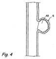
ここで図2を参照すると、架橋剤は病巣6内の実質的に閉じ込められた血液へ混合されてもよい。これは、例えば、マイクロカテーテル20などの遠位端を通り抜けて、内部空洞へ少なくとも1回通るコイル40などの混合機構の手段によって、遂行されてもよい。この目的のために使用されてもよい市販のコイルの例は、MicroPlex(登録商標)コイルシステム(MicroVention,Inc.、カリフォルニア州、アリソビエホ)、HelipaqTMコイルシステム(Micrus,Inc.、カリフォルニア州、サニーベール)およびGuglielmiコイルシステム(Boston Scientific、マサチューセッツ州、ネーティック)を含むが、必ずしも限定されない。架橋剤は、例えば図3に示すように、障害6内で実質的に固体である塊48を形成するように、1つ以上の血液の構成要素と反応する。本発明の少なくともいくつかの適用では、この実質的に固体である塊48は、約3分から約20分の時間で形成できる可能性がある。塊48の形成後、拡張可能な部材36は収縮してもよく、図4の中で示されるように、その後、マイクロカテーテル20および閉塞部材30は部位から除去または回収されてもよい。Referringnow to FIG.2, the cross-linking agent may be mixed to a substantially confined
閉塞部材30およびカテーテル20は、図1〜図4では別個の要素として示されるが、本発明の他の実施形態は、カテーテルと一体化して形成されるか、またはカテーテルに取り付けられている閉塞部材を利用してもよい。図5は、血管2の障害6を治療するために使用される本発明に従う別のキット110を示し、キット110は、障害6内の血液を隔離し、閉じ込めるために、ある量の架橋剤、障害6の中へ架橋剤を導入するためのカテーテル120、前記カテーテル120に一体化して形成される閉塞部材130を含む。 Although the
架橋剤溶液の一部またはすべてが障害の外の血管に入った場合、架橋剤溶液の性質および反応の比較的遅い動態のため、好ましくない塞栓形成のリスクは低い。どんな固体の塞栓も形成され得る前に、血管中に流れる血液は架橋剤を希釈する。If some or all of the crosslinker solution entersthe blood vesseloutside the lesion, the risk of undesirable embolization is low due to the nature of the crosslinker solution and the relatively slow kinetics of the reaction. Before alsobe embolized of any solid,blood flowing through the blood vessel to dilute the cross-linking agent.
得られた架橋したネットワークは、自然な手段によって形成された血栓に構造において類似している。血栓の自然形成は、トロンビンによって触媒されたフィブリンへのフィブリノゲンの変換によって生じる。繊維素溶解は、tPAによって触媒されたプラスミンへのプラスミノゲンの変換によって生じる。実質的に固体である塊は架橋剤による血液の架橋によって形成され、自然な繊維素溶解機構による分解の影響を受けにくい。 The resulting cross-linked network is similar in structure to a thrombus formed by natural means. The spontaneous formation of a thrombus results from the conversion of fibrinogen to fibrin catalyzed by thrombin. Fibrinolysis occurs by the conversion of plasminogen to plasmin catalyzed by tPA. A substantially solid mass is formed by cross-linking of blood with a cross-linking agent and is not susceptible to degradation by the natural fibrinolysis mechanism.
自然血栓と同じように、また他の液体塞栓剤とは違って、本発明の方法およびシステムによって形成された実質的に固体である塊は、筋線維芽細胞および治癒過程の他の要素によって、浸透、再形成および分解できる、架橋したネットワークを直ちに提供する。この治癒過程は、事実上無細胞の合成血栓を線維性結合組織(これはコラーゲン細胞外マトリクス中の筋線維芽細胞を特徴とする)に変換する。マクロファージおよび治癒過程の他の要素は、分解された合成血栓を貪食する。As with natural thrombi, and unlike other liquid emboli, the substantially solid mass formed by the methods and systems of the present invention is affected by myofibroblasts and other elements of the healing process. Immediately provide a cross-linked network that can penetrate,reform and decompose. This healing process convertsa virtually cell-free synthetic thrombus into fibrous connective tissue,which is characterized by myofibroblasts in the collagen extracellular matrix . Macrophages and other elements of the healing process phagocytose degraded synthetic thrombus.
以下は、上述の実質的に固体である塊を形成する架橋剤の生物医学的適用のうちのいくつかの実施例である。しかしながら、本明細書に説明される具体的な実施例に加えて、この架橋したネットワークの物質が他の多くの医学的および非医学的な適用を有することは理解されるであろう。 The following are some examples of the biomedical applications of the cross-linking agents that form the substantially solid mass described above. However, it will be appreciated that in addition to the specific examples described herein, this cross-linked network material has many other medical and non-medical applications.
実施例1
サンプル架橋剤、トリメチロールプロパン20/3の調製
エトキシ化トリメチロールプロパン20/3(20g、Aldrich)をキシレンに溶解させる。水を共沸蒸留によって系から除去する。無水コハク酸(8.1g、Aldrich)およびピリジン(100mL、Aldrich)を加えた。一晩還流させながら反応を進める。ピリジンおよびキシレンの一部を蒸留する。エトキシ化トリメチロールプロパンスクシネートを、冷却によりキシレンから除去し、分液漏斗を使用して収集した。エトキシ化トリメチロールプロパンスクシネートを、50mLのジクロロメタン中に再溶解させ、過剰の無水コハク酸を再結晶させるために−20℃で一晩保存した。結晶をろ過し、200mLの容量を調製するためにジクロロメタンを加えた。N−ヒドロキシスクシンイミド(7.8g、Aldrich)およびジイソプロピルカルボジイミド(10.5mL、Aldrich)を順次加えた。室温で約4時間反応を進める。真空濾過を使用して尿素を除去する。エトキシ化トリメチロールプロパンスクシンイミジルスクシネートを、過剰なヘキサンでの沈殿によって回収し、分液漏斗を使用して収集した。真空オーブンを使用して残留有機溶媒を除去した。Example 1
Preparation of sample cross-linking agent,
実施例2
架橋時間の決定
研究所において架橋時間を決定するために、様々な他の架橋剤と同様、実施例1に従って調製された架橋剤を合成および評価した。評価された架橋剤は、エトキシ化トリメチロールプロパン20/3スクシンイミジルスクシネート(TMP−SS)、エトキシ化ペンタエリスリトール15/4スクシンイミジルスクシネート(PE−SS)、4アームポリ(エチレングリコール)2kスクシンイミジルスクシネート(PEG2k−SS)、4アームポリ(エチレングリコール)10kスクシンイミジルスクシネート(PEG10k−SS)、およびエトキシ化フロログルシノール30/3スクシンイミジルスクシネート(ePG−SS)を含む。Example 2
Determination of Crosslink Time To determine the crosslink time in the laboratory, the crosslinker prepared according to Example 1 was synthesized and evaluated as well as various other crosslinkers. The evaluated crosslinkers were
それぞれの架橋剤を望ましい濃度で適切な溶媒に溶解させる。それぞれの架橋剤溶液(100マイクロリットル)を、EDTAで処理された350マイクロリットルのブタ血液と混合する。架橋時間、すなわち溶液が実質的に凝固するために必要な時間を決定し、架橋時間の結果を以下の表1に示す。Each crosslinker is dissolved in a suitable solvent at the desired concentration. Each crosslinker solution (100 microliters) is mixed with 350 microliters of porcine blood treated with EDTA. The crosslinking time, i.e. the time required for the solution to substantially solidify, is determinedand the results ofthe crosslinking time are shown in Table 1 below.

実施例3
架橋した塊の圧縮強度
この実施例では、研究所において圧縮強度を決定するために、様々な架橋剤を合成および評価した。これらの架橋剤は、エトキシ化トリメチロールプロパン20/3スクシンイミジルスクシネート(TMP−SS)、エトキシ化ペンタエリスリトール15/4スクシンイミジルスクシネート(PE−SS)、4アームポリ(エチレングリコール)2kスクシンイミジルスクシネート(PEG2k−SS)、4アームポリ(エチレングリコール)10kスクシンイミジルスクシネート(PEG10k−SS)、およびエトキシ化フロログルシノール30/3スクシンイミジルスクシネート(ePG−SS)を含む。Example 3
In this example, various crosslinkers were synthesized and evaluated to determine compressive strength in the laboratory. These crosslinkers include
それぞれの架橋剤を、例えば、上述の実施例2に従って、望ましい濃度で適切な溶媒に溶解する。それぞれの架橋剤溶液(1mL)を、EDTAで処理された3.5mLのブタ血液と混合する。混合物を3ccの注射器(Becton Dickinson)に置き、約30分間、約37℃で凝固させる。Luerフィッティングをカミソリの刃を使用して注射器から切り取り、架橋した塊を注射器から取り出し、0.9%の生理食塩水で満たされた15ccの遠心分離チューブに入れる。塊を室温で一晩保存する。インキュベーションの後、塊を長さ約8.5mmの切片に切り、Instron5543圧縮アセンブリ上の2つの圧盤間に入れる。Instronにより、破壊が起きるまで15mm/分の割合で合成血栓を圧縮した。応力およびひずみのデータを収集し、表2に示す。Each crosslinker is dissolved in a suitable solvent at the desired concentration, eg, according to Example 2 above. Each crosslinker solution (1 mL) is mixed with 3.5 mL porcine blood treated with EDTA.Place the mixture 3cc syringe to (Becton Dickinson), about 30 minutes, allowed to solidify at about 37 ° C.. The Luerfitting is cut from the syringe using a razor blade and the crosslinked mass is removed from the syringe and placed in a 15 cc centrifugetube filled with 0.9% saline. Store the mass overnight at room temperature. Afterincubation , the mass is cut into approximately 8.5 mm long sections andplaced between twoplatens on an Instron 5543 compression assembly. The Instron compressed the synthetic thrombus at a rate of 15 mm / min until failure occurred. Stress and strain data were collected and shown in Table 2.
実施例4
架橋した塊の耐久性
架橋した実質的に固体である塊の耐久性は、側壁動脈瘤の流動力学をシミュレートする流動モデルで試験される。架橋剤を、1mLのジメチルスルホキシドにTSAT(50mg、Pierce)を加えることにより、溶液として調製する。100マイクロリットルの架橋剤溶液を、350マイクロリットルのEDTAで処理されたブタ血液に加え、混合する。架橋剤溶液を6mmの側壁、シリコーン動脈瘤に注入し、20分間架橋させる。動脈瘤を流動モデルに置き、またリン酸緩衝化生理食塩水を、流速300mL/分、温度37℃、圧力120/80mmHgで、動脈瘤の親動脈を通して送り込む。塊は、16時間を超えてもこれらの条件のもとで安定していることが分かった。Example 4
Crosslinked Durability Durability of a cross-linked substantially solid mass is tested in a flow model that simulates the flow dynamics of a side wall aneurysm. The crosslinker is prepared as a solution by adding TSAT (50 mg, Pierce) to 1 mL of dimethyl sulfoxide. 100 microliters of crosslinker solution is added to and mixed with 350 microliters of EDTA-treated porcine blood. The crosslinker solution is injected into the 6 mm sidewall, silicone aneurysm and allowed to crosslink for 20 minutes. The aneurysm is placed in the flow model and phosphate buffered saline is pumped through the parent artery of the aneurysm at a flow rate of 300 mL / min, temperature 37 ° C.,
次に、実質的に固体である塊の耐久性が、分岐動脈瘤の流動力学をシミュレートする流動モデルで試験される。架橋剤溶液は、1mLのジメチルスルホキシドにTSAT(50mg、Pierce)を加えることにより、上記に示されるように調製される。その後、100マイクロリットルの架橋剤溶液を、350マイクロリットルのEDTAで処理されたブタ血液に加え、混合する。溶液を5mmの分岐、シリコーン動脈瘤に注入し、20分間架橋させた。動脈瘤を流動モデルに置き、またリン酸緩衝化生理食塩水を、流速300mL/分、温度37℃、圧力120/80mmHgで、動脈瘤の親動脈を通して送り込む。合成凝血塊は、2時間を超えてもこれらの条件のもとで安定している。 The durability of the substantially solid mass is then tested in a flow model that simulates the flow dynamics of a branch aneurysm. The crosslinker solution is prepared as indicated above by adding TSAT (50 mg, Pierce) to 1 mL of dimethyl sulfoxide. Thereafter, 100 microliters of the crosslinker solution is added to the porcine blood treated with 350 microliters of EDTA and mixed. The solution was injected into a 5 mm branch, silicone aneurysm and allowed to crosslink for 20 minutes. The aneurysm is placed in the flow model and phosphate buffered saline is pumped through the parent artery of the aneurysm at a flow rate of 300 mL / min, temperature 37 ° C.,
実施例5
実験動脈瘤中の架橋した塊の形成
Cloftらの、Radiology、213:223−228(1999)によって説明された方法に従って、右総頸動脈の隔離およびエラスターゼ消化によって実験動脈瘤をウサギに形成させる。動脈瘤は、本発明の方法に従って、治療される前に約6週間成熟させる。Example 5
Formation of a cross-linked mass in an experimental aneurysm An experimental aneurysm is formed in a rabbit by isolation of the right common carotid artery and elastasedigestion according to the method described by Cloft et al., Radiology, 213: 223-228 (1999). The aneurysm is matured for approximately 6 weeks before being treated according to the method of the present invention.
治療の時点で、動脈瘤は約3mm×約8mmであると測定された。大腿鞘を通して、マイクロカテーテル(Rebar Microcatheter,Micro Therapeutics,Inc.、カルフォルニア州、アービン)を、動脈瘤の内部に設置する。3方向の栓をRebarのハブに取り付ける。回転止血弁を、マイクロカテーテルの長さと一致して3方向に取り付ける。1方向の栓を、回転止血弁のサイドポートへ取り付ける。生理食塩水を使用して、空気を装置から除去する。混合コイルを、3方向の栓に近接している回転止血弁に設置する。4mm×10mmのバルーンカテーテル(例えば、HyperGlide,MicroTherapeutics,Inc.、カルフォルニア州、アービン)を、動脈瘤の頚に設置する。 At the time of treatment, the aneurysm was measured to be about 3 mm x about 8 mm. Through the femoral sheath, a microcatheter (Rebar Microcatheter, Micro Therapeutics, Inc., Irvine, CA) is placed inside the aneurysm. Attach a 3-way plug to the Rebar hub. A rotary hemostasis valve is attached in three directions, consistent with the length of the microcatheter. A one-way stop is attached to the side port of the rotary hemostasis valve. Saline is used to remove air from the device. A mixing coil is placed on the rotary hemostasis valve in close proximity to the three-way stopcock. A 4 mm × 10 mm balloon catheter (eg, HyperGlide, MicroTherapeutics, Inc., Irvine, Calif.) Is placed in the neck of the aneurysm.
続いて、マイクロカテーテルを、ヘパリン化食塩水(数ミリリットル)およびメチルピロリジノン(0.35mL)で洗い流す。架橋剤溶液は、メチルピロリジノンの中の190mg/mLのエトキシ化ペンタエリスリトール15/4スクシンイミジルグルタレートである。マイクロカテーテルを架橋剤溶液で満たす。動脈瘤頚を密封するためにバルーンを膨張させる。架橋剤溶液を、混合コイルと同時に動脈瘤嚢へ導入させる。混合コイルは、架橋溶液およびもとの場所の血液を混合するために、動脈瘤嚢に配置され、除去され、配置され、除去される。5分後、バルーンを収縮させる。 Subsequently, the microcatheter is flushed with heparinized saline (several milliliters) and methylpyrrolidinone (0.35 mL). The crosslinker solution is 190 mg / mL ethoxylated pentaerythritol 15/4 succinimidyl glutarate in methylpyrrolidinone. Fill the microcatheter with the crosslinker solution. The balloon is inflated to seal the aneurysm neck. The crosslinker solution is introduced into the aneurysm sac simultaneously with the mixing coil. The mixing coil is placed, removed, placed and removed from the aneurysm sac to mix the cross-linking solution and the original blood. After 5 minutes, the balloon is deflated.
デジタル消去血管造影は、動脈瘤嚢がほとんど完全に(すなわち、95%)閉塞されたことを示した。引き続き行われた治療後2週間の血管造影は、動脈瘤嚢がほとんど閉塞されたことを明示した。組織学的評価は、組織化されていない血餅が動脈瘤嚢の大部分を満たしていることを示した。事実上、炎症は存在していない。Digitalerased angiography showed that the aneurysm sac was almost completely occluded (ie, 95%). Subsequent angiography 2 weeks after treatment revealed that the aneurysm sac was almost occluded. Histological evaluation showed that an unorganizedclot filled the majority of the aneurysm sac. Virtually no inflammation exists.
本発明は、特定の実施例および実施形態のみを参照して本明細書に説明されてきた。本発明の考え得るすべての実施例および実施形態を網羅的に説明する試みはされていない。確かに、当業者は、様々な追加、削除、修正および他の変更が、以下の請求項で列挙される本発明の意図した精神および範囲から逸脱することなく、上述の実施例および実施形態に行われてもよいことを理解するであろう。かかる追加、削除、修正および他の変更はすべて以下の特許請求の範囲内に含まれることが意図される。 The present invention has been described herein with reference to only specific examples and embodiments. No attempt has been made to exhaustively describe all possible examples and embodiments of the invention. Indeed, those skilled in the art will recognize that various additions, deletions, modifications and other changes may be made to the examples and embodiments described above without departing from the intended spirit and scope of the invention as recited in the following claims. It will be understood that this may be done. All such additions, deletions, modifications and other changes are intended to be included within the scope of the following claims.
Claims (48)
Translated fromJapanese架橋剤が前記障害内の血液と混合されるように、前記障害に前記架橋剤を導入するための手段と、
前記障害内に実質的に固体である塊を形成させるように、前記架橋剤を血液と反応させるための手段と、
前記導入するための手段を通って延び、該導入するための手段に対して可動であり、前記導入するための手段の遠位端を超えて前記障害内で前記架橋剤とその場で意志的に血液を混合するための混合機構と、
を備えるシステム。A system for treating a disorder in a vasculature, wherein the vasculature has a lumen and a wall, the system comprising:
Means for introducing the cross-linking agent into the disorder such that the cross-linking agent is mixed with blood within the disorder;
Means for reacting the cross-linking agent with blood so as to form a substantially solid mass within the obstacle;
Extends through the means for the introduction, is movable relative to the means for the introduction,volitional in the crosslinking agent situwithin the fault beyond the distal end of said means for introducing a mixing mechanism for mixing theblood,
A system comprising:
それぞれの官能基は、求電子基、エステル、N−ヒドロキシスクシンイミドエステル、ビニルスルホン、N−エチルマレイミド、ヨードアセトアミド、オルトピリジルジスルフィド、アルデヒド、塩化スルホニル、ハロゲン化アリール、エポキシド、活性エステル、カルボニルジイミダゾール、ニトロフェニルカーボネート、トレシレート、トシラート、メシラートおよびイソシアネートから独立して選択される、
請求項27に記載のシステム。The core portion is pentaerythritol, di (pentaerythritol), nitriloacetic acid, glycerin, ethylene glycol, trimethylolpropane, di (trimethylolpropane), polyacid, heptanedioic acid, octanedioic acid, hexadecanedioic acid, polyfunctional Selected from aromatic aromatic compounds, phloroglucinol, 1,2,4-benzenetriol and pyrogallol,
Each functional group is an electrophilic group, ester, N-hydroxysuccinimide ester, vinyl sulfone, N-ethylmaleimide, iodoacetamide, orthopyridyl disulfide, aldehyde, sulfonyl chloride, aryl halide, epoxide, active ester, carbonyldiimidazole. Independently selected from nitrophenyl carbonate, tresylate, tosylate, mesylate and isocyanate,
28. The system of claim 27.
Applications Claiming Priority (3)
| Application Number | Priority Date | Filing Date | Title |
|---|---|---|---|
| US10/990,942US7201918B2 (en) | 2004-11-16 | 2004-11-16 | Compositions, systems and methods for treatment of defects in blood vessels |
| US10/990,942 | 2004-11-16 | ||
| PCT/US2005/041637WO2006055690A2 (en) | 2004-11-16 | 2005-11-16 | Compositions, systems and methods for treatment of defects in blood vessels |
Related Child Applications (1)
| Application Number | Title | Priority Date | Filing Date |
|---|---|---|---|
| JP2008292851ADivisionJP2009082728A (en) | 2004-11-16 | 2008-11-14 | Composition, system and method for treatment of defects in blood vessel |
Publications (3)
| Publication Number | Publication Date |
|---|---|
| JP2008520283A JP2008520283A (en) | 2008-06-19 |
| JP2008520283A5 JP2008520283A5 (en) | 2012-09-20 |
| JP5456974B2true JP5456974B2 (en) | 2014-04-02 |
Family
ID=36386612
Family Applications (2)
| Application Number | Title | Priority Date | Filing Date |
|---|---|---|---|
| JP2007541483AExpired - Fee RelatedJP5456974B2 (en) | 2004-11-16 | 2005-11-16 | Compositions, systems and methods for treating vascular disorders |
| JP2008292851APendingJP2009082728A (en) | 2004-11-16 | 2008-11-14 | Composition, system and method for treatment of defects in blood vessel |
Family Applications After (1)
| Application Number | Title | Priority Date | Filing Date |
|---|---|---|---|
| JP2008292851APendingJP2009082728A (en) | 2004-11-16 | 2008-11-14 | Composition, system and method for treatment of defects in blood vessel |
Country Status (7)
| Country | Link |
|---|---|
| US (1) | US7201918B2 (en) |
| EP (1) | EP1824414A4 (en) |
| JP (2) | JP5456974B2 (en) |
| CN (1) | CN101056595B (en) |
| AU (1) | AU2005307790B2 (en) |
| CA (1) | CA2587335C (en) |
| WO (1) | WO2006055690A2 (en) |
Families Citing this family (38)
| Publication number | Priority date | Publication date | Assignee | Title |
|---|---|---|---|---|
| US7744652B2 (en)* | 2006-01-23 | 2010-06-29 | Hesham Morsi | Aneurysm sealing device |
| US8066753B2 (en)* | 2006-07-06 | 2011-11-29 | Robert Kipperman | Specialized catheter and method for placement in a bifurcated vessel |
| WO2008134468A1 (en)* | 2007-04-27 | 2008-11-06 | Wisconsin Alumni Research Foundation | Bioactive and biocompatible polyurethane-butanediol-glycosaminoglycan salt copolymers |
| US20110022149A1 (en) | 2007-06-04 | 2011-01-27 | Cox Brian J | Methods and devices for treatment of vascular defects |
| US20090011043A1 (en)* | 2007-07-03 | 2009-01-08 | Hua Xie | Tissue sealant made from whole blood |
| US8241609B2 (en)* | 2007-08-24 | 2012-08-14 | E I Du Pont De Nemours And Company | Method for embolization using liquid embolic materials |
| US20090137981A1 (en)* | 2007-11-26 | 2009-05-28 | Valor Medical | Methods of treating a blood vessel |
| US20110046716A1 (en)* | 2008-02-20 | 2011-02-24 | Murray Vascular Pty Limited | Stent |
| AU2009242528B2 (en)* | 2008-05-02 | 2015-12-10 | Microvention, Inc. | Filamentary devices for treatment of vascular defects |
| US20100016833A1 (en)* | 2008-07-15 | 2010-01-21 | Ogle Matthew F | Devices for the Treatment of Vascular Aneurysm |
| US20100119605A1 (en)* | 2008-11-12 | 2010-05-13 | Isenburg Jason C | Compositions for tissue stabilization |
| CA2777083C (en) | 2009-10-06 | 2014-11-25 | Regents Of The University Of Minnesota | Bioresorbable embolization microspheres |
| WO2011044455A1 (en)* | 2009-10-09 | 2011-04-14 | Vatrix Medical, Inc. | In vivo chemical stabilization of vulnerable plaque |
| US8444624B2 (en)* | 2009-10-19 | 2013-05-21 | Vatrix Medical, Inc. | Vascular medical devices with sealing elements and procedures for the treatment of isolated vessel sections |
| BR112012010758A2 (en) | 2009-11-05 | 2019-09-24 | Sequent Medical Inc | multilayer filament devices for treatment of vascular defects |
| SA111320355B1 (en)* | 2010-04-07 | 2015-01-08 | Baxter Heathcare S A | Hemostatic sponge |
| US8936795B2 (en)* | 2012-12-19 | 2015-01-20 | Regents Of The University Of Minnesota | Liquid embolic material including carboxymethyl chitosan crosslinked with carboxymethyl cellulose |
| US10307493B2 (en) | 2013-03-15 | 2019-06-04 | Biocompatible UK Limited | Imageable embolic microsphere |
| US9955976B2 (en) | 2013-08-16 | 2018-05-01 | Sequent Medical, Inc. | Filamentary devices for treatment of vascular defects |
| US9078658B2 (en) | 2013-08-16 | 2015-07-14 | Sequent Medical, Inc. | Filamentary devices for treatment of vascular defects |
| GB2521997A (en) | 2013-09-06 | 2015-07-15 | Biocompatibles Uk Ltd | Radiopaque polymers |
| GB2519738A (en) | 2013-09-06 | 2015-05-06 | Biocompatibles Uk Ltd | Radiopaque polymers |
| ES2806084T3 (en)* | 2014-03-28 | 2021-02-16 | Swiss Vx Venentherapie Und Forschung Gmbh | Sclerotherapy devices using light curable adhesives |
| US9629635B2 (en) | 2014-04-14 | 2017-04-25 | Sequent Medical, Inc. | Devices for therapeutic vascular procedures |
| US10182979B2 (en) | 2016-03-22 | 2019-01-22 | Regents Of The University Of Minnesota | Biodegradable microspheres |
| US10576099B2 (en)* | 2016-10-21 | 2020-03-03 | Covidien Lp | Injectable scaffold for treatment of intracranial aneurysms and related technology |
| CN108245216A (en)* | 2016-12-29 | 2018-07-06 | 香港科技大学深圳研究院 | A kind of aneurysm treatment system |
| JP6730956B2 (en)* | 2017-04-11 | 2020-07-29 | テルモ株式会社 | Medical equipment |
| CN107736909B (en)* | 2017-10-13 | 2023-09-01 | 上海长海医院 | Left tube cutting device after cerebral artery and vein malformation interventional embolism operation |
| CN109330654B (en)* | 2018-08-31 | 2021-02-05 | 华中科技大学 | A kind of hemostatic needle, its preparation method and application |
| CN109350161B (en)* | 2018-10-11 | 2020-10-16 | 南华大学 | A spring coil electromagnetic release device |
| CN109199502B (en)* | 2018-10-16 | 2020-05-29 | 中国医学科学院北京协和医院 | Aneurysm embolization catheter and aneurysm operation auxiliary device |
| US11559309B2 (en) | 2019-03-15 | 2023-01-24 | Sequent Medical, Inc. | Filamentary devices for treatment of vascular defects |
| CN113573765B (en) | 2019-03-15 | 2024-08-13 | 美科微先股份有限公司 | Silk device for treating vascular defects |
| CN113573650B (en) | 2019-03-15 | 2024-05-28 | 后续医疗股份有限公司 | Silk device with flexible connector for treating vascular defects |
| US12070220B2 (en) | 2020-03-11 | 2024-08-27 | Microvention, Inc. | Devices having multiple permeable shells for treatment of vascular defects |
| US12023034B2 (en) | 2020-03-11 | 2024-07-02 | Microvention, Inc. | Devices for treatment of vascular defects |
| US20210282789A1 (en) | 2020-03-11 | 2021-09-16 | Microvention, Inc. | Multiple layer devices for treatment of vascular defects |
Family Cites Families (46)
| Publication number | Priority date | Publication date | Assignee | Title |
|---|---|---|---|---|
| HU184722B (en)* | 1980-02-18 | 1984-10-29 | Laszlo Lazar | Therapeutically suitable silicone rubber mixture and therapeuticaid |
| US4795741A (en)* | 1987-05-06 | 1989-01-03 | Biomatrix, Inc. | Compositions for therapeutic percutaneous embolization and the use thereof |
| US4819637A (en)* | 1987-09-01 | 1989-04-11 | Interventional Therapeutics Corporation | System for artificial vessel embolization and devices for use therewith |
| US6004261A (en)* | 1989-04-28 | 1999-12-21 | C. R. Bard, Inc. | Formed-in-place endovascular stent and delivery system |
| US5122136A (en)* | 1990-03-13 | 1992-06-16 | The Regents Of The University Of California | Endovascular electrolytically detachable guidewire tip for the electroformation of thrombus in arteries, veins, aneurysms, vascular malformations and arteriovenous fistulas |
| JP3103368B2 (en)* | 1990-11-29 | 2000-10-30 | テルモ株式会社 | Embolization material for vascular lesions |
| JPH04197356A (en)* | 1990-11-29 | 1992-07-16 | Matsushita Electric Ind Co Ltd | Bathtub device |
| WO1995008289A2 (en)* | 1993-09-16 | 1995-03-30 | Scimed Life Systems, Inc. | Percutaneous repair of cardiovascular anomalies and repair compositions |
| US5795331A (en)* | 1994-01-24 | 1998-08-18 | Micro Therapeutics, Inc. | Balloon catheter for occluding aneurysms of branch vessels |
| JP2535785B2 (en)* | 1994-06-03 | 1996-09-18 | 工業技術院長 | Vascular embolic agent |
| US5580568A (en)* | 1995-07-27 | 1996-12-03 | Micro Therapeutics, Inc. | Cellulose diacetate compositions for use in embolizing blood vessels |
| DK0876165T3 (en)* | 1995-12-18 | 2007-08-06 | Angiotech Biomaterials Corp | Crosslinked polymer compositions and methods for their preparation |
| US5752974A (en)* | 1995-12-18 | 1998-05-19 | Collagen Corporation | Injectable or implantable biomaterials for filling or blocking lumens and voids of the body |
| US20010029349A1 (en)* | 1996-04-12 | 2001-10-11 | Boris Leschinsky | Method and apparatus for treating aneurysms |
| AU4648697A (en)* | 1996-09-23 | 1998-04-14 | Chandrashekar Pathak | Methods and devices for preparing protein concentrates |
| US6146373A (en)* | 1997-10-17 | 2000-11-14 | Micro Therapeutics, Inc. | Catheter system and method for injection of a liquid embolic composition and a solidification agent |
| US6015424A (en)* | 1998-04-28 | 2000-01-18 | Microvention, Inc. | Apparatus and method for vascular embolization |
| US6152943A (en)* | 1998-08-14 | 2000-11-28 | Incept Llc | Methods and apparatus for intraluminal deposition of hydrogels |
| US6703047B2 (en)* | 2001-02-02 | 2004-03-09 | Incept Llc | Dehydrated hydrogel precursor-based, tissue adherent compositions and methods of use |
| US6514534B1 (en)* | 1998-08-14 | 2003-02-04 | Incept Llc | Methods for forming regional tissue adherent barriers and drug delivery systems |
| US6899889B1 (en)* | 1998-11-06 | 2005-05-31 | Neomend, Inc. | Biocompatible material composition adaptable to diverse therapeutic indications |
| US6454738B1 (en)* | 1999-05-21 | 2002-09-24 | Micro Therapeutics, Inc. | Methods for delivering in vivo uniform dispersed embolic compositions of high viscosity |
| US6602261B2 (en)* | 1999-10-04 | 2003-08-05 | Microvention, Inc. | Filamentous embolic device with expansile elements |
| US6652883B2 (en)* | 2000-03-13 | 2003-11-25 | Biocure, Inc. | Tissue bulking and coating compositions |
| JP4871476B2 (en)* | 2000-03-13 | 2012-02-08 | バイオコンパティブルズ ユーケー リミテッド | Embolization composition |
| US6660247B1 (en)* | 2000-06-23 | 2003-12-09 | Battelle Memorial Institute | Multiple stimulus reversible hydrogels |
| KR100366600B1 (en)* | 2000-09-21 | 2003-01-09 | 광주과학기술원 | Liquified Embolic Materials Using Sol-Gel Phase Transition and Use of the Same |
| US6527790B2 (en)* | 2000-12-07 | 2003-03-04 | Scimed Life Systems, Inc. | Intravascular balloon catheter for embolic coil delivery |
| US6547804B2 (en)* | 2000-12-27 | 2003-04-15 | Scimed Life Systems, Inc. | Selectively permeable highly distensible occlusion balloon |
| US6878384B2 (en)* | 2001-03-13 | 2005-04-12 | Microvention, Inc. | Hydrogels that undergo volumetric expansion in response to changes in their environment and their methods of manufacture and use |
| ATE516759T1 (en)* | 2001-05-29 | 2011-08-15 | Microvention Inc | METHOD FOR PRODUCING EXPANDABLE FILAMENTOUS EMBOLIZATION DEVICES |
| US20030014075A1 (en)* | 2001-07-16 | 2003-01-16 | Microvention, Inc. | Methods, materials and apparatus for deterring or preventing endoleaks following endovascular graft implanation |
| JP4377688B2 (en)* | 2001-09-04 | 2009-12-02 | マイクロ セラピューティクス, インコーポレイテッド | Occlusion catheter with extensible balloon for use in complex vasculature |
| CN100366289C (en)* | 2001-09-12 | 2008-02-06 | 维日克斯医药公司 | Vascular occlusion solid-phase agent with immobilised platelet binding agent |
| JP3607231B2 (en)* | 2001-09-28 | 2005-01-05 | 有限会社日本エレクテル | High frequency heating balloon catheter |
| US6808518B2 (en)* | 2001-09-28 | 2004-10-26 | Ethicon, Inc. | Methods and devices for treating diseased blood vessels |
| JP2003250896A (en)* | 2002-02-28 | 2003-09-09 | Terumo Corp | Balloon catheter for aneurysmal embolic treatment |
| US7462366B2 (en)* | 2002-03-29 | 2008-12-09 | Boston Scientific Scimed, Inc. | Drug delivery particle |
| US7341716B2 (en)* | 2002-04-12 | 2008-03-11 | Boston Scientific Scimed, Inc. | Occlusive composition |
| US7771387B2 (en)* | 2002-05-17 | 2010-08-10 | Boston Scientific Scimed, Inc. | Liquid embolic composition delivery devices and methods |
| EP1364663A1 (en)* | 2002-05-21 | 2003-11-26 | Commonwealth Scientific And Industrial Research Organisation | Ocular devices with functionalized surface with adhesive properties |
| DE60331367D1 (en)* | 2002-12-30 | 2010-04-01 | Angiotech Int Ag | ACTIVE COMPOSITION OF FAST GELING POLYMERIC COMPOSITION |
| JP4067976B2 (en)* | 2003-01-24 | 2008-03-26 | 有限会社日本エレクテル | High frequency heating balloon catheter |
| US20050060017A1 (en)* | 2003-09-15 | 2005-03-17 | Fischell Robert E. | Means and method for the treatment of cerebral aneurysms |
| US20050133046A1 (en)* | 2003-12-17 | 2005-06-23 | Becker Timothy A. | Compositions and methods for improved occlusion of vascular defects |
| US20050276863A1 (en)* | 2004-05-27 | 2005-12-15 | Medtronic Vascular, Inc. | Methods for treatment of aneurysmal tissue |
- 2004
- 2004-11-16USUS10/990,942patent/US7201918B2/ennot_activeExpired - Lifetime
- 2005
- 2005-11-16CNCN2005800389413Apatent/CN101056595B/ennot_activeExpired - Fee Related
- 2005-11-16CACA2587335Apatent/CA2587335C/ennot_activeExpired - Fee Related
- 2005-11-16JPJP2007541483Apatent/JP5456974B2/ennot_activeExpired - Fee Related
- 2005-11-16AUAU2005307790Apatent/AU2005307790B2/ennot_activeCeased
- 2005-11-16WOPCT/US2005/041637patent/WO2006055690A2/enactiveApplication Filing
- 2005-11-16EPEP05823232Apatent/EP1824414A4/ennot_activeWithdrawn
- 2008
- 2008-11-14JPJP2008292851Apatent/JP2009082728A/enactivePending
Also Published As
| Publication number | Publication date |
|---|---|
| CN101056595A (en) | 2007-10-17 |
| CN101056595B (en) | 2012-02-08 |
| JP2008520283A (en) | 2008-06-19 |
| JP2009082728A (en) | 2009-04-23 |
| WO2006055690A2 (en) | 2006-05-26 |
| CA2587335A1 (en) | 2006-05-26 |
| AU2005307790B2 (en) | 2010-09-02 |
| US7201918B2 (en) | 2007-04-10 |
| EP1824414A2 (en) | 2007-08-29 |
| CA2587335C (en) | 2013-03-12 |
| WO2006055690A3 (en) | 2007-02-22 |
| EP1824414A4 (en) | 2012-06-27 |
| US20060105014A1 (en) | 2006-05-18 |
| AU2005307790A1 (en) | 2006-05-26 |
Similar Documents
| Publication | Publication Date | Title |
|---|---|---|
| JP5456974B2 (en) | Compositions, systems and methods for treating vascular disorders | |
| US6113629A (en) | Hydrogel for the therapeutic treatment of aneurysms | |
| JP5148030B2 (en) | Novel high viscosity embolization composition | |
| JP4979150B2 (en) | Vascular embolization | |
| DE60130544T2 (en) | EMBOLIC COMPOSITIONS | |
| JP4262891B2 (en) | Device for injecting a hydrogel into a cavity | |
| JP2008520283A5 (en) | ||
| US20100063472A1 (en) | Compositions and methods for improved occlusion of vascular defects | |
| JP2002539853A (en) | Methods to prevent the formation of endoleaks associated with endovascular repair of celiac aortic aneurysms | |
| JP2003500106A (en) | Method for delivering uniformly dispersed high viscosity embolic compositions in vivo | |
| JP2002539856A (en) | Method of treating internal leakage during endovascular repair of abdominal aortic aneurysm | |
| JP2002504400A (en) | Gynecological embolization method | |
| JP2001509133A (en) | Compositions for use in occluded vessels | |
| JP2003511188A (en) | Filamentary embolic device with expansion element | |
| US7374568B2 (en) | Methods for embolizing aneurysmal sites with a high viscosity embolizing composition | |
| JP2003524456A (en) | Biodegradable polymer protein-based coils for use as luminal implants | |
| US8048407B2 (en) | In situ gelling self-reactive materials for embolization | |
| JP2002504406A (en) | Gynecological endovascular embolization treatment | |
| US20160346423A1 (en) | Cyanoacrylate compositions including non-agglomerating radiopaque nanoparticles | |
| JPH0517369A (en) | Thromboembolic agent | |
| RU2735500C1 (en) | Method for embolisation of cerebral aneurysms with a liquid adhesive composition | |
| Peer et al. | Sequential Aspiration and injection of a Mixture of gelatin-based hemostatic agent and autologous blood clot followed by suture coiling for Effective Embolization of splenic artery pseudoaneurysm by interventional Radiologist (SAMEER): Description of a novel technique | |
| TR2023012295A1 (en) | VASCULAR OCCLUSION APPARATUS | |
| WO2025155730A1 (en) | Injectable hydrogel for embolization |
Legal Events
| Date | Code | Title | Description |
|---|---|---|---|
| A524 | Written submission of copy of amendment under article 19 pct | Free format text:JAPANESE INTERMEDIATE CODE: A524 Effective date:20081114 | |
| A621 | Written request for application examination | Free format text:JAPANESE INTERMEDIATE CODE: A621 Effective date:20081114 | |
| RD03 | Notification of appointment of power of attorney | Free format text:JAPANESE INTERMEDIATE CODE: A7423 Effective date:20110506 | |
| RD04 | Notification of resignation of power of attorney | Free format text:JAPANESE INTERMEDIATE CODE: A7424 Effective date:20110512 | |
| A131 | Notification of reasons for refusal | Free format text:JAPANESE INTERMEDIATE CODE: A131 Effective date:20120203 | |
| A601 | Written request for extension of time | Free format text:JAPANESE INTERMEDIATE CODE: A601 Effective date:20120501 | |
| A602 | Written permission of extension of time | Free format text:JAPANESE INTERMEDIATE CODE: A602 Effective date:20120510 | |
| A521 | Request for written amendment filed | Free format text:JAPANESE INTERMEDIATE CODE: A523 Effective date:20120730 | |
| A524 | Written submission of copy of amendment under article 19 pct | Free format text:JAPANESE INTERMEDIATE CODE: A524 Effective date:20120730 | |
| A131 | Notification of reasons for refusal | Free format text:JAPANESE INTERMEDIATE CODE: A131 Effective date:20130205 | |
| A601 | Written request for extension of time | Free format text:JAPANESE INTERMEDIATE CODE: A601 Effective date:20130430 | |
| A602 | Written permission of extension of time | Free format text:JAPANESE INTERMEDIATE CODE: A602 Effective date:20130509 | |
| A601 | Written request for extension of time | Free format text:JAPANESE INTERMEDIATE CODE: A601 Effective date:20130604 | |
| A602 | Written permission of extension of time | Free format text:JAPANESE INTERMEDIATE CODE: A602 Effective date:20130611 | |
| A601 | Written request for extension of time | Free format text:JAPANESE INTERMEDIATE CODE: A601 Effective date:20130705 | |
| A602 | Written permission of extension of time | Free format text:JAPANESE INTERMEDIATE CODE: A602 Effective date:20130712 | |
| A521 | Request for written amendment filed | Free format text:JAPANESE INTERMEDIATE CODE: A523 Effective date:20130805 | |
| A131 | Notification of reasons for refusal | Free format text:JAPANESE INTERMEDIATE CODE: A131 Effective date:20130823 | |
| A521 | Request for written amendment filed | Free format text:JAPANESE INTERMEDIATE CODE: A523 Effective date:20131121 | |
| TRDD | Decision of grant or rejection written | ||
| A01 | Written decision to grant a patent or to grant a registration (utility model) | Free format text:JAPANESE INTERMEDIATE CODE: A01 Effective date:20131211 | |
| A61 | First payment of annual fees (during grant procedure) | Free format text:JAPANESE INTERMEDIATE CODE: A61 Effective date:20140109 | |
| R150 | Certificate of patent or registration of utility model | Free format text:JAPANESE INTERMEDIATE CODE: R150 Ref document number:5456974 Country of ref document:JP Free format text:JAPANESE INTERMEDIATE CODE: R150 | |
| R250 | Receipt of annual fees | Free format text:JAPANESE INTERMEDIATE CODE: R250 | |
| R250 | Receipt of annual fees | Free format text:JAPANESE INTERMEDIATE CODE: R250 | |
| R250 | Receipt of annual fees | Free format text:JAPANESE INTERMEDIATE CODE: R250 | |
| LAPS | Cancellation because of no payment of annual fees |