JP5385394B2 - MR segmentation using transmission data in hybrid nuclear / MR imaging - Google Patents
MR segmentation using transmission data in hybrid nuclear / MR imagingDownload PDFInfo
- Publication number
- JP5385394B2 JP5385394B2JP2011526607AJP2011526607AJP5385394B2JP 5385394 B2JP5385394 B2JP 5385394B2JP 2011526607 AJP2011526607 AJP 2011526607AJP 2011526607 AJP2011526607 AJP 2011526607AJP 5385394 B2JP5385394 B2JP 5385394B2
- Authority
- JP
- Japan
- Prior art keywords
- map
- data
- transmission data
- pet
- radiation
- Prior art date
- Legal status (The legal status is an assumption and is not a legal conclusion. Google has not performed a legal analysis and makes no representation as to the accuracy of the status listed.)
- Expired - Fee Related
Links
Images


Classifications
- G—PHYSICS
- G01—MEASURING; TESTING
- G01R—MEASURING ELECTRIC VARIABLES; MEASURING MAGNETIC VARIABLES
- G01R33/00—Arrangements or instruments for measuring magnetic variables
- G01R33/20—Arrangements or instruments for measuring magnetic variables involving magnetic resonance
- G01R33/44—Arrangements or instruments for measuring magnetic variables involving magnetic resonance using nuclear magnetic resonance [NMR]
- G01R33/48—NMR imaging systems
- G01R33/4808—Multimodal MR, e.g. MR combined with positron emission tomography [PET], MR combined with ultrasound or MR combined with computed tomography [CT]
- G01R33/481—MR combined with positron emission tomography [PET] or single photon emission computed tomography [SPECT]
- G—PHYSICS
- G06—COMPUTING OR CALCULATING; COUNTING
- G06T—IMAGE DATA PROCESSING OR GENERATION, IN GENERAL
- G06T7/00—Image analysis
- G06T7/10—Segmentation; Edge detection
- G06T7/11—Region-based segmentation
- G—PHYSICS
- G01—MEASURING; TESTING
- G01R—MEASURING ELECTRIC VARIABLES; MEASURING MAGNETIC VARIABLES
- G01R33/00—Arrangements or instruments for measuring magnetic variables
- G01R33/20—Arrangements or instruments for measuring magnetic variables involving magnetic resonance
- G01R33/44—Arrangements or instruments for measuring magnetic variables involving magnetic resonance using nuclear magnetic resonance [NMR]
- G01R33/48—NMR imaging systems
- G01R33/54—Signal processing systems, e.g. using pulse sequences ; Generation or control of pulse sequences; Operator console
- G01R33/56—Image enhancement or correction, e.g. subtraction or averaging techniques, e.g. improvement of signal-to-noise ratio and resolution
- G01R33/5608—Data processing and visualization specially adapted for MR, e.g. for feature analysis and pattern recognition on the basis of measured MR data, segmentation of measured MR data, edge contour detection on the basis of measured MR data, for enhancing measured MR data in terms of signal-to-noise ratio by means of noise filtering or apodization, for enhancing measured MR data in terms of resolution by means for deblurring, windowing, zero filling, or generation of gray-scaled images, colour-coded images or images displaying vectors instead of pixels
- G—PHYSICS
- G06—COMPUTING OR CALCULATING; COUNTING
- G06T—IMAGE DATA PROCESSING OR GENERATION, IN GENERAL
- G06T2207/00—Indexing scheme for image analysis or image enhancement
- G06T2207/10—Image acquisition modality
- G06T2207/10072—Tomographic images
- G06T2207/10088—Magnetic resonance imaging [MRI]
- G—PHYSICS
- G06—COMPUTING OR CALCULATING; COUNTING
- G06T—IMAGE DATA PROCESSING OR GENERATION, IN GENERAL
- G06T2207/00—Indexing scheme for image analysis or image enhancement
- G06T2207/10—Image acquisition modality
- G06T2207/10072—Tomographic images
- G06T2207/10104—Positron emission tomography [PET]
- G—PHYSICS
- G06—COMPUTING OR CALCULATING; COUNTING
- G06T—IMAGE DATA PROCESSING OR GENERATION, IN GENERAL
- G06T2207/00—Indexing scheme for image analysis or image enhancement
- G06T2207/30—Subject of image; Context of image processing
- G06T2207/30004—Biomedical image processing
Landscapes
- Engineering & Computer Science (AREA)
- Physics & Mathematics (AREA)
- Theoretical Computer Science (AREA)
- Nuclear Medicine, Radiotherapy & Molecular Imaging (AREA)
- Pulmonology (AREA)
- Radiology & Medical Imaging (AREA)
- Health & Medical Sciences (AREA)
- General Health & Medical Sciences (AREA)
- General Physics & Mathematics (AREA)
- High Energy & Nuclear Physics (AREA)
- Condensed Matter Physics & Semiconductors (AREA)
- Computer Vision & Pattern Recognition (AREA)
- Nuclear Medicine (AREA)
- Magnetic Resonance Imaging Apparatus (AREA)
Description
Translated fromJapanese 本発明は、解剖学的な画像形成システムにおける特定の応用を見い出すものであり、特に、結合されたPET-MR画像形成を含むものであるが、他の核画像形成システム等における応用をも見い出すものである。
しかし、記載される技術は、他の画像形成システム、他の画像形成シナリオ、他の画像分析技術等における応用を見い出す場合があることを理解されたい。The present invention finds particular application in anatomical imaging systems, and in particular includes combined PET-MR imaging, but also finds application in other nuclear imaging systems and the like. is there.
However, it should be understood that the described techniques may find application in other imaging systems, other imaging scenarios, other image analysis techniques, and the like.
陽電子放出型断層撮影(PET: Position Emission Tomography)による画像形成では、PETデータにおける減衰は、典型的に減衰マップを使用して補正される。結合されたPETによるコンピュータ断層撮影(CT)では、CTデータは、PETデータについて正確な減衰マップを容易に生成することができる。CTデータ及び画像は、画像形成された組織の放射線の減衰特性に基づいている。しかし、組み合わせたPET-磁気共鳴(MR: magnetic Resonance)画像形成では、MR画像は、典型的には水ダイポール(H1)からなる共振特性を示し、この特性は、患者の放射線の減衰特性の関数ではない。In image formation by positron emission tomography (PET), attenuation in PET data is typically corrected using an attenuation map. In computed tomography (CT) with combined PET, CT data can easily generate an accurate attenuation map for the PET data. CT data and images are based on the radiation attenuation characteristics of the imaged tissue. However, in combined PET-magnetic resonance (MR) imaging, MR images typically exhibit a resonance characteristic consisting of a water dipole (H1 ), which is a characteristic of the attenuation characteristics of the patient's radiation. It is not a function.
PETスキャナは、CTスキャナと通常は結合されるか、最近ではMRスキャナと結合される。MR画像は、解剖学的臓器又は類似の組織のタイプからなる分割された領域を示すマップを提供する。たとえば、MR画像は、通常、暗い領域として骨を示し、これは、暗い領域として示される空気から骨を区別することにおいて問題を生じさせる可能性がある。空気及び骨は、非常に異なる減衰特性を有する。骨の構造は、典型的に、表面において密度の高い皮質骨又はシェル、及び内部において弾力のある骨梁を含んでいる。解剖学の情報は、典型的に、骨と空気の領域とを区別することが必要とされる。皮質のシェルがエアポケットに隣接する場合、MR画像は、両者を1つの暗い領域として示す場合がある。従って、皮質骨には、減衰マップにおいて誤った値(大気に一致する値)が割り当てられる場合があり、逆に、空気には、減衰マップにおいて誤った値(皮質骨に一致する値)が割り当てられる場合がある。皮質骨の領域を分割する技術は、存在するものの、それらは常に信頼できるものではない。 PET scanners are usually combined with CT scanners or more recently with MR scanners. An MR image provides a map showing a segmented region of anatomical organs or similar tissue types. For example, MR images typically show bone as dark areas, which can cause problems in distinguishing bone from air shown as dark areas. Air and bone have very different attenuation characteristics. Bone structures typically include cortical bone or shell that is dense at the surface and elastic trabeculae inside. Anatomical information is typically required to distinguish between bone and air regions. If the cortical shell is adjacent to the air pocket, the MR image may show both as one dark area. Thus, cortical bone may be assigned an incorrect value (a value that matches the atmosphere) in the attenuation map, and conversely, air is assigned an incorrect value (a value that matches the cortical bone) in the attenuation map. May be. Although techniques exist to segment cortical bone areas, they are not always reliable.
本発明は、上述された課題及び他の課題を克服する、核画像における減衰を補正するMRに基づいた減衰補正マップを洗練“refine”させる新たな洗練されたシステム及び方法を提供するものである。 The present invention provides a new and refined system and method for refining MR based attenuation correction maps that correct for attenuation in nuclear images that overcome the above and other problems. .
1態様によれば、解剖学的な画像補正システムは、MR取得スキャンの間に被検体のMR画像データを取得する磁気共鳴(MR)イメージャ、及び、核取得スキャンの間に被検体の核画像データを取得し、同時に、核スキャナの検査領域に位置される放射線源からの透過データを測定する核スキャナを含む。本システムは、MR画像データからの減衰補正(AC)マップを生成し、測定された透過データを使用して、生成されたACマップを繰り返し洗練させ、洗練されたACマップを生成し、洗練されたACマップを使用して、減衰について核画像データを補正する。 According to one aspect, an anatomical image correction system includes a magnetic resonance (MR) imager that acquires MR image data of a subject during an MR acquisition scan, and a nuclear image of the subject during a nuclear acquisition scan. It includes a nuclear scanner that acquires data and at the same time measures transmission data from a radiation source located in the examination area of the nuclear scanner. The system generates an attenuation correction (AC) map from MR image data and uses the measured transmission data to iteratively refine the generated AC map to generate a refined AC map. Use the AC map to correct the nuclear image data for attenuation.
別の態様によれば、磁気共鳴(MR)減衰補正(AC)マップを洗練させる方法は、被検体のMR画像から減衰補正(AC)マップを生成するステップと、被検体を通して、被検体から離れて配置されている放射線源から放射線を放出するステップと、被検体を通して透過された放射線から透過データを測定するステップとを含む。本方法は、ACマップから推定された透過データを生成するステップと、測定された透過データと推定された透過データとの比較に基づいてACマップを調整することで、洗練されたACマップを生成するステップを更に含む。 According to another aspect, a method for refining a magnetic resonance (MR) attenuation correction (AC) map includes generating an attenuation correction (AC) map from an MR image of a subject, and leaving the subject through the subject. Emitting radiation from a radiation source arranged in a step, and measuring transmission data from radiation transmitted through the subject. The method generates a refined AC map by generating estimated transmission data from the AC map and adjusting the AC map based on a comparison of the measured transmission data with the estimated transmission data. The method further includes the step of:
別の態様によれば、PETスキャナは、放射線検出器のリング、放射線検出器のリング内に固定して設けられる放射線源、請求項1に係る方法を実行して、洗練された減衰補正(AC)マップを生成するプロセッサを含む。さらに、PETスキャナは、洗練されたACマップにより放射線検出器からのPETデータを補正し、補正されたPETデータを減衰が補正されたPET画像に再構成する再構成アルゴリズムを更に含む。核イメージにおける減衰を補正する減衰補正マップを洗練させる装置は、被検体のMR画像から減衰補正(AC)マップを生成する手段、被検体から離れて配置され、被検体を通して放射線を透過させる手段、被検体を通して透過された放射線から透過データを測定する手段を含む。さらに、本装置は、ACマップから推定される透過データを生成する手段、測定された透過データと推定された透過データとの比較に基づいてACマップを調整することで、洗練されたACマップを生成する手段を更に含む。 According to another aspect, a PET scanner comprises a ring of radiation detectors, a radiation source fixedly provided in the ring of radiation detectors, performing the method according to claim 1 to perform sophisticated attenuation correction (AC ) Includes a processor that generates the map. The PET scanner further includes a reconstruction algorithm that corrects the PET data from the radiation detector with a refined AC map and reconstructs the corrected PET data into a PET image with corrected attenuation. An apparatus for refining an attenuation correction map that corrects attenuation in a nuclear image includes means for generating an attenuation correction (AC) map from an MR image of a subject, means disposed remotely from the subject, and transmits radiation through the subject. Means for measuring transmission data from radiation transmitted through the subject. In addition, the device adjusts the AC map based on a means of generating transmission data estimated from the AC map, comparing the measured transmission data with the estimated transmission data, thereby creating a refined AC map. Further comprising means for generating.
本発明の1つの利点は、減衰補正マップの精度が改善されることである。
本発明の別の利点は、MRに基づく減衰補正マップにおける骨のボクセルと空気のボクセルとの間の不明確さを解決することにある。
本発明の更なる利点は、以下の詳細な説明を読んで理解することで当業者により理解されるであろう。One advantage of the present invention is that the accuracy of the attenuation correction map is improved.
Another advantage of the present invention is to resolve the ambiguity between bone voxels and air voxels in MR based attenuation correction maps.
Further advantages of the present invention will be appreciated by those of ordinary skill in the art upon reading and understand the following detailed description.
本発明は、様々なコンポーネント及びコンポーネントのアレンジメントの形態、様々なステップ及びステップのアレンジメントの形態をとる場合がある。図面は、様々な態様を説明するためのものであって、本発明を限定するものとして解釈されるべきものではない。
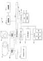
図1は、本発明の様々な態様に係る、MR減衰マップを洗練させるポイントソースの使用を容易にするシステム10を例示する図である。MR画像形成又はスキャニング装置12は、MR減衰補正(AC)マップ16を生成するために身体の輪郭、肺、軟組織、骨構造等が分割される被検体の組織又は他の関心のあるボリュームのMRに基づく画像14を生成する。たとえばポイントソースである1以上の放射線源18は、被検体(たとえば関心のあるボリューム又は患者)の境界の外側であって、核画像形成装置20の検査領域内に配置される。ポイントソースの減衰又は透過データ22は、PETスキャナ又は単電子放出型断層撮影(SPECT: Single Photon Emission Computed Tomography)スキャナのような核画像形成装置20により測定される。なお、減衰及び透過は、一方が、吸収されない(透過される)放射線の全体量の一部を暗示する意味で、互いに逆のことを意味する。数学的に、何れかを使用することができる。本実施の形態では「透過」データが使用されるが、透過データは、透過情報と減衰情報の両者を含むことを理解されたい。1つの固定されたポイントソースから、被検体又は患者の2次元(2D)の投影核イメージ23が生成される。その間、推定される透過データ24は、MR減衰マップ16を使用して被検体のボリュームを通して投影するレイトレーシング技術又はアルゴリズム26を使用してシミュレートされる。測定されたポイントソースの透過データ22及びシミュレートされた透過データ24は、コンパレータ28により比較され、減衰マップ上で実行される反復的な微調整アルゴリズム30により差が最小にされる。従って、準最適なMR減衰マップ16は、測定されたポイントソースの透過データを使用して最適化され、洗練された(たとえば最適な)MR減衰マップ32が生成される。 FIG. 1 is an illustration of a
MR画像が異なる組織のタイプに分割されるとき、それぞれの組織のタイプは、対応する放射線の減衰特性について推定される。それぞれのセグメントのMR画像及び推定された透過値から、被検体を通して任意の放射線に沿った推定される透過を計算することができる。実際に収集されるポイントソースの透過のそれぞれの放射線に沿って、推定される透過が計算される。対応する放射線に沿って推定された透過と実際に測定された透過とを比較することで、推定された透過値における誤差が決定される。多数の放射線が、異なる分割された領域を通過するのと同様に、共通の分割された領域であって、それぞれの領域において異なる経路長をもつ領域をも通過する。空気であると推定された領域及び骨であると推定された領域を容易に確かめることができる。さらに、放射線に沿って、推定された透過と実際に測定された透過との間の適合が最適化されるまで、それぞれ分割された領域の減衰/透過値は、減衰ファクタを繰り返し調整することで改善される。 When the MR image is divided into different tissue types, each tissue type is estimated for the attenuation characteristics of the corresponding radiation. From the MR image of each segment and the estimated transmission value, an estimated transmission along any radiation through the subject can be calculated. An estimated transmission is calculated along each ray of transmission of the point source that is actually collected. By comparing the estimated transmission along the corresponding radiation with the actually measured transmission, an error in the estimated transmission value is determined. In the same way that a large number of radiations pass through different divided areas, they also pass through common divided areas that have different path lengths in the respective areas. The area estimated to be air and the area estimated to be bone can be easily ascertained. In addition, along the radiation, the attenuation / transmission value for each segmented area can be adjusted by repeatedly adjusting the attenuation factor until the fit between the estimated transmission and the actual measured transmission is optimized. Improved.
同様に、予め選択された閾値にないデータを最適化することが不可能なことは、患者の動きを示す場合がある。身体の輪郭及び全体として内部領域の位置を繰り返し調節すること、又は身体の輪郭内にある内部領域の位置を繰り返し調節することで、患者の動きに適合すること、及び/又は患者の動きを検出することを最適化することができる。 Similarly, the inability to optimize data that is not at a preselected threshold may indicate patient movement. Adapt to patient movement and / or detect patient movement by repeatedly adjusting the position of the body contour and the internal area as a whole, or repeatedly adjusting the position of the internal area within the body contour Can be optimized.
MR画像形成装置12及び核イメージング又はスキャニング装置20は、様々な実施の形態によれば、個別の画像形成装置であるか、組み合わせ又はデュアルモダリティ画像形成装置である場合がある。たとえば、デュアルモダリティイメージャの場合、被検体は、MR画像形成のモダリティを使用してスキャニングされ、次いで、ポイントソースが画像形成装置に位置された後、核イメージング(たとえばPET又はSPECT)モダリティを使用して再びスキャンされる。このようにして、被検体の動きは、スキャンの間で最小にすることができる。さらに、この実施の形態及び他の実施の形態では、ポイントソースの位置は、ポイントソースからの透過データの検出を容易にするために、核イメージングスキャンの間で一貫性があるように、一定に維持されるか、及び/又は予め決定される場合がある。 The MR
システム10は、MR画像データ14、準最適なACマップ16、測定されたポイントソースデータ22、シミュレートされた又は推定されたポイントソースの透過データ24、及び洗練されたACマップ32を記憶するメモリ34を含む。メモリは、シミュレートされた透過データ24を生成するために使用されるレイトレーシングアルゴリズム26、コンパレータ28(たとえばシミュレートされたデータを測定されたポイントソースデータに比較するコンピュータ実行可能な命令)、洗練されたACマップ32を生成するとき、ACマップ16を洗練させるために使用される反復的な微調整アルゴリズム(iterative fine-tuning algorithm)30を更に記憶する。
さらに、システム10は、メモリ34に記憶されたデータを分析し、メモリに記憶されるアルゴリズムを実行して、メモリ34における記憶のために新たなデータを生成するプロセッサ36を含む。たとえば、プロセッサは、1以上のMR再構成アルゴリズム38を実行して、被検体のMRスキャンの間に取得されたrawデータであるMRデータからMR画像14を再構成する。同様に、プロセッサは、1以上の核イメージ再構成アルゴリズム39を実行して、被検体の核スキャンの間に取得されたrawデータである核スキャンデータ41から投影核イメージ23を再構成する。さらに、プロセッサは、推定された透過データ24を生成し、コンパレータ28を実行して、推定されたデータ24と測定されたポイントソースの透過データ22との間の差を決定し、反復的な微調整アルゴリズム30を実行して、オリジナルのACマップ16から洗練されたACマップ32を生成する。本明細書で使用される「アルゴリズム」は、メモリ34に持続して記憶され、プロセッサ36により実行される1以上のコンピュータにより実行可能な命令を意味するように解釈される場合がある。 In addition, the
従って、システム10は、被検体と共に核画像形成の検査領域においてポイントソース(又はラインソース)を配置するように利用される。1例では、放射線のポイントソース又はラインソースは、イメージングアイソトープとポイント又はラインソースとを識別するのを容易にするため、被検体のPET画像を生成するために使用されるPETアイソトープとは異なるエネルギーをもつ放射性同位体である。ポイント又はラインソースからの放射線は、PET検出器により検出され、(たとえばメモリ34に記憶され、プロセッサ36により実行される)エネルギー識別アルゴリズム42を使用してPETデータから分離され、特に、被検体を通した投影である透過の放射線データを生成するために使用される。2以上の放射線源を使用するか、患者とは相対的に放射線源を回転することで、3次元の放射線の減衰マップが容易に生成される。ポイントソース又はラインソースは、ガントリー又は核イメージング装置20又は患者支持体の構造に一時的に挿入されるか、又は永続的に搭載することができる。たとえば、レシービング構造は、患者支持体又は穴を画定する構造における予め選択された既知の位置で定義される。ソースの既知の位置は、投影データの分析を容易にする。代替的に、ソースの位置は、投影データを分析することで決定することができる。 Accordingly, the
別の実施の形態によれば、減衰マップは、MRスキャナ12のような解剖のイメージングモダリティから導出される。良好な性能のため、多数のポイントソース18が実際に利用される場合がある。しかし、以下の例では、1つのポイントソースとのPET/MRの組み合わせが記載される。この例によれば、MRスキャナ12を使用して、被検体又は患者のMR画像が生成される。身体の輪郭及び他の内部の臓器の境界が確定される。実質的又は完全に軟組織である知られている領域が識別され、ラベル付けされる。空気及び骨の組織を含む、肺組織のような特徴付けすることができない領域は、ラベル付けされないままにされる。 According to another embodiment, the attenuation map is derived from an anatomical imaging modality such as
同じ被検体又は患者の(たとえばPET又はSPECTといった)核イメージが生成される(たとえば核イメージデータが取得される)。同時に、異なるエネルギーのウィンドウが開き、患者又は被検体の境界の外であって、核イメージャのガントリにおける検査領域内に適切に意図される放射線のポイントソース18からのカウントを受ける。たとえば、SPECT画像形成のため、標準的なエネルギーウィンドウが使用される。PET画像形成のため、エネルギーウィンドウ及び「シングル“single”」モードが使用される。ポイントソースは、患者又は被検体を画像形成するために使用される放射線同位体から発生される放射データよりも高いエネルギーである(たとえば、511kevでPETデータを画像形成するとき、662kevで137Csである)。身体の輪郭又は外形は、散乱からポイントソースの放射線を識別するのを更に容易にする。さらに、PET放射線よりも高いエネルギーをもつポイントソースを使用することで、PET放射線の散乱からポイントソースの放射線を識別することが容易となる。これは、ポイントソースの放射線は、低エネルギーのPET放射線の散乱とオーバラップしないためである。ソースからのデータが取得され、メモリ34に記憶されるヒストグラム又は投影に蓄積される。減衰係数は、分割されたMR画像の既知の領域に(たとえばACマップ16を生成するときに、プロセッサにより)割り当てられる。外部のポイントソース(図2参照)と同じ幾何学的形状をもつMRに基づいた減衰マップ16を通して推定された投影データ24を生成するため、レイトレーシングアルゴリズムを使用して、数学モデルが構築される。減衰マップ16を通して推定された又はシミュレートされた数学投影データ24が、ポイントソースの測定された透過データ22に厳密に一致するまで、未知の領域には、空気又は骨の値に代替的に割り当てられる。適切に分割され、空気及び骨の組織の様々な不明確な領域について値が適切に割り当てられた洗練されたACマップ32は、核イメージ23の減衰の補正を実行するために使用することができる。ひとたび空気及び骨の領域が決定されると、様々な軟組織の領域の減衰ファクタが任意に最適化される。A nuclear image (eg, nuclear image data is acquired) of the same subject or patient (eg, PET or SPECT). At the same time, a window of different energies opens and receives a count from the
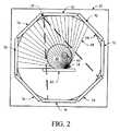
図2は、本発明の1以上の態様に係る、核画像形成装置20に位置されるポイントソース18を例示する。(たとえば被検体、患者である)関心のあるボリューム60は、核画像形成装置20のガントリ64により定義される画像形成領域におけるテーブル又は患者支持体62に配置される。関心のあるボリュームは、関心のあるボリュームのMR画像を使用して容易に曖昧さをなくすことができない、(たとえば肺組織、大気又はガスポケット等といった)骨の組織又は空気の場合がある不明瞭な領域66を有する。したがって、ポイントソース18は、関心のある領域に隣接するテーブル62の近くに位置され、ガントリ64に搭載される核検出器70により検出される放射線透過ライン68を放出する。1実施の形態では、ガントリ64は、回転可能なガントリであり、核検出器70は、透過ライン68を検出するのと同様に、関心のあるボリュームに投与される放射性同位体からの核スキャンデータを取得して核イメージを生成するため、検査領域に関して回転される。 FIG. 2 illustrates a
別の実施の形態では、異なる透過エネルギーを有する2以上のポイントソース18,18’は、完全な視野を形成するために検査領域の周りに配置される。このように、MR画像データにおける骨と空気のボクセルとの間の不明確さを解決するのを容易にするため、2以上のラインの統合が生成される。 In another embodiment, two or
別の実施の形態では、核検出器70は、PET検出器であり、取得されたPETデータにおける減衰は、PET画像を再構成するとき、上述された洗練されたACマップを使用して補正される。 In another embodiment, the
別の実施の形態では、核検出器70は、SPECT(Single Position Emission Computed Tomography)検出器であり、取得されたSPECTデータにおける減衰は、SPECT画像を再構成するとき、上述された洗練されたACマップを使用して補正される。 In another embodiment, the
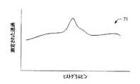
図3Aは、測定されたポイントソースの透過データ22と、それぞれのデータが含まれる複数のヒストグラムビンとのグラフ上の関係71を例示する。PET減衰の補正のために容易に使用されるようになるまでACマップを繰り返し洗練させるために、測定されたポイントソースの透過データ22は、推定されたポイントソース透過データ24(図3B)と共に使用される。 FIG. 3A illustrates a
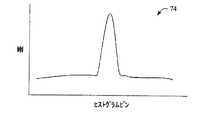
図3Bは、レイトレーシングアルゴリズム26(図1)を使用してプロセッサ36により生成されたシミュレートされた又は推定されたポイントソースの透過データ24と、それぞれのシミュレートされたデータが含まれる複数のヒストグラムビンとの間のグラフ上の関係を例示する。推定されたポイントソースの透過データ24は、測定されたポイントソースデータ22と比較され、PET画像を再構成するとき、ACマップがPETスキャンデータにおける減衰の補正における使用の準備があるかを判定する。 FIG. 3B illustrates the simulated or estimated point
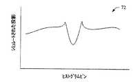
図3Cは、測定されたポイントソースの透過データの値とシミュレートされた透過データの値との差と、それぞれの差のデータが含まれる複数のヒストグラムのビンとのグラフ上の関係74を例示する。差の値は、プロセッサ36が図1のコンパレータ28を実行するときに決定される。それぞれのACマップでの繰り返しの調節により、差の値が低減される。ひとたび、推定された透過データとシミュレートされた透過データとの間の差の値が十分に小さくなると(たとえば予め決定された閾値以下)、洗練されたACマップは、PET減衰の補正において使用されるために記憶される。 FIG. 3C illustrates a
図4は、本発明の様々な態様に係る、核イメージにおける減衰を補正する洗練されたACマップを生成する方法を例示する。ステップ78で、被検体のMR画像が生成される。ステップ80で、ACマップは、被検体の分割されたMR画像から生成される。ステップ82で、被検体は、核スキャナ(たとえばPET又はSPECT)の検査領域に配置され、ポイントソースは、被検体の外側に位置され、且つ検査領域内に位置される。ポイントソースは、(たとえば約662kevである)137Csのような被検体に投与される放射線同位体(たとえば511kev)よりも高いエネルギーをもつガンマ線を放出する。代替的に、133Ba(たとえば約360kev)のような、(たとえば511kevよりも低い)低エネルギーがポイントソースについて使用される。FIG. 4 illustrates a method for generating a refined AC map that corrects for attenuation in a nuclear image, in accordance with various aspects of the present invention. At
ステップ84で、核スキャナを使用して、ポイントソースからの透過データが測定され、被検体の核イメージデータが生成される。たとえば、シングルモードで動作しているPETは、図3Aに示されるように、核同位体の放出データと同時にポイントソースの透過データを測定する。ステップ86は、図3Bに示されるように、ACマップに関するレイトレーシング技術を使用して透過データが導出又は推定される。ステップ88で、図3Cに示されるように、ステップ84からの透過データとステップ86からの投影データとの間の差が決定される。ステップ90で、二乗平均平方根誤差(rms)等のような、ステップ88で決定された差を表すエラーファクタが計算される。 At
ステップ92で、ステップ90で計算されたファクタを使用して減衰マップが変更され、本方法は、次の投影データの生成、測定された透過データへの比較、差の決定及びrmsファクタの生成のためにステップ86に戻る。ステップ96で、最小のrmsエラーが発見されたかに関する判定が行われる。最小のrmsエラーが発見されていないと判定された場合、本方法は、ステップ92に戻る。最小のrmsエラーが発見されたと判定された場合、ステップ97で、ステップ84で生成された被検体の核イメージにおける減衰の補正で使用するため、改善された減衰マップが保存される。ステップ98で、ステップ84で生成された核イメージデータは、減衰が補正された核イメージ(たとえばPET又はSPECT)を生成するため、ステップ97で記憶されたACマップを使用して再構成される。 In
図5を参照して、例示的なホスピタルシステム100は、MR画像形成装置12、核スキャナ20(たとえばPET又はSPECT)等のような複数の画像形成装置を含み、これら複数の画像形成装置は、3D画像の表現を生成するため、個別の再構成プロセッサ又は共有される再構成プロセッサ102により再構成される画像形成データを生成する。画像の表現は、ネットワーク104から中央のメモリ106又はローカルのメモリ108に伝達される。 Referring to FIG. 5, an
ネットワークに接続されるステーション110で、オペレータは、ユーザインタフェース124を使用して、選択された3Dの患者のMR減衰マップを中央のメモリ106及びローカルのメモリ108に移動するか、中央のメモリ106とローカルのメモリ108との間に移動する。ビデオプロセッサ116は、ディスプレイ122の第一の窓(view port)1181において選択された減衰マップ(又はMR画像)を表示する。核イメージは、第二の窓1182において表示される。第三の窓1183は、減衰マップと核イメージの重ね合わせを表示する。たとえば、ユーザは、MR減衰マップにおけるランドマークを対応する構造又は核イメージにおけるランドマークに位置決めすることができる。たとえば、オペレータは、インタフェース124を通して、減衰マップの画像におけるランドマークに対応する核イメージのランドマークを(たとえばマウス、スタイラス、又は他の適切なユーザ入力装置を使用して)選択する。代替的に、減衰マップは、プロセッサ116におけるプログラムにより自動的に位置決めすることができる。ユーザインタフェース124におけるプロセッサ36(図1)は、次いで補正アルゴリズムを実行し、減衰マップにおける不明確な領域において補うときに採用すべき適切な組織のタイプ(たとえば骨又は空気)を推測する。At the
次いで、他の応用で使用される場合がある、減衰が補正された核イメージを再構成するために洗練された減衰マップが使用される。たとえば、治療計画ステーション130は、治療のセッションを計画するために減衰が補正されたPET画像を使用する。ひとたびオペレータの満足に対して計画されると、計画された治療は、自動化された手順に対して必要に応じて、計画されたセッションを実現する治療装置132に転送することができる。他のステーションは、様々な他の計画の手順において減衰が補正されたPET画像を使用する場合がある。 The refined attenuation map is then used to reconstruct the attenuation corrected nuclear image that may be used in other applications. For example, the
別の実施の形態では、窓1183で表示されたオーバレイは、核イメージに対してMR画像に重み付けし、逆に、MR画像に対して核イメージを重み付けするために調節可能である。たとえば、メカニカル又はディスプレイ122に提示され、入力装置により操作される場合があるスライダバー又はノブ(図示せず)は、MR画像又は核イメージの重みを変化させるために調節される場合がある。1実施の形態では、オペレータは、(窓1181で示される)MR画像データから、MR及び核イメージデータの多数の及び/又は連続した組み合わせを通して、(窓1182に示される)核イメージデータへの画像を窓1183において調節する。たとえば、MR画像データの核画像データに対する比率は、0:1から1:0に不連続に又は連続して調節される。別の選択肢として、MR画像は、グレイスケールで表示され、核イメージは、カラー化することができる。MR画像における解剖学的ランドマークは、核イメージを被検体に関連付けるのに役立つ。In another embodiment, the overlay displayed in window 1183, and weighted MR image relative to nuclear image, conversely, is adjustable to weight the nuclear image to the MR image. For example, a slider bar or knob (not shown) that may be presented on the mechanical or
本発明は、幾つかの実施の形態を参照して記載された。先の詳細な説明を読んで理解することで、他の変更及び代替が行われる場合がある。本発明は、特許請求の範囲又はその等価なものの範囲に含まれる限り、全ての係る変更及び代替を含むとして解釈されるべきことが意図される。 The invention has been described with reference to several embodiments. Other changes and substitutions may be made upon reading and understanding the above detailed description. It is intended that the invention be construed to include all such modifications and alternatives as long as they fall within the scope of the claims or their equivalents.
Claims (15)
Translated fromJapanese核取得スキャンの間に前記被検体の核画像データを取得する核スキャナであって、前記核スキャナの検査領域に位置される放射線源からの透過データを測定する核スキャナと、
前記MR画像データから減衰補正(AC)マップを生成し、前記測定された透過データを使用して、前記ACマップを繰り返し洗練させ、洗練されたACマップを生成し、前記洗練されたACマップを使用して、減衰について前記核画像データを補正するプロセッサと、
を備える解剖学的な画像補正システム。An MR imager that acquires MR image data of a subject during a magnetic resonance (MR) acquisition scan;
A nuclear scanner for acquiring nuclear image data of the subject during a nuclear acquisition scan, wherein the nuclear scanner measures transmission data from a radiation source located in an examination region of the nuclear scanner;
An attenuation correction (AC) map is generated from the MR image data, and the measured transmission data is used to repeatedly refine the AC map to generate a refined AC map. Using a processor to correct the nuclear image data for attenuation;
An anatomical image correction system comprising:
前記ACマップにレイトレーシングアルゴリズムを実行して、推定された透過データを生成し、
前記推定された透過データと前記測定された透過データとの間の差を決定する比較を行い、
前記ACマップにおける減衰の値を調節して、前記推定された透過データと前記測定された透過データとの間の差を低減する、
請求項1記載のシステム。The processor is
Run a ray tracing algorithm on the AC map to generate estimated transmission data;
Performing a comparison to determine the difference between the estimated transmission data and the measured transmission data;
Adjusting the value of attenuation in the AC map to reduce the difference between the estimated transmission data and the measured transmission data;
The system of claim 1.
請求項2記載のシステム。The processor calculates a least mean square error value to minimize a difference between the estimated transmission data and the measured transmission data;
The system according to claim 2.
請求項2記載のシステム。The processor stores the refined AC map in memory in response to determining that the difference has been minimized.
The system according to claim 2.
前記PET用のスキャナは、前記被検体に投与される放射線同位体から放出される放射線であって、約511kevのエネルギーレベルを有する放射線からデータを取得し、
前記放射線源は、約511kevとは異なるエネルギーレベルを有する放射性物質である、
請求項1記載のシステム。The nuclear scanner is a scanner for positron emission tomography (PET),
The PET scanner acquires data from radiation emitted from a radioisotope administered to the subject and having an energy level of about 511 kev ,
The radiation source is a radioactive material having an energy level different from about 511 kev ;
The system of claim1 .
請求項1記載のシステム。The radiation source is provided in a fixed position, or provided in at least one of a patient support and the gantry where the subject is located in an examination region of the gantry of the nuclear scanner.
The system of claim 1.
被検体のMR画像データからACマップを生成するステップと、
前記被検体を通して、前記被検体の放射線源から放射線を放出するステップと、
前記被検体を通して透過された放射線から透過データを測定するステップと、
前記ACマップから推定された透過データを生成するステップと、
前記測定された透過データと前記推定された透過データとの比較に基づいて前記ACマップを調整することで、洗練されたACマップを生成するステップと、
を含む方法。A method for refining a magnetic resonance (MR) attenuation correction (AC) map, comprising:
Generating an AC map from MR image data of the subject;
Emitting radiation through the subject from a radiation source of the subject;
Measuring transmission data from radiation transmitted through the subject;
Generating transmission data estimated from the AC map;
Generating a refined AC map by adjusting the AC map based on a comparison of the measured transmission data and the estimated transmission data;
Including methods.
洗練されたACマップにより前記PETデータを補正するステップと、
補正されたPETデータをPET画像に再構成するステップと、
を更に含む請求項7記載の方法。Generating data for positron emission tomography (PET);
Correcting the PET data with a refined AC map;
Reconstructing the corrected PET data into a PET image;
The method of claim7 further comprising:
前記ACマップを調整して前記第一の誤差を低減するステップと、
調整されたACマップを使用して第二の推定された透過データを決定するステップと、
前記測定された透過データと前記第二の推定された透過データとの間の第二の誤差を決定するステップと、
前記調整されたACマップを更に調整して前記第二の誤差を低減するステップと、
を更に含む請求項7記載の方法。Determining a first error between the measured transmission data and the estimated transmission data;
Adjusting the AC map to reduce the first error;
Determining a second estimated transmission data using the adjusted AC map ;
Determining a second error between the measured transmission data and the second estimated transmission data ;
Further adjusting the adjusted AC map to reduce the second error ;
The method of claim7 further comprising:
二乗平均平方根(rms)の値を繰り返し決定し、決定されたrms値が最適化されるまで前記ACマップを調整するステップを含む、
請求項9記載の方法。Determining the second error comprises:
Repeatedly determining a root mean square (rms) value and adjusting the AC map until the determined rms value is optimized,
The method of claim9 .
放射線源は、約511kevとは異なるエネルギーレベルを有する放射性物質である、
請求項7記載の方法。A scanner for positron emission tomography (PET) acquires data from radiation emitted from a radioisotope administered to the subject having an energy level of about 511 kev;
The radiation source is a radioactive material having an energy level different from about 511 kev,
The method of claim7 .
放射線検出器のリングと、
前記放射線検出器のリング内の固定して設けられた放射線源と、
洗練された減衰補正(AC)マップを生成するために請求項7記載の方法を実行するプロセッサと、
前記洗練されたACマップにより前記放射線検出器からのPETデータを補正し、補正されたPETデータを減衰が補正されたPET画像に再構成する再構成アルゴリズムと、
を備えるPCT用のスキャナ。A scanner for positron emission tomography (PET),
A ring of radiation detectors;
A fixedly provided radiation source in the ring of the radiation detector;
A processor for performing the method of claim7 to generate a refined attenuation correction (AC) map;
A reconstruction algorithm that corrects the PET data from the radiation detector with the refined AC map and reconstructs the corrected PET data into a PET image with corrected attenuation;
PCT scanner equipped with.
複数の放射線検出器と、
放射線検出器の検査領域内に固定して設けられた放射線源と、
洗練された減衰補正(AC)マップを生成するために請求項7記載の方法を実行するプログラムと、
前記洗練されたACマップにより前記放射線検出器からのSPECTデータを補正し、補正されたSPECTデータを減衰が補正されたSPECT画像に再構成する再構成アルゴリズムと、
を備えるSPECT用のスキャナ。A scanner for single electron emission computed tomography (SPECT),
A plurality of radiation detectors;
A radiation source fixed in the examination area of the radiation detector;
A program for performing the method of claim7 to generate a refined attenuation correction (AC) map;
A reconstruction algorithm that corrects SPECT data from the radiation detector with the refined AC map and reconstructs the corrected SPECT data into a SPECT image with corrected attenuation;
SPECT scanner with
取得された磁気共鳴(MR)画像データから前記ACマップを生成するステップと、
放射線のポイントソースから検出された透過データを使用して前記MR画像データにおける骨のボクセルと空気のボクセルとを識別するステップと、
検出されたポイントソースの透過データに基づいて前記ACマップを更新するステップと、
を含む方法。A method for refining an attenuation correction (AC) map,
Generating the AC map from acquired magnetic resonance (MR) image data;
Identifying bone voxels and air voxels in the MR image data using transmission data detected from a point source of radiation;
Updating the AC map based on detected point source transparency data;
Including methods.
Applications Claiming Priority (3)
| Application Number | Priority Date | Filing Date | Title |
|---|---|---|---|
| US9755508P | 2008-09-17 | 2008-09-17 | |
| US61/097,555 | 2008-09-17 | ||
| PCT/IB2009/053944WO2010032167A2 (en) | 2008-09-17 | 2009-09-09 | Mr segmentation using transmission data in hybrid nuclear/mr imaging |
Publications (2)
| Publication Number | Publication Date |
|---|---|
| JP2012503177A JP2012503177A (en) | 2012-02-02 |
| JP5385394B2true JP5385394B2 (en) | 2014-01-08 |
Family
ID=42039963
Family Applications (1)
| Application Number | Title | Priority Date | Filing Date |
|---|---|---|---|
| JP2011526607AExpired - Fee RelatedJP5385394B2 (en) | 2008-09-17 | 2009-09-09 | MR segmentation using transmission data in hybrid nuclear / MR imaging |
Country Status (6)
| Country | Link |
|---|---|
| US (1) | US8406495B2 (en) |
| EP (1) | EP2338142A2 (en) |
| JP (1) | JP5385394B2 (en) |
| CN (1) | CN102265307B (en) |
| RU (1) | RU2504841C2 (en) |
| WO (1) | WO2010032167A2 (en) |
Families Citing this family (26)
| Publication number | Priority date | Publication date | Assignee | Title |
|---|---|---|---|---|
| US8218848B2 (en)* | 2008-07-23 | 2012-07-10 | Siemens Aktiengesellschaft | System and method for the generation of attenuation correction maps from MR images |
| DE102008047840B4 (en)* | 2008-09-18 | 2018-08-30 | Siemens Healthcare Gmbh | Method for the at least partial determination and / or adaptation of an attenuation map used for the attenuation correction of positron emission tomography image data sets in a combined magnetic resonance positron emission tomography device |
| CN103415779B (en)* | 2011-03-07 | 2016-05-11 | 皇家飞利浦有限公司 | Systems and methods for MR segmentation using nuclear emission data in hybrid nuclear imaging/MR |
| CN103458790A (en)* | 2011-03-17 | 2013-12-18 | 皇家飞利浦有限公司 | Multiple modality cardiac imaging |
| JP6042879B2 (en)* | 2011-05-24 | 2016-12-14 | コーニンクレッカ フィリップス エヌ ヴェKoninklijke Philips N.V. | Apparatus for generating an assignment between an image region and an element class of an image |
| WO2013121379A2 (en) | 2012-02-16 | 2013-08-22 | Koninklijke Philips N.V. | Spatially corrected nuclear image reconstruction |
| US9204817B2 (en)* | 2012-04-19 | 2015-12-08 | General Electric Company | Attenuation correction in positron emission tomography using magnetic resonance imaging |
| WO2013168043A2 (en)* | 2012-05-08 | 2013-11-14 | Koninklijke Philips N.V. | Spect/pet imaging system |
| KR101350496B1 (en)* | 2012-05-22 | 2014-01-16 | 서울대학교산학협력단 | Method to generate a attenuation map of emission tomography and MRI combined imaging system |
| US8923592B2 (en)* | 2012-05-29 | 2014-12-30 | General Electric Company | Methods and systems for performing attenuation correction |
| US8867814B2 (en)* | 2012-10-04 | 2014-10-21 | General Electric Company | Methods and systems for generating a positron emission tomography attenuation correction map |
| US9002082B2 (en) | 2012-12-27 | 2015-04-07 | General Electric Company | Axially varying truncation completion for MR-based attenuation correction for PET/MR |
| US9364192B2 (en)* | 2013-03-15 | 2016-06-14 | Siemens Medical Solutions Usa, Inc. | Error estimates in quantitative functional imaging |
| RU2523929C1 (en)* | 2013-03-18 | 2014-07-27 | Корпорация "САМСУНГ ЭЛЕКТРОНИКС Ко., Лтд." | System and method for automated planning of views in 3d brain images |
| US9311707B2 (en)* | 2013-09-27 | 2016-04-12 | General Electric Company | System and method for attenuation correction of phantom images |
| US10772580B2 (en)* | 2014-06-13 | 2020-09-15 | Siemens Medical Solutions Usa, Inc. | Multiple emission energies in single photon emission computed tomography |
| US20160066874A1 (en)* | 2014-09-10 | 2016-03-10 | The General Hospital Corporation | Attenuation correction of positron emission tomography data using magnetic resonance images depicting bone density variations |
| JP6467221B2 (en)* | 2014-12-22 | 2019-02-06 | キヤノン株式会社 | Image processing apparatus and method |
| US9508165B1 (en)* | 2015-06-30 | 2016-11-29 | General Electric Company | Systems and methods for peak tracking and gain adjustment |
| EP3371617B1 (en)* | 2015-11-02 | 2024-09-25 | Koninklijke Philips N.V. | Method for tissue classification and magnetic resonance imaging system |
| US10210634B2 (en) | 2016-07-20 | 2019-02-19 | Shanghai United Imaging Healthcare Co., Ltd. | System and method for segmenting medical image |
| WO2019172997A1 (en)* | 2018-03-07 | 2019-09-12 | Siemens Medical Solutions Usa, Inc. | Calibration bias reduction in a pressurized gas ion chamber-based dose calibrator |
| CN109308728B (en)* | 2018-10-25 | 2023-01-03 | 上海联影医疗科技股份有限公司 | Positron emission computed tomography image processing method and device |
| JP7676443B2 (en)* | 2020-05-01 | 2025-05-14 | シーメンス メディカル ソリューションズ ユーエスエー インコーポレイテッド | Transmission imaging in forward-scattered gamma-ray-based PET scanners with coincidence detection - Patents.com |
| CN112435861B (en)* | 2020-10-16 | 2022-01-11 | 惠州亿纬锂能股份有限公司 | Positive electrode of hybrid capacitor and preparation method and application thereof |
| EP4156100A1 (en)* | 2021-09-23 | 2023-03-29 | Siemens Healthcare GmbH | Providing result image data |
Family Cites Families (22)
| Publication number | Priority date | Publication date | Assignee | Title |
|---|---|---|---|---|
| US5376795A (en)* | 1990-07-09 | 1994-12-27 | Regents Of The University Of California | Emission-transmission imaging system using single energy and dual energy transmission and radionuclide emission data |
| US5449913A (en) | 1993-11-03 | 1995-09-12 | Chang; Wei | Apparatus for producing attenuation scan |
| US5672877A (en)* | 1996-03-27 | 1997-09-30 | Adac Laboratories | Coregistration of multi-modality data in a medical imaging system |
| WO1999056150A1 (en) | 1998-04-27 | 1999-11-04 | Duke University | Transmission scanning technique for gamma camera coincidence imaging |
| US6310968B1 (en) | 1998-11-24 | 2001-10-30 | Picker International, Inc. | Source-assisted attenuation correction for emission computed tomography |
| US6628983B1 (en)* | 2000-10-25 | 2003-09-30 | Koninklijke Philips Electronics N.V. | Nuclear imaging systems and methods with feature-enhanced transmission imaging |
| TW542993B (en) | 2001-07-12 | 2003-07-21 | Inst Information Industry | Multi-dimension and multi-algorithm document classifying method and system |
| DE10231061A1 (en)* | 2002-07-10 | 2004-01-22 | Philips Intellectual Property & Standards Gmbh | Process and system for improving the information content in an image |
| EP1573495B1 (en)* | 2002-11-04 | 2009-11-04 | Spectrum Dynamics LLC | Apparatus and methods for imaging and attenuation correction |
| US7197171B2 (en)* | 2003-02-19 | 2007-03-27 | Elgems Ltd. | Nuclear imaging |
| GB0414277D0 (en)* | 2004-06-25 | 2004-07-28 | Leuven K U Res & Dev | Orthognatic surgery |
| US20060004274A1 (en)* | 2004-06-30 | 2006-01-05 | Hawman Eric G | Fusing nuclear medical images with a second imaging modality |
| WO2006064400A2 (en)* | 2004-12-15 | 2006-06-22 | Koninklijke Philips Electronics, N.V. | Registration of multi-modality images |
| ATE538841T1 (en)* | 2005-10-17 | 2012-01-15 | Alberta Health Services | INTEGRATED EXTERNAL BEAM RADIOTHERAPY AND MRI SYSTEM |
| US7402807B2 (en)* | 2005-11-02 | 2008-07-22 | Siemens Medical Solutions Usa, Inc. | Method for reducing an electronic time coincidence window in positron emission tomography |
| US7348564B2 (en) | 2005-12-12 | 2008-03-25 | General Electric Company | Multi modality imaging methods and apparatus |
| US7465927B2 (en)* | 2006-03-23 | 2008-12-16 | Siemens Medical Solutions Usa, Inc. | Attenuation correction for nuclear medical imaging scanners with simultaneous transmission and emission acquisition |
| DE102006046287A1 (en)* | 2006-09-29 | 2008-04-03 | Siemens Ag | Magnetic resonance-positron emissions tomography field generating unit for representation of tissue in human or animal body, has movable resting board in examination channel and main magnet for generating constant magnetic field |
| US20080135769A1 (en)* | 2006-11-22 | 2008-06-12 | Rosen Bruce R | Attenuation correction of pet image using image data acquired with an mri system |
| JP4584232B2 (en)* | 2006-11-27 | 2010-11-17 | 富士フイルムRiファーマ株式会社 | Brain model |
| DE102007034953B4 (en)* | 2007-07-26 | 2016-09-22 | Siemens Healthcare Gmbh | A method for recording movements of a patient and associated medical device |
| US8098916B2 (en)* | 2007-10-30 | 2012-01-17 | General Electric Company | System and method for image-based attenuation correction of PET/SPECT images |
- 2009
- 2009-09-09USUS13/062,222patent/US8406495B2/ennot_activeExpired - Fee Related
- 2009-09-09RURU2011115056/08Apatent/RU2504841C2/ennot_activeIP Right Cessation
- 2009-09-09JPJP2011526607Apatent/JP5385394B2/ennot_activeExpired - Fee Related
- 2009-09-09CNCN200980136210.0Apatent/CN102265307B/ennot_activeExpired - Fee Related
- 2009-09-09EPEP09787148Apatent/EP2338142A2/ennot_activeWithdrawn
- 2009-09-09WOPCT/IB2009/053944patent/WO2010032167A2/enactiveApplication Filing
Also Published As
| Publication number | Publication date |
|---|---|
| JP2012503177A (en) | 2012-02-02 |
| US8406495B2 (en) | 2013-03-26 |
| RU2011115056A (en) | 2012-10-27 |
| RU2504841C2 (en) | 2014-01-20 |
| EP2338142A2 (en) | 2011-06-29 |
| CN102265307A (en) | 2011-11-30 |
| WO2010032167A3 (en) | 2011-04-21 |
| CN102265307B (en) | 2014-10-22 |
| WO2010032167A2 (en) | 2010-03-25 |
| US20110164801A1 (en) | 2011-07-07 |
Similar Documents
| Publication | Publication Date | Title |
|---|---|---|
| JP5385394B2 (en) | MR segmentation using transmission data in hybrid nuclear / MR imaging | |
| CN102711617B (en) | Scan plan field of view adjustor, determiner, and/or quality assessor | |
| CN102934143B (en) | For producing the method for decay pattern in PET-MR | |
| JP5462865B2 (en) | Use of non-attenuated corrected PET emission images to compensate for imperfect anatomical images | |
| CN109060849B (en) | Method, system and device for determining radiation dose modulation line | |
| JP6133089B2 (en) | System and method for attenuation compensation in nuclear medicine imaging based on emission data | |
| CN111080706B (en) | Method, device, computer program and data carrier for reconstructing an image dataset | |
| US20220313176A1 (en) | Artificial Intelligence Training with Multiple Pulsed X-ray Source-in-motion Tomosynthesis Imaging System | |
| CN107635469B (en) | Estimation of attenuation map based on scatter coincidence in PET system | |
| EP2245592B1 (en) | Image registration alignment metric | |
| CN108697402A (en) | The gyrobearing of deep brain stimulation electrode is determined in 3-D view | |
| CN106999135B (en) | Radiation Emission Imaging Systems and Methods | |
| CN101903908A (en) | Feature-based 2D/3D image registration | |
| CN109077746B (en) | Method, system and device for determining radiation dose modulation line | |
| CN109124666A (en) | A kind of mthods, systems and devices of determining dose of radiation modulation lines | |
| EP4097683B1 (en) | Methods and apparatus for deep learning based image attenuation correction | |
| JP6855476B2 (en) | Tissue classification methods, computer programs, and magnetic resonance imaging systems | |
| CN111050652A (en) | Spectral (multi-energy) image data for image-guided applications | |
| KR101350496B1 (en) | Method to generate a attenuation map of emission tomography and MRI combined imaging system | |
| CN111316327B (en) | Attenuation correction of PET data for moving objects | |
| US20240374227A1 (en) | Method for generating a volume model of an object under examination, control device, x-ray apparatus, computer program, and electronically readable data medium |
Legal Events
| Date | Code | Title | Description |
|---|---|---|---|
| A621 | Written request for application examination | Free format text:JAPANESE INTERMEDIATE CODE: A621 Effective date:20120906 | |
| TRDD | Decision of grant or rejection written | ||
| A01 | Written decision to grant a patent or to grant a registration (utility model) | Free format text:JAPANESE INTERMEDIATE CODE: A01 Effective date:20130910 | |
| A61 | First payment of annual fees (during grant procedure) | Free format text:JAPANESE INTERMEDIATE CODE: A61 Effective date:20131003 | |
| R150 | Certificate of patent or registration of utility model | Free format text:JAPANESE INTERMEDIATE CODE: R150 | |
| LAPS | Cancellation because of no payment of annual fees |