JP5377330B2 - Vaccine based on targeting of antigen to DCIR expressed on antigen presenting cells - Google Patents
Vaccine based on targeting of antigen to DCIR expressed on antigen presenting cellsDownload PDFInfo
- Publication number
- JP5377330B2 JP5377330B2JP2009548481AJP2009548481AJP5377330B2JP 5377330 B2JP5377330 B2JP 5377330B2JP 2009548481 AJP2009548481 AJP 2009548481AJP 2009548481 AJP2009548481 AJP 2009548481AJP 5377330 B2JP5377330 B2JP 5377330B2
- Authority
- JP
- Japan
- Prior art keywords
- antigen
- dcir
- cells
- antibody
- flu
- Prior art date
- Legal status (The legal status is an assumption and is not a legal conclusion. Google has not performed a legal analysis and makes no representation as to the accuracy of the status listed.)
- Expired - Fee Related
Links
- 239000000427antigenSubstances0.000titleclaimsabstractdescription302
- 108091007433antigensProteins0.000titleclaimsabstractdescription286
- 102000036639antigensHuman genes0.000titleclaimsabstractdescription286
- 101000766908Homo sapiens C-type lectin domain family 4 member AProteins0.000titleclaimsabstractdescription23
- 102100028667C-type lectin domain family 4 member AHuman genes0.000titleclaimsabstractdescription18
- 229960005486vaccineDrugs0.000titleclaimsdescription51
- 230000008685targetingEffects0.000titleclaimsdescription47
- 210000000612antigen-presenting cellAnatomy0.000titleclaimsdescription10
- 210000004443dendritic cellAnatomy0.000claimsabstractdescription152
- 239000012634fragmentSubstances0.000claimsabstractdescription34
- 230000030741antigen processing and presentationEffects0.000claimsabstractdescription12
- 210000004027cellAnatomy0.000claimsdescription77
- 108090000623proteins and genesProteins0.000claimsdescription62
- 210000001744T-lymphocyteAnatomy0.000claimsdescription56
- 102000004169proteins and genesHuman genes0.000claimsdescription56
- 108090000765processed proteins & peptidesProteins0.000claimsdescription32
- 230000001580bacterial effectEffects0.000claimsdescription29
- 230000027455bindingEffects0.000claimsdescription29
- 108020001507fusion proteinsProteins0.000claimsdescription21
- 102000037865fusion proteinsHuman genes0.000claimsdescription21
- 239000003795chemical substances by applicationSubstances0.000claimsdescription18
- 108010038512Platelet-Derived Growth FactorProteins0.000claimsdescription17
- 102000010780Platelet-Derived Growth FactorHuman genes0.000claimsdescription17
- -1TN -ΑProteins0.000claimsdescription15
- 239000008194pharmaceutical compositionSubstances0.000claimsdescription15
- 230000003612virological effectEffects0.000claimsdescription15
- 230000002538fungal effectEffects0.000claimsdescription14
- 239000003102growth factorSubstances0.000claimsdescription13
- 108010029697CD40 LigandProteins0.000claimsdescription12
- 102100032937CD40 ligandHuman genes0.000claimsdescription12
- 102000004127CytokinesHuman genes0.000claimsdescription12
- 108090000695CytokinesProteins0.000claimsdescription12
- 102000018233Fibroblast Growth FactorHuman genes0.000claimsdescription12
- 108050007372Fibroblast Growth FactorProteins0.000claimsdescription12
- 229940126864fibroblast growth factorDrugs0.000claimsdescription12
- 108010009583Transforming Growth FactorsProteins0.000claimsdescription11
- 102000009618Transforming Growth FactorsHuman genes0.000claimsdescription11
- 108010045512cohesinsProteins0.000claimsdescription11
- 102000004196processed proteins & peptidesHuman genes0.000claimsdescription11
- 102100039620Granulocyte-macrophage colony-stimulating factorHuman genes0.000claimsdescription10
- 150000007523nucleic acidsChemical class0.000claimsdescription10
- 108010017213Granulocyte-Macrophage Colony-Stimulating FactorProteins0.000claimsdescription9
- 108010002350Interleukin-2Proteins0.000claimsdescription9
- 102000039446nucleic acidsHuman genes0.000claimsdescription9
- 108020004707nucleic acidsProteins0.000claimsdescription9
- 102100037850Interferon gammaHuman genes0.000claimsdescription7
- 108010074328Interferon-gammaProteins0.000claimsdescription7
- 210000003719b-lymphocyteAnatomy0.000claimsdescription7
- VBEQCZHXXJYVRD-GACYYNSASA-NuroantheloneChemical compoundC([C@@H](C(=O)N[C@H](C(=O)N[C@@H](CS)C(=O)N[C@@H](CC(N)=O)C(=O)N[C@@H](CS)C(=O)N[C@H](C(=O)N[C@@H]([C@@H](C)CC)C(=O)NCC(=O)N[C@@H](CC=1C=CC(O)=CC=1)C(=O)N[C@@H](CO)C(=O)NCC(=O)N[C@@H](CC(O)=O)C(=O)N[C@@H](CCCNC(N)=N)C(=O)N[C@@H](CS)C(=O)N[C@@H](CCC(N)=O)C(=O)N[C@@H]([C@@H](C)O)C(=O)N[C@@H](CCCNC(N)=N)C(=O)N[C@@H](CC(O)=O)C(=O)N[C@@H](CC(C)C)C(=O)N[C@@H](CCCNC(N)=N)C(=O)N[C@@H](CC=1C2=CC=CC=C2NC=1)C(=O)N[C@@H](CC=1C2=CC=CC=C2NC=1)C(=O)N[C@@H](CCC(O)=O)C(=O)N[C@@H](CC(C)C)C(=O)N[C@@H](CCCNC(N)=N)C(O)=O)C(C)C)[C@@H](C)O)NC(=O)[C@H](CO)NC(=O)[C@H](CC(O)=O)NC(=O)[C@H](CC(C)C)NC(=O)[C@H](CO)NC(=O)[C@H](CCC(O)=O)NC(=O)[C@@H](NC(=O)[C@H](CC=1NC=NC=1)NC(=O)[C@H](CCSC)NC(=O)[C@H](CS)NC(=O)[C@@H](NC(=O)CNC(=O)CNC(=O)[C@H](CC(N)=O)NC(=O)[C@H](CC(C)C)NC(=O)[C@H](CS)NC(=O)[C@H](CC=1C=CC(O)=CC=1)NC(=O)CNC(=O)[C@H](CC(O)=O)NC(=O)[C@H](CC=1C=CC(O)=CC=1)NC(=O)[C@H](CO)NC(=O)[C@H](CO)NC(=O)[C@H]1N(CCC1)C(=O)[C@H](CS)NC(=O)CNC(=O)[C@H]1N(CCC1)C(=O)[C@H](CC=1C=CC(O)=CC=1)NC(=O)[C@H](CO)NC(=O)[C@@H](N)CC(N)=O)C(C)C)[C@@H](C)CC)C1=CC=C(O)C=C1VBEQCZHXXJYVRD-GACYYNSASA-N0.000claimsdescription7
- 108010059616ActivinsProteins0.000claimsdescription6
- 102100026818Inhibin beta E chainHuman genes0.000claimsdescription6
- 108090000723Insulin-Like Growth Factor IProteins0.000claimsdescription6
- 102000014429Insulin-like growth factorHuman genes0.000claimsdescription6
- 108090000172Interleukin-15Proteins0.000claimsdescription6
- 102000003812Interleukin-15Human genes0.000claimsdescription6
- 108090000978Interleukin-4Proteins0.000claimsdescription6
- 102000004388Interleukin-4Human genes0.000claimsdescription6
- 102100032101Tumor necrosis factor ligand superfamily member 9Human genes0.000claimsdescription6
- 239000000488activinSubstances0.000claimsdescription6
- 108010002586Interleukin-7Proteins0.000claimsdescription5
- 102100021592Interleukin-7Human genes0.000claimsdescription5
- 230000012010growthEffects0.000claimsdescription5
- 102000019034ChemokinesHuman genes0.000claimsdescription4
- 108010012236ChemokinesProteins0.000claimsdescription4
- 108010017080Granulocyte Colony-Stimulating FactorProteins0.000claimsdescription4
- 102000004269Granulocyte Colony-Stimulating FactorHuman genes0.000claimsdescription4
- 102000013462Interleukin-12Human genes0.000claimsdescription4
- 108010065805Interleukin-12Proteins0.000claimsdescription4
- 150000001720carbohydratesChemical class0.000claimsdescription4
- 235000014633carbohydratesNutrition0.000claimsdescription4
- 230000010261cell growthEffects0.000claimsdescription4
- 230000004069differentiationEffects0.000claimsdescription4
- 1080100828084-1BB LigandProteins0.000claimsdescription3
- 101710098275C-X-C motif chemokine 10Proteins0.000claimsdescription3
- 101150093802CXCL1 geneProteins0.000claimsdescription3
- 108010071942Colony-Stimulating FactorsProteins0.000claimsdescription3
- 108010079505EndostatinsProteins0.000claimsdescription3
- 102000003951ErythropoietinHuman genes0.000claimsdescription3
- 108090000394ErythropoietinProteins0.000claimsdescription3
- 101001043796Gallus gallus Interleukin-8Proteins0.000claimsdescription3
- 102100039939Growth/differentiation factor 8Human genes0.000claimsdescription3
- 101001002634Homo sapiens Interleukin-1 alphaProteins0.000claimsdescription3
- 101000998146Homo sapiens Interleukin-17AProteins0.000claimsdescription3
- 101001033279Homo sapiens Interleukin-3Proteins0.000claimsdescription3
- 101000764294Homo sapiens Lymphotoxin-betaProteins0.000claimsdescription3
- 101000582950Homo sapiens Platelet factor 4Proteins0.000claimsdescription3
- 101000638251Homo sapiens Tumor necrosis factor ligand superfamily member 9Proteins0.000claimsdescription3
- 102000026633IL6Human genes0.000claimsdescription3
- 108091058560IL8Proteins0.000claimsdescription3
- 108010004250InhibinsProteins0.000claimsdescription3
- 102100026720Interferon betaHuman genes0.000claimsdescription3
- 108010047761Interferon-alphaProteins0.000claimsdescription3
- 102000006992Interferon-alphaHuman genes0.000claimsdescription3
- 108090000467Interferon-betaProteins0.000claimsdescription3
- 102100020881Interleukin-1 alphaHuman genes0.000claimsdescription3
- 108090000174Interleukin-10Proteins0.000claimsdescription3
- 102000003814Interleukin-10Human genes0.000claimsdescription3
- 108090000177Interleukin-11Proteins0.000claimsdescription3
- 102000003815Interleukin-11Human genes0.000claimsdescription3
- 102000003816Interleukin-13Human genes0.000claimsdescription3
- 108090000176Interleukin-13Proteins0.000claimsdescription3
- 101800003050Interleukin-16Proteins0.000claimsdescription3
- 102000049772Interleukin-16Human genes0.000claimsdescription3
- 102100033461Interleukin-17AHuman genes0.000claimsdescription3
- 102000003810Interleukin-18Human genes0.000claimsdescription3
- 108090000171Interleukin-18Proteins0.000claimsdescription3
- 102100039064Interleukin-3Human genes0.000claimsdescription3
- 102100039897Interleukin-5Human genes0.000claimsdescription3
- 108010002616Interleukin-5Proteins0.000claimsdescription3
- 108090001005Interleukin-6Proteins0.000claimsdescription3
- 102000004890Interleukin-8Human genes0.000claimsdescription3
- 108090001007Interleukin-8Proteins0.000claimsdescription3
- 108010002335Interleukin-9Proteins0.000claimsdescription3
- 102000015696InterleukinsHuman genes0.000claimsdescription3
- 108010063738InterleukinsProteins0.000claimsdescription3
- 102000016267LeptinHuman genes0.000claimsdescription3
- 108010092277LeptinProteins0.000claimsdescription3
- 102000004083Lymphotoxin-alphaHuman genes0.000claimsdescription3
- 108090000542Lymphotoxin-alphaProteins0.000claimsdescription3
- 108010046938Macrophage Colony-Stimulating FactorProteins0.000claimsdescription3
- 102000007651Macrophage Colony-Stimulating FactorHuman genes0.000claimsdescription3
- 108010056852MyostatinProteins0.000claimsdescription3
- 102000015336Nerve Growth FactorHuman genes0.000claimsdescription3
- 108010025020Nerve Growth FactorProteins0.000claimsdescription3
- 102000005789Vascular Endothelial Growth FactorsHuman genes0.000claimsdescription3
- 108010019530Vascular Endothelial Growth FactorsProteins0.000claimsdescription3
- 230000024245cell differentiationEffects0.000claimsdescription3
- 108010035886connective tissue-activating peptideProteins0.000claimsdescription3
- 229940105423erythropoietinDrugs0.000claimsdescription3
- 102000055414human LTBHuman genes0.000claimsdescription3
- 239000000893inhibinSubstances0.000claimsdescription3
- ZPNFWUPYTFPOJU-LPYSRVMUSA-NiniprolChemical compoundC([C@H]1C(=O)NCC(=O)NCC(=O)N[C@H]2CSSC[C@H]3C(=O)N[C@@H](CCCCN)C(=O)N[C@@H](C)C(=O)N[C@@H](CCCNC(N)=N)C(=O)N[C@H](C(N[C@H](C(=O)N[C@@H](CCCNC(N)=N)C(=O)N[C@@H](CC=4C=CC(O)=CC=4)C(=O)N[C@@H](CC=4C=CC=CC=4)C(=O)N[C@@H](CC=4C=CC(O)=CC=4)C(=O)N[C@@H](CC(N)=O)C(=O)N[C@@H](C)C(=O)N[C@@H](CCCCN)C(=O)N[C@@H](C)C(=O)NCC(=O)N[C@@H](CC(C)C)C(=O)N[C@@H](CSSC[C@H](NC(=O)[C@H](CC(O)=O)NC(=O)[C@H](CCC(O)=O)NC(=O)[C@H](C)NC(=O)[C@H](CO)NC(=O)[C@H](CCCCN)NC(=O)[C@H](CC=4C=CC=CC=4)NC(=O)[C@H](CC(N)=O)NC(=O)[C@H](CC(N)=O)NC(=O)[C@H](CCCNC(N)=N)NC(=O)[C@H](CCCCN)NC(=O)[C@H](C)NC(=O)[C@H](CCCNC(N)=N)NC2=O)C(=O)N[C@@H](CCSC)C(=O)N[C@@H](CCCNC(N)=N)C(=O)N[C@@H]([C@@H](C)O)C(=O)N[C@@H](CSSC[C@H](NC(=O)[C@H](CC=2C=CC=CC=2)NC(=O)[C@H](CC(O)=O)NC(=O)[C@H]2N(CCC2)C(=O)[C@@H](N)CCCNC(N)=N)C(=O)N[C@@H](CC(C)C)C(=O)N[C@@H](CCC(O)=O)C(=O)N2[C@@H](CCC2)C(=O)N2[C@@H](CCC2)C(=O)N[C@@H](CC=2C=CC(O)=CC=2)C(=O)N[C@@H]([C@@H](C)O)C(=O)NCC(=O)N2[C@@H](CCC2)C(=O)N3)C(=O)NCC(=O)NCC(=O)N[C@@H](C)C(O)=O)C(=O)N[C@@H](CCC(N)=O)C(=O)N[C@H](C(=O)N[C@@H](CC=2C=CC=CC=2)C(=O)N[C@H](C(=O)N1)C(C)C)[C@@H](C)O)[C@@H](C)CC)=O)[C@@H](C)CC)C1=CC=C(O)C=C1ZPNFWUPYTFPOJU-LPYSRVMUSA-N0.000claimsdescription3
- 229940039781leptinDrugs0.000claimsdescription3
- NRYBAZVQPHGZNS-ZSOCWYAHSA-NleptinChemical compoundO=C([C@H](CO)NC(=O)[C@H](CC(C)C)NC(=O)[C@H](CC(O)=O)NC(=O)[C@H](CC(C)C)NC(=O)[C@H](CCC(N)=O)NC(=O)[C@H](CC=1C2=CC=CC=C2NC=1)NC(=O)[C@H](CC(C)C)NC(=O)[C@@H](NC(=O)[C@H](CC(O)=O)NC(=O)[C@H](CCC(N)=O)NC(=O)[C@H](CC(C)C)NC(=O)[C@H](CO)NC(=O)CNC(=O)[C@H](CCC(N)=O)NC(=O)[C@@H](N)CC(C)C)CCSC)N1CCC[C@H]1C(=O)NCC(=O)N[C@@H](CS)C(O)=ONRYBAZVQPHGZNS-ZSOCWYAHSA-N0.000claimsdescription3
- 150000002632lipidsChemical class0.000claimsdescription3
- 108010053292macrophage stimulating proteinProteins0.000claimsdescription3
- 230000002297mitogenic effectEffects0.000claimsdescription3
- OHDXDNUPVVYWOV-UHFFFAOYSA-Nn-methyl-1-(2-naphthalen-1-ylsulfanylphenyl)methanamineChemical compoundCNCC1=CC=CC=C1SC1=CC=CC2=CC=CC=C12OHDXDNUPVVYWOV-UHFFFAOYSA-N0.000claimsdescription3
- 230000002188osteogenic effectEffects0.000claimsdescription3
- OXCMYAYHXIHQOA-UHFFFAOYSA-Npotassium;[2-butyl-5-chloro-3-[[4-[2-(1,2,4-triaza-3-azanidacyclopenta-1,4-dien-5-yl)phenyl]phenyl]methyl]imidazol-4-yl]methanolChemical compound[K+].CCCCC1=NC(Cl)=C(CO)N1CC1=CC=C(C=2C(=CC=CC=2)C2=N[N-]N=N2)C=C1OXCMYAYHXIHQOA-UHFFFAOYSA-N0.000claimsdescription3
- 238000012545processingMethods0.000claimsdescription3
- 102400000068AngiostatinHuman genes0.000claimsdescription2
- 108010079709AngiostatinsProteins0.000claimsdescription2
- 102100031000Hepatoma-derived growth factorHuman genes0.000claimsdescription2
- 101001083798Homo sapiens Hepatoma-derived growth factorProteins0.000claimsdescription2
- FZCSTZYAHCUGEM-UHFFFAOYSA-Naspergillomarasmine BNatural productsOC(=O)CNC(C(O)=O)CNC(C(O)=O)CC(O)=OFZCSTZYAHCUGEM-UHFFFAOYSA-N0.000claimsdescription2
- 230000015572biosynthetic processEffects0.000claimsdescription2
- 210000000988bone and boneAnatomy0.000claimsdescription2
- 239000000411inducerSubstances0.000claimsdescription2
- OFMQLVRLOGHAJI-FGHAYEPSSA-N(4r,7s,10s,13r,16s,19r)-n-[(2s,3r)-1-amino-3-hydroxy-1-oxobutan-2-yl]-19-[[(2r)-2-amino-3-phenylpropanoyl]amino]-10-[3-(diaminomethylideneamino)propyl]-7-[(1r)-1-hydroxyethyl]-16-[(4-hydroxyphenyl)methyl]-13-(1h-indol-3-ylmethyl)-3,3-dimethyl-6,9,12,15,18Chemical compoundC([C@H]1C(=O)N[C@H](CC=2C3=CC=CC=C3NC=2)C(=O)N[C@@H](CCCNC(N)=N)C(=O)N[C@H](C(=O)N[C@@H](C(SSC[C@@H](C(=O)N1)NC(=O)[C@H](N)CC=1C=CC=CC=1)(C)C)C(=O)N[C@@H]([C@H](O)C)C(N)=O)[C@@H](C)O)C1=CC=C(O)C=C1OFMQLVRLOGHAJI-FGHAYEPSSA-N0.000claims2
- 102000007644Colony-Stimulating FactorsHuman genes0.000claims1
- 102400001047EndostatinHuman genes0.000claims1
- 102000000588Interleukin-2Human genes0.000claims1
- 102000000585Interleukin-9Human genes0.000claims1
- 210000004204blood vesselAnatomy0.000claims1
- 229940047120colony stimulating factorsDrugs0.000claims1
- 102000049853macrophage stimulating proteinHuman genes0.000claims1
- 239000000203mixtureSubstances0.000abstractdescription42
- 238000000034methodMethods0.000abstractdescription32
- 206010022000influenzaDiseases0.000description117
- 101000946843Homo sapiens T-cell surface glycoprotein CD8 alpha chainProteins0.000description53
- 102100034922T-cell surface glycoprotein CD8 alpha chainHuman genes0.000description53
- 235000018102proteinsNutrition0.000description53
- 102000005962receptorsHuman genes0.000description45
- 108020003175receptorsProteins0.000description45
- 239000013598vectorSubstances0.000description37
- 206010028980NeoplasmDiseases0.000description30
- 230000028993immune responseEffects0.000description19
- 241000725303Human immunodeficiency virusSpecies0.000description15
- FAPWRFPIFSIZLT-UHFFFAOYSA-MSodium chlorideChemical compound[Na+].[Cl-]FAPWRFPIFSIZLT-UHFFFAOYSA-M0.000description15
- 238000002255vaccinationMethods0.000description15
- 241000699666Mus <mouse, genus>Species0.000description14
- 230000014102antigen processing and presentation of exogenous peptide antigen via MHC class IEffects0.000description14
- 230000028327secretionEffects0.000description14
- 238000002965ELISAMethods0.000description13
- 150000004676glycansChemical group0.000description13
- 210000004408hybridomaAnatomy0.000description13
- 238000001727in vivoMethods0.000description13
- 238000010186stainingMethods0.000description13
- 239000006228supernatantSubstances0.000description13
- 239000000872bufferSubstances0.000description12
- 108090000342C-Type LectinsProteins0.000description11
- 102000003930C-Type LectinsHuman genes0.000description11
- 108020004414DNAProteins0.000description11
- 102100031573Hematopoietic progenitor cell antigen CD34Human genes0.000description11
- 101000777663Homo sapiens Hematopoietic progenitor cell antigen CD34Proteins0.000description11
- 230000006052T cell proliferationEffects0.000description11
- 210000004369bloodAnatomy0.000description11
- 239000008280bloodSubstances0.000description11
- 230000036039immunityEffects0.000description11
- 239000002671adjuvantSubstances0.000description10
- 201000011510cancerDiseases0.000description10
- 230000004927fusionEffects0.000description10
- 102000040430polynucleotideHuman genes0.000description10
- 108091033319polynucleotideProteins0.000description10
- 239000002157polynucleotideSubstances0.000description10
- 239000000047productSubstances0.000description10
- 230000004044responseEffects0.000description10
- 230000001225therapeutic effectEffects0.000description10
- 208000023275Autoimmune diseaseDiseases0.000description9
- 241000699670Mus sp.Species0.000description9
- 230000005867T cell responseEffects0.000description9
- 238000001943fluorescence-activated cell sortingMethods0.000description9
- 238000009472formulationMethods0.000description9
- RAXXELZNTBOGNW-UHFFFAOYSA-NimidazoleNatural productsC1=CNC=N1RAXXELZNTBOGNW-UHFFFAOYSA-N0.000description9
- 238000000338in vitroMethods0.000description9
- 230000035755proliferationEffects0.000description9
- 239000011780sodium chlorideSubstances0.000description9
- 241000282693CercopithecidaeSpecies0.000description8
- 108020004705CodonProteins0.000description8
- PEDCQBHIVMGVHV-UHFFFAOYSA-NGlycerineChemical compoundOCC(O)COPEDCQBHIVMGVHV-UHFFFAOYSA-N0.000description8
- DHMQDGOQFOQNFH-UHFFFAOYSA-NGlycineChemical compoundNCC(O)=ODHMQDGOQFOQNFH-UHFFFAOYSA-N0.000description8
- 108010001336Horseradish PeroxidaseProteins0.000description8
- 102100020873Interleukin-2Human genes0.000description8
- 101710199789Oxidized low-density lipoprotein receptor 1Proteins0.000description8
- 102100025386Oxidized low-density lipoprotein receptor 1Human genes0.000description8
- 230000004913activationEffects0.000description8
- 239000004480active ingredientSubstances0.000description8
- 238000004519manufacturing processMethods0.000description8
- 201000001441melanomaDiseases0.000description8
- 244000052769pathogenSpecies0.000description8
- 239000002953phosphate buffered salineSubstances0.000description8
- 230000001681protective effectEffects0.000description8
- 102000004190EnzymesHuman genes0.000description7
- 108090000790EnzymesProteins0.000description7
- 101000946889Homo sapiens Monocyte differentiation antigen CD14Proteins0.000description7
- 101000716102Homo sapiens T-cell surface glycoprotein CD4Proteins0.000description7
- 108060003951ImmunoglobulinProteins0.000description7
- 108090001030LipoproteinsProteins0.000description7
- 102000004895LipoproteinsHuman genes0.000description7
- 102100035877Monocyte differentiation antigen CD14Human genes0.000description7
- 108010076504Protein Sorting SignalsProteins0.000description7
- 229920002684SepharosePolymers0.000description7
- 102100036011T-cell surface glycoprotein CD4Human genes0.000description7
- 238000004458analytical methodMethods0.000description7
- 239000003153chemical reaction reagentSubstances0.000description7
- 239000003814drugSubstances0.000description7
- 229940088598enzymeDrugs0.000description7
- 102000018358immunoglobulinHuman genes0.000description7
- 210000003491skinAnatomy0.000description7
- 241001465754MetazoaSpecies0.000description6
- 241000700605VirusesSpecies0.000description6
- 239000011324beadSubstances0.000description6
- 230000007969cellular immunityEffects0.000description6
- 238000004132cross linkingMethods0.000description6
- 230000000694effectsEffects0.000description6
- 239000013613expression plasmidSubstances0.000description6
- 230000003053immunizationEffects0.000description6
- 238000002649immunizationMethods0.000description6
- 238000002347injectionMethods0.000description6
- 239000007924injectionSubstances0.000description6
- 230000001404mediated effectEffects0.000description6
- 230000001717pathogenic effectEffects0.000description6
- 238000002360preparation methodMethods0.000description6
- 230000037452primingEffects0.000description6
- 210000001519tissueAnatomy0.000description6
- 238000011282treatmentMethods0.000description6
- 102000025850HLA-A2 AntigenHuman genes0.000description5
- 108010074032HLA-A2 AntigenProteins0.000description5
- 206010020751HypersensitivityDiseases0.000description5
- 241000282560Macaca mulattaSpecies0.000description5
- 108010031099Mannose ReceptorProteins0.000description5
- 108010072866Prostate-Specific AntigenProteins0.000description5
- 102100038358Prostate-specific antigenHuman genes0.000description5
- 150000001413amino acidsChemical group0.000description5
- 210000000182cd11c+cd123- dcAnatomy0.000description5
- 238000010367cloningMethods0.000description5
- 230000009260cross reactivityEffects0.000description5
- 210000004544dc2Anatomy0.000description5
- 201000010099diseaseDiseases0.000description5
- 208000037265diseases, disorders, signs and symptomsDiseases0.000description5
- 230000006870functionEffects0.000description5
- 102000053234human CLEC4AHuman genes0.000description5
- 210000000987immune systemAnatomy0.000description5
- 210000001821langerhans cellAnatomy0.000description5
- 230000035800maturationEffects0.000description5
- 239000002773nucleotideSubstances0.000description5
- 125000003729nucleotide groupChemical group0.000description5
- 229920001184polypeptidePolymers0.000description5
- 150000003839saltsChemical class0.000description5
- 210000002966serumAnatomy0.000description5
- 238000002415sodium dodecyl sulfate polyacrylamide gel electrophoresisMethods0.000description5
- 102100034540Adenomatous polyposis coli proteinHuman genes0.000description4
- 102000007592ApolipoproteinsHuman genes0.000description4
- 108010071619ApolipoproteinsProteins0.000description4
- 241000193738Bacillus anthracisSpecies0.000description4
- 108091026890Coding regionProteins0.000description4
- 108010030351DEC-205 receptorProteins0.000description4
- 230000004568DNA-bindingEffects0.000description4
- AOJJSUZBOXZQNB-TZSSRYMLSA-NDoxorubicinChemical compoundO([C@H]1C[C@@](O)(CC=2C(O)=C3C(=O)C=4C=CC=C(C=4C(=O)C3=C(O)C=21)OC)C(=O)CO)[C@H]1C[C@H](N)[C@H](O)[C@H](C)O1AOJJSUZBOXZQNB-TZSSRYMLSA-N0.000description4
- 239000004471GlycineSubstances0.000description4
- 101710154606HemagglutininProteins0.000description4
- 101000924577Homo sapiens Adenomatous polyposis coli proteinProteins0.000description4
- VEXZGXHMUGYJMC-UHFFFAOYSA-NHydrochloric acidChemical compoundClVEXZGXHMUGYJMC-UHFFFAOYSA-N0.000description4
- 102000001706Immunoglobulin Fab FragmentsHuman genes0.000description4
- 108010054477Immunoglobulin Fab FragmentsProteins0.000description4
- 101710093908Outer capsid protein VP4Proteins0.000description4
- 101710135467Outer capsid protein sigma-1Proteins0.000description4
- 108010058846OvalbuminProteins0.000description4
- 239000004698PolyethyleneSubstances0.000description4
- 101710176177Protein A56Proteins0.000description4
- 241000725643Respiratory syncytial virusSpecies0.000description4
- 108091081024Start codonProteins0.000description4
- 238000009171T-cell vaccinationMethods0.000description4
- 239000007983Tris bufferSubstances0.000description4
- 239000002253acidSubstances0.000description4
- 239000013543active substanceSubstances0.000description4
- 230000007815allergyEffects0.000description4
- 238000003556assayMethods0.000description4
- 210000005208blood dendritic cellAnatomy0.000description4
- 239000000969carrierSubstances0.000description4
- 238000005119centrifugationMethods0.000description4
- 239000002299complementary DNASubstances0.000description4
- 239000012228culture supernatantSubstances0.000description4
- 238000011161developmentMethods0.000description4
- 230000018109developmental processEffects0.000description4
- 238000009826distributionMethods0.000description4
- 239000002552dosage formSubstances0.000description4
- 239000000185hemagglutininSubstances0.000description4
- 210000002865immune cellAnatomy0.000description4
- 238000011534incubationMethods0.000description4
- 230000006698inductionEffects0.000description4
- 230000003993interactionEffects0.000description4
- 239000002609mediumSubstances0.000description4
- 229940092253ovalbuminDrugs0.000description4
- 239000000825pharmaceutical preparationSubstances0.000description4
- 229940127557pharmaceutical productDrugs0.000description4
- 229920001282polysaccharidePolymers0.000description4
- 239000005017polysaccharideSubstances0.000description4
- 238000012360testing methodMethods0.000description4
- LENZDBCJOHFCAS-UHFFFAOYSA-NtrisChemical compoundOCC(N)(CO)COLENZDBCJOHFCAS-UHFFFAOYSA-N0.000description4
- 210000000605viral structureAnatomy0.000description4
- MZOFCQQQCNRIBI-VMXHOPILSA-N(3s)-4-[[(2s)-1-[[(2s)-1-[[(1s)-1-carboxy-2-hydroxyethyl]amino]-4-methyl-1-oxopentan-2-yl]amino]-5-(diaminomethylideneamino)-1-oxopentan-2-yl]amino]-3-[[2-[[(2s)-2,6-diaminohexanoyl]amino]acetyl]amino]-4-oxobutanoic acidChemical compoundOC[C@@H](C(O)=O)NC(=O)[C@H](CC(C)C)NC(=O)[C@H](CCCN=C(N)N)NC(=O)[C@H](CC(O)=O)NC(=O)CNC(=O)[C@@H](N)CCCCNMZOFCQQQCNRIBI-VMXHOPILSA-N0.000description3
- 108091032973(ribonucleotides)n+mProteins0.000description3
- QTBSBXVTEAMEQO-UHFFFAOYSA-NAcetic acidChemical compoundCC(O)=OQTBSBXVTEAMEQO-UHFFFAOYSA-N0.000description3
- 206010006187Breast cancerDiseases0.000description3
- 208000026310Breast neoplasmDiseases0.000description3
- LFQSCWFLJHTTHZ-UHFFFAOYSA-NEthanolChemical compoundCCOLFQSCWFLJHTTHZ-UHFFFAOYSA-N0.000description3
- 241000606768Haemophilus influenzaeSpecies0.000description3
- 241000282412HomoSpecies0.000description3
- 101000917858Homo sapiens Low affinity immunoglobulin gamma Fc region receptor III-AProteins0.000description3
- 101000917839Homo sapiens Low affinity immunoglobulin gamma Fc region receptor III-BProteins0.000description3
- 101000738771Homo sapiens Receptor-type tyrosine-protein phosphatase CProteins0.000description3
- 241000710842Japanese encephalitis virusSpecies0.000description3
- OUYCCCASQSFEME-QMMMGPOBSA-NL-tyrosineChemical compoundOC(=O)[C@@H](N)CC1=CC=C(O)C=C1OUYCCCASQSFEME-QMMMGPOBSA-N0.000description3
- 102100029185Low affinity immunoglobulin gamma Fc region receptor III-BHuman genes0.000description3
- 102000043129MHC class I familyHuman genes0.000description3
- 108091054437MHC class I familyProteins0.000description3
- 241000712079Measles morbillivirusSpecies0.000description3
- 108010063954MucinsProteins0.000description3
- 102000015728MucinsHuman genes0.000description3
- 101100042687Mus musculus Slamf1 geneProteins0.000description3
- 241001460678Napo <wasp>Species0.000description3
- 229930182555PenicillinNatural products0.000description3
- JGSARLDLIJGVTE-MBNYWOFBSA-NPenicillin GChemical compoundN([C@H]1[C@H]2SC([C@@H](N2C1=O)C(O)=O)(C)C)C(=O)CC1=CC=CC=C1JGSARLDLIJGVTE-MBNYWOFBSA-N0.000description3
- 206010037742RabiesDiseases0.000description3
- 102100037422Receptor-type tyrosine-protein phosphatase CHuman genes0.000description3
- 102000007056Recombinant Fusion ProteinsHuman genes0.000description3
- 108010008281Recombinant Fusion ProteinsProteins0.000description3
- 241000700584SimplexvirusSpecies0.000description3
- HEMHJVSKTPXQMS-UHFFFAOYSA-MSodium hydroxideChemical compound[OH-].[Na+]HEMHJVSKTPXQMS-UHFFFAOYSA-M0.000description3
- 206010043376TetanusDiseases0.000description3
- 206010052779Transplant rejectionsDiseases0.000description3
- 108060008682Tumor Necrosis FactorProteins0.000description3
- 102000000852Tumor Necrosis Factor-alphaHuman genes0.000description3
- 230000003213activating effectEffects0.000description3
- 238000001042affinity chromatographyMethods0.000description3
- 230000003127anti-melanomic effectEffects0.000description3
- 230000000890antigenic effectEffects0.000description3
- 239000002246antineoplastic agentSubstances0.000description3
- 230000000975bioactive effectEffects0.000description3
- 230000037396body weightEffects0.000description3
- 210000004899c-terminal regionAnatomy0.000description3
- 239000011575calciumSubstances0.000description3
- 229910052791calciumInorganic materials0.000description3
- 206010013023diphtheriaDiseases0.000description3
- 239000003937drug carrierSubstances0.000description3
- 239000012636effectorSubstances0.000description3
- 230000002708enhancing effectEffects0.000description3
- 239000013604expression vectorSubstances0.000description3
- 229940088597hormoneDrugs0.000description3
- 239000005556hormoneSubstances0.000description3
- 230000028996humoral immune responseEffects0.000description3
- 230000002163immunogenEffects0.000description3
- 230000001976improved effectEffects0.000description3
- 230000001939inductive effectEffects0.000description3
- 230000002401inhibitory effectEffects0.000description3
- 230000000977initiatory effectEffects0.000description3
- 239000002523lectinSubstances0.000description3
- 230000000670limiting effectEffects0.000description3
- 210000001165lymph nodeAnatomy0.000description3
- 210000004962mammalian cellAnatomy0.000description3
- 210000001616monocyteAnatomy0.000description3
- 229940049954penicillinDrugs0.000description3
- 239000013612plasmidSubstances0.000description3
- 239000013615primerSubstances0.000description3
- 108020001580protein domainsProteins0.000description3
- 238000000746purificationMethods0.000description3
- 230000001105regulatory effectEffects0.000description3
- 238000012216screeningMethods0.000description3
- 230000003248secreting effectEffects0.000description3
- 210000000130stem cellAnatomy0.000description3
- 230000004936stimulating effectEffects0.000description3
- 239000000829suppositorySubstances0.000description3
- 239000003053toxinSubstances0.000description3
- 231100000765toxinToxicity0.000description3
- OUYCCCASQSFEME-UHFFFAOYSA-NtyrosineNatural productsOC(=O)C(N)CC1=CC=C(O)C=C1OUYCCCASQSFEME-UHFFFAOYSA-N0.000description3
- YHQZWWDVLJPRIF-JLHRHDQISA-N(4R)-4-[[(2S,3R)-2-[acetyl-[(3R,4R,5S,6R)-3-amino-4-[(1R)-1-carboxyethoxy]-5-hydroxy-6-(hydroxymethyl)oxan-2-yl]amino]-3-hydroxybutanoyl]amino]-5-amino-5-oxopentanoic acidChemical compoundC(C)(=O)N([C@@H]([C@H](O)C)C(=O)N[C@H](CCC(=O)O)C(N)=O)C1[C@H](N)[C@@H](O[C@@H](C(=O)O)C)[C@H](O)[C@H](O1)COYHQZWWDVLJPRIF-JLHRHDQISA-N0.000description2
- XBMULXNXJLWLLD-UHFFFAOYSA-N4,5-bis(4-methoxyphenyl)-2-thiophen-2-yl-1h-imidazoleChemical compoundC1=CC(OC)=CC=C1C1=C(C=2C=CC(OC)=CC=2)NC(C=2SC=CC=2)=N1XBMULXNXJLWLLD-UHFFFAOYSA-N0.000description2
- XHVDVFNHQJLOBV-UHFFFAOYSA-N5-[(4-fluoroimidazol-1-yl)methyl]quinolin-8-olChemical compoundOc1ccc(Cn2cnc(F)c2)c2cccnc12XHVDVFNHQJLOBV-UHFFFAOYSA-N0.000description2
- 10210003822260 kDa heat shock protein, mitochondrialHuman genes0.000description2
- STQGQHZAVUOBTE-UHFFFAOYSA-N7-Cyan-hept-2t-en-4,6-diinsaeureNatural productsC1=2C(O)=C3C(=O)C=4C(OC)=CC=CC=4C(=O)C3=C(O)C=2CC(O)(C(C)=O)CC1OC1CC(N)C(O)C(C)O1STQGQHZAVUOBTE-UHFFFAOYSA-N0.000description2
- RZVAJINKPMORJF-UHFFFAOYSA-NAcetaminophenChemical compoundCC(=O)NC1=CC=C(O)C=C1RZVAJINKPMORJF-UHFFFAOYSA-N0.000description2
- 101100220718Acetivibrio thermocellus (strain ATCC 27405 / DSM 1237 / JCM 9322 / NBRC 103400 / NCIMB 10682 / NRRL B-4536 / VPI 7372) cipA geneProteins0.000description2
- 201000003695AlexiaDiseases0.000description2
- 241000752021AlexiaSpecies0.000description2
- 241000712891ArenavirusSpecies0.000description2
- 101150075175Asgr1 geneProteins0.000description2
- BSYNRYMUTXBXSQ-UHFFFAOYSA-NAspirinChemical compoundCC(=O)OC1=CC=CC=C1C(O)=OBSYNRYMUTXBXSQ-UHFFFAOYSA-N0.000description2
- 102100035526B melanoma antigen 1Human genes0.000description2
- 102100024222B-lymphocyte antigen CD19Human genes0.000description2
- 102100022005B-lymphocyte antigen CD20Human genes0.000description2
- 108010049931Bone Morphogenetic Protein 2Proteins0.000description2
- 108010049951Bone Morphogenetic Protein 3Proteins0.000description2
- 108010049955Bone Morphogenetic Protein 4Proteins0.000description2
- 108010049974Bone Morphogenetic Protein 6Proteins0.000description2
- 108010049870Bone Morphogenetic Protein 7Proteins0.000description2
- 108010007726Bone Morphogenetic ProteinsProteins0.000description2
- 102000007350Bone Morphogenetic ProteinsHuman genes0.000description2
- 102100028728Bone morphogenetic protein 1Human genes0.000description2
- 108090000654Bone morphogenetic protein 1Proteins0.000description2
- 102100024506Bone morphogenetic protein 2Human genes0.000description2
- 102100024504Bone morphogenetic protein 3Human genes0.000description2
- 102100024505Bone morphogenetic protein 4Human genes0.000description2
- 102100022525Bone morphogenetic protein 6Human genes0.000description2
- 102100022544Bone morphogenetic protein 7Human genes0.000description2
- 102100022545Bone morphogenetic protein 8BHuman genes0.000description2
- 108091003079Bovine Serum AlbuminProteins0.000description2
- 102100021943C-C motif chemokine 2Human genes0.000description2
- 101710155857C-C motif chemokine 2Proteins0.000description2
- 102100028668C-type lectin domain family 4 member CHuman genes0.000description2
- 102000017420CD3 protein, epsilon/gamma/delta subunitHuman genes0.000description2
- 108050005493CD3 protein, epsilon/gamma/delta subunitProteins0.000description2
- OYPRJOBELJOOCE-UHFFFAOYSA-NCalciumChemical compound[Ca]OYPRJOBELJOOCE-UHFFFAOYSA-N0.000description2
- 229920000324CellulosomePolymers0.000description2
- 108010058432Chaperonin 60Proteins0.000description2
- 101710098119Chaperonin GroEL 2Proteins0.000description2
- 108010009685Cholinergic ReceptorsProteins0.000description2
- 102100031162Collagen alpha-1(XVIII) chainHuman genes0.000description2
- 208000011231Crohn diseaseDiseases0.000description2
- 108010037897DC-specific ICAM-3 grabbing nonintegrinProteins0.000description2
- 238000001712DNA sequencingMethods0.000description2
- 208000001490DengueDiseases0.000description2
- 206010012310Dengue feverDiseases0.000description2
- 201000004624DermatitisDiseases0.000description2
- 206010012438Dermatitis atopicDiseases0.000description2
- 102100040897Embryonic growth/differentiation factor 1Human genes0.000description2
- ULGZDMOVFRHVEP-RWJQBGPGSA-NErythromycinChemical compoundO([C@@H]1[C@@H](C)C(=O)O[C@@H]([C@@]([C@H](O)[C@@H](C)C(=O)[C@H](C)C[C@@](C)(O)[C@H](O[C@H]2[C@@H]([C@H](C[C@@H](C)O2)N(C)C)O)[C@H]1C)(C)O)CC)[C@H]1C[C@@](C)(OC)[C@@H](O)[C@H](C)O1ULGZDMOVFRHVEP-RWJQBGPGSA-N0.000description2
- 101000867232Escherichia coli Heat-stable enterotoxin IIProteins0.000description2
- 108010087819Fc receptorsProteins0.000description2
- 102000009109Fc receptorsHuman genes0.000description2
- 238000012413Fluorescence activated cell sorting analysisMethods0.000description2
- 101710177291Gag polyproteinProteins0.000description2
- 102000008214Glutamate decarboxylaseHuman genes0.000description2
- 108091022930Glutamate decarboxylaseProteins0.000description2
- 102000003886GlycoproteinsHuman genes0.000description2
- 108090000288GlycoproteinsProteins0.000description2
- 108010090296Growth Differentiation Factor 1Proteins0.000description2
- 108010090290Growth Differentiation Factor 2Proteins0.000description2
- 102100040892Growth/differentiation factor 2Human genes0.000description2
- HTTJABKRGRZYRN-UHFFFAOYSA-NHeparinChemical compoundOC1C(NC(=O)C)C(O)OC(COS(O)(=O)=O)C1OC1C(OS(O)(=O)=O)C(O)C(OC2C(C(OS(O)(=O)=O)C(OC3C(C(O)C(O)C(O3)C(O)=O)OS(O)(=O)=O)C(CO)O2)NS(O)(=O)=O)C(C(O)=O)O1HTTJABKRGRZYRN-UHFFFAOYSA-N0.000description2
- 241000700721Hepatitis B virusSpecies0.000description2
- 102100032813Hepatocyte growth factor-like proteinHuman genes0.000description2
- 102100034523Histone H4Human genes0.000description2
- 108010033040HistonesProteins0.000description2
- 101000874316Homo sapiens B melanoma antigen 1Proteins0.000description2
- 101000980825Homo sapiens B-lymphocyte antigen CD19Proteins0.000description2
- 101000897405Homo sapiens B-lymphocyte antigen CD20Proteins0.000description2
- 101000899368Homo sapiens Bone morphogenetic protein 8BProteins0.000description2
- 101000766907Homo sapiens C-type lectin domain family 4 member CProteins0.000description2
- 101100005713Homo sapiens CD4 geneProteins0.000description2
- 101000578784Homo sapiens Melanoma antigen recognized by T-cells 1Proteins0.000description2
- 101000581981Homo sapiens Neural cell adhesion molecule 1Proteins0.000description2
- 101001012157Homo sapiens Receptor tyrosine-protein kinase erbB-2Proteins0.000description2
- 241000701806Human papillomavirusSpecies0.000description2
- HEFNNWSXXWATRW-UHFFFAOYSA-NIbuprofenChemical compoundCC(C)CC1=CC=C(C(C)C(O)=O)C=C1HEFNNWSXXWATRW-UHFFFAOYSA-N0.000description2
- 102000017727Immunoglobulin Variable RegionHuman genes0.000description2
- 108010067060Immunoglobulin Variable RegionProteins0.000description2
- 102100037877Intercellular adhesion molecule 1Human genes0.000description2
- 102100026871Interleukin-9Human genes0.000description2
- XEEYBQQBJWHFJM-UHFFFAOYSA-NIronChemical compound[Fe]XEEYBQQBJWHFJM-UHFFFAOYSA-N0.000description2
- 241000222722Leishmania <genus>Species0.000description2
- 102000043131MHC class II familyHuman genes0.000description2
- 108091054438MHC class II familyProteins0.000description2
- 101710085938Matrix proteinProteins0.000description2
- 102100022430Melanocyte protein PMELHuman genes0.000description2
- 102100028389Melanoma antigen recognized by T-cells 1Human genes0.000description2
- 101710127721Membrane proteinProteins0.000description2
- 102100027347Neural cell adhesion molecule 1Human genes0.000description2
- 208000001132OsteoporosisDiseases0.000description2
- 201000005702PertussisDiseases0.000description2
- NBIIXXVUZAFLBC-UHFFFAOYSA-NPhosphoric acidChemical compoundOP(O)(O)=ONBIIXXVUZAFLBC-UHFFFAOYSA-N0.000description2
- 108010004729PhycoerythrinProteins0.000description2
- 241000223960Plasmodium falciparumSpecies0.000description2
- 241000125945ProtoparvovirusSpecies0.000description2
- 101000766906Rattus norvegicus C-type lectin domain family 4 member AProteins0.000description2
- 102100030086Receptor tyrosine-protein kinase erbB-2Human genes0.000description2
- 241000702263Reovirus sp.Species0.000description2
- 108010073443Ribi adjuvantProteins0.000description2
- 108010081734RibonucleoproteinsProteins0.000description2
- 102000004389RibonucleoproteinsHuman genes0.000description2
- 241000702670RotavirusSpecies0.000description2
- 241000710799Rubella virusSpecies0.000description2
- 241000193448Ruminiclostridium thermocellumSpecies0.000description2
- 238000012300Sequence AnalysisMethods0.000description2
- 208000021386Sjogren SyndromeDiseases0.000description2
- 108010090804StreptavidinProteins0.000description2
- 108010023197StreptokinaseProteins0.000description2
- 102000019197Superoxide DismutaseHuman genes0.000description2
- 108010012715Superoxide dismutaseProteins0.000description2
- 230000024932T cell mediated immunityEffects0.000description2
- MUMGGOZAMZWBJJ-DYKIIFRCSA-NTestostosteroneChemical compoundO=C1CC[C@]2(C)[C@H]3CC[C@](C)([C@H](CC4)O)[C@@H]4[C@@H]3CCC2=C1MUMGGOZAMZWBJJ-DYKIIFRCSA-N0.000description2
- 102000011923ThyrotropinHuman genes0.000description2
- 108010061174ThyrotropinProteins0.000description2
- 208000002474TineaDiseases0.000description2
- 101710120037Toxin CcdBProteins0.000description2
- 241000223996ToxoplasmaSpecies0.000description2
- 102000046299Transforming Growth Factor beta1Human genes0.000description2
- 102000011117Transforming Growth Factor beta2Human genes0.000description2
- 101800002279Transforming growth factor beta-1Proteins0.000description2
- 101800000304Transforming growth factor beta-2Proteins0.000description2
- 102000056172Transforming growth factor beta-3Human genes0.000description2
- 108090000097Transforming growth factor beta-3Proteins0.000description2
- 241000893966Trichophyton verrucosumSpecies0.000description2
- 229920004929Triton X-114Polymers0.000description2
- 241000700647Variola virusSpecies0.000description2
- 108010067390Viral ProteinsProteins0.000description2
- 102000034337acetylcholine receptorsHuman genes0.000description2
- 229960001138acetylsalicylic acidDrugs0.000description2
- 108010023082activin AProteins0.000description2
- 108060000200adenylate cyclaseProteins0.000description2
- 102000030621adenylate cyclaseHuman genes0.000description2
- 208000026935allergic diseaseDiseases0.000description2
- 229940035676analgesicsDrugs0.000description2
- 230000033115angiogenesisEffects0.000description2
- 239000000730antalgic agentSubstances0.000description2
- 239000003242anti bacterial agentSubstances0.000description2
- 230000001754anti-pyretic effectEffects0.000description2
- 229940088710antibiotic agentDrugs0.000description2
- 239000002221antipyreticSubstances0.000description2
- 229940125716antipyretic agentDrugs0.000description2
- 208000006673asthmaDiseases0.000description2
- 201000008937atopic dermatitisDiseases0.000description2
- 208000010668atopic eczemaDiseases0.000description2
- 230000008901benefitEffects0.000description2
- 239000012148binding bufferSubstances0.000description2
- 229940112869bone morphogenetic proteinDrugs0.000description2
- 125000003178carboxy groupChemical group[H]OC(*)=O0.000description2
- 210000003855cell nucleusAnatomy0.000description2
- 210000000166cellulosomeAnatomy0.000description2
- 238000006243chemical reactionMethods0.000description2
- 230000000295complement effectEffects0.000description2
- 238000012258culturingMethods0.000description2
- STQGQHZAVUOBTE-VGBVRHCVSA-NdaunorubicinChemical compoundO([C@H]1C[C@@](O)(CC=2C(O)=C3C(=O)C=4C=CC=C(C=4C(=O)C3=C(O)C=21)OC)C(C)=O)[C@H]1C[C@H](N)[C@H](O)[C@H](C)O1STQGQHZAVUOBTE-VGBVRHCVSA-N0.000description2
- 230000003247decreasing effectEffects0.000description2
- 208000025729dengue diseaseDiseases0.000description2
- 230000001419dependent effectEffects0.000description2
- 238000013461designMethods0.000description2
- 206010012601diabetes mellitusDiseases0.000description2
- 238000010790dilutionMethods0.000description2
- 239000012895dilutionSubstances0.000description2
- PSLWZOIUBRXAQW-UHFFFAOYSA-Mdimethyl(dioctadecyl)azanium;bromideChemical compound[Br-].CCCCCCCCCCCCCCCCCC[N+](C)(C)CCCCCCCCCCCCCCCCCCPSLWZOIUBRXAQW-UHFFFAOYSA-M0.000description2
- 229940079593drugDrugs0.000description2
- 206010013932dyslexiaDiseases0.000description2
- 230000002500effect on skinEffects0.000description2
- 230000008030eliminationEffects0.000description2
- 238000003379elimination reactionMethods0.000description2
- 238000002474experimental methodMethods0.000description2
- 239000012091fetal bovine serumSubstances0.000description2
- 108700014844flt3 ligandProteins0.000description2
- MHMNJMPURVTYEJ-UHFFFAOYSA-Nfluorescein-5-isothiocyanateChemical compoundO1C(=O)C2=CC(N=C=S)=CC=C2C21C1=CC=C(O)C=C1OC1=CC(O)=CC=C21MHMNJMPURVTYEJ-UHFFFAOYSA-N0.000description2
- 239000012737fresh mediumSubstances0.000description2
- 238000001415gene therapyMethods0.000description2
- 210000001280germinal centerAnatomy0.000description2
- 229940047650haemophilus influenzaeDrugs0.000description2
- 229960002897heparinDrugs0.000description2
- 229920000669heparinPolymers0.000description2
- 208000006454hepatitisDiseases0.000description2
- 231100000283hepatitisToxicity0.000description2
- 208000002672hepatitis BDiseases0.000description2
- JYGXADMDTFJGBT-VWUMJDOOSA-NhydrocortisoneChemical compoundO=C1CC[C@]2(C)[C@H]3[C@@H](O)C[C@](C)([C@@](CC4)(O)C(=O)CO)[C@@H]4[C@@H]3CCC2=C1JYGXADMDTFJGBT-VWUMJDOOSA-N0.000description2
- 238000002991immunohistochemical analysisMethods0.000description2
- 208000015181infectious diseaseDiseases0.000description2
- 108010061181influenza matrix peptide (58-66)Proteins0.000description2
- 239000004615ingredientSubstances0.000description2
- 108010067471inhibin AProteins0.000description2
- 108010067479inhibin BProteins0.000description2
- 206010023332keratitisDiseases0.000description2
- 201000010666keratoconjunctivitisDiseases0.000description2
- 210000003734kidneyAnatomy0.000description2
- 208000032839leukemiaDiseases0.000description2
- 239000003446ligandSubstances0.000description2
- 239000002502liposomeSubstances0.000description2
- 239000007788liquidSubstances0.000description2
- 230000007774longtermEffects0.000description2
- 210000004072lungAnatomy0.000description2
- 239000012516mab select resinSubstances0.000description2
- HQKMJHAJHXVSDF-UHFFFAOYSA-Lmagnesium stearateChemical compound[Mg+2].CCCCCCCCCCCCCCCCCC([O-])=O.CCCCCCCCCCCCCCCCCC([O-])=OHQKMJHAJHXVSDF-UHFFFAOYSA-L0.000description2
- 108700025647major vaultProteins0.000description2
- 201000004792malariaDiseases0.000description2
- 238000005259measurementMethods0.000description2
- 244000005700microbiomeSpecies0.000description2
- 230000035772mutationEffects0.000description2
- 210000002741palatine tonsilAnatomy0.000description2
- 244000045947parasiteSpecies0.000description2
- 230000003071parasitic effectEffects0.000description2
- 230000037361pathwayEffects0.000description2
- 210000005105peripheral blood lymphocyteAnatomy0.000description2
- 239000000546pharmaceutical excipientSubstances0.000description2
- 230000000069prophylactic effectEffects0.000description2
- 239000011347resinSubstances0.000description2
- 229920005989resinPolymers0.000description2
- 230000001177retroviral effectEffects0.000description2
- 239000000243solutionSubstances0.000description2
- 125000006850spacer groupChemical group0.000description2
- 210000000952spleenAnatomy0.000description2
- 210000003046sporozoiteAnatomy0.000description2
- 230000000638stimulationEffects0.000description2
- 238000003860storageMethods0.000description2
- 229960005202streptokinaseDrugs0.000description2
- UCSJYZPVAKXKNQ-HZYVHMACSA-NstreptomycinChemical compoundCN[C@H]1[C@H](O)[C@@H](O)[C@H](CO)O[C@H]1O[C@@H]1[C@](C=O)(O)[C@H](C)O[C@H]1O[C@@H]1[C@@H](NC(N)=N)[C@H](O)[C@@H](NC(N)=N)[C@H](O)[C@H]1OUCSJYZPVAKXKNQ-HZYVHMACSA-N0.000description2
- 239000000126substanceSubstances0.000description2
- 238000006467substitution reactionMethods0.000description2
- 239000000725suspensionSubstances0.000description2
- 229940124597therapeutic agentDrugs0.000description2
- 238000002560therapeutic procedureMethods0.000description2
- 231100000419toxicityToxicity0.000description2
- 230000001988toxicityEffects0.000description2
- 238000002054transplantationMethods0.000description2
- GETQZCLCWQTVFV-UHFFFAOYSA-NtrimethylamineChemical compoundCN(C)CGETQZCLCWQTVFV-UHFFFAOYSA-N0.000description2
- 241000712461unidentified influenza virusSpecies0.000description2
- 241001430294unidentified retrovirusSpecies0.000description2
- HDTRYLNUVZCQOY-UHFFFAOYSA-Nα-D-glucopyranosyl-α-D-glucopyranosideNatural productsOC1C(O)C(O)C(CO)OC1OC1C(O)C(O)C(O)C(CO)O1HDTRYLNUVZCQOY-UHFFFAOYSA-N0.000description1
- JWDFQMWEFLOOED-UHFFFAOYSA-N(2,5-dioxopyrrolidin-1-yl) 3-(pyridin-2-yldisulfanyl)propanoateChemical compoundO=C1CCC(=O)N1OC(=O)CCSSC1=CC=CC=N1JWDFQMWEFLOOED-UHFFFAOYSA-N0.000description1
- FFILOTSTFMXQJC-QCFYAKGBSA-N(2r,4r,5s,6s)-2-[3-[(2s,3s,4r,6s)-6-[(2s,3r,4r,5s,6r)-5-[(2s,3r,4r,5r,6r)-3-acetamido-4,5-dihydroxy-6-(hydroxymethyl)oxan-2-yl]oxy-2-[(2r,3s,4r,5r,6r)-4,5-dihydroxy-2-(hydroxymethyl)-6-[(e)-3-hydroxy-2-(octadecanoylamino)octadec-4-enoxy]oxan-3-yl]oxy-3-hyChemical compoundO[C@@H]1[C@@H](O)[C@H](OCC(NC(=O)CCCCCCCCCCCCCCCCC)C(O)\C=C\CCCCCCCCCCCCC)O[C@H](CO)[C@H]1O[C@H]1[C@H](O)[C@@H](O[C@]2(O[C@@H]([C@@H](N)[C@H](O)C2)C(O)C(O)CO[C@]2(O[C@@H]([C@@H](N)[C@H](O)C2)C(O)C(O)CO)C(O)=O)C(O)=O)[C@@H](O[C@H]2[C@@H]([C@@H](O)[C@@H](O)[C@@H](CO)O2)NC(C)=O)[C@@H](CO)O1FFILOTSTFMXQJC-QCFYAKGBSA-N0.000description1
- PJRSUKFWFKUDTH-JWDJOUOUSA-N(2s)-6-amino-2-[[2-[[(2s)-2-[[(2s,3s)-2-[[(2s)-2-[[2-[[(2s)-2-[[(2s)-6-amino-2-[[(2s)-2-[[(2s)-2-[[(2s)-2-[(2-aminoacetyl)amino]-4-methylsulfanylbutanoyl]amino]propanoyl]amino]-3-hydroxypropanoyl]amino]hexanoyl]amino]propanoyl]amino]acetyl]amino]propanoylChemical compoundCSCC[C@H](NC(=O)CN)C(=O)N[C@@H](C)C(=O)N[C@@H](CO)C(=O)N[C@@H](CCCCN)C(=O)N[C@@H](C)C(=O)NCC(=O)N[C@@H](C)C(=O)N[C@@H]([C@@H](C)CC)C(=O)N[C@@H](C)C(=O)NCC(=O)N[C@@H](CCCCN)C(=O)N[C@@H]([C@@H](C)CC)C(=O)N[C@@H](C)C(=O)N[C@@H](CCCCN)C(=O)N[C@@H](C(C)C)C(=O)N[C@@H](C)C(=O)N[C@@H](CC(C)C)C(=O)N[C@@H](CCCCN)C(=O)N[C@@H](C)C(=O)N[C@@H](CC(C)C)C(N)=OPJRSUKFWFKUDTH-JWDJOUOUSA-N0.000description1
- YYGNTYWPHWGJRM-UHFFFAOYSA-N(6E,10E,14E,18E)-2,6,10,15,19,23-hexamethyltetracosa-2,6,10,14,18,22-hexaeneChemical compoundCC(C)=CCCC(C)=CCCC(C)=CCCC=C(C)CCC=C(C)CCC=C(C)CYYGNTYWPHWGJRM-UHFFFAOYSA-N0.000description1
- 1021000255731-alkyl-2-acetylglycerophosphocholine esteraseHuman genes0.000description1
- 2380000051601H NMR spectroscopyMethods0.000description1
- NMIZONYLXCOHEF-UHFFFAOYSA-N1h-imidazole-2-carboxamideChemical compoundNC(=O)C1=NC=CN1NMIZONYLXCOHEF-UHFFFAOYSA-N0.000description1
- NHBKXEKEPDILRR-UHFFFAOYSA-N2,3-bis(butanoylsulfanyl)propyl butanoateChemical compoundCCCC(=O)OCC(SC(=O)CCC)CSC(=O)CCCNHBKXEKEPDILRR-UHFFFAOYSA-N0.000description1
- ASNTZYQMIUCEBV-UHFFFAOYSA-N2,5-dioxo-1-[6-[3-(pyridin-2-yldisulfanyl)propanoylamino]hexanoyloxy]pyrrolidine-3-sulfonic acidChemical compoundO=C1C(S(=O)(=O)O)CC(=O)N1OC(=O)CCCCCNC(=O)CCSSC1=CC=CC=N1ASNTZYQMIUCEBV-UHFFFAOYSA-N0.000description1
- MIJDSYMOBYNHOT-UHFFFAOYSA-N2-(ethylamino)ethanolChemical compoundCCNCCOMIJDSYMOBYNHOT-UHFFFAOYSA-N0.000description1
- VNTJGCYVIRTGMZ-PXGLAOGESA-N2-[[2-[[(2s)-2-[[2-[[(2s)-2-[[(2s)-2-[[(2s,3r)-2-[[(2s)-2-[[2-[[(2s)-5-amino-2-[[(2s)-2-[[(2s)-6-amino-2-[[(2s)-2-[[(2s)-2-aminopropanoyl]amino]-4-methylpentanoyl]amino]hexanoyl]amino]-5-(diaminomethylideneamino)pentanoyl]amino]-5-oxopentanoyl]amino]acetyChemical compoundC([C@H](NC(=O)[C@H](CC(C)C)NC(=O)[C@H]([C@@H](C)O)NC(=O)[C@H](CCCN=C(N)N)NC(=O)CNC(=O)[C@H](CCC(N)=O)NC(=O)[C@H](CCCN=C(N)N)NC(=O)[C@H](CCCCN)NC(=O)[C@@H](NC(=O)[C@H](C)N)CC(C)C)C(=O)NCC(=O)N[C@@H](CC=1C=CC=CC=1)C(=O)NCC(=O)NCC(O)=O)C1=CC=C(O)C=C1VNTJGCYVIRTGMZ-PXGLAOGESA-N0.000description1
- KISWVXRQTGLFGD-UHFFFAOYSA-N2-[[2-[[6-amino-2-[[2-[[2-[[5-amino-2-[[2-[[1-[2-[[6-amino-2-[(2,5-diamino-5-oxopentanoyl)amino]hexanoyl]amino]-5-(diaminomethylideneamino)pentanoyl]pyrrolidine-2-carbonyl]amino]-3-hydroxypropanoyl]amino]-5-oxopentanoyl]amino]-5-(diaminomethylideneamino)pChemical compoundC1CCN(C(=O)C(CCCN=C(N)N)NC(=O)C(CCCCN)NC(=O)C(N)CCC(N)=O)C1C(=O)NC(CO)C(=O)NC(CCC(N)=O)C(=O)NC(CCCN=C(N)N)C(=O)NC(CO)C(=O)NC(CCCCN)C(=O)NC(C(=O)NC(CC(C)C)C(O)=O)CC1=CC=C(O)C=C1KISWVXRQTGLFGD-UHFFFAOYSA-N0.000description1
- WUBBRNOQWQTFEX-UHFFFAOYSA-N4-aminosalicylic acidChemical compoundNC1=CC=C(C(O)=O)C(O)=C1WUBBRNOQWQTFEX-UHFFFAOYSA-N0.000description1
- 108010066676AbrinProteins0.000description1
- 102000007469ActinsHuman genes0.000description1
- 108010085238ActinsProteins0.000description1
- 206010067484Adverse reactionDiseases0.000description1
- 229930195730AflatoxinNatural products0.000description1
- XWIYFDMXXLINPU-UHFFFAOYSA-NAflatoxin GChemical compoundO=C1OCCC2=C1C(=O)OC1=C2C(OC)=CC2=C1C1C=COC1O2XWIYFDMXXLINPU-UHFFFAOYSA-N0.000description1
- OGSPWJRAVKPPFI-UHFFFAOYSA-NAlendronic AcidChemical compoundNCCCC(O)(P(O)(O)=O)P(O)(O)=OOGSPWJRAVKPPFI-UHFFFAOYSA-N0.000description1
- 201000004384AlopeciaDiseases0.000description1
- 102100035248Alpha-(1,3)-fucosyltransferase 4Human genes0.000description1
- GUBGYTABKSRVRQ-XLOQQCSPSA-NAlpha-LactoseChemical compoundO[C@@H]1[C@@H](O)[C@@H](O)[C@@H](CO)O[C@H]1O[C@@H]1[C@@H](CO)O[C@H](O)[C@H](O)[C@H]1OGUBGYTABKSRVRQ-XLOQQCSPSA-N0.000description1
- 101710092462Alpha-hemolysinProteins0.000description1
- 101710197219Alpha-toxinProteins0.000description1
- QGZKDVFQNNGYKY-UHFFFAOYSA-OAmmoniumChemical compound[NH4+]QGZKDVFQNNGYKY-UHFFFAOYSA-O0.000description1
- APKFDSVGJQXUKY-KKGHZKTASA-NAmphotericin-BNatural productsO[C@H]1[C@@H](N)[C@H](O)[C@@H](C)O[C@H]1O[C@H]1C=CC=CC=CC=CC=CC=CC=C[C@H](C)[C@@H](O)[C@@H](C)[C@H](C)OC(=O)C[C@H](O)C[C@H](O)CC[C@@H](O)[C@H](O)C[C@H](O)C[C@](O)(C[C@H](O)[C@H]2C(O)=O)O[C@H]2C1APKFDSVGJQXUKY-KKGHZKTASA-N0.000description1
- 108010029748AngiostatProteins0.000description1
- 241000272814Anser sp.Species0.000description1
- 108020005544Antisense RNAProteins0.000description1
- 101710203310Apical membrane antigen 1Proteins0.000description1
- 208000032467Aplastic anaemiaDiseases0.000description1
- 102100021569Apoptosis regulator Bcl-2Human genes0.000description1
- 101100446590Arabidopsis thaliana FIM5 geneProteins0.000description1
- 206010003399Arthropod biteDiseases0.000description1
- 108010024976AsparaginaseProteins0.000description1
- 206010003571AstrocytomaDiseases0.000description1
- 102100022717Atypical chemokine receptor 1Human genes0.000description1
- 208000032116Autoimmune Experimental EncephalomyelitisDiseases0.000description1
- 241000304886BacilliSpecies0.000description1
- 101100399280Bacillus subtilis (strain 168) licH geneProteins0.000description1
- 241000894006BacteriaSpecies0.000description1
- 102000015735Beta-cateninHuman genes0.000description1
- 108060000903Beta-cateninProteins0.000description1
- 208000008439Biliary Liver CirrhosisDiseases0.000description1
- 208000033222Biliary cirrhosis primaryDiseases0.000description1
- 108010006654BleomycinProteins0.000description1
- 102000004506Blood ProteinsHuman genes0.000description1
- 108010017384Blood ProteinsProteins0.000description1
- 108010049976Bone Morphogenetic Protein 5Proteins0.000description1
- 102100022526Bone morphogenetic protein 5Human genes0.000description1
- 208000018084Bone neoplasmDiseases0.000description1
- 108030001720BontoxilysinProteins0.000description1
- 101800004538BradykininProteins0.000description1
- 208000014644Brain diseaseDiseases0.000description1
- 102100028672C-type lectin domain family 4 member DHuman genes0.000description1
- 102100028681C-type lectin domain family 4 member KHuman genes0.000description1
- 101710183165C-type lectin domain family 4 member KProteins0.000description1
- 102100040840C-type lectin domain family 7 member AHuman genes0.000description1
- 101150013553CD40 geneProteins0.000description1
- 102100032912CD44 antigenHuman genes0.000description1
- 102100035793CD83 antigenHuman genes0.000description1
- 241000222120Candida <Saccharomycetales>Species0.000description1
- 241000282461Canis lupusSpecies0.000description1
- 241000283707CapraSpecies0.000description1
- 108090000994Catalytic RNAProteins0.000description1
- 102000053642Catalytic RNAHuman genes0.000description1
- 241000218645CedrusSpecies0.000description1
- 229930186147CephalosporinNatural products0.000description1
- 206010008342Cervix carcinomaDiseases0.000description1
- 241000242722CestodaSpecies0.000description1
- 101710091342Chemotactic peptideProteins0.000description1
- 201000006082ChickenpoxDiseases0.000description1
- 241000606161ChlamydiaSpecies0.000description1
- LIRCDOVJWUGTMW-ZWNOBZJWSA-NChloramphenicol succinateChemical compoundOC(=O)CCC(=O)OC[C@@H](NC(=O)C(Cl)Cl)[C@H](O)C1=CC=C([N+]([O-])=O)C=C1LIRCDOVJWUGTMW-ZWNOBZJWSA-N0.000description1
- 206010008631CholeraDiseases0.000description1
- 208000002691ChoroiditisDiseases0.000description1
- 206010008909Chronic HepatitisDiseases0.000description1
- 108090000317ChymotrypsinProteins0.000description1
- 241000223203CoccidioidesSpecies0.000description1
- 208000015943Coeliac diseaseDiseases0.000description1
- 206010009900Colitis ulcerativeDiseases0.000description1
- 108010035532CollagenProteins0.000description1
- 102000008186CollagenHuman genes0.000description1
- 102000029816CollagenaseHuman genes0.000description1
- 108060005980CollagenaseProteins0.000description1
- 206010009944Colon cancerDiseases0.000description1
- 206010010741ConjunctivitisDiseases0.000description1
- 101710139375CorneodesmosinProteins0.000description1
- 241000711573CoronaviridaeSpecies0.000description1
- 241000701022CytomegalovirusSpecies0.000description1
- FBPFZTCFMRRESA-KVTDHHQDSA-ND-MannitolChemical compoundOC[C@@H](O)[C@@H](O)[C@H](O)[C@H](O)COFBPFZTCFMRRESA-KVTDHHQDSA-N0.000description1
- 238000000116DAPI stainingMethods0.000description1
- 108020001019DNA PrimersProteins0.000description1
- 239000003155DNA primerSubstances0.000description1
- WEAHRLBPCANXCN-UHFFFAOYSA-NDaunomycinNatural productsCCC1(O)CC(OC2CC(N)C(O)C(C)O2)c3cc4C(=O)c5c(OC)cccc5C(=O)c4c(O)c3C1WEAHRLBPCANXCN-UHFFFAOYSA-N0.000description1
- 206010011891Deafness neurosensoryDiseases0.000description1
- FEWJPZIEWOKRBE-JCYAYHJZSA-NDextrotartaric acidChemical compoundOC(=O)[C@H](O)[C@@H](O)C(O)=OFEWJPZIEWOKRBE-JCYAYHJZSA-N0.000description1
- 208000002699Digestive System NeoplasmsDiseases0.000description1
- 108010053187Diphtheria ToxinProteins0.000description1
- 102000016607Diphtheria ToxinHuman genes0.000description1
- 208000019872Drug EruptionsDiseases0.000description1
- KCXVZYZYPLLWCC-UHFFFAOYSA-NEDTAChemical compoundOC(=O)CN(CC(O)=O)CCN(CC(O)=O)CC(O)=OKCXVZYZYPLLWCC-UHFFFAOYSA-N0.000description1
- 101150029707ERBB2 geneProteins0.000description1
- 206010014596Encephalitis Japanese BDiseases0.000description1
- 208000032274EncephalopathyDiseases0.000description1
- 206010060742Endocrine ophthalmopathyDiseases0.000description1
- 101001095863Enterobacteria phage T4 RNA ligase 1Proteins0.000description1
- 101710126487Envelope glycoprotein BProteins0.000description1
- 101710204837Envelope small membrane proteinProteins0.000description1
- 102400001368Epidermal growth factorHuman genes0.000description1
- 101800003838Epidermal growth factorProteins0.000description1
- 206010015150ErythemaDiseases0.000description1
- 206010049466ErythroblastosisDiseases0.000description1
- 241000588724Escherichia coliSpecies0.000description1
- 101100113310Escherichia coli (strain K12) chbR geneProteins0.000description1
- 241001198387Escherichia coli BL21(DE3)Species0.000description1
- 101001065501Escherichia phage MS2 Lysis proteinProteins0.000description1
- 101150106011FIM2 geneProteins0.000description1
- 101150048576FIM3 geneProteins0.000description1
- 241000282324FelisSpecies0.000description1
- 108010029961FilgrastimProteins0.000description1
- 241000710831FlavivirusSpecies0.000description1
- 102000012673Follicle Stimulating HormoneHuman genes0.000description1
- 108010079345Follicle Stimulating HormoneProteins0.000description1
- 241000233866FungiSpecies0.000description1
- 102100021260Galactosylgalactosylxylosylprotein 3-beta-glucuronosyltransferase 1Human genes0.000description1
- WQZGKKKJIJFFOK-GASJEMHNSA-NGlucoseNatural productsOC[C@H]1OC(O)[C@H](O)[C@@H](O)[C@@H]1OWQZGKKKJIJFFOK-GASJEMHNSA-N0.000description1
- 102000005720Glutathione transferaseHuman genes0.000description1
- 108010070675Glutathione transferaseProteins0.000description1
- 229930186217GlycolipidNatural products0.000description1
- 102000004457Granulocyte-Macrophage Colony-Stimulating FactorHuman genes0.000description1
- 206010072579Granulomatosis with polyangiitisDiseases0.000description1
- 108010051696Growth HormoneProteins0.000description1
- QXZGBUJJYSLZLT-UHFFFAOYSA-NH-Arg-Pro-Pro-Gly-Phe-Ser-Pro-Phe-Arg-OHNatural productsNC(N)=NCCCC(N)C(=O)N1CCCC1C(=O)N1C(C(=O)NCC(=O)NC(CC=2C=CC=CC=2)C(=O)NC(CO)C(=O)N2C(CCC2)C(=O)NC(CC=2C=CC=CC=2)C(=O)NC(CCCN=C(N)N)C(O)=O)CCC1QXZGBUJJYSLZLT-UHFFFAOYSA-N0.000description1
- 241000197306H1N1 subtypeSpecies0.000description1
- 102000006354HLA-DR AntigensHuman genes0.000description1
- 108010058597HLA-DR AntigensProteins0.000description1
- 208000030836Hashimoto thyroiditisDiseases0.000description1
- 241000590002Helicobacter pyloriSpecies0.000description1
- 241000711549Hepacivirus CSpecies0.000description1
- 208000005176Hepatitis CDiseases0.000description1
- 206010019755Hepatitis chronic activeDiseases0.000description1
- 206010019799Hepatitis viralDiseases0.000description1
- 208000009889Herpes SimplexDiseases0.000description1
- 208000007514Herpes zosterDiseases0.000description1
- 102000007625HirudinsHuman genes0.000description1
- 108010007267HirudinsProteins0.000description1
- 241000228402HistoplasmaSpecies0.000description1
- 208000017604Hodgkin diseaseDiseases0.000description1
- 208000010747Hodgkins lymphomaDiseases0.000description1
- 101001022185Homo sapiens Alpha-(1,3)-fucosyltransferase 4Proteins0.000description1
- 101000971171Homo sapiens Apoptosis regulator Bcl-2Proteins0.000description1
- 101000678879Homo sapiens Atypical chemokine receptor 1Proteins0.000description1
- 101000766905Homo sapiens C-type lectin domain family 4 member DProteins0.000description1
- 101100220044Homo sapiens CD34 geneProteins0.000description1
- 101000868273Homo sapiens CD44 antigenProteins0.000description1
- 101000946856Homo sapiens CD83 antigenProteins0.000description1
- 101000894906Homo sapiens Galactosylgalactosylxylosylprotein 3-beta-glucuronosyltransferase 1Proteins0.000description1
- 101001046686Homo sapiens Integrin alpha-MProteins0.000description1
- 101000935043Homo sapiens Integrin beta-1Proteins0.000description1
- 101000935040Homo sapiens Integrin beta-2Proteins0.000description1
- 101000599852Homo sapiens Intercellular adhesion molecule 1Proteins0.000description1
- 101000608935Homo sapiens LeukosialinProteins0.000description1
- 101000972485Homo sapiens Lupus La proteinProteins0.000description1
- 101001063392Homo sapiens Lymphocyte function-associated antigen 3Proteins0.000description1
- 101000620359Homo sapiens Melanocyte protein PMELProteins0.000description1
- 101000934346Homo sapiens T-cell surface antigen CD2Proteins0.000description1
- 241000726041Human respirovirus 1Species0.000description1
- 241000712003Human respirovirus 3Species0.000description1
- 108010003272Hyaluronate lyaseProteins0.000description1
- 102000001974HyaluronidasesHuman genes0.000description1
- XQFRJNBWHJMXHO-RRKCRQDMSA-NIDURChemical compoundC1[C@H](O)[C@@H](CO)O[C@H]1N1C(=O)NC(=O)C(I)=C1XQFRJNBWHJMXHO-RRKCRQDMSA-N0.000description1
- 206010021245Idiopathic thrombocytopenic purpuraDiseases0.000description1
- 108010073807IgG ReceptorsProteins0.000description1
- 102000009490IgG ReceptorsHuman genes0.000description1
- 206010061598ImmunodeficiencyDiseases0.000description1
- 208000029462Immunodeficiency diseaseDiseases0.000description1
- 206010061218InflammationDiseases0.000description1
- 241001539805Influenza A virus (A/Puerto Rico/8/1934(H1N1))Species0.000description1
- 241000797260Influenza A virus (A/Viet Nam/1203/2004(H5N1))Species0.000description1
- 208000002979Influenza in BirdsDiseases0.000description1
- 102100034349IntegraseHuman genes0.000description1
- 102100034353IntegraseHuman genes0.000description1
- 102100022338Integrin alpha-MHuman genes0.000description1
- 102100022297Integrin alpha-XHuman genes0.000description1
- 102100025304Integrin beta-1Human genes0.000description1
- 102100025390Integrin beta-2Human genes0.000description1
- 108010064593Intercellular Adhesion Molecule-1Proteins0.000description1
- 102000010789Interleukin-2 ReceptorsHuman genes0.000description1
- 108010038453Interleukin-2 ReceptorsProteins0.000description1
- 208000029523Interstitial Lung diseaseDiseases0.000description1
- 201000005807Japanese encephalitisDiseases0.000description1
- 208000003456Juvenile ArthritisDiseases0.000description1
- 206010059176Juvenile idiopathic arthritisDiseases0.000description1
- 102100035792Kininogen-1Human genes0.000description1
- ZDXPYRJPNDTMRX-VKHMYHEASA-NL-glutamineChemical compoundOC(=O)[C@@H](N)CCC(N)=OZDXPYRJPNDTMRX-VKHMYHEASA-N0.000description1
- 229930182816L-glutamineNatural products0.000description1
- HNDVDQJCIGZPNO-YFKPBYRVSA-NL-histidineChemical compoundOC(=O)[C@@H](N)CC1=CN=CN1HNDVDQJCIGZPNO-YFKPBYRVSA-N0.000description1
- FBOZXECLQNJBKD-ZDUSSCGKSA-NL-methotrexateChemical compoundC=1N=C2N=C(N)N=C(N)C2=NC=1CN(C)C1=CC=C(C(=O)N[C@@H](CCC(O)=O)C(O)=O)C=C1FBOZXECLQNJBKD-ZDUSSCGKSA-N0.000description1
- 101000844802Lacticaseibacillus rhamnosus Teichoic acid D-alanyltransferaseProteins0.000description1
- GUBGYTABKSRVRQ-QKKXKWKRSA-NLactoseNatural productsOC[C@H]1O[C@@H](O[C@H]2[C@H](O)[C@@H](O)C(O)O[C@@H]2CO)[C@H](O)[C@@H](O)[C@H]1OGUBGYTABKSRVRQ-QKKXKWKRSA-N0.000description1
- 241000222732Leishmania majorSpecies0.000description1
- 206010024229LeprosyDiseases0.000description1
- 102100039564LeukosialinHuman genes0.000description1
- OJMMVQQUTAEWLP-UHFFFAOYSA-NLincomycinNatural productsCN1CC(CCC)CC1C(=O)NC(C(C)O)C1C(O)C(O)C(O)C(SC)O1OJMMVQQUTAEWLP-UHFFFAOYSA-N0.000description1
- 206010058467Lung neoplasm malignantDiseases0.000description1
- 102100022742Lupus La proteinHuman genes0.000description1
- 102000009151Luteinizing HormoneHuman genes0.000description1
- 108010073521Luteinizing HormoneProteins0.000description1
- 208000016604Lyme diseaseDiseases0.000description1
- 102100030984Lymphocyte function-associated antigen 3Human genes0.000description1
- 102000008072LymphokinesHuman genes0.000description1
- 108010074338LymphokinesProteins0.000description1
- 206010025323LymphomasDiseases0.000description1
- 101710125418Major capsid proteinProteins0.000description1
- 208000032271Malignant tumor of penisDiseases0.000description1
- 229930195725MannitolNatural products0.000description1
- 101710199771Matrix protein 1Proteins0.000description1
- 201000005505MeaslesDiseases0.000description1
- 108010071463Melanoma-Specific AntigensProteins0.000description1
- 102000007557Melanoma-Specific AntigensHuman genes0.000description1
- 206010027480Metastatic malignant melanomaDiseases0.000description1
- 108010008707Mucin-1Proteins0.000description1
- 102100034256Mucin-1Human genes0.000description1
- 102100034263Mucin-2Human genes0.000description1
- 108010008705Mucin-2Proteins0.000description1
- 208000034578Multiple myelomasDiseases0.000description1
- 108010085220Multiprotein ComplexesProteins0.000description1
- 102000007474Multiprotein ComplexesHuman genes0.000description1
- 241001529936MurinaeSpecies0.000description1
- 241000187479Mycobacterium tuberculosisSpecies0.000description1
- 241000204031MycoplasmaSpecies0.000description1
- 231100000678MycotoxinToxicity0.000description1
- 102000047918Myelin BasicHuman genes0.000description1
- 102000055324Myelin ProteolipidHuman genes0.000description1
- 101710107068Myelin basic proteinProteins0.000description1
- 101710094913Myelin proteolipid proteinProteins0.000description1
- 101800000135N-terminal proteinProteins0.000description1
- CMWTZPSULFXXJA-UHFFFAOYSA-NNaproxenNatural productsC1=C(C(C)C(O)=O)C=CC2=CC(OC)=CC=C21CMWTZPSULFXXJA-UHFFFAOYSA-N0.000description1
- 241001644525Nastus productusSpecies0.000description1
- 241000588652Neisseria gonorrhoeaeSpecies0.000description1
- 241000588650Neisseria meningitidisSpecies0.000description1
- 241000244206NematodaSpecies0.000description1
- 108010006232NeuraminidaseProteins0.000description1
- 102000005348NeuraminidaseHuman genes0.000description1
- 206010054107NoduleDiseases0.000description1
- 208000015914Non-Hodgkin lymphomasDiseases0.000description1
- 102000019040Nuclear AntigensHuman genes0.000description1
- 108010051791Nuclear AntigensProteins0.000description1
- 108091028043Nucleic acid sequenceProteins0.000description1
- 108091005461Nucleic proteinsProteins0.000description1
- 102000011931NucleoproteinsHuman genes0.000description1
- 108010061100NucleoproteinsProteins0.000description1
- 102000043276OncogeneHuman genes0.000description1
- 108700020796OncogeneProteins0.000description1
- 241000713112OrthobunyavirusSpecies0.000description1
- 102000004067OsteocalcinHuman genes0.000description1
- 108090000573OsteocalcinProteins0.000description1
- 101800003595Osteogenic growth peptideProteins0.000description1
- 206010033128Ovarian cancerDiseases0.000description1
- 206010061535Ovarian neoplasmDiseases0.000description1
- 101800001452P1 proteinaseProteins0.000description1
- 102000036673PRAMEHuman genes0.000description1
- 108060006580PRAMEProteins0.000description1
- 229930012538PaclitaxelNatural products0.000description1
- 241001234580PalioSpecies0.000description1
- 206010061902Pancreatic neoplasmDiseases0.000description1
- 108010019160PancreatinProteins0.000description1
- 108010067035PancrelipaseProteins0.000description1
- 108090000445Parathyroid hormoneProteins0.000description1
- 102000003982Parathyroid hormoneHuman genes0.000description1
- 208000002471Penile NeoplasmsDiseases0.000description1
- 206010034299Penile cancerDiseases0.000description1
- 108010081690Pertussis ToxinProteins0.000description1
- 101710124951Phospholipase CProteins0.000description1
- 241000709664PicornaviridaeSpecies0.000description1
- 206010035226Plasma cell myelomaDiseases0.000description1
- 102000013566PlasminogenHuman genes0.000description1
- 108010051456PlasminogenProteins0.000description1
- 102100024616Platelet endothelial cell adhesion moleculeHuman genes0.000description1
- 101710183389PneumolysinProteins0.000description1
- 206010035734Pneumonia staphylococcalDiseases0.000description1
- 206010065159PolychondritisDiseases0.000description1
- 108091036414Polyinosinic:polycytidylic acidProteins0.000description1
- 229920001213Polysorbate 20Polymers0.000description1
- 208000003971Posterior uveitisDiseases0.000description1
- ZLMJMSJWJFRBEC-UHFFFAOYSA-NPotassiumChemical compound[K]ZLMJMSJWJFRBEC-UHFFFAOYSA-N0.000description1
- 208000012654Primary biliary cholangitisDiseases0.000description1
- 206010036774ProctitisDiseases0.000description1
- 206010036790Productive coughDiseases0.000description1
- 102000003946ProlactinHuman genes0.000description1
- 108010057464ProlactinProteins0.000description1
- OFOBLEOULBTSOW-UHFFFAOYSA-NPropanedioic acidNatural productsOC(=O)CC(O)=OOFOBLEOULBTSOW-UHFFFAOYSA-N0.000description1
- 206010060862Prostate cancerDiseases0.000description1
- 208000000236Prostatic NeoplasmsDiseases0.000description1
- 229940124158Protease/peptidase inhibitorDrugs0.000description1
- 241001495182Pseudobacteroides cellulosolvensSpecies0.000description1
- 201000004681PsoriasisDiseases0.000description1
- 201000001263Psoriatic ArthritisDiseases0.000description1
- 208000036824Psoriatic arthropathyDiseases0.000description1
- 108010092799RNA-directed DNA polymeraseProteins0.000description1
- 239000012980RPMI-1640 mediumSubstances0.000description1
- 241000711798Rabies lyssavirusSpecies0.000description1
- 108020004511Recombinant DNAProteins0.000description1
- 101710088839Replication initiation proteinProteins0.000description1
- 108010039491RicinProteins0.000description1
- 235000004443Ricinus communisNutrition0.000description1
- 241000606701RickettsiaSpecies0.000description1
- 238000011579SCID mouse modelMethods0.000description1
- 101000999689Saimiriine herpesvirus 2 (strain 11) Transcriptional regulator ICP22 homologProteins0.000description1
- 241000607142SalmonellaSpecies0.000description1
- 206010039710SclerodermaDiseases0.000description1
- 208000009966Sensorineural Hearing LossDiseases0.000description1
- 108010079723Shiga ToxinProteins0.000description1
- 241000607768ShigellaSpecies0.000description1
- BQCADISMDOOEFD-UHFFFAOYSA-NSilverChemical compound[Ag]BQCADISMDOOEFD-UHFFFAOYSA-N0.000description1
- 208000000453Skin NeoplasmsDiseases0.000description1
- VMHLLURERBWHNL-UHFFFAOYSA-MSodium acetateChemical compound[Na+].CC([O-])=OVMHLLURERBWHNL-UHFFFAOYSA-M0.000description1
- 102100038803SomatotropinHuman genes0.000description1
- 101001039853Sonchus yellow net virus Matrix proteinProteins0.000description1
- 241000191940StaphylococcusSpecies0.000description1
- 229920002472StarchPolymers0.000description1
- 206010042033Stevens-Johnson syndromeDiseases0.000description1
- 231100000168Stevens-Johnson syndromeToxicity0.000description1
- 208000005718Stomach NeoplasmsDiseases0.000description1
- 108700011201Streptococcus IgG Fc-bindingProteins0.000description1
- 241000193998Streptococcus pneumoniaeSpecies0.000description1
- 101800001271Surface proteinProteins0.000description1
- 230000037453T cell primingEffects0.000description1
- 102100025237T-cell surface antigen CD2Human genes0.000description1
- 210000000662T-lymphocyte subsetAnatomy0.000description1
- FEWJPZIEWOKRBE-UHFFFAOYSA-NTartaric acidNatural products[H+].[H+].[O-]C(=O)C(O)C(O)C([O-])=OFEWJPZIEWOKRBE-UHFFFAOYSA-N0.000description1
- 208000024313Testicular NeoplasmsDiseases0.000description1
- 206010057644Testis cancerDiseases0.000description1
- 108010055044Tetanus ToxinProteins0.000description1
- 239000004098TetracyclineSubstances0.000description1
- BHEOSNUKNHRBNM-UHFFFAOYSA-NTetramethylsqualeneNatural productsCC(=C)C(C)CCC(=C)C(C)CCC(C)=CCCC=C(C)CCC(C)C(=C)CCC(C)C(C)=CBHEOSNUKNHRBNM-UHFFFAOYSA-N0.000description1
- 108010034949ThyroglobulinProteins0.000description1
- 102000009843ThyroglobulinHuman genes0.000description1
- 208000024770Thyroid neoplasmDiseases0.000description1
- 241000130764TineaSpecies0.000description1
- 102000004357TransferasesHuman genes0.000description1
- 108090000992TransferasesProteins0.000description1
- 108700019146TransgenesProteins0.000description1
- HDTRYLNUVZCQOY-WSWWMNSNSA-NTrehaloseNatural productsO[C@@H]1[C@@H](O)[C@@H](O)[C@@H](CO)O[C@@H]1O[C@@H]1[C@H](O)[C@@H](O)[C@@H](O)[C@@H](CO)O1HDTRYLNUVZCQOY-WSWWMNSNSA-N0.000description1
- 102000005937TropomyosinHuman genes0.000description1
- 108010030743TropomyosinProteins0.000description1
- 241000223104TrypanosomaSpecies0.000description1
- 241000223109Trypanosoma cruziSpecies0.000description1
- 108090000631TrypsinProteins0.000description1
- 102000004142TrypsinHuman genes0.000description1
- 102000018690TrypsinogenHuman genes0.000description1
- 108010027252TrypsinogenProteins0.000description1
- 208000034784TularaemiaDiseases0.000description1
- 102100040245Tumor necrosis factor receptor superfamily member 5Human genes0.000description1
- 241001467018TyphisSpecies0.000description1
- 102100039094TyrosinaseHuman genes0.000description1
- 108060008724TyrosinaseProteins0.000description1
- 201000006704Ulcerative ColitisDiseases0.000description1
- 102000003990Urokinase-type plasminogen activatorHuman genes0.000description1
- 108090000435Urokinase-type plasminogen activatorProteins0.000description1
- 208000006105Uterine Cervical NeoplasmsDiseases0.000description1
- 208000002495Uterine NeoplasmsDiseases0.000description1
- 206010046914Vaginal infectionDiseases0.000description1
- 201000008100VaginitisDiseases0.000description1
- 108010059993VancomycinProteins0.000description1
- 206010046980VaricellaDiseases0.000description1
- 241000607626Vibrio choleraeSpecies0.000description1
- JXLYSJRDGCGARV-WWYNWVTFSA-NVinblastineNatural productsO=C(O[C@H]1[C@](O)(C(=O)OC)[C@@H]2N(C)c3c(cc(c(OC)c3)[C@]3(C(=O)OC)c4[nH]c5c(c4CCN4C[C@](O)(CC)C[C@H](C3)C4)cccc5)[C@@]32[C@H]2[C@@]1(CC)C=CCN2CC3)CJXLYSJRDGCGARV-WWYNWVTFSA-N0.000description1
- 208000036142Viral infectionDiseases0.000description1
- 241000710886West Nile virusSpecies0.000description1
- 208000003152Yellow FeverDiseases0.000description1
- 241000607479Yersinia pestisSpecies0.000description1
- 238000010521absorption reactionMethods0.000description1
- 150000007513acidsChemical class0.000description1
- 108010023079activin BProteins0.000description1
- 230000001154acute effectEffects0.000description1
- 229940009456adriamycinDrugs0.000description1
- 230000006838adverse reactionEffects0.000description1
- 239000000443aerosolSubstances0.000description1
- 239000005409aflatoxinSubstances0.000description1
- 229940062527alendronateDrugs0.000description1
- 239000013566allergenSubstances0.000description1
- 229960004784allergensDrugs0.000description1
- 201000009961allergic asthmaDiseases0.000description1
- 230000000172allergic effectEffects0.000description1
- 208000030961allergic reactionDiseases0.000description1
- 230000000735allogeneic effectEffects0.000description1
- 231100000360alopeciaToxicity0.000description1
- 208000004631alopecia areataDiseases0.000description1
- 239000002776alpha toxinSubstances0.000description1
- HDTRYLNUVZCQOY-LIZSDCNHSA-Nalpha,alpha-trehaloseChemical compoundO[C@@H]1[C@@H](O)[C@H](O)[C@@H](CO)O[C@@H]1O[C@@H]1[C@H](O)[C@@H](O)[C@H](O)[C@@H](CO)O1HDTRYLNUVZCQOY-LIZSDCNHSA-N0.000description1
- WNROFYMDJYEPJX-UHFFFAOYSA-Kaluminium hydroxideChemical compound[OH-].[OH-].[OH-].[Al+3]WNROFYMDJYEPJX-UHFFFAOYSA-K0.000description1
- 229910000147aluminium phosphateInorganic materials0.000description1
- DKNWSYNQZKUICI-UHFFFAOYSA-NamantadineChemical compoundC1C(C2)CC3CC2CC1(N)C3DKNWSYNQZKUICI-UHFFFAOYSA-N0.000description1
- 229960003805amantadineDrugs0.000description1
- 150000001412aminesChemical class0.000description1
- 235000001014amino acidNutrition0.000description1
- 125000003277amino groupChemical group0.000description1
- 229960004909aminosalicylic acidDrugs0.000description1
- APKFDSVGJQXUKY-INPOYWNPSA-Namphotericin BChemical compoundO[C@H]1[C@@H](N)[C@H](O)[C@@H](C)O[C@H]1O[C@H]1/C=C/C=C/C=C/C=C/C=C/C=C/C=C/[C@H](C)[C@@H](O)[C@@H](C)[C@H](C)OC(=O)C[C@H](O)C[C@H](O)CC[C@@H](O)[C@H](O)C[C@H](O)C[C@](O)(C[C@H](O)[C@H]2C(O)=O)O[C@H]2C1APKFDSVGJQXUKY-INPOYWNPSA-N0.000description1
- 229960003942amphotericin bDrugs0.000description1
- 230000003698anagen phaseEffects0.000description1
- 230000002924anti-infective effectEffects0.000description1
- 229940121363anti-inflammatory agentDrugs0.000description1
- 239000002260anti-inflammatory agentSubstances0.000description1
- 229940124599anti-inflammatory drugDrugs0.000description1
- 239000003146anticoagulant agentSubstances0.000description1
- 229940127219anticoagulant drugDrugs0.000description1
- 229960005475antiinfective agentDrugs0.000description1
- 239000003443antiviral agentSubstances0.000description1
- 229940121357antiviralsDrugs0.000description1
- 238000002617apheresisMethods0.000description1
- 208000002399aphthous stomatitisDiseases0.000description1
- 239000003125aqueous solventSubstances0.000description1
- 206010003246arthritisDiseases0.000description1
- 230000006472autoimmune responseEffects0.000description1
- 201000003710autoimmune thrombocytopenic purpuraDiseases0.000description1
- 201000004982autoimmune uveitisDiseases0.000description1
- 206010064097avian influenzaDiseases0.000description1
- 230000009286beneficial effectEffects0.000description1
- WQZGKKKJIJFFOK-VFUOTHLCSA-Nbeta-D-glucoseChemical compoundOC[C@H]1O[C@@H](O)[C@H](O)[C@@H](O)[C@@H]1OWQZGKKKJIJFFOK-VFUOTHLCSA-N0.000description1
- 230000002146bilateral effectEffects0.000description1
- 210000000013bile ductAnatomy0.000description1
- 239000011230binding agentSubstances0.000description1
- 230000004071biological effectEffects0.000description1
- 230000008033biological extinctionEffects0.000description1
- 230000006287biotinylationEffects0.000description1
- 238000007413biotinylationMethods0.000description1
- OWMVSZAMULFTJU-UHFFFAOYSA-Nbis-trisChemical compoundOCCN(CCO)C(CO)(CO)COOWMVSZAMULFTJU-UHFFFAOYSA-N0.000description1
- 229960001561bleomycinDrugs0.000description1
- OYVAGSVQBOHSSS-UAPAGMARSA-Obleomycin A2Chemical compoundN([C@H](C(=O)N[C@H](C)[C@@H](O)[C@H](C)C(=O)N[C@@H]([C@H](O)C)C(=O)NCCC=1SC=C(N=1)C=1SC=C(N=1)C(=O)NCCC[S+](C)C)[C@@H](O[C@H]1[C@H]([C@@H](O)[C@H](O)[C@H](CO)O1)O[C@@H]1[C@H]([C@@H](OC(N)=O)[C@H](O)[C@@H](CO)O1)O)C=1N=CNC=1)C(=O)C1=NC([C@H](CC(N)=O)NC[C@H](N)C(N)=O)=NC(N)=C1COYVAGSVQBOHSSS-UAPAGMARSA-O0.000description1
- 239000002981blocking agentSubstances0.000description1
- 210000000601blood cellAnatomy0.000description1
- 238000006664bond formation reactionMethods0.000description1
- 229940053031botulinum toxinDrugs0.000description1
- QXZGBUJJYSLZLT-FDISYFBBSA-NbradykininChemical compoundNC(=N)NCCC[C@H](N)C(=O)N1CCC[C@H]1C(=O)N1[C@H](C(=O)NCC(=O)N[C@@H](CC=2C=CC=CC=2)C(=O)N[C@@H](CO)C(=O)N2[C@@H](CCC2)C(=O)N[C@@H](CC=2C=CC=CC=2)C(=O)N[C@@H](CCCNC(N)=N)C(O)=O)CCC1QXZGBUJJYSLZLT-FDISYFBBSA-N0.000description1
- 210000004556brainAnatomy0.000description1
- 210000000621bronchiAnatomy0.000description1
- 238000010804cDNA synthesisMethods0.000description1
- 208000020670canker soreDiseases0.000description1
- 239000002775capsuleSubstances0.000description1
- 150000001732carboxylic acid derivativesChemical class0.000description1
- 125000002843carboxylic acid groupChemical group0.000description1
- 101150072516cbhA geneProteins0.000description1
- 101150072511celD geneProteins0.000description1
- 230000007910cell fusionEffects0.000description1
- 239000013592cell lysateSubstances0.000description1
- 210000000170cell membraneAnatomy0.000description1
- 238000001516cell proliferation assayMethods0.000description1
- 210000002421cell wallAnatomy0.000description1
- 230000001413cellular effectEffects0.000description1
- 230000036755cellular responseEffects0.000description1
- 239000001913celluloseSubstances0.000description1
- 229920002678cellulosePolymers0.000description1
- 229940124587cephalosporinDrugs0.000description1
- 150000001780cephalosporinsChemical class0.000description1
- 201000010881cervical cancerDiseases0.000description1
- 230000008859changeEffects0.000description1
- 238000012512characterization methodMethods0.000description1
- 230000009920chelationEffects0.000description1
- JQXXHWHPUNPDRT-BQVAUQFYSA-Nchembl1523493Chemical compoundO([C@](C1=O)(C)O\C=C/[C@@H]([C@H]([C@@H](OC(C)=O)[C@H](C)[C@H](O)[C@H](C)[C@@H](O)[C@@H](C)/C=C\C=C(C)/C(=O)NC=2C(O)=C3C(O)=C4C)C)OC)C4=C1C3=C(O)C=2C=NN1CCN(C)CC1JQXXHWHPUNPDRT-BQVAUQFYSA-N0.000description1
- 238000010382chemical cross-linkingMethods0.000description1
- 229960002579chloramphenicol sodium succinateDrugs0.000description1
- 238000004587chromatography analysisMethods0.000description1
- 208000019069chronic childhood arthritisDiseases0.000description1
- 229960002376chymotrypsinDrugs0.000description1
- 229960002227clindamycinDrugs0.000description1
- KDLRVYVGXIQJDK-AWPVFWJPSA-NclindamycinChemical compoundCN1C[C@H](CCC)C[C@H]1C(=O)N[C@H]([C@H](C)Cl)[C@@H]1[C@H](O)[C@H](O)[C@@H](O)[C@@H](SC)O1KDLRVYVGXIQJDK-AWPVFWJPSA-N0.000description1
- 238000003501co-cultureMethods0.000description1
- 239000000701coagulantSubstances0.000description1
- 229920001436collagenPolymers0.000description1
- 229960002424collagenaseDrugs0.000description1
- 208000029742colonic neoplasmDiseases0.000description1
- 239000003184complementary RNASubstances0.000description1
- 230000009918complex formationEffects0.000description1
- 150000001875compoundsChemical class0.000description1
- 238000004624confocal microscopyMethods0.000description1
- 210000002808connective tissueAnatomy0.000description1
- 238000007796conventional methodMethods0.000description1
- 239000003431cross linking reagentSubstances0.000description1
- 239000013078crystalSubstances0.000description1
- 208000004921cutaneous lupus erythematosusDiseases0.000description1
- 230000016396cytokine productionEffects0.000description1
- 230000001461cytolytic effectEffects0.000description1
- 210000000805cytoplasmAnatomy0.000description1
- 210000001151cytotoxic T lymphocyteAnatomy0.000description1
- 229940127089cytotoxic agentDrugs0.000description1
- 229960000975daunorubicinDrugs0.000description1
- 108010025838dectin 1Proteins0.000description1
- 230000007123defenseEffects0.000description1
- 229940029030dendritic cell vaccineDrugs0.000description1
- 210000004207dermisAnatomy0.000description1
- 238000001514detection methodMethods0.000description1
- 229960003957dexamethasoneDrugs0.000description1
- UREBDLICKHMUKA-CXSFZGCWSA-NdexamethasoneChemical compoundC1CC2=CC(=O)C=C[C@]2(C)[C@]2(F)[C@@H]1[C@@H]1C[C@@H](C)[C@@](C(=O)CO)(O)[C@@]1(C)C[C@@H]2OUREBDLICKHMUKA-CXSFZGCWSA-N0.000description1
- 239000008121dextroseSubstances0.000description1
- 238000010586diagramMethods0.000description1
- 238000000502dialysisMethods0.000description1
- 229960005223diatrizoic acidDrugs0.000description1
- 229960001193diclofenac sodiumDrugs0.000description1
- 235000015872dietary supplementNutrition0.000description1
- LOKCTEFSRHRXRJ-UHFFFAOYSA-Idipotassium trisodium dihydrogen phosphate hydrogen phosphate dichlorideChemical compoundP(=O)(O)(O)[O-].[K+].P(=O)(O)([O-])[O-].[Na+].[Na+].[Cl-].[K+].[Cl-].[Na+]LOKCTEFSRHRXRJ-UHFFFAOYSA-I0.000description1
- 108010007093dispaseProteins0.000description1
- 239000006185dispersionSubstances0.000description1
- PRAKJMSDJKAYCZ-UHFFFAOYSA-NdodecahydrosqualeneNatural productsCC(C)CCCC(C)CCCC(C)CCCCC(C)CCCC(C)CCCC(C)CPRAKJMSDJKAYCZ-UHFFFAOYSA-N0.000description1
- 229960004679doxorubicinDrugs0.000description1
- 239000000428dustSubstances0.000description1
- 229940096118ellaDrugs0.000description1
- 238000010828elutionMethods0.000description1
- 239000003995emulsifying agentSubstances0.000description1
- 239000000839emulsionSubstances0.000description1
- 239000002158endotoxinSubstances0.000description1
- 231100000655enterotoxinToxicity0.000description1
- 108010078428env Gene ProductsProteins0.000description1
- 108700004025env GenesProteins0.000description1
- 101150030339env geneProteins0.000description1
- 229940116977epidermal growth factorDrugs0.000description1
- 210000003426epidermal langerhans cellAnatomy0.000description1
- 239000006167equilibration bufferSubstances0.000description1
- 231100000321erythemaToxicity0.000description1
- 229960003276erythromycinDrugs0.000description1
- 210000003238esophagusAnatomy0.000description1
- 125000004185ester groupChemical group0.000description1
- 238000000605extractionMethods0.000description1
- 230000002349favourable effectEffects0.000description1
- 230000001605fetal effectEffects0.000description1
- 238000001914filtrationMethods0.000description1
- 229940028334follicle stimulating hormoneDrugs0.000description1
- 108700004026gag GenesProteins0.000description1
- 101150098622gag geneProteins0.000description1
- 210000000232gallbladderAnatomy0.000description1
- GIVLTTJNORAZON-HDBOBKCLSA-Nganglioside GM2 (18:0)Chemical compoundO[C@@H]1[C@@H](O)[C@H](OC[C@H](NC(=O)CCCCCCCCCCCCCCCCC)[C@H](O)\C=C\CCCCCCCCCCCCC)O[C@H](CO)[C@H]1O[C@H]1[C@H](O)[C@@H](O[C@]2(O[C@H]([C@H](NC(C)=O)[C@@H](O)C2)[C@H](O)[C@H](O)CO)C(O)=O)[C@@H](O[C@H]2[C@@H]([C@@H](O)[C@@H](O)[C@@H](CO)O2)NC(C)=O)[C@@H](CO)O1GIVLTTJNORAZON-HDBOBKCLSA-N0.000description1
- 206010017758gastric cancerDiseases0.000description1
- 238000010353genetic engineeringMethods0.000description1
- 210000004602germ cellAnatomy0.000description1
- 208000005017glioblastomaDiseases0.000description1
- 229940046528grass pollenDrugs0.000description1
- 239000000122growth hormoneSubstances0.000description1
- 230000003394haemopoietic effectEffects0.000description1
- 201000010536head and neck cancerDiseases0.000description1
- 208000014829head and neck neoplasmDiseases0.000description1
- 230000036541healthEffects0.000description1
- 210000002216heartAnatomy0.000description1
- 229940037467helicobacter pyloriDrugs0.000description1
- 244000000013helminthSpecies0.000description1
- 230000002489hematologic effectEffects0.000description1
- 230000002008hemorrhagic effectEffects0.000description1
- 208000005252hepatitis ADiseases0.000description1
- WQPDUTSPKFMPDP-OUMQNGNKSA-NhirudinChemical compoundC([C@@H](C(=O)N[C@@H](CCC(O)=O)C(=O)N[C@@H](CCC(O)=O)C(=O)N[C@@H]([C@@H](C)CC)C(=O)N1[C@@H](CCC1)C(=O)N[C@@H](CCC(O)=O)C(=O)N[C@@H](CCC(O)=O)C(=O)N[C@@H](CC=1C=CC(OS(O)(=O)=O)=CC=1)C(=O)N[C@@H](CC(C)C)C(=O)N[C@@H](CCC(N)=O)C(O)=O)NC(=O)[C@H](CC(O)=O)NC(=O)CNC(=O)[C@H](CC(O)=O)NC(=O)[C@H](CC(N)=O)NC(=O)[C@H](CC=1NC=NC=1)NC(=O)[C@H](CO)NC(=O)[C@H](CCC(N)=O)NC(=O)[C@H]1N(CCC1)C(=O)[C@H](CCCCN)NC(=O)[C@H]1N(CCC1)C(=O)[C@@H](NC(=O)CNC(=O)[C@H](CCC(O)=O)NC(=O)CNC(=O)[C@@H](NC(=O)[C@@H](NC(=O)[C@H]1NC(=O)[C@H](CCC(N)=O)NC(=O)[C@H](CC(N)=O)NC(=O)[C@H](CCCCN)NC(=O)[C@H](CCC(O)=O)NC(=O)CNC(=O)[C@H](CC(O)=O)NC(=O)[C@H](CO)NC(=O)CNC(=O)[C@H](CC(C)C)NC(=O)[C@H]([C@@H](C)CC)NC(=O)[C@@H]2CSSC[C@@H](C(=O)N[C@@H](CCC(O)=O)C(=O)NCC(=O)N[C@@H](CO)C(=O)N[C@@H](CC(N)=O)C(=O)N[C@H](C(=O)N[C@H](C(NCC(=O)N[C@@H](CCC(N)=O)C(=O)NCC(=O)N[C@@H](CC(N)=O)C(=O)N[C@@H](CCCCN)C(=O)N2)=O)CSSC1)C(C)C)NC(=O)[C@H](CC(C)C)NC(=O)[C@H]1NC(=O)[C@H](CC(C)C)NC(=O)[C@H](CC(N)=O)NC(=O)[C@H](CCC(N)=O)NC(=O)CNC(=O)[C@H](CO)NC(=O)[C@H](CCC(O)=O)NC(=O)[C@H]([C@@H](C)O)NC(=O)[C@@H](NC(=O)[C@H](CC(O)=O)NC(=O)[C@@H](NC(=O)[C@H](CC=2C=CC(O)=CC=2)NC(=O)[C@@H](NC(=O)[C@@H](N)C(C)C)C(C)C)[C@@H](C)O)CSSC1)C(C)C)[C@@H](C)O)[C@@H](C)O)C1=CC=CC=C1WQPDUTSPKFMPDP-OUMQNGNKSA-N0.000description1
- 229940006607hirudinDrugs0.000description1
- HNDVDQJCIGZPNO-UHFFFAOYSA-NhistidineNatural productsOC(=O)C(N)CC1=CN=CN1HNDVDQJCIGZPNO-UHFFFAOYSA-N0.000description1
- 108010007811human immunodeficiency virus p17 gag peptideProteins0.000description1
- 230000004727humoral immunityEffects0.000description1
- 229960002773hyaluronidaseDrugs0.000description1
- 229960000890hydrocortisoneDrugs0.000description1
- 229960001680ibuprofenDrugs0.000description1
- 229960004716idoxuridineDrugs0.000description1
- 230000001900immune effectEffects0.000description1
- 238000003018immunoassayMethods0.000description1
- 230000007813immunodeficiencyEffects0.000description1
- 239000002955immunomodulating agentSubstances0.000description1
- 229940121354immunomodulatorDrugs0.000description1
- 230000006872improvementEffects0.000description1
- 239000012535impuritySubstances0.000description1
- 239000012678infectious agentSubstances0.000description1
- 230000002458infectious effectEffects0.000description1
- 230000004054inflammatory processEffects0.000description1
- 208000037797influenza ADiseases0.000description1
- 238000001802infusionMethods0.000description1
- 238000011081inoculationMethods0.000description1
- 239000002054inoculumSubstances0.000description1
- 150000007529inorganic basesChemical class0.000description1
- 238000003780insertionMethods0.000description1
- 230000037431insertionEffects0.000description1
- 229940047122interleukinsDrugs0.000description1
- 238000007918intramuscular administrationMethods0.000description1
- 238000007912intraperitoneal administrationMethods0.000description1
- 238000010253intravenous injectionMethods0.000description1
- 201000004614iritisDiseases0.000description1
- 229910052742ironInorganic materials0.000description1
- 229960003350isoniazidDrugs0.000description1
- QRXWMOHMRWLFEY-UHFFFAOYSA-NisoniazideChemical compoundNNC(=O)C1=CC=NC=C1QRXWMOHMRWLFEY-UHFFFAOYSA-N0.000description1
- BPHPUYQFMNQIOC-NXRLNHOXSA-Nisopropyl beta-D-thiogalactopyranosideChemical compoundCC(C)S[C@@H]1O[C@H](CO)[C@H](O)[C@H](O)[C@H]1OBPHPUYQFMNQIOC-NXRLNHOXSA-N0.000description1
- JJWLVOIRVHMVIS-UHFFFAOYSA-NisopropylamineChemical compoundCC(C)NJJWLVOIRVHMVIS-UHFFFAOYSA-N0.000description1
- 201000002215juvenile rheumatoid arthritisDiseases0.000description1
- 229960000318kanamycinDrugs0.000description1
- 229930027917kanamycinNatural products0.000description1
- SBUJHOSQTJFQJX-NOAMYHISSA-NkanamycinChemical compoundO[C@@H]1[C@@H](O)[C@H](O)[C@@H](CN)O[C@@H]1O[C@H]1[C@H](O)[C@@H](O[C@@H]2[C@@H]([C@@H](N)[C@H](O)[C@@H](CO)O2)O)[C@H](N)C[C@@H]1NSBUJHOSQTJFQJX-NOAMYHISSA-N0.000description1
- 229930182823kanamycin ANatural products0.000description1
- 239000008101lactoseSubstances0.000description1
- 210000000867larynxAnatomy0.000description1
- 201000011486lichen planusDiseases0.000description1
- 229960005287lincomycinDrugs0.000description1
- OJMMVQQUTAEWLP-KIDUDLJLSA-NlincomycinChemical compoundCN1C[C@H](CCC)C[C@H]1C(=O)N[C@H]([C@@H](C)O)[C@@H]1[C@H](O)[C@H](O)[C@@H](O)[C@@H](SC)O1OJMMVQQUTAEWLP-KIDUDLJLSA-N0.000description1
- 210000000088lipAnatomy0.000description1
- 229920006008lipopolysaccharidePolymers0.000description1
- 239000006193liquid solutionSubstances0.000description1
- 239000006194liquid suspensionSubstances0.000description1
- 210000004185liverAnatomy0.000description1
- 201000007270liver cancerDiseases0.000description1
- 208000014018liver neoplasmDiseases0.000description1
- 238000011068loading methodMethods0.000description1
- 201000005202lung cancerDiseases0.000description1
- 208000020816lung neoplasmDiseases0.000description1
- 229940040129luteinizing hormoneDrugs0.000description1
- 210000004698lymphocyteAnatomy0.000description1
- 229920002521macromoleculePolymers0.000description1
- 239000011777magnesiumSubstances0.000description1
- 229910052749magnesiumInorganic materials0.000description1
- ZLNQQNXFFQJAID-UHFFFAOYSA-Lmagnesium carbonateChemical compound[Mg+2].[O-]C([O-])=OZLNQQNXFFQJAID-UHFFFAOYSA-L0.000description1
- 239000001095magnesium carbonateSubstances0.000description1
- 229910000021magnesium carbonateInorganic materials0.000description1
- 235000019359magnesium stearateNutrition0.000description1
- VZCYOOQTPOCHFL-UPHRSURJSA-Nmaleic acidChemical compoundOC(=O)\C=C/C(O)=OVZCYOOQTPOCHFL-UPHRSURJSA-N0.000description1
- 239000011976maleic acidSubstances0.000description1
- 208000015486malignant pancreatic neoplasmDiseases0.000description1
- 239000000594mannitolSubstances0.000description1
- 235000010355mannitolNutrition0.000description1
- 230000007246mechanismEffects0.000description1
- 102000027540membrane-bound PRRsHuman genes0.000description1
- 108091008872membrane-bound PRRsProteins0.000description1
- 239000002207metaboliteSubstances0.000description1
- 229910052751metalInorganic materials0.000description1
- 239000002184metalSubstances0.000description1
- 208000021039metastatic melanomaDiseases0.000description1
- 229960000485methotrexateDrugs0.000description1
- 238000012737microarray-based gene expressionMethods0.000description1
- JMUHBNWAORSSBD-WKYWBUFDSA-NmifamurtideChemical compoundCCCCCCCCCCCCCCCC(=O)OC[C@@H](OC(=O)CCCCCCCCCCCCCCC)COP(O)(=O)OCCNC(=O)[C@H](C)NC(=O)CC[C@H](C(N)=O)NC(=O)[C@H](C)NC(=O)[C@@H](C)O[C@H]1[C@H](O)[C@@H](CO)OC(O)[C@@H]1NC(C)=OJMUHBNWAORSSBD-WKYWBUFDSA-N0.000description1
- 229960005225mifamurtideDrugs0.000description1
- 150000007522mineralic acidsChemical class0.000description1
- CFCUWKMKBJTWLW-BKHRDMLASA-NmithramycinChemical compoundO([C@@H]1C[C@@H](O[C@H](C)[C@H]1O)OC=1C=C2C=C3C[C@H]([C@@H](C(=O)C3=C(O)C2=C(O)C=1C)O[C@@H]1O[C@H](C)[C@@H](O)[C@H](O[C@@H]2O[C@H](C)[C@H](O)[C@H](O[C@@H]3O[C@H](C)[C@@H](O)[C@@](C)(O)C3)C2)C1)[C@H](OC)C(=O)[C@@H](O)[C@@H](C)O)[C@H]1C[C@@H](O)[C@H](O)[C@@H](C)O1CFCUWKMKBJTWLW-BKHRDMLASA-N0.000description1
- ZAHQPTJLOCWVPG-UHFFFAOYSA-Nmitoxantrone dihydrochlorideChemical compoundCl.Cl.O=C1C2=C(O)C=CC(O)=C2C(=O)C2=C1C(NCCNCCO)=CC=C2NCCNCCOZAHQPTJLOCWVPG-UHFFFAOYSA-N0.000description1
- 238000002156mixingMethods0.000description1
- 230000004048modificationEffects0.000description1
- 238000012986modificationMethods0.000description1
- 108091005601modified peptidesProteins0.000description1
- 230000023185monocyte chemotactic protein-1 productionEffects0.000description1
- 229940035032monophosphoryl lipid aDrugs0.000description1
- 229940072709motrinDrugs0.000description1
- 238000010172mouse modelMethods0.000description1
- 210000000214mouthAnatomy0.000description1
- 229940051875mucinsDrugs0.000description1
- 201000006417multiple sclerosisDiseases0.000description1
- 238000012243multiplex automated genomic engineeringMethods0.000description1
- 230000000869mutational effectEffects0.000description1
- 206010028417myasthenia gravisDiseases0.000description1
- 239000002636mycotoxinSubstances0.000description1
- 229960002009naproxenDrugs0.000description1
- CMWTZPSULFXXJA-VIFPVBQESA-NnaproxenChemical compoundC1=C([C@H](C)C(O)=O)C=CC2=CC(OC)=CC=C21CMWTZPSULFXXJA-VIFPVBQESA-N0.000description1
- 210000001989nasopharynxAnatomy0.000description1
- 230000002956necrotizing effectEffects0.000description1
- 210000005036nerveAnatomy0.000description1
- 210000000653nervous systemAnatomy0.000description1
- 201000011682nervous system cancerDiseases0.000description1
- 229940029345neupogenDrugs0.000description1
- 208000014500neuronal tumorDiseases0.000description1
- 230000007935neutral effectEffects0.000description1
- 238000006386neutralization reactionMethods0.000description1
- 229940021182non-steroidal anti-inflammatory drugDrugs0.000description1
- 229960000988nystatinDrugs0.000description1
- VQOXZBDYSJBXMA-NQTDYLQESA-Nnystatin A1Chemical compoundO[C@H]1[C@@H](N)[C@H](O)[C@@H](C)O[C@H]1O[C@H]1/C=C/C=C/C=C/C=C/CC/C=C/C=C/[C@H](C)[C@@H](O)[C@@H](C)[C@H](C)OC(=O)C[C@H](O)C[C@H](O)C[C@H](O)CC[C@@H](O)[C@H](O)C[C@](O)(C[C@H](O)[C@H]2C(O)=O)O[C@H]2C1VQOXZBDYSJBXMA-NQTDYLQESA-N0.000description1
- 239000003921oilSubstances0.000description1
- 229940126578oral vaccineDrugs0.000description1
- 210000000056organAnatomy0.000description1
- 210000003463organelleAnatomy0.000description1
- 150000007524organic acidsChemical class0.000description1
- 235000005985organic acidsNutrition0.000description1
- 150000007530organic basesChemical class0.000description1
- 230000011164ossificationEffects0.000description1
- 201000008482osteoarthritisDiseases0.000description1
- 239000006179pH buffering agentSubstances0.000description1
- 238000004806packaging method and processMethods0.000description1
- 229960001592paclitaxelDrugs0.000description1
- 210000000496pancreasAnatomy0.000description1
- 201000002528pancreatic cancerDiseases0.000description1
- 208000008443pancreatic carcinomaDiseases0.000description1
- 229940055695pancreatinDrugs0.000description1
- 229960005489paracetamolDrugs0.000description1
- 239000000199parathyroid hormoneSubstances0.000description1
- 229960001319parathyroid hormoneDrugs0.000description1
- 230000036961partial effectEffects0.000description1
- 230000007170pathologyEffects0.000description1
- 239000000137peptide hydrolase inhibitorSubstances0.000description1
- 238000010647peptide synthesis reactionMethods0.000description1
- 230000000737periodic effectEffects0.000description1
- 210000005259peripheral bloodAnatomy0.000description1
- 239000011886peripheral bloodSubstances0.000description1
- 210000003819peripheral blood mononuclear cellAnatomy0.000description1
- 108010021711pertactinProteins0.000description1
- 210000003800pharynxAnatomy0.000description1
- 150000003904phospholipidsChemical class0.000description1
- 239000006187pillSubstances0.000description1
- 229960003171plicamycinDrugs0.000description1
- 108700004029pol GenesProteins0.000description1
- 101150088264pol geneProteins0.000description1
- 229920001515polyalkylene glycolPolymers0.000description1
- 229920001223polyethylene glycolPolymers0.000description1
- 239000000256polyoxyethylene sorbitan monolaurateSubstances0.000description1
- 235000010486polyoxyethylene sorbitan monolaurateNutrition0.000description1
- 235000010482polyoxyethylene sorbitan monooleateNutrition0.000description1
- 229920000053polysorbate 80Polymers0.000description1
- 239000011591potassiumSubstances0.000description1
- 229910052700potassiumInorganic materials0.000description1
- 239000000843powderSubstances0.000description1
- 239000002243precursorSubstances0.000description1
- 229960005205prednisoloneDrugs0.000description1
- OIGNJSKKLXVSLS-VWUMJDOOSA-NprednisoloneChemical compoundO=C1C=C[C@]2(C)[C@H]3[C@@H](O)C[C@](C)([C@@](CC4)(O)C(=O)CO)[C@@H]4[C@@H]3CCC2=C1OIGNJSKKLXVSLS-VWUMJDOOSA-N0.000description1
- 239000003755preservative agentSubstances0.000description1
- 230000002335preservative effectEffects0.000description1
- 150000003141primary aminesChemical class0.000description1
- MFDFERRIHVXMIY-UHFFFAOYSA-NprocaineChemical compoundCCN(CC)CCOC(=O)C1=CC=C(N)C=C1MFDFERRIHVXMIY-UHFFFAOYSA-N0.000description1
- 229960004919procaineDrugs0.000description1
- CPTBDICYNRMXFX-UHFFFAOYSA-NprocarbazineChemical compoundCNNCC1=CC=C(C(=O)NC(C)C)C=C1CPTBDICYNRMXFX-UHFFFAOYSA-N0.000description1
- 229960000624procarbazineDrugs0.000description1
- 230000008569processEffects0.000description1
- 230000000750progressive effectEffects0.000description1
- 229940097325prolactinDrugs0.000description1
- 230000002062proliferating effectEffects0.000description1
- 230000000644propagated effectEffects0.000description1
- 210000002307prostateAnatomy0.000description1
- 230000001698pyrogenic effectEffects0.000description1
- 230000002285radioactive effectEffects0.000description1
- 238000003127radioimmunoassayMethods0.000description1
- 230000009257reactivityEffects0.000description1
- 230000010837receptor-mediated endocytosisEffects0.000description1
- 238000010188recombinant methodMethods0.000description1
- 238000005215recombinationMethods0.000description1
- 230000006798recombinationEffects0.000description1
- 210000000664rectumAnatomy0.000description1
- 230000002829reductive effectEffects0.000description1
- 238000011160researchMethods0.000description1
- 230000000241respiratory effectEffects0.000description1
- 230000000717retained effectEffects0.000description1
- 206010039073rheumatoid arthritisDiseases0.000description1
- 108091092562ribozymeProteins0.000description1
- 229960001225rifampicinDrugs0.000description1
- CVHZOJJKTDOEJC-UHFFFAOYSA-NsaccharinChemical compoundC1=CC=C2C(=O)NS(=O)(=O)C2=C1CVHZOJJKTDOEJC-UHFFFAOYSA-N0.000description1
- 201000000306sarcoidosisDiseases0.000description1
- 229920006395saturated elastomerPolymers0.000description1
- RPQXVSUAYFXFJA-HGRQIUPRSA-NsaxitoxinChemical compoundNC(=O)OC[C@@H]1N=C(N)N2CCC(O)(O)[C@@]22N=C(N)N[C@@H]12RPQXVSUAYFXFJA-HGRQIUPRSA-N0.000description1
- RPQXVSUAYFXFJA-UHFFFAOYSA-Nsaxitoxin hydrateNatural productsNC(=O)OCC1N=C(N)N2CCC(O)(O)C22NC(N)=NC12RPQXVSUAYFXFJA-UHFFFAOYSA-N0.000description1
- 201000004409schistosomiasisDiseases0.000description1
- 239000013606secretion vectorSubstances0.000description1
- 231100000879sensorineural hearing lossToxicity0.000description1
- 208000023573sensorineural hearing loss diseaseDiseases0.000description1
- 229910052709silverInorganic materials0.000description1
- 239000004332silverSubstances0.000description1
- 238000002741site-directed mutagenesisMethods0.000description1
- 201000000849skin cancerDiseases0.000description1
- 210000004927skin cellAnatomy0.000description1
- 239000003998snake venomSubstances0.000description1
- 239000001632sodium acetateSubstances0.000description1
- 235000017281sodium acetateNutrition0.000description1
- JGMJQSFLQWGYMQ-UHFFFAOYSA-Msodium;2,6-dichloro-n-phenylaniline;acetateChemical compound[Na+].CC([O-])=O.ClC1=CC=CC(Cl)=C1NC1=CC=CC=C1JGMJQSFLQWGYMQ-UHFFFAOYSA-M0.000description1
- 239000007787solidSubstances0.000description1
- 230000009870specific bindingEffects0.000description1
- 239000002708spider venomSubstances0.000description1
- 210000003802sputumAnatomy0.000description1
- 208000024794sputumDiseases0.000description1
- 229940031439squaleneDrugs0.000description1
- TUHBEKDERLKLEC-UHFFFAOYSA-NsqualeneNatural productsCC(=CCCC(=CCCC(=CCCC=C(/C)CCC=C(/C)CC=C(C)C)C)C)CTUHBEKDERLKLEC-UHFFFAOYSA-N0.000description1
- 238000011272standard treatmentMethods0.000description1
- 239000008107starchSubstances0.000description1
- 235000019698starchNutrition0.000description1
- 150000003431steroidsChemical class0.000description1
- 201000011549stomach cancerDiseases0.000description1
- 229940031000streptococcus pneumoniaeDrugs0.000description1
- 229960005322streptomycinDrugs0.000description1
- 230000004960subcellular localizationEffects0.000description1
- 238000007920subcutaneous administrationMethods0.000description1
- 235000000346sugarNutrition0.000description1
- 150000008163sugarsChemical class0.000description1
- 229940124530sulfonamideDrugs0.000description1
- 150000003456sulfonamidesChemical class0.000description1
- 239000012134supernatant fractionSubstances0.000description1
- 238000011477surgical interventionMethods0.000description1
- 238000013268sustained releaseMethods0.000description1
- 239000012730sustained-release formSubstances0.000description1
- 230000009897systematic effectEffects0.000description1
- 201000000596systemic lupus erythematosusDiseases0.000description1
- 239000003826tabletSubstances0.000description1
- 235000002906tartaric acidNutrition0.000description1
- 239000011975tartaric acidSubstances0.000description1
- RCINICONZNJXQF-MZXODVADSA-NtaxolChemical compoundO([C@@H]1[C@@]2(C[C@@H](C(C)=C(C2(C)C)[C@H](C([C@]2(C)[C@@H](O)C[C@H]3OC[C@]3([C@H]21)OC(C)=O)=O)OC(=O)C)OC(=O)[C@H](O)[C@@H](NC(=O)C=1C=CC=CC=1)C=1C=CC=CC=1)O)C(=O)C1=CC=CC=C1RCINICONZNJXQF-MZXODVADSA-N0.000description1
- 201000003120testicular cancerDiseases0.000description1
- 229960003604testosteroneDrugs0.000description1
- 229940118376tetanus toxinDrugs0.000description1
- 229960000814tetanus toxoidDrugs0.000description1
- 229960002180tetracyclineDrugs0.000description1
- 229930101283tetracyclineNatural products0.000description1
- 235000019364tetracyclineNutrition0.000description1
- 150000003522tetracyclinesChemical class0.000description1
- CFMYXEVWODSLAX-QOZOJKKESA-NtetrodotoxinChemical compoundO([C@@]([C@H]1O)(O)O[C@H]2[C@@]3(O)CO)[C@H]3[C@@H](O)[C@]11[C@H]2[C@@H](O)N=C(N)N1CFMYXEVWODSLAX-QOZOJKKESA-N0.000description1
- 229950010357tetrodotoxinDrugs0.000description1
- CFMYXEVWODSLAX-UHFFFAOYSA-NtetrodotoxinNatural productsC12C(O)NC(=N)NC2(C2O)C(O)C3C(CO)(O)C1OC2(O)O3CFMYXEVWODSLAX-UHFFFAOYSA-N0.000description1
- 125000003396thiol groupChemical group[H]S*0.000description1
- 229960002175thyroglobulinDrugs0.000description1
- 201000002510thyroid cancerDiseases0.000description1
- 238000011200topical administrationMethods0.000description1
- 230000000699topical effectEffects0.000description1
- 231100000041toxicology testingToxicity0.000description1
- VZCYOOQTPOCHFL-UHFFFAOYSA-Ntrans-butenedioic acidNatural productsOC(=O)C=CC(O)=OVZCYOOQTPOCHFL-UHFFFAOYSA-N0.000description1
- 238000001890transfectionMethods0.000description1
- 238000012546transferMethods0.000description1
- 238000003146transient transfectionMethods0.000description1
- 238000011269treatment regimenMethods0.000description1
- 150000003626triacylglycerolsChemical class0.000description1
- 239000012588trypsinSubstances0.000description1
- 229960001322trypsinDrugs0.000description1
- OOLLAFOLCSJHRE-ZHAKMVSLSA-Nulipristal acetateChemical compoundC1=CC(N(C)C)=CC=C1[C@@H]1C2=C3CCC(=O)C=C3CC[C@H]2[C@H](CC[C@]2(OC(C)=O)C(C)=O)[C@]2(C)C1OOLLAFOLCSJHRE-ZHAKMVSLSA-N0.000description1
- 241000701161unidentified adenovirusSpecies0.000description1
- 241001529453unidentified herpesvirusSpecies0.000description1
- 210000003932urinary bladderAnatomy0.000description1
- 229960005356urokinaseDrugs0.000description1
- 206010046766uterine cancerDiseases0.000description1
- MYPYJXKWCTUITO-LYRMYLQWSA-NvancomycinChemical compoundO([C@@H]1[C@@H](O)[C@H](O)[C@@H](CO)O[C@H]1OC1=C2C=C3C=C1OC1=CC=C(C=C1Cl)[C@@H](O)[C@H](C(N[C@@H](CC(N)=O)C(=O)N[C@H]3C(=O)N[C@H]1C(=O)N[C@H](C(N[C@@H](C3=CC(O)=CC(O)=C3C=3C(O)=CC=C1C=3)C(O)=O)=O)[C@H](O)C1=CC=C(C(=C1)Cl)O2)=O)NC(=O)[C@@H](CC(C)C)NC)[C@H]1C[C@](C)(N)[C@H](O)[C@H](C)O1MYPYJXKWCTUITO-LYRMYLQWSA-N0.000description1
- 229960003165vancomycinDrugs0.000description1
- MYPYJXKWCTUITO-UHFFFAOYSA-NvancomycinNatural productsO1C(C(=C2)Cl)=CC=C2C(O)C(C(NC(C2=CC(O)=CC(O)=C2C=2C(O)=CC=C3C=2)C(O)=O)=O)NC(=O)C3NC(=O)C2NC(=O)C(CC(N)=O)NC(=O)C(NC(=O)C(CC(C)C)NC)C(O)C(C=C3Cl)=CC=C3OC3=CC2=CC1=C3OC1OC(CO)C(O)C(O)C1OC1CC(C)(N)C(O)C(C)O1MYPYJXKWCTUITO-UHFFFAOYSA-N0.000description1
- 208000020416vascular bone neoplasmDiseases0.000description1
- 239000003981vehicleSubstances0.000description1
- 239000002435venomSubstances0.000description1
- 210000001048venomAnatomy0.000description1
- 231100000611venomToxicity0.000description1
- 229940118696vibrio choleraeDrugs0.000description1
- 229960003048vinblastineDrugs0.000description1
- JXLYSJRDGCGARV-XQKSVPLYSA-NvincaleukoblastineChemical compoundC([C@@H](C[C@]1(C(=O)OC)C=2C(=CC3=C([C@]45[C@H]([C@@]([C@H](OC(C)=O)[C@]6(CC)C=CCN([C@H]56)CC4)(O)C(=O)OC)N3C)C=2)OC)C[C@@](C2)(O)CC)N2CCC2=C1NC1=CC=CC=C21JXLYSJRDGCGARV-XQKSVPLYSA-N0.000description1
- OGWKCGZFUXNPDA-XQKSVPLYSA-NvincristineChemical compoundC([N@]1C[C@@H](C[C@]2(C(=O)OC)C=3C(=CC4=C([C@]56[C@H]([C@@]([C@H](OC(C)=O)[C@]7(CC)C=CCN([C@H]67)CC5)(O)C(=O)OC)N4C=O)C=3)OC)C[C@@](C1)(O)CC)CC1=C2NC2=CC=CC=C12OGWKCGZFUXNPDA-XQKSVPLYSA-N0.000description1
- 229960004528vincristineDrugs0.000description1
- OGWKCGZFUXNPDA-UHFFFAOYSA-NvincristineNatural productsC1C(CC)(O)CC(CC2(C(=O)OC)C=3C(=CC4=C(C56C(C(C(OC(C)=O)C7(CC)C=CCN(C67)CC5)(O)C(=O)OC)N4C=O)C=3)OC)CN1CCC1=C2NC2=CC=CC=C12OGWKCGZFUXNPDA-UHFFFAOYSA-N0.000description1
- 201000001862viral hepatitisDiseases0.000description1
- 230000009385viral infectionEffects0.000description1
- 229960005080warfarinDrugs0.000description1
- PJVWKTKQMONHTI-UHFFFAOYSA-NwarfarinChemical compoundOC=1C2=CC=CC=C2OC(=O)C=1C(CC(=O)C)C1=CC=CC=C1PJVWKTKQMONHTI-UHFFFAOYSA-N0.000description1
- 238000005406washingMethods0.000description1
- XLYOFNOQVPJJNP-UHFFFAOYSA-NwaterSubstancesOXLYOFNOQVPJJNP-UHFFFAOYSA-N0.000description1
- 238000001262western blotMethods0.000description1
- 238000009736wettingMethods0.000description1
- 239000000080wetting agentSubstances0.000description1
Images


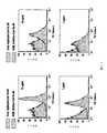


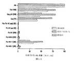


Classifications
- C—CHEMISTRY; METALLURGY
- C07—ORGANIC CHEMISTRY
- C07K—PEPTIDES
- C07K16/00—Immunoglobulins [IGs], e.g. monoclonal or polyclonal antibodies
- C07K16/18—Immunoglobulins [IGs], e.g. monoclonal or polyclonal antibodies against material from animals or humans
- C07K16/28—Immunoglobulins [IGs], e.g. monoclonal or polyclonal antibodies against material from animals or humans against receptors, cell surface antigens or cell surface determinants
- C07K16/2851—Immunoglobulins [IGs], e.g. monoclonal or polyclonal antibodies against material from animals or humans against receptors, cell surface antigens or cell surface determinants against the lectin superfamily, e.g. CD23, CD72
- A—HUMAN NECESSITIES
- A61—MEDICAL OR VETERINARY SCIENCE; HYGIENE
- A61K—PREPARATIONS FOR MEDICAL, DENTAL OR TOILETRY PURPOSES
- A61K39/00—Medicinal preparations containing antigens or antibodies
- A61K39/02—Bacterial antigens
- A—HUMAN NECESSITIES
- A61—MEDICAL OR VETERINARY SCIENCE; HYGIENE
- A61K—PREPARATIONS FOR MEDICAL, DENTAL OR TOILETRY PURPOSES
- A61K39/00—Medicinal preparations containing antigens or antibodies
- A61K39/12—Viral antigens
- A—HUMAN NECESSITIES
- A61—MEDICAL OR VETERINARY SCIENCE; HYGIENE
- A61K—PREPARATIONS FOR MEDICAL, DENTAL OR TOILETRY PURPOSES
- A61K39/00—Medicinal preparations containing antigens or antibodies
- A61K39/12—Viral antigens
- A61K39/145—Orthomyxoviridae, e.g. influenza virus
- A—HUMAN NECESSITIES
- A61—MEDICAL OR VETERINARY SCIENCE; HYGIENE
- A61K—PREPARATIONS FOR MEDICAL, DENTAL OR TOILETRY PURPOSES
- A61K39/00—Medicinal preparations containing antigens or antibodies
- A61K39/12—Viral antigens
- A61K39/21—Retroviridae, e.g. equine infectious anemia virus
- A—HUMAN NECESSITIES
- A61—MEDICAL OR VETERINARY SCIENCE; HYGIENE
- A61K—PREPARATIONS FOR MEDICAL, DENTAL OR TOILETRY PURPOSES
- A61K40/00—Cellular immunotherapy
- A61K40/10—Cellular immunotherapy characterised by the cell type used
- A61K40/19—Dendritic cells
- A—HUMAN NECESSITIES
- A61—MEDICAL OR VETERINARY SCIENCE; HYGIENE
- A61K—PREPARATIONS FOR MEDICAL, DENTAL OR TOILETRY PURPOSES
- A61K40/00—Cellular immunotherapy
- A61K40/20—Cellular immunotherapy characterised by the effect or the function of the cells
- A61K40/24—Antigen-presenting cells [APC]
- A—HUMAN NECESSITIES
- A61—MEDICAL OR VETERINARY SCIENCE; HYGIENE
- A61K—PREPARATIONS FOR MEDICAL, DENTAL OR TOILETRY PURPOSES
- A61K40/00—Cellular immunotherapy
- A61K40/40—Cellular immunotherapy characterised by antigens that are targeted or presented by cells of the immune system
- A61K40/41—Vertebrate antigens
- A61K40/42—Cancer antigens
- A61K40/4202—Receptors, cell surface antigens or cell surface determinants
- A—HUMAN NECESSITIES
- A61—MEDICAL OR VETERINARY SCIENCE; HYGIENE
- A61K—PREPARATIONS FOR MEDICAL, DENTAL OR TOILETRY PURPOSES
- A61K47/00—Medicinal preparations characterised by the non-active ingredients used, e.g. carriers or inert additives; Targeting or modifying agents chemically bound to the active ingredient
- A61K47/50—Medicinal preparations characterised by the non-active ingredients used, e.g. carriers or inert additives; Targeting or modifying agents chemically bound to the active ingredient the non-active ingredient being chemically bound to the active ingredient, e.g. polymer-drug conjugates
- A61K47/51—Medicinal preparations characterised by the non-active ingredients used, e.g. carriers or inert additives; Targeting or modifying agents chemically bound to the active ingredient the non-active ingredient being chemically bound to the active ingredient, e.g. polymer-drug conjugates the non-active ingredient being a modifying agent
- A61K47/68—Medicinal preparations characterised by the non-active ingredients used, e.g. carriers or inert additives; Targeting or modifying agents chemically bound to the active ingredient the non-active ingredient being chemically bound to the active ingredient, e.g. polymer-drug conjugates the non-active ingredient being a modifying agent the modifying agent being an antibody, an immunoglobulin or a fragment thereof, e.g. an Fc-fragment
- A61K47/6891—Pre-targeting systems involving an antibody for targeting specific cells
- A61K47/6897—Pre-targeting systems with two or three steps using antibody conjugates; Ligand-antiligand therapies
- A—HUMAN NECESSITIES
- A61—MEDICAL OR VETERINARY SCIENCE; HYGIENE
- A61K—PREPARATIONS FOR MEDICAL, DENTAL OR TOILETRY PURPOSES
- A61K47/00—Medicinal preparations characterised by the non-active ingredients used, e.g. carriers or inert additives; Targeting or modifying agents chemically bound to the active ingredient
- A61K47/50—Medicinal preparations characterised by the non-active ingredients used, e.g. carriers or inert additives; Targeting or modifying agents chemically bound to the active ingredient the non-active ingredient being chemically bound to the active ingredient, e.g. polymer-drug conjugates
- A61K47/69—Medicinal preparations characterised by the non-active ingredients used, e.g. carriers or inert additives; Targeting or modifying agents chemically bound to the active ingredient the non-active ingredient being chemically bound to the active ingredient, e.g. polymer-drug conjugates the conjugate being characterised by physical or galenical forms, e.g. emulsion, particle, inclusion complex, stent or kit
- A61K47/6901—Conjugates being cells, cell fragments, viruses, ghosts, red blood cells or viral vectors
- A—HUMAN NECESSITIES
- A61—MEDICAL OR VETERINARY SCIENCE; HYGIENE
- A61P—SPECIFIC THERAPEUTIC ACTIVITY OF CHEMICAL COMPOUNDS OR MEDICINAL PREPARATIONS
- A61P31/00—Antiinfectives, i.e. antibiotics, antiseptics, chemotherapeutics
- A61P31/04—Antibacterial agents
- A—HUMAN NECESSITIES
- A61—MEDICAL OR VETERINARY SCIENCE; HYGIENE
- A61P—SPECIFIC THERAPEUTIC ACTIVITY OF CHEMICAL COMPOUNDS OR MEDICINAL PREPARATIONS
- A61P31/00—Antiinfectives, i.e. antibiotics, antiseptics, chemotherapeutics
- A61P31/10—Antimycotics
- A—HUMAN NECESSITIES
- A61—MEDICAL OR VETERINARY SCIENCE; HYGIENE
- A61P—SPECIFIC THERAPEUTIC ACTIVITY OF CHEMICAL COMPOUNDS OR MEDICINAL PREPARATIONS
- A61P31/00—Antiinfectives, i.e. antibiotics, antiseptics, chemotherapeutics
- A61P31/12—Antivirals
- A—HUMAN NECESSITIES
- A61—MEDICAL OR VETERINARY SCIENCE; HYGIENE
- A61P—SPECIFIC THERAPEUTIC ACTIVITY OF CHEMICAL COMPOUNDS OR MEDICINAL PREPARATIONS
- A61P33/00—Antiparasitic agents
- A61P33/02—Antiprotozoals, e.g. for leishmaniasis, trichomoniasis, toxoplasmosis
- A—HUMAN NECESSITIES
- A61—MEDICAL OR VETERINARY SCIENCE; HYGIENE
- A61P—SPECIFIC THERAPEUTIC ACTIVITY OF CHEMICAL COMPOUNDS OR MEDICINAL PREPARATIONS
- A61P35/00—Antineoplastic agents
- A—HUMAN NECESSITIES
- A61—MEDICAL OR VETERINARY SCIENCE; HYGIENE
- A61P—SPECIFIC THERAPEUTIC ACTIVITY OF CHEMICAL COMPOUNDS OR MEDICINAL PREPARATIONS
- A61P37/00—Drugs for immunological or allergic disorders
- A—HUMAN NECESSITIES
- A61—MEDICAL OR VETERINARY SCIENCE; HYGIENE
- A61P—SPECIFIC THERAPEUTIC ACTIVITY OF CHEMICAL COMPOUNDS OR MEDICINAL PREPARATIONS
- A61P37/00—Drugs for immunological or allergic disorders
- A61P37/02—Immunomodulators
- A61P37/04—Immunostimulants
- B—PERFORMING OPERATIONS; TRANSPORTING
- B82—NANOTECHNOLOGY
- B82Y—SPECIFIC USES OR APPLICATIONS OF NANOSTRUCTURES; MEASUREMENT OR ANALYSIS OF NANOSTRUCTURES; MANUFACTURE OR TREATMENT OF NANOSTRUCTURES
- B82Y5/00—Nanobiotechnology or nanomedicine, e.g. protein engineering or drug delivery
- C—CHEMISTRY; METALLURGY
- C12—BIOCHEMISTRY; BEER; SPIRITS; WINE; VINEGAR; MICROBIOLOGY; ENZYMOLOGY; MUTATION OR GENETIC ENGINEERING
- C12N—MICROORGANISMS OR ENZYMES; COMPOSITIONS THEREOF; PROPAGATING, PRESERVING, OR MAINTAINING MICROORGANISMS; MUTATION OR GENETIC ENGINEERING; CULTURE MEDIA
- C12N5/00—Undifferentiated human, animal or plant cells, e.g. cell lines; Tissues; Cultivation or maintenance thereof; Culture media therefor
- C12N5/06—Animal cells or tissues; Human cells or tissues
- C12N5/0602—Vertebrate cells
- C12N5/0634—Cells from the blood or the immune system
- C12N5/0639—Dendritic cells, e.g. Langherhans cells in the epidermis
- A—HUMAN NECESSITIES
- A61—MEDICAL OR VETERINARY SCIENCE; HYGIENE
- A61K—PREPARATIONS FOR MEDICAL, DENTAL OR TOILETRY PURPOSES
- A61K39/00—Medicinal preparations containing antigens or antibodies
- A61K2039/505—Medicinal preparations containing antigens or antibodies comprising antibodies
- A—HUMAN NECESSITIES
- A61—MEDICAL OR VETERINARY SCIENCE; HYGIENE
- A61K—PREPARATIONS FOR MEDICAL, DENTAL OR TOILETRY PURPOSES
- A61K39/00—Medicinal preparations containing antigens or antibodies
- A61K2039/60—Medicinal preparations containing antigens or antibodies characteristics by the carrier linked to the antigen
- A61K2039/6031—Proteins
- A61K2039/6056—Antibodies
- C—CHEMISTRY; METALLURGY
- C12—BIOCHEMISTRY; BEER; SPIRITS; WINE; VINEGAR; MICROBIOLOGY; ENZYMOLOGY; MUTATION OR GENETIC ENGINEERING
- C12N—MICROORGANISMS OR ENZYMES; COMPOSITIONS THEREOF; PROPAGATING, PRESERVING, OR MAINTAINING MICROORGANISMS; MUTATION OR GENETIC ENGINEERING; CULTURE MEDIA
- C12N2760/00—MICROORGANISMS OR ENZYMES; COMPOSITIONS THEREOF; PROPAGATING, PRESERVING, OR MAINTAINING MICROORGANISMS; MUTATION OR GENETIC ENGINEERING; CULTURE MEDIA ssRNA viruses negative-sense
- C12N2760/00011—Details
- C12N2760/16011—Orthomyxoviridae
- C12N2760/16111—Influenzavirus A, i.e. influenza A virus
- C12N2760/16134—Use of virus or viral component as vaccine, e.g. live-attenuated or inactivated virus, VLP, viral protein
Landscapes
- Health & Medical Sciences (AREA)
- Life Sciences & Earth Sciences (AREA)
- Chemical & Material Sciences (AREA)
- General Health & Medical Sciences (AREA)
- Veterinary Medicine (AREA)
- Animal Behavior & Ethology (AREA)
- Public Health (AREA)
- Immunology (AREA)
- Medicinal Chemistry (AREA)
- Engineering & Computer Science (AREA)
- Pharmacology & Pharmacy (AREA)
- Epidemiology (AREA)
- Organic Chemistry (AREA)
- Bioinformatics & Cheminformatics (AREA)
- Microbiology (AREA)
- Virology (AREA)
- Genetics & Genomics (AREA)
- Biomedical Technology (AREA)
- Mycology (AREA)
- Biotechnology (AREA)
- Nuclear Medicine, Radiotherapy & Molecular Imaging (AREA)
- General Chemical & Material Sciences (AREA)
- Chemical Kinetics & Catalysis (AREA)
- Zoology (AREA)
- Wood Science & Technology (AREA)
- Biochemistry (AREA)
- Hematology (AREA)
- Molecular Biology (AREA)
- Biophysics (AREA)
- General Engineering & Computer Science (AREA)
- Cell Biology (AREA)
- Nanotechnology (AREA)
- Communicable Diseases (AREA)
- Proteomics, Peptides & Aminoacids (AREA)
- Oncology (AREA)
- Medical Informatics (AREA)
- Crystallography & Structural Chemistry (AREA)
- Pulmonology (AREA)
- Tropical Medicine & Parasitology (AREA)
- Medicines Containing Antibodies Or Antigens For Use As Internal Diagnostic Agents (AREA)
Abstract
Description
Translated fromJapanese【技術分野】
【0001】
本発明は、概して、ワクチン接種の分野に関し、より詳細には、抗原提示細胞上に発現されたDCIRへの抗原のターゲティングに基づくワクチンに関する。
【背景技術】
【0004】
本発明の範囲を限定せずに、その背景を抗原提示との関連において説明する。
【0005】
樹状細胞(DC,dendritic cell)ターゲッティングに基づくヒトワクチンは、マウスモデルでの説得力のある研究を基にする新概念である。この場合、特定のDC受容体に対する抗体によってDCにもたらされる少量の比較的弱い抗原が、強力かつ広範な免疫応答を誘発しうる。ヒト用のそのようなワクチンを開発するには、この抗原ターゲティングの適用に正確にどのDC受容体を用いるべきかに関する、より詳細な理解を必要とする。これは、マウスの免疫系とヒトの免疫系との間で、常に正確な対応があるわけではないからであり、また、このワクチン適用に関して、すべての潜在的DC受容体が慎重に検査されているわけではないからでもある。
【0006】
したがって、様々な抗ヒトDC受容体標的をインビトロで試験する研究が開始されており、例えば、HLAクラスI分子及びクラスII分子との関連で、マンノース受容体で活性化されたT細胞に対するヒトmAbに融合している黒色腫抗原pmel17を用いて、DCターゲッティングが行われた(Ramakrishna, V., J. F. Treml, et al. (2004). "Mannose receptor targeting of tumor antigen pmel17 to human dendritic cells directs anti-melanoma T cell responses via multiple HLA molecules." J Immunol 172(5): 2845-52.)。また、ヒト化抗DC−SIGN mAbを介して、モデル抗原であるKLHをDCにターゲティングすることによって、抗原特異的なナイーブT細胞応答に加えて、用量抑制的なリコールT細胞応答も効果的に誘導された(Tacken, P. J., I. J. de Vries, et al. (2005). "Effective induction of naive and recall T-cell responses by targeting antigen to human dendritic cells via a humanized anti-DC-SIGN antibody." Blood 106(4): 1278-85.)。マンノース受容体及びDC−SIGNに加えて、ヒトDCは、抗原捕捉に関与していることが知られている他の受容体も発現する。これらの多くは、LOX−1、DEC205、DC−ASGPR、ランゲリン、DCIR、BDCA−2、DECTIN−1及びCLEC−6を含めたC型レクチン受容体(CLR,C-type lectin receptor)である。これらのCLRは、DCの異なったサブセットにより異なって発現され、それらの発現は、DC成熟の状態に従って相違しうる(Figdor, C. G., Y. van Kooyk, et al. (2002). "C-type lectin receptors on dendritic cells and Langerhans cells." Nat Rev Immunol 2(2): 77-84.、Geijtenbeek, T. B., S. J. van Vliet, et al. (2004). "Self- and nonself-recognition by C-type lectins on dendritic cells." Annu Rev Immunol 22: 33-54.)。
【0007】
DCのサブセットは、異なった免疫応答を刺激する。したがって、異なって発現される受容体を介して、抗原をこれらのサブセットにターゲティングすることによって、異なった免疫応答が誘発されるはずである(Shortman, K. and Y. J. Liu (2002). "Mouse and human dendritic cell subtypes." Nat Rev Immunol 2(3): 151-61.)。さらに、同じDCサブセット上の異なった受容体が、抗原を別々のプロセシング経路に方向付ける可能性もある(Trombetta, E. S. and I. Mellman (2005). "Cell biology of antigen processing in vitro and in vivo." Annu Rev Immunol 23: 975-1028.)。最後に、これらの受容体の一部は内因的に活性化しないものであり(例えば、DEC205(Bonifaz, L. C., D. P. Bonnyay, et al. (2004). "In vivo targeting of antigens to maturing dendritic cells via the DEC-205 receptor improves T cell vaccination." J Exp Med 199(6): 815-24.))、一方、他のものは、活性化するものであるか(例えば、LOX−1(Delneste, Y., G. Magistrelli, et al. (2002). "Involvement of LOX-1 in dendritic cell-mediated antigen cross-presentation." Immunity 17(3): 353-62.))、まだ完全には研究されていないものである。抗原の取り込みと同時に起こるDC活性化の重要性は知られていない。しかし、これが有益であるならば、ターゲッティングmAbを介したDCの活性化によって、ターゲッティングワクチンの製剤が単純化される。
【非特許文献】
【0008】
【非特許文献1】Ramakrishna, V., J. F. Treml, et al. (2004). "Mannose receptor targeting of tumor antigen pmel17 to human dendritic cells directs anti-melanoma T cell responses via multiple HLA molecules." J Immunol 172(5): 2845-52.
【非特許文献2】Tacken, P. J., I. J. de Vries, et al. (2005). "Effective induction of naive and recall T-cell responses by targeting antigen to human dendritic cells via a humanized anti-DC-SIGN antibody." Blood 106(4): 1278-85.
【非特許文献3】Figdor, C. G., Y. van Kooyk, et al. (2002). "C-type lectin receptors on dendritic cells and Langerhans cells." Nat Rev Immunol 2(2): 77-84.
【非特許文献4】Geijtenbeek, T. B., S. J. van Vliet, et al. (2004). "Self- and nonself-recognition by C-type lectins on dendritic cells." Annu Rev Immunol 22: 33-54.
【非特許文献5】Shortman, K. and Y. J. Liu (2002). "Mouse and human dendritic cell subtypes." Nat Rev Immunol 2(3): 151-61.
【非特許文献6】Trombetta, E. S. and I. Mellman (2005). "Cell biology of antigen processing in vitro and in vivo." Annu Rev Immunol 23: 975-1028.
【非特許文献7】Bonifaz, L. C., D. P. Bonnyay, et al. (2004). "In vivo targeting of antigens to maturing dendritic cells via the DEC-205 receptor improves T cell vaccination." J Exp Med 199(6): 815-24.
【非特許文献8】Delneste, Y., G. Magistrelli, et al. (2002). "Involvement of LOX-1 in dendritic cell-mediated antigen cross-presentation." Immunity 17(3): 353-62.
【発明の概要】
【発明が解決しようとする課題】
【0009】
これらの考慮を背景として、本発明者らは、望ましい免疫結果を得るために最も適したヒトDCターゲッティング受容体を特定するための、CD4+及びCD8+T細胞ナイーブ応答及びリコール応答をインビトロで詳細に探索することによる系統的な比較の緊急な必要性を認識している。本出願は、特定のDC受容体、すなわち、樹状細胞抑制型受容体(DCIR,Dendritic Cell Inhibitory Receptor)が、予防用及び治療用ワクチン接種のために抗原をヒトDCにターゲティングする目的に理想的な受容体であることを示す、その特殊かつ予想外な特徴を記載する。
【課題を解決するための手段】
【0010】
本発明は、抗原に対する強力かつ広範な免疫応答を誘発する目的で、抗原を抗原提示細胞に特異的にターゲティングする(送達する)ワクチンを作製及び使用するための組成物及び方法を含む。目的は、主として、抗原が由来する作用物質(病原体又は癌)に対する防御的又は治療的免疫応答を誘起することである。
【0011】
より詳細には、本発明は、制御されたモジュール構造で1又は複数の抗原を保持する、標的特異的な単一の組換え抗体(mAb)、活性化タンパク質又は他の抗体を設計及び作製するための組成物、方法及び方法を提供する。本発明のモジュールrAb担体は、例えば、複数の抗原、並びに/又は抗原及び活性化サイトカインを(内部移行するヒト樹状細胞受容体に対する1つの一次組換え抗体を介して)樹状細胞(DC)にターゲティングするのに用いることができる。また、本発明は、制御され、かつ特定されたやり方で、2つの異なった組換えmAbを端と端とで連結する方法を提供する。
【0012】
本発明は、抗体−抗原複合体を形成する標的作用物質が結合しているDCIR特異的抗体又はその断片を単離及び精製することによって、DCIR発現抗原提示細胞による抗原提示の有効性を増大させる組成物及び方法であって、その作用物質が、抗体−作用物質複合体と接触した、例えば樹状細胞によってプロセシング及び提示される方法を含む。一実施形態では、抗原提示細胞が樹状細胞であり、DCIR特異的抗体又はその断片がコヒーシン/ドックリン対の一方の半分に結合する。DCIR特異的抗体又はその断片がコヒーシン/ドックリン対の一方の半分に結合し、かつ抗原がコヒーシン/ドックリン対の他方の半分に結合して複合体を形成してもよい。作用物質の非限定的な例には、ペプチド、タンパク質、脂質、糖質、核酸及びこれらの組合せのうちの1又は複数が含まれる。
【0013】
作用物質は、インターロイキン、トランスフォーミング成長因子(TGF,transforming growth factor)、線維芽細胞成長因子(FGF,fibroblast growth factor)、血小板由来成長因子(PDGF,platelet derived growth factor)、上皮成長因子(EGF,epidermal growth factor)、結合組織活性化ペプチド(CTAP,connective tissue activated peptide)、骨形成因子、並びにそのような成長因子の生理活性類似体、断片及び誘導体、B/T細胞分化因子、B/T細胞成長因子、分裂促進サイトカイン、走化性サイトカイン、コロニー刺激因子、血管形成誘導因子、IFN−α、IFN−β、IFN−γ、IL1、IL2、IL3、IL4、IL5、IL6、IL7、IL8、IL9、IL10、IL11、IL12、IL13、IL14、IL15、IL16、IL17、IL18など、レプチン、ミオスタチン、マクロファージ刺激タンパク質、血小板由来成長因子、TNF−α、TNF−β、NGF、CD40L、CD137L/4−1BBL、ヒトリンフォトキシン−β、G−CSF、M−CSF、GM−CSF、PDGF、IL−1α、IL1−β、IP−10、PF4、GRO、9E3、エリスロポエチン、エンドスタチン、アンギオスタチン、VEGF、並びにβトランスフォーミング成長因子(例えば、TGF−β1、TGF−β2、TGF−β3)、骨形成タンパク質(例えば、BMP−1、BMP−2、BMP−3、BMP−4、BMP−5、BMP−6、BMP−7、BMP−8、BMP−9)、ヘパリン結合性成長因子(線維芽細胞成長因子(FGF)、上皮成長因子(EGF)、血小板由来性成長因子(PDGF)、インスリン様増殖因子(IGF,insulin-like growth factor))、インヒビン(例えば、インヒビンA、インヒビンB)、増殖分化因子(例えばGDF−1)及びアクチビン(例えば、アクチビンA、アクチビンB、アクチビンAB)を含めたトランスフォーミング成長因子(TGF)遺伝子スーパーファミリーから選択される1又は複数のサイトカインでありうる。別の実施形態では、作用物質は、細菌性、ウイルス性、真菌性、原生動物性及び癌性のタンパク質である抗原を含む。
【0014】
本発明は、樹状細胞による抗原提示の有効性を増大させる組成物及び方法であって、抗体−抗原複合体を形成する抗原が結合するDCIR特異的抗体又はその断片を結合させるステップを含み、その抗原が、抗体−抗原複合体と接触した樹状細胞によってプロセシング及び提示される組成物及び方法も含む。別の実施形態は、防御的又は治療的免疫応答を誘発する目的で、抗原を抗原提示細胞に送達するための、DCIRに対する抗体又は他の特異的結合分子の使用である。皮膚を介したワクチン接種のための、DCIRに特異的な抗原標的試薬の使用;同時投与又は連結されたワクチン接種用のアジュバントと併用した、DCIRに特異的な抗原標的試薬の使用、又は組換えの抗原−抗体融合タンパク質として発現することができる特異的抗原の、抗原ターゲッティング(ワクチン接種)目的での使用。
【0015】
別の実施形態は、患者の樹状細胞を単離するステップ;上記樹状細胞を活性化量の抗DCIR抗体又はその断片及び抗原に曝露して、抗原が負荷されて活性化された樹状細胞を形成するステップ;並びに抗原が負荷されて活性化された上記樹状細胞を上記患者に再導入するステップによって、樹状細胞の有効性を増大させる方法を含む。抗原は、細菌性、ウイルス性、真菌性、原生動物性又は癌性のタンパク質を含みうる。本発明は、哺乳動物細胞から分泌された抗DCIR免疫グロブリン又はその部分、及び免疫グロブリンに結合した抗原も含む。免疫グロブリンは、コヒーシン/ドックリンドメインの半分に結合しており、又はそれは、モジュールrAb担体と複合体を形成する抗原に結合した、コヒーシン−ドックリン結合対の他方の半分、若しくは抗原との融合タンパク質である、コヒーシン−ドックリン結合対の他方の半分を含んでもよい。抗原特異的なドメインは、完全長抗体、抗体可変領域ドメイン、Fab断片、Fab’断片、F(ab)2断片及びFv断片、並びにFabc断片、及び/又はFcドメインの部分を伴ったFab断片でありうる。抗DCIR免疫グロブリンは、放射性同位元素、金属、酵素、ボツリヌス毒素、破傷風、リシン、コレラ、ジフテリア、アフラトキシン、ウェルシュ菌毒素、マイコトキシン、志賀毒素、ブドウ球菌エンテロトキシンB、T2、セグイトキシン(seguitoxin)、サキシトキシン、アブリン、シアノジノシン、α毒素、テトロドトキシン、アコノトキシン(aconotoxin)、ヘビ毒及びクモ毒から選択される毒素に結合していてもよい。抗原は、免疫グロブリンとの融合タンパク質であるか、又は免疫グロブリンに共有結合若しくは非共有結合によって化学的に結合したものでありうる。
【0016】
別の実施形態は、抗体−抗原複合体を形成する抗原が結合しているDCIR特異的抗体又はその断片を含むワクチンであって、その抗原が、抗体−抗原複合体と接触した樹状細胞によってプロセシング及び提示されるワクチンである。
【0017】
本発明の新規抗体は、新規な組織分布情報を示すこともできた。それらの特異的親和性により、本発明の抗DCIR抗体は、サルDCIRに結合することが見出された。本発明の抗DCIR抗体は、インビボ[hu−マウス]における、抗DCIR−Flu m1を用いた、増殖中のFlu m1特異的CD8細胞へのターゲッティング、及びエキソビボヒト皮膚細胞のインビトロターゲッティングに有効である。さらに、DCIRの炭水化物リガンドの複合体は、抗原送達のための抗DCIR薬の代用薬として使用できることが見出された。したがって、本発明の別の実施形態は、Neu5Acα2−3Galβ1−4GlcNAcβ1−2Manα1−3(Neu5Acα2−3Galβ1−4GlcNAcβ1−2Manα1−6)Manβ1−4GlcNAcβ1−4GlcNAcβ−Sp12を含有する、グリカンの少なくとも一部に結合した、DCIRに特異的に結合する抗原性T細胞エピトープペプチドである。上記グリカン(及びその誘導体)は、単独又は併用で、DCIR結合を阻止するのに使用できる。
【0018】
本発明の特徴及び利点をより完全に理解するために、これより、添付された図と共に、本発明の詳細な説明を参照する。
【図面の簡単な説明】
【0019】
【図1】プレートに結合したDCIRとの、mAbの高親和性相互作用を捕捉ELISAによって示す図である。
【図2】プレートに結合したDCIRとの、mAbの高親和性相互作用を直接ELISAによって示す図である。
【図3】FACSで使用するための高親和性抗体の結合を示す図である。
【図4】ヒト皮膚から直接単離された3種のヒトDCサブタイプでもDCIRが発現されていることを示す図である。
【図5】ヒト扁桃腺内の胚中心を囲む細胞集団のDCIR特異的染色を示す図である。
【図6】架橋Flu M1タンパク質及びDCIRに対するmAbの例を示す図である。
【図7】抗DCIR_2C9 mAbへのCoh.Flu M1の架橋結合を示す図である。
【図8】抗DCIR mAbに架橋結合されたFlu M1が、mAbに架橋結合されていないFlu M1タンパク質より効率的に、Flu M1特異的CD8+T細胞の増殖を誘導することを示す図である。
【図9】抗DCIR mAbに架橋結合されたFlu M1が、Flu M1特異的CD8+T細胞の増殖を、Int−DCより、LCを介して、より効率的に誘導することを示す図である。
【図10】293細胞内に同時形質移入され、培養上清中へのrAbの分泌に関して抗ヒトFC ELISAによってアッセイされた多数の異なった抗DCIR mAbに対応するマウス−ヒトキメラrAbをコードするH+L鎖ベクターを示す図である。
【図11】抗DCIR.Doc rAbに連結されたCoh.Flu M1がGM/IL−15ヒトDCに特異的に結合することを示す図である。
【図12】抗DC−SIGN/L.Doc又は抗DCIR.Doc rAbに連結されたCoh.Flu M1がGM−CSF/IL−4ヒトDCに結合し、その中に内部移行されることを示す図である。
【図13】抗DCIR.Doc:Coh.Flu複合体が、他の[抗DC受容体rAb.Doc:Coh.Flu M1]複合体より、Flu M1特異的CD8+T細胞の増殖に効率的であることを示す図である。
【図14】1日間投与された抗DCIR.Doc:Coh.Flu複合体が、他の[抗DC受容体rAb.Doc:Coh.Flu M1]複合体より、Flu M1特異的CD8+T細胞の増殖に効率的であることを示す図である。
【図15】rAb H鎖のC末端との融合体として発現された様々な抗原が、rAb.抗原の分泌に内因的な影響を有することを示す図である。
【図16】抗DCIR.Flu HA5 rAbが可変領域の性質に応じて様々な効率で分泌されることを示す図である。
【図17】抗DCIR mAbが、HIV抗原特異的CD8+細胞のプライミングを増強することを示す図である。
【図18】抗DCIR mAbが、HIV抗原特異的CD8+細胞のプライミングを増強することを示す図である。
【図19】ヒト上皮シート内におけるDCIR分布の免疫組織化学分析を示す図である。
【図20A】図20A〜20Dは、DCIR抗原に対するモノクローナル抗体、とりわけDCIRへの親和性を示す図である。
【図20B】図20A〜20Dは、DCIR抗原に対するモノクローナル抗体、とりわけDCIRへの親和性を示す図である。
【図20C】図20A〜20Dは、DCIR抗原に対するモノクローナル抗体、とりわけDCIRへの親和性を示す図である。
【図20D】図20A〜20Dは、DCIR抗原に対するモノクローナル抗体、とりわけDCIRへの親和性を示す図である。
【図21】抗DCIR mAbの、アカゲザルDCIRに対する交差反応性を示す図である。
【図22】特定のグリカン構造への、DCIR外部ドメインの結合を示すグラフである。
【図23A】図23A〜23Cは、DCIRが、すべての血液DCサブセットのための包括的な標的であることを示す図である。
【図23B】図23A〜23Cは、DCIRが、すべての血液DCサブセットのための包括的な標的であることを示す図である。
【図23C】図23A〜23Cは、DCIRが、すべての血液DCサブセットのための包括的な標的であることを示す図である。
【図24】DCIR−FluM1でのワクチン接種によって、FluM1特異的なリコールCD8+T細胞免疫の産生が可能となることを実証する図である。
【発明を実施するための形態】
【0020】
本発明の様々な実施形態の作製及び使用について以下に詳細に論じるが、本発明は、様々な特定の概念に具体化できる多数の適用可能な発明概念を提供することを理解されたい。本明細書で論じる特定の実施形態は、本発明を作製及び使用する特定の方法を例示するに過ぎず、本発明の範囲の境界を定めるものではない。
【0021】
本発明の理解を容易にするために、いくつかの用語を以下に定義する。本明細書で定義する用語は、本発明に関連する領域の当業者によって一般的に理解されている意味を有する。「1つの(a)」、「1つの(an)」や「その(the)」などの用語は、単数の存在物のみを指すのではなく、それには、その特定の例を例示目的に用いることのできる一般クラスが含まれる。本明細書の用語は、本発明の特定の実施形態を記載するのに使用されるが、それらの使用は、特許請求の範囲で大要を記載しているのを除いて、本発明の境界を定めるものではない。
【0022】
樹状細胞(DC)は、抗原特異的免疫を調節するのに重要な役割を果たしている抗原提示細胞である(Mellman, I. and R. M. Steinman (2001). "Dendritic cells: specialized and regulated antigen processing machines." Cell 106(3): 255-8.、Banchereau, J., F. Briere, et al. (2000). “Immunobiology of dendritic cells.” Annu Rev Immunol 18: 767-811.、Cella, M., F. Sallusto, et al. (1997). "Origin, maturation and antigen presenting function of dendritic cells." Curr Opin Immunol 9(1): 10-6.)。DCは、抗原を捕捉して、それらをペプチドにプロセシングして、これらをT細胞に提示する。したがって、抗原をDCに直接送達することは、ワクチンを改善するための重点領域である。そのような例の1つは、後で患者に再投与される自己DCへのエキソビボ抗原負荷を用いたDCベースのワクチンの開発である(Banchereau, J., B. Schuler-Thurner, et al. (2001). "Dendritic cells as vectors for therapy." Cell 106(3): 271-4.、Steinman, R. M. and M. Dhodapkar (2001). "Active immunization against cancer with dendritic cells: the near future." Int J Cancer 94(4): 459-73.)。ワクチンの効力を改善するための別の戦略は、内部移行するDC特異的な受容体に対する抗体に結合した抗原をDCに特異的にターゲッティングすることである。ワクチン接種のためにDCをターゲッティングすることの可能性は、主要なマウス研究によって注目を浴びた。オボアルブミン(OVA)に結合した抗LOX−1 mAbを用いたインビボでのターゲッティングは、MHCクラスI経路に向けた外因性抗原のクロスプレゼンテーションを介して、防御的CD8+T細胞反応を誘発した(Delneste, Y., G. Magistrelli, et al. (2002). "Involvement of LOX-1 in dendritic cell-mediated antigen cross-presentation." Immunity 17(3): 353-62.)。また、CD40L成熟刺激と併用すると、抗DEC205 mAbに結合したOVAも、インビボで、DCによるMHCクラスI拘束性の提示を増強し、エフェクターメモリーCD8+T細胞の持続的な形成をもたらす(Bonifaz, L. C., D. P. Bonnyay, et al. (2004). "In vivo targeting of antigens to maturing dendritic cells via the DEC-205 receptor improves T cell vaccination." J Exp Med 199(6): 815-24.)。これらの研究は両方とも劇的な用量の節約(すなわち非常に少ない抗原量で強い免疫応答)を示し、他のタイプのOVA免疫処置で通常見られるより広範な応答を示唆した。DEC205を介したDCへのHIV gag抗原のターゲッティングを用いた最近の研究は、これらの概念を臨床的に重要な抗原にまで拡張し、DCへの抗原のターゲティングの基本特性、すなわち、劇的な用量の節約、単一ワクチン接種に由来する防御応答、並びにCD8及びCD4コンパートメントの両方における抗原特異的なT細胞の増殖を確認した(Trumpfheller, C., J. S. Finke, et al. (2006). "Intensified and protective CD4+ T cell immunity in mice with anti-dendritic cell HIV gag fusion antibody vaccine." J Exp Med 203(3): 607-17.)。
【0023】
本発明は、制御された多変量様式における、単一の組換え一次mAbへの、(一次mAbから独立して工学的に作製、発現及び精製された)複数の抗原又はタンパク質の複合体形成を提供する。現在、1つの一次mAbに(それぞれストレプトアビジンに別々に連結するように工学的に作製された)様々なタンパク質の付加を可能にする部位特異的ビオチン化部位を工学的に作製する方法が存在する。しかし、本発明は、所定のモル比及び位置における、複数の組合せの一次mAbへの、別々に工学的に作製されたタンパク質の付加を提供する。
【0024】
本明細書で使用される場合、「モジュールrAb担体」という用語は、単一の組換えモノクローナル抗体(mAb)への、様々な抗原、活性化タンパク質又は他の抗体の、制御されたモジュール付加をもたらすように工学的に作製された組換え抗体系を示すのに使用される。このrAbは、標準的なハイブリドーマ技法、組換え抗体ディスプレイ及びヒト化モノクローナル抗体などを用いて作製されたモノクローナル抗体でありうる。モジュールrAb担体は、例えば、複数の抗原、並びに/又は抗原及び活性化サイトカインを(内部移行する受容体、例えばヒト樹状細胞受容体に対する1つの組換え一次抗体を介して)、樹状細胞(DC)にターゲティングするのに使用できる。モジュールrAb担体は、制御された特定の方法で、2つの異なった組換えmAbを端と端とで連結するのに使用できる。
【0025】
「モジュールrAb担体」の抗原結合部分は、認識されるモジュール結合部分が、そのアミノ酸配列に付加及び/又は結合される、1又は複数の可変ドメイン、1又は複数の可変ドメイン及び最初の定常ドメイン、Fab断片、Fab’断片、F(ab)2断片及びFv断片、並びに、Fabc断片、及び/又はFcドメインの部分を伴ったFab断片でありうる。モジュールrAb担体で使用するための抗体は、いずれのアイソタイプ又はクラスのものでも、サブクラスでも、又はどんな供給源(動物性及び/又は組換え)由来の抗体でもよい。
【0026】
非限定的な一例において、モジュールrAb担体は、工学的に作製された組換えmAbとの関連で、特異的かつ特定のタンパク質複合体を作製するための1又は複数のモジュールコヒーシン−ドックリンタンパク質ドメインを有するように工学的に作製される。mAbは、mAbの抗原結合ドメインのカルボキシ側に、1又は複数のモジュールコヒーシン−ドックリンタンパク質ドメインを含有する融合タンパク質の一部分である。コヒーシン−ドックリンタンパク質ドメインは、例えば化学架橋結合物質及び/又はジスルフィド結合形成を用いることによって、翻訳後に付加されたものでも可能である。
【0027】
「抗原」という用語は、本明細書で使用される場合、その抗原の受容者体内で液性及び/又は細胞性免疫応答を開始できる分子を指す。抗原は、本発明では、2つの異なった場面で、すなわち抗体又はrAbの他の抗原認識ドメインの標的として、又はモジュールrAb担体に相補的なドックリン/コヒーシン分子の一部として細胞若しくは細胞内にもたらされるか、rAbによってターゲッティングされる分子として使用されうる。抗原は、通常、ワクチン接種が有利な治療法となる疾患を引き起こす病原体である。抗原がMHCに提示される場合、そのペプチドは、しばしば約8アミノ酸から約25アミノ酸である。抗原には、例えば、単純な中間代謝物質、糖、脂質及びホルモン、並びに複合糖質、リン脂質、核酸及びタンパク質などの高分子を含めたどんなタイプの生物学的分子も含まれる。抗原の一般的な範疇には、ウイルス抗原、細菌性抗原、真菌抗原、原子動物及び他の寄生虫の抗原、腫瘍抗原、自己免疫疾患、アレルギー及び移植拒絶に関与している抗原並びに他の種々雑多な抗原が含まれるが、これらに限定されない。
【0028】
モジュールrAb担体は、任意の数の活性物質、例えば、抗生物質、抗感染症薬、抗ウイルス剤、抗腫瘍薬、解熱薬、鎮痛剤、抗炎症薬、骨粗鬆症の治療薬、酵素、サイトカイン、抗凝血物質、多糖、コラーゲン、細胞及び上記の活性物質の2種以上の組合せを保持できる。本発明を用いて送達する抗生物質の例には、テトラサイクリン、アミノグルコシド、ペニシリン、セファロスポリン、スルホンアミド薬物、コハク酸クロラムフェニコールナトリウム、エリスロマイシン、バンコマイシン、リンコマイシン、クリンダマイシン、ナイスタチン、アムホテリシンB、アマンタジン、イドクスウリジン、p−アミノサリチル酸、イソニアジド、リファンピン、アンティノマイシンD、ミトラマイシン、ダウノマイシン、アドリアマイシン、ブレオマイシン、ビンブラスチン、ビンクリスチン、プロカルバジン及びイミダゾールカルボキシアミドなどが含まれるが、これらに限定されない。
【0029】
本発明を用いて送達する抗腫瘍薬の例には、ドキソルビシン、ダウノルビシン、タキソール及びメトトレキセートが含まれるが、これらに限定されない。解熱薬及び鎮痛剤の例には、アスピリン、Motrin(登録商標)、Ibuprofen(登録商標)、ナプロキセン及びアセトアミノフェンなどが含まれる。
【0030】
本発明を用いて送達する抗炎症薬の例には、NSAIDS、アスピリン、ステロイド、デキサメサゾン、ヒドロコルチゾン、プレドニソロン及びジクロフェナクナトリウムなどが含まれるが、これらに限定されない。
【0031】
本発明を用いて送達する骨粗鬆症治療薬並びに骨及び骨格に作用する他の因子の例には、カルシウム、アレンドロネート、骨GLaペプチド、副甲状腺ホルモン及びその活性断片、ヒストンH4関連骨形成増殖ペプチド(histone H4-related bone formation and proliferation peptide )、並びにこれらの変異体、誘導体及び類似体が含まれるが、これらに限定されない。
【0032】
本発明を用いて送達する酵素及び酵素補因子の例には、Pancrease(登録商標)、L−アスパラギナーゼ、ヒアルロニダーゼ、キモトリプシン、トリプシン、tPA、ストレプトキナーゼ、ウロキナーゼ、パンクレアチン、コラゲナーゼ、トリプシノーゲン、キモトリプシノゲン、プラスミノーゲン、ストレプトキナーゼ、アデニルシクラーゼ及びスーパーオキシドジスムターゼ(SOD)などが含まれるが、これらに限定されない。
【0033】
本発明を用いて送達するサイトカインの例には、インターロイキン、トランスフォーミング成長因子(TGF)、線維芽細胞成長因子(FGF)、血小板由来成長因子(PDGF)、上皮成長因子(EGF)、結合組織活性化ペプチド(CTAP)、骨形成因子、並びにそのような成長因子の生理活性類似体、断片及び誘導体が含まれるが、これらに限定されない。サイトカインは、B/T細胞分化因子、B/T細胞成長因子、マイトジェニックサイトカイン、走化性サイトカイン、コロニー刺激因子、血管形成誘導因子、IFN−α、IFN−β、IFN−γ、IL1、IL2、IL3、IL4、IL5、IL6、IL7、IL8、IL9、IL10、IL11、IL12、IL13、IL14、IL15、IL16、IL17、IL18など;レプチン、ミオスタチン、マクロファージ刺激タンパク質、血小板由来成長因子、TNF−α、TNF−β、NGF、CD40L、CD137L/4−1BBL、ヒトリンフォトキシン−β、G−CSF、M−CSF、GM−CSF、PDGF、IL−1α、IL1−β、IP−10、PF4、GRO、9E3、エリスロポエチン、エンドスタチン、アンギオスタチン、VEGF、又はこれらの任意の断片若しくは組合せでありうる。他のサイトカインには、βトランスフォーミング成長因子(例えば、TGF−β1、TGF−β2、TGF−β3)、骨形成タンパク質(例えば、BMP−1、BMP−2、BMP−3、BMP−4、BMP−5、BMP−6、BMP−7、BMP−8、BMP−9)、ヘパリン結合性成長因子(例えば、線維芽細胞成長因子(FGF)、上皮成長因子(EGF)、血小板由来成長因子(PDGF)、インスリン様成長因子(IGF))、インヒビン(例えば、インヒビンA、インヒビンB)、増殖分化因子(例えば、GDF−1)及びアクチビン(例えば、アクチビンA、アクチビンB、アクチビンAB)を含めたトランスフォーミング成長因子(TGF)スーパーファミリーのメンバーが含まれる。
【0034】
本発明を用いて送達する成長因子の例には、哺乳動物細胞からなど、天然又は自然の供給源から単離することのできる成長因子も、組換えDNA技法によって、又は様々な化学的方法によって、合成によって調製することのできる成長因子も含まれるが、これらに限定されない。加えて、これらの因子の類似体、断片又は誘導体も、それらが天然分子の生物活性の少なくとも一部を示すという条件で、用いることができる。例えば、類似体は、部位特異的変異導入又は他の遺伝子工学的手法によって改変された遺伝子を発現することによって調製することができる。
【0035】
本発明を用いて送達する抗凝血物質の例には、ワルファリン、ヘパリン及びヒルジンなどが含まれるが、これらに限定されない。本発明を用いて送達する、免疫系に作用する因子の例には、走化性ペプチド及びブラジキニンなど、炎症及び悪性新生物を制御する因子、並びに伝染性微生物を攻撃する因子が含まれるが、これらに限定されない。
【0036】
ウイルス抗原の例には、例えば、レトロウイルス(retroviral)抗原、すなわち、gag、pol及びenv遺伝子の遺伝子産物、Nefタンパク質、逆転写酵素及び他のHIV成分などのヒト免疫不全症ウイルス(HIV,human immunodeficiency virus)抗原に由来するレトロウイルス抗原など;肝炎ウイルス(hepatitis viral)抗原、すなわち、B型肝炎ウイルス(hepatitis B virus)のS、M及びLタンパク質、B型肝炎ウイルス並びに他の肝炎、例えば、A型、B型及びC型肝炎のプレS抗原、C型肝炎のウイルスRNAなどのウイルス成分など;インフルエンザウイルス(influenza viral)抗原、すなわち、赤血球凝集素、ノイラミニダーゼ及び他のインフルエンザウイルス成分など;麻疹ウイルス(measles viral)抗原、すなわち、麻疹ウイルス融合タンパク質及び他の麻疹ウイルス成分など;風疹(rubella)ウイルス抗原、すなわち、タンパク質E1及びE2並びに他の風疹ウイルス成分など;ロタウイルス(rotaviral)抗原、すなわち、VP7sc及び他のロタウイルス成分など;サイトメガロウイルス(cytomegaloviral)抗原、すなわち、エンベロープ糖タンパク質B及び他のサイトメガロウイルス抗原成分など;呼吸器合胞体ウイルス(respiratory syncytial viral)抗原、すなわち、RSV融合タンパク質、M2タンパク質及び他の呼吸器合胞体ウイルス抗原成分など;単純ヘルペスウイルス(herpes simplex viral)抗原、すなわち、前初期タンパク質、糖タンパク質D及び他の単純ヘルペスウイルス抗原成分など;水痘帯状疱疹ウイルス(varicella zoster viral)抗原、すなわち、gpI、gpII及び他の水痘帯状疱疹ウイルス抗原成分など;日本脳炎ウイルス(Japanese encephalitis viral)抗原、タンパク質E、M−E、M−E−NS1、NS1、NS1−NS2A、80%E及び他の日本脳炎ウイルス抗原成分など;並びに狂犬病ウイルス抗原(rabies viral)、すなわち、狂犬病糖タンパク質、狂犬病核タンパク質及び他の狂犬病ウイルス抗原成分などが含まれるが、これらに限定されない。ウイルス抗原の追加の例については、Fundamental Virology, Second Edition, eds. Fields, B. N. and Knipe, D. M. (Raven Press, New York, 1991)を参照されたい。
【0037】
本発明のrAb−DC/DC抗原ワクチンを用いて送達できる抗原性の標的には、ウイルス抗原、細菌抗原、真菌抗原又は寄生虫抗原などの抗原をコードする遺伝子が含まれる。ウイルスには、ピコルナウイルス(picornavirus)、コロナウイルス(coronavirus)、トガウイルス(togavirus)、フラビウイルス(flavirvirus)、ラブドウイルス(rhabdovirus)、パラミクソウイルス(paramyxovirus)、オルソミクソウイルス(orthomyxovirus)、ブニアウイルス(bunyavirus)、アレナウイルス(arenavirus)、レオウイルス(reovirus)、レトロウイルス(retrovirus)、パピローマウイルス(papilomavirus)、パルボウイルス(parvovirus)、ヘルペスウイルス(herpesvirus)、ポックスウイルス(poxvirus)、ヘパドナウイルス(hepadnavirus)及び海綿状ウイルス(spongiform virus)が含まれる。他のウイルス性の標的には、インフルエンザ、単純ヘルペスウイルス1及び2、麻疹、デング熱(dengue)、痘瘡(smallpox)、小児マヒ(polio)又はHIVが含まれる。病原体には、トリパノソーマ(trypanosome)、条虫(tapeworm)、回虫(roundworm)、蠕虫(helminthes)及びマラリア原虫(malaria)が含まれる。この方法で、胎児性抗原又は前立腺特異抗原などの癌マーカーをターゲティングできる。他の例には、HIV envタンパク質及びB型肝炎表面抗原が含まれる。ワクチン接種を目的とした、本発明によるベクターの投与は、強い免疫応答が望まれる導入遺伝子の長期的発現を可能にするために、ベクター随伴抗原が十分に非免疫原性であることが必要となる。場合によっては、個体のワクチン接種を、年に1回又は2年に1回など、まれに行うのみでよく、かつ感染性病原体に対して長期の免疫学的防御を提供しうる。本発明と共に、ベクターで、そして最終的には抗原として使用するための生物、アレルゲン、核酸配列及びアミノ酸配列の特定の例は、参照により関連部分が本明細書に組み込まれている、米国特許第6541011号明細書、とりわけ、本発明と共に用いることができる生物と特定の配列とを照らし合わせる表に見出すことができる。
【0038】
本明細書で開示するrAbワクチンと共に使用するための細菌抗原には、例えば、百日咳(pertussis)細菌抗原、すなわち、百日咳毒素、線維状赤血球凝集素、ペルタクチン、FIM2、FIM3、アデニル酸シクラーゼ及び他の百日咳細菌抗原成分など;ジフテリア(diptheria)細菌抗原、すなわち、ジフテリア毒素又はトキソイド、及び他のジフテリア細菌抗原成分など;破傷風(tetanus)細菌抗原、破傷風毒素又はトキソイド、及び他の破傷風細菌抗原成分など;レンサ球菌(streptococcal)細菌抗原、すなわち、Mタンパク質及び他のレンサ球菌細菌性抗原成分など;グラム陰性桿菌(gram-negative bacilli)細菌抗原、すなわち、リポ多糖及び他のグラム陰性細菌抗原成分など;結核菌(Mycobacterium tuberculosis)細菌抗原、すなわち、ミコール酸、熱ショックタンパク質65(HSP65)、30kDa主要分泌タンパク質、抗原85A及び他のミコバクテリア(mycobacterial)抗原成分など;ピロリ菌(Helicobacter pylori)細菌抗原成分;肺炎球菌(pneumococcal)細菌抗原、すなわち、ニューモリシン、肺炎球菌莢膜多糖及び他の肺炎球菌細菌抗原成分など;インフルエンザ菌(haemophilus influenza)細菌抗原、すなわち、莢膜多糖及び他のインフルエンザ菌細菌抗原成分など;炭疸(anthrax)細菌抗原、すなわち、炭疸菌防御抗原及び他の炭疸細菌抗原成分など;並びにリケッチア(rickettsiae)細菌抗原、すなわち、rompA及び他のリケッチア細菌抗原成分などが含まれるが、これらに限定されない。他のどんな細菌抗原、ミコバクテリア抗原、マイコプラズマ抗原、リッケチア抗原又はクラミジア抗原も、本明細書に記載の細菌抗原に含まれる。インフルエンザ菌、熱帯熱マラリア原虫(Plasmodium falciparum)、髄膜炎菌(neisseria meningitidis)、肺炎レンサ球菌(streptococcus pneumoniae)、淋菌(neisseria gonorrhoeae)、サルモネラ血清型チフス菌(salmonella serotype typhi)、赤痢菌(shigella)、コレラ菌(vibrio cholerae)、デング熱、脳炎(Encephalitides)、日本脳炎、ライム病(lyme disease)、ペスト菌(Yersinia pestis)、ウエストナイルウイルス(west nile virus)、黄熱(yellow fever)、野兎病(tularemia)、肝炎(ウイルス性、細菌性)、RSV(呼吸器合胞体ウイルス、respiratory syncytial virus)、HPIV1及びHPIV3、アデノウイルス(adenovirus)、天然痘(small pox)、アレルギー並びに癌も、部分的又は全体的な病原体となりうる。
【0039】
本発明の組成物及び方法と共に使用するための真菌抗原には、例えば、カンジダ菌(candida)真菌抗原成分;ヒストプラスマ(histoplasma)真菌抗原、すなわち、熱ショックタンパク質60(HSP60)及び他のヒストプラスマ真菌抗原成分など;クリプトコッカス(cryptococcal)真菌抗原、すなわち、莢膜多糖及び他のクリプトコッカス真菌抗原成分など;コクシジオイデス(coccidiodes)真菌抗原、すなわち、球状体抗原及び他のコクシジオイデス菌抗原成分など;並びに白癬(tinea)真菌抗原、すなわち、白癬菌ワクチン及び他の白癬真菌抗原成分などが含まれるが、これらに限定されない。
【0040】
原生動物抗原及び他の寄生虫抗原の例には、例えば、熱帯熱マラリア原虫抗原、すなわち、メロゾイト表面抗原、スポロゾイト表面抗原、スポロゾイト周囲抗原、生殖母細胞/配偶子表面抗原、血液段階抗原pf155/RESA及び他のマラリア原虫抗原成分など;トキソプラズマ(toxoplasma)抗原、すなわち、SAG−1、p30及び他のトキソプラズマ抗原成分など;住血吸虫(schistosomae)抗原、すなわち、グルタチオン−S−トランスフェラーゼ、パラミオシン及び他の住血吸虫抗原成分など;森林型熱帯リーシュマニア(leishmania major)及び他のリーシュマニア抗原、すなわち、gp63、リポホスホグリカン及びその関連タンパク質、並びに他のリーシュマニア抗原成分など;並びにクルーズトリパノソーマ(trypanosoma cruzi)抗原、すなわち、75〜77kDa抗原、56kDa抗原及び他のトリパノソーマ抗原成分などが含まれるが、これらに限定されない。
【0041】
本発明のrAbを用いてターゲティングすることができる抗原は、通常、内部移行の可能性、免疫細胞特異性のレベル、標的免疫細胞、免疫細胞の成熟及び/又は活性化のレベルなどを含めた多くの因子に基づいて選択される。限定されるものではないが、樹状細胞の細胞表面マーカーの例には、MHCクラスI、MHCクラスII、B7−2、CD18、CD29、CD31、CD43、CD44、CD45、CD54、CD58、CD83、CD86、CMRF−44、CMRF−56、DCIR及び/又はASPGRなどが含まれ、場合によっては、CD2、CD3、CD4、CD8、CD14、CD15、CD16、CD19、CD20、CD56及び/又はCD57の不在も有する。抗原提示細胞の細胞表面マーカーの例には、MHCクラスI、MHCクラスII、CD40、CD45、B7−1、B7−2、IFN−γ受容体及びIL−2受容体、ICAM−1並びに/又はFcγ受容体が含まれるが、これらに限定されない。T細胞の細胞表面マーカーの例には、CD3、CD4、CD8、CD14、CD20、CD11b、CD16、CD45及びHLA−DRが含まれるが、これらに限定されない。
【0042】
送達用の、細胞表面にある標的抗原には、典型的には腫瘍組織の細胞の細胞表面、細胞質、核及び細胞小器官などに由来する腫瘍抗原に特徴的なものが含まれる。本発明の抗体部分への腫瘍標的の例には、血液癌、すなわち、白血病及びリンパ腫など、神経腫瘍、すなわち、星状細胞腫又はグリア芽腫など、黒色腫、乳癌、肺癌、頭頚部癌、消化管腫瘍、すなわち、胃癌又は大腸癌など、肝臓癌、膵臓癌、泌尿生殖器腫瘍、すなわち、頚部癌、子宮癌、卵巣癌、腟癌、精巣癌、前立腺癌又は陰茎癌、骨腫瘍、血管腫瘍、又は口唇、鼻咽頭、咽頭部及び口腔、食道、直腸、胆嚢、胆管、喉頭、肺及び気管支、膀胱、腎臓、脳及び神経系の他の部分並びに甲状腺の癌、ホジキン病、非ホジキンリンパ腫、多発性骨髄腫並びに白血病が含まれるが、これらに限定されない。
【0043】
本発明を用いた抗原提示のために、単独で、又は組合せて、免疫細胞に送達できる抗原の例には、腫瘍タンパク質、例えば、変異導入された発癌遺伝子、腫瘍関連のウイルスタンパク質並びに腫瘍ムチン及び糖脂質が含まれる。腫瘍関連のウイルスタンパク質でありうる抗原は、上述のウイルスのクラスに由来するものとなる。特定の抗原は、腫瘍に特徴的なものでありえ(サブセットの1つは、通常は腫瘍前駆細胞によって発現されないタンパク質である)、又は、通常は腫瘍前駆細胞で発現されるが、腫瘍の変異特性を有するタンパク質でありうる。他の抗原には、活性又は細胞内分布が改変されている正常タンパク質の変異変種、例えば、腫瘍抗原を生み出す遺伝子変異が含まれる。
【0044】
腫瘍抗原の非限定的な特定の例には、CEA、前立腺特異的抗原(PSA,prostate specific antigen)、HER−2/neu、BAGE、GAGE、MAGE1〜4、6及び12、MUC(ムチン、Mucin)(例えば、MUC−1、MUC−2など)、GM2及びGD2ガングリオシド、ras、myc、チロシナーゼ、MART(黒色腫抗原)、Pmel17(gp100)、GnT−VイントロンV配列(N−アセチルグルコサミニルトランスフェラーゼVイントロンV配列)、前立腺Ca psm、PRAME(黒色腫抗原)、β−カテニン、MUM−1−B(黒色腫偏在性変異遺伝子産物、melanoma ubiquitous mutated gene product)、GAGE(黒色腫抗原)1、BAGE(黒色腫抗原)2〜10、c−ERB2(Her2/neu)、EBNA(エプスタイン−バーウイルス核抗原、Epstein-Barr Virus nuclear antigen)1〜6、gp75、ヒトパピローマウイルス(HPV,human papilloma virus)E6及びE7、p53、肺耐性タンパク質(LRP,lung resistance protein)、Bcl−2並びにKi−67が含まれる。加えて、上記免疫原性分子は、自己免疫疾患の開始及び/又は伝搬に関与する自己抗原でありえ、この自己免疫疾患の病態は、例えばSLE又はMGのように、大部分、関連する標的器管、組織又は細胞によって発現された分子に特異的な抗体の活性に起因する。そのような疾患では、関連した自己抗原に対する、進行中の抗体媒介性(すなわち、Th2型)免疫応答を、細胞性(すなわち、Th1型)の免疫応答へと誘導することが望ましい場合がある。別法では、関連する自己免疫疾患を有してはいないが、罹患しやすいことが疑われている対象体内の自己抗原に対するTh2応答の開始を、適切な自己抗原に対するTh1応答を予防的に誘導することによって、防止するか、又はそのレベルを低減することが望ましい場合がある。所望の自己抗原には、(a)SLEに関しては、Smith抗原、RNP(リボ核タンパク質、ribonucleoprotein)、SS−A及びSS−Bタンパク質が含まれ、(b)MGに関しては、アセチルコリン受容体が含まれるが、これらに限定されない。1又は複数のタイプの自己免疫応答に関与するその他の種々な抗原の例には、例えば、内因性ホルモン、すなわち、黄体形成ホルモン、濾胞刺激ホルモン、テストステロン、成長ホルモン、プロラクチン及び他のホルモンなどが含まれる。
【0045】
自己免疫疾患、アレルギー及び移植拒絶に関与する抗原を、本発明の組成物及び方法で用いることができる。例えば、以下の自己免疫疾患又は障害、すなわち、糖尿病、真性糖尿病、関節炎(関節リウマチ、若年性関節リウマチ、骨関節炎、乾癬性関節炎が含まれる)、多発性硬化症、重症筋無力症、全身性エリテマトーデス、自己免疫性甲状腺炎、皮膚炎(アトピー性皮膚炎及び湿疹性皮膚炎が含まれる)、乾癬、シェーグレン症候群によって続発する乾性角結膜炎を含めたシェーグレン症候群、円形脱毛症、節足動物咬傷反応によるアレルギー反応、クローン病、アフタ性潰瘍、虹彩炎、結膜炎、角結膜炎、潰瘍性大腸炎、喘息、アレルギー性喘息、皮膚エリテマトーデス、強皮症、腟炎、直腸炎、薬疹、ハンセン病境界反応、結節性紅斑癩、自己免疫性ブドウ膜炎、アレルギー性脳脊髄炎、急性壊死性出血性脳症、特発性両側性進行性感音難聴、再生不良性貧血、赤芽球癆、特発性血小板減少症、多発性軟骨炎、ヴェゲナー肉芽腫症、慢性活動性肝炎、スチーブンス−ジョンソン症候群、特発性スプルー、扁平苔癬、クローン病、グレーブス眼症、サルコイドーシス、原発性胆汁性肝硬変、後部ブドウ膜炎及び間質性肺線維症のうちのいずれか1又は複数に関与する抗原を、本発明で用いることができる。自己免疫疾患に関与する抗原の例には、グルタミン酸デカルボキシラーゼ65(GAD65,glutamic acid decarboxylase 65)、天然DNA、ミエリン塩基性タンパク質、ミエリンプロテオリピドタンパク質、アセチルコリン受容体成分、チログロブリン及び甲状腺刺激ホルモン(TSH,thyroid stimulating hormone)受容体が含まれる。アレルギーに関与する抗原の例には、花粉抗原、すなわち、スギ花粉抗原、ブタ草花粉抗原及びライ麦花粉抗原など、動物由来の抗原、すなわち、塵ダニ抗原及びネコ抗原など、組織適合性抗原、並びにペニシリン及び他の治療薬が含まれる。移植拒絶に関与する抗原の例には、心臓、肺、肝臓、膵臓、腎臓及び神経移植片成分など、移植受容動物体内に移植される移植片の抗原成分が含まれる。抗原は、自己免疫疾患を治療するのに有用な改変ペプチドリガンドでもよい。
【0046】
本明細書で使用される場合、「エピトープ」という用語は、病原体DNA又はRNAによってコードされている多くの病原体ポリペプチドのいずれかの中に位置するエピトープに類似した一次構造、二次構造又は三次構造を含有するペプチド又はタンパク質抗原を指す。類似性のレベルは、通常、そのようなポリペプチドに対するモノクローナル又はポリクロナール抗体が、そのペプチド抗原又はタンパク質抗原にも結合若しくは反応するか、又はそれ以外の方法でそれも認識する程度となる。そのような抗体と併用して、例えば、ウェスタンブロッティング、ELISA及びRIAなど、様々な免疫検定法が利用でき、それらの方法はすべて当業者には公知である。ワクチンでの使用に適した病原体エピトープ及び/又はそれらの機能的等価物の同定は、本発明の一部である。単離及び同定された後、容易に機能的等価物を得ることができる。例えば、参照により本明細書に組み込まれている米国特許第4554101号明細書に教示されているHoppの方法を利用することができる。米国特許第4554101号明細書は、親水性に基づいた、アミノ酸配列からのエピトープの同定及び調製を教示する。他のいくつかの文書にも記載されている方法、及びそれらに基づいたソフトウェアプログラムも、エピトープコア配列を同定するのに使用できる(例えば、Jameson and Wolf, 1988、Wolf et al., 1988、米国特許第4554101号明細書を参照)。これらの「エピトープコア配列」のアミノ酸配列は、その後、ペプチド合成又は組換え技法のいずれかの適用を介して、容易にペプチドに組み入れることができる。
【0047】
活性成分として、本発明の抗原をコードする核酸を含有しているワクチン組成物の調製物は、液体溶液又は懸濁液として、注射剤として調製することができ、また、感染の前に液体に入れて溶液又は懸濁液にするのに適した固体形態も調製することができる。この調製物は、乳化されて、リポソーム内に封入されたものでよい。活性免疫原性成分は、しばしば担体と混合され、この担体は、薬学的に許容され、活性成分との適合性を有するものである。
【0048】
「薬学的に許容される担体」という用語は、それが投与される対象の体内でアレルギー反応又は他の有害反応を引き起こさない担体を指す。適した、薬学的に許容される担体には、例えば、水、食塩水、リン酸緩衝食塩水、ブドウ糖、グリセロール、エタノールなど、及びこれらの組合せのうちの1又は複数が含まれる。加えて、望ましい場合には、ワクチンは、湿潤剤若しくは乳化剤、pH緩衝剤、及び/又はワクチンの有効性を増強するアジュバントなど、微量の佐剤を含有しうる。有効でありうるアジュバントの例には、水酸化アルミニウム、N−アセチル−ムラミル−L−スレオニル−D−イソグルタミン(thr−MDP,N-acetyl-muramyl-L-threonyl-D-isoglutamine)、N−アセチル−ノル−ムラミル−L−アラニル−D−イソグルタミン、MTP−PE及びRIBIが含まれるが、これらに限定されない。RIBIは、細菌から抽出された3つの成分、すなわち、モノホスホリルリピドA、トレハロースジミコール酸及び細胞壁骨格(MPL+TDM+CWS)を2%スクアレン/Tween 80エマルジョン中に含有している。アジュバントの他の例には、DDA(臭化ジメチルジオクタデシルアンモニウム、dimethyl dioctadecyl ammonium bromide)、完全フロイントアジュバント及び不完全フロイントアジュバント、並びにQuilAが含まれる。加えて、リンフォカイン(例えば、IFN−γ、IL−2及びIL−12)又はポリI:Cなどの合成IFN−γ誘導因子などの免疫調節物質も、本明細書に記載のアジュバントと併用できる。
【0049】
本発明に記載の医薬製品は、血清リポタンパク質表面に存在するアポリポタンパク質の特異的DNA結合部位に結合する単コピー又は多コピーの特定ヌクレオチド配列を有する裸のポリヌクレオチドを含有しうる。このポリヌクレオチドは、生理活性ペプチド、アンチセンスRNA又はリボザイムをコードするものでよく、生理的に許容される投与可能な形態で用意されるであろう。本発明から生じうる別の医薬製品は、本明細書に記載の方法によって患者血液又は他の取得源から単離された高度に精製された血漿リポタンパク質画分と、血清リポタンパク質表面に存在するアポリポタンパク質の特異的DNA結合部位に結合する単コピー又は多コピーの特定ヌクレオチド配列を含有する、上記精製リポタンパク質画分に予め結合された生理的に許容される投与可能形態のポリヌクレオチドとを含有しうる。
【0050】
さらに別の医薬製品は、生理的に許容される投与可能形態の、単コピー又は多コピーの特定ヌクレオチド配列を含有するポリヌクレオチドに予め結合された、単コピー又は多コピーの特異的DNA結合モチーフを含有する組換えアポリポタンパク質断片を含有する高度に精製された血漿リポタンパク質画分を含有しうる。さらに別の医薬製品は、生理的に許容される投与可能形態の、単コピー又は多コピーの特定ヌクレオチド配列を含有するポリヌクレオチドに予め結合された、単コピー又は多コピーの特異的DNA結合モチーフを含有する組換えアポリポタンパク質断片を含有する高度に精製された血漿リポタンパク質画分を含有しうる。
【0051】
投与するべき用量は、治療される対象の体重及び身体状態、並びに治療の投与経路及び頻度に大きく依存している。高度に精製されたリポタンパク質画分に予め結合された裸のポリヌクレオチドを含有する医薬組成物は、ポリヌクレオチド1μg〜1mg及びタンパク質1μg〜100mgの範囲にある量で投与できる。
【0052】
rAb及びrAb複合体の投与では、患者は、もしあればベクターの毒性を考慮に入れて、化学療法剤を投与するための一般的プロトコールに従う。治療サイクルは、必要に応じて繰り返されることが予測される。記載の遺伝子療法と組合せて、様々な標準的治療及び外科的介入を適用できることも企図されている。
【0053】
遺伝子療法の臨床適用が企図されている場合、複合体を、意図されている適用に適した医薬組成物として調製することが必要となる。通常これは、発熱因子、及びヒト又は動物に有害でありうるどんな他の不純物も本質的に含まない医薬組成物を調製することが必要となる。通常、複合体に安定性を与え、かつ標的細胞による複合体の摂取を可能にするために、適切な塩及び緩衝剤を用いることも望まれる。
【0054】
本発明の水性組成物は、薬学的に許容される担体又は水溶媒中に溶解又は分散された、有効量の化合物を含有しうる。そのような組成物は、接種材料と呼ぶこともできる。医薬活性物質用のそのような媒体及び作用物質の使用は、当技術分野でよく知られている。どんな従来の媒体又は作用物質も、それが活性成分と不適合でない限り、治療組成物中でのその使用が企図されている。栄養補助活性成分も組成物に組み込むことができる。本発明の組成物は、古典的な製剤も含有しうる。グリセロール、液体ポリエチレングリコール及びこれらの混合物中、並びに油中にデイスパージョンを調製することもできる。貯蔵及び使用の通常の条件下では、これらの調製物は、微生物の増殖を防止する保存剤を含有する。
【0055】
疾患状態。最大(又は場合によっては最小)の免疫応答を得るための部位への抗原の送達を最大にするために、治療するべき特定の疾患に応じて、本発明による治療組成物の投与は、その経路を介して標的組織に到達可能である限り、任意の一般的な経路を介したものとなる。投与は、通常、正所性、皮内、皮下、筋肉内、腹腔内又は静脈内注射となる。送達するための他の領域には、経口、鼻腔、頬側、直腸、腟又は局所が含まれる。局所投与は、皮膚癌の治療にとりわけ有利となる。そのような組成物は、通常、生理的に許容される担体、緩衝剤又は他の賦形剤を含有する薬学的に許容される組成物として投与される。
【0056】
本発明のワクチン又は治療組成物は、注射によって、非経口的に、例えば皮下又は筋肉内に投与できる。投与の他の方法に適した別の製剤には、坐剤が含まれ、場合によっては、経口製剤又はエアゾールとして供与するのに適した製剤が含まれる。経口製剤の場合、アジュバントを利用したT細胞サブセットの操作、抗原パッケージング、又は様々な製剤への個々のサイトカインの添加によって、最適化された免疫応答を伴った、経口ワクチンの改善がもたらされる。坐剤用には、伝統的な結合剤及び担体は、例えば、ポリアルキレングリコール又はトリグリセリドを含有しうる。そのような坐剤は、0.5%〜10%、好ましくは1%〜2%の範囲で活性成分を含有する混合物から生成することができる。経口製剤は、通常使用される賦形剤、例えば、製薬グレードのマンニトール、ラクトース、デンプンステアリン酸マグネシウム、サッカリンナトリウム、セルロース及び炭酸マグネシウムなどを含有する。これらの組成物は、溶液、懸濁液、錠剤、丸剤、カプセル剤、持続放出製剤又は粉末薬の形態を取って、10%〜95%、好ましくは25〜70%の活性成分を含有しうる。
【0057】
本発明の抗原をコードする核酸は、中性形態又は塩形態で、ワクチン又は治療組成物中に処方することができる。薬学的に許容される塩には、酸付加塩(そのペプチドの遊離アミノ基で形成される)が含まれ、それらは、無機酸、例えば塩酸若しくはリン酸など、又は有機酸、すなわち酢酸、シュウ酸、酒石酸及びマレイン酸などで形成される。遊離カルボキシル基で形成された塩も、無機塩基、例えば、水酸化ナトリウム、カリウム、アンモニウム、カルシウム、又は鉄(III)など、並びに有機塩基、すなわちイソプロピルアミン、トリメチルアミン、2−エチルアミノエタノール、ヒスチジン及びプロカインなどから得ることができる。
【0058】
ワクチン又は治療組成物は、投与剤形と適合した方法並びに予防上及び/又は治療上有効となる量で投与する。投与するべき量は、治療される対象に依存し、これには、例えば、対象の免疫系が抗体を合成する能力、及び望ましい防御又は治療の程度が含まれる。適した用量範囲は、約1mg〜300mgの範囲及び好ましくは約10mg〜50mgの範囲など、約0.1mg〜1000mgの範囲のワクチン接種1回あたり、活性成分数百マイクログラムの桁のものである。初期投与及び追加免疫注射に適した投与計画も可変的であるが、典型的には、初期投与の後に後続の接種を行うもの、又は他の投与を行うものである。投与するのに必要な活性成分の正確な量は、開業医の判断により、対象それぞれに特有でありうる。本発明の核酸分子又は融合ポリペプチドの治療有効量が、とりわけ、投与スケジュール、投与される抗原の最小投薬単位、核酸分子又は融合ポリペプチドが他の治療薬と併用投与されるかどうか、受容者の免疫状態及び健康、並びに特定の核酸分子又は融合ポリペプチドの治療活性によって決まることは、当業者には明らかであろう。
【0059】
組成物は、単回投与スケジュールで投与することも、反復投与スケジュールで投与することもできる。反復投与スケジュールは、ワクチン接種の初期コースが、例えば1〜10回の別々の投与を含みうる投与スケジュールであって、その後、その免疫応答を維持及び/又は補強するのに必要な後続の時間間隔で、例えば次の投与まで1〜4カ月で、他の投与が行われ、そして、必要ならば、それに続く投与が数カ月後に行われる投与スケジュールである。防御免疫の望ましいレベルを維持するためには、1〜5年、通常3年間隔の周期的な追加免疫が望ましい。免疫処置コースの後に、ESAT6又はST−CFと共培養された末梢血液リンパ球(PBL、peripheral blood lymphocyte)のインビトロ増殖アッセイ、及び初プライミングされたリンパ球から放出されたIFN−γのレベルの測定を行うことができる。これらのアッセイは、放射性ヌクレオチド、酵素及び蛍光標識など、従来の標識を用いて実施できる。これらの技法は当業者に知られており、参照により関連の部分が本明細書に組み込まれている米国特許第3791932号、第4174384号及び第3949064号明細書に見出すことができる。
【0060】
モジュールrAb担体及び/又は結合rAb担体−(コヒーシン/ドックリン及び/又はドックリン−コヒーシン)−抗原複合体(rAb−DC/DC−抗原ワクチン)は、核酸ベクターが使用されるかどうか、又は最終精製タンパク質若しくは最終ワクチン形態が使用されるかどうかに応じて、1又は複数の「最小投薬単位」で与えることができる。最小投薬単位は、その投与、すなわち適切な経路及び治療実施計画に伴って望ましい応答を産生すると計算された所定の量の治療組成物を含有するものとして定義されている。投与するべき量、並びに特定の経路及び剤形は、臨床分野の当業者の技術に包含される。治療するべき対象、とりわけ対象の免疫系の状態及び望ましい防御についても評価することができる。最小投薬単位は、単回の注射で投与する必要はなく、それには、一定の時間にわたる持続注入も含まれうる。本発明の最小投薬単位は、DNA/kg(又はタンパク質/kg)体重によって、好都合に表記されうるものであり、約0.05、0.10、0.15、0.20、0.25、0.5、1、10、50、100、1000mg又は1000mg超DNA又はタンパク質/kg体重の間の範囲が投与される。同様に、送達されるrAb−DC/DC−抗原ワクチンの量は約0.2〜約8.0mg/kg体重の範囲で変動しうる。したがって、特定の実施形態では、0.4mg、0.5mg、0.8mg、1.0mg、1.5mg、2.0mg、2.5mg、3.0mg、4.0mg、5.0mg、5.5mg、6.0mg、6.5mg、7.0mg及び7.5mgのワクチンをインビボで個体に送達できる。投与するべきrAb−DC/DC−抗原ワクチンの用量は、治療される対象の体重及び身体状態、並びに投与経路及び治療の頻度に大きく依存している。リポソーム送達ベクター又はウイルス性送達ベクターに予め結合された裸のポリヌクレオチドを含有する医薬組成物は、1μg〜1mgポリヌクレオチド又は1μg〜100mgタンパク質の範囲の量で投与できる。したがって、特定の組成物は、1μg、5μg、10μg、20μg、3.0μg、40μg50μg、60μg、70μg、80μg、100μg、150μg、200μg、250μg、500μg、600μg、700μg、800μg、900μg、1mg、1.5mg、5mg、10mg、20mg、30mg、40mg、50mg、60mg、70mg、80mg、90mg又は100mgのベクターに独立に結合した、約1μg、5μg、10μg、20μg、30μg、40μg、50μg、60μg、70μg、80μg、100μg、150μg、200μg、250μg、500μg、600μg、700μg、800μg、900μg、1000μgの間のポリヌクレオチド又はタンパク質を含みうる。
【0061】
Flu抗原をターゲティングされた樹状細胞による、ヒトFlu特異的T細胞の免疫刺激を測定するインビトロ細胞系で本発明を試験した。ここに示す結果は、単独ではこの系で効力がない抗原用量における、そのような抗原特異的細胞の特異的な増殖を実証する。
【0062】
本発明は、モジュールrAb担体、すなわち、例えば、ヒマ毒、炭疽菌(Anthrax)毒素、及びスタフィロコッカスエンテロトキシンB由来の防御抗原と複合体形成した組換えヒト化mAb(特異的なヒト樹状細胞受容体に対するもの)を作製するのにも使用できる。この存在の潜在的市場は、すべての軍関係者のワクチン接種、及びこれらの物質に関連したどんな生体脅威(biothreat)にも反応して大規模人口密集地に投与するための予備に保存されるワクチンである。本発明は、ヒト使用及び動物使用の両方のためのワクチン一般の設計に広範な適用を有する。所望の産業には、製薬産業及びバイオテクノロジー産業が含まれる。
【0063】
一般的方法−制限酵素及びDNA修飾酵素はNEB社から入手した。プラスミド及びDNA断片の精製には、Qiagen社製の製品を用いた。SDS−PAGEは、Simply Blue(Invitrogen社製)で染色された4〜12%のBis-Trisゲルで行った。クロマトグラフィー用のカラム及び樹脂は、GE Healthcare社から入手した。プラスミドコンストラクトは、DNA配列決定(MCLAB社)によって確認した。DNAプライマーは、Operon社又はMidland Certified Reagent Company社から入手した。配列分析は、Sequencher(Gene Codes社製)で行った。ProtParamツール(2005年)によって予測された計算上の吸光係数に基づいたタンパク質濃度は、UV吸収(NanoDrop社製ND-1000)によって測定した。配列は、参照により本明細書に組み込まれている配列表の配列番号1〜39に示す。それらは、抗DCIR mAb重鎖(配列番号1〜17)及び軽鎖のシグナルペプチド及び可変領域配列(配列番号18〜39)のアラインメントである。予測されるN末端シグナルペプチド領域、変種間又は近縁配列間における配列の相違は、Sequencherを用いて決定した。
【0064】
コヒーシン−屈曲部−hMART−1−ペプチドA−6×Hisタンパク質のコヒーシンドメインへのC末端伸長部の配列。免疫優性ペプチド配列ペプチドには下線が引かれており、このペプチドの境界にある太字の残基は、抗原の天然配列である。C末端のHisタグは、Ni++アフィニティークロマトグラフィーによる精製を容易にする。C186 コヒーシン−屈曲部−hMART−1−ペプチドA−6×His:
ASDTTEARHPPVTTPTTDRRKGTTAEELAGIGILTVILGGKRTNNSTPTKGEFCRYPSHWRPLEHHHHHH(配列番号40)。
【0065】
抗原発現コンストラクト−PCRを用いて、インフルエンザA/Puerto Rico/8/34/Mount Sinai (H1N1)のM1タンパク質のORFを増幅し、その間に、開始コドンの遠位に位置するNhe I部位と、終止コドンの遠位に位置するNot I部位とを組み込んだ。消化断片をpET-28b(+)(Novagen社製)にクローニングし、M1 ORFをHis6タグとインフレームになるように挿入して、それによって、His.Flu M1タンパク質をコードするようにした。N末端のプロテインG前駆体B2ドメイン残基298〜352(gi|124267|)をNco I部位の遠位にコードし、その後に、GGSGGSGGSLD(配列番号41)をコードするリンカー残基を有する類似のベクターにFlu M1 ORFを挿入した。このベクターは、Q246E変化を有するProG.Flu M1タンパク質を発現した。Nco I部位とNhe I部位との間に挿入された、クロストリジウム・サーモセラム(C. thermocellum)由来のN末端169残基コヒーシンドメインをコードするpET28b(+)誘導体は、Coh.Hisを発現した。Coh.Flu M1.Hisの発現には、上記誘導体のNhe I部位とXho I部位との間にFlu M1 ORFを挿入した。Coh.PEP.His発現コンストラクトも、必要な配列をコードする合成DNAをそれらが利用していることを除いて、同様に作製した。タンパク質は、カナマイシン耐性(40μg/ml)の選択を用い、200回転/分で振盪しながら37℃で対数増殖期中期まで培養した大腸菌(E.coli)株BL21(DE3)(Novagen社製)又はT7 Express(NEB社製)で、120mg/LのIPTGを添加して発現させた。3時間後に、遠心法によって細胞を採取し、−80℃で保存した。開始コドンの代わりにSal I部位を組み込み、ベクターのXho I部位に挿入するために、遠位のXho I部位を付加することによって、上述のAP融合体分泌ベクター内の外部ドメインセグメントを、ProGセグメント及びコヒーシンセグメントで置換した。「空」のAPベクターは、外部ドメインセグメントを除去することによって作製した。これらのコンストラクトは、それぞれ、ProG.AP、Coh.AP及びAPの分泌を指示した。
【0066】
組換えタンパク質の発現及び精製−プロテアーゼ阻害剤カクテルII(Calbiochem社製)0.1mlを含有する、氷冷の0.1M NaPO4 pH7.4(緩衝液A、ProG.Flu M1用)又は50mM Tris、1mM EDTA pH8.0(緩衝液B、他のすべてのタンパク質用)30mlに、各1Lの発酵物から得られた大腸菌細胞を再懸濁した。これらの細胞を、5分間の休止時間を挟んだ2×5分間、設定18で(Fisher社製Sonic Dismembrator 60)、氷上で超音波処理し、その後、17000r.p.m.(Sorvall社製SA-600)、4℃で20分間、遠心処理を行った。ProG.Flu M1を得るために、緩衝液Aで平衡化したQセファロース5ml中に上清を通し、その後、QのフロースルーにhIgGビーズ5mlを添加し、混合しながら4℃で1時間インキュベートした。ビーズに結合したタンパク質を、冷PBS 50mlで洗浄し、0.1MグリシンpH2.7 2×10mlで溶出した。プールした溶出液を、0.1M MES pH5.0緩衝液を用いてpH5にし、50mM MES pH5.0(緩衝液C)で平衡化した1ml HiTrap Sカラム内を流した。カラムに結合したタンパク質を緩衝液Cで繰り返し洗浄し、0〜1MのNaCl勾配を有する緩衝液Cで溶出した。ピーク画分をプールした。His.Flu M1を精製するために、Qセファロースビーズ5mlの中に、細胞溶解液上清画分50mlを通し、160mM Tris、40mMイミダゾール、4M NaCl pH7.9 6.25mlをQセファロースのフロースルーに添加した。これを、Ni++充填した5ml HiTrapキレート化HPカラムに4ml/分で添加した。カラムに結合したタンパク質を20mM NaPO4、300mM NaCl pH7.6(緩衝液D)で洗浄し、その後、100mM H3COONa pH4.0でもう1回洗浄した。結合したタンパク質を100mM〜1MのH3COONa勾配pH4.0で溶出した。ピーク画分をプールし、100mM H3COONa pH4.0で平衡化した5ml HiTrap Sカラムに4ml/分で添加し、平衡化緩衝液で洗浄し、その後、50mM NaPO4 pH7.5でもう1回洗浄した。結合したタンパク質を、0〜1MのNaCl勾配を有する50mM NaPO4 pH7.5で溶出した。約500mM NaClで溶出したピーク画分をプールした。His.Flu M1の調製物は、変動的な量の非完全長産物、おそらくC末端部分を欠失した産物を含有していた。Coh.Flu M1.Hisを精製するために、2Lの培養物から得られた細胞を、上述の通り、但し緩衝液B中で超音波処理した。遠心処理の後、上清にTriton X114 2.5mlを添加し、氷上で5分間インキュベートした。25℃でさらに5分間インキュベートした後、25℃で遠心処理して、上清をTriton X114から分離した。この抽出を繰り返し、Qセファロースビーズ5mlの中に上清を通し、160mM Tris、40mMイミダゾール、4M NaCl pH7.9 6.25mlをQセファロースのフロースルーに添加した。その後、Ni++キレート化クロマトグラフィーによって上述の通りタンパク質を精製し、0〜500mMイミダゾールを有する緩衝液Dで溶出した。
【0067】
マウス/ヒトキメラmAbのcDNAクローニング及び発現−全RNAをハイブリドーマ細胞から調製し(RNeasyキット、Qiagen社製)、供給された5’プライマー及び遺伝子特異的な3’プライマー、すなわち、
mIgGκ、5’ggatggtgggaagatggatacagttggtgcagcatc3’;(配列番号42)
mIgGλ、5’ctaggaacagtcagcacgggacaaactcttctccacagtgtgaccttc3’;(配列番号43)
mIgG1、5’gtcactggctcagggaaatagcccttgaccaggcatc3’;(配列番号44)
mIgG2a、5’ccaggcatcctagagtcaccgaggagccagt3’;(配列番号45)
及びmIgG2b、5’ggtgctggaggggacagtcactgagctgctcatagtgt3’(配列番号46)
を用いたcDNA合成及びPCR(SMART RACEキット、BD Biosciences社製)に用いた。PCR産物をクローニングし(pCR2.1 TAキット、Invitrogen社製)、DNA配列決定によって特徴付けした。得られた、マウスH鎖及びL鎖のV領域cDNAの配列を用い、特異的プライマーを使用して、シグナルペプチド及びV領域をPCR増幅し、その間、下流のヒトIgGκ領域又はIgG4H領域をコードする発現ベクター内にクローニング用の隣接制限部位を組み込んだ。Xho I部位及びNot I部位によって挟まれた残基401〜731(gi|63101937|)を増幅し、これをpIRES2-DsRed2(BD Biosciences社製)のXho I〜Not Iの間に挿入することによって、キメラmvκ−hIgκを発現するためのベクターを構築した。PCRを用いて、Nhe I部位又はSpe I部位、その後CACCを追加した開始コドンから、Xho I部位を追加したコード領域(例えばgi|76779294|の残基126)までmAb Vk領域を増幅した。その後、このPCR断片を上記ベクターのNhe I〜Not Iの間にクローニングした。κmSLAMリーダーを用いたキメラmvκ−hIg用のベクターは、5’ctagttgctggctaatggaccccaaaggctccctttcctggagaatacttctgtttctctccctggcttttgagttgtcgtacggattaattaagggcccactcgag3’(配列番号47)という配列を、上記ベクターのNhe I〜Xho I間に挿入することによって構築した。PCRを用いて、予測されていた成熟型のN末端コドン(SignalP 3.0 Serverサーバを用いて定義された)(Bendtsen, J. D., H. Nielsen, et al. (2004). "Improved prediction of signal peptides: SignalP 3.0." J Mol Biol 340(4): 783-95.)と、mVκ領域の終端(上記の定義の通り)との間を、5’tcgtacgga3’を追加しながら増幅した。Bsi WI及びXho Iで消化した断片を、上記ベクターの対応する部位に挿入した。対照hIgκ配列は、gi|49257887|の残基26〜85及びgi|21669402|の残基67〜709に対応している。対照hIgG4Hベクターは、S229P及びL236E置換を有する、gi|19684072|の残基12〜1473がpIRES2-DsRed2ベクターのBgl II部位〜Not I部位間に挿入され、終止コドンの代わりに配列5’gctagctgattaattaa3’が追加されたものに対応する。これらの置換は、ジスルフィド結合を安定させ、残留FcR結合を消失させる(Reddy, M. P., C. A. Kinney, et al. (2000). "Elimination of Fc receptor-dependent effector functions of a modified IgG4 monoclonal antibody to human CD4." J Immunol 164(4): 1925-33.)。PCRを用いて、CACCと、その後Bgl II部位とを追加した開始コドンから、gi|19684072|の残基473をコードする領域まで、mAb VH領域を増幅した。その後、このPCR断片を上記ベクターのBgl II〜Apa I間にクローニングした。mSLAMリーダーを用いたキメラmVH−hIgG4配列用のベクターは、5’ctagttgctggctaatggaccccaaaggctccctttcctggagaatacttctgtttctctccctggcttttgagttgtcgtacggattaattaagggccc3’(配列番号48)という配列を、上記ベクターのBgl II〜Apa I間に挿入することによって構築した。PCRを用いて、予測されていた成熟型N末端コドンとmVκ領域の終端との間を、5’tcgtacgga3’を追加して増幅した。Bsi WI及びApa Iで消化された断片を上記ベクターの対応する部位に挿入した。
【0068】
近位のNhe I部位と、終止コドンに続く遠位のNot I部位とによって挟まれた様々な抗原コード配列を、H鎖ベクターのNhe I〜Pac I〜Not I間に挿入した。Flu HA1−1は、近位の5’gctagcgatacaacagaacctgcaacacctacaacacctgtaacaa3’(配列番号49)配列(Nhe I部位と、それに続くcipAコヒーシン−コヒーシンリンカー残基とコードする配列)と、遠位の5’caccatcaccatcaccattgagcggccgc3’(配列番号50)配列(His6、終止コドン及びNot I部位をコードしている)とを伴った、インフルエンザA型ウイルス(A/Puerto Rico/8/34(H1N1))の赤血球凝集素gi|21693168|残基82〜1025(C982T変化を有する)によってコードされていた。Flu HA5−1は、Flu HA1−1と同じ配列が結合した、gi|50296052|インフルエンザAウイルス(A/Viet Nam/1203/2004(H5N1))赤血球凝集素残基49〜990によってコードされていた。Docは、近位のNhe I及び遠位のNot I部位を伴ったgi|40671|celD残基1923〜2150によってコードされていた。PSAは、5’gctagcgatacaacagaacctgcaacacctacaacacctgtaacaacaccgacaacaacacttctagcgc3’(配列番号51)(Nhe I部位及びcipAスペーサー)という近位配列と、遠位のNot I部位とを伴った、gi|34784812|前立腺特異抗原残基101〜832によってコードされていた。Flu M1−PEPは、5’gctagccccattctgagccccctgaccaaaggcattctgggctttgtgtttaccctgaccgtgcccagcgaacgcaagggtatacttggattcgttttcacacttacttaagcggccgc3’(配列番号52)によってコードされていた。これも、他のすべてのペプチドをコードする配列も、Nhe I及びNot I制限H鎖ベクター又はNhe I - Xho I制限Coh.Hisベクターへのクローニングに適合した末端を有する相補的な合成DNA断片の混合物を介して生成された。制限部位を組み込む必要がある場合、又はCipAスペーサー配列中を除いて、好ましいヒトコドンを常に使用した。
【0069】
rAb発現コンストラクトの産生レベルは、それぞれ約2.5μgのL鎖コンストラクト及びH鎖コンストラクトと、上述のプロトコールとを用いた5ml一時的形質移入で試験した。上清は、抗hIgG ELISA(AffiniPureヤギ抗ヒトIgG(H+L)、Jackson ImmunoResearch社製)によって分析した。このプロトコールの試験では、分泌されたrAbの産生は、各DNA濃度の約2倍の範囲にわたって、H鎖ベクター及びL鎖ベクター濃度に非依存的であった(すなわち、この系はDNA飽和していた)。
【0070】
CD34−DCの生成−CD34+HPCを動員し、10U/kg/日の組換えG−CSF(Neupogen(登録商標))を5日間皮下投与された正常な健常ドナーの末梢血を採取した。CD34+−HPCは、CEPRATE SC幹細胞濃縮システム(ISOLEX)を用いて取得した。CD34−DCは、5%自己血清、50μM 2−βメルカプトエタノール、1%L−グルタミン、1%ペニシリン/ストレプトマイシン、並びにサイトカイン、すなわちGM−CSF(50ng/ml;Immunex Corp.社製)、FLT3−L(100ng/ml;R&D社製)及びTNF−α(10ng/ml;R&D社製)を補充したYssel培地(米国カリフォルニア州所在Irvine Scientific社製)中で0.5×10⁶/mlの密度で培養することによって生成した。培養5日目に、サイトカインを補充した新鮮な培地に細胞を移し、9日目に採取した。
【0071】
CD34−DCの選別−培養9日目に、CD34由来のDCを採取し、抗CD1a FITC(Biosource International社製)及び抗CD14PE(BD Biosciences社製)で染色した。FACS Vantage(商標)(BD Biosciences社製)で、CD1a+CD14−−LC及びCD1a−CD14+−intDCを選別した。純度は、常に95〜99%であった。
【0072】
自己CD8+T細胞の精製−自己CD8+T細胞は、CD14、CD19、CD16、CD56及びCD4ビーズを用いて枯渇させた後に、CD8磁性ビーズ(Miltenyi社製)を用いて同一ドナーから得られたPBMCからの正の選択によって得た。一部の実験では、メモリーCD8+T細胞をCD8+CCR7−CD45RA−として選別した。
【0073】
CD34−DCサブセットによる、CD8+T細胞への、Flu M1タンパク質のクロスプレゼンテーション−HLA−A2ドナーから得た未選別又は選別済のCD34+DCサブセット、CD1a+LC又はCD14+IntDC(5×10⁴細胞/ml)を、10%の熱不活性化ヒトAB型プール血清と、10U/ml IL−7(R&D社製)と、漸減用量の、抗DC抗体に架橋結合されたFlu M1とを補充したYssel培地中で、精製された自己CD8+T細胞(1×10⁶細胞/ml)と共に培養した。CD40Lは24時間後に培養物に添加し、IL−2は3日後に添加した。クロスプレゼンテーション効率は、特異的なFlu M1、HLA−A201/pMI、フィコエリトリン結合iTAg MHC四量体(Beckman Coulter社製)を用いて、抗原特異的CD8+T細胞の増殖のレベルを分析することによって、8日後又は10日後に評価した。
【0074】
抗ヒトDCIRモノクローナル抗体の開発−それぞれマウスの免疫化及びmAbのスクリーニングのために、受容体外部ドメイン.hIgG(ヒトIgG1Fc)融合タンパク質及びHRP(西洋ワサビペルオキシダーゼ、horse radish peroxidase)融合タンパク質を産生した。hDCIR外部ドメイン.IgG用の発現コンストラクトは、以前に記載されており(Bates, E. E., N. Fournier, et al. (1999). "APCs express DCIR, a novel C-type lectin surface receptor containing an immunoreceptor tyrosine-based inhibitory motif." J Immunol 163(4): 1973-83.)、分泌を指示するために、マウスSLAM(mSLAM)シグナルペプチドを使用する(Bendtsen, J. D., H. Nielsen, et al. (2004). "Improved prediction of signal peptides: SignalP 3.0." J Mol Biol 340(4): 783-95.)。近位のインフレームXho I部位と、遠位のTGA終止コドン及びNot I部位とを付加しながら、AP残基133〜1581(gb|BC009647|)を増幅するPCRを用いて、hDCIR外部ドメイン.AP用の発現ベクターを作製した。上記hDCIR外部ドメイン.IgGベクター内のIgGコード配列をこのXho I〜Not I断片で置換した。上記に定義したDCIR外部ドメインコード領域の遠位に、gi|208493|残基14〜940をクローニングすることによって、DCIR.HRP融合タンパク質ベクターを作製した。
【0075】
哺乳動物細胞から分泌された組換えタンパク質の発現及び精製−融合タンパク質は、製造業者によるプロトコール(形質移入1Lあたり総プラスミドDNA1mg及び293 Fectin試薬1.3ml)に従って、FreeStyle(商標)293発現システム(Invitrogen社製)を用いて産生した。組換え抗体(rAb)を産生するには、H鎖及びL鎖をコードする等量のベクターを同時形質移入した。形質移入細胞を3日間培養し、培養上清を採取し、新たな培地を添加して、2日間インキュベーションを続けた。プールした上清を濾過によって清澄化した。受容体外部ドメイン.hIgGを、0.1MグリシンpH2.7による溶離を用いたHiTrapプロテインAアフィニティークロマトグラフィーによって精製し、その後、PBSに対して透析した。rAbは、HiTrap MabSelect(商標)カラムを用いて、同様に精製した。
【0076】
モノクローナル抗体の産生−マウスmAbは従来の細胞融合技術によって産生した。簡潔には、Ribiアジュバントと共に受容体外部ドメイン.hIgGFc融合タンパク質20μgを、6週齢のBALB/cマウスに腹腔内投与して免疫処置を行い、その後、10日後及び15日後に、20μgの抗原で追加免疫を行った。3カ月後、マウスを再度追加免疫し、その3日後に脾臓を摘出した。別法として、3〜4日ごとに30〜40日間の期間にわたって、1〜10μgの抗原を含有するRibiアジュバントを足蹠でマウスに注射した。最終追加免疫の3〜4日後に、流入領域リンパ節を採取した。従来の技法(Shulman, M., C. D. Wilde, et al. (1978). "A better cell line for making hybridomas secreting specific antibodies." Nature 276(5685): 269-70.)を用いて、脾臓から得たB細胞又はリンパ節細胞をSP2/O-Ag14細胞に融合させた。ELISAを用いて、融合パートナー単独と比較して、又はAPに融合した受容体外部ドメイン(Bates, E. E., N. Fournier, et al. (1999). "APCs express DCIR, a novel C-type lectin surface receptor containing an immunoreceptor tyrosine-based inhibitory motif." J Immunol 163(4): 1973-83.)に対して、受容体外部ドメイン融合タンパク質に対するハイブリドーマ上清をスクリーニングした。その後、完全長受容体cDNAをコードする発現プラスミドで一時的に形質移入された293 F細胞を用いて、陽性ウェルをFACSでスクリーニングした。
【0077】
抗DCIR mAbを開発するために、1000のハイブリドーマクローンから得た上清をスクリーニングした。そのうち、
90クローンは、DCIR.Ig対Ig ELISAに関して+であった。
64クローンは、FACSによるDCIR−293細胞に関して+であった。
62のFACS+クローンは、ELISA+であった。
2クローンは293+であった(したがって、DCIRに特異的でなかった)。
【0078】
ヒトDCによるサイトカイン産生を刺激する抗DCIR mAbを得るための生物学的スクリーニング−DCターゲッティングの目的には、抗原をDCに送達し、同時にDCを活性化して、送達された抗原に対する生産的な免疫応答を刺激する抗体を有することが潜在的に望ましい。したがって、本発明者らは、62クローンのFACS陽性抗DCIRハイブリドーマ上清のパネルを、DC刺激活性に関して直接的にスクリーニングした。CD34+由来のヒトDCをハイブリドーマ上清と共に24時間培養し、24時間後に、DC培養上清をケモカインMCP−1の存在に関してアッセイした。下の図は、すべてではないが、多くのハイブリドーマ上清が、対照と比較して、MCP−1の特異的産生を誘発したことを示す。
【0079】
上の図において星印で標識されている、選択されたハイブリドーマ(すべてではないが、大部分MCP−1産生を刺激する)は、クローン化され、CELLineフラスコ(Intergra社製)内で増殖された単一細胞であった。ハイブリドーマ上清を等容積の1.5Mグリシン、3M NaCl、1×PBS、pH7.8と混合し、MabSelect樹脂と共に回転させた。樹脂を結合緩衝液で洗浄し、0.1Mグリシン、pH2.7で溶出した。2M Trisで中和させた後、mAbをPBSに対して透析した。
【0080】
純粋な抗DCIR mAbの特徴付け−純粋なmAbは、最初に、ELISA(DCIR.Igがプレートに結合しており、HRP結合抗ヒトFc試薬で発色させた)によって、そしてDCIR.HRP捕捉アッセイ(mAbがプレートに結合しており、DCIR.HRP融合タンパク質で発色させた)によって試験した。図1及び2は、キャプチャELISA及び直接ELISAによるプレートに結合したDCIRとmAbの高親和性の相互作用を示す代表的なアッセイ結果を示す(結合の特異性を示す対照は示されていない)。DCIR.HRP捕捉アッセイでは、(すべてでないが)いくつかのmAbが、可溶性のDCIR.HRPをプレートの表面に捕捉することができた。これらのデータは、このパネルにおける、選択された抗DCIR mAbが様々なDCIR結合親和性及び特性を有していたことを示す。
【0081】
FACS反応性に関しても、これらの純粋なmAbを試験した。最初に、完全長DCIRをコードする発現プラスミドで一時的に形質移入された293細胞に対して試験し、その後、様々なタイプの培養ヒトDC及びエキソビボヒトDCに対して試験した。下の図は、DCIR293細胞に対するFACS分析で力価決定されたmAbの代表的なセットを示す(対照細胞は陰性であった)。
【0082】
図3は、CD14+サブタイプ及びCD1a+サブタイプ両方のCD34由来ヒトDCが細胞表面DCIRを発現することを示している。これらの2つのDCサブタイプは、液性免疫応答対、細胞溶解性免疫応答の方向付けにおいて顕著に異なった役割を有し、したがって、両方のサブタイプのDCIRが存在することは、DCIRを介してヒトDCにターゲティングされた抗原が両方のタイプの免疫を誘発することを示唆しており、これは、例えばウイルス感染に対するワクチンにおける重要な特徴となる。
【0083】
図4は、ヒト皮膚から直接単離された3種のヒトDCサブタイプでもDCIRが発現されていることを示している。これらのサブタイプのDCはすべてこの受容体を発現するので、この観察は、DCIR抗原をターゲッティングするワクチンには、皮膚内への投与が有利であるはずであることを示している。これらのサブタイプのDCは、免疫を方向付けるそれらの特性に関して、上記培養ヒトDCに類似していることが知られており、したがって、望ましい複合免疫応答を誘発するには、DCIRを有する皮膚DCを介した抗原のターゲティングが有利であるはずである。
【0084】
真皮のDC及びLCは正常ヒト皮膚標本から精製した。細菌性プロテアーゼであるジスパーゼ2型の存在下で標本を4℃で18時間インキュベートし、その後、37℃で2時間インキュベートした。その後、表皮シート及び真皮シートを分離し、切断して小片(約1〜10mm)にし、10%ウシ胎児血清(FBS)を補充したRPMI 1640中に入れた。2日後に、培地中に移動した細胞を収集し、1.077g/dlのフィコール−ジアトリゾ酸勾配を用いてさらに濃縮した。DCは、抗CD1a FITC及び抗CD14 APC mAbで染色した後に、細胞選別によって精製した。
【0085】
他のヒト組織におけるDCIRの存在。図5は、ヒト扁桃腺内の胚中心を囲む細胞集団のDCIR特異的染色を示す。これらの細胞は、常在DCであるか、又は最近、例えば外来抗原で負荷されて、活性化された後に、この部位に移動してきたDCのいずれかである。この染色は、免疫が引き起こされる臓器への接近を可能にする、皮膚以外の経路による、DCIRにターゲッティングされたワクチンの投与も、免疫応答を誘発するのに有利であるはずであることを示している。
【0086】
抗原をヒトDCにターゲッティングする抗DCIR mAbを使用し、製造業者によるプロトコールに従って、6−[3’(2−ピリジルジチオ)−プロピオンアミド]ヘキサン酸スルホスクシンイミジル(sulfo−LC−SPDP、sulfosuccinimidyl 6-[3' (2-pyridyldithio)-propionamido] hexanoate;Pierce社製)を用いて、Flu M1タンパク質をmAbに化学的に架橋結合した。この多ステッププロトコールは、SPDPのNHSエステル基を介して、室温で30分間、mAbのアミンを修飾することによってmAbの活性化を行い、その後、PBSに対して透析を行うものである。その後、Flu M1タンパク質を添加して、室温で終夜インキュベートする。Flu M1タンパク質は、2つの遊離スルフヒドリル基を含有している。架橋結合反応の効率は、mAbと反応させる前のFlu M1タンパク質の量を、架橋結合後のmAb/Flu M1比と比較することによって見積もられた。本発明者らは、平均して50%のmAbが1分子のFlu M1と反応したと計算した。図6及び7は、架橋Flu M1タンパク質及びDCIRに対するmAbの例を示す。還元SDS−PAGEによる分析は、Flu M1/H鎖の染色の比率に基づいて、mAb 1に対して、1〜2のFlu M1を有する産物を同定した。これらの調製をインビトロ研究に用いた。非還元SDS−PAGE分析(下の2番目の図は、非常に大きな複合体の比率が低いことによって証明されるように、複合体が主としてFlu M1と単一のmAbとの間のものであったことを示している。
【0087】
図6は、抗DCIR_2C9 mAbへのCoh.Flu M1の架橋結合を示す。プロテインGセファロースアフィニティーによって精製された架橋結合産物の還元SDS−PAGE分析である。左から右へ、2.5μg、1μg Coh.Flu M1、1:1、2:1、4:1の比率でCoh.Flu M1をmAbと反応させて得られた産物10μgである。
【0088】
図7は、mAbへのHis.Flu M1の架橋結合を示す。プロテインGセファロースアフィニティーによって精製された架橋結合産物の非還元SDS−PAGE分析である。左から右へと、5μg His.Flu M1、それに続いて、5μg mAb(抗CD1a_OKT6、抗LANG_2G3、抗DCIR_2C9)と、5μg His.Flu M1と反応させた5μg mAbとの対である。
【0089】
Flu M1タンパク質に架橋結合された抗DC受容体mAbは抗原を効果的にヒトDCにターゲッティングする−抗DC受容体mAbをFlu M1タンパク質に化学的に架橋結合させ、様々な用量を、自己CD8+T細胞とのヒトCD34由来CD1a+DCの共培養物に添加した。24時間後に、DC活性化のために、CD40Lを培養物に添加し、続いて、3日目に、T細胞増殖のために、IL−2を添加した。8〜10日後に、Flu M1ペプチドであるGILGFVFTL(配列番号53)に特異的なT細胞をMHC四量体分析によって評価した。図8は、抗DCIR mAbに架橋結合されたFlu M1がFlu M1特異的な細胞の増殖を誘発したが、同様な用量の非架橋Flu M1及びmAbでは、有意に少ないFlu M1特異的細胞増殖が観察されたことを示す。用量範囲は、架橋結合されたmAbが、遊離Flu M1より少なくとも50倍効果的に反応を誘発したことを示す。このデータは抗原ターゲッティング、すなわち、免疫応答の増強、この場合は特異的なFlu M1エピトープの記憶によるT細胞の復活を実証している。CD34−DCは、CD1a+LCサブセット又はCD14+IntDCサブセットに選別した。図9は、両方の細胞型でDCIR発現が同様なレベルであったのにもかかわらず、抗DCIRターゲッティングされたCD1a+LCが、Flu M1特異的CD8+細胞の増殖を方向付けるのにはるかに有効であったことを示す。
【0090】
図8は、抗DCIR mAbに架橋結合されたFlu M1が、mAbに連結されていないFlu M1タンパク質より効率的に、Flu M1特異的CD8+T細胞の増殖を誘導することを示す。CD34由来のCD1a+DCを、CD8+T細胞、及び示されている濃度の、His.Flu M1に架橋結合された抗DCIR_2C9 mAb又は非連結のmAbと共にインキュベートした。その後、Flu M1特異的な増殖に関してCD8+T細胞を分析した。内側の四角は、四量体特異的CD8+T細胞のパーセントを示す。
【0091】
図9は、抗DCIR mAbに架橋結合されたFlu M1が、Int−DCよりLCを介して、より効率的にFlu M1特異的CD8+T細胞の増殖を誘導することを示す。示されている濃度の、His.Flu M1に架橋結合した抗DCIR_2C9 mAbと共に、HLA−A2ドナーから得たLC又はInt−DCと自己CD8+T細胞とを共培養した。クロスプレゼンテーション効率をFlu M1特異的CD8+T細胞の頻度によって評価し、HLA−A201/pMI四量体を用いて分析した。内側の四角は、四量体特異的CD8+T細胞のパーセントを示す。
【0092】
プロトタイプ抗原ターゲッティングワクチンとしての組換え抗DCIR mAb(rAb)の開発。マウスハイブリドーマでコードされているH鎖及びL鎖可変(V)領域と、ヒト免疫グロブリンκ又はヒトIgG4H定常(C)領域とのキメラである分泌性抗DC受容体rAbを、一時的に形質移入された哺乳動物細胞で発現するためのベクターを開発した。異なった特異性を有する(すなわち異なった抗DCIRハイブリドーマから得た)抗DC受容体mAbのL鎖及びH鎖からのV領域を、cDNAクローニングし、DNA配列分析によって特徴付け、これらのベクター中に工学的に作製した。図10は、293細胞に同時形質移入され、培養上清中へのrAbの分泌に関して抗ヒトFC ELISAでアッセイされた多くの異なった抗DCIR mAbに対応するマウス−ヒトキメラrAbをコードするそのようなH+L鎖ベクターを示す。
【0093】
これらの抗DCIR rAbは、rAb H鎖とインフレームになっている約9.5kDAのドックリンドメインをコードしていた。これらのドックリンドメイン(rAb.Docと呼ばれる)の目的は、特異的な[rAb.Doc:Coh.抗原]複合体の構築を可能にすることである。この場合、Coh.抗原は、約17.5kDaのコヒーシンドメインと抗原との間の融合タンパク質を指す。コヒーシンとドックリンとの間の高親和性相互作用は、受容体特異性を有するDCの表面に抗原を送達することが本発明者らによって示されている特定の複合体を組み立てるのに使用される。例えば、下の図は、ヒトDCの表面に結合した[抗DCIR.Doc:Coh.Flu M1]複合体を示す(ここでは、Coh.Flu M1がビオチン化されており、洗浄ステップの後に細胞表面で検出される)。対照rAb.Doc:Coh.Flu M1複合体(下の図では赤で示されている)は、検出用ストレプトアビジンPE試薬単独より多くは結合しなかった。
【0094】
DCIRは、抗原の内部移行の動態が遅く、これによって他のDC受容体から区別される。DC−SIGNなどのDC受容体は、内部移行の動態が迅速であることを特徴としている。例えば、図11は、抗DC−SIGN/L.Docが、アレクシア標識されたCoh.Flu M1を、GM−CSF/IFN培養されたヒトDCの中に迅速に内部移行させることを示しており、標識の大部分は、15分以内に細胞の内部に入っている。対照的に、抗DCIR.Docは、Coh.Flu M1を極めて緩徐に内部移行させ、3時間目には、内部抗原及び細胞表面抗原の両方がかなりの量存在している。この結果は、DCIRを、内部移行の遅いDC受容体として識別するものであり、Bates et. alの結論と対照的となっている。Bates et. alは、「架橋結合の後、DCIRは、MMRで観察された迅速な動態(データは示されていない)とは対照的に、単球由来DC及びCD34由来DCの中に緩徐かつ微弱に移行したのみであった。この知見は、受容体介在性エンドサイトーシスによる銀捕捉がDCIRの主要な機能ではないことを示唆している」ことを示唆している。
【0095】
図11は、抗DCIR.Doc rAbに連結されたCoh.Flu M1がGM/IL−15ヒトDCに特異的に結合することを示す。4倍のモル過剰であるビオチン化Coh.Flu M1と予め1時間混合されていた、示されている濃度の抗DCIR.Doc rAbと共に、単球由来の、GM−CSF/IL−15培養されたヒトDCをインキュベートした。1時間後に、細胞を洗浄し、ストレプトアビジン−PEと共にインキュベートした。もう1回洗浄した後に、細胞をFACSによって分析して、細胞に結合したPEを検出した。緑色のプロットが抗DCIR.Doc rAbであり、赤色の曲線が対照IgG4.Doc複合体である。
【0096】
図12は、抗DC−SIGN/L.Doc又は抗DCIR.Doc rAに連結されたCoh.Flu M1が、GM−CSF/IL−4ヒトDCに結合し、その中に内部移行することを示す。単球由来の、GM−CSF/IL−4培養されたヒトDCを、抗DCIR.Doc、又は4倍のモル過剰であるアレクシア標識Coh.Flu M1と予め1時間混合されていた抗DC−SIGN/L.Doc rAbと共にインキュベートした。氷上に1時間おいた後、細胞を洗浄し、37℃に移した。共焦点顕微鏡法を用いて、細胞結合抗原(赤色で示されている)の細胞内局在を分析した。緑色は、細胞膜結合アクチンを標識している。
【0097】
DCIR.Docを介した、ヒトDCへのCoh.Flu M1のターゲッティングは、DCIRを、ワクチン開発目的のための優れた受容体として同定する。内部移行の遅いDCIR受容体を介した、ヒトDCへのFlu M1抗原のターゲッティングを、内部移行が迅速なASGPR及びLOX−1受容体を介したターゲッティングと比較した。モニターされた免疫応答は、Flu M1特異的なCD8+T細胞の増殖であった。結果は、DCIRを介したターゲッティングが、LOX−1又はASGPRを介したものより有意に効果的であったことを示している。同様な実験では、CD8+T細胞と共に培養する前に、残留の[rAb.Doc:Coh.抗原]がないように洗浄した場合、DCIRを介したターゲッティングの優越性が、なお一層明らかであった。この状況は、インビボの状況に、より近いものである可能性が高い。インビボでは、ターゲッティングされたDCが、流入領域リンパ節内のT細胞に遭遇するために、投与された抗原の残留物から遠ざかるように移動する。
【0098】
図13は、抗DCIR.Doc:Coh.Flu複合体は、他の[抗DC受容体rAb.Doc:Coh.Flu M1]複合体より、Flu M1特異的CD8+T細胞を増殖させるのが効率的であることを示す。CD34由来のCD1a+DCを、CD8+T細胞、及び8nM(上のパネル)又は0.8nM(下のパネル)の、それぞれCoh.Flu M1と複合体形成した[抗DCIR_2C9.Doc:Coh.Flu M1]、抗LOX1_15C4.Doc、抗ASGPR_49C11.Doc又はIgG4.Doc対照rAbと共に共培養した。その後、CD8+T細胞をFlu M1特異的な増殖に関して分析した。内側の四角は、四量体特異的CD8+T細胞のパーセントを示す。
【0099】
図14は、1日間にわたって投与された抗DCIR.Doc:Coh.Flu複合体が、他の[抗DC受容体rAb.Doc:Coh.Flu M1]複合体より、Flu M1特異的CD8+T細胞を増殖させるのに効率的であることを示す。試験条件は、自己CD8+T細胞を添加する1日前にDCを洗浄したことを除いて、上の図と同様であった。一部の抗DCIR V領域は、rAb H鎖のC末端に融合された重要な抗原を分泌させるのにとりわけ好ましい。
【0100】
図15は、rAb H鎖のC末端への融合体として発現される様々な抗原がrAb.抗原の分泌に内因的な影響を有することを示す。ここでは、複数の同じ抗原コード領域を、2通りの異なったマウスV領域特異性を有するキメラhIgG4 rAb上に工学的に作製した。これらの発現コンストラクトを、適切なL鎖マウスV領域−hIgkコンストラクトと共に、293F細胞に同時形質移入し、3日後に、rAbの分泌を評価した。一部のrAb抗原は良好に発現されたが、他のもの(Flu HA5−1を含める)は極めて貧弱であった。各抗原は、rAbとの関連で分泌に影響を与える内因的な生化学的特性を有すると予測するべきである。実際、試験された2通りのV領域特異性との関連で、発現への著しく類似した影響がある。
【0101】
Flu HA5は、トリインフルエンザに対するワクチンを開発する際に考慮することが重要な抗原である。図16は、様々な抗DCIR V領域(様々な抗DCIR mAbに由来する)が、望ましい抗DCIR.Flu HA5ワクチンの分泌に大きな影響を与えるという思いがけない発見を示す。下記の例では、他のDCIR V領域と比較した場合、DCIR_25A4が、このタイプのワクチンの分泌にとりわけ好ましい。
【0102】
図16は、抗DCIR.Flu HA5 rAbが、可変領域の性質に応じて様々な効率で分泌されることを示す。H鎖C末端を介してDoc(青色円)又はHA5−1(赤色三角)のいずれかに融合した、マウスV領域及びヒトC領域のキメラをコードするH鎖発現プラスミド及びL鎖発現プラスミドを、293細胞に同時形質移入し、3日後に、ELISAによって、上清の希釈液にIgGFcが存在するかどうかアッセイした。DCIR_2C9を除いて、rAb.Docは概ね良好に発現された。しかし、rAb.HA5−1の発現にはばらつきが大きかった。
【0103】
rAb.HA5−1の分泌を促進するという抗DCIR 25A4 V領域の独特な特性が、請求項5の適用を説明する。前者は、特定のV領域がrAb.抗原の分泌に影響を与えうるという本発明者らの発明に基づいている。これは、望ましい結合特異性を有する様々なV領域をスクリーニングして、分泌に好ましいものを得ることによって、rAb融合タンパク質と関連における、特定の抗原の内因的に貧弱な分泌を克服できることを意味する。どんな分泌性rAb.融合タンパク質にもあてはまる新規な一般原理として、これを主張する。
【0104】
抗DCIRは、HIV特異的CD8+T細胞のプライミングを増強する。図17は、プライミングを増強するという、樹状細胞への特定の作用を抗DCIR mAbが有することを示す。プライミングは、ペプチドを取り込み、それを、表面MHC上で、そのペプチド抗原に特異的なT細胞に提示することである。この例は、同族のT細胞によって通常送達される既知のDC活性化シグナルであるCD40Lと共に、抗DCIR mAbでDCを刺激することによって、そのDC培養物に添加された免疫優性なHIV gagペプチドに特異的なCD8+T細胞の数の大幅な増加がもたらされることを示している。この特性は、抗DC受容体rAbワクチンを介した成功した抗原ターゲッティングでは、高い確度で予測されることであり、抗DCIR.抗原ワクチンが優れていることを示している。
【0105】
図17は、抗DCIR mAbが、HIV抗原特異的CD8+細胞のプライミングを増強することを示している。精製された全CD8+T細胞(2×10⁶細胞/ウェル)を、自己IFN−DC(1×105細胞/ウェル)、並びにHLA−A201拘束性HIVペプチドであるpol(pol476〜484 ILKEPVHGV(配列番号54)、pol293〜302 KYTAFTIPSI(配列番号55))及びgag(gag77〜85、SLYNTVATL(配列番号56)、gag151〜159、TLNAWVKVV(配列番号57))(5μM)で刺激した。4℃で終夜、PBS(pH9.6)中に希釈された5μg/ウェルの抗DCIR mAb又は対照モノクローナル抗体でプレコーティングされた24ウェルプレート内で細胞を9日間培養し、繰り返し洗浄した。細胞は、10%ヒトAB型血清、10U/ml IL−7(R&D社製)及び100ng/ml CD40L(R&D社製)を補充したYssel培地中で培養した。3日目にIL−2を10U/mlで添加した。ペプチド特異的CD8+T細胞の増殖は、インキュベーション期間の終わりに、ペプチド/HLA−A201四量体(Beckman Coulter社製)に結合している細胞の数を数えることによって測定した。
【0106】
抗DCIR mAbはクロスプライミングを増強する。図18は、クロスプライミングを増強するという、樹状細胞への特定の作用を抗DCIR mAbが有することを示す。クロスプライミングは、タンパク質を取り込み、それを正しくプロセッシングして、表面MHCで提示することであり、抗原由来のペプチドに特異的なT細胞の増殖によって測定にされる。この例は、同族のT細胞によって通常送達される既知なDC活性化シグナルであるCD40Lと共に、抗DCIR mAbでDCを刺激することによって、DC培養物に添加されたコヒーシン−MART−1融合タンパク質に由来する免疫優性なMART−1エピトープに特異的なCD8+T細胞の数の大幅な増加がもたらされることを示している。この特性は、抗DC受容体rAbワクチンを介した抗原ターゲッティングに極めて望ましく、抗DCIR.抗原ワクチンが優れていることを示している。
【0107】
図18は、抗DCIR mAbが、HIV抗原特異的CD8+細胞のプライミングを増強することを示す。上図の方法は、ペプチドがcoh.MART−1ペプチド融合タンパク質で置換されていることを除いて、前の図の方法と同じである。
【0108】
本明細書で論じたどんな実施形態も、本発明の任意の方法、キット、試薬又は組成物に関して実施することができ、かつその逆も同様であることが企図されている。さらに、本発明の方法を実現するのに、本発明の組成物を用いることができる。
【0109】
図19は、ヒト上皮シートにおけるDCIR分布の免疫組織化学分析を示す。DR−FITC染色は緑色で示されており、PAB269(DCIR)−568は赤色で示されている。右上のパネルは、それらの画像を重ね合わせたものを示す。青色の染色は、細胞核のDAPI染色である。倍率40倍のデジタル画像。細胞の形態及びDR染色は、表皮ランゲルハンス細胞に特徴的なものであり、したがって、この分析は、ランゲルハンス細胞におけるDCIR発現を明らかにし、例えば、乱切りされた皮膚に適用された抗DCIR.抗原結合体を取り込む効用をDCIRが有することを指摘するものである。したがって、これらのデータは、アジュバントを介したDC活性化に伴った、ランゲルハンス細胞による抗原の取り込みが、ターゲッティングされた抗原に対する強力な細胞応答をもたらすことを示している。
【0110】
図20A〜20Dは、DCIR抗原に対するモノクローナル抗体を示し、詳細にはDCIRへの親和性を示す。
【0111】
DCIR抗原の固定:一級アミン(10mM酢酸ナトリウム、pH5.5中に50μg・mL−1)を介して、AKT_ivコバレントセンサー(covalent sensor)の表面にDCIR抗原を固定した。EDCとNHSとの混合物を用いて、カルボン酸表面を活性化し、4チャネルすべてにDCIRを結合させた。最後に、専用のブロッキング剤を用いて、どんな残留カルボン酸基も不活性化した。
【0112】
HBS中における4種の抗DCIR抗体の親和性の測定:4種の抗DCIR抗体の親和性を測定するために、10〜0.3125μg mL−1の各抗体の希釈系列を調製し、平行して、固定されたDCIR抗原の上に180秒間注入した。試料注入の間に、60秒間2回の100mM塩酸の注入を用いて、表面を再生させた。バイオセンサー測定は、Akubio社製アコースティックバイオセンサーで行った。
【0113】
【表1】
表1は、2種の好ましい抗DCIRモノクローナル抗体24A5及び9E8の高親和性DCIR外部ドメイン結合特性を示し、それらに由来するマウス可変領域は、ヒトIgG4本体に移植した場合、高親和性の結合特性を大部分保持していることを実証する。このデータは、ヒトDCIRに結合するという効用に関する、これらの可変領域の配列[及びそれらの「ヒト化」誘導体]に対する明確な主張を支持する。
【0115】
アカゲザルDCIRに対する抗DCIR mAbの交差反応性。アカゲザル(Rhesus macaque)DCIRへの、抗ヒトDCIR mAbの交差反応性を試験するために、哺乳動物発現ベクター内に構成したアカゲザルDCIR cDNAで233F細胞に形質移入した。形質移入されていない293F細胞及びヒトDCIRの発現を指示する同一のベクターで形質移入された293F細胞と比較して、そのパネルの抗ヒトDCIR抗体がサルDCIRに結合するかどうか、FACSによって検査した。ヒトDCIR配列とサルDCIR配列との間の比較を下記に示す。ヒトDCIRとサルDCIRとの間の交差反応性を示す抗体は、例えば、組換えヒト化抗DCIR.抗原ワクチンとして構成した場合、治療薬としてのNHP毒性研究によって、作用機序ベースの問題にも取り組むことができる[すなわち、これらの試験は、毒性と比較した効力に関しても取り組むことができる]ので、とりわけ好ましい。
【0116】
ヒトDCIR対、サルDCIR。最初の配列はヒト配列であり、サルDCIRで見られた変化はヒト配列の下に示されている。推定上の膜通過領域は下線で強調されている。非保存性の変化は太字で強調されている。
MTSEITYAEVRFKNEFKSSGINTASSAASKERTAPHKSNTGFPKLLCASLLIFFLLLAISFFIAFVIFFQKYSQLLEKKT(配列番号58)
MTSEITYAEVRQNESKSSGIDSASSAASKKRTAPHKSNTGFSKLLCASLMIFFLLLAISFFFAFFIFFQKYSQLLEKMT(配列番号59)
TKELVHTTLECVKKNMPVEETAWSCCPKNWKSFSSNCYFISTESASWQDSEKDCARMEAHLLVINTQEEQDFIFQNLQEE(配列番号60)
TKDLVHTTLECVKKNMTTEETAWSCCPKNWKPFSSNCYFISTESASWQKSEKDCARMEAHLLVINTREEQDFIFQNLQEE(配列番号61)
SAYFVGLSDPEGQRHWQWVDQTPYNESSTFWHPREPSDPNERCVVLNFRKSPKRWGWNDVNCLGPQRSVCEMMKIHL(配列番号62)
SAYFVGLSDPEGQRHWQWVDQTPYNESSTFWHPHEPSDPDERCVVLNFRKTPKRWGWNDVHCIVPQRSVCEMMKIHL(配列番号63)
【0117】
図21は、アカゲザルDCIRへの、抗DCIR mAbの交差反応性を示す。試料のFACS分析が下に提示されている。緑色のプロットは、対照でIgG4.gag組換えタンパク質によるバックグランド結合を示す。赤色のプロットは、抗DCIR.gagタンパク質を介した結合である[2次抗体はPE標識された抗ヒトIgGFcであった]。結果は、ヒトDCIR発現プラスミドで形質移入された293F細胞には、9E8 mAbと24A5 mAbとで匹敵した結合を示しており、サルDCIR発現プラスミドによって形質移入された293のF細胞には、9E8は結合したが、24A5は結合しなかった。同様な分析で、9E8、29G10、31A6、及び3C2 mAbはサルDCIRによく結合したが、24A5、6C8、24E7、5F9及び29E9 mAbは結合しなかった。
【0118】
図22は、特定のグリカン構造への、DCIR外部ドメインの結合を示すグラフである。DCIR外部ドメインは、293F細胞から分泌されるhIgGFc融合タンパク質として発現させ、プロテインAアフィニティークロマトグラフィーによって精製した。米国糖鎖コンソーシアム(the Consortium for Functional Glycomics)から得たプリントアレイのバージョン3.0を用いて、このタンパク質を特定のグリカンの結合に関して試験した。このアレイは、6通りに複製された320種のグリカン(又はグリコフォーム)からなる。
【0119】
下記に示すExcelスプレッドシートは、それぞれ、グリカン番号、構造又は名称、6通りの複製の平均RFU値、標準偏差、平均値の標準誤差(全データセットを提示している上記グラフのエラーバーに使用した)及び%CVを列A〜Fに示している。列C〜Yは、グリカン数対、平均RFUのグラフを含有し、列Z〜AEは、最高の強度で結合したグリカンのリストを提供するために、RFUによって並べ替えられた(高い値から低い値)A〜Fのデータである。6通りの複製からなる各セットから、最高値及び最低値が除外されているので、これらの平均は、6つの値ではなく、4つの値の平均である。これは、単一の非常に高いポイントを含有した誤ヒットの一部を排除する。したがって、高い%CVを有するポイントは、疑わしいものと考えるべきである。これらの分析は、フィコエリトリンで標識された抗ヒトIgG−Fcを使用した検出を用いて行った。2mM Ca及びMg、1%BSA並びに0.05%Tween 20を含有するTris−食塩水結合緩衝液を用いて、DCIR.IgFcをPBS中に200μg/mlに希釈した。
【0120】
このデータは、米国糖鎖コンソーシアムと協力して作成した。DCIR外部ドメインに最も強固に結合したグリカンは、Neu5Acα2−3Galβ1−4GlcNAcβ1−2Manα1−3(Neu5Acα2−3Galβ1−4GlcNAcβ1−2Manα1−6)Manβ1−4GlcNAcβ1−4GlcNAcβ−Sp12であった。このグリカンは、いくつかのヒト血漿タンパク質で見出される極めて複雑な糖質であるが、試験した他のhIgGFc融合タンパク質は、このグリカンへの選好を示さなかった。
【0121】
したがって、グリカン143又は関連構造のパネルからスクリーニングされた親和性がより高い誘導体で装飾された抗原は、DCIRに対する抗原に磨きをかけるはずであり、DCターゲッティングワクチン又は他のDCIRターゲッティング作用物質の抗DCIR成分の代用物として働く。これはワクチン製造及び貯蔵における費用便益を有しうるであろう。
【0122】
【表2】
図23A〜23Cは、DCIRがすべての血液DCサブセットの包括的な標的であることを示す。血液中では、CD11c+mDC及びBDCA2+pDCという、DCの2つのサブセットが同定される。DCIRは、両方のDCサブセットで見出される稀少なレクチン型受容体の1つである。mDC及びpDCを血球アフェレーシスから精製し、各DCサブセットを、精製された自己CD8+T細胞並びに漸減濃度のFlu−MPの4種の組換え型、すなわちFlu−MP、IgG4に融合したFlu−MP及び2種の異なった組換え抗DCIR抗体24A5及び9E8に融合したFlu−MPと共に培養した。
【0124】
下記図23Aに示す結果は、両方の組換えDCIR−Flu−MP融合タンパク質が、80pMという低い濃度で、1.78%〜2.18%の四量体陽性細胞を誘導できるので、これら2種のタンパク質は、Flu−MPをmDCに強力にターゲッティングできることを示している。この濃度は、Flu−MP自体及びIgG4−Flu−MPが抗原特異的T細胞の増殖を誘導できない濃度である。pDCは、8nMで、4種の形態の組換えFlu−MPのクロスプレゼンテーションを行うこともできた。0.8nM及び80pMでは、2種のDCIR−Flu−MPコンストラクトがクロスプレゼンテーションされたが、他の2種のFlu−MPコンストラクトはクロスプレゼンテーションされなかった(下側の図B)。
【0125】
まとめると、これらのデータは、DCIRは、血液mDC及びpDCの両方によって、タンパク質をクロスプレゼンテーション用に強力にターゲッティングすることを示す。ヒトでは、LC及びIntDCが、それぞれ細胞性免疫及び液性免疫を選択的にプライミングする能力を有する。抗原を、DCIRのような、汎DC分子にターゲッティングすることによって、様々なDCサブセットをターゲッティングすることによる広範な液性免疫応答及び細胞性免疫応答が潜在的に誘導される。これは、抗ランゲリンなどの、サブセット特異的抗原送達媒体とは対照的である。
【0126】
図23Aは、CD40Lで成熟し、自己CD8+T細胞と共培養された、HLA−A2ドナーから得た血液由来のmDCが、各8nM、0.8nM又は80pMのaDCIR−Flu−MP(a#24A5及びb#9E8)、IgG4−Flu−MP又はFlu−MPでターゲッティングされることを示す。10日後に、特異的なHLA−A2−M1四量体染色によってT細胞増殖を評価した[垂直軸]。
【0127】
図23Bは、CD40Lで成熟し、自己CD8+T細胞と共培養された、HLA−A2ドナーから得た血液由来のpDCが、各8nM、0.8nM又は80pMのaDCIR−Flu−MP(a#24A5及びb#9E8)、IgG4−Flu−MP又はFlu−MPでターゲッティングされることを示す。10日後に、特異的なHLA−A2−M1四量体染色によってT細胞増殖を評価した[垂直軸]。
【0128】
図23Cは、LC及び真皮CD14+DCによるタンパク質のクロスプレゼンテーションを、DCIRが可能にすることを示す。CD40Lで成熟し、自己CD8+T細胞と共培養された、HLA−A2ドナーから得た皮膚由来のDCが、各8nMの抗DCIR:Flu−MP、抗ランゲリン:Flu−MP又はIgG4:Flu−MPでターゲッティングされる。10日後に、特異的なHLA−A2−M1四量体染色によってT細胞増殖を評価した[垂直軸]。
【0129】
図24は、DCIR−FluM1でのワクチン接種が、FluM1特異的なリコールCD8+T細胞免疫の産生を可能にすることを示す。亜致死条件で照射されたNOD/SCID β2m−/−免疫不全マウスにHLA−A*0201+健常ドナーから得た3×106 CD34+HPCを移植した結果を、移植の4〜8週後に、20×106の自己T細胞を養子移入することによって再構成した。DCを動員するために、5回用量のヒト組換えFLT3リガンド(FLT3−L)でマウスを10日間前処置した。アジュバントとして、50mcg/マウスのポリICと共に、全量30mcgのDCIR−FluM1ワクチンを、2通りの時点、すなわち1日目及び7日目に、2つの部位、すなわちi.p.及びi.v.に送達した。インフルエンザ特異的免疫応答の誘導は、マトリックスタンパク質1:FluM1 58〜66(GILGFVFTL)(配列番号64)ペプチドを負荷した四量体で、血液及び組織を染色することによって評価した。図1に示す通り、DCIR−FluM1でワクチン接種されたマウスの5匹中4匹は、ワクチン接種後の11日目に、FluM1四量体に結合する循環ヒトCD8+T細胞が、0.63%、0.34%、0.21%及び0.62%であることを示した。HIVgagペプチドが負荷された対照四量体による染色はほぼ陰性であった。これらの予備的な結果は、独立したマウスコホートで確認され、FluM1四量体に高親和性で結合するCD8+T細胞の増殖は、合計で12匹中9匹のワクチン接種マウスで観察された。これらの結果は、DCIR−FluM1でのワクチン接種が、FluM1特異的なリコールCD8+T細胞免疫の産生を可能にすることを実証する。
【0130】
本明細書に記載した特定の実施形態は、本発明の制限としてではなく、例として示したものであることが理解されよう。本発明の主要な特徴は、本発明の範囲から逸脱せずに、様々な実施形態で利用することができる。当業者ならば、本明細書に記載した本発明の特定の方法に対する多数の均等物を認識するか、常例的実験を用いて確認することができるであろう。そのような均等物は、本発明の範囲内にあると考えられ、特許請求の範囲に包含されている。
【0131】
本明細書で参照したすべての出版物及び特許出願は、本発明が属する当業者の技術レベルを示すものである。すべての刊行物及び特許出願を、あたかも個々の刊行物又は特許出願を具体的かつ個別的に参照により組み込むと示されているのと同程度に、参照により本明細書に組み込む。
【0132】
「1つの(a)」又は「1つの(an)」という語の使用は、特許請求の範囲及び/又は本明細書中で「含む(comprising)」という用語と共に使用される場合、「1つ」であることを意味しうるが、それは、「1又は複数」、「少なくとも1つ」及び「1又は1超」という意味とも一貫性を有する。特許請求の範囲における「又は」という用語の使用は、一選択肢のみの選択を指すことが明示的に示されているか、選択肢が相互に排他的でない限り、「及び/又は」を意味するのに用いられる。但し、本開示は、一選択肢のみの選択並びに「及び/又は」を指すという定義を支持する。本出願全体にわたって、「約」という用語は、ある値に、その値を決定するのに用いられた装置若しくは方法に固有な誤差の変異又は研究対象間に存在する変異が含まれていることを示すのに使用される。
【0133】
本明細書及び特許請求の範囲で使用される場合、「含む(comprising)」(並びに「含む(comprise)」及び「含む(comprises)」など、含む(comprising)のどんな形態も)、「有する(having)」(並びに「有する(have)」及び「有する(has)」など、有する(having)のどんな形態も)、「含む(including)」(並びに「含む(include)」及び「含む(includes)」など、含む(including)のどんな形態も)、又は「含有する(containing)」(並びに「含有する(contain)」及び「含有する(contains)」など、含有する(containing)のどんな形態も)という語は、包括的又は非制限的であって、追加の、記載されていない要素又は方法ステップを除外しない。
【0134】
「又はこれらの組合せ」という用語は、本明細書で使用される場合、その用語に先行して列挙されている項目のすべての順列及び組合せを指す。例えば、「A、B、C又はこれらの組合せ」には、A、B、C、AB、AC、BC又はABCのうちの少なくとも1つが含まれ、特定の文脈で順序が重要である場合には、BA、CA、CB、CBA、BCA、ACB、BAC又はCABも含まれると意図されている。この例示を続けると、BB、AAA、MB、BBC、AAABCCCC、CBBAAA、CABABBなど、1又は複数の項目又は用語の繰り返しを含有する組合せも、明示的に含まれる。当業者ならば、通常は、どんな組合せも、文脈から別段のことが明らかでない限り、それに含まれる項目又は用語の数に制限がないことを理解するであろう。
【0135】
本明細書で開示及び特許請求されている組成物及び/又は方法はすべて、本明細書の開示に照らして、過度の実験をすることなく作製及び実行することができる。本発明の組成物及び方法を好ましい実施形態に関して記述したが、本発明の概念、精神、及び範囲から逸脱せずに、上記の組成物及び/又は方法、並びに本明細書に記載の方法のステップ又はステップの順序に変異を適用しうることは、当業者には明らかであろう。当業者に明らかなそのようなすべての類似した置換及び改変は、添付されている特許請求の範囲によって定義される本発明の精神、範囲、及び概念に包含されるものとする。
【0136】
(参考文献)
Banchereau, J., F. Briere, et al. (2000). "Immunobiology of dendritic cells." Annu Rev Immunol 18: 767-811.
Banchereau, J., A. K. Palucka, et al. (2001). "Immune and clinical responses in patients with metastatic melanoma to CD34(+) progenitor-derived dendritic cell vaccine." Cancer Res 61(17): 6451-8.
Banchereau, J., B. Schuler-Thurner, et al. (2001). "Dendritic cells as vectors for therapy." Cell 106(3): 271-4.
Bates, E. E., N. Fournier, et al. (1999). "APCs express DCIR, a novel C-type lectin surface receptor containing an immunoreceptor tyrosine-based inhibitory motif." J Immunol 163(4): 1973-83.
Bendtsen, J. D., H. Nielsen, et al. (2004). "Improved prediction of signal peptides: SignalP 3.0." J Mol Biol 340(4): 783-95.
Berard, F., P. Blanco, et al. (2000). "Cross-priming of naive CD8 T cells against melanoma antigens using dendritic cells loaded with killed allogeneic melanoma cells." J Exp Med 192(11): 1535-44.
Bonifaz, L. C., D. P. Bonnyay, et al. (2004). "In vivo targeting of antigens to maturing dendritic cells via the DEC-205 receptor improves T cell vaccination." J Exp Med 199(6): 815-24.
Carvalho, A. L., F. M. Dias, et al. (2003). "Cellulosome assembly revealed by the crystal structure of the cohesin-dockerin complex." Proc Natl Acad Sci U S A 100(24): 13809-14.
Cella, M., F. Sallusto, et al. (1997). "Origin, maturation and antigen presenting function of dendritic cells." Curr Opin Immunol 9(1): 10-6.
Delneste, Y., G. Magistrelli, et al. (2002). "Involvement of LOX-1 in dendritic cell-mediated antigen cross-presentation." Immunity 17(3): 353-62.
Figdor, C. G., Y. van Kooyk, et al. (2002). "C-type lectin receptors on dendritic cells and Langerhans cells." Nat Rev Immunol 2(2): 77-84.
Geijtenbeek, T. B., S. J. van Vliet, et al. (2004). "Self- and nonself-recognition by C-type lectins on dendritic cells." Annu Rev Immunol 22: 33-54.
Gerwig, G. J., J. P. Kamerling, et al. (1993). "The nature of the carbohydrate-peptide linkage region in glycoproteins from the cellulosomes of Clostridium thermocellum and Bacteroides cellulosolvens." J Biol Chem 268(36): 26956-60.
Mellman, I. and R. M. Steinman (2001). "Dendritic cells: specialized and regulated antigen processing machines." Cell 106(3): 255-8.
Neidhardt-Berard, E. M., F. Berard, et al. (2004). "Dendritic cells loaded with killed breast cancer cells induce differentiation of tumor-specific cytotoxic T lymphocytes." Breast Cancer Res 6(4): R322-8.
Orban, J., P. Alexander, et al. (1992). "Sequence-specific 1H NMR assignments and secondary structure of the streptococcal protein G B2-domain." Biochemistry 31(14): 3604-11.
Palucka, A. K., J. Gatlin, et al. (2003). "Human dendritic cell subsets in NOD/SCID mice engrafted with CD34+ hematopoietic progenitors." Blood 102(9): 3302-10.
Ramakrishna, V., J. F. Treml, et al. (2004). "Mannose receptor targeting of tumor antigen pmel17 to human dendritic cells directs anti-melanoma T cell responses via multiple HLA molecules." J Immunol 172(5): 2845-52.
Reddy, M. P., C. A. Kinney, et al. (2000). "Elimination of Fc receptor-dependent effector functions of a modified IgG4 monoclonal antibody to human CD4." J Immunol 164(4): 1925-33.
Shortman, K. and Y. J. Liu (2002). "Mouse and human dendritic cell subtypes." Nat Rev Immunol 2(3): 151-61.
Shulman, M., C. D. Wilde, et al. (1978). "A better cell line for making hybridomas secreting specific antibodies." Nature 276(5685): 269-70.
Steinman, R. M. and M. Dhodapkar (2001). "Active immunization against cancer with dendritic cells: the near future." Int J Cancer 94(4): 459-73.
Tacken, P. J., I. J. de Vries, et al. (2005). "Effective induction of naive and recall T-cell responses by targeting antigen to human dendritic cells via a humanized anti-DC-SIGN antibody." Blood 106(4): 1278-85.
Trombetta, E. S. and I. Mellman (2005). "Cell biology of antigen processing in vitro and in vivo." Annu Rev Immunol 23: 975-1028.
Trumpfheller, C., J. S. Finke, et al. (2006). "Intensified and protective CD4+ T cell immunity in mice with anti-dendritic cell HIV gag fusion antibody vaccine." J Exp Med 203(3): 607-17.【Technical field】
[0001]
The present invention relates generally to the field of vaccination, and more particularly to vaccines based on targeting antigens to DCIR expressed on antigen-presenting cells.
[Background]
[0004]
Without limiting the scope of the invention, its background will be explained in the context of antigen presentation.
[0005]
Human vaccines based on dendritic cell (DC) targeting are a new concept based on persuasive research in mouse models. In this case, small amounts of relatively weak antigens brought to DC by antibodies against specific DC receptors can elicit strong and widespread immune responses. Developing such a vaccine for humans requires a more detailed understanding of exactly which DC receptor should be used for this antigen targeting application. This is because there is not always an exact correspondence between the mouse immune system and the human immune system, and all potential DC receptors have been carefully examined for this vaccine application. It is not because they are.
[0006]
Thus, studies have begun to test various anti-human DC receptor targets in vitro, eg, human mAbs against T cells activated with mannose receptors in the context of HLA class I and class II molecules. DC targeting was carried out using the melanoma antigen pmel17 fused to Randol (Ramakrishna, V., JF Treml, et al. (2004). "Mannose receptor targeting of tumor antigen pmel17 to human dendritic cells directs anti- melanoma T cell responses via multiple HLA molecules. "J Immunol 172 (5): 2845-52.). Furthermore, by targeting KLH, which is a model antigen, to DC via humanized anti-DC-SIGN mAb, in addition to antigen-specific naive T cell response, a dose-suppressed recall T cell response is also effective. Induced (Tacken, PJ, IJ de Vries, et al. (2005). "Effective induction of naive and recall T-cell responses by targeting antigen to human dendritic cells via a humanized anti-DC-SIGN antibody." Blood 106 (4): 1278-85.). In addition to the mannose receptor and DC-SIGN, human DCs also express other receptors known to be involved in antigen capture. Many of these are C-type lectin receptors (CLR) including LOX-1, DEC205, DC-ASGPR, Langerin, DCIR, BDCA-2, DECTIN-1 and CLEC-6. These CLRs are differentially expressed by different subsets of DCs, and their expression may differ according to the state of DC maturation (Figdor, CG, Y. van Kooyk, et al. (2002). "C-type lectin receptors on dendritic cells and Langerhans cells. "Nat Rev Immunol 2 (2): 77-84., Geijtenbeek, TB, SJ van Vliet, et al. (2004)." Self- and nonself-recognition by C-type lectins on dendritic cells. "Annu Rev Immunol 22: 33-54.).
[0007]
A subset of DCs stimulates different immune responses. Thus, targeting a subset of these antigens through differently expressed receptors should elicit different immune responses (Shortman, K. and YJ Liu (2002). "Mouse and human dendritic cell subtypes. "Nat Rev Immunol 2 (3): 151-61.). In addition, different receptors on the same DC subset may direct antigens to different processing pathways (Trombetta, ES and I. Mellman (2005). "Cell biology of antigen processing in vitro and in vivo. "Annu Rev Immunol 23: 975-1028.). Finally, some of these receptors are not endogenously activated (eg, DEC205 (Bonifaz, LC, DP Bonnyay, et al. (2004). "In vivo targeting of antigens to maturing dendritic cells via the DEC-205 receptor improves T cell vaccination. "J Exp Med 199 (6): 815-24.), while others are activated (eg LOX-1 (Delneste, Y ., G. Magistrelli, et al. (2002). "Involvement of LOX-1 in dendritic cell-mediated antigen cross-presentation." Immunity 17 (3): 353-62.)), Still fully studied There is nothing. The importance of DC activation that coincides with antigen uptake is not known. However, if this is beneficial, the activation of DC via the targeting mAb simplifies the formulation of the targeting vaccine.
[Non-patent literature]
[0008]
[Non-Patent Document 1] Ramakrishna, V., JF Treml, et al. (2004). "Mannose receptor targeting of tumor antigen pmel17 to human dendritic cells directs anti-melanoma T cell responses via multiple HLA molecules." J Immunol 172 ( 5): 2845-52.
[Non-patent document 2] Tacken, PJ, IJ de Vries, et al. (2005). "Effective induction of naive and recall T-cell responses by targeting antigen to human dendritic cells via a humanized anti-DC-SIGN antibody." Blood 106 (4): 1278-85.
[Non-Patent Document 3] Figdor, C. G., Y. van Kooyk, et al. (2002). "C-type lectin receptors on dendritic cells and Langerhans cells." Nat Rev Immunol 2 (2): 77-84.
[Non-Patent Document 4] Geijtenbeek, T. B., S. J. van Vliet, et al. (2004). "Self- and nonself-recognition by C-type lectins on dendritic cells." Annu Rev Immunol 22: 33-54.
[Non-Patent Document 5] Shortman, K. and Y. J. Liu (2002). "Mouse and human dendritic cell subtypes." Nat Rev Immunol 2 (3): 151-61.
[Non-patent document 6] Trombetta, ES and I. Mellman (2005). "Cell biology of antigen processing in vitro and in vivo." Annu Rev Immunol 23: 975-1028.
[Non-patent document 7] Bonifaz, LC, DP Bonnyay, et al. (2004). "In vivo targeting of antigens to maturing dendritic cells via the DEC-205 receptor improves T cell vaccination." J Exp Med 199 (6): 815-24.
[Non-patent document 8] Delneste, Y., G. Magistrelli, et al. (2002). "Involvement of LOX-1 in dendritic cell-mediated antigen cross-presentation." Immunity 17 (3): 353-62.
SUMMARY OF THE INVENTION
[Problems to be solved by the invention]
[0009]
With these considerations in mind, we have identified CD4 to identify the most suitable human DC targeting receptor for obtaining desirable immunity results.+And CD8+We recognize the urgent need for a systematic comparison by exploring in detail T cell naive and recall responses in vitro. The present application is ideal for the purpose of targeting specific DC receptors, ie dendritic cell inhibitory receptors (DCIRs), to human DCs for prophylactic and therapeutic vaccination. Describes its special and unexpected characteristics that indicate that it is a novel receptor.
[Means for Solving the Problems]
[0010]
The present invention includes compositions and methods for making and using vaccines that specifically target (deliver) antigens to antigen-presenting cells for the purpose of eliciting a strong and broad immune response to the antigen. The purpose is primarily to elicit a protective or therapeutic immune response against the agent (pathogen or cancer) from which the antigen is derived.
[0011]
More particularly, the present invention designs and creates a single target-specific recombinant antibody (mAb), activating protein or other antibody that retains one or more antigens in a controlled modular structure. Compositions, methods and methods are provided. The modular rAb carrier of the present invention can be used for example for dendritic cells (DCs) (via one primary recombinant antibody against internalized human dendritic cell receptors) with multiple antigens and / or antigens and activated cytokines. Can be used to target. The present invention also provides a method of linking two different recombinant mAbs end to end in a controlled and specified manner.
[0012]
The present invention increases the effectiveness of antigen presentation by DCIR-expressing antigen-presenting cells by isolating and purifying DCIR-specific antibodies or fragments thereof to which target agents that form antibody-antigen complexes are bound. Compositions and methods, wherein the agent is processed and presented by, for example, dendritic cells in contact with the antibody-agent complex. In one embodiment, the antigen presenting cell is a dendritic cell and the DCIR specific antibody or fragment thereof isCohesinBind to one half of the dockrin pair. A DCIR-specific antibody or fragment thereofCohesinBinds to one half of the dockrin pair and the antigenCohesin/ May bind to the other half of the dockrin pair to form a complex. Non-limiting examples of agents include one or more of peptides, proteins, lipids, carbohydrates, nucleic acids, and combinations thereof.
[0013]
The active substances are interleukin, transforming growth factor (TGF), fibroblast growth factor (FGF), platelet derived growth factor (PDGF), epidermal growth factor (EGF). , Epidermal growth factor), connective tissue activated peptide (CTAP), osteogenic factors, and bioactive analogs, fragments and derivatives of such growth factors, B / T cell differentiation factors, B / T Cell growth factor, mitogenic cytokine, chemotactic cytokine, colony stimulating factor, angiogenesis inducing factor, IFN-α, IFN-β, IFN-γ, IL1, IL2, IL3, IL4, IL5, IL6, IL7, IL8, IL9, IL10, IL11, IL12, IL13, IL14, IL15, IL16, IL17 IL18, leptin, myostatin, macrophage stimulating protein, platelet-derived growth factor, TNF-α, TNF-β, NGF, CD40L, CD137L / 4-1BBL, human lymphotoxin-β, G-CSF, M-CSF, GM -CSF, PDGF, IL-1α, IL1-β, IP-10, PF4, GRO, 9E3, erythropoietin, endostatin, angiostatin, VEGF, and β-transforming growth factors (eg, TGF-β1, TGF-β2, TGF-β3), bone morphogenetic proteins (eg, BMP-1, BMP-2, BMP-3, BMP-4, BMP-5, BMP-6, BMP-7, BMP-8, BMP-9), heparin binding Growth factor (fibroblast growth factor (FGF), epidermal growth factor (EGF), platelet-derived Growth factor (PDGF), insulin-like growth factor (IGF)), inhibin (eg, inhibin A, inhibin B), growth differentiation factor (eg, GDF-1) and activin (eg, activin A, activin) B, one or more cytokines selected from the transforming growth factor (TGF) gene superfamily including activin AB). In another embodiment, the agent comprises an antigen that is a bacterial, viral, fungal, protozoan and cancerous protein.
[0014]
The present invention provides compositions and methods for increasing the effectiveness of antigen presentation by dendritic cells, comprising the step of binding a DCIR-specific antibody or fragment thereof to which an antigen that forms an antibody-antigen complex binds, Also included are compositions and methods in which the antigen is processed and presented by dendritic cells in contact with the antibody-antigen complex. Another embodiment is the use of antibodies to DCIR or other specific binding molecules to deliver antigens to antigen presenting cells in order to elicit a protective or therapeutic immune response. Use of a DCIR-specific antigen targeting reagent for vaccination through the skin; use of a DCIR-specific antigen targeting reagent in combination with a co-administered or linked vaccination adjuvant, or recombination Use of a specific antigen that can be expressed as an antigen-antibody fusion protein for antigen targeting purposes.
[0015]
Another embodiment is the step of isolating a patient's dendritic cells; exposing the dendritic cells to an activating amount of an anti-DCIR antibody or fragment thereof and an antigen to load the antigen and activate it Forming a cell; and reintroducing the antigen-loaded and activated dendritic cell into the patient, thereby increasing the efficiency of the dendritic cell. Antigens can include bacterial, viral, fungal, protozoan or cancerous proteins. The invention also includes an anti-DCIR immunoglobulin or portion thereof secreted from mammalian cells and an antigen bound to the immunoglobulin. The immunoglobulin is bound to one half of the cohesin / docklin domain, or it is the other half of the cohesin-docklin binding pair, or a fusion protein with the antigen, bound to the antigen complexed with the modular rAb carrier. Which may comprise the other half of the cohesin-docklin binding pair. Antigen-specific domains include full-length antibodies, antibody variable region domains, Fab fragments, Fab 'fragments, F (ab)2It can be a fragment and an Fv fragment, as well as a Fabc fragment and / or a Fab fragment with part of an Fc domain. Anti-DCIR immunoglobulin includes radioisotope, metal, enzyme, botulinum toxin, tetanus, ricin, cholera, diphtheria, aflatoxin, welsh toxin, mycotoxin, shiga toxin, staphylococcal enterotoxin B, T2, seguitoxin, saxitoxin, It may be conjugated to a toxin selected from abrin, cyanodinosine, alpha toxin, tetrodotoxin, aconotoxin, snake venom and spider venom. The antigen can be a fusion protein with an immunoglobulin, or it can be chemically linked to the immunoglobulin either covalently or non-covalently.
[0016]
Another embodiment is a vaccine comprising a DCIR-specific antibody or fragment thereof to which an antigen that forms an antibody-antigen complex is bound, wherein the antigen is by a dendritic cell in contact with the antibody-antigen complex. Processed and presented vaccine.
[0017]
The novel antibody of the present invention could also show novel tissue distribution information. Due to their specific affinity, it was found that the anti-DCIR antibodies of the present invention bind to monkey DCIR. The anti-DCIR antibody of the present invention is effective for targeting to proliferating Flum1-specific CD8 cells and in vitro targeting of ex vivo human skin cells using anti-DCIR-Flum1 in vivo [hu-mouse]. Furthermore, it has been found that the carbohydrate ligand complex of DCIR can be used as a substitute for an anti-DCIR drug for antigen delivery. Accordingly, another embodiment of the present invention provides Neu5Acα2-3Galβ1-4GlcNAcβ1-2Manα1-3 (Neu5Acα2-3Galβ1-4GlcNAcβ1-2Manα1-6) Manβ1-4GlcNAcβ1-4GlcNAcβ-Sp12 , An antigenic T cell epitope peptide that specifically binds to DCIR. The above glycans (and their derivatives) can be used alone or in combination to block DCIR binding.
[0018]
For a more complete understanding of the features and advantages of the present invention, reference is now made to the detailed description of the invention along with the accompanying figures.
[Brief description of the drawings]
[0019]
[Figure 1]FIG. 5 shows a high affinity interaction of mAb with DCIR bound to the plate by capture ELISA.
FIG. 2: mAb high affinity interaction with DCIR bound to the plate.DirectlyIt is a figure shown by ELISA.
FIG. 3 shows the binding of high affinity antibodies for use in FACS.
FIG. 4 shows that DCIR is also expressed in three human DC subtypes isolated directly from human skin.
FIG. 5 shows DCIR-specific staining of a cell population surrounding the germinal center in human tonsils.
FIG. 6 shows examples of mAbs against cross-linked Flu M1 protein and DCIR.
FIG. 7: Coh. To anti-DCIR_2C9 mAb. It is a figure which shows the cross-linking of Flu M1.
FIG. 8. Flu M1 cross-linked to anti-DCIR mAb is more efficient than Flu M1 protein not cross-linked to mAb, Flu M1-specific CD8+It is a figure which shows inducing the proliferation of T cell.
FIG. 9: Flu M1 cross-linked to anti-DCIR mAb is Flu M1-specific CD8+It is a figure which shows that the proliferation of T cell is induced | guided | derived more efficiently via LC than Int-DC.
FIG. 10 H + L chains encoding mouse-human chimeric rAbs corresponding to a number of different anti-DCIR mAbs co-transfected into 293 cells and assayed by anti-human FC ELISA for secretion of rAb into the culture supernatant. It is a figure which shows a vector.
FIG. 11: Anti-DCIR. Coh. Linked to Doc rAb. FIG. 3 shows that Flu M1 specifically binds to GM / IL-15 human DC.
FIG. 12 shows anti-DC-SIGN / L. Doc or anti-DCIR. Coh. Linked to Doc rAb. FIG. 4 shows that Flu M1 binds to GM-CSF / IL-4 human DC and is internalized therein.
FIG. 13: Anti-DCIR. Doc: Coh. The Flu complex is another [anti-DC receptor rAb. Doc: Coh. From Flu M1] complex, Flu M1 specific CD8+It is a figure which shows that it is efficient for the proliferation of T cell.
FIG. 14: Anti-DCIR. Doc: Coh. The Flu complex is another [anti-DC receptor rAb. Doc: Coh. From Flu M1] complex, Flu M1 specific CD8+It is a figure which shows that it is efficient for the proliferation of T cell.
FIG. 15 shows that various antigens expressed as fusions with the C-terminus of the rAb heavy chain are rAb. It is a figure which shows having an endogenous influence on the secretion of an antigen.
FIG. 16: Anti-DCIR. FIG. 5 shows that Flu HA5 rAb is secreted with various efficiencies depending on the nature of the variable region.
FIG. 17: Anti-DCIR mAb is HIV antigen specific CD8+It is a figure which shows enhancing priming of a cell.
FIG. 18: Anti-DCIR mAb is HIV antigen specific CD8.+It is a figure which shows enhancing priming of a cell.
FIG. 19 shows an immunohistochemical analysis of DCIR distribution in a human epithelial sheet.
FIGS. 20A-20D show the affinity for monoclonal antibodies, particularly DCIR, against DCIR antigen. FIGS.
FIGS. 20A-20D show the affinity for monoclonal antibodies, particularly DCIR, against DCIR antigen.
FIGS. 20A-20D show the affinity for monoclonal antibodies, particularly DCIR, against DCIR antigen.
FIGS. 20A-20D show the affinity for monoclonal antibodies, particularly DCIR, against DCIR antigen.
FIG. 21 shows cross-reactivity of anti-DCIR mAb to rhesus monkey DCIR.
FIG. 22 is a graph showing DCIR ectodomain binding to specific glycan structures.
FIGS. 23A-23C show that DCIR is a global target for all blood DC subsets.
Figures 23A-23C show that DCIR is a global target for all blood DC subsets.
FIGS. 23A-23C show that DCIR is a global target for all blood DC subsets.
FIG. 24. FluM1-specific recall CD8 by vaccination with DCIR-FluM1+It is a figure demonstrating that the production | generation of T cell immunity is attained.
BEST MODE FOR CARRYING OUT THE INVENTION
[0020]
Although the creation and use of various embodiments of the present invention are discussed in detail below, it should be understood that the present invention provides a number of applicable inventive concepts that can be embodied in various specific concepts. The specific embodiments discussed herein are merely illustrative of specific ways to make and use the invention, and do not delimit the scope of the invention.
[0021]
In order to facilitate understanding of the present invention, several terms are defined below. Terms defined herein have meanings as commonly understood by a person of ordinary skill in the areas relevant to the present invention. Terms such as “a”, “an”, and “the” do not refer to only a single entity, but are used for illustrative purposes in that particular example. Contains general classes that can. The terminology herein is used to describe specific embodiments of the invention, but their use is the boundary of the invention, except as outlined in the claims. Is not stipulated.
[0022]
Dendritic cells (DCs) are antigen-presenting cells that play an important role in regulating antigen-specific immunity (Mellman, I. and RM Steinman (2001). "Dendritic cells: specialized and regulated antigen processing machines "Cell 106 (3): 255-8., Banchereau, J., F. Briere, et al. (2000).“ Immunobiology of dendritic cells. ”Annu Rev Immunol 18: 767-811., Cella, M. , F. Sallusto, et al. (1997). "Origin, maturation and antigen presenting function of dendritic cells." Curr Opin Immunol 9 (1): 10-6.). DCs capture antigens, process them into peptides, and present them to T cells. Therefore, delivering antigens directly to DC is a key area for improving vaccines. One such example is the development of DC-based vaccines using ex vivo antigen loading on autologous DCs that are later re-administered to patients (Banchereau, J., B. Schuler-Thurner, et al. (2001). "Dendritic cells as vectors for therapy." Cell 106 (3): 271-4., Steinman, RM and M. Dhodapkar (2001). "Active immunization against cancer with dendritic cells: the near future." Int J Cancer 94 (4): 459-73.). Another strategy to improve vaccine efficacy is to specifically target DCs with antigen bound to antibodies to internalizing DC-specific receptors. The possibility of targeting DCs for vaccination has been noted by major mouse studies. In vivo targeting with anti-LOX-1 mAb conjugated to ovalbumin (OVA) is a protective CD8 via cross-presentation of exogenous antigen towards the MHC class I pathway.+T cell response was induced (Delneste, Y., G. Magistrelli, et al. (2002). "Involvement of LOX-1 in dendritic cell-mediated antigen cross-presentation." Immunity 17 (3): 353-62. ). When combined with CD40L maturation stimulation, OVA bound to anti-DEC205 mAb also enhanced MHC class I-restricted presentation by DC in vivo, and effector memory CD8+It leads to the continuous formation of T cells (Bonifaz, LC, DP Bonnyay, et al. (2004). "In vivo targeting of antigens to maturing dendritic cells via the DEC-205 receptor improves T cell vaccination." J Exp Med 199 (6): 815-24.). Both of these studies showed dramatic dose savings (ie, a strong immune response with very low amounts of antigen), suggesting a broader response than is usually seen with other types of OVA immunization. Recent work using targeting of the HIV gag antigen to DC via DEC205 has extended these concepts to clinically important antigens, a fundamental property of antigen targeting to DC, namely dramatic Confirmed dose savings, protective response from single vaccination, and antigen-specific T cell proliferation in both CD8 and CD4 compartments (Trumpfheller, C., JS Finke, et al. (2006). " Intensified and protective CD4 + T cell immunity in mice with anti-dendritic cell HIV gag fusion antibody vaccine. "J Exp Med 203 (3): 607-17.).
[0023]
The present invention allows complex formation of multiple antigens or proteins (engineered, expressed and purified independently from the primary mAb) into a single recombinant primary mAb in a controlled multivariate manner. provide. Currently, there are methods to engineer site-specific biotinylation sites that allow the addition of various proteins (each engineered to be separately linked to streptavidin) to one primary mAb. . However, the present invention provides for the addition of separately engineered proteins to multiple combinations of primary mAbs at a given molar ratio and position.
[0024]
As used herein, the term “module rAb carrier” refers to the controlled modular addition of various antigens, activating proteins or other antibodies to a single recombinant monoclonal antibody (mAb). Used to denote a recombinant antibody system engineered to produce. The rAb can be a monoclonal antibody made using standard hybridoma techniques, recombinant antibody display, humanized monoclonal antibodies, and the like. A modular rAb carrier can contain, for example, multiple antigens and / or antigens and activated cytokines (via an internalizing receptor, eg, a single recombinant primary antibody to a human dendritic cell receptor), dendritic cells ( DC). Modular rAb carriers can be used to link two different recombinant mAbs end to end in a controlled and specific manner.
[0025]
The antigen-binding portion of the “module rAb carrier” comprises one or more variable domains, one or more variable domains and an initial constant domain, to which the recognized module-binding portion is added and / or linked to its amino acid sequence, Fab fragment, Fab ′ fragment, F (ab)2It may be a fragment and an Fv fragment, and a Fab fragment with a Fabc fragment and / or part of an Fc domain. Antibodies for use in modular rAb carriers can be of any isotype or class, subclass, or antibodies from any source (animal and / or recombinant).
[0026]
In one non-limiting example, the modular rAb carrier is one or more modular cohesin-docklin protein domains for creating specific and specific protein complexes in the context of engineered recombinant mAbs. It is engineered to have A mAb is part of a fusion protein that contains one or more modular cohesin-docklin protein domains on the carboxy side of the antigen binding domain of the mAb. Cohesin-docklin protein domains can be post-translationally added, for example, by using chemical cross-linking agents and / or disulfide bond formation.
[0027]
The term “antigen” as used herein refers to a molecule capable of initiating a humoral and / or cellular immune response within the recipient body of that antigen. Antigens are brought into cells or into cells in the present invention in two different ways, ie as targets of antibodies or other antigen recognition domains of rAbs, or as part of a dockerin / cohesin molecule complementary to a modular rAb carrier. Or can be used as a molecule targeted by rAbs. Antigens are pathogens that cause disease, for which vaccination is usually an advantageous treatment. When antigen is presented to MHC, the peptide is often about 8 to about 25 amino acids. Antigens include, for example, any type of biological molecule, including simple intermediate metabolites, sugars, lipids and hormones, and macromolecules such as glycoconjugates, phospholipids, nucleic acids and proteins. The general categories of antigens include viral antigens, bacterial antigens, fungal antigens, atomic animal and other parasitic antigens, tumor antigens, autoimmune diseases, antigens involved in allergy and transplant rejection, and various other It includes, but is not limited to, miscellaneous antigens.
[0028]
The modular rAb carrier can be any number of active substances, for example, antibiotics, anti-infectives, antivirals, antitumor agents, antipyretics, analgesics, anti-inflammatory agents, osteoporosis therapeutics, enzymes, cytokines, It can retain a coagulant, polysaccharide, collagen, cell and a combination of two or more of the above active substances. Examples of antibiotics delivered using the present invention include tetracycline, aminoglucoside, penicillin, cephalosporin, sulfonamide drugs, chloramphenicol sodium succinate, erythromycin, vancomycin, lincomycin, clindamycin, nystatin, Examples include amphotericin B, amantadine, idoxuridine, p-aminosalicylic acid, isoniazid, rifampin, antinomycin D, mitramycin, daunomycin, adriamycin, bleomycin, vinblastine, vincristine, procarbazine and imidazolecarboxamide. .
[0029]
Examples of anti-tumor agents delivered using the present invention include, but are not limited to, doxorubicin, daunorubicin, taxol and methotrexate. Examples of antipyretics and analgesics include aspirin, Motrin®, Ibuprofen®, naproxen and acetaminophen.
[0030]
Examples of anti-inflammatory drugs delivered using the present invention include, but are not limited to, NSAIDS, aspirin, steroids, dexamethasone, hydrocortisone, prednisolone and diclofenac sodium.
[0031]
Examples of osteoporosis therapeutics delivered using the present invention and other factors that act on bone and skeleton include calcium, alendronate, bone GLa peptide, parathyroid hormone and its active fragments, histone H4-related osteogenic growth peptide (Histone H4-related bone formation and proliferation peptide), and mutants, derivatives and analogs thereof, but are not limited thereto.
[0032]
Examples of enzymes and enzyme cofactors delivered using the present invention include Pancrease®, L-asparaginase, hyaluronidase, chymotrypsin, trypsin, tPA, streptokinase, urokinase, pancreatin, collagenase, trypsinogen, chymotrypsino Include, but are not limited to, genogen, plasminogen, streptokinase, adenyl cyclase and superoxide dismutase (SOD).
[0033]
Examples of cytokines delivered using the present invention include interleukins, transforming growth factor (TGF), fibroblast growth factor (FGF), platelet derived growth factor (PDGF), epidermal growth factor (EGF), connective tissue Activating peptides (CTAP), osteogenic factors, and bioactive analogs, fragments and derivatives of such growth factors include, but are not limited to. Cytokines include B / T cell differentiation factor, B / T cell growth factor, mitogenic cytokine, chemotactic cytokine, colony stimulating factor, angiogenesis inducing factor, IFN-α, IFN-β, IFN-γ, IL1, IL2 IL3, IL4, IL5, IL6, IL7, IL8, IL9, IL10, IL11, IL12, IL13, IL14, IL15, IL16, IL17, IL18, etc .; leptin, myostatin, macrophage stimulating protein, platelet-derived growth factor, TNF-α , TNF-β, NGF, CD40L, CD137L / 4-1BBL, human lymphotoxin-β, G-CSF, M-CSF, GM-CSF, PDGF, IL-1α, IL1-β, IP-10, PF4, GRO, 9E3, erythropoietin, endostatin, angiostat , VEGF, or any fragment or combination thereof. Other cytokines include β-transforming growth factors (eg, TGF-β1, TGF-β2, TGF-β3), bone morphogenetic proteins (eg, BMP-1, BMP-2, BMP-3, BMP-4, BMP). -5, BMP-6, BMP-7, BMP-8, BMP-9), heparin binding growth factors (eg, fibroblast growth factor (FGF), epidermal growth factor (EGF), platelet derived growth factor (PDGF) ), Insulin-like growth factor (IGF)), inhibin (eg, inhibin A, inhibin B), growth differentiation factor (eg, GDF-1) and activin (eg, activin A, activin B, activin AB) Members of the forming growth factor (TGF) superfamily are included.
[0034]
Examples of growth factors delivered using the present invention include growth factors that can be isolated from natural or natural sources, such as from mammalian cells, by recombinant DNA techniques or by various chemical methods. Also included are, but are not limited to, growth factors that can be prepared synthetically. In addition, analogs, fragments or derivatives of these factors can also be used provided that they exhibit at least part of the biological activity of the natural molecule. For example, analogs can be prepared by expressing genes that have been modified by site-directed mutagenesis or other genetic engineering techniques.
[0035]
Examples of anticoagulants delivered using the present invention include, but are not limited to warfarin, heparin, hirudin and the like. Examples of factors that act on the immune system delivered using the present invention include factors that control inflammation and malignant neoplasms, such as chemotactic peptides and bradykinin, and factors that attack infectious microorganisms, It is not limited to these.
[0036]
Examples of viral antigens include, for example, retroviral antigens, ie, human immunodeficiency viruses such as gag, pol and env gene gene products, Nef protein, reverse transcriptase and other HIV components. retroviral antigens derived from immunodeficiency virus antigens; hepatitis viral antigens, ie S, M and L proteins of hepatitis B virus, hepatitis B virus and other hepatitis, eg Virus components such as pre-S antigens of hepatitis A, B and C, hepatitis C virus RNA, etc .; influenza virus antigens, ie, hemagglutinin, neuraminidase and other influenza virus components, etc .; measles Virus antigen (ie measles virus fusion tamper) Quality and other measles virus components; rubella virus antigens, ie, proteins E1 and E2 and other rubella virus components; rotavirus antigens, ie, VP7sc and other rotavirus components; cytomegalo Viral (cytomegaloviral) antigens, such as envelope glycoprotein B and other cytomegalovirus antigen components, etc .; Respiratory syncytial viral antigens, ie, RSV fusion proteins, M2 proteins and other respiratory syncytial viruses Antigen components, etc .; herpes simplex viral antigens, ie immediate early protein, glycoprotein D and other herpes simplex virus antigen components, etc .; varicella zoster viral antigens, ie, gpI, gpII and Other chickenpox Herpes simplex virus antigen component, etc .; Japanese encephalitis virus antigen, protein E, ME, ME-NS1, NS1, NS1-NS2A, 80% E and other Japanese encephalitis virus antigen components, etc .; and rabies It includes, but is not limited to, viral antigens, ie, rabies glycoprotein, rabies nucleoprotein and other rabies virus antigen components. For additional examples of viral antigens, see Fundamental Virology, Second Edition, eds. Fields, B. N. and Knipe, D. M. (Raven Press, New York, 1991).
[0037]
Antigenic targets that can be delivered using the rAb-DC / DC antigen vaccines of the present invention include genes that encode antigens such as viral, bacterial, fungal or parasitic antigens. Viruses include picornavirus, coronavirus, togavirus, flavivirus, rhhabdovirus, paramyxovirus, orthomyxovirus, orthodox Virus (bunyavirus), arenavirus (arenavirus), reovirus (reovirus), retrovirus (retrovirus), papilomavirus, parvovirus (parvovirus), herpesvirus (poxvirus), hepadnavirus (Hepadnavirus) and spongiform virus. Other viral targets include influenza,
[0038]
Bacterial antigens for use with the rAb vaccines disclosed herein include, for example, pertussis bacterial antigens such as pertussis toxin, fibrillar hemagglutinin, pertactin, FIM2, FIM3, adenylate cyclase and other Pertussis bacterial antigen components; diphtheria bacterial antigens, ie diphtheria toxin or toxoid, and other diphtheria bacterial antigen components; tetanus bacterial antigen, tetanus toxin or toxoid, and other tetanus bacterial antigen components; Streptococcal bacterial antigens, such as M protein and other streptococcal bacterial antigen components; Gram-negative bacilli bacterial antigens, such as lipopolysaccharide and other Gram-negative bacterial antigen components; Mycobacterium tuberculosis bacterial antigen, ie, mycolic acid, heat Shock protein 65 (HSP65), 30 kDa major secreted protein, antigen 85A and other mycobacterial antigen components, etc .; Helicobacter pylori bacterial antigen component; pneumococcal bacterial antigen, ie pneumolysin, pneumonia Staphylococcal capsular polysaccharides and other pneumococcal bacterial antigen components; Haemophilus influenza bacterial antigens, ie, capsular polysaccharides and other Haemophilus influenzae antigen components; anthrax bacterial antigens, ie, anthrax Including, but not limited to, fungal defense antigens and other anthrax bacterial antigen components; and rickettsiae bacterial antigens, ie, rompA and other rickettsiae bacterial antigen components. Any other bacterial antigen, mycobacterial antigen, mycoplasma antigen, rickettsia antigen or chlamydia antigen is included in the bacterial antigens described herein. Haemophilus influenzae, Plasmodium falciparum, Neisseria meningitidis, Streptococcus pneumoniae, Neisseria gonorrhoeae, Salmonella serotype typhi, Shigella ella ), Vibrio cholerae, Dengue fever, Encephalitides, Japanese encephalitis, Lyme disease, Yersinia pestis, West nile virus, yellow fever, wild goose Partial disease, tularemia, hepatitis (viral, bacterial), RSV (respiratory syncytial virus), HPIV1 and HPIV3, adenovirus, small pox, allergies and cancer Can be a pathogen or global pathogen.
[0039]
Fungal antigens for use with the compositions and methods of the present invention include, for example, candida fungal antigen components; histoplasma fungal antigens, ie, heat shock protein 60 (HSP60) and other histoplasmic fungal antigens. Components, such as cryptococcal fungal antigens, ie, capsular polysaccharides and other cryptococcal fungal antigen components; coccidiodes fungal antigens, ie, globular antigens and other coccidioides antigen components; and tinea Fungi antigens include, but are not limited to, ringworm fungus vaccines and other ringworm fungal antigen components.
[0040]
Examples of protozoan antigens and other parasite antigens include, for example, P. falciparum antigens, ie merozoite surface antigen, sporozoite surface antigen, sporozoite surrounding antigen, germline / gamete surface antigen, blood stage antigen pf155 / RESA and other malaria parasite antigen components, etc .; Toxoplasma antigen, ie, SAG-1, p30 and other Toxoplasma antigen components; Schistosomae antigen, ie, glutathione-S-transferase, paramyosin and other Schistosomiasis antigen component, etc .; forest type tropical leishmania major and other leishmania antigens, ie gp63, lipophosphoglycan and related proteins, and other leishmania antigen components; and trypanosoma cruzi antigen, That is, it includes, but is not limited to, a 75-77 kDa antigen, a 56 kDa antigen and other trypanosoma antigen components.
[0041]
Many antigens that can be targeted using the rAbs of the present invention usually include the possibility of internalization, the level of immune cell specificity, the target immune cell, the level of maturation and / or activation of immune cells, etc. Selected based on the factors. Examples of cell surface markers for dendritic cells include, but are not limited to, MHC class I, MHC class II, B7-2, CD18, CD29, CD31, CD43, CD44, CD45, CD54, CD58, CD83, CD86, CMRF-44, CMRF-56, DCIR and / or ASPGR, etc. are included, and in some cases the absence of CD2, CD3, CD4, CD8, CD14, CD15, CD16, CD19, CD20, CD56 and / or CD57 Have. Examples of cell surface markers for antigen presenting cells include MHC class I, MHC class II, CD40, CD45, B7-1, B7-2, IFN-γ receptor and IL-2 receptor, ICAM-1 and / or Including but not limited to Fcγ receptors. Examples of cell surface markers for T cells include, but are not limited to, CD3, CD4, CD8, CD14, CD20, CD11b, CD16, CD45 and HLA-DR.
[0042]
Target antigens on the cell surface for delivery typically include those characteristic of tumor antigens derived from the cell surface, cytoplasm, nucleus and organelles of cells of tumor tissue. Examples of tumor targets to the antibody portion of the invention include hematological cancers, ie leukemias and lymphomas, neuronal tumors, ie astrocytomas or glioblastomas, melanoma, breast cancer, lung cancer, head and neck cancer Gastrointestinal tumors, ie, gastric cancer or colon cancer, liver cancer, pancreatic cancer, genitourinary tumors, ie, cervical cancer, uterine cancer, ovarian cancer, sputum cancer, testicular cancer, prostate cancer or penile cancer, bone tumor, vascular tumor Or lip, nasopharynx, pharynx and oral cavity, esophagus, rectum, gallbladder, bile duct, larynx, lungs and bronchi, bladder, kidney, brain and other parts of the nervous system and thyroid cancer, Hodgkin's disease, non-Hodgkin lymphoma, This includes, but is not limited to, multiple myeloma as well as leukemia.
[0043]
Examples of antigens that can be delivered alone or in combination to immune cells for antigen presentation using the present invention include tumor proteins such as mutated oncogenes, tumor-associated viral proteins and tumor mucins and Contains glycolipids. Antigens that may be tumor-associated viral proteins will be derived from the above-mentioned virus classes. Certain antigens can be characteristic of tumors (one of the subsets are proteins that are not normally expressed by tumor progenitor cells) or are usually expressed in tumor progenitor cells, but the mutational properties of the tumor It can be a protein having Other antigens include mutant variants of normal proteins that have altered activity or subcellular distribution, such as genetic mutations that produce tumor antigens.
[0044]
Non-limiting specific examples of tumor antigens include CEA, prostate specific antigen (PSA), HER-2 / neu, BAGE, GAGE, MAGE 1-4, 6 and 12, MUC (mucin, Mucin ) (Eg, MUC-1, MUC-2, etc.), GM2 and GD2 ganglioside, ras, myc, tyrosinase, MART (melanoma antigen), Pmel17 (gp100), GnT-V intron V sequence (N-acetylglucosaminyl) Transferase V intron V sequence), prostate Capsm, PRAME (melanoma antigen), β-catenin, MUM-1-B (melanoma ubiquitous mutated gene product), GAGE (melanoma antigen) 1 , BAGE (melanoma antigen) 2-10, c-ERB2 (Her2 / neu), EBNA (ep Tyne-Barr virus nuclear antigen) 1-6, gp75, human papilloma virus (HPV) E6 and E7, p53, lung resistance protein (LRP), Bcl-2 and Ki-67 is included. In addition, the immunogenic molecule can be an autoantigen involved in the initiation and / or propagation of an autoimmune disease, and the pathology of the autoimmune disease is largely related to the target device, eg, SLE or MG. Due to the activity of antibodies specific for molecules expressed by the tubes, tissues or cells. In such diseases, it may be desirable to induce an ongoing antibody-mediated (ie Th2-type) immune response against the associated self-antigen to a cellular (ie Th1-type) immune response. Alternatively, the initiation of a Th2 response to a self-antigen in a subject that does not have an associated autoimmune disease, but is suspected of being susceptible, and prophylactically induces a Th1 response to the appropriate self-antigen. By doing so, it may be desirable to prevent or reduce that level. Desired autoantigens include (a) Smith antigen, RNP (ribonucleoprotein), SS-A and SS-B proteins for SLE, and (b) acetylcholine receptor for MG. However, it is not limited to these. Examples of various other antigens involved in one or more types of autoimmune responses include, for example, endogenous hormones, ie luteinizing hormone, follicle stimulating hormone, testosterone, growth hormone, prolactin and other hormones. included.
[0045]
Antigens involved in autoimmune diseases, allergies and transplant rejection can be used in the compositions and methods of the present invention. For example, the following autoimmune diseases or disorders: diabetes, diabetes mellitus, arthritis (including rheumatoid arthritis, juvenile rheumatoid arthritis, osteoarthritis, psoriatic arthritis), multiple sclerosis, myasthenia gravis, systemic Lupus erythematosus, autoimmune thyroiditis, dermatitis (including atopic dermatitis and eczema dermatitis), psoriasis, Sjogren's syndrome, including alopecia keratoconjunctivitis secondary to Sjogren's syndrome, alopecia areata, arthropod bite reaction Allergic reaction, Crohn's disease, aphthous ulcer, iritis, conjunctivitis, keratoconjunctivitis, ulcerative colitis, asthma, allergic asthma, cutaneous lupus erythematosus, scleroderma, vaginitis, proctitis, drug eruption, leprosy, nodules Erythema, autoimmune uveitis, allergic encephalomyelitis, acute necrotizing hemorrhagic encephalopathy, idiopathic bilateral progressive sensorineural hearing loss Aplastic anemia, erythroblastosis, idiopathic thrombocytopenia, polychondritis, Wegener's granulomatosis, chronic active hepatitis, Stevens-Johnson syndrome, idiopathic sprue, lichen planus, Crohn's disease, Graves' ophthalmopathy , Antigens involved in any one or more of sarcoidosis, primary biliary cirrhosis, posterior uveitis and interstitial pulmonary fibrosis can be used in the present invention. Examples of antigens involved in autoimmune diseases include glutamate decarboxylase 65 (GAD65, glutamic acid decarboxylase 65), natural DNA, myelin basic protein, myelin proteolipid protein, acetylcholine receptor component, thyroglobulin and thyroid stimulating hormone ( TSH, thyroid stimulating hormone) receptor. Examples of antigens involved in allergy include pollen antigens, i.e. cedar pollen antigen, pig grass pollen antigen and rye pollen antigen, antigens derived from animals, i.e. histocompatibility antigens such as dust mite antigen and feline antigen, and Penicillin and other therapeutic agents are included. Examples of antigens involved in transplant rejection include antigen components of grafts transplanted into transplant recipient animals, such as heart, lung, liver, pancreas, kidney and nerve graft components. The antigen may be a modified peptide ligand useful for treating autoimmune diseases.
[0046]
As used herein, the term “epitope” refers to a primary structure, secondary structure, or tertiary that resembles an epitope located in any of a number of pathogen polypeptides encoded by pathogen DNA or RNA. Refers to a peptide or protein antigen containing structure. The level of similarity is usually such that a monoclonal or polyclonal antibody against such a polypeptide binds or reacts with or otherwise recognizes that peptide or protein antigen. Various immunoassays are available in combination with such antibodies, such as, for example, Western blotting, ELISA and RIA, all of which are known to those skilled in the art. Identification of pathogen epitopes and / or their functional equivalents suitable for use in vaccines is part of the present invention. Once isolated and identified, functional equivalents can be readily obtained. For example, the Hopp method taught in US Pat. No. 4,554,101, which is incorporated herein by reference, can be utilized. US Pat. No. 4,554,101 teaches the identification and preparation of epitopes from amino acid sequences based on hydrophilicity. Methods described in several other documents, and software programs based on them, can also be used to identify epitope core sequences (eg, Jameson and Wolf, 1988, Wolf et al., 1988, USA). (See Japanese Patent No. 4554101). The amino acid sequences of these “epitope core sequences” can then be easily incorporated into peptides through the application of either peptide synthesis or recombinant techniques.
[0047]
A preparation of a vaccine composition containing as an active ingredient a nucleic acid encoding the antigen of the invention can be prepared as an injection, as a liquid solution or suspension, and can be made into a liquid prior to infection. Solid forms suitable for putting into solutions or suspensions can also be prepared. This preparation may be emulsified and encapsulated in liposomes. The active immunogenic ingredient is often mixed with a carrier that is pharmaceutically acceptable and compatible with the active ingredient.
[0048]
The term “pharmaceutically acceptable carrier” refers to a carrier that does not cause an allergic or other adverse reaction in the body of the subject to which it is administered. Suitable pharmaceutically acceptable carriers include, for example, one or more of water, saline, phosphate buffered saline, dextrose, glycerol, ethanol, and the like, and combinations thereof. In addition, if desired, the vaccine may contain minor amounts of adjuvants such as wetting or emulsifying agents, pH buffering agents, and / or adjuvants that enhance the effectiveness of the vaccine. Examples of adjuvants that may be effective include aluminum hydroxide, N-acetyl-muramyl-L-threonyl-D-isoglutamine (N-acetyl-muramyl-L-threonyl-D-isoglutamine), N- Including, but not limited to, acetyl-nor-muramyl-L-alanyl-D-isoglutamine, MTP-PE and RIBI. RIBI contains three components extracted from bacteria: monophosphoryl lipid A, trehalose dimycolic acid and cell wall scaffold (MPL + TDM + CWS) in a 2% squalene /
[0049]
The pharmaceutical product described in the present invention may contain a naked polynucleotide having a single or multiple copy specific nucleotide sequence that binds to a specific DNA binding site of apolipoprotein present on the surface of serum lipoprotein. The polynucleotide may encode a bioactive peptide, antisense RNA or ribozyme and will be provided in a physiologically acceptable administrable form. Other pharmaceutical products that may result from the present invention are present on the surface of serum lipoproteins and highly purified plasma lipoprotein fractions isolated from patient blood or other sources by the methods described herein. Containing a physiologically acceptable administrable form of a polynucleotide previously bound to the purified lipoprotein fraction, containing a single or multiple copy of a specific nucleotide sequence that binds to a specific DNA binding site of an apolipoprotein Yes.
[0050]
Yet another pharmaceutical product comprises a single copy or multiple copy of a specific DNA binding motif pre-bound to a polynucleotide containing a single copy or multiple copy of a specific nucleotide sequence in a physiologically acceptable dosage form. It may contain a highly purified plasma lipoprotein fraction containing the recombinant apolipoprotein fragment that it contains. Yet another pharmaceutical product comprises a single copy or multiple copy of a specific DNA binding motif pre-bound to a polynucleotide containing a single copy or multiple copy of a specific nucleotide sequence in a physiologically acceptable dosage form. It may contain a highly purified plasma lipoprotein fraction containing the recombinant apolipoprotein fragment that it contains.
[0051]
The dose to be administered depends to a large extent on the weight and physical condition of the subject being treated, as well as the route and frequency of treatment. A pharmaceutical composition containing a naked polynucleotide pre-bound to a highly purified lipoprotein fraction can be administered in amounts ranging from 1 [mu] g to 1 mg of polynucleotide and 1 [mu] g to 100 mg of protein.
[0052]
For administration of rAb and rAb complexes, the patient follows a general protocol for administering chemotherapeutic agents, taking into account the toxicity of the vector, if any. The treatment cycle is expected to repeat as necessary. It is also contemplated that various standard treatments and surgical interventions can be applied in combination with the described gene therapy.
[0053]
Where clinical application of gene therapy is contemplated, it will be necessary to prepare the complex as a pharmaceutical composition suitable for the intended application. Usually this involves preparing a pharmaceutical composition that is essentially free of pyrogenic factors and any other impurities that may be harmful to humans or animals. In general, it is also desirable to use appropriate salts and buffers to confer stability to the complex and allow uptake of the complex by target cells.
[0054]
The aqueous compositions of the present invention may contain an effective amount of the compound dissolved or dispersed in a pharmaceutically acceptable carrier or aqueous solvent. Such a composition can also be referred to as an inoculum. The use of such media and agents for pharmaceutically active substances is well known in the art. Any conventional media or agent is contemplated for use in therapeutic compositions, as long as it is not incompatible with the active ingredient. Nutritional supplement active ingredients can also be incorporated into the compositions. The compositions of the present invention may also contain classic formulations. Dispersions can also be prepared in glycerol, liquid polyethylene glycols and mixtures thereof, and in oils. Under ordinary conditions of storage and use, these preparations contain a preservative to prevent the growth of microorganisms.
[0055]
Disease state. Depending on the particular disease to be treated, administration of the therapeutic composition according to the present invention may be routed to maximize delivery of the antigen to the site to obtain a maximum (or even minimum) immune response. As long as the target tissue is reachable via any, it will be via any common route. Administration will usually be orthotopic, intradermal, subcutaneous, intramuscular, intraperitoneal or intravenous injection. Other areas for delivery include oral, nasal, buccal, rectal, vaginal or topical. Topical administration is particularly advantageous for the treatment of skin cancer. Such compositions are usually administered as a pharmaceutically acceptable composition containing a physiologically acceptable carrier, buffer or other excipient.
[0056]
The vaccine or therapeutic composition of the present invention can be administered parenterally, for example subcutaneously or intramuscularly, by injection. Other formulations suitable for other methods of administration include suppositories and, in some cases, formulations suitable for delivery as oral formulations or aerosols. In the case of oral formulations, manipulation of T cell subsets with adjuvants, antigen packaging, or addition of individual cytokines to various formulations results in improvements in oral vaccines with an optimized immune response. For suppositories, traditional binders and carriers can contain, for example, polyalkylene glycols or triglycerides. Such suppositories may be made from mixtures containing the active ingredient in the range of 0.5% to 10%, preferably 1% -2%. Oral formulations contain commonly used excipients such as pharmaceutical grade mannitol, lactose, starch magnesium stearate, sodium saccharin, cellulose and magnesium carbonate. These compositions take the form of solutions, suspensions, tablets, pills, capsules, sustained release formulations or powders and contain 10% to 95% of active ingredient, preferably 25 to 70%. sell.
[0057]
Nucleic acids encoding the antigens of the invention can be formulated into vaccines or therapeutic compositions in neutral or salt form. Pharmaceutically acceptable salts include acid addition salts (formed with the free amino group of the peptide), which can be inorganic acids such as hydrochloric acid or phosphoric acid, or organic acids such as acetic acid, Formed with acids, tartaric acid and maleic acid. Salts formed with free carboxyl groups also include inorganic bases such as sodium hydroxide, potassium, ammonium, calcium, or iron (III), and organic bases such as isopropylamine, trimethylamine, 2-ethylaminoethanol, histidine and It can be obtained from procaine.
[0058]
The vaccine or therapeutic composition is administered in a manner compatible with the dosage form and in a prophylactic and / or therapeutically effective amount. The amount to be administered depends on the subject being treated, including, for example, the ability of the subject's immune system to synthesize antibodies, and the degree of protection or treatment desired. Suitable dosage ranges are in the order of hundreds of micrograms of active ingredient per vaccination in the range of about 0.1 mg to 1000 mg, such as in the range of about 1 mg to 300 mg and preferably in the range of about 10 mg to 50 mg. . Dosage regimens suitable for initial administration and booster injections are also variable, but are typically those with subsequent inoculations after the initial administration, or other administrations. The exact amount of active ingredient required to administer can be unique to each subject at the discretion of the practitioner. The therapeutically effective amount of the nucleic acid molecule or fusion polypeptide of the present invention includes, inter alia, the administration schedule, the minimum dosage unit of antigen administered, whether the nucleic acid molecule or fusion polypeptide is administered in combination with other therapeutic agents, the recipient It will be apparent to those skilled in the art that it depends on the immune status and health of the individual and the therapeutic activity of the particular nucleic acid molecule or fusion polypeptide.
[0059]
The compositions can be administered on a single dose schedule or on a repeated dose schedule. A repeated dosing schedule is a dosing schedule in which the initial course of vaccination can include, for example, 1-10 separate doses, followed by subsequent time intervals necessary to maintain and / or reinforce the immune response Thus, for example, in a period of 1 to 4 months until the next administration, another administration is performed, and if necessary, a subsequent administration is performed several months later. In order to maintain the desired level of protective immunity, periodic boosts of 1-5 years, usually 3 years, are desirable. In vitro proliferation assay of peripheral blood lymphocytes (PBL) co-cultured with ESAT6 or ST-CF after the course of immunization, and measurement of the level of IFN-γ released from the initially primed lymphocytes It can be performed. These assays can be performed using conventional labels such as radioactive nucleotides, enzymes and fluorescent labels. These techniques are known to those skilled in the art and can be found in US Pat. Nos. 3,791,932, 4,174,384 and 3,940,064, the relevant portions of which are incorporated herein by reference.
[0060]
Modular rAb carrier and / or bound rAb carrier- (cohesin / docklin and / or dockrin-cohesin) -antigen complex (rAb-DC / DC-antigen vaccine) whether the nucleic acid vector is used or the final purified protein Alternatively, one or more “minimum dosage units” can be given, depending on whether the final vaccine form is used. The minimum dosage unit is defined as containing a predetermined amount of the therapeutic composition calculated to produce the desired response with its administration, ie, appropriate route and treatment regimen. The amount to be administered, as well as the particular route and dosage form, are encompassed by those skilled in the clinical arts. The subject to be treated, particularly the subject's immune system status and the desired protection, can also be evaluated. The minimum dosage unit need not be administered as a single injection, which may include continuous infusion over a period of time. The minimum dosage unit of the present invention can be conveniently expressed by DNA / kg (or protein / kg) body weight and is about 0.05, 0.10, 0.15, 0.20, 0.25, A range between 0.5, 1, 10, 50, 100, 1000 mg or more than 1000 mg DNA or protein / kg body weight is administered. Similarly, the amount of rAb-DC / DC-antigen vaccine delivered can vary from about 0.2 to about 8.0 mg / kg body weight. Thus, in certain embodiments, 0.4 mg, 0.5 mg, 0.8 mg, 1.0 mg, 1.5 mg, 2.0 mg, 2.5 mg, 3.0 mg, 4.0 mg, 5.0 mg, 5. 5 mg, 6.0 mg, 6.5 mg, 7.0 mg and 7.5 mg of vaccine can be delivered to an individual in vivo. The dose of rAb-DC / DC-antigen vaccine to be administered depends largely on the weight and physical condition of the subject being treated, as well as the route of administration and frequency of treatment. A pharmaceutical composition containing a naked polynucleotide pre-bound to a liposome delivery vector or viral delivery vector can be administered in an amount ranging from 1 μg to 1 mg polynucleotide or 1 μg to 100 mg protein. Thus, certain compositions are 1 μg, 5 μg, 10 μg, 20 μg, 3.0 μg, 40
[0061]
The present invention was tested in an in vitro cell line that measures immune stimulation of human Flu-specific T cells by dendritic cells targeted to the Flu antigen. The results presented here demonstrate the specific growth of such antigen-specific cells at antigen doses that alone are not effective in this system.
[0062]
The present invention relates to modular rAb carriers, ie recombinant humanized mAbs (specific human dendritic cells) complexed with protective antigens derived from, for example, castor venom, Anthrax toxin, and Staphylococcus enterotoxin B. It can also be used to make receptors). The potential market for this presence is kept in reserve for administration to large population centers in response to vaccination of all military personnel and any biothreat associated with these substances It is a vaccine. The present invention has broad application in the general design of vaccines for both human and animal use. Desired industries include the pharmaceutical industry and the biotechnology industry.
[0063]
General Methods-Restriction enzymes and DNA modifying enzymes were obtained from NEB. Qiagen products were used for purification of plasmids and DNA fragments. SDS-PAGE was performed on a 4-12% Bis-Tris gel stained with Simply Blue (Invitrogen). Chromatographic columns and resins were obtained from GE Healthcare. The plasmid construct was confirmed by DNA sequencing (MCLAB). DNA primers were obtained from Operon or Midland Certified Reagent Company. Sequence analysis was performed with Sequencher (Gene Codes). Protein concentration based on the calculated extinction coefficient predicted by the ProtParam tool (2005) was measured by UV absorption (ND-1000 from NanoDrop). The sequences are shown in SEQ ID NO: 1-39 in the sequence listing which is incorporated herein by reference. They are an alignment of anti-DCIR mAb heavy chain (SEQ ID NO: 1-17) and light chain signal peptide and variable region sequences (SEQ ID NO: 18-39). Sequence differences between predicted N-terminal signal peptide regions, variants, or related sequences were determined using Sequencher.
[0064]
Cohesin-bend-hMART-1-Sequence of C-terminal extension to the cohesin domain of peptide A-6xHis protein. The immunodominant peptide sequence peptide is underlined and the bold residues at the border of this peptide are the native sequence of the antigen. The C-terminal His tag is Ni++Facilitates purification by affinity chromatography. C186 Cohesin-bend-hMART-1-peptide A-6 × His:
ASDTTEARHPPVTTPTTDRRKGTTAEELAGIGILTVILGGKRTNNSTPTKGEFCRYPSHWRPLEHHHHHH (SEQ ID NO: 40).
[0065]
Antigen expression construct-PCR is used to amplify the ORF of the M1 protein of influenza A / Puerto Rico / 8/34 / Mount Sinai (H1N1), while Nhe I site located distal to the start codon and termination A Not I site located distal to the codon was incorporated. The digested fragment was cloned into pET-28b (+) (Novagen) and the M1 ORF was inserted in-frame with the His6 tag, thereby allowing His. It encoded the Flu M1 protein. An N-terminal protein G precursor B2 domain residue 298-352 (gi | 124267 |) is encoded distal to the Nco I site, followed by a similar one having a linker residue encoding GGSGGSGGSLD (SEQ ID NO: 41). The Flu M1 ORF was inserted into the vector. This vector is a ProG. Flu M1 protein was expressed. The pET28b (+) derivative encoding the N-terminal 169 residue cohesin domain from C. thermocellum inserted between the Nco I and Nhe I sites is described in Coh. His was expressed. Coh. Flu M1. For the expression of His, a Flu M1 ORF was inserted between the Nhe I site and the Xho I site of the above derivative. Coh. PEP. His expression constructs were similarly generated except that they utilized synthetic DNA encoding the required sequences. The protein used was selected from kanamycin resistance (40 μg / ml), E. coli strain BL21 (DE3) (Novagen) or cultured at 37 ° C. to the middle of the logarithmic growth phase with shaking at 200 rpm. 120 mg / L IPTG was added and expressed with T7 Express (manufactured by NEB). After 3 hours, cells were collected by centrifugation and stored at -80 ° C. The ectodomain segment in the AP fusion secretion vector described above is converted to a ProG segment by incorporating a Sal I site in place of the start codon and adding a distal Xho I site for insertion into the Xho I site of the vector. And replaced with a cohesin segment. An “empty” AP vector was created by removing the ectodomain segment. Each of these constructs is described in ProG. AP, Coh. AP and AP secretion were directed.
[0066]
Recombinant protein expression and purification-ice-cold 0.1 M NaPO containing 0.1 ml of protease inhibitor cocktail II (Calbiochem)4 30 ml of pH 7.4 (buffer A, for ProG.Flu M1) or 50 mM Tris, 1 mM EDTA pH 8.0 (buffer B, for all other proteins), E. coli cells obtained from 1 L of each fermented product. Resuspended. These cells were sonicated on ice, 2 × 5 minutes with a 5 minute rest period, setting 18 (Fisher Sonic Dismembrator 60), then 17000 r. p. m. (Sorvall SA-600) Centrifugation was performed at 4 ° C. for 20 minutes. ProG. To obtain Flu M1, the supernatant was passed through 5 ml of Q Sepharose equilibrated with Buffer A, and then 5 ml of hIgG beads were added to the Q flow-through and incubated at 4 ° C. for 1 hour with mixing. The protein bound to the beads was washed with 50 ml cold PBS and eluted with 2 × 10 ml 0.1 M glycine pH 2.7. The pooled eluate was adjusted to
[0067]
CDNA cloning and expression of mouse / human chimeric mAb—Total RNA was prepared from hybridoma cells (RNeasy kit, Qiagen) and supplied 5 'primer and gene specific 3' primer,
mIgGκ, 5′ggatggtgggaagatggatacagttggtgcagcatc3 ′; (SEQ ID NO: 42)
mIgGλ, 5 'ctaggaacagtcagcacgggacaaactcttctccacagtgtgaccttc3'; (SEQ ID NO: 43)
mIgG1, 5'gtcactggctcagggaaatagcccttgaccaggcatc3 '; (SEQ ID NO: 44)
mIgG2a, 5'ccaggcatcctagagtcaccgaggagccagt3 '; (SEQ ID NO: 45)
And mIgG2b, 5'ggtgctggaggggacagtcactgagctgctcatagtgt3 '(SEQ ID NO: 46)
Was used for cDNA synthesis and PCR (SMART RACE kit, manufactured by BD Biosciences). The PCR product was cloned (pCR2.1 TA kit, Invitrogen) and characterized by DNA sequencing. Using the obtained mouse H chain and L chain V region cDNA sequences, using specific primers, the signal peptide and V region were PCR amplified, while encoding the downstream human IgGκ region or IgG4H region. An adjacent restriction site for cloning was incorporated into the expression vector. Residues 401 to 731 (gi | 63101937 |) flanked by Xho I and Not I sites are amplified and inserted between XHO I and Not I of pIRES2-DsRed2 (BD Biosciences). A vector for expressing chimeric mvκ-hIgκ was constructed. Using PCR, the mAb Vk region was amplified from the Nhe I site or Spe I site, followed by the start codon with the addition of CACC to the coding region with the addition of the Xho I site (eg, residue 126 of gi | 76779294 |). Thereafter, this PCR fragment was cloned between Nhe I to Not I of the above vector. The vector for the chimeric mvκ-hIg using the κmSLAM leader is a 5 ′ ctagttgctggctaatggaccccaaaggctccctttcctggagaatacttctgtttctctccctggcttttgagttgtcgtacggattaattaagggcccactcgag3 ′ (SEQ ID NO: 47) inserted between the above vectors Nhe I to Xho. The predicted mature N-terminal codon (defined using the SignalP 3.0 Server server) using PCR (Bendtsen, JD, H. Nielsen, et al. (2004). "Improved prediction of signal peptides: SignalP 3.0. "J Mol Biol 340 (4): 783-95.) And the end of the mVκ region (as defined above) were amplified with the addition of 5'tcgtacgga3 '. The fragment digested with Bsi WI and Xho I was inserted into the corresponding site of the vector. The control hIgκ sequence corresponds to residues 26-85 of gi | 49285787 | and residues 67-709 of gi | 216669402 |. In the control hIgG4H vector, residues 12 to 1473 of gi | 19686872 | having S229P and L236E substitutions are inserted between the Bgl II site to Not I site of the pIRES2-DsRed2 vector, and the
[0068]
Various antigen coding sequences flanked by a proximal Nhe I site and a distal Not I site following a stop codon were inserted between Nhe I-Pac I-Not I of the heavy chain vector. Flu HA1-1 has a proximal 5 'gctagcgatacaacagaacctgcaacacctacaacacctgtaacaa 3' (SEQ ID NO: 49) sequence (Nhe I site followed by a sequence coding for a cipA cohesin-cohesin linker residue) and a distal 5 'caccatcaccatcaccattgagcggccgc3' (sequence No. 50) Influenza A virus (A / Puerto Rico / 8/34 (H1N1)) hemagglutinin gi | 21633168 | residue with sequence (encoding His6, stop codon and Not I site) Encoded by groups 82-1025 (with C982T change). Flu HA5-1 was encoded by gi | 502996052 | influenza A virus (A / Viet Nam / 1203/2004 (H5N1)) hemagglutinin residues 49-990 linked to the same sequence as Flu HA1-1 . Doc was encoded by gi | 40671 | celD residues 1923-2150 with a proximal Nhe I and a distal Not I site. PSA is represented by gi | 34774812 | prostate specific antigen residues 101-832 with a proximal sequence of 5'gctagcgatacaacagaacctgcaacacctacaacacctgtaacaacaccgacaacaacacttctagcgc3 '(SEQ ID NO: 51) (Nhe I site and cipA spacer) and a distal Not I site. It was coded. Flu M1-PEP was encoded by 5 'gctagccccattctgagccccctgaccaaaggcattctgggctttgtgtttaccctgaccgtgcccagcgaacgcaagggtatacttggattcgttttcacacttacttaagcggccgc3' (SEQ ID NO: 52). This, as well as sequences encoding all other peptides, can be obtained from Nhe I and Not I restriction heavy chain vectors or Nhe I-Xho I restriction Coh. It was generated via a mixture of complementary synthetic DNA fragments with ends compatible for cloning into His vectors. Preferred human codons were always used when it was necessary to incorporate restriction sites or except in the CipA spacer sequence.
[0069]
The production level of the rAb expression construct was tested with 5 ml transient transfections using approximately 2.5 μg of L and H chain constructs, respectively, and the protocol described above. The supernatant was analyzed by anti-hIgG ELISA (AffiniPure goat anti-human IgG (H + L), manufactured by Jackson ImmunoResearch). In testing this protocol, production of secreted rAb was independent of H and L chain vector concentrations over a range approximately twice that of each DNA concentration (ie, the system was DNA saturated). )
[0070]
Generation of CD34-DC-CD34+Peripheral blood was collected from a normal healthy donor who was recruited with HPC and subcutaneously administered 10 U / kg / day of recombinant G-CSF (Neupogen®) for 5 days. CD34+-HPC was obtained using the CEPRATE SC stem cell enrichment system (ISOLEX). CD34-DC is 5% autologous serum, 50 μM 2-β mercaptoethanol, 1% L-glutamine, 1% penicillin / streptomycin, and cytokine, ie GM-CSF (50 ng / ml; manufactured by Immunex Corp.), FLT3- 0.5 × 10 6 in Yssel medium (Irvine Scientific, California, USA) supplemented with L (100 ng / ml; manufactured by R & D) and TNF-α (10 ng / ml; manufactured by R & D)6Produced by culturing at a density of / ml. On
[0071]
Selection of CD34-DC-
[0072]
Self CD8+T cell purification-autologous CD8+T cells were obtained by positive selection from PBMCs obtained from the same donor using CD8 magnetic beads (Miltenyi) after depletion using CD14, CD19, CD16, CD56 and CD4 beads. In some experiments, Memory CD8+T cells are CD8+CCR7−CD45RA−As sorted.
[0073]
CD8 with CD34-DC subset+Cross presentation of Flu M1 protein to T cells-unsorted or sorted CD34 from HLA-A2 donor+DC subset, CD1a+LC or CD14+IntDC (5 × 10⁴Cells / ml) supplemented with 10% heat-inactivated human AB pool serum, 10 U / ml IL-7 (R & D) and decreasing doses of Flu M1 cross-linked to anti-DC antibody Purified autologous CD8 in Yssel medium+T cells (1 x 10⁶Cells / ml). CD40L was added to the culture after 24 hours and IL-2 was added after 3 days. Cross-presentation efficiency was determined using antigen-specific CD8 using specific Flu M1, HLA-A201 / pMI, phycoerythrin-conjugated iTAg MHC tetramer (manufactured by Beckman Coulter).+Evaluated after 8 or 10 days by analyzing the level of T cell proliferation.
[0074]
Development of anti-human DCIR monoclonal antibodies—receptor ectodomains for mouse immunization and mAb screening, respectively. hIgG (human IgG1 Fc) fusion protein and HRP (horse radish peroxidase) fusion protein were produced. hDCIR external domain. Expression constructs for IgG have been previously described (Bates, EE, N. Fournier, et al. (1999). "APCs express DCIR, a novel C-type lectin surface receptor containing an immunoreceptor tyrosine-based inhibitory motif. . "J Immunol 163 (4): 1973-83.), Mouse SLAM (mSLAM) signal peptide is used to direct secretion (Bendtsen, JD, H. Nielsen, et al. (2004)." Improved prediction of signal peptides: SignalP 3.0. "J Mol Biol 340 (4): 783-95.). Using PCR to amplify AP residues 133-1581 (gb | BC009647 |) while adding a proximal in-frame Xho I site and a distal TGA stop codon and Not I site, the hDCIR ectodomain. An expression vector for AP was prepared. The hDCIR external domain. The IgG coding sequence in the IgG vector was replaced with this Xho I to Not I fragment. By cloning gi | 208493 | residues 14-940 distal to the DCIR ectodomain coding region defined above, DCIR. An HRP fusion protein vector was produced.
[0075]
Expression and purification of recombinant protein secreted from mammalian cells-The fusion protein was prepared according to the manufacturer's protocol (1 mg total plasmid DNA per liter transfection and 1.3 ml 293 Fectin reagent) according to the FreeStyle ™ 293 expression system (Invitrogen Produced by the company). To produce recombinant antibodies (rAbs), equal amounts of vectors encoding heavy and light chains were co-transfected. The transfected cells were cultured for 3 days, the culture supernatant was collected, fresh medium was added, and the incubation was continued for 2 days. The pooled supernatant was clarified by filtration. Receptor ectodomain. hIgG was purified by HiTrap protein A affinity chromatography using elution with 0.1 M glycine pH 2.7 and then dialyzed against PBS. The rAb was similarly purified using a HiTrap MabSelect ™ column.
[0076]
Production of monoclonal antibodies-Mouse mAbs were produced by conventional cell fusion techniques. Briefly, the receptor ectodomain with Ribi adjuvant. 20 μg of hIgGFc fusion protein was intraperitoneally administered to 6-week-old BALB / c mice for immunization, followed by boosting with 20 μg of
[0077]
In order to develop anti-DCIR mAbs, supernatants from 1000 hybridoma clones were screened. Of which
Ninety clones are available from DCIR. + For Ig vs. Ig ELISA.
64 clones were positive for DCIR-293 cells by FACS.
62 FACS + clones were ELISA +.
Two clones were 293+ (thus not specific for DCIR).
[0078]
Biological screening to obtain anti-DCIR mAbs that stimulate cytokine production by human DCs—For the purposes of DC targeting, antigens are delivered to DCs and simultaneously activated to produce productive immunity against the delivered antigens. It is potentially desirable to have antibodies that stimulate the response. Therefore, we screened a panel of 62 clones of FACS positive anti-DCIR hybridoma supernatants directly for DC stimulating activity. CD34+Human DCs from were cultured with hybridoma supernatants for 24 hours, and after 24 hours, DC culture supernatants were assayed for the presence of chemokine MCP-1. The lower figure shows that many, but not all, hybridoma supernatants induced specific production of MCP-1 compared to controls.
[0079]
Selected hybridomas (most but not all stimulate MCP-1 production) labeled with an asterisk in the upper figure were cloned and propagated in CELLine flasks (Intergra). It was a single cell. The hybridoma supernatant was mixed with an equal volume of 1.5M glycine, 3M NaCl, 1 × PBS, pH 7.8 and spun with MabSelect resin. The resin was washed with binding buffer and eluted with 0.1 M glycine, pH 2.7. After neutralization with 2M Tris, the mAb was dialyzed against PBS.
[0080]
Characterization of pure anti-DCIR mAb—Pure mAb was first obtained by ELISA (DCIR.Ig bound to plate and developed with HRP-conjugated anti-human Fc reagent) and DCIR. Tested by HRP capture assay (mAb bound to plate and developed with DCIR.HRP fusion protein). Figure1 and2 isBy capture ELISA and direct ELISAShown are representative assay results showing high affinity interaction of DCIR and mAb bound to the plate (controls not showing specificity of binding are shown). DCIR. In the HRP capture assay, some (but not all) mAbs are soluble in soluble DCIR. HRP could be captured on the surface of the plate. These data indicate that the selected anti-DCIR mAbs in this panel had various DCIR binding affinities and properties.
[0081]
These pure mAbs were also tested for FACS reactivity. Initially tested against 293 cells transiently transfected with an expression plasmid encoding full-length DCIR, and then tested against various types of cultured and ex vivo human DCs. The lower figure shows a representative set of mAbs titered by FACS analysis on DCIR293 cells (control cells were negative).
[0082]
3 shows CD14+Subtype and CD1a+It shows that both subtypes of CD34-derived human DCs express cell surface DCIR. These two DC subtypes have significantly different roles in directing the humoral immune response versus the cytolytic immune response, and thus the presence of both subtypes of DCIR is via DCIR. This suggests that antigens targeted to human DCs elicit both types of immunity, which is an important feature in vaccines, for example against viral infections.
[0083]
FIG. 4 shows that DCIR is also expressed in three human DC subtypes isolated directly from human skin. Since all of these subtypes of DC express this receptor, this observation shows that intradermal administration should be advantageous for vaccines targeting the DCIR antigen. These subtypes of DC are known to be similar to the cultured human DCs with respect to their properties that direct immunity, and thus to induce a desirable combined immune response, skin DCs with DCIR It would be advantageous to target the antigen via.
[0084]
Dermal DCs and LCs were purified from normal human skin specimens. The specimens were incubated at 4 ° C. for 18 hours in the presence of the bacterial
[0085]
The presence of DCIR in other human tissues. FIG. 5 shows DCIR-specific staining of a cell population surrounding the germinal center in human tonsils. These cells are either resident DCs or DCs that have recently migrated to this site after being activated, eg, loaded with a foreign antigen. This staining indicates that administration of vaccines targeted to DCIR by routes other than the skin that allow access to the organs that cause immunity should also be advantageous to elicit an immune response. Yes.
[0086]
Anti-DCIR mAb targeting antigen to human DC was used according to the manufacturer's protocol and 6- [3 ′ (2-pyridyldithio) -propionamide] hexanoic acid sulfosuccinimidyl (sulfo-LC-SPDP, sulfosuccinimidyl) 6- [3 ′ (2-pyridyldithio) -propionamido] hexanoate; manufactured by Pierce) was used to chemically cross-link the Flu M1 protein to the mAb. This multi-step protocol involves activation of the mAb via the NHS ester group of SPDP by modifying the amine of the mAb for 30 minutes at room temperature, followed by dialysis against PBS. Then add Flu M1 protein and incubate overnight at room temperature. The Flu M1 protein contains two free sulfhydryl groups. The efficiency of the cross-linking reaction was estimated by comparing the amount of Flu M1 protein before reacting with the mAb to the mAb / Flu M1 ratio after cross-linking. We calculated that on average 50% of the mAbs reacted with one molecule of Flu M1. Figures 6 and 7 show examples of mAbs against cross-linked Flu M1 protein and DCIR. Analysis by reducing SDS-PAGE identified products with 1-2 Flu M1 against
[0087]
FIG. 6 shows Coh. To anti-DCIR_2C9 mAb. Flu M1 cross-linking is shown. FIG. 5 is a reduced SDS-PAGE analysis of cross-linked products purified by protein G sepharose affinity. From left to right, 2.5 μg, 1 μg Coh. Flu M1, 1: 1, 2: 1, 4: 1 in a ratio of Coh. 10 μg of product obtained by reacting Flu M1 with mAb.
[0088]
FIG. 7 shows His. Flu M1 cross-linking is shown. Non-reducing SDS-PAGE analysis of cross-linked products purified by Protein G Sepharose affinity. From left to right, 5 μg His. Flu M1, followed by 5 μg mAb (anti-CD1a_OKT6, anti-LANG_2G3, anti-DCIR_2C9) and 5 μg His. Pair with 5 μg mAb reacted with Flu M1.
[0089]
Anti-DC receptor mAb cross-linked to Flu M1 protein effectively targets antigen to human DC—anti-DC receptor mAb is chemically cross-linked to Flu M1 protein, and various doses can be administered to self-CD8+Human CD34-derived CD1a with T cells+Added to DC co-culture. After 24 hours, CD40L was added to the cultures for DC activation, followed by IL-2 for T cell proliferation on
[0090]
FIG. 8 shows that Flu M1 cross-linked to anti-DCIR mAb is more efficient than Flu M1 protein not linked to mAb, Flu M1 specific CD8.+Shows that it induces T cell proliferation. CD1a derived from CD34+DC to CD8+T cells and the indicated concentrations of His. Incubated with anti-DCIR_2C9 mAb cross-linked to Flu M1 or unlinked mAb. Subsequently, CD8 for proliferation specific to Flu M1.+T cells were analyzed. The inner square is tetramer-specific CD8+The percentage of T cells is shown.
[0091]
FIG. 9 shows that Flu M1 cross-linked to anti-DCIR mAb is more efficient through LC than Int-DC through Flu M1-specific CD8.+Shows that it induces T cell proliferation. At the indicated concentration, His. LC or Int-DC and autologous CD8 from HLA-A2 donor with anti-DCIR_2C9 mAb cross-linked to Flu M1+Co-cultured with T cells. Cross presentation efficiency with Flu M1-specific CD8+Evaluated by T cell frequency and analyzed using HLA-A201 / pMI tetramer. The inner square is tetramer-specific CD8+The percentage of T cells is shown.
[0092]
Development of recombinant anti-DCIR mAb (rAb) as a prototype antigen targeting vaccine. Transiently transfected with a secreted anti-DC receptor rAb that is a chimera of the heavy and light chain variable (V) regions encoded by mouse hybridoma and human immunoglobulin κ or human IgG4H constant (C) region A vector has been developed for expression in cultured mammalian cells. V regions from the light and heavy chains of anti-DC receptor mAbs with different specificities (ie, obtained from different anti-DCIR hybridomas) were characterized by cDNA cloning and DNA sequence analysis in these vectors. Engineered. FIG. 10 shows such an encoding of a mouse-human chimeric rAb corresponding to a number of different anti-DCIR mAbs co-transfected into 293 cells and assayed by anti-human FC ELISA for secretion of rAb into the culture supernatant. H + L chain vector is shown.
[0093]
These anti-DCIR rAbs encoded an approximately 9.5 kDa dockerin domain in frame with the rAb heavy chain. The purpose of these dockerin domains (called rAb.Doc) is specific [rAb. Doc: Coh. It is possible to construct an [antigen] complex. In this case, Coh. Antigen refers to a fusion protein between the cohesin domain of about 17.5 kDa and the antigen. The high affinity interaction between cohesin and dockrin is used to assemble specific complexes that have been shown by the inventors to deliver antigen to the surface of DCs with receptor specificity. . For example, the bottom figure bound to the surface of human DC [anti-DCIR. Doc: Coh. Flu M1] complex (here, Coh.Flu M1 is biotinylated and is detected on the cell surface after the washing step). Control rAb. Doc: Coh. The Flu M1 complex (shown in red in the figure below) did not bind more than the detecting streptavidin PE reagent alone.
[0094]
DCIR has a slow kinetics of antigen internalization, thereby distinguishing it from other DC receptors. DC receptors such as DC-SIGN are characterized by rapid internalization kinetics. For example, FIG. 11 shows anti-DC-SIGN / L. Doc is an Alexia-labeled Coh. Flu M1 has been shown to be rapidly internalized into GM-CSF / IFN cultured human DCs, with most of the label entering the interior of the cells within 15 minutes. In contrast, anti-DCIR. Doc, Coh. Flu M1 is internalized very slowly and at 3 hours there is a significant amount of both internal and cell surface antigens. This result identifies DCIR as a slow internalization DC receptor, in contrast to the conclusion of Bates et. Al. Bates et. Al, “After cross-linking, DCIR is slowly and slowly in monocyte-derived DCs and CD34-derived DCs as opposed to the rapid kinetics observed in MMR (data not shown). This finding suggests that silver capture by receptor-mediated endocytosis is not a major function of DCIR. "
[0095]
FIG. 11 shows anti-DCIR. Coh. Linked to Doc rAb. FIG. 5 shows that Flu M1 specifically binds to GM / IL-15 human DC. A 4-fold molar excess of biotinylated Coh. Anti-DCIR. At the indicated concentrations previously mixed with Flu M1 for 1 hour. Monocyte-derived, GM-CSF / IL-15 cultured human DCs were incubated with Doc rAb. After 1 hour, the cells were washed and incubated with streptavidin-PE. After another wash, the cells were analyzed by FACS to detect PE bound to the cells. Green plot shows anti-DCIR. Doc rAb, the red curve is the control IgG4. Doc complex.
[0096]
FIG. 12 shows anti-DC-SIGN / L. Doc or anti-DCIR. Coh. Linked to Doc rA. Flu M1 binds to GM-CSF / IL-4 human DC and is shown to be internalized therein. Monocyte-derived human DCs cultured in GM-CSF / IL-4 were treated with anti-DCIR. Doc, or Alexia-labeled Coh. With a 4-fold molar excess. Anti-DC-SIGN / L. Previously mixed with Flu M1 for 1 hour. Incubated with Doc rAb. After 1 hour on ice, the cells were washed and transferred to 37 ° C. Confocal microscopy was used to analyze the subcellular localization of cell-bound antigen (shown in red). Green labels cell membrane bound actin.
[0097]
DCIR. Coh. To human DC via Doc. Targeting Flu M1 identifies DCIR as an excellent receptor for vaccine development purposes. Targeting the Flu M1 antigen to human DC via the slow internalization DCIR receptor was compared to targeting via the fast internalization ASGPR and LOX-1 receptors. The monitored immune response is Flu M1-specific CD8+It was T cell proliferation. The results show that targeting via DCIR was significantly more effective than via LOX-1 or ASGPR. In a similar experiment, CD8+Before culturing with T cells, residual [rAb. Doc: Coh. The superiority of targeting via DCIR was even more evident when washed without antigen. This situation is likely to be closer to the in vivo situation. In vivo, the targeted DC moves away from the administered antigenic residue to encounter T cells in the draining lymph nodes.
[0098]
FIG. 13 shows anti-DCIR. Doc: Coh. The Flu complex is another [anti-DC receptor rAb. Doc: Coh. From Flu M1] complex, Flu M1 specific CD8+It shows that it is efficient to grow T cells. CD1a derived from CD34+DC to CD8+T cells and 8 nM (upper panel) or 0.8 nM (lower panel) of Coh. Complexed with Flu M1 [anti-DCIR_2C9. Doc: Coh. Flu M1], anti-LOX1 — 15C4. Doc, Anti-ASGPR_49C11. Doc or IgG4. Co-cultured with Doc control rAb. Then CD8+T cells were analyzed for Flu M1-specific proliferation. The inner square is tetramer-specific CD8+The percentage of T cells is shown.
[0099]
FIG. 14 shows anti-DCIR. Doc: Coh. The Flu complex is another [anti-DC receptor rAb. Doc: Coh. From Flu M1] complex, Flu M1 specific CD8+It is shown to be efficient in expanding T cells. Test conditions are self-CD8+Same as above, except that the DCs were washed one day before adding T cells. Some anti-DCIR V regions are particularly preferred for secreting important antigens fused to the C-terminus of the rAb heavy chain.
[0100]
FIG. 15 shows that various antigens expressed as fusions to the C-terminus of the rAb heavy chain are rAb. It shows an intrinsic effect on the secretion of antigen. Here, multiple identical antigen coding regions were engineered on a chimeric hIgG4 rAb with two different mouse V region specificities. These expression constructs, along with the appropriate light chain mouse V region-hIgk construct, were co-transfected into 293F cells, and rAb secretion was assessed after 3 days. Some rAb antigens were well expressed while others (including Flu HA5-1) were very poor. Each antigen should be expected to have intrinsic biochemical properties that affect secretion in the context of rAbs. In fact, there are markedly similar effects on expression in the context of the two V region specificities tested.
[0101]
Flu HA5 is an important antigen to consider when developing a vaccine against avian influenza. FIG. 16 shows that various anti-DCIR V regions (derived from various anti-DCIR mAbs) are desirable anti-DCIR. It represents an unexpected discovery that has a major impact on the secretion of the Flu HA5 vaccine. In the example below, DCIR — 25A4 is particularly preferred for the secretion of this type of vaccine when compared to other DCIR V regions.
[0102]
FIG. 16 shows anti-DCIR. We show that Flu HA5 rAb is secreted with varying efficiency depending on the nature of the variable region. H chain expression plasmid and L chain expression plasmid encoding mouse V region and human C region chimera fused to either Doc (blue circle) or HA5-1 (red triangle) via H chain C-terminus, 293 cells were co-transfected and, 3 days later, assayed for the presence of IgG Fc in the supernatant dilution by ELISA. Except for DCIR_2C9, rAb. Doc was generally well expressed. However, rAb. There was a large variation in the expression of HA5-1.
[0103]
rAb. The unique property of the anti-DCIR 25A4 V region that promotes the secretion of HA5-1 explains the application of
[0104]
Anti-DCIR is HIV-specific CD8+Enhance T cell priming. FIG. 17 shows that anti-DCIR mAbs have a specific effect on dendritic cells to enhance priming. Priming is to take up the peptide and present it on the surface MHC to T cells specific for that peptide antigen. This example demonstrates the immunodominant HIV gag peptide added to a DC culture by stimulating DC with anti-DCIR mAb, along with CD40L, a known DC activation signal normally delivered by cognate T cells. Specific CD8+It shows that there is a significant increase in the number of T cells. This property is to be predicted with high accuracy with successful antigen targeting via anti-DC receptor rAb vaccine, and anti-DCIR. It shows that the antigen vaccine is excellent.
[0105]
FIG. 17 shows that anti-DCIR mAb is HIV antigen specific CD8.+It has been shown to enhance cell priming. All purified CD8+T cells (2 × 10⁶Cells / well), autologous IFN-DC (1 × 10⁵Cells / well), as well as pol (pol), an HLA-A201 restricted HIV peptide476-484 ILKEPVHGV (SEQ ID NO: 54), pol293-302 KYTAFTIPSI (SEQ ID NO: 55)) and gag (gag77-85, SLYNTVATL (SEQ ID NO: 56), gag151-159, TLNAWVKVV (SEQ ID NO: 57)) (5 μM). Cells were cultured for 9 days in 24-well plates pre-coated with 5 μg / well anti-DCIR mAb or control monoclonal antibody diluted in PBS (pH 9.6) overnight at 4 ° C. and washed repeatedly. The cells were cultured in Yssel medium supplemented with 10% human AB serum, 10 U / ml IL-7 (R & D) and 100 ng / ml CD40L (R & D). On the third day, IL-2 was added at 10 U / ml. Peptide specific CD8+T cell proliferation was measured by counting the number of cells bound to the peptide / HLA-A201 tetramer (Beckman Coulter) at the end of the incubation period.
[0106]
Anti-DCIR mAb enhances cross-priming. FIG. 18 shows that anti-DCIR mAbs have a specific effect on dendritic cells to enhance cross-priming. Cross-priming is the uptake of protein, correct processing and presentation on surface MHC, measured by proliferation of T cells specific for antigen-derived peptides. This example demonstrates the cohesin-MART-1 fusion protein added to DC cultures by stimulating DCs with anti-DCIR mAb along with CD40L, a known DC activation signal normally delivered by cognate T cells. CD8 specific for the immunodominant MART-1 epitope derived from+It shows that there is a significant increase in the number of T cells. This property is highly desirable for antigen targeting via an anti-DC receptor rAb vaccine, and anti-DCIR. It shows that the antigen vaccine is excellent.
[0107]
FIG. 18 shows that anti-DCIR mAb is HIV-specific CD8+Shows enhanced priming of cells. In the above method, the peptide is coh. The method is the same as in the previous figure except that it is replaced with a MART-1 peptide fusion protein.
[0108]
It is contemplated that any embodiment discussed herein can be implemented with respect to any method, kit, reagent or composition of the invention, and vice versa. Furthermore, the composition of the present invention can be used to realize the method of the present invention.
[0109]
FIG. 19 shows an immunohistochemical analysis of DCIR distribution in human epithelial sheets. DR-FITC staining is shown in green and PAB269 (DCIR) -568 is shown in red. The upper right panel shows a superposition of these images. Blue staining is DAPI staining of cell nuclei. Digital image with 40x magnification. Cell morphology and DR staining are characteristic of epidermal Langerhans cells, so this analysis reveals DCIR expression in Langerhans cells, eg anti-DCIR. It is pointed out that DCIR has the utility of incorporating antigen conjugates. Therefore, these data indicate that antigen uptake by Langerhans cells, along with adjuvant-mediated DC activation, results in a strong cellular response to the targeted antigen.
[0110]
Figures 20A-20D show monoclonal antibodies against DCIR antigens, in particular showing affinity for DCIR.
[0111]
Immobilization of DCIR antigen: primary amine (50 μg · mL in 10 mM sodium acetate, pH 5.5)-1), The DCIR antigen was immobilized on the surface of the AKT_iv covalent sensor. A mixture of EDC and NHS was used to activate the carboxylic acid surface and bind DCIR to all four channels. Finally, any residual carboxylic acid groups were deactivated using a dedicated blocking agent.
[0112]
Measuring the affinity of four anti-DCIR antibodies in HBS: 10-0.3125 μg mL to measure the affinity of four anti-DCIR antibodies-1A dilution series of each antibody was prepared and injected in parallel over the immobilized DCIR antigen for 180 seconds. During sample injection, the surface was regenerated using 2 injections of 100 mM hydrochloric acid for 60 seconds. Biosensor measurement was performed with an acoustic biosensor manufactured by Akubio.
[0113]
[Table 1]
Table 1 shows the high affinity DCIR ectodomain binding properties of two preferred anti-DCIR monoclonal antibodies 24A5 and 9E8, and the murine variable regions derived from them show high affinity binding properties when transplanted into human IgG4 bodies. It is proved that most are retained. This data supports a clear claim to the sequences of these variable regions [and their “humanized” derivatives] regarding the utility of binding to human DCIR.
[0115]
Cross-reactivity of anti-DCIR mAb against rhesus monkey DCIR. To test the cross-reactivity of anti-human DCIR mAbs to rhesus macaque DCIR, 233F cells were transfected with rhesus monkey DCIR cDNA constructed in a mammalian expression vector. FACS tested whether the panel of anti-human DCIR antibodies binds to monkey DCIR compared to untransfected 293F cells and 293F cells transfected with the same vector that directs expression of human DCIR. . A comparison between human and monkey DCIR sequences is shown below. Antibodies that exhibit cross-reactivity between human DCIR and monkey DCIR are described, for example, in recombinant humanized anti-DCIR. When configured as an antigen vaccine, NHP toxicity studies as therapeutics can also address mechanism-based issues [ie these trials can also address efficacy relative to toxicity] Especially preferred.
[0116]
Human DCIR vs monkey DCIR. The first sequence is a human sequence and the changes seen in monkey DCIR are shown below the human sequence. The putative transmembrane region is highlighted with an underline. Non-conservative changes are highlighted in bold.
MTSEITYAEVRFKNEFKSSGINTASSAASKERTAPHKSNTGFPKLLCASLLIFFLLLAISFFIAFVIFFQKYSQLLEKKT (SEQ ID NO: 58)
MTSEITYAEVRQNESKSSGIDSASSAASKKRTAPHKSNTGFSKLLCASLMIFFLLLAISFFFAFFIFFQKYSQLLEKMT (SEQ ID NO: 59)
TKELVHTTLECVKKNMPVEETAWSCCPKNWKSFSSNCYFISTESASWQDSEKDCARMEAHLLVINTQEEQDFIFQNLQEE (SEQ ID NO: 60)
TKDLVHTTLECVKKNMTTEETAWSCCPKNWKPFSSNCYFISTESASWQKSEKDCARMEAHLLVINTREEQDFIFQNLQEE (SEQ ID NO: 61)
SAYFVGLSDPEGQRHWQWVDQTPYNESSTFWHPREPSDPNERCVVLNFRKSPKRWGWNDVNCLGPQRSVCEMMKIHL (SEQ ID NO: 62)
SAYFVGLSDPEGQRHWQWVDQTPYNESSTFWHPHEPSDPDERCVVLNFRKTPKRWGWNDVHCIVPQRSVCEMMKIHL (SEQ ID NO: 63)
[0117]
FIG. 21 shows the cross-reactivity of anti-DCIR mAb to rhesus monkey DCIR. A FACS analysis of the sample is presented below. Green plot shows IgG4. Background binding by gag recombinant protein is shown. The red plot shows anti-DCIR. Binding via gag protein [secondary antibody was PE-labeled anti-human IgG Fc]. The results show comparable binding of 9E8 mAb and 24A5 mAb to 293F cells transfected with the human DCIR expression plasmid, and 9E8 is 293F cells transfected with the monkey DCIR expression plasmid. Bound but 24A5 did not bind. In a similar analysis, 9E8, 29G10, 31A6, and 3C2 mAb bound well to monkey DCIR, while 24A5, 6C8, 24E7, 5F9, and 29E9 mAb did not bind.
[0118]
FIG. 22 is a graph showing DCIR ectodomain binding to specific glycan structures. The DCIR ectodomain was expressed as a hIgGFc fusion protein secreted from 293F cells and purified by protein A affinity chromatography. The protein was tested for specific glycan binding using a print array version 3.0 obtained from the Consortium for Functional Glycomics. This array consists of 320 glycans (or glycoforms) replicated in 6 ways.
[0119]
The following Excel spreadsheets are used for the glycan number, structure or name, the average RFU value of the 6 replicates, the standard deviation, the standard error of the mean (the error bar in the above graph showing the entire data set) And% CV are shown in columns AF. Columns C to Y contain a graph of glycan number versus average RFU, and columns Z to AE were sorted by RFU (from high to low) to provide a list of glycans bound with the highest intensity. Value) A to F data. Since the highest and lowest values are excluded from each set of 6 replicates, these averages are averages of 4 values, not 6 values. This eliminates some of the false hits that contained a single very high point. Therefore, points with high% CV should be considered suspicious. These analyzes were performed using detection using anti-human IgG-Fc labeled with phycoerythrin. Using a Tris-saline binding buffer containing 2 mM Ca and Mg, 1% BSA and 0.05
[0120]
This data was created in collaboration with the US Glycan Consortium. The glycan that bound most strongly to the DCIR ectodomain was Neu5Acα2-3Galβ1-4GlcNAcβ1-2Manα1-3 (Neu5Acα2-3Galβ1-4GlcNAcβ1-2Manα1-6) Manβ1-4GlcNAcβ1-4GlcNAcβ-Sp12. This glycan is a very complex carbohydrate found in some human plasma proteins, but other hIgGFc fusion proteins tested did not show preference for this glycan.
[0121]
Thus, antigens decorated with higher affinity derivatives screened from a panel of glycans 143 or related structures should polish the antigen against DCIR, and anti-DCIR of DC targeting vaccines or other DCIR targeting agents. Acts as a substitute for ingredients. This could have cost benefits in vaccine manufacturing and storage.
[0122]
[Table 2]
Figures 23A-23C show that DCIR is a global target for all blood DC subsets. In blood, CD11c+mDC and BDCA2+Two subsets of DC, pDC, are identified. DCIR is one of the rare lectin-type receptors found in both DC subsets. mDC and pDC were purified from blood cell apheresis and each DC subset was purified to purified autologous CD8.+With four recombinant forms of T cells and decreasing concentrations of Flu-MP, ie Flu-MP fused to Flu-MP, IgG4 and Flu-MP fused to two different recombinant anti-DCIR antibodies 24A5 and 9E8 Cultured.
[0124]
The results shown in FIG. 23A below show that both recombinant DCIR-Flu-MP fusion proteins can induce 1.78% -2.18% tetramer positive cells at concentrations as low as 80 pM. This protein indicates that Flu-MP can be strongly targeted to mDC. This concentration is such that Flu-MP itself and IgG4-Flu-MP cannot induce the proliferation of antigen-specific T cells. pDC was also able to cross-present four forms of recombinant Flu-MP at 8 nM. At 0.8 nM and 80 pM, two DCIR-Flu-MP constructs were cross-presented, while the other two Flu-MP constructs were not cross-presented (lower diagram B).
[0125]
Taken together, these data indicate that DCIR strongly targets proteins for cross-presentation by both blood mDCs and pDCs. In humans, LC and IntDC have the ability to selectively prime cellular and humoral immunity, respectively. Targeting antigens to pan-DC molecules, such as DCIR, potentially induces a broad humoral and cellular immune response by targeting various DC subsets. This is in contrast to subset-specific antigen delivery vehicles such as anti-langerin.
[0126]
FIG. 23A matures with CD40L and self CD8+Blood-derived mDCs from HLA-A2 donors co-cultured with T cells were each 8 nM, 0.8 nM or 80 pM aDCIR-Flu-MP (a # 24A5 and b # 9E8), IgG4-Flu-MP Or it shows being targeted by Flu-MP. Ten days later, T cell proliferation was assessed by specific HLA-A2-M1 tetramer staining [vertical axis].
[0127]
FIG. 23B matures with CD40L and self CD8+Blood-derived pDCs obtained from HLA-A2 donors co-cultured with T cells were each 8 nM, 0.8 nM or 80 pM aDCIR-Flu-MP (a # 24A5 and b # 9E8), IgG4-Flu-MP Or it shows being targeted by Flu-MP. Ten days later, T cell proliferation was assessed by specific HLA-A2-M1 tetramer staining [vertical axis].
[0128]
FIG. 23C shows LC and dermal CD14.+We show that DCIR enables cross-presentation of proteins by DC. Mature with CD40L, self-CD8+Skin-derived DCs obtained from HLA-A2 donors co-cultured with T cells are targeted with 8 nM each of anti-DCIR: Flu-MP, anti-langerin: Flu-MP or IgG4: Flu-MP. Ten days later, T cell proliferation was assessed by specific HLA-A2-M1 tetramer staining [vertical axis].
[0129]
FIG. 24 shows that vaccination with DCIR-FluM1 is a FluM1-specific recall CD8+It shows that it enables the production of T cell immunity. NOD / SCID β2m irradiated under sublethal conditions− / −HLA-A * 0201 in
[0130]
It will be understood that the specific embodiments described herein are shown by way of illustration and not as limitations of the invention. The principal features of this invention can be employed in various embodiments without departing from the scope of the invention. Those skilled in the art will recognize, or be able to ascertain using no more than routine experimentation, many equivalents to the specific methods of the invention described herein. Such equivalents are considered to be within the scope of this invention and are covered by the claims.
[0131]
All publications and patent applications referred to in this specification are indicative of the level of skill of those skilled in the art to which this invention pertains. All publications and patent applications are incorporated herein by reference to the same extent as if individual publications or patent applications were specifically and individually indicated to be incorporated by reference.
[0132]
The use of the word “a” or “an” when used with the term “comprising” in the claims and / or herein is “one”. It is also consistent with the meaning of “one or more”, “at least one” and “greater than one”. The use of the term “or” in the claims is intended to mean “and / or” unless it is explicitly stated to refer to a choice of only one option or the options are not mutually exclusive. Used. However, this disclosure supports the definition of referring to only one choice and “and / or”. Throughout this application, the term “about” means that a value includes a variation in error inherent in the device or method used to determine that value, or a variation that exists between subjects of study. Used to indicate.
[0133]
As used in the specification and claims, “comprising” (and any form of comprising, such as “comprise” and “comprises”), “having ( having "(and any form of having, such as" have "and" has ")," including "(and" include "and" includes ") Any form of including, etc.), or “containing” (and any form of containing, such as “contain” and “contains”). The term is inclusive or non-limiting and does not exclude additional, undescribed elements or method steps.
[0134]
The term “or combinations thereof” as used herein refers to all permutations and combinations of the items listed preceding the term. For example, “A, B, C or a combination thereof” includes at least one of A, B, C, AB, AC, BC or ABC, where order is important in a particular context , BA, CA, CB, CBA, BCA, ACB, BAC or CAB are also intended to be included. Continuing with this illustration, explicitly including combinations containing one or more items or term repetitions such as BB, AAA, MB, BBC, AAABCCCCC, CBBAAA, CABABB. One of ordinary skill in the art will understand that typically any combination is unlimited in the number of items or terms it contains, unless otherwise apparent from the context.
[0135]
All of the compositions and / or methods disclosed and claimed herein can be made and executed without undue experimentation in light of the present disclosure. Although the compositions and methods of the present invention have been described with reference to preferred embodiments, the above-described compositions and / or methods and method steps described herein may be used without departing from the concept, spirit, and scope of the present invention. Alternatively, it will be apparent to those skilled in the art that mutations can be applied in the order of steps. All such similar substitutes and modifications apparent to those skilled in the art are intended to be included within the spirit, scope and concept of the invention as defined by the appended claims.
[0136]
(References)
Banchereau, J., F. Briere, et al. (2000). "Immunobiology of dendritic cells." Annu Rev Immunol 18: 767-811.
Banchereau, J., A. K. Palucka, et al. (2001). "Immune and clinical responses in patients with metastatic melanoma to CD34 (+) progenitor-derived dendritic cell vaccine." Cancer Res 61 (17): 6451-8.
Banchereau, J., B. Schuler-Thurner, et al. (2001). "Dendritic cells as vectors for therapy." Cell 106 (3): 271-4.
Bates, E. E., N. Fournier, et al. (1999). "APCs express DCIR, a novel C-type lectin surface receptor containing an immunoreceptor tyrosine-based inhibitory motif." J Immunol 163 (4): 1973-83.
Bendtsen, J. D., H. Nielsen, et al. (2004). "Improved prediction of signal peptides: SignalP 3.0." J Mol Biol 340 (4): 783-95.
Berard, F., P. Blanco, et al. (2000). "Cross-priming of naive CD8 T cells against melanoma antigens using dendritic cells loaded with killed allogeneic melanoma cells." J Exp Med 192 (11): 1535-44 .
Bonifaz, L. C., D. P. Bonnyay, et al. (2004). "In vivo targeting of antigens to maturing dendritic cells via the DEC-205 receptor improves T cell vaccination." J Exp Med 199 (6): 815-24.
Carvalho, A. L., F. M. Dias, et al. (2003). "Cellulosome assembly revealed by the crystal structure of the cohesin-dockerin complex." Proc Natl Acad Sci U S A 100 (24): 13809-14.
Cella, M., F. Sallusto, et al. (1997). "Origin, maturation and antigen presenting function of dendritic cells." Curr Opin Immunol 9 (1): 10-6.
Delneste, Y., G. Magistrelli, et al. (2002). "Involvement of LOX-1 in dendritic cell-mediated antigen cross-presentation." Immunity 17 (3): 353-62.
Figdor, C. G., Y. van Kooyk, et al. (2002). "C-type lectin receptors on dendritic cells and Langerhans cells." Nat Rev Immunol 2 (2): 77-84.
Geijtenbeek, T. B., S. J. van Vliet, et al. (2004). "Self- and nonself-recognition by C-type lectins on dendritic cells." Annu Rev Immunol 22: 33-54.
Gerwig, G. J., J. P. Kamerling, et al. (1993). "The nature of the carbohydrate-peptide linkage region in glycoproteins from the cellulosomes of Clostridium thermocellum and Bacteroides cellulosolvens." J Biol Chem 268 (36): 26956-60.
Mellman, I. and R. M. Steinman (2001). "Dendritic cells: specialized and regulated antigen processing machines." Cell 106 (3): 255-8.
Neidhardt-Berard, E. M., F. Berard, et al. (2004). "Dendritic cells loaded with killed breast cancer cells induce differentiation of tumor-specific cytotoxic T lymphocytes." Breast Cancer Res 6 (4): R322-8.
Orban, J., P. Alexander, et al. (1992). "Sequence-specific 1H NMR assignments and secondary structure of the streptococcal protein G B2-domain." Biochemistry 31 (14): 3604-11.
Palucka, A. K., J. Gatlin, et al. (2003). "Human dendritic cell subsets in NOD / SCID mice engrafted with CD34 + hematopoietic progenitors." Blood 102 (9): 3302-10.
Ramakrishna, V., JF Treml, et al. (2004). "Mannose receptor targeting of tumor antigen pmel17 to human dendritic cells directs anti-melanoma T cell responses via multiple HLA molecules." J Immunol 172 (5): 2845-52 .
Reddy, M. P., C. A. Kinney, et al. (2000). "Elimination of Fc receptor-dependent effector functions of a modified IgG4 monoclonal antibody to human CD4." J Immunol 164 (4): 1925-33.
Shortman, K. and Y. J. Liu (2002). "Mouse and human dendritic cell subtypes." Nat Rev Immunol 2 (3): 151-61.
Shulman, M., C. D. Wilde, et al. (1978). "A better cell line for making hybridomas secreting specific antibodies." Nature 276 (5685): 269-70.
Steinman, R. M. and M. Dhodapkar (2001). "Active immunization against cancer with dendritic cells: the near future." Int J Cancer 94 (4): 459-73.
Tacken, PJ, IJ de Vries, et al. (2005). "Effective induction of naive and recall T-cell responses by targeting antigen to human dendritic cells via a humanized anti-DC-SIGN antibody." Blood 106 (4): 1278-85.
Trombetta, E. S. and I. Mellman (2005). "Cell biology of antigen processing in vitro and in vivo." Annu Rev Immunol 23: 975-1028.
Trumpfheller, C., J. S. Finke, et al. (2006). "Intensified and protective CD4 + T cell immunity in mice with anti-dendritic cell HIV gag fusion antibody vaccine." J Exp Med 203 (3): 607-17.
Claims (12)
Translated fromJapaneseApplications Claiming Priority (3)
| Application Number | Priority Date | Filing Date | Title |
|---|---|---|---|
| US88803207P | 2007-02-02 | 2007-02-02 | |
| US60/888,032 | 2007-02-02 | ||
| PCT/US2008/052850WO2008097866A2 (en) | 2007-02-02 | 2008-02-01 | Vaccines based on targeting antigen to dcir expressed an antigen-presenting cells |
Publications (3)
| Publication Number | Publication Date |
|---|---|
| JP2010517538A JP2010517538A (en) | 2010-05-27 |
| JP2010517538A5 JP2010517538A5 (en) | 2011-03-17 |
| JP5377330B2true JP5377330B2 (en) | 2013-12-25 |
Family
ID=39682352
Family Applications (1)
| Application Number | Title | Priority Date | Filing Date |
|---|---|---|---|
| JP2009548481AExpired - Fee RelatedJP5377330B2 (en) | 2007-02-02 | 2008-02-01 | Vaccine based on targeting of antigen to DCIR expressed on antigen presenting cells |
Country Status (14)
| Country | Link |
|---|---|
| US (5) | US8057798B2 (en) |
| EP (2) | EP2527363A1 (en) |
| JP (1) | JP5377330B2 (en) |
| KR (1) | KR20090114430A (en) |
| CN (1) | CN101679508B (en) |
| AU (1) | AU2008214032B2 (en) |
| BR (1) | BRPI0807160A2 (en) |
| CA (1) | CA2715044A1 (en) |
| IL (1) | IL200088A0 (en) |
| MX (1) | MX2009008140A (en) |
| NZ (2) | NZ578696A (en) |
| TW (1) | TWI446921B (en) |
| WO (1) | WO2008097866A2 (en) |
| ZA (1) | ZA200905347B (en) |
Families Citing this family (65)
| Publication number | Priority date | Publication date | Assignee | Title |
|---|---|---|---|---|
| US7563874B2 (en) | 1998-08-31 | 2009-07-21 | The Regents Of The University Of California | Therapeutic monoclonal antibodies that neutralize botulinum neurotoxins |
| EP2684889A3 (en)* | 2007-02-02 | 2014-03-05 | Baylor Research Institute | Multivariable antigens complexed with targeting humanized monoclonal antibody |
| EP2527363A1 (en)* | 2007-02-02 | 2012-11-28 | Baylor Research Institute | Vaccines based on targeting antigen to dcir expressed on antigen-presenting cells |
| TWI422594B (en) | 2007-02-02 | 2014-01-11 | Baylor Res Inst | Agent for presenting cells via a dendritic cell asialoglycoprotein receptor (DC-ASGPR) |
| US8598321B2 (en) | 2007-03-22 | 2013-12-03 | The Regents Of The University Of California | Therapeutic monoclonal antibodies that neutralize botulinum neurotoxins |
| CA2633246A1 (en) | 2008-06-19 | 2009-12-19 | Universite Laval | Identification of therapeutic agents for hiv infection |
| CA2732003A1 (en) | 2008-07-31 | 2010-02-04 | James D. Marks | Antibodies that neutralize botulinum neurotoxins |
| EP2326346A4 (en)* | 2008-08-20 | 2012-06-13 | Ibc Pharmaceuticals Inc | DOCK-AND-LOCK (DNL) ACCOSTAGE AND LOCK VACCINES FOR CANCER THERAPY |
| CN106432493B (en) | 2009-03-10 | 2020-01-31 | 贝勒研究院 | Anti-CD40 antibody and use thereof |
| BRPI1009194A2 (en)* | 2009-03-10 | 2016-11-01 | Baylor Res Inst | vaccines targeting antigen presenting cell |
| ES2618864T3 (en) | 2009-03-10 | 2017-06-22 | Baylor Research Institute | Vaccines targeting antigen presenting cells |
| FR2955773B1 (en) | 2010-02-01 | 2017-05-26 | Commissariat A L'energie Atomique | MOLECULAR COMPLEX FOR TARGETING ANTIGENS TO ANTIGEN-PRESENTING CELLS AND ITS APPLICATIONS FOR VACCINATION |
| WO2011105424A1 (en)* | 2010-02-23 | 2011-09-01 | 国立大学法人東京大学 | Dendritic cell immunoreceptor stimulant |
| RU2012152828A (en)* | 2010-05-07 | 2014-06-20 | Бейлор Рисёч Инститьют | Mediated by Dendritic Cell Immunoreceptors (DCIR) Cross-Priming of Human CD8 + T Cells |
| WO2012021512A2 (en) | 2010-08-10 | 2012-02-16 | Ecole Polytechnique Federale De Lausanne | Erythrocyte-binding therapeutics |
| US9517257B2 (en) | 2010-08-10 | 2016-12-13 | Ecole Polytechnique Federale De Lausanne (Epfl) | Erythrocyte-binding therapeutics |
| US9850296B2 (en) | 2010-08-10 | 2017-12-26 | Ecole Polytechnique Federale De Lausanne (Epfl) | Erythrocyte-binding therapeutics |
| CN103328005A (en)* | 2010-08-13 | 2013-09-25 | 贝勒研究院 | Novel vaccine adjuvants based on targeting adjuvants to antibodies directly to antigen-presenting cells |
| WO2012047427A2 (en) | 2010-08-31 | 2012-04-12 | The Regents Of The University Of California | Antibodies for botulinum neurotoxins |
| EP2646468B1 (en) | 2010-12-01 | 2018-07-25 | AlderBio Holdings LLC | Anti-ngf compositions and use thereof |
| US9884909B2 (en) | 2010-12-01 | 2018-02-06 | Alderbio Holdings Llc | Anti-NGF compositions and use thereof |
| US9078878B2 (en) | 2010-12-01 | 2015-07-14 | Alderbio Holdings Llc | Anti-NGF antibodies that selectively inhibit the association of NGF with TrkA, without affecting the association of NGF with p75 |
| US9067988B2 (en) | 2010-12-01 | 2015-06-30 | Alderbio Holdings Llc | Methods of preventing or treating pain using anti-NGF antibodies |
| US9539324B2 (en) | 2010-12-01 | 2017-01-10 | Alderbio Holdings, Llc | Methods of preventing inflammation and treating pain using anti-NGF compositions |
| US11214610B2 (en) | 2010-12-01 | 2022-01-04 | H. Lundbeck A/S | High-purity production of multi-subunit proteins such as antibodies in transformed microbes such as Pichia pastoris |
| CN110038135B (en) | 2011-03-17 | 2021-03-05 | 伯明翰大学 | redirected immunotherapy |
| EP2688592A4 (en) | 2011-03-25 | 2015-07-22 | Baylor Res Inst | COMPOSITIONS AND METHODS FOR IMMUNIZATION AGAINST HEPATITIS C VIRUS |
| TW201247700A (en)* | 2011-05-05 | 2012-12-01 | Baylor Res Inst | Immunoglobulin-like transcript (ILT) receptors as CD8 antagonists |
| JP6121436B2 (en)* | 2011-11-23 | 2017-04-26 | バイオベン・3・リミテッドBioven 3 Limited | Recombinant proteins and their therapeutic uses |
| KR101749195B1 (en)* | 2011-12-12 | 2017-06-20 | 셀 메디카 리미티드 | Process of expanding t cells |
| GB201203442D0 (en) | 2012-02-28 | 2012-04-11 | Univ Birmingham | Immunotherapeutic molecules and uses |
| CN103045627B (en)* | 2012-11-09 | 2015-04-15 | 逢甲大学 | Method for making protein complexes in cells |
| JP6566941B2 (en) | 2013-06-28 | 2019-08-28 | ベイラー リサーチ インスティテュートBaylor Research Institute | Dendritic cell ASGPR targeted immunotherapy for multiple sclerosis |
| CN103409451A (en)* | 2013-06-28 | 2013-11-27 | 扬州维克斯生物科技有限公司 | Method for loading tumor antigen peptide to dendritic cell (DC) in targeting manner |
| JP7037884B2 (en) | 2014-01-13 | 2022-03-17 | ベイラー リサーチ インスティテュート | New vaccines for HPV and HPV-related diseases |
| US10953101B2 (en) | 2014-02-21 | 2021-03-23 | École Polytechnique Fédérale De Lausanne (Epfl) | Glycotargeting therapeutics |
| US10046056B2 (en) | 2014-02-21 | 2018-08-14 | École Polytechnique Fédérale De Lausanne (Epfl) | Glycotargeting therapeutics |
| US10946079B2 (en) | 2014-02-21 | 2021-03-16 | Ecole Polytechnique Federale De Lausanne | Glycotargeting therapeutics |
| EP3107563B1 (en) | 2014-02-21 | 2021-04-07 | Ecole Polytechnique Fédérale de Lausanne (EPFL) EPFL-TTO | Glycotargeting therapeutics |
| SG11201607746QA (en) | 2014-03-21 | 2016-10-28 | Abbvie Inc | Anti-egfr antibodies and antibody drug conjugates |
| US10993990B2 (en)* | 2014-05-16 | 2021-05-04 | Baylor Research Institute | Methods and compositions for treating autoimmune and inflammatory conditions |
| MD4733C1 (en) | 2014-08-19 | 2021-07-31 | Merck Sharp & Dohme Corp | Anti-TIGIT antibodies |
| WO2016118787A1 (en)* | 2015-01-21 | 2016-07-28 | University Of Florida Research Foundation, Inc. | Engineered receptor/ligand system for delivery of therapeutic agents |
| US10441649B2 (en) | 2015-02-02 | 2019-10-15 | The University Of Birmingham | Targeting moiety peptide epitope complexes having a plurality of T-cell epitopes |
| ES2873846T5 (en) | 2015-11-19 | 2025-06-23 | Revitope Ltd | Functional antibody fragment complementation for a two-components system for redirected killing of unwanted cells |
| WO2017201635A1 (en)* | 2016-05-23 | 2017-11-30 | 蔡胜和 | Cellular expression of hyaluronidase and use thereof in solid tumour cell therapy |
| EP4364754A3 (en) | 2016-06-08 | 2025-02-12 | AbbVie Inc. | Anti-b7-h3 antibodies and antibody drug conjugates |
| CN109563167A (en) | 2016-06-08 | 2019-04-02 | 艾伯维公司 | Anti-B7-H3 Antibodies and Antibody Drug Conjugates |
| MX2018015274A (en) | 2016-06-08 | 2019-10-07 | Abbvie Inc | Anti-cd98 antibodies and antibody drug conjugates. |
| BR112018075649A2 (en) | 2016-06-08 | 2019-04-09 | Abbvie Inc. | anti-b7-h3 antibodies and antibody drug conjugates |
| CN110234348B (en) | 2016-12-16 | 2024-06-25 | 蓝鳍生物医药公司 | Anti-CUB domain-containing protein 1 (CDCP 1) antibodies, antibody drug conjugates, and methods of use thereof |
| EP3585883A4 (en) | 2017-02-21 | 2021-04-14 | University of Florida Research Foundation, Incorporated | MODIFIED AAV CAPSID PROTEINS AND USES THEREOF |
| CA3064697A1 (en) | 2017-04-19 | 2018-10-25 | Bluefin Biomedicine, Inc. | Anti-vtcn1 antibodies and antibody drug conjugates |
| CN110997725B (en) | 2017-06-12 | 2024-08-09 | 蓝鳍生物医药公司 | Anti-IL1RAP Antibodies and Antibody Drug Conjugates |
| EP3638296A1 (en) | 2017-06-16 | 2020-04-22 | The University Of Chicago | Compositions and methods for inducing immune tolerance |
| US10610585B2 (en) | 2017-09-26 | 2020-04-07 | Inserm (Institut National De La Sante Et De La Recherche Medicale) | Methods and compositions for treating and preventing HIV |
| CN117004573A (en)* | 2017-12-13 | 2023-11-07 | 苏州康聚生物科技有限公司 | Immune cell containing tumor antigen recognition receptor and application thereof |
| KR20210003099A (en)* | 2018-03-19 | 2021-01-11 | 아베오미 코포레이션 | High Affinity Neutralizing Monoclonal Antibody to Prospective Cell Death Ligand 1 (PD-L1) and Use thereof |
| SG11202011078VA (en) | 2018-05-09 | 2020-12-30 | Univ Chicago | Compositions and methods concerning immune tolerance |
| WO2021055816A1 (en) | 2019-09-18 | 2021-03-25 | Molecular Templates, Inc. | Pd-l1 binding molecules comprising shiga toxin a subunit scaffolds |
| KR20220081977A (en) | 2019-09-18 | 2022-06-16 | 몰레큘러 템플레이츠, 인코퍼레이션. | PD-L1 Binding Molecules Containing Shiga Toxin A Subunit Scaffolds (PD-L1 BINDING MOLECULES COMPRISING SHIGA TOXIN A SUBUNIT SCAFFOLDS) |
| US12180284B2 (en) | 2020-12-16 | 2024-12-31 | Molecular Templates, Inc. | Clinical methods for use of a PD-L1-binding molecule comprising a Shiga toxin effector |
| UY39610A (en) | 2021-01-20 | 2022-08-31 | Abbvie Inc | ANTI-EGFR ANTIBODY-DRUG CONJUGATES |
| WO2025027529A1 (en) | 2023-07-31 | 2025-02-06 | Advesya | Anti-il-1rap antibody drug conjugates and methods of use thereof |
| WO2025126157A1 (en) | 2023-12-15 | 2025-06-19 | Advesya | Anti-il-1rap binding domains and antibody-drug conjugates thereof |
Family Cites Families (18)
| Publication number | Priority date | Publication date | Assignee | Title |
|---|---|---|---|---|
| NL154600B (en)* | 1971-02-10 | 1977-09-15 | Organon Nv | METHOD FOR THE DETERMINATION AND DETERMINATION OF SPECIFIC BINDING PROTEINS AND THEIR CORRESPONDING BINDABLE SUBSTANCES. |
| US3949064A (en)* | 1973-10-26 | 1976-04-06 | Baxter Laboratories, Inc. | Method of detecting antigens or antibodies |
| US4174384A (en)* | 1975-06-30 | 1979-11-13 | Syva Company | Fluorescence quenching with immunological pairs in immunoassays |
| US4554101A (en) | 1981-01-09 | 1985-11-19 | New York Blood Center, Inc. | Identification and preparation of epitopes on antigens and allergens on the basis of hydrophilicity |
| US5738985A (en)* | 1993-04-02 | 1998-04-14 | Ribogene, Inc. | Method for selective inactivation of viral replication |
| WO1997034634A1 (en)* | 1996-03-20 | 1997-09-25 | Sloan-Kettering Institute For Cancer Research | Single chain fv constructs of anti-ganglioside gd2 antibodies |
| US7037510B2 (en)* | 1997-04-18 | 2006-05-02 | Statens Serum Institut | Hybrids of M. tuberculosis antigens |
| DE69839555D1 (en)* | 1997-07-09 | 2008-07-10 | Schering Corp | ISOLATED DENTRIC CELL MEMBRANE PROTEIN GENE |
| US20020025315A1 (en)* | 1998-01-14 | 2002-02-28 | Naveen N. Anand | Chimeric antibodies for delivery of antigens to selected cells of the immune system |
| US6541011B2 (en) | 1998-02-11 | 2003-04-01 | Maxygen, Inc. | Antigen library immunization |
| EP1046651A1 (en)* | 1999-04-19 | 2000-10-25 | Koninklijke Universiteit Nijmegen | Composition and method for modulating dendritic cell-T interaction |
| US20050106700A1 (en)* | 2001-10-11 | 2005-05-19 | Tsuyoshi Nomura | Method of purifying recombinant fused protein and method of producing protein using the same |
| US20060088520A1 (en)* | 2001-11-30 | 2006-04-27 | Crucell Holland B.V. | Antigen presenting cell targeting conjugate, an antigen presenting cell contacted with such conjugate, their use for vaccination or as medicament, and methods for their production or generation |
| WO2007103048A2 (en)* | 2006-03-01 | 2007-09-13 | Regents Of The University Of Colorado | Tlr agonist (flagellin)/cd40 agonist/antigen protein and dna conjugates and use thereof for inducing synergistic enhancement in immunity |
| TW200745158A (en)* | 2006-03-07 | 2007-12-16 | Vaxinnate Corp | Compositions that include hemagglutinin, methods of making and methods of use thereof |
| EP2527363A1 (en) | 2007-02-02 | 2012-11-28 | Baylor Research Institute | Vaccines based on targeting antigen to dcir expressed on antigen-presenting cells |
| EP2684889A3 (en)* | 2007-02-02 | 2014-03-05 | Baylor Research Institute | Multivariable antigens complexed with targeting humanized monoclonal antibody |
| US8236318B2 (en)* | 2007-11-07 | 2012-08-07 | Celldex Therapeutics Inc. | Antibodies that bind human dendritic and epithelial cell 205 (DEC-205) |
- 2008
- 2008-02-01EPEP12164572Apatent/EP2527363A1/ennot_activeWithdrawn
- 2008-02-01BRBRPI0807160Apatent/BRPI0807160A2/ennot_activeIP Right Cessation
- 2008-02-01CNCN200880011285.1Apatent/CN101679508B/ennot_activeExpired - Fee Related
- 2008-02-01TWTW097104113Apatent/TWI446921B/ennot_activeIP Right Cessation
- 2008-02-01NZNZ578696Apatent/NZ578696A/ennot_activeIP Right Cessation
- 2008-02-01USUS12/024,897patent/US8057798B2/enactiveActive
- 2008-02-01AUAU2008214032Apatent/AU2008214032B2/ennot_activeCeased
- 2008-02-01CACA2715044Apatent/CA2715044A1/ennot_activeAbandoned
- 2008-02-01NZNZ593450Apatent/NZ593450A/ennot_activeIP Right Cessation
- 2008-02-01EPEP08728868.4Apatent/EP2115002B1/enactiveActive
- 2008-02-01MXMX2009008140Apatent/MX2009008140A/enactiveIP Right Grant
- 2008-02-01WOPCT/US2008/052850patent/WO2008097866A2/enactiveApplication Filing
- 2008-02-01KRKR1020097018301Apatent/KR20090114430A/ennot_activeCeased
- 2008-02-01JPJP2009548481Apatent/JP5377330B2/ennot_activeExpired - Fee Related
- 2009
- 2009-07-27ILIL200088Apatent/IL200088A0/enunknown
- 2009-07-30ZAZA200905347Apatent/ZA200905347B/enunknown
- 2011
- 2011-09-16USUS13/234,914patent/US8449888B2/ennot_activeExpired - Fee Related
- 2012
- 2012-04-24USUS13/454,404patent/US8586052B2/enactiveActive
- 2013
- 2013-04-29USUS13/872,793patent/US9102730B2/enactiveActive
- 2013-10-03USUS14/045,454patent/US9234040B2/enactiveActive
Also Published As
| Publication number | Publication date |
|---|---|
| ZA200905347B (en) | 2010-04-28 |
| TWI446921B (en) | 2014-08-01 |
| IL200088A0 (en) | 2010-04-15 |
| BRPI0807160A2 (en) | 2018-09-25 |
| US8057798B2 (en) | 2011-11-15 |
| US9102730B2 (en) | 2015-08-11 |
| US8586052B2 (en) | 2013-11-19 |
| HK1141812A1 (en) | 2010-11-19 |
| TW200902061A (en) | 2009-01-16 |
| WO2008097866A3 (en) | 2008-12-24 |
| CN101679508B (en) | 2014-11-05 |
| US9234040B2 (en) | 2016-01-12 |
| AU2008214032A1 (en) | 2008-08-14 |
| US20130295120A1 (en) | 2013-11-07 |
| AU2008214032B2 (en) | 2012-06-28 |
| EP2115002A2 (en) | 2009-11-11 |
| EP2115002A4 (en) | 2010-09-01 |
| CN101679508A (en) | 2010-03-24 |
| MX2009008140A (en) | 2009-10-26 |
| WO2008097866A2 (en) | 2008-08-14 |
| EP2527363A1 (en) | 2012-11-28 |
| NZ578696A (en) | 2012-01-12 |
| US20140134168A1 (en) | 2014-05-15 |
| US8449888B2 (en) | 2013-05-28 |
| NZ593450A (en) | 2012-08-31 |
| US20080241170A1 (en) | 2008-10-02 |
| CA2715044A1 (en) | 2008-08-14 |
| EP2115002B1 (en) | 2014-08-20 |
| JP2010517538A (en) | 2010-05-27 |
| US20120004643A1 (en) | 2012-01-05 |
| US20120237513A1 (en) | 2012-09-20 |
| KR20090114430A (en) | 2009-11-03 |
Similar Documents
| Publication | Publication Date | Title |
|---|---|---|
| JP5377330B2 (en) | Vaccine based on targeting of antigen to DCIR expressed on antigen presenting cells | |
| US12016915B2 (en) | Agents that engage antigen-presenting cells through dendritic cell asialoglycoprotein receptor (DC-ASGPR) | |
| AU2012245170B2 (en) | Agents that engage antigen - presenting cells through dendritic cell asialoglycoprotein receptor (dc-asgpr) | |
| AU2014240262B2 (en) | Agents that engage antigen - presenting cells through dendritic cell asialoglycoprotein receptor (dc-asgpr) | |
| HK1141812B (en) | Vaccines based on targeting antigen to dcir expressed an antigen-presenting cells |
Legal Events
| Date | Code | Title | Description |
|---|---|---|---|
| A521 | Request for written amendment filed | Free format text:JAPANESE INTERMEDIATE CODE: A523 Effective date:20110131 | |
| A621 | Written request for application examination | Free format text:JAPANESE INTERMEDIATE CODE: A621 Effective date:20110131 | |
| A131 | Notification of reasons for refusal | Free format text:JAPANESE INTERMEDIATE CODE: A131 Effective date:20130415 | |
| A521 | Request for written amendment filed | Free format text:JAPANESE INTERMEDIATE CODE: A523 Effective date:20130712 | |
| TRDD | Decision of grant or rejection written | ||
| A01 | Written decision to grant a patent or to grant a registration (utility model) | Free format text:JAPANESE INTERMEDIATE CODE: A01 Effective date:20130903 | |
| A61 | First payment of annual fees (during grant procedure) | Free format text:JAPANESE INTERMEDIATE CODE: A61 Effective date:20130924 | |
| R150 | Certificate of patent or registration of utility model | Free format text:JAPANESE INTERMEDIATE CODE: R150 | |
| LAPS | Cancellation because of no payment of annual fees |