JP5302011B2 - Tissue retractor for the treatment of respiratory disorders - Google Patents
Tissue retractor for the treatment of respiratory disordersDownload PDFInfo
- Publication number
- JP5302011B2 JP5302011B2JP2008553547AJP2008553547AJP5302011B2JP 5302011 B2JP5302011 B2JP 5302011B2JP 2008553547 AJP2008553547 AJP 2008553547AJP 2008553547 AJP2008553547 AJP 2008553547AJP 5302011 B2JP5302011 B2JP 5302011B2
- Authority
- JP
- Japan
- Prior art keywords
- tongue
- retractor
- tissue
- anchor
- shaft
- Prior art date
- Legal status (The legal status is an assumption and is not a legal conclusion. Google has not performed a legal analysis and makes no representation as to the accuracy of the status listed.)
- Expired - Fee Related
Links
- 238000011282treatmentMethods0.000titleclaimsdescription25
- 208000023504respiratory system diseaseDiseases0.000titleclaimsdescription4
- 210000001519tissueAnatomy0.000claimsabstractdescription104
- 210000004872soft tissueAnatomy0.000claimsdescription57
- 210000000214mouthAnatomy0.000claimsdescription43
- 210000003800pharynxAnatomy0.000claimsdescription37
- 230000000694effectsEffects0.000claimsdescription14
- 230000002441reversible effectEffects0.000claimsdescription13
- 238000006243chemical reactionMethods0.000claimsdescription12
- 208000000884Airway ObstructionDiseases0.000claimsdescription11
- 230000006378damageEffects0.000claimsdescription4
- 230000001154acute effectEffects0.000claimsdescription3
- 230000008859changeEffects0.000claimsdescription3
- 230000000295complement effectEffects0.000claims2
- 210000001584soft palateAnatomy0.000abstractdescription95
- 238000000034methodMethods0.000abstractdescription55
- 210000004877mucosaAnatomy0.000abstractdescription46
- 239000007943implantSubstances0.000abstractdescription14
- 238000003780insertionMethods0.000abstractdescription14
- 230000037431insertionEffects0.000abstractdescription14
- 208000037265diseases, disorders, signs and symptomsDiseases0.000abstractdescription13
- 241000124008MammaliaSpecies0.000abstractdescription4
- 230000001771impaired effectEffects0.000abstractdescription4
- 238000012423maintenanceMethods0.000abstractdescription2
- 206010049244Ankyloglossia congenitalDiseases0.000abstract1
- 210000003205muscleAnatomy0.000description81
- 201000002859sleep apneaDiseases0.000description40
- 210000003128headAnatomy0.000description36
- 208000001797obstructive sleep apneaDiseases0.000description30
- 230000007246mechanismEffects0.000description24
- 230000008901benefitEffects0.000description17
- 238000006073displacement reactionMethods0.000description16
- 208000019116sleep diseaseDiseases0.000description15
- 230000009747swallowingEffects0.000description15
- 241001465754MetazoaSpecies0.000description14
- 238000010586diagramMethods0.000description14
- 241000283073Equus caballusSpecies0.000description13
- 210000002808connective tissueAnatomy0.000description13
- 230000006870functionEffects0.000description13
- 210000002741palatine tonsilAnatomy0.000description13
- 230000000670limiting effectEffects0.000description12
- 230000029058respiratory gaseous exchangeEffects0.000description12
- 206010041235SnoringDiseases0.000description11
- 208000008784apneaDiseases0.000description11
- 208000035475disorderDiseases0.000description11
- 239000000463materialSubstances0.000description11
- 206010016717FistulaDiseases0.000description10
- 206010042674SwellingDiseases0.000description10
- 230000003890fistulaEffects0.000description10
- 210000004373mandibleAnatomy0.000description10
- 210000002345respiratory systemAnatomy0.000description10
- 238000001356surgical procedureMethods0.000description10
- 230000008961swellingEffects0.000description10
- 230000009471actionEffects0.000description9
- 230000008878couplingEffects0.000description9
- 238000010168coupling processMethods0.000description9
- 238000005859coupling reactionMethods0.000description9
- 210000002409epiglottisAnatomy0.000description9
- 210000004400mucous membraneAnatomy0.000description9
- 210000001847jawAnatomy0.000description8
- 210000001331noseAnatomy0.000description8
- 210000003254palateAnatomy0.000description8
- 210000003823hyoid boneAnatomy0.000description7
- 210000004072lungAnatomy0.000description7
- 210000002435tendonAnatomy0.000description7
- 206010061876ObstructionDiseases0.000description6
- 210000003484anatomyAnatomy0.000description6
- 230000007423decreaseEffects0.000description6
- 206010034825Pharyngeal fistulaDiseases0.000description5
- 239000000853adhesiveSubstances0.000description5
- 230000001070adhesive effectEffects0.000description5
- 230000009286beneficial effectEffects0.000description5
- 239000008280bloodSubstances0.000description5
- 210000004369bloodAnatomy0.000description5
- 230000006835compressionEffects0.000description5
- 238000007906compressionMethods0.000description5
- 229920001971elastomerPolymers0.000description5
- 210000005036nerveAnatomy0.000description5
- 208000024891symptomDiseases0.000description5
- 241000282412HomoSpecies0.000description4
- 206010021079HypopnoeaDiseases0.000description4
- 208000002193PainDiseases0.000description4
- 206010041349SomnolenceDiseases0.000description4
- 206010063968Upper airway resistance syndromeDiseases0.000description4
- 239000002253acidSubstances0.000description4
- 210000000038chestAnatomy0.000description4
- 230000034994deathEffects0.000description4
- 231100000517deathToxicity0.000description4
- 210000003195fasciaAnatomy0.000description4
- 210000001983hard palateAnatomy0.000description4
- 201000000615hard palate cancerDiseases0.000description4
- 230000006872improvementEffects0.000description4
- 230000030214innervationEffects0.000description4
- 210000000867larynxAnatomy0.000description4
- 230000036407painEffects0.000description4
- 238000007634remodelingMethods0.000description4
- 230000001953sensory effectEffects0.000description4
- 210000005070sphincterAnatomy0.000description4
- 241000283086EquidaeSpecies0.000description3
- 206010049816Muscle tightnessDiseases0.000description3
- 206010028813NauseaDiseases0.000description3
- XUIMIQQOPSSXEZ-UHFFFAOYSA-NSiliconChemical compound[Si]XUIMIQQOPSSXEZ-UHFFFAOYSA-N0.000description3
- 210000000988bone and boneAnatomy0.000description3
- 230000008602contractionEffects0.000description3
- 239000013013elastic materialSubstances0.000description3
- 210000004392genitaliaAnatomy0.000description3
- 238000002513implantationMethods0.000description3
- 208000015181infectious diseaseDiseases0.000description3
- 230000007774longtermEffects0.000description3
- 230000003387muscularEffects0.000description3
- 210000001989nasopharynxAnatomy0.000description3
- 230000008693nauseaEffects0.000description3
- 229920001296polysiloxanePolymers0.000description3
- 230000002980postoperative effectEffects0.000description3
- 238000000926separation methodMethods0.000description3
- 229910052710siliconInorganic materials0.000description3
- 239000010703siliconSubstances0.000description3
- 230000001225therapeutic effectEffects0.000description3
- 210000001260vocal cordAnatomy0.000description3
- 241000777300CongiopodidaeSpecies0.000description2
- 206010016654FibrosisDiseases0.000description2
- 208000032974GaggingDiseases0.000description2
- 206010036790Productive coughDiseases0.000description2
- 230000003187abdominal effectEffects0.000description2
- 230000037007arousalEffects0.000description2
- QVGXLLKOCUKJST-UHFFFAOYSA-Natomic oxygenChemical compound[O]QVGXLLKOCUKJST-UHFFFAOYSA-N0.000description2
- 239000000560biocompatible materialSubstances0.000description2
- 235000013351cheeseNutrition0.000description2
- 230000001684chronic effectEffects0.000description2
- 238000013461designMethods0.000description2
- 230000000916dilatatory effectEffects0.000description2
- 239000003814drugSubstances0.000description2
- 229940079593drugDrugs0.000description2
- 210000003238esophagusAnatomy0.000description2
- 210000003054facial boneAnatomy0.000description2
- 230000004761fibrosisEffects0.000description2
- 208000021302gastroesophageal reflux diseaseDiseases0.000description2
- 239000000499gelSubstances0.000description2
- 210000003026hypopharynxAnatomy0.000description2
- 210000004283incisorAnatomy0.000description2
- 208000014674injuryDiseases0.000description2
- 230000002452interceptive effectEffects0.000description2
- 210000003041ligamentAnatomy0.000description2
- 238000012986modificationMethods0.000description2
- 230000004048modificationEffects0.000description2
- 229910052760oxygenInorganic materials0.000description2
- 239000001301oxygenSubstances0.000description2
- 230000008506pathogenesisEffects0.000description2
- 239000004033plasticSubstances0.000description2
- 230000008569processEffects0.000description2
- 238000011084recoveryMethods0.000description2
- 230000002829reductive effectEffects0.000description2
- 230000011514reflexEffects0.000description2
- 231100000241scarToxicity0.000description2
- 230000035807sensationEffects0.000description2
- 239000007787solidSubstances0.000description2
- 210000003802sputumAnatomy0.000description2
- 208000024794sputumDiseases0.000description2
- 239000010935stainless steelSubstances0.000description2
- 229910001220stainless steelInorganic materials0.000description2
- 230000000638stimulationEffects0.000description2
- 210000002396uvulaAnatomy0.000description2
- 206010002091AnaesthesiaDiseases0.000description1
- 241000282472Canis lupus familiarisSpecies0.000description1
- 206010009244ClaustrophobiaDiseases0.000description1
- 102000008186CollagenHuman genes0.000description1
- 108010035532CollagenProteins0.000description1
- 206010011224CoughDiseases0.000description1
- 208000007590Disorders of Excessive SomnolenceDiseases0.000description1
- 102000016942ElastinHuman genes0.000description1
- 108010014258ElastinProteins0.000description1
- 102000009123FibrinHuman genes0.000description1
- 108010073385FibrinProteins0.000description1
- BWGVNKXGVNDBDI-UHFFFAOYSA-NFibrin monomerChemical compoundCNC(=O)CNC(=O)CNBWGVNKXGVNDBDI-UHFFFAOYSA-N0.000description1
- 206010017711GangreneDiseases0.000description1
- 206010019233HeadachesDiseases0.000description1
- 206010020772HypertensionDiseases0.000description1
- 206010021143HypoxiaDiseases0.000description1
- 206010061218InflammationDiseases0.000description1
- 208000009612LaryngismusDiseases0.000description1
- 206010023891LaryngospasmDiseases0.000description1
- 208000031481Pathologic ConstrictionDiseases0.000description1
- 208000035965Postoperative ComplicationsDiseases0.000description1
- 206010040030Sensory lossDiseases0.000description1
- 208000032140SleepinessDiseases0.000description1
- RTAQQCXQSZGOHL-UHFFFAOYSA-NTitaniumChemical compound[Ti]RTAQQCXQSZGOHL-UHFFFAOYSA-N0.000description1
- 206010066991Tongue abscessDiseases0.000description1
- 206010043991Tongue ulcerationDiseases0.000description1
- 208000003443UnconsciousnessDiseases0.000description1
- 208000027418Wounds and injuryDiseases0.000description1
- 230000002159abnormal effectEffects0.000description1
- 230000006978adaptationEffects0.000description1
- 210000004727amygdalaAnatomy0.000description1
- 230000037005anaesthesiaEffects0.000description1
- 210000000436anusAnatomy0.000description1
- 210000001367arteryAnatomy0.000description1
- 208000006673asthmaDiseases0.000description1
- 230000004888barrier functionEffects0.000description1
- 230000000740bleeding effectEffects0.000description1
- 239000003518causticsSubstances0.000description1
- 210000005185central part of the tongueAnatomy0.000description1
- 239000000919ceramicSubstances0.000description1
- 238000003776cleavage reactionMethods0.000description1
- 239000011248coating agentSubstances0.000description1
- 238000000576coating methodMethods0.000description1
- 229920001436collagenPolymers0.000description1
- 239000012141concentrateSubstances0.000description1
- 210000003685cricoid cartilageAnatomy0.000description1
- 239000012045crude solutionSubstances0.000description1
- 238000011461current therapyMethods0.000description1
- 230000008021depositionEffects0.000description1
- 238000003745diagnosisMethods0.000description1
- 201000010099diseaseDiseases0.000description1
- 230000000517effect on sleepEffects0.000description1
- 229920002549elastinPolymers0.000description1
- 239000000806elastomerSubstances0.000description1
- 239000013536elastomeric materialSubstances0.000description1
- 238000005516engineering processMethods0.000description1
- 210000000887faceAnatomy0.000description1
- 230000001815facial effectEffects0.000description1
- 239000000835fiberSubstances0.000description1
- 229950003499fibrinDrugs0.000description1
- 238000002695general anesthesiaMethods0.000description1
- 239000011521glassSubstances0.000description1
- 231100000869headacheToxicity0.000description1
- 230000035876healingEffects0.000description1
- 230000036541healthEffects0.000description1
- 230000005802health problemEffects0.000description1
- 239000000017hydrogelSubstances0.000description1
- 230000002706hydrostatic effectEffects0.000description1
- 230000007954hypoxiaEffects0.000description1
- 230000004054inflammatory processEffects0.000description1
- 230000000977initiatory effectEffects0.000description1
- 230000003434inspiratory effectEffects0.000description1
- 230000007794irritationEffects0.000description1
- 230000002045lasting effectEffects0.000description1
- 239000003550markerSubstances0.000description1
- 229920002529medical grade siliconePolymers0.000description1
- 238000013160medical therapyMethods0.000description1
- 230000003340mental effectEffects0.000description1
- 230000005012migrationEffects0.000description1
- 238000013508migrationMethods0.000description1
- 210000002643mouth floorAnatomy0.000description1
- 210000003097mucusAnatomy0.000description1
- 230000004118muscle contractionEffects0.000description1
- 208000010125myocardial infarctionDiseases0.000description1
- 210000003928nasal cavityAnatomy0.000description1
- 210000002850nasal mucosaAnatomy0.000description1
- 230000001537neural effectEffects0.000description1
- HLXZNVUGXRDIFK-UHFFFAOYSA-Nnickel titaniumChemical compound[Ti].[Ti].[Ti].[Ti].[Ti].[Ti].[Ti].[Ti].[Ti].[Ti].[Ti].[Ni].[Ni].[Ni].[Ni].[Ni].[Ni].[Ni].[Ni].[Ni].[Ni].[Ni].[Ni].[Ni].[Ni]HLXZNVUGXRDIFK-UHFFFAOYSA-N0.000description1
- 229910001000nickel titaniumInorganic materials0.000description1
- 210000000056organAnatomy0.000description1
- 210000003300oropharynxAnatomy0.000description1
- RVTZCBVAJQQJTK-UHFFFAOYSA-Noxygen(2-);zirconium(4+)Chemical compound[O-2].[O-2].[Zr+4]RVTZCBVAJQQJTK-UHFFFAOYSA-N0.000description1
- 239000002245particleSubstances0.000description1
- 238000005192partitionMethods0.000description1
- 231100000915pathological changeToxicity0.000description1
- 230000036285pathological changeEffects0.000description1
- 230000001575pathological effectEffects0.000description1
- 230000007310pathophysiologyEffects0.000description1
- 230000000149penetrating effectEffects0.000description1
- 230000010412perfusionEffects0.000description1
- 230000002688persistenceEffects0.000description1
- 230000002085persistent effectEffects0.000description1
- 210000001184pharyngeal muscleAnatomy0.000description1
- 208000019899phobic diseaseDiseases0.000description1
- 230000035479physiological effects, processes and functionsEffects0.000description1
- 239000004848polyfunctional curativeSubstances0.000description1
- 230000009290primary effectEffects0.000description1
- 230000002035prolonged effectEffects0.000description1
- 210000003065pyriform sinusAnatomy0.000description1
- 230000009467reductionEffects0.000description1
- 230000036389reflex bronchoconstrictionEffects0.000description1
- 238000011160researchMethods0.000description1
- 210000001034respiratory centerAnatomy0.000description1
- 230000000241respiratory effectEffects0.000description1
- 210000003019respiratory muscleAnatomy0.000description1
- 230000004044responseEffects0.000description1
- 210000005181root of the tongueAnatomy0.000description1
- 230000007017scissionEffects0.000description1
- 229920000260silasticPolymers0.000description1
- 210000002027skeletal muscleAnatomy0.000description1
- 210000003625skullAnatomy0.000description1
- 208000020685sleep-wake diseaseDiseases0.000description1
- 239000000243solutionSubstances0.000description1
- 230000036262stenosisEffects0.000description1
- 208000037804stenosisDiseases0.000description1
- 230000004936stimulating effectEffects0.000description1
- 210000002784stomachAnatomy0.000description1
- 230000009182swimmingEffects0.000description1
- 208000011580syndromic diseaseDiseases0.000description1
- 210000000966temporal muscleAnatomy0.000description1
- 238000012360testing methodMethods0.000description1
- 238000002560therapeutic procedureMethods0.000description1
- 210000000779thoracic wallAnatomy0.000description1
- 210000001685thyroid glandAnatomy0.000description1
- 210000005182tip of the tongueAnatomy0.000description1
- 230000007838tissue remodelingEffects0.000description1
- 239000010936titaniumSubstances0.000description1
- 229910052719titaniumInorganic materials0.000description1
- 238000007483tonsillectomyMethods0.000description1
- 210000003437tracheaAnatomy0.000description1
- 238000002054transplantationMethods0.000description1
- 230000008733traumaEffects0.000description1
- 210000001942upper esophageal sphincterAnatomy0.000description1
- 210000004916vomitAnatomy0.000description1
- 230000008673vomitingEffects0.000description1
Images


Classifications
- A—HUMAN NECESSITIES
- A61—MEDICAL OR VETERINARY SCIENCE; HYGIENE
- A61F—FILTERS IMPLANTABLE INTO BLOOD VESSELS; PROSTHESES; DEVICES PROVIDING PATENCY TO, OR PREVENTING COLLAPSING OF, TUBULAR STRUCTURES OF THE BODY, e.g. STENTS; ORTHOPAEDIC, NURSING OR CONTRACEPTIVE DEVICES; FOMENTATION; TREATMENT OR PROTECTION OF EYES OR EARS; BANDAGES, DRESSINGS OR ABSORBENT PADS; FIRST-AID KITS
- A61F5/00—Orthopaedic methods or devices for non-surgical treatment of bones or joints; Nursing devices ; Anti-rape devices
- A61F5/56—Devices for preventing snoring
- A61F5/566—Intra-oral devices
Landscapes
- Health & Medical Sciences (AREA)
- Vascular Medicine (AREA)
- Animal Behavior & Ethology (AREA)
- Nursing (AREA)
- Orthopedic Medicine & Surgery (AREA)
- Engineering & Computer Science (AREA)
- Biomedical Technology (AREA)
- Pulmonology (AREA)
- Heart & Thoracic Surgery (AREA)
- Life Sciences & Earth Sciences (AREA)
- Otolaryngology (AREA)
- General Health & Medical Sciences (AREA)
- Public Health (AREA)
- Veterinary Medicine (AREA)
- Orthopedics, Nursing, And Contraception (AREA)
- Prostheses (AREA)
- Surgical Instruments (AREA)
Abstract
Description
Translated fromJapanese本願は、2004年2月26日に出願された米国仮特許出願第60/547,897号による優先権を主張して2005年2月28日に出願された国際特許出願第PCT/US2005/006430号の国内段階に移行された2006年7月31日に出願された米国特許出願第10,597,590号の一部継続出願である。これらの出願は全てその参照番号を記すことによってその内容が本願に組み入れられている。本願はまた、2006年2月6日に出願された米国仮特許出願第60/765,638号の優先権も主張しており、この出願もまたその参照符号を記すことによりその内容が本願に組み入れられている。 This application claims International Patent Application No. PCT / US2005 / 006430 filed on February 28, 2005, claiming priority from US Provisional Patent Application No. 60 / 547,897 filed on February 26, 2004. This is a continuation-in-part of US Patent Application No. 10,597,590, filed on July 31, 2006, which was transferred to the national phase of the issue. All of these applications are incorporated herein by their reference numbers. This application also claims the priority of US Provisional Patent Application No. 60 / 765,638, filed February 6, 2006, which is also hereby incorporated by reference in its entirety. Is incorporated.
本発明は、上気道の開通性を維持するための方法及び装置に関する。 The present invention relates to a method and apparatus for maintaining upper airway patency.
いびき、上気道抵抗症候群及び閉塞性睡眠時無呼吸症候群(OSAS)は、全て、睡眠時の上気道の狭窄又は障害(睡眠時の呼吸障害)に関係する。国立衛生研究所(NIH)によると、およそ1800万人のアメリカ人が睡眠時無呼吸(睡眠時の呼吸障害)を持っているが、そのうちの50%未満が現在のところ診断を受けている。米運輸省高速道路安全局(NHTSA)によると、年間100,000件の事故と1,500件の交通事故死が居眠り運転に関連している。65才以上のアメリカ人の50%以上に睡眠障害があり、睡眠の問題は65才以上の人口の増加と共に広がって行くであろう。睡眠障害、睡眠不足や過剰な日中の眠気により米国の年間医療費は毎年約160億ドルが付加され、年間500億ドルの生産性の損失となっている。 Snoring, upper airway resistance syndrome and obstructive sleep apnea syndrome (OSAS) are all related to stenosis or disturbance of the upper airway during sleep (disordered breathing during sleep). According to the National Institutes of Health (NIH), approximately 18 million Americans have sleep apnea (sleep disordered breathing), but less than 50% are currently diagnosed. According to the US Department of Transportation Highway Safety Administration (NHTSA), 100,000 accidents and 1,500 traffic deaths per year are associated with dozing. Over 50% of Americans over the age of 65 have sleep disorders, and sleep problems will spread with an increasing population over the age of 65. Sleep disorders, lack of sleep and excessive daytime sleepiness add about $ 16 billion annually to US medical costs, resulting in a productivity loss of $ 50 billion annually.
睡眠障害の病態生理
睡眠障害の多くは喉軟組織が柔らかすぎることで起こる。ヒトの上気道は曲線形という点でユニークであり、この解剖学的変化はヒトの話し言葉の進化と関連する。その結果、ヒトの上気道は他種属より屈曲性があり、陰圧下でより圧潰されやすい。覚醒状態では、上気道筋での一定量の緊張度の存在により圧潰を防ぐ。しかし睡眠時には筋肉の緊張度は上気道筋で減少し、ある敏感な人ではこの弛緩により気道が圧潰する(ホルナー、アールエル(Horner RL)、喉頭筋系の運動制御と閉塞型睡眠時無呼吸の発病の意味(Motor control of the pharyngeal musculature and implications for the pathogenesis of obstructive sleep apnea)、スリープ(Sleep)、1996年、19巻、827−853頁)。Pathophysiology of sleep disorders Many sleep disorders are caused by too soft throat soft tissue. The human upper airway is unique in that it is curved, and this anatomical change is associated with the evolution of human spoken language. As a result, the human upper respiratory tract is more flexible than other genera and is more easily crushed under negative pressure. In the awake state, crushing is prevented by the presence of a certain amount of tension in the upper airway muscles. However, during sleep, muscle tension decreases in the upper respiratory tract, and in some sensitive individuals, this relaxation causes the airway to collapse (Horner RL), motor control of the laryngeal musculature and obstructive sleep apnea. The meaning of pathogenesis (Motor control of the pharyngeal musculature and implications for the pathogenesis of obstructive sleep apnea), Sleep, 1996, 19, 827-853).
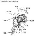
上気道は鼻と喉頭との間の空気を満たす空間を意味する(図1)。上気道で睡眠障害に最も関連した部分は咽頭と呼ばれる喉の後方の空気腔である。咽頭は三つの解剖学的水準に分けられる(図2)。 The upper airway means the space that fills the air between the nose and the larynx (FIG. 1). The most relevant part of the upper airway for sleep disorders is the air space behind the throat called the pharynx. The pharynx is divided into three anatomical levels (Figure 2).
1)上咽頭は鼻腔背部の咽頭の一部である。
2)口の背後にある部分は中咽頭と呼ばれている。より正確には、これは口蓋帆咽頭と呼ばれるのが最良である。口蓋帆咽頭は、口蓋帆(軟口蓋)と舌湾曲部を含む咽頭の一部に相当する。1) The nasopharynx is a part of the pharynx behind the nasal cavity.
2) The part behind the mouth is called the oropharynx. More precisely, this is best called the palatal pharynx. The palatal pharynx corresponds to a part of the pharynx including the palate sail (soft palate) and the tongue curved portion.
3)下咽頭は舌根背後にある。
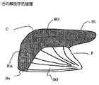
口蓋帆咽頭は軟組織構造がより多く存在するため、より圧潰されやすく、気流のスペースが少なくなる。口蓋帆咽頭の主要構造は軟口蓋と舌であり、両者は非常に曲がりやすい。軟口蓋は口と鼻の間の障壁として働く。多くの人においては軟口蓋は必要以上に長く、舌と咽頭壁の間を下方に延びている。舌は上気道の最大の筋器官であり、解剖学的に舌端、舌体と舌根に分けられる(図3)。舌湾曲部の大部分は舌体と舌根との接合部にある。3) The hypopharynx is behind the tongue base.
Since the palatal pharynx has more soft tissue structure, it is more likely to be crushed and less airflow space. The main structures of the palatal pharynx are the soft palate and the tongue, both of which are very flexible. The soft palate acts as a barrier between the mouth and nose. In many people, the soft palate is longer than necessary and extends downward between the tongue and the pharyngeal wall. The tongue is the largest muscle organ of the upper respiratory tract and is anatomically divided into the tongue end, tongue body and tongue base (FIG. 3). Most of the tongue flexure is at the junction of the tongue body and tongue base.
覚醒状態では口蓋帆咽頭の構造はその内筋を継続的に緊張させることによりその形を維持する。睡眠中のようにこの緊張が低下すると、この構造は非常に曲がりやすく伸び易くなる。それを適所に保持する正常な筋緊張なしには、比較的低陰圧で圧潰し易い。筋は睡眠中体全体で弛緩するが、多くの呼吸器筋は活動したままである。具体的には舌を前に引き出す主な筋であるオトガイ舌筋は、睡眠障害中は通常の又は高い活動性を示すことが報告されている。通常オトガイ舌筋は舌を前方へ移動させ、口から突き出させることさえ可能である。なぜオトガイ筋が閉塞を防げないかの理由は説明されていない。 In the awake state, the palatopharyngeal structure maintains its shape by continuously tensioning its internal muscles. When this tension is reduced, such as during sleep, the structure becomes very flexible and easy to stretch. Without the normal muscle tension that holds it in place, it tends to collapse with a relatively low negative pressure. While muscles relax throughout the body during sleep, many respiratory muscles remain active. Specifically, the genioglossus muscle, which is the main muscle that pulls the tongue forward, has been reported to show normal or high activity during sleep disorders. Usually the genioglossus muscle can move the tongue forward and even stick out of the mouth. The reason why the genital muscle cannot prevent obstruction is not explained.
吸息時には胸壁が拡張し、陰圧を発生し空気を鼻と口に吸い込み、咽頭を通って肺に吸い込む。この陰圧により上気道軟組織を変形させ、さらにその気道を狭小化させる。もし気道が十分に狭小化されると、気流は乱流になり軟口蓋を振動させる。軟口蓋の振動によりいびきとして知られる音を発生する。いびきは非常に一般的で、男の50%、女の25%迄の人がかく。いびき自身は医学的問題ではないが、いびきをかく人のベッドの相手にとってはとてつもない問題であり、夫婦関係のひずみの主原因である。 During inspiration, the chest wall expands, creating a negative pressure, sucking air into the nose and mouth, and into the lungs through the pharynx. This negative pressure deforms the upper airway soft tissue and further narrows the airway. If the airway is narrowed sufficiently, the airflow becomes turbulent and vibrates the soft palate. A sound known as snoring is generated by the vibration of the soft palate. Snoring is very common, affecting up to 50% of men and 25% of women. Although snoring itself is not a medical problem, it is a tremendous problem for a snoring person and is a major cause of marital distortion.
睡眠中に気流の量のわずかな低下や短い閉塞は全ての人に起こる。気流が正常状態の50%以下まで低下するか(呼吸低下)、気流が10秒以上遮断される場合(無呼吸)には、これらの発症は医学的に有意であると判断される。睡眠中の一時間毎に起こる無呼吸と呼吸低下の数を測定して睡眠障害の重症度が診断される。呼吸低下と無呼吸の発症により、睡眠中にある程度の覚醒状態が惹き起こされる。患者の意識は完全には目覚めてはいないが、睡眠パターンが妨げられ、しばしば患者に日中眠たいと感じさせる。呼吸低下か無呼吸の頻度が一時間に5回以上の場合には、上気道抵抗症候群と呼ばれる。これらの患者はしばしば睡眠障害に関連した症状を示す。具体的にはこれらの患者は日中過度に眠たくなる。更に鬱や集中することが困難という繊細な症状もまた一般的報告されている。 A slight drop in airflow or short obstruction occurs during sleep in all people. These episodes are judged to be medically significant if the airflow is reduced to 50% or less of normal conditions (respiratory decline) or if the airflow is interrupted for more than 10 seconds (apnea). The severity of sleep disorders is diagnosed by measuring the number of apneas and hypopneas that occur every hour during sleep. The onset of hypopnea and apnea causes some arousal during sleep. Although the patient's consciousness is not fully awake, sleep patterns are disturbed, often making the patient feel sleepy during the day. If the frequency of hypopnea or apnea is more than 5 times per hour, it is called upper airway resistance syndrome. These patients often show symptoms associated with sleep disorders. Specifically, these patients become excessively sleepy during the day. In addition, subtle symptoms such as depression and difficulty concentrating are also commonly reported.
技術的にはOSASの診断は睡眠の一時間毎に呼吸低下か無呼吸が平均で10回以上発症するものと定義する。気道が閉塞しても、患者は繰り返し継続的に無理に吸気しようとする。これらの発症は大部分が無音で、更に患者は空気を肺に入れるように必死に努力するような腹部と胸部との運動で特徴付けられる。無呼吸の発症は一分以上続き、この時間の間血液中の酸素レベルは減少する。最後に閉塞に打ち勝って、通常は大きないびきをかくか、或いは患者は息が詰まると感じで目覚める。 Technically, a diagnosis of OSAS is defined as an average of 10 or more episodes of hypopnea or apnea per hour of sleep. Even if the airway becomes obstructed, the patient repeatedly and continuously attempts to inhale. These episodes are largely silent and are further characterized by abdominal and chest movements in which the patient desperately tries to get air into the lungs. The onset of apnea lasts for more than a minute, during which time the oxygen level in the blood decreases. Finally overcome the occlusion and usually snore less or the patient wakes up feeling suffocated.
OSAS患者の非常に一般的症状は朝の頭痛と酸の逆流である。気道閉塞の間空気を無理に吸気しようとするため、胸部に強大な陰圧を生ずる。この高い陰圧により酸を胃から食道へ引き上げる。酸はずっと口まで進み、声帯と鼻粘膜の炎症を起こす。上気道に酸が存在すると、喘息発作と類似の肺の反射気管支収縮を起こす。酸が少量でさえも肺にはいると、声帯襞をしっかりと閉じさせ、喉頭痙攣と呼ばれる持続性無呼吸を起こす。多くの患者においては、食道括約筋の伸張を繰り返し慢性的変化を起こし、これら患者は日中でも酸の逆流を起こす。 Very common symptoms in OSAS patients are morning headache and acid reflux. It attempts to inhale air during airway obstruction, creating a strong negative pressure on the chest. This high negative pressure raises the acid from the stomach to the esophagus. The acid goes all the way to the mouth, causing inflammation of the vocal cords and nasal mucosa. The presence of acid in the upper respiratory tract causes reflex bronchoconstriction of the lung similar to an asthma attack. Even a small amount of acid in the lungs causes the vocal folds to close tightly and cause persistent apnea called laryngeal spasm. In many patients, chronic changes in esophageal sphincter stretch occur, and these patients have acid reflux even during the day.
より重要なことには、睡眠障害により深刻な医学的問題と死をもたらす。無呼吸により心臓と肺に大きな負担をかける。常に無呼吸を繰り返し発症することで慢性的変化を起こし、高血圧症をもたらす。長期の無呼吸により血液中の酸素レベルが低下する。次いで低酸素により心臓麻痺または脳卒中を起こす。 More importantly, sleep disorders cause serious medical problems and death. Apnea places a heavy burden on the heart and lungs. It causes chronic changes by constantly developing apnea, resulting in hypertension. Long-term apnea reduces oxygen levels in the blood. Hypoxia then causes heart attack or stroke.
睡眠障害の治療
OSASは子供と大人の両者で起こるが、その原因と治療は全く異なる。子供のOSASは子供が大きい扁桃腺を持つときに起こり、扁桃摘出術によりその症状が治癒する。扁桃腺の大きさは年齢と共に小さくなり、大人では殆ど問題にならない。その代わりに、罹りやすい大人は、通常、拡大した舌、柔らかい口蓋及び/又は咽頭壁を有している。この拡大は大抵その構造内の脂肪沈着による。Treatment of sleep disorders
OSAS occurs in both children and adults, but the causes and treatments are quite different. OSAS in children occurs when the child has a large tonsil, and tonsillectomy cures the symptoms. The size of the tonsils decreases with age and is not a problem for adults. Instead, susceptible adults usually have an enlarged tongue, soft palate and / or pharyngeal wall. This enlargement is mostly due to fat deposition within the structure.
大人の睡眠障害治療は種々の理由により困難である。上気道は、嚥下と発語という重要な機能を行う構造で、非常に動きやすい。これらの機能は外科手術か他処置により容易に損なわれてしまう。更に上気道には、吐き気や咳嗽のような反射を発生させる多くの知覚性神経支配もある。理論的には、口腔や咽頭に取り付ける体内ステントは睡眠時無呼吸を軽減するのに十分に有効であろう。患者が手術のために麻酔されたときのように完全に意識不明な場合、湾曲経口チューブを口と咽頭内に配置して気道をステント挿入のために開くことができる。更に、気管内チューブにより人工呼吸用の安全な気道が確保される。しかし、麻酔が効かなくなった後は、患者は直ちに喉の異物に気づき反応して吐く。それ故、経口チューブ、気管内チューブやいずれの類似物のような器具はOSAS治療には使用できない。 Adult sleep disorder treatment is difficult for various reasons. The upper airway is a structure that performs the important functions of swallowing and speaking and is very mobile. These functions are easily impaired by surgery or other procedures. In addition, the upper respiratory tract also has many sensory innervations that generate reflexes such as nausea and cough. Theoretically, an internal stent attached to the oral cavity or pharynx would be effective enough to reduce sleep apnea. If the patient is completely unconscious, such as when the patient is anesthetized for surgery, a curved oral tube can be placed in the mouth and pharynx to open the airway for stent insertion. In addition, the endotracheal tube ensures a safe airway for artificial respiration. However, after anesthesia stops working, the patient immediately notices and reacts to the throat foreign material and vomits. Therefore, devices such as oral tubes, endotracheal tubes and any similar cannot be used to treat OSAS.
体内ステントはOSASには使用できないが、陽空気圧でステントを上気道に挿入する間接的な方法は、OSASの最も一般的に処方される治療である。本法は持続的気道陽圧法(CPAP)と称されている。CPAPは、鼻の周りにきつく取り付けた人工呼吸器に連結したマスクを用いる必要がある。各患者により陽圧の正確な量は異なり、種々の圧力を用いて一泊滞在試験により設定する必要がある。陽圧はステントのように気道が開け続けるように働く。CPAPは、治癒法ではなく、毎晩使用しなければならない療法である。多くのOSAS患者はCPAPにより助かるが、CPAPは患者や隣接ベッドの人たちにとって快適ではない。患者はしばしばその顔にきつく取り付けたマスクによる閉所恐怖症感に耐えられない。更に、顔に対するマスクの適切なシールを維持することに関し多くの技術的問題がある。これらの理由によりCPAPを処方された全患者の半分までがその使用を六ヶ月以内にやめる。(サンダーズ(Sanders)、“睡眠無呼吸のための医学的治療”(Medical Therapy for Sleep Apnea)、睡眠医学の原理と実際(Principles and Practice of Sleep Medicine)第二版、678−684頁。)
気管切開
OSASのための唯一の十分に有効な外科的療法は、上気道全体を迂回して首から気管へ直接連結する外科手術である永久的気管切開である。この処置は、OSASによる重大な医学的合併症の危険性が極めて高い最悪の場合のために残されている危険な処置である。特に、一時的な気管切開は、上気道に他の何らかの処置を行う前に、重度のOSASをもつ患者に実施前気道を制御するために行われることが多い。その理由は、これらの患者に急性気道閉塞や気道に何かの腫脹がある場合には死の危険性が高いからである。OSAS患者は、上気道組織が極めて過剰に腫脹しているため、緊急事態での挿管が非常に困難である。同様に首に大量の脂肪があるため、救急気管切開は非常に危険になる。Endovascular stents cannot be used for OSAS, but the indirect method of inserting the stent into the upper airway with positive air pressure is the most commonly prescribed treatment for OSAS. This method is called continuous positive airway pressure (CPAP). CPAP requires the use of a mask connected to a ventilator tightly attached around the nose. The exact amount of positive pressure varies from patient to patient and needs to be set by an overnight stay test using different pressures. Positive pressure works like a stent to keep the airway open. CPAP is a cure that must be used every night, not a cure. Many OSAS patients are helped by CPAP, but CPAP is not comfortable for patients and people in adjacent beds. Patients often cannot tolerate claustrophobia due to a tightly attached mask on their face. Furthermore, there are a number of technical problems associated with maintaining a proper seal of the mask to the face. For these reasons, up to half of all patients prescribed CPAP will stop using it within six months. (Sanders, “Medical Therapy for Sleep Apnea”, Principles and Practice of Sleep Medicine, Second Edition, pages 678-684.)
Tracheostomy
The only fully effective surgical therapy for OSAS is a permanent tracheotomy, a surgery that bypasses the entire upper airway and connects directly from the neck to the trachea. This procedure is a dangerous procedure that remains for the worst case where the risk of serious medical complications from OSAS is extremely high. In particular, temporary tracheostomy is often performed to control the pre-implemented airway in patients with severe OSAS before performing any other treatment on the upper airway. The reason is that if these patients have acute airway obstruction or some swelling in the airway, the risk of death is high. OSAS patients are very difficult to intubate in an emergency situation because the upper airway tissue is extremely excessively swollen. Similarly, emergency tracheostomy is very dangerous because of the large amount of fat in the neck.
本保存的処置以前には、術後死は重度のOSAS患者ではまれではなかった。更に、この患者はしばしば呼吸抵抗になれおり、その抵抗が突然除かれると呼吸中枢は低下する。現在でも、大部分のOSAS患者の治療における治癒の基本は、外科的処置後、患者を集中治療室か回復室で注意深く観察することである。 Prior to this conservative treatment, postoperative death was not uncommon in severe OSAS patients. In addition, the patient is often reluctant to breathe, and the respiratory center is lowered when the resistance is suddenly removed. Even now, the basis of healing in the treatment of most OSAS patients is to carefully observe the patient in the intensive care unit or recovery room after surgery.
いびきに関する軟口蓋処置
軟口蓋が他の組織以上に振動すると、いびきと均衡がとれていない役割を果たす。軟口蓋を収縮か硬化させるために種々の外科的治療が利用できる。使用される主な処置は口蓋垂軟口蓋咽頭形成術(UPPP)と呼ばれる。UPPPでは、手術用メスで咽頭壁と軟口蓋の過剰軟組織を切除する。咽頭領域の粘膜がUPPP時にあまりに多く傷つけられるので、大きな術後腫脹と激痛が起こる。いびきをかくが閉塞がない選ばれた患者では、より限定版のUPPPをレーザーか電気メスで行う。Soft palate treatment for snoring When the soft palate vibrates more than other tissues, it plays an unbalanced role with snoring. Various surgical treatments are available to shrink or harden the soft palate. The main procedure used is called uvulopalatopharyngoplasty (UPPP). UPPP removes excess soft tissue from the pharyngeal wall and soft palate with a scalpel. The mucosa in the pharyngeal area is damaged too much during UPPP, resulting in large postoperative swelling and severe pain. For selected patients who snore but have no obstruction, a more limited version of UPPP is performed with a laser or electrosurgical unit.
より新しい処置では、粘膜の外傷を最小限にし、針を使って下にある軟組織に達して当該軟組織の体積を収縮させるか硬化させて振動に抗するようにする。電極を軟口蓋に挿入して口蓋を収縮させるか硬化させるように高周波エネルギーを送る。(パウエル、エヌビー等(Powel,NB et al.)(1998年)、睡眠障害型呼吸をもつ被験者の口蓋における高周波エネルギーによる組織容積縮減術(Radiofrequency volumetric tissue reduction of the palate in subjects with sleep-disordered breathing)、チェスト(Chest)、113巻、1163−1174頁。)(ソムノプラスティ(Somnoplasty)、ソムナス(Somnus)、マウンテンビュー(Mountainview)、カルフォルニア州(CA))。軽い腐食剤を注入して軟口蓋の容積を減らすことができる。ベンゼル(Benzel)の米国特許6439238では、軟口蓋表面への硬化剤塗布が教示されている。ごく最近には外来診療所での軟口蓋硬化用プラスチック挿入物移植が食品医薬品局(FDA)により認可された。(ピラー(登録商標)処置(Pillar Procedure)、米国特許6546936、上咽頭部位の状態を治療するための方法と器具(Method and apparatus to treat conditions of the naso-phrayngeal area)。)
より新しい処置を含む軟口蓋を標的とする全ての方法での基本的な欠陥はOSASの一部だけしか改善できないことである。(ラウベ、ディアイ(Loube DI)、(1999年)、閉塞型睡眠無呼吸症候群治療における技術的進歩(Technological Advances in the Treatment of Obstructive Sleep Apnea Syndrome)、チェスト(Chest)、1999年、116巻、1426−1433頁;ドグラム、ジーケイ等(Doghramji, K et al.)、(1995年)、口蓋垂咽頭形成術成績の予測(Predictors of outcome for uvulopalatophryngoplasty)、ラリンゴスコープ(Laryngoscope)、105巻、311−314頁)。研究では無呼吸数の減少を報告しているが、これら患者はめったに治癒しない。明らかにOSASを起こす重要な構造は軟口蓋ではなく舌である。In newer procedures, mucosal trauma is minimized and the needle is used to reach the underlying soft tissue to shrink or harden the soft tissue volume to resist vibration. High frequency energy is sent to insert the electrode into the soft palate to shrink or harden the palate. (Powel, NB et al. (1998), Radiofrequency volumetric tissue reduction of the palate in subjects with sleep-disordered breathing) ), Chest, 113, 1163-1174.) (Somnoplasty, Somnus, Mountainview, California (CA)). Light caustic can be injected to reduce soft palate volume. US Pat. No. 6,439,238 to Benzel teaches the application of a hardener to the soft palate surface. Most recently, the Food and Drug Administration (FDA) has approved a plastic insert for soft palate curing in an outpatient clinic. (Pillar Procedure, US Pat. No. 6,546,936, Method and apparatus to treat conditions of the naso-phrayngeal area).
The fundamental flaw in all methods that target the soft palate, including newer treatments, is that only a portion of OSAS can be improved. (Loube DI, (1999), Technological Advances in the Treatment of Obstructive Sleep Apnea Syndrome, Chest, 1999, 116, 1426. -1433; Dogramji, K et al., (1995), Predictors of outcome for uvulopalatophryngoplasty, Laryngoscope, 105, 311-314 page). Although studies have reported a decrease in apnea rate, these patients rarely heal. Clearly the important structure that causes OSAS is the tongue, not the soft palate.
OSASに関する舌根法
OSASでの舌根治療に用いる方法は、その体積を永久的に減少させて撓み性を低下するか、舌全体を前方へ移動させるかのいずれかである。Tongue root method for OSAS
The method used to treat the tongue base with OSAS is to either permanently reduce its volume to reduce flexibility or move the entire tongue forward.
舌根の外科的切除の効果は不十分である。OSAS治療でのメスかレーザーによる舌根の切除の結果は、この方法の継続的な適用を勧めるには不十分であった。(マイケルソン、エスエイ、ローゼンタール、エル(Mickelson, SA, Rosenthal, L)(1997年)、閉塞型睡眠無呼吸症候群治療での舌根正中部切除術と喉頭蓋切除術(Midline glossectomy and epiglottiectomy)、ラリンゴスコープ(Laryngoscope)、107巻、614−619頁)。より最近では、高周波エネルギー(エドワード(Edwards)の米国特許5843021)や超音波エネルギー(米国特許6409720)が高周波エネルギによって舌根を収縮させ且つ硬化させることに対して提案された。針電極を舌根に挿入し、高周波エネルギーを伝えて損傷させて傷跡を残し且つ時間と共に収縮させる。術後の腫脹や痛みを避けるために、制限量の損傷を1回の治療で行うが、患者には平均で5回の治療が必要である。おおよそ三分の一の患者のOSASが50%以上良くなる。しかし、約四分の一の患者では舌根潰瘍形成、舌根膿瘍や一時的気管切開を含む重大な術後合併症がある。 The effect of surgical excision of the tongue base is inadequate. The results of scalpel or laser excision with OSAS treatment were insufficient to encourage continued application of this method. (Mickelson, SA, Rosenthal, L (1997), Midline glossectomy and epiglottiectomy for the treatment of obstructive sleep apnea syndrome, Midline glossectomy and epiglottiectomy, LA The apple scope (Laryngoscope), 107, 614-619). More recently, high frequency energy (Edwards US Pat. No. 5,843,221) and ultrasonic energy (US Pat. No. 6,409,720) have been proposed for shrinking and hardening the tongue base with high frequency energy. A needle electrode is inserted into the tongue base, transmitting high frequency energy and damaging, leaving a scar and contracting over time. To avoid postoperative swelling and pain, a limited amount of damage is done in one treatment, but patients need an average of 5 treatments. Approximately one third of patients have a 50% better OSAS. However, about a quarter of patients have significant postoperative complications including tongue ulceration, tongue abscess and temporary tracheostomy.
舌根を前進させるために最近導入された器具は、レポーズ(登録商標)(Repose)システム(インフルエント社(Influent Corp)、サンフランシスコ、カリフォルニア州(CA))である。レスポンス(登録商標)(Response)による方法は、全身麻酔下で行い、ねじを下顎骨の基部に挿入する。ねじは、永久縫合糸用のアタッチメントを含み、当該縫合糸は、口腔底粘膜の下に舌の後面への通路を開け、次いで舌根を幅方向に横断させて戻し、下顎骨にねじ込んだ金属フックに取り付けられる。縫合糸を締めて舌根を前方へ移動させるが、組織壊疽となる過剰な引っ張りを防ぐように注意する必要がある。残念なことにはレポーズ(登録商標)(Repose)法の研究では、当該方法はOSASを除去するには効果的でないことが示された。15人の患者のうち一人だけがOSASが治癒したが、二人の患者では痛みと腫脹のため縫合糸を除去する必要があった。 A recently introduced instrument for advancing the tongue base is the Repose® system (Influent Corp, San Francisco, CA (CA)). The Response method is performed under general anesthesia and a screw is inserted into the base of the mandible. The screw includes an attachment for a permanent suture that opens a passage to the posterior surface of the tongue under the floor mucosa and then traverses the tongue base back in the width direction and is screwed into the mandible Attached to. Tighten the suture to move the tongue base forward, but care must be taken to prevent excessive pulling that leads to tissue gangrene. Unfortunately, research on the Repose method has shown that the method is not effective in removing OSAS. Only one out of 15 patients healed OSAS, but two had to remove the suture because of pain and swelling.
より積極的な外科的手段では、下顎骨、顔面骨格や舌骨の再構築が必要である。当該技術の例としては、ウッドソン(Woodson)の米国特許6,161,541号があり、当該特許は、咽頭気道を外科的に拡張する方法を教示している。この方法は、より危険性が高く、回復期間が遙かに長い広範囲な手術を必要とする。 More aggressive surgical measures require reconstruction of the mandible, facial skeleton and hyoid bone. An example of such technology is Woodson, US Pat. No. 6,161,541, which teaches a method for surgically dilating the pharyngeal airway. This method is more risky and requires extensive surgery with a much longer recovery period.
舌根治療の他の提案された方法としては、米国特許6,742,524による硬化粒子又は米国特許出願第20050004417A1号による他の移植材料を注入して軟組織を硬化する方法がある。 Other proposed methods of tongue base treatment include injecting hardened particles according to US Pat. No. 6,742,524 or other implant materials according to US Patent Application No. 20050004417A1 to harden soft tissue.
神経機能代替え装置
上気道筋を刺激する種々の装置が発明された。サンダーズ(Sanders)の米国特許第4,907,602号には、気道を拡張する経粘膜刺激が記載されている。カレル(Karell)の米国特許第5,792,067号では、硬口蓋、軟口蓋又は咽頭部位を電気的に刺激して上気道筋の収縮を誘発する口腔内装置が教示されている。メール(Meer)の米国特許第5,190,053号では、舌小帯の両側の口腔底粘膜上に配置された電極によりオトガイ舌筋に電気的刺激を付与する口腔内装置が教示されている。更に、テスターマン(Testerman)の米国特許第5,591,216号には、オトガイ舌筋の神経を刺激する全体が移植可能な装置が記載されている。更に、ゴードン(Gordon)の国際特許出願第04064729号には、いびきを治療するために軟口蓋に注入できる神経機能代替え装置が記載されている。現在のところ、これらの装置は臨床的には検証されていない。Various devices for stimulatingneural function alternative device upper airway has been invented. Sanders U.S. Pat. No. 4,907,602 describes transmucosal irritation to dilate the airways. Karell in US Pat. No. 5,792,067 teaches an intraoral device that electrically stimulates the hard palate, soft palate, or pharyngeal site to induce upper airway muscle contraction. U.S. Pat. No. 5,190,053 to Meer teaches an intraoral device that provides electrical stimulation to the genioglossus muscle by means of electrodes placed on the oral floor mucosa on either side of the lingual band. . In addition, Testerman, US Pat. No. 5,591,216, describes a fully implantable device that stimulates the nerve of the genioglossus muscle. In addition, Gordon International Patent Application No. 04064729 describes a nerve function replacement device that can be injected into the soft palate to treat snoring. At present, these devices have not been clinically validated.
要約すると、睡眠障害は良好な解決策のない大きな健康問題であり、新規でより有効な治療技術の必要性がある。
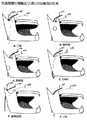
理論には縛られたくないが、人の舌解剖学に関する本発明者の研究によれば、閉塞の発症は一連の現象によって発展することが示唆される(図4)。最初の刺激現象は舌の比較的小部分の変形である。ある特定条件下で、変形は、舌の頂点、特に舌弯曲部位、具体的には舌弯曲部の中心線付近で始まる。この組織が変形すると、気道を狭小化し、より大きい陰圧を発生してより大きな変形が惹き起こされる。次いで、このフィードバックサイクルによってその部位の組織が十分に変形し、口蓋帆咽頭部位の完全な閉塞を起こす。In summary, sleep disorders are a major health problem without a good solution and there is a need for new and more effective treatment techniques.
Although not wishing to be bound by theory, the inventor's study on human tongue anatomy suggests that the onset of obstruction develops through a series of phenomena (FIG. 4). The first stimulus phenomenon is a deformation of a relatively small part of the tongue. Under certain conditions, the deformation begins at the apex of the tongue, in particular near the tongue-curved part, in particular near the centerline of the tongue-curved part. When this tissue is deformed, the airway is narrowed and a greater negative pressure is generated, causing greater deformation. This feedback cycle then sufficiently deforms the tissue at that site, causing complete occlusion of the palatal and pharyngeal region.
最初の閉塞が吸息終了近くで起こる場合には、その閉塞は呼息又はオトガイ舌筋の作用で解放される。しかし、閉塞が吸息初期に起こる場合には、反射現象により、より強い吸息活動を引き起こし、気道圧を更に低下させる。この陰圧の増加により、変形が起こり、大部分の舌根が圧潰される。この時点で、気道は軟組織によってしっかりと塞がれ、オトガイ舌筋は塞がっている舌組織を伸ばすだけで取り除けない。 If the first occlusion occurs near the end of inspiration, the occlusion is released by expiration or the action of the genioglossus muscle. However, if occlusion occurs in the early inspiration period, the reflex phenomenon causes stronger inspiratory activity and further reduces airway pressure. Due to this increase in negative pressure, deformation occurs and most tongue bases are crushed. At this point, the airway is tightly blocked by soft tissue, and the genioglossus muscle cannot be removed simply by stretching the closed tongue tissue.
それ故、舌屈曲部は、閉塞をもたらす段階的動作の開始の重要な部位である。この弛緩した筋肉は非常に曲がり易く且つ変形し易いが、その逆も正しく、この変形を防ぐには非常に小さな力が必要とされるだけである。それ故、舌の適切な局在部位に十分な反力が働くと、会話動作や嚥下動作に目立った影響なしに閉塞を防げる。 Therefore, the tongue flexion is an important site for the initiation of a gradual movement that results in occlusion. This relaxed muscle is very easy to bend and deform, but vice versa, only a very small force is needed to prevent this deformation. Therefore, if a sufficient reaction force is applied to the appropriate localized part of the tongue, the obstruction can be prevented without noticeably affecting the conversational or swallowing movements.
装置が舌弯曲部の変形と圧潰をどのようにして防ぐかは些細な問題ではない。
・舌のこの領域は会話中及び燕下中に極めて良く動き、従って、この領域にかかる力は、小さく且つ極めて局部化されなければならない。この領域を不動にすることは許容されないが、大きな移植片又は瘢痕組織によって強ばった場合には不動となる。It is not a trivial matter how the device prevents deformation and crushing of the tongue fold.
• This area of the tongue moves very well during conversation and during armpits, so the force on this area must be small and very localized. It is not permissible to immobilize this area, but it becomes immobile when it is strengthened by a large graft or scar tissue.
・更に、口蓋帆咽頭の全部位は広範囲の知覚神経支配下にあり、比較的小さな刺激で吐き気か嚥下を起こす。
・舌根及び舌体は、人体の他の部位の相当する筋肉よりも血液の供給が多い。この領域に移植片を配置することによって、潜在的に壊滅性の舌の腫脹によって内出血を惹き起こす可能性が高く、最悪の場合には舌の腫脹の可能性がある。In addition, all parts of the palatal pharynx are under extensive sensory innervation, causing nausea or swallowing with relatively small stimuli.
-The tongue base and tongue body supply more blood than the corresponding muscles in other parts of the human body. Placing the implant in this area is likely to cause internal bleeding due to potentially devastating swelling of the tongue, and in the worst case, possible swelling of the tongue.
・軟組織及び舌は、特に容易に型が変わる。特に、力をかける縫合又は移植片によって組織が改造されてこの力が解放される。このことは、チーズとカッターとの間の作用として知られている。従って、かけられる力は比較的弱く且つかけられる時間は限られた期間でなければならない。 • The soft tissue and tongue change in shape especially easily. In particular, the tissue is remodeled by a suture or graft that applies force to release this force. This is known as the action between the cheese and the cutter. Therefore, the applied force must be relatively weak and the applied time must be a limited period.
・人間の上気道構造は極めて変化し易く、更に、睡眠障害を有する患者の上気道構造は、疾患が進むか又は改良されるにつれて時間と共に変化する。
・最後に、OSAS患者は外科的処置後の腫脹のようなわずかな量の腫脹の後でさえ、閉塞するきわどい気道をもっている。それ故、装置がこの部位に腫脹を避けながら力をかける方法は明らかではない。The human upper airway structure is highly variable, and the upper airway structure of patients with sleep disorders changes with time as the disease progresses or improves.
Finally, OSAS patients have critical airways that obstruct even after a small amount of swelling, such as swelling after a surgical procedure. Therefore, it is not clear how the device applies force to this site while avoiding swelling.
更に最も効果的で、患者と医師に受け入れられるためには、その装置は理想的には追加の特質を必要とするであろう。
・装置は外来処置で挿入できる必要がある。In order to be most effective and acceptable to patients and physicians, the device would ideally require additional attributes.
• The device must be able to be inserted in an outpatient procedure.
・装置を患者が日中に除去でき、夜に再挿入できるのが好ましい。
・装置は患者の特定の必要性に適合するように調節可能である。
・装置は患者にとって快適である。Preferably the device can be removed by the patient during the day and reinserted at night.
• The device can be adjusted to suit the specific needs of the patient.
-The device is comfortable for the patient.
・装置を取り付けたとき、誰にも気づかれない。 ・ No one notices when the device is installed.
人体の上気道内には著しい変動が存在し、睡眠障害及びそれに関連する障害の一因となる病理変化となる更なる変化さえも存在する。更に、病理解剖学は、それらの状態が改善され又は悪化するときに、各患者において常に変化する。全ての偶然性を処置することができる単一の方法及び装置は存在しない。従って、上気道内の種々の部位に対して最適化されている方法及び装置の重大な必要性が存在する。 There are significant fluctuations in the upper respiratory tract of the human body, and even further changes that result in pathological changes that contribute to sleep disorders and related disorders. Furthermore, pathological anatomy always changes in each patient as their condition improves or worsens. There is no single method and device that can treat all contingencies. Accordingly, there is a significant need for methods and devices that are optimized for various sites within the upper respiratory tract.
本発明の実施形態は、障害のある気流に関する哺乳類における上気道障害を防止し又は治療する方法及び装置を含んでいる。これらの障害は、限定的ではないが、いびき、上気道抵抗症候群及び閉塞性睡眠時無呼吸症である。更に、本発明は、限定的ではないが、馬の軟口蓋の背側の変位及びある品種の犬における短頭気道閉塞症候群を含む動物における気道障害に適用可能である。当業者は、本発明の用途は上気道の他の状態に適用することができることを容易に理解するであろう。
本発明の一つの実施形態は、組織牽引子呼吸障害の治療のための組織牽引子であり、該組織牽引子は、軸と牽引子部材とアンカー部材とボルスタとを備えている。前記軸は、患者の口腔又は咽頭内に配置されている軟組織内に挿入される大きさとされている。前記牽引子部材は、前記軸の第一の端部又はその近くに結合されている。前記アンカー部材は、前記軸の第二の端部又はその近くに結合されている。前記ボルスタは、前記軟組織の外面と前記アンカー部材との間に配置されている。前記軸、牽引子部材及び前記アンカー部材のうちの少なくとも1つが軟組織の外面上に配置されている。更に、前記軸、牽引子部材及びアンカー部材のうちの少なくとも1つが、軟組織の少なくとも一部分の変形を防止する力を変えて患者の気道障害を防止するように調整可能である。
更に本発明の別の実施形態は、呼吸障害を治療のための組織牽引子であり、該組織牽引子は、軸と牽引子部材とアンカー部材とボルスタとを備えている。該軸は、患者の口腔又は咽頭内に配置されている軟組織内に挿入される大きさとされている。前記牽引子部材は、当該軸の第一の端部と一体に形成されており且つ配備されていない状態においては前記軸に対して鋭角に位置決め可能であり且つ配備された状態においては前記軸に対して直角である。前記アンカー部材は、前記軸の第二の端部又はその近くに結合されている。前記ボルスタは、前記軟組織の外面と前記アンカー部材との間に配置されている。前記牽引子部材は、前記配備状態にあるときに軟組織の外面上に位置決めすることができ、前記牽引子部材、軸、及びアンカー部材は、相互に作用して、前記軟組織の少なくとも一部分の変形を防止する押圧力をかけて患者の気道閉塞を防止することができるようになされている。Embodiments of the present invention include methods and devices for preventing or treating upper airway disorders in mammals with impaired airflow. These disorders include but are not limited to snoring, upper airway resistance syndrome and obstructive sleep apnea. Furthermore, the present invention is applicable to airway disorders in animals including, but not limited to, displacement of the dorsal side of the horse's soft palate and short head airway obstruction syndrome in certain breeds of dogs. One skilled in the art will readily appreciate that the application of the present invention can be applied to other conditions of the upper respiratory tract.
One embodiment of the present invention is a tissue retractor for the treatment of tissue retractor breathing disorders, the tissue retractor comprising a shaft, a retractor member, an anchor member, and a bolster. The shaft is sized to be inserted into soft tissue placed in the patient's oral cavity or pharynx. The retractor member is coupled at or near the first end of the shaft. The anchor member is coupled to or near the second end of the shaft. The bolster is disposed between the outer surface of the soft tissue and the anchor member. At least one of the shaft, retractor member and anchor member is disposed on the outer surface of the soft tissue. Further, at least one of the shaft, retractor member, and anchor member is adjustable to alter a force that prevents deformation of at least a portion of the soft tissue to prevent patient airway obstruction.
Yet another embodiment of the present invention is a tissue retractor for treating respiratory disorders, the tissue retractor comprising ashaft, a retractormember, an anchor member, and a bolster. The shaft is sized to be inserted into soft tissue located inthe patient's oral cavity or pharynx. The retractor member is formed integrally with the first end portion of the shaft and can be positioned at an acute angle with respect to the shaft in astate where the retractor member is notdeployed and on the shaft in adeployedstate . It is a right angle to it. The anchor member is coupled to or near the second end of the shaft. The bolster is disposed between the outer surface of the soft tissue and the anchor member. The retractor member can be positioned on an outer surface of soft tissue when in thedeployed state , and the retractor member, shaft, and anchor member interact to effect deformation of at least a portion of the soft tissue. The patient can prevent the airway from being obstructed by applying a pressing force to prevent the airway from being blocked.
本発明の一つの特徴は、気道を拡張させ又は組織が変形するのを防止することによって、気道の障害を防止することである。過剰な組織が存在し且つ上気道構造の弛緩した軟組織に対する気道陰圧による変形作用にも対抗する。これらの構造としては、限定的ではないが、舌、軟口蓋、咽頭壁及び声門喉頭がある。 One feature of the present invention is to prevent airway obstruction by dilating the airway or preventing tissue deformation. Excess tissue is present and counteracts the deforming effect of negative airway pressure on soft tissue with relaxed upper airway structure. These structures include, but are not limited to, the tongue, soft palate, pharyngeal wall, and glottic larynx.
PCT国際公開WO 2005/082452には、ここではリンガフレックス舌牽引子(LTR)と称される方法及び装置の一つの実施形態が記載されており、ここに記載されている装置の使用は、舌又は牽引に限定されないことが示されている。当該LTRは、牽引子(R)、軸(S)及びアンカー(A)からなる。好ましい実施形態においては、牽引子は舌根の軟組織に肉体的に結合されている。軸は、アンカーと結合するために舌の正中線を通っている。当該アンカーは、軸によって牽引子の力に対抗し、それによって軟組織の変形を防止する。 PCT International Publication No. WO 2005/082452 describes one embodiment of a method and apparatus referred to herein as a Ringerflex tongue retractor (LTR), the use of the apparatus described herein being Or it is shown that it is not limited to traction. The LTR includes a retractor (R), a shaft (S), and an anchor (A). In a preferred embodiment, the retractor is physically attached to the soft tissue of the tongue base. The shaft passes through the midline of the tongue to connect with the anchor. The anchor counteracts the force of the retractor by the shaft, thereby preventing soft tissue deformation.
本発明の一つの特徴は、患者の不快感を減じながら器具の有効性を増す牽引子ヘッド、軸及びアンカーに対する改良について記載している。LTRの部品の改良としては、限定的ではないが、狭い給送器具内に嵌合するために圧潰し且つ挿入後に拡張する牽引子ヘッド、周囲の舌の動きに応じて長さ及び張力を受動的に調整する軸及び患者によって調整可能であり且つ軟らかいボルスター(支持部材)、口の中の部分的に移植された受け口及び/又は歯科器具に取り付けられる改良されたアンカーがある。 One aspect of the present invention describes improvements to retractor heads, shafts and anchors that increase the effectiveness of the instrument while reducing patient discomfort. Improvements to LTR components include, but are not limited to, a retractor head that collapses to fit into a narrow feeder and expands after insertion, passive length and tension depending on the movement of the surrounding tongue There is an adjustable shaft and a patient-adjustable and soft bolster, a partially implanted receptacle in the mouth and / or an improved anchor attached to a dental instrument.
本発明の他の特徴は、器具を、日中は殆ど張力がかからないか又は全くかからない無負荷状態とさせ、夜間には張力を治療レベルすなわち負荷がかかった状態まで増大させることによって、移植片をより快適なものとする大きな制御度を患者に許容する。患者の意思の欠如が恐らく現在の睡眠時無呼吸の治療における最も大きな問題であるので、この方法を実行する方法及び装置は極めて重要である。 Another feature of the present invention is that the device can be placed in an unloaded state with little or no tension during the day and by increasing the tension to a therapeutic level or loaded at night. Allow the patient a greater degree of control to make it more comfortable. The method and apparatus for carrying out this method is extremely important since lack of patient will is probably the biggest problem in the current treatment of sleep apnea.
本発明の別の特徴は、舌内及び舌の周囲の付加的な部位が本発明によって予想外に治療されて気道障害を防止することができることである。これらの部位の非限定的な例は、舌根、舌を覆っている粘膜、舌小帯、咽頭襞、口蓋襞及び披裂喉頭蓋襞、咽頭側壁及び軟口蓋である。これらの部位に適用される改良されたLTRは、舌根、軟口蓋及び咽頭側壁を直接又は間接的に硬化させ且つ変位させる。各部位は、LTRに対する新規且つ予想外の改良が最少の危険性及び患者に対する不快感が最少な状態で効率良く行われることを可能にする特別な解剖学的構造を有している。 Another feature of the present invention is that additional sites in and around the tongue can be unexpectedly treated by the present invention to prevent airway obstruction. Non-limiting examples of these sites are the tongue base, mucous membrane covering the tongue, tongue band, pharyngeal fistula, palatal and nasopharyngeal fistula, pharyngeal sidewall and soft palate. An improved LTR applied to these sites hardens and displaces the tongue base, soft palate and pharyngeal sidewall directly or indirectly. Each site has special anatomical structures that allow new and unexpected improvements to the LTR to be performed efficiently with minimal risk and minimal patient discomfort.
本発明の一つの特徴は、小帯領域内の移植片部位によって舌根を間接的に牽引するLTRである。これは、器具の挿入、調整及び維持を簡単にする。
本発明の別の特徴は、弛緩した表面粘膜を緊張させ又は内部の舌内部構造に機械的に結合するために舌根内へ挿入される極めて局所化され且つ完全に挿入できるLTRを記載している。One feature of the present invention is an LTR that indirectly pulls the tongue base by a graft site within the zonule region. This simplifies instrument insertion, adjustment and maintenance.
Another aspect of the invention describes a highly localized and fully insertable LTR that is inserted into the tongue base to tension the relaxed surface mucosa or mechanically couple to the internal lingual internal structure. .
本発明の別の特徴は、咽頭襞内又はその近くに挿入されるLTRである。この部位は、舌根組織のみならず軟口蓋及び咽頭側壁の牽引及び緊張を可能にする。この部位の利点は、最少侵襲性、安全性及び多くの異なる構造に有益な作用である。 Another feature of the present invention is an LTR that is inserted into or near the pharyngeal fistula. This site allows traction and tension of the soft palate and pharyngeal sidewall as well as the tongue base tissue. The advantages of this site are minimal invasiveness, safety and beneficial effects on many different structures.
本発明の別の特徴は、咽頭空域を拡張するために上気道組織を改造するための方法及び装置である。改造される組織としては、限定的ではないが、舌根、口蓋扁桃、咽頭壁及び軟口蓋がある。これらの組織は、その体積を小さくするために、圧縮されるか又は変位若しくは再形成されるのが好ましい。この作用は、当該器具を取り外した後、数ヶ月から数年続く。この持続的に有益な作用を達成するために、当該器具は、好ましくは1週間乃至1年、より好ましくは1乃至6ヶ月間に亘って力をかけるのが好ましい。 Another feature of the present invention is a method and apparatus for remodeling upper airway tissue to expand the pharyngeal airspace. Tissues that are remodeled include, but are not limited to, the tongue base, palatine tonsils, pharyngeal wall, and soft palate. These tissues are preferably compressed or displaced or reshaped to reduce their volume. This action lasts for months to years after removal of the device. In order to achieve this lasting beneficial effect, the device is preferably applied over a period of 1 week to 1 year, more preferably 1 to 6 months.
本発明の別の特徴は、磁石、接着剤、真空及び/又は機械的手段を使用して軟組織を把持し、変位させ且つ/又は再配置するために、粘膜に可逆的に結合する非侵襲的な方法及び器具である。一つの実施形態においては、湾曲した牽引子が選択された部位内へ可逆的に挿入される。別の実施形態においては、留置先端が、PGF、扁桃襞、軟口蓋及びその他の軟組織の襞上に配置される。これらの牽引子は、必要に応じて、口の中又は外の改造されたアンカーに結合させることによって装着することができる。更に別の実施形態においては、口腔底は、舌根を変位させるために延ばされている。更に別の実施形態においては、真空によって、舌根の体積を減じるように舌が再形成されている。 Another feature of the present invention is a non-invasive reversible connection to the mucosa to grasp, displace and / or reposition soft tissue using magnets, adhesives, vacuum and / or mechanical means. Method and apparatus. In one embodiment, a curved retractor is reversibly inserted into a selected site. In another embodiment, the indwelling tip is placed on the folds of PGF, tonsil, soft palate and other soft tissues. These retractors can be mounted as required by coupling to a modified anchor in or outside the mouth. In yet another embodiment, the floor of the mouth is extended to displace the tongue base. In yet another embodiment, the tongue is reshaped by vacuum to reduce the volume of the tongue base.
本発明の別の特徴は、特に、馬の軟口蓋の背側への変位を防止するようになされたLTRが記載されている。
各部位において、LTRは多くの実施形態を有している。LTRは、組織内を通過することができ且つ組織の外側に牽引子又はアンカー端部を備えるか又は露出された一つの端部のみを有しており、又は器具全体を移植することができる。当該器具の軸は、組織内へと深く入ることができ又は表面上の粘膜のすぐ下側へ入ることができる。牽引子及びアンカー部材は、力を均一に配分するためにその部位に嵌合する形状とされるか、舌根中央、咽頭壁及び軟口蓋のような平らな又は中央が湾曲した面のための平らな形状、咽頭襞及び軟口蓋の外側縁の深さのための形状とされた楔形状、小帯に対するV字形状及び歯のためのT字形状とされるのが好ましい。移植片、牽引子及びアンカーの材料は、当該技術において知られている公知の非反応性の生体適合性材料のうちのいずれかとすることができる。堅牢な材料の非限定的な例としては、ステンレス鋼、チタン、セラミック及びプラスチックがある。弾性材料としては、シリコン及びゴムがある。舌を前方へ変位させ又は軟口蓋を上方へ変位させるのに必要とされる力は、0.001グラム乃至10,000グラムであり、0.1グラム乃至1000グラムが更に好ましく、10乃至100グラムが最も好ましい。この力は、0.01秒乃至永久にかけることができ、1分乃至1ヶ月がより好ましく、睡眠中であるのが更に好ましく、制限された上気道の流れの一回分中であるのが最も好ましい。Another aspect of the invention describes an LTR specifically adapted to prevent displacement of the horse's soft palate back.
At each site, the LTR has many embodiments. The LTR can pass through tissue and has a retractor or anchor end outside the tissue or has only one exposed end, or the entire device can be implanted. The instrument shaft can penetrate deeply into the tissue or can enter just below the mucosa on the surface. The retractor and anchor member can be shaped to fit into the site to distribute the force evenly, or flat for flat or centrally curved surfaces such as the base of the tongue base, pharyngeal wall and soft palate Preferably, the shape is a wedge shape shaped for the depth of the outer edge of the pharyngeal fistula and soft palate, a V shape for the zonule and a T shape for the teeth. The graft, retractor and anchor material can be any of the known non-reactive biocompatible materials known in the art. Non-limiting examples of robust materials include stainless steel, titanium, ceramic and plastic. Elastic materials include silicon and rubber. The force required to displace the tongue forward or displace the soft palate is 0.001 to 10,000 grams, more preferably 0.1 to 1000 grams, more preferably 10 to 100 grams. Most preferred. This force can be applied from 0.01 seconds to forever, more preferably from 1 minute to 1 month, even more preferably during sleep, and most preferably during a limited upper airway flow. preferable.
本発明は、添付図面と関連付けて考慮される以下に提供する例示的な実施形態の詳細な説明によってより容易に理解できるであろう。
図面の詳細な説明
図1 人体の上気道の正中面図
図2 舌及びその周辺組織の簡素化された概略図
NP 上咽頭
VP 口蓋帆咽頭
HP 下咽頭
SP 軟口蓋
P 硬口蓋
T 舌
GG オトガイ舌筋
図3 舌の解剖学的境界標識
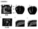
舌は、この図の灰色の領域として規定されている。舌は、前方から後方に向かって、舌端、舌体及び舌根に分けられる。オトガイ舌筋(GG)が舌の下面の結合組織境界(Bo)内に差し込まれている。オトガイ舌筋及びその筋肉の領域全体は、“小帯領域”と称される。
BA)舌根
BD)舌体
BL)舌端
Bo)舌とオトガイとの境界部
C)舌の曲線
F)小帯
GG)オトガイ舌筋
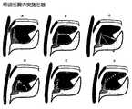
図4 気道閉塞機構及び現在治療法の効果
A)覚醒中の正常な緊張度
舌は定位置のままであって気道を開いたままとさせている。青い矢印410は空気の流れを示しており、小さな黒い矢印412は下顎に対する咽頭壁(赤線414)の関係を示している。
B)睡眠中に舌の中の筋肉の緊張度は失われ弛緩状態となる。
吸息中の咽頭内の陰圧は、口蓋帆咽頭領域内での舌の後方圧潰を生じさせる。なぜならば、この状態では気道は狭く、舌の曲線(円形)は最も変形し得るからである。
C)気道が口蓋帆咽頭領域で閉塞した後の吸息は、咽頭内の圧力を下げ、更に、舌根を変形させ且つ気道をしっかりと遮断する。
D)CPAPは、高い圧力で空気を鼻内に圧送し(青い線416)、それによって咽頭に副木を施して開かせる。
E)歯用器具は、顎全体を前方へ動かすことによって作動する。舌が口腔底に沿った軟組織に付着し、軟組織が顎に付着すると、舌は間接的に動かされて気道を拡張させる。顎が咽頭壁に対して動いていることに注意(矢印)。
F)LTRは、舌の曲線が後方へ動くのを直接保持することによって舌の曲線の後方への変形を防止する。
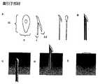
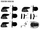
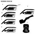
図5 LTR器具の実施形態
LTRの一つの実施形態が示されている。
A.LTRは、3つの主要な構成要素、牽引子(R)、軸(S)及びアンカー(A)を備えている。
B.舌内に挿入されたLTRの側面図
C.牽引子の一部分示している舌の曲線の背面図
D.湾曲した中心線形状を示している舌根の背面図
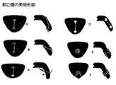
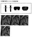
図6 牽引子部材
この図は、上気道組織内に移植するために針に取り付けることができ且つ針が引き抜かれると展開するLTRの牽引子構成部品を示している。牽引子は、軸と一体の部品として示されており且つ軟質の弾性材料によって一部品として成形されている。
A.牽引子ヘッドの側面図及び前面図
牽引子の面は、軸に対して約15°の角度で配置されている。
B.針内に取り付けられた牽引子ヘッドの側面図
C.組織内を通る針の側面図
牽引子の伸長部は注射針バレルに対して平らに横たわっており且つ組織内での針の通過を妨害しないことに注意。
D.針が粘膜内へと十分に貫入して牽引子の伸長部が粘膜を通過した後に、当該伸長部は再び軸から離れるように伸長している。
E.軸を若干牽引することによって、牽引子は粘膜を捕捉し且つその作動位置に載置された状態となる。
図7 軸部材
舌が睡眠中のように弛緩しているときに、牽引子の張力を維持するLTRの軸に対する改良が示されている。しかしながら、会話及び嚥下中に舌根がしばしば後方へ動くときには、舌の動きによって軸が押し込まれて延ばされる。このようにして、通常の舌の動きに対する抵抗が殆ど又は全く存在しない。
A.通常の筋肉緊張状態での舌内のLTRの概略図
牽引子は、窪むことなく舌根の粘膜面上に横たわっている。
B.睡眠中に、舌は全ての緊張度を失い且つ気道内へと後方へ倒れる傾向があり、牽引子はこの変形に抗する。
C.嚥下中及び会話中に、舌根はときどき後方へ動く。
これらの動きの間に、舌筋の強い収縮が存在する。この収縮は、上方軸を押し込んで軸を長くし且つ牽引子を動かす。
図8 アンカー部材、ボルスタ
A.ボルスタの前面図
B.ボルスタの頂面図
C.ボルスタの側面図
D.LTRが無負荷状態の舌の側面図
E.軸のアンカーがボルスタの下側の裂溝内に差し入れられて前方へ引っ張られている。
F.舌の下方位置にあるボルスタ
G.ボルスタ凹部内に着座しているLTRアンカーの拡大図
H.LTRとボルスタとを備えた舌の頂面図
図9 アンカー部材、歯用
A.上前歯又は下前歯上に移植されている改造されたアンカー
アンカー(A)は、歯と連結され、軸(S)は牽引子/カプラー(R/C)に結合している。牽引子/カプラーは、組織と連結される牽引子からなるか又は移植された牽引子、軸又は移植されたLTRのアンカー部材に結合されたカプラー構成部品からなる。
B.LTRが舌根から小帯へと移植された状態の舌及び下顎の頂面図
LTRのアンカーは、歯用アンカーのR/C部品に可逆的に取り付けることができる。
C.側歯上で使用するための改造されたアンカーの別の実施形態
D.側歯用アンカーを備えた舌及び下顎の頂面図
アンカーは大臼歯に取り付けられ、軸は咽頭襞内を通過し、牽引子は襞の後面に当接している。
E.軟組織、PGF、口腔底及び舌面を結合する牽引又は伸長のための幾つかの可能な結合伸長部を備えた口蓋人工器官
図10 アンカー部材、小帯領域
オトガイ舌筋内には小さな腱があり、当該筋肉上に種々の角度で繊維が付着している。主要な腱は、筋肉の中間で且つ種々の点でより小さな腱が分岐している。小帯内のアンカーは、移植された部分が腱内を通過できるように挿入される。しかしながら、アンカーは、下顎に取り付けられた小帯領域又は軟組織内のどの位置にも挿入することができる。アンカーは、以下に説明する種々の機構によってLTRに結合することができる。
A.中心線(中央矢状面)で断面した舌及び下顎の側面図
B.Aの図
C.小帯領域の拡大図
図11 小帯領域の実施形態
A.中心線で断面した舌及び下顎の側面図
小帯は、オトガイ舌筋の前方端縁であり、小帯領域は、オトガイ及び周囲の粘膜全体を指している。オトガイ舌筋の前方及び後方境界部は実線によって示されている。オトガイ舌筋は、下顎の内側面上の小さな領域及び当該領域からの腱の伸長部に付着している。これは、これらの付着部から広がっており、舌体及び舌根の長さに沿った境界層と呼ばれる結合組織内へ殆ど挿入される。
B.Aの図
C.小帯領域内を通り且つ歯用アンカーの外側に取り付けられているLTRが示されている。LTRの移植された部分は、オトガイ舌筋に側方の力をかけ、これは、境界層へ運ばれ且つ最終的に舌根へと運ばれる(矢印)。
D.境界部内を通り且つ小帯アンカーに固定されているLTRが示されている。舌の変位は矢印によって示されている。
E.2つの位置で境界領域を結合している小帯領域内に十分に移植されたLTRが示されている。舌根の有益な後退によって舌端に何らかの後退が生じるが、これは舌の機能と干渉しないことは注目すべきである。
図12 舌根移植片
A.舌根の前方部分
B.Aの図面
光線は、舌の上層(SL)と正中線隔壁(MS)との結合組織である。MLは中間層である。
C.SLとMLとを結合しているLTR移植片の位置
D.中央矢状面内に見られる舌
楕円は、機械的な分離領域を示している。
E.Dに示されている部分の概略図
F.移植片の位置
図13 舌根の実施形態
A.LTRが舌根の粘膜の下側を通過している軸によって結合されている状態の横(左)及び頂部(右)の図面
緑の楕円1310は、アンカー及び牽引子に該当し、軸は、移植されたときに点線の黄色の点で示されており、外側にあるときは実線で示されており、黒い矢印1312は牽引の方向を示している。
B.牽引子とアンカーとの間のより直接的な経路をとっている軸を備えたLTR
C.軸が牽引子とアンカーとに近接している粘膜から出ている状態のLTR
D.取り外し可能に取り付けることができる軸を備えた移植されたアンカー及び牽引子
E.部分的に移植可能なアンカー又は牽引子の横方向図
F.部分的に移植可能なアンカー又は牽引子の頂面図
G.軸の結合を示している部分的に移植可能なアンカー/牽引子
H.伸長部が使用されていないときに粘膜と同面まで押圧されている状態の部分的に移植可能なアンカー/牽引子の横方向図
I.半分移植された牽引子部材に結合されている舌端及び軸を覆うように配置された弾性スリーブを備えたLTR
J.PGFに固着されているLTRと舌根を横切って通過しており且つ半分移植された牽引子部材
K.軸が舌端内を舌の上面上の中間アンカーまで通過している状態で舌端の下側に固定されているLTR
当該軸は、次いで、半分移植された牽引子部材まで後方へ通過される。これによって舌端の下側のアンカー部位からの張力の調整が可能になる。
L.左の堅牢な軸が舌端の下方のアンカー部材を舌端の上方の牽引部材に結合している。牽引子は、舌端を覆うように取り外し可能に配置されているスリーブによって前方へ回転されている。堅牢な軸に沿った牽引部材の回転によって、舌の組織が中心線に沿って変位せしめられる。図面は、この作用を示すために意図的に拡大されている。
図14 上口蓋舌襞
A.PGFが挿入されている舌領域を示している上気道の側面図
より小さな上方領域は、軟口蓋と咽頭側壁、限定的ではないが口蓋舌筋と上咽頭括約筋とを結合している筋肉の重なっている部分を収容するので特に重要である。
B.標識された上方PGF取り付け領域との下顎に関する舌の側面図
C.軟口蓋を上方PGFに結合する口蓋舌筋が示されている。
D.上咽頭括約筋は、咽頭壁を上方PGFに結合している。
E.PGFの牽引力が舌根、軟口蓋及び咽頭壁に分配されることを示している概略図
図15 咽頭舌襞の実施形態
A.舌の後方への圧潰及び気道に対する作用を示している図
B.PGFの牽引子及び小帯を他のPGF内の牽引子まで通過している軸
C.PGF内の牽引子及び舌の組織内を通過して出て来た改造されたアンカーに結合している軸
代替的な実施形態は、口腔底内を通過し、皮膚上に載置されている外部アンカーまで伸びている。
D.PGF内又はその近くに牽引子を備え且つ舌内をオトガイ舌筋又は口腔底内へ移植されたアンカーまで通過している軸を備えた移植されたLTR
E.上方PGF内の牽引子及び下PGF内のアンカーを備えたLTR
F.PGF内の牽引子及び舌の上面上のアンカーまで通過している軸
図16 咽頭舌襞の実施形態
A.各PGFの前方のアンカーに2つの粘膜下の軸によって結合されている舌根に設けられた牽引子
B.粘膜下の軸は、各PGFの前方で2つの牽引子/アンカー部材を結合している。
C.PGF内又はその近くの2つの移植された牽引子/アンカー
D.各PGF内又はその近くに移植されている磁石は、粘膜下の軸によって結合されている。
E.左側 PGF内に移植された磁石は、改造されたアンカーに取り付けられた逆極性の磁石によって牽引される。
右側 磁石がPGF内の定位置に維持するための2つのフランジを備えている移植片に包囲されている。
F.左側 歯用に改造されたアンカーの図である。
アンカー部材は、右側に示されているように、歯に可逆的に着用可能に取り付けられており、当該結合機構は、次いで、移植されたLTRに結合されている。牽引子部材は磁石又は機械的な機構とすることができる。
右側 上から見た舌及び下顎の図である。
歯用の改造されたアンカーの2つの実施形態が示されており、底部では、牽引子は、Eの左側に示されている移植された磁石に結合されている磁石である。頂部では、軸端部が、図Eの右側に示されている可逆的に着用可能な磁気移植片に結合されている磁石を備えている。
図17 軟口蓋の実施形態
A.軟口蓋と口蓋舌襞とを示している口の図(Henry Gray. Anatomy of Human Body.1918)
B)Aと同じ図面であるが、粘膜が取り除かれ、下にある筋肉(右側)及び神経及び血液供給源(左側)が示されている図
C)正中断面の後方の左咽頭側壁の図
舌は下方に後退せしめられている。
上PGF内のアンカーによって4つの好ましいLTRの配置が示されており、1)軸は、扁桃の周囲の口蓋舌筋の隣を通過し、牽引子は、軟口蓋の側方端縁に当接している。側方口蓋帆領域を増すための好ましい実施形態、2)軸は、正中軟口蓋近くの口蓋舌筋牽引子内を移動する。内側口蓋帆空洞を増すための好ましい実施形態、3)軸は、口蓋舌筋、口蓋扁桃及び口蓋咽頭筋内を通り、牽引子は軟口蓋の後壁に当接している。口蓋扁桃を圧縮し且つ永久的に改造するための好ましい実施形態、4)軸は、舌根粘膜の下1cmのところを通り、牽引子は舌根に当接している。舌根を引っ張るための好ましい実施形態
図18 扁桃襞の実施形態
18A.後方扁桃襞の牽引子後面、前方扁桃襞のアンカー前面
口蓋扁桃の圧縮のための好ましい実施形態である。
18B.牽引子上口蓋舌襞、牽引子下口蓋舌筋襞又はPGF
18C.口蓋舌筋内に移植されたアンカー
18D.軟口蓋牽引子正中面の側面に設けられたアンカー
18E.口蓋舌筋襞の改造された歯用アンカーの内面上に設けられた牽引子
18F.後扁桃襞に設けられた牽引子及び前扁桃襞に設けられたアンカー
図19 軟口蓋の実施形態
A.アンカーの上咽頭側部及び牽引子の口腔下方側部
B.牽引子の口腔上方側部及びアンカーの咽頭舌側部
C.LTRに負荷をかけるためにアンカーの前方に付加されたボルスタ
凹み及び回転に注意
D.全体が植え込まれたLTR
E.対向している牽引子とアンカー
F.軟口蓋の端縁を持ち上げるリテナーのためのアタッチメントとしてのLTR
図20 獣医関連の実施形態
ウマの軟口蓋の脊椎の変位のための本発明の実施形態が示されている。
A.運動中のウマの上気道の正常な形態
軟口蓋は、喉頭の喉頭蓋に重なり且つ連結して気道のための開いた導管を提供していることに注意
B.DDSPにおいては、軟口蓋は、係止位置から押しのけられ且つ気道を閉塞する。これは、舌根の後方への動きによって惹き起こされると考えられる。
C.この状態のためのLTRの実施形態
軸は、下顎を介して当該下顎の前方の調整可能なアンカーに達している。
D.運動中に、軸は、一片の係留帯に可逆的に取り付けられている舌面上のアンカーに係合している別の実施形態
E.軟口蓋をその正常な位置から押しのけられている真反対の実施形態
軟口蓋の前方のアンカーは、後方へ通過し、次いで、喉頭蓋内を通って喉頭の咽頭面上の牽引子へと達している。
F.代替的な実施形態においては、LTRは、PGFから軟口蓋の側面へと通過している。
前方から見た図であり、舌は透明に示されている。比較のために、Eに示されている正中面の実施形態も示されている。
図21 非侵襲性PGF牽引子
A.舌の後方への圧潰による気道閉塞状態の図
B.PGF牽引
軟らかい“フック”はPGFを前方へ牽引し、それによって、舌根、軟口蓋及び咽頭壁を牽引する。
C.“フック”の拡大図
D.“クリップ”の拡大図
クリップは、その両方のアームによって軟組織を圧縮することによって定位置とされる。
E.圧縮が磁石によってなされているクリップの実施形態
F.牽引子を改造されたアンカーに結合するために磁石も使用されているEの実施形態
G.定位置にある2つのフック牽引子及び舌根(点線)に対する当該フック牽引子の作用を示している図
H.フックLTRの拡大図
図22 非侵襲性の牽引クリップの実施形態
A.軟組織襞上に設けられたクリップの側面図
襞に接着する一つの方法は、クリップの端部によって組織を圧縮することである。
B.互いに反対の極性の対向する磁石によって構成されている側面図
これらの磁石による吸引は、安定した位置のための十分な力を提供し、軸は不要である。
C.軟組織襞上のクリップの前面図
軸の結合は、襞の端縁を牽引する機能を果たすことができる。
D.軟口蓋及び舌根のような硬化による利点を得る構造に対する伸び(伸長)及び有用な作用を提供するために使用されるチップ
E.歯用アンカーに取り付けられた前扁桃柱上のチップ
F.歯用アンカーに取り付けられた後扁桃柱上のチップ
G.歯用アンカーに取り付けられた軟口蓋の端縁上のチップ
H.咽頭壁を披裂喉頭蓋襞の方へ牽引して咽頭側壁を硬化させている2つのチップ
図23 非侵襲性前突及び真空
A.側面図
口腔底は、下顎から舌骨まで延びている縞模様によって示されている。
B.前面図
口腔底は、下顎の各側部の底部に結合している。
C.頂面図
舌は透明で、舌の三角の根元を見ることができる。根元の前方伸長部は、下顎内へのオトガイ舌筋の挿入部分である。
D.歯用アンカー(図示せず)から伸出筋によって下方且つ若干前方へ押されているボルスタ
FOM及び舌とPGFとの改造位置の窪みに注意
E.舌面の低い高さによって反射されたボルスタによるFOM凹陥
F.上方から見たボルスタ
舌根の前方への変位に注意
G.舌の側面に適用された真空装置
H.牽引部材としての真空装置
詳細な説明
ここで使用されている“対象物”という用語は、人間を含む哺乳類由来の動物を含んでいる。ここで使用されている位置及び向きを記載するために使用されている解剖学的用語は、以下の説明によって最も良く規定することができる。The present invention will be more readily understood by the detailed description of exemplary embodiments provided below considered in conjunction with the accompanying drawings.
Detailed Description of the Drawings Fig. 1 Median view of the upper airway of the human body
Fig. 2 Simplified schematic diagram of tongue and surrounding tissues NP nasopharynx VP palatal pharynx HP hypopharynx SP soft palate P hard palate T tongue GG genioglossus muscle
Fig. 3 Anatomical boundary sign of the tongue The tongue is defined as the gray area in this figure. The tongue is divided into a tongue end, a tongue body and a tongue base from the front to the rear. The genioglossus muscle (GG) is inserted into the connective tissue boundary (Bo) on the lower surface of the tongue. The genioglossus muscle and the entire region of the muscle are referred to as the “small band region”.
BA) Tongue root BD) Tongue body BL) Tongue end Bo) Boundary between tongue and mental C) Tongue curve F) Small band GG) Genital tongue muscle
Fig. 4 Effects of airway obstruction mechanism and current therapy A) Normal tension during awakening The tongue remains in place and the airway is left open. The
B) During sleep, the muscles in the tongue lose their tension and become relaxed.
Negative pressure in the pharynx during inspiration causes posterior crushing of the tongue in the palatal pharyngeal region. This is because in this state the airway is narrow and the curved tongue (circular) is most deformable.
C) Inspiration after the airway is occluded in the palatal pharyngeal region lowers the pressure in the pharynx, further deforms the tongue base and firmly blocks the airway.
D) CPAP pumps air into the nose at high pressure (blue line 416), thereby splinting and opening the pharynx.
E) The dental appliance operates by moving the entire jaw forward. When the tongue attaches to soft tissue along the floor of the mouth and the soft tissue attaches to the jaw, the tongue is indirectly moved to dilate the airway. Note that the jaw moves relative to the pharyngeal wall (arrow).
F) The LTR prevents the tongue curve from deforming backwards by directly holding the tongue curve moving backwards.
FIG. 5 LTR Instrument Embodiment One embodiment of an LTR is shown.
A. The LTR comprises three main components: retractor (R), shaft (S) and anchor (A).
B. Side view of the LTR inserted into the tongue. D. Rear view of tongue curve showing part of retractor Back view of tongue base showing curved centerline shape
FIG. 6 Retractor Member This figure shows the retractor component of the LTR that can be attached to a needle for implantation into upper airway tissue and deploys when the needle is withdrawn. The retractor is shown as an integral part of the shaft and is molded as a single part from a soft elastic material.
A. Side view and front view of retractor head The surface of the retractor is arranged at an angle of about 15 ° to the axis.
B. C. Side view of retractor head mounted in needle. Side view of needle passing through tissue Note that the retractor extension lies flat against the needle barrel and does not obstruct the passage of the needle through the tissue.
D. After the needle has fully penetrated into the mucosa and the retractor extension has passed through the mucosa, the extension has again extended away from the axis.
E. By slightly retracting the shaft, the retractor captures the mucosa and is placed in its operating position.
FIG. 7 Shaft Member An improvement to the LTR shaft that maintains retractor tension when the tongue is relaxed as during sleep is shown. However, when the tongue base often moves backwards during conversation and swallowing, the movement of the tongue pushes and extends the shaft. In this way, there is little or no resistance to normal tongue movement.
A. Schematic diagram of the LTR in the tongue under normal muscle tension The retractor lies on the mucosal surface of the tongue base without depression.
B. During sleep, the tongue tends to lose all tension and fall back into the airway, and the retractor resists this deformation.
C. During swallowing and talking, the tongue base sometimes moves backwards.
During these movements, there is a strong contraction of the tongue muscle. This contraction pushes the upper shaft to lengthen the shaft and move the retractor.
Fig. 8 Anchor member, bolster B. Front view of bolster. Top view of bolster B. Side view of bolster Side view of tongue with LTR unloaded E. A shaft anchor is inserted into the lower cleft of the bolster and pulled forward.
F. Bolster below the tongue Magnified view of LTR anchor seated in bolster recess. Top view of tongue with LTR and bolster
Fig. 9 Anchor member for teeth A modified anchor (A) implanted on the upper or lower anterior teeth is connected to the teeth and the shaft (S) is connected to the retractor / coupler (R / C). The retractor / coupler consists of a retractor coupled to tissue or a coupler component coupled to an implanted retractor, shaft or anchor member of an implanted LTR.
B. Top view of the tongue and lower jaw with the LTR implanted from the tongue base to the zonule The anchor of the LTR can be reversibly attached to the R / C part of the dental anchor.
C. D. Another embodiment of a modified anchor for use on side teeth Top view of tongue and mandible with side tooth anchors The anchor is attached to the molar, the shaft passes through the pharyngeal fistula, and the retractor abuts the posterior surface of the fistula.
E. A palatal prosthesis with several possible connection extensions for traction or extension to connect soft tissue, PGF, floor of mouth and tongue surface
Fig. 10 Anchor member, small band region There is a small tendon in the genioglossus muscle, and fibers are attached to the muscle at various angles. The main tendon is in the middle of the muscle and smaller tendons branch off at various points. The anchor within the zonule is inserted so that the implanted portion can pass through the tendon. However, the anchor can be inserted anywhere within the zonule region or soft tissue attached to the lower jaw. The anchor can be coupled to the LTR by various mechanisms described below.
A. B. Side view of tongue and lower jaw taken along the center line (center sagittal plane). Figure A. C. Enlarged view of the small belt area
FIG. 11 Embodiment of small band area Side view of tongue and mandible cut at the centerline The zonule is the anterior edge of the genioglossus muscle, and the zonule region points to the genitalia and the entire surrounding mucosa. The anterior and posterior boundaries of the genioglossus muscle are indicated by solid lines. The genioglossus muscle is attached to a small area on the inner surface of the mandible and an extension of the tendon from that area. It extends from these attachments and is almost inserted into the connective tissue called the boundary layer along the length of the tongue and tongue base.
B. Figure A. C. An LTR is shown passing through the zonule region and attached to the outside of the dental anchor. The implanted part of the LTR exerts a lateral force on the genioglossus muscle, which is carried to the boundary layer and finally to the tongue base (arrow).
D. The LTR is shown passing through the boundary and secured to the zonule anchor. The tongue displacement is indicated by an arrow.
E. A fully implanted LTR is shown in the zonule region joining the border region at two locations. It should be noted that the beneficial retraction of the tongue base causes some retraction at the tongue end, but this does not interfere with the function of the tongue.
Fig. 12 Tongue root graft Anterior portion of tongue base Drawing A A light ray is a connective tissue between the upper layer of the tongue (SL) and the midline partition wall (MS). ML is an intermediate layer.
C. Location of the LTR graft connecting SL and ML. The tongue ellipse found in the central sagittal plane indicates the mechanical separation region.
E. Schematic diagram of the part shown in D. Graft location
FIG. 13 Tongue base embodiment Horizontal (left) and top (right) drawings with LTRs joined by a shaft passing under the mucosa of the tongue base The
B. LTR with axis taking a more direct path between retractor and anchor
C. LTR with shaft protruding from mucosa close to retractor and anchor
D. Implanted anchor and retractor with a shaft that can be removably attached e. F. Transverse view of partially implantable anchor or retractor Top view of partially implantable anchor or retractor. B. Partially implantable anchor / retractor showing shaft coupling Transverse view of a partially implantable anchor / retractor with the extension being pressed to the same plane as the mucosa when not in use. LTR with an elastic sleeve arranged to cover the tongue end and shaft coupled to a half-implanted retractor member
J. et al. LTR anchored to PGF and retractor member passing across the tongue base and half-implanted. LTR secured to the lower side of the tongue end with the shaft passing through the end of the tongue to an intermediate anchor on the upper surface of the tongue
The shaft is then passed back to the half-implanted retractor member. This allows adjustment of the tension from the anchor site below the tongue end.
L. A rigid shaft on the left connects the anchor member below the tongue end to the traction member above the tongue end. The retractor is rotated forward by a sleeve that is removably disposed over the tongue end. The rotation of the traction member along the rigid axis causes the tongue tissue to be displaced along the centerline. The drawing is intentionally enlarged to show this effect.
Fig. 14 Upper palatal tongue tongue Side view of the upper airway showing the tongue region where the PGF is inserted. The smaller upper region is the overlap of the muscles connecting the soft palate and pharyngeal side walls, but not limited to the palatal and pharyngeal sphincters. It is especially important because it accommodates the part that is being held.
B. B. Side view of the tongue with respect to the lower jaw with labeled upper PGF attachment area. The palatine and lingual muscles that connect the soft palate to the upper PGF are shown.
D. The nasopharyngeal sphincter connects the pharyngeal wall to the upper PGF.
E. Schematic showing that the traction force of PGF is distributed to the tongue base, soft palate and pharyngeal wall
Fig. 15 Embodiment of pharyngeal tongue fistula Figure showing posterior crushing of tongue and effect on airway. Axis passing through the PGF retractor and ligament to the retractor in the other PGF. A shaft coupled to a retractor in the PGF and a modified anchor that has passed through the tissue of the tongue An alternative embodiment passes through the floor of the mouth and rests on the skin Extends to the external anchor.
D. Implanted LTR with retractor in or near PGF and axis passing through the tongue to the genioglossus muscle or anchor implanted into the floor of the mouth
E. LTR with retractor in upper PGF and anchor in lower PGF
F. A shaft passing through the retractor in the PGF and the anchor on the upper surface of the tongue
Fig. 16 Embodiment of pharyngeal tongue fistula B. A retractor on the tongue base that is connected to the anterior anchor of each PGF by two submucosal axes. The submucosal shaft connects two retractors / anchor members in front of each PGF.
C. 2 implanted retractors / anchors in or near the PGF Magnets implanted in or near each PGF are joined by a submucosal shaft.
E. The magnet implanted in the left PGF is pulled by a reverse polarity magnet attached to the modified anchor.
The right magnet is surrounded by an implant with two flanges to maintain it in place in the PGF.
F. It is a figure of the anchor remodeled for left-hand teeth.
The anchor member is reversibly wearably attached to the tooth, as shown on the right, and the coupling mechanism is then coupled to the implanted LTR. The retractor member can be a magnet or a mechanical mechanism.
It is the figure of the tongue and lower jaw seen from the upper right side.
Two embodiments of modified dental anchors are shown, and at the bottom, the retractor is a magnet that is coupled to the implanted magnet shown on the left side of E. At the top, the shaft end includes a magnet coupled to the reversibly wearable magnetic implant shown on the right side of FIG.
Figure 17 Soft palate embodiment Illustration of the mouth showing the soft palate and palatal tongue fold (Henry Gray. Anatomy of Human Body.1918)
B) The same drawing as A, but with the mucous membrane removed and the underlying muscle (right side) and nerve and blood sources (left side) shown C) Illustration of the left pharyngeal side wall behind the median section Is retracted downward.
Four preferred LTR placements are shown by anchors in the upper PGF: 1) The axis passes next to the palatine and tongue muscles around the tonsils and the retractor abuts the lateral edge of the soft palate Yes. Preferred embodiment for increasing the lateral palate sail area, 2) The axis moves in the palatal lingual muscle retractor near the midline soft palate. Preferred embodiments for increasing the medial palatal cavity, 3) The axis passes through the palatine lingual muscle, palatine tonsil and palatopharyngeal muscle, and the retractor rests against the back wall of the soft palate. Preferred embodiment for compressing and permanently remodeling the palatine tonsil, 4) The axis passes 1 cm below the tongue base mucosa and the retractor rests against the tongue base. Preferred embodiment for pulling tongue base
FIG. 18 Tonsillar Embodiment 18A. A rear embodiment of the retractor of the posterior tonsillar, the front surface of the anchor of the anterior tonsillar is a preferred embodiment for compression of the palatine tonsils.
18B. Retractor upper palatine tongue fistula, retractor lower palatine tongue fistula or PGF
18C. Anchor implanted in the palatine lingual muscle 18D. Anchor provided on the side of the median surface of the soft palate retractor 18E. Retractor provided on the inner surface of the modified dental anchor of the palatine and gluteal muscle 18F. The retractor provided on the back tonsillar and the anchor provided on the front tonsillar
FIG. 19 Soft palate embodiment. B. Upper pharyngeal side of anchor and lower oral side of retractor B. Oral upper side of retractor and pharyngeal lingual side of anchor Bolster added to the front of the anchor to load the LTR. Note the dent and rotation. LTR with the entire plant
E. Opposing retractors and anchors F. LTR as an attachment for a retainer that lifts the edge of the soft palate
FIG. 20 Veterinary Related Embodiment An embodiment of the present invention for the displacement of the spine of the equine soft palate is shown.
A. Note that the normal form of the upper airway of the running horse is that the soft palate overlays and connects to the epiglottis of the larynx to provide an open conduit for the airway. In DDSP, the soft palate is pushed away from the locked position and closes the airway. This is thought to be caused by the backward movement of the tongue base.
C. Embodiment of LTR for this state The axis reaches the adjustable anchor in front of the lower jaw via the lower jaw.
D. In another embodiment, during movement, the shaft engages an anchor on the tongue that is reversibly attached to a piece of tether. The opposite embodiment in which the soft palate is pushed away from its normal position The anchor in front of the soft palate passes backward and then through the epiglottis to the retractor on the laryngeal surface of the larynx.
F. In an alternative embodiment, the LTR passes from the PGF to the side of the soft palate.
It is the figure seen from the front, and the tongue is shown transparently. For comparison, an embodiment of the median plane shown at E is also shown.
Figure 21 Non-invasive PGF retractor Fig. B. Airway obstruction due to crushing of the tongue. PGF Retraction A soft “hook” pulls the PGF forward, thereby pulling the tongue base, soft palate and pharyngeal wall.
C. Enlarged view of “hook” Enlarged view of the “clip” The clip is brought into place by compressing the soft tissue by both its arms.
E. Embodiment of clip where compression is done by magnets F. E. Embodiment in which magnets are also used to couple the retractor to the modified anchor. H. Two hook retractors in position and the action of the hook retractor on the tongue base (dotted line). Enlarged view of hook LTR
Figure 22 Non-invasive traction clip embodiment Side view of a clip provided on a soft tissue fold One way to adhere to the fold is to compress the tissue by the end of the clip.
B. Side view constituted by opposing magnets of opposite polarities The attraction by these magnets provides sufficient force for a stable position and no shaft is required.
C. Front view of clip on soft tissue fold The coupling of the shaft can serve to pull the edge of the fold.
D. Chips used to provide elongation and useful effects on structures that benefit from hardening such as the soft palate and tongue base E. Tip on anterior tonsillar column attached to dental anchor F. Tip on the tonsillar column after attached to dental anchor G. A tip on the edge of the soft palate attached to the dental anchor. Two tips that pull the pharyngeal wall toward the nasopharyngeal fistula to harden the pharyngeal side wall
Figure 23 Non-invasive front bump and vacuum Side view The floor of the mouth is indicated by a striped pattern extending from the lower jaw to the hyoid bone.
B. Front view The floor of the mouth is connected to the bottom of each side of the lower jaw.
C. Top view The tongue is transparent and you can see the root of the triangle. The root anterior extension is the insertion part of the genioglossus muscle in the lower jaw.
D. Note the bolster FOM being pushed downward and slightly forward by the extension muscle from the dental anchor (not shown) and the recess in the remodeling position of the tongue and PGF. FOM depression by bolster reflected by low height of tongue surface Bolster as seen from above Note the forward displacement of the tongue base. Vacuum device applied to the side of the tongue Vacuum device as a traction member
DETAILED DESCRIPTION The term “subject” as used herein includes animals from mammals including humans. The anatomical terms used to describe the position and orientation used here can best be defined by the following description.
典型的には、一方の端部に頭部及び口を有し、他端は肛門及び尾を有している場合が多い動物について言及しているときには、頭の端部は、頭端部と称される。頭部自体では、“頭部の”は、鼻の端部に向かう方向を指しており、“尾部の”は、尾方向を指すために使用されている。通常は重力の引力と反対の上方を向いている動物の体の表面又は側面は背面であり、反対側の側面は典型的には全ての足で歩いているとき、泳いでいるとき又は飛んでいるときに、地面に最も近い側は“基端”であり、遠い側は“末端”である。3つの基本的な基準面は、動物化学的構造内で使用される。“矢状”面は、体を左側部分と右側部分とに分ける。“正中”面は中心線上にある。すなわち、正中面は脊柱のような中心線構造を通り、全ての他の矢状面は当該中心構造に平行である。“左右縦断”面は、体を背面部分と腹面部分とに分ける。“横断”面は体を頭側部分と尾側部分とに分ける。 Typically, when referring to animals that often have a head and mouth at one end and an anus and tail at the other end, the end of the head is the head end. Called. In the head itself, “in the head” refers to the direction towards the end of the nose and “in the tail” is used to refer to the tail direction. The surface or side of the animal's body that normally faces upwards opposite to the gravitational attraction is the back, and the opposite side is typically walking with all feet, swimming or flying The side closest to the ground is the “base” and the far side is the “terminal”. Three basic reference planes are used within the animal chemistry structure. The “sagittal” plane divides the body into a left part and a right part. The “midline” plane is on the center line. That is, the median plane passes through a centerline structure such as the spinal column, and all other sagittal planes are parallel to the center structure. The “left and right longitudinal” plane divides the body into a back part and an abdominal part. The “crossing” plane divides the body into a cranial part and a caudal part.
人間について言及するときには、人体及びその部分は、常に人体が真っ直ぐに立っているという仮定を使用して説明される。頭部側端部に近い人体の部分は“上方”(動物における頭部に対応する)であり、一方、頭部側端部から遠い方の人体の部分は“下方”(動物における尾部に対応する)である。人体の前面近くの対象物は、“前方”(動物における腹側に対応する)と称され、人体の背中に近い部分は“後方”(動物における背側に対応する)である。横断面、軸平面又は水平面は、地面に平行なX−Y面であり、この面は、上方/頭部を下方/脚部から分離する。前頭面又は前面は、前方を後方から分離する地面に直角なY−Z面である。矢状面は、左側を右側から分離する地面及び前頭面に直角なX−Z面である。正中面は、正確に人体の中央にある特定の矢状面である。 When referring to a human being, the human body and its parts are always described using the assumption that the human body stands upright. The part of the human body near the head end is “upper” (corresponding to the head in the animal), while the part of the human body farther from the head end is “lower” (corresponding to the tail in the animal) ). An object near the front of the human body is referred to as “front” (corresponding to the ventral side in the animal), and a portion near the back of the human body is “backward” (corresponding to the back side in the animal). A cross section, axial plane or horizontal plane is an XY plane parallel to the ground, which separates the upper / head from the lower / legs. The frontal surface or front surface is a YZ plane perpendicular to the ground that separates the front from the rear. The sagittal plane is an XZ plane perpendicular to the ground and frontal plane separating the left side from the right side. The median plane is a specific sagittal plane that is exactly in the center of the human body.
正中線近くの構造は内側と称され、動物の体側近くの構造は外側と称される。従って、内側構造は正中面に近く、外側構造は正中面からより遠い部分である。体の正中線内に位置する構造は中央の部分である。例えば、対象物である人間の鼻の先端は正中線にある。 The structure near the midline is referred to as the inside, and the structure near the animal's body side is referred to as the outside. Thus, the inner structure is closer to the median plane and the outer structure is the portion further from the median plane. The structure located in the midline of the body is the central part. For example, the tip of the human nose, which is the object, is at the midline.
同側は同じ側を意味し、対側は反対側を意味し、両側は両方の側を意味している。体の中心に近い構造は、基端又は中心であり、一方、より遠い方の構造は末端又は外周である。例えば、手は腕の末端にあり、肩は腕の基端にある。 The same side means the same side, the opposite side means the opposite side, and both sides mean both sides. The structure closer to the center of the body is the proximal end or center, while the more distant structure is the distal or outer periphery. For example, the hand is at the distal end of the arm and the shoulder is at the proximal end of the arm.
定義
“アンカー”は、牽引子に対して動かない部位に機械的に結合している器具の部品を指している。The definition “anchor” refers to the part of the instrument that is mechanically coupled to a site that does not move relative to the retractor.
“変形”は、上気道軟組織構造の形状の異常な変形を指している。この変形は、睡眠中に弛緩した上気道に作用する陰圧によって上気道を狭めさせ得る。最も好ましくは、この軟組織は舌の湾曲部であり得る。 “Deformation” refers to an abnormal deformation of the shape of the upper airway soft tissue structure. This deformation can cause the upper airway to narrow due to negative pressure acting on the relaxed upper airway during sleep. Most preferably, the soft tissue may be a tongue curve.
“小帯”は、オトガイ舌筋の垂直前方端縁を指している。小帯は、口腔底から舌の下側の中心線まで通っている。小帯は、舌端と舌体との間の境界部を示している。“小帯領域”は、オトガイ舌筋とその周囲の粘膜とを指している。 “Short” refers to the vertical front edge of the genioglossus muscle. The zonule runs from the floor of the mouth to the centerline below the tongue. The small band shows the boundary between the tongue end and the tongue body. “Small belt region” refers to the genioglossus muscle and the surrounding mucosa.
“負荷がかけられた”形状は、日中は最少の張力を有し夜間はより高い治療レベルの張力を有するように調整された張力を有することができるLTRを指している。負荷がかけられた形状はより高い治療レベルに対応する。 The “loaded” shape refers to an LTR that can have a tension adjusted to have minimal tension during the day and higher therapeutic levels of tension at night. The loaded shape corresponds to a higher treatment level.
“改造されたアンカー”は、LTRの永久的なアンカーの取り付けを可能にする付加的な部品である。幾つかの実施形態においては、改造されたアンカーは、患者がLTR内の張力を、特に夜間は張力を増し且つ日中は解放するように調整できるようにする。 A “modified anchor” is an additional component that allows the attachment of a permanent anchor of the LTR. In some embodiments, the modified anchor allows the patient to adjust the tension in the LTR to increase tension and release during the day, especially at night.
“口蓋牽引子”は、軟口蓋の変形を防止するために使用される完成した器具を指している。
“永久アンカー”は、移植期間中LTR上に留まるLTRのアンカー部材を指している。当該永久アンカーは、軸のアンカーの前方端部が舌組織内へ摺動して戻るのを阻止する。ある種の実施形態においては、永久アンカーはまた、“改造されたアンカー”が使用されているときにコネクタの一部として機能する。“Palate retractor” refers to the finished instrument used to prevent deformation of the soft palate.
“Permanent anchor” refers to an anchor member of an LTR that remains on the LTR during implantation. The permanent anchor prevents the forward end of the shaft anchor from sliding back into the tongue tissue. In certain embodiments, the permanent anchor also functions as part of the connector when a “modified anchor” is used.
“咽頭壁牽引子”は、咽頭壁の変形を防止するための完成した器具を指している。
“伸長”は、長く延ばすこと又は押し離すことを意味している。
“逆方向の変形”は、組織牽引子によって惹き起こされる軟組織の変化を指している。幾つかの実施形態においては、逆方向の変形は、変形せしめられた構造を正常な形状へ復元させることを指している。他の実施形態においては、逆方向の変形は、組織牽引子の作用による所定の領域内の軟組織の傾斜を指している。“Pharyngeal wall retractor” refers to a completed device to prevent deformation of the pharyngeal wall.
“Extension” means to extend or push apart.
“Reverse deformation” refers to soft tissue changes caused by tissue retractors. In some embodiments, reverse deformation refers to restoring the deformed structure to a normal shape. In other embodiments, the reverse deformation refers to the inclination of the soft tissue within a predetermined region due to the action of the tissue retractor.
“睡眠時呼吸障害”は、限定的ではないが、睡眠時無呼吸、睡眠時無呼吸症候群、上気道抵抗症候群及び鼾を含む睡眠中に起こる全ての呼吸障害を指している。
“舌根”は、舌内の舌湾曲部後方の部分を指している。解剖学的用語においては、舌根の境界線は、舌の上面の隆起した味覚器官の囲まれた乳頭である良く見える線である。“Sleep breathing disorder” refers to all breathing disorders that occur during sleep, including but not limited to sleep apnea, sleep apnea syndrome, upper airway resistance syndrome and sputum.
“Tongue root” refers to the portion of the tongue behind the curved portion of the tongue. In anatomical terms, the base of the tongue base is a good-looking line that is the enclosed papillae of the raised taste organ on the upper surface of the tongue.
“舌端”は、舌の小帯の前方の部分を指している。舌端は、その頂部、側部及び下面上を粘膜によって覆われている。
“舌体”は、舌端と舌根との間に位置する舌の中央部分である。“Lingual end” refers to the front part of the ligament of the tongue. The tongue end is covered with mucous membranes on the top, sides and lower surface.
The “tongue body” is the central part of the tongue located between the tongue end and the tongue base.
“舌の境界部”又は“境界部”は、舌体及び舌根の下の面である。オトガイ舌筋は、当該境界部の大きな部分上に入り込んでいる。
“舌湾曲部”は、上面が水平な向き(舌体及び舌端)から垂直方向の向き(舌根)まで湾曲している舌の領域を指している。舌湾曲部は、舌の粘膜被覆とオトガイ舌筋が付着している結合組織境界部との間のこの領域内の軟組織を指している。The “tongue boundary” or “boundary” is the lower surface of the tongue body and root. The genioglossus muscle penetrates over a large portion of the boundary.
The “lingual curved portion” refers to a region of the tongue whose upper surface is curved from a horizontal orientation (tongue body and tongue end) to a vertical orientation (tongue root). Tongue curve refers to the soft tissue in this region between the mucosal coating of the tongue and the connective tissue boundary where the genioglossus muscle is attached.
“組織牽引子”は、軟組織の変形を防止するための本発明の実施形態の完成された器具を指している。当該器具は、舌、軟口蓋又は咽頭壁において制限なく使用することができる。 “Tissue retractor” refers to the completed device of an embodiment of the present invention for preventing soft tissue deformation. The device can be used without limitation on the tongue, soft palate or pharyngeal wall.
“舌牽引子”は、舌の変形を防止するために使用される完成された器具を指している。当該器具は、軸部に結合されている牽引子を備えており、当該軸部は、次いでアンカーに結合されている。 A “tongue retractor” refers to a complete instrument used to prevent tongue deformation. The instrument includes a retractor coupled to the shaft, which is then coupled to the anchor.
“喉頭牽引子”は、喉頭の軟組織の変形を防止するための完成された器具を指している。
“牽引子”又は“牽引子ヘッド”は、組織牽引子全体の一部分を指している。当該牽引子は、軟組織と物理的に直接的又は間接的に相互作用して変形するのを防止する。ある種の実施形態においては、牽引子ヘッドは舌の外面上に配置されている円板であり、別の実施形態においては、牽引子ヘッジは拡張可能なバルーンであり、別の実施形態においては、牽引子ヘッドはフックのように作用する湾曲した部分を有することができ、別の実施形態においては、牽引子は組織内を通る可撓性のワイヤとすることができる。幾つかの実施形態においては、牽引子は組織内に全体を移植することができる。“Laryngeal retractor” refers to a complete device for preventing the deformation of the laryngeal soft tissue.
“Retractor” or “retractor head” refers to a portion of the entire tissue retractor. The retractor prevents the physical tissue from interacting directly or indirectly with the soft tissue and deforming. In certain embodiments, the retractor head is a disc disposed on the outer surface of the tongue, in other embodiments, the retractor hedge is an expandable balloon, and in another embodiment. The retractor head can have a curved portion that acts like a hook, and in another embodiment, the retractor can be a flexible wire that passes through the tissue. In some embodiments, the retractor can be implanted entirely within the tissue.
“牽引子軸”、“軸”又は“牽引子部材”は、牽引子ヘッドに取り付けられ且つ牽引子アンカーに結合する機能を果たす舌牽引子の部分を指している。幾つかの実施形態においては牽引子、別の実施形態においては、軸は、堅牢な又は可撓性の、中実又は中空の、一部品又は連結された多部品としても良い。 "Retractor shaft", "shaft" or "retractor member" refers to the portion of the tongue retractor that is attached to the retractor head and that functions to couple to the retractor anchor. In some embodiments, the retractor, in other embodiments, the shaft may be a solid or hollow, solid or hollow, one-part or multi-part connected.
“無負荷状態”は、殆ど又は全く張力がかかっていないLTRを指している。ここで使用されているように、これは、通常は日中の形態を意味している。比較として、LTRは夜間には治療レベルまで負荷をかけられる。
実施例
1.牽引子部材(図6)
ここには、針によって挿入され且つその作動形状へと自動的に展開する牽引子部材が開示されている。“No load condition” refers to an LTR with little or no tension. As used herein, this usually means the daytime form. As a comparison, the LTR is loaded to therapeutic levels at night.
Example
1. Retractor member (Fig. 6)
Here, a retractor member is disclosed that is inserted by a needle and automatically deploys to its working shape.
牽引子ヘッドは、舌根が変形するのを防止する。舌根の粘膜上に配置される牽引子ヘッドの好ましい品質は、患者にとって無視できる程度に、その表面積が十分な反力を提供するのに十分な大きさであるように、その深さが最小にされることである。LTRを挿入するために使用される給送具は、その設計と一体化されている。器具全体が舌の背面において使用される最小の器具によって舌の前方から挿入されることが好ましい。従って、牽引子ヘッドは、舌の前方から挿入される針によって移植された後にその作動形状へと自動的に展開するのが好ましい。 The retractor head prevents the tongue base from deforming. The preferred quality of the retractor head placed on the mucosa of the tongue base is such that its depth is minimized so that its surface area is large enough to provide sufficient reaction force, negligible for the patient. It is to be done. The feeder used to insert the LTR is integrated with its design. Preferably, the entire instrument is inserted from the front of the tongue with the smallest instrument used on the back of the tongue. Thus, the retractor head preferably deploys automatically into its working shape after being implanted by a needle inserted from the front of the tongue.
本発明のこの特徴の一部として、容易に挿入できるようにした牽引子ヘッドの設計の改良がある。ある種の実施形態においては、挿入は針によってなされる。従って、本発明の一つの実施形態は、針の中へ折り畳まれるが挿入後には作動形態へと展開する牽引子ヘッドである。体内へ挿入するために器具を最小化することができる多くの機構が知られており、非限定的な例として、ニチノールワイヤ、高圧バルーン及びばね機構がある。これらの機構は良好に機能するが、複雑さ及び不必要な費用を付加する。 Part of this feature of the invention is an improved retractor head design that allows for easy insertion. In certain embodiments, the insertion is made with a needle. Accordingly, one embodiment of the present invention is a retractor head that folds into a needle but deploys into an activated configuration after insertion. Many mechanisms are known that can minimize the instrument for insertion into the body, non-limiting examples include nitinol wire, high pressure balloons, and spring mechanisms. These mechanisms work well, but add complexity and unnecessary expense.
好ましい実施形態においては、牽引子部材は、楕円形状(長さ10mm、幅5mm、深さ2.5mm)とされ且つ軸(1mm)と共に中程度の精度の医療等級シリコーン(ショア硬度80、Nusil,Ca)によって一部品として成形されている(図6A)。牽引子は、軸に対して75°傾けられている。当該器具が針内へねじ込まれると(6B)、楕円の一方の側部は針の穴から延びて針の外壁に対して15°の角度で突出している。針が組織内に挿入されると、この伸長部は針の壁に対して面一となるように押され、針の高さの増加が最少となる(6C)。しかしながら、針が粘膜を貫通した直後に、牽引子はその伸長位置へと復元する(6D)。この方法において、針を後方へ引っ張ることによって、牽引子が粘膜を捕捉するようにさせ且つ針に沿って引き抜かれるのを防止する。針が取り外された後に、軸上の小さな張力によって、牽引子ヘッドが適正な位置へ回転するようにし且つ粘膜に対して面一に横たわるようにする(6E)。 In a preferred embodiment, the retractor member is oval shaped (
本発明の実際的な利点は、医師が針を迅速に且つ容易に挿入し且つ引き抜くことができ、器具がその適正な位置へと自動的に落ち着くことである。
2.軸部材(図7)
ここには、舌根の通常の動きとの干渉を避けるようにその長さを適合させた改造された軸が開示されている。A practical advantage of the present invention is that the physician can quickly and easily insert and withdraw the needle and the instrument will automatically settle into its proper position.
2. Shaft member (Fig. 7 )
Here, a modified shaft is disclosed that is adapted in length so as to avoid interference with the normal movement of the tongue base.
舌根の背面にかけられる反力は、睡眠中に存在するが覚醒中には存在しないのが好ましい。舌が弛緩し且つ後方への圧潰に対して不安定であるときに反力が存在するが会話及び嚥下中には存在しないのがより好ましい。嚥下中には、舌根は約1cm後方へ迅速に動いて咽頭後壁と接触する。舌根は、たとえ力がより少なくても、幾つかの会話動作中に同じように動く。これらの嚥下及び会話動作は損なわれないことが望ましい。 The reaction force applied to the back of the tongue base is preferably present during sleep but not during arousal. More preferably, there is a reaction force when the tongue is relaxed and unstable against posterior crushing, but not during conversation and swallowing. During swallowing, the tongue base moves rapidly about 1 cm backward and contacts the posterior pharyngeal wall. The tongue base moves in the same way during several conversational movements, even if it is less powerful. It is desirable that these swallowing and conversational operations are not impaired.
軸の一つの実施形態においては、舌の中に位置する部分は伸張性であり、一つの非限定的な例はバルーンである。軸のバルーン部分の圧縮により、軸が伸びるのが可能になる。嚥下中に、舌は軸の周囲を強力に収縮させる。この収縮によってバルーンが絞られて細長くされ、それによって、牽引子ヘッドが上方へ変位せしめられる。嚥下中に舌根が牽引子ヘッドの領域で上方へ動くと、軸にかけられる圧縮力によって、軸が比例的に細長くされ且つ牽引子ヘッドが嚥下中に舌根に対して不必要な反力がかかるのが防止される。しかしながら、舌が弛緩されているときに適正な量の反力をかける能力が付与される。伸張する量は、0.01〜10cmが好ましく、1cmがより好ましい。In one embodiment of the shaft,the portion located in the tongue is extensible and one non-limiting example is a balloon. Thecompression of the balloon portion of the shaft,allowing the shaft extends. During swallowing, the tongue strongly contracts around the shaft. This contraction causes the balloon to be squeezed and elongated, thereby causing the retractor head to be displaced upward. When the tongue base moves upward in the region of the retractor head during swallowing, the compressive force applied to the shaft causes the shaft to be proportionally elongated and the retractor head applies an unnecessary reaction force against the tongue base during swallowing Is prevented. However, it provides the ability to apply an appropriate amount of reaction force when the tongue is relaxed. The amount of stretching is preferably 0.01 to 10 cm, and more preferably 1 cm.
この実施形態は、本発明の好ましいが限定的な例である。嚥下及び会話中に牽引子ヘッドによる反力の低下は、多くの公知の機械的及び電子機械的な機構によって達成することができる。当業者は、本発明は多くの実施形態を有することができることを容易に理解できる。
3.アンカー部材:ボルスタ、歯用、移植されたもの(図8,9,10)
ここには、移植されたLTRに可逆的に負荷をかけることを可能にする改造されたアンカーが開示されている。This embodiment is a preferred but limiting example of the present invention. Reducing the reaction force by the retractor head during swallowing and talking can be achieved by many known mechanical and electromechanical mechanisms. One skilled in the art can readily appreciate that the present invention can have many embodiments.
3. Anchor member: bolster, dental, transplanted (Figs. 8, 9, 10)
Here, a modified anchor is disclosed that makes it possible to reversibly load the implanted LTR.
アンカーは、軸及び牽引子ヘッドの変位に抗するLTRの前方の部品である。好ましい実施形態においては、LTRは、日中(無負荷状態)には殆ど又は全く張力がかかっておらず且つ夜間(負荷状態)は張力をかけるように調整されている。この実施形態においては、アンカーは、単に、軸の前方端部が舌組織内へ引っ張り戻されるのを防止する。この目的のためには小さなフランジで十分である。しかしながら、牽引子に対する反力が更に望ましい夜間には、改造アンカーと総称されるアンカーに置換し、改造し又は補充することができる。The anchor is the front part of the LTR that resists displacement of the shaft and retractor head. In a preferred embodiment, the LTR is tuned to apply little or no tension during the day (no load) and at night (load). In this embodiment, the anchor simply prevents the forward end of the shaft from being pulled back into the tongue tissue. A small flange is sufficient for this purpose. However, at night the reaction force is furtherdesirable against the retractor, to replace the anchor which are generically referred to as modified anchor can be modified or supplemented.
改造されたアンカーの一つの実施形態は、永久アンカーと舌との間に介装されるボルスタである。このボルスタは、軸の長さを長くするか又は固定の長さに設定されている場合には、牽引子ヘッドと軸との間で圧縮される全体積を増大させる。いずれの場合にも、このような改造されたアンカーを付加することにより牽引子に対する反力の可逆的な増大が生じる。One embodiment of a modified anchor is a bolster that is interposed between the permanent anchor and the tongue. This bolster, when it is set to the length of either or fixed to increase the length of the shaft increases the total volumethat is compressed between the retractor head and the shaft. In either case,such reversible increase of the reaction forceagainst the retractorby adding themodified anchorisarising.
改造されたアンカーの一つの実施形態においては、ボルスタは、V字形状のシリコンゲルからなる(図8のA,B,C)。“V”字形状の凹状内面は、小帯(舌端下方の構造)に適合している。広い表面積を横切って牽引反力を広げることが意図されている。ボルスタ型のアンカーの中心には、永久的なアンカーと軸とがその中を貫通せしめられる導管が設けられている。一つの実施形態においては、頂部の中心端縁の中心には裂溝の始点が設けられている。この裂溝は、ほぼ軸の幅であるが永久的アンカーより狭い。患者は、舌の下に達することができ且つ永久的なアンカーを前方へ引っ張り(図8のD,E,F)、舌の下方でボルスタを摺動させ、軸を裂溝内に横たわるように配置し、永久的アンカーを解放することができる。次いで、永久的なアンカーが裂溝の前面にしっかりと当接し且つ力をかける。裂溝は、より硬質のシリコン又は別の生体適合性材料によって補強しても良い。
In one embodiment of the modified anchor, the bolster consists of a V-shaped silicon gel (A, B, C in FIG. 8). The concave inner surface of the “V” shape is adapted to the small band (structure below the tongue end). It is intended to spread the traction reaction force across a large surface area. In the center of bolster type anchors, conduits and permanent anchor and the shaft is caused to penetrate thereinit is provided. In one embodiment, the center of the central edge of the top is provided with a fissure start point. This fissure is approximately the width of the shaft but narrower than the permanent anchor. The patient can reach under the tongue and pull the permanent anchor forward (D, E, F in FIG. 8), slide the bolster below the tongue, and lay the axis in the fissure Can be deployed and the permanent anchor released. The permanent anchor then firmly abuts and applies force to the front surface of the fissure. The fissure may be reinforced with harder silicon or another biocompatible material.
本発明の別の実施形態は、永久的なアンカーを、歯に永久的に又は可逆的に取り付けられる改造されたボルスタ、すなわち歯用アンカーに固定することである(図9)。歯に取り付ける多くの器具が当該技術において知られている。非限定的な例は、“T”字形状をしており、当該“T”字形状の頂部の横棒は下門歯の前面に当接している。このTの字の垂直棒部の最初の部分は、前方の2つの門歯間を通るのに十分な薄さである。この垂直部分は、牽引子ヘッドが貫通せしめられるのを可能にするように幅が広がる。この機構によって、アンカーを歯のボルスタに容易に且つ可逆的に取り付けることができる(図9のA及びB)。 Another embodiment of the present invention is to secure the permanent anchor to a modified bolster or dental anchor that is permanently or reversibly attached to the tooth (FIG. 9). Many appliances for attaching to teeth are known in the art. A non-limiting example has a “T” shape, and the horizontal bar at the top of the “T” shape is in contact with the front surface of the lower incisor. The first part of this T-shaped vertical bar is thin enough to pass between the two front incisors. This vertical portion is widened to allow the retractor head to be penetrated. This mechanism allows the anchor to be easily and reversibly attached to the tooth bolster (A and B of FIG. 9).
別の実施形態は、口の前面よりもむしろ口の側面において使用するように最適化された歯用アンカーである。この実施形態(図9のC,D)は、臼歯又は小臼歯又はそれに隣接している構造に取り付けられる。この実施形態は、LTRと改造されたアンカーとの間の距離が短いので有利であり、舌の横面上の位置は、正常な舌の機能と干渉することはなく、配置、調整及び取り外しのために患者及び医師が容易にアクセス可能である。 Another embodiment is a dental anchor that is optimized for use on the side of the mouth rather than on the front of the mouth. This embodiment (C, D in FIG. 9) is attached to a molar or premolar or structure adjacent thereto. This embodiment is advantageous because the distance between the LTR and the modified anchor is short, and the position on the lateral surface of the tongue does not interfere with normal tongue function and is easy to place, adjust and remove. Therefore, it can be easily accessed by patients and doctors.
更に別の実施形態においては、歯用人工器官は、軟口蓋、口蓋舌襞、咽頭襞、舌又はその他の上気道部位内でLTRに結合されるアンカーとして使用される(図9のE)。これらの人工器官は、歯科技術において公知であり且つLTRの実施形態を固定するための広く且つ安定したプラットホームを提供する。更に別の実施形態は、これらの人工器官のより大きなサイズ及び位置を利用することができる。当業者は、種々の電気的又は機械的機構をこれらの人工器官内に組み込むことができることを理解することができる。非限定的な例として、上気道内の多くの位置において結合されたLTRにかけられる力を制御するために電動モーターを使用することができる。 In yet another embodiment, the dental prosthesis is used as an anchor that is coupled to the LTR within the soft palate, palatine tongue, pharyngeal fistula, tongue or other upper airway site (FIG. 9E). These prostheses are known in the dental arts and provide a wide and stable platform for securing LTR embodiments. Yet another embodiment can take advantage of the larger size and position of these prostheses. One skilled in the art can appreciate that various electrical or mechanical mechanisms can be incorporated into these prostheses. As a non-limiting example, an electric motor can be used to control the force applied to the combined LTR at many locations in the upper airway.
改造されたアンカーの更に別の実施形態は、口腔底内に部分的に移植される。本発明の一つの実施形態においては、口腔底の小帯又は軟組織構造を横切って穿刺がなされる。可撓性の軸が、当該穿刺部及びリング状の構造を形成するために結合された端部内に貫通される。従って、この改造されたアンカーは組織内に固定され、一方、残りの部分は、口腔底に沿って位置している(図10)。次いで、この改造されたアンカーは、夜間には永久的なアンカーに可逆的に取り付けることができ且つ朝には外すことができる。
4.小帯領域の実施形態
オトガイ舌筋又は当該オトガイ舌筋が挿入される境界筋膜(小帯領域と総称される)を牽引することによって舌根を牽引し又は舌根の変形を防止するための方法及び器具が開示されている。Yet another embodiment of a modified anchor is partially implanted within the floor of the mouth. In one embodiment of the present invention, a puncture is made across a small band or soft tissue structure in the floor of the mouth. A flexible shaft is passed through the end that is joined to form the puncture and ring-like structure. The modified anchor is thus secured in the tissue, while the rest is located along the floor of the mouth (FIG. 10). This modified anchor can then be reversibly attached to a permanent anchor at night and removed in the morning.
4). Embodiments of the zonule region A method for pulling the tongue base or preventing deformation of the tongue base by pulling the genioglossus muscle or the boundary fascia into which the genioglossus muscle is inserted (collectively referred to as the zonule region) and An instrument is disclosed.
LTRの軸は、安全に、舌の下面を横切って通過させることができることが予想外に判明した。この組織は、オトガイ舌筋を含んでおり、その前方端縁は小帯である(図11のA,B)。オトガイ舌筋は、下顎から延びており且つ水平から垂直方向の角度へ広がっている多数の別個の筋肉束を有している。オトガイ舌筋束は、境界部と称される舌内の結合組織の層に付着している(図11のAの点線)。オトガイ舌筋の束は、これらが付着する境界部の一部の束の軸内に力をかけることによって作用する。しかしながら、作動していないときでさえ、オトガイ舌筋の束は、舌の境界部に機械的に結合され且つ受動的に動かされる場合に力をかけることができる。このことは、予想外に、それらの軸に直角にこれらの筋肉束を引っ張ることによってなすことができる(図11のC)。この機構を簡素化するため、オトガイ舌筋束は、LTRの軸によって投げ縄状に捕らえられる。 It has been unexpectedly found that the LTR axis can be safely passed across the lower surface of the tongue. This tissue includes the genioglossus muscle, and its front edge is a small band (A and B in FIG. 11). The genioglossus muscle has a number of separate muscle bundles extending from the lower jaw and extending from a horizontal to a vertical angle. The genioglossus muscle bundle is attached to a layer of connective tissue in the tongue called the boundary (dotted line A in FIG. 11). The genioglossus muscle bundle acts by applying a force in the axis of the bundle that is part of the boundary to which they adhere. However, even when not actuated, the genioglossus bundle can be forced when mechanically coupled to the tongue boundary and passively moved. This can be done unexpectedly by pulling these muscle bundles perpendicular to their axes (FIG. 11C). In order to simplify this mechanism, the genioglossus muscle bundle is captured in a lasso shape by the LTR axis.
オトガイ舌筋を介して牽引子を配置する際のある種の重要な問題が存在する。最初に、オトガイ舌筋は舌根と比較して軟らかく、従って、局所化された領域にかけられる大きな力が組織を開裂させるか又はときどきは“チーズカッター作用”と使用される不所望な組織の改造を常に生じさせ得る。しかしながら、極めて強いオトガイ舌筋に対する腱中心が存在する。この腱は、小帯の端縁から約1cmの位置に位置している。第二に、神経のオトガイ舌筋への供給は筋肉の上面、従って、筋肉の頂部0.5cmのところに沿って通過し、舌端のすぐ下の領域は移植片にとって好ましい部位ではない。 There are certain important issues in placing the retractor through the genioglossus muscle. First, the genioglossus muscle is soft compared to the tongue base, so a large force applied to the localized area will cause the tissue to rupture or sometimes undesired tissue remodeling used with the “cheese cutter action”. Can always occur. However, there is a tendon center for the extremely strong genioglossus muscle. This tendon is located approximately 1 cm from the edge of the zonule. Second, the supply of nerves to the genioglossus muscle passes along the top surface of the muscle, and thus at the top 0.5 cm of the muscle, and the area just below the tongue end is not the preferred site for the implant.
一つの実施形態においては、軸は、リボン形状とされている長さが5cmの弾性的な材料である。断面の大きさは、深さ0.5mm及び幅3mmである。更に広い幅のリボンは、その力がより広い面積に亘って分散され、続いてリボンの狭い端縁に分散されるときに組織に力をかけるであろう。軸は、針に取り付けられ且つ小帯の後方約1cmのところで筋肉内を通る。軸の端部は次いで改造されたアンカーに可逆的に結合されている。軸自体の中間部は、オトガイ舌筋に前方への牽引力をかけ且つ牽引ヘッドとして機能し、次いで、軸の一端又は両端は、前方へ動かされ且つこの改造された軸に固定される。この移動は、舌根へ伝えられてある程度の凹陥を生じさせる。この受動的な動きは、前方及び下方へとなされるのが好ましい。 In one embodiment, the shaft is a 5 cm long elastic material that is ribbon shaped. The cross section has a depth of 0.5 mm and a width of 3 mm. A wider ribbon will exert a force on the tissue as its force is distributed over a larger area and subsequently distributed to the narrow edges of the ribbon. The shaft is attached to the needle and passes through the muscle approximately 1 cm behind the zonule. The end of the shaft is then reversibly coupled to the modified anchor. The middle part of the shaft itself applies a forward traction force to the genioglossus muscle and functions as a traction head, and then one or both ends of the shaft are moved forward and secured to this modified shaft. This movement is transmitted to the tongue base and causes a certain degree of depression. This passive movement is preferably made forward and downward.
オトガイ舌筋の牽引の利点は、この筋肉が舌の下方で容易にアクセス可能であるという点である。組織は軟らかく且つ容易に圧縮されて、複雑さが無い状態で穿刺することを容易にする。舌下方の他の部分に対する位置は見えず、これは患者にとって重要な特性である。 The advantage of genioglossus muscle traction is that it is easily accessible below the tongue. The tissue is soft and easily compressed, making it easy to puncture without complexity. The position relative to the rest of the tongue is not visible, which is an important characteristic for the patient.
本発明の更に別の実施形態は、LTRを舌内へより深く通して境界層に直接結合させることである(図11のD)。境界層は、舌体の長さに沿って広がっており且つオトガイ舌筋の挿入を受け入れる比較的堅牢な結合組織構造である。境界層に結合するこの利点は、より確実に取り付け、次いでオトガイ舌筋自体のより確実な取り付けを提供することである。特に、舌動脈は、境界層のちょうど上方及び横方向を通り、従って、挿入がこの組織の内側とされることは本質的である。 Yet another embodiment of the present invention is to penetrate the LTR deeper into the tongue and bond directly to the boundary layer (D in FIG. 11). The boundary layer is a relatively robust connective tissue structure that extends along the length of the tongue and accepts the insertion of the genioglossus muscle. The advantage of coupling to the boundary layer is that it provides a more secure attachment and then a more secure attachment of the genioglossus muscle itself. In particular, the lingual artery passes just above and laterally of the boundary layer, so it is essential that the insertion be inside this tissue.
更に別の実施形態においては、十分に移植されたLTRは、舌根に作用する一方の部位に結合され且つ舌根に作用しない別の部位に固定されている。非限定的な例が図11Eに示されている。この例においては、後方境界層は前方境界層に結合されている。2つの部位間の張力は、舌根を前方へ動かす、これと同時に、前方の境界部位の周囲に伸長せしめられた何らかの移動力が存在するが、正常な舌の機能に対しては重要でない作用を有する。
5.移植された舌根牽引子(図12)
ここには、舌内に移植され且つ舌根構造の機械的な押しのけを防止するために極めて局所化された力をかける方法及び器具が開示されている。In yet another embodiment, the fully implanted LTR is attached to one site that acts on the tongue base and is secured to another site that does not act on the tongue base. A non-limiting example is shown in FIG. 11E. In this example, the rear boundary layer is coupled to the front boundary layer. The tension between the two parts moves the tongue base forward, and at the same time, there is some moving force stretched around the front boundary part, but it has an unimportant effect on normal tongue function. Have.
5. Implanted tongue root retractor (Figure 12)
Disclosed herein are methods and instruments that are implanted in the tongue and apply a very localized force to prevent mechanical displacement of the tongue base structure.
舌内の長期にわたる移植片は技術的に興味深く、嚥下中及び会話中の舌の動きは舌の運動特性に依存する。舌は、内部に骨を有しておらず、その運動機構は、体の筋肉構造中独特のものである。ほとんどの骨格筋肉は骨に付着しており、動きは機械的なテコ動作として起こる。舌構造においては、舌構造の形状及び体積を広げ且つ変えることによって生じる。この機構は、筋肉静水圧装置と呼ばれ、可撓性の油圧装置にたとえることができる。更に、舌は、容易に損傷を受け得る大量の神経及び血液供給源を有している。更に、舌は、移植片及びその他の力によって作用を受けると、それ自体を改造する優れた能力を備えている。このことは、多くの従来技術による器具が次第に張力又は押し出し力が次第に失われることによって故障する理由であった。更に、如何なる移植片も感染及び傷を生じ可能性のある部位である。これらの理由により、舌内の如何なる侵襲性の介入も舌の解剖学的構造及び生理学を詳細に知って設計しなければならない。 Long-term implants in the tongue are technically interesting, and tongue movement during swallowing and speaking depends on the motor characteristics of the tongue. The tongue does not have bone inside, and its movement mechanism is unique in the muscular structure of the body. Most skeletal muscles are attached to the bone and movement occurs as a mechanical lever action. In the tongue structure, it occurs by expanding and changing the shape and volume of the tongue structure. This mechanism is called a muscular hydrostatic device and can be compared to a flexible hydraulic device. In addition, the tongue has a large amount of nerve and blood sources that can be easily damaged. Furthermore, the tongue has an excellent ability to remodel itself when acted upon by grafts and other forces. This was the reason why many prior art instruments failed due to the gradual loss of tension or push force. In addition, any implant is a potential site for infection and injury. For these reasons, any invasive intervention in the tongue must be designed with detailed knowledge of the anatomy and physiology of the tongue.
従って、本発明に開示されている移植される実施形態は、出来るだけ侵襲性が最少であり且つ正常な機能を損なうおそれが内状態で病理学的に最も重要な領域にこれらの作用を集中するように注意深く設計されている。 Thus, the transplanted embodiments disclosed in the present invention concentrate these effects on the most pathologically important areas with the least invasiveness and the risk of impairing normal functioning in the internal state. Designed carefully.
ここに開示されている移植されるLTRの好ましい実施形態は、舌根内に移植される極めて小さな器具である。
舌は、粘膜によって覆われており、この粘膜は下方に横たわっている結合組織を有している。この結合組織は、舌の上面の下が最も厚い。この上面は、下に横たわっている上縦舌筋に密接して結合されている。粘膜結合組織と筋肉とは、協働して舌の先端から舌根まで舌の上面に広がっている上層(SL)を形成している。この上層は、正常時は、横筋によって主として形成されている下方に横たわっている舌の中間層に結合されている。横筋は、舌の中心線内(正中面)に配向されている筋膜シートと呼ばれる内側中隔(MS)から延びている。The preferred embodiment of the implanted LTR disclosed herein is a very small device implanted within the tongue base.
The tongue is covered by a mucosa, which has connective tissue lying below. This connective tissue is thickest below the upper surface of the tongue. This upper surface is intimately coupled to the underlying upper longitudinal tongue muscle. The mucosal connective tissue and the muscles cooperate to form an upper layer (SL) that extends from the tip of the tongue to the base of the tongue on the upper surface of the tongue. This upper layer is normally connected to the intermediate layer of the underlying tongue, which is mainly formed by the transverse muscles. The transverse muscle extends from the medial septum (MS), called the fascia sheet, which is oriented within the centerline (median plane) of the tongue.
本発明者による理論的研究に束縛されることは望まれていないけれども、鼾の際の振動及び気道閉塞中の伸張は中間層への上層の付着が次第に失われることを示唆している。これは、楕円によって標示されている舌の湾曲部領域内の上層の広がりによってもたらされる。この機械的な分離によって、気道内の圧力が低下するときに比較的容易に変形し、それによって患者を睡眠時無呼吸及びその他の睡眠時呼吸障害にかかり易くさせる比較的弛緩し且つ従順な舌根がもたらされる。 Whilst not wishing to be bound by theoretical studies by the inventor, vibration during drought and stretching during airway obstruction suggests that upper layer adhesion to the intermediate layer is gradually lost. This is brought about by the spread of the upper layer in the curved area of the tongue, marked by an ellipse. This mechanical separation causes a relatively relaxed and compliant tongue base that deforms relatively easily when the pressure in the airway decreases, thereby making the patient more susceptible to sleep apnea and other sleep breathing disorders. Is brought about.
本発明の一つの実施形態においては、極めて小さいLTRを舌根の湾曲部に挿入して舌の層の機械的な分離を矯正することができる。LTRは、矢印状の牽引子ヘッド及びアンカーと対称である。LTRの各端部は、軟組織好ましくは粘膜の結合組織の筋膜及び正中中隔内に機械的に引っかけられる。多くの変形例が当該技術において知られているので、この引っ掛け機構はかなり変更することができる。非限定的な例としては、フック、棘状突起、螺旋状、ステープル、ねじ、縫合、生体一体型永久材料、コラーゲン及びエラスチンがある。しかしながら、好ましい実施形態は、出来るだけ小さく且つ両端により堅固なフックを備えた短い弾性の軸が好ましい。 In one embodiment of the invention, a very small LTR can be inserted into the tongue base curve to correct the mechanical separation of the tongue layer. The LTR is symmetric with the arrow-shaped retractor head and anchor. Each end of the LTR is mechanically hooked into the fascia and median septum of soft tissue, preferably mucosal connective tissue. Since many variations are known in the art, this hooking mechanism can vary considerably. Non-limiting examples include hooks, spinous processes, spirals, staples, screws, sutures, biointegrated permanent materials, collagen and elastin. However, the preferred embodiment prefers a short elastic shaft with a hook that is as small as possible and more rigid at both ends.
代替的な実施形態においては、移植されたLTRは、1mm〜3cmまで変化し得る。より長いLTRは、舌根を、舌とオトガイ舌筋との間の境界筋膜に、オトガイ舌筋、口腔底又は下顎への境界層を介して、棘状突起、フック、線維形成作用又は頭蓋技術において知られているその他の方法によって結合することができる。外科的縫合のために使用される多くの材料がこの目的のために適合し得る。 In an alternative embodiment, the implanted LTR can vary from 1 mm to 3 cm. Longer LTRs, spinous processes, hooks, fibrosis or cranial techniques, through the base of the tongue to the boundary fascia between the tongue and genioglossus muscle, via the boundary layer to the genioglossus muscle, the floor of the mouth or the lower jaw Can be combined by other methods known in the art. Many materials used for surgical suturing can be adapted for this purpose.
好ましくは、軸は、牽引子に対する力が、下方、前方及び側方を含む少なくとも1つの向きであるように配向されるのが好ましい。正常な機能と干渉することなく、結合力を分配するために、多くの移植片を正中線に沿って使用することができる。患者の解剖学的構造に依存して、舌内のあらゆる部位に移植片を挿入することができるが、舌の正中線が更に好ましく、舌の湾曲部の正中線が最も好ましい。本発明の以下の特徴の1以上は、移植部位及び向き、軸の長さ及び弾力性及びフックの大きさ、形状及び硬度のような個々の患者に的確な必要性のための作用を形成するために使用することができる。 Preferably, the shaft is oriented so that the force on the retractor is in at least one orientation including downward, forward and lateral. Many implants can be used along the midline to distribute the binding force without interfering with normal function. Depending on the patient's anatomy, the implant can be inserted anywhere in the tongue, but the midline of the tongue is more preferred, and the midline of the tongue curve is most preferred. One or more of the following features of the present invention create an action for the exact needs of an individual patient, such as implantation site and orientation, shaft length and elasticity, and hook size, shape and hardness. Can be used for.
移植片は、好ましくは1日から10年の期間に亘って、より好ましくは1ヶ月〜1年に亘って、最も好ましくは1ヶ月〜6ヶ月の期間に亘って生体再吸収可能であるのが好ましい。この最も好ましい期間は、舌の改造及び永久的な復元でない場合にはこの領域の機械的な結合の持続性のための十分な時間を許容する。永久的な移植はそれほど好ましくない。 The implant is preferably bioresorbable over a period of 1 day to 10 years, more preferably over a period of 1 month to 1 year, and most preferably over a period of 1 month to 6 months. preferable. This most preferred period allows sufficient time for the persistence of mechanical bonding in this area if not tongue remodeling and permanent restoration. Permanent transplantation is less desirable.
永久的又は吸収可能な移植片は、正常な舌の運動中に大きな形状変化を通常は受けない領域内で舌の皮相高さに挿入されるのが好ましい。このことは、特に感染又は移植片の線維化作用が存在する場合に、正常な機能を妨害する可能性を最少にする。感染、痛み又はその他複雑な状況が存在する場合に移植片を非外傷的に除去するために、移植片は、長時間に亘る外科手術を施すことなく容易に取り外せるように設計されるべきである。移植片の取り外しを容易にするために、引っ掛け機構の開裂強度は、1〜1000グラムの好ましい範囲内であるべきである。開裂強度は、10〜100グラムがより好ましい。フックのアームは、これらの開裂強度の限度において真っ直ぐに折り畳まれ、移植片が引き抜かれるときに組織を更に損傷させることなく取り外すことができることが好ましい。
6.舌根の牽引(図13)
ここには、舌根の組織、特に舌根の粘膜に焦点を絞った本発明の実施形態が開示されている。これは、医師が最少の侵襲性によって挿入することが容易であり且つ患者によって容易に調整可能であるという利点を有する。The permanent or resorbable implant is preferably inserted at the apparent height of the tongue in a region that does not normally undergo large shape changes during normal tongue movement. This minimizes the possibility of interfering with normal function, especially in the presence of infection or graft fibrosis. In order to atraumatically remove the graft in the presence of infection, pain or other complex situations, the graft should be designed to be easily removable without prolonged surgery. . In order to facilitate removal of the graft, the tear strength of the hooking mechanism should be within the preferred range of 1-1000 grams. The cleavage strength is more preferably 10 to 100 grams. The hook arms are preferably folded straight at these tear strength limits and can be removed without further damage to the tissue when the implant is withdrawn.
6). Tongue root traction (Figure 13)
Disclosed herein is an embodiment of the present invention that focuses on the tissue of the tongue base, particularly the mucosa of the tongue base. This has the advantage that it is easy for the physician to insert with minimal invasiveness and is easily adjustable by the patient.
一つの実施形態においては、当該器具は、舌の上面上の一つの部位から別の部位へ挿入される。当該器具の前方部分はアンカーであり、後方部分は牽引子である。これら2つの部材間の張力によって、舌面が牽引され且つ舌根が変位せしめられる。反対方向への牽引は前方舌面に作用するけれども、正常な機能に対しては影響を及ぼさない。 In one embodiment, the instrument is inserted from one site on the top of the tongue to another. The front part of the instrument is an anchor and the rear part is a retractor. The tension between these two members pulls the tongue surface and displaces the tongue base. Towing in the opposite direction acts on the anterior lingual surface but does not affect normal function.
軸は、粘膜のすぐ下方を通るか(図13のA)又は舌内でより真っ直ぐなラインをたどる(図13のB)。軸を粘膜のすぐ下方を通すことは医師にとって比較的容易である。この形態においては、牽引子ヘッドにかかる力の方向は横方向であり、これは、牽引子ヘッドの後方の粘膜がある程度凹んでピンと引っ張られるようにさせる。より直接的な経路では、牽引力は舌面に直角に近く配向され、より大きな凹み及びそれに続く粘膜の引っ張りが存在する。挿入部位での正確な向きを変化させて患者のための有益な作用を最大化することができる。 The shaft passes directly under the mucosa (A in FIG. 13) or follows a straighter line in the tongue (B in FIG. 13). It is relatively easy for the physician to pass the shaft just below the mucosa. In this configuration, the direction of the force applied to the retractor head is lateral, which causes the mucosa behind the retractor head to be recessed to some extent and pulled. In the more direct path, the traction force is oriented nearly perpendicular to the lingual surface, with a larger dent and subsequent mucosal tension. The exact orientation at the insertion site can be changed to maximize the beneficial effects for the patient.
もう一つ別の実施形態においては、軸は、アンカーに極めて近接した位置に再び現れ、その経路の殆どが舌面に沿って延びている。これは、A及びBによって形成されている最少侵襲性のトンネルさえも避けることができるという利点を有している。更に、この構造は結合され、軸は全距離に亘って粘膜の下方を移動するか又は1回以上出現することができる(図13のC)。 In another embodiment, the axis reappears in a position very close to the anchor, with most of its path extending along the lingual surface. This has the advantage that even the minimally invasive tunnel formed by A and B can be avoided. Furthermore, this structure is combined and the axis can move under the mucosa over the entire distance or can appear more than once (FIG. 13C).
もう一つ別の実施形態においては、アンカー及び/又は牽引子は、粘膜の下方に埋設することができ、軸は取り外すことができる(図13のD)。好ましい実施形態においては、アンカー/牽引子は、粘膜の下方に移植される直径5mmのシラスティック製の円板である。この円板は、ポケットから出て来る直径が1mmの伸長部を備えている。当該伸長部の端部は2mmの円板とされている(図13のE、F)。この伸長部は軸に可逆的に結合している。好ましい軸は弾性である。好ましい実施形態は、純然たる医療等級のゴムバンドである。別の実施形態は、移植されたアンカー/牽引子に取り付けるために切り込まれたキー穴を収容するために、一方の端部に拡張部を備えた1×1mmのエラストマ材料の条片である。これらの取り付け穴は、伸ばされた軸が、伸長部の上方で当該伸長部内に開けられた穴又は裂溝の1mm外側を通過するようにさせるために2mm以上の内側穴を備えている(図13のG)。材料は、舌粘膜の色に適合するように着色することができる。更に別の実施形態においては、患者が、持ち上げられた伸長部を押し込んで特に使用していないときに粘膜と面一になるようにされている(図13のH)。ボタン状の器具の可逆的な押し込みを可能にするための多くの機構が当該技術において知られている。 In another embodiment, the anchor and / or retractor can be embedded below the mucosa and the shaft can be removed (D in FIG. 13). In a preferred embodiment, the anchor / retractor is a 5 mm diameter silastic disc implanted below the mucosa. This disk has an extension with a diameter of 1 mm coming out of the pocket. The end of the elongated portion is a 2 mm disk (E and F in FIG. 13). This extension is reversibly coupled to the shaft. A preferred shaft is elastic. A preferred embodiment is a pure medical grade rubber band. Another embodiment is a strip of 1 × 1 mm elastomeric material with an extension at one end to accommodate a keyhole cut to attach to the implanted anchor / retractor. . These mounting holes are provided with an inner hole of 2 mm or more in order to allow the stretched shaft to pass 1 mm outside the hole or fissure drilled in the extension above the extension. 13 G). The material can be colored to match the color of the lingual mucosa. In yet another embodiment, the patient is forced to be flush with the mucosa when the lifted extension is pushed in and not specifically used (H in FIG. 13). Many mechanisms are known in the art to allow reversible pushing of a button-like instrument.
更に別の実施形態は、舌端の上を滑らされる弾性スリーブからなるアンカー部材である(図13のI)。軸は、スリーブの一体部品とするか又は別個の取り付け可能な部品とすることができる。スリーブは、シリコーン又はその他の生体適合性エラストマによって作られるのが好ましい。軸の末端は、移植された牽引ヘッドに可逆的に取り付けることができる。軸及び移植片が結合される機構としては、弾性バンド、クリップ、互いに反対極性の磁石及び当業者に公知のその他の機構がある。この構造の利点は、牽引を達成するためには部分的に埋め込まれる小さな移植片のみが必要とされることである。 Yet another embodiment is an anchor member consisting of an elastic sleeve that is slid over the tongue end (I in FIG. 13). The shaft can be an integral part of the sleeve or a separate attachable part. The sleeve is preferably made of silicone or other biocompatible elastomer. The end of the shaft can be reversibly attached to the implanted traction head. The mechanism by which the shaft and graft are coupled includes elastic bands, clips, magnets of opposite polarity and other mechanisms known to those skilled in the art. The advantage of this structure is that only a small implant that is partially implanted is required to achieve traction.
更に別の実施形態においては、PGF内に2つのアンカーが配置される(図13のJ)。当該アンカーは、舌根を牽引する機構を果たすか又は半分移植されているより小さな牽引子を押圧する舌根上を通っている弾性のバンドのための取り付け箇所である。 In yet another embodiment, two anchors are placed in the PGF (J in FIG. 13). The anchor is the attachment point for an elastic band passing over the tongue base that acts to pull the tongue base or presses the smaller retractor that is half-implanted.
更に別の実施形態においては、LTRは、軸が舌端内を通り、舌の上面上の中間アンカーに達している状態で舌端の下方に固定されている。軸は、次いで、半分移植された牽引子部材の後方へと延びている(図13のK)。これによって、アンカー部位から舌端の下方への張力の調整が可能になる。 In yet another embodiment, the LTR is secured below the tongue end with the axis passing through the tongue end and reaching the intermediate anchor on the upper surface of the tongue. The shaft then extends rearward of the half-implanted retractor member (K in FIG. 13). This allows adjustment of the tension from the anchor site to the bottom of the tongue.
更に別の実施形態においては、堅牢な軸が、舌端の下方のアンカー部材を舌端の上方の牽引子部材に結合されている。牽引子部材は、舌端上に可逆的に配置されているスリーブによって前方へ回転せしめられる。堅牢な軸に沿った牽引子部材の回転によって、舌根の組織が正中線に沿って変位せしめられる(図13のL)。
7.咽頭舌襞
ここには、舌、咽頭壁及び/又は軟口蓋に有利に作用するために、牽引子又はアンカー部材としてPGFを使用する方法及び器具が開示されている。In yet another embodiment, a rigid shaft couples the anchor member below the tongue end to the retractor member above the tongue end. The retractor member is rotated forward by a sleeve that is reversibly disposed on the tongue end. The rotation of the retractor member along the rigid axis causes the tongue base tissue to be displaced along the midline (L in FIG. 13).
7). Pharyngeal tonguefolds Disclosed herein are methods and devices that use PGF as a retractor or anchor member to favor the tongue, pharyngeal wall and / or soft palate.
舌の両側部において、粘膜の薄い襞が舌を下顎に結合している。これらは咽頭舌襞(PGF)と呼ばれている。これらの襞内には、上方から下方に向かって、口蓋、上咽頭収縮筋、茎突舌筋及び舌骨舌筋がある。PGFは、口腔(前方)を咽頭(後方)から分離している。このアタッチメントの前方には、舌の横方向結合部があり、これは自由に動くことができる。PGF内の筋肉のうちの一つは、軟口蓋と結合して前扁桃柱として口の中に見られるものを形成するために上方へ延びている口蓋舌筋である。 On both sides of the tongue, a thin fold of the mucous membrane connects the tongue to the lower jaw. These are called pharyngeal tongue folds (PGF). Within these folds are the palate, nasopharyngeal constrictor muscles, stylohyoid and hyoid hyoid muscles from above to below. PGF separates the oral cavity (front) from the pharynx (back). In front of this attachment is a lateral coupling of the tongue, which can move freely. One of the muscles in the PGF is the palatine lingual muscle that extends upward to join the soft palate to form what is seen in the mouth as the anterior tonsillar column.
予想外に、PGFは、咽頭空域を広げる牽引部位として幾つかの利点を有することが判明した。PGFの結合組織は、舌の結合組織と結合されている。従って、PGFの牽引は舌根に伝えられることが予想外に判明した。更に、上咽頭収縮筋と口蓋舌筋とがPGFに取り付けられ、次いで、咽頭側壁と軟口蓋とに結合すると、これらの構造はまた牽引される(図14)。特に、PGF内の好ましい部位は、これらの筋肉の多くが舌内に入り込むときに重なる上端である。従って、一つの部位における牽引によって、舌根、咽頭側壁及び軟口蓋が同時に堅牢にされ且つ/又は牽引されることによって咽頭気道が拡張される。これらの作用の全てが、睡眠時呼吸障害に有用な作用を有する。 Unexpectedly, PGF has been found to have several advantages as a traction site that widens the pharyngeal airspace. The connective tissue of PGF is connected to the connective tissue of the tongue. Thus, it was unexpectedly found that PGF traction is transmitted to the tongue base. In addition, when the nasopharyngeal constrictor and palatine lingual muscles are attached to the PGF and then bonded to the pharyngeal sidewall and the soft palate, these structures are also pulled (FIG. 14). In particular, the preferred site within the PGF is the upper end where many of these muscles overlap when they enter the tongue. Thus, traction at one site dilates the pharyngeal airway by simultaneously tightening and / or pulling the tongue base, pharyngeal sidewall and soft palate. All of these effects have a useful effect on sleep disordered breathing.
PGFの更に別の利点は、医師及び患者の両方が容易にアクセス可能である点である。PGFは、通常は口の診察中は折り畳まれた状態で見えず且つその上の舌面によって隠されている。しかしながら、PGFは、下顎の端縁の高さで下顎に隣接した口腔底に沿って指をずらすことによって容易に触診することができ、指は咽頭内への入り込みが遮断される滑らかな垂直の壁に到達するが、これがPGFである。PGFを見えるようにするためには、舌端を中央へ牽引することができる。 Yet another advantage of PGF is that it is easily accessible to both physicians and patients. PGF is usually not visible in the folded state during mouth examination and is hidden by the tongue surface above it. However, PGF can be easily palpated by shifting the finger along the floor of the mouth adjacent to the mandible at the level of the mandibular edge, where the finger is a smooth vertical that is blocked from entering the pharynx. It reaches the wall, which is PGF. To make PGF visible, the tongue end can be pulled to the center.
PGFの更に別の利点は、多量の感覚神経支配を有していないことである。特に、扁桃柱及びPGFに隣接した舌面は、咽頭反射を生じさせる上気道の最も敏感な領域である。しかしながら、PGF自体に触れることによって咽頭反射がほとんど又は全く生じないことが予想外に判明した。更に、PGFに触れることによって生じる少量の感覚さえも数分間で消える。 Yet another advantage of PGF is that it does not have a large amount of sensory innervation. In particular, the lingual surface adjacent to the amygdala and PGF is the most sensitive area of the upper respiratory tract that causes pharyngeal reflexes. However, it was unexpectedly found that little or no pharyngeal reflexes occurred by touching the PGF itself. Furthermore, even a small amount of sensation caused by touching PGF disappears in minutes.
PGFの更に別の利点は、PGFは、薄く且つ容易に穿刺でき、しかも、牽引子との確実な境界面を提供するのに十分な結合組織を含んでいることである。本発明者による解剖学的研究によって、PGFが2〜3の神経脈管構造を有し且つ厚みが1〜3mmであることが示されている。 Yet another advantage of PGF is that it contains enough connective tissue to be thin and easy to puncture and yet provide a secure interface with the retractor. Anatomical studies by the inventor have shown that PGF has 2-3 neurovascular structures and a thickness of 1-3 mm.
本発明は、LTRを使用してPGF又はこれに隣接する組織を変位させることからなる。
最も好ましい牽引は前方への牽引であり、この牽引によって、舌根全体が前方へ変位せしめられ、それによって、逆舌空域及び逆口蓋空域を増大する。PGFの下方への牽引もまた好ましく、この牽引によって舌根が下方へ変位せしめられ、それによって、組織体積が上気道の最も狭い部分である逆口蓋領域から取り除かれる。横方向への牽引は、舌根の後面領域を堅牢にし且つ平らにする。後方又は内側への牽引はそれほど好ましくない。The present invention consists of displacing the PGF or adjacent tissue using the LTR.
The most preferred traction is forward traction, which causes the entire tongue base to be displaced forward, thereby increasing the reverse tongue airspace and the reverse palatal airspace. The downward traction of the PGF is also preferred, which causes the tongue base to be displaced downward, thereby removing the tissue volume from the reverse palate region, which is the narrowest part of the upper airway. Lateral traction makes the posterior area of the tongue base firm and flat. Backward or inward traction is less desirable.
この牽引は、片側だけとすることができるが、両側であるのが好ましい。この牽引は、閉塞症状の発現中は急激的なものとし、又は眠っている夜中間は若しくは長期間に亘る場合はやや急激的なものとすることができる。長期間に亘る牽引は、力がかからない場合でも比較的前方位置に留まるように意図された組織の改造を生じさせる。 This traction can be on one side only, but preferably on both sides. This traction can be abrupt during the onset of obstruction symptoms, or it can be abrupt during sleeping nights or over a long period of time. Long-term traction results in tissue modifications that are intended to remain in a relatively forward position even when no force is applied.
一つの実施形態においては、LTRは、PGFを横切って挿入され且つ小帯を横切って延び、同様にして反対側に移植されたLTRに結合させる(図15のB)。
一つの実施形態においては、牽引子はPGFに対して当接しているが、軸は舌の中を通って外部アンカーまで延びている(図15のC)。In one embodiment, the LTR is inserted across the PGF and extends across the zonule and similarly binds to the contralaterally implanted LTR (FIG. 15B).
In one embodiment, the retractor abuts against the PGF, but the shaft extends through the tongue to the external anchor (FIG. 15C).
一つの実施形態においては、牽引子は、PGF又はそれに隣接している舌組織内に移植され且つ前方且つ下方へ延びて舌内に移植されたアンカー、オトガイ舌筋及び/又は口腔底に達している(図15のD)。 In one embodiment, the retractor reaches the anchor, genioglossus muscle and / or the floor of the mouth implanted in the PGF or adjacent tongue tissue and extending forward and downward into the tongue. (D in FIG. 15).
一つの実施形態においては、牽引子は、上PGFに移植され、軸は、PGF内又はPGFの外側を下方へ通り、同じ側か反対側上の同じPGFに対して移植されているアンカーまで延びている(図15のE)。この方法は、上PGFを下方へ牽引する。 In one embodiment, the retractor is implanted in the upper PGF and the axis extends down in or outside the PGF to an anchor implanted for the same PGF on the same side or the opposite side. (E in FIG. 15). This method pulls the upper PGF downward.
更に別の実施形態においては、PGF及び軸は、舌の上面上のアンカーへと中央を下方へと延びている(図15のF)。
更に別の実施形態においては、牽引子は、舌に移植されており且つどちらかのPGFに配置されている2つの軸に結合している(図16のB)。In yet another embodiment, the PGF and shaft extend down the center to an anchor on the upper surface of the tongue (F in FIG. 15).
In yet another embodiment, the retractor is coupled to two axes that are implanted in the tongue and located in either PGF (FIG. 16B).
更に別の実施形態においては、舌根の粘膜の下方に配置されている軸は、各PGFの前方の牽引子部材に結合している(図16のC)。
更に別の実施形態においては、舌根の粘膜の下方に配置されている軸は、両端がPGF内又は当該PGFの周囲に移植された磁石に結合されている反対極性の磁石を備えている外側の改造されたアンカーは、移植された磁石に結合し且つこれらを外部構造に固定している。In yet another embodiment, the shaft located below the tongue mucosa is coupled to the retractor member in front of each PGF (FIG. 16C).
In yet another embodiment, the shaft disposed below the tongue base mucosa is an outer side with opposite polarity magnets coupled to magnets implanted in or around the PGF at both ends. The modified anchor couples to the implanted magnet and secures them to the external structure.
更に別の好ましい実施形態においては、牽引子部材のみがPGF内に移植されている。牽引子は、その後方面近くにフランジ面を有し、このフランジ面は、PGFに対する境界面を提供して前方への牽引を生じさせる。第二のフランジは、導管の移動を防止するために前方へ付加することができる。種々の結合機構を使用することができ、図16のEは磁気的機構を示している。 In yet another preferred embodiment, only the retractor member is implanted in the PGF. The retractor has a flange surface near its rear surface, which provides a boundary surface for the PGF to cause forward traction. A second flange can be added forward to prevent movement of the conduit. Various coupling mechanisms can be used, E in FIG. 16 shows the magnetic mechanism.
磁石は、各PGF及び外部の改造されたアンカー内に移植されており、移植された磁石に接合するために互いに反対極性の磁石が使用され且つこれらの磁石は次いで外部構造に固定される。多くの他の結合機構が当該技術において知られており、非限定的な例としては、フック、クランプ又はねじがある。この実施形態は、侵襲性が最少であり且つ移植片が分離されて、負荷がかかっていない日中には極めて高い快適性を患者に許容する。種々の異なる軸及びアンカーの組み合わせを、牽引子移植片を交換する必要なく試験することができる。 Magnets are implanted within each PGF and external modified anchors, opposite polarity magnets are used to join the implanted magnets, and these magnets are then secured to the external structure. Many other coupling mechanisms are known in the art and non-limiting examples include hooks, clamps or screws. This embodiment allows the patient a very high degree of comfort during the day when the invasiveness is minimal and the graft is isolated and unloaded. A variety of different shaft and anchor combinations can be tested without having to replace the retractor graft.
本発明の別の特徴は横方向の歯用アンカーであり、このアンカーは、磁石又は当該技術において知られている機械的な機構を使用してLTRに係合することができる。この器具の利点は、PGFが下顎の歯に極めて近接しており、移植されたLTRの安定しているが可逆的な負荷を短い器具によってかけることができる点である。更に、PGFから大臼歯への経路は患者に著しい不快感を生じさせそうにない。 Another feature of the present invention is a lateral dental anchor that can be engaged to the LTR using a magnet or mechanical mechanism known in the art. The advantage of this device is that the PGF is in close proximity to the lower jaw teeth and a stable but reversible load of the implanted LTR can be applied by a short device. Furthermore, the path from the PGF to the molar is unlikely to cause significant discomfort to the patient.
本発明の更に別の実施形態においては、以下の非限定的な構造リストのうちの一つに結合されたアンカーが、PGF上又は横牽引子内へ挿入され又はPGF内を舌構造、すなわち、茎突舌筋、舌骨舌筋、小角舌、咽頭括約筋、口蓋の挙筋及び張筋、咬筋、側頭筋、翼骨、顔面筋及び広頚筋、舌骨、下顎骨、顔面骨及び椎骨、甲状腺、輪状軟骨、喉頭蓋軟骨、茎突舌筋、唾液管下顎腱及びその他の筋膜構造内へ通される。
8.軟口蓋及び扁桃襞
ここには、咽頭側壁及び軟口蓋を牽引するための方法及び器具が開示されている。In yet another embodiment of the present invention, an anchor coupled to one of the following non-limiting list of structures is inserted on or into the PGF or tongue structure within the PGF, i.e. Pedunculolingual muscle, hyoid hyoid muscle, small-angular tongue, pharyngeal sphincter, palatine levator and tonius, masseter, temporal muscle, wing bone, facial and cervical muscle, hyoid bone, mandible, facial bone and vertebra, It is passed into the thyroid, cricoid cartilage, epiglottis cartilage, lingual lingual muscle, salivary mandibular tendon and other fascial structures.
8). Soft palate and tonsillar folds Disclosed herein are methods and instruments for towing the pharyngeal sidewall and soft palate.
図17は、内側軟口蓋構造の基本的な解剖学的構造及び本発明の幾つかの実施形態を示している。軟口蓋は、上咽頭及び口蓋帆咽頭を口腔空域から分離している薄い筋肉構造である。軟口蓋は、硬口蓋の端縁から始まり、喉に向かって下方へ延びている。軟口蓋の正中線端部は口蓋垂で終わっており、各側部において口蓋扁桃を包囲している2つの襞に分かれている。前扁桃襞即ちいわゆる口蓋舌襞は、上PGFの近くで舌の側部へ入り込み、後扁桃襞即ちいわゆる口蓋舌襞は咽頭側壁内へ入り込んでいる。 FIG. 17 shows the basic anatomy of the inner soft palate structure and some embodiments of the present invention. The soft palate is a thin muscular structure that separates the nasopharynx and palatal pharynx from the oral cavity. The soft palate begins at the edge of the hard palate and extends downward toward the throat. The midline end of the soft palate ends in the uvula and is divided into two folds that surround the tonsils on each side. The anterior tonsillar or so-called palatine tongue folds into the side of the tongue near the upper PGF, and the posterior tonsillar or so-called palatine tongue folds enter the pharyngeal side wall.
軟口蓋の余分な長さ又は厚みは、口蓋帆咽頭の体積を減らし且つ鼾及び睡眠時無呼吸の一因となる。更に、軟口蓋及び咽頭壁の弛緩は、気道の圧潰になりやすくさせる。本発明の方法は、軟口蓋及び咽頭壁構造を可逆的に又は持続的に薄くし、堅牢にし且つ/又は牽引する。 The extra length or thickness of the soft palate reduces the volume of the palatal pharynx and contributes to sputum and sleep apnea. In addition, relaxation of the soft palate and pharyngeal wall tends to cause airway collapse. The method of the present invention makes the soft palate and pharyngeal wall structures reversibly or continuously thin, rigid and / or towable.
軟口蓋のLTRへの負荷は、上記した舌における実施形態と似ている。特に、軟口蓋のLTRは、無負荷状態すなわち軸を組織内に保持するのに十分な最少の張力(好ましくは1〜100グラム、最も好ましくは5〜15グラム)で定位置に静止することができ、アンカー及び牽引子は粘膜に対して不動状態で静止する。従って、患者は、LTRの存在をほとんど又は全く感じない。夜間においては、LTRは、改造されたアンカーの配置によって負荷がかけられる。非限定的な例としては、アンカーと粘膜との間の又は歯用器具に結合しているボルスタである。 The load on the LTR of the soft palate is similar to the embodiment on the tongue described above. In particular, the soft palate LTR can rest in place with no load, ie, the minimum tension (preferably 1-100 grams, most preferably 5-15 grams) sufficient to hold the shaft in the tissue. The anchor and retractor remain stationary relative to the mucosa. Thus, the patient feels little or no LTR present. At night, the LTR is loaded by a modified anchor placement. A non-limiting example is a bolster between the anchor and the mucosa or coupled to a dental appliance.
横LTRの正確な位置及び向きは、LTRの作用が主として組織を圧縮するか又は変位させるかに対して大きな影響力を有する。
図17は、上PGFの同じアンカー位置の幾つかの非限定的な例が異なる有益な作用を有する多くの好ましい実施形態を有することができることを示している。The exact position and orientation of the lateral LTR has a significant impact on whether the action of the LTR primarily compresses or displaces tissue.
FIG. 17 shows that several non-limiting examples of the same anchor position of the upper PGF can have many preferred embodiments with different beneficial effects.
図面におけるLTR♯1は、軟口蓋の横面の下方への変位を達成し、それによって口蓋帆咽頭を拡張させる向きとされている。
LTR♯2は、軟口蓋の正中線へと延びている。LTRの正確な位置、力及び数は、各患者特有の病状を最良に治療するために変えることができる。LTR # 1 in the drawing is oriented to achieve a downward displacement of the lateral surface of the soft palate, thereby expanding the palatal pharynx.
LTR # 2 extends to the midline of the soft palate. The exact location, force and number of LTRs can be varied to best treat each patient's unique medical condition.
LTR♯3は、扁桃を横切って後扁桃柱の咽頭側の牽引子まで延びている。睡眠時無呼吸症患者の扁桃は、正常状態に比して大きくなっていることが多く、この腫脹は、上気道の余分な軟組織の一因となる。軸の張力は、扁桃を圧縮し且つそれらの体積を小さくする。 LTR # 3 extends across the tonsils to the retractor on the pharyngeal side of the posterior tonsillar column. The tonsils of sleep apnea patients are often larger than normal, and this swelling contributes to extra soft tissue in the upper airways. The axial tension compresses the tonsils and reduces their volume.
LTR♯4は、上PGFから舌根の粘膜まで延びている。この実施形態は、舌根の粘膜を堅牢にし且つ舌根が後方へ変形するのを防止する。
図18は、扁桃襞における種々の好ましい実施形態を示している。LTR # 4 extends from the upper PGF to the mucosa of the tongue base. This embodiment makes the tongue base mucosa firm and prevents the tongue base from deforming backwards.
FIG. 18 shows various preferred embodiments for tonsils.
図19Aは、硬口蓋の近くにアンカーを備えた軟口蓋における正中線LTRの実施形態であり、軸が軟口蓋及び口蓋垂領域内の牽引子ヘッド内を通されている(図18A)。アンカー部材は、口又は咽頭側壁上の粘膜に当接することができる。アンカーは、歯科器具のような口腔側面上の改造されたアンカー又は咽頭側壁上の改造されたアンカーに結合するように利用可能である。アンカー部材は、前方か、下方か又は後方に面するように挿入することができる。 FIG. 19A is an embodiment of the midline LTR in the soft palate with an anchor near the hard palate, with the axis being passed through the retractor head in the soft palate and uvula region (FIG. 18A). The anchor member can abut against the mucosa on the mouth or pharyngeal sidewall. The anchor can be used to couple to a modified anchor on the oral side such as a dental instrument or a modified anchor on the pharyngeal sidewall. The anchor member can be inserted to face forward, downward or rearward.
図19B,19Cは、軟口蓋の形状及び位置に対するボルスタ形態の改造されたアンカーの作用を示している。ボルスタを挿入することによって軟口蓋が回転せしめられ、堅牢化され、窪ませられる。ボルスタによって付加される張力の大きさは、1〜500グラム(gm)、好ましくは5〜250グラム、最も好ましくは10〜50グラムの範囲内である。ボルスタ又はアンカーヘッドが前面の凹部内に嵌まる設計とされており、挿入後に、結合されたアンカーのボルスタは、滑らかで軟らかい連続面を提供し、それによって、会話又は嚥下に影響を及ぼさず、患者に生じる不快感は最少である。 Figures 19B and 19C show the effect of a modified bolster-shaped anchor on the shape and position of the soft palate. By inserting a bolster, the soft palate is rotated, hardened and recessed. The amount of tension applied by the bolster is in the range of 1 to 500 grams (gm), preferably 5 to 250 grams, and most preferably 10 to 50 grams. The bolster or anchor head is designed to fit into the recess in the front, and after insertion, the combined anchor bolster provides a smooth and soft continuous surface, thereby not affecting speech or swallowing, There is minimal discomfort to the patient.
図19Dは、全体的に移植されているLTRの別の好ましい実施形態を示している。
図19Eは、その主要な作用が厚みのある軟口蓋を圧縮することである更に別の実施形態を示している。アンカー部材及び牽引子部材は、軟口蓋の両端に整合されている。軸内の張力によって、これらの間の組織が圧縮され且つ薄くされる。FIG. 19D shows another preferred embodiment of an LTR that is totally implanted.
FIG. 19E shows yet another embodiment whose primary effect is to compress a thick soft palate. The anchor member and retractor member are aligned with both ends of the soft palate. The tension in the shaft compresses and thins the tissue between them.
図19Fは、保持軸がLTRによって固定されており且つ軟口蓋の端縁の近くを通ってこれらの軸を定位置に保持している実施形態を示している。
9.獣医関連の実施形態
ここには、哺乳類における睡眠時無呼吸症及びそれに関連する障害を治療するための方法及び器具が開示されている。FIG. 19F shows an embodiment in which the holding shafts are secured by the LTR and pass near the edges of the soft palate to hold them in place.
9. Veterinary Related Embodiments Disclosed herein are methods and devices for treating sleep apnea and related disorders in mammals.
ヒト以外の動物の上気道障害の非限定的な例は、馬の軟口蓋(DDSP)を背面へ変位させる方法である。競争馬は、呼吸の必要性が最も大きな極上の競争動物である。全てのヒト以外の動物は種々の形状の上気道を有している。特に、軟口蓋と咽頭とが極めて近接しており、通常、これらは連続している(図20のA)。特に、軟口蓋は、咽頭の喉頭蓋の周囲にしっかりと保持されていて、鼻から咽頭を通り肺への気道が保護され且つ固定されるようになされている。競争馬の場合には、このことは、運動中の各呼吸によって多量の空気が肺内へ及び肺から滑らかに流れなければならないので特に重要である。 A non-limiting example of upper airway disorders in non-human animals is the method of displacing the soft palate (DDSP) of the horse back. Racehorses are the best competing animals with the greatest need for breathing. All non-human animals have various shapes of the upper respiratory tract. In particular, the soft palate and the pharynx are in close proximity and are usually continuous (A in FIG. 20). In particular, the soft palate is held firmly around the epipharynx of the pharynx so that the airway from the nose through the pharynx to the lungs is protected and secured. In the case of competing horses, this is particularly important because a large amount of air must flow smoothly into and out of the lungs with each breath during exercise.
幾らかの馬においては、軟口蓋と喉頭蓋とのこの連結が破壊しており、軟口蓋は喉頭蓋を越えて後方へ延びている(図20のB)。軟口蓋のこの変形は、すぐさま呼吸と干渉し、動物は走るのをやめる。DDSPの原因はわかっているけれども、ある種の多くの調教師は、舌が軟口蓋を後方へ押し且つ定位置から移動させることによって変形させると考えている。この理由のために、多くの調教師は、実際には、レースに先立ってレース馬の舌を前方へ固定する。これは、粗雑な解決方法であり動物にとっては不快である。 In some horses, this connection between the soft palate and the epiglottis is broken, and the soft palate extends backward beyond the epiglottis (FIG. 20B). This deformation of the soft palate immediately interferes with breathing and the animal stops running. Although the cause of DDSP is known, many types of trainers believe that the tongue deforms by pushing the soft palate back and moving it out of place. For this reason, many trainers actually fix the race horse's tongue forward prior to the race. This is a crude solution and uncomfortable for animals.
本発明の一部分は、舌の後方への変位を防止し且つ軟口蓋を喉頭蓋にしっかりと結合することによってDDSPを防止する方法及び器具である。人間においては、睡眠時呼吸障害を取り巻く条件としては、睡眠中に舌が弛緩することが含まれる。馬における状況は全く異なり、舌及びその他の上気道構造は遙かに大きく且つ最大限に活動する。従って、LTRは、これらの比較的過酷な状態に適合しなければならない。更に、舌の後方への動きを確実に防止することは通常の嚥下を不可能にする。従って、LTRは、運動直前に負荷がかけられ且つ直後に負荷が除去されることが必要である。更に、このことは、馬との協働によって又は馬と協働せずに、調教師によってなされる必要がある。 Part of the present invention is a method and device for preventing DDSP by preventing the tongue from posterior displacement and firmly coupling the soft palate to the epiglottis. In humans, conditions surrounding sleep disordered breathing include relaxation of the tongue during sleep. The situation in horses is quite different, the tongue and other upper airway structures are much larger and maximally active. The LTR must therefore adapt to these relatively harsh conditions. Furthermore, ensuring that the tongue does not move backwards makes normal swallowing impossible. Therefore, the LTR needs to be loaded immediately before exercise and removed immediately after exercise. Furthermore, this needs to be done by the trainer with or without cooperation with the horse.
一つの実施形態においては、LTRは、馬の軟口蓋の背面への変位を防止して舌の後方への動きを防止するために使用されている。ウマ科の患者における状況は、多くの実質的な方法においてヒトの場合と異なっている。動物が覚醒しており且つ最大能力で運動しているときにこの問題が生じる。舌は、後方へ動き且つ軟口蓋を喉頭蓋(K)と連続している正常位置から押しのけると考えられている。従って、必要とされる牽引力は、ヒトにおいて使用される力よりも遙かに大きい(好ましくは1グラム〜50キログラム、より好ましくは10グラム〜10キログラム、最も好ましくは100グラム〜1キログラム)。これらの力に適応するためには、LTRの材料は、ステンレス鋼又は比較的伸張性のある強度の材料によって作られるのが好ましい。一つの実施形態においては、LTRは、舌根から舌の上面へと延びている。LTRは、ほとんどの時間負荷がかかっておらず、運動に先立って馬の糸状帯の先端に結合されたときに負荷がかかるようになるだけである。更に別の実施形態においては、LTRは、舌根からアクセス可能な下顎を介して唇の下側まで延びている。ボルスタは、運動に先立ってLTRに負荷をかけるように配置される。 In one embodiment, the LTR is used to prevent backward movement of the tongue by preventing displacement of the horse's soft palate back. The situation in equine patients differs from that in humans in many substantial ways. This problem occurs when the animal is awake and is exercising at full capacity. It is believed that the tongue moves backward and pushes the soft palate away from the normal position continuous with the epiglottis (K). Thus, the required traction force is much greater than that used in humans (preferably 1 gram to 50 kilograms, more preferably 10 grams to 10 kilograms, most preferably 100 grams to 1 kilogram). In order to accommodate these forces, the LTR material is preferably made of stainless steel or a relatively extensible material. In one embodiment, the LTR extends from the tongue base to the upper surface of the tongue. The LTR is not loaded for most of the time, it only becomes loaded when coupled to the tip of the horse's filiform prior to exercise. In yet another embodiment, the LTR extends to the underside of the lips via the lower jaw accessible from the tongue base. The bolster is arranged to load the LTR prior to exercise.
図20のDは、馬の体内に存在するある種の特有の状況を利用しているLTRの実施形態を示している。特に、糸状帯は、通常は馬を制御するためにレースの際に馬の頭に配置され、ほとんどの糸状帯は、先端すなわち馬の口を横切る棒を備えている。この先端は、LTRに結合し且つ負荷をかける改造されたアンカーとして使用することができる。 FIG. 20D illustrates an embodiment of an LTR that takes advantage of certain unique situations that exist within a horse's body. In particular, the filiform band is usually placed on the horse's head during the race to control the horse, and most filiform bands have a tip or bar that crosses the horse's mouth. This tip can be used as a modified anchor that binds and loads the LTR.
代替的な実施形態は、軟口蓋と喉頭蓋とを固定している。図20のEは、LTRが軟口蓋から喉頭蓋へと延びていて変位に抗し、変位が起こった場合に連結された形状に迅速に復元する実施形態を示している。図20のFは、LTRが各PGFから各軟口蓋の側面へ付着している実施形態を示している。
10.非侵襲性の実施形態
ここには、睡眠時無呼吸症及びそれに関連する障害の治療のために粘膜を非侵襲的に牽引し且つ軟組織の体積を変位させる方法及び器具が開示されている。主要な利点は、外科処置が必要とされず、患者が非侵襲性器具を容易に挿入でき且つ取り外すことができる点である。An alternative embodiment secures the soft palate and epiglottis. FIG. 20E shows an embodiment in which the LTR extends from the soft palate to the epiglottis, resists displacement, and quickly recovers to its connected shape when displacement occurs. FIG. 20F shows an embodiment in which the LTR is attached from each PGF to the side surface of each soft palate.
10. Non-invasive Embodiments Disclosed herein are methods and devices for non-invasive traction and displacement of soft tissue volume for the treatment of sleep apnea and related disorders. The main advantage is that no surgical procedure is required and the patient can easily insert and remove non-invasive instruments.
現在のところ睡眠時無呼吸症のための唯一の有効な非侵襲性治療方法はCPAPである。CPAPは、空気圧によって軟組織を変位させ、多くの場合には患者にとって不快であり且つ順守率が極めて低い。睡眠時無呼吸症に対して幾分対抗性を有する唯一の他の非侵襲性治療方法は歯用器具である。歯用器具は、顎を前後に動かすことによって作動し、それによって、口腔底及び舌の全体を間接的に動かす。この方法によって、気道は拡張せしめられ、顎を咽頭に結合している粘膜は、若干伸張せしめられ且つ堅牢化される。不幸にも、顎を頭蓋骨に結合している結合部は、気道を拡張させることができる程度に限度があり、少量だけ伸張させることができる。従って、歯用器具は現在のところ幾つかの緩やかな場合にのみ有効である。 Currently, the only effective non-invasive treatment method for sleep apnea is CPAP. CPAP displaces soft tissue by air pressure, is often uncomfortable for the patient and has a very low compliance rate. The only other non-invasive treatment method that has some resistance to sleep apnea is dental appliances. The dental appliance operates by moving the jaw back and forth, thereby indirectly moving the entire mouth floor and tongue. In this way, the airway is dilated and the mucous membrane connecting the jaw to the pharynx is stretched slightly and stiffened. Unfortunately, the joint connecting the jaw to the skull is limited to the extent that the airway can be dilated and can be stretched by a small amount. Thus, dental appliances are currently only effective in some loose cases.
如何にしてLTRが舌及びその他の軟組織を粘膜に貫入することなく牽引することができるかは明らかではない。舌及び咽頭は、接触に対して敏感であり且つ刺激によって吐き気を生じさせる。更に、全領域が滑りやすい粘膜によって覆われており且つ常に動いている。従って、器具が組織に対する何らかのしっかりした固定が無い状態で定位置に維持することができることは明らかではない。 It is not clear how the LTR can pull the tongue and other soft tissues without penetrating the mucosa. The tongue and pharynx are sensitive to touch and cause nausea upon stimulation. Furthermore, the entire area is covered with a slippery mucous membrane and is always moving. Thus, it is not clear that the instrument can be maintained in place without any firm fixation to the tissue.
一つの実施形態においては、牽引子は、耳の上に引っ掛けることができる眼鏡に良く似てPGF下に“引っ掛かる”(図21のB,C)。牽引子は、PGFの垂直背面に沿って主として接触した状態で舌根及び咽頭側壁によって形成された溝内に横たわる。一つの実施形態においては、牽引子は、薄く且つ軟らかく且つ粘膜に力を心地良く分布させるように嵌合される。非限定的な例としてシリコーンのような軟らかいゲルがある。牽引子の長さは1mm〜100cmであり、より好ましくは0.5cm〜5cmであり、最も好ましくは1〜2cmである。牽引子は、食道まで下方へ延びることができ、幾つかの実施形態においては、上部食道括約筋、梨状洞、声帯襞、披裂喉頭蓋襞、咽頭蓋及び/又は咽頭側壁を牽引する。 In one embodiment, the retractor “hangs” under the PGF, much like glasses that can be hooked on the ear (B, C in FIG. 21). The retractor lies in a groove formed by the base of the tongue and pharyngeal side wall, mainly in contact along the vertical back of the PGF. In one embodiment, the retractor is thin and soft and is fitted to comfortably distribute the force across the mucosa. A non-limiting example is a soft gel such as silicone. The length of the retractor is 1 mm to 100 cm, more preferably 0.5 cm to 5 cm, and most preferably 1 to 2 cm. The retractor can extend down to the esophagus and, in some embodiments, retracts the upper esophageal sphincter, piriform sinus, vocal cord fistula, nasopharyngeal fistula, pharynx and / or pharyngeal sidewall.
牽引子の別の好ましい形態は、図21のHに示されている楔形状である。楔の比較的幅が広い平面は、1〜10mmの幅であるのが好ましい。この幅は、舌根を圧縮し且つその追従性を減少させ、それによって、後方への圧潰を防止する補助となる。この比較的広い舌根によって舌根が前方へ誘われ得る(図21のG,H)。 Another preferred form of retractor is the wedge shape shown in FIG. The plane of the wedge having a relatively wide width is preferably 1 to 10 mm wide. This width helps to compress the tongue base and reduce its followability, thereby preventing backward crushing. The tongue base can be attracted forward by this relatively wide tongue base (G and H in FIG. 21).
幾つかの実施形態においては、軸は、PGFの上方の牽引子の頂部から直接延びていて、ここに開示されている固定部位のうちの1以上に結合する。最も近くの構造は、歯及び特に粘膜である。臼歯に固定されている歯用器具は当該技術において公知である。これらの器具は、軸に取り付けるための広範囲の機構を有することができる。歯科矯正において使用されるものに似た一つの一般的な方法は、ゴムバンドを使用することである。この場合には、ゴムバンドは、一端が牽引子に取り付けられ、他端は大臼歯上のアンカーに戸の付けられる。この実施形態の利点は、当該器具が容易に取り外し可能であり且つ交換可能であり、完璧に調整可能であり、固定するために軸が移動する距離を極めて短くすることができ、それによって患者の不快感が最少の状態で目的を達成することができる点である。 In some embodiments, the shaft extends directly from the top of the retractor above the PGF and couples to one or more of the fixation sites disclosed herein. The nearest structure is the teeth and especially the mucosa. Dental appliances secured to molars are known in the art. These instruments can have a wide range of mechanisms for attaching to the shaft. One common method similar to that used in orthodontics is to use rubber bands. In this case, one end of the rubber band is attached to the retractor, and the other end is attached to the anchor on the molar tooth. The advantages of this embodiment are that the instrument is easily removable and replaceable, perfectly adjustable, and the distance that the shaft travels to secure can be very short, thereby reducing the patient's The object can be achieved with minimal discomfort.
引っ掛け牽引子は、ここに開示されている多くの部位において使用することができる。しかしながら、定位置に保持するためには、フックは、定位置にある間負荷がかけられることを必要とする。PGFの位置は、牽引子が全ての側部を組織によって包囲されており且つ頂部さえも張り出している舌の側部によって覆われている。 Hook retractors can be used at many sites disclosed herein. However, in order to hold in place, the hook needs to be loaded while in place. The position of the PGF is covered by the side of the tongue where the retractor is surrounded on all sides by tissue and even the top is protruding.
本発明の別の好ましい実施形態においては、牽引子は、非侵襲性の手段によって曖昧な形態で組織の壁に取り付けられたままとなるように設計されている。一つの好ましい実施形態においては、機械的な牽引子が、アンカーに結合させることなく長期間に亘って定位置に維持されたままとなるように組織の襞を覆うようにクリップされている(図22)。組織の襞は、クリップの端部での圧縮が組織を窪ませ且つその位置からの移行に抗する適応性を有する(図22のA)。クリップのアームの端部の圧縮力は機械的であるのが好ましい。これらの力は、クリップ、その下端に組み込まれたばね、力を維持することができる挿入長さのニチノール若しくはその他の材料の塑的物理特性又はアンカー/牽引子部材内の磁石によって発生させることができる(図22のB)。磁石はまた、改造されたアンカーに可逆的に結合するために使用することもできる。この実施形態は、粘膜を圧縮し且つ軟組織の襞の端縁を牽引する(図22のC)。2つのクリップは、拡張方向への(突き出す又は延ばす)力をかけている軸によって結合することができる(図22のD)。 In another preferred embodiment of the invention, the retractor is designed to remain attached to the tissue wall in an ambiguous form by non-invasive means. In one preferred embodiment, the mechanical retractor is clipped to cover the tissue fold so that it remains in place for an extended period of time without being attached to the anchor (see FIG. 22). The tissue folds have the flexibility that compression at the end of the clip will cause the tissue to dent and resist migration from that position (FIG. 22A). The compressive force at the end of the clip arm is preferably mechanical. These forces can be generated by a clip, a spring incorporated at its lower end, an insertion length of Nitinol or other material that can maintain the force, or a magnet in the anchor / retractor member. (B in FIG. 22). The magnet can also be used to reversibly couple to a modified anchor. This embodiment compresses the mucosa and pulls the edge of the soft tissue fold (FIG. 22C). The two clips can be joined by an axis that exerts a force in the expansion direction (protruding or extending) (D in FIG. 22).
クリップの非侵襲性の牽引は、粘膜襞が存在するか又は組織を把持することによって襞を形成することができる上気道内の全ての部位において使用することができる。これらの部位としては、限定的ではないが、PGF、小帯、舌側面、扁桃襞(図22のE,F)、軟口蓋(図22のG)、咽頭壁、口腔底及び披裂喉頭蓋襞がある。これらの部位の幾つかは、広く且つ敏感な神経支配を有しているが、クリップと粘膜との間の接触が安定していて動かない限り、感覚は数分で消える。この感覚の消失は、順応と呼ばれ且つ官能生理学者に公知である。粘膜接触に対する牽引子の安定性は、当該技術において知られている接着剤の使用によって高められる。粘膜に対して有効な接着剤としては、限定的ではないが、フィブリン、ヒドロゲル、シアノアクリル系接着剤がある。クリップのみならず軸及びアンカーのための部位は、構成要素と粘膜との間に最少の力学的な接触が生じるように配置されることも重要である。更に、永続的使用のためには、クリップのアーム間の圧縮は、毛細血管循環が停止する所謂潅流圧力(約25mmHg又は34cmH2O)を超えてはならない。Non-invasive traction of the clip can be used at all sites in the upper airway where mucosal folds are present or can be formed by grasping tissue. These sites include, but are not limited to, PGF, zonule, lingual side, tonsillar fold (E, F in FIG. 22), soft palate (G in FIG. 22), pharyngeal wall, oral floor, and nasopharyngeal fistula . Some of these sites have wide and sensitive innervation, but the sensation disappears in a few minutes unless the contact between the clip and the mucosa is stable and moves. This loss of sensation is called adaptation and is known to sensory physiologists. The stability of the retractor against mucosal contact is enhanced by the use of adhesives known in the art. Adhesives effective for the mucous membrane include, but are not limited to, fibrin, hydrogel, and cyanoacrylic adhesive. It is also important that the site for the shaft and anchor as well as the clip be positioned so that minimal mechanical contact is made between the component and the mucosa. Furthermore, for permanent use, the compression between the arms of the clip must not exceed the so-called perfusion pressure (about 25 mmHg or 34 cmH2 O) at which capillary circulation stops.
本発明のもう一つ別の非侵襲的な実施形態は、真空を使用して軟組織を牽引し又は変位させる方法及び器具である。真空器具は2つの異なる方法において使用される。一つの方法においては、真空は粘膜面上で牽引子ヘッドを吸引する機能を果たし、それによって、牽引子を取り付けることである。もう一つの方法においては、真空は、舌の比較的大きな部分を覆うように適用し、吸引器内への舌組織の変位を生じさせることができる。舌の体積は一定であるので、組織の変位は、舌の他の部分、最も好ましくは舌根から生じるはずである。 Another non-invasive embodiment of the present invention is a method and instrument for pulling or displacing soft tissue using a vacuum. Vacuum instruments are used in two different ways. In one method, the vacuum serves to suck the retractor head over the mucosal surface, thereby attaching the retractor. In another method, a vacuum can be applied over a relatively large portion of the tongue, causing displacement of the tongue tissue into the aspirator. Since the volume of the tongue is constant, tissue displacement should occur from other parts of the tongue, most preferably from the tongue base.
一つの方法においては、真空は、牽引子を粘膜に結合するために使用される。次いで、牽引子は、ここに説明した改造されたアンカーに固定することができる。真空を使用する第二の方法は、軟組織の体積を変形させることである。これは、組織体積を吸引する比較的大きな器具によって舌上で使用され、それによって、体積が舌根から除去されるように舌の形状を変えるようにするのが好ましい。 In one method, a vacuum is used to couple the retractor to the mucosa. The retractor can then be secured to the modified anchor described herein. The second method of using vacuum is to deform the soft tissue volume. This is preferably used on the tongue by a relatively large device that aspirates the tissue volume, thereby changing the shape of the tongue so that the volume is removed from the tongue base.
真空牽引子は、当該技術において公知であり且つ粘膜に押し付けることによって真空が形成される吸引カップ型とすることができる。器具と粘膜との間の境界部は90°以上の角度で内側壁によって明確に規定された端縁を備えている。この実施形態は、吸引部位に作用する剪断力に対して抗する少量の組織を穴内へ吸引する(図23のJ)。当該真空牽引子は、当該真空牽引子から延びているチューブを介して口から真空源に接続することもできる。別の実施形態においては、口の中に小さな空気ポンプを配置することができる。このポンプは、一方向弁を備えた小さな空気袋によって形成して受動的なものとし、その結果、空気袋を圧縮する舌又は顎の動きによって空気が一方向弁を介して押し出される。空気袋の弾性駆動によってその比較的大きな体積へと戻ることによって真空が形成される。 The vacuum retractor is known in the art and can be a suction cup type that creates a vacuum by pressing against the mucous membrane. The interface between the instrument and the mucous membrane has an edge that is clearly defined by the inner wall at an angle of 90 ° or more. This embodiment aspirates a small amount of tissue into the hole that resists the shear forces acting on the aspiration site (J in FIG. 23). The vacuum retractor can also be connected from the mouth to a vacuum source via a tube extending from the vacuum retractor. In another embodiment, a small air pump can be placed in the mouth. The pump is formed passively by a small bladder with a one-way valve so that the air is pushed through the one-way valve by movement of the tongue or jaw compressing the bladder. A vacuum is created by returning to its relatively large volume by elastic actuation of the bladder.
真空牽引子は、粘膜に結合した後に、他の実施形態のために使用されるものに似た方法で、軸によって前方へ変位させることができる。真空牽引子は、単一の吸引境界部又は多数の比較的小さな境界部(小さな吸引カップ)とすることができる。シールを維持することを補助するために、粘性の粘液材料又は接着剤を粘膜に適用することができる。真空牽引子は、PGFの前面に適用されるのが最も好ましい。他の好ましい部位は舌の横下面である。当業者は、牽引子が有利である全ての位置に真空を適用することができることを理解することができる。 After the vacuum retractor is coupled to the mucosa, it can be displaced forward by the shaft in a manner similar to that used for other embodiments. The vacuum retractor can be a single suction boundary or multiple relatively small boundaries (small suction cups). To help maintain the seal, a viscous mucus material or adhesive can be applied to the mucosa. Most preferably, the vacuum retractor is applied to the front surface of the PGF. Another preferred site is the lateral lower surface of the tongue. One skilled in the art can appreciate that a vacuum can be applied to all locations where the retractor is advantageous.
本発明の別の実施形態は、口腔底を押圧することによって咽頭空域を増すことである。口腔底(FOM)は、後が舌骨に付着し且つ前及び横が下顎に付着している筋肉及びその他の組織によって構成されている。舌はFOM上に載っていてFOMの動きに追従する。例えば、顎が前又は下へ動かされると、舌はそれに沿って動く。同様に、舌骨が前に動くと、舌の背面が同じ方向へ変位せしめられる。幾つかの外科処置は、舌骨を下顎の前方に結びつけることによって、比較的前方位置へ舌骨を結びつけることによってこの関係を利用することを試みている。不幸にも、舌骨は、当該舌骨が再配置されることに逆らう多くの他のアタッチメントを有している。 Another embodiment of the present invention is to increase the pharyngeal airspace by pressing the floor of the mouth. The floor of the mouth (FOM) is composed of muscles and other tissues that are attached to the hyoid bone behind and attached to the mandible in front and side. The tongue rests on the FOM and follows the movement of the FOM. For example, when the jaw is moved forward or downward, the tongue moves along with it. Similarly, when the hyoid bone moves forward, the back of the tongue is displaced in the same direction. Some surgical procedures attempt to exploit this relationship by tying the hyoid bone to a relatively forward position by tying the hyoid bone to the front of the mandible. Unfortunately, the hyoid has many other attachments that counteract the hyoid being repositioned.
本発明は、FOMの骨による取り付けを動かすことに焦点を絞っておらず、その代わりに、FOM自体の軟組織を動かすことに焦点を絞っている。特に、FOM上に載っている舌根(舌の根っこ)は三角形状であり且つFOMの全体の面積よりも小さい(図23)。従って、FOMは、舌根と下顎との間に届くことができる。この領域が押し下げられると、舌根も同様に押し下げられる。露出されたFOM全体を押し下げることができるけれども、これは有効ではない。なぜならば、動くことが最も重要なのは舌根であるからである。 The present invention does not focus on moving the FOM's bone attachment, but instead focuses on moving the soft tissue of the FOM itself. In particular, the tongue base on the FOM (the root of the tongue) has a triangular shape and is smaller than the entire area of the FOM (FIG. 23). Thus, the FOM can reach between the tongue base and the lower jaw. When this area is pushed down, the tongue base is pushed down as well. Although it is possible to push down the entire exposed FOM, this is not effective. Because it is the tongue base that moves most importantly.
好ましい実施形態においては、舌根の周囲のFOMの領域が押し込まれ、それによって、舌根が下方へ移動せしめられ且つ咽頭空域が増大せしめられる。実際的な目的のためには、PGFは、FOMが後方へどこまで届くことができるかに対して制限を設ける。一つの実施形態においては、0.5cm×0.5cm×1cmのシリコーン製のボルスタが、一端がPGFに当接している舌の下面と並んで長手方向に配置されている。下方への圧力は、大臼歯に取り付けられた歯用器具によって達成される。局部的な下方向への動きの全てが舌に伝えられる訳ではない。しかしながら、咽頭空域の著しい増大は有利である。 In a preferred embodiment, the region of FOM around the tongue base is pushed in, thereby moving the tongue base downward and increasing the pharyngeal airspace. For practical purposes, the PGF places a limit on how far the FOM can reach backwards. In one embodiment, a 0.5 cm × 0.5 cm × 1 cm silicone bolster is disposed in the longitudinal direction alongside the lower surface of the tongue that is in contact with the PGF at one end. Downward pressure is achieved by a dental appliance attached to the molar. Not all local downward movement is transmitted to the tongue. However, a significant increase in pharyngeal airspace is advantageous.
FOMの押し込みに加えて、変位力を前方、内側又は外側へかけることができる。前方への移動は、舌根もまた前方へ動く程度まで咽頭空域が拡張されるので有利である。両側部が舌に把持力をかけ、それによって後方への圧潰に抗する場合には、内方への動きが有利である。後方への圧潰をも防止する程度まで組織を伸張させ且つ緊張させる外方への移動も有利である。 In addition to pushing the FOM, the displacement force can be applied forward, inward or outward. Forward movement is advantageous because the pharyngeal airspace is expanded to the extent that the tongue base also moves forward. Inward movement is advantageous if both sides exert a gripping force on the tongue, thereby resisting crushing backwards. An outward movement that stretches and tensions the tissue to such an extent that also prevents backward collapse is also advantageous.
ここに示した実施形態は本発明の単なる例示であり、上記の実施形態の多くの変形例が当業者によって本発明の範囲から逸脱することなく考案され得ることは理解されるべきである。従って、このような変形例の全てが、特許請求の範囲及びその等価物の範囲内に含まれることが意図されている。 It should be understood that the embodiments shown herein are merely illustrative of the invention, and that many variations of the above-described embodiments may be devised by those skilled in the art without departing from the scope of the invention. Accordingly, all such modifications are intended to be included within the scope of the claims and their equivalents.
添付図面は、本発明の概念を示す目的のためのものでありことは理解されるべきである。
Claims (15)
Translated fromJapanesea)患者の口腔又は咽頭内に配置されている軟組織内に挿入される大きさの軸と、
b)当該軸の第一の端部又はその近くに結合される牽引子部材と、
c)前記軸の第二の端部又はその近くに結合されるアンカー部材と、
d)前記軟組織の外面と前記アンカー部材との間に配置されているボルスタと、を備え、
前記軸、牽引子部材及び前記アンカー部材のうちの少なくとも1つが軟組織の外面上に配置されており、前記軸、牽引子部材及びアンカー部材のうちの少なくとも1つが、軟組織の少なくとも一部分の変形を防止する力を変えて患者の気道障害を防止するように調整可能である組織牽引子。A tissue retractor for the treatment of respiratory disorders,
a) an axis sized to be inserted into soft tissue located in the oral cavity or pharynx of the patient;
b) a retractor member coupled to or near the first end of the shaft;
c) an anchor member coupled at or near the second end of the shaft;
d) a bolster disposed between the outer surface of the soft tissue and the anchor member;
At least one of the shaft, retractor member, and anchor member is disposed on an outer surface of soft tissue, and at least one of the shaft, retractor member, and anchor member prevents deformation of at least a portion of soft tissue. Tissue retractor that can be adjusted to change the force to prevent airway damage to the patient.
前記ボルスタが、前記軟組織の外面を補足するようになされた凹状面を備えたほぼV字形状である組織牽引子。The tissue retractor according to claim 1,
A tissue retractor wherein the bolster is substantially V-shaped with a concave surface adapted to complement the outer surface of the soft tissue.
前記ボルスタが裂溝を有し、前記軸の少なくとも一部分は前記裂溝内に嵌合する大きさとされている組織牽引子。The tissue retractor according to claim 1,
A tissue retractor wherein the bolster has a fissure and at least a portion of the shaft is sized to fit within the fissure.
前記アンカー部材が歯用ボルスタに固定される組織牽引子。The tissue retractor according to claim 1,
A tissue retractor in which the anchor member is fixed to a dental bolster.
前記アンカー部材が前記牽引子に対する反力の可逆的な増大が分配されるように改造されている、組織牽引子。The tissue retractor according to claim 1,
A tissue retractor wherein the anchor member is modified todistribute a reversible increase in reaction force against the retractor.
前記アンカー部材が改造されたアンカーに可逆的に取り付けられており、前記改造されたアンカーは、患者の口腔又は咽頭内に位置する組織に固定されている、組織牽引子。The tissue retractor according to claim 1,
A tissue retractor, wherein the anchor member is reversibly attached to a modified anchor, and the modified anchor is secured to tissue located in a patient's oral cavity or pharynx.
前記軸が上方部分を備えており、該上方部分は伸張性である組織牽引子。The tissue retractor according to claim 1,
A tissue retractor wherein theshaft includes an upper portion, the upper portion being extensible.
前記上方部分がバルーンを備えている組織牽引子。The tissue retractor according to claim 7,
A tissue retractor wherein the upper portion comprises a balloon.
a)患者の口腔又は咽頭内に配置されている軟組織内に挿入される大きさの軸と、
b)当該軸の第一の端部と一体に形成されており且つ配備されていない状態においては前記軸に対して鋭角に位置決め可能であり且つ配備された状態においては前記軸に対して直角である牽引子部材と、
c)前記軸の第二の端部又はその近くに結合されているアンカー部材と、
d)前記軟組織の外面と前記アンカー部材との間に配置されているボルスタと、を備え、
前記牽引子部材は、前記配備状態にあるときに軟組織の外面上に位置決めすることができ、前記牽引子部材、軸、及びアンカー部材は、相互に作用して、前記軟組織の少なくとも一部分の変形を防止する押圧力をかけて患者の気道閉塞を防止することができるようになされている、組織牽引子。Is a tissue retractor for treating respiratory disorders,
a) an axis sized to be inserted into soft tissue located in the oral cavity or pharynx of the patient;
b) ina state of beingdeployed and are positionable at an acute angle to the axis in the first end and thestate of not beingdeployed and are formed integrally of the shaft is perpendicular to said axis A retractor member,
c) an anchor member coupled at or near the second end of the shaft;
d) a bolster disposed between the outer surface of the soft tissue and the anchor member;
The retractor member can be positioned on an outer surface of soft tissue when in thedeployed state , and the retractor member, shaft, and anchor member interact to effect deformation of at least a portion of the soft tissue. A tissue retractor adapted to apply a pressing force to prevent airway obstruction of a patient.
前記ボルスタが、前記軟組織の外面を補足するようになされた凹状面を備えたほぼV字形状である組織牽引子。The tissue retractor according to claim 9,
A tissue retractor wherein the bolster is substantially V-shaped with a concave surface adapted to complement the outer surface of the soft tissue.
前記ボルスタが裂溝を有し、前記軸の少なくとも一部分は前記裂溝内に嵌合する大きさとされている組織牽引子。The tissue retractor according to claim 9,
A tissue retractor wherein the bolster has a fissure and at least a portion of the shaft is sized to fit within the fissure.
前記アンカー部材が歯用ボルスタに固定される組織牽引子。The tissue retractor according to claim 9,
A tissue retractor in which the anchor member is fixed to a dental bolster.
前記アンカー部材が前記牽引子に対する反力の可逆的な増大が分配されるように改造されている組織牽引子。The tissue retractor according to claim 9,
A tissue retractor wherein the anchor member has been modified todistribute a reversible increase in reaction force against the retractor.
前記アンカー部材が改造されたアンカーに可逆的に取り付けられており、前記改造されたアンカーは、患者の口腔又は咽頭内に位置する組織に固定されている組織牽引子。The tissue retractor according to claim 9,
A tissue retractor, wherein the anchor member is reversibly attached to a modified anchor, and the modified anchor is secured to tissue located in a patient's oral cavity or pharynx.
前記牽引子と前記軸とが単一部品として一体化されている組織牽引子。The tissue retractor according to claim 9,
A tissue retractor in which the retractor and the shaft are integrated as a single component.
Applications Claiming Priority (3)
| Application Number | Priority Date | Filing Date | Title |
|---|---|---|---|
| US76563806P | 2006-02-06 | 2006-02-06 | |
| US60/765,638 | 2006-02-06 | ||
| PCT/US2007/061721WO2007092865A2 (en) | 2006-02-06 | 2007-02-06 | Methods and devices for treating sleep apnea and snoring |
Related Child Applications (1)
| Application Number | Title | Priority Date | Filing Date |
|---|---|---|---|
| JP2012037453ADivisionJP5721649B2 (en) | 2006-02-06 | 2012-02-23 | Treatment and apparatus for sleep apnea and snoring |
Publications (3)
| Publication Number | Publication Date |
|---|---|
| JP2009525786A JP2009525786A (en) | 2009-07-16 |
| JP2009525786A5 JP2009525786A5 (en) | 2013-05-16 |
| JP5302011B2true JP5302011B2 (en) | 2013-10-02 |
Family
ID=38345928
Family Applications (2)
| Application Number | Title | Priority Date | Filing Date |
|---|---|---|---|
| JP2008553547AExpired - Fee RelatedJP5302011B2 (en) | 2006-02-06 | 2007-02-06 | Tissue retractor for the treatment of respiratory disorders |
| JP2012037453AExpired - Fee RelatedJP5721649B2 (en) | 2006-02-06 | 2012-02-23 | Treatment and apparatus for sleep apnea and snoring |
Family Applications After (1)
| Application Number | Title | Priority Date | Filing Date |
|---|---|---|---|
| JP2012037453AExpired - Fee RelatedJP5721649B2 (en) | 2006-02-06 | 2012-02-23 | Treatment and apparatus for sleep apnea and snoring |
Country Status (10)
| Country | Link |
|---|---|
| EP (1) | EP1981431B8 (en) |
| JP (2) | JP5302011B2 (en) |
| CN (1) | CN101389288A (en) |
| AU (1) | AU2007212057B2 (en) |
| BR (1) | BRPI0707500A2 (en) |
| CA (2) | CA2641600C (en) |
| IL (1) | IL221805A (en) |
| MX (1) | MX2008010080A (en) |
| NZ (2) | NZ570213A (en) |
| WO (1) | WO2007092865A2 (en) |
Families Citing this family (28)
| Publication number | Priority date | Publication date | Assignee | Title |
|---|---|---|---|---|
| NZ550020A (en) | 2004-02-26 | 2011-02-25 | Linguaflex Llc | A tissue retractor for treatment of a breathing disorder comprising a shaft, a retractor, and an anchor |
| US8074655B2 (en) | 2004-02-26 | 2011-12-13 | Linguaflex, Inc. | Methods and devices for treating sleep apnea and snoring |
| US10524954B2 (en) | 2004-02-26 | 2020-01-07 | Linguaflex, Inc. | Methods and devices for treating sleep apnea and snoring |
| JP4981690B2 (en) | 2005-02-08 | 2012-07-25 | コーニンクレッカ フィリップス エレクトロニクス エヌ ヴィ | System and method for percutaneous lingual plastic surgery |
| EP2344093B1 (en) | 2008-10-16 | 2021-03-31 | Linguaflex, Inc. | Devices for treating sleep apnea |
| US10751537B2 (en) | 2009-10-20 | 2020-08-25 | Nyxoah SA | Arced implant unit for modulation of nerves |
| US9409013B2 (en) | 2009-10-20 | 2016-08-09 | Nyxoah SA | Method for controlling energy delivery as a function of degree of coupling |
| US10716940B2 (en) | 2009-10-20 | 2020-07-21 | Nyxoah SA | Implant unit for modulation of small diameter nerves |
| EP2575708B1 (en)* | 2010-06-03 | 2015-11-25 | Koninklijke Philips N.V. | Glossoplasty implant tension relief system |
| CN102198010B (en) | 2010-09-29 | 2015-11-25 | 张湘民 | Embedded type tongue pulling device, towing plate, draught line, tractor and method |
| US10052097B2 (en) | 2012-07-26 | 2018-08-21 | Nyxoah SA | Implant unit delivery tool |
| US9907967B2 (en) | 2012-07-26 | 2018-03-06 | Adi Mashiach | Transcutaneous power conveyance device |
| US11253712B2 (en) | 2012-07-26 | 2022-02-22 | Nyxoah SA | Sleep disordered breathing treatment apparatus |
| EP2877239B1 (en) | 2012-07-26 | 2023-06-21 | Nyxoah SA | Electrical contacts on a medical device patch |
| CN104434379B (en)* | 2013-09-16 | 2017-02-01 | 张湘民 | Tunnel type tongue back connecting mechanism applied to tongue-back elastic traction device |
| US9855164B2 (en) | 2013-03-06 | 2018-01-02 | Ethicon, Inc. | Method and device for treating obstructive sleep apnea |
| AU2014288938B2 (en) | 2013-06-17 | 2019-03-14 | Nyxoah SA | Dynamic modification of modulation throughout a therapy period |
| US10195426B2 (en)* | 2014-01-07 | 2019-02-05 | Invicta Medical, Inc. | Method and apparatus for treating sleep apnea |
| EP3151791A1 (en)* | 2014-06-09 | 2017-04-12 | Koninklijke Philips N.V. | A tongue treatment electrode and a device using the same |
| WO2016027246A1 (en)* | 2014-08-21 | 2016-02-25 | Koninklijke Philips N.V. | Tools for a tongue manipulation system |
| WO2017005152A1 (en)* | 2015-07-09 | 2017-01-12 | 周星 | Tractive device for implanted tongue tractive apparatus |
| WO2017135907A1 (en)* | 2016-02-04 | 2017-08-10 | Emin Toprak | Devices and methods for alleviating of obstructive sleep apnea and snoring |
| SE540680C2 (en) | 2016-06-20 | 2018-10-09 | Goodsomnia Ab | Device for massaging muscles in oral cavity |
| US12016583B2 (en) | 2016-12-13 | 2024-06-25 | Linguaflex, Inc. | Tongue retractor |
| CA3053415A1 (en)* | 2017-02-17 | 2018-08-23 | Institut National De La Sante Et De La Recherche Medicale (Inserm) | Temporo-mandibular prosthesis |
| KR101849649B1 (en)* | 2017-09-28 | 2018-04-17 | 장민호 | Cough treatment device and cough treatment method using the same |
| JP7670606B2 (en)* | 2018-05-31 | 2025-04-30 | リン・ビルストン | Systems, devices and methods for treating oropharyngeal disorders |
| NL2023830B1 (en)* | 2019-09-13 | 2021-05-17 | Lma Optimizer B V | Ventilation assembly and harness |
Family Cites Families (5)
| Publication number | Priority date | Publication date | Assignee | Title |
|---|---|---|---|---|
| US5988171A (en)* | 1997-06-26 | 1999-11-23 | Influence Medical Technologies, Ltd. | Methods and devices for the treatment of airway obstruction, sleep apnea and snoring |
| US6408851B1 (en)* | 1999-10-01 | 2002-06-25 | Manuel L. Karell | Method and device for holding a tongue in a forward position |
| US8070743B2 (en)* | 2002-11-01 | 2011-12-06 | Valentx, Inc. | Devices and methods for attaching an endolumenal gastrointestinal implant |
| US7237554B2 (en)* | 2003-10-31 | 2007-07-03 | Restore Medical, Inc. | Airway implant |
| NZ550020A (en)* | 2004-02-26 | 2011-02-25 | Linguaflex Llc | A tissue retractor for treatment of a breathing disorder comprising a shaft, a retractor, and an anchor |
- 2007
- 2007-02-06MXMX2008010080Apatent/MX2008010080A/enactiveIP Right Grant
- 2007-02-06CACA2641600Apatent/CA2641600C/enactiveActive
- 2007-02-06NZNZ570213Apatent/NZ570213A/ennot_activeIP Right Cessation
- 2007-02-06EPEP07717576.8Apatent/EP1981431B8/enactiveActive
- 2007-02-06NZNZ598991Apatent/NZ598991A/ennot_activeIP Right Cessation
- 2007-02-06WOPCT/US2007/061721patent/WO2007092865A2/enactiveApplication Filing
- 2007-02-06BRBRPI0707500-6Apatent/BRPI0707500A2/ennot_activeApplication Discontinuation
- 2007-02-06JPJP2008553547Apatent/JP5302011B2/ennot_activeExpired - Fee Related
- 2007-02-06CNCNA2007800045049Apatent/CN101389288A/enactivePending
- 2007-02-06AUAU2007212057Apatent/AU2007212057B2/ennot_activeCeased
- 2007-02-06CACA2893731Apatent/CA2893731C/enactiveActive
- 2012
- 2012-02-23JPJP2012037453Apatent/JP5721649B2/ennot_activeExpired - Fee Related
- 2012-09-06ILIL221805Apatent/IL221805A/enactiveIP Right Grant
Also Published As
| Publication number | Publication date |
|---|---|
| CA2641600C (en) | 2015-09-29 |
| MX2008010080A (en) | 2008-12-15 |
| CA2641600A1 (en) | 2007-08-16 |
| AU2007212057A1 (en) | 2007-08-16 |
| EP1981431B1 (en) | 2014-07-02 |
| JP5721649B2 (en) | 2015-05-20 |
| CA2893731A1 (en) | 2007-08-16 |
| EP1981431A2 (en) | 2008-10-22 |
| WO2007092865A3 (en) | 2007-12-06 |
| JP2012130728A (en) | 2012-07-12 |
| EP1981431A4 (en) | 2012-01-18 |
| EP1981431B8 (en) | 2014-08-13 |
| NZ570213A (en) | 2012-04-27 |
| BRPI0707500A2 (en) | 2011-05-10 |
| NZ598991A (en) | 2013-10-25 |
| WO2007092865A2 (en) | 2007-08-16 |
| AU2007212057B2 (en) | 2014-02-20 |
| CA2893731C (en) | 2016-04-12 |
| IL221805A (en) | 2016-03-31 |
| JP2009525786A (en) | 2009-07-16 |
| IL221805A0 (en) | 2012-10-31 |
| CN101389288A (en) | 2009-03-18 |
Similar Documents
| Publication | Publication Date | Title |
|---|---|---|
| JP5302011B2 (en) | Tissue retractor for the treatment of respiratory disorders | |
| US20200315839A1 (en) | Methods and Devices for Treating Sleep Apnea and Snoring | |
| US8074655B2 (en) | Methods and devices for treating sleep apnea and snoring | |
| JP5603371B2 (en) | Obstructive Sleep Apnea and Snoring Treatment and Device | |
| JP2007525277A5 (en) | ||
| AU2013267024B2 (en) | Device for the treatment of a breathing disorder |
Legal Events
| Date | Code | Title | Description |
|---|---|---|---|
| A621 | Written request for application examination | Free format text:JAPANESE INTERMEDIATE CODE: A621 Effective date:20100107 | |
| A977 | Report on retrieval | Free format text:JAPANESE INTERMEDIATE CODE: A971007 Effective date:20111117 | |
| A131 | Notification of reasons for refusal | Free format text:JAPANESE INTERMEDIATE CODE: A131 Effective date:20111130 | |
| A521 | Request for written amendment filed | Free format text:JAPANESE INTERMEDIATE CODE: A523 Effective date:20120223 | |
| A131 | Notification of reasons for refusal | Free format text:JAPANESE INTERMEDIATE CODE: A131 Effective date:20120919 | |
| A601 | Written request for extension of time | Free format text:JAPANESE INTERMEDIATE CODE: A601 Effective date:20121206 | |
| A602 | Written permission of extension of time | Free format text:JAPANESE INTERMEDIATE CODE: A602 Effective date:20121213 | |
| A521 | Request for written amendment filed | Free format text:JAPANESE INTERMEDIATE CODE: A523 Effective date:20130318 | |
| A524 | Written submission of copy of amendment under article 19 pct | Free format text:JAPANESE INTERMEDIATE CODE: A524 Effective date:20130318 | |
| TRDD | Decision of grant or rejection written | ||
| A01 | Written decision to grant a patent or to grant a registration (utility model) | Free format text:JAPANESE INTERMEDIATE CODE: A01 Effective date:20130529 | |
| A61 | First payment of annual fees (during grant procedure) | Free format text:JAPANESE INTERMEDIATE CODE: A61 Effective date:20130620 | |
| R150 | Certificate of patent or registration of utility model | Ref document number:5302011 Country of ref document:JP Free format text:JAPANESE INTERMEDIATE CODE: R150 Free format text:JAPANESE INTERMEDIATE CODE: R150 | |
| R250 | Receipt of annual fees | Free format text:JAPANESE INTERMEDIATE CODE: R250 | |
| R250 | Receipt of annual fees | Free format text:JAPANESE INTERMEDIATE CODE: R250 | |
| R250 | Receipt of annual fees | Free format text:JAPANESE INTERMEDIATE CODE: R250 | |
| R250 | Receipt of annual fees | Free format text:JAPANESE INTERMEDIATE CODE: R250 | |
| R250 | Receipt of annual fees | Free format text:JAPANESE INTERMEDIATE CODE: R250 | |
| R250 | Receipt of annual fees | Free format text:JAPANESE INTERMEDIATE CODE: R250 | |
| R250 | Receipt of annual fees | Free format text:JAPANESE INTERMEDIATE CODE: R250 | |
| R250 | Receipt of annual fees | Free format text:JAPANESE INTERMEDIATE CODE: R250 | |
| LAPS | Cancellation because of no payment of annual fees |