JP5280214B2 - Diagnosis method of inflammatory bowel disease - Google Patents
Diagnosis method of inflammatory bowel diseaseDownload PDFInfo
- Publication number
- JP5280214B2 JP5280214B2JP2008558008AJP2008558008AJP5280214B2JP 5280214 B2JP5280214 B2JP 5280214B2JP 2008558008 AJP2008558008 AJP 2008558008AJP 2008558008 AJP2008558008 AJP 2008558008AJP 5280214 B2JP5280214 B2JP 5280214B2
- Authority
- JP
- Japan
- Prior art keywords
- ptx3
- antibody
- inflammatory bowel
- bowel disease
- disease
- Prior art date
- Legal status (The legal status is an assumption and is not a legal conclusion. Google has not performed a legal analysis and makes no representation as to the accuracy of the status listed.)
- Expired - Fee Related
Links
Images


Classifications
- G—PHYSICS
- G01—MEASURING; TESTING
- G01N—INVESTIGATING OR ANALYSING MATERIALS BY DETERMINING THEIR CHEMICAL OR PHYSICAL PROPERTIES
- G01N33/00—Investigating or analysing materials by specific methods not covered by groups G01N1/00 - G01N31/00
- G01N33/48—Biological material, e.g. blood, urine; Haemocytometers
- G01N33/50—Chemical analysis of biological material, e.g. blood, urine; Testing involving biospecific ligand binding methods; Immunological testing
- G01N33/68—Chemical analysis of biological material, e.g. blood, urine; Testing involving biospecific ligand binding methods; Immunological testing involving proteins, peptides or amino acids
- G01N33/6893—Chemical analysis of biological material, e.g. blood, urine; Testing involving biospecific ligand binding methods; Immunological testing involving proteins, peptides or amino acids related to diseases not provided for elsewhere
- G—PHYSICS
- G01—MEASURING; TESTING
- G01N—INVESTIGATING OR ANALYSING MATERIALS BY DETERMINING THEIR CHEMICAL OR PHYSICAL PROPERTIES
- G01N2333/00—Assays involving biological materials from specific organisms or of a specific nature
- G01N2333/435—Assays involving biological materials from specific organisms or of a specific nature from animals; from humans
- G01N2333/705—Assays involving receptors, cell surface antigens or cell surface determinants
- G01N2333/71—Assays involving receptors, cell surface antigens or cell surface determinants for growth factors; for growth regulators
- G—PHYSICS
- G01—MEASURING; TESTING
- G01N—INVESTIGATING OR ANALYSING MATERIALS BY DETERMINING THEIR CHEMICAL OR PHYSICAL PROPERTIES
- G01N2800/00—Detection or diagnosis of diseases
- G01N2800/06—Gastro-intestinal diseases
- G01N2800/065—Bowel diseases, e.g. Crohn, ulcerative colitis, IBS
Landscapes
- Life Sciences & Earth Sciences (AREA)
- Health & Medical Sciences (AREA)
- Engineering & Computer Science (AREA)
- Molecular Biology (AREA)
- Chemical & Material Sciences (AREA)
- Biomedical Technology (AREA)
- Urology & Nephrology (AREA)
- Hematology (AREA)
- Immunology (AREA)
- Cell Biology (AREA)
- Analytical Chemistry (AREA)
- Biotechnology (AREA)
- Proteomics, Peptides & Aminoacids (AREA)
- Food Science & Technology (AREA)
- Medicinal Chemistry (AREA)
- Physics & Mathematics (AREA)
- Microbiology (AREA)
- Biochemistry (AREA)
- General Health & Medical Sciences (AREA)
- General Physics & Mathematics (AREA)
- Pathology (AREA)
- Peptides Or Proteins (AREA)
- Investigating Or Analysing Biological Materials (AREA)
- Measuring Or Testing Involving Enzymes Or Micro-Organisms (AREA)
Description
Translated fromJapanese本発明は、炎症性腸疾患への罹患の有無および/または炎症性腸疾患の活動性を判定するための方法及び診断薬に関する。 The present invention relates to a method and a diagnostic agent for determining the presence or absence of inflammatory bowel disease and / or the activity of inflammatory bowel disease.
炎症性腸疾患は、クローン病、潰瘍性大腸炎などを含む、消化管各部の慢性炎症または潰瘍の存在で特徴付けられる疾患である。 Inflammatory bowel disease is a disease characterized by the presence of chronic inflammation or ulcers in various parts of the digestive tract, including Crohn's disease, ulcerative colitis and the like.
クローン病は、主として若年層に発症し、口腔より肛門に至るまでの消化管において炎症および潰瘍を生じる疾患で、回腸末端部が病変の好発部位である。腸の病変部は縦走潰瘍および敷石状変化よりなり、病変が非連続性に散在するのが特徴的である。クローン病の罹患率は世界的にも増加傾向にあり、日本においても近年は患者数が顕著に増加しており、その原因のひとつとして食生活の欧米化が関与するとされている。クローン病は、再燃・再発を繰り返し慢性の経過をたどり、完全な治癒は困難である。 Crohn's disease is a disease that develops mainly in young people and causes inflammation and ulceration in the gastrointestinal tract from the oral cavity to the anus. The terminal ileum is a common site of lesions. Intestinal lesions consist of longitudinal ulcers and cobblestone changes, and are characterized by discontinuous lesions. The prevalence of Crohn's disease is increasing worldwide, and the number of patients in Japan has increased remarkably in recent years, and one of the causes is considered to be the westernization of dietary habits. Crohn's disease repeats relapse and recurrence, has a chronic course, and is difficult to cure completely.
潰瘍性大腸炎は、大腸の粘膜から粘膜下層にかけてびらんや潰瘍が形成される大腸の炎症性疾患である。病変は直腸から連続的に、そして上行性(口側)に広がる性質があり、最も重症例では病変部は直腸から結腸全体におよぶ。潰瘍性大腸炎の日本における平成14年度の特定疾患医療受給者証交付件数は77073件であり、患者数は毎年約5000人増加している。また、米国においては約100万人の患者が存在するといわれている。潰瘍性大腸炎も、クローン病と同様に完治は困難な疾患であり再燃と寛解を繰り返す。 Ulcerative colitis is an inflammatory disease of the large intestine where erosions and ulcers form from the mucosa of the large intestine to the submucosa. The lesion has the property of spreading continuously from the rectum and ascending (oral), and in the most severe cases, the lesion extends from the rectum to the entire colon. In 2002, the number of patients with specific disease medical certificate issued in Japan for ulcerative colitis was 77,073, and the number of patients increased by about 5,000 every year. In the United States, it is said that there are about 1 million patients. Ulcerative colitis, like Crohn's disease, is a disease that is difficult to cure and repeats relapse and remission.
したがって、炎症性腸疾患の治療にあたっては、症状が安定している時期(寛解期)を長く維持することが患者のクオリティオブライフ(QOL)の向上を図る上で非常に重要である。そのためには、患者の病態を的確に把握することが治療方針を立てる上で重要である。炎症性腸疾患の活動性を判定するための血液検査としては、数ある炎症マーカーのうち最も有用なものとして、急性期C反応性タンパク(CRP)、白血球数、赤血球沈降速度(赤沈)が従来より用いられている。しかし、CRPは強度の炎症においては上昇するが、中等度の炎症ではしばしば上昇が認められず活動性を把握するための感度が十分ではない(非特許文献1)。また、炎症性腸疾患の治療のために通常処方されるステロイド薬投与の影響を受け、炎症が存在するにも関わらず正常値程度にまで下がる傾向があり、寛解期に至ったか否かを判断できないため治療方針の策定に利用することができない。逆に、白血球数はステロイド薬の投与により増加するので(非特許文献2)、こちらも治療の指標とすることはできない。赤沈は最も古典的な炎症マーカーの1つであるが、炎症が起きてから亢進するまで、炎症が沈静化してから正常値に戻るまでに時間を要するため、タイムリーに病態を把握することができない。 Therefore, in the treatment of inflammatory bowel disease, it is very important to maintain the period of stable symptoms (remission period) in order to improve the quality of life (QOL) of the patient. To that end, it is important to accurately grasp the patient's pathology in order to make a treatment policy. As blood tests for determining the activity of inflammatory bowel disease, the most useful of many inflammatory markers are the acute phase C-reactive protein (CRP), white blood cell count, and erythrocyte sedimentation rate (red sediment). More used. However, CRP increases in severe inflammation, but in moderate inflammation, an increase is often not recognized, and the sensitivity for grasping activity is not sufficient (Non-patent Document 1). Also, it is affected by the administration of steroid drugs that are usually prescribed for the treatment of inflammatory bowel disease, and there is a tendency to decrease to the normal level despite the presence of inflammation. It cannot be used to formulate treatment policies because it cannot. On the contrary, since the white blood cell count is increased by administration of a steroid drug (Non-patent Document 2), it cannot be used as an index for treatment. Akaseki is one of the most classic inflammatory markers, but it takes time for the inflammation to subside after the inflammation has subsided until it returns to normal. Can not.
血液検査以外の炎症性腸疾患の診断方法として、内視鏡検査、問診が行われている。内視鏡検査は、確実性は高いが、時間・費用を多く要し、また、症状が激しい活動期においては病状の悪化や合併症を誘発するおそれがあるため実施することができない。問診もまた疾患の活動性を把握する手段であるが、時間を要し、客観性にやや欠けるという欠点がある。炎症性腸疾患の活動性の把握は、現在は先に挙げた血液検査項目、内視鏡検査、問診を適宜組み合わせることにより行われているが、患者への負担が少なく、簡便で客観的な炎症性腸疾患の診断方法および活動性の指標が求められていた。 As a method for diagnosing inflammatory bowel disease other than blood tests, endoscopy and interviews are performed. Although endoscopy is highly reliable, it requires a lot of time and money, and it cannot be performed in active periods with severe symptoms because it may cause worsening of the medical condition or complications. Interrogation is also a means of grasping the activity of the disease, but it has the disadvantage that it takes time and is somewhat lacking in objectivity. Grasping the activity of inflammatory bowel disease is currently carried out by appropriately combining the blood test items, endoscopy, and interviews listed above, but it is easy and objective with little burden on patients. There has been a need for diagnostic methods and activity indicators for inflammatory bowel disease.
PTX3は、Pentraxin、Pentaxin、TSG−14、MPTX3とも呼ばれ、インターロイキン1(IL−1)刺激を受けたヒト臍帯内皮細胞に発現しているものとして発見されたペントラキシン(Pentraxin)ファミリーに属する分泌タンパク質である(非特許文献3)。
ペントラキシンファミリーはLong PentraxinとShort Pentraxinに大別される。炎症性タンパクとして知られているC reactive protein(CRP)やserum amyloid P component(SAP)はShort Pentraxinに属し、炎症により生じるIL−6に反応して肝臓で産生される。しかし、PTX3はCRPやSAPと異なりIL−6による誘導を受けないことが知られている(非特許文献3、4)。PTX3 is also referred to as Pentraxin, Pentaxin, TSG-14, and MPTX3, and is a secretion belonging to the Pentraxin family that was discovered to be expressed in human umbilical cord endothelial cells stimulated with interleukin 1 (IL-1). It is a protein (Non-patent Document 3).
The pentraxin family is broadly divided into Long Pentraxin and Short Pentraxin. C reactive protein (CRP) and serum amyloid component (SAP) known as inflammatory proteins belong to Short Pentraxin, and are produced in the liver in response to IL-6 generated by inflammation. However, it is known that PTX3 is not induced by IL-6 unlike CRP and SAP (
PTX3は、局所の炎症や組織の障害により、IL−1やTNF−α等の炎症性サイトカインの誘導により種々の細胞で産生が上昇するとされているが(非特許文献3、4)、炎症性腸疾患において、患部にPTX3が存在すること、血中PTX3を測定し診断に利用できること、さらには活動性の判定に利用できることを示唆する文献は知られていない。
本発明の目的は、炎症性腸疾患の有無および/または活動性を診断する方法を提供することにある。 An object of the present invention is to provide a method for diagnosing the presence and / or activity of inflammatory bowel disease.
本発明者らは、抗PTX3モノクローナル抗体を用いて血中及び病変部のPTX3濃度を測定し、その濃度と種々の疾患との関係について検討してきたところ、全く意外にも炎症性腸疾患において、病変部および血中にPTX3が増加することを見出し、また、炎症性腸疾患の活動性を示すスコア(IOIBD、メイヨースコア)とPTX3濃度を対比した結果、炎症性腸疾患の活動性とPTX3濃度との間に明確な相関関係を認めた。PTX3濃度の測定により炎症性腸疾患の有無および炎症性腸疾患の活動性を判定することができ、患者毎に適切な治療指針を決定することができることを見出し、本発明を完成するに至った。 The present inventors have measured the PTX3 concentration in the blood and lesion using an anti-PTX3 monoclonal antibody, and have examined the relationship between the concentration and various diseases, and surprisingly in inflammatory bowel disease, As a result of finding that PTX3 increases in the lesion and blood, and comparing the score indicating the activity of inflammatory bowel disease (IOIBD, Mayo score) with the PTX3 concentration, the activity of inflammatory bowel disease and the PTX3 concentration A clear correlation was found between By measuring PTX3 concentration, the presence or absence of inflammatory bowel disease and the activity of inflammatory bowel disease can be determined, and it was found that appropriate treatment guidelines can be determined for each patient, and the present invention has been completed. .
すなわち、本発明は、被検試料中のPTX3を測定することを特徴とする、炎症性腸疾患の有無および/または炎症性腸疾患の活動性判定方法を提供するものである。
また、本発明は、PTX3測定試薬を含有する炎症性腸疾患の有無および/または炎症性腸疾患の活動性の診断薬を提供するものである。
また、本発明は、PTX3測定試薬の、炎症性腸疾患の有無および/または炎症性腸疾患の活動性の診断薬製造のための使用を提供するものである。That is, the present invention provides a method for determining the presence / absence of inflammatory bowel disease and / or activity of inflammatory bowel disease, characterized by measuring PTX3 in a test sample.
The present invention also provides a diagnostic agent for the presence or absence of inflammatory bowel disease and / or the activity of inflammatory bowel disease, which contains a reagent for measuring PTX3.
The present invention also provides use of a reagent for measuring PTX3 for the production of a diagnostic agent for the presence or absence of inflammatory bowel disease and / or activity of inflammatory bowel disease.
本発明によれば、炎症性腸疾患の有無および/または炎症性腸疾患の活動性を簡便に短時間で診断することができる。炎症性腸疾患の活動性の診断は、寛解期と活動期の鑑別、再燃の早期把握、活動性の重篤度を把握することにより、適切な治療方針を策定するために有用である。 According to the present invention, the presence or absence of inflammatory bowel disease and / or the activity of inflammatory bowel disease can be diagnosed easily and in a short time. Diagnosis of the activity of inflammatory bowel disease is useful for formulating an appropriate treatment policy by distinguishing between remission and active period, early recognition of relapse, and the severity of activity.
本発明において測定とは、定量的または非定量的な測定を含み、例えば、非定量的な測定としては、単にPTX3タンパク質が存在するか否かの測定、PTX3タンパク質が一定の量以上存在するか否かの測定、PTX3タンパク質の量を他の試料(例えば、コントロール試料など)と比較する測定などを挙げることができる。定量的な測定としては、PTX3タンパク質の濃度の測定、PTX3タンパク質の量の測定などを挙げることができる。なおPTX3遺伝子の塩基およびアミノ酸配列の情報はGenbank等の公共データベースより得ることができ、例えばGenbankのアクセッション番号NM_002852に開示されている。 In the present invention, measurement includes quantitative or non-quantitative measurement. For example, as non-quantitative measurement, whether or not PTX3 protein is present, whether PTX3 protein is present in a certain amount or more is used. Measurement of whether or not, measurement of comparing the amount of PTX3 protein with other samples (for example, control samples, etc.) can be mentioned. Examples of quantitative measurement include measurement of the concentration of PTX3 protein, measurement of the amount of PTX3 protein, and the like. Information on the base and amino acid sequence of the PTX3 gene can be obtained from a public database such as Genbank, and is disclosed in Genbank accession number NM_002852, for example.
被検試料とは、PTX3のタンパク質が含まれる可能性のある試料であれば特に制限されないが、哺乳類などの生物の体から採取された試料が好ましく、さらに好ましくはヒトから採取された試料である。被検試料の具体的な例としては、例えば、血液、間質液、血漿、血管外液、脳脊髄液、滑液、胸膜液、血清、リンパ液、唾液、尿、腸組織などを挙げることができるが、好ましいのは血液、血清、血漿である。又、採取された腸組織の培養液などの、被検試料から得られる試料も本発明の被検試料に含まれる。 The test sample is not particularly limited as long as it may contain PTX3 protein, but is preferably a sample collected from the body of an organism such as a mammal, more preferably a sample collected from a human. . Specific examples of the test sample include blood, interstitial fluid, plasma, extravascular fluid, cerebrospinal fluid, synovial fluid, pleural fluid, serum, lymph fluid, saliva, urine, intestinal tissue, and the like. Preferred are blood, serum, and plasma. A sample obtained from a test sample such as a collected culture solution of intestinal tissue is also included in the test sample of the present invention.
患者より被検試料を採取し、被検試料のPTX3濃度を測定し、健常人のPTX3濃度の分布より求められた基準値と比較して高値であれば炎症性腸疾患の存在が疑われる。 A test sample is collected from a patient, the PTX3 concentration of the test sample is measured, and if it is higher than the reference value obtained from the distribution of PTX3 concentration in healthy individuals, the presence of inflammatory bowel disease is suspected.
炎症性腸疾患の活動性とは、炎症性腸疾患の臨床症状の重症度および炎症の重篤度をいい、活動性を把握する方法の例としてクローン病においてはIOIBD、潰瘍性大腸炎においてはメイヨースコア(Mayo score)を用いることができる。以下にそれぞれのスコアの算出方法を示す。 The activity of inflammatory bowel disease refers to the severity of clinical symptoms and the severity of inflammation of inflammatory bowel disease. Examples of methods for grasping the activity include IOIBD in Crohn's disease and ulcerative colitis. A Mayo score can be used. The calculation method of each score is shown below.
<<メイヨースコアの算出方法>>
以下の4項目のサブスコアの合計とする
1.排便回数
・潰瘍性大腸炎になる前の1日排便回数と同程度(0点)
・潰瘍性大腸炎になる前の1日排便回数より1〜2回多い(1点)
・潰瘍性大腸炎になる前の1日排便回数より3〜4回多い(2点)
・潰瘍性大腸炎になる前の1日排便回数より5回以上多い(3点)<< Calculation method of Mayo score >>
The total of the following four subscores: Defecation frequency ・ Same as the number of daily defecation before ulcerative colitis (0 points)
・ 1 to 2 times more than the number of daily defecation before ulcerative colitis (1 point)
・ 3-4 times more than the number of daily defecation before ulcerative colitis (2 points)
・ More than 5 times daily defecation before ulcerative colitis (3 points)
2.直腸からの出血
・血液なし(0点)
・少量の血液、排便回数の半分以下(1点)
・はっきりした血液、ほぼ毎回(2点)
・ほぼ血液ばかり(3点)2. No rectal bleeding or blood (0 points)
・ A small amount of blood, less than half the number of defecations (1 point)
・ Clear blood, almost every time (2 points)
・ Almost only blood (3 points)
3.内視鏡所見
・正常もしくは寛解期粘膜(0点)
・軽症(発赤、血管透見の減少、軽度の脆弱性)(1点)
・中等症(著名な発赤、血管透見の消失、脆弱性、びらん)(2点)
・重症(自然出血、潰瘍)(3点)3. Endoscopic findings, normal or remission mucosa (0 points)
・ Mild (redness, decreased vascular transparency, mild vulnerability) (1 point)
・ Moderate (prominent redness, loss of vascular vision, vulnerability, erosion) (2 points)
・ Severe (natural bleeding, ulcer) (3 points)
4.医師による全般評価
・正常と区別がつかない状態(完全な寛解期)(0点)
・軽度の活動期(軽症)(1点)
・中等度の活動期(中等症)(2点)
・高度の活動期(重症)(3点)
注)内視鏡検査を実施することができなかった場合には「3.内視鏡所見」を除いた3項目のサブスコアの合計を算出し、それを部分的メイヨースコアとする。4). General evaluation by a doctor ・ Indistinguishable from normal (complete remission period) (0 points)
・ Mild activity period (mild) (1 point)
・ Moderate activity (moderate) (2 points)
・ Highly active period (severe) (3 points)
Note) If endoscopy could not be performed, calculate the total of the three sub-scores excluding “3. Endoscopic findings” and use it as the partial Mayo score.
<<IOIBDスコアの算出方法>>
以下の1項目を1点とし、合計点をスコアとする。スコアが0または1で、赤沈値、CRPが正常化した状態を「寛解」とする。また、スコアが2以上で、赤沈値、CRPが異常な状態を「再燃」とする。
1.腹痛
2.1日6回以上の下痢あるいは粘血便
3.肛門部病変
4.ろう孔
5.その他の合併症
6.腹部腫瘤
7.体重減少
8.38℃以上の発熱
9.腹部圧痛
10.10g/dL以下の血色素<< Method for calculating IOIBD score >>
The following 1 item is 1 point, and the total score is the score. A state in which the score is 0 or 1 and the erythema value and CRP are normalized is defined as “remission”. Further, a state where the score is 2 or more and the red sink value and CRP are abnormal is defined as “relapse”.
1. 2. Abdominal pain 2.1 or more diarrhea or
本発明方法においては、PTX3の測定は、抗PTX3抗体を用いる免疫学的測定法が好ましい。以下、抗PTX3抗体を用いた測定法について詳細に説明する。 In the method of the present invention, the measurement of PTX3 is preferably an immunological measurement method using an anti-PTX3 antibody. Hereinafter, the measurement method using the anti-PTX3 antibody will be described in detail.
本発明で用いられる抗PTX3抗体はPTX3タンパク質に特異的に結合すればよい。好ましくは、PTX3の立体構造に高い結合親和性を示し、より好ましくはPTX3の立体構造に高い結合親和性を示し、且つ、CRPやSAPに交差反応しない抗体である。さらに好ましくは、PPMX0102(FERM BP−10326)、PPMX0104(FERM BP−10719)およびPPMX0105(FERM BP−10720)であり、最も好ましくは、PPMX0104(FERM BP−10719)およびPPMX0105(FERM BP−10720)である。
本明細書に記載したPPMX0102(FERM BP−10326)、PPMX0104(FERM BP−10719)およびPPMX0105(FERM BP−10720)は、産業技術総合研究所 特許生物寄託センター(住所:茨城県つくば市東1−1−1 中央第6)に寄託したものである(PPMX0102:寄託日:平成17(2005)年2月10日、PPMX0104およびPPMX0105:寄託日:平成17(2005)年9月22日)。The anti-PTX3 antibody used in the present invention may specifically bind to the PTX3 protein. Preferably, the antibody exhibits a high binding affinity for the three-dimensional structure of PTX3, more preferably a high binding affinity for the three-dimensional structure of PTX3, and does not cross-react with CRP or SAP. More preferred are PPMX0102 (FERM BP-10326), PPMX0104 (FERM BP-10719) and PPMX0105 (FERM BP-10720), and most preferred are PPMX0104 (FERM BP-10719) and PPMX0105 (FERM BP-10720). is there.
PPMX0102 (FERM BP-10326), PPMX0104 (FERM BP-10719), and PPMX0105 (FERM BP-10720) described in this specification are the National Institute of Advanced Industrial Science and Technology patent biological deposit center (address: 1-1, Tsukuba City, Ibaraki Prefecture) -1 Center No. 6) (PPMX0102: Date of deposit: February 10, 2005, PPMX0104 and PPMX0105: Date of deposit: September 22, 2005).
抗体の由来、種類(モノクローナル、ポリクローナル)および形状を問わない。具体的には、マウス抗体、ラット抗体、ヒト抗体、キメラ抗体、ヒト型化抗体などの公知の抗体を用いることができる。抗体はポリクローナル抗体でもよいが、モノクローナル抗体であることが好ましい。 The origin, type (monoclonal, polyclonal) and shape of the antibody are not limited. Specifically, known antibodies such as mouse antibodies, rat antibodies, human antibodies, chimeric antibodies, humanized antibodies can be used. The antibody may be a polyclonal antibody, but is preferably a monoclonal antibody.
また、免疫学的測定法において支持体に固定される抗PTX3抗体と標識物質で標識される抗PTX3抗体はPTX3分子の同じエピトープを認識してもよいし、異なるエピトープを認識してもよい。 In addition, the anti-PTX3 antibody immobilized on the support and the anti-PTX3 antibody labeled with the labeling substance in the immunoassay may recognize the same epitope of the PTX3 molecule or may recognize different epitopes.
本発明で使用される抗PTX3抗体は、公知の手段を用いてポリクローナルまたはモノクローナル抗体として得ることができる。本発明で使用される抗PTX3抗体として、特に哺乳動物由来のモノクローナル抗体が好ましい。哺乳動物由来のモノクローナル抗体は、ハイブリドーマにより産生されるもの、および遺伝子工学的手法により抗体遺伝子を含む発現ベクターで形質転換した宿主に産生されるものを含む。 The anti-PTX3 antibody used in the present invention can be obtained as a polyclonal or monoclonal antibody using known means. As the anti-PTX3 antibody used in the present invention, a mammal-derived monoclonal antibody is particularly preferable. Mammal-derived monoclonal antibodies include those produced by hybridomas and those produced by hosts transformed with expression vectors containing antibody genes by genetic engineering techniques.
モノクローナル抗体産生ハイブリドーマは、基本的には公知技術を使用し、以下のようにして作製できる。すなわち、PTX3を感作抗原として使用して、これを通常の免疫方法にしたがって免疫し、得られる免疫細胞を通常の細胞融合法によって公知の親細胞と融合させ、通常のスクリーニング法により、モノクローナルな抗体産生細胞をスクリーニングすることによって作製できる。 A monoclonal antibody-producing hybridoma can be basically produced using a known technique as follows. That is, using PTX3 as a sensitizing antigen, this is immunized according to a normal immunization method, and the resulting immune cells are fused with a known parent cell by a normal cell fusion method, and monoclonal antibodies are obtained by a normal screening method. It can be produced by screening antibody-producing cells.
具体的には、モノクローナル抗体を作製するには次のようにすればよい。
まず、抗体取得の感作抗原として使用されるPTX3を、入手可能な細胞の培養上清から精製して得る。あるいは、特表2002−503642に開示された方法に従い得ることもできる。
次に、この精製PTX3タンパク質を感作抗原として用いる。あるいは、PTX3の部分ペプチドを感作抗原として使用することもできる。この際、当該部分ペプチドはヒトPTX3のアミノ酸配列より化学合成により得ることもできるし、PTX3遺伝子の一部を発現ベクターに組込んで得ることもでき、さらに天然のPTX3をタンパク質分解酵素により分解することによっても得ることができる。部分ペプチドとして用いるPTX3の部分および大きさは特に限定されない。Specifically, the monoclonal antibody can be produced as follows.
First, PTX3 used as a sensitizing antigen for obtaining an antibody is obtained by purifying it from an available cell culture supernatant. Alternatively, it can be obtained according to the method disclosed in JP-T-2002-503642.
Next, this purified PTX3 protein is used as a sensitizing antigen. Alternatively, a partial peptide of PTX3 can also be used as a sensitizing antigen. In this case, the partial peptide can be obtained by chemical synthesis from the amino acid sequence of human PTX3, or a part of the PTX3 gene can be incorporated into an expression vector, and further, natural PTX3 is degraded by a proteolytic enzyme. Can also be obtained. The part and size of PTX3 used as the partial peptide are not particularly limited.
感作抗原で免疫される哺乳動物としては、特に限定されるものではないが、細胞融合に使用する親細胞との適合性を考慮して選択するのが好ましく、一般的にはげっ歯類の動物、例えば、マウス、ラット、ハムスター、その他、ウサギ、サル等が使用される。 The mammal to be immunized with the sensitizing antigen is not particularly limited, but is preferably selected in consideration of compatibility with the parent cell used for cell fusion. Animals such as mice, rats, hamsters, others, rabbits, monkeys and the like are used.
感作抗原の動物への免疫は公知の方法に従って行うことができる。例えば、一般的方法として、感作抗原を哺乳動物の腹腔内または皮下に注射することにより行われる。具体的には、感作抗原をPBS(Phosphate-Buffered Saline)や生理食塩水等で適当量に希釈、懸濁したものに所望により通常のアジュバント、例えばフロイント完全アジュバントを適量混合し、乳化後、哺乳動物に4〜21日毎に数回投与する。また、感作抗原免疫時に適当な担体を使用することもできる。特に分子量の小さい部分ペプチドを感作抗原として用いる場合には、アルブミン、キーホールリンペットヘモシアニン等の担体タンパク質と結合させて免疫することが望ましい。 Immunization of animals with a sensitizing antigen can be performed according to a known method. For example, as a general method, a sensitizing antigen is injected into a mammal intraperitoneally or subcutaneously. Specifically, the sensitizing antigen is diluted to an appropriate amount with PBS (Phosphate-Buffered Saline), physiological saline or the like, and mixed with an appropriate amount of an ordinary adjuvant, for example, Freund's complete adjuvant, if necessary, and emulsified. The mammal is dosed several times every 4-21 days. In addition, an appropriate carrier can be used during immunization with the sensitizing antigen. In particular, when a partial peptide having a small molecular weight is used as a sensitizing antigen, it is desirable to immunize by binding to a carrier protein such as albumin or keyhole limpet hemocyanin.
このように哺乳動物を免疫し、血清中に所望の抗体レベルが上昇するのを確認した後に、哺乳動物から免疫細胞を採取し、細胞融合に付されるが、好ましい免疫細胞としては、特に脾細胞が挙げられる。 Thus, after immunizing a mammal and confirming that the desired antibody level rises in serum, immune cells are collected from the mammal and subjected to cell fusion. Cell.
前記免疫細胞と融合される他方の親細胞として、哺乳動物のミエローマ細胞を用いる。このミエローマ細胞は、公知の種々の細胞株、例えば、P3(P3x63Ag8.653)(J.Immnol.(1979)123,1548-1550)、P3x63Ag8U.1(Current Topics in Microbiology and Immunology(1978)81,1-7)、NS−1(Kohler.G.and Milstein,C.Eur.J.Immunol.(1976)6,511-519)、MPC−11(Margulies.D.H.et al.,Cell(1976)8,405-415)、SP2/0(Shulman,M.et al.,Nature(1978)276,269-270)、FO(de St.Groth,S.F.et al.,J.Immunol.Methods(1980)35,1-21)、S194(Trowbridge,I.S.J.Exp.Med.(1978)148,313-323)、R210(Galfre,G.et al.,Nature(1979)277,131-133)等が好適に使用される。Mammalian myeloma cells are used as the other parent cell to be fused with the immune cells. This myeloma cell is known in various known cell lines such as P3 (P3 × 63Ag8.653) (J. Immunol. (1979) 123,1548-1550), P3 × 63Ag8U. 1 (Current Topics in Microbiology and Immunology (1978) 81, 1-7), NS-1 (Kohler. G. and Milstein, C. Eur. J. Immunol. (1976) 6,511-519), MPC-11 (Margulies) DHet al., Cell (1976) 8,405-415), SP2 / 0 (Shulman, M. et al., Nature (1978) 276, 269-270), FO (de St. Groth, SFet al., J. Immunol. Methods (1980) 35, 1-21), S194 (Trowbridge, ISJ Exp. Med. (1978) 148, 313-323), R210 (Galfre, G. et al., Nature (1979) 277, 131-133) and the like. Preferably used.
前記免疫細胞とミエローマ細胞との細胞融合は、基本的には公知の方法、たとえば、ケーラーとミルステインらの方法(Kohler.G.and Milstein,C.,Methods Enzymol.(1981)73,3-46)等に準じて行うことができる。 The cell fusion between the immune cells and myeloma cells is basically performed by a known method such as the method of Kohler and Milstein, C., Methods Enzymol. (1981) 73, 3- 46) etc.
より具体的には、前記細胞融合は、例えば細胞融合促進剤の存在下に通常の栄養培養液中で実施される。融合促進剤としては、例えばポリエチレングリコール(PEG)、センダイウイルス(HVJ)等が使用され、さらに所望により融合効率を高めるためにジメチルスルホキシド等の補助剤を添加使用することもできる。 More specifically, the cell fusion is performed in a normal nutrient culture medium in the presence of a cell fusion promoter, for example. As the fusion promoter, for example, polyethylene glycol (PEG), Sendai virus (HVJ) or the like is used, and an auxiliary agent such as dimethyl sulfoxide can be added and used to increase the fusion efficiency if desired.
免疫細胞とミエローマ細胞との使用割合は任意に設定することができる。例えば、ミエローマ細胞に対して免疫細胞を1〜10倍とするのが好ましい。前記細胞融合に用いる培養液としては、例えば、前記ミエローマ細胞株の増殖に好適なRPMI1640培養液、MEM培養液、その他、この種の細胞培養に用いられる通常の培養液が使用可能であり、さらに、牛胎児血清(FCS)等の血清補液を併用することもできる。 The use ratio of immune cells and myeloma cells can be arbitrarily set. For example, the number of immune cells is preferably 1 to 10 times that of myeloma cells. As the culture solution used for the cell fusion, for example, RPMI1640 culture solution suitable for growth of the myeloma cell line, MEM culture solution, and other normal culture solutions used for this kind of cell culture can be used. Serum replacement fluid such as fetal calf serum (FCS) can be used in combination.
細胞融合は、前記免疫細胞とミエローマ細胞との所定量を前記培養液中でよく混合し、予め37℃程度に加温したポリエチレングリコール(PEG)(例えば平均分子量1000〜6000程度)溶液を通常30〜60%(w/v)の濃度で添加し、混合することによって目的とする融合細胞(ハイブリドーマ)を形成する。続いて、適当な培養液を逐次添加し、遠心して上清を除去する操作を繰り返すことによりハイブリドーマの生育に好ましくない細胞融合剤等を除去する。 For cell fusion, a predetermined amount of the immune cells and myeloma cells are mixed well in the culture solution, and a polyethylene glycol (PEG) (for example, an average molecular weight of about 1000 to 6000) solution that is preliminarily heated to about 37 ° C. is usually 30. The target fusion cell (hybridoma) is formed by adding at a concentration of ˜60% (w / v) and mixing. Subsequently, cell fusion agents and the like that are undesirable for the growth of the hybridoma are removed by sequentially adding an appropriate culture medium and centrifuging to remove the supernatant.
このようにして得られたハイブリドーマは、通常の選択培養液、例えばHAT培養液(ヒポキサンチン、アミノプテリンおよびチミジンを含む培養液)で培養することにより選択される。上記HAT培養液での培養は、目的とするハイブリドーマ以外の細胞(非融合細胞)が死滅するのに十分な時間(通常、数日〜数週間)継続する。ついで、通常の限界希釈法を実施し、目的とする抗体を産生するハイブリドーマのスクリーニングおよび単一クローニングを行う。 The hybridoma thus obtained is selected by culturing in a normal selective culture solution, for example, a HAT culture solution (a culture solution containing hypoxanthine, aminopterin and thymidine). The culture in the HAT culture solution is continued for a sufficient time (usually several days to several weeks) for cells other than the target hybridoma (non-fused cells) to die. Subsequently, a normal limiting dilution method is performed, and screening and single cloning of a hybridoma that produces the target antibody are performed.
目的とする抗体のスクリーニングおよび単一クローニングは、公知の抗原抗体反応に基づくスクリーニング方法で行えばよい。例えば、ポリスチレン等でできたビーズや市販の96ウェルのマイクロタイタープレート等の担体に抗原を結合させ、ハイブリドーマの培養上清と反応させ、担体を洗浄した後に酵素標識第2次抗体等を反応させることにより、培養上清中に感作抗原と反応する目的とする抗体が含まれるかどうか決定できる。目的とする抗体を産生するハイブリドーマを限界希釈法等によりクローニングすることができる。この際、抗原としては免疫に用いたものを用いればよい。 Screening and single cloning of the target antibody may be performed by a screening method based on a known antigen-antibody reaction. For example, an antigen is bound to a carrier such as beads made of polystyrene or a commercially available 96-well microtiter plate, reacted with the culture supernatant of the hybridoma, washed with the carrier, and then reacted with an enzyme-labeled secondary antibody or the like. Thus, it can be determined whether or not the target antibody reacting with the sensitizing antigen is contained in the culture supernatant. A hybridoma producing the target antibody can be cloned by limiting dilution or the like. In this case, the antigen used for immunization may be used.
また、ヒト以外の動物に抗原を免疫して上記ハイブリドーマを得る他に、ヒトリンパ球をin vitroでPTX3に感作し、感作リンパ球をヒト由来の永久分裂能を有するミエローマ細胞と融合させ、PTX3への結合活性を有する所望のヒト抗体を得ることもできる(特公平1−59878号公報参照)。さらに、ヒト抗体遺伝子の全てのレパートリーを有するトランスジェニック動物に抗原となるPTX3を投与して抗PTX3抗体産生細胞を取得し、これを不死化させた細胞からPTX3に対するヒト抗体を取得してもよい(WO94/25585号パンフレット、WO93/12227号パンフレット、WO92/03918号パンフレット、WO94/02602号パンフレット報参照)。 In addition to immunizing a non-human animal with an antigen to obtain the above hybridoma, human lymphocytes are sensitized to PTX3 in vitro, and the sensitized lymphocytes are fused with human-derived myeloma cells having a permanent division ability. A desired human antibody having binding activity to PTX3 can also be obtained (see Japanese Patent Publication No. 1-59878). Further, PTX3 as an antigen may be administered to a transgenic animal having all repertoires of human antibody genes to obtain anti-PTX3 antibody-producing cells, and human antibodies against PTX3 may be obtained from the immortalized cells. (See WO94 / 25585 pamphlet, WO93 / 12227 pamphlet, WO92 / 03918 pamphlet, WO94 / 02602 pamphlet).
このようにして作製されるモノクローナル抗体を産生するハイブリドーマは、通常の培養液中で継代培養することが可能であり、また、液体窒素中で長期保存することが可能である。 The hybridoma producing the monoclonal antibody thus produced can be subcultured in a normal culture solution, and can be stored for a long time in liquid nitrogen.
当該ハイブリドーマからモノクローナル抗体を取得するには、当該ハイブリドーマを通常の方法に従い培養し、その培養上清として得る方法、あるいはハイブリドーマをこれと適合性がある哺乳動物に投与して増殖させ、その腹水として得る方法などが採用される。前者の方法は、高純度の抗体を得るのに適しており、一方、後者の方法は、抗体の大量生産に適している。 In order to obtain a monoclonal antibody from the hybridoma, the hybridoma is cultured according to a usual method and obtained as a culture supernatant thereof, or the hybridoma is administered to a mammal compatible therewith to proliferate, and as ascites is obtained. The method of obtaining is adopted. The former method is suitable for obtaining highly pure antibodies, while the latter method is suitable for mass production of antibodies.
これら抗体断片をコードする遺伝子を構築し、これを発現ベクターに導入した後、適当な宿主細胞で発現させる方法がもちいられる。 A method for constructing a gene encoding these antibody fragments, introducing the gene into an expression vector, and then expressing the gene in an appropriate host cell can be used.
また、これらの抗体は、PTX3遺伝子によってコードされる蛋白質の全長または一部を認識する特性を失わない限り、抗体断片(フラグメント)等の低分子化抗体や抗体の修飾物などであってもよい。抗体断片の具体例としては、例えば、Fab、Fab’、F(ab’)2、Fv、Diabodyなどを挙げることができる。このような抗体断片を得るには、ペプシンやパパインによりIgGのFc部分を消化する方法や、これら抗体断片をコードする遺伝子を構築し、これを発現ベクターに導入した後、適当な宿主細胞で発現させればよい(例えば、Co,M.S.et al.,J.Immunol.(1994)152,2968-2976; Better,M.and Horwitz,A.H.,Methods Enzymol.(1989)178,476-496; Pluckthun,A.and Skerra,A.,Methods Enzymol.(1989)178,497-515; Lamoyi,E.,Methods Enzymol.(1986)121,652-663; Rousseaux,J.et al.,Methods Enzymol.(1986)121,663-669;Bird,R.E.and Walker,B.W.,Trends Biotechnol.(1991)9,132-137参照)。 In addition, these antibodies may be low molecular weight antibodies such as antibody fragments (fragments) or modified antibodies as long as they do not lose the property of recognizing the full length or part of the protein encoded by the PTX3 gene. . Specific examples of the antibody fragment include Fab, Fab ', F (ab') 2, Fv, Diabody, and the like. Such antibody fragments can be obtained by digesting the Fc part of IgG with pepsin or papain, constructing genes encoding these antibody fragments, introducing them into expression vectors, and expressing them in appropriate host cells. (For example, Co, MS et al., J. Immunol. (1994) 152, 2968-2976; Better, M. and Horwitz, AH, Methods Enzymol. (1989) 178, 476-496; Pluckthun, A. and Skerra, A., Methods Enzymol. (1989) 178,497-515; Lamoyi, E., Methods Enzymol. (1986) 121, 652-663; Rousseaux, J. et al., Methods Enzymol. (1986) 121,663-669; Bird REand Walker, BW, Trends Biotechnol. (1991) 9, 132-137).
前記のように産生された抗体は、細胞、宿主動物から分離し均一にまで精製することができる。本発明で使用される抗体の分離、精製はアフィニティーカラムを用いて行うことができる。例えば、プロテインAカラムを用いたカラムとして、Hyper D、POROS、Sepharose F.F.(GEヘルスケア社製)等が挙げられる。その他、通常のタンパク質で使用されている分離、精製方法を使用すればよく、何ら限定されるものではない。例えば、上記アフィニティーカラム以外のクロマトグラフィーカラム、フィルター、限外濾過、塩析、透析等を適宜選択、組み合わせることにより、抗体を分離、精製することができる(Antibodies A Laboratory Manual.Ed Harlow,David Lane,Cold Spring Harbor Laboratory,1988)。 The antibody produced as described above can be isolated from cells and host animals and purified to homogeneity. Separation and purification of the antibody used in the present invention can be performed using an affinity column. For example, as a column using a protein A column, Hyper D, POROS, Sepharose F. F. (Manufactured by GE Healthcare). In addition, it is only necessary to use a separation and purification method used in ordinary proteins, and the method is not limited at all. For example, antibodies can be separated and purified by appropriately selecting and combining chromatography columns other than the affinity column, filters, ultrafiltration, salting out, dialysis, etc. (Antibodies A Laboratory Manual. Ed Harlow, David Lane Cold Spring Harbor Laboratory, 1988).
抗体の修飾物として、標識物質等の各種分子と結合した抗PTX3抗体を使用することもできる。本発明における「抗体」にはこれらの抗体修飾物も包含される。このような抗体修飾物は、得られた抗体に化学的な修飾を施すことによって得ることができる。なお、抗体の修飾方法はこの分野においてすでに確立されている。 As a modified antibody, an anti-PTX3 antibody conjugated with various molecules such as a labeling substance can be used. The “antibody” in the present invention includes these modified antibodies. Such a modified antibody can be obtained by chemically modifying the obtained antibody. Antibody modification methods have already been established in this field.
本発明において測定するPTX3は、特に限定されず、全長PTX3でも、その断片でもよい。 PTX3 measured in the present invention is not particularly limited, and may be full-length PTX3 or a fragment thereof.
被検試料に含まれるPTX3タンパク質の検出方法は特に限定されないが、抗PTX3抗体を用いた免疫学的方法により検出することが好ましい。免疫学的方法としては、例えば、ラジオイムノアッセイ、エンザイムイムノアッセイ、蛍光イムノアッセイ、発光イムノアッセイ、免疫沈降法、免疫比濁法、ウエスタンブロット、免疫染色、免疫拡散法などを挙げることができるが、好ましくはエンザイムイムノアッセイであり、特に好ましいのは酵素結合免疫吸着定量法(enzyme-linked immunosorbent assay:ELISA)(例えば、sandwich ELISA)である。ELISAなどの上述した免疫学的方法は当業者に公知の方法により行うことが可能である。 Although the detection method of PTX3 protein contained in a test sample is not particularly limited, it is preferably detected by an immunological method using an anti-PTX3 antibody. Examples of the immunological method include radioimmunoassay, enzyme immunoassay, fluorescent immunoassay, luminescence immunoassay, immunoprecipitation method, immunoturbidimetric method, Western blot, immunostaining, immunodiffusion method, etc. An immunoassay, particularly preferred is an enzyme-linked immunosorbent assay (ELISA) (for example, a sandwich ELISA). The above-described immunological methods such as ELISA can be performed by methods known to those skilled in the art.
抗PTX3抗体を用いた一般的な検出方法としては、例えば、抗PTX3抗体を支持体に固定し、ここに被検試料を加え、インキュベートを行い抗PTX3抗体とPTX3タンパク質を結合させた後に洗浄して、抗PTX3抗体を介して支持体に結合したPTX3タンパク質を検出することにより、被検試料中のPTX3タンパク質の検出を行う方法を挙げることができる。 As a general detection method using an anti-PTX3 antibody, for example, the anti-PTX3 antibody is immobilized on a support, a test sample is added thereto, and incubation is performed to bind the anti-PTX3 antibody and the PTX3 protein, followed by washing. A method of detecting PTX3 protein in a test sample by detecting PTX3 protein bound to a support via an anti-PTX3 antibody can be mentioned.
本発明において用いられる支持体としては、例えば、アガロース、セルロースなどの不溶性の多糖類、シリコーン樹脂、ポリスチレン樹脂、ポリアクリルアミド樹脂、ナイロン樹脂、ポリカーボネイト樹脂などの合成樹脂や、ガラスなどの不溶性の支持体を挙げることができる。これらの支持体は、ビーズやプレートなどの形状で用いることが可能である。ビーズの場合、これらが充填されたカラムなどを用いることができる。プレートの場合、マルチウェルプレート(96穴マルチウェルプレート等)やバイオセンサーチップなどを用いることができる。抗PTX3抗体と支持体との結合は、化学結合や物理的な吸着などの通常用いられる方法により結合することができる。これらの支持体はすべて市販のものを用いることができる。 Examples of the support used in the present invention include insoluble polysaccharides such as agarose and cellulose, synthetic resins such as silicone resins, polystyrene resins, polyacrylamide resins, nylon resins, and polycarbonate resins, and insoluble supports such as glass. Can be mentioned. These supports can be used in the form of beads or plates. In the case of beads, a column packed with these can be used. In the case of a plate, a multiwell plate (such as a 96-well multiwell plate) or a biosensor chip can be used. The anti-PTX3 antibody and the support can be bound by a commonly used method such as chemical bonding or physical adsorption. All of these supports can be commercially available.
抗PTX3抗体とPTX3タンパク質との結合は、通常、緩衝液中で行われる。緩衝液としては、例えば、リン酸緩衝液、Tris緩衝液、クエン酸緩衝液、ホウ酸塩緩衝液、炭酸塩緩衝液、などが使用される。また、インキュベーションの条件としては、すでによく用いられている条件、例えば、4℃〜室温にて1時間〜24時間のインキュベーションが行われる。インキュベート後の洗浄は、PTX3タンパク質と抗PTX3抗体の結合を妨げないものであれば何でもよく、例えば、Tween20等の界面活性剤を含む緩衝液などが使用される。 The binding between the anti-PTX3 antibody and the PTX3 protein is usually performed in a buffer solution. As the buffer solution, for example, phosphate buffer solution, Tris buffer solution, citrate buffer solution, borate buffer solution, carbonate buffer solution and the like are used. Incubation is performed under conditions that are already often used, for example, incubation at 4 ° C. to room temperature for 1 to 24 hours. The washing after the incubation may be anything as long as it does not interfere with the binding between the PTX3 protein and the anti-PTX3 antibody. For example, a buffer containing a surfactant such as Tween 20 is used.
本発明のPTX3タンパク質測定方法においては、PTX3タンパク質を検出したい被検試料の他に、コントロール試料を設置してもよい。コントロール試料としては、PTX3タンパク質を含まない陰性コントロール試料やPTX3タンパク質を含む陽性コントロール試料などがある。この場合、PTX3タンパク質を含まない陰性コントロール試料で得られた結果、PTX3タンパク質を含む陽性コントロール試料で得られた結果と比較することにより、被検試料中のPTX3タンパク質を検出することが可能である。また、濃度を段階的に変化させた一連のコントロール試料を調製し、各コントロール試料に対する検出結果を数値として得て、標準曲線を作成し、被検試料の数値から標準曲線に基づいて、被検試料に含まれるPTX3タンパク質を定量的に検出することも可能である。 In the method for measuring PTX3 protein of the present invention, a control sample may be installed in addition to the test sample for which PTX3 protein is to be detected. Examples of the control sample include a negative control sample not containing PTX3 protein and a positive control sample containing PTX3 protein. In this case, it is possible to detect the PTX3 protein in the test sample by comparing the result obtained with the negative control sample not containing PTX3 protein and the result obtained with the positive control sample containing PTX3 protein. . In addition, a series of control samples with varying concentrations are prepared, the detection results for each control sample are obtained as numerical values, a standard curve is created, and a test curve is created based on the standard curve from the test sample values. It is also possible to quantitatively detect the PTX3 protein contained in the sample.
抗PTX3抗体を介して支持体に結合したPTX3タンパク質の測定の好ましい態様として、標識物質で標識された抗PTX3抗体を用いる方法を挙げることができる。
例えば、支持体に固定された抗PTX3抗体に被検試料を接触させ、洗浄後に、PTX3タンパク質を特異的に認識する標識抗体を用いて検出する。As a preferred embodiment of the measurement of the PTX3 protein bound to the support through the anti-PTX3 antibody, a method using an anti-PTX3 antibody labeled with a labeling substance can be mentioned.
For example, the test sample is brought into contact with the anti-PTX3 antibody immobilized on the support, and after washing, detection is performed using a labeled antibody that specifically recognizes the PTX3 protein.
抗PTX3抗体の標識は通常知られている方法により行うことが可能である。標識物質としては、蛍光色素、酵素、補酵素、化学発光物質、放射性物質などの当業者に公知の標識物質を用いることが可能であり、具体的な例としては、ラジオアイソトープ(32P、14C、125I、3H、131Iなど)、フルオレセイン、ローダミン、ダンシルクロリド、ウンベリフェロン、ルシフェラーゼ、ペルオキシダーゼ、アルカリホスファターゼ、β−ガラクトシダーゼ、β−グルコシダーゼ、ホースラディッシュパーオキシダーゼ、グルコアミラーゼ、リゾチーム、サッカリドオキシダーゼ、マイクロペルオキシダーゼ、ビオチンなどを挙げることができる。標識物質としてビオチンを用いる場合には、ビオチン標識抗体を添加後に、アルカリホスファターゼなどの酵素を結合させたアビジンをさらに添加することが好ましい。標識物質と抗PTX3抗体との結合には、グルタルアルデヒド法、マレイミド法、ピリジルジスルフィド法、過ヨウ素酸法、などの公知の方法を用いることができる。The labeling of the anti-PTX3 antibody can be performed by a generally known method. As the labeling substance, labeling substances known to those skilled in the art such as fluorescent dyes, enzymes, coenzymes, chemiluminescent substances, radioactive substances and the like can be used. As specific examples, radioisotopes (32 P,14 C,125 I,3 H,131 I, etc.), fluorescein, rhodamine, dansyl chloride, umbelliferone, luciferase, peroxidase, alkaline phosphatase, β-galactosidase, β-glucosidase, horseradish peroxidase, glucoamylase, lysozyme, saccharide Examples include oxidase, microperoxidase, and biotin. When biotin is used as the labeling substance, it is preferable to further add avidin to which an enzyme such as alkaline phosphatase is bound after adding the biotin-labeled antibody. For binding of the labeling substance and the anti-PTX3 antibody, a known method such as glutaraldehyde method, maleimide method, pyridyl disulfide method, or periodic acid method can be used.
抗体の酵素標識法としては、ヒンジ法とノンヒンジ法の2つが挙げられるがこれに限定しない。ヒンジ法は、抗体IgGの抗原結合能を有するF(ab’)2部分との間のヒンジ部と呼ばれる部分にあるジルスフィド結合を還元して生成するチオール基を利用してFab’と酵素分子を結合する方法である。一方、ノンヒンジ法は、抗体のいずれの反応基を利用するかは特定しないが、多くの場合、抗体のアミノ基を利用して抗体分子と酵素分子を結合する方法である。 Examples of enzyme labeling methods for antibodies include, but are not limited to, the hinge method and the non-hinge method. In the hinge method, Fab ′ and an enzyme molecule are separated from each other by using a thiol group generated by reducing a disulfide bond in a portion called a hinge portion between an F (ab ′) 2 portion having an antigen-binding ability of antibody IgG. It is a way to combine. On the other hand, the non-hinge method does not specify which reactive group of the antibody is used, but in many cases, it is a method of binding the antibody molecule and the enzyme molecule using the amino group of the antibody.
具体的には、抗PTX3抗体を含む溶液をプレートなどの支持体に加え、抗PTX3抗体を支持体に固定する。プレートを洗浄後、タンパク質の非特異的な結合を防ぐため、例えばBSA、ゼラチン、アルブミンなどでブロッキングする。再び洗浄し、被検試料をプレートに加える。インキュベートの後、洗浄し、標識抗PTX3抗体を加える。適度なインキュベーションの後、プレートを洗浄し、プレートに残った標識抗PTX3抗体を検出する。検出は当業者に公知の方法により行うことができ、例えば、放射性物質による標識の場合には液体シンチレーションやRIA法により検出することができる。酵素による標識の場合には基質を加え、基質の酵素的変化、例えば発色を吸光度計により検出することができる。基質の具体的な例としては、2,2−アジノビス(3−エチルベンゾチアゾリン−6−スルホン酸)ジアンモニウム塩(ABTS)、1,2−フェニレンジアミン(オルソ−フェニレンジアミン)、3,3’,5,5’−テトラメチルベンジジン(TMB)などを挙げることができる。蛍光物質の場合には蛍光光度計により検出することができる。 Specifically, a solution containing an anti-PTX3 antibody is added to a support such as a plate, and the anti-PTX3 antibody is fixed to the support. After washing the plate, it is blocked with, for example, BSA, gelatin, albumin or the like in order to prevent nonspecific binding of proteins. Wash again and add the test sample to the plate. After incubation, wash and add labeled anti-PTX3 antibody. After moderate incubation, the plate is washed and the labeled anti-PTX3 antibody remaining on the plate is detected. Detection can be performed by methods known to those skilled in the art. For example, in the case of labeling with a radioactive substance, it can be detected by liquid scintillation or RIA. In the case of labeling with an enzyme, a substrate is added, and an enzymatic change of the substrate, for example, color development, can be detected with an absorptiometer. Specific examples of the substrate include 2,2-azinobis (3-ethylbenzothiazoline-6-sulfonic acid) diammonium salt (ABTS), 1,2-phenylenediamine (ortho-phenylenediamine), 3,3 ′ , 5,5′-tetramethylbenzidine (TMB) and the like. In the case of a fluorescent substance, it can be detected by a fluorometer.
本発明のPTX3タンパク質測定方法の特に好ましい態様として、抗体IgGの抗原結合能とは関係のないFc部分を除去し、一般的な酵素標識法記載の方法で標識をした抗体を用いる方法を挙げることができる。 As a particularly preferred embodiment of the method for measuring PTX3 protein of the present invention, there is a method using an antibody labeled with a method described in a general enzyme labeling method after removing an Fc portion unrelated to the antigen-binding ability of antibody IgG. Can do.
具体的には、抗PTX3抗体を含む溶液をプレートなどの支持体に加え、抗PTX3抗体を固定する。プレートを洗浄後、タンパク質の非特異的な結合を防ぐため、例えばBSAなどでブロッキングする。再び洗浄し、被検試料をプレートに加える。インキュベートの後、洗浄し、ペルオキシダーゼ直接標識抗PTX3抗体を加える。適度なインキュベーションの後、プレートを洗浄し、酵素に対応した基質を加え、基質の酵素的変化などを指標にPTX3タンパク質を検出する。 Specifically, a solution containing an anti-PTX3 antibody is added to a support such as a plate, and the anti-PTX3 antibody is immobilized. After washing the plate, it is blocked with, for example, BSA in order to prevent non-specific protein binding. Wash again and add the test sample to the plate. After incubation, wash and add peroxidase directly labeled anti-PTX3 antibody. After appropriate incubation, the plate is washed, a substrate corresponding to the enzyme is added, and the PTX3 protein is detected using an enzymatic change of the substrate as an indicator.
本発明のPTX3タンパク質測定方法の他の態様として、PTX3タンパク質を特異的に認識する一次抗体を一種類以上、および該一次抗体を特異的に認識する二次抗体を一種類以上用いる方法を挙げることができる。 As another embodiment of the method for measuring PTX3 protein of the present invention, there may be mentioned a method using one or more primary antibodies that specifically recognize PTX3 protein and one or more secondary antibodies that specifically recognize the primary antibody. Can do.
例えば、支持体に固定された一種類以上の抗PTX3抗体に被検試料を接触させ、インキュベーションした後、洗浄し、洗浄後に結合しているPTX3タンパク質を、一次抗PTX3抗体および該一次抗体を特異的に認識する一種類以上の二次抗体により検出する。この場合、二次抗体は好ましくは標識物質により標識されている。 For example, the test sample is brought into contact with one or more types of anti-PTX3 antibody immobilized on a support, incubated, washed, and the PTX3 protein bound after washing is identified with the primary anti-PTX3 antibody and the primary antibody. It is detected by one or more secondary antibodies that are recognized automatically. In this case, the secondary antibody is preferably labeled with a labeling substance.
本発明のPTX3タンパク質の測定方法の他の態様としては、凝集反応を利用した検出方法を挙げることができる。該方法においては、抗PTX3抗体を感作した担体を用いてPTX3を検出することができる。抗体を感作する担体としては、不溶性で、非特異的な反応を起こさず、かつ安定である限り、いかなる担体を使用してもよい。例えば、ラテックス粒子、ベントナイト、コロジオン、カオリン、固定羊赤血球等を使用することができるが、ラテックス粒子を使用するのが好ましい。ラテックス粒子としては、例えば、ポリスチレンラテックス粒子、スチレン−ブタジエン共重合体ラテックス粒子、ポリビニルトルエンラテックス粒子等を使用することができるが、ポリスチレンラテックス粒子を使用するのが好ましい。感作した粒子を試料と混合し、一定時間攪拌する。試料中に抗PTX3抗体が高濃度で含まれるほど粒子の凝集度が大きくなるので、凝集を肉眼でみることによりPTX3を検出することができる。また、凝集による濁度を分光光度計等により測定することによっても検出することが可能である。 As another aspect of the method for measuring PTX3 protein of the present invention, a detection method utilizing an agglutination reaction can be mentioned. In this method, PTX3 can be detected using a carrier sensitized with an anti-PTX3 antibody. As the carrier for sensitizing the antibody, any carrier may be used as long as it is insoluble, does not cause a non-specific reaction, and is stable. For example, latex particles, bentonite, collodion, kaolin, fixed sheep erythrocytes and the like can be used, but it is preferable to use latex particles. As the latex particles, for example, polystyrene latex particles, styrene-butadiene copolymer latex particles, polyvinyl toluene latex particles and the like can be used, but it is preferable to use polystyrene latex particles. The sensitized particles are mixed with the sample and stirred for a certain time. The higher the concentration of the anti-PTX3 antibody in the sample, the greater the degree of aggregation of the particles. Therefore, PTX3 can be detected by viewing the aggregation with the naked eye. It is also possible to detect turbidity due to aggregation by measuring with a spectrophotometer or the like.
本発明のPTX3タンパク質の測定方法の他の態様としては、例えば、表面プラズモン共鳴現象を利用したバイオセンサーを用いた方法を挙げることができる。表面プラズモン共鳴現象を利用したバイオセンサーはタンパク質−タンパク質間の相互作用を微量のタンパク質を用いてかつ標識することなく、表面プラズモン共鳴シグナルとしてリアルタイムに観察することが可能である。例えば、BIAcore(Pharmacia社製)等のバイオセンサーを用いることによりPTX3タンパク質と抗PTX3抗体の結合を検出することが可能である。具体的には、抗PTX3抗体を固定化したセンサーチップに、被検試料を接触させ、抗PTX3抗体に結合するPTX3タンパク質を共鳴シグナルの変化として検出することができる。 As another aspect of the method for measuring PTX3 protein of the present invention, for example, a method using a biosensor utilizing the surface plasmon resonance phenomenon can be mentioned. A biosensor using the surface plasmon resonance phenomenon can observe a protein-protein interaction in real time as a surface plasmon resonance signal without labeling with a minute amount of protein. For example, it is possible to detect the binding between the PTX3 protein and the anti-PTX3 antibody by using a biosensor such as BIAcore (Pharmacia). Specifically, a test sample is brought into contact with a sensor chip on which an anti-PTX3 antibody is immobilized, and PTX3 protein that binds to the anti-PTX3 antibody can be detected as a change in resonance signal.
本発明の測定方法は、種々の自動検査装置を用いて自動化することもでき、一度に大量の試料について検査を行うことも可能である。 The measurement method of the present invention can be automated using various automatic inspection apparatuses, and a large number of samples can be inspected at a time.
本発明は、免疫組織化学染色に用いることもできる。バイオプシーや手術によって得られた組織を当業者に公知の方法で抗PTX3抗体を用いて染色することができる。 The present invention can also be used for immunohistochemical staining. Tissue obtained by biopsy or surgery can be stained with anti-PTX3 antibody by methods known to those skilled in the art.
本発明は、炎症性腸疾患の有無および/または炎症性腸疾患の活動性の判定に用いる診断薬の提供をも目的とするが、該診断薬は抗PTX3抗体を含むことが好ましい。ここで診断薬には、キットも含まれる。該診断薬がELISA法に基づく場合は、抗体を固相化する担体を含んでいてもよく、抗体があらかじめ担体に結合していてもよい。該診断薬がラテックス等の担体を用いた凝集法に基づく場合は抗体が吸着した担体を含んでいてもよい。また、該診断薬は、適宜、ブロッキング溶液、反応溶液、反応停止液、試料を処理するための試薬等を含んでいてもよい。 The object of the present invention is also to provide a diagnostic agent used to determine the presence or absence of inflammatory bowel disease and / or the activity of inflammatory bowel disease, and the diagnostic agent preferably contains an anti-PTX3 antibody. Here, the diagnostic agent includes a kit. When the diagnostic agent is based on the ELISA method, it may contain a carrier for immobilizing the antibody, and the antibody may be bound to the carrier in advance. When the diagnostic agent is based on an agglutination method using a carrier such as latex, it may contain a carrier to which an antibody is adsorbed. In addition, the diagnostic agent may appropriately contain a blocking solution, a reaction solution, a reaction stop solution, a reagent for treating the sample, and the like.
以下、実施例により、本発明を具体的に説明する。但し、本発明はこれらの実施例に限定されるものでない。 Hereinafter, the present invention will be described specifically by way of examples. However, the present invention is not limited to these examples.
<実施例1 ELISA系の構築と血中PTX3濃度の測定>
血中のPTX3タンパク質を検出するため、PTX3のサンドイッチELISA系を以下のように構築した。すなわち、96ウェルプレートにコートする抗体にはF(ab’)2化PPMX0104(FERM BP−10719)を5μg/mL、100μL/well、4℃、一晩インキュベーションし固相化を行った。
翌日300μL/wellの洗浄緩衝液(0.05%(v/v)Tween20,PBS)で3回洗浄後、ABI社のイムノアッセイスタビライザー(ABI #10−601−001)を150μL加え、ブロッキングを行い、4℃で一晩保管した。本ELISA法については、出願番号PCT/JP2006/322505に開示されている。潰瘍性大腸炎患者(活動期9名、寛解期6名)、クローン病患者(活動期7名、寛解期6名)、対照として健常人6名より血漿を採取しPTX3−ELISA法の測定試料とした。血漿を、動物血清などを含む希釈緩衝液(50mM Tris-Cl pH8.0, 0.15M NaCl)で適当に希釈したものを加え2時間室温でインキュベートした。次いで、動物血清などを含むPBS(−)で20μg/mLとなるように希釈したHRPO(ホースラディッシュペルオキシダーゼ)標識Fab’化PPMX0105抗体を加え2時間室温でインキュベートした。反応液を捨てた後、300μL/wellの洗浄緩衝液で5回洗浄した後、添付のプロトコールに従いScytek社のTMB(Cat#TM4999)を用いて発色させ、マイクロプレートリーダーで吸光度を測定した。サンプル中のPTX3タンパク質濃度の換算には、表計算ソフトGlaphPad PRISM(GlaphPad software Inc.ver.3.0)を用いて解析した。
PTX3測定の結果、健常人の平均値±標準偏差(SD)は1.38±0.48ng/mL、潰瘍性大腸炎の活動期患者では7.98±1.4ng/mL、潰瘍性大腸炎の寛解期患者では1.88±0.2ng/mL、クローン病患者の活動期患者では5.31±0.74ng/mL、クローン病患者の寛解期患者では2.03±0.38ng/mLであった。
クルスカルワーリス検定の後、多重比較検定で分散分析したところ、潰瘍性大腸炎の活動期と寛解期、潰瘍性大腸炎の活動期と健常人との間には有意差が認められた(p<0.05)(図1)。マンーホイットニー検定では、潰瘍性大腸炎の活動期と、寛解期および健常人それぞれとの間、クローン病の活動期と、寛解期および健常人それぞれとの間に有意差が認められた(p<0.05)。<Example 1 Construction of ELISA system and measurement of blood PTX3 concentration>
In order to detect PTX3 protein in blood, a PTX3 sandwich ELISA system was constructed as follows. That is, F (ab ′) 2 -modified PPMX0104 (FERM BP-10719) was incubated at 5 μg / mL, 100 μL / well, 4 ° C. overnight to immobilize the antibody coated on the 96-well plate.
On the next day, after washing 3 times with 300 μL / well wash buffer (0.05% (v / v) Tween 20, PBS), 150 μL of ABI immunoassay stabilizer (ABI # 10-601-001) was added, blocking was performed, Stored overnight at 4 ° C. The ELISA method is disclosed in application number PCT / JP2006 / 322505. Patients with ulcerative colitis (9 active, 6 remission), Crohn's disease (7 active, 6 remission), plasma collected from 6 healthy subjects as controls, PTX3-ELISA measurement sample It was. Plasma was diluted appropriately with a dilution buffer containing animal serum (50 mM Tris-Cl pH 8.0, 0.15 M NaCl), and incubated for 2 hours at room temperature. Subsequently, HRPO (horseradish peroxidase) -labeled Fab′-modified PPMX0105 antibody diluted to 20 μg / mL with PBS (−) containing animal serum was added and incubated at room temperature for 2 hours. After discarding the reaction solution, the plate was washed 5 times with a 300 μL / well wash buffer, and then developed using Scytek's TMB (Cat # TM4999) according to the attached protocol, and the absorbance was measured with a microplate reader. The PTX3 protein concentration in the sample was converted using a spreadsheet software GraphPad PRISM (GlaphPad software Inc. ver. 3.0).
As a result of PTX3 measurement, the mean value ± standard deviation (SD) of healthy subjects was 1.38 ± 0.48 ng / mL, 7.98 ± 1.4 ng / mL in active patients with ulcerative colitis, ulcerative colitis 1.88 ± 0.2 ng / mL for patients in remission, 5.31 ± 0.74 ng / mL for patients with active Crohn's disease, and 2.03 ± 0.38 ng / mL for patients with Crohn's disease in remission Met.
Analysis of variance using the multiple comparison test after the Kruskal-Wallis test revealed significant differences between the active phase and remission phase of ulcerative colitis, and the active phase of ulcerative colitis and healthy individuals ( p <0.05) (FIG. 1). The Mann-Whitney test showed significant differences between the active phase of ulcerative colitis and the remission phase and healthy individuals, and between the active phase of Crohn's disease and the remission phase and healthy individuals, respectively (p <0.05).
<実施例2 クローン病および潰瘍性大腸炎患者の生検検体におけるPTX3の免疫組織化学染色>
潰瘍性大腸炎患者3名、クローン病患者4名、対照として大腸癌患者3名より採取した大腸組織の免疫組織化学染色を行った。バイオプシーにより得た検体を、通常の方法により固定し、パラフィン包埋し、組織切片を作製した。一次抗体として、抗PTX3モノクローナル抗体(PPMX0102(BP−10326))、抗CD45RO(UCHL−1)モノクローナル抗体、抗CD20(L20)モノクローナル抗体、抗ミエロペルオキシダーゼモノクローナル抗体の4通りを用いた。二次抗体として、抗マウスIgGモノクローナル抗体を用いた。ABC法により反応を行い光学顕微鏡で観察した。対照として、一次抗体であるPPMX0102を使用せず、二次抗体であるマウスモノクローナルIgGのみを用いた染色を行った。
免疫組織化学染色の結果、PTX3は浸潤するリンパ球、好中球および炎症を有する腸管の血管に存在した(図2)。<Example 2> Immunohistochemical staining of PTX3 in biopsy specimens of patients with Crohn's disease and ulcerative colitis>
Immunohistochemical staining of colon tissue collected from 3 patients with ulcerative colitis, 4 patients with Crohn's disease, and 3 patients with colorectal cancer as controls was performed. A specimen obtained by biopsy was fixed by an ordinary method and embedded in paraffin to prepare a tissue section. As the primary antibody, four types of anti-PTX3 monoclonal antibody (PPMX0102 (BP-10326)), anti-CD45RO (UCHL-1) monoclonal antibody, anti-CD20 (L20) monoclonal antibody, and anti-myeloperoxidase monoclonal antibody were used. An anti-mouse IgG monoclonal antibody was used as a secondary antibody. Reaction was carried out by the ABC method and observed with an optical microscope. As a control, the primary antibody PPMX0102 was not used, and staining was performed using only the mouse monoclonal IgG that was the secondary antibody.
As a result of immunohistochemical staining, PTX3 was present in infiltrating lymphocytes, neutrophils and intestinal blood vessels with inflammation (FIG. 2).
<実施例3 疾患活動性の評価>
(A)潰瘍性大腸炎の活動性は治療薬の治験等においても疾患の活動性の指標として広く用いられているメイヨースコアを用いて評価した。<Example 3 Evaluation of disease activity>
(A) The activity of ulcerative colitis was evaluated using a Mayo score, which is widely used as an index of disease activity in clinical trials of therapeutic drugs.
<<メイヨースコアの算出方法>>
以下の4項目のサブスコアの合計とする
1.排便回数
・潰瘍性大腸炎になる前の1日排便回数と同程度(0点)
・潰瘍性大腸炎になる前の1日排便回数より1〜2回多い(1点)
・潰瘍性大腸炎になる前の1日排便回数より3〜4回多い(2点)
・潰瘍性大腸炎になる前の1日排便回数より5回以上多い(3点)<< Calculation method of Mayo score >>
The total of the following four subscores: Defecation frequency ・ Same as the number of daily defecation before ulcerative colitis (0 points)
・ 1 to 2 times more than the number of daily defecation before ulcerative colitis (1 point)
・ 3-4 times more than the number of daily defecation before ulcerative colitis (2 points)
・ More than 5 times daily defecation before ulcerative colitis (3 points)
2.直腸からの出血
・血液なし(0点)
・少量の血液、排便回数の半分以下(1点)
・はっきりした血液、ほぼ毎回(2点)
・ほぼ血液ばかり(3点)2. No rectal bleeding or blood (0 points)
・ A small amount of blood, less than half the number of defecations (1 point)
・ Clear blood, almost every time (2 points)
・ Almost only blood (3 points)
3.内視鏡所見
・正常もしくは寛解期粘膜(0点)
・軽症(発赤、血管透見の減少、軽度の脆弱性)(1点)
・中等症(著名な発赤、血管透見の消失、脆弱性、びらん)(2点)
・重症(自然出血、潰瘍)(3点)3. Endoscopic findings, normal or remission mucosa (0 points)
・ Mild (redness, decreased vascular transparency, mild vulnerability) (1 point)
・ Moderate (prominent redness, loss of vascular vision, vulnerability, erosion) (2 points)
・ Severe (natural bleeding, ulcer) (3 points)
4.医師による全般評価
・正常と区別がつかない状態(完全な寛解期)(0点)
・軽度の活動期(軽症)(1点)
・中等度の活動期(中等症)(2点)
・高度の活動期(重症)(3点)
注)内視鏡検査を実施することができなかった場合には「3.内視鏡所見」を除いた3項目のサブスコアの合計を算出し、それを部分的メイヨースコアとする。4). General evaluation by a doctor ・ Indistinguishable from normal (complete remission period) (0 points)
・ Mild activity period (mild) (1 point)
・ Moderate activity (moderate) (2 points)
・ Highly active period (severe) (3 points)
Note) If endoscopy could not be performed, calculate the total of the three sub-scores excluding “3. Endoscopic findings” and use it as the partial Mayo score.
クローン病の活動性は、治療薬の治験等においても疾患の活動性の指標として広く用いられているIOIBDスコアを用いて評価した。 The activity of Crohn's disease was evaluated using an IOIBD score that is widely used as an index of disease activity in clinical trials of therapeutic drugs.
<<IOIBDスコアの算出方法>>
以下の1項目を1点とし、合計点をスコアとする。スコアが0または1で、赤沈値、CRPが正常化した状態を「寛解」とする。また、スコアが2以上で、赤沈値、CRPが異常な状態を「再燃」とする。
11.腹痛
12.1日6回以上の下痢あるいは粘血便
13.肛門部病変
14.ろう孔
15.その他の合併症
16.腹部腫瘤
17.体重減少
18.38℃以上の発熱
19.腹部圧痛
20.10g/dL以下の血色素<< Method for calculating IOIBD score >>
The following 1 item is 1 point, and the total score is the score. A state in which the score is 0 or 1 and the erythema value and CRP are normalized is defined as “remission”. Further, a state where the score is 2 or more and the red sink value and CRP are abnormal is defined as “relapse”.
11. 12. Abdominal pain 12.1 or more diarrhea or mucous stool 13 times a day Anal lesion 14. Wax hole 15. Other complications16. Abdominal mass 17. Weight loss 18. Fever over 18.38 ° C 19. Abdominal tenderness less than 20.10 g / dL hemoglobin
(B)疾患活動性指標と血中PTX3濃度との相関
潰瘍性大腸炎患者17名、クローン病患者25名について、血漿PTX3濃度と血清CRP濃度を測定した。PTX3は実施例1に記載の方法、CRPは免疫免濁法を用いて測定を行った。また、潰瘍性大腸炎患者15名の内視鏡所見を除く部分的なメイヨースコア、クローン病患者25名のIOIBDスコアを得た。
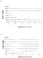
ピアソンの方法に従って、潰瘍性大腸炎患者の部分的なメイヨースコアと血漿PTX3濃度との間の相関を検討したところ、r=0.63、p<0.01と、血清CRP濃度との相関(r=0.51、p<0.05)より優れた相関を示した(図3A、B)。
PTX3の潰瘍性大腸炎患者における疾患活動性判定の感度は91%、特異性は75%であり、CRPでは感度は45%、特異性は100%であった(表1、表2)。(B) Correlation between disease activity index and blood PTX3 concentration Plasma PTX3 concentration and serum CRP concentration were measured for 17 patients with ulcerative colitis and 25 patients with Crohn's disease. PTX3 was measured using the method described in Example 1, and CRP was measured using an immunoturbidity method. Moreover, the partial Mayo score except the endoscopic findings of 15 ulcerative colitis patients and the IOIBD score of 25 patients with Crohn's disease were obtained.
According to Pearson's method, the correlation between the partial Mayo score and plasma PTX3 concentration in patients with ulcerative colitis was examined. The correlation between r = 0.63, p <0.01 and the serum CRP concentration ( r = 0.51, p <0.05) (Fig. 3A, B).
The sensitivity of PTX3 in ulcerative colitis patients was 91% and the specificity was 75%, and the CRP had a sensitivity of 45% and a specificity of 100% (Tables 1 and 2).

クローン病患者におけるIOIBDスコアと血漿PTX3濃度との相関はr=0.49、p<0.01であり、血清CRP濃度との相関(r=0.61、p<0.01)とほぼ同等であった(図4A、B)。
PTX3のクローン病患者における疾患活動性判定の感度は71%、特異性は62%であり、CRPでは感度は71%、特異性は56%であり、特異性に関してやや良好な結果を示した(表3、表4)。The correlation between the IOIBD score and plasma PTX3 concentration in Crohn's disease patients is r = 0.49, p <0.01, which is almost equivalent to the correlation with serum CRP concentration (r = 0.61, p <0.01). (FIGS. 4A and B).
The sensitivity of PTX3 in Crohn's disease patients was 71% for sensitivity and 62% for specificity, and CRP was 71% for sensitivity and 56% for specificity. Tables 3 and 4).
Claims (12)
Translated fromJapanesePriority Applications (1)
| Application Number | Priority Date | Filing Date | Title |
|---|---|---|---|
| JP2008558008AJP5280214B2 (en) | 2007-02-15 | 2008-02-14 | Diagnosis method of inflammatory bowel disease |
Applications Claiming Priority (4)
| Application Number | Priority Date | Filing Date | Title |
|---|---|---|---|
| JP2007034233 | 2007-02-15 | ||
| JP2007034233 | 2007-02-15 | ||
| PCT/JP2008/000222WO2008099608A1 (en) | 2007-02-15 | 2008-02-14 | Method for diagnosis of inflammatory bowel disease |
| JP2008558008AJP5280214B2 (en) | 2007-02-15 | 2008-02-14 | Diagnosis method of inflammatory bowel disease |
Publications (2)
| Publication Number | Publication Date |
|---|---|
| JPWO2008099608A1 JPWO2008099608A1 (en) | 2010-05-27 |
| JP5280214B2true JP5280214B2 (en) | 2013-09-04 |
Family
ID=39689855
Family Applications (1)
| Application Number | Title | Priority Date | Filing Date |
|---|---|---|---|
| JP2008558008AExpired - Fee RelatedJP5280214B2 (en) | 2007-02-15 | 2008-02-14 | Diagnosis method of inflammatory bowel disease |
Country Status (2)
| Country | Link |
|---|---|
| JP (1) | JP5280214B2 (en) |
| WO (1) | WO2008099608A1 (en) |
Families Citing this family (5)
| Publication number | Priority date | Publication date | Assignee | Title |
|---|---|---|---|---|
| JP5837761B2 (en)* | 2010-05-12 | 2015-12-24 | イーエヌ大塚製薬株式会社 | Classification of Crohn's disease activity |
| US20120034706A1 (en)* | 2010-08-06 | 2012-02-09 | Hakujyujikai Medical Inc. | Method for assessing inflammatory condition |
| WO2019056991A1 (en)* | 2017-09-19 | 2019-03-28 | 臻崴生物科技有限公司 | Monoclonal antibody or antigen-binding fragment thereof and use of same |
| TWI754171B (en) | 2018-09-14 | 2022-02-01 | 臻崴生物科技有限公司 | Medicinal composition including monoclonal antibody or antigen-binding fragment and use of the same |
| WO2025013850A1 (en)* | 2023-07-12 | 2025-01-16 | 株式会社島津製作所 | Method for assessing disorder of mucous membrane of gastrointestinal tract |
Citations (1)
| Publication number | Priority date | Publication date | Assignee | Title |
|---|---|---|---|---|
| WO2005106494A2 (en)* | 2004-04-29 | 2005-11-10 | Farma Development S.R.L. | Monoclonal antibodies, hybridomas, improved method for determining the protein ptx3 and kit for said determination |
- 2008
- 2008-02-14WOPCT/JP2008/000222patent/WO2008099608A1/enactiveApplication Filing
- 2008-02-14JPJP2008558008Apatent/JP5280214B2/ennot_activeExpired - Fee Related
Patent Citations (1)
| Publication number | Priority date | Publication date | Assignee | Title |
|---|---|---|---|---|
| WO2005106494A2 (en)* | 2004-04-29 | 2005-11-10 | Farma Development S.R.L. | Monoclonal antibodies, hybridomas, improved method for determining the protein ptx3 and kit for said determination |
Non-Patent Citations (1)
| Title |
|---|
| JPN6013022917; 奥谷大介: Jpn. J. Clin. Immunol. 29(3), 2006, 107-113* |
Also Published As
| Publication number | Publication date |
|---|---|
| WO2008099608A1 (en) | 2008-08-21 |
| JPWO2008099608A1 (en) | 2010-05-27 |
Similar Documents
| Publication | Publication Date | Title |
|---|---|---|
| JP2009020049A (en) | Diagnosis method of cerebrovascular disease | |
| WO2009090882A1 (en) | Diagnosis method for non-alcoholic fatty liver disease | |
| JP7174385B2 (en) | Use of Laminin-2 to Diagnose Pancreatic Cancer | |
| RU2769987C2 (en) | Antibody analysis | |
| CN104884958B (en) | Sugar chain isomers detection method and sugar chain isomers detection means | |
| JP6691060B2 (en) | Cell surface prostate cancer antigen for diagnostics | |
| US20170146541A1 (en) | Detection of high-risk intraductal papillary mucinous neoplasm and pancreatic adenocarcinoma | |
| JP5280214B2 (en) | Diagnosis method of inflammatory bowel disease | |
| JP5670422B2 (en) | Gastric cancer detection marker and gastric cancer detection method | |
| US20120107295A1 (en) | Methods and Compositions for Detecting Pancreatic Disease | |
| CN103765220B (en) | The detection mark of colorectal cancer or cancer of the esophagus and inspection method | |
| JP5137015B2 (en) | PTX3 high sensitivity measurement method | |
| JP5773979B2 (en) | Gastric cancer detection marker and gastric cancer detection method | |
| US20100041167A1 (en) | Drug for diagnosing large intestinal cancer and/or polyp, observing postoperative course and monitoring recurrence | |
| US8158373B2 (en) | Method of detecting cancer and evaluating cancer prognosis | |
| JP2007192557A (en) | Method for monitoring the severity of liver disease in hepatitis C patients and method for diagnosing liver cancer | |
| JP2014115186A (en) | Method for detecting stomach cancer, lung cancer, and/or esophagus cancer | |
| JP2009014521A (en) | Diagnosis of sleep apnea syndrome | |
| JP2009288219A (en) | Diagnostic method of cardiovascular event onset risk | |
| JP2008249618A (en) | External secretion disorder diagnostic reagent | |
| JP2012018119A (en) | Colon cancer detection marker and colon cancer detection method using the same | |
| WO2013176070A1 (en) | Cancer detection method | |
| JP5593502B2 (en) | Method for measuring chemerin concentration | |
| JP2024039982A (en) | How to assess the risk of developing acute kidney injury | |
| JP2014115188A (en) | Method for detecting stomach cancer, pancreas cancer, lung cancer, and/or esophagus cancer |
Legal Events
| Date | Code | Title | Description |
|---|---|---|---|
| A621 | Written request for application examination | Free format text:JAPANESE INTERMEDIATE CODE: A621 Effective date:20110121 | |
| TRDD | Decision of grant or rejection written | ||
| A01 | Written decision to grant a patent or to grant a registration (utility model) | Free format text:JAPANESE INTERMEDIATE CODE: A01 Effective date:20130514 | |
| A61 | First payment of annual fees (during grant procedure) | Free format text:JAPANESE INTERMEDIATE CODE: A61 Effective date:20130522 | |
| R150 | Certificate of patent or registration of utility model | Free format text:JAPANESE INTERMEDIATE CODE: R150 | |
| R250 | Receipt of annual fees | Free format text:JAPANESE INTERMEDIATE CODE: R250 | |
| R250 | Receipt of annual fees | Free format text:JAPANESE INTERMEDIATE CODE: R250 | |
| LAPS | Cancellation because of no payment of annual fees |