JP5164971B2 - A method for differential detection of multimers from monomers of multimer-forming polypeptides using three-dimensional interactions - Google Patents
A method for differential detection of multimers from monomers of multimer-forming polypeptides using three-dimensional interactionsDownload PDFInfo
- Publication number
- JP5164971B2 JP5164971B2JP2009506420AJP2009506420AJP5164971B2JP 5164971 B2JP5164971 B2JP 5164971B2JP 2009506420 AJP2009506420 AJP 2009506420AJP 2009506420 AJP2009506420 AJP 2009506420AJP 5164971 B2JP5164971 B2JP 5164971B2
- Authority
- JP
- Japan
- Prior art keywords
- antibody
- epitope
- detection antibody
- detection
- bound
- Prior art date
- Legal status (The legal status is an assumption and is not a legal conclusion. Google has not performed a legal analysis and makes no representation as to the accuracy of the status listed.)
- Active
Links
- 238000001514detection methodMethods0.000titleclaimsdescription177
- 238000000034methodMethods0.000titleclaimsdescription61
- 108090000765processed proteins & peptidesProteins0.000titleclaimsdescription55
- 229920001184polypeptidePolymers0.000titleclaimsdescription52
- 102000004196processed proteins & peptidesHuman genes0.000titleclaimsdescription52
- 239000000178monomerSubstances0.000titleclaimsdescription6
- 230000003993interactionEffects0.000titledescription2
- 239000011324beadSubstances0.000claimsdescription147
- 108091000054PrionProteins0.000claimsdescription51
- 102000029797PrionHuman genes0.000claimsdescription51
- 125000003275alpha amino acid groupChemical group0.000claimsdescription20
- 230000027455bindingEffects0.000claimsdescription17
- 239000000126substanceSubstances0.000claimsdescription17
- 239000007790solid phaseSubstances0.000claimsdescription16
- 108010067770Endopeptidase KProteins0.000claimsdescription14
- 210000004369bloodAnatomy0.000claimsdescription10
- 239000008280bloodSubstances0.000claimsdescription10
- 239000004816latexSubstances0.000claimsdescription9
- 229920000126latexPolymers0.000claimsdescription9
- 102000004190EnzymesHuman genes0.000claimsdescription7
- 108090000790EnzymesProteins0.000claimsdescription7
- 102000008847SerpinHuman genes0.000claimsdescription7
- 108050000761SerpinProteins0.000claimsdescription7
- 239000003001serine protease inhibitorSubstances0.000claimsdescription7
- -1Ig light chainProteins0.000claimsdescription6
- 210000004556brainAnatomy0.000claimsdescription6
- 108010071690PrealbuminProteins0.000claimsdescription4
- 102000009190TransthyretinHuman genes0.000claimsdescription4
- 108010026424tau ProteinsProteins0.000claimsdescription4
- 102000013498tau ProteinsHuman genes0.000claimsdescription4
- 102000012192Cystatin CHuman genes0.000claimsdescription3
- 108010061642Cystatin CProteins0.000claimsdescription3
- 102000036770Islet Amyloid PolypeptideHuman genes0.000claimsdescription3
- 108010041872Islet Amyloid PolypeptideProteins0.000claimsdescription3
- 102000054727Serum Amyloid AHuman genes0.000claimsdescription3
- 108700028909Serum Amyloid AProteins0.000claimsdescription3
- 108090000185alpha-SynucleinProteins0.000claimsdescription3
- 102000003802alpha-SynucleinHuman genes0.000claimsdescription3
- 238000002866fluorescence resonance energy transferMethods0.000claimsdescription3
- 108090000631TrypsinProteins0.000claimsdescription2
- 102000004142TrypsinHuman genes0.000claimsdescription2
- 239000000941radioactive substanceSubstances0.000claimsdescription2
- 239000012588trypsinSubstances0.000claimsdescription2
- 102000013455Amyloid beta-PeptidesHuman genes0.000claims2
- 108010090849Amyloid beta-PeptidesProteins0.000claims2
- 102000019197Superoxide DismutaseHuman genes0.000claims2
- 108010012715Superoxide dismutaseProteins0.000claims2
- 101710138751Major prion proteinProteins0.000description91
- 210000002381plasmaAnatomy0.000description83
- 239000000523sampleSubstances0.000description46
- 241001494479PecoraSpecies0.000description22
- SEQKRHFRPICQDD-UHFFFAOYSA-NN-tris(hydroxymethyl)methylglycineChemical compoundOCC(CO)(CO)[NH2+]CC([O-])=OSEQKRHFRPICQDD-UHFFFAOYSA-N0.000description14
- 241000283690Bos taurusSpecies0.000description12
- 239000013504Triton X-100Substances0.000description11
- 229920004890Triton X-100Polymers0.000description11
- 229960002685biotinDrugs0.000description11
- 239000011616biotinSubstances0.000description11
- 239000000872bufferSubstances0.000description11
- 230000009977dual effectEffects0.000description11
- 238000005194fractionationMethods0.000description11
- 201000010099diseaseDiseases0.000description10
- 208000037265diseases, disorders, signs and symptomsDiseases0.000description10
- 208000024777Prion diseaseDiseases0.000description9
- 239000007997Tricine bufferSubstances0.000description9
- 238000004458analytical methodMethods0.000description9
- 238000006243chemical reactionMethods0.000description9
- 208000022256primary systemic amyloidosisDiseases0.000description9
- 102000004169proteins and genesHuman genes0.000description8
- 108090000623proteins and genesProteins0.000description8
- UZMAPBJVXOGOFT-UHFFFAOYSA-NSyringetinNatural productsCOC1=C(O)C(OC)=CC(C2=C(C(=O)C3=C(O)C=C(O)C=C3O2)O)=C1UZMAPBJVXOGOFT-UHFFFAOYSA-N0.000description7
- KCFYHBSOLOXZIF-UHFFFAOYSA-NdihydrochrysinNatural productsCOC1=C(O)C(OC)=CC(C2OC3=CC(O)=CC(O)=C3C(=O)C2)=C1KCFYHBSOLOXZIF-UHFFFAOYSA-N0.000description7
- LOKCTEFSRHRXRJ-UHFFFAOYSA-Idipotassium trisodium dihydrogen phosphate hydrogen phosphate dichlorideChemical compoundP(=O)(O)(O)[O-].[K+].P(=O)(O)([O-])[O-].[Na+].[Na+].[Cl-].[K+].[Cl-].[Na+]LOKCTEFSRHRXRJ-UHFFFAOYSA-I0.000description7
- 239000002953phosphate buffered salineSubstances0.000description7
- 230000036470plasma concentrationEffects0.000description7
- 208000003407Creutzfeldt-Jakob SyndromeDiseases0.000description6
- 206010035226Plasma cell myelomaDiseases0.000description6
- 238000003776cleavage reactionMethods0.000description6
- 239000003599detergentSubstances0.000description6
- 230000008569processEffects0.000description6
- 230000007017scissionEffects0.000description6
- 239000000758substrateSubstances0.000description6
- 239000011534wash bufferSubstances0.000description6
- 229920001213Polysorbate 20Polymers0.000description5
- 239000000427antigenSubstances0.000description5
- 102000036639antigensHuman genes0.000description5
- 108091007433antigensProteins0.000description5
- 210000004027cellAnatomy0.000description5
- 238000001378electrochemiluminescence detectionMethods0.000description5
- 238000002372labellingMethods0.000description5
- 239000000256polyoxyethylene sorbitan monolaurateSubstances0.000description5
- 235000010486polyoxyethylene sorbitan monolaurateNutrition0.000description5
- 239000011541reaction mixtureSubstances0.000description5
- 208000008864scrapieDiseases0.000description5
- 239000003656tris buffered salineSubstances0.000description5
- YBJHBAHKTGYVGT-ZKWXMUAHSA-N(+)-BiotinChemical compoundN1C(=O)N[C@@H]2[C@H](CCCCC(=O)O)SC[C@@H]21YBJHBAHKTGYVGT-ZKWXMUAHSA-N0.000description4
- 208000020406Creutzfeldt Jacob diseaseDiseases0.000description4
- 208000010859Creutzfeldt-Jakob diseaseDiseases0.000description4
- 241001465754MetazoaSpecies0.000description4
- 206010002026amyotrophic lateral sclerosisDiseases0.000description4
- 239000007853buffer solutionSubstances0.000description4
- 230000002860competitive effectEffects0.000description4
- 230000003247decreasing effectEffects0.000description4
- 238000002474experimental methodMethods0.000description4
- 239000012530fluidSubstances0.000description4
- 239000000463materialSubstances0.000description4
- 238000012545processingMethods0.000description4
- 238000005406washingMethods0.000description4
- 208000024827Alzheimer diseaseDiseases0.000description3
- 206010014561EmphysemaDiseases0.000description3
- 208000007487Familial Cerebral Amyloid AngiopathyDiseases0.000description3
- 108010001336Horseradish PeroxidaseProteins0.000description3
- 208000023105Huntington diseaseDiseases0.000description3
- 208000005531Immunoglobulin Light-chain AmyloidosisDiseases0.000description3
- 101001095054Ovis aries Major prion proteinProteins0.000description3
- 208000018737Parkinson diseaseDiseases0.000description3
- 230000002159abnormal effectEffects0.000description3
- 238000004220aggregationMethods0.000description3
- 230000002776aggregationEffects0.000description3
- 206010002022amyloidosisDiseases0.000description3
- 230000015572biosynthetic processEffects0.000description3
- 208000019425cirrhosis of liverDiseases0.000description3
- 230000007812deficiencyEffects0.000description3
- 238000010586diagramMethods0.000description3
- 238000001631haemodialysisMethods0.000description3
- 230000000322hemodialysisEffects0.000description3
- 230000035945sensitivityEffects0.000description3
- 210000002966serumAnatomy0.000description3
- GPRLSGONYQIRFK-MNYXATJNSA-NtritonChemical compound[3H+]GPRLSGONYQIRFK-MNYXATJNSA-N0.000description3
- 208000001072type 2 diabetes mellitusDiseases0.000description3
- PKYCWFICOKSIHZ-UHFFFAOYSA-N1-(3,7-dihydroxyphenoxazin-10-yl)ethanoneChemical compoundOC1=CC=C2N(C(=O)C)C3=CC=C(O)C=C3OC2=C1PKYCWFICOKSIHZ-UHFFFAOYSA-N0.000description2
- 102000002260Alkaline PhosphataseHuman genes0.000description2
- 108020004774Alkaline PhosphataseProteins0.000description2
- 206010012289DementiaDiseases0.000description2
- 208000034846Familial Amyloid NeuropathiesDiseases0.000description2
- 206010016654FibrosisDiseases0.000description2
- 201000011240Frontotemporal dementiaDiseases0.000description2
- 206010019889Hereditary neuropathic amyloidosisDiseases0.000description2
- 241000282412HomoSpecies0.000description2
- 229910019142PO4Inorganic materials0.000description2
- 108091005804PeptidasesProteins0.000description2
- 239000004365ProteaseSubstances0.000description2
- 102100037486Reverse transcriptase/ribonuclease HHuman genes0.000description2
- 108010090804StreptavidinProteins0.000description2
- 239000006180TBST bufferSubstances0.000description2
- 150000001413amino acidsChemical class0.000description2
- 230000008901benefitEffects0.000description2
- 239000013060biological fluidSubstances0.000description2
- 235000020958biotinNutrition0.000description2
- XJMXIWNOKIEIMX-UHFFFAOYSA-Nbromo chloro 1h-indol-2-yl phosphateChemical compoundC1=CC=C2NC(OP(=O)(OBr)OCl)=CC2=C1XJMXIWNOKIEIMX-UHFFFAOYSA-N0.000description2
- 230000008859changeEffects0.000description2
- 239000007795chemical reaction productSubstances0.000description2
- 230000007882cirrhosisEffects0.000description2
- 238000004040coloringMethods0.000description2
- 238000011161developmentMethods0.000description2
- 238000003018immunoassayMethods0.000description2
- 208000019715inherited Creutzfeldt-Jakob diseaseDiseases0.000description2
- KNJDBYZZKAZQNG-UHFFFAOYSA-NlucigeninChemical compound[O-][N+]([O-])=O.[O-][N+]([O-])=O.C12=CC=CC=C2[N+](C)=C(C=CC=C2)C2=C1C1=C(C=CC=C2)C2=[N+](C)C2=CC=CC=C12KNJDBYZZKAZQNG-UHFFFAOYSA-N0.000description2
- 238000004020luminiscence typeMethods0.000description2
- 230000005389magnetismEffects0.000description2
- 238000002156mixingMethods0.000description2
- JPXMTWWFLBLUCD-UHFFFAOYSA-Nnitro blue tetrazolium(2+)Chemical compoundCOC1=CC(C=2C=C(OC)C(=CC=2)[N+]=2N(N=C(N=2)C=2C=CC=CC=2)C=2C=CC(=CC=2)[N+]([O-])=O)=CC=C1[N+]1=NC(C=2C=CC=CC=2)=NN1C1=CC=C([N+]([O-])=O)C=C1JPXMTWWFLBLUCD-UHFFFAOYSA-N0.000description2
- 239000010452phosphateSubstances0.000description2
- 239000012488sample solutionSubstances0.000description2
- 229940016590sarkosylDrugs0.000description2
- 108700004121sarkosylProteins0.000description2
- 238000000926separation methodMethods0.000description2
- KSAVQLQVUXSOCR-UHFFFAOYSA-Msodium lauroyl sarcosinateChemical compound[Na+].CCCCCCCCCCCC(=O)N(C)CC([O-])=OKSAVQLQVUXSOCR-UHFFFAOYSA-M0.000description2
- 210000001519tissueAnatomy0.000description2
- 201000007905transthyretin amyloidosisDiseases0.000description2
- UCLKLGIYGBLTSM-UHFFFAOYSA-N1,2,3,4-tetrachloro-5-(2,5-dichlorophenyl)benzeneChemical compoundClC1=CC=C(Cl)C(C=2C(=C(Cl)C(Cl)=C(Cl)C=2)Cl)=C1UCLKLGIYGBLTSM-UHFFFAOYSA-N0.000description1
- HWTAKVLMACWHLD-UHFFFAOYSA-N2-(9h-carbazol-1-yl)ethanamineChemical compoundC12=CC=CC=C2NC2=C1C=CC=C2CCNHWTAKVLMACWHLD-UHFFFAOYSA-N0.000description1
- WONRDHPFOHAWOG-UHFFFAOYSA-N2-chloronaphthalen-1-olChemical compoundC1=CC=C2C(O)=C(Cl)C=CC2=C1WONRDHPFOHAWOG-UHFFFAOYSA-N0.000description1
- ZBQCCTCQUCOXBO-UHFFFAOYSA-N4-(4-aminophenyl)-2,2,6,6-tetramethylcyclohex-3-en-1-amineChemical compoundCC1(C)C(N)C(C)(C)CC(C=2C=CC(N)=CC=2)=C1ZBQCCTCQUCOXBO-UHFFFAOYSA-N0.000description1
- 241000282979Alces alcesSpecies0.000description1
- KFYRPLNVJVHZGT-UHFFFAOYSA-NAmitriptyline hydrochlorideChemical compoundCl.C1CC2=CC=CC=C2C(=CCCN(C)C)C2=CC=CC=C21KFYRPLNVJVHZGT-UHFFFAOYSA-N0.000description1
- 241000195940BryophytaSpecies0.000description1
- 241000283707CapraSpecies0.000description1
- 241000282994CervidaeSpecies0.000description1
- 102000018832CytochromesHuman genes0.000description1
- 108010052832CytochromesProteins0.000description1
- IGXWBGJHJZYPQS-SSDOTTSWSA-ND-LuciferinChemical compoundOC(=O)[C@H]1CSC(C=2SC3=CC=C(O)C=C3N=2)=N1IGXWBGJHJZYPQS-SSDOTTSWSA-N0.000description1
- 238000002965ELISAMethods0.000description1
- 241000282414Homo sapiensSpecies0.000description1
- 102000004157HydrolasesHuman genes0.000description1
- 108090000604HydrolasesProteins0.000description1
- 108060003951ImmunoglobulinProteins0.000description1
- 102000008394Immunoglobulin FragmentsHuman genes0.000description1
- 108010021625Immunoglobulin FragmentsProteins0.000description1
- 241000772415Neovison visonSpecies0.000description1
- 208000012902Nervous system diseaseDiseases0.000description1
- IOVCWXUNBOPUCH-UHFFFAOYSA-MNitrite anionChemical compound[O-]N=OIOVCWXUNBOPUCH-UHFFFAOYSA-M0.000description1
- OAICVXFJPJFONN-UHFFFAOYSA-NPhosphorusChemical compound[P]OAICVXFJPJFONN-UHFFFAOYSA-N0.000description1
- 241000276498Pollachius virensSpecies0.000description1
- 206010036105PolyneuropathyDiseases0.000description1
- 102000001708Protein IsoformsHuman genes0.000description1
- 108010029485Protein IsoformsProteins0.000description1
- 108020004511Recombinant DNAProteins0.000description1
- 108091081062Repeated sequence (DNA)Proteins0.000description1
- 208000002704Sporadic Creutzfeldt-Jakob diseaseDiseases0.000description1
- 208000034953Twin anemia-polycythemia sequenceDiseases0.000description1
- 208000018756Variant Creutzfeldt-Jakob diseaseDiseases0.000description1
- SXEHKFHPFVVDIR-UHFFFAOYSA-N[4-(4-hydrazinylphenyl)phenyl]hydrazineChemical compoundC1=CC(NN)=CC=C1C1=CC=C(NN)C=C1SXEHKFHPFVVDIR-UHFFFAOYSA-N0.000description1
- 238000009825accumulationMethods0.000description1
- 230000000890antigenic effectEffects0.000description1
- OHDRQQURAXLVGJ-HLVWOLMTSA-Nazane;(2e)-3-ethyl-2-[(e)-(3-ethyl-6-sulfo-1,3-benzothiazol-2-ylidene)hydrazinylidene]-1,3-benzothiazole-6-sulfonic acidChemical compound[NH4+].[NH4+].S/1C2=CC(S([O-])(=O)=O)=CC=C2N(CC)C\1=N/N=C1/SC2=CC(S([O-])(=O)=O)=CC=C2N1CCOHDRQQURAXLVGJ-HLVWOLMTSA-N0.000description1
- 102000005936beta-GalactosidaseHuman genes0.000description1
- 108010005774beta-GalactosidaseProteins0.000description1
- 102000006995beta-GlucosidaseHuman genes0.000description1
- 108010047754beta-GlucosidaseProteins0.000description1
- 230000004071biological effectEffects0.000description1
- 230000015556catabolic processEffects0.000description1
- 230000001413cellular effectEffects0.000description1
- 239000003153chemical reaction reagentSubstances0.000description1
- 238000004587chromatography analysisMethods0.000description1
- 239000003593chromogenic compoundSubstances0.000description1
- 230000001684chronic effectEffects0.000description1
- 230000000052comparative effectEffects0.000description1
- 230000009137competitive bindingEffects0.000description1
- 239000013078crystalSubstances0.000description1
- 238000005520cutting processMethods0.000description1
- 238000000354decomposition reactionMethods0.000description1
- 238000006731degradation reactionMethods0.000description1
- 229960003964deoxycholic acidDrugs0.000description1
- KXGVEGMKQFWNSR-LLQZFEROSA-Ndeoxycholic acidChemical compoundC([C@H]1CC2)[C@H](O)CC[C@]1(C)[C@@H]1[C@@H]2[C@@H]2CC[C@H]([C@@H](CCC(O)=O)C)[C@@]2(C)[C@@H](O)C1KXGVEGMKQFWNSR-LLQZFEROSA-N0.000description1
- 230000001419dependent effectEffects0.000description1
- 239000000539dimerSubstances0.000description1
- 238000013399early diagnosisMethods0.000description1
- 230000000694effectsEffects0.000description1
- 238000005516engineering processMethods0.000description1
- 238000006911enzymatic reactionMethods0.000description1
- 201000006061fatal familial insomniaDiseases0.000description1
- 210000003608feceAnatomy0.000description1
- 208000037957feline spongiform encephalopathyDiseases0.000description1
- GNBHRKFJIUUOQI-UHFFFAOYSA-NfluoresceinChemical compoundO1C(=O)C2=CC=CC=C2C21C1=CC=C(O)C=C1OC1=CC(O)=CC=C21GNBHRKFJIUUOQI-UHFFFAOYSA-N0.000description1
- 239000012634fragmentSubstances0.000description1
- 230000008570general processEffects0.000description1
- 230000005484gravityEffects0.000description1
- 208000010544human prion diseaseDiseases0.000description1
- 210000004408hybridomaAnatomy0.000description1
- 208000027488iatrogenic Creutzfeldt-Jakob diseaseDiseases0.000description1
- 230000000642iatrogenic effectEffects0.000description1
- 102000018358immunoglobulinHuman genes0.000description1
- 230000016784immunoglobulin productionEffects0.000description1
- 238000001727in vivoMethods0.000description1
- 206010023497kuruDiseases0.000description1
- 244000144972livestockSpecies0.000description1
- HWYHZTIRURJOHG-UHFFFAOYSA-NluminolChemical compoundO=C1NNC(=O)C2=C1C(N)=CC=C2HWYHZTIRURJOHG-UHFFFAOYSA-N0.000description1
- 210000002751lymphAnatomy0.000description1
- 210000004698lymphocyteAnatomy0.000description1
- 230000007257malfunctionEffects0.000description1
- 238000004519manufacturing processMethods0.000description1
- 238000005259measurementMethods0.000description1
- 210000004080milkAnatomy0.000description1
- 239000008267milkSubstances0.000description1
- 235000013336milkNutrition0.000description1
- 235000011929mousseNutrition0.000description1
- 239000013642negative controlSubstances0.000description1
- 230000004770neurodegenerationEffects0.000description1
- 208000015122neurodegenerative diseaseDiseases0.000description1
- 238000007500overflow downdraw methodMethods0.000description1
- 210000002741palatine tonsilAnatomy0.000description1
- 150000002978peroxidesChemical class0.000description1
- 239000012071phaseSubstances0.000description1
- 239000011574phosphorusSubstances0.000description1
- 229910052698phosphorusInorganic materials0.000description1
- 229920000642polymerPolymers0.000description1
- 230000007824polyneuropathyEffects0.000description1
- 238000002360preparation methodMethods0.000description1
- 230000007065protein hydrolysisEffects0.000description1
- 238000004445quantitative analysisMethods0.000description1
- 230000003252repetitive effectEffects0.000description1
- 210000003296salivaAnatomy0.000description1
- 210000000582semenAnatomy0.000description1
- 210000000952spleenAnatomy0.000description1
- BDHFUVZGWQCTTF-UHFFFAOYSA-MsulfonateChemical compound[O-]S(=O)=OBDHFUVZGWQCTTF-UHFFFAOYSA-M0.000description1
- 210000001138tearAnatomy0.000description1
- 210000002700urineAnatomy0.000description1
Images


Classifications
- G—PHYSICS
- G01—MEASURING; TESTING
- G01N—INVESTIGATING OR ANALYSING MATERIALS BY DETERMINING THEIR CHEMICAL OR PHYSICAL PROPERTIES
- G01N33/00—Investigating or analysing materials by specific methods not covered by groups G01N1/00 - G01N31/00
- G01N33/48—Biological material, e.g. blood, urine; Haemocytometers
- G01N33/50—Chemical analysis of biological material, e.g. blood, urine; Testing involving biospecific ligand binding methods; Immunological testing
- G01N33/68—Chemical analysis of biological material, e.g. blood, urine; Testing involving biospecific ligand binding methods; Immunological testing involving proteins, peptides or amino acids
- G01N33/6854—Immunoglobulins
- G—PHYSICS
- G01—MEASURING; TESTING
- G01N—INVESTIGATING OR ANALYSING MATERIALS BY DETERMINING THEIR CHEMICAL OR PHYSICAL PROPERTIES
- G01N33/00—Investigating or analysing materials by specific methods not covered by groups G01N1/00 - G01N31/00
- G01N33/48—Biological material, e.g. blood, urine; Haemocytometers
- G01N33/50—Chemical analysis of biological material, e.g. blood, urine; Testing involving biospecific ligand binding methods; Immunological testing
- G01N33/53—Immunoassay; Biospecific binding assay; Materials therefor
- G—PHYSICS
- G01—MEASURING; TESTING
- G01N—INVESTIGATING OR ANALYSING MATERIALS BY DETERMINING THEIR CHEMICAL OR PHYSICAL PROPERTIES
- G01N33/00—Investigating or analysing materials by specific methods not covered by groups G01N1/00 - G01N31/00
- G01N33/48—Biological material, e.g. blood, urine; Haemocytometers
- G01N33/50—Chemical analysis of biological material, e.g. blood, urine; Testing involving biospecific ligand binding methods; Immunological testing
- G01N33/53—Immunoassay; Biospecific binding assay; Materials therefor
- G01N33/543—Immunoassay; Biospecific binding assay; Materials therefor with an insoluble carrier for immobilising immunochemicals
- G01N33/54306—Solid-phase reaction mechanisms
Landscapes
- Health & Medical Sciences (AREA)
- Life Sciences & Earth Sciences (AREA)
- Engineering & Computer Science (AREA)
- Immunology (AREA)
- Chemical & Material Sciences (AREA)
- Molecular Biology (AREA)
- Biomedical Technology (AREA)
- Hematology (AREA)
- Urology & Nephrology (AREA)
- Analytical Chemistry (AREA)
- Biochemistry (AREA)
- Pathology (AREA)
- Food Science & Technology (AREA)
- Medicinal Chemistry (AREA)
- Physics & Mathematics (AREA)
- General Physics & Mathematics (AREA)
- General Health & Medical Sciences (AREA)
- Microbiology (AREA)
- Biotechnology (AREA)
- Cell Biology (AREA)
- Chemical Kinetics & Catalysis (AREA)
- Proteomics, Peptides & Aminoacids (AREA)
- Peptides Or Proteins (AREA)
Description
Translated fromJapanese本発明は、三次元的相互作用を用いてマルチマー形成ポリペプチドのモノマー形からマルチマー形を分別検出する方法及びそのためのキットに関する。 The present invention relates to a method for differentially detecting a multimer form from a monomer form of a multimer-forming polypeptide using a three-dimensional interaction, and a kit therefor.
タンパク質の機能は、タンパク質を構成するポリペプチドのマルチマー化が必要であるということは、一般によく知られている。しかしながら、あるタンパク質においてマルチマーの形成は、よく疾患または疾病を引き起こす。特に、タンパク質は、正常的な状態ではモノマーとして存在し、非正常的な状態では、マルチマー(または凝集)に変換される(例えば、ミスフォールディング形成)。 It is generally well known that the function of a protein requires multimerization of polypeptides constituting the protein. However, the formation of multimers in certain proteins often causes disease or illness. In particular, proteins exist as monomers in the normal state and are converted to multimers (or aggregates) in the abnormal state (eg, misfolding).
ミスフォールディングまたは機能的に形成されなかった凝集(蓄積)が形成されたタンパク質は、正常的な生物学的活性を有しないことが既によく知られている。タンパク質が正確にフォールディングされなかったり、正確なフォールディングを維持できない場合は、多様な生物学的機能不全(malfunction)を誘発し、その結果、多様な疾患を誘発する(Massimo Stefani, et al., J. Mol. Med. 81:678-699(2003); and Radford SE, et al., Cell. 97:291-298(1999))。数々の疾病は、生体内で不正確な構造、即ち、正常的に機能を果たすものとは異なる構造を有するタンパク質分子により引き起こされる。 It is already well known that proteins with misfolded or aggregates (accumulation) that have not been formed functionally do not have normal biological activity. If the protein is not folded correctly or cannot maintain the correct folding, it can induce a variety of biological malfunctions, resulting in a variety of diseases (Massimo Stefani, et al., J Mol. Med. 81: 678-699 (2003); and Radford SE, et al., Cell. 97: 291-298 (1999)). Numerous diseases are caused by protein molecules that have an inaccurate structure in vivo, that is, a structure that is different from those that normally function.
例えば、タンパク質の非正常的な凝集またはミスフォールディングに係わる疾患または疾病には、アルツハイマー疾患、クロイツフェルト・ヤコブ病、海綿状脳症 (Spongiform encephalopathies)、パーキンソン疾患、ハンチントン疾患、筋萎縮性側索硬化症(Amyotrophic lateral sclerosis)、セルピン欠乏症(Serpin deficiency)、肺気腫(emphysema)、硬変症(cirrhosis)、第II型糖尿病、一次全身性アミロイド症、二次全身性アミロイド症、フロント一時的痴呆(Fronto-temporal dementias)、老人全身性アミロイド症、家族性アミロイドポリニューロパチー(familial amyloid polyneuropathy)、遺伝性大脳アミロイドアンギオパチー(hereditary cerebral amyloid angiopathy)、及び血透析関連アミロイド症がある。 For example, diseases or illnesses associated with abnormal aggregation or misfolding of proteins include Alzheimer's disease, Creutzfeldt-Jakob disease, Spongiform encephalopathies, Parkinson's disease, Huntington's disease, amyotrophic lateral sclerosis (Amyotrophic lateral sclerosis), Serpin deficiency, emphysema, cirrhosis, type II diabetes, primary systemic amyloidosis, secondary systemic amyloidosis, fronto-dementia (Fronto- temporal dementias), elderly systemic amyloidosis, familial amyloid polyneuropathy, hereditary cerebral amyloid angiopathy, and hemodialysis-related amyloidosis.
凝集関連疾患の初期診断は、集中的に研究されてきた。しかしながら、モノマー形(正常)からマルチマー形(凝集)を分別的に検出するための方法及び研究は、まだ提示されていない状態である。 Early diagnosis of aggregation-related diseases has been intensively studied. However, methods and studies for differentially detecting multimeric forms (aggregation) from monomeric forms (normal) have not yet been presented.
人間において、散発性、変種、医原性、及び家族性クロイツフェルト・ヤコブ病、クル(kuru)、家族性致命的不眠症、及びゲルストマン・シュトロイスラー・シャインカ病、羊及びヤギのスクレイピー、猫の海綿状脳症、ミンク海綿状脳症、シカ、エルク及びムースの慢性消耗性疾患、及び家畜の狂牛病は、致命的な神経退行性疾患であり、このような疾病は、伝達性海綿状脳症(Transmissible spongiform encephalopathies:TSE)によるものである(Prusiner S.B. Proc. Natl. Acad. Sci. USA 95:13363-13383(1998); and Hope J. Curr. Opin. Genet. Dev. 10, 568-57(2000))。プリオンタンパク質の非正常的なイソフォームまたはスクレイピー形(PrPSc)は、TSEの主要因として強力に提案されている(Caughey B. Trends Biochem. Sci. 26:235-42(2001))。In humans, sporadic, variant, iatrogenic, and familial Creutzfeldt-Jakob disease, kuru, familial fatal insomnia, and Gerstmann-Stroisler-Scheinka disease, sheep and goat scrapie, feline Spongiform encephalopathy, mink spongiform encephalopathy, chronic debilitating disease of deer, elk and mousse, and livestock mad cow disease are fatal neurodegenerative diseases, such as transmissible spongiform encephalopathy ( Transmissible spongiform encephalopathies (TSE) (Prusiner SB Proc. Natl. Acad. Sci. USA 95: 13363-13383 (1998); and Hope J. Curr. Opin. Genet. Dev. 10, 568-57 (2000) )). Abnormal isoforms or scrapie forms of prion proteins (PrPSc ) have been strongly proposed as a major factor in TSE (Caughey B. Trends Biochem. Sci. 26: 235-42 (2001)).
プリオンタンパク質の正常形(PrPC)は、α−鎖と流動的な無秩序な部位を含み、モノマー形として存在して(Zahn, R., et al., Proc. Natl. Acad. Sci. USA 97:145-150(2000))、スクレイピー形(PrPSc)は、高度のβ−シート構造を有して、マルチマー形または少なくとも二量体形以上として存在する(Caughey, B., et al., J. Biol. Chem. 273:32230-35(1998))。α−鎖構造からβ−シート構造への変化は、神経系疾患を発生させる疾病において主要原因である。The normal form of the prion protein (PrPC ) contains the α-chain and a fluidly disordered site and exists as a monomeric form (Zahn, R., et al., Proc. Natl. Acad. Sci. USA 97 : 145-150 (2000)), the scrapie form (PrPSc ) has a highly β-sheet structure and exists as a multimer form or at least as a dimer form (Caughey, B., et al., J Biol. Chem. 273: 32230-35 (1998)). The change from α-chain structure to β-sheet structure is a major cause in diseases that cause nervous system diseases.
PrPCは、タンパク質加水分解酵素に敏感性(PrPsen)を有する反面、PrPScは、タンパク質加水分解に対する部分的な抵抗性(PrPres)を有し、高分子凝集体を形成する傾向がある(Bolton D. C. Lancet, 358:164-5 (2001))。このような凝集体の形成は、PrPresの形成を誘導する構造的変化に対する分析や特性の記述を難しくする。PrPC is sensitive to protein hydrolase (PrPsen ), whereas PrPSc has partial resistance to protein hydrolysis (PrPres ) and tends to form polymer aggregates. (Bolton DC Lancet, 358: 164-5 (2001)). The formation of such aggregates makes it difficult to analyze and characterize the structural changes that induce the formation ofPrPres .
プロテアーゼK(PK)の切断方法は、細胞形を切断して、スクレイピー形のみを残しておき、これはELISAで検出されるが、PrP(スクレイピー形)の多様な形態の抵抗性を区別するに利用される。しかしながら、PK切断方法に対しては問題点が提起されている。なぜなら、PrP構造、濃度、組織抗体、切断時間及び緩衝液は、PK敏感度に影響を与え、これは、PK切断方法に対する信頼度を大きく低下させるからである。 The method of cleaving protease K (PK) cleaves the cell form, leaving only the scrapie form, which is detected by ELISA, but distinguishes the resistance of various forms of PrP (scrapie form). Used. However, problems have been raised for the PK cutting method. This is because PrP structure, concentration, tissue antibody, cleavage time and buffer affect PK sensitivity, which greatly reduces the reliability of the PK cleavage method.
したがって、モノマー形(例えば、PrPC、PrPの細胞形)からマルチマー形(例えば、PrPSc、PrPのスクレイピー形)を、より信頼度高く便利に分別検出できるような新しい方法の開発が切実である。Therefore, it is urgent to develop a new method for more reliable and convenient differential detection of monomeric forms (for example, PrPC , PrP cell forms) to multimeric forms (for example, PrPSc , PrP scrapy forms). .
本明細書全体にかけて多数の特許文献及び論文が参照されて、その引用は、表示されている。引用された特許文献及び論文の開示内容は、その全体が本明細書に参照として取り込まれ、本発明の属する技術分野の水準及び本発明の内容がより明確に説明される。 Throughout this specification, numerous patent documents and articles are referenced and their citations are displayed. The disclosures of the cited patent documents and papers are incorporated herein by reference in their entirety, and the level of the technical field to which the present invention belongs and the contents of the present invention are explained more clearly.
本発明の目的は、マルチマー形成ポリペプチドのモノマー形からマルチマー形を分別検出する方法を提供することにある。 An object of the present invention is to provide a method for differentially detecting a multimeric form from a monomeric form of a multimer-forming polypeptide.
本発明の他の目的は、マルチマー形成ポリペプチドのモノマー形からマルチマー形を分別検出するためのキットを提供することにある。 Another object of the present invention is to provide a kit for differentially detecting a multimeric form from a monomeric form of a multimer-forming polypeptide.
本発明の他の目的及び利点は、発明の詳細な説明、請求の範囲及び図面により、さらに明確にされる。 Other objects and advantages of the invention will become more apparent from the detailed description of the invention, the claims and the drawings.
本発明の一様態によると、本発明は、以下のステップを含む、生試料内のマルチマー形成ポリペプチドのモノマー形からマルチマー形を分別検出する方法を提供する:(a)固相のキャリアー(solid phase carrier)の表面に捕獲抗体を3次元的に結合させて、キャリアー−捕獲抗体複合体を用意するステップであって、前記捕獲抗体は、前記マルチマー形成ポリペプチドの一つの特定エピトープに結合して、(b)検出抗体を用意するステップであって、前記検出抗体が認識するエピトープは、検出抗体がエピトープに結合した場合、前記捕獲抗体のエピトープに結合された捕獲抗体により立体的妨害(Steric hindrance)を起す位置にあることを特徴とし、(c)生試料に前記キャリアー−捕獲抗体複合体及び検出抗体を同時に反応させるステップと、(d)キャリアー−捕獲抗体−マルチマー形検出抗体複合体を検出するステップ。 According to one aspect of the present invention, the present invention provides a method for differentially detecting a multimeric form from a monomeric form of a multimer-forming polypeptide in a raw sample, comprising the following steps: (a) a solid phase carrier a carrier-capture antibody complex by three-dimensionally binding a capture antibody to the surface of the phase carrier), wherein the capture antibody binds to one specific epitope of the multimer-forming polypeptide. (B) preparing a detection antibody, wherein the epitope recognized by the detection antibody is sterically hindered by a capture antibody bound to the epitope of the capture antibody when the detection antibody binds to the epitope. (C) reacting the carrier sample with the carrier-capture antibody complex and the detection antibody at the same time, and (d) carrying the carrier. Chromatography - detecting a multimeric form detection antibody complex - the capture antibody.
本発明のまた他の様態によると、本発明は、以下を含む、生試料内のマルチマー形成ポリペプチドのモノマー形からマルチマー形を分別検出するためのキットを提供する:(a)固相のキャリアー(solid phase carrier)の表面に3次元的に結合されており、前記マルチマー形成ポリペプチドの一つの特定エピトープに結合する捕獲抗体と、(b)前記捕獲抗体のエピトープに結合された捕獲抗体により立体的妨害(Steric hindrance)を起す位置にあるエピトープを認識する検出抗体。 According to yet another aspect of the present invention, the present invention provides a kit for the differential detection of multimeric forms from monomeric forms of multimeric polypeptides in a raw sample, comprising: (a) a solid phase carrier three-dimensionally bound to the surface of (solid phase carrier) and bound to one specific epitope of the multimer-forming polypeptide; and (b) a capture antibody bound to the epitope of the capture antibody. Antibody that recognizes the epitope at the position that causes Steric hindrance.
本発明は、抗原−抗体反応である免疫分析を用いて、生試料においてマルチマー形成ポリペプチドのマルチマー形をモノマー形から分別的に検出する方法を提供する。また、本発明は、マルチマー形成ポリペプチドとの結合において競争的な関係である2種の抗体、即ち、捕獲抗体及び検出抗体を利用する。競争的な抗体は、立体的な妨害を起す。特に、マルチマー形成ポリペプチドのエピトープに結合された捕獲抗体は、マルチマー形成ポリペプチドのエピトープへの結合において、検出抗体と競争的であるため、検出抗体がマルチマー形成ポリペプチドのエピトープに結合することを抑制する。本発明の特徴の一つは、三次元的結合に対する免疫分析を利用する。本発明において、捕獲及び検出抗体は、生試料において抗原と三次元的結合をする。 The present invention provides a method for differentially detecting multimeric forms of a multimer-forming polypeptide from a monomeric form in a live sample using immunoassay, an antigen-antibody reaction. The present invention also utilizes two antibodies that are in a competitive relationship in binding to a multimer-forming polypeptide, namely a capture antibody and a detection antibody. Competitive antibodies cause steric hindrance. In particular, the capture antibody bound to the epitope of the multimer forming polypeptide is competitive with the detection antibody in binding to the epitope of the multimer forming polypeptide, so that the detection antibody binds to the epitope of the multimer forming polypeptide. Suppress. One feature of the present invention utilizes an immunoassay for three-dimensional binding. In the present invention, the capture and detection antibody is three-dimensionally bound to the antigen in the live sample.
本発明者らは、マルチマー形成ポリペプチドのマルチマー形をモノマー形から分別的に検出する方法を既に開発し、これをマルチマー検出システム(Multimer Detection System:MDS)と命名して、PCT出願した(PCT/KR2005/004001)。本発明は、実施例15で述べたように、検出度及び分別力において、MDSを向上させた。本発明の最も大きい特徴は、捕獲抗体と検出抗体が三次元的に分析対象の試料と接触することであるため、本発明で提示される方法を‘MDS−3D(threedimensional)システム’と命名する。 The present inventors have already developed a method for differentially detecting a multimer form of a multimer-forming polypeptide from a monomer form, and named it as a multimer detection system (MDS) and filed a PCT application (PCT / KR2005 / 004001). As described in Example 15, the present invention improved the MDS in the degree of detection and the separation power. Since the greatest feature of the present invention is that the capture antibody and the detection antibody come in contact with the sample to be analyzed three-dimensionally, the method presented in the present invention is named as “MDS-3D (threedimensional) system”. .
本明細書において、用語“マルチマー形成ポリペプチド”は、凝集形(aggregation form)、特に構造の変化により凝集形を形成できるポリペプチドを意味する。このような凝集形ポリペプチドは、多様な疾患、例えば、アルツハイマー疾患、クロイツフェルト・ヤコブ病、海綿状脳症 (Spongiform encephalopathies)、パーキンソン疾患、ハンチントン疾患、筋萎縮性側索硬化症(Amyotrophic lateral sclerosis)、セルピン欠乏症(Serpin deficiency)、肺気腫(emphysema)、硬変症(cirrhosis)、第II型糖尿病、一次全身性アミロイド症、二次全身性アミロイド症、フロント一時的痴呆(Fronto-temporal dementias)、老人全身性アミロイド症、家族性アミロイドポリニューロパチー(familial amyloid polyneuropathy)、遺伝性大脳アミロイドアンギオパチー(hereditary cerebral amyloid angiopathy)、及び血透析関連アミロイド症を誘発する。したがって、用語“マルチマー形成ポリペプチド”は、用語“凝集体形成ポリペプチド”と相互交換的に使用される。 As used herein, the term “multimer-forming polypeptide” refers to a polypeptide that is capable of forming an aggregated form, particularly an aggregated form due to a structural change. Such aggregated polypeptides can be used in various diseases, such as Alzheimer's disease, Creutzfeldt-Jakob disease, spongiform encephalopathies, Parkinson's disease, Huntington's disease, Amyotrophic lateral sclerosis. , Serpin deficiency, emphysema, cirrhosis, type II diabetes, primary systemic amyloidosis, secondary systemic amyloidosis, fronto-temporal dementias, elderly Induces systemic amyloidosis, familial amyloid polyneuropathy, hereditary cerebral amyloid angiopathy, and hemodialysis-related amyloidosis. Accordingly, the term “multimer-forming polypeptide” is used interchangeably with the term “aggregate-forming polypeptide”.
本発明は、二種の抗体、即ち、捕獲抗体及び検出抗体を利用する。本明細書において、用語“捕獲抗体”は、生試料内のターゲットとするマルチマー形成ポリペプチドに結合能を有する抗体を意味する。用語“検出抗体”は、捕獲抗体により捕獲されたマルチマー形成ポリペプチドに結合できる抗体を意味する。本明細書において用語“抗体”は、抗原に結合できる免疫グロブリンタンパク質を意味する。抗体は、全体抗体(entire antibody)だけではなく、エピトープ、抗原または抗原性断片に結合能を有する抗体断片(例えば、F(ab’)2、Fab’、Fab、Fv)も含む。 The present invention utilizes two antibodies, a capture antibody and a detection antibody. As used herein, the term “capture antibody” means an antibody that has the ability to bind to a target multimer-forming polypeptide in a live sample. The term “detection antibody” means an antibody capable of binding to a multimer-forming polypeptide captured by a capture antibody. As used herein, the term “antibody” means an immunoglobulin protein capable of binding to an antigen. Antibodies include not only whole antibodies but also antibody fragments (eg, F (ab ') 2, Fab', Fab, Fv) capable of binding to an epitope, antigen or antigenic fragment.
本発明によると、検出抗体と捕獲抗体に特異的に認識されるエピトープは、これらに結合する抗体間に立体的妨害(steric hindrance)を起す個所に位置する。好ましくは、捕獲抗体のエピトープと検出抗体のエピトープのアミノ酸配列は、同一であるか、オーバーラップまたは隣接したアミノ酸配列を有する。捕獲抗体のエピトープと検出抗体のエピトープのアミノ酸配列が同一であるか、オーバーラップされた場合は、これに結合された捕獲抗体と検出抗体が互いに立体的妨害、即ち、競争的結合をすることは容易に理解できる。また、前記2種のエピトープが互いに隣接したアミノ酸配列であるというのは、上述の立体的妨害、即ち、競争的結合を誘発できる程度の距離にあるアミノ酸配列であるという意味を有する。 According to the present invention, the epitope specifically recognized by the detection antibody and the capture antibody is located where steric hindrance occurs between the antibodies that bind to them. Preferably, the amino acid sequences of the capture antibody epitope and the detection antibody epitope are identical, or have overlapping or adjacent amino acid sequences. When the amino acid sequences of the epitope of the capture antibody and the epitope of the detection antibody are the same or overlap, the capture antibody and the detection antibody bound to the epitope are sterically hindered, that is, competitively bound to each other. Easy to understand. Further, the fact that the two kinds of epitopes are amino acid sequences adjacent to each other means that the above-mentioned steric hindrance, that is, an amino acid sequence at a distance that can induce competitive binding.
本発明において、用語‘オーバーラップ’は、エピトープと捕獲抗体及び検出抗体のアミノ酸配列が同一または部分的に一致することを意味する。例えば、T2及び3E7抗体に対するエピトープは、それぞれ牛プリオンタンパク質アミノ酸配列の147−152及び140−160であって、完全にオーバーラップする。さらに、ICSM35と1E4抗体に対するエピトープは、牛プリオンタンパク質アミノ酸配列の104−113及び108−119であって、部分的にオーバーラップする。 In the present invention, the term 'overlap' means that the epitope and the amino acid sequences of the capture antibody and the detection antibody are identical or partially identical. For example, the epitopes for T2 and 3E7 antibodies are bovine prion protein amino acid sequences 147-152 and 140-160, respectively, which overlap completely. Furthermore, the epitopes for ICSM35 and 1E4 antibodies are bovine prion protein amino acid sequences 104-113 and 108-119, which partially overlap.
エピトープが隣接する場合は、二つの抗体の立体的妨害を起すため、マルチマー形ポリペプチドにおいて、一つの特定エピトープ(捕獲抗体が認識するエピトープ)は、他の特定エピトープ(検出抗体が認識するエピトープ)と隣接しない個所に位置する。 When epitopes are adjacent to each other, steric hindrance occurs between two antibodies, so in a multimeric polypeptide, one specific epitope (an epitope recognized by a capture antibody) is another specific epitope (an epitope recognized by a detection antibody). It is located in a place that is not adjacent to.
本発明の特徴の一つは、固相のキャリアーの表面に捕獲抗体を三次元的に結合させて、キャリアー−捕獲抗体複合体を用意することである。例えば、立体的ビーズ表面に捕獲抗体を結合させることがこれに該当する。しかしながら、プレート表面上に捕獲抗体を結合させることは、二次元的な結合であるため、本発明から除かれる。このように立体的にキャリアーに結合された捕獲抗体は、三次元方式で生試料と接触し、これにより、生試料と接触する機会をさらに多く有するようになる。 One of the features of the present invention is to prepare a carrier-capture antibody complex by three-dimensionally binding a capture antibody to the surface of a solid-phase carrier. For example, this corresponds to binding of a capture antibody to the surface of a three-dimensional bead. However, binding capture antibodies on the plate surface is a two-dimensional binding and is excluded from the present invention. The capture antibody sterically bound to the carrier in this manner comes into contact with the raw sample in a three-dimensional manner, and thereby has more opportunities to contact the raw sample.
捕獲抗体が結合される固相のキャリアーは、立体的構造を有するいかなる物質でも可能であり、好ましくは、重量(gravity)、電荷または磁気によって容易に分離または回収できる物質である。最も好ましくは、固相のキャリアーは、磁性ビーズである。 The solid phase carrier to which the capture antibody is bound can be any material having a steric structure, preferably a material that can be easily separated or recovered by gravity, charge or magnetism. Most preferably, the solid phase carrier is a magnetic bead.
本発明の好ましい具現例によると、検出抗体には、検出可能な信号を発生させる標識が結合されているか、親和性物質が結合されている。前記標識は、酵素(例えば、アルカリンフォスファターゼ、β−ガラクトシダーゼ、ホースラディッシュパーオキシダーゼ、β-グルコシダーゼ及びサイトクロムP450)、放射能物質(例えば、C14及びI125)、蛍光物質(例えば、フルオレセイン)、発光物質、化学発光物質(chemiluminescent)、及びFRET(蛍光共鳴エネルギー伝達; fluorescence resonance energy transfer)を含むが、これらに限定されるものではない。検出を容易にするために、検出抗体に結合できる親和性物質は、バイオチンを含む。多様な標識及び標識方法は、Ed Harlow and David Lane, Using Antibodies: A Laboratory Manual ,Cold Spring Harbor Laboratory Press, N.Y(1988)に記載されており、前記文献は、本明細書に参照として取り込まれる。According to a preferred embodiment of the present invention, the detection antibody is bound with a label that generates a detectable signal or an affinity substance. Wherein the label is an enzyme (e.g., alkaline phosphatase, beta-galactosidase, horseradish peroxidase, beta-glucosidase and cytochrome P450), radioactive substances (e.g., C14 and I125), fluorescent substances (e.g., fluorescein ), Luminescent materials, chemiluminescent materials, and FRET (fluorescence resonance energy transfer), but are not limited thereto. For ease of detection, affinity substances that can bind to the detection antibody include biotin. A variety of labels and labeling methods are described in Ed Harlow and David Lane, Using Antibodies: A Laboratory Manual, Cold Spring Harbor Laboratory Press, NY (1988), which is incorporated herein by reference.
検出抗体に結合する標識として放射能同位元素が利用される場合は、本発明の最終過程で形成された抗原−抗体複合体を、放射能を測定することにより検出できる。検出抗体に結合する標識として、発色反応を触媒する酵素が利用される場合は、酵素反応に利用される基質を本発明の測定システムに添加して反応生成物を測定し、抗原−抗体複合体を検出することができる。例えば、標識としてアルカリンフォスファターゼが利用される場合は、基質としてブロモクロロインドリルフォスフェート(BCIP)、ニトロブルーテトラゾリウム(NBT)、ナフトール−AS−B1−フォスフェート(naphthol-ASB1-phosphate)及びECF(enhanced chemifluorescence)のような発色反応基質が利用できて、ホースラディッシュペルオキシダーゼが利用される場合は、クロロナフトール、アミノエチルカルバゾール、ジアミノベンジジン、D−ルシフェリン、ルシゲニン(ビス−N−メチルアクリジニウムニトレート)、レソルフィンベンジルエーテル、ルミノール、アンプレックスレッド試薬(10−アセチル−3,7−ジヒドロキシフェノキサジン)、TMB(3,3,5,5−テトラメチルベンジジン)及びABTS(2,2−アジン−ジ[3−エチルベンズチアゾリンスルホネート])のような基質が利用できる。 When a radioisotope is used as a label that binds to the detection antibody, the antigen-antibody complex formed in the final process of the present invention can be detected by measuring the radioactivity. When an enzyme that catalyzes a color reaction is used as a label that binds to the detection antibody, a substrate used for the enzyme reaction is added to the measurement system of the present invention, the reaction product is measured, and an antigen-antibody complex Can be detected. For example, when alkaline phosphatase is used as a label, bromochloroindolyl phosphate (BCIP), nitro blue tetrazolium (NBT), naphthol-AS-B1-phosphate (naphthol-ASB1-phosphate) and ECF are used as substrates. When a chromogenic reaction substrate such as (enhanced chemifluorescence) is available and horseradish peroxidase is used, chloronaphthol, aminoethylcarbazole, diaminobenzidine, D-luciferin, lucigenin (bis-N-methylacridinium nitrite) Rate), resorufin benzyl ether, luminol, amplex red reagent (10-acetyl-3,7-dihydroxyphenoxazine), TMB (3,3,5,5-tetramethylbenzidine) and ABTS (2,2-azine) -Di [3-ethylbenz Substrate such as azo phosphorus sulfonate]) can be used.
検出抗体に親和性物質が結合される場合は、この親和性物質に結合するパートナーに検出可能な信号を発生させる標識(例えば、発色反応−触媒酵素)が結合された物質を利用して、抗原−抗体複合体を検出することができる。例えば、バイオチンが結合された検出抗体を利用する場合は、ストレブトアビジンが結合された発色反応−触媒酵素(例えば、ホースラディッシュペルオキシダーゼ)を抗原−抗体複合体に処理した後、発色反応基質を反応させて出る信号を検出することにより、抗原−抗体複合体を検出する。 In the case where an affinity substance is bound to the detection antibody, an antigen is obtained by using a substance in which a label (for example, a color reaction-catalytic enzyme) that generates a detectable signal is bound to a partner that binds to the affinity substance. -Antibody complexes can be detected. For example, when a detection antibody to which biotin is bound is used, a coloring reaction-catalytic enzyme (for example, horseradish peroxidase) to which streptavidin is bound is treated with an antigen-antibody complex and then the coloring reaction substrate is reacted. The antigen-antibody complex is detected by detecting the signal emitted.
このような標識は、前記ステップ(d)において、マルチマー形抗原−抗体複合体の検出を容易にするだけではなく、定量的分析も可能にする。検出抗体に標識が結合された場合、ステップ(d)は、ポリペプチドのマルチマー形に結合された検出抗体の標識から出る信号を測定して行われる。 Such a label not only facilitates the detection of multimeric antigen-antibody complexes in step (d), but also allows quantitative analysis. When the label is bound to the detection antibody, step (d) is performed by measuring the signal emanating from the label of the detection antibody bound to the multimeric form of the polypeptide.
捕獲抗体と検出抗体とから構成された好ましい抗体セットは、牛プリオンアミノ酸配列140−160のエピトープを認識する捕獲抗体と、牛プリオンアミノ酸配列147−152のエピトープを認識する検出抗体を含む。他の好ましい抗体セットは、牛プリオンアミノ酸配列104−113のエピトープを認識する捕獲抗体と、牛プリオンアミノ酸配列108−119のエピトープを認識する検出抗体を含む。 A preferred antibody set composed of a capture antibody and a detection antibody includes a capture antibody that recognizes an epitope of bovine prion amino acid sequence 140-160 and a detection antibody that recognizes an epitope of bovine prion amino acid sequence 147-152. Another preferred set of antibodies comprises a capture antibody that recognizes an epitope of bovine prion amino acid sequence 104-113 and a detection antibody that recognizes an epitope of bovine prion amino acid sequence 108-119.
本発明の特徴の一つは、キャリアー−捕獲抗体と検出抗体とを試料に同時に処理することである。仮に、二つの抗体を同時に処理せず、捕獲抗体を先に処理してから検出抗体を処理すると、遊離状態(free form)のキャリアー−捕獲抗体は、プレートの表面に固定されている抗体とは違って、マルチマー形に存在する多数のエピトープに結合することにより、後で検出抗体がエピトープ(前記捕獲抗体に対するエピトープと同一、オーバーラップ、または隣接したエピトープ)に結合することを立体的に妨害する問題点がある。したがって、別々に処理を施すと、検出抗体による信号が非常に微弱に出る。しかし、二つの抗体を同時に処理すると、捕獲抗体と検出抗体とがエピトープに対して互いに競争的な状態となり、二つの抗体が濃度依存的にエピトープに結合するようになる。 One feature of the present invention is that the sample is processed simultaneously with the carrier-capture antibody and the detection antibody. If the two antibodies are not treated simultaneously, the capture antibody is treated first, and then the detection antibody is treated, the free form carrier-capture antibody is the antibody immobilized on the surface of the plate. Unlike, binding to multiple epitopes present in a multimeric form sterically hinders subsequent detection antibodies from binding to epitopes (identical, overlapping, or adjacent epitopes to the capture antibody). There is a problem. Therefore, when processing is performed separately, the signal from the detection antibody is very weak. However, when two antibodies are processed simultaneously, the capture antibody and the detection antibody are in a competitive state with respect to the epitope, and the two antibodies bind to the epitope in a concentration-dependent manner.
本明細書において、キャリアー−捕獲抗体と検出抗体を言及する際に使用される用語‘同時処理’は、(i)キャリアー捕獲抗体と検出抗体のそれぞれを試料に同時に処理すること、または(ii)キャリアー−捕獲抗体と検出抗体との混合カクテルを試料に処理することを意味する。 As used herein, the term 'simultaneous processing' used when referring to carrier-capture antibody and detection antibody refers to (i) simultaneously processing each of the carrier capture antibody and detection antibody into a sample, or (ii) Means processing a mixed cocktail of carrier-capture antibody and detection antibody into a sample.
本発明の好ましい具現例によると、ステップ(c)において、捕獲抗体と検出抗体は、モル比5:1〜1:5で同時に添加されて、より好ましいモル比は、3:1〜1:3であり、さらに好ましいモル比は、2:1〜1:2であって、最も好ましいモル比は、1:1である。 According to a preferred embodiment of the present invention, in step (c), the capture antibody and the detection antibody are added simultaneously at a molar ratio of 5: 1 to 1: 5, and a more preferable molar ratio is 3: 1 to 1: 3. A more preferred molar ratio is 2: 1 to 1: 2, and a most preferred molar ratio is 1: 1.
本発明の方法によりMDS−3d−Single Beadシステムを行う場合、好ましくは、ステップ(c)において、キャリアー−捕獲抗体とキャリアー−検出抗体のモル比5:1〜1:5で同時に添加されて、より好ましいモル比は、3:1〜1:3であり、最も好ましいモル比は、2:1である。 When performing the MDS-3d-Single Bead system by the method of the present invention, preferably, in step (c), the carrier-capture antibody and the carrier-detection antibody are added simultaneously at a molar ratio of 5: 1 to 1: 5, A more preferred molar ratio is 3: 1 to 1: 3, and a most preferred molar ratio is 2: 1.
本発明は、いかなるマルチマー形成ポリペプチドでも、マルチマー形をモノマー形から分別的に検出することができる。好ましくは、本発明の方法により検出できるマルチマー形成ポリペプチドは、アルツハイマー疾患に関与するAβペプチドとtauタンパク質、海綿状脳症及びクロイツフェルト・ヤコブ病に関与するプリオン、パーキンソン疾患に関与するα−シヌクレイン、一次全身性アミロイド症に関与するIg軽鎖、二次全身性アミロイド症に関与する血清アミロイドA、フロント一時的痴呆に関与するtauタンパク質、老人全身性アミロイド症に関与するトランスチレチン、家族性アミロイドポリニューロパチーに関与するトランスチレチン、遺伝性大脳アミロイドアンギオパチーに関与するシスタチンC、血透析関連アミロイド症に関与するβ2−マイクログロブリン、ハンチントン疾患に関与するハンチンチン、筋萎縮性側索硬化症に関与するスーパーオキサイドディスムターゼ(SOD)、セルピン欠乏症に関与するセルピン(Serpin)、肺気腫、肝硬変及び第II型糖尿病に関与するアミリン(amyline)である。The present invention can differentially detect multimeric forms from monomeric forms in any multimer-forming polypeptide. Preferably, the multimer-forming polypeptide that can be detected by the method of the present invention comprises Aβ peptide and tau protein involved in Alzheimer's disease, prion involved in spongiform encephalopathy and Creutzfeldt-Jakob disease, α-synuclein involved in Parkinson's disease, Ig light chain involved in primary systemic amyloidosis, serum amyloid A involved in secondary systemic amyloidosis, tau protein involved in frontal temporary dementia, transthyretin involved in elderly systemic amyloidosis, familial amyloid Transthyretin involved in polyneuropathy, cystatin C involved in hereditary cerebral amyloid angiopathy, β2 -microglobulin involved in hemodialysis-related amyloidosis, huntingtin involved in Huntington's disease, amyotrophic lateral sclerosis Involved in Over peroxide dismutase (SOD), a serpin involved in serpin deficiency (Serpin), emphysema, amylin involved in liver cirrhosis and Type II diabetes mellitus (amyline).
本発明の最も好ましい具現例によると、検出対象のマルチマー形成ポリペプチドは、クロイツフェルト・ヤコブ病及び海綿状脳症を誘発するプリオンタンパク質である。 According to the most preferred embodiment of the present invention, the multimer-forming polypeptide to be detected is a prion protein that induces Creutzfeldt-Jakob disease and spongiform encephalopathy.
本発明の方法は、このようなプリオンタンパク質の構造的変化によるマルチマー形プリオン、即ち、PrPScで形成されたマルチマーの検出に非常に有用である。The method of the present invention is very useful for the detection of multimer prions formed by such structural changes of the prion protein, that is, multimers formed with PrPSc .
本発明の方法がPrPScにより形成されるマルチマーの検出に利用される場合、前記ポリペプチドのモノマー形は、PrPc(cellular form of prion)であり、マルチマー形は、PrPScで形成されたマルチマーである。When the method of the present invention is used for detecting a multimer formed by PrPSc, the monomeric form of the polypeptide is PrPc (cellular form of prion), and the multimer form is a multimer formed by PrPSc. It is.
本発明の最も大きい特徴の一つは、反復されない配列(non-repeated sequence)をエピトープとして有する抗体を利用することである。もし、抗体が認識するエピトープが反復配列である場合は、本発明の方法は、モノマー形からマルチマー形を効果的に分別して検出することができない。 One of the greatest features of the present invention is the use of an antibody having a non-repeated sequence as an epitope. If the epitope recognized by the antibody is a repetitive sequence, the method of the present invention cannot effectively distinguish the multimeric form from the monomeric form and detect it.
本発明の好ましい具現例によると、捕獲抗体及び/または検出抗体が認識するポリペプチドのエピトープは、ポリペプチドにおいて反復されない配列である。 According to a preferred embodiment of the present invention, the epitope of the polypeptide recognized by the capture antibody and / or detection antibody is a sequence that is not repeated in the polypeptide.
本発明に利用される抗体は、当業界で通常的に行われる方法、例えば、融合方法(Kohler and Milstein, European Journal of Immunology, 6:511-519(1976))、組み換えDNA方法(米国特許第4,816,56号)、またはファージ抗体ライブラリー(Clackson et al, Nature, 352:624-628(1991);及びMarks et al, J. Mol. Biol., 222:58, 1-597(1991))により製造できる。抗体製造に対する一般的な過程は、Harlow, E. and Lane, D., Antibodies: A Laboratory Manual, Cold Spring Harbor Press, New York, 1988; Zola, H., Monoclonal Antibodies: A Manual of Techniques, CRC Press, Inc., Boca Raton, Florida, 1984;及びColigan, CURRENT PROTOCOLS IN IMMUNOLOGY, Wiley/Greene, NY, 1991に詳細に記載されており、前記文献は、本明細書に参照として取り込まれる。例えば、単クローン抗体を生産するハイブリドーマ細胞の製造は、不死滅化細胞株(immortal cell line)を、抗体を生産するリンパ球と融合させてなされ、この過程に必要な技術は、当業界によく知られており、容易に実施できる。多クローン抗体は、酵素抗原を適合した動物に注入して、この動物から抗血清を収集した後、公知の親和性(affinity)技術を利用して、抗血清から抗体を分離して得ることができる。 Antibodies used in the present invention may be prepared by methods commonly used in the art, such as fusion methods (Kohler and Milstein, European Journal of Immunology, 6: 511-519 (1976)), recombinant DNA methods (US Pat. 4,816,56), or phage antibody library (Clackson et al, Nature, 352: 624-628 (1991); and Marks et al, J. Mol. Biol., 222: 58, 1-597 (1991)) Can be manufactured. The general process for antibody production is described in Harlow, E. and Lane, D., Antibodies: A Laboratory Manual, Cold Spring Harbor Press, New York, 1988; Zola, H., Monoclonal Antibodies: A Manual of Techniques, CRC Press , Inc., Boca Raton, Florida, 1984; and Coligan, CURRENT PROTOCOLS IN IMMUNOLOGY, Wiley / Greene, NY, 1991, which is incorporated herein by reference. For example, the production of a hybridoma cell producing a monoclonal antibody is made by fusing an immortal cell line with an antibody-producing lymphocyte, and the technology required for this process is well known in the art. Known and easy to implement. Polyclonal antibodies can be obtained by injecting enzyme antigens into compatible animals, collecting antisera from these animals, and then separating the antibodies from the antisera using known affinity techniques. it can.
また、本発明で使用する捕獲抗体または検出抗体カクテルは、捕獲抗体カクテルが、検出抗体と同一またはオーバーラップされたエピトープと反応する場合、及び、検出抗体カクテルが、捕獲抗体と同一またはオーバーラップされたエピトープと反応する場合に限って有用に使用できる。実施例13及び14から分かるように、本発明で使用する捕獲抗体または検出抗体カクテルは、PrPcからPrPScを分別的に検出する。In addition, the capture antibody or detection antibody cocktail used in the present invention is used when the capture antibody cocktail reacts with an epitope that is the same or overlapped with the detection antibody, and when the detection antibody cocktail is the same or overlapped with the capture antibody. It is useful only when it reacts with other epitopes. As can be seen from Examples 13 and 14, the capture antibody or detection antibody cocktail used in the present invention differentially detects PrPSc from PrPc .
本明細書において、用語‘生試料(biosample)’は、検査対象の有機体由来の試料を意味する。生試料は、細胞、組織、生体液、本発明を実施するに好ましい他の媒介物(medium)、人間または動物からの試料または人間または動物の排泄物を含む。好ましくは、生試料は、血液、血清、血漿、リンパ液、牛乳、小便、糞便、涙液、唾液、精液、脳抽出物(例えば、脳均質液)、脊髄液(SCF)、虫垂液、脾臓液、及び扁桃組織抽出物を含む生体液である。より好ましくは、前記生試料は、脳均質液または血漿であり、最も好ましくは、血漿である。 As used herein, the term 'biosample' means a sample derived from the organism to be examined. Live samples include cells, tissues, biological fluids, other media preferred for practicing the invention, samples from humans or animals or human or animal excrement. Preferably, the raw sample is blood, serum, plasma, lymph, milk, urine, feces, tears, saliva, semen, brain extract (e.g., brain homogenous fluid), spinal fluid (SCF), appendix fluid, spleen fluid And a biological fluid containing a tonsil tissue extract. More preferably, the live sample is brain homogenate or plasma, most preferably plasma.
生試料が脳均質液である場合、本発明の方法は、生試料をプロテアーゼKまたはトリプシンで前処理する過程をさらに含むことが好ましい。 When the raw sample is a brain homogenate, the method of the present invention preferably further comprises a step of pretreating the raw sample with protease K or trypsin.
しかしながら、生試料が血液または血漿である場合は、生試料にプロテアーゼKで前処理しない方が好ましい。これは、プロテアーゼ前処理をすると、マルチマー形、特に本発明の方法によりPrPScを検出するにおいて、検出度及び分別力を非常に減少させる結果を示すからである。実施例10に述べたように、本発明の方法により血液または血漿試料からPrPScを検出する際、プロテアーゼ切断が不要であることを確認することができる。However, when the raw sample is blood or plasma, it is preferable not to pretreat the raw sample with protease K. This is because protease pretreatment shows a result of greatly reducing the detectability and the discriminating power in detecting multimeric forms, particularly PrPSc by the method of the present invention. As described in Example 10, when PrPSc is detected from a blood or plasma sample by the method of the present invention, it can be confirmed that protease cleavage is unnecessary.
一方、生試料が血液または血漿である場合、サルコシル(sarkosyl)またはトリトン系(例えば、Triton X-100)デタージェント、より好ましくはトリトン系列、最も好ましくは、Triton X−100で生試料を前処理する過程をさらに含むことが好ましい。生試料(好ましくは血液、最も好ましくは血漿)を準備するために、好ましい緩衝液は、TBST(Tris-buffered saline with Tween 20)とトリシン(tricine)である。好ましくは、ステップ(c)において生試料(好ましくは血液、最も好ましくは血漿)の濃度は、10v/v%〜70v/v%であり、より好ましくは、20v/v%〜40v/v%であって、最も好ましくは、23v/v%〜30v/v%である。 On the other hand, if the raw sample is blood or plasma, the raw sample is pretreated with a sarkosyl or triton (eg, Triton X-100) detergent, more preferably a Triton series, most preferably Triton X-100. Preferably, the method further includes the step of: To prepare a raw sample (preferably blood, most preferably plasma), preferred buffers are TBST (Tris-buffered saline with Tween 20) and tricine. Preferably, the concentration of the raw sample (preferably blood, most preferably plasma) in step (c) is 10 v / v% to 70 v / v%, more preferably 20 v / v% to 40 v / v%. Most preferably, it is 23 v / v% to 30 v / v%.
本発明の他の様態によると、本発明は、以下のステップを含む、マルチマー形成ポリペプチドのモノマー形からマルチマー形を分別検出する方法を提供する:(a)磁性ビーズの表面に捕獲抗体を3次元的に結合させて、磁性ビーズ−捕獲抗体複合体を用意するステップであって、前記捕獲抗体は、前記マルチマー形成ポリペプチドの一つの特定エピトープに結合して、(b)検出抗体を用意するステップであって、前記検出抗体が認識するエピトープは、検出抗体がエピトープに結合した場合、前記捕獲抗体のエピトープに結合された捕獲抗体により立体的妨害(steric hindrance)を起す位置にあることを特徴とし、(c)生試料に前記磁性ビーズ−捕獲抗体複合体及び検出抗体を同時に反応させるステップと、(d)前記ステップ(c)の結果物に磁場を適用するステップと、(e)前記ステップ(c)で形成された磁性ビーズ−捕獲抗体−マルチマー形ポリペプチド−検出抗体複合体を検出するステップ。 According to another aspect of the present invention, the present invention provides a method for differentially detecting a multimeric form from a monomeric form of a multimer-forming polypeptide comprising the following steps: (a) 3 capture antibodies on the surface of magnetic beads. Dimensionally binding to prepare a magnetic bead-capture antibody complex, wherein the capture antibody binds to one specific epitope of the multimer-forming polypeptide, and (b) prepares a detection antibody In the step, the epitope recognized by the detection antibody is located at a position where a steric hindrance is caused by the capture antibody bound to the epitope of the capture antibody when the detection antibody binds to the epitope. And (c) simultaneously reacting the magnetic bead-capture antibody complex and detection antibody with a raw sample, and (d) applying a magnetic field to the result of step (c). And (e) detecting the magnetic bead-capture antibody-multimeric polypeptide-detection antibody complex formed in step (c).
好ましくは、本発明の方法は、ステップ(d)とステップ(e)との間に、分離された捕獲抗体−マルチマー形−検出抗体複合体を洗浄するステップをさらに含む。洗浄ステップでは、PBS(phosphate-buffered saline)、PBST(phosphate-buffered saline with Tween 20)、PBSX(phosphate-buffered saline with Triton X-100)、TBSX(tris-buffered saline with triton-100)及びTBST(tris-buffered saline with Tween 20)のような多様な洗浄緩衝液を利用することができる。最も好ましくは、洗浄ステップでは、TBST洗浄緩衝液を利用する。 Preferably, the method of the present invention further comprises washing the separated capture antibody-multimer form-detection antibody complex between step (d) and step (e). In the washing step, PBS (phosphate-buffered saline), PBST (phosphate-buffered saline with Tween 20), PBSX (phosphate-buffered saline with Triton X-100), TBSX (tris-buffered saline with triton-100) and TBST ( Various washing buffers such as tris-buffered saline with Tween 20) can be used. Most preferably, the wash step utilizes TBST wash buffer.
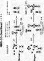
キャリアーとして磁性ビーズを利用する本発明の方法の一実施例は、図1aに示されている。図1aを参照し、本発明の方法をより詳細に説明すると、以下のようである:仮に、検体にPrPの多様な形態、即ち、PrPc及びPrPScがある場合、この検体を、捕獲抗体がコーティングされた磁性ビーズと検出抗体に同時に適用させると、HRP−検出抗体は、磁性ビーズ−捕獲抗体に結合されたPrPcには結合できず、磁性ビーズ−捕獲抗体に結合されたPrPScにのみ結合するようになる。また、利用された捕獲抗体及び検出抗体は、プリオンタンパク質において反復されない配列をエピトープとして認識するものである。また、捕獲抗体及び検出抗体は、プリオンタンパク質において、同一、オーバーラップまたは隣接したエピトープを認識する。図1aにおいて、エピトープは三角形で示されている。検出抗体により認識されるエピトープは、捕獲抗体と既に結合されているため、このようなエピトープを一つのみ有しているPrPcには、検出抗体が結合しない。しかしながら、PrPScにより形成されたマルチマー形には同一エピトープが多数存在するため、検出抗体が結合するようになる。抗原−抗体反応後、磁場を付与して、磁性ビーズを収集し、洗浄をした後、HRPの基質で発色、蛍光または発光反応を誘導する。最終的に、前記発色、蛍光または発光反応を測定して、PrPScで構成されたマルチマー−抗体複合体の存在または量を分析する。One embodiment of the method of the present invention utilizing magnetic beads as a carrier is shown in FIG. Referring to FIG. 1a, the method of the present invention will be described in more detail as follows: If there are various forms of PrP in the specimen, namely PrPc and PrPSc , the specimen is designated as a capture antibody. When applied simultaneously to a magnetic bead coated with a detection antibody and a detection antibody, the HRP-detection antibody cannot bind to PrPc bound to the magnetic bead-capture antibody, but to PrPSc bound to the magnetic bead-capture antibody. Only come to join. Moreover, the capture antibody and the detection antibody used recognize a sequence that is not repeated in the prion protein as an epitope. In addition, the capture antibody and the detection antibody recognize the same, overlapping, or adjacent epitope in the prion protein. In FIG. 1a, the epitope is indicated by a triangle. Since the epitope recognized by the detection antibody is already bound to the capture antibody, the detection antibody does not bind to PrPc having only one such epitope. However, since the multimeric form formed by PrPSc has many identical epitopes, the detection antibody becomes bound. After the antigen-antibody reaction, a magnetic field is applied, the magnetic beads are collected, washed, and then a color development, fluorescence or luminescence reaction is induced with the HRP substrate. Finally, the color development, fluorescence or luminescence reaction is measured to analyze the presence or amount of multimer-antibody complexes composed of PrPSc .
本発明のキットの好ましい具現例によると、キャリアー−捕獲抗体及び検出抗体は、モル比5:1〜1:5、より好ましくは、モル比3:1〜1:3、さらに好ましくは、2:1〜1:2、最も好ましくは、モル比約1:1のカクテル形態として含まれる。本発明のキットは、磁場プレート、緩衝溶液、発色酵素、発色基質などを含むことができる。 According to a preferred embodiment of the kit of the present invention, the carrier-capture antibody and the detection antibody have a molar ratio of 5: 1 to 1: 5, more preferably a molar ratio of 3: 1 to 1: 3, still more preferably 2: It is included as a cocktail form of 1-1: 2, most preferably about 1: 1 molar ratio. The kit of the present invention can include a magnetic field plate, a buffer solution, a chromogenic enzyme, a chromogenic substrate, and the like.
本発明のMDS−3Dシステムは、MDS−3D Single Beadシステム及びMDS−3D Dual Beadシステムを含む。 The MDS-3D system of the present invention includes an MDS-3D single bead system and an MDS-3D dual bead system.
MDS−3D Single Beadシステム(参照:図1a)は、上述のように、ビーズに結合された捕獲抗体を利用するもので、MDS−3D Dual Beadシステム(図1b及び図1c)は、捕獲抗体と検出抗体ともビーズに結合されたものを利用するものである。 As described above, the MDS-3D Single Bead system (see FIG. 1a) uses a capture antibody bound to a bead, and the MDS-3D Dual Bead system (FIG. 1b and FIG. 1c) includes a capture antibody and a capture antibody. A detection antibody that is bound to a bead is used.
MDS−3D Dual Beadシステムによると、前記検出抗体は、固相のキャリアーの表面に三次元的(即ち、立体的)に結合されている。このように立体的にキャリアーに結合された検出抗体は、三次元方式で、そしてより集中された方式(concentrated manner)でマルチ形ポリペプチドと接触するようになる。 According to the MDS-3D Dual Bead system, the detection antibody is three-dimensionally (ie, sterically) bound to the surface of a solid-phase carrier. The detection antibody thus sterically bound to the carrier comes into contact with the multi-form polypeptide in a three-dimensional manner and in a more concentrated manner.
検出抗体が結合される固相のキャリアーは、立体的構造を有するいかなる物質でも可能であって、好ましくは、重量、電荷または磁気によって容易に分離または回収できる物質である。最も好ましくは、固相のキャリアーは、ラテックスビーズである。検出抗体が結合されるキャリアーには標識物質が結合できる。このようにキャリアーに標識物質が結合された場合、例えば、蛍光物質のあるラテックスビーズを利用する場合、検出抗体に標識(例えば、HRP)を結合させなくても、キャリアーから出る安定した最終検出信号(マルチマー形の存在を意味する信号)を得ることができる。 The solid phase carrier to which the detection antibody is bound can be any substance having a three-dimensional structure, and is preferably a substance that can be easily separated or recovered by weight, charge or magnetism. Most preferably, the solid phase carrier is a latex bead. A labeling substance can be bound to the carrier to which the detection antibody is bound. In this way, when the labeling substance is bound to the carrier, for example, when using latex beads with a fluorescent substance, a stable final detection signal from the carrier can be output without binding a label (for example, HRP) to the detection antibody. (Signal indicating the presence of multimer form) can be obtained.
MDS−3D Dual Beadシステムは、シングルラベルを使用する MDS−3D Dual Beadシステム(参照:図1b)及びダブルラベルを使用する MDS−3D Dual Beadシステム(参照:図1c)をさらに含む。 The MDS-3D Dual Bead system further includes an MDS-3D Dual Bead system that uses a single label (see: FIG. 1b) and an MDS-3D Dual Bead system that uses a double label (see: FIG. 1c).
シングルラベルを使用する MDS−3D Dual Beadシステムによると、検出抗体またはキャリアーに、検出可能な信号を生成させる標識が結合されている。ダブルラベルを使用する MDS−3D Dual Beadシステムによると、キャリアーと検出抗体ともに、検出可能な信号を生成させる標識が結合されている。このような二重的なラベリングは、キャリアーから出る信号及び検出抗体から出る信号を、相互チェック可能であるという利点がある。 According to the MDS-3D Dual Bead system, which uses a single label, the detection antibody or carrier is conjugated with a label that produces a detectable signal. According to the MDS-3D Dual Bead system, which uses a double label, both the carrier and the detection antibody are bound to a label that produces a detectable signal. Such dual labeling has the advantage that the signal from the carrier and the signal from the detection antibody can be cross-checked.
図1bを参照して、シングルラベルを使用する MDS−3D Dual Beadシステムを説明すると、以下のようである。仮に、検体にPrPの多様な形態、即ち、PrPc及びPrPScがある場合、この検体を、捕獲抗体がコーティングされた磁性ビーズと検出抗体に同時に適用させると、Fluor−検出抗体(蛍光物質が結合されているラテックスビーズに結合された検出抗体)は、磁性ビーズ−捕獲抗体に結合されたPrPcには結合できず、磁性ビーズ−捕獲抗体に結合されたPrPScにのみ結合するようになる。利用された捕獲抗体及び検出抗体は、プリオンタンパク質において反復されない配列をエピトープとして認識するものである。また、捕獲抗体及び検出抗体は、プリオンタンパク質において、同一、オーバーラップまたは隣接したエピトープを認識する。図1bにおいて、エピトープは三角形で示されている。検出抗体により認識されるエピトープは、捕獲抗体と既に結合されているため、このようなエピトープを一つのみ有しているPrPcには、検出抗体が結合しない。しかし、PrPScにより形成されたマルチマー形にはエピトープが多数存在するため、検出抗体が結合するようになる。抗原−抗体反応後、磁場を付与して、磁性ビーズを収集し、洗浄をした後、蛍光を測定して、PrPScで構成されたマルチマー−抗体複合体の存在または量を分析する。Referring to FIG. 1b, the MDS-3D Dual Bead system using a single label will be described as follows. If the specimen has various forms of PrP, that is, PrPc and PrPSc , when the specimen is applied to the magnetic beads coated with the capture antibody and the detection antibody at the same time, a Fluor-detection antibody (fluorescent substance) Detection antibody bound to bound latex beads) cannot bind to PrPc bound to magnetic bead-capture antibody, but will only bind to PrPSc bound to magnetic bead-capture antibody . The capture antibody and the detection antibody used recognize a sequence that is not repeated in the prion protein as an epitope. In addition, the capture antibody and the detection antibody recognize the same, overlapping, or adjacent epitope in the prion protein. In FIG. 1b, the epitope is indicated by a triangle. Since the epitope recognized by the detection antibody is already bound to the capture antibody, the detection antibody does not bind to PrPc having only one such epitope. However, since the multimer form formed by PrPSc has many epitopes, the detection antibody becomes bound. After the antigen-antibody reaction, a magnetic field is applied, magnetic beads are collected, washed, and fluorescence is measured to analyze the presence or amount of multimer-antibody complexes composed of PrPSc .
図1cを参照して、ダブルラベルを使用するMDS−3D Dual Beadシステムを説明すると、以下のようである。仮に、検体にPrPの多様な形態、即ち、PrPc及びPrPScがある場合、この検体を、捕獲抗体がコーティングされた磁性ビーズと検出抗体に同時に適用させると、Fluor−HRP−検出抗体(蛍光物質が結合されているラテックスビーズに結合された検出抗体であって、検出抗体にはHRPが結合されている)は、磁性ビーズ−捕獲抗体に結合されたPrPcには結合できず、磁性ビーズ−捕獲抗体に結合されたPrPScにのみ結合するようになる。利用された捕獲抗体及び検出抗体は、プリオンタンパク質において反復されない配列をエピトープとして認識するものである。また、捕獲抗体及び検出抗体は、プリオンタンパク質において、同一、オーバーラップまたは隣接したエピトープを認識する。図1cにおいて、エピトープは三角形で示されている。検出抗体により認識されるエピトープは、捕獲抗体と既に結合されているため、このようなエピトープを一つのみ有しているPrPcには、検出抗体が結合しない。しかし、PrPScにより形成されたマルチマー形にはエピトープが多数存在するため、検出抗体が結合するようになる。抗原−抗体反応後、磁場を付与して、磁性ビーズを収集し、洗浄をした後、HRPによる反応産物及び/または蛍光を測定して、PrPScで構成されたマルチマー−抗体複合体の存在または量を分析する。Referring to FIG. 1c, the MDS-3D Dual Bead system using double labels will be described as follows. If the specimen has various forms of PrP, that is, PrPc and PrPSc , when the specimen is applied simultaneously to the magnetic beads coated with the capture antibody and the detection antibody, the Fluor-HRP-detection antibody (fluorescence The detection antibody bound to the latex bead to which the substance is bound, and HRP bound to the detection antibody) cannot bind to the PrPc bound to the magnetic bead-capture antibody.-It will only bind to PrPSc bound to the capture antibody. The capture antibody and the detection antibody used recognize a sequence that is not repeated in the prion protein as an epitope. In addition, the capture antibody and the detection antibody recognize the same, overlapping, or adjacent epitope in the prion protein. In FIG. 1c, the epitope is indicated by a triangle. Since the epitope recognized by the detection antibody is already bound to the capture antibody, the detection antibody does not bind to PrPc having only one such epitope. However, since the multimer form formed by PrPSc has many epitopes, the detection antibody becomes bound. After the antigen-antibody reaction, a magnetic field is applied, the magnetic beads are collected and washed, and then the reaction product and / or fluorescence by HRP is measured to determine the presence or absence of a multimer-antibody complex composed of PrPSc Analyze the amount.
以下、実施例を通じて本発明をさらに詳細に説明する。これら実施例は、本発明をより具体的に説明するためのものであって、本発明の範囲がこれらの実施例に限定されるものではない。 Hereinafter, the present invention will be described in more detail through examples. These examples are for explaining the present invention more specifically, and the scope of the present invention is not limited to these examples.
[実施例]
実施例I:MDS−3D−Single Beadシステムによる血漿内マルチマー形PrP検出
2705μlの羊血漿、22.5μlの組み換えマルチマー形羊PrP(ゲノタイプARQ、120倍希釈されたもの)及び3605μlの2.5×デタージェント(detergent)(3% Triton X−100、1.5%デオキシコール酸ナトリウム、及び0.25%サルコシル)を含む試料を製造した。また、前記試料において組み換えマルチマー形羊PrPの代わりに22.5μlのPBSを含む陰性対照群を製造した。一方、磁性ビーズに捕獲抗体を結合させるが、磁性ビーズ2.5μlに捕獲抗体1μgが結合されるように結合させた。磁性ビーズに結合させた捕獲抗体は、3E7またはMA1−750単クローン抗体である。3E7単クローン抗体は、PrPcのアミノ酸140−160(牛プリオン基準)または132−152(羊プリオン基準)をエピトープとして認識するもので、前記エピトープ配列は、PrPcにおいて反復されない。MA1−750単クローン抗体は、PrPcのSer-Arg-Pro-Leu-Ile-His-Phe-Gly-Ser-Asp-Tyr-Glu-Asp-Argエピトープを認識するもので、前記エピトープ配列は、PrPcにおいて反復されない。[Example]
Example I: Detection of multimeric PrP in plasma by MDS-3D-Single Bead system 2705 μl sheep plasma, 22.5 μl recombinant multimeric sheep PrP (genotype ARQ, 120-fold diluted) and 3605 μl 2.5 × Samples were prepared containing detergent (3% Triton X-100, 1.5% sodium deoxycholate, and 0.25% sarkosyl). In addition, a negative control group containing 22.5 μl of PBS instead of the recombinant multimer sheep PrP in the sample was prepared. On the other hand, the capture antibody was bound to the magnetic beads, but was bound so that 1 μg of the capture antibody was bound to 2.5 μl of the magnetic beads. The capture antibody bound to the magnetic beads is a 3E7 or MA1-750 monoclonal antibody. 3E7 monoclonal antibody, one that recognizes the amino acid of PrPc 140-160 (bovine prions basis) or 132-152 (sheep prions reference) as an epitope, the epitope sequence is not repeated in PrPc. MA1-750 monoclonal antibodies, one that recognizes the Ser-Arg-Pro-Leu- Ile-His-Phe-Gly-Ser-Asp-Tyr-Glu-Asp-Arg epitope of PrPc, the epitope sequence, Not repeated in PrPc .
次いで、前記試料に磁性ビーズ−捕獲抗体複合体及び検出抗体を同時に血漿試料と反応して、マルチマー形羊PrPを検出できるかどうかを調べた。検出抗体として利用したものは、3B8/D5−HRPまたはT2−HRPである。T2単クローン抗体は、Hiroko Hayashi, et al., J. Vet. Med. Sci., 66(6):515(2004)に開示されており、PrPcのアミノ酸配列147−152(牛プリオン基準)、140−145(羊プリオン基準)エピトープに特異的に反応する抗体である。3B8/D5単クローン抗体のエピトープは、PrPcのアミノ酸132−152(羊プリオン基準)、140−160(牛プリオン基準)である。Next, the magnetic bead-capture antibody complex and the detection antibody were simultaneously reacted with the plasma sample to determine whether multimer sheep PrP could be detected. The one used as the detection antibody is 3B8 / D5-HRP or T2-HRP. The T2 monoclonal antibody is disclosed in Hiroko Hayashi, et al., J. Vet. Med. Sci., 66 (6): 515 (2004), and the amino acid sequence of PrPc 147-152 (bovine prion reference). , 140-145 (sheep prion reference) epitope. The epitope of the 3B8 / D5 monoclonal antibody is amino acids 132-152 of PrPc (sheep prion standard) and 140-160 (bovine prion standard).
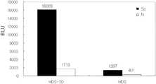
前記試料に磁性ビーズ−捕獲抗体複合体及び検出抗体を、表1に記載の量で同時に添加し、37℃で1時間反応した。次いで、磁場を反応混合物に付与して磁性ビーズを分離し、磁性ビーズをTBSTで3回洗浄した後、ECL(enhanced chemiluminescence)検出を行った。 Magnetic beads-capture antibody complex and detection antibody were simultaneously added to the sample in the amounts shown in Table 1, and reacted at 37 ° C. for 1 hour. Next, a magnetic field was applied to the reaction mixture to separate the magnetic beads. The magnetic beads were washed three times with TBST, and then ECL (enhanced chemiluminescence) detection was performed.

一方、比較実施例として、磁性ビーズ−捕獲抗体複合体を試料と優先的に反応した後、検出抗体を反応させるプロトコールで行った。 On the other hand, as a comparative example, the magnetic bead-capture antibody complex was preferentially reacted with the sample, and then the detection antibody was reacted.
試料を上述と同様に用意した。次いで、磁性ビーズ−捕獲抗体複合体を試料に添加して、37℃で1時間反応した。次いで、磁場を反応混合物に付与して磁性ビーズを分離し、磁性ビーズをTBSTで3回洗浄した。その後、分離された磁性ビーズに検出抗体を処理して、37℃で1時間反応した。その後、磁場を反応混合物に付与して磁性ビーズを分離し、磁性ビーズをTBSTで3回洗浄した。最終的にECL(enhanced chemiluminescence)検出を行った。 Samples were prepared as described above. Next, the magnetic bead-capture antibody complex was added to the sample and reacted at 37 ° C. for 1 hour. A magnetic field was then applied to the reaction mixture to separate the magnetic beads and the magnetic beads were washed 3 times with TBST. Thereafter, the separated magnetic beads were treated with a detection antibody and reacted at 37 ° C. for 1 hour. Thereafter, a magnetic field was applied to the reaction mixture to separate the magnetic beads, and the magnetic beads were washed three times with TBST. Finally, ECL (enhanced chemiluminescence) detection was performed.
図2から分かるように、本発明により磁性ビーズ−捕獲抗体(3E7抗体)及び検出抗体(T2−HRP)を試料に同時に処理した場合は、分離処理した場合に比べ、検出信号が非常に高く得られ、プリオン検出敏感度が大きく増加した。また、本発明により磁性ビーズ−捕獲抗体及び検出抗体を試料に同時に処理した場合は、分離処理(step by step)した場合に比べ、正常形プリオンの信号強度に対するマルチマー形プリオンの信号強度の比率が著しく増加して、マルチマー形プリオンに対する分別力が非常に大きく向上されることが分かる。 As can be seen from FIG. 2, when a magnetic bead-capture antibody (3E7 antibody) and a detection antibody (T2-HRP) are simultaneously processed according to the present invention, the detection signal is much higher than when the sample is separated. The prion detection sensitivity has been greatly increased. In addition, when the magnetic bead-capture antibody and the detection antibody are simultaneously processed on the sample according to the present invention, the ratio of the signal intensity of the multimer prion to the signal intensity of the normal prion is higher than that in the case of separation (step by step) It can be seen that with a significant increase, the fractionation power for multimer prions is greatly improved.
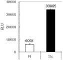
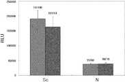
実施例II: MDS−3D−Single Beadシステムに適合した緩衝液の分析
MDS−3D Single Beadシステムに適合した緩衝液を選別するために、磁性ビーズに結合された捕獲抗体3E7、3E7−bead及び検出抗体T2−HRPを1:1比率(0.8μg:0.8μg)で利用し、Single Beadシステムを実施した。試料は、次のように用意した:10% Triton X−100 in d−H2O 120μl、緩衝液(TAPS、TBSTまたはTricine、pH8.0)580μl、及び羊の血漿300μlを混合して、1.2%Triton X−100及び30%血漿を含む総容量1mlの試料を準備した。捕獲抗体としての3E7−結合磁性ビーズ2μl(0.8μg)及び検出抗体としてのT2−HRP(4μg/ml in TBST)200μl(0.8μg)を混合して、混合抗体を準備した。試料1mlに前記混合抗体200μlを添加して混合し、37℃で1時間反応した。次いで、磁場を反応混合物に付与して磁性ビーズを分離し、磁性ビーズをTBSTで3回洗浄した後、ECL検出を行った(図3a及び3b)。図3a及び3bにおいて、‘N’は、正常血漿、‘Sc’は、PrPScを有する血漿を示す。図3a及び図3bから分かるように、Tricine緩衝液が、PrPSc血漿に対して最も高い信号を示しながらも、正常血漿に対しては最も低い信号を示し、結晶試料からPrPScを検出するにおいて、最も優れた分別力を有することが分かる。Example II: Analysis of Buffers Compatible with MDS-3D-Single Bead System To select buffers compatible with the MDS-3D Single Bead system, capture antibodies 3E7, 3E7-bead bound to magnetic beads and detection The Single Bead system was implemented using antibody T2-HRP in a 1: 1 ratio (0.8 μg: 0.8 μg). Samples were prepared as follows: 120 μl of 10% Triton X-100 in d-H2 O, 580 μl of buffer (TAPS, TBST or Tricine, pH 8.0), and 300 μl of sheep plasma, A sample with a total volume of 1 ml containing 2% Triton X-100 and 30% plasma was prepared. A mixed antibody was prepared by mixing 2 μl (0.8 μg) of 3E7-bound magnetic beads as a capture antibody and 200 μl (0.8 μg) of T2-HRP (4 μg / ml in TBST) as a detection antibody. 200 μl of the mixed antibody was added to 1 ml of the sample, mixed, and reacted at 37 ° C. for 1 hour. Next, a magnetic field was applied to the reaction mixture to separate the magnetic beads, and the magnetic beads were washed three times with TBST, and then ECL detection was performed (FIGS. 3a and 3b). 3a and 3b, 'N' indicates normal plasma and 'Sc' indicates plasma with PrPSc . As can be seen from FIGS. 3a and 3b, Tricine buffer shows the highest signal for PrPSc plasma, but the lowest signal for normal plasma, in detecting PrPSc from a crystal sample. It can be seen that it has the best sorting power.
実施例III:MDS−3D−Dual Beadシステムによる血漿内マルチマー形PrPの検出
10%Triton X−100 120μl、TBST緩衝液(pH8.0)730μl及び羊血漿150μlを混合して、1.2%Triton X及び15%羊血漿を含む試料溶液1mlを製造した。Example III: Detection of multimeric PrP in plasma by MDS-3D-Dual Bead system 120 μl of 10% Triton X-100, 730 μl of TBST buffer (pH 8.0) and 150 μl of sheep plasma were mixed to obtain 1.2
捕獲抗体としての3E7−結合磁性ビーズ4μl、検出抗体としてのMA1 750−結合蛍光ラテックスビーズ2μl及びTBST緩衝液200μlを混合して、混合抗体を準備した(3E7−ビーズ/MA1 750−蛍光ビーズ、2:1比率)。試料1mlに前記混合抗体200μlを添加して混合し、37℃で1時間反応した。次いで、磁場を反応混合物に付与して磁性ビーズを分離し、磁性ビーズをTBSTで3回洗浄した後、蛍光を測定した。図4から分かるように、本発明のMDS−3D−Dual Beadシステムは、血漿内のPrPScを分別的に検出していることが分かる。4 μl of 3E7-conjugated magnetic beads as a capture antibody, 2 μl of MA1 750-conjugated fluorescent latex beads as detection antibodies and 200 μl of TBST buffer were mixed to prepare a mixed antibody (3E7-beads / MA1 750-fluorescent beads, 2 : 1 ratio). 200 μl of the mixed antibody was added to 1 ml of the sample, mixed, and reacted at 37 ° C. for 1 hour. Next, a magnetic field was applied to the reaction mixture to separate the magnetic beads, and the magnetic beads were washed three times with TBST, and then fluorescence was measured. As can be seen from FIG. 4, it can be seen that the MDS-3D-Dual Bead system of the present invention detects PrPSc in the plasma in a differential manner.
実施例IV: MDS−3D−Dual Beadシステムに適合した緩衝液の分析
捕獲抗体としての3E7−結合磁性ビーズ4μl及び検出抗体としてのMA1 750−結合蛍光ラテックスビーズ1μlを混合して、混合抗体を準備した(3E7−ビーズ/MA1 750−蛍光ビーズ、4:1比率)。10% Triton X−100 in d−H2O 120μl、緩衝液(TBSTまたはTricine、pH8.0)730μl及び羊の血漿150μlを混合して、試料溶液を準備した。その他の過程は、前記実施例3と同様である。図5から確認できるように、本発明のMDS−3D−Dual Beadシステムを利用して血漿試料からPrPScを検出するにおいて、TBSTがTricine緩衝液より優れた信号強度及び分別力を示す。Example IV: Analysis of buffer adapted to MDS-3D-
一方、T2及びMA1抗体をそれぞれ捕獲抗体及び検出抗体として利用する他のMDS−3D−Dual Beadシステムを構築し、上記の過程を行った。図5から分かるように、この抗体組み合わせにおいても、MDS−3D−Dual Beadシステムは、血漿試料からPrPScを検出した。On the other hand, another MDS-3D-Dual Bead system using T2 and MA1 antibodies as a capture antibody and a detection antibody was constructed, and the above process was performed. As can be seen from FIG. 5, even in this antibody combination, the MDS-3D-Dual Bead system detected PrPSc from the plasma sample.
実施例V:MDS−3D−Dual Beadシステムにおける適合した血漿試料濃度の分析
捕獲抗体としての3E7−結合磁性ビーズ2μl及び検出抗体としてのMA1 750−結合蛍光ラテックスビーズ1μl(3E7−ビーズ/MA1 750−蛍光ビーズ、2:1比率)の混合抗体を利用し、血漿試料をそれぞれ30%、15%及び7.5%として、MDS−3D−Dual Beadシステムの適合した血漿試料濃度を分析した。緩衝液としては、Tricine(pH8.0)を利用して、その他の実験方法は、前記実施例3と同様である。Example V: Analysis of ConsistentPlasma Sample Concentration in MDS-3D-
図6から分かるように、血漿試料30%、15%及び7.5%に存在するそれぞれのPrPScは、本発明のMDS−3D−Dual Beadシステムにより分別的に検出された。Sc/N比率は、血漿濃度が減少するほど減少したが、信号の強度は、低い血漿濃度で最も高かった。したがって、本発明のMDS−3D−Dual Beadシステムに適合した血漿濃度は、約15%であることが分かる。As can be seen from FIG. 6, each PrPSc present in 30%, 15% and 7.5% of the plasma samples was differentially detected by the MDS-3D-Dual Bead system of the present invention. The Sc / N ratio decreased with decreasing plasma concentration, but the signal intensity was highest at lower plasma concentrations. Therefore, it can be seen that the plasma concentration adapted to the MDS-3D-Dual Bead system of the present invention is about 15%.
実施例VI:MDS−3D−Single Beadシステムにおける適合した血漿試料濃度の分析
MDS−3D−Single Beadシステムに適合した血漿試料濃度を分析するために、磁性ビーズに結合された捕獲抗体3E7、3E7−bead及び検出抗体T2−HRPを1:1比率(0.8μg:0.8μg)で利用し、MDS−3D−Single Beadシステムを実施した。血漿試料は、それぞれ30%、15%及び7.5%とした。緩衝液としては、Tricine(pH8.0)を利用して、その他の実験方法は、前記実施例2と同様である。Example VI: Analysis of adapted plasma sample concentration in the MDS-3D-Single Bead system To analyze the plasma sample concentration adapted to the MDS-3D-Single Bead system, capture antibodies 3E7, 3E7-conjugated to magnetic beads are used. The MDS-3D-Single Bead system was implemented using bead and detection antibody T2-HRP at a 1: 1 ratio (0.8 μg: 0.8 μg). Plasma samples were 30%, 15% and 7.5%, respectively. As a buffer solution, Tricine (pH 8.0) is used, and other experimental methods are the same as in Example 2.
図7から分かるように、血漿試料30%、15%及び7.5%に存在するそれぞれのPrPScは、本発明のMDS−3D−Single Beadシステムにより分別的に検出された。Sc/N比率と正常血漿及びPrPSc血漿に対する信号の強度は、血漿濃度が減少するほど減少した。したがって、本発明のMDS−3D−Single Beadシステムに適合した血漿濃度は、約30%であることが分かる。As can be seen from FIG. 7, each PrPSc present in 30%, 15% and 7.5% of the plasma samples was differentially detected by the MDS-3D-Single Bead system of the present invention. The Sc / N ratio and the intensity of the signal for normal plasma and PrPSc plasma decreased with decreasing plasma concentration. Therefore, it can be seen that the plasma concentration adapted to the MDS-3D-Single Bead system of the present invention is about 30%.
また、3E7−ビーズ抗体及びT2−HRP抗体を1:2の比率として、30%及び100%濃度の血漿とTricine(pH0.8)緩衝液を利用して、その他の実験方法は、前記実施例2と同様にして行った。図11から分かるように、100%濃度の血漿は、30%濃度の血漿に比べ、さらに低い信号強度と分別力を示した。 In addition, other experimental methods were carried out in the same manner as in the Examples described above, using 3E7-bead antibody and T2-HRP antibody at a ratio of 1: 2, using 30% and 100% plasma and Tricine (pH 0.8) buffer. Performed in the same manner as 2. As can be seen from FIG. 11, 100% plasma showed lower signal strength and fractionation power than 30% plasma.
また、3E7−ビーズ抗体及びT2−HRP抗体を1:1の比率として、20%及び25%濃度の血漿とTricine(pH0.8)緩衝液を利用して、20%及び25%濃度の血漿の量は、それぞれ300μl及び400μlである。その他の実験方法は、前記実施例2と同様にして行った。図12から分かるように、25%濃度の血漿は、20%濃度の血漿に比べ、さらに高い信号強度と分別力を示した。 In addition, 3E7-bead antibody and T2-HRP antibody were used at a ratio of 1: 1, and 20% and 25% concentrations of plasma and Tricine (pH 0.8) buffer were used. The amounts are 300 μl and 400 μl, respectively. Other experimental methods were performed in the same manner as in Example 2. As can be seen from FIG. 12, the plasma at 25% concentration showed higher signal intensity and discriminating power than the plasma at 20% concentration.
実施例VII:MDS−3D−Dual Beadシステムによる血漿内マルチマー形PrPの検出
捕獲抗体としてT2−結合磁性ビーズ、検出抗体としてMA1 750−結合蛍光ラテックスビーズを利用し、二つの抗体の比率を2:1または4:1として本発明のMDS−3D−Dual Beadシステムを実施した。緩衝液としてはTBST(pH8.0)を利用して、血漿の濃度は、15%であった。その他の過程は、前記実施例3と同様である。図8から分かるように、T2−ビーズ/MA1 750−ビーズの比率が4:1の場合が、2:1の場合に比べ、Sc/N比率が大きくて、信号の強度も高かった。Example VII: Detection of Multimeric PrP in Plasma by MDS-3D-Dual Bead System Using T2-conjugated magnetic beads as capture antibodies and MA1 750-conjugated fluorescent latex beads as detection antibodies, the ratio of the two antibodies was 2: The MDS-3D-Dual Bead system of the present invention was implemented as 1 or 4: 1. TBST (pH 8.0) was used as a buffer, and the plasma concentration was 15%. Other processes are the same as those in the third embodiment. As can be seen from FIG. 8, when the ratio of T2-beads / MA1 750-beads was 4: 1, the Sc / N ratio was larger and the signal intensity was higher than when the ratio was 2: 1.
実施例VIII:多様なデタージェント(detergent)の濃度及びタイプにおいて、MDS−3D−Single Beadシステムによる血漿内マルチマー形PrPの検出
デタージェントの濃度とタイプを多様にしながら、実施例2と同様な方法(デタージェントの条件を除いた)により、PrPScが存在するとの臨床的所見を示す羊血漿試料に対してMDS−3D−Single Beadシステムによりマルチマー形PrPを検出した。図9から分かるように、多様なトリトンX−100の濃度(1%、1.5%及び2%)は、マルチマーPrPに対してほぼ等しい検出度及び分別力を示した。また、結果の偏差が相対的に高いが、Np−40(USB社、米国)も相当な検出及び分別力を示した。Np−40は、非イオン性デタージェントの一つで、[オクチルフェノキシ]ポリエトキシエタノール([Octylphenoxy]polyethoxyethanol)を示す。Example VIII: The same method as in Example 2 while varying the concentration and typeof plasma multimeric PrP detected by MDS-3D-Single Bead system at various detergent concentrations and types Multimeric PrP was detected by MDS-3D-Single Bead system on sheep plasma samples showing clinical findings that PrPSc was present (excluding detergent conditions). As can be seen from FIG. 9, various concentrations of Triton X-100 (1%, 1.5% and 2%) showed almost equal detectability and fractionation power for multimeric PrP. Moreover, although the deviation of the results is relatively high, Np-40 (USB, USA) also showed considerable detection and discrimination power. Np-40 is one of non-ionic detergents and represents [Octylphenoxy] polyethoxyethanol.
実施例IX:多様な洗浄緩衝液において、MDS−3D−Single Beadシステムによる血漿内マルチマー形PrPの検出
羊血漿試料に存在するマルチマー形PrPScを検出するために、抗体重量比(3E7−ビーズ:T2−HRP、1:2)と洗浄条件を除いては、実施例2と同様な方法により、洗浄緩衝液のタイプを多様にしてMDS−3D−Single Beadシステムを実施した。図10から分かるように、マルチマー形PrPは、PBST(phosphate-buffered saline with Tween 20)、PBSX(phosphate-buffered saline with Triton X-100)及びTBSX(tris-buffered saline with Triton X-100)と比較し、TBST(tris-buffered saline with Tween 20)洗浄緩衝液において最も優れた検出度及び分別力を示した。Example IX: Detection of multimeric PrP in plasma by MDS-3D-Single Bead system in various wash buffers To detect multimeric PrPSc present in sheep plasma samples, antibody weight ratio (3E7-beads: Except for T2-HRP, 1: 2) and washing conditions, the MDS-3D-Single Bead system was implemented in the same manner as in Example 2 with various washing buffer types. As can be seen from FIG. 10, multimeric PrP is compared with PBST (phosphate-buffered saline with Tween 20), PBSX (phosphate-buffered saline with Triton X-100) and TBSX (tris-buffered saline with Triton X-100). In the TBST (tris-buffered saline with Tween 20) washing buffer, the best detection and fractionation power were shown.
実施例X:MDS−3D−Single Beadシステムによる血漿内マルチマー形プリオンタンパク質の検出において、PK切断の影響
羊血漿試料に存在するマルチマー形PrPScを検出するために、抗体比率(3E7−ビーズ:T2−HRP、1:2)除いては、実施例2と同様な方法により、プロテアーゼK(PK)の切断有無と共にMDS−3D−Single Beadシステムを実施した。図13から分かるように、PK分解は、血漿試料においてPrPScに対する信号の強度を減少させる。これにより、MDS−3D−Single Beadシステムを利用して血液または血清試料からPrPScを検出するにおいて、プロテアーゼKの切断過程は不要であることが分かったExample X: Effect of PK cleavage in detection of multimeric prion protein in plasma by MDS-3D-Single Bead system To detect multimeric PrPSc present in sheep plasma samples, antibody ratio (3E7-beads: T2 Except for -HRP, 1: 2), the MDS-3D-Single Bead system was carried out in the same manner as in Example 2 together with the presence or absence of protease K (PK) cleavage. As can be seen from FIG. 13, PK degradation reduces the strength of the signal for PrPSc in plasma samples. As a result, it was found that the cleavage process of protease K is unnecessary in detecting PrPSc from a blood or serum sample using the MDS-3D-Single Bead system.
実施例XI:多様な比率の抗体において、MDS−3D−Single Beadシステムによる血漿内マルチマー形PrPの検出
羊血漿試料に存在するマルチマー形PrPScを検出するために、実施例2と同様な方法により、抗体比率を多様にしてMDS−3D−Single Beadシステムを実施した。3E7−ビーズ捕獲抗体とT2−HRP検出抗体の質量比率を4:1、2:1、1.33:1及び1:1として添加した。図14aから分かるように、二つの抗体を同じ量で添加した場合、MDS−3D−Single Beadシステムによる血漿内PrPSc検出において、最も優れた検出度及び分別力を示した。Example XI: Detection of multimeric PrPSc inplasma by MDS-3D-Single Bead system in various ratios of antibodies In order to detect multimeric PrPSc present in sheep plasma samples, the same method as in Example 2 was used. The MDS-3D-Single Bead system was implemented with various antibody ratios. The mass ratio of 3E7-bead capture antibody to T2-HRP detection antibody was added as 4: 1, 2: 1, 1.33: 1 and 1: 1. As can be seen from FIG. 14a, when the two antibodies were added in the same amount, the highest detection rate and fractionation power were shown in the detection of PrPSc in plasma by the MDS-3D-Single Bead system.
実施例XII:多様な比率の抗体において、MDS−3D−Dual Beadシステムによる血漿内PrPの検出
羊血漿試料に存在するマルチマー形PrPScを検出するために、実施例3と同様な方法により、抗体比率を多様にしてMDS−3D−Dual Beadシステムを実施した。3E7−ビーズ捕獲抗体とMAI−750蛍光ビーズ検出抗体を4:1と2:1の質量比率で添加した。図14bから分かるように、捕獲抗体及び検出抗体の量を2:1の比率で添加した場合、MDS−3D−Dual Beadシステムによる血漿内PrPSc検出において、最も優れた検出度及び分別力を示した。Example XII: Detection of plasma PrP by MDS-3D-Dual Bead system in various ratios of antibodies In order to detect multimeric PrPSc present in sheep plasma samples, The MDS-3D-Dual Bead system was implemented with various ratios. 3E7-bead capture antibody and MAI-750 fluorescent bead detection antibody were added at a mass ratio of 4: 1 and 2: 1. As can be seen from FIG. 14b, when the amounts of the capture antibody and the detection antibody were added at a ratio of 2: 1, the highest detection and fractionation power were shown in the detection of PrPSc in plasma by the MDS-3D-Dual Bead system. It was.
実施例XIII:捕獲抗体カクテルと、MDS−3D−Single Beadシステムによる血漿内PrPの検出
羊血漿試料に存在するマルチマー形PrPScを検出するために、実施例2と同様な方法により、捕獲抗体カクテルと共にMDS−3D−Single Beadシステムを実施した。捕獲抗体として、3E7−ビーズとT2−ビーズ抗体(図15a)または3E7−ビーズとMAI−ビーズ(図15b)から構成された捕獲抗体カクテルが使用された。捕獲抗体カクテルと検出抗体、即ちT2−HRPの比率は、1:1とした。図15aと15bから分かるように、捕獲抗体カクテルは、血漿内PrPScに対して、優れた検出度及び分別力を示した。Example XIII: Capture Antibody Cocktail and Detection of PrP in Plasma by MDS-3D-Single Bead System In order to detect multimeric PrPSc present in sheep plasma samples, a capture antibody cocktail was prepared in the same manner as in Example 2. And implemented the MDS-3D-Single Bead system. As capture antibody, a capture antibody cocktail composed of 3E7-bead and T2-bead antibody (FIG. 15a) or 3E7-bead and MAI-bead (FIG. 15b) was used. The ratio of capture antibody cocktail to detection antibody, ie T2-HRP, was 1: 1. As can be seen from Figure 15a and 15b, the capture antibody cocktail against plasmatic PrPSc, it showed excellent degree of detection and fractionation force.
実施例XIV:検出抗体カクテルと、MDS−3D−Single Beadシステムによる血漿内PrPの検出
羊血漿試料に存在するマルチマー形PrPScを検出するために、実施例2と同様な方法により、検出抗体カクテルを添加してMDS−3D−Single Beadシステムを実施した。添加した抗体セットは、(i)捕獲抗体として3E7−bead及び検出抗体カクテルとしてT2−バイオチンとMAI−バイオチン、及び(ii)捕獲抗体としてMAI−bead及び検出抗体カクテルとしてT2−バイオチンと3E7−バイオチンである。図15cから分かるように、検出抗体カクテルは、血漿内PrPScに対して、優れた検出度及び分別力を示したExample XIV: Detection Antibody Cocktail and Detection of PrP in Plasma by MDS-3D-Single Bead System In order to detect multimeric PrPSc present in sheep plasma samples, detection antibody cocktail was prepared in the same manner as in Example 2. Was added to implement the MDS-3D-Single Bead system. The added antibody sets were (i) 3E7-bead as capture antibody and T2-biotin and MAI-biotin as detection antibody cocktail, and (ii) MAI-bead as capture antibody and T2-biotin and 3E7-biotin as detection antibody cocktail. It is. As can be seen from Figure 15c, the detection antibody cocktail against plasmatic PrPSc, showed excellent degree of detection and fractionation force
実施例XV:同一な条件下でMDS及びMDS−3D−Single Beadシステムの検出力の比較
本発明者らは、マルチマーを形成するポリペプチドのモノマー形からマルチマー形を分別的に検出する方法を既に開発し、これをマルチマー検出システム(Multimer Detection System:MDS)と命名して、PCT出願した(PCT/KR2005/004001)。Example XV: Comparison of power of MDS and MDS-3D-Single Bead system under identical conditions We have already developed a method for differentially detecting multimeric forms from monomeric forms of polypeptides that form multimers. This was developed and named as Multimer Detection System (MDS), and a PCT application was filed (PCT / KR2005 / 004001).
信頼できる実験方法でMDSとMDS−3D−Single BeadシステムのPrPSc検出度及び分別力を比較するために、羊血漿内のマルチマー形PrPScは、同一な実験条件下でそれぞれ検出された。3E7及びT7抗体が、捕獲抗体及び検出抗体としてそれぞれ利用された。In order to compare the PrPSc detectability and fractionation power of MDS and MDS-3D-Single Bead system in a reliable experimental manner, multimeric PrPSc in sheep plasma was respectively detected under the same experimental conditions. 3E7 and T7 antibodies were utilized as capture and detection antibodies, respectively.
図16から分かるように、本発明のMDS−3D−Single Beadシステムが、MDSに比べ、羊血漿内のPrPScに対してさらに高い敏感度及び分別力を示した。As can be seen from FIG. 16, the MDS-3D-Single Bead system of the present invention showed higher sensitivity and discriminating power against PrPSc in sheep plasma than MDS.
実施例XVI:他の抗体セットの利用するMDS−3D−Single Beadシステムに対する最適血漿濃度の決定
羊血漿試料に存在するマルチマー形PrPScを検出するために、抗体セットタイプと血清濃度を除いては、実施例2と同様な方法によりMDS−3D−Single Beadシステムを実施した。捕獲抗体としては、ICSM35−バイオチンストレプトアビジンビーズ、検出抗体としては、1E4−HRPを利用して、羊血漿の濃度は25%にした。ICSM35抗体(D−Gen、英国)は、アミノ酸配列96−105(羊プリオン基準)またはアミノ酸配列104−113(牛プリオン基準)に対応するエピトープを認識して、1E4抗体(Sanquin Reagents、オランダ)は、アミノ酸配列100―111(羊プリオン基準)またはアミノ酸配列108−119(牛プリオン基準)に対応するエピトープを認識する。Example XVI: Determination of Optimal Plasma Concentration for MDS-3D-Single Bead System Utilizing Other Antibody Sets To detect multimeric PrPSc present in sheep plasma samples, except for antibody set type and serum concentration The MDS-3D-Single Bead system was implemented by the same method as in Example 2. ICSM35-biotin streptavidin beads were used as a capture antibody, and 1E4-HRP was used as a detection antibody, so that the concentration of sheep plasma was 25%. The ICSM35 antibody (D-Gen, UK) recognizes an epitope corresponding to amino acid sequence 96-105 (sheep prion reference) or amino acid sequence 104-113 (bovine prion reference), and 1E4 antibody (Sanquin Reagents, Netherlands) Recognizes an epitope corresponding to amino acid sequence 100-111 (sheep prion reference) or amino acid sequence 108-119 (bovine prion reference).
図17から分かるように、部分的にオーバーラップするエピトープを認識する抗体セットは、血漿内PrPScに対して非常に優れた検出度及び分別力を示した。As it can be seen from Figure 17, set of antibodies that recognize partially overlapping epitopes exhibited degree of detection and fractionation force very superior to the plasma within the PrPSc.
以上、本発明の望ましい具現例を詳細に記述したが、当業界の通常の知識を有する者にとって、このような具体的な記述はただ望ましい具現例に過ぎなく、これに本発明の範囲が限定されないことは明らかである。従って、本発明の実質的な範囲は、添付の請求項とその等価物により定義されると言える。 As described above, the preferred embodiments of the present invention have been described in detail. However, for those having ordinary skill in the art, such specific descriptions are merely preferred embodiments, and the scope of the present invention is limited thereto. Obviously not. Therefore, the substantial scope of the present invention will be defined by the appended claims and equivalents thereof.
Claims (22)
Translated fromJapanese(b)検出抗体を用意するステップであって、前記検出抗体が認識するエピトープは、検出抗体がエピトープに結合した場合、前記捕獲抗体のエピトープに結合された捕獲抗体により立体的妨害を起す位置にあり、前記捕獲抗体のエピトープと検出抗体のエピトープのアミノ酸配列は、同一ではなくオーバーラップされるアミノ酸配列を有して、前記捕獲抗体が認識するポリペプチドのエピトープは、ポリペプチドにおいて反復しない配列であり、前記検出抗体が認識するポリペプチドのエピトープは、ポリペプチドにおいて反復しない配列であることを特徴とするステップと、
(c)生試料に前記キャリアー−捕獲抗体複合体及び検出抗体を同時に反応させるステップと、
(d)キャリアー−捕獲抗体−マルチマー形検出抗体複合体を検出するステップと、
を含む、生試料内のマルチマー形成ポリペプチドのモノマー形からマルチマー形を分別検出する方法。(a) a step of three-dimensionally binding a capture antibody to the surface of a solid phase carrier having a three-dimensional structure to prepare a carrier-capture antibody complex, wherein the capture antibody comprises a multimer-forming polypeptide; Binding to one specific epitope;
(b) preparing a detection antibody, wherein the epitope recognized by the detection antibody is located at a position where steric hindrance is caused by the capture antibody bound to the epitope of the capture antibody when the detection antibody binds to the epitope. And the epitope of the capture antibody and the epitope of the detection antibody have an amino acid sequence that is not identical but overlaps, and the epitope of the polypeptide that the capture antibody recognizes is a sequence that does not repeat in the polypeptide. A polypeptide epitope recognized by the detection antibody is a non-repeating sequence in the polypeptide;
(c) simultaneously reacting the carrier-capture antibody complex and the detection antibody with a raw sample;
(d) detecting a carrier-capture antibody-multimeric detection antibody complex;
A method for differentially detecting a multimeric form from a monomeric form of a multimer-forming polypeptide in a raw sample.
(b)前記捕獲抗体のエピトープに結合された捕獲抗体により立体的妨害を起す位置にあるエピトープを認識する検出抗体とを含み、
前記捕獲抗体のエピトープと検出抗体のエピトープのアミノ酸配列は、同一ではなくオーバーラップされるアミノ酸配列を有し、
前記捕獲抗体が認識するポリペプチドのエピトープは、ポリペプチドにおいて反復しない配列であり、
前記検出抗体が認識するポリペプチドのエピトープは、ポリペプチドにおいて反復しない配列であることを特徴とする、生試料内のマルチマー形成ポリペプチドのモノマー形からマルチマー形を分別検出するためのキット。(a) a capture antibody that is three-dimensionally bound to the surface of a solid-phase carrier having a three-dimensional structure and binds to one specific epitope of the multimer-forming polypeptide;
(b) a detection antibody that recognizes an epitope at a position that causes steric hindrance by the capture antibody bound to the epitope of the capture antibody;
The amino acid sequence of the epitope of the capture antibody and the epitope of the detection antibody has an amino acid sequence that is not identical but overlaps,
The polypeptide epitope recognized by the capture antibody is a non-repeating sequence in the polypeptide,
The epitope of the detection antibody recognizes the polypeptide ischaracterized by a sequence which does not repeat in the polypeptide, the kit for fractionating detecting multimeric forms from monomeric form of a multimer-forming polypeptide in a living sample.
Applications Claiming Priority (5)
| Application Number | Priority Date | Filing Date | Title |
|---|---|---|---|
| KR20060036434 | 2006-04-21 | ||
| KR10-2006-0036434 | 2006-04-21 | ||
| KR10-2006-0056429 | 2006-06-22 | ||
| KR20060056429 | 2006-06-22 | ||
| PCT/KR2007/001947WO2007123345A1 (en) | 2006-04-21 | 2007-04-20 | Method for differentially detecting multimeric form from monomeric form of multimer-forming polypeptides through three-dimensional interactions |
Publications (2)
| Publication Number | Publication Date |
|---|---|
| JP2009534648A JP2009534648A (en) | 2009-09-24 |
| JP5164971B2true JP5164971B2 (en) | 2013-03-21 |
Family
ID=38625210
Family Applications (1)
| Application Number | Title | Priority Date | Filing Date |
|---|---|---|---|
| JP2009506420AActiveJP5164971B2 (en) | 2006-04-21 | 2007-04-20 | A method for differential detection of multimers from monomers of multimer-forming polypeptides using three-dimensional interactions |
Country Status (7)
| Country | Link |
|---|---|
| US (1) | US8008026B2 (en) |
| EP (1) | EP2010904B1 (en) |
| JP (1) | JP5164971B2 (en) |
| KR (2) | KR101188861B1 (en) |
| AU (1) | AU2007241729A1 (en) |
| CA (1) | CA2649359A1 (en) |
| WO (1) | WO2007123345A1 (en) |
Cited By (1)
| Publication number | Priority date | Publication date | Assignee | Title |
|---|---|---|---|---|
| WO2017138497A1 (en)* | 2016-02-08 | 2017-08-17 | シスメックス株式会社 | Analyte detection method and reagent kit for detecting analyte |
Families Citing this family (36)
| Publication number | Priority date | Publication date | Assignee | Title |
|---|---|---|---|---|
| DE10303974A1 (en) | 2003-01-31 | 2004-08-05 | Abbott Gmbh & Co. Kg | Amyloid β (1-42) oligomers, process for their preparation and their use |
| US8497072B2 (en) | 2005-11-30 | 2013-07-30 | Abbott Laboratories | Amyloid-beta globulomer antibodies |
| EP1954718B1 (en) | 2005-11-30 | 2014-09-03 | AbbVie Inc. | Anti-a globulomer antibodies, antigen-binding moieties thereof, corresponding hybridomas, nucleic acids, vectors, host cells, methods of producing said antibodies, compositions comprising said antibodies, uses of said antibodies and methods of using said antibodies |
| US8455626B2 (en) | 2006-11-30 | 2013-06-04 | Abbott Laboratories | Aβ conformer selective anti-aβ globulomer monoclonal antibodies |
| US8895004B2 (en) | 2007-02-27 | 2014-11-25 | AbbVie Deutschland GmbH & Co. KG | Method for the treatment of amyloidoses |
| US8048420B2 (en) | 2007-06-12 | 2011-11-01 | Ac Immune S.A. | Monoclonal antibody |
| US8613923B2 (en) | 2007-06-12 | 2013-12-24 | Ac Immune S.A. | Monoclonal antibody |
| SI2238166T1 (en) | 2007-10-05 | 2014-03-31 | Genentech, Inc. | Use of anti-amyloid beta antibody in ocular diseases |
| CA2781519A1 (en) | 2009-09-16 | 2011-03-24 | Genentech, Inc. | Coiled coil and/or tether containing protein complexes and uses thereof |
| AR080793A1 (en) | 2010-03-26 | 2012-05-09 | Roche Glycart Ag | BISPECIFIC ANTIBODIES |
| US8987419B2 (en) | 2010-04-15 | 2015-03-24 | AbbVie Deutschland GmbH & Co. KG | Amyloid-beta binding proteins |
| US20110262989A1 (en) | 2010-04-21 | 2011-10-27 | Nanomr, Inc. | Isolating a target analyte from a body fluid |
| US9476812B2 (en) | 2010-04-21 | 2016-10-25 | Dna Electronics, Inc. | Methods for isolating a target analyte from a heterogeneous sample |
| US8841104B2 (en) | 2010-04-21 | 2014-09-23 | Nanomr, Inc. | Methods for isolating a target analyte from a heterogeneous sample |
| CA2806909C (en) | 2010-07-30 | 2019-12-17 | Ac Immune S.A. | Safe and functional humanized antibodies |
| EP3533803B1 (en) | 2010-08-14 | 2021-10-27 | AbbVie Inc. | Anti-amyloid-beta antibodies |
| CA2817448C (en) | 2010-12-23 | 2019-01-22 | F. Hoffmann-La Roche Ag | Binding agent |
| WO2012085069A2 (en)* | 2010-12-23 | 2012-06-28 | Roche Diagnostics Gmbh | Detection of a polypeptide dimer by a bivalent binding agent |
| JP5766296B2 (en) | 2010-12-23 | 2015-08-19 | エフ.ホフマン−ラ ロシュ アーゲーF. Hoffmann−La Roche Aktiengesellschaft | Polypeptide-polynucleotide complexes and their use in targeted delivery of effector components |
| EP2659269B1 (en) | 2010-12-23 | 2016-10-26 | Roche Diagniostics GmbH | Detection of a posttranslationally modified polypeptide by a bi-valent binding agent |
| KR101352849B1 (en)* | 2012-01-03 | 2014-01-21 | 주식회사 나노엔텍 | Method for Differentially Detecting Multimeric Form of Multimer-Forming Polypeptides |
| RU2644341C2 (en) | 2012-02-10 | 2018-02-08 | Дженентек, Инк. | Single-chain antibodies and other heteromultimers |
| HK1207864A1 (en) | 2012-06-27 | 2016-02-12 | F. Hoffmann-La Roche Ag | Method for selection and production of tailor-made highly selective and multi-specific targeting entities containing at least two different binding entities and uses thereof |
| CN104395340B9 (en)* | 2012-06-27 | 2018-11-30 | 弗·哈夫曼-拉罗切有限公司 | Method of tailoring selective and multispecific therapeutic molecules comprising at least two different targeting entities and uses thereof |
| WO2014001325A1 (en) | 2012-06-27 | 2014-01-03 | F. Hoffmann-La Roche Ag | Method for making antibody fc-region conjugates comprising at least one binding entity that specifically binds to a target and uses thereof |
| CA2879499A1 (en) | 2012-09-14 | 2014-03-20 | F. Hoffmann-La Roche Ag | Method for the production and selection of molecules comprising at least two different entities and uses thereof |
| US9551704B2 (en) | 2012-12-19 | 2017-01-24 | Dna Electronics, Inc. | Target detection |
| US10000557B2 (en) | 2012-12-19 | 2018-06-19 | Dnae Group Holdings Limited | Methods for raising antibodies |
| US9434940B2 (en) | 2012-12-19 | 2016-09-06 | Dna Electronics, Inc. | Methods for universal target capture |
| US9599610B2 (en) | 2012-12-19 | 2017-03-21 | Dnae Group Holdings Limited | Target capture system |
| US9995742B2 (en) | 2012-12-19 | 2018-06-12 | Dnae Group Holdings Limited | Sample entry |
| US9804069B2 (en) | 2012-12-19 | 2017-10-31 | Dnae Group Holdings Limited | Methods for degrading nucleic acid |
| KR102117701B1 (en)* | 2013-07-12 | 2020-06-02 | 주식회사 피플바이오 | Method for Differentially Detecting a Multimeric Form from a Monomeric Form of Multimer-Forming Polypeptides in Blood Samples |
| KR101658620B1 (en)* | 2014-12-02 | 2016-09-23 | 주식회사 피플바이오 | Method for Detecting Aggregate Form of Aggregate-Forming Polypeptides |
| JP6721590B2 (en) | 2014-12-03 | 2020-07-15 | エフ.ホフマン−ラ ロシュ アーゲーF. Hoffmann−La Roche Aktiengesellschaft | Multispecific antibody |
| KR20170108203A (en) | 2016-03-16 | 2017-09-27 | 주식회사 피플바이오 | Method for Detecting Aggregate Form of Aggregate-Forming Polypeptides |
Family Cites Families (14)
| Publication number | Priority date | Publication date | Assignee | Title |
|---|---|---|---|---|
| US4208479A (en)* | 1977-07-14 | 1980-06-17 | Syva Company | Label modified immunoassays |
| JPS6034997A (en) | 1983-05-09 | 1985-02-22 | ジヨージ、ジヨセフ、トダロ | Biologically active polypeptides |
| JPH05504828A (en)* | 1988-05-04 | 1993-07-22 | ケンブリッジ、バイオテック、コーポレーション | Capillary flow device and dual capture assay method |
| JP2681370B2 (en)* | 1988-06-30 | 1997-11-26 | 塩野義製薬株式会社 | Immunoassay |
| EP0861900A1 (en)* | 1997-02-21 | 1998-09-02 | Erziehungsdirektion Of The Canton Zurich | Immunological detection of prions |
| GB9902659D0 (en)* | 1999-02-05 | 1999-03-31 | Microbiological Res Authority | Assay with reduced background |
| JP2004526124A (en)* | 2000-06-23 | 2004-08-26 | ミナーヴァ・バイオテクノロジーズ・コーポレーション | Rapid and sensitive detection of protein aggregation |
| MXPA03011000A (en) | 2001-05-31 | 2004-02-27 | Arete Associates | Misfolded protein sensor method. |
| US6846640B2 (en)* | 2002-04-30 | 2005-01-25 | Pharmacia & Upjohn Company | Time-resolved fluorescence assay for the detection of multimeric forms of A-beta 1-40 |
| DE10230141B4 (en)* | 2002-07-04 | 2004-07-15 | Priontype Gmbh | Method and kit for the enrichment and detection of modified prion proteins (PrPSc) |
| US20060057671A1 (en)* | 2004-09-10 | 2006-03-16 | Orser Cindy S | Immobilized probes and methods of detecting conformationally altered prion proteins |
| EP1800125A4 (en)* | 2004-09-16 | 2009-04-29 | Univ Case Western Reserve | Detection of protein aggregates by homologous elisa |
| KR20100036324A (en) | 2005-02-19 | 2010-04-07 | 주식회사 피플바이오 | Method for differentially detecting multimeric form from monomeric form of multimer-forming polypeptides |
| WO2007008966A1 (en)* | 2005-07-12 | 2007-01-18 | The Scripps Research Institute | Elisa to detect multimeric forms of a protein |
- 2007
- 2007-04-20JPJP2009506420Apatent/JP5164971B2/enactiveActive
- 2007-04-20EPEP07746109.3Apatent/EP2010904B1/enactiveActive
- 2007-04-20KRKR1020117012457Apatent/KR101188861B1/enactiveActive
- 2007-04-20WOPCT/KR2007/001947patent/WO2007123345A1/enactiveApplication Filing
- 2007-04-20KRKR1020087027104Apatent/KR101132293B1/enactiveActive
- 2007-04-20CACA002649359Apatent/CA2649359A1/ennot_activeAbandoned
- 2007-04-20USUS12/297,212patent/US8008026B2/enactiveActive
- 2007-04-20AUAU2007241729Apatent/AU2007241729A1/ennot_activeAbandoned
Cited By (1)
| Publication number | Priority date | Publication date | Assignee | Title |
|---|---|---|---|---|
| WO2017138497A1 (en)* | 2016-02-08 | 2017-08-17 | シスメックス株式会社 | Analyte detection method and reagent kit for detecting analyte |
Also Published As
| Publication number | Publication date |
|---|---|
| KR101188861B1 (en) | 2012-10-08 |
| CA2649359A1 (en) | 2007-11-01 |
| AU2007241729A1 (en) | 2007-11-01 |
| EP2010904A1 (en) | 2009-01-07 |
| KR20090013186A (en) | 2009-02-04 |
| EP2010904B1 (en) | 2016-06-08 |
| US20100021943A1 (en) | 2010-01-28 |
| KR101132293B1 (en) | 2012-04-05 |
| WO2007123345A1 (en) | 2007-11-01 |
| US8008026B2 (en) | 2011-08-30 |
| EP2010904A4 (en) | 2009-10-21 |
| JP2009534648A (en) | 2009-09-24 |
| KR20110081330A (en) | 2011-07-13 |
Similar Documents
| Publication | Publication Date | Title |
|---|---|---|
| JP5164971B2 (en) | A method for differential detection of multimers from monomers of multimer-forming polypeptides using three-dimensional interactions | |
| EP1848817B1 (en) | Method for differentially detecting multimeric form from monomeric form of multimer-forming polypeptides | |
| EP3229023B1 (en) | Method for detecting aggregate form of aggregate-forming polypeptide | |
| JP4197393B2 (en) | Test method for IgA nephropathy | |
| JP7503505B2 (en) | Direct immunoassay measurement of autoantibodies | |
| CN101427132A (en) | Method for differentially detecting multimeric forms from monomeric forms of multimer-forming polypeptides by three-dimensional interactions | |
| CN113728233B (en) | Immunoassay method and assay kit for free AIM in biological samples | |
| CA3017723C (en) | Method for detecting aggregate of aggregate-forming polypeptide | |
| WO2009011500A2 (en) | Methods for decreasing false signals in immunoassay to detect a multimeric form from a monomeric form of multimer-forming polypeptides | |
| JP7597580B2 (en) | Reducing interference in immunoassays | |
| WO2008029965A1 (en) | Simultaneous reaction assay for differentially detecting multimeric form | |
| JP2024058961A (en) | Methods to aid in the detection of hepatocellular carcinoma | |
| Teunissen | Critical Steps to be Taken into Consideration Before Quantification of b-Amyloid and Tau Isoforms in Blood can be Implemented in a Clinical Environment |
Legal Events
| Date | Code | Title | Description |
|---|---|---|---|
| A977 | Report on retrieval | Free format text:JAPANESE INTERMEDIATE CODE: A971007 Effective date:20101203 | |
| A131 | Notification of reasons for refusal | Free format text:JAPANESE INTERMEDIATE CODE: A131 Effective date:20101221 | |
| A601 | Written request for extension of time | Free format text:JAPANESE INTERMEDIATE CODE: A601 Effective date:20110318 | |
| A602 | Written permission of extension of time | Free format text:JAPANESE INTERMEDIATE CODE: A602 Effective date:20110330 | |
| A601 | Written request for extension of time | Free format text:JAPANESE INTERMEDIATE CODE: A601 Effective date:20110421 | |
| A602 | Written permission of extension of time | Free format text:JAPANESE INTERMEDIATE CODE: A602 Effective date:20110428 | |
| A601 | Written request for extension of time | Free format text:JAPANESE INTERMEDIATE CODE: A601 Effective date:20110520 | |
| A602 | Written permission of extension of time | Free format text:JAPANESE INTERMEDIATE CODE: A602 Effective date:20110527 | |
| A521 | Request for written amendment filed | Free format text:JAPANESE INTERMEDIATE CODE: A523 Effective date:20110620 | |
| A131 | Notification of reasons for refusal | Free format text:JAPANESE INTERMEDIATE CODE: A131 Effective date:20120814 | |
| A521 | Request for written amendment filed | Free format text:JAPANESE INTERMEDIATE CODE: A523 Effective date:20121030 | |
| TRDD | Decision of grant or rejection written | ||
| A01 | Written decision to grant a patent or to grant a registration (utility model) | Free format text:JAPANESE INTERMEDIATE CODE: A01 Effective date:20121127 | |
| A61 | First payment of annual fees (during grant procedure) | Free format text:JAPANESE INTERMEDIATE CODE: A61 Effective date:20121218 | |
| FPAY | Renewal fee payment (event date is renewal date of database) | Free format text:PAYMENT UNTIL: 20151228 Year of fee payment:3 | |
| R150 | Certificate of patent or registration of utility model | Ref document number:5164971 Country of ref document:JP Free format text:JAPANESE INTERMEDIATE CODE: R150 Free format text:JAPANESE INTERMEDIATE CODE: R150 | |
| R250 | Receipt of annual fees | Free format text:JAPANESE INTERMEDIATE CODE: R250 | |
| R250 | Receipt of annual fees | Free format text:JAPANESE INTERMEDIATE CODE: R250 | |
| R250 | Receipt of annual fees | Free format text:JAPANESE INTERMEDIATE CODE: R250 | |
| R250 | Receipt of annual fees | Free format text:JAPANESE INTERMEDIATE CODE: R250 | |
| R250 | Receipt of annual fees | Free format text:JAPANESE INTERMEDIATE CODE: R250 | |
| R250 | Receipt of annual fees | Free format text:JAPANESE INTERMEDIATE CODE: R250 | |
| R250 | Receipt of annual fees | Free format text:JAPANESE INTERMEDIATE CODE: R250 | |
| R250 | Receipt of annual fees | Free format text:JAPANESE INTERMEDIATE CODE: R250 | |
| R250 | Receipt of annual fees | Free format text:JAPANESE INTERMEDIATE CODE: R250 | |
| R250 | Receipt of annual fees | Free format text:JAPANESE INTERMEDIATE CODE: R250 |