JP5115855B2 - Pulse oximetry and pulse oximeter - Google Patents
Pulse oximetry and pulse oximeterDownload PDFInfo
- Publication number
- JP5115855B2 JP5115855B2JP2008160013AJP2008160013AJP5115855B2JP 5115855 B2JP5115855 B2JP 5115855B2JP 2008160013 AJP2008160013 AJP 2008160013AJP 2008160013 AJP2008160013 AJP 2008160013AJP 5115855 B2JP5115855 B2JP 5115855B2
- Authority
- JP
- Japan
- Prior art keywords
- light
- sao2
- time
- series data
- value
- Prior art date
- Legal status (The legal status is an assumption and is not a legal conclusion. Google has not performed a legal analysis and makes no representation as to the accuracy of the status listed.)
- Expired - Fee Related
Links
Images


Classifications
- A—HUMAN NECESSITIES
- A61—MEDICAL OR VETERINARY SCIENCE; HYGIENE
- A61B—DIAGNOSIS; SURGERY; IDENTIFICATION
- A61B5/00—Measuring for diagnostic purposes; Identification of persons
- A61B5/72—Signal processing specially adapted for physiological signals or for diagnostic purposes
- A61B5/7203—Signal processing specially adapted for physiological signals or for diagnostic purposes for noise prevention, reduction or removal
- A61B5/7207—Signal processing specially adapted for physiological signals or for diagnostic purposes for noise prevention, reduction or removal of noise induced by motion artifacts
- A—HUMAN NECESSITIES
- A61—MEDICAL OR VETERINARY SCIENCE; HYGIENE
- A61B—DIAGNOSIS; SURGERY; IDENTIFICATION
- A61B5/00—Measuring for diagnostic purposes; Identification of persons
- A61B5/145—Measuring characteristics of blood in vivo, e.g. gas concentration or pH-value ; Measuring characteristics of body fluids or tissues, e.g. interstitial fluid or cerebral tissue
- A61B5/1455—Measuring characteristics of blood in vivo, e.g. gas concentration or pH-value ; Measuring characteristics of body fluids or tissues, e.g. interstitial fluid or cerebral tissue using optical sensors, e.g. spectral photometrical oximeters
Landscapes
- Health & Medical Sciences (AREA)
- Life Sciences & Earth Sciences (AREA)
- Physics & Mathematics (AREA)
- Engineering & Computer Science (AREA)
- Surgery (AREA)
- Medical Informatics (AREA)
- Veterinary Medicine (AREA)
- Public Health (AREA)
- General Health & Medical Sciences (AREA)
- Biophysics (AREA)
- Pathology (AREA)
- Biomedical Technology (AREA)
- Heart & Thoracic Surgery (AREA)
- Animal Behavior & Ethology (AREA)
- Molecular Biology (AREA)
- Signal Processing (AREA)
- Physiology (AREA)
- Artificial Intelligence (AREA)
- Computer Vision & Pattern Recognition (AREA)
- Psychiatry (AREA)
- Spectroscopy & Molecular Physics (AREA)
- Optics & Photonics (AREA)
- Measurement Of The Respiration, Hearing Ability, Form, And Blood Characteristics Of Living Organisms (AREA)
Description
Translated fromJapanese本発明は、脈拍による組織内動脈血の血液量変動を利用することにより、動脈血の酸素飽和度(SaO2)を連続的無侵襲的に測定するためのパルスオキシメトリおよびこれを実施するパルスオキシメータに関する。 The present invention relates to pulse oximetry for continuously and non-invasively measuring oxygen saturation (SaO2) of arterial blood by utilizing fluctuations in the volume of arterial blood in a tissue due to a pulse, and a pulse oximeter for performing the same. .
今日、パルスオキシメトリと呼ばれる手法では、動脈血の酸素飽和度(SaO2)を求める場合において、次のような手順が一般的に使用されている。
(1)複数の波長により組織透過光を連続測定する。
(2)測定される組織透過光の脈動の山と谷とを判定し、それぞれの透過光をL+ΔL,
Lとする。
(3)ΔA≡log[(L+ΔL)/L]≒ΔL/Lを求める。
(4)Φij≡ΔAi/ΔAjを求める。
(5)ΦijはSaO2とほぼ1対1で対応するので、これをSaO2に換算する。Today, in a technique called pulse oximetry, the following procedure is generally used when determining oxygen saturation (SaO2) of arterial blood.
(1) The tissue transmitted light is continuously measured with a plurality of wavelengths.
(2) Determine the pulsation peaks and valleys of the tissue transmitted light to be measured, and determine the transmitted light as L + ΔL,
Let L be.
(3) ΔA≡log [(L + ΔL) / L] ≈ΔL / L is obtained.
(4) Find Φij≡ΔAi / ΔAj.
(5) Since Φij has a one-to-one correspondence with SaO2, it is converted to SaO2.
現在市販されている動脈血の酸素飽和度を測定する装置の多くは2波長を用いており、前記ΦijをSaO2に換算するに際し、変換表を使用している。変換表の使用については、2波長式の装置の場合には特に問題はない。しかし、測定精度を向上させるために、より多くの波長を使用する装置の場合、理論的かつ実験的に得られた計算式によることが必要である。 Many of the devices for measuring oxygen saturation of arterial blood currently on the market use two wavelengths, and a conversion table is used to convert Φij into SaO2. The use of the conversion table is not particularly problematic in the case of a two-wavelength apparatus. However, in order to improve the measurement accuracy, in the case of an apparatus using a larger number of wavelengths, it is necessary to use a calculation formula obtained theoretically and experimentally.
例えば、本出願人は、先に、脈拍による動脈の血液量変動を利用して、連続的無侵襲的に動脈血の酸素飽和度を測定する装置として、5個の異なる波長の光をそれぞれ生体組織に照射する5波長式のパルスオキシメータを提案した(特許文献1参照)。 For example, the present applicant previously used five different wavelengths of light as biological tissue as a device that continuously and non-invasively measures arterial blood oxygen saturation by utilizing arterial blood volume fluctuations due to pulse. Proposed a five-wavelength pulse oximeter for irradiating (see Patent Document 1).
すなわち、前記特許文献1に記載のパルスオキシメータは、5個の異なる波長の光をそれぞれ生体組織に照射する発光部と、前記発光部から発せられ生体組織を透過または反射した光を受光してそれぞれ電気信号に変換する受光部と、前記受光部から出力される各波長の透過光または反射光の変動分に基づいてそれぞれ生体組織に対する減光度変動分を求める減光度変動分計算部と、前記減光度変動分計算部で得られた5個の減光度変動分についてそれぞれ相互の比を少なくとも4個求める減光度変動分比計算部と、前記減光度変動分比計算部で得られる減光度変動分比に基づいて動脈血酸素飽和度、静脈血酸素飽和度、動脈血と静脈血との変動分の比および組織項の4個を未知数とし血中の酸素飽和度を計算する酸素飽和度計算部とを備え、静脈血の変動および組織の変動の影響を消去して動脈血の酸素飽和度を求めるように構成したことを特徴とするものである。 That is, the pulse oximeter described in
従って、このような構成からなる前記特許文献1に記載のパルスオキシメータは、静脈血が何等かの原因で拍動している場合に、その影響を確実に消去して、動脈血の酸素飽和度を時間的遅れおよび平滑化を生じることなく高精度に測定することができるものである。また、脈波が小さくてパルスオキシメトリが不可能であるような場合に、意図的に体動を与えて、その際に含まれる動脈血の酸素飽和度を求めることが可能となる。さらに、静脈血の酸素飽和度についても、同時に測定することができるという利点を有している。 Therefore, the pulse oximeter described in
パルスオキシメトリにおける長年の問題は、体動等の機械的外乱によって透過光が乱れることである。すなわち、透過光の外乱により測定された脈動波形の山谷を適切に見出すことが困難になる。 A long-standing problem in pulse oximetry is that transmitted light is disturbed by mechanical disturbances such as body movements. That is, it becomes difficult to properly find the peaks and valleys of the pulsation waveform measured by the disturbance of the transmitted light.
このような問題を解消する対策として、従来において提案ないし採用されている手法は、正しいSaO2の値を前後のデータから推定するという統計的手法である。しかし、この場合には、次のような問題を生じる。
(1)大きな時間遅れを生じるので、例えばSaO2が低下し始めたことを見出すのが遅れる。
(2)SaO2の変化が平滑化されるので、例えばSaO2が激しく低下した場合にも、どの程度であったかが不明である。As a countermeasure for solving such a problem, a conventionally proposed or adopted method is a statistical method in which a correct value of SaO2 is estimated from previous and subsequent data. However, in this case, the following problem occurs.
(1) Since a large time delay occurs, for example, it is delayed to find that SaO2 starts to decrease.
(2) Since the change in SaO2 is smoothed, for example, when SaO2 is drastically reduced, it is unclear how much it was.
このような従来のパルスオキシメトリ手法においては、患者に対してSaO2の変化を早く見出して、早く対処することが、今後さらに期待されることであり、このようなパルスオキシメトリ手法本来の特長を生かすには、前述したような問題を解決しなくてはならないことである。
また、患者の体動が非常に激しい場合、測定される透過光の脈動波形の山谷の判定に基づく従来のパルスオキシメトリ手法では、十分な測定結果が得られないことが判明した。In such a conventional pulse oximetry method, it is expected that a change in SaO2 will be quickly detected and dealt with quickly by the patient. In order to make the most of it, the above-mentioned problems must be solved.
Further, it has been found that when the patient's body movement is very intense, the conventional pulse oximetry technique based on the determination of the peak and valley of the pulsation waveform of the transmitted light to be measured cannot obtain a sufficient measurement result.
このような観点から、本出願人は、測定される透過光の脈動波形について、測定される透過光の信号全体を用いることにより、動脈血の酸素飽和度(SaO2)を適正に測定することができる、時間区分パルスオキシメトリおよびパルスオキシメータの開発に成功し、特許出願を行った(特許文献2参照)。 From this point of view, the present applicant can appropriately measure the oxygen saturation (SaO2) of arterial blood by using the entire signal of the transmitted light to be measured with respect to the pulsation waveform of the transmitted light to be measured. Succeeded in developing a time-division pulse oximetry and a pulse oximeter and filed a patent application (see Patent Document 2).
前記特許文献2に記載される発明においては、測定される透過光の脈動波形の山谷の点だけでなく、透過光の時系列データ全体を用いることにより、前記測定波形の山谷の判定の必要がなくなることである。すなわち、前記特許文献2に記載される発明は、発光素子により複数個の異なる波長の光をそれぞれ生体組織に照射し、前記生体組織を透過または反射した光を受光素子により受光してそれぞれ電気信号に変換し、前記受光素子により得られる電気信号の時系列データを時間的に区分し、区分された時系列データについて異なる2波長間の回帰直線の傾斜値をそれぞれ算出し、算出された傾斜値をそれぞれSaO2に換算後、平滑して、体動による影響を消去した動脈血の酸素飽和度を求めることを特徴とするものである。 In the invention described in Patent Document 2, it is necessary to determine the peaks and valleys of the measured waveform by using not only the peaks and valleys of the pulsation waveform of the transmitted light to be measured but also the entire time series data of the transmitted light. It is to disappear. That is, the invention described in Patent Document 2 irradiates a living tissue with light of a plurality of different wavelengths by a light emitting element, receives light transmitted or reflected by the living tissue by a light receiving element, and receives an electrical signal. The time series data of the electrical signal obtained by the light receiving element is temporally divided, and the slope values of the regression lines between two different wavelengths are calculated for the divided time series data, and the calculated slope values Is converted into SaO2, smoothed, and the oxygen saturation of arterial blood from which the influence of body motion has been eliminated is obtained.
従って、前記特許文献2に記載される発明によれば、測定される透過光の脈動波形について、その山谷の判定をすることなく、透過光の時系列データ全体を用いることにより、体動の影響を消去すると共に、動脈血酸素飽和度(SaO2)の測定精度の改善に寄与し、測定部位の自由度拡大を図ることができるものである。 Therefore, according to the invention described in Patent Document 2, the influence of body movement is obtained by using the entire time-series data of transmitted light without determining the peaks and valleys of the measured pulsation waveform of transmitted light. Can be eliminated, and the measurement accuracy of the arterial blood oxygen saturation (SaO2) can be improved, and the degree of freedom of the measurement site can be expanded.
本発明の目的は、測定される透過光の脈動波形について、透過光の時系列データ全体を用いることにより、体動の影響を消去すると共に、動脈血酸素飽和度(SaO2)の測定精度の更なる改善に寄与することができるパルスオキシメトリおよびパルスオキシメータを提供することにある。 An object of the present invention is to eliminate the influence of body movement and to further improve the accuracy of measurement of arterial oxygen saturation (SaO2) by using the entire time-series data of transmitted light for the measured pulsation waveform of transmitted light. It is to provide a pulse oximetry and a pulse oximeter that can contribute to improvement.
前記の目的を達成するため、本発明のパルスオキシメトリは、発光素子により異なる5波長以上の光をそれぞれ生体組織に照射し、前記生体組織を透過または反射した光を受光素子により受光してそれぞれ電気信号に変換し、前記受光素子により得られる電気信号の時系列データを時間的に区分し、区分された時系列データについて異なる2波長間の回帰直線の傾斜値をそれぞれ算出し、算出された傾斜値に基づいてSaO2を計算し、所定時間区分毎のSaO2のヒストグラムに基づいてSaO2の最頻値を求めることによって、体動の影響を消去した動脈血の酸素飽和度を求めることを特徴とする。In order to achieve the above object,the pulse oximetry ofthe present invention irradiates a living tissue with light offive or more wavelengths different from eachother by a light emitting element, and receives light transmitted or reflected by the living tissue by a light receiving element. The time series data of the electrical signal obtained by the light receiving element is converted into an electrical signal, and the time series data of the electrical signal obtained by the light receiving element is temporally divided, and the slope values of the regression lines between two different wavelengths are calculated for the divided time series data. SaO2 is calculated on the basis of the slope value, and the mode value of SaO2 is obtained on the basis of the SaO2 histogram for each predetermined time section, thereby obtaining the oxygen saturation of arterial blood from which the influence of body movement has been eliminated. .
本発明の請求項2に記載のパルスオキシメトリは、前記受光素子により得られる電気信号の時系列データをフイルタに通して低周波成分を遮断し、この電気信号の時系列データを時間的に区分して、以降の処理を行うことを特徴とする。 In the pulse oximetry according to claim 2 of the present invention, the time series data of the electric signal obtained by the light receiving element is passed through a filter to cut off low frequency components, and the time series data of the electric signal is divided in time. Then, the subsequent processing is performed.
本発明の請求項3に記載のパルスオキシメトリは、前記区分された時系列データについて異なる2波長間の相関をそれぞれ算出し、相関が一定以下の場合の前記2波長間の回帰直線の傾斜値(Φi2)を削除することを特徴とする。 The pulse oximetry according to claim 3 of the present invention calculates a correlation between two different wavelengths for each of the divided time series data, and a slope value of a regression line between the two wavelengths when the correlation is below a certain value. (Φi2) is deleted.
本発明の請求項4に記載のパルスオキシメトリは、SaO2のヒストグラムに平滑化処理を加え、これに基づいてSaO2の最頻値を求め、以降の処理を行うことを特徴とする。 The pulse oximetry according to
本発明のパルスオキシメータは、異なる5波長以上の光を生体組織に照射する発光部と、前記発光部から発せられ生体組織を透過または反射した光を受光して、電気信号に変換する受光部と、前記受光部から変換出力される各波長の透過光または反射光の電気信号からなる時系列データを一定時間毎に区分する処理装置と、区分された時間毎の前記時系列データについて異なる2波長間の回帰直線の傾斜値をそれぞれ算出する傾斜値演算装置と、前記算出された傾斜値に基づいてSaO2を計算する計算装置と、所定時間区分毎のSaO2のヒストグラムに基づいてSaO2の最頻値を求める最頻値演算装置と、を備え、体動の影響を消去した動脈血の酸素飽和度を求めるように構成することを特徴とする。The pulse oximeter according to thepresent invention includes a light emitting unit that irradiates a living tissue with light offive or more different wavelengths, and a light receiving unit that receives light emitted from the light emitting unit and transmitted or reflected through the living tissue and converts the light into an electrical signal. And a processing device that classifies time-series data composed of electrical signals of transmitted light or reflected light of each wavelength converted and output from the light-receiving unit at regular intervals, and the time-series data for each segmented time is different 2 An inclination value calculating device for calculating the inclination value of the regression line between wavelengths, a calculating device for calculating SaO2 based on the calculated inclination value, and the mode of SaO2 based on the histogram of SaO2 for each predetermined time interval. A mode value calculating device for obtaining a value, and configured to obtain the oxygen saturation of arterial blood from which the influence of body motion has been eliminated.
本発明のパルスオキシメトリおよびパルスオキシメータによれば、測定される透過光の脈動波形について、その山谷の判定をすることなく、透過光の時系列データ全体を用いることにより、体動の影響を消去すると共に、動脈血酸素飽和度(SaO2)の測定精度の改善に寄与する。 According to the pulse oximetry and pulse oximeter of the present invention, it is possible to reduce the influence of body movement by using the entire time-series data of transmitted light without determining the peaks and valleys of the measured pulsation waveform of transmitted light. It erases and contributes to the improvement of arterial oxygen saturation (SaO2) measurement accuracy.
次に、本発明に係るパルスオキシメトリの実施例につき、これを実施するパルスオキシメータの装置構成との関係において、添付図面を参照しながら以下詳細に説明する。 Next, an embodiment of the pulse oximetry according to the present invention will be described in detail below with reference to the accompanying drawings in relation to the apparatus configuration of a pulse oximeter that implements the embodiment.
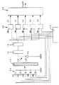
I.パルスオキシメータの装置構成の概要
図1は、本発明に係るパルスオキシメトリを実施するパルスオキシメータとしての装置構成を示す概略説明図である。すなわち、図1において、参照符号10は発光部を示し、それぞれ5個の異なる波長の光をそれぞれ生体組織に照射する5個の発光素子LED1〜LED5が設けられている。参照符号12は前記発光部10から発せられる光によって照射される生体組織を示す。参照符号14は受光部を示し、前記生体組織12を透過した光を受光する受光素子PDと、電流電圧変換器16と、AD変換器18とから構成されている。I.Outline of Apparatus Configuration of Pulse Oximeter FIG. 1 is a schematic explanatory diagram showing an apparatus configuration as a pulse oximeter for carrying out pulse oximetry according to the present invention. That is, in FIG. 1,
参照符号20は記憶部を示し、前記受光部14の受光素子PDにより得られた透過光信号を、波長毎にそれぞれ時系列的に一時記憶する透過光信号一時記憶器20A〜20Eにより構成されている。例えば、これら記憶器毎に、1秒当たり60個の透過光信号が記憶される。
参照符号30は計算部を示し、前記透過光信号一時記憶器20A〜20Eにおいてそれぞれ時系列的に一時記憶された透過光信号L1〜L5に基づいて、(1)前記透過光信号L1〜L5を一定時間毎に区分し、(2)次いで一定時間毎に区分された透過光信号L1〜L5の時系列データについて異なる波長の時系列データ相互の回帰直線の傾斜値を算出し、(3)算出された傾斜値に基づいてSaO2(動脈血酸素飽和度)を計算し、(4)更に相関係数を計算し、相関係数が一定値以下の場合の傾斜値を削除し、残った傾斜値を用いてSaO2のヒストグラムを作成し、(5)ヒストグラムの最頻値をSp02値とする処理を行う。
なお、参照符号22はタイミング器を示し、前記発光部10の各発光素子LED1〜LED5による発光タイミングと、前記記憶部20の各透過光信号一時記憶器20A〜20Eにおける透過光信号の記憶保持タイミングとの制御を行うように構成される。
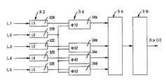
図2は、前記計算部30としての酸素飽和度計算器において前述した計算処理を行うためのシステム構成図を示すものである。すなわち、図2において、参照符号32は透過光信号の区分記憶部を示し、前記透過光信号一時記憶器20A〜20Eから入力される透過光信号L1〜L5を、一定時間(例えば、0.2秒)毎に区分して、この区分された時間毎にそれぞれ透過光信号を時系列的に逐次記憶する区分記憶回路32A〜32Eとして構成されている。 FIG. 2 shows a system configuration diagram for performing the above-described calculation processing in the oxygen saturation calculator as the
また、参照符号34は、回帰直線の傾斜値計算部を示し、前記透過光信号の区分記憶部32にそれぞれ記憶された一定時間毎に区分された透過光信号L1〜L5について回帰直線の傾斜値Φ12、Φ32、Φ42、Φ52をそれぞれ算出する傾斜値計算回路34a、34b、34c、34dとして構成されている。
参照符号36は、前記傾斜値計算回路34a、34b、34c、34dにより得られた回帰直線の傾斜値Φ12、Φ32、Φ42、Φ52に関して連立方程式の解としてSaO2値を求める第1計算回路を示す。この場合、前記第1計算回路により得られた連立方程式の解としての値を5wSall と名付ける。
そして、参照符号38は、連立方程式の解として得られたSaO2について、所定時間区分(例えば、5秒)毎にそのヒストグラムを求め、その最頻値を決定するための第2計算回路を示す。従って、前記第2計算回路38において、血中の酸素飽和度[SpO2]が算出される。
さらに、より良い精度を得るためには、次のような方法を付加すれば好適である。
<1> Li あるいは logLi について、低周波成分を除去する。
<2> Φi2を、2データ間の相関に基づいて選別する。
<3> ヒストグラムに平滑化を加える。Furthermore, in order to obtain better accuracy, it is preferable to add the following method.
<1> The low frequency component is removed for Li or logLi.
<2> Φi2 is selected based on the correlation between the two data.
<3> Add smoothing to the histogram.
II.パルスオキシメータの計算処理操作
次に、前述したパルスオキシメータの装置構成による動脈血の酸素飽和度の計算処理操作、すなわち本発明に係るパルスオキシメトリについて、前記パルスオキシメータの作用と共に説明する。II.Calculation Processing Operation of Pulse Oximeter Next,the calculation processing operation of the arterial blood oxygen saturation by the apparatus configuration of the pulse oximeter, that is, the pulse oximetry according to the present invention will be described together with the operation of the pulse oximeter.
(1)透過光信号の時間区分処理
まず、発光部10の5個の発光素子LED1〜LED5を、それぞれタイミング器22の信号に基づいて、順次交互に異なる波長λ1,λ2,λ3,λ4,λ5で発光させる。これにより、生体組織12を透過した光を受光部14で受信して、発光素子LED1〜LED5の各波長に対応して、各透過光信号L1,L2,L3,L4,L5を、それぞれ所定のタイミングで記憶部20の各透過光信号一時記憶器20A〜20Eに記憶保持する。(図1参照)。(1)Time Division Processing of Transmitted Light Signal First, the five light emitting elements LED1 to LED5 of the
このようにして、前記透過光信号一時記憶器20A〜20Eにそれぞれ記憶保持された透過光信号L1〜L5は、前記計算部30における区分記憶部32の各区分記憶回路32A〜32Eに入力されて、一定時間(例えば、0.2秒)毎に区分され、この区分された時間毎にそれぞれ透過光信号を時系列的に逐次記憶される(図2参照)。 In this way, the transmitted light signals L1 to L5 stored and held in the transmitted light signal temporary storage devices 20A to 20E, respectively, are input to the respective
(2)時間区分された透過光信号に関する回帰直線の傾斜値を求める計算処理
血中の酸素飽和度(SpO2)の計算は、例えば5波長の透過光について得られる減光度変動分(ΔAi)に基づき、これら減光度変動分の比(Φij:i,jは波長)を用いて、次式により求められる。
なお、透過光の脈動を構成する要素は、動脈血(a)、静脈血(v)および血液以外の組織すなわち純組織(t)である。(2)Calculation processing for obtainingthe slope value of the regression line relating to the time- dividedtransmitted light signal The oxygen saturation (SpO2) in the blood is calculated by, for example, the change in attenuation (ΔAi) obtained for transmitted light of 5 wavelengths. Based on the ratio of the change in the light attenuation (Φij: i, j is the wavelength), the following equation is obtained.
The elements constituting the pulsation of transmitted light are arterial blood (a), venous blood (v), and tissues other than blood, that is, pure tissue (t).
Φij≡ΔAi/ΔAj
=[√Eai(Eai+F)+√Evi(Evi+F)*V+Exi]
/[√Eaj(Eaj+F)+√Evj(Evj+F)*V+Exj]
但し、
ΔAi≡log[(Li+ΔLi)/Li]≒ΔLi/Li
Eai≡SaEoi+(1―Sa)Eri
Evi≡SvEoi+(1―Sv)Eri
V≡ΔDv/ΔDa
Exi≡ZtiΔDt/(HbΔDa)≡AiEx2+BiΦij≡ΔAi / ΔAj
= [√Eai (Eai + F) + √Evi (Evi + F) * V + Exi]
/ [√Eaj (Eaj + F) + √Evj (Evj + F) * V + Exj]
However,
ΔAi≡log [(Li + ΔLi) / Li] ≈ΔLi / Li
Eai≡SaEoi + (1-Sa) Eri
Evi≡SvEoi + (1-Sv) Eri
V≡ΔDv / ΔDa
Exi≡ZtiΔDt / (HbΔDa) ≡AiEx2 + Bi
上記式において、Liは組織透過光、ΔAiは減光度の変化分、Eoiは酸素化ヘモグロビンの吸光係数、Eriは脱酸素ヘモグロビンの吸光係数、Saは動脈血の酸素飽和度(SaO2)、Svは末梢静脈血の酸素飽和度(SvO2)、ΔDaは動脈血の実効的厚みの変化分、ΔDvは静脈血の実効的厚みの変化分、ΔDtは純組織の実効的厚みの変化分、Ztiは純組織の減光の定数、Ex2は第2波長におけるExiの値、Ai,Biは組織定数(実測で決定される)である。
従って、上記式において、未知数はSa、Sv、V、Ex2の4個である。In the above equation, Li is the tissue transmitted light, ΔAi is the change in attenuation, Eoi is the extinction coefficient of oxygenated hemoglobin, Eri is the extinction coefficient of deoxygenated hemoglobin, Sa is the oxygen saturation of arterial blood (SaO2), and Sv is the peripheral. Venous blood oxygen saturation (SvO2), ΔDa is the change in effective thickness of arterial blood, ΔDv is the change in effective thickness of venous blood, ΔDt is the change in effective thickness of pure tissue, and Zti is the change in pure tissue The light extinction constant, Ex2 is the value of Exi at the second wavelength, and Ai, Bi are tissue constants (determined by actual measurement).
Therefore, in the above formula, there are four unknowns, Sa, Sv, V, and Ex2.
この場合、適当な5波長で組織透過光を測定して、上記式に関し4元連立方程式を立て、それらの解としてSaを求めることができる。なお、前記5波長としては、例えばλ1=805nm、λ2=875nm、λ3=660nm、λ4=700nm、λ5=730nmが好ましい波長選択の一例である。 In this case, tissue transmitted light is measured at appropriate five wavelengths, a quaternary simultaneous equation is established with respect to the above equation, and Sa can be obtained as a solution thereof. For example, λ1 = 805 nm, λ2 = 875 nm, λ3 = 660 nm, λ4 = 700 nm, and λ5 = 730 nm are preferable examples of wavelength selection.
そこで、本発明の時間区分オキシメトリにおいては、前記区分記憶回路32において、それぞれ時間区分されて記憶された5波長(λ1〜λ5)の透過光信号L1〜L5に基づいて、それぞれ回帰直線の傾斜値(Φij:但し、i,jは波長)を次式により求める。すなわち、この場合の傾斜値(Φij)は、前記のΦij=ΔAi/ΔAjに相当するものである。なお、次式において、nは時間区分内のデータの個数、tは区分された時間(例えば、0.2秒)、Σは時間区分内のデータについての和である。 Therefore, in the time division oximetry of the present invention, the slope values of the regression lines are respectively obtained based on the transmitted light signals L1 to L5 of the five wavelengths (λ1 to λ5) stored in the
Φij≡{nΣ[Li(t)*Lj(t)]−ΣLi(t)*ΣLj(t)}
/{nΣLj(t)2−[ΣLj(t)]2}Φij≡ {nΣ [Li (t) * Lj (t)] − ΣLi (t) * ΣLj (t)}
/ {NΣLj (t)2 − [ΣLj (t)]2 }
(3)傾斜値に関する連立方程式の解を求める計算処理
前記式に基づいて、それぞれ5波長(λ1〜λ5)の組織透過光についての回帰直線の傾斜値(Φ12、Φ32、Φ42、Φ52)に関する4元連立方程式を立て、それらの解としてSaを求める計算を行う(図2参照)。(3)Calculation processing for obtainingsolutions of simultaneous equations related to
III.計算処理の例
前述した本発明に係るパルスオキシメトリに基づいて,被験者による実測データについて動脈血酸素飽和度(SaO2)を計算した例を、従来のパルスオキシメトリによる場合と比較し、それぞれの測定結果を示すグラフと共に説明する。III.Example of calculation processing Based on the pulse oximetry according to the present invention described above, anexample in which the arterial blood oxygen saturation (SaO2) is calculated for the actual measurement data by the subject is compared with the case of the conventional pulse oximetry, and the respective measurement results A description will be given together with a graph showing.
被験者の指尖に発光部10および受光部14を装着して、次のような呼吸を行った:
<1> 空気呼吸。
<2> 息こらえによってSaO2を低下。
<3> 再び空気呼吸。
<4> 酸素呼吸によってSaO2を上昇。The
<1> Air breathing.
<2> SaO2 decreases due to breath holding.
<3> Air breathing again.
<4> SaO2 is increased by oxygen breathing.
<1>の途中から、手でスポンジを握る動作をランンダムに行った。この体動を<4>の途中まで継続した。
なお、この体動する手とは反対の手にも、同様の発光部および受光部を装着し、こちらは静止を保って、これを参照データとした。From the middle of <1>, the hand gripping the sponge with the hand was performed in Randumdam. This body movement was continued halfway through <4>.
The same light-emitting part and light-receiving part were also attached to the hand opposite to the body moving hand, and this was kept stationary and used as reference data.
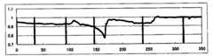
図3は、静止側において、拍毎に山谷を判定して、5波長計算によって求めたSpO2の結果を示すものである。すなわち、図3によれば、呼吸の変化に基づくSaO2の変化が良く示されている。なお、呼吸の変更時点と体動の開始、終了の時点をグラフ中では縦棒で示している。 FIG. 3 shows the SpO2 result obtained by calculating the five wavelengths by determining the valleys for each beat on the stationary side. That is, according to FIG. 3, the change of SaO2 based on the change of respiration is well shown. In addition, the change time of breathing and the start and end time of body movement are indicated by vertical bars in the graph.
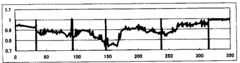
図4は、体動側において、拍毎に山谷を判定して、5波長計算によって求めたSpO2の結果を示すものである。図4においては、SpO2は大きく乱れているが、これが体動の影響である。 FIG. 4 shows the SpO2 result obtained by calculating the five wavelengths by determining the peaks and valleys for each beat on the body movement side. In FIG. 4, SpO2 is greatly disturbed, which is the influence of body movement.
図5は、体動側について、本発明の方法によって0.2秒毎に求めたSpO2の分布特性を示すものである。すなわち、図5は、次のような処理の結果を示す図である。
<1> 透過光信号Liを対数変換してlogLiとした。
<2> logLiの低周波成分を除去した。具体的には、0.1秒毎のlogLi平均値をlogLiから差し引いた。
<3> 0.2秒毎に、logLi (i=1,3,4,5) とlogL2との回帰直線の傾斜Φi2を求めた。
<4> 0.2秒毎に、logLi (i=1,3,4,5) とlogL2との相関係数Ri2を求め、R12*R32*R42*R52=PRを求めた。
<5> PR<0.9の場合のΦi2を削除した。
<6> 残ったΦi2を連立方程式に基づいてSaO2に換算した。これを5wSallと名付ける。
<7> 5wSallを5秒毎に時間区分し、各5秒区間のヒストグラムを求めた。
<8> ヒストグラムについて平滑化を行った。なお、図6は、このようなヒストグラムの表示例を示すものである。
<9> 5wSall のヒストグラムに基づいて最頻値を決定した。
<10> その結果をSpO2として出力した。すなわち、図7は、体動側について求めたSpO2の変化を示すものである。FIG. 5 shows the distribution characteristics of SpO2 obtained every 0.2 seconds by the method of the present invention on the body movement side. That is, FIG. 5 is a diagram showing the results of the following processing.
<1> The transmitted light signal Li is logarithmically converted to log Li.
<2> The low frequency component of logLi was removed. Specifically, the logLi average value every 0.1 seconds was subtracted from logLi.
<3> The slope Φi2 of the regression line between logLi (i = 1,3,4,5) and logL2 was determined every 0.2 seconds.
<4> Correlation coefficient Ri2 between logLi (i = 1,3,4,5) and logL2 was determined every 0.2 seconds, and R12 * R32 * R42 * R52 = PR was determined.
<5> Φi2 in the case of PR <0.9 was deleted.
<6> The remaining Φi2 was converted to SaO2 based on simultaneous equations. This is named 5wSall.
<7> 5wSall was divided into time intervals every 5 seconds, and a histogram of each 5-second interval was obtained.
<8> The histogram was smoothed. FIG. 6 shows a display example of such a histogram.
<9> The mode value was determined based on a histogram of 5 wSall.
<10> The result was output as SpO2. That is, FIG. 7 shows the change in SpO2 obtained for the body movement side.
このように、本発明のパルスオキシメトリによれば、体動の影響は十分に消去され、しかもSaO2の急激な変化が明確に測定されている。特に、SaO2の低下の始まる時点が早く見出せることが確認された。 Thus, according to the pulse oximetry of the present invention, the influence of body movement is sufficiently eliminated, and a rapid change in SaO2 is clearly measured. In particular, it was confirmed that the point in time at which SaO2 starts to decrease can be found early.
以上、本発明の好適な実施例について説明したが、本発明は前記実施例に限定されることなく、例えば5波長を用いる場合について説明したが、波長がそれよりも多い場合にも、あるいは少ない場合にも適用できるばかりでなく、測定対象としては、血中のCOヘモグロビンや体外から注入した色素の希釈状態等の動脈血と共に拍動するもの全ての測定に適用することができる。また時間区分の間隔はそれぞれの目的に応じて適宜変更でき、その他本発明の精神を逸脱しない範囲内において、多くの設計変更が可能である。 The preferred embodiments of the present invention have been described above. However, the present invention is not limited to the above-described embodiments. For example, the case where five wavelengths are used has been described. In addition to being applicable to the case, it can be applied to the measurement of all pulsating with arterial blood such as CO hemoglobin in blood or a diluted state of dye injected from outside the body. In addition, the interval of the time division can be appropriately changed according to each purpose, and many other design changes can be made without departing from the spirit of the present invention.
10 発光部
12 生体組織
14 受光部
16 電流電圧変換器
18 AD変換器
20 記憶部
20A〜20E 透過光信号一時記憶器
22 タイミング器
30 計算部
32 透過光信号の区分記憶部
32A〜32E 区分記憶回路
34 回帰直線の傾斜値計算部
34a〜34d 傾斜値計算回路
36 第1計算回路
38 第2計算回路
LED1〜LED5 発光素子
PD 受光素子DESCRIPTION OF
Claims (5)
Translated fromJapanese前記生体組織を透過または反射した光を受光素子により受光してそれぞれ電気信号に変換し、
前記受光素子により得られる電気信号の時系列データを時間的に区分し、
区分された時系列データについて異なる2波長間の回帰直線の傾斜値をそれぞれ算出し、
算出された傾斜値に基づいてSaO2を計算し、
所定時間区分毎のSaO2のヒストグラムに基づいてSaO2の最頻値を求めることによって、体動の影響を消去した動脈血の酸素飽和度を求めることを特徴とするパルスオキシメトリ。Irradiate living tissue with light of5 wavelengths or more that differ depending on the light emitting element,
The light transmitted through or reflected by the living tissue is received by a light receiving element and converted into electrical signals, respectively.
Time series data of electrical signals obtained by the light receiving element is divided in time,
Calculate the slope value of the regression line between two different wavelengths for the divided time-series data,
Calculate SaO2 based on the calculated slope value,
A pulse oximetry characterized by obtaining an oxygen saturation of arterial blood from which the influence of body motion has been eliminated by obtaining a mode value of SaO2 based on a histogram of SaO2 for each predetermined time segment.
前記発光部から発せられ生体組織を透過または反射した光を受光して、電気信号に変換する受光部と、
前記受光部から変換出力される各波長の透過光または反射光の電気信号からなる時系列データを一定時間毎に区分する処理装置と、
区分された時間毎の前記時系列データについて異なる2波長間の回帰直線の傾斜値をそれぞれ算出する傾斜値演算装置と、
前記算出された傾斜値に基づいてSaO2を計算する計算装置と、
所定時間区分毎のSaO2のヒストグラムに基づいてSaO2の最頻値を求める最頻値演算装置と、を備え、体動の影響を消去した動脈血の酸素飽和度を求めるように構成することを特徴とするパルスオキシメータ。A light emitting unit that irradiates a living tissue with light offive different wavelengths or more ;
A light receiving unit that receives light emitted from the light emitting unit and transmitted or reflected through biological tissue, and converts the light into an electrical signal;
A processing device that divides time-series data composed of electrical signals of transmitted light or reflected light of each wavelength converted and output from the light receiving unit at regular intervals;
An inclination value calculation device for calculating an inclination value of a regression line between two different wavelengths for the time-series data for each divided time;
A calculation device for calculating SaO2 based on the calculated inclination value;
A mode value calculating device for determining a mode value of SaO2 based on a histogram of SaO2 for each predetermined time segment, and characterized in that the oxygen saturation level of arterial blood from which the influence of body motion has been eliminated is determined. Pulse oximeter to do.
Priority Applications (3)
| Application Number | Priority Date | Filing Date | Title |
|---|---|---|---|
| JP2008160013AJP5115855B2 (en) | 2008-06-19 | 2008-06-19 | Pulse oximetry and pulse oximeter |
| EP09162886.7AEP2135550B1 (en) | 2008-06-19 | 2009-06-17 | Pulse oximeter and pulse oximetry method |
| US12/486,843US8548546B2 (en) | 2008-06-19 | 2009-06-18 | Pulse oximetry and pulse oximeter |
Applications Claiming Priority (1)
| Application Number | Priority Date | Filing Date | Title |
|---|---|---|---|
| JP2008160013AJP5115855B2 (en) | 2008-06-19 | 2008-06-19 | Pulse oximetry and pulse oximeter |
Publications (2)
| Publication Number | Publication Date |
|---|---|
| JP2010000160A JP2010000160A (en) | 2010-01-07 |
| JP5115855B2true JP5115855B2 (en) | 2013-01-09 |
Family
ID=40911947
Family Applications (1)
| Application Number | Title | Priority Date | Filing Date |
|---|---|---|---|
| JP2008160013AExpired - Fee RelatedJP5115855B2 (en) | 2008-06-19 | 2008-06-19 | Pulse oximetry and pulse oximeter |
Country Status (3)
| Country | Link |
|---|---|
| US (1) | US8548546B2 (en) |
| EP (1) | EP2135550B1 (en) |
| JP (1) | JP5115855B2 (en) |
Families Citing this family (12)
| Publication number | Priority date | Publication date | Assignee | Title |
|---|---|---|---|---|
| WO2011086644A1 (en)* | 2010-01-18 | 2011-07-21 | コニカミノルタセンシング株式会社 | Biometric information measuring device and method for same |
| JP5353790B2 (en)* | 2010-03-30 | 2013-11-27 | コニカミノルタ株式会社 | Biological information measuring apparatus and method |
| US9706952B2 (en) | 2011-01-06 | 2017-07-18 | Siemens Healthcare Gmbh | System for ventricular arrhythmia detection and characterization |
| US9402571B2 (en) | 2011-01-06 | 2016-08-02 | Siemens Medical Solutions Usa, Inc. | Biological tissue function analysis |
| WO2013148180A1 (en) | 2012-03-27 | 2013-10-03 | The University Of Vermont And State Agricultural College | Non-invasive methods for determining cardiac output |
| WO2014011368A1 (en)* | 2012-06-18 | 2014-01-16 | Eso-Technologies, Inc. | Compositions and methods for measurement of oxygen saturation in blood filled structures |
| US9848785B2 (en) | 2013-12-05 | 2017-12-26 | Siemens Healthcare Gmbh | Analysis and characterization of patient signals |
| US10206581B2 (en) | 2014-10-29 | 2019-02-19 | Zoll Medical Corporation | Transesophageal or transtracheal cardiac monitoring by optical spectroscopy |
| EP3064137B1 (en)* | 2015-03-04 | 2020-12-23 | Nihon Kohden Corporation | Pulse photometer and method for calculating concentration of light absorber in blood |
| US10561375B2 (en)* | 2015-08-31 | 2020-02-18 | Nihon Kohden Corporation | Pulse photometer and method for evaluating reliability of calculated value of blood light absorber concentration |
| CN105433957A (en)* | 2015-12-29 | 2016-03-30 | 深圳贝特莱电子科技股份有限公司 | Integrated chip for detecting human body oxyhemoglobin saturation |
| JP7203419B2 (en)* | 2019-03-15 | 2023-01-13 | 株式会社ニューロシューティカルズ | Biological information analysis device, biological information analysis method, and program |
Family Cites Families (14)
| Publication number | Priority date | Publication date | Assignee | Title |
|---|---|---|---|---|
| JPH02309929A (en)* | 1989-05-24 | 1990-12-25 | Sumitomo Electric Ind Ltd | Liver function testing device |
| JP3270917B2 (en)* | 1994-06-02 | 2002-04-02 | 日本光電工業株式会社 | Oxygen saturation measuring device, blood light absorbing substance concentration measuring device, and biological signal processing method |
| US8019400B2 (en)* | 1994-10-07 | 2011-09-13 | Masimo Corporation | Signal processing apparatus |
| US5765563A (en)* | 1996-08-15 | 1998-06-16 | Nellcor Puritan Bennett Incorporated | Patient monitoring system |
| US7738936B1 (en)* | 1999-11-10 | 2010-06-15 | Pacesetter, Inc. | Methods and systems for reducing data acquisition, power and/or processing for pulse oximetry applications |
| US6381351B1 (en)* | 1999-11-24 | 2002-04-30 | Direct Radiography Corp. | Weighted inverse topography method for digital x-ray image data processing |
| US6771997B2 (en)* | 2001-09-11 | 2004-08-03 | The Board Of Trustees Of The Leland Stanford Junior University | Respiratory compensation in MRI coronary imaging using diminishing variance |
| JP2004202190A (en)* | 2002-11-08 | 2004-07-22 | Minolta Co Ltd | Biological information measuring device |
| US7025728B2 (en)* | 2003-06-30 | 2006-04-11 | Nihon Kohden Corporation | Method for reducing noise, and pulse photometer using the method |
| JP4196209B2 (en)* | 2003-06-30 | 2008-12-17 | 日本光電工業株式会社 | Signal processing method and pulse photometer using the same |
| US7206621B2 (en) | 2003-08-27 | 2007-04-17 | Nihon Kohden Corporation | Pulse oximeter |
| JP4399847B2 (en) | 2003-08-27 | 2010-01-20 | 日本光電工業株式会社 | Pulse oximeter |
| US8024021B2 (en)* | 2005-08-30 | 2011-09-20 | Nihon Kohden Corporation | Time-segmented pulse oximetry and pulse oximeter performing the same |
| JP4844291B2 (en)* | 2005-08-30 | 2011-12-28 | 日本光電工業株式会社 | Time division pulse oximetry and pulse oximeter |
- 2008
- 2008-06-19JPJP2008160013Apatent/JP5115855B2/ennot_activeExpired - Fee Related
- 2009
- 2009-06-17EPEP09162886.7Apatent/EP2135550B1/enactiveActive
- 2009-06-18USUS12/486,843patent/US8548546B2/enactiveActive
Also Published As
| Publication number | Publication date |
|---|---|
| US20090318787A1 (en) | 2009-12-24 |
| JP2010000160A (en) | 2010-01-07 |
| EP2135550A1 (en) | 2009-12-23 |
| EP2135550B1 (en) | 2017-10-18 |
| US8548546B2 (en) | 2013-10-01 |
Similar Documents
| Publication | Publication Date | Title |
|---|---|---|
| JP5115855B2 (en) | Pulse oximetry and pulse oximeter | |
| US8024021B2 (en) | Time-segmented pulse oximetry and pulse oximeter performing the same | |
| US7499740B2 (en) | Techniques for detecting heart pulses and reducing power consumption in sensors | |
| US7006856B2 (en) | Signal quality metrics design for qualifying data for a physiological monitor | |
| JP5096310B2 (en) | Method and apparatus for determining blood perfusion in a body part | |
| CN103381094B (en) | Monitoring system and method for fetus pulse blood oxygen saturation | |
| US8777867B2 (en) | Detection of oximetry sensor sites based on waveform characteristics | |
| US20090247849A1 (en) | Pulse Oximeter With Adaptive Power Conservation | |
| JP2003530189A (en) | Pulse oximeter sensor with piecewise function | |
| US20120310060A1 (en) | Method of analyzing photon density waves in a medical monitor | |
| JPH10314150A (en) | System and method for detecting blood oxygen saturation degree | |
| CN107198529B (en) | Blood oxygen saturation sensor with LED current modulation | |
| WO2011041071A1 (en) | Method of analyzing photon density waves in a medical monitor | |
| US8870783B2 (en) | Pulse rate determination using Gaussian kernel smoothing of multiple inter-fiducial pulse periods | |
| US8840562B2 (en) | Signal processing warping technique | |
| WO2011014297A1 (en) | Patient monitoring system and method utilising photon density waves in transmission mode | |
| JP2023532318A (en) | Method and apparatus for assessing peripheral arterial tone | |
| WO2011123239A1 (en) | Multi-wavelength photon density wave system using an optical switch | |
| JP4399847B2 (en) | Pulse oximeter | |
| JP4844291B2 (en) | Time division pulse oximetry and pulse oximeter | |
| JP2007295973A (en) | Pulse oximeter | |
| US20140187884A1 (en) | Systems and methods for ensemble averaging in pulse oximetry | |
| US20250176873A1 (en) | Non-contact oxygen saturation estimation using ambient light | |
| JP2000037371A (en) | Blood absorption material concentration measurement device | |
| Maddi et al. | Skin Color-Corrected Cardiovascular Diagnostics |
Legal Events
| Date | Code | Title | Description |
|---|---|---|---|
| A621 | Written request for application examination | Free format text:JAPANESE INTERMEDIATE CODE: A621 Effective date:20101215 | |
| A977 | Report on retrieval | Free format text:JAPANESE INTERMEDIATE CODE: A971007 Effective date:20120627 | |
| A131 | Notification of reasons for refusal | Free format text:JAPANESE INTERMEDIATE CODE: A131 Effective date:20120703 | |
| A521 | Request for written amendment filed | Free format text:JAPANESE INTERMEDIATE CODE: A523 Effective date:20120831 | |
| TRDD | Decision of grant or rejection written | ||
| A01 | Written decision to grant a patent or to grant a registration (utility model) | Free format text:JAPANESE INTERMEDIATE CODE: A01 Effective date:20120925 | |
| A01 | Written decision to grant a patent or to grant a registration (utility model) | Free format text:JAPANESE INTERMEDIATE CODE: A01 | |
| A61 | First payment of annual fees (during grant procedure) | Free format text:JAPANESE INTERMEDIATE CODE: A61 Effective date:20121004 | |
| R150 | Certificate of patent or registration of utility model | Free format text:JAPANESE INTERMEDIATE CODE: R150 Ref document number:5115855 Country of ref document:JP Free format text:JAPANESE INTERMEDIATE CODE: R150 | |
| FPAY | Renewal fee payment (event date is renewal date of database) | Free format text:PAYMENT UNTIL: 20151026 Year of fee payment:3 | |
| R250 | Receipt of annual fees | Free format text:JAPANESE INTERMEDIATE CODE: R250 | |
| R250 | Receipt of annual fees | Free format text:JAPANESE INTERMEDIATE CODE: R250 | |
| R250 | Receipt of annual fees | Free format text:JAPANESE INTERMEDIATE CODE: R250 | |
| LAPS | Cancellation because of no payment of annual fees |