JP4782927B2 - Surrogate cell-based system and method for evaluating the activity of hepatitis C virus NS3 protease - Google Patents
Surrogate cell-based system and method for evaluating the activity of hepatitis C virus NS3 proteaseDownload PDFInfo
- Publication number
- JP4782927B2 JP4782927B2JP2000615652AJP2000615652AJP4782927B2JP 4782927 B2JP4782927 B2JP 4782927B2JP 2000615652 AJP2000615652 AJP 2000615652AJP 2000615652 AJP2000615652 AJP 2000615652AJP 4782927 B2JP4782927 B2JP 4782927B2
- Authority
- JP
- Japan
- Prior art keywords
- protease
- hcv
- activity
- expression
- protein
- Prior art date
- Legal status (The legal status is an assumption and is not a legal conclusion. Google has not performed a legal analysis and makes no representation as to the accuracy of the status listed.)
- Expired - Fee Related
Links
- 230000000694effectsEffects0.000titleclaimsabstractdescription111
- 238000000034methodMethods0.000titleclaimsdescription57
- 108700012707hepatitis C virus NS3Proteins0.000titleclaimsdescription4
- 238000003776cleavage reactionMethods0.000claimsabstractdescription121
- 230000007017scissionEffects0.000claimsabstractdescription120
- 101800001838Serine protease/helicase NS3Proteins0.000claimsabstractdescription94
- 108091023040Transcription factorProteins0.000claimsabstractdescription84
- 102000040945Transcription factorHuman genes0.000claimsabstractdescription80
- 108010076039PolyproteinsProteins0.000claimsabstractdescription64
- 230000014509gene expressionEffects0.000claimsabstractdescription61
- 108700008625Reporter GenesProteins0.000claimsabstractdescription35
- 239000012634fragmentSubstances0.000claimsabstractdescription34
- 101800001554RNA-directed RNA polymeraseProteins0.000claimsabstractdescription30
- 150000001875compoundsChemical class0.000claimsabstractdescription23
- 101800001014Non-structural protein 5AProteins0.000claimsabstract4
- 210000004027cellAnatomy0.000claimsdescription108
- 108090000623proteins and genesProteins0.000claimsdescription105
- 108020004414DNAProteins0.000claimsdescription54
- 108091005804PeptidasesProteins0.000claimsdescription37
- 239000004365ProteaseSubstances0.000claimsdescription37
- 102100037486Reverse transcriptase/ribonuclease HHuman genes0.000claimsdescription32
- 102000053602DNAHuman genes0.000claimsdescription27
- 210000004962mammalian cellAnatomy0.000claimsdescription22
- 238000001890transfectionMethods0.000claimsdescription20
- 108020004511Recombinant DNAProteins0.000claimsdescription16
- 210000003494hepatocyteAnatomy0.000claimsdescription16
- 239000003112inhibitorSubstances0.000claimsdescription16
- 239000012528membraneSubstances0.000claimsdescription13
- 150000007523nucleic acidsChemical class0.000claimsdescription13
- 230000004927fusionEffects0.000claimsdescription11
- 102000039446nucleic acidsHuman genes0.000claimsdescription10
- 108020004707nucleic acidsProteins0.000claimsdescription10
- 239000013613expression plasmidSubstances0.000claimsdescription9
- 241000714474Rous sarcoma virusSpecies0.000claimsdescription6
- 230000005945translocationEffects0.000claimsdescription6
- 210000002472endoplasmic reticulumAnatomy0.000claimsdescription5
- 238000004519manufacturing processMethods0.000claimsdescription5
- 210000003292kidney cellAnatomy0.000claimsdescription4
- 238000013519translationMethods0.000claimsdescription4
- 102000007056Recombinant Fusion ProteinsHuman genes0.000claimsdescription3
- 108010008281Recombinant Fusion ProteinsProteins0.000claimsdescription3
- 230000000977initiatory effectEffects0.000claimsdescription3
- 238000011156evaluationMethods0.000claimsdescription2
- 210000002950fibroblastAnatomy0.000claimsdescription2
- 210000004698lymphocyteAnatomy0.000claimsdescription2
- 230000001939inductive effectEffects0.000claims1
- 238000003556assayMethods0.000abstractdescription37
- 101800001019Non-structural protein 4BProteins0.000abstractdescription29
- 230000003612virological effectEffects0.000abstractdescription12
- 238000012216screeningMethods0.000abstractdescription11
- 230000000120cytopathologic effectEffects0.000abstractdescription10
- 230000027455bindingEffects0.000abstractdescription8
- 238000000423cell based assayMethods0.000abstractdescription7
- 238000012360testing methodMethods0.000abstractdescription7
- 230000035945sensitivityEffects0.000abstractdescription5
- 238000011161developmentMethods0.000abstractdescription3
- 238000013518transcriptionMethods0.000abstractdescription3
- 230000035897transcriptionEffects0.000abstractdescription3
- 230000002401inhibitory effectEffects0.000abstractdescription2
- 101800001020Non-structural protein 4AProteins0.000abstract1
- 241000711549Hepacivirus CSpecies0.000description101
- 102000004169proteins and genesHuman genes0.000description91
- 235000018102proteinsNutrition0.000description90
- 239000005089LuciferaseSubstances0.000description63
- 108060001084LuciferaseProteins0.000description63
- 239000013612plasmidSubstances0.000description45
- 150000001413amino acidsChemical class0.000description35
- 235000001014amino acidNutrition0.000description27
- 230000035772mutationEffects0.000description24
- 239000004098TetracyclineSubstances0.000description21
- 229960002180tetracyclineDrugs0.000description21
- 229930101283tetracyclineNatural products0.000description21
- 235000019364tetracyclineNutrition0.000description21
- 150000003522tetracyclinesChemical class0.000description21
- 239000000047productSubstances0.000description20
- 239000013598vectorSubstances0.000description18
- 230000003321amplificationEffects0.000description17
- 238000003199nucleic acid amplification methodMethods0.000description17
- 238000012545processingMethods0.000description16
- 230000001419dependent effectEffects0.000description13
- 108090000765processed proteins & peptidesProteins0.000description13
- 230000004913activationEffects0.000description12
- 238000006243chemical reactionMethods0.000description12
- 102000004196processed proteins & peptidesHuman genes0.000description12
- 241000700605VirusesSpecies0.000description11
- 108091008146restriction endonucleasesProteins0.000description11
- 239000000758substrateSubstances0.000description11
- 238000001262western blotMethods0.000description11
- 108020001507fusion proteinsProteins0.000description10
- 102000037865fusion proteinsHuman genes0.000description10
- 229920001184polypeptidePolymers0.000description10
- 239000002243precursorSubstances0.000description10
- 208000015181infectious diseaseDiseases0.000description9
- 239000002773nucleotideSubstances0.000description9
- 125000003729nucleotide groupChemical group0.000description9
- 101710118188DNA-binding protein HU-alphaProteins0.000description8
- 102000004190EnzymesHuman genes0.000description8
- 108090000790EnzymesProteins0.000description8
- 101710144128Non-structural protein 2Proteins0.000description8
- 101710199667Nuclear export proteinProteins0.000description8
- 241000700618Vaccinia virusSpecies0.000description8
- 239000002299complementary DNASubstances0.000description8
- 238000003670luciferase enzyme activity assayMethods0.000description8
- 108091028043Nucleic acid sequenceProteins0.000description7
- 108091034117OligonucleotideProteins0.000description7
- MTCFGRXMJLQNBG-UHFFFAOYSA-NSerineNatural productsOCC(N)C(O)=OMTCFGRXMJLQNBG-UHFFFAOYSA-N0.000description7
- 239000012190activatorSubstances0.000description7
- 238000002474experimental methodMethods0.000description7
- 230000008569processEffects0.000description7
- 239000000126substanceSubstances0.000description7
- 102000002260Alkaline PhosphataseHuman genes0.000description6
- 108020004774Alkaline PhosphataseProteins0.000description6
- 235000004279alanineNutrition0.000description6
- 238000004113cell cultureMethods0.000description6
- 238000010367cloningMethods0.000description6
- 230000002950deficientEffects0.000description6
- 239000000499gelSubstances0.000description6
- 230000005764inhibitory processEffects0.000description6
- 230000002441reversible effectEffects0.000description6
- 108091032973(ribonucleotides)n+mProteins0.000description5
- QNAYBMKLOCPYGJ-REOHCLBHSA-NL-alanineChemical compoundC[C@H](N)C(O)=OQNAYBMKLOCPYGJ-REOHCLBHSA-N0.000description5
- 108060004795MethyltransferaseProteins0.000description5
- 102000035195PeptidasesHuman genes0.000description5
- 230000004071biological effectEffects0.000description5
- 238000001514detection methodMethods0.000description5
- 239000000284extractSubstances0.000description5
- 230000007246mechanismEffects0.000description5
- 238000005457optimizationMethods0.000description5
- 230000036961partial effectEffects0.000description5
- 238000003752polymerase chain reactionMethods0.000description5
- 230000004044responseEffects0.000description5
- 102100039556Galectin-4Human genes0.000description4
- 240000004808Saccharomyces cerevisiaeSpecies0.000description4
- 235000014680Saccharomyces cerevisiaeNutrition0.000description4
- 101710137500T7 RNA polymeraseProteins0.000description4
- 230000001413cellular effectEffects0.000description4
- 238000012761co-transfectionMethods0.000description4
- 210000003527eukaryotic cellAnatomy0.000description4
- 230000006870functionEffects0.000description4
- 238000000338in vitroMethods0.000description4
- 230000000670limiting effectEffects0.000description4
- 239000013642negative controlSubstances0.000description4
- 230000010076replicationEffects0.000description4
- 239000004475ArginineSubstances0.000description3
- 108020004705CodonProteins0.000description3
- PEDCQBHIVMGVHV-UHFFFAOYSA-NGlycerineChemical compoundOCC(O)COPEDCQBHIVMGVHV-UHFFFAOYSA-N0.000description3
- 241000238631HexapodaSpecies0.000description3
- 101000608765Homo sapiens Galectin-4Proteins0.000description3
- XUJNEKJLAYXESH-REOHCLBHSA-NL-CysteineChemical compoundSC[C@H](N)C(O)=OXUJNEKJLAYXESH-REOHCLBHSA-N0.000description3
- ONIBWKKTOPOVIA-BYPYZUCNSA-NL-ProlineChemical compoundOC(=O)[C@@H]1CCCN1ONIBWKKTOPOVIA-BYPYZUCNSA-N0.000description3
- ODKSFYDXXFIFQN-BYPYZUCNSA-PL-argininium(2+)Chemical compoundNC(=[NH2+])NCCC[C@H]([NH3+])C(O)=OODKSFYDXXFIFQN-BYPYZUCNSA-P0.000description3
- 108010006035MetalloproteasesProteins0.000description3
- 102000005741MetalloproteasesHuman genes0.000description3
- -1NS5AProteins0.000description3
- 101710152005Non-structural polyproteinProteins0.000description3
- 101710114167Polyprotein P1234Proteins0.000description3
- 101710124590Polyprotein nsP1234Proteins0.000description3
- ONIBWKKTOPOVIA-UHFFFAOYSA-NProlineNatural productsOC(=O)C1CCCN1ONIBWKKTOPOVIA-UHFFFAOYSA-N0.000description3
- 101710172711Structural proteinProteins0.000description3
- ODKSFYDXXFIFQN-UHFFFAOYSA-NarginineNatural productsOC(=O)C(N)CCCNC(N)=NODKSFYDXXFIFQN-UHFFFAOYSA-N0.000description3
- 230000008901benefitEffects0.000description3
- 108010005774beta-GalactosidaseProteins0.000description3
- 102000005936beta-GalactosidaseHuman genes0.000description3
- 230000000295complement effectEffects0.000description3
- 238000010276constructionMethods0.000description3
- 235000018417cysteineNutrition0.000description3
- XUJNEKJLAYXESH-UHFFFAOYSA-NcysteineNatural productsSCC(N)C(O)=OXUJNEKJLAYXESH-UHFFFAOYSA-N0.000description3
- 239000013604expression vectorSubstances0.000description3
- 230000005714functional activityEffects0.000description3
- 238000001727in vivoMethods0.000description3
- 208000019423liver diseaseDiseases0.000description3
- 238000005259measurementMethods0.000description3
- 210000004940nucleusAnatomy0.000description3
- 239000002245particleSubstances0.000description3
- 230000023603positive regulation of transcription initiation, DNA-dependentEffects0.000description3
- 230000001105regulatory effectEffects0.000description3
- 238000003757reverse transcription PCRMethods0.000description3
- 238000006467substitution reactionMethods0.000description3
- 230000001052transient effectEffects0.000description3
- 238000011282treatmentMethods0.000description3
- MTCFGRXMJLQNBG-REOHCLBHSA-N(2S)-2-Amino-3-hydroxypropansäureChemical compoundOC[C@H](N)C(O)=OMTCFGRXMJLQNBG-REOHCLBHSA-N0.000description2
- 102000040650(ribonucleotides)n+mHuman genes0.000description2
- 208000006154Chronic hepatitis CDiseases0.000description2
- 108010043121Green Fluorescent ProteinsProteins0.000description2
- 102000004144Green Fluorescent ProteinsHuman genes0.000description2
- 208000005176Hepatitis CDiseases0.000description2
- 241000713772Human immunodeficiency virus 1Species0.000description2
- 108700026244Open Reading FramesProteins0.000description2
- 101710150451Protein Bel-1Proteins0.000description2
- 108010022999Serine ProteasesProteins0.000description2
- 102000012479Serine ProteasesHuman genes0.000description2
- 108090000340TransaminasesProteins0.000description2
- 102000003929TransaminasesHuman genes0.000description2
- 108091061763Triple-stranded DNAProteins0.000description2
- 238000004458analytical methodMethods0.000description2
- 230000000840anti-viral effectEffects0.000description2
- 210000004899c-terminal regionAnatomy0.000description2
- 229910000389calcium phosphateInorganic materials0.000description2
- 239000001506calcium phosphateSubstances0.000description2
- 235000011010calcium phosphatesNutrition0.000description2
- 230000010261cell growthEffects0.000description2
- 230000008859changeEffects0.000description2
- 239000003795chemical substances by applicationSubstances0.000description2
- 229960005091chloramphenicolDrugs0.000description2
- WIIZWVCIJKGZOK-RKDXNWHRSA-NchloramphenicolChemical compoundClC(Cl)C(=O)N[C@H](CO)[C@H](O)C1=CC=C([N+]([O-])=O)C=C1WIIZWVCIJKGZOK-RKDXNWHRSA-N0.000description2
- 210000000349chromosomeAnatomy0.000description2
- OPTASPLRGRRNAP-UHFFFAOYSA-NcytosineChemical compoundNC=1C=CNC(=O)N=1OPTASPLRGRRNAP-UHFFFAOYSA-N0.000description2
- 230000007812deficiencyEffects0.000description2
- 239000005547deoxyribonucleotideSubstances0.000description2
- 125000002637deoxyribonucleotide groupChemical group0.000description2
- 238000013461designMethods0.000description2
- 229940042399direct acting antivirals protease inhibitorsDrugs0.000description2
- 201000010099diseaseDiseases0.000description2
- 208000037265diseases, disorders, signs and symptomsDiseases0.000description2
- 238000005516engineering processMethods0.000description2
- 238000001952enzyme assayMethods0.000description2
- 230000002068genetic effectEffects0.000description2
- 239000005090green fluorescent proteinSubstances0.000description2
- UYTPUPDQBNUYGX-UHFFFAOYSA-NguanineChemical compoundO=C1NC(N)=NC2=C1N=CN2UYTPUPDQBNUYGX-UHFFFAOYSA-N0.000description2
- 208000010710hepatitis C virus infectionDiseases0.000description2
- 238000012203high throughput assayMethods0.000description2
- 238000013537high throughput screeningMethods0.000description2
- 210000005260human cellAnatomy0.000description2
- 238000011534incubationMethods0.000description2
- 239000003446ligandSubstances0.000description2
- MYWUZJCMWCOHBA-VIFPVBQESA-NmethamphetamineChemical compoundCN[C@@H](C)CC1=CC=CC=C1MYWUZJCMWCOHBA-VIFPVBQESA-N0.000description2
- 238000002703mutagenesisMethods0.000description2
- 231100000350mutagenesisToxicity0.000description2
- 239000000137peptide hydrolase inhibitorSubstances0.000description2
- 239000002953phosphate buffered salineSubstances0.000description2
- 239000013600plasmid vectorSubstances0.000description2
- 230000020978protein processingEffects0.000description2
- 230000002797proteolythic effectEffects0.000description2
- 230000002829reductive effectEffects0.000description2
- 230000000717retained effectEffects0.000description2
- 210000002966serumAnatomy0.000description2
- 238000002415sodium dodecyl sulfate polyacrylamide gel electrophoresisMethods0.000description2
- 238000010561standard procedureMethods0.000description2
- RWQNBRDOKXIBIV-UHFFFAOYSA-NthymineChemical compoundCC1=CNC(=O)NC1=ORWQNBRDOKXIBIV-UHFFFAOYSA-N0.000description2
- 230000002103transcriptional effectEffects0.000description2
- 230000001131transforming effectEffects0.000description2
- QORWJWZARLRLPR-UHFFFAOYSA-Htricalcium bis(phosphate)Chemical compound[Ca+2].[Ca+2].[Ca+2].[O-]P([O-])([O-])=O.[O-]P([O-])([O-])=OQORWJWZARLRLPR-UHFFFAOYSA-H0.000description2
- 238000010200validation analysisMethods0.000description2
- 230000035899viabilityEffects0.000description2
- 230000029812viral genome replicationEffects0.000description2
- 238000012800visualizationMethods0.000description2
- 210000005253yeast cellAnatomy0.000description2
- LOGFVTREOLYCPF-KXNHARMFSA-N(2s,3r)-2-[[(2r)-1-[(2s)-2,6-diaminohexanoyl]pyrrolidine-2-carbonyl]amino]-3-hydroxybutanoic acidChemical compoundC[C@@H](O)[C@@H](C(O)=O)NC(=O)[C@H]1CCCN1C(=O)[C@@H](N)CCCCNLOGFVTREOLYCPF-KXNHARMFSA-N0.000description1
- HBZBAMXERPYTFS-SECBINFHSA-N(4S)-2-(6,7-dihydro-5H-pyrrolo[3,2-f][1,3]benzothiazol-2-yl)-4,5-dihydro-1,3-thiazole-4-carboxylic acidChemical compoundOC(=O)[C@H]1CSC(=N1)c1nc2cc3CCNc3cc2s1HBZBAMXERPYTFS-SECBINFHSA-N0.000description1
- 229930024421AdenineNatural products0.000description1
- GFFGJBXGBJISGV-UHFFFAOYSA-NAdenineChemical compoundNC1=NC=NC2=C1N=CN2GFFGJBXGBJISGV-UHFFFAOYSA-N0.000description1
- 108700028369AllelesProteins0.000description1
- 108091003079Bovine Serum AlbuminProteins0.000description1
- 101710132601Capsid proteinProteins0.000description1
- 206010057573Chronic hepatic failureDiseases0.000description1
- RGJOEKWQDUBAIZ-IBOSZNHHSA-NCoASHChemical compoundO[C@@H]1[C@H](OP(O)(O)=O)[C@@H](COP(O)(=O)OP(O)(=O)OCC(C)(C)[C@@H](O)C(=O)NCCC(=O)NCCS)O[C@H]1N1C2=NC=NC(N)=C2N=C1RGJOEKWQDUBAIZ-IBOSZNHHSA-N0.000description1
- 108091026890Coding regionProteins0.000description1
- 208000003322CoinfectionDiseases0.000description1
- 102000012410DNA LigasesHuman genes0.000description1
- 108010061982DNA LigasesProteins0.000description1
- 238000007399DNA isolationMethods0.000description1
- 230000006820DNA synthesisEffects0.000description1
- 230000004568DNA-bindingEffects0.000description1
- 208000010334End Stage Liver DiseaseDiseases0.000description1
- 241000991587Enterovirus CSpecies0.000description1
- 241000588724Escherichia coliSpecies0.000description1
- 206010016654FibrosisDiseases0.000description1
- 241000710781FlaviviridaeSpecies0.000description1
- 108010001515Galectin 4Proteins0.000description1
- 108700028146Genetic Enhancer ElementsProteins0.000description1
- 206010019786Hepatitis non-A non-BDiseases0.000description1
- 101000979342Homo sapiens Nuclear factor NF-kappa-B p105 subunitProteins0.000description1
- 108010058683Immobilized ProteinsProteins0.000description1
- 102000006992Interferon-alphaHuman genes0.000description1
- 108010047761Interferon-alphaProteins0.000description1
- 102000003777Interleukin-1 betaHuman genes0.000description1
- 108090000193Interleukin-1 betaProteins0.000description1
- KDXKERNSBIXSRK-UHFFFAOYSA-NLysineNatural productsNCCCCC(N)C(O)=OKDXKERNSBIXSRK-UHFFFAOYSA-N0.000description1
- 239000004472LysineSubstances0.000description1
- 101800000021N-terminal proteaseProteins0.000description1
- 101800001292Non-structural protein 2-3Proteins0.000description1
- 101150038760Ns3 geneProteins0.000description1
- 102100023050Nuclear factor NF-kappa-B p105 subunitHuman genes0.000description1
- 102000008021Nucleoside-TriphosphataseHuman genes0.000description1
- 108010075285Nucleoside-TriphosphataseProteins0.000description1
- 101800000212P1A proteinProteins0.000description1
- 238000012408PCR amplificationMethods0.000description1
- 101800000862Protease cofactorProteins0.000description1
- 108090000944RNA HelicasesProteins0.000description1
- 102000004409RNA HelicasesHuman genes0.000description1
- 108091027981Response elementProteins0.000description1
- IWUCXVSUMQZMFG-AFCXAGJDSA-NRibavirinChemical compoundN1=C(C(=O)N)N=CN1[C@H]1[C@H](O)[C@H](O)[C@@H](CO)O1IWUCXVSUMQZMFG-AFCXAGJDSA-N0.000description1
- 108091028664RibonucleotideProteins0.000description1
- 241000700584SimplexvirusSpecies0.000description1
- 241000710960Sindbis virusSpecies0.000description1
- 230000024932T cell mediated immunityEffects0.000description1
- 229920004890Triton X-100Polymers0.000description1
- 239000013504Triton X-100Substances0.000description1
- 206010046865Vaccinia virus infectionDiseases0.000description1
- 108020005202Viral DNAProteins0.000description1
- 108700022715Viral ProteasesProteins0.000description1
- 208000036142Viral infectionDiseases0.000description1
- HCHKCACWOHOZIP-UHFFFAOYSA-NZincChemical compound[Zn]HCHKCACWOHOZIP-UHFFFAOYSA-N0.000description1
- QWXOJIDBSHLIFI-UHFFFAOYSA-N[3-(1-chloro-3'-methoxyspiro[adamantane-4,4'-dioxetane]-3'-yl)phenyl] dihydrogen phosphateChemical compoundO1OC2(C3CC4CC2CC(Cl)(C4)C3)C1(OC)C1=CC=CC(OP(O)(O)=O)=C1QWXOJIDBSHLIFI-UHFFFAOYSA-N0.000description1
- JLCPHMBAVCMARE-UHFFFAOYSA-N[3-[[3-[[3-[[3-[[3-[[3-[[3-[[3-[[3-[[3-[[3-[[5-(2-amino-6-oxo-1H-purin-9-yl)-3-[[3-[[3-[[3-[[3-[[3-[[5-(2-amino-6-oxo-1H-purin-9-yl)-3-[[5-(2-amino-6-oxo-1H-purin-9-yl)-3-hydroxyoxolan-2-yl]methoxy-hydroxyphosphoryl]oxyoxolan-2-yl]methoxy-hydroxyphosphoryl]oxy-5-(5-methyl-2,4-dioxopyrimidin-1-yl)oxolan-2-yl]methoxy-hydroxyphosphoryl]oxy-5-(6-aminopurin-9-yl)oxolan-2-yl]methoxy-hydroxyphosphoryl]oxy-5-(6-aminopurin-9-yl)oxolan-2-yl]methoxy-hydroxyphosphoryl]oxy-5-(6-aminopurin-9-yl)oxolan-2-yl]methoxy-hydroxyphosphoryl]oxy-5-(6-aminopurin-9-yl)oxolan-2-yl]methoxy-hydroxyphosphoryl]oxyoxolan-2-yl]methoxy-hydroxyphosphoryl]oxy-5-(5-methyl-2,4-dioxopyrimidin-1-yl)oxolan-2-yl]methoxy-hydroxyphosphoryl]oxy-5-(4-amino-2-oxopyrimidin-1-yl)oxolan-2-yl]methoxy-hydroxyphosphoryl]oxy-5-(5-methyl-2,4-dioxopyrimidin-1-yl)oxolan-2-yl]methoxy-hydroxyphosphoryl]oxy-5-(5-methyl-2,4-dioxopyrimidin-1-yl)oxolan-2-yl]methoxy-hydroxyphosphoryl]oxy-5-(6-aminopurin-9-yl)oxolan-2-yl]methoxy-hydroxyphosphoryl]oxy-5-(6-aminopurin-9-yl)oxolan-2-yl]methoxy-hydroxyphosphoryl]oxy-5-(4-amino-2-oxopyrimidin-1-yl)oxolan-2-yl]methoxy-hydroxyphosphoryl]oxy-5-(4-amino-2-oxopyrimidin-1-yl)oxolan-2-yl]methoxy-hydroxyphosphoryl]oxy-5-(4-amino-2-oxopyrimidin-1-yl)oxolan-2-yl]methoxy-hydroxyphosphoryl]oxy-5-(6-aminopurin-9-yl)oxolan-2-yl]methoxy-hydroxyphosphoryl]oxy-5-(4-amino-2-oxopyrimidin-1-yl)oxolan-2-yl]methyl [5-(6-aminopurin-9-yl)-2-(hydroxymethyl)oxolan-3-yl] hydrogen phosphatePolymersCc1cn(C2CC(OP(O)(=O)OCC3OC(CC3OP(O)(=O)OCC3OC(CC3O)n3cnc4c3nc(N)[nH]c4=O)n3cnc4c3nc(N)[nH]c4=O)C(COP(O)(=O)OC3CC(OC3COP(O)(=O)OC3CC(OC3COP(O)(=O)OC3CC(OC3COP(O)(=O)OC3CC(OC3COP(O)(=O)OC3CC(OC3COP(O)(=O)OC3CC(OC3COP(O)(=O)OC3CC(OC3COP(O)(=O)OC3CC(OC3COP(O)(=O)OC3CC(OC3COP(O)(=O)OC3CC(OC3COP(O)(=O)OC3CC(OC3COP(O)(=O)OC3CC(OC3COP(O)(=O)OC3CC(OC3COP(O)(=O)OC3CC(OC3COP(O)(=O)OC3CC(OC3COP(O)(=O)OC3CC(OC3COP(O)(=O)OC3CC(OC3CO)n3cnc4c(N)ncnc34)n3ccc(N)nc3=O)n3cnc4c(N)ncnc34)n3ccc(N)nc3=O)n3ccc(N)nc3=O)n3ccc(N)nc3=O)n3cnc4c(N)ncnc34)n3cnc4c(N)ncnc34)n3cc(C)c(=O)[nH]c3=O)n3cc(C)c(=O)[nH]c3=O)n3ccc(N)nc3=O)n3cc(C)c(=O)[nH]c3=O)n3cnc4c3nc(N)[nH]c4=O)n3cnc4c(N)ncnc34)n3cnc4c(N)ncnc34)n3cnc4c(N)ncnc34)n3cnc4c(N)ncnc34)O2)c(=O)[nH]c1=OJLCPHMBAVCMARE-UHFFFAOYSA-N0.000description1
- 238000010521absorption reactionMethods0.000description1
- 239000002253acidSubstances0.000description1
- 229960000643adenineDrugs0.000description1
- 238000000246agarose gel electrophoresisMethods0.000description1
- 125000000539amino acid groupChemical group0.000description1
- 239000003242anti bacterial agentSubstances0.000description1
- 230000000692anti-sense effectEffects0.000description1
- 229940088710antibiotic agentDrugs0.000description1
- 239000003443antiviral agentSubstances0.000description1
- 101150010487are geneProteins0.000description1
- 210000004369bloodAnatomy0.000description1
- 239000008280bloodSubstances0.000description1
- 239000000872bufferSubstances0.000description1
- 239000000969carrierSubstances0.000description1
- 230000015556catabolic processEffects0.000description1
- 230000003197catalytic effectEffects0.000description1
- 230000030833cell deathEffects0.000description1
- 239000013592cell lysateSubstances0.000description1
- 239000003153chemical reaction reagentSubstances0.000description1
- 239000013611chromosomal DNASubstances0.000description1
- 208000011444chronic liver failureDiseases0.000description1
- 230000007882cirrhosisEffects0.000description1
- 208000019425cirrhosis of liverDiseases0.000description1
- RGJOEKWQDUBAIZ-UHFFFAOYSA-Ncoenzime ANatural productsOC1C(OP(O)(O)=O)C(COP(O)(=O)OP(O)(=O)OCC(C)(C)C(O)C(=O)NCCC(=O)NCCS)OC1N1C2=NC=NC(N)=C2N=C1RGJOEKWQDUBAIZ-UHFFFAOYSA-N0.000description1
- 239000005516coenzyme ASubstances0.000description1
- 229940093530coenzyme aDrugs0.000description1
- 230000002860competitive effectEffects0.000description1
- 230000021615conjugationEffects0.000description1
- ZTOGYGRJDIWKAQ-UHFFFAOYSA-Ncyclohexene-1,2-diamineChemical compoundNC1=C(N)CCCC1ZTOGYGRJDIWKAQ-UHFFFAOYSA-N0.000description1
- 229940104302cytosineDrugs0.000description1
- 230000034994deathEffects0.000description1
- 230000007423decreaseEffects0.000description1
- 239000007857degradation productSubstances0.000description1
- 238000006731degradation reactionMethods0.000description1
- KDTSHFARGAKYJN-UHFFFAOYSA-Ndephosphocoenzyme ANatural productsOC1C(O)C(COP(O)(=O)OP(O)(=O)OCC(C)(C)C(O)C(=O)NCCC(=O)NCCS)OC1N1C2=NC=NC(N)=C2N=C1KDTSHFARGAKYJN-UHFFFAOYSA-N0.000description1
- 238000010586diagramMethods0.000description1
- 238000009792diffusion processMethods0.000description1
- 230000029087digestionEffects0.000description1
- LOKCTEFSRHRXRJ-UHFFFAOYSA-Idipotassium trisodium dihydrogen phosphate hydrogen phosphate dichlorideChemical compoundP(=O)(O)(O)[O-].[K+].P(=O)(O)([O-])[O-].[Na+].[Na+].[Cl-].[K+].[Cl-].[Na+]LOKCTEFSRHRXRJ-UHFFFAOYSA-I0.000description1
- 238000011143downstream manufacturingMethods0.000description1
- 238000007876drug discoveryMethods0.000description1
- 241001493065dsRNA virusesSpecies0.000description1
- 230000008030eliminationEffects0.000description1
- 238000003379elimination reactionMethods0.000description1
- 239000003623enhancerSubstances0.000description1
- 238000009585enzyme analysisMethods0.000description1
- 238000001976enzyme digestionMethods0.000description1
- 230000002349favourable effectEffects0.000description1
- 239000012894fetal calf serumSubstances0.000description1
- 238000010353genetic engineeringMethods0.000description1
- 230000012010growthEffects0.000description1
- 206010073071hepatocellular carcinomaDiseases0.000description1
- 231100000844hepatocellular carcinomaToxicity0.000description1
- 239000000833heterodimerSubstances0.000description1
- 238000012188high-throughput screening assayMethods0.000description1
- 230000028996humoral immune responseEffects0.000description1
- 230000002209hydrophobic effectEffects0.000description1
- 210000000987immune systemAnatomy0.000description1
- 230000000415inactivating effectEffects0.000description1
- 230000002779inactivationEffects0.000description1
- 238000003780insertionMethods0.000description1
- 230000037431insertionEffects0.000description1
- 238000005304joiningMethods0.000description1
- 238000003367kinetic assayMethods0.000description1
- 238000002372labellingMethods0.000description1
- 210000005229liver cellAnatomy0.000description1
- 210000005228liver tissueAnatomy0.000description1
- 230000004807localizationEffects0.000description1
- 239000006166lysateSubstances0.000description1
- 239000012139lysis bufferSubstances0.000description1
- ADKOXSOCTOWDOP-UHFFFAOYSA-Lmagnesium;aluminum;dihydroxide;trihydrateChemical compoundO.O.O.[OH-].[OH-].[Mg+2].[Al]ADKOXSOCTOWDOP-UHFFFAOYSA-L0.000description1
- 239000000463materialSubstances0.000description1
- 239000000203mixtureSubstances0.000description1
- 238000012986modificationMethods0.000description1
- 230000004048modificationEffects0.000description1
- 238000010369molecular cloningMethods0.000description1
- 230000004660morphological changeEffects0.000description1
- 230000001338necrotic effectEffects0.000description1
- 238000007899nucleic acid hybridizationMethods0.000description1
- 230000007170pathologyEffects0.000description1
- 230000002688persistenceEffects0.000description1
- 239000013641positive controlSubstances0.000description1
- 210000001236prokaryotic cellAnatomy0.000description1
- 238000004445quantitative analysisMethods0.000description1
- 230000009467reductionEffects0.000description1
- 238000011160researchMethods0.000description1
- 229960000329ribavirinDrugs0.000description1
- HZCAHMRRMINHDJ-DBRKOABJSA-NribavirinNatural productsO[C@@H]1[C@H](O)[C@@H](CO)O[C@H]1N1N=CN=C1HZCAHMRRMINHDJ-DBRKOABJSA-N0.000description1
- 239000002336ribonucleotideSubstances0.000description1
- 125000002652ribonucleotide groupChemical group0.000description1
- 150000003839saltsChemical class0.000description1
- 239000000523sampleSubstances0.000description1
- 238000013341scale-upMethods0.000description1
- 238000007423screening assayMethods0.000description1
- 230000028327secretionEffects0.000description1
- 239000013605shuttle vectorSubstances0.000description1
- 238000007873sievingMethods0.000description1
- 239000002356single layerSubstances0.000description1
- 238000002741site-directed mutagenesisMethods0.000description1
- 241000894007speciesSpecies0.000description1
- 238000010186stainingMethods0.000description1
- 239000006228supernatantSubstances0.000description1
- 108700020534tetracycline resistance-encoding transposon repressorProteins0.000description1
- 230000001225therapeutic effectEffects0.000description1
- 229940113082thymineDrugs0.000description1
- 210000001519tissueAnatomy0.000description1
- 231100000419toxicityToxicity0.000description1
- 230000001988toxicityEffects0.000description1
- 238000003151transfection methodMethods0.000description1
- 230000009466transformationEffects0.000description1
- 230000014621translational initiationEffects0.000description1
- JLEXUIVKURIPFI-UHFFFAOYSA-Ntris phosphateChemical compoundOP(O)(O)=O.OCC(N)(CO)COJLEXUIVKURIPFI-UHFFFAOYSA-N0.000description1
- 108010036927trypsin-like serine proteaseProteins0.000description1
- 241000701447unidentified baculovirusSpecies0.000description1
- 238000011144upstream manufacturingMethods0.000description1
- 208000007089vacciniaDiseases0.000description1
- 230000009385viral infectionEffects0.000description1
- 210000000605viral structureAnatomy0.000description1
- 210000002845virionAnatomy0.000description1
- 239000011701zincSubstances0.000description1
- 229910052725zincInorganic materials0.000description1
Images


Classifications
- C—CHEMISTRY; METALLURGY
- C07—ORGANIC CHEMISTRY
- C07K—PEPTIDES
- C07K14/00—Peptides having more than 20 amino acids; Gastrins; Somatostatins; Melanotropins; Derivatives thereof
- C07K14/005—Peptides having more than 20 amino acids; Gastrins; Somatostatins; Melanotropins; Derivatives thereof from viruses
- C—CHEMISTRY; METALLURGY
- C12—BIOCHEMISTRY; BEER; SPIRITS; WINE; VINEGAR; MICROBIOLOGY; ENZYMOLOGY; MUTATION OR GENETIC ENGINEERING
- C12Q—MEASURING OR TESTING PROCESSES INVOLVING ENZYMES, NUCLEIC ACIDS OR MICROORGANISMS; COMPOSITIONS OR TEST PAPERS THEREFOR; PROCESSES OF PREPARING SUCH COMPOSITIONS; CONDITION-RESPONSIVE CONTROL IN MICROBIOLOGICAL OR ENZYMOLOGICAL PROCESSES
- C12Q1/00—Measuring or testing processes involving enzymes, nucleic acids or microorganisms; Compositions therefor; Processes of preparing such compositions
- C12Q1/68—Measuring or testing processes involving enzymes, nucleic acids or microorganisms; Compositions therefor; Processes of preparing such compositions involving nucleic acids
- C12Q1/6897—Measuring or testing processes involving enzymes, nucleic acids or microorganisms; Compositions therefor; Processes of preparing such compositions involving nucleic acids involving reporter genes operably linked to promoters
- C—CHEMISTRY; METALLURGY
- C07—ORGANIC CHEMISTRY
- C07K—PEPTIDES
- C07K2319/00—Fusion polypeptide
- C07K2319/01—Fusion polypeptide containing a localisation/targetting motif
- C07K2319/04—Fusion polypeptide containing a localisation/targetting motif containing an ER retention signal such as a C-terminal HDEL motif
- C—CHEMISTRY; METALLURGY
- C07—ORGANIC CHEMISTRY
- C07K—PEPTIDES
- C07K2319/00—Fusion polypeptide
- C07K2319/50—Fusion polypeptide containing protease site
- C—CHEMISTRY; METALLURGY
- C07—ORGANIC CHEMISTRY
- C07K—PEPTIDES
- C07K2319/00—Fusion polypeptide
- C07K2319/70—Fusion polypeptide containing domain for protein-protein interaction
- C07K2319/71—Fusion polypeptide containing domain for protein-protein interaction containing domain for transcriptional activaation, e.g. VP16
- C—CHEMISTRY; METALLURGY
- C12—BIOCHEMISTRY; BEER; SPIRITS; WINE; VINEGAR; MICROBIOLOGY; ENZYMOLOGY; MUTATION OR GENETIC ENGINEERING
- C12N—MICROORGANISMS OR ENZYMES; COMPOSITIONS THEREOF; PROPAGATING, PRESERVING, OR MAINTAINING MICROORGANISMS; MUTATION OR GENETIC ENGINEERING; CULTURE MEDIA
- C12N2710/00—MICROORGANISMS OR ENZYMES; COMPOSITIONS THEREOF; PROPAGATING, PRESERVING, OR MAINTAINING MICROORGANISMS; MUTATION OR GENETIC ENGINEERING; CULTURE MEDIA dsDNA viruses
- C12N2710/00011—Details
- C12N2710/24011—Poxviridae
- C12N2710/24111—Orthopoxvirus, e.g. vaccinia virus, variola
- C12N2710/24141—Use of virus, viral particle or viral elements as a vector
- C12N2710/24143—Use of virus, viral particle or viral elements as a vector viral genome or elements thereof as genetic vector
- C—CHEMISTRY; METALLURGY
- C12—BIOCHEMISTRY; BEER; SPIRITS; WINE; VINEGAR; MICROBIOLOGY; ENZYMOLOGY; MUTATION OR GENETIC ENGINEERING
- C12N—MICROORGANISMS OR ENZYMES; COMPOSITIONS THEREOF; PROPAGATING, PRESERVING, OR MAINTAINING MICROORGANISMS; MUTATION OR GENETIC ENGINEERING; CULTURE MEDIA
- C12N2770/00—MICROORGANISMS OR ENZYMES; COMPOSITIONS THEREOF; PROPAGATING, PRESERVING, OR MAINTAINING MICROORGANISMS; MUTATION OR GENETIC ENGINEERING; CULTURE MEDIA ssRNA viruses positive-sense
- C12N2770/00011—Details
- C12N2770/24011—Flaviviridae
- C12N2770/24211—Hepacivirus, e.g. hepatitis C virus, hepatitis G virus
- C12N2770/24222—New viral proteins or individual genes, new structural or functional aspects of known viral proteins or genes
Landscapes
- Chemical & Material Sciences (AREA)
- Life Sciences & Earth Sciences (AREA)
- Organic Chemistry (AREA)
- Health & Medical Sciences (AREA)
- Proteomics, Peptides & Aminoacids (AREA)
- Genetics & Genomics (AREA)
- Biophysics (AREA)
- Wood Science & Technology (AREA)
- Biochemistry (AREA)
- Zoology (AREA)
- Molecular Biology (AREA)
- General Health & Medical Sciences (AREA)
- Engineering & Computer Science (AREA)
- Gastroenterology & Hepatology (AREA)
- Analytical Chemistry (AREA)
- Virology (AREA)
- Biotechnology (AREA)
- Immunology (AREA)
- Microbiology (AREA)
- Physics & Mathematics (AREA)
- Medicinal Chemistry (AREA)
- Bioinformatics & Cheminformatics (AREA)
- General Engineering & Computer Science (AREA)
- Measuring Or Testing Involving Enzymes Or Micro-Organisms (AREA)
- Micro-Organisms Or Cultivation Processes Thereof (AREA)
- Enzymes And Modification Thereof (AREA)
Abstract
Description
Translated fromJapanese【0001】
発明の分野
本発明は、哺乳類の細胞培養システム及びC型肝炎ウイルス(HCV)NS3プロテアーゼの活性及びその阻害のアッセイ方法に関する。より詳細には、本発明は、組換え分子、トランスフェクションした哺乳類の宿主細胞アッセイシステム及びNS3プロテアーゼ活性及び抗HCV化合物の候補によるその阻害の測定方法に関する。
発明の背景
C型肝炎ウイルス(HCV)は、輸血後の重要な病原菌(etiological agent)であり、世界的に院外感染性(community-acquired)非A非B型肝炎である。世界中で100万人を超える人々がそのウイルスによって感染していると見積もられる。キャリヤーの多くは慢性的に感染を起こし、多くは慢性C型肝炎と呼ばれる慢性肝疾患に進行する。このグループは、肝硬変、肝細胞癌及び死に至る末期の肝疾患等の深刻な肝疾患のために危険性が高い。HCVがウイルス持続性を定着させ、高い率の慢性肝疾患を生じるメカニズムは、詳細には解明されていない。HCVがどのように宿主免疫システムと相互作用し、そして逃れるかは知られていない。さらに、HCV感染及び疾患に対する保護における細胞及び体液性免疫反応の役割が明らかにされなければならない。
【0002】
ウイルス臨床研究は、医薬化合物が慢性C型肝炎に冒されている患者のHCV感染を効率的に治療できるかを確認することを目的として行われている。これらの研究は、単独及びリバビリン等のその他の抗ウイルス剤と組み合わせて、インターフェロン-αの使用を含んでいる。このような研究は、実質的に多くの関係者がこれらの治療に応答しないことを示し、それは好ましい応答であり、高い割合で、治療の終了後、再発することが見出された。現在まで、HCV感染の治療用の広く有効な抗ウイルス化合物は存在しない。
【0003】
HCVは、フラビウイルス(Flaviviridae)科の被殻鎖(enveloped positive strand)RNAウイルスである。
一本鎖HCV RNAゲノムは約9500ヌクレオチドの長さであり、約3000のアミノ酸の単一の大きなポリタンパク質をコードする単鎖で開いた読み取りフレーム(single open reading frame: ORF)を有する。感染した細胞において、このポリタンパク質は、細胞及びウイルスプロテアーゼによって複数の部位で切断されて、構造及び非構造(NS)タンパク質を産生する。構造タンパク質(C、E1、E2及びE2-p7)は、ウイルス粒子を構成するポリペプチドを含む。非構造タンパク質(NS2、NS3、NS4A、NS4B、NS5A、NS5B)は、HCV RNAゲノムの複製を触媒し、制御する酵素又はアクセサリー・ファクター(accessory factors)をコードする。構造的タンパク質のプロセシングは宿主細胞プロテアーゼによって触媒される。成熟した非構造的タンパク質の産生は2つのウイルス性コードプロテアーゼによって触媒される。第1は、ポリタンパク質由来のNS3タンパク質の放出を自己触媒するNS2-3亜鉛依存金属プロテアーゼ(zinc-dependent metalloprotease)である。放出されたNS3は、N末端にセリンプロテアーゼ領域を含み、ポリタンパク質由来の残存する切断(remaining cleavages)を触媒する。放出されたNS4Aタンパク質は少なくとも2つの役割を有する。第1は、NS3タンパク質と安定な複合体を形成し、NS3/NS4A複合体の膜局在化を補助することであり(Kimら、1999)、第2は、NS3プロテアーゼ活性のための補助因子として作用することである。この膜結合型複合体は、ポリタンパク質の残存する部位の切断を触媒し、こうしてNS4B、NS5A及びNS5Bの放出をもたらす。また、NS3タンパク質のC末端セグメントは、ヌクレオシドトリホスファターゼ及びRNAヘリカーゼ活性に入り込む。NS5Bは、HCVの複製に関与するRNA依存RNAポリメラーゼである。
【0004】
NS3タンパク質のN末端の180アミノ酸は、最初の工程としてNS3/4Aの自己切断(auto-cleavage)を媒介するトリプシン様セリンプロテアーゼである。さらに、膜結合型NS3/4A複合体は、NS4A/4b、NS4B/5A及びNS5A/NS5B接合部を切断して、ウイルス複製に必要であると考えられているウイルス酵素を放出する。NS3/NS4Aの複合化は、ポリタンパク質の経時的に下流のプロセシングにおいて重要な工程である。NS3タンパク質は最も詳細に特徴付けられたHCVタンパク質である。すべてのプロセシング部位の切断の動力学的パラメータは記載されている。N末端プロテアーゼ及びC末端ヘリカーゼ領域の両方は、独立に結晶化され、高分解能三次元モデルはこれらの構造体について存在する。
NS3プロテアーゼ活性は創薬のための魅力的な標的である。酵素の研究は、NS5A/5B切断部位のN末端産物に基づくペプチドが酵素の競合的阻害剤であることを示している。これらのペプチドは、臨床的に有効な抗HCV化合物と同様にNS3プロテアーゼ阻害剤を合理的に設計する医薬品化学の試みにおいて有用な出発点として役立っている。
【0005】
HCVの高い発生率及びHCV感染の結果のために、HCVに対する治療化合物の発見は重要になっている。その目的のために、HCVに対する化合物を発見しようという試みは、抗HCV化合物をスクリーニングし、選択するためのアッセイシステムの開発を必要としている。
現在まで、HCVウイルスのための便利な細胞培養複製システムは、入手できない。この欠如は、大抵のHCV阻害剤のスクリーニングをin-vitro酵素アッセイ及び間接的な代理細胞ベースアッセイに制限している(Lohmannら(1999))。これは、細胞培養において、抗HCVとしての可能性を有する化合物の評価を著しく限定している。
【0006】
Hirowatariら(1995)は、NS5A/NS5B切断部位(NS3の基質)及びトランス活性化因子Tax 1と融合したNS2/NS3領域を含む第一プラスミド及びその発現がTax 1のトランス活性化に依存するレポーター遺伝子を含む第二プラスミドの同時トランスフェクションを記載する。NS5A/5B部位は、発現したNS2又はNS3プロテアーゼ活性によって切断され、その結果Tax 1を放出して、第二プラスミドのレポーター遺伝子をトランス活性化する。このように、発現したレポーター遺伝子の量は、NS2又はNS3のタンパク質分解活性の尺度である。欠点の1つは、このシステムはNS2金属プロテアーゼの活性とNS3プロテアーゼ活性との区別ができないことである。さらに、このシステムは、NS4A領域と複合化する場合(その複合体はプロテアーゼの特異性を増大する)、NS3タンパク質のプロテアーゼ活性の尺度を提供しない。また、このアッセイは、ポリタンパク質のプロセシングの自然な状態に最も近いシステムにおいて、プロテアーゼ活性を測定させない。最後に、別の欠点は、このシステムが半定量的であり、そのため高い処理量の定量的なスクリーニングに適していないという事実にある。
【0007】
Overton, H.ら(1995)は、昆虫の細胞において、バキュロウイルス発現HCV NS3活性を教示する。NS3プロテアーゼ活性を発現するように設計された一連のバキュロウイルス構築物及び種々の基質が記載される。一部のNS2/NS3及びNS3/NS4A/NS4Bをコードする構築物は昆虫の細胞株にトランスフェクションされる。NS3からNS5A及びNS5A/5Bまでをコードする追加のウイルス性構築物は、単独で又は上記構築物の1つと一緒にトランスフェクションされる。発現され、切断された産物はウエスタン分析によって免疫学的に可視化される。このシステムにおける幾つかの欠如は、ウイルス感染(この現象は細胞の通常の機能をさらに乱す)の必要性と同様に、哺乳類の細胞株の代わりに昆虫を使用することに関する。また、このシステムはT7ポリメラーゼ発現システム(このシステムは本発明のシステムにとって必要ではない)を利用する。さらに、分解産物の検出方法は、長く、正確に定量化することは難しく、高い処理量スケールを許容しない。
【0008】
Songら(1996)は、イースト中のlexA-GAL4融合タンパク質を利用するプロテアーゼアッセイシステムを記載した。著者らは、及びlexA-DNA結合領域とGAL4の転写性活性領域の間にNS3プロテアーゼ及び切断部位を挿入することを記載する。NS3プロテアーゼによるその部位の切断は、形質転換イーストがβ-ガラクトシダーゼを合成できないようにGAL4を転写的に不活性にする。このシステムはNS4Aを欠き、HCVポリタンパク質においてプロセシングを再現しない。
Cho, Y.G.ら(1997)は、シンドビスウイルス複製システムを用いるアッセイを教示する。このハイブリッドウイルス構築物は、NS4A/NS4B切断配列によってSINコアタンパク質と結合したHCV NS3/NS4Aプロテアーゼ領域をコードする。哺乳類の細胞株のウイルス粒子の構築は、NS4A/4B切断部位の処理に依存する。このシステムの1つの主な欠点は、HCVプロテアーゼ活性が細胞の細胞変性形態学変化を誘導するキメラウイルスを産生することにある。培地のpH変化の測定は、せいぜいこれらの変化を測定する半定量的な方法を構成する。このシステムの別の欠点は、シンドビスコアタンパク質がNS3部位と同様に天然セリンプロテアーゼ切断部位を含むことであり、こうしてこのシステムを潜在的なプロテアーゼ阻害剤をスクリーニングするための使用に制限する。
【0009】
Cho, Y.G.ら(1998)は、NS3/NS4A領域及び哺乳類の細胞株にトランスフェクションされた、SEAP(分泌したアルカリホスファターゼ)遺伝子と融合したNS4A/4B切断部位をコードする発現ベクターを教示する。NS3プロテアーゼによるNS4A/4B部位の切断は培地にSEAPタンパク質を放出する。培地のSEAPタンパク質の量は、NS3プロテアーゼ活性の尺度である。このシステムの1つの欠点は、レポーター分子が基質たんぱく質と直接融合し、そのためウイルス複合タンパク質(NS3/NS4A)の自然コンホメーションに影響を与える事実にある。別の主な欠点は、分泌されるレポータータンパク質の量がシステムにおいて発現され、切断されるタンパク質の量に正比例する事実による(切断される1つの基質分子=分泌される1つのレポーター分子)。HCVポリタンパク質は、非常に低いレベルで発現するシステムであるため(その自然の状況においてさえ)、観測されるシグナルは、大きなスクリーニングスケールで行うには低すぎる。
【0010】
WO 98/00548は、ピコナウイルス(好ましくはポリオウイルス)、HCV NS3プロテアーゼ領域及び単NS3プロテアーゼターゲット部位を含むハイブリッドウイルスを記載する。これらのキメラウイルスは、HCV NS3のタンパク質分解プロセシング活性はウイルス生存度及び増殖に必須であるように操作される。異なるNS3切断部位(NS5A/NS5B、NS4A/NS4B又はNS4B/NS5A)を有する種々のハイブリッドを教示する。生存度は、HeLa単層細胞のプラークアッセイを用いてウイルス価によって測定される。今度の場合も、このシステムは、高い処理量の様式で大量の潜在的な阻害剤の定量的なスクリーニング方法を提供せず、ポリタンパク質セグメントの自然の状況において、NS3プロテアーゼ活性のスクリーニングを提供しない。
【0011】
Vertexによる米国特許第5,861,267号は、NS3/NS4A領域の発現及び分泌したIL-1βレポーター(インターロイキン-1β)と融合したNS4A/NS4B切断部位を利用するHCV NS3プロテアーゼのアッセイ方法を開示する。NS3プロテアーゼによるNS4A/4B部位の切断はNS3プロテアーゼ活性を直接測定できる培地にIL-1βを放出する。このシステムは、NS3/NS4A複合体に隣接した切断部位でのNS3切断の阻害を試験し、この状況は、複数部位のポリタンパク質プロセシングの標準条件を意味せず、又は複製しない。さらに、このシステムは、測定されるシグナルがシステムにおいて発現されるタンパク質の量及び切断されるタンパク質の量に正比例するレポーターシステムを提供し、今度の場合も、大きなスクリーニングスケールで行うには低すぎるシグナルを生じる。
【0012】
AgouronによるWO 00/08469は、NS2金属プロテアーゼ、NS3プロテアーゼ、NS4A補助因子及び種々の切断されたNS4B及び5A(前述のNS5A/5B切断部位)からなるプロテアーゼ・レポーター構築物を含む別のシステムを開示する。しかし、このシステムはNS3プロテアーゼ活性/特異性を最適化するために完全なポリタンパク質を有することの重要性を開示しない。さらに、このシステムはワクシニアウイルスベクターによる感染を要求し、このファクターは、宿主細胞の完全性(host cell integrity)を緩和し、アッセイのポリタンパク質プロセシング現象の際のシス又はトランスプロテアーゼ切断のメカニズムに影響を及ぼす場合がある。ワクシニア発現を使用することの別の欠点は、アッセイの細胞が約24時間後にネクローシスになり、その結果、より長期間の動力学的アッセイのためのこのアッセイの使用を制限する事実による。
【0013】
VertexによるWO 00/12727は、リガンド結合領域を含む融合タンパク質を有するシステムを開示し、このDNA結合領域は、VP16活性化領域を生じるリガンド応答要素と結合して、レポーター遺伝子の発現を制御できる。NS5A/5B切断部位は融合タンパク質内に挿入され、分離構築物から発現されるNS3/4プロテアーゼによる切断でレポーター遺伝子発現を調節する。今度の場合も、このシステムは、NS3プロテアーゼ活性/特異性を最適化するために完全なポリタンパク質を有することの重要性を開示しない。
従って、高い処理量のシステムにスケールアップできることにより、大量の抗HCV化合物のスクリーニングアッセイを開発することは重要になってくる。従って、本発明は、容易に実施でき、信頼性があり、高感度で、大スケールで再現できるアッセイを提供する。
従って、本発明の目的は、細胞ベースシステム及びHCV NS3プロテアーゼ活性の阻害薬を測定するために向上した感度を有するアッセイを提供することであり、このアッセイは、HCV疾患の経過において感染した細胞に生じる、できる限り多くのNS3ポリタンパク質プロセシング現象を再現する構築物のプロテアーゼ活性を同時に試験するように設計される。
【0014】
発明の概要
このように、本発明は、公知のアッセイと比較した場合、HCV NS3プロテアーゼ活性に対する向上した感受性を有する細胞ベースアッセイシステムの開発に関するものであり、HCV NS3プロテアーゼ活性を調節(特に阻害)できる化合物のスクリーニング試験に有用である。
本発明は、非細胞変性ウイルスプロモーターシステムの制御の下で、HCVのNS3-5領域の下流と結合したトランス活性化因子領域を含む第一の構築物を提供する。また、トランス活性化因子の結合に感受性があるオペレーターの制御の下で、レポーター遺伝子を含む第二の構築物を提供する。
NS3-5領域は、NS3プロテアーゼ、続いてNS4A補助因子、NS4B及びNS5Aタンパク質(任意のその誘導体、変異体又はフラグメントを含む)、NS5A/5B切断部位を構成するのに充分なNS5Bタンパク質(任意のその誘導体、変異体又はフラグメントを含む)によって終了するものを含むNS3ポリタンパク質をコードする。ポリタンパク質から発現され、放出される場合、トランス活性化因子は測定可能であるレポーター遺伝子の転写及び発現を開始する。
【0015】
このシステムの利点は、発現される場合、NS4A補助因子領域は小胞体膜に包埋され、その結果NS4Aと複合体化される場合、NS3プロテアーゼを固着し、ポリタンパク質の下流の切断部位がいずれも処理されない場合、(非特異的トランスロケーションを防止することによって)バックグラウンドシグナルを減少させる事実を何倍も含んでいる。また、このシステムの利点は、測定されるシグナルの種々のレベルの増幅にある。出願人は、増幅の第一レベルはタンパク質が感覚的にポリタンパク質の未変性コンホメーション及びヒトの感染の際に生じる自然なプロセシング現象を再現する事実にあると考える。これは、その特異性と同様NS3プロテアーゼ切断の活性を増大する。従って、核及びレポーター発現へのシグナルトランスロケーションは、より特異的であり、高い処理量のスクリーニングアッセイについて受け入れられる最低限に減少したバックグラウンドノイズである。
【0016】
また、トランス活性化因子システムの使用は、シグナル出力をさらに増大する増幅の第二レベルを提供する。
最後に、このトランス活性化因子システムは、抗生物質の存在に感受性があり、アッセイの固有なバックグラウンドノイズの尺度を与え、その結果、アッセイ及び試験化合物の両方の特異性を保証する内部“組み込み(built-in)”のネガティブコントロールを提供する。
このように、本発明は、自然感染プロセスを実質的に再現する状況におけるHCV非構造タンパク質のプロセシングを厳密に模倣するシステムにおいてNS3プロテアーゼ阻害の測定能力を有する細胞ベースアッセイを提供する。その他の公知のアッセイと比較した場合、本発明の哺乳類の細胞ベースシステムは感受性が高く、増大したシグナル/ノイズ比を示す。従って、このシステムを使用するアッセイは、高い処理量のスクリーニングシステムに容易にスケールアップできる。
【0017】
従って、本発明の第一の実施態様によれば、以下を含むC型肝炎ウイルスNS3プロテアーゼのプロテアーゼ活性を評価する代理細胞ベースシステムを提供し、a)以下を有する第一キメラ分子、
i)哺乳類の細胞のトランスフェクションにより前記第一キメラの発現を含むことができる非細胞変性発現システム、及び
ii)前記発現システムと結合可能なHCV組換えDNA分子;ここで、NS3-5ポリタンパク質をコードする前記HCV DNA分子は以下を含む:
・活性NS3プロテアーゼ領域、
・翻訳によりER膜に包埋させ、NS3プロテアーゼ活性の補助因子として作用さるのに充分なNS4A領域、
・任意のその誘導体、変異体又はフラグメントを含むNS4B及びNS5A領域、及び
・前記NS3プロテアーゼにNS5A/5B切断部位を提供するのに充分な、任意のその誘導体、変異体又はフラグメントを含むNS5Bタンパク質;
iii)及び前記HCV DNA分子の下流を融合したトランス活性化因子領域、ここで、前記トランス活性化因子領域はレポーター遺伝子の発現を開始できるトランス活性化因子分子をコードする;
b)及び前記トランス活性化因子分子に反応するオペロンと結合した前記レポーター遺伝子をコードする第二キメラDNA分子;
それにより、前記第一の組換え分子の発現は前記哺乳類の細胞の小胞体に固定した融合ポリタンパク質の産生を導き、固定したタンパク質はプロテアーゼによって切断され、その結果前記プロテアーゼ活性を評価する方法として、前記レポーター遺伝子の発現を含むために、前記トランス活性化因子領域のトランスロケーションを与える。
【0018】
第二の実施態様において、本発明は、このシステムにおいて有用である組換えDNA分子及び任意のそのフラグメント、変異体及び誘導体を包含する。
第三の実施態様において、本発明は、本発明の組換えDNA分子から産生される組換えタンパク質を包含する。
第四の実施態様において、また本発明は、組換えDNA分子の内いずれかを含むベクターをも包含する。
第五の実施態様において、本発明は、これらのベクターによりトランスフェクションされた真核生物の宿主細胞を包含する。
第六の実施態様において、本発明は、本発明の組換え分子及びトランスフェクションされた宿主細胞を用いるNS3プロテアーゼ活性のアッセイ方法を包含し、またこの方法は、その阻害剤としての可能性の同定に有用である。
本発明のその他の目的、利点及び特徴は、具体例であり、本発明の範囲を限定するものとして解釈すべきでない、添付した図面を参照した以下の好ましい実施態様の限定されない説明を読むことにより明らかになるであろう。
【0019】
好ましい実施態様の詳細な説明
定義
他に定義しない限り、ここで使用される化学的及び技術的用語及び命名法は、本発明が関係する当業者によって一般に理解されるものと同一の意味を有する。一般に、細胞培養、感染、分子生物学的方法等の手順は、技術において使用される通常の方法である。このような標準方法は、例えばSambrookら(1989)及びAusubelら(1994)等のリファレンスマニュアルに見出される。
ヌクレオチド配列は、単鎖によって、5'から3'方向に、左から右へ、技術において一般に使用される1文字のヌクレオチドシンボルを用いて、IUPAC-IUB生化学的命名法委員会(Biochemical Nomenclature Commission)(1972)の推奨に従って、本明細書に示される。
本明細書の記載は、多くの日常的に使用される組換えDNA(rDNA)技術用語を参照する。にもかかわらず、このようなrDNA用語の選択された例の決定は、明瞭さ及び整合性のために提供される。
【0020】
技術において知られる用語“組換えDNA”、“組換え核酸分子”又は“組換えプラスミド”は、DNAセグメントの連結から生じるDNA分子を参照する。これは、しばしば遺伝子操作として参照される。
本明細書で使用される用語“DNAセグメント又は分子又は配列”はデオキシリボヌクレオチドのアデニン(A)、グアニン(G)、チミン(T)及び/又はシトシン(C)を含む分子を参照する。これらのセグメント、分子又は配列は、天然又は合成誘導体に見出される。遺伝コードに従って解読する場合、これらの配列は、ポリペプチド、タンパク質、タンパク質フラグメント等と呼ばれるアミノ酸の直鎖状配列(linear strech)又は配列をコードできる。
本明細書で使用される用語“遺伝子”は技術においてよく知られ、単一のタンパク質又はポリペプチドを定義する核酸配列に関係する。ポリペプチドは、タンパク質の機能的活性が保持される限りにおいては、コード配列の全長配列又は任意の部分によってコードされる。
【0021】
“構造遺伝子”は、RNAへと転写され、特定のアミノ酸配列を有するタンパク質に翻訳され、その結果特定のポリペプチド又はタンパク質を生じるるDNA配列を定義する。“構造タンパク質”は、ウイルス粒子の中に取り込まれるHCVタンパク質、すなわちコア“C”、E1、E2及びE2-p7を定義する。“非構造タンパク質”は、ウイルス粒子に含まれないHCVタンパク質、すなわちNS2、NS3、NS4A、NS5A及びNS5Bを定義する。
“制限エンドヌクレアーゼ又は制限酵素”は、DNA分子の特定の塩基配列(長さにおいて、通常4、5又は6塩基対)認識し、この配列が現れるすべての場所でDNA分子を切断する能力を有する酵素である。このような酵素の例は、塩基配列G↓AATTCを認識し、DNA分子をこの認識部位で切断するEcoRIである。
“制限酵素断片”は、制限エンドヌクレアーゼによるDNAの分解によって産生されるDNA分子である。任意の与えられたゲノム又はDNAセグメントは、特定の制限エンドヌクレアーゼによって制限酵素断片の少なくとも2つの分離した分子に分解される。
【0022】
“アガロースゲル電気泳動”は、DNAのサイズに基づく二重鎖DNA分子を分画するための分析方法である。この方法は、DNA分子が篩を通るようにゲルを通って移動する事実に基づき、それによって最も小さいDNA分子は最も大きな移動度を有し、ゲルを通って最も離れたところへ移動する。ゲルの篩特性は、最も大きなDNA分子を遅延させ、これらは最も小さい移動度を有する。分画したDNAは、技術においてよく知られた方法を用いたゲルの染色、核酸雑種形成又は検出可能な標識による分画されたDNA分子のタギングによって可視化される。これらのすべての方法は、技術においてよく知られ、特定の方法はAusubelら(supra)により見出される。
“オリゴヌクレオチド又はオリゴマー”は、2つ以上の、好ましくは3つより多いデオキシリボヌクレオチド又はリボヌクレオチドを含む分子である。分子の正確な大きさは多くの因子に依存し、最終的機能又はオリゴヌクレオチドの使用にも依存する。オリゴヌクレオチドは合成的に、クローニングにより、又は増幅により誘導される。
【0023】
“配列増幅”は、大量のターゲット配列の産生方法である。一般に、1つ以上の増幅プライマーは、核酸配列にアニールされる。好適な酵素を用いて、プライマーの近くで、又はプライマーの間で見出される配列は増幅される。本明細書で使用される増幅方法はポリメラーゼ連鎖反応法(PCR)である。
“増幅プリマリー”は、ターゲット配列の近くのDNA領域をアニールでき、技術においてよく知られる好適な条件の下でDNA合成のための開始プライマーとして役立つオリゴヌクレオチドを参照する。合成プライマー伸長産成物は、ターゲット配列と相補的である。
用語“領域(domain)”又は“領域(region)”は、タンパク質内の特定の機能又は構造を定義する特定のアミノ酸配列を参照する。本明細書における例として、NS3プロテアーゼ領域は、HCV非構造ポリタンパク質に含まれる。
【0024】
用語“レポーター遺伝子”は、“レポータータンパク質”をコードするヌクレオチド配列を参照する。レポータータンパク質は、遺伝子発現を評価するための検出可能な方法を提供する。レポーター遺伝子は、“レポータープラスミド”に含まれる。本発明の目的のための有用なレポーター遺伝子としては、分泌したアルカリ性ホスファターゼ(SEAP)、ルシフェラーゼ、クロラムフェニコールアミノトランスフェラーゼ(CAT)、β-ガラクトシダーゼ、緑色蛍光タンパク質(GPF)等が挙げられる。
用語“レポーターシステム”は、2つ以上のレポーターの組み合わせを参照する。本発明の目的のための有用なレポーターシステムの非制限的例は、ルシフェラーゼ又は分泌したアルカリ性ホスファターゼ(SEAP)等の第二レポーターの発現を支配するtTA等の遺伝子活性化因子である第一レポーターである。
用語“活性化因子”及び“オペロン”は、“活性化因子”分子がオペロンに含まれるオペレーターと結合して、“オペロン”の発現を刺激するシステムを参照する。このようなシステムとしては、テトラサイクリントランス活性化因子(tTA)、HIV-1 tatトランス活性化因子、GAL 4トランス活性化因子、NFκβ等が挙げられる。
【0025】
本明細書で使用される用語“キメラ分子”又は“キメラ”は、自然には一緒に結合されない少なくとも2つの核酸領域を参照する。本発明のこのようなキメラの非制限的例としては、HCV NS3-4A-4B-5A-5B-tTA領域構築物が挙げられる。発現した場合、このようなキメラは“融合タンパク質”を生じる。
本明細書で定義される用語“融合タンパク質”は、自然には一緒に結合されない少なくとも2つのポリペプチドセグメントを参照する。本発明のこのような“融合タンパク質”の非制限的例としては、tTA及びSEAP等のレポーター能力を直接的又は間接的に有するタンパク質と結合するHCVポリタンパク質の一部が挙げられる。
用語“プラスミド”、“ベクター”又は“DNA構築物”は、技術において一般に知られ、任意の遺伝因子を参照し、オリゴヌクレオチド配列又は本発明の配列を組み込み、本発明のDNAがクローン化されるDNA媒体として役立つプラスミドDNA、ファージDNA、ウイルスDNA等が挙げられるが、これらに限定されない。多くのタイプのベクターが存在し、技術においてよく知られている。
【0026】
用語“発現ベクター”は、上述の通りにベクターを定義するが、宿主へのトランスフォーメーション又はトランスフェクションの後、挿入された配列が発現できるようにデザインされる。クローン化遺伝子(挿入された配列)は、通常プロモーター配列等の調節領域配列の制御下で配置される。このような発現制御配列は、ベクターが原核生物又は真核生物宿主又は両方の結合可能な遺伝子(シャトルベクター)を発現するように設計されるか否か、さらにエンハンサーエレメント、ターミネーション配列、組織特異性エレメント及び/又は翻訳開始及び終結部位等の転写性エレメントを含むか否かに依存して変化する。
用語“非細胞変性”発現システムは、発現される細胞システムにおいて細胞変性の変化を誘導しないシステムを定義する。この発現システムは、ウイルスとの同時感染を要求せず、細胞病態及び細胞死を最終的に誘導する非HCVウイルス成分の発現を生じない。非細胞変性プロモーターの例は、CMVプロモーターシステム、SV40初期プロモーターシステム又はRSV(ラウス肉腫ウイルス)LTRプロモーターシステムである。
【0027】
“真核生物発現システム”は、好適な発現ベクターと興味ある遺伝子を発現するために使用される真核細胞株との組み合わせを意味する。また、設計された遺伝子を含むプラスミドベクターを使用してもよい。すべての場合に、ベクターは、好適な調節領域(プロモーター)を含み、興味ある細胞タイプの遺伝子を発現する。典型的に使用される真核細胞タイプは、プラスミドベクターでトランスフェクションされたイースト(例えばサッカロミセスセレヴィシエ、Pischia pastoris)、及び過渡的又は恒常的発現のためのDNAベクターでトランスフェクションされた哺乳類の細胞である。本発明の目的のために有用な好ましい細胞株は、肝組織に由来する。
【0028】
宿主細胞又はインジケーター細胞は、このようなDNAが細胞内に導入される場合、外来性又は異種性DNA(例えばDNA構築物)によって“トランスフェクション”されている。トランスフェクションするDNAは、細胞のゲノムを産生する染色体DNAに組み込んでもよく(共有結合的に結合)、又組み込まなくてもよい。原核生物、イースト及び哺乳類の細胞において、例えばトランスフェクション/トランスフォームするDNAは、プラスミド等のエピソームエレメント上で維持してもよい。真核細胞に関して、安定にトランスフェクションされる細胞の例は、トランスフェクションするDNAが染色体に組み込まれているものであり、染色体の複製を経て娘細胞によって遺伝する。この安定性は、真核細胞がトランスフェクションするDNAを含む娘細胞の集団を含む細胞株又はクローンを確立する能力によって示される。トランスフェクション法は、技術においてよく知られている(Sambrookら, 1989、Ausubelら, 1994)。
本発明を行うのに有用なヌクレオチド配列及びポリペプチドとしては、ミュータント、ホモログ、サブタイプ、準種(quasi-species)、アレル等が挙げられるが、これらに限定されない。一般に、本発明の配列はポリタンパク質をコードすることが理解されている。本発明のポリタンパク質及び任意のその変異体、誘導体又はフラグメントは、活性プロテアーゼを自己処理することは当業者にとって明らかである。
【0029】
本明細書で使用されるように、名称“変異体”は、本発明の状況において、配列が核酸であろうとアミノ酸であろうと、本来の配列のものと実質的に同様である生物活性(機能的又は構造的)を保持する分子を表す。この変異体は、同一の又は異なる種に由来してもよく、自然変異体であってもよく、又合成的に調製していもよい。このような変異体としては、タンパク質の生物活性が保存されることを条件として、1つ以上のアミノ酸が置換、欠如又は付加されたアミノ酸配列が挙げられる。配列の生物活性が一般に保持されることを条件として、同様のことは、1つ以上のヌクレオチドが置換、欠如又は付加された核酸配列の変異体に適用する。
【0030】
用語“誘導体”は、これらの分子の一部が正常でない付加的な化学物質を含む場合、上述のいずれの変異体も含むことを意味する。これらの化学物質は、分子の溶解性、吸収、生物学的半減期の改善、毒性の低減及び望ましくない副作用の排除又は低減を含む種々の目的を有する。さらに、これらの物質は、標識化及び結合する目的で使用され、又はそれらは融合産物に含まれてもよい。上述の効果を媒介できる異なる物質は、RemingtonのThe Science and Practice of Pharmacy (1995)で見出される。このような物質を分子と結合させる方法は、技術においてよく知られている。
用語“フラグメント”は、同定されるDNA、RNA又はアミノ酸配列の任意のセグメント及び/又は本発明によって要求されるその生物活性(機能的又は構造的)を実質的に保持する上述のいずれかの変異体又は誘導体の任意のセグメントを参照する。
本発明の用語“変異体”、“誘導体”及び“フラグメント”は、組換えDNA技術によって単離/精製され、化学的に合成され、又は製造されるタンパク質又は拡散分子を参照する。これらのすべての方法は、技術においてよく知られている。本明細書において、以下に例証するように、本発明で使用されるヌクレオチド配列及びポリペプチドは、例えばin vitro突然変異誘発によって改変される。
【0031】
例えば、本発明で定義されるように、NS3タンパク質の所望するフラグメントは、そのプロテアーゼ活性を保持しなければならない。NS4Aタンパク質の所望するフラグメントは、そのプロテアーゼ補助因子活性を保持し(機能的活性)、ER膜に包埋させるその疎水性タンパク質をも保持する(構造的活性)。NS4Bタンパク質の所望するフラグメントは、天然のものにできる限り近づけてポリタンパク質をプロセシングさせるために、その構造的活性を保持する。NS5Aタンパク質の所望するフラグメントは、天然のものにできる限り近づけてポリタンパク質をプロセシングさせるためにその構造的活性を保持するが、その機能的活性をも保持して、機能的NS5A/5B切断部位を提供する。NS5Bタンパク質の所望するフラグメントは、機能的NS5A/5B切断部位を保持するのに充分でなければならない。例えば、NS5Bタンパク質の最初の6つのアミノ酸は、この目的のために充分である。各領域のこのような機能的又は構造的活性は、過度に実験をすることなく、当業者によって容易に評価できる。
【0032】
本明細書で使用される用語“切断部位”はHCV NS3プロテアーゼによって“シス”又は“トランス”で切断されるポリペプチドを参照する。
本明細書で使用される用語“ターゲット切断部位”又は“ターゲット部位”は、NS3/4A切断部位の下流の切断部位を参照する。NS3プロテアーゼの少なくとも1つのターゲット部位は、トランス活性化因子のトランスロケーション及びレポーター遺伝子の発現を得るために、及びレポーター遺伝子産物の測定を達成するために切断されなければならない。それ自身によるその切断がレポーター遺伝子産物を放出しないため、本明細書では、NS3/4A切断部位は、“ターゲット”部位としてみなされない。
本明細書で使用される用語“機能的切断部位”は、(突然変異又は任意のその他の化学的方法によって)改変されているがNS3プロテアーゼによって切断できる状態である前駆物質ポリペプチドの切断部位を意味する。このような改変としては、核酸コドン又はアミノ酸の保存された置換が挙げられるが、これらに限定されない。
【0033】
好ましい実施態様
細胞ベースアッセイ
従って、本発明の最初の実施態様に従って、以下を含むC型肝炎ウイルスNS3プロテアーゼのプロテアーゼ活性を評価するための代理細胞ベースシステムを提供する:
a)以下を含む第一キメラDNA分子:
i)哺乳類の細胞のトランスフェクションによる前記第一キメラの発現を含むことができる非細胞変性発現システム;及び
ii)前記発現システムと結合可能なHCV組換えDNA分子;ここで前記HCV DNA分子は以下を含むNS3-5ポリタンパク質をコードする:
‐活性NS3プロテアーゼ領域、
‐トランスレーションによりER膜で包埋させ、NS3プロテアーゼ活性の補助因子として作用させるのに充分なNS4A領域、
‐その任意の誘導体、変異体又はフラグメントを含むNS4B及びNS5A領域、及び
‐前記NS3プロテアーゼのためにNS5A/5B切断部位を提供するのに充分な、その任意の誘導体、変異体又はフラグメントを含むNS5Bタンパク質;
iii)及び前記HCV DNA分子の下流を融合したトランス活性化因子領域、ここで前記トランス活性化因子領域はレポーター遺伝子の発現を開始できるトランス活性化因子分子をコードする;
b)及び前記トランス活性化因子分子に応答するオペロンと結合した前記レポーター遺伝子をコードする第二キメラDNA分子;
それによって、前記第一組換え分子の発現は、前記哺乳類の細胞の小胞体に固定された融合ポリタンパク質の産生を導き、前記固定したタンパク質は前記プロテアーゼによって切断されることができ、その結果前記プロテアーゼ活性を評価する方法として前記レポーター遺伝子の発現を含むための前記トランス活性化因子領域のトランスロケーションを与える。
【0034】
第一の実施態様の好ましい局面において、キメラ分子は、一度翻訳された活性プロテアーゼを提供するのに充分なNS3領域、その変異体、誘導体又はフラグメントを有するHCVポリタンパク質を発現することができるヌクレオチド配列を包含する。これは、ヘリカーゼ領域を排除するために切断されたNS3タンパク質又はヘリカーゼ領域が不活化されるNS3タンパク質を含む。本発明に必要なNS4A領域は、翻訳されたNS4Aタンパク質、その変異体、誘導体又はフラグメントが翻訳によりER膜のポリタンパク質を包埋させ、NS3プロテアーゼ活性のための補助因子として作用させるのに充分であることを必要とする。
本発明に必要なNS4B領域は、翻訳されたタンパク質がNS5A/5Bターゲット切断部位を考慮してNS3プロテアーゼの正しい構造配向を与えるのに充分な長さであることを必要とする。従って、NS4Bタンパク質の変異体、誘導体又はフラグメントは、その切断部位(4A/4B又は4B/5A)において、又はそのポリタンパク質の定位効果(orienting effect)を損なうことなしに、その未変性生物活性を除去するために突然変異することができる。
【0035】
本発明に必要なNS5A領域は、翻訳されたタンパク質がNS5A/5Bターゲット切断部位を考慮してNS3プロテアーゼの正しい構造配向を与えるのに充分な長さであることを必要とする。従って、NS5Aタンパク質の変異体、誘導体又はフラグメントは、その上流の切断部位(4B/5A)において突然変異し、又はポリタンパク質及びNS5A/5B切断部位におけるその定位効果を損なうことなしに、それぞれの生物活性を排除するために突然変異することができる。
最後に、本発明に必要なNS5A/5B切断部位は、機能的すなわちNS3プロテアーゼによって認識され、切断されなければならない。このような機能的ターゲット切断部位は、NS3プロテアーゼによって切断することができる限り、その誘導体、変異体又はフラグメントを含む。NS5A/5B切断部位の好ましい実施態様は、NS5B領域の最初の6つのアミノ酸を包含する。
【0036】
この第一の実施態様の別の局面において、全長NS3ポリタンパク質をコードするヌクレオチド配列を提供する。あるいは、上述のように必要な領域の変異体を含む部分的なポリタンパク質をコードする部分的なヌクレオチド配列を提供する。
あるいは、キメラ分子は、すべての機能的切断部位:NS3/4A;4A/4B;4B/5A及び5A/5Bを含む前駆物質ポリタンパク質の誘導体、変異体又はフラグメントを発現することができるヌクレオチド配列を包含する。あるいは、少なくともNS5A/5Bは、機能的切断部位であるが、その他の切断部位は、非機能的になるように修飾されていてもよい。
この第一の実施態様の特別な局面に従って、非細胞変性プロモーターシステムは、CMVプロモーター、SV40初期プロモーターシステム又はRSV(ラウス肉腫ウイルス)LTRプロモーターシステムを含む。
【0037】
この第一の実施態様の特別な局面に従って、レポーターシステムは、第一の組換え核酸と結合したトランス活性化因子領域を含み、これにより、発現した融合タンパク質から切断された場合、トランス活性化因子は、核に移動し、産物がNS3プロテアーゼ活性の評価方法として測定されるレポーター遺伝子の発現を活性化する。トランス活性化因子領域の融合は、アッセイの感受性を増大する増幅レベルを提供する。この特別な局面に従って、トランス活性化因子によるレポーター遺伝子システムの活性化は、前駆物質ポリタンパク質の切断が特異的であることを保証する手段として特異的に抑制してもよい(組み込み型負の制御(built-in negative control))。このような特別の局面のものは、哺乳類の細胞株におけるテトラサイクリン制御トランス活性化因子の設計及び潜在的使用を記載するGossen, M.及びBujard, H. (1992) の方法に順応させたシステムを含む。
この方法において、tetリプレッサーは、単純ヘルペスウイルスタンパク質16(VP16)の活性化領域と融合し、その結果テトラサイクリン依存トランス活性化因子(tTA)を産生する。このtTAは、結合したtetオペロン/ウイルスプロモーターの制御下でレポーター遺伝子の転写/発現を開始する。レポーター遺伝子の産物は、遺伝子活性化及びタンパク質発現の指標である。
【0038】
しかし、レポーターシステムの好ましい局面において、トランス活性化因子は、それがターゲット切断部位の下流であり、従って切断により放出される限り、キメラ分子又は融合タンパク質のいずれにあってもよい。より好ましくは、トランス活性化因子は、組換え分子の3'末端に位置する。トランス活性化因子としては、テトラサイクリントランス活性化因子(tTA)、NFκB、HIV-1 tat及びGAL-4が挙げられるが、これらに限定されない。より好ましくは、トランス活性化因子はtTAである。レポーター分子としては、分泌性アルカリホスファターゼ、β-ガラクトシダーゼ、ルシフェラーゼ、クロラムフェニコールアミノトランスフェラーゼ及び緑色蛍光タンパク質が挙げられるが、これらに限定されない。好ましくは、レポーター分子はSEAP又はルシフェラーゼである。より好ましくは、レポーター分子はSEAPである。
【0039】
好ましい局面において、レポーターシステムはtTA及びルシフェラーゼの組み合わせ又はtTA及びSEAPの組み合わせである。より好ましくは、レポーターシステムはtTA及びSEAPの組み合わせである。
トランス活性化因子及びレポーターシステムの異なる順列が技術においてよく知られた方法を用いて結合されることは、本発明の局面であり、これらのすべては本発明の範囲内である。本発明の目的に有用な異なるレポーター分子が存在するため、レポーター遺伝子産物の検出は、公知の方法の使用と一致する。例えば、ルシフェラーゼは、ルシフェラーゼ基質として補酵素A(Promega製ルシフェラーゼアッセイシステム, NI, USA)を用いて化学発光反応によって測定され、SEAPは、CSPD基質及びエンハンサー(Tropix, Inc.製, MA, USA)を組み込む化学発光反応によって測定される。
【0040】
組換えDNA分子及びタンパク質
第二の実施態様において、本発明は、このアッセイにおいて有用な組換えDNA分子及び任意のそのフラグメント、変異体及び誘導体を包含する。本発明のこの第二の実施態様の特定の局面に従って、SEQ ID NO.1に定義される核酸分子を提供する。
第三の実施態様において、本発明は、本発明の組換えDNA分子から産生される組換えタンパク質を包含する。この第二の実施態様の特定の局面に従って、SEQ ID NO.2に定義されるアミノ酸配列を提供する。
アミノ酸及びヌクレオチド配列の上述の実施態様に従って、本明細書の配列と機能的に等価であるすべてのその変異体、誘導体及びフラグメントは、本発明の範囲内である。
【0041】
ベクター
第四の実施態様において、また本発明は、前記組換えDNA分子のいずれかを含むベクターを包含する。SEQ ID NO.1の核酸分子が哺乳類の宿主細胞へのトランスフェクションのための発現プラスミドに挿入されることは、この第四の実施態様の特別の局面である。第四の実施態様の好ましい局面において、本発明の目的のために有用な異なるプラスミド、ベクター又はウイルスは、ヒトの宿主細胞を安定に、又は過渡的にトランスフォームすることができるヒトの発現プラスミド、ベクター又はウイルスを含む。これらは公知であり、本発明の範囲である。より好ましくは、プラスミドは、tTA遺伝子が存在するpUHD15-1 CMVプロモーターベースプラスミド及びpUHC13-3 tTA制御可能なルシフェラーゼ又はSEAPレポータープラスミドからなる群から選択される。この第四の実施態様の最も好ましい局面において、SEQ ID NO.1の核酸は、pUHD15-1プラスミドに挿入される。
【0042】
トランスフェクション
宿主細胞における上述のベクターのトランスフェクションは、同時に(同時トランスフェクション)又は連続して行ってもよい。あるいは、レポーターシステムのトランスフェクションは初めに行ってもよく、安定なトランスフェクタントを得るために、後にそれらをHCV領域含有ベクターの第二のトランスフェクションで使用して、本発明に有用な二重のトランスフェクタント(過渡的又は安定な)を得る。
【0043】
宿主細胞
第五の実施態様において、本発明は、これらのベクターでトランスフェクションされる真核生物の宿主細胞を包含する。従って、この第五の実施態様の特定の局面に従って、上記で定義されるキメラ分子を含む発現プラスミドでトランスフェクションされた宿主細胞を提供する。より詳細には、SEQ ID NO.1の核酸分子でトランスフェクションされた哺乳類の細胞株を提供する。第五の実施態様の好ましい局面において、宿主細胞は、安定に、又は過渡的にトランスフォームされる哺乳類の細胞である。哺乳類の宿主細胞及び細胞株としては、一次肝細胞、肝細胞株、繊維芽細胞、リンパ球、腎細胞等が挙げられるがこれらに限定されない。より好ましくは、キメラ分子でトランスフェクションされる過渡的なヒトの細胞株を提供する。より一層好ましくは、ヒトの細胞株は、肝又は腎細胞株である。最も好ましくは、宿主細胞は、293、Huh-7、WRL68、HepG2及びチャング細胞(Chang cells)からなる群から選択してもよい。
【0044】
阻害剤の同定方法
第六の実施態様において、本発明は、組換え分子及び本発明のトランスフェクションした宿主細胞を用いてNS3プロテアーゼ活性の評価方法を包含する。この第六の実施態様の特定の局面において、上記で定義したシステムを用いて抗HCV阻害剤をスクリーニング方法を提供する。この第六の実施態様の特定の局面に従って、以下の工程を含むHCV NS3プロテアーゼ活性のアッセイ方法を提供する:
a)最初に、上述のように、HCV NS3-5ポリタンパク質及び発現される前記レポーター遺伝子産物を生じる条件下でトランスフェクションされた宿主細胞をインキュベートする工程;及び
b)発現された前記遺伝子産物の量を測定する工程。
【0045】
この第六の実施態様の別の特定の局面に従って、以下の工程を含むHCV NS3プロテアーゼの阻害剤として化合物を同定する方法を提供する:
a、b)上記で定義した方法によって前記化合物の非存在下で前記プロテアーゼの活性をアッセイする工程;及び
c)上述の方法によって前記化合物の存在下で前記プロテアーゼの活性をアッセイする工程、ここで前記化合物は前記最初のインキュベーション後宿主細胞に加えられ、前記活性をアッセイした後、宿主細胞はさらにインキュベートされる;及び
d)工程c)の結果と工程b)の結果を比較する工程。
本発明のアッセイ及び方法は、当業者によく知られた哺乳類の細胞増殖条件下、すなわち生理的pH、PBS等の緩衝剤を用いた塩濃度、30°から42°の範囲の温度、適した細胞培地及び細胞増殖のために十分な時間の提供で行われる。
【0046】
本発明の方法の好ましい概念において、トランスフェクションされた宿主細胞はコードされた前駆物質アミノ酸配列を発現及びプロセシングさせるために、及びレポーター分子の発現のために十分な時間をかけてインキュベートされる。より詳細には、細胞は少なくとも1時間、最も詳細には少なくとも18時間、インキュベートされ、レポーター遺伝子産物の量は標準と比較される。本明細書において、標準は、トランスフェクションされていない宿主細胞又は媒体でトランスフェクションされた宿主細胞を参照する(ベクター又はプラスミドは、前駆物質アミノ酸配列をコードする組換え核酸分子を有さない)。
第五の実施態様の好ましい局面において、トランスフェクションされた宿主細胞は、試験化合物の存在下又は非存在下で、約30時間、特に約20時間、さらに特に約10時間さらにインキュベートされ、レポーター遺伝子産物の量が比較される。
【0047】
実施例
材料
すべての制限酵素は、ファルマシアバイオテクインコーポレイテッド社(ケベック, カナダ)から購入した。ポリメラーゼ連鎖反応法は、製造業者の説明書を用いてIDラボ社(ON, カナダ)から入手したID-検査済みポリメラーゼで行った。熱安定性アルカリホスファターゼは、ギブコ-BRL(MD, USA)から入手した。ライゲーション反応は、製造業者の説明書に従って、ファルマシアバイオテクインコーポレイテッド社(ケベック, カナダ)から入手したT4 DNAリガーゼを用いて行った。HCV-tTAキメラは、CMVプロモーター(ディスプレーシステムバイオテクノロジーインコーポレイテッド社, CA, USA)によって発現されるtTA遺伝子をコードするベクターpUHD15-1を用いて構築し、ヌクレオチド配列は制限部位Xba 1を用いて挿入したHCVタンパク質をコードした。また、ルシフェラーゼレポータープラスミド(pUHC13-3)は、ディスプレーシステムバイオテクノロジー社から入手した。プラスミドPCR3.1は、インビトロゲン社から入手し、T7 RNAポリメラーゼを発現する組換えワクシニアウイルスは、Bernard Moss(NIH, MD, USA)から入手した。HCV 1bウイルスcDNAは、HCV J4/38(遺伝子銀行受け入れ番号(accession number):D13558)のHCV 1b配列に由来するプライマーを用いてウイルスに感染させたヒトの血清サンプルのRT-PCRにより産生し、3'末端の増幅で使用されるプライマーは、遺伝子銀行受け入れ番号D36922に由来した。このHCV cDNAは配列され(SEQ ID NO 1の部分的な配列)、図2Aから2Iに示される組換え分子の構築で使用される特異的HCVセグメントの増幅反応におけるテンプレートとして役立った。
【0048】
標準方法は、すべての増幅反応、制限酵素消化、部位特異的オリゴヌクレオチド変異誘発、クローン化、プラスミドDNAの単離、細胞培養、トランスフェクション及びウエスタンブロット分析について使用した(Sambrookら, 1989)。上述のプライマー対によって産生された増幅産物は、制限酵素Nhe 1及びXba 1によって消化し、プラスミドpUHD15-1を制限したXba 1にクローン化し、大腸菌にトランスフォームした。トランスフォームは、制限酵素分析による方向性で選別される。選択されたインフレームHCV-tTAキメラは、図2Aから2Iに示される。これらの各HCV-tTAキメラは、TAクローン化(インビトロゲン, CA, USA)によってプラスミドPCR3.1に付加的にサブクローン化し、その結果T7プロモーターの制御下でHCV-tTAキメラを認識する。
【0049】
実施例1:HCV 1bのRT-PCR及び増幅した産物の配列
HCV 1B RNAを有するヒトの血清サンプルを逆転写し、HCV 1B cDNA配列(HCV J4/83;受け入れ番号D13558)にまたがる12センス及びアンチセンスプライマーを用いて増幅した。Kolykhalovら(1996)によって記載されるHCV(受け入れ番号D63922)に由来するプライマーを用いて、3'末端配列を増幅した。RT-PCR反応は、HCV-1b全体にわたる4つの重複フラグメント産生した。そのフラグメントは、固有の制限酵素部位の消化により構築され、結合して全長HCV cDNAを形成した。この得られたcDNAを完全に配列し、すべての増幅反応においてテンプレートとして使用し、増幅産物をこの利用において記載されたキメラの構築物に使用した。TTAに融合される配列されたポリタンパク質領域はSEQ ID NO 1に、翻訳されたアミノ酸配列はSEQ ID NO 2に示される。
【0050】
実施例2:キメラの構築において使用されるHCVフラグメントの増幅
NS3遺伝子の5'末端に相補的なフォワードプライマー(GGCGCTAGCGCGCCCATCACGGCCTAC(SEQ ID NO 3))はすべての増幅反応で使用されたNhe 1部分を含む。本明細書で使用される3'末端に相補的なすべての逆プライマーはXba 1部位を含む。逆プライマー(GGCTCTAGAGTAAGGGAGGTGT GAGGC(SEQ ID NO 4))をキメラA及びBの配列を増幅するために使用し、NS4B P1'からP6'までの切断部位を含む。逆プライマー(GGCTCTAGAGTAAGGGAGGTGTGAGGGGCGCTCTTCC(SEQ ID NO 5))をキメラCのHCV配列を増幅するために使用し、それはNS4A/NS4B P1-P1'残基の置換が導入されるNS4B P1'からP6'までの切断部位に対応する。
2つの重複逆プライマー(GCAGCAGACGACGTCCTCGAATTCCCGGTAG AG GAC(SEQ ID NO 6)及びGGCTCTAGACCATGTGTAGGACATCGAGCAGCAGACGACGTCCTC(SEQ ID NO 7))を、クローンA及びBそれぞれのNS4A/NS4B切断部位がNS5A/NS5B切断部位で置換されるキメラD及びEのHCV配列を増幅するために使用した。
2つの重複逆プライマー(GCAGCAGACGACGTCCTCGAATTCCCGGTAGAGGAC(SEQ ID NO 6)及びGGCTCTAGACCATGTGTAGGACATAGGCCTGCAGACGACGTCCTC(SEQ ID NO 8))を、キメラFのHCV配列 を増幅するために使用し、それは、NS5A/NS5B P1-P1'残基の置換が導入される NS5B P1'からP6'までの切断部位に対応する。
【0051】
キメラGのHCV配列は、全長HCV cDNAをSEQ ID NO 3及びSEQ ID NO 7のプライマーで増幅することによって産生した。これは、HCV NS3からNS5B-P6'コドンにまたがる配列を含めて産生した。
同様に、キメラIのHCV配列は、全長HCV cDNAテンプレートをSEQ ID NO 3及びSEQ ID NO 8のプライマーで増幅することによって産生した。これは、HCV NS3からNS5B-P6'コドンにまたがる配列を含めて産生し、P1-P1'切断部位置換を含む。
クローンB、E及びHは、セリンを残基1165でアラニン突然変異に導入し、NS3活性部位ミュータントを産生するオリゴヌクレオチド(CCCCCGGGTGCACACAGCTGCCC GGAAGATGCCCACAACGGCCCCGAAGGGCAGAGCAGTGGGCCACCCGCAGAGCC(SEQ ID NO 9))による部位特異的変異誘発によって改変したHCV cDNAテンプレートを用いて増幅した。
構築物AからIに含まれるHCVポリタンパク質の非構造領域は、特異性が各タンパク質の接合部にまたがるアミノ酸配列によって決定されるNS3プロテアーゼ切断によって処理した。この切断部位の優先度は、以前、ペプチド基質及び精製したNS3プロテアーゼによってin vitroで再現された(Kwongら, 1998参照)。
【0052】
in vivoでのこの利用において、切断を検討した。種々の異種発現システムにおけるポリタンパク質の発現及び成熟HCVタンパク質の出現は、ウエスタンブロット分析によって処理された産物の間接的試験によってモニターした。NS3プロテアーゼ活性の最適な高処理量レポーターベースアッセイを提供する試みの中で、図2Aから2Iに示される活性化因子/レポーターと融合したHCVポリタンパク質セグメントの異なる構築物を評価した。
HCV-tTA融合の3つの異なるファミリーを構築し、in vivo切断の効率を試験した。結果は、NS3切断の効率が切断部位にまたがる配列によって決定されるだけでなく、その他のタンパク質又はHCVポリタンパク質の領域に関しては、その部位の位置的な状況によっても決定されることを示した。
【0053】
この発見を示すために使用されるHCV-tTA融合の3つのファミリーは、以下のものである:
NS3からNS4AにまたがるクローンA、B及びCは、NS4Bの6番目のアミノ酸に融合したtTA活性化因子を有して、tTAの切断はNS4Bタンパク質のプロセシングを模倣する;
クローンD、E及びFは、NS4A/NS4B接合部にまたがる12のアミノ酸セグメント(DEMEEC↓ASHLPY)がNS5A/NS5B接合部にまたがる配列(EDVVCC↓SMSYTW)に変更された以外は(i)と同様であり、この変異体は、NS4B の状況においてtTAに位置し、まだより効率的な切断部位(NS5A/NS5B)に存在する;及び
クローンG、H及びIは、NS3アミノ末端であり、NS5Bの6番目のアミノ酸で終結し、tTAと融合したHCV非構造領域を含み、そのためtTAの切断は、その位置的な状況において、NS5Bタンパク質のプロセシングを模倣する。
変異体(クローンB、E及びH)は、NS3プロテアーゼ活性部位突然変異を含み(セリン1165からアラニンへ、アミノ酸残基は、HCVポリタンパク質の初めから1165番目の位置である。)、これらは、tTA切断が活性NS3プロテアーゼに依存することを示すためのコントロールとして使用される。
変異体(クローンC、F、I)は、R-Pジアミノ酸モチーフに突然変異したP1-P1'切断部位を含み、これらは、tTAプロセシングがtTA活性化因子により操作された接合部で機能的NS3切断配列部位にのみ依存することを示すためのコントロールとして使用される。
【0054】
実施例3:トランスフェクション/T7ワクシニアウイルス(vvT7-3)感染細胞におけるHCV-tTA融合の可視化
CMVプロモーターによるHCV NS3プロテアーゼの発現がウエスタンブロット分析による可視化にとって低すぎるため、ワクシニアウイルスT7(vvT7-3)発現システム(Elroy-Stein, O.及びMoss, B., 1998)を用いて293細胞の融合タンパク質の発現によって各キメラのプロテアーゼの活性を変えた。
DMEM-10% FBSの6ウエルプレートの60%コンフルエンスに増殖した細胞をvvT7-3に感染させ(感染効率10から15)、 リン酸カルシウム法を用いて5μgのpCR3.1 HCV-tTA構築物A、B及びCをトランスフェクションした。トランスフェクション後18時間で、細胞を収集し、細胞ライセート全体からタンパク質をSDS-PAGEによって分離し、電気泳動的に膜に移動させ、膜をHCV特異的ポリクローナル抗体でプローブした。
【0055】
キメラA、B、C、D、E、F、G、H又はIを含むプラスミドpCR3.1をvvT7感染した293細胞にトランスフェクションし、HCVタンパク質プロセシングを、HCV特異的ポリクローナル抗血清を用いたウエスタンブロット分析により試験した。キメラAは、成熟したNS3及びNS4Aタンパク質反応バンドを生じることを示した(図3A及び3B、レーンA)。NS4Aタンパク質は、54のアミノ酸のみを含むため、分子量の小さい分子の検出を可能にする条件下でゲルを流すことが必要であった(図3B下部パネル)。NS3の不活性S1165A突然変異をコードするキメラBは、NS3及びNS4A反応バンドの欠如によって示されるようにポリタンパク質前駆物質を処理しない(図3A及び3B、レーンB)。NS4A/NS4B切断部位突然変異を有するキメラCは、成熟したNS3に対応する反応バンドを生じるが(図3A、レーンC)、成熟したNS4A反応バンドを生じない。NS4A-tTA切断部位での切断の欠如(図3B、レーンC)は、抗NS4Aのウエスタンブロット法におけるNS4A-tTA融合の検出によって確認される。
【0056】
キメラD、E及びFにおいて、NS4A/4B切断部位をNS5A/5B切断部位で置換した。キメラDを含むトランスフェクションした宿主細胞の発現は、成熟したNS3タンパク質に対応する反応バンドを生じた(図4、レーンD)。S1165A突然変異を有するキメラEは、明らかなNS3反応バンドを有さず(図4、レーンE)、ポリタンパク質前駆物質が処理されないことを示す。活性NS3プロテアーゼを有するキメラFは、成熟したNS3を産生するが、おそらくNS4A-tTA切断部位での切断をブロックする切断部位突然変異を有する(図4、レーンF)。
入手可能な抗NS4A抗血清は、NS4Aタンパク質のC末端でエピトープを認識する。キメラD、E及びFは、NS5A/NS5B切断部位を与えるために修飾されたこのセグメントを有している。従って、これらのキメラのプローブとして抗NS4A抗血清を用いたウエスタンブロット分析は、修飾されたNS4Aを検出できなかった。
【0057】
キメラG、H及びIは、多くのHCVポリタンパク質を含む。キメラGを含むトランスフェクタントの発現は、結果として期待される成熟したタンパク質、NS3、NS4A、及びNS5Aに処理される前駆物質ポリタンパク質を生じる。これらは、それぞれの抗血清によってプローブされるウエスタン分析によって可視化した(図5A、5B及び5C、レーンG)。S1165A突然変異を有し、NS3を不活性化するキメラHは、ポリタンパク質前駆物質を処理せず、いずれの成熟したタンパク質も産生しない(図5、レーンH)。NS5A/5B切断部位突然変異を有するキメラIは、成熟したNS3及びNS4Aタンパク質を産生するが(図5A及び5B、レーンI)、NS5A/tTA切断部位で切断できない(図5C、レーンI)。
【0058】
実施例4:ルシフェラーゼアッセイ
HCV-tTAキメラの3つのファミリー(クローンA、D及びG)、及びその活性部位及び切断部位ミュータント変異体を、pUHC13-3レポーターにより293細胞に、それぞれ同時トランスフェクションし、NS3依存ルシフェラーゼ発現の範囲を決定した(図6)。35mmウエルの細胞を図2Aから2Iに記載される各構築物及びリン酸カルシウム法を用いたpUHC13-3レポータープラスミドで同時トランスフェクションした。細胞をトランスフェクション後48時間で収集し、リン酸緩衝食塩水で2回洗浄した。収集した細胞を200μlのリーシス(lysis)緩衝液(25mM トリスホスフェート pH 7.8、2mM DTT、2mM 1.2 ジアミノシクロヘキサン-N,N,N',N'-テトラ酢酸、10% グリセロール及び1% トリトン X-100)に溶解し、5分間12000gで遠心分離した。10μlの上澄みを96ウエルプレートで50μlのルシフェラーゼアッセイ試薬(プロメガ社)と混合し、生じた光の量をジジェネ光量計(Digene luminometer)又はパッカードトップカウント(Packard top-count)シンチレーションカウンターを用いて測定した。
【0059】
キメラAは、約1200000cpsのルシフェラーゼ活性を生じ、このシグナルは、キメラB(不活性NS3プロテアーゼ)及びキメラC(切断部位突然変異)によって産生されるルシフェラーゼ(500000cps)よりも2から3倍高い。
キメラDによって表されるHCV-tTA融合ファミリーは、293細胞において1300000cpsのルシフェラーゼ活性を生じた。このシグナルのNS3依存性は、キメラE(不活性NS3プロテアーゼ)及びキメラF(切断部位突然変異)によって産生される明らかに少量のルシフェラーゼ(100000から300000cps)によって強調される。
キメラGによって表される最も長いHCV-tTA融合は、 最も高いルシフェラーゼシグナルを生じた(約2200000cps)。このシグナルのNS3依存性は、キメラH(不活性NS3プロテアーゼ)及びキメラI(切断部位突然変異)によって産生される少量のルシフェラーゼ(100000から300000cps)によって増強される。
トランスフェクションされた293細胞で生じるNS3依存ルシフェラーゼシグナルは、野生型/活性部位ミュータントから得られるシグナルの比として発現される(図7)。A/B、D/E及びG/Hの比は、それぞれルシフェラーゼの3、15.4及び28.1倍の活性を生じる。
これらの結果は、その“自然な”状況によって先行されるNS5A/NS5B切断部位を含むHCV NS3-5ポリタンパク質のすべての領域(NS3からNS5Bの6番目のアミノ酸までの領域を含む)を有する構築物Gが、ルシフェラーゼの最も高い活性化を生じたことを示す。
【0060】
実施例5:テトラサイクリン内部標準バリデーション
HCV-tTAキメラから発現される成熟tTAは、テトラサイクリン標準化様式(controllable fashion)で機能する。例えば図8は、pUD15-1から発現されるキメラG及びpUHC13-3レポーターにより同時トランスフェクションされた細胞由来ライセートがテトラサイクリン感受性のルシフェラーゼ活性を示すことを示す。テトラサイクリンの非存在下でキメラGを発現する細胞は、2,000,000cpsを超えるルシフェラーゼ活性を生じる。テトラサイクリンの存在下では、成熟tTA活性化因子の活性を阻害して、結果として40000cpsのルシフェラーゼ活性を生じる。テトラサイクリンの非存在下で、Gキメラトランスフェクタントによって産生されるルシフェラーゼの量は、コントロールpUHD15-1から産生されるルシフェラーゼの量と同じオーダーである。さらに、キメラH及びpUHC13-3の同時トランスフェクションは、ルシフェラーゼのtTA活性が活性NS3プロテアーゼを必要とすることを示す。不活性プロテアーゼをコードするキメラHトランスフェクタントは、成熟tTAを産生することができず、結果として90000cpsのルシフェラーゼ活性のみを生じる。
【0061】
実施例6:トランスフェクションした肝細胞(WRL68)のルシフェラーゼアッセイ
HCVは、肝細胞で見つかるため、このアッセイが肝細胞のウエルと等しく作用することを検証することは重要であった。従って、ルシフェラーゼ活性化を各キメラについて肝細胞株WRL68で試験した(図9)。この細胞株における活性化は、NS3依存であることを見出し、異なるキメラにおける活性ルシフェラーゼ発現の効率(図10)は、293細胞におけるキメラの活性化効率と類似している。
【0062】
実施例7:DNAの量に関するルシフェラーゼ活性化の最適化
同時トランスフェクション実験の重要な点は、細胞に導入されるプラスミドDNAの量である。この二成分システムは、HCV-tTAプラスミド及びルシフェラーゼレポータープラスミドの相対量の最適化を必要とした。0.1から1μgの範囲のHCV-tTAプラスミドGを、0.2μgのpUHC13-3ルシフェラーゼレポータープラスミドによって293細胞(6ウエルプレートの50%コンフルエント)へ同時トランスフェクションした(図11)。これらのトランスフェクタントにおいて最適なルシフェラーゼ活性化は、0.4から0.7μgのHCV-tTAプラスミドで得られた。同様に、最適なルシフェラーゼ活性化は、WRL68細胞における0.4から0.7μgのHCV-tTAキメラGで見られた(図12)。
【0063】
実施例8:NS3トランス切断活性
全長ポリタンパク質から産生されたNS3プロテアーゼは、トランスでNS5A/5B接合部を切断できる(Tomeiら, 1993; Bartenschlagerら, 1994; Linら, 1994)。HCV-tTAキメラの有用性を、NS3トランス切断活性を示すのに使用した。図13は、キメラGが約2,000,000cpsを生じるためにルシフェラーゼの発現を活性化できることを明らかにする。tTA接合部で不活性プロテアーゼ及び切断部位突然変異を含むキメラH及びIは、明らかにより少ないルシフェラーゼを活性化する(図5参照)。キメラH、I及びpUHC13-3レポーターの293細胞への三重トランスフェクションは、キメラIから発現された活性NS3プロテアーゼがキメラHにおいてコードされる“未変性”(無修飾)NS5A/5B接合部を切断できることを示すルシフェラーゼ活性を回復する(約2,000,000cps)。肝細胞株WRL68の同様の三重トランスフェクションは、部分的にルシフェラーゼ活性を回復した(図14)。
【0064】
実施例9:SEAPアッセイ(高処理量システム)
肝細胞、10%ウシ胎児血清で補充したCHO-SFMII培地(ギブコBRL)において増殖したHuh-7を、FuGene6 Boehringer Mannheim(マンハイム, ドイツ)を用いて、キメラG及びpUHC13-3 SEAP(修飾したpUHC 13-3)レポータープラスミドで同時トランスフェクションした。pUHC 13-3レポータープラスミドを、ルシフェラーゼをSEAPで置換することによって修飾した。5時間インキュベートした後、細胞を洗浄し、トリプシン処理し、異なる濃度の化合物を含む96ウエル培養プレートで1ウエル当たり80,000細胞で培養し、24時間インキュベートした。培地に分泌したSEAPの量は、Phospha-light基質(Tropix Inc., MA, USA)により測定した。
【0065】
実施例10:特異的NS3阻害剤によるバリデーション
本明細書に記載されたアッセイは、WO 00/09543に記載された酵素のアッセイによって40nMのIC50でNS3プロテアーゼを阻害するために示される化合物により確認した。
図15は、分泌された測定したSEAPの量が方法に依存した投与量を減少させることを示す。さらに、この結果は、この化合物が細胞浸透性であり、細胞ベースアッセイにおいてその阻害性効力を保持することを示す。本発明のアッセイで測定されるこの化合物のEC50は、75nMである。結果として投与量依存性を生じるこの化合物の濃度の増加は、分泌されたSEAPの量を減少させ、NS3プロテアーゼ活性による1つ以上の部位での切断の増加した阻害剤に対応する。さらに、コントロール実験において(図15、白四角)、この化合物は1μM未満の濃度でコントロールtTA活性を明らかに阻害しない。この化合物は本発明のアッセイの有用性及び特異性を確認する。
【0066】
NS5Bの約6番目のアミノ酸までの未変性NS3ポリタンパク質のすべての切断部位を包含するHCVキメラ分子が、哺乳類の細胞においてHCV NS3プロテアーゼ活性を評価する目的のための最も効果的な構築物であることは、本発明の驚くべき、かつ有利な結果である。このキメラは、NS3-NS4Aプロテアーゼヘテロダイマー複合体を形成する全長HCV非構造領域のプロセシングを模倣し、その結果NS3プロテアーゼ活性の活性を改善する。この組換え分子の改善された有効性は、その他の公知のアッセイよりも特異的であり、敏感であるNS3プロテアーゼ活性の新しいアッセイシステムの基本を提供している。さらに、この改善された新しいシステムは、高処理量スクリーニングシステムにスケールアップできる、プロテアーゼ活性の阻害剤のスクリーニングするためのアッセイシステムを確立するのに有用である。
【0067】
考察
図7及び8に示されるように、出願人は従来技術の代表的な構築物(構築物A及びD)を再現し、最適化せずに高処理量のアッセイにおいてこのシステムの使用を試みた。得られたシグナル及びシグナル/ノイズ比は、アッセイの自動化を許容するのに最適化でなかった。
アッセイの感度を改善するための1つの方法は、成熟NS3-5ポリタンパク質の標準のコンホメーションを実質的に再現する構築物を提供することであり、その結果その未変性の状態でNS3プロテアーゼ活性を模倣する。驚いたことに、従来技術のシステムとの比較により、本発明の構築物は、2倍から6倍高いシグナル/ノイズ比への増加を提供する。
従って、出願人は、新規の改善した細胞ベースシステム及び信頼性及び再現性のある方法でプロテアーゼ活性を測定するためのアッセイを開発した。図7及び9の実験を行った後、出願人は、驚いたことにプロテアーゼ基質の追加の切断部位が得られるシグナルを改善することを見出した。これは、基質が1度、2度又は3度切断されされようと、ただ1つのトランス活性化因子分子が放出されるために驚くべきことである。シグナルが増加する事実は予想外であり、このシステムを有利にする。より詳細に、図7及び9は、構築物A(1つの切断部位、従来技術で記載される)対構築物D(1つよりも多い切断部位)対構築物G(未変性ポリタンパク質)を用いた場合、得られるシグナルの増加を示す。出願人は、構築物Gが充分に高い活性を増加及び高処理量のアッセイをセットアップするのに充分な再現性を提供することを予測できなかった。
【0068】
図8及び10は、使用した細胞株に依存して、各追加の切断部位がバックグラウンドシグナルの増加なしに、2から6回よりも多いシグナルを与えることを示し、さらに切断部位の追加が、従来技術によって予測も、示唆もされていないため、予測されていたこと以上に増加した活性をシステムに与えることを示している。
活性の増加に加えて、NS3プロテアーゼによる経時的な切断の特異性の増大が見られる。HCV NS3プロテアーゼのin vitro構造研究に基づいて、最近の新しい理論は、NS3プロテアーゼ触媒機構が、誘発適合理論(induced-fit mechanism)によって酵素活性化に寄与する特異的な基質によって安定化されることである(Barbatoら, 2000, EMBO J., 19, 1195-1206)。ポリタンパク質に関連して表されるように、多重基質を組み込むin vivo細胞ベースアッセイが、結果としてより高い細胞ベース特異的活性によってNS3プロテアーゼを生じる本発明の特許請求の範囲は、この理論に対する生物学的な支持を提供する。
また、ポリタンパク質の発現のレベルは、CMVプロモーター等の非細胞変性発現システムにより発現したタンパク質を可視化できないほど低くはない(ワクシニアウイルス発現システムが使用されている限り)。従って、そのような低いレベルの発現がルシフェラーゼ又はSEAPのこのようなよい検出を導くことは、はるかに驚くべきことである。
【0069】
参考文献:
Ausubel et al., 1994, Current Protocols in Molecular Biology, Wiley, New York.
Barbato et al., 2000. EMBO J., 19, 1195-1206
Bartenschlager, R. et al., 1993, J. Virol., 67, 3835-3844.
Bartenschlager, R. et al., 1994, J. Virol., 68, 8147-8157.
Cho, Y.G. et al., 1997, J. Vir. Meth., 65, 201-207.
Cho, Y.G. et al., 1998, J. Vir. Meth., 72, 109-115.
Elroy-Stein, O. and Moss, B. 1998, Current Protocols in Molecular Biology, Wiley, New York.
Griffiths and Page, 1994, Methods in Molec. Biol., 75, 427-440.
Gossen , M. and Bujard. H., 1992, Proc. Natl. Acad. Sci. USA., 89, 5547-5551.
Grakoui, A. et al., 1993(a), J. Virol. 67, 1385-1395.
Grakoui A, et al., 1993(b), Proc Natl Acad Sci USA, 90, 10583-7
Hijikata, M. et al., 1991, Proc. Natl. Acad. Sci. USA. 88, 5547-5551.
Hijikata, M. et al., 1993, J. Virol. 67, 4665-4675.
Hirowatari, Y. et al., 1995, Anal. Biochem., 225, 113-220.
IUPAC-IUB Biochemical Nomenclature Commission, 1972, Biochemistry, 11, 1726-1732.
Kim, D.W. et al., 1995, Biochem. Biophys. Res. Comm., 215, 160-166.
Kim et al., 1999, Arch. Virol, 144, 329-343.
Kolykhalov et al., 1996, J. Virol., 70, 3363-3371,
Kwong AD. et al., 1998, Antiviral Res., 40, 1-18.
Lin, C. et al., 1994, J. Virol., 68, 8147-8157.
Llinas-Brunet, M. et al., 1998, Bioorganic & Med. Chem. Letters, 8, 2719-2724.
Lohmann et al., 1999, Science, 285, 110-113.
Love, R.A. et al., 1996, Cell, 87, 331-342.
Luckow. V.A., 1993, Curr. Op. Biotech., 4, 564-572.
Merrington et al., 1997, Molec. Biotech., 8(3), 283-297.
Overton, H. et al., 1995, J. Gen. Virol., 76, 3009-3019
Sambrook et al., 1989, Molecular Cloning A Laboratory Manual, Cold Spring Harbor Labs.
Song et al., 1996, Mol. Cells, 6, 183-189.
Steinkuhler, C. et al., 1998. Biochemistry, 37, 8899-8905.
Tomei, L. et al., 1993, J. Virol., 67, 4017-4026.
【図面の簡単な説明】
【図1】 NS3プロテアーゼ活性がルシフェラーゼの活性化を媒介する2つのプラスミドベースアッセイの模式的な概要を示す。スキームは、5A/5Bターゲット切断部位を経たテトラサイクリントランス活性化因子領域(tTA)と結合した組換えHCV非構造前駆物質ポリタンパク質を示す。初期プロセシング工程において、NS4Aは、NS3により“シスで”切断されて、NS3/NS4Aの円の重なりによって示される複合体を形成する。この複合体は、小胞体膜(ER)と結合しており、下流のターゲット部位(4A/4B、4B/5A及び5A/5B)の切断を触媒し、その結果、組換え分子からtTA領域を放出する。一度遊離すると、tTA領域は核に移動し、レポーター遺伝子、この場合にはルシフェラーゼの発現をトランス活性化する。このように、NS3プロテアーゼ活性及びNS3/4A切断部位の下流の切断は、ルシフェラーゼの発現に必須である。
中央のパネルは、ルシフェラーゼトランス活性化が放出されたtTA領域の非存在下で発生しないことを示す。tTAの非放出の可能なメカニズムは、NS3プロテアーゼ活性の欠如(この場合のように突然変異によって)、又は下流のターゲット切断部位の障害物である。
下のパネルは、ルシフェラーゼトランス活性化がテトラサイクリンの存在下で発生しないことを示す。NS3ポリタンパク質が切断され、tTA領域が核へトランスロケーションされるが、テトラサイクリンはtTAと結合し、DNA結合及びレポーター遺伝子の発現を妨げ、その結果ルシフェラーゼのバックグラウンド活性を測定するための信頼できる内部コントロールを提供する。
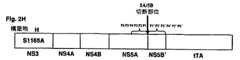
【図2】 それぞれのコントロールを有するテトラサイクリントランス活性化因子(tTA)と融合したHCVポリタンパク質の異なる部分を含むキメラの3つの異なるファミリーの模式図を示す。これらのキメラのHCVセグメントをコードするDNAは、pUHD15-1のNhe 1及びXba 1部位にXba 1フラグメントとしてクローン化されて、tTAキメラを産生する。
【図2A】 第一ファミリーの第一キメラを示し(構築物A)、組換え分子は、HCVの全長NS3タンパク質(プロテアーゼ及びヘリカーゼ領域の両方を含む)、全長NS4A(P6からP1で示される最後の6個のアミノ酸を含む)、tTAタンパク質と融合したP1'からP6'で示されるNS4Bの最初の6個のアミノ酸を含む。P6からP1及びP1'からP6'アミノ酸は、矢印で示される4A/4B切断部位を挟む。
【図2B】 第一ファミリーの第二キメラを示し(構築物B)、NS3タンパク質がセリンからアラニンまでのアミノ酸1165(S1165A)で突然変異したことを除いて2Aと同じ組換え分子である。この構築物はコントロールとして与えられ、突然変異はNS3プロテアーゼ活性を完全に取り除く。
【図2C】 第一ファミリーの第三キメラを示し(構築物B)、NS4A/4B切断部位に隣接するP1及びP1'で示される2つのアミノ酸が、それぞれシステイン及びアラニンからアルギニン及びプロリンまで変化させることを除いて2Aと同じキメラである。これらの変化は、この部位での切断を防ぐ。この構築物は切断部位のコントロールとして与えられる。
【図2D】 第二ファミリーの第一キメラを示し(構築物D)、この構築物はNS4A/NS4B切断部位を挟むアミノ酸配列(P6-P6')がNS5A/NS5B切断部位を定義するアミノ酸配列(P6-P6')で置換されるが、2Aのキメラと同様である。
【図2E】 第二ファミリーの第二キメラを示し(構築物E)、NS3プロテアーゼがセリンからアラニンまでのアミノ酸(S1165A)を突然変異することによって不活性化されることを除いて2Dと同じ構築物である。この構築物は第二キメラのためのコントロールとして与えられる。
【図2F】 第二ファミリーの第三キメラを示し(構築物F)、NS5A/5B切断部位に隣接するP1及びP1'で示される2つのアミノ酸が、それぞれシステイン及びセリンからアルギニン及びプロリンまで変化させることを除いて2Dと同じ構築物である。これらの変化はこの部位での切断を防ぐ。この構築物は5A/5B切断部位のコントロールとして与えられる。
【図2G】 第三ファミリーの第一キメラを示し(構築物G)、組換え分子は、全長NS3タンパク質、全長NS4A、NS4B及びNS5Aタンパク質とtTAタンパク質と融合した部分的なNS5Bタンパク質(P1'からP6'で示される最初の6個のアミノ酸からなる)との間の領域を含む。全長NS5A及び5BのP1'からP6'は矢印で示されるように、5A/5Bターゲット切断部位を挟む。
【図2H】 第三ファミリーの第二キメラを示し(構築物H)、それは、NS3プロテアーゼがセリンからアラニンまでのアミノ酸の突然変異(S1165A)により不活性化されることを除いて、2Gと同じ組換え分子である。この構築物は、NS3プロテアーゼ活性の負のコントロールとして与えられる。
【図2I】 第三ファミリーの第三キメラを示し(構築物I)、それは、NS5A/5B切断部位に隣接するP1及びP1'で示される2つのアミノ酸が、それぞれシステイン及びセリンからアルギニン及びプロリンまで変化させることを除いて、2Gと同じ組換え分子である。これらの変化はこの部位での切断を防ぐ。この構築物は5A/5Bターゲット切断部位のコントロールとして与えられる。
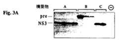
【図3】 ウエスタンブロット分析の結果を示す。PCR増幅及びTAクローニングにより発現プラスミドpCR3.1に挿入される図2A、2B及び2Cに記載されるキメラは(インビトロゲン, CA, USA)、T7 RNAポリメラーゼが存在する組換えT7ワクシニアウイルス(vvT7-3)とともにヒトの胚の腎臓細胞株293に過渡的にトランスフェクションされる。トランスフェクションし、感染させた細胞を増殖し、細胞タンパク質を抽出し、電気泳動し、ブロットし、プローブする。
【図3A】 ブロットがNS3タンパク質をポリクローナル抗体でプローブした場合の結果を示す。レーンA、B及びCは、2A、2B及び2Cに記載される構築物に対応し、レーン“−”は、ニセの293トランスフェクションされた細胞のコントロールを示す。レーンA及びCは成熟NS3タンパク質(NS3)の存在を示し、S1165A突然変異を有する構築物を表すレーンBは、予備的処理されたNS3(pre)の存在を示すが、いずれかの成熟NS3タンパク質を有するようには見えない。
【図3B】 ブロットがNS4Aをポリクローナル抗体でプローブした場合の結果を示す。レーンAは成熟NS4Aタンパク質(NS4A)の存在を示し、S1165A突然変異を有する構築物2Bを表すレーンBは、いずれかの成熟NS4Aタンパク質を有するようには見えず、同様に、突然変異した切断部位を有する構築物Cを表すレーンCも、成熟NS4Aタンパク質を有するようには見えない。54のアミノ酸からなる成熟NS4Aタンパク質を可視化するために、細胞抽出物は、膜に移動した16.5% SDS PAGEとリシンゲルで分解され、NS4Aポリクローナル抗体でプローブされる。得られたバンドは下のパネルに示される。
【図4】 ウエスタンブロット分析の結果を示す。PCR産物の直接TAクローニングにより発現プラスミドpCR3.1に挿入される図2D、2E及び2Fに記載されるキメラは、T7 RNAポリメラーゼが存在する組換えT7ワクシニアウイルス(vvT7-3)とともに哺乳類の宿主細胞株293に過渡的にトランスフェクションされる。トランスフェクションされた細胞を増殖し、細胞タンパク質を抽出し、電気泳動し、ブロットし、プローブする。結果はNS3タンパク質をポリクローナル抗体でプローブするブロットを示す。レーンD、E及びFは、それぞれ2D、2E及び2Fに記載される構築物に対応し、レーン“−”は、ニセの293トランスフェクションされた細胞のコントロールを示す。レーンD及びFは成熟NS3タンパク質(NS3)の存在を示し、他方ではS1165A突然変異を有する構築物を表すレーンEは、予備的処理されたNS3(pre)の存在を示すが、いずれかの成熟NS3タンパク質を有するようには見えない。
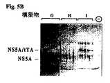
【図5】 ウエスタンブロット分析の結果を示す。PCR産物の直接TAクローニングにより発現プラスミドpCR3.1に挿入される図2G、2H及び2Iに記載されるキメラは、T7 RNAポリメラーゼが存在する組換えT7ワクシニアウイルス(vvT7-3)とともに哺乳類の宿主細胞株293に過渡的にトランスフェクションされる。トランスフェクションされた細胞を増殖し、細胞タンパク質を抽出し、電気泳動し、ブロットし、プローブする。
【図5A】 NS3タンパク質をポリクローナル抗体でプローブしたブロットの結果を示す。レーンG、H及びIは、それぞれ2G、2H及び2Iに記載される構築物に対応し、レーン“−”はニセの293トランスフェクションされた細胞のコントロールを示す。レーンG及びIは成熟NS3タンパク質(NS3)の存在を示すが、他方ではS1165A突然変異を有する構築物を表すレーンHは、いずれかの成熟NS3タンパク質を有するようには見えない。
【図5B】 NS4Aタンパク質をポリクローナル抗体でプローブしたブロットの結果を示す。レーンG、H及びIは、それぞれ2G、2H及び2Iに記載される構築物に対応し、レーン“−”はニセの293トランスフェクションされた細胞のコントロールを示す。レーンG及びIは成熟NS4Aタンパク質(NS4A)の存在を示すが、他方ではS1165A突然変異を有する構築物を表すレーンHは、いずれかの成熟NS4Aを有するようには見えない。
【図5C】 NS5Aタンパク質をポリクローナル抗体でプローブしたウエスタンブロットの結果を示す。レーンG、H及びIは、それぞれ構築物G、H及びIに対応し、レーン“−”はニセの293トランスフェクションされた細胞のコントロールを示す。レーンGは成熟NS5Aタンパク質(NS5A)の存在を示すが、他方ではS1165A突然変異を有する構築物を表すレーンHは、いずれかの成熟NS5Aを有するようには見えない。同様に突然変異したNS5A/5B切断部位を有する構築物Iを表すレーンIも、成熟NS5Aタンパク質を有するようには見えず、未処理のNS5A-tTA融合タンパク質は、約90kDaの産物として検出される。
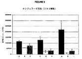
【図6】 構築物AからIの1つ及びpUHC13-3レポータープラスミドで同時トランスフェクションされた293細胞の抽出物で行ったルシフェラーゼアッセイの結果を示す。ルシフェラーゼ活性は1秒当たりのフォトン数の測定である。表示AからIは、それぞれ図2Aから2Iに記載される構築物に対応する。コントロールB、E及びHはプロテアーゼ活性部位に欠陥があり、他方でコントロールC、F及びIは欠陥のある切断部位を有する。このアッセイの結果は、ルシフェラーゼ活性が活性NS3プロテアーゼ及び機能的切断部位の両方に依存することを示す。さらに、結果は、構築物Gが構築物A又はDよりも大きなルシフェラーゼ活性を示すことを示す。これらの値は2つの実験の平均である。
【図7】 図6で示される値を用いて、構築物A、D及びGとそれぞれの活性部位突然変異体B、E及びHとによって産生されるルシフェラーゼ活性の比を示す。結果は、G/Hの比がA/B及びD/Eよりも、それぞれ約9倍及び2倍大きいことを示す。全長NS3、NS4A、NS4B及びNS5A、及びNS5Bの一部を含むHCV非構造的領域の最も長いひと配列を有する構築物Gは、最も大きなNS3依存ルシフェラーゼ応答を生じる。結果は、n回の実験の平均である。
【図8】 テトラサイクリン応答性転写性トランス活性化因子におけるテトラサイクリンの効果を示す。ルシフェラーゼレポーター遺伝子の発現は、テトラサイクリン応答性転写性トランス活性化因子のNS3プロセシングによって制御される。ルシフェラーゼアッセイは、構築物G又はH、又はポジティブコントロールtTA-産生性プラスミド(pUHD15-1)、及びルシフェラーゼレポーターを含むpUHC13-3プラスミドを同時トランスフェクションした293細胞の抽出物で行った。クローズドバー及びオープンバーは、それぞれテトラサイクリンの欠乏及び存在を示す。構築物Gは、テトラサイクリン制御シグナルを産生することに注意すべきであり、(構築物Hにおいて)NS3プロテアーゼ活性の不活性化は、このシグナルを無効にする。
【図9】 構築物AからIの1つ及びpUHC13-3レポータープラスミドを同時トランスフェクションした肝細胞株WRL68の抽出物で行われるルシフェラーゼアッセイの結果を示す。ルシフェラーゼ活性は、毎秒のフォトン数の測定である。コントロールB、E及びHは、プロテアーゼ活性部位において欠陥がある。コントロールC、F及びIは、欠陥のある切断部位を有し、このため機能的(切断する)NS3プロテアーゼ切断部位を有さない。このアッセイの結果は、ルシフェラーゼ活性が活性NS3プロテアーゼ及び機能的切断部位の両方に依存することを示す。さらに、この結果は、構築物Gが構築物A又はDよりも大きなルシフェラーゼ活性を示すことを示す。
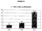
【図10】 構築物A、D及びGとそれぞれの活性部位突然変異体B、E及びHとによって産生されるルシフェラーゼ活性の比を示す。ルシフェラーゼアッセイは、構築物A、B、D、E、G又はHの1つ及びレポータープラスミドpUHC13-3を同時トランスフェクションした肝細胞株WRL68の抽出物で行った。ルシフェラーゼ活性は、毎秒のフォトン数の測定である。結果は、G/Hの比がA/B及びD/Eの比の、それぞれ約5倍及び3.5倍大きいことを示す。また、部分又は全長NS3、NS4A、NS4B、NS5A及びNS5Bを含むHCV非構造領域の最も長いひと配列を有する構築物Gは、WRL68細胞株の最も大きなNS3依存ルシフェラーゼ応答を産生するようにみえる。
【図11】 293細胞株を形質転換するためのプラスミドDNAの量の最適化の結果を示す。異なる量の構築物G又は対応する欠陥のあるNS3コントロール構築物HのDNAは、pUHC13-3の一定量(0.2μg)と同時トランスフェクションした。50%コンフルエンシーで6ウェルプレートの細胞を使用した。結果は、pUHC13-3(0.2μg)よりも約3倍多いNS3コードプラスミドDNAの量(0.6μg)が293細胞において最適なNS3依存ルシフェラーゼシグナルを産生することを示す。
【図12】 肝細胞株WRL68を形質転換するためのプラスミドDNAの量の最適化の結果を示す。異なる量の構築物G又は対応する欠陥のあるNS3コントロール構築物HのDNAは、pUHC13-3の一定量(0.2μg)と同時トランスフェクションした。結果は、pUHC13-3(0.2μg)よりも約2.5倍多い量(0.6μg)が肝細胞株WRL68において最適なNS3依存ルシフェラーゼシグナルを産生することを示す。
【図13】 構築物Iから発現されるNS3タンパク質が293細胞において、“トランス”のHCV非構造ポリタンパク質を切断できることを示す。実験は、pUHC13-3レポーターと構築物G、H又はIの1つを同時トランスフェクションすることを意味する。また、構築物H及びIは、pUHC13-3レポータープラスミドと一緒に同時トランスフェクションされる。構築物Gとレポーターとのトランスフェクションのみが、強いルシフェラーゼ活性シグナルを生じるのに対して、構築物Hとレポーター(NS3プロテアーゼ活性部位突然変異体)又は構築物Iとレポーター(切断部位突然変異体)のトランスフェクションは、弱いルシフェラーゼ活性を生じる。レポータープラスミドと一緒に構築物H及びIとの三重同時トランスフェクションは、ルシフェラーゼ活性シグナルを回復する。構築物Iから発現される機能的NS3タンパク質は、構築物Hから発現される無修飾切断部位を処理でき、それによってテトラサイクリンオペロンのルシフェラーゼの発現を開始するテトラサイクリントランス活性化因子を放出する。トランスフェクションで使用されるプラスミドDNAの異なる量は、示される通りである。
【図14】 肝細胞株WRL68を用いた図13と同様の実験の結果を示す。同様に、結果は、構築物Iから産生されるNS3タンパク質がWRL68細胞において、“トランス”のHCV非構造ポリタンパク質を切断できることを示す。示されるように、異なる量のプラスミドDNAはトランスフェクションで使用される。
【図15】 黒四角:構築物G及び分泌アルカリ性ホスファターゼ(SEAP)を発現するpUHC13-3レポータープラスミドを同時トランスフェクションされた宿主細胞におけるSEAP発現のtTAトランス活性化因子のHCV NS3プロテアーゼ活性の%阻害を示す。白四角:コントロールpUHD15-1及びpUHC13-3プラスミドを同時トランスフェクションされた宿主細胞においてトランス活性化SEAPのコントロールtTA(HCV NS3とは無関係)活性の%阻害を示す。細胞は、種々の濃度の阻害剤Aの存在下でトランスフェクション後に増殖した。SEAP活性を測定し、EC50及びTC50値を決定した。
【配列表】
Field of Invention
The present invention relates to mammalian cell culture systems and methods for assaying hepatitis C virus (HCV) NS3 protease activity and inhibition thereof. More particularly, the present invention relates to recombinant molecules, transfected mammalian host cell assay systems and methods for measuring NS3 protease activity and its inhibition by candidate anti-HCV compounds.
Background of the Invention
Hepatitis C virus (HCV) is an important etiological agent after blood transfusion and is a worldwide community-acquired non-A non-B hepatitis. It is estimated that over 1 million people worldwide are infected by the virus. Many carriers are chronically infected and many progress to a chronic liver disease called chronic hepatitis C. This group is at high risk for serious liver diseases such as cirrhosis, hepatocellular carcinoma and end-stage liver disease leading to death. The mechanism by which HCV establishes virus persistence and causes a high rate of chronic liver disease has not been elucidated in detail. It is not known how HCV interacts with and escapes the host immune system. Furthermore, the role of cellular and humoral immune responses in protection against HCV infection and disease must be elucidated.
[0002]
Viral clinical research is being conducted to see if pharmaceutical compounds can efficiently treat HCV infection in patients affected by chronic hepatitis C. These studies include the use of interferon-α alone and in combination with other antiviral agents such as ribavirin. Such studies have shown that a substantial number of participants do not respond to these treatments, which is a favorable response and has been found to relapse after the end of treatment at a high rate. To date, there are no widely effective antiviral compounds for the treatment of HCV infection.
[0003]
HCV is an enveloped positive strand RNA virus of the Flaviviridae family.
The single-stranded HCV RNA genome is approximately 9500 nucleotides in length and has a single open reading frame (ORF) that encodes a single large polyprotein of approximately 3000 amino acids. In infected cells, this polyprotein is cleaved at multiple sites by cells and viral proteases to produce structural and nonstructural (NS) proteins. Structural proteins (C, E1, E2, and E2-p7) contain the polypeptides that make up the viral particles. Nonstructural proteins (NS2, NS3, NS4A, NS4B, NS5A, NS5B) encode enzymes or accessory factors that catalyze and control the replication of the HCV RNA genome. Structural protein processing is catalyzed by host cell proteases. The production of mature nonstructural protein is catalyzed by two virally encoded proteases. The first is an NS2-3 zinc-dependent metalloprotease that autocatalyses the release of polyprotein-derived NS3 protein. The released NS3 contains a serine protease region at the N-terminus and catalyzes remaining cleavages from the polyprotein. The released NS4A protein has at least two roles. The first is to form a stable complex with the NS3 protein and assist in membrane localization of the NS3 / NS4A complex (Kim et al., 1999), and the second is a cofactor for NS3 protease activity. Is to act as This membrane-bound complex catalyzes cleavage of the remaining sites of the polyprotein, thus leading to the release of NS4B, NS5A and NS5B. The C-terminal segment of NS3 protein also enters nucleoside triphosphatase and RNA helicase activities. NS5B is an RNA-dependent RNA polymerase involved in HCV replication.
[0004]
The N-terminal 180 amino acids of NS3 protein is a trypsin-like serine protease that mediates NS3 / 4A auto-cleavage as the first step. Furthermore, the membrane-bound NS3 / 4A complex cleaves the NS4A / 4b, NS4B / 5A and NS5A / NS5B junctions, releasing viral enzymes believed to be necessary for viral replication. NS3 / NS4A conjugation is an important step in the downstream processing of polyproteins over time. NS3 protein is the most characterized HCV protein. Kinetic parameters for cleavage of all processing sites have been described. Both the N-terminal protease and C-terminal helicase regions are crystallized independently, and a high-resolution three-dimensional model exists for these structures.
NS3 protease activity is an attractive target for drug discovery. Enzyme studies have shown that peptides based on the N-terminal product of the NS5A / 5B cleavage site are competitive inhibitors of the enzyme. These peptides serve as useful starting points in medicinal chemistry attempts to rationally design NS3 protease inhibitors as well as clinically effective anti-HCV compounds.
[0005]
Because of the high incidence of HCV and the consequences of HCV infection, discovery of therapeutic compounds for HCV has become important. To that end, attempts to find compounds against HCV require the development of assay systems for screening and selecting anti-HCV compounds.
To date, no convenient cell culture replication system for HCV virus is available. This lack limits the screening of most HCV inhibitors to in-vitro enzyme assays and indirect surrogate cell-based assays (Lohmann et al. (1999)). This significantly limits the evaluation of compounds with potential as anti-HCV in cell culture.
[0006]
Hirowatari et al. (1995) described a first plasmid containing an NS5A / NS5B cleavage site (NS3 substrate) and the NS2 / NS3 region fused to the
[0007]
Overton, H. et al. (1995) teach baculovirus-expressed HCV NS3 activity in insect cells. A series of baculovirus constructs and various substrates designed to express NS3 protease activity are described. Some NS2 / NS3 and NS3 / NS4A / NS4B encoding constructs are transfected into insect cell lines. Additional viral constructs encoding NS3 through NS5A and NS5A / 5B are transfected alone or together with one of the above constructs. Expressed and cleaved products are visualized immunologically by Western analysis. Some deficiencies in this system relate to the use of insects instead of mammalian cell lines, as well as the need for viral infections (this phenomenon further disrupts the normal functioning of cells). This system also utilizes a T7 polymerase expression system (this system is not necessary for the system of the present invention). In addition, degradation product detection methods are long, difficult to quantify accurately, and do not allow high throughput scales.
[0008]
Song et al. (1996) described a protease assay system that utilizes a lexA-GAL4 fusion protein in yeast. The authors describe the insertion of NS3 protease and cleavage site between the lexA-DNA binding region and the transcriptional active region of GAL4. Cleavage of that site by NS3 protease renders GAL4 transcriptionally inactive so that transformed yeast cannot synthesize β-galactosidase. This system lacks NS4A and does not reproduce processing in the HCV polyprotein.
Cho, Y.G. et al. (1997) teach an assay using the Sindbis virus replication system. This hybrid virus construct encodes the HCV NS3 / NS4A protease region linked to the SIN core protein by an NS4A / NS4B cleavage sequence. The construction of virions in mammalian cell lines relies on the processing of NS4A / 4B cleavage sites. One major drawback of this system is that it produces a chimeric virus in which HCV protease activity induces cytopathic morphological changes in the cells. Measuring the change in pH of the medium at best constitutes a semi-quantitative method for measuring these changes. Another disadvantage of this system is that the Sindbiscore protein contains a natural serine protease cleavage site as well as an NS3 site, thus limiting the system to use for screening potential protease inhibitors.
[0009]
Cho, Y.G. et al. (1998) teach an expression vector encoding an NS4A / 4B cleavage site fused to a SEAP (secreted alkaline phosphatase) gene transfected into an NS3 / NS4A region and a mammalian cell line. Cleavage of the NS4A / 4B site by NS3 protease releases the SEAP protein into the medium. The amount of SEAP protein in the medium is a measure of NS3 protease activity. One drawback of this system lies in the fact that the reporter molecule is directly fused to the substrate protein, thus affecting the natural conformation of the viral complex protein (NS3 / NS4A). Another major drawback is due to the fact that the amount of reporter protein secreted is expressed in the system and is directly proportional to the amount of protein cleaved (one substrate molecule cleaved = one reporter molecule secreted). Since the HCV polyprotein is a very low level expression system (even in its natural context), the observed signal is too low to be performed on a large screening scale.
[0010]
WO 98/00548 describes a hybrid virus comprising a piconavirus (preferably a poliovirus), an HCV NS3 protease region and a single NS3 protease target site. These chimeric viruses are engineered so that the proteolytic processing activity of HCV NS3 is essential for virus viability and growth. Various hybrids with different NS3 cleavage sites (NS5A / NS5B, NS4A / NS4B or NS4B / NS5A) are taught. Viability is measured by viral titer using a HeLa monolayer plaque assay. Again, this system does not provide a quantitative screening method for large amounts of potential inhibitors in a high-throughput manner, nor does it provide a screen for NS3 protease activity in the natural context of polyprotein segments. .
[0011]
US Pat. No. 5,861,267 by Vertex discloses a method for assaying HCV NS3 protease that utilizes NS4A / NS4B cleavage sites fused to the expression and secretion of the IL-1β reporter (interleukin-1β) of the NS3 / NS4A region. Cleavage of the NS4A / 4B site by NS3 protease releases IL-1β into the medium where NS3 protease activity can be measured directly. This system tests inhibition of NS3 cleavage at the cleavage site adjacent to the NS3 / NS4A complex, and this situation does not imply standard conditions for multisite polyprotein processing or replicate. In addition, this system provides a reporter system in which the signal measured is directly proportional to the amount of protein expressed in the system and the amount of protein cleaved, which in this case is too low to be performed on a large screening scale. Produce.
[0012]
WO 00/08469 by Agouron discloses another system comprising a protease reporter construct consisting of NS2 metalloprotease, NS3 protease, NS4A cofactor and various cleaved NS4B and 5A (NS5A / 5B cleavage sites described above) . However, this system does not disclose the importance of having a complete polyprotein to optimize NS3 protease activity / specificity. In addition, this system requires infection by vaccinia virus vectors, a factor that relaxes host cell integrity and affects the mechanism of cis or transprotease cleavage during the polyprotein processing event of the assay. May affect. Another disadvantage of using vaccinia expression is due to the fact that the cells of the assay become necrotic after about 24 hours, thus limiting the use of this assay for longer term kinetic assays.
[0013]
WO 00/12727 by Vertex discloses a system having a fusion protein that includes a ligand binding region, which can bind to a ligand response element that generates a VP16 activation region and control the expression of a reporter gene. The NS5A / 5B cleavage site is inserted into the fusion protein and regulates reporter gene expression upon cleavage by the NS3 / 4 protease expressed from the isolated construct. Again, this system does not disclose the importance of having a complete polyprotein to optimize NS3 protease activity / specificity.
Therefore, the ability to scale up to high throughput systems makes it important to develop screening assays for large quantities of anti-HCV compounds. Thus, the present invention provides an assay that is easy to implement, reliable, sensitive and reproducible on a large scale.
Accordingly, it is an object of the present invention to provide an assay with improved sensitivity for measuring cell-based systems and inhibitors of HCV NS3 protease activity, which assay is performed on infected cells in the course of HCV disease. It is designed to simultaneously test the protease activity of constructs that reproduce as many NS3 polyprotein processing events as possible.
[0014]
Summary of the Invention
Thus, the present invention relates to the development of a cell-based assay system that has improved sensitivity to HCV NS3 protease activity when compared to known assays, and for compounds that can modulate (especially inhibit) HCV NS3 protease activity. Useful for screening tests.
The present invention provides a first construct comprising a transactivator region associated with the downstream of the NS3-5 region of HCV under the control of a non-cytopathic viral promoter system. Also provided is a second construct comprising a reporter gene under the control of an operator sensitive to transactivator binding.
The NS3-5 region consists of NS3 protease followed by NS4A cofactor, NS4B and NS5A proteins (including any derivative, variant or fragment thereof), NS5B protein sufficient to constitute the NS5A / 5B cleavage site (optional NS3 polyproteins including those terminated by (including derivatives, variants or fragments thereof). When expressed and released from a polyprotein, the transactivator initiates transcription and expression of a reporter gene that is measurable.
[0015]
The advantage of this system is that when expressed, the NS4A cofactor region is embedded in the endoplasmic reticulum membrane and, as a result, complexed with NS4A, anchors the NS3 protease, and the cleavage site downstream of the polyprotein If untreated, it contains many times the fact that it reduces the background signal (by preventing non-specific translocation). The advantage of this system is also in the amplification of various levels of the signal measured. Applicants believe that the first level of amplification is in the fact that the protein sensuously reproduces the native conformation of the polyprotein and the natural processing phenomena that occur during human infection. This increases the activity of NS3 protease cleavage as well as its specificity. Thus, signal translocation to nuclear and reporter expression is a more specific and minimally reduced background noise that is acceptable for high throughput screening assays.
[0016]
Also, the use of a transactivator system provides a second level of amplification that further increases signal output.
Finally, this transactivator system is sensitive to the presence of antibiotics and gives a measure of the assay's inherent background noise, thus guaranteeing the specificity of both the assay and the test compound. (Built-in) ”negative control.
Thus, the present invention provides a cell-based assay that has the ability to measure NS3 protease inhibition in a system that closely mimics the processing of HCV nonstructural proteins in a situation that substantially mimics the natural infection process. When compared to other known assays, the mammalian cell-based system of the present invention is sensitive and exhibits an increased signal / noise ratio. Thus, assays using this system can be easily scaled up to high throughput screening systems.
[0017]
Thus, according to a first embodiment of the present invention, there is provided a surrogate cell-based system for evaluating the protease activity of hepatitis C virus NS3 protease comprising: a) a first chimeric molecule having:
i) a non-cytopathic expression system capable of including expression of said first chimera by transfection of mammalian cells; and
ii) an HCV recombinant DNA molecule capable of binding to the expression system; wherein the HCV DNA molecule encoding NS3-5 polyprotein comprises:
Active NS3 protease region,
NS4A region sufficient to be embedded in the ER membrane by translation and act as a cofactor for NS3 protease activity,
NS4B and NS5A regions containing any derivative, variant or fragment thereof, and
An NS5B protein comprising any derivative, variant or fragment thereof sufficient to provide an NS5A / 5B cleavage site for said NS3 protease;
iii) and a transactivator region fused downstream of the HCV DNA molecule, wherein the transactivator region encodes a transactivator molecule capable of initiating expression of a reporter gene;
b) and a second chimeric DNA molecule encoding the reporter gene associated with an operon reactive with the transactivator molecule;
Thereby, the expression of the first recombinant molecule leads to the production of a fusion polyprotein immobilized on the endoplasmic reticulum of the mammalian cell, and the immobilized protein is cleaved by a protease, with the result that the protease activity is evaluated as a method. , Provide translocation of the transactivator region to include expression of the reporter gene.
[0018]
In a second embodiment, the present invention encompasses recombinant DNA molecules and any fragments, variants and derivatives thereof that are useful in this system.
In a third embodiment, the present invention encompasses a recombinant protein produced from a recombinant DNA molecule of the present invention.
In a fourth embodiment, the present invention also encompasses a vector comprising any of the recombinant DNA molecules.
In a fifth embodiment, the present invention includes eukaryotic host cells transfected with these vectors.
In a sixth embodiment, the present invention includes a method for assaying NS3 protease activity using the recombinant molecules of the present invention and transfected host cells, and the method identifies its potential as an inhibitor. Useful for.
Other objects, advantages and features of the present invention are illustrative and should not be construed as limiting the scope of the invention by reading the following non-limiting description of preferred embodiments with reference to the accompanying drawings. It will become clear.
[0019]
Detailed Description of the Preferred Embodiment
Definition
Unless defined otherwise, chemical and technical terms and nomenclature used herein have the same meaning as commonly understood by one of ordinary skill in the art to which this invention pertains. In general, procedures such as cell culture, infection, molecular biology and the like are common methods used in the art. Such standard methods are found in reference manuals such as Sambrook et al. (1989) and Ausubel et al. (1994).
Nucleotide sequences are single-stranded, in the 5 'to 3' direction, left to right, using the one-letter nucleotide symbol commonly used in the technology, the IUPAC-IUB Biochemical Nomenclature Commission ) (1972) according to recommendations.
The description herein refers to a number of commonly used recombinant DNA (rDNA) technical terms. Nevertheless, the determination of selected examples of such rDNA terms is provided for clarity and consistency.
[0020]
The term “recombinant DNA”, “recombinant nucleic acid molecule” or “recombinant plasmid” as known in the art refers to a DNA molecule resulting from the joining of DNA segments. This is often referred to as genetic manipulation.
As used herein, the term “DNA segment or molecule or sequence” refers to a molecule comprising the deoxyribonucleotides adenine (A), guanine (G), thymine (T) and / or cytosine (C). These segments, molecules or sequences are found in natural or synthetic derivatives. When decoded according to the genetic code, these sequences can encode linear sequences or sequences of amino acids called polypeptides, proteins, protein fragments, and the like.
The term “gene” as used herein is well known in the art and refers to a nucleic acid sequence that defines a single protein or polypeptide. A polypeptide is encoded by the full-length sequence or any portion of the coding sequence so long as the functional activity of the protein is retained.
[0021]
A “structural gene” defines a DNA sequence that is transcribed into RNA and translated into a protein having a specific amino acid sequence, resulting in a specific polypeptide or protein. “Structural proteins” define HCV proteins that are incorporated into the viral particle, ie, cores “C”, E1, E2, and E2-p7. “Nonstructural protein” defines HCV proteins that are not contained in the viral particle, ie NS2, NS3, NS4A, NS5A and NS5B.
A “restriction endonuclease or restriction enzyme” recognizes a specific base sequence (usually 4, 5 or 6 base pairs in length) of a DNA molecule and has the ability to cleave the DNA molecule wherever this sequence appears It is an enzyme. An example of such an enzyme is EcoRI that recognizes the base sequence G ↓ AATTC and cleaves a DNA molecule at this recognition site.
A “restriction enzyme fragment” is a DNA molecule produced by the degradation of DNA by a restriction endonuclease. Any given genome or DNA segment is broken down into at least two separate molecules of restriction enzyme fragments by a specific restriction endonuclease.
[0022]
“Agarose gel electrophoresis” is an analytical method for fractionating double-stranded DNA molecules based on the size of the DNA. This method is based on the fact that the DNA molecules move through the gel as it passes through the sieve, whereby the smallest DNA molecule has the highest mobility and travels the farthest through the gel. The sieving properties of the gel retard the largest DNA molecules, which have the least mobility. Fractionated DNA is visualized by gel staining using methods well known in the art, nucleic acid hybridization or tagging of fractionated DNA molecules by detectable label. All these methods are well known in the art and specific methods are found by Ausubel et al. (Supra).
An “oligonucleotide or oligomer” is a molecule comprising two or more, preferably more than three deoxyribonucleotides or ribonucleotides. The exact size of the molecule depends on many factors and also on the final function or use of the oligonucleotide. Oligonucleotides are derived synthetically, by cloning or by amplification.
[0023]
“Sequence amplification” is a method of producing large amounts of target sequences. In general, one or more amplification primers are annealed to a nucleic acid sequence. Using suitable enzymes, sequences found near or between primers are amplified. The amplification method used herein is the polymerase chain reaction (PCR).
“Amplification primary” refers to an oligonucleotide that can anneal a region of DNA near a target sequence and serves as a starting primer for DNA synthesis under suitable conditions well known in the art. The synthetic primer extension product is complementary to the target sequence.
The term “domain” or “region” refers to a specific amino acid sequence that defines a specific function or structure within a protein. As an example herein, the NS3 protease region is included in the HCV nonstructural polyprotein.
[0024]
The term “reporter gene” refers to a nucleotide sequence that encodes a “reporter protein”. Reporter proteins provide a detectable method for assessing gene expression. The reporter gene is included in a “reporter plasmid”. Useful reporter genes for the purposes of the present invention include secreted alkaline phosphatase (SEAP), luciferase, chloramphenicol aminotransferase (CAT), β-galactosidase, green fluorescent protein (GPF) and the like.
The term “reporter system” refers to a combination of two or more reporters. Non-limiting examples of reporter systems useful for the purposes of the present invention are first reporters that are gene activators such as tTA that govern the expression of second reporters such as luciferase or secreted alkaline phosphatase (SEAP). is there.
The terms “activator” and “operon” refer to a system in which an “activator” molecule binds to an operator contained in the operon and stimulates the expression of the “operon”. Such systems include tetracycline transactivator (tTA), HIV-1 tat transactivator, GAL4 transactivator, NFκβ and the like.
[0025]
The term “chimeric molecule” or “chimera” as used herein refers to at least two nucleic acid regions that are not naturally joined together. Non-limiting examples of such chimeras of the invention include the HCV NS3-4A-4B-5A-5B-tTA region construct. When expressed, such chimeras produce “fusion proteins”.
The term “fusion protein” as defined herein refers to at least two polypeptide segments that are not naturally joined together. Non-limiting examples of such “fusion proteins” of the invention include a portion of an HCV polyprotein that binds to a protein having reporter ability, directly or indirectly, such as tTA and SEAP.
The term “plasmid”, “vector” or “DNA construct” is generally known in the art, refers to any genetic element, incorporates an oligonucleotide sequence or a sequence of the invention, and the DNA into which the DNA of the invention is cloned. Examples include, but are not limited to, plasmid DNA, phage DNA, and viral DNA that serve as a medium. Many types of vectors exist and are well known in the art.
[0026]
The term “expression vector” defines a vector as described above, but is designed such that the inserted sequence can be expressed after transformation or transfection into the host. The cloned gene (inserted sequence) is usually placed under the control of a regulatory region sequence such as a promoter sequence. Such expression control sequences can be used to determine whether the vector is designed to express a prokaryotic or eukaryotic host or both bindable genes (shuttle vectors), as well as enhancer elements, termination sequences, tissue specificity. It varies depending on whether or not the elements and / or transcriptional elements such as translation initiation and termination sites are included.
The term “non-cytopathic” expression system defines a system that does not induce a cytopathic change in the expressed cellular system. This expression system does not require co-infection with the virus and does not result in the expression of non-HCV viral components that ultimately induce cell pathology and cell death. Examples of non-cytopathic promoters are the CMV promoter system, the SV40 early promoter system or the RSV (Rous Sarcoma Virus) LTR promoter system.
[0027]
“Eukaryotic expression system” means a combination of a suitable expression vector and a eukaryotic cell line used to express a gene of interest. Moreover, you may use the plasmid vector containing the designed gene. In all cases, the vectors contain suitable regulatory regions (promoters) and express genes of the cell type of interest. Eukaryotic cell types typically used are yeast cells transfected with plasmid vectors (eg, Saccharomyces cerevisiae, Pischia pastoris), and mammalian cells transfected with DNA vectors for transient or constitutive expression It is. Preferred cell lines useful for the purposes of the present invention are derived from liver tissue.
[0028]
A host cell or indicator cell is “transfected” by exogenous or heterologous DNA (eg, a DNA construct) when such DNA is introduced into the cell. The DNA to be transfected may or may not be integrated into the chromosomal DNA that produces the cell's genome (covalently linked). In prokaryotic, yeast and mammalian cells, for example, transfected / transformed DNA may be maintained on episomal elements such as plasmids. For eukaryotic cells, an example of a stably transfected cell is one in which the DNA to be transfected is integrated into a chromosome and is inherited by daughter cells via chromosome replication. This stability is demonstrated by the ability to establish cell lines or clones that contain a population of daughter cells that contain DNA to be transfected by eukaryotic cells. Transfection methods are well known in the art (Sambrook et al., 1989, Ausubel et al., 1994).
Nucleotide sequences and polypeptides useful for practicing the invention include, but are not limited to, mutants, homologs, subtypes, quasi-species, alleles, and the like. In general, it is understood that the sequences of the invention encode polyproteins. It will be apparent to those skilled in the art that the polyproteins of the invention and any variants, derivatives or fragments thereof self-process active proteases.
[0029]
As used herein, the name “variant” is a biological activity (function) that, in the context of the present invention, is substantially similar to that of the original sequence, whether the sequence is a nucleic acid or an amino acid. Or a molecule that retains (structural). This mutant may be derived from the same or different species, may be a natural mutant, or may be prepared synthetically. Such mutants include amino acid sequences in which one or more amino acids are substituted, missing or added, provided that the biological activity of the protein is preserved. The same applies to variants of nucleic acid sequences in which one or more nucleotides are substituted, missing or added, provided that the biological activity of the sequence is generally retained.
[0030]
The term “derivative” is meant to include any of the variants described above if some of these molecules contain additional chemicals that are not normal. These chemicals have a variety of purposes, including molecular solubility, absorption, improved biological half-life, reduced toxicity and elimination or reduction of undesirable side effects. Furthermore, these substances are used for the purpose of labeling and binding, or they may be included in the fusion product. Different substances that can mediate the above effects are found in The Science and Practice of Pharmacy (1995) by Remington. Methods for binding such substances to molecules are well known in the art.
The term “fragment” refers to any segment of the identified DNA, RNA or amino acid sequence and / or any of the mutations described above that substantially retain its biological activity (functional or structural) as required by the present invention. Reference to any segment of the body or derivative.
The terms “variant”, “derivative” and “fragment” of the present invention refer to a protein or diffusion molecule that is isolated / purified, chemically synthesized, or produced by recombinant DNA technology. All these methods are well known in the art. As demonstrated herein below, the nucleotide sequences and polypeptides used in the present invention are modified, for example, by in vitro mutagenesis.
[0031]
For example, as defined in the present invention, the desired fragment of NS3 protein must retain its protease activity. The desired fragment of NS4A protein retains its protease cofactor activity (functional activity) and also retains its hydrophobic protein embedded in the ER membrane (structural activity). The desired fragment of the NS4B protein retains its structural activity in order to process the polyprotein as close as possible to the natural one. The desired fragment of the NS5A protein retains its structural activity to process the polyprotein as close as possible to the natural one, but also retains its functional activity to create a functional NS5A / 5B cleavage site. provide. The desired fragment of the NS5B protein must be sufficient to retain a functional NS5A / 5B cleavage site. For example, the first six amino acids of NS5B protein are sufficient for this purpose. Such functional or structural activity of each region can be readily assessed by one of ordinary skill in the art without undue experimentation.
[0032]
As used herein, the term “cleavage site” refers to a polypeptide that is cleaved “cis” or “trans” by HCV NS3 protease.
The term “target cleavage site” or “target site” as used herein refers to a cleavage site downstream of the NS3 / 4A cleavage site. At least one target site of NS3 protease must be cleaved to obtain transactivator translocation and reporter gene expression and to achieve measurement of the reporter gene product. In the present specification, the NS3 / 4A cleavage site is not considered a “target” site because its cleavage by itself does not release the reporter gene product.
As used herein, the term “functional cleavage site” refers to a cleavage site of a precursor polypeptide that has been modified (by mutation or any other chemical method) but is ready for cleavage by NS3 protease. means. Such modifications include, but are not limited to, nucleic acid codons or amino acid conserved substitutions.
[0033]
Preferred embodiment
Cell-based assay
Thus, according to the first embodiment of the present invention, a surrogate cell-based system for assessing the protease activity of hepatitis C virus NS3 protease comprising the following is provided:
a) First chimeric DNA molecule comprising:
i) a non-cytopathic expression system that can include expression of said first chimera by transfection of mammalian cells;
ii) an HCV recombinant DNA molecule capable of binding to the expression system; wherein the HCV DNA molecule encodes an NS3-5 polyprotein comprising:
-An active NS3 protease region,
-NS4A region sufficient to be embedded in the ER membrane by translation and act as a cofactor for NS3 protease activity,
-NS4B and NS5A regions including any derivative, variant or fragment thereof, and
-An NS5B protein comprising any derivative, variant or fragment thereof sufficient to provide an NS5A / 5B cleavage site for said NS3 protease;
iii) and a transactivator region fused downstream of the HCV DNA molecule, wherein the transactivator region encodes a transactivator molecule capable of initiating expression of a reporter gene;
b) and a second chimeric DNA molecule encoding the reporter gene associated with an operon responsive to the transactivator molecule;
Thereby, the expression of the first recombinant molecule leads to the production of a fusion polyprotein anchored to the endoplasmic reticulum of the mammalian cell, which can be cleaved by the protease, so that Translocation of the transactivator region to include expression of the reporter gene as a method for assessing protease activity is provided.
[0034]
In a preferred aspect of the first embodiment, the chimeric molecule is a nucleotide sequence capable of expressing an HCV polyprotein having sufficient NS3 region, variant, derivative or fragment thereof to provide an active protease once translated. Is included. This includes NS3 proteins that have been cleaved to eliminate the helicase region, or where the helicase region is inactivated. The NS4A region required for the present invention is sufficient for the translated NS4A protein, its variants, derivatives or fragments to embed the ER membrane polyprotein by translation and act as a cofactor for NS3 protease activity. You need to be.
The NS4B region required for the present invention requires that the translated protein be of sufficient length to give the correct structural orientation of NS3 protease taking into account the NS5A / 5B target cleavage site. Thus, a variant, derivative or fragment of an NS4B protein has its native biological activity at its cleavage site (4A / 4B or 4B / 5A) or without compromising the orienting effect of the polyprotein. Can be mutated to eliminate.
[0035]
The NS5A region required for the present invention requires that the translated protein be long enough to give the correct structural orientation of the NS3 protease, taking into account the NS5A / 5B target cleavage site. Thus, a variant, derivative or fragment of an NS5A protein can be mutated at its upstream cleavage site (4B / 5A) or without loss of its stereotactic effect at the polyprotein and NS5A / 5B cleavage site. Mutations can be made to eliminate activity.
Finally, the NS5A / 5B cleavage site required for the present invention must be recognized and cleaved by a functional or NS3 protease. Such functional target cleavage sites include derivatives, variants or fragments thereof as long as they can be cleaved by NS3 protease. A preferred embodiment of the NS5A / 5B cleavage site includes the first six amino acids of the NS5B region.
[0036]
In another aspect of this first embodiment, a nucleotide sequence encoding a full length NS3 polyprotein is provided. Alternatively, a partial nucleotide sequence is provided that encodes a partial polyprotein comprising a required region variant as described above.
Alternatively, the chimeric molecule comprises a nucleotide sequence capable of expressing derivatives, variants or fragments of precursor polyproteins including all functional cleavage sites: NS3 / 4A; 4A / 4B; 4B / 5A and 5A / 5B. Include. Alternatively, at least NS5A / 5B is a functional cleavage site, but other cleavage sites may be modified to be non-functional.
According to a particular aspect of this first embodiment, the non-cytopathic promoter system comprises a CMV promoter, SV40 early promoter system or RSV (Rous Sarcoma Virus) LTR promoter system.
[0037]
According to a particular aspect of this first embodiment, the reporter system comprises a transactivator region associated with the first recombinant nucleic acid, so that when cleaved from the expressed fusion protein, the transactivator Activates the expression of a reporter gene that migrates to the nucleus and the product is measured as a method of assessing NS3 protease activity. Fusion of the transactivator region provides an amplification level that increases the sensitivity of the assay. In accordance with this particular aspect, activation of the reporter gene system by a transactivator may be specifically suppressed as a means to ensure that cleavage of the precursor polyprotein is specific (integrated negative control). (Built-in negative control)). Such special aspects include a system adapted to the method of Gossen, M. and Bujard, H. (1992) that describes the design and potential use of tetracycline-regulated transactivators in mammalian cell lines. Including.
In this method, the tet repressor fuses with the activation region of herpes simplex virus protein 16 (VP16), resulting in the production of a tetracycline-dependent transactivator (tTA). This tTA initiates transcription / expression of the reporter gene under the control of the bound tet operon / viral promoter. The product of the reporter gene is an indicator of gene activation and protein expression.
[0038]
However, in a preferred aspect of the reporter system, the transactivator can be either a chimeric molecule or a fusion protein as long as it is downstream of the target cleavage site and is thus released by cleavage. More preferably, the transactivator is located at the 3 ′ end of the recombinant molecule. Transactivators include, but are not limited to, tetracycline transactivator (tTA), NFκB, HIV-1 tat and GAL-4. More preferably, the transactivator is tTA. Reporter molecules include, but are not limited to, secreted alkaline phosphatase, β-galactosidase, luciferase, chloramphenicol aminotransferase, and green fluorescent protein. Preferably, the reporter molecule is SEAP or luciferase. More preferably, the reporter molecule is SEAP.
[0039]
In a preferred aspect, the reporter system is a combination of tTA and luciferase or a combination of tTA and SEAP. More preferably, the reporter system is a combination of tTA and SEAP.
It is an aspect of the present invention that different permutations of transactivator and reporter system are combined using methods well known in the art, all of which are within the scope of the present invention. Since there are different reporter molecules useful for the purposes of the present invention, detection of the reporter gene product is consistent with the use of known methods. For example, luciferase is measured by chemiluminescence reaction using coenzyme A (Promega Luciferase Assay System, NI, USA) as the luciferase substrate, and SEAP is the CSPD substrate and enhancer (Tropix, Inc., MA, USA). Is measured by a chemiluminescent reaction incorporating.
[0040]
Recombinant DNA molecules and proteins
In a second embodiment, the present invention encompasses recombinant DNA molecules useful in this assay and any fragments, variants and derivatives thereof. According to a particular aspect of this second embodiment of the invention, there is provided a nucleic acid molecule as defined in SEQ ID NO.1.
In a third embodiment, the present invention includes a recombinant protein produced from the recombinant DNA molecule of the present invention. According to a particular aspect of this second embodiment, the amino acid sequence defined in SEQ ID NO. 2 is provided.
In accordance with the above-described embodiments of amino acid and nucleotide sequences, all variants, derivatives and fragments thereof that are functionally equivalent to the sequences herein are within the scope of the invention.
[0041]
vector
In a fourth embodiment, the present invention also includes a vector comprising any of the recombinant DNA molecules. It is a special aspect of this fourth embodiment that the nucleic acid molecule of SEQ ID NO.1 is inserted into an expression plasmid for transfection into a mammalian host cell. In a preferred aspect of the fourth embodiment, the different plasmids, vectors or viruses useful for the purposes of the present invention are human expression plasmids capable of stably or transiently transforming human host cells, Contains vector or virus. These are known and within the scope of the present invention. More preferably, the plasmid is selected from the group consisting of a pUHD15-1 CMV promoter base plasmid in which the tTA gene is present and a pUHC13-3 tTA-controllable luciferase or SEAP reporter plasmid. In the most preferred aspect of this fourth embodiment, the nucleic acid of SEQ ID NO.1 is inserted into the pUHD15-1 plasmid.
[0042]
Transfection
Transfection of the above-described vector in the host cell may be performed simultaneously (co-transfection) or sequentially. Alternatively, transfection of the reporter system may be performed first and later used in a second transfection of the HCV region-containing vector to obtain stable transfectants, which are useful in the present invention. Get heavy transfectants (transient or stable).
[0043]
Host cell
In a fifth embodiment, the present invention encompasses eukaryotic host cells transfected with these vectors. Accordingly, according to a particular aspect of this fifth embodiment, a host cell transfected with an expression plasmid comprising a chimeric molecule as defined above is provided. More particularly, a mammalian cell line transfected with the nucleic acid molecule of SEQ ID NO.1 is provided. In a preferred aspect of the fifth embodiment, the host cell is a mammalian cell that is stably or transiently transformed. Mammalian host cells and cell lines include, but are not limited to, primary hepatocytes, hepatocyte cell lines, fibroblasts, lymphocytes, kidney cells and the like. More preferably, a transient human cell line transfected with the chimeric molecule is provided. Even more preferably, the human cell line is a liver or kidney cell line. Most preferably, the host cell may be selected from the group consisting of 293, Huh-7, WRL68, HepG2, and Chang cells.
[0044]
Inhibitor identification method
In a sixth embodiment, the present invention includes a method for assessing NS3 protease activity using a recombinant molecule and a transfected host cell of the present invention. In a particular aspect of this sixth embodiment, there is provided a method of screening for anti-HCV inhibitors using the system defined above. According to a particular aspect of this sixth embodiment, there is provided an assay method for HCV NS3 protease activity comprising the following steps:
a) first incubating the transfected host cells under conditions that yield the HCV NS3-5 polyprotein and the reporter gene product expressed as described above; and
b) measuring the amount of the gene product expressed.
[0045]
According to another particular aspect of this sixth embodiment, a method of identifying a compound as an inhibitor of HCV NS3 protease comprising the following steps is provided:
a, b) assaying the activity of the protease in the absence of the compound by the method as defined above; and
c) assaying the activity of the protease in the presence of the compound by the method described above, wherein the compound is added to the host cell after the initial incubation, and after assaying the activity, the host cell is further incubated. And;
d) A step of comparing the result of step c) with the result of step b).
The assays and methods of the present invention are suitable for mammalian cell growth conditions well known to those skilled in the art, i.e., physiological pH, salt concentration using buffers such as PBS, temperatures in the range of 30 ° to 42 °, suitable. Providing sufficient time for cell culture and cell growth.
[0046]
In a preferred concept of the method of the invention, the transfected host cell is incubated for a sufficient amount of time to express and process the encoded precursor amino acid sequence and for expression of the reporter molecule. More particularly, the cells are incubated for at least 1 hour, most particularly at least 18 hours, and the amount of reporter gene product is compared to a standard. As used herein, a standard refers to an untransfected host cell or a host cell transfected with a vehicle (the vector or plasmid does not have a recombinant nucleic acid molecule encoding the precursor amino acid sequence).
In a preferred aspect of the fifth embodiment, the transfected host cell is further incubated for about 30 hours, particularly about 20 hours, more particularly about 10 hours, in the presence or absence of the test compound, and the reporter gene product The amount of is compared.
[0047]
Example
material
All restriction enzymes were purchased from Pharmacia Biotech Incorporated (Quebec, Canada). The polymerase chain reaction was performed with ID-tested polymerase obtained from ID Labs (ON, Canada) using the manufacturer's instructions. Thermostable alkaline phosphatase was obtained from Gibco-BRL (MD, USA). The ligation reaction was performed using T4 DNA ligase obtained from Pharmacia Biotech Incorporated (Quebec, Canada) according to the manufacturer's instructions. The HCV-tTA chimera was constructed using the vector pUHD15-1 encoding the tTA gene expressed by the CMV promoter (Display System Biotechnology Inc., CA, USA), and the nucleotide sequence was determined using the
[0048]
Standard methods were used for all amplification reactions, restriction enzyme digestion, site-specific oligonucleotide mutagenesis, cloning, plasmid DNA isolation, cell culture, transfection and Western blot analysis (Sambrook et al., 1989). The amplification product produced by the above primer pair was digested with restriction enzymes Nhe 1 and
[0049]
Example 1: RT-PCR of HCV 1b and sequence of amplified product
Human serum samples carrying HCV 1B RNA were reverse transcribed and amplified using 12 sense and antisense primers spanning the HCV 1B cDNA sequence (HCV J4 / 83; accession number D13558). The 3 'end sequence was amplified using primers derived from HCV (accession number D63922) described by Kolykhalov et al. (1996). The RT-PCR reaction produced 4 overlapping fragments throughout HCV-1b. The fragment was constructed by digestion of unique restriction enzyme sites and ligated to form full length HCV cDNA. The resulting cDNA was fully sequenced and used as a template in all amplification reactions, and the amplification product was used in the chimeric construct described in this application. The sequenced polyprotein region fused to TTA is shown in
[0050]
Example 2: Amplification of HCV fragments used in the construction of chimeras
A forward primer complementary to the 5 ′ end of the NS3 gene (GGCGCTAGCGCGCCCCCATCACGGCCTAC (SEQ ID NO 3)) contains the
Two overlapping reverse primers (GCAGCAGACGACGTCCTCGAATTCCCGGTAG AG GAC (SEQ ID NO 6) and GGCTCTAGACCATGTGTAGGACATCGAGCAGCAGACGACGTCCTC (SEQ ID NO 7)) were replaced by chimera D and NS4A / NS4B cleavage sites in clones A and B respectively with NS5A / NS5B Used to amplify the HCV sequence of E.
Two overlapping reverse primers (GCAGCAGACGACGTCCTCGAATTCCCGGTAGAGGAC (SEQ ID NO 6) and GGCTCTAGACCATGTGTAGGACATAGGCCTGCAGACGACGTCCTC (SEQ ID NO 8)) were used to amplify the HCV sequence of chimera F, replacing NS5A / NS5B P1-P1 'residues NS5B corresponds to the cleavage site from P1 ′ to P6 ′.
[0051]
The HCV sequence of Chimera G was generated by amplifying full length HCV cDNA with the primers of
Similarly, the HCV sequence of Chimera I was generated by amplifying the full length HCV cDNA template with the primers of
Clones B, E, and H were modified by site-directed mutagenesis with an oligonucleotide (CCCCCGGGTGCACACAGCTGCCC GGAAGATGCCCACAACGGCCCCGAAGGGCAGAGCAGTGGGCCACCCGCAGAGCC (SEQ ID NO 9)) in which serine was introduced into the alanine mutation at residue 1165 and produced an NS3 active site mutant. Amplified using cDNA template.
Nonstructural regions of the HCV polyprotein contained in constructs A through I were processed by NS3 protease cleavage, the specificity of which is determined by the amino acid sequence spanning the junction of each protein. This cleavage site priority was previously reproduced in vitro by peptide substrates and purified NS3 protease (see Kwong et al., 1998).
[0052]
For this use in vivo, cleavage was considered. Polyprotein expression and the appearance of mature HCV protein in various heterologous expression systems were monitored by indirect testing of the processed products by Western blot analysis. In an attempt to provide an optimal high-throughput reporter-based assay for NS3 protease activity, different constructs of HCV polyprotein segments fused to the activator / reporter shown in FIGS. 2A to 2I were evaluated.
Three different families of HCV-tTA fusions were constructed and tested for the efficiency of in vivo cleavage. The results showed that the efficiency of NS3 cleavage is not only determined by the sequence across the cleavage site, but for other protein or HCV polyprotein regions, it is also determined by the positional context of that site.
[0053]
The three families of HCV-tTA fusions used to demonstrate this finding are:
Clones A, B and C spanning NS3 to NS4A have a tTA activator fused to the 6th amino acid of NS4B, and cleavage of tTA mimics the processing of NS4B protein;
Clones D, E and F are the same as (i) except that 12 amino acid segments spanning the NS4A / NS4B junction (DEMEEC ↓ ASHLPY) have been changed to a sequence spanning the NS5A / NS5B junction (EDVVCC ↓ SMSYTW). And this mutant is located at tTA in the NS4B context and is still present at a more efficient cleavage site (NS5A / NS5B); and
Clones G, H and I are NS3 amino-terminal and terminate at the 6th amino acid of NS5B and contain a non-structural region of HCV fused to tTA, so that the cleavage of tTA is in the positional context in the NS5B protein Mimics the processing of
Mutants (clone B, E and H) contain NS3 protease active site mutations (serine 1165 to alanine, the amino acid residue is position 1165 from the beginning of the HCV polyprotein), which are Used as a control to show that tTA cleavage is dependent on active NS3 protease.
Mutants (clone C, F, I) contain a P1-P1 ′ cleavage site mutated in the RP diamino acid motif, which is functional NS3 cleavage at the junction where tTA processing was engineered by the tTA activator. Used as a control to show that it depends only on the sequence site.
[0054]
Example 3: Visualization of HCV-tTA fusion in transfected / T7 vaccinia virus (vvT7-3) infected cells
Since the expression of HCV NS3 protease by the CMV promoter is too low for visualization by Western blot analysis, the vaccinia virus T7 (vvT7-3) expression system (Elroy-Stein, O. and Moss, B., 1998) The activity of each chimeric protease was altered by expression of the fusion protein.
Cells grown to 60% confluence in 6-well plates of DMEM-10% FBS were infected with vvT7-3 (infection efficiency 10-15) and 5 μg of pCR3.1 HCV-tTA constructs A, B, and B using the calcium phosphate method. C was transfected. At 18 hours post-transfection, cells were harvested, proteins were separated from the entire cell lysate by SDS-PAGE, electrophoretically transferred to the membrane, and the membrane was probed with an HCV specific polyclonal antibody.
[0055]
Plasmid pCR3.1 containing chimera A, B, C, D, E, F, G, H or I was transfected into 293 cells infected with vvT7, and HCV protein processing was performed by Western using HCV-specific polyclonal antiserum. Tested by blot analysis. Chimera A was shown to produce mature NS3 and NS4A protein response bands (FIGS. 3A and 3B, lane A). Since NS4A protein contains only 54 amino acids, it was necessary to run the gel under conditions that allowed detection of molecules with low molecular weight (FIG. 3B bottom panel). Chimera B, which encodes an inactive S1165A mutation in NS3, does not process the polyprotein precursor as shown by the absence of NS3 and NS4A reaction bands (FIGS. 3A and 3B, lane B). Chimera C with NS4A / NS4B cleavage site mutation produces a reaction band corresponding to mature NS3 (FIG. 3A, lane C), but no mature NS4A reaction band. The lack of cleavage at the NS4A-tTA cleavage site (FIG. 3B, lane C) is confirmed by detection of NS4A-tTA fusion in anti-NS4A Western blot.
[0056]
In chimeras D, E and F, the NS4A / 4B cleavage site was replaced with the NS5A / 5B cleavage site. Expression of transfected host cells containing chimera D resulted in a reaction band corresponding to the mature NS3 protein (FIG. 4, lane D). Chimera E with the S1165A mutation has no apparent NS3 response band (FIG. 4, lane E), indicating that the polyprotein precursor is not processed. Chimera F with active NS3 protease produces mature NS3, but probably has a cleavage site mutation that blocks cleavage at the NS4A-tTA cleavage site (FIG. 4, lane F).
Available anti-NS4A antisera recognize epitopes at the C-terminus of NS4A protein. Chimeras D, E, and F have this segment modified to provide an NS5A / NS5B cleavage site. Therefore, Western blot analysis using anti-NS4A antiserum as these chimeric probes failed to detect modified NS4A.
[0057]
Chimeras G, H and I contain many HCV polyproteins. Expression of a transfectant containing chimera G results in a precursor polyprotein that is processed into the expected mature protein, NS3, NS4A, and NS5A. These were visualized by Western analysis probed with the respective antisera (FIGS. 5A, 5B and 5C, lane G). Chimeric H with the S1165A mutation and inactivating NS3 does not process the polyprotein precursor and does not produce any mature protein (FIG. 5, lane H). Chimera I with NS5A / 5B cleavage site mutations produces mature NS3 and NS4A proteins (FIGS. 5A and 5B, lane I) but cannot cleave at the NS5A / tTA cleavage site (FIG. 5C, lane I).
[0058]
Example 4: Luciferase assay
Three families of HCV-tTA chimeras (clone A, D and G) and their active site and cleavage site mutant mutants were cotransfected into 293 cells with the pUHC13-3 reporter, respectively, and the range of NS3-dependent luciferase expression Was determined (FIG. 6). 35 mm well cells were co-transfected with each construct described in FIGS. 2A-2I and the pUHC13-3 reporter plasmid using the calcium phosphate method. Cells were harvested 48 hours after transfection and washed twice with phosphate buffered saline. Collected cells in 200 μl lysis buffer (25 mM Trisphosphate pH 7.8, 2 mM DTT, 2 mM 1.2 diaminocyclohexane-N, N, N ', N'-tetraacetic acid, 10% glycerol and 1% Triton X-100 ) And centrifuged at 12000 g for 5 minutes.
[0059]
Chimera A produces luciferase activity of about 1.200000 cps, and this signal is 2 to 3 times higher than the luciferase produced by chimera B (inactive NS3 protease) and chimera C (cleavage site mutation) (500000 cps).
The HCV-tTA fusion family represented by chimera D produced 1.300000 cps luciferase activity in 293 cells. The NS3 dependence of this signal is accentuated by the apparently small amount of luciferase (100,000 to 300,000 cps) produced by chimera E (inactive NS3 protease) and chimera F (cleavage site mutation).
The longest HCV-tTA fusion represented by chimera G yielded the highest luciferase signal (approximately 2.200000 cps). The NS3 dependence of this signal is enhanced by the small amount of luciferase (100,000 to 300,000 cps) produced by Chimera H (inactive NS3 protease) and Chimera I (cleavage site mutation).
The NS3-dependent luciferase signal generated in transfected 293 cells is expressed as the ratio of the signal obtained from the wild type / active site mutant (FIG. 7). The ratio of A / B, D / E and G / H yields 3, 15.4 and 28.1 times the activity of luciferase, respectively.
These results show that the construct has all regions of the HCV NS3-5 polyprotein including the NS5A / NS5B cleavage site preceded by its “natural” situation, including the region from NS3 to the sixth amino acid of NS5B Shows that G produced the highest activation of luciferase.
[0060]
Example 5: Tetracycline internal standard validation
Mature tTA expressed from HCV-tTA chimeras functions in a tetracycline standardized fashion. For example, FIG. 8 shows that cell-derived lysate co-transfected with chimeric G expressed from pUD15-1 and the pUHC13-3 reporter exhibits tetracycline-sensitive luciferase activity. Cells expressing chimeric G in the absence of tetracycline produce luciferase activity in excess of 2,000,000 cps. In the presence of tetracycline, it inhibits the activity of the mature tTA activator, resulting in 40,000 cps luciferase activity. In the absence of tetracycline, the amount of luciferase produced by the G chimeric transfectant is on the same order as the amount of luciferase produced from the control pUHD15-1. Furthermore, co-transfection of chimeric H and pUHC13-3 indicates that the tTA activity of luciferase requires an active NS3 protease. Chimeric H transfectants encoding inactive proteases cannot produce mature tTA, resulting in only 90000 cps luciferase activity.
[0061]
Example 6: Luciferase assay of transfected hepatocytes (WRL68)
Since HCV is found in hepatocytes, it was important to verify that this assay works equally well with hepatocyte wells. Therefore, luciferase activation was tested for each chimera in hepatocyte cell line WRL68 (FIG. 9). Activation in this cell line was found to be NS3-dependent and the efficiency of active luciferase expression in different chimeras (Figure 10) is similar to that of chimeras in 293 cells.
[0062]
Example 7: Optimization of luciferase activation with respect to DNA quantity
An important aspect of co-transfection experiments is the amount of plasmid DNA introduced into the cell. This two component system required optimization of the relative amounts of HCV-tTA plasmid and luciferase reporter plasmid. HCV-tTA plasmid G ranging from 0.1 to 1 μg was cotransfected into 293 cells (50% confluent in 6-well plates) with 0.2 μg pUHC13-3 luciferase reporter plasmid (FIG. 11). Optimal luciferase activation in these transfectants was obtained with 0.4 to 0.7 μg of HCV-tTA plasmid. Similarly, optimal luciferase activation was seen with 0.4 to 0.7 μg HCV-tTA chimera G in WRL68 cells (FIG. 12).
[0063]
Example 8: NS3 trans-cleaving activity
NS3 protease produced from the full-length polyprotein can cleave the NS5A / 5B junction in trans (Tomei et al., 1993; Bartenschlager et al., 1994; Lin et al., 1994). The utility of the HCV-tTA chimera was used to demonstrate NS3 trans-cleaving activity. FIG. 13 demonstrates that Chimera G can activate the expression of luciferase to yield about 2,000,000 cps. Chimeras H and I containing inactive proteases and cleavage site mutations at the tTA junction clearly activate less luciferase (see FIG. 5). Triple transfection of chimera H, I and pUHC13-3 reporter into 293 cells cleaves the “native” (unmodified) NS5A / 5B junction where active NS3 protease expressed from chimera I is encoded in chimera H Restores luciferase activity indicating that it is possible (approximately 2,000,000 cps). Similar triple transfection of hepatocyte cell line WRL68 partially restored luciferase activity (FIG. 14).
[0064]
Example 9: SEAP assay (high throughput system)
Huh-7 grown in CHO-SFMII medium (Gibco BRL) supplemented with hepatocytes, 10% fetal calf serum was used to produce chimeric G and pUHC13-3 SEAP (modified pUHC) using FuGene6 Boehringer Mannheim (Mannheim, Germany). 13-3) Co-transfected with reporter plasmid. The pUHC 13-3 reporter plasmid was modified by replacing luciferase with SEAP. After 5 hours of incubation, the cells were washed, trypsinized, cultured at 80,000 cells per well in 96 well culture plates containing different concentrations of compounds, and incubated for 24 hours. The amount of SEAP secreted into the medium was measured with a Phospha-light substrate (Tropix Inc., MA, USA).
[0065]
Example 10: Validation with specific NS3 inhibitors
The assay described herein is based on the enzyme assay described in WO 00/09543, with an IC of 40 nM.50Confirmed by the compounds shown to inhibit NS3 protease.
FIG. 15 shows that the amount of measured SEAP secreted reduces the dose depending on the method. Furthermore, the results indicate that the compound is cell permeable and retains its inhibitory potency in cell based assays. EC of this compound measured in the assay of the present invention50Is 75 nM. An increase in the concentration of this compound that results in dose dependence decreases the amount of secreted SEAP, corresponding to an inhibitor with increased cleavage at one or more sites due to NS3 protease activity. Furthermore, in control experiments (FIG. 15, open squares) this compound clearly does not inhibit control tTA activity at concentrations below 1 μM. This compound confirms the usefulness and specificity of the assay of the present invention.
[0066]
HCV chimeric molecules encompassing all cleavage sites of native NS3 polyprotein up to about the 6th amino acid of NS5B are the most effective constructs for the purpose of assessing HCV NS3 protease activity in mammalian cells Is a surprising and advantageous result of the present invention. This chimera mimics the processing of the full-length HCV nonstructural region that forms the NS3-NS4A protease heterodimer complex, resulting in improved activity of NS3 protease activity. The improved efficacy of this recombinant molecule provides the basis for a new assay system for NS3 protease activity that is more specific and sensitive than other known assays. Furthermore, this improved new system is useful for establishing an assay system for screening for inhibitors of protease activity that can be scaled up to a high throughput screening system.
[0067]
Consideration
As shown in FIGS. 7 and 8, Applicants reproduced a representative prior art construct (constructs A and D) and attempted to use this system in a high throughput assay without optimization. The resulting signal and signal / noise ratio were not optimized to allow assay automation.
One way to improve the sensitivity of the assay is to provide a construct that substantially reproduces the standard conformation of the mature NS3-5 polyprotein, resulting in NS3 protease activity in its native state. Imitate. Surprisingly, by comparison with prior art systems, the construct of the present invention provides an increase in signal / noise ratio that is 2 to 6 times higher.
Accordingly, Applicants have developed a new and improved cell-based system and assay for measuring protease activity in a reliable and reproducible manner. After performing the experiments of FIGS. 7 and 9, Applicants have surprisingly found that the signal resulting in an additional cleavage site for the protease substrate is improved. This is surprising because only one transactivator molecule is released whether the substrate is cleaved once, twice or three times. The fact that the signal increases is unexpected and makes this system advantageous. In more detail, FIGS. 7 and 9 show the case where construct A (one cleavage site, described in the prior art) versus construct D (more than one cleavage site) versus construct G (native polyprotein) is used. , Showing an increase in the signal obtained. Applicant was unable to predict that construct G would provide sufficiently high activity and sufficient reproducibility to set up a high throughput assay.
[0068]
Figures 8 and 10 show that, depending on the cell line used, each additional cleavage site gives more than 2 to 6 more signals without an increase in background signal; Neither is predicted nor suggested by the prior art, indicating that the system is given more increased activity than previously predicted.
In addition to increased activity, there is an increase in the specificity of cleavage by NS3 protease over time. Based on in vitro structural studies of HCV NS3 protease, a recent new theory is that the NS3 protease catalytic mechanism is stabilized by a specific substrate that contributes to enzyme activation by an induced-fit mechanism. (Barbato et al., 2000, EMBO J., 19, 1195-1206). As expressed in the context of polyproteins, in vivo cell-based assays incorporating multiple substrates result in NS3 protease with higher cell-based specific activity. Provide academic support.
Also, the level of polyprotein expression is not so low that proteins expressed by non-cytopathic expression systems such as the CMV promoter cannot be visualized (as long as the vaccinia virus expression system is used). It is therefore far more surprising that such low level expression leads to such a good detection of luciferase or SEAP.
[0069]
References:
Ausubel et al., 1994, Current Protocols in Molecular Biology, Wiley, New York.
Barbato et al., 2000. EMBO J., 19, 1195-1206
Bartenschlager, R. et al., 1993, J. Virol., 67, 3835-3844.
Bartenschlager, R. et al., 1994, J. Virol., 68, 8147-8157.
Cho, Y.G. et al., 1997, J. Vir. Meth., 65, 201-207.
Cho, Y.G. et al., 1998, J. Vir. Meth., 72, 109-115.
Elroy-Stein, O. and Moss, B. 1998, Current Protocols in Molecular Biology, Wiley, New York.
Griffiths and Page, 1994, Methods in Molec. Biol., 75, 427-440.
Gossen, M. and Bujard. H., 1992, Proc. Natl. Acad. Sci. USA., 89, 5547-5551.
Grakoui, A. et al., 1993 (a), J. Virol. 67, 1385-1395.
Grakoui A, et al., 1993 (b), Proc Natl Acad Sci USA, 90, 10583-7
Hijikata, M. et al., 1991, Proc. Natl. Acad. Sci. USA. 88, 5547-5551.
Hijikata, M. et al., 1993, J. Virol. 67, 4665-4675.
Hirowatari, Y. et al., 1995, Anal. Biochem., 225, 113-220.
IUPAC-IUB Biochemical Nomenclature Commission, 1972, Biochemistry, 11, 1726-1732.
Kim, D.W. et al., 1995, Biochem. Biophys. Res. Comm., 215, 160-166.
Kim et al., 1999, Arch. Virol, 144, 329-343.
Kolykhalov et al., 1996, J. Virol., 70, 3363-3371,
Kwong AD. Et al., 1998, Antiviral Res., 40, 1-18.
Lin, C. et al., 1994, J. Virol., 68, 8147-8157.
Llinas-Brunet, M. et al., 1998, Bioorganic & Med. Chem. Letters, 8, 2719-2724.
Lohmann et al., 1999, Science, 285, 110-113.
Love, R.A. et al., 1996, Cell, 87, 331-342.
Luckow. V.A., 1993, Curr. Op. Biotech., 4, 564-572.
Merrington et al., 1997, Molec. Biotech., 8 (3), 283-297.
Overton, H. et al., 1995, J. Gen. Virol., 76, 3009-3019
Sambrook et al., 1989, Molecular Cloning A Laboratory Manual, Cold Spring Harbor Labs.
Song et al., 1996, Mol. Cells, 6, 183-189.
Steinkuhler, C. et al., 1998. Biochemistry, 37, 8899-8905.
Tomei, L. et al., 1993, J. Virol., 67, 4017-4026.
[Brief description of the drawings]
FIG. 1 shows a schematic overview of two plasmid-based assays in which NS3 protease activity mediates luciferase activation. The scheme shows a recombinant HCV nonstructural precursor polyprotein bound to a tetracycline transactivator region (tTA) via a 5A / 5B target cleavage site. In the initial processing step, NS4A is cleaved “cis” by NS3 to form a complex indicated by NS3 / NS4A circle overlap. This complex is bound to the endoplasmic reticulum membrane (ER) and catalyzes the cleavage of downstream target sites (4A / 4B, 4B / 5A, and 5A / 5B), resulting in the tTA region from the recombinant molecule. discharge. Once released, the tTA region moves to the nucleus, transactivating the expression of the reporter gene, in this case luciferase. Thus, NS3 protease activity and cleavage downstream of the NS3 / 4A cleavage site are essential for luciferase expression.
The middle panel shows that luciferase transactivation does not occur in the absence of the released tTA region. Possible mechanisms for non-release of tTA are lack of NS3 protease activity (by mutation as in this case) or obstacles in downstream target cleavage sites.
The lower panel shows that luciferase transactivation does not occur in the presence of tetracycline. NS3 polyprotein is cleaved and the tTA region is translocated to the nucleus, but tetracycline binds to tTA, preventing DNA binding and reporter gene expression, resulting in a reliable internal to measure luciferase background activity Provide control.
FIG. 2 shows a schematic diagram of three different families of chimeras containing different portions of the HCV polyprotein fused to tetracycline transactivator (tTA) with each control. DNA encoding the HCV segments of these chimeras is cloned as an
FIG. 2A shows the first chimera of the first family (construct A), where the recombinant molecule is the full-length NS3 protein of HCV (including both protease and helicase regions), the full-length NS4A (the last shown as P6 to P1). 6 amino acids), including the first 6 amino acids of NS4B denoted P1 ′ to P6 ′ fused to the tTA protein. P6 to P1 and P1 ′ to P6 ′ amino acids sandwich the 4A / 4B cleavage site indicated by the arrow.
FIG. 2B shows a second chimera of the first family (construct B), the same recombinant molecule as 2A except that the NS3 protein was mutated at amino acid 1165 from serine to alanine (S1165A). This construct is given as a control and the mutation completely eliminates NS3 protease activity.
FIG. 2C shows a third chimera of the first family (construct B), with the two amino acids designated P1 and P1 ′ adjacent to the NS4A / 4B cleavage site changing from cysteine and alanine to arginine and proline, respectively. It is the same chimera as 2A except for. These changes prevent cleavage at this site. This construct is given as a control for the cleavage site.
FIG. 2D shows a second chimera of the second family (construct D), in which the amino acid sequence flanking the NS4A / NS4B cleavage site (P6-P6 ′) defines the NS5A / NS5B cleavage site (P6- P6 ′) but is similar to the 2A chimera.
FIG. 2E shows a second chimera of the second family (construct E), with the same construct as 2D except that NS3 protease is inactivated by mutating the amino acid from serine to alanine (S1165A). is there. This construct is given as a control for the second chimera.
FIG. 2F shows a third chimera of the second family (construct F), with the two amino acids designated P1 and P1 ′ adjacent to the NS5A / 5B cleavage site changing from cysteine and serine to arginine and proline, respectively. It is the same structure as 2D except for. These changes prevent cleavage at this site. This construct is given as a control for the 5A / 5B cleavage site.
FIG. 2G shows a first chimera of the third family (construct G), where the recombinant molecule is a full-length NS3 protein, full-length NS4A, NS4B and partial NS5B protein (P1 ′ to P6 fused to NS5A protein and tTA protein). The region between the first 6 amino acids). P1 ′ to P6 ′ of full length NS5A and 5B sandwich the 5A / 5B target cleavage site as indicated by the arrow.
FIG. 2H shows a second chimera of the third family (construct H), which is identical to 2G except that NS3 protease is inactivated by a serine to alanine amino acid mutation (S1165A). It is a replacement molecule. This construct is given as a negative control of NS3 protease activity.
FIG. 2I shows the third chimera of the third family (construct I), which changes the two amino acids designated P1 and P1 ′ adjacent to the NS5A / 5B cleavage site from cysteine and serine to arginine and proline, respectively. It is the same recombinant molecule as 2G except that These changes prevent cleavage at this site. This construct is given as a control for the 5A / 5B target cleavage site.
FIG. 3 shows the results of Western blot analysis. The chimeras described in FIGS. 2A, 2B and 2C inserted into the expression plasmid pCR3.1 by PCR amplification and TA cloning (Invitrogen, CA, USA) are recombinant T7 vaccinia viruses in the presence of T7 RNA polymerase (vvT7- 3) and transiently transfected into human embryonic kidney cell line 293. Transfected and infected cells are expanded, cellular proteins are extracted, electrophoresed, blotted, and probed.
FIG. 3A shows the results when the blot probed NS3 protein with a polyclonal antibody. Lanes A, B, and C correspond to the constructs described in 2A, 2B, and 2C, and lane “−” represents a control of fake 293 transfected cells. Lanes A and C show the presence of mature NS3 protein (NS3) and lane B, which represents the construct with the S1165A mutation, shows the presence of pre-treated NS3 (pre), but shows any mature NS3 protein. Does not seem to have.
FIG. 3B shows the results when the blot probed NS4A with a polyclonal antibody. Lane A shows the presence of mature NS4A protein (NS4A), and Lane B, which represents construct 2B with the S1165A mutation, does not appear to have any mature NS4A protein, and similarly shows the mutated cleavage site. Lane C, which represents construct C, also does not appear to have mature NS4A protein. To visualize the mature NS4A protein consisting of 54 amino acids, the cell extract is digested with 16.5% SDS PAGE and lysine gel transferred to the membrane and probed with NS4A polyclonal antibody. The resulting band is shown in the lower panel.
FIG. 4 shows the results of Western blot analysis. The chimeras described in FIGS. 2D, 2E and 2F, inserted into the expression plasmid pCR3.1 by direct TA cloning of the PCR product, together with a recombinant T7 vaccinia virus (vvT7-3) in the presence of T7 RNA polymerase in mammalian host cells Strain 293 is transiently transfected. Transfected cells are grown and cellular proteins are extracted, electrophoresed, blotted and probed. Results show blots probing NS3 protein with polyclonal antibodies. Lanes D, E, and F correspond to the constructs described in 2D, 2E, and 2F, respectively, and lane “−” represents a control of fake 293 transfected cells. Lanes D and F show the presence of mature NS3 protein (NS3), while Lane E, which represents a construct with the S1165A mutation, shows the presence of pretreated NS3 (pre), but either mature NS3 It does not appear to have protein.
FIG. 5 shows the results of Western blot analysis. The chimeras described in FIGS. 2G, 2H and 2I inserted into the expression plasmid pCR3.1 by direct TA cloning of the PCR product, together with the recombinant T7 vaccinia virus (vvT7-3) in the presence of T7 RNA polymerase in mammalian host cells Strain 293 is transiently transfected. Transfected cells are grown and cellular proteins are extracted, electrophoresed, blotted and probed.
FIG. 5A shows the results of a blot in which NS3 protein was probed with a polyclonal antibody. Lanes G, H, and I correspond to the constructs described in 2G, 2H, and 2I, respectively, and lane “−” represents a control of fake 293 transfected cells. Lanes G and I show the presence of mature NS3 protein (NS3), while lane H, which represents the construct with the S1165A mutation, does not appear to have any mature NS3 protein.
FIG. 5B shows the results of a blot in which NS4A protein was probed with a polyclonal antibody. Lanes G, H, and I correspond to the constructs described in 2G, 2H, and 2I, respectively, and lane “−” represents a control of fake 293 transfected cells. Lanes G and I show the presence of mature NS4A protein (NS4A), while Lane H, which represents a construct with the S1165A mutation, does not appear to have any mature NS4A.
FIG. 5C shows the results of Western blotting in which NS5A protein was probed with a polyclonal antibody. Lanes G, H, and I correspond to constructs G, H, and I, respectively, and lane “−” represents a control of fake 293 transfected cells. Lane G shows the presence of mature NS5A protein (NS5A), while Lane H, which represents a construct with the S1165A mutation, does not appear to have any mature NS5A. Lane I, which also represents construct I with a mutated NS5A / 5B cleavage site, does not appear to have mature NS5A protein, and the untreated NS5A-tTA fusion protein is detected as a product of approximately 90 kDa.
FIG. 6 shows the results of a luciferase assay performed on extracts of 293 cells co-transfected with one of constructs A through I and the pUHC13-3 reporter plasmid. Luciferase activity is a measure of the number of photons per second. Indications A to I correspond to the constructs described in FIGS. 2A to 2I, respectively. Controls B, E and H are defective in the protease active site, while controls C, F and I have a defective cleavage site. The results of this assay indicate that luciferase activity is dependent on both the active NS3 protease and a functional cleavage site. Furthermore, the results indicate that construct G exhibits greater luciferase activity than construct A or D. These values are the average of two experiments.
FIG. 7 shows the ratio of luciferase activity produced by constructs A, D and G and the respective active site mutants B, E and H using the values shown in FIG. The results show that the G / H ratio is about 9 and 2 times greater than A / B and D / E, respectively. Construct G, which has the longest human sequence of the HCV nonstructural region, including full length NS3, NS4A, NS4B and NS5A, and part of NS5B, produces the largest NS3-dependent luciferase response. Results are the average of n experiments.
FIG. 8 shows the effect of tetracycline on a tetracycline-responsive transcriptional transactivator. Expression of the luciferase reporter gene is controlled by NS3 processing of a tetracycline-responsive transcriptional transactivator. The luciferase assay was performed on extracts of 293 cells co-transfected with construct G or H or the positive control tTA-producing plasmid (pUHD15-1) and the pUHC13-3 plasmid containing the luciferase reporter. Closed and open bars indicate tetracycline deficiency and presence, respectively. Note that construct G produces a tetracycline control signal, and inactivation of NS3 protease activity (in construct H) abolishes this signal.
FIG. 9 shows the results of a luciferase assay performed on extracts of hepatocyte cell line WRL68 co-transfected with one of constructs A through I and the pUHC13-3 reporter plasmid. Luciferase activity is a measurement of the number of photons per second. Controls B, E and H are defective in the protease active site. Controls C, F and I have a defective cleavage site, and thus no functional (cleaving) NS3 protease cleavage site. The results of this assay indicate that luciferase activity is dependent on both the active NS3 protease and a functional cleavage site. Furthermore, this result indicates that construct G exhibits greater luciferase activity than construct A or D.
FIG. 10 shows the ratio of luciferase activity produced by constructs A, D and G and the respective active site mutants B, E and H. The luciferase assay was performed on an extract of hepatocyte cell line WRL68 co-transfected with one of constructs A, B, D, E, G or H and the reporter plasmid pUHC13-3. Luciferase activity is a measurement of the number of photons per second. The results show that the G / H ratio is about 5 and 3.5 times greater than the A / B and D / E ratios, respectively. Also, construct G, which has the longest human sequence in the HCV nonstructural region, including partial or full length NS3, NS4A, NS4B, NS5A and NS5B, appears to produce the largest NS3-dependent luciferase response of the WRL68 cell line.
FIG. 11 shows the results of optimization of the amount of plasmid DNA to transform the 293 cell line. Different amounts of construct G or corresponding defective NS3 control construct H DNA were co-transfected with a constant amount (0.2 μg) of pUHC13-3. Cells from 6-well plates at 50% confluency were used. The results show that an amount of NS3-encoding plasmid DNA (0.6 μg) approximately 3 times greater than pUHC13-3 (0.2 μg) produces an optimal NS3-dependent luciferase signal in 293 cells.
FIG. 12 shows the results of optimization of the amount of plasmid DNA for transforming the hepatocyte cell line WRL68. Different amounts of construct G or corresponding defective NS3 control construct H DNA were co-transfected with a constant amount (0.2 μg) of pUHC13-3. The results show that an amount about 2.5 times greater (0.6 μg) than pUHC13-3 (0.2 μg) produces an optimal NS3-dependent luciferase signal in hepatocyte cell line WRL68.
FIG. 13 shows that NS3 protein expressed from construct I can cleave “trans” HCV nonstructural polyprotein in 293 cells. Experiments mean co-transfecting the pUHC13-3 reporter and one of constructs G, H or I. Constructs H and I are also co-transfected with the pUHC13-3 reporter plasmid. Transfection of construct H and reporter (NS3 protease active site mutant) or construct I and reporter (cleavage site mutant), whereas only transfection of construct G and reporter produces a strong luciferase activity signal. Produces weak luciferase activity. Triple cotransfection with constructs H and I together with the reporter plasmid restores the luciferase activity signal. A functional NS3 protein expressed from construct I can process the unmodified cleavage site expressed from construct H, thereby releasing a tetracycline transactivator that initiates the expression of the tetracycline operon luciferase. Different amounts of plasmid DNA used in transfection are as indicated.
FIG. 14 shows the results of an experiment similar to FIG. 13 using the hepatocyte cell line WRL68. Similarly, the results indicate that NS3 protein produced from construct I can cleave “trans” HCV nonstructural polyproteins in WRL68 cells. As indicated, different amounts of plasmid DNA are used in the transfection.
FIG. 15: Black square:% inhibition of HCV NS3 protease activity of tTA transactivator of SEAP expression in host cells co-transfected with construct U and pUHC13-3 reporter plasmid expressing secreted alkaline phosphatase (SEAP). Show. Open squares:% inhibition of control tTA (unrelated to HCV NS3) activity of transactivated SEAP in host cells co-transfected with control pUHD15-1 and pUHC13-3 plasmids. Cells grew after transfection in the presence of various concentrations of inhibitor A. Measure SEAP activity, EC50And TC50The value was determined.
[Sequence Listing]
Claims (15)
Translated fromJapanesea)以下の(I)及び(II)を含む第一キメラDNA分子:
(I) 哺乳類の細胞のトランスフェクションによりDNA分子の発現を誘導することができるCMVプロモーター、SV40初期プロモーターシステム又はRSV(ラウス肉腫ウイルス)LTRプロモーターシステム;及び
(II)以下のi)及びii)を含むSEQ ID NO.1で定義されるDNA分子又はSEQ ID NO.2のポリタンパク質をコードするDNA分子
i) 前記発現システムと作用可能に結合されたHCV組換えDNA分子;ここで、前記HCV DNA分子は以下を含むNS3-5ポリタンパク質をコードする:
‐活性NS3プロテアーゼ領域、
‐翻訳によりER膜に包埋させ、NS3プロテアーゼ活性の補助因子として作用させるのに充分なNS4A領域、
‐任意のその誘導体、変異体又はフラグメントを含むNS4B及びNS5A領域、及び
‐前記NS3プロテアーゼにNS5A/5B切断部位を提供するのに充分な、任意のその誘導体、変異体又はフラグメントを含むNS5Bタンパク質;
ii) 及び前記HCV DNA分子の下流を融合したトランス活性化因子領域、ここで、前記トランス活性化因子領域はレポーター遺伝子の発現を開始できるトランス活性化因子分子をコードする;
b)及び前記トランス活性化因子分子に反応するオペロンと結合した前記レポーター遺伝子をコードする第二キメラDNA分子;
それによって、前記第一組換え分子の発現は前記哺乳類の細胞の小胞体に固定された融合ポリタンパク質の産生を導き、前記固定したタンパク質は前記プロテアーゼによって切断することができ、その結果前記プロテアーゼ活性を評価する方法として前記レポーター遺伝子の発現を含むための前記トランス活性化因子領域のトランスロケーションを与える前記代理細胞ベースシステム。A surrogate cell-based system for assessing protease activity of hepatitis C virus NS3 protease comprising:
a) First chimeric DNA molecule comprising the following(I) and (II) :
(I) aCMV promoter, SV40 early promoter system or RSV (Rous sarcoma virus) LTR promoter system capable ofinducing the expression ofDNA molecules by transfection of mammalian cells; and
(II)DNA molecule defined in SEQ ID NO. 1 or DNA molecule encoding the polyprotein of SEQ ID NO. 2 comprising the following i) and ii)
i) an HCV recombinant DNA molecule operably linked to the expression system; wherein the HCV DNA molecule encodes an NS3-5 polyprotein comprising:
-An active NS3 protease region,
-NS4A region sufficient to be embedded in the ER membrane by translation and act as a cofactor for NS3 protease activity,
-NS4B and NS5A regions containing any derivative, variant or fragment thereof, and
-An NS5B protein comprising any derivative, variant or fragment thereof sufficient to provide an NS5A / 5B cleavage site for said NS3 protease;
ii) and a transactivator region fused downstream of the HCV DNA molecule, wherein the transactivator region encodes a transactivator molecule capable of initiating expression of a reporter gene;
b) and a second chimeric DNA molecule encoding the reporter gene associated with an operon reactive with the transactivator molecule;
Thereby, the expression of the first recombinant molecule leads to the production of a fusion polyprotein anchored in the endoplasmic reticulum of the mammalian cell, which can be cleaved by the protease, so that the protease activity The surrogate cell-based system that provides translocation of the transactivator region to include expression of the reporter gene as a method of assessing.
a)最初に、HCVプロテアーゼポリタンパク質及び発現される前記レポーター遺伝子産物を生じる条件下で、請求項5で定義されるトランスフェクションされた宿主細胞をインキュベートし、
b)発現された前記遺伝子産物の量を測定する工程を含む前記NS3プロテアーゼ活性の評価方法。NS3 protease activity evaluation method,
a) first, incubating the transfected host cell as defined in claim 5 under conditions that produce an HCV protease polyprotein and said reporter gene product to be expressed;
b) The method for evaluating NS3 protease activity, comprising measuring the amount of the expressed gene product.
c)請求項9記載の方法によって、前記阻害剤としての可能性を有する化合物の存在下で前記プロテアーゼの活性をアッセイし、ここで前記化合物は工程a)の後、宿主細胞に加えられ、工程b)において、前記活性をアッセイする前に、宿主細胞をさらにインキュベートし、
d)工程c)の結果と工程b)の結果を比較する工程をさらに含む前記HCV NS3プロテアーゼの阻害剤としての可能性を同定する方法。A method of identifying potential as an inhibitor of HCV NS3 protease activity comprising the method of claim 9 comprising:
c) assaying the activity of the protease in the presence of a compound having potential as an inhibitor according to the method of claim 9, wherein said compound is added to the host cell after step a), In b), further incubating the host cell prior to assaying said activity,
d) A method for identifying the possibility of the HCV NS3 protease as an inhibitor, further comprising the step of comparing the result of step c) with the result of step b).
Applications Claiming Priority (3)
| Application Number | Priority Date | Filing Date | Title |
|---|---|---|---|
| US13236099P | 1999-05-04 | 1999-05-04 | |
| US60/132,360 | 1999-05-04 | ||
| PCT/CA2000/000496WO2000066623A2 (en) | 1999-05-04 | 2000-05-01 | Surrogate cell-based system and method for assaying the activity of hepatitis c virus ns3 protease |
Publications (3)
| Publication Number | Publication Date |
|---|---|
| JP2002542812A JP2002542812A (en) | 2002-12-17 |
| JP2002542812A5 JP2002542812A5 (en) | 2007-06-21 |
| JP4782927B2true JP4782927B2 (en) | 2011-09-28 |
Family
ID=22453660
Family Applications (1)
| Application Number | Title | Priority Date | Filing Date |
|---|---|---|---|
| JP2000615652AExpired - Fee RelatedJP4782927B2 (en) | 1999-05-04 | 2000-05-01 | Surrogate cell-based system and method for evaluating the activity of hepatitis C virus NS3 protease |
Country Status (14)
| Country | Link |
|---|---|
| US (3) | US20030162168A1 (en) |
| EP (1) | EP1177211B1 (en) |
| JP (1) | JP4782927B2 (en) |
| AT (1) | ATE386746T1 (en) |
| AU (1) | AU772386B2 (en) |
| CA (1) | CA2365262C (en) |
| DE (1) | DE60038113T2 (en) |
| DK (1) | DK1177211T3 (en) |
| ES (1) | ES2300262T3 (en) |
| HU (1) | HUP0200945A3 (en) |
| IL (2) | IL145376A0 (en) |
| MX (1) | MXPA01011045A (en) |
| NZ (1) | NZ515861A (en) |
| WO (1) | WO2000066623A2 (en) |
Families Citing this family (18)
| Publication number | Priority date | Publication date | Assignee | Title |
|---|---|---|---|---|
| MXPA01011045A (en)* | 1999-05-04 | 2002-07-22 | Boehringer Ingelheim Ltd | Surrogate cell-based system and method for assaying the activity of hepatitis c virus ns3 protease. |
| WO2001002424A2 (en)* | 1999-07-07 | 2001-01-11 | Du Pont Pharmaceuticals Company | Peptide boronic acid inhibitors of hepatitis c virus protease |
| US6986892B1 (en) | 1999-11-24 | 2006-01-17 | Chiron Corporation | Immunogenic Hepatitis C virus non-structural polypeptides |
| CA2390082C (en)* | 1999-11-24 | 2010-06-29 | Chiron Corporation | Novel hcv non-structural polypeptide |
| US6921634B2 (en)* | 1999-12-23 | 2005-07-26 | Board Of Regents, The University Of Texas System | Replication competent hepatitus C virus and methods of use |
| AU766501B2 (en)* | 2000-02-25 | 2003-10-16 | Biofocus Discovery Limited | Methods for identifying a protease inhibitor |
| WO2002052015A2 (en) | 2000-12-22 | 2002-07-04 | Boehringer Ingelheim (Canada) Ltd. | Self-replicating rna molecule from hepatitis c virus |
| DE10234418A1 (en)* | 2002-07-29 | 2004-02-12 | Bayer Ag | Procedure for the examination and assessment of virus enzymes in HTS |
| EP1670415A4 (en)* | 2003-09-12 | 2007-12-05 | Vertex Pharma | Animal model for protease activity and liver damage |
| CA2540858C (en) | 2003-10-14 | 2009-12-08 | Intermune, Inc. | Macrocyclic carboxylic acids and acylsulfonamides as inhibitors of hcv replication |
| EP1924594A2 (en) | 2005-07-25 | 2008-05-28 | Intermune, Inc. | Novel macrocyclic inhibitors of hepatitis c virus replication |
| EA200801050A1 (en) | 2005-10-11 | 2008-12-30 | Интермьюн, Инк. | COMPOUNDS AND METHODS OF INHIBITING THE REPLICATION OF VIRAL HEPATITIS C |
| EP2392589A3 (en) | 2005-11-11 | 2012-06-20 | Vertex Pharmaceuticals Incorporated | Hepatitis C virus variants |
| US7705138B2 (en) | 2005-11-11 | 2010-04-27 | Vertex Pharmaceuticals Incorporated | Hepatitis C virus variants |
| WO2013027217A1 (en)* | 2011-08-22 | 2013-02-28 | Ramot At Tel-Aviv University Ltd. | Zymoxins and methods of using the same |
| WO2014074620A1 (en)* | 2012-11-07 | 2014-05-15 | Gilead Sciences, Inc. | Hcv genotype 6 replicons |
| WO2019023164A1 (en)* | 2017-07-24 | 2019-01-31 | The Board Of Trustees Of The Leland Stanford Junior University | Compositions and methods for inducing protein function |
| WO2022094463A1 (en)* | 2020-11-02 | 2022-05-05 | Regents Of The University Of Minnesota | Live cell assay for protease inhibition |
Citations (3)
| Publication number | Priority date | Publication date | Assignee | Title |
|---|---|---|---|---|
| JPH06303980A (en)* | 1990-06-25 | 1994-11-01 | Handai Biseibutsubiyou Kenkyukai | Non-a, non-b hepatitic viral gene cdna and antigenic polypeptide |
| JP2002523102A (en)* | 1998-08-31 | 2002-07-30 | バーテックス ファーマシューティカルズ インコーポレイテッド | Fusion proteins, DNA molecules, vectors, and host cells useful for measuring protease activity |
| JP2003523717A (en)* | 1998-08-05 | 2003-08-12 | アゴーロン・ファーマシューティカルズ・インコーポレイテッド | Reporter gene system for use in cell-based evaluation of hepatitis C virus protease inhibitors |
Family Cites Families (5)
| Publication number | Priority date | Publication date | Assignee | Title |
|---|---|---|---|---|
| FI913068L (en)* | 1990-06-25 | 1991-12-26 | Univ Osaka Res Found | NON-A, NON-B HEPATITIS VIRUS PARTICULARS. |
| US5861267A (en)* | 1995-05-01 | 1999-01-19 | Vertex Pharmaceuticals Incorporated | Methods, nucleotide sequences and host cells for assaying exogenous and endogenous protease activity |
| IT1277914B1 (en)* | 1995-08-22 | 1997-11-12 | Angeletti P Ist Richerche Bio | PROCEDURE TO PRODUCE - IN PURE FORM AND IN HIGH QUANTITIES - POLYPEPTIDES WITH THE PROTEOLYTIC ACTIVITY OF THE NS3 PROTEASE OF HCV, AND |
| KR100209095B1 (en)* | 1996-06-28 | 1999-07-15 | 성재갑 | Substitution hepatitis c virus which can be used for detecting the activity of the hcv protease, and recombinant gene and use |
| MXPA01011045A (en)* | 1999-05-04 | 2002-07-22 | Boehringer Ingelheim Ltd | Surrogate cell-based system and method for assaying the activity of hepatitis c virus ns3 protease. |
- 2000
- 2000-05-01MXMXPA01011045Apatent/MXPA01011045A/enactiveIP Right Grant
- 2000-05-01DKDK00922396Tpatent/DK1177211T3/enactive
- 2000-05-01ILIL14537600Apatent/IL145376A0/enunknown
- 2000-05-01ESES00922396Tpatent/ES2300262T3/ennot_activeExpired - Lifetime
- 2000-05-01AUAU42825/00Apatent/AU772386B2/ennot_activeCeased
- 2000-05-01HUHU0200945Apatent/HUP0200945A3/enunknown
- 2000-05-01CACA002365262Apatent/CA2365262C/ennot_activeExpired - Fee Related
- 2000-05-01EPEP00922396Apatent/EP1177211B1/ennot_activeExpired - Lifetime
- 2000-05-01JPJP2000615652Apatent/JP4782927B2/ennot_activeExpired - Fee Related
- 2000-05-01WOPCT/CA2000/000496patent/WO2000066623A2/enactiveIP Right Grant
- 2000-05-01DEDE60038113Tpatent/DE60038113T2/ennot_activeExpired - Lifetime
- 2000-05-01NZNZ515861Apatent/NZ515861A/ennot_activeIP Right Cessation
- 2000-05-01ATAT00922396Tpatent/ATE386746T1/enactive
- 2001
- 2001-09-11ILIL145376Apatent/IL145376A/ennot_activeIP Right Cessation
- 2002
- 2002-12-23USUS10/328,127patent/US20030162168A1/ennot_activeAbandoned
- 2002-12-23USUS10/328,206patent/US7276373B2/ennot_activeExpired - Lifetime
- 2005
- 2005-05-27USUS11/140,379patent/US7838207B2/ennot_activeExpired - Fee Related
Patent Citations (3)
| Publication number | Priority date | Publication date | Assignee | Title |
|---|---|---|---|---|
| JPH06303980A (en)* | 1990-06-25 | 1994-11-01 | Handai Biseibutsubiyou Kenkyukai | Non-a, non-b hepatitic viral gene cdna and antigenic polypeptide |
| JP2003523717A (en)* | 1998-08-05 | 2003-08-12 | アゴーロン・ファーマシューティカルズ・インコーポレイテッド | Reporter gene system for use in cell-based evaluation of hepatitis C virus protease inhibitors |
| JP2002523102A (en)* | 1998-08-31 | 2002-07-30 | バーテックス ファーマシューティカルズ インコーポレイテッド | Fusion proteins, DNA molecules, vectors, and host cells useful for measuring protease activity |
Also Published As
| Publication number | Publication date |
|---|---|
| HUP0200945A3 (en) | 2004-07-28 |
| DE60038113T2 (en) | 2009-02-12 |
| AU4282500A (en) | 2000-11-17 |
| ATE386746T1 (en) | 2008-03-15 |
| DK1177211T3 (en) | 2008-06-02 |
| AU772386B2 (en) | 2004-04-29 |
| US7838207B2 (en) | 2010-11-23 |
| CA2365262C (en) | 2008-06-10 |
| IL145376A0 (en) | 2002-06-30 |
| DE60038113D1 (en) | 2008-04-03 |
| US20030162168A1 (en) | 2003-08-28 |
| MXPA01011045A (en) | 2002-07-22 |
| WO2000066623A3 (en) | 2001-03-22 |
| WO2000066623A2 (en) | 2000-11-09 |
| IL145376A (en) | 2009-09-01 |
| NZ515861A (en) | 2004-08-27 |
| HUP0200945A2 (en) | 2002-08-28 |
| JP2002542812A (en) | 2002-12-17 |
| EP1177211B1 (en) | 2008-02-20 |
| US20050227226A1 (en) | 2005-10-13 |
| EP1177211A2 (en) | 2002-02-06 |
| CA2365262A1 (en) | 2000-11-09 |
| US7276373B2 (en) | 2007-10-02 |
| ES2300262T3 (en) | 2008-06-16 |
| US20030162169A1 (en) | 2003-08-28 |
Similar Documents
| Publication | Publication Date | Title |
|---|---|---|
| JP4782927B2 (en) | Surrogate cell-based system and method for evaluating the activity of hepatitis C virus NS3 protease | |
| Flajolet et al. | A genomic approach of the hepatitis C virus generates a protein interaction map | |
| JP4223068B2 (en) | Functional DNA clone for hepatitis C virus (HCV) and use thereof | |
| Satoh et al. | Cleavage of hepatitis C virus nonstructural protein 5A by a caspase-like protease (s) in mammalian cells | |
| JP2001017187A (en) | Hepatitis C virus cell culture system, hepatitis C virus-RNA construct, use of cell culture system or construct, method for obtaining a cell culture-compatible mutant of hepatitis C virus-RNA construct, hepatitis C Preparation of mutants of virus-full-length genome, hepatitis C virus-partial genome or any hepatitis C virus-construct, hepatitis C virus-construct adapted for cell culture, mutants thereof, hepatitis C virus A mutant of the full-length genome, hepatitis C virus or virus-like particles and cells infected therewith | |
| US20030082518A1 (en) | Reporter gene system for use in cell-based assessment of inhibitors of the hepatitis C virus protease | |
| HUP0302593A2 (en) | Self-replicating RNA molecule from hepatitis C virus | |
| Shimoike et al. | Down-regulation of the internal ribosome entry site (IRES)-mediated translation of the hepatitis C virus: critical role of binding of the stem-loop IIId domain of IRES and the viral core protein | |
| JP2002542812A5 (en) | ||
| CA2543846C (en) | Hcv replicons containing ns5b from genotype 2b | |
| Huang et al. | A human hepatoma cell line expressing hepatitis C virus nonstructural proteins tightly regulated by tetracycline | |
| JP3878132B2 (en) | Purified active HCVNS2 / 3 protease | |
| Pacini et al. | Reporter substrates for assessing the activity of the hepatitis C virus NS3-4A serine protease in living cells | |
| Song et al. | Development of an in vivo assay system suitable for screening inhibitors of hepatitis C viral protease | |
| US20040229213A1 (en) | Exhaustive analysis of viral protein interactions by two-hybrid screens and selection of correctly folded viral interacting polypeptides | |
| US20010034019A1 (en) | Chimeric HCV/GBV-B viruses | |
| CA2437072A1 (en) | In vitro system for replication of rna-dependent rna polymerase (rdrp) viruses | |
| CA2331786A1 (en) | Exhaustive analysis of viral protein interactions by two-hybrid screens and selection of correctly folded viral interacting polypeptides | |
| CA2483513A1 (en) | Reporter-selectable hepatitis c virus replicon | |
| Appel | Structural and functional analysis of the hepatitis C virus non-structural protein 5A | |
| AU2002240214A1 (en) | In vitro system for replication of RNA-dependent RNA polymerase (RDRP) viruses |
Legal Events
| Date | Code | Title | Description |
|---|---|---|---|
| A521 | Request for written amendment filed | Free format text:JAPANESE INTERMEDIATE CODE: A523 Effective date:20070425 | |
| A621 | Written request for application examination | Free format text:JAPANESE INTERMEDIATE CODE: A621 Effective date:20070425 | |
| A131 | Notification of reasons for refusal | Free format text:JAPANESE INTERMEDIATE CODE: A131 Effective date:20100311 | |
| A601 | Written request for extension of time | Free format text:JAPANESE INTERMEDIATE CODE: A601 Effective date:20100611 | |
| A602 | Written permission of extension of time | Free format text:JAPANESE INTERMEDIATE CODE: A602 Effective date:20100618 | |
| A521 | Request for written amendment filed | Free format text:JAPANESE INTERMEDIATE CODE: A523 Effective date:20100910 | |
| A131 | Notification of reasons for refusal | Free format text:JAPANESE INTERMEDIATE CODE: A131 Effective date:20101118 | |
| A601 | Written request for extension of time | Free format text:JAPANESE INTERMEDIATE CODE: A601 Effective date:20110216 | |
| A602 | Written permission of extension of time | Free format text:JAPANESE INTERMEDIATE CODE: A602 Effective date:20110223 | |
| A521 | Request for written amendment filed | Free format text:JAPANESE INTERMEDIATE CODE: A523 Effective date:20110518 | |
| TRDD | Decision of grant or rejection written | ||
| A01 | Written decision to grant a patent or to grant a registration (utility model) | Free format text:JAPANESE INTERMEDIATE CODE: A01 Effective date:20110609 | |
| A01 | Written decision to grant a patent or to grant a registration (utility model) | Free format text:JAPANESE INTERMEDIATE CODE: A01 | |
| A61 | First payment of annual fees (during grant procedure) | Free format text:JAPANESE INTERMEDIATE CODE: A61 Effective date:20110708 | |
| FPAY | Renewal fee payment (event date is renewal date of database) | Free format text:PAYMENT UNTIL: 20140715 Year of fee payment:3 | |
| R150 | Certificate of patent or registration of utility model | Free format text:JAPANESE INTERMEDIATE CODE: R150 | |
| LAPS | Cancellation because of no payment of annual fees |