JP4693944B2 - Nucleic acid isolation method - Google Patents
Nucleic acid isolation methodDownload PDFInfo
- Publication number
- JP4693944B2 JP4693944B2JP53321197AJP53321197AJP4693944B2JP 4693944 B2JP4693944 B2JP 4693944B2JP 53321197 AJP53321197 AJP 53321197AJP 53321197 AJP53321197 AJP 53321197AJP 4693944 B2JP4693944 B2JP 4693944B2
- Authority
- JP
- Japan
- Prior art keywords
- nucleic acid
- polymer
- ionic strength
- adsorption
- temperature
- Prior art date
- Legal status (The legal status is an assumption and is not a legal conclusion. Google has not performed a legal analysis and makes no representation as to the accuracy of the status listed.)
- Expired - Lifetime
Links
- 150000007523nucleic acidsChemical class0.000titleclaimsabstractdescription83
- 108020004707nucleic acidsProteins0.000titleclaimsabstractdescription75
- 102000039446nucleic acidsHuman genes0.000titleclaimsabstractdescription75
- 238000002955isolationMethods0.000titleclaimsabstractdescription13
- 229920000642polymerPolymers0.000claimsabstractdescription59
- 238000000034methodMethods0.000claimsabstractdescription51
- 238000001179sorption measurementMethods0.000claimsabstractdescription45
- 238000003795desorptionMethods0.000claimsabstractdescription34
- 239000003153chemical reaction reagentSubstances0.000claimsabstractdescription20
- 239000012071phaseSubstances0.000claimsabstractdescription18
- 238000000926separation methodMethods0.000claimsabstractdescription12
- 125000002091cationic groupChemical group0.000claimsabstractdescription8
- 239000008346aqueous phaseSubstances0.000claimsabstractdescription4
- 239000000523sampleSubstances0.000claimsdescription44
- 239000000126substanceSubstances0.000claimsdescription24
- QNILTEGFHQSKFF-UHFFFAOYSA-Nn-propan-2-ylprop-2-enamideChemical compoundCC(C)NC(=O)C=CQNILTEGFHQSKFF-UHFFFAOYSA-N0.000claimsdescription21
- 238000006116polymerization reactionMethods0.000claimsdescription21
- 239000000463materialSubstances0.000claimsdescription18
- 238000005119centrifugationMethods0.000claimsdescription12
- 230000007935neutral effectEffects0.000claimsdescription8
- 230000008569processEffects0.000claimsdescription8
- 239000003463adsorbentSubstances0.000claimsdescription6
- 239000011837N,N-methylenebisacrylamideSubstances0.000claimsdescription4
- NZHROCUWIUVWCT-UHFFFAOYSA-NNCCC=C(C(=O)Cl)CChemical compoundNCCC=C(C(=O)Cl)CNZHROCUWIUVWCT-UHFFFAOYSA-N0.000claimsdescription4
- 239000003999initiatorSubstances0.000claimsdescription4
- ZIUHHBKFKCYYJD-UHFFFAOYSA-Nn,n'-methylenebisacrylamideChemical compoundC=CC(=O)NCNC(=O)C=CZIUHHBKFKCYYJD-UHFFFAOYSA-N0.000claimsdescription4
- 239000003505polymerization initiatorSubstances0.000claimsdescription4
- 238000004519manufacturing processMethods0.000claimsdescription3
- 230000008018meltingEffects0.000claimsdescription3
- 238000002844meltingMethods0.000claimsdescription3
- 239000004793PolystyreneSubstances0.000claimsdescription2
- 150000001875compoundsChemical class0.000claimsdescription2
- 238000001914filtrationMethods0.000claimsdescription2
- 229920002223polystyrenePolymers0.000claimsdescription2
- 238000001556precipitationMethods0.000claimsdescription2
- 238000004062sedimentationMethods0.000claimsdescription2
- 239000000178monomerSubstances0.000abstractdescription23
- 239000003431cross linking reagentSubstances0.000abstractdescription9
- 239000012429reaction mediaSubstances0.000abstractdescription8
- 238000002360preparation methodMethods0.000abstractdescription5
- 150000003926acrylamidesChemical class0.000abstractdescription4
- HRPVXLWXLXDGHG-UHFFFAOYSA-NAcrylamideChemical compoundNC(=O)C=CHRPVXLWXLXDGHG-UHFFFAOYSA-N0.000abstractdescription3
- 239000000758substrateSubstances0.000abstract3
- 230000000379polymerizing effectEffects0.000abstract1
- 239000002245particleSubstances0.000description49
- 230000003321amplificationEffects0.000description23
- 238000003199nucleic acid amplification methodMethods0.000description23
- 108020004414DNAProteins0.000description21
- 108091003079Bovine Serum AlbuminProteins0.000description18
- 229940098773bovine serum albuminDrugs0.000description18
- 238000006243chemical reactionMethods0.000description16
- 238000003752polymerase chain reactionMethods0.000description15
- FAPWRFPIFSIZLT-UHFFFAOYSA-MSodium chlorideChemical compound[Na+].[Cl-]FAPWRFPIFSIZLT-UHFFFAOYSA-M0.000description14
- 238000001514detection methodMethods0.000description11
- 230000000694effectsEffects0.000description10
- 239000006228supernatantSubstances0.000description10
- XLYOFNOQVPJJNP-UHFFFAOYSA-NwaterSubstancesOXLYOFNOQVPJJNP-UHFFFAOYSA-N0.000description10
- 239000000203mixtureSubstances0.000description9
- 238000000746purificationMethods0.000description9
- 238000009396hybridizationMethods0.000description7
- 239000011780sodium chlorideSubstances0.000description7
- 239000000243solutionSubstances0.000description7
- 102000004169proteins and genesHuman genes0.000description6
- 108090000623proteins and genesProteins0.000description6
- 241000894006BacteriaSpecies0.000description5
- 230000001580bacterial effectEffects0.000description5
- 239000012620biological materialSubstances0.000description5
- 230000007423decreaseEffects0.000description5
- IJGRMHOSHXDMSA-UHFFFAOYSA-NAtomic nitrogenChemical compoundN#NIJGRMHOSHXDMSA-UHFFFAOYSA-N0.000description4
- 108010067770Endopeptidase KProteins0.000description4
- 241000191963Staphylococcus epidermidisSpecies0.000description4
- PPBRXRYQALVLMV-UHFFFAOYSA-NStyreneChemical compoundC=CC1=CC=CC=C1PPBRXRYQALVLMV-UHFFFAOYSA-N0.000description4
- 230000002378acidificating effectEffects0.000description4
- 239000000470constituentSubstances0.000description4
- -1diphosphate esterChemical class0.000description4
- 239000006166lysateSubstances0.000description4
- 239000008363phosphate bufferSubstances0.000description4
- 239000000047productSubstances0.000description4
- VEXZGXHMUGYJMC-UHFFFAOYSA-MChloride anionChemical compound[Cl-]VEXZGXHMUGYJMC-UHFFFAOYSA-M0.000description3
- 239000011324beadSubstances0.000description3
- 230000008859changeEffects0.000description3
- 230000002255enzymatic effectEffects0.000description3
- 238000007429general methodMethods0.000description3
- 230000002209hydrophobic effectEffects0.000description3
- RAXXELZNTBOGNW-UHFFFAOYSA-NimidazoleNatural productsC1=CNC=N1RAXXELZNTBOGNW-UHFFFAOYSA-N0.000description3
- 239000003112inhibitorSubstances0.000description3
- 230000003993interactionEffects0.000description3
- 239000011259mixed solutionSubstances0.000description3
- 238000012986modificationMethods0.000description3
- 230000004048modificationEffects0.000description3
- 229910052757nitrogenInorganic materials0.000description3
- 239000002773nucleotideSubstances0.000description3
- 125000003729nucleotide groupChemical group0.000description3
- 239000008188pelletSubstances0.000description3
- 239000000725suspensionSubstances0.000description3
- 238000003786synthesis reactionMethods0.000description3
- YBJHBAHKTGYVGT-ZKWXMUAHSA-N(+)-BiotinChemical compoundN1C(=O)N[C@@H]2[C@H](CCCCC(=O)O)SC[C@@H]21YBJHBAHKTGYVGT-ZKWXMUAHSA-N0.000description2
- SMZOUWXMTYCWNB-UHFFFAOYSA-N2-(2-methoxy-5-methylphenyl)ethanamineChemical compoundCOC1=CC=C(C)C=C1CCNSMZOUWXMTYCWNB-UHFFFAOYSA-N0.000description2
- NIXOWILDQLNWCW-UHFFFAOYSA-N2-Propenoic acidNatural productsOC(=O)C=CNIXOWILDQLNWCW-UHFFFAOYSA-N0.000description2
- VEXZGXHMUGYJMC-UHFFFAOYSA-NHydrochloric acidChemical compoundClVEXZGXHMUGYJMC-UHFFFAOYSA-N0.000description2
- CERQOIWHTDAKMF-UHFFFAOYSA-NMethacrylic acidChemical classCC(=C)C(O)=OCERQOIWHTDAKMF-UHFFFAOYSA-N0.000description2
- ISAKRJDGNUQOIC-UHFFFAOYSA-NUracilChemical compoundO=C1C=CNC(=O)N1ISAKRJDGNUQOIC-UHFFFAOYSA-N0.000description2
- 239000011543agarose gelSubstances0.000description2
- 125000001931aliphatic groupChemical group0.000description2
- 238000004458analytical methodMethods0.000description2
- 239000012736aqueous mediumSubstances0.000description2
- 238000003556assayMethods0.000description2
- 230000015572biosynthetic processEffects0.000description2
- 238000009835boilingMethods0.000description2
- 239000000872bufferSubstances0.000description2
- 125000004122cyclic groupChemical group0.000description2
- OPTASPLRGRRNAP-UHFFFAOYSA-NcytosineChemical compoundNC=1C=CNC(=O)N=1OPTASPLRGRRNAP-UHFFFAOYSA-N0.000description2
- 150000002148estersChemical class0.000description2
- 239000012634fragmentSubstances0.000description2
- 125000000524functional groupChemical group0.000description2
- 239000000499gelSubstances0.000description2
- UYTPUPDQBNUYGX-UHFFFAOYSA-NguanineChemical compoundO=C1NC(N)=NC2=C1N=CN2UYTPUPDQBNUYGX-UHFFFAOYSA-N0.000description2
- 150000002430hydrocarbonsChemical group0.000description2
- 239000001257hydrogenSubstances0.000description2
- 229910052739hydrogenInorganic materials0.000description2
- 239000004816latexSubstances0.000description2
- 229920000126latexPolymers0.000description2
- 239000002609mediumSubstances0.000description2
- 125000002467phosphate groupChemical group[H]OP(=O)(O[H])O[*]0.000description2
- 239000011541reaction mixtureSubstances0.000description2
- 239000007787solidSubstances0.000description2
- 235000000346sugarNutrition0.000description2
- RWQNBRDOKXIBIV-UHFFFAOYSA-NthymineChemical compoundCC1=CNC(=O)NC1=ORWQNBRDOKXIBIV-UHFFFAOYSA-N0.000description2
- JLCDKDGHTWGGQM-CQSZACIVSA-N(2r)-n-benzyl-1-phenylpropan-2-amineChemical compoundC([C@@H](C)NCC=1C=CC=CC=1)C1=CC=CC=C1JLCDKDGHTWGGQM-CQSZACIVSA-N0.000description1
- OZFIGURLAJSLIR-UHFFFAOYSA-N1-ethenyl-2h-pyridineChemical classC=CN1CC=CC=C1OZFIGURLAJSLIR-UHFFFAOYSA-N0.000description1
- 10802000446516S ribosomal RNAProteins0.000description1
- MXHRCPNRJAMMIM-SHYZEUOFSA-N2'-deoxyuridineChemical compoundC1[C@H](O)[C@@H](CO)O[C@H]1N1C(=O)NC(=O)C=C1MXHRCPNRJAMMIM-SHYZEUOFSA-N0.000description1
- WZFUQSJFWNHZHM-UHFFFAOYSA-N2-[4-[2-(2,3-dihydro-1H-inden-2-ylamino)pyrimidin-5-yl]piperazin-1-yl]-1-(2,4,6,7-tetrahydrotriazolo[4,5-c]pyridin-5-yl)ethanoneChemical compoundC1C(CC2=CC=CC=C12)NC1=NC=C(C=N1)N1CCN(CC1)CC(=O)N1CC2=C(CC1)NN=N2WZFUQSJFWNHZHM-UHFFFAOYSA-N0.000description1
- ASJSAQIRZKANQN-CRCLSJGQSA-N2-deoxy-D-riboseChemical groupOC[C@@H](O)[C@@H](O)CC=OASJSAQIRZKANQN-CRCLSJGQSA-N0.000description1
- YQIGLEFUZMIVHU-UHFFFAOYSA-N2-methyl-n-propan-2-ylprop-2-enamideChemical compoundCC(C)NC(=O)C(C)=CYQIGLEFUZMIVHU-UHFFFAOYSA-N0.000description1
- 10802000509628S Ribosomal RNAProteins0.000description1
- DBCAQXHNJOFNGC-UHFFFAOYSA-N4-bromo-1,1,1-trifluorobutaneChemical compoundFC(F)(F)CCCBrDBCAQXHNJOFNGC-UHFFFAOYSA-N0.000description1
- YVOYWIWAVSLUKN-DJLDLDEBSA-N5-(dimethylamino)-1-[(2r,4s,5r)-4-hydroxy-5-(hydroxymethyl)oxolan-2-yl]pyrimidine-2,4-dioneChemical compoundO=C1NC(=O)C(N(C)C)=CN1[C@@H]1O[C@H](CO)[C@@H](O)C1YVOYWIWAVSLUKN-DJLDLDEBSA-N0.000description1
- WOVKYSAHUYNSMH-RRKCRQDMSA-N5-bromodeoxyuridineChemical compoundC1[C@H](O)[C@@H](CO)O[C@H]1N1C(=O)NC(=O)C(Br)=C1WOVKYSAHUYNSMH-RRKCRQDMSA-N0.000description1
- LUCHPKXVUGJYGU-XLPZGREQSA-N5-methyl-2'-deoxycytidineChemical compoundO=C1N=C(N)C(C)=CN1[C@@H]1O[C@H](CO)[C@@H](O)C1LUCHPKXVUGJYGU-XLPZGREQSA-N0.000description1
- MSSXOMSJDRHRMC-UHFFFAOYSA-N9H-purine-2,6-diamineChemical compoundNC1=NC(N)=C2NC=NC2=N1MSSXOMSJDRHRMC-UHFFFAOYSA-N0.000description1
- 229930024421AdenineNatural products0.000description1
- GFFGJBXGBJISGV-UHFFFAOYSA-NAdenineChemical compoundNC1=NC=NC2=C1N=CN2GFFGJBXGBJISGV-UHFFFAOYSA-N0.000description1
- 102000002260Alkaline PhosphataseHuman genes0.000description1
- 108020004774Alkaline PhosphataseProteins0.000description1
- WOVKYSAHUYNSMH-UHFFFAOYSA-NBROMODEOXYURIDINENatural productsC1C(O)C(CO)OC1N1C(=O)NC(=O)C(Br)=C1WOVKYSAHUYNSMH-UHFFFAOYSA-N0.000description1
- 239000004971Cross linkerSubstances0.000description1
- 230000004544DNA amplificationEffects0.000description1
- KCXVZYZYPLLWCC-UHFFFAOYSA-NEDTAChemical compoundOC(=O)CN(CC(O)=O)CCN(CC(O)=O)CC(O)=OKCXVZYZYPLLWCC-UHFFFAOYSA-N0.000description1
- 108090000790EnzymesProteins0.000description1
- 102000004190EnzymesHuman genes0.000description1
- 241000588724Escherichia coliSpecies0.000description1
- 108091027305HeteroduplexProteins0.000description1
- 108010001336Horseradish PeroxidaseProteins0.000description1
- 229930010555InosineNatural products0.000description1
- UGQMRVRMYYASKQ-KQYNXXCUSA-NInosineChemical compoundO[C@@H]1[C@H](O)[C@@H](CO)O[C@H]1N1C2=NC=NC(O)=C2N=C1UGQMRVRMYYASKQ-KQYNXXCUSA-N0.000description1
- 108091034117OligonucleotideProteins0.000description1
- 238000012408PCR amplificationMethods0.000description1
- 229910019142PO4Inorganic materials0.000description1
- 239000004952PolyamideSubstances0.000description1
- JUJWROOIHBZHMG-UHFFFAOYSA-NPyridineChemical groupC1=CC=NC=C1JUJWROOIHBZHMG-UHFFFAOYSA-N0.000description1
- 239000007983Tris bufferSubstances0.000description1
- 229920004896Triton X-405Polymers0.000description1
- 241000700605VirusesSpecies0.000description1
- JLCPHMBAVCMARE-UHFFFAOYSA-N[3-[[3-[[3-[[3-[[3-[[3-[[3-[[3-[[3-[[3-[[3-[[5-(2-amino-6-oxo-1H-purin-9-yl)-3-[[3-[[3-[[3-[[3-[[3-[[5-(2-amino-6-oxo-1H-purin-9-yl)-3-[[5-(2-amino-6-oxo-1H-purin-9-yl)-3-hydroxyoxolan-2-yl]methoxy-hydroxyphosphoryl]oxyoxolan-2-yl]methoxy-hydroxyphosphoryl]oxy-5-(5-methyl-2,4-dioxopyrimidin-1-yl)oxolan-2-yl]methoxy-hydroxyphosphoryl]oxy-5-(6-aminopurin-9-yl)oxolan-2-yl]methoxy-hydroxyphosphoryl]oxy-5-(6-aminopurin-9-yl)oxolan-2-yl]methoxy-hydroxyphosphoryl]oxy-5-(6-aminopurin-9-yl)oxolan-2-yl]methoxy-hydroxyphosphoryl]oxy-5-(6-aminopurin-9-yl)oxolan-2-yl]methoxy-hydroxyphosphoryl]oxyoxolan-2-yl]methoxy-hydroxyphosphoryl]oxy-5-(5-methyl-2,4-dioxopyrimidin-1-yl)oxolan-2-yl]methoxy-hydroxyphosphoryl]oxy-5-(4-amino-2-oxopyrimidin-1-yl)oxolan-2-yl]methoxy-hydroxyphosphoryl]oxy-5-(5-methyl-2,4-dioxopyrimidin-1-yl)oxolan-2-yl]methoxy-hydroxyphosphoryl]oxy-5-(5-methyl-2,4-dioxopyrimidin-1-yl)oxolan-2-yl]methoxy-hydroxyphosphoryl]oxy-5-(6-aminopurin-9-yl)oxolan-2-yl]methoxy-hydroxyphosphoryl]oxy-5-(6-aminopurin-9-yl)oxolan-2-yl]methoxy-hydroxyphosphoryl]oxy-5-(4-amino-2-oxopyrimidin-1-yl)oxolan-2-yl]methoxy-hydroxyphosphoryl]oxy-5-(4-amino-2-oxopyrimidin-1-yl)oxolan-2-yl]methoxy-hydroxyphosphoryl]oxy-5-(4-amino-2-oxopyrimidin-1-yl)oxolan-2-yl]methoxy-hydroxyphosphoryl]oxy-5-(6-aminopurin-9-yl)oxolan-2-yl]methoxy-hydroxyphosphoryl]oxy-5-(4-amino-2-oxopyrimidin-1-yl)oxolan-2-yl]methyl [5-(6-aminopurin-9-yl)-2-(hydroxymethyl)oxolan-3-yl] hydrogen phosphatePolymersCc1cn(C2CC(OP(O)(=O)OCC3OC(CC3OP(O)(=O)OCC3OC(CC3O)n3cnc4c3nc(N)[nH]c4=O)n3cnc4c3nc(N)[nH]c4=O)C(COP(O)(=O)OC3CC(OC3COP(O)(=O)OC3CC(OC3COP(O)(=O)OC3CC(OC3COP(O)(=O)OC3CC(OC3COP(O)(=O)OC3CC(OC3COP(O)(=O)OC3CC(OC3COP(O)(=O)OC3CC(OC3COP(O)(=O)OC3CC(OC3COP(O)(=O)OC3CC(OC3COP(O)(=O)OC3CC(OC3COP(O)(=O)OC3CC(OC3COP(O)(=O)OC3CC(OC3COP(O)(=O)OC3CC(OC3COP(O)(=O)OC3CC(OC3COP(O)(=O)OC3CC(OC3COP(O)(=O)OC3CC(OC3COP(O)(=O)OC3CC(OC3CO)n3cnc4c(N)ncnc34)n3ccc(N)nc3=O)n3cnc4c(N)ncnc34)n3ccc(N)nc3=O)n3ccc(N)nc3=O)n3ccc(N)nc3=O)n3cnc4c(N)ncnc34)n3cnc4c(N)ncnc34)n3cc(C)c(=O)[nH]c3=O)n3cc(C)c(=O)[nH]c3=O)n3ccc(N)nc3=O)n3cc(C)c(=O)[nH]c3=O)n3cnc4c3nc(N)[nH]c4=O)n3cnc4c(N)ncnc34)n3cnc4c(N)ncnc34)n3cnc4c(N)ncnc34)n3cnc4c(N)ncnc34)O2)c(=O)[nH]c1=OJLCPHMBAVCMARE-UHFFFAOYSA-N0.000description1
- 239000002253acidSubstances0.000description1
- 230000006978adaptationEffects0.000description1
- 229960000643adenineDrugs0.000description1
- 230000002776aggregationEffects0.000description1
- 238000004220aggregationMethods0.000description1
- 125000000217alkyl groupChemical group0.000description1
- 239000007864aqueous solutionSubstances0.000description1
- 229920005601base polymerPolymers0.000description1
- 239000013060biological fluidSubstances0.000description1
- 239000012472biological sampleSubstances0.000description1
- 229960002685biotinDrugs0.000description1
- 235000020958biotinNutrition0.000description1
- 239000011616biotinSubstances0.000description1
- 229950004398broxuridineDrugs0.000description1
- 238000005251capillar electrophoresisMethods0.000description1
- 210000004027cellAnatomy0.000description1
- 230000003196chaotropic effectEffects0.000description1
- 238000007334copolymerization reactionMethods0.000description1
- 230000009089cytolysisEffects0.000description1
- 229940104302cytosineDrugs0.000description1
- 235000013365dairy productNutrition0.000description1
- 230000008021depositionEffects0.000description1
- MXHRCPNRJAMMIM-UHFFFAOYSA-NdesoxyuridineNatural productsC1C(O)C(CO)OC1N1C(=O)NC(=O)C=C1MXHRCPNRJAMMIM-UHFFFAOYSA-N0.000description1
- 239000003085diluting agentSubstances0.000description1
- 239000001177diphosphateSubstances0.000description1
- 235000011180diphosphatesNutrition0.000description1
- 239000006185dispersionSubstances0.000description1
- 238000002296dynamic light scatteringMethods0.000description1
- 238000001962electrophoresisMethods0.000description1
- 230000009881electrostatic interactionEffects0.000description1
- 239000012149elution bufferSubstances0.000description1
- 238000005538encapsulationMethods0.000description1
- 238000005516engineering processMethods0.000description1
- 238000006872enzymatic polymerization reactionMethods0.000description1
- 238000006911enzymatic reactionMethods0.000description1
- ZMMJGEGLRURXTF-UHFFFAOYSA-Nethidium bromideChemical compound[Br-].C12=CC(N)=CC=C2C2=CC=C(N)C=C2[N+](CC)=C1C1=CC=CC=C1ZMMJGEGLRURXTF-UHFFFAOYSA-N0.000description1
- 229960005542ethidium bromideDrugs0.000description1
- STVZJERGLQHEKB-UHFFFAOYSA-Nethylene glycol dimethacrylateSubstancesCC(=C)C(=O)OCCOC(=O)C(C)=CSTVZJERGLQHEKB-UHFFFAOYSA-N0.000description1
- 235000013305foodNutrition0.000description1
- 239000011521glassSubstances0.000description1
- 125000000623heterocyclic groupChemical group0.000description1
- 238000004128high performance liquid chromatographyMethods0.000description1
- 150000002431hydrogenChemical class0.000description1
- 238000003018immunoassayMethods0.000description1
- 238000011534incubationMethods0.000description1
- 230000002401inhibitory effectEffects0.000description1
- 230000005764inhibitory processEffects0.000description1
- 230000000977initiatory effectEffects0.000description1
- 238000002347injectionMethods0.000description1
- 239000007924injectionSubstances0.000description1
- 229960003786inosineDrugs0.000description1
- 150000002500ionsChemical class0.000description1
- 229920002521macromoleculePolymers0.000description1
- 239000006249magnetic particleSubstances0.000description1
- 238000007885magnetic separationMethods0.000description1
- VZCYOOQTPOCHFL-UPHRSURJSA-Nmaleic acidChemical compoundOC(=O)\C=C/C(O)=OVZCYOOQTPOCHFL-UPHRSURJSA-N0.000description1
- 230000001404mediated effectEffects0.000description1
- FQPSGWSUVKBHSU-UHFFFAOYSA-NmethacrylamideChemical compoundCC(=C)C(N)=OFQPSGWSUVKBHSU-UHFFFAOYSA-N0.000description1
- 238000013508migrationMethods0.000description1
- 230000005012migrationEffects0.000description1
- OVHHHVAVHBHXAK-UHFFFAOYSA-Nn,n-diethylprop-2-enamideChemical compoundCCN(CC)C(=O)C=COVHHHVAVHBHXAK-UHFFFAOYSA-N0.000description1
- LCXIFAOALNZGDO-UHFFFAOYSA-Nn-cyclopropylprop-2-enamideChemical compoundC=CC(=O)NC1CC1LCXIFAOALNZGDO-UHFFFAOYSA-N0.000description1
- ZIWDVJPPVMGJGR-UHFFFAOYSA-Nn-ethyl-2-methylprop-2-enamideChemical compoundCCNC(=O)C(C)=CZIWDVJPPVMGJGR-UHFFFAOYSA-N0.000description1
- COYVWKMZTCAFHO-UHFFFAOYSA-Nn-methyl-n-propan-2-ylprop-2-enamideChemical compoundCC(C)N(C)C(=O)C=CCOYVWKMZTCAFHO-UHFFFAOYSA-N0.000description1
- 239000012299nitrogen atmosphereSubstances0.000description1
- QJGQUHMNIGDVPM-UHFFFAOYSA-Nnitrogen groupChemical group[N]QJGQUHMNIGDVPM-UHFFFAOYSA-N0.000description1
- 238000001821nucleic acid purificationMethods0.000description1
- 230000003287optical effectEffects0.000description1
- 238000010979pH adjustmentMethods0.000description1
- NBIIXXVUZAFLBC-UHFFFAOYSA-KphosphateChemical compound[O-]P([O-])([O-])=ONBIIXXVUZAFLBC-UHFFFAOYSA-K0.000description1
- 239000010452phosphateSubstances0.000description1
- 229920002647polyamidePolymers0.000description1
- 239000011148porous materialSubstances0.000description1
- 238000004445quantitative analysisMethods0.000description1
- 230000005855radiationEffects0.000description1
- 238000010526radical polymerization reactionMethods0.000description1
- 150000003254radicalsChemical class0.000description1
- 239000000700radioactive tracerSubstances0.000description1
- 230000006798recombinationEffects0.000description1
- 238000010839reverse transcriptionMethods0.000description1
- 150000003839saltsChemical class0.000description1
- 229920006395saturated elastomerChemical group0.000description1
- 229930195734saturated hydrocarbonChemical group0.000description1
- 238000012163sequencing techniqueMethods0.000description1
- 238000013207serial dilutionMethods0.000description1
- 239000002594sorbentSubstances0.000description1
- 229940037645staphylococcus epidermidisDrugs0.000description1
- 150000008163sugarsChemical class0.000description1
- 238000012360testing methodMethods0.000description1
- UMGDCJDMYOKAJW-UHFFFAOYSA-NthioureaChemical groupNC(N)=SUMGDCJDMYOKAJW-UHFFFAOYSA-N0.000description1
- XJVIPPHGDPEDJL-UHFFFAOYSA-Nthiourea;hydrochlorideChemical classCl.NC(N)=SXJVIPPHGDPEDJL-UHFFFAOYSA-N0.000description1
- 229940113082thymineDrugs0.000description1
- 238000013518transcriptionMethods0.000description1
- 230000035897transcriptionEffects0.000description1
- 125000005208trialkylammonium groupChemical group0.000description1
- LENZDBCJOHFCAS-UHFFFAOYSA-NtrisChemical compoundOCC(N)(CO)COLENZDBCJOHFCAS-UHFFFAOYSA-N0.000description1
- 229930195735unsaturated hydrocarbonChemical group0.000description1
- 229940035893uracilDrugs0.000description1
- 229920003169water-soluble polymerPolymers0.000description1
Images


Classifications
- C—CHEMISTRY; METALLURGY
- C12—BIOCHEMISTRY; BEER; SPIRITS; WINE; VINEGAR; MICROBIOLOGY; ENZYMOLOGY; MUTATION OR GENETIC ENGINEERING
- C12N—MICROORGANISMS OR ENZYMES; COMPOSITIONS THEREOF; PROPAGATING, PRESERVING, OR MAINTAINING MICROORGANISMS; MUTATION OR GENETIC ENGINEERING; CULTURE MEDIA
- C12N15/00—Mutation or genetic engineering; DNA or RNA concerning genetic engineering, vectors, e.g. plasmids, or their isolation, preparation or purification; Use of hosts therefor
- C12N15/09—Recombinant DNA-technology
- C12N15/10—Processes for the isolation, preparation or purification of DNA or RNA
- C12N15/1003—Extracting or separating nucleic acids from biological samples, e.g. pure separation or isolation methods; Conditions, buffers or apparatuses therefor
- C12N15/1006—Extracting or separating nucleic acids from biological samples, e.g. pure separation or isolation methods; Conditions, buffers or apparatuses therefor by means of a solid support carrier, e.g. particles, polymers
- C12N15/101—Extracting or separating nucleic acids from biological samples, e.g. pure separation or isolation methods; Conditions, buffers or apparatuses therefor by means of a solid support carrier, e.g. particles, polymers by chromatography, e.g. electrophoresis, ion-exchange, reverse phase
- C—CHEMISTRY; METALLURGY
- C12—BIOCHEMISTRY; BEER; SPIRITS; WINE; VINEGAR; MICROBIOLOGY; ENZYMOLOGY; MUTATION OR GENETIC ENGINEERING
- C12N—MICROORGANISMS OR ENZYMES; COMPOSITIONS THEREOF; PROPAGATING, PRESERVING, OR MAINTAINING MICROORGANISMS; MUTATION OR GENETIC ENGINEERING; CULTURE MEDIA
- C12N15/00—Mutation or genetic engineering; DNA or RNA concerning genetic engineering, vectors, e.g. plasmids, or their isolation, preparation or purification; Use of hosts therefor
- C12N15/09—Recombinant DNA-technology
- C12N15/10—Processes for the isolation, preparation or purification of DNA or RNA
- C12N15/1003—Extracting or separating nucleic acids from biological samples, e.g. pure separation or isolation methods; Conditions, buffers or apparatuses therefor
- C12N15/1006—Extracting or separating nucleic acids from biological samples, e.g. pure separation or isolation methods; Conditions, buffers or apparatuses therefor by means of a solid support carrier, e.g. particles, polymers
- C—CHEMISTRY; METALLURGY
- C12—BIOCHEMISTRY; BEER; SPIRITS; WINE; VINEGAR; MICROBIOLOGY; ENZYMOLOGY; MUTATION OR GENETIC ENGINEERING
- C12Q—MEASURING OR TESTING PROCESSES INVOLVING ENZYMES, NUCLEIC ACIDS OR MICROORGANISMS; COMPOSITIONS OR TEST PAPERS THEREFOR; PROCESSES OF PREPARING SUCH COMPOSITIONS; CONDITION-RESPONSIVE CONTROL IN MICROBIOLOGICAL OR ENZYMOLOGICAL PROCESSES
- C12Q1/00—Measuring or testing processes involving enzymes, nucleic acids or microorganisms; Compositions therefor; Processes of preparing such compositions
- C12Q1/68—Measuring or testing processes involving enzymes, nucleic acids or microorganisms; Compositions therefor; Processes of preparing such compositions involving nucleic acids
- C12Q1/6806—Preparing nucleic acids for analysis, e.g. for polymerase chain reaction [PCR] assay
Landscapes
- Chemical & Material Sciences (AREA)
- Engineering & Computer Science (AREA)
- Health & Medical Sciences (AREA)
- Life Sciences & Earth Sciences (AREA)
- Genetics & Genomics (AREA)
- Organic Chemistry (AREA)
- Biomedical Technology (AREA)
- Zoology (AREA)
- Wood Science & Technology (AREA)
- Analytical Chemistry (AREA)
- General Engineering & Computer Science (AREA)
- Biotechnology (AREA)
- Bioinformatics & Cheminformatics (AREA)
- Molecular Biology (AREA)
- General Health & Medical Sciences (AREA)
- Microbiology (AREA)
- Biophysics (AREA)
- Physics & Mathematics (AREA)
- Biochemistry (AREA)
- Crystallography & Structural Chemistry (AREA)
- Plant Pathology (AREA)
- Proteomics, Peptides & Aminoacids (AREA)
- Chemical Kinetics & Catalysis (AREA)
- Immunology (AREA)
- Measuring Or Testing Involving Enzymes Or Micro-Organisms (AREA)
- Pharmaceuticals Containing Other Organic And Inorganic Compounds (AREA)
- Saccharide Compounds (AREA)
Abstract
Description
Translated fromJapanese本発明は水性媒体での核酸の精製の分野に属する。
サンプル内に存在する核酸を水性媒体内において精製する方法が文献WO-A-95/04140から知られており、それによると上記サンプルをシルカビーズから成る粒子系にカオトロピズム物質の存在下で接触させ、次いでビーズに付着した核酸を最終の水溶液から分離する。
文献F.Meunier等,Polymers for Advanced Technologies, 6:489-496,(1995)によると、重合開始剤の存在下で、(1)N-イソプロピルアクリルアミド、(2)N,N-メチレンビスアクリルアミド、そして(3)塩化2-アミノエチルメタクリル酸の重合による、PNIPAMと呼ばれるポリマーの調製が記述されている。この表面機能化ポリマーの挙動は、生物学的な分子の共有結合に特に適している。
文献EP-A-0 161 881は、N-アルキル-もしくはN-アルキレン-アクリルアミドまたはメタクリルアミドのモノマーと、アクリル酸もしくはメタクリル酸誘導体のモノマーの共重合によって得られるポリマーのような温度感受性ポリマーが、温度の関数として構造を変えるその能力によって、生物学的物質の単離に用いられ得ることを教示する。それは低温では開放構造をとり、生物学的物質の付着を促進し、高温では収縮した構造をとり、それが付着した生物学的物質の遊離を許容する。それゆえ生物学的物質の付着と遊離の段階のコントロールは、温度を変化させることによって実施可能である。よりよいコントロールとしては、pHをまた変化させることが可能である。
この文献で提案された使用は、サンプル内に存在する任意の生物学的物質の単離、特に核酸物質とタンパク質物質の単離に何の限定性もなく拡張される。
本発明によると、サンプル内に存在する核酸物質の選択的な単離法が提供される。サンプルが複雑で、タンパク物質および/または酵素反応の阻害剤を含んでいたとしてさえ、本発明のプロセスは核酸物質の単離を促進しながら、タンパク物質および/または上記阻害剤のいかなる単離をも制限し、または排除さえする。
サンプル内に存在する核酸物質の、本発明に係る水性相での単離方法は、以下の工程:
吸着試薬の製造工程(a)であって、機能化された粒子状のポリマーを含む粒子状の支持体の不連続相と水性連続相から成るゾルを含む吸着試薬が入手可能であり、該ポリマーは(1)アクリルアミドまたはアクリルアミド誘導体の第一の水溶性モノマー、(2)少なくとも一つの架橋剤、および(3)少なくとも第二のカチオン性の水溶性機能的モノマーの重合によって得られ、該ポリマーは25と45℃の間、好ましくは30と40℃の間の所定の下部臨界溶解温度(LCST)を有している工程と、
接触工程(b)であって、吸着試薬を核酸物質を含むサンプルと接触させる工程と、
(b)による接触のための吸着工程(c)であって、反応媒質に対して少なくとも一つ、好ましくは少なくとも二つの以下のパラメーターが選択される工程で、
・高くとも7に等しいpH
・高くとも10-2Mに等しいイオン強度
・ポリマーのLCST未満の温度
分離工程(d)であって、吸着が起こったことが必要に応じて観察された後に、不連続相、特に核酸物質を吸着した不連続相を連続相から分離する工程と、
脱着工程(e)であって、10-2Mを越えるイオン強度までイオン強度を増大させて粒子状支持体から脱着により、核酸物質を引き離す工程、を含む。
有利には、脱着工程(e)で、pHと温度から選択される少なくとも一つのパラメーターを、加えて以下のように変化させる:
・7より高いpHまでpHを増大する。
・ポリマーのLCSTより高い温度まで温度を増大する。
本発明はまた、サンプル内に存在する核酸物質の水性相での単離法であって粒子状の支持体へ上記核酸物質を吸着させ、粒子状の支持体に吸着した核酸物質をその後の分析的ステップにおいて使用する工程を含んで成る方法にも関する。この方法は以下の工程:
吸着試薬の製造工程(a)であって、機能化された粒子状のポリマーを含む粒子状の支持体の不連続相と水性連続相から成るゾルを含む吸着試薬が入手可能であり、該ポリマーは(1)アクリルアミドまたはアクリルアミド誘導体の第一の水溶性モノマー、(2)少なくとも一つの架橋剤、および(3)少なくとも第二のカチオン性の水溶性機能的モノマーの重合によって得られ、該ポリマーは25と45℃の間の所定の下部臨界溶解温度(LCST)を有している工程、
接触工程(b)であって、吸着試薬を核酸物質を含むサンプルと接触させる工程、
(b)による接触のための吸着工程(c)であって、反応媒体に対し多くとも10-2Mに等しいイオン強度が選択される工程、
分離工程(d)であって、吸着が起こったことが必要に応じて観察された後に、不連続相を連続相から分離する工程で、脱着工程が任意である工程を含んで成る。
後者の方法の好ましい実施態様に従うと、(b)による接触のための吸着工程(c)では、以下のパラメーターの少なくとも一つが加えて反応媒体のために選択される:
・高くとも7に等しいpH
・ポリマーのLCST未満の温度
もちろんこのプロセスは、分離工程(d)後に、イオン強度、pHそして温度から選択される少なくとも一つのパラメーターを以下のように変化させることによって、粒子状の支持体から脱着により核酸物質を引き離すことによる脱着工程を含んでもよい。
・10-2Mより高いイオン強度までイオン強度を増大する。
・7より高いpHまでpHを増大する。
・ポリマーのLCSTより高い温度まで温度を増大する。
少なくともイオン強度が好ましくは変化させられる。
本発明に係る上述の方法は好ましくは工程(a)と関連した二つの変形例にしたがって実施される。
実施例で説明する第一の変形例によると、粒子状の支持体が上記粒子状のポリマーよりなり、この場合架橋剤(類)(a)は水溶性である。
第二の変形例によると、粒子状の支持体は、加えて、上記粒子状ポリマーで完全にまたは部分的に被覆された有機または無機コアを含み、該コアは上記核酸物質に関してポリマーの吸着性を変えない。そしてそのコアまたはコア部分が架橋剤(2)の機能を果たし、水溶性架橋剤タイプのもう一つの架橋剤の提供を可能にする。例としてはコアはポリスチレンコアがあげられ、および/または磁性化合物を含んでもよい。
これらの方法の特定の好ましい実施態様によると、工程(b)の前または後に核酸物質と特異的にハイブリダイズすることが可能な少なくとも一つのプローブおよび/または一つのプライマーが、工程(b)の前のサンプルに、またはステップ(b)の後、特にステップ(c)またはステップ(d)の後の反応媒体に加えられる。
他の特定の実施態様によると、核酸物質はプローブまたはプライマーより成り、(b)と(c)で、吸着試薬がハイブリダイゼーション試薬を得るために上記核酸物質と接触せしめられ、それから(b’)において、吸着が起こり、反応媒体からハイブリダイゼーション試薬を分離したことが随意に観察された後、ハイブリダイゼーションまたはプライマーの伸長のために適したコンディションの下で、少なくとも一つの核酸または核酸断片を含む媒体と上記ハイブリダイゼーション試薬を接触させる。
有利には、粒子状ポリマーは、カチオン性または中性で、そして水溶性の重合開始剤の存在下でのフリーラジカル重合によって得られる。
第一のモノマー(1)は、好ましくは、N-アルキルアクリルアミドとN,N-ジアルキルアクリルアミドから、より詳しくはN-イソプロピルアクリルアミド、N-エチルメタクリルアミド、N-n-プロピルアクリルアミド、N-n-プロピルメタクリルアミド、N-イソプロピルメタクリルアミド、N-シクロプロピルアクリルアミド、N,N-ジエチルアクリルアミド、N-メチル-N-イソプロピルアクリルアミド、N-メチル-N-n-プロピルアクリルアミドから選択され、第一のモノマーは好ましくはN-イソプロピルアクリルアミド(NIPAM)である。
第二の機能的なモノマー(類)は、好ましくはアクリル酸とメタクリル酸誘導体、塩化2-アミノエチルメタクリル酸(AEM)、N-ビニルピリジン誘導体、トリアルキルアンモニウム誘導体、および塩化イソチオウロニウム誘導体から選択される。
有利には、水溶性架橋剤(2)は、N,N-メチレンビスアクリルアミド(MBA)、エチレングリコールジメタクリラートから選択され、そして重合開始剤は塩化2,2’-アゾビスアミジノプロパン(V50)である。
分離工程(d)は、好ましくは遠心分離、濾過、沈殿、沈降、そして磁場の応用から選択される方法にしたがって実施される。
分離工程(d)の前に、吸着反応が生じることが随意に観察できる。例として、HPLCや細管電気泳動法を用いることができる。
より詳細に本発明を開示する前に、本記述と請求項で用いられるいくつかの語句を以下に定義する。
本発明において核酸物質の単離とは、質的なおよび/または量的な手法による特異的または非特異的な単離法にしたがった、この物質の分離、検出、核酸物質の画分の濃縮を意味する。
本発明において核酸物質とは、核酸、核酸断片、核酸および/または核酸断片の混合物、または核酸および/または核酸断片の画分である。核酸は細胞、細菌、またはウィルス由来等に関わらず、あらゆる種類の核酸、遊離した状態または随意にタンパク質と複合した状態を意味するものと理解される。それは、その構成要素が糖、リン酸基、そしてアデニン、グアニン、ウラシル、シトシン、チミンから選択される窒素塩基である天然のヌクレオチドの配列、および/または三つの構成要素の少なくとも一つが修飾されたヌクレオチドより成る;修飾が塩基レベルで起き、例えば、イノシン、5-メチルデオキシシチジン、デオキシウリジン、5-ジメチルアミノデオキシウリジン、2,6-ジアミノプリン、5-ブロモデオキシウリジンのような修飾塩基と、例えばビオチンによって修飾された塩基のような、当業者に既知の技術によって直接または間接に検出可能なトレーサーによって修飾された塩基のような修飾塩基を生じる;糖のレベルでは、主にポリアミドによって、少なくとも一つのデオキシリボースが置換されたもの;および/またはリン酸基のレベルでは、例えば特に二リン酸エステル、アルキル-とアリルリン酸エステル、そしてアリルモノチオリン酸エステルから選択されたエステルによって置換されたものである。本発明に係る核酸は、完全にまたは部分的に一本鎖および/または二本鎖であり、特にプローブ-核酸、プローブ-核酸断片、プライマー-核酸またはプライマー-核酸断片の二重鎖より成り得、二重鎖はホモ二重鎖またはヘテロ二重鎖であり得る。
本発明は、もちろん、様々なサイズの上述のように定義された核酸の断片、またはオリゴヌクレオチド(ODN)の単離に適用される。
核酸物質は、天然由来、および/または遺伝学的な組換えによって得られたもの、および/または化学的合成によって得られたものであろう;例として、それはプローブまたはプライマーより成る。
本発明は、サンプル内に含まれる核酸および/または核酸断片の画分の非特異的単離に適用されるが、サンプル内に存在する核酸や核酸断片、または核酸の混合物や核酸断片の混合物の特異的単離にもまた応用できる。
本発明で理解されるサンプルとは、核酸物質を含むことが可能な任意のサンプル、特に生物学的液体、食物由来のサンプルのような生物学的サンプルを含む。該サンプルは全体的または部分的なサンプルより成ってもよく、特にそれは部分標本、希釈液より成るかもしれない。そのサンプルは前処理、特に核酸の遊離を促進するための精製や溶解を受けていてもいなくてもよい。
本発明の主題のもののようなポリマーのLCSTは、特に以下の文献に記述された技術によって定義されて測定される:Hiroshi Inomata等,Macromolecules 1994,27,6459-6464。
プローブは、ヌクレオチド断片とハイブリダイゼーション複合体を形成するための所定のコンディションの下で、ハイブリダイゼーション特異性を持ったヌクレオチド断片である。本発明の枠組み内で用いられるプローブは好ましくはキャプチャープローブ(capture probe)であるが、他のタイプのプローブを用いることもできる。
本発明によるプライマーは、例えばPCR(ポリメラーゼ連鎖反応)、いわゆるNASBA法(「核酸配列ベース増幅」(Nucleic Acid Sequence-Based Amplification))、または代わりにいわゆるTMA法(転写介在性増幅(Transcription Mediated Amplification))のような増幅法、シークエンシングのような伸長過程、逆転写法等の酵素的な重合の開始のための所定のコンディションの下でハイブリダイゼイション特異性を持つプローブを意味する。
本発明ではアクリルアミド誘導体は、R0-CH=C(R1)-CONR2R3の式に相当する重合性モノマーを意味するものと理解され、ここでR0,R1,R2,そしてR3が水素、脂肪族または環状、直鎖状または分枝状の低級炭化水素基、イミダゾールのような窒素含有複素環基から独立に選択される基を表す。
本発明における理解では核酸物質の吸着とは、以下のように定義される:上記物質と粒子状の支持体の一定期間の接触の後、核酸物質の構成成分に属する基少なくとも一つが支持体の表面に付着したならば、核酸物質が粒子状の支持体に吸着される;吸着は物質と支持体間のイオンの相互作用、および/または水素結合、そしてあるいは疎水性の相互作用から由来し、いかなる共有結合も除かれる。
最後に、機能化したポリマーとは、核酸物質の構成成分の基と、吸着現象に関与する相互作用、および/または結合のどれか一つを生じることが可能な官能基を携えた少なくとも一つの界面を持つポリマーを意味する。好ましくはこれらの官能基は、NH3+;NH4+;NR3+から選択され、あるいはRは脂肪族か環状、飽和か不飽和炭化水素基を表し、NR3+がピリジニウム基;そしてイソチオウロニウム基を表すことも可能である。
本発明を以下に示す実施例1から6と図1から7を参照して説明する。
図1はpHと温度によるポリマーの界面の変動を表す。
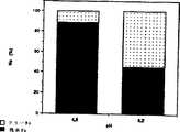
図2はpHと温度のRNAの吸着に対する効果を表す。
図3は40℃でのpHのBSAの吸着に対する効果を表す。
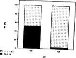
図4はイオン強度と温度のRNAの吸着に対する効果を表す。
図5は20℃でのpHのRNA脱着に対する効果を表す。
図6は40℃でのpHのRNA脱着に対する効果を表す。
図7は20℃でpH9.2でのイオン強度のRNA脱着に対する効果を表す。
図2から4において、Ns値はポリマーに付着した生物学的存在の量に相当し、ポリマーのミリグラム当たりに付着した生物学的分子のミリグラムを表す。
図5から7において、Ns値は実施例2に従って以前に粒子に吸着したRNAの量に対しての、遊離RNA(フリーNs)または非遊離RNA(残余Ns)のパーセンテージに相当する。以下の実施例が例示するように、吸着工程(c)の間のpH、イオン強度、および/または温度条件は決定的に重要である。実際、図1で観察できるように、7と等しいpH値以下で温度がポリマーのLCST未満の値であると、ポリマーは荷電した親水性の尾を持ち、その一方で7と等しいpH値を越え温度がLCSTより高い値であると、ポリマーは疎水性で中性の収縮した構造を示し、それが核酸の吸着の減少と、同時にタンパク質の吸着の増大を引き起こす。
実施例1:NIPAMベースポリマーの調製
このポリマーの調製のため三つの重合法を用いた:
1)バッチ重合法(または閉反応器法)
2)半連続重合法
3)シード上での重合(polymerization on a seed)
これらの技術のそれぞれにおいて、以下の同じ試薬を用いた。
・第一のモノマー:Kodakで市販されているN-イソプロピルアクリルアミド(NIPAM)
・架橋剤:Aldrichから入手可能なN,N-メチレンビスアクリルアミド(MBA)
・開始剤:Wakoで市販されている塩化2,2’-アゾビスアミジノプロパン(V50)
・イオン強度調節用塩:NaCl(Prolabo)
・機能的な第二のモノマー:Kodakで市販されている塩化2-アミノエチルメタクリル酸(AEM)
1)バッチ重合法
熱の影響の下で分解し、フリーラジカルを作り出す開始剤(V50)の添加により重合を開始する前に、第一のモノマー(NIPAM)、機能的な第二のモノマー(AEM)、そして架橋剤(MBA)を単一のステップで一緒に導入した。重合の継続時間は30分である。
得られるポリマーの組成は、参照符号PNIPAM42と与えられているが、以下の通りである:
全容量(a) 250ml
NIPAM 48.51mmol
MBA 3mmol
AEM 0.48mmol
V50 0.30mmol
温度 70℃
(a)沸騰脱気水
得られたポリマーの特性を以下の表Iに示す。
重合の始めとそれらの全転換の終わりの間の期間中、機能的な第二のモノマーを反応器に導入する。この添加は一定速度の注入で実施することができ(連続添加による重合)、または代わりに規則的な間隔での十分に制御された添加による(半連続重合)。この重合法の目的は重合の進行を中断させるであろう反応媒体中の水溶性ポリマーのパーセンテージを増大させることなく、機能的な第二のモノマー(電荷している)の取り込みを増大することにある。
得られたポリマーの組成は、参照符号PNIPAM45と与えられているが、以下の通りである:
全容量(a) 250ml
NIPAM 48.51mmol
MBA 3mmol
AEM 0.48mmol
V50 0.30mmol
温度 70℃
添加 3から6分の間
(a)沸騰脱気水
得られるポリマーPNIPAM45の特性は以下の表IIに表されている。
この技術は事前に準備され完全に特徴付けられたポリマーを含む反応媒体に、機能的な第二のモノマーを導入することから成る。機能的な第二のモノマーは一つのステップまたは半連続的に、単独で加えられるか、またはモノマー(類)やコモノマーと混ぜることが可能である。
得られるポリマーの組成は、参照符号PNIPAM94と与えられているが、以下の通りである:
4.5%の固体濃度を持つ40mlの容量のシードを用いる。試薬は5mlの容量の水で希釈して加えられた。第二の工程で加えられるNIPAM、MBA、そしてV50のモルパーセンテージは、シードのもの(1参照)と同一である。一方、機能的な第二のモノマーの濃度がコントロールされる(望ましい電荷密度にしたがって増減される);この場合10%(モル)のAEMが、第一のモノマーNIPAMに比例して加えられる。
参照符号PNIPAM93の下で記録され、1)で記述された方法にしたがって合成されたシードを用いた再添加の後に得られたポリマーPNIPAM94の特性は以下の表IIIに表されている。
1)から3)の技術のいずれか一つにより得られたポリマーの特性は以下の通りである:
・5から150μmol/gの間のポリマーの電荷密度(カチオンの)
・20℃での動的光散乱法により測定された粒子の直径が、0.05から2μmの間の範囲の粒子径
・20℃では0.001から1.5mol/lのNaClで、40℃では0.01から0.9mol/lのNaClの間にある凝集のための臨界濃度の範囲
実施例2:実施例1により調製されたPNIPAMの粒子へのRNAまたはBSA(ウシ血清アルブミン)の吸着
以下のプロトコールが吸着反応のための一般的方法を構成する。
反応混合物は10μlのRNA(4mg/ml)または50μlのBSA(5mg/ml)、そして50μlのN-IPAM粒子(45g/l)より成る。望ましいイオン強度とpHに到達するためにリン酸塩バッファー(10mM,pH4.6または9.2)とNaCl(5M)を加えることにより、1ミリリッターの最終容量が得られる。
分子存在物(RNAまたはBSA)は、所定のコンディション(pH、イオン強度)で(20または40℃で)で2時間以上で粒子に吸着する:混合物を1分当たり14,000回転で20分遠心分離する。上清を回収し、懸濁液中のポリマー粒子を分離するためミリポアーフィルターMillex-GV13(0.22μm)を用いて濾過する。ポリマー支持体に付着した生物学的存在物の量は、始めに導入した量と、残余の遊離している量(上清をアッセイする)の間の単純な相違によって測定される:この量はポリマーのミリグラム当たりの生物学的分子のミリグラムにより表される(Ns)。RNAまたはBSAの濃度は、それぞれ260nmと280nmの波長によるUV分光光度計(Kontron Instrument)によって推定される。
E.coliの16Sと28SリボソームRNA(Boerhinger)とBSA(Sigma参照符号A0281)を事前の精製なしで用いてテストを実施した。
用いた粒子はPNIPAM94の熱感受性粒子である。これらの粒子は室温では非常に親水性で、LCST(32℃)以上の温度では疎水性である。それらは実施例1に記述されたように合成された。
酸性リン酸塩バッファー(KH2PO4 10mM pH4.6)と中性リン酸塩バッファー(K2HPO4 10mM pH9.2)を吸着反応と反応のpH調節のため用いた。
NaCl(5M)を反応のイオン強度調節のため用いた。
全ての反応で用いられる水はMillipore-MilliQ精製システムで精製した。
インキュベーションはサーモミキサー(Eppendorf 5436)で実施した。
全ての反応は1.5mlエッペンドルフチューブで実施した。
1)pHと温度の吸着に対する影響の研究
図2によれば、RNAのよりよい吸着は中性のpHより酸性のpHで観察される。酸性のpHでは、粒子は広く正に帯電し、負に帯電した核酸は静電気力によって粒子に付着する。付着は40℃より20℃の方がより起こりやすい。40℃での結果は吸着の減少を例示する。
図3によれば、粒子へのBSAの吸着はpHの影響を受けないで可能である。20℃では、この温度での粒子の親水性の性質のため、BSAの付着は観察されなかった。
2)イオン強度と温度の吸着に対する影響の研究
図4によれば、負に帯電したRNAと正に帯電したポリマー表面の間の吸引性のある静電気力は、イオン強度の増大と共に減少し、結果としてRNAの付着が減少する。
同じ実験条件の下で、イオン強度の増大が粒子へのBSAの付着を促進しないことが確かめられた。
結論として、核酸は好ましくはLCST(20℃)未満の温度、低いイオン強度、そして酸性pHで粒子に吸着する。これらのコンディションの下では、タンパク質(BSAのような)の吸着は好まれない。
実施例3:PNIPAMポリマーの粒子へ吸着したRNAの脱着
用いられる試薬は実施例2で記述されたものと同様である。
以下のプロトコールが脱着反応のための一般的方法を構成する。
実施例2で実施された吸着ステップの後、一分当たり14,000回転の遠心分離工程後に脱着反応が実施される。上清を取り除き、望ましいpHとイオン強度を得るため1ミリリッターの脱着バッファー(リン酸塩(10mM pH4.6または9.2)とNaCl(5M))に置き換える。脱着は20℃または40℃で2時間実施される。それから混合物を一分当たり14,000回転で20分遠心分離する。上清を回収し、懸濁液中のポリマー粒子を分離するためミリポアーフィルターMillex-GV13(0.22μm)を用いて濾過する。遊離したRNAの量は、260nmの波長によるUV分光光度計(Kontron Instrument)によって測定される。回収した核酸は他の解析に利用できる。
1)pHと温度のRNAの脱着に対する影響の研究
図5によれば、中性pHでの核酸の脱着はポリマーの電荷の欠如のためより起こりやすい;酸性pHでは、その時粒子は高く正に帯電しているので遊離する核酸の量はずっと低い。
図6によれば、上述のように核酸の脱着は中性のpHで促進される。それはまた、温度をLCST(32℃)以上にすると粒子は収縮するので、温度の増大によっても促進される。
2)イオン強度のRNAの脱着に対する影響の研究
図7によれば、イオン強度が増大するにつれRNAとポリマー表面の吸引性のある静電気の相互作用が減少する。
結論として、核酸の脱着は好ましくは40℃、高いイオン強度、そして中性pHでなされる。
さらに、40℃(LCSTより高い温度)での粒子の収縮性質は、核酸溶液を濃縮することに対して促進可能である。実際、核酸物質の脱着とLCST以上の温度の上昇の後核酸を吸着する粒子は収縮し、それゆえ緩んだ状態にあるものより小さい体積を占め、遠心分離の後より小さい最終体積で粒子を回収することが可能となる。
実施例4:NIPAMを用いたDNAとRHAの混合溶液からのDNAの吸着と脱着
Short Protocols in Molecular Biology第二版編:Harvard Medical School,1992,pp.2-4/2-7内のD.Trecoにより記述されたプロトコールにしたがって、Stap hylococcus epidermidisのDNA溶液を細菌から単離したコロニーから抽出し精製する。
milliQ水中の10%(w/v)BSA(ウシ血清アルブミン)溶液(Intergen3210-01)を用いる。
PCRプロトコール:以下のPCR法は、PCR Strategies編:Innis, Gelfand and Sninsky Academic Press 1995,pp.17-31内のGoodmanにより記述されたものである。二つの増幅プライマーを用いた。それらは以下のようなシークエンスをもつ。
プライマー1:
プライマー2:
以下のような温度サイクルを増幅プロトコールの間用いた。
1)粒子へのDNAの吸着と脱着、そして遊離したDNAのPCR法後の検出
DNA溶液(1010コピー/ml)を20℃、pH4.6で2時間粒子に吸着させ、実施例2と3それぞれに記述されたような41℃、pH8.3、イオン強度0.05Hで15分の吸着ステップを受けさせた。吸着ステップと遠心分離の後、50μlの上清に回収した物質をPCRにより増幅させ、0.8%アガロースゲル上で分析した。期待されたサイズ(490pb)のバンドがゲル上で検出された。さらに、PCR後に検出されたDNAの量は、106コピー/mlのDNAのPCR増幅後に検出されたものと少なくとも同等であり、事前に粒子に吸着されたものではなかった。
NIPAM94の粒子はそれゆえDNAを吸着するのにも使用できる。脱着後、DNAはPCRタイプの増幅反応に用いることができる。
2)DNAとBSAの混合溶液からのDNAの吸着と、脱着により遊離したDNAのPCR法後の検出
10%(w/v)のBSAの存在下のDNA溶液(1010コピー/ml)が実施例4-1に記述されたような吸着と脱着ステップを受ける。同じ増幅法と検出法が用いられる。期待されたサイズのDNA(490pb)がゲル上で検出される。明視化されたDNAバンドの強度はBSAの存在、非存在で同様である。
NIPAM94の粒子はDNA-10%BSA混合溶液由来のDNAの吸着と、脱着による遊離を可能にする。始めの溶液内のBSAの存在は、粒子へのDNAの吸着を中断しない。
実施例5:NIPAMの粒子を用いた細菌溶解産物(Staphylococcus epidermidis)由来の核酸の精製
1)細菌溶解産物の調製
Staphylococcus epidermidisのカルチャーを37℃オーバーナイトで構成する。懸濁液中に含まれる細菌の数は550nmの光学濃度を測定することによって推定される。それぞれ2.106,2.104,そして2.101の細菌を含む細菌ペレットを14,000回転で3分の遠心分離で1.5mlチューブに調製する。上清は除去し、以下に記述する技術にしたがって細菌ペレットを溶解する(Arora等,J. Dairy Sci.1990,73,264-273の翻案)。
ペレットは6mg/mlのプロテイナーゼK(boehringer)と300μlのグラスビーズを含む1mlのバッファー(30mM Tris,100mM NaCl,5mM EDTA,pH7.2)内に集められる。この混合物をボルテックスで攪拌し、37度、15分間インキュベートする。遠心分離ステップ(14,000回転で3分)の後、核酸を含む上清を次なる段階のため回収する。
2)核酸の精製
用いられる粒子はその合成が実施例1に記述されているPNIPAM94の熱感受性粒子である。
以下のプロトコールは精製反応のための一般的方法を構成する。
反応混合物は、それぞれ105,103,そして106の細菌を含む50μlの細菌溶解産物と、2mgの粒子より成る。1ミリリットルの最終容量は、リン酸バッファー(10mM,pH4.6)で反応容量を調節して得られる。反応はサーモミキサー(Eppendorf 5436)で20℃で30分間インキュベートさせる。一分当たり14,000回転で20分間の遠心分離ステップの後、上清を取り除く。粒子に付着した核酸の脱着は、50μlのエルーションバッファー(elution buffer)(0.5M KCl,pH8.3)を加えることによりイオン強度の影響によって実施する;反応はサーモミキサーで42℃で15分間インキュベートさせる。一分当たり14,000回転で20分間のさらなる遠心分離ステップの後、核酸を含む上清を回収する;10μlをDNA増幅ステップ(PCR)のために用い、5μlをRNA増幅ステップ(NASBA)のために用いる。
3)核酸の検出
精製された核酸を酵素的な増幅ステップ(DNAならPCR、RNAならNASBA)の後分析する。それから増幅産物はELOSA(Enzyme Linked Oligo Sorbent Assay)、ミクロプレート(NASBA)、VIDAS(PCR)法によって明らかにされる。
PCRプロトコール:以下のプロトコールは実施例4で記述されたものと同一である。増幅産物(90μl)を、Habilat等,J.Clin.Microbiol;1994,32,2702-2705によって記述されたプロトコールにしたがって、自動バイダス免疫分析器(bioMeriuex)で分析し、キャプチャープローブと検出プローブは以下の通りである。
キャプチャープローブ:
検出プローブ:
検出プローブはアルカリフォスファターゼと接合しておく。
NASBAプロトコール:プロトコールはKievits等,J.Virol.Methods(1991)35,273-286に記述されたものと同一である。用いられるプライマーは以下のシークエンスを持つ。
プライマー1:
プライマー2:
増幅産物(5μl)をMallet等,J.Clin.Microbiol.(1993)31,1444-1449に記述されたプロトコールにしたがって、ミクロプレート形式(microplate format)でElosa法を用いて分析する。キャプチャープローブと検出プローブは以下のシークエンスを持つ:
キャプチャープローブ:
検出プローブ
検出プローブはセイヨウワサビペルオキシダーゼと接合しておく。
プロテイナーゼKは増幅反応の阻害剤として知られているが、1/10の連続的な希釈を得られる増幅の程度を測定するために増幅ステップの前に実施する。
得られた結果は付属書類中の表IVに整理されている。
増幅ステップの前にサンプルを1/1000に希釈することが必要であるため、プロテイナーゼKの阻害力をチェックする。増幅ステップ後、増幅前の1/10だけサンプルを希釈するが、それは100のファクターの増大を表す。粒子はサンプル内に連帯して存在するRNAとDNAの精製が可能である。これらの核酸は酵素的な増幅ステップで両立できる。
実施例6:磁気コアに融合されたNIPAMポリマーを用いた細菌溶解産物(Staphylococcus epidermidis)由来の核酸の精製
前述の実施例に記述された粒子は、吸着と脱着ステップの後の遠心分離ステップの調節に不都合がある。これらのステップは長く(20分が二回)、そして自動的にするのは難しい。可能な変更はNIPAMポリマーをカチオンの磁気コアに融合することである。試験された支持体の一つはカチオン磁気ラテックスR95-07(Estapor,Rhone-Poulenc)であり、その粒子は多分散である。
それゆえ得られた粒子の精製能が試験された。
1)Nipamの磁気粒子の合成
カチオンエスタポアー粒子R95-07をカプセル化した。各カプセル化の前に、粒子を0.005Mの塩酸溶液で3回洗った。
シード粒子の1gを、事前に沸騰した温度まで温められ窒素で脱気した40mlのmilliQ水に希釈した。
スチレン: 100μg
NIPAM: 0.3254g
BAM: 0.0274g
MAE: 0.0740g
トリトンX-405: 0.14g
V50: 0.0061g
コアへの修飾前ステップ(70℃で2時間)のための100μgのスチレン、NIPAM,BAM,MAEを10mlの水に可溶化し、シード(エスタポアーラテックス)に導入する。1mlの水に可溶化した開始剤を、シード粒子の周りへの重合を許容するために加える。重合は70℃の窒素雰囲気の下で実施される。
そしてこれらの粒子は、粒子のサイズ分散を修正しないと、220の電荷と粒子グラム当たり82μモルのNH2をもつ。
2)核酸の精製
用いられるプロトコールは実施例5に記述されたものに以下の修正を加えたものと同一である。
・200μgの粒子を用い
・粒子を上清と分離するための遠心分離ステップを取り除き、磁場の影響の下での分離ステップに置き換える(磁気分離装置、promega Z5342)。
全ての他のステップは電荷していないままである。
結果は付属書類中の表Vに整理されている。
増幅ステップの前にサンプルを1/1000に希釈することが必要であるため、プロテイナーゼKの阻害力を再びチェックする。増幅ステップ後、増幅前の1/10(PCR)または1/100(NASBA)にサンプルを希釈することが可能だが、それは10から100のファクターの増大を表す。これらの粒子もサンプル内に連帯して存在するRNAとDNAの精製が可能である。これらの核酸は酵素的な増幅ステップで両立できる。
A method for purifying nucleic acid present in a sample in an aqueous medium is known from document WO-A-95 / 04140, whereby the sample is brought into contact with a particle system consisting of silk beads in the presence of a chaotropic substance. The nucleic acid attached to the beads is then separated from the final aqueous solution.
According to the document F. Meunier et al., Polymers for Advanced Technologies, 6: 489-496, (1995), in the presence of a polymerization initiator, (1) N-isopropylacrylamide, (2) N, N-methylenebisacrylamide, And (3) the preparation of a polymer called PNIPAM by polymerization of 2-aminoethyl methacrylic acid chloride is described. The behavior of this surface functionalized polymer is particularly suitable for covalent binding of biological molecules.
The document EP-A-0 161 881 describes a temperature-sensitive polymer such as a polymer obtained by copolymerization of monomers of N-alkyl- or N-alkylene-acrylamide or methacrylamide and monomers of acrylic acid or methacrylic acid derivatives, It teaches that its ability to change structure as a function of temperature can be used to isolate biological material. It takes an open structure at low temperatures and promotes the attachment of biological material, and takes a contracted structure at high temperatures, allowing it to release the attached biological material. Therefore, control of the biological material deposition and release steps can be performed by changing the temperature. As a better control, the pH can also be changed.
The proposed use in this document extends without any limitation to the isolation of any biological material present in the sample, in particular the isolation of nucleic acid material and protein material.
According to the present invention, a method for selective isolation of nucleic acid material present in a sample is provided. Even if the sample is complex and contains inhibitors of protein substances and / or enzyme reactions, the process of the present invention facilitates the isolation of nucleic acid substances while any isolation of protein substances and / or inhibitors as described above. Restrict or even eliminate.
The method for isolating nucleic acid material present in a sample in an aqueous phase according to the present invention comprises the following steps:
An adsorbent reagent comprising a sol consisting of a discontinuous phase and an aqueous continuous phase of a particulate support containing a functionalized particulate polymer is available for the production of the adsorbent reagent (a). Is obtained by polymerization of (1) a first water-soluble monomer of acrylamide or an acrylamide derivative, (2) at least one crosslinking agent, and (3) at least a second cationic water-soluble functional monomer, the polymer Having a predetermined lower critical melting temperature (LCST) between 25 and 45 ° C, preferably between 30 and 40 ° C;
Contacting step (b), wherein the adsorption reagent is brought into contact with a sample containing a nucleic acid substance;
an adsorption step (c) for contact according to (b), wherein at least one, preferably at least two of the following parameters are selected for the reaction medium:
-PH equal to at most 7
・ At most 10-2Ionic strength equal to M
・ Temperature below LCST of polymer
Separating the discontinuous phase, in particular the discontinuous phase adsorbing the nucleic acid substance, from the continuous phase after the separation step (d) is observed as necessary, as the adsorption has occurred;
Desorption process (e), comprising 10-2The step of increasing the ionic strength to an ionic strength exceeding M and separating the nucleic acid substance by desorption from the particulate support.
Advantageously, in the desorption step (e), at least one parameter selected from pH and temperature is additionally changed as follows:
Increase the pH to a pH higher than 7.
Increase the temperature to a temperature above the LCST of the polymer.
The present invention is also a method for isolating a nucleic acid substance present in a sample in an aqueous phase, wherein the nucleic acid substance is adsorbed on a particulate support, and the nucleic acid substance adsorbed on the particulate support is subjected to subsequent analysis. It also relates to a method comprising a process used in a general step. This method involves the following steps:
An adsorbent reagent comprising a sol consisting of a discontinuous phase and an aqueous continuous phase of a particulate support containing a functionalized particulate polymer is available for the production of the adsorbent reagent (a). Is obtained by polymerization of (1) a first water-soluble monomer of acrylamide or an acrylamide derivative, (2) at least one crosslinking agent, and (3) at least a second cationic water-soluble functional monomer, the polymer Having a predetermined lower critical melting temperature (LCST) between 25 and 45 ° C.,
Contacting step (b), wherein the adsorption reagent is brought into contact with a sample containing nucleic acid substance;
an adsorption step (c) for contact according to (b), wherein at most 10 relative to the reaction medium;-2A process in which an ionic strength equal to M is selected,
A separation step (d) comprising the step of separating the discontinuous phase from the continuous phase after the occurrence of adsorption has been observed as necessary, wherein the desorption step is optional.
According to a preferred embodiment of the latter method, in the adsorption step (c) for contact according to (b), at least one of the following parameters is selected for the reaction medium in addition:
-PH equal to at most 7
・ Temperature below LCST of polymer
Of course, this process is by separating the nucleic acid material from the particulate support by desorption after the separation step (d) by changing at least one parameter selected from ionic strength, pH and temperature as follows: A desorption step may be included.
·Ten-2Increase ionic strength to an ionic strength higher than M.
Increase the pH to a pH higher than 7.
Increase the temperature to a temperature above the LCST of the polymer.
At least the ionic strength is preferably varied.
The above-described method according to the invention is preferably carried out according to two variants associated with step (a).
According to a first variant described in the examples, the particulate support is made of the particulate polymer, in which case the crosslinking agent (s) (a) is water soluble.
According to a second variant, the particulate support additionally comprises an organic or inorganic core completely or partially coated with the particulate polymer, the core being adsorbable for the polymer with respect to the nucleic acid material. Will not change. The core or core portion serves as a cross-linking agent (2), making it possible to provide another cross-linking agent of the water-soluble cross-linking agent type. By way of example, the core may be a polystyrene core and / or may contain a magnetic compound.
According to certain preferred embodiments of these methods, at least one probe and / or one primer capable of specifically hybridizing with the nucleic acid substance before or after step (b) is selected from step (b). Added to the previous sample or to the reaction medium after step (b), in particular after step (c) or step (d).
According to another particular embodiment, the nucleic acid material consists of a probe or primer, and in (b) and (c), an adsorption reagent is contacted with the nucleic acid material to obtain a hybridization reagent, and then (b ′) A medium comprising at least one nucleic acid or nucleic acid fragment under conditions suitable for hybridization or primer extension after adsorption is optionally observed and separation of the hybridization reagent from the reaction medium And the above hybridization reagent.
Advantageously, the particulate polymer is obtained by free radical polymerization in the presence of a cationic or neutral and water-soluble polymerization initiator.
The first monomer (1) is preferably N-alkylacrylamide and N, N-dialkylacrylamide, more specifically N-isopropylacrylamide, N-ethylmethacrylamide, Nn-propylacrylamide, Nn-propylmethacrylamide, N-isopropylmethacrylamide, N-cyclopropylacrylamide, N, N-diethylacrylamide, N-methyl-N-isopropylacrylamide, N-methyl-Nn-propylacrylamide, preferably the first monomer is N-isopropyl Acrylamide (NIPAM).
The second functional monomer (s) is preferably from acrylic acid and methacrylic acid derivatives, 2-aminoethyl methacrylic acid chloride (AEM), N-vinylpyridine derivatives, trialkylammonium derivatives, and isothiouronium chloride derivatives. Selected.
Advantageously, the water-soluble crosslinking agent (2) is selected from N, N-methylenebisacrylamide (MBA), ethylene glycol dimethacrylate, and the polymerization initiator is 2,2′-azobisamidinopropane chloride (V50 ).
The separation step (d) is preferably carried out according to a method selected from centrifugation, filtration, precipitation, sedimentation, and magnetic field application.
It can optionally be observed that an adsorption reaction takes place before the separation step (d). As an example, HPLC or capillary electrophoresis can be used.
Before disclosing the present invention in more detail, some terms used in the description and the claims are defined below.
In the present invention, isolation of a nucleic acid substance means separation or detection of this substance and concentration of a fraction of nucleic acid substance according to a specific or non-specific isolation method by a qualitative and / or quantitative method. Means.
In the present invention, the nucleic acid substance is a nucleic acid, a nucleic acid fragment, a nucleic acid and / or a mixture of nucleic acid fragments, or a fraction of nucleic acids and / or nucleic acid fragments. Nucleic acid is understood to mean any kind of nucleic acid, free or optionally complexed with a protein, whether derived from cells, bacteria or viruses. It is a natural nucleotide sequence whose constituents are sugars, phosphate groups, and nitrogen bases selected from adenine, guanine, uracil, cytosine, thymine, and / or at least one of the three constituents is modified The modification occurs at the base level, for example, a modified base such as inosine, 5-methyldeoxycytidine, deoxyuridine, 5-dimethylaminodeoxyuridine, 2,6-diaminopurine, 5-bromodeoxyuridine; This results in a modified base such as a base modified by a tracer that can be detected directly or indirectly by techniques known to those skilled in the art, such as a base modified by biotin; at the sugar level, at least mainly by a polyamide, One deoxyribose substituted; and / or phosphate group The level, such as, in particular, diphosphate ester, alkyl - and Arirurin ester, and those substituted by an ester selected from allyl mono thiophosphate ester. The nucleic acids according to the present invention are fully or partially single-stranded and / or double-stranded and may consist in particular of a probe-nucleic acid, probe-nucleic acid fragment, primer-nucleic acid or primer-nucleic acid fragment duplex. The duplex can be a homoduplex or a heteroduplex.
The present invention is of course applicable to the isolation of nucleic acid fragments as defined above of various sizes, or oligonucleotides (ODN).
Nucleic acid material may be of natural origin and / or obtained by genetic recombination and / or obtained by chemical synthesis; by way of example, it consists of a probe or primer.
The present invention applies to non-specific isolation of nucleic acids and / or fractions of nucleic acid fragments contained in a sample, but does not include nucleic acids or nucleic acid fragments or mixtures of nucleic acids or mixtures of nucleic acid fragments present in a sample. It can also be applied to specific isolation.
Samples as understood by the present invention include any sample capable of containing nucleic acid material, in particular biological samples such as biological fluids, food-derived samples. The sample may consist of a whole or partial sample, in particular it may consist of a partial specimen, a diluent. The sample may or may not have undergone pretreatment, particularly purification or lysis to promote nucleic acid release.
The LCST of a polymer such as that of the subject of the present invention is defined and measured in particular by the technique described in the following literature: Hiroshi Inomata et al., Macromolecules 1994, 27, 6459-6464.
The probe is a nucleotide fragment having hybridization specificity under a predetermined condition for forming a hybridization complex with the nucleotide fragment. The probe used within the framework of the present invention is preferably a capture probe, although other types of probes can be used.
Primers according to the present invention are, for example, PCR (polymerase chain reaction), the so-called NASBA method (`` Nucleic Acid Sequence-Based Amplification ''), or alternatively the so-called TMA method (Transcription Mediated Amplification). A probe having hybridization specificity under a predetermined condition for initiation of enzymatic polymerization such as an amplification method such as), an extension process such as sequencing, and a reverse transcription method.
In the present invention, the acrylamide derivative is R0-CH = C (R1) -CONR2RThreeIs understood to mean a polymerizable monomer corresponding to the formula0, R1, R2, And RThreeRepresents a group independently selected from hydrogen, an aliphatic or cyclic, linear or branched lower hydrocarbon group, and a nitrogen-containing heterocyclic group such as imidazole.
Under the understanding of the present invention, the adsorption of a nucleic acid substance is defined as follows: After contact for a certain period of time between the substance and the particulate support, at least one group belonging to the constituents of the nucleic acid substance is the support. Once attached to the surface, the nucleic acid material is adsorbed to the particulate support; the adsorption results from ionic interactions between the material and the support, and / or hydrogen bonding, and / or hydrophobic interactions, Any covalent bonds are excluded.
Finally, a functionalized polymer means at least one functional group capable of producing any one of the constituent groups of the nucleic acid material and the interactions and / or bonds involved in the adsorption phenomenon. It means a polymer having an interface. Preferably these functional groups are NHThree+; NHFour+; NRThree+Or R represents an aliphatic or cyclic, saturated or unsaturated hydrocarbon group, and NRThree+Can represent a pyridinium group; and an isothiouronium group.
The present invention will be described with reference to Examples 1 to 6 and FIGS. 1 to 7 shown below.
Figure 1 represents the variation of the polymer interface with pH and temperature.
Figure 2 shows the effect of pH and temperature on RNA adsorption.
FIG. 3 shows the effect of pH at 40 ° C. on BSA adsorption.
FIG. 4 shows the effect of ionic strength and temperature on RNA adsorption.
FIG. 5 represents the effect of pH at 20 ° C. on RNA desorption.
FIG. 6 shows the effect of pH at 40 ° C. on RNA desorption.
FIG. 7 represents the effect of ionic strength on RNA desorption at 20 ° C. and pH 9.2.
2-4, the Ns value corresponds to the amount of biological entity attached to the polymer and represents milligrams of biological molecules attached per milligram of polymer.
5 to 7, the Ns value corresponds to the percentage of free RNA (free Ns) or non-free RNA (residual Ns) relative to the amount of RNA previously adsorbed to the particles according to Example 2. As the examples below illustrate, the pH, ionic strength, and / or temperature conditions during the adsorption step (c) are critical. In fact, as can be observed in FIG. 1, below the pH value equal to 7 and below the LCST of the polymer, the polymer has a charged hydrophilic tail, while exceeding a pH value equal to 7. When the temperature is higher than the LCST, the polymer exhibits a hydrophobic, neutral, contracted structure, which causes a decrease in nucleic acid adsorption and at the same time an increase in protein adsorption.
Example 1: Preparation of NIPAM base polymer
Three polymerization methods were used for the preparation of this polymer:
1) Batch polymerization method (or closed reactor method)
2) Semi-continuous polymerization method
3) Polymerization on a seed
In each of these techniques, the following same reagents were used.
First monomer: N-isopropylacrylamide (NIPAM) commercially available from Kodak
Crosslinking agent: N, N-methylenebisacrylamide (MBA) available from Aldrich
Initiator: 2,2'-azobisamidinopropane chloride (V50) commercially available from Wako
・ Salt for adjusting ionic strength: NaCl (Prolabo)
Functional second monomer: 2-aminoethyl methacrylic acid chloride (AEM), commercially available from Kodak
1) Batch polymerization method
The first monomer (NIPAM), the functional second monomer (AEM), and the crosslinker before starting the polymerization by adding an initiator (V50) that decomposes under the influence of heat and creates free radicals (MBA) were introduced together in a single step. The duration of the polymerization is 30 minutes.
The composition of the resulting polymer is given the reference PNIPAM42 and is as follows:
Full capacity(a) 250ml
NIPAM 48.51mmol
MBA 3mmol
AEM 0.48mmol
V50 0.30mmol
70 ℃
(a) Boiling deaerated water
The properties of the resulting polymer are shown in Table I below.
A functional second monomer is introduced into the reactor during the period between the beginning of the polymerization and the end of their total conversion. This addition can be carried out with a constant rate of injection (polymerization by continuous addition) or alternatively by well-controlled addition at regular intervals (semi-continuous polymerization). The purpose of this polymerization method is to increase the uptake of functional second monomer (charged) without increasing the percentage of water-soluble polymer in the reaction medium that would interrupt the progress of the polymerization. is there.
The composition of the polymer obtained is given the reference number PNIPAM45 and is as follows:
Full capacity(a) 250ml
NIPAM 48.51mmol
MBA 3mmol
AEM 0.48mmol
V50 0.30mmol
70 ℃
Addition for 3 to 6 minutes
(a) Boiling deaerated water
The properties of the resulting polymer PNIPAM45 are shown in Table II below.
This technique consists of introducing a functional second monomer into a reaction medium containing a previously prepared and fully characterized polymer. The functional second monomer can be added alone or mixed with monomer (s) or comonomers in one step or semi-continuously.
The composition of the resulting polymer is given the reference number PNIPAM94 and is as follows:
Use a 40 ml volume seed with a solids concentration of 4.5%. The reagent was added diluted in a 5 ml volume of water. The molar percentage of NIPAM, MBA, and V50 added in the second step is the same as that of the seed (see 1). Meanwhile, the concentration of the functional second monomer is controlled (scaled according to the desired charge density); in this case 10% (mole) of AEM is added in proportion to the first monomer NIPAM.
The properties of the polymer PNIPAM94, recorded under the reference PNIPAM93 and obtained after re-addition with a seed synthesized according to the method described in 1) are represented in Table III below.
The properties of the polymer obtained by any one of the techniques 1) to 3) are as follows:
The polymer charge density (cationic) between 5 and 150 μmol / g
-Particle diameter measured by dynamic light scattering at 20 ° C in the range between 0.05 and 2μm
A critical concentration range for aggregation between 0.001 and 1.5 mol / l NaCl at 20 ° C and between 0.01 and 0.9 mol / l NaCl at 40 ° C.
Example 2: Adsorption of RNA or BSA (bovine serum albumin) to PNIPAM particles prepared according to Example 1
The following protocol constitutes a general method for the adsorption reaction.
The reaction mixture consists of 10 μl RNA (4 mg / ml) or 50 μl BSA (5 mg / ml) and 50 μl N-IPAM particles (45 g / l). By adding phosphate buffer (10 mM, pH 4.6 or 9.2) and NaCl (5M) to reach the desired ionic strength and pH, a final volume of 1 milliliter is obtained.
The molecular entity (RNA or BSA) adsorbs to the particles in a given condition (pH, ionic strength) (at 20 or 40 ° C.) for over 2 hours: the mixture is centrifuged at 14,000 revolutions per minute for 20 minutes . The supernatant is collected and filtered using a Millipore filter Millex-GV13 (0.22 μm) to separate the polymer particles in the suspension. The amount of biological entity attached to the polymer support is measured by a simple difference between the amount initially introduced and the remaining free amount (assay the supernatant): Expressed in milligrams of biological molecule per milligram of polymer (Ns). The concentration of RNA or BSA is estimated by a UV spectrophotometer (Kontron Instrument) with wavelengths of 260 nm and 280 nm, respectively.
Tests were performed using E. coli 16S and 28S ribosomal RNA (Boerhinger) and BSA (Sigma reference A0281) without prior purification.
The particles used are PNIPAM94 heat sensitive particles. These particles are very hydrophilic at room temperature and hydrophobic at temperatures above LCST (32 ° C.). They were synthesized as described in Example 1.
Acid phosphate buffer (KH2POFour 10 mM pH 4.6) and neutral phosphate buffer (K2HPOFour 10 mM pH 9.2) was used for adsorption reaction and pH adjustment of the reaction.
NaCl (5M) was used to adjust the ionic strength of the reaction.
The water used in all reactions was purified with a Millipore-MilliQ purification system.
Incubation was performed with a thermomixer (Eppendorf 5436).
All reactions were performed in 1.5 ml Eppendorf tubes.
1) Study of the effects of pH and temperature on adsorption
According to FIG. 2, better adsorption of RNA is observed at acidic pH than at neutral pH. At acidic pH, the particles are widely positively charged, and negatively charged nucleic acids are attached to the particles by electrostatic forces. Adhesion is more likely at 20 ° C than at 40 ° C. The results at 40 ° C illustrate the decrease in adsorption.
According to FIG. 3, adsorption of BSA to the particles is possible without being affected by pH. At 20 ° C., no BSA adhesion was observed due to the hydrophilic nature of the particles at this temperature.
2) Study of the effects of ionic strength and temperature on adsorption
According to FIG. 4, the attractive electrostatic force between the negatively charged RNA and the positively charged polymer surface decreases with increasing ionic strength, resulting in a decrease in RNA adhesion.
Under the same experimental conditions, it was confirmed that the increase in ionic strength did not promote the attachment of BSA to the particles.
In conclusion, nucleic acids preferably adsorb to particles at temperatures below LCST (20 ° C), low ionic strength, and acidic pH. Under these conditions, adsorption of proteins (such as BSA) is not preferred.
Example 3: Desorption of RNA adsorbed on PNIPAM polymer particles
The reagents used are the same as those described in Example 2.
The following protocol constitutes a general method for the desorption reaction.
After the adsorption step carried out in Example 2, the desorption reaction is carried out after a centrifugation step of 14,000 revolutions per minute. The supernatant is removed and replaced with 1 milliliter of desorption buffer (phosphate (10 mM pH 4.6 or 9.2) and NaCl (5M)) to obtain the desired pH and ionic strength. Desorption is carried out at 20 ° C. or 40 ° C. for 2 hours. The mixture is then centrifuged for 20 minutes at 14,000 revolutions per minute. The supernatant is collected and filtered using a Millipore filter Millex-GV13 (0.22 μm) to separate the polymer particles in the suspension. The amount of RNA released is measured by a UV spectrophotometer (Kontron Instrument) with a wavelength of 260 nm. The recovered nucleic acid can be used for other analyses.
1) Study of the effects of pH and temperature on RNA desorption
According to FIG. 5, nucleic acid desorption at neutral pH is more likely due to the lack of charge on the polymer; at acidic pH, the amount of nucleic acid released is much lower since then the particles are highly positively charged.
According to FIG. 6, nucleic acid desorption is promoted at a neutral pH as described above. It is also facilitated by increasing the temperature, as the particles shrink when the temperature is above LCST (32 ° C.).
2) Study of the effect of ionic strength on RNA desorption
According to FIG. 7, as ionic strength increases, the attractive electrostatic interaction between RNA and the polymer surface decreases.
In conclusion, nucleic acid desorption is preferably done at 40 ° C., high ionic strength, and neutral pH.
Furthermore, the contractile nature of the particles at 40 ° C. (temperatures higher than LCST) can promote the concentration of nucleic acid solutions. In fact, after desorption of nucleic acid material and temperature rise above LCST, the particles that adsorb nucleic acid shrink, thus occupying a smaller volume than in a relaxed state and recovering particles in a smaller final volume after centrifugation It becomes possible to do.
Example 4: Adsorption and desorption of DNA from a mixed solution of DNA and RHA using NIPAM
Short Protocols in Molecular Biology 2nd edition: DNA solution of Stap hylococcus epidermidis was isolated from bacteria according to the protocol described by D. Treco in Harvard Medical School, 1992, pp.2-4 / 2-7 Extract from the colony and purify.
Use 10% (w / v) BSA (bovine serum albumin) solution (Intergen 3210-01) in milliQ water.
PCR protocol: The following PCR method was described by Goodman in PCR Strategies edited by Innis, Gelfand and Sninsky Academic Press 1995, pp. 17-31. Two amplification primers were used. They have the following sequence:
Primer 1:
Primer 2:
The following temperature cycle was used during the amplification protocol.
1) Adsorption and desorption of DNA on particles, and detection of free DNA after PCR
DNA solution (10TenCopy / ml) is adsorbed to the particles for 2 hours at 20 ° C, pH 4.6 and subjected to an adsorption step of 15 minutes at 41 ° C, pH 8.3, ionic strength 0.05H as described in Examples 2 and 3, respectively. I let you. After the adsorption step and centrifugation, the material recovered in 50 μl of supernatant was amplified by PCR and analyzed on a 0.8% agarose gel. A band of the expected size (490 pb) was detected on the gel. In addition, the amount of DNA detected after PCR is 106It was at least equivalent to that detected after PCR amplification of copy / ml DNA and was not previously adsorbed to the particles.
NIPAM94 particles can therefore also be used to adsorb DNA. After desorption, the DNA can be used for PCR type amplification reactions.
2) Adsorption of DNA from a mixed solution of DNA and BSA, and detection of DNA released by desorption after PCR
DNA solution in the presence of 10% (w / v) BSA (10TenCopy / ml) undergo an adsorption and desorption step as described in Example 4-1. The same amplification and detection methods are used. The expected size of DNA (490pb) is detected on the gel. The intensity of the visualized DNA band is the same in the presence or absence of BSA.
NIPAM94 particles enable adsorption of DNA from DNA-10% BSA mixed solution and release by desorption. The presence of BSA in the initial solution does not interrupt the adsorption of DNA to the particles.
Example 5: Purification of nucleic acid from bacterial lysate (Staphylococcus epidermidis) using NIPAM particles
1) Preparation of bacterial lysate
The culture of Staphylococcus epidermidis is composed overnight at 37 ° C. The number of bacteria contained in the suspension is estimated by measuring the optical density at 550 nm. 2.10 each6, 2.10FourAnd 2.101Bacteria pellets containing the bacteria are prepared in 1.5 ml tubes by centrifugation at 14,000 rpm for 3 minutes. The supernatant is removed and the bacterial pellet is lysed according to the technique described below (adaptation of Arora et al., J. Dairy Sci. 1990, 73, 264-273).
The pellet is collected in 1 ml buffer (30 mM Tris, 100 mM NaCl, 5 mM EDTA, pH 7.2) containing 6 mg / ml proteinase K (boehringer) and 300 μl glass beads. This mixture is vortexed and incubated at 37 degrees for 15 minutes. After the centrifugation step (14,000 rpm for 3 minutes), the supernatant containing the nucleic acid is recovered for the next step.
2) Purification of nucleic acid
The particles used are PNIPAM94 heat-sensitive particles whose synthesis is described in Example 1.
The following protocol constitutes a general method for purification reactions.
Each reaction mixture was 10Five,TenThreeAnd 10650 μl of bacterial lysate containing 2 mg of bacteria and 2 mg of particles. A final volume of 1 ml is obtained by adjusting the reaction volume with phosphate buffer (10 mM, pH 4.6). The reaction is incubated for 30 minutes at 20 ° C. in a thermomixer (Eppendorf 5436). After a 20 minute centrifugation step at 14,000 revolutions per minute, the supernatant is removed. Desorption of nucleic acids attached to the particles is performed by the influence of ionic strength by adding 50 μl of elution buffer (0.5 M KCl, pH 8.3); the reaction is incubated for 15 minutes at 42 ° C. in a thermomixer Let After a further centrifugation step at 14,000 revolutions per minute for 20 minutes, the supernatant containing the nucleic acid is recovered; 10 μl is used for the DNA amplification step (PCR) and 5 μl is used for the RNA amplification step (NASBA) .
3) Detection of nucleic acid
The purified nucleic acid is analyzed after an enzymatic amplification step (PCR for DNA, NASBA for RNA). The amplified product is then revealed by ELISA (Enzyme Linked Oligo Sorbent Assay), microplate (NASBA), and VIDAS (PCR).
PCR protocol: The following protocol is identical to that described in Example 4. Amplification product (90 μl) was added to Habilat et al., J. Clin. Microbiol; 1994,32, 2702-2705, and analyzed with an automated Vidus immunoassay (bioMeriuex), the capture and detection probes are as follows.
Capture probe:
Detection probe:
The detection probe is conjugated with alkaline phosphatase.
NASBA protocol: The protocol is the same as that described in Kievits et al., J. Virol. Methods (1991) 35,273-286. The primers used have the following sequence:
Primer 1:
Primer 2:
The amplification product (5 μl) is analyzed using the Elosa method in a microplate format according to the protocol described in Mallet et al., J. Clin. Microbiol. (1993) 31, 1444-1449. The capture and detection probes have the following sequence:
Capture probe:
Detection probe
The detection probe is conjugated with horseradish peroxidase.
Proteinase K is known as an inhibitor of the amplification reaction, but is performed prior to the amplification step to determine the degree of amplification that can be obtained in serial dilutions of 1/10.
The results obtained are summarized in Table IV in the annex.
Check the inhibitory activity of proteinase K as it is necessary to dilute the sample to 1/1000 prior to the amplification step. After the amplification step, the sample is diluted by 1/10 before amplification, which represents a factor increase of 100. Particles can purify RNA and DNA present in solid in the sample. These nucleic acids are compatible in an enzymatic amplification step.
Example 6: Purification of nucleic acid from bacterial lysate (Staphylococcus epidermidis) using NIPAM polymer fused to magnetic core
The particles described in the previous examples have disadvantages in adjusting the centrifugation step after the adsorption and desorption steps. These steps are long (twice 20 minutes) and difficult to automate. A possible change is to fuse the NIPAM polymer to the cationic magnetic core. One of the supports tested is cationic magnetic latex R95-07 (Estapor, Rhone-Poulenc), whose particles are polydisperse.
Therefore, the purification ability of the resulting particles was tested.
1) Synthesis of Nipam magnetic particles
Cationic ester pore particles R95-07 were encapsulated. Prior to each encapsulation, the particles were washed 3 times with 0.005 M hydrochloric acid solution.
1 g of seed particles was diluted in 40 ml of milliQ water that had been pre-boiled and degassed with nitrogen.
Styrene: 100μg
NIPAM: 0.3254g
BAM: 0.0274g
MAE: 0.0740g
Triton X-405: 0.14g
V50: 0.0061g
100 μg of styrene, NIPAM, BAM, MAE for the pre-modification step to the core (2 hours at 70 ° C.) is solubilized in 10 ml of water and introduced into the seed (Estapore latex). Initiator solubilized in 1 ml of water is added to allow polymerization around the seed particles. The polymerization is carried out under a nitrogen atmosphere at 70 ° C.
And these particles have 220 charges and 82 μmol NH per gram of particle without modifying the particle size dispersion2It has.
2) Purification of nucleic acid
The protocol used is the same as that described in Example 5 with the following modifications.
・ Use 200μg particles
Remove the centrifugation step to separate the particles from the supernatant and replace with a separation step under the influence of a magnetic field (magnetic separation device, promega Z5342).
All other steps remain uncharged.
The results are organized in Table V in the appendix.
Since the sample needs to be diluted 1/1000 prior to the amplification step, proteinase K inhibition is checked again. After the amplification step, it is possible to dilute the sample to 1/10 (PCR) or 1/100 (NASBA) before amplification, which represents a factor increase of 10 to 100. These particles can also purify RNA and DNA present in the sample. These nucleic acids are compatible in an enzymatic amplification step.
Claims (11)
Translated fromJapanese吸着試薬の製造工程(a)において、機能化された粒子状のポリマーを含む粒子状の支持体の不連続相と水性連続相から成るゾルを含む吸着試薬が入手可能であり、該ポリマーは(1)N-イソプロピルアクリルアミド、(2)少なくともN,N-メチレンビスアクリルアミド(MBA)、および(3)少なくとも塩化2-アミノエチルメタクリル酸(AEM)の重合によって得られ、該ポリマーは25と45℃の間の所定の下部臨界溶解温度(LCST)を持っており、
接触工程(b)において、吸着試薬を核酸物質を含むサンプルと接触させ、水性連続層と核酸物質が粒子状の支持体に吸着している不連続相とを有するゾルを形成し、
接触工程(b)では、ゾルは
・高くとも7に等しいpH;
・高くとも10-2Mに等しいイオン強度;
・ポリマーのLCST未満の温度;
の全ての条件を有し、
観察工程(C)において、吸着が起こったことが必要に応じて観察され、
分離工程(d)において、不連続相を連続相から分離することを特徴とする単離方法。In a method for isolating a nucleic acid substance present in a sample in an aqueous phase, comprising the step of adsorbing the nucleic acid substance to a particulate support,
In the adsorbing reagent production step (a), an adsorbing reagent comprising a sol composed of a discontinuous phase and an aqueous continuous phase of a particulate support containing a functionalized particulate polymer is available, the polymer comprising ( Obtained by polymerization of 1) N-isopropylacrylamide, (2) at least N, N-methylenebisacrylamide (MBA), and (3) at least 2-aminoethylmethacrylic acid chloride (AEM), the polymer being at 25 and 45 ° C. Has a predetermined lower critical melting temperature (LCST) between
In the contacting step (b), the adsorption reagent is brought into contact with a sample containing a nucleic acid substance to form a sol having an aqueous continuous layer and a discontinuous phase in which the nucleic acid substance is adsorbed on a particulate support,
In the contacting step (b), the sol has a pH equal to at most 7;
An ionic strength equal to at most 10-2 M;
A temperature below the LCST of the polymer;
With all the conditions
In the observation step (C), it was observed as necessary that adsorption occurred,
An isolation method comprising separating the discontinuous phase from the continuous phase in the separation step (d).
・7より高いpHまでpHを増大する
・ポリマーのLCSTより高い温度まで温度を増大する
ことを特徴とする請求項2に記載の方法。For the desorption step (e), at least one parameter selected from pH and temperature is further changed as follows:
3. The method according to claim 2, characterized in that the pH is increased to a pH higher than 7, the temperature is increased to a temperature higher than the LCST of the polymer.
・10-2Mより高いイオン強度までイオン強度を増大する
・7より高いpHまでpHを増大する
・ポリマーのLCSTより高い温度まで温度を増大する
変化させることによって、粒子状の支持体から脱着により核酸物質を引き離すことによる脱着工程を含むことを特徴とする請求項1に記載の方法。After separation step (d), at least one parameter selected from ionic strength, pH and temperature is as follows:-Increase ionic strength to an ionic strength higher than 10-2 M-Increase pH to a pH higher than 7 The method according to claim 1, comprising a desorption step by separating the nucleic acid material by desorption from the particulate support by changing the temperature up to a temperature higher than the LCST of the polymer.
Applications Claiming Priority (5)
| Application Number | Priority Date | Filing Date | Title |
|---|---|---|---|
| FR9603753 | 1996-03-20 | ||
| FR96/03753 | 1996-03-20 | ||
| FR96/04691 | 1996-04-09 | ||
| FR9604691 | 1996-04-09 | ||
| PCT/FR1997/000496WO1997034909A1 (en) | 1996-03-20 | 1997-03-20 | Nucleic acid isolation |
Publications (2)
| Publication Number | Publication Date |
|---|---|
| JPH11505426A JPH11505426A (en) | 1999-05-21 |
| JP4693944B2true JP4693944B2 (en) | 2011-06-01 |
Family
ID=26232612
Family Applications (1)
| Application Number | Title | Priority Date | Filing Date |
|---|---|---|---|
| JP53321197AExpired - LifetimeJP4693944B2 (en) | 1996-03-20 | 1997-03-20 | Nucleic acid isolation method |
Country Status (8)
| Country | Link |
|---|---|
| US (1) | US6737235B1 (en) |
| EP (1) | EP0842184B1 (en) |
| JP (1) | JP4693944B2 (en) |
| AT (1) | ATE231875T1 (en) |
| CA (1) | CA2222192A1 (en) |
| DE (1) | DE69718752T2 (en) |
| ES (1) | ES2188926T3 (en) |
| WO (1) | WO1997034909A1 (en) |
Families Citing this family (59)
| Publication number | Priority date | Publication date | Assignee | Title |
|---|---|---|---|---|
| JP2965131B2 (en) | 1995-07-07 | 1999-10-18 | 東洋紡績株式会社 | Magnetic carrier for nucleic acid binding and nucleic acid isolation method using the same |
| CA2214495C (en)* | 1996-09-25 | 2002-02-05 | Daniel L. Woodard | Hydrated zirconium silicate composition for purification of nucleic acids |
| US7569342B2 (en) | 1997-12-10 | 2009-08-04 | Sierra Molecular Corp. | Removal of molecular assay interferences |
| JP3551293B2 (en) | 1998-02-02 | 2004-08-04 | 東洋紡績株式会社 | Nucleic acid extraction device |
| DE19907023A1 (en)* | 1999-02-19 | 2000-08-24 | Bayer Ag | Isolation of nucleic acids, useful for e.g. genetic diagnosis by amplification, by adsorption onto polymeric beads at neutral or acidic pH then release at basic pH |
| FR2800635B1 (en) | 1999-11-05 | 2002-07-26 | Bio Merieux | COMPOSITE NANOSPHERES, DERIVATIVE CONJUGATES, METHOD OF PREPARATION AND USES THEREOF |
| US7666588B2 (en) | 2001-03-02 | 2010-02-23 | Ibis Biosciences, Inc. | Methods for rapid forensic analysis of mitochondrial DNA and characterization of mitochondrial DNA heteroplasmy |
| US7226739B2 (en) | 2001-03-02 | 2007-06-05 | Isis Pharmaceuticals, Inc | Methods for rapid detection and identification of bioagents in epidemiological and forensic investigations |
| US20030027135A1 (en) | 2001-03-02 | 2003-02-06 | Ecker David J. | Method for rapid detection and identification of bioagents |
| US20040121309A1 (en) | 2002-12-06 | 2004-06-24 | Ecker David J. | Methods for rapid detection and identification of bioagents in blood, bodily fluids, and bodily tissues |
| US7217510B2 (en) | 2001-06-26 | 2007-05-15 | Isis Pharmaceuticals, Inc. | Methods for providing bacterial bioagent characterizing information |
| US8073627B2 (en) | 2001-06-26 | 2011-12-06 | Ibis Biosciences, Inc. | System for indentification of pathogens |
| FR2829580B1 (en) | 2001-09-07 | 2004-02-13 | Bio Merieux | HYBRID OR COMPLEX READING, DETECTION OR QUANTIFICATION METHOD USED IN THIS METHOD AND BIOPUCE USING THE SAME |
| JP3890556B2 (en)* | 2001-11-28 | 2007-03-07 | 富士フイルム株式会社 | Nucleic acid separation and purification method and nucleic acid separation and purification unit |
| FR2837401B1 (en)* | 2002-03-25 | 2004-12-03 | Commissariat Energie Atomique | PROCESS FOR CONCENTRATION OF MACROMOLECULES OR AGGLOMERATES OF MOLECULES OR PARTICLES |
| JP2003339379A (en)* | 2002-05-24 | 2003-12-02 | Tosoh Corp | Nucleic acid separation method |
| JP2006516193A (en) | 2002-12-06 | 2006-06-29 | アイシス・ファーマシューティカルス・インコーポレーテッド | Rapid identification of pathogens in humans and animals |
| US8057993B2 (en) | 2003-04-26 | 2011-11-15 | Ibis Biosciences, Inc. | Methods for identification of coronaviruses |
| US8158354B2 (en) | 2003-05-13 | 2012-04-17 | Ibis Biosciences, Inc. | Methods for rapid purification of nucleic acids for subsequent analysis by mass spectrometry by solution capture |
| US7964343B2 (en) | 2003-05-13 | 2011-06-21 | Ibis Biosciences, Inc. | Method for rapid purification of nucleic acids for subsequent analysis by mass spectrometry by solution capture |
| US20050112625A1 (en)* | 2003-08-22 | 2005-05-26 | Konica Minolta Medical & Graphic, Inc. | DNA amplification method |
| US20120122103A1 (en) | 2003-09-11 | 2012-05-17 | Rangarajan Sampath | Compositions for use in identification of bacteria |
| US8097416B2 (en) | 2003-09-11 | 2012-01-17 | Ibis Biosciences, Inc. | Methods for identification of sepsis-causing bacteria |
| US8546082B2 (en) | 2003-09-11 | 2013-10-01 | Ibis Biosciences, Inc. | Methods for identification of sepsis-causing bacteria |
| US8163895B2 (en) | 2003-12-05 | 2012-04-24 | Ibis Biosciences, Inc. | Compositions for use in identification of orthopoxviruses |
| US7666592B2 (en) | 2004-02-18 | 2010-02-23 | Ibis Biosciences, Inc. | Methods for concurrent identification and quantification of an unknown bioagent |
| CA2567839C (en) | 2004-05-24 | 2011-06-28 | Isis Pharmaceuticals, Inc. | Mass spectrometry with selective ion filtration by digital thresholding |
| US20050266411A1 (en) | 2004-05-25 | 2005-12-01 | Hofstadler Steven A | Methods for rapid forensic analysis of mitochondrial DNA |
| US7811753B2 (en) | 2004-07-14 | 2010-10-12 | Ibis Biosciences, Inc. | Methods for repairing degraded DNA |
| US8084207B2 (en) | 2005-03-03 | 2011-12-27 | Ibis Bioscience, Inc. | Compositions for use in identification of papillomavirus |
| WO2006094238A2 (en) | 2005-03-03 | 2006-09-08 | Isis Pharmaceuticals, Inc. | Compositions for use in identification of adventitious viruses |
| KR100657957B1 (en)* | 2005-04-12 | 2006-12-14 | 삼성전자주식회사 | A method for separating nucleic acids using an amino group and a carboxyl group and having a positively charged substance in the first pH and a solid phase material for nucleic acid separation that can be used in the method |
| US20060234251A1 (en)* | 2005-04-19 | 2006-10-19 | Lumigen, Inc. | Methods of enhancing isolation of RNA from biological samples |
| DE102006020463B4 (en)* | 2005-05-24 | 2013-05-23 | Sartorius Stedim Biotech Gmbh | Method for removing nucleic acids from a liquid, method for producing adsorber elements and use of membrane adsorber material blend |
| CA2616281C (en) | 2005-07-21 | 2014-04-22 | Isis Pharmaceuticals, Inc. | Methods for rapid identification and quantitation of mitochondrial dna variants |
| US20070185322A1 (en)* | 2006-02-08 | 2007-08-09 | Nexgen Diagnostics Llc | Methods of extracting RNA |
| US20070190526A1 (en)* | 2006-02-16 | 2007-08-16 | Nexgen Diagnostics Llc | Methods of extracting nucleic acids |
| FR2902799B1 (en)* | 2006-06-27 | 2012-10-26 | Millipore Corp | METHOD AND UNIT FOR PREPARING A SAMPLE FOR THE MICROBIOLOGICAL ANALYSIS OF A LIQUID |
| JP5420412B2 (en) | 2006-09-14 | 2014-02-19 | アイビス バイオサイエンシズ インコーポレイティッド | Targeted whole genome amplification method for pathogen identification |
| JP5354894B2 (en)* | 2006-12-11 | 2013-11-27 | エフ.ホフマン−ラ ロシュ アーゲー | Nucleic acid isolation using polidocanols and derivatives |
| US8362217B2 (en) | 2006-12-21 | 2013-01-29 | Emd Millipore Corporation | Purification of proteins |
| US8569464B2 (en) | 2006-12-21 | 2013-10-29 | Emd Millipore Corporation | Purification of proteins |
| WO2008079302A2 (en) | 2006-12-21 | 2008-07-03 | Millipore Corporation | Purification of proteins |
| US8871471B2 (en) | 2007-02-23 | 2014-10-28 | Ibis Biosciences, Inc. | Methods for rapid forensic DNA analysis |
| US9598724B2 (en) | 2007-06-01 | 2017-03-21 | Ibis Biosciences, Inc. | Methods and compositions for multiple displacement amplification of nucleic acids |
| US8999702B2 (en) | 2008-06-11 | 2015-04-07 | Emd Millipore Corporation | Stirred tank bioreactor |
| US8148163B2 (en) | 2008-09-16 | 2012-04-03 | Ibis Biosciences, Inc. | Sample processing units, systems, and related methods |
| EP2349549B1 (en) | 2008-09-16 | 2012-07-18 | Ibis Biosciences, Inc. | Mixing cartridges, mixing stations, and related kits, and system |
| EP2344893B1 (en) | 2008-09-16 | 2014-10-15 | Ibis Biosciences, Inc. | Microplate handling systems and methods |
| WO2010074953A1 (en)* | 2008-12-16 | 2010-07-01 | Millipore Corporation | Stirred tank reactor and method |
| WO2010093943A1 (en) | 2009-02-12 | 2010-08-19 | Ibis Biosciences, Inc. | Ionization probe assemblies |
| US8950604B2 (en) | 2009-07-17 | 2015-02-10 | Ibis Biosciences, Inc. | Lift and mount apparatus |
| US9194877B2 (en) | 2009-07-17 | 2015-11-24 | Ibis Biosciences, Inc. | Systems for bioagent indentification |
| EP3225695A1 (en) | 2009-10-15 | 2017-10-04 | Ibis Biosciences, Inc. | Multiple displacement amplification |
| US8691918B2 (en) | 2010-05-17 | 2014-04-08 | Emd Millipore Corporation | Stimulus responsive polymers for the purification of biomolecules |
| CA2934641A1 (en)* | 2013-12-23 | 2015-07-02 | The University Of Queensland | Nucleic acid detection method and kit |
| EP3505934A1 (en)* | 2017-12-29 | 2019-07-03 | Blink AG | A sensor body for binding and/or enriching and/or detecting an analyte in a sample |
| JP7031900B2 (en)* | 2020-11-09 | 2022-03-08 | 国立大学法人信州大学 | Method for manufacturing layered particles and layered particles |
| CN119350534A (en)* | 2024-10-21 | 2025-01-24 | 大连理工大学 | A method for preparing a hydrogel nucleic acid separation device |
Family Cites Families (14)
| Publication number | Priority date | Publication date | Assignee | Title |
|---|---|---|---|---|
| CA1279307C (en)* | 1984-05-07 | 1991-01-22 | Hiroshi Itoh | High molecular composite material |
| US4767700A (en) | 1985-02-15 | 1988-08-30 | Beckman Research Institute Of The City Of Hope | Detection of particular nucleotide sequences |
| US4912032A (en)* | 1986-04-17 | 1990-03-27 | Genetec Systems Corporation | Methods for selectively reacting ligands immobilized within a temperature-sensitive polymer gel |
| US5206136A (en)* | 1986-11-19 | 1993-04-27 | Genetic Systems Corporation | Rapid membrane affinity concentration assays |
| DE3717209A1 (en)* | 1987-05-22 | 1988-12-01 | Diagen Inst Molekularbio | Medium for the selective adsorption of biopolymers |
| JP2739068B2 (en)* | 1988-09-24 | 1998-04-08 | 春馬 川口 | DNA-immobilized microsphere and method for purifying DNA transcription factor using the same |
| EP0366241A3 (en) | 1988-10-04 | 1990-05-23 | Fisher Scientific Company | Device with adsorbent surface and method |
| US4997932A (en)* | 1989-11-13 | 1991-03-05 | Boehringer Mannheim Corporation | Method and kit for purifying nucleic acids |
| TW226382B (en)* | 1990-09-21 | 1994-07-11 | Takeda Pharm Industry Co Ltd | |
| JPH06510363A (en)* | 1990-10-29 | 1994-11-17 | ディカルブ プラント ジェネティクス | Isolation of biological materials using magnetic particles |
| US5225062A (en)* | 1991-02-27 | 1993-07-06 | W. R. Grace & Co. -Conn. | Electrophoretic gel for separation and recovery of substances and its use |
| US5569364A (en)* | 1992-11-05 | 1996-10-29 | Soane Biosciences, Inc. | Separation media for electrophoresis |
| AU7375794A (en) | 1993-07-28 | 1995-02-28 | Akzo Nobel N.V. | Process for isolating nucleic acid from gram positive microorganisms |
| US5434270A (en)* | 1994-09-15 | 1995-07-18 | Eastman Kodak Company | Weakly basic polymerizable monomers and polymers prepared therefrom |
- 1997
- 1997-03-20JPJP53321197Apatent/JP4693944B2/ennot_activeExpired - Lifetime
- 1997-03-20USUS08/945,731patent/US6737235B1/ennot_activeExpired - Lifetime
- 1997-03-20WOPCT/FR1997/000496patent/WO1997034909A1/enactiveIP Right Grant
- 1997-03-20EPEP97915528Apatent/EP0842184B1/ennot_activeExpired - Lifetime
- 1997-03-20ATAT97915528Tpatent/ATE231875T1/ennot_activeIP Right Cessation
- 1997-03-20DEDE69718752Tpatent/DE69718752T2/ennot_activeExpired - Lifetime
- 1997-03-20CACA002222192Apatent/CA2222192A1/ennot_activeAbandoned
- 1997-03-20ESES97915528Tpatent/ES2188926T3/ennot_activeExpired - Lifetime
Also Published As
| Publication number | Publication date |
|---|---|
| DE69718752T2 (en) | 2003-12-24 |
| ATE231875T1 (en) | 2003-02-15 |
| CA2222192A1 (en) | 1997-09-25 |
| JPH11505426A (en) | 1999-05-21 |
| EP0842184A1 (en) | 1998-05-20 |
| WO1997034909A1 (en) | 1997-09-25 |
| ES2188926T3 (en) | 2003-07-01 |
| EP0842184B1 (en) | 2003-01-29 |
| US6737235B1 (en) | 2004-05-18 |
| DE69718752D1 (en) | 2003-03-06 |
Similar Documents
| Publication | Publication Date | Title |
|---|---|---|
| JP4693944B2 (en) | Nucleic acid isolation method | |
| JP4969760B2 (en) | polymer | |
| US8110351B2 (en) | Method for isolating nucleic acids and protein from a single sample | |
| US6534262B1 (en) | Solid phase technique for selectively isolating nucleic acids | |
| EP0988307B1 (en) | Solid-phase nucleic acid isolation | |
| US20150166592A1 (en) | Selective Nucleic Acid Fragment Recovery | |
| US20090182120A1 (en) | Surface mediated self-assembly of nanoparticles | |
| US20090048439A1 (en) | Isolation of nucleic acids molecules using modified solid supports | |
| US20030096987A1 (en) | Nucleic acid-bondable magnetic carrier and method for isolating nucleic acid using the same | |
| WO2002016528A1 (en) | Magnetic particles and process for producing the same | |
| JP2003511046A (en) | Removal of dye-labeled dideoxy terminator from DNA sequencing reaction | |
| KR20130041843A (en) | Methods of detecting residual amounts of polymers used in the purification of biomolecules | |
| EP3380607A2 (en) | Purification of polymerase complexes | |
| US20080154029A1 (en) | Process For Manufacturing a Composite Sorbent Material For Chromatographical Separation of Biopolymers | |
| JP2006512457A (en) | Sorbent and method for separating plasmid DNA | |
| JP2009065849A (en) | Nucleic acid extraction method | |
| JP3987342B2 (en) | Chromatographic material and method of use | |
| JP2003508015A (en) | Electrophoretic analysis of target molecules using adapter molecules | |
| EP2189530B1 (en) | Method of separating genomic DNA and plasmid DNA from each other and kit therefor | |
| Elaissari et al. | Biorelevant latexes and microgels for the interaction with nucleic acids | |
| WO2008116225A2 (en) | Surface mediated self-assembly of nanoparticles | |
| JP4134351B2 (en) | Particle carrier for nucleic acid binding | |
| US20070207460A1 (en) | Method For Enriching And Stabilising Components Which Contain Dna And Which Are Made Of Biological Materials |
Legal Events
| Date | Code | Title | Description |
|---|---|---|---|
| A131 | Notification of reasons for refusal | Free format text:JAPANESE INTERMEDIATE CODE: A131 Effective date:20070213 | |
| A521 | Request for written amendment filed | Free format text:JAPANESE INTERMEDIATE CODE: A523 Effective date:20070509 | |
| A02 | Decision of refusal | Free format text:JAPANESE INTERMEDIATE CODE: A02 Effective date:20070703 | |
| A521 | Request for written amendment filed | Free format text:JAPANESE INTERMEDIATE CODE: A523 Effective date:20101116 | |
| A521 | Request for written amendment filed | Free format text:JAPANESE INTERMEDIATE CODE: A523 Effective date:20101224 | |
| A01 | Written decision to grant a patent or to grant a registration (utility model) | Free format text:JAPANESE INTERMEDIATE CODE: A01 | |
| A61 | First payment of annual fees (during grant procedure) | Free format text:JAPANESE INTERMEDIATE CODE: A61 Effective date:20110223 | |
| FPAY | Renewal fee payment (event date is renewal date of database) | Free format text:PAYMENT UNTIL: 20140304 Year of fee payment:3 | |
| R150 | Certificate of patent or registration of utility model | Free format text:JAPANESE INTERMEDIATE CODE: R150 | |
| R250 | Receipt of annual fees | Free format text:JAPANESE INTERMEDIATE CODE: R250 | |
| R250 | Receipt of annual fees | Free format text:JAPANESE INTERMEDIATE CODE: R250 | |
| R250 | Receipt of annual fees | Free format text:JAPANESE INTERMEDIATE CODE: R250 | |
| R250 | Receipt of annual fees | Free format text:JAPANESE INTERMEDIATE CODE: R250 | |
| EXPY | Cancellation because of completion of term |