JP4558197B2 - Continuous format high throughput screening - Google Patents
Continuous format high throughput screeningDownload PDFInfo
- Publication number
- JP4558197B2 JP4558197B2JP2000524662AJP2000524662AJP4558197B2JP 4558197 B2JP4558197 B2JP 4558197B2JP 2000524662 AJP2000524662 AJP 2000524662AJP 2000524662 AJP2000524662 AJP 2000524662AJP 4558197 B2JP4558197 B2JP 4558197B2
- Authority
- JP
- Japan
- Prior art keywords
- matrix
- assay
- gel
- reagent
- ligand
- Prior art date
- Legal status (The legal status is an assumption and is not a legal conclusion. Google has not performed a legal analysis and makes no representation as to the accuracy of the status listed.)
- Expired - Fee Related
Links
- 238000013537high throughput screeningMethods0.000titleabstractdescription14
- 238000003556assayMethods0.000claimsabstractdescription166
- 239000011159matrix materialSubstances0.000claimsabstractdescription160
- 210000004027cellAnatomy0.000claimsdescription87
- 239000003446ligandSubstances0.000claimsdescription64
- 238000000034methodMethods0.000claimsdescription55
- 239000003153chemical reaction reagentSubstances0.000claimsdescription47
- 102000004190EnzymesHuman genes0.000claimsdescription45
- 108090000790EnzymesProteins0.000claimsdescription45
- 238000012360testing methodMethods0.000claimsdescription35
- 239000011324beadSubstances0.000claimsdescription23
- 102000004169proteins and genesHuman genes0.000claimsdescription17
- 108090000623proteins and genesProteins0.000claimsdescription16
- 230000003993interactionEffects0.000claimsdescription14
- 238000009792diffusion processMethods0.000claimsdescription12
- 239000000126substanceSubstances0.000claimsdescription10
- 230000014509gene expressionEffects0.000claimsdescription9
- 239000000463materialSubstances0.000claimsdescription8
- 238000005406washingMethods0.000claimsdescription8
- 230000031018biological processes and functionsEffects0.000claimsdescription6
- 238000012800visualizationMethods0.000claimsdescription6
- 239000000284extractSubstances0.000claimsdescription3
- 210000003463organelleAnatomy0.000claimsdescription3
- 230000002596correlated effectEffects0.000claimsdescription2
- 238000000151depositionMethods0.000claims2
- 239000000725suspensionSubstances0.000claims2
- 230000008021depositionEffects0.000claims1
- 229920000642polymerPolymers0.000claims1
- 230000000694effectsEffects0.000abstractdescription21
- 150000005829chemical entitiesChemical class0.000abstract1
- 239000000499gelSubstances0.000description113
- 150000001875compoundsChemical class0.000description79
- 239000000523sampleSubstances0.000description64
- 230000027455bindingEffects0.000description52
- 239000003112inhibitorSubstances0.000description38
- 108020003175receptorsProteins0.000description38
- 102000005962receptorsHuman genes0.000description38
- 239000012528membraneSubstances0.000description36
- 239000004033plasticSubstances0.000description31
- 229920003023plasticPolymers0.000description31
- 239000000758substrateSubstances0.000description27
- 239000000243solutionSubstances0.000description20
- 229920000936AgarosePolymers0.000description19
- 238000011534incubationMethods0.000description18
- IAZDPXIOMUYVGZ-UHFFFAOYSA-NDimethylsulphoxideChemical compoundCS(C)=OIAZDPXIOMUYVGZ-UHFFFAOYSA-N0.000description17
- 239000000872bufferSubstances0.000description17
- 102000003990Urokinase-type plasminogen activatorHuman genes0.000description15
- 108090000435Urokinase-type plasminogen activatorProteins0.000description15
- 230000005764inhibitory processEffects0.000description15
- 239000002985plastic filmSubstances0.000description14
- 235000018102proteinsNutrition0.000description14
- 102000009816urokinase plasminogen activator receptor activity proteinsHuman genes0.000description13
- 108040001269urokinase plasminogen activator receptor activity proteinsProteins0.000description13
- 238000002965ELISAMethods0.000description12
- OHCQJHSOBUTRHG-KGGHGJDLSA-NFORSKOLINChemical compoundO=C([C@@]12O)C[C@](C)(C=C)O[C@]1(C)[C@@H](OC(=O)C)[C@@H](O)[C@@H]1[C@]2(C)[C@@H](O)CCC1(C)COHCQJHSOBUTRHG-KGGHGJDLSA-N0.000description12
- 238000006243chemical reactionMethods0.000description12
- 102000004890Interleukin-8Human genes0.000description11
- 108090001007Interleukin-8Proteins0.000description11
- 229920001328Polyvinylidene chloridePolymers0.000description11
- 229940096397interleukin-8Drugs0.000description11
- XKTZWUACRZHVAN-VADRZIEHSA-Ninterleukin-8Chemical compoundC([C@H](NC(=O)[C@H](CC(O)=O)NC(=O)[C@H](CC=1C2=CC=CC=C2NC=1)NC(=O)[C@@H](NC(C)=O)CCSC)C(=O)N[C@@H](CC(O)=O)C(=O)N[C@@H](CC(O)=O)C(=O)N[C@@H](CC(C)C)C(=O)N[C@@H](CC(N)=O)C(=O)N[C@@H](CC=1C=CC=CC=1)C(=O)N[C@@H]([C@@H](C)O)C(=O)NCC(=O)N[C@@H](CCSC)C(=O)N1[C@H](CCC1)C(=O)N1[C@H](CCC1)C(=O)N[C@@H](C)C(=O)N[C@H](CC(O)=O)C(=O)N[C@H](CCC(O)=O)C(=O)N[C@H](CC(O)=O)C(=O)N[C@H](CC=1C=CC(O)=CC=1)C(=O)N[C@H](CO)C(=O)N1[C@H](CCC1)C(N)=O)C1=CC=CC=C1XKTZWUACRZHVAN-VADRZIEHSA-N0.000description11
- 238000002156mixingMethods0.000description11
- 239000005033polyvinylidene chlorideSubstances0.000description11
- 238000012216screeningMethods0.000description11
- YBJHBAHKTGYVGT-ZKWXMUAHSA-N(+)-BiotinChemical compoundN1C(=O)N[C@@H]2[C@H](CCCCC(=O)O)SC[C@@H]21YBJHBAHKTGYVGT-ZKWXMUAHSA-N0.000description10
- 239000011543agarose gelSubstances0.000description10
- 239000000047productSubstances0.000description10
- 230000008901benefitEffects0.000description9
- 238000001514detection methodMethods0.000description9
- 239000007788liquidSubstances0.000description9
- 229920001817AgarPolymers0.000description8
- 101710088786Elongation factor 3Proteins0.000description8
- 108060001084LuciferaseProteins0.000description8
- LOKCTEFSRHRXRJ-UHFFFAOYSA-Idipotassium trisodium dihydrogen phosphate hydrogen phosphate dichlorideChemical compoundP(=O)(O)(O)[O-].[K+].P(=O)(O)([O-])[O-].[Na+].[Na+].[Cl-].[K+].[Cl-].[Na+]LOKCTEFSRHRXRJ-UHFFFAOYSA-I0.000description8
- 239000002953phosphate buffered salineSubstances0.000description8
- GEYOCULIXLDCMW-UHFFFAOYSA-N1,2-phenylenediamineChemical compoundNC1=CC=CC=C1NGEYOCULIXLDCMW-UHFFFAOYSA-N0.000description7
- 108010001336Horseradish PeroxidaseProteins0.000description7
- 231100000673dose–response relationshipToxicity0.000description7
- 238000002474experimental methodMethods0.000description7
- 230000004850protein–protein interactionEffects0.000description7
- SUZLHDUTVMZSEV-UHFFFAOYSA-NDeoxycoleonolNatural productsC12C(=O)CC(C)(C=C)OC2(C)C(OC(=O)C)C(O)C2C1(C)C(O)CCC2(C)CSUZLHDUTVMZSEV-UHFFFAOYSA-N0.000description6
- 239000005089LuciferaseSubstances0.000description6
- 229910019142PO4Inorganic materials0.000description6
- 108010005774beta-GalactosidaseProteins0.000description6
- OHCQJHSOBUTRHG-UHFFFAOYSA-NcolforsinNatural productsOC12C(=O)CC(C)(C=C)OC1(C)C(OC(=O)C)C(O)C1C2(C)C(O)CCC1(C)COHCQJHSOBUTRHG-UHFFFAOYSA-N0.000description6
- 238000011161developmentMethods0.000description6
- 239000002609mediumSubstances0.000description6
- 239000000203mixtureSubstances0.000description6
- NBIIXXVUZAFLBC-UHFFFAOYSA-KphosphateChemical compound[O-]P([O-])([O-])=ONBIIXXVUZAFLBC-UHFFFAOYSA-K0.000description6
- 239000010452phosphateSubstances0.000description6
- 102000005348NeuraminidaseHuman genes0.000description5
- 108010006232NeuraminidaseProteins0.000description5
- 239000008272agarSubstances0.000description5
- 239000012131assay bufferSubstances0.000description5
- 229960002685biotinDrugs0.000description5
- 235000020958biotinNutrition0.000description5
- 239000011616biotinSubstances0.000description5
- 238000005516engineering processMethods0.000description5
- 230000002829reductive effectEffects0.000description5
- 239000007787solidSubstances0.000description5
- 241000894007speciesSpecies0.000description5
- ZKHQWZAMYRWXGA-UHFFFAOYSA-NAdenosine triphosphateNatural productsC1=NC=2C(N)=NC=NC=2N1C1OC(COP(O)(=O)OP(O)(=O)OP(O)(O)=O)C(O)C1OZKHQWZAMYRWXGA-UHFFFAOYSA-N0.000description4
- QNAYBMKLOCPYGJ-UWTATZPHSA-ND-alanineChemical compoundC[C@@H](N)C(O)=OQNAYBMKLOCPYGJ-UWTATZPHSA-N0.000description4
- QNAYBMKLOCPYGJ-UHFFFAOYSA-ND-alpha-AlaNatural productsCC([NH3+])C([O-])=OQNAYBMKLOCPYGJ-UHFFFAOYSA-N0.000description4
- LFQSCWFLJHTTHZ-UHFFFAOYSA-NEthanolChemical compoundCCOLFQSCWFLJHTTHZ-UHFFFAOYSA-N0.000description4
- NBIIXXVUZAFLBC-UHFFFAOYSA-NPhosphoric acidChemical compoundOP(O)(O)=ONBIIXXVUZAFLBC-UHFFFAOYSA-N0.000description4
- 230000004888barrier functionEffects0.000description4
- 239000013068control sampleSubstances0.000description4
- 230000000875corresponding effectEffects0.000description4
- MHMNJMPURVTYEJ-UHFFFAOYSA-Nfluorescein-5-isothiocyanateChemical groupO1C(=O)C2=CC(N=C=S)=CC=C2C21C1=CC=C(O)C=C1OC1=CC(O)=CC=C21MHMNJMPURVTYEJ-UHFFFAOYSA-N0.000description4
- 239000000411inducerSubstances0.000description4
- 230000000670limiting effectEffects0.000description4
- 238000002844meltingMethods0.000description4
- 230000008018meltingEffects0.000description4
- 239000002287radioligandSubstances0.000description4
- JKMHFZQWWAIEOD-UHFFFAOYSA-N2-[4-(2-hydroxyethyl)piperazin-1-yl]ethanesulfonic acidChemical compoundOCC[NH+]1CCN(CCS([O-])(=O)=O)CC1JKMHFZQWWAIEOD-UHFFFAOYSA-N0.000description3
- DYDCUQKUCUHJBH-UWTATZPHSA-ND-CycloserineChemical compoundN[C@@H]1CONC1=ODYDCUQKUCUHJBH-UWTATZPHSA-N0.000description3
- DYDCUQKUCUHJBH-UHFFFAOYSA-ND-CycloserineNatural productsNC1CONC1=ODYDCUQKUCUHJBH-UHFFFAOYSA-N0.000description3
- IGXWBGJHJZYPQS-SSDOTTSWSA-ND-LuciferinChemical compoundOC(=O)[C@H]1CSC(C=2SC3=CC=C(O)C=C3N=2)=N1IGXWBGJHJZYPQS-SSDOTTSWSA-N0.000description3
- CYCGRDQQIOGCKX-UHFFFAOYSA-NDehydro-luciferinNatural productsOC(=O)C1=CSC(C=2SC3=CC(O)=CC=C3N=2)=N1CYCGRDQQIOGCKX-UHFFFAOYSA-N0.000description3
- BJGNCJDXODQBOB-UHFFFAOYSA-NFivefly LuciferinNatural productsOC(=O)C1CSC(C=2SC3=CC(O)=CC=C3N=2)=N1BJGNCJDXODQBOB-UHFFFAOYSA-N0.000description3
- PEDCQBHIVMGVHV-UHFFFAOYSA-NGlycerineChemical compoundOCC(O)COPEDCQBHIVMGVHV-UHFFFAOYSA-N0.000description3
- 108010043121Green Fluorescent ProteinsProteins0.000description3
- 102000004144Green Fluorescent ProteinsHuman genes0.000description3
- DDWFXDSYGUXRAY-UHFFFAOYSA-NLuciferinNatural productsCCc1c(C)c(CC2NC(=O)C(=C2C=C)C)[nH]c1Cc3[nH]c4C(=C5/NC(CC(=O)O)C(C)C5CC(=O)O)CC(=O)c4c3CDDWFXDSYGUXRAY-UHFFFAOYSA-N0.000description3
- OAICVXFJPJFONN-UHFFFAOYSA-NPhosphorusChemical compound[P]OAICVXFJPJFONN-UHFFFAOYSA-N0.000description3
- 229920001213Polysorbate 20Polymers0.000description3
- 239000004793PolystyreneSubstances0.000description3
- 108700008625Reporter GenesProteins0.000description3
- 102000005936beta-GalactosidaseHuman genes0.000description3
- 238000004166bioassayMethods0.000description3
- 230000003851biochemical processEffects0.000description3
- 230000000903blocking effectEffects0.000description3
- 238000005266castingMethods0.000description3
- 239000013626chemical specieSubstances0.000description3
- VHJLVAABSRFDPM-QWWZWVQMSA-NdithiothreitolChemical compoundSC[C@@H](O)[C@H](O)CSVHJLVAABSRFDPM-QWWZWVQMSA-N0.000description3
- 238000001035dryingMethods0.000description3
- 230000005284excitationEffects0.000description3
- 239000005090green fluorescent proteinSubstances0.000description3
- 238000010438heat treatmentMethods0.000description3
- 230000006698inductionEffects0.000description3
- 238000005304joiningMethods0.000description3
- 229920002521macromoleculePolymers0.000description3
- 229920000515polycarbonatePolymers0.000description3
- 239000004417polycarbonateSubstances0.000description3
- 239000000256polyoxyethylene sorbitan monolaurateSubstances0.000description3
- 235000010486polyoxyethylene sorbitan monolaurateNutrition0.000description3
- -1polypropylenePolymers0.000description3
- 229920002223polystyrenePolymers0.000description3
- 108090000765processed proteins & peptidesProteins0.000description3
- 230000005855radiationEffects0.000description3
- 238000002821scintillation proximity assayMethods0.000description3
- 150000003384small moleculesChemical class0.000description3
- 239000002904solventSubstances0.000description3
- 238000004448titrationMethods0.000description3
- MPDDTAJMJCESGV-CTUHWIOQSA-M(3r,5r)-7-[2-(4-fluorophenyl)-5-[methyl-[(1r)-1-phenylethyl]carbamoyl]-4-propan-2-ylpyrazol-3-yl]-3,5-dihydroxyheptanoateChemical compoundC1([C@@H](C)N(C)C(=O)C2=NN(C(CC[C@@H](O)C[C@@H](O)CC([O-])=O)=C2C(C)C)C=2C=CC(F)=CC=2)=CC=CC=C1MPDDTAJMJCESGV-CTUHWIOQSA-M0.000description2
- HBZBAMXERPYTFS-SECBINFHSA-N(4S)-2-(6,7-dihydro-5H-pyrrolo[3,2-f][1,3]benzothiazol-2-yl)-4,5-dihydro-1,3-thiazole-4-carboxylic acidChemical compoundOC(=O)[C@H]1CSC(=N1)c1nc2cc3CCNc3cc2s1HBZBAMXERPYTFS-SECBINFHSA-N0.000description2
- 108090001008AvidinProteins0.000description2
- 241000254173ColeopteraSpecies0.000description2
- 229930182843D-Lactic acidNatural products0.000description2
- JVTAAEKCZFNVCJ-UWTATZPHSA-ND-lactic acidChemical compoundC[C@@H](O)C(O)=OJVTAAEKCZFNVCJ-UWTATZPHSA-N0.000description2
- VEXZGXHMUGYJMC-UHFFFAOYSA-NHydrochloric acidChemical compoundClVEXZGXHMUGYJMC-UHFFFAOYSA-N0.000description2
- 108010018976Interleukin-8A ReceptorsProteins0.000description2
- SEQKRHFRPICQDD-UHFFFAOYSA-NN-tris(hydroxymethyl)methylglycineChemical compoundOCC(CO)(CO)[NH2+]CC([O-])=OSEQKRHFRPICQDD-UHFFFAOYSA-N0.000description2
- 206010028980NeoplasmDiseases0.000description2
- VYPSYNLAJGMNEJ-UHFFFAOYSA-NSilicium dioxideChemical compoundO=[Si]=OVYPSYNLAJGMNEJ-UHFFFAOYSA-N0.000description2
- 230000006044T cell activationEffects0.000description2
- 108010059993VancomycinProteins0.000description2
- 229910000147aluminium phosphateInorganic materials0.000description2
- APUPEJJSWDHEBO-UHFFFAOYSA-Pammonium molybdateChemical compound[NH4+].[NH4+].[O-][Mo]([O-])(=O)=OAPUPEJJSWDHEBO-UHFFFAOYSA-P0.000description2
- 239000011609ammonium molybdateSubstances0.000description2
- 235000018660ammonium molybdateNutrition0.000description2
- 229940010552ammonium molybdateDrugs0.000description2
- 238000003491arrayMethods0.000description2
- 239000008364bulk solutionSubstances0.000description2
- 201000011510cancerDiseases0.000description2
- 239000005018caseinSubstances0.000description2
- BECPQYXYKAMYBN-UHFFFAOYSA-Ncasein, tech.Chemical compoundNCCCCC(C(O)=O)N=C(O)C(CC(O)=O)N=C(O)C(CCC(O)=N)N=C(O)C(CC(C)C)N=C(O)C(CCC(O)=O)N=C(O)C(CC(O)=O)N=C(O)C(CCC(O)=O)N=C(O)C(C(C)O)N=C(O)C(CCC(O)=N)N=C(O)C(CCC(O)=N)N=C(O)C(CCC(O)=N)N=C(O)C(CCC(O)=O)N=C(O)C(CCC(O)=O)N=C(O)C(COP(O)(O)=O)N=C(O)C(CCC(O)=N)N=C(O)C(N)CC1=CC=CC=C1BECPQYXYKAMYBN-UHFFFAOYSA-N0.000description2
- 235000021240caseinsNutrition0.000description2
- 230000003915cell functionEffects0.000description2
- 230000001413cellular effectEffects0.000description2
- 230000003399chemotactic effectEffects0.000description2
- 238000004040coloringMethods0.000description2
- 230000002860competitive effectEffects0.000description2
- 230000008878couplingEffects0.000description2
- 238000010168coupling processMethods0.000description2
- 238000005859coupling reactionMethods0.000description2
- 229940022769d- lactic acidDrugs0.000description2
- 230000001419dependent effectEffects0.000description2
- 238000009826distributionMethods0.000description2
- 229940079593drugDrugs0.000description2
- 239000003814drugSubstances0.000description2
- 229940000406drug candidateDrugs0.000description2
- 239000000975dyeSubstances0.000description2
- 239000000835fiberSubstances0.000description2
- 239000011521glassSubstances0.000description2
- 238000003384imaging methodMethods0.000description2
- 210000001822immobilized cellAnatomy0.000description2
- 206010022000influenzaDiseases0.000description2
- 230000003834intracellular effectEffects0.000description2
- FDZZZRQASAIRJF-UHFFFAOYSA-Mmalachite greenChemical compound[Cl-].C1=CC(N(C)C)=CC=C1C(C=1C=CC=CC=1)=C1C=CC(=[N+](C)C)C=C1FDZZZRQASAIRJF-UHFFFAOYSA-M0.000description2
- 229940107698malachite greenDrugs0.000description2
- 238000005259measurementMethods0.000description2
- 238000000329molecular dynamics simulationMethods0.000description2
- 230000010807negative regulation of bindingEffects0.000description2
- 108020004707nucleic acidsProteins0.000description2
- 102000039446nucleic acidsHuman genes0.000description2
- 150000007523nucleic acidsChemical class0.000description2
- 230000003287optical effectEffects0.000description2
- 229920000131polyvinylidenePolymers0.000description2
- 230000001376precipitating effectEffects0.000description2
- 230000008569processEffects0.000description2
- 102000004196processed proteins & peptidesHuman genes0.000description2
- 239000000376reactantSubstances0.000description2
- 238000013207serial dilutionMethods0.000description2
- 239000002911sialidase inhibitorSubstances0.000description2
- 229960003165vancomycinDrugs0.000description2
- MYPYJXKWCTUITO-UHFFFAOYSA-NvancomycinNatural productsO1C(C(=C2)Cl)=CC=C2C(O)C(C(NC(C2=CC(O)=CC(O)=C2C=2C(O)=CC=C3C=2)C(O)=O)=O)NC(=O)C3NC(=O)C2NC(=O)C(CC(N)=O)NC(=O)C(NC(=O)C(CC(C)C)NC)C(O)C(C=C3Cl)=CC=C3OC3=CC2=CC1=C3OC1OC(CO)C(O)C(O)C1OC1CC(C)(N)C(O)C(C)O1MYPYJXKWCTUITO-UHFFFAOYSA-N0.000description2
- MYPYJXKWCTUITO-LYRMYLQWSA-Ovancomycin(1+)Chemical compoundO([C@@H]1[C@@H](O)[C@H](O)[C@@H](CO)O[C@H]1OC1=C2C=C3C=C1OC1=CC=C(C=C1Cl)[C@@H](O)[C@H](C(N[C@@H](CC(N)=O)C(=O)N[C@H]3C(=O)N[C@H]1C(=O)N[C@H](C(N[C@@H](C3=CC(O)=CC(O)=C3C=3C(O)=CC=C1C=3)C([O-])=O)=O)[C@H](O)C1=CC=C(C(=C1)Cl)O2)=O)NC(=O)[C@@H](CC(C)C)[NH2+]C)[C@H]1C[C@](C)([NH3+])[C@H](O)[C@H](C)O1MYPYJXKWCTUITO-LYRMYLQWSA-O0.000description2
- 239000011534wash bufferSubstances0.000description2
- KZMAWJRXKGLWGS-UHFFFAOYSA-N2-chloro-n-[4-(4-methoxyphenyl)-1,3-thiazol-2-yl]-n-(3-methoxypropyl)acetamideChemical compoundS1C(N(C(=O)CCl)CCCOC)=NC(C=2C=CC(OC)=CC=2)=C1KZMAWJRXKGLWGS-UHFFFAOYSA-N0.000description1
- JINJZWSZQKHCIP-UFGQHTETSA-N2-deoxy-2,3-dehydro-N-acetylneuraminic acidChemical compoundCC(=O)N[C@@H]1[C@@H](O)C=C(C(O)=O)O[C@H]1[C@H](O)[C@H](O)COJINJZWSZQKHCIP-UFGQHTETSA-N0.000description1
- ZKHQWZAMYRWXGA-KQYNXXCUSA-JATP(4-)Chemical compoundC1=NC=2C(N)=NC=NC=2N1[C@@H]1O[C@H](COP([O-])(=O)OP([O-])(=O)OP([O-])([O-])=O)[C@@H](O)[C@H]1OZKHQWZAMYRWXGA-KQYNXXCUSA-J0.000description1
- 108091006112ATPasesProteins0.000description1
- HRPVXLWXLXDGHG-UHFFFAOYSA-NAcrylamideChemical compoundNC(=O)C=CHRPVXLWXLXDGHG-UHFFFAOYSA-N0.000description1
- 102000057290Adenosine TriphosphatasesHuman genes0.000description1
- ZKHQWZAMYRWXGA-KQYNXXCUSA-NAdenosine triphosphateChemical compoundC1=NC=2C(N)=NC=NC=2N1[C@@H]1O[C@H](COP(O)(=O)OP(O)(=O)OP(O)(O)=O)[C@@H](O)[C@H]1OZKHQWZAMYRWXGA-KQYNXXCUSA-N0.000description1
- 102000002260Alkaline PhosphataseHuman genes0.000description1
- 108020004774Alkaline PhosphataseProteins0.000description1
- 102100034612Annexin A4Human genes0.000description1
- 108090000669Annexin A4Proteins0.000description1
- UXVMQQNJUSDDNG-UHFFFAOYSA-LCalcium chlorideChemical compound[Cl-].[Cl-].[Ca+2]UXVMQQNJUSDDNG-UHFFFAOYSA-L0.000description1
- 102000003846Carbonic anhydrasesHuman genes0.000description1
- 108090000209Carbonic anhydrasesProteins0.000description1
- 102000019034ChemokinesHuman genes0.000description1
- 108010012236ChemokinesProteins0.000description1
- 108010035563Chloramphenicol O-acetyltransferaseProteins0.000description1
- RGJOEKWQDUBAIZ-IBOSZNHHSA-NCoASHChemical compoundO[C@@H]1[C@H](OP(O)(O)=O)[C@@H](COP(O)(=O)OP(O)(=O)OCC(C)(C)[C@@H](O)C(=O)NCCC(=O)NCCS)O[C@H]1N1C2=NC=NC(N)=C2N=C1RGJOEKWQDUBAIZ-IBOSZNHHSA-N0.000description1
- 206010053567CoagulopathiesDiseases0.000description1
- 102000005636Cyclic AMP Response Element-Binding ProteinHuman genes0.000description1
- 108010045171Cyclic AMP Response Element-Binding ProteinProteins0.000description1
- IVOMOUWHDPKRLL-KQYNXXCUSA-NCyclic adenosine monophosphateChemical compoundC([C@H]1O2)OP(O)(=O)O[C@H]1[C@@H](O)[C@@H]2N1C(N=CN=C2N)=C2N=C1IVOMOUWHDPKRLL-KQYNXXCUSA-N0.000description1
- KCXVZYZYPLLWCC-UHFFFAOYSA-NEDTAChemical compoundOC(=O)CN(CC(O)=O)CCN(CC(O)=O)CC(O)=OKCXVZYZYPLLWCC-UHFFFAOYSA-N0.000description1
- 238000012286ELISA AssayMethods0.000description1
- 108050001049Extracellular proteinsProteins0.000description1
- 108010010803GelatinProteins0.000description1
- 239000007995HEPES bufferSubstances0.000description1
- 108060003951ImmunoglobulinProteins0.000description1
- 206010061218InflammationDiseases0.000description1
- OUYCCCASQSFEME-QMMMGPOBSA-NL-tyrosineChemical compoundOC(=O)[C@@H](N)CC1=CC=C(O)C=C1OUYCCCASQSFEME-QMMMGPOBSA-N0.000description1
- 206010027476MetastasesDiseases0.000description1
- JINJZWSZQKHCIP-UHFFFAOYSA-NNeu5Ac2enNatural productsCC(=O)NC1C(O)C=C(C(O)=O)OC1C(O)C(O)COJINJZWSZQKHCIP-UHFFFAOYSA-N0.000description1
- 229940123424Neuraminidase inhibitorDrugs0.000description1
- 239000000020NitrocelluloseSubstances0.000description1
- 108091005461Nucleic proteinsProteins0.000description1
- 239000004677NylonSubstances0.000description1
- 239000002033PVDF binderSubstances0.000description1
- 108010067902Peptide LibraryProteins0.000description1
- 102000003992PeroxidasesHuman genes0.000description1
- 102000004160Phosphoric Monoester HydrolasesHuman genes0.000description1
- 108090000608Phosphoric Monoester HydrolasesProteins0.000description1
- 239000004698PolyethyleneSubstances0.000description1
- 239000004743PolypropyleneSubstances0.000description1
- 108091027981Response elementProteins0.000description1
- 239000006146Roswell Park Memorial Institute mediumSubstances0.000description1
- 240000004808Saccharomyces cerevisiaeSpecies0.000description1
- 108010090804StreptavidinProteins0.000description1
- UZMAPBJVXOGOFT-UHFFFAOYSA-NSyringetinNatural productsCOC1=C(O)C(OC)=CC(C2=C(C(=O)C3=C(O)C=C(O)C=C3O2)O)=C1UZMAPBJVXOGOFT-UHFFFAOYSA-N0.000description1
- 102000016266T-Cell Antigen ReceptorsHuman genes0.000description1
- 108010092262T-Cell Antigen ReceptorsProteins0.000description1
- 239000007997Tricine bufferSubstances0.000description1
- 102000004142TrypsinHuman genes0.000description1
- 108090000631TrypsinProteins0.000description1
- IVOMOUWHDPKRLL-UHFFFAOYSA-NUNPD107823Natural productsO1C2COP(O)(=O)OC2C(O)C1N1C(N=CN=C2N)=C2N=C1IVOMOUWHDPKRLL-UHFFFAOYSA-N0.000description1
- GPKUGWDQUVWHIC-UHFFFAOYSA-N[4-(4-hydrazinylphenyl)phenyl]hydrazine tetrahydrochlorideChemical compoundCl.Cl.Cl.Cl.NNC1=CC=C(C=C1)C1=CC=C(NN)C=C1GPKUGWDQUVWHIC-UHFFFAOYSA-N0.000description1
- 239000002253acidSubstances0.000description1
- 230000004913activationEffects0.000description1
- 108010038083amyloid fibril protein AS-SAMProteins0.000description1
- 230000000844anti-bacterial effectEffects0.000description1
- 229940124350antibacterial drugDrugs0.000description1
- 230000009833antibody interactionEffects0.000description1
- 239000000427antigenSubstances0.000description1
- 230000009831antigen interactionEffects0.000description1
- 108091007433antigensProteins0.000description1
- 102000036639antigensHuman genes0.000description1
- 239000002246antineoplastic agentSubstances0.000description1
- 229940041181antineoplastic drugDrugs0.000description1
- 238000013459approachMethods0.000description1
- WQZGKKKJIJFFOK-FPRJBGLDSA-Nbeta-D-galactoseChemical compoundOC[C@H]1O[C@@H](O)[C@H](O)[C@@H](O)[C@H]1OWQZGKKKJIJFFOK-FPRJBGLDSA-N0.000description1
- 239000012867bioactive agentSubstances0.000description1
- 238000010256biochemical assayMethods0.000description1
- 239000006177biological bufferSubstances0.000description1
- 230000004071biological effectEffects0.000description1
- 230000008512biological responseEffects0.000description1
- 230000015572biosynthetic processEffects0.000description1
- 239000007853buffer solutionSubstances0.000description1
- 239000001110calcium chlorideSubstances0.000description1
- 229910001628calcium chlorideInorganic materials0.000description1
- 150000001720carbohydratesChemical class0.000description1
- 235000014633carbohydratesNutrition0.000description1
- 238000006555catalytic reactionMethods0.000description1
- 239000013592cell lysateSubstances0.000description1
- 230000036978cell physiologyEffects0.000description1
- 230000004663cell proliferationEffects0.000description1
- 230000015861cell surface bindingEffects0.000description1
- 238000005119centrifugationMethods0.000description1
- 239000007795chemical reaction productSubstances0.000description1
- 239000003795chemical substances by applicationSubstances0.000description1
- 230000035605chemotaxisEffects0.000description1
- 239000012916chromogenic reagentSubstances0.000description1
- 238000003776cleavage reactionMethods0.000description1
- 230000035602clottingEffects0.000description1
- 238000005345coagulationMethods0.000description1
- 230000015271coagulationEffects0.000description1
- RGJOEKWQDUBAIZ-UHFFFAOYSA-Ncoenzime ANatural productsOC1C(OP(O)(O)=O)C(COP(O)(=O)OP(O)(=O)OCC(C)(C)C(O)C(=O)NCCC(=O)NCCS)OC1N1C2=NC=NC(N)=C2N=C1RGJOEKWQDUBAIZ-UHFFFAOYSA-N0.000description1
- 239000005516coenzyme ASubstances0.000description1
- 229940093530coenzyme aDrugs0.000description1
- 230000006957competitive inhibitionEffects0.000description1
- 235000014510cookyNutrition0.000description1
- 210000004748cultured cellAnatomy0.000description1
- 229940095074cyclic ampDrugs0.000description1
- 230000001086cytosolic effectEffects0.000description1
- 231100000433cytotoxicToxicity0.000description1
- 230000001472cytotoxic effectEffects0.000description1
- 230000003247decreasing effectEffects0.000description1
- KDTSHFARGAKYJN-UHFFFAOYSA-Ndephosphocoenzyme ANatural productsOC1C(O)C(COP(O)(=O)OP(O)(=O)OCC(C)(C)C(O)C(=O)NCCC(=O)NCCS)OC1N1C2=NC=NC(N)=C2N=C1KDTSHFARGAKYJN-UHFFFAOYSA-N0.000description1
- 238000013461designMethods0.000description1
- KCFYHBSOLOXZIF-UHFFFAOYSA-NdihydrochrysinNatural productsCOC1=C(O)C(OC)=CC(C2OC3=CC(O)=CC(O)=C3C(=O)C2)=C1KCFYHBSOLOXZIF-UHFFFAOYSA-N0.000description1
- 238000007865dilutingMethods0.000description1
- 239000003085diluting agentSubstances0.000description1
- 238000010790dilutionMethods0.000description1
- 239000012895dilutionSubstances0.000description1
- 238000007598dipping methodMethods0.000description1
- 229940042406direct acting antivirals neuraminidase inhibitorsDrugs0.000description1
- 238000002845discolorationMethods0.000description1
- 201000010099diseaseDiseases0.000description1
- 208000037265diseases, disorders, signs and symptomsDiseases0.000description1
- 210000002889endothelial cellAnatomy0.000description1
- 238000001952enzyme assayMethods0.000description1
- 239000002532enzyme inhibitorSubstances0.000description1
- 229940125532enzyme inhibitorDrugs0.000description1
- IDGUHHHQCWSQLU-UHFFFAOYSA-Nethanol;hydrateChemical compoundO.CCOIDGUHHHQCWSQLU-UHFFFAOYSA-N0.000description1
- 238000001704evaporationMethods0.000description1
- 230000008020evaporationEffects0.000description1
- 230000001747exhibiting effectEffects0.000description1
- 238000001914filtrationMethods0.000description1
- 229920002457flexible plasticPolymers0.000description1
- 238000002875fluorescence polarizationMethods0.000description1
- 239000007850fluorescent dyeSubstances0.000description1
- 230000006870functionEffects0.000description1
- 230000002538fungal effectEffects0.000description1
- 239000008273gelatinSubstances0.000description1
- 229920000159gelatinPolymers0.000description1
- 235000019322gelatineNutrition0.000description1
- 235000011852gelatine dessertsNutrition0.000description1
- 239000003365glass fiberSubstances0.000description1
- 239000001963growth mediumSubstances0.000description1
- 230000003100immobilizing effectEffects0.000description1
- 230000028993immune responseEffects0.000description1
- 238000003018immunoassayMethods0.000description1
- 102000018358immunoglobulinHuman genes0.000description1
- 229960003444immunosuppressant agentDrugs0.000description1
- 239000003018immunosuppressive agentSubstances0.000description1
- 230000004054inflammatory processEffects0.000description1
- 230000002401inhibitory effectEffects0.000description1
- 150000002500ionsChemical class0.000description1
- 210000003292kidney cellAnatomy0.000description1
- 230000004807localizationEffects0.000description1
- 230000001404mediated effectEffects0.000description1
- 210000002752melanocyteAnatomy0.000description1
- 239000002207metaboliteSubstances0.000description1
- 230000009401metastasisEffects0.000description1
- 238000003541multi-stage reactionMethods0.000description1
- 229920001220nitrocellulosPolymers0.000description1
- 239000002773nucleotideSubstances0.000description1
- 125000003729nucleotide groupChemical group0.000description1
- 229920001778nylonPolymers0.000description1
- 230000036961partial effectEffects0.000description1
- 238000005192partitionMethods0.000description1
- 238000013148permeation assayMethods0.000description1
- 108040007629peroxidase activity proteinsProteins0.000description1
- 238000005191phase separationMethods0.000description1
- 210000004694pigment cellAnatomy0.000description1
- 239000013612plasmidSubstances0.000description1
- 229920000729poly(L-lysine) polymerPolymers0.000description1
- 229920000728polyesterPolymers0.000description1
- 229920000573polyethylenePolymers0.000description1
- 229920001155polypropylenePolymers0.000description1
- 229920002981polyvinylidene fluoridePolymers0.000description1
- 239000013641positive controlSubstances0.000description1
- 238000010791quenchingMethods0.000description1
- 230000000171quenching effectEffects0.000description1
- 238000003653radioligand binding assayMethods0.000description1
- 238000003571reporter gene assayMethods0.000description1
- 238000011160researchMethods0.000description1
- 230000004044responseEffects0.000description1
- 150000003839saltsChemical class0.000description1
- 230000007017scissionEffects0.000description1
- 230000028327secretionEffects0.000description1
- 238000004062sedimentationMethods0.000description1
- 230000035945sensitivityEffects0.000description1
- 230000019491signal transductionEffects0.000description1
- 239000000377silicon dioxideSubstances0.000description1
- 238000002791soakingMethods0.000description1
- 239000001509sodium citrateSubstances0.000description1
- NLJMYIDDQXHKNR-UHFFFAOYSA-Ksodium citrateChemical compoundO.O.[Na+].[Na+].[Na+].[O-]C(=O)CC(O)(CC([O-])=O)C([O-])=ONLJMYIDDQXHKNR-UHFFFAOYSA-K0.000description1
- 238000007711solidificationMethods0.000description1
- 230000008023solidificationEffects0.000description1
- 238000000638solvent extractionMethods0.000description1
- 230000000638stimulationEffects0.000description1
- 238000003786synthesis reactionMethods0.000description1
- 210000001519tissueAnatomy0.000description1
- 239000003104tissue culture mediaSubstances0.000description1
- 231100000331toxicToxicity0.000description1
- 230000002588toxic effectEffects0.000description1
- 230000001988toxicityEffects0.000description1
- 231100000419toxicityToxicity0.000description1
- 239000012588trypsinSubstances0.000description1
- OUYCCCASQSFEME-UHFFFAOYSA-NtyrosineNatural productsOC(=O)C(N)CC1=CC=C(O)C=C1OUYCCCASQSFEME-UHFFFAOYSA-N0.000description1
- 238000007794visualization techniqueMethods0.000description1
- XLYOFNOQVPJJNP-UHFFFAOYSA-NwaterSubstancesOXLYOFNOQVPJJNP-UHFFFAOYSA-N0.000description1
Images


Classifications
- G—PHYSICS
- G01—MEASURING; TESTING
- G01N—INVESTIGATING OR ANALYSING MATERIALS BY DETERMINING THEIR CHEMICAL OR PHYSICAL PROPERTIES
- G01N33/00—Investigating or analysing materials by specific methods not covered by groups G01N1/00 - G01N31/00
- G01N33/48—Biological material, e.g. blood, urine; Haemocytometers
- G01N33/50—Chemical analysis of biological material, e.g. blood, urine; Testing involving biospecific ligand binding methods; Immunological testing
- G01N33/53—Immunoassay; Biospecific binding assay; Materials therefor
- G01N33/543—Immunoassay; Biospecific binding assay; Materials therefor with an insoluble carrier for immobilising immunochemicals
- G01N33/54313—Immunoassay; Biospecific binding assay; Materials therefor with an insoluble carrier for immobilising immunochemicals the carrier being characterised by its particulate form
- B—PERFORMING OPERATIONS; TRANSPORTING
- B01—PHYSICAL OR CHEMICAL PROCESSES OR APPARATUS IN GENERAL
- B01J—CHEMICAL OR PHYSICAL PROCESSES, e.g. CATALYSIS OR COLLOID CHEMISTRY; THEIR RELEVANT APPARATUS
- B01J2219/00—Chemical, physical or physico-chemical processes in general; Their relevant apparatus
- B01J2219/00274—Sequential or parallel reactions; Apparatus and devices for combinatorial chemistry or for making arrays; Chemical library technology
- B01J2219/00277—Apparatus
- B01J2219/00497—Features relating to the solid phase supports
- B01J2219/00527—Sheets
- B—PERFORMING OPERATIONS; TRANSPORTING
- B01—PHYSICAL OR CHEMICAL PROCESSES OR APPARATUS IN GENERAL
- B01J—CHEMICAL OR PHYSICAL PROCESSES, e.g. CATALYSIS OR COLLOID CHEMISTRY; THEIR RELEVANT APPARATUS
- B01J2219/00—Chemical, physical or physico-chemical processes in general; Their relevant apparatus
- B01J2219/00274—Sequential or parallel reactions; Apparatus and devices for combinatorial chemistry or for making arrays; Chemical library technology
- B01J2219/00583—Features relative to the processes being carried out
- B01J2219/00585—Parallel processes
- B—PERFORMING OPERATIONS; TRANSPORTING
- B01—PHYSICAL OR CHEMICAL PROCESSES OR APPARATUS IN GENERAL
- B01J—CHEMICAL OR PHYSICAL PROCESSES, e.g. CATALYSIS OR COLLOID CHEMISTRY; THEIR RELEVANT APPARATUS
- B01J2219/00—Chemical, physical or physico-chemical processes in general; Their relevant apparatus
- B01J2219/00274—Sequential or parallel reactions; Apparatus and devices for combinatorial chemistry or for making arrays; Chemical library technology
- B01J2219/00583—Features relative to the processes being carried out
- B01J2219/00596—Solid-phase processes
- B—PERFORMING OPERATIONS; TRANSPORTING
- B01—PHYSICAL OR CHEMICAL PROCESSES OR APPARATUS IN GENERAL
- B01J—CHEMICAL OR PHYSICAL PROCESSES, e.g. CATALYSIS OR COLLOID CHEMISTRY; THEIR RELEVANT APPARATUS
- B01J2219/00—Chemical, physical or physico-chemical processes in general; Their relevant apparatus
- B01J2219/00274—Sequential or parallel reactions; Apparatus and devices for combinatorial chemistry or for making arrays; Chemical library technology
- B01J2219/00583—Features relative to the processes being carried out
- B01J2219/00603—Making arrays on substantially continuous surfaces
- B01J2219/00605—Making arrays on substantially continuous surfaces the compounds being directly bound or immobilised to solid supports
- B01J2219/0061—The surface being organic
- B—PERFORMING OPERATIONS; TRANSPORTING
- B01—PHYSICAL OR CHEMICAL PROCESSES OR APPARATUS IN GENERAL
- B01J—CHEMICAL OR PHYSICAL PROCESSES, e.g. CATALYSIS OR COLLOID CHEMISTRY; THEIR RELEVANT APPARATUS
- B01J2219/00—Chemical, physical or physico-chemical processes in general; Their relevant apparatus
- B01J2219/00274—Sequential or parallel reactions; Apparatus and devices for combinatorial chemistry or for making arrays; Chemical library technology
- B01J2219/00583—Features relative to the processes being carried out
- B01J2219/00603—Making arrays on substantially continuous surfaces
- B01J2219/00605—Making arrays on substantially continuous surfaces the compounds being directly bound or immobilised to solid supports
- B01J2219/00614—Delimitation of the attachment areas
- B—PERFORMING OPERATIONS; TRANSPORTING
- B01—PHYSICAL OR CHEMICAL PROCESSES OR APPARATUS IN GENERAL
- B01J—CHEMICAL OR PHYSICAL PROCESSES, e.g. CATALYSIS OR COLLOID CHEMISTRY; THEIR RELEVANT APPARATUS
- B01J2219/00—Chemical, physical or physico-chemical processes in general; Their relevant apparatus
- B01J2219/00274—Sequential or parallel reactions; Apparatus and devices for combinatorial chemistry or for making arrays; Chemical library technology
- B01J2219/00583—Features relative to the processes being carried out
- B01J2219/00603—Making arrays on substantially continuous surfaces
- B01J2219/00605—Making arrays on substantially continuous surfaces the compounds being directly bound or immobilised to solid supports
- B01J2219/00623—Immobilisation or binding
- B01J2219/0063—Other, e.g. van der Waals forces, hydrogen bonding
- B—PERFORMING OPERATIONS; TRANSPORTING
- B01—PHYSICAL OR CHEMICAL PROCESSES OR APPARATUS IN GENERAL
- B01J—CHEMICAL OR PHYSICAL PROCESSES, e.g. CATALYSIS OR COLLOID CHEMISTRY; THEIR RELEVANT APPARATUS
- B01J2219/00—Chemical, physical or physico-chemical processes in general; Their relevant apparatus
- B01J2219/00274—Sequential or parallel reactions; Apparatus and devices for combinatorial chemistry or for making arrays; Chemical library technology
- B01J2219/00583—Features relative to the processes being carried out
- B01J2219/00603—Making arrays on substantially continuous surfaces
- B01J2219/00639—Making arrays on substantially continuous surfaces the compounds being trapped in or bound to a porous medium
- B01J2219/00641—Making arrays on substantially continuous surfaces the compounds being trapped in or bound to a porous medium the porous medium being continuous, e.g. porous oxide substrates
- B—PERFORMING OPERATIONS; TRANSPORTING
- B01—PHYSICAL OR CHEMICAL PROCESSES OR APPARATUS IN GENERAL
- B01J—CHEMICAL OR PHYSICAL PROCESSES, e.g. CATALYSIS OR COLLOID CHEMISTRY; THEIR RELEVANT APPARATUS
- B01J2219/00—Chemical, physical or physico-chemical processes in general; Their relevant apparatus
- B01J2219/00274—Sequential or parallel reactions; Apparatus and devices for combinatorial chemistry or for making arrays; Chemical library technology
- B01J2219/00583—Features relative to the processes being carried out
- B01J2219/00603—Making arrays on substantially continuous surfaces
- B01J2219/00659—Two-dimensional arrays
- B—PERFORMING OPERATIONS; TRANSPORTING
- B01—PHYSICAL OR CHEMICAL PROCESSES OR APPARATUS IN GENERAL
- B01J—CHEMICAL OR PHYSICAL PROCESSES, e.g. CATALYSIS OR COLLOID CHEMISTRY; THEIR RELEVANT APPARATUS
- B01J2219/00—Chemical, physical or physico-chemical processes in general; Their relevant apparatus
- B01J2219/00274—Sequential or parallel reactions; Apparatus and devices for combinatorial chemistry or for making arrays; Chemical library technology
- B01J2219/0068—Means for controlling the apparatus of the process
- B01J2219/00702—Processes involving means for analysing and characterising the products
- B01J2219/00707—Processes involving means for analysing and characterising the products separated from the reactor apparatus
Landscapes
- Health & Medical Sciences (AREA)
- Immunology (AREA)
- Life Sciences & Earth Sciences (AREA)
- Engineering & Computer Science (AREA)
- Molecular Biology (AREA)
- Biomedical Technology (AREA)
- Chemical & Material Sciences (AREA)
- Hematology (AREA)
- Urology & Nephrology (AREA)
- Biochemistry (AREA)
- Microbiology (AREA)
- Cell Biology (AREA)
- Food Science & Technology (AREA)
- Medicinal Chemistry (AREA)
- Physics & Mathematics (AREA)
- Analytical Chemistry (AREA)
- Biotechnology (AREA)
- General Health & Medical Sciences (AREA)
- General Physics & Mathematics (AREA)
- Pathology (AREA)
- Measuring Or Testing Involving Enzymes Or Micro-Organisms (AREA)
- Investigating Or Analysing Biological Materials (AREA)
- Analysing Materials By The Use Of Radiation (AREA)
- Peptides Or Proteins (AREA)
- Organic Low-Molecular-Weight Compounds And Preparation Thereof (AREA)
- Paper (AREA)
- Image Processing (AREA)
- Transition And Organic Metals Composition Catalysts For Addition Polymerization (AREA)
- Crystals, And After-Treatments Of Crystals (AREA)
- Geophysics And Detection Of Objects (AREA)
- Pyrane Compounds (AREA)
- Preparation Of Compounds By Using Micro-Organisms (AREA)
Abstract
Description
Translated fromJapanese【0001】
発明の技術分野
少なくとも1個の多孔質マトリックスを使用する連続フォーマット高スループットスクリーニング(CF−HTS)によると、医薬産業は多数の化学種の広範な生物又は生化学活性を同時にアッセイすることが可能になる。更に、CF−HTSは多段階アッセイを実施するためにも有用である。
【0002】
発明の背景
生化学及び生物アッセイはタンパク質−タンパク質相互作用、酵素触媒反応、小分子−タンパク質結合から細胞機能に至る広範な系で活性を試験することを目的とする。「高スループットスクリーニング」(HTS)では、化学種の未知の生物又は生化学活性を見いだすためにこの種のアッセイを使用して多数の化学種を試験する。
【0003】
均質アッセイと不均質アッセイ
種々の生物アッセイは均質アッセイと不均質アッセイの2種類に大きく分けられる。均質アッセイでは、全試薬を同時に加え、付加操作なしに結果を測定又は解釈する。例えば、ペトリ皿で増殖させた細胞を薬品に暴露させることができる。薬品が細胞に対して毒性である場合には、単純な観察により透明ゾーンから毒性を検出する。別の例として、細胞を変色させるタンパク質を発現する細胞を使用することもできる。x−galを含む寒天中でβ−ガラクトシダーゼ(β−gal)を発現する細胞を増殖させると、細胞はβ−galタンパク質が発現される程度に依存して多少なりとも青変する。そこで、β−ガラクトシダーゼ遺伝子等のレポーター遺伝子の発現に作用する生物学的段階の均質アッセイを構築することができる。均質アッセイの更に別の例では、酵素によりプロセシングされると変色又は蛍光を変化する物質を利用する。最後に、AmershamによるScintillation Proximity Assay(SPA)等の技術はシンチラントを含むビーズに固定したタンパク質又はリガンド結合物質と放射性リガンドの結合を直接測定する。上記例はいずれも最終検出、測定又はシグナル読み取りの前に試薬添加以外の段階を必要としないので均質アッセイである。
【0004】
他方、不均質アッセイはある程度まで元々不適合であるため、1段階に組み合わせることができない少なくとも2段階を必要とする。例えば、多くの不均質アッセイは所定の順序で試薬を添加することが必要である(例えば、所定の試薬がアッセイの初期段階に干渉するが、後期段階を実施するためには必要である場合)。この一般的な例としては、シグナル発生試薬を添加して反応生成物の存在又は濃度を直接調べるアッセイが挙げられる。不均質アッセイの別の一般段階は洗浄段階である。アッセイの初期には過剰の試薬を添加しなければならないことが多いが、後続段階前に洗い流して高いバックグラウンドシグナルなしに反応が進行できるようにする必要がある。例えば、放射性リガンド結合アッセイでは、まず標識リガンドを固体表面に結合したタンパク質と共にインキュベートするが、実際には少数のタンパク質部位にリガンドのごく一部しか結合しない。インキュベーション後に過剰の未結合リガンドを洗い流し、結合放射性リガンドを正確に測定できるようにしなければならない。洗浄は濾過、洗浄/デカントサイクル、沈降/相分離及び/又は遠心分離等の種々の代替方法により実施することができる。
【0005】
多数の生物及び生化学プロセスは不均質法だけで測定することができる。更に、他の生物及び生化学プロセスを均質法に適用することもできるが、これらの他のプロセスは不均質法のほうが費用効果的であり、及び/又は試薬を入手し易い。均質法に用いる方法及びキットは種々のものが市販されており、2、3の例を挙げると、SPA(Amersham)、蛍光偏光(Jolleyら)及び時分割蛍光(Packardら)が挙げられる。しかし、この種の方法を使用すると常に余計な費用と時間がかかる。多くのアッセイでは、不均質法のほうが確立しており、迅速に進行し易い。このため、ELISA、フィルター結合、RIA、ルシフェラーゼ細胞アッセイ等の不均質法の使用は普及し続けている。これらの方法のいくつかについては追って詳述する。
【0006】
高スループットスクリーニング(HTS)
過去数年間の間に医薬産業は薬品候補を見いだすために化合物のライブラリーのHTSをますます利用している。HTSは多数の個別化合物を平行して試験し、多数の試験化合物の生物活性を同時又はほぼ同時にスクリーニングする方法である。現在、最も広く確立している技術は96ウェルマイクロタイタープレートを利用する方法である。このフォーマットでは、96個の反応ウェルを含む単一の8cm×12cmプラスチックプレートで96個の独立した試験を同時に実施する。これらのウェルは一般に50〜500μlのアッセイ容量が必要である。プレート以外に、96ウェルフォーマットを広範な均質及び不均質アッセイに適合させるための多数の器具、材料、ピペッター、ロボット、プレート洗浄器及びプレート読み取り器が市販されている。
【0007】
現在まで、HTSを改善する試みはウェルを小型にすること(小型化)に集中している。ウェル寸法を小さくすると、各プレートのウェル数を増し、より多数の平行試験が可能になる。更に、アッセイ容量を減らすことにより、試験当たりの試薬の費用も減る。更に、より少量のアッセイ容量でより多数の平行試験を実施することができるので、薬品候補を見いだすためにより多数の化合物を同時に試験することもできる。これまでに、小型化は96ウェル技術を384ウェル(96×4)フォーマットに改善するに止まっている。Comleyら,J.Biomol.Screening,vol.2(3),pp.171−78(1997)参照。実際に、9600ウェルフォーマットといった高密度フォーマットも報告されている。しかし、小型化は費用と複雑さを伴う。
【0008】
これらの費用と複雑さはスクリーニングフォーマット小型化の主要な3要素に直接関係している。第1に、試験容器(チューブ、ウェル、ディンプル等)を小さくできなければならない。第2に、(通常は試薬を多数のウェルに同時に分配する液体操作ロボットにより)必要な全アッセイ試薬をより小型でより多数のウェルに正確に分配できなければならない。第3に、高密度アレー状の試験結果を「読み取る」ことができなければならない。
【0009】
独立した平行アレーが必要であるため、これらの各要素は小型化がどの程度実現可能又は費用効果的であるかという点に問題と限界を加えている。例えば、新規小型フォーマットによっては完全に別の試薬分配方法が必要であったり、新規小型フォーマットに適合可能な解像度、感度及び工学設計をもつ読み取り機器が必要である。各ウェルの寸法を減らすにつれ、容器アレーを作製し、より少量の試薬を分配し、各試料を読み取ることも困難になり、時間消費的で費用も増す。更に、少量の試薬を分配して強度の弱い試料シグナルを測定するのは不正確になり易いので、試料寸法を小さくすると試料間の統計的変動も増す。更に、試料寸法が所定点よりも小さくなると、蒸発や表面張力等の要因により新規小型化フォーマットの実施に更に費用がかかり、複雑になる。
HTS技術の大躍進に、産業界は「フリーフォーマットアッセイ」即ち試料間に物理的障壁のないアッセイの可能性を更に切望している。一般にウェルを用いないフォーマットで小液滴を試験することが予想されるが、標準個別化合物系でHTSでフリーフォーマットアッセイを使用することは実際には報告されていない。
【0010】
組み合わせライブラリーのスクリーニング−ゲル透過法
組み合わせ化学の出現により、数百万種の化学種を固体支持体(一般にビーズ)上で迅速に製造することができる。ビーズ結合ライブラリーをスクリーニングするために96ウェルフォーマットが使用されているが、このフォーマットは、(1)各ビーズに少量の化学種しか担持できず、(2)試験化合物数が非常に大きく、(3)ビーズは96ウェルマイクロタイタープレートで取り扱いにくいため、一般に無効であるとみなされている。
【0011】
96ウェルフォーマットによる組み合わせライブラリーのスクリーニングに固有の問題を回避するために、「フリーフォーマット」と言うことのできる単純均質アッセイの使用が報告されている。1例として、組み合わせペプチドライブラリーの単純均質アッセイで色素細胞(メラノサイト)を使用するアッセイがJayawickremeら,Proc.Nat’l Acad.Sci.(USA),vol.191,pp.1614−18(March 1994)により報告されている。著者らによると、細胞をペトリ皿でアガロースに加えた後、組み合わせ化合物を担持させたビーズをアガロースの表面に加え、ビーズから化合物を部分的に放出させている。化合物がゲルマトリックスに局在拡散するにつれて活性化合物は細胞を変色させたので、活性化合物は暗色領域として可視化された。
【0012】
別の最近の例は、フィラデルフィア、PAで開催された生体分子スクリーニング学会第1回年次会議(1995年11月7〜10日)で報告されたDaniel Chelskyの“Strategies for Screening Combinatorial Libraries: Novel and Traditional Approaches”である。Chelskyはゲル内の酵素がゲル全体を変色させるようにアガロースゲルの内側でカルボニックアンヒドラーゼの単純均質酵素アッセイを実施した。その後、光リンカーを介して組み合わせ化合物を担持するビーズをゲルの内側に置き、化合物を紫外線により部分的に遊離させた。酵素を阻害した化合物は変色の少ない局所阻害ゾーンとして観察された。最後に、Molecular Diversity,v.2,pp.57−63(1996)においてSalmonらはJayawickremeらの方法に似た方法を報告しており、寒天中で増殖する癌細胞に細胞毒性効果をもつ化合物について組み合わせライブラリーをスクリーニングしている。
全3例は抗菌又は抗癌剤の時間試験ゲルアッセイの変形であり、抗原/抗体相互作用をゲル中で測定する慣用免疫アッセイにも似ている。これらのゲル透過アッセイはビーズ結合組み合わせライブラリーのスクリーニングに好適であったが、このフォーマットを不均質アッセイ又は非ビーズ結合ライブラリーに拡大することについては報告されていない。
【0013】
従来の認識では研究者らは試料を混合できる連続フォーマットで試料を試験することができなかった。報告されているアッセイ種は少なく、連続フォーマットでは試料が混ざるという問題に挟まれ、ビーズ結合ライブラリーしかアッセイされていない。これらの制約により、研究者らは96ウェルフォーマットが不均質非ビーズ結合ライブラリースクリーニングに好適であると考えた。フリーフォーマット構成で不均質アッセイを実施できるならば望ましい。更に、フリーフォーマット構成で個別化合物を試験できるならば望ましい。
【0014】
発明の要約
本発明は連続フォーマット高スループットスクリーニング(CF−HTS)に関し、均質又は不均質を問わずに96ウェルフォーマットで実施可能な任意アッセイにフリーフォーマット概念を実現することができる。更に、CF−HTSは均質アッセイでなく不均質アッセイで組み合わせライブラリーをスクリーニングするためにも有用である。更に、CF−HTSは小型化に付随する費用と複雑さを伴うことなしに個別化合物をアッセイすることができる。試薬と試験結果が後続段階で混ざるといとう可能性に関する問題も認められないことが判明した。
【0015】
本発明の1態様は試料の生物又は生化学活性の試験に関し、
a)場合により1種以上のアッセイ成分を含む多孔質アッセイマトリックス内又はマトリックス上に複数の試料を導入する段階と、
b)多孔質アッセイマトリックスでもよいし、そうでなくてもよい少なくとも1種のマトリックスを使用して1種以上のアッセイ成分をアッセイに導入する段階と、
c)i)アッセイで使用したマトリックスを洗浄し、過剰量の試料、アッセイ成分又はその組み合わせを除去するか、又は
ii)アッセイで使用したマトリックスをバルク溶液中で又は液体として付加試薬と接触させる付加段階を実施する段階を含む。
【0016】
別の態様は、場合により1種以上のアッセイ成分を含む多孔質アッセイマトリックス内又はマトリックス上に複数の試料を導入し、少なくとも2種の付加マトリックスを使用してアッセイを実施することによる、試料の生物又は生化学活性の試験に関する。
【0017】
更に別の態様は、場合により1種以上のアッセイ成分を含む多孔質アッセイマトリックス内又はマトリックス上に96個を上回る試料を導入し、アッセイを実施することによる、96個を上回る試料の生物又は生化学活性の同時試験に関する。
【0018】
発明の詳細な説明
CF−HTSの中心思想は試料を多孔質マトリックスに関連させることである。この方法はゲル、プラスチックシート、フィルター又は他の形態の取り扱い易い固体支持体等のマトリックスの内部、上部又は下部に1種以上のアッセイ成分を配置するものである。多孔質マトリックスに加えると、試料は混ざらずにアッセイを実施できるように十分にゆっくりと拡散する。従って、CF−HTSフォーマットは不透過性バリヤーでなく拡散により試料を分離する。過度に長時間にわたって試験を続けると、試料と結果は混ざってしまうことがある。しかし、注意深くタイミングをとれば、CF−HTSは個々のウェル又は反応容器に溶剤又はアッセイ成分を充填する必要なしに、非常に高密度の化合物を同時且つ個々にスクリーニングすることができる。更に、反応成分を加えたマトリックスを操作することにより、このフォーマットではより複雑な不均質アッセイを実施することができる。不均質アッセイのマトリックスの操作は全く前例がなく、CF−HTSは広範な生物又は生化学プロセスをスクリーニングする能力において96ウェルフォーマットと同等の柔軟性をもつ。更に、CF−HTSは付随する欠点を伴うことなく小型化により予想される種の利点を達成すると共に、固有の利点をもつ。
【0019】
CF−HTSは広範なマトリックス及びアッセイ成分を利用する。マトリックスの非限定的な例としては、アガロース、アクリルアミド、又は他のゼラチン材料、膜、フィルター及びプラスチックが挙げられる。マトリックスは非限定的な例として、ポリスチレン、ポリプロピレン、他のプラスチック、紙繊維、ガラス、ガラス繊維、シリカ、ポリカーボネート、ポリエステル、ポリ塩化ビニリデン及びポリエチレン等の材料から構成することができる。マトリックスは不浸透性固体、多孔質固体(例えばフィルター)、又はゲルとすることができる。アッセイ成分の非限定的な例としては、核酸、タンパク質及び他の合成もしくは天然高分子等の高分子;細胞、細胞溶解液、生物抽出液、オルガネラ、及び他の複合生物体及び混合物;並びに緩衝液、塩類、阻害剤、基質、ペプチド、色素、ヌクレオチド、補因子、イオン及び溶剤等の小分子か挙げられる。
【0020】
CF−HTSアッセイは不浸透性バリヤーではなく拡散により複数の試料又は化合物を分離するアッセイである。必須構成要素は場合により1種以上のアッセイ成分を含む多孔質アッセイマトリックス内又はマトリックス上に複数(2種以上の)試料を導入することである。アッセイ成分を含む多孔質アッセイマトリックスは、マトリックスに成分を添加、混合、注入、分配又は浸漬することにより調製される。多孔質マトリックスはアッセイ成分をマトリックスの表面内又は表面上にカップリング、塗布、結合、固定、連結、接合又は付着することによっても調製される。更に、多孔質マトリックスは細胞ベッド、酵素又は他の固定化アッセイ成分に溶液又は液体の薄膜を形成することによっても調製される。多孔質アッセイマトリックスはアッセイ成分を併用する場合に成分添加順序及び/又は期間、並びに混合及び拡散程度を制御するために使用される。
【0021】
CF−HTSは非多孔質マトリックスを使用してもよい。非多孔質マトリックスはアッセイ成分又は試料を非多孔質マトリックスの表面にカップリング、塗布、結合、固定、連結、接合又は付着することにより調製される。CF−HTSで非多孔質マトリックスを使用する場合には、アッセイ成分の1種以上を空間的に固定する。
【0022】
アッセイ成分をマトリックスの表面に導入すると、アッセイ成分はアッセイ成分を結合し易いように非修飾、修飾又は他の方法で前処理したマトリックスとの共有又は非共有、特異的又は非特異的相互作用により結合される。結合後に、アッセイ成分はアッセイ目的に固定化されるように空間的に固定される。この場合には、試料はアッセイ成分に到達するようにマトリックスに拡散できなければならないか、及び/又はアッセイ成分のサブ成分又は生成物は試料に到達するようにマトリックスに拡散できなければならない。
【0023】
試料を含む少なくとも1個の多孔質マトリックスを下記段階の任意の1段階以上で使用する。
【0024】
(1)試料及び/又はアッセイ成分の1種以上が界面を通って拡散できるように多孔質マトリックスの表面を少なくとも1個の他の(多孔質又は非多孔質)マトリックスと接触させる。
【0025】
(2)2個以上のマトリックスを分離し、成分及び/又は試料の相互作用を停止する。
【0026】
(3)アッセイ成分が相互作用できるように2個以上のマトリックスの表面を相互に接触させる。
【0027】
(4)マトリックスを緩衝液又は他の溶剤で洗浄、リンス又は溶離し、未結合及び/又は非特異的に結合したアッセイ成分を除去する。
【0028】
(5)溶液中のアッセイ成分をマトリックスに分配、注入、添加もしくは浸漬するか、又は前記成分をマトリックスで濾過する。
【0029】
(6)1個以上のマトリックス上又はマトリックス中に存在する放射、蛍光、分光又は電磁シグナルを撮像、読取り、走査、検出又は他の方法で可視化する。
【0030】
CF−HTSは従来技術にまさる多数の利点がある。個別ウェルを使用しないため、アッセイ成分又は試薬をウェルに同時に正確に分配する必要がない。その代わりに、均質バルク操作によりアッセイ成分を分配及び混合する。アッセイ成分は均質バルク溶液又はマトリックスとして調製されるので、試料間の統計的変動は最小になる。これに対して、96ウェルフォーマットではウェルの存在により大きな試料間変動が生じる。
【0031】
更に、CF−HTSは多数の化合物を極めて高密度でスクリーニングすることができる。観察される正報が多少「混ざる」としても、正報の近傍の化合物を再試するだけでよい。例えば、10,000個の試料を可視化領域の周囲の50個の候補まで減らすことができたならば、従来の96ウェル技術でも50個の候補を活性化合物に減らすのは容易である。
【0032】
個別化合物をプラスチックシート上に高充填アレー状に分配し、乾燥した後、CF−HTSを実施することにより、重大な小型化の全問題に対処する。このフォーマットは単にマトリックスに拡散する試料の量を制限することにより小型化を達成するので、小型化を達成するためにプラスチック又は他の材料を改変する必要が全くない。このフォーマットはアッセイ全体が試薬及び溶液をバルク操作する「単一巨大ウェル」でほぼ実施されるので、アッセイ試薬を分配するためにミクロフルイディクスを必要としない。ミクロフルイディクスにより分配するのは試料のみである。更に、このフォーマットは均質マトリックスで不均質局在ゾーンを探すだけでよいので、統計的変動が著しく少ない。多数の異なるウェルを読み取って比較する必要がない。小型化に予想される全利点(費用、スループット、試薬使用、試験化合物使用)に加え、CF−HTSはアッセイの大部分の段階をバルク操作できる点などの驚くべき利点もある。
【0033】
CF−HTSの中心的側面は、アッセイ成分と試料がマトリックス間の界面でも横方向に迅速に拡散しないという知見である。例えば、アガロースゲルをプラスチックプレートに配置すると、ゲルの表面の界面にかなりの液体が存在する。(ELISAの場合のように)ゲル中のアッセイ成分とプレート上のアッセイ成分の間に相互作用が必要な場合には、ゲル中の成分はゲルからプレートに拡散できなければならない。しかし、CF−HTSは付随する横拡散をかなり遅くするので、プレート上の相互作用はゲル側成分の元の位置の近傍に局在する。ゲル、フィルター又は表面(又は他の任意のマトリックス)の任意組み合わせ間の任意マトリックス−マトリックス界面にこれらの同一原理が当てはまる。この拡散挙動が制御可能であり、一般に全マトリックス界面に適用できるという認識は前例がなく、従来の認識に反する。
【0034】
試料又は化合物をマトリックス(例えばゲル又は湿潤フィルター)に導入する好適方法は、2個の試料が混合又はオーバーラップせずに各々特定位置に配置されるように、小容量の各試料をプラスチックシートの表面等の表面にアレー状に分配して乾燥する方法である。プラスチックシートをマトリックスに重ねると、試料は元のアレー中のそれらの所定位置に対応する位置でマトリックスに溶解及び拡散する。
【0035】
試料をアレー状に分配する代替方法は、試料がマトリックス内でオーバーラップしないように各試料の分配容量を十分小さくするよう多孔質マトリックス(例えばフィルター)に試料を分配する方法である。より多量の液体を含む別の多孔質マトリックスに接触すると、化合物は拡散してアッセイを開始する。
【0036】
ビーズに結合した組み合わせ化合物をマトリックスに導入する好適方法は、プラスチックシートの表面等の表面にビーズをランダム又は規則的アレー状に分配する方法である。その後、試験化合物がレービルリンカーによりビーズに共有結合している場合には試験化合物を遊離(開裂)させるようにビーズを処理することができる(光開裂及び気相酸開裂は当技術分野で周知である)。次に各化合物をその元のビーズにより占有されている領域と非共有的に結合させ、乾燥化合物をマトリックス内又はマトリックス上に導入することができる。
【0037】
個別化合物をマトリックスに導入する代替方法は、浸漬又は他の方法で各化合物をビーズ内又はビーズ上に非共有的に結合する方法である。この方法を使用すると、各ビーズが固有の化合物を担持し続けるように多数の化合物をビーズ上で一度に混合することができる。その後、マトリックスに導入するためにビーズ混合物を表面に容易に広げることができる。この方法は少容量液体操作が全く不要である。
【0038】
CF−HTSスクリーンでマトリックスに導入される試料の初期アレーが高密度であり、特定の活性ゾーンが2個以上の試料の初期位置を空間的に占める場合には、これらの試料のいずれも観測活性源である可能性がある。多数の化合物が特定ゾーンに存在するので、初期密度が高いほど各ゾーンの候補化合物は多く なる。化合物が一緒に拡散する可能性もあるが、各々それ自体の空間勾配を維持しているので、任意の1位置で定量的に混合することはない。従って、ゾーンの中心は初期アレーにおける活性化合物の厳密な位置に相関し続ける。実際に、正報は稀であるため、各活性ゾーンの活性化合物の同定を確保するために複数の試料を再試験するのは問題ない。
【0039】
本発明の別の態様によると、アッセイのマトリックスに物理的バリヤーを導入し(即ち、厳密に言うとフォーマットを不連続にし)、試料が拡散できる距離を制限する。例えば、夫々酵素と基質を含む2個のゲルをメッシュ(「クッキーカッター」)で各々切断し、各ゲルを多数の個別位置に細分することができる。その後も2個のゲルを接触させることができるので、細分したゲル片の内側に基質と酵素が一緒に拡散できる。その後、アッセイがアッセイ間の拡散なしに完全に独立するように試料を各細分ゲル片に導入することができる。この態様は試料間に統計的偏差を導入し、容量を固定して高密度アレーのシグナルを制限することにより、CF−HTSの利点の一部を事実上削減している。しかし、この態様はアッセイ成分を多数の並列反応容器に分配する必要がなく、試料の部分混合が不要であるというマトリックス不均質アッセイの利点は維持している。
【0040】
gELISA
酵素結合免疫アッセイ(ELISA)は溶液中のリガンドと固定化レセプターの結合を検出する不均質アッセイである。ELISAは96ウェルフォーマットでは実施することが困難な多数の試薬混合及び洗浄段階が必要であり、ウェルを96ウェルフォーマットから384ウェルフォーマットに細分すると、困難は一層増すと予想される。本発明者らはリガンドと固定化レセプターターゲットの結合の阻害を検出するためにCF−HTS法を適用した(gELISA)。
【0041】
レセプターは別の分子と結合することができる任意分子である。非限定的な例はタンパク質、ペプチド、核酸、炭水化物及び上記例の複合体である。レセプターは非限定的な例としてプラスチック表面(例えばペトリ皿又はプラスチックプレート(Nunc製品))、高いターゲット結合能をもつ膜又はフィルター(例えばニトロセルロース、ナイロン又はPVDF(Millipore,Corning Costar,Schleicher & Schuell,BioRad)又はSAM膜(Promega)等の修飾膜)等の数種の可能なマトリックス(レセプターマトリックス)の1種に固定化される。多孔質リガンドマトリックス(例えばアガロースゲル又は多孔質膜)は固定化レセプターのリガンドをマトリックス上又はマトリックス中に分配するように調製される。試験化合物又は試料はリガンドマトリックスに直接分配するか、あるいは試料マトリックス(例えばポリスチレン(Tekra)、ポリビニリデン(例えばDow Brands製品)又は他の軟質プラスチックシートもしくは膜)上又は中に分配する。試験マトリックスをリガンドマトリックスと接触させ、試料をリガンドマトリックスに拡散させる。適当なインキュベーション時間後にリガンドマトリックスをレセプターマトリックスと接触させ、リガンドと試料を拡散によりレセプターと接触させ、場合により反応させる(図1は固定化レセプターRとビオチン化リガンドLβの結合を示す)。インキュベーション中に、試料化合物がリガンド/レセプター結合を阻害しない限り、リガンドは固定化レセプターに結合する。
【0042】
適当なインキュベーション期間後、レセプターマトリックスを取り出し、適当な緩衝液で洗浄し、未結合及び非特異的に結合したリガンド及び試料を除去する。その後、リガンドと相互作用し(例えば抗体、ビオチン化リガンドの場合にはアビジン又はストレプトアビジン)、直接(例えば蛍光又は放射能により)又は間接的に(例えば西洋ワサビペルオキシダーゼ(HRP)、アルカリホスファターゼ(AP)、β−ガラクトシダーゼ)検出することができるアッセイ試薬を含む溶液にレセプターマトリックスを浸漬する(図2はビオチン化リガンドに結合したアビジン−HRP結合体AHRPを示す)。適当なインキュベーション後にレセプターマトリックスを溶液から取り出し、洗浄して未結合又は非特異的に結合した試薬を除去する。直接シグナル検出の場合には、適当な方法(例えば分光スキャナー、CCDカメラ、フィルム、ホスホロイメージャー、又はシンチレーション検出装置)を使用してマトリックスを撮像する。間接シグナル(例えばHRP又はAP)は、基質又は他の必要な反応成分を多孔質基質マトリックス内又はマトリックス上に分配し、このマトリックスをレセプターマトリックスと接触させることにより付加シグナル発生反応が必要である。その後、酵素(例えばHRP又はAP)は基質と反応する(図3)。あるいは、沈降性基質をマトリックスでなく溶液に導入する。任意可視化方法で、リガンド/レセプター結合領域は可視反応を生じ、リガンド/レセプター結合が阻止された領域は可視反応を生じない。
【0043】
細胞/リガンド結合
CF−HTSはリガンド/細胞レセプター結合の阻害を検出するためにも使用することができる。慣用アッセイでは、試験化合物と、放射性標識リガンドと、対応するレセプターを発現する細胞をウェル等の容器で混合する。その後、このような結合が試験化合物により阻害されていない場合にはレセプターをリガンドに結合させるために十分な時間を与える。未結合及び非特異的に結合した成分を細胞から除去し、細胞と結合した放射能の量を測定する。本発明者らはリガンドの細胞結合の阻害を検出するためにCF−HTS法を採用した。
【0044】
所望レセプターを発現する細胞を非限定的な例としてゲル、フィルター、又は膜(例えばTranswell組織培養膜(Corning Costar)又は走化性膜(Neuro Probe))等のマトリックス(細胞マトリックス)で培養又はプレーティングする。レセプターの標識リガンドをマトリックス(リガンドマトリックス)上又はマトリックス内に分配するように多孔質マトリックス(例えばアガロースゲル又は多孔質膜)を調製する。試験化合物又は試料をリガンド又は細胞マトリックスに直接分配するか、あるいは別のマトリックス(例えばポリスチレン(Tekra)、ポリビニリデン又は他の軟質プラスチックシートもしくは膜、試料マトリックス)上又はマトリックス内に分配する。試料マトリックスをリガンドマトリックスと接触させ、試料をリガンドマトリックス内に拡散させる。適当なインキュベーション期間後、リガンドマトリックスを好ましくはマトリックスの非細胞側で細胞マトリックスと接触させ、リガンドと試料を拡散によりレセプターと接触させ、反応させる(図4)。インキュベーション中に、試料がリガンド/細胞結合を阻害しない限り、標識リガンドは固定化細胞に結合する。
【0045】
インキュベーション後、細胞マトリックスをリガンドマトリックスから分離し、適当な緩衝液で洗浄し、未結合及び非特異的に結合したリガンドと試料を除去する。適当な方法(例えば分光スキャナー、CCDカメラ、フィルム、ホスホロイメージャー、又はシンチレーション検出装置)を使用して細胞マトリックスを撮像する(図5はフィルム上でのアッセイの展開を示す)。
【0046】
上述のように、CF−HTSは「フリーフォーマット」アッセイに予想される全利点を達成し、全く異種のフォーマットで全く異種の試薬と装置を使用して全く異種の生物又は生化学アッセイに適用することができる。適用範囲が広いので、以下の実施例により更に説明する。しかし、これらの実施例は本発明の好適態様を説明するものであり、請求の範囲又は明細書を制限するものではない。当業者は発明の範囲及び精神から逸脱することなく特定態様を変更又は修正してもよいことを容易に理解しよう。最後に、全引用文献は参考資料として本明細書の一部とする。
【0047】
実施例
実施例1:バンコマイシン耐性酵素VanAにより生産されるリン酸を検出するための2段階比色ゲルアッセイ
VanAは主要なバンコマイシン耐性酵素であり、D−アラニンとD−アラニン又はD−アラニンとD−乳酸の結合を触媒する。従来、この酵素は酵素が活性であるときに遊離されるリン酸から発色することによりアッセイされている(VanA活性はATPをADPとリン酸に加水分解する)。科学者はD−シクロセリンがVanAを用量依存的に阻害することを認識しており、他の潜在的阻害剤に対する陽性対照としてこの阻害剤を使用している。
【0048】
酵素ゲル
45℃で50mM HEPES(N−[2−ヒドロキシエチル]ピペラジン−N’−[2−エタンスルホン酸])、20mM MgCl2、20mM KCl、pH7.3中溶融1%アガロース(高融点アガロース、Gibco BRL)に最終VanA濃度2μMまでVanA酵素を加えることにより酵素ゲルを調製した。次に、この寒天混合物をBioRadゲルキャスチング装置に注入し、2〜8℃で30分間凝固させた。
【0049】
基質ゲル
ATP、D−アラニン及びD−乳酸を夫々1mM、1.5mM及び1.75mMとなるように溶融アガロースに加え、酵素ゲルについて記載したようにゲルを調製することにより、基質ゲルを調製した。
【0050】
試料マトリックス
対照試料として使用される公知阻害剤であるD−シクロセリンの5000、2000、1000、500、200、100μMの1:1エタノール−水中連続希釈液の1μlアリコートを1枚のポリ塩化ビニリデンフィルム(PVDC)上に分配し、10分間乾燥させた。
【0051】
阻害剤の存在下で酵素を基質と共にインキュベート
酵素ゲルを試料マトリックスと5分間接触させた。次に、基質ゲルを酵素ゲルの上に載せ、15〜20分間インキュベートした。次に、2個のゲルを分離した。インキュベーション中に、酵素が基質の結合を触媒するにつれて、D−シクロセリンが反応を阻害するために十分な濃度であるゾーンを除いてゲル全体にリン酸が生産されると予想される。
【0052】
アッセイの可視化
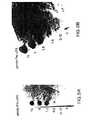
新たに調製した0.15%マラカイトグリーンと1.4%モリブデン酸アンモニウムを1.33N HClに溶かしたリン酸検出カクテルを酵素及び基質ゲルに注ぎ、均一に分配した。これらの試薬はリン酸と反応し、リン酸濃度の増加の関数として次第に濃い緑色を発色する。5〜10分間発色させた(図6は阻害剤量を5ナノモル〜0.5ナノモルとした場合のゲル上の発色を示す)。Stratagene Eagle Eye CCDカメラを使用して緑色ゲルを撮影した。予想通り、阻害ゾーンの緑色が薄くなる程度は加える阻害剤の濃度に相関した。従って、このアッセイを使用すると、他の任意の表面に分配した組み合わせビーズ又は化合物を配置した後にゲルアッセイと接触させることによりVanAの阻害剤をスクリーニングすることができる。
【0053】
このアッセイは更に、ゲルスクリーニングフォーマットが多段階反応に適応可能であることも立証する。多くのアッセイは多段階を必要とするので、この特徴はこのフォーマットが広範なアッセイで有用であるために必要である。この場合、VanAアッセイは酵素活性後に発色させる2段階アッセイである。発色試薬及び条件はVanA活性に干渉し、寒天ゲル内放出にも不適合であるので、このアッセイの均質(単段階)法は実現しにくい。従って、まず酵素ゲルアッセイを実施した後に溶液相発色段階を実施することによりこれらの2段階を空間及び時間的に分離することが望ましい。
【0054】
実施例2:リボソーム結合により刺激されたS.Cerevisiae伸長因子3ATPアーゼ活性により生産されたリン酸を検出するための2段階ゲルアッセイ
菌類伸長因子3(EF−3)がリボソームと相互作用すると、ホスファターゼ活性が刺激される。本発明者らはこの活性のアッセイにCF−HTSを適用した。
【0055】
EF−3アッセイ緩衝液中にEF−3(高度に温度感受性の酵素)と酵母リボソームを含む酵素ゲルを調製した。アッセイ緩衝液中に1mM ATPを含む基質ゲルも調製した。両者ゲルは低融点アガロース(Gibco BRL)中に2%のジメチルスルホキシドを加え、37℃で調製し、30分間4℃に静置した。対照試料として使用したEF−3の阻害剤であるPoly−L−リジンの連続希釈液をPVDCフィルムに滴下し、乾燥した(試料マトリックス)。次に、酵素ゲルを試料マトリックス上の阻害剤と共にプレインキュベートすることにより実施例1と同様にアッセイを実施した。次に、酵素ゲルを基質ゲルと20分間接触させた。実施例1と同様に、酵素ゲルと基質ゲルをマラカイトグリーン/モリブデン酸アンモニウム発色カクテルで染色した。次に、酵素ゲルをCCDカメラで撮影した。阻害剤スポットは緑色のバックグラウンド中に透明なゾーンとして見える。図7は阻害剤量を5ピコモル〜200ピコモルとした場合のゲル上の発色を示す。その結果は阻害剤の用量依存性シグナルを示し、新規EF−3阻害剤を見いだすためにこのアッセイで化合物をスクリーニングできると思われる。実施例2はオルガネラ又は他の粗生物混合物もしくは抽出物の存在により複雑になった場合でもCF−HTSが有用であることを立証する。
【0056】
実施例3:タンパク質−タンパク質相互作用阻害剤のgELISA間接色検出 上述のように、ELISAはレセプターをマイクロタイターウェルに固定化してリガンド−レセプター相互作用の阻害を検出するために一般に使用されている。ELISAで使用される「リガンド−レセプター」対はタンパク質又は他の高分子からの分子と小分子の任意結合対とすることができる。これらのアッセイはレセプターを固定化し、レセプターをリガンドと共にインキュベートし、高いシグナルバックグラウンドを生じる非特異的に結合した不要なリガンドを洗浄により除去し、レセプターと結合したリガンドに可視化剤(例えばレポーター酵素に結合したリガンド特異抗体)を結合し、レポーター酵素の基質を提供することにより可視シグナルを発生することが必要な複雑な多段階アッセイである。ELISAは複雑であるため、HTS産業はELISAをフリーフォーマットアッセイに適用できないとみなしているようである。しかし、本発明者らはこの多段階アッセイをCF−HTSフォーマットに適用し、種々のタンパク質−タンパク質、タンパク質−リガンド及び他のリガンド結合相互作用をアッセイした。
【0057】
ウロキナーゼ型プラスミノーゲンアクチベーター(uPA)はその対応するレセプター(uPAR)に結合する。uPA/uPAR相互作用は種々の癌の転移に結び付けられている。本発明者らは精製レセプター及びリガンドを使用して従来のuPA/uPAR ELISAをCF−HTSに適用した。
【0058】
uPARマトリックス
プラスチックプレート(7.5cm×11.5cm;Nunc,Inc.Naperville,IL製品)をpH7.4及び4℃のリン酸緩衝塩類(PBS)(Life Technologies,Grand Island,NY)中118nM精製uPAR15mlで一晩被覆した。プラスチックプレートを一晩被覆後、uPAR溶液をデカントし、1%(w/v)カゼインを含むPBS15mlを加えて室温(RT)で2時間インキュベートすることによりプラスチックプレート上の残留結合部位をブロックした。ブロック後、ブロッキング溶液をデカントし、プラスチックプレートをPBS中0.05% Tween−20(ポリオキシエチレンソルビタンモノラウレート)から構成される洗浄用緩衝液20mlで5回洗浄した。洗浄後、プラスチックプレートを10分間室温で乾燥した。これは、活性低下の原因となるマトリックスの過剰乾燥を避けるためにプラスチックプレートを下記アッセイで直後に使用できるような時間に設定した。
【0059】
β−uPAマトリックス
アッセイの目的には、使用するuPAをビオチンで標識する(β−uPA)。(実施例1及び2では溶融寒天に注入したが、これとは対照的に)高温にならないようにβ−uPAを寒天に浸漬することによりβ−uPAを含むゲルを調製した。寒天はまずアガロース(Sigma,St.Louis,MO)0.1gをPBS10mlと混合し、溶融するまで加熱した後、8×7×0.075cm3ゲル装置(Bio−Rad,Hercules,CA)に注入することにより調製した。(室温又は4℃で)凝固後、PBS中0.05% Tween−20及び0.1%カゼイン(いずれもSigma,St.Louis,MO製品)からなるアッセイ緩衝液中β−uPA15ml(約10nM)に4℃で一晩ゲルを浸漬した。ゲルを使用直前に室温で20分間乾燥した。
【0060】
試料マトリックス
uPA/uPAR結合の公知小分子阻害剤の不在下に非ビオチン化uPA(pro−uPA)をβ−uPA/uPAR結合の対照試料阻害剤として使用した。アッセイ緩衝液中0、0.03、0.1、0.3、1及び3μM pro−uPAの5μlアリコートをPVDCフィルム(Dowbrands,L.P.,Indianopolis,IN)上に分配し、2時間室温で乾燥した。
【0061】
uPARをpro−uPA及びβ−uPAと共にインキュベート
乾燥pro−uPAスポットがβ−uPAゲル表面と接触するように試料マトリックスをβ−uPAゲルの片側に配置した。pro−uPAを室温で10分間ゲルに拡散させた。次に、β−uPAゲルの反対側をプラスチックプレートに配置し、β−uPA(リガンドとして作用)とpro−uPA(競合的阻害剤として作用)をプラスチックプレートの表面でuPARと相互作用させた。結合/競合反応体を室温で20分間インキュベートした。インキュベーション後にプラスチックプレートをゲルから分離し、洗浄用緩衝液20mlで迅速に4回洗浄した。アビジン−HRP(Sigma,St.Louis,MO)をアッセイ緩衝液で25,000倍に希釈し、15mlをプラスチックプレートに加えることにより、アビジン−西洋ワサビペルオキシダーゼ(アビジン−HRP)結合体溶液を調製した。反応体を室温で10分間インキュベートした後、アビジン−HRP溶液をデカントし、プラスチックプレートを上述のように洗浄した。プラスチックプレートを20分間乾燥させた。
【0062】
アビジン−HRP中のアビジンはビオチンと特異的に結合するので、「リガンド/レセプター」結合を示すマトリックスの領域のみが発色を示す。「リガンド/レセプター」結合がpro−uPAにより競合的に阻害されるマトリックスの領域は発色しない。
【0063】
発色
o−フェニレンジアミンHCl(OPD)タブレット2錠を希釈剤7ml(いずれもAbbottキット番号6172−30、Abbott Labs,Abbott Park,IL)に溶かし、アガロース0.1gを水3mlに溶かして調製したアガロース溶液をこの溶液に加えることにより、比色HRP基質(OPDゲル)を含む発色ゲルを調製した。終量10mlの混合物を8×7×0.075cmミニタンパク質IIゲル装置に注入し、4℃で15分間凝固させた。ゲルをゲルキャスティングスタンドのガラスプレートからPVDC又は軟質プラスチックプレートに移し、室温で10分間風乾した。次に、ゲルを別のPVDC又はプラスチックシートに移し、反対側を10分間乾燥した。次に、OPDゲルをプラスチックプレートに加え、発色を開始した。OPDインキュベーション中の種々の時点でプラスチックプレートを440nm帯域フィルター(Omega Optical,Inc.,Brattleboro,VT)に載せ、フィルターを光ファイバー拡散プレートに載せてFiber−Lite光源(いずれもDolan−Jenner Industries,Inc.,Lawrence,MA製品)で照射した。CCDカメラ(Eagle Eye system,Stratagene,La Jolla,CA)で撮影した。
【0064】
対照実験
図8は実施例3の対照実験を示す。図8Aは、アガロース正方形片(1cm2)を各正方形片の下に記載した種々のβ−uPA濃度の溶液に浸漬した(但し、アガロース正方形片の左側は50nM溶液で標識する)。アガロース正方形片をuPAR固定化プラスチックプレート上でインキュベートした後、取り出した。次に、プラスチックプレートを洗浄し、β−uPA/uPAR結合が生じたマトリックスの領域を上述のように可視化した。図8BはOPD発色中の種々の時点で各アガロース正方形片におけるβ−uPAの濃度に対する各正方形片の(NIH Image Analysisソフトウェアを使用してCCDカメラからのディジタル画像を分析することにより決定した)平均画素値(バックグラウンドを差し引いた値)のプロットを示す。アガロースゲルから放出されたβ−uPAは標準ELISA及びこのパラメーターを測定する他のアッセイでこの反応に報告されている値に一致する約3〜5nMの半最大結合(Kd)をもつ典型的レセプター−リガンド結合曲線を示した。図8はgELISAにより発生される間接比色シグナルがリガンド−レセプター相互作用の程度に定量的に依存していることを示す。
【0065】
pro−uPA/β−uPA競合の結果
図9はpro−uPAによるβ−uPA/uPAR結合の阻害を示す。図9Aのスポットはβ−uPA/uPAR結合が阻害された領域を示す。従って、アビジン−HRPはこれらの領域で結合しなかった。従って、HRPはOPDゲル中の基質と反応して発色しなかった。図9Bは定量滴定をヒトの目で判断し易くするためにこれらの同一CCD画像データを別の方法で表示した。
【0066】
実施例4:タンパク質−タンパク質相互作用の阻害剤の直接放射検出
T細胞活性化は身体の免疫反応の要素である。T細胞活性化中に下流イベントが生じるためには、p56lck(LCK、タンパク質)がT細胞抗原レセプターの細胞質ドメインのITAM領域(イムノグロブリン関連チロシン介在活性化モチーフ)と相互作用する必要がある。このタンパク質−タンパク質相互作用を阻害する化合物は潜在的免疫抑制剤である。本発明者らはCF−HTSを使用してLCKが膜に固定化されるこのタンパク質−タンパク質相互作用をアッセイした。
【0067】
LCKマトリックス
5mM DTT(ジチオトレイトール)を含むPBS中3μM LCK5mlに11cm×2cmストリップを10分間室温で浸した後、緩衝液を除去することにより、ビオチン化LCKをビオチン捕獲膜(SAM膜)(Promega Corp.,Madison,WI)に固定化する。これは、SAM膜を直後(数分以内)に下記アッセイで使用できるような時間に設定する。
【0068】
ITAM*マトリックス
アガロース0.1gを緩衝液10mlと混合し、溶融するまで加熱した後、8×7×0.075cmゲル装置に注入することにより、放射性標識ITAMペプチド(ITAM*)を含むアガロースゲルを調製する。ITAM*はキャスティング直前に加えるか、又は最終濃度10nMまで凝固後にゲルに浸漬させる。
【0069】
試料マトリックス
スクリーニングしようとする試料をプラスチック表面又はPVDC上に分配し、乾燥して試料マトリックスを形成する。
【0070】
ITAM*、LCK及び試料のインキュベーションと可視化
試料がITAM*ゲルに拡散できるように試料マトリックスをITAM*ゲルの片側と接触させる。次に、LCKを固定化したSAM膜とITAM*ゲルの反対側を接触させる。15〜45分間インキュベーション後、SAM膜を取り出し、洗浄し、ホスホロイメージャー又はフィルムで撮像する。ITAM−LCK相互作用の阻害剤は画像上の低シグナル強度に対応する低放射能ゾーンにより示される。
【0071】
ITAM*/LCK結合の対照実験
アガロース0.1gを緩衝液10mlと混合し、溶融するまで加熱した後、8×7×0.075cmゲル装置に注入することにより、アガロースゲルを調製した。凝固後、直径1cmの円をゲルから打ち抜き、緩衝液中0.1、0.3、1、3、10及び20nM125I標識ITAM(Amersham,Arlington Heights,IL)400μlに4℃で一晩浸漬した。ゲル円を溶液から取り出し、20分間室温で乾燥させた。その後、LCKを固定化したSAM膜にゲルを載せ、45分間室温でインキュベートした。ゲルを取り出し、膜を緩衝液で4回洗浄した。乾燥後、SAM膜をホスホロイメージャーで撮像した。
【0072】
図10は125I−ITAMとSAM膜上の固定化LCKの用量依存性結合を示す。
【0073】
図11はゲル円内の125I−ITAM濃度に対する各125I−ITAMゲルの(Molecular Dynamics製品ImageQuantソフトウェアを使用してホスホロイメージャーからのディジタル画像を分析することにより決定した)平均画素値(バックグラウンドを差し引いた値)のプロットを示す。ゲルから固定化LCKに放出された125I−ITAMは典型的レセプター−リガンド結合曲線を示した。
【0074】
実施例5:組み合わせゲル/フィルターフォーマットを使用した全細胞レポーター遺伝子アッセイ
ルシフェラーゼ遺伝子(Promega製品であるルシフェラーゼレポーター遺伝子)に融合したサイクリックAMP応答配列(CREB)プロモーターを含むプラスミドをHEK細胞として知られる腎細胞にトランスフェクトする。トランスフェクトした細胞をフォルスコリンで処理すると、ルシフェラーゼレポーター遺伝子発現が誘導される。その後、ルシフェラーゼ基質(Promega製品である甲虫ルシフェリン)と適当な補因子(20mMトリシン,pH7.8,0.1mM EDTA,33mM DTT,0.3mM補酵素A,0.5mMアデノシン三リン酸及び1mM MgCl2)を含む生物緩衝液をHEK細胞に加えると、従来機器で検出可能な光子発生が生じる。本発明者らはこのアッセイをCF−HTSに適用した。
【0075】
細胞マトリックス
培養細胞をトリプシンで処理し、Corning/Costar TRANSWELL(登録商標)膜(プラスチック支持リングを備える3ミクロンポリカーボネートフィルター)に移し、組織培地の存在下に37℃、5%CO2下に一晩インキュベートした。その後、培地を膜から除去し、細胞が結合したフィルターを15分間風乾し、直後に下記段階で使用した。
【0076】
インデューサーマトリックス
フォルスコリンの10mMストック(エタノール中Sigmaストック)12μlを1%低融点アガロースゲル6mlに加えることによりルシフェリン発現のインデューサーを含むゲルを調製する。フォルスコリンの最終濃度が20μMになるまでゲルを室温で凝固させる。
【0077】
試料マトリックス
フォルスコリン誘導を阻止することが可能な試料をPVDC上の個別位置に高密度で分配し、乾燥する。
【0078】
試薬のインキュベーションと阻害の検出
PVDCの阻害剤側をインデューサーゲルと共にインキュベートする。次に、フォルスコリンインデューサーを含むゲルを上記のように調製した細胞マトリックスの非細胞側に配置する。これらを一緒に37℃で5%CO2下に20分間インキュベートする。次に、フォルスコリンゲルを取り出し、細胞マトリックスを37℃で5%CO2下に更に4時間インキュベートし、ルシフェラーゼ構築物の発現を最大にする。ルシフェラーゼ酵素活性(又はその阻害)を検出するために、細胞マトリックスフィルターをそのプラスチック支持リングから物理的に取り外し、ペトリ皿に入れる。適当な補因子を加えた生物緩衝液中のルシフェラーゼ基質(Promega製品である甲虫ルシフェリン)をペトリ皿に注ぎ、シグナルとして発光させる。シグナルはルシフェラーゼを発現する固定化細胞の内側に局在しているので、初期誘導段階の阻害剤は光子発生の低いゾーンとなる。
【0079】
実施例6:リガンド−細胞相互作用の阻害剤を直接検出するためのゲル/フィルターCF−HTS
細胞表面のリガンド/レセプター結合は最終的に機能的応答(例えば細胞増殖又は生物活性物質の分泌)を生じるシグナル経路を細胞内で開始する。疾病状態における細胞の生物応答を調節するために、リガンドの細胞表面結合を阻害しようとする例は多い。リガンド/細胞レセプター結合の阻害剤を評価するための一般的な方法は、阻害剤が放射性標識天然リガンドと細胞の結合を低下させる能力を評価することである。このためには、細胞を放射性リガンド及び阻害剤と共にインキュベートした後、未結合及び非特異的に結合した放射性リガンドを洗浄により除去し、次いで結合放射能の量を測定する。
【0080】
インターロイキン8(IL−8)は種々の細胞でレセプターと結合することにより炎症に関与する走化性ケモカインである。本発明者らはこの相互作用の阻害剤を評価するためにCF−HTSリガンド−レセプター細胞アッセイを開発した。
【0081】
細胞マトリックス
IL−8aレセプターを発現するHEK細胞(ATCC,Bethesda,MD)を直径75mmトランスウェルメンブレンフィルター(Corning Costar Corp.,Cambridge,MA)に約20×106細胞/プレートの密度でプレーティングした。pH7.2及び37℃で10mM Hepes(Sigma,St Louis,MO)を含む緩衝液(Life Technologies,Grand Island,NY製品であるRPMI)中でメンブレンフィルターと細胞を一晩結合させた。細胞をフィルターに結合した後、培地を捨て、細胞を新鮮な緩衝液で洗浄し、未結合細胞を除去した。細胞側が下を向くようにフィルターを逆さにし、過剰の培地を排出させるような角度にした後、20分間乾燥した。これは、細胞マトリックスを下記アッセイですぐに使用できるような時間に設定した。
【0082】
リガンドマトリックス
アガロース0.1gを緩衝液10mlと混合し、溶融するまで加熱した後、8×7×0.075cmゲル装置に注入することにより調製したアガロースゲルに125I標識IL−8(Amersham,Inc.,Arlington Heights,IL)を浸漬することにより、リガンドマトリックスを調製した。回転プラットフォーム(New Brunswick Scientific Co.,Inc.,Edison,NJ)上でゆっくりと混合しながらゲルに一晩室温で浸漬させた。浸漬後、ゲルを使用直前に室温で20分間乾燥した。これは、ゲルをアッセイですぐに使用できるような時間に設定した。
【0083】
試料マトリックス
IL−8/細胞レセプター結合の公知阻害剤の不在下に、非放射性標識IL−8(Genzyme Corp.,Cambridge,MA)を対照試料阻害剤として使用して125I−IL−8のHEK細胞結合の阻害を観察した。1μlの0、0.03、0.1、0.3、1、3、10及び100μM IL−8をPVDC等のプラスチックシート上に分配し、1時間室温で減圧乾燥した。
【0084】
インキュベーション
乾燥後のIL−8スポットがゲル表面と接触するようにリガンドマトリックスの片側に試料マトリックスを配置した。ゲルを逆さにして反対側を10分間室温で乾燥し、「阻害剤」をゲルに拡散させた。次に、ゲルを細胞マトリックスの非細胞側に配置した。結合反応体を45分間室温でインキュベートした。その後、ゲルを除去し、膜の非細胞側を緩衝液で4回洗浄した。膜を完全に乾燥させてからプラスチック支持リングから取り外した。次に、膜をX線フィルム又はホスホロイメージャー(Molecular Dynamics,Sunnyvale,CA)で撮像した。
【0085】
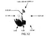
図12は細胞マトリックスを拡散してIL−8aレセプターを発現するHEK細胞に結合するアガロースゲル中の125I−IL−8の用量応答を示す。数個の1cm2アガロース正方形片を種々の指定125I−IL−8濃度の溶液に浸漬した。次に、リガンドに浸漬した正方形片を上述のように細胞マトリックスと接触させた。インキュベーション後、正方形片を細胞マトリックスから取り出し、膜の非細胞側を緩衝液で洗浄した。ホスホロイメージャーを使用して細胞に結合した125I−IL−8の位置を調べた。データによると、このゲルを利用した細胞アッセイにおける直接放射読取り値はリガンド−レセプター相互作用の程度に定量的に依存している。図12は結合領域が形状的に別々のままであることも示しており、シグナルの横拡散がアッセイで問題とならないことが確認される。
【0086】
図13は競合的阻害実験の結果を示す。125I−IL−8とHEK細胞の結合の阻害は明スポットにより示される。非標識IL−8による阻害が定量的に用量依存性であることも容易に理解されよう。これらのデータは、この場合には非標識IL−8である阻害剤が細胞マトリックスを横断して125I−IL−8と細胞の結合を低下できることを示している。
【0087】
実施例7:フィルターを利用した無ゲル機能細胞アッセイ
細胞に組込んだレポーター系に及ぼす試験化合物又は試料の影響を観察することにより細胞機能の変化を測定する例は多い。例えば、グリーン蛍光タンパク質(GFP)等の蛍光細胞内タンパク質、レセプターもしくは接着分子等の細胞外タンパク質、又はルシフェラーゼ、クロラムフェニコールアセチルトランスフェラーゼもしくはβ−ガラクトシダーゼ(Promega)等の特異酵素の合成に及ぼす影響の観察が挙げられる。まず、試験化合物を細胞と共にインキュベートする。その後、適当な時間(例えば数分間、数時間又は数日間)細胞にレポータータンパク質を発現させる。その後、レポータータンパク質の量を直接法(例えばGFP)又は間接法(例えば膜結合タンパク質によるELISA法)により定量する。更に酵素の場合には、細胞を破壊してレポータータンパク質を抽出し、酵素活性を定量することができる。その他、レセプターの刺激又は膜電位等の細胞生理の変化に応じて色素の特定分子又は放射性標識代謝物等の挙動又は局在を測定する機能細胞アッセイもある。
【0088】
細胞内接着分子−1(ICAM−1)の発現に及ぼす化合物の影響を測定するELISAアッセイは次のようにCF−HTSに適用することができる。ICAM−1を発現する内皮細胞をポリカーボネート走化性膜(Neuro Probe)に約5,000細胞/mm2でプレーティングする。細胞を培地中で37℃で一晩インキュベートする。培地を捨て、膜を10分間室温で部分乾燥させる。ICAM−1の誘導を試験している試料又は化合物をプラスチックシート上で乾燥させ、湿潤膜の非細胞側と接触させる。化合物を湿潤チャンバー内で37℃で1時間細胞と相互作用させた後、細胞を培地に浸漬し、5時間37℃でインキュベートする。
【0089】
細胞が誘導タンパク質を合成するまで待ってから、培地を膜から除去し、フルオレセインイソチオシアネート(FITC)又はビオチンに結合しているか又は未結合の抗ICAM−1抗体(Genzyme,R&D systems)を含む緩衝液中で細胞をインキュベートする。室温で30分間インキュベーション後、緩衝液を捨て、細胞を数回洗浄して未結合抗ICAM−1を除去する。FITC結合抗体の場合には、485nm励起及び520nm発光でCCDカメラ(Stratagene,Imaging Research)を使用して膜を撮影する。ICAM−1発現を刺激した化合物はFITC−抗ICAM−1抗体の結合により蛍光が増加したゾーンとなる。ビオチン結合抗体の場合には、アビジン−HRPを含む緩衝液中で室温で10分間細胞をインキュベートする。緩衝液を捨て、未結合アビジン−HRPを洗い流す。その後、四塩化ジアミノベンジジン(Pierce)等の沈降性HRP基質を含む緩衝液に膜を浸漬し、下位の細胞がICAM−1を発現するように誘導された領域で発色を観察する。非結合抗ICAM−1抗体の場合には、結合二次抗抗ICAM−1抗体を細胞と反応させた後、結合体の適当な基質でシグナルを発生させる。CCDカメラで撮影する。これらの全方法(FITC、アビジン−HRP及び抗抗ICAM−1)は同一定量結果を与える代替レポーターであり、即ちICAM−1発現に影響を与える試料をシグナルの増加又は減少したゾーンに相関させることができる。
【0090】
この場合の連続フォーマットマトリックスは膜とプラスチックシートであることに留意されたい。CF−HTSにゲルは不要である。
【0091】
実施例8:ノイラミニダーゼ阻害に関する個別化合物のCF−HTSアッセイ
試料マトリックス
528種の構造的に関連する個別化合物のライブラリーをCF−HTSにより試験した。Packard Multiprobe MP204DTを使用してバイアルからの化合物を96ウェルプレートに希釈し、化合物を希釈してプラスチックシートに移した。化合物をまずバイアル中のDMSO中40mMから96ウェルプレート中のDMSO中4mMに希釈した。次に、96ウェルプレート中でDMSO中4mMから50%EtOH/H2O中200μMに希釈した。これらの試料を8cm×8cmプラスチックシート(Bio−Rad cat.#165−2956)に平均5mmの試料間隔で各シート合計192個の試料数となるように1μlずつ2画分を移した。従って、各1μlスポットがライブラリーからの特定化合物を約200pmol含むものとした。対照として、公知ノイラミニダーゼ阻害剤である2,3−デヒドロ−2−デオキシ−N−アセチルノイラミン酸(DANA)(Boehringer Mannheim #528544)の希釈液系を各シート上の化合物の隣に手動分配した。各化合物がプラスチック上のそれ自体の位置で乾燥されるようにシートを減圧乾燥した。
【0092】
酵素マトリックス
アッセイに先立ち、インフルエンザノイラミニダーゼ酵素を25%グリセロール、リン酸緩衝塩類から、40℃で1%アガロース、50mMクエン酸ナトリウム(pH6.0)、10mM塩化カルシウムからなる液化寒天ゲルに1500倍に希釈した。8cm×8cm×0.75mm酵素ゲルを注入し、温度を4℃まで下げることにより凝固させた。
【0093】
基質マトリックス
合成インフルエンザノイラミニダーゼ基質である2’−(4−メチルウンベリフェリル)−α−D−N−アセチルノイラミン酸(Sigma cab.#M−8639)をDMSO中3mMから液化寒天ゲル中30μMに希釈し、上記酵素ゲルと同様に注入した。
【0094】
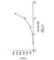
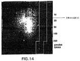
インキュベーションと検出
着目化合物を乾燥した側で試料マトリックスに酵素マトリックスを重ねた。次に、基質マトリックスを酵素マトリックスの上に重ねた。マトリックスを室温で30分間インキュベートした。この間に、蛍光消光基質と酵素は2つのゲルの間に拡散し、基質は酵素により分解され、蛍光強度を増した。これは340nm励起及び450nm発光によりモニターした。酵素活性を阻害することが可能な化合物は蛍光強度の増加を最小にした。ゲルが大部分の位置で蛍光強度を増すにつれて、プラスチックシートからゲルに拡散された酵素阻害剤を含む領域はより低い蛍光をもつ暗領域として見えた。これは発光波長と励起波長を調節するように適当なフィルターを取り付けたCCDカメラにより容易にモニターされた。阻害ゾーンを示す化合物の種を阻害剤マトリックスに対するゾーンの位置により決定した。DANA対照の蛍光を既知量の各試験阻害剤と比較することにより、化合物毎にIC50の定量的推定を行った。
【0095】
ライブラリーの全528種の化合物を2回ずつ試験した。使用した合計酵素ゲル容量は33ml、即ち試験当たり31.25μlとした。更に、全化合物を同時に試験し、アッセイ条件は従来の96ウェルアッセイに比較して一定であった。更に、図14に示すように、アッセイは100μM程度の低濃度の阻害剤を検出するのに十分感受性であった。活性化合物の推定IC50値は試験当たり200μlを必要とするコストの高い96ウェルアッセイにより試験した同一化合物で観察される値とよく一致した。定量ゲルアッセイ結果表参照。本実施例は高密度の化合物アレーを試験すると、費用と時間を節約できることを立証するものである。例えば、間隔を5mmでなく2.5mmに僅かに減らすだけで単位容量当たりの試験化合物数は4倍になる。この実験では、こうすることによって試験化合物当たりの容量が10μl未満に減ったと思われる。しかも、小型化スクリーニングで一般に使用されている低容量液体操作装置を必要とすることなく大容量で試薬を操作する。
【0096】
【表1】
この小型化の利点を立証するための追跡実施例として、10,080種の個別化合物を合計酵素ゲル容量17ml(試験当たり2μl未満)となるようにCF−HTSにより試験した。Packard Multipetteを使用して各試料30nl容量を1mm間隔で分配した。化合物は濃度5mMでDMSOに分配し、各々約150pmolをプラスチックに分配した。この場合もDANAを対照阻害剤として使用した。化合物アレーの外側の通常対照滴定以外に盲試験としてアレー自体の内側にも対照滴定を加え、対照試料をプラスチックに分配する際に10,080種の未知化合物と同様に処理した。上記と同様にシートを乾燥し、ノイラミニダーゼアッセイで使用した。全10,080種をこの方法により1時間未満で同時にスクリーニングした。この場合も、初期化合物のシート分配時を除き、ミクロフルイディクス及び/又は低容量操作は不要であった。このアッセイで使用される非常に高い密度は阻害剤の検出に干渉しなかった。更に、高密度は(ゲル中の明スポットとして容易に観察される)蛍光化合物が活性化合物の最近傍にあった場合でも、活性化合物の同定を複雑にしなかった。図15参照。
【図面の簡単な説明】
【図1】 非多孔質マトリックスに担持したgELISA試料を、アッセイ試薬を含む多孔質ゲルマトリックスの片側に接触させ、更に別のアッセイ試薬を担持する非多孔質マトリックスに接触させる方法を示す。
【図2】 gELISA試料マトリックスと多孔質ゲルマトリックスを除去した後に洗浄し、液体又は溶液試薬を添加して非多孔質試薬マトリックス上にレポーター複合体を形成する方法を示す。
【図3】 レポーター基質を含む多孔質ゲルマトリックスにレポーター複合体マトリックスを接触させることによりgELISAアッセイを可視化する方法を示す。
【図4】 細胞を担持するフィルター表面に対向するフィルター表面にリガンドマトリックスを加える方法を示す。
【図5】 アッセイを可視化したときに細胞を担持するフィルター表面に観察される様相を示す。
【図6】 種々の濃度の公知用量依存性阻害剤を使用したVanAアッセイの結果を示す。
【図7】 種々の濃度の公知阻害剤を使用したEF−3アッセイの結果を示す。
【図8】 タンパク質−タンパク質相互作用のgELISAアッセイの対照実験を示す。
【図9】 タンパク質−タンパク質相互作用の「阻害」のgELISA結果をを示す。
【図10】 固定化LCKへの放射性標識ITAMの用量依存性結合を示す。
【図11】 ITAM濃度に対する画素値のグラフであり、典型的レセプター−リガンド結合曲線を示す。
【図12】 放射性標識IL−8のリガンド−細胞相互作用の対照実験を示す。
【図13】 IL−8リガンド−細胞相互作用の「阻害」を示す。
【図14】 ノイラミニダーゼの阻害剤のアッセイを示す。
【図15】 ノイラミニダーゼ阻害に関する10,080種の個別化合物の同時アッセイを示す。[0001]
TECHNICAL FIELD OF THE INVENTION
Continuous format high-throughput screening (CF-HTS) using at least one porous matrix allows the pharmaceutical industry to simultaneously assay a wide range of biological or biochemical activities of many chemical species. In addition, CF-HTS is useful for performing multi-step assays.
[0002]
Background of the Invention
Biochemistry and biological assays aim to test activity in a wide range of systems ranging from protein-protein interactions, enzyme catalysis, small molecule-protein binding to cellular function. In “High Throughput Screening” (HTS), a number of species are tested using this type of assay to find the unknown biological or biochemical activity of the species.
[0003]
Homogeneous and heterogeneous assays
Various biological assays can be broadly divided into two types: homogeneous assays and heterogeneous assays. In a homogeneous assay, all reagents are added simultaneously and the results measured or interpreted without additional manipulation. For example, cells grown in Petri dishes can be exposed to drugs. If the drug is toxic to the cells, the toxicity is detected from the clear zone by simple observation. As another example, cells expressing a protein that changes the color of the cells can be used. When cells expressing β-galactosidase (β-gal) are grown in agar containing x-gal, the cells turn more or less blue depending on the degree to which the β-gal protein is expressed. Thus, a homogeneous assay at the biological stage that affects the expression of a reporter gene such as the β-galactosidase gene can be constructed. Yet another example of a homogeneous assay utilizes a substance that changes color or changes fluorescence when processed by an enzyme. Finally, techniques such as Scintillation Proximity Assay (SPA) by Amersham directly measure the binding of radioligands to proteins or ligand binding substances immobilized on beads containing scintillant. All of the above examples are homogeneous assays because no steps other than reagent addition are required prior to final detection, measurement or signal reading.
[0004]
On the other hand, heterogeneous assays are inherently incompatible to some extent and therefore require at least two steps that cannot be combined in one step. For example, many heterogeneous assays require that reagents be added in a predetermined order (eg, if a given reagent interferes with the early stages of the assay but is necessary to perform a late stage). . A common example of this is an assay in which a signal generating reagent is added to directly determine the presence or concentration of the reaction product. Another common step in a heterogeneous assay is a wash step. Often, excess reagent must be added early in the assay, but it must be washed away before subsequent steps to allow the reaction to proceed without a high background signal. For example, in a radioligand binding assay, a labeled ligand is first incubated with a protein bound to a solid surface, but in practice only a small portion of the ligand binds to a small number of protein sites. Excess unbound ligand must be washed away after incubation to allow accurate measurement of bound radioligand. Washing can be performed by various alternative methods such as filtration, wash / decant cycle, sedimentation / phase separation and / or centrifugation.
[0005]
Many biological and biochemical processes can be measured only by heterogeneous methods. In addition, other biological and biochemical processes can be applied to the homogeneous process, but these other processes are more cost effective and / or easier to obtain reagents. Various methods and kits used in the homogeneous method are commercially available, and examples include SPA (Amersham), fluorescence polarization (Jolley et al.), And time-resolved fluorescence (Packard et al.). However, using this type of method is always extra cost and time. For many assays, heterogeneous methods have been established and are more likely to proceed quickly. For this reason, the use of heterogeneous methods such as ELISA, filter binding, RIA, and luciferase cell assays continues to become widespread. Some of these methods will be described in detail later.
[0006]
High throughput screening (HTS)
Over the past few years, the pharmaceutical industry has increasingly used compound library HTS to find drug candidates. HTS is a method in which multiple individual compounds are tested in parallel and the biological activity of multiple test compounds is screened simultaneously or nearly simultaneously. Currently, the most widely established technique is a method using a 96-well microtiter plate. In this format, 96 independent tests are performed simultaneously on a single 8 cm × 12 cm plastic plate containing 96 reaction wells. These wells generally require an assay volume of 50-500 μl. In addition to plates, a number of instruments, materials, pipettors, robots, plate washers and plate readers are commercially available to adapt the 96 well format to a wide range of homogeneous and heterogeneous assays.
[0007]
To date, attempts to improve HTS have focused on making the wells smaller (smaller). Reducing the well dimensions increases the number of wells in each plate, allowing a larger number of parallel tests. In addition, reducing the assay volume also reduces the cost of reagents per test. In addition, since a larger number of parallel tests can be performed with a smaller assay volume, more compounds can be tested simultaneously to find drug candidates. To date, miniaturization has only improved 96-well technology to a 384-well (96 × 4) format. Commey et al.,J. et al. Biomol. Screening , Vol. 2 (3), pp. 171-78 (1997). In fact, high density formats such as the 9600 well format have also been reported. However, miniaturization comes with cost and complexity.
[0008]
These costs and complexity are directly related to the three main elements of screening format miniaturization. First, it must be possible to make test containers (tubes, wells, dimples, etc.) small. Second, it must be able to dispense all required assay reagents in a smaller and more accurate well (usually by a liquid handling robot that dispenses the reagents to multiple wells simultaneously). Third, it must be able to “read” high density array test results.
[0009]
Since independent parallel arrays are required, each of these elements adds problems and limitations to how much miniaturization is feasible or cost effective. For example, some new small formats require completely different reagent dispensing methods, or readers with resolution, sensitivity and engineering design that are compatible with the new small format. As the size of each well is reduced, it becomes more time consuming and expensive to create a container array, dispense smaller amounts of reagent, and read each sample. In addition, dispensing a small amount of reagent and measuring a weak sample signal tends to be inaccurate, so reducing the sample size increases the statistical variation between samples. Furthermore, if the sample size is smaller than a predetermined point, the implementation of the new miniaturization format becomes more expensive and complicated due to factors such as evaporation and surface tension.
With the breakthrough in HTS technology, the industry is even more eager for the possibility of “free format assays”, ie assays without physical barriers between samples. Although it is generally expected to test small droplets in a format that does not use wells, the use of free format assays with HTS in standard individual compound systems has not been reported in practice.
[0010]
Combination library screening-gel permeation method
With the advent of combinatorial chemistry, millions of chemical species can be rapidly produced on solid supports (generally beads). A 96-well format is used to screen bead-bound libraries, but this format (1) can only carry a small amount of chemical species on each bead, (2) the number of test compounds is very large ( 3) Since beads are difficult to handle in 96 well microtiter plates, they are generally considered invalid.
[0011]
To avoid the problems inherent in screening combinatorial libraries in a 96-well format, the use of a simple homogeneous assay that can be referred to as a “free format” has been reported. As an example, an assay using pigment cells (melanocytes) in a simple homogenous assay of a combinatorial peptide library is described by Jaywickreme et al.,Proc. Nat'l Acad. Sci. (USA) , Vol. 191, pp. 1614-18 (March 1994). According to the authors, after adding cells to agarose in a Petri dish, beads carrying the combined compound are added to the surface of the agarose, partially releasing the compound from the beads. As the active compound discolored the cells as the compound diffused locally into the gel matrix, the active compound was visualized as a dark area.
[0012]
Another recent example is Daniel chelsky's “Strategies for Screening combinatorial libraries: Novell, reported at the first annual meeting of the Biomolecular Screening Society held in Philadelphia, PA (November 7-10, 1995). and Traditional Approaches ”. Chelsky performed a simple homogeneous enzyme assay of carbonic anhydrase inside the agarose gel so that the enzyme in the gel changes the color of the entire gel. Thereafter, beads carrying the combined compound were placed inside the gel via an optical linker, and the compound was partially released by UV light. Compounds that inhibited the enzyme were observed as local inhibition zones with less discoloration. Finally,Molecular Diversity , V. 2, pp. 57-63 (1996), Salmon et al. Reported a method similar to that of Jaywickreme et al., Screening combinatorial libraries for compounds that have cytotoxic effects on cancer cells growing in agar.
All three examples are variants of antibacterial or anticancer drug time-tested gel assays, similar to conventional immunoassays that measure antigen / antibody interactions in gels. Although these gel permeation assays were suitable for screening of bead-bound combinatorial libraries, no extension of this format to heterogeneous assays or non-bead-bound libraries has been reported.
[0013]
With conventional recognition, researchers have not been able to test samples in a continuous format in which they can be mixed. Only a few bead-bound libraries have been assayed, with few reported assay species and the problem of sample mixing in the continuous format. Because of these constraints, researchers considered the 96-well format suitable for heterogeneous non-bead-bound library screening. It would be desirable if heterogeneous assays could be performed in a free format configuration. It is further desirable if individual compounds can be tested in a free format configuration.
[0014]
Summary of invention
The present invention relates to continuous format high throughput screening (CF-HTS) and can implement the free format concept in any assay that can be performed in a 96 well format, whether homogeneous or heterogeneous. In addition, CF-HTS is useful for screening combinatorial libraries in a heterogeneous assay rather than a homogeneous assay. Furthermore, CF-HTS can assay individual compounds without the cost and complexity associated with miniaturization. It was found that there was no problem with the possibility that the reagent and test results would be mixed at a later stage.
[0015]
One aspect of the invention relates to testing biological or biochemical activity of a sample,
a) introducing a plurality of samples in or on a porous assay matrix optionally containing one or more assay components;
b) introducing at least one assay component into the assay using at least one matrix that may or may not be a porous assay matrix;
c) i) washing the matrix used in the assay to remove excess sample, assay components or combinations thereof, or
ii) performing an additional step of contacting the matrix used in the assay with the additional reagent in bulk solution or as a liquid.
[0016]
Another aspect is the introduction of a sample by introducing a plurality of samples in or on a porous assay matrix optionally containing one or more assay components and performing the assay using at least two additional matrices. It relates to testing for biological or biochemical activity.
[0017]
Yet another aspect is the introduction of more than 96 samples in or on a porous assay matrix optionally containing one or more assay components and performing the assay to produce more than 96 sample organisms or organisms. Related to simultaneous testing of chemical activity.
[0018]
Detailed Description of the Invention
The central idea of CF-HTS is to associate the sample with a porous matrix. This method places one or more assay components within, above or below the matrix, such as a gel, plastic sheet, filter or other form of a solid support that is easy to handle. When added to the porous matrix, the sample diffuses slowly enough to allow the assay to run without mixing. Thus, the CF-HTS format separates samples by diffusion rather than an impermeable barrier. If the test is continued for an excessively long time, the sample and the result may be mixed. However, with careful timing, CF-HTS can screen very high concentrations of compounds simultaneously and individually without the need to fill individual wells or reaction vessels with solvents or assay components. Furthermore, more complex heterogeneous assays can be performed in this format by manipulating the matrix to which the reaction components have been added. Heterogeneous assay matrix manipulation is unprecedented and CF-HTS is as flexible as the 96-well format in its ability to screen a wide range of biological or biochemical processes. Furthermore, CF-HTS achieves the kind of benefits expected from miniaturization without the attendant drawbacks and has its own advantages.
[0019]
CF-HTS utilizes a wide range of matrix and assay components. Non-limiting examples of matrices include agarose, acrylamide, or other gelatin materials, membranes, filters and plastics. As a non-limiting example, the matrix can be composed of materials such as polystyrene, polypropylene, other plastics, paper fibers, glass, glass fibers, silica, polycarbonate, polyester, polyvinylidene chloride and polyethylene. The matrix can be an impermeable solid, a porous solid (eg, a filter), or a gel. Non-limiting examples of assay components include: macromolecules such as nucleic acids, proteins and other synthetic or natural macromolecules; cells, cell lysates, biological extracts, organelles, and other complex organisms and mixtures; and buffers Small molecules such as solutions, salts, inhibitors, substrates, peptides, dyes, nucleotides, cofactors, ions and solvents.
[0020]
The CF-HTS assay is an assay that separates multiple samples or compounds by diffusion rather than an impermeable barrier. An essential component is the introduction of multiple (two or more) samples within or on the porous assay matrix optionally containing one or more assay components. A porous assay matrix containing assay components is prepared by adding, mixing, pouring, dispensing or dipping components into the matrix. Porous matrices are also prepared by coupling, applying, binding, fixing, linking, joining or attaching assay components within or on the surface of the matrix. In addition, the porous matrix can be prepared by forming a solution or liquid film on a cell bed, enzyme or other immobilized assay component. The porous assay matrix is used to control the order and / or duration of component addition and the degree of mixing and diffusion when assay components are used in combination.
[0021]
CF-HTS may use a non-porous matrix. Non-porous matrices are prepared by coupling, applying, binding, fixing, joining, joining or attaching assay components or samples to the surface of the non-porous matrix. When using a non-porous matrix with CF-HTS, one or more of the assay components are spatially immobilized.
[0022]
Once the assay component is introduced to the surface of the matrix, the assay component may be covalently or non-covalently, specifically or nonspecifically interacted with the matrix that has been unmodified, modified or otherwise pretreated to facilitate binding of the assay component Combined. After binding, the assay components are spatially immobilized so that they are immobilized for assay purposes. In this case, the sample must be able to diffuse into the matrix to reach the assay component and / or a sub-component or product of the assay component must be able to diffuse into the matrix to reach the sample.
[0023]
At least one porous matrix containing the sample is used in any one or more of the following stages.
[0024]
(1) contacting the surface of the porous matrix with at least one other (porous or non-porous) matrix so that one or more of the sample and / or assay components can diffuse through the interface.
[0025]
(2) Separate two or more matrices and stop component and / or sample interaction.
[0026]
(3) bringing two or more matrix surfaces into contact with each other so that assay components can interact;
[0027]
(4) The matrix is washed, rinsed or eluted with buffer or other solvent to remove unbound and / or non-specifically bound assay components.
[0028]
(5) Distribute, inject, add or immerse assay components in solution into the matrix or filter the components through the matrix.
[0029]
(6) Imaging, reading, scanning, detecting or otherwise visualizing radiation, fluorescence, spectroscopic or electromagnetic signals present on or in one or more matrices.
[0030]
CF-HTS has a number of advantages over the prior art. Since individual wells are not used, there is no need to accurately dispense assay components or reagents simultaneously to the wells. Instead, assay components are dispensed and mixed by homogeneous bulk operations. Since assay components are prepared as homogeneous bulk solutions or matrices, statistical variation between samples is minimized. On the other hand, in the 96-well format, there is a large variation between samples due to the presence of wells.
[0031]
Furthermore, CF-HTS can screen a large number of compounds at a very high density. Even if the observed correct report is somewhat “mixed”, it is only necessary to retry the compound in the vicinity of the correct report. For example, if 10,000 samples could be reduced to 50 candidates around the visualization region, it would be easy to reduce 50 candidates to active compounds with conventional 96 well technology.
[0032]
After the individual compounds are distributed on the plastic sheet in a highly filled array and dried, CF-HTS is performed to address all the serious miniaturization issues. Since this format simply achieves miniaturization by limiting the amount of sample that diffuses into the matrix, there is no need to modify plastics or other materials to achieve miniaturization. This format does not require microfluidics to dispense assay reagents, since the entire assay is generally performed in “single giant wells” where reagents and solutions are bulk-manipulated. Only the sample is distributed by microfluidics. In addition, this format only requires a search for non-homogeneous localized zones in a homogeneous matrix, so there is significantly less statistical variation. There is no need to read and compare many different wells. In addition to all the benefits expected for miniaturization (cost, throughput, reagent usage, test compound usage), CF-HTS also has surprising advantages such as the ability to bulk manipulate most stages of the assay.
[0033]
A central aspect of CF-HTS is the finding that assay components and samples do not diffuse rapidly laterally at the interface between the matrices. For example, when an agarose gel is placed on a plastic plate, there is considerable liquid at the surface interface of the gel. If an interaction is required between the assay components in the gel and the assay components on the plate (as in an ELISA), the components in the gel must be able to diffuse from the gel to the plate. However, since CF-HTS slows the accompanying lateral diffusion considerably, the interaction on the plate is localized near the original position of the gel side component. These same principles apply to any matrix-matrix interface between any combination of gels, filters or surfaces (or any other matrix). The recognition that this diffusion behavior is controllable and generally applicable to the entire matrix interface is unprecedented and contrary to conventional recognition.
[0034]
A preferred method for introducing a sample or compound into a matrix (eg, gel or wet filter) is to place each sample in a small volume on a plastic sheet so that the two samples are each placed in a specific location without mixing or overlapping. This is a method of distributing and drying in an array on a surface such as a surface. When the plastic sheet is overlaid on the matrix, the sample dissolves and diffuses into the matrix at a location corresponding to their predetermined location in the original array.
[0035]
An alternative method of distributing samples in an array is to distribute the samples in a porous matrix (eg, a filter) so that the distribution volume of each sample is sufficiently small so that the samples do not overlap within the matrix. Upon contact with another porous matrix containing a larger amount of liquid, the compound diffuses and initiates the assay.
[0036]
A preferred method for introducing the combined compound bound to the beads into the matrix is to distribute the beads randomly or regularly on a surface such as the surface of a plastic sheet. The bead can then be treated to release (cleave) the test compound if the test compound is covalently attached to the bead by a labile linker (photocleavage and gas phase acid cleavage are well known in the art. Is). Each compound can then be non-covalently associated with the area occupied by its original beads and the dry compound can be introduced into or onto the matrix.
[0037]
An alternative method of introducing individual compounds into the matrix is to immerse or otherwise bind each compound non-covalently in or on the beads. Using this method, multiple compounds can be mixed on the beads at once so that each bead continues to carry a unique compound. The bead mixture can then be easily spread over the surface for introduction into the matrix. This method does not require any small volume liquid operation.
[0038]
If the initial array of samples introduced into the matrix with a CF-HTS screen is dense and a particular active zone spatially occupies the initial position of two or more samples, any of these samples will be observed activity May be a source. Since a large number of compounds exist in a specific zone, the higher the initial density, the more candidate compounds in each zone. Although the compounds may diffuse together, each maintains its own spatial gradient and does not mix quantitatively at any one position. Thus, the center of the zone continues to correlate with the exact position of the active compound in the initial array. In fact, since correct reports are rare, there is no problem retesting multiple samples to ensure identification of the active compound in each active zone.
[0039]
According to another aspect of the invention, a physical barrier is introduced into the assay matrix (ie, strictly speaking, the format is discontinuous) to limit the distance that the sample can diffuse. For example, two gels, each containing an enzyme and a substrate, can each be cut with a mesh ("cookie cutter"), and each gel can be subdivided into a number of individual locations. Thereafter, the two gels can be brought into contact, so that the substrate and the enzyme can diffuse together inside the subdivided gel pieces. Samples can then be introduced into each sub-gel piece so that the assay is completely independent without inter-assay diffusion. This embodiment effectively reduces some of the benefits of CF-HTS by introducing statistical deviation between samples and fixing the volume to limit the signal of the high density array. However, this embodiment does not require the assay components to be distributed across multiple parallel reaction vessels, and maintains the advantage of a matrix heterogeneous assay that does not require partial sample mixing.
[0040]
gELISA
An enzyme-linked immunoassay (ELISA) is a heterogeneous assay that detects the binding of ligand and immobilized receptor in solution. ELISA requires a large number of reagent mixing and washing steps that are difficult to perform in a 96 well format, and the difficulty is expected to increase further when subdividing wells from a 96 well format to a 384 well format. We applied the CF-HTS method to detect inhibition of binding between the ligand and the immobilized receptor target (gELISA).
[0041]
A receptor is any molecule that can bind to another molecule. Non-limiting examples are proteins, peptides, nucleic acids, carbohydrates and complexes of the above examples. Receptors include, as non-limiting examples, plastic surfaces (eg Petri dishes or plastic plates (Nunc products)), membranes or filters with high target binding capacity (eg nitrocellulose, nylon or PVDF (Millipore, Corning Costar, Schleicher & Schuell, It is immobilized on one of several possible matrices (receptor matrices) such as BioRad) or modified membranes such as SAM membranes (Promega). A porous ligand matrix (eg, agarose gel or porous membrane) is prepared to partition the ligand of the immobilized receptor onto or into the matrix. The test compound or sample is dispensed directly into the ligand matrix or is dispensed on or in the sample matrix (eg, polystyrene (Tekra), polyvinylidene (eg, Dow Brands product) or other flexible plastic sheet or membrane). The test matrix is contacted with the ligand matrix and the sample is diffused into the ligand matrix. After an appropriate incubation time, the ligand matrix is contacted with the receptor matrix, and the ligand and sample are contacted with the receptor by diffusion and optionally reacted (FIG. 1 shows binding of immobilized receptor R and biotinylated ligand Lβ). During incubation, the ligand binds to the immobilized receptor as long as the sample compound does not inhibit ligand / receptor binding.
[0042]
After an appropriate incubation period, the receptor matrix is removed and washed with an appropriate buffer to remove unbound and non-specifically bound ligand and sample. It then interacts with the ligand (eg antibody, avidin or streptavidin in the case of biotinylated ligand), directly (eg by fluorescence or radioactivity) or indirectly (eg horseradish peroxidase (HRP), alkaline phosphatase (AP ), Β-galactosidase) The receptor matrix is immersed in a solution containing an assay reagent that can be detected (FIG. 2 shows the avidin-HRP conjugate AHRP bound to a biotinylated ligand). After appropriate incubation, the receptor matrix is removed from the solution and washed to remove unbound or non-specifically bound reagents. For direct signal detection, the matrix is imaged using an appropriate method (eg, a spectroscopic scanner, CCD camera, film, phosphor imager, or scintillation detector). Indirect signals (eg, HRP or AP) require an additional signal generating reaction by partitioning the substrate or other necessary reaction components into or on the porous substrate matrix and contacting the matrix with the receptor matrix. The enzyme (eg, HRP or AP) then reacts with the substrate (FIG. 3). Alternatively, the precipitating substrate is introduced into the solution rather than into the matrix. In an optional visualization method, the ligand / receptor binding region produces a visible reaction and the region where ligand / receptor binding is blocked does not produce a visible reaction.
[0043]
Cell / ligand binding
CF-HTS can also be used to detect inhibition of ligand / cell receptor binding. In a conventional assay, a test compound, a radiolabeled ligand, and cells expressing the corresponding receptor are mixed in a container such as a well. Thereafter, sufficient time is allowed to bind the receptor to the ligand if such binding is not inhibited by the test compound. Unbound and non-specifically bound components are removed from the cells and the amount of radioactivity bound to the cells is measured. We employed the CF-HTS method to detect inhibition of ligand cell binding.
[0044]
Cells expressing the desired receptor can be cultured or plated in a matrix (cell matrix) such as a gel, filter or membrane (eg, Transwell tissue culture membrane (Corning Costar) or chemotactic membrane (Neuro Probe)) as non-limiting examples. Ting. A porous matrix (eg, agarose gel or porous membrane) is prepared to distribute the labeled ligand of the receptor on or within the matrix (ligand matrix). The test compound or sample is dispensed directly into the ligand or cell matrix, or is dispensed on or within another matrix (eg, polystyrene (Tekra), polyvinylidene or other soft plastic sheet or membrane, sample matrix). The sample matrix is contacted with the ligand matrix and the sample is diffused into the ligand matrix. After an appropriate incubation period, the ligand matrix is contacted with the cell matrix, preferably on the non-cellular side of the matrix, and the ligand and sample are contacted and reacted with the receptor by diffusion (FIG. 4). During the incubation, the labeled ligand binds to the immobilized cells as long as the sample does not inhibit ligand / cell binding.
[0045]
After incubation, the cell matrix is separated from the ligand matrix and washed with a suitable buffer to remove unbound and non-specifically bound ligand and sample. The cell matrix is imaged using an appropriate method (eg, a spectroscopic scanner, CCD camera, film, phosphorimager, or scintillation detector) (FIG. 5 shows the development of the assay on film).
[0046]
As mentioned above, CF-HTS achieves all the benefits expected of a “free format” assay and applies to completely heterogeneous biological or biochemical assays using completely different reagents and equipment in a completely different format. be able to. Since the application range is wide, it will be further explained by the following examples. However, these examples illustrate preferred embodiments of the present invention and do not limit the scope of the claims or the specification. Those skilled in the art will readily appreciate that certain embodiments may be altered or modified without departing from the scope and spirit of the invention. Finally, all cited references are hereby incorporated by reference.
[0047]
Example
Example 1: Two-step colorimetric gel assay for detecting phosphate produced by the vancomycin resistant enzyme VanA
VanA is a major vancomycin resistance enzyme and catalyzes the binding of D-alanine and D-alanine or D-alanine and D-lactic acid. Traditionally, this enzyme has been assayed by developing color from phosphate released when the enzyme is active (VanA activity hydrolyzes ATP to ADP and phosphate). Scientists have recognized that D-cycloserine inhibits VanA in a dose-dependent manner and uses this inhibitor as a positive control for other potential inhibitors.
[0048]
Enzyme gel
50 mM HEPES (N- [2-hydroxyethyl] piperazine-N ′-[2-ethanesulfonic acid]), 20 mM MgCl at 45 ° C.2 The enzyme gel was prepared by adding VanA enzyme to 1% agarose (high melting point agarose, Gibco BRL) in 20 mM KCl, pH 7.3, to a final VanA concentration of 2 μM. The agar mixture was then poured into a BioRad gel casting apparatus and allowed to solidify for 30 minutes at 2-8 ° C.
[0049]
Substrate gel
A substrate gel was prepared by adding ATP, D-alanine and D-lactic acid to molten agarose to 1 mM, 1.5 mM and 1.75 mM, respectively, and preparing the gel as described for the enzyme gel.
[0050]
Sample matrix
A polyvinylidene chloride film (PVDC) with a 1 μl aliquot of a serial dilution of 5000, 2000, 1000, 500, 200, 100 μM 1: 1 ethanol-water in D-cycloserine, a known inhibitor used as a control sample. Distribute above and dry for 10 minutes.
[0051]
Incubate enzyme with substrate in the presence of inhibitor
The enzyme gel was contacted with the sample matrix for 5 minutes. The substrate gel was then placed on the enzyme gel and incubated for 15-20 minutes. Next, the two gels were separated. During incubation, as the enzyme catalyzes the binding of the substrate, it is expected that phosphate will be produced throughout the gel except in zones where D-cycloserine is at a concentration sufficient to inhibit the reaction.
[0052]
Visualization of the assay
A phosphate detection cocktail prepared by dissolving freshly prepared 0.15% malachite green and 1.4% ammonium molybdate in 1.33N HCl was poured onto the enzyme and substrate gel, and uniformly distributed. These reagents react with phosphoric acid and gradually develop a dark green color as a function of increasing phosphoric acid concentration. The color was developed for 5 to 10 minutes (FIG. 6 shows the color on the gel when the amount of the inhibitor was 5 nmol to 0.5 nmol). Green gels were photographed using a Stratagene Eagle Eye CCD camera. As expected, the extent to which the green color in the inhibition zone was correlated with the concentration of inhibitor added. Thus, this assay can be used to screen for inhibitors of VanA by placing combinatorial beads or compounds distributed on any other surface and then contacting the gel assay.
[0053]
This assay further demonstrates that the gel screening format is adaptable to multistep reactions. Because many assays require multiple steps, this feature is necessary for this format to be useful in a wide range of assays. In this case, the VanA assay is a two-step assay that develops color after enzyme activity. Since the chromogenic reagents and conditions interfere with VanA activity and are also incompatible with in-agar gel release, a homogeneous (single step) method of this assay is difficult to achieve. Therefore, it is desirable to separate these two stages spatially and temporally by first performing an enzyme gel assay followed by a solution phase color development stage.
[0054]
Example 2: S. cerevisiae stimulated by ribosome binding Two-step gel assay for detecting phosphate produced by
When fungal elongation factor 3 (EF-3) interacts with the ribosome, phosphatase activity is stimulated. We applied CF-HTS to this activity assay.
[0055]
An enzyme gel containing EF-3 (a highly temperature sensitive enzyme) and yeast ribosome in EF-3 assay buffer was prepared. A substrate gel containing 1 mM ATP in assay buffer was also prepared. Both gels were prepared at 37 ° C. by adding 2% dimethyl sulfoxide in low melting point agarose (Gibco BRL) and allowed to stand at 4 ° C. for 30 minutes. A serial dilution of Poly-L-lysine, which is an inhibitor of EF-3 used as a control sample, was dropped onto the PVDC film and dried (sample matrix). The assay was then performed as in Example 1 by preincubating the enzyme gel with the inhibitor on the sample matrix. The enzyme gel was then contacted with the substrate gel for 20 minutes. As in Example 1, the enzyme gel and substrate gel were stained with malachite green / ammonium molybdate coloring cocktail. Next, the enzyme gel was photographed with a CCD camera. Inhibitor spots appear as transparent zones in the green background. FIG. 7 shows the color development on the gel when the amount of the inhibitor is 5 pmol to 200 pmol. The results show a dose-dependent signal for the inhibitor, and it is likely that compounds can be screened in this assay to find new EF-3 inhibitors. Example 2 demonstrates that CF-HTS is useful even when complicated by the presence of organelles or other crude mixtures or extracts.
[0056]
Example 3: gELISA Indirect Color Detection of Protein-Protein Interaction Inhibitors As described above, ELISA is commonly used to detect inhibition of ligand-receptor interaction by immobilizing the receptor in a microtiter well. The “ligand-receptor” pair used in an ELISA can be any binding pair of molecules and small molecules from proteins or other macromolecules. These assays immobilize the receptor, incubate the receptor with the ligand, wash away unspecifically bound unwanted ligand that produces a high signal background, and visualize the ligand bound to the receptor with a visualization agent (eg, a reporter enzyme). It is a complex multi-step assay that requires generating a visible signal by binding a bound ligand-specific antibody) and providing a substrate for a reporter enzyme. Due to the complexity of the ELISA, the HTS industry appears to consider the ELISA not applicable to free format assays. However, we applied this multi-step assay to the CF-HTS format to assay various protein-protein, protein-ligand and other ligand binding interactions.
[0057]
Urokinase-type plasminogen activator (uPA) binds to its corresponding receptor (uPAR). The uPA / uPAR interaction has been linked to various cancer metastasis. We applied a conventional uPA / uPAR ELISA to CF-HTS using purified receptors and ligands.
[0058]
uPAR matrix
Plastic plates (7.5 cm × 11.5 cm; Nunc, Inc. Naperville, IL product) were washed with 15 ml of 118 nM purified uPAR in phosphate buffered saline (PBS) (Life Technologies, Grand Island, NY) at pH 7.4 and 4 ° C. Covered overnight. After the plastic plate was coated overnight, the uPAR solution was decanted and residual binding sites on the plastic plate were blocked by adding 15 ml of PBS containing 1% (w / v) casein and incubating at room temperature (RT) for 2 hours. After blocking, the blocking solution was decanted and the plastic plate was washed 5 times with 20 ml wash buffer consisting of 0.05% Tween-20 (polyoxyethylene sorbitan monolaurate) in PBS. After washing, the plastic plate was dried for 10 minutes at room temperature. This was set to a time that allowed the plastic plate to be used immediately in the assay below to avoid over-drying of the matrix, which caused a decrease in activity.
[0059]
β-uPA matrix
For assay purposes, the uPA used is labeled with biotin (β-uPA). A gel containing β-uPA was prepared by immersing β-uPA in agar so as not to reach a high temperature (in contrast to injecting into molten agar in Examples 1 and 2). First, agarose (Sigma, St. Louis, MO) 0.1 g was mixed with 10 ml of PBS and heated until melted, then 8 × 7 × 0.075 cm.3 Prepared by injecting into a gel apparatus (Bio-Rad, Hercules, CA). After clotting (at room temperature or 4 ° C.) 15 ml β-uPA (approximately 10 nM) in assay buffer consisting of 0.05% Tween-20 and 0.1% casein in PBS (both from Sigma, St. Louis, MO product) The gel was soaked overnight at 4 ° C. The gel was dried at room temperature for 20 minutes immediately before use.
[0060]
Sample matrix
Non-biotinylated uPA (pro-uPA) was used as a control sample inhibitor for β-uPA / uPAR binding in the absence of known small molecule inhibitors of uPA / uPAR binding. Distribute 5 μl aliquots of 0, 0.03, 0.1, 0.3, 1 and 3 μM pro-uPA in assay buffer onto PVDC film (Dowbrands, LP, Indianopolis, IN) for 2 hours at room temperature And dried.
[0061]
Incubate uPAR with pro-uPA and β-uPA
The sample matrix was placed on one side of the β-uPA gel so that the dried pro-uPA spot was in contact with the β-uPA gel surface. Pro-uPA was allowed to diffuse into the gel for 10 minutes at room temperature. Next, the opposite side of the β-uPA gel was placed on a plastic plate and β-uPA (acting as a ligand) and pro-uPA (acting as a competitive inhibitor) were allowed to interact with uPAR on the surface of the plastic plate. The binding / competitive reactants were incubated for 20 minutes at room temperature. After incubation, the plastic plate was separated from the gel and washed rapidly 4 times with 20 ml of wash buffer. Avidin-horseradish peroxidase (avidin-HRP) conjugate solution was prepared by diluting avidin-HRP (Sigma, St. Louis, MO) 25,000-fold with assay buffer and adding 15 ml to a plastic plate. . After incubating the reaction at room temperature for 10 minutes, the avidin-HRP solution was decanted and the plastic plate was washed as described above. The plastic plate was allowed to dry for 20 minutes.
[0062]
Since avidin in avidin-HRP specifically binds to biotin, only the region of the matrix that exhibits “ligand / receptor” binding will develop color. Regions of the matrix where “ligand / receptor” binding is competitively inhibited by pro-uPA do not develop color.
[0063]
Coloring
An agarose solution prepared by dissolving 2 tablets of o-phenylenediamine HCl (OPD) in 7 ml of diluent (both Abbott Kit No. 6172-30, Abbott Labs, Abbott Park, IL) and 0.1 g of agarose in 3 ml of water. Was added to this solution to prepare a chromogenic gel containing a colorimetric HRP substrate (OPD gel). A final volume of 10 ml of the mixture was poured into an 8 × 7 × 0.075 cm mini protein II gel apparatus and allowed to solidify for 15 minutes at 4 ° C. The gel was transferred from the glass plate of the gel casting stand to a PVDC or soft plastic plate and air dried at room temperature for 10 minutes. The gel was then transferred to another PVDC or plastic sheet and the other side was dried for 10 minutes. Next, OPD gel was added to the plastic plate to initiate color development. At various times during the OPD incubation, the plastic plate was placed on a 440 nm bandpass filter (Omega Optical, Inc., Brattleboro, VT), the filter was placed on a fiber optic diffusion plate, and a Fiber-Lite light source (both Dolan-Jenner Industries, Inc.). , Lawrence, MA product). Images were taken with a CCD camera (Eagle Eye system, Stratagene, La Jolla, CA).
[0064]
Control experiment
FIG. 8 shows a control experiment of Example 3. FIG. 8A shows an agarose square piece (1 cm2 ) Were immersed in solutions of various β-uPA concentrations described under each square piece (note that the left side of the agarose square piece is labeled with a 50 nM solution). Agarose square pieces were incubated on uPAR-immobilized plastic plates and then removed. The plastic plate was then washed and the areas of the matrix where β-uPA / uPAR binding occurred were visualized as described above. FIG. 8B shows the average (determined by analyzing digital images from a CCD camera using NIH Image Analysis software) against the concentration of β-uPA in each agarose square strip at various times during OPD color development. A plot of pixel values (value minus background) is shown. Β-uPA released from an agarose gel is a typical receptor with a half-maximal binding (Kd) of about 3-5 nM, consistent with the values reported for this reaction in standard ELISA and other assays that measure this parameter. A ligand binding curve is shown. FIG. 8 shows that the indirect colorimetric signal generated by gELISA is quantitatively dependent on the extent of ligand-receptor interaction.
[0065]
Results of pro-uPA / β-uPA competition
FIG. 9 shows inhibition of β-uPA / uPAR binding by pro-uPA. The spot in FIG. 9A shows a region where β-uPA / uPAR binding is inhibited. Thus, avidin-HRP did not bind in these regions. Therefore, HRP reacted with the substrate in the OPD gel and did not develop color. FIG. 9B displays these same CCD image data in a different way to facilitate quantitative titration with the human eye.
[0066]
Example 4: Direct radiation detection of inhibitors of protein-protein interactions
T cell activation is a component of the body's immune response. For downstream events to occur during T cell activation, p56lck (LCK, protein) must interact with the ITAM region (immunoglobulin-related tyrosine-mediated activation motif) of the cytoplasmic domain of the T cell antigen receptor. Compounds that inhibit this protein-protein interaction are potential immunosuppressants. We used CF-HTS to assay this protein-protein interaction where LCK is immobilized on the membrane.
[0067]
LCK matrix
A 11 cm × 2 cm strip was immersed in 5 ml of 3 μM LCK in PBS containing 5 mM DTT (dithiothreitol) for 10 minutes at room temperature, and then the biotinylated LCK was removed from the biotin capture membrane (SAM membrane) (Promega Corp. , Madison, WI). This is set so that the SAM membrane can be used in the following assay immediately after (within a few minutes).
[0068]
ITAM * Matrix
An agarose gel containing a radiolabeled ITAM peptide (ITAM *) is prepared by mixing 0.1 g of agarose with 10 ml of buffer, heating to melting, and pouring into an 8 × 7 × 0.075 cm gel apparatus. ITAM * is added just before casting or is immersed in the gel after solidification to a final concentration of 10 nM.
[0069]
Sample matrix
The sample to be screened is dispensed onto a plastic surface or PVDC and dried to form a sample matrix.
[0070]
ITAM *, LCK and sample incubation and visualization
The sample matrix is brought into contact with one side of the ITAM * gel so that the sample can diffuse into the ITAM * gel. Next, the SAM film on which LCK is immobilized is brought into contact with the opposite side of the ITAM * gel. After incubation for 15-45 minutes, the SAM membrane is removed, washed and imaged with a phosphor imager or film. Inhibitors of the ITAM-LCK interaction are indicated by a low radioactivity zone corresponding to a low signal intensity on the image.
[0071]
Control experiment of ITAM * / LCK binding
Agarose gel was prepared by mixing 0.1 g of agarose with 10 ml of buffer, heating until melted, and pouring into an 8 × 7 × 0.075 cm gel apparatus. After coagulation, a 1 cm diameter circle is punched out of the gel and 0.1, 0.3, 1, 3, 10, and 20 nM in buffer.125 It was immersed overnight at 4 ° C. in 400 μl of I-labeled ITAM (Amersham, Arlington Heights, IL). The gel circle was removed from the solution and dried at room temperature for 20 minutes. Thereafter, the gel was placed on a SAM membrane on which LCK was immobilized, and incubated at room temperature for 45 minutes. The gel was removed and the membrane was washed 4 times with buffer. After drying, the SAM film was imaged with a phosphor imager.
[0072]
FIG.125 Figure 2 shows dose-dependent binding of immobilized LCK on I-ITAM and SAM membrane.
[0073]
Figure 11 shows the gel circle125 Each for I-
[0074]
Example 5: Whole cell reporter gene assay using a combined gel / filter format
A plasmid containing a cyclic AMP response element (CREB) promoter fused to a luciferase gene (a luciferase reporter gene, a Promega product) is transfected into kidney cells known as HEK cells. Treatment of transfected cells with forskolin induces luciferase reporter gene expression. Then, luciferase substrate (Promega product beetle luciferin) and appropriate cofactors (20 mM Tricine, pH 7.8, 0.1 mM EDTA, 33 mM DTT, 0.3 mM Coenzyme A, 0.5 mM Adenosine Triphosphate and 1 mM MgCl2 Is added to HEK cells, photon generation that can be detected with conventional instruments occurs. We applied this assay to CF-HTS.
[0075]
Cell matrix
Cultured cells are treated with trypsin and transferred to Corning / Costar TRANSWELL® membrane (3 micron polycarbonate filter with plastic support ring), 37 ° C., 5
[0076]
Inducer matrix
A gel containing an inducer of luciferin expression is prepared by adding 12 μl of a 10 mM stock of forskolin (Sigma stock in ethanol) to 6 ml of a 1% low melting point agarose gel. The gel is allowed to solidify at room temperature until the final concentration of forskolin is 20 μM.
[0077]
Sample matrix
Samples capable of blocking forskolin induction are distributed at high density to individual locations on the PVDC and dried.
[0078]
Reagent incubation and inhibition detection
Incubate the inhibitor side of PVDC with the inducer gel. Next, the gel containing the forskolin inducer is placed on the non-cell side of the cell matrix prepared as described above. Combine these with 5% CO at 37 ° C.2 Incubate under 20 minutes. The forskolin gel is then removed and the cell matrix is 5% CO at 37 ° C.2 Incubate further for 4 hours to maximize expression of the luciferase construct. To detect luciferase enzyme activity (or inhibition thereof), the cell matrix filter is physically removed from the plastic support ring and placed in a petri dish. A luciferase substrate (Promega product beetle luciferin) in biological buffer with the appropriate cofactors is poured into a petri dish and allowed to emit light as a signal. Since the signal is localized inside immobilized cells that express luciferase, the early induction stage inhibitors are in a zone with low photon generation.
[0079]
Example 6: Gel / filter CF-HTS for direct detection of inhibitors of ligand-cell interaction
Cell surface ligand / receptor binding ultimately initiates a signaling pathway within the cell that results in a functional response (eg, cell proliferation or secretion of a bioactive agent). There are many examples of attempting to inhibit cell surface binding of ligands in order to modulate the biological response of cells in disease states. A common method for evaluating inhibitors of ligand / cell receptor binding is to assess the ability of the inhibitor to reduce the binding of the radiolabeled natural ligand to the cell. To do this, after incubating the cells with radioligand and inhibitor, unbound and non-specifically bound radioligand is removed by washing, and then the amount of bound radioactivity is measured.
[0080]
Interleukin-8 (IL-8) is a chemotactic chemokine that is involved in inflammation by binding to receptors in various cells. We have developed a CF-HTS ligand-receptor cell assay to evaluate inhibitors of this interaction.
[0081]
Cell matrix
HEK cells expressing the IL-8a receptor (ATCC, Bethesda, MD) were placed on a 75 mm diameter transwell membrane filter (Corning Costar Corp., Cambridge, MA) at approximately 20 × 10 ×.6 Plated at cell / plate density. Membrane filters and cells were combined overnight in buffer (Life Technologies, Grand Island, NY product RPMI) containing 10 mM Hepes (Sigma, St Louis, MO) at pH 7.2 and 37 ° C. After binding the cells to the filter, the medium was discarded and the cells were washed with fresh buffer to remove unbound cells. The filter was turned upside down so that the cell side was facing down, and then at an angle that allowed the excess medium to drain, and then dried for 20 minutes. This was set to a time when the cell matrix could be used immediately in the assay described below.
[0082]
Ligand matrix
Agarose gel prepared by mixing 0.1 g of agarose with 10 ml of buffer, heating until melted, and pouring into an 8 × 7 × 0.075 cm gel apparatus125 A ligand matrix was prepared by immersing I-labeled IL-8 (Amersham, Inc., Arlington Heights, IL). The gel was soaked overnight at room temperature with slow mixing on a rotating platform (New Brunswick Scientific Co., Inc., Edison, NJ). After soaking, the gel was dried at room temperature for 20 minutes immediately before use. This was set to a time that allowed the gel to be used immediately in the assay.
[0083]
Sample matrix
In the absence of known inhibitors of IL-8 / cell receptor binding, non-radiolabeled IL-8 (Genzyme Corp., Cambridge, Mass.) Was used as a control sample inhibitor.125 Inhibition of I-IL-8 binding to HEK cells was observed. 1 μl of 0, 0.03, 0.1, 0.3, 1, 3, 10, and 100 μM IL-8 was distributed on a plastic sheet such as PVDC and dried under reduced pressure at room temperature for 1 hour.
[0084]
incubation
A sample matrix was placed on one side of the ligand matrix so that the dried IL-8 spot was in contact with the gel surface. The gel was inverted and the other side was dried for 10 minutes at room temperature to allow the “inhibitor” to diffuse into the gel. The gel was then placed on the non-cell side of the cell matrix. The binding reactants were incubated for 45 minutes at room temperature. Thereafter, the gel was removed and the non-cell side of the membrane was washed 4 times with buffer. The membrane was completely dried before being removed from the plastic support ring. The membrane was then imaged with an X-ray film or phosphorimager (Molecular Dynamics, Sunnyvale, Calif.).
[0085]
FIG. 12 shows in an agarose gel that diffuses through the cell matrix and binds to HEK cells expressing the IL-8a receptor.125 The dose response of I-IL-8 is shown. Several 1cm2 Various specification of agarose square pieces125 It was immersed in the solution of I-IL-8 concentration. The square pieces immersed in the ligand were then contacted with the cell matrix as described above. After incubation, the square pieces were removed from the cell matrix and the non-cell side of the membrane was washed with buffer. Bound to cells using a phosphorimager125 The location of I-IL-8 was examined. According to the data, the direct radiation reading in the cell assay utilizing this gel is quantitatively dependent on the degree of ligand-receptor interaction. FIG. 12 also shows that the binding regions remain geometrically separate, confirming that signal lateral diffusion is not a problem in the assay.
[0086]
FIG. 13 shows the results of a competitive inhibition experiment.125 Inhibition of binding of I-IL-8 to HEK cells is indicated by a bright spot. It will also be readily appreciated that inhibition by unlabeled IL-8 is quantitatively dose dependent. These data indicate that an inhibitor, in this case unlabeled IL-8, crossed the cell matrix.125 It shows that the binding between I-IL-8 and cells can be reduced.
[0087]
Example 7: Gel-free functional cell assay using filters
There are many examples of measuring changes in cell function by observing the effect of a test compound or sample on a reporter system incorporated into the cell. For example, effects on the synthesis of fluorescent intracellular proteins such as green fluorescent protein (GFP), extracellular proteins such as receptors or adhesion molecules, or specific enzymes such as luciferase, chloramphenicol acetyltransferase or β-galactosidase (Promega) Observation. First, the test compound is incubated with the cells. Thereafter, the reporter protein is expressed in the cells for an appropriate period of time (eg, minutes, hours or days). Thereafter, the amount of the reporter protein is quantified by a direct method (for example, GFP) or an indirect method (for example, an ELISA method using a membrane-bound protein). Further, in the case of an enzyme, the reporter protein can be extracted by destroying the cells, and the enzyme activity can be quantified. In addition, there is a functional cell assay that measures the behavior or localization of a specific molecule of a dye or a radiolabeled metabolite according to changes in cell physiology such as receptor stimulation or membrane potential.
[0088]
An ELISA assay that measures the effect of a compound on the expression of intracellular adhesion molecule-1 (ICAM-1) can be applied to CF-HTS as follows. Endothelial cells expressing ICAM-1 were placed on polycarbonate chemotaxis membrane (Neuro Probe) at about 5,000 cells / mm.2 Plate with. Cells are incubated overnight at 37 ° C. in medium. Discard the medium and allow the membrane to partially dry at room temperature for 10 minutes. The sample or compound being tested for induction of ICAM-1 is dried on a plastic sheet and contacted with the non-cellular side of the wet membrane. After allowing the compound to interact with the cells in a humid chamber at 37 ° C. for 1 hour, the cells are immersed in the medium and incubated for 5 hours at 37 ° C.
[0089]
Wait until the cells synthesize the derived protein, then remove the medium from the membrane and buffer containing anti-ICAM-1 antibody (Genzyme, R & D systems) bound to fluorescein isothiocyanate (FITC) or biotin or unbound Incubate cells in liquid. After 30 minutes incubation at room temperature, the buffer is discarded and the cells are washed several times to remove unbound anti-ICAM-1. In the case of FITC-conjugated antibodies, the membrane is imaged using a CCD camera (Stratagene, Imaging Research) with 485 nm excitation and 520 nm emission. A compound that stimulates ICAM-1 expression becomes a zone in which fluorescence is increased by the binding of FITC-anti-ICAM-1 antibody. In the case of a biotin-conjugated antibody, the cells are incubated for 10 minutes at room temperature in a buffer containing avidin-HRP. Discard the buffer and wash away unbound avidin-HRP. Thereafter, the membrane is immersed in a buffer solution containing a precipitating HRP substrate such as diaminobenzidine tetrachloride (Pierce), and color development is observed in the region where the lower cells are induced to express ICAM-1. In the case of an unbound anti-ICAM-1 antibody, a bound secondary anti-anti-ICAM-1 antibody is reacted with cells and then a signal is generated with an appropriate substrate of the conjugate. Take a picture with a CCD camera. All these methods (FITC, avidin-HRP and anti-anti-ICAM-1) are alternative reporters that give the same quantitative results, ie correlating samples that affect ICAM-1 expression to zones with increased or decreased signal Can do.
[0090]
Note that the continuous format matrix in this case is a membrane and a plastic sheet. No gel is required for CF-HTS.
[0091]
Example 8: CF-HTS assay of individual compounds for neuraminidase inhibition
Sample matrix
A library of 528 structurally related individual compounds was tested by CF-HTS. The compound from the vial was diluted into a 96 well plate using a Packard Multiprobe MP204DT, and the compound was diluted and transferred to a plastic sheet. Compounds were first diluted from 40 mM in DMSO in vials to 4 mM in DMSO in 96 well plates. Next, 4 mM to 50% EtOH / H in DMSO in 96 well plates.2 Dilute to 200 μM in O. These samples were transferred to an 8 cm × 8 cm plastic sheet (Bio-Rad cat. # 165-2956) in an amount of 2 μl each so as to obtain a total of 192 samples at an average sample interval of 5 mm. Therefore, each 1 μl spot contained about 200 pmol of the specific compound from the library. As a control, a dilution system of 2,3-dehydro-2-deoxy-N-acetylneuraminic acid (DANA) (Boehringer Mannheim # 528544), a known neuraminidase inhibitor, was manually dispensed next to the compound on each sheet. . The sheet was vacuum dried so that each compound was dried in its own position on the plastic.
[0092]
Enzyme matrix
Prior to the assay, the influenza neuraminidase enzyme was diluted 1500-fold from 25% glycerol, phosphate buffered saline to a liquefied agar gel consisting of 1% agarose, 50 mM sodium citrate (pH 6.0), 10 mM calcium chloride at 40 ° C. An enzyme gel of 8 cm × 8 cm × 0.75 mm was injected and solidified by lowering the temperature to 4 ° C.
[0093]
Substrate matrix
A synthetic
[0094]
Incubation and detection
The enzyme matrix was overlaid on the sample matrix on the dry side of the compound of interest. The substrate matrix was then overlaid on the enzyme matrix. The matrix was incubated at room temperature for 30 minutes. During this time, the fluorescence quenching substrate and the enzyme diffused between the two gels, and the substrate was degraded by the enzyme, increasing the fluorescence intensity. This was monitored by 340 nm excitation and 450 nm emission. Compounds capable of inhibiting enzyme activity minimized the increase in fluorescence intensity. As the gel increased in fluorescence intensity at most locations, the area containing the enzyme inhibitor diffused from the plastic sheet into the gel appeared as a dark area with lower fluorescence. This was easily monitored by a CCD camera fitted with appropriate filters to adjust the emission and excitation wavelengths. The species of compound exhibiting the zone of inhibition was determined by the position of the zone relative to the inhibitor matrix. By comparing the fluorescence of the DANA control to known amounts of each test inhibitor, the IC for each compound50 Quantitative estimation was performed.
[0095]
All 528 compounds in the library were tested in duplicate. The total enzyme gel volume used was 33 ml, ie 31.25 μl per test. In addition, all compounds were tested simultaneously and the assay conditions were constant compared to the conventional 96 well assay. Furthermore, as shown in FIG. 14, the assay was sensitive enough to detect inhibitors as low as 100 μM. Presumed IC of active compound50 The values agreed well with those observed with the same compounds tested by the costly 96 well assay requiring 200 μl per test. See quantitative gel assay results table. This example demonstrates that testing high density compound arrays can save money and time. For example, reducing the spacing slightly to 2.5 mm instead of 5 mm increases the number of test compounds per unit volume by a factor of four. In this experiment, this would have reduced the volume per test compound to less than 10 μl. In addition, the reagent is operated in a large volume without the need for a low volume liquid operation device generally used in miniaturization screening.
[0096]
[Table 1]
As a follow-up example to demonstrate the benefits of this miniaturization, 10,080 individual compounds were tested by CF-HTS to a total enzyme gel volume of 17 ml (less than 2 μl per test). A 30 nl volume of each sample was dispensed at 1 mm intervals using a Packard Multipette. The compound was distributed in DMSO at a concentration of 5 mM, and approximately 150 pmol each was distributed on plastic. Again, DANA was used as a control inhibitor. In addition to the normal control titration outside the compound array, a control titration was also applied inside the array itself as a blind test and treated in the same way as 10,080 unknown compounds when dispensing the control samples onto plastic. Sheets were dried as above and used in neuraminidase assays. All 10,080 species were simultaneously screened by this method in less than 1 hour. Again, microfluidics and / or low volume operation was not required except during initial compound sheet distribution. The very high density used in this assay did not interfere with inhibitor detection. Furthermore, the high density did not complicate the identification of the active compound, even when the fluorescent compound (easily observed as a bright spot in the gel) was in the immediate vicinity of the active compound. See FIG.
[Brief description of the drawings]
FIG. 1 shows a method in which a gELISA sample supported on a non-porous matrix is brought into contact with one side of a porous gel matrix containing an assay reagent, and further contacted with a non-porous matrix carrying another assay reagent.
FIG. 2 shows a method of forming a reporter complex on a non-porous reagent matrix by removing a gELISA sample matrix and a porous gel matrix followed by washing and adding a liquid or solution reagent.
FIG. 3 illustrates a method for visualizing a gELISA assay by contacting a reporter complex matrix with a porous gel matrix containing a reporter substrate.
FIG. 4 shows a method of adding a ligand matrix to the filter surface opposite the filter-bearing filter surface.
FIG. 5 shows the appearance observed on the surface of the filter carrying the cells when the assay is visualized.
FIG. 6 shows the results of the VanA assay using various concentrations of known dose-dependent inhibitors.
FIG. 7 shows the results of an EF-3 assay using various concentrations of known inhibitors.
FIG. 8 shows a control experiment of a gELISA assay of protein-protein interaction.
FIG. 9 shows gELISA results of “inhibition” of protein-protein interactions.
FIG. 10 shows dose-dependent binding of radiolabeled ITAM to immobilized LCK.
FIG. 11 is a graph of pixel values versus ITAM density showing a typical receptor-ligand binding curve.
FIG. 12 shows a control experiment of ligand-cell interaction of radiolabeled IL-8.
FIG. 13 shows “inhibition” of IL-8 ligand-cell interaction.
FIG. 14 shows an assay for neuraminidase inhibitors.
FIG. 15 shows a simultaneous assay of 10,080 individual compounds for neuraminidase inhibition.
Claims (12)
Translated fromJapanesea)平坦である第1マトリックス上に個別物質の96個を上回る試料の各々の小容量をアレー状に堆積し、ここに、各個別物質をそれ自身の個別部位を中心にして位置させ、各個別物質がその堆積部位から同定されるようにし、
b)均一に分散した第1アッセイ試薬を収容又は担持する多孔質の第2マトリックスに前記第1マトリックスを接触させて、個別物質を前記第2マトリックス中に拡散させ、拡散後の個別物質の位置を最初に堆積した第1マトリックス上の部位に相関させ、ここに前記第1及び第2マトリックスは分離可能であり、
c)各個別物質と前記アッセイ試薬の相互作用を観察することにより、拡散した各個別物質の前記生物プロセスを強化又は阻害する能力を測定する前記方法であって、
前記個別物質はビーズ上にはない前記方法。Ahigh-throughput method for simultaneously testing the ability to enhance or inhibit biological processes of multiple samples of different substances,
a) Depositing a small volume of each of over 96 samples of individual material in an array on a first matrix that is flat, where each individual material is located around its own individual site, Ensure that individual substances are identified from their deposition site,
b) contacting the first matrix with a porous second matrix containing or carrying the uniformly dispersed first assay reagent to diffuse the individual substances into the second matrix, and position of the individual substances after diffusion; To a site on the first deposited first matrix, wherein the first and second matrices are separable,
By observing c) and each individual material interaction of the assay reagent,a the method of measuring the ability to enhance or inhibit said biological process for each individual substancediffused,
The method wherein the individual material is not on a bead .
d)均一に分散した第2アッセイ試薬を収容又は担持する第3マトリックスに前記第2マトリックスを接触させて、第2アッセイ試薬を第2マトリックス中に拡散させるか、又は各個別物質と第1アッセイ試薬を第3マトリックス中に拡散させ、後者の場合、第3マトリックス中の各個別物質の位置が最初に堆積した第1マトリックス上の部位に相関できるように拡散を行い、
e)前記cの段階を行う方法。2. The method of claim 1, further comprising: d) contacting the second matrix with a third matrix containing or carrying the uniformly dispersed second assay reagent to diffuse the second assay reagent into the second matrix. Alternatively, each individual substance and the first assay reagent are diffused into the third matrix so that in the latter case, the position of each individual substance in the third matrix can be correlated to the site on the first matrix where it was first deposited. Do the diffusion,
e) A method of performing the step c.
Applications Claiming Priority (3)
| Application Number | Priority Date | Filing Date | Title |
|---|---|---|---|
| US08/990,168 | 1997-12-12 | ||
| US08/990,168US5976813A (en) | 1997-12-12 | 1997-12-12 | Continuous format high throughput screening |
| PCT/US1998/026465WO1999030154A2 (en) | 1997-12-12 | 1998-12-11 | Continuous format high throughput screening |
Publications (3)
| Publication Number | Publication Date |
|---|---|
| JP2001526390A JP2001526390A (en) | 2001-12-18 |
| JP2001526390A5 JP2001526390A5 (en) | 2006-01-05 |
| JP4558197B2true JP4558197B2 (en) | 2010-10-06 |
Family
ID=25535858
Family Applications (1)
| Application Number | Title | Priority Date | Filing Date |
|---|---|---|---|
| JP2000524662AExpired - Fee RelatedJP4558197B2 (en) | 1997-12-12 | 1998-12-11 | Continuous format high throughput screening |
Country Status (25)
| Country | Link |
|---|---|
| US (1) | US5976813A (en) |
| EP (1) | EP1038179B1 (en) |
| JP (1) | JP4558197B2 (en) |
| KR (1) | KR100657777B1 (en) |
| CN (2) | CN101256187A (en) |
| AT (1) | ATE336001T1 (en) |
| AU (1) | AU759538B2 (en) |
| BG (1) | BG65264B1 (en) |
| BR (1) | BR9815059A (en) |
| CA (1) | CA2310684C (en) |
| CY (1) | CY1106220T1 (en) |
| CZ (1) | CZ299740B6 (en) |
| DE (1) | DE69835537T2 (en) |
| DK (1) | DK1038179T3 (en) |
| ES (1) | ES2270539T3 (en) |
| HU (1) | HU228578B1 (en) |
| IL (1) | IL135793A (en) |
| NO (1) | NO326662B1 (en) |
| NZ (1) | NZ504112A (en) |
| PL (1) | PL194813B1 (en) |
| PT (1) | PT1038179E (en) |
| SK (1) | SK286095B6 (en) |
| TR (1) | TR200001647T2 (en) |
| TW (1) | TW555967B (en) |
| WO (1) | WO1999030154A2 (en) |
Families Citing this family (66)
| Publication number | Priority date | Publication date | Assignee | Title |
|---|---|---|---|---|
| WO1997027480A1 (en) | 1996-01-26 | 1997-07-31 | Virco N.V. | Method of managing the chemotherapy of patients who are hiv positive based on the phenotypic drug sensitivity of human hiv strains |
| US6790652B1 (en)* | 1998-01-08 | 2004-09-14 | Bioimage A/S | Method and apparatus for high density format screening for bioactive molecules |
| IL141810A (en)* | 1998-09-08 | 2005-08-31 | Tibotec Nv | Method for the rapid screening of analytes |
| US6410332B1 (en)* | 1998-09-08 | 2002-06-25 | Symyx Technologies, Inc. | Sampling and analysis of reactions by trapping reaction components on a sorbent |
| EP1149282A2 (en)* | 1998-12-18 | 2001-10-31 | Symyx Technologies, Inc. | Apparatus and method for characterizing libraries of different materials using x-ray scattering |
| US20040185432A1 (en)* | 1999-01-08 | 2004-09-23 | Bioimage A/S | Method and apparatus for high density format screening for bioactive molecules |
| US6690399B1 (en)* | 1999-05-07 | 2004-02-10 | Tropix, Inc. | Data display software for displaying assay results |
| US6800437B1 (en) | 1999-08-06 | 2004-10-05 | Tibotec Bvba | Method of monitoring a biological system by labeling with an apo metal binding protein |
| US6544480B1 (en) | 1999-10-26 | 2003-04-08 | Tibotec Bvba | Device and related method for dispensing small volumes of liquid |
| AU2001247744A1 (en)* | 2000-03-24 | 2001-10-08 | Mark B Lyles | High throughput screening array containing porous material |
| US20030031675A1 (en) | 2000-06-06 | 2003-02-13 | Mikesell Glen E. | B7-related nucleic acids and polypeptides useful for immunomodulation |
| AU2001266902A1 (en)* | 2000-06-13 | 2001-12-24 | Glaxo Group Limited | High throughput screening of compounds for biological activity |
| US7087375B1 (en) | 2000-06-23 | 2006-08-08 | Johan Lennerstrand | Method for determining the mechanism of HIV RT inhibitors |
| US7052659B1 (en)* | 2000-07-18 | 2006-05-30 | General Electric Company | Sequential high throughput screening method and system |
| US6653124B1 (en) | 2000-11-10 | 2003-11-25 | Cytoplex Biosciences Inc. | Array-based microenvironment for cell culturing, cell monitoring and drug-target validation |
| AUPR224600A0 (en)* | 2000-12-21 | 2001-01-25 | Polymerat Pty Ltd | Novel polymers |
| US20030060695A1 (en)* | 2001-03-07 | 2003-03-27 | Connelly Patrick R. | Implantable artificial organ devices |
| US7244232B2 (en)* | 2001-03-07 | 2007-07-17 | Biomed Solutions, Llc | Process for identifying cancerous and/or metastatic cells of a living organism |
| US20020142304A1 (en)* | 2001-03-09 | 2002-10-03 | Anderson Daniel G. | Uses and methods of making microarrays of polymeric biomaterials |
| US20080009019A1 (en)* | 2001-06-13 | 2008-01-10 | Smithkline Beecham Corporation | High throughput method for screening candidate compounds for biological activity |
| DE10138766A1 (en)* | 2001-08-07 | 2003-02-20 | Bayer Cropscience Ag | Identifying macrocyclic polyketides, useful e.g. as cytostatic agents, using biosensor cells containing reporter gene controlled by polyketide responsive promoter |
| US20030032198A1 (en)* | 2001-08-13 | 2003-02-13 | Symyx Technologies, Inc. | High throughput dispensing of fluids |
| EP1298433A1 (en)* | 2001-09-28 | 2003-04-02 | EMC microcollections GmbH | Device and method for the generation of arrays of substance mixtures by diffusion |
| JP2005511066A (en)* | 2001-12-07 | 2005-04-28 | ダイアックス、コープ | Method and apparatus for cleaning magnetically responsive particles |
| US7087418B2 (en)* | 2001-12-19 | 2006-08-08 | Bristol-Myers Squibb Company | Pichia pastoris formate dehydrogenase and uses therefor |
| AU2003212471A1 (en)* | 2002-02-27 | 2003-09-09 | Miragene, Inc. | Improved substrate chemistry for protein immobilization on a rigid support |
| US20050054118A1 (en)* | 2002-02-27 | 2005-03-10 | Lebrun Stewart J. | High throughput screening method |
| US20040072274A1 (en)* | 2002-05-09 | 2004-04-15 | Lebrun Stewart J. | System and method for visualization and digital analysis of protein and other macromolecule microarrays |
| WO2003095494A1 (en) | 2002-05-10 | 2003-11-20 | Bio-Layer Pty Limited | Generation of surface coating diversity |
| EP1365034A3 (en) | 2002-05-21 | 2004-02-18 | Bayer HealthCare AG | Methods and compositions for the prediction, diagnosis, prognosis, prevention and treatment of malignant neoplasia |
| US20040028804A1 (en)* | 2002-08-07 | 2004-02-12 | Anderson Daniel G. | Production of polymeric microarrays |
| WO2004015393A2 (en)* | 2002-08-13 | 2004-02-19 | Discovery Partners International | Spotting pattern for placement of compounds in an array |
| US20040032058A1 (en)* | 2002-08-13 | 2004-02-19 | Neeper Robert K. | Systems and methods for casting and handling assay matrices |
| USD480815S1 (en) | 2002-08-13 | 2003-10-14 | Discovery Partners International | Card carrying an array of compounds |
| US8557743B2 (en) | 2002-09-05 | 2013-10-15 | Dyax Corp. | Display library process |
| US20040058327A1 (en)* | 2002-09-20 | 2004-03-25 | Pan Jeffrey Y | Method for using a blank matrix in a continuous format high throughput screening process |
| US20050008212A1 (en)* | 2003-04-09 | 2005-01-13 | Ewing William R. | Spot finding algorithm using image recognition software |
| US7025935B2 (en)* | 2003-04-11 | 2006-04-11 | Illumina, Inc. | Apparatus and methods for reformatting liquid samples |
| WO2004106934A2 (en)* | 2003-05-28 | 2004-12-09 | Bayer Healthcare Ag | Diagnostics and therapeutics for diseases associated with 5-hydroxytryptamine (serotonin) receptor 7 (5-ht7) |
| JP2005046121A (en)* | 2003-07-31 | 2005-02-24 | Japan Science & Technology Agency | On-chip bioassay method and kit |
| US20050136536A1 (en)* | 2003-09-15 | 2005-06-23 | Anderson Daniel G. | Embryonic epithelial cells |
| US20050069949A1 (en)* | 2003-09-30 | 2005-03-31 | International Business Machines Corporation | Microfabricated Fluidic Structures |
| US20050069462A1 (en)* | 2003-09-30 | 2005-03-31 | International Business Machines Corporation | Microfluidics Packaging |
| EP1522594A3 (en)* | 2003-10-06 | 2005-06-22 | Bayer HealthCare AG | Methods and kits for investigating cancer |
| US20060127963A1 (en)* | 2003-11-21 | 2006-06-15 | Lebrun Stewart J | Microarray-based analysis of rheumatoid arthritis markers |
| US20070003934A1 (en) | 2003-12-02 | 2007-01-04 | Basf Akiengesellschaft | 2-Methyl-6-solanylbenzoquinone methyltransferase as target for herbicides |
| US20050124017A1 (en)* | 2003-12-05 | 2005-06-09 | Stewart Lebrun | Quantitative alkaline-phosphatase precipitation reagent and methods for visualization of protein microarrays |
| AU2004296412B2 (en)* | 2003-12-12 | 2011-03-10 | Anteo Technologies Pty Ltd | A method for designing surfaces |
| CA2553468A1 (en)* | 2004-01-21 | 2005-08-04 | Istituto Di Ricerche Di Biologia Molecolare P. Angeletti Spa | Inhibitors of mammalian hdac 11 useful for treating hdac 11 mediated disorders |
| EP1564556A1 (en) | 2004-02-17 | 2005-08-17 | DST Diagnostic Science & Technology GmbH | Method and apparatus for assaying several analytes simultaneously with an internal control |
| US8105849B2 (en) | 2004-02-27 | 2012-01-31 | Board Of Regents, The University Of Texas System | Integration of fluids and reagents into self-contained cartridges containing sensor elements |
| US8101431B2 (en) | 2004-02-27 | 2012-01-24 | Board Of Regents, The University Of Texas System | Integration of fluids and reagents into self-contained cartridges containing sensor elements and reagent delivery systems |
| WO2006002472A1 (en) | 2004-07-02 | 2006-01-12 | Bio-Layer Pty Ltd | Use of metal complexes |
| US20080206855A1 (en)* | 2005-05-06 | 2008-08-28 | Kalypsys, Inc. | Automated Compound Screening Using Gel-Permeation Matrix and Pin-Based Sample Transfer |
| US7629115B2 (en)* | 2005-05-13 | 2009-12-08 | Honeywell International Inc. | Cell-based platform for high throughput screening |
| AU2006309284B2 (en) | 2005-05-31 | 2012-08-02 | Board Of Regents, The University Of Texas System | Methods and compositions related to determination and use of white blood cell counts |
| ES2389111T3 (en) | 2005-12-02 | 2012-10-23 | Sirtris Pharmaceuticals, Inc. | Mass spectrometry assays for acetyltransferase / deacetylase activity |
| WO2007076580A1 (en)* | 2005-12-30 | 2007-07-12 | Bio-Layer Pty Limited | Binding of molecules |
| EP2851091B1 (en) | 2007-04-13 | 2017-12-27 | Dana-Farber Cancer Institute, Inc. | Methods for treating cancer resistant to ERBB therapeutics |
| WO2008154207A1 (en)* | 2007-06-08 | 2008-12-18 | The Burnham Institute For Medical Research | Methods and compounds for regulating apoptosis |
| US20090272657A1 (en)* | 2008-05-01 | 2009-11-05 | Massachusetts Institute Of Technology | Devices and processes for analyzing nucleic acid damage and repair using electrophoresis |
| CN104583226B (en)* | 2012-07-25 | 2018-05-04 | 生物辐射实验室股份有限公司 | Pass through the reagent targeted delivery of pattern transfer sheet material |
| US9447135B2 (en) | 2012-12-14 | 2016-09-20 | University Of Kentucky Research Foundation | Semi-synthetic mithramycin derivatives with anti-cancer activity |
| GB201318814D0 (en) | 2013-10-24 | 2013-12-11 | Petroleum & Chemical Corp | Microfluidic devices and arrangements for supplying such devices with reagents and biological samples |
| US10024845B2 (en) | 2013-12-23 | 2018-07-17 | Basf Se | Methods for determining modulators of insect transient receptor potential V (TRPV) channel |
| CN111406065A (en) | 2017-07-27 | 2020-07-10 | 南加利福尼亚大学 | Otopterin proton channels as targets for drug discovery |
Family Cites Families (10)
| Publication number | Priority date | Publication date | Assignee | Title |
|---|---|---|---|---|
| US4447526A (en)* | 1981-04-20 | 1984-05-08 | Miles Laboratories, Inc. | Homogeneous specific binding assay with carrier matrix incorporating specific binding partner |
| EP0063810B1 (en)* | 1981-04-29 | 1986-03-05 | Ciba-Geigy Ag | New devices and kits for immunological analysis |
| US4591570A (en)* | 1983-02-02 | 1986-05-27 | Centocor, Inc. | Matrix of antibody-coated spots for determination of antigens |
| JP2564181B2 (en)* | 1988-11-24 | 1996-12-18 | コニカ株式会社 | Specific component analysis method |
| DE4208645A1 (en)* | 1992-03-18 | 1993-09-23 | Bayer Ag | OPTICAL SOLID PHASE BIOSENSOR BASED ON FLUORESCENT COLOR-MARGINED POLYIONIC LAYERS |
| WO1994002515A1 (en)* | 1992-07-21 | 1994-02-03 | Bunsen Rush Laboratories Inc. | Oligomer library formats and methods relating thereto |
| CA2130947C (en)* | 1993-11-12 | 2001-02-06 | Robert E. Emmons | Dry elements, test devices, test kits and methods for chemiluminescent detection of analytes using peroxidase-labeled reagents |
| US5856083A (en)* | 1994-05-06 | 1999-01-05 | Pharmacopeia, Inc. | Lawn assay for compounds that affect enzyme activity or bind to target molecules |
| AU691296B2 (en)* | 1994-05-06 | 1998-05-14 | Pharmacopeia Drug Discovery, Inc. | Combinatorial dihydrobenzopyran library |
| EP0774116B1 (en)* | 1994-07-22 | 1999-11-03 | Zeneca Limited | Method for screening compound libraries |
- 1997
- 1997-12-12USUS08/990,168patent/US5976813A/ennot_activeExpired - Lifetime
- 1998
- 1998-12-10TWTW087120533Apatent/TW555967B/ennot_activeIP Right Cessation
- 1998-12-11BRBRPI9815059-6Apatent/BR9815059A/ennot_activeApplication Discontinuation
- 1998-12-11TRTR2000/01647Tpatent/TR200001647T2/enunknown
- 1998-12-11CZCZ20002078Apatent/CZ299740B6/ennot_activeIP Right Cessation
- 1998-12-11EPEP98963130Apatent/EP1038179B1/ennot_activeExpired - Lifetime
- 1998-12-11ATAT98963130Tpatent/ATE336001T1/enactive
- 1998-12-11SKSK836-2000Apatent/SK286095B6/ennot_activeIP Right Cessation
- 1998-12-11CNCNA2008100817676Apatent/CN101256187A/enactivePending
- 1998-12-11PLPL341655Apatent/PL194813B1/enunknown
- 1998-12-11PTPT98963130Tpatent/PT1038179E/enunknown
- 1998-12-11CACA2310684Apatent/CA2310684C/ennot_activeExpired - Fee Related
- 1998-12-11CNCN98812079Apatent/CN1281552A/enactivePending
- 1998-12-11ESES98963130Tpatent/ES2270539T3/ennot_activeExpired - Lifetime
- 1998-12-11JPJP2000524662Apatent/JP4558197B2/ennot_activeExpired - Fee Related
- 1998-12-11NZNZ504112Apatent/NZ504112A/ennot_activeIP Right Cessation
- 1998-12-11DEDE69835537Tpatent/DE69835537T2/ennot_activeExpired - Lifetime
- 1998-12-11AUAU18216/99Apatent/AU759538B2/ennot_activeCeased
- 1998-12-11HUHU0100207Apatent/HU228578B1/ennot_activeIP Right Cessation
- 1998-12-11ILIL13579398Apatent/IL135793A/ennot_activeIP Right Cessation
- 1998-12-11KRKR1020007006322Apatent/KR100657777B1/ennot_activeExpired - Fee Related
- 1998-12-11DKDK98963130Tpatent/DK1038179T3/enactive
- 1998-12-11WOPCT/US1998/026465patent/WO1999030154A2/enactiveIP Right Grant
- 2000
- 2000-04-19NONO20002079Apatent/NO326662B1/ennot_activeIP Right Cessation
- 2000-06-27BGBG104564Apatent/BG65264B1/enunknown
- 2006
- 2006-10-26CYCY20061101543Tpatent/CY1106220T1/enunknown
Also Published As
Similar Documents
| Publication | Publication Date | Title |
|---|---|---|
| JP4558197B2 (en) | Continuous format high throughput screening | |
| WO2000073799A1 (en) | Microscale assays and microfluidic devices for transporter, gradient induced, and binding activities | |
| US20060177877A1 (en) | Methods for identifying combinations of entities as therapeutics | |
| US20080194421A1 (en) | Screening system for identifying drug-drug interactions and methods of use thereof | |
| Pavlickova et al. | Microarray of recombinant antibodies using a streptavidin sensor surface self-assembled onto a gold layer | |
| US20060234229A1 (en) | Novel method for monitoring biomolecular interactions | |
| JP3340715B2 (en) | Method for quantitatively or qualitatively measuring analyte, its interaction or reaction kinetics, and sample carrier | |
| AU2002323317A1 (en) | A screening system for identifying drug-drug interactions and methods of use thereof | |
| HK1032996B (en) | Continuous format high throughput screening using planar matrices | |
| MXPA00005800A (en) | Continuous format high throughput screening |
Legal Events
| Date | Code | Title | Description |
|---|---|---|---|
| A521 | Request for written amendment filed | Free format text:JAPANESE INTERMEDIATE CODE: A523 Effective date:20051006 | |
| A621 | Written request for application examination | Free format text:JAPANESE INTERMEDIATE CODE: A621 Effective date:20051006 | |
| A977 | Report on retrieval | Free format text:JAPANESE INTERMEDIATE CODE: A971007 Effective date:20080930 | |
| A131 | Notification of reasons for refusal | Free format text:JAPANESE INTERMEDIATE CODE: A131 Effective date:20081007 | |
| A601 | Written request for extension of time | Free format text:JAPANESE INTERMEDIATE CODE: A601 Effective date:20081219 | |
| A602 | Written permission of extension of time | Free format text:JAPANESE INTERMEDIATE CODE: A602 Effective date:20090105 | |
| A521 | Request for written amendment filed | Free format text:JAPANESE INTERMEDIATE CODE: A523 Effective date:20090407 | |
| A131 | Notification of reasons for refusal | Free format text:JAPANESE INTERMEDIATE CODE: A131 Effective date:20090707 | |
| A601 | Written request for extension of time | Free format text:JAPANESE INTERMEDIATE CODE: A601 Effective date:20090930 | |
| A602 | Written permission of extension of time | Free format text:JAPANESE INTERMEDIATE CODE: A602 Effective date:20091007 | |
| A521 | Request for written amendment filed | Free format text:JAPANESE INTERMEDIATE CODE: A523 Effective date:20100107 | |
| TRDD | Decision of grant or rejection written | ||
| A01 | Written decision to grant a patent or to grant a registration (utility model) | Free format text:JAPANESE INTERMEDIATE CODE: A01 Effective date:20100706 | |
| A01 | Written decision to grant a patent or to grant a registration (utility model) | Free format text:JAPANESE INTERMEDIATE CODE: A01 | |
| A61 | First payment of annual fees (during grant procedure) | Free format text:JAPANESE INTERMEDIATE CODE: A61 Effective date:20100721 | |
| R150 | Certificate of patent or registration of utility model | Free format text:JAPANESE INTERMEDIATE CODE: R150 | |
| FPAY | Renewal fee payment (event date is renewal date of database) | Free format text:PAYMENT UNTIL: 20130730 Year of fee payment:3 | |
| R250 | Receipt of annual fees | Free format text:JAPANESE INTERMEDIATE CODE: R250 | |
| S111 | Request for change of ownership or part of ownership | Free format text:JAPANESE INTERMEDIATE CODE: R313111 | |
| R350 | Written notification of registration of transfer | Free format text:JAPANESE INTERMEDIATE CODE: R350 | |
| R250 | Receipt of annual fees | Free format text:JAPANESE INTERMEDIATE CODE: R250 | |
| R250 | Receipt of annual fees | Free format text:JAPANESE INTERMEDIATE CODE: R250 | |
| R250 | Receipt of annual fees | Free format text:JAPANESE INTERMEDIATE CODE: R250 | |
| LAPS | Cancellation because of no payment of annual fees |