JP4519898B2 - Medical image processing apparatus and medical image processing program - Google Patents
Medical image processing apparatus and medical image processing programDownload PDFInfo
- Publication number
- JP4519898B2 JP4519898B2JP2007288451AJP2007288451AJP4519898B2JP 4519898 B2JP4519898 B2JP 4519898B2JP 2007288451 AJP2007288451 AJP 2007288451AJP 2007288451 AJP2007288451 AJP 2007288451AJP 4519898 B2JP4519898 B2JP 4519898B2
- Authority
- JP
- Japan
- Prior art keywords
- icon
- image processing
- medical image
- processing apparatus
- cursor
- Prior art date
- Legal status (The legal status is an assumption and is not a legal conclusion. Google has not performed a legal analysis and makes no representation as to the accuracy of the status listed.)
- Active
Links
Images


Classifications
- G—PHYSICS
- G06—COMPUTING OR CALCULATING; COUNTING
- G06F—ELECTRIC DIGITAL DATA PROCESSING
- G06F3/00—Input arrangements for transferring data to be processed into a form capable of being handled by the computer; Output arrangements for transferring data from processing unit to output unit, e.g. interface arrangements
- G06F3/01—Input arrangements or combined input and output arrangements for interaction between user and computer
- G06F3/048—Interaction techniques based on graphical user interfaces [GUI]
- G06F3/0484—Interaction techniques based on graphical user interfaces [GUI] for the control of specific functions or operations, e.g. selecting or manipulating an object, an image or a displayed text element, setting a parameter value or selecting a range
- G06F3/04847—Interaction techniques to control parameter settings, e.g. interaction with sliders or dials
- G—PHYSICS
- G06—COMPUTING OR CALCULATING; COUNTING
- G06F—ELECTRIC DIGITAL DATA PROCESSING
- G06F3/00—Input arrangements for transferring data to be processed into a form capable of being handled by the computer; Output arrangements for transferring data from processing unit to output unit, e.g. interface arrangements
- G06F3/01—Input arrangements or combined input and output arrangements for interaction between user and computer
- G06F3/048—Interaction techniques based on graphical user interfaces [GUI]
- G06F3/0481—Interaction techniques based on graphical user interfaces [GUI] based on specific properties of the displayed interaction object or a metaphor-based environment, e.g. interaction with desktop elements like windows or icons, or assisted by a cursor's changing behaviour or appearance
- G06F3/04817—Interaction techniques based on graphical user interfaces [GUI] based on specific properties of the displayed interaction object or a metaphor-based environment, e.g. interaction with desktop elements like windows or icons, or assisted by a cursor's changing behaviour or appearance using icons
- G—PHYSICS
- G06—COMPUTING OR CALCULATING; COUNTING
- G06F—ELECTRIC DIGITAL DATA PROCESSING
- G06F3/00—Input arrangements for transferring data to be processed into a form capable of being handled by the computer; Output arrangements for transferring data from processing unit to output unit, e.g. interface arrangements
- G06F3/01—Input arrangements or combined input and output arrangements for interaction between user and computer
- G06F3/048—Interaction techniques based on graphical user interfaces [GUI]
- G06F3/0481—Interaction techniques based on graphical user interfaces [GUI] based on specific properties of the displayed interaction object or a metaphor-based environment, e.g. interaction with desktop elements like windows or icons, or assisted by a cursor's changing behaviour or appearance
- G06F3/0482—Interaction with lists of selectable items, e.g. menus
- G—PHYSICS
- G06—COMPUTING OR CALCULATING; COUNTING
- G06F—ELECTRIC DIGITAL DATA PROCESSING
- G06F3/00—Input arrangements for transferring data to be processed into a form capable of being handled by the computer; Output arrangements for transferring data from processing unit to output unit, e.g. interface arrangements
- G06F3/01—Input arrangements or combined input and output arrangements for interaction between user and computer
- G06F3/048—Interaction techniques based on graphical user interfaces [GUI]
- G06F3/0484—Interaction techniques based on graphical user interfaces [GUI] for the control of specific functions or operations, e.g. selecting or manipulating an object, an image or a displayed text element, setting a parameter value or selecting a range
- G06F3/04845—Interaction techniques based on graphical user interfaces [GUI] for the control of specific functions or operations, e.g. selecting or manipulating an object, an image or a displayed text element, setting a parameter value or selecting a range for image manipulation, e.g. dragging, rotation, expansion or change of colour
- G—PHYSICS
- G06—COMPUTING OR CALCULATING; COUNTING
- G06F—ELECTRIC DIGITAL DATA PROCESSING
- G06F3/00—Input arrangements for transferring data to be processed into a form capable of being handled by the computer; Output arrangements for transferring data from processing unit to output unit, e.g. interface arrangements
- G06F3/01—Input arrangements or combined input and output arrangements for interaction between user and computer
- G06F3/048—Interaction techniques based on graphical user interfaces [GUI]
- G06F3/0484—Interaction techniques based on graphical user interfaces [GUI] for the control of specific functions or operations, e.g. selecting or manipulating an object, an image or a displayed text element, setting a parameter value or selecting a range
- G06F3/0486—Drag-and-drop
- G—PHYSICS
- G16—INFORMATION AND COMMUNICATION TECHNOLOGY [ICT] SPECIALLY ADAPTED FOR SPECIFIC APPLICATION FIELDS
- G16H—HEALTHCARE INFORMATICS, i.e. INFORMATION AND COMMUNICATION TECHNOLOGY [ICT] SPECIALLY ADAPTED FOR THE HANDLING OR PROCESSING OF MEDICAL OR HEALTHCARE DATA
- G16H40/00—ICT specially adapted for the management or administration of healthcare resources or facilities; ICT specially adapted for the management or operation of medical equipment or devices
- G16H40/60—ICT specially adapted for the management or administration of healthcare resources or facilities; ICT specially adapted for the management or operation of medical equipment or devices for the operation of medical equipment or devices
- G16H40/63—ICT specially adapted for the management or administration of healthcare resources or facilities; ICT specially adapted for the management or operation of medical equipment or devices for the operation of medical equipment or devices for local operation
- G—PHYSICS
- G16—INFORMATION AND COMMUNICATION TECHNOLOGY [ICT] SPECIALLY ADAPTED FOR SPECIFIC APPLICATION FIELDS
- G16H—HEALTHCARE INFORMATICS, i.e. INFORMATION AND COMMUNICATION TECHNOLOGY [ICT] SPECIALLY ADAPTED FOR THE HANDLING OR PROCESSING OF MEDICAL OR HEALTHCARE DATA
- G16H30/00—ICT specially adapted for the handling or processing of medical images
- G16H30/20—ICT specially adapted for the handling or processing of medical images for handling medical images, e.g. DICOM, HL7 or PACS
- G—PHYSICS
- G16—INFORMATION AND COMMUNICATION TECHNOLOGY [ICT] SPECIALLY ADAPTED FOR SPECIFIC APPLICATION FIELDS
- G16H—HEALTHCARE INFORMATICS, i.e. INFORMATION AND COMMUNICATION TECHNOLOGY [ICT] SPECIALLY ADAPTED FOR THE HANDLING OR PROCESSING OF MEDICAL OR HEALTHCARE DATA
- G16H30/00—ICT specially adapted for the handling or processing of medical images
- G16H30/40—ICT specially adapted for the handling or processing of medical images for processing medical images, e.g. editing
Landscapes
- Engineering & Computer Science (AREA)
- General Engineering & Computer Science (AREA)
- Theoretical Computer Science (AREA)
- Human Computer Interaction (AREA)
- Physics & Mathematics (AREA)
- General Physics & Mathematics (AREA)
- Health & Medical Sciences (AREA)
- Biomedical Technology (AREA)
- Business, Economics & Management (AREA)
- General Business, Economics & Management (AREA)
- Epidemiology (AREA)
- General Health & Medical Sciences (AREA)
- Medical Informatics (AREA)
- Primary Health Care (AREA)
- Public Health (AREA)
- Apparatus For Radiation Diagnosis (AREA)
- Measuring And Recording Apparatus For Diagnosis (AREA)
- Processing Or Creating Images (AREA)
- User Interface Of Digital Computer (AREA)
Description
Translated fromJapanese本発明は、画像を作成し描画ウィンドウに表示させる際の操作性を向上させる医療画像処理装置および医療画像処理プログラムに関する。 The present invention relates to a medical image processing apparatus and a medical image processing program that improve operability when an image is created and displayed on a drawing window.
近年、コンピュータを用いた画像処理技術の進展に伴い、3次元物体の内部を可視化する技術が注目されている。特に、医療分野では、生体内部を可視化することにより病巣を早期に発見することができるCT(Computed Tomography)装置もしくはMRI(Magnetic Resonance Imaging)装置による医療診断が広く行われている。 In recent years, with the progress of image processing technology using a computer, a technology for visualizing the inside of a three-dimensional object has attracted attention. In particular, in the medical field, medical diagnosis using a CT (Computed Tomography) apparatus or an MRI (Magnetic Resonance Imaging) apparatus that can detect a lesion early by visualizing the inside of a living body is widely performed.
また、物体内部の3次元画像を得る方法として、ボリュームレンダリングという方法が知られている。このボリュームレンダリングでは、3次元のボクセル(微小体積要素)空間に対して光(レイ)を照射することにより投影面に画像が投影される。この操作をレイキャスティングと称する。このレイキャスティングでは、レイの経路に沿って一定間隔でサンプリングし、各サンプリング点のボクセルからボクセル値を取得する。 As a method for obtaining a three-dimensional image inside an object, a method called volume rendering is known. In this volume rendering, an image is projected onto a projection plane by irradiating light (ray) onto a three-dimensional voxel (microvolume element) space. This operation is called ray casting. In this ray casting, sampling is performed at regular intervals along the ray path, and voxel values are obtained from the voxels at each sampling point.
ボクセルは、物体の3次元領域の構成単位であり、ボクセル値は、ボクセルの濃度値等の特性を表わす固有のデータである。物体全体はボクセル値の3次元配列であるボクセルデータで表現される。通常、CTにより得られる2次元の断層画像データを断層面に垂直な方向に沿って積層し、必要な補間を行うことにより3次元配列のボクセルデータが得られる。 A voxel is a structural unit of a three-dimensional region of an object, and a voxel value is unique data representing characteristics such as a voxel density value. The entire object is represented by voxel data that is a three-dimensional array of voxel values. Usually, two-dimensional tomographic image data obtained by CT is stacked along a direction perpendicular to the tomographic plane, and necessary interpolation is performed to obtain three-dimensional voxel data.
レイキャスティングでは、仮想視点から物体に対して照射された仮想光線に対する仮想反射光は、ボクセル値に対して人為的に設定される不透明度に応じて生ずるものとされる。そして、仮想的な表面を捕捉するためにボクセルデータのグラディエントすなわち法線ベクトルを求め、仮想光線と法線ベクトルのなす角の余弦から陰影付けのシェーディング係数を計算する。仮想反射光は、ボクセルに照射される仮想光線の強度にボクセルの不透明度とシェーディング係数を乗じて算出される。 In ray casting, virtual reflected light with respect to a virtual ray irradiated to an object from a virtual viewpoint is generated according to opacity artificially set for a voxel value. Then, in order to capture the virtual surface, a gradient of the voxel data, that is, a normal vector is obtained, and a shading coefficient for shading is calculated from the cosine of the angle formed by the virtual ray and the normal vector. The virtual reflected light is calculated by multiplying the intensity of the virtual ray irradiated to the voxel by the opacity of the voxel and the shading coefficient.
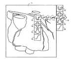
図11は、描画ウィンドウとアイコンを説明するための図である。この場合、描画ウィンドウ11には、ボリュームデータから作成された心臓の3次元画像が表示されている。また、描画ウィンドウ11の周辺には、画像回転アイコン13、画像平行移動アイコン14、画像拡大縮小アイコン15およびWW(ウィンドウ幅)/WL(ウィンドウレベル)変換アイコン16を含むアイコン群が表示されている。 FIG. 11 is a diagram for explaining a drawing window and icons. In this case, the
ユーザは、画像回転アイコン13、画像平行移動アイコン14、画像拡大縮小アイコン15またはWW/WL変換アイコン16をクリックすることによって操作種別を変更し、画像上でカーソル12を位置aから位置fへドラッグすることにより、クリックしたアイコン13〜16に対応する操作をその画像に対して行うことができる。 The user changes the operation type by clicking the
このように、描画ウィンドウ11の周辺に操作種別に対応したアイコン13〜16が表示されているが、ユーザが操作種別を変更する都度、カーソルを移動させるために描画ウィンドウ11内の画像を見ていた視線を描画ウィンドウ11外のアイコン13〜16へ頻繁に動かす必要がある。例えば、医師が医療画像を見ながら病変部を探す場合、画像の回転、移動、拡大および縮小などを頻繁に行い、視線を画像とアイコンの間で頻繁に動かすので、医療画像の病変部(陰影や色がわずかに異なる)を見失ってしまう恐れもある。 As described above, the
また、これはユーザの疲労を大きくし、結果として見落としの発生など、診断の質の低下を招く場合があった。また、操作種別を変更する場合に、キーボードのシフトキーなどを用いて操作種別の変更を行うようなUI(ユーザインターフェース)は、多数の画像種が存在し、画像種毎に操作可能な操作種別がそれぞれ異なる医療画像処理装置においては、不適切である。 In addition, this increases the fatigue of the user, and as a result, the quality of diagnosis may be reduced, such as occurrence of oversight. In addition, when changing the operation type, a UI (user interface) that changes the operation type using a keyboard shift key or the like has a large number of image types, and there are operation types that can be operated for each image type. It is inappropriate for different medical image processing apparatuses.
本発明は、上記従来の事情に鑑みてなされたものであって、視線の移動を少なくして操作種別を切り替えることができる医療画像処理装置および医療画像処理プログラムを提供することを目的としている。 The present invention has been made in view of the above-described conventional circumstances, and an object of the present invention is to provide a medical image processing apparatus and a medical image processing program that can switch the operation type by reducing the movement of the line of sight.
本発明は、パラメータに応じた画像を作成し描画ウィンドウに表示させる医療画像処理装置であって、前記描画ウィンドウ領域内にカーソルが存在するかどうか検出するカーソル制御手段と、前記描画ウィンドウ領域内に前記カーソルが存在する場合に、指定の操作が行われたことに応答して、前記カーソルの位置の近傍に、前記描画ウィンドウに表示される画像の1次元以上の連続なパラメータを表現するアイコンを少なくとも二つ含むアイコン群を表示させるアイコン制御手段と、前記アイコンを起点としてドラッグ操作が行われた場合に、前記アイコンの表現する前記1次元以上の連続なパラメータを前記ドラッグ操作に対応して変化させるパラメータ制御手段と、を備える医療画像処理装置である。 The present invention provides a medical image processing apparatus that creates an image according to a parameter and displays the image on a drawing window, the cursor control means for detecting whether or not a cursor exists in the drawing window area, and the drawing window area In response to a specified operation being performed when the cursor is present, an icon representing one or more one-dimensional continuous parameters of the image displayed in the drawing window is displayed in the vicinity of the cursor position. Icon control means for displaying an icon group including at least two, and when a drag operation is performed with the icon as a starting point, the one-dimensional or more continuous parameter expressed by the icon is changed corresponding to the drag operation. And a parameter control means for causing the medical image processing apparatus to include the medical image processing apparatus.
上記構成によれば、描画ウィンドウ領域内にカーソルが存在する場合に、ユーザが指定の操作を行うことに応答してカーソルの近傍にアイコン群が出現し、カーソルの近傍に表示されたアイコンを起点としてドラッグ操作を行うことにより、パラメータを変更し所望の操作を実行できるので、ユーザは、画像上で関心がある位置(カーソルが存在する位置)から視線をそらすことなく、所望の操作を迅速に行うことができる。すなわち、画像回転などの操作を1回のポインティングデバイスのボタンの押下に続くドラッグ操作で行うことができるので、ユーザの操作負担を軽減することができる。 According to the above configuration, when a cursor exists in the drawing window area, an icon group appears in the vicinity of the cursor in response to the user performing a specified operation, and the icon displayed in the vicinity of the cursor is the starting point. By performing the drag operation, the user can change the parameter and execute the desired operation. Therefore, the user can quickly perform the desired operation without diverting the line of sight from the position of interest on the image (the position where the cursor exists). It can be carried out. In other words, operations such as image rotation can be performed by a drag operation subsequent to pressing a button on the pointing device once, so that the operation burden on the user can be reduced.
また、本発明の医療画像処理装置は、前記アイコンを表示し、前記アイコンに対するドラッグ操作を受け付けるタッチパネルを備える。 The medical image processing apparatus of the present invention includes a touch panel that displays the icon and receives a drag operation on the icon.
上記構成によれば、可動部分がなく消毒殺菌等が容易なタッチパネルを手術現場で好適に使用することができる。 According to the above configuration, a touch panel that has no movable part and is easy to disinfect and sterilize can be suitably used at the operation site.
また、本発明の医療画像処理装置において、前記カーソル制御手段は、前記アイコンに対するドラッグ操作完了後に、前記カーソルの位置を、ドラッグ操作開始時における前記描画ウィンドウの座標上の位置に復帰させる。 In the medical image processing apparatus of the present invention, after the completion of the drag operation on the icon, the cursor control unit returns the position of the cursor to the position on the coordinates of the drawing window at the start of the drag operation.
上記構成によれば、ドラッグ操作完了後に、カーソルの位置をドラッグ操作開始時における描画ウィンドウの座標上の位置に復帰させるので、次の操作に直ちに取りかかることができる。これは、特に、ドラッグ操作によりカーソルが描画ウィンドウの外に移動した場合に便利である。 According to the above configuration, after the drag operation is completed, the position of the cursor is returned to the position on the coordinates of the drawing window at the start of the drag operation, so that the next operation can be started immediately. This is particularly convenient when the cursor is moved out of the drawing window by a drag operation.
また、本発明の医療画像処理装置において、前記カーソル制御手段は、前記アイコンに対するドラッグ操作完了後に、前記カーソルの位置を、ドラッグ操作開始時の画像上の点に対応する位置に移動させる。 In the medical image processing apparatus of the present invention, after the completion of the drag operation on the icon, the cursor control means moves the position of the cursor to a position corresponding to a point on the image at the start of the drag operation.
上記構成によれば、例えば画像の平行移動または拡大縮小操作の時に、カーソル位置を操作後の画像上の点に追従させ、画像操作後にカーソルを探すための視線の移動をなくすことができる。また、次の操作のためにカーソルを移動させる距離が短くなり、次の操作を迅速に行うことができる。 According to the above configuration, for example, at the time of image parallel movement or enlargement / reduction operation, the cursor position can be made to follow a point on the image after the operation, and the movement of the line of sight for searching the cursor after the image operation can be eliminated. Further, the distance to move the cursor for the next operation is shortened, and the next operation can be performed quickly.
また、本発明の医療画像処理装置において、前記パラメータ制御手段は、前記アイコンに対し、前記ドラッグ操作の2自由度に対応してそれぞれ異なるパラメータの操作を割り当てる。 In the medical image processing apparatus of the present invention, the parameter control means assigns different parameter operations to the icons corresponding to the two degrees of freedom of the drag operation.
上記構成によれば、一つのアイコンを2つの操作種別に対応させるので、表示するアイコンの数を減らして多くの操作を割り当てることができる。また、1回のクリックまたは連続するドラッグ操作で2つの操作種別を実行できるので、操作を迅速に行うことができる。 According to the above configuration, since one icon is associated with two operation types, a large number of operations can be assigned by reducing the number of icons to be displayed. Moreover, since two operation types can be executed by one click or continuous drag operation, the operation can be performed quickly.
また、本発明の医療画像処理装置は、前記アイコンに対して操作が行われたときにあらかじめ定められた処理を起動する。 In addition, the medical image processing apparatus of the present invention activates a predetermined process when an operation is performed on the icon.
上記構成によれば、1回の操作であらかじめ定められた処理を実行できるので、操作を迅速に行うことができる。 According to the above configuration, since a predetermined process can be executed by one operation, the operation can be performed quickly.
また、本発明の医療画像処理装置において、前記アイコンの表現する前記1次元以上の連続なパラメータは、2次元以上の連続パラメータであり、前記パラメータ制御手段は、前記アイコンを起点としてドラッグ操作が行われた場合、前記ドラッグ操作開始時にカーソルの動いた方向により前記2次元以上の連続パラメータから前記1次元以上の連続なパラメータを更に決定する。 In the medical image processing apparatus of the present invention, the one-dimensional or more continuous parameter represented by the icon is a two-dimensional or more continuous parameter, and the parameter control means performs a drag operation starting from the icon. In this case, the one-dimensional or more continuous parameter is further determined from the two-dimensional or more continuous parameter according to the direction in which the cursor moves at the start of the drag operation.
上記構成によれば、直感的な操作により操作種別を決定できるので操作が容易になる。 According to the above configuration, the operation type can be determined by an intuitive operation, so that the operation becomes easy.
また、本発明の医療画像処理装置において、前記パラメータ制御手段は、前記アイコンに対して操作が行われたときに、前記アイコンの表現する前記1次元以上の連続なパラメータを選択し、前記描画ウィンドウを起点としてドラッグ操作が行われたときに、前記選択された1次元以上の連続なパラメータを前記ドラッグ操作に対応して変化させる。 In the medical image processing apparatus of the present invention, when the operation is performed on the icon, the parameter control unit selects the one-dimensional or more continuous parameter represented by the icon, and the drawing window When a drag operation is performed starting from, the selected one-dimensional or more continuous parameters are changed corresponding to the drag operation.
上記構成によれば、アイコンに対する操作により、操作種別が切り替わった後は、描画ウィンドウ内で自由に何度ドラッグ操作を行っても、その選択された操作種別に従った処理が行われるため、頻繁には操作種別を変更する必要がない場合等に利便性が高い。 According to the above configuration, after the operation type is switched by the operation on the icon, the process according to the selected operation type is performed regardless of how many times the drag operation is freely performed in the drawing window. Is highly convenient when there is no need to change the operation type.
また、本発明の医療画像処理装置は、ボリュームデータから前記描画ウィンドウに表示する画像を生成する画像処理手段を備え、前記アイコン制御手段は、前記描画ウィンドウに表示される画像の種別に対応して、表示させるアイコン群を決定する。 The medical image processing apparatus according to the present invention further includes image processing means for generating an image to be displayed on the drawing window from volume data, and the icon control means corresponds to a type of image displayed on the drawing window. The icon group to be displayed is determined.
上記構成によれば、表示する画像種に応じて異なるアイコン群を表示させるので、画像種に応じた操作を迅速に行い、画像診断を円滑に行うことができる。 According to the above configuration, different icon groups are displayed according to the image type to be displayed, so that an operation according to the image type can be quickly performed and image diagnosis can be performed smoothly.
また、本発明の医療画像処理装置において、前記カーソルの位置の近傍とは、前記カーソルの位置に対してあらかじめ相対的に定められた位置である。 In the medical image processing apparatus of the present invention, the vicinity of the cursor position is a position that is determined in advance relative to the cursor position.
上記構成によれば、アイコン群をカーソルの位置に対してあらかじめ相対的に定められた位置に表示するので、ユーザはアイコンが表示される位置を体感的に記憶でき、操作を正確に高速に行うことができる。 According to the above configuration, the icon group is displayed at a predetermined position relative to the cursor position, so that the user can memorize the position where the icon is displayed and perform the operation accurately and at high speed. be able to.
また、本発明は、画像を作成し描画ウィンドウに表示させる医療画像処理プログラムであって、コンピュータを、上記医療画像処理装置を構成する各手段として機能させるための医療画像処理プログラムである。 Further, the present invention is a medical image processing program for creating an image and displaying it on a drawing window, and for causing a computer to function as each means constituting the medical image processing apparatus.
本発明にかかる医療画像処理装置および医療画像処理プログラムによれば、アイコン群はカーソルの近傍に出現するので、カーソルの近傍に表示されたアイコンを起点として画像を操作するドラッグ操作を行う時に、画像から視線を逸らす必要がないのでより迅速に操作ができる。更に、ユーザはアイコンの表示される位置を体感的に記憶でき、操作を正確に高速に行うことができるようになる。更に、表示する画像種に応じて異なるアイコン群を表示させるので、ユーザは、画像種に応じた操作を迅速に行い、画像診断を円滑に行うことがきる。更に、ドラッグ開始時のカーソルの動いた方向により操作種別を決定するので、ユーザは、ドラッグ方向に注意を払うことなく画像の操作に専念することができ、画像診断を円滑に行うことができる。 According to the medical image processing apparatus and the medical image processing program according to the present invention, the icon group appears in the vicinity of the cursor. Therefore, when performing a drag operation for operating the image with the icon displayed in the vicinity of the cursor as the starting point, the image It is possible to operate more quickly because it is not necessary to shift the line of sight. Further, the user can experience the position where the icon is displayed and can perform the operation accurately and at high speed. Furthermore, since different icon groups are displayed according to the type of image to be displayed, the user can quickly perform an operation according to the type of image and smoothly perform image diagnosis. Furthermore, since the operation type is determined according to the direction in which the cursor moves at the start of dragging, the user can concentrate on the operation of the image without paying attention to the drag direction, and can smoothly perform image diagnosis.
図1は、本発明の一実施形態にかかる医療画像処理装置およびコンピュータ断層撮影(CT)装置を概略的に示す。コンピュータ断層撮影装置は、被検体の組織等を可視化するものである。X線源101からは同図に鎖線で示す縁部ビームを有するピラミッド状のX線ビーム束102が放射される。X線ビーム束102は、例えば患者103である被検体を透過しX線検出器104に照射される。X線源101及びX線検出器104は、本実施形態の場合にはリング状のガントリー105に互いに対向配置されている。リング状のガントリー105は、このガントリーの中心点を通るシステム軸線106に対して、同図に示されていない保持装置に回転可能(矢印a参照)に支持されている。 FIG. 1 schematically shows a medical image processing apparatus and a computed tomography (CT) apparatus according to an embodiment of the present invention. The computer tomography apparatus visualizes the tissue of a subject. An
患者103は、本実施形態の場合には、X線が透過するテーブル107上に寝ている。このテーブルは、図示されていない支持装置によりシステム軸線106に沿って移動可能(矢印b参照)に支持されている。 In the case of this embodiment, the
従って、X線源101及びX線検出器104は、システム軸線106に対して回転可能でありかつシステム軸線106に沿って患者103に対して相対的に移動可能である測定システムを構成するので、患者103はシステム軸線106に関して種々の投影角及び種々の位置のもとで投射されることができる。その際に発生するX線検出器104の出力信号は、画像処理手段111に供給される。 Thus, the
シーケンス走査の場合には患者103の層毎の走査が行なわれる。その際に、X線源101及びX線検出器104はシステム軸線106を中心に患者103の周りを回転し、X線源101及びX線検出器104を含む測定システムは患者103の2次元断層を走査するために多数の投影を撮影する。その際に取得された測定値から、走査された断層を表示する断層像が再構成される。相連続する断層の走査の間に、患者103はその都度システム軸線106に沿って移動される。この過程は全ての関心断層が捕捉されるまで繰り返される。 In the case of a sequence scan, a scan for each layer of the
一方、スパイラル走査中は、X線源101及びX線検出器104を含む測定システムはシステム軸線106を中心に回転し、テーブル107は連続的に矢印bの方向に移動する。すなわち、X線源101及びX線検出器104を含む測定システムは、患者103に対して相対的に連続的にスパイラル軌道上を、患者103の関心領域が全部捕捉されるまで移動する。本実施形態の場合、同図に示されたコンピュータ断層撮影装置により、患者103の診断範囲における多数の相連続する断層信号が画像処理手段111に供給される。 On the other hand, during spiral scanning, the measurement system including the
カーソル制御手段112は、画像に表示されているカーソルが存在する位置を制御するものであり、画像を表示する描画ウィンドウの領域(描画ウィンドウ領域)内にカーソルが存在するかどうかを検出する。アイコン制御手段114は、描画ウィンドウ領域内にカーソルが存在する場合に、ユーザがクリック操作により指定の操作を行ったことに応答して、カーソルの位置の近傍に、描画ウィンドウに表示される画像の1次元以上の連続なパラメータを表現するアイコンを少なくとも二つ含むアイコン群を表示する。ここで、カーソルの位置の近傍とは、カーソルの位置に対してあらかじめ定められた相対的な位置であり、ユーザが、カーソルが存在している位置に注目した状態で視認可能な、該カーソルの位置の周辺領域を意味する。 The
パラメータ制御手段115は、ユーザが描画ウィンドウ領域内に表示されるアイコンを起点としてドラッグ操作を行う場合に、ドラッグ操作に対応して、当該アイコンに対応付けられた1次元以上の連続なパラメータを変化させる。ここで、ユーザが描画ウィンドウ内に表示されるアイコンを起点としてドラッグ操作を行う場合とは、アイコンの表示領域内に、ドラッグ操作の開始位置が存在する場合を意味する。また、ドラッグ操作に対応してパラメータを変化させるとは、ドラッグ操作の操作方向及び操作量、操作時間、加速度に応じてパラメータを変化させることを意味する。また、画像処理手段111は、断層信号からボリュームデータを生成し、表示手段116に表示する画像を作成する。 When the user performs a drag operation starting from an icon displayed in the drawing window area, the
また、操作手段113はGUI(Graphical User Interface)を含み、キーボードやポインティングデバイス(マウスやトラックボール)などからの操作信号に応じて、描画ウィンドウの設定を行って設定値の制御信号を生成し、カーソル制御手段112、アイコン制御手段114、パラメータ制御手段115に供給する。これにより、表示手段116に表示される画像を見ながら画像をインタラクティブに変更し、病巣領域の発見を支援することができる。 The operation means 113 includes a GUI (Graphical User Interface), sets a drawing window according to an operation signal from a keyboard or a pointing device (mouse or trackball), and generates a setting value control signal. The data is supplied to the
図2は、本発明の実施形態にかかる医療画像処理方法を説明するためのフローチャートを示す。まず、画像処理手段111は、CT装置から供給されるボリュームデータに基づいて描画ウィンドウに表示する画像を生成する(ステップS11)。また、アイコン制御手段114は、描画ウィンドウに表示される画像の種別に対応して、表示するアイコン群を決定する(ステップS12)。 FIG. 2 is a flowchart for explaining the medical image processing method according to the embodiment of the present invention. First, the
次に、カーソル制御手段112は、描画ウィンドウ領域内にカーソルが存在するかどうか判断する(ステップS13)。そして、描画ウィンドウ領域内にカーソルが存在する場合(Yes)、ユーザが指定の操作(例えば画像上クリック操作)を行ったかどうかを判断する(ステップS14)。 Next, the
ユーザが指定の操作を行った場合(Yes)、アイコン制御手段114は、ユーザの操作に応答してカーソルの位置の近傍に、描画ウィンドウに表示される画像の1次元以上の連続なパラメータを表現する2以上のアイコンを含むアイコン群を表示する(ステップS15)。 When the user performs a specified operation (Yes), the
次に、パラメータ制御手段115は、アイコンを起点としたドラッグ操作が行われたかどうかを判断し(ステップS16)、ユーザがアイコンを起点としてドラッグ操作を行った場合(Yes)は、起点となったアイコンの種別に対応した1次元以上の連続なパラメータを変化させる(ステップS17)。 Next, the
次に、カーソル制御手段112は、ドラッグ操作が完了したかどうかを判断し(ステップS18)、起点となったアイコンに対するドラッグ操作完了を検出した場合(Yes)に、カーソルの位置をドラッグ操作開始時における描画ウィンドウの座標上の位置に復帰させる(ステップS19)。なお、この場合、起点となったアイコンに対するドラッグ操作完了後に、カーソルの位置をドラッグ操作開始時の画像上の点に対応する位置に移動させることもできる。 Next, the cursor control means 112 determines whether or not the drag operation is completed (step S18), and when the completion of the drag operation for the starting icon is detected (Yes), the cursor position is set at the start of the drag operation. Is returned to the position on the coordinates of the drawing window (step S19). In this case, after completion of the drag operation on the starting icon, the position of the cursor can be moved to a position corresponding to the point on the image at the start of the drag operation.
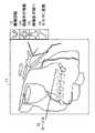
図3は、本発明の実施形態にかかる医療画像処理装置における描画ウィンドウ11の例を示す。ユーザが描画ウィンドウ11上でクリックを行うと、図3aに示すように、カーソル12の近傍に画像に対する操作の種類を表現するアイコン群(アイコン13〜16)が現れる。図3bは、カーソル12位置にアイコン群(アイコン13〜16)を表示する例を示す。図3bに示すように、カーソル12の近傍とは、カーソル12位置からの距離が0である位置を含む。そして、ユーザがアイコン13〜16を起点としてドラッグ操作を行うことにより、そのアイコン13〜16に対応した種別の画像の操作が行われる。アイコン群(アイコン13〜16)が出現する位置は、カーソル12との相対的な座標によって決定される。これにより、アイコン13〜16の位置をユーザが体感的に記憶することが出来る。 FIG. 3 shows an example of the drawing
このように本発明の実施形態にかかる医療画像処理装置によれば、アイコン群(アイコン13〜16)はカーソル12の近傍に出現するので、カーソル12の近傍に表示されたアイコン13〜16を起点として画像を操作するドラッグ操作を行う時に、画像から視線を逸らす必要がないのでより迅速に操作ができる。更に、ユーザはアイコン13〜16の表示される位置を体感的に記憶でき、操作を正確に高速に行うことができるようになる。 As described above, according to the medical image processing apparatus according to the embodiment of the present invention, since the icon group (
図4は、本実施形態の医療画像処理装置において、ドラッグ操作後にカーソル位置を自動復帰させる様子を示す図である。まず、画像を回転する場合、描画ウィンドウ11に表示されている画像回転アイコン13上にカーソル12を合わせ、そこでポインティングデバイスのボタンを押下したままカーソル12を位置aから位置fへドラッグ操作を行うことにより、描画ウィンドウ11に表示されている画像を回転させることができる。 FIG. 4 is a diagram illustrating a state in which the cursor position is automatically returned after the drag operation in the medical image processing apparatus of the present embodiment. First, when rotating an image, the
このように本実施形態の医療画像処理装置によれば、画像上で関心がある位置(カーソルが存在する位置)の近傍にアイコン群(アイコン13〜16)が表示されているので、ユーザは、関心領域から視線を外すことなく、画像回転などの操作を行うことができる。また、画像回転を1回のポインティングデバイスによる操作であるドラッグ操作で行うことができるので、ユーザの操作負担を軽減することができる。 As described above, according to the medical image processing apparatus of the present embodiment, the icon group (
一方、本実施形態の医療画像処理装置では、カーソル12を位置aから位置fへドラッグし画像回転操作を行った後に、カーソル12を位置aに自動的に復帰させる。これにより画像拡大縮小などの他の操作に直ちに取りかかることができる。また、画像操作後にカーソル12を探すための視線の移動をなくすことができる。また、特に、カーソル12がドラッグ操作により描画ウィンドウ11の外に移動した場合(位置f)に操作終了後にカーソルを手動で描画ウィンドウに復帰させる必要がないので便利である。なお、ドラッグ操作中にカーソル12を非表示にしても良い。このようにすることで、カーソル12がドラッグ操作により画面の端に到達移動した場合に操作が中断されることが無くなる。その為に、実装上はカーソルを全く移動させずにポインティングデバイスに対する移動操作のみを検知しても良い。また、アイコン群(アイコン13〜16)を非表示にしても良い。このようにすることで、ユーザは画像に集中することが出来る。 On the other hand, in the medical image processing apparatus according to the present embodiment, after the
図5は、本実施形態の医療画像処理装置において、操作中の画像にカーソル12を追従させる様子を示す図である。図5(a)は、ユーザが画像の平行移動を行うために、カーソル12を画像平行移動アイコン14上に合わせた場合を示す。ユーザは、画像平行移動アイコン14上でポインティングデバイスのボタンを押下し、ボタンを押下したまま右にドラッグすることにより、画像を右に平行移動することができる。 FIG. 5 is a diagram illustrating a state in which the
図5(b)は、カーソル12を位置aから位置cにドラッグし、画像を平行移動した状態を示す。本実施形態では、位置cでドラッグ操作を終了した場合に、カーソル12を位置cに残しておく。このように本実施形態によれば、特に画像の平行移動、拡大縮小操作の時に、カーソル位置を操作後の画像上の点に追従させるので、画像操作後にカーソル12を探すための視線の移動をなくすことができる。また、この時にアイコン群(アイコン13〜16)も操作後の画像に追従させることにより、次の操作のためにカーソル12を移動させる距離が短くなり、次の操作を迅速に行うことができる。 FIG. 5B shows a state in which the
図6は、本実施形態の医療画像処理装置において、一つのアイコンを2つの操作種別に対応させる場合を示す図である。ドラッグ操作には上下左右の2自由度があるので、「上下のスライスの表示」と「時系列上の前後」のように複数の異なる概念の操作種別を一つのアイコンで行うようにしても良い。また、ドラッグではなく単にクリックしたことに対して、なんらかの処理、例えばメニューの表示などの処理を起動しても良い。 FIG. 6 is a diagram illustrating a case where one icon corresponds to two operation types in the medical image processing apparatus of the present embodiment. Since the drag operation has two degrees of freedom, up, down, left, and right, a plurality of different types of operation types such as “display upper and lower slices” and “front and back in time series” may be performed with one icon. . Further, in response to a simple click instead of dragging, some processing, such as menu display processing, may be activated.
図6は、アイコン群の一つにスライス/時系列アイコン21が表示されており、ユーザがカーソル12をスライス/時系列アイコン21に合わせている様子を示す。この場合、スライス/時系列アイコン21上でポインティングデバイスのボタンを押下し、上下にドラッグすることによりスライス位置が異なるスライス画像を表示することがきる。一方、スライス/時系列アイコン21上でポインティングデバイスのボタンを押下し、左右にドラッグすることにより時系列に従った画像を表示することができる。 FIG. 6 shows a state where a slice /
このように本実施形態の医療画像処理装置によれば、一つのアイコンを2つの操作種別に対応させるので、表示するアイコンの数を減らして多くの操作を割り当てることができる。また、1回のポインティングデバイスのボタンの押下または連続するドラッグ操作で2つの操作種別を実行できるので、操作を迅速に行うことができる。 As described above, according to the medical image processing apparatus of the present embodiment, since one icon is associated with two operation types, many operations can be assigned by reducing the number of icons to be displayed. In addition, since two types of operations can be executed by pressing a button on the pointing device once or by a continuous drag operation, the operation can be performed quickly.
図7は、本実施形態の医療画像処理装置において、表示する画像種に応じて異なるアイコン群を表示させる場合の説明図である。本実施形態では、カーソルが存在する描画ウィンドウの画像の画像種(ボリュームデータの可視化手段)に応じて表示されるアイコンが異なる。 FIG. 7 is an explanatory diagram for displaying different icon groups depending on the image type to be displayed in the medical image processing apparatus of the present embodiment. In the present embodiment, the icons displayed differ depending on the image type (volume data visualization means) of the image of the drawing window where the cursor exists.
図7(a)は、描画ウィンドウ11に3次元画像を表示する場合のアイコン群を示す。この場合のアイコン群は、画像回転アイコン13、画像平行移動アイコン14、画像拡大縮小アイコン15およびWW(ウィンドウ幅)/WL(ウィンドウレベル)変換アイコン16で構成される。 FIG. 7A shows an icon group when a three-dimensional image is displayed in the drawing
WW/WL値はMIP(Maximum Intensity Projection)画像のように濃淡で表現される画像において表示のコントラストと明るさを調整するためのパラメータである。例えば、濃淡値が4096階調で与えられているときに、その内の特に診断に有効な範囲を切り出して256階調に変換することをWW/WL変換と言い、この時の切り出し範囲の幅をWW(ウィンドウ幅)、切り出し範囲の中心値をWL(ウィンドウレベル)値と言う。 The WW / WL value is a parameter for adjusting the contrast and brightness of display in an image expressed in shades such as a MIP (Maximum Intensity Projection) image. For example, when the gray value is given in 4096 gradations, cutting out a particularly effective range of them and converting it to 256 gradations is called WW / WL conversion, and the width of the extraction range at this time Is called WW (window width), and the center value of the cutout range is called WL (window level) value.
図7(b)は、描画ウィンドウ11にスライス画像を表示する場合のアイコン群を示す。この場合のアイコン群は、画像平行移動アイコン14、画像拡大縮小アイコン15、WW/WL変換アイコン16、およびスライス変更アイコン26で構成される。本実施形態によれば、表示する画像種に応じて異なるアイコン群を表示させるので、画像種に応じた操作を迅速に行い、画像診断を円滑に行うことがきる。 FIG. 7B shows an icon group when a slice image is displayed in the drawing
なお、画像種とアイコンは(表1)のように対応させることができる。 Note that image types and icons can be associated as shown in (Table 1).

図8は、本実施形態の医療画像処理装置において、ドラッグ開始時のカーソルの動きにより操作種別を決定する場合の説明図を示す。本実施形態では、ドラッグ操作開始時にカーソルの動いた方向により操作種別を決定する。すなわち、本実施形態においては、アイコンが2次元以上の連続パラメータを表現するものであり、パラメータ制御手段が、アイコンを起点としてドラッグ操作が行われた場合、ドラッグ操作開始時にカーソルの動いた方向により前記2次元以上の連続パラメータから前記1次元以上の連続なパラメータを更に決定するものである。例えば、ドラッグ開始時にカーソルが左右に動いた場合は水平回転と決定し、ドラッグ開始時にカーソルが上下に動いた場合は垂直回転と決定し、ドラッグ開始時にカーソルがななめに動いた場合は自由回転と決定する。これによれば、直感的な操作により操作種別を決定できるので操作が容易になる。 FIG. 8 is an explanatory diagram when the operation type is determined by the movement of the cursor at the start of dragging in the medical image processing apparatus of the present embodiment. In the present embodiment, the operation type is determined based on the direction in which the cursor moves at the start of the drag operation. That is, in the present embodiment, the icon represents a continuous parameter of two or more dimensions, and when the parameter control means performs a drag operation starting from the icon, depending on the direction in which the cursor moves when the drag operation starts. The one-dimensional or more continuous parameter is further determined from the two-dimensional or more continuous parameter. For example, if the cursor moves left or right at the start of dragging, it is determined as horizontal rotation, if the cursor moves up or down at the start of dragging, it is determined as vertical rotation, and if the cursor moves smoothly at the start of dragging, it is determined as free rotation. decide. According to this, since the operation type can be determined by an intuitive operation, the operation becomes easy.
図8は、描画ウィンドウ11に画像回転アイコン13等が表示されている様子を示す。ユーザが、画像回転アイコン13にカーソル12を合わせてポインティングデバイスのボタンを押下し、右(位置a〜d)にドラッグすることにより、画像の水平回転を行うことができる。本実施形態によれば、ドラッグ開始時のカーソルの動いた方向により操作種別を決定するので、操作中にドラッグ方向が少々ななめにずれた場合でも、画像の水平回転を継続することができる。従って、ユーザの意図に即したと判断される処理が決定されることによりユーザは、ドラッグ方向に注意を払うことなく画像の操作に専念することができ、画像診断を円滑に行うことができる。 FIG. 8 shows a state where the
図9は、本実施形態の医療画像処理装置において、アイコンをダブルクリックすることにより操作モードを切り替える場合の説明図を示す。本実施形態では、アイコンのダブルクリックにより、操作種別が切り替わった後は、描画ウィンドウ内で自由に何度ドラッグ操作を行っても、その操作種別に従った処理が行われる。これによれば、頻繁には操作種別を変更する必要がない場合等に利便性が高い。 FIG. 9 is an explanatory diagram when the operation mode is switched by double-clicking an icon in the medical image processing apparatus of the present embodiment. In the present embodiment, after the operation type is switched by double-clicking on the icon, the process according to the operation type is performed even if the drag operation is freely performed in the drawing window. This is highly convenient when it is not necessary to frequently change the operation type.
図9は、描画ウィンドウ11に画像回転アイコン13等が表示されている様子を示す。ユーザが、画像平行移動アイコン14にカーソル12を合わせてダブルクリックすることにより、画像平行移動モードとすることができる。本実施形態によれば、一旦、ダブルクリックにより画像平行移動モードに設定した後は、そのモードに従った処理を行うことが可能となり、利便性が高い。特に、ダブルクリックによりモードを設定した後、次にアイコン群が表示されるときに、モードをリセットすることにより、一層利便性が向上する。なお、モードの変更があったときは、カーソルをそのモードを表現するものに変更すると使い勝手がよい。また、モードは、描画ウィンドウ毎に設定可能である。 FIG. 9 shows a state where the
図10は、本明細書において使用する用語について説明するための図である。描画ウィンドウ11は、ボリュームレンダリングの描画結果の表示されるウィンドウを表わす。アイコン13〜16,13’〜16’は、何らかのコマンドが割り当てられている図形もしくは図形と文字の組み合わせを表示するものである。アイコン群(group)17は、画像種に対応して操作可能なアイコンのグループを示す。なお、「操作種別」は、操作可能なレンダリングの条件を決定する連続なパラメータもしくは連続なパラメータの組合せの種別を表わし、「操作」はユーザが行う操作を言う。また、「連続なパラメータ」とは回転角度、座標、時間、コントラスト値等のように本来的に連続であるが情報処理の都合上離散値を取るものを指す。また、スライス画像の番号も事実上スライス画像の座標等を表現するので、「連続なパラメータ」に含まれる。 FIG. 10 is a diagram for explaining terms used in this specification. The drawing
また、上記の説明では、アイコン群をあらかじめ設定しておき画像種に応じて表示するものとして説明したが、アイコン群の例えば表示させるアイコンの種類、表示させる場所等をユーザがカスタマイズすることもできる。また、描画ウィンドウの外にアイコン群をあらかじめ表示しておいても良い。また、アイコンのなかにはコマンドの起動などドラッグ操作と関係ない操作を受け付けるアイコンがあっても良い。 In the above description, the icon group is set in advance and displayed according to the image type. However, for example, the type of icon to be displayed, the place to be displayed, and the like can be customized by the user. . Further, an icon group may be displayed in advance outside the drawing window. In addition, among icons, there may be an icon that accepts an operation unrelated to a drag operation such as activation of a command.
また、上記の説明では、ユーザがアイコンを起点としてドラッグ操作によって操作が行われたが、アイコンにポインティングデバイスをあわせた状態でホイールの回転操作を行っても良い。これにより、ポインティングデバイスの上下左右方向への移動の2自由度に加えて、3自由度の操作ができるので、3次元画像に対する操作を容易に行うことができる。 In the above description, the user performs the operation by the drag operation starting from the icon. However, the wheel may be rotated by putting the pointing device on the icon. Thereby, in addition to the two degrees of freedom of movement of the pointing device in the up / down / left / right directions, an operation with three degrees of freedom can be performed, so that the operation on the three-dimensional image can be easily performed.
また、上記の説明では、ユーザの操作に応答してカーソルの位置の近傍にアイコン群を表示していたが、この時のカーソルの位置を後の操作に用いても良い。例えば、拡大縮小操作を行うときに、カーソルの位置を拡大縮小の中心と出来る。また、例えば、回転操作を行うときに、カーソルの位置からボリュームデータに投射した物体上の座標(いわゆるポイントピック処理で得られる座標)を回転の中心と出来る。 In the above description, an icon group is displayed in the vicinity of the cursor position in response to a user operation. However, the cursor position at this time may be used for a subsequent operation. For example, when performing an enlargement / reduction operation, the position of the cursor can be the center of enlargement / reduction. Also, for example, when performing a rotation operation, the coordinates on the object projected from the cursor position onto the volume data (coordinates obtained by so-called point pick processing) can be used as the center of rotation.
また、本実施形態の医療画像処理装置において、アイコンを表示し、アイコンに対するドラッグ操作を受け付けるタッチパネルを操作表示手段として設けることもできる。タッチパネルは、可動部分がなく消毒殺菌等が容易なため手術現場で使用する場合に好適である。 In the medical image processing apparatus according to the present embodiment, a touch panel that displays an icon and receives a drag operation on the icon can be provided as an operation display unit. The touch panel is suitable for use at a surgical site because it has no moving parts and is easy to disinfect and sterilize.
また、クリック以外の方法でアイコン群が表示されるようにしてもよい。例えば、キーボードに対する操作や、音声入力デバイスに対する操作や、専用スイッチに対する操作が考えられる。 The icon group may be displayed by a method other than clicking. For example, an operation on a keyboard, an operation on a voice input device, or an operation on a dedicated switch can be considered.
このように本実施形態の医療画像処理装置によれば、画像上で関心がある位置(カーソルが存在する位置)の近傍にアイコン群が表示されているので、ユーザは、関心領域から視線を外すことなく、画像回転などの操作を行うことができる。また、画像回転などを1回のポインティングデバイスのボタンの押下に続くドラッグ操作で行うことができるので、ユーザの操作負担を軽減することができる。これは特に次々と操作種別を切り替えながら操作を行うときに有効である。 As described above, according to the medical image processing apparatus of the present embodiment, the icon group is displayed in the vicinity of the position of interest (position where the cursor is present) on the image, so the user removes his / her line of sight from the region of interest. Thus, operations such as image rotation can be performed. In addition, image rotation and the like can be performed by a drag operation subsequent to pressing the button of the pointing device once, so that the operation burden on the user can be reduced. This is particularly effective when performing operations while switching operation types one after another.
本発明は、視線の移動を少なくしてドラッグによる操作種別を切り替えることができる医療画像処理装置および医療画像処理プログラムとして利用可能である。 INDUSTRIAL APPLICABILITY The present invention can be used as a medical image processing apparatus and a medical image processing program that can switch the operation type by dragging with less movement of the line of sight.
11 描画ウィンドウ
12 カーソル
13 画像回転アイコン
14 画像平行移動アイコン
15 画像拡大縮小アイコン
16 WW/WL変換アイコン
17 アイコン群
21 スライス/時系列アイコン
26 スライス変更アイコン
101 X線源
102 X線ビーム束
103 患者
104 X線検出器
105 ガントリー
106 システム軸線
107 テーブル
111 画像処理手段
112 カーソル制御手段
113 操作手段
114 アイコン制御手段
115 パラメータ制御手段
116 表示手段DESCRIPTION OF
Claims (10)
Translated fromJapanese前記描画ウィンドウ領域内にカーソルが存在するかどうか検出するカーソル制御手段と、
前記描画ウィンドウ領域内に前記カーソルが存在する場合に、指定の操作が行われたことに応答して、前記カーソルの位置の近傍であって、前記カーソルの位置に対してあらかじめ相対的に定められた位置に、前記描画ウィンドウに表示される画像の1次元以上の連続なパラメータを表現するアイコンを少なくとも二つ含むアイコン群を表示させるアイコン制御手段と、
前記アイコンを起点としてドラッグ操作が行われた場合に、前記アイコンの表現する前記1次元以上の連続なパラメータを前記ドラッグ操作に対応して変化させるパラメータ制御手段と、を備える医療画像処理装置。A medical image processing apparatus that creates an image according to a parameter and displays the image on a drawing window,
Cursor control means for detecting whether a cursor is present in the drawing window area;
When the cursor exists in the drawing window area, in response to a designated operation being performed, the cursor is in the vicinityof the cursor position and is determined in advance relative to the cursor position. Icon control means for displaying an icon group including at least two icons representing one-dimensional or more continuous parameters of the image displayed in the drawing window atthe position ,
A medical image processing apparatus comprising: a parameter control unit that changes the one-dimensional or more continuous parameters represented by the icon in response to the drag operation when a drag operation is performed using the icon as a starting point.
前記アイコンを表示し、前記アイコンに対するドラッグ操作を受け付けるタッチパネルを備える医療画像処理装置。The medical image processing apparatus according to claim 1,
A medical image processing apparatus including a touch panel that displays the icon and receives a drag operation on the icon.
前記カーソル制御手段は、前記アイコンに対するドラッグ操作完了後に、前記カーソルの位置を、ドラッグ操作開始時における前記描画ウィンドウの座標上の位置に復帰させる医療画像処理装置。The medical image processing apparatus according to claim 1,
The medical image processing apparatus, wherein the cursor control unit returns the position of the cursor to the position on the coordinates of the drawing window at the start of the drag operation after the completion of the drag operation on the icon.
前記カーソル制御手段は、前記アイコンに対するドラッグ操作完了後に、前記カーソルの位置を、ドラッグ操作開始時の画像上の点に対応する位置に移動させる医療画像処理装置。The medical image processing apparatus according to claim 1,
The medical image processing apparatus, wherein the cursor control means moves the position of the cursor to a position corresponding to a point on the image at the start of the drag operation after completion of the drag operation on the icon.
前記パラメータ制御手段は、前記アイコンに対し、前記ドラッグ操作の2自由度に対応してそれぞれ異なるパラメータの操作を割り当てる医療画像処理装置。The medical image processing apparatus according to claim 1,
The said parameter control means is a medical image processing apparatus which allocates the operation of a parameter which is different with respect to the said icon corresponding to the two degrees of freedom of the said drag operation.
前記アイコンに対して操作が行われたときにあらかじめ定められた処理を起動する医療画像処理装置。The medical image processing apparatus according to claim 1,
A medical image processing apparatus that activates a predetermined process when an operation is performed on the icon.
前記アイコンの表現する前記1次元以上の連続なパラメータは、2次元以上の連続パラメータであり、
前記パラメータ制御手段は、前記アイコンを起点としてドラッグ操作が行われた場合、前記ドラッグ操作開始時にカーソルの動いた方向により前記2次元以上の連続パラメータから前記1次元以上の連続なパラメータを更に決定する医療画像処理装置。The medical image processing apparatus according to claim 1,
The one-dimensional or more continuous parameter represented by the icon is a two-dimensional or more continuous parameter,
When the drag operation is performed with the icon as a starting point, the parameter control means further determines the one-dimensional or more continuous parameter from the two-dimensional or more continuous parameter according to the direction in which the cursor moves at the start of the drag operation. Medical image processing device.
前記パラメータ制御手段は、前記アイコンに対して操作が行われたときに、前記アイコンの表現する前記1次元以上の連続なパラメータを選択し、
前記描画ウィンドウを起点としてドラッグ操作が行われたときに、前記選択された1次元以上の連続なパラメータを前記ドラッグ操作に対応して変化させる医療画像処理装置。The medical image processing apparatus according to claim 1,
The parameter control means, when an operation is performed on the icon, selects the one or more continuous parameters represented by the icon,
A medical image processing apparatus that changes a selected one-dimensional or more continuous parameter corresponding to the drag operation when a drag operation is performed from the drawing window.
ボリュームデータから前記描画ウィンドウに表示する画像を生成する画像処理手段を備え、
前記アイコン制御手段は、前記描画ウィンドウに表示される画像の種別に対応して、表示させるアイコン群を決定する医療画像処理装置。The medical image processing apparatus according to claim 1,
Image processing means for generating an image to be displayed on the drawing window from volume data;
The said icon control means is a medical image processing apparatus which determines the icon group to display corresponding to the classification of the image displayed on the said drawing window.
Priority Applications (2)
| Application Number | Priority Date | Filing Date | Title |
|---|---|---|---|
| JP2007288451AJP4519898B2 (en) | 2007-11-06 | 2007-11-06 | Medical image processing apparatus and medical image processing program |
| US12/263,647US20090119609A1 (en) | 2007-11-06 | 2008-11-03 | Medical image processing apparatus |
Applications Claiming Priority (1)
| Application Number | Priority Date | Filing Date | Title |
|---|---|---|---|
| JP2007288451AJP4519898B2 (en) | 2007-11-06 | 2007-11-06 | Medical image processing apparatus and medical image processing program |
Publications (2)
| Publication Number | Publication Date |
|---|---|
| JP2009116581A JP2009116581A (en) | 2009-05-28 |
| JP4519898B2true JP4519898B2 (en) | 2010-08-04 |
Family
ID=40589414
Family Applications (1)
| Application Number | Title | Priority Date | Filing Date |
|---|---|---|---|
| JP2007288451AActiveJP4519898B2 (en) | 2007-11-06 | 2007-11-06 | Medical image processing apparatus and medical image processing program |
Country Status (2)
| Country | Link |
|---|---|
| US (1) | US20090119609A1 (en) |
| JP (1) | JP4519898B2 (en) |
Cited By (4)
| Publication number | Priority date | Publication date | Assignee | Title |
|---|---|---|---|---|
| US11016579B2 (en) | 2006-12-28 | 2021-05-25 | D3D Technologies, Inc. | Method and apparatus for 3D viewing of images on a head display unit |
| US11228753B1 (en) | 2006-12-28 | 2022-01-18 | Robert Edwin Douglas | Method and apparatus for performing stereoscopic zooming on a head display unit |
| US11275242B1 (en) | 2006-12-28 | 2022-03-15 | Tipping Point Medical Images, Llc | Method and apparatus for performing stereoscopic rotation of a volume on a head display unit |
| US11315307B1 (en) | 2006-12-28 | 2022-04-26 | Tipping Point Medical Images, Llc | Method and apparatus for performing rotating viewpoints using a head display unit |
Families Citing this family (26)
| Publication number | Priority date | Publication date | Assignee | Title |
|---|---|---|---|---|
| KR20100030968A (en)* | 2008-09-11 | 2010-03-19 | 엘지전자 주식회사 | Terminal and method for displaying menu thereof |
| JP5502382B2 (en) | 2009-07-02 | 2014-05-28 | 株式会社東芝 | Mammography apparatus and image processing apparatus |
| JP5582755B2 (en)* | 2009-10-06 | 2014-09-03 | 株式会社東芝 | MEDICAL IMAGE MANAGEMENT DEVICE AND MEDICAL IMAGE DISPLAY DEVICE |
| KR20130052743A (en)* | 2010-10-15 | 2013-05-23 | 삼성전자주식회사 | Method for selecting menu item |
| JP5718019B2 (en)* | 2010-10-28 | 2015-05-13 | 株式会社日立メディコ | Medical image display device |
| JP5387556B2 (en)* | 2010-12-24 | 2014-01-15 | 株式会社デンソー | In-vehicle device |
| US9746989B2 (en)* | 2011-10-13 | 2017-08-29 | Toshiba Medical Systems Corporation | Three-dimensional image processing apparatus |
| EP2672456B1 (en) | 2012-06-07 | 2019-07-24 | Dassault Systèmes | Method and system for dynamically manipulating an assembly of objects in a three-dimensional scene of a system of computer-aided design |
| GB2506924B (en)* | 2012-10-15 | 2020-08-12 | Pen Chang Chin | Touch control system for touch panel |
| DE102015215820A1 (en)* | 2015-08-19 | 2017-02-23 | Siemens Healthcare Gmbh | Controlling a medical device |
| JPWO2017034020A1 (en)* | 2015-08-26 | 2018-08-02 | 株式会社根本杏林堂 | Medical image processing apparatus and medical image processing program |
| KR102354328B1 (en)* | 2015-09-22 | 2022-01-21 | 삼성전자주식회사 | Image display apparatus and operating method for the same |
| JP6645904B2 (en)* | 2016-04-28 | 2020-02-14 | キヤノンメディカルシステムズ株式会社 | Medical image display device and display program |
| JP7172093B2 (en)* | 2017-03-31 | 2022-11-16 | 大日本印刷株式会社 | Computer program, display device, display system and display method |
| KR102255401B1 (en)* | 2019-01-30 | 2021-05-24 | (주)비주얼터미놀로지 | Method and System for 3D medical information input |
| US11275453B1 (en)* | 2019-09-30 | 2022-03-15 | Snap Inc. | Smart ring for manipulating virtual objects displayed by a wearable device |
| US11277597B1 (en) | 2020-03-31 | 2022-03-15 | Snap Inc. | Marker-based guided AR experience |
| US11798429B1 (en) | 2020-05-04 | 2023-10-24 | Snap Inc. | Virtual tutorials for musical instruments with finger tracking in augmented reality |
| US11520399B2 (en) | 2020-05-26 | 2022-12-06 | Snap Inc. | Interactive augmented reality experiences using positional tracking |
| US11925863B2 (en) | 2020-09-18 | 2024-03-12 | Snap Inc. | Tracking hand gestures for interactive game control in augmented reality |
| KR20230124732A (en) | 2020-12-29 | 2023-08-25 | 스냅 인코포레이티드 | Fine hand gestures to control virtual and graphical elements |
| KR20230124077A (en) | 2020-12-30 | 2023-08-24 | 스냅 인코포레이티드 | Augmented reality precision tracking and display |
| US11531402B1 (en) | 2021-02-25 | 2022-12-20 | Snap Inc. | Bimanual gestures for controlling virtual and graphical elements |
| EP4320502A1 (en) | 2021-04-08 | 2024-02-14 | Snap, Inc. | Bimanual interactions between mapped hand regions for controlling virtual and graphical elements |
| US11861070B2 (en) | 2021-04-19 | 2024-01-02 | Snap Inc. | Hand gestures for animating and controlling virtual and graphical elements |
| JP2023050881A (en)* | 2021-09-30 | 2023-04-11 | 株式会社Luxonus | Image processing device, image processing method and recording medium |
Family Cites Families (11)
| Publication number | Priority date | Publication date | Assignee | Title |
|---|---|---|---|---|
| JPH0289124A (en)* | 1988-09-26 | 1990-03-29 | Sharp Corp | Menu display method |
| JPH0342713A (en)* | 1989-07-11 | 1991-02-22 | Meta Corp Japan:Kk | User interface system |
| JPH0566750A (en)* | 1991-09-09 | 1993-03-19 | Nec Corp | Designation processing method for rectangular region |
| US5987345A (en)* | 1996-11-29 | 1999-11-16 | Arch Development Corporation | Method and system for displaying medical images |
| JPH10198517A (en)* | 1997-01-10 | 1998-07-31 | Tokyo Noukou Univ | Method for controlling display content of display device |
| JP3065025B2 (en)* | 1998-06-01 | 2000-07-12 | 株式会社日立製作所 | Information processing device |
| JP2000066785A (en)* | 1998-08-19 | 2000-03-03 | Nippon Telegr & Teleph Corp <Ntt> | Information processing device using pointing device |
| US7536644B2 (en)* | 2002-06-27 | 2009-05-19 | Siemens Medical Solutions Usa, Inc. | Method and system for facilitating selection of stored medical images |
| JP4115198B2 (en)* | 2002-08-02 | 2008-07-09 | 株式会社日立製作所 | Display device with touch panel |
| JP2005296156A (en)* | 2004-04-08 | 2005-10-27 | Hitachi Medical Corp | Medical image display device |
| US20070279435A1 (en)* | 2006-06-02 | 2007-12-06 | Hern Ng | Method and system for selective visualization and interaction with 3D image data |
- 2007
- 2007-11-06JPJP2007288451Apatent/JP4519898B2/enactiveActive
- 2008
- 2008-11-03USUS12/263,647patent/US20090119609A1/ennot_activeAbandoned
Cited By (6)
| Publication number | Priority date | Publication date | Assignee | Title |
|---|---|---|---|---|
| US11016579B2 (en) | 2006-12-28 | 2021-05-25 | D3D Technologies, Inc. | Method and apparatus for 3D viewing of images on a head display unit |
| US11036311B2 (en) | 2006-12-28 | 2021-06-15 | D3D Technologies, Inc. | Method and apparatus for 3D viewing of images on a head display unit |
| US11228753B1 (en) | 2006-12-28 | 2022-01-18 | Robert Edwin Douglas | Method and apparatus for performing stereoscopic zooming on a head display unit |
| US11275242B1 (en) | 2006-12-28 | 2022-03-15 | Tipping Point Medical Images, Llc | Method and apparatus for performing stereoscopic rotation of a volume on a head display unit |
| US11315307B1 (en) | 2006-12-28 | 2022-04-26 | Tipping Point Medical Images, Llc | Method and apparatus for performing rotating viewpoints using a head display unit |
| US11520415B2 (en) | 2006-12-28 | 2022-12-06 | D3D Technologies, Inc. | Interactive 3D cursor for use in medical imaging |
Also Published As
| Publication number | Publication date |
|---|---|
| US20090119609A1 (en) | 2009-05-07 |
| JP2009116581A (en) | 2009-05-28 |
Similar Documents
| Publication | Publication Date | Title |
|---|---|---|
| JP4519898B2 (en) | Medical image processing apparatus and medical image processing program | |
| JP3548088B2 (en) | Method of determining subject length and computed tomography system | |
| US8218727B2 (en) | System for medical image processing, manipulation and display | |
| JP5078609B2 (en) | Image display apparatus and program | |
| US6996205B2 (en) | Methods and apparatus to facilitate review of CT colonography exams | |
| US10463333B2 (en) | Synthetic images for biopsy control | |
| JP4450786B2 (en) | Image processing method and image processing program | |
| US7889227B2 (en) | Intuitive user interface for endoscopic view visualization | |
| CN105101879A (en) | X-ray CT device and CT image display method | |
| JP2007195970A (en) | Tomography system and visualization method of tomography display | |
| US20160171158A1 (en) | Medical imaging apparatus and method using comparison image | |
| WO2016117418A1 (en) | X-ray ct apparatus and imaging method | |
| US20140184587A1 (en) | Apparatus and method for supporting 3d ultrasound image analysis | |
| JP4450797B2 (en) | Image processing method and image processing program | |
| EP1737347B1 (en) | Multiple volume exploration system and method | |
| JPWO2015156125A1 (en) | Medical image photographing apparatus and medical image photographing method | |
| JP2007512064A (en) | Method for navigation in 3D image data | |
| JP4444346B2 (en) | Medical image processing method and medical image processing program | |
| EP4409532A1 (en) | Method for analysing 3d medical image data, computer program and 3d medical image data evaluation device | |
| JP2001084409A (en) | Three-dimensional image processing method and three-dimensional image processing device | |
| JP4105176B2 (en) | Image processing method and image processing program | |
| CN105103194A (en) | Visualization of reconstructed image data | |
| JP2009022476A (en) | Image display apparatus, control method of image display apparatus and control program of image display apparatus | |
| JP2008104798A (en) | Image processing method | |
| JP6109479B2 (en) | X-ray CT system |
Legal Events
| Date | Code | Title | Description |
|---|---|---|---|
| A977 | Report on retrieval | Free format text:JAPANESE INTERMEDIATE CODE: A971007 Effective date:20090820 | |
| A131 | Notification of reasons for refusal | Free format text:JAPANESE INTERMEDIATE CODE: A131 Effective date:20090901 | |
| A521 | Request for written amendment filed | Free format text:JAPANESE INTERMEDIATE CODE: A523 Effective date:20091015 | |
| TRDD | Decision of grant or rejection written | ||
| A01 | Written decision to grant a patent or to grant a registration (utility model) | Free format text:JAPANESE INTERMEDIATE CODE: A01 Effective date:20100511 | |
| A01 | Written decision to grant a patent or to grant a registration (utility model) | Free format text:JAPANESE INTERMEDIATE CODE: A01 | |
| A61 | First payment of annual fees (during grant procedure) | Free format text:JAPANESE INTERMEDIATE CODE: A61 Effective date:20100519 | |
| FPAY | Renewal fee payment (event date is renewal date of database) | Free format text:PAYMENT UNTIL: 20130528 Year of fee payment:3 | |
| R150 | Certificate of patent or registration of utility model | Ref document number:4519898 Country of ref document:JP Free format text:JAPANESE INTERMEDIATE CODE: R150 Free format text:JAPANESE INTERMEDIATE CODE: R150 | |
| FPAY | Renewal fee payment (event date is renewal date of database) | Free format text:PAYMENT UNTIL: 20140528 Year of fee payment:4 | |
| R250 | Receipt of annual fees | Free format text:JAPANESE INTERMEDIATE CODE: R250 | |
| R250 | Receipt of annual fees | Free format text:JAPANESE INTERMEDIATE CODE: R250 | |
| R250 | Receipt of annual fees | Free format text:JAPANESE INTERMEDIATE CODE: R250 | |
| R250 | Receipt of annual fees | Free format text:JAPANESE INTERMEDIATE CODE: R250 | |
| R250 | Receipt of annual fees | Free format text:JAPANESE INTERMEDIATE CODE: R250 | |
| R250 | Receipt of annual fees | Free format text:JAPANESE INTERMEDIATE CODE: R250 | |
| R250 | Receipt of annual fees | Free format text:JAPANESE INTERMEDIATE CODE: R250 | |
| R250 | Receipt of annual fees | Free format text:JAPANESE INTERMEDIATE CODE: R250 | |
| R250 | Receipt of annual fees | Free format text:JAPANESE INTERMEDIATE CODE: R250 | |
| R250 | Receipt of annual fees | Free format text:JAPANESE INTERMEDIATE CODE: R250 | |
| R250 | Receipt of annual fees | Free format text:JAPANESE INTERMEDIATE CODE: R250 | |
| R250 | Receipt of annual fees | Free format text:JAPANESE INTERMEDIATE CODE: R250 | |
| R250 | Receipt of annual fees | Free format text:JAPANESE INTERMEDIATE CODE: R250 |