JP4501430B2 - Inspection apparatus, inspection method, and synthesis apparatus - Google Patents
Inspection apparatus, inspection method, and synthesis apparatusDownload PDFInfo
- Publication number
- JP4501430B2 JP4501430B2JP2004002936AJP2004002936AJP4501430B2JP 4501430 B2JP4501430 B2JP 4501430B2JP 2004002936 AJP2004002936 AJP 2004002936AJP 2004002936 AJP2004002936 AJP 2004002936AJP 4501430 B2JP4501430 B2JP 4501430B2
- Authority
- JP
- Japan
- Prior art keywords
- primer
- dna sample
- base sequence
- stranded dna
- gel
- Prior art date
- Legal status (The legal status is an assumption and is not a legal conclusion. Google has not performed a legal analysis and makes no representation as to the accuracy of the status listed.)
- Expired - Fee Related
Links
Images


Landscapes
- Apparatus Associated With Microorganisms And Enzymes (AREA)
- Measuring Or Testing Involving Enzymes Or Micro-Organisms (AREA)
Description
Translated fromJapanese本発明は、検査装置及び検査方法に関し、特にDNAを検査する装置及び検査方法並びに検査に用いる生成物の合成装置に関する。 The present invention relates to an inspection apparatus and an inspection method, and more particularly to an apparatus and an inspection method for inspecting DNA and a product synthesis apparatus used for the inspection.
近年、医療分野、農業分野等の幅広い分野で生物の遺伝子情報が利用されるようになってきているが、遺伝子の利用に際しては、DNAの構造解明が不可欠である。DNAは螺旋状によじれあった2本のポリヌクレオチド鎖を有し、それぞれのポリヌクレオチド鎖は4種の塩基(アデニン:A、グアニン:G、シトシン:C、チミン:T)が一次元的に並んだ塩基配列を有し、アデニンとチミン、グアニンとシトシンという相補性に基づいて一方のポリヌクレオチド鎖の塩基が他方のポリヌクレオチド鎖の塩基に結合している。 In recent years, genetic information on living organisms has been used in a wide range of fields such as the medical field and the agricultural field. However, elucidation of the DNA structure is indispensable when using genes. DNA has two polynucleotide strands that are twisted in a spiral shape, and each polynucleotide strand is one-dimensionally composed of four types of bases (adenine: A, guanine: G, cytosine: C, thymine: T). Based on the complementarity of adenine and thymine and guanine and cytosine, the bases of one polynucleotide chain are bonded to the bases of the other polynucleotide chain.
一本鎖DNAが相補的な一本鎖DNAとハイブリダイゼーションする(例えば、特許文献1参照。)ことを利用して、一本鎖DNAの塩基配列を特定する方法がある。その一例としては、既知の塩基配列を有した一本鎖DNAをPCR法により増幅し、増幅した複数の一本鎖DNAをスライドガラス等の固体担体に固定したDNAプローブを準備し、一本鎖DNAサンプルに蛍光物質を結合させ、蛍光標識した一本鎖DNAサンプルを固体担体上に散布する。このとき、温度を調整することで、一本鎖DNAサンプルは相補的な一本鎖DNAプローブとハイブリダイゼーションにより結合し、相補的でない一本鎖DNAプローブとは結合しない。一本鎖DNAサンプルに蛍光物質を結合させているため、一本鎖DNAサンプルと結合した一本鎖DNAプローブが蛍光を発し、一本鎖DNAサンプルと結合しない一本鎖DNAプローブは蛍光を発しない。これにより、一本鎖DNAサンプルの塩基配列を特定することができる。
しかしながら、DNAサンプルをPCR増幅した後にDNAプローブとして固定し、さらにハイブリダイゼーションを行うために、検査時間を要する。 However, a test time is required in order to amplify a DNA sample after PCR amplification, to fix it as a DNA probe, and to perform further hybridization.
そこで、本発明は、上記のような問題点を解決しようとしてなされたものであり、短時間で、特定の塩基配列の有無を判定できるようにすることを目的とする。 Accordingly, the present invention has been made to solve the above-described problems, and an object of the present invention is to make it possible to determine the presence or absence of a specific base sequence in a short time.
以上の課題を解決するために、本発明の検査装置は、
複数の光電変換素子を備えた固体撮像デバイスと、
前記固体撮像デバイスの受光面上に形成され、生成物が取り込まれるためのゲルが注入された槽と、を備え、
前記槽の一方の面側に、既知の塩基配列を有し、蛍光標識されたプライマーと、dNTPと、未知の塩基配列を有した一本鎖DNAサンプルと、が注入され、
注入された前記プライマーが前記一本鎖DNAサンプルと相補的な塩基配列である場合、前記dNTP及び前記一本鎖DNAサンプルにより前記プライマーがPCR法により伸長させられ、
伸長した前記プライマーを前記槽の前記一方の面側から前記一方の面側と対向する他方の面側に泳動させ、
泳動後に前記固体撮像デバイスを用いて蛍光強度分布が測定されることを特徴とする。In order to solve the above problems, the inspection apparatus of the present invention provides:
A solid-state imaging device including a plurality of photoelectric conversion elements;
A tank formed on the light-receiving surface of the solid-state imaging device and filled with agel for taking in the product,
On one side of the tank, a primer having a known base sequence, a fluorescently labeled primer, dNTP, and a single-stranded DNA sample having an unknown base sequence are injected,
When the injected primer has a base sequence complementary to the single-stranded DNA sample, the primer is extended by the PCR method with the dNTP and the single-stranded DNA sample,
The extended primer ismigrated from the one surface side of thetank to the other surface side facing the one surface side,
A fluorescence intensity distribution is measured using the solid-state imaging device after electrophoresis.
前記泳動は、前記ゲルに電界を加えて行ってもよい。
前記泳動は、前記ゲルに磁界を加えて行ってもよい。
前記ゲル内の前記蛍光標識が蛍光を発するような励起光を出射する光源を有していてもよい。
前記蛍光分布の測定とともに、前記蛍光標識の蛍光を判別してもよい。
前記プライマーに、重りとなる所定の重さの不活性分子を結合させていてもよい。The electrophoresis may be performed by applying an electric field to thegel .
The electrophoresis may be performed byapplying a magnetic field to thegel .
You may have the light source which radiate | emits excitation light which the said fluorescent label in the saidgel emits fluorescence.
Together with the measurement of the fluorescence distribution, the fluorescence of the fluorescent label may be determined.
An inert molecule having a predetermined weight as a weight may be bound to the primer.
本発明の検査方法は、
複数の光電変換素子を備えた固体撮像デバイスと、前記固体撮像デバイスの受光面上に形成され、生成物が取り込まれるためのゲルが注入された槽と、を備えた検査装置による検査方法において、
前記槽の一方の面側に、既知の塩基配列を有し、蛍光標識されたプライマーと、dNTPと、未知の塩基配列を有した一本鎖DNAサンプルと、を注入する注入工程と、
前記注入工程において注入された前記プライマーが前記一本鎖DNAサンプルと相補的な塩基配列である場合、前記dNTP及び前記一本鎖DNAサンプルにより前記プライマーをPCR法により伸長させるPCR工程と、
前記PCR工程で伸長させた前記プライマーを前記槽の前記一方の面側から前記一方の面側と対向する他方の面側に泳動させる泳動工程と、
前記泳動工程後に前記固体撮像デバイスを用いて蛍光強度分布を測定する測定工程と、を含むことを特徴とする。The inspection method of the present invention comprises:
In an inspection method using an inspection apparatus comprising: a solid-state imaging device including a plurality of photoelectric conversion elements; and a tank into which agel for taking in a product is formed and formed on a light-receiving surface of the solid-state imaging device.
An injection step of injecting a primer having a known base sequence, a fluorescently labeled primer, dNTP, and a single-stranded DNA sample having an unknown base sequence on one surface side of the tank;
When the primer injected in the injection step has a base sequence complementary to the single-stranded DNA sample, a PCR step of extending the primer by a PCR method with the dNTP and the single-stranded DNA sample;
A migration step of migrating the primer extended in the PCR step from the one surface side of thetank to the other surface side facing the one surface side;
And a measurement step of measuring a fluorescence intensity distribution using the solid-state imaging device after the migration step.
前記泳動工程は、前記ゲルに電界を加える電界形成工程であってもよい。
前記泳動工程は、前記ゲルに磁界を加える磁界形成工程であってもよい。
前記ゲル内の前記蛍光標識が蛍光を発するような励起光を出射する励起光出射工程を含んでいてもよい。
前記蛍光標識の蛍光を判別する判別工程を含んでいてもよい。
前記プライマーに、重りとなる所定の重さの不活性分子を結合させていてもよい。The migration step may be an electric field forming step of applying an electric field to thegel .
The migration step may be a magnetic field forming step of applying a magnetic field to thegel .
An excitation light emitting step for emitting excitation light such that the fluorescent label in thegel emits fluorescence may be included.
A discriminating step for discriminating the fluorescence of the fluorescent label may be included.
An inert molecule having a predetermined weight as a weight may be bound to the primer.
本発明によれば、DNAサンプルをハイブリダイゼーションさせるわけではなく、DNAサンプルにプライマーの塩基配列と相補的な塩基配列が存在するか否かを判定することを短時間で行うことができる。
本発明によれば、プライマーに結合された重りの重さに応じてプライマーの移動速度を変えるような生成物を合成することができる。According to the present invention, the DNA sample is not hybridized, and it can be determined in a short time whether or not the DNA sample has a base sequence complementary to the base sequence of the primer.
According to the present invention, it is possible to synthesize a product that changes the moving speed of the primer according to the weight of the weight bound to the primer.
本発明を実施するための最良の形態について図面を用いて説明する。但し、以下に述べる実施形態には、本発明を実施するために技術的に好ましい種々の限定が付されているが、発明の範囲を以下の実施形態及び図示例に限定するものではない。 The best mode for carrying out the present invention will be described with reference to the drawings. However, although various technically preferable limitations for implementing the present invention are given to the embodiments described below, the scope of the invention is not limited to the following embodiments and illustrated examples.
〔第1の実施の形態〕
以下、本発明を適用した第1の実施の形態における”検査装置、検査方法及び合成装置”について説明する。[First Embodiment]
The “inspection apparatus, inspection method, and composition apparatus” according to the first embodiment to which the present invention is applied will be described below.
(目的)
DNAサンプル中に特定の塩基配列が存在するか否かを判定検査すること。(the purpose)
Judging and testing whether a specific base sequence exists in a DNA sample.
(装置)
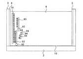
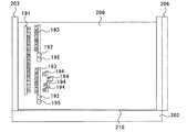
図1は、判定判定に用いる検査装置1の平面図であり、図2は、図1のII−II面に沿った断面図である。
この検査装置1は、矩形状を呈した固体撮像デバイス2と、固体撮像デバイス2の受光面の外周縁に沿って取り付けられた矩形枠状の浴槽3と、浴槽3の4辺のうち1つの框の内壁面に形成された陽極5と、陽極5が形成された框の対側となる框の内壁面に形成された陰極4と、固体撮像デバイス2の受光面に対向した紫外線源7と、固体撮像デバイス2の受光面に成膜された紫外線遮蔽膜10と、を具備する。固体撮像デバイス2は、フォトダイオード、フォトトランジスタ、ダブルゲート型フォトトランジスタといった光電変換素子をマトリクス状に配列したものである。固体撮像デバイス2としては、CCD型固体撮像デバイス、CMOS型固体撮像デバイス、MOS型固体撮像デバイス等がある。浴槽3内には、電気泳動媒体であるゲル8を入れておく。ゲル8に電界を加える電界形成手段は陽極5と陰極4とからなり、陽極5と陰極4との間においてゲル8に電界が形成され、これにより陽極5及び陰極4はゲル8中にプライマーを泳動させる泳動手段として機能する。紫外線源7は、下方の固体撮像デバイス2に向かって紫外線(励起光)を出射する光源である。紫外線遮蔽膜10は、アナターゼ型又はルチル型の酸化チタンからなる膜である。紫外線遮蔽膜10は、紫外線を吸収するとともに蛍光(主に可視光波長領域)を透過する性質を有する。(apparatus)
FIG. 1 is a plan view of an inspection apparatus 1 used for determination and determination, and FIG. 2 is a cross-sectional view taken along the II-II plane of FIG.
The inspection apparatus 1 includes a solid-
(工程順序)
工程(1):塩基配列が既知であるとともに蛍光標識されたプライマーを準備する。プライマーは、未知の塩基配列を有した一本鎖のDNAサンプルに相補的な可能性のある一本鎖DNAであり、PCR増幅されて実質的に同一種の塩基配列の分子が多数存在している。(Process order)
Step (1): A primer with a known base sequence and a fluorescent label is prepared. A primer is a single-stranded DNA that may be complementary to a single-stranded DNA sample having an unknown base sequence, and a large number of molecules of the same type of base sequence exist after PCR amplification. Yes.
工程(2):四種のデオキシヌクレオシド3リン酸(dNTP)を準備する。 Step (2): Four types of deoxynucleoside triphosphates (dNTPs) are prepared.
工程(3):判定対象となるDNAサンプルをユニバーサルプライマー及び酵素試薬(ポリメラーゼ)等を用いてPCR(Polymerase Chain Reaction)法により増幅する。 Step (3): A DNA sample to be determined is amplified by a PCR (Polymerase Chain Reaction) method using a universal primer and an enzyme reagent (polymerase).
工程(4):増幅したDNAサンプルを一本鎖に変性させる。 Step (4): The amplified DNA sample is denatured to a single strand.
工程(5):dNTPと、蛍光標識されたプライマーとを用いてDNAサンプルに対してPCR法を行う。すなわち、工程(4)において得られた一本鎖DNAサンプルを含む溶液を合成用の槽に入れ、工程(1)において準備したプライマーを合成用の槽に入れ、混入された既知のプライマーが一本鎖のDNAサンプルと相補的である塩基配列である場合にアニール結合できる程度に、プライマー及びDNAサンプルを冷却する。ここで、注入されたプライマー分子の数は、一本鎖DNAサンプルの数より遙かに多い。この後、工程(2)において準備したdNTP及び酵素試薬をプライマー及び一本鎖DNAサンプルを含む溶液に混入させる。ここでプライマー及びDNAサンプルの塩基配列が互いに相補的であればアニール結合し、プライマーの末端から一つの塩基分だけ外側に位置するDNAサンプルの対応する塩基と相補的になるような種のdNTPがプライマーの末端に結合され伸長する(PCR法)。このとき、合成用の槽内にはプライマーが過剰に存在しているため、一本鎖のDNAサンプルとアニール結合できないプライマーが存在することになり、このプライマーは伸長したプライマーと比べて短い。一方、工程(1)において準備したプライマーが一本鎖DNAサンプルに相補的でない場合には、一本鎖DNAサンプルとプライマーが結合しないので、伸長反応は起きないので全てのプライマーの分子長は変わらず互いに等しい。なお、合成用の槽は、蛍光標識を有するプライマーを相補的な塩基配列のDNAと結合してから重りを有するdNTPを入れてプライマーにdNTPを結合するための槽である。また、合成用の槽は合成装置に備わっており、合成装置は、合成用の槽に入ったプライマーをその槽に入った相補的な塩基配列の一本鎖DNAサンプルにその槽により結合してから、重りを有するdNTPをその槽に入れてその槽をもってプライマーをdNTPに結合する。 Step (5): PCR is performed on the DNA sample using dNTP and a fluorescently labeled primer. That is, the solution containing the single-stranded DNA sample obtained in the step (4) is put in a synthesis tank, the primer prepared in the step (1) is put in a synthesis tank, and one known primer mixed therein is found. The primer and the DNA sample are cooled to such an extent that they can be annealed when the base sequence is complementary to the DNA sample of the main strand. Here, the number of injected primer molecules is much larger than the number of single-stranded DNA samples. Thereafter, the dNTP and enzyme reagent prepared in step (2) are mixed in a solution containing the primer and the single-stranded DNA sample. Here, if the base sequences of the primer and DNA sample are complementary to each other, a dNTP of a species that anneals and is complementary to the corresponding base of the DNA sample located one base from the end of the primer. It is bound to the end of the primer and extended (PCR method). At this time, since the primer is excessive in the synthesis tank, there is a primer that cannot be annealed to the single-stranded DNA sample, and this primer is shorter than the extended primer. On the other hand, if the primer prepared in step (1) is not complementary to the single-stranded DNA sample, the single-stranded DNA sample and the primer do not bind, so no extension reaction occurs, so the molecular lengths of all primers change. Are equal to each other. The synthesis tank is a tank for combining a primer having a fluorescent label with DNA having a complementary base sequence and then adding dNTP having a weight to bind dNTP to the primer. In addition, the synthesis tank is provided in the synthesis apparatus, and the synthesis apparatus binds the primer contained in the synthesis tank to the single-stranded DNA sample of the complementary base sequence contained in the tank. Then, dNTP having a weight is put into the tank, and the primer is bound to dNTP with the tank.
工程(6):工程(5)で合成用の槽内で形成された生成物を、浴槽3内のゲル8の陽極5から離れた陰極4寄りに注入する。この状態を図3又は図4に模式的に示す。図3は、一本鎖DNAサンプル91がプライマー93と相補的な関係にある場合を示しており、図4は、一本鎖DNAサンプル91がプライマー93と相補的な関係にない場合を示している。図3に示すように、一本鎖DNAサンプル91が蛍光標識92を付したプライマー93と相補的な関係にある場合には、工程(5)で一本鎖DNAサンプル91とアニール結合したプライマー93がdNTP94と結合して伸長しているが、一本鎖DNAサンプル91とアニール結合していないプライマー93は伸長していない。一方、図4に示すように、一本鎖DNAサンプル91が蛍光標識92を付したプライマー93と相補的な関係にない場合には、どのプライマー93も伸長してない。
なお、工程(4)において得られた一本鎖DNAサンプル(図3、図4の場合には、一本鎖DNAサンプル91)、工程(1)において準備したプライマー(図3、図4の場合には、プライマー93)、及び、酵素試薬をゲル8の陰極4寄りに注入し、ゲル8中でPCR法により増幅しても良い。つまり、工程(5)の処理をゲル8中で行っても良い。Step (6): The product formed in the synthesis tank in step (5) is injected near the
The single-stranded DNA sample obtained in step (4) (single-stranded
工程(7):陰極4寄りに注入した生成物を陽極5から離れた位置(陰極4寄り)から陽極5に向けて電気泳動させる。すなわち、一本鎖DNAサンプル(図3、図4の場合には、一本鎖DNAサンプル91)及びプライマー(図3、図4の場合には、プライマー93)が互いに一本鎖に変性する程度に加熱させてから、陽極5が陰極4よりも高電圧となるように陽極5−陰極4間に電圧を印加し、ゲル8中に電界を発生させる。すると、DNAサンプル、プライマー、dNTPが陽極5に向かって電気泳動する。この状態を図5又は図6に模式的に示す。図5は、一本鎖DNAサンプル91がプライマー93と相補的な関係にある場合を示しており、図6は、一本鎖DNAサンプル91がプライマー93と相補的な関係にない場合を示している。この工程後においては、図5に示すように、一本鎖DNAサンプル91がプライマー93と相補的な関係にある場合には、一本鎖DNAサンプル91とアニール結合したプライマー93は増幅されているのでその分子長が一本鎖DNAサンプル91とアニール結合していないプライマー93よりも長いために、ゲル8中を電気泳動する際の抵抗が大きいので泳動速度が遅く、単位時間辺りの泳動距離が短い。一方、図6に示すように、一本鎖DNAサンプル91がプライマー93と相補的な関係にない場合には、どのプライマー93もほぼ同じ分子長なので電気泳動速度もほぼ同じなためほぼ同じ距離だけ泳動する。 Step (7): The product injected closer to the
工程(8):紫外線源7を点灯させ、紫外線をゲル8に向かって照射し、固体撮像デバイス2を用いて撮像を行うことによって固体撮像デバイス2の受光面に沿った蛍光強度分布を観察する。なお、紫外線によってプライマーの蛍光標識が励起して蛍光を発し、蛍光標識の蛍光が紫外線遮蔽膜10を透過して固体撮像デバイス2に入射するが、紫外線源7の紫外線は紫外線遮蔽膜10によって遮蔽されて固体撮像デバイス2に入射しない。つまり、紫外線遮蔽膜10は、蛍光標識の蛍光を紫外線源7の紫外線と分別する分別手段として機能する。ここで、固体撮像デバイス2は、各光電変換素子での蛍光の受光量を判別することによって、受光面に沿った蛍光強度分布を測定するものである。 Step (8): The ultraviolet
蛍光強度分布を観察した結果、特定の塩基配列がDNAサンプルに存在するか否かを判定することができる。
詳細には、一本鎖DNAサンプルと相補的なプライマーがある場合には、相補的でないために伸長していないプライマーが相補的であるために伸長したプライマーよりも同じ時間で長い距離泳動するため(図5を参照。)、蛍光強度のピークが二箇所で検出される。このように、陰極4から陽極5にかけて二箇所で蛍光強度のピークが検出された場合には、一本鎖DNAサンプルがプライマーと相補的であり、特定の塩基配列(プライマーと相補的な塩基配列)がDNAサンプルに存在することがわかる。例えば、図5の場合には、蛍光強度のピークは伸長したプライマー93(図5では、左側)の位置と、伸長していないプライマー93(図5では、右側)の位置に検出され、プライマー93と相補的な塩基配列が一本鎖DNAサンプル91に存在することがわかる。As a result of observing the fluorescence intensity distribution, it can be determined whether or not a specific base sequence exists in the DNA sample.
Specifically, if there is a primer that is complementary to the single-stranded DNA sample, the primer that is not extended because it is not complementary will run a longer distance in the same time as the extended primer because it is complementary. (See FIG. 5). Fluorescence intensity peaks are detected at two locations. Thus, when a peak of fluorescence intensity is detected at two locations from the
一方、一本鎖DNAサンプルがプライマーと相補的でない場合には、どのプライマーも同じ距離だけ泳動するから(図6を参照。)、陰極4から陽極5にかけて一箇所で蛍光強度のピークが検出される。蛍光強度のピークが一箇所で検出された場合には、一本鎖DNAサンプルがプライマーと相補的でなく、特定の塩基配列(プライマーと相補的な塩基配列)がDNAサンプルに存在しないことがわかる。例えば、図6の場合には、蛍光強度のピークは伸長していないプライマー93の位置に検出され、プライマー93と相補的な塩基配列が一本鎖DNAサンプル91に存在しないことがわかる。 On the other hand, when the single-stranded DNA sample is not complementary to the primer, since all the primers migrate by the same distance (see FIG. 6), a fluorescence intensity peak is detected at one location from the
なお、固体撮像デバイス2を用いずに、目視によってゲル8の蛍光強度分布を観察しても良いし、ゲル8から離れた位置に配置されたカメラを用いてゲル8の蛍光強度分布を観察しても良い。 Note that the fluorescence intensity distribution of the
以上のように、本実施形態の検査方法は、dNTPと、蛍光標識されたプライマー(プライマーは、DNAサンプルの増幅に必要な総数よりも多い。)とを用いてDNAサンプルに対してPCR法を行う工程(5)と、工程(5)で得られた生成物をゲル8中の陽極4から離れた位置より陽極4に向けて電気泳動させる工程(7)と、ゲル8の蛍光強度分布を観察する工程(8)と、を含む。 As described above, the inspection method of the present embodiment uses the PCR method for DNA samples using dNTPs and fluorescently labeled primers (primers are more than the total number necessary for amplification of DNA samples). The step (5) to be performed, the step (7) in which the product obtained in the step (5) is electrophoresed from the position away from the
以上の検査方法によれば、工程(5)において、プライマーの塩基配列と相補的な塩基配列がDNAサンプルに含まれている場合には、プライマーとDNAサンプルがアニールし、プライマーにdNTPが結合することによってプライマーが伸長する。一方、プライマーの塩基配列と相補的な塩基配列がDNAサンプルに含まれていない場合には、プライマーとDNAサンプルがアニールせず、プライマーは伸長しない。すなわち、プライマーがDNAサンプルの増幅に必要な総数よりも多いから、プライマーの塩基配列と相補的な塩基配列がDNAサンプルに含まれている場合には、生成物には伸長したプライマーと伸長していないプライマーとが含まれ、プライマーの塩基配列と相補的な塩基配列がDNAサンプルに含まれていない場合には、生成物には伸長していないプライマーが含まれる。
その後の工程(7)では、生成物が陽極に向かって泳動する。ここで伸長したプライマーは伸長していないプライマーよりも陽極4により近づく。そのため、伸長したプライマーがある場合には(プライマーの塩基配列と相補的な塩基配列がDNAサンプルに含まれている場合には)、伸長したプライマーの泳動後の位置と、伸長していないプライマーの泳動後の位置との両方で蛍光強度のピークが存するが、伸長したプライマーがない場合には(プライマーの塩基配列と相補的な塩基配列がDNAサンプルに含まれていない場合には)、伸長していないプライマーの泳動後の位置のみでしか蛍光強度のピークが存しない。従って、二箇所で蛍光強度のピークがある場合には、伸長したプライマーの塩基配列と相補的な塩基配列がDNAサンプルに含まれていると判定することができる。一方、一箇所でしか蛍光強度のピークがない場合には、プライマーの塩基配列と相補的な塩基配列がDNAサンプルに含まれていないと判定することができる。According to the above inspection method, in step (5), when the DNA sample contains a base sequence complementary to the base sequence of the primer, the primer and the DNA sample anneal, and dNTP binds to the primer. This extends the primer. On the other hand, when the DNA sample does not contain a base sequence complementary to the base sequence of the primer, the primer and the DNA sample are not annealed and the primer does not extend. That is, since the number of primers is larger than the total number required for amplification of the DNA sample, if the DNA sample contains a base sequence that is complementary to the base sequence of the primer, the product will be extended with the extended primer. In the case where the DNA sample does not contain a base sequence complementary to the base sequence of the primer, the product contains a non-extended primer.
In the subsequent step (7), the product migrates toward the anode. Here, the extended primer is closer to the
以上のように、本実施形態によれば、プライマー(図3〜図6の場合、プライマー93)の塩基配列と相補的な塩基配列がDNAサンプル(図3〜図6の場合、DNAサンプル91)に存在するか否かを判定するに際して、DNAサンプルをハイブリダイゼーションさせていないので、判定に要する時間が短くて済む。 As described above, according to the present embodiment, the base sequence complementary to the base sequence of the primer (
〔第2の実施の形態〕
第2の実施の形態における”検査装置、検査方法及び合成装置”について説明する。なお第1の実施の形態と実質的に同様な部分は説明を省略する。第1の実施の形態ではDNAサンプルに対して過剰な量の一種類の塩基配列のプライマーを準備したが、第2の実施の形態ではDNAサンプルに対して過剰な量の塩基配列の互いに異なる複数種のプライマーを準備する。更に、プライマーに、重りとなる所定の重さの不活性分子を結合させるが、結合する重りの重さ(分子量)は塩基配列ごとに異なるようにし、多くとも一種類のプライマーには重りを結合しなくても良い。ここで、重りの重さはプライマーの分子量よりも十分に重くする。また塩基配列がそれぞれ異なる各種プライマーの数は、DNAサンプルの数より過少になる。[Second Embodiment]
The “inspection apparatus, inspection method, and composition apparatus” in the second embodiment will be described. Note that description of parts substantially similar to those of the first embodiment will be omitted. In the first embodiment, an excessive amount of one kind of base sequence primer is prepared for the DNA sample, but in the second embodiment, an excessive amount of different base sequences for the DNA sample is different from each other. Prepare seed primer. In addition, an inert molecule of a predetermined weight that becomes a weight is bound to the primer, but the weight (molecular weight) of the weight to be bound is different for each base sequence, and the weight is bound to at most one kind of primer. You don't have to. Here, the weight of the weight is sufficiently heavier than the molecular weight of the primer. In addition, the number of various primers having different base sequences is less than the number of DNA samples.
そして、第1の実施の形態における工程(5)の場合と同様に、複数種類のプライマーと、dNTPとを用いて一本鎖DNAサンプルに対してPCR法を行う。その後、第1の実施の形態における工程(6)の場合と同様に、得られた生成物をゲル8の陰極4寄りに注入し、その後第1の実施の形態における工程(7)の場合と同様に、生成物を一本鎖に変性させ、陽極5が陰極4よりも高電圧となるように陽極5−陰極4間に電圧を印加し、ゲル8中に電界を発生させる。その後、第1の実施の形態における工程(8)の場合と同様に、紫外線源7を点灯させ、固体撮像デバイス2を用いて撮像を行うことによって固体撮像デバイス2の受光面に沿った蛍光強度分布を観察する。 Then, as in the case of step (5) in the first embodiment, the PCR method is performed on the single-stranded DNA sample using a plurality of types of primers and dNTP. Thereafter, as in the case of the step (6) in the first embodiment, the obtained product is injected near the
蛍光強度分布を観察した結果、DNAサンプルに特定の塩基配列が存在するか否かを判定することができる。
即ち、塩基配列の種類ごとに結合されている重りの重さが異なるため、複数種のプライマーに着目すると、結合する重りの重さが重くなるにつれてプライマーの泳動度も低くなるので、塩基配列の種類ごとにプライマーの泳動を観察することができる。その上、或る一種類の塩基配列のプライマーに着目して或る時間だけ泳動させると、一本鎖DNAサンプルがプライマーと相補的である場合には、伸長していないプライマーが伸長したプライマーよりも同じ時間で長い距離泳動するため、蛍光強度のピークが二箇所で検出されるが、一本鎖DNAサンプルがプライマーと相補的でない場合には、どのプライマーも同じ距離だけ泳動するため、陰極4から陽極5にかけて一箇所で蛍光強度のピークが検出される。このように、或る一種類の塩基配列のプライマーに着目して或る時間だけ泳動させて、蛍光強度のピークが二箇所で検出された場合には、その塩基配列と相補的な塩基配列がDNAサンプルに存在すると判定することができる。一方、或る一種類の塩基配列のプライマーに着目して或る時間だけ泳動させても、蛍光強度のピークが二箇所で検出されない場合には、その塩基配列と相補的な塩基配列がDNAサンプルに存在しないと判定することができる。As a result of observing the fluorescence intensity distribution, it can be determined whether or not a specific base sequence exists in the DNA sample.
In other words, since the weight of the weights that are bound differs depending on the type of base sequence, focusing on multiple types of primers, the mobility of the primer decreases as the weight of the bound weight increases. The primer migration can be observed for each type. In addition, when focusing on a primer of one kind of base sequence and migrating for a certain period of time, if the single-stranded DNA sample is complementary to the primer, the non-extended primer is more than the extended primer. Since a long distance migration is performed at the same time, fluorescence intensity peaks are detected at two locations. However, when a single-stranded DNA sample is not complementary to the primer, all the primers migrate at the same distance, so that the cathode 4 A peak of fluorescence intensity is detected at one point from the
例えば、三種類のプライマーを用いた場合に、第2の実施の形態において工程(7)のようにプライマーを泳動させた後の状態を模式的に図7に示す。図7において、黒塗りの円はプライマーに結合させた重りを表し、黒塗りの円の数が多いほど、重りの重さが重くなっていることを表す。また、白抜きの円はプライマーに結合させた蛍光物質を表す。そして、図7において、プライマー101は、一本鎖DNAサンプルと相補的であるが為にPCR法によりプライマー102が伸長したものであり、プライマー103は、一本鎖DNAサンプルと相補的であるが為にPCR法によりプライマー104が伸長したものであり、プライマー105は、一本鎖DNAサンプルと相補的でないがために伸長していない。なお、図7において、DNAサンプル、dNTPの図示は蛍光標識が付いていないから固体撮像デバイス2で観測できないため省略している。なお、重り一つの重さは、相補的なために伸長反応した塩基の重さより十分重い。 For example, when three types of primers are used, FIG. 7 schematically shows a state after the primers are migrated as in step (7) in the second embodiment. In FIG. 7, a black circle represents a weight bonded to the primer, and the greater the number of black circles, the greater the weight of the weight. The white circle represents the fluorescent substance bound to the primer. In FIG. 7,
図7の場合には、塩基配列の種類ごとに重りの重さが異なるため、プライマー101,102は重りがないためプライマー103,104よりも同じ時間で長い距離泳動し、一つの重りが付いているプライマー103,104は二つの重りが付いているプライマー105,105よりも同じ時間で長い距離泳動する。これにより、塩基配列の種類ごとに泳動距離を観察することができる。更に、同じ塩基配列のプライマー101,102に着目すると、伸長していないプライマー101が、伸長したために重く且つ電気泳動の際の流体抵抗が大きいプライマー102よりも同じ時間で長い距離泳動しているため、プライマー101,102と相補的な塩基配列がDNAサンプルに存在すると判定することができる。プライマー103,104についても同様に、プライマー103,104と相補的な塩基配列がDNAサンプルに存在すると判定することができる。一方、プライマー105,105は泳動距離に差が無いので、プライマー105,105と相補的な塩基配列がDNAサンプルに存在しないと判定することができる。 In the case of FIG. 7, since the weights of the weights differ depending on the type of the base sequence, the
本実施形態においても、DNAサンプルをハイブリダイゼーションさせていないので、プライマーの塩基配列と相補的な塩基配列がDNAサンプルに存在するか否かを判定する時間が短くて済むという効果を奏する。
また、複数種の塩基配列それぞれについて相補的な配列がDNAサンプルに存在するか否かを同一工程で判定することができる。Also in this embodiment, since the DNA sample is not hybridized, there is an effect that it takes a short time to determine whether or not a base sequence complementary to the primer base sequence is present in the DNA sample.
In addition, it can be determined in the same step whether or not a complementary sequence exists in the DNA sample for each of a plurality of types of base sequences.
〔第3の実施の形態〕
第3の実施の形態における”検査装置、検査方法及び合成装置”について説明する。なお第1又は第2の実施の形態と実質的に同様な部分は説明を省略する。[Third Embodiment]
The “inspection apparatus, inspection method and synthesis apparatus” in the third embodiment will be described. Note that description of parts substantially the same as those in the first or second embodiment will be omitted.
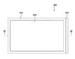
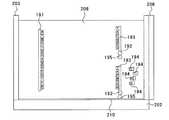
第3の実施の形態では、図1に示された装置1は用いずに、図8、図9に示された装置201を用いる。図8は、判定に用いる装置201の平面図であり、図9は、図8のIX−IX面に沿った断面図である。 In the third embodiment, the
この装置201は、矩形状を呈した固体撮像デバイス202と、固体撮像デバイス202の受光面の外周縁に沿って取り付けられた矩形枠状の浴槽203と、浴槽203の4辺のうち1つの框の外壁面に当接するように設けられた磁石206と、固体撮像デバイス202の受光面に対向した紫外線源207と、固体撮像デバイス202の受光面に成膜された紫外線遮蔽膜210と、を具備する。磁石206は、電磁石であっても良いし、永久磁石であっても良い。浴槽203内には、媒体であるゲル208を入れておく。紫外線源207は、下方の固体撮像デバイス2に向かって紫外線(励起光)を出射する光源である。ゲル8に磁界を加える磁界形成手段は磁石206からなり、磁石206によってゲル8に磁界が形成され、これにより磁石206はゲル8中にプライマーを泳動させる泳動手段として機能する。紫外線遮蔽膜210は、アナターゼ型又はルチル型の酸化チタンからなる膜である。紫外線遮蔽膜210は、紫外線を吸収するとともに蛍光(主に可視光波長領域)を透過する性質を有する。 The
第3の実施の形態では、第1の実施の形態の場合と同様に蛍光標識された既知の塩基配列のプライマーを準備するが、更にプライマーに磁気微粒子を結合させる。 In the third embodiment, a primer having a known base sequence that is fluorescently labeled is prepared in the same manner as in the first embodiment, and magnetic particles are further bound to the primer.
そして、第1の実施の形態における工程(5)の場合と同様に、プライマーと、dNTPとを用いて一本鎖DNAサンプルに対してPCR法を行う。その後、第1の実施の形態における工程(6)の場合と同様に、得られた生成物をゲル208の磁石206から離れた箇所に注入する。この状態を図10又は図11に模式的に示す。図10は、一本鎖DNAサンプルがプライマーと相補的な関係にある場合を示しており、図11は、一本鎖DNAサンプルがプライマーと相補的な関係にない場合を示している。図10に示すように、図3と同様に、一本鎖DNAサンプル191が蛍光標識192及び磁気微粒子195を付したプライマー193と相補的な関係にある場合には、一本鎖DNAサンプル191とアニール結合したプライマー193がdNTP194と結合して伸長しているが、一本鎖DNAサンプル191とアニール結合していないプライマー193は伸長していない。一方、図11に示すように、図4と同様に、一本鎖DNAサンプル191が蛍光標識192を付したプライマー193と相補的な関係にない場合には、どのプライマー193も伸長してない。 Then, as in the case of step (5) in the first embodiment, the PCR method is performed on the single-stranded DNA sample using the primer and dNTP. Thereafter, as in the case of the step (6) in the first embodiment, the obtained product is injected into a place away from the
その後、生成物を一本鎖に変性させ、磁石206によってゲル208中に磁界を発生させる。すると、DNAサンプル、プライマー、dNTPが磁石206に向かって泳動する。ここで、図12に示すように、一本鎖DNAサンプル191がプライマー193と相補的な関係にある場合には、一本鎖DNAサンプル191とアニール結合したプライマー193は、一本鎖DNAサンプル191とアニール結合していないプライマー193よりも泳動距離が短い。一方、図13に示すように、一本鎖DNAサンプル191がプライマー193と相補的な関係にない場合には、どのプライマー193もほぼ同じ距離だけ泳動する。 The product is then denatured into single strands and a magnetic field is generated in the
紫外線源207を点灯させ、ゲル208に向かって紫外線を照射し、固体撮像デバイス202を用いて撮像を行うことによって固体撮像デバイス2の受光面に沿った蛍光強度分布を観察する。ここで、固体撮像デバイス202は、各光電変換素子での蛍光の受光量を判別することによって、受光面に沿った蛍光強度分布を測定するものである。 The
蛍光強度分布を観察した結果、DNAサンプルに特定の塩基配列が存在するか否かを判定することができる。
詳細には、一本鎖DNAサンプルがプライマーと相補的である場合には、伸長していないプライマーが伸長したプライマーよりも同じ時間で長い距離泳動するため(図12を参照。)、蛍光強度のピークが二箇所で検出される。このように、磁石206の反対側から磁石206にかけて二箇所で蛍光強度のピークが検出された場合には、一本鎖DNAサンプルがプライマーと相補的であり、DNAサンプルに特定の塩基配列(プライマーと相補的な塩基配列)が存在することがわかる。As a result of observing the fluorescence intensity distribution, it can be determined whether or not a specific base sequence exists in the DNA sample.
Specifically, when the single-stranded DNA sample is complementary to the primer, the non-extended primer migrates longer than the extended primer in the same time (see FIG. 12). Peaks are detected at two locations. Thus, when fluorescence intensity peaks are detected at two locations from the opposite side of the
一方、一本鎖DNAサンプルがプライマーと相補的でない場合には、どのプライマーも同じ距離だけ泳動するから(図13を参照。)、磁石206の反対側から磁石206にかけて一箇所で蛍光強度のピークが検出される。蛍光強度のピークが一箇所で検出された場合には、一本鎖DNAサンプルがプライマーと相補的でなく、DNAサンプルに特定の塩基配列(プライマーと相補的な塩基配列)が存在しないことがわかる。 On the other hand, when the single-stranded DNA sample is not complementary to the primer, since all the primers migrate the same distance (see FIG. 13), the peak of the fluorescence intensity is observed at one point from the opposite side of the
本実施形態においても、DNAサンプルをハイブリダイゼーションさせていないので、プライマーの塩基配列と相補的な塩基配列がDNAサンプルに存在するか否かを判定する時間が短くて済むという効果を奏する。
また、ゲル208に電界を発生させていないので、ゲル208が発熱しない。そのため、発熱による蛍光標識のプライマーからの遊離等の現象を防ぎ、安定して判定することができる。Also in this embodiment, since the DNA sample is not hybridized, there is an effect that it takes a short time to determine whether or not a base sequence complementary to the primer base sequence is present in the DNA sample.
Further, since no electric field is generated in the
〔第4の実施の形態〕
第4の実施の形態における”検査装置、検査方法及び合成装置”について説明する。第3の実施の形態では一種類の塩基配列のプライマーを準備したが、第4の実施の形態では塩基配列の互いに異なる複数種のプライマーを準備する。なお第1又は第2又は第3の実施の形態と実質的に同様な部分は説明を省略する。更に、プライマーに重りを結合させるが、結合する重りの重さは塩基配列ごとに異なるようにし、多くとも一種類のプライマーには重りを結合しなくても良い。ここで、重りの重さ(分子量)はプライマーの分子量よりも十分に重くする。勿論、各種のプライマーには、磁気微粒子及び蛍光標識を付す。[Fourth Embodiment]
The “inspection apparatus, inspection method, and composition apparatus” in the fourth embodiment will be described. In the third embodiment, a primer having one type of base sequence is prepared. In the fourth embodiment, a plurality of types of primers having different base sequences are prepared. Note that description of parts substantially similar to those of the first, second, or third embodiment will be omitted. Furthermore, the weight is bound to the primer, but the weight of the weight to be bound is different for each base sequence, and the weight may not be bound to at most one kind of primer. Here, the weight (molecular weight) of the weight is sufficiently heavier than the molecular weight of the primer. Of course, magnetic primers and fluorescent labels are attached to various primers.
そして、第3の実施の形態の場合と同様に、複数種類のプライマーと、dNTPとを用いて一本鎖DNAサンプルに対してPCR法を行う。その後、第3の実施の形態の場合と同様に、得られた生成物をゲル8の磁石206から離れた箇所に注入する。その後、第3の実施の形態の場合と同様に、生成物を一本鎖に変性させ、磁石206によってゲル8中に磁界を発生させる。その後、第3の実施の形態における工程(8)の場合と同様に、紫外線源207を点灯させ、固体撮像デバイス202を用いて撮像を行うことによって固体撮像デバイス202の受光面に沿った蛍光強度分布を観察する。 Then, as in the case of the third embodiment, a PCR method is performed on a single-stranded DNA sample using a plurality of types of primers and dNTP. Thereafter, as in the case of the third embodiment, the obtained product is injected into a portion of the
蛍光強度分布を観察した結果、DNAサンプルに特定の塩基配列が存在するか否かを判定することができる。
即ち、塩基配列の種類ごとに結合されている重りの重さが異なるため、複数種のプライマーに着目すると、結合する重りの重さが重くなるにつれてプライマーの泳動度も低くなるので、塩基配列の種類ごとにプライマーの泳動を観察することができる。その上、或る一種類の塩基配列のプライマーに着目して或る時間だけ泳動させると、一本鎖DNAサンプルがプライマーと相補的である場合には、伸長していないプライマーが伸長したプライマーよりも同じ時間で長い距離泳動するため、蛍光強度のピークが二箇所で検出されるが、一本鎖DNAサンプルがプライマーと相補的でない場合には、どのプライマーも同じ距離だけ泳動するため、磁石206の反対側から磁石206にかけてにかけて一箇所で蛍光強度のピークが検出される。このように、或る一種類の塩基配列のプライマーに着目して或る時間だけ泳動させて、蛍光強度のピークが二箇所で検出された場合には、その塩基配列と相補的な塩基配列がDNAサンプルに存在すると判定することができる。一方、或る一種類の塩基配列のプライマーに着目して或る時間だけ泳動させても、蛍光強度のピークが二箇所で検出されない場合には、その塩基配列と相補的な塩基配列がDNAサンプルに存在しないと判定することができる。As a result of observing the fluorescence intensity distribution, it can be determined whether or not a specific base sequence exists in the DNA sample.
In other words, since the weight of the weights that are bound differs depending on the type of base sequence, focusing on multiple types of primers, the mobility of the primer decreases as the weight of the bound weight increases. The primer migration can be observed for each type. In addition, when focusing on a primer of one kind of base sequence and migrating for a certain period of time, if the single-stranded DNA sample is complementary to the primer, the non-extended primer is more than the extended primer. Since a long distance migration is performed at the same time, fluorescence intensity peaks are detected at two locations. However, when a single-stranded DNA sample is not complementary to the primer, all the primers migrate at the same distance, so that the magnet 206 A peak of fluorescence intensity is detected at one point from the opposite side to the
例えば、三種類のプライマーを用いた場合に、第4の実施の形態において磁界によりプライマーを泳動させた後の状態を模式的に図14に示す。図14において、黒塗りの円はプライマーに結合させた重りを表し、黒丸の数が多いほど、重りの重さが重くなっていることを表す。また、白抜きの円はプライマーに結合させた蛍光物質を表し、白抜きの正方形はプライマーに結合させた磁気微粒子を表す。そして、図14において、プライマー301は、一本鎖DNAサンプルと相補的であるが為にPCR法によりプライマー302が伸長したものであり、プライマー303は、一本鎖DNAサンプルと相補的であるが為にPCR法によりプライマー304が伸長したものであり、プライマー305は、一本鎖DNAサンプルと相補的でないがために伸長していない。なお、図14において、DNAサンプル、dNTPの図示は省略している。 For example, when three types of primers are used, FIG. 14 schematically shows a state after the primers are migrated by a magnetic field in the fourth embodiment. In FIG. 14, a black circle represents a weight bonded to the primer, and the greater the number of black circles, the greater the weight of the weight. A white circle represents a fluorescent substance bonded to the primer, and a white square represents a magnetic fine particle bonded to the primer. In FIG. 14, the
図14の場合には、塩基配列の種類ごとに重りの重さが異なるため、重りのないプライマー301,302は重りが一つ付いているプライマー303,304よりも同じ時間で長い距離泳動し、プライマー303,304は重りが二つ付いているプライマー305,305よりも同じ時間で長い距離泳動する。これにより、塩基配列の種類ごとに泳動距離を観察することができる。更に、同じ塩基配列のプライマー301,302に着目すると、プライマー302がプライマー302よりも同じ時間で長い距離泳動しているため、プライマー301,302と相補的な塩基配列がDNAサンプルに存在すると判定することができる。プライマー303,304についても同様に、プライマー303,304と相補的な塩基配列がDNAサンプルに存在すると判定することができる。一方、プライマー305,305は泳動距離に差が無いので、プライマー305,305と相補的な塩基配列がDNAサンプルに存在しないと判定することができる。
なお、第3実施の形態及び第4実施の形態において、磁石206は浴槽203の一方の面に形成されたが、浴槽203の対向する両壁側のどちらか一方がゲル208にN極の磁界を発生させる磁石で、他方がゲル208にS極の磁界を発生させる磁石であってもよい。In the case of FIG. 14, since the weight of the weight differs depending on the type of the base sequence, the
In the third embodiment and the fourth embodiment, the
2,202 … 固体撮像デバイス(判別手段)
4 … 陰極(泳動手段、電界形成手段)
5 … 陽極(泳動手段、電界形成手段)
7,207 … 紫外線源(光源)
8,208 … ゲル(媒体)
206 … 磁石(泳動手段、磁界形成手段)2,202 ... Solid-state imaging device (discrimination means)
4 ... Cathode (electrophoresis means, electric field forming means)
5 ... Anode (electrophoresis means, electric field forming means)
7,207 ... UV source (light source)
8,208 ... Gel (medium)
206 ... Magnet (electrophoresis means, magnetic field forming means)
Claims (12)
Translated fromJapanese前記固体撮像デバイスの受光面上に形成され、生成物が取り込まれるためのゲルが注入された槽と、を備え、
前記槽の一方の面側に、既知の塩基配列を有し、蛍光標識されたプライマーと、dNTPと、未知の塩基配列を有した一本鎖DNAサンプルと、が注入され、
注入された前記プライマーが前記一本鎖DNAサンプルと相補的な塩基配列である場合、前記dNTP及び前記一本鎖DNAサンプルにより前記プライマーがPCR法により伸長させられ、
伸長した前記プライマーを前記槽の前記一方の面側から前記一方の面側と対向する他方の面側に泳動させ、
泳動後に前記固体撮像デバイスを用いて蛍光強度分布が測定されることを特徴とする検査装置。A solid-state imaging device including a plurality of photoelectric conversion elements;
A tank formed on the light-receiving surface of the solid-state imaging device and filled with agel for taking in the product,
On one side of the tank, a primer having a known base sequence, a fluorescently labeled primer, dNTP, and a single-stranded DNA sample having an unknown base sequence are injected,
When the injected primer has a base sequence complementary to the single-stranded DNA sample, the primer is extended by the PCR method with the dNTP and the single-stranded DNA sample,
The extended primer ismigrated from the one surface side of thetank to the other surface side facing the one surface side,
An inspection apparatus in which a fluorescence intensity distribution is measured using the solid-state imaging device after electrophoresis.
前記槽の一方の面側に、既知の塩基配列を有し、蛍光標識されたプライマーと、dNTPと、未知の塩基配列を有した一本鎖DNAサンプルと、を注入する注入工程と、
前記注入工程において注入された前記プライマーが前記一本鎖DNAサンプルと相補的な塩基配列である場合、前記dNTP及び前記一本鎖DNAサンプルにより前記プライマーをPCR法により伸長させるPCR工程と、
前記PCR工程で伸長させた前記プライマーを前記槽の前記一方の面側から前記一方の面側と対向する他方の面側に泳動させる泳動工程と、
前記泳動工程後に前記固体撮像デバイスを用いて蛍光強度分布を測定する測定工程と、を含むことを特徴とする検査方法。In an inspection method using an inspection apparatus comprising: a solid-state imaging device including a plurality of photoelectric conversion elements; and a tank into which agel for taking in a product is formed and formed on a light-receiving surface of the solid-state imaging device.
An injection step of injecting a primer having a known base sequence, a fluorescently labeled primer, dNTP, and a single-stranded DNA sample having an unknown base sequence on one surface side of the tank;
When the primer injected in the injection step has a base sequence complementary to the single-stranded DNA sample, a PCR step of extending the primer by a PCR method with the dNTP and the single-stranded DNA sample;
A migration step of migrating the primer extended in the PCR step from the one surface side of thetank to the other surface side facing the one surface side;
A measuring step of measuring a fluorescence intensity distribution using the solid-state imaging device after the electrophoresis step.
Priority Applications (1)
| Application Number | Priority Date | Filing Date | Title |
|---|---|---|---|
| JP2004002936AJP4501430B2 (en) | 2004-01-08 | 2004-01-08 | Inspection apparatus, inspection method, and synthesis apparatus |
Applications Claiming Priority (1)
| Application Number | Priority Date | Filing Date | Title |
|---|---|---|---|
| JP2004002936AJP4501430B2 (en) | 2004-01-08 | 2004-01-08 | Inspection apparatus, inspection method, and synthesis apparatus |
Publications (2)
| Publication Number | Publication Date |
|---|---|
| JP2005192498A JP2005192498A (en) | 2005-07-21 |
| JP4501430B2true JP4501430B2 (en) | 2010-07-14 |
Family
ID=34817987
Family Applications (1)
| Application Number | Title | Priority Date | Filing Date |
|---|---|---|---|
| JP2004002936AExpired - Fee RelatedJP4501430B2 (en) | 2004-01-08 | 2004-01-08 | Inspection apparatus, inspection method, and synthesis apparatus |
Country Status (1)
| Country | Link |
|---|---|
| JP (1) | JP4501430B2 (en) |
Families Citing this family (2)
| Publication number | Priority date | Publication date | Assignee | Title |
|---|---|---|---|---|
| JP2008011829A (en)* | 2006-07-10 | 2008-01-24 | Olympus Corp | Method for detecting nucleic acid |
| WO2014164479A1 (en)* | 2013-03-11 | 2014-10-09 | Elitech Holding B.V. | Methods for true isothermal strand displacement amplification |
Family Cites Families (4)
| Publication number | Priority date | Publication date | Assignee | Title |
|---|---|---|---|---|
| US5686271A (en)* | 1994-06-09 | 1997-11-11 | Gamera Bioscience Corporation | Apparatus for performing magnetic cycle reaction |
| JP3832256B2 (en)* | 2000-05-15 | 2006-10-11 | 株式会社日立製作所 | Capillary array |
| JP4407068B2 (en)* | 2001-03-26 | 2010-02-03 | 横河電機株式会社 | Magnetic particle migration method and apparatus |
| EP1532273A4 (en)* | 2002-06-20 | 2009-11-18 | Primera Biosystems Inc | APPARATUS FOR QUANTITATIVE DETECTION AND ANALYSIS OF POLYNUCLEOTIDES |
- 2004
- 2004-01-08JPJP2004002936Apatent/JP4501430B2/ennot_activeExpired - Fee Related
Also Published As
| Publication number | Publication date |
|---|---|
| JP2005192498A (en) | 2005-07-21 |
Similar Documents
| Publication | Publication Date | Title |
|---|---|---|
| US11214796B2 (en) | Gene expression analysis ME1HOD using two dimensional cDNA library | |
| Miotke et al. | High sensitivity detection and quantitation of DNA copy number and single nucleotide variants with single color droplet digital PCR | |
| JP5627886B2 (en) | PCR-free sample preparation and detection system for rapid biological analysis and identification | |
| US9651492B2 (en) | Optical detector | |
| TW200918888A (en) | Plastic microfluidic separation and detection platforms | |
| Fortina et al. | DOP-PCR amplification of whole genomic DNA and microchip-based capillary electrophoresis | |
| Wattiau et al. | Nucleotide polymorphism-based single-tube test for robust molecular identification of all currently described Brucella species | |
| Rajagopal et al. | Supercolor coding methods for large-scale multiplexing of biochemical assays | |
| JP6374967B2 (en) | Detection of nucleic acid amplification in porous substrates | |
| JP4501430B2 (en) | Inspection apparatus, inspection method, and synthesis apparatus | |
| US20220228193A1 (en) | Methods to detect cells latently infected with hiv | |
| Mahdavi et al. | Design and fabrication of multiplex microfluidic LAMP PCR for simultaneous detection of opportunistic protozoa | |
| Zhou et al. | Highly sensitive determination of the methylated p16 gene in cancer patients by microchip electrophoresis | |
| JP6295578B2 (en) | Reaction vessel, nucleic acid analyzer, and nucleic acid analysis method | |
| Hamada et al. | Development of a ligase detection reaction/CGE method using a LIF dual‐channel detection system for direct identification of allelic composition of mutated DNA in a mixed population of excess wild‐type DNA | |
| Shin et al. | Triblock copolymer‐based microchip device for rapid analysis of stuffer‐free multiplex ligation‐dependent probe amplification products | |
| Monshat et al. | Integration of nucleic acid amplification, detection, and melting curve analysis for rapid genotyping of antimicrobial resistance | |
| Qian et al. | Effect of oligonucleotide probes substituted by deoxyinosines on the specificity of SNP detection on the DNA microarray | |
| Choi et al. | Highly multiplex and sensitive SNP genotyping method using a three‐color fluorescence‐labeled ligase detection reaction coupled with conformation‐sensitive CE | |
| Lee et al. | Portable capillary electrophoresis system for identification of cattle breeds based on DNA mobility | |
| Huang et al. | Novel micro-nanofluidic chip and device for fast identifying pathogenic bacteria | |
| Zhang et al. | Genotyping by alkaline dehybridization using graphically encoded particles | |
| JP2005192497A (en) | Inspection apparatus, inspection method, and synthesis apparatus | |
| Ido et al. | Loop-mediated isothermal amplification in microchambers on a cell microarray chip for identification of Plasmodium species | |
| Zhang et al. | Fast high-throughput screening of H1N1 virus by parallel detection with multichannel microchip electrophoresis |
Legal Events
| Date | Code | Title | Description |
|---|---|---|---|
| A621 | Written request for application examination | Free format text:JAPANESE INTERMEDIATE CODE: A621 Effective date:20070104 | |
| A131 | Notification of reasons for refusal | Free format text:JAPANESE INTERMEDIATE CODE: A131 Effective date:20090929 | |
| A521 | Request for written amendment filed | Free format text:JAPANESE INTERMEDIATE CODE: A523 Effective date:20091130 | |
| A131 | Notification of reasons for refusal | Free format text:JAPANESE INTERMEDIATE CODE: A131 Effective date:20100105 | |
| A521 | Request for written amendment filed | Free format text:JAPANESE INTERMEDIATE CODE: A523 Effective date:20100304 | |
| TRDD | Decision of grant or rejection written | ||
| A01 | Written decision to grant a patent or to grant a registration (utility model) | Free format text:JAPANESE INTERMEDIATE CODE: A01 Effective date:20100330 | |
| A01 | Written decision to grant a patent or to grant a registration (utility model) | Free format text:JAPANESE INTERMEDIATE CODE: A01 | |
| A61 | First payment of annual fees (during grant procedure) | Free format text:JAPANESE INTERMEDIATE CODE: A61 Effective date:20100412 | |
| R150 | Certificate of patent or registration of utility model | Free format text:JAPANESE INTERMEDIATE CODE: R150 | |
| FPAY | Renewal fee payment (event date is renewal date of database) | Free format text:PAYMENT UNTIL: 20130430 Year of fee payment:3 | |
| FPAY | Renewal fee payment (event date is renewal date of database) | Free format text:PAYMENT UNTIL: 20130430 Year of fee payment:3 | |
| FPAY | Renewal fee payment (event date is renewal date of database) | Free format text:PAYMENT UNTIL: 20140430 Year of fee payment:4 | |
| LAPS | Cancellation because of no payment of annual fees |