JP4445697B2 - Biological repair material having affinity with biological tissue adhesive - Google Patents
Biological repair material having affinity with biological tissue adhesiveDownload PDFInfo
- Publication number
- JP4445697B2 JP4445697B2JP2002253322AJP2002253322AJP4445697B2JP 4445697 B2JP4445697 B2JP 4445697B2JP 2002253322 AJP2002253322 AJP 2002253322AJP 2002253322 AJP2002253322 AJP 2002253322AJP 4445697 B2JP4445697 B2JP 4445697B2
- Authority
- JP
- Japan
- Prior art keywords
- eptfe
- fibrin glue
- tissue adhesive
- affinity
- artificial
- Prior art date
- Legal status (The legal status is an assumption and is not a legal conclusion. Google has not performed a legal analysis and makes no representation as to the accuracy of the status listed.)
- Expired - Lifetime
Links
Images


Classifications
- A—HUMAN NECESSITIES
- A61—MEDICAL OR VETERINARY SCIENCE; HYGIENE
- A61L—METHODS OR APPARATUS FOR STERILISING MATERIALS OR OBJECTS IN GENERAL; DISINFECTION, STERILISATION OR DEODORISATION OF AIR; CHEMICAL ASPECTS OF BANDAGES, DRESSINGS, ABSORBENT PADS OR SURGICAL ARTICLES; MATERIALS FOR BANDAGES, DRESSINGS, ABSORBENT PADS OR SURGICAL ARTICLES
- A61L27/00—Materials for grafts or prostheses or for coating grafts or prostheses
- A61L27/14—Macromolecular materials
- A61L27/16—Macromolecular materials obtained by reactions only involving carbon-to-carbon unsaturated bonds
- A—HUMAN NECESSITIES
- A61—MEDICAL OR VETERINARY SCIENCE; HYGIENE
- A61L—METHODS OR APPARATUS FOR STERILISING MATERIALS OR OBJECTS IN GENERAL; DISINFECTION, STERILISATION OR DEODORISATION OF AIR; CHEMICAL ASPECTS OF BANDAGES, DRESSINGS, ABSORBENT PADS OR SURGICAL ARTICLES; MATERIALS FOR BANDAGES, DRESSINGS, ABSORBENT PADS OR SURGICAL ARTICLES
- A61L17/00—Materials for surgical sutures or for ligaturing blood vessels ; Materials for prostheses or catheters
- A61L17/06—At least partially resorbable materials
- A61L17/10—At least partially resorbable materials containing macromolecular materials
- A61L17/12—Homopolymers or copolymers of glycolic acid or lactic acid
- A—HUMAN NECESSITIES
- A61—MEDICAL OR VETERINARY SCIENCE; HYGIENE
- A61L—METHODS OR APPARATUS FOR STERILISING MATERIALS OR OBJECTS IN GENERAL; DISINFECTION, STERILISATION OR DEODORISATION OF AIR; CHEMICAL ASPECTS OF BANDAGES, DRESSINGS, ABSORBENT PADS OR SURGICAL ARTICLES; MATERIALS FOR BANDAGES, DRESSINGS, ABSORBENT PADS OR SURGICAL ARTICLES
- A61L17/00—Materials for surgical sutures or for ligaturing blood vessels ; Materials for prostheses or catheters
- A61L17/14—Post-treatment to improve physical properties
- A—HUMAN NECESSITIES
- A61—MEDICAL OR VETERINARY SCIENCE; HYGIENE
- A61L—METHODS OR APPARATUS FOR STERILISING MATERIALS OR OBJECTS IN GENERAL; DISINFECTION, STERILISATION OR DEODORISATION OF AIR; CHEMICAL ASPECTS OF BANDAGES, DRESSINGS, ABSORBENT PADS OR SURGICAL ARTICLES; MATERIALS FOR BANDAGES, DRESSINGS, ABSORBENT PADS OR SURGICAL ARTICLES
- A61L24/00—Surgical adhesives or cements; Adhesives for colostomy devices
- A61L24/001—Use of materials characterised by their function or physical properties
- A—HUMAN NECESSITIES
- A61—MEDICAL OR VETERINARY SCIENCE; HYGIENE
- A61L—METHODS OR APPARATUS FOR STERILISING MATERIALS OR OBJECTS IN GENERAL; DISINFECTION, STERILISATION OR DEODORISATION OF AIR; CHEMICAL ASPECTS OF BANDAGES, DRESSINGS, ABSORBENT PADS OR SURGICAL ARTICLES; MATERIALS FOR BANDAGES, DRESSINGS, ABSORBENT PADS OR SURGICAL ARTICLES
- A61L24/00—Surgical adhesives or cements; Adhesives for colostomy devices
- A61L24/04—Surgical adhesives or cements; Adhesives for colostomy devices containing macromolecular materials
- A—HUMAN NECESSITIES
- A61—MEDICAL OR VETERINARY SCIENCE; HYGIENE
- A61L—METHODS OR APPARATUS FOR STERILISING MATERIALS OR OBJECTS IN GENERAL; DISINFECTION, STERILISATION OR DEODORISATION OF AIR; CHEMICAL ASPECTS OF BANDAGES, DRESSINGS, ABSORBENT PADS OR SURGICAL ARTICLES; MATERIALS FOR BANDAGES, DRESSINGS, ABSORBENT PADS OR SURGICAL ARTICLES
- A61L27/00—Materials for grafts or prostheses or for coating grafts or prostheses
- A61L27/14—Macromolecular materials
- A61L27/18—Macromolecular materials obtained otherwise than by reactions only involving carbon-to-carbon unsaturated bonds
- A—HUMAN NECESSITIES
- A61—MEDICAL OR VETERINARY SCIENCE; HYGIENE
- A61L—METHODS OR APPARATUS FOR STERILISING MATERIALS OR OBJECTS IN GENERAL; DISINFECTION, STERILISATION OR DEODORISATION OF AIR; CHEMICAL ASPECTS OF BANDAGES, DRESSINGS, ABSORBENT PADS OR SURGICAL ARTICLES; MATERIALS FOR BANDAGES, DRESSINGS, ABSORBENT PADS OR SURGICAL ARTICLES
- A61L27/00—Materials for grafts or prostheses or for coating grafts or prostheses
- A61L27/50—Materials characterised by their function or physical properties, e.g. injectable or lubricating compositions, shape-memory materials, surface modified materials
- A61L27/507—Materials characterised by their function or physical properties, e.g. injectable or lubricating compositions, shape-memory materials, surface modified materials for artificial blood vessels
- A—HUMAN NECESSITIES
- A61—MEDICAL OR VETERINARY SCIENCE; HYGIENE
- A61L—METHODS OR APPARATUS FOR STERILISING MATERIALS OR OBJECTS IN GENERAL; DISINFECTION, STERILISATION OR DEODORISATION OF AIR; CHEMICAL ASPECTS OF BANDAGES, DRESSINGS, ABSORBENT PADS OR SURGICAL ARTICLES; MATERIALS FOR BANDAGES, DRESSINGS, ABSORBENT PADS OR SURGICAL ARTICLES
- A61L2400/00—Materials characterised by their function or physical properties
- A61L2400/18—Modification of implant surfaces in order to improve biocompatibility, cell growth, fixation of biomolecules, e.g. plasma treatment
- Y—GENERAL TAGGING OF NEW TECHNOLOGICAL DEVELOPMENTS; GENERAL TAGGING OF CROSS-SECTIONAL TECHNOLOGIES SPANNING OVER SEVERAL SECTIONS OF THE IPC; TECHNICAL SUBJECTS COVERED BY FORMER USPC CROSS-REFERENCE ART COLLECTIONS [XRACs] AND DIGESTS
- Y10—TECHNICAL SUBJECTS COVERED BY FORMER USPC
- Y10T—TECHNICAL SUBJECTS COVERED BY FORMER US CLASSIFICATION
- Y10T428/00—Stock material or miscellaneous articles
- Y10T428/31504—Composite [nonstructural laminate]
- Y10T428/3154—Of fluorinated addition polymer from unsaturated monomers
Landscapes
- Health & Medical Sciences (AREA)
- Chemical & Material Sciences (AREA)
- Animal Behavior & Ethology (AREA)
- Veterinary Medicine (AREA)
- Public Health (AREA)
- General Health & Medical Sciences (AREA)
- Epidemiology (AREA)
- Life Sciences & Earth Sciences (AREA)
- Dermatology (AREA)
- Medicinal Chemistry (AREA)
- Oral & Maxillofacial Surgery (AREA)
- Transplantation (AREA)
- Surgery (AREA)
- Vascular Medicine (AREA)
- Materials Engineering (AREA)
- Engineering & Computer Science (AREA)
- Chemical Kinetics & Catalysis (AREA)
- Materials For Medical Uses (AREA)
- Treatments Of Macromolecular Shaped Articles (AREA)
Abstract
Description
Translated fromJapanese【0001】
【発明の属する技術分野】
本発明は、生体組織接着剤と親和性を有する生体修復材料、即ち、生体組織接着剤と組み合わせて使用するための高分子材料に関する。より詳細には、本発明は、生体組織接着剤と組み合わせて使用するための高分子材料であって、表面の少なくとも一部がイオン衝撃により改質されていることにより生体組織接着剤との親和性が向上している高分子材料、並びにその製造方法に関する。
【0002】
【従来の技術】
頭蓋骨内にあって脳実質を保護する三層の膜(軟膜、クモ膜、硬膜)のうち、硬膜は最も硬く、三層の中で最外層に存在し、頭蓋骨の内側骨膜でもある。脳神経外科手術に際し、硬膜を切除せざるを得ないことがしばしばあり、硬膜欠損が生じる。また、硬膜自体の自然収縮のために一次的な縫合が困難になることもある。硬膜を開放したまま閉創する事は、髄液の漏出を招いて頭蓋内感染症を生じたり、脳実質と骨ないし皮下組織との癒着を生じて、局所神経症状を呈したり、てんかん発作の焦点となるなど、重篤な合併症を来たす原因となる。従って閉創時には硬膜に隙間が生じないよう厳密な縫合が要求される。このため、硬膜に欠損が生じたり一次縫合が困難となった場合には何らかの補填材料を用いて隙間が生じない様に完全に縫合する必要が生じる。
【0003】
いかなる補填材料を用いて硬膜欠損の補填を行うかは長期に渡り脳神経外科医を悩ませ続けた問題である。当初人工物が使用された時期もあるが、生体適合性、使いやすさ等に問題があり、何れも長続きしなかった。当初より今日に到るまで最も広く用いられているのは自家筋膜であるが、摘出部位に筋膜の欠損を生じること、脳に対して癒着しやすいことなど問題点も少なくない。ヒト乾燥硬膜は屍体から採取された硬膜を放射線処理等を行った硬膜補填材料であり、これまでの中では最も優れたものであった。しかし、クロイツフェルト・ヤコブ病の原因とされるプリオンが硬膜内に存在する可能性があり、ヒト乾燥硬膜を介してクロイツフェルト・ヤコブ病の感染が生じと事例が報告されるに到り、1998年にその使用は全面的に禁止された。
【0004】
現在自己筋膜以外に硬膜補填材料として使用可能な素材は、厚生省が認可しているePTFE(expanded polytetra-fluoroethylene)のみである。ePTFEは高分子材料であるため生体に対して全く接着性を有していない。この性質は脳と癒着を生じないという面では優れている。一方収縮性に乏しいため針穴から髄液が漏出してしまうため特殊な縫合糸を使用して縫合を行う必要がある。また生体接着性がないため縫合面の隙間からも髄液漏が生じる可能性が高い。これと共に、周辺組織とも接着性を有さないため、単なる骨格素材となってしまう可能性も高い。これ迄にePTFEをいかにうまく使用するかについての多くの試みがなされてきたが、何れもePTFEを骨格素材して使用し、周囲に線維性組織の被膜が形成されるのを待つものであった。
【0005】
一方、人工材料表面にイオンを用いて処理する方法としては、プラズマ処理による方法が知られている(特願平10−302170)。この方法は表面上を改質する方法によって接着性を改善する方法である。プラズマ処理法によるプラズマ処理層は生体内では不安定であり、経時的に分解・剥離する危険性がある。生体内では長期にわたって安定した細胞接着層を維持する必要がある。プラズマ処理による方法では、特に人工硬膜では、置換初期に頭蓋骨接触面に接着するものの、長期間の後、剥離する危険性を有する。
【0006】
このプラズマ処理法よりさらにエネルギーの高いイオンを用いて生体内埋入材料表層を改質して抗菌性を高めるという報告もある(特願平5−148994)。この方法の目的は、主に埋入材料の感染性を弱めることである。
【0007】
【発明が解決しようとする課題】
生体内で外科的手術に用いる医療材料、特に人工血管、人工硬膜、心臓又は血管を修復するパッチ材料を治療のために組織に固定するためには、これら材料と生体を吻合により固定する方法が採用されている。しかしながら、手術糸によりこれらの医療用材料を吻合した場合、針の通過した材料部分から血液又は脳髄液の漏出が生じる。そのため通常は、血液凝固を患部の圧迫により誘導するか、フィブリングルーと呼ばれる生体組織接着剤を用いることにより、これらの漏出を防いでいる。
【0008】
本発明は、人工血管又は人工硬膜などの生体内埋入材料と生体組織接着剤との親和性を向上させることにより、より迅速に血液又は髄液の漏出を防ぐことができる手段を提供することを目的とする。
【0009】
【課題を解決するための手段】
本発明者らは上記課題を解決するために鋭意検討した結果、Neイオンビーム照射ePTFEは、未照射ePTFEと比較して、生体組織接着剤との高い親和性を有することを見出し、本発明を完成するに至った。
【0010】
即ち、本発明によれば、生体組織接着剤と組み合わせて使用する、炭素又は珪素を構成元素として含む高分子材料より構成され、表面の少なくとも一部がイオン衝撃により改質されてなる高分子材料が提供される。
【0011】
好ましくは、生体組織接着剤はフィブリングルーである。
好ましくは、炭素又は珪素を構成元素として含む高分子材料は、延伸ポリテトラフルオロエチエン(ePTFE)、ポリ乳酸、又はポリグラクチンである。
好ましくは、ドース量φが1×10¹²≦φ≦1×10¹⁶イオン/cm2となる範囲でイオン注入を行うことによってイオン衝撃による改質を行う。
好ましくは、本発明の高分子材料は、人工硬膜、人工血管、心臓・血管用パッチ又は縫合糸として使用する。
【0012】
本発明の別の側面によれば、炭素又は珪素を構成元素として含む高分子材料の表面の少なくとも一部にドース量φが1×10¹²≦φ≦1×10¹⁶イオン/cm2となる範囲でイオン注入を行うことを特徴とする、生体組織接着剤と組み合わせて使用する高分子材料の製造方法が提供される。
【0013】
本発明のさらに別の側面によれば、炭素又は珪素を構成元素として含む高分子材料の表面の少なくとも一部にドース量φが1×10¹²≦φ≦1×10¹⁶イオン/cm2となる範囲でイオン注入を行うことを特徴とする、該高分子材料の生体組織接着剤との親和性を向上させる方法が提供される。
【0014】
【発明の実施の形態】
以下、本発明の実施の形態について詳細に説明する。
本発明の高分子材料は、生体組織接着剤と組み合わせて使用するためのものであり、炭素又は珪素を構成元素として含む高分子材料より構成され、表面の少なくとも一部がイオン衝撃により改質されていることを特徴とする。
【0015】
本発明では、炭素又は珪素を構成元素として含む高分子材料にイオンビームを照射することにより生体組織接着剤の親和性を向上させ、血液漏出、髄液漏出を防ぐことができる。本発明はさらに、上記した高分子材料にイオンビームを照射することによって生体組織接着剤との親和性を向上させる、表層処理方法にも関する。
【0016】
腹部大動脈瘤などの治療で人工血管を使用し損傷血管を治療する際、生体血管につなぎ合わせるため、手術用縫合糸により生体血管と人工血管を縫い合わせる。しかしながらポリエステル製、あるいはフッ素化合物製の人工血管は同種の縫合糸を用いても針穴から血液が漏出する。
【0017】
また心臓あるいは血管の病巣を摘出した場合、欠損部位を修復する必要がある。この修復材料を血管、心臓などに縫い合わせた場合も、同様に針穴からの血液、組織液が漏出する。脳外科領域では脳腫瘍、くも膜下出血、あるいは交通事故などによって開頭手術が行われる際には硬膜の欠損が生じる。この硬膜の欠損を補うために人工硬膜が使用されるが、人工血管と同じく針穴から脳髄液の漏出が生じる。
【0018】
多くの場合、これらの血液漏出、髄液漏出を防ぐためにフィブリングルーと呼ばれる生体組織接着剤を用いて、血液、髄液の漏出を防止している。しかしながら、人工血管素材、人工硬膜素材の内、とりわけフッ素化合物系の材料に関してはフィブリングルーとの接着が極めて悪く、血液、髄液漏出の防止が不十分である。
【0019】
本発明では、これら人工素材にイオン注入法によってイオンビームを素材表層に照射し、フィブリングルーとの接着性を改善させることができる。このイオンビーム処理は、人工硬膜、人工血管、又は心臓・血管用パッチに対して行うことができるのみならず、これらと生体とを結合させる縫合糸の表層に対して行うこともできる。縫合糸の表層をビームによって改質することによって、人工素材とフィブリングルーとの接着性は改善される。
【0020】
本発明で使用される炭素又は珪素を構成元素として有する高分子材料は、生体適合性があり、操作が容易である材料であれば特に限定されず任意の材料を使用できる。例えば、延伸ポリテトラフルオロエチレン(ePTFE)などのフッ素樹脂成形物やシリコーンなどの珪素化合物を使用することができる。本発明で好ましい高分子材料としては、延伸ポリテトラフルオロエチエン(ePTFE)、または生分解性高分子(例えば、ポリ乳酸、又はポリグラクチンなど)が挙げられ、特に延伸ポリテトラフルオロエチエン(ePTFE)が好ましい。
【0021】
本発明の高分子材料の表面の少なくとも一部は、イオン衝撃により改質されている。注入するイオン種としてはH+,He+,C+,N+,Ne+,Na+,N2+,O2+,Ar+,Kr+,Xe+などが例示されるが、溶出して生体組織接着剤との親和性を阻害するものでなければこれらに特に限定されるものではない。好ましくは、Ne+,Ar+,Kr+,Xe+などである。
【0022】
ドース量(照射量)φは、1×10¹²≦φ≦1×10¹⁶イオン/cm2の範囲であることが好ましい。1012イオン/cm2より低いと、生体組織接着剤との親和性の顕著な改善効果が小さくなり、1016イオン/cm2より高いと高分子材料が破壊され易くなり、何れも好ましくない。より好ましくは、ドース量φは、1×1013≦φ≦1×1015イオン/cm2の範囲である。
【0023】
イオン加速エネルギーに関しては、その高低によりエネルギー伝達機構に差異が生ずるものと考えられるが、実用的には数十keV〜数MeV程度の範囲で設定することができ、好ましくは50keV〜2MeV程度である。
【0024】
ビーム電流密度はおおよそ0.5μA/cm2を越えない範囲に設定することが好ましい。これは、ビーム電流密度が過大になるとターゲットである高分子材料の温度が上がり過ぎ、高分子材料自身が劣化する上、生体組織接着剤との親和性が低下する恐れがあるからである。
【0025】
本発明においてイオン衝撃を与える手段としてはイオン注入が挙げられる。イオン注入は、その反応自体がイオン・ビームと被注入材料(ターゲット材料)との間の相互作用に限られる。しかも、イオン入射エネルギーを選択することにより表面から任意に深さイオンを埋め込むことができ、極めて制御性に優れている。これは、プラズマ処理にはない特徴である。注入されたイオンは、比較的質量の軽いイオンに対しては拡散初期に電子阻止能が働き、比較的質量の重いイオンに対しては始めから核阻止能が働くという機構上の差異はあるものの、高分子材料に格子振動による加熱をもたらし(熱的非平衡状態)、溶融,アモルファス化等を引き起こす。
【0026】
本発明の高分子材料は、生体組織接着剤と組み合わせて使用する。生体組織接着剤の好ましい例としてはフィブリングルー(フィブリン糊)、又は高分子系接着剤であるシアノアクリレート系の瞬間接着剤などが挙げられる。従来、外科手術においては、絹毛、cat gutのような糸と針によって縫合が行われてきたが、これに接着剤が使われている。従来の縫合法では、小血管の縫い合せ、血管修復などは困難な場合が多く、又、一般的に縫合に時間を要し、あとがみにくいなどの難点があったが、これらの欠点を補う方法として生体組織接着剤が採用されている。
【0027】
フィブリングルーは、フィブリノーゲン凍結乾燥粉末、フィブリノーゲン溶解液、トロンビン凍結乾燥粉末、及びトロンビン溶解液から構成される。フィブリノーゲン凍結乾燥粉末をフィブリノーゲン溶解液で溶解しA液とする。トロンビン凍結乾燥粉末をトロンビン溶解液で溶解しB液とする。溶解した両液の等容量を接着部位に重層または混合して適用する。フィブリングルーは、血液凝固の最終段階を利用した生理的組織接着剤であり、含有するフィブリノーゲンはトロンビンの作用により可溶性フィブリン塊となり、さらにカルシウムイオン存在下でトロンビンにより活性化された血液凝固第XIII因子により物理的強度をもった尿素不溶性の安定なフィブリン塊となり、組織を接着・閉鎖する。この安定化したフィブリン塊内で,線維芽細胞が増殖し、膠原線維や肉芽基質成分が産生され、組織修復を経て治癒に至る。フィブリングルーの具体例としては、ボルヒール(商品名)((財団法人)化学及血清療法研究所)などが挙げられる。
【0028】
フィブリン糊として使用される外科的処置としては、生体組織同士を接着させる目的では、出血創傷面の閉鎖、骨折片の固定、末梢神経ならびに微小血管の吻合、腱接着または腱縫合の補強、実質臓器の接着などが挙げられる。また人工硬膜、人工血管などの人工物を生体組織と吻合する際、針穴から脳髄液、血液の漏出の防止にも用いられる。同様に、心臓欠損部の修復、血管欠損部の修復に用いられる心臓用パッチ、血管用パッチにおいても針穴から漏出する血液の防止目的で用いられる。特にePTFEの場合、フィブリングルーとの接着性が乏しいことが問題であったが、本発明に従って表面の少なくとも一部がイオン衝撃により改質することにより、フィブリングルーとの接着性の問題は解消した。
以下の実施例により本発明をより具体的に説明するが、本発明は実施例によって限定されることはない。
【0029】
【実施例】
(1)イオン照射処理
体重2.5〜3.0kgのオス日本白色家兎を実験に使用した。ペントバルピタールによる全身麻酔下に頭皮を冠状縫合に沿って切開して頭骨を露出した。試料は理化学研究所200KeVイオン注入装置を用いてイオンビーム照射(Ne+, 150keV, 5×10¹⁴ions/cm2)した延伸ポリテトラフルオロエチレン(ePTFE)を用いた。頭骨の表面骨膜を完全に除去した状態でさらに頭蓋骨を除去し硬膜を露出させた。また硬膜の一部は数ミリの穴を開けた。試料を硬膜上に非照射面を当てて置き、イオンビーム照射面を頭皮側に置いた。その後、生体組織接着剤(フィブリングルー;商品名ボルヒール、財団法人 化学及血清療法研究所)をイオンビーム照射面に垂らして除去した後に残存した頭蓋骨と固定させた。比較としてイオン照射しない試料も同様の操作を行った。フィブリングルー滴下後、頭皮を吻合し患部を被覆した。
【0030】
(2)観察
(a)目視による観察
イオンビーム照射面はフィブリングルー滴下後、数分で周囲の骨組織と良好な接着を生じた。それに対して未処理の延伸ポリテトラフルオロエチレンはピンセットで微弱な力を加えるだけで移動するほど接着力は弱いものであった。
【0031】
(b)組織検査による観察
試料を包埋移植後2週間目にネンブタールを用いて家兎を犠牲とし、それぞれ周辺組織ごと一塊として取り出し、10%バッファーホルマリンで固定した。周辺組織ごと摘出したのはイオンビーム非処理面ではフィブリングルーとePTFEが全く接着していないためにePTFEと組織が分離してしまうことを避けるためである。硬膜上に置いた試料は頭骨を脱灰後パラフィン包埋し、ヘマトキシリン・エオジン染色、マッソントリクローム染色を行って顕微鏡下に観察を行った。
【0032】
その結果、未処理のePTFE面については、フィブリングルーとの接着は2週目で全く観察されなかった(図1)。一方、イオンビーム処理ePTFEはフィブリングルーとの接着性は極めて良好であった(図2)。
【0033】
(3)脳圧付加実験
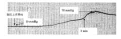
同条件でイオンビーム照射試料を4週間置換したウサギを麻酔下にて、頭皮を剥離して、置換部分を露出させた。その後、置換部位とは異なる頭蓋骨部を直径約 1 mmの穴を開け、脳圧加圧用カテーテルを挿入した。他のもう1つの部位に脳圧測定用カテーテルを挿入した。カテーテルをシリンジポンプに接続し、脳圧を上昇させて、人工硬膜およびそれを固定しているフィブリングルーを観察して脳圧による剥離が生じているか観察した。加圧は50 mlの注射器をシリンジポンプに装着して、1 ml/分の速度で加圧した。図3に時間と脳圧の関係を示す。加圧前の脳圧は約 10mmHgであった。加圧後1分半で脳圧が上昇し始めた。初期は緩やかに脳圧は上昇したが加圧後3分から急激に脳圧は上昇した。脳圧は70mmHgまで達したが、フィブリングルーで固体化されたイオンビーム照射した人工硬膜は加圧中、脳髄液の漏出は見られず、良好な密封性を示した。
【0034】
(4)生体外での接着力評価試験
イオンビーム照射したePTFEのフィブリングルーによる接着を評価するために生体外で加圧装置を用いて評価した。
接着評価は同じ条件のイオンビーム照射面同士をフィブリングルーで接着させ、その接着強度を測定した。ePTFEへのイオンビーム照射はHe+, Ne+, Ar+, Kr+イオンを加速エネルギー150 keVで照射量は1×10¹⁴ ,5×1014,1×1015 イオン/cm2とした。図4に圧力耐久装置を示す。
【0035】
未照射およびイオンビーム照射ePTFE同士のの接着は直径15 mmに切断した円形のイオンビーム照射試料を直径10 mmの円形の穴を開けた正方形の試料にフィブリングルーで両イオンブーム照射面同士を接着した。Cの孔周辺にフィブリノゲン溶液を塗布し、そこにトロンビン溶液を滴下するとともにBをのせ、2液を混合しながらBとCを密着させる。接着部分は直径15 mmの円と10mmの円の重なる部分でのフィブリングルーによる接着部分である。
【0036】
アクリル製円筒(D)の上部にフィブリングルーで接着したePTFE(B+C)を固定する。30分後、墨汁によって着色した水を満たした円柱Dの上に接着したBとCをのせ、さらに上から内径17 mmの穴を有するアクリル版Aで挟み押さえ込み固定する。
この状態で、加圧用ポートFから円柱Dに 60ml/h の割合で水を注入する。その間の円柱D内の圧力をモニター用ポートEから圧力センサーを用いて、BとCの接着部分からの水の漏出が生じるまで圧力を測定する。
【0037】
図5に加圧試験による漏出臨界圧力とイオン種、照射量の関係を示す。未照射ePTFE同士の接着では約20 mmHgの圧力で漏出した。それに対してイオンビーム照射したePTFE同士の接着では顕著に耐圧力が上昇した。特にAr+イオンビーム照射した試料で照射量1×10¹⁴ ions/cm2の条件では100 mmHgの圧力まで臨界圧力は上昇し、良好な接着性を示した。
【0038】
【発明の効果】
本発明により、人工血管又は人工硬膜などの生体内埋入材料と生体組織接着剤との親和性を向上させることが可能になった。本発明の高分子材料を人工血管又は人工硬膜などの生体内埋入材料として用いることにより、血液又は髄液の漏出を防ぐことができる。
【図面の簡単な説明】
【図1】図1は、未処理ePTFEとフィブリングルーとの相互作用を示す。ウサギに置換し、1ヶ月目を示す。未照射ePTFEとフィブリングルーは接着しない
【図2】図2は、イオンビーム照射処理ePTFEとフィブリングルーとの相互作用を示す。ウサギに置換し、1ヶ月目を示す。イオンビーム照射ePTFEとフィブリングルーは細胞を介して接着する。
【図3】図3は、イオンビーム照射硬膜を置換したウサギの脳圧上昇カーブを示す。
【図4】図4は、圧力耐久試験用装置を示す。
【図5】図5は、加圧試験による漏出臨界圧力とイオン種、照射量の関係を示す。凡例の0は未照射試料を示す。[0001]
BACKGROUND OF THE INVENTION
The present invention relates to a biological repair material having an affinity for a biological tissue adhesive, that is, a polymer material for use in combination with a biological tissue adhesive. More specifically, the present invention relates to a polymer material for use in combination with a biological tissue adhesive, wherein at least a part of the surface is modified by ion bombardment so that the affinity with the biological tissue adhesive is increased. The present invention relates to a polymer material having improved properties and a method for producing the same.
[0002]
[Prior art]
Among the three layers of membranes (buffy coat, arachnoid membrane, dura mater) that are inside the skull and protect the brain parenchyma, the dura mater is the hardest and is present in the outermost layer of the three layers and is also the inner periosteum of the skull. During neurosurgery, the dura is often excised, resulting in dural defects. In addition, primary suturing may be difficult due to the spontaneous contraction of the dura mater itself. Closing with the dura mater open may cause cerebrospinal fluid leakage, causing intracranial infections, causing adhesions between the brain parenchyma and bone or subcutaneous tissue, resulting in local neurological symptoms, and epileptic seizures. It may cause serious complications such as focusing. Therefore, strict suturing is required so that there is no gap in the dura when closing. For this reason, when a deficiency arises in the dura mater or primary suturing becomes difficult, it is necessary to completely sew so as not to generate a gap by using any supplementary material.
[0003]
Which filling material should be used to fill dural defects is a problem that has long plagued neurosurgeons for a long time. Although there were times when the artifact was initially used, there were problems with biocompatibility, ease of use, etc., and none of them lasted long. Autologous fascia is the most widely used from the beginning to the present day, but there are many problems such as fascia defects at the excision site and easy adhesion to the brain. The human dry dura mater is a dura mater filling material obtained by subjecting the dura mater collected from the cadaver to radiation treatment and the like, and has been the most excellent so far. However, there is a possibility that a prion, which is the cause of Creutzfeldt-Jakob disease, may be present in the dura mater, and a case has been reported that infection of Creutzfeldt-Jakob disease occurs through the human dura mater. In 1998, its use was completely banned.
[0004]
Currently, only ePTFE (expanded polytetrafluoroethylene) approved by the Ministry of Health and Welfare can be used as a dural replacement material in addition to the self fascia. Since ePTFE is a polymer material, it has no adhesiveness to living organisms. This property is excellent in that it does not cause adhesions with the brain. On the other hand, since cerebrospinal fluid leaks from the needle hole due to poor contractility, it is necessary to perform suturing using a special suture. In addition, since there is no bioadhesiveness, there is a high possibility of cerebrospinal fluid leakage from the gap between the stitched surfaces. At the same time, since there is no adhesiveness with the surrounding tissue, there is a high possibility that it becomes a simple skeleton material. Many attempts have been made on how to use ePTFE so far, but they all use ePTFE as a skeletal material and wait for a fibrous tissue coating to form around them. .
[0005]
On the other hand, as a method of treating the artificial material surface with ions, a method by plasma treatment is known (Japanese Patent Application No. 10-302170). This method is a method of improving adhesion by a method of modifying the surface. The plasma treatment layer formed by the plasma treatment method is unstable in the living body and has a risk of being decomposed and peeled off with time. In vivo, it is necessary to maintain a stable cell adhesion layer over a long period of time. In the method using plasma treatment, particularly in the case of an artificial dura mater, although it adheres to the skull contact surface in the initial stage of replacement, there is a risk of peeling after a long period of time.
[0006]
There is also a report of improving the antibacterial property by modifying the surface layer of the in-vivo implant material using ions having higher energy than this plasma processing method (Japanese Patent Application No. Hei 5-148994). The purpose of this method is mainly to reduce the infectivity of the implant material.
[0007]
[Problems to be solved by the invention]
In order to fix medical materials used for surgical operations in vivo, in particular artificial blood vessels, artificial dura mater, patch materials for repairing the heart or blood vessels, to tissues for treatment, a method of fixing these materials and the living body by anastomosis Is adopted. However, when these medical materials are anastomosed with a surgical thread, blood or cerebrospinal fluid leaks from the material portion through which the needle passes. For this reason, normally, blood leakage is prevented by inducing blood coagulation by pressing the affected area or by using a biological tissue adhesive called fibrin glue.
[0008]
The present invention provides means capable of preventing blood or cerebrospinal fluid from leaking more quickly by improving the affinity between a living tissue adhesive such as an artificial blood vessel or an artificial dura mater and a living tissue adhesive. For the purpose.
[0009]
[Means for Solving the Problems]
As a result of intensive studies to solve the above problems, the present inventors have found that Ne ion beam irradiated ePTFE has a higher affinity with a biological tissue adhesive than unirradiated ePTFE. It came to be completed.
[0010]
That is, according to the present invention, a polymer material comprising a polymer material containing carbon or silicon as a constituent element, used in combination with a biological tissue adhesive, and having at least a part of the surface modified by ion bombardment. Is provided.
[0011]
Preferably, the biological tissue adhesive is a fibrin glue.
Preferably, the polymer material containing carbon or silicon as a constituent element is expanded polytetrafluoroethylene (ePTFE), polylactic acid, or polyglactin.
Preferably, the modification by ion bombardment is performed by performing ion implantation in a range where the dose amount φ is 1 × 10¹² ≦ φ ≦ 1 × 10¹⁶ ions / cm2 .
Preferably, the polymer material of the present invention is used as an artificial dura mater, an artificial blood vessel, a heart / blood vessel patch or a suture.
[0012]
According to another aspect of the present invention, a dose amount φ is 1 × 10¹² ≦ φ ≦ 1 × 10¹⁶ ions / cm2 on at least a part of the surface of a polymer material containing carbon or silicon as a constituent element. A method for producing a polymer material to be used in combination with a biological tissue adhesive is provided.
[0013]
According to still another aspect of the present invention, the dose amount φ is 1 × 10¹² ≦ φ ≦ 1 × 10¹⁶ ions / cm2 on at least a part of the surface of the polymer material containing carbon or silicon as a constituent element. A method for improving the affinity of the polymer material with a biological tissue adhesive, characterized by performing ion implantation in a range.
[0014]
DETAILED DESCRIPTION OF THE INVENTION
Hereinafter, embodiments of the present invention will be described in detail.
The polymer material of the present invention is for use in combination with a biological tissue adhesive, is composed of a polymer material containing carbon or silicon as a constituent element, and at least a part of the surface is modified by ion bombardment. It is characterized by.
[0015]
In the present invention, the affinity of the biological tissue adhesive can be improved by irradiating a polymer material containing carbon or silicon as a constituent element with an ion beam, thereby preventing blood leakage and cerebrospinal fluid leakage. The present invention further relates to a surface treatment method for improving affinity with a biological tissue adhesive by irradiating the above-described polymer material with an ion beam.
[0016]
When treating an injured blood vessel using an artificial blood vessel in the treatment of an abdominal aortic aneurysm or the like, the biological blood vessel and the artificial blood vessel are stitched together with a surgical suture to join the living blood vessel. However, an artificial blood vessel made of polyester or fluorine compound leaks blood from the needle hole even if the same kind of suture is used.
[0017]
When the heart or blood vessel lesion is removed, it is necessary to repair the defect site. Similarly, when this repair material is sewed onto a blood vessel, heart, etc., blood and tissue fluid from the needle hole leak out in the same manner. In the field of brain surgery, dural defects occur when craniotomy is performed due to brain tumors, subarachnoid hemorrhage, or traffic accidents. Artificial dura is used to compensate for this dural deficiency, but cerebrospinal fluid leaks from the needle hole like the artificial blood vessel.
[0018]
In many cases, leakage of blood and cerebrospinal fluid is prevented by using a biological tissue adhesive called fibrin glue to prevent these blood leaks and cerebrospinal fluid leaks. However, among artificial blood vessel materials and artificial dura mater materials, especially fluorine compound materials, adhesion to the fibrin glue is extremely poor, and leakage of blood and cerebrospinal fluid is insufficient.
[0019]
In the present invention, these artificial materials can be irradiated with an ion beam on the material surface layer by an ion implantation method to improve the adhesion to the fibrin glue. This ion beam treatment can be performed not only on an artificial dura mater, an artificial blood vessel, or a heart / blood vessel patch, but can also be performed on a surface layer of a suture thread that binds these to a living body. By modifying the surface layer of the suture with a beam, the adhesion between the artificial material and the fibrin glue is improved.
[0020]
The polymer material having carbon or silicon as a constituent element used in the present invention is not particularly limited as long as the material is biocompatible and easy to operate, and any material can be used. For example, a fluororesin molding such as expanded polytetrafluoroethylene (ePTFE) or a silicon compound such as silicone can be used. Preferred polymer materials in the present invention include expanded polytetrafluoroethylene (ePTFE) or biodegradable polymers (for example, polylactic acid or polyglactin), particularly expanded polytetrafluoroethylene (ePTFE). Is preferred.
[0021]
At least a part of the surface of the polymer material of the present invention is modified by ion bombardment. Examples of ion species to be implanted include H+ , He+ , C+ , N+ , Ne+ , Na+ , N2+ , O2+ , Ar+ , Kr+ , and Xe+. It is not particularly limited to these as long as it does not inhibit the affinity with the biological tissue adhesive. Preferred are Ne+ , Ar+ , Kr+ , Xe+ and the like.
[0022]
The dose amount (irradiation amount) φ is preferably in the range of 1 × 10¹² ≦ φ ≦ 1 × 10¹⁶ ions / cm2 . If it is lower than 1012 ions / cm2, the effect of remarkably improving the affinity with the biological tissue adhesive will be small, and if it is higher than 1016 ions / cm2 , the polymer material will be easily broken, which is not preferable. More preferably, the dose amount φ is in the range of 1 × 10¹³ ≦ φ ≦ 1 × 10¹⁵ ions / cm2 .
[0023]
Regarding the ion acceleration energy, the energy transfer mechanism is considered to be different depending on the height, but practically, it can be set in the range of several tens keV to several MeV, and preferably about 50 keV to 2 MeV. .
[0024]
The beam current density is preferably set in a range not exceeding about 0.5 μA / cm2 . This is because when the beam current density is excessive, the temperature of the target polymer material is excessively increased, the polymer material itself is deteriorated, and the affinity with the biological tissue adhesive may be decreased.
[0025]
In the present invention, ion implantation may be mentioned as a means for giving ion bombardment. In the ion implantation, the reaction itself is limited to the interaction between the ion beam and the material to be implanted (target material). In addition, by selecting the ion incident energy, ions can be embedded at an arbitrary depth from the surface, which is extremely excellent in controllability. This is a feature not found in plasma processing. Although the implanted ions have a mechanistic difference that the electron stopping power works at the early stage of diffusion for ions with relatively light mass, and the nuclear stopping power works for ions with relatively heavy mass from the beginning. It causes heating by lattice vibration to the polymer material (thermal non-equilibrium state), causing melting, amorphization and the like.
[0026]
The polymer material of the present invention is used in combination with a biological tissue adhesive. Preferable examples of the biological tissue adhesive include fibrin glue (fibrin glue) or a cyanoacrylate instantaneous adhesive which is a polymer adhesive. Conventionally, in a surgical operation, sutures have been performed with a thread and a needle such as silk or cat gut, and an adhesive is used for this. In the conventional suturing method, it is often difficult to sew small blood vessels and repair blood vessels. In addition, it generally takes time for suturing and is difficult to look back. A biological tissue adhesive is adopted as a method.
[0027]
Fibrin glue is composed of fibrinogen lyophilized powder, fibrinogen lysate, thrombin lyophilized powder, and thrombin lysate. Fibrinogen lyophilized powder is dissolved in a fibrinogen solution to prepare solution A. Thrombin lyophilized powder is dissolved in a thrombin solution to prepare solution B. Equal volumes of both dissolved solutions are applied in layers or mixed to the adhesion site. Fibrin glue is a physiological tissue adhesive that utilizes the final stage of blood coagulation, and the fibrinogen contained in it becomes a soluble fibrin clot by the action of thrombin, and blood coagulation factor XIII activated by thrombin in the presence of calcium ions It becomes a urea-insoluble stable fibrin clot with physical strength and adheres and closes the tissue. Within this stabilized fibrin clot, fibroblasts proliferate, collagen fibers and granulation matrix components are produced, and then heal through tissue repair. A specific example of fibrin glue is Bolhir (trade name) (Chemical and Serum Therapy Research Institute).
[0028]
Surgical procedures used as fibrin glue include the closing of bleeding wound surfaces, fixation of fractured pieces, peripheral nerve and microvascular anastomosis, tendon adhesion or reinforcement of tendon sutures, parenchymal organs for the purpose of bonding biological tissues together And the like. It is also used to prevent leakage of cerebrospinal fluid and blood from the needle hole when an artificial object such as an artificial dura mater or an artificial blood vessel is anastomosed with a living tissue. Similarly, it is also used for the purpose of preventing blood leaking from a needle hole in a heart patch and a blood vessel patch used for repairing a heart defect, repairing a blood vessel defect. In particular, in the case of ePTFE, the problem of poor adhesion with the fibrin glue was a problem, but at least a part of the surface was modified by ion bombardment according to the present invention, thereby eliminating the problem of adhesion with the fibrin glue. .
The present invention will be described more specifically with reference to the following examples, but the present invention is not limited to the examples.
[0029]
【Example】
(1) Ion irradiation treatment Male Japanese white rabbits weighing 2.5 to 3.0 kg were used in the experiment. Under general anesthesia with pentobarpital, the scalp was incised along the coronal suture to expose the skull. The sample used was expanded polytetrafluoroethylene (ePTFE) that was irradiated with an ion beam (Ne+ , 150 keV, 5 × 10¹⁴ ions / cm2 ) using a RIKEN 200 KeV ion implanter. With the surface periosteum of the skull completely removed, the skull was further removed to expose the dura mater. A part of the dura was drilled with a few millimeters. The sample was placed on the dura mater with the non-irradiated surface, and the ion beam irradiated surface was placed on the scalp side. Thereafter, the biological tissue adhesive (fibrin glue; trade name Bolhir, Institute for Chemical and Serum Therapy) was dropped on the ion beam irradiation surface and then fixed to the remaining skull. As a comparison, the same operation was performed on a sample not irradiated with ions. After the fibrin glue was dropped, the scalp was anastomosed to cover the affected area.
[0030]
(2) Observation (a) Observation by visual observation The ion beam irradiated surface showed good adhesion to the surrounding bone tissue within a few minutes after the fibrin glue was dropped. On the other hand, the untreated expanded polytetrafluoroethylene had such a weak adhesive force that it moved only by applying a weak force with tweezers.
[0031]
(B) Two weeks after embedding and transplanting the observation sample by histological examination, the rabbit was sacrificed using Nembutal, and each surrounding tissue was taken out as a lump and fixed with 10% buffer formalin. The reason why the surrounding tissue was removed was to avoid separation of the ePTFE and the tissue because the fibrin glue and the ePTFE were not adhered to each other on the non-ion beam treated surface. The sample placed on the dura mater was decalcified from the skull, embedded in paraffin, stained with hematoxylin / eosin, and Masson trichrome, and observed under a microscope.
[0032]
As a result, no adhesion with the fibrin glue was observed on the untreated ePTFE surface at the second week (FIG. 1). On the other hand, the ion beam-treated ePTFE had extremely good adhesion to the fibrin glue (Fig. 2).
[0033]
(3) Brain pressure application experiment Under the same conditions, a rabbit in which the ion beam irradiated sample was replaced for 4 weeks was anesthetized, and the scalp was peeled off to expose the replacement part. After that, a hole with a diameter of about 1 mm was made in the skull part different from the replacement site, and a brain pressure catheter was inserted. A brain pressure measurement catheter was inserted into another site. The catheter was connected to a syringe pump, the brain pressure was increased, and the artificial dura mater and the fibrin glue that fixed the artificial dura mater were observed to observe whether peeling due to brain pressure occurred. The pressurization was performed by attaching a 50 ml syringe to a syringe pump and pressurizing at a rate of 1 ml / min. FIG. 3 shows the relationship between time and brain pressure. The brain pressure before pressurization was about 10 mmHg. The brain pressure began to rise 1 and a half minutes after pressurization. In the initial stage, the brain pressure gradually increased, but the brain pressure suddenly increased 3 minutes after the pressurization. Although the brain pressure reached 70 mmHg, the artificial dura mater irradiated with an ion beam solidified by fibrin glue showed no leakage of cerebrospinal fluid during pressurization and showed good sealing performance.
[0034]
(4) Evaluation test of adhesion force in vitro In order to evaluate the adhesion of ePTFE irradiated with an ion beam by a fibrin glue, it was evaluated in vitro using a pressure device.
For the adhesion evaluation, ion beam irradiated surfaces under the same conditions were adhered to each other with a fibrin glue, and the adhesion strength was measured. The ePTFE was irradiated with He+ , Ne+ , Ar+ , and Kr+ ions at an acceleration energy of 150 keV and an irradiation dose of 1 × 10¹⁴ , 5 × 10¹⁴ , and 1 × 10¹⁵ ions / cm2 . FIG. 4 shows a pressure durability device.
[0035]
Adhesion between non-irradiated and ion beam irradiated ePTFE is achieved by bonding a circular ion beam irradiated sample cut to a diameter of 15 mm to a square sample with a 10 mm diameter circular hole and bonding both ion boom irradiated surfaces with a fibrin glue. did. A fibrinogen solution is applied around the pores of C, and a thrombin solution is added dropwise thereto and B is placed thereon, and B and C are brought into close contact while mixing the two solutions. The bonded part is a bonded part by a fibrin glue at the overlapping part of a circle with a diameter of 15 mm and a circle with a diameter of 10 mm.
[0036]
EPTFE (B + C) bonded with fibrin glue is fixed to the top of the acrylic cylinder (D). After 30 minutes, the adhered B and C are placed on a cylinder D filled with water colored with ink, and are further clamped and fixed by an acrylic plate A having an inner diameter of 17 mm from above.
In this state, water is injected from the pressurizing port F into the cylinder D at a rate of 60 ml / h. In the meantime, the pressure in the cylinder D is measured from the monitoring port E using a pressure sensor until water leaks from the bonded portion of B and C.
[0037]
FIG. 5 shows the relationship between the leakage critical pressure, the ion species, and the irradiation dose in the pressurization test. In adhesion between unirradiated ePTFEs, leakage occurred at a pressure of about 20 mmHg. On the other hand, the pressure resistance increased remarkably when bonding ePTFE irradiated with ion beams. In particular, in the sample irradiated with Ar + ion beam, the critical pressure increased to a pressure of 100 mmHg under the condition of an irradiation dose of 1 × 10¹⁴ ions / cm2 , and good adhesion was exhibited.
[0038]
【The invention's effect】
According to the present invention, it has become possible to improve the affinity between a living tissue adhesive such as an artificial blood vessel or an artificial dura mater and a living tissue adhesive. By using the polymer material of the present invention as an in vivo material such as an artificial blood vessel or an artificial dura mater, leakage of blood or cerebrospinal fluid can be prevented.
[Brief description of the drawings]
FIG. 1 shows the interaction between untreated ePTFE and fibrin glue. Replaced with rabbits, showing the first month. Unirradiated ePTFE and fibrin glue do not adhere. FIG. 2 shows the interaction between ion beam irradiated ePTFE and fibrin glue. Replaced with rabbits, showing the first month. Ion beam irradiated ePTFE and fibrin glue adhere through cells.
FIG. 3 shows a brain pressure increase curve of a rabbit in which an ion beam irradiated dura is replaced.
FIG. 4 shows a pressure durability test apparatus.
FIG. 5 shows the relationship between the leakage critical pressure, the ion species, and the irradiation dose in the pressurization test.
Claims (4)
Translated fromJapanesePriority Applications (5)
| Application Number | Priority Date | Filing Date | Title |
|---|---|---|---|
| JP2002253322AJP4445697B2 (en) | 2002-08-30 | 2002-08-30 | Biological repair material having affinity with biological tissue adhesive |
| EP20030797534EP1535632B1 (en) | 2002-08-30 | 2003-08-29 | Biological repair material compatible with biological tissue adhesive |
| PCT/JP2003/011048WO2004026355A1 (en) | 2002-08-30 | 2003-08-29 | Biological repair material compatible with biological tissue adhesive |
| US10/525,724US7722934B2 (en) | 2002-08-30 | 2003-08-29 | Biological repair material compatible with biological tissue adhesive |
| AT03797534TATE518550T1 (en) | 2002-08-30 | 2003-08-29 | BIOLOGICAL TISSUE ADHESIVE COMPATIBLE BIOLOGICAL REPAIR MATERIAL |
Applications Claiming Priority (1)
| Application Number | Priority Date | Filing Date | Title |
|---|---|---|---|
| JP2002253322AJP4445697B2 (en) | 2002-08-30 | 2002-08-30 | Biological repair material having affinity with biological tissue adhesive |
Publications (2)
| Publication Number | Publication Date |
|---|---|
| JP2004089361A JP2004089361A (en) | 2004-03-25 |
| JP4445697B2true JP4445697B2 (en) | 2010-04-07 |
Family
ID=32024502
Family Applications (1)
| Application Number | Title | Priority Date | Filing Date |
|---|---|---|---|
| JP2002253322AExpired - LifetimeJP4445697B2 (en) | 2002-08-30 | 2002-08-30 | Biological repair material having affinity with biological tissue adhesive |
Country Status (5)
| Country | Link |
|---|---|
| US (1) | US7722934B2 (en) |
| EP (1) | EP1535632B1 (en) |
| JP (1) | JP4445697B2 (en) |
| AT (1) | ATE518550T1 (en) |
| WO (1) | WO2004026355A1 (en) |
Cited By (2)
| Publication number | Priority date | Publication date | Assignee | Title |
|---|---|---|---|---|
| WO2019026341A1 (en)* | 2017-08-01 | 2019-02-07 | 株式会社多磨バイオ | Medical sheet |
| JP2019025824A (en)* | 2017-08-01 | 2019-02-21 | 株式会社多磨バイオ | Medical sheet |
Families Citing this family (10)
| Publication number | Priority date | Publication date | Assignee | Title |
|---|---|---|---|---|
| JP2005034256A (en)* | 2003-07-17 | 2005-02-10 | Institute Of Physical & Chemical Research | Blood vessel wall repair material |
| JP2007020590A (en)* | 2003-08-19 | 2007-02-01 | Institute Of Physical & Chemical Research | Aneurysm treatment material |
| JP2006230639A (en) | 2005-02-24 | 2006-09-07 | Institute Of Physical & Chemical Research | Catheters with modified living body contact parts |
| JP2006263144A (en)* | 2005-03-24 | 2006-10-05 | Mcrotech Kk | Soft biotissue substitute grafting material and its production method |
| CN102634978A (en)* | 2011-02-11 | 2012-08-15 | 傅亚 | Preparation method of fibrin glue reinforced polylactic acid fiber |
| US9526503B2 (en)* | 2013-08-12 | 2016-12-27 | W. L. Gore & Associates, Inc. | Lumbar ostia occlusion devices and methods of deploying the same |
| CN103933609B (en)* | 2014-02-28 | 2016-06-08 | 武汉杨森生物技术有限公司 | One-time formed polyurethane artificial blood vessel and preparation method thereof |
| WO2018193996A1 (en)* | 2017-04-17 | 2018-10-25 | 学校法人 慶應義塾 | Medical tube connection device |
| JP6316496B1 (en)* | 2017-11-24 | 2018-04-25 | 株式会社多磨バイオ | Artificial pericardium sheet |
| US20230137248A1 (en)* | 2021-10-28 | 2023-05-04 | Kismet Technologies Llc | Therapeutic article of manufacture with nanoparticles to promote wound healing and/or antimicrobial infection control |
Family Cites Families (11)
| Publication number | Priority date | Publication date | Assignee | Title |
|---|---|---|---|---|
| JP2930329B2 (en) | 1989-09-28 | 1999-08-03 | ソニー株式会社 | Antithrombotic material |
| JPH0549689A (en)* | 1991-08-20 | 1993-03-02 | Sony Corp | Cell adhesive material and method for producing the same |
| JPH05208042A (en)* | 1992-01-30 | 1993-08-20 | Ajinomoto Co Inc | Adhesive agent |
| JPH07500A (en) | 1993-06-21 | 1995-01-06 | Sumitomo Electric Ind Ltd | Bioimplant material and method of manufacturing the same |
| US6051751A (en)* | 1995-01-20 | 2000-04-18 | Spire Corporation | Arthroplasty process for securely anchoring prostheses to bone, and arthroplasty products therefor |
| JP3536051B2 (en)* | 1996-05-17 | 2004-06-07 | 独立行政法人理化学研究所 | Antithrombotic material and method for producing the same |
| US5891192A (en)* | 1997-05-22 | 1999-04-06 | The Regents Of The University Of California | Ion-implanted protein-coated intralumenal implants |
| WO1999025782A2 (en)* | 1997-11-17 | 1999-05-27 | Haemacure Corporation | Fibrin sealants or adhesives comprising a hyaluronic acid derivative material |
| JP2000129015A (en) | 1998-10-23 | 2000-05-09 | Nitto Denko Corp | Surface modification method for fluororesin molding |
| AU2001250875A1 (en)* | 2000-03-30 | 2001-10-15 | Brennen Medical, Inc. | Anti-microbial and immunostimulating composition |
| JP5505752B2 (en)* | 2001-04-23 | 2014-05-28 | 独立行政法人理化学研究所 | Artificial dura mater having cell adhesion and method for producing the same |
- 2002
- 2002-08-30JPJP2002253322Apatent/JP4445697B2/ennot_activeExpired - Lifetime
- 2003
- 2003-08-29WOPCT/JP2003/011048patent/WO2004026355A1/enactiveApplication Filing
- 2003-08-29ATAT03797534Tpatent/ATE518550T1/ennot_activeIP Right Cessation
- 2003-08-29EPEP20030797534patent/EP1535632B1/ennot_activeExpired - Lifetime
- 2003-08-29USUS10/525,724patent/US7722934B2/ennot_activeExpired - Fee Related
Cited By (7)
| Publication number | Priority date | Publication date | Assignee | Title |
|---|---|---|---|---|
| WO2019026341A1 (en)* | 2017-08-01 | 2019-02-07 | 株式会社多磨バイオ | Medical sheet |
| JP2019025824A (en)* | 2017-08-01 | 2019-02-21 | 株式会社多磨バイオ | Medical sheet |
| JP2019025280A (en)* | 2017-08-01 | 2019-02-21 | 株式会社多磨バイオ | Medical sheet |
| CN111032098A (en)* | 2017-08-01 | 2020-04-17 | 株式会社多磨生物科技 | Medical sheet |
| DE112018003956T5 (en) | 2017-08-01 | 2020-05-14 | Tama Bio Inc. | Medical edition |
| US11246960B2 (en) | 2017-08-01 | 2022-02-15 | Tama Bio Inc. | Medical sheet |
| CN111032098B (en)* | 2017-08-01 | 2022-07-26 | 株式会社多磨生物科技 | Medical sheet and method for producing same |
Also Published As
| Publication number | Publication date |
|---|---|
| US7722934B2 (en) | 2010-05-25 |
| EP1535632A1 (en) | 2005-06-01 |
| EP1535632A4 (en) | 2008-09-17 |
| ATE518550T1 (en) | 2011-08-15 |
| JP2004089361A (en) | 2004-03-25 |
| WO2004026355A1 (en) | 2004-04-01 |
| US20060155041A1 (en) | 2006-07-13 |
| EP1535632B1 (en) | 2011-08-03 |
Similar Documents
| Publication | Publication Date | Title |
|---|---|---|
| US12226086B2 (en) | Tissue repair and sealing devices having a detachable graft and clasp assembly and methods for the use thereof | |
| JP4537998B2 (en) | Composition for repairing and regenerating human dura mater | |
| US8834864B2 (en) | Methods for repairing and regenerating human dura mater | |
| US10058416B2 (en) | Tissue marking implant | |
| US6214045B1 (en) | Bioabsorbable breast implant | |
| US6319712B1 (en) | Biohybrid articular surface replacement | |
| JP4445697B2 (en) | Biological repair material having affinity with biological tissue adhesive | |
| JP5702515B2 (en) | Nerve regeneration induction tube | |
| TW200924803A (en) | Use of a regenerative biofunctional collagen biomatrix for treating visceral or parietal defects | |
| JP2008510063A (en) | Water-swellable copolymers and articles and coatings made therefrom | |
| US6872759B2 (en) | Artificial dura mater having cell adhesiveness and a process for producing the same | |
| Ito et al. | Comparative study of fibrin and chemical synthetic sealant on dural regeneration and brain damage | |
| JP2002204826A (en) | Biomedical substitute artificial membrane | |
| Zhang | Applications of fibrin tissue sealant | |
| RU175525U1 (en) | COMBINED IMPLANT FOR RECONSTRUCTION OF SKULL BASIS DEFECTS | |
| CA2358567A1 (en) | Tissue adhesive for treating vigorously bleeding surfaces | |
| CZ38337U1 (en) | An absorbable composite bandage from polymer reinforcement and collagen matrix | |
| Géza | Polymers of Natural Origin as Biomaterials. 1. Fibrin | |
| Nunamaker | Alginate as a novel material for duraplasty: Investigation of the material properties, in vivo stability, and sealing capabilities | |
| HOSSAIN | DESIGN AND DEVELOPMENT OF A MICROANASTOMOSIS DEVICE | |
| JP2012066096A (en) | Artificial dura mater having cell adhesion property and method of manufacturing the same |
Legal Events
| Date | Code | Title | Description |
|---|---|---|---|
| A711 | Notification of change in applicant | Free format text:JAPANESE INTERMEDIATE CODE: A712 Effective date:20031201 | |
| RD02 | Notification of acceptance of power of attorney | Free format text:JAPANESE INTERMEDIATE CODE: A7422 Effective date:20040419 | |
| A621 | Written request for application examination | Free format text:JAPANESE INTERMEDIATE CODE: A621 Effective date:20050805 | |
| A131 | Notification of reasons for refusal | Free format text:JAPANESE INTERMEDIATE CODE: A131 Effective date:20090519 | |
| A521 | Request for written amendment filed | Free format text:JAPANESE INTERMEDIATE CODE: A523 Effective date:20090713 | |
| A131 | Notification of reasons for refusal | Free format text:JAPANESE INTERMEDIATE CODE: A131 Effective date:20090929 | |
| A521 | Request for written amendment filed | Free format text:JAPANESE INTERMEDIATE CODE: A523 Effective date:20091127 | |
| TRDD | Decision of grant or rejection written | ||
| A01 | Written decision to grant a patent or to grant a registration (utility model) | Free format text:JAPANESE INTERMEDIATE CODE: A01 Effective date:20100105 | |
| A01 | Written decision to grant a patent or to grant a registration (utility model) | Free format text:JAPANESE INTERMEDIATE CODE: A01 | |
| A61 | First payment of annual fees (during grant procedure) | Free format text:JAPANESE INTERMEDIATE CODE: A61 Effective date:20100118 | |
| R150 | Certificate of patent or registration of utility model | Ref document number:4445697 Country of ref document:JP Free format text:JAPANESE INTERMEDIATE CODE: R150 Free format text:JAPANESE INTERMEDIATE CODE: R150 | |
| FPAY | Renewal fee payment (event date is renewal date of database) | Free format text:PAYMENT UNTIL: 20130122 Year of fee payment:3 | |
| FPAY | Renewal fee payment (event date is renewal date of database) | Free format text:PAYMENT UNTIL: 20160122 Year of fee payment:6 | |
| R250 | Receipt of annual fees | Free format text:JAPANESE INTERMEDIATE CODE: R250 | |
| S533 | Written request for registration of change of name | Free format text:JAPANESE INTERMEDIATE CODE: R313533 | |
| R350 | Written notification of registration of transfer | Free format text:JAPANESE INTERMEDIATE CODE: R350 | |
| R250 | Receipt of annual fees | Free format text:JAPANESE INTERMEDIATE CODE: R250 | |
| R250 | Receipt of annual fees | Free format text:JAPANESE INTERMEDIATE CODE: R250 | |
| R250 | Receipt of annual fees | Free format text:JAPANESE INTERMEDIATE CODE: R250 | |
| R250 | Receipt of annual fees | Free format text:JAPANESE INTERMEDIATE CODE: R250 | |
| R250 | Receipt of annual fees | Free format text:JAPANESE INTERMEDIATE CODE: R250 | |
| R250 | Receipt of annual fees | Free format text:JAPANESE INTERMEDIATE CODE: R250 | |
| R250 | Receipt of annual fees | Free format text:JAPANESE INTERMEDIATE CODE: R250 | |
| EXPY | Cancellation because of completion of term |