JP4417255B2 - Device for treating vascular obstacles - Google Patents
Device for treating vascular obstaclesDownload PDFInfo
- Publication number
- JP4417255B2 JP4417255B2JP2004532874AJP2004532874AJP4417255B2JP 4417255 B2JP4417255 B2JP 4417255B2JP 2004532874 AJP2004532874 AJP 2004532874AJP 2004532874 AJP2004532874 AJP 2004532874AJP 4417255 B2JP4417255 B2JP 4417255B2
- Authority
- JP
- Japan
- Prior art keywords
- sheet
- vascular
- shape
- materials
- aneurysm
- Prior art date
- Legal status (The legal status is an assumption and is not a legal conclusion. Google has not performed a legal analysis and makes no representation as to the accuracy of the status listed.)
- Expired - Lifetime
Links
Images


Classifications
- A—HUMAN NECESSITIES
- A61—MEDICAL OR VETERINARY SCIENCE; HYGIENE
- A61B—DIAGNOSIS; SURGERY; IDENTIFICATION
- A61B17/00—Surgical instruments, devices or methods
- A61B17/12—Surgical instruments, devices or methods for ligaturing or otherwise compressing tubular parts of the body, e.g. blood vessels or umbilical cord
- A61B17/12022—Occluding by internal devices, e.g. balloons or releasable wires
- A61B17/12099—Occluding by internal devices, e.g. balloons or releasable wires characterised by the location of the occluder
- A61B17/12109—Occluding by internal devices, e.g. balloons or releasable wires characterised by the location of the occluder in a blood vessel
- A61B17/12113—Occluding by internal devices, e.g. balloons or releasable wires characterised by the location of the occluder in a blood vessel within an aneurysm
- A—HUMAN NECESSITIES
- A61—MEDICAL OR VETERINARY SCIENCE; HYGIENE
- A61B—DIAGNOSIS; SURGERY; IDENTIFICATION
- A61B17/00—Surgical instruments, devices or methods
- A61B17/12—Surgical instruments, devices or methods for ligaturing or otherwise compressing tubular parts of the body, e.g. blood vessels or umbilical cord
- A61B17/12022—Occluding by internal devices, e.g. balloons or releasable wires
- A61B17/12131—Occluding by internal devices, e.g. balloons or releasable wires characterised by the type of occluding device
- A61B17/12168—Occluding by internal devices, e.g. balloons or releasable wires characterised by the type of occluding device having a mesh structure
- A61B17/12172—Occluding by internal devices, e.g. balloons or releasable wires characterised by the type of occluding device having a mesh structure having a pre-set deployed three-dimensional shape
- A—HUMAN NECESSITIES
- A61—MEDICAL OR VETERINARY SCIENCE; HYGIENE
- A61B—DIAGNOSIS; SURGERY; IDENTIFICATION
- A61B17/00—Surgical instruments, devices or methods
- A61B17/12—Surgical instruments, devices or methods for ligaturing or otherwise compressing tubular parts of the body, e.g. blood vessels or umbilical cord
- A61B17/12022—Occluding by internal devices, e.g. balloons or releasable wires
- A61B17/12131—Occluding by internal devices, e.g. balloons or releasable wires characterised by the type of occluding device
- A61B17/12181—Occluding by internal devices, e.g. balloons or releasable wires characterised by the type of occluding device formed by fluidized, gelatinous or cellular remodelable materials, e.g. embolic liquids, foams or extracellular matrices
- A61B17/1219—Occluding by internal devices, e.g. balloons or releasable wires characterised by the type of occluding device formed by fluidized, gelatinous or cellular remodelable materials, e.g. embolic liquids, foams or extracellular matrices expandable in contact with liquids
- A—HUMAN NECESSITIES
- A61—MEDICAL OR VETERINARY SCIENCE; HYGIENE
- A61B—DIAGNOSIS; SURGERY; IDENTIFICATION
- A61B17/00—Surgical instruments, devices or methods
- A61B17/12—Surgical instruments, devices or methods for ligaturing or otherwise compressing tubular parts of the body, e.g. blood vessels or umbilical cord
- A61B17/12022—Occluding by internal devices, e.g. balloons or releasable wires
- A61B2017/1205—Introduction devices
Landscapes
- Health & Medical Sciences (AREA)
- Surgery (AREA)
- Life Sciences & Earth Sciences (AREA)
- Heart & Thoracic Surgery (AREA)
- Molecular Biology (AREA)
- Vascular Medicine (AREA)
- Engineering & Computer Science (AREA)
- Biomedical Technology (AREA)
- Reproductive Health (AREA)
- Medical Informatics (AREA)
- Nuclear Medicine, Radiotherapy & Molecular Imaging (AREA)
- Animal Behavior & Ethology (AREA)
- General Health & Medical Sciences (AREA)
- Public Health (AREA)
- Veterinary Medicine (AREA)
- Neurosurgery (AREA)
- Surgical Instruments (AREA)
- Materials For Medical Uses (AREA)
Description
Translated fromJapanese外科手術では容易に到達されない脈管構造における障害物を治療するために、多くの低侵襲性又は非侵襲性のインターベンショナルな医療装置及び処置が用いられてきた。弱化又は閉塞した管壁を支持し、且つ流体の流れを可能にするために身体の内腔に植え込むのに適した医療装置がよく知られ、市販されている。このような装置の1つが、例えば脈管ステントである。ステントは、冠状動脈系、脳、泌尿器、胆管、食道、気管及び気管支などにおいて管壁を支え、また、管の開口を維持するために用いられ得る。 Many minimally invasive or non-invasive interventional medical devices and procedures have been used to treat obstacles in the vasculature that are not easily reached by surgery. Medical devices suitable for implantation in a body lumen to support weakened or occluded tube walls and allow fluid flow are well known and commercially available. One such device is, for example, a vascular stent. Stents can be used to support the vessel wall in the coronary arterial system, brain, urinary organs, bile ducts, esophagus, trachea, bronchi, etc., and maintain the opening of the vessel.
しかしながら、流体の流れを遮断することが必要な状況もある。例えば、脈管系における深刻な障害物の1つは、管壁の弱化した領域である動脈瘤であり、その管壁は膨隆又はバブルを生じて、隣接する管から突出する。動脈瘤は、治療しなければ破裂するまで膨張し続け、出血を生じることがある。従って、動脈瘤への流体の流れを遮断することが望ましい場合が多い。 However, there are situations where it is necessary to interrupt the fluid flow. For example, one serious obstruction in the vascular system is an aneurysm, a weakened area of the vessel wall, which bulges or bubbles and protrudes from an adjacent vessel. Aneurysms can continue to swell until they rupture without treatment, resulting in bleeding. Accordingly, it is often desirable to block fluid flow to the aneurysm.
このような障害物の治療のために用いられる装置は、血管閉塞(vaso−occlusive)装置と称されることがあり、一般に、カテーテル装置を用いて動脈瘤部位に配置される。血管閉塞装置は様々な形状を有することが可能であり、概して1つ以上の要素から形成される。これらの要素は、血流を遮断するための、配置された形状を有し、この形状は動脈瘤部位への配送中の形状とは異なる。 Devices used for the treatment of such obstacles are sometimes referred to as vaso-occlusive devices and are typically placed at the aneurysm site using a catheter device. Vascular occlusion devices can have a variety of shapes and are generally formed from one or more elements. These elements have a deployed shape to block blood flow, which is different from the shape during delivery to the aneurysm site.
おそらく、動脈瘤の血管内治療に最も広く用いられる方法はコイル塞栓術である。しかし、この方法は、頚径が小さい動脈瘤には非常に効果的であるが、頚部が幅広の、又は巨大な動脈瘤に用いるには容易でない。なぜなら、動脈瘤嚢にコイルを十分に詰め及び/又はその嚢の内側でコイルの安定性を維持することは、より困難であるからである。 Perhaps the most widely used method for endovascular treatment of aneurysms is coil embolization. However, this method is very effective for aneurysms with a small neck diameter, but is not easy to use for aneurysms with a wide or large neck. This is because it is more difficult to fully fill the aneurysm sac and / or maintain the stability of the coil inside the sac.
頚部が幅広の又は頚部が幅狭の動脈瘤の頚部のブリッジングのための装置が、例えば、特許文献1〜5に見られる。これらの装置は、螺旋状に巻かれたコイルのような血管閉塞装置を動脈瘤内に配置して安定化させる、すなわちコイル塞栓術のためにも用いられたり、動脈瘤の頚部を少なくとも部分的に閉鎖するためにも用いられたりする。上記の特許に記載されている動脈瘤頚部ブリッジ又はリテーナ組立体は、様々な方法で動脈瘤に配送され得るが、好ましくは、電気分解で分解可能な配置のためのジョイントに取り付けられる。頚部ブリッジ又はリテーナを配置した後、動脈瘤の少なくとも一部には、螺旋状に巻かれたコイルのような血管閉塞装置が詰められる。また、血管閉塞装置は多数の様々な方法を用いて動脈瘤に配送されることが可能であり、例えば、電気分解で分解可能なジョイントや機械的に分解可能なジョイントでコイルにリンクされたコアワイヤにより配送され得る。また、血管閉塞装置を、単に動脈瘤内に押し込むことも可能である。上記のような装置の成功は、幾つかの要因に依存するであろうが、いずれにせよ、装置が動脈瘤から、その動脈瘤の頚部を通って移動する可能性がある。 Devices for bridging the neck of an aneurysm with a wide neck or a narrow neck are found, for example, in US Pat. These devices are also used to place and stabilize a vascular occlusion device, such as a spirally wound coil, within an aneurysm, i.e., for coil embolization, or at least partially cervical aneurysm neck It is also used for closing. The aneurysm cervical bridge or retainer assembly described in the above patents can be delivered to the aneurysm in a variety of ways, but is preferably attached to a joint for electrolysis-resolvable placement. After placement of the cervical bridge or retainer, at least a portion of the aneurysm is packed with a vaso-occlusive device such as a spirally wound coil. Vascular occlusion devices can also be delivered to an aneurysm using a number of different methods, for example, core wires linked to coils with electrolysable or mechanically decomposable joints. Can be delivered by. It is also possible to simply push the vascular occlusion device into the aneurysm. The success of a device as described above will depend on several factors, but in any case, the device may move from the aneurysm through the neck of the aneurysm.
動脈瘤の治療に適用可能な血管閉塞装置の別の例は、カバードステント又はステントグラフトである。カバードステントの幾つかは、装置の剛性により有用性に限界があり、人工移植片自体が、小さい血管に用いたときに閉塞する傾向を有する。動脈瘤がある動脈は、一般的に多くの分枝を有し、カバードステントを用いると、動脈瘤の頚部を単に望ましく閉塞するというよりはむしろ、親動脈から生じる小さい分枝管を閉塞するというさらなるリスクがある。
従って、動脈瘤又は身体のその他の管に、第1の非展開形状で配送され、配置及び解放されて動脈瘤の頚部を閉塞する第2の展開形状になることができ、さらに動脈瘤の部位から移動しないようにその部位に固定される血管閉塞装置を有することが有利であろう。 Accordingly, it can be delivered to the aneurysm or other vessel of the body in a first undeployed configuration, placed and released into a second deployed configuration that occludes the neck of the aneurysm, and further the site of the aneurysm It would be advantageous to have a vaso-occlusive device that is secured to the site so that it does not move away from it.
本発明は、頚部が幅広の大きい動脈瘤を治療するための装置に関する。
一態様において、本発明は、患者の管における障害物を治療するための装置に関し、該障害物は嚢状物の形状であり、該嚢状物は頚部を有する。この装置は、管から障害物を閉塞するための少なくとも1つのシートと、少なくとも1つの固定部材とを備える。シート又は固定部材の少なくとも一方は嚢状物内に固定され、シート又は固定部材の少なくとも一方は血管内にあるが、両方が嚢状物内又は血管内にあることはない。The present invention relates to a device for treating large aneurysms with a wide neck.
In one aspect, the present invention relates to a device for treating an obstruction in a patient's tract, wherein the obstruction is in the form of a sac, and the sac has a neck. The device comprises at least one seat for closing an obstacle from the tube and at least one fixing member. At least one of the sheet or securing member is secured within the sac and at least one of the sheet or securing member is within the vessel, but not both within the sac or vessel.
一実施形態において、固定部材は嚢状物内にあるとともに複数のストラットを含み、シートは管内にあるとともに障害物を管から実質的に閉塞する。
別の態様において、本発明は、脈管障害物を閉塞するための装置に関し、該装置は第1のシート及び第2のシートを含む2リーフ型又は2シート型の構造を備え、該第2のシートは第1のシートと同一又は異なる材料から形成され得る。少なくとも一方のシートは固定部材として機能し、少なくとも一方のシートは閉塞部材として機能する。第1のシート及び/又は第2のシートは、脈管障害物を閉塞するように作用し、また、第1のシート及び/又は第2のシートは、血管閉塞装置を脈管障害物の頚部で固定及び安定化させるように作用し得る。第1のシート及び第2のシートは、同一の材料から形成されても、異なる材料から形成されてもよい。第1のシート又は第2のシートを、本発明に係る代替の固定部材に置き換え可能である。In one embodiment, the fixation member is within the sac and includes a plurality of struts, and the sheet is within the tube and substantially occludes the obstruction from the tube.
In another aspect, the invention relates to an apparatus for occluding a vascular obstruction, the apparatus comprising a two-leaf or two-sheet structure comprising a first sheet and a second sheet, the second The sheet may be formed from the same or different material as the first sheet. At least one sheet functions as a fixing member, and at least one sheet functions as a closing member. The first sheet and / or the second sheet acts to occlude the vascular obstacle, and the first sheet and / or the second sheet connects the vascular occlusion device to the neck of the vascular obstacle. It can act to fix and stabilize with. The first sheet and the second sheet may be formed of the same material or different materials. The first sheet or the second sheet can be replaced with an alternative fixing member according to the present invention.
第1のシートは、脈管構造と接触する第1の面と、対向する第2の非接触面とを備えることが可能である。第2のシートもまた、脈管構造と接触する第1の面と、対向する第2の非接触面とを備えることが可能である。シートが形成される材料内には、第3の材料をコーティング、埋め込み、又は混合することが望ましい。第3の材料は、装置と脈管構造との一体化を促進し、及び/又は動脈瘤の治癒を促進する。例えば、接触面は、このような材料でコーティングされることが望ましい。 The first sheet can include a first surface that contacts the vasculature and a second non-contact surface that faces the first sheet. The second sheet can also include a first surface that contacts the vasculature and a second non-contact surface that opposes the second sheet. It is desirable to coat, embed or mix a third material in the material from which the sheet is formed. The third material promotes integration of the device with the vasculature and / or promotes aneurysm healing. For example, the contact surface is desirably coated with such a material.
第1のシート及び第2のシートは、さらに、脈管構造を通って血管閉塞装置を動脈瘤の部位に配送するための第1の非展開形状と、第2の展開形状とを備え、該第2の展開形状では第1のシートが親の管から脈管障害物を閉塞する。さらに、第2のシートは、第1のシートを、脈管障害物の内側で該障害物の頚部に固定する。望ましくは、血管閉塞装置は、脈管障害物の部位に、カテーテル配送装置を用いることによって配送される。引き込み可能なシースもまた使用可能である。 The first sheet and the second sheet further comprise a first undeployed shape for delivering the vaso-occlusive device through the vasculature to the aneurysm site, and a second deployed shape, In the second deployed configuration, the first sheet occludes the vascular obstacle from the parent tube. Further, the second sheet secures the first sheet to the neck of the obstacle inside the vascular obstacle. Desirably, the vaso-occlusive device is delivered to the site of a vascular obstruction by using a catheter delivery device. A retractable sheath can also be used.
また、第1のシートをアンカーとして機能させた状態で、第2のシートが脈管障害物を閉塞することも可能である。
血管閉塞装置は、さらに、プッシャワイヤのような当業者に知られている任意の手段を用いて、脈管障害物の内側で該障害物の頚部に位置づけることが可能である。血管閉塞装置は、分解可能な接合部を用いて、例えば公知の着脱可能なあらゆる結合を用いてプッシャワイヤに対し着脱可能に結合され得る。分解可能な接合部は、限定はされないが、電蝕、機械的作動、水圧、熱処理、電磁エネルギーなどを含む多数の異なる機構を用いて分解され得る。It is also possible for the second sheet to occlude the vascular obstruction with the first sheet functioning as an anchor.
The vaso-occlusion device can further be positioned in the neck of the obstruction inside the vascular obstruction using any means known to those skilled in the art, such as a pusher wire. The vaso-occlusive device can be detachably coupled to the pusher wire using a releasable joint, for example, using any known detachable coupling. Degradable joints can be disassembled using a number of different mechanisms including, but not limited to, electroerosion, mechanical actuation, hydraulic pressure, heat treatment, electromagnetic energy, and the like.
第1のシート又は第2のシートは、ストラットのような別のアンカーシステムに随意に置き換え可能である。この実施形態において、第1のシートの接触面上に生体適合性材料を備えることにより、脈管構造との一体化及び/又は脈管障害物の治癒を促進することが特に望ましい。 The first sheet or the second sheet can optionally be replaced by another anchor system such as a strut. In this embodiment, it is particularly desirable to provide biocompatible material on the contact surface of the first sheet to promote integration with the vasculature and / or healing of vascular obstacles.
第1及び第2のシートは、患者の脈管構造を通って配送される前に結合されていてもよく、配送部位にて結合されてもよい。
いずれの実施形態においても、シートは、さらに、生体適合性接着剤、治癒を促進する材料、線維形成を促進する材料、内皮形成を促進する材料、組織成長を促進する材料又はこれらの混合物のような装置と脈管構造との一体化を促進する材料を備えることが可能である。The first and second sheets may be joined prior to delivery through the patient's vasculature or may be joined at the delivery site.
In any embodiment, the sheet may further be a biocompatible adhesive, a material that promotes healing, a material that promotes fibrosis, a material that promotes endothelium formation, a material that promotes tissue growth, or a mixture thereof. It is possible to provide materials that facilitate the integration of the device with the vasculature.
本発明は、さらに、脈管障害物を治療する方法に関し、この方法は、血管閉塞装置を脈管障害部位に配置するステップと、第1のシート又は他のアンカー手段を、脈管障害物の頚部を通して挿入するステップと、第1のシートを配置するステップと、第2のシート又は他のアンカー手段を脈管障害物の頚部と対向する側に配置するステップとを含む。好適には、第1のシート及び第2のシートの両方は、カテーテル配送装置を用いることによって配置される。引き込み可能なシースは随意に使用可能である。第1のシート又は第2のシートが別のアンカー手段に置き換えられるならば、アンカー手段もまたカテーテル配送装置を用いることによって好適に配置される。引き込み可能なシースは随意に使用可能である。配置は、1つの装置を用いて実施可能である。また、例えば、シートを配置部位にて結合するならば複数の装置を組み合わせて実施することも可能である。 The present invention further relates to a method of treating a vascular obstacle, the method comprising: placing a vascular occlusion device at a vascular obstacle site; and a first sheet or other anchoring means for the vascular obstacle. Inserting through the neck, placing a first sheet, and placing a second sheet or other anchoring means on the side of the vascular obstacle opposite the neck. Preferably, both the first sheet and the second sheet are placed by using a catheter delivery device. A retractable sheath can optionally be used. If the first sheet or the second sheet is replaced by another anchoring means, the anchoring means are also preferably arranged by using a catheter delivery device. A retractable sheath can optionally be used. The arrangement can be performed using one device. For example, if a sheet | seat is couple | bonded in an arrangement | positioning site | part, it is also possible to implement combining several apparatus.
この装置は、脈管障害物の治療のための低侵襲性のインターベンショナルな処置において使用可能であり、管の障害領域内への流体の流れを、完全ではなくともかなりの程度まで遮断することが望ましい場合に用いられ得る。 This device can be used in minimally invasive interventional procedures for the treatment of vascular obstructions, blocking fluid flow into the vascular lesion area to a significant degree if not complete. It can be used when it is desirable.
本発明のこれら及び他の態様及び利点は、以下の詳細な説明及び添付図面から明らかになるであろう。これらの説明及び図面は、本発明の特徴を一例として説明するものである。 These and other aspects and advantages of the present invention will become apparent from the following detailed description and accompanying drawings. These descriptions and the drawings illustrate the features of the present invention as an example.
本発明は、多くの様々な形状にて具体化され得るが、本発明の特定の実施形態を本文にて詳細に説明する。この説明は本発明の原理の例示であり、本発明を、例示された特定の実施形態に限定するためのものではない。 While the invention may be embodied in many different forms, specific embodiments of the invention are described in detail herein. This description is an exemplification of the principles of the invention and is not intended to limit the invention to the particular embodiments illustrated.
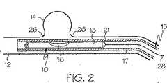
ここで図面を参照すると、図1は、本発明に係る2リーフ型又は2シート型の血管閉塞装置の全体を番号10で示す。リーフは、以下、シートと称す。血管閉塞装置は、血管12の内側で、脈管障害部位(この場合、側壁動脈瘤14)に位置づけられている様子が示されている。装置10は、動脈瘤14の内側で展開状態にある第1のシート16と、コネクタ20により第1のシート16に結合した第2のシート18とを有するように示されている。第1のシート16は、血管と接触する面22と、対向する非接触面とを有し、第2のシート18は、血管と接触する面24と、対向する非接触面とを有する。第1のシート16は、血管内への配置に先だって第2のシート18に結合可能であり、それ故に単一のカテーテル装置内に配置可能である。また、第1のシート16及び第2のシート18を別々に配置し、血管の内側で結合させることも可能である。 Referring now to the drawings, FIG. 1 shows the entire two-leaf or two-sheet vaso-occlusive device according to the present invention at 10. The leaf is hereinafter referred to as a sheet. It is shown that the vascular occlusion device is positioned inside the
さらに、シート16の接触面22、シート18の接触面24のいずれか一方又は両方は、装置及び血管の一体化を促進させたり、治癒を促進させたりするような生体適合性材料をコーティングしたり、埋め込んだり、その材料自体との混合をしたりすることを随意に実施可能である。これは、接着、線維形成、組織成長、内皮形成又は細胞増殖などを促進する生体適合性材料を含み得る。 Further, either or both of the
生体適合性高分子材料の例は、限定はされないが、コラーゲン、フィブリン、フィブロネクチン、抗体、サイトカイン、成長因子、酵素などのような蛋白質;ヘパリン、コンドロイチンのような多糖類;生物由来の架橋ゼラチン;ヒアルロン酸;ポリ(α−ヒドロキシ酸);RNA;DNA;ポリグリコリド、ポリラクチド、ラクチドとグリコリドのコポリマーのようなポリエステル及びポリオルトエステル;ポリカプロラクトンを含むポリラクトン;ポリジオキサノン;ポリリジンのようなポリアミド酸;ポリシアノアクリレート;ポリ(ホスファジン);ポリ(ホスフォエステル);ポリエステルアミド;ポリアセタール;ポリケタール;トリメチレンカーボネートを含むポリカーボネート及びポリオルトカーボネート;分解性ポリエチレン;ポリアルキレンオキサレート;ポリアルキレンサクシネート;キチン;キトサン;酸化セルロース;ポリヒドロキシブチレート、ポリヒドロキシバレレート及びこれらのコポリマーを含むポリヒドロキシアルカノエート;ポリエチレンオキサイドのポリマー及びコポリマー;アクリル末端ポリエチレンオキサイド;ポリアミド;ポリエチレン;ポリアクリロニトリル;ポリホスファゼン;不飽和ポリ無水物、ポリ(アミド無水物)、ポリ(アミド−エステル)無水物、脂肪族−芳香族ホモポリ無水物、芳香族ポリ無水物、ポリ(エステル無水物)、脂肪酸を基とするポリ無水物などを含む、ジカルボン酸モノマーから形成されたポリ無水物;他の生体適合性又は天然由来の高分子材料;その他;そのコポリマー及びターポリマー;生物活性材料のフラグメント;並びにこれらの混合物を含む。以下、用語「コポリマー」を、2つ以上のモノマーを有する任意のポリマーを示すために用いることにする。 Examples of biocompatible polymeric materials include, but are not limited to, proteins such as collagen, fibrin, fibronectin, antibodies, cytokines, growth factors, enzymes, etc .; polysaccharides such as heparin, chondroitin; biological derived cross-linked gelatin; Poly (α-hydroxy acid); RNA; DNA; Polyglycolide, polylactide, polyesters and polyorthoesters such as lactide and glycolide copolymers; polylactones including polycaprolactone; polydioxanone; polyamic acid such as polylysine; Poly (phosphadine); Poly (phosphoester); Polyester amide; Polyacetal; Polyketal; Polycarbonate and polyorthocarbonate including trimethylene carbonate; Degradable polyethylene Polyalkylene oxalate; polyalkylene succinate; chitin; chitosan; oxidized cellulose; polyhydroxyalkanoates including polyhydroxybutyrate, polyhydroxyvalerate and copolymers thereof; polymers and copolymers of polyethylene oxide; acrylic-terminated polyethylene oxide Polyamide; polyethylene; polyacrylonitrile; polyphosphazene; unsaturated polyanhydride, poly (amide anhydride), poly (amide-ester) anhydride, aliphatic-aromatic homopolyanhydride, aromatic polyanhydride, poly ( Ester anhydrides), polyanhydrides formed from dicarboxylic acid monomers, including fatty anhydride-based polyanhydrides; other biocompatible or naturally derived polymeric materials; others; copolymers and terpolymers; Life Fragments of sexual materials; as well as mixtures thereof. Hereinafter, the term “copolymer” will be used to indicate any polymer having two or more monomers.
幾つかの生体適合性ポリマーはまた、ポリラクチド、ポリグリコリド、ポリラクチドとグリコリドのコポリマー、ポリ無水物、ポリ−p−ジオキサノン、トリメチレンカーボネート、ポリカプロラクトン、ポリヒドロキシアルカノエートなどのような生体吸収性とみなされる。 Some biocompatible polymers also have bioabsorbable properties such as polylactide, polyglycolide, copolymers of polylactide and glycolide, polyanhydrides, poly-p-dioxanone, trimethylene carbonate, polycaprolactone, polyhydroxyalkanoates and the like. It is regarded.
生物分解性でない生体適合性ポリマーで、本件に有用であると考えられるポリマーは、限定はされないが、ポリアクリレート;エチレン−ビニルアセテート;セルロース、並びに、セルロースアセテートブチレート及びセルロースアセテートプロピオネートを含むセルロース誘導体;アシル置換セルロースアセテート及びその誘導体;非腐食性ポリオレフィン;ポリスチレン;ポリビニルクロライド;ポリビニルフルオリド;ポリビニル(イミダゾール);クロロスルホン化ポリオレフィン;ポリエチレンオキサイド;ポリエチレングリコール;ポリビニルピロリドン;ポリウレタン;ポリシロキサン;これらのコポリマー及びターポリマー;並びにこれらの混合物を含む。 Non-biodegradable, biocompatible polymers that may be useful in the present case include, but are not limited to, polyacrylates; ethylene-vinyl acetate; cellulose and cellulose acetate butyrate and cellulose acetate propionate. Acyl-substituted cellulose acetate and its derivatives; Non-corrosive polyolefin; Polystyrene; Polyvinyl chloride; Polyvinyl fluoride; Polyvinyl (imidazole); Chlorosulfonated polyolefin; Polyethylene oxide; Polyethylene glycol; Polyvinyl pyrrolidone; Polyurethane; Copolymers and terpolymers; and mixtures thereof.
上記の様々なポリマーの幾つかの例が、米国特許第4891225号及び米国特許第4906474号(ポリ無水物)、米国特許第4767628号(ポリラクチド、ラクチドとグリコール酸のコポリマー)、米国特許第4530840号(ポリラクチド、ポリグリコリド、及びこれらのコポリマー)、米国特許第5234520号(生物分解性ポリマー)などに見られる。これらの特許の各々は、全体を援用して本文の記載の一部とする。 Some examples of the various polymers described above are US Pat. No. 4,891,225 and US Pat. No. 4,906,474 (polyanhydride), US Pat. No. 4,767,628 (polylactide, copolymer of lactide and glycolic acid), US Pat. No. 4,530,840. (Polylactide, polyglycolide, and copolymers thereof), US Pat. No. 5,234,520 (biodegradable polymer), and the like. Each of these patents is incorporated herein by reference in its entirety.
これらの生体適合性ポリマーの幾つかが、米国特許第6413536号に記載されており、この特許もその全てを援用して本文の記載の一部とする。
また、本願と共に譲渡された米国特許第6335029号も参照されたい。この特許の全てを援用して本文の記載の一部とする。Some of these biocompatible polymers are described in US Pat. No. 6,413,536, which is hereby incorporated by reference in its entirety.
See also US Pat. No. 6,335,029, assigned with the present application. All of this patent is incorporated herein by reference.
当業者は、このような生物分解性ポリマーが、本文中に列挙するには数が多すぎることを理解するであろう。したがって、上記のリストは網羅的ではなく、例示のためのものに過ぎない。 One skilled in the art will appreciate that such biodegradable polymers are too numerous to be listed herein. Accordingly, the above list is not exhaustive and is for illustration only.
好適な非高分子材料は、例えば、ホルモン及び抗腫瘍剤を含む。
患者の脈管構造との一体化を促進するその他の生体適合性材料の例は、例えば、加工された人間又は動物の組織を含み、該組織は、例えば、細胞若しくは細胞フラグメント、培養された脈管組織、膀胱、腹部、肝臓、又は、天然若しくは合成由来の遺伝物質からのマトリクス材料を含む。Suitable non-polymeric materials include, for example, hormones and antitumor agents.
Examples of other biocompatible materials that facilitate integration with the patient's vasculature include, for example, processed human or animal tissue, such as cells or cell fragments, cultured veins. Includes matrix material from ductal tissue, bladder, abdomen, liver, or genetic material of natural or synthetic origin.
図2〜3は、2シート構造を有する本発明に係る血管閉塞装置10を側壁動脈瘤14の内側に配置する様子を示す。図2に示すように、血管閉塞装置10は、カテーテル配送装置15のシャフト17の内側に折り畳まれた形状で、血管12を通して配送される。カテーテル配送装置15は、血管閉塞装置10のシート16及びシート18が動脈瘤14の頚部26のほぼ中央に位置するように血管閉塞装置10を配置するために用いられる。図2には、血管閉塞装置10のシート16及びシート18が、カテーテル配送装置15の内側で非展開状態にある様子が示されている。この実施形態において、非展開状態にある第2のシートは、巻かれた形状を有している。このシートは、例えば折り曲げられることも可能である。第1のシート16をカテーテルから押し出すとともに動脈瘤内に押し込み、且つ第2のシート18をカテーテルから押し出すためのプッシャワイヤ28も図示されている。 2 to 3 show a state in which the
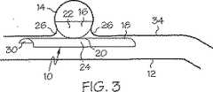
図3は、図2の血管閉塞装置10が配置後に動脈瘤14にて展開された様子を示す。この時点で、カテーテル配送装置15は、シート16及びシート18が配置されるように引き戻されている。カテーテル配送装置15が引き戻されると、シート16が最初に配置され、カテーテル配送装置がさらに引き戻されると、次いでシート18が解放される。引き込み可能なシースは随意に使用可能である。図3は、シート16及びシート18の両方が完全に配置及び展開された状態を示す。この実施形態において、シート18は、脈管障害物14が位置する血管壁34に対して凸となる矩形のシートの形状で示されている。シート18は、動脈瘤の頚部26に対しても凸となる。シート18の接触面24は、装置と患者の脈管構造との一体化を促進したり、動脈瘤をより迅速に治癒したりするための生体適合性材料を備えることが可能である。対向する非接触面30の一部が図3に明確に示されている。また、シート16の接触面22も、装置と患者の脈管構造との一体化を促進したり、動脈瘤をより迅速に治癒したりするための生体適合性材料を備えることが可能である。 FIG. 3 shows the
この実施形態において、第1のシートは、主に、装置を適切な位置に保持するための固定部材として機能し、第2のシートは、主に、閉塞部材として機能する。別の実施形態において、第1のシートの形状は、障害物を閉塞するために設計され、第2シートは、固定部材として機能するように設計され得る。また、両方のシートが閉塞部材及び固定部材の両方の機能を有するように設計されることも可能である。 In this embodiment, the first sheet mainly functions as a fixing member for holding the apparatus in an appropriate position, and the second sheet mainly functions as a closing member. In another embodiment, the shape of the first sheet can be designed to occlude obstacles and the second sheet can be designed to function as a securing member. It is also possible that both sheets are designed to function as both a closing member and a fixing member.
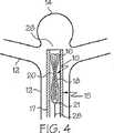
図4〜6は、本発明に係る血管閉塞装置10を末端の動脈瘤14に用いる場合の配置を示す。図4に示すように、シート16及びシート18を有する血管閉塞装置は、カテーテル配送システム15により、血管12を通って動脈瘤14の部位に配送されている。図4は、シート16及びシート18の両方がカテーテルシャフト17の内側で非展開状態にある血管閉塞装置を示す。シート16及びシート18はコネクタ20によって取り付けられている。この例において、これらのシートは、非展開状態において巻かれた形状ではなく、折り畳まれた形状である。血管閉塞装置10に取り外し可能に取り付けられるプッシャワイヤ28が示されている。取り外し可能な分解は、例えば分解可能な接合部を含む様々な手段により達成可能である。分解可能な接合部は、例えば、電蝕、機械的作動、水圧、熱処理、電磁エネルギーなどにより分解可能なものを含む。この列挙は例示を目的としたものに過ぎず、本発明に用いられ得るアタッチメントシステムを網羅的に示したものではない。当業者は、このようなアタッチメントシステムを知っているであろう。 4 to 6 show an arrangement when the
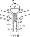
図5は、図4に示されるのと同じ血管閉塞装置10が部分的に配置された状態を示し、この状態において、傘の形状で示される第1のシート16は、動脈瘤14の内側に配置されるとともに、動脈瘤14の頚部26に位置づけられている。シート16を動脈瘤内に押し込むためにプッシャワイヤ28が用いられ、次いで、そのプッシャワイヤ28は、シート16が頚部26と接触するまでシート16を引き戻すために用いられ得る。シート18は、依然として、カテーテルシャフト17の内側で、非配置及び非展開状態にある。シート18はコネクタ20によりシート16に結合している。 FIG. 5 shows a state in which the same vaso-
次いで、操作者は、プッシャワイヤ28を使い続けながら、カテーテルシャフト17の外側にシート18を押し出す。その間にカテーテル配送装置15が引き戻される(図示せず)。 Next, the operator pushes out the
図6は、完全に配置された状態の血管閉塞装置10を示し、この状態において、シート16及びシート18の両方は動脈瘤14の頚部26の適切な位置にあり、シート18もすでに配置されている。 FIG. 6 shows the vaso-
少なくとも一方のシートがかなりの量の血流を遮断するように脈管障害物の適切な閉塞をもたらす限りにおいて、さらには、少なくとも一方のシートが装置を脈管障害物の頚部に適切に固定する限りにおいて、シートが特定の形状も構造も有する必要がないことに留意することが重要である。形状の幾つかの例は、限定はされないが、傘状の構造、放物面状の構造、球面、円盤、矩形の構造、又は、脈管障害物に向って凸状に曲がる半円形の部分円筒などを含む。また、シートは、配置されたときに半曲げの凸状構造を成す矩形の形状であってもよい。凸側は脈管障害物の頚部に向けられる。 In addition, at least one sheet properly secures the device to the neck of the vascular obstacle so long as at least one sheet provides adequate occlusion of the vascular obstacle so as to block a significant amount of blood flow. Insofar, it is important to note that the sheet need not have a specific shape or structure. Some examples of shapes include, but are not limited to, umbrella-shaped structures, parabolic structures, spherical surfaces, disks, rectangular structures, or semi-circular parts that bend convexly toward vascular obstacles Includes cylinders. The sheet may have a rectangular shape that forms a semi-bent convex structure when placed. The convex side is directed to the neck of the vascular obstruction.
図7は、代替の実施形態を示し、この実施形態においては、シート16の代わりに、血管閉塞装置を脈管障害物14の部位で適切な位置に括りつけるためのストラット(支柱)19が用いられる。この図において、血管閉塞装置10は、カテーテル配送装置15のシャフト17の内側にて非展開状態で示されている。ストラット19は、シート18上で折り畳まれている。この実施形態において、シート18は、非展開形態の巻かれた形状で示されている。プッシャワイヤ28は、番号21の場所で、分解可能な接合部を用いて取り外し可能に結合されているように見える。前記接合部は多数の様々な機構を用いて分解可能であり、該機構は、限定はされないが、電蝕、機械的作動、水圧、熱処理、電磁エネルギーなどを含む。従って、この接合部21において、血管閉塞装置10は、カテーテル配送装置15のカテーテルシャフト17の内側に配置されるプッシャワイヤ28から最終的に取り外される。引き込み可能なシースは随意に使用可能である。本文中に記載しないが当分野で知られている他の方法もまた、本発明の装置の取り外しに使用可能である。本発明に用いられ得る分解可能な接合部は、例えば、米国特許第5122136号、米国特許第5354295号、米国特許第5540680号、米国特許第5855578号、米国特許第5895385号、米国特許第5925037号、米国特許第5944714号、米国特許第5947963号、米国特許第5976126号、米国特許第6010498号、米国特許第6066133号、及び米国特許第6083220号に記載されており、これらの特許の各々の全体を援用して本文の記載の一部とする。 FIG. 7 shows an alternative embodiment, in which a
図8は、脈管障害物14の部位に展開状態で配置された図7と同一の装置を示す。この実施形態において、シート18の接触面22には、装置と脈管構造との一体化を促進するための生体適合性材料がコーティングされている。シート18は、脈管障害物14の頚部26、及び脈管障害物が見られる血管壁34に対して凸となるように示されており、このように、接触面22は、血管壁34、及び脈管障害物14の頚部26と密接状態にある。非接触面30は、この実施形態において明確に見られ得る。装置はプッシャワイヤ28から既に取り外されている。 FIG. 8 shows the same device as FIG. 7 deployed in the deployed state at the site of the
図9は代替の実施形態を示し、この実施形態においても、シート16がアンカーストラット19に置き換えられている。この特別な装置は、図7及び8のような側壁動脈瘤にではなく、末端動脈瘤の部位に用いられる。図9においても、カテーテル配送装置15のシャフト17の内側にて折り畳まれた形状にある装置が示されている。引き込み可能なシースは随意に使用可能である。ストラット19はカテーテル15の末端40にあるように示されている。従って、ストラットは脈管障害物14内に最初に押し込まれるとともに、最初に配置される。 FIG. 9 shows an alternative embodiment, in which the
図10は、脈管障害物14の内側に配置されたストラット19を示し、シート18はカテーテル配送装置15のシャフト17の内側で依然として折り畳まれている。図9及び10の両方には、番号21の場所でプッシャワイヤ28に結合した血管閉塞装置10が示されている。 FIG. 10 shows a
図11では、既にシートが配置され、カテーテル配送装置が引き戻され、プッシャワイヤが取り外され、装置が動脈瘤14の頚部26に括りつけられている。この実施形態において、図8に示される実施形態とは異なり、血管壁34及び動脈瘤頚部26に対して凸状でないシート18が示されている。 In FIG. 11, the sheet is already in place, the catheter delivery device is pulled back, the pusher wire is removed, and the device is tied to the
シートは、限定はされないが、高分子材料を含む任意の様々な材料から構成可能である。生体適合性、生体吸収性及び生物分解性の材料が好適である。もちろん、材料は、これらの特性の任意の組合せを有していてもよく、これらの特性の全てを有していてもよい。 The sheet can be composed of any of a variety of materials including, but not limited to, polymeric materials. Biocompatible, bioabsorbable and biodegradable materials are preferred. Of course, the material may have any combination of these properties and may have all of these properties.
有用な高分子材料の例は、合成材料及び天然材料の両方を含む。さらに、材料は、生体適合性材料及び/又は生物分解性材料であることが可能である。有用な高分子材料の例は、限定はされないが、ポリエチレン及びポリプロピレンを含むポリオレフィン、ポリエチレンテレフタレート(PET)及びポリブチレンテレフタレート(PBT)のようなポリエステル、ポリウレタン、アクリル、ポリペプチド、ポリエステル、ポリアミド、発泡ポリテトラフルオロエチレンなどのようなフルオロポリマー、などを含む。 Examples of useful polymeric materials include both synthetic and natural materials. Further, the material can be a biocompatible material and / or a biodegradable material. Examples of useful polymeric materials include, but are not limited to, polyolefins including polyethylene and polypropylene, polyesters such as polyethylene terephthalate (PET) and polybutylene terephthalate (PBT), polyurethanes, acrylics, polypeptides, polyesters, polyamides, foams Fluoropolymers such as polytetrafluoroethylene and the like.
膨潤性高分子材料が本件に有用である。このような材料は、液体中で膨張して滑らかになることが知られているものを含み、そのような材料は、例えば、概してヒドロゲルと称される材料群を含み、このような材料も、本発明に係る装置の製造に使用可能である。このような材料は、親水性のマクロポーラスな高分子ヒドロゲル発泡体材料を含む。このような材料の例は、限定はされないが、ポリビニルピロリドン、ポリエチレンオキサイド、及び、ポリエチレンオキサイドとポリプロピレンオキサイドとのコポリマー、ポリアクリル酸、ポリビニルアルコール、ヒアルロン酸、ヘパリン、コンドロイチン硫酸塩、ペクチン酸、カルボキシル被覆ポリサッカライド、ポリヒドロキシエチルメタクリレート、ポリアクリルアミド、ポリアクリロニトリル加水分解物、ポリメタクリル酸、ポリエチレンアミド、ポリサッカライド、並びに、これらのコポリマー及び組合せなどを含む。 Swellable polymeric materials are useful in this case. Such materials include those known to swell and smooth in liquids, such materials include, for example, a group of materials generally referred to as hydrogels, and such materials also It can be used to manufacture the device according to the present invention. Such materials include hydrophilic macroporous polymeric hydrogel foam materials. Examples of such materials include, but are not limited to, polyvinylpyrrolidone, polyethylene oxide, and copolymers of polyethylene oxide and polypropylene oxide, polyacrylic acid, polyvinyl alcohol, hyaluronic acid, heparin, chondroitin sulfate, pectinic acid, carboxyl Coated polysaccharides, polyhydroxyethyl methacrylates, polyacrylamides, polyacrylonitrile hydrolysates, polymethacrylic acid, polyethylene amides, polysaccharides, and copolymers and combinations thereof.
マクロポーラスな固体として形成された膨潤性発泡体マトリクスを含む膨潤性材料の特定の例が米国特許第5750585号に記載されており、この特許の全体を援用して本文の記載の一部とする。この材料は、発泡安定剤、及び、約10重量%までのマルチオレフィン官能性架橋剤と架橋されるフリーラジカル重合性の親水性オレフィンモノマーのポリマー又はコポリマーを含む。 A specific example of a swellable material comprising a swellable foam matrix formed as a macroporous solid is described in US Pat. No. 5,750,585, which is hereby incorporated by reference in its entirety. . The material includes a foam stabilizer and a polymer or copolymer of a free-radically polymerizable hydrophilic olefin monomer that is crosslinked with up to about 10% by weight of a multiolefin functional crosslinker.
本件に有用な天然物質をベースにした材料又は生物由来の材料は、限定はされないが、コラーゲンフォーム、採取した脈管材料、加工した組織から構成される膜などを含む。
好適な生物吸収性材料は、限定はされないが、分解可能なヒドロゲル、ラクチド/グリコリド又はPHAを含む。好適な生物吸収性材料のより特定的な例は、限定はされないが、コラーゲン、ポリカプロラクトン、ポリ(グリコール酸)、ポリ(3−ヒドロキシブチル酸)、ポリ(dl−乳酸)、ポリ(l−乳酸)、ポリ(dl−ラクチド/グリコリド)50:50、ポリ(ヒドロキシバレレート)、ポリ(ヒドロキシバレレート−ヒドロキシブチレート)、又はその他のPHAを含む。このような材料は、米国特許第5056211号及び米国特許第6251116号に記載されており、これらの特許の両方の全体を援用して本文の記載の一部とする。Materials based on natural substances or biological materials useful in the present case include, but are not limited to, collagen foam, harvested vascular material, membranes composed of processed tissue, and the like.
Suitable bioabsorbable materials include, but are not limited to, a degradable hydrogel, lactide / glycolide or PHA. More specific examples of suitable bioabsorbable materials include, but are not limited to, collagen, polycaprolactone, poly (glycolic acid), poly (3-hydroxybutyric acid), poly (dl-lactic acid), poly (l- Lactic acid), poly (dl-lactide / glycolide) 50:50, poly (hydroxyvalerate), poly (hydroxyvalerate-hydroxybutyrate), or other PHA. Such materials are described in US Pat. No. 5,052,211 and US Pat. No. 6,251,116, both of which are incorporated by reference in their entirety.
シリコーン、ポリオレフィン、フルオロポリマー又はポリウレタンのような非吸収性ポリマー及びエラストマーもまた、使用可能である。
本発明の血管閉塞装置の形成には、形状記憶材料が適している。形状記憶材料は、ポリマー製又は金属製であることが可能である。形状記憶材料は、機械的変形、又は冷却及び加熱による変形後にも、元の形状を記憶する能力を有する。このような材料は、構造的相変態をすると考えられる。一般的に、形状記憶ポリマー(SMPs)は、ハードセグメントとソフトセグメントとを有する隔離された線状ブロックコポリマーであることが分かっており、ハードセグメントが決められた融点を有する結晶質であり、ソフトセグメントが決められたガラス転移温度を有する非晶質である。しかしながら、ハードセグメントが非結晶質で、融点以外のガラス転移温度を有していてもよく、また、ソフトセグメントが結晶質で、ガラス転移温度以外の融点を有していてもよい。ソフトセグメントの融点又はガラス転移温度は、実質的に、ハードセグメントの融点又はガラス転移温度よりも低い。形状記憶ポリマーの幾つかの例は、限定はされないが、ポリエステル、ポリアクリレート、ポリアミド、ポリシロキサン、ポリウレタン、ポリエーテルアミド、ポリウレタン/ウレア、ポリエーテルエステル、ウレタン/ブタジエンコポリマー、ポリノルボルネン、及びこれらの混合物から形成されるものを含む。例えば、米国特許第5506300号、米国特許第5145935号、米国特許第5665822号、及び米国特許第6388043号を参照されたい。これらの特許の各々の全体を援用して本文の記載の一部とする。分解性の形状記憶ポリマーもまた使用可能である。Non-absorbable polymers and elastomers such as silicones, polyolefins, fluoropolymers or polyurethanes can also be used.
Shape memory materials are suitable for forming the vascular occlusion device of the present invention. The shape memory material can be made of polymer or metal. Shape memory materials have the ability to memorize their original shape even after mechanical deformation or deformation by cooling and heating. Such materials are believed to undergo structural phase transformations. In general, shape memory polymers (SMPs) have been found to be isolated linear block copolymers having hard and soft segments, where the hard segments are crystalline with a determined melting point, and soft The segment is amorphous with a defined glass transition temperature. However, the hard segment may be amorphous and have a glass transition temperature other than the melting point, or the soft segment may be crystalline and have a melting point other than the glass transition temperature. The melting or glass transition temperature of the soft segment is substantially lower than the melting or glass transition temperature of the hard segment. Some examples of shape memory polymers include, but are not limited to, polyesters, polyacrylates, polyamides, polysiloxanes, polyurethanes, polyether amides, polyurethane / ureas, polyether esters, urethane / butadiene copolymers, polynorbornene, and these Including those formed from mixtures. See, for example, US Pat. No. 5,506,300, US Pat. No. 5,145,935, US Pat. No. 5,656,822, and US Pat. No. 6,388,043. The entirety of each of these patents is incorporated herein by reference. Degradable shape memory polymers can also be used.
本件に用いるのに好適な形状記憶金属は、例えば、TiNi合金(ニチノール(登録商標))、CuZnAl合金及びFeNiAl合金を含む。これらの材料は、マルテンサイト変態と称される構造的相変態をする。 Shape memory metals suitable for use in the present case include, for example, TiNi alloys (Nitinol (registered trademark)), CuZnAl alloys and FeNiAl alloys. These materials undergo a structural phase transformation called the martensitic transformation.
例えば形状記憶金属を用いる状況においては、十分なフレキシビリティをもたらすための適切な幾何学的特徴及びクロスパターンを有する金属網構造を用いることが適切であろう。このような金属網は、例えば、超弾性ニッケルチタン合金であるニチノール(登録商標)から構成可能である。さらに、このような形状にはステンレス鋼もまた使用可能である。このタイプの形状は、第1のシートが第2のシートのための固定部材として用いられ、第2のシートが脈管障害物のための閉塞部材として機能する実施形態に、より適しているであろう。このとき第1のシートは、金属網形状に適切に構成可能である。 For example, in a situation where shape memory metal is used, it may be appropriate to use a metal mesh structure with appropriate geometric features and cross patterns to provide sufficient flexibility. Such a metal net | network can be comprised from Nitinol (trademark) which is a superelastic nickel titanium alloy, for example. Furthermore, stainless steel can also be used for such shapes. This type of shape is more suitable for embodiments in which the first sheet is used as a securing member for the second sheet and the second sheet functions as an occlusion member for a vascular obstruction. I will. At this time, the first sheet can be appropriately configured in a metal net shape.
他の形状のために金属を用いることも可能である。金属の基礎構造物を用いる場合、生体適合性、ポリマー性、生物分解性又は生物吸収性の材料でコーティングすることが望ましいであろう。さらに、コーティングすることにより、これらの特性の全てを有することが可能となる。装置が金属から構成されたり、金属の構成要素を含んだりする場合、金属は、用いられる血管内で所望の程度のフレキシビリティをもたらすために、十分に柔軟でなければならない。先に記載したように、好ましい結果を得るためには、装置内の金属の幾何学的パターンが重要であり、該パターンは正弦状又は円形の金属基礎構造物であり得る。 It is also possible to use metal for other shapes. If a metal substructure is used, it may be desirable to coat with a biocompatible, polymeric, biodegradable or bioabsorbable material. Furthermore, coating makes it possible to have all of these properties. If the device is composed of metal or contains metallic components, the metal must be sufficiently flexible to provide the desired degree of flexibility within the blood vessel used. As described above, to obtain favorable results, the geometric pattern of the metal in the device is important, and the pattern can be a sinusoidal or circular metal substructure.
圧縮された発泡体もまた、元の形状に戻る能力を有するため、本発明に使用可能である。連続気泡発泡体及び独立気泡発泡体の両方が使用可能である。圧縮発泡体に用いられるのに適した材料は、限定はされないが、医療グレードのシリコーン及びポリウレタンを含む。先に記載したように、コラーゲンのような天然の材料も、圧縮発泡体の材料をつくるために使用可能である。 Compressed foam can also be used in the present invention because it has the ability to return to its original shape. Both open cell foams and closed cell foams can be used. Suitable materials for use in the compressed foam include, but are not limited to, medical grade silicone and polyurethane. As described above, natural materials such as collagen can also be used to make compressed foam materials.
先に記載した材料のコポリマー及び架橋可能な変種もまた、本件に用いるのに好適である。また、もちろん、先に記載した様々な材料の混合物も、本発明に係る装置の製造に使用可能である。 Copolymers and crosslinkable variants of the materials described above are also suitable for use in the present case. Of course, mixtures of the various materials described above can also be used for the production of the device according to the invention.
各シートは同一材料から構成されても、異なる材料や材料のブレンドから構成されてもよい。
シートは均一の厚さを有していてもよく、シートの厚さがそのシートの面上で変化していてもよい。例えば、シートは、縁でより薄くなるように形成され得る。Each sheet may be composed of the same material or may be composed of different materials or blends of materials.
The sheet may have a uniform thickness and the thickness of the sheet may vary on the surface of the sheet. For example, the sheet can be formed to be thinner at the edges.
第1のシートを、例えば、複数のストラットを有する固定部材に置き替えるならば、それらのストラットは金属又は金属合金からも形成可能である。
先に記載したように、シート材料自体の中、又はシートの表面上には、生体適合性接着剤、高分子材料、組織、細胞、遺伝物質などのような脈管構造との一体化又は治癒を促進する生体適合性材料が取り込まれることが望ましい。If the first sheet is replaced with, for example, a fixing member having a plurality of struts, the struts can also be formed from a metal or metal alloy.
As described above, in the sheet material itself, or on the surface of the sheet, integration or healing with vasculature such as biocompatible adhesives, polymeric materials, tissues, cells, genetic material, etc. It is desirable to incorporate a biocompatible material that promotes.
望ましい化合物又は薬剤は、様々な方法を用いて、シートに加えられることが可能である。これらの方法は、シートをコーティングすること、シートを構成する材料に化合物又は薬剤を埋め込むこと、及び、シートの形成に先だって化合物又は薬剤をシートの材料に混合することなどを含む。 Desired compounds or agents can be added to the sheet using a variety of methods. These methods include coating the sheet, embedding the compound or agent in the material comprising the sheet, and mixing the compound or agent into the sheet material prior to forming the sheet, and the like.
生体適合性接着剤は、動脈瘤の頚部で化学結合の形成が可能な面上に加えられることが可能であり、該化学結合は、装置が動脈瘤又は脈管障害物の内側に配送及び配置されたならば動脈瘤の内側に形成され、装置が親血管の内側であるが動脈瘤の外側に配送及び配置されたならば動脈瘤頚部の外側に形成され得る。このような生体適合性接着剤は、米国特許第6368586号に記載されており、この特許の全体を援用して本文の記載の一部とする。 The biocompatible adhesive can be applied on a surface capable of forming a chemical bond at the neck of the aneurysm, which is delivered and placed by the device inside the aneurysm or vascular obstruction If formed, it can be formed inside the aneurysm and can be formed outside the aneurysm neck if the device is delivered and placed inside the parent vessel but outside the aneurysm. Such a biocompatible adhesive is described in US Pat. No. 6,368,586, the entirety of which is incorporated herein by reference.
先に述べたように、このような化合物又は薬剤は、例えば、組織成長又は内皮形成を含む身体内の様々な活動を促進可能である。後者の内皮形成では、シートの表面の一部又は全体、特に脈管構造と接触する面が、内皮細胞により裏打ち又はコーティングされることが可能である。これらの細胞は、装置を配置する患者から摘出される細胞であっても、別の患者に由来するそのような細胞の組織培養物から摘出される細胞であってもよい。 As mentioned above, such compounds or agents can promote various activities in the body including, for example, tissue growth or endothelium formation. In the latter endothelium formation, part or all of the surface of the sheet, in particular the surface in contact with the vasculature, can be lined or coated with endothelial cells. These cells may be cells removed from the patient in which the device is placed, or may be cells extracted from a tissue culture of such cells from another patient.
他の有用な化合物は、例えばヘパリンのようなポリサッカライドを含み、このポリサッカライドは、単独でも、又は、例えばヒドロゲルや親水性化合物と組み合わされた場合でも有益に使用可能である。 Other useful compounds include polysaccharides such as heparin, which can be beneficially used either alone or in combination with, for example, hydrogels or hydrophilic compounds.
抗凝血化合物は、心血管系の血管内に挿入する装置上に施されるコーティングとして非常に有用であろう。タクソール(Taxol)(登録商標)のような化合物は、本発明の装置の材料にコーティング、又は埋め込むための有用な化合物であろう。 Anticoagulant compounds would be very useful as coatings applied on devices that are inserted into the blood vessels of the cardiovascular system. Compounds such as Taxol® may be useful compounds for coating or embedding in the device materials of the present invention.
装置内に取り込まれ得る他の有用な材料は、限定はされないが、抗血小板薬、カルシウム作動薬、抗炎症化合物、増殖抑制剤、脂質低下剤、及び血管形成因子を含む。装置は、これらの化合物の全て又は幾つかを、材料の表面にコーティングしたり埋め込んだり、又は材料中に混合したりして構成することが可能である。 Other useful materials that can be incorporated into the device include, but are not limited to, antiplatelet agents, calcium agonists, anti-inflammatory compounds, growth inhibitors, lipid lowering agents, and angiogenic factors. The device can be constructed with all or some of these compounds coated or embedded in the surface of the material or mixed into the material.
血管閉塞装置を形成する材料又は血管閉塞装置自体は、一部変更が可能であり、或いは、他の添加剤を加えることが可能であり、それにより、血管閉塞装置を慣用のイメージング技術によって可視化可能にする。例えば、装置は、蛍光透視技術、MRI可視技術、又はその両方により、可視化可能になる。これは、ワイヤ巻付け、マーカーバンド、リベット、プラグなどのようなマーカーの使用により、或いは、血管閉塞装置を形成する材料内に取り込まれ得る放射線不透過性材料やMRI可視材料を用いて実施可能である。任意の好適な放射線不透過性材料又はMRI可視材料が使用可能である。 The material forming the vaso-occlusion device or the vaso-occlusion device itself can be modified in part, or other additives can be added so that the vaso-occlusion device can be visualized by conventional imaging techniques To. For example, the device can be visualized by fluoroscopic techniques, MRI visible techniques, or both. This can be done by using markers such as wire wraps, marker bands, rivets, plugs, etc., or with radiopaque or MRI visible materials that can be incorporated into the material forming the vaso-occlusive device. It is. Any suitable radiopaque material or MRI visible material can be used.
装置に放射線不透過性をもたらすための好適な材料は、限定はされないが、例えば、プラチナ、ロジウム、パラジウム、レニウム、イリジウム、タンタル、タングステン、金、銀、これらの金属の合金、及び、バリウムを含有する高分子材料を含む。放射線不透過性は、装置を可視化するために望ましく、該可視化は、装置を障害部位に位置決めし、装置を障害物の内側に位置合わせし、さらに装置の括りつけを適切に行うという目的を有する。 Suitable materials for providing radiopacity to the device include, but are not limited to, for example, platinum, rhodium, palladium, rhenium, iridium, tantalum, tungsten, gold, silver, alloys of these metals, and barium. Contains polymeric materials. Radiopacity is desirable for visualizing the device, which has the purpose of positioning the device at the obstacle site, aligning the device inside the obstacle, and properly tying the device. .
以上に列挙した材料は、例示を目的としたものに過ぎず、網羅的なものでは全くない。様々な目的のために本発明の装置に用いられ得る非常に多くの材料がある。当業者はこれらの材料を知っているであろう。 The materials listed above are for illustrative purposes only and are not exhaustive. There are numerous materials that can be used in the device of the present invention for various purposes. Those skilled in the art will know these materials.
本発明は、また、本発明の血管閉塞装置をカテーテル配送装置と組み合わせて用いることにも向けられている。当分野において、カテーテル配送装置の様々な構造が知られているため、本願では任意の好適な構造のものを用い得る。引き込み可能なシースも随意に使用可能である。 The present invention is also directed to the use of the vascular occlusion device of the present invention in combination with a catheter delivery device. Since various structures of catheter delivery devices are known in the art, any suitable structure may be used herein. A retractable sheath can optionally be used.
本発明は、さらに、開口部を有する脈管障害物を閉塞する方法に関する。この方法は、
a)非展開形状及び展開形状を有する第1のシートを、先に記載したように、脈管障害物の頚部を通して脈管障害物内へと配置するステップと、
b)第1のシートを脈管障害物内で展開させるステップと、
c)展開形状及び非展開形状を有する第2のシートを、先に記載したように、脈管障害物の外側に配置するステップであって、該第2のシートは、第1のシートに取り付けられていることと、
d)第2のシートを展開させるステップとを含む。The invention further relates to a method for occluding a vascular obstruction having an opening. This method
a) placing a first sheet having a non-deployed shape and a deployed shape through the neck of the vascular obstacle and into the vascular obstacle, as described above;
b) deploying the first sheet within the vascular obstacle;
c) placing the second sheet having a deployed shape and a non-deployed shape on the outside of the vascular obstruction, as described above, wherein the second sheet is attached to the first sheet And that
d) unfolding the second sheet.
これらのシートは、部位への配送に先だって結合されていてもよく、或いは前記部位で結合されてもよい。
第1のシート又は第2のシートは代替の固定構造物に置き替え可能である。一実施形態において、固定構造物は複数のストラットを含む。These sheets may be joined prior to delivery to the site, or may be joined at the site.
The first sheet or the second sheet can be replaced with an alternative fixed structure. In one embodiment, the stationary structure includes a plurality of struts.
本発明は、動脈瘤の開口部を親血管から閉鎖し、且つ実質的に閉塞させるために使用可能である。
上記の開示は、例示を目的としているに過ぎず、網羅的ではない。上記の実施形態は多くの変形例及び代替例を当業者に提示するであろう。これらの変形例及び代替例の全てが、添付の特許請求の範囲に含まれるものとする。当分野に精通している者は、本文中に記載された特定の実施形態以外にその均等物があることを理解するであろう。これらの均等物もまた、添付の特許請求の範囲に包含されるものとする。The present invention can be used to close and substantially occlude an aneurysm opening from a parent vessel.
The above disclosure is intended to be illustrative only and not exhaustive. The above embodiments will present many variations and alternatives to those skilled in the art. All of these variations and alternatives are intended to be included within the scope of the appended claims. Those skilled in the art will appreciate that there are equivalents besides the specific embodiments described herein. These equivalents are also intended to be encompassed by the appended claims.
Claims (13)
Translated fromJapanese第1の材料から形成され、非展開形状及び展開形状を有する第1のシートと、
該第1の材料と同一又は異なる第2の材料から形成され、非展開形状及び展開形状を有する第2のシートとを備え、
該第1のシート及び該第2のシートの一方は閉塞部材として機能し、
該第1のシート及び該第2のシートの他方は固定部材として機能し、
前記第2のシートは、前記傷害物の内部に配置されると共に、前記傷害物への流体の流れを実質的に遮断するように構成されている装置。A device for occluding an injury in the vasculature, the injury having a neck, the device comprising:
A first sheet formed from a first material and having a non-deployed shape and a deployed shape;
Is formed from the first material and the same or different second material,and a second sheet having a non-deployed configuration and the deployed configuration,
Hand of the first sheet and the second sheet functions as closing member,
The other of the first sheet and the second sheet functionsas a fixing member,
Said second sheet, it said while being positioned inside the lesion wasdevice is configured tosubstantially cut off the flow of fluid to the injury thereof.
Applications Claiming Priority (2)
| Application Number | Priority Date | Filing Date | Title |
|---|---|---|---|
| US10/231,391US8075585B2 (en) | 2002-08-29 | 2002-08-29 | Device and method for treatment of a vascular defect |
| PCT/US2003/024763WO2004019790A1 (en) | 2002-08-29 | 2003-07-29 | Device and method for treatment of a vascular defect |
Publications (2)
| Publication Number | Publication Date |
|---|---|
| JP2005537091A JP2005537091A (en) | 2005-12-08 |
| JP4417255B2true JP4417255B2 (en) | 2010-02-17 |
Family
ID=31976699
Family Applications (1)
| Application Number | Title | Priority Date | Filing Date |
|---|---|---|---|
| JP2004532874AExpired - LifetimeJP4417255B2 (en) | 2002-08-29 | 2003-07-29 | Device for treating vascular obstacles |
Country Status (6)
| Country | Link |
|---|---|
| US (1) | US8075585B2 (en) |
| EP (1) | EP1534148A1 (en) |
| JP (1) | JP4417255B2 (en) |
| AU (1) | AU2003256879A1 (en) |
| CA (1) | CA2485204A1 (en) |
| WO (1) | WO2004019790A1 (en) |
Families Citing this family (113)
| Publication number | Priority date | Publication date | Assignee | Title |
|---|---|---|---|---|
| CA2319447C (en) | 1998-12-01 | 2010-01-26 | Washington University | Embolization device |
| US8252040B2 (en)* | 2001-07-20 | 2012-08-28 | Microvention, Inc. | Aneurysm treatment device and method of use |
| US8715312B2 (en)* | 2001-07-20 | 2014-05-06 | Microvention, Inc. | Aneurysm treatment device and method of use |
| US8465516B2 (en)* | 2001-07-26 | 2013-06-18 | Oregon Health Science University | Bodily lumen closure apparatus and method |
| US9861517B2 (en) | 2001-07-26 | 2018-01-09 | Cook Medical Technologies Llc | Vessel closure member, delivery apparatus, and method of inserting the member |
| JP4566988B2 (en)* | 2003-04-02 | 2010-10-20 | ボストン サイエンティフィック リミテッド | Separable and recoverable stent assembly |
| EP1689481A4 (en)* | 2003-10-07 | 2008-04-02 | Ford Henry Health System | PLATFORM CATHETER |
| US7232461B2 (en)* | 2003-10-29 | 2007-06-19 | Cordis Neurovascular, Inc. | Neck covering device for an aneurysm |
| US8333798B2 (en) | 2003-11-07 | 2012-12-18 | Merlin Md Pte Ltd. | Implantable medical devices with enhanced visibility, mechanical properties and biocompatability |
| US20080027531A1 (en)* | 2004-02-12 | 2008-01-31 | Reneker Darrell H | Stent for Use in Cardiac, Cranial, and Other Arteries |
| US8500751B2 (en)* | 2004-03-31 | 2013-08-06 | Merlin Md Pte Ltd | Medical device |
| WO2005094725A1 (en) | 2004-03-31 | 2005-10-13 | Merlin Md Pte Ltd | A method for treating aneurysms |
| US8715340B2 (en)* | 2004-03-31 | 2014-05-06 | Merlin Md Pte Ltd. | Endovascular device with membrane |
| US20050267570A1 (en)* | 2004-05-27 | 2005-12-01 | Shadduck John H | Endovascular occlusion devices and methods of use |
| EP2468348B1 (en)* | 2004-09-17 | 2016-10-26 | Codman & Shurtleff, Inc. | Thin film metallic devices for plugging aneurysms or vessels |
| US8968390B2 (en) | 2004-09-27 | 2015-03-03 | Medinol Ltd. | Covering for an endoprosthetic device and methods of using for aneurysm treatment |
| EP1809202A4 (en)* | 2004-12-22 | 2011-04-27 | Merlin Md Pte Ltd | A medical device |
| US20060155323A1 (en) | 2005-01-07 | 2006-07-13 | Porter Stephen C | Intra-aneurysm devices |
| EP2397182A1 (en)* | 2005-04-19 | 2011-12-21 | Medinol Ltd. | A covering for an endoprosthetic device and methods of using for aneurysm treatment |
| EP1951129B1 (en) | 2005-10-19 | 2012-11-21 | Pulsar Vascular, Inc. | Methods and systems for endovascularly clipping and repairing lumen and tissue defects |
| US8545530B2 (en) | 2005-10-19 | 2013-10-01 | Pulsar Vascular, Inc. | Implantable aneurysm closure systems and methods |
| CN103381101B (en)* | 2005-10-19 | 2017-12-01 | 帕尔萨脉管公司 | For the method and system of clamping and repairing lumen and tissue defects in vascular |
| US7744652B2 (en)* | 2006-01-23 | 2010-06-29 | Hesham Morsi | Aneurysm sealing device |
| US20090054966A1 (en)* | 2006-02-13 | 2009-02-26 | Merlin Md Pte Ltd. | Endovascular device with membrane |
| US9307995B2 (en) | 2006-06-15 | 2016-04-12 | Cook Medical Technologies Llc | Methods, systems and devices for the delivery of endoluminal prostheses |
| US8372114B2 (en)* | 2006-11-13 | 2013-02-12 | Electroformed Stents, Inc. | Over-the-wire exclusion device and system for delivery |
| WO2009003049A2 (en) | 2007-06-25 | 2008-12-31 | Micro Vention, Inc. | Self-expanding prosthesis |
| WO2009076515A1 (en)* | 2007-12-11 | 2009-06-18 | Cornell University | Method and apparatus for sealing an opening in the side wall of a body lumen |
| EP2633823B1 (en) | 2008-04-21 | 2016-06-01 | Covidien LP | Braid-ball embolic devices and delivery systems |
| US10028747B2 (en) | 2008-05-01 | 2018-07-24 | Aneuclose Llc | Coils with a series of proximally-and-distally-connected loops for occluding a cerebral aneurysm |
| US10716573B2 (en) | 2008-05-01 | 2020-07-21 | Aneuclose | Janjua aneurysm net with a resilient neck-bridging portion for occluding a cerebral aneurysm |
| WO2009140437A1 (en) | 2008-05-13 | 2009-11-19 | Nfocus Neuromedical, Inc. | Braid implant delivery systems |
| US9179918B2 (en) | 2008-07-22 | 2015-11-10 | Covidien Lp | Vascular remodeling device |
| US8262692B2 (en)* | 2008-09-05 | 2012-09-11 | Merlin Md Pte Ltd | Endovascular device |
| WO2010028314A1 (en) | 2008-09-05 | 2010-03-11 | Pulsar Vascular, Inc. | Systems and methods for supporting or occluding a physiological opening or cavity |
| US9138233B2 (en)* | 2011-03-17 | 2015-09-22 | Micokoll Inc. | Apparatus and method for tissue adhesion |
| JP5750051B2 (en)* | 2009-01-22 | 2015-07-15 | コーネル ユニヴァーシティー | Method and apparatus for restricting flow through a lumen wall |
| EP2805680B1 (en) | 2009-09-04 | 2017-10-25 | Pulsar Vascular, Inc. | Systems for enclosing an anatomical opening |
| US9095342B2 (en) | 2009-11-09 | 2015-08-04 | Covidien Lp | Braid ball embolic device features |
| US9358140B1 (en) | 2009-11-18 | 2016-06-07 | Aneuclose Llc | Stent with outer member to embolize an aneurysm |
| WO2011072053A1 (en)* | 2009-12-10 | 2011-06-16 | Neurovasx, Inc. | Aneurysm shield |
| WO2011094634A1 (en) | 2010-01-28 | 2011-08-04 | Micro Therapeutics, Inc. | Vascular remodeling device |
| WO2011094638A1 (en) | 2010-01-28 | 2011-08-04 | Micro Therapeutics, Inc. | Vascular remodeling device |
| US10321998B2 (en) | 2010-09-23 | 2019-06-18 | Transmural Systems Llc | Methods and systems for delivering prostheses using rail techniques |
| JP5868432B2 (en) | 2011-02-11 | 2016-02-24 | コヴィディエン リミテッド パートナーシップ | Two-stage deployed aneurysm embolization device |
| US20120245674A1 (en) | 2011-03-25 | 2012-09-27 | Tyco Healthcare Group Lp | Vascular remodeling device |
| US10398444B2 (en) | 2011-03-30 | 2019-09-03 | Noha, Llc | Advanced endovascular clip and method of using same |
| US10028745B2 (en) | 2011-03-30 | 2018-07-24 | Noha, Llc | Advanced endovascular clip and method of using same |
| WO2012135859A2 (en) | 2011-04-01 | 2012-10-04 | Cornell University | Method and apparatus for restricting flow through an opening in the side wall of a body lumen, and/or for reinforcing a weakness in the side wall of a body lumen, while still maintaining substantially normal flow through the body lumen |
| EP2707077B1 (en) | 2011-05-11 | 2017-10-04 | Microvention, Inc. | Device for occluding a lumen |
| CN103582460B (en) | 2011-06-03 | 2019-03-19 | 帕尔萨维斯库勒公司 | Aneurysm devices and related systems and methods with additional anchoring mechanism |
| EP2713905B1 (en) | 2011-06-03 | 2022-03-16 | Pulsar Vascular, Inc. | Systems for enclosing an anatomical opening, including shock absorbing aneurysm devices |
| US9549715B2 (en) | 2011-08-09 | 2017-01-24 | Cook Regentec Llc | Vial useable in tissue extraction procedures |
| EP2750614B1 (en) | 2011-09-01 | 2015-04-29 | Cook Medical Technologies LLC | Aneurysm closure clip |
| US9549817B2 (en) | 2011-09-22 | 2017-01-24 | Transmural Systems Llc | Devices, systems and methods for repairing lumenal systems |
| US9060886B2 (en) | 2011-09-29 | 2015-06-23 | Covidien Lp | Vascular remodeling device |
| US9119625B2 (en) | 2011-10-05 | 2015-09-01 | Pulsar Vascular, Inc. | Devices, systems and methods for enclosing an anatomical opening |
| EP2763601B1 (en) | 2011-10-07 | 2020-03-25 | Cornell University | Apparatus for restricting flow through an opening in a body lumen while maintaining normal flow |
| US8968354B2 (en)* | 2011-10-26 | 2015-03-03 | Boston Scientific Scimed, Inc. | Extended protection embolic filter |
| US9427172B2 (en)* | 2011-12-30 | 2016-08-30 | Mediguide Ltd. | Roll detection and six degrees of freedom sensor assembly |
| US10987208B2 (en) | 2012-04-06 | 2021-04-27 | Merlin Md Pte Ltd. | Devices and methods for treating an aneurysm |
| EP2846706A1 (en) | 2012-05-10 | 2015-03-18 | Pulsar Vascular, Inc. | Coil-tipped aneurysm devices |
| US9314248B2 (en) | 2012-11-06 | 2016-04-19 | Covidien Lp | Multi-pivot thrombectomy device |
| US12053378B2 (en) | 2012-11-07 | 2024-08-06 | Transmural Systems Llc | Devices, systems and methods for repairing lumenal systems |
| US10327781B2 (en) | 2012-11-13 | 2019-06-25 | Covidien Lp | Occlusive devices |
| US9295571B2 (en) | 2013-01-17 | 2016-03-29 | Covidien Lp | Methods and apparatus for luminal stenting |
| US9463105B2 (en) | 2013-03-14 | 2016-10-11 | Covidien Lp | Methods and apparatus for luminal stenting |
| US10736758B2 (en) | 2013-03-15 | 2020-08-11 | Covidien | Occlusive device |
| US20170014115A1 (en) | 2014-03-27 | 2017-01-19 | Transmural Systems Llc | Devices and methods for closure of transvascular or transcameral access ports |
| WO2015148821A1 (en)* | 2014-03-27 | 2015-10-01 | Nasser Rafiee | Devices and methods for closure of transvascular or transcameral access ports |
| US11076860B2 (en) | 2014-03-31 | 2021-08-03 | DePuy Synthes Products, Inc. | Aneurysm occlusion device |
| US11154302B2 (en) | 2014-03-31 | 2021-10-26 | DePuy Synthes Products, Inc. | Aneurysm occlusion device |
| US10130372B2 (en) | 2014-04-30 | 2018-11-20 | Cerus Endovascular Limited | Occlusion Device |
| EP3349687B1 (en) | 2015-09-15 | 2020-09-09 | THE UNITED STATES OF AMERICA, represented by the S | Devices for effectuating percutaneous glenn and fontan procedures |
| US10478194B2 (en) | 2015-09-23 | 2019-11-19 | Covidien Lp | Occlusive devices |
| EP4011303B1 (en) | 2015-12-07 | 2024-06-12 | Cerus Endovascular Limited | Occlusion device |
| WO2017153603A1 (en) | 2016-03-11 | 2017-09-14 | Cerus Endovascular Limited | Occlusion device |
| JP6926195B2 (en)* | 2016-04-23 | 2021-08-25 | トランスミューラル システムズ エルエルシーTransmural Systems LLC | Devices and methods for closing transvascular or transluminal access ports |
| EP3463109A4 (en) | 2016-05-26 | 2020-01-08 | Nanostructures, Inc. | System and methods for embolized occlusion of neurovascular aneurysms |
| US10576099B2 (en) | 2016-10-21 | 2020-03-03 | Covidien Lp | Injectable scaffold for treatment of intracranial aneurysms and related technology |
| WO2018102779A1 (en)* | 2016-12-02 | 2018-06-07 | The Texas A&M University System | Chemically modified shape memory polymer embolic foams with increased x-ray visualization |
| US10543015B2 (en)* | 2016-12-05 | 2020-01-28 | Daniel Ezra Walzman | Mesh disc for saccular aneurysms and cover for saccular out-pouching |
| US12426885B2 (en)* | 2016-12-05 | 2025-09-30 | Daniel Ezra Walzman | Mesh cap for ameliorating outpouchings |
| US10617428B2 (en)* | 2016-12-05 | 2020-04-14 | Daniel Ezra Walzman | Complex coil with mesh cap |
| CN110545739A (en) | 2017-02-23 | 2019-12-06 | 德普伊新特斯产品公司 | aneurysm devices and delivery systems |
| JP7414710B2 (en) | 2017-08-21 | 2024-01-16 | シーラス エンドバスキュラー リミテッド | occlusion device |
| JP7249332B2 (en) | 2017-09-01 | 2023-03-30 | トランスミューラル システムズ エルエルシー | Percutaneous shunt device and related methods |
| US10905430B2 (en) | 2018-01-24 | 2021-02-02 | DePuy Synthes Products, Inc. | Aneurysm device and delivery system |
| CN111936063B (en) | 2018-01-31 | 2024-08-20 | 纳米结构公司 | Vascular occlusion device utilizing thin film nitinol foil |
| US10752772B1 (en)* | 2018-03-28 | 2020-08-25 | United States Of America As Represented By The Secretary Of The Navy | Marine biodegradable composition for 3-D printing |
| US11596412B2 (en) | 2018-05-25 | 2023-03-07 | DePuy Synthes Products, Inc. | Aneurysm device and delivery system |
| US11058430B2 (en) | 2018-05-25 | 2021-07-13 | DePuy Synthes Products, Inc. | Aneurysm device and delivery system |
| US10939915B2 (en) | 2018-05-31 | 2021-03-09 | DePuy Synthes Products, Inc. | Aneurysm device and delivery system |
| US11051825B2 (en) | 2018-08-08 | 2021-07-06 | DePuy Synthes Products, Inc. | Delivery system for embolic braid |
| WO2020060932A1 (en) | 2018-09-18 | 2020-03-26 | Nanostructures, Inc. | Catheter based methods and devices for obstructive blood flow restriction |
| US11123077B2 (en) | 2018-09-25 | 2021-09-21 | DePuy Synthes Products, Inc. | Intrasaccular device positioning and deployment system |
| US11076861B2 (en) | 2018-10-12 | 2021-08-03 | DePuy Synthes Products, Inc. | Folded aneurysm treatment device and delivery method |
| US11406392B2 (en) | 2018-12-12 | 2022-08-09 | DePuy Synthes Products, Inc. | Aneurysm occluding device for use with coagulating agents |
| US11272939B2 (en) | 2018-12-18 | 2022-03-15 | DePuy Synthes Products, Inc. | Intrasaccular flow diverter for treating cerebral aneurysms |
| US11134953B2 (en) | 2019-02-06 | 2021-10-05 | DePuy Synthes Products, Inc. | Adhesive cover occluding device for aneurysm treatment |
| US11337706B2 (en) | 2019-03-27 | 2022-05-24 | DePuy Synthes Products, Inc. | Aneurysm treatment device |
| US11672542B2 (en) | 2019-05-21 | 2023-06-13 | DePuy Synthes Products, Inc. | Aneurysm treatment with pushable ball segment |
| US11413046B2 (en) | 2019-05-21 | 2022-08-16 | DePuy Synthes Products, Inc. | Layered braided aneurysm treatment device |
| US11278292B2 (en) | 2019-05-21 | 2022-03-22 | DePuy Synthes Products, Inc. | Inverting braided aneurysm treatment system and method |
| US10653425B1 (en) | 2019-05-21 | 2020-05-19 | DePuy Synthes Products, Inc. | Layered braided aneurysm treatment device |
| US11497504B2 (en) | 2019-05-21 | 2022-11-15 | DePuy Synthes Products, Inc. | Aneurysm treatment with pushable implanted braid |
| US11607226B2 (en) | 2019-05-21 | 2023-03-21 | DePuy Synthes Products, Inc. | Layered braided aneurysm treatment device with corrugations |
| US11602350B2 (en) | 2019-12-05 | 2023-03-14 | DePuy Synthes Products, Inc. | Intrasaccular inverting braid with highly flexible fill material |
| WO2021092618A1 (en) | 2019-11-04 | 2021-05-14 | Covidien Lp | Devices, systems, and methods for treatment of intracranial aneurysms |
| US11457926B2 (en) | 2019-12-18 | 2022-10-04 | DePuy Synthes Products, Inc. | Implant having an intrasaccular section and intravascular section |
| US11406404B2 (en) | 2020-02-20 | 2022-08-09 | Cerus Endovascular Limited | Clot removal distal protection methods |
| AU2021226497A1 (en)* | 2020-02-27 | 2022-09-08 | Boston Scientific Scimed, Inc. | Medical systems, devices, and related methods |
| US20230404592A1 (en)* | 2022-05-25 | 2023-12-21 | Covidien Lp | Implant with intrasaccular and intravascular portions and related technology |
Family Cites Families (44)
| Publication number | Priority date | Publication date | Assignee | Title |
|---|---|---|---|---|
| IE52535B1 (en)* | 1981-02-16 | 1987-12-09 | Ici Plc | Continuous release pharmaceutical compositions |
| US4530840A (en)* | 1982-07-29 | 1985-07-23 | The Stolle Research And Development Corporation | Injectable, long-acting microparticle formulation for the delivery of anti-inflammatory agents |
| US4906474A (en)* | 1983-03-22 | 1990-03-06 | Massachusetts Institute Of Technology | Bioerodible polyanhydrides for controlled drug delivery |
| US4891225A (en)* | 1984-05-21 | 1990-01-02 | Massachusetts Institute Of Technology | Bioerodible polyanhydrides for controlled drug delivery |
| US5506300A (en)* | 1985-01-04 | 1996-04-09 | Thoratec Laboratories Corporation | Compositions that soften at predetermined temperatures and the method of making same |
| US5234520A (en)* | 1987-03-20 | 1993-08-10 | Morgan Adhesives Co. | Method of making an adhesive construction |
| JP2502132B2 (en)* | 1988-09-30 | 1996-05-29 | 三菱重工業株式会社 | Shape memory polyurethane elastomer molded body |
| DE3917578A1 (en)* | 1989-05-30 | 1990-12-06 | Neff Gmbh | METHOD FOR PRODUCING A BALL BULK DEVICE ON THE NUT OF A BALL SCREW AND BALL SCREW |
| US6083220A (en)* | 1990-03-13 | 2000-07-04 | The Regents Of The University Of California | Endovascular electrolytically detachable wire and tip for the formation of thrombus in arteries, veins, aneurysms, vascular malformations and arteriovenous fistulas |
| US5122136A (en)* | 1990-03-13 | 1992-06-16 | The Regents Of The University Of California | Endovascular electrolytically detachable guidewire tip for the electroformation of thrombus in arteries, veins, aneurysms, vascular malformations and arteriovenous fistulas |
| US5354295A (en)* | 1990-03-13 | 1994-10-11 | Target Therapeutics, Inc. | In an endovascular electrolytically detachable wire and tip for the formation of thrombus in arteries, veins, aneurysms, vascular malformations and arteriovenous fistulas |
| US5665822A (en)* | 1991-10-07 | 1997-09-09 | Landec Corporation | Thermoplastic Elastomers |
| US5108420A (en)* | 1991-02-01 | 1992-04-28 | Temple University | Aperture occlusion device |
| US5750585A (en)* | 1995-04-04 | 1998-05-12 | Purdue Research Foundation | Super absorbent hydrogel foams |
| US6413536B1 (en)* | 1995-06-07 | 2002-07-02 | Southern Biosystems, Inc. | High viscosity liquid controlled delivery system and medical or surgical device |
| US6168622B1 (en) | 1996-01-24 | 2001-01-02 | Microvena Corporation | Method and apparatus for occluding aneurysms |
| US6368586B1 (en)* | 1996-01-26 | 2002-04-09 | Brown University Research Foundation | Methods and compositions for enhancing the bioadhesive properties of polymers |
| EP0900051A1 (en)* | 1996-05-08 | 1999-03-10 | Salviac Limited | An occluder device |
| US5941249A (en)* | 1996-09-05 | 1999-08-24 | Maynard; Ronald S. | Distributed activator for a two-dimensional shape memory alloy |
| US5919224A (en)* | 1997-02-12 | 1999-07-06 | Schneider (Usa) Inc | Medical device having a constricted region for occluding fluid flow in a body lumen |
| EP1006890B1 (en) | 1997-08-04 | 2006-09-20 | Boston Scientific Limited | Occlusion system for aneurysm repair |
| US6063070A (en)* | 1997-08-05 | 2000-05-16 | Target Therapeutics, Inc. | Detachable aneurysm neck bridge (II) |
| US6036720A (en)* | 1997-12-15 | 2000-03-14 | Target Therapeutics, Inc. | Sheet metal aneurysm neck bridge |
| US6068622A (en)* | 1998-02-10 | 2000-05-30 | Medtronic Inc. | Single piece hub/strain relief that can be injection molded over a shaft |
| DE69931474T2 (en)* | 1998-02-23 | 2007-05-10 | Mnemoscience Gmbh | SHAPE MEMORY POLYMER |
| AU756080B2 (en)* | 1998-06-04 | 2003-01-02 | New York University | Endovascular thin film devices and methods for treating and preventing stroke |
| US6139564A (en)* | 1998-06-16 | 2000-10-31 | Target Therapeutics Inc. | Minimally occlusive flow disruptor stent for bridging aneurysm necks |
| US5935148A (en)* | 1998-06-24 | 1999-08-10 | Target Therapeutics, Inc. | Detachable, varying flexibility, aneurysm neck bridge |
| US6335029B1 (en)* | 1998-08-28 | 2002-01-01 | Scimed Life Systems, Inc. | Polymeric coatings for controlled delivery of active agents |
| US6613074B1 (en)* | 1999-03-10 | 2003-09-02 | Cordis Corporation | Endovascular aneurysm embolization device |
| US6860899B1 (en)* | 1999-04-15 | 2005-03-01 | Boston Scientific Scimed, Inc. | Method for treating neurovascular aneurysms |
| US6284802B1 (en)* | 1999-04-19 | 2001-09-04 | The Procter & Gamble Company | Methods for regulating the condition of mammalian keratinous tissue |
| US6375668B1 (en)* | 1999-06-02 | 2002-04-23 | Hanson S. Gifford | Devices and methods for treating vascular malformations |
| US6309367B1 (en)* | 1999-07-23 | 2001-10-30 | Neurovasx, Inc. | Aneurysm shield |
| US6251116B1 (en)* | 1999-07-28 | 2001-06-26 | Vasconnect, Inc. | Device for interconnecting vessels in a patient |
| AU7373700A (en)* | 1999-09-13 | 2001-04-17 | Rex Medical, Lp | Vascular closure |
| US6652555B1 (en) | 1999-10-27 | 2003-11-25 | Atritech, Inc. | Barrier device for covering the ostium of left atrial appendage |
| US6689150B1 (en)* | 1999-10-27 | 2004-02-10 | Atritech, Inc. | Filter apparatus for ostium of left atrial appendage |
| US6551303B1 (en)* | 1999-10-27 | 2003-04-22 | Atritech, Inc. | Barrier device for ostium of left atrial appendage |
| US6692510B2 (en)* | 2001-06-14 | 2004-02-17 | Cordis Neurovascular, Inc. | Aneurysm embolization device and deployment system |
| US7288105B2 (en)* | 2001-08-01 | 2007-10-30 | Ev3 Endovascular, Inc. | Tissue opening occluder |
| US6596013B2 (en)* | 2001-09-20 | 2003-07-22 | Scimed Life Systems, Inc. | Method and apparatus for treating septal defects |
| US6802851B2 (en)* | 2001-09-20 | 2004-10-12 | Gordia Neurovascular, Inc. | Stent aneurysm embolization method using collapsible member and embolic coils |
| JP2003190175A (en)* | 2001-11-15 | 2003-07-08 | Cordis Neurovascular Inc | Aneurysm neck cover for sealing aneurysm |
- 2002
- 2002-08-29USUS10/231,391patent/US8075585B2/ennot_activeExpired - Fee Related
- 2003
- 2003-07-29EPEP03791656Apatent/EP1534148A1/ennot_activeWithdrawn
- 2003-07-29AUAU2003256879Apatent/AU2003256879A1/ennot_activeAbandoned
- 2003-07-29CACA002485204Apatent/CA2485204A1/ennot_activeAbandoned
- 2003-07-29WOPCT/US2003/024763patent/WO2004019790A1/enactiveApplication Filing
- 2003-07-29JPJP2004532874Apatent/JP4417255B2/ennot_activeExpired - Lifetime
Also Published As
| Publication number | Publication date |
|---|---|
| JP2005537091A (en) | 2005-12-08 |
| CA2485204A1 (en) | 2004-03-11 |
| WO2004019790A1 (en) | 2004-03-11 |
| US20040087998A1 (en) | 2004-05-06 |
| US8075585B2 (en) | 2011-12-13 |
| AU2003256879A1 (en) | 2004-03-19 |
| EP1534148A1 (en) | 2005-06-01 |
Similar Documents
| Publication | Publication Date | Title |
|---|---|---|
| JP4417255B2 (en) | Device for treating vascular obstacles | |
| US8444667B2 (en) | Device for closure of a vascular defect and method for treating the same | |
| US10499927B2 (en) | Methods and systems for endovascularly clipping and repairing lumen and tissue defects | |
| JP5976899B2 (en) | System and method for supporting or occluding a physiological opening or cavity | |
| US8444668B2 (en) | Expandable vascular occlusion device | |
| JP2022525317A (en) | Filamentous devices with flexible seams for the treatment of angiopathy | |
| WO2020190620A1 (en) | Filamentary devices for treatment of vascular defects | |
| US20040193178A1 (en) | Multiple joint implant delivery systems for sequentially-controlled implant deployment | |
| JP4751661B2 (en) | Intravascular device for causing a controlled inflammatory response in a cell | |
| CA3012247A1 (en) | Methods and systems for endovascularly clipping and repairing lumen and tissue defects | |
| JP2007307409A (en) | Aneurysm treatment device | |
| EP2588042A1 (en) | Reducing flow through a tubular structure | |
| WO1999012484A1 (en) | Vessel occlusion device | |
| US20070078479A1 (en) | Self-expanding vaso-occlusive devices with regulated expansion | |
| CN113116594A (en) | Blood flow guiding device and treatment device comprising same | |
| AU2014200427B2 (en) | Methods and systems for endovascularly clipping and repairing lumen and tissue defects |
Legal Events
| Date | Code | Title | Description |
|---|---|---|---|
| A621 | Written request for application examination | Free format text:JAPANESE INTERMEDIATE CODE: A621 Effective date:20060721 | |
| A131 | Notification of reasons for refusal | Free format text:JAPANESE INTERMEDIATE CODE: A131 Effective date:20090526 | |
| A601 | Written request for extension of time | Free format text:JAPANESE INTERMEDIATE CODE: A601 Effective date:20090825 | |
| A602 | Written permission of extension of time | Free format text:JAPANESE INTERMEDIATE CODE: A602 Effective date:20090901 | |
| A521 | Request for written amendment filed | Free format text:JAPANESE INTERMEDIATE CODE: A523 Effective date:20090925 | |
| TRDD | Decision of grant or rejection written | ||
| A01 | Written decision to grant a patent or to grant a registration (utility model) | Free format text:JAPANESE INTERMEDIATE CODE: A01 Effective date:20091110 | |
| A01 | Written decision to grant a patent or to grant a registration (utility model) | Free format text:JAPANESE INTERMEDIATE CODE: A01 | |
| A61 | First payment of annual fees (during grant procedure) | Free format text:JAPANESE INTERMEDIATE CODE: A61 Effective date:20091125 | |
| R150 | Certificate of patent or registration of utility model | Ref document number:4417255 Country of ref document:JP Free format text:JAPANESE INTERMEDIATE CODE: R150 Free format text:JAPANESE INTERMEDIATE CODE: R150 | |
| FPAY | Renewal fee payment (event date is renewal date of database) | Free format text:PAYMENT UNTIL: 20121204 Year of fee payment:3 | |
| FPAY | Renewal fee payment (event date is renewal date of database) | Free format text:PAYMENT UNTIL: 20121204 Year of fee payment:3 | |
| FPAY | Renewal fee payment (event date is renewal date of database) | Free format text:PAYMENT UNTIL: 20131204 Year of fee payment:4 | |
| R250 | Receipt of annual fees | Free format text:JAPANESE INTERMEDIATE CODE: R250 | |
| R250 | Receipt of annual fees | Free format text:JAPANESE INTERMEDIATE CODE: R250 | |
| R250 | Receipt of annual fees | Free format text:JAPANESE INTERMEDIATE CODE: R250 | |
| R250 | Receipt of annual fees | Free format text:JAPANESE INTERMEDIATE CODE: R250 | |
| R250 | Receipt of annual fees | Free format text:JAPANESE INTERMEDIATE CODE: R250 | |
| S111 | Request for change of ownership or part of ownership | Free format text:JAPANESE INTERMEDIATE CODE: R313113 | |
| S531 | Written request for registration of change of domicile | Free format text:JAPANESE INTERMEDIATE CODE: R313531 | |
| S111 | Request for change of ownership or part of ownership | Free format text:JAPANESE INTERMEDIATE CODE: R313117 | |
| R350 | Written notification of registration of transfer | Free format text:JAPANESE INTERMEDIATE CODE: R350 | |
| R371 | Transfer withdrawn | Free format text:JAPANESE INTERMEDIATE CODE: R371 | |
| S111 | Request for change of ownership or part of ownership | Free format text:JAPANESE INTERMEDIATE CODE: R313117 | |
| R350 | Written notification of registration of transfer | Free format text:JAPANESE INTERMEDIATE CODE: R350 | |
| R250 | Receipt of annual fees | Free format text:JAPANESE INTERMEDIATE CODE: R250 | |
| R250 | Receipt of annual fees | Free format text:JAPANESE INTERMEDIATE CODE: R250 | |
| R250 | Receipt of annual fees | Free format text:JAPANESE INTERMEDIATE CODE: R250 | |
| R250 | Receipt of annual fees | Free format text:JAPANESE INTERMEDIATE CODE: R250 | |
| S111 | Request for change of ownership or part of ownership | Free format text:JAPANESE INTERMEDIATE CODE: R313117 | |
| S531 | Written request for registration of change of domicile | Free format text:JAPANESE INTERMEDIATE CODE: R313531 | |
| S533 | Written request for registration of change of name | Free format text:JAPANESE INTERMEDIATE CODE: R313533 | |
| R371 | Transfer withdrawn | Free format text:JAPANESE INTERMEDIATE CODE: R371 | |
| S111 | Request for change of ownership or part of ownership | Free format text:JAPANESE INTERMEDIATE CODE: R313117 | |
| R360 | Written notification for declining of transfer of rights | Free format text:JAPANESE INTERMEDIATE CODE: R360 | |
| R360 | Written notification for declining of transfer of rights | Free format text:JAPANESE INTERMEDIATE CODE: R360 | |
| R371 | Transfer withdrawn | Free format text:JAPANESE INTERMEDIATE CODE: R371 | |
| R360 | Written notification for declining of transfer of rights | Free format text:JAPANESE INTERMEDIATE CODE: R360 | |
| R360 | Written notification for declining of transfer of rights | Free format text:JAPANESE INTERMEDIATE CODE: R360 | |
| R371 | Transfer withdrawn | Free format text:JAPANESE INTERMEDIATE CODE: R371 | |
| S111 | Request for change of ownership or part of ownership | Free format text:JAPANESE INTERMEDIATE CODE: R313117 | |
| R250 | Receipt of annual fees | Free format text:JAPANESE INTERMEDIATE CODE: R250 | |
| R350 | Written notification of registration of transfer | Free format text:JAPANESE INTERMEDIATE CODE: R350 | |
| S531 | Written request for registration of change of domicile | Free format text:JAPANESE INTERMEDIATE CODE: R313531 | |
| S533 | Written request for registration of change of name | Free format text:JAPANESE INTERMEDIATE CODE: R313533 | |
| R350 | Written notification of registration of transfer | Free format text:JAPANESE INTERMEDIATE CODE: R350 | |
| R250 | Receipt of annual fees | Free format text:JAPANESE INTERMEDIATE CODE: R250 | |
| EXPY | Cancellation because of completion of term |